EP2456400B1 - Laser system for performing and sealing corneal incisions in the eye - Google Patents
Laser system for performing and sealing corneal incisions in the eye Download PDFInfo
- Publication number
- EP2456400B1 EP2456400B1 EP10802654.3A EP10802654A EP2456400B1 EP 2456400 B1 EP2456400 B1 EP 2456400B1 EP 10802654 A EP10802654 A EP 10802654A EP 2456400 B1 EP2456400 B1 EP 2456400B1
- Authority
- EP
- European Patent Office
- Prior art keywords
- eye
- laser
- cornea
- anterior
- lens
- Prior art date
- Legal status (The legal status is an assumption and is not a legal conclusion. Google has not performed a legal analysis and makes no representation as to the accuracy of the status listed.)
- Active
Links
- 238000007789 sealing Methods 0.000 title claims description 19
- 210000004087 cornea Anatomy 0.000 claims description 33
- 210000002555 descemet membrane Anatomy 0.000 claims description 4
- 238000010304 firing Methods 0.000 claims description 2
- 230000001154 acute effect Effects 0.000 claims 1
- 210000000695 crystalline len Anatomy 0.000 description 78
- 239000002775 capsule Substances 0.000 description 40
- 238000000034 method Methods 0.000 description 34
- 239000000463 material Substances 0.000 description 26
- 208000002177 Cataract Diseases 0.000 description 9
- 230000004438 eyesight Effects 0.000 description 8
- 238000005520 cutting process Methods 0.000 description 7
- 230000000694 effects Effects 0.000 description 7
- 210000003786 sclera Anatomy 0.000 description 7
- 230000001225 therapeutic effect Effects 0.000 description 5
- 238000010586 diagram Methods 0.000 description 4
- 238000005286 illumination Methods 0.000 description 4
- 238000005259 measurement Methods 0.000 description 4
- 230000004308 accommodation Effects 0.000 description 3
- 230000008901 benefit Effects 0.000 description 3
- 230000015572 biosynthetic process Effects 0.000 description 3
- 230000003287 optical effect Effects 0.000 description 3
- 238000001356 surgical procedure Methods 0.000 description 3
- 230000000007 visual effect Effects 0.000 description 3
- 230000002350 accommodative effect Effects 0.000 description 2
- 230000009471 action Effects 0.000 description 2
- 230000008859 change Effects 0.000 description 2
- 238000004581 coalescence Methods 0.000 description 2
- 238000012937 correction Methods 0.000 description 2
- 230000001419 dependent effect Effects 0.000 description 2
- 238000009826 distribution Methods 0.000 description 2
- 230000035876 healing Effects 0.000 description 2
- 238000003780 insertion Methods 0.000 description 2
- 230000037431 insertion Effects 0.000 description 2
- 238000003698 laser cutting Methods 0.000 description 2
- 238000012545 processing Methods 0.000 description 2
- 230000003068 static effect Effects 0.000 description 2
- 230000029663 wound healing Effects 0.000 description 2
- 206010002945 Aphakia Diseases 0.000 description 1
- 102000008186 Collagen Human genes 0.000 description 1
- 108010035532 Collagen Proteins 0.000 description 1
- 230000003466 anti-cipated effect Effects 0.000 description 1
- 238000013459 approach Methods 0.000 description 1
- 230000015556 catabolic process Effects 0.000 description 1
- 238000006243 chemical reaction Methods 0.000 description 1
- 229920001436 collagen Polymers 0.000 description 1
- 230000008602 contraction Effects 0.000 description 1
- 238000004132 cross linking Methods 0.000 description 1
- 238000011157 data evaluation Methods 0.000 description 1
- 230000007423 decrease Effects 0.000 description 1
- 208000029436 dilated pupil Diseases 0.000 description 1
- 239000003814 drug Substances 0.000 description 1
- 229940079593 drug Drugs 0.000 description 1
- 238000000605 extraction Methods 0.000 description 1
- 238000002513 implantation Methods 0.000 description 1
- 230000007246 mechanism Effects 0.000 description 1
- 238000012986 modification Methods 0.000 description 1
- 230000004048 modification Effects 0.000 description 1
- 208000001491 myopia Diseases 0.000 description 1
- 230000004379 myopia Effects 0.000 description 1
- 230000010399 physical interaction Effects 0.000 description 1
- 201000010041 presbyopia Diseases 0.000 description 1
- 230000008569 process Effects 0.000 description 1
- 230000035945 sensitivity Effects 0.000 description 1
- 238000012163 sequencing technique Methods 0.000 description 1
- 230000035939 shock Effects 0.000 description 1
- 210000001519 tissue Anatomy 0.000 description 1
- 230000007704 transition Effects 0.000 description 1
Images

Classifications
-
- A—HUMAN NECESSITIES
- A61—MEDICAL OR VETERINARY SCIENCE; HYGIENE
- A61F—FILTERS IMPLANTABLE INTO BLOOD VESSELS; PROSTHESES; DEVICES PROVIDING PATENCY TO, OR PREVENTING COLLAPSING OF, TUBULAR STRUCTURES OF THE BODY, e.g. STENTS; ORTHOPAEDIC, NURSING OR CONTRACEPTIVE DEVICES; FOMENTATION; TREATMENT OR PROTECTION OF EYES OR EARS; BANDAGES, DRESSINGS OR ABSORBENT PADS; FIRST-AID KITS
- A61F9/00—Methods or devices for treatment of the eyes; Devices for putting-in contact lenses; Devices to correct squinting; Apparatus to guide the blind; Protective devices for the eyes, carried on the body or in the hand
- A61F9/007—Methods or devices for eye surgery
- A61F9/008—Methods or devices for eye surgery using laser
- A61F9/00825—Methods or devices for eye surgery using laser for photodisruption
-
- A—HUMAN NECESSITIES
- A61—MEDICAL OR VETERINARY SCIENCE; HYGIENE
- A61F—FILTERS IMPLANTABLE INTO BLOOD VESSELS; PROSTHESES; DEVICES PROVIDING PATENCY TO, OR PREVENTING COLLAPSING OF, TUBULAR STRUCTURES OF THE BODY, e.g. STENTS; ORTHOPAEDIC, NURSING OR CONTRACEPTIVE DEVICES; FOMENTATION; TREATMENT OR PROTECTION OF EYES OR EARS; BANDAGES, DRESSINGS OR ABSORBENT PADS; FIRST-AID KITS
- A61F9/00—Methods or devices for treatment of the eyes; Devices for putting-in contact lenses; Devices to correct squinting; Apparatus to guide the blind; Protective devices for the eyes, carried on the body or in the hand
- A61F9/007—Methods or devices for eye surgery
- A61F9/008—Methods or devices for eye surgery using laser
- A61F2009/00861—Methods or devices for eye surgery using laser adapted for treatment at a particular location
- A61F2009/00865—Sclera
-
- A—HUMAN NECESSITIES
- A61—MEDICAL OR VETERINARY SCIENCE; HYGIENE
- A61F—FILTERS IMPLANTABLE INTO BLOOD VESSELS; PROSTHESES; DEVICES PROVIDING PATENCY TO, OR PREVENTING COLLAPSING OF, TUBULAR STRUCTURES OF THE BODY, e.g. STENTS; ORTHOPAEDIC, NURSING OR CONTRACEPTIVE DEVICES; FOMENTATION; TREATMENT OR PROTECTION OF EYES OR EARS; BANDAGES, DRESSINGS OR ABSORBENT PADS; FIRST-AID KITS
- A61F9/00—Methods or devices for treatment of the eyes; Devices for putting-in contact lenses; Devices to correct squinting; Apparatus to guide the blind; Protective devices for the eyes, carried on the body or in the hand
- A61F9/007—Methods or devices for eye surgery
- A61F9/008—Methods or devices for eye surgery using laser
- A61F2009/00861—Methods or devices for eye surgery using laser adapted for treatment at a particular location
- A61F2009/0087—Lens
-
- A—HUMAN NECESSITIES
- A61—MEDICAL OR VETERINARY SCIENCE; HYGIENE
- A61F—FILTERS IMPLANTABLE INTO BLOOD VESSELS; PROSTHESES; DEVICES PROVIDING PATENCY TO, OR PREVENTING COLLAPSING OF, TUBULAR STRUCTURES OF THE BODY, e.g. STENTS; ORTHOPAEDIC, NURSING OR CONTRACEPTIVE DEVICES; FOMENTATION; TREATMENT OR PROTECTION OF EYES OR EARS; BANDAGES, DRESSINGS OR ABSORBENT PADS; FIRST-AID KITS
- A61F9/00—Methods or devices for treatment of the eyes; Devices for putting-in contact lenses; Devices to correct squinting; Apparatus to guide the blind; Protective devices for the eyes, carried on the body or in the hand
- A61F9/007—Methods or devices for eye surgery
- A61F9/008—Methods or devices for eye surgery using laser
- A61F2009/00861—Methods or devices for eye surgery using laser adapted for treatment at a particular location
- A61F2009/00872—Cornea
-
- A—HUMAN NECESSITIES
- A61—MEDICAL OR VETERINARY SCIENCE; HYGIENE
- A61F—FILTERS IMPLANTABLE INTO BLOOD VESSELS; PROSTHESES; DEVICES PROVIDING PATENCY TO, OR PREVENTING COLLAPSING OF, TUBULAR STRUCTURES OF THE BODY, e.g. STENTS; ORTHOPAEDIC, NURSING OR CONTRACEPTIVE DEVICES; FOMENTATION; TREATMENT OR PROTECTION OF EYES OR EARS; BANDAGES, DRESSINGS OR ABSORBENT PADS; FIRST-AID KITS
- A61F9/00—Methods or devices for treatment of the eyes; Devices for putting-in contact lenses; Devices to correct squinting; Apparatus to guide the blind; Protective devices for the eyes, carried on the body or in the hand
- A61F9/007—Methods or devices for eye surgery
- A61F9/008—Methods or devices for eye surgery using laser
- A61F2009/00885—Methods or devices for eye surgery using laser for treating a particular disease
- A61F2009/00887—Cataract
- A61F2009/00889—Capsulotomy
-
- A—HUMAN NECESSITIES
- A61—MEDICAL OR VETERINARY SCIENCE; HYGIENE
- A61F—FILTERS IMPLANTABLE INTO BLOOD VESSELS; PROSTHESES; DEVICES PROVIDING PATENCY TO, OR PREVENTING COLLAPSING OF, TUBULAR STRUCTURES OF THE BODY, e.g. STENTS; ORTHOPAEDIC, NURSING OR CONTRACEPTIVE DEVICES; FOMENTATION; TREATMENT OR PROTECTION OF EYES OR EARS; BANDAGES, DRESSINGS OR ABSORBENT PADS; FIRST-AID KITS
- A61F9/00—Methods or devices for treatment of the eyes; Devices for putting-in contact lenses; Devices to correct squinting; Apparatus to guide the blind; Protective devices for the eyes, carried on the body or in the hand
- A61F9/007—Methods or devices for eye surgery
- A61F9/008—Methods or devices for eye surgery using laser
- A61F9/00825—Methods or devices for eye surgery using laser for photodisruption
- A61F9/00834—Inlays; Onlays; Intraocular lenses [IOL]
Definitions
- the present invention relates to systems for improving procedures that address cataracts, opacifications in the lens, clear lens extraction, removal of natural lens material, use of lens replacement materials and combinations of these. Specifically, the present invention relates to systems that provide predetermined, precise and reproducible laser shot patterns for creating incisions in the cornea and for sealing such incisions, and which are made in predetermined and precise shapes that are reproducible from patient to patient and surgeon to surgeon.
- IOL intraocular lens
- IOLs consist of a small plastic lens with plastic side struts, called haptics, to hold the lens in place within the capsular bag inside the eye.
- IOLs include monofocal lenses, multifocal IOLs, which provide the patient with multiple-focused vision at far and reading distance, and accommodative IOLs, which provide the patient with visual accommodation.
- the flexible nature of many IOLs enables them to be rolled and/or folded up for insertion into the capsule. Examples of IOL are found in U.S. Patent Nos. 7,188,949 , 6,849,091 , 5,699,142 and 5,607,472 .
- Commercially available IOLs that, by way of example, may benefit from the present invention are CRYSTALENS and ACRYSOF RESTOR.
- the CRYSTALENS IOL was developed by Eyeonics and is presently provided by Bausch & Lomb, and it is at least in part believed to be disclosed in U.S. Patent No. 6,849,091 . Further information regarding its structure and efficacy is provided by Food and Drug Administration (FDA) PMA P030002 and related documents to that PMA file.
- FDA Food and Drug Administration
- the FDA approved indicated use for CRYSTALENS was in part: "The crystalensTM Model AT-45 Accommodating IOL is intended for primary implantation in the capsular bar of the eye for visual correction of apkakia in adult patients in whom a cataractous lens has been removed and is intended to provide near, intermediate, and distance vision without spectacles.
- the crystalensTM IOL provides approximately one diopter of monocular accommodation.” (November 14, 2003 PMA P030002 at Part 2, Summary of Safety and Effectiveness Data, INDICATIONS FOR USE).
- the ACRYSOF RESTOR IOL is provided by Alcon, and it is at least in part believed to be disclosed in U.S. Patent No. 5,669,142 . Further information regarding its structure and efficacy is provided by FDA PMA P040020 and related documents to that PMA file.
- the FDA approved use for RESTOR was in part: "AcrySOF ® ReSTOR ® IOLs are indicated for the visual correction of aphakia secondary to removal of a cataractous lens in adult patients with and without presbyopia, who desire near, intermediate and distance vision with increased spectacle independence. The lens is intended to be placed in the capsular bag.” (April 24, 2004, PMA P040020, at Part 2, Summary of Safety and Effectiveness Data, INDICATIONS).
- the removal of the natural crystalline lens and replacement with a lens replacement material employs the use of small initial incision or incisions in the limbal area of the eye, which is the transition area between the cornea and sclera.
- This initial incision is typically made with a small triangular blade that is pushed into the limbal area of the eye. It is through this initial incision that other instruments for use in the removal and replacement of natural lens material are inserted and also it is through this incision that the natural lens material is removed from the eye and replacement lens material inserted into the eye.
- a capsulorhexis generally consists of the removal of a part of the anterior lens capsule and the creation of a hole or opening in the lens capsule, that results from at least in part a tearing action.
- a capsulotomy generally consists of a cutting of the lens capsule, without or with minimum tearing of the capsule.
- the lens capsule is opened.
- novel and improved systems for the performance of incisions in the sclera, limbus and cornea may provide for better implementation of other methods and systems for delivering laser beams to the lens of the eye, such as those disclosed in published applications US 2007/173794A1 , US 2007/173795A1 , US 2007/185475A1 , WO 2007/084694 A2 , and WO 2007/084627A2 .
- the present invention solves this need by providing greater control in the creation of precise and predetermined incision to the sclera, limbus and cornea.
- the present inventions relate to systems for providing a laser to the cornea of the eye to address, improve and procedures relating to the removal of the natural crystalline lens and replacement of that lens with lens replacement material, and more specifically to improvements in systems related to cataract surgery.
- the present inventions relate to systems for providing self sealing incisions to the eye which incisions may be used as a way to insert instruments into the eye and to remove material from the eye, and to provide increased healing of these particular incisions, as well as, other types of corneal incisions.
- serial number 61/228,506 serial number 61/228,529 ; serial number 61/228,514 ; serial number 12/509,412 ; and serial number 12/509,211 .
- the present invention provides for a laser system, i.e., a laser device for delivering a laser to the cornea of the eye.
- the laser system has a treatment or therapeutic laser, optics for delivering the laser beam from the treatment laser to the eye, and a particular pattern which provides for the placement of treatment laser shots in the cornea to create precise and predetermined incisions in that material, which incisions are for the purpose of inserting instruments and material into and from the eye.
- the patterns and resultant incisions are made in general by creating a single cut having an anterior point or opening located at or near the limbus and a posterior point or opening located at or near Descemet's membrane, which results in two sides or side pieces.
- each piece is shaped in a manner that provides for physical interaction between the pieces, creating a locking and/or tongue and groove effect.
- the incisions of the present invention have a total length that is greater then a straight incision along the same general path, i.e., from posterior opening to anterior opening, and thus have greater surface area between the two sides, it is theorized that this increased surface area promotes quicker healing. In this way after the incision is physically opened with instruments for the insertion and removal of material from the eye and in particular the natural lens of the eye, upon removal of the instrument the incision will close and the physical shape of the sides of the incision will result in the self-sealing of the incision.
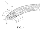
- FIG. 1 there is provided an example of a self-sealing pattern and resultant incision of the present invention. Accordingly, there is shown in a schematic representation the cornea 1, the limbus 2 and sclera 3. A shot pattern 4 is provided, which when delivered to the eye results in an incision of the same shape. The incision has two sides 5 and 6. When viewed along the anterior to posterior axis of the incision, axis arrow 7, it is seen that the incision loops back on top of itself. Thus the incision crosses its anterior to posterior axis at least twice. This would be an example of a tongue and groove type of pattern.
- the angle of the cut with respect to the surface of the eye at the anterior point or opening of the cut resulting from pattern 4 is very shallow, i.e., it is less than 90 degrees, preferably less than about 45 degrees and more preferably less than about 30 degrees.
- the angle of the cut with respect to the posterior surface of the cornea, and in particular Descemet's membrane, at the posterior point or opening of the cut resulting from pattern 4 is about 90 degrees and more preferably about 75 degrees to about 105 degrees.
- FIG. 2 there is provided an example of a self-sealing pattern and resultant incision of the present invention.
- a schematic representation the cornea 1, the limbus 2 and sclera 3.
- a shot pattern 8 is provided, which when delivered to the eye results in an incision of the same shape.
- the incision has two sides 9 and 10.
- axis arrow 7 When viewed along the anterior to posterior axis of the incision, axis arrow 7, it is seen that the incision loops back on top of itself.
- the incision crosses its anterior to posterior axis at least three times.
- the side 10 has a finger section 11 extending into side 9.
- This finger section 11 has a narrow portion 12 and a wide portion 13. It can further be seen that the portion 13 is wider than the narrow portion 12, thus providing added locking force to this tongue and groove configuration.
- axis arrow 7 When viewed along the anterior to posterior axis of the incision, axis arrow 7, it is seen that the incision loops back on top of itself. Thus the incision crosses its anterior to posterior axis at least three times. This would be an example of a tongue and groove type of pattern. If can further be seen that side 15 has finger section 17 and side 16 has finger section 18 and 16 have finger section 11 extending into side 9. Unlike finger section 11, finger sections 17, 18 do not have narrow and wide portions.
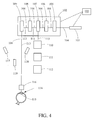
- a preferred laser system i.e ., a laser device, for treating patients is provided as shown by way of example in FIG. 4 .
- a treatment laser 101 there is provided a treatment laser 101; optics 102 for delivering the laser beam 104; a control system 103 for delivering the laser beam to the lens in a particular pattern, which control system 103 is associated with and/or interfaces with the other components of the system, as shown for example by dashed lines in FIG. 4 , and/or other control systems not shown in FIG. 4 .
- a laser system for providing the pattern and creating the resultant cuts in the cornea has by way of example and referring to FIG. 4 a treatment laser 101 which should provide a beam 104.
- the beam should be of a short pulse width, together with the energy and beam size, to produce photodisruption.
- laser shot or shot refers to a laser beam pulse delivered to a location that results in photodisruption.
- photodisruption essentially refers to the conversion of matter to a gas by the laser.
- the term photodisruption has also been generally refered to as Laser Induced Optical Breakdown (LIOB).
- LIOB Laser Induced Optical Breakdown
- wavelengths of about 300 nm to 2500 nm may be employed.
- Pulse widths from about 1 femtosecond to 100 picoseconds may be employed.
- Energy from about a 1 nanojoule to 1 millijoule may be employed.
- the pulse rate also referred to as pulse repetition frequency (PRF) and pulses per second measured in Hertz
- PRF pulse repetition frequency
- a wide variety of laser types may be used to cause photodisruption of ocular tissues, dependent upon pulse width and energy density. Thus, examples of such lasers are disclosed in 2007/084694 A2 and WO 2007/084627A2 . These and other similar lasers may be used a therapeutic lasers.
- the same type of therapeutic laser as described herein may be used, with the energy and focal point being selected to perform the desired procedure.
- the optics 102 for delivering the laser beam 104 to the structures of the eye including the cornea and the natural lens of the eye should be capable of providing a series of shots in a precise and predetermined pattern in the x, y and z dimension.
- the z dimensions as used herein refers to that dimension which has an axis that corresponds to, or is essentially parallel with the A-P axis of the eye.
- the optics should also provide a predetermined beam spot size to cause photodisruption with the laser energy reaching the structure of the eye intended to be cut.
- control system 103 for delivering the laser beam 104 may be any computer, controller, and/or software hardware combination that is capable of selecting and controlling x-y-z scanning parameters and laser firing. These components may typically be associated at least in part with circuit boards that interface to the x-y scanner, the z focusing device and/or the laser.
- the control system may also, but does not necessarily, have the further capabilities of controlling the other components of the system, as well as, maintaining data, obtaining data and performing calculations.
- the control system may contain the programs that direct the laser through one or more laser shot patterns.
- the control system may be capable of processing data from the slit scanned laser and/or from a separate controller for the slit scanned laser system.
- Optical images 113 of the eye 114 and in particular optical images of the natural lens 115 of the eye 114 are conveyed along a path 113.
- This path 113 follows the same path as the laser beam 104 from the natural lens 115 through the laser patient interface 116, the focusing optics 109, the x-y scanner 108 and the beam combiner 107.
- a laser patient interface 116 and a structured light source 117 and a structured light camera 118, including a lens. Examples of patient interface and related apparatus that are useful with the present system are provided in provisional and regular U.S. patent applications serial number 12/509,021 , and serial number 61/228,457 .
- the structured light illumination source 117 and the structured light camera 118 are arranged in an angled relationship.
- the angled relationship may be but is not required to be in the so-called Scheimpflug configuration, which is well-known.
- the structured light source 117 in conjunction with the slit scanning means 119, projects a line and or a plurality of lines onto the eye lens 115 at an angle or plurality of angles.
- the light scattered at the eye lens 115 forms the object to be imaged by the lens and focused onto the camera system 118. Since the slit illuminated image in the eye lens 115 may be at a large angle with respect to the camera 118, this presents a large depth of field to the camera and the entire slit image may not be in sharp focus at the camera.
- arithmetic data evaluation means are further provided herein to determine a more precise location of the illuminated structures with respect to the laser device.
- the images from the camera 118 may be conveyed to the controller 103 for processing and further use in the operation of the system. They may also be sent to a separate processor and/or controller, which in turn communicates with the controller 103.
- the structured light source 117, the camera 118 and the slit scanning means 119 includes a means for determining the position and apex of the lens in relation to the laser system.
- anterior capsule cuts are envisioned and provided that may be a continuous cuts, cuts and lands (uncut capsule portions between cuts) and perforations.
- missed cut or “missed cuts” refer to a cut that was intended to be carried out by the delivery of a particular laser shot pattern, but which did not occur because the laser beam missed the lens capsule or targeted lens material. Thus, in a cut and land pattern the lands would not be considered missed cuts, if they were intended to be left uncut by the laser pattern.
- the cuts in the lens anterior surface are for the purpose of creating an opening in the lens capsule for the remove of the interior structures of the lens.
- various laser shot patterns that cut the interior structure of the lens into small volumes, which volumes can then be removed from the lens capsule.
- These small volumes can range from about 1 mm 2 to about 16 mm 2 and more preferably from about 2.5 mm 2 to about 4 mm 2 .
- a grid laser shot pattern within the interior structures of the lens which creates cube shaped volumes of interior lens material, can be employed.
- These cubes can range in size from a side having a length of about 100 ⁇ m to about 4 mm, with about 500 ⁇ m to 2 mm being a preferred size.
- this invention is not limited to the formation of cubes and other volumetric shapes of similar general size may be employed. For example arrangement of other shapes such as triangles and pie sliced volumes may be employed.
- the laser cut in the anterior capsule is used to create a small opening in the lens anterior surface of the lens capsule for removal of the sectioned volumes of interior material.
- this procedure may be used to treat cataracts.
- This procedure may also be used to remove a lens having opacification that has not progressed to the point of being cataractous.
- This procedure may further be used to remove a natural lens that is clear, but which has lost its ability to accommodate.
- a suitable replacement such as an IOL, accommodative IOL, or synthetic lens refilling materials.
- the size and the shape of the opening is variable and precisely controlled and preferably for presently know lens refilling materials and IOLs is 2 mm or less diameter for lens refilling applications and about 5 mm for IOLs.
- the preferred laser system for treating patients is capable of making precise and predetermined cuts in the capsule of the lens thus giving rise to capsulotomies that are of precise and predetermined shapes.
- the method of obtaining and analyzing the shape and structure of an IOL and in particular obtaining and analyzing the shape and structure of an accommodating IOL, an IOL that reduces and/or eliminates the need for spectacles, and/or an IOL for near, intermediate and distance vision, including but limited to FDA approved versions of the IOLs.
- Based upon this analysis an optimized shape and position for the capsulotomy for use with a particular IOL, or grouping of similarly shaped IOLs, is determined.
- a predetermined shot pattern for making this optimized shaped capsulotomy is then provided to the laser system, preferably by providing the shot pattern to the control system 103.
- the laser system can then be used for an one or all of the following procedures, determining the shape and position of the anterior surface of the lens, and in particular the anterior surface of the lens capsule, determining the apex of the lens capsule in relation to the laser system, performing a laser capsulotomy having the precise and predetermined shape selected for a particular type of IOL, and removal of the natural lens material.
- the placement of individual shots with respect to adjacent shots in the pattern may in general be such that they are as close as possible, typically limited by the size and time frame of photodisruption physics, which would include among other things gas bubble expansion of the previous shot.
- the time frame of photodisruptive physics referrers to the effects that take place surrounding photodisruption, such as plasma formation and expansion, shock waive propagation, and gas bubble expansion and contraction.
- first, second, third, etc. are relative terms and must be viewed in the context in which they are used. They do not relate to timing, unless specifically referred to as such. Thus, a first cut may be made after a second cut.
- cataracts it may be advantageous to shoot from anterior to posterior, because of the inability of the laser to penetrate substantially beyond the cataract.
Description
- The present invention relates to systems for improving procedures that address cataracts, opacifications in the lens, clear lens extraction, removal of natural lens material, use of lens replacement materials and combinations of these. Specifically, the present invention relates to systems that provide predetermined, precise and reproducible laser shot patterns for creating incisions in the cornea and for sealing such incisions, and which are made in predetermined and precise shapes that are reproducible from patient to patient and surgeon to surgeon.
- The established treatment for cataracts is the removal of the opacified human crystalline lens and its replacement with an intraocular lens ("IOL"). In general, IOLs consist of a small plastic lens with plastic side struts, called haptics, to hold the lens in place within the capsular bag inside the eye. Exemplary types of IOLs include monofocal lenses, multifocal IOLs, which provide the patient with multiple-focused vision at far and reading distance, and accommodative IOLs, which provide the patient with visual accommodation. The flexible nature of many IOLs enables them to be rolled and/or folded up for insertion into the capsule. Examples of IOL are found in
U.S. Patent Nos. 7,188,949 ,6,849,091 ,5,699,142 and5,607,472 . Commercially available IOLs that, by way of example, may benefit from the present invention are CRYSTALENS and ACRYSOF RESTOR. - The CRYSTALENS IOL was developed by Eyeonics and is presently provided by Bausch & Lomb, and it is at least in part believed to be disclosed in
U.S. Patent No. 6,849,091 . Further information regarding its structure and efficacy is provided by Food and Drug Administration (FDA) PMA P030002 and related documents to that PMA file. The FDA approved indicated use for CRYSTALENS was in part: "The crystalens™ Model AT-45 Accommodating IOL is intended for primary implantation in the capsular bar of the eye for visual correction of apkakia in adult patients in whom a cataractous lens has been removed and is intended to provide near, intermediate, and distance vision without spectacles. The crystalens™ IOL provides approximately one diopter of monocular accommodation." (November 14, 2003 PMA P030002 atPart 2, Summary of Safety and Effectiveness Data, INDICATIONS FOR USE). - Thus, the CRYSTALENS is an example of an FDA approved accommodating IOL. The term "FDA approved accommodating IOL" refers to any IOL that has obtained FDA approval having an indicated use that provides for accommodation, regardless of whether such IOL is actually being employed for such an approved use.
- The ACRYSOF RESTOR IOL is provided by Alcon, and it is at least in part believed to be disclosed in
U.S. Patent No. 5,669,142 . Further information regarding its structure and efficacy is provided by FDA PMA P040020 and related documents to that PMA file. The FDA approved use for RESTOR was in part: "AcrySOF® ReSTOR® IOLs are indicated for the visual correction of aphakia secondary to removal of a cataractous lens in adult patients with and without presbyopia, who desire near, intermediate and distance vision with increased spectacle independence. The lens is intended to be placed in the capsular bag." (April 24, 2004, PMA P040020, atPart 2, Summary of Safety and Effectiveness Data, INDICATIONS). - Thus, the RESTOR is an example of an FDA approved IOL for near, intermediate and distance vision. The term "FDA approved IOL for near, intermediate and distance vision" refers to any IOL that has obtained FDA approval having an indicated use that provides for near, intermediate and distance vision, regardless of whether such IOL is actually being employed for such an approved use. The CRYSTALENS would also be an example of an FDA approved IOL for near, intermediate and distance vision. Moreover, the RESTOR and CRYSTALENS are examples of an FDA approved IOLs that reduce and/or eliminate the need for spectacles.
- The removal of the natural crystalline lens and replacement with a lens replacement material employs the use of small initial incision or incisions in the limbal area of the eye, which is the transition area between the cornea and sclera. This initial incision is typically made with a small triangular blade that is pushed into the limbal area of the eye. It is through this initial incision that other instruments for use in the removal and replacement of natural lens material are inserted and also it is through this incision that the natural lens material is removed from the eye and replacement lens material inserted into the eye.
- Once the initial incision has been made the removal of the opacified natural crystalline lens and replacement with a lens replacement material, such as an FDA approved IOL, presently employs a capsulorhexis and/or a capsulotomy. A capsulorhexis generally consists of the removal of a part of the anterior lens capsule and the creation of a hole or opening in the lens capsule, that results from at least in part a tearing action. A capsulotomy generally consists of a cutting of the lens capsule, without or with minimum tearing of the capsule. Thus, to remove the opacified natural lens material, the lens capsule is opened. There are several known techniques for performing a capsulorhexis and a capsulotomy. These would include the technique known as a can opener approach, a Continuous Curvilinear Capsulorhexis (CCC) and the use of a Fugo plasma blade.
- To date is it believed that all prior techniques and apparatus and in particular all prior FDA approved apparatus for creating the initial incision into the cornea of the eye have to varying degrees given rise to surgeon-to-surgeon and patient-to-patient irregularities. These irregularities have given rise to slower or less desirable wound healing and results. Moreover, it is believed that all of these prior techniques and apparatus, which are performed by hand, in general can only produce cuts or holes that are essentially simple and planar in nature. Further, because these are hand held devices the shape of these cuts may vary from patient-to-patient and surgeon-to-surgeon. Thus, it is not believed that these hand held devices and non-automated techniques can provide the precise predetermined techniques and cuts of the present invention.
- It is desirable to develop systems that would reduce, eliminate or correct these undesirable features, provide greater control in the creation of the incisions and make these improvements patient and surgeon independent, or at least, reduce the variability from patient-to-patient and surgeon-to-surgeon, associated with the formation of these undesirable features that is found with the use of present techniques and tools.
- The novel and improved systems for the performance of incisions in the sclera, limbus and cornea, which comprise aspects of the present inventions and which are set forth in detail in the present patent specification, may provide for better implementation of other methods and systems for delivering laser beams to the lens of the eye, such as those disclosed in published applications
US 2007/173794A1 ,US 2007/173795A1 ,US 2007/185475A1 ,WO 2007/084694 A2 , andWO 2007/084627A2 . - The present invention, among other things, solves this need by providing greater control in the creation of precise and predetermined incision to the sclera, limbus and cornea. Thus, there is provided herein a system as disclosed and claimed here in.
- One of ordinary skill in the art will recognize, based on the teachings set forth in these specifications and drawings, that there are various embodiments and implementations of these teachings to practice the present invention. Accordingly, the embodiments in this summary are not meant to limit these teachings in any way.
- The invention is defined in the claims. Other embodiments are merely exemplary.
-
-
FIG. 1 is a schematic diagram of a first embodiment of a limbal area pattern and resultant cut in accordance with the present invention. -
FIG. 2 is a schematic diagram of a second embodiment of a limbal area pattern and resultant cut in accordance with the present invention. -
FIG. 3 is a schematic diagram of a third embodiment of a limbal area pattern and resultant cut in accordance with the present invention. -
FIG. 4 is a schematic diagram of an embodiment of a system for delivering a laser beam to the lens of an eye in accordance with the present invention to produce the limbal area patterns and cuts shown inFIGS. 1-3 . - In general, the present inventions relate to systems for providing a laser to the cornea of the eye to address, improve and procedures relating to the removal of the natural crystalline lens and replacement of that lens with lens replacement material, and more specifically to improvements in systems related to cataract surgery. In particular, the present inventions relate to systems for providing self sealing incisions to the eye which incisions may be used as a way to insert instruments into the eye and to remove material from the eye, and to provide increased healing of these particular incisions, as well as, other types of corneal incisions.
- The present methods and systems described herein can be used with the novel and innovative laser system techniques that are the subject of the co-pending patent applications that are cited herein, and the present methods and systems may possibly be used with other laser delivery systems for the removal of lens material to the extent such systems may be developed in the future. Preferably, the present methods and systems can be incorporated into and used in conjunction with the systems of the co-pending applications. In this way a single system, with a single therapeutic laser, can function as a start to finish device for performing the cuts necessary to remove and replace the natural lens.
- Novel and pioneering laser systems and methods for the removal and replace of lens material are disclosed in regular and provisional U.S. patent applications: serial number
61/228,506 61/228,529 61/228,514 serial number 12/509,412 serial number 12/509,211 - Thus, in general the present invention provides for a laser system, i.e., a laser device for delivering a laser to the cornea of the eye. In general, the laser system has a treatment or therapeutic laser, optics for delivering the laser beam from the treatment laser to the eye, and a particular pattern which provides for the placement of treatment laser shots in the cornea to create precise and predetermined incisions in that material, which incisions are for the purpose of inserting instruments and material into and from the eye. The patterns and resultant incisions are made in general by creating a single cut having an anterior point or opening located at or near the limbus and a posterior point or opening located at or near Descemet's membrane, which results in two sides or side pieces. It is theorized that each piece is shaped in a manner that provides for physical interaction between the pieces, creating a locking and/or tongue and groove effect. Additionally, because the incisions of the present invention have a total length that is greater then a straight incision along the same general path, i.e., from posterior opening to anterior opening, and thus have greater surface area between the two sides, it is theorized that this increased surface area promotes quicker healing. In this way after the incision is physically opened with instruments for the insertion and removal of material from the eye and in particular the natural lens of the eye, upon removal of the instrument the incision will close and the physical shape of the sides of the incision will result in the self-sealing of the incision.
- Referring now to
FIG. 1 there is provided an example of a self-sealing pattern and resultant incision of the present invention. Accordingly, there is shown in a schematic representation thecornea 1, thelimbus 2 andsclera 3. Ashot pattern 4 is provided, which when delivered to the eye results in an incision of the same shape. The incision has twosides axis arrow 7, it is seen that the incision loops back on top of itself. Thus the incision crosses its anterior to posterior axis at least twice. This would be an example of a tongue and groove type of pattern. Further, the angle of the cut with respect to the surface of the eye at the anterior point or opening of the cut resulting frompattern 4 is very shallow, i.e., it is less than 90 degrees, preferably less than about 45 degrees and more preferably less than about 30 degrees. The angle of the cut with respect to the posterior surface of the cornea, and in particular Descemet's membrane, at the posterior point or opening of the cut resulting frompattern 4 is about 90 degrees and more preferably about 75 degrees to about 105 degrees. - Referring now to
FIG. 2 there is provided an example of a self-sealing pattern and resultant incision of the present invention. Accordingly, there is shown in a schematic representation thecornea 1, thelimbus 2 andsclera 3. Ashot pattern 8 is provided, which when delivered to the eye results in an incision of the same shape. The incision has twosides axis arrow 7, it is seen that the incision loops back on top of itself. Thus the incision crosses its anterior to posterior axis at least three times. This would be an example of a tongue and groove type of pattern. If can further be seen that theside 10 has afinger section 11 extending intoside 9. Thisfinger section 11 has anarrow portion 12 and awide portion 13. It can further be seen that theportion 13 is wider than thenarrow portion 12, thus providing added locking force to this tongue and groove configuration. - Referring now to
FIG. 3 there is provided an example of a self-sealing pattern and resultant incision of the present invention. Accordingly, there is shown in a schematic representation thecornea 1, thelimbus 2 andsclera 3. Ashot pattern 14 is provided, which when delivered to the eye results in an incision of the same shape. The shot pattern has ananterior start point 19 and aposterior end point 20, which is located in Descemet's membrane. The length of the incision is substantially greater than the length of a straight line drawn betweenpoints sides axis arrow 7, it is seen that the incision loops back on top of itself. Thus the incision crosses its anterior to posterior axis at least three times. This would be an example of a tongue and groove type of pattern. If can further be seen thatside 15 hasfinger section 17 andside 16 hasfinger section finger section 11 extending intoside 9. Unlikefinger section 11,finger sections - In general a preferred laser system, i.e., a laser device, for treating patients is provided as shown by way of example in
FIG. 4 . In this system there is provided atreatment laser 101;optics 102 for delivering thelaser beam 104; acontrol system 103 for delivering the laser beam to the lens in a particular pattern, whichcontrol system 103 is associated with and/or interfaces with the other components of the system, as shown for example by dashed lines inFIG. 4 , and/or other control systems not shown inFIG. 4 . - In general, a laser system for providing the pattern and creating the resultant cuts in the cornea has by way of example and referring to
FIG. 4 atreatment laser 101 which should provide abeam 104. The beam should be of a short pulse width, together with the energy and beam size, to produce photodisruption. Thus, as used herein, the term laser shot or shot refers to a laser beam pulse delivered to a location that results in photodisruption. As used herein, the term photodisruption essentially refers to the conversion of matter to a gas by the laser. The term photodisruption has also been generally refered to as Laser Induced Optical Breakdown (LIOB). In particular, wavelengths of about 300 nm to 2500 nm may be employed. Pulse widths from about 1 femtosecond to 100 picoseconds may be employed. Energies from about a 1 nanojoule to 1 millijoule may be employed. The pulse rate (also referred to as pulse repetition frequency (PRF) and pulses per second measured in Hertz) may be from about 1 KHz to several GHz. Generally, lower pulse rates correspond to higher pulse energy in commercial laser devices. A wide variety of laser types may be used to cause photodisruption of ocular tissues, dependent upon pulse width and energy density. Thus, examples of such lasers are disclosed in2007/084694 A2 andWO 2007/084627A2 . These and other similar lasers may be used a therapeutic lasers. For procedures on the cornea, limbus and area where the cornea and sclera join the same type of therapeutic laser as described herein may be used, with the energy and focal point being selected to perform the desired procedure. - In general, the
optics 102 for delivering thelaser beam 104 to the structures of the eye including the cornea and the natural lens of the eye should be capable of providing a series of shots in a precise and predetermined pattern in the x, y and z dimension. The z dimensions as used herein refers to that dimension which has an axis that corresponds to, or is essentially parallel with the A-P axis of the eye. The optics should also provide a predetermined beam spot size to cause photodisruption with the laser energy reaching the structure of the eye intended to be cut. - In general, the
control system 103 for delivering thelaser beam 104 may be any computer, controller, and/or software hardware combination that is capable of selecting and controlling x-y-z scanning parameters and laser firing. These components may typically be associated at least in part with circuit boards that interface to the x-y scanner, the z focusing device and/or the laser. The control system may also, but does not necessarily, have the further capabilities of controlling the other components of the system, as well as, maintaining data, obtaining data and performing calculations. Thus, the control system may contain the programs that direct the laser through one or more laser shot patterns. Similarly, the control system may be capable of processing data from the slit scanned laser and/or from a separate controller for the slit scanned laser system. - The
laser optics 102 for delivering thelaser beam 104 includes abeam expander telescope 105,a z focus mechanism 106, abeam combiner 107, anx-y scanner 108, and focusingoptics 109. There is further providedrelay optics 110,camera optics 111, which include a zoom, and afirst ccd camera 112. -
Optical images 113 of theeye 114 and in particular optical images of thenatural lens 115 of theeye 114 are conveyed along apath 113. Thispath 113 follows the same path as thelaser beam 104 from thenatural lens 115 through thelaser patient interface 116, the focusingoptics 109, thex-y scanner 108 and thebeam combiner 107. There is further provided alaser patient interface 116, and a structuredlight source 117 and a structuredlight camera 118, including a lens. Examples of patient interface and related apparatus that are useful with the present system are provided in provisional and regularU.S. patent applications , and serial numberserial number 12/509,02161/228,457 - The structured
light source 117 may be a slit illumination having focusing and structured light projection optics, such as a Schafter+Kirchhoff Laser Macro Line Generator Model 13LTM+90CM, (Type 13LTM-250S-41 + 90CM-M60-780-5-Y03-C-6) or a StockerYale Model SNF-501L-660-20-5, which is also referred to as a slit scanned laser. In this embodiment the structuredillumination source 117 also includes slit scanning means 119. - When using a scanned slit illumination the operation includes positioning the slit on one side of the lens, taking an image then moving the slit approximately one slit width, then taking another image, and then repeating this sequence until the entire lens is observed. For example, a 100 µm slit width can scan a nominal 9 mm dilated pupil diameter in 90 images, which takes approximately 3 seconds using a 30 Hz frame rate camera. To obtain images of the anterior surface in a single image without overlap, the slit should be at an angle to the AP axis, i.e., it should not be parallel to that axis. The nominal slit angle can be approximately 15 to 30 degrees from the AP axis. Any visible or near IR wavelength source within the sensitivity of the camera may be used. Low coherence length sources are preferable to reduce speckle noise.
- The structured
light illumination source 117 and the structuredlight camera 118 are arranged in an angled relationship. The angled relationship may be but is not required to be in the so-called Scheimpflug configuration, which is well-known. The structuredlight source 117, in conjunction with the slit scanning means 119, projects a line and or a plurality of lines onto theeye lens 115 at an angle or plurality of angles. The light scattered at theeye lens 115 forms the object to be imaged by the lens and focused onto thecamera system 118. Since the slit illuminated image in theeye lens 115 may be at a large angle with respect to thecamera 118, this presents a large depth of field to the camera and the entire slit image may not be in sharp focus at the camera. By tilting the camera at an angle or plurality of angles the image along the illuminated plane can be in sharper focus. To the extent that a shaper focus is not obtained, arithmetic data evaluation means are further provided herein to determine a more precise location of the illuminated structures with respect to the laser device. - The images from the
camera 118 may be conveyed to thecontroller 103 for processing and further use in the operation of the system. They may also be sent to a separate processor and/or controller, which in turn communicates with thecontroller 103. The structuredlight source 117, thecamera 118 and the slit scanning means 119 includes a means for determining the position and apex of the lens in relation to the laser system. - The delivery of laser shot patterns and cuts per
FIGS. 1-3 for the removal of lens material is provided. Thus, there are provided methods and systems for producing cuts, i.e., incisions in the anterior lens capsule. These cuts are created by thetherapeutic laser beam 104 being delivered to the anterior lens capsule in precise predetermined and highly reproducible patterns, delivery results in precise predetermined and highly reproducible shaped cuts in patterns as described and taught herein, or as may be called for by the use of a particular IOL or other device or material to be inserted within the lens capsule. As used herein geometric shaped patterns or cuts referrer to circular and elliptical shaped patterns or cuts. As used herein non-geometric shaped patterns or cuts refers to all other shapes that are not circular or elliptical. - The methods and systems to create these cuts in the anterior capsule provide superior results to the handheld methods and apparatus previously known for performing capsulorhexus and capsulotomy, and thus, the methods and systems disclosed herein are considered to be a substantial advancement in these techniques. In addition the delivery of the laser beam shots in a manner that greatly reduces the risk of a missed cut, which depending upon the particular application may be very significant is provided. Moreover, as provided in the following examples, anterior capsule cuts are envisioned and provided that may be a continuous cuts, cuts and lands (uncut capsule portions between cuts) and perforations. Thus, as used herein the terms "missed cut" or "missed cuts" refer to a cut that was intended to be carried out by the delivery of a particular laser shot pattern, but which did not occur because the laser beam missed the lens capsule or targeted lens material. Thus, in a cut and land pattern the lands would not be considered missed cuts, if they were intended to be left uncut by the laser pattern.
- The cuts in the lens anterior surface are for the purpose of creating an opening in the lens capsule for the remove of the interior structures of the lens. To facilitate this removal there are provided various laser shot patterns that cut the interior structure of the lens into small volumes, which volumes can then be removed from the lens capsule. These small volumes can range from about 1 mm2 to about 16 mm2 and more preferably from about 2.5 mm2 to about 4 mm2. Thus a grid laser shot pattern within the interior structures of the lens, which creates cube shaped volumes of interior lens material, can be employed. These cubes can range in size from a side having a length of about 100 µm to about 4 mm, with about 500 µm to 2 mm being a preferred size. Additionally, this invention is not limited to the formation of cubes and other volumetric shapes of similar general size may be employed. For example arrangement of other shapes such as triangles and pie sliced volumes may be employed.
- The laser cut in the anterior capsule is used to create a small opening in the lens anterior surface of the lens capsule for removal of the sectioned volumes of interior material. Thus, this procedure may be used to treat cataracts. This procedure may also be used to remove a lens having opacification that has not progressed to the point of being cataractous. This procedure may further be used to remove a natural lens that is clear, but which has lost its ability to accommodate. In all of the above scenarios, it being understood that upon removal of the lens material the lens capsule would subsequently house a suitable replacement, such as an IOL, accommodative IOL, or synthetic lens refilling materials. Moreover, the size and the shape of the opening is variable and precisely controlled and preferably for presently know lens refilling materials and IOLs is 2 mm or less diameter for lens refilling applications and about 5 mm for IOLs.
- The order in which these activities are performed may depend upon the particular characteristics of the internal lens structure, the density of the cataract, the position of the cataract, the type of device used to remove the internal lens material once it has been sectioned into small volumes, the type and power of the laser used, the amount and size of gas bubbles that are produced by the laser, and other factors. Thus, although the examples herein provide for an order of performing the activity of cutting the anterior surface of the lens and sectioning the interior structures of the lens, it should be recognized that this order can be changed, as well as, performed essentially simultaneously or simultaneously.
- The preferred laser system for treating patients is capable of making precise and predetermined cuts in the capsule of the lens thus giving rise to capsulotomies that are of precise and predetermined shapes. Thus, there is provided the method of obtaining and analyzing the shape and structure of an IOL, and in particular obtaining and analyzing the shape and structure of an accommodating IOL, an IOL that reduces and/or eliminates the need for spectacles, and/or an IOL for near, intermediate and distance vision, including but limited to FDA approved versions of the IOLs. Based upon this analysis an optimized shape and position for the capsulotomy for use with a particular IOL, or grouping of similarly shaped IOLs, is determined. A predetermined shot pattern for making this optimized shaped capsulotomy is then provided to the laser system, preferably by providing the shot pattern to the
control system 103. The laser system can then be used for an one or all of the following procedures, determining the shape and position of the anterior surface of the lens, and in particular the anterior surface of the lens capsule, determining the apex of the lens capsule in relation to the laser system, performing a laser capsulotomy having the precise and predetermined shape selected for a particular type of IOL, and removal of the natural lens material. - Thus, there is provided techniques, systems and apparatus to deliver laser beam in a shot pattern to the lens of the eye and in particular to the capsule of the lens of the eye in a precise and predetermined manner to provided for a precise predetermined capsulotomy. The shape of these patterns may be delivered using either the jigsaw or ring delivery sequences.
- When performing laser assisted cataract surgery the process of cutting the nucleus with a photodisruption laser can cause a buildup of gas bubbles sufficiently near the soft cortex to allow the gas bubbles to propagate toward the capsule. In those situations where bubbles collect in close proximity to the anterior capsule, when the laser attempts to cut the capsulotomy, the sudden release of bubbles my change the position of the anterior capsule during the delivery of the laser shot pattern causing the laser to miss the capsule resulting in missed cuts, at least partially around the circumference of the capsulotomy. To solve this problem, there is provided herein a special cutting pattern that is less dependent of capsule position versus time and provides cutting of the capsule despite position changes of the capsule during the laser capsulotomy procedure. Thus, resulting in substantially reduced or no missed cuts.
- There is further provided herein the use of laser shot patterns having a large range of Z swept at a high rate of speed, while the X-Y position is moved in a circular, or elliptical or other pattern or desired shape, more slowly so that the laser cutting action occurs multiple times over essentially the same X-Y position. Thus, it could be envisioned that the laser beam is operating like the tip of a jigsaw blade moving up and down rapidly compared to the X-Y positioning to create the cut shape. In this way, if the anterior capsule shifts during the cut, due to gas bubble propagation or any other reason, the cut will still be made to the capsule, albeit perhaps outside the center region of the z direction up-down distribution of shots, and more to the anterior or posterior ends of that distribution. For laser cutting of the nucleus where a great deal of bubble buildup is created, a Z range, or up-down range of the cut should be approximately 1 mm in extent, nominally centered on the anterior capsule which would allow approximately +/- 475 µm of capsule movement and still provide cutting of a 25 µm thick capsule.
- In addition to enabling cutting of a capsule that moves during the procedure, this procedure can be used to compensate for static errors in capsule position due to for example measurement errors. In this way the extent of the Z range may be increased by the known error of the system.
- In addition to the large Z range sweeps disclosed herein, there is also contemplated the use of a smaller Z range of cut motion for the case where the uncertainty in capsule position from both static measurement error and anticipated change in position might be smaller, perhaps in the range of hundreds of µm or in the case of highly precise measurement data and near zero movement of the capsule during surgery. In such a case the Z range could be tens of µm -- enough range to cut through the capsule thickness.
- Further methods and systems to define a high accuracy position measurement of structures of the eye and in particular the anterior capsule, so as to provide in general greater accuracy, precisions and reproducibility from patient to patient for procedures on the eye and in particular capsulotomies, is provided in regular
U.S. patent application .serial number 12/509,412 - In the laser shot patterns provided herein it is preferred that the placement of individual shots with respect to adjacent shots in the pattern are sufficiently close enough to each other, such that when the pattern is complete a sufficiently continuous layer and/or line and/or volume of material has been removed. Shot spacing of lesser or greater distances are contemplated herein and including overlap as necessary to obtain the desired results. Shot spacing considerations include gas bubble dissipation, volume removal efficiency, sequencing efficiency, scanner performance, and cleaving efficiency among others. For example, by way of illustration, for a 5 µm size spot with an energy sufficient to cause photodisruption, a spacing of 20 µm or greater results in individual gas bubbles, which are not coalesced and dissipate more quickly, than with close shot spaces with the same energy, which result in gas bubble coalescence. As the shot spacing gets closer together volume efficiency increases. As shot spacing gets closer together bubble coalescence also increases. Further, there comes a point where the shot spacing becomes so close that volume efficiency dramatically decreases. Moreover, the forgoing shot spacing considerations are interrelated to a lesser or greater extent and one of skill in the art will know how to evaluate these conditions based upon the teachings of the present disclosure to accomplish the objectives herein. Finally, it is contemplated that the placement of individual shots with respect to adjacent shots in the pattern may in general be such that they are as close as possible, typically limited by the size and time frame of photodisruption physics, which would include among other things gas bubble expansion of the previous shot. As used herein, the time frame of photodisruptive physics referrers to the effects that take place surrounding photodisruption, such as plasma formation and expansion, shock waive propagation, and gas bubble expansion and contraction. Thus, the timing of sequential pulses such that they are timed faster than some of, elements of, or all of those effects, can increase volumetric removal and/or cleaving efficiency. Accordingly, it is proposed to use pulse repetition frequencies from 50 MHz to 5 GHz, which could be accomplished by a laser with the following parameters: a mode lock laser of cavity length from 3 meters to 3 cm. Such high PRF lasers can more easily produce multiple pulses overlapping a location allowing for a lower energy per pulse to achieve photodisruption.
- The terms first, second, third, etc. as used herein are relative terms and must be viewed in the context in which they are used. They do not relate to timing, unless specifically referred to as such. Thus, a first cut may be made after a second cut. In general, it is preferred to fire laser shots in general from posterior points in the laser pattern to anterior points, to avoid and/or minimize the effect of the gas bubbles resulting from prior laser shots. However, because of the varied laser shot patterns that are provided herein, it is not a requirement that a strict posterior to anterior shot sequence be followed. Moreover, in the case of cataracts it may be advantageous to shoot from anterior to posterior, because of the inability of the laser to penetrate substantially beyond the cataract.
- An additional benefit from this type of laser system may be obtained. Shots or pulses from this laser can be distributed throughout the cornea in a plane or series of planes that are perpendicular to the A-P axis of the eye. These planes will cause a wound healing effect, creating myacin. Additionally, the creation of myacin will cause a cross linking of the cornea collagen which will have the tendency to shrink the area treated with the plane of shots, which in turn tends to flatten the curvature of the cornea providing the added benefit of correcting low levels of myopia.
- From the foregoing description, one skilled in the art can readily ascertain the essential characteristics of this invention, and without departing from the scope thereof, can make various changes and/or modifications of the invention to adapt it to various usages and conditions.
Claims (8)
- A system for providing a self-sealing incision to an eye (114), the system comprising:a laser (101) for producing a laser beam (104);optics (102) for receiving the laser beam (104) and directing the laser beam (104) to an eye (114) to generate a laser shot pattern (4,8,14) for providing a self-sealing incision in a cornea of the eye (114), the self-sealing incision having an anterior start point and a posterior end point, and a length of the incision beginning at the anterior start point and ending at the posterior end point being greater than a distance between the anterior start point and the posterior end point; anda controller (103) in communication with the laser (101) and the optics (102) and having a structure that controls the laser beam (104) by controlling firing of the laser (101) and controlling the optics (102) to control x-y-z scanning parameters of the laser beam (104) so that the laser shot pattern (4,8,14) is generated on the eye (114), wherein the shot pattern (4,8,14) comprises a pattern for providing the self-sealing incision in the cornea of the eye (114); andwherein an angle of the self-sealing incision in the cornea of the eye (114) at the anterior start point with respect to an anterior surface of the cornea of the eye (114) is acute; characterised in that
the angle of the self-sealing incision in the cornea of the eye at the posterior end point with respect to the posterior surface of the cornea of the eye is 90 degrees. - The system of claim 1 wherein the laser shot pattern (4, 8, 14) for providing the self-sealing incision in the cornea of the eye (114) is a tongue and groove pattern.
- The system of claim 1 wherein the laser shot pattern (8) for providing the self-sealing incision in the cornea of the eye (114) is a locking tongue and groove pattern.
- The system of claim 1 wherein the laser shot pattern (4,8, 14) for providing the self-sealing incision in the cornea of the eye (114) comprises an anterior posterior axis (7) and crosses the anterior posterior axis (7) at least twice.
- The system of claim 1 wherein the laser shot pattern (8,14) for providing the self-sealing incision in the cornea of the eye (114) comprises an anterior posterior axis (7) and crosses the anterior posterior axis (7) at least three times.
- The system of claim 1 wherein the posterior end point is in Descemet's membrane of the eye.
- The system of claim 1, wherein the angle of the self-sealing incision in the cornea of the eye at the anterior start point with respect to the anterior surface of the cornea of the eye is less than 45 degrees.
- The system of claim 1, wherein an angle of the self-sealing incision in the cornea of the eye at the anterior start point with respect to the anterior surface of the cornea of the eye is less than 30 degrees.
Priority Applications (1)
| Application Number | Priority Date | Filing Date | Title |
|---|---|---|---|
| EP16187407.8A EP3132776A1 (en) | 2009-07-24 | 2010-07-08 | Laser system for performing and sealing corneal incisions in the eye |
Applications Claiming Priority (2)
| Application Number | Priority Date | Filing Date | Title |
|---|---|---|---|
| US22848409P | 2009-07-24 | 2009-07-24 | |
| PCT/US2010/041286 WO2011011202A1 (en) | 2009-07-24 | 2010-07-08 | Laser system and method for performing and sealing corneal incisions in the eye |
Related Child Applications (1)
| Application Number | Title | Priority Date | Filing Date |
|---|---|---|---|
| EP16187407.8A Division EP3132776A1 (en) | 2009-07-24 | 2010-07-08 | Laser system for performing and sealing corneal incisions in the eye |
Publications (3)
| Publication Number | Publication Date |
|---|---|
| EP2456400A1 EP2456400A1 (en) | 2012-05-30 |
| EP2456400A4 EP2456400A4 (en) | 2013-04-03 |
| EP2456400B1 true EP2456400B1 (en) | 2016-09-07 |
Family
ID=43499349
Family Applications (2)
| Application Number | Title | Priority Date | Filing Date |
|---|---|---|---|
| EP16187407.8A Withdrawn EP3132776A1 (en) | 2009-07-24 | 2010-07-08 | Laser system for performing and sealing corneal incisions in the eye |
| EP10802654.3A Active EP2456400B1 (en) | 2009-07-24 | 2010-07-08 | Laser system for performing and sealing corneal incisions in the eye |
Family Applications Before (1)
| Application Number | Title | Priority Date | Filing Date |
|---|---|---|---|
| EP16187407.8A Withdrawn EP3132776A1 (en) | 2009-07-24 | 2010-07-08 | Laser system for performing and sealing corneal incisions in the eye |
Country Status (7)
| Country | Link |
|---|---|
| US (1) | US8758332B2 (en) |
| EP (2) | EP3132776A1 (en) |
| JP (1) | JP2013500056A (en) |
| CN (1) | CN102639088B (en) |
| AU (1) | AU2010274169A1 (en) |
| CA (1) | CA2769059A1 (en) |
| WO (1) | WO2011011202A1 (en) |
Families Citing this family (38)
| Publication number | Priority date | Publication date | Assignee | Title |
|---|---|---|---|---|
| US10842675B2 (en) | 2006-01-20 | 2020-11-24 | Lensar, Inc. | System and method for treating the structure of the human lens with a laser |
| US10213340B2 (en) | 2006-01-20 | 2019-02-26 | Lensar, Inc. | Methods and systems to provide excluded defined zones for increasing accommodative amplitude |
| US9889043B2 (en) | 2006-01-20 | 2018-02-13 | Lensar, Inc. | System and apparatus for delivering a laser beam to the lens of an eye |
| US9545338B2 (en) | 2006-01-20 | 2017-01-17 | Lensar, Llc. | System and method for improving the accommodative amplitude and increasing the refractive power of the human lens with a laser |
| US10709610B2 (en) | 2006-01-20 | 2020-07-14 | Lensar, Inc. | Laser methods and systems for addressing conditions of the lens |
| US8262646B2 (en) * | 2006-01-20 | 2012-09-11 | Lensar, Inc. | System and method for providing the shaped structural weakening of the human lens with a laser |
| US11185226B2 (en) | 2008-07-25 | 2021-11-30 | Lensar, Inc. | System and method for measuring tilt in the crystalline lens for laser phaco fragmentation |
| US8480659B2 (en) | 2008-07-25 | 2013-07-09 | Lensar, Inc. | Method and system for removal and replacement of lens material from the lens of an eye |
| US8500723B2 (en) | 2008-07-25 | 2013-08-06 | Lensar, Inc. | Liquid filled index matching device for ophthalmic laser procedures |
| US8617146B2 (en) | 2009-07-24 | 2013-12-31 | Lensar, Inc. | Laser system and method for correction of induced astigmatism |
| US8382745B2 (en) | 2009-07-24 | 2013-02-26 | Lensar, Inc. | Laser system and method for astigmatic corrections in association with cataract treatment |
| CN102639078B (en) | 2009-07-24 | 2015-10-21 | 能斯雅有限公司 | A kind of is the para-operative system and method for eye lens enforcement laser radar |
| AU2010275482A1 (en) | 2009-07-24 | 2012-02-16 | Lensar, Inc. | System and method for providing laser shot patterns to the lens of an eye |
| US10772499B2 (en) | 2009-07-25 | 2020-09-15 | Lensar, Inc. | System and method for measuring tilt |
| US20110190739A1 (en) * | 2010-01-29 | 2011-08-04 | Lensar, Inc. | Servo controlled docking force device for use in ophthalmic applications |
| WO2011094678A1 (en) | 2010-02-01 | 2011-08-04 | Lensar, Inc. | Purkinjie image-based alignment of suction ring in ophthalmic applications |
| US9622911B2 (en) | 2010-09-30 | 2017-04-18 | Cxl Ophthalmics, Llc | Ophthalmic treatment device, system, and method of use |
| WO2012051490A1 (en) | 2010-10-15 | 2012-04-19 | Lensar, Inc. | System and method of scan controlled illumination of structures within an eye |
| USD694890S1 (en) | 2010-10-15 | 2013-12-03 | Lensar, Inc. | Laser system for treatment of the eye |
| USD695408S1 (en) | 2010-10-15 | 2013-12-10 | Lensar, Inc. | Laser system for treatment of the eye |
| US20120150158A1 (en) * | 2010-12-10 | 2012-06-14 | Johannes Krause | Device and process for machining the cornea of a human eye with focused pulsed laser radiation |
| US10463541B2 (en) | 2011-03-25 | 2019-11-05 | Lensar, Inc. | System and method for correcting astigmatism using multiple paired arcuate laser generated corneal incisions |
| DE102011006085A1 (en) * | 2011-03-25 | 2012-09-27 | Carl Zeiss Meditec Ag | Ophthalmic device |
| EP2633841B1 (en) * | 2012-02-28 | 2017-08-16 | Ziemer Ophthalmic Systems AG | Device for treating eye tissue using pulsed laser beams |
| US9737438B2 (en) | 2012-03-14 | 2017-08-22 | Ziemer Ophthalmic Systems Ag | Device for processing eye tissue by means of pulsed laser beams |
| WO2013148896A1 (en) | 2012-03-29 | 2013-10-03 | Cxl Ophthalmics, Llc | Ocular treatment solutions, delivery devices and delivery augmentation methods |
| WO2013148895A1 (en) | 2012-03-29 | 2013-10-03 | Cxl Ophthalmics, Llc | Ocular cross-linking system and method for sealing corneal wounds |
| WO2013149075A1 (en) | 2012-03-29 | 2013-10-03 | Cxl Ophthalmics, Llc | Compositions and methods for treating or preventing diseases associated with oxidative stress |
| US9629750B2 (en) | 2012-04-18 | 2017-04-25 | Technolas Perfect Vision Gmbh | Surgical laser unit with variable modes of operation |
| US10058453B2 (en) * | 2012-04-24 | 2018-08-28 | Wavelight Gmbh | Extracting lenticules for refractive correction |
| US9968485B2 (en) | 2013-07-29 | 2018-05-15 | Lensar, Inc. | Patient interface device for ophthalmic laser procedures |
| US9844465B2 (en) | 2013-07-29 | 2017-12-19 | Lensar, Inc. | Second pass femtosecond laser for incomplete laser full or partial thickness corneal incisions |
| US9339414B2 (en) | 2014-04-29 | 2016-05-17 | Chukyo Medical Co., Inc. | Irradiating device and program |
| US11083625B2 (en) | 2015-07-01 | 2021-08-10 | Amo Development, Llc | Sub-nanosecond laser surgery system utilizing multiple pulsed laser beams |
| US10485705B2 (en) | 2015-07-01 | 2019-11-26 | Optimedica Corporation | Sub-nanosecond laser cataract surgery system |
| US10383767B2 (en) * | 2015-12-17 | 2019-08-20 | Novartis Ag | Ophthalmic relaxing incisions and associated devices, systems, and methods |
| KR20200139161A (en) * | 2018-03-02 | 2020-12-11 | 렌사르, 인크. | Laser methods and systems for handling, alleviating and reversing presbyopia |
| DE112019006884T5 (en) | 2019-07-03 | 2021-11-11 | Sun Yat-Sen University | Method for making an automated seamless major corneal incision |
Citations (2)
| Publication number | Priority date | Publication date | Assignee | Title |
|---|---|---|---|---|
| US20080058841A1 (en) * | 2006-09-05 | 2008-03-06 | Kurtz Ronald M | System and method for marking corneal tissue in a transplant procedure |
| WO2008112292A1 (en) * | 2007-03-13 | 2008-09-18 | Optimedica Corporation | Apparatus for creating ocular surgical and relaxing incisions |
Family Cites Families (476)
| Publication number | Priority date | Publication date | Assignee | Title |
|---|---|---|---|---|
| US3074407A (en) | 1956-09-17 | 1963-01-22 | Marguerite Barr Moon Eye Res F | Surgical devices for keratoplasty and methods thereof |
| US3971382A (en) | 1973-12-11 | 1976-07-27 | Krasnov Mikhail M | Method of non-surgical treatment of cataracts |
| US3982541A (en) | 1974-07-29 | 1976-09-28 | Esperance Jr Francis A L | Eye surgical instrument |
| US4024852A (en) | 1976-02-05 | 1977-05-24 | Esperance Paul M L | Solar energy reflector-collector |
| FR2442622A1 (en) | 1978-06-08 | 1980-06-27 | Aron Rosa Daniele | OPHTHALMOLOGICAL SURGERY APPARATUS |
| US4263893A (en) | 1978-10-03 | 1981-04-28 | Consuntrator, Inc. | Solar energy collector construction |
| DE2946451C2 (en) | 1979-11-17 | 1984-09-20 | Fa. Carl Zeiss, 7920 Heidenheim | Device for examining the anterior segments of the eye |
| US4306546A (en) | 1980-03-26 | 1981-12-22 | Propper Manufacturing Co., Inc. | Endoscope |
| US4334736A (en) | 1980-05-23 | 1982-06-15 | Herbert M Linton | Wet cornea microscope |
| DE3024169C2 (en) | 1980-06-27 | 1983-09-15 | Reginald Dipl.-Phys. Dr. 8028 Taufkirchen Birngruber | Method and device for operating a photocoagulator for biological tissue |
| US4477159A (en) | 1980-11-06 | 1984-10-16 | Nidek Co., Ltd. | Photocoagulator |
| FR2497087A1 (en) | 1980-12-30 | 1982-07-02 | Essilor Int | Automatic corneal size measuring appts. - has rotating luminous source and magnifying lens providing image detected by photodiodes giving proportional signal over scanned meridians |
| US4381007A (en) | 1981-04-30 | 1983-04-26 | The United States Of America As Represented By The United States Department Of Energy | Multipolar corneal-shaping electrode with flexible removable skirt |
| US4394144A (en) | 1981-09-03 | 1983-07-19 | Kaken Chemical Co., Ltd. | Dehumidifying container |
| FR2513873B1 (en) | 1981-10-02 | 1985-10-25 | Essilor Int | OPHTHALMOLOGICAL LASER SURGERY APPARATUS |
| US4633866A (en) | 1981-11-23 | 1987-01-06 | Gholam Peyman | Ophthalmic laser surgical method |
| US4583539A (en) | 1982-01-12 | 1986-04-22 | Cornell Research Foundation, Inc. | Laser surgical system |
| US4712543A (en) | 1982-01-20 | 1987-12-15 | Baron Neville A | Process for recurving the cornea of an eye |
| US4461294A (en) | 1982-01-20 | 1984-07-24 | Baron Neville A | Apparatus and process for recurving the cornea of an eye |
| FR2524298A1 (en) | 1982-04-01 | 1983-10-07 | Essilor Int | LASER OPHTHALMOLOGICAL SURGICAL APPARATUS |
| JPS5914848A (en) | 1982-07-15 | 1984-01-25 | 株式会社トプコン | Light treating apparatus |
| US4883351A (en) | 1982-09-10 | 1989-11-28 | Weiss Jeffrey N | Apparatus for the detection of diabetes and other abnormalities affecting the lens of the eye |
| US4715703A (en) | 1982-10-12 | 1987-12-29 | Rodenstock Instrument Corporation | Ocular-fundus analyzer |
| US4537193A (en) | 1982-10-28 | 1985-08-27 | Hgm, Inc. | Laser endocoagulator apparatus |
| DE3245939C2 (en) | 1982-12-11 | 1985-12-19 | Fa. Carl Zeiss, 7920 Heidenheim | Device for generating an image of the fundus |
| DE3306981C2 (en) | 1983-02-28 | 1987-11-12 | Wolfram 8048 Haimhausen Weinberg | Device for the photocoagulation of biological tissue |
| US4573778A (en) | 1983-03-16 | 1986-03-04 | Boston University | Aqueous fluorophotometer |
| US4770162A (en) | 1983-05-26 | 1988-09-13 | Phillips Petroleum Company | Solar energy collecting system |
| DE3319203C2 (en) | 1983-05-27 | 1986-03-27 | Fa. Carl Zeiss, 7920 Heidenheim | Device for dose measurement during photocoagulation |
| US4502816A (en) | 1983-06-27 | 1985-03-05 | Creter Vault Corp. | Shoreline breakwater |
| US4638801A (en) | 1983-07-06 | 1987-01-27 | Lasers For Medicine | Laser ophthalmic surgical system |
| JPS6029135A (en) * | 1983-07-25 | 1985-02-14 | 三菱電機株式会社 | Indivisual discriminator |
| DE3331431C2 (en) | 1983-08-31 | 1986-03-27 | Optische Werke G. Rodenstock, 8000 München | Device for coupling operating light into an eye examination device |
| DE3331586A1 (en) | 1983-09-01 | 1985-03-28 | Fa. Carl Zeiss, 7920 Heidenheim | OPHTHALMOLOGICAL COMBINATION DEVICE FOR DIAGNOSIS AND THERAPY |
| US4669839A (en) | 1983-09-16 | 1987-06-02 | Carl-Zeiss-Stiftung | Optical system for therapeutic use of laser light |
| US4561436A (en) | 1983-10-28 | 1985-12-31 | Cooper Lasersonics, Inc. | Optical system for surgical ophthalmic laser instrument |
| US4862888A (en) | 1983-10-28 | 1989-09-05 | Bausch & Lomb Incorporated | Laser system |
| US4870952A (en) | 1983-10-28 | 1989-10-03 | Miquel Martinez | Fiber optic illuminator for use in surgery |
| DE3339370A1 (en) | 1983-10-29 | 1985-05-09 | Meditec GmbH, 8501 Heroldsberg | PULSE LASER FOR MEDICAL APPLICATIONS |
| US4773414A (en) | 1983-11-17 | 1988-09-27 | Lri L.P. | Method of laser-sculpture of the optically used portion of the cornea |
| US4718418A (en) | 1983-11-17 | 1988-01-12 | Lri L.P. | Apparatus for ophthalmological surgery |
| US5312320A (en) | 1983-11-17 | 1994-05-17 | Visx, Incorporated | Apparatus for performing ophthalmological surgery |
| US5207668A (en) | 1983-11-17 | 1993-05-04 | Visx Incorporated | Method for opthalmological surgery |
| US5219343A (en) | 1983-11-17 | 1993-06-15 | Visx Incorporated | Apparatus for performing ophthalmogolical surgery |
| US4665913A (en) | 1983-11-17 | 1987-05-19 | Lri L.P. | Method for ophthalmological surgery |
| US4729372A (en) | 1983-11-17 | 1988-03-08 | Lri L.P. | Apparatus for performing ophthalmic laser surgery |
| US4770172A (en) | 1983-11-17 | 1988-09-13 | Lri L.P. | Method of laser-sculpture of the optically used portion of the cornea |
| US4732148A (en) | 1983-11-17 | 1988-03-22 | Lri L.P. | Method for performing ophthalmic laser surgery |
| US4565197A (en) | 1983-11-22 | 1986-01-21 | Lasers For Medicine | Laser ophthalmic surgical system |
| US5108388B1 (en) | 1983-12-15 | 2000-09-19 | Visx Inc | Laser surgery method |
| US4711540A (en) | 1984-01-04 | 1987-12-08 | Tokyo Kogaku Kikai Kabushiki Kaisha | Eye disease inspecting instrument |
| US4686979A (en) | 1984-01-09 | 1987-08-18 | The United States Of America As Represented By The United States Department Of Energy | Excimer laser phototherapy for the dissolution of abnormal growth |
| JPS60148537A (en) | 1984-01-12 | 1985-08-05 | 興和株式会社 | Opththalimic measuring apparatus utilizing laser beam |
| US4711541A (en) | 1984-02-02 | 1987-12-08 | Tokyo Kogaku Kikai Kabushiki Kaisha | Slit lamp and accessory device thereof |
| US4601288A (en) | 1984-02-24 | 1986-07-22 | Myers John D | Laser device and method |
| US4538608A (en) | 1984-03-23 | 1985-09-03 | Esperance Jr Francis A L | Method and apparatus for removing cataractous lens tissue by laser radiation |
| US4588505A (en) | 1984-05-07 | 1986-05-13 | Frontier Technology, Inc. | Water scavenger pouch |
| US4601037A (en) | 1984-06-13 | 1986-07-15 | Britt Corporation | Pulsed laser system |
| US4580559A (en) | 1984-07-24 | 1986-04-08 | Esperance Francis A L | Indirect ophthalmoscopic photocoagulation delivery system for retinal surgery |
| US4669466A (en) | 1985-01-16 | 1987-06-02 | Lri L.P. | Method and apparatus for analysis and correction of abnormal refractive errors of the eye |
| FR2576780B1 (en) | 1985-02-04 | 1991-06-14 | Azema Alain | APPARATUS FOR CHANGING THE CURVATURE OF THE EYE CORNEA OVER THE WHOLE PUPILLARY SURFACE BY PHOTOCHEMICAL ABLATION OF THE SAME |
| US4682595A (en) | 1985-03-25 | 1987-07-28 | Carl-Zeiss-Stiftung | Illuminance dosage device |
| US4657013A (en) | 1985-03-25 | 1987-04-14 | Carl-Zeiss-Stiftung | Illuminance dosage device for an operation microscope |
| US4607622A (en) | 1985-04-11 | 1986-08-26 | Charles D. Fritch | Fiber optic ocular endoscope |
| US4820264A (en) | 1985-05-01 | 1989-04-11 | Tokyo Kogaku Kikai Kabushiki Kaisha | Infusion instrument |
| US4686992A (en) | 1985-05-03 | 1987-08-18 | Coopervision, Inc. | Ophthalmic beam director |
| US4628416A (en) | 1985-05-03 | 1986-12-09 | Coopervision, Inc. | Variable spot size illuminator with constant convergence angle |
| US4648400A (en) | 1985-05-06 | 1987-03-10 | Rts Laboratories, Inc. | Ophthalmic surgery system |
| JPS61268230A (en) | 1985-05-22 | 1986-11-27 | 興和株式会社 | Ophthalmic measuring apparatus |
| EP0208950A3 (en) | 1985-06-27 | 1987-12-16 | Patrik Dr. med. Gründler | Apparatus for human cornea transplantation |
| US5000751A (en) | 1985-06-29 | 1991-03-19 | Aesculap Ag | Apparatus for laser surgery and particularly for the keratotomy of the cornea (III) |
| US4758081A (en) | 1985-07-18 | 1988-07-19 | Bausch & Lomb Incorporated | Control of laser photocoagulation using Raman radiation |
| US4887019A (en) | 1985-09-11 | 1989-12-12 | G. Rodenstock Instruments Gmbh | Device for the generation of a laser beam spot of adjustable size |
| AU606315B2 (en) | 1985-09-12 | 1991-02-07 | Summit Technology, Inc. | Surface erosion using lasers |
| US4770486A (en) | 1985-09-26 | 1988-09-13 | Alcon Laboratories, Inc. | Optical system for powered surgical instrument system |
| US5484432A (en) | 1985-09-27 | 1996-01-16 | Laser Biotech, Inc. | Collagen treatment apparatus |
| US4702576A (en) | 1985-09-27 | 1987-10-27 | Cambridge Instruments Inc. | Ocular scattering analyzer |
| US5137530A (en) | 1985-09-27 | 1992-08-11 | Sand Bruce J | Collagen treatment apparatus |
| US4976709A (en) | 1988-12-15 | 1990-12-11 | Sand Bruce J | Method for collagen treatment |
| US5304169A (en) | 1985-09-27 | 1994-04-19 | Laser Biotech, Inc. | Method for collagen shrinkage |
| JPS6294153A (en) | 1985-10-18 | 1987-04-30 | 興和株式会社 | Laser beam coagulation apparatus |
| JPS6294154A (en) | 1985-10-18 | 1987-04-30 | 興和株式会社 | Laser beam coagulation apparatus |
| CA1284823C (en) | 1985-10-22 | 1991-06-11 | Kenneth K. York | Systems and methods for creating rounded work surfaces by photoablation |
| DE8601287U1 (en) | 1986-01-20 | 1986-02-27 | Carl Zeiss, 89518 Heidenheim | Extended slit lamp device for eye treatment using laser beams |
| CH671329A5 (en) | 1986-01-21 | 1989-08-31 | Interzeag Ag | |
| GB8606821D0 (en) | 1986-03-19 | 1986-04-23 | Pa Consulting Services | Corneal reprofiling |
| US5423801A (en) | 1986-03-19 | 1995-06-13 | Summit Technology, Inc. | Laser corneal surgery |
| US4856513A (en) | 1987-03-09 | 1989-08-15 | Summit Technology, Inc. | Laser reprofiling systems and methods |
| US4775361A (en) | 1986-04-10 | 1988-10-04 | The General Hospital Corporation | Controlled removal of human stratum corneum by pulsed laser to enhance percutaneous transport |
| US4865029A (en) | 1986-04-24 | 1989-09-12 | Eye Research Institute Of Retina Foundation | Endophotocoagulation probe |
| DE8611912U1 (en) | 1986-04-30 | 1986-12-18 | Meditec Reinhardt Thyzel Gmbh, 8501 Heroldsberg, De | |
| US4724522A (en) | 1986-05-27 | 1988-02-09 | Belgorod Barry M | Method and apparatus for modification of corneal refractive properties |
| DE3620744A1 (en) | 1986-06-20 | 1987-12-23 | Rodenstock Instr | DEVICE FOR TREATING THE EYE WITH A LASER |
| US4732460A (en) | 1986-07-01 | 1988-03-22 | Coherent, Inc. | Beam selector for a photocoagulator |
| US4838266A (en) | 1986-09-08 | 1989-06-13 | Koziol Jeffrey E | Lens shaping device using a laser attenuator |
| US4837857A (en) | 1986-11-06 | 1989-06-06 | Storz Instrument Company | Foot pedal assembly for ophthalmic surgical instrument |
| JPS63135128A (en) | 1986-11-27 | 1988-06-07 | 興和株式会社 | Ophthalmic measuring apparatus |
| US4911711A (en) | 1986-12-05 | 1990-03-27 | Taunton Technologies, Inc. | Sculpture apparatus for correcting curvature of the cornea |
| US4729373A (en) | 1986-12-18 | 1988-03-08 | Peyman Gholam A | Laser-powered surgical device with a vibrating crystalline tip |
| US4744360A (en) | 1986-12-18 | 1988-05-17 | Bath Patricia E | Apparatus for ablating and removing cataract lenses |
| US4840175A (en) | 1986-12-24 | 1989-06-20 | Peyman Gholam A | Method for modifying corneal curvature |
| US4830483A (en) | 1987-02-07 | 1989-05-16 | Canon Kabushiki Kaisha | Laser applying apparatus |
| US5019074A (en) | 1987-03-09 | 1991-05-28 | Summit Technology, Inc. | Laser reprofiling system employing an erodable mask |
| US5324281A (en) | 1987-03-09 | 1994-06-28 | Summit Technology, Inc. | Laser reprofiling system employing a photodecomposable mask |
| JP2520418B2 (en) | 1987-04-09 | 1996-07-31 | 興和株式会社 | Ophthalmic measuring device |
| US5090798A (en) | 1987-04-27 | 1992-02-25 | Canon Kabushiki Kaisha | Applied intensity distribution controlling apparatus |
| US4866243A (en) | 1987-04-30 | 1989-09-12 | Canon Kabushiki Kaisha | Laser applying apparatus |
| US5403307A (en) | 1987-05-01 | 1995-04-04 | Zelman; Jerry | Apparatus, system, and method for softening and extracting cataractous tissue |
| US4798204A (en) | 1987-05-13 | 1989-01-17 | Lri L.P. | Method of laser-sculpture of the optically used portion of the cornea |
| EP0293126A1 (en) | 1987-05-20 | 1988-11-30 | Keeler Limited | Photocoagulation apparatus |
| US4846172A (en) | 1987-05-26 | 1989-07-11 | Berlin Michael S | Laser-delivery eye-treatment method |
| US4891043A (en) | 1987-05-28 | 1990-01-02 | Board Of Trustees Of The University Of Illinois | System for selective release of liposome encapsulated material via laser radiation |
| US4887592A (en) | 1987-06-02 | 1989-12-19 | Hanspeter Loertscher | Cornea laser-cutting apparatus |
| US5284477A (en) | 1987-06-25 | 1994-02-08 | International Business Machines Corporation | Device for correcting the shape of an object by laser treatment |
| FR2617986B1 (en) | 1987-07-08 | 1989-10-27 | Synthelabo | OPTICAL SYSTEM AND SURGICAL APPARATUS COMPRISING SAID SYSTEM |
| DE3724283A1 (en) | 1987-07-22 | 1989-02-16 | Rodenstock Instr | DEVICE FOR LASER TREATMENT OF THE EYES |
| US5163934A (en) | 1987-08-05 | 1992-11-17 | Visx, Incorporated | Photorefractive keratectomy |
| EP0310045B1 (en) | 1987-09-30 | 1993-02-03 | Canon Kabushiki Kaisha | Ophthalmologic apparatus |
| US4993826A (en) | 1987-11-25 | 1991-02-19 | Taunton Technologies, Inc. | Topography measuring apparatus |
| US5133708A (en) | 1988-01-14 | 1992-07-28 | Smith Robert F | Method for controlled corneal ablation |
| US5112328A (en) | 1988-01-25 | 1992-05-12 | Refractive Laser Research & Development Program, Ltd. | Method and apparatus for laser surgery |
| US4931053A (en) | 1988-01-27 | 1990-06-05 | L'esperance Medical Technologies, Inc. | Method and apparatus for enhanced vascular or other growth |
| US4901718A (en) | 1988-02-02 | 1990-02-20 | Intelligent Surgical Lasers | 3-Dimensional laser beam guidance system |
| US4881808A (en) | 1988-02-10 | 1989-11-21 | Intelligent Surgical Lasers | Imaging system for surgical lasers |
| US4848340A (en) | 1988-02-10 | 1989-07-18 | Intelligent Surgical Lasers | Eyetracker and method of use |
| US6544254B1 (en) | 1988-02-24 | 2003-04-08 | Patricia Era Bath | Combination ultrasound and laser method and apparatus for removing cataract lenses |
| US4905711A (en) | 1988-03-08 | 1990-03-06 | Taunton Technologies, Inc. | Eye restraining device |
| US4900143A (en) | 1988-03-09 | 1990-02-13 | Electro-Optics Laboratory, Inc. | Ophthalmoscope handpiece with laser delivery system |
| US4966577A (en) | 1988-03-16 | 1990-10-30 | Allergan, Inc. | Prevention of lens-related tissue growth in the eye |
| US4907586A (en) | 1988-03-31 | 1990-03-13 | Intelligent Surgical Lasers | Method for reshaping the eye |
| US5364388A (en) | 1988-04-01 | 1994-11-15 | Koziol Jeffrey E | Beam delivery system for corneal surgery |
| US5425727A (en) | 1988-04-01 | 1995-06-20 | Koziol; Jeffrey E. | Beam delivery system and method for corneal surgery |
| US5423798A (en) | 1988-04-20 | 1995-06-13 | Crow; Lowell M. | Ophthalmic surgical laser apparatus |
| US5102409A (en) | 1988-04-22 | 1992-04-07 | Balgorod Barry M | Method and apparatus for modification of corneal refractive properties |
| US5364390A (en) | 1988-05-19 | 1994-11-15 | Refractive Laser Research And Development, Inc. | Handpiece and related apparatus for laser surgery and dentistry |
| US5219344A (en) | 1988-06-09 | 1993-06-15 | Visx, Incorporated | Methods and apparatus for laser sculpture of the cornea |
| ES2014730A6 (en) | 1988-07-11 | 1990-07-16 | Mezhotraslevoi Nt Komplex Mikr | Device for correcting ocular refraction anomalies |
| US5215104A (en) | 1988-08-16 | 1993-06-01 | Steinert Roger F | Method for corneal modification |
| US4902124A (en) | 1988-09-06 | 1990-02-20 | Roy Sr Frederick H | Cataract monitoring method and means |
| DE3830378C2 (en) | 1988-09-07 | 1997-11-27 | Zeiss Carl Fa | Ophthalmic device |
| DE3831141A1 (en) | 1988-09-13 | 1990-03-22 | Zeiss Carl Fa | METHOD AND DEVICE FOR MICROSURGERY ON EYE BY LASER RADIATION |
| CH676786A5 (en) | 1988-10-06 | 1991-03-15 | Lasag Ag | |
| CH676419A5 (en) | 1988-10-06 | 1991-01-31 | Lasag Ag | |
| CH676420A5 (en) | 1988-10-06 | 1991-01-31 | Lasag Ag | |
| US5147349A (en) | 1988-10-07 | 1992-09-15 | Spectra-Physics, Inc. | Diode laser device for photocoagulation of the retina |
| DE3838253A1 (en) | 1988-11-11 | 1990-05-23 | Krumeich Joerg H | Suction ring for operations on the human eye |
| US4903695C1 (en) | 1988-11-30 | 2001-09-11 | Lri L P | Method and apparatus for performing a keratomileusis or the like operation |
| US5098426A (en) | 1989-02-06 | 1992-03-24 | Phoenix Laser Systems, Inc. | Method and apparatus for precision laser surgery |
| US6099522A (en) | 1989-02-06 | 2000-08-08 | Visx Inc. | Automated laser workstation for high precision surgical and industrial interventions |
| US5002571A (en) | 1989-02-06 | 1991-03-26 | Donnell Jr Francis E O | Intraocular lens implant and method of locating and adhering within the posterior chamber |
| IL89874A0 (en) | 1989-04-06 | 1989-12-15 | Nissim Nejat Danon | Apparatus for computerized laser surgery |
| US5263951A (en) | 1989-04-21 | 1993-11-23 | Kerus Medical Systems | Correction of the optical focusing system of the eye using laser thermal keratoplasty |
| US5196006A (en) | 1989-04-25 | 1993-03-23 | Summit Technology, Inc. | Method and apparatus for excision endpoint control |
| US4988348A (en) | 1989-05-26 | 1991-01-29 | Intelligent Surgical Lasers, Inc. | Method for reshaping the cornea |
| JP2933945B2 (en) | 1989-05-29 | 1999-08-16 | 株式会社トプコン | Laser therapy equipment |
| US5152759A (en) | 1989-06-07 | 1992-10-06 | University Of Miami, School Of Medicine, Dept. Of Ophthalmology | Noncontact laser microsurgical apparatus |
| JPH0779797B2 (en) | 1989-07-28 | 1995-08-30 | キヤノン株式会社 | Keratometer |
| US5041134A (en) | 1989-08-11 | 1991-08-20 | Donnell Francis E O | Intraocular lens assembly |
| US5203353A (en) | 1989-10-24 | 1993-04-20 | Surgical Technologies, Inc. | Method of penetrating and working in the vitreous humor of the eye |
| US5201730A (en) | 1989-10-24 | 1993-04-13 | Surgical Technologies, Inc. | Tissue manipulator for use in vitreous surgery combining a fiber optic endoilluminator with an infusion/aspiration system |
| US4972836A (en) | 1989-12-18 | 1990-11-27 | General Electric Company | Motion detector for high-resolution magnetic resonance imaging |
| US5054907A (en) | 1989-12-22 | 1991-10-08 | Phoenix Laser Systems, Inc. | Ophthalmic diagnostic apparatus and method |
| US5627162A (en) | 1990-01-11 | 1997-05-06 | Gwon; Arlene E. | Methods and means for control of proliferation of remnant cells following surgery |
| US5092863A (en) | 1990-04-09 | 1992-03-03 | St. Louis University | Ophthalmological surgery apparatus and methods |
| FR2660859B1 (en) | 1990-04-12 | 1992-07-10 | Hanna Khalil | KERATOTOME FOR THE MAKING OF ARCIFORM INCISIONS. |
| JP2980938B2 (en) | 1990-04-12 | 1999-11-22 | 株式会社ニデック | Lens system for condensing semiconductor laser light |
| US5196027A (en) | 1990-05-02 | 1993-03-23 | Thompson Keith P | Apparatus and process for application and adjustable reprofiling of synthetic lenticules for vision correction |
| US5048946A (en) | 1990-05-15 | 1991-09-17 | Phoenix Laser Systems, Inc. | Spectral division of reflected light in complex optical diagnostic and therapeutic systems |
| US5408484A (en) | 1990-05-30 | 1995-04-18 | Weimel; Erich | Switchable energy supply for a laser system |
| WO1991019539A1 (en) | 1990-06-21 | 1991-12-26 | Phoenix Laser Systems, Inc. | Dynamic control of laser energy output |
| US5077033A (en) | 1990-08-07 | 1991-12-31 | Mediventures Inc. | Ophthalmic drug delivery with thermo-irreversible gels of polxoxyalkylene polymer and ionic polysaccharide |
| JP2942321B2 (en) | 1990-08-10 | 1999-08-30 | 株式会社ニデック | Transillumination imaging equipment |
| US5355181A (en) | 1990-08-20 | 1994-10-11 | Sony Corporation | Apparatus for direct display of an image on the retina of the eye using a scanning laser |
| AU651374B2 (en) | 1990-08-22 | 1994-07-21 | Visx Incorporated | System for scanning a surgical laser beam |
| US5128509A (en) | 1990-09-04 | 1992-07-07 | Reliant Laser Corp. | Method and apparatus for transforming and steering laser beams |
| AU647533B2 (en) | 1990-10-16 | 1994-03-24 | Summit Technology, Inc. | Laser thermokeratoplasty methods and apparatus |
| US5171242A (en) | 1990-10-26 | 1992-12-15 | Coherent, Inc. | Combination lens system for retinal photocoagulator laser system |
| US5094521A (en) | 1990-11-07 | 1992-03-10 | Vision Research Laboratories | Apparatus for evaluating eye alignment |
| IT1242932B (en) | 1990-11-14 | 1994-05-18 | Guido Maria Nizzola | PRESBYOPIA CORRECTION EQUIPMENT BY MODELING THE CORNEAL SURFACE FOR PHOTO ABLATION |
| JP3165146B2 (en) | 1990-11-16 | 2001-05-14 | 株式会社ニデック | Laser therapy equipment |
| US5258025A (en) | 1990-11-21 | 1993-11-02 | Fedorov Svjatoslav N | Corrective intraocular lens |
| JP3206923B2 (en) | 1991-01-30 | 2001-09-10 | 株式会社ニデック | Ophthalmic laser surgery device |
| JPH06505906A (en) | 1991-03-13 | 1994-07-07 | アイリス メディカル インスツルメンツ インコーポレイテッド | Contact probe for laser cyclophotocoagulation |
| US5346491A (en) | 1991-03-28 | 1994-09-13 | Sony Corporation | Feed device for bipolar electrodes for capsulotomy |
| US5722970A (en) | 1991-04-04 | 1998-03-03 | Premier Laser Systems, Inc. | Laser surgical method using transparent probe |
| US5194948A (en) | 1991-04-26 | 1993-03-16 | At&T Bell Laboratories | Article alignment method and apparatus |
| US5152055A (en) | 1991-04-26 | 1992-10-06 | At&T Bell Laboratories | Article alignment method |
| US5300063A (en) | 1991-05-11 | 1994-04-05 | Nidek Co., Ltd. | Ophthalmic laser apparatus |
| US5300020A (en) | 1991-05-31 | 1994-04-05 | Medflex Corporation | Surgically implantable device for glaucoma relief |
| US5174021A (en) | 1991-05-31 | 1992-12-29 | At&T Bell Laboratories | Device manipulation apparatus and method |
| JPH04354947A (en) | 1991-05-31 | 1992-12-09 | Nidek Co Ltd | Light cable for optical coagulator |
| US5257988A (en) | 1991-07-19 | 1993-11-02 | L'esperance Medical Technologies, Inc. | Apparatus for phacoemulsifying cataractous-lens tissue within a protected environment |
| US5263950A (en) | 1991-07-24 | 1993-11-23 | L'esperance Medical Technologies, Inc. | Phaco-extractor for fragmenting cataractous-lens situs of fragmentation |
| JP3165186B2 (en) | 1991-07-31 | 2001-05-14 | 株式会社ニデック | Light therapy equipment |
| US5222981A (en) | 1991-08-15 | 1993-06-29 | Werblin Research & Development Corp. | Multi-component intraocular lens |
| US5217459A (en) | 1991-08-27 | 1993-06-08 | William Kamerling | Method and instrument for performing eye surgery |
| US5300061A (en) | 1991-08-29 | 1994-04-05 | Surgical Technologies, Inc. | Laser delivery probe having a mechanically free floating sheath |
| JP3164236B2 (en) | 1991-10-04 | 2001-05-08 | 株式会社ニデック | Light therapy equipment |
| DE69221806T2 (en) | 1991-10-10 | 1998-03-26 | Coherent Inc | Device for emitting a defocused laser beam with a sharp-edged cross section |
| US5312428A (en) * | 1991-10-10 | 1994-05-17 | Lieberman David M | Corneal punch and method of use |
| US6322556B1 (en) | 1991-10-30 | 2001-11-27 | Arlene E. Gwon | Method of laser photoablation of lenticular tissue for the correction of vision problems |
| US20020103478A1 (en) | 1991-10-30 | 2002-08-01 | Gwon Arlene E. | Method of laser photoablation of lenticular tissue for the correction of vision problems |
| US5213092A (en) | 1991-10-31 | 1993-05-25 | Martin Uram | Aspirating endoscope |
| US5318560A (en) | 1991-11-06 | 1994-06-07 | Surgical Technologies, Inc. | Laser delivery system |
| EP1159986A3 (en) | 1991-11-06 | 2004-01-28 | LAI, Shui, T. | Corneal surgery device and method |
| US6325792B1 (en) | 1991-11-06 | 2001-12-04 | Casimir A. Swinger | Ophthalmic surgical laser and method |
| US5984916A (en) | 1993-04-20 | 1999-11-16 | Lai; Shui T. | Ophthalmic surgical laser and method |
| US5462739A (en) | 1991-11-21 | 1995-10-31 | Yeda Research And Development Co., Ltd. | Microdelivery device and method for enhanced drug administration to the eye |
| IT1253489B (en) | 1991-11-21 | 1995-08-08 | Guido Maria Nizzola | EQUIPMENT FOR CORRECTION OF ASTIGMATISM BY MODELING THE CORNEAL SURFACE FOR PHOTO ABLATION. |
| US5246436A (en) | 1991-12-18 | 1993-09-21 | Alcon Surgical, Inc. | Midinfrared laser tissue ablater |
| CN1037580C (en) | 1992-01-09 | 1998-03-04 | 孙汉军 | Light pulsed and acoustic amblyopia therapeutic instrument |
| BR9305734A (en) | 1992-01-14 | 1997-01-28 | Keravision Inc | Corneal ring of varying thickness and process for selecting an intra-stromal corneal ring |
| US5224942A (en) | 1992-01-27 | 1993-07-06 | Alcon Surgical, Inc. | Surgical method and apparatus utilizing laser energy for removing body tissue |
| US5412561A (en) | 1992-01-28 | 1995-05-02 | Rosenshein; Joseph S. | Method of analysis of serial visual fields |
| US5282798A (en) | 1992-02-12 | 1994-02-01 | Heraeus Surgical, Inc. | Apparatus for supporting an orbicularly tipped surgical laser fiber |
| US5439462A (en) | 1992-02-25 | 1995-08-08 | Intelligent Surgical Lasers | Apparatus for removing cataractous material |
| US5246435A (en) | 1992-02-25 | 1993-09-21 | Intelligent Surgical Lasers | Method for removing cataractous material |
| US5279611A (en) | 1992-03-13 | 1994-01-18 | Mcdonnell Peter J | Laser shaping of ocular surfaces using ablation mask formed in situ |
| US5290272A (en) | 1992-03-16 | 1994-03-01 | Helios Inc. | Method for the joining of ocular tissues using laser light |
| JP3415838B2 (en) | 1992-04-10 | 2003-06-09 | ケラビジョン,インコーポレイテッド | Vacuum centering guide and dissection device for cornea |
| DE69331381T2 (en) | 1992-04-10 | 2002-08-08 | Surgilight Inc | DEVICE FOR PERFORMING EYE SURGERY |
| US5356407A (en) | 1992-04-30 | 1994-10-18 | Infinitech, Inc. | Ophthalmic surgery probe assembly |
| US5275593A (en) | 1992-04-30 | 1994-01-04 | Surgical Technologies, Inc. | Ophthalmic surgery probe assembly |
| US5178635A (en) | 1992-05-04 | 1993-01-12 | Allergan, Inc. | Method for determining amount of medication in an implantable device |
| US5370641A (en) | 1992-05-22 | 1994-12-06 | O'donnell, Jr.; Francis E. | Laser trabeculodissection |
| US5465737A (en) | 1992-07-15 | 1995-11-14 | Schachar; Ronald A. | Treatment of presbyopia and other eye disorders |
| US6197056B1 (en) | 1992-07-15 | 2001-03-06 | Ras Holding Corp. | Segmented scleral band for treatment of presbyopia and other eye disorders |
| US5354331A (en) | 1992-07-15 | 1994-10-11 | Schachar Ronald A | Treatment of presbyopia and other eye disorders |
| ATE208174T1 (en) | 1992-08-10 | 2001-11-15 | Noel Ami Alpins | DEVICE FOR PERFORMING A SURGICAL PROCEDURE ON THE CORNEA |
| US6467906B1 (en) | 1992-08-10 | 2002-10-22 | Noel Ami Alpins | System for evaluating and correcting irregular astigmatism in the eye of a patient |
| DE4227390C2 (en) | 1992-08-19 | 1994-06-30 | Zeiss Carl Fa | Ophthalmic device |
| JP2907656B2 (en) | 1992-08-31 | 1999-06-21 | 株式会社ニデック | Laser surgery device |
| WO1994005225A1 (en) | 1992-09-02 | 1994-03-17 | Epstein Robert L | Instrument for ophthalmological surgery |
| US5323788A (en) | 1992-09-21 | 1994-06-28 | Keravision | Overlapping split ring device for corneal curvature adjustment |
| US5288293A (en) | 1992-09-24 | 1994-02-22 | Donnell Jr Francis E O | In vivo modification of refractive power of an intraocular lens implant |
| JP2515675B2 (en) | 1992-09-29 | 1996-07-10 | 向井 克彦 | Corneal surface water absorbent |
| US5437658A (en) | 1992-10-07 | 1995-08-01 | Summit Technology, Incorporated | Method and system for laser thermokeratoplasty of the cornea |
| US5423800A (en) | 1992-10-19 | 1995-06-13 | The University Of Miami | Laser scleral buckling method and instruments therefor |
| US5549632A (en) | 1992-10-26 | 1996-08-27 | Novatec Laser Systems, Inc. | Method and apparatus for ophthalmic surgery |
| JP3197375B2 (en) | 1992-11-07 | 2001-08-13 | 株式会社ニデック | Corneal ablation device |
| WO1994012131A1 (en) | 1992-11-20 | 1994-06-09 | Shinseiro Okamoto | Cornea operating method and apparatus |
| US5279298A (en) | 1992-11-20 | 1994-01-18 | The Johns Hopkins University | Method and apparatus to identify and treat neovascular membranes in the eye |
| US5520679A (en) | 1992-12-03 | 1996-05-28 | Lasersight, Inc. | Ophthalmic surgery method using non-contact scanning laser |
| US5312393A (en) | 1992-12-31 | 1994-05-17 | Douglas Mastel | Ring lighting system for microsurgery |
| US5336215A (en) | 1993-01-22 | 1994-08-09 | Intelligent Surgical Lasers | Eye stabilizing mechanism for use in ophthalmic laser surgery |
| US5350374A (en) | 1993-03-18 | 1994-09-27 | Smith Robert F | Topography feedback control system for photoablation |
| US5342370A (en) | 1993-03-19 | 1994-08-30 | University Of Miami | Method and apparatus for implanting an artifical meshwork in glaucoma surgery |
| US5345948A (en) | 1993-04-08 | 1994-09-13 | Donnell Jr Francis E O | Method of performing translactrimal laser dacryocystorhinostomy |
| US5441496A (en) | 1993-04-15 | 1995-08-15 | Infinitech, Inc. | Laser delivery system with soft tip |
| US5413555A (en) | 1993-04-30 | 1995-05-09 | Mcmahan; William H. | Laser delivery system |
| US5460627A (en) | 1993-05-03 | 1995-10-24 | O'donnell, Jr.; Francis E. | Method of evaluating a laser used in ophthalmological surgery |
| US5507740A (en) | 1993-05-03 | 1996-04-16 | O'donnell, Jr.; Francis E. | Corneal topography enhancement device |
| US5556395A (en) | 1993-05-07 | 1996-09-17 | Visx Incorporated | Method and system for laser treatment of refractive error using an offset image of a rotatable mask |
| US5549597A (en) | 1993-05-07 | 1996-08-27 | Visx Incorporated | In situ astigmatism axis alignment |
| US5411501A (en) | 1993-06-04 | 1995-05-02 | Summit Technology, Inc. | Laser reprofiling system for correction of astigmatisms |
| US5461212A (en) | 1993-06-04 | 1995-10-24 | Summit Technology, Inc. | Astigmatic laser ablation of surfaces |
| US5360424A (en) | 1993-06-04 | 1994-11-01 | Summit Technology, Inc. | Tracking system for laser surgery |
| US5395356A (en) | 1993-06-04 | 1995-03-07 | Summit Technology, Inc. | Correction of presbyopia by photorefractive keratectomy |
| US5527774A (en) | 1993-07-12 | 1996-06-18 | Girard; Louis J. | Dislocation of cataractous lens by enzymatic zonulolysis |
| US5474548A (en) | 1993-07-14 | 1995-12-12 | Knopp; Carl F. | Method of establishing a unique machine independent reference frame for the eye |
| US5870167A (en) | 1993-07-14 | 1999-02-09 | Knopp; Carl F. | Apparatus and method for imaging anterior structures of the eye |
| US5376086A (en) | 1993-10-26 | 1994-12-27 | Khoobehi; Bahram | Laser surgical method of sculpting a patient's cornea and associated intermediate controlling mask |
| SE9303627D0 (en) | 1993-11-03 | 1993-11-03 | Kabi Pharmacia Ab | Method and means for the prevention of cataract |
| US5548352A (en) | 1994-01-19 | 1996-08-20 | Coherent, Inc. | Anti-astigmatic ophthalmic contact lens for use in performing laser surgery |
| AU686743B2 (en) | 1994-02-09 | 1998-02-12 | Keravision, Inc. | An electrosurgical procedure recurving the cornea |
| US5425730A (en) | 1994-02-16 | 1995-06-20 | Luloh; K. P. | Illumination cannula system for vitreous surgery |
| US5591773A (en) | 1994-03-14 | 1997-01-07 | The Trustees Of Columbia University In The City Of New York | Inhibition of cataract formation, diseases resulting from oxidative stress, and HIV replication by caffeic acid esters |
| WO1995027534A1 (en) | 1994-04-08 | 1995-10-19 | Summit Technology, Inc. | Control of photorefractive keratectomy |
| US5656186A (en) | 1994-04-08 | 1997-08-12 | The Regents Of The University Of Michigan | Method for controlling configuration of laser induced breakdown and ablation |
| US5632742A (en) | 1994-04-25 | 1997-05-27 | Autonomous Technologies Corp. | Eye movement sensing method and system |
| US5849006A (en) | 1994-04-25 | 1998-12-15 | Autonomous Technologies Corporation | Laser sculpting method and system |
| US5980513A (en) | 1994-04-25 | 1999-11-09 | Autonomous Technologies Corp. | Laser beam delivery and eye tracking system |
| US5594753A (en) | 1994-04-25 | 1997-01-14 | Autonomous Technology Corporation | Cartridge excimer laser system |
| US5442412A (en) | 1994-04-25 | 1995-08-15 | Autonomous Technologies Corp. | Patient responsive eye fixation target method and system |
| US5752950A (en) | 1994-04-25 | 1998-05-19 | Autonomous Technologies Corp. | System for automatically inhibiting ophthalmic treatment laser |
| DE9409616U1 (en) | 1994-06-17 | 1994-08-04 | Zeiss Carl Fa | Applicator for the treatment of increased intraocular pressure using laser radiation |
| US5928129A (en) | 1994-06-29 | 1999-07-27 | Ruiz; Luis Antonio | Apparatus and method for performing presbyopia corrective surgery |
| US5533997A (en) | 1994-06-29 | 1996-07-09 | Ruiz; Luis A. | Apparatus and method for performing presbyopia corrective surgery |
| US5531753A (en) | 1994-08-18 | 1996-07-02 | Philip Stephen Cantor | Surgical correction of astigmatism |
| US5699142A (en) | 1994-09-01 | 1997-12-16 | Alcon Laboratories, Inc. | Diffractive multifocal ophthalmic lens |
| US6013101A (en) | 1994-11-21 | 2000-01-11 | Acuity (Israel) Limited | Accommodating intraocular lens implant |
| US5480396A (en) | 1994-12-09 | 1996-01-02 | Simon; Gabriel | Laser beam ophthalmological surgery method and apparatus |
| US6059772A (en) | 1995-03-10 | 2000-05-09 | Candela Corporation | Apparatus and method for treating glaucoma using a gonioscopic laser trabecular ablation procedure |
| US5684560A (en) | 1995-05-04 | 1997-11-04 | Johnson & Johnson Vision Products, Inc. | Concentric ring single vision lens designs |
| US5607472A (en) | 1995-05-09 | 1997-03-04 | Emory University | Intraocular lens for restoring accommodation and allows adjustment of optical power |
| US5774274A (en) | 1995-05-12 | 1998-06-30 | Schachar; Ronald A. | Variable focus lens by small changes of the equatorial lens diameter |
| DE19519150A1 (en) | 1995-05-30 | 1996-12-12 | Fraunhofer Ges Forschung | Laser beam device and method for machining workpieces |
| US6312424B1 (en) | 1995-07-25 | 2001-11-06 | Allergan | Method of vision correction |
| US5709868A (en) | 1995-09-20 | 1998-01-20 | Perricone; Nicholas V. | Lipoic acid in topical compositions |
| US5620435A (en) | 1995-10-05 | 1997-04-15 | Optomedic Medical Technologies, Ltd. | Eye surgery |
| US6149609A (en) | 1995-10-18 | 2000-11-21 | Scientific Optics, Inc. | Method and apparatus for improving vision |
| US20020133146A1 (en) | 1995-10-27 | 2002-09-19 | William B. Telfair | Short pulse mid-infrared parametric generator for surgery |
| US7655002B2 (en) | 1996-03-21 | 2010-02-02 | Second Sight Laser Technologies, Inc. | Lenticular refractive surgery of presbyopia, other refractive errors, and cataract retardation |
| US20020049450A1 (en) | 1996-03-21 | 2002-04-25 | Second Sight Laser Technologies, Inc. | Correction of presbyopia, other refractive errors and cataract retardation |
| JP3615871B2 (en) | 1996-05-31 | 2005-02-02 | 株式会社ニデック | Anterior segment cross-section imaging device |
| US5669142A (en) | 1996-05-31 | 1997-09-23 | Beckers; William J. | Knife for cutting insulation batts |
| US6252595B1 (en) | 1996-06-16 | 2001-06-26 | Ati Technologies Inc. | Method and apparatus for a multi-state window |
| US6197018B1 (en) | 1996-08-12 | 2001-03-06 | O'donnell, Jr. Francis E. | Laser method for restoring accommodative potential |
| US6022088A (en) | 1996-08-29 | 2000-02-08 | Bausch & Lomb Surgical, Inc. | Ophthalmic microsurgical system |
| US6271914B1 (en) | 1996-11-25 | 2001-08-07 | Autonomous Technologies Corporation | Objective measurement and correction of optical systems using wavefront analysis |
| DE19734732A1 (en) | 1996-12-10 | 1998-06-18 | Wavelight Laser Technologie Gm | Arrangement for treating bodily substances |
| US6019472A (en) | 1997-05-12 | 2000-02-01 | Koester; Charles J. | Contact lens element for examination or treatment of ocular tissues |
| US5912915A (en) | 1997-05-19 | 1999-06-15 | Coherent, Inc. | Ultrafast laser with multiply-folded resonant cavity |
| US6027494A (en) | 1997-06-06 | 2000-02-22 | Autonomous Technologies Corporation | Ablatement designed for dark adaptability |
| US6325791B1 (en) | 1997-06-10 | 2001-12-04 | Yutaka Shimoji | Method of using a cordless medical laser to cure composites |
| US5907908A (en) | 1997-10-01 | 1999-06-01 | Tetra Technologies, Inc. | Dehumidifying pouch |
| US6007578A (en) | 1997-10-08 | 1999-12-28 | Ras Holding Corp | Scleral prosthesis for treatment of presbyopia and other eye disorders |
| JP3740546B2 (en) | 1997-11-11 | 2006-02-01 | 株式会社トプコン | Ophthalmic measuring device |
| US6132424A (en) | 1998-03-13 | 2000-10-17 | Lasersight Technologies Inc. | Smooth and uniform laser ablation apparatus and method |
| US5843184A (en) | 1998-01-26 | 1998-12-01 | Cionni; Robert J. | Endocapsular tension ring and method of implanting same |
| JP3660118B2 (en) | 1998-01-30 | 2005-06-15 | 株式会社ニデック | Anterior segment imaging device |
| US6186148B1 (en) | 1998-02-04 | 2001-02-13 | Kiyoshi Okada | Prevention of posterior capsular opacification |
| US6280435B1 (en) | 1998-03-04 | 2001-08-28 | Visx, Incorporated | Method and systems for laser treatment of presbyopia using offset imaging |
| DE19814095C2 (en) | 1998-03-30 | 2003-08-14 | Zeiss Carl Jena Gmbh | Method and arrangement for checking and controlling the treatment parameters on an ophthalmic treatment device |
| US6638271B2 (en) | 1998-04-17 | 2003-10-28 | Visx, Inc. | Multiple beam laser sculpting system and method |
| US6319274B1 (en) | 1998-06-22 | 2001-11-20 | John H. Shadduck | Devices and techniques for light-mediated stimulation of trabecular meshwork in glaucoma therapy |
| NZ510742A (en) | 1998-10-02 | 2003-07-25 | Scient Optics Inc | Method for diagnosing and improving vision where the cornea is treated by surface modelling to from a corrected surface model that is used to control laser ablation of the cornea |
| US6254595B1 (en) | 1998-10-15 | 2001-07-03 | Intralase Corporation | Corneal aplanation device |
| US6623476B2 (en) | 1998-10-15 | 2003-09-23 | Intralase Corp. | Device and method for reducing corneal induced aberrations during ophthalmic laser surgery |
| US6547394B2 (en) | 1998-10-20 | 2003-04-15 | Victor J. Doherty | Hand-held ophthalmic illuminator |
| US6146366A (en) | 1998-11-03 | 2000-11-14 | Ras Holding Corp | Device for the treatment of macular degeneration and other eye disorders |
| USRE40002E1 (en) | 1998-11-10 | 2008-01-15 | Surgilight, Inc. | Treatment of presbyopia and other eye disorders using a scanning laser system |
| US6779891B1 (en) | 1998-12-10 | 2004-08-24 | Carl Zeiss Jena Gmbh | System and method for non-contacting measurement of the eye |
| FR2787991B1 (en) | 1998-12-31 | 2001-05-25 | Medicale De Prec S M P Sa Soc | DEVICE FOR TREATING PRESBYGIA OR OTHER EYE CONDITION |
| AU3209700A (en) | 1999-01-15 | 2000-08-01 | Medjet, Inc. | Corneal microjet cutter with adjustable applanation template |
| DE19904753C1 (en) | 1999-02-05 | 2000-09-07 | Wavelight Laser Technologie Gm | Device for photorefractive corneal surgery of the eye for correcting high-order visual defects |
| US6373571B1 (en) | 1999-03-11 | 2002-04-16 | Intralase Corp. | Disposable contact lens for use with an ophthalmic laser system |
| US6344040B1 (en) | 1999-03-11 | 2002-02-05 | Intralase Corporation | Device and method for removing gas and debris during the photodisruption of stromal tissue |
| IT1305306B1 (en) | 1999-03-17 | 2001-05-04 | Farmigea Spa | USE OF PYRENOXIN FOR THE PROTECTION OF CORNEAL TISSUES IN INTERVENTION OF PHOTOCHERATECTOMY. |
| US6275718B1 (en) | 1999-03-23 | 2001-08-14 | Philip Lempert | Method and apparatus for imaging and analysis of ocular tissue |
| US6258082B1 (en) | 1999-05-03 | 2001-07-10 | J. T. Lin | Refractive surgery and presbyopia correction using infrared and ultraviolet lasers |
| US20010029363A1 (en) | 1999-05-03 | 2001-10-11 | Lin J. T. | Methods and apparatus for presbyopia correction using ultraviolet and infrared lasers |
| US6050687A (en) | 1999-06-11 | 2000-04-18 | 20/10 Perfect Vision Optische Geraete Gmbh | Method and apparatus for measurement of the refractive properties of the human eye |
| CA2684415A1 (en) | 1999-07-28 | 2001-02-08 | Visx, Incorporated | Hydration and topography tissue measurements for laser sculpting |
| DE19940712A1 (en) | 1999-08-26 | 2001-03-01 | Aesculap Meditec Gmbh | Method and device for treating opacities and / or hardening of an unopened eye |
| US6626445B2 (en) | 1999-12-02 | 2003-09-30 | Alcon Universal Ltd. | Cart for surgical console |
| US6324191B1 (en) | 2000-01-12 | 2001-11-27 | Intralase Corp. | Oscillator with mode control |
| US7090669B2 (en) | 2000-02-14 | 2006-08-15 | Paul Phillip Van Saarloos | Apparatus and procedure for ultraviolet laser ablation |
| JP2001252300A (en) | 2000-03-14 | 2001-09-18 | Mototsugu Nishinobu | Method for replacing crystalline lens substance |
| US6929638B2 (en) | 2000-04-19 | 2005-08-16 | Alcon Refractivehorizons, Inc. | Eye registration and astigmatism alignment control systems and method |
| US6460997B1 (en) | 2000-05-08 | 2002-10-08 | Alcon Universal Ltd. | Apparatus and method for objective measurements of optical systems using wavefront analysis |
| US6554825B1 (en) | 2000-05-09 | 2003-04-29 | Laserscope | Variable pulse duration, adjustable wavelength medical laser system |
| US6849091B1 (en) | 2000-05-19 | 2005-02-01 | Eyeonics, Inc. | Lens assembly for depth of focus |
| SE0001934D0 (en) * | 2000-05-24 | 2000-05-24 | Pharmacia & Upjohn Bv | Method of implantation in intraocular lens |
| CA2410962C (en) | 2000-06-01 | 2015-08-04 | The General Hospital Corporation | An apparatus and method for performing selective photocoagulation |
| US6648877B1 (en) | 2000-06-30 | 2003-11-18 | Intralase Corp. | Method for custom corneal corrections |
| US6923955B2 (en) | 2000-08-16 | 2005-08-02 | Newlens, Llc | Presbyopia treatment by lens alteration |
| WO2002013863A2 (en) | 2000-08-16 | 2002-02-21 | Refocus, Llc. | Presbyopia treatment by lens alteration |
| JP2004513383A (en) | 2000-09-26 | 2004-04-30 | カルホーン ビジョン インコーポレーテッド | Adjustable lens power adjustment |
| JP3709335B2 (en) | 2000-09-28 | 2005-10-26 | 株式会社ニデック | Ophthalmic equipment |
| US6607527B1 (en) | 2000-10-17 | 2003-08-19 | Luis Antonio Ruiz | Method and apparatus for precision laser surgery |
| EP1208792A1 (en) | 2000-11-17 | 2002-05-29 | Haag-Streit Ag | Device and method for examination and/or treatment of the eye |
| US6863667B2 (en) | 2001-01-29 | 2005-03-08 | Intralase Corp. | Ocular fixation and stabilization device for ophthalmic surgical applications |
| US20080071254A1 (en) | 2001-01-29 | 2008-03-20 | Advanced Medical Optics, Inc. | Ophthalmic interface apparatus and system and method of interfacing a surgical laser with an eye |
| AU2002306791A1 (en) | 2001-03-22 | 2002-10-08 | Newlens, Llc. | Presbyopia treatment by scleral compression |
| US20020140903A1 (en) | 2001-03-28 | 2002-10-03 | Ras Holding Corp | System and method for providing an improved test for determining the resolving power of the eye |
| JP4769365B2 (en) | 2001-03-29 | 2011-09-07 | キヤノン株式会社 | Ophthalmic apparatus and auto alignment method thereof |
| JP2004518517A (en) | 2001-04-09 | 2004-06-24 | エスアイエス アクチェンゲゼルシャフト サージカル インストルメント システムズ | Method and apparatus for measuring eye corneal thickness |
| USD462442S1 (en) | 2001-04-11 | 2002-09-03 | Intralase Corporation | Suction ring for ophthalmic laser surgery |
| USD462443S1 (en) | 2001-04-11 | 2002-09-03 | Intralase Corporation | Applanation lens cone for ophthalmic laser surgery |
| USD459806S1 (en) | 2001-04-11 | 2002-07-02 | Intralase Corporation | Patient interface gripper for ophthalmic laser surgery |
| USD459807S1 (en) | 2001-04-11 | 2002-07-02 | Intralase Corporation | Patient interface gripper for ophthalmic laser surgery |
| US6656197B1 (en) | 2001-06-28 | 2003-12-02 | Lahaye Leon C. | Multi-function surgical instrument for facilitating ophthalmic laser surgery |
| US6733491B2 (en) | 2001-09-07 | 2004-05-11 | Advanced Medical Optics | Cataract extraction apparatus and method |
| US7182759B2 (en) | 2001-09-07 | 2007-02-27 | Advanced Medical Optics, Inc. | Cataract extraction apparatus and method with rapid pulse phaco power |
| US6836337B2 (en) | 2001-09-20 | 2004-12-28 | Visual Pathways, Inc. | Non-invasive blood glucose monitoring by interferometry |
| JP2003126043A (en) | 2001-10-22 | 2003-05-07 | Canon Inc | Ophthalmologic photographic apparatus |
| WO2003039351A2 (en) | 2001-11-09 | 2003-05-15 | Surgilight, Inc. | Methods and systems for treating presbyopia via laser ablation |
| IL161936A0 (en) | 2001-11-15 | 2005-11-20 | Optotech Ltd | Non-penetrating filtration surgery |
| US7097660B2 (en) | 2001-12-10 | 2006-08-29 | Valdemar Portney | Accommodating intraocular lens |
| AU2003210534A1 (en) | 2002-01-14 | 2003-07-30 | Advanced Medical Optics, Inc. | Accommodating intraocular lens with integral capsular bag ring |
| DE10202036A1 (en) | 2002-01-18 | 2003-07-31 | Zeiss Carl Meditec Ag | Femtosecond laser system for precise processing of material and tissue |
| US20030139737A1 (en) | 2002-01-24 | 2003-07-24 | J.T. Lin | Method and apparatus for treatment of presbyopia by lens relaxation and anterior shift |
| WO2003075778A1 (en) | 2002-03-04 | 2003-09-18 | The Cleveland Clinic Foundation | Method and apparatus for controlling ablation in refractive surgery |
| CN100446739C (en) | 2002-03-23 | 2008-12-31 | Amo发展有限责任公司 | System and method for improving material processing using a laser beam |
| US7077838B2 (en) | 2002-05-30 | 2006-07-18 | Visx, Incorporated | Variable repetition rate firing scheme for refractive laser systems |
| AU2003245458A1 (en) | 2002-06-12 | 2003-12-31 | Advanced Research And Technology Institute, Inc. | Method and apparatus for improving both lateral and axial resolution in ophthalmoscopy |
| US20040106929A1 (en) | 2002-08-20 | 2004-06-03 | Samuel Masket | Method and apparatus for performing an accurately sized and placed anterior capsulorhexis |
| US7044568B2 (en) | 2002-09-05 | 2006-05-16 | Alcon, Inc. | Surgical console |
| US6693927B1 (en) | 2002-09-13 | 2004-02-17 | Intralase Corp. | Method and apparatus for oscillator start-up control for mode-locked laser |
| US6992765B2 (en) | 2002-10-11 | 2006-01-31 | Intralase Corp. | Method and system for determining the alignment of a surface of a material in relation to a laser beam |
| ITTO20021007A1 (en) | 2002-11-19 | 2004-05-20 | Franco Bartoli | EXCIMER LASER EQUIPMENT AND DRIVING METHOD |
| US7896916B2 (en) | 2002-11-29 | 2011-03-01 | Amo Groningen B.V. | Multifocal ophthalmic lens |
| DE50212076D1 (en) | 2002-12-16 | 2008-05-21 | Sis Ag Surgical Instr Systems | Opthalmological device and opthalmological measuring method |
| JP2004298461A (en) | 2003-03-31 | 2004-10-28 | Topcon Corp | Refraction measuring apparatus |
| US7223275B2 (en) * | 2003-05-27 | 2007-05-29 | Yichieh Shiuey | System for cutting the cornea of an eye |
| US7351241B2 (en) | 2003-06-02 | 2008-04-01 | Carl Zeiss Meditec Ag | Method and apparatus for precision working of material |
| DE10331792A1 (en) | 2003-07-11 | 2005-02-17 | Medizinisches Laserzentrum Lübeck GmbH | Laser with dose rate control |
| DE10334110A1 (en) * | 2003-07-25 | 2005-02-17 | Carl Zeiss Meditec Ag | Apparatus and method for forming curved cut surfaces in a transparent material |
| EP1677717B1 (en) | 2003-10-23 | 2013-02-27 | Carl Zeiss Meditec AG | Laser machining |
| US7467871B2 (en) | 2003-10-31 | 2008-12-23 | Reliance Medical Products, Inc. | Ophthalmological instrument stand |
| US7503655B2 (en) | 2003-11-19 | 2009-03-17 | Vision Crc Limited | Methods and apparatuses for altering relative curvature of field and positions of peripheral, off-axis focal positions |
| US7338167B2 (en) | 2003-12-10 | 2008-03-04 | Joslin Diabetes Center, Inc. | Retinal imaging system |
| US7252661B2 (en) | 2003-12-23 | 2007-08-07 | Alcon Refractivehorizons, Inc. | Method and system for patient optical fixation |
| US7766903B2 (en) | 2003-12-24 | 2010-08-03 | The Board Of Trustees Of The Leland Stanford Junior University | Patterned laser treatment of the retina |
| US8186357B2 (en) | 2004-01-23 | 2012-05-29 | Rowiak Gmbh | Control device for a surgical laser |
| US7402159B2 (en) | 2004-03-01 | 2008-07-22 | 20/10 Perfect Vision Optische Geraete Gmbh | System and method for positioning a patient for laser surgery |
| ES2665536T3 (en) | 2004-04-20 | 2018-04-26 | Alcon Research, Ltd. | Integrated surgical microscope and wavefront sensor |
| EP1602321A1 (en) | 2004-06-02 | 2005-12-07 | SensoMotoric Instruments GmbH | Method and apparatus for image-based eye tracking for retinal diagnostic or surgery device |
| US7452080B2 (en) | 2004-06-10 | 2008-11-18 | Optimedica Corporation | Scanning ophthalmic fixation method and apparatus |
| ITMI20041625A1 (en) | 2004-08-06 | 2004-11-06 | Roberto Pinelli | PRESBYOPIA CORRECTION APPARATUS |
| JP4609838B2 (en) | 2004-08-10 | 2011-01-12 | 株式会社ニデック | Cornea surgery device |
| US7479106B2 (en) | 2004-09-30 | 2009-01-20 | Boston Scientific Scimed, Inc. | Automated control of irrigation and aspiration in a single-use endoscope |
| US7188949B2 (en) | 2004-10-25 | 2007-03-13 | Advanced Medical Optics, Inc. | Ophthalmic lens with multiple phase plates |
| US7252662B2 (en) | 2004-11-02 | 2007-08-07 | Lenticular Research Group Llc | Apparatus and processes for preventing or delaying one or more symptoms of presbyopia |
| US7892225B2 (en) | 2004-12-17 | 2011-02-22 | Technolas Perfect Vision Gmbh | Devices and methods for separating layers of materials having different ablation thresholds |
| US8394084B2 (en) | 2005-01-10 | 2013-03-12 | Optimedica Corporation | Apparatus for patterned plasma-mediated laser trephination of the lens capsule and three dimensional phaco-segmentation |
| US7390089B2 (en) | 2005-02-25 | 2008-06-24 | 20/10 Perfect Vision Optische Geraete Gmbh | Device and method for aligning an eye with a surgical laser |
| DE102005013949A1 (en) | 2005-03-26 | 2006-09-28 | Carl Zeiss Meditec Ag | scanning device |
| DE102005027355A1 (en) | 2005-06-13 | 2006-12-14 | Femtotechnologies Gmbh | Method for processing an organic material |
| US7955324B2 (en) | 2005-10-21 | 2011-06-07 | Technolas Perfect Vision Gmbh | Cornea contact system |
| US7611507B2 (en) | 2005-10-24 | 2009-11-03 | Amo Development Llc | Disposable patient interface |
| WO2007058966A1 (en) | 2005-11-11 | 2007-05-24 | Massachusetts Institute Of Technology | Controlled needle-free eye injector |
| WO2007084627A2 (en) | 2006-01-20 | 2007-07-26 | Lensar, Inc. | System and method for improving the accommodative amplitude and increasing the refractive power of the human lens with a laser |
| US9889043B2 (en) | 2006-01-20 | 2018-02-13 | Lensar, Inc. | System and apparatus for delivering a laser beam to the lens of an eye |
| US9545338B2 (en) | 2006-01-20 | 2017-01-17 | Lensar, Llc. | System and method for improving the accommodative amplitude and increasing the refractive power of the human lens with a laser |
| US10842675B2 (en) | 2006-01-20 | 2020-11-24 | Lensar, Inc. | System and method for treating the structure of the human lens with a laser |
| US8262646B2 (en) | 2006-01-20 | 2012-09-11 | Lensar, Inc. | System and method for providing the shaped structural weakening of the human lens with a laser |
| DE602006017895D1 (en) | 2006-03-16 | 2010-12-09 | Sis Ag | OPHTHALMOLOGICAL DEVICE AND OPHTHALMOLOGICAL MEASUREMENT METHOD |
| BR122019016664B8 (en) | 2006-05-31 | 2021-06-22 | Aeon Imaging Llc | digital imaging device |
| US7540613B2 (en) | 2006-08-19 | 2009-06-02 | Severns Matthew L | Device to monitor retinal ischemia |
| US7887532B2 (en) * | 2006-09-05 | 2011-02-15 | Amo Development, Llc. | System and method for resecting corneal tissue using non-continuous initial incisions |
| DE502006008285D1 (en) | 2006-09-07 | 2010-12-23 | Ziemer Holding Ag | Opthalmological device for the refractive correction of an eye. |
| US8262553B2 (en) | 2006-09-18 | 2012-09-11 | Novartis Ag | Ophthalmic surgical console system |
| US7620147B2 (en) | 2006-12-13 | 2009-11-17 | Oraya Therapeutics, Inc. | Orthovoltage radiotherapy |
| US20080114386A1 (en) * | 2006-11-09 | 2008-05-15 | Bernardino Iliakis | Method of providing corneal tissue and method of determining the bioburden of laboratory providing same |
| EP2094208B1 (en) | 2006-11-10 | 2013-09-04 | Lars Michael Larsen | Apparatus for non or minimally disruptive photomanipulation of an eye |
| ES2336611T3 (en) | 2007-03-14 | 2010-04-14 | Wavelight Ag | APPARATUS FOR THE COUPLING OF AN ELEMENT TO THE EYE. |
| DE102007019813A1 (en) | 2007-04-26 | 2008-10-30 | Carl Zeiss Meditec Ag | Apparatus and method for creating cut surfaces in the cornea of an eye for correction of ametropia |
| NO2148628T3 (en) | 2007-05-07 | 2017-12-30 | ||
| US20080312675A1 (en) | 2007-06-18 | 2008-12-18 | Advanced Medical Optics, Inc. | System and method for calculating limbal relaxing incisions |
| US9456925B2 (en) * | 2007-09-06 | 2016-10-04 | Alcon Lensx, Inc. | Photodisruptive laser treatment of the crystalline lens |
| WO2009033111A2 (en) | 2007-09-06 | 2009-03-12 | Lensx Lasers, Inc. | Precise targeting of surgical photodisruption |
| WO2009036098A2 (en) | 2007-09-10 | 2009-03-19 | Lensx Lasers, Inc. | Apparatus, systems and techniques for interfacing with an eye in laser surgery |
| JP2010538770A (en) * | 2007-09-18 | 2010-12-16 | アルコン レンゼックス, インコーポレーテッド | Method and apparatus for integrated cataract surgery |
| US20090137991A1 (en) | 2007-09-18 | 2009-05-28 | Kurtz Ronald M | Methods and Apparatus for Laser Treatment of the Crystalline Lens |
| US8409182B2 (en) | 2007-09-28 | 2013-04-02 | Eos Holdings, Llc | Laser-assisted thermal separation of tissue |
| US20100324542A1 (en) | 2007-11-02 | 2010-12-23 | Kurtz Ronald M | Method to Guide a Cataract Procedure by Corneal Imaging |
| EP2211802B1 (en) | 2007-11-02 | 2012-06-27 | Alcon LenSx, Inc. | Apparatus for improved post-operative ocular optical peformance |
| US8231612B2 (en) | 2007-11-19 | 2012-07-31 | Amo Development Llc. | Method of making sub-surface photoalterations in a material |
| US20100249761A1 (en) | 2007-12-17 | 2010-09-30 | Luis Antonio Ruiz | System and method for altering the optical properties of a material |
| US7717908B2 (en) | 2007-12-17 | 2010-05-18 | Technolas Perfect Vision Gmbh | Method patterns for intrastromal refractive surgery |
| US8758252B2 (en) | 2008-01-02 | 2014-06-24 | Arcscan, Inc. | Innovative components for an ultrasonic arc scanning apparatus |
| DE112009000064T5 (en) | 2008-01-09 | 2010-11-04 | LenSx Lasers, Inc., Aliso Viejo | Photodisruptive laser fragmentation of tissue |
| DE102008005053A1 (en) | 2008-01-18 | 2009-07-30 | Rowiak Gmbh | Laser correction of vision defects on the natural eye lens |
| US8469952B2 (en) | 2008-01-23 | 2013-06-25 | Avedro, Inc. | System and method for positioning an eye therapy device |
| US8231643B2 (en) | 2008-05-13 | 2012-07-31 | Andrew Davis | Universal limbal relaxing incision guide |
| US20100022996A1 (en) | 2008-07-25 | 2010-01-28 | Frey Rudolph W | Method and system for creating a bubble shield for laser lens procedures |
| US8500723B2 (en) | 2008-07-25 | 2013-08-06 | Lensar, Inc. | Liquid filled index matching device for ophthalmic laser procedures |
| US11185226B2 (en) | 2008-07-25 | 2021-11-30 | Lensar, Inc. | System and method for measuring tilt in the crystalline lens for laser phaco fragmentation |
| US8480659B2 (en) | 2008-07-25 | 2013-07-09 | Lensar, Inc. | Method and system for removal and replacement of lens material from the lens of an eye |
| US7891811B2 (en) | 2008-09-08 | 2011-02-22 | Oculocam, Llc | System and method for axis identification in astigmatic cataract surgery |
| US8388609B2 (en) | 2008-12-01 | 2013-03-05 | Amo Development, Llc. | System and method for multibeam scanning |
| EP2395955B1 (en) | 2009-02-09 | 2018-08-01 | AMO Development, LLC | System for intrastromal refractive correction |
| US8900221B2 (en) | 2009-04-01 | 2014-12-02 | Wavelight Gmbh | Apparatus for treating an eye with laser radiation |
| US8617146B2 (en) | 2009-07-24 | 2013-12-31 | Lensar, Inc. | Laser system and method for correction of induced astigmatism |
| AU2010275482A1 (en) | 2009-07-24 | 2012-02-16 | Lensar, Inc. | System and method for providing laser shot patterns to the lens of an eye |
| EP2456402B1 (en) | 2009-07-24 | 2014-08-20 | Lensar, Inc. | Liquid holding interface device for ophthalmic laser procedures |
| CN102639078B (en) | 2009-07-24 | 2015-10-21 | 能斯雅有限公司 | A kind of is the para-operative system and method for eye lens enforcement laser radar |
| US8382745B2 (en) | 2009-07-24 | 2013-02-26 | Lensar, Inc. | Laser system and method for astigmatic corrections in association with cataract treatment |
| US20110028948A1 (en) | 2009-07-29 | 2011-02-03 | Lensx Lasers, Inc. | Optical System for Ophthalmic Surgical Laser |
| US9101295B2 (en) | 2009-11-12 | 2015-08-11 | Noel Ami Alpins | Assessment of topographic semi-meridian parameters for corneal astigmatism analysis and vector planning treatment |
| WO2011060324A1 (en) | 2009-11-12 | 2011-05-19 | Cohen, Julian | Assessment of topographic semi-meridian parameters for corneal astigmatism analysis and vector planning treatment |
| US20110184395A1 (en) | 2009-12-23 | 2011-07-28 | Optimedica Corporation | Method for laser capsulotomy and lens conditioning |
| AU2011203989B2 (en) | 2010-01-08 | 2015-06-11 | Amo Development, Llc | System for modifying eye tissue and intraocular lenses |
| US20110190739A1 (en) | 2010-01-29 | 2011-08-04 | Lensar, Inc. | Servo controlled docking force device for use in ophthalmic applications |
| WO2011094678A1 (en) | 2010-02-01 | 2011-08-04 | Lensar, Inc. | Purkinjie image-based alignment of suction ring in ophthalmic applications |
| EP2531090A4 (en) | 2010-02-01 | 2014-11-12 | Lensar Inc | Placido ring measurement of astigmatism axis and laser marking of astigmatism axis |
| JP5511516B2 (en) | 2010-05-31 | 2014-06-04 | 株式会社ニデック | Ophthalmic equipment |
| US20120089134A1 (en) | 2010-10-11 | 2012-04-12 | Christopher Horvath | Contactless Photodisruptive Laser assisted Cataract Surgery |
| USD695408S1 (en) | 2010-10-15 | 2013-12-10 | Lensar, Inc. | Laser system for treatment of the eye |
| USD694890S1 (en) | 2010-10-15 | 2013-12-03 | Lensar, Inc. | Laser system for treatment of the eye |
| WO2012051490A1 (en) | 2010-10-15 | 2012-04-19 | Lensar, Inc. | System and method of scan controlled illumination of structures within an eye |
| US10463541B2 (en) | 2011-03-25 | 2019-11-05 | Lensar, Inc. | System and method for correcting astigmatism using multiple paired arcuate laser generated corneal incisions |
| WO2012134931A1 (en) | 2011-03-25 | 2012-10-04 | Lensar, Inc. | System and method for measuring and correcting astigmatism using laser generated corneal incisions |
| WO2012135579A2 (en) | 2011-04-01 | 2012-10-04 | Lensar, Inc. | System and method for laser generated corneal and crystalline lens incisions using a variable f/# optical system with aspheric contact interface to the cornea or rotating and adaptive optics |
-
2010
- 2010-07-07 US US12/831,783 patent/US8758332B2/en active Active
- 2010-07-08 CA CA2769059A patent/CA2769059A1/en not_active Abandoned
- 2010-07-08 AU AU2010274169A patent/AU2010274169A1/en not_active Abandoned
- 2010-07-08 EP EP16187407.8A patent/EP3132776A1/en not_active Withdrawn
- 2010-07-08 EP EP10802654.3A patent/EP2456400B1/en active Active
- 2010-07-08 WO PCT/US2010/041286 patent/WO2011011202A1/en active Application Filing
- 2010-07-08 CN CN201080042778.9A patent/CN102639088B/en active Active
- 2010-07-08 JP JP2012521667A patent/JP2013500056A/en active Pending
Patent Citations (2)
| Publication number | Priority date | Publication date | Assignee | Title |
|---|---|---|---|---|
| US20080058841A1 (en) * | 2006-09-05 | 2008-03-06 | Kurtz Ronald M | System and method for marking corneal tissue in a transplant procedure |
| WO2008112292A1 (en) * | 2007-03-13 | 2008-09-18 | Optimedica Corporation | Apparatus for creating ocular surgical and relaxing incisions |
Also Published As
| Publication number | Publication date |
|---|---|
| EP2456400A1 (en) | 2012-05-30 |
| CN102639088A (en) | 2012-08-15 |
| US20110160710A1 (en) | 2011-06-30 |
| EP3132776A1 (en) | 2017-02-22 |
| US8758332B2 (en) | 2014-06-24 |
| EP2456400A4 (en) | 2013-04-03 |
| AU2010274169A1 (en) | 2012-02-16 |
| CA2769059A1 (en) | 2011-01-27 |
| CN102639088B (en) | 2014-08-20 |
| WO2011011202A1 (en) | 2011-01-27 |
| JP2013500056A (en) | 2013-01-07 |
Similar Documents
| Publication | Publication Date | Title |
|---|---|---|
| US11717444B2 (en) | Laser system and method for correction of induced astigmatism | |
| EP2456400B1 (en) | Laser system for performing and sealing corneal incisions in the eye | |
| US8382745B2 (en) | Laser system and method for astigmatic corrections in association with cataract treatment | |
| US8708491B2 (en) | Method and system for measuring an eye | |
| EP2456385B1 (en) | System for performing ladar assisted procedures on the lens of an eye | |
| EP2456384B1 (en) | System for providing laser shot patterns to the lens of an eye | |
| EP2837367A2 (en) | Second pass femtosecond laser for incomplete laser full or partial thickness corneal incisions | |
| EP2676645A1 (en) | Laser system for reducing astigmatism induced from cataract therapy |
Legal Events
| Date | Code | Title | Description |
|---|---|---|---|
| PUAI | Public reference made under article 153(3) epc to a published international application that has entered the european phase |
Free format text: ORIGINAL CODE: 0009012 |
|
| 17P | Request for examination filed |
Effective date: 20120126 |
|
| AK | Designated contracting states |
Kind code of ref document: A1 Designated state(s): AL AT BE BG CH CY CZ DE DK EE ES FI FR GB GR HR HU IE IS IT LI LT LU LV MC MK MT NL NO PL PT RO SE SI SK SM TR |
|
| DAX | Request for extension of the european patent (deleted) | ||
| REG | Reference to a national code |
Ref country code: DE Ref legal event code: R079 Ref document number: 602010036284 Country of ref document: DE Free format text: PREVIOUS MAIN CLASS: A61F0009010000 Ipc: A61F0009008000 |
|
| A4 | Supplementary search report drawn up and despatched |
Effective date: 20130306 |
|
| RIC1 | Information provided on ipc code assigned before grant |
Ipc: A61F 9/008 20060101AFI20130228BHEP |
|
| 17Q | First examination report despatched |
Effective date: 20131113 |
|
| RAP1 | Party data changed (applicant data changed or rights of an application transferred) |
Owner name: LENSAR, INC. |
|
| RIN1 | Information on inventor provided before grant (corrected) |
Inventor name: FREY, RUDOLPH, W. Inventor name: NARANJO-TACKMAN, RAMON |
|
| GRAP | Despatch of communication of intention to grant a patent |
Free format text: ORIGINAL CODE: EPIDOSNIGR1 |
|
| INTG | Intention to grant announced |
Effective date: 20160322 |
|
| RAP1 | Party data changed (applicant data changed or rights of an application transferred) |
Owner name: LENSAR, LLC |
|
| GRAS | Grant fee paid |
Free format text: ORIGINAL CODE: EPIDOSNIGR3 |
|
| GRAA | (expected) grant |
Free format text: ORIGINAL CODE: 0009210 |
|
| AK | Designated contracting states |
Kind code of ref document: B1 Designated state(s): AL AT BE BG CH CY CZ DE DK EE ES FI FR GB GR HR HU IE IS IT LI LT LU LV MC MK MT NL NO PL PT RO SE SI SK SM TR |
|
| REG | Reference to a national code |
Ref country code: GB Ref legal event code: FG4D |
|
| REG | Reference to a national code |
Ref country code: CH Ref legal event code: EP |
|
| REG | Reference to a national code |
Ref country code: IE Ref legal event code: FG4D |
|
| REG | Reference to a national code |
Ref country code: AT Ref legal event code: REF Ref document number: 826149 Country of ref document: AT Kind code of ref document: T Effective date: 20161015 |
|
| REG | Reference to a national code |
Ref country code: DE Ref legal event code: R096 Ref document number: 602010036284 Country of ref document: DE |
|
| REG | Reference to a national code |
Ref country code: LT Ref legal event code: MG4D |
|
| REG | Reference to a national code |
Ref country code: NL Ref legal event code: MP Effective date: 20160907 |
|
| PG25 | Lapsed in a contracting state [announced via postgrant information from national office to epo] |
Ref country code: LT Free format text: LAPSE BECAUSE OF FAILURE TO SUBMIT A TRANSLATION OF THE DESCRIPTION OR TO PAY THE FEE WITHIN THE PRESCRIBED TIME-LIMIT Effective date: 20160907 Ref country code: FI Free format text: LAPSE BECAUSE OF FAILURE TO SUBMIT A TRANSLATION OF THE DESCRIPTION OR TO PAY THE FEE WITHIN THE PRESCRIBED TIME-LIMIT Effective date: 20160907 Ref country code: NO Free format text: LAPSE BECAUSE OF FAILURE TO SUBMIT A TRANSLATION OF THE DESCRIPTION OR TO PAY THE FEE WITHIN THE PRESCRIBED TIME-LIMIT Effective date: 20161207 Ref country code: HR Free format text: LAPSE BECAUSE OF FAILURE TO SUBMIT A TRANSLATION OF THE DESCRIPTION OR TO PAY THE FEE WITHIN THE PRESCRIBED TIME-LIMIT Effective date: 20160907 |
|
| REG | Reference to a national code |
Ref country code: AT Ref legal event code: MK05 Ref document number: 826149 Country of ref document: AT Kind code of ref document: T Effective date: 20160907 |
|
| PG25 | Lapsed in a contracting state [announced via postgrant information from national office to epo] |
Ref country code: NL Free format text: LAPSE BECAUSE OF FAILURE TO SUBMIT A TRANSLATION OF THE DESCRIPTION OR TO PAY THE FEE WITHIN THE PRESCRIBED TIME-LIMIT Effective date: 20160907 Ref country code: ES Free format text: LAPSE BECAUSE OF FAILURE TO SUBMIT A TRANSLATION OF THE DESCRIPTION OR TO PAY THE FEE WITHIN THE PRESCRIBED TIME-LIMIT Effective date: 20160907 Ref country code: LV Free format text: LAPSE BECAUSE OF FAILURE TO SUBMIT A TRANSLATION OF THE DESCRIPTION OR TO PAY THE FEE WITHIN THE PRESCRIBED TIME-LIMIT Effective date: 20160907 Ref country code: GR Free format text: LAPSE BECAUSE OF FAILURE TO SUBMIT A TRANSLATION OF THE DESCRIPTION OR TO PAY THE FEE WITHIN THE PRESCRIBED TIME-LIMIT Effective date: 20161208 Ref country code: SE Free format text: LAPSE BECAUSE OF FAILURE TO SUBMIT A TRANSLATION OF THE DESCRIPTION OR TO PAY THE FEE WITHIN THE PRESCRIBED TIME-LIMIT Effective date: 20160907 |
|
| PG25 | Lapsed in a contracting state [announced via postgrant information from national office to epo] |
Ref country code: RO Free format text: LAPSE BECAUSE OF FAILURE TO SUBMIT A TRANSLATION OF THE DESCRIPTION OR TO PAY THE FEE WITHIN THE PRESCRIBED TIME-LIMIT Effective date: 20160907 Ref country code: EE Free format text: LAPSE BECAUSE OF FAILURE TO SUBMIT A TRANSLATION OF THE DESCRIPTION OR TO PAY THE FEE WITHIN THE PRESCRIBED TIME-LIMIT Effective date: 20160907 |
|
| PG25 | Lapsed in a contracting state [announced via postgrant information from national office to epo] |
Ref country code: PT Free format text: LAPSE BECAUSE OF FAILURE TO SUBMIT A TRANSLATION OF THE DESCRIPTION OR TO PAY THE FEE WITHIN THE PRESCRIBED TIME-LIMIT Effective date: 20170109 Ref country code: SK Free format text: LAPSE BECAUSE OF FAILURE TO SUBMIT A TRANSLATION OF THE DESCRIPTION OR TO PAY THE FEE WITHIN THE PRESCRIBED TIME-LIMIT Effective date: 20160907 Ref country code: BE Free format text: LAPSE BECAUSE OF FAILURE TO SUBMIT A TRANSLATION OF THE DESCRIPTION OR TO PAY THE FEE WITHIN THE PRESCRIBED TIME-LIMIT Effective date: 20160907 Ref country code: IS Free format text: LAPSE BECAUSE OF FAILURE TO SUBMIT A TRANSLATION OF THE DESCRIPTION OR TO PAY THE FEE WITHIN THE PRESCRIBED TIME-LIMIT Effective date: 20170107 Ref country code: AT Free format text: LAPSE BECAUSE OF FAILURE TO SUBMIT A TRANSLATION OF THE DESCRIPTION OR TO PAY THE FEE WITHIN THE PRESCRIBED TIME-LIMIT Effective date: 20160907 Ref country code: CZ Free format text: LAPSE BECAUSE OF FAILURE TO SUBMIT A TRANSLATION OF THE DESCRIPTION OR TO PAY THE FEE WITHIN THE PRESCRIBED TIME-LIMIT Effective date: 20160907 Ref country code: SM Free format text: LAPSE BECAUSE OF FAILURE TO SUBMIT A TRANSLATION OF THE DESCRIPTION OR TO PAY THE FEE WITHIN THE PRESCRIBED TIME-LIMIT Effective date: 20160907 Ref country code: PL Free format text: LAPSE BECAUSE OF FAILURE TO SUBMIT A TRANSLATION OF THE DESCRIPTION OR TO PAY THE FEE WITHIN THE PRESCRIBED TIME-LIMIT Effective date: 20160907 Ref country code: BG Free format text: LAPSE BECAUSE OF FAILURE TO SUBMIT A TRANSLATION OF THE DESCRIPTION OR TO PAY THE FEE WITHIN THE PRESCRIBED TIME-LIMIT Effective date: 20161207 |
|
| REG | Reference to a national code |
Ref country code: DE Ref legal event code: R097 Ref document number: 602010036284 Country of ref document: DE |
|
| REG | Reference to a national code |
Ref country code: DE Ref legal event code: R081 Ref document number: 602010036284 Country of ref document: DE Owner name: LENSAR, INC., ORLANDO, US Free format text: FORMER OWNER: LENSAR, LLC, ORLANDO, FLA., US |
|
| RAP2 | Party data changed (patent owner data changed or rights of a patent transferred) |
Owner name: LENSAR, INC. |
|
| PG25 | Lapsed in a contracting state [announced via postgrant information from national office to epo] |
Ref country code: IT Free format text: LAPSE BECAUSE OF FAILURE TO SUBMIT A TRANSLATION OF THE DESCRIPTION OR TO PAY THE FEE WITHIN THE PRESCRIBED TIME-LIMIT Effective date: 20160907 |
|
| PLBE | No opposition filed within time limit |
Free format text: ORIGINAL CODE: 0009261 |
|
| STAA | Information on the status of an ep patent application or granted ep patent |
Free format text: STATUS: NO OPPOSITION FILED WITHIN TIME LIMIT |
|
| REG | Reference to a national code |
Ref country code: FR Ref legal event code: PLFP Year of fee payment: 8 |
|
| PG25 | Lapsed in a contracting state [announced via postgrant information from national office to epo] |
Ref country code: DK Free format text: LAPSE BECAUSE OF FAILURE TO SUBMIT A TRANSLATION OF THE DESCRIPTION OR TO PAY THE FEE WITHIN THE PRESCRIBED TIME-LIMIT Effective date: 20160907 |
|
| 26N | No opposition filed |
Effective date: 20170608 |
|
| PG25 | Lapsed in a contracting state [announced via postgrant information from national office to epo] |
Ref country code: SI Free format text: LAPSE BECAUSE OF FAILURE TO SUBMIT A TRANSLATION OF THE DESCRIPTION OR TO PAY THE FEE WITHIN THE PRESCRIBED TIME-LIMIT Effective date: 20160907 |
|
| REG | Reference to a national code |
Ref country code: CH Ref legal event code: PL |
|
| PG25 | Lapsed in a contracting state [announced via postgrant information from national office to epo] |
Ref country code: LI Free format text: LAPSE BECAUSE OF NON-PAYMENT OF DUE FEES Effective date: 20170731 Ref country code: CH Free format text: LAPSE BECAUSE OF NON-PAYMENT OF DUE FEES Effective date: 20170731 |
|
| REG | Reference to a national code |
Ref country code: FR Ref legal event code: PLFP Year of fee payment: 9 |
|
| PG25 | Lapsed in a contracting state [announced via postgrant information from national office to epo] |
Ref country code: LU Free format text: LAPSE BECAUSE OF NON-PAYMENT OF DUE FEES Effective date: 20170708 |
|
| PG25 | Lapsed in a contracting state [announced via postgrant information from national office to epo] |
Ref country code: MT Free format text: LAPSE BECAUSE OF NON-PAYMENT OF DUE FEES Effective date: 20170708 |
|
| PG25 | Lapsed in a contracting state [announced via postgrant information from national office to epo] |
Ref country code: AL Free format text: LAPSE BECAUSE OF FAILURE TO SUBMIT A TRANSLATION OF THE DESCRIPTION OR TO PAY THE FEE WITHIN THE PRESCRIBED TIME-LIMIT Effective date: 20160907 |
|
| PG25 | Lapsed in a contracting state [announced via postgrant information from national office to epo] |
Ref country code: HU Free format text: LAPSE BECAUSE OF FAILURE TO SUBMIT A TRANSLATION OF THE DESCRIPTION OR TO PAY THE FEE WITHIN THE PRESCRIBED TIME-LIMIT; INVALID AB INITIO Effective date: 20100708 Ref country code: MC Free format text: LAPSE BECAUSE OF FAILURE TO SUBMIT A TRANSLATION OF THE DESCRIPTION OR TO PAY THE FEE WITHIN THE PRESCRIBED TIME-LIMIT Effective date: 20160907 |
|
| PG25 | Lapsed in a contracting state [announced via postgrant information from national office to epo] |
Ref country code: CY Free format text: LAPSE BECAUSE OF NON-PAYMENT OF DUE FEES Effective date: 20160907 |
|
| PG25 | Lapsed in a contracting state [announced via postgrant information from national office to epo] |
Ref country code: MK Free format text: LAPSE BECAUSE OF FAILURE TO SUBMIT A TRANSLATION OF THE DESCRIPTION OR TO PAY THE FEE WITHIN THE PRESCRIBED TIME-LIMIT Effective date: 20160907 |
|
| PG25 | Lapsed in a contracting state [announced via postgrant information from national office to epo] |
Ref country code: TR Free format text: LAPSE BECAUSE OF FAILURE TO SUBMIT A TRANSLATION OF THE DESCRIPTION OR TO PAY THE FEE WITHIN THE PRESCRIBED TIME-LIMIT Effective date: 20160907 |
|
| PGFP | Annual fee paid to national office [announced via postgrant information from national office to epo] |
Ref country code: IE Payment date: 20230613 Year of fee payment: 14 Ref country code: FR Payment date: 20230612 Year of fee payment: 14 |
|
| PGFP | Annual fee paid to national office [announced via postgrant information from national office to epo] |
Ref country code: GB Payment date: 20230607 Year of fee payment: 14 |
|
| PGFP | Annual fee paid to national office [announced via postgrant information from national office to epo] |
Ref country code: DE Payment date: 20230605 Year of fee payment: 14 |