EP2452707A1 - Su-8 microneedles for monitoring and stimulating neurons - Google Patents
Su-8 microneedles for monitoring and stimulating neurons Download PDFInfo
- Publication number
- EP2452707A1 EP2452707A1 EP10796759A EP10796759A EP2452707A1 EP 2452707 A1 EP2452707 A1 EP 2452707A1 EP 10796759 A EP10796759 A EP 10796759A EP 10796759 A EP10796759 A EP 10796759A EP 2452707 A1 EP2452707 A1 EP 2452707A1
- Authority
- EP
- European Patent Office
- Prior art keywords
- microneedles
- manufacturing
- monitoring
- microneedle
- layer
- Prior art date
- Legal status (The legal status is an assumption and is not a legal conclusion. Google has not performed a legal analysis and makes no representation as to the accuracy of the status listed.)
- Withdrawn
Links
Images
Classifications
-
- A—HUMAN NECESSITIES
- A61—MEDICAL OR VETERINARY SCIENCE; HYGIENE
- A61B—DIAGNOSIS; SURGERY; IDENTIFICATION
- A61B5/00—Measuring for diagnostic purposes; Identification of persons
- A61B5/05—Detecting, measuring or recording for diagnosis by means of electric currents or magnetic fields; Measuring using microwaves or radio waves
-
- A—HUMAN NECESSITIES
- A61—MEDICAL OR VETERINARY SCIENCE; HYGIENE
- A61B—DIAGNOSIS; SURGERY; IDENTIFICATION
- A61B5/00—Measuring for diagnostic purposes; Identification of persons
- A61B5/24—Detecting, measuring or recording bioelectric or biomagnetic signals of the body or parts thereof
- A61B5/25—Bioelectric electrodes therefor
- A61B5/263—Bioelectric electrodes therefor characterised by the electrode materials
-
- A—HUMAN NECESSITIES
- A61—MEDICAL OR VETERINARY SCIENCE; HYGIENE
- A61B—DIAGNOSIS; SURGERY; IDENTIFICATION
- A61B5/00—Measuring for diagnostic purposes; Identification of persons
- A61B5/68—Arrangements of detecting, measuring or recording means, e.g. sensors, in relation to patient
- A61B5/6846—Arrangements of detecting, measuring or recording means, e.g. sensors, in relation to patient specially adapted to be brought in contact with an internal body part, i.e. invasive
- A61B5/6847—Arrangements of detecting, measuring or recording means, e.g. sensors, in relation to patient specially adapted to be brought in contact with an internal body part, i.e. invasive mounted on an invasive device
- A61B5/685—Microneedles
-
- A—HUMAN NECESSITIES
- A61—MEDICAL OR VETERINARY SCIENCE; HYGIENE
- A61M—DEVICES FOR INTRODUCING MEDIA INTO, OR ONTO, THE BODY; DEVICES FOR TRANSDUCING BODY MEDIA OR FOR TAKING MEDIA FROM THE BODY; DEVICES FOR PRODUCING OR ENDING SLEEP OR STUPOR
- A61M37/00—Other apparatus for introducing media into the body; Percutany, i.e. introducing medicines into the body by diffusion through the skin
- A61M37/0015—Other apparatus for introducing media into the body; Percutany, i.e. introducing medicines into the body by diffusion through the skin by using microneedles
-
- A—HUMAN NECESSITIES
- A61—MEDICAL OR VETERINARY SCIENCE; HYGIENE
- A61M—DEVICES FOR INTRODUCING MEDIA INTO, OR ONTO, THE BODY; DEVICES FOR TRANSDUCING BODY MEDIA OR FOR TAKING MEDIA FROM THE BODY; DEVICES FOR PRODUCING OR ENDING SLEEP OR STUPOR
- A61M5/00—Devices for bringing media into the body in a subcutaneous, intra-vascular or intramuscular way; Accessories therefor, e.g. filling or cleaning devices, arm-rests
-
- A—HUMAN NECESSITIES
- A61—MEDICAL OR VETERINARY SCIENCE; HYGIENE
- A61N—ELECTROTHERAPY; MAGNETOTHERAPY; RADIATION THERAPY; ULTRASOUND THERAPY
- A61N1/00—Electrotherapy; Circuits therefor
- A61N1/02—Details
- A61N1/04—Electrodes
- A61N1/05—Electrodes for implantation or insertion into the body, e.g. heart electrode
- A61N1/0526—Head electrodes
- A61N1/0529—Electrodes for brain stimulation
-
- A—HUMAN NECESSITIES
- A61—MEDICAL OR VETERINARY SCIENCE; HYGIENE
- A61N—ELECTROTHERAPY; MAGNETOTHERAPY; RADIATION THERAPY; ULTRASOUND THERAPY
- A61N1/00—Electrotherapy; Circuits therefor
- A61N1/02—Details
- A61N1/04—Electrodes
- A61N1/05—Electrodes for implantation or insertion into the body, e.g. heart electrode
- A61N1/0551—Spinal or peripheral nerve electrodes
-
- A—HUMAN NECESSITIES
- A61—MEDICAL OR VETERINARY SCIENCE; HYGIENE
- A61B—DIAGNOSIS; SURGERY; IDENTIFICATION
- A61B2562/00—Details of sensors; Constructional details of sensor housings or probes; Accessories for sensors
- A61B2562/02—Details of sensors specially adapted for in-vivo measurements
- A61B2562/028—Microscale sensors, e.g. electromechanical sensors [MEMS]
-
- A—HUMAN NECESSITIES
- A61—MEDICAL OR VETERINARY SCIENCE; HYGIENE
- A61B—DIAGNOSIS; SURGERY; IDENTIFICATION
- A61B2562/00—Details of sensors; Constructional details of sensor housings or probes; Accessories for sensors
- A61B2562/12—Manufacturing methods specially adapted for producing sensors for in-vivo measurements
- A61B2562/125—Manufacturing methods specially adapted for producing sensors for in-vivo measurements characterised by the manufacture of electrodes
-
- A—HUMAN NECESSITIES
- A61—MEDICAL OR VETERINARY SCIENCE; HYGIENE
- A61B—DIAGNOSIS; SURGERY; IDENTIFICATION
- A61B5/00—Measuring for diagnostic purposes; Identification of persons
- A61B5/20—Measuring for diagnostic purposes; Identification of persons for measuring urological functions restricted to the evaluation of the urinary system
- A61B5/201—Assessing renal or kidney functions
-
- A—HUMAN NECESSITIES
- A61—MEDICAL OR VETERINARY SCIENCE; HYGIENE
- A61B—DIAGNOSIS; SURGERY; IDENTIFICATION
- A61B5/00—Measuring for diagnostic purposes; Identification of persons
- A61B5/42—Detecting, measuring or recording for evaluating the gastrointestinal, the endocrine or the exocrine systems
- A61B5/4222—Evaluating particular parts, e.g. particular organs
- A61B5/4244—Evaluating particular parts, e.g. particular organs liver
-
- A—HUMAN NECESSITIES
- A61—MEDICAL OR VETERINARY SCIENCE; HYGIENE
- A61M—DEVICES FOR INTRODUCING MEDIA INTO, OR ONTO, THE BODY; DEVICES FOR TRANSDUCING BODY MEDIA OR FOR TAKING MEDIA FROM THE BODY; DEVICES FOR PRODUCING OR ENDING SLEEP OR STUPOR
- A61M37/00—Other apparatus for introducing media into the body; Percutany, i.e. introducing medicines into the body by diffusion through the skin
- A61M37/0015—Other apparatus for introducing media into the body; Percutany, i.e. introducing medicines into the body by diffusion through the skin by using microneedles
- A61M2037/0053—Methods for producing microneedles
-
- A—HUMAN NECESSITIES
- A61—MEDICAL OR VETERINARY SCIENCE; HYGIENE
- A61M—DEVICES FOR INTRODUCING MEDIA INTO, OR ONTO, THE BODY; DEVICES FOR TRANSDUCING BODY MEDIA OR FOR TAKING MEDIA FROM THE BODY; DEVICES FOR PRODUCING OR ENDING SLEEP OR STUPOR
- A61M37/00—Other apparatus for introducing media into the body; Percutany, i.e. introducing medicines into the body by diffusion through the skin
- A61M37/0015—Other apparatus for introducing media into the body; Percutany, i.e. introducing medicines into the body by diffusion through the skin by using microneedles
- A61M2037/0061—Methods for using microneedles
Definitions
- the present invention relates to designing and manufacturing SU-8 microneedles having a thickness less than 100 micrometers and a length which can range from 50 micrometers to 10 centimeters, for use in monitoring and/or stimulating neurons in brain-machine interface devices.
- silicon microneedles Today, the technology for manufacturing microneedles for neurological application almost exclusively uses silicon as the substrate. This material allows different manufacturing processes assuring the production of thin devices. This silicon ability has traditionally prevented the use of other more biocompatible and flexible materials. Thus, no manufacturing process is known today which allows using polymers for designing thin microneedles, assuring the monopoly of silicon in the field. Nevertheless, silicon microneedles are shown to be hardly suitable for use in chronic implants associated with brain-machine interface devices and they are extraordinarily fragile and have a large risk of breaking inside the brain itself once implanted.
- the polymer needles manufactured until now for use in organs such as liver or kidneys are obtained by the photolithography of a negative photoresin deposited on a rigid substrate.
- this method is not viable if one attempts to manufacture microneedles having thicknesses less than 100 micrometers, because it entails mechanically removing the already manufactured microneedle with the resulting risk of breaking.
- a new manufacturing method for manufacturing microneedles having thicknesses less than 100 micrometers is developed using the SU-8 polymer, a photoresin widely used in microelectromechanical system applications, as the substrate
- the present invention relates to microneedles having a thickness less than 100 micrometers, manufactured using SU-8 as the structural material, for use in monitoring and/or stimulating neural tissue.
- the manufacturing method allows removing the microneedle from the substrate through chemical means thus preventing the use of mechanical methods and reducing the risk of breaking.
- This method uses a sacrificial aluminium layer for the initial coating of a rigid substrate, on which SU-8 is deposited as a negative resin. After a series of intermediate steps already known in the lithographic micro-electrode patterns manufacturing, the chemical etching of the sacrificial layer for removing the microneedle having the desired thickness is performed. This further allows manufacturing needles having a length which can be as long as 10 centimeters.
- the SU-8 needles thus manufactured have many advantages over the only technology currently available which uses silicon as the substrate. Not only do they have a greater mechanical behavior and greater biocompatibility, solving part of the problems associated with the silicon without being detrimental to their performances, but they implicitly entail the possibility of introducing further improved technologies in the interface with the nerve tissue. Due to the method described, the electrodes adhered to the SU-8 microneedles are internalized within the polymer a few micrometers in depth below the tip surface, thus facilitating the subsequent deposition of materials which optimize the recording and electrical stimulation.
- the present invention relates to SU8 microneedles having a thickness less than 100 micrometers and a length of 50 micrometers to 10 centimeters, for use in biomedicine neurological procedures such as neuron monitoring and stimulation.
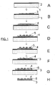
- microneedles of the invention are manufactured by means of known photolithographic methods, with the difference that in a first step,(A), depicted in Figure 1 , an aluminium layer (2) is deposited on a rigid substrate (1), in a second step (B) a negative SU8 photoresin (3) is placed on the aluminium layer (2). From this step the method continues in several known steps (C-F) for obtaining microneedles in which the SU-8 and aluminium surface are treated with a Cr/Au layer (4) which allows configuring different micro-electrode spatial patterns.
- a next layer of 1.8 ⁇ m-thick positive S1818 photoresin (5) which allows protecting the Cr/Au layer (4) is subsequently deposited resulting in electrodes in the desired areas.
- the S1818 photoresin (5) is subsequently removed and a passivation layer (6) is added in step G.
- the microneedles are removed by chemical etching which allows preventing the breakages produced when using mechanical removing means currently used for thicker needles.
- microneedles can be obtained without breaking, the characteristics of which allow them to be used in neurological procedures such as monitoring and stimulating small neuron populations in brain-machine interface devices.
Landscapes
- Health & Medical Sciences (AREA)
- Life Sciences & Earth Sciences (AREA)
- Engineering & Computer Science (AREA)
- Public Health (AREA)
- Biomedical Technology (AREA)
- Heart & Thoracic Surgery (AREA)
- Veterinary Medicine (AREA)
- Animal Behavior & Ethology (AREA)
- General Health & Medical Sciences (AREA)
- Medical Informatics (AREA)
- Physics & Mathematics (AREA)
- Surgery (AREA)
- Molecular Biology (AREA)
- Pathology (AREA)
- Biophysics (AREA)
- Nuclear Medicine, Radiotherapy & Molecular Imaging (AREA)
- Radiology & Medical Imaging (AREA)
- Neurology (AREA)
- Neurosurgery (AREA)
- Cardiology (AREA)
- Hematology (AREA)
- Anesthesiology (AREA)
- Dermatology (AREA)
- Psychology (AREA)
- Orthopedic Medicine & Surgery (AREA)
- Vascular Medicine (AREA)
- Media Introduction/Drainage Providing Device (AREA)
- Micromachines (AREA)
Abstract
Description
- The present invention relates to designing and manufacturing SU-8 microneedles having a thickness less than 100 micrometers and a length which can range from 50 micrometers to 10 centimeters, for use in monitoring and/or stimulating neurons in brain-machine interface devices.
- Different methods are used today for obtaining microneedles for use in biomedicine, the materials making them up and their manufacturing method, which determines the hardness and fragility thereof and their use in one application or another, being of great importance.
- In the case of monitoring organs such as liver or kidneys, needles with sufficient rigidity are required, so as to be able to be introduced through the hard outer layers. Therefore until now, work in obtaining needles having thicknesses of the order of several tenths of a millimeter has been done using both rigid materials such as silicon, and more flexible materials such as different polymers. Unfortunately, the neurological application of these devices is limited. Manipulating and stimulating neurons by means of microneedles is a very delicate and risky procedure not requiring high rigidity given the low mechanical strength of neural tissue. However, this application requires very thin materials to minimize damage during insertion; they must be flexible to prevent breakages during use and at the same time be sufficiently rigid to assure an appropriate insertion into the neural tissue. Until now no microneedle capable of meeting all these requirements is known.
- Today, the technology for manufacturing microneedles for neurological application almost exclusively uses silicon as the substrate. This material allows different manufacturing processes assuring the production of thin devices. This silicon ability has traditionally prevented the use of other more biocompatible and flexible materials. Thus, no manufacturing process is known today which allows using polymers for designing thin microneedles, assuring the monopoly of silicon in the field. Nevertheless, silicon microneedles are shown to be hardly suitable for use in chronic implants associated with brain-machine interface devices and they are extraordinarily fragile and have a large risk of breaking inside the brain itself once implanted.
- The polymer needles manufactured until now for use in organs such as liver or kidneys are obtained by the photolithography of a negative photoresin deposited on a rigid substrate. However, this method is not viable if one attempts to manufacture microneedles having thicknesses less than 100 micrometers, because it entails mechanically removing the already manufactured microneedle with the resulting risk of breaking. In this application a new manufacturing method for manufacturing microneedles having thicknesses less than 100 micrometers is developed using the SU-8 polymer, a photoresin widely used in microelectromechanical system applications, as the substrate
- The present invention relates to microneedles having a thickness less than 100 micrometers, manufactured using SU-8 as the structural material, for use in monitoring and/or stimulating neural tissue. The manufacturing method allows removing the microneedle from the substrate through chemical means thus preventing the use of mechanical methods and reducing the risk of breaking. This method uses a sacrificial aluminium layer for the initial coating of a rigid substrate, on which SU-8 is deposited as a negative resin. After a series of intermediate steps already known in the lithographic micro-electrode patterns manufacturing, the chemical etching of the sacrificial layer for removing the microneedle having the desired thickness is performed. This further allows manufacturing needles having a length which can be as long as 10 centimeters.
- The SU-8 needles thus manufactured have many advantages over the only technology currently available which uses silicon as the substrate. Not only do they have a greater mechanical behavior and greater biocompatibility, solving part of the problems associated with the silicon without being detrimental to their performances, but they implicitly entail the possibility of introducing further improved technologies in the interface with the nerve tissue. Due to the method described, the electrodes adhered to the SU-8 microneedles are internalized within the polymer a few micrometers in depth below the tip surface, thus facilitating the subsequent deposition of materials which optimize the recording and electrical stimulation. Recent studies have proven the advantages of depositing carbon nanotubes on the micro-electrodes for improving not only their electrical capacity, but also the interaction with the neural tissue (Keefer EW, Botterman BR, Romero MI, Rossi AF, Gross GW. Carbon nanotube coating improves neuronal recordings, Nature Nanotechnol. 2008 Jul;3(7):434-9). While in the case of the silicon microneedles, designing additional methods would be necessary to facilitate coating the electrodes, the present manufacturing method for manufacturing SU-8 microneedles produces devices ready to be treated with current deposition methods.
- A set of drawings aiding to better understand the invention which is specifically related with an embodiment of said invention which is depicted as a non-limiting example thereof is described briefly below.
-
Figure 1 shows a diagram of the manufacturing method for manufacturing the microneedles of the invention. A series of numerical references corresponding with the following elements are represented in said figure:- 1.- Rigid substrate
- 2.- Aluminium layer
- 3.- Negative SU-8 photoresin
- 4.- Cr/Au layer
- 5.- Positive photoresin
- 6.- Passivation layer
- The present invention relates to SU8 microneedles having a thickness less than 100 micrometers and a length of 50 micrometers to 10 centimeters, for use in biomedicine neurological procedures such as neuron monitoring and stimulation.
- The manufacturing method for manufacturing SU-8 microneedles is critical, since the thinness of these devices complicates their production without producing breakages with the current manufacturing methods. To that end the microneedles of the invention are manufactured by means of known photolithographic methods, with the difference that in a first step,(A), depicted in
Figure 1 , an aluminium layer (2) is deposited on a rigid substrate (1), in a second step (B) a negative SU8 photoresin (3) is placed on the aluminium layer (2). From this step the method continues in several known steps (C-F) for obtaining microneedles in which the SU-8 and aluminium surface are treated with a Cr/Au layer (4) which allows configuring different micro-electrode spatial patterns. A next layer of 1.8 µm-thick positive S1818 photoresin (5) which allows protecting the Cr/Au layer (4) is subsequently deposited resulting in electrodes in the desired areas. The S1818 photoresin (5) is subsequently removed and a passivation layer (6) is added in step G. Finally the microneedles are removed by chemical etching which allows preventing the breakages produced when using mechanical removing means currently used for thicker needles. - By means of the mentioned method, very thin microneedles can be obtained without breaking, the characteristics of which allow them to be used in neurological procedures such as monitoring and stimulating small neuron populations in brain-machine interface devices.
Claims (3)
- A microneedle for monitoring and stimulating neurons, characterized by using a negative SU8 microresin as a substrate and by having thicknesses less than 100 micrometers.
- A manufacturing method for manufacturing microneedles described in claim 1, characterized by comprising the steps of:• coating a rigid substrate (1) with an aluminium layer (2)• depositing a negative SU8 photoresin layer (3)• obtaining small, localized Cr/Au layers (4) on the negative photoresin by positive photoresin treatment• chemically etching the Al layer for chemically removing the microneedle.
- Use of microneedles obtained by the method described in claim 2 for monitoring and stimulating neurons in neurological procedures.
Applications Claiming Priority (2)
| Application Number | Priority Date | Filing Date | Title |
|---|---|---|---|
| ES200930430A ES2332399B2 (en) | 2009-07-09 | 2009-07-09 | SU-8 MICROAGUJAS FOR MONITORING AND NEURONAL STIMULATION. |
| PCT/ES2010/070472 WO2011004047A1 (en) | 2009-07-09 | 2010-07-08 | Su-8 microneedles for monitoring and stimulating neurons |
Publications (2)
| Publication Number | Publication Date |
|---|---|
| EP2452707A1 true EP2452707A1 (en) | 2012-05-16 |
| EP2452707A4 EP2452707A4 (en) | 2014-03-12 |
Family
ID=41559311
Family Applications (1)
| Application Number | Title | Priority Date | Filing Date |
|---|---|---|---|
| EP10796759.8A Withdrawn EP2452707A4 (en) | 2009-07-09 | 2010-07-08 | SU-8 MICROARRAYS FOR NEURONAL MONITORING AND NEURONAL STIMULATION |
Country Status (4)
| Country | Link |
|---|---|
| US (1) | US20120172960A1 (en) |
| EP (1) | EP2452707A4 (en) |
| ES (1) | ES2332399B2 (en) |
| WO (1) | WO2011004047A1 (en) |
Cited By (1)
| Publication number | Priority date | Publication date | Assignee | Title |
|---|---|---|---|---|
| US10064565B2 (en) | 2013-11-05 | 2018-09-04 | The Regents Of The University Of California | Multielectrode array and method of fabrication |
Families Citing this family (1)
| Publication number | Priority date | Publication date | Assignee | Title |
|---|---|---|---|---|
| TWI696448B (en) * | 2014-11-11 | 2020-06-21 | 芬蘭商腦部關懷公司 | An implantable electrode device and a method for manufacturing thereof |
Family Cites Families (2)
| Publication number | Priority date | Publication date | Assignee | Title |
|---|---|---|---|---|
| IL152271A (en) * | 2002-10-13 | 2006-04-10 | Meir Hefetz | Microneedles structures and production methods |
| US8195267B2 (en) * | 2006-01-26 | 2012-06-05 | Seymour John P | Microelectrode with laterally extending platform for reduction of tissue encapsulation |
-
2009
- 2009-07-09 ES ES200930430A patent/ES2332399B2/en not_active Expired - Fee Related
-
2010
- 2010-07-08 US US13/383,137 patent/US20120172960A1/en not_active Abandoned
- 2010-07-08 EP EP10796759.8A patent/EP2452707A4/en not_active Withdrawn
- 2010-07-08 WO PCT/ES2010/070472 patent/WO2011004047A1/en not_active Ceased
Cited By (1)
| Publication number | Priority date | Publication date | Assignee | Title |
|---|---|---|---|---|
| US10064565B2 (en) | 2013-11-05 | 2018-09-04 | The Regents Of The University Of California | Multielectrode array and method of fabrication |
Also Published As
| Publication number | Publication date |
|---|---|
| EP2452707A4 (en) | 2014-03-12 |
| WO2011004047A1 (en) | 2011-01-13 |
| US20120172960A1 (en) | 2012-07-05 |
| ES2332399B2 (en) | 2010-11-24 |
| ES2332399A1 (en) | 2010-02-03 |
Similar Documents
| Publication | Publication Date | Title |
|---|---|---|
| Szostak et al. | Neural interfaces for intracortical recording: requirements, fabrication methods, and characteristics | |
| US8738110B2 (en) | Rigid spine reinforced polymer microelectrode array probe and method of fabrication | |
| Lee et al. | Scalable thousand channel penetrating microneedle arrays on flex for multimodal and large area coverage brainmachine interfaces | |
| Wu et al. | Silk-backed structural optimization of high-density flexible intracortical neural probes | |
| CA2436207C (en) | Method of manufacturing microneedle structures using soft lithography and photolithography | |
| US9399128B2 (en) | Flexible neural interfaces with integrated stiffening shank | |
| JP5816277B2 (en) | Method for manufacturing a flexible intraocular retinal implant having a doped diamond electrode | |
| US20140107446A1 (en) | Flexible microelectrode array with integrated stiffening shank, and method of fabrication | |
| EP2968913B1 (en) | Three-dimensional multi-electrode array | |
| CN101912666A (en) | A flexible implantable neural microelectrode based on PDMS and its manufacturing method | |
| CN113574637B (en) | Method of making an electrode array for a neural prosthetic device | |
| US20250134433A1 (en) | Flexible electrode and manufacturing method therefor | |
| CN113827860A (en) | Implantable electrode device and method of making same | |
| WO2020114617A1 (en) | Hermetic packaging of electronic components | |
| KR20210099262A (en) | Carbon nanotube microelectrode applicable to artificial retina | |
| US20120172960A1 (en) | Su-8 microneedles for monitoring and stimulating neurons | |
| US11191950B2 (en) | High resolution brain-electronics interface | |
| US8504148B2 (en) | Neural device having via-hole connection and using at least one nano-wire | |
| Xu et al. | Extreme manufacturing of minimally invasive intracortical neural electrodes | |
| Pothof et al. | Fabrication and characterization of a high-resolution neural probe for stereoelectroencephalography and single neuron recording | |
| US20190382908A1 (en) | Method of producing a structure | |
| Grabiec et al. | Electrode array design and fabrication for implantable systems | |
| CN114768080B (en) | Multichannel microneedle and manufacturing method thereof | |
| KR20130022419A (en) | Microelectrode array and fabrication method thereof | |
| KR102430741B1 (en) | Carbon nanotube sensor and manufacturing method of the carbon nanotube sensor |
Legal Events
| Date | Code | Title | Description |
|---|---|---|---|
| PUAI | Public reference made under article 153(3) epc to a published international application that has entered the european phase |
Free format text: ORIGINAL CODE: 0009012 |
|
| 17P | Request for examination filed |
Effective date: 20120106 |
|
| AK | Designated contracting states |
Kind code of ref document: A1 Designated state(s): AL AT BE BG CH CY CZ DE DK EE ES FI FR GB GR HR HU IE IS IT LI LT LU LV MC MK MT NL NO PL PT RO SE SI SK SM TR |
|
| DAX | Request for extension of the european patent (deleted) | ||
| A4 | Supplementary search report drawn up and despatched |
Effective date: 20140212 |
|
| RIC1 | Information provided on ipc code assigned before grant |
Ipc: A61B 5/05 20060101ALI20140206BHEP Ipc: A61M 5/00 20060101AFI20140206BHEP |
|
| STAA | Information on the status of an ep patent application or granted ep patent |
Free format text: STATUS: THE APPLICATION IS DEEMED TO BE WITHDRAWN |
|
| 18D | Application deemed to be withdrawn |
Effective date: 20140911 |