EP2441388A1 - Medical apparatus and method for controlling the medical apparatus - Google Patents
Medical apparatus and method for controlling the medical apparatus Download PDFInfo
- Publication number
- EP2441388A1 EP2441388A1 EP11757524A EP11757524A EP2441388A1 EP 2441388 A1 EP2441388 A1 EP 2441388A1 EP 11757524 A EP11757524 A EP 11757524A EP 11757524 A EP11757524 A EP 11757524A EP 2441388 A1 EP2441388 A1 EP 2441388A1
- Authority
- EP
- European Patent Office
- Prior art keywords
- region
- blood vessel
- organ
- dimensional image
- extracts
- Prior art date
- Legal status (The legal status is an assumption and is not a legal conclusion. Google has not performed a legal analysis and makes no representation as to the accuracy of the status listed.)
- Withdrawn
Links
Images








Classifications
-
- G—PHYSICS
- G06—COMPUTING OR CALCULATING; COUNTING
- G06T—IMAGE DATA PROCESSING OR GENERATION, IN GENERAL
- G06T7/00—Image analysis
- G06T7/0002—Inspection of images, e.g. flaw detection
- G06T7/0012—Biomedical image inspection
-
- G—PHYSICS
- G06—COMPUTING OR CALCULATING; COUNTING
- G06T—IMAGE DATA PROCESSING OR GENERATION, IN GENERAL
- G06T2207/00—Indexing scheme for image analysis or image enhancement
- G06T2207/10—Image acquisition modality
- G06T2207/10072—Tomographic images
- G06T2207/10081—Computed x-ray tomography [CT]
-
- G—PHYSICS
- G06—COMPUTING OR CALCULATING; COUNTING
- G06T—IMAGE DATA PROCESSING OR GENERATION, IN GENERAL
- G06T2207/00—Indexing scheme for image analysis or image enhancement
- G06T2207/20—Special algorithmic details
- G06T2207/20036—Morphological image processing
-
- G—PHYSICS
- G06—COMPUTING OR CALCULATING; COUNTING
- G06T—IMAGE DATA PROCESSING OR GENERATION, IN GENERAL
- G06T2207/00—Indexing scheme for image analysis or image enhancement
- G06T2207/30—Subject of image; Context of image processing
- G06T2207/30004—Biomedical image processing
- G06T2207/30061—Lung
-
- G—PHYSICS
- G06—COMPUTING OR CALCULATING; COUNTING
- G06T—IMAGE DATA PROCESSING OR GENERATION, IN GENERAL
- G06T2207/00—Indexing scheme for image analysis or image enhancement
- G06T2207/30—Subject of image; Context of image processing
- G06T2207/30004—Biomedical image processing
- G06T2207/30096—Tumor; Lesion
-
- G—PHYSICS
- G06—COMPUTING OR CALCULATING; COUNTING
- G06T—IMAGE DATA PROCESSING OR GENERATION, IN GENERAL
- G06T2207/00—Indexing scheme for image analysis or image enhancement
- G06T2207/30—Subject of image; Context of image processing
- G06T2207/30004—Biomedical image processing
- G06T2207/30101—Blood vessel; Artery; Vein; Vascular
Definitions
- the present invention relates to a medical instrument that performs navigation for treatment that a surgeon performs with respect to an examinee by extracting a second region that is one part of an organ of the examinee that includes a first region that is designated by the surgeon, and a method of controlling the medical instrument.
- Japanese Patent Application Laid-Open Publication No. 2008-142481 discloses an apparatus that carries out segmentation of the pulmonary lobes into units of pulmonary segments based on a CT image.
- Japanese Patent Application Laid-Open Publication No. 2008-529641 discloses a method that separates pulmonary blood vessels into arteries and veins based on a three-dimensional image.
- An object of the present invention is to provide a medical instrument and a method of controlling the medical instrument for extracting the second region in an appropriate range including the first region.
- a medical instrument comprises: storage means hat stores three-dimensional image data of inside a body of an examinee that is previously acquired; organ extracting means that extracts a predetermined organ from the three-dimensional image data; first region designating means that designates a first region of the organ in the three-dimensional image data; blood vessel extracting means that extracts blood vessels that are inside the organ from the three-dimensional image data; blood vessel classifying means that classifies a plurality of the blood vessels that the blood vessel extracting portion extracts into either arteries or veins; and second region extracting means that extracts, on the basis of three-dimensional image information of the blood vessel of at least either one of the arteries and the veins classified by the blood vessel classifying portion, a second region which is a region that is classified based on anatomical features of the organ, and which includes the first region.
- a medical instrument control method comprises: a data storage step of storing three-dimensional image data of inside a body of an examinee that is previously acquired; an organ extraction step of extracting a predetermined organ from the three-dimensional image data; a first region designating step of designating a first region of the organ in the three-dimensional image data; a blood vessel extraction step of extracting blood vessels that are inside the organ from the three-dimensional image data; a blood vessel classifying step of classifying a plurality of the blood vessels that are extracted in the step by the blood vessel extraction means into either arteries or veins; and a second region extraction step of extracting, on the basis of three-dimensional image information of the blood vessel of at least either one of the arteries and the veins in the blood vessel classifying step, a second region which is a region that is classified based on anatomical features of the organ and which includes the first region.
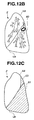
- Fig. 12C is a view that illustrates a tumor resection region for describing matching processing of the navigation apparatus according to the second embodiment.
- lungs 2 are separated into a plurality of parts based on anatomical features, in other words, based on a physiological classification. More specifically, there are two lungs, namely, a left lung and a right lung.
- the right lung is composed of, in order from a top, an upper lobe RU, a middle lobe RM, and a lower lobe RD.
- the left lung is composed of an upper lobe LU and a lower lobe LD. These five pulmonary lobes are called "major lobes”.
- the right upper lobe RU is separated into three pulmonary segments (hereunder, may also be referred to as "lobules") that are denoted by reference numerals 1, 2, and 3.
- the right middle lobe RM is separated into two pulmonary segments that are denoted by reference numerals 4 and 5.
- the right lower lobe RD is separated into five pulmonary segments that are denoted by reference numerals 6, 7, 8, 9 and 10.
- the left upper lobe LU is separated into four pulmonary segments that are denoted by reference numerals 1+2, 3, 4, and 5.
- the left lower lobe LD is separated into four pulmonary segments that are denoted by reference numerals 6, 8, 9 and 10.
- bronchi 3 are inside the lungs 2 and can not be observed from outside.
- the bronchi 3, pulmonary arteries 4, and pulmonary veins 5 are inside the lungs 2, and similarly to the bronchi 3, it is not possible to observe the pulmonary arteries 4, the pulmonary veins 5, or a tumor from outside of the lungs 2. Consequently, when resecting a tumor, it is not easy for the surgeon to decide a region that enables complete resection of the tumor.
- the navigation apparatus 1 includes an extracting unit 10, a display portion 11 as display means, and an input portion 12 as input means.
- the display portion 11 displays a processing result of the extracting unit 10.
- the input portion 12 is a keyboard, a mouse, a touch panel, or the like. The surgeon uses the input portion 12 to input information regarding the examinee, designate a position and size of a tumor based on an image of the lungs that is displayed on the display portion 11, and input an instruction for changing an observational direction of an image that is displayed on the display portion 11 or a display state of a transparent display or the like in accordance with a purpose.
- the extracting unit 10 includes a storage portion 21 as storage means, an organ extracting portion 22 as organ extracting means, a first region designating portion 23 as first region designating means, a blood vessel extracting portion 24 as blood vessel extracting means, a blood vessel classifying portion 25 as blood vessel classifying means, a second region extracting portion 26 as second region extracting means, and a template creating portion 27 as template creating means.
- a configuration may also be adopted in which, for example, a CPU 20 that is a control portion of the extracting unit 10 has functions of all of the functional portions of the extracting unit 10. More specifically, a configuration may be adopted in which the CPU 20 reads and executes a program stored in an unshown storage medium or the like to implement the respective functional portions.
- the storage portion 21 stores three-dimensional image data of inside the body of an examinee that is previously acquired.
- the organ extracting portion 22 extracts a predetermined organ, for example, the lungs and bronchi, from the three-dimensional image data stored in the storage portion 21.
- the surgeon designates a tumor as a first region in the three-dimensional image data by means of the first region designating portion 23 through the input portion 12.
- the blood vessel extracting portion 24 extracts blood vessels inside the lungs, that is, pulmonary arteries and pulmonary veins.
- the blood vessel classifying portion 25 classifies blood vessels that the blood vessel extracting portion 24 extracts into either arteries or veins.
- the template creating portion 27 creates a blood vessel region template composed of a blood vessel-related region in which a diameter of a blood vessel is increased by a predetermined amount, based on three-dimensional image information of the blood vessel.
- the second region extracting portion 26 extracts a second region (tumor resection region) that includes the first region (tumor) based on three-dimensional image information of blood vessels for at least either one of the arteries or the veins that the blood vessel classifying portion 25 has classified the blood vessels into, more specifically, based on information of the blood vessel-related region that includes the first region.
- the second region extracting portion 26 includes a region determining portion 26X as region determining means and a segment determining portion 26Y as segment determining means.
- the region determining portion 26X determines whether a tumor exists in any template based on information regarding the organ that includes positional information of a tumor that is obtained from the organ extracting portion 22 and information regarding a blood vessel-related region that is obtained from the template creating portion 27. Coordinate information is used for the respective determination processes.
- the segment determining portion 26Y determines the blood vessel-related region template in which the tumor is included, and also identifies an associated pulmonary segment based on the information regarding the veins or arteries.
- X-ray tomographic data for example, three-dimensional image data in DICOM (Digital Imaging and Communication in Medicine) format, of the examinee that is generated with a known CT apparatus (not shown) is received by the navigation apparatus 1 through a receiving portion (not shown), and is stored in the storage portion 21 that is a semiconductor storage device or a magnetic recording device or the like.
- DICOM Digital Imaging and Communication in Medicine
- the organ extracting portion 22 extracts a predetermined organ, that is, the lungs and bronchi as an organ that is the treatment target from the three-dimensional image data that is stored in the storage portion 21.
- a term "extract" refers to obtaining three-dimensional shape information of the lungs shown in Fig. 1 and the bronchi shown in Fig. 2 . Note that when data of a tumor is included in the three-dimensional image data, three-dimensional shape information of the tumor is also extracted.
- the three-dimensional shape information of the lungs, bronchi and tumor that the organ extracting portion 22 extracts is displayed on the display portion 11.
- the lungs are displayed as a transparent image.
- the surgeon designates a first region, more specifically, a size and a position of a tumor, in an image of the lungs that is being displayed on the display portion 11, by means of the first region designating portion 23 through the input portion 12.
- the first region may also be designated by CAD (Computer-Aided Detection).
- the blood vessel extracting portion 24 extracts three-dimensional shape information of blood vessels, more specifically, pulmonary arteries and pulmonary veins, inside the lungs.
- the pulmonary blood vessels form vascular networks that are composed of groups of blood vessels that branch out in stages from thicker blood vessels.
- the respective vascular networks supply oxygen and nutrition to tissue of regions that are classified according to the anatomical features thereof.
- the navigation apparatus 1 classifies the blood vessels inside the lungs that are extracted in the organ extraction step into either arteries or veins. For example, blood vessels that are adjacent to the bronchi and parallel to the bronchi are classified as pulmonary arteries, and blood vessels that are between one pulmonary artery and another pulmonary artery are classified as pulmonary veins. As shown in Fig. 2 , the fact that pulmonary arteries and pulmonary veins form a plurality of vascular networks that branch out in stages from thicker blood vessels is also utilized for classification of peripheral blood vessels. The blood vessels of the respective pulmonary veins that are branched out are associated with any one of the major lobes.
- the template creating portion 27 increases the respective blood vessel diameters by a predetermined amount to create a vein region template composed of a vein-related region in which, for example, an external diameter of the blood vessels is increased n-fold.
- n is between 2 and 10.
- a configuration may be adopted in which the proportion of increase in the blood vessel diameter is determined according to a method that adds a predetermined value. Further, if a region in which adjacent blood vessels overlap is generated when the diameters are increased, a place that is the same distance from both blood vessels is taken as a boundary.
- Fig. 5A illustrates a three-dimensional shape of veins 30.
- a vein 30A is one part of a vascular network belonging to a left upper lobe LU
- veins 30B1 and 30B2 are one part of a vascular network belonging to a left lower lobe LD that are branched from a vein 30B.
- a line in the center of each blood vessel represents a core line 50 that is obtained, for example, by joining the centers of gravity of the relevant blood vessel.
- the template creating portion 27 increases the external diameter of the veins 30A, 30B1, and 30B2, for example, threefold, to create respective vein-related regions 40A, 40B1, and 40B2.
- the template creating portion 27 may create the vein-related regions 40A, 40B 1, and 40B2 with respect to only either the right lung or the left lung in which the tumor exists or with respect to only the veins 30A, 30B1, and 30B2 in the vicinity of the tumor, or may create a vein-related region with respect to all the veins that are extracted in the organ extraction step.
- the second region extracting portion 26 determines whether or not ((Yes) or (No)) any of the vein-related regions includes the first region, that is, a tumor 60A, that is designated in the first region designating step.
- the navigation apparatus 1 enters a manual selection mode in which the surgeon selects a second region.
- the surgeon can select a tumor resection region through the input portion 12 while viewing an image of the lungs, bronchi, blood vessels or a tumor that is displayed on the display portion 11.
- a configuration may also be adopted in which, when there is no vein-related region that includes the tumor 60D, the surgeon resets the increase amount for the blood vessel diameter that is used when creating the vein region template, and the second region extracting portion 26 performs extraction once more under the new condition.
- the second region extracting portion 26 extracts the plurality of major lobes to which the respective blood vessels belong, that is, the left upper lobe LU and the left lower lobe LD, that are a basis of the vein-related regions. In other words, the second region extracting portion 26 extracts a plurality of second regions.
- the second region extracting portion 26 extracts the major lobe of the lung to which the blood vessel that is the basis of the vein-related region belongs, more specifically, the left lower lobe LD, as a second region 61A.
- the second region 61 A is a tumor resection region that is suitable for resecting the tumor 60A.
- the second region extracting portion 26 extracts the major lobe of the lung to which the blood vessel that is the basis of the vein-related region 40A belongs, more specifically, the left upper lobe LU, as a second region A.
- the second region extracting portion 26 may display reference information that is calculated according to a proportion of the tumor 60A included in the vein-related region 40B2 on the display portion 11. For example, if the entire tumor 60A is included in the vein-related region 40B2, the reference information is "100%", while if a portion of the tumor 60A (for example, 30%) is included in the vein-related region 40B2 the reference information is "30%".
- a configuration may be adopted such that the navigation apparatus 1 can be set so that the second region extracting portion 26 does not extract a second region if the proportion of the tumor 60A included in the vein-related region 40B2 is less than or equal to a predetermined value, for example, less than or equal to 5%.
- the surgeon can appropriately set the predetermined value through the input portion 12.
- the surgeon can appropriately set a blood vessel diameter increase amount of a vein region template that the template creating portion 27 creates, through the input portion 12.
- a tumor resection region (second region) that the second region extracting portion 26 extracted is displayed on the display portion 11 in a color or the like that makes the tumor resection region (second region) distinguishable from other regions.
- the navigation apparatus 1 of the present embodiment can extract an appropriate tumor resection region that includes a tumor 60.
- the template creating portion 27 creates a vein region template composed of a vein-related region in which a blood vessel diameter is increased by a predetermined amount.
- a configuration may be adopted in which, based on core lines 50A, 50B1, and 50B2, the template creating portion 27 creates vein-related regions 41A, 41B1, and 41B2 of a predetermined diameter that are centered on core lines, and the second region extracting portion 26 extracts a second region based on a relationship between the vein-related regions 41A, 41B1, and 41B2 and a tumor 60E.
- an extraction method of the second region extracting portion 26 may be a method that calculates distances d1 and d2 from the core lines 50A and 50B2 to a tumor 60F, and extracts a major lobe to which the blood vessel that is the basis of a core line that is a shortest distance from the tumor 60F belongs as the second region.
- a configuration may be adopted in which the template creating portion creates a tumor region template composed of a tumor-related region 60G1 in which an outer shape (contour) of a tumor 60G is increased by a predetermined amount, for example, the outer shape (contour) of the tumor 60G is increased n-fold, and the second region extracting portion extracts the second region based on a relationship between the tumor-related region 60G1 and the veins 30A, 30B1, and 30B2.
- a configuration may be adopted in which the second region extracting portion extracts the second region based on a relationship between the core lines 50A, 50B1, and 50B2 and a tumor region template (tumor-related region) 60H in which an outer shape (contour) of a tumor 60H is increased by a predetermined amount.
- the second region extracting portion 26 of the navigation apparatus 1 extracts a second region based on a distance relationship between a three-dimensional image of venous blood vessels and the first region. More specifically, the second region extracting portion 26 extracts one of the major lobes as a second region based on information regarding a vein that has been classified by the blood vessel classifying portion 25 that is a shortest distance from the first region.
- a navigation apparatus 1A as a medical instrument is described. Since the navigation apparatus 1A is similar to the navigation apparatus 1 of the first embodiment, components that are the same as in the navigation apparatus 1 of the first embodiment are denoted by the same reference symbols and a description of such components is omitted.
- the tumor can be completely resected by, for example, resecting only one of pulmonary segments 1 to 10 that constitute the major lobes or the like.
- a second region extracting portion 26A of the navigation apparatus 1A extracts a pulmonary segment, which is a smaller region, as the second region from one of the major lobes that has been initially extracted.
- the second region extracting portion 26 extracts one of the major lobes as a second region based on a vein-related region.
- the second region extracting portion 26 of the navigation apparatus 1A limits extraction of a second region to the pulmonary segments 1 to 10 that are a part of the major lobes based on an artery-related region.
- the second region extracting portion 26A extracts one of the major lobes, for example, the left upper lobe LU, as a tumor resection region (second region A) 61 A based on a vein-related region.
- a template creating portion 27A Based on three-dimensional image information of the pulmonary arteries, a template creating portion 27A creates a pulmonary artery region template composed of a pulmonary artery-related region in which respective blood vessel diameters are increased by a predetermined amount, for example, in which external diameters are increased n-fold.
- the template creating portion 27A may create an artery-related region for only part of the pulmonary arteries.
- the second region extracting portion 26A determines whether or not ((Yes) or (No)) any of the artery-related regions includes the first region, that is, the tumor 60A, that is designated in the first region designating step.
- the navigation apparatus 1A enters a manual selection mode that allows the surgeon to select a second region.
- the second region extracting portion 26A may extract a single pulmonary segment as the second region using various methods, and may perform extraction once again after an extraction condition is changed.
- the second region extracting portion 26A extracts a plurality of pulmonary segments to which respective blood vessels that are a basis of the artery-related regions belong. In other words, the second region extracting portion 26A extracts a plurality of second regions 62.
- the second region extracting portion 26 extracts, as a second region 62A, the pulmonary segment to which the blood vessel that is a basis of the artery-related region belongs.
- the second region 62A is a part of the second region 61A, and is a tumor resection region that is more suitable in order to resect the tumor 60A.
- the tumor resection region (second region) that the second region extracting portion 26A extracts is displayed on the display portion 11.
- the navigation apparatus 1A according to the present modification example has the advantageous effects of the navigation apparatus 1A, and can also extract the second region (tumor resection region) 62 of a narrower range that includes the tumor 60A.
- a navigation apparatus 1B as a medical instrument is described. Since the navigation apparatus 1B is similar to the navigation apparatus 1 of the first embodiment, components that are the same as in the navigation apparatus 1 of the first embodiment are denoted by the same reference symbols and a description of such components is omitted.
- the navigation apparatus 1B includes a standard template storage portion 28 as standard template storage means and a matching processing portion 29 as matching processing means.
- a second region extracting portion 26B extracts a second region based on a first region that is associated with three-dimensional image information, and information of an organ according to a standard template or the like.
- each functional portion of an extracting unit 10B of the navigation apparatus 1B may be a program that a CPU 20B reads and executes to perform relevant operations.
- the standard template storage portion 28 stores a standard template that is composed of three-dimensional shape information of standard organs and blood vessels inside the organs and the like that is based on, at least, a gender, a body type, and an age of the examinee.
- the matching processing portion 29 performs matching processing between the standard template and the three-dimensional shape information of the examinee.
- X-ray tomographic data for example, three-dimensional image data in DICOM (Digital Imaging and Communication in Medicine) format, of the examinee that is generated with an known CT apparatus (not shown) is received by the navigation apparatus 1A through an unshown receiving portion, and is stored in the storage portion 21 that is a semiconductor storage device or a magnetic recording device or the like.
- DICOM Digital Imaging and Communication in Medicine
- the organ extracting portion 22 extracts a predetermined organ, for example, the lungs 2 and bronchi 3 from the three-dimensional image data that is stored in the storage portion 21. If data of a tumor is included in the three-dimensional image data, three-dimensional shape information of the tumor 60 is also extracted.
- the surgeon designates a first region, more specifically, a position and a size of a tumor, in an image of the lungs that is being displayed on the display portion 11, by means of the first region designating portion 23 through the input portion 12.
- the first region may also be designated by CAD (Computer-Aided Detection).
- the blood vessel extracting portion 24 extracts three-dimensional shape information of blood vessels, more specifically, pulmonary arteries 4 and pulmonary veins 5, inside the lungs.
- the navigation apparatus 1B classifies the blood vessels inside the lungs that are extracted in the organ extraction step into either pulmonary arteries 4 or pulmonary veins 5.
- the surgeon inputs information showing at least the gender, the body type, and the age of the examinee through the input portion 12.
- information that is inputted may include body weight, height, chest measurement and the like of the examinee.
- a standard template 70 composed of three-dimensional shape information of a standard organ and blood vessels inside the organ that is based on the information regarding the examinee is stored in the standard template storage portion 28.
- the standard template 70 may be selected from among a large number of templates that are stored in a template storage portion 28A, or may be created on a basis of inputted information, or may be a template obtained by further modifying a selected template.
- the standard template also includes boundary information regarding the major lobes/pulmonary segments.
- the matching processing portion 29 performs matching processing between the standard template 70 and the three-dimensional shape information of the examinee.
- the matching processing portion 29 performs at least either one of matching processing between three-dimensional shape information of the blood vessels of the standard template 70 and three-dimensional image information of the blood vessels extracted by the blood vessel extracting portion 24, and matching processing between three-dimensional shape information of the organ of the standard template and three-dimensional image information of the organ that is extracted by the organ extracting means.
- the matching processing portion 29 performs matching processing that takes at least any one of “bronchi (organ) 3 ", “bronchi (organ) 3 + pulmonary blood vessels 4 and 5", “pulmonary blood vessels 4 and 5", “pulmonary arteries 4", and “pulmonary veins 5" as a target.
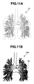
- Fig. 11A illustrates the standard template 70
- Fig. 11B illustrates three-dimensional shape information 70X of the examinee
- Fig. 11C illustrates a state in which matching processing is performed between the standard template 70 and the three-dimensional shape information 70X of the examinee.
- matching processing may be performed that takes the pulmonary blood vessels 4 and 5 as a target.
- matching processing that takes at least "blood vessels 4 and 5" as a target is performed.
- the matching processing may be performed by known image processing, and may use either matching at a pixel data level or matching at a level of features extracted from an image.
- the matching processing portion 29 may change the selected standard template to a different standard template and perform the matching processing once more, or may switch to a manual selection mode (S51).
- a term "error e between the images” refers to, for example, when the bronchi 3 are the target of the matching processing as shown in Fig. 11C , a total value of positional deviations at a peripheral plurality of bronchial branch points when matching processing is performed using a tracheal core line 6A at the carina 3A at which the trachea 6 is connected to the bronchi 3.
- Step S48 Second region extraction step.
- the second region extracting portion 26B judges whether a tumor 60J of the examinee after the matching processing is included in any region of the lungs in the standard template 70.
- a term "region" refers to a major lobe of the lung or a pulmonary segment.
- the matching processing step and the second region extraction step may be repeatedly performed. More specifically, after performing a first matching processing operation that takes "pulmonary veins 5" as a target and extracting one of the major lobes, a second matching processing operation that takes the "pulmonary arteries 4" as a target may be performed to extract a single pulmonary segment inside the extracted major lobe.
- the navigation apparatus 1 enters a manual selection mode that allows the surgeon to select the second region.
- the second region extracting portion 26 extracts the plurality of regions. In other words, the second region extracting portion 26B extracts a plurality of second regions.
- the second region extracting portion 26 extracts that region.
- a tumor resection region (second region) that the second region extracting portion 26B extracts is displayed on the display portion 11.
- the navigation apparatus 1B of the present embodiment has the same advantageous effects as those of the navigation apparatus 1 of the first embodiment or the like.
- a state in which the blood vessels of an examinee are arranged or the like may differ significantly from the standard template due to individual differences.
- an end of a lower lobar vein that remains inside the left lower lobe LD in the standard template that is shown in Fig. 11A extends as far as the left upper lobe LU as shown in Fig. 11B .
- the second region extracting portion 26B extracts a third region 63 that belongs to the same vascular network, as a tumor resection additional region.
- the third region 63 that is shown in Fig. 11C is a pulmonary segment 2 (LU2) of the left upper lobe LU.
- an image that is displayed on the display portion 11 calls a surgeon's attention to the third region 63 by displaying the third region 63 using hatching or a display color or the like that is different from that of a second region 61.
- the navigation apparatus 1B may display a display that is designed to call the attention of the surgeon to the difference.
- navigation can also be performed by a similar method for another organ, for example, a liver. More specifically, when performing a resection of the liver, any of a sub-regional resection, a regional resection, a lobectomy, and an extended right lobectomy is performed, and the navigation apparatus can extract an appropriate tumor resection region and display the extracted tumor resection region to the surgeon.
Landscapes
- Engineering & Computer Science (AREA)
- Quality & Reliability (AREA)
- General Health & Medical Sciences (AREA)
- Medical Informatics (AREA)
- Nuclear Medicine, Radiotherapy & Molecular Imaging (AREA)
- Radiology & Medical Imaging (AREA)
- Health & Medical Sciences (AREA)
- Computer Vision & Pattern Recognition (AREA)
- Physics & Mathematics (AREA)
- General Physics & Mathematics (AREA)
- Theoretical Computer Science (AREA)
- Apparatus For Radiation Diagnosis (AREA)
- Magnetic Resonance Imaging Apparatus (AREA)
- Measuring And Recording Apparatus For Diagnosis (AREA)
Abstract
Description
- The present invention relates to a medical instrument that performs navigation for treatment that a surgeon performs with respect to an examinee by extracting a second region that is one part of an organ of the examinee that includes a first region that is designated by the surgeon, and a method of controlling the medical instrument.
- Conventionally, when resecting a tumor of a lung of an examinee such as, for example, when resecting lung cancer tissue, a lobectomy in which an entire right lobe or left lobe is resected has been widely performed. In this regard, in recent years, to preserve postoperative pulmonary function and maintain cardiopulmonary function, resection of a major lobe of a part of one lung or resection of a pulmonary segment of a part of a major lobe has been performed with respect to localized tumors.
- In this regard, Japanese Patent Application Laid-Open Publication No.
2008-142481 - Further, Japanese Patent Application Laid-Open Publication No.
2008-529641 - However, there have been cases where it is not easy for a surgeon to determine, from the first region (tumor region), the second region (tumor resection region) in an appropriate range including the first region.
- An object of the present invention is to provide a medical instrument and a method of controlling the medical instrument for extracting the second region in an appropriate range including the first region.
- A medical instrument according to one embodiment of the present invention comprises: storage means hat stores three-dimensional image data of inside a body of an examinee that is previously acquired; organ extracting means that extracts a predetermined organ from the three-dimensional image data; first region designating means that designates a first region of the organ in the three-dimensional image data; blood vessel extracting means that extracts blood vessels that are inside the organ from the three-dimensional image data; blood vessel classifying means that classifies a plurality of the blood vessels that the blood vessel extracting portion extracts into either arteries or veins; and second region extracting means that extracts, on the basis of three-dimensional image information of the blood vessel of at least either one of the arteries and the veins classified by the blood vessel classifying portion, a second region which is a region that is classified based on anatomical features of the organ, and which includes the first region.
- A medical instrument control method according to another embodiment of the present invention comprises: a data storage step of storing three-dimensional image data of inside a body of an examinee that is previously acquired; an organ extraction step of extracting a predetermined organ from the three-dimensional image data; a first region designating step of designating a first region of the organ in the three-dimensional image data; a blood vessel extraction step of extracting blood vessels that are inside the organ from the three-dimensional image data; a blood vessel classifying step of classifying a plurality of the blood vessels that are extracted in the step by the blood vessel extraction means into either arteries or veins; and a second region extraction step of extracting, on the basis of three-dimensional image information of the blood vessel of at least either one of the arteries and the veins in the blood vessel classifying step, a second region which is a region that is classified based on anatomical features of the organ and which includes the first region.
-
-
Fig. 1A is a view of lungs as observed from a front for describing regions into which the lungs are classified based on anatomical features thereof; -
Fig. 1B is a view of lungs as observed from a rear for describing regions into which the lungs are classified based on the anatomical features thereof; -
Fig. 2 is a view for describing blood vessels and bronchi of the lungs; -
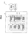
Fig. 3 is a configuration diagram for describing a configuration of a navigation apparatus according to a first embodiment; -
Fig. 4 is a flowchart for describing operations of the navigation apparatus according to the first embodiment; -
Fig. 5A is an explanatory diagram for describing operations of the navigation apparatus according to the first embodiment; -
Fig. 5B is an explanatory diagram for describing operations of the navigation apparatus according to the first embodiment; -
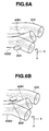
Fig. 6A is an explanatory diagram for describing operations of the navigation apparatus according to the first embodiment; -
Fig. 6B is an explanatory diagram for describing operations of the navigation apparatus according to the first embodiment; -
Fig. 6C is an explanatory diagram for describing operations of the navigation apparatus according to the first embodiment; -
Fig. 6D is an explanatory diagram for describing operations of the navigation apparatus according to the first embodiment; -
Fig. 7A is an explanatory diagram for describing operations of the navigation apparatus according to the first embodiment; -
Fig. 7B is an explanatory diagram for describing operations of the navigation apparatus according to the first embodiment; -
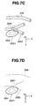
Fig. 7C is an explanatory diagram for describing operations of the navigation apparatus according to the first embodiment; -
Fig. 7D is an explanatory diagram for describing operations of the navigation apparatus according to the first embodiment; -
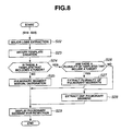
Fig. 8 is a flowchart for describing operations of a navigation apparatus according to a modification example of the first embodiment; -
Fig. 9 is a configuration diagram for describing a configuration of a navigation apparatus according to a second embodiment; -
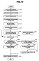
Fig. 10 is a flowchart for describing operations of the navigation apparatus according to the second embodiment; -
Fig. 11A is a view that illustrates a standard template for describing matching processing of the navigation apparatus according to the second embodiment; -
Fig. 11B is a view that illustrates a three-dimensional image of an examinee for describing matching processing of the navigation apparatus according to the second embodiment; -
Fig. 11C is a view that illustrates a matching processing state for describing matching processing of the navigation apparatus according to the second embodiment; -
Fig. 12A is a view that illustrates a standard template for describing matching processing of the navigation apparatus according to the second embodiment; -
Fig. 12B is a view that illustrates a three-dimensional image of an examinee for describing matching processing of the navigation apparatus according to the -
Fig. 12C is a view that illustrates a tumor resection region for describing matching processing of the navigation apparatus according to the second embodiment. - Hereunder, a
navigation apparatus 1 for pulmonary tumor resection as a medical instrument according to a first embodiment of the present invention is described with reference to the drawings. As shown inFig. 1A and Fig. 1B ,lungs 2 are separated into a plurality of parts based on anatomical features, in other words, based on a physiological classification. More specifically, there are two lungs, namely, a left lung and a right lung. The right lung is composed of, in order from a top, an upper lobe RU, a middle lobe RM, and a lower lobe RD. The left lung is composed of an upper lobe LU and a lower lobe LD. These five pulmonary lobes are called "major lobes". The right upper lobe RU is separated into three pulmonary segments (hereunder, may also be referred to as "lobules") that are denoted byreference numerals reference numerals reference numerals reference numerals 1+2, 3, 4, and 5. The left lower lobe LD is separated into four pulmonary segments that are denoted byreference numerals - In this connection, as shown in
Fig. 1 , other than an area in a vicinity of acarina 3A that connects from atrachea 6,bronchi 3 are inside thelungs 2 and can not be observed from outside. - As shown in
Fig. 2 , thebronchi 3,pulmonary arteries 4, andpulmonary veins 5 are inside thelungs 2, and similarly to thebronchi 3, it is not possible to observe thepulmonary arteries 4, thepulmonary veins 5, or a tumor from outside of thelungs 2. Consequently, when resecting a tumor, it is not easy for the surgeon to decide a region that enables complete resection of the tumor. - As shown in
Fig. 3 , thenavigation apparatus 1 includes an extractingunit 10, adisplay portion 11 as display means, and aninput portion 12 as input means. Thedisplay portion 11 displays a processing result of the extractingunit 10. Theinput portion 12 is a keyboard, a mouse, a touch panel, or the like. The surgeon uses theinput portion 12 to input information regarding the examinee, designate a position and size of a tumor based on an image of the lungs that is displayed on thedisplay portion 11, and input an instruction for changing an observational direction of an image that is displayed on thedisplay portion 11 or a display state of a transparent display or the like in accordance with a purpose. - The extracting
unit 10 includes astorage portion 21 as storage means, anorgan extracting portion 22 as organ extracting means, a firstregion designating portion 23 as first region designating means, a bloodvessel extracting portion 24 as blood vessel extracting means, a bloodvessel classifying portion 25 as blood vessel classifying means, a secondregion extracting portion 26 as second region extracting means, and atemplate creating portion 27 as template creating means. Although each of the functional portions of the extractingunit 10 may be constituted by respectively independent hardware, a configuration may also be adopted in which, for example, aCPU 20 that is a control portion of the extractingunit 10 has functions of all of the functional portions of the extractingunit 10. More specifically, a configuration may be adopted in which theCPU 20 reads and executes a program stored in an unshown storage medium or the like to implement the respective functional portions. - The
storage portion 21 stores three-dimensional image data of inside the body of an examinee that is previously acquired. Theorgan extracting portion 22 extracts a predetermined organ, for example, the lungs and bronchi, from the three-dimensional image data stored in thestorage portion 21. The surgeon designates a tumor as a first region in the three-dimensional image data by means of the firstregion designating portion 23 through theinput portion 12. The bloodvessel extracting portion 24 extracts blood vessels inside the lungs, that is, pulmonary arteries and pulmonary veins. The bloodvessel classifying portion 25 classifies blood vessels that the bloodvessel extracting portion 24 extracts into either arteries or veins. Thetemplate creating portion 27 creates a blood vessel region template composed of a blood vessel-related region in which a diameter of a blood vessel is increased by a predetermined amount, based on three-dimensional image information of the blood vessel. - The second
region extracting portion 26 extracts a second region (tumor resection region) that includes the first region (tumor) based on three-dimensional image information of blood vessels for at least either one of the arteries or the veins that the bloodvessel classifying portion 25 has classified the blood vessels into, more specifically, based on information of the blood vessel-related region that includes the first region. - More specifically, the second
region extracting portion 26 includes aregion determining portion 26X as region determining means and asegment determining portion 26Y as segment determining means. Theregion determining portion 26X determines whether a tumor exists in any template based on information regarding the organ that includes positional information of a tumor that is obtained from theorgan extracting portion 22 and information regarding a blood vessel-related region that is obtained from thetemplate creating portion 27. Coordinate information is used for the respective determination processes. - If a tumor exists in any of the templates, the
segment determining portion 26Y determines the blood vessel-related region template in which the tumor is included, and also identifies an associated pulmonary segment based on the information regarding the veins or arteries. - Hereunder, operations of the
navigation apparatus 1 are described according to the flowchart shown inFig. 4 . - X-ray tomographic data, for example, three-dimensional image data in DICOM (Digital Imaging and Communication in Medicine) format, of the examinee that is generated with a known CT apparatus (not shown) is received by the
navigation apparatus 1 through a receiving portion (not shown), and is stored in thestorage portion 21 that is a semiconductor storage device or a magnetic recording device or the like. - The
organ extracting portion 22 extracts a predetermined organ, that is, the lungs and bronchi as an organ that is the treatment target from the three-dimensional image data that is stored in thestorage portion 21. Here, a term "extract" refers to obtaining three-dimensional shape information of the lungs shown inFig. 1 and the bronchi shown inFig. 2 . Note that when data of a tumor is included in the three-dimensional image data, three-dimensional shape information of the tumor is also extracted. - The three-dimensional shape information of the lungs, bronchi and tumor that the
organ extracting portion 22 extracts is displayed on thedisplay portion 11. At this time, in order to clearly show the positions of the bronchi and tumor or the like, preferably the lungs are displayed as a transparent image. - For example, the surgeon designates a first region, more specifically, a size and a position of a tumor, in an image of the lungs that is being displayed on the
display portion 11, by means of the firstregion designating portion 23 through theinput portion 12. The first region may also be designated by CAD (Computer-Aided Detection). - The blood
vessel extracting portion 24 extracts three-dimensional shape information of blood vessels, more specifically, pulmonary arteries and pulmonary veins, inside the lungs. As shown inFig. 2 , the pulmonary blood vessels form vascular networks that are composed of groups of blood vessels that branch out in stages from thicker blood vessels. The respective vascular networks supply oxygen and nutrition to tissue of regions that are classified according to the anatomical features thereof. - The
navigation apparatus 1 classifies the blood vessels inside the lungs that are extracted in the organ extraction step into either arteries or veins. For example, blood vessels that are adjacent to the bronchi and parallel to the bronchi are classified as pulmonary arteries, and blood vessels that are between one pulmonary artery and another pulmonary artery are classified as pulmonary veins. As shown inFig. 2 , the fact that pulmonary arteries and pulmonary veins form a plurality of vascular networks that branch out in stages from thicker blood vessels is also utilized for classification of peripheral blood vessels. The blood vessels of the respective pulmonary veins that are branched out are associated with any one of the major lobes. - Based on the three-dimensional image information of the veins, the
template creating portion 27 increases the respective blood vessel diameters by a predetermined amount to create a vein region template composed of a vein-related region in which, for example, an external diameter of the blood vessels is increased n-fold. Here, for example, n is between 2 and 10. In this connection, a configuration may be adopted in which the proportion of increase in the blood vessel diameter is determined according to a method that adds a predetermined value. Further, if a region in which adjacent blood vessels overlap is generated when the diameters are increased, a place that is the same distance from both blood vessels is taken as a boundary. - For example,
Fig. 5A illustrates a three-dimensional shape ofveins 30. InFig. 5A , avein 30A is one part of a vascular network belonging to a left upper lobe LU, and veins 30B1 and 30B2 are one part of a vascular network belonging to a left lower lobe LD that are branched from avein 30B. A line in the center of each blood vessel represents acore line 50 that is obtained, for example, by joining the centers of gravity of the relevant blood vessel. - As shown in
Fig. 5B , thetemplate creating portion 27 increases the external diameter of theveins 30A, 30B1, and 30B2, for example, threefold, to create respective vein-relatedregions 40A, 40B1, and 40B2. - In this connection, the
template creating portion 27 may create the vein-relatedregions 40A,40B 1, and 40B2 with respect to only either the right lung or the left lung in which the tumor exists or with respect to only theveins 30A, 30B1, and 30B2 in the vicinity of the tumor, or may create a vein-related region with respect to all the veins that are extracted in the organ extraction step. - The second
region extracting portion 26 determines whether or not ((Yes) or (No)) any of the vein-related regions includes the first region, that is, atumor 60A, that is designated in the first region designating step. - As shown in
Fig. 6D , if there is no vein-related region that includes atumor 60D, thenavigation apparatus 1 enters a manual selection mode in which the surgeon selects a second region. The surgeon can select a tumor resection region through theinput portion 12 while viewing an image of the lungs, bronchi, blood vessels or a tumor that is displayed on thedisplay portion 11. - In this connection, a configuration may also be adopted in which, when there is no vein-related region that includes the
tumor 60D, the surgeon resets the increase amount for the blood vessel diameter that is used when creating the vein region template, and the secondregion extracting portion 26 performs extraction once more under the new condition. - As shown in
Fig. 6C , when atumor 60C (first region) is included in a plurality of the vein-relatedregions 40A and 40B1, the secondregion extracting portion 26 extracts the plurality of major lobes to which the respective blood vessels belong, that is, the left upper lobe LU and the left lower lobe LD, that are a basis of the vein-related regions. In other words, the secondregion extracting portion 26 extracts a plurality of second regions. - As shown in
Fig. 6A , when there is a single vein-related region 40B2 that includes thetumor 60A, the secondregion extracting portion 26 extracts the major lobe of the lung to which the blood vessel that is the basis of the vein-related region belongs, more specifically, the left lower lobe LD, as a second region 61A. The second region 61 A is a tumor resection region that is suitable for resecting thetumor 60A. - Further, for example, as shown in
Fig. 6B , when the vein-relatedregion 40A includes atumor 60B, the secondregion extracting portion 26 extracts the major lobe of the lung to which the blood vessel that is the basis of the vein-relatedregion 40A belongs, more specifically, the left upper lobe LU, as a second region A. - In this connection, the second
region extracting portion 26 may display reference information that is calculated according to a proportion of thetumor 60A included in the vein-related region 40B2 on thedisplay portion 11. For example, if theentire tumor 60A is included in the vein-related region 40B2, the reference information is "100%", while if a portion of thetumor 60A (for example, 30%) is included in the vein-related region 40B2 the reference information is "30%". - Furthermore, a configuration may be adopted such that the
navigation apparatus 1 can be set so that the secondregion extracting portion 26 does not extract a second region if the proportion of thetumor 60A included in the vein-related region 40B2 is less than or equal to a predetermined value, for example, less than or equal to 5%. Preferably, the surgeon can appropriately set the predetermined value through theinput portion 12. Similarly, preferably the surgeon can appropriately set a blood vessel diameter increase amount of a vein region template that thetemplate creating portion 27 creates, through theinput portion 12. - A tumor resection region (second region) that the second
region extracting portion 26 extracted is displayed on thedisplay portion 11 in a color or the like that makes the tumor resection region (second region) distinguishable from other regions. - As described above, the
navigation apparatus 1 of the present embodiment can extract an appropriate tumor resection region that includes atumor 60. - In this connection, according to the above description, the
template creating portion 27 creates a vein region template composed of a vein-related region in which a blood vessel diameter is increased by a predetermined amount. However, as shown inFig. 7A , a configuration may be adopted in which, based oncore lines 50A, 50B1, and 50B2, thetemplate creating portion 27 creates vein-relatedregions 41A, 41B1, and 41B2 of a predetermined diameter that are centered on core lines, and the secondregion extracting portion 26 extracts a second region based on a relationship between the vein-relatedregions 41A, 41B1, and 41B2 and atumor 60E. - Further, as shown in
Fig. 7B , an extraction method of the secondregion extracting portion 26 may be a method that calculates distances d1 and d2 from thecore lines 50A and 50B2 to atumor 60F, and extracts a major lobe to which the blood vessel that is the basis of a core line that is a shortest distance from thetumor 60F belongs as the second region. - Further, as shown in
Fig. 7C , a configuration may be adopted in which the template creating portion creates a tumor region template composed of a tumor-related region 60G1 in which an outer shape (contour) of atumor 60G is increased by a predetermined amount, for example, the outer shape (contour) of thetumor 60G is increased n-fold, and the second region extracting portion extracts the second region based on a relationship between the tumor-related region 60G1 and theveins 30A, 30B1, and 30B2. - Furthermore, as shown in
Fig. 7D , a configuration may be adopted in which the second region extracting portion extracts the second region based on a relationship between thecore lines 50A, 50B1, and 50B2 and a tumor region template (tumor-related region) 60H in which an outer shape (contour) of atumor 60H is increased by a predetermined amount. - As described above, the second
region extracting portion 26 of thenavigation apparatus 1 extracts a second region based on a distance relationship between a three-dimensional image of venous blood vessels and the first region. More specifically, the secondregion extracting portion 26 extracts one of the major lobes as a second region based on information regarding a vein that has been classified by the bloodvessel classifying portion 25 that is a shortest distance from the first region. - Next, a
navigation apparatus 1A as a medical instrument according to a modification example of the first embodiment of the present invention is described. Since thenavigation apparatus 1A is similar to thenavigation apparatus 1 of the first embodiment, components that are the same as in thenavigation apparatus 1 of the first embodiment are denoted by the same reference symbols and a description of such components is omitted. - In a case of a tumor that is localized to a pulmonary segment, the tumor can be completely resected by, for example, resecting only one of
pulmonary segments 1 to 10 that constitute the major lobes or the like. In this case, in accordance with an instruction of the surgeon or the like, a secondregion extracting portion 26A of thenavigation apparatus 1A extracts a pulmonary segment, which is a smaller region, as the second region from one of the major lobes that has been initially extracted. - Hereunder, operations of the
navigation apparatus 1A are described according to the flowchart shown inFig. 8 . Note that a description of the operations from S10 to S20 is omitted because the operations are the same as those of thenavigation apparatus 1 shown inFig. 4 . - According to the
navigation apparatus 1 of the first embodiment, the secondregion extracting portion 26 extracts one of the major lobes as a second region based on a vein-related region. In contrast, the secondregion extracting portion 26 of thenavigation apparatus 1A limits extraction of a second region to thepulmonary segments 1 to 10 that are a part of the major lobes based on an artery-related region. - More specifically, by performing the operations from
S 10 to S20, the secondregion extracting portion 26A extracts one of the major lobes, for example, the left upper lobe LU, as a tumor resection region (second region A) 61 A based on a vein-related region. - Based on three-dimensional image information of the pulmonary arteries, a
template creating portion 27A creates a pulmonary artery region template composed of a pulmonary artery-related region in which respective blood vessel diameters are increased by a predetermined amount, for example, in which external diameters are increased n-fold. In this connection, similarly to the above described vein-related region, thetemplate creating portion 27A may create an artery-related region for only part of the pulmonary arteries. - The second
region extracting portion 26A determines whether or not ((Yes) or (No)) any of the artery-related regions includes the first region, that is, thetumor 60A, that is designated in the first region designating step. - If there is no artery-related region that includes a tumor (S24 = No), the
navigation apparatus 1A enters a manual selection mode that allows the surgeon to select a second region. - In this connection, similarly to when extracting a major lobe, the second
region extracting portion 26A may extract a single pulmonary segment as the second region using various methods, and may perform extraction once again after an extraction condition is changed. - If a tumor (first region) is included in a plurality of artery-related regions (Yes), the second
region extracting portion 26A extracts a plurality of pulmonary segments to which respective blood vessels that are a basis of the artery-related regions belong. In other words, the secondregion extracting portion 26A extracts a plurality of second regions 62. - If there is not a plurality of artery-related regions that include the
tumor 60A (S26 = No), the secondregion extracting portion 26 extracts, as a second region 62A, the pulmonary segment to which the blood vessel that is a basis of the artery-related region belongs. The second region 62A is a part of the second region 61A, and is a tumor resection region that is more suitable in order to resect thetumor 60A. - The tumor resection region (second region) that the second
region extracting portion 26A extracts is displayed on thedisplay portion 11. - As described above, the
navigation apparatus 1A according to the present modification example has the advantageous effects of thenavigation apparatus 1A, and can also extract the second region (tumor resection region) 62 of a narrower range that includes thetumor 60A. - Next, a
navigation apparatus 1B as a medical instrument according to a second embodiment of the present invention is described. Since thenavigation apparatus 1B is similar to thenavigation apparatus 1 of the first embodiment, components that are the same as in thenavigation apparatus 1 of the first embodiment are denoted by the same reference symbols and a description of such components is omitted. - As shown in
Fig. 9 , thenavigation apparatus 1B includes a standardtemplate storage portion 28 as standard template storage means and amatching processing portion 29 as matching processing means. A secondregion extracting portion 26B extracts a second region based on a first region that is associated with three-dimensional image information, and information of an organ according to a standard template or the like. Similarly to thenavigation apparatus 1, each functional portion of an extractingunit 10B of thenavigation apparatus 1B may be a program that aCPU 20B reads and executes to perform relevant operations. - The standard
template storage portion 28 stores a standard template that is composed of three-dimensional shape information of standard organs and blood vessels inside the organs and the like that is based on, at least, a gender, a body type, and an age of the examinee. The matchingprocessing portion 29 performs matching processing between the standard template and the three-dimensional shape information of the examinee. - Hereunder, operations of the
navigation apparatus 1B are described according to the flowchart shown inFig. 10 . - X-ray tomographic data, for example, three-dimensional image data in DICOM (Digital Imaging and Communication in Medicine) format, of the examinee that is generated with an known CT apparatus (not shown) is received by the
navigation apparatus 1A through an unshown receiving portion, and is stored in thestorage portion 21 that is a semiconductor storage device or a magnetic recording device or the like. - The
organ extracting portion 22 extracts a predetermined organ, for example, thelungs 2 andbronchi 3 from the three-dimensional image data that is stored in thestorage portion 21. If data of a tumor is included in the three-dimensional image data, three-dimensional shape information of thetumor 60 is also extracted. - For example, the surgeon designates a first region, more specifically, a position and a size of a tumor, in an image of the lungs that is being displayed on the
display portion 11, by means of the firstregion designating portion 23 through theinput portion 12. The first region may also be designated by CAD (Computer-Aided Detection). - The blood
vessel extracting portion 24 extracts three-dimensional shape information of blood vessels, more specifically,pulmonary arteries 4 andpulmonary veins 5, inside the lungs. - The
navigation apparatus 1B classifies the blood vessels inside the lungs that are extracted in the organ extraction step into eitherpulmonary arteries 4 orpulmonary veins 5. - The surgeon inputs information showing at least the gender, the body type, and the age of the examinee through the
input portion 12. In addition to the gender, body type, and age, information that is inputted may include body weight, height, chest measurement and the like of the examinee. - A
standard template 70 composed of three-dimensional shape information of a standard organ and blood vessels inside the organ that is based on the information regarding the examinee is stored in the standardtemplate storage portion 28. - The
standard template 70 may be selected from among a large number of templates that are stored in atemplate storage portion 28A, or may be created on a basis of inputted information, or may be a template obtained by further modifying a selected template. In addition to three-dimensional shape information of thelungs 2, thebronchi 3, and thepulmonary blood vessels - The matching
processing portion 29 performs matching processing between thestandard template 70 and the three-dimensional shape information of the examinee. The matchingprocessing portion 29 performs at least either one of matching processing between three-dimensional shape information of the blood vessels of thestandard template 70 and three-dimensional image information of the blood vessels extracted by the bloodvessel extracting portion 24, and matching processing between three-dimensional shape information of the organ of the standard template and three-dimensional image information of the organ that is extracted by the organ extracting means. - More specifically, the matching
processing portion 29 performs matching processing that takes at least any one of "bronchi (organ) 3 ", "bronchi (organ) 3 +pulmonary blood vessels pulmonary blood vessels pulmonary arteries 4", and "pulmonary veins 5" as a target. For example,Fig. 11A illustrates thestandard template 70,Fig. 11B illustrates three-dimensional shape information 70X of the examinee, andFig. 11C illustrates a state in which matching processing is performed between thestandard template 70 and the three-dimensional shape information 70X of the examinee. - In this connection, after performing matching processing that takes the
bronchi 3 as a target, in order to obtain a more accurate result, matching processing may be performed that takes thepulmonary blood vessels blood vessels - The matching processing may be performed by known image processing, and may use either matching at a pixel data level or matching at a level of features extracted from an image.
- If an error e between two images that is calculated by comparing a similarity between the
standard template 70 and the three-dimensional shape information of the examinee is greater than an allowable error e0, the matchingprocessing portion 29 may change the selected standard template to a different standard template and perform the matching processing once more, or may switch to a manual selection mode (S51). - Here, a term "error e between the images" refers to, for example, when the
bronchi 3 are the target of the matching processing as shown inFig. 11C , a total value of positional deviations at a peripheral plurality of bronchial branch points when matching processing is performed using atracheal core line 6A at thecarina 3A at which thetrachea 6 is connected to thebronchi 3. - The second
region extracting portion 26B judges whether a tumor 60J of the examinee after the matching processing is included in any region of the lungs in thestandard template 70. In this case, a term "region" refers to a major lobe of the lung or a pulmonary segment. - As described according to the first embodiment, in particular, by performing matching processing that takes the
pulmonary veins 5 as a target, a high accuracy is obtained when extracting a major lobe of the lung as the second region, and by performing matching processing that takes thepulmonary arteries 4 as a target, a high accuracy is obtained when extracting a pulmonary segment as the second region. Consequently, the matching processing step and the second region extraction step may be repeatedly performed. More specifically, after performing a first matching processing operation that takes "pulmonary veins 5" as a target and extracting one of the major lobes, a second matching processing operation that takes the "pulmonary arteries 4" as a target may be performed to extract a single pulmonary segment inside the extracted major lobe. - If there is no region that includes the
tumor 60D (S48 = No), thenavigation apparatus 1 enters a manual selection mode that allows the surgeon to select the second region. - If the tumor 60J (first region) is included in a plurality of regions, the second
region extracting portion 26 extracts the plurality of regions. In other words, the secondregion extracting portion 26B extracts a plurality of second regions. - If the tumor 60J (first region) is included in a single region, the second
region extracting portion 26 extracts that region. - A tumor resection region (second region) that the second
region extracting portion 26B extracts is displayed on thedisplay portion 11. - As described above, the
navigation apparatus 1B of the present embodiment has the same advantageous effects as those of thenavigation apparatus 1 of the first embodiment or the like. - In this connection, in some cases a state in which the blood vessels of an examinee are arranged or the like may differ significantly from the standard template due to individual differences. For example, in some cases, depending on the examinee, an end of a lower lobar vein that remains inside the left lower lobe LD in the standard template that is shown in
Fig. 11A , extends as far as the left upper lobe LU as shown inFig. 11B . - In such a case, as shown in
Fig. 11C , preferably the secondregion extracting portion 26B extracts athird region 63 that belongs to the same vascular network, as a tumor resection additional region. Thethird region 63 that is shown inFig. 11C is a pulmonary segment 2 (LU2) of the left upper lobe LU. Further, as shown inFig. 11C , preferably an image that is displayed on thedisplay portion 11 calls a surgeon's attention to thethird region 63 by displaying thethird region 63 using hatching or a display color or the like that is different from that of asecond region 61. - Furthermore, when the arrangement or the like of the blood vessels of the examinee differs significantly from the standard template, even if a difference is at a position that is not related to a tumor resection region, the
navigation apparatus 1B may display a display that is designed to call the attention of the surgeon to the difference. - Although an example of extracting a tumor resection region of the lungs has been described above, navigation can also be performed by a similar method for another organ, for example, a liver. More specifically, when performing a resection of the liver, any of a sub-regional resection, a regional resection, a lobectomy, and an extended right lobectomy is performed, and the navigation apparatus can extract an appropriate tumor resection region and display the extracted tumor resection region to the surgeon.
- Note that the present invention is not limited to the above described embodiments, and various changes and modifications and the like can be made within a range that does not depart from the spirit and scope of the invention.
- The present application is filed claiming the priority of Japanese Patent Application No.
2010-127187
Claims (14)
- A medical instrument, comprising:storage means hat stores three-dimensional image data of inside a body of an examinee that is previously acquired;organ extracting means that extracts a predetermined organ from the three-dimensional image data;first region designating means that designates a first region of the organ in the three-dimensional image data;blood vessel extracting means that extracts blood vessels that are inside the organ from the three-dimensional image data;blood vessel classifying means that classifies a plurality of the blood vessels that the blood vessel extracting portion extracts into either arteries or veins; andsecond region extracting means that extracts, on the basis of three-dimensional image information of the blood vessels of at least either one of the arteries and the veins classified by the blood vessel classifying portion, a second region which is a region that is classified based on anatomical features of the organ, and which includes the first region.
- The medical instrument according to claim 1, wherein the second region extracting means extracts the second region from information of a blood vessel, which is classified by the blood vessel classifying means, that is at a shortest distance from the first region and.
- The medical instrument according to claim 2, further comprising template creating means that creates a blood vessel region template comprising a blood vessel-related region in which a diameter of the blood vessel is increased by a predetermined amount, based on the three-dimensional image information of the blood vessel,
wherein the second region extracting means extracts the second region based on information of the blood vessel-related region including the first region. - The medical instrument according to claim 3, wherein when there are a plurality of the blood vessel-related regions that each include the first region, the second region extracting portion extracts a plurality of the second regions.
- The medical instrument according to claim 2, comprising:standard template storage means that stores a standard template comprising standard three-dimensional shape information of the organ and the blood vessels that is based on at least a gender, a body type, and an age of the examinee; andmatching processing means that performs at least either one of matching processing between the three-dimensional shape information of the blood vessels of the standard template and the three-dimensional image information of the blood vessels that the blood vessel extracting means extracts, and matching processing between the three-dimensional shape information of the organ of the standard template and the three-dimensional image information of the organ that the organ extracting means extracts,wherein the second region extracting means extracts the second region based on the first region that is associated with the three-dimensional image information and the information of the organ of the standard template.
- The medical instrument according to claim 5, wherein:the first region is a tumor region; andthe second region is a tumor resection region.
- The medical instrument according to claim 6, wherein:the organ is a lung including a bronchus; andthe second region extracting means extracts any one major lobe from among a right upper lobe, a right middle lobe, a right lower lobe, a left upper lobe, and a left lower lobe as the second region from the three-dimensional image information of the veins.
- The medical instrument according to claim 7, wherein:the second region extracting means further extracts a subregion in the major lobe as the second region from the three-dimensional image information of the arteries.
- The medical instrument according to claim 7, further comprising:third region designating means that, based on the three-dimensional image information of the blood vessels, extracts a region adjacent to the second region that is extracted by the second region extracting means, as a third region that is a tumor resection additional region.
- The medical instrument according to claim 9, wherein a blood vessel of the third region belongs to an identical vascular network as a blood vessel of the second region.
- A medical instrument control method, comprising:a data storage step of storing three-dimensional image data of inside a body of an examinee that is previously acquired;an organ extraction step of extracting a predetermined organ from the three-dimensional image data;a first region designating step of designating a first region of the organ in the three-dimensional image data;a blood vessel extraction step of extracting blood vessels that are inside the organ from the three-dimensional image data;a blood vessel classifying step of classifying a plurality of the blood vessels that are extracted in the step by the blood vessel extraction means into either arteries or veins; anda second region extraction step of extracting, on the basis of three-dimensional image information of the blood vessels of at least either one of the arteries and the veins in the blood vessel classifying step, a second region which is a region that is classified based on anatomical features of the organ and which includes the first region.
- The medical instrument control method according to claim 11, wherein:in the second region extraction step, a second region is extracted based on a distance relationship between a three-dimensional image of the blood vessels of at least either one of the arteries and the veins, and the first region.
- The medical instrument control method according to claim 12, further comprising a template creating step that creates a blood vessel region template comprising a blood vessel-related region in which a diameter of the blood vessel is increased by a predetermined amount, based on the three-dimensional image information of the blood vessel,
wherein in the second region extracting step, the second region is extracted based on information of the blood vessel-related region including the first region. - A medical instrument control method according to claim 12, further comprising:a standard template storage step of storing a standard template comprising standard three-dimensional shape information of the organ and the blood vessels that is based on at least a gender, a body type, and an age of the examinee; anda matching processing step of performing matching processing between the three-dimensional shape information of the blood vessels of the standard template and the three-dimensional image information of the blood vessels extracted by the blood vessel extraction means, or matching processing between the three-dimensional shape information of the organ of the standard template and the three-dimensional image information of the organ that is extracted by the organ extraction means,wherein in the second region extraction step, the second region is extracted based on the first region that is associated with the three-dimensional image information and the information of the organ of the standard template.
Applications Claiming Priority (2)
| Application Number | Priority Date | Filing Date | Title |
|---|---|---|---|
| JP2010127187 | 2010-06-02 | ||
| PCT/JP2011/055044 WO2011152094A1 (en) | 2010-06-02 | 2011-03-04 | Medical apparatus and method for controlling the medical apparatus |
Publications (2)
| Publication Number | Publication Date |
|---|---|
| EP2441388A1 true EP2441388A1 (en) | 2012-04-18 |
| EP2441388A4 EP2441388A4 (en) | 2012-07-25 |
Family
ID=45066483
Family Applications (1)
| Application Number | Title | Priority Date | Filing Date |
|---|---|---|---|
| EP20110757524 Withdrawn EP2441388A4 (en) | 2010-06-02 | 2011-03-04 | MEDICAL APPARATUS AND METHOD FOR CONTROLLING THE MEDICAL DEVICE |
Country Status (5)
| Country | Link |
|---|---|
| US (1) | US8150138B2 (en) |
| EP (1) | EP2441388A4 (en) |
| JP (1) | JP4824146B1 (en) |
| CN (1) | CN102395320B (en) |
| WO (1) | WO2011152094A1 (en) |
Families Citing this family (19)
| Publication number | Priority date | Publication date | Assignee | Title |
|---|---|---|---|---|
| US20070066881A1 (en) | 2005-09-13 | 2007-03-22 | Edwards Jerome R | Apparatus and method for image guided accuracy verification |
| EP3492008B1 (en) | 2005-09-13 | 2021-06-02 | Veran Medical Technologies, Inc. | Apparatus and method for image guided accuracy verification |
| US10264947B2 (en) | 2010-08-20 | 2019-04-23 | Veran Medical Technologies, Inc. | Apparatus and method for airway registration and navigation |
| US8787649B2 (en) * | 2011-06-24 | 2014-07-22 | Siemens Aktiengesellschaft | System and method for processing an X-ray image of an organ |
| EP2816966B1 (en) | 2012-02-22 | 2023-10-25 | Veran Medical Technologies, Inc. | Steerable surgical catheter comprising a biopsy device at the distal end portion thereof |
| JP5814853B2 (en) * | 2012-04-18 | 2015-11-17 | 富士フイルム株式会社 | Stereo model data generation apparatus and method, and program |
| US10092251B2 (en) * | 2013-03-15 | 2018-10-09 | Varian Medical Systems, Inc. | Prospective evaluation of tumor visibility for IGRT using templates generated from planning CT and contours |
| US20150305612A1 (en) | 2014-04-23 | 2015-10-29 | Mark Hunter | Apparatuses and methods for registering a real-time image feed from an imaging device to a steerable catheter |
| US20150305650A1 (en) | 2014-04-23 | 2015-10-29 | Mark Hunter | Apparatuses and methods for endobronchial navigation to and confirmation of the location of a target tissue and percutaneous interception of the target tissue |
| CN106725853B (en) * | 2014-11-07 | 2019-07-02 | 常州朗合医疗器械有限公司 | Navigation path planning device |
| JP6271458B2 (en) * | 2015-02-25 | 2018-01-31 | 富士フイルム株式会社 | Image processing apparatus, method, and program |
| DE112016003687T5 (en) * | 2015-09-09 | 2018-05-03 | Fujifilm Corporation | ASSIGNMENT SCREEN DISPLAY CONTROL DEVICE, METHOD AND PROGRAM |
| US10226199B2 (en) * | 2015-12-21 | 2019-03-12 | Canon Kabushiki Kaisha | Medical-image processing apparatus, method for controlling the same, and storage medium |
| JP6702902B2 (en) * | 2017-02-24 | 2020-06-03 | 富士フイルム株式会社 | Mapping image display control device, method and program |
| US10692211B2 (en) * | 2017-06-20 | 2020-06-23 | Case Western Reserve University | Intra-perinodular textural transition (IPRIS): a three dimenisonal (3D) descriptor for nodule diagnosis on lung computed tomography (CT) images |
| CN111433817A (en) * | 2017-12-15 | 2020-07-17 | 威里利生命科学有限责任公司 | Generating a virtual stain image of an unstained sample |
| US11354804B1 (en) | 2019-09-27 | 2022-06-07 | Verily Life Sciences Llc | Transforming multispectral images to enhanced resolution images enabled by machine learning |
| JP7667389B2 (en) * | 2021-03-25 | 2025-04-23 | 日本電気株式会社 | Inspection support device, inspection support system, inspection support method, and program |
| CN114283159B (en) * | 2021-12-10 | 2025-06-24 | 上海联影智能医疗科技有限公司 | Image data processing method, computer device and storage medium |
Family Cites Families (17)
| Publication number | Priority date | Publication date | Assignee | Title |
|---|---|---|---|---|
| US7486811B2 (en) * | 1996-09-16 | 2009-02-03 | The Research Foundation Of State University Of New York | System and method for performing a three-dimensional virtual examination of objects, such as internal organs |
| US7526112B2 (en) * | 2001-04-30 | 2009-04-28 | Chase Medical, L.P. | System and method for facilitating cardiac intervention |
| JP4688361B2 (en) * | 2001-07-23 | 2011-05-25 | 株式会社日立メディコ | Organ specific area extraction display device and display method thereof |
| CN1403057A (en) * | 2001-09-13 | 2003-03-19 | 田捷 | 3D Euclidean distance transformation process for soft tissue display in CT image |
| JP4193201B2 (en) * | 2002-05-24 | 2008-12-10 | 株式会社日立メディコ | Organ excision area extraction and display device |
| US7968851B2 (en) * | 2004-01-13 | 2011-06-28 | Spectrum Dynamics Llc | Dynamic spect camera |
| JP2006034585A (en) | 2004-07-27 | 2006-02-09 | Fuji Photo Film Co Ltd | Picture display device and picture display method, and program thereof |
| DE602006008113D1 (en) * | 2005-02-11 | 2009-09-10 | Philips Intellectual Property | METHOD FOR THE AUTOMATIC EXTRACTION OF THE LUNG ARTERY CONSTANCE FROM MEDICAL 3D IMAGES |
| US7903849B2 (en) * | 2005-04-15 | 2011-03-08 | Kabushiki Kaisha Toshiba | Medical image processing apparatus |
| JP2007117384A (en) * | 2005-10-27 | 2007-05-17 | Toshiba Corp | Image diagnosis / treatment support apparatus and image data generation method for treatment effect determination |
| JP2008142481A (en) | 2006-12-13 | 2008-06-26 | Med Solution Kk | Apparatus and program for automatically segmenting lungs into lung area units |
| JP5105997B2 (en) * | 2007-08-21 | 2012-12-26 | 株式会社東芝 | Medical image processing apparatus, medical image diagnostic apparatus, and program |
| CN101303767B (en) * | 2007-11-15 | 2012-05-09 | 复旦大学 | Registration Method of Digital Silhouette Image Based on Adaptive Classification of Block Image Content |
| US8150113B2 (en) * | 2008-01-23 | 2012-04-03 | Carestream Health, Inc. | Method for lung lesion location identification |
| JP5252994B2 (en) * | 2008-05-26 | 2013-07-31 | 株式会社東芝 | Ablation region confirmation device using excision marker and control program therefor |
| JP2010127187A (en) | 2008-11-27 | 2010-06-10 | Yanmar Co Ltd | Diesel engine |
| US9679389B2 (en) * | 2009-05-19 | 2017-06-13 | Algotec Systems Ltd. | Method and system for blood vessel segmentation and classification |
-
2011
- 2011-03-04 WO PCT/JP2011/055044 patent/WO2011152094A1/en not_active Ceased
- 2011-03-04 CN CN201180001775.5A patent/CN102395320B/en not_active Expired - Fee Related
- 2011-03-04 JP JP2011527140A patent/JP4824146B1/en not_active Expired - Fee Related
- 2011-03-04 EP EP20110757524 patent/EP2441388A4/en not_active Withdrawn
- 2011-07-06 US US13/177,027 patent/US8150138B2/en active Active
Also Published As
| Publication number | Publication date |
|---|---|
| CN102395320A (en) | 2012-03-28 |
| WO2011152094A1 (en) | 2011-12-08 |
| US8150138B2 (en) | 2012-04-03 |
| CN102395320B (en) | 2014-02-26 |
| JPWO2011152094A1 (en) | 2013-07-25 |
| US20110311124A1 (en) | 2011-12-22 |
| EP2441388A4 (en) | 2012-07-25 |
| JP4824146B1 (en) | 2011-11-30 |
Similar Documents
| Publication | Publication Date | Title |
|---|---|---|
| EP2441388A1 (en) | Medical apparatus and method for controlling the medical apparatus | |
| US10111713B2 (en) | Surgery assistance apparatus, surgery assistance method and non-transitory computer-readable recording medium having stored therein surgery assistance program | |
| JP4931027B2 (en) | Medical image diagnosis support apparatus and method, and program | |
| US8805471B2 (en) | Surgery-assistance apparatus, method and program | |
| US7840044B2 (en) | Method and system for liver lobe segmentation and pre-operative surgical planning | |
| US12051156B2 (en) | System and method for linking a segmentation graph to volumetric data | |
| US9198603B2 (en) | Device, method and program for searching for the shortest path in a tubular structure | |
| US9619938B2 (en) | Virtual endoscopic image display apparatus, method and program | |
| JP2012034988A (en) | Device, method, and program for assisting diagnostic imaging | |
| WO2006011545A1 (en) | Medical image diagnosis assisting system, device and image processing program | |
| JP2020120828A (en) | Medical image processing apparatus, medical image processing method, and medical image processing program | |
| US9123163B2 (en) | Medical image display apparatus, method and program | |
| JP5486616B2 (en) | MEDICAL IMAGE DIAGNOSIS SUPPORT DEVICE, ITS OPERATION METHOD, AND PROGRAM | |
| JP5992853B2 (en) | Surgery support apparatus, method and program | |
| KR102229367B1 (en) | Cerebrovascular image displaying apparatus and method for comparison and diagnosis | |
| US20230394672A1 (en) | Medical image processing apparatus, medical image processing method, and program |
Legal Events
| Date | Code | Title | Description |
|---|---|---|---|
| PUAI | Public reference made under article 153(3) epc to a published international application that has entered the european phase |
Free format text: ORIGINAL CODE: 0009012 |
|
| 17P | Request for examination filed |
Effective date: 20110921 |
|
| AK | Designated contracting states |
Kind code of ref document: A1 Designated state(s): AL AT BE BG CH CY CZ DE DK EE ES FI FR GB GR HR HU IE IS IT LI LT LU LV MC MK MT NL NO PL PT RO RS SE SI SK SM TR |
|
| RIC1 | Information provided on ipc code assigned before grant |
Ipc: G06T 7/00 20060101ALI20120613BHEP Ipc: A61B 6/03 20060101AFI20120613BHEP |
|
| A4 | Supplementary search report drawn up and despatched |
Effective date: 20120622 |
|
| DAX | Request for extension of the european patent (deleted) | ||
| 17Q | First examination report despatched |
Effective date: 20140227 |
|
| RAP1 | Party data changed (applicant data changed or rights of an application transferred) |
Owner name: OLYMPUS CORPORATION |
|
| RAP1 | Party data changed (applicant data changed or rights of an application transferred) |
Owner name: OLYMPUS CORPORATION |
|
| RAP1 | Party data changed (applicant data changed or rights of an application transferred) |
Owner name: OLYMPUS CORPORATION |
|
| RIN1 | Information on inventor provided before grant (corrected) |
Inventor name: OHNISHI, JUNICHI |
|
| GRAP | Despatch of communication of intention to grant a patent |
Free format text: ORIGINAL CODE: EPIDOSNIGR1 |
|
| STAA | Information on the status of an ep patent application or granted ep patent |
Free format text: STATUS: GRANT OF PATENT IS INTENDED |
|
| INTG | Intention to grant announced |
Effective date: 20180112 |
|
| STAA | Information on the status of an ep patent application or granted ep patent |
Free format text: STATUS: THE APPLICATION IS DEEMED TO BE WITHDRAWN |
|
| 18D | Application deemed to be withdrawn |
Effective date: 20180523 |