EP2436776A1 - Method for monitoring Cathepsin S inhibition - Google Patents
Method for monitoring Cathepsin S inhibition Download PDFInfo
- Publication number
- EP2436776A1 EP2436776A1 EP10185418A EP10185418A EP2436776A1 EP 2436776 A1 EP2436776 A1 EP 2436776A1 EP 10185418 A EP10185418 A EP 10185418A EP 10185418 A EP10185418 A EP 10185418A EP 2436776 A1 EP2436776 A1 EP 2436776A1
- Authority
- EP
- European Patent Office
- Prior art keywords
- level
- cathepsin
- white blood
- blood cells
- mhc
- Prior art date
- Legal status (The legal status is an assumption and is not a legal conclusion. Google has not performed a legal analysis and makes no representation as to the accuracy of the status listed.)
- Ceased
Links
Images


Classifications
-
- C—CHEMISTRY; METALLURGY
- C12—BIOCHEMISTRY; BEER; SPIRITS; WINE; VINEGAR; MICROBIOLOGY; ENZYMOLOGY; MUTATION OR GENETIC ENGINEERING
- C12Q—MEASURING OR TESTING PROCESSES INVOLVING ENZYMES, NUCLEIC ACIDS OR MICROORGANISMS; COMPOSITIONS OR TEST PAPERS THEREFOR; PROCESSES OF PREPARING SUCH COMPOSITIONS; CONDITION-RESPONSIVE CONTROL IN MICROBIOLOGICAL OR ENZYMOLOGICAL PROCESSES
- C12Q1/00—Measuring or testing processes involving enzymes, nucleic acids or microorganisms; Compositions therefor; Processes of preparing such compositions
- C12Q1/34—Measuring or testing processes involving enzymes, nucleic acids or microorganisms; Compositions therefor; Processes of preparing such compositions involving hydrolase
- C12Q1/37—Measuring or testing processes involving enzymes, nucleic acids or microorganisms; Compositions therefor; Processes of preparing such compositions involving hydrolase involving peptidase or proteinase
-
- G—PHYSICS
- G01—MEASURING; TESTING
- G01N—INVESTIGATING OR ANALYSING MATERIALS BY DETERMINING THEIR CHEMICAL OR PHYSICAL PROPERTIES
- G01N33/00—Investigating or analysing materials by specific methods not covered by groups G01N1/00 - G01N31/00
- G01N33/48—Biological material, e.g. blood, urine; Haemocytometers
- G01N33/50—Chemical analysis of biological material, e.g. blood, urine; Testing involving biospecific ligand binding methods; Immunological testing
- G01N33/53—Immunoassay; Biospecific binding assay; Materials therefor
- G01N33/569—Immunoassay; Biospecific binding assay; Materials therefor for microorganisms, e.g. protozoa, bacteria, viruses
- G01N33/56966—Animal cells
- G01N33/56972—White blood cells
-
- G—PHYSICS
- G01—MEASURING; TESTING
- G01N—INVESTIGATING OR ANALYSING MATERIALS BY DETERMINING THEIR CHEMICAL OR PHYSICAL PROPERTIES
- G01N33/00—Investigating or analysing materials by specific methods not covered by groups G01N1/00 - G01N31/00
- G01N33/48—Biological material, e.g. blood, urine; Haemocytometers
- G01N33/50—Chemical analysis of biological material, e.g. blood, urine; Testing involving biospecific ligand binding methods; Immunological testing
- G01N33/53—Immunoassay; Biospecific binding assay; Materials therefor
- G01N33/573—Immunoassay; Biospecific binding assay; Materials therefor for enzymes or isoenzymes
-
- G—PHYSICS
- G01—MEASURING; TESTING
- G01N—INVESTIGATING OR ANALYSING MATERIALS BY DETERMINING THEIR CHEMICAL OR PHYSICAL PROPERTIES
- G01N2333/00—Assays involving biological materials from specific organisms or of a specific nature
- G01N2333/435—Assays involving biological materials from specific organisms or of a specific nature from animals; from humans
- G01N2333/705—Assays involving receptors, cell surface antigens or cell surface determinants
- G01N2333/70503—Immunoglobulin superfamily, e.g. VCAMs, PECAM, LFA-3
- G01N2333/70539—MHC-molecules, e.g. HLA-molecules
-
- G—PHYSICS
- G01—MEASURING; TESTING
- G01N—INVESTIGATING OR ANALYSING MATERIALS BY DETERMINING THEIR CHEMICAL OR PHYSICAL PROPERTIES
- G01N2333/00—Assays involving biological materials from specific organisms or of a specific nature
- G01N2333/81—Protease inhibitors
- G01N2333/8107—Endopeptidase (E.C. 3.4.21-99) inhibitors
- G01N2333/8139—Cysteine protease (E.C. 3.4.22) inhibitors, e.g. cystatin
-
- G—PHYSICS
- G01—MEASURING; TESTING
- G01N—INVESTIGATING OR ANALYSING MATERIALS BY DETERMINING THEIR CHEMICAL OR PHYSICAL PROPERTIES
- G01N2333/00—Assays involving biological materials from specific organisms or of a specific nature
- G01N2333/90—Enzymes; Proenzymes
- G01N2333/914—Hydrolases (3)
- G01N2333/948—Hydrolases (3) acting on peptide bonds (3.4)
- G01N2333/95—Proteinases, i.e. endopeptidases (3.4.21-3.4.99)
- G01N2333/964—Proteinases, i.e. endopeptidases (3.4.21-3.4.99) derived from animal tissue
- G01N2333/96425—Proteinases, i.e. endopeptidases (3.4.21-3.4.99) derived from animal tissue from mammals
- G01N2333/96427—Proteinases, i.e. endopeptidases (3.4.21-3.4.99) derived from animal tissue from mammals in general
- G01N2333/9643—Proteinases, i.e. endopeptidases (3.4.21-3.4.99) derived from animal tissue from mammals in general with EC number
- G01N2333/96466—Cysteine endopeptidases (3.4.22)
Definitions
- the present invention provides a method for monitoring Cathepsin S inhibition and a method for screening of compounds for Cathepsin S inhibitory activity.
- Cathepsin S is well known for its critical function in the proteolytic digestion of the invariant chain chaperone molecules, thus controlling antigen presentation to CD4 + T-cells by major histocompatibility complex (MHC) class II molecules or to NK1.1+ T-cells via CD1 molecules.
- MHC major histocompatibility complex
- Cathepsin S also appears to participate in direct processing of exogenous antigens for presentation by MHC class II to CD4 + T-cells, or in cross-presentation by MHC class I molecules to CD8 + T-cells.
- MHC major histocompatibility complex
- cathepsin S in its secreted form cathepsin S is implicated in degradation of the extracellular matrix, which may contribute to the pathology of a number of diseases, including arthritis, atherosclerosis and chronic obstructive pulmonary disease. Therefore, inhibition of cathepsin S is a promising target for the development of novel therapeutics for a variety of indications.
- MHC class II molecules bind and display antigenic peptides as class II-peptide complexes on the cell surface of antigen presenting cells (APCs) for recognition by CD4 + helper cells.
- APCs antigen presenting cells
- the molecular mechanisms leading to formation of class II-peptide complexes and presentation of antigen on the cell surface begin with synthesis of class II ⁇ heterodimers in the endoplasmatic reticulum. During biosynthesis these ⁇ heterodimers associate with a type II membrane protein, the invariant chain li, to form a nonameric complex of ⁇ li.
- the ⁇ li complex traverses the Golgi apparatus and is delivered to intracellular compartments.
- Cathepsin S plays a key role in processing li in APCs. Cathepsin S degrades the invariant chain li from class II-li complexes generating class II-CLIP. Inhibition of Cathepsin S leads to a decrease of CLIP peptide and to accumulation of invariant chain li degradation peptide p10.
- EP 1 315 966 discloses a method for monitoring the effect of in vivo administration of cathepsin S inhibitors.
- the method includes the purification of white blood cells from a blood sample of a subject treated with a cathepsin S inhibitor, preparation of whole cell lysates of the purified white blood cells and analyzing the whole cell lysates for presence of p10 fragment of invariant chain li.
- This method requires blood samples of about 10 - 30 ml and is labour intensive (isolation of white blood cells and preparation of whole cell lysates).
- the present invention provides a method for monitoring Cathepsin S inhibitor activity in tissue samples of animals comprising:
- the present invention provides a method for the identification of a Cathepsin S inhibitor comprising:
- the tissue sample is blood.
- the level of CLIP peptide and/or MHC II polypeptide level on the cell surface of white blood cells is measured.
- the white blood cells are Antigen Presenting Cells (APCs), preferably the APCs are selected from B cells, monocytes, macrophages and dendritic cells, more preferably selected from B cells and monocytes.
- APCs Antigen Presenting Cells
- the level of CLIP peptide and/or MHC II polypeptide on the cell surface of white blood cells, preferably APCs, is measured by Flow Cytometry.
- APCs Antigen Presenting cells
- MHC antigenic peptides as (MHC) class II - antigenic peptide complex
- APCs comprise dendritic cells, monocytes, macrophages and B cells.
- Cathepsin S refers to a cysteine protease in the papain super-family.
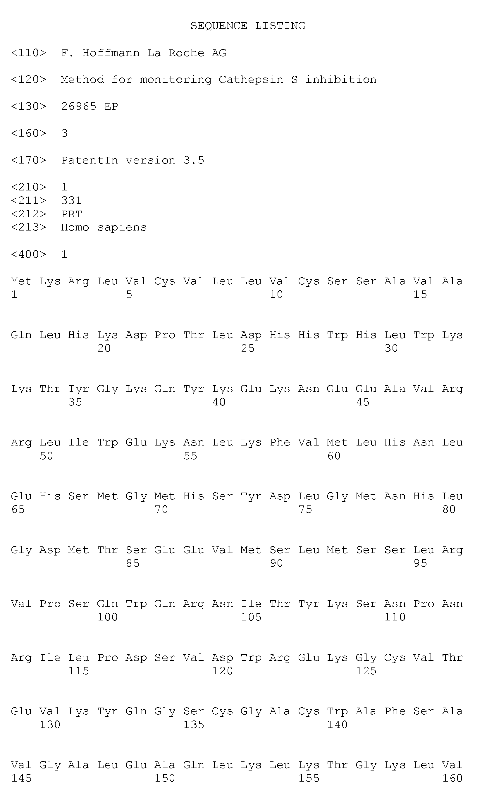
- the amino acid sequence of human Cathepsin S is given in Seq. Id. No. 1.
- invariant chain li refers to invariant polypeptide of major histocompatibility complex or CD74.
- the amino acid sequence of human invariant chain li is given in Seq. Id. No. 2.
- CLIP refers to class II-associated li peptide.
- the amino acid sequence of human CLIP is given in Seq. Id. No. 3.
- MHC II polypeptide refers to the alpha chain and the beta chain constituting the MHC II complex.
- animal as used herein comprises human beings and non-human animals such as e.g. monkey, dog, pig, rabbit, guinea pig and rodents.
- test compound or a “drug candidate compound” described in connection with the assays of the present invention.
- these compounds comprise organic or inorganic compounds, derived synthetically or from natural sources.
- the compounds include inorganic or organic compounds such as polynucleotides, lipids or hormone analogs that are characterized by relatively low molecular weights.
- Other biopolymeric organic test compounds include peptides comprising from about 2 to about 40 amino acids and larger polypeptides comprising from about 40 to about 500 amino acids, such as antibodies or antibody conjugates.
- the inventors of the present invention have surprisingly found that CLIP peptide can be detected on the cell surface of white blood cells and not only intracellularly and that Cathepsin S inhibitors decrease the expression of MHC II polypeptides on the cell surface of white blood cells.
- the methods of the present invention have the advantage compared to the methods described in the prior art that tissue samples, preferably whole blood, can be assayed directly without involving the isolation of white blood cells. In the methods of the present invention there is no need for the isolation of e.g. white blood cells from whole blood samples, in order to be able to be processed in a method of the present invention. This is of great importance if the assay is used in clinical settings where a large number of samples have to be processed.
- the methods of the present invention require smaller tissue samples, preferably blood samples, than the prior art methods.
- Methods for detection and/or measurement of polypeptides in biological samples include, but are not limited to, Western-blotting, Flow cytometry, ELISAs or RIAs, or various proteomics techniques.
- Monoclonal or polyclonal antibodies recognizing the CLIP peptide or MHC II polypeptide or fragments thereof can either be generated for the purpose of detecting the polypeptides or peptide fragments, e.g. by immunizing rabbits with purified proteins, or known antibodies recognizing the polypeptides or peptide fragments can be used.
- an antibody capable of binding to the denatured proteins can be used to detect CLIP peptide or MHC II polypeptide in a Western Blot.
- An example for a method to measure a polypeptide is an ELISA. This type of protein quantitation is based on an antibody capable of capturing a specific antigen, and a second antibody capable of detecting the captured antigen. Methods for preparation and use of antibodies, and the assays mentioned hereinbefore are described in Harlow, E. and Lane, D. Antibodies: A Laboratory Manual, (1988), Cold Spring Harbor Laboratory Press .
- a preferred method for the detection of CLIP peptide and/or MHC II polypeptide is flow cytometry.
- Flow cytometry methods are described in Handbook of Flow Cytometry Method, J. Paul Robinson (Editor); Flow Cytometry - A Basic Introduction, Michael G Ormerod (2008 ) and Current Protocols in Cytometry (2010), Wiley .
- CLIP FITC (BD, order ID: 555981) 20 ⁇ l/sample CD14 PerCp-Cy5.5 (BD, order ID: 550787) 5 ⁇ l/sample CD19 PE (BD, order ID: 555413) 20 ⁇ l/sample HLA-DR APC (BD, order ID: 347403) 1.25 ⁇ l/sample
- TopSeal-A 96-well adhesive plate sealing film (PerkinElmer, order ID: 6005185). Storage: room temperature
- 10x BD Pharm Lyse cell lysis buffer (BD, order ID: 555899). Storage: +4°C. Dilute the 10x BD Pharm Lyse buffer with H 2 O bidest. to 1x and prewarm it to the required temperature prior to use
- 10x BD CellFix cell fixation buffer (BD, order ID: 340181). Storage: room temperature. Dilute the 10x BD CellFix buffer with H 2 O bidest. to 1x prior to use.
Landscapes
- Life Sciences & Earth Sciences (AREA)
- Health & Medical Sciences (AREA)
- Chemical & Material Sciences (AREA)
- Engineering & Computer Science (AREA)
- Immunology (AREA)
- Molecular Biology (AREA)
- Hematology (AREA)
- Urology & Nephrology (AREA)
- Biomedical Technology (AREA)
- Cell Biology (AREA)
- Biotechnology (AREA)
- General Health & Medical Sciences (AREA)
- Organic Chemistry (AREA)
- Microbiology (AREA)
- Zoology (AREA)
- Biochemistry (AREA)
- Analytical Chemistry (AREA)
- Physics & Mathematics (AREA)
- Food Science & Technology (AREA)
- Wood Science & Technology (AREA)
- Pathology (AREA)
- Proteomics, Peptides & Aminoacids (AREA)
- General Physics & Mathematics (AREA)
- Medicinal Chemistry (AREA)
- Tropical Medicine & Parasitology (AREA)
- Bioinformatics & Cheminformatics (AREA)
- Biophysics (AREA)
- Virology (AREA)
- Genetics & Genomics (AREA)
- General Engineering & Computer Science (AREA)
- Investigating Or Analysing Biological Materials (AREA)
- Measuring Or Testing Involving Enzymes Or Micro-Organisms (AREA)
Abstract
The present invention relates to a method for monitoring Cathepsin S inhibitor activity in tissue samples of animals comprising:
a) providing a tissue sample of an animal to whom a Cathepsin S inhibitor has been administered or providing a tissue sample of an animal that was contacted in vitro with a Cathepsin S inhibitor, wherein the tissue samples comprise white blood cells,
b) measuring CLIP peptide level and/or MHC II polypeptide level in the white blood cells of the tissue sample of step a) and
c) correlating CLIP peptide level and/or MHC II polypeptide level in the white blood cells to Cathepsin S inhibitor dose, wherein a Cathepsin S inhibitor leads to decreased level of CLIP peptide and/or a decreased level of MHC II polypeptide in the white blood cells.
a) providing a tissue sample of an animal to whom a Cathepsin S inhibitor has been administered or providing a tissue sample of an animal that was contacted in vitro with a Cathepsin S inhibitor, wherein the tissue samples comprise white blood cells,
b) measuring CLIP peptide level and/or MHC II polypeptide level in the white blood cells of the tissue sample of step a) and
c) correlating CLIP peptide level and/or MHC II polypeptide level in the white blood cells to Cathepsin S inhibitor dose, wherein a Cathepsin S inhibitor leads to decreased level of CLIP peptide and/or a decreased level of MHC II polypeptide in the white blood cells.
Description
- The present invention provides a method for monitoring Cathepsin S inhibition and a method for screening of compounds for Cathepsin S inhibitory activity.
- Cathepsin S is well known for its critical function in the proteolytic digestion of the invariant chain chaperone molecules, thus controlling antigen presentation to CD4+ T-cells by major histocompatibility complex (MHC) class II molecules or to NK1.1+ T-cells via CD1 molecules. Cathepsin S also appears to participate in direct processing of exogenous antigens for presentation by MHC class II to CD4+ T-cells, or in cross-presentation by MHC class I molecules to CD8+ T-cells. In addition, in its secreted form cathepsin S is implicated in degradation of the extracellular matrix, which may contribute to the pathology of a number of diseases, including arthritis, atherosclerosis and chronic obstructive pulmonary disease. Therefore, inhibition of cathepsin S is a promising target for the development of novel therapeutics for a variety of indications.
- MHC class II molecules bind and display antigenic peptides as class II-peptide complexes on the cell surface of antigen presenting cells (APCs) for recognition by CD4+ helper cells. The molecular mechanisms leading to formation of class II-peptide complexes and presentation of antigen on the cell surface begin with synthesis of class II αβ heterodimers in the endoplasmatic reticulum. During biosynthesis these αβ heterodimers associate with a type II membrane protein, the invariant chain li, to form a nonameric complex of αβli. The αβli complex traverses the Golgi apparatus and is delivered to intracellular compartments. In these acidic compartments, the li is degraded and liberated from the αβli complexes, allowing class II molecules to bind antigenic peptides. The cysteine protease Cathepsin S plays a key role in processing li in APCs. Cathepsin S degrades the invariant chain li from class II-li complexes generating class II-CLIP. Inhibition of Cathepsin S leads to a decrease of CLIP peptide and to accumulation of invariant chain li degradation peptide p10.
-
EP 1 315 966 discloses a method for monitoring the effect of in vivo administration of cathepsin S inhibitors. The method includes the purification of white blood cells from a blood sample of a subject treated with a cathepsin S inhibitor, preparation of whole cell lysates of the purified white blood cells and analyzing the whole cell lysates for presence of p10 fragment of invariant chain li. This method requires blood samples of about 10 - 30 ml and is labour intensive (isolation of white blood cells and preparation of whole cell lysates). - Therefore, there is a need for a cathepsin S inhibitor assay which overcomes at least some of the drawbacks of the prior art methods.
- In a first object, the present invention provides a method for monitoring Cathepsin S inhibitor activity in tissue samples of animals comprising:
- a) providing a tissue sample of an animal to whom a Cathepsin S inhibitor has been administered or providing a tissue sample of an animal that was contacted in vitro with a Cathepsin S inhibitor, wherein the tissue sample comprises white blood cells,
- b) measuring CLIP peptide level and/or MHC II polypeptide level in the white blood cells of the tissue sample of step a) and
- c) correlating CLIP peptide level and/or MHC II polypeptide level in the white blood cells to Cathepsin S inhibitor dose, wherein a Cathepsin S inhibitor leads to decreased level of CLIP peptide and/or a decreased level of MHC II polypeptide in the white blood cells.
- In a second object the present invention provides a method for the identification of a Cathepsin S inhibitor comprising:
- a) treating a non-human animal with a test compound,
- b) providing a tissue sample of the animal of step a) comprising white blood cells,
- c) measuring CLIP peptide level and/or MHC II polypeptide level in the white blood cells of the tissue sample of step b),
wherein a decreased level of CLIP peptide and/or a decreased level of MHC II polypeptide in the white blood cells in the sample of step a) compared to a level of CLIP peptide and/or a level of MHC II polypeptide in white blood cells in a tissue sample of an untreated animal is indicative for a Cathepsin S inhibitor. - In a preferred embodiment of the methods of the present invention the tissue sample is blood.
- In a further preferred embodiment of the methods of the present invention the level of CLIP peptide and/or MHC II polypeptide level on the cell surface of white blood cells is measured.
- In a further preferred embodiment of the methods of the present invention the white blood cells are Antigen Presenting Cells (APCs), preferably the APCs are selected from B cells, monocytes, macrophages and dendritic cells, more preferably selected from B cells and monocytes.
- In another preferred embodiment of the methods of the present invention the level of CLIP peptide and/or MHC II polypeptide on the cell surface of white blood cells, preferably APCs, is measured by Flow Cytometry.
- The term Antigen Presenting cells (APCs) as used herein refers to cells that display antigenic peptides as (MHC) class II - antigenic peptide complex on its surface. APCs comprise dendritic cells, monocytes, macrophages and B cells.
- The term Cathepsin S refers to a cysteine protease in the papain super-family. The amino acid sequence of human Cathepsin S is given in Seq. Id. No. 1.
- The term "invariant chain li" refers to invariant polypeptide of major histocompatibility complex or CD74. The amino acid sequence of human invariant chain li is given in Seq. Id. No. 2.
- The term CLIP refers to class II-associated li peptide. The amino acid sequence of human CLIP is given in Seq. Id. No. 3.
- The term "MHC II polypeptide" as used herein refers to the alpha chain and the beta chain constituting the MHC II complex.
- The term animal as used herein comprises human beings and non-human animals such as e.g. monkey, dog, pig, rabbit, guinea pig and rodents.
- The term "compound" is used herein in the context of a "test compound" or a "drug candidate compound" described in connection with the assays of the present invention. As such, these compounds comprise organic or inorganic compounds, derived synthetically or from natural sources. The compounds include inorganic or organic compounds such as polynucleotides, lipids or hormone analogs that are characterized by relatively low molecular weights. Other biopolymeric organic test compounds include peptides comprising from about 2 to about 40 amino acids and larger polypeptides comprising from about 40 to about 500 amino acids, such as antibodies or antibody conjugates.
- The inventors of the present invention have surprisingly found that CLIP peptide can be detected on the cell surface of white blood cells and not only intracellularly and that Cathepsin S inhibitors decrease the expression of MHC II polypeptides on the cell surface of white blood cells. The methods of the present invention have the advantage compared to the methods described in the prior art that tissue samples, preferably whole blood, can be assayed directly without involving the isolation of white blood cells. In the methods of the present invention there is no need for the isolation of e.g. white blood cells from whole blood samples, in order to be able to be processed in a method of the present invention. This is of great importance if the assay is used in clinical settings where a large number of samples have to be processed. The methods of the present invention require smaller tissue samples, preferably blood samples, than the prior art methods.
- Methods for detection and/or measurement of polypeptides in biological samples are well known in the art and include, but are not limited to, Western-blotting, Flow cytometry, ELISAs or RIAs, or various proteomics techniques. Monoclonal or polyclonal antibodies recognizing the CLIP peptide or MHC II polypeptide or fragments thereof, can either be generated for the purpose of detecting the polypeptides or peptide fragments, e.g. by immunizing rabbits with purified proteins, or known antibodies recognizing the polypeptides or peptide fragments can be used. For example, an antibody capable of binding to the denatured proteins, such as a polyclonal antibody, can be used to detect CLIP peptide or MHC II polypeptide in a Western Blot. An example for a method to measure a polypeptide is an ELISA. This type of protein quantitation is based on an antibody capable of capturing a specific antigen, and a second antibody capable of detecting the captured antigen. Methods for preparation and use of antibodies, and the assays mentioned hereinbefore are described in Harlow, E. and Lane, D. Antibodies: A Laboratory Manual, (1988), Cold Spring Harbor Laboratory Press.
- A preferred method for the detection of CLIP peptide and/or MHC II polypeptide is flow cytometry. Flow cytometry methods are described in Handbook of Flow Cytometry Method, J. Paul Robinson (Editor); Flow Cytometry - A Basic Introduction, Michael G Ormerod (2008) and Current Protocols in Cytometry (2010), Wiley.
-
-
Figure 1A-D : CLIP surface expression on B cells. Whole blood has been incubated for 14 hours with varying concentrations of Cathepsin S inhibitor or carrier. CLIP surface expression on B cells (CD19+) was then determined via flow cytometry. The figure shows the duplicate analysis of four different healthy volunteers (R31, R51, R15 and R40). Four-parameter-dose-response curves and IC50 values have been calculated with GraphPad Prism software. MFI = Mean Fluorescence Intensity. -
Figure 2A - D : CLIP surface expression on Monocytes. Whole blood has been incubated for 14 hours with varying concentrations of Cathepsin S inhibitor or carrier. The blood has been costimulated with IFNγ and GM-CSF. Subsequently, flow cytometric analysis of CLIP surface expression on Monocytes (CD14+) was performed. The figure shows the duplicate analysis of four different healthy volunteers (R31, R51, R50 and R40). Four-parameter-dose-response curves and IC50 values have been calculated with GraphPad Prism software. -
Figure 3A - D : MHC class II cell surface expression on B cells. Whole blood has been incubated for 14 hours with varying concentrations of Cathepsin S inhibitor or carrier. Subsequently, MHC class II surface expression on B cells (CD19+) was determined by flow cytommetry. The figure shows the duplicate analysis of four different healthy volunteers (R31, R51, R15 and R40). Four-parameter-dose-response curves and IC50 values have been calculated with GraphPad Prism software. - FACS whole blood assay with Cathepsin S inhibitor
- use a sterile Polypropylene 96-well U-bottom plate
- add 100 µl complete RPMI ± Cathepsin S inhibitor
- add 125 µl LI-Heparin whole blood to each tube and mix carefully
- preincubate 30 minutes at 37°C
- add 25 µl complete RPMI ± IFN-γ and GM-CSF to each well
- incubate 13h and 30 minutes at 37°C
- prepare 1x lysing solution
- add 1.8 ml 1x lysing solution to each well of a Polypropylene 96-well deep-well plate and prewarm it to 37°C
- add the antibodies (comp. control) or antibody mixture (no predilution in BD stain buffer) to a Polypropylene 96-well V-bottom plate
- add 200 µl whole blood of each sample to the prepared V-bottom plate and mix carefully
- incubate for 30 min on ice in the dark
- transfer 200 µl stained whole blood of each sample to the prepared deep-well plate containing the prewarmed lysis buffer and mix carefully
- incubate at RT for 15 minutes in the dark
- centrifuge 250 x g for 5 minutes and +4°C, remove supernatant
- repeat the cell lysing procedure 1x with 1x lysis buffer prewarmed to room temperature
- wash the cells 1x with 2 ml BD stain buffer (250 x g, 5 minutes, +4°C), remove supernatant
- resuspend the cell pellet in 80 µl BD stain buffer
- add 20 µl FC-blocking reagent and 10 µl FITC-Activator and mix
- incubate for 10 minutes at +4°C
- wash 1x with 2 ml cold BD stain buffer (250 x g, 5 minutes, +4°C) remove supernatant
- resuspend the cell pellet in 80 µl BD stain buffer
- add 20 µl FC-blocking reagent and 10 µl FITC- Enhancer and mix
- incubate for 10 minutes at +4°C
- transfer the cells to FACS tubes via a cell filter and flush the filter with 2 ml PBS
- centrifuge 250 x g for 5 minutes and +4°C, remove supernatant
- resuspend pellet in 200 µl 1x cell fix buffer, vortex carefully
- keep the cells in the dark until the analysis on the FACS
-
CLIP FITC (BD, order ID: 555981) 20 µl/sample CD14 PerCp-Cy5.5 (BD, order ID: 550787) 5 µl/sample CD19 PE (BD, order ID: 555413) 20 µl/sample HLA-DR APC (BD, order ID: 347403) 1.25 µl/sample - 96-well plates, V-bottom (Nunc, order ID: 249944) Storage: room temperature, non sterile, non tissue culture treated
- 96-well deep well plates, 2.2 ml (Thermo Scientific, order ID: AB-0661) Storage: room temperature. Non sterile, non tissue culture treated
- TopSeal-A 96-well adhesive plate sealing film (PerkinElmer, order ID: 6005185). Storage: room temperature
- 10x BD Pharm Lyse cell lysis buffer (BD, order ID: 555899). Storage: +4°C. Dilute the 10x BD Pharm Lyse buffer with H2O bidest. to 1x and prewarm it to the required temperature prior to use
- 1x BD stain buffer containing 2% FCS (BD, order ID: 554656). Storage: +4C the 1x BD stain buffer containing 2% FCS is ready to use. Keep the BD stain buffer on ice while using
- 1x PBS without CaCl2 and without MgCl2 (Gibco, order ID: 14190-094). Storage: room temperature. The PBS is ready to use. Precool the PBS on ice prior to use
- 5 ml Polystyrene round-bottom tube (FACS tube, BD, order ID: 352052). Storage: room temperature. Ready to use.
- 70 µM cup Filcon cell suspension filter (BD, order ID: 340636) Storage: room temperature. Ready to use.
- 10x BD CellFix cell fixation buffer (BD, order ID: 340181). Storage: room temperature. Dilute the 10x BD CellFix buffer with H2O bidest. to 1x prior to use.


Cpd (0, 0.0001, 0.0003, 0.001, 0.003, 0.009, 0.027, 0.08, 0.25, 0.74, 2.2 and 20 µM)
stimulated :DMSO
+IFN-γ 400 ng/ml (stock 100 µg/ml, 1:250)
+ GM-CSF 400 ng/ml (stock 100 µg/ml, 1:250)
Cpd (0, 0.0001, 0.0003, 0.001, 0.003, 0.009, 0.027, 0.08, 0.25, 0.74, 2.2 and 20 µM)
+IFN-γ 400 ng/ml (stock 100 µg/ml, 1:250)
+ GM-CSF 400 ng/ml (stock 100 µg/ml, 1:250)
FACS staining:
Claims (9)
- A method for monitoring Cathepsin S inhibitor activity in a tissue sample of animals comprising:a) providing a tissue sample of an animal to whom a Cathepsin S inhibitor has been administered or providing a tissue sample of an animal that was contacted in vitro with a Cathepsin S inhibitor, wherein the tissue sample comprise white blood cells,b) measuring CLIP peptide level and/or MHC II polypeptide level in the white blood cells of the tissue sample of step a) andc) correlating CLIP peptide level and/or MHC II polypeptide level in the white blood cells to Cathepsin S inhibitor dose, wherein a Cathepsin S inhibitor leads to decreased level of CLIP peptide and/or a decreased level of MHC II polypeptide in the white blood cells.
- A method for the identification of a Cathepsin S inhibitor comprising:a) contacting a non-human animal with a test compound,b) providing a tissue sample of the animal of step a) comprising white blood cells,c) measuring CLIP peptide level and/or MHC II polypeptide level in the white blood cells of the tissue sample of step b),
wherein a decreased level of CLIP peptide and/or a decreased level of MHC II polypeptide in the white blood cells in the sample of step a) compared to a level of CLIP peptide and/or a level of MHC II polypeptide in white blood cells in a tissue sample of an untreated animal is indicative for a Cathepsin S inhibitor. - The method of claim 1 or 2, wherein the tissue sample is blood.
- The method of claim 1 to 3, wherein the level of CLIP peptide and/or MHC II polypeptide on the cell surface of the white blood cells is measured.
- The method of claim 4, wherein the level of CLIP peptide and/or MHC II polypeptide on the cell surface of the white blood cells is measured by Flow Cytometry.
- The method of claim 1 to 5, wherein the white blood cells are Antigen Presenting Cells (APCs), preferably B cells, monocytes, macrophages and dendritic cells.
- The method of claim 6, wherein the APCs are selected from B cells and monocytes.
- The method of claim 1 and 3-7, wherein the animal is a human being.
- The method of claim 2 -7, wherein the non-human animal is a rodent, preferably a rat or mouse.
Priority Applications (2)
| Application Number | Priority Date | Filing Date | Title |
|---|---|---|---|
| EP10185418A EP2436776A1 (en) | 2010-10-01 | 2010-10-01 | Method for monitoring Cathepsin S inhibition |
| PCT/EP2011/066711 WO2012041824A1 (en) | 2010-10-01 | 2011-09-27 | Method for monitoring cathepsin s inhibition |
Applications Claiming Priority (1)
| Application Number | Priority Date | Filing Date | Title |
|---|---|---|---|
| EP10185418A EP2436776A1 (en) | 2010-10-01 | 2010-10-01 | Method for monitoring Cathepsin S inhibition |
Publications (1)
| Publication Number | Publication Date |
|---|---|
| EP2436776A1 true EP2436776A1 (en) | 2012-04-04 |
Family
ID=43558407
Family Applications (1)
| Application Number | Title | Priority Date | Filing Date |
|---|---|---|---|
| EP10185418A Ceased EP2436776A1 (en) | 2010-10-01 | 2010-10-01 | Method for monitoring Cathepsin S inhibition |
Country Status (2)
| Country | Link |
|---|---|
| EP (1) | EP2436776A1 (en) |
| WO (1) | WO2012041824A1 (en) |
Families Citing this family (1)
| Publication number | Priority date | Publication date | Assignee | Title |
|---|---|---|---|---|
| JP6826527B2 (en) * | 2014-09-18 | 2021-02-03 | エフ.ホフマン−ラ ロシュ アーゲーF. Hoffmann−La Roche Aktiengesellschaft | Methods for Monitoring Cathepsin S Inhibition |
Citations (2)
| Publication number | Priority date | Publication date | Assignee | Title |
|---|---|---|---|---|
| WO2002021129A1 (en) * | 2000-09-06 | 2002-03-14 | Ortho-Mcneil Pharmaceutical, Inc. | Method of monitoring the effect of cathepsin s inhibitors |
| US20030069240A1 (en) * | 2000-08-14 | 2003-04-10 | Breitenbucher J. Guy | Method for treating allergies using substituted pyrazoles |
Family Cites Families (1)
| Publication number | Priority date | Publication date | Assignee | Title |
|---|---|---|---|---|
| US4902613A (en) * | 1984-05-14 | 1990-02-20 | Becton, Dickinson And Company | Lysing agent for analysis of peripheral blood cells |
-
2010
- 2010-10-01 EP EP10185418A patent/EP2436776A1/en not_active Ceased
-
2011
- 2011-09-27 WO PCT/EP2011/066711 patent/WO2012041824A1/en not_active Ceased
Patent Citations (3)
| Publication number | Priority date | Publication date | Assignee | Title |
|---|---|---|---|---|
| US20030069240A1 (en) * | 2000-08-14 | 2003-04-10 | Breitenbucher J. Guy | Method for treating allergies using substituted pyrazoles |
| WO2002021129A1 (en) * | 2000-09-06 | 2002-03-14 | Ortho-Mcneil Pharmaceutical, Inc. | Method of monitoring the effect of cathepsin s inhibitors |
| EP1315966A1 (en) | 2000-09-06 | 2003-06-04 | Ortho-McNeil Pharmaceutical, Inc. | Method of monitoring the effect of cathepsin s inhibitors |
Non-Patent Citations (5)
| Title |
|---|
| "Current Protocols in Cytometry", 2010, WILEY |
| HARLOW, E.; LANE, D.: "Antibodies: A Laboratory Manual", 1988, COLD SPRING HARBOR LABORATORY PRESS |
| J. PAUL ROBINSON: "Handbook of Flow Cytometry Method", WILEY-LISS |
| MICHAEL G ORMEROD, FLOW CYTOMETRY - A BASIC INTRODUCTION, 2008 |
| SENDIDE KHALID ET AL: "Mycobacterium bovis BCG attenuates surface expression of mature class II molecules through IL-10-dependent inhibition of cathepsin S", JOURNAL OF IMMUNOLOGY, vol. 175, no. 8, October 2005 (2005-10-01), pages 5324 - 5332, XP002623164, ISSN: 0022-1767 * |
Also Published As
| Publication number | Publication date |
|---|---|
| WO2012041824A1 (en) | 2012-04-05 |
Similar Documents
| Publication | Publication Date | Title |
|---|---|---|
| US11787846B2 (en) | EphA2 T-cell epitope agonists and uses therefore | |
| Tanaka | The proteasome: overview of structure and functions | |
| Hilger et al. | Identification and isolation of a Fel d 1–like molecule as a major rabbit allergen | |
| AU2019203459A1 (en) | PIF binding as a marker for immune dysregulation | |
| KR20030037272A (en) | Peptides presented by cells | |
| EP2150819B1 (en) | S100a9 interaction screening method | |
| JP2019511722A (en) | Methods for measuring cell-mediated immunity of a subject | |
| US20090280511A1 (en) | Methods for selecting candidate peptides presented on the surface of cancer cells following their binding to MHC class I molecules | |
| US8222211B2 (en) | Methods of administering PIF agonist peptides and uses thereof | |
| AU2004284097A1 (en) | PIF peptides biologic activities, site of action, and the antibody to detect PIF | |
| EP2436776A1 (en) | Method for monitoring Cathepsin S inhibition | |
| US7026132B2 (en) | Method of monitoring the effect of Cathepsin S inhibitors | |
| Huber et al. | CyrA, a matricellular protein that modulates cell motility in Dictyostelium discoideum | |
| EP3194610B1 (en) | Anti li p10 antibody | |
| Anandarajah et al. | SPARC (secreted protein acidic and rich in cysteine) of the intestinal nematode Strongyloides ratti is involved in mucosa-associated parasite-host interaction | |
| US20130259884A1 (en) | Peptides | |
| EP1801202A1 (en) | Process for producing dendritic cell having acquired ctl inducing capability | |
| WO2008066483A1 (en) | Use of nalp5 in methods for diagnosis and therapy of parathyroid related disorders | |
| CA2894868C (en) | Pif peptides biologic activities, site of action, and the antibody to detect pif | |
| HK1237356A1 (en) | Method for monitoring cathepsin s inhibition | |
| Desmarais et al. | Uterine Signaling at the Emergence of the Embryo from Obligate Diapause | |
| EP1680442A2 (en) | Pif peptides biologic activities, site of action, and the antibody to detect pif |
Legal Events
| Date | Code | Title | Description |
|---|---|---|---|
| PUAI | Public reference made under article 153(3) epc to a published international application that has entered the european phase |
Free format text: ORIGINAL CODE: 0009012 |
|
| AK | Designated contracting states |
Kind code of ref document: A1 Designated state(s): AL AT BE BG CH CY CZ DE DK EE ES FI FR GB GR HR HU IE IS IT LI LT LU LV MC MK MT NL NO PL PT RO RS SE SI SK SM TR |
|
| AX | Request for extension of the european patent |
Extension state: BA ME |
|
| STAA | Information on the status of an ep patent application or granted ep patent |
Free format text: STATUS: THE APPLICATION HAS BEEN REFUSED |
|
| 18R | Application refused |
Effective date: 20120423 |