EP2419119B1 - Muscle function enhancing peptide - Google Patents
Muscle function enhancing peptide Download PDFInfo
- Publication number
- EP2419119B1 EP2419119B1 EP10714584.9A EP10714584A EP2419119B1 EP 2419119 B1 EP2419119 B1 EP 2419119B1 EP 10714584 A EP10714584 A EP 10714584A EP 2419119 B1 EP2419119 B1 EP 2419119B1
- Authority
- EP
- European Patent Office
- Prior art keywords
- peptide
- seq
- amino acids
- prt
- human
- Prior art date
- Legal status (The legal status is an assumption and is not a legal conclusion. Google has not performed a legal analysis and makes no representation as to the accuracy of the status listed.)
- Not-in-force
Links
- 108090000765 processed proteins & peptides Proteins 0.000 title claims description 625
- 230000002708 enhancing effect Effects 0.000 title claims description 86
- 230000004220 muscle function Effects 0.000 title claims description 45
- 150000001413 amino acids Chemical group 0.000 claims description 496
- 229940024606 amino acid Drugs 0.000 claims description 489
- 235000001014 amino acid Nutrition 0.000 claims description 489
- 108700031547 S100A1 Proteins 0.000 claims description 405
- 239000011575 calcium Substances 0.000 claims description 197
- OYPRJOBELJOOCE-UHFFFAOYSA-N Calcium Chemical compound [Ca] OYPRJOBELJOOCE-UHFFFAOYSA-N 0.000 claims description 187
- 229910052791 calcium Inorganic materials 0.000 claims description 187
- KZSNJWFQEVHDMF-BYPYZUCNSA-N L-valine Chemical compound CC(C)[C@H](N)C(O)=O KZSNJWFQEVHDMF-BYPYZUCNSA-N 0.000 claims description 74
- KZSNJWFQEVHDMF-UHFFFAOYSA-N Valine Natural products CC(C)C(N)C(O)=O KZSNJWFQEVHDMF-UHFFFAOYSA-N 0.000 claims description 74
- 239000004474 valine Substances 0.000 claims description 74
- QNAYBMKLOCPYGJ-REOHCLBHSA-N L-alanine Chemical compound C[C@H](N)C(O)=O QNAYBMKLOCPYGJ-REOHCLBHSA-N 0.000 claims description 70
- 235000004279 alanine Nutrition 0.000 claims description 70
- 208000037265 diseases, disorders, signs and symptoms Diseases 0.000 claims description 63
- AGPKZVBTJJNPAG-WHFBIAKZSA-N L-isoleucine Chemical compound CC[C@H](C)[C@H](N)C(O)=O AGPKZVBTJJNPAG-WHFBIAKZSA-N 0.000 claims description 59
- 229960000310 isoleucine Drugs 0.000 claims description 59
- AGPKZVBTJJNPAG-UHFFFAOYSA-N isoleucine Natural products CCC(C)C(N)C(O)=O AGPKZVBTJJNPAG-UHFFFAOYSA-N 0.000 claims description 59
- DHMQDGOQFOQNFH-UHFFFAOYSA-N Glycine Chemical compound NCC(O)=O DHMQDGOQFOQNFH-UHFFFAOYSA-N 0.000 claims description 54
- 102100023097 Protein S100-A1 Human genes 0.000 claims description 51
- 208000035475 disorder Diseases 0.000 claims description 49
- FFEARJCKVFRZRR-BYPYZUCNSA-N L-methionine Chemical compound CSCC[C@H](N)C(O)=O FFEARJCKVFRZRR-BYPYZUCNSA-N 0.000 claims description 45
- 229930182817 methionine Natural products 0.000 claims description 45
- 230000003387 muscular Effects 0.000 claims description 44
- 230000007257 malfunction Effects 0.000 claims description 42
- 206010019280 Heart failures Diseases 0.000 claims description 37
- WHUUTDBJXJRKMK-VKHMYHEASA-N L-glutamic acid Chemical compound OC(=O)[C@@H](N)CCC(O)=O WHUUTDBJXJRKMK-VKHMYHEASA-N 0.000 claims description 37
- 229930195712 glutamate Natural products 0.000 claims description 37
- 229940049906 glutamate Drugs 0.000 claims description 37
- 230000002861 ventricular Effects 0.000 claims description 36
- 210000000663 muscle cell Anatomy 0.000 claims description 32
- 230000001351 cycling effect Effects 0.000 claims description 31
- 102000005701 Calcium-Binding Proteins Human genes 0.000 claims description 30
- 108010045403 Calcium-Binding Proteins Proteins 0.000 claims description 30
- 239000004471 Glycine Substances 0.000 claims description 28
- -1 aromatic amino acid Chemical class 0.000 claims description 27
- 210000004165 myocardium Anatomy 0.000 claims description 27
- 239000004475 Arginine Substances 0.000 claims description 25
- ODKSFYDXXFIFQN-UHFFFAOYSA-N arginine Natural products OC(=O)C(N)CCCNC(N)=N ODKSFYDXXFIFQN-UHFFFAOYSA-N 0.000 claims description 25
- 230000002950 deficient Effects 0.000 claims description 24
- CKLJMWTZIZZHCS-REOHCLBHSA-N L-aspartic acid Chemical compound OC(=O)[C@@H](N)CC(O)=O CKLJMWTZIZZHCS-REOHCLBHSA-N 0.000 claims description 23
- ROHFNLRQFUQHCH-YFKPBYRVSA-N L-leucine Chemical compound CC(C)C[C@H](N)C(O)=O ROHFNLRQFUQHCH-YFKPBYRVSA-N 0.000 claims description 23
- ROHFNLRQFUQHCH-UHFFFAOYSA-N Leucine Natural products CC(C)CC(N)C(O)=O ROHFNLRQFUQHCH-UHFFFAOYSA-N 0.000 claims description 23
- 229940009098 aspartate Drugs 0.000 claims description 23
- 230000000747 cardiac effect Effects 0.000 claims description 23
- 208000021642 Muscular disease Diseases 0.000 claims description 22
- 208000013363 skeletal muscle disease Diseases 0.000 claims description 22
- ODKSFYDXXFIFQN-BYPYZUCNSA-P L-argininium(2+) Chemical compound NC(=[NH2+])NCCC[C@H]([NH3+])C(O)=O ODKSFYDXXFIFQN-BYPYZUCNSA-P 0.000 claims description 21
- KDXKERNSBIXSRK-UHFFFAOYSA-N Lysine Natural products NCCCCC(N)C(O)=O KDXKERNSBIXSRK-UHFFFAOYSA-N 0.000 claims description 20
- 239000004472 Lysine Substances 0.000 claims description 20
- KDXKERNSBIXSRK-YFKPBYRVSA-N L-lysine Chemical compound NCCCC[C@H](N)C(O)=O KDXKERNSBIXSRK-YFKPBYRVSA-N 0.000 claims description 17
- 239000003814 drug Substances 0.000 claims description 17
- 210000004899 c-terminal region Anatomy 0.000 claims description 16
- 101710156967 Protein S100-A1 Proteins 0.000 claims description 15
- 230000009091 contractile dysfunction Effects 0.000 claims description 15
- 208000029578 Muscle disease Diseases 0.000 claims description 14
- 239000012528 membrane Substances 0.000 claims description 14
- 150000003943 catecholamines Chemical class 0.000 claims description 12
- 230000002209 hydrophobic effect Effects 0.000 claims description 11
- 208000010125 myocardial infarction Diseases 0.000 claims description 11
- ONIBWKKTOPOVIA-BYPYZUCNSA-N L-Proline Chemical compound OC(=O)[C@@H]1CCCN1 ONIBWKKTOPOVIA-BYPYZUCNSA-N 0.000 claims description 10
- ONIBWKKTOPOVIA-UHFFFAOYSA-N Proline Natural products OC(=O)C1CCCN1 ONIBWKKTOPOVIA-UHFFFAOYSA-N 0.000 claims description 10
- 230000009090 positive inotropic effect Effects 0.000 claims description 10
- 102000012740 beta Adrenergic Receptors Human genes 0.000 claims description 9
- 108010079452 beta Adrenergic Receptors Proteins 0.000 claims description 9
- 230000035515 penetration Effects 0.000 claims description 9
- 201000003728 Centronuclear myopathy Diseases 0.000 claims description 8
- 102000013674 S-100 Human genes 0.000 claims description 8
- 108700021018 S100 Proteins 0.000 claims description 8
- 208000032978 Structural Congenital Myopathies Diseases 0.000 claims description 8
- 230000004064 dysfunction Effects 0.000 claims description 8
- 230000008685 targeting Effects 0.000 claims description 8
- 208000020446 Cardiac disease Diseases 0.000 claims description 7
- 208000010428 Muscle Weakness Diseases 0.000 claims description 7
- 206010028372 Muscular weakness Diseases 0.000 claims description 7
- 201000002481 Myositis Diseases 0.000 claims description 7
- 208000019622 heart disease Diseases 0.000 claims description 7
- 239000000203 mixture Substances 0.000 claims description 7
- 201000006938 muscular dystrophy Diseases 0.000 claims description 7
- 208000031229 Cardiomyopathies Diseases 0.000 claims description 6
- 101000685718 Homo sapiens Protein S100-Z Proteins 0.000 claims description 6
- 229940096437 Protein S Drugs 0.000 claims description 6
- 102100023107 Protein S100-Z Human genes 0.000 claims description 6
- 206010007559 Cardiac failure congestive Diseases 0.000 claims description 5
- 208000015374 Central core disease Diseases 0.000 claims description 5
- 239000000808 adrenergic beta-agonist Substances 0.000 claims description 5
- 206010007625 cardiogenic shock Diseases 0.000 claims description 5
- 201000007303 central core myopathy Diseases 0.000 claims description 5
- 210000003709 heart valve Anatomy 0.000 claims description 5
- 239000003550 marker Substances 0.000 claims description 5
- 201000002169 Mitochondrial myopathy Diseases 0.000 claims description 4
- 208000034965 Nemaline Myopathies Diseases 0.000 claims description 4
- 206010040070 Septic Shock Diseases 0.000 claims description 4
- 229910052799 carbon Inorganic materials 0.000 claims description 4
- 208000023692 inborn mitochondrial myopathy Diseases 0.000 claims description 4
- 201000000585 muscular atrophy Diseases 0.000 claims description 4
- 206010030875 ophthalmoplegia Diseases 0.000 claims description 4
- 230000036303 septic shock Effects 0.000 claims description 4
- 101100420475 Homo sapiens S100A1 gene Proteins 0.000 description 369
- 210000004413 cardiac myocyte Anatomy 0.000 description 114
- 125000003275 alpha amino acid group Chemical group 0.000 description 106
- 235000014393 valine Nutrition 0.000 description 70
- 210000001908 sarcoplasmic reticulum Anatomy 0.000 description 63
- 230000006870 function Effects 0.000 description 60
- 210000004027 cell Anatomy 0.000 description 54
- 238000001727 in vivo Methods 0.000 description 53
- RYYVLZVUVIJVGH-UHFFFAOYSA-N caffeine Chemical compound CN1C(=O)N(C)C(=O)C2=C1N=CN2C RYYVLZVUVIJVGH-UHFFFAOYSA-N 0.000 description 44
- 210000000107 myocyte Anatomy 0.000 description 44
- 210000002363 skeletal muscle cell Anatomy 0.000 description 41
- 239000000243 solution Substances 0.000 description 40
- 230000000297 inotrophic effect Effects 0.000 description 38
- 101000685712 Homo sapiens Protein S100-A1 Proteins 0.000 description 36
- 230000000694 effects Effects 0.000 description 32
- 230000006907 apoptotic process Effects 0.000 description 31
- 230000036461 convulsion Effects 0.000 description 27
- 210000001087 myotubule Anatomy 0.000 description 27
- 241000699670 Mus sp. Species 0.000 description 26
- 230000003288 anthiarrhythmic effect Effects 0.000 description 24
- 230000000004 hemodynamic effect Effects 0.000 description 24
- 210000001519 tissue Anatomy 0.000 description 24
- 238000000338 in vitro Methods 0.000 description 23
- 210000002027 skeletal muscle Anatomy 0.000 description 23
- LPHGQDQBBGAPDZ-UHFFFAOYSA-N Isocaffeine Natural products CN1C(=O)N(C)C(=O)C2=C1N(C)C=N2 LPHGQDQBBGAPDZ-UHFFFAOYSA-N 0.000 description 22
- 241001465754 Metazoa Species 0.000 description 22
- MTCFGRXMJLQNBG-UHFFFAOYSA-N Serine Natural products OCC(N)C(O)=O MTCFGRXMJLQNBG-UHFFFAOYSA-N 0.000 description 22
- 239000003416 antiarrhythmic agent Substances 0.000 description 22
- 229960001948 caffeine Drugs 0.000 description 22
- VJEONQKOZGKCAK-UHFFFAOYSA-N caffeine Natural products CN1C(=O)N(C)C(=O)C2=C1C=CN2C VJEONQKOZGKCAK-UHFFFAOYSA-N 0.000 description 22
- 235000004400 serine Nutrition 0.000 description 22
- 102000019027 Ryanodine Receptor Calcium Release Channel Human genes 0.000 description 21
- 102000004196 processed proteins & peptides Human genes 0.000 description 21
- 239000008194 pharmaceutical composition Substances 0.000 description 20
- MTCFGRXMJLQNBG-REOHCLBHSA-N (2S)-2-Amino-3-hydroxypropansäure Chemical compound OC[C@H](N)C(O)=O MTCFGRXMJLQNBG-REOHCLBHSA-N 0.000 description 19
- JWZZKOKVBUJMES-UHFFFAOYSA-N (+-)-Isoprenaline Chemical compound CC(C)NCC(O)C1=CC=C(O)C(O)=C1 JWZZKOKVBUJMES-UHFFFAOYSA-N 0.000 description 16
- 241000282414 Homo sapiens Species 0.000 description 16
- AYFVYJQAPQTCCC-UHFFFAOYSA-N Threonine Natural products CC(O)C(N)C(O)=O AYFVYJQAPQTCCC-UHFFFAOYSA-N 0.000 description 16
- 239000004473 Threonine Substances 0.000 description 16
- 239000000835 fiber Substances 0.000 description 16
- 231100000518 lethal Toxicity 0.000 description 16
- 230000001665 lethal effect Effects 0.000 description 16
- 238000007911 parenteral administration Methods 0.000 description 16
- 235000008521 threonine Nutrition 0.000 description 16
- 230000001960 triggered effect Effects 0.000 description 16
- 210000002216 heart Anatomy 0.000 description 15
- 238000007912 intraperitoneal administration Methods 0.000 description 15
- 201000010099 disease Diseases 0.000 description 14
- 238000005259 measurement Methods 0.000 description 14
- 230000001536 pro-arrhythmogenic effect Effects 0.000 description 14
- 206010003119 arrhythmia Diseases 0.000 description 13
- 210000000170 cell membrane Anatomy 0.000 description 13
- 229940039009 isoproterenol Drugs 0.000 description 13
- 210000004379 membrane Anatomy 0.000 description 13
- 238000000034 method Methods 0.000 description 13
- 210000003205 muscle Anatomy 0.000 description 13
- 102000004169 proteins and genes Human genes 0.000 description 13
- 108090000623 proteins and genes Proteins 0.000 description 13
- OUYCCCASQSFEME-UHFFFAOYSA-N tyrosine Natural products OC(=O)C(N)CC1=CC=C(O)C=C1 OUYCCCASQSFEME-UHFFFAOYSA-N 0.000 description 13
- AYFVYJQAPQTCCC-GBXIJSLDSA-N L-threonine Chemical compound C[C@@H](O)[C@H](N)C(O)=O AYFVYJQAPQTCCC-GBXIJSLDSA-N 0.000 description 12
- OUYCCCASQSFEME-QMMMGPOBSA-N L-tyrosine Chemical compound OC(=O)[C@@H](N)CC1=CC=C(O)C=C1 OUYCCCASQSFEME-QMMMGPOBSA-N 0.000 description 12
- 108010012219 Ryanodine Receptor Calcium Release Channel Proteins 0.000 description 12
- 230000006793 arrhythmia Effects 0.000 description 12
- 230000008602 contraction Effects 0.000 description 12
- 238000002360 preparation method Methods 0.000 description 12
- 230000002040 relaxant effect Effects 0.000 description 12
- 230000000638 stimulation Effects 0.000 description 12
- 108091006146 Channels Proteins 0.000 description 11
- COLNVLDHVKWLRT-QMMMGPOBSA-N L-phenylalanine Chemical compound OC(=O)[C@@H](N)CC1=CC=CC=C1 COLNVLDHVKWLRT-QMMMGPOBSA-N 0.000 description 11
- 241000699666 Mus <mouse, genus> Species 0.000 description 11
- 230000002424 anti-apoptotic effect Effects 0.000 description 11
- 238000002474 experimental method Methods 0.000 description 11
- COLNVLDHVKWLRT-UHFFFAOYSA-N phenylalanine Natural products OC(=O)C(N)CC1=CC=CC=C1 COLNVLDHVKWLRT-UHFFFAOYSA-N 0.000 description 11
- 235000018102 proteins Nutrition 0.000 description 11
- 230000001225 therapeutic effect Effects 0.000 description 11
- 230000001052 transient effect Effects 0.000 description 11
- 201000009623 Myopathy Diseases 0.000 description 10
- 238000000540 analysis of variance Methods 0.000 description 10
- MHMNJMPURVTYEJ-UHFFFAOYSA-N fluorescein-5-isothiocyanate Chemical compound O1C(=O)C2=CC(N=C=S)=CC=C2C21C1=CC=C(O)C=C1OC1=CC(O)=CC=C21 MHMNJMPURVTYEJ-UHFFFAOYSA-N 0.000 description 10
- 238000001415 gene therapy Methods 0.000 description 10
- 230000001965 increasing effect Effects 0.000 description 10
- 229960002237 metoprolol Drugs 0.000 description 10
- IUBSYMUCCVWXPE-UHFFFAOYSA-N metoprolol Chemical compound COCCC1=CC=C(OCC(O)CNC(C)C)C=C1 IUBSYMUCCVWXPE-UHFFFAOYSA-N 0.000 description 10
- 230000002829 reductive effect Effects 0.000 description 10
- 108091052345 ryanodine receptor (TC 1.A.3.1) family Proteins 0.000 description 10
- 150000003839 salts Chemical class 0.000 description 10
- 206010047302 ventricular tachycardia Diseases 0.000 description 10
- 101710129655 Sarcoplasmic/endoplasmic reticulum calcium ATPase Proteins 0.000 description 9
- 206010049418 Sudden Cardiac Death Diseases 0.000 description 9
- 230000003205 diastolic effect Effects 0.000 description 9
- DCXYFEDJOCDNAF-UHFFFAOYSA-N Asparagine Natural products OC(=O)C(N)CC(N)=O DCXYFEDJOCDNAF-UHFFFAOYSA-N 0.000 description 8
- DCXYFEDJOCDNAF-REOHCLBHSA-N L-asparagine Chemical compound OC(=O)[C@@H](N)CC(N)=O DCXYFEDJOCDNAF-REOHCLBHSA-N 0.000 description 8
- QIVBCDIJIAJPQS-VIFPVBQESA-N L-tryptophane Chemical compound C1=CC=C2C(C[C@H](N)C(O)=O)=CNC2=C1 QIVBCDIJIAJPQS-VIFPVBQESA-N 0.000 description 8
- QIVBCDIJIAJPQS-UHFFFAOYSA-N Tryptophan Natural products C1=CC=C2C(CC(N)C(O)=O)=CNC2=C1 QIVBCDIJIAJPQS-UHFFFAOYSA-N 0.000 description 8
- 206010047281 Ventricular arrhythmia Diseases 0.000 description 8
- 229960001230 asparagine Drugs 0.000 description 8
- 235000009582 asparagine Nutrition 0.000 description 8
- 210000005003 heart tissue Anatomy 0.000 description 8
- 238000002169 hydrotherapy Methods 0.000 description 8
- DZNKOAWEHDKBEP-UHFFFAOYSA-N methyl 2-[6-[bis(2-methoxy-2-oxoethyl)amino]-5-[2-[2-[bis(2-methoxy-2-oxoethyl)amino]-5-methylphenoxy]ethoxy]-1-benzofuran-2-yl]-1,3-oxazole-5-carboxylate Chemical compound COC(=O)CN(CC(=O)OC)C1=CC=C(C)C=C1OCCOC(C(=C1)N(CC(=O)OC)CC(=O)OC)=CC2=C1OC(C=1OC(=CN=1)C(=O)OC)=C2 DZNKOAWEHDKBEP-UHFFFAOYSA-N 0.000 description 8
- 230000004118 muscle contraction Effects 0.000 description 8
- 230000000149 penetrating effect Effects 0.000 description 8
- 238000004904 shortening Methods 0.000 description 8
- DEFVIWRASFVYLL-UHFFFAOYSA-N ethylene glycol bis(2-aminoethyl)tetraacetic acid Chemical compound OC(=O)CN(CC(O)=O)CCOCCOCCN(CC(O)=O)CC(O)=O DEFVIWRASFVYLL-UHFFFAOYSA-N 0.000 description 7
- 230000001976 improved effect Effects 0.000 description 7
- UCTWMZQNUQWSLP-UHFFFAOYSA-N adrenaline Chemical compound CNCC(O)C1=CC=C(O)C(O)=C1 UCTWMZQNUQWSLP-UHFFFAOYSA-N 0.000 description 6
- 125000000539 amino acid group Chemical group 0.000 description 6
- 239000003795 chemical substances by application Substances 0.000 description 6
- 230000001404 mediated effect Effects 0.000 description 6
- 238000000386 microscopy Methods 0.000 description 6
- PYWVYCXTNDRMGF-UHFFFAOYSA-N rhodamine B Chemical compound [Cl-].C=12C=CC(=[N+](CC)CC)C=C2OC2=CC(N(CC)CC)=CC=C2C=1C1=CC=CC=C1C(O)=O PYWVYCXTNDRMGF-UHFFFAOYSA-N 0.000 description 6
- 101800001415 Bri23 peptide Proteins 0.000 description 5
- 101800000655 C-terminal peptide Proteins 0.000 description 5
- 102400000107 C-terminal peptide Human genes 0.000 description 5
- OYPRJOBELJOOCE-OUBTZVSYSA-N Calcium-41 Chemical compound [41Ca] OYPRJOBELJOOCE-OUBTZVSYSA-N 0.000 description 5
- ZDXPYRJPNDTMRX-VKHMYHEASA-N L-glutamine Chemical compound OC(=O)[C@@H](N)CCC(N)=O ZDXPYRJPNDTMRX-VKHMYHEASA-N 0.000 description 5
- 239000000654 additive Substances 0.000 description 5
- 230000000996 additive effect Effects 0.000 description 5
- 238000012512 characterization method Methods 0.000 description 5
- 210000000805 cytoplasm Anatomy 0.000 description 5
- 230000007423 decrease Effects 0.000 description 5
- 238000002592 echocardiography Methods 0.000 description 5
- 230000005684 electric field Effects 0.000 description 5
- ZDXPYRJPNDTMRX-UHFFFAOYSA-N glutamine Natural products OC(=O)C(N)CCC(N)=O ZDXPYRJPNDTMRX-UHFFFAOYSA-N 0.000 description 5
- 230000003834 intracellular effect Effects 0.000 description 5
- 238000001990 intravenous administration Methods 0.000 description 5
- FWMNVWWHGCHHJJ-SKKKGAJSSA-N 4-amino-1-[(2r)-6-amino-2-[[(2r)-2-[[(2r)-2-[[(2r)-2-amino-3-phenylpropanoyl]amino]-3-phenylpropanoyl]amino]-4-methylpentanoyl]amino]hexanoyl]piperidine-4-carboxylic acid Chemical compound C([C@H](C(=O)N[C@H](CC(C)C)C(=O)N[C@H](CCCCN)C(=O)N1CCC(N)(CC1)C(O)=O)NC(=O)[C@H](N)CC=1C=CC=CC=1)C1=CC=CC=C1 FWMNVWWHGCHHJJ-SKKKGAJSSA-N 0.000 description 4
- XUJNEKJLAYXESH-REOHCLBHSA-N L-Cysteine Chemical compound SC[C@H](N)C(O)=O XUJNEKJLAYXESH-REOHCLBHSA-N 0.000 description 4
- 102100023087 Protein S100-A4 Human genes 0.000 description 4
- 241000700159 Rattus Species 0.000 description 4
- 108010085149 S100 Calcium-Binding Protein A4 Proteins 0.000 description 4
- 241000251539 Vertebrata <Metazoa> Species 0.000 description 4
- 230000004913 activation Effects 0.000 description 4
- 230000001154 acute effect Effects 0.000 description 4
- 238000004458 analytical method Methods 0.000 description 4
- 238000010171 animal model Methods 0.000 description 4
- 230000001640 apoptogenic effect Effects 0.000 description 4
- 230000001684 chronic effect Effects 0.000 description 4
- 230000008878 coupling Effects 0.000 description 4
- 238000010168 coupling process Methods 0.000 description 4
- 238000005859 coupling reaction Methods 0.000 description 4
- XUJNEKJLAYXESH-UHFFFAOYSA-N cysteine Natural products SCC(N)C(O)=O XUJNEKJLAYXESH-UHFFFAOYSA-N 0.000 description 4
- 235000018417 cysteine Nutrition 0.000 description 4
- 230000001086 cytosolic effect Effects 0.000 description 4
- 238000011161 development Methods 0.000 description 4
- 230000018109 developmental process Effects 0.000 description 4
- 231100000673 dose–response relationship Toxicity 0.000 description 4
- 238000011702 heart failure animal model Methods 0.000 description 4
- 238000002347 injection Methods 0.000 description 4
- 239000007924 injection Substances 0.000 description 4
- 238000011068 loading method Methods 0.000 description 4
- 230000007774 longterm Effects 0.000 description 4
- 238000010172 mouse model Methods 0.000 description 4
- 210000000056 organ Anatomy 0.000 description 4
- 230000004044 response Effects 0.000 description 4
- 210000002966 serum Anatomy 0.000 description 4
- 241000894007 species Species 0.000 description 4
- 210000003699 striated muscle Anatomy 0.000 description 4
- 238000007920 subcutaneous administration Methods 0.000 description 4
- 230000004083 survival effect Effects 0.000 description 4
- 208000024891 symptom Diseases 0.000 description 4
- VZCYOOQTPOCHFL-UHFFFAOYSA-N trans-butenedioic acid Natural products OC(=O)C=CC(O)=O VZCYOOQTPOCHFL-UHFFFAOYSA-N 0.000 description 4
- 208000003663 ventricular fibrillation Diseases 0.000 description 4
- 230000003612 virological effect Effects 0.000 description 4
- SFLSHLFXELFNJZ-QMMMGPOBSA-N (-)-norepinephrine Chemical compound NC[C@H](O)C1=CC=C(O)C(O)=C1 SFLSHLFXELFNJZ-QMMMGPOBSA-N 0.000 description 3
- UCTWMZQNUQWSLP-VIFPVBQESA-N (R)-adrenaline Chemical compound CNC[C@H](O)C1=CC=C(O)C(O)=C1 UCTWMZQNUQWSLP-VIFPVBQESA-N 0.000 description 3
- YGDVXSDNEFDTGV-UHFFFAOYSA-N 2-[6-[bis(carboxymethyl)amino]hexyl-(carboxymethyl)amino]acetic acid Chemical compound OC(=O)CN(CC(O)=O)CCCCCCN(CC(O)=O)CC(O)=O YGDVXSDNEFDTGV-UHFFFAOYSA-N 0.000 description 3
- QTBSBXVTEAMEQO-UHFFFAOYSA-N Acetic acid Chemical compound CC(O)=O QTBSBXVTEAMEQO-UHFFFAOYSA-N 0.000 description 3
- 102000003952 Caspase 3 Human genes 0.000 description 3
- 108090000397 Caspase 3 Proteins 0.000 description 3
- IVOMOUWHDPKRLL-KQYNXXCUSA-N Cyclic adenosine monophosphate Chemical compound C([C@H]1O2)OP(O)(=O)O[C@H]1[C@@H](O)[C@@H]2N1C(N=CN=C2N)=C2N=C1 IVOMOUWHDPKRLL-KQYNXXCUSA-N 0.000 description 3
- 108020004414 DNA Proteins 0.000 description 3
- JRWZLRBJNMZMFE-UHFFFAOYSA-N Dobutamine Chemical compound C=1C=C(O)C(O)=CC=1CCNC(C)CCC1=CC=C(O)C=C1 JRWZLRBJNMZMFE-UHFFFAOYSA-N 0.000 description 3
- VZCYOOQTPOCHFL-OWOJBTEDSA-N Fumaric acid Chemical compound OC(=O)\C=C\C(O)=O VZCYOOQTPOCHFL-OWOJBTEDSA-N 0.000 description 3
- VEXZGXHMUGYJMC-UHFFFAOYSA-N Hydrochloric acid Chemical compound Cl VEXZGXHMUGYJMC-UHFFFAOYSA-N 0.000 description 3
- 241000283973 Oryctolagus cuniculus Species 0.000 description 3
- 208000001871 Tachycardia Diseases 0.000 description 3
- 108010065729 Troponin I Proteins 0.000 description 3
- 230000005856 abnormality Effects 0.000 description 3
- AMJRSUWJSRKGNO-UHFFFAOYSA-N acetyloxymethyl 2-[n-[2-(acetyloxymethoxy)-2-oxoethyl]-2-[2-[2-[bis[2-(acetyloxymethoxy)-2-oxoethyl]amino]-5-(2,7-dichloro-3-hydroxy-6-oxoxanthen-9-yl)phenoxy]ethoxy]-4-methylanilino]acetate Chemical compound CC(=O)OCOC(=O)CN(CC(=O)OCOC(C)=O)C1=CC=C(C)C=C1OCCOC1=CC(C2=C3C=C(Cl)C(=O)C=C3OC3=CC(O)=C(Cl)C=C32)=CC=C1N(CC(=O)OCOC(C)=O)CC(=O)OCOC(C)=O AMJRSUWJSRKGNO-UHFFFAOYSA-N 0.000 description 3
- 239000002253 acid Substances 0.000 description 3
- 230000003213 activating effect Effects 0.000 description 3
- WPYMKLBDIGXBTP-UHFFFAOYSA-N benzoic acid Chemical compound OC(=O)C1=CC=CC=C1 WPYMKLBDIGXBTP-UHFFFAOYSA-N 0.000 description 3
- 239000000872 buffer Substances 0.000 description 3
- 230000001413 cellular effect Effects 0.000 description 3
- KRKNYBCHXYNGOX-UHFFFAOYSA-N citric acid Chemical compound OC(=O)CC(O)(C(O)=O)CC(O)=O KRKNYBCHXYNGOX-UHFFFAOYSA-N 0.000 description 3
- 238000003776 cleavage reaction Methods 0.000 description 3
- 230000008828 contractile function Effects 0.000 description 3
- 230000001419 dependent effect Effects 0.000 description 3
- 230000003292 diminished effect Effects 0.000 description 3
- 229960001089 dobutamine Drugs 0.000 description 3
- 238000003708 edge detection Methods 0.000 description 3
- 230000001747 exhibiting effect Effects 0.000 description 3
- 229940090044 injection Drugs 0.000 description 3
- 238000002372 labelling Methods 0.000 description 3
- 210000004962 mammalian cell Anatomy 0.000 description 3
- 230000003278 mimic effect Effects 0.000 description 3
- 210000003470 mitochondria Anatomy 0.000 description 3
- 230000035772 mutation Effects 0.000 description 3
- 230000002107 myocardial effect Effects 0.000 description 3
- 229960002748 norepinephrine Drugs 0.000 description 3
- SFLSHLFXELFNJZ-UHFFFAOYSA-N norepinephrine Natural products NCC(O)C1=CC=C(O)C(O)=C1 SFLSHLFXELFNJZ-UHFFFAOYSA-N 0.000 description 3
- 230000002018 overexpression Effects 0.000 description 3
- 108010043655 penetratin Proteins 0.000 description 3
- 239000000546 pharmaceutical excipient Substances 0.000 description 3
- 239000002571 phosphodiesterase inhibitor Substances 0.000 description 3
- 229920001184 polypeptide Polymers 0.000 description 3
- 230000001681 protective effect Effects 0.000 description 3
- 239000003087 receptor blocking agent Substances 0.000 description 3
- 239000000523 sample Substances 0.000 description 3
- 230000007017 scission Effects 0.000 description 3
- 238000002864 sequence alignment Methods 0.000 description 3
- 230000011664 signaling Effects 0.000 description 3
- 238000010186 staining Methods 0.000 description 3
- 238000007619 statistical method Methods 0.000 description 3
- 239000000126 substance Substances 0.000 description 3
- 239000000758 substrate Substances 0.000 description 3
- 239000013589 supplement Substances 0.000 description 3
- 239000003981 vehicle Substances 0.000 description 3
- YBJHBAHKTGYVGT-ZKWXMUAHSA-N (+)-Biotin Chemical compound N1C(=O)N[C@@H]2[C@H](CCCCC(=O)O)SC[C@@H]21 YBJHBAHKTGYVGT-ZKWXMUAHSA-N 0.000 description 2
- XWTYSIMOBUGWOL-UHFFFAOYSA-N (+-)-Terbutaline Chemical compound CC(C)(C)NCC(O)C1=CC(O)=CC(O)=C1 XWTYSIMOBUGWOL-UHFFFAOYSA-N 0.000 description 2
- AXNVHPCVMSNXNP-IVKVKCDBSA-N (2s,3s,4s,5r,6r)-6-[[(3s,4s,4ar,6ar,6bs,8r,8ar,9r,10r,12as,14ar,14br)-9-acetyloxy-8-hydroxy-4,8a-bis(hydroxymethyl)-4,6a,6b,11,11,14b-hexamethyl-10-[(e)-2-methylbut-2-enoyl]oxy-1,2,3,4a,5,6,7,8,9,10,12,12a,14,14a-tetradecahydropicen-3-yl]oxy]-4-hydroxy-3, Chemical compound O([C@@H]1[C@H](O[C@H]([C@@H]([C@H]1O)O[C@H]1[C@@H]([C@@H](O)[C@H](O)[C@@H](CO)O1)O)O[C@H]1CC[C@]2(C)[C@H]3CC=C4[C@@]([C@@]3(CC[C@H]2[C@]1(CO)C)C)(C)C[C@@H](O)[C@@]1(CO)[C@@H](OC(C)=O)[C@@H](C(C[C@H]14)(C)C)OC(=O)C(/C)=C/C)C(O)=O)[C@@H]1O[C@H](CO)[C@@H](O)[C@H](O)[C@H]1O AXNVHPCVMSNXNP-IVKVKCDBSA-N 0.000 description 2
- 229930182837 (R)-adrenaline Natural products 0.000 description 2
- METKIMKYRPQLGS-GFCCVEGCSA-N (R)-atenolol Chemical compound CC(C)NC[C@@H](O)COC1=CC=C(CC(N)=O)C=C1 METKIMKYRPQLGS-GFCCVEGCSA-N 0.000 description 2
- KOHIRBRYDXPAMZ-YHBROIRLSA-N (S,R,R,R)-nebivolol Chemical compound C1CC2=CC(F)=CC=C2O[C@H]1[C@H](O)CNC[C@@H](O)[C@H]1OC2=CC=C(F)C=C2CC1 KOHIRBRYDXPAMZ-YHBROIRLSA-N 0.000 description 2
- JKMHFZQWWAIEOD-UHFFFAOYSA-N 2-[4-(2-hydroxyethyl)piperazin-1-yl]ethanesulfonic acid Chemical compound OCC[NH+]1CCN(CCS([O-])(=O)=O)CC1 JKMHFZQWWAIEOD-UHFFFAOYSA-N 0.000 description 2
- LSLYOANBFKQKPT-DIFFPNOSSA-N 5-[(1r)-1-hydroxy-2-[[(2r)-1-(4-hydroxyphenyl)propan-2-yl]amino]ethyl]benzene-1,3-diol Chemical compound C([C@@H](C)NC[C@H](O)C=1C=C(O)C=C(O)C=1)C1=CC=C(O)C=C1 LSLYOANBFKQKPT-DIFFPNOSSA-N 0.000 description 2
- AXNVHPCVMSNXNP-GKTCLTPXSA-N Aescin Natural products O=C(O[C@H]1[C@@H](OC(=O)C)[C@]2(CO)[C@@H](O)C[C@@]3(C)[C@@]4(C)[C@@H]([C@]5(C)[C@H]([C@](CO)(C)[C@@H](O[C@@H]6[C@@H](O[C@H]7[C@@H](O)[C@@H](O)[C@H](O)[C@@H](CO)O7)[C@@H](O)[C@H](O[C@H]7[C@H](O)[C@@H](O)[C@H](O)[C@H](CO)O7)[C@@H](C(=O)O)O6)CC5)CC4)CC=C3[C@@H]2CC1(C)C)/C(=C/C)/C AXNVHPCVMSNXNP-GKTCLTPXSA-N 0.000 description 2
- CIWBSHSKHKDKBQ-JLAZNSOCSA-N Ascorbic acid Chemical compound OC[C@H](O)[C@H]1OC(=O)C(O)=C1O CIWBSHSKHKDKBQ-JLAZNSOCSA-N 0.000 description 2
- 241000283690 Bos taurus Species 0.000 description 2
- CURLTUGMZLYLDI-UHFFFAOYSA-N Carbon dioxide Chemical compound O=C=O CURLTUGMZLYLDI-UHFFFAOYSA-N 0.000 description 2
- 206010010356 Congenital anomaly Diseases 0.000 description 2
- 208000027205 Congenital disease Diseases 0.000 description 2
- 208000029767 Congenital, Hereditary, and Neonatal Diseases and Abnormalities Diseases 0.000 description 2
- FEWJPZIEWOKRBE-JCYAYHJZSA-N Dextrotartaric acid Chemical compound OC(=O)[C@H](O)[C@@H](O)C(O)=O FEWJPZIEWOKRBE-JCYAYHJZSA-N 0.000 description 2
- 206010013801 Duchenne Muscular Dystrophy Diseases 0.000 description 2
- KCXVZYZYPLLWCC-UHFFFAOYSA-N EDTA Chemical compound OC(=O)CN(CC(O)=O)CCN(CC(O)=O)CC(O)=O KCXVZYZYPLLWCC-UHFFFAOYSA-N 0.000 description 2
- 241000283073 Equus caballus Species 0.000 description 2
- 241000282326 Felis catus Species 0.000 description 2
- OZLGRUXZXMRXGP-UHFFFAOYSA-N Fluo-3 Chemical compound CC1=CC=C(N(CC(O)=O)CC(O)=O)C(OCCOC=2C(=CC=C(C=2)C2=C3C=C(Cl)C(=O)C=C3OC3=CC(O)=C(Cl)C=C32)N(CC(O)=O)CC(O)=O)=C1 OZLGRUXZXMRXGP-UHFFFAOYSA-N 0.000 description 2
- 239000007995 HEPES buffer Substances 0.000 description 2
- 208000026350 Inborn Genetic disease Diseases 0.000 description 2
- 108060001084 Luciferase Proteins 0.000 description 2
- 239000005089 Luciferase Substances 0.000 description 2
- TWRXJAOTZQYOKJ-UHFFFAOYSA-L Magnesium chloride Chemical compound [Mg+2].[Cl-].[Cl-] TWRXJAOTZQYOKJ-UHFFFAOYSA-L 0.000 description 2
- AFVFQIVMOAPDHO-UHFFFAOYSA-N Methanesulfonic acid Chemical compound CS(O)(=O)=O AFVFQIVMOAPDHO-UHFFFAOYSA-N 0.000 description 2
- 241001529936 Murinae Species 0.000 description 2
- 206010028347 Muscle twitching Diseases 0.000 description 2
- 229910002651 NO3 Inorganic materials 0.000 description 2
- NHNBFGGVMKEFGY-UHFFFAOYSA-N Nitrate Chemical compound [O-][N+]([O-])=O NHNBFGGVMKEFGY-UHFFFAOYSA-N 0.000 description 2
- 229910019142 PO4 Inorganic materials 0.000 description 2
- 241000009328 Perro Species 0.000 description 2
- NBIIXXVUZAFLBC-UHFFFAOYSA-N Phosphoric acid Chemical compound OP(O)(O)=O NBIIXXVUZAFLBC-UHFFFAOYSA-N 0.000 description 2
- 241000288906 Primates Species 0.000 description 2
- 229940124158 Protease/peptidase inhibitor Drugs 0.000 description 2
- 108020004511 Recombinant DNA Proteins 0.000 description 2
- GIIZNNXWQWCKIB-UHFFFAOYSA-N Serevent Chemical compound C1=C(O)C(CO)=CC(C(O)CNCCCCCCOCCCCC=2C=CC=CC=2)=C1 GIIZNNXWQWCKIB-UHFFFAOYSA-N 0.000 description 2
- QAOWNCQODCNURD-UHFFFAOYSA-L Sulfate Chemical compound [O-]S([O-])(=O)=O QAOWNCQODCNURD-UHFFFAOYSA-L 0.000 description 2
- QAOWNCQODCNURD-UHFFFAOYSA-N Sulfuric acid Chemical compound OS(O)(=O)=O QAOWNCQODCNURD-UHFFFAOYSA-N 0.000 description 2
- 241000282898 Sus scrofa Species 0.000 description 2
- 230000002378 acidificating effect Effects 0.000 description 2
- 230000009471 action Effects 0.000 description 2
- NDAUXUAQIAJITI-UHFFFAOYSA-N albuterol Chemical compound CC(C)(C)NCC(O)C1=CC=C(O)C(CO)=C1 NDAUXUAQIAJITI-UHFFFAOYSA-N 0.000 description 2
- 230000001668 ameliorated effect Effects 0.000 description 2
- 239000007864 aqueous solution Substances 0.000 description 2
- 230000003126 arrythmogenic effect Effects 0.000 description 2
- 229960002274 atenolol Drugs 0.000 description 2
- 229940093314 beta-escin Drugs 0.000 description 2
- AXNVHPCVMSNXNP-BEJCRFBNSA-N beta-escin Natural products CC=C(/C)C(=O)O[C@H]1[C@H](OC(=O)C)[C@]2(CO)[C@H](O)C[C@@]3(C)C(=CC[C@@H]4[C@@]5(C)CC[C@H](O[C@H]6O[C@@H]([C@H](O[C@H]7O[C@H](CO)[C@@H](O)[C@H](O)[C@H]7O)[C@H](O)[C@@H]6O[C@@H]8O[C@H](CO)[C@@H](O)[C@H](O)[C@H]8O)C(=O)O)[C@](C)(CO)[C@@H]5CC[C@@]34C)[C@@H]2CC1(C)C AXNVHPCVMSNXNP-BEJCRFBNSA-N 0.000 description 2
- 229960004324 betaxolol Drugs 0.000 description 2
- CHDPSNLJFOQTRK-UHFFFAOYSA-N betaxolol hydrochloride Chemical compound [Cl-].C1=CC(OCC(O)C[NH2+]C(C)C)=CC=C1CCOCC1CC1 CHDPSNLJFOQTRK-UHFFFAOYSA-N 0.000 description 2
- 230000033228 biological regulation Effects 0.000 description 2
- 229960002781 bisoprolol Drugs 0.000 description 2
- VHYCDWMUTMEGQY-UHFFFAOYSA-N bisoprolol Chemical compound CC(C)NCC(O)COC1=CC=C(COCCOC(C)C)C=C1 VHYCDWMUTMEGQY-UHFFFAOYSA-N 0.000 description 2
- 210000004369 blood Anatomy 0.000 description 2
- 239000008280 blood Substances 0.000 description 2
- 210000004900 c-terminal fragment Anatomy 0.000 description 2
- 230000004094 calcium homeostasis Effects 0.000 description 2
- MIOPJNTWMNEORI-UHFFFAOYSA-N camphorsulfonic acid Chemical compound C1CC2(CS(O)(=O)=O)C(=O)CC1C2(C)C MIOPJNTWMNEORI-UHFFFAOYSA-N 0.000 description 2
- 229940082638 cardiac stimulant phosphodiesterase inhibitors Drugs 0.000 description 2
- 230000015556 catabolic process Effects 0.000 description 2
- 239000003153 chemical reaction reagent Substances 0.000 description 2
- 229960001117 clenbuterol Drugs 0.000 description 2
- STJMRWALKKWQGH-UHFFFAOYSA-N clenbuterol Chemical compound CC(C)(C)NCC(O)C1=CC(Cl)=C(N)C(Cl)=C1 STJMRWALKKWQGH-UHFFFAOYSA-N 0.000 description 2
- 238000007796 conventional method Methods 0.000 description 2
- 230000003247 decreasing effect Effects 0.000 description 2
- 238000006731 degradation reaction Methods 0.000 description 2
- 230000003111 delayed effect Effects 0.000 description 2
- 238000012217 deletion Methods 0.000 description 2
- 230000037430 deletion Effects 0.000 description 2
- 201000001981 dermatomyositis Diseases 0.000 description 2
- 238000001514 detection method Methods 0.000 description 2
- 239000003085 diluting agent Substances 0.000 description 2
- MOTZDAYCYVMXPC-UHFFFAOYSA-N dodecyl hydrogen sulfate Chemical compound CCCCCCCCCCCCOS(O)(=O)=O MOTZDAYCYVMXPC-UHFFFAOYSA-N 0.000 description 2
- 229940043264 dodecyl sulfate Drugs 0.000 description 2
- VYFYYTLLBUKUHU-UHFFFAOYSA-N dopamine Chemical compound NCCC1=CC=C(O)C(O)=C1 VYFYYTLLBUKUHU-UHFFFAOYSA-N 0.000 description 2
- 239000002552 dosage form Substances 0.000 description 2
- 230000008482 dysregulation Effects 0.000 description 2
- 229960005139 epinephrine Drugs 0.000 description 2
- 229960003745 esmolol Drugs 0.000 description 2
- AQNDDEOPVVGCPG-UHFFFAOYSA-N esmolol Chemical compound COC(=O)CCC1=CC=C(OCC(O)CNC(C)C)C=C1 AQNDDEOPVVGCPG-UHFFFAOYSA-N 0.000 description 2
- CCIVGXIOQKPBKL-UHFFFAOYSA-M ethanesulfonate Chemical compound CCS([O-])(=O)=O CCIVGXIOQKPBKL-UHFFFAOYSA-M 0.000 description 2
- 230000000763 evoking effect Effects 0.000 description 2
- 239000011536 extraction buffer Substances 0.000 description 2
- 229960001022 fenoterol Drugs 0.000 description 2
- 229960002848 formoterol Drugs 0.000 description 2
- BPZSYCZIITTYBL-UHFFFAOYSA-N formoterol Chemical compound C1=CC(OC)=CC=C1CC(C)NCC(O)C1=CC=C(O)C(NC=O)=C1 BPZSYCZIITTYBL-UHFFFAOYSA-N 0.000 description 2
- 230000014509 gene expression Effects 0.000 description 2
- 208000016361 genetic disease Diseases 0.000 description 2
- 125000003630 glycyl group Chemical group [H]N([H])C([H])([H])C(*)=O 0.000 description 2
- XMBWDFGMSWQBCA-UHFFFAOYSA-N hydrogen iodide Chemical compound I XMBWDFGMSWQBCA-UHFFFAOYSA-N 0.000 description 2
- 230000001771 impaired effect Effects 0.000 description 2
- 230000007154 intracellular accumulation Effects 0.000 description 2
- 210000003093 intracellular space Anatomy 0.000 description 2
- 239000007788 liquid Substances 0.000 description 2
- 229920002521 macromolecule Polymers 0.000 description 2
- VZCYOOQTPOCHFL-UPHRSURJSA-N maleic acid Chemical compound OC(=O)\C=C/C(O)=O VZCYOOQTPOCHFL-UPHRSURJSA-N 0.000 description 2
- 230000007246 mechanism Effects 0.000 description 2
- 239000002609 medium Substances 0.000 description 2
- LMOINURANNBYCM-UHFFFAOYSA-N metaproterenol Chemical compound CC(C)NCC(O)C1=CC(O)=CC(O)=C1 LMOINURANNBYCM-UHFFFAOYSA-N 0.000 description 2
- 238000002156 mixing Methods 0.000 description 2
- 230000009456 molecular mechanism Effects 0.000 description 2
- 210000004400 mucous membrane Anatomy 0.000 description 2
- 229960000619 nebivolol Drugs 0.000 description 2
- 229960002657 orciprenaline Drugs 0.000 description 2
- 230000003950 pathogenic mechanism Effects 0.000 description 2
- 230000001575 pathological effect Effects 0.000 description 2
- 239000000137 peptide hydrolase inhibitor Substances 0.000 description 2
- NBIIXXVUZAFLBC-UHFFFAOYSA-K phosphate Chemical compound [O-]P([O-])([O-])=O NBIIXXVUZAFLBC-UHFFFAOYSA-K 0.000 description 2
- 239000010452 phosphate Substances 0.000 description 2
- 230000002265 prevention Effects 0.000 description 2
- AQHHHDLHHXJYJD-UHFFFAOYSA-N propranolol Chemical compound C1=CC=C2C(OCC(O)CNC(C)C)=CC=CC2=C1 AQHHHDLHHXJYJD-UHFFFAOYSA-N 0.000 description 2
- 230000004850 protein–protein interaction Effects 0.000 description 2
- 239000001397 quillaja saponaria molina bark Substances 0.000 description 2
- 230000002285 radioactive effect Effects 0.000 description 2
- 210000001567 regular cardiac muscle cell of ventricle Anatomy 0.000 description 2
- 238000009877 rendering Methods 0.000 description 2
- 229960002052 salbutamol Drugs 0.000 description 2
- 229960004017 salmeterol Drugs 0.000 description 2
- 229930182490 saponin Natural products 0.000 description 2
- 150000007949 saponins Chemical class 0.000 description 2
- 230000035945 sensitivity Effects 0.000 description 2
- 229960000195 terbutaline Drugs 0.000 description 2
- 238000012360 testing method Methods 0.000 description 2
- ABZLKHKQJHEPAX-UHFFFAOYSA-N tetramethylrhodamine Chemical compound C=12C=CC(N(C)C)=CC2=[O+]C2=CC(N(C)C)=CC=C2C=1C1=CC=CC=C1C([O-])=O ABZLKHKQJHEPAX-UHFFFAOYSA-N 0.000 description 2
- 238000002560 therapeutic procedure Methods 0.000 description 2
- 230000036962 time dependent Effects 0.000 description 2
- 230000009261 transgenic effect Effects 0.000 description 2
- 108010062760 transportan Proteins 0.000 description 2
- YFDSDPIBEUFTMI-UHFFFAOYSA-N tribromoethanol Chemical compound OCC(Br)(Br)Br YFDSDPIBEUFTMI-UHFFFAOYSA-N 0.000 description 2
- 229950004616 tribromoethanol Drugs 0.000 description 2
- 238000013042 tunel staining Methods 0.000 description 2
- 125000001493 tyrosinyl group Chemical group [H]OC1=C([H])C([H])=C(C([H])=C1[H])C([H])([H])C([H])(N([H])[H])C(*)=O 0.000 description 2
- CEMAWMOMDPGJMB-UHFFFAOYSA-N (+-)-Oxprenolol Chemical compound CC(C)NCC(O)COC1=CC=CC=C1OCC=C CEMAWMOMDPGJMB-UHFFFAOYSA-N 0.000 description 1
- LSPHULWDVZXLIL-UHFFFAOYSA-N (+/-)-Camphoric acid Chemical compound CC1(C)C(C(O)=O)CCC1(C)C(O)=O LSPHULWDVZXLIL-UHFFFAOYSA-N 0.000 description 1
- HKSJKXOOBAVPKR-SSDOTTSWSA-N (4s)-2-(6-amino-1,3-benzothiazol-2-yl)-4,5-dihydro-1,3-thiazole-4-carboxylic acid Chemical compound S1C2=CC(N)=CC=C2N=C1C1=N[C@@H](C(O)=O)CS1 HKSJKXOOBAVPKR-SSDOTTSWSA-N 0.000 description 1
- GHOKWGTUZJEAQD-ZETCQYMHSA-N (D)-(+)-Pantothenic acid Chemical compound OCC(C)(C)[C@@H](O)C(=O)NCCC(O)=O GHOKWGTUZJEAQD-ZETCQYMHSA-N 0.000 description 1
- WZRCQWQRFZITDX-UHFFFAOYSA-N (RS)-norcoclaurine Chemical compound C1=CC(O)=CC=C1CC1C2=CC(O)=C(O)C=C2CCN1 WZRCQWQRFZITDX-UHFFFAOYSA-N 0.000 description 1
- TWBNMYSKRDRHAT-RCWTXCDDSA-N (S)-timolol hemihydrate Chemical compound O.CC(C)(C)NC[C@H](O)COC1=NSN=C1N1CCOCC1.CC(C)(C)NC[C@H](O)COC1=NSN=C1N1CCOCC1 TWBNMYSKRDRHAT-RCWTXCDDSA-N 0.000 description 1
- BUXRLJCGHZZYNE-UHFFFAOYSA-N 2-amino-5-[1-hydroxy-2-(propan-2-ylamino)ethyl]benzonitrile Chemical compound CC(C)NCC(O)C1=CC=C(N)C(C#N)=C1 BUXRLJCGHZZYNE-UHFFFAOYSA-N 0.000 description 1
- UAXZWLFRPNDCMX-UHFFFAOYSA-N 2-bromo-n-[2-[4-[[2-hydroxy-3-(2-prop-2-enylphenoxy)propyl]amino]-4-methylcyclohexyl]propan-2-yl]acetamide Chemical compound C1CC(C(C)(NC(=O)CBr)C)CCC1(C)NCC(O)COC1=CC=CC=C1CC=C UAXZWLFRPNDCMX-UHFFFAOYSA-N 0.000 description 1
- MSXVEPNJUHWQHW-UHFFFAOYSA-N 2-methylbutan-2-ol Chemical compound CCC(C)(C)O MSXVEPNJUHWQHW-UHFFFAOYSA-N 0.000 description 1
- 229940080296 2-naphthalenesulfonate Drugs 0.000 description 1
- 238000011265 2D-echocardiography Methods 0.000 description 1
- SQVIAVUSQAWMKL-UHFFFAOYSA-N 3-[2-(ethylamino)-1-hydroxyethyl]phenol Chemical compound CCNCC(O)C1=CC=CC(O)=C1 SQVIAVUSQAWMKL-UHFFFAOYSA-N 0.000 description 1
- FEWJPZIEWOKRBE-UHFFFAOYSA-M 3-carboxy-2,3-dihydroxypropanoate Chemical compound OC(=O)C(O)C(O)C([O-])=O FEWJPZIEWOKRBE-UHFFFAOYSA-M 0.000 description 1
- ALKYHXVLJMQRLQ-UHFFFAOYSA-M 3-carboxynaphthalen-2-olate Chemical compound C1=CC=C2C=C(C([O-])=O)C(O)=CC2=C1 ALKYHXVLJMQRLQ-UHFFFAOYSA-M 0.000 description 1
- ZRPLANDPDWYOMZ-UHFFFAOYSA-N 3-cyclopentylpropionic acid Chemical compound OC(=O)CCC1CCCC1 ZRPLANDPDWYOMZ-UHFFFAOYSA-N 0.000 description 1
- XMIIGOLPHOKFCH-UHFFFAOYSA-M 3-phenylpropionate Chemical compound [O-]C(=O)CCC1=CC=CC=C1 XMIIGOLPHOKFCH-UHFFFAOYSA-M 0.000 description 1
- BMUKKTUHUDJSNZ-UHFFFAOYSA-N 4-[1-hydroxy-2-(1-phenoxypropan-2-ylamino)propyl]phenol Chemical compound C=1C=C(O)C=CC=1C(O)C(C)NC(C)COC1=CC=CC=C1 BMUKKTUHUDJSNZ-UHFFFAOYSA-N 0.000 description 1
- PTGXAUBQBSGPKF-UHFFFAOYSA-N 4-[1-hydroxy-2-(4-phenylbutan-2-ylamino)propyl]phenol Chemical compound C=1C=C(O)C=CC=1C(O)C(C)NC(C)CCC1=CC=CC=C1 PTGXAUBQBSGPKF-UHFFFAOYSA-N 0.000 description 1
- FHVDTGUDJYJELY-UHFFFAOYSA-N 6-{[2-carboxy-4,5-dihydroxy-6-(phosphanyloxy)oxan-3-yl]oxy}-4,5-dihydroxy-3-phosphanyloxane-2-carboxylic acid Chemical compound O1C(C(O)=O)C(P)C(O)C(O)C1OC1C(C(O)=O)OC(OP)C(O)C1O FHVDTGUDJYJELY-UHFFFAOYSA-N 0.000 description 1
- GEPVMDINFCTKRV-UHFFFAOYSA-N 8,8-dimethyl-3H-purine-2,6-dione Chemical class CC1(N=C2NC(NC(C2=N1)=O)=O)C GEPVMDINFCTKRV-UHFFFAOYSA-N 0.000 description 1
- QTBSBXVTEAMEQO-UHFFFAOYSA-M Acetate Chemical compound CC([O-])=O QTBSBXVTEAMEQO-UHFFFAOYSA-M 0.000 description 1
- PLXMOAALOJOTIY-FPTXNFDTSA-N Aesculin Natural products OC[C@@H]1[C@@H](O)[C@H](O)[C@@H](O)[C@H](O)[C@H]1Oc2cc3C=CC(=O)Oc3cc2O PLXMOAALOJOTIY-FPTXNFDTSA-N 0.000 description 1
- 239000012109 Alexa Fluor 568 Substances 0.000 description 1
- 239000012099 Alexa Fluor family Substances 0.000 description 1
- QGZKDVFQNNGYKY-UHFFFAOYSA-O Ammonium Chemical compound [NH4+] QGZKDVFQNNGYKY-UHFFFAOYSA-O 0.000 description 1
- 101800002011 Amphipathic peptide Proteins 0.000 description 1
- 206010002091 Anaesthesia Diseases 0.000 description 1
- 241000039087 Apostates Species 0.000 description 1
- 238000011725 BALB/c mouse Methods 0.000 description 1
- 241000894006 Bacteria Species 0.000 description 1
- 239000005711 Benzoic acid Substances 0.000 description 1
- BVKZGUZCCUSVTD-UHFFFAOYSA-M Bicarbonate Chemical compound OC([O-])=O BVKZGUZCCUSVTD-UHFFFAOYSA-M 0.000 description 1
- BTBUEUYNUDRHOZ-UHFFFAOYSA-N Borate Chemical compound [O-]B([O-])[O-] BTBUEUYNUDRHOZ-UHFFFAOYSA-N 0.000 description 1
- CPELXLSAUQHCOX-UHFFFAOYSA-M Bromide Chemical compound [Br-] CPELXLSAUQHCOX-UHFFFAOYSA-M 0.000 description 1
- FERIUCNNQQJTOY-UHFFFAOYSA-M Butyrate Chemical compound CCCC([O-])=O FERIUCNNQQJTOY-UHFFFAOYSA-M 0.000 description 1
- FERIUCNNQQJTOY-UHFFFAOYSA-N Butyric acid Natural products CCCC(O)=O FERIUCNNQQJTOY-UHFFFAOYSA-N 0.000 description 1
- 241001631457 Cannula Species 0.000 description 1
- 241000283707 Capra Species 0.000 description 1
- BVKZGUZCCUSVTD-UHFFFAOYSA-L Carbonate Chemical compound [O-]C([O-])=O BVKZGUZCCUSVTD-UHFFFAOYSA-L 0.000 description 1
- 101800005309 Carboxy-terminal peptide Proteins 0.000 description 1
- 206010049993 Cardiac death Diseases 0.000 description 1
- 206010007556 Cardiac failure acute Diseases 0.000 description 1
- 206010007558 Cardiac failure chronic Diseases 0.000 description 1
- 102000011727 Caspases Human genes 0.000 description 1
- 108010076667 Caspases Proteins 0.000 description 1
- 241000700199 Cavia porcellus Species 0.000 description 1
- JOATXPAWOHTVSZ-UHFFFAOYSA-N Celiprolol Chemical compound CCN(CC)C(=O)NC1=CC=C(OCC(O)CNC(C)(C)C)C(C(C)=O)=C1 JOATXPAWOHTVSZ-UHFFFAOYSA-N 0.000 description 1
- 108010051109 Cell-Penetrating Peptides Proteins 0.000 description 1
- 102000020313 Cell-Penetrating Peptides Human genes 0.000 description 1
- VEXZGXHMUGYJMC-UHFFFAOYSA-M Chloride anion Chemical compound [Cl-] VEXZGXHMUGYJMC-UHFFFAOYSA-M 0.000 description 1
- 206010008805 Chromosomal abnormalities Diseases 0.000 description 1
- 208000031404 Chromosome Aberrations Diseases 0.000 description 1
- YAORIDZYZDUZCM-UHFFFAOYSA-N Cirazoline Chemical compound N=1CCNC=1COC1=CC=CC=C1C1CC1 YAORIDZYZDUZCM-UHFFFAOYSA-N 0.000 description 1
- KRKNYBCHXYNGOX-UHFFFAOYSA-K Citrate Chemical compound [O-]C(=O)CC(O)(CC([O-])=O)C([O-])=O KRKNYBCHXYNGOX-UHFFFAOYSA-K 0.000 description 1
- HZZVJAQRINQKSD-UHFFFAOYSA-N Clavulanic acid Natural products OC(=O)C1C(=CCO)OC2CC(=O)N21 HZZVJAQRINQKSD-UHFFFAOYSA-N 0.000 description 1
- 102000002585 Contractile Proteins Human genes 0.000 description 1
- 108010068426 Contractile Proteins Proteins 0.000 description 1
- KIQKJXYVGSYDFS-ZLUOBGJFSA-N Cys-Asn-Asn Chemical compound SC[C@H](N)C(=O)N[C@@H](CC(N)=O)C(=O)N[C@@H](CC(N)=O)C(O)=O KIQKJXYVGSYDFS-ZLUOBGJFSA-N 0.000 description 1
- RGHNJXZEOKUKBD-SQOUGZDYSA-M D-gluconate Chemical compound OC[C@@H](O)[C@@H](O)[C@H](O)[C@@H](O)C([O-])=O RGHNJXZEOKUKBD-SQOUGZDYSA-M 0.000 description 1
- 108010008286 DNA nucleotidylexotransferase Proteins 0.000 description 1
- 102100033215 DNA nucleotidylexotransferase Human genes 0.000 description 1
- 231100001074 DNA strand break Toxicity 0.000 description 1
- 206010011906 Death Diseases 0.000 description 1
- AHCYMLUZIRLXAA-SHYZEUOFSA-N Deoxyuridine 5'-triphosphate Chemical compound O1[C@H](COP(O)(=O)OP(O)(=O)OP(O)(O)=O)[C@@H](O)C[C@@H]1N1C(=O)NC(=O)C=C1 AHCYMLUZIRLXAA-SHYZEUOFSA-N 0.000 description 1
- 102100034239 Emerin Human genes 0.000 description 1
- 201000009344 Emery-Dreifuss muscular dystrophy Diseases 0.000 description 1
- 241000283074 Equus asinus Species 0.000 description 1
- 241000588724 Escherichia coli Species 0.000 description 1
- 208000037149 Facioscapulohumeral dystrophy Diseases 0.000 description 1
- 208000001640 Fibromyalgia Diseases 0.000 description 1
- BDAGIHXWWSANSR-UHFFFAOYSA-M Formate Chemical compound [O-]C=O BDAGIHXWWSANSR-UHFFFAOYSA-M 0.000 description 1
- TUTIHHSZKFBMHM-WHFBIAKZSA-N Glu-Asn Chemical compound OC(=O)CC[C@H](N)C(=O)N[C@@H](CC(N)=O)C(O)=O TUTIHHSZKFBMHM-WHFBIAKZSA-N 0.000 description 1
- WQZGKKKJIJFFOK-GASJEMHNSA-N Glucose Natural products OC[C@H]1OC(O)[C@H](O)[C@@H](O)[C@@H]1O WQZGKKKJIJFFOK-GASJEMHNSA-N 0.000 description 1
- 241000238631 Hexapoda Species 0.000 description 1
- 241000282412 Homo Species 0.000 description 1
- CPELXLSAUQHCOX-UHFFFAOYSA-N Hydrogen bromide Chemical compound Br CPELXLSAUQHCOX-UHFFFAOYSA-N 0.000 description 1
- 206010020844 Hyperthermia malignant Diseases 0.000 description 1
- DGAQECJNVWCQMB-PUAWFVPOSA-M Ilexoside XXIX Chemical compound C[C@@H]1CC[C@@]2(CC[C@@]3(C(=CC[C@H]4[C@]3(CC[C@@H]5[C@@]4(CC[C@@H](C5(C)C)OS(=O)(=O)[O-])C)C)[C@@H]2[C@]1(C)O)C)C(=O)O[C@H]6[C@@H]([C@H]([C@@H]([C@H](O6)CO)O)O)O.[Na+] DGAQECJNVWCQMB-PUAWFVPOSA-M 0.000 description 1
- HUYWAWARQUIQLE-UHFFFAOYSA-N Isoetharine Chemical compound CC(C)NC(CC)C(O)C1=CC=C(O)C(O)=C1 HUYWAWARQUIQLE-UHFFFAOYSA-N 0.000 description 1
- 102000004016 L-Type Calcium Channels Human genes 0.000 description 1
- 108090000420 L-Type Calcium Channels Proteins 0.000 description 1
- JVTAAEKCZFNVCJ-UHFFFAOYSA-M Lactate Chemical compound CC(O)C([O-])=O JVTAAEKCZFNVCJ-UHFFFAOYSA-M 0.000 description 1
- 102100022745 Laminin subunit alpha-2 Human genes 0.000 description 1
- 206010049694 Left Ventricular Dysfunction Diseases 0.000 description 1
- 201000009342 Limb-girdle muscular dystrophy Diseases 0.000 description 1
- 108090000362 Lymphotoxin-beta Proteins 0.000 description 1
- 208000018717 Malignant hyperthermia of anesthesia Diseases 0.000 description 1
- OFOBLEOULBTSOW-UHFFFAOYSA-L Malonate Chemical compound [O-]C(=O)CC([O-])=O OFOBLEOULBTSOW-UHFFFAOYSA-L 0.000 description 1
- 108700021757 Minicore Myopathy with External Ophthalmoplegia Proteins 0.000 description 1
- 208000010358 Myositis Ossificans Diseases 0.000 description 1
- 125000001429 N-terminal alpha-amino-acid group Chemical group 0.000 description 1
- PVNIIMVLHYAWGP-UHFFFAOYSA-N Niacin Chemical compound OC(=O)C1=CC=CN=C1 PVNIIMVLHYAWGP-UHFFFAOYSA-N 0.000 description 1
- WZRCQWQRFZITDX-AWEZNQCLSA-N Norcoclaurine Natural products C1=CC(O)=CC=C1C[C@H]1C2=CC(O)=C(O)C=C2CCN1 WZRCQWQRFZITDX-AWEZNQCLSA-N 0.000 description 1
- 201000009110 Oculopharyngeal muscular dystrophy Diseases 0.000 description 1
- MUBZPKHOEPUJKR-UHFFFAOYSA-N Oxalic acid Chemical compound OC(=O)C(O)=O MUBZPKHOEPUJKR-UHFFFAOYSA-N 0.000 description 1
- 239000005662 Paraffin oil Substances 0.000 description 1
- 241001494479 Pecora Species 0.000 description 1
- 108091005804 Peptidases Proteins 0.000 description 1
- 229940099471 Phosphodiesterase inhibitor Drugs 0.000 description 1
- 108091000080 Phosphotransferase Proteins 0.000 description 1
- VQDBNKDJNJQRDG-UHFFFAOYSA-N Pirbuterol Chemical compound CC(C)(C)NCC(O)C1=CC=C(O)C(CO)=N1 VQDBNKDJNJQRDG-UHFFFAOYSA-N 0.000 description 1
- 102100024616 Platelet endothelial cell adhesion molecule Human genes 0.000 description 1
- ADUKCCWBEDSMEB-NSHDSACASA-N Prenalterol Chemical compound CC(C)NC[C@H](O)COC1=CC=C(O)C=C1 ADUKCCWBEDSMEB-NSHDSACASA-N 0.000 description 1
- OFOBLEOULBTSOW-UHFFFAOYSA-N Propanedioic acid Natural products OC(=O)CC(O)=O OFOBLEOULBTSOW-UHFFFAOYSA-N 0.000 description 1
- XBDQKXXYIPTUBI-UHFFFAOYSA-M Propionate Chemical compound CCC([O-])=O XBDQKXXYIPTUBI-UHFFFAOYSA-M 0.000 description 1
- 239000004365 Protease Substances 0.000 description 1
- 102000001708 Protein Isoforms Human genes 0.000 description 1
- 108010029485 Protein Isoforms Proteins 0.000 description 1
- 201000001177 Pyomyositis Diseases 0.000 description 1
- 102100037486 Reverse transcriptase/ribonuclease H Human genes 0.000 description 1
- 206010039020 Rhabdomyolysis Diseases 0.000 description 1
- 208000008166 Right Ventricular Dysfunction Diseases 0.000 description 1
- 229930001406 Ryanodine Natural products 0.000 description 1
- KDYFGRWQOYBRFD-UHFFFAOYSA-N Succinic acid Natural products OC(=O)CCC(O)=O KDYFGRWQOYBRFD-UHFFFAOYSA-N 0.000 description 1
- 229920002253 Tannate Polymers 0.000 description 1
- FEWJPZIEWOKRBE-UHFFFAOYSA-N Tartaric acid Natural products [H+].[H+].[O-]C(=O)C(O)C(O)C([O-])=O FEWJPZIEWOKRBE-UHFFFAOYSA-N 0.000 description 1
- 206010044565 Tremor Diseases 0.000 description 1
- 102000013534 Troponin C Human genes 0.000 description 1
- 102000013394 Troponin I Human genes 0.000 description 1
- 239000004904 UV filter Substances 0.000 description 1
- 208000009982 Ventricular Dysfunction Diseases 0.000 description 1
- DXPOSRCHIDYWHW-UHFFFAOYSA-N Xamoterol Chemical compound C=1C=C(O)C=CC=1OCC(O)CNCCNC(=O)N1CCOCC1 DXPOSRCHIDYWHW-UHFFFAOYSA-N 0.000 description 1
- ZSTCZWJCLIRCOJ-DGCLKSJQSA-N Zilpaterol Chemical compound O[C@H]1[C@H](NC(C)C)CCN2C(=O)NC3=CC=CC1=C32 ZSTCZWJCLIRCOJ-DGCLKSJQSA-N 0.000 description 1
- 230000001594 aberrant effect Effects 0.000 description 1
- 230000002159 abnormal effect Effects 0.000 description 1
- 229960002122 acebutolol Drugs 0.000 description 1
- GOEMGAFJFRBGGG-UHFFFAOYSA-N acebutolol Chemical compound CCCC(=O)NC1=CC=C(OCC(O)CNC(C)C)C(C(C)=O)=C1 GOEMGAFJFRBGGG-UHFFFAOYSA-N 0.000 description 1
- 235000011054 acetic acid Nutrition 0.000 description 1
- 208000037919 acquired disease Diseases 0.000 description 1
- 208000033017 acquired idiopathic inflammatory myopathy Diseases 0.000 description 1
- 239000004480 active ingredient Substances 0.000 description 1
- WNLRTRBMVRJNCN-UHFFFAOYSA-L adipate(2-) Chemical compound [O-]C(=O)CCCCC([O-])=O WNLRTRBMVRJNCN-UHFFFAOYSA-L 0.000 description 1
- 229940072056 alginate Drugs 0.000 description 1
- 235000010443 alginic acid Nutrition 0.000 description 1
- 229920000615 alginic acid Polymers 0.000 description 1
- 229910052783 alkali metal Inorganic materials 0.000 description 1
- 229910052784 alkaline earth metal Inorganic materials 0.000 description 1
- 150000008055 alkyl aryl sulfonates Chemical class 0.000 description 1
- 150000008052 alkyl sulfonates Chemical class 0.000 description 1
- AWUCVROLDVIAJX-UHFFFAOYSA-N alpha-glycerophosphate Natural products OCC(O)COP(O)(O)=O AWUCVROLDVIAJX-UHFFFAOYSA-N 0.000 description 1
- 229960002213 alprenolol Drugs 0.000 description 1
- PAZJSJFMUHDSTF-UHFFFAOYSA-N alprenolol Chemical compound CC(C)NCC(O)COC1=CC=CC=C1CC=C PAZJSJFMUHDSTF-UHFFFAOYSA-N 0.000 description 1
- 230000004075 alteration Effects 0.000 description 1
- 229910000147 aluminium phosphate Inorganic materials 0.000 description 1
- 150000003862 amino acid derivatives Chemical class 0.000 description 1
- 230000037005 anaesthesia Effects 0.000 description 1
- 230000019552 anatomical structure morphogenesis Effects 0.000 description 1
- 210000001765 aortic valve Anatomy 0.000 description 1
- 229960001488 arbutamine Drugs 0.000 description 1
- IIRWWTKISYTTBL-SFHVURJKSA-N arbutamine Chemical compound C([C@H](O)C=1C=C(O)C(O)=CC=1)NCCCCC1=CC=C(O)C=C1 IIRWWTKISYTTBL-SFHVURJKSA-N 0.000 description 1
- 230000002763 arrhythmic effect Effects 0.000 description 1
- 125000003118 aryl group Chemical group 0.000 description 1
- 229940072107 ascorbate Drugs 0.000 description 1
- 235000010323 ascorbic acid Nutrition 0.000 description 1
- 239000011668 ascorbic acid Substances 0.000 description 1
- 238000003149 assay kit Methods 0.000 description 1
- 210000003050 axon Anatomy 0.000 description 1
- OISFUZRUIGGTSD-LJTMIZJLSA-N azane;(2r,3r,4r,5s)-6-(methylamino)hexane-1,2,3,4,5-pentol Chemical compound N.CNC[C@H](O)[C@@H](O)[C@H](O)[C@H](O)CO OISFUZRUIGGTSD-LJTMIZJLSA-N 0.000 description 1
- 229960004374 befunolol Drugs 0.000 description 1
- ZPQPDBIHYCBNIG-UHFFFAOYSA-N befunolol Chemical compound CC(C)NCC(O)COC1=CC=CC2=C1OC(C(C)=O)=C2 ZPQPDBIHYCBNIG-UHFFFAOYSA-N 0.000 description 1
- 230000009286 beneficial effect Effects 0.000 description 1
- 229940077388 benzenesulfonate Drugs 0.000 description 1
- SRSXLGNVWSONIS-UHFFFAOYSA-M benzenesulfonate Chemical compound [O-]S(=O)(=O)C1=CC=CC=C1 SRSXLGNVWSONIS-UHFFFAOYSA-M 0.000 description 1
- 229940050390 benzoate Drugs 0.000 description 1
- 235000010233 benzoic acid Nutrition 0.000 description 1
- WQZGKKKJIJFFOK-VFUOTHLCSA-N beta-D-glucose Chemical compound OC[C@H]1O[C@@H](O)[C@H](O)[C@@H](O)[C@@H]1O WQZGKKKJIJFFOK-VFUOTHLCSA-N 0.000 description 1
- XMIIGOLPHOKFCH-UHFFFAOYSA-N beta-phenylpropanoic acid Natural products OC(=O)CCC1=CC=CC=C1 XMIIGOLPHOKFCH-UHFFFAOYSA-N 0.000 description 1
- 239000011230 binding agent Substances 0.000 description 1
- 230000000975 bioactive effect Effects 0.000 description 1
- 238000004166 bioassay Methods 0.000 description 1
- 229960002685 biotin Drugs 0.000 description 1
- 235000020958 biotin Nutrition 0.000 description 1
- 239000011616 biotin Substances 0.000 description 1
- 210000001124 body fluid Anatomy 0.000 description 1
- 239000010839 body fluid Substances 0.000 description 1
- JBRBWHCVRGURBA-UHFFFAOYSA-N broxaterol Chemical compound CC(C)(C)NCC(O)C1=CC(Br)=NO1 JBRBWHCVRGURBA-UHFFFAOYSA-N 0.000 description 1
- 229950008847 broxaterol Drugs 0.000 description 1
- 229960003455 buphenine Drugs 0.000 description 1
- 229960000330 bupranolol Drugs 0.000 description 1
- HQIRNZOQPUAHHV-UHFFFAOYSA-N bupranolol Chemical compound CC1=CC=C(Cl)C(OCC(O)CNC(C)(C)C)=C1 HQIRNZOQPUAHHV-UHFFFAOYSA-N 0.000 description 1
- KDYFGRWQOYBRFD-NUQCWPJISA-N butanedioic acid Chemical compound O[14C](=O)CC[14C](O)=O KDYFGRWQOYBRFD-NUQCWPJISA-N 0.000 description 1
- 229940111195 caffeine injection Drugs 0.000 description 1
- FATUQANACHZLRT-KMRXSBRUSA-L calcium glucoheptonate Chemical compound [Ca+2].OC[C@@H](O)[C@@H](O)[C@H](O)[C@@H](O)C(O)C([O-])=O.OC[C@@H](O)[C@@H](O)[C@H](O)[C@@H](O)C(O)C([O-])=O FATUQANACHZLRT-KMRXSBRUSA-L 0.000 description 1
- 230000009460 calcium influx Effects 0.000 description 1
- 230000028956 calcium-mediated signaling Effects 0.000 description 1
- 239000012482 calibration solution Substances 0.000 description 1
- 239000001569 carbon dioxide Substances 0.000 description 1
- 229910002092 carbon dioxide Inorganic materials 0.000 description 1
- BVKZGUZCCUSVTD-UHFFFAOYSA-N carbonic acid Chemical compound OC(O)=O BVKZGUZCCUSVTD-UHFFFAOYSA-N 0.000 description 1
- 150000007942 carboxylates Chemical class 0.000 description 1
- 230000022900 cardiac muscle contraction Effects 0.000 description 1
- 210000001715 carotid artery Anatomy 0.000 description 1
- 239000000969 carrier Substances 0.000 description 1
- 229960004195 carvedilol Drugs 0.000 description 1
- NPAKNKYSJIDKMW-UHFFFAOYSA-N carvedilol Chemical compound COC1=CC=CC=C1OCCNCC(O)COC1=CC=CC2=NC3=CC=C[CH]C3=C12 NPAKNKYSJIDKMW-UHFFFAOYSA-N 0.000 description 1
- 229960002320 celiprolol Drugs 0.000 description 1
- 230000008859 change Effects 0.000 description 1
- 238000006243 chemical reaction Methods 0.000 description 1
- 230000002057 chronotropic effect Effects 0.000 description 1
- 229950010971 cimaterol Drugs 0.000 description 1
- 229950008137 cirazoline Drugs 0.000 description 1
- 235000015165 citric acid Nutrition 0.000 description 1
- 229940090805 clavulanate Drugs 0.000 description 1
- HZZVJAQRINQKSD-PBFISZAISA-N clavulanic acid Chemical compound OC(=O)[C@H]1C(=C/CO)/O[C@@H]2CC(=O)N21 HZZVJAQRINQKSD-PBFISZAISA-N 0.000 description 1
- 239000003086 colorant Substances 0.000 description 1
- 239000002299 complementary DNA Substances 0.000 description 1
- 238000004590 computer program Methods 0.000 description 1
- 238000001218 confocal laser scanning microscopy Methods 0.000 description 1
- 201000006815 congenital muscular dystrophy Diseases 0.000 description 1
- 239000013256 coordination polymer Substances 0.000 description 1
- 238000012937 correction Methods 0.000 description 1
- 230000007547 defect Effects 0.000 description 1
- 229950007304 denopamine Drugs 0.000 description 1
- VHSBBVZJABQOSG-MRXNPFEDSA-N denopamine Chemical compound C1=C(OC)C(OC)=CC=C1CCNC[C@@H](O)C1=CC=C(O)C=C1 VHSBBVZJABQOSG-MRXNPFEDSA-N 0.000 description 1
- 239000010432 diamond Substances 0.000 description 1
- ACYGYJFTZSAZKR-UHFFFAOYSA-J dicalcium;2-[2-[bis(carboxylatomethyl)amino]ethyl-(carboxylatomethyl)amino]acetate Chemical compound [Ca+2].[Ca+2].[O-]C(=O)CN(CC([O-])=O)CCN(CC([O-])=O)CC([O-])=O ACYGYJFTZSAZKR-UHFFFAOYSA-J 0.000 description 1
- 238000010790 dilution Methods 0.000 description 1
- 239000012895 dilution Substances 0.000 description 1
- SPCNPOWOBZQWJK-UHFFFAOYSA-N dimethoxy-(2-propan-2-ylsulfanylethylsulfanyl)-sulfanylidene-$l^{5}-phosphane Chemical compound COP(=S)(OC)SCCSC(C)C SPCNPOWOBZQWJK-UHFFFAOYSA-N 0.000 description 1
- 239000001177 diphosphate Substances 0.000 description 1
- XPPKVPWEQAFLFU-UHFFFAOYSA-J diphosphate(4-) Chemical compound [O-]P([O-])(=O)OP([O-])([O-])=O XPPKVPWEQAFLFU-UHFFFAOYSA-J 0.000 description 1
- 235000011180 diphosphates Nutrition 0.000 description 1
- 201000009338 distal myopathy Diseases 0.000 description 1
- POULHZVOKOAJMA-UHFFFAOYSA-M dodecanoate Chemical compound CCCCCCCCCCCC([O-])=O POULHZVOKOAJMA-UHFFFAOYSA-M 0.000 description 1
- 229960003638 dopamine Drugs 0.000 description 1
- 239000003937 drug carrier Substances 0.000 description 1
- 239000000975 dye Substances 0.000 description 1
- 229940009662 edetate Drugs 0.000 description 1
- 230000002526 effect on cardiovascular system Effects 0.000 description 1
- 229950005627 embonate Drugs 0.000 description 1
- 239000003995 emulsifying agent Substances 0.000 description 1
- 239000000839 emulsion Substances 0.000 description 1
- 229940089602 epinephrine injection Drugs 0.000 description 1
- 229950000206 estolate Drugs 0.000 description 1
- 229960004695 etilefrine Drugs 0.000 description 1
- 230000007717 exclusion Effects 0.000 description 1
- 239000000284 extract Substances 0.000 description 1
- 208000008570 facioscapulohumeral muscular dystrophy Diseases 0.000 description 1
- 206010016256 fatigue Diseases 0.000 description 1
- 210000003754 fetus Anatomy 0.000 description 1
- 239000000796 flavoring agent Substances 0.000 description 1
- GNBHRKFJIUUOQI-UHFFFAOYSA-N fluorescein Chemical compound O1C(=O)C2=CC=CC=C2C21C1=CC=C(O)C=C1OC1=CC(O)=CC=C21 GNBHRKFJIUUOQI-UHFFFAOYSA-N 0.000 description 1
- 238000000799 fluorescence microscopy Methods 0.000 description 1
- 239000007850 fluorescent dye Substances 0.000 description 1
- 235000013355 food flavoring agent Nutrition 0.000 description 1
- 239000012634 fragment Substances 0.000 description 1
- 239000001530 fumaric acid Substances 0.000 description 1
- 238000012239 gene modification Methods 0.000 description 1
- 230000002068 genetic effect Effects 0.000 description 1
- 230000005017 genetic modification Effects 0.000 description 1
- 235000013617 genetically modified food Nutrition 0.000 description 1
- 229960001731 gluceptate Drugs 0.000 description 1
- KWMLJOLKUYYJFJ-VFUOTHLCSA-N glucoheptonic acid Chemical compound OC[C@@H](O)[C@@H](O)[C@H](O)[C@@H](O)[C@@H](O)C(O)=O KWMLJOLKUYYJFJ-VFUOTHLCSA-N 0.000 description 1
- 229940050410 gluconate Drugs 0.000 description 1
- 239000008103 glucose Substances 0.000 description 1
- 229930182470 glycoside Natural products 0.000 description 1
- 150000002338 glycosides Chemical class 0.000 description 1
- 230000005484 gravity Effects 0.000 description 1
- 239000001963 growth medium Substances 0.000 description 1
- 150000004820 halides Chemical class 0.000 description 1
- CPBQJMYROZQQJC-UHFFFAOYSA-N helium neon Chemical compound [He].[Ne] CPBQJMYROZQQJC-UHFFFAOYSA-N 0.000 description 1
- 238000007490 hematoxylin and eosin (H&E) staining Methods 0.000 description 1
- MNWFXJYAOYHMED-UHFFFAOYSA-N heptanoic acid Chemical compound CCCCCCC(O)=O MNWFXJYAOYHMED-UHFFFAOYSA-N 0.000 description 1
- IPCSVZSSVZVIGE-UHFFFAOYSA-M hexadecanoate Chemical compound CCCCCCCCCCCCCCCC([O-])=O IPCSVZSSVZVIGE-UHFFFAOYSA-M 0.000 description 1
- FUZZWVXGSFPDMH-UHFFFAOYSA-N hexanoic acid Chemical compound CCCCCC(O)=O FUZZWVXGSFPDMH-UHFFFAOYSA-N 0.000 description 1
- 229960000708 hexoprenaline Drugs 0.000 description 1
- OXLZNBCNGJWPRV-UHFFFAOYSA-N hexoprenaline Chemical compound C=1C=C(O)C(O)=CC=1C(O)CNCCCCCCNCC(O)C1=CC=C(O)C(O)=C1 OXLZNBCNGJWPRV-UHFFFAOYSA-N 0.000 description 1
- 238000000265 homogenisation Methods 0.000 description 1
- 210000005260 human cell Anatomy 0.000 description 1
- XGIHQYAWBCFNPY-AZOCGYLKSA-N hydrabamine Chemical compound C([C@@H]12)CC3=CC(C(C)C)=CC=C3[C@@]2(C)CCC[C@@]1(C)CNCCNC[C@@]1(C)[C@@H]2CCC3=CC(C(C)C)=CC=C3[C@@]2(C)CCC1 XGIHQYAWBCFNPY-AZOCGYLKSA-N 0.000 description 1
- QAOWNCQODCNURD-UHFFFAOYSA-M hydrogensulfate Chemical compound OS([O-])(=O)=O QAOWNCQODCNURD-UHFFFAOYSA-M 0.000 description 1
- XLYOFNOQVPJJNP-UHFFFAOYSA-M hydroxide Chemical compound [OH-] XLYOFNOQVPJJNP-UHFFFAOYSA-M 0.000 description 1
- 238000007654 immersion Methods 0.000 description 1
- 238000010166 immunofluorescence Methods 0.000 description 1
- 238000003125 immunofluorescent labeling Methods 0.000 description 1
- 230000006872 improvement Effects 0.000 description 1
- 201000008319 inclusion body myositis Diseases 0.000 description 1
- 238000011534 incubation Methods 0.000 description 1
- 238000001802 infusion Methods 0.000 description 1
- 238000010212 intracellular staining Methods 0.000 description 1
- 238000007918 intramuscular administration Methods 0.000 description 1
- 238000010253 intravenous injection Methods 0.000 description 1
- 150000002500 ions Chemical class 0.000 description 1
- SUMDYPCJJOFFON-UHFFFAOYSA-N isethionic acid Chemical compound OCCS(O)(=O)=O SUMDYPCJJOFFON-UHFFFAOYSA-N 0.000 description 1
- 229960001268 isoetarine Drugs 0.000 description 1
- 238000002955 isolation Methods 0.000 description 1
- 229960001317 isoprenaline Drugs 0.000 description 1
- 150000002540 isothiocyanates Chemical class 0.000 description 1
- 229960004819 isoxsuprine Drugs 0.000 description 1
- 229940001447 lactate Drugs 0.000 description 1
- 229940099584 lactobionate Drugs 0.000 description 1
- JYTUSYBCFIZPBE-AMTLMPIISA-N lactobionic acid Chemical compound OC(=O)[C@H](O)[C@@H](O)[C@@H]([C@H](O)CO)O[C@@H]1O[C@H](CO)[C@H](O)[C@H](O)[C@H]1O JYTUSYBCFIZPBE-AMTLMPIISA-N 0.000 description 1
- 230000002045 lasting effect Effects 0.000 description 1
- 229940070765 laurate Drugs 0.000 description 1
- 210000005240 left ventricle Anatomy 0.000 description 1
- 230000000670 limiting effect Effects 0.000 description 1
- 239000007791 liquid phase Substances 0.000 description 1
- 239000000314 lubricant Substances 0.000 description 1
- 238000004020 luminiscence type Methods 0.000 description 1
- JSJCTEKTBOKRST-UHFFFAOYSA-N mabuterol Chemical compound CC(C)(C)NCC(O)C1=CC(Cl)=C(N)C(C(F)(F)F)=C1 JSJCTEKTBOKRST-UHFFFAOYSA-N 0.000 description 1
- 229950004407 mabuterol Drugs 0.000 description 1
- 229910001629 magnesium chloride Inorganic materials 0.000 description 1
- 159000000003 magnesium salts Chemical class 0.000 description 1
- 229940049920 malate Drugs 0.000 description 1
- 239000011976 maleic acid Substances 0.000 description 1
- BJEPYKJPYRNKOW-UHFFFAOYSA-N malic acid Chemical compound OC(=O)C(O)CC(O)=O BJEPYKJPYRNKOW-UHFFFAOYSA-N 0.000 description 1
- 201000007004 malignant hyperthermia Diseases 0.000 description 1
- IWYDHOAUDWTVEP-UHFFFAOYSA-M mandelate Chemical compound [O-]C(=O)C(O)C1=CC=CC=C1 IWYDHOAUDWTVEP-UHFFFAOYSA-M 0.000 description 1
- 238000004519 manufacturing process Methods 0.000 description 1
- 239000000463 material Substances 0.000 description 1
- 239000011159 matrix material Substances 0.000 description 1
- 238000001840 matrix-assisted laser desorption--ionisation time-of-flight mass spectrometry Methods 0.000 description 1
- 230000028161 membrane depolarization Effects 0.000 description 1
- 229960005405 methoxyphenamine Drugs 0.000 description 1
- OEHAYUOVELTAPG-UHFFFAOYSA-N methoxyphenamine Chemical compound CNC(C)CC1=CC=CC=C1OC OEHAYUOVELTAPG-UHFFFAOYSA-N 0.000 description 1
- JZMJDSHXVKJFKW-UHFFFAOYSA-M methyl sulfate(1-) Chemical compound COS([O-])(=O)=O JZMJDSHXVKJFKW-UHFFFAOYSA-M 0.000 description 1
- 238000010369 molecular cloning Methods 0.000 description 1
- 239000003068 molecular probe Substances 0.000 description 1
- 208000010924 multiminicore myopathy Diseases 0.000 description 1
- 230000003274 myotonic effect Effects 0.000 description 1
- KVBGVZZKJNLNJU-UHFFFAOYSA-M naphthalene-2-sulfonate Chemical compound C1=CC=CC2=CC(S(=O)(=O)[O-])=CC=C21 KVBGVZZKJNLNJU-UHFFFAOYSA-M 0.000 description 1
- 235000001968 nicotinic acid Nutrition 0.000 description 1
- 239000011664 nicotinic acid Substances 0.000 description 1
- 102000039446 nucleic acids Human genes 0.000 description 1
- 108020004707 nucleic acids Proteins 0.000 description 1
- 150000007523 nucleic acids Chemical class 0.000 description 1
- QIQXTHQIDYTFRH-UHFFFAOYSA-N octadecanoic acid Chemical compound CCCCCCCCCCCCCCCCCC(O)=O QIQXTHQIDYTFRH-UHFFFAOYSA-N 0.000 description 1
- 229940049964 oleate Drugs 0.000 description 1
- ZQPPMHVWECSIRJ-KTKRTIGZSA-N oleic acid Chemical compound CCCCCCCC\C=C/CCCCCCCC(O)=O ZQPPMHVWECSIRJ-KTKRTIGZSA-N 0.000 description 1
- 239000013110 organic ligand Substances 0.000 description 1
- 229960004570 oxprenolol Drugs 0.000 description 1
- 229960001818 oxyfedrine Drugs 0.000 description 1
- GDYUVHBMFVMBAF-LIRRHRJNSA-N oxyfedrine Chemical compound COC1=CC=CC(C(=O)CCN[C@@H](C)[C@H](O)C=2C=CC=CC=2)=C1 GDYUVHBMFVMBAF-LIRRHRJNSA-N 0.000 description 1
- 229940014662 pantothenate Drugs 0.000 description 1
- 235000019161 pantothenic acid Nutrition 0.000 description 1
- 239000011713 pantothenic acid Substances 0.000 description 1
- 210000003540 papillary muscle Anatomy 0.000 description 1
- 238000010647 peptide synthesis reaction Methods 0.000 description 1
- 230000010412 perfusion Effects 0.000 description 1
- 230000035699 permeability Effects 0.000 description 1
- 230000008823 permeabilization Effects 0.000 description 1
- 239000012466 permeate Substances 0.000 description 1
- 238000001558 permutation test Methods 0.000 description 1
- JRKICGRDRMAZLK-UHFFFAOYSA-L peroxydisulfate Chemical compound [O-]S(=O)(=O)OOS([O-])(=O)=O JRKICGRDRMAZLK-UHFFFAOYSA-L 0.000 description 1
- 239000012071 phase Substances 0.000 description 1
- 102000005681 phospholamban Human genes 0.000 description 1
- 108010059929 phospholamban Proteins 0.000 description 1
- 230000026731 phosphorylation Effects 0.000 description 1
- 238000006366 phosphorylation reaction Methods 0.000 description 1
- 102000020233 phosphotransferase Human genes 0.000 description 1
- 229940075930 picrate Drugs 0.000 description 1
- OXNIZHLAWKMVMX-UHFFFAOYSA-M picrate anion Chemical compound [O-]C1=C([N+]([O-])=O)C=C([N+]([O-])=O)C=C1[N+]([O-])=O OXNIZHLAWKMVMX-UHFFFAOYSA-M 0.000 description 1
- 229960002508 pindolol Drugs 0.000 description 1
- PHUTUTUABXHXLW-UHFFFAOYSA-N pindolol Chemical compound CC(C)NCC(O)COC1=CC=CC2=NC=C[C]12 PHUTUTUABXHXLW-UHFFFAOYSA-N 0.000 description 1
- 229960005414 pirbuterol Drugs 0.000 description 1
- 229950010765 pivalate Drugs 0.000 description 1
- IUGYQRQAERSCNH-UHFFFAOYSA-N pivalic acid Chemical compound CC(C)(C)C(O)=O IUGYQRQAERSCNH-UHFFFAOYSA-N 0.000 description 1
- 208000005987 polymyositis Diseases 0.000 description 1
- 159000000001 potassium salts Chemical class 0.000 description 1
- 230000003389 potentiating effect Effects 0.000 description 1
- 229960004358 prenalterol Drugs 0.000 description 1
- 239000003755 preservative agent Substances 0.000 description 1
- 125000002924 primary amino group Chemical group [H]N([H])* 0.000 description 1
- 229960002288 procaterol Drugs 0.000 description 1
- FKNXQNWAXFXVNW-BLLLJJGKSA-N procaterol Chemical compound N1C(=O)C=CC2=C1C(O)=CC=C2[C@@H](O)[C@@H](NC(C)C)CC FKNXQNWAXFXVNW-BLLLJJGKSA-N 0.000 description 1
- 230000008569 process Effects 0.000 description 1
- 229960003712 propranolol Drugs 0.000 description 1
- 230000004952 protein activity Effects 0.000 description 1
- 230000004853 protein function Effects 0.000 description 1
- 229930002161 purine alkaloid Natural products 0.000 description 1
- 150000003212 purines Chemical class 0.000 description 1
- 125000001453 quaternary ammonium group Chemical group 0.000 description 1
- YJQZYXCXBBCEAQ-UHFFFAOYSA-N ractopamine Chemical compound C=1C=C(O)C=CC=1C(O)CNC(C)CCC1=CC=C(O)C=C1 YJQZYXCXBBCEAQ-UHFFFAOYSA-N 0.000 description 1
- 229940074095 ractopamine Drugs 0.000 description 1
- 238000011552 rat model Methods 0.000 description 1
- 102000005962 receptors Human genes 0.000 description 1
- 108020003175 receptors Proteins 0.000 description 1
- 230000009467 reduction Effects 0.000 description 1
- 230000001105 regulatory effect Effects 0.000 description 1
- 229960002720 reproterol Drugs 0.000 description 1
- WVLAAKXASPCBGT-UHFFFAOYSA-N reproterol Chemical compound C1=2C(=O)N(C)C(=O)N(C)C=2N=CN1CCCNCC(O)C1=CC(O)=CC(O)=C1 WVLAAKXASPCBGT-UHFFFAOYSA-N 0.000 description 1
- 238000011160 research Methods 0.000 description 1
- 230000029058 respiratory gaseous exchange Effects 0.000 description 1
- 230000004043 responsiveness Effects 0.000 description 1
- 230000000284 resting effect Effects 0.000 description 1
- 229960001457 rimiterol Drugs 0.000 description 1
- IYMMESGOJVNCKV-SKDRFNHKSA-N rimiterol Chemical compound C([C@@H]1[C@@H](O)C=2C=C(O)C(O)=CC=2)CCCN1 IYMMESGOJVNCKV-SKDRFNHKSA-N 0.000 description 1
- 229960001634 ritodrine Drugs 0.000 description 1
- IOVGROKTTNBUGK-SJCJKPOMSA-N ritodrine Chemical compound N([C@@H](C)[C@H](O)C=1C=CC(O)=CC=1)CCC1=CC=C(O)C=C1 IOVGROKTTNBUGK-SJCJKPOMSA-N 0.000 description 1
- HJORMJIFDVBMOB-UHFFFAOYSA-N rolipram Chemical compound COC1=CC=C(C2CC(=O)NC2)C=C1OC1CCCC1 HJORMJIFDVBMOB-UHFFFAOYSA-N 0.000 description 1
- 229950005741 rolipram Drugs 0.000 description 1
- JJSYXNQGLHBRRK-SFEDZAPPSA-N ryanodine Chemical compound O([C@@H]1[C@]([C@@]2([C@]3(O)[C@]45O[C@@]2(O)C[C@]([C@]4(CC[C@H](C)[C@H]5O)O)(C)[C@@]31O)C)(O)C(C)C)C(=O)C1=CC=CN1 JJSYXNQGLHBRRK-SFEDZAPPSA-N 0.000 description 1
- YGSDEFSMJLZEOE-UHFFFAOYSA-M salicylate Chemical compound OC1=CC=CC=C1C([O-])=O YGSDEFSMJLZEOE-UHFFFAOYSA-M 0.000 description 1
- 229960001860 salicylate Drugs 0.000 description 1
- 210000002235 sarcomere Anatomy 0.000 description 1
- 230000035807 sensation Effects 0.000 description 1
- 210000002460 smooth muscle Anatomy 0.000 description 1
- AWUCVROLDVIAJX-GSVOUGTGSA-N sn-glycerol 3-phosphate Chemical compound OC[C@@H](O)COP(O)(O)=O AWUCVROLDVIAJX-GSVOUGTGSA-N 0.000 description 1
- 239000011734 sodium Substances 0.000 description 1
- 229910052708 sodium Inorganic materials 0.000 description 1
- 239000007787 solid Substances 0.000 description 1
- 239000007790 solid phase Substances 0.000 description 1
- 229960002370 sotalol Drugs 0.000 description 1
- ZBMZVLHSJCTVON-UHFFFAOYSA-N sotalol Chemical compound CC(C)NCC(O)C1=CC=C(NS(C)(=O)=O)C=C1 ZBMZVLHSJCTVON-UHFFFAOYSA-N 0.000 description 1
- 230000002269 spontaneous effect Effects 0.000 description 1
- 230000000087 stabilizing effect Effects 0.000 description 1
- 238000003860 storage Methods 0.000 description 1
- KDYFGRWQOYBRFD-UHFFFAOYSA-L succinate(2-) Chemical compound [O-]C(=O)CCC([O-])=O KDYFGRWQOYBRFD-UHFFFAOYSA-L 0.000 description 1
- COIVODZMVVUETJ-UHFFFAOYSA-N sulforhodamine 101 Chemical compound OS(=O)(=O)C1=CC(S([O-])(=O)=O)=CC=C1C1=C(C=C2C3=C4CCCN3CCC2)C4=[O+]C2=C1C=C1CCCN3CCCC2=C13 COIVODZMVVUETJ-UHFFFAOYSA-N 0.000 description 1
- YBBRCQOCSYXUOC-UHFFFAOYSA-N sulfuryl dichloride Chemical group ClS(Cl)(=O)=O YBBRCQOCSYXUOC-UHFFFAOYSA-N 0.000 description 1
- 239000006228 supernatant Substances 0.000 description 1
- 239000004094 surface-active agent Substances 0.000 description 1
- 238000001356 surgical procedure Methods 0.000 description 1
- 239000000725 suspension Substances 0.000 description 1
- 230000009885 systemic effect Effects 0.000 description 1
- 239000011975 tartaric acid Substances 0.000 description 1
- 235000002906 tartaric acid Nutrition 0.000 description 1
- 229940095064 tartrate Drugs 0.000 description 1
- 229950002757 teoclate Drugs 0.000 description 1
- 239000012085 test solution Substances 0.000 description 1
- JGVWCANSWKRBCS-UHFFFAOYSA-N tetramethylrhodamine thiocyanate Chemical compound [Cl-].C=12C=CC(N(C)C)=CC2=[O+]C2=CC(N(C)C)=CC=C2C=1C1=CC=C(SC#N)C=C1C(O)=O JGVWCANSWKRBCS-UHFFFAOYSA-N 0.000 description 1
- 239000002562 thickening agent Substances 0.000 description 1
- 229960004605 timolol Drugs 0.000 description 1
- JOXIMZWYDAKGHI-UHFFFAOYSA-N toluene-4-sulfonic acid Chemical compound CC1=CC=C(S(O)(=O)=O)C=C1 JOXIMZWYDAKGHI-UHFFFAOYSA-N 0.000 description 1
- 238000010361 transduction Methods 0.000 description 1
- 230000026683 transduction Effects 0.000 description 1
- 238000010967 transthoracic echocardiography Methods 0.000 description 1
- 229960005204 tretoquinol Drugs 0.000 description 1
- RGVPOXRFEPSFGH-AWEZNQCLSA-N tretoquinol Chemical compound COC1=C(OC)C(OC)=CC(C[C@H]2C3=CC(O)=C(O)C=C3CCN2)=C1 RGVPOXRFEPSFGH-AWEZNQCLSA-N 0.000 description 1
- ZDPHROOEEOARMN-UHFFFAOYSA-N undecanoic acid Chemical compound CCCCCCCCCCC(O)=O ZDPHROOEEOARMN-UHFFFAOYSA-N 0.000 description 1
- 229940070710 valerate Drugs 0.000 description 1
- NQPDZGIKBAWPEJ-UHFFFAOYSA-N valeric acid Chemical compound CCCCC(O)=O NQPDZGIKBAWPEJ-UHFFFAOYSA-N 0.000 description 1
- 230000006815 ventricular dysfunction Effects 0.000 description 1
- 238000001262 western blot Methods 0.000 description 1
- 229960004928 xamoterol Drugs 0.000 description 1
- 210000005253 yeast cell Anatomy 0.000 description 1
- 229960000960 zilpaterol Drugs 0.000 description 1
- XJBCFFLVLOPYBV-UHFFFAOYSA-N zinterol Chemical compound C=1C=C(O)C(NS(C)(=O)=O)=CC=1C(O)CNC(C)(C)CC1=CC=CC=C1 XJBCFFLVLOPYBV-UHFFFAOYSA-N 0.000 description 1
- 229950004209 zinterol Drugs 0.000 description 1
Images
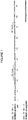
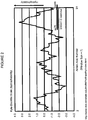
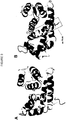
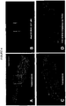

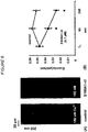
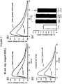

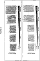
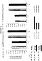

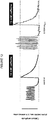
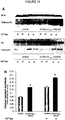
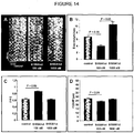

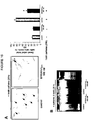


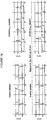


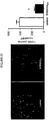
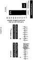
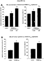

Classifications
-
- C—CHEMISTRY; METALLURGY
- C07—ORGANIC CHEMISTRY
- C07K—PEPTIDES
- C07K14/00—Peptides having more than 20 amino acids; Gastrins; Somatostatins; Melanotropins; Derivatives thereof
- C07K14/435—Peptides having more than 20 amino acids; Gastrins; Somatostatins; Melanotropins; Derivatives thereof from animals; from humans
- C07K14/46—Peptides having more than 20 amino acids; Gastrins; Somatostatins; Melanotropins; Derivatives thereof from animals; from humans from vertebrates
- C07K14/47—Peptides having more than 20 amino acids; Gastrins; Somatostatins; Melanotropins; Derivatives thereof from animals; from humans from vertebrates from mammals
- C07K14/4701—Peptides having more than 20 amino acids; Gastrins; Somatostatins; Melanotropins; Derivatives thereof from animals; from humans from vertebrates from mammals not used
- C07K14/4728—Calcium binding proteins, e.g. calmodulin
-
- A—HUMAN NECESSITIES
- A61—MEDICAL OR VETERINARY SCIENCE; HYGIENE
- A61K—PREPARATIONS FOR MEDICAL, DENTAL OR TOILETRY PURPOSES
- A61K38/00—Medicinal preparations containing peptides
- A61K38/16—Peptides having more than 20 amino acids; Gastrins; Somatostatins; Melanotropins; Derivatives thereof
- A61K38/17—Peptides having more than 20 amino acids; Gastrins; Somatostatins; Melanotropins; Derivatives thereof from animals; from humans
- A61K38/1703—Peptides having more than 20 amino acids; Gastrins; Somatostatins; Melanotropins; Derivatives thereof from animals; from humans from vertebrates
- A61K38/1709—Peptides having more than 20 amino acids; Gastrins; Somatostatins; Melanotropins; Derivatives thereof from animals; from humans from vertebrates from mammals
-
- A—HUMAN NECESSITIES
- A61—MEDICAL OR VETERINARY SCIENCE; HYGIENE
- A61K—PREPARATIONS FOR MEDICAL, DENTAL OR TOILETRY PURPOSES
- A61K45/00—Medicinal preparations containing active ingredients not provided for in groups A61K31/00 - A61K41/00
- A61K45/06—Mixtures of active ingredients without chemical characterisation, e.g. antiphlogistics and cardiaca
-
- A—HUMAN NECESSITIES
- A61—MEDICAL OR VETERINARY SCIENCE; HYGIENE
- A61P—SPECIFIC THERAPEUTIC ACTIVITY OF CHEMICAL COMPOUNDS OR MEDICINAL PREPARATIONS
- A61P21/00—Drugs for disorders of the muscular or neuromuscular system
-
- A—HUMAN NECESSITIES
- A61—MEDICAL OR VETERINARY SCIENCE; HYGIENE
- A61P—SPECIFIC THERAPEUTIC ACTIVITY OF CHEMICAL COMPOUNDS OR MEDICINAL PREPARATIONS
- A61P21/00—Drugs for disorders of the muscular or neuromuscular system
- A61P21/02—Muscle relaxants, e.g. for tetanus or cramps
-
- A—HUMAN NECESSITIES
- A61—MEDICAL OR VETERINARY SCIENCE; HYGIENE
- A61P—SPECIFIC THERAPEUTIC ACTIVITY OF CHEMICAL COMPOUNDS OR MEDICINAL PREPARATIONS
- A61P21/00—Drugs for disorders of the muscular or neuromuscular system
- A61P21/04—Drugs for disorders of the muscular or neuromuscular system for myasthenia gravis
-
- A—HUMAN NECESSITIES
- A61—MEDICAL OR VETERINARY SCIENCE; HYGIENE
- A61P—SPECIFIC THERAPEUTIC ACTIVITY OF CHEMICAL COMPOUNDS OR MEDICINAL PREPARATIONS
- A61P9/00—Drugs for disorders of the cardiovascular system
-
- A—HUMAN NECESSITIES
- A61—MEDICAL OR VETERINARY SCIENCE; HYGIENE
- A61P—SPECIFIC THERAPEUTIC ACTIVITY OF CHEMICAL COMPOUNDS OR MEDICINAL PREPARATIONS
- A61P9/00—Drugs for disorders of the cardiovascular system
- A61P9/04—Inotropic agents, i.e. stimulants of cardiac contraction; Drugs for heart failure
-
- A—HUMAN NECESSITIES
- A61—MEDICAL OR VETERINARY SCIENCE; HYGIENE
- A61P—SPECIFIC THERAPEUTIC ACTIVITY OF CHEMICAL COMPOUNDS OR MEDICINAL PREPARATIONS
- A61P9/00—Drugs for disorders of the cardiovascular system
- A61P9/06—Antiarrhythmics
-
- A—HUMAN NECESSITIES
- A61—MEDICAL OR VETERINARY SCIENCE; HYGIENE
- A61P—SPECIFIC THERAPEUTIC ACTIVITY OF CHEMICAL COMPOUNDS OR MEDICINAL PREPARATIONS
- A61P9/00—Drugs for disorders of the cardiovascular system
- A61P9/10—Drugs for disorders of the cardiovascular system for treating ischaemic or atherosclerotic diseases, e.g. antianginal drugs, coronary vasodilators, drugs for myocardial infarction, retinopathy, cerebrovascula insufficiency, renal arteriosclerosis
Definitions
- the present invention relates to a peptide comprising a muscle function enhancing amino acid sequence derived from an S100 protein, which can be used for treating or preventing myopathies, in particular for treating or preventing skeletal muscle or cardiac muscle disorders, a pharmaceutical composition comprising said peptide, and a method for treating or preventing such myopathies.
- Muscle tissue is subdivided into skeletal muscle, cardiac muscle, and smooth muscle tissue and can be considered the biggest organ of a vertebrate.
- an average adult human male is made up of 40 to 50% skeletal muscle.
- Skeletal muscle and cardiac muscle belong to the striated muscle tissue and share many functional aspects.
- the process of excitation-contraction coupling in skeletal muscle cells and cardiac muscle cells is very similar.
- Membrane depolarization of the myocytes causes calcium influx via activated voltage-gated L-type calcium channels into the cytoplasm (sarcoplasm) of the myocyte.
- cytoplasmic calcium concentration leads to calcium release from the sarcoplasmic reticulum (SR) by activation of ryanodine receptors (RyR) through the calcium-induced calcium release (CICR) mechanism, and thus, to a further rapid rise of the cytoplasmic calcium concentration.
- Calcium molecules diffuse through the cytoplasm and bind to the contractile proteins such as troponin C which causes contraction of the myocytes. After contraction, calcium is cleared from the cytoplasm by re-uptake of calcium into the sarcoplasmic reticulum mainly by the action of a sarcoplasmic/endoplasmic reticulum calcium ATPase (SERCA).
- SERCA sarcoplasmic/endoplasmic reticulum calcium ATPase
- RyR1 is the predominant sarcoplasmic reticulum calcium release channel in skeletal muscle cells
- RyR2 is predominant in cardiomyocyte.
- the skeletal muscle sarcoplasmic/endoplasmic reticulum calcium ATPase is SERCA1a
- SERCA2a is cardiomyocyte-specific.
- S100A1 belonging to the S100 protein family (the largest EF-hand calcium-binding protein subfamily) has been reported to interact with both the RyR calcium release channel and SERCA.
- S100A1 stabilizes RyR in diastole reducing the frequency of calcium sparks and augments calcium release during systole.
- S100A1 increases SERCA activity during the relaxation phase and it was found to increase contractile function in cardiac muscle as well as skeletal muscle cells. It has been shown that a carboxy-terminal peptide derived from the S100A1 protein mimics the inotropic effect of the full-length S100A1 protein ( Most P. et al., 2007, Am. J. Physiol. Regul. Integr. Comp. Physiol. 293:R568-577 ; Voelkers M. et al., 2007, Cell Calcium 41:135-143 ).
- Defective calcium cycling in myocytes results in a variety of myopathies, i.e., diseases associated with muscular malfunction.
- myopathies i.e., diseases associated with muscular malfunction.
- cardiac insufficiency, contractile ventricular dysfunction, arrhythmias, heart failure, cardiogenic shock, myocardial infarction, and dysfunction of heart valves have been associated with dysregulation of calcium handling in cardiomyocytes.
- defective calcium cycling in skeletal muscle fibers has been linked with muscular dystrophy ( Hopf F.W. et al., 2007, Subcell. Biochem.
- myopathies preferably myopathies associated with dysregulation of calcium cycling in muscle cells, which do not exhibit the severe side effects observed for the approved therapeutics and which do not require the high risk delivery route of gene therapy.
- myopathies preferably myopathies associated with dysregulation of calcium cycling in muscle cells, which do not exhibit the severe side effects observed for the approved therapeutics and which do not require the high risk delivery route of gene therapy.
- any inotropic therapeutics having the ability to increase the contractile performance of skeletal muscle cells and/or reducing calcium-induced apoptotic cell death in skeletal muscle cells.
- peptides which are derived from the calcium binding protein S100 exhibit inotropic effects when administered parenterally and are useful for the treatment of myopathies, such as cardiac and skeletal muscle disorders, without exhibiting mentionable side effects and without requiring gene therapy.
- myopathies such as cardiac and skeletal muscle disorders
- the peptides enhance and restore inotropy in normal and failing myocardium as well as in normal and diseased skeletal muscle, enhance and restore sarcoplasmic reticulum calcium handling, prevent calcium induced apoptotic cell death in myocytes, protect from proarrhythmogenic sarcoplasmic reticulum calcium leak and tachyarrhythmias, and prevent cardiac death due to protection from pump failure and tachyarrhythmias.
- the peptides described herein are particularly useful for enhancing contractile performance of cardiac and skeletal muscle tissue without major side effects.
- the present invention provides a peptide comprising a muscle function enhancing amino acid sequence comprising or consisting of the amino acid motif ⁇ 4 -X- ⁇ -L-[T/A]- ⁇ 2 , wherein ⁇ and ⁇ are in each instance an independently selected hydrophobic non-aromatic amino acid, and X is any amino acid, preferably a small amino acid, wherein the muscle function enhancing amino acid sequence does not contain more than 18 continuous amino acids of the carboxy-terminal amino acids of an S100A1 protein, preferably of any S100 calcium binding protein, the peptide further comprises a hydrophilic motif, the peptide has a total length of maximally 100 amino acids, and the peptide exhibits a positive inotropic action for use in treating or preventing a disorder associated with muscular malfunction; wherein the disorder is a cardiac and/or skeletal muscle disorder; wherein the (a) cardiac muscle disorder is selected from the group consisting of postischemic contractile dysfunction, congestive heart failure, cardiogenic shock, septic
- said peptide is capable of penetrating cell membranes.
- said peptide exhibits the ability to enhance contractile performance and/or calcium cycling in myocytes.
- the muscle function enhancing amino acid sequence comprises, essentially consists or consists of the amino acid sequence [V/I]-[V/I]-[L/M]-[V/I/M]-[A/G/S]-[A/V]-L-[T/A]-[V/A/I]-[A/M/V], preferably comprises, essentially consists or consists of the amino acid sequence V-V-L-V-A-A-L-T-V-A (SEQ ID NO: 4).
- the muscle function enhancing amino acid sequence does not contain more than 18 continuous amino acids of the carboxy-terminal amino acids of an S100A1 protein.
- said peptide further comprises one or more of the elements selected from the group consisting of a membrane penetration enhancing motif, one or more epitope-tag(s), and a peptide targeting motif.
- the hydrophilic motif comprises, essentially consists or consists of the hydrophilic amino acid motif ⁇ 4 - ⁇ 2 , wherein A is in each instance independently selected from aspartate, glutamate, lysine, and arginine, and ⁇ is an ⁇ -helix interrupter, preferably is in each instance independently selected from proline or glycine.
- the hydrophilic motif comprises, essentially consists or consists of the amino acid sequence [D/E]-[K/R]-[D/E]-[D/E]-[P/G]-[P/G], more preferably comprises, essentially consists or consists of the amino acid sequence D-K-D-D-P-P (SEQ ID NO: 354).
- the peptide comprises or consists of the amino acid sequence D-K-D-D-P-P-V-V-L-V-A-A-L-T-V-A (SEQ ID NO: 370), or an amino acid sequence which is at least 60% identical to said amino acid sequence.
- the muscular malfunction is associated with defective calcium cycling and/or defective contractile performance in muscle cells.
- the peptide is for enhancing and/or restoring calcium cycling and/or for enhancing and/or restoring contractile performance in muscle cells.
- the present invention provides a composition comprising the peptide described in the first aspect of the present invention and a medicament selected from the group consisting of catecholamines, ⁇ -adrenergic receptor agonists, and ⁇ -adrenergic receptor blockers.
- the muscle function enhancing amino acid sequence comprises, essentially consists or consists of the amino acid motif [V/I]-[V/I]-[L/M]-[V/I/M]-[A/G/S]-[A/V]-L-[T/A]-[V/A/I]-[A/M/V], and in another embodiment the hydrophilic motif comprises, essentially consists or consists of the hydrophilic amino acid motif ⁇ 4 - ⁇ 2 , wherein A and ⁇ are as defined herein below and the hydrophilic motif is preferably directly linked to the amino terminus of the amino acid motif comprised by the muscle function enhancing amino acid sequence, a peptide comprising the amino acid sequence ⁇ 4 - ⁇ 2 -[V/I]-[V/I]-[L/M]-[V/I/M]-[A/G/S]-[A/V
- a “peptide” in the context of the present invention refers to a chain of amino acid residues which may be naturally occurring or derivatives of naturally occurring amino acid residues and which are preferably linked via peptide bonds, wherein the peptide consists of not more than 100 amino acid residues or amino acid residue derivatives.
- the term "amino acid” encompasses naturally occurring amino acids as well as amino acid derivatives.
- a "small amino acid” in the context of the present invention is preferably an amino acid having a molecular weight of less than 125 Dalton.
- a small amino acid in the context of the present invention is selected from the group consisting of the amino acids glycine, alanine, serine, cysteine, threonine, and valine, or derivatives thereof.
- a hydrophobic non-aromatic amino acid in the context of the present invention is preferably any amino acid which has a Kyte-Doolittle hydropathy index of higher than 0.5, more preferably of higher than 1.0, even more preferably of higher than 1.5 and is not aromatic.
- a hydrophobic non-aromatic amino acid in the context of the present invention is selected from the group consisting of the amino acids alanine (Kyte Doolittle hydropathy index 1.8), methionine (Kyte Doolittle hydropathy index 1.9), isoleucine (Kyte Doolittle hydropathy index 4.5), leucine (Kyte Doolittle hydropathy index 3.8), and valine (Kyte Doolittle hydropathy index 4.2), or derivatives thereof having a Kyte Doolittle hydropathy index as defined above.
- Muscle in the context of the present invention means preferably striated muscle tissue or muscle cells derived from striated muscle tissue such as skeletal muscle cells/tissue and cardiac muscle cells (cardiomyocytes) and cardiac muscle tissue.
- the term "muscle function enhancing amino acid sequence” refers to an amino acid sequence that is capable of enhancing and/or restoring any muscle-specific function, for example, enhancing the contractile performance of muscle cells and muscle tissue, preferably of striated muscle tissue, most preferably of cardiac and skeletal muscle cells and tissues. Since it is assumed that proper muscle function is tightly dependent on a functioning calcium handling within the muscle cell, the term “muscle function enhancing amino acid sequence” also refers to an amino acid sequence that is capable of enhancing and/or restoring the calcium handling/cycling, preferably the sarcoplasmic reticulum calcium handling/cycling in muscle cells, preferably skeletal muscle cells and/or cardiomyocytes.
- Contractile performance in myocytes can be directly measured, for example, by assessing single cardiomyocyte electrical field stimulated contractility using the video-edge-detection (VED) technique ( Most et al., 2004, J. Clin. Invest. 114:1550-1563, page 1561 ).
- VED video-edge-detection
- Calcium handling can be determined by assessing calcium transients using fluorescent calcium indicators ( Most et al., 2004, J. Clin. Invest. 114:1550-1563, page 1561 ).
- enhancing in the context of the present invention, e.g., enhancing muscle function, contractile performance, and/or calcium handling, means that the particular function is increased/enhanced independently of whether the function is normal or defective, i.e., the muscle cell is healthy or diseased.
- enhancing means that the particular function is enhanced by at least 15%, preferably by at least 25%, preferably by at least 35%, more preferably by at least 45%, and most preferably by at least 50% compared to a control setting.
- the control setting is the muscle function, contractile performance, and/or calcium handling of a healthy patient or the average of a group of healthy patients.
- restoring in the context of the present invention, e.g., restoring muscle function, contractile performance, and/or calcium handling, preferably means that a defective function is brought back to at least 50% of the normal function, preferably to at least 60% of the normal function, preferably to at least 70% of the normal function, more preferably to at least 80% of the normal function, more preferably to at least 85% of normal function, even more preferably to at least 90% of the normal function, even more preferably to at least 95% of the normal function, and most preferably to at least 100% of the normal function, wherein "normal function” means an average value of the function exhibited by muscle cells derived from an individual who does not suffer from any muscle diseases.
- Roughly "normal” +dp/dt in anesthetized mice can range from 5000-8000 mmHg/sec, "normal” Echo EF% 60-80% / FS% 40-70%, "normal” cellular FS% can range from 5-12% and calibrated calcium transients might range from 200 to 400 nM.
- inotropic action with respect to an agent means that said agent affects the force of muscle contraction irrespective of the muscle type.
- “Positive inotropic action” means that the force of muscle contraction is increased, wherein “negative inotropic action” means that the force of muscle contraction is decreased.
- the peptide for use in treating or preventing a disorder associated with muscular malfunction of the present invention exhibits a positive inotropic action, preferably in vitro as well as in vivo.
- the inotropic effect of an agent, e.g., of the peptide for use in treating or preventing a disorder associated with muscular malfunction of the present invention can be readily determined in vitro, for example, by determining calcium transients in stimulated myocytes with and without the agent/peptide to be tested.
- calcium transients can be assessed in FURA2-AM field-stimulated cardiomyocytes employing epifluorescence digitalized microscopy ( Most et al., 2004, J. Clin. Invest. 114: 1550-1563, page 1561 ). Any fluorescent calcium indicator can be used instead of FURA-2AM such as a member of the Fluo calcium indicator family or Rhod-2AM. The underlying principal remains the same.
- calcium transient measurements in patch-clamped isolated cardiomyocytes ( Kettlewell/Most et al., 2005, J. Mol. Cell. Cardiol., 200: 900-910, page 901 ) may also be used.
- the positive inotropic effect of a peptide can also be tested in vivo, for example, by determining the contractile performance by left ventricular catherization in anesthetized mice with and without administration of the peptide.
- contractility is described as the first derivative of maximal left ventricular pressure rise (+dp/dt max) ( Most et al., 2004, J. Clin. Invest. 114: 1550-1563 ; Most et al., 2006, Circulation 114;1258-1268 )
- echocardiography Most et al., 2006, Circulation 114; 1258-1268 ) can be used.
- enhancing and/or restoring calcium cycling in the context of the present invention means that either calcium cycling in myocytes, preferably sarcoplasmic reticulum calcium cycling, is improved under normal/non-pathological conditions or restored to normal function as specified above under pathological conditions, i.e., if calcium cycling is defective.
- Defective calcium cycling may be a result of reduced calcium content in the sarcoplasmic reticulum, reduced release of calcium from the sarcoplasmic reticulum during excitation-contraction coupling, calcium leakage from the sarcoplasmic reticulum in quiescent muscle cells, for example, due to leaky RyR sarcoplasmic reticulum calcium release channels, increased calcium spark frequency, or reduced/slowed re-uptake of calcium into the sarcoplasmic reticulum and/or the mitochondria after contraction, for example, due to a malfunctioning or non-functioning sarcoplasmic/endoplasmic reticulum calcium ATPase (SERCA).
- SERCA malfunctioning or non-functioning sarcoplasmic/endoplasmic reticulum calcium ATPase
- the calcium cycling can preferably be enhanced or restored by improving said parameters, e.g., increasing sarcoplasmic reticulum calcium content, increasing release of calcium from the sarcoplasmic reticulum during excitation-contraction coupling, reducing calcium leakage from the sarcoplasmic reticulum in quiescent muscle cells, reducing calcium spark frequency, and/or improving calcium re-uptake into the sarcoplasmic reticulum or the mitochondria.
- defective calcium cycling is one of the major reasons for defective contractile performance, e.g., contractile dysfunction, of muscle cells.
- enhancing or restoring calcium cycling also enhances and/or restores contractile performance.
- contractile performance encompasses any function that is associated with muscle contraction, for example, the force of muscle contraction or the timing of muscle contraction. In case of skeletal muscle tetanic contractions fall also within the term “contractile performance”. "Defective contractile performance” refers to contractile dysfunction when compared to average values for normal/healthy muscle cells or tissue.
- the contractile performance of a muscle cell or tissue is considered defective if, for example, the force of contraction of a given muscle cell or tissue deviates from the average value for normal/healthy muscle cells or tissue by at least 10%, preferably at least 20%, preferably at least 30%, more preferably at least 40%, and most preferably at least 50%, wherein the term "deviate" can refer to values less than the normal average value or to values higher than the normal average value, preferably it refers to values less than the normal average value.
- the term "deviate” can refer to values less than the normal average value or to values higher than the normal average value, preferably it refers to values less than the normal average value.
- an echocardiographic cardiac EF% below 50% is considered as beginning heart failure. Normal human cardiac conscious EF% is around 65-70%.
- the term "enhancing and/or restoring contractile performance” means the increase of contractile force of muscle cells or tissue, preferably skeletal muscle cells or tissue or cardiac muscle cells or tissue, as well as the correction of defective timing of muscle cell contractions.
- the term “defective timing” refers to inappropriately timed muscle contraction events such as arrhythmias in the heart muscle or tremor or twitching of skeletal muscle tissue.
- Anti-arrhythmic potential in the context of the peptide described herein means that the peptide is capable of reducing inappropriately timed muscle contractions, i.e., arrhythmic events in myocytes, preferably in cardiomyocytes and cardiac tissue.
- the peptide described herein preferably protects cardiomyocytes from pro-arrhythmic store overload-induced calcium release (SOICR) which is a critical pathomechanism underlying arrhythmogenic sudden cardiac death, e.g., by lethal ventricular arrhythmias.
- SOICR pro-arrhythmic store overload-induced calcium release
- the peptide described herein combines the inotropic action with protection from arrhythmias, preferably calcium-induced arrhythmias in cardiomyocytes.
- a peptide exhibits anti-arrhythmic potency, for example, by assessing whether the peptide to be tested is capable of protecting cardiomyocytes, preferably ventricular cardiomyocytes, with leaky RyR2s sensitized to luminal calcium from ⁇ AR-triggered pro-arrhythmogenic SOICR and calcium waves.
- cardiomyocytes preferably ventricular cardiomyocytes
- leaky RyR2s sensitized to luminal calcium from ⁇ AR-triggered pro-arrhythmogenic SOICR and calcium waves.
- normal ventricular cardiomyocytes may be treated with 10 -7 M Isoproterenol and 0.5 mM caffeine with and without the peptide potentially exhibiting anti-arrhythmic potency and monitor the diastolic calcium concentration.
- Isoproterenol or an equi-effective catecholamine e.g., dobutamine, noradrenaline, adrenaline
- catecholamine e.g., dobutamine, noradrenaline, adrenaline
- other agents enhancing the ⁇ -adrenergic receptor downstream second messenger cyclic adenosine mono-phosphate (cAMP) such as phosphodiesterase inhibitors (rolipram, enoximon) at appropriate equi-effective dosages can be used with or without caffeine.
- cAMP second messenger cyclic adenosine mono-phosphate
- SOICR can be identified by confocal microscopic calcium wave and spark measurements in fluorescent calcium indicator loaded quiescent cardiomyocytes ( Voelkers et al., 2007, Cell Calcium 41:135-143, page 136 ) or as diastolic calcium waves/release in fluorescent calcium indicator loaded electrical field stimulated ( Most et al., 2004, J. Clin. Invest. 114:1550-1563 ) and patch clamped ( Kettlewell et al., 2005, J. Mol. Cell. Cardiol. 39:900-910, page 901 ) cardiomyocytes by epifluorescent microscopy.
- SOICR and calcium wave equivalents such as delayed or early after-contractions can be assessed by diastolic contractions in electrical field stimulated cardiomyocytes by VED ( Most et al., 2004, J. Clin. Invest. 114:1550-1563 ).
- the term "carboxy-terminal amino acids of an S100 protein” preferably refers to the carboxy-terminal 20 amino acids of an S100 protein, e.g., to amino acids 75 to 94 of the amino acid sequence set forth in SEQ ID NO: 1, i.e., the amino acid sequence Y-V-V-L-V-A-A-L-T-V-A-C-N-N-F-F-W-E-N-S (SEQ ID NO: 2), more preferably to the carboxy-terminal 25 amino acids of an S100 protein, and most preferably to the carboxy-terminal 30 amino acids of an S100 protein.
- a cell in the context of the present invention is a muscle cell, preferably a skeletal muscle cell or a cardiomyocyte.
- a mammalian muscle cell preferably a muscle cell, preferably a skeletal muscle cell or a cardiomyocyte.
- the cell in the context of the present invention is a mammalian muscle cell.
- a peptide is capable of penetrating cell membranes, e.g., by labeling said peptide, for example, with a radioactive or fluorescent marker, and incubating the labeled peptide with intact cells, preferably mammalian muscle cells, for example, rat ventricular cardiomyocytes, and assessing whether the labeled peptide can be detected inside the cells, for example, in the cytoplasm of the intact cells, e.g., by fluorescence microscopy ( Most et al., 2005, J. Cell Sci. 118:421-431, page 422 ; Voelkers et al., 2007, Cell Calcium 41:135-143, page 136 ).
- An S100 calcium binding protein in the context of the present invention is preferably selected from the group consisting of S100 calcium binding protein A1, S100 calcium binding protein Z, S100 calcium binding protein T, S100 calcium binding protein S, and the S100 protein ⁇ -chain. Most preferably the S100 calcium binding protein in the context of the present invention is S100 A1.
- the S100 calcium binding protein in the context of the present invention may be of any species, for example, human or other primate, mouse, or rat S100 protein etc, and is preferably of human origin.
- S100 calcium binding proteins are those accessible by the following GenBank or Ref Seq accession numbers: XP_001494920.1, XP_001365057.1, XP_001140144, XP_513820.2, XP_001111052.1, CAI19674.1, XP_537265.1, NP_001092512.1, NP_006262.1, NP_001127319.1, AAB20539.2, NP_001007637.1, NP_035439.1, XP_002196029.1, XP_001332692.1, NP_001082820.1, XP_001504000.2, NP_570128.2, XP_526887.2, XP_226710.1, XP_607154.2, XP_853219.1, NP_001074628.1, NP_001013513.1, AAN63527.1, ACI68060.1, and XP_001344575.2.
- the term "treating" a disease or disorder means that a disease condition is ameliorated independently whether the cause of the disease is eliminated, i.e., the individual having the disease is cured, or only the symptoms are diminished.
- the peptide described herein exerts its therapeutic effects by stabilizing and/or restoring the calcium cycling/handling in muscle cells, and thereby, improving contractile performance of said cells
- the peptide may also be used for the treatment of muscle diseases which are not caused by defective calcium cycling.
- the symptoms of a skeletal muscle disorder such as muscle weakness, which are not caused by or are not associated with defective calcium cycling in the muscle cells, are also diminished by the peptide described herein.
- the term "individual” in the context of the present invention preferably refers to an animal patient, preferably suffering from a cardiac muscle disorder or a skeletal muscle disorder or suffering from both.
- An animal patient is preferably a vertebrate patient, more preferably a mammalian patient, such as a domesticated animal, e.g., a mouse, rat, cat, guinea pig, rabbit, dog, pig, cow, or horse.
- a domesticated animal e.g., a mouse, rat, cat, guinea pig, rabbit, dog, pig, cow, or horse.
- an animal patient is a human patient and the term "individual” refers to a human patient suffering from a muscle disorder, preferably from a cardiac muscle disorder and/or a skeletal muscle disorder.
- the term "individual” preferably refers to an experimental animal, such as a mouse, rat, rabbit, or primate, most preferably said term in this context refers to a heart failure animal model such as the post-myocardial infarction mouse or rat model (mouse: Most et al., 2006, Circulation 114:1258-1268 , supplement; rat: Most et al., 2004, J. Clin. Invest. 114:1550-1563 ).
- the present invention provides a peptide comprising a muscle function enhancing amino acid sequence comprising or consisting of the amino acid motif ⁇ 4 -X- ⁇ -L-[T/A]- ⁇ 2 , wherein ⁇ and ⁇ are in each instance an independently selected hydrophobic non-aromatic amino acid, and X is any amino acid, preferably a small amino acid, wherein the muscle function enhancing amino acid sequence does not contain more than 18 continuous amino acids, e.g., not more than 18, 17, 16, 15, 14, 13, 12, 11 or 10 continuous amino acids, of the carboxy-terminal amino acids of an S100 calcium binding protein A1, the peptide has a total length of maximally 100 amino acids, e.g., maximally 100, 95, 90, 85, 80, 75, 70, 65, 60, 55, 50, 45, 40, 35, 30, 25, 20, 19, 18, 17, 16, 15, 14, 13, 12, 11, or 10 amino acids, and the peptide exhibits a positive inotropic action for use in treating or preventing a disorder associated with muscular
- L refers to the amino acid leucine, T to the amino acid threonine, and A to the amino acid alanine.
- the peptide has a length of between 10 and 80 amino acids, more preferably of between 10 and 70 amino acids, more preferably of between 10 and 60 amino acids, more preferably between 10 and 50 amino acids, even more preferably between 10 and 40 amino acids, even more preferably between 10 and 30 amino acids, most preferably the peptide has a length of between 10 and 20 amino acids. In a preferred embodiment, the peptide is 15 or 16 amino acids long.
- the peptide for use in treating or preventing a disorder associated with muscular malfunction according to the present invention with the exception of the muscle function enhancing amino acid sequence significantly differs from the carboxy-terminal amino acids of an S100 calcium binding protein A1, preferably from an S100 calcium binding protein selected from the group consisting of S100 calcium binding protein A1, S100 calcium binding protein Z, S100 calcium binding protein T, S100 calcium binding protein S, and S100 protein ⁇ -chain, and most preferably significantly differs from the carboxy-terminus of any S100 calcium binding protein.
- the peptide with the exception of the muscle function enhancing amino acid sequence significantly differs from the amino acid sequence of an S100 calcium binding protein A1, preferably from an S100 calcium binding protein selected from the group consisting of S100 calcium binding protein A1, S100 calcium binding protein Z, S100 calcium binding protein T, S100 calcium binding protein S, and S100 protein ⁇ -chain, and most preferably significantly differs from the amino acid sequence of any S100 calcium binding protein.
- the term "significantly differs" means that the amino acid sequences are at least 80% different, more preferably at least 85%, more preferably at least 90%, even more preferably at least 95%, and most preferably 100% different. The difference in the sequences may be assessed by aligning the polypeptide sequences.
- Such alignment tools are well known to the person skilled in the art and can be, for example, obtained on the World Wide Web, e.g., ClustalW (www.ebi.ac.uk/clustalw) or Align (http://www.ebi.ac.uk/ emboss/align/ index.html) using standard settings, preferably for Align EMBOSS::needle, Matrix: Blosum62, Gap Open 10.0, Gap Extend 0.5. Residues in two or more polypeptide sequences are said to differ from each other if the residues which are aligned in the best sequence alignment differ from each other. The "best sequence alignment" between two polypeptides is defined as the alignment that produces the largest number of aligned identical residues.
- the muscle function enhancing amino acid sequence comprises, essentially consists or consists of the amino acid motif [Y/F/W]- ⁇ 4 -X- ⁇ -L-[T/A]- ⁇ 2 , wherein ⁇ and ⁇ are in each instance an independently selected hydrophobic non-aromatic amino acid, and X is any amino acid, preferably a small amino acid.
- the hydrophobic non-aromatic amino acid and the small amino acid are as defined above.
- the muscle function enhancing amino acid sequence of the peptide forms an ⁇ -helical structure.
- the peptide is capable of penetrating cell membranes, preferably vertebrate cell membranes, even more preferably mammalian cell membranes, even more preferably mammalian muscle cell membranes, and most preferably mammalian skeletal muscle cell membranes and membranes of mammalian cardiomyocytes.
- the peptide is capable of penetrating cell membranes as defined above in a physiological environment such as in culture medium, for example, for mammalian tissue culture, and/or in body fluids such as in blood.
- the peptide is capable of penetrating cell membranes in vivo when it is administered by a parenteral administration route such as by intravenous injection.
- the peptide does not contain more than 18, 17, 16, 15, 14, 13, 12, 11, or 10 continuous amino acids of the carboxy-terminal amino acids of an S100 calcium binding protein selected from the group consisting of S 100 calcium binding protein A1, S100 calcium binding protein Z, S100 calcium binding protein T, S100 calcium binding protein S, and S100 protein ⁇ -chain, wherein the S100 calcium binding protein is preferably of human origin, most preferably of any species.
- the peptide does not contain more than 18, 17, 16, 15, 14, 13, 12, 11, or 10 continuous amino acids of the carboxy-terminal amino acids of an S100 calcium binding protein A1, does not contain more than 18, 17, 16, 15, 14, 13, 12, 11, or 10 continuous amino acids of the carboxy-terminal amino acids of an S100 calcium binding protein Z, does not contain more than 18, 17, 16, 15, 14, 13, 12, 11, or 10 continuous amino acids of the carboxy-terminal amino acids of an S100 calcium binding protein T, does not contain more than 18, 17, 16, 15, 14, 13, 12, 11, or 10 continuous amino acids of the carboxy-terminal amino acids of an S100 calcium binding protein S, and does not contain more than 18, 17, 16, 15, 14, 13, 12, 11, or 10 continuous amino acids of the carboxy-terminal amino acids of an S100 protein alpha chain, wherein the S100 calcium binding protein is preferably of human origin, most preferably of any species.
- the peptide does not contain more than 18, 17, 16, 15, 14, 13, 12, 11, or 10 continuous amino acids of the carboxy-terminal amino acids of any S100 calcium binding protein preferably of human origin, more preferably of any species.
- the peptide does not comprise or consist of the sequence Y-V-V-L-V-A-A-L-T-V-A-C-N-N-F-F-W-E-N-S (SEQ ID NO: 2), i.e., amino acids 75 to 94 of the amino acid sequence set forth in SEQ ID NO: 1.
- the peptide exhibits the ability to enhance contractile performance and/or calcium cycling in myocytes, preferably in skeletal muscle cells or cardiomyocytes.
- the peptide exhibits anti-arrhythmic potential on myocytes, preferably on cardiomyocytes, and thus, is preferably capable of protecting myocytes and heart tissue from arrhythmias, preferably from catecholamine triggered arrhythmias, preferably from ventricular arrhythmias which frequently are the cause of sudden cardiac death.
- the peptide exhibits the anti-arrhythmic potential in vitro as well as in vivo.
- the peptide exhibits the ability of protecting an individual from lethal ventricular tachyarrhythmias, preferably from ⁇ -adrenergic receptor ( ⁇ AR) triggered lethal ventricular tachyarrhythmias, preferably from catecholamine triggered lethal ventricular tachyarrhythmias.
- ⁇ AR ⁇ -adrenergic receptor
- the in vivo anti-arrhythmic potential is observed when the peptide is administered via a parenteral administration route.
- the anti-arrhythmic potential of a peptide can be assessed in vitro, for example, by examining whether the peptide protects cardiomyocytes from SOICR as described above.
- the anti-arrhythmic potential of a peptide can be assessed in vivo, for example, by examining the effect of a treatment with the peptide on mortality caused by ⁇ AR triggered tachyarrhythmias in a heart failure animal model, for example, in a post myocardial infarction mouse model ( Most et al., 2006, Circulation 114:1258-1268 , supplement).
- the peptide may be administered to mice with postischemic contractile dysfunction, preferably parenterally, such as intraperitoneally, intravenously, or subcutaneously, daily or every second day for several days, such as 6, 7, 8, or 9 days, up to a few weeks, such as 2, 3, or 4 weeks, preferably 2 weeks.
- the lethal ventricular tachyarrhythmias may be triggered in the animals after a certain period of treatment with the peptide, for example after 7, 8, 9, 10 days or after 2 weeks, by administration of epinephrine, e.g., at a concentration in the range of 1.5 to 2.5 mg/kg, preferably at a concentration of 2 mg/kg, in combination of caffeine, e.g., at a concentration in the range of 100 to 140 mg/kg, preferably at a concentration of 120 mg/kg.
- the lethal ventricular fibrillation may be monitored by telemetric ECG (cf., for example, Xiao et al., 2007, J. Biol. Chem. 282:34828-34838 ).
- the peptide has the ability to reduce calcium spark frequency in myocytes such as skeletal muscle cells and cardiomyocytes, preferably in cardiomyocytes.
- the peptide exhibits the ability to reduce calcium spark frequency in vitro as well as in vivo.
- the in vivo effect is observed when the peptide is administered via a parenteral administration route.
- "Reducing" in this context preferably means that the calcium spark frequency in myocytes treated with the peptide is at least 15%, more preferably at least 25%, even more preferably at least 30%, and most preferably at least 40% reduced compared to control myocytes that have not been treated with the peptide.
- the peptide has the ability of reducing calcium spark frequency in intact cardiomyocytes when added to the liquid in which the cardiomyocytes are present.
- the peptide preferably reduces calcium spark frequency in quiescent cardiomyocytes, e.g., in cultured quiescent rat ventricular cardiomyocytes, when added to the medium of the cardiomyocytes at a concentration in the range of 50 nM to 500 nM, preferably, when applied at a concentration in the range of 50 nM to 250 nM, more preferably when applied at a concentration in the range of 75 to 150 nM, and most preferably when applied at 100 nM, whereas the calcium spark frequency is increased when applied at a concentration of 600 nM or higher, preferably at a concentration of 700 mM or higher, more preferably at a concentration of 800 nM or higher, even more preferably at a concentration of 900 nM or higher
- a peptide has the ability to reduce calcium spark frequency.
- the peptide exhibits an anti-arrhythmic potential and the ability to reduce calcium spark frequency as described above.
- the peptide protects myocytes, preferably skeletal muscle cells and/or cardiomyocytes from apoptotic cell death, preferably from calcium-induced apoptotic cell death, preferably from sarcoplasmic reticulum calcium leakage triggered apoptotic cell death.
- the peptide exhibits anti-apoptotic potential.
- the peptide exhibits this anti-apoptotic effect in vitro as well as in vivo.
- the peptide prevents apoptotic cell death in failing myocardium in vivo, i.e., protects cardiomyocytes in failing myocardium from apoptotic cell death in vivo.
- the in vivo protective effect is observed when the peptide is administered via a parenteral administration route.
- Protecting in this context means that the extent of apoptotic cell death is reduced in the cells treated with the peptide compared to a control group by at least 20%, preferably by at least 30%, more preferably by at least 40%, and even more preferably by at least 50%, and most preferably by at least 60%.
- the skilled person is able to test for this feature in vitro, for example, by observing the extent of apoptosis in myocytes, preferably in ventricular cardiomyocytes, with leaky RyR calcium release channels that are sensitized to luminal calcium by long-term caffeine exposure with and without the peptide.
- An indication for apoptosis is, for example, a fragmented genome which can be examined, e.g., by DNA laddering ( Liu et al., 2005, Circulation 111:90-96 ), cytochrom-c release, or caspase 3 activity ( Most et al., 2003, J. Biol. Chem. 278:48404-48412 ).
- the anti-apoptotic effect of a peptide may be assessed in vivo in an experimental heart failure animal model. For example, mice with postischemic contractile dysfunction may be treated with the peptide and cardiac tissue of treated and control mice may be assessed for the extent of apoptotic cardiomyocytes.
- the peptide may be administered preferably parenterally, such as intraperitoneally, intravenously, or subcutaneously, daily or every second day for several days, such as 6, 7, 8, or 9 days, up to a few weeks, such as 2, 3, or 4 weeks, preferably 2 weeks.
- the extent of apoptotic cells may be assessed by TUNEL staining of TnI and CD31 counterstained heart tissue sections ( Most et al., 2006, Circulation 114:1258-1268 ).
- the peptide exhibits anti-arrhythmic potential and protects myocytes from apoptotic cell death as described above.
- the peptide has the ability to prevent and/or reduce calcium leakage from the sarcoplasmic reticulum, preferably in quiescent myocytes such as skeletal muscle cells and cardiomyocytes.
- the peptide exhibits this effect in vitro and in vivo.
- this in vivo effect is observed when the peptide is administered via a parenteral administration route.
- the peptide stabilizes RyR sarcoplasmic reticulum calcium release channels in their closed conformation, and thereby reduces calcium leakage from these channels ( Most et al., 2006, Circulation 114:1258-1268 ; Voelkers M. et al., 2007, Cell Calcium 41:135-143 ).
- the peptide exhibits anti-arrhythmic potential and prevents and/or reduces calcium leakage as described above.
- the peptide exhibits the ability of restoring hemodynamic function in vivo.
- the peptide restores hemodynamic function in an individual suffering from heart failure such as during or after myocardial infarction.
- this effect is observed when the peptide is administered via a parenteral administration route.
- the skilled person can readily test a peptide for this function, e.g., by using an experimental mouse heart failure model. For example, the skilled person may determine cardiac performance and survival rate in mice with postischemic contractile dysfunction with and without administration of the peptide.
- the peptide may be administered preferably parenterally, such as intraperitoneally, intravenously, or subcutaneously, daily or every second day for several days, such as 6, 7, 8, or 9 days, up to a few weeks, such as 2, 3, or 4 weeks, preferably 2 weeks.
- the left ventricular performance of the experimental animals may be assessed by serial echocardiography ( Most et al., 2003, J. Bio. Chem. 278; 33809-33817 ; Most et al., 2006, Circulation 114:1258-1268 ).
- the peptide exhibits anti-arrhythmic potency and the ability of restoring hemodynamic function in vivo.
- the peptide enhances the isometric and/or tetanic twitch force in skeletal muscle tissue, such as skeletal muscle fibers.
- the peptide exhibits this effect in vitro and in vivo.
- this in vivo effect is observed when the peptide is administered via a parenteral administration route.
- the skilled person can readily assess this function for a given peptide, for example, by isometric tension measurement in peptide treated and untreated intact muscles or muscle fibers, e.g., intact extensor digitorum longum skeletal muscles, isolated from an experimental animal.
- the isolated muscle may be incubated for a certain period of time, such as 30 to 60 minutes, preferably 45 minutes, with the peptide at different concentrations, for example at a concentration in the range of 500 nM to 4 ⁇ M, preferably at a concentration of 1 ⁇ M.
- the isolated muscle may then be stimulated with a tetanic train, for example, applied at 125 Hz for 175 ms and the isometric tension may be measured ( Weisleder et al., 2006, J. Cell Biol. 174:639-654 ).
- the enhancing effect on isometric and/or tetanic twitch force is also observed for muscle fibers isolated from an experimental animal which was treated systemically with the peptide, wherein preferably the peptide was administered parenterally.
- the peptide attenuates skeletal muscle dysfunction and enhances contractile performance in skeletal muscle cells in vivo when administered systemically, preferably parenterally, such as intraperitoneally, intravenously, or subcutaneously.
- the peptide exhibits the ability to enhance isometric and tetanic twitch force in skeletal muscle cells, the ability to increase contractile performance in cardiomyocytes, and the anti-arrhythmic potential described above.
- the peptide exhibits two or more, e.g., 2, 3, 4, or 5, preferably all of the above functions, i.e., anti-arrhythmic potential, anti-apoptotic potential, the ability to reduce calcium spark frequency, the ability to prevent and/or reduce calcium leakage from the sarcoplasmic reticulum, the ability to restore hemodynamic function preferably in an individual suffering from heart failure, and the ability to enhance isometric and/or tetanic twitch force in skeletal muscle cells and/or fibers.
- said functions can be observed in vitro and in vivo.
- said in vivo effects can be observed when the peptide is administered via a parenteral administration route.
- ⁇ is in each instance independently selected from the group of amino acids consisting of alanine, methionine, isoleucine, leucine, and valine, preferably ⁇ is in each instance independently selected from methionine, isoleucine, leucine, and valine.
- ⁇ is in each instance independently selected from the group of amino acids consisting of alanine, methionine, isoleucine, leucine, and valine, preferably ⁇ is in each instance independently selected from alanine, methionine, isoleucine, and valine.
- ⁇ is in each instance independently selected from methionine, isoleucine, leucine, and valine, and ⁇ is in each instance independently selected from alanine, methionine, isoleucine, and valine.
- X is a small amino acid, wherein the small amino acid is preferably not proline.
- X is selected from the group of amino acids consisting of glycine, alanine, serine, cysteine, threonine, and valine, more preferably X is selected from the group consisting of glycine, alanine, and serine.
- ⁇ is in each instance independently selected from methionine, isoleucine, leucine, and valine
- ⁇ is in each instance independently selected from alanine, methionine, isoleucine, and valine
- X is selected from glycine, alanine, serine, cysteine, threonine, and valine, preferably from glycine, alanine, and serine.
- the muscle function enhancing amino acid sequence comprises or consists of the amino acid sequence [V/I]-[V/I]-[L/M]-[V/I/M]-[A/G/S]-[A/V]-L-[T/A]-[V/A/I]-[A/M/V].
- amino acid motif is preferably the sequence [V/I]-[V/I]-[L/M]-[V/I/M]-[A/G/S]-[A/V]-L-[T/A]-[V/A/I]-[A/M/V].
- the muscle function enhancing amino acid sequence comprises or consists of the amino acid sequence V 1 -V 2 -L 3 -V 4 -A 5 -A 6 -L 7 -T 8 -V 9 -A 10 (SEQ ID NO: 3), wherein V 1 and V 2 may be independently replaced by isoleucine, preferably V 1 is not replaced, L 3 may be replaced by methionine, but is preferably not replaced, V 4 may be replaced by isoleucine or methionine, preferably by isoleucine, A 5 may be replaced by glycine or serine, preferably by serine, A 6 may be replaced by valine, T 8 may be replaced by alanine, V 9 may be replaced by alanine or isoleucine, preferably by alanine, and A 10 may be replaced by methionine or valine, but is preferably not replaced, wherein preferably
- maximally 3, e.g., 1, 2, or 3 amino acids are replaced in the most preferred amino acid sequence, i.e., V-V-L-V-A-A-L-T-V-A (SEQ ID NO: 4) as described above.
- the muscle function enhancing amino acid sequence comprises or consists of an amino acid sequence selected from the group consisting of V-V-L-V-A-A-L-T-V-A (SEQ ID NO: 4), I-V-L-V-A-A-L-T-V-A (SEQ ID NO: 5), V-I-L-V-A-A-L-T-V-A (SEQ ID NO: 6), V-V-M-V-A-A-L-T-V-A (SEQ ID NO: 7), V-V-L-I-A-A-L-T-V-A (SEQ ID NO: 8), V-V-L-M-A-A-L-T-V-A (SEQ ID NO: 9), V-V-L-V-G-A-L-T-V-A (SEQ ID NO: 10), V-V-L-V-S-A
- the muscle function enhancing amino acid sequence comprises or consists of an amino acid sequence selected from the group consisting of the above amino acid sequences, i.e., SEQ ID NO: 4 to 333, wherein said amino acid sequences further have or comprise a tyrosine residue directly attached at their N-terminus.
- the muscle function enhancing amino acid sequence comprises or consists of the amino acid sequence Y-V-V-L-V-A-A-L-T-V-A (SEQ ID NO: 390), i.e., the amino acid sequence set forth in SEQ ID NO: 4 having a tyrosine directly attached to its amino-terminus.
- the muscle function enhancing amino acid sequence comprises or consists of the amino acid sequence V-[V/I]-L-[V/I]-[A/S]-[A/V]-L-[T/A]-[V/A]-A (SEQ ID NO: 374), wherein the preferred sequence is V-V-L-V-A-A-L-T-V-A (SEQ ID NO: 4), wherein preferably maximally 4, more preferably maximally 3, even more preferably maximally 2, and most preferably maximally 1 amino acid is replaced as specified above.
- the amino acid motif consists of the amino acid sequence V-[V/I]-L-[V/I]-[A/S]-[A/V]-L-[T/A]-[V/A]-A (SEQ ID NO: 374), wherein the preferred sequence is V-V-L-V-A-A-L-T-V-A (SEQ ID NO: 4), wherein preferably maximally 4, more preferably maximally 3, even more preferably maximally 2, and most preferably maximally 1 amino acid is replaced with another amino acid as specified above.
- the muscle function enhancing amino acid sequence comprises or consists of an amino acid sequence selected from the group consisting of the amino acid sequences V-V-L-V-A-A-L-T-V-A (SEQ ID NO: 4), V-I-L-V-A-A-L-T-V-A (SEQ ID NO: 6), V-V-M-V-A-A-L-T-V-A (SEQ ID NO: 7), I-I-L-V-G-A-L-T-V-A (SEQ ID NO: 95), V-V-L-I-A-A-L-A-A-A (SEQ ID NO: 263), V-I-L-V-S-V-L-T-V-A (SEQ ID NO: 186), I-I-L-M-G-A-L-T-V-A (SEQ ID NO: 334), and V-V-M-V-A-A-L-T-V-V (SEQ ID NO: 4), V-I-L
- the muscle function enhancing amino acid sequence comprises or consists of an amino acid sequence selected from the group consisting of [V/I]-[V/I]-[L/M]-[V/I/M]-[A/G/S]-[A/V]-L-[T/A]-[V/A/I]-[A/M/V]-C, [V/I]-[V/I]-[L/M]-[V/I/M]-[A/G/S]-[A/V]-L-[T/A]-[V/A/I]-[A/M/V]-C-N, [V/I]-[V/I]-[L/M]-[V/I/M]-[A/G/S]-[A/V]-L-[T/A]-[V/A/I]-[A/M/V]-C-N, [V/I]-[V/I]-[L/M]-[V/I/M]-[A
- the muscle function enhancing amino acid sequence comprises or consists of any of the above specifically disclosed amino acid sequences, i.e., specifically disclosed amino acid sequences of the amino acid motif, to which an amino acid or an amino acid sequence selected from the group specified above is linked directly to the carboxy-terminus.
- the muscle function enhancing amino acid sequence comprises or consists of an amino acid sequence selected from the group consisting of V-V-L-V-A-A-L-T-V-A-C (SEQ ID NO: 340), V-V-L-V-A-A-L-T-V-A-C-N (SEQ ID NO: 341), V-V-L-V-A-A-L-T-V-A-C-N-N (SEQ ID NO: 342), V-V-L-V-A-A-L-T-V-A-C-N-N-F (SEQ ID NO: 343), V-V-L-V-A-A-L-T-V-A-C-N-N-F-F (SEQ ID NO: 344), V-V-L-V-A-A-L-T-V-A-C-N-N-F-F-W (SEQ ID NO: 345), V-V-L-V-A-A-L-T-V-A-
- an aromatic amino acid preferably tyrosine, phenylalanine, or tryptophan, more preferably tyrosine or phenylalanine, most preferably tyrosine
- the muscle function enhancing amino acid sequence comprises or consists of one of the above specifically disclosed amino acid sequences extended at their amino-terminus by a tyrosine.
- the peptide for use in treating or preventing a disorder associated with muscular malfunction of the present invention further comprises one or more, e.g., one, two, three, or four, of the elements selected from the group consisting of a membrane penetration enhancing motif, one or more epitope-tag(s), and a peptide targeting motif.
- a membrane penetration enhancing motif may be any amino acid sequence that is capable of penetrating membranes as specified above, e.g., a cell-penetrating peptide (CCP).
- CCP cell-penetrating peptide
- Such a motif may enable other macromolecules, such as peptides, proteins or nucleic acids, which normally do not possess the ability to traverse cell membranes, to penetrate intact cell membranes when said membrane penetration enhancing motif is attached to said macromolecule.
- Such membrane penetration enhancing motifs may be derived from protein transduction domains, may be amphipathic peptides, or may be any other penetrating peptide.
- the membrane penetration enhancing motif may be derived from the HIV Tat peptide, e.g., G-R-K-K-R-R-Q-R-R-R (SEQ ID NO: 348), the penetratin peptide, e.g., R-Q-I-K-I-W-F-Q-N-R-R-M-K-W-K-K (SEQ ID NO: 349) or K-K-W-K-M-R-R-N-Q-F-W-V-K-V-Q-R-G (SEQ ID NO: 350), the transportan peptide, e.g., G-W-T-L-N-S-A-G-Y-L-L-G-K-I-N-L-K-A-L-A-A-L-A-K-K-I-L (SEQ ID NO: 351), an MPG/Pep family member peptide, e.g., G-A-L-F-L-R (
- Such a membrane penetration enhancing motif may be located amino-terminally or carboxy-terminally to the muscle function enhancing amino acid sequence within the peptide for use in treating or preventing a disorder associated with muscular malfunction according to the present invention.
- the peptide may comprise more than one membrane penetration enhancing motif, for example, the peptide may contain 2, 3, 4, or 5 such motifs.
- an epitope is a portion of a molecule to which an antibody binds.
- an epitope is preferably a peptide-tag, for example, hemagglutinin-(HA-), FLAG-, myc-, or a poly-His-tag.
- a peptide-tag for example, hemagglutinin-(HA-), FLAG-, myc-, or a poly-His-tag.
- Such an epitope tag may be used to locate the peptide described herein within a cell, for example, for determining whether the peptide penetrates, i.e., traverses, cell membranes and can be found inside an intact cell incubated with said peptide.
- the peptide for use in treating or preventing a disorder associated with muscular malfunction further comprises a hydrophilic motif.
- said hydrophilic motif comprises acidic, basic, and/or otherwise negatively or positively charged amino acids.
- the hydrophilic motif comprises or consists of the amino acid motif ⁇ 4 - ⁇ 2 , wherein ⁇ is in each instance independently selected from aspartate, glutamate, lysine, and arginine and ⁇ is an ⁇ -helix interrupter, preferably proline or glycine.
- the hydrophilic motif comprises or consists of an amino acid sequence selected from the group consisting of [D/E]-[D/E]-[D/E]-[D/E]-[P/G]-[P/G], [K/R]-[D/E]-[D/E]-[D/E]-[P/G]-[P/G], [D/E]-[K/R]-[D/E]-[D/E]-[P/G]-[P/G], [D/E]-[D/E]-[K/R]-[D/E]-[P/G]-[P/G], [D/E]-[D/E]-[D/E]-[K/R]-[P/G]-[P/G], [K/E]-[D/E]-[D/E]-[K/R]-[P/G]-[P/G], [K/R]-[K/R]-[D/E]-[D/E]
- the hydrophilic motif comprises or consists of the amino acid sequence [D/E]-[K/R]-[D/E]-[D/E]-[P/G]-[P/G]. More preferably, the hydrophilic motif comprises or consists of an amino acid sequence selected from the group consisting of D-K-D-D-P-P (SEQ ID NO: 354), E-K-D-D-P-P (SEQ ID NO: 355), D-R-D-D-P-P (SEQ ID NO: 356), D-K-E-D-P-P (SEQ ID NO: 357), D-K-D-E-P-P (SEQ ID NO: 358), E-R-D-D-P-P (SEQ ID NO: 359), E-K-E-D-P-P (SEQ ID NO: 360), E-K-D-E-P-P (SEQ ID NO: 361), D-R-E-D-P-P (SEQ ID NO: 361), D
- the hydrophilic motif comprises or consists of the amino acid sequence D-K-D-D-P-P (SEQ ID NO: 354), wherein P-P in said sequences may also be G-G.
- the hydrophilic motif is located within the peptide amino-terminally to the muscle function enhancing amino acid sequence, but could also be located carboxy-terminally to the muscle function enhancing amino acid sequence.
- the hydrophilic motif is directly linked to the amino-terminus of the muscle function enhancing amino acid sequence, preferably is directly linked to the amino-terminus of the amino acid motif.
- the peptide comprises, preferably consists of the amino acid sequence ⁇ 4 - ⁇ 2 - ⁇ 4 -X- ⁇ -L-[T/A]- ⁇ 2 , wherein A is in each instance independently selected from aspartate, glutamate, lysine, and arginine, ⁇ is an ⁇ -helix interrupter, preferably is in each instance independently selected from proline or glycine, ⁇ and ⁇ are in each instance an independently selected hydrophobic non-aromatic amino acid, preferably ⁇ is in each instance independently selected from methionine, isoleucine, leucine, and valine, T is in each instance preferably selected from alanine, methionine, isoleucine, and valine, and X is any amino acid, preferably a small amino acid, preferably selected from glycine, alanine, serine, cysteine, threonine, and valine, more preferably selected from glycine,
- said peptide exhibits one or more, e.g., 1, 2, 3, 4, or 5, preferably all of the above defined functional characteristics, i.e., anti-arrhythmic potential, anti-apoptotic potential, the ability to reduce calcium spark frequency, the ability to prevent and/or reduce calcium leakage from the sarcoplasmic reticulum, the ability to restore hemodynamic function preferably in an individual suffering from heart failure, and the ability to enhance isometric and/or tetanic twitch force in skeletal muscle cells and/or fibers.
- said functions can be observed in vitro and in vivo.
- said in vivo effects can be observed when the peptide is administered via a parenteral administration route.
- the peptide for use in treating or preventing a disorder associated with muscular malfunction comprises, preferably consists of the amino acid sequence ⁇ 4 - ⁇ 2 -[V/I]-[V/I]-[L/M]-[V/I/M]-[A/G/S]-[A/V]-L-[T/A]-[V/A/I]-[A/M/V], wherein A is in each instance independently selected from aspartate, glutamate, lysine, and arginine and ⁇ is an ⁇ -helix interrupter, preferably proline or glycine.
- said peptide exhibits one or more, e.g., 1, 2, 3, 4, or 5, preferably all of the above defined functional characteristics, i.e., anti-arrhythmic potential, anti-apoptotic potential, the ability to reduce calcium spark frequency, the ability to prevent and/or reduce calcium leakage from the sarcoplasmic reticulum, the ability to restore hemodynamic function preferably in an individual suffering from heart failure, and the ability to enhance isometric and/or tetanic twitch force in skeletal muscle cells and/or fibers.
- said functions can be observed in vitro and in vivo.
- said in vivo effects can be observed when the peptide is administered via a parenteral administration route.
- the peptide for use in treating or preventing a disorder associated with muscular malfunction comprises, preferably consists of the amino acid sequence [D/E]-[K/R]-[D/E]-[D/E]-[P/G]-[P/G]-[V/I]-[V/I]-[L/M]-[V/I/M]-[A/G/S]-[A/V]-L-[T/A]-[V/A/I]-[A/M/V].
- said peptide exhibits one or more, e.g., 1, 2, 3, 4, or 5, preferably all of the above defined functional characteristics, i.e., anti-arrhythmic potential, anti-apoptotic potential, the ability to reduce calcium spark frequency, the ability to prevent and/or reduce calcium leakage from the sarcoplasmic reticulum, the ability to restore hemodynamic function preferably in an individual suffering from heart failure, and the ability to enhance isometric and/or tetanic twitch force in skeletal muscle cells and/or fibers.
- said functions can be observed in vitro and in vivo.
- said in vivo effects can be observed when the peptide is administered via a parenteral administration route.
- the peptide for use in treating or preventing a disorder associated with muscular malfunction comprises, preferably consists of the amino acid sequence [D/E]-[K/R]-[D/E]-[D/E]-P-P-V-[V/I]-L-[V/I]-[A/S]-[A/V]-L-[T/A]-[V/A]-A (SEQ ID NO: 381).
- said peptide exhibits one or more, e.g., 1, 2, 3, 4, or 5, preferably all of the above defined functional characteristics, i.e., anti-arrhythmic potential, anti-apoptotic potential, the ability to reduce calcium spark frequency, the ability to prevent and/or reduce calcium leakage from the sarcoplasmic reticulum, the ability to restore hemodynamic function preferably in an individual suffering from heart failure, and the ability to enhance isometric and/or tetanic twitch force in skeletal muscle cells and/or fibers.
- said functions can be observed in vitro and in vivo.
- said in vivo effects can be observed when the peptide is administered via a parenteral administration route.
- the peptide for use in treating or preventing a disorder associated with muscular malfunction comprises, preferably consists of the amino acid sequence D 1 -K 2 -D 3 -D 4 -P 5 -P 6 -V 7 -V 8 -L 9 -V 10 -A 11 -A 12 -L 13 -T 14 -V 15 -A 16 (SEQ NO: 370) or an amino acid sequence that is preferably at least 60%, preferably at least 65%, preferably at least 70%, more preferably at least 75%, more preferably at least 80%, even more preferably at least 85%, even more preferably at least 90%, and most preferably at least 95% identical to the amino acid sequence set forth in SEQ ID NO: 370, i.e., D 1 -K 2 -D 3 -D 4 -P 5 -P 6 -V 7 -V 8 -L 9 -V 10 -A 11 -A 12 -L 13 -T 14
- the amino acid replacements are as specified above, e.g., D 1 may be replaced by glutamate, arginine, or lysine, preferably by glutamate, K 2 may be replaced by arginine, glutamate, or aspartate, preferably by arginine, D 3 may be replaced by glutamate, arginine, or lysine, preferably by glutamate, D 4 may be replaced by glutamate, arginine, or lysine, preferably by glutamate, P 5 and P 6 may be independently replaced by glycine, V 7 and V 8 may be independently replaced by isoleucine, preferably V 7 is not replaced, L 9 may be replaced by methionine, but is preferably not replaced, V 10 may be replaced by isoleucine or methionine, preferably by isoleucine, A 11 may be replaced by glycine or serine, preferably by serine, A 12 may be replaced by valine, T 14 may be replaced by alanine,
- said peptide exhibits one or more, e.g., 1, 2, 3, 4, or 5, preferably all of the above defined functional characteristics, i.e., anti-arrhythmic potential, anti-apoptotic potential, the ability to reduce calcium spark frequency, the ability to prevent and/or reduce calcium leakage from the sarcoplasmic reticulum, the ability to restore hemodynamic function preferably in an individual suffering from heart failure, and the ability to enhance isometric and/or tetanic twitch force in skeletal muscle cells and/or fibers.
- said functions can be observed in vitro and in vivo.
- said in vivo effects can be observed when the peptide is administered via a parenteral administration route.
- the peptide comprises, preferably consists of the amino acid sequence D 1 -K 2 -D 3 -D 4 -P 5 -P 6 -V 7 -V 8 -L 9 -V 10 -A 11 -A 12 -L 13 -T 14 -V 15 -A 16 , wherein the amino acid residues may be replaced as specified above (SEQ NO: 371), wherein preferably the amino acid sequence is at least 60%, preferably at least 65%, preferably at least 70%, more preferably at least 75%, more preferably at least 80%, even more preferably at least 85%, even more preferably at least 90%, and most preferably at least 95% identical to the amino acid sequence set forth in SEQ ID NO: 370, wherein preferably one or more, preferably all of the above functions can be observed, and wherein most preferably the peptide is cell permeable.
- the peptide for use in treating or preventing a disorder associated with muscular malfunction comprises, essentially consists, preferably consists of the amino acid sequence D 1 -K 2 -D 3 -D 4 -P 5 -P 6 -Y 7 -V 8 -V 9 -L 10 -V 11 -A 12 -A 13 -L 14 -T 15 -V 16 -A 17 (SEQ ID NO: 372) or an amino acid sequence that is preferably at least 60%, preferably at least 65%, preferably at least 70%, more preferably at least 75%, more preferably at least 80%, even more preferably at least 85%, even more preferably at least 90%, and most preferably at least 95% identical to the amino acid sequence set forth in SEQ ID NO: 372, i.e., D 1 -K 2 -D 3 -D 4 -P 5 -P 6 -Y 7 -V 8 -V 9 -L 10 -V 11 -A 12 -
- the amino acid replacements are as specified above, e.g., D 1 may be replaced by glutamate, arginine, or lysine, preferably by glutamate, K 2 may be replaced by arginine, glutamate, or aspartate, preferably by arginine, D 3 may be replaced by glutamate, arginine, or lysine, preferably by glutamate, D 4 may be replaced by glutamate, arginine, or lysine, preferably by glutamate, P 5 and P 6 may be independently replaced by glycine, Y 7 may be replaced by phenylalanine or tryptophan, preferably by phenylalanine, V 8 and V 9 may be independently replaced by isoleucine, preferably V 8 is not replaced, L 10 may be replaced by methionine, but is preferably not replaced, V 11 may be replaced by isoleucine or methionine, preferably by isoleucine, A 12 may be replaced by glycine or
- said peptide exhibits one or more, e.g., 1, 2, 3, 4, or 5, preferably all of the above defined functional characteristics, i.e., anti-arrhythmic potential, anti-apoptotic potential, the ability to reduce calcium spark frequency, the ability to prevent and/or reduce calcium leakage from the sarcoplasmic reticulum, the ability to restore hemodynamic function preferably in an individual suffering from heart failure, and the ability to enhance isometric and/or tetanic twitch force in skeletal muscle cells and/or fibers.
- said functions can be observed in vitro and in vivo.
- said in vivo effects can be observed when the peptide is administered via a parenteral administration route.
- the peptide comprises, preferably consists of the amino acid sequence D 1 -K 2 -D 3 -D 4 -P 5 -P 6 -Y 7 -V 8 -V 9 -L 10 -V 11 -A 12 -A 13 -L 14 -T 15 -V 16 -A 17 , wherein the amino acid residues may be replaced as specified above (SEQ ID NO: 373), wherein preferably the amino acid sequence is at least 60%, preferably at least 65%, preferably at least 70%, more preferably at least 75%, more preferably at least 80%, even more preferably at least 85%, even more preferably at least 90%, and most preferably at least 95% identical to the amino acid sequence set forth in SEQ ID NO: 372, wherein preferably one or more, preferably all of the above functions can be observed, and wherein most preferably the peptide is cell permeable.
- a peptide targeting motif in the context of the present invention may be any moiety that is suitable for targeting a peptide in vivo to a specific organ or specific cells.
- a peptide targeting motif may be a peptide that specifically binds to a particular receptor which is specific for certain cells or a certain organ.
- the presence of a peptide targeting motif within the peptide allows for specific targeting of cells or organs in a patient to which the peptide was administered systemically.
- the peptide for use in treating or preventing a disorder associated with muscular malfunction of the present invention further comprises a marker moiety.
- a marker moiety in the context of the present invention may be any moiety that allows for a straightforward detection of the peptide, such as a fluorescent label, e.g., fluorescein (for example, fluorescein isothiocyanate FITC), rhodamine (for example, tetramethylrhodamine TAMRA or its isothiocyanate derivative TRITC, sulforhodamine 101 and its sulfonylchloride form Texas RedTM, and Rhodamine Red), or Alexa Fluor ® dyes, a radioactive label, e.g., a radioactively labeled amino acid, or biotin.
- fluorescein for example, fluorescein isothiocyanate FITC
- rhodamine for example, tetramethylrhodamine TAMRA or its isothi
- the peptide comprises a hydrophilic motif, preferably D-K-D-D-P-P (SEQ ID NO: 354), and a marker moiety, preferably FITC or rhodamine, wherein preferably the muscle function enhancing amino acid sequence is V-V-L-V-A-A-L-T-V-A (SEQ ID NO: 4), and preferably the hydrophilic motif is directly linked to the amino-terminus of the muscle function enhancing amino acid sequence.
- a hydrophilic motif preferably D-K-D-D-P-P (SEQ ID NO: 354)
- a marker moiety preferably FITC or rhodamine
- the peptide may be chemically synthesized, e.g., by liquid phase or solid phase peptide synthesis, or the peptide may be genetically engineered using recombinant DNA techniques and a cellular expression system, such as bacteria (e.g., Escherichia coli ), yeast cells, insect cells, mammalian cells etc, or an in vitro expression system.
- bacteria e.g., Escherichia coli
- the present invention provides the peptide described herein for therapeutic use for treating and/or preventing a disorder associated with muscular malfunction, e.g., a myopathy.
- Said disorder is a cardiac and/or skeletal muscle disorder.
- the disorder may be acquired or congenital.
- the term "acquired” means that the medical condition, i.e., the disorder, developed post-fetally.
- Such an acquired disorder in the context of the present invention may be a myocardial infarction.
- An example for an acquired skeletal muscle disorder is myositis.
- Congenital disorders involve defects to a developing fetus which may be the result of genetic abnormalities, errors of morphogenesis, or chromosomal abnormalities.
- Congenital disorders are all congenital, though they may not be expressed or recognized until later in life.
- Congenital disorders in the context of the present invention are, for example, Nemaline myopathy, Myotubular myopathy, or Centronuclear myopathy.
- the cardiac or skeletal muscle disorder may be acute or chronic.
- an acute cardiac muscle disorder is acute heart failure
- an acute skeletal muscle disorder is Rhabdomyolysis.
- a chronic skeletal muscle disorder is, for example, Dermatomyositis.
- a chronic cardiac muscle disease is, for example, chronic heart failure.
- the muscular malfunction is associated with defective calcium cycling and/or defective contractile performance in muscle cells, preferably in skeletal muscle cells or cardiomyocytes.
- the peptide is for enhancing and/or restoring calcium cycling and/or for enhancing and/or restoring contractile performance in muscle cells.
- Defective calcium cycling in myocytes may be a result of reduced calcium content in the sarcoplasmic reticulum, reduced release of calcium from the sarcoplasmic reticulum during excitation-contraction coupling, calcium leakage from the sarcoplasmic reticulum, for example, due to a leaky RyR sarcoplasmic reticulum calcium release channel, increased calcium spark frequency, or reduced or slowed re-uptake of calcium into the sarcoplasmic reticulum and/or the mitochondria after contraction, for example, due to a malfunctioning or non-functioning sarcoplasmic/endoplasmic reticulum calcium ATPase (SERCA).
- SERCA malfunctioning or non-functioning sarcoplasmic/endoplasmic reticulum calcium ATPase
- a defective calcium cycling is one of the major reasons for defective contractile performance, e.g., contractile dysfunction, of muscle cells.
- enhancing or restoring calcium cycling also enhances and/or restores contractile performance.
- cardiac and skeletal muscle disorders/diseases are a result of contractile dysfunction of the respective muscle cells.
- the cardiac muscle contraction is not precisely timed. This may have lethal consequences.
- the contractile performance is reduced which has the consequence of muscle weakness such as in various types of dystrophies.
- the peptide for use in treating or preventing a disorder associated with muscular malfunction is capable of enhancing and/or restoring calcium cycling in myocytes, and thereby, enhances and/or restores contractile performance.
- the peptide is not only suitable for treating disorder associated with muscular malfunction, wherein the muscular malfunction is associated with defective calcium cycling, but also muscular diseases which are not based on malfunctioning calcium handling. In these diseases the peptide may relief the symptoms such as muscle weakness.
- the peptide is for protecting myocytes, preferably skeletal muscle cells and/or cardiomyocytes, more preferably heart tissue from arrhythmias, preferably from catecholamine triggered arrhythmias, preferably for protecting an individual from ventricular arrhythmias, preferably from lethal ventricular tachyarrhythmias, and thus, preferably from sudden cardiac death.
- said function is exhibited in vivo, preferably when the peptide is applied parenterally without the need for gene therapy.
- the peptide is for reducing calcium spark frequency in myocytes, preferably in skeletal muscle cells and/or cardiomyocytes, and/or for preventing and/or reducing calcium leakage from the sarcoplasmic reticulum of myocytes, preferably in skeletal muscle cells and/or cardiomyocytes.
- said function is exhibited in vivo, preferably when the peptide is applied parenterally without the need for gene therapy.
- the peptide is for preventing or reducing calcium leakage from the sarcoplasmic reticulum of muscle cells, preferably of skeletal muscle cells and/or cardiomyocytes.
- the peptide is for preventing or reducing calcium leakage from the sarcoplasmic reticulum due to leaky RyR sarcoplasmic reticulum calcium release channels.
- said function is exhibited in vivo, preferably when the peptide is applied parenterally without the need for gene therapy.
- the peptide is for protecting myocytes, preferably skeletal muscle cells and/or cardiomyocytes from apoptotic cell death, preferably from calcium-induced apoptotic cell death, preferably from sarcoplasmic reticulum calcium leakage triggered apoptotic cell death.
- the peptide is for preventing apoptotic cell death in failing myocardium, i.e., protecting cardiomyocytes from apoptotic cell death in failing myocardium.
- said function is exhibited in vivo, preferably when the peptide is applied parenterally without the need for gene therapy.
- the peptide is for restoring and/or enhancing hemodynamic function, for example, cardiac performance such as contractile performance of cardiomyocytes, preferably the peptide is for restoring and/or enhancing hemodynamic function in an individual suffering or has suffered from heart failure such as from myocardial infarction.
- said function is exhibited in vivo when the peptide is applied parenterally without the need for gene therapy.
- the peptide is for enhancing and/or restoring contractile performance in skeletal muscle cells, preferably for enhancing and/or restoring isometric and/or tetanic twitch force in skeletal muscle cells, preferably in skeletal muscle tissue.
- the isometric twitch force is tension development without muscle shortening
- the tetanic twitch force is the maximal isometric force development, normally, when single contractions start to merge above 50 Hz stimulation.
- said function is exhibited in vivo, preferably when the peptide is applied parenterally without the need for gene therapy.
- the cardiac muscle disorder is selected from the group consisting of postischemic contractile dysfunction, preferably postischemic contractile right and/or left ventricular dysfunction, congestive heart failure, preferably compensated and/or decompensated congestive heart failure, cardiogenic shock, septic shock, myocardial infarction, cardiomyopathy, dysfunction of heart valves, and ventricular disorder, such as acute or chronic right ventricular disorder.
- the skeletal muscle disorder is selected from the group consisting of muscular dystrophy, muscle weakness, muscular atrophy, myositis, central core disease, nemaline (rod) myopathy, centronuclear myopathy, myotubular myopathy, centronuclear myotubular myopathy, ophthalmoplegia of the eye, and mitochondrial myopathy.
- the muscular dystrophy may be selected from the group consisting of Becker's muscular dystrophy, congenital muscular dystrophy, Duchenne muscular dystrophy, distal muscular dystrophy, Emery-Dreifuss muscular dystrophy, facioscapulohumeral muscular dystrophy, Limb-girdle muscular dystrophy, myotonic muscular dystrophy, and oculopharyngeal muscular dystrophy.
- the myositis may be selected from the group consisting of myositis ossificans, fibromyositis, idiopathic inflammatory myopathies (such as dermatomyositis, polymyositis, and inclusion body myositis), and pyomyositis.
- composition comprising the peptide described in the first aspect or a pharmaceutically acceptable salt thereof and a pharmaceutically acceptable excipient, carrier, and/or diluent.
- pharmaceutically acceptable salt refers to a salt of the peptide described herein.
- suitable pharmaceutically acceptable salts include acid addition salts which may, for example, be formed by mixing a solution of the peptide described herein with a solution of a pharmaceutically acceptable acid such as hydrochloric acid, sulfuric acid, fumaric acid, maleic acid, succinic acid, acetic acid, benzoic acid, citric acid, tartaric acid, carbonic acid or phosphoric acid.
- suitable pharmaceutically acceptable salts thereof may include alkali metal salts (e.g., sodium or potassium salts); alkaline earth metal salts (e.g., calcium or magnesium salts); and salts formed with suitable organic ligands (e.g., ammonium, quaternary ammonium and amine cations formed using counteranions such as halide, hydroxide, carboxylate, sulfate, phosphate, nitrate, alkyl sulfonate and aryl sulfonate).
- alkali metal salts e.g., sodium or potassium salts
- alkaline earth metal salts e.g., calcium or magnesium salts
- suitable organic ligands e.g., ammonium, quaternary ammonium and amine cations formed using counteranions such as halide, hydroxide, carboxylate, sulfate, phosphate, nitrate, alkyl sulfonate and aryl sul
- compositions include, but are not limited to, acetate, adipate, alginate, ascorbate, aspartate, benzenesulfonate, benzoate, bicarbonate, bisulfate, bitartrate, borate, bromide, butyrate, calcium edetate, camphorate, camphorsulfonate, camsylate, carbonate, chloride, citrate, clavulanate, cyclopentanepropionate, digluconate, dihydrochloride, dodecylsulfate, edetate, edisylate, estolate, esylate, ethanesulfonate, formate, fumarate, gluceptate, glucoheptonate, gluconate, glutamate, glycerophosphate, glycolylarsanilate, hemisulfate, heptanoate, hexanoate, hexylresorcinate
- excipient when used herein is intended to indicate all substances in a pharmaceutical formulation which are not active ingredients such as, e.g., carriers, binders, lubricants, thickeners, surface active agents, preservatives, emulsifiers, buffers, flavoring agents, or colorants.
- composition described herein is for treating or preventing disorders associated with muscular malfunction as specified above for the peptide for use in treating or preventing a disorder associated with muscular malfunction of the invention in the first aspect of the present invention.
- the pharmaceutical composition described herein is for protecting myocytes from arrhythmias, protecting an individual from ventricular arrhythmias, preferably from lethal ventricular tachyarrhythmias, and thus, preferably from sudden cardiac death, for reducing calcium spark frequency in myocytes, for preventing and/or reducing calcium leakage from the sarcoplasmic reticulum of myocytes, for protecting myocytes from apoptotic cell death, preferably protecting cardiomyocytes from apoptotic cell death in failing myocardium, for restoring and/or enhancing hemodynamic function, preferably enhancing hemodynamic function in an individual suffering from heart failure, and/or for enhancing and/or restoring contractile performance in skeletal muscle cells, preferably for enhancing and/or restoring isometric and/or tetanic twitch force in skeletal muscle cells as described above for the peptide for use in treating or preventing a disorder associated with muscular malfunction according to the present invention in the first aspect of the present invention.
- the pharmaceutical composition contemplated by the present invention may be formulated in various ways well known to one of skill in the art.
- the pharmaceutical composition described herein may be in liquid form such as in the form of solutions, emulsions, or suspensions.
- the pharmaceutical composition described herein is formulated for parenteral administration, preferably for intravenous, intramuscular, subcutaneous, transdermal, intrapulmonary, intraperitoneal, intracardiac administration, or administration via mucous membranes, preferably for intravenous, subcutaneous, or intraperitoneal administration.
- the pharmaceutical composition described herein is in the form of a sterile aqueous solution which may contain other substances, for example, enough salts or glucose to make the solution isotonic with blood.
- the aqueous solutions should be suitably buffered (preferably to a pH of from 3 to 9, more preferably to a pH of from 5 to 7), if necessary.
- the pharmaceutical composition is preferably in unit dosage form.
- the pharmaceutical composition is subdivided into unit doses containing appropriate quantities of the active component.
- the unit dosage form can be a packaged preparation, the package containing discrete quantities of pharmaceutical composition such as vials or ampoules.
- a pharmaceutical composition for ameliorating a disease condition associated with a muscular disorder preferably a skeletal muscle disorder and/or a cardiac muscle disorder, wherein the term "ameliorating a disease condition" is as defined below.
- a method for treating or preventing disorders associated with muscular malfunction comprising administering to an individual in need thereof the peptide described in the first aspect of the present invention or the pharmaceutical composition described herein in an amount sufficient to ameliorate the disease condition of said individual, preferably the patient.
- "ameliorating the disease condition” means, for example, that the individual has a subjective sensation of improvement after a certain period of time after the peptide or the pharmaceutical composition has been administered to the patient, or that the function of the diseased muscle has been measurably improved after treatment with the peptide or the pharmaceutical composition.
- the contractile performance such as the contractile force of a muscular tissue, e.g.
- the disease condition is ameliorated by the treatment if, after the treatment, the contractile performance of said musclular tissue deviates less than 50%, e.g. less than 40%, less than 30%, less than 20%, less than 10%, or not at all, from the average normal contractile function of a corresponding healthy muscular tissue.
- the contractile performance may also be improved compared to the average cardiac performance of a healthy cardiac tissue.
- the term "individual in need thereof" preferably refers to an animal patient, more preferably to a mammalian patient, most preferably to a human patient as defined above.
- the disorder associated with muscular malfunction is preferably as defined for the first aspect of the present invention.
- the method is for protecting myocytes from arrhythmias, protecting an individual from ventricular arrhythmias, preferably from lethal ventricular tachyarrhythmias, and thus, preferably from sudden cardiac death, for reducing calcium spark frequency in myocytes, for preventing and/or reducing calcium leakage from the sarcoplasmic reticulum of myocytes, for protecting myocytes from apoptotic cell death, preferably protecting cardiomyocytes from apoptotic cell death in failing myocardium, for restoring and/or enhancing hemodynamic function in an individual suffering from heart failure, and/or for enhancing and/or restoring contractile performance in skeletal muscle cells, preferably for enhancing and/or restoring isometric and/or tetanic twitch force in skeletal muscle cells as described above for the peptide described in the first aspect of the present invention.
- the peptide or the pharmaceutical composition described herein can be administered to an animal patient, preferably a mammalian patient, preferably a human patient, preferably via a parenteral administration route, for example, intravenously, intramuscularly, subcutaneously, transdermally, intrapulmonary, intraperitoneally, intracardiacally, or via mucous membranes, preferably intravenously, subcutaneously, or intraperitoneally.
- Administration may be by infusion or classical injection, for example, using cannulas, or by needleless injection techniques.
- the present invention provides a composition comprising, essentially consisting or consisting of the peptide described in the first aspect of the present invention in combination with another medicament usually administered for treating or preventing diseases associated with muscular malfunction, preferably skeletal muscle diseases, more preferably cardiac muscle diseases.
- said composition is a pharmaceutical composition which may also comprise one or more pharmaceutically acceptable diluent(s), carrier(s), and/or excipient(s).
- said medicament exhibits pro-arrhythmogenic potential, preferably on cardiomyocytes.
- the peptide reduces the pro-arrhythmogenic potential of said medicament.
- said medicament is a catecholamine, e.g., a direct ⁇ -mimetics such as endogenous or synthetic catecholamine or an indirect ⁇ -mimetics such as a phosphodiesterase inhibitor ⁇ -mimetics or another agent enhancing RyR2 calcium-sensitivity such as caffeine or similar chemicals, e.g., purine alkaloids or dimethylxanthines.
- said medicament is selected form the group consisting of a catecholamine, a ⁇ -adrenergic receptor agonist, and a ⁇ -adrenergic receptor blocker.
- said medicament is a catecholamine such as dobutamine, noradrenaline, adrenaline, dopamine, or isoprenalin, preferably dobutamine, noradrenaline, or adrenaline.
- said medicament is a ⁇ -adrenergic receptor agonist such as isoproterenol, salbutamol, fenoterol, formoterol, metaproterenol, salmeterol, terbutaline, clenbuterol, arbutamine, befunolol, bromoacetylalprenololmenthane, broxaterol, cimaterol, cirazoline, denopamine, epinephrine, etilefrine, hexoprenaline, higenamine, isoetharine, isoxsuprine, mabuterol, methoxyphenamine, nylidrin, oxyfedrine, pirbuterol, prenalterol, pro
- said medicament is a ⁇ -adrenergic receptor blocker such as metoprolol, atenolol, bisoprolol, nebivolol, esmolol, betaxolol, acebutolol, celiprolol, bupranolol, propranolol, timolol, carvedilol, sotalol, pindolol, oxprenolol, or alprenolol, preferably metoprolol, atenolol, bisoprolol, nebivolol, esmolol, or betaxolol, most preferably metoprolol.
- the composition according to the second aspect of the invention may comprise one or more, e.g., 1, 2, 3, or 4 different medicaments either of the same category or of different categories, in combination with the peptide defined in the first aspect of the present invention.
- the present inventors have surprisingly found that the peptides described herein are useful for treating or preventing disorders associated with muscular malfunction as specified throughout the description, that the peptides described herein have the ability to reduce the pro-arrhythmogenic potential of medicaments such as catecholamines, ⁇ -adrenergic receptor agonists, or ⁇ -adrenergic receptor blocker without counteracting their beneficial effects, and that these therapeutic effects of said peptides are exerted even if the peptides are administered parenterally, preferably via an intravenous, intraperitoneal, or subcutaneous administration route, without the need for genetic modification by gene therapy and without causing major side effects.
- medicaments such as catecholamines, ⁇ -adrenergic receptor agonists, or ⁇ -adrenergic receptor blocker
- LSCM Bio-Rad 2000 laser scanning confocal microscope
- a striated staining pattern can be observed that resembles the ryanodine staining pattern ( Figure 4 ).
- Ca 2+ -spark frequency has been assessed in the permeabilized cells treated with S100A1 protein or the S100A1 C-terminal 20-mer peptide fused to a hydrophilic linker and it was shown that both, the full-length protein as well as the C-terminal fragment, decrease calcium spark frequency in permeabilized cardiomyocytes ( Figures 5 and 6 ). Isolated cardiomyocytes were perfused with a mock intracellular solution and permeabilized using ß-escin (0.1 mg/ml).
- the scanning laser line orientated parallel with the long axis and placed approximately equidistant between the outer edge of the cell and the nucleus/nuclei, to ensure the nuclear area was not included in the scan line.
- the high relaxation and the high activation solution contained 50 mM EGTA to buffer free Ca 2+
- the low relaxing solution contained 0.5 mM EGTA and 49.5 mM 1,6-diamino hexane- N , N , N , N -tetraacetic acid (HDTA), which in contrast to EGTA has very low affinity to Ca 2+
- the skinning solution is obtained by the addition of 50 ⁇ g/ml saponin to the low relaxing solution.
- the release solution consisted of the low relaxing solution with 5 mM caffeine added. Loading solution contained 50 mM EGTA to clamp free Ca 2+ to 0.4 ⁇ M (pCa 6.4).
- the preparation was dipped for 1 s into the high relaxing solution and again for 2 min in low relaxing solution.
- the fibers were exposed to the release solution containing 5 mM caffeine until the initial force transient returned to the resting force level. Maximum force was measured in the high activating solution at pCa 4.28 and 5 mM caffeine.
- the fibers then were relaxed in high relaxing solution for 1 min to buffer Ca 2+ .
- Several control transients were recorded before the fiber was exposed to the S100A1 protein or the S100A1 peptide mixture (N/H/C) or the C-terminal 20-mer alone, and the experiment was repeated as outlined above.
- S100A1 protein or peptides were added to the low relaxing solution before and during release and to the high activating solution.
- the pCa-force relation in response to S100A1 interventions was measured with six different Ca 2+ concentrations (EDL, pCa 9.07, 5.91, 5.72, 5.49, 5.17, and 4.28), each containing 5 mM caffeine.
- the EC50 and the Hill coefficient were obtained from a Hilltype fit.
- the EC50 value indicates the Ca 2+ concentration needed for half-maximal isometric force activation, which is as a measure of Ca 2+ sensitivity of the contractile apparatus.
- the Hill coefficient gives an indication of the maximum steepness of the sigmoidal curve.
- the correlation coefficients were calculated to determine the accuracy of the fit.
- the force transient was transformed into the corresponding free Ca 2+ transient by using the individual pCa 2+ force relation as a Ca 2+ indicator and reversing each point of the force transients into the corresponding free Ca 2+ level as previously described.
- the pCa force relation relates free Ca 2+ and force.
- the pCa-force relation can be used as a bioassay, which converts the rather slow force transients from the Ca 2+ release from the SR into apparent Ca 2+ transients.
- rhodamine-labeled S100A1 protein nor the FITC-labeled S100A1 C-terminal 20-mer peptide with or without a hydrophilic motif such as D-K-D-D-P-P (SEQ ID NO: 354) are able to penetrate the cell membrane of adult intact cardiomyocytes.
- the present inventors surprisingly found that a peptide having the sequence D-K-D-D-P-P-Y-V-V-L-V-A-A-L-T-V-A (SEQ ID NO: 372) referred to as S100A1ct 6/11 is cell permeable.
- FITC-labeled S100A1ct 6/11 was incubated with intact rat ventricular cardiomyocytes for 15 minutes before the cells were monitored using confocal laser scanning microscopy. Endogenous S100A1 protein was stained using a conventional immunofluorescence protocol. The intracellular staining pattern of FITC-labeled S100A1ct 6/11 resembles that of endogenous S100A1 ( Figure 8 ).
- Intracellular Ca 2+ transients of mouse ventricular cardiomyocytes were calibrated and measured as previously described (Remppis A. et al., 2002, Basic Res. Cardiol. 97: I/56-I/62). Briefly, isolated cells were washed in HEPES-modified medium 199 (M199) (Sigma), incubated in 1 ml of M199 (2 mM [Ca 2+ ] e ) with 2 ⁇ M Fura2-AM for 20 min at room temperature. Calibration and fluorescence measurements were carried out using an inverse Olympus microscope (Ix70) with a UV filter connected to a monochromator (Polychrome II, T.I.L.L. Photonics GmbH, Germany).
- Ca 2+ transients were investigated at baseline and throughout a stepwise increase of isoproterenol concentrations (10 -9 -10 -5 M) under electrical stimulation at 1 Hz and 2 mM [Ca 2+ ] e in M199.
- SR Ca 2+ load was assessed using a standard caffeine pulse protocol. After 2 min of electrical stimulation (1 Hz), myocytes were abruptly exposed to 0 Na + /0 Ca 2+ solution with caffeine (10 mM). The peak of the caffeine-induced Ca 2+ transient was used as an index of the SR Ca 2+ load.
- Example 4 S100A1ct 6/11 does not alter ⁇ -adrenergic receptor signaling and protects cardiomyocytes from pro-arrhythmic store-overload-induced calcium release (SOICR)
- the inotropic effect of S100A1ct 6/11 is additive to and independent of ⁇ -adrenergic stimulation ( Figure 13 ).
- Ventricular cardiomyocytes have been isolated as described above and the calcium transient amplitude has been assessed in presence and absence of isoproterenol and in presence or absence of the S100A1ct 6/11 peptide, respectively.
- S100A1ct 6/11 peptide protects cardiomyocytes from pro-arrhythmic store-overload-induced calcium release (SOICR) ( Figure 15 ). Calcium sparks were assessed in Fluo-3 AM loaded cardiomyocytes under control and ⁇ AR (10 -7 M Isoproterenol + 0.5 mM caffeine) as described in Ventucci et al., 2007, Circ. Res. 100:105-111 ). It is important to note that the protective effect of S100A1ct 6/11 is effective at concentrations (100 and 1000 nM) that exert inotropic actions in cardiomyocytes due to enhanced SR calcium load.
- S100A1ct 6/11 effectively antagonizes ⁇ AR-triggered SOICR highlighting the unique molecular profile combining inotropic actions with anti-arrhythmic potency.
- the S100A1ct 6/11 peptide exerts significant in vivo hemodynamic effects resulting in enhanced contractile performance under basal and ⁇ AR-stimulated conditions ( Figure 18 ). These hemodynamic effects are effective in response to the ⁇ 1AR-blocker metoprolol ( Figures 19 and 20 ). Furthermore, the S100A1ct 6/11 peptide exerts significant therapeutic effects in vivo restoring hemodynamic function in an experimental heart failure mouse model ( Figure 21 ) and preventing apoptotic cell death in failing myocardium in said mouse model ( Figure 22 ). Furthermore, the S100A1ct 6/11 peptide protects the heart failure mice from ⁇ AR-triggered lethal ventricular tachyarrhythmias ( Figure 22 ).
- Two-dimensional guided M-mode and Doppler echocardiography was carried out using an HDI 5000 echocardiograph (ATL, Bothell, WA) in conscious mice as previously described ( Kohout et al., 2001, Circulation 104:2485-2491 ). Three independent echocardiographic measurements were taken in both modes.
- LVESD left ventricular chamber diameter in endsystole
- LVEDD end-diastole
- IVSth interventricular septum
- LVPth LV posterior wall thickness in end-diastole
- FS% LVEDD - LVESD/ LVEDD x 100; (%).
- LV ejection time (LVET) and heart rate (bpm) taken from aortic valve
- TTE Transthoracic two-dimensional echocardiography
- mice lightly anesthesized mice (tribromoethanol/amylene hydrate; Avertin; 2.5% wt/vol, 8 ⁇ l/g IP) with spontaneous respiration was performed with a 12-MHz probe both in sham and infarcted mice
- a 1.4 French micromanometer-tipped catheter (SPC-320, Millar instruments, Inc.) was inserted into the right carotid artery and then advanced into the LV. Hemodynamic analysis, including heart rate (beats/min -1 ), LV end-diastolic pressure (LVEDP) and maximal (LV +dp/dt max .) and minimal (LV dp/dt min ) first derivate of LV pressure.
- LV tissue was cryosectioned (5 ⁇ m) and stained with hematoxylin-eosin (HE) to measure myocyte width in the remote, non-infarcted area of the LV, and measures were obtained at the level of the nucleus in longitudinally sectioned myocytes using NIH image software (ImageJ 1.34; http:/rsb.info.nih.gov/ij). Terminal deoxy-nucleotidyl transferase-mediated dUTP nick end-labeling (TUNEL) staining was carried out according to the manufacturers protocol (Roche, 11684795001).
- HE hematoxylin-eosin
- the number of TUNEL-positive cardiac myocyte nuclei in the remote area were counted a IX 70 inverse Olympus microscope (T.I.L.L. Vision software, version 3.3) and normalized per 10 5 total nuclei identified by HE staining in the same section.
- the sections were double stained with a cardiac specific anti-troponin C antibody (Santa Cruz, sc-8117, 1:50 dilution) and a corresponding pair of donkey anti-goat Alexa Fluor 568 (Molecular Probes, 1:100) (data not shown).
- Caspase 3 activity in myocardial tissue was measured using a Caspase-Glo assay kit (Promega).
- the proluminescent substrate is cleaved by caspase-3. After caspase cleavage, a substrate for luciferase (aminoluciferin) is released resulting in the luciferase reaction and the production of luminescent signal.
- Cytosolic extracts from heart tissue were prepared by homogenization in hypotonic extraction buffer (25 mM HEPES, pH 7.5, 5 mM MgCl 2 , 1 mM EGTA) containing protease inhibitor mix (1 tablet/5ml) (Roche; Mini complete EDTA free protease inhibitor) and subsequently centrifuged (15 min, 13.000 rpm, 4°C).
- the protein concentration of the supernatant was adjusted to 1 mg/ml with extraction buffer and stored at -80°C.
- An equal volume of reagents and 10 ⁇ g/ml cytosolic protein were added to a white-walled 96-well plate and incubated at room temperature for 1h.
- the luminescence of each sample was measured in triplicates in a plate-reading luminometer.
- the pro-arrhythmic protocol was adapted from the previously published protocol by Wayne Chen and co-workers ( Xiao et al., 2007, J. Biol. Chem. 282:34828-34838 ).
- the S100A1ct 6/11 peptide significantly enhances isometric twitch force in normal and diseased skeletal muscle ( Figure 24 ).
- the protocol used for assessing isometric twitch force in skeletal muscle fibers is described in Example 1.
- S100A1ct 6/11 peptide intact (non-permeabilized) extensor digitorum longum (EDL) skeletal muscles fibers have been used.
- the twitch force of the isolated muscle fibers were enhanced upon S100A1ct 6/11 treatment, irrespective of whether the peptide was incubated with the isolated muscle fiber ( Figure 24 A) or whether the peptide was administered systemically before the muscle fiber was isolated ( Figure 24 B) .
- the protocol for assessing the calcium transient amplitude is described above.
- the linker alone, the vehicle alone, or amino-terminal deletion peptides lacking more than the amino acid 76 do not exhibit the inotropic effect. This experiment demonstrates that the tyrosine at position 76 is not essential for the inotropic function and cell permeability of the S100A1ct 6/11 peptide.
Landscapes
- Health & Medical Sciences (AREA)
- Life Sciences & Earth Sciences (AREA)
- Chemical & Material Sciences (AREA)
- General Health & Medical Sciences (AREA)
- Medicinal Chemistry (AREA)
- Pharmacology & Pharmacy (AREA)
- Animal Behavior & Ethology (AREA)
- Veterinary Medicine (AREA)
- Public Health (AREA)
- Organic Chemistry (AREA)
- Engineering & Computer Science (AREA)
- Bioinformatics & Cheminformatics (AREA)
- Nuclear Medicine, Radiotherapy & Molecular Imaging (AREA)
- General Chemical & Material Sciences (AREA)
- Chemical Kinetics & Catalysis (AREA)
- Cardiology (AREA)
- Zoology (AREA)
- Epidemiology (AREA)
- Proteomics, Peptides & Aminoacids (AREA)
- Gastroenterology & Hepatology (AREA)
- Heart & Thoracic Surgery (AREA)
- Neurology (AREA)
- Orthopedic Medicine & Surgery (AREA)
- Physical Education & Sports Medicine (AREA)
- Immunology (AREA)
- Marine Sciences & Fisheries (AREA)
- Toxicology (AREA)
- Biochemistry (AREA)
- Biophysics (AREA)
- Genetics & Genomics (AREA)
- Molecular Biology (AREA)
- Pain & Pain Management (AREA)
- Vascular Medicine (AREA)
- Urology & Nephrology (AREA)
- Hospice & Palliative Care (AREA)
- Medicines That Contain Protein Lipid Enzymes And Other Medicines (AREA)
- Peptides Or Proteins (AREA)
Description
- The present invention relates to a peptide comprising a muscle function enhancing amino acid sequence derived from an S100 protein, which can be used for treating or preventing myopathies, in particular for treating or preventing skeletal muscle or cardiac muscle disorders, a pharmaceutical composition comprising said peptide, and a method for treating or preventing such myopathies.
- Muscle tissue is subdivided into skeletal muscle, cardiac muscle, and smooth muscle tissue and can be considered the biggest organ of a vertebrate. For example, an average adult human male is made up of 40 to 50% skeletal muscle. Skeletal muscle and cardiac muscle belong to the striated muscle tissue and share many functional aspects. For example, the process of excitation-contraction coupling in skeletal muscle cells and cardiac muscle cells (cardiomyocytes) is very similar. Membrane depolarization of the myocytes causes calcium influx via activated voltage-gated L-type calcium channels into the cytoplasm (sarcoplasm) of the myocyte. The rise of the cytoplasmic calcium concentration leads to calcium release from the sarcoplasmic reticulum (SR) by activation of ryanodine receptors (RyR) through the calcium-induced calcium release (CICR) mechanism, and thus, to a further rapid rise of the cytoplasmic calcium concentration. Calcium molecules diffuse through the cytoplasm and bind to the contractile proteins such as troponin C which causes contraction of the myocytes. After contraction, calcium is cleared from the cytoplasm by re-uptake of calcium into the sarcoplasmic reticulum mainly by the action of a sarcoplasmic/endoplasmic reticulum calcium ATPase (SERCA). These events are essentially identical in skeletal muscle cells and cardiac muscle cells with minor differences in the isoforms of the involved proteins. For example, while RyR1 is the predominant sarcoplasmic reticulum calcium release channel in skeletal muscle cells, RyR2 is predominant in cardiomyocyte. Similarly, the skeletal muscle sarcoplasmic/endoplasmic reticulum calcium ATPase is SERCA1a, whereas SERCA2a is cardiomyocyte-specific.
- Calcium cycling in myocytes is regulated by a plethora of proteins. For example, S100A1 belonging to the S100 protein family (the largest EF-hand calcium-binding protein subfamily) has been reported to interact with both the RyR calcium release channel and SERCA. S100A1 stabilizes RyR in diastole reducing the frequency of calcium sparks and augments calcium release during systole. Furthermore, S100A1 increases SERCA activity during the relaxation phase and it was found to increase contractile function in cardiac muscle as well as skeletal muscle cells. It has been shown that a carboxy-terminal peptide derived from the S100A1 protein mimics the inotropic effect of the full-length S100A1 protein (Most P. et al., 2007, Am. J. Physiol. Regul. Integr. Comp. Physiol. 293:R568-577; Voelkers M. et al., 2007, Cell Calcium 41:135-143).
- Defective calcium cycling in myocytes, for example, reduced calcium release from the sarcoplasmic reticulum during contraction, aberrant calcium release events, calcium leakage from the sarcoplasmic reticulum, or slowed calcium clearance from the cytoplasm, results in a variety of myopathies, i.e., diseases associated with muscular malfunction. For example, cardiac insufficiency, contractile ventricular dysfunction, arrhythmias, heart failure, cardiogenic shock, myocardial infarction, and dysfunction of heart valves have been associated with dysregulation of calcium handling in cardiomyocytes. Analogously, defective calcium cycling in skeletal muscle fibers has been linked with muscular dystrophy (Hopf F.W. et al., 2007, Subcell. Biochem. 45:429-64). Furthermore, mutations in the RyR calcium release channels causing disruption of calcium signaling in muscle cells have been associated with myopathies. In particular, more than 80 mutations have been identified in the skeletal muscle RyR1 calcium release channel and have been linked to malignant hyperthermia, central core disease, or multi-minicore disease. Furthermore, more than 40 mutations in the cardiac RyR2 calcium release channel leading to ventricular arrhythmias and sudden cardiac death have been reported (Dulhunty A.F. et al., 2006, J. Muscle Res. Cell Motil. 27:351-365).
- At present, there are no clinical inotropic therapies available for skeletal muscle disorders. Approved therapeutics currently available for the inotropic treatment of cardiomyopathies, such as glycoside derivatives, catecholamines, and phosphodiesterase inhibitors, are afflicted with severe side effects such as increased heart rate and life threatening proarrhythmogenic potential. Besides these approved therapeutics, the S100A1 protein has been suggested as therapeutic in cardiomyopathies, since it was shown that myocardial levels of S100A1 are decreased in heart failure and that S100A1 delivery to cardiomyocytes results in an increase of isometric contraction followed by an increase in the amount of calcium pumped into the sarcoplasmic reticulum. However, the administration of S100A1 to a patient with the purpose of treating myopathies requires the delivery route of gene therapy, for example, using viral delivery, with all its well-known side effects and disadvantages (Most P. et al., 2007, Am. J. Physiol. Regul. Integr. Comp. Physiol. 293:R568-577,
WO 2008/054713 , and Vinge L.E. et al., 2008, Circ. Res. 102:1458-1470). - Therefore, there is an urgent need for novel therapeutics for the inotropic treatment of myopathies, preferably myopathies associated with dysregulation of calcium cycling in muscle cells, which do not exhibit the severe side effects observed for the approved therapeutics and which do not require the high risk delivery route of gene therapy. Regarding skeletal muscle diseases, there is an urgent need for any inotropic therapeutics having the ability to increase the contractile performance of skeletal muscle cells and/or reducing calcium-induced apoptotic cell death in skeletal muscle cells.
- The present inventors have surprisingly found that peptides which are derived from the calcium binding protein S100 exhibit inotropic effects when administered parenterally and are useful for the treatment of myopathies, such as cardiac and skeletal muscle disorders, without exhibiting mentionable side effects and without requiring gene therapy. For example, the peptides enhance and restore inotropy in normal and failing myocardium as well as in normal and diseased skeletal muscle, enhance and restore sarcoplasmic reticulum calcium handling, prevent calcium induced apoptotic cell death in myocytes, protect from proarrhythmogenic sarcoplasmic reticulum calcium leak and tachyarrhythmias, and prevent cardiac death due to protection from pump failure and tachyarrhythmias. The peptides described herein are particularly useful for enhancing contractile performance of cardiac and skeletal muscle tissue without major side effects.
- In a first aspect, the present invention provides a peptide comprising a muscle function enhancing amino acid sequence comprising or consisting of the amino acid motif
Φ4-X-Ψ-L-[T/A]-Ψ2,
wherein Φ and Ψ are in each instance an independently selected hydrophobic non-aromatic amino acid, and X is any amino acid, preferably a small amino acid, wherein the muscle function enhancing amino acid sequence does not contain more than 18 continuous amino acids of the carboxy-terminal amino acids of an S100A1 protein, preferably of any S100 calcium binding protein, the peptide further comprises a hydrophilic motif, the peptide has a total length of maximally 100 amino acids, and the peptide exhibits a positive inotropic action for use in treating or preventing a disorder associated with muscular malfunction; wherein the disorder is a cardiac and/or skeletal muscle disorder; wherein the (a) cardiac muscle disorder is selected from the group consisting of postischemic contractile dysfunction, congestive heart failure, cardiogenic shock, septic shock, myocardial infarction, cardiomyopathy, dysfunction of heart valves, and ventricular disorder and (b) the skeletal muscle disorder is selected from the group consisting of muscular dystrophy, muscle weakness, muscular atrophy, myositis, central core disease, nemaline rod myopathy, centronuclear myotubular myopathy, ophthalmoplegia of the eye, mitochondrial myopathy. Preferably, said peptide is capable of penetrating cell membranes. Preferably, said peptide exhibits the ability to enhance contractile performance and/or calcium cycling in myocytes. In a preferred embodiment, the muscle function enhancing amino acid sequence comprises, essentially consists or consists of the amino acid sequence [V/I]-[V/I]-[L/M]-[V/I/M]-[A/G/S]-[A/V]-L-[T/A]-[V/A/I]-[A/M/V], preferably comprises, essentially consists or consists of the amino acid sequence V-V-L-V-A-A-L-T-V-A (SEQ ID NO: 4). As indicated above it is preferred in this context that the muscle function enhancing amino acid sequence does not contain more than 18 continuous amino acids of the carboxy-terminal amino acids of an S100A1 protein. Preferably, said peptide further comprises one or more of the elements selected from the group consisting of a membrane penetration enhancing motif, one or more epitope-tag(s), and a peptide targeting motif. Preferably the hydrophilic motif comprises, essentially consists or consists of the hydrophilic amino acid motif Λ4-Θ2, wherein A is in each instance independently selected from aspartate, glutamate, lysine, and arginine, and Θ is an α-helix interrupter, preferably is in each instance independently selected from proline or glycine. Preferably the hydrophilic motif comprises, essentially consists or consists of the amino acid sequence [D/E]-[K/R]-[D/E]-[D/E]-[P/G]-[P/G], more preferably comprises, essentially consists or consists of the amino acid sequence D-K-D-D-P-P (SEQ ID NO: 354). In a particular preferred embodiment, the peptide comprises or consists of the amino acid sequence D-K-D-D-P-P-V-V-L-V-A-A-L-T-V-A (SEQ ID NO: 370), or an amino acid sequence which is at least 60% identical to said amino acid sequence. - Preferably the muscular malfunction is associated with defective calcium cycling and/or defective contractile performance in muscle cells. Preferably, the peptide is for enhancing and/or restoring calcium cycling and/or for enhancing and/or restoring contractile performance in muscle cells.
- In a second aspect, the present invention provides a composition comprising the peptide described in the first aspect of the present invention and a medicament selected from the group consisting of catecholamines, β-adrenergic receptor agonists, and β-adrenergic receptor blockers.
-
-
FIG. 1 : Characterization of the S100A1 protein C-terminus as the bioactive lead structure.Figure 1 shows the primary structure of native human S100A1 protein (S100A1, 94 amino acids; SEQ ID NO: 1) in the upper row and the S100A1 C-terminus in the lower row as a 20-mer peptide (S100A1-ct peptide) encompassing amino acids 75-94 (SEQ ID NO: 2) devoid of the C-terminal calcium binding EF-hand. -
FIG. 2 : Hydrophobicity plot of human S100A1.
The S100A1 C-terminal domain encompassing amino acids 75-94 is the most hydrophobic region of the protein. The Kyte-Doolittle Plot was performed by the inventors with the published cDNA sequence of the human s100a1 gene (GenBank accession number: NM006271) employing a hydrophobicity plot accessible at http://www.vivo.colostate.edu/ molkit/hydropathy/index.html. A y-axis score > 0 depicts increasing hydrophobicity. The S100A1 C-terminus including amino acids 75-94 is marked by a grey bar. -
FIG. 3 : Tertiary/quaternary structure of human S100A1 protein.
Figure 3A visualizes the tertiary/quaternary structure of S100A1 showing that the hydrophobic C-terminus is buried inside the calcium-unbound form and apo-state, respectively, of the homodimeric protein.Figure 3B shows that calcium binding to both EF-hand motives results in exposure of the S100A1 C-terminus to the molecule surface rendering the hydrophobic domain accessible for protein-protein interactions. It has therefore been suggested that the S100A1 C-terminus including amino acids 75-94 accounts for target protein binding and modulation of target protein function/activity in the calcium-bound and "activated" dimeric S100A1 protein. Calcium binding to S100A1 confers a conformational change rendering the C-terminal domain (amino acids 75-94) (dashed box) accessible for protein-protein interaction. -
FIG. 4 : Human S100A1 protein and the 20-mer C-terminal peptide in chemically permeabilized cardiac and skeletal muscle cell preparations.
Equivalent bioactivity of native human S100A1 protein and the 20-mer C-terminal peptide was shown by the inventors in chemically permeabilized cardiac and skeletal muscle cell preparations enabling intracellular access and regulation of RyR2 and RyR1 function.Figure 4 depicts a similar intracellular binding pattern for rhodamine-labeled recombinant human S100A1 protein (10415 Mw) (Figure 4 A/B) and the 20-mer FITC-labeled synthetic S100A1 C-terminal peptide (2258 Mw) (Figure 4 C/D). Neither rhod-S100A1 protein nor FITC-S100A1 C-terminus (amino acids 75-94) is able to permeate the cell membrane of adult intact cardiomyocytes. -
FIG. 5 : S100A1 protein decreases diastolic calcium spark frequency and RyR2 activity in permeabilized cardiomyocytes (A) and enhances isometric twitch force in permeabilized skeletal muscle fibers (B). S100A1 protein does not alter calcium homeostasis in adult intact cardiomyocytes or skeletal muscle fibers. -
FIG. 6 : The 20-mer S100A1 C-terminal peptide (amino acids 75 to 94 of the S100A1 protein) decreases diastolic calcium spark frequency and RyR2 activity in permeabilized cardiomyocytes (A) and enhances isometric twitch force in permeabilized skeletal muscle fibers (B), but does not alter calcium homeostasis in adult intact cardiomyocytes or skeletal muscle fibers. -
FIG. 7 : Both S100A1 protein (B1) and the 20-mer S100A1 C-terminal peptide (B3) have equivalent biological potency to enhance isometric force in permeabilized EDL murine skeletal muscle fibers. B2 and B3 confirm that solely the S100A1 C-terminus mediates the inotropic effect. S100A1 peptides (N/H/C) refer to the N-terminal peptide (N) G-S-E-L-E-TA-M-E-T-L-I-N-V-F (amino acids 2 to 16 of S100A1, SEQ ID NO: 388), the hinge-region peptide (H) L-S-G-F-L-D-A-Q-K-D-V-D-A (amino acids 42 to 54 of S100A1, SEQ ID NO: 389), and the C-terminal 20-mer (C) (SEQ ID NO: 2). -
FIG. 8 : Cell permeable S100A1ct6/11 sequence and intracellular accumulation in normal and diseased cardiomyocytes.
S100A1ct6/11 refers to the peptide D-K-D-D-P-P-Y-V-V-L-V-A-A-L-T-V-A (SEQ ID NO: 372), wherein the sequence D-K-D-D-P-P (SEQ ID NO: 354) is a hydrophilic motif and the sequence Y-V-V-L-V-A-A-L-T-V-A (SEQ ID NO: 390) are amino acids 75 to 85 of the human S100A1 protein. The S100A1ct6/11 peptide is cell permeable and accumulates in the intracellular space of cardiomyocytes in contrast to the cell-impermeable 20-mer C-terminal S100A1 peptide.Figure 8 shows that FITC-coupled S100A1ct6/11 enriches in the intracellular space of intact rat ventricular cardiomyocytes resulting in a striated pattern (B, confocal image taken after 15 min of extracellular exposure) similar to endogenous S100A1 protein assessed by anti-S100A1 immunofluorescence staining (C), while the control (A) did not exhibit a specific labeling pattern. -
FIG. 9 : Time- and dose-dependent positive inotropic effect of S100A1ct6/11 in electrical field stimulated isolated ventricular rat cardiomyocytes.
S100A1ct6/11 mimics the inotropic effect both of viral-mediated and cardiac-targeted transgenic S100A1 over-expression in adult ventricular cardiomyocytes in a dose-dependent and time-dependent manner.Figure 9 shows representative tracings of the dose-dependent (upper panel) and time-dependent (lower panel) positive inotropic effect of S100A1ct6/11 in isolated electrical-field stimulated (2 Hz) rat ventricular cardiomyocytes. Note the onset of S100A1ct6/11 inotropic actions between 10 and 20 minutes in line with its intracellular accumulation after 15 minutes extracellular exposure (Figure 8B ). Calculated EC50% is 87±6 nM S100A1ct6/11. Calcium transients were assessed in FURA2-AM field-stimulated cardiomyocytes employing epifluorescent digitalized microscopy. -
FIG. 10 : Neither the synthetic peptide DKDDPP-YVVLVA (amino acids 75-80 of human S100A1 fused to a hydrophilic motif, SEQ ID NO: 382) nor the synthetic peptide DKDDPP-AALTVA (amino acids 80-85 of human S100A1 fused to a hydrophilic motif, SEQ ID NO: 383) is sufficient to mimic or reproduce inotropic effects of S100A1ct6/11. Calcium transients were assessed in FURA2-AM field-stimulated cardiomyocytes employing epifluorescent digitalized microscopy (n = 60 in each group). -
FIG. 11 : Peptides encompassing amino acids 75-85 derived from S100 paralogs A4 and B are not sufficient to mimic S100A1ct6/11 mediated inotropy.
Both the synthetic peptide DKDDPP-YCVFLSCIAMM (amino acids 75-85 of S100A4 fused to a hydrophilic motif, SEQ ID NO: 386) and DKDDPP-FMAFVAMVTTA (amino acids 75-85 of S100B fused to a hydrophilic motif, SEQ ID NO: 387) fail to reproduce S100A1ct6/11 inotropic actions. (A) shows that neither S100A4ct nor S100Bct mimic S100A1ct6/11 inotropic effects under basal (left panel) and β-AR stimulated conditions (right panel). (B) depicts primary sequence alignment of DKDDPP (SEQ ID NO: 354) coupled amino acids 75-85 peptides derived from S100A1 (top), S100A4 (middle) and S100B (down). Identical amino acids between S100A1ct and S100A4ct and S100Bct are underlined. (n = 60 cells in each group, *P<0.05 vs. control, S100A4ct and S100Bct, 2-way ANOVA). 10-9 Iso means 10-9 M Isoproterenol. -
FIG. 12 : The inotropic effect of S100A1ct6/11 is associated with control and regulation of the sarcoplasmic reticulum (SR) calcium content.
Inotropic effects of S100A1ct6/11 are conveyed by enhanced SR calcium load similar to effects of native human S100A1 protein employing viral-mediated and cardiac-targeted transgenic S100A1 over-expression in adult ventricular cardiomyocytes.Figure 12 depicts representative tracings of a field-stimulated (2 Hz) control (black, left trace) and S100A1ct6/11 (1000 nM, light grey, right trace) ventricular cardiomyocyte subjected to 10 mM caffeine in vitro resulting in complete release of SR calcium. The amplitude of the caffeine evoked calcium transient serves as an indirect measure of the SR calcium content being greater in S100A1ct6/11 treated cardiomyocytes. These data indicate that S100A1ct6/11 inotropic effects are associated with control and enhanced SR calcium storage and content. -
FIG. 13 : The positive inotropic effect of S100A1ct6/11 in cardiomyocytes is additive and independent of β-adrenergic stimulation and signaling, respectively.- (A) shows a representative Western blot employing phospho-specific antibodies revealing that S100A1ct6/11 neither recruits nor alters β-adrenergic receptor (βAR) signaling including cAMP-dependent kinase (PKA) activity at sarcoplasmic reticulum (phospholamban, PLB) and sarcomeric (troponin I, TnI) targets under basal conditions and βAR stimulation. In support of this,
Figure 13B shows that S100A1ct6/11 inotropic effects are additive and preserved under βAR stimulation assessed in FURA2-AM field-stimulated cardiomyocytes employing epifluorescent digitalized microscopy. Note that the major inotropic effect of the βAR-PKA axis is conveyed by enhanced PLB-ser16 phosphorylation. S100A1ct6/11 neither includes nor alters this mechanism explaining its additive inotropic effect on βAR stimulation. (n = 60 cells in each group, *P<0.05 vs. control, 2-way ANOVA). 10-9 Iso means 10-9 M Isoproterenol.
- (A) shows a representative Western blot employing phospho-specific antibodies revealing that S100A1ct6/11 neither recruits nor alters β-adrenergic receptor (βAR) signaling including cAMP-dependent kinase (PKA) activity at sarcoplasmic reticulum (phospholamban, PLB) and sarcomeric (troponin I, TnI) targets under basal conditions and βAR stimulation. In support of this,
-
FIG. 14 : S100A1ct6/11 controls diastolic RyR2 function and modulates physiologic diastolic SR calcium spark frequency in ventricular cardiomyocytes.
S100A1ct6/11 modulates diastolic SR calcium spark frequency in intact ventricular cardiomyocytes and mimics the effect of cell-impermeable native S100A1 protein and the 20-mer S100A1 C-terminal domain peptide in permeabilized ventricular cardiomyocytes.Figure 14A shows representative confocal tracings of calcium sparks in Fluo-3 AM loaded control and S100A1ct6/11 treated quiescent ventricular rat cardiomyocytes.Figure 14 B-D depict that S100A1ct6/11 differentially controls diastolic SR calcium spark frequency and amplitude. While 100 nM S100A1ct6/11 decreases calcium spark frequency under basal conditions, a tenfold greater S100A1ct6/11 concentration (1000 nM) enhances calcium spark frequency (n = 60 cells in each group, 2-way ANOVA). -
FIG. 15 : The molecular mechanism conveying S100A1ct6/11 inotropic effects concurrently protects cardiomyocytes from pro-arrhythmic store overload-induced calcium release (SOICR) and calcium waves, respectively.
SOICR, being a critical pathomechanism for arrhythmogenic sudden cardiac death, was evoked in vitro employing a previously published protocol by Isner and co-workers (Venetucci et al., 2007, Circ Res 100:105-111).Figure 15A shows representative confocal tracings of calcium sparks in a Fluo-3 AM loaded control cardiomyocyte (left) which frequency and spatial characteristics are dramatically increased under conditions (βAR stimulation + 0.5 mM caffeine) resulting in SOICR (middle) as described by (Venetucci et al., 2007, Circ Res 100:105-111). Note that treatment with 1000 nM S100A1ct6/11 (Figure 15A , right) effectively antagonizes the SR calcium leak.Figure 15B reveals statistical analysis of the therapeutic impact of S100A1ct6/11 normalizing abnormal calcium spark frequency and spatial characteristics in the presence of isoproterenol/caffeine.Figure 15C depicts the potent anti-arrhythmic effect of S100A1ct6/11 with representative tracings of a control cardiomyocyte exhibiting SOICR triggered calcium waves in the presence of βAR stimulation + 0.5 mM caffeine that are completely prevented by 100 nM and 1000 nM S100A1ct6/11. Given that SOICR and subsequent calcium waves are molecular substrates for lethal ventricular arrhythmias and sudden cardiac death, these experiments uncover the unique molecular profile of S100A1ct6/11 combining inotropy with protection from calcium-induced arrhythmias in cardiomyocytes (n = 60 cells in each group, 2-way ANOVA). It is important to note that the protective effect of S100A1ct6/11 is effective at concentrations (100 nM and 1000 nM) that exert inotropic actions in cardiomyocytes (Figure 9 ) due to enhanced SR calcium load. Thus, despite its own enhancing effect on SR calcium resequestration, S100A1ct6/11 effectively antagonizes βAR-triggered SOICR highlighting the unique molecular profile combining inotropic actions with anti-arrhythmic potency. Akin S100A1ct6/11, viral-mediated S100A1 over-expression also prevented βAR-triggered pro-arrhythmic SR calcium leak in adult ventricular cardiomyocytes with leaky RyR2s indicating that cell-permeable S100A1ct6/11 mimics the anti-arrhythmic effect of over-expressed S100A1 protein. -
FIG. 16 : The molecular mechanism conveying S100A1ct6/11 inotropic effects concurrently protects cardiomyocytes from apoptotic cell death due to the prevention of SR calcium leak. S100A1ct6/11 protects adult ventricular cardiomyocytes with leaky RyR2s that are sensitized to luminal calcium by long-term caffeine exposure from apoptotic cell death.Figure 16A shows representative images of control and S100A1ct6/11 treated cardiomyocytes exposed to caffeine. Black arrowheads highlight dead cells due to SR calcium leak induced apoptosis facilitated by leaky RyR2. Statistical analysis revealed significantly less apoptotic cardiomyocytes in the S100A1ct6/11 treated group.Figure 16B shows a representative DNA gel of two independent experiments with laddered DNA in control but not S100A1ct6/11 treated cardiomyocytes indicative of a prevention of apoptosis. -
FIG. 17 : S100A1ct6/11 resists cleavage and degradation in human serum enabling application and long-term biological effectiveness in vivo.
Human serum spiked with S100A1ct6/11 in vitro (1 µM) shows uncleaved S100A1ct6/11 for up to 3 hours indicating high serum stability as a prerequisite for in vivo administration and biological long-term effectiveness. A-D show representative tracings of MALDI-TOF analyses of human serum samples spiked with 1 µM S100A1ct6/11 in vitro at different time points. Note that Figure A throughout D reveals no cleavage and degradation of S100A1ct6/11 indicating high stability in a protease rich environment. -
FIG. 18 : S100A1ct6/11 exerts significant in vivo hemodynamic effects resulting in enhanced contractile performance under basal and βAR-stimulated conditions.
Anesthetized adult C57/B6 male mice (30 g BW) receiving a single intravenous (i.v.) application of 225 ng S100A1ct6/11 (squares) exhibited a 3-hour lasting enhancement of left ventricular contractile performance (left panel) that was preserved and additive to i.v. isoproterenol application (250 pg) (compare to solid diamonds of control animals). Note that the in vivo effect reflects in vitro actions of S100A1ct6/11 under basal and βAR stimulated conditions. The inotropic effect of S100A1ct6/11 in vivo was independent of heart rate and its responsiveness to βAR stimulation (right panel). S100A1ct6/11 is also effective with delayed onset after intraperitoneal and subcutaneous use.Figure 18 shows significantly enhanced basal contractile performance assessed by left ventricular catherization in anesthetized mice after i.v. S100A1ct6/11 injection. The gain in function was preserved under βAR stimulation and independent of heart rate (n=7 animals in each group, *P<0.05 vs. corresponding control animal, 2-way ANOVA). -
FIG. 19 S100A1ct6/11 exerts significant in vivo hemodynamic effects which are effective in response to the β1AR-blocker metoprolol.
Anesthetized adult C57/B6 male mice (30 g BW) receiving a single intravenous (i.v.) application of 225 ng S100A1ct6/11 15 min after intraperitoneal (i.p.) administration of metoprolol (62.5 µg) showed similar slowing in heart rate (Figure 19 ) without ECG abnormalities but preserved S100A1ct6/11 mediated gain in function (Figure 20 ).Figure 19 shows representative telemetric ECG recordings (DSI systems, Einthoven lead II) in a control (i.v. vehicle) and i.v. treated S100A1ct6/11 mice with similar slowing in heart rate without conduction abnormalities in response to metoprolol. -
FIG. 20 : shows preserved S100A1ct6/11 inotropic effectiveness in presence of the β1AR blocker metoprolol in anesthetized mice (left panel). Note that S100A1ct6/11 antagonized the negative inotropic but not the negative chronotropic effect of metoprolol (right panel) highlighting feasibility of combined S100A1ct6/11 and metoprolol therapy for cardiac dysfunction (n = 7 animals in each group, *P<0.05 vs. corresponding control animal, 2-way ANOVA). -
FIG. 21 : S100A1ct6/11 exerts significant therapeutic effects in vivo restoring hemodynamic function in an experimental mouse heart failure model.
Daily S100A1ct6/11 i.p. treatment of adult C57/B6 male/female mice with postischemic contractile dysfunction at 225 ng (30 g BW) for 2 weeks results in significantly improved cardiac performance and survival.Figure 21A depicts the therapeutic effect of 2-week i.p. S100A1ct6/11 heart failure treatment restoring left ventricular performance in mice with contractile dysfunction assessed by serial echocardiography.Figure 21B depicts that improved contractile performance in S100A1ct6/11 treated heart failure mice is translated in significantly improved survival. (A, n = 10 animals in each group; B, 18 animals in each group, *P<0.05 vs. pre-MI, †P<0.01 vs. control heart failure animals, 2-way ANOVA). -
FIG. 22 : S100A1ct6/11 exerts significant therapeutic effects in vivo preventing apoptotic cell death in failing myocardium in an experimental heart failure animal model.
Daily S100A1ct6/11 i.p. treatment of adult C57/B6 male/female mice with postischemic contractile dysfunction at 225 ng (30 g BW) for 2 weeks resulted in significantly diminished apoptosis in failing hearts. Note that the in vivo effect reflects the anti-apoptotic action of S100A1ct6/11 in cardiomyocytes in vitro.Figure 22 shows representative TUNEL stainings of a heart failure (HF) control and an S100A1ct6/11 treated failing heart (2-week i.p.) where green nuclei indicate DNA strand breaks labeled by a FITC-coupled probe. Note that the S100A1ct6/11 treated failing heart exhibits less apoptotic nuclei (middle panel). Statistical analysis revealed a significant reduction of apoptosis in S100A1ct6/11 treated failing hearts contributing to the overall therapeutic effect on survival (n = 6 animals in each group, P<0.01 vs. control hearts, 2-way ANOVA). -
FIG. 23 : S100A1ct6/11 exerts significant therapeutic effects in vivo protecting heart failure mice from βAR triggered lethal ventricular tachyarrhythmias.
Daily S100A1ct6/11 i.p. treatment of adult C57/B6 male/female mice with postischemic contractile dysfunction at 225 ng (30 g BW) for 2 weeks protects from βAR triggered ventricular fibrillations in hearts with calcium sensitized leaky RyR2 channels by caffeine. The pro-arrhythmogenic protocol in heart failure mice was adapted from the previously published protocol by Wayne Chen and co-workers (Xiao et al., 2007, JBC 282:34828-34838).Figure 23 shows representative ECG tracings in a heart failure control and S100A1ct6/11 treated mouse (2-week i.p.) exposed to i.p. epinephrin/caffeine injection resulting in abrupt onset of lethal ventricular fibrillation (left panel). Note that lethal ventricular fibrillation only occurred in 2 out of 10 animals in the S100A1ct6/11 treated group whereas control heart failure mice showed 80% mortality (Contingency tested by Fischer's exact test). -
FIG. 24 : S100A1ct6/11 significantly enhances isometric twitch force in normal and diseased skeletal muscle.
Incubation of intact extensor digitorum longum (EDL) skeletal muscle isolated from 12 weeks old C57/B6 male mice with S100A1ct6/11 (1 µM) for 45 min resulted in significantly enhanced specific isometric and tetanic twitch force as shown inFigure 24A applying a method for muscle isolation and isometric tension measurement as previously published by the inventors (Most et al., 2003, J. Biol. Chem. 278:26356-26364). Tetanic train was applied at 125 Hz for 175 ms reaching a stable force plateau. Moreover, post-myocardial infarction heart failure mice generated by the inventors as described previously (Most et al., 2006, Circulation 114:1258-1268) presented with improved skeletal muscle function after a 2-week i.p. S100A1ct6/11 (225 ng, daily injections) treatment as shown inFigure 24B . This is a significant finding as major clinical symptoms such as fatigue and impaired exercise capacity in heart failure patients are caused by impaired skeletal muscle function and are not directly related to cardiac output.Figure 24A shows that extracellular application of S100A1ct6/11 (0.1-1µM) significantly enhances EDL isometric and tetanic twitch force in a dose-dependent manner.Figure 23B depicts that systemic (i.p.) S100A1ct6/11 administration in heart failure mice attenuates skeletal muscle dysfunction and significantly improves contractile performance (n = 5 muscles/animals in each group, *P<0.05 vs. corresponding control, †P<0.01 vs. control, 2-way ANOVA). -
FIG. 25 : Effect of S100A1 peptides N-75-85-C (amino acids 75 to 85 of human S100A1 protein set forth in SEQ ID NO: 1), N-76-85-C (amino acids 76 to 85 of human S100A1 protein set forth in SEQ ID NO: 1), N-77-85-C (amino acids 77 to 85 of human S100A1 protein set forth in SEQ ID NO: 1), N-78-85-C (amino acids 78 to 85 of human S100A1 protein set forth in SEQ ID NO: 1), N-79-85-C (amino acids 79 to 85 of human S100A1 protein set forth in SEQ ID NO: 1) on calcium transient amplitudes in field-stimulated (1 Hz) isolated rat ventricular cardiomyocytes. Note that N-75-85-C and N-76- 85-C have similar potency in enhancing the calcium transient. Any further deletion of N-terminal amino acids abolishes the inotropic effect of the peptide. n equals the number of tested cells from three different preparations. * P < 0.05 vs. linker and vehicle, ANOVA. - Before the present invention is described in detail below, it is to be understood that this invention is not limited to the particular methodology, protocols and reagents described herein as these may vary. It is also to be understood that the terminology used herein is for the purpose of describing particular embodiments only, and is not intended to limit the scope of the present invention which will be limited only by the appended claims. Unless defined otherwise, all technical and scientific terms used herein have the same meanings as commonly understood by one of ordinary skill in the art.
- In the following, the elements of the present invention will be described. These elements are listed with specific embodiments, however, it should be understood that they may be combined in any manner and in any number to create additional embodiments. The variously described examples and preferred embodiments should not be construed to limit the present invention to only the explicitly described embodiments. This description should be understood to support and encompass embodiments which combine the explicitly described embodiments with any number of the disclosed and/or preferred elements. Furthermore, any permutations and combinations of all described elements in this application should be considered disclosed by the description of the present application unless the context indicates otherwise. For example, if in an embodiment of the peptide for use in treating or preventing a disorder associated with muscular malfunction according to the present invention the muscle function enhancing amino acid sequence comprises, essentially consists or consists of the amino acid motif [V/I]-[V/I]-[L/M]-[V/I/M]-[A/G/S]-[A/V]-L-[T/A]-[V/A/I]-[A/M/V], and in another embodiment the hydrophilic motif comprises, essentially consists or consists of the hydrophilic amino acid motif Λ4-Θ2, wherein A and Θ are as defined herein below and the hydrophilic motif is preferably directly linked to the amino terminus of the amino acid motif comprised by the muscle function enhancing amino acid sequence, a peptide comprising the amino acid sequence Λ4-Θ2-[V/I]-[V/I]-[L/M]-[V/I/M]-[A/G/S]-[A/V]-L-[T/A]-[V/A/I]-[A/M/V] is an embodiment of the peptide for use in treating or preventing a disorder associated with muscular malfunction according to the present invention.
- Preferably, the terms used herein are defined as described in "A multilingual glossary of biotechnological terms: (IUPAC Recommendations)", H.G.W. Leuenberger, B. Nagel, and H. Kölbl, Eds., Helvetica Chimica Acta, CH-4010 Basel, Switzerland, (1995).
- To practice the present invention, unless otherwise indicated, conventional methods of chemistry, biochemistry, cell biology, and recombinant DNA techniques are employed which are explained in the literature in the field (cf., e.g., Molecular Cloning: A Laboratory Manual, 2nd Edition, J. Sambrook et al. eds., Cold Spring Harbor Laboratory Press, Cold Spring Harbor 1989). Furthermore, conventional methods of clinical cardiology are employed which are also explained in the literature in the field (cf., e.g., Practical Methods in Cardiovascular Research, S. Dhein et al. eds., Springer Verlag Berlin Heidelberg, 2005).
- Throughout this specification and the claims which follow, unless the context requires otherwise, the word "comprise", and variations such as "comprises" and "comprising", will be understood to imply the inclusion of a stated integer or step or group of integers or steps but not the exclusion of any other integer or step or group of integers or steps. As used in this specification and the appended claims, the singular forms "a", "an", and "the" include plural referents, unless the content clearly dictates otherwise.
- A "peptide" in the context of the present invention refers to a chain of amino acid residues which may be naturally occurring or derivatives of naturally occurring amino acid residues and which are preferably linked via peptide bonds, wherein the peptide consists of not more than 100 amino acid residues or amino acid residue derivatives. The term "amino acid" encompasses naturally occurring amino acids as well as amino acid derivatives. A "small amino acid" in the context of the present invention is preferably an amino acid having a molecular weight of less than 125 Dalton. Preferably, a small amino acid in the context of the present invention is selected from the group consisting of the amino acids glycine, alanine, serine, cysteine, threonine, and valine, or derivatives thereof. A hydrophobic non-aromatic amino acid in the context of the present invention, is preferably any amino acid which has a Kyte-Doolittle hydropathy index of higher than 0.5, more preferably of higher than 1.0, even more preferably of higher than 1.5 and is not aromatic. Preferably, a hydrophobic non-aromatic amino acid in the context of the present invention, is selected from the group consisting of the amino acids alanine (Kyte Doolittle hydropathy index 1.8), methionine (Kyte Doolittle hydropathy index 1.9), isoleucine (Kyte Doolittle hydropathy index 4.5), leucine (Kyte Doolittle hydropathy index 3.8), and valine (Kyte Doolittle hydropathy index 4.2), or derivatives thereof having a Kyte Doolittle hydropathy index as defined above.
- "Muscle" in the context of the present invention means preferably striated muscle tissue or muscle cells derived from striated muscle tissue such as skeletal muscle cells/tissue and cardiac muscle cells (cardiomyocytes) and cardiac muscle tissue.
- According to the present invention, the term "muscle function enhancing amino acid sequence" refers to an amino acid sequence that is capable of enhancing and/or restoring any muscle-specific function, for example, enhancing the contractile performance of muscle cells and muscle tissue, preferably of striated muscle tissue, most preferably of cardiac and skeletal muscle cells and tissues. Since it is assumed that proper muscle function is tightly dependent on a functioning calcium handling within the muscle cell, the term "muscle function enhancing amino acid sequence" also refers to an amino acid sequence that is capable of enhancing and/or restoring the calcium handling/cycling, preferably the sarcoplasmic reticulum calcium handling/cycling in muscle cells, preferably skeletal muscle cells and/or cardiomyocytes. Contractile performance in myocytes can be directly measured, for example, by assessing single cardiomyocyte electrical field stimulated contractility using the video-edge-detection (VED) technique (Most et al., 2004, J. Clin. Invest. 114:1550-1563, page 1561). Calcium handling can be determined by assessing calcium transients using fluorescent calcium indicators (Most et al., 2004, J. Clin. Invest. 114:1550-1563, page 1561).
- The term "enhancing" in the context of the present invention, e.g., enhancing muscle function, contractile performance, and/or calcium handling, means that the particular function is increased/enhanced independently of whether the function is normal or defective, i.e., the muscle cell is healthy or diseased. Preferably, "enhancing" means that the particular function is enhanced by at least 15%, preferably by at least 25%, preferably by at least 35%, more preferably by at least 45%, and most preferably by at least 50% compared to a control setting. Preferably the control setting is the muscle function, contractile performance, and/or calcium handling of a healthy patient or the average of a group of healthy patients.
- The term "restoring" in the context of the present invention, e.g., restoring muscle function, contractile performance, and/or calcium handling, preferably means that a defective function is brought back to at least 50% of the normal function, preferably to at least 60% of the normal function, preferably to at least 70% of the normal function, more preferably to at least 80% of the normal function, more preferably to at least 85% of normal function, even more preferably to at least 90% of the normal function, even more preferably to at least 95% of the normal function, and most preferably to at least 100% of the normal function, wherein "normal function" means an average value of the function exhibited by muscle cells derived from an individual who does not suffer from any muscle diseases. For example, in left ventricular catheterization, force development is assessed by the first derivative of pressure rise in the left ventricle, +dp/dt [mmHg/sec], in echocardiography, contractile performance is assessed by fractional shortening (FS%) in M-Mode or the calculated ejection fraction (EF%) (Most et al., 2004, J. Clin. Invest. 114: 1550-1563; Most et al., 2006, Circulation 114, 1258-1268, Material and Methods in on-line supplement), in VED, contractility is assessed by fractional shortening (FS%) and velocity of contraction (µm/sec). Calcium cycling can only be assessed in single cells - if calibrated - it is measured in nM free calcium concentrations. Roughly "normal" +dp/dt in anesthetized mice can range from 5000-8000 mmHg/sec, "normal" Echo EF% 60-80% / FS% 40-70%, "normal" cellular FS% can range from 5-12% and calibrated calcium transients might range from 200 to 400 nM.
- The term "inotropic action" with respect to an agent means that said agent affects the force of muscle contraction irrespective of the muscle type. "Positive inotropic action" means that the force of muscle contraction is increased, wherein "negative inotropic action" means that the force of muscle contraction is decreased. The peptide for use in treating or preventing a disorder associated with muscular malfunction of the present invention exhibits a positive inotropic action, preferably in vitro as well as in vivo. The inotropic effect of an agent, e.g., of the peptide for use in treating or preventing a disorder associated with muscular malfunction of the present invention, can be readily determined in vitro, for example, by determining calcium transients in stimulated myocytes with and without the agent/peptide to be tested. For example, calcium transients can be assessed in FURA2-AM field-stimulated cardiomyocytes employing epifluorescence digitalized microscopy (Most et al., 2004, J. Clin. Invest. 114: 1550-1563, page 1561). Any fluorescent calcium indicator can be used instead of FURA-2AM such as a member of the Fluo calcium indicator family or Rhod-2AM. The underlying principal remains the same. Alternatively, calcium transient measurements in patch-clamped isolated cardiomyocytes (Kettlewell/Most et al., 2005, J. Mol. Cell. Cardiol., 200: 900-910, page 901) may also be used. The positive inotropic effect of a peptide can also be tested in vivo, for example, by determining the contractile performance by left ventricular catherization in anesthetized mice with and without administration of the peptide. Usually, in this experiment, contractility is described as the first derivative of maximal left ventricular pressure rise (+dp/dt max) (Most et al., 2004, J. Clin. Invest. 114: 1550-1563; Most et al., 2006, Circulation 114;1258-1268) Alternatively, echocardiography (Most et al., 2006, Circulation 114; 1258-1268) can be used.
- The term "enhancing and/or restoring calcium cycling" in the context of the present invention means that either calcium cycling in myocytes, preferably sarcoplasmic reticulum calcium cycling, is improved under normal/non-pathological conditions or restored to normal function as specified above under pathological conditions, i.e., if calcium cycling is defective. Defective calcium cycling may be a result of reduced calcium content in the sarcoplasmic reticulum, reduced release of calcium from the sarcoplasmic reticulum during excitation-contraction coupling, calcium leakage from the sarcoplasmic reticulum in quiescent muscle cells, for example, due to leaky RyR sarcoplasmic reticulum calcium release channels, increased calcium spark frequency, or reduced/slowed re-uptake of calcium into the sarcoplasmic reticulum and/or the mitochondria after contraction, for example, due to a malfunctioning or non-functioning sarcoplasmic/endoplasmic reticulum calcium ATPase (SERCA). Therefore, according to the present invention the calcium cycling can preferably be enhanced or restored by improving said parameters, e.g., increasing sarcoplasmic reticulum calcium content, increasing release of calcium from the sarcoplasmic reticulum during excitation-contraction coupling, reducing calcium leakage from the sarcoplasmic reticulum in quiescent muscle cells, reducing calcium spark frequency, and/or improving calcium re-uptake into the sarcoplasmic reticulum or the mitochondria. Without being bound to this theory, it is assumed that defective calcium cycling is one of the major reasons for defective contractile performance, e.g., contractile dysfunction, of muscle cells. Thus, it is assumed that enhancing or restoring calcium cycling also enhances and/or restores contractile performance.
- In the context of the present invention, the term "contractile performance" encompasses any function that is associated with muscle contraction, for example, the force of muscle contraction or the timing of muscle contraction. In case of skeletal muscle tetanic contractions fall also within the term "contractile performance". "Defective contractile performance" refers to contractile dysfunction when compared to average values for normal/healthy muscle cells or tissue. For example, the contractile performance of a muscle cell or tissue is considered defective if, for example, the force of contraction of a given muscle cell or tissue deviates from the average value for normal/healthy muscle cells or tissue by at least 10%, preferably at least 20%, preferably at least 30%, more preferably at least 40%, and most preferably at least 50%, wherein the term "deviate" can refer to values less than the normal average value or to values higher than the normal average value, preferably it refers to values less than the normal average value. For example, for conscious humans an echocardiographic cardiac EF% below 50% is considered as beginning heart failure. Normal human cardiac conscious EF% is around 65-70%. Preferably, the term "enhancing and/or restoring contractile performance" means the increase of contractile force of muscle cells or tissue, preferably skeletal muscle cells or tissue or cardiac muscle cells or tissue, as well as the correction of defective timing of muscle cell contractions. In this context, the term "defective timing" refers to inappropriately timed muscle contraction events such as arrhythmias in the heart muscle or tremor or twitching of skeletal muscle tissue.
- "Anti-arrhythmic potential" in the context of the peptide described herein means that the peptide is capable of reducing inappropriately timed muscle contractions, i.e., arrhythmic events in myocytes, preferably in cardiomyocytes and cardiac tissue. The peptide described herein preferably protects cardiomyocytes from pro-arrhythmic store overload-induced calcium release (SOICR) which is a critical pathomechanism underlying arrhythmogenic sudden cardiac death, e.g., by lethal ventricular arrhythmias. In a preferred embodiment, the peptide described herein combines the inotropic action with protection from arrhythmias, preferably calcium-induced arrhythmias in cardiomyocytes. The skilled person can readily determine whether a peptide exhibits anti-arrhythmic potency, for example, by assessing whether the peptide to be tested is capable of protecting cardiomyocytes, preferably ventricular cardiomyocytes, with leaky RyR2s sensitized to luminal calcium from βAR-triggered pro-arrhythmogenic SOICR and calcium waves. For example, normal ventricular cardiomyocytes may be treated with 10-7 M Isoproterenol and 0.5 mM caffeine with and without the peptide potentially exhibiting anti-arrhythmic potency and monitor the diastolic calcium concentration. In failing cardiomyocytes treatment with 10-7 M Isoproterenol or an equi-effective catecholamine (e.g., dobutamine, noradrenaline, adrenaline) alone can be used to unmask pro-arrhythmic molecular alterations with respect to calcium handling. In addition, other agents enhancing the β-adrenergic receptor downstream second messenger cyclic adenosine mono-phosphate (cAMP) such as phosphodiesterase inhibitors (rolipram, enoximon) at appropriate equi-effective dosages can be used with or without caffeine. SOICR can be identified by confocal microscopic calcium wave and spark measurements in fluorescent calcium indicator loaded quiescent cardiomyocytes (Voelkers et al., 2007, Cell Calcium 41:135-143, page 136) or as diastolic calcium waves/release in fluorescent calcium indicator loaded electrical field stimulated (Most et al., 2004, J. Clin. Invest. 114:1550-1563) and patch clamped (Kettlewell et al., 2005, J. Mol. Cell. Cardiol. 39:900-910, page 901) cardiomyocytes by epifluorescent microscopy. Alternatively, SOICR and calcium wave equivalents such as delayed or early after-contractions can be assessed by diastolic contractions in electrical field stimulated cardiomyocytes by VED (Most et al., 2004, J. Clin. Invest. 114:1550-1563).
- In the context of the present invention, the term "carboxy-terminal amino acids of an S100 protein" preferably refers to the carboxy-
terminal 20 amino acids of an S100 protein, e.g., to amino acids 75 to 94 of the amino acid sequence set forth in SEQ ID NO: 1, i.e., the amino acid sequence Y-V-V-L-V-A-A-L-T-V-A-C-N-N-F-F-W-E-N-S (SEQ ID NO: 2), more preferably to the carboxy-terminal 25 amino acids of an S100 protein, and most preferably to the carboxy-terminal 30 amino acids of an S100 protein. - The term "capable of penetrating cell membranes" in the context of the peptide described herein means that the peptide is able to traverse cell membranes of intact cells, wherein preferably the cell is a vertebrate cell, more preferably a mammalian cell, such as a mouse, rat, goat, sheep, dog, cat, pig, cow, or horse cell etc., most preferably a human cell, Preferably, a cell in the context of the present invention is a muscle cell, preferably a skeletal muscle cell or a cardiomyocyte. Thus, most preferably the cell in the context of the present invention is a mammalian muscle cell. The skilled person can readily assess whether a peptide is capable of penetrating cell membranes, e.g., by labeling said peptide, for example, with a radioactive or fluorescent marker, and incubating the labeled peptide with intact cells, preferably mammalian muscle cells, for example, rat ventricular cardiomyocytes, and assessing whether the labeled peptide can be detected inside the cells, for example, in the cytoplasm of the intact cells, e.g., by fluorescence microscopy (Most et al., 2005, J. Cell Sci. 118:421-431, page 422; Voelkers et al., 2007, Cell Calcium 41:135-143, page 136).
- An S100 calcium binding protein in the context of the present invention is preferably selected from the group consisting of S100 calcium binding protein A1, S100 calcium binding protein Z, S100 calcium binding protein T, S100 calcium binding protein S, and the S100 protein α-chain. Most preferably the S100 calcium binding protein in the context of the present invention is S100 A1. The S100 calcium binding protein in the context of the present invention may be of any species, for example, human or other primate, mouse, or rat S100 protein etc, and is preferably of human origin. Preferred examples of S100 calcium binding proteins are those accessible by the following GenBank or Ref Seq accession numbers: XP_001494920.1, XP_001365057.1, XP_001140144, XP_513820.2, XP_001111052.1, CAI19674.1, XP_537265.1, NP_001092512.1, NP_006262.1, NP_001127319.1, AAB20539.2, NP_001007637.1, NP_035439.1, XP_002196029.1, XP_001332692.1, NP_001082820.1, XP_001504000.2, NP_570128.2, XP_526887.2, XP_226710.1, XP_607154.2, XP_853219.1, NP_001074628.1, NP_001013513.1, AAN63527.1, ACI68060.1, and XP_001344575.2.
- In the context of the present invention the term "treating" a disease or disorder means that a disease condition is ameliorated independently whether the cause of the disease is eliminated, i.e., the individual having the disease is cured, or only the symptoms are diminished. Thus, even though it is assumed that the peptide described herein exerts its therapeutic effects by stabilizing and/or restoring the calcium cycling/handling in muscle cells, and thereby, improving contractile performance of said cells, the peptide may also be used for the treatment of muscle diseases which are not caused by defective calcium cycling. For example, the symptoms of a skeletal muscle disorder, such as muscle weakness, which are not caused by or are not associated with defective calcium cycling in the muscle cells, are also diminished by the peptide described herein.
- The term "individual" in the context of the present invention preferably refers to an animal patient, preferably suffering from a cardiac muscle disorder or a skeletal muscle disorder or suffering from both. An animal patient is preferably a vertebrate patient, more preferably a mammalian patient, such as a domesticated animal, e.g., a mouse, rat, cat, guinea pig, rabbit, dog, pig, cow, or horse. Most preferably an animal patient is a human patient and the term "individual" refers to a human patient suffering from a muscle disorder, preferably from a cardiac muscle disorder and/or a skeletal muscle disorder. In the context of assessing functional features of the peptide described herein, the term "individual" preferably refers to an experimental animal, such as a mouse, rat, rabbit, or primate, most preferably said term in this context refers to a heart failure animal model such as the post-myocardial infarction mouse or rat model (mouse: Most et al., 2006, Circulation 114:1258-1268, supplement; rat: Most et al., 2004, J. Clin. Invest. 114:1550-1563).
- In a first aspect, the present invention provides a peptide comprising a muscle function enhancing amino acid sequence comprising or consisting of the amino acid motif
Φ4-X-Ψ-L-[T/A]-Ψ2,
wherein Φ and Ψ are in each instance an independently selected hydrophobic non-aromatic amino acid, and X is any amino acid, preferably a small amino acid, wherein the muscle function enhancing amino acid sequence does not contain more than 18 continuous amino acids, e.g., not more than 18, 17, 16, 15, 14, 13, 12, 11 or 10 continuous amino acids, of the carboxy-terminal amino acids of an S100 calcium binding protein A1, the peptide has a total length of maximally 100 amino acids, e.g., maximally 100, 95, 90, 85, 80, 75, 70, 65, 60, 55, 50, 45, 40, 35, 30, 25, 20, 19, 18, 17, 16, 15, 14, 13, 12, 11, or 10 amino acids, and the peptide exhibits a positive inotropic action for use in treating or preventing a disorder associated with muscular malfunction; wherein the disorder is a cardiac and/or skeletal muscle disorder; wherein the (a) cardiac muscle disorder is selected from the group consisting of postischemic contractile dysfunction, congestive heart failure, cardiogenic shock, septic shock, myocardial infarction, cardiomyopathy, dysfunction of heart valves, and ventricular disorder and (b) the skeletal muscle disorder is selected from the group consisting of muscular dystrophy, muscle weakness, muscular atrophy, myositis, central core disease, nemaline rod myopathy, centronuclear myotubular myopathy, ophthalmoplegia of the eye, mitochondrial myopathy. L refers to the amino acid leucine, T to the amino acid threonine, and A to the amino acid alanine. In a preferred embodiment, the peptide has a length of between 10 and 80 amino acids, more preferably of between 10 and 70 amino acids, more preferably of between 10 and 60 amino acids, more preferably between 10 and 50 amino acids, even more preferably between 10 and 40 amino acids, even more preferably between 10 and 30 amino acids, most preferably the peptide has a length of between 10 and 20 amino acids. In a preferred embodiment, the peptide is 15 or 16 amino acids long. - Preferably, the peptide for use in treating or preventing a disorder associated with muscular malfunction according to the present invention with the exception of the muscle function enhancing amino acid sequence significantly differs from the carboxy-terminal amino acids of an S100 calcium binding protein A1, preferably from an S100 calcium binding protein selected from the group consisting of S100 calcium binding protein A1, S100 calcium binding protein Z, S100 calcium binding protein T, S100 calcium binding protein S, and S100 protein α-chain, and most preferably significantly differs from the carboxy-terminus of any S100 calcium binding protein. More preferably, the peptide with the exception of the muscle function enhancing amino acid sequence significantly differs from the amino acid sequence of an S100 calcium binding protein A1, preferably from an S100 calcium binding protein selected from the group consisting of S100 calcium binding protein A1, S100 calcium binding protein Z, S100 calcium binding protein T, S100 calcium binding protein S, and S100 protein α-chain, and most preferably significantly differs from the amino acid sequence of any S100 calcium binding protein. The term "significantly differs" means that the amino acid sequences are at least 80% different, more preferably at least 85%, more preferably at least 90%, even more preferably at least 95%, and most preferably 100% different. The difference in the sequences may be assessed by aligning the polypeptide sequences. Such alignment tools are well known to the person skilled in the art and can be, for example, obtained on the World Wide Web, e.g., ClustalW (www.ebi.ac.uk/clustalw) or Align (http://www.ebi.ac.uk/ emboss/align/ index.html) using standard settings, preferably for Align EMBOSS::needle, Matrix: Blosum62, Gap Open 10.0, Gap Extend 0.5. Residues in two or more polypeptide sequences are said to differ from each other if the residues which are aligned in the best sequence alignment differ from each other. The "best sequence alignment" between two polypeptides is defined as the alignment that produces the largest number of aligned identical residues.
- In a preferred embodiment of the peptide for use in treating or preventing a disorder associated with muscular malfunction according to the present invention, the muscle function enhancing amino acid sequence comprises, essentially consists or consists of the amino acid motif
[Y/F/W]-Φ4-X-Ψ-L-[T/A]-Ψ2,
wherein Φ and Ψ are in each instance an independently selected hydrophobic non-aromatic amino acid, and X is any amino acid, preferably a small amino acid. The hydrophobic non-aromatic amino acid and the small amino acid are as defined above. - In a preferred embodiment, the muscle function enhancing amino acid sequence of the peptide forms an α-helical structure.
- In a particularly preferred embodiment, the peptide is capable of penetrating cell membranes, preferably vertebrate cell membranes, even more preferably mammalian cell membranes, even more preferably mammalian muscle cell membranes, and most preferably mammalian skeletal muscle cell membranes and membranes of mammalian cardiomyocytes. Preferably, the peptide is capable of penetrating cell membranes as defined above in a physiological environment such as in culture medium, for example, for mammalian tissue culture, and/or in body fluids such as in blood. Thus, most preferably, the peptide is capable of penetrating cell membranes in vivo when it is administered by a parenteral administration route such as by intravenous injection.
- In a preferred embodiment, the peptide does not contain more than 18, 17, 16, 15, 14, 13, 12, 11, or 10 continuous amino acids of the carboxy-terminal amino acids of an S100 calcium binding protein selected from the group consisting of
S 100 calcium binding protein A1, S100 calcium binding protein Z, S100 calcium binding protein T, S100 calcium binding protein S, and S100 protein α-chain, wherein the S100 calcium binding protein is preferably of human origin, most preferably of any species. Thus, preferably the peptide does not contain more than 18, 17, 16, 15, 14, 13, 12, 11, or 10 continuous amino acids of the carboxy-terminal amino acids of an S100 calcium binding protein A1, does not contain more than 18, 17, 16, 15, 14, 13, 12, 11, or 10 continuous amino acids of the carboxy-terminal amino acids of an S100 calcium binding protein Z, does not contain more than 18, 17, 16, 15, 14, 13, 12, 11, or 10 continuous amino acids of the carboxy-terminal amino acids of an S100 calcium binding protein T, does not contain more than 18, 17, 16, 15, 14, 13, 12, 11, or 10 continuous amino acids of the carboxy-terminal amino acids of an S100 calcium binding protein S, and does not contain more than 18, 17, 16, 15, 14, 13, 12, 11, or 10 continuous amino acids of the carboxy-terminal amino acids of an S100 protein alpha chain, wherein the S100 calcium binding protein is preferably of human origin, most preferably of any species. In a most preferred embodiment, the peptide does not contain more than 18, 17, 16, 15, 14, 13, 12, 11, or 10 continuous amino acids of the carboxy-terminal amino acids of any S100 calcium binding protein preferably of human origin, more preferably of any species. In particular, the peptide does not comprise or consist of the sequence Y-V-V-L-V-A-A-L-T-V-A-C-N-N-F-F-W-E-N-S (SEQ ID NO: 2), i.e., amino acids 75 to 94 of the amino acid sequence set forth in SEQ ID NO: 1. - In a particular preferred embodiment, the peptide exhibits the ability to enhance contractile performance and/or calcium cycling in myocytes, preferably in skeletal muscle cells or cardiomyocytes.
- In a particularly preferred embodiment, the peptide exhibits anti-arrhythmic potential on myocytes, preferably on cardiomyocytes, and thus, is preferably capable of protecting myocytes and heart tissue from arrhythmias, preferably from catecholamine triggered arrhythmias, preferably from ventricular arrhythmias which frequently are the cause of sudden cardiac death. Preferably, the peptide exhibits the anti-arrhythmic potential in vitro as well as in vivo. Preferably, the peptide exhibits the ability of protecting an individual from lethal ventricular tachyarrhythmias, preferably from β-adrenergic receptor (βAR) triggered lethal ventricular tachyarrhythmias, preferably from catecholamine triggered lethal ventricular tachyarrhythmias. Preferably, the in vivo anti-arrhythmic potential is observed when the peptide is administered via a parenteral administration route. The anti-arrhythmic potential of a peptide can be assessed in vitro, for example, by examining whether the peptide protects cardiomyocytes from SOICR as described above. The anti-arrhythmic potential of a peptide can be assessed in vivo, for example, by examining the effect of a treatment with the peptide on mortality caused by βAR triggered tachyarrhythmias in a heart failure animal model, for example, in a post myocardial infarction mouse model (Most et al., 2006, Circulation 114:1258-1268, supplement). For example, the peptide may be administered to mice with postischemic contractile dysfunction, preferably parenterally, such as intraperitoneally, intravenously, or subcutaneously, daily or every second day for several days, such as 6, 7, 8, or 9 days, up to a few weeks, such as 2, 3, or 4 weeks, preferably 2 weeks. The lethal ventricular tachyarrhythmias may be triggered in the animals after a certain period of treatment with the peptide, for example after 7, 8, 9, 10 days or after 2 weeks, by administration of epinephrine, e.g., at a concentration in the range of 1.5 to 2.5 mg/kg, preferably at a concentration of 2 mg/kg, in combination of caffeine, e.g., at a concentration in the range of 100 to 140 mg/kg, preferably at a concentration of 120 mg/kg. The lethal ventricular fibrillation may be monitored by telemetric ECG (cf., for example, Xiao et al., 2007, J. Biol. Chem. 282:34828-34838).
- In another preferred embodiment, the peptide has the ability to reduce calcium spark frequency in myocytes such as skeletal muscle cells and cardiomyocytes, preferably in cardiomyocytes. Preferably, the peptide exhibits the ability to reduce calcium spark frequency in vitro as well as in vivo. Preferably, the in vivo effect is observed when the peptide is administered via a parenteral administration route. "Reducing" in this context preferably means that the calcium spark frequency in myocytes treated with the peptide is at least 15%, more preferably at least 25%, even more preferably at least 30%, and most preferably at least 40% reduced compared to control myocytes that have not been treated with the peptide. Preferably, this ability is dependent on the concentration of the peptide applied to the cardiomyocytes. Preferably, the peptide has the ability of reducing calcium spark frequency in intact cardiomyocytes when added to the liquid in which the cardiomyocytes are present. For example, the peptide preferably reduces calcium spark frequency in quiescent cardiomyocytes, e.g., in cultured quiescent rat ventricular cardiomyocytes, when added to the medium of the cardiomyocytes at a concentration in the range of 50 nM to 500 nM, preferably, when applied at a concentration in the range of 50 nM to 250 nM, more preferably when applied at a concentration in the range of 75 to 150 nM, and most preferably when applied at 100 nM, whereas the calcium spark frequency is increased when applied at a concentration of 600 nM or higher, preferably at a concentration of 700 mM or higher, more preferably at a concentration of 800 nM or higher, even more preferably at a concentration of 900 nM or higher, and most preferably at a concentration of 1000 nM or higher (Voelkers M. et al., 2007, Cell Calcium 41:135-143). Thus, the skilled person can readily determine whether a peptide has the ability to reduce calcium spark frequency. In a particularly preferred embodiment, the peptide exhibits an anti-arrhythmic potential and the ability to reduce calcium spark frequency as described above.
- In another preferred embodiment, the peptide protects myocytes, preferably skeletal muscle cells and/or cardiomyocytes from apoptotic cell death, preferably from calcium-induced apoptotic cell death, preferably from sarcoplasmic reticulum calcium leakage triggered apoptotic cell death. Thus, preferably, the peptide exhibits anti-apoptotic potential. Preferably, the peptide exhibits this anti-apoptotic effect in vitro as well as in vivo. Preferably, the peptide prevents apoptotic cell death in failing myocardium in vivo, i.e., protects cardiomyocytes in failing myocardium from apoptotic cell death in vivo. Preferably, the in vivo protective effect is observed when the peptide is administered via a parenteral administration route. "Protecting" in this context means that the extent of apoptotic cell death is reduced in the cells treated with the peptide compared to a control group by at least 20%, preferably by at least 30%, more preferably by at least 40%, and even more preferably by at least 50%, and most preferably by at least 60%. The skilled person is able to test for this feature in vitro, for example, by observing the extent of apoptosis in myocytes, preferably in ventricular cardiomyocytes, with leaky RyR calcium release channels that are sensitized to luminal calcium by long-term caffeine exposure with and without the peptide. An indication for apoptosis is, for example, a fragmented genome which can be examined, e.g., by DNA laddering (Liu et al., 2005, Circulation 111:90-96), cytochrom-c release, or
caspase 3 activity (Most et al., 2003, J. Biol. Chem. 278:48404-48412). The anti-apoptotic effect of a peptide may be assessed in vivo in an experimental heart failure animal model. For example, mice with postischemic contractile dysfunction may be treated with the peptide and cardiac tissue of treated and control mice may be assessed for the extent of apoptotic cardiomyocytes. The peptide may be administered preferably parenterally, such as intraperitoneally, intravenously, or subcutaneously, daily or every second day for several days, such as 6, 7, 8, or 9 days, up to a few weeks, such as 2, 3, or 4 weeks, preferably 2 weeks. The extent of apoptotic cells may be assessed by TUNEL staining of TnI and CD31 counterstained heart tissue sections (Most et al., 2006, Circulation 114:1258-1268). In a particularly preferred embodiment, the peptide exhibits anti-arrhythmic potential and protects myocytes from apoptotic cell death as described above. - In another preferred embodiment, the peptide has the ability to prevent and/or reduce calcium leakage from the sarcoplasmic reticulum, preferably in quiescent myocytes such as skeletal muscle cells and cardiomyocytes. Preferably, the peptide exhibits this effect in vitro and in vivo. Preferably, this in vivo effect is observed when the peptide is administered via a parenteral administration route. Without being bound by this theory, it is assumed that the peptide stabilizes RyR sarcoplasmic reticulum calcium release channels in their closed conformation, and thereby reduces calcium leakage from these channels (Most et al., 2006, Circulation 114:1258-1268; Voelkers M. et al., 2007, Cell Calcium 41:135-143). In a particularly preferred embodiment, the peptide exhibits anti-arrhythmic potential and prevents and/or reduces calcium leakage as described above.
- In another preferred embodiment, the peptide exhibits the ability of restoring hemodynamic function in vivo. Preferably, the peptide restores hemodynamic function in an individual suffering from heart failure such as during or after myocardial infarction. Preferably, this effect is observed when the peptide is administered via a parenteral administration route. The skilled person can readily test a peptide for this function, e.g., by using an experimental mouse heart failure model. For example, the skilled person may determine cardiac performance and survival rate in mice with postischemic contractile dysfunction with and without administration of the peptide. The peptide may be administered preferably parenterally, such as intraperitoneally, intravenously, or subcutaneously, daily or every second day for several days, such as 6, 7, 8, or 9 days, up to a few weeks, such as 2, 3, or 4 weeks, preferably 2 weeks. The left ventricular performance of the experimental animals may be assessed by serial echocardiography (Most et al., 2003, J. Bio. Chem. 278; 33809-33817; Most et al., 2006, Circulation 114:1258-1268). Preferably, the peptide exhibits anti-arrhythmic potency and the ability of restoring hemodynamic function in vivo.
- In another preferred embodiment, the peptide enhances the isometric and/or tetanic twitch force in skeletal muscle tissue, such as skeletal muscle fibers. Preferably, the peptide exhibits this effect in vitro and in vivo. Preferably, this in vivo effect is observed when the peptide is administered via a parenteral administration route. The skilled person can readily assess this function for a given peptide, for example, by isometric tension measurement in peptide treated and untreated intact muscles or muscle fibers, e.g., intact extensor digitorum longum skeletal muscles, isolated from an experimental animal. For example, the isolated muscle may be incubated for a certain period of time, such as 30 to 60 minutes, preferably 45 minutes, with the peptide at different concentrations, for example at a concentration in the range of 500 nM to 4 µM, preferably at a concentration of 1 µM. The isolated muscle may then be stimulated with a tetanic train, for example, applied at 125 Hz for 175 ms and the isometric tension may be measured (Weisleder et al., 2006, J. Cell Biol. 174:639-654). Preferably, the enhancing effect on isometric and/or tetanic twitch force is also observed for muscle fibers isolated from an experimental animal which was treated systemically with the peptide, wherein preferably the peptide was administered parenterally. Thus, in a preferred embodiment, the peptide attenuates skeletal muscle dysfunction and enhances contractile performance in skeletal muscle cells in vivo when administered systemically, preferably parenterally, such as intraperitoneally, intravenously, or subcutaneously. In a particularly preferred embodiment, the peptide exhibits the ability to enhance isometric and tetanic twitch force in skeletal muscle cells, the ability to increase contractile performance in cardiomyocytes, and the anti-arrhythmic potential described above.
- In a particularly preferred embodiment, the peptide exhibits two or more, e.g., 2, 3, 4, or 5, preferably all of the above functions, i.e., anti-arrhythmic potential, anti-apoptotic potential, the ability to reduce calcium spark frequency, the ability to prevent and/or reduce calcium leakage from the sarcoplasmic reticulum, the ability to restore hemodynamic function preferably in an individual suffering from heart failure, and the ability to enhance isometric and/or tetanic twitch force in skeletal muscle cells and/or fibers. Preferably, said functions can be observed in vitro and in vivo. Preferably, said in vivo effects can be observed when the peptide is administered via a parenteral administration route.
- In a preferred embodiment of the present invention, Φ is in each instance independently selected from the group of amino acids consisting of alanine, methionine, isoleucine, leucine, and valine, preferably Φ is in each instance independently selected from methionine, isoleucine, leucine, and valine.
- In another preferred embodiment of the present invention, Ψ is in each instance independently selected from the group of amino acids consisting of alanine, methionine, isoleucine, leucine, and valine, preferably Ψ is in each instance independently selected from alanine, methionine, isoleucine, and valine. In a particularly preferred embodiment of the present invention, Φ is in each instance independently selected from methionine, isoleucine, leucine, and valine, and Ψ is in each instance independently selected from alanine, methionine, isoleucine, and valine.
- In another preferred embodiment of the present invention, X is a small amino acid, wherein the small amino acid is preferably not proline. Preferably, X is selected from the group of amino acids consisting of glycine, alanine, serine, cysteine, threonine, and valine, more preferably X is selected from the group consisting of glycine, alanine, and serine. In a particularly preferred embodiment of the present invention, Φ is in each instance independently selected from methionine, isoleucine, leucine, and valine, Ψ is in each instance independently selected from alanine, methionine, isoleucine, and valine, and X is selected from glycine, alanine, serine, cysteine, threonine, and valine, preferably from glycine, alanine, and serine.
- In a particularly preferred embodiment of the present invention, the muscle function enhancing amino acid sequence comprises or consists of the amino acid sequence [V/I]-[V/I]-[L/M]-[V/I/M]-[A/G/S]-[A/V]-L-[T/A]-[V/A/I]-[A/M/V]. This means that the amino acid motif is preferably the sequence [V/I]-[V/I]-[L/M]-[V/I/M]-[A/G/S]-[A/V]-L-[T/A]-[V/A/I]-[A/M/V].
- In a particularly preferred embodiment of the peptide for use in treating or preventing a disorder associated with muscular malfunction according to the present invention, the muscle function enhancing amino acid sequence comprises or consists of the amino acid sequence V1-V2-L3-V4-A5-A6-L7-T8-V9-A10 (SEQ ID NO: 3), wherein V1 and V2 may be independently replaced by isoleucine, preferably V1 is not replaced, L3 may be replaced by methionine, but is preferably not replaced, V4 may be replaced by isoleucine or methionine, preferably by isoleucine, A5 may be replaced by glycine or serine, preferably by serine, A6 may be replaced by valine, T8 may be replaced by alanine, V9 may be replaced by alanine or isoleucine, preferably by alanine, and A10 may be replaced by methionine or valine, but is preferably not replaced, wherein preferably maximally 5, preferably maximally 4, more preferably maximally 3, even more preferably maximally 2, and most preferably maximally 1 amino acid of the amino acid sequence V1-V2-L3-V4-A5-A6-L7-T8-V9-A10 are/is replaced with another amino acid as specified above. In a preferred embodiment of the present invention, maximally 3, e.g., 1, 2, or 3 amino acids are replaced in the most preferred amino acid sequence, i.e., V-V-L-V-A-A-L-T-V-A (SEQ ID NO: 4) as described above.
- In a particular preferred embodiment of the peptide for use in treating or preventing a disorder associated with muscular malfunction according to the present invention, the muscle function enhancing amino acid sequence comprises or consists of an amino acid sequence selected from the group consisting of V-V-L-V-A-A-L-T-V-A (SEQ ID NO: 4), I-V-L-V-A-A-L-T-V-A (SEQ ID NO: 5), V-I-L-V-A-A-L-T-V-A (SEQ ID NO: 6), V-V-M-V-A-A-L-T-V-A (SEQ ID NO: 7), V-V-L-I-A-A-L-T-V-A (SEQ ID NO: 8), V-V-L-M-A-A-L-T-V-A (SEQ ID NO: 9), V-V-L-V-G-A-L-T-V-A (SEQ ID NO: 10), V-V-L-V-S-A-L-T-V-A (SEQ ID NO: 11), V-V-L-V-A-V-L-T-V-A (SEQ ID NO: 12), V-V-L-V-A-A-L-A-V-A (SEQ ID NO: 13), V-V-L-V-A-A-L-T-A-A (SEQ ID NO: 14), V-V-L-V-A-A-L-T-I-A (SEQ ID NO: 15), V-V-L-V-A-A-L-T-V-M (SEQ ID NO: 16), V-V-L-V-A-A-L-T-V-V (SEQ ID NO: 17), I-I-L-V-A-A-L-T-V-A (SEQ ID NO: 18), I-V-M-V-A-A-L-T-V-A (SEQ ID NO: 19), I-V-L-I-A-A-L-T-V-A (SEQ ID NO: 20), I-V-L-M-A-A-L-T-V-A (SEQ ID NO: 21), I-V-L-V-G-A-L-T-V-A (SEQ ID NO: 22), I-V-L-V-S-A-L-T-V-A (SEQ ID NO: 23), I-V-L-V-A-V-L-T-V-A (SEQ ID NO: 24), I-V-L-V-A-A-L-A-V-A (SEQ ID NO: 25), I-V-L-V-A-A-L-T-A-A (SEQ ID NO: 26), I-V-L-V-A-A-L-T-I-A (SEQ ID NO: 27), I-V-L-V-A-A-L-T-V-M (SEQ ID NO: 28), I-V-L-V-A-A-L-T-V-V (SEQ ID NO: 29), V-I-M-V-A-A-L-T-V-A (SEQ ID NO: 30), V-I-L-I-A-A-L-T-V-A (SEQ ID NO: 31), V-I-L-M-A-A-L-T-V-A (SEQ ID NO: 32), V-I-L-V-G-A-L-T-V-A (SEQ ID NO: 33), V-I-L-V-S-A-L-T-V-A (SEQ ID NO: 34), V-I-L-V-A-V-L-T-V-A (SEQ ID NO: 35), V-I-L-V-A-A-L-A-V-A (SEQ ID NO: 36), V-I-L-V-A-A-L-T-A-A (SEQ ID NO: 37), V-I-L-V-A-A-L-T-I-A (SEQ ID NO: 38), V-I-L-V-A-A-L-T-V-M (SEQ ID NO: 39), V-I-L-V-A-A-L-T-V-V (SEQ ID NO: 40),V-V-M-I-A-A-L-T-V-A (SEQ ID NO: 41), V-V-M-M-A-A-L-T-V-A (SEQ ID NO: 42), V-V-M-V-G-A-L-T-V-A (SEQ ID NO: 43), V-V-M-V-S-A-L-T-V-A (SEQ ID NO: 44), V-V-M-V-A-V-L-T-V-A (SEQ ID NO: 45), V-V-M-V-A-A-L-A-V-A (SEQ ID NO: 46), V-V-M-V-A-A-L-T-A-A (SEQ ID NO: 47), V-V-M-V-A-A-L-T-I-A (SEQ ID NO: 48), V-V-M-V-A-A-L-T-V-M (SEQ ID NO: 49), V-V-M-V-A-A-L-T-V-V (SEQ ID NO: 50), V-V-L-I-G-A-L-T-V-A (SEQ ID NO: 51), V-V-L-I-S-A-L-T-V-A (SEQ ID NO: 52), V-V-L-I-A-V-L-T-V-A (SEQ ID NO: 53), V-V-L-I-A-A-L-A-V-A (SEQ ID NO: 54), V-V-L-I-A-A-L-T-A-A (SEQ ID NO: 55), V-V-L-I-A-A-L-T-I-A (SEQ ID NO: 56), V-V-L-I-A-A-L-T-V-M (SEQ ID NO: 57), V-V-L-I-A-A-L-T-V-V (SEQ ID NO: 58), V-V-L-M-G-A-L-T-V-A (SEQ ID NO: 59), V-V-L-M-S-A-L-T-V-A (SEQ ID NO: 60), V-V-L-M-A-V-L-T-V-A (SEQ ID NO: 61), V-V-L-M-A-A-L-A-V-A (SEQ ID NO: 62), V-V-L-M-A-A-L-T-A-A (SEQ ID NO: 63), V-V-L-M-A-A-L-T-I-A (SEQ ID NO: 64), V-V-L-M-A-A-L-T-V-M (SEQ ID NO: 65), V-V-L-M-A-A-L-T-V-V (SEQ ID NO: 66), V-V-L-V-G-V-L-T-V-A (SEQ ID NO: 67), V-V-L-V-G-A-L-A-V-A (SEQ ID NO: 68), V-V-L-V-G-A-L-T-A-A (SEQ ID NO: 69), V-V-L-V-G-A-L-T-I-A (SEQ ID NO: 70), V-V-L-V-G-A-L-T-V-M (SEQ ID NO: 71), V-V-L-V-G-A-L-T-V-V (SEQ ID NO: 72), V-V-L-V-S-V-L-T-V-A (SEQ ID NO: 73), V-V-L-V-S-A-L-A-V-A (SEQ ID NO: 74), V-V-L-V-S-A-L-T-A-A (SEQ ID NO: 75), V-V-L-V-S-A-L-T-I-A (SEQ ID NO: 76), V-V-L-V-S-A-L-T-V-M (SEQ ID NO: 77), V-V-L-V-S-A-L-T-V-V (SEQ ID NO: 78), V-V-L-V-A-V-L-A-V-A (SEQ ID NO: 79), V-V-L-V-A-V-L-T-A-A (SEQ ID NO: 80), V-V-L-V-A-V-L-T-I-A (SEQ ID NO: 81), V-V-L-V-A-V-L-T-V-M (SEQ ID NO: 82), V-V-L-V-A-V-L-T-V-V (SEQ ID NO: 83), V-V-L-V-A-A-L-A-A-A (SEQ ID NO: 84), V-V-L-V-A-A-L-A-I-A (SEQ ID NO: 85), V-V-L-V-A-A-L-A-V-M (SEQ ID NO: 86), V-V-L-V-A-A-L-A-V-V (SEQ ID NO: 87), V-V-L-V-A-A-L-T-A-M (SEQ ID NO: 88), V-V-L-V-A-A-L-T-A-V (SEQ ID NO: 89), V-V-L-V-A-A-L-T-I-M (SEQ ID NO: 90), V-V-L-V-A-A-L-T-I-V (SEQ ID NO: 91), I-I-M-V-A-A-L-T-V-A (SEQ ID NO: 92), I-I-L-I-A-A-L-T-V-A (SEQ ID NO: 93), I-I-L-M-A-A-L-T-V-A (SEQ ID NO: 94), I-I-L-V-G-A-L-T-V-A (SEQ ID NO: 95), I-I-L-V-S-A-L-T-V-A (SEQ ID NO: 96), I-I-L-V-A-V-L-T-V-A (SEQ ID NO: 97), I-I-L-V-A-A-L-A-V-A (SEQ ID NO: 98), I-I-L-V-A-A-L-T-A-A (SEQ ID NO: 99), I-I-L-V-A-A-L-T-I-A (SEQ ID NO: 100), I-I-L-V-A-A-L-T-V-M (SEQ ID NO: 101), I-I-L-V-A-A-L-T-V-V (SEQ ID NO: 102), I-V-M-I-A-A-L-T-V-A (SEQ ID NO: 103), I-V-M-M-A-A-L-T-V-A (SEQ ID NO: 104), I-V-M-V-G-A-L-T-V-A (SEQ ID NO: 105), I-V-M-V-S-A-L-T-V-A (SEQ ID NO: 106), I-V-M-V-A-V-L-T-V-A (SEQ ID NO: 107), I-V-M-V-A-A-L-A-V-A (SEQ ID NO: 108), I-V-M-V-A-A-L-T-A-A (SEQ ID NO: 109), I-V-M-V-A-A-L-T-I-A (SEQ ID NO: 110), I-V-M-V-A-A-L-T-V-M (SEQ ID NO: 111), I-V-M-V-A-A-L-T-V-V (SEQ ID NO: 112), I-V-L-I-G-A-L-T-V-A (SEQ ID NO: 113), I-V-L-I-S-A-L-T-V-A (SEQ ID NO: 114), I-V-L-I-A-V-L-T-V-A (SEQ ID NO: 115), I-V-L-I-A-A-L-A-V-A (SEQ ID NO: 116), I-V-L-I-A-A-L-T-A-A (SEQ ID NO: 117), I-V-L-I-A-A-L-T-I-A (SEQ ID NO: 118), I-V-L-I-A-A-L-T-V-M (SEQ ID NO: 119), I-V-L-I-A-A-L-T-V-V (SEQ ID NO: 120), I-V-L-M-G-A-L-T-V-A (SEQ ID NO: 121), I-V-L-M-S-A-L-T-V-A (SEQ ID NO: 122), I-V-L-M-A-V-L-T-V-A (SEQ ID NO: 123), I-V-L-M-A-A-L-A-V-A (SEQ ID NO: 124), I-V-L-M-A-A-L-T-A-A (SEQ ID NO: 125), I-V-L-M-A-A-L-T-I-A (SEQ ID NO: 126), I-V-L-MA-A-L-T-V-M (SEQ ID NO: 127), I-V-L-M-A-A-L-T-V-V (SEQ ID NO: 128), I-V-L-V-G-V-L-T-V-A (SEQ ID NO: 129), I-V-L-V-G-A-L-A-V-A (SEQ ID NO: 130), I-V-L-V-G-A-L-T-A-A (SEQ ID NO: 131), I-V-L-V-G-A-L-T-I-A (SEQ ID NO: 132), I-V-L-V-G-A-L-T-V-M (SEQ ID NO: 133), I-V-L-V-G-A-L-T-V-V (SEQ ID NO: 134), I-V-L-V-S-V-L-T-V-A (SEQ ID NO: 135), I-V-L-V-S-A-L-A-V-A (SEQ ID NO: 136), I-V-L-V-S-A-L-T-A-A (SEQ ID NO: 137), I-V-L-V-S-A-L-T-I-A (SEQ ID NO: 138), I-V-L-V-S-A-L-T-V-M (SEQ ID NO: 139), I-V-L-V-S-A-L-T-V-V (SEQ ID NO: 140), I-V-L-V-A-V-L-A-V-A (SEQ ID NO: 141), I-V-L-V-A-V-L-T-A-A (SEQ ID NO: 142), I-V-L-V-A-V-L-T-I-A (SEQ ID NO: 143), I-V-L-V-A-V-L-T-V-M (SEQ ID NO: 144), I-V-L-V-A-V-L-T-V-V (SEQ ID NO: 145), I-V-L-V-A-A-L-A-A-A (SEQ ID NO: 146), I-V-L-V-A-A-L-A-I-A (SEQ ID NO: 147), I-V-L-V-A-A-L-A-V-M (SEQ ID NO: 148), I-V-L-V-A-A-L-A-V-V (SEQ ID NO: 149), I-V-L-V-A-A-L-T-A-M (SEQ ID NO: 150), I-V-L-V-A-A-L-T-A-V (SEQ ID NO: 151), I-V-L-V-A-A-L-T-I-M (SEQ ID NO: 152), I-V-L-V-A-A-L-T-I-V (SEQ ID NO: 153), V-I-M-I-A-A-L-T-V-A (SEQ ID NO: 154), V-I-M-M-A-A-L-T-V-A (SEQ ID NO: 155), V-I-M-V-G-A-L-T-V-A (SEQ ID NO: 156), V-I-M-V-S-A-L-T-V-A (SEQ ID NO: 157), V-I-M-V-A-V-L-T-V-A (SEQ ID NO: 158), V-I-M-V-A-A-L-A-V-A (SEQ ID NO: 159), V-I-M-V-A-A-L-T-A-A (SEQ ID NO: 160), V-I-M-V-A-A-L-T-I-A (SEQ ID NO: 161), V-I-M-V-A-A-L-T-V-M (SEQ ID NO: 162), V-I-M-V-A-A-L-T-V-V (SEQ ID NO: 163), V-I-L-I-G-A-L-T-V-A (SEQ ID NO: 164), V-I-L-I-S-A-L-T-V-A (SEQ ID NO: 165), V-I-L-I-A-V-L-T-V-A (SEQ ID NO: 166), V-I-L-I-A-A-L-A-V-A (SEQ ID NO: 167), V-I-L-I-A-A-L-T-A-A (SEQ ID NO: 168), V-I-L-I-A-A-L-T-I-A (SEQ ID NO: 169), V-I-L-I-A-A-L-T-V-M (SEQ ID NO: 170), V-I-L-I-A-A-L-T-V-V (SEQ ID NO: 171), V-I-L-M-G-A-L-T-V-A (SEQ ID NO: 172), V-I-L-M-S-A-L-T-V-A (SEQ ID NO: 173), V-I-L-M-A-V-L-T-V-A (SEQ ID NO: 174), V-I-L-M-A-A-L-A-V-A (SEQ ID NO: 175), V-I-L-M-A-A-L-T-A-A (SEQ ID NO: 176), V-I-L-M-A-A-L-T-I-A (SEQ ID NO: 177), V-I-L-M-A-A-L-T-V-M (SEQ ID NO: 178), V-I-L-M-A-A-L-T-V-V (SEQ ID NO: 179), V-I-L-V-G-V-L-T-V-A (SEQ ID NO: 180), V-I-L-V-G-A-L-A-V-A (SEQ ID NO: 181), V-I-L-V-G-A-L-T-A-A (SEQ ID NO: 182), V-I-L-V-G-A-L-T-I-A (SEQ ID NO: 183), V-I-L-V-G-A-L-T-V-M (SEQ ID NO: 184), V-I-L-V-G-A-L-T-V-V (SEQ ID NO: 185), V-I-L-V-S-V-L-T-V-A (SEQ ID NO: 186), V-I-L-V-S-A-L-A-V-A (SEQ ID NO: 187), V-I-L-V-S-A-L-T-A-A (SEQ ID NO: 188), V-I-L-V-S-A-L-T-I-A (SEQ ID NO: 189), V-I-L-V-S-A-L-T-V-M (SEQ ID NO: 190), V-I-L-V-S-A-L-T-V-V (SEQ ID NO: 191), V-I-L-V-A-V-L-A-V-A (SEQ ID NO: 192), V-I-L-V-A-V-L-T-A-A (SEQ ID NO: 193), V-I-L-V-A-V-L-T-I-A (SEQ ID NO: 194), V-I-L-V-A-V-L-T-V-M (SEQ ID NO: 195), V-I-L-V-A-V-L-T-V-V (SEQ ID NO: 196), V-I-L-V-A-A-L-A-A-A (SEQ ID NO: 197), V-I-L-V-A-A-L-A-I-A (SEQ ID NO: 198), V-I-L-V-A-A-L-A-V-M (SEQ ID NO: 199), V-I-L-V-A-A-L-A-V-V (SEQ ID NO: 200), V-I-L-V-A-A-L-T-A-M (SEQ ID NO: 201), V-I-L-V-A-A-L-T-A-V (SEQ ID NO: 202), V-I-L-V-A-A-L-T-I-M (SEQ ID NO: 203), V-I-L-V-A-A-L-T-I-V (SEQ ID NO: 204), V-V-M-I-G-A-L-T-V-A (SEQ ID NO: 205), V-V-M-I-S-A-L-T-V-A (SEQ ID NO: 206), V-V-M-I-A-V-L-T-V-A (SEQ ID NO: 207), V-V-M-I-A-A-L-A-V-A (SEQ ID NO: 208), V-V-M-I-A-A-L-T-A-A (SEQ ID NO: 209), V-V-M-I-A-A-L-T-I-A (SEQ ID NO: 210), V-V-M-I-A-A-L-T-V-M (SEQ ID NO: 211), V-V-M-I-A-A-L-T-V-V (SEQ ID NO: 212), V-V-M-M-G-A-L-T-V-A (SEQ ID NO: 213), V-V-M-M-S-A-L-T-V-A (SEQ ID NO: 214), V-V-M-M-A-V-L-T-V-A (SEQ ID NO: 215), V-V-M-M-A-A-L-A-V-A (SEQ ID NO: 216), V-V-M-M-A-A-L-T-A-A (SEQ ID NO: 217), V-V-M-M-A-A-L-T-I-A (SEQ ID NO: 218), V-V-M-M-A-A-L-T-V-M (SEQ ID NO: 219), V-V-M-M-A-A-L-T-V-V (SEQ ID NO: 220), V-V-M-V-G-V-L-T-V-A (SEQ ID NO: 221), V-V-M-V-G-A-L-A-V-A (SEQ ID NO: 222), V-V-M-V-G-A-L-T-A-A (SEQ ID NO: 223), V-V-M-V-G-A-L-T-I-A (SEQ ID NO: 224), V-V-M-V-G-A-L-T-V-M (SEQ ID NO: 225), V-V-M-V-G-A-L-T-V-V (SEQ ID NO: 226), V-V-M-V-S-V-L-T-V-A (SEQ ID NO: 227), V-V-M-V-S-A-L-A-V-A (SEQ ID NO: 228), V-V-M-V-S-A-L-T-A-A (SEQ ID NO: 229), V-V-M-V-S-A-L-T-I-A (SEQ ID NO: 230), V-V-M-V-S-A-L-T-V-M (SEQ ID NO: 231), V-V-M-V-S-A-L-T-V-V (SEQ ID NO: 232), V-V-M-V-A-V-L-A-V-A (SEQ ID NO: 233), V-V-M-V-A-V-L-T-A-A (SEQ ID NO: 234), V-V-M-V-A-V-L-T-I-A (SEQ ID NO: 235), V-V-M-V-A-V-L-T-V-M (SEQ ID NO: 236), V-V-M-V-A-V-L-T-V-V (SEQ ID NO: 237), V-V-M-V-A-A-L-A-A-A (SEQ ID NO: 238), V-V-M-V-A-A-L-A-I-A (SEQ ID NO: 239), V-V-M-V-A-A-L-A-V-M (SEQ ID NO: 240), V-V-M-V-A-A-L-A-V-V (SEQ ID NO: 241), V-V-M-V-A-A-L-T-A-M (SEQ ID NO: 242), V-V-M-V-A-A-L-T-A-V (SEQ ID NO: 243), V-V-M-V-A-A-L-T-I-M (SEQ ID NO: 244), V-V-M-V-A-A-L-T-I-V (SEQ ID NO: 245), V-V-L-I-G-V-L-T-V-A (SEQ ID NO: 246), V-V-L-I-G-A-L-A-V-A (SEQ ID NO: 247), V-V-L-I-G-A-L-T-A-A (SEQ ID NO: 248), V-V-L-I-G-A-L-T-I-A (SEQ ID NO: 249), V-V-L-I-G-A-L-T-V-M (SEQ ID NO: 250), V-V-L-I-G-A-L-T-V-V (SEQ ID NO: 251), V-V-L-I-S-V-L-T-V-A (SEQ ID NO: 252), V-V-L-I-S-A-L-A-V-A (SEQ ID NO: 253), V-V-L-I-S-A-L-T-A-A (SEQ ID NO: 254), V-V-L-IS-A-L-T-I-A (SEQ ID NO: 255), V-V-L-I-S-A-L-T-V-M (SEQ ID NO: 256), V-V-L-I-S-A-L-T-V-V (SEQ ID NO: 257), V-V-L-I-A-V-L-A-V-A (SEQ ID NO: 258), V-V-L-I-A-V-L-TA-A (SEQ ID NO: 259), V-V-L-I-A-V-L-T-I-A (SEQ ID NO: 260), V-V-L-I-A-V-L-T-V-M (SEQ ID NO: 261), V-V-L-I-A-V-L-T-V-V (SEQ ID NO: 262), V-V-L-I-A-A-L-A-A-A (SEQ ID NO: 263), V-V-L-I-A-A-L-A-I-A (SEQ ID NO: 264), V-V-L-I-A-A-L-A-V-M (SEQ ID NO: 265), V-V-L-I-A-A-L-A-V-V (SEQ ID NO: 266), V-V-L-I-A-A-L-T-A-M (SEQ ID NO: 267), V-V-L-I-A-A-L-T-A-V (SEQ ID NO: 268), V-V-L-I-A-A-L-T-I-M (SEQ ID NO: 269), V-V-L-I-A-A-L-T-I-V (SEQ ID NO: 270), V-V-L-M-G-V-L-T-V-A (SEQ ID NO: 271), V-V-L-M-G-A-L-A-V-A (SEQ ID NO: 272), V-V-L-M-G-A-L-T-A-A (SEQ ID NO: 273), V-V-L-M-G-A-L-T-I-A (SEQ ID NO: 274), V-V-L-M-G-A-L-T-V-M (SEQ ID NO: 275), V-V-L-M-G-A-L-T-V-V (SEQ ID NO: 276), V-V-L-M-S-V-L-T-V-A (SEQ ID NO: 277), V-V-L-M-S-A-L-A-V-A (SEQ ID NO: 278), V-V-L-M-S-A-L-T-A-A (SEQ ID NO: 279), V-V-L-M-S-A-L-T-I-A (SEQ ID NO: 280), V-V-L-M-S-A-L-T-V-M (SEQ ID NO: 281), V-V-L-M-S-A-L-T-V-V (SEQ ID NO: 282), V-V-L-M-A-V-L-A-V-A (SEQ ID NO: 283), V-V-L-M-A-V-L-T-A-A (SEQ ID NO: 284), V-V-L-M-A-V-L-T-I-A (SEQ ID NO: 285), V-V-L-M-A-V-L-T-V-M (SEQ ID NO: 286), V-V-L-M-A-V-L-T-V-V (SEQ ID NO: 287), V-V-L-M-A-A-L-A-A-A (SEQ ID NO: 288), V-V-L-M-A-A-L-A-I-A (SEQ ID NO: 289), V-V-L-M-A-A-L-A-V-M (SEQ ID NO: 290), V-V-L-M-A-A-L-A-V-V (SEQ ID NO: 291), V-V-L-M-A-A-L-T-A-M (SEQ ID NO: 292), V-V-L-M-A-A-L-T-A-V (SEQ ID NO: 293), V-V-L-M-A-A-L-T-I-M (SEQ ID NO: 294), V-V-L-M-A-A-L-T-I-V (SEQ ID NO: 295), V-V-L-V-G-V-L-A-V-A (SEQ ID NO: 296), V-V-L-V-G-V-L-T-A-A (SEQ ID NO: 297), V-V-L-V-G-V-L-T-I-A (SEQ ID NO: 298), V-V-L-V-G-V-L-T-V-M (SEQ ID NO: 299), V-V-L-V-G-V-L-T-V-V (SEQ ID NO: 300), V-V-L-V-G-A-L-A-A-A (SEQ ID NO: 301), V-V-L-V-G-A-L-A-I-A (SEQ ID NO: 302), V-V-L-V-G-A-L-A-V-M (SEQ ID NO: 303), V-V-L-V-G-A-L-A-V-V (SEQ ID NO: 304), V-V-L-V-G-A-L-T-A-M (SEQ ID NO: 305), V-V-L-V-G-A-L-T-A-V (SEQ ID NO: 306), V-V-L-V-G-A-L-T-I-M (SEQ ID NO: 307), V-V-L-V-G-A-L-T-I-V (SEQ ID NO: 308), V-V-L-V-S-V-L-A-V-A (SEQ ID NO: 309), V-V-L-V-S-V-L-T-A-A (SEQ ID NO: 310), V-V-L-V-S-V-L-T-I-A (SEQ ID NO: 311), V-V-L-V-S-V-L-T-V-M (SEQ ID NO: 312), V-V-L-V-S-V-L-T-V-V (SEQ ID NO: 313), V-V-L-V-S-A-L-A-A-A (SEQ ID NO: 314), V-V-L-V-S-A-L-A-I-A (SEQ ID NO: 315), V-V-L-V-S-A-L-A-V-M (SEQ ID NO: 316), V-V-L-V-S-A-L-A-V-A-V (SEQ ID NO: 317), V-V-L-V-S-A-L-T-A-M (SEQ ID NO: 318), V-V-L-V-S-A-L-T-A-V (SEQ ID NO: 319), V-V-L-V-S-A-L-T-I-M (SEQ ID NO: 320), V-V-L-V-S-A-L-T-I-V (SEQ ID NO: 321), V-V-L-V-A-V-L-A-A-A (SEQ ID NO: 322), V-V-L-V-A-V-L-A-I-A (SEQ ID NO: 323), V-V-L-V-A-V-L-A-V-M (SEQ ID NO: 324), V-V-L-V-A-V-L-A-V-V (SEQ ID NO: 325), V-V-L-V-A-V-L-T-A-M (SEQ ID NO: 326), V-V-L-V-A-V-L-T-A-V (SEQ ID NO: 327), V-V-L-V-A-V-L-T-I-M (SEQ ID NO: 328), V-V-L-V-A-V-L-T-I-V (SEQ ID NO: 329), V-V-L-V-A-A-L-A-A-M (SEQ ID NO: 330), V-V-L-V-A-A-L-A-A-V (SEQ ID NO: 331), V-V-L-V-A-A-L-A-I-M (SEQ ID NO: 332), and V-V-L-V-A-A-L-A-I-V (SEQ ID NO: 333). These amino acid sequences are preferred specific embodiments of the amino acid motif comprised in the peptide for use in treating or preventing a disorder associated with muscular malfunction according to the present invention. In another embodiment, the muscle function enhancing amino acid sequence comprises or consists of an amino acid sequence selected from the group consisting of the above amino acid sequences, i.e., SEQ ID NO: 4 to 333, wherein said amino acid sequences further have or comprise a tyrosine residue directly attached at their N-terminus. Thus, for example, in a preferred embodiment, the muscle function enhancing amino acid sequence comprises or consists of the amino acid sequence Y-V-V-L-V-A-A-L-T-V-A (SEQ ID NO: 390), i.e., the amino acid sequence set forth in SEQ ID NO: 4 having a tyrosine directly attached to its amino-terminus.
- In another particularly preferred embodiment of the present invention, the muscle function enhancing amino acid sequence comprises or consists of the amino acid sequence V-[V/I]-L-[V/I]-[A/S]-[A/V]-L-[T/A]-[V/A]-A (SEQ ID NO: 374), wherein the preferred sequence is V-V-L-V-A-A-L-T-V-A (SEQ ID NO: 4), wherein preferably maximally 4, more preferably maximally 3, even more preferably maximally 2, and most preferably maximally 1 amino acid is replaced as specified above. Thus, in a particularly preferred embodiment of the present invention, the amino acid motif consists of the amino acid sequence V-[V/I]-L-[V/I]-[A/S]-[A/V]-L-[T/A]-[V/A]-A (SEQ ID NO: 374), wherein the preferred sequence is V-V-L-V-A-A-L-T-V-A (SEQ ID NO: 4), wherein preferably maximally 4, more preferably maximally 3, even more preferably maximally 2, and most preferably maximally 1 amino acid is replaced with another amino acid as specified above.
- In a preferred embodiment of the present invention, the muscle function enhancing amino acid sequence comprises or consists of an amino acid sequence selected from the group consisting of the amino acid sequences V-V-L-V-A-A-L-T-V-A (SEQ ID NO: 4), V-I-L-V-A-A-L-T-V-A (SEQ ID NO: 6), V-V-M-V-A-A-L-T-V-A (SEQ ID NO: 7), I-I-L-V-G-A-L-T-V-A (SEQ ID NO: 95), V-V-L-I-A-A-L-A-A-A (SEQ ID NO: 263), V-I-L-V-S-V-L-T-V-A (SEQ ID NO: 186), I-I-L-M-G-A-L-T-V-A (SEQ ID NO: 334), and V-V-M-V-A-A-L-T-V-V (SEQ ID NO: 50). These amino acid sequences are particularly preferred specific embodiments of the amino acid motif comprised by the peptide for use in treating or preventing a disorder associated with muscular malfunction according to the present invention.
- In another embodiment of the peptide for use in treating or preventing a disorder associated with muscular malfunction according to the present invention, an amino acid or an amino acid sequence selected from the group consisting of C, C-N, C-N-[N/D/E], C-N-[N/D/E]-[F/Y], C-N-[N/D/E]-[F/Y]-F, C-N-[N/D/E]-[F/Y]-F-[W/L/Q], C-N-[N/D/E]-[F/Y]-F-[W/L/Q]-E (SEQ ID NO: 375), C-N-[N/D/E]-[F/Y]-F-[W/L/Q]-E-[N/T] (SEQ ID NO: 376), preferably selected from the group consisting of C, C-N, C-N-N, C-N-N-F (SEQ ID NO: 335), C-N-N-F-F (SEQ ID NO: 336), C-N-N-F-F-W (SEQ ID NO: 337), C-N-N-F-F-WE (SEQ ID NO: 338), and C-N-N-F-F-W-E-N (SEQ ID NO: 339), is directly linked to the carboxy-terminus of the amino acid motif. In a preferred embodiment of the present invention, the muscle function enhancing amino acid sequence comprises or consists of an amino acid sequence selected from the group consisting of [V/I]-[V/I]-[L/M]-[V/I/M]-[A/G/S]-[A/V]-L-[T/A]-[V/A/I]-[A/M/V]-C, [V/I]-[V/I]-[L/M]-[V/I/M]-[A/G/S]-[A/V]-L-[T/A]-[V/A/I]-[A/M/V]-C-N, [V/I]-[V/I]-[L/M]-[V/I/M]-[A/G/S]-[A/V]-L-[T/A]-[V/A/I]-[A/M/V]-C-N-[N/D/E], [V/I]-[V/I]-[L/M]-[V/I/M]-[A/G/S]-[A/V]-L-[T/A]-[V/A/I]-[A/M/V]-C-N-[N/D/E]-[F/Y], [V/I]-[V/I]-[L/M]-[V/I/M]-[A/G/S]-[A/V]-L-[T/A]-[V/A/I]-[A/M/V]-C-N-[N/D/E]-[F/Y]-F (SEQ ID NO: 377), [V/I]-[V/I]-[L/M]-[V/I/M]-[A/G/S]-[A/V]-L-[T/A]-[V/A/I]-[A/M/V]-C-N-[N/D/E]-[F/Y]-F-[W/L/Q] (SEQ ID NO: 378), [V/I]-[V/I]-[L/M]-[V/I/M]-[A/G/S]-[A/V]-L-[T/A]-[V/A/I]-[A/M/V]-C-N-[N/D/E]-[F/Y]-F-[W/L/Q]-E (SEQ ID NO: 379), and [V/I]-[V/I]-[L/M]-[V/I/M]-[A/G/S]-[A/V]-L-[T/A]-[V/A/I]-[A/M/V]-C-N-[N/D/E]-[F/Y]-F-[W/L/Q]-E-[N/T] (SEQ ID NO: 380).
- Preferably, the muscle function enhancing amino acid sequence comprises or consists of any of the above specifically disclosed amino acid sequences, i.e., specifically disclosed amino acid sequences of the amino acid motif, to which an amino acid or an amino acid sequence selected from the group specified above is linked directly to the carboxy-terminus. For example, preferably the muscle function enhancing amino acid sequence comprises or consists of an amino acid sequence selected from the group consisting of V-V-L-V-A-A-L-T-V-A-C (SEQ ID NO: 340), V-V-L-V-A-A-L-T-V-A-C-N (SEQ ID NO: 341), V-V-L-V-A-A-L-T-V-A-C-N-N (SEQ ID NO: 342), V-V-L-V-A-A-L-T-V-A-C-N-N-F (SEQ ID NO: 343), V-V-L-V-A-A-L-T-V-A-C-N-N-F-F (SEQ ID NO: 344), V-V-L-V-A-A-L-T-V-A-C-N-N-F-F-W (SEQ ID NO: 345), V-V-L-V-A-A-L-T-V-A-C-N-N-F-F-W-E (SEQ ID NO: 346), and V-V-L-V-A-A-L-T-V-A-C-N-N-F-F-W-E-N (SEQ ID NO: 347).
- In another embodiment of the peptide for use in treating or preventing a disorder associated with muscular malfunction according to the present invention, an aromatic amino acid, preferably tyrosine, phenylalanine, or tryptophan, more preferably tyrosine or phenylalanine, most preferably tyrosine, is directly linked to the amino-terminus of the amino acid motif. Preferably, the muscle function enhancing amino acid sequence comprises or consists of one of the above specifically disclosed amino acid sequences extended at their amino-terminus by a tyrosine.
- In another preferred embodiment, the peptide for use in treating or preventing a disorder associated with muscular malfunction of the present invention further comprises one or more, e.g., one, two, three, or four, of the elements selected from the group consisting of a membrane penetration enhancing motif, one or more epitope-tag(s), and a peptide targeting motif.
- A membrane penetration enhancing motif may be any amino acid sequence that is capable of penetrating membranes as specified above, e.g., a cell-penetrating peptide (CCP). Such a motif may enable other macromolecules, such as peptides, proteins or nucleic acids, which normally do not possess the ability to traverse cell membranes, to penetrate intact cell membranes when said membrane penetration enhancing motif is attached to said macromolecule. Such membrane penetration enhancing motifs may be derived from protein transduction domains, may be amphipathic peptides, or may be any other penetrating peptide. For example, the membrane penetration enhancing motif may be derived from the HIV Tat peptide, e.g., G-R-K-K-R-R-Q-R-R-R (SEQ ID NO: 348), the penetratin peptide, e.g., R-Q-I-K-I-W-F-Q-N-R-R-M-K-W-K-K (SEQ ID NO: 349) or K-K-W-K-M-R-R-N-Q-F-W-V-K-V-Q-R-G (SEQ ID NO: 350), the transportan peptide, e.g., G-W-T-L-N-S-A-G-Y-L-L-G-K-I-N-L-K-A-L-A-A-L-A-K-K-I-L (SEQ ID NO: 351), an MPG/Pep family member peptide, e.g., G-A-L-F-L-G-F-L-G-A-A-G-S-T-M-G-A-W-S-QP-K-K-K-R-K-V (SEQ ID NO: 352) or K-E-T-W-W-E-T-W-W-T-E-W-S-Q-P-K-K-K-R-K-V (SEQ ID NO: 353), or arginine rich peptides etc. (Deshayes et al., 2005, Cell. Mol. Life Sci. 62:1839-1849). Such a membrane penetration enhancing motif may be located amino-terminally or carboxy-terminally to the muscle function enhancing amino acid sequence within the peptide for use in treating or preventing a disorder associated with muscular malfunction according to the present invention. Furthermore, the peptide may comprise more than one membrane penetration enhancing motif, for example, the peptide may contain 2, 3, 4, or 5 such motifs.
- An epitope is a portion of a molecule to which an antibody binds. In the context of the present invention, an epitope is preferably a peptide-tag, for example, hemagglutinin-(HA-), FLAG-, myc-, or a poly-His-tag. Such an epitope tag may be used to locate the peptide described herein within a cell, for example, for determining whether the peptide penetrates, i.e., traverses, cell membranes and can be found inside an intact cell incubated with said peptide.
- The peptide for use in treating or preventing a disorder associated with muscular malfunction according to the present invention further comprises a hydrophilic motif. In a preferred embodiment, said hydrophilic motif comprises acidic, basic, and/or otherwise negatively or positively charged amino acids. In a particular preferred embodiment of the present invention, the hydrophilic motif comprises or consists of the amino acid motif Λ4-Θ2, wherein Λ is in each instance independently selected from aspartate, glutamate, lysine, and arginine and Θ is an α-helix interrupter, preferably proline or glycine. Preferably, the hydrophilic motif comprises or consists of an amino acid sequence selected from the group consisting of [D/E]-[D/E]-[D/E]-[D/E]-[P/G]-[P/G], [K/R]-[D/E]-[D/E]-[D/E]-[P/G]-[P/G], [D/E]-[K/R]-[D/E]-[D/E]-[P/G]-[P/G], [D/E]-[D/E]-[K/R]-[D/E]-[P/G]-[P/G], [D/E]-[D/E]-[D/E]-[K/R]-[P/G]-[P/G], [K/R]-[K/R]-[D/E]-[D/E]-[P/G]-[P/G], [K/R]-[D/E]-[K/R]-[D/E]-[P/G]-[P/G], [K/R]-[D/E]-[D/E]-[K/R]-[P/G]-[P/G], [D/E]-[K/R]-[K/R]-[D/E]-[P/G]-[P/G], [D/E]-[K/R]-[D/E]-[K/R]-[P/G]-[P/G], [D/E]-[D/E]-[K/R]-[K/R]-[P/G]-[P/G], [K/R]-[K/R]-[K/R]-[D/E]-[P/G]-[P/G], [K/R]-[K/R]-[D/E]-[K/R]-[P/G]-[P/G], [K/R]-[D/E]-[K/R]-[K/R]-[P/G]-[P/G], [D/E]-[K/R]-[K/R]-[K/R]-[P/G]-[P/G], and [K/R]-[K/R]-[K/R]-[K/R]-[P/G]-[P/G]. Preferably, the hydrophilic motif comprises or consists of the amino acid sequence [D/E]-[K/R]-[D/E]-[D/E]-[P/G]-[P/G]. More preferably, the hydrophilic motif comprises or consists of an amino acid sequence selected from the group consisting of D-K-D-D-P-P (SEQ ID NO: 354), E-K-D-D-P-P (SEQ ID NO: 355), D-R-D-D-P-P (SEQ ID NO: 356), D-K-E-D-P-P (SEQ ID NO: 357), D-K-D-E-P-P (SEQ ID NO: 358), E-R-D-D-P-P (SEQ ID NO: 359), E-K-E-D-P-P (SEQ ID NO: 360), E-K-D-E-P-P (SEQ ID NO: 361), D-R-E-D-P-P (SEQ ID NO: 362), D-R-D-E-P-P (SEQ ID NO: 363), D-K-E-E-P-P (SEQ ID NO: 364), E-R-E-D-P-P (SEQ ID NO: 365), E-R-D-E-P-P (SEQ ID NO: 366), D-R-E-E-P-P (SEQ ID NO: 367), E-K-E-E-P-P (SEQ ID NO: 368), and E-R-E-E-P-P (SEQ ID NO: 369), wherein P-P in said sequences may be exchanged for G-G. Most preferably, the hydrophilic motif comprises or consists of the amino acid sequence D-K-D-D-P-P (SEQ ID NO: 354), wherein P-P in said sequences may also be G-G. Preferably, the hydrophilic motif is located within the peptide amino-terminally to the muscle function enhancing amino acid sequence, but could also be located carboxy-terminally to the muscle function enhancing amino acid sequence. In a particularly preferred embodiment, the hydrophilic motif is directly linked to the amino-terminus of the muscle function enhancing amino acid sequence, preferably is directly linked to the amino-terminus of the amino acid motif.
- Thus, in a particular preferred embodiment of the present invention, the peptide comprises, preferably consists of the amino acid sequence Λ4-Θ2-Φ4-X-Ψ-L-[T/A]-Ψ2, wherein A is in each instance independently selected from aspartate, glutamate, lysine, and arginine, Θ is an α-helix interrupter, preferably is in each instance independently selected from proline or glycine, Φ and Ψ are in each instance an independently selected hydrophobic non-aromatic amino acid, preferably Φ is in each instance independently selected from methionine, isoleucine, leucine, and valine, T is in each instance preferably selected from alanine, methionine, isoleucine, and valine, and X is any amino acid, preferably a small amino acid, preferably selected from glycine, alanine, serine, cysteine, threonine, and valine, more preferably selected from glycine, alanine, and serine. Preferably said peptide exhibits one or more, e.g., 1, 2, 3, 4, or 5, preferably all of the above defined functional characteristics, i.e., anti-arrhythmic potential, anti-apoptotic potential, the ability to reduce calcium spark frequency, the ability to prevent and/or reduce calcium leakage from the sarcoplasmic reticulum, the ability to restore hemodynamic function preferably in an individual suffering from heart failure, and the ability to enhance isometric and/or tetanic twitch force in skeletal muscle cells and/or fibers. Preferably, said functions can be observed in vitro and in vivo. Preferably, said in vivo effects can be observed when the peptide is administered via a parenteral administration route.
- In an even more preferred embodiment, the peptide for use in treating or preventing a disorder associated with muscular malfunction according to the present invention comprises, preferably consists of the amino acid sequence Λ4-Θ2-[V/I]-[V/I]-[L/M]-[V/I/M]-[A/G/S]-[A/V]-L-[T/A]-[V/A/I]-[A/M/V], wherein A is in each instance independently selected from aspartate, glutamate, lysine, and arginine and Θ is an α-helix interrupter, preferably proline or glycine. Preferably said peptide exhibits one or more, e.g., 1, 2, 3, 4, or 5, preferably all of the above defined functional characteristics, i.e., anti-arrhythmic potential, anti-apoptotic potential, the ability to reduce calcium spark frequency, the ability to prevent and/or reduce calcium leakage from the sarcoplasmic reticulum, the ability to restore hemodynamic function preferably in an individual suffering from heart failure, and the ability to enhance isometric and/or tetanic twitch force in skeletal muscle cells and/or fibers. Preferably, said functions can be observed in vitro and in vivo. Preferably, said in vivo effects can be observed when the peptide is administered via a parenteral administration route.
- In an even more preferred embodiment, the peptide for use in treating or preventing a disorder associated with muscular malfunction according to the present invention comprises, preferably consists of the amino acid sequence [D/E]-[K/R]-[D/E]-[D/E]-[P/G]-[P/G]-[V/I]-[V/I]-[L/M]-[V/I/M]-[A/G/S]-[A/V]-L-[T/A]-[V/A/I]-[A/M/V]. Preferably said peptide exhibits one or more, e.g., 1, 2, 3, 4, or 5, preferably all of the above defined functional characteristics, i.e., anti-arrhythmic potential, anti-apoptotic potential, the ability to reduce calcium spark frequency, the ability to prevent and/or reduce calcium leakage from the sarcoplasmic reticulum, the ability to restore hemodynamic function preferably in an individual suffering from heart failure, and the ability to enhance isometric and/or tetanic twitch force in skeletal muscle cells and/or fibers. Preferably, said functions can be observed in vitro and in vivo. Preferably, said in vivo effects can be observed when the peptide is administered via a parenteral administration route.
- In an even more preferred embodiment, the peptide for use in treating or preventing a disorder associated with muscular malfunction according to the present invention comprises, preferably consists of the amino acid sequence [D/E]-[K/R]-[D/E]-[D/E]-P-P-V-[V/I]-L-[V/I]-[A/S]-[A/V]-L-[T/A]-[V/A]-A (SEQ ID NO: 381). Preferably said peptide exhibits one or more, e.g., 1, 2, 3, 4, or 5, preferably all of the above defined functional characteristics, i.e., anti-arrhythmic potential, anti-apoptotic potential, the ability to reduce calcium spark frequency, the ability to prevent and/or reduce calcium leakage from the sarcoplasmic reticulum, the ability to restore hemodynamic function preferably in an individual suffering from heart failure, and the ability to enhance isometric and/or tetanic twitch force in skeletal muscle cells and/or fibers. Preferably, said functions can be observed in vitro and in vivo. Preferably, said in vivo effects can be observed when the peptide is administered via a parenteral administration route.
- In a most preferred embodiment, the peptide for use in treating or preventing a disorder associated with muscular malfunction according to the present invention comprises, preferably consists of the amino acid sequence D1-K2-D3-D4-P5-P6-V7-V8-L9-V10-A11-A12-L13-T14-V15-A16 (SEQ NO: 370) or an amino acid sequence that is preferably at least 60%, preferably at least 65%, preferably at least 70%, more preferably at least 75%, more preferably at least 80%, even more preferably at least 85%, even more preferably at least 90%, and most preferably at least 95% identical to the amino acid sequence set forth in SEQ ID NO: 370, i.e., D1-K2-D3-D4-P5-P6-V7-V8-L9-V10-A11-A12-L13-T14-V15-A16. Preferably, the amino acid replacements are as specified above, e.g., D1 may be replaced by glutamate, arginine, or lysine, preferably by glutamate, K2 may be replaced by arginine, glutamate, or aspartate, preferably by arginine, D3 may be replaced by glutamate, arginine, or lysine, preferably by glutamate, D4 may be replaced by glutamate, arginine, or lysine, preferably by glutamate, P5 and P6 may be independently replaced by glycine, V7 and V8 may be independently replaced by isoleucine, preferably V7 is not replaced, L9 may be replaced by methionine, but is preferably not replaced, V10 may be replaced by isoleucine or methionine, preferably by isoleucine, A11 may be replaced by glycine or serine, preferably by serine, A12 may be replaced by valine, T14 may be replaced by alanine, V15 may be replaced by alanine or isoleucine, preferably by alanine, and A16 may be replaced by methionine or valine, but is preferably not replaced. Preferably said peptide exhibits one or more, e.g., 1, 2, 3, 4, or 5, preferably all of the above defined functional characteristics, i.e., anti-arrhythmic potential, anti-apoptotic potential, the ability to reduce calcium spark frequency, the ability to prevent and/or reduce calcium leakage from the sarcoplasmic reticulum, the ability to restore hemodynamic function preferably in an individual suffering from heart failure, and the ability to enhance isometric and/or tetanic twitch force in skeletal muscle cells and/or fibers. Preferably, said functions can be observed in vitro and in vivo. Preferably, said in vivo effects can be observed when the peptide is administered via a parenteral administration route. Thus, in a preferred embodiment, the peptide comprises, preferably consists of the amino acid sequence D1-K2-D3-D4-P5-P6-V7-V8-L9-V10-A11-A12-L13-T14-V15-A16, wherein the amino acid residues may be replaced as specified above (SEQ NO: 371), wherein preferably the amino acid sequence is at least 60%, preferably at least 65%, preferably at least 70%, more preferably at least 75%, more preferably at least 80%, even more preferably at least 85%, even more preferably at least 90%, and most preferably at least 95% identical to the amino acid sequence set forth in SEQ ID NO: 370, wherein preferably one or more, preferably all of the above functions can be observed, and wherein most preferably the peptide is cell permeable.
- In another preferred embodiment, the peptide for use in treating or preventing a disorder associated with muscular malfunction according to the present invention comprises, essentially consists, preferably consists of the amino acid sequence D1-K2-D3-D4-P5-P6-Y7-V8-V9-L10-V11-A12-A13-L14-T15-V16-A17 (SEQ ID NO: 372) or an amino acid sequence that is preferably at least 60%, preferably at least 65%, preferably at least 70%, more preferably at least 75%, more preferably at least 80%, even more preferably at least 85%, even more preferably at least 90%, and most preferably at least 95% identical to the amino acid sequence set forth in SEQ ID NO: 372, i.e., D1-K2-D3-D4-P5-P6-Y7-V8-V9-L10-V11-A12-A13-L14-T15-V16-A17. Preferably, the amino acid replacements are as specified above, e.g., D1 may be replaced by glutamate, arginine, or lysine, preferably by glutamate, K2 may be replaced by arginine, glutamate, or aspartate, preferably by arginine, D3 may be replaced by glutamate, arginine, or lysine, preferably by glutamate, D4 may be replaced by glutamate, arginine, or lysine, preferably by glutamate, P5 and P6 may be independently replaced by glycine, Y7 may be replaced by phenylalanine or tryptophan, preferably by phenylalanine, V8 and V9 may be independently replaced by isoleucine, preferably V8 is not replaced, L10 may be replaced by methionine, but is preferably not replaced, V11 may be replaced by isoleucine or methionine, preferably by isoleucine, A12 may be replaced by glycine or serine, preferably by serine, A13 may be replaced by valine, T15 may be replaced by alanine, V16 may be replaced by alanine or isoleucine, preferably by alanine, and A17 may be replaced by methionine or valine, but is preferably not replaced. Preferably said peptide exhibits one or more, e.g., 1, 2, 3, 4, or 5, preferably all of the above defined functional characteristics, i.e., anti-arrhythmic potential, anti-apoptotic potential, the ability to reduce calcium spark frequency, the ability to prevent and/or reduce calcium leakage from the sarcoplasmic reticulum, the ability to restore hemodynamic function preferably in an individual suffering from heart failure, and the ability to enhance isometric and/or tetanic twitch force in skeletal muscle cells and/or fibers. Preferably, said functions can be observed in vitro and in vivo. Preferably, said in vivo effects can be observed when the peptide is administered via a parenteral administration route. Thus, in a preferred embodiment, the peptide comprises, preferably consists of the amino acid sequence D1-K2-D3-D4-P5-P6-Y7-V8-V9-L10-V11-A12-A13-L14-T15-V16-A17, wherein the amino acid residues may be replaced as specified above (SEQ ID NO: 373), wherein preferably the amino acid sequence is at least 60%, preferably at least 65%, preferably at least 70%, more preferably at least 75%, more preferably at least 80%, even more preferably at least 85%, even more preferably at least 90%, and most preferably at least 95% identical to the amino acid sequence set forth in SEQ ID NO: 372, wherein preferably one or more, preferably all of the above functions can be observed, and wherein most preferably the peptide is cell permeable.
- A peptide targeting motif in the context of the present invention may be any moiety that is suitable for targeting a peptide in vivo to a specific organ or specific cells. For example, a peptide targeting motif may be a peptide that specifically binds to a particular receptor which is specific for certain cells or a certain organ. Preferably, the presence of a peptide targeting motif within the peptide allows for specific targeting of cells or organs in a patient to which the peptide was administered systemically.
- In another embodiment, the peptide for use in treating or preventing a disorder associated with muscular malfunction of the present invention further comprises a marker moiety. A marker moiety in the context of the present invention may be any moiety that allows for a straightforward detection of the peptide, such as a fluorescent label, e.g., fluorescein (for example, fluorescein isothiocyanate FITC), rhodamine (for example, tetramethylrhodamine TAMRA or its isothiocyanate derivative TRITC, sulforhodamine 101 and its sulfonylchloride form Texas Red™, and Rhodamine Red), or Alexa Fluor® dyes, a radioactive label, e.g., a radioactively labeled amino acid, or biotin. In one embodiment, the peptide comprises a hydrophilic motif, preferably D-K-D-D-P-P (SEQ ID NO: 354), and a marker moiety, preferably FITC or rhodamine, wherein preferably the muscle function enhancing amino acid sequence is V-V-L-V-A-A-L-T-V-A (SEQ ID NO: 4), and preferably the hydrophilic motif is directly linked to the amino-terminus of the muscle function enhancing amino acid sequence.
- The skilled person is well aware of methods for producing peptides for use in treating or preventing a disorder associated with muscular malfunction according to the present invention. For example, the peptide may be chemically synthesized, e.g., by liquid phase or solid phase peptide synthesis, or the peptide may be genetically engineered using recombinant DNA techniques and a cellular expression system, such as bacteria (e.g., Escherichia coli), yeast cells, insect cells, mammalian cells etc, or an in vitro expression system.
- In the first aspect, the present invention provides the peptide described herein for therapeutic use for treating and/or preventing a disorder associated with muscular malfunction, e.g., a myopathy. Said disorder is a cardiac and/or skeletal muscle disorder. The disorder may be acquired or congenital. In this context, the term "acquired" means that the medical condition, i.e., the disorder, developed post-fetally. Such an acquired disorder in the context of the present invention may be a myocardial infarction. An example for an acquired skeletal muscle disorder is myositis. Congenital disorders involve defects to a developing fetus which may be the result of genetic abnormalities, errors of morphogenesis, or chromosomal abnormalities. Genetic diseases or disorders are all congenital, though they may not be expressed or recognized until later in life. Congenital disorders in the context of the present invention are, for example, Nemaline myopathy, Myotubular myopathy, or Centronuclear myopathy. Furthermore, in the context of the present invention, the cardiac or skeletal muscle disorder may be acute or chronic. For example, an acute cardiac muscle disorder is acute heart failure, an acute skeletal muscle disorder is Rhabdomyolysis. A chronic skeletal muscle disorder is, for example, Dermatomyositis. A chronic cardiac muscle disease is, for example, chronic heart failure.
- In a preferred embodiment of the first aspect of the present invention, the muscular malfunction is associated with defective calcium cycling and/or defective contractile performance in muscle cells, preferably in skeletal muscle cells or cardiomyocytes. Preferably, the peptide is for enhancing and/or restoring calcium cycling and/or for enhancing and/or restoring contractile performance in muscle cells. Defective calcium cycling in myocytes may be a result of reduced calcium content in the sarcoplasmic reticulum, reduced release of calcium from the sarcoplasmic reticulum during excitation-contraction coupling, calcium leakage from the sarcoplasmic reticulum, for example, due to a leaky RyR sarcoplasmic reticulum calcium release channel, increased calcium spark frequency, or reduced or slowed re-uptake of calcium into the sarcoplasmic reticulum and/or the mitochondria after contraction, for example, due to a malfunctioning or non-functioning sarcoplasmic/endoplasmic reticulum calcium ATPase (SERCA). Without being bound to this theory, it is assumed that a defective calcium cycling is one of the major reasons for defective contractile performance, e.g., contractile dysfunction, of muscle cells. Thus, it is assumed that enhancing or restoring calcium cycling also enhances and/or restores contractile performance. Almost all cardiac and skeletal muscle disorders/diseases are a result of contractile dysfunction of the respective muscle cells. For example, in cardiac arrhythmias, the cardiac muscle contraction is not precisely timed. This may have lethal consequences. In most of the skeletal muscle disorders, the contractile performance is reduced which has the consequence of muscle weakness such as in various types of dystrophies. It is assumed that the peptide for use in treating or preventing a disorder associated with muscular malfunction according to the first aspect of the present invention is capable of enhancing and/or restoring calcium cycling in myocytes, and thereby, enhances and/or restores contractile performance. However, it is emphasized that the peptide is not only suitable for treating disorder associated with muscular malfunction, wherein the muscular malfunction is associated with defective calcium cycling, but also muscular diseases which are not based on malfunctioning calcium handling. In these diseases the peptide may relief the symptoms such as muscle weakness.
- In a preferred embodiment of the first aspect of the present invention, the peptide is for protecting myocytes, preferably skeletal muscle cells and/or cardiomyocytes, more preferably heart tissue from arrhythmias, preferably from catecholamine triggered arrhythmias, preferably for protecting an individual from ventricular arrhythmias, preferably from lethal ventricular tachyarrhythmias, and thus, preferably from sudden cardiac death. Preferably, said function is exhibited in vivo, preferably when the peptide is applied parenterally without the need for gene therapy.
- In a further preferred embodiment of the first aspect of the present invention, the peptide is for reducing calcium spark frequency in myocytes, preferably in skeletal muscle cells and/or cardiomyocytes, and/or for preventing and/or reducing calcium leakage from the sarcoplasmic reticulum of myocytes, preferably in skeletal muscle cells and/or cardiomyocytes. Preferably, said function is exhibited in vivo, preferably when the peptide is applied parenterally without the need for gene therapy.
- In a further preferred embodiment of the first aspect of the present invention, the peptide is for preventing or reducing calcium leakage from the sarcoplasmic reticulum of muscle cells, preferably of skeletal muscle cells and/or cardiomyocytes. Preferably, the peptide is for preventing or reducing calcium leakage from the sarcoplasmic reticulum due to leaky RyR sarcoplasmic reticulum calcium release channels. Preferably, said function is exhibited in vivo, preferably when the peptide is applied parenterally without the need for gene therapy.
- In another preferred embodiment of the first aspect of the present invention, the peptide is for protecting myocytes, preferably skeletal muscle cells and/or cardiomyocytes from apoptotic cell death, preferably from calcium-induced apoptotic cell death, preferably from sarcoplasmic reticulum calcium leakage triggered apoptotic cell death. Preferably, the peptide is for preventing apoptotic cell death in failing myocardium, i.e., protecting cardiomyocytes from apoptotic cell death in failing myocardium. Preferably, said function is exhibited in vivo, preferably when the peptide is applied parenterally without the need for gene therapy.
- In another particularly preferred embodiment of the first aspect of the present invention, the peptide is for restoring and/or enhancing hemodynamic function, for example, cardiac performance such as contractile performance of cardiomyocytes, preferably the peptide is for restoring and/or enhancing hemodynamic function in an individual suffering or has suffered from heart failure such as from myocardial infarction. Preferably, said function is exhibited in vivo when the peptide is applied parenterally without the need for gene therapy.
- In another preferred embodiment of the first aspect of the present invention, the peptide is for enhancing and/or restoring contractile performance in skeletal muscle cells, preferably for enhancing and/or restoring isometric and/or tetanic twitch force in skeletal muscle cells, preferably in skeletal muscle tissue. The isometric twitch force is tension development without muscle shortening, the tetanic twitch force is the maximal isometric force development, normally, when single contractions start to merge above 50 Hz stimulation. Preferably, said function is exhibited in vivo, preferably when the peptide is applied parenterally without the need for gene therapy.
- The cardiac muscle disorder is selected from the group consisting of postischemic contractile dysfunction, preferably postischemic contractile right and/or left ventricular dysfunction, congestive heart failure, preferably compensated and/or decompensated congestive heart failure, cardiogenic shock, septic shock, myocardial infarction, cardiomyopathy, dysfunction of heart valves, and ventricular disorder, such as acute or chronic right ventricular disorder.
- The skeletal muscle disorder is selected from the group consisting of muscular dystrophy, muscle weakness, muscular atrophy, myositis, central core disease, nemaline (rod) myopathy, centronuclear myopathy, myotubular myopathy, centronuclear myotubular myopathy, ophthalmoplegia of the eye, and mitochondrial myopathy. The muscular dystrophy may be selected from the group consisting of Becker's muscular dystrophy, congenital muscular dystrophy, Duchenne muscular dystrophy, distal muscular dystrophy, Emery-Dreifuss muscular dystrophy, facioscapulohumeral muscular dystrophy, Limb-girdle muscular dystrophy, myotonic muscular dystrophy, and oculopharyngeal muscular dystrophy. The myositis may be selected from the group consisting of myositis ossificans, fibromyositis, idiopathic inflammatory myopathies (such as dermatomyositis, polymyositis, and inclusion body myositis), and pyomyositis.
- Also disclosed herein is a pharmaceutical composition comprising the peptide described in the first aspect or a pharmaceutically acceptable salt thereof and a pharmaceutically acceptable excipient, carrier, and/or diluent.
- The term "pharmaceutically acceptable salt" refers to a salt of the peptide described herein. Suitable pharmaceutically acceptable salts include acid addition salts which may, for example, be formed by mixing a solution of the peptide described herein with a solution of a pharmaceutically acceptable acid such as hydrochloric acid, sulfuric acid, fumaric acid, maleic acid, succinic acid, acetic acid, benzoic acid, citric acid, tartaric acid, carbonic acid or phosphoric acid. Furthermore, where the peptide carries an acidic moiety, suitable pharmaceutically acceptable salts thereof may include alkali metal salts (e.g., sodium or potassium salts); alkaline earth metal salts (e.g., calcium or magnesium salts); and salts formed with suitable organic ligands (e.g., ammonium, quaternary ammonium and amine cations formed using counteranions such as halide, hydroxide, carboxylate, sulfate, phosphate, nitrate, alkyl sulfonate and aryl sulfonate). Illustrative examples of pharmaceutically acceptable salts include, but are not limited to, acetate, adipate, alginate, ascorbate, aspartate, benzenesulfonate, benzoate, bicarbonate, bisulfate, bitartrate, borate, bromide, butyrate, calcium edetate, camphorate, camphorsulfonate, camsylate, carbonate, chloride, citrate, clavulanate, cyclopentanepropionate, digluconate, dihydrochloride, dodecylsulfate, edetate, edisylate, estolate, esylate, ethanesulfonate, formate, fumarate, gluceptate, glucoheptonate, gluconate, glutamate, glycerophosphate, glycolylarsanilate, hemisulfate, heptanoate, hexanoate, hexylresorcinate, hydrabamine, hydrobromide, hydrochloride, hydroiodide, 2-hydroxy-ethanesulfonate, hydroxynaphthoate, iodide, isothionate, lactate, lactobionate, laurate, lauryl sulfate, malate, maleate, malonate, mandelate, mesylate, methanesulfonate, methylsulfate, mucate, 2-naphthalenesulfonate, napsylate, nicotinate, nitrate, N-methylglucamine ammonium salt, oleate, oxalate, pamoate (embonate), palmitate, pantothenate, pectinate, persulfate, 3-phenylpropionate, phosphate/diphosphate, picrate, pivalate, polygalacturonate, propionate, salicylate, stearate, sulfate, subacetate, succinate, tannate, tartrate, teoclate, tosylate, triethiodide, undecanoate, valerate, and the like (see, for example, S. M. Berge et al., "Pharmaceutical Salts", J. Pharm. Sci., 66, pp. 1-19 (1977)).
- The term "excipient" when used herein is intended to indicate all substances in a pharmaceutical formulation which are not active ingredients such as, e.g., carriers, binders, lubricants, thickeners, surface active agents, preservatives, emulsifiers, buffers, flavoring agents, or colorants.
- It is also disclosed that the pharmaceutical composition described herein is for treating or preventing disorders associated with muscular malfunction as specified above for the peptide for use in treating or preventing a disorder associated with muscular malfunction of the invention in the first aspect of the present invention.
- It is also disclosed that the pharmaceutical composition described herein is for protecting myocytes from arrhythmias, protecting an individual from ventricular arrhythmias, preferably from lethal ventricular tachyarrhythmias, and thus, preferably from sudden cardiac death, for reducing calcium spark frequency in myocytes, for preventing and/or reducing calcium leakage from the sarcoplasmic reticulum of myocytes, for protecting myocytes from apoptotic cell death, preferably protecting cardiomyocytes from apoptotic cell death in failing myocardium, for restoring and/or enhancing hemodynamic function, preferably enhancing hemodynamic function in an individual suffering from heart failure, and/or for enhancing and/or restoring contractile performance in skeletal muscle cells, preferably for enhancing and/or restoring isometric and/or tetanic twitch force in skeletal muscle cells as described above for the peptide for use in treating or preventing a disorder associated with muscular malfunction according to the present invention in the first aspect of the present invention.
- The pharmaceutical composition contemplated by the present invention may be formulated in various ways well known to one of skill in the art. For example, the pharmaceutical composition described herein may be in liquid form such as in the form of solutions, emulsions, or suspensions. Preferably, the pharmaceutical composition described herein is formulated for parenteral administration, preferably for intravenous, intramuscular, subcutaneous, transdermal, intrapulmonary, intraperitoneal, intracardiac administration, or administration via mucous membranes, preferably for intravenous, subcutaneous, or intraperitoneal administration. Preferably, the pharmaceutical composition described herein is in the form of a sterile aqueous solution which may contain other substances, for example, enough salts or glucose to make the solution isotonic with blood. The aqueous solutions should be suitably buffered (preferably to a pH of from 3 to 9, more preferably to a pH of from 5 to 7), if necessary.
- The pharmaceutical composition is preferably in unit dosage form. In such form the pharmaceutical composition is subdivided into unit doses containing appropriate quantities of the active component. The unit dosage form can be a packaged preparation, the package containing discrete quantities of pharmaceutical composition such as vials or ampoules.
- Further disclosed is a use of the peptide described in the first aspect of the present invention for the preparation of a pharmaceutical composition for treating or preventing disorders associated with muscular malfunction, wherein said disorder is preferably as specified above in the first aspect of the present invention. Also disclosed is the use of the peptide for the preparation of a pharmaceutical composition for protecting myocytes from arrhythmias, protecting an individual from ventricular arrhythmias, preferably from lethal ventricular tachyarrhythmias, and thus, preferably from sudden cardiac death, for reducing calcium spark frequency in myocytes, for preventing and/or reducing calcium leakage from the sarcoplasmic reticulum of myocytes, for protecting myocytes from apoptotic cell death, preferably protecting cardiomyocytes from apoptotic cell death in failing myocardium, for restoring and/or enhancing hemodynamic function in an individual suffering from heart failure, and/or for enhancing and/or restoring contractile performance in skeletal muscle cells, preferably for enhancing and/or restoring isometric and/or tetanic twitch force in skeletal muscle cells as described above for the peptide in the first aspect of the present invention. Also disclosed is the use for the preparation of a pharmaceutical composition for ameliorating a disease condition associated with a muscular disorder, preferably a skeletal muscle disorder and/or a cardiac muscle disorder, wherein the term "ameliorating a disease condition" is as defined below.
- Also disclosed is a method for treating or preventing disorders associated with muscular malfunction comprising administering to an individual in need thereof the peptide described in the first aspect of the present invention or the pharmaceutical composition described herein in an amount sufficient to ameliorate the disease condition of said individual, preferably the patient. In this context, "ameliorating the disease condition" means, for example, that the individual has a subjective sensation of improvement after a certain period of time after the peptide or the pharmaceutical composition has been administered to the patient, or that the function of the diseased muscle has been measurably improved after treatment with the peptide or the pharmaceutical composition. For example, if the contractile performance such as the contractile force of a muscular tissue, e.g. a diseased heart muscle or a diseased skeletal muscle, deviated from an average normal contractile function by 50%, the disease condition is ameliorated by the treatment if, after the treatment, the contractile performance of said musclular tissue deviates less than 50%, e.g. less than 40%, less than 30%, less than 20%, less than 10%, or not at all, from the average normal contractile function of a corresponding healthy muscular tissue. The contractile performance may also be improved compared to the average cardiac performance of a healthy cardiac tissue. The term "individual in need thereof" preferably refers to an animal patient, more preferably to a mammalian patient, most preferably to a human patient as defined above.
- The disorder associated with muscular malfunction is preferably as defined for the first aspect of the present invention.
- Also disclosed is that the method is for protecting myocytes from arrhythmias, protecting an individual from ventricular arrhythmias, preferably from lethal ventricular tachyarrhythmias, and thus, preferably from sudden cardiac death, for reducing calcium spark frequency in myocytes, for preventing and/or reducing calcium leakage from the sarcoplasmic reticulum of myocytes, for protecting myocytes from apoptotic cell death, preferably protecting cardiomyocytes from apoptotic cell death in failing myocardium, for restoring and/or enhancing hemodynamic function in an individual suffering from heart failure, and/or for enhancing and/or restoring contractile performance in skeletal muscle cells, preferably for enhancing and/or restoring isometric and/or tetanic twitch force in skeletal muscle cells as described above for the peptide described in the first aspect of the present invention.
- For treating or preventing a disorder associated with muscular malfunction, the peptide or the pharmaceutical composition described herein can be administered to an animal patient, preferably a mammalian patient, preferably a human patient, preferably via a parenteral administration route, for example, intravenously, intramuscularly, subcutaneously, transdermally, intrapulmonary, intraperitoneally, intracardiacally, or via mucous membranes, preferably intravenously, subcutaneously, or intraperitoneally. Administration may be by infusion or classical injection, for example, using cannulas, or by needleless injection techniques.
- In a second aspect, the present invention provides a composition comprising, essentially consisting or consisting of the peptide described in the first aspect of the present invention in combination with another medicament usually administered for treating or preventing diseases associated with muscular malfunction, preferably skeletal muscle diseases, more preferably cardiac muscle diseases. Preferably, said composition is a pharmaceutical composition which may also comprise one or more pharmaceutically acceptable diluent(s), carrier(s), and/or excipient(s). In a preferred embodiment, said medicament exhibits pro-arrhythmogenic potential, preferably on cardiomyocytes. In a preferred embodiment of this aspect of the present invention, the peptide reduces the pro-arrhythmogenic potential of said medicament. Preferably, said medicament is a catecholamine, e.g., a direct β-mimetics such as endogenous or synthetic catecholamine or an indirect β-mimetics such as a phosphodiesterase inhibitor β-mimetics or another agent enhancing RyR2 calcium-sensitivity such as caffeine or similar chemicals, e.g., purine alkaloids or dimethylxanthines. In a preferred embodiment of the second aspect of the present invention, said medicament is selected form the group consisting of a catecholamine, a β-adrenergic receptor agonist, and a β-adrenergic receptor blocker. In one embodiment, said medicament is a catecholamine such as dobutamine, noradrenaline, adrenaline, dopamine, or isoprenalin, preferably dobutamine, noradrenaline, or adrenaline. In another embodiment, said medicament is a β-adrenergic receptor agonist such as isoproterenol, salbutamol, fenoterol, formoterol, metaproterenol, salmeterol, terbutaline, clenbuterol, arbutamine, befunolol, bromoacetylalprenololmenthane, broxaterol, cimaterol, cirazoline, denopamine, epinephrine, etilefrine, hexoprenaline, higenamine, isoetharine, isoxsuprine, mabuterol, methoxyphenamine, nylidrin, oxyfedrine, pirbuterol, prenalterol, procaterol, ractopamine, reproterol, rimiterol, ritodrine, tretoquinol, tolubuterol, xamoterol, zilpaterol, or zinterol, preferably isoproterenol, salbutamol, fenoterol, formoterol, metaproterenol, salmeterol, terbutaline, clenbuterol, more preferably isoproterenol. In another embodiment, said medicament is a β-adrenergic receptor blocker such as metoprolol, atenolol, bisoprolol, nebivolol, esmolol, betaxolol, acebutolol, celiprolol, bupranolol, propranolol, timolol, carvedilol, sotalol, pindolol, oxprenolol, or alprenolol, preferably metoprolol, atenolol, bisoprolol, nebivolol, esmolol, or betaxolol, most preferably metoprolol. The composition according to the second aspect of the invention may comprise one or more, e.g., 1, 2, 3, or 4 different medicaments either of the same category or of different categories, in combination with the peptide defined in the first aspect of the present invention.
- The present inventors have surprisingly found that the peptides described herein are useful for treating or preventing disorders associated with muscular malfunction as specified throughout the description, that the peptides described herein have the ability to reduce the pro-arrhythmogenic potential of medicaments such as catecholamines, β-adrenergic receptor agonists, or β-adrenergic receptor blocker without counteracting their beneficial effects, and that these therapeutic effects of said peptides are exerted even if the peptides are administered parenterally, preferably via an intravenous, intraperitoneal, or subcutaneous administration route, without the need for genetic modification by gene therapy and without causing major side effects.
- The Examples are designed in order to further illustrate the present invention and serve a better understanding. They are not to be construed as limiting the scope of the invention in any way.
- Adult ventricular rabbit cardiomyocytes were isolated from four different animals as previously described (Loughrey C.M. et al., 2004, J. Physiol. 556:919-934) and permeabilized using ß-escin (0.1 mg/ml). The permeabilized cells were incubated for 1 minute with rhodamine-labeled human S100A1 protein (0.1 µM) or FITC-labeled S100A1 C-terminal 20-mer peptide (0.1 µM) fused to a hydrophilic linker (amino acids 75 to 94 of human S100A1 fused to D-K-D-D-P-P (SEQ ID NO: 354)). Cells were monitored using a
Bio-Rad 2000 laser scanning confocal microscope (LSCM). A striated staining pattern can be observed that resembles the ryanodine staining pattern (Figure 4 ).
Furthermore, Ca2+-spark frequency has been assessed in the permeabilized cells treated with S100A1 protein or the S100A1 C-terminal 20-mer peptide fused to a hydrophilic linker and it was shown that both, the full-length protein as well as the C-terminal fragment, decrease calcium spark frequency in permeabilized cardiomyocytes (Figures 5 and6 ). Isolated cardiomyocytes were perfused with a mock intracellular solution and permeabilized using ß-escin (0.1 mg/ml). Fluo-3 free acid (10 µM) present in the perfusing solution was excited at 488 nm (Kr-laser) and measured at >515 nm applying epifluorescence optics of an inverted microscope with a ×60-1.2 NA water-immersion objective lens. Fluorescence was acquired in line scan mode at 2 ms line-1; pixel dimension was 0.3 µm (512 pixels scan-1; zoom = 1.4). The scanning laser line orientated parallel with the long axis and placed approximately equidistant between the outer edge of the cell and the nucleus/nuclei, to ensure the nuclear area was not included in the scan line. To enable this trace to be converted to free calcium concentration([Ca2+]) a series of calibration solutions were used at the end of each Ca2+-spark measurement period incorporating 10 mM EGTA. In all experiments concerning Ca2+-sparks, the [Ca2+] in the test solution was 145-160 nM. Ca2+-sparks recorded in Fluo-3-containing solutions were quantified using an automated detection and measurement algorithm. All Ca2+-spark measurements were made within 7-8 min of cell permeabilization. This time was standardized to minimize loss of soluble proteins. S100A1 protein or S100A1-ct peptide was applied in mock solution using a gravity-fed perfusion system. Effects were compared to permeabilized control cardiomyocytes perfused with mock-solution without addition of S100A1. Up to four different cells from each animal were used for Ca2+-spark measurements. - Muscle Fiber Preparation and Experimental Solutions. All of the animals were handled according to the guidelines of the animal care committee of the University of Heidelberg. Male BALB/c mice (3-6- months-old) were sacrificed by an overdose of carbon dioxide, and muscle fiber preparation was carried out as previously described (Fink R.H. and Stephenson D.G., 1987, Pflugers Arch. Eur. J. Physiol. 409:374-380; Makabe M. et al., 1996, Pflugers Arch. Eur. J. Physiol. 432: 717-726). Either EDL (M. ext. dig. longum) or Soleus was isolated, and a small fiber bundle containing two to four single fibers (between 80 and 150 µm in diameter and 3-4-mm-long) was dissected in paraffin oil. The fiber preparation was glued between a force transducer pin (AE801, Senso-Noras, Horton, Norway) and a micrometer-adjustable screw. All of the experiments were carried out at room temperature (23-25 °C). All of the solutions were adjusted to pH 7.0. The free ion concentrations were calculated with the computer program REACT (version 2.0) from G. L. Smith (Glasgow, Scotland). Table I shows the concentrations of the solution used in the experiments.
TABLE I Total concentration, in brackets is free concentration LR, low relaxing solution; HR, high relaxing solution; HA, high activation solution; SK, skinning solution; LS, loading solution. LR HR HA SK LS ATP (mM) 8 8 8 8 8 CP (mM) 10 10 10 10 10 CK (unit/ml) 150 150 150 150 150 Ca2+ (mM) 0.01 49.5 [4 × 10⁻⁴] Mg2+ (mM) [0.5] [0.5] [0.5] [0.5] [0.5] Na+ (mM) 36 36 36 36 36 K+ (mM) 117 117 117 117 117 HEPES (mM) 60 60 60 60 60 EGTA (mM) 0.5 50 50 0.5 50 HDTA (mM) 49.5 49.5 Saponin (mg/ml) 50 - The high relaxation and the high activation solution contained 50 mM EGTA to buffer free Ca2+, whereas the low relaxing solution contained 0.5 mM EGTA and 49.5
mM 1,6-diamino hexane-N,N,N,N-tetraacetic acid (HDTA), which in contrast to EGTA has very low affinity to Ca2+. The skinning solution is obtained by the addition of 50 µg/ml saponin to the low relaxing solution. The release solution consisted of the low relaxing solution with 5 mM caffeine added. Loading solution contained 50 mM EGTA to clamp free Ca2+ to 0.4 µM (pCa 6.4). The solutions to measure the pCa-force relation were obtained by mixing high relaxing solution with appropriate amounts of high activating solution, and 5 mM caffeine added. All of the experiments were recorded using a strip chart recorder and were simultaneously digitally converted with an Axon Instruments Digidata 1200 board and interface (using the Axotape Software, version 2.0). For muscle fiber preparation and force measurements see also Weisleder N. et al., 2006, J. Cell Biol. 174:639-645. - It was shown that both, the full-length S100A1 protein as well as the C-terminal fragment, has a potency to enhance isometric twitch force in permeabilized murine skeletal muscle fibers (
Figure 7 ). Muscle fibers were skinned for 5 min in skinning solution while the sarcomere length was adjusted to 2.6 ± 0.1 µm using the diffraction pattern of a helium-neon laser. Before loading the SR with the loading solution (pCa 6.4) for 1 min, the fibers were shortly immersed in release solution and high relaxing solution and then equilibrated for 2 min in low relaxing solution. Subsequently, the preparation was dipped for 1 s into the high relaxing solution and again for 2 min in low relaxing solution. The fibers were exposed to the release solution containing 5 mM caffeine until the initial force transient returned to the resting force level. Maximum force was measured in the high activating solution at pCa 4.28 and 5 mM caffeine. The fibers then were relaxed in high relaxing solution for 1 min to buffer Ca2+. Several control transients were recorded before the fiber was exposed to the S100A1 protein or the S100A1 peptide mixture (N/H/C) or the C-terminal 20-mer alone, and the experiment was repeated as outlined above. S100A1 protein or peptides were added to the low relaxing solution before and during release and to the high activating solution. The pCa-force relation in response to S100A1 interventions (0.001-10 µM) was measured with six different Ca2+ concentrations (EDL, pCa 9.07, 5.91, 5.72, 5.49, 5.17, and 4.28), each containing 5 mM caffeine. The EC50 and the Hill coefficient were obtained from a Hilltype fit. The EC50 value indicates the Ca2+ concentration needed for half-maximal isometric force activation, which is as a measure of Ca2+ sensitivity of the contractile apparatus. The Hill coefficient gives an indication of the maximum steepness of the sigmoidal curve. The correlation coefficients were calculated to determine the accuracy of the fit. The force transient was transformed into the corresponding free Ca2+ transient by using the individual pCa2+ force relation as a Ca2+ indicator and reversing each point of the force transients into the corresponding free Ca2+ level as previously described. Based on the fact that sensitivity of the Ca2+-regulatory proteins and the corresponding force development directly provide a measure of the free myofibrillar Ca2+, the pCa force relation relates free Ca2+ and force. Thus, the pCa-force relation can be used as a bioassay, which converts the rather slow force transients from the Ca2+ release from the SR into apparent Ca2+ transients. - Neither rhodamine-labeled S100A1 protein nor the FITC-labeled S100A1 C-terminal 20-mer peptide with or without a hydrophilic motif such as D-K-D-D-P-P (SEQ ID NO: 354) are able to penetrate the cell membrane of adult intact cardiomyocytes. However, the present inventors surprisingly found that a peptide having the sequence D-K-D-D-P-P-Y-V-V-L-V-A-A-L-T-V-A (SEQ ID NO: 372) referred to as S100A1ct6/11 is cell permeable. FITC-labeled S100A1ct6/11 was incubated with intact rat ventricular cardiomyocytes for 15 minutes before the cells were monitored using confocal laser scanning microscopy. Endogenous S100A1 protein was stained using a conventional immunofluorescence protocol. The intracellular staining pattern of FITC-labeled S100A1ct6/11 resembles that of endogenous S100A1 (
Figure 8 ). - All experiments performed for the functional characterization of the S100A1ct6/11 peptide were performed on intact, i.e., non-permeabilized cardiomyocytes. It was shown that the S100A1ct6/11 peptide exerts positive inotropic effects on stimulated isolated ventricular cardiomyocytes (
Figure 9 ), while fragments thereof (Figure 10 ) or corresponding peptides derived from the carboxy-terminus of S100A4 or S100B (Figure 11 ) do not show this ability. Calcium transients were assessed in FURA2-AM field-stimulated cardiomyocytes employing epifluorescent digitalized microscopy and sarcoplasmic reticulum calcium load was determined (Figure 12 ). - Intracellular Ca2+ transients of mouse ventricular cardiomyocytes were calibrated and measured as previously described (Remppis A. et al., 2002, Basic Res. Cardiol. 97: I/56-I/62). Briefly, isolated cells were washed in HEPES-modified medium 199 (M199) (Sigma), incubated in 1 ml of M199 (2 mM [Ca2+] e ) with 2 µM Fura2-AM for 20 min at room temperature. Calibration and fluorescence measurements were carried out using an inverse Olympus microscope (Ix70) with a UV filter connected to a monochromator (Polychrome II, T.I.L.L. Photonics GmbH, Germany). Cells were electrically stimulated with 1 Hz and excited at 340/380 nm. Fluorescence emission was detected at 510 nm, digitized, and analyzed with T.I.L.L.VISION software (v. 3.3). Baseline data from five consecutive steady-state transients were averaged for analysis of transient amplitude (Ca2+ amplitude; (nM)), time to peak (ms), and time to 50% decline (ms). Calibration for Fura2-AM loaded mouse ventricular myocytes on 50 cells yielded a minimal ratio (R min) of 0.38 ± 0.05 and a maximal ratio (R max) of 3.36 ± 0.21, whereas β and Kd were estimated to amount to 5.21 ± 0.24 and 236 ± 29 nM, respectively. Free intracellular Ca2+ concentration [Ca2+]i was calculated by the equation of Grynkiewicz et al. (Grynkiewicz G. et al., 1985, J. Biol. Chem. 260:3440-3450). Ca2+ transients were investigated at baseline and throughout a stepwise increase of isoproterenol concentrations (10-9-10-5 M) under electrical stimulation at 1 Hz and 2 mM [Ca2+] e in M199. SR Ca2+ load was assessed using a standard caffeine pulse protocol. After 2 min of electrical stimulation (1 Hz), myocytes were abruptly exposed to 0 Na+/0 Ca2+ solution with caffeine (10 mM). The peak of the caffeine-induced Ca2+ transient was used as an index of the SR Ca2+ load.
- Contractility studies of isolated ventricular myocytes were performed as recently described (Most P. et al., 2001, Proc. Natl. Acad. Sci. U. S. A. 98:13889-13894) with a video-edge detection system (Crescent Electronics, Sandy, UT). In brief, myocytes were electrically stimulated to contract at 1 Hz in M199 at room temperature; edge detection measurements were obtained under basal condition and incremental isoproterenol concentrations (10-9- 10-5 M). Data from five consecutive steady-state twitches were averaged for analysis of fractional cellular shortening (%CS (%)), shortening velocity (-dL/dt, (µm/s)) and relengthening velocity (+dL/dt, (µm/s)).
- The inotropic effect of S100A1ct6/11 is additive to and independent of β-adrenergic stimulation (
Figure 13 ). Ventricular cardiomyocytes have been isolated as described above and the calcium transient amplitude has been assessed in presence and absence of isoproterenol and in presence or absence of the S100A1ct6/11 peptide, respectively. - Furthermore, the S100A1ct6/11 peptide protects cardiomyocytes from pro-arrhythmic store-overload-induced calcium release (SOICR) (
Figure 15 ). Calcium sparks were assessed in Fluo-3 AM loaded cardiomyocytes under control and βAR (10-7 M Isoproterenol + 0.5 mM caffeine) as described in Ventucci et al., 2007, Circ. Res. 100:105-111). It is important to note that the protective effect of S100A1ct6/11 is effective at concentrations (100 and 1000 nM) that exert inotropic actions in cardiomyocytes due to enhanced SR calcium load. Thus, despite its own enhancing effect on SR Ca resequestration, S100A1ct6/11 effectively antagonizes βAR-triggered SOICR highlighting the unique molecular profile combining inotropic actions with anti-arrhythmic potency. - The S100A1ct6/11 peptide exerts significant in vivo hemodynamic effects resulting in enhanced contractile performance under basal and βAR-stimulated conditions (
Figure 18 ). These hemodynamic effects are effective in response to the β1AR-blocker metoprolol (Figures 19 and20 ). Furthermore, the S100A1ct6/11 peptide exerts significant therapeutic effects in vivo restoring hemodynamic function in an experimental heart failure mouse model (Figure 21 ) and preventing apoptotic cell death in failing myocardium in said mouse model (Figure 22 ). Furthermore, the S100A1ct6/11 peptide protects the heart failure mice from βAR-triggered lethal ventricular tachyarrhythmias (Figure 22 ). - Two-dimensional guided M-mode and Doppler echocardiography was carried out using an
HDI 5000 echocardiograph (ATL, Bothell, WA) in conscious mice as previously described (Kohout et al., 2001, Circulation 104:2485-2491). Three independent echocardiographic measurements were taken in both modes. Left ventricular chamber diameter in endsystole (LVESD) and end-diastole (LVEDD), interventricular septum (IVSth), LV posterior (LVPth) wall thickness in end-diastole, and LV fractional shortening (FS%) were determined in a short-axis M-modeview at the level of the papillary muscles; FS% = LVEDD - LVESD/ LVEDD x 100; (%). LV ejection time (LVET) and heart rate (bpm) taken from aortic valve Doppler measurements were used to assess heart rate corrected mean velocity of circumferential fiber shortening: mean Vcfc = FS%/ET x √60/bpm x 10; (circ/s). - Transthoracic two-dimensional echocardiography (TTE) in lightly anesthesized mice (tribromoethanol/amylene hydrate; Avertin; 2.5% wt/vol, 8µl/g IP) with spontaneous respiration was performed with a 12-MHz probe both in sham and infarcted mice (TTE in M-mode was carried out in the parasternal short axis before and after (7 and 28 days) surgical procedure to assess LV diameter and subsequently fractional shortening (FS% = [(LVEDD - LVESD)/LVEDD) X 100]). Under the same anesthesia, a 1.4 French micromanometer-tipped catheter (SPC-320, Millar instruments, Inc.) was inserted into the right carotid artery and then advanced into the LV. Hemodynamic analysis, including heart rate (beats/min-1), LV end-diastolic pressure (LVEDP) and maximal (LV +dp/dtmax.) and minimal (LV dp/dtmin) first derivate of LV pressure.
- LV tissue was cryosectioned (5 µm) and stained with hematoxylin-eosin (HE) to measure myocyte width in the remote, non-infarcted area of the LV, and measures were obtained at the level of the nucleus in longitudinally sectioned myocytes using NIH image software (ImageJ 1.34; http:/rsb.info.nih.gov/ij). Terminal deoxy-nucleotidyl transferase-mediated dUTP nick end-labeling (TUNEL) staining was carried out according to the manufacturers protocol (Roche, 11684795001). The number of TUNEL-positive cardiac myocyte nuclei in the remote area were counted a
IX 70 inverse Olympus microscope (T.I.L.L. Vision software, version 3.3) and normalized per 105 total nuclei identified by HE staining in the same section. To identify cells or bodies of cardiac origin, the sections were double stained with a cardiac specific anti-troponin C antibody (Santa Cruz, sc-8117, 1:50 dilution) and a corresponding pair of donkey anti-goat Alexa Fluor 568 (Molecular Probes, 1:100) (data not shown). Caspase 3 activity in myocardial tissue was measured using a Caspase-Glo assay kit (Promega). Briefly, the proluminescent substrate is cleaved by caspase-3. After caspase cleavage, a substrate for luciferase (aminoluciferin) is released resulting in the luciferase reaction and the production of luminescent signal. Cytosolic extracts from heart tissue were prepared by homogenization in hypotonic extraction buffer (25 mM HEPES, pH 7.5, 5 mM MgCl2, 1 mM EGTA) containing protease inhibitor mix (1 tablet/5ml) (Roche; Mini complete EDTA free protease inhibitor) and subsequently centrifuged (15 min, 13.000 rpm, 4°C). The protein concentration of the supernatant was adjusted to 1 mg/ml with extraction buffer and stored at -80°C. An equal volume of reagents and 10µg/ml cytosolic protein were added to a white-walled 96-well plate and incubated at room temperature for 1h. The luminescence of each sample was measured in triplicates in a plate-reading luminometer. - The pro-arrhythmic protocol was adapted from the previously published protocol by Wayne Chen and co-workers (Xiao et al., 2007, J. Biol. Chem. 282:34828-34838).
- The S100A1ct6/11 peptide significantly enhances isometric twitch force in normal and diseased skeletal muscle (
Figure 24 ). The protocol used for assessing isometric twitch force in skeletal muscle fibers is described in Example 1. For the experiment S100A1ct6/11 peptide, intact (non-permeabilized) extensor digitorum longum (EDL) skeletal muscles fibers have been used. The twitch force of the isolated muscle fibers were enhanced upon S100A1ct6/11 treatment, irrespective of whether the peptide was incubated with the isolated muscle fiber (Figure 24 A) or whether the peptide was administered systemically before the muscle fiber was isolated (Figure 24 B) . - A peptide consisting of amino acids 76 to 85 of human S100A1 fused to a hydrophilic linker (D-K-D-D-P-P, SEQ ID NO: 354) exerts the same inotropic function as the S100A1ct6/11 peptide (
Figure 25 ). The protocol for assessing the calcium transient amplitude is described above. The linker alone, the vehicle alone, or amino-terminal deletion peptides lacking more than the amino acid 76 do not exhibit the inotropic effect. This experiment demonstrates that the tyrosine at position 76 is not essential for the inotropic function and cell permeability of the S100A1ct6/11 peptide. -
- <110> universitätsklinikum Heidelberg
- <120> Muscle Function Enhancing Peptide
- <130> 492-25 PCT
- <140>
<141> 2010-04-16 - <150>
US 61/169,962
<151> 2009-04-16 - <160> 390
- <170> PatentIn version 3.5
- <210> 1
<211> 94
<212> PRT
<213> Homo sapiens - <400> 1

- <210> 2
<211> 20
<212> PRT
<213> Artificial - <220>
<223> amino acids 75 to 94 of human S100A1 - <400> 2

- <210> 3
<211> 10
<212> PRT
<213> Artificial - <220>
<223> variable peptide based on amino acids 76 to 85 of human S100A1 - <220>
<221> VARIANT
<222> (1)..(1)
<223>valine 1 may be isoleucine - <220>
<221> VARIANT
<222> (2)..(2)
<223>valine 2 may be isoleucine - <220>
<221> VARIANT
<222> (3)..(3)
<223>leucine 3 may be methionine - <220>
<221> VARIANT
<222> (4)..(4)
<223> valine 4 may be isoleucine or methionine - <220>
<221> VARIANT
<222> (5)..(5)
<223>alanine 5 may be glycine or serine - <220>
<221> VARIANT
<222> (6)..(6)
<223> alanine 6 may be valine - <220>
<221> VARIANT
<222> (8)..(8)
<223> threonine 8 may be alanine - <220>
<221> VARIANT
<222> (9)..(9)
<223> valine 9 may be alanine or isoleucine - <220>
<221> VARIANT
<222> (10)..(10)
<223>alanine 10 may be methionine or valine - <400> 3

- <210> 4
<211> 10
<212> PRT
<213> Artificial - <220>
<223> amino acids 76 to 85 of human S100A1 - <400> 4

- <210> 5
<211> 10
<212> PRT
<213> Artificial - <220>
<223> peptide variant based on amino acids 76 to 85 of human S100A1 - <400> 5

- <210> 6
<211> 10
<212> PRT
<213> Artificial - <220>
<223> peptide variant based on amino acids 76 to 85 of human S100A1 - <400> 6

- <210> 7
<211> 10
<212> PRT
<213> Artificial - <220>
<223> peptide variant based on amino acids 76 to 85 of human S100A1 - <400> 7

- <210> 8
<211> 10
<212> PRT
<213> Artificial - <220>
<223> peptide variant based on amino acids 76 to 85 of human S100A1 - <400> 8

- <210> 9
<211> 10
<212> PRT
<213> Artificial - <220>
<223> peptide variant based on amino acids 76 to 85 of human S100A1 - <400> 9

- <210> 10
<211> 10
<212> PRT
<213> Artificial - <220>
<223> peptide variant based on amino acids 76 to 85 of human S100A1 - <400> 10

- <210> 11
<211> 10
<212> PRT
<213> Artificial - <220>
<223> peptide variant based on amino acids 76 to 85 of human S100A1 - <400> 11

- <210> 12
<211> 10
<212> PRT
<213> Artificial - <220>
<223> peptide variant based on amino acids 76 to 85 of human s100A1 - <400> 12

- <210> 13
<211> 10
<212> PRT
<213> Artificial - <220>
<223> peptide variant based on amino acids 76 to 85 of human S100A1 - <400> 13

- <210> 14
<211> 10
<212> PRT
<213> Artificial - <220>
<223> peptide variant based on amino acids 76 to 85 of human S100A1 - <400> 14

- <210> 15
<211> 10
<212> PRT
<213> Artificial - <220>
<223> peptide variant based on amino acids 76 to 85 of human S100A1 - <400> 15

- <210> 16
<211> 10
<212> PRT
<213> Artificial - <220>
<223> peptide variant based on amino acids 76 to 85 of human S100A1 - <400> 16

- <210> 17
<211> 10
<212> PRT
<213> Artificial - <220>
<223> peptide variant based on amino acids 76 to 85 of human S100A1 - <400> 17

- <210> 18
<211> 10
<212> PRT
<213> Artificial - <220>
<223> peptide variant based on amino acids 76 to 85 of human S100A1 - <400> 18

- <210> 19
<211> 10
<212> PRT
<213> Artificial - <220>
<223> peptide variant based on amino acids 76 to 85 of human S100A1 - <400> 19

- <210> 20
<211> 10
<212> PRT
<213> Artificial - <220>
<223> peptide variant based on amino acids 76 to 85 of human S100A1 - <400> 20

- <210> 21
<211> 10
<212> PRT
<213> Artificial - <220>
<223> peptide variant based on amino acids 76 to 85 of human S100A1 - <400> 21

- <210> 22
<211> 10
<212> PRT
<213> Artificial - <220>
<223> peptide variant based on amino acids 76 to 85 of human S100A1 - <400> 22

- <210> 23
<211> 10
<212> PRT
<213> Artificial - <220>
<223> peptide variant based on amino acids 76 to 85 of human S100A1 - <400> 23

- <210> 24
<211> 10
<212> PRT
<213> Artificial - <220>
<223> peptide variant based on amino acids 76 to 85 of human S100A1 - <400> 24

- <210> 25
<211> 10
<212> PRT
<213> Artificial - <220>
<223> peptide variant based on amino acids 76 to 85 of human S100A1 - <400> 25

- <210> 26
<211> 10
<212> PRT
<213> Artificial - <220>
<223> peptide variant based on amino acids 76 to 85 of human S100A1 - <400> 26

- <210> 27
<211> 10
<212> PRT
<213> Artificial - <220>
<223> peptide variant based on amino acids 76 to 85 of human S100A1 - <400> 27

- <210> 28
<211> 10
<212> PRT
<213> Artificial - <220>
<223> peptide variant based on amino acids 76 to 85 of human S100A1 - <400> 28

- <210> 29
<211> 10
<212> PRT
<213> Artificial - <220>
<223> peptide variant based on amino acids 76 to 85 of human S100A1 - <400> 29

- <210> 30
<211> 10
<212> PRT
<213> Artificial - <220>
<223> peptide variant based on amino acids 76 to 85 of human S100A1 - <400> 30

- <210> 31
<211> 10
<212> PRT
<213> Artificial - <220>
<223> peptide variant based on amino acids 76 to 85 of human S100A1 - <400> 31

- <210> 32
<211> 10
<212> PRT
<213> Artificial - <220>
<223> peptide variant based on amino acids 76 to 85 of human S100A1 - <400> 32

- <210> 33
<211> 10
<212> PRT
<213> Artificial - <220>
<223> peptide variant based on amino acids 76 to 85 of human S100A1 - <400> 33


- <210> 34
<211> 10
<212> PRT
<213> Artificial - <220>
<223> peptide variant based on amino acids 76 to 85 of human S100A1 - <400> 34

- <210> 35
<211> 10
<212> PRT
<213> Artificial - <220>
<223> peptide variant based on amino acids 76 to 85 of human S100A1 - <400> 35

- <210> 36
<211> 10
<212> PRT
<213> Artificial - <220>
<223> peptide variant based on amino acids 76 to 85 of human S100A1 - <400> 36

- <210> 37
<211> 10
<212> PRT
<213> Artificial - <220>
<223> peptide variant based on amino acids 76 to 85 of human S100A1 - <400> 37

- <210> 38
<211> 10
<212> PRT
<213> Artificial - <220>
<223> peptide variant based on amino acids 76 to 85 of human S100A1 - <400> 38

- <210> 39
<211> 10
<212> PRT
<213> Artificial - <220>
<223> peptide variant based on amino acids 76 to 85 of human S100A1 - <400> 39

- <210> 40
<211> 10
<212> PRT
<213> Artificial - <220>
<223> peptide variant based on amino acids 76 to 85 of human S100A1 - <400> 40

- <210> 41
<211> 10
<212> PRT
<213> Artificial - <220>
<223> peptide variant based on amino acids 76 to 85 of human S100A1 - <400> 41

- <210> 42
<211> 10
<212> PRT
<213> Artificial - <220>
<223> peptide variant based on amino acids 76 to 85 of human S100A1 - <400> 42

- <210> 43
<211> 10
<212> PRT
<213> Artificial - <220>
<223> peptide variant based on amino acids 76 to 85 of human S100A1 - <400> 43

- <210> 44
<211> 10
<212> PRT
<213> Artificial - <220>
<223> peptide variant based on amino acids 76 to 85 of human S100A1 - <400> 44

- <210> 45
<211> 10
<212> PRT
<213> Artificial - <220>
<223> peptide variant based on amino acids 76 to 85 of human S100A1 - <400> 45

- <210> 46
<211> 10
<212> PRT
<213> Artificial - <220>
<223> peptide variant based on amino acids 76 to 85 of human S100A1 - <400> 46

- <210> 47
<211> 10
<212> PRT
<213> Artificial - <220>
<223> peptide variant based on amino acids 76 to 85 of human S100A1 - <400> 47

- <210> 48
<211> 10
<212> PRT
<213> Artificial - <220>
<223> peptide variant based on amino acids 76 to 85 of human S100A1 - <400> 48

- <210> 49
<211> 10
<212> PRT
<213> Artificial - <220>
<223> peptide variant based on amino acids 76 to 85 of human S100A1 - <400> 49

- <210> 50
<211> 10
<212> PRT
<213> Artificial - <220>
<223> peptide variant based on amino acids 76 to 85 of human S100A1 - <400> 50

- <210> 51
<211> 10
<212> PRT
<213> Artificial - <220>
<223> peptide variant based on amino acids 76 to 85 of human S100A1 - <400> 51

- <210> 52
<211> 10
<212> PRT
<213> Artificial - <220>
<223> peptide variant based on amino acids 76 to 85 of human S100A1 - <400> 52

- <210> 53
<211> 10
<212> PRT
<213> Artificial - <220>
<223> peptide variant based on amino acids 76 to 85 of human S100A1 - <400> 53

- <210> 54
<211> 10
<212> PRT
<213> Artificial - <220>
<223> peptide variant based on amino acids 76 to 85 of human S100A1 - <400> 54

- <210> 55
<211> 10
<212> PRT
<213> Artificial - <220>
<223> peptide variant based on amino acids 76 to 85 of human S100A1 - <400> 55

- <210> 56
<211> 10
<212> PRT
<213> Artificial - <220>
<223> peptide variant based on amino acids 76 to 85 of human S100A1 - <400> 56

- <210> 57
<211> 10
<212> PRT
<213> Artificial - <220>
<223> peptide variant based on amino acids 76 to 85 of human S100A1 - <400> 57

- <210> 58
<211> 10
<212> PRT
<213> Artificial - <220>
<223> peptide variant based on amino acids 76 to 85 of human S100A1 - <400> 58

- <210> 59
<211> 10
<212> PRT
<213> Artificial - <220>
<223> peptide variant based on amino acids 76 to 85 of human S100A1 - <400> 59

- <210> 60
<211> 10
<212> PRT
<213> Artificial - <220>
<223> peptide variant based on amino acids 76 to 85 of human S100A1 - <400> 60

- <210> 61
<211> 10
<212> PRT
<213> Artificial - <220>
<223> peptide variant based on amino acids 76 to 85 of human S100A1 - <400> 61

- <210> 62
<211> 10
<212> PRT
<213> Artificial - <220>
<223> peptide variant based on amino acids 76 to 85 of human S100A1 - <400> 62

- <210> 63
<211> 10
<212> PRT
<213> Artificial - <220>
<223> peptide variant based on amino acids 76 to 85 of human S100A1 - <400> 63

- <210> 64
<211> 10
<212> PRT
<213> Artificial - <220>
<223> peptide variant based on amino acids 76 to 85 of human S100A1 - <400> 64

- <210> 65
<211> 10
<212> PRT
<213> Artificial - <220>
<223> peptide variant based on amino acids 76 to 85 of human S100A1 - <400> 65

- <210> 66
<211> 10
<212> PRT
<213> Artificial - <220>
<223> peptide variant based on amino acids 76 to 85 of human S100A1 - <400> 66

- <210> 67
<211> 10
<212> PRT
<213> Artificial - <220>
<223> peptide variant based on amino acids 76 to 85 of human S100A1 - <400> 67


- <210> 68
<211> 10
<212> PRT
<213> Artificial - <220>
<223> peptide variant based on amino acids 76 to 85 of human S100A1 - <400> 68

- <210> 69
<211> 10
<212> PRT
<213> Artificial - <220>
<223> peptide variant based on amino acids 76 to 85 of human S100A1 - <400> 69

- <210> 70
<211> 10
<212> PRT
<213> Artificial - <220>
<223> peptide variant based on amino acids 76 to 85 of human S100A1 - <400> 70

- <210> 71
<211> 10
<212> PRT
<213> Artificial - <220>
<223> peptide variant based on amino acids 76 to 85 of human S100A1 - <400> 71

- <210> 72
<211> 10
<212> PRT
<213> Artificial - <220>
<223> peptide variant based on amino acids 76 to 85 of human S100A1 - <400> 72

- <210> 73
<211> 10
<212> PRT
<213> Artificial - <220>
<223> peptide variant based on amino acids 76 to 85 of human S100A1 - <400> 73

- <210> 74
<211> 10
<212> PRT
<213> Artificial - <220>
<223> peptide variant based on amino acids 76 to 85 of human S100A1 - <400> 74

- <210> 75
<211> 10
<212> PRT
<213> Artificial - <220>
<223> peptide variant based on amino acids 76 to 85 of human S100A1 - <400> 75

- <210> 76
<211> 10
<212> PRT
<213> Artificial - <220>
<223> peptide variant based on amino acids 76 to 85 of human S100A1 - <400> 76

- <210> 77
<211> 10
<212> PRT
<213> Artificial - <220>
<223> peptide variant based on amino acids 76 to 85 of human S100A1 - <400> 77

- <210> 78
<211> 10
<212> PRT
<213> Artificial - <220>
<223> peptide variant based on amino acids 76 to 85 of human S100A1 - <400> 78

- <210> 79
<211> 10
<212> PRT
<213> Artificial - <220>
<223> peptide variant based on amino acids 76 to 85 of human S100A1 - <400> 79

- <210> 80
<211> 10
<212> PRT
<213> Artificial - <220>
<223> peptide variant based on amino acids 76 to 85 of human S100A1 - <400> 80

- <210> 81
<211> 10
<212> PRT
<213> Artificial - <220>
<223> peptide variant based on amino acids 76 to 85 of human S100A1 - <400> 81

- <210> 82
<211> 10
<212> PRT
<213> Artificial - <220>
<223> peptide variant based on amino acids 76 to 85 of human S100A1 - <400> 82

- <210> 83
<211> 10
<212> PRT
<213> Artificial - <220>
<223> peptide variant based on amino acids 76 to 85 of human S100A1 - <400> 83

- <210> 84
<211> 10
<212> PRT
<213> Artificial - <220>
<223> peptide variant based on amino acids 76 to 85 of human S100A1 - <400> 84

- <210> 85
<211> 10
<212> PRT
<213> Artificial - <220>
<223> peptide variant based on amino acids 76 to 85 of human S100A1 - <400> 85

- <210> 86
<211> 10
<212> PRT
<213> Artificial - <220>
<223> peptide variant based on amino acids 76 to 85 of human S100A1 - <400> 86

- <210> 87
<211> 10
<212> PRT
<213> Artificial - <220>
<223> peptide variant based on amino acids 76 to 85 of human S100A1 - <400> 87

- <210> 88
<211> 10
<212> PRT
<213> Artificial - <220>
<223> peptide variant based on amino acids 76 to 85 of human S100A1 - <400> 88

- <210> 89
<211> 10
<212> PRT
<213> Artificial - <220>
<223> peptide variant based on amino acids 76 to 85 of human S100A1 - <400> 89

- <210> 90
<211> 10
<212> PRT
<213> Artificial - <220>
<223> peptide variant based on amino acids 76 to 85 of human S100A1 - <400> 90

- <210> 91
<211> 10
<212> PRT
<213> Artificial - <220>
<223> peptide variant based on amino acids 76 to 85 of human S100A1 - <400> 91

- <210> 92
<211> 10
<212> PRT
<213> Artificial - <220>
<223> peptide variant based on amino acids 76 to 85 of human S100A1 - <400> 92

- <210> 93
<211> 10
<212> PRT
<213> Artificial - <220>
<223> peptide variant based on amino acids 76 to 85 of human S100A1 - <400> 93

- <210> 94
<211> 10
<212> PRT
<213> Artificial - <220>
<223> peptide variant based on amino acids 76 to 85 of human S100A1 - <400> 94

- <210> 95
<211> 10
<212> PRT
<213> Artificial - <220>
<223> peptide variant based on amino acids 76 to 85 of human S100A1 - <400> 95

- <210> 96
<211> 10
<212> PRT
<213> Artificial - <220>
<223> peptide variant based on amino acids 76 to 85 of human S100A1 - <400> 96

- <210> 97
<211> 10
<212> PRT
<213> Artificial - <220>
<223> peptide variant based on amino acids 76 to 85 of human S100A1 - <400> 97

- <210> 98
<211> 10
<212> PRT
<213> Artificial - <220>
<223> peptide variant based on amino acids 76 to 85 of human S100A1 - <400> 98

- <210> 99
<211> 10
<212> PRT
<213> Artificial - <220>
<223> peptide variant based on amino acids 76 to 85 of human S100A1 - <400> 99

- <210> 100
<211> 10
<212> PRT
<213> Artificial - <220>
<223> peptide variant based on amino acids 76 to 85 of human S100A1 - <400> 100

- <210> 101
<211> 10
<212> PRT
<213> Artificial - <220>
<223> peptide variant based on amino acids 76 to 85 of human S100A1 - <400> 101


- <210> 102
<211> 10
<212> PRT
<213> Artificial - <220>
<223> peptide variant based on amino acids 76 to 85 of human S100A1 - <400> 102

- <210> 103
<211> 10
<212> PRT
<213> Artificial - <220>
<223> peptide variant based on amino acids 76 to 85 of human S100A1 - <400> 103

- <210> 104
<211> 10
<212> PRT
<213> Artificial - <220>
<223> peptide variant based on amino acids 76 to 85 of human S100A1 - <400> 104

- <210> 105
<211> 10
<212> PRT
<213> Artificial - <220>
<223> peptide variant based on amino acids 76 to 85 of human S100A1 - <400> 105

- <210> 106
<211> 10
<212> PRT
<213> Artificial - <220>
<223> peptide variant based on amino acids 76 to 85 of human S100A1 - <400> 106

- <210> 107
<211> 10
<212> PRT
<213> Artificial - <220>
<223> peptide variant based on amino acids 76 to 85 of human S100A1 - <400> 107

- <210> 108
<211> 10
<212> PRT
<213> Artificial - <220>
<223> peptide variant based on amino acids 76 to 85 of human S100A1 - <400> 108

- <210> 109
<211> 10
<212> PRT
<213> Artificial - <220>
<223> peptide variant based on amino acids 76 to 85 of human S100A1 - <400> 109

- <210> 110
<211> 10
<212> PRT
<213> Artificial - <220>
<223> peptide variant based on amino acids 76 to 85 of human S100A1 - <400> 110

- <210> 111
<211> 10
<212> PRT
<213> Artificial - <220>
<223> peptide variant based on amino acids 76 to 85 of human S100A1 - <400> 111

- <210> 112
<211> 10
<212> PRT
<213> Artificial - <220>
<223> peptide variant based on amino acids 76 to 85 of human S100A1 - <400> 112

- <210> 113
<211> 10
<212> PRT
<213> Artificial - <220>
<223> peptide variant based on amino acids 76 to 85 of human S100A1 - <400> 113

- <210> 114
<211> 10
<212> PRT
<213> Artificial - <220>
<223> peptide variant based on amino acids 76 to 85 of human S100A1 - <400> 114

- <210> 115
<211> 10
<212> PRT
<213> Artificial - <220>
<223> peptide variant based on amino acids 76 to 85 of human S100A1 - <400> 115

- <210> 116
<211> 10
<212> PRT
<213> Artificial - <220>
<223> peptide variant based on amino acids 76 to 85 of human S100A1 - <400> 116

- <210> 117
<211> 10
<212> PRT
<213> Artificial - <220>
<223> peptide variant based on amino acids 76 to 85 of human S100A1 - <400> 117

- <210> 118
<211> 10
<212> PRT
<213> Artificial - <220>
<223> peptide variant based on amino acids 76 to 85 of human S100A1 - <400> 118

- <210> 119
<211> 10
<212> PRT
<213> Artificial - <220>
<223> peptide variant based on amino acids 76 to 85 of human S100A1 - <400> 119

- <210> 120
<211> 10
<212> PRT
<213> Artificial - <220>
<223> peptide variant based on amino acids 76 to 85 of human S100A1 - <400> 120

- <210> 121
<211> 10
<212> PRT
<213> Artificial - <220>
<223> peptide variant based on amino acids 76 to 85 of human S100A1 - <400> 121

- <210> 122
<211> 10
<212> PRT
<213> Artificial - <220>
<223> peptide variant based on amino acids 76 to 85 of human S100A1 - <400> 122

- <210> 123
<211> 10
<212> PRT
<213> Artificial - <220>
<223> peptide variant based on amino acids 76 to 85 of human S100A1 - <400> 123

- <210> 124
<211> 10
<212> PRT
<213> Artificial - <220>
<223> peptide variant based on amino acids 76 to 85 of human S100A1 - <400> 124

- <210> 125
<211> 10
<212> PRT
<213> Artificial - <220>
<223> peptide variant based on amino acids 76 to 85 of human S100A1 - <400> 125

- <210> 126
<211> 10
<212> PRT
<213> Artificial - <220>
<223> peptide variant based on amino acids 76 to 85 of human S100A1 - <400> 126

- <210> 127
<211> 10
<212> PRT
<213> Artificial - <220>
<223> peptide variant based on amino acids 76 to 85 of human S100A1 - <400> 127

- <210> 128
<211> 10
<212> PRT
<213> Artificial - <220>
<223> peptide variant based on amino acids 76 to 85 of human S100A1 - <400> 128

- <210> 129
<211> 10
<212> PRT
<213> Artificial - <220>
<223> peptide variant based on amino acids 76 to 85 of human S100A1 - <400> 129

- <210> 130
<211> 10
<212> PRT
<213> Artificial - <220>
<223> peptide variant based on amino acids 76 to 85 of human S100A1 - <400> 130

- <210> 131
<211> 10
<212> PRT
<213> Artificial - <220>
<223> peptide variant based on amino acids 76 to 85 of human S100A1 - <400> 131

- <210> 132
<211> 10
<212> PRT
<213> Artificial - <220>
<223> peptide variant based on amino acids 76 to 85 of human S100A1 - <400> 132

- <210> 133
<211> 10
<212> PRT
<213> Artificial - <220>
<223> peptide variant based on amino acids 76 to 85 of human S100A1 - <400> 133

- <210> 134
<211> 10
<212> PRT
<213> Artificial - <220>
<223> peptide variant based on amino acids 76 to 85 of human S100A1 - <400> 134

- <210> 135
<211> 10
<212> PRT
<213> Artificial - <220>
<223> peptide variant based on amino acids 76 to 85 of human S100A1 - <400> 135


- <210> 136
<211> 10
<212> PRT
<213> Artificial - <220>
<223> peptide variant based on amino acids 76 to 85 of human S100A1 - <400> 136

- <210> 137
<211> 10
<212> PRT
<213> Artificial - <220>
<223> peptide variant based on amino acids 76 to 85 of human S100A1 - <400> 137

- <210> 138
<211> 10
<212> PRT
<213> Artificial - <220>
<223> peptide variant based on amino acids 76 to 85 of human S100A1 - <400> 138

- <210> 139
<211> 10
<212> PRT
<213> Artificial - <220>
<223> peptide variant based on amino acids 76 to 85 of human S100A1 - <400> 139

- <210> 140
<211> 10
<212> PRT
<213> Artificial - <220>
<223> peptide variant based on amino acids 76 to 85 of human S100A1 - <400> 140

- <210> 141
<211> 10
<212> PRT
<213> Artificial - <220>
<223> peptide variant based on amino acids 76 to 85 of human S100A1 - <400> 141

- <210> 142
<211> 10
<212> PRT
<213> Artificial - <220>
<223> peptide variant based on amino acids 76 to 85 of human S100A1 - <400> 142

- <210> 143
<211> 10
<212> PRT
<213> Artificial - <220>
<223> peptide variant based on amino acids 76 to 85 of human S100A1 - <400> 143

- <210> 144
<211> 10
<212> PRT
<213> Artificial - <220>
<223> peptide variant based on amino acids 76 to 85 of human S100A1 - <400> 144

- <210> 145
<211> 10
<212> PRT
<213> Artificial - <220>
<223> peptide variant based on amino acids 76 to 85 of human S100A1 - <400> 145

- <210> 146
<211> 10
<212> PRT
<213> Artificial - <220>
<223> peptide variant based on amino acids 76 to 85 of human S100A1 - <400> 146

- <210> 147
<211> 10
<212> PRT
<213> Artificial - <220>
<223> peptide variant based on amino acids 76 to 85 of human S100A1 - <400> 147

- <210> 148
<211> 10
<212> PRT
<213> Artificial - <220>
<223> peptide variant based on amino acids 76 to 85 of human S100A1 - <400> 148

- <210> 149
<211> 10
<212> PRT
<213> Artificial - <220>
<223> peptide variant based on amino acids 76 to 85 of human S100A1 - <400> 149

- <210> 150
<211> 10
<212> PRT
<213> Artificial - <220>
<223> peptide variant based on amino acids 76 to 85 of human S100A1 - <400> 150

- <210> 151
<211> 10
<212> PRT
<213> Artificial - <220>
<223> peptide variant based on amino acids 76 to 85 of human S100A1 - <400> 151

- <210> 152
<211> 10
<212> PRT
<213> Artificial - <220>
<223> peptide variant based on amino acids 76 to 85 of human S100A1 - <400> 152

- <210> 153
<211> 10
<212> PRT
<213> Artificial - <220>
<223> peptide variant based on amino acids 76 to 85 of human S100A1 - <400> 153

- <210> 154
<211> 10
<212> PRT
<213> Artificial - <220>
<223> peptide variant based on amino acids 76 to 85 of human S100A1 - <400> 154

- <210> 155
<211> 10
<212> PRT
<213> Artificial - <220>
<223> peptide variant based on amino acids 76 to 85 of human S100A1 - <400> 155

- <210> 156
<211> 10
<212> PRT
<213> Artificial - <220>
<223> peptide variant based on amino acids 76 to 85 of human S100A1 - <400> 156

- <210> 157
<211> 10
<212> PRT
<213> Artificial - <220>
<223> peptide variant based on amino acids 76 to 85 of human S100A1 - <400> 157

- <210> 158
<211> 10
<212> PRT
<213> Artificial - <220>
<223> peptide variant based on amino acids 76 to 85 of human S100A1 - <400> 158

- <210> 159
<211> 10
<212> PRT
<213> Artificial - <220>
<223> peptide variant based on amino acids 76 to 85 of human S100A1 - <400> 159

- <210> 160
<211> 10
<212> PRT
<213> Artificial - <220>
<223> peptide variant based on amino acids 76 to 85 of human S100A1 - <400> 160

- <210> 161
<211> 10
<212> PRT
<213> Artificial - <220>
<223> peptide variant based on amino acids 76 to 85 of human S100A1 - <400> 161

- <210> 162
<211> 10
<212> PRT
<213> Artificial - <220>
<223> peptide variant based on amino acids 76 to 85 of human S100A1 - <400> 162

- <210> 163
<211> 10
<212> PRT
<213> Artificial - <220>
<223> peptide variant based on amino acids 76 to 85 of human S100A1 - <400> 163

- <210> 164
<211> 10
<212> PRT
<213> Artificial - <220>
<223> peptide variant based on amino acids 76 to 85 of human S100A1 - <400> 164

- <210> 165
<211> 10
<212> PRT
<213> Artificial - <220>
<223> peptide variant based on amino acids 76 to 85 of human S100A1 - <400> 165

- <210> 166
<211> 10
<212> PRT
<213> Artificial - <220>
<223> peptide variant based on amino acids 76 to 85 of human S100A1 - <400> 166

- <210> 167
<211> 10
<212> PRT
<213> Artificial - <220>
<223> peptide variant based on amino acids 76 to 85 of human S100A1 - <400> 167

- <210> 168
<211> 10
<212> PRT
<213> Artificial - <220>
<223> peptide variant based on amino acids 76 to 85 of human S100A1 - <400> 168

- <210> 169
<211> 10
<212> PRT
<213> Artificial - <220>
<223> peptide variant based on amino acids 76 to 85 of human S100A1 - <400> 169
1 5 10

- <210> 170
<211> 10
<212> PRT
<213> Artificial - <220>
<223> peptide variant based on amino acids 76 to 85 of human S100A1 - <400> 170

- <210> 171
<211> 10
<212> PRT
<213> Artificial - <220>
<223> peptide variant based on amino acids 76 to 85 of human S100A1 - <400> 171

- <210> 172
<211> 10
<212> PRT
<213> Artificial - <220>
<223> peptide variant based on amino acids 76 to 85 of human S100A1 - <400> 172

- <210> 173
<211> 10
<212> PRT
<213> Artificial - <220>
<223> peptide variant based on amino acids 76 to 85 of human S100A1 - <400> 173

- <210> 174
<211> 10
<212> PRT
<213> Artificial - <220>
<223> peptide variant based on amino acids 76 to 85 of human S100A1 - <400> 174

- <210> 175
<211> 10
<212> PRT
<213> Artificial - <220>
<223> peptide variant based on amino acids 76 to 85 of human S100A1 - <400> 175

- <210> 176
<211> 10
<212> PRT
<213> Artificial - <220>
<223> peptide variant based on amino acids 76 to 85 of human S100A1 - <400> 176

- <210> 177
<211> 10
<212> PRT
<213> Artificial - <220>
<223> peptide variant based on amino acids 76 to 85 of human S100A1 - <400> 177

- <210> 178
<211> 10
<212> PRT
<213> Artificial - <220>
<223> peptide variant based on amino acids 76 to 85 of human S100A1 - <400> 178

- <210> 179
<211> 10
<212> PRT
<213> Artificial - <220>
<223> peptide variant based on amino acids 76 to 85 of human S100A1 - <400> 179

- <210> 180
<211> 10
<212> PRT
<213> Artificial - <220>
<223> peptide variant based on amino acids 76 to 85 of human S100A1 - <400> 180

- <210> 181
<211> 10
<212> PRT
<213> Artificial - <220>
<223> peptide variant based on amino acids 76 to 85 of human S100A1 - <400> 181

- <210> 182
<211> 10
<212> PRT
<213> Artificial - <220>
<223> peptide variant based on amino acids 76 to 85 of human S100A1 - <400> 182

- <210> 183
<211> 10
<212> PRT
<213> Artificial - <220>
<223> peptide variant based on amino acids 76 to 85 of human S100A1 - <400> 183

- <210> 184
<211> 10
<212> PRT
<213> Artificial - <220>
<223> peptide variant based on amino acids 76 to 85 of human S100A1 - <400> 184

- <210> 185
<211> 10
<212> PRT
<213> Artificial - <220>
<223> peptide variant based on amino acids 76 to 85 of human S100A1 - <400> 185

- <210> 186
<211> 10
<212> PRT
<213> Artificial - <220>
<223> peptide variant based on amino acids 76 to 85 of human S100A1 - <400> 186

- <210> 187
<211> 10
<212> PRT
<213> Artificial - <220>
<223> peptide variant based on amino acids 76 to 85 of human S100A1 - <400> 187

- <210> 188
<211> 10
<212> PRT
<213> Artificial - <220>
<223> peptide variant based on amino acids 76 to 85 of human S100A1 - <400> 188

- <210> 189
<211> 10
<212> PRT
<213> Artificial - <220>
<223> peptide variant based on amino acids 76 to 85 of human S100A1 - <400> 189

- <210> 190
<211> 10
<212> PRT
<213> Artificial - <220>
<223> peptide variant based on amino acids 76 to 85 of human S100A1 - <400> 190

- <210> 191
<211> 10
<212> PRT
<213> Artificial - <220>
<223> peptide variant based on amino acids 76 to 85 of human S100A1 - <400> 191

- <210> 192
<211> 10
<212> PRT
<213> Artificial - <220>
<223> peptide variant based on amino acids 76 to 85 of human S100A1 - <400> 192

- <210> 193
<211> 10
<212> PRT
<213> Artificial - <220>
<223> peptide variant based on amino acids 76 to 85 of human S100A1 - <400> 193

- <210> 194
<211> 10
<212> PRT
<213> Artificial - <220>
<223> peptide variant based on amino acids 76 to 85 of human S100A1 - <400> 194

- <210> 195
<211> 10
<212> PRT
<213> Artificial - <220>
<223> peptide variant based on amino acids 76 to 85 of human S100A1 - <400> 195

- <210> 196
<211> 10
<212> PRT
<213> Artificial - <220>
<223> peptide variant based on amino acids 76 to 85 of human S100A1 - <400> 196

- <210> 197
<211> 10
<212> PRT
<213> Artificial - <220>
<223> peptide variant based on amino acids 76 to 85 of human S100A1 - <400> 197

- <210> 198
<211> 10
<212> PRT
<213> Artificial - <220>
<223> peptide variant based on amino acids 76 to 85 of human S100A1 - <400> 198

- <210> 199
<211> 10
<212> PRT
<213> Artificial - <220>
<223> peptide variant based on amino acids 76 to 85 of human S100A1 - <400> 199

- <210> 200
<211> 10
<212> PRT
<213> Artificial - <220>
<223> peptide variant based on amino acids 76 to 85 of human S100A1 - <400> 200

- <210> 201
<211> 10
<212> PRT
<213> Artificial - <220>
<223> peptide variant based on amino acids 76 to 85 of human S100A1 - <400> 201

- <210> 202
<211> 10
<212> PRT
<213> Artificial - <220>
<223> peptide variant based on amino acids 76 to 85 of human S100A1 - <400> 202

- <210> 203
<211> 10
<212> PRT
<213> Artificial - <220>
<223> peptide variant based on amino acids 76 to 85 of human S100A1 - <400> 203


- <210> 204
<211> 10
<212> PRT
<213> Artificial - <220>
<223> peptide variant based on amino acids 76 to 85 of human S100A1 - <400> 204

- <210> 205
<211> 10
<212> PRT
<213> Artificial - <220>
<223> peptide variant based on amino acids 76 to 85 of human S100A1 - <400> 205

- <210> 206
<211> 10
<212> PRT
<213> Artificial - <220>
<223> peptide variant based on amino acids 76 to 85 of human S100A1 - <400> 206

- <210> 207
<211> 10
<212> PRT
<213> Artificial - <220>
<223> peptide variant based on amino acids 76 to 85 of human S100A1 - <400> 207

- <210> 208
<211> 10
<212> PRT
<213> Artificial - <220>
<223> peptide variant based on amino acids 76 to 85 of human S100A1 - <400> 208

- <210> 209
<211> 10
<212> PRT
<213> Artificial - <220>
<223> peptide variant based on amino acids 76 to 85 of human S100A1 - <400> 209

- <210> 210
<211> 10
<212> PRT
<213> Artificial - <220>
<223> peptide variant based on amino acids 76 to 85 of human S100A1 - <400> 210

- <210> 211
<211> 10
<212> PRT
<213> Artificial - <220>
<223> peptide variant based on amino acids 76 to 85 of human S100A1 - <400> 211

- <210> 212
<211> 10
<212> PRT
<213> Artificial - <220>
<223> peptide variant based on amino acids 76 to 85 of human S100A1 - <400> 212

- <210> 213
<211> 10
<212> PRT
<213> Artificial - <220>
<223> peptide variant based on amino acids 76 to 85 of human S100A1 - <400> 213

- <210> 214
<211> 10
<212> PRT
<213> Artificial - <220>
<223> peptide variant based on amino acids 76 to 85 of human S100A1 - <400> 214

- <210> 215
<211> 10
<212> PRT
<213> Artificial - <220>
<223> peptide variant based on amino acids 76 to 85 of human S100A1 - <400> 215

- <210> 216
<211> 10
<212> PRT
<213> Artificial - <220>
<223> peptide variant based on amino acids 76 to 85 of human S100A1 - <400> 216

- <210> 217
<211> 10
<212> PRT
<213> Artificial - <220>
<223> peptide variant based on amino acids 76 to 85 of human S100A1 - <400> 217

- <210> 218
<211> 10
<212> PRT
<213> Artificial - <220>
<223> peptide variant based on amino acids 76 to 85 of human S100A1 - <400> 218

- <210> 219
<211> 10
<212> PRT
<213> Artificial - <220>
<223> peptide variant based on amino acids 76 to 85 of human S100A1 - <400> 219

- <210> 220
<211> 10
<212> PRT
<213> Artificial - <220>
<223> peptide variant based on amino acids 76 to 85 of human S100A1 - <400> 220

- <210> 221
<211> 10
<212> PRT
<213> Artificial - <220>
<223> peptide variant based on amino acids 76 to 85 of human S100A1 - <400> 221

- <210> 222
<211> 10
<212> PRT
<213> Artificial - <220>
<223> peptide variant based on amino acids 76 to 85 of human S100A1 - <400> 222

- <210> 223
<211> 10
<212> PRT
<213> Artificial - <220>
<223> peptide variant based on amino acids 76 to 85 of human S100A1 - <400> 223

- <210> 224
<211> 10
<212> PRT
<213> Artificial - <220>
<223> peptide variant based on amino acids 76 to 85 of human S100A1 - <400> 224

- <210> 225
<211> 10
<212> PRT
<213> Artificial - <220>
<223> peptide variant based on amino acids 76 to 85 of human S100A1 - <400> 225

- <210> 226
<211> 10
<212> PRT
<213> Artificial - <220>
<223> peptide variant based on amino acids 76 to 85 of human S100A1 - <400> 226

- <210> 227
<211> 10
<212> PRT
<213> Artificial - <220>
<223> peptide variant based on amino acids 76 to 85 of human S100A1 - <400> 227

- <210> 228
<211> 10
<212> PRT
<213> Artificial - <220>
<223> peptide variant based on amino acids 76 to 85 of human S100A1 - <400> 228

- <210> 229
<211> 10
<212> PRT
<213> Artificial - <220>
<223> peptide variant based on amino acids 76 to 85 of human S100A1 - <400> 229

- <210> 230
<211> 10
<212> PRT
<213> Artificial - <220>
<223> peptide variant based on amino acids 76 to 85 of human S100A1 - <400> 230

- <210> 231
<211> 10
<212> PRT
<213> Artificial - <220>
<223> peptide variant based on amino acids 76 to 85 of human S100A1 - <400> 231

- <210> 232
<211> 10
<212> PRT
<213> Artificial - <220>
<223> peptide variant based on amino acids 76 to 85 of human S100A1 - <400> 232

- <210> 233
<211> 10
<212> PRT
<213> Artificial - <220>
<223> peptide variant based on amino acids 76 to 85 of human S100A1 - <400> 233

- <210> 234
<211> 10
<212> PRT
<213> Artificial - <220>
<223> peptide variant based on amino acids 76 to 85 of human S100A1 - <400> 234

- <210> 235
<211> 10
<212> PRT
<213> Artificial - <220>
<223> peptide variant based on amino acids 76 to 85 of human S100A1 - <400> 235

- <210> 236
<211> 10
<212> PRT
<213> Artificial - <220>
<223> peptide variant based on amino acids 76 to 85 of human S100A1 - <400> 236

- <210> 237
<211> 10
<212> PRT
<213> Artificial - <220>
<223> peptide variant based on amino acids 76 to 85 of human S100A1 - <400> 237


- <210> 238
<211> 10
<212> PRT
<213> artificial - <220>
<223> peptide variant based on amino acids 76 to 85 of human S100A1 - <400> 238

- <210> 239
<211> 10
<212> PRT
<213> Artificial - <220>
<223> peptide variant based on amino acids 76 to 85 of human S100A1 - <400> 239

- <210> 240
<211> 10
<212> PRT
<213> Artificial - <220>
<223> peptide variant based on amino acids 76 to 85 of human S100A1 - <400> 240

- <210> 241
<211> 10
<212> PRT
<213> Artificial - <220>
<223> peptide variant based on amino acids 76 to 85 of human S100A1 - <400> 241

- <210> 242
<211> 10
<212> PRT
<213> Artificial - <220>
<223> peptide variant based on amino acids 76 to 85 of human S100A1 - <400> 242

- <210> 243
<211> 10
<212> PRT
<213> Artificial - <220>
<223> peptide variant based on amino acids 76 to 85 of human S100A1 - <400> 243

- <210> 244
<211> 10
<212> PRT
<213> Artificial - <220>
<223> peptide variant based on amino acids 76 to 85 of human S100A1 - <400> 244

- <210> 245
<211> 10
<212> PRT
<213> Artificial - <220>
<223> peptide variant based on amino acids 76 to 85 of human S100A1 - <400> 245

- <210> 246
<211> 10
<212> PRT
<213> Artificial - <220>
<223> peptide variant based on amino acids 76 to 85 of human S100A1 - <400> 246

- <210> 247
<211> 10
<212> PRT
<213> Artificial - <220>
<223> peptide variant based on amino acids 76 to 85 of human S100A1 - <400> 247

- <210> 248
<211> 10
<212> PRT
<213> Artificial - <220>
<223> peptide variant based on amino acids 76 to 85 of human S100A1 - <400> 248

- <210> 249
<211> 10
<212> PRT
<213> Artificial - <220>
<223> peptide variant based on amino acids 76 to 85 of human S100A1 - <400> 249

- <210> 250
<211> 10
<212> PRT
<213> Artificial - <220>
<223> peptide variant based on amino acids 76 to 85 of human S100A1 - <400> 250

- <210> 251
<211> 10
<212> PRT
<213> Artificial - <220>
<223> peptide variant based on amino acids 76 to 85 of human S100A1 - <400> 251

- <210> 252
<211> 10
<212> PRT
<213> Artificial - <220>
<223> peptide variant based on amino acids 76 to 85 of human S100A1 - <400> 252

- <210> 253
<211> 10
<212> PRT
<213> Artificial - <220>
<223> peptide variant based on amino acids 76 to 85 of human S100A1 - <400> 253

- <210> 254
<211> 10
<212> PRT
<213> Artificial - <220>
<223> peptide variant based on amino acids 76 to 85 of human S100A1 - <400> 254

- <210> 255
<211> 10
<212> PRT
<213> Artificial - <220>
<223> peptide variant based on amino acids 76 to 85 of human S100A1 - <400> 255

- <210> 256
<211> 10
<212> PRT
<213> Artificial - <220>
<223> peptide variant based on amino acids 76 to 85 of human S100A1 - <400> 256

- <210> 257
<211> 10
<212> PRT
<213> Artificial - <220>
<223> peptide variant based on amino acids 76 to 85 of human S100A1 - <400> 257

- <210> 258
<211> 10
<212> PRT
<213> Artificial - <220>
<223> peptide variant based on amino acids 76 to 85 of human S100A1 - <400> 258

- <210> 259
<211> 10
<212> PRT
<213> Artificial - <220>
<223> peptide variant based on amino acids 76 to 85 of human S100A1 - <400> 259

- <210> 260
<211> 10
<212> PRT
<213> Artificial - <220>
<223> peptide variant based on amino acids 76 to 85 of human S100A1 - <400> 260

- <210> 261
<211> 10
<212> PRT
<213> Artificial - <220>
<223> peptide variant based on amino acids 76 to 85 of human S100A1 - <400> 261

- <210> 262
<211> 10
<212> PRT
<213> Artificial - <220>
<223> peptide variant based on amino acids 76 to 85 of human S100A1 - <400> 262

- <210> 263
<211> 10
<212> PRT
<213> Artificial - <220>
<223> peptide variant based on amino acids 76 to 85 of human S100A1 - <400> 263

- <210> 264
<211> 10
<212> PRT
<213> Artificial - <220>
<223> peptide variant based on amino acids 76 to 85 of human S100A1 - <400> 264

- <210> 265
<211> 10
<212> PRT
<213> Artificial - <220>
<223> peptide variant based on amino acids 76 to 85 of human S100A1 - <400> 265

- <210> 266
<211> 10
<212> PRT
<213> Artificial - <220>
<223> peptide variant based on amino acids 76 to 85 of human S100A1 - <400> 266

- <210> 267
<211> 10
<212> PRT
<213> Artificial - <220>
<223> peptide variant based on amino acids 76 to 85 of human S100A1 - <400> 267

- <210> 268
<211> 10
<212> PRT
<213> Artificial - <220>
<223> peptide variant based on amino acids 76 to 85 of human S100A1 - <400> 268

- <210> 269
<211> 10
<212> PRT
<213> Artificial - <220>
<223> peptide variant based on amino acids 76 to 85 of human S100A1 - <400> 269

- <210> 270
<211> 10
<212> PRT
<213> Artificial - <220>
<223> peptide variant based on amino acids 76 to 85 of human S100A1 - <400> 270

- <210> 271
<211> 10
<212> PRT
<213> Artificial - <220>
<223> peptide variant based on amino acids 76 to 85 of human S100A1 - <400> 271


- <210> 272
<211> 10
<212> PRT
<213> Artificial - <220>
<223> peptide variant based on amino acids 76 to 85 of human S100A1 - <400> 272

- <210> 273
<211> 10
<212> PRT
<213> Artificial - <220>
<223> peptide variant based on amino acids 76 to 85 of human S100A1 - <400> 273

- <210> 274
<211> 10
<212> PRT
<213> Artificial - <220>
<223> peptide variant based on amino acids 76 to 85 of human S100A1 - <400> 274

- <210> 275
<211> 10
<212> PRT
<213> Artificial - <220>
<223> peptide variant based on amino acids 76 to 85 of human S100A1 - <400> 275

- <210> 276
<211> 10
<212> PRT
<213> Artificial - <220>
<223> peptide variant based on amino acids 76 to 85 of human S100A1 - <400> 276

- <210> 277
<211> 10
<212> PRT
<213> Artificial - <220>
<223> peptide variant based on amino acids 76 to 85 of human S100A1 - <400> 277

- <210> 278
<211> 10
<212> PRT
<213> Artificial - <220>
<223> peptide variant based on amino acids 76 to 85 of human S100A1 - <400> 278

- <210> 279
<211> 10
<212> PRT
<213> Artificial - <220>
<223> peptide variant based on amino acids 76 to 85 of human S100A1 - <400> 279

- <210> 280
<211> 10
<212> PRT
<213> Artificial - <220>
<223> peptide variant based on amino acids 76 to 85 of human S100A1 - <400> 280

- <210> 281
<211> 10
<212> PRT
<213> Artificial - <220>
<223> peptide variant based on amino acids 76 to 85 of human S100A1 - <400> 281

- <210> 282
<211> 10
<212> PRT
<213> Artificial - <220>
<223> peptide variant based on amino acids 76 to 85 of human S100A1 - <400> 282

- <210> 283
<211> 10
<212> PRT
<213> Artificial - <220>
<223> peptide variant based on amino acids 76 to 85 of human S100A1 - <400> 283

- <210> 284
<211> 10
<212> PRT
<213> Artificial - <220>
<223> peptide variant based on amino acids 76 to 85 of human S100A1 - <400> 284

- <210> 285
<211> 10
<212> PRT
<213> Artificial - <220>
<223> peptide variant based on amino acids 76 to 85 of human S100A1 - <400> 285

- <210> 286
<211> 10
<212> PRT
<213> Artificial - <220>
<223> peptide variant based on amino acids 76 to 85 of human S100A1 - <400> 286

- <210> 287
<211> 10
<212> PRT
<213> Artificial - <220>
<223> peptide variant based on amino acids 76 to 85 of human S100A1 - <400> 287

- <210> 288
<211> 10
<212> PRT
<213> Artificial - <220>
<223> peptide variant based on amino acids 76 to 85 of human S100A1 - <400> 288

- <210> 289
<211> 10
<212> PRT
<213> Artificial - <220>
<223> peptide variant based on amino acids 76 to 85 of human S100A1 - <400> 289

- <210> 290
<211> 10
<212> PRT
<213> Artificial - <220>
<223> peptide variant based on amino acids 76 to 85 of human S100A1 - <400> 290

- <210> 291
<211> 10
<212> PRT
<213> Artificial - <220>
<223> peptide variant based on amino acids 76 to 85 of human S100A1 - <400> 291

- <210> 292
<211> 10
<212> PRT
<213> Artificial - <220>
<223> peptide variant based on amino acids 76 to 85 of human S100A1 - <400> 292

- <210> 293
<211> 10
<212> PRT
<213> Artificial - <220>
<223> peptide variant based on amino acids 76 to 85 of human S100A1 - <400> 293

- <210> 294
<211> 10
<212> PRT
<213> Artificial - <220>
<223> peptide variant based on amino acids 76 to 85 of human S100A1 - <400> 294

- <210> 295
<211> 10
<212> PRT
<213> Artificial - <220>
<223> peptide variant based on amino acids 76 to 85 of human S100A1 - <400> 295

- <210> 296
<211> 10
<212> PRT
<213> Artificial - <220>
<223> peptide variant based on amino acids 76 to 85 of human S100A1 - <400> 296

- <210> 297
<211> 10
<212> PRT
<213> Artificial - <220>
<223> peptide variant based on amino acids 76 to 85 of human S100A1 - <400> 297

- <210> 298
<211> 10
<212> PRT
<213> Artificial - <220>
<223> peptide variant based on amino acids 76 to 85 of human S100A1 - <400> 298

- <210> 299
<211> 10
<212> PRT
<213> Artificial - <220>
<223> peptide variant based on amino acids 76 to 85 of human S100A1 - <400> 299

- <210> 300
<211> 10
<212> PRT
<213> Artificial - <220>
<223> peptide variant based on amino acids 76 to 85 of human S100A1 - <400> 300

- <210> 301
<211> 10
<212> PRT
<213> Artificial - <220>
<223> peptide variant based on amino acids 76 to 85 of human S100A1 - <400> 301

- <210> 302
<211> 10
<212> PRT
<213> Artificial - <220>
<223> peptide variant based on amino acids 76 to 85 of human S100A1 - <400> 302

- <210> 303
<211> 10
<212> PRT
<213> Artificial - <220>
<223> peptide variant based on amino acids 76 to 85 of human S100A1 - <400> 303

- <210> 304
<211> 10
<212> PRT
<213> Artificial - <220>
<223> peptide variant based on amino acids 76 to 85 of human S100A1 - <400> 304

- <210> 305
<211> 10
<212> PRT
<213> Artificial - <220>
<223> peptide variant based on amino acids 76 to 85 of human S100A1 - <400> 305


- <210> 306
<211> 10
<212> PRT
<213> Artificial - <220>
<223> peptide variant based on amino acids 76 to 85 of human S100A1 - <400> 306

- <210> 307
<211> 10
<212> PRT
<213> Artificial - <220>
<223> peptide variant based on amino acids 76 to 85 of human S100A1 - <400> 307

- <210> 308
<211> 10
<212> PRT
<213> Artificial - <220>
<223> peptide variant based on amino acids 76 to 85 of human S100A1 - <400> 308

- <210> 309
<211> 10
<212> PRT
<213> Artificial - <220>
<223> peptide variant based on amino acids 76 to 85 of human S100A1 - <400> 309

- <210> 310
<211> 10
<212> PRT
<213> Artificial - <220>
<223> peptide variant based on amino acids 76 to 85 of human S100A1 - <400> 310

- <210> 311
<211> 10
<212> PRT
<213> Artificial - <220>
<223> peptide variant based on amino acids 76 to 85 of human S100A1 - <400> 311

- <210> 312
<211> 10
<212> PRT
<213> Artificial - <220>
<223> peptide variant based on amino acids 76 to 85 of human S100A1 - <400> 312

- <210> 313
<211> 10
<212> PRT
<213> Artificial - <220>
<223> peptide variant based on amino acids 76 to 85 of human S100A1 - <400> 313

- <210> 314
<211> 10
<212> PRT
<213> Artificial - <220>
<223> peptide variant based on amino acids 76 to 85 of human S100A1 - <400> 314

- <210> 315
<211> 10
<212> PRT
<213> Artificial - <220>
<223> peptide variant based on amino acids 76 to 85 of human S100A1 - <400> 315

- <210> 316
<211> 10
<212> PRT
<213> Artificial - <220>
<223> peptide variant based on amino acids 76 to 85 of human S100A1 - <400> 316

- <210> 317
<211> 11
<212> PRT
<213> Artificial - <220>
<223> peptide variant based on amino acids 76 to 85 of human S100A1 - <400> 317

- <210> 318
<211> 10
<212> PRT
<213> Artificial - <220>
<223> peptide variant based on amino acids 76 to 85 of human S100A1 - <400> 318

- <210> 319
<211> 10
<212> PRT
<213> Artificial - <220>
<223> peptide variant based on amino acids 76 to 85 of human S100A1 - <400> 319

- <210> 320
<211> 10
<212> PRT
<213> Artificial - <220>
<223> peptides variant based on amino acids 76 to 85 of human S100A1 - <400> 320

- <210> 321
<211> 10
<212> PRT
<213> Artificial - <220>
<223> peptide variant based on amino acids 76 to 85 of human S100A1 - <400> 321

- <210> 322
<211> 10
<212> PRT
<213> Artificial - <220>
<223> peptide variant based on amino acids 76 to 85 of human S100A1 - <400> 322

- <210> 323
<211> 10
<212> PRT
<213> Artificial - <220>
<223> peptide variant based on amino acids 76 to 85 of human S100A1 - <400> 323

- <210> 324
<211> 10
<212> PRT
<213> Artificial - <220>
<223> peptide variant based on amino acids 76 to 85 of human S100A1 - <400> 324

- <210> 325
<211> 10
<212> PRT
<213> Artificial - <220>
<223> peptide variant based on amino acids 76 to 85 of human S100A1 - <400> 325

- <210> 326
<211> 10
<212> PRT
<213> Artificial - <220>
<223> peptide variant based on amino acids 76 to 85 of human S100A1 - <400> 326

- <210> 327
<211> 10
<212> PRT
<213> Artificial - <220>
<223> peptide variant based on amino acids 76 to 85 of human S100A1 - <400> 327

- <210> 328
<211> 10
<212> PRT
<213> Artificial - <220>
<223> peptide variant based on amino acids 76 to 85 of human S100A1 - <400> 328

- <210> 329
<211> 10
<212> PRT
<213> Artificial - <220>
<223> peptide variant based on amino acids 76 to 85 of human S100A1 - <400> 329

- <210> 330
<211> 10
<212> PRT
<213> Artificial - <220>
<223> peptide variant based on amino acids 76 to 85 of human S100A1 - <400> 330

- <210> 331
<211> 10
<212> PRT
<213> Artificial - <220>
<223> peptide variant based on amino acids 76 to 85 of human S100A1 - <400> 331

- <210> 332
<211> 10
<212> PRT
<213> Artificial - <220>
<223> peptide variant based on amino acids 76 to 85 of human S100A1 - <400> 332

- <210> 333
<211> 10
<212> PRT
<213> Artificial - <220>
<223> peptide variant based on amino acids 76 to 85 of human S100A1 - <400> 333

- <210> 334
<211> 10
<212> PRT
<213> Artificial - <220>
<223> peptide variant based on amino acids 76 to 85 of human S100A1 - <400> 334

- <210> 335
<211> 4
<212> PRT
<213> Artificial - <220>
<223> amino acids 86 to 89 of human S100A1 - <400> 335
Cys Asn Asn Phe
1 - <210> 336
<211> 5
<212> PRT
<213> Artificial - <220>
<223> amino acids 86 to 90 of human S100A1 - <400> 336

- <210> 337
<211> 6
<212> PRT
<213> Artificial - <220>
<223> amino acids 86 to 91 of human S100A1 - <400> 337

- <210> 338
<211> 7
<212> PRT
<213> Artificial - <220>
<223> amino acids 86 to 92 of human S100A1 - <400> 338

- <210> 339
<211> 8
<212> PRT
<213> Artificial - <220>
<223> amino acids 86 to 93 of human S100A1 - <400> 339


- <210> 340
<211> 11
<212> PRT
<213> Artificial - <220>
<223> amino acids 76 to 86 of human S100A1 - <400> 340

- <210> 341
<211> 12
<212> PRT
<213> Artificial - <220>
<223> amino acids 76 to 87 of human S100A1 - <400> 341

- <210> 342
<211> 13
<212> PRT
<213> Artificial - <220>
<223> amino acids 76 to 88 of human S100A1 - <400> 342

- <210> 343
<211> 14
<212> PRT
<213> Artificial - <220>
<223> amino acids 76 to 89 of human S100A1 - <400> 343

- <210> 344
<211> 15
<212> PRT
<213> Artificial - <220>
<223> amino acids 76 to 90 of human S100A1 - <400> 344

- <210> 345
<211> 16
<212> PRT
<213> Artificial - <220>
<223> amino acids 76 to 91 of human S100A1 - <400> 345

- <210> 346
<211> 17
<212> PRT
<213> Artificial - <220>
<223> amino acids 76 to 92 of human S100A1 - <400> 346

- <210> 347
<211> 18
<212> PRT
<213> Artificial - <220>
<223> amino acids 76 to 93 of human S100A1 - <400> 347
Glu Asn

- <210> 348
<211> 10
<212> PRT
<213> Artificial - <220>
<223> HIV Tat peptide - <400> 348

- <210> 349
<211> 16
<212> PRT
<213> Artificial - <220>
<223> penetratin peptide - <400> 349

- <210> 350
<211> 17
<212> PRT
<213> Artificial - <220>
<223> penetratin peptide - <400> 350

- <210> 351
<211> 27
<212> PRT
<213> Artificial - <220>
<223> transportan peptide - <400> 351

- <210> 352
<211> 27
<212> PRT
<213> Artificial - <220>
<223> MPG/Pep family member peptide - <400> 352

- <210> 353
<211> 21
<212> PRT
<213> Artificial - <220>
<223> MPG/Pep family member peptide - <400> 353

- <210> 354
<211> 6
<212> PRT
<213> Artificial - <220>
<223> hydrophilic motif - <400> 354

- <210> 355
<211> 6
<212> PRT
<213> Artificial - <220>
<223> hydrophilic motif - <400> 355

- <210> 356
<211> 6
<212> PRT
<213> Artificial - <220>
<223> hydrophilic motif - <400> 356

- <210> 357
<211> 6
<212> PRT
<213> Artificial - <220>
<223> hydrophilic motif - <400> 357

- <210> 358
<211> 6
<212> PRT
<213> Artificial - <220>
<223> hydrophilic motif - <400> 358

- <210> 359
<211> 6
<212> PRT
<213> Artificial - <220>
<223> hydrophilic motif - <400> 359

- <210> 360
<211> 6
<212> PRT
<213> Artificial - <220>
<223> hydrophilic motif - <400> 360

- <210> 361
<211> 6
<212> PRT
<213> Artificial - <220>
<223> hydrophilic motif - <400> 361

- <210> 362
<211> 6
<212> PRT
<213> Artificial - <220>
<223> hydrophilic motif - <400> 362

- <210> 363
<211> 6
<212> PRT
<213> Artificial - <220>
<223> hydrophilic motif - <400> 363

- <210> 364
<211> 6
<212> PRT
<213> Artificial - <220>
<223> hydrophilic motif - <400> 364

- <210> 365
<211> 6
<212> PRT
<213> Artificial - <220>
<223> hydrophilic motif - <400> 365

- <210> 366
<211> 6
<212> PRT
<213> Artificial - <220>
<223> hydrophilic motif - <400> 366

- <210> 367
<211> 6
<212> PRT
<213> Artificial - <220>
<223> hydrophilic motif - <400> 367

- <210> 368
<211> 6
<212> PRT
<213> Artificial - <220>
<223> hydrophilic motif - <400> 368

- <210> 369
<211> 6
<212> PRT
<213> Artificial - <220>
<223> hydrophilic motif - <400> 369

- <210> 370
<211> 16
<212> PRT
<213> Artificial - <220>
<223> amino acids 76 to 85 of human S100A1 fused to a hydrophilic motif - <400> 370

- <210> 371
<211> 16
<212> PRT
<213> Artificial - <220>
<223> variable peptide based on amino acids 76 to 85 of human S100A1 fused to a hydrophilic motif - <220>
<221> VARIANT
<222> (1)..(1)
<223>aspartate 1 may be glutamate, arginine, or lysine - <220>
<221> VARIANT
<222> (2)..(2)
<223>lysine 2 may be glutamate, arginine, or aspartate - <220>
<221> VARIANT
<222> (3)..(3)
<223>aspartate 3 may be glutamate, arginine, or lysine - <220>
<221> VARIANT
<222> (4)..(4)
<223> aspartate 4 may be glutamate, arginine, or lysine - <220>
<221> VARIANT
<222> (5)..(5)
<223>proline 5 may be glycine - <220>
<221> VARIANT
<222> (6)..(6)
<223> proline 6 may be glycine - <220>
<221> VARIANT
<222> (7)..(7)
<223>valine 7 may be isoleucine - <220>
<221> VARIANT
<222> (8)..(8)
<223> valine 8 may be isoleucine - <220>
<221> VARIANT
<222> (9)..(9)
<223> leucine 9 may be methionine - <220>
<221> VARIANT
<222> (10)..(10)
<223>valine 10 may be isoleucine or methionine - <220>
<221> VARIANT
<222> (11)..(11)
<223>alanine 11 may be glycine or serine - <220>
<221> VARIANT
<222> (12)..(12)
<223>alanine 12 may be valine - <220>
<221> VARIANT
<222> (14)..(14)
<223>threonine 14 may be alanine - <220>
<221> VARIANT
<222> (15)..(15)
<223>valine 15 may be alanine or isoleucine - <220>
<221> VARIANT
<222> (16)..(16)
<223>alanine 16 may be methionine or valine - <400> 371

- <210> 372
<211> 17
<212> PRT
<213> Artificial - <220>
<223> amino acids 75 to 85 of human S100A1 fused to a hydrophilic motif - <400> 372

- <210> 373
<211> 17
<212> PRT
<213> Artificial - <220>
<223> variable peptide based on amino acids 75 to 85 of human S100A1 fused to a hydrophilic motif - <220>
<221> VARIANT
<222> (1)..(1)
<223>asparate 1 may be glutamate, arginine, or lysine - <220>
<221> VARIANT
<222> (2)..(2)
<223>lysine 2 may be glutamate, arginine, or aspartate - <220>
<221> VARIANT
<222> (3)..(3)
<223>aspartate 3 may be glutamate, arginine, or lysine - <220>
<221> VARIANT
<222> (4)..(4)
<223> aspartate 4 may be glutamate, arginine, or lysine - <220>
<221> VARIANT
<222> (5)..(5)
<223>proline 5 may be glycine - <220>
<221> VARIANT
<222> (6)..(6)
<223> proline 6 may be glycine - <220>
<221> VARIANT
<222> (7)..(7)
<223>tyrosine 7 may be phenylalanine or tryptophan - <220>
<221> VARIANT
<222> (8)..(8)
<223> valine 8 may be isoleucine - <220>
<221> VARIANT
<222> (9)..(9)
<223> valine 9 may be isoleucine - <220>
<221> VARIANT
<222> (10)..(10)
<223>leucine 10 may be methionine - <220>
<221> VARIANT
<222> (11)..(11)
<223>valine 11 may be isoleucine or methionine - <220>
<221> VARIANT
<222> (12)..(12)
<223>alanine 12 may be glycine or serine - <220>
<221> VARIANT
<222> (13)..(13)
<223> alanine 13 may be valine - <220>
<221> VARIANT
<222> (15)..(15)
<223>threonine 15 may be alanine - <220>
<221> VARIANT
<222> (16)..(16)
<223>valine 16 may be isoleucine or alanine - <220>
<221> VARIANT
<222> (17)..(17)
<223> alanine 17 may be methionine or valine - <400> 373

- <210> 374
<211> 10
<212> PRT
<213> Artificial - <220>
<223> variable peptide based on amino acids 76 to 85 of human S100A1 - <220>
<221> VARIANT
<222> (2)..(2)
<223>residue 2 may be valine or isoleucine - <220>
<221> VARIANT
<222> (4)..(4)
<223> residue 4 may be valine or isoleucine - <220>
<221> VARIANT
<222> (5)..(5)
<223>residue 5 may be alanine or serine - <220>
<221> VARIANT
<222> (6)..(6)
<223> residue 6 may be alanine or valine - <220>
<221> VARIANT
<222> (8)..(8)
<223> residue 8 may be threonine or alanine - <220>
<221> VARIANT
<222> (9)..(9)
<223> residue 9 may be valine or alanine - <400> 374

- <210> 375
<211> 7
<212> PRT
<213> Artificial - <220>
<223> variable peptide based on amino acids 86 to 92 of human S100A1 - <220>
<221> VARIANT
<222> (3)..(3)
<223>residue 3 may be asparagine, aspartate, or glutamate - <220>
<221> VARIANT
<222> (4)..(4)
<223> residue 4 may be phenylalanine or tyrosine - <220>
<221> VARIANT
<222> (6)..(6)
<223> residue 6 may be tryptophan, leucine, or glutamine - <400> 375

- <210> 376
<211> 8
<212> PRT
<213> Artificial - <220>
<223> variable peptide based on amino acids 86 to 93 of human S100A1 - <220>
<221> VARIANT
<222> (3)..(3)
<223>residue 3 may be asparagine, aspartarte, or glutamate - <220>
<221> VARIANT
<222> (4)..(4)
<223> residue 4 may be phenylalanine or tyrosine - <220>
<221> VARIANT
<222> (6)..(6)
<223> residue 6 may be tryptophan, leucine, or glutamine - <220>
<221> VARIANT
<222> (8)..(8)
<223> residue 8 may be asparagine or threonine - <400> 376

- <210> 377
<211> 15
<212> PRT
<213> Artificial - <220>
<223> variable peptide based on amino acids 76 to 90 of human S100A1 - <220>
<221> VARIANT
<222> (1)..(1)
<223>residue 1 may be valine or isoleucine - <220>
<221> VARIANT
<222> (2)..(2)
<223>residue 2 may be valine or isoleucine - <220>
<221> VARIANT
<222> (3)..(3)
<223>residue 3 may be leucine or methionine - <220>
<221> VARIANT
<222> (4)..(4)
<223> residue 4 may be valine, isoleucine, or methionine - <220>
<221> VARIANT
<222> (5)..(5)
<223>residue 5 may be alanine, glycine, or serine - <220>
<221> VARIANT
<222> (6)..(6)
<223> residue 6 may be alanine or valine - <220>
<221> VARIANT
<222> (8)..(8)
<223> residue 8 may be threonine or alanine - <220>
<221> VARIANT
<222> (9)..(9)
<223> residue 9 may be valine, alanine, or isoleucine - <220>
<221> VARIANT
<222> (10)..(10)
<223>residue 10 may be alanine, methionine, or valine - <220>
<221> VARIANT
<222> (13)..(13)
<223> residue 13 may be asparagine, aspartate, or glutamate - <220>
<221> VARIANT
<222> (14)..(14)
<223>residue 14 may be phenylalanine or tyrosine - <400> 377

- <210> 378
<211> 16
<212> PRT
<213> Artificial - <220>
<223> variable peptide based on amino acids 76 to 91 of human S100A1 - <220>
<221> VARIANT
<222> (1)..(1)
<223>residue 1 may be valine or isoleucine - <220>
<221> VARIANT
<222> (2)..(2)
<223>residue 2 may be valine or isoleucine - <220>
<221> VARIANT
<222> (3)..(3)
<223>residue 3 may be leucine or methionine - <220>
<221> VARIANT
<222> (4)..(4)
<223> residue 4 may be valine, isoleucine, or methionine - <220>
<221> VARIANT
<222> (5)..(5)
<223>residue 5 may be alanine, glycine, or serine - <220>
<221> VARIANT
<222> (6)..(6)
<223> residue 6 may be alanine or valine - <220>
<221> VARIANT
<222> (8)..(8)
<223> residue 8 may be threonine or alanine - <220>
<221> VARIANT
<222> (9)..(9)
<223> residue 9 may be valine, alanine, or isoleucine - <220>
<221> VARIANT
<222> (10)..(10)
<223>residue 10 may be alanine, methionine, or valine - <220>
<221> VARIANT
<222> (13)..(13)
<223> residue 13 may be asparagine, aspartate, or glutamate - <220>
<221> VARIANT
<222> (14)..(14)
<223>residue 14 may be phenylalanine or tyrosine - <220>
<221> VARIANT
<222> (16)..(16)
<223>residue 16 may be tryptophan, leucine, or glutamine - <400> 378

- <210> 379
<211> 17
<212> PRT
<213> Artificial - <220>
<223> variable peptide based on amino acids 76 to 92 of human S100A1 - <220>
<221> VARIANT
<222> (1)..(1)
<223>residue 1 may be valine or isoleucine - <220>
<221> VARIANT
<222> (2)..(2)
<223>residue 2 may be valine or isoleucine - <220>
<221> VARIANT
<222> (3)..(3)
<223>residue 3 may be leucine or methionine - <220>
<221> VARIANT
<222> (4)..(4)
<223> residue 4 may be lvaline, isoleucine, or methionine - <220>
<221> VARIANT
<222> (5)..(5)
<223>residue 5 may be alanine, glycine, or serine - <220>
<221> VARIANT
<222> (6)..(6)
<223> residue 6 may be alanine or valine - <220>
<221> VARIANT
<222> (8)..(8)
<223> residue 8 may be threonine or alanine - <220>
<221> VARIANT
<222> (9)..(9)
<223> residue 9 may be valine, alanine, or isoleucine - <220>
<221> VARIANT
<222> (10)..(10)
<223>residue 10 may be alanine, methionine, or valine - <220>
<221> VARIANT
<222> (13)..(13)
<223> residue 13 may be asparagine, aspartate, or glutamate - <220>
<221> VARIANT
<222> (14)..(14)
<223>residue 14 may be phenylalanine or tyrosine - <220>
<221> VARIANT
<222> (16)..(16)
<223>residue 16 may be tryptophan, leucine, or glutamine - <400> 379

- <210> 380
<211> 18
<212> PRT
<213> Artificial - <220>
<223> variable peptide based on amino acids 76 to 93 of human S100A1 - <220>
<221> VARIANT
<222> (1)..(1)
<223>residue 1 may be valine or isoleucine - <220>
<221> VARIANT
<222> (2)..(2)
<223>residue 2 may be valine or isoleucine - <220>
<221> VARIANT
<222> (3)..(3)
<223>residue 3 may be leucine or methionine - <220>
<221> VARIANT
<222> (4)..(4)
<223> residue 4 may be valine, isoleucine, or methionine - <220>
<221> VARIANT
<222> (5)..(5)
<223>residue 5 may be alanine, glycine, or serine - <220>
<221> VARIANT
<222> (6)..(6)
<223> residue 6 may be alanine or valine - <220>
<221> VARIANT
<222> (8)..(8)
<223> residue 8 may be threonine or alanine - <220>
<221> VARIANT
<222> (9)..(9)
<223> residue 9 may be valine, alanine, or isoleucine - <220>
<221> VARIANT
<222> (10)..(10)
<223>residue 10 may be alanine, methionine, or valine - <220>
<221> VARIANT
<222> (13)..(13)
<223> residue 13 may be asparagine, aspartate, or glutamate - <220>
<221> VARIANT
<222> (14)..(14)
<223>residue 14 may be phenylalanine or tyrosine - <220>
<221> VARIANT
<222> (16)..(16)
<223>residue 16 may be tryptophan, leucine, or glutamine - <220>
<221> VARIANT
<222> (18)..(18)
<223> residue 18 may be asparagine or threonine - <400> 380

- <210> 381
<211> 16
<212> PRT
<213> Artificial - <220>
<223> variable peptide based on amino acids 76 to 85 of human S100A1 fused to a hydrophilic motif - <220>
<221> VARIANT
<222> (1)..(1)
<223>residue 1 may be aspartate or glutamate - <220>
<221> VARIANT
<222> (2)..(2)
<223>residue 2 may be lysine or arginine - <220>
<221> VARIANT
<222> (3)..(3)
<223>residue 3 may be aspartate or glutamate - <220>
<221> VARIANT
<222> (4)..(4)
<223> residue 4 may be aspartate or glutamate - <220>
<221> VARIANT
<222> (8)..(8)
<223> residue 8 may be valine or isoleucine - <220>
<221> VARIANT
<222> (10)..(10)
<223>residue 10 may be valine or isoleucine - <220>
<221> VARIANT
<222> (11)..(11)
<223>residue 11 may be alanine or serine - <220>
<221> VARIANT
<222> (12)..(12)
<223>residue 12 may be alanine or valine - <220>
<221> VARIANT
<222> (14)..(14)
<223>residue 14 may be threonine or alanine - <220>
<221> VARIANT
<222> (15)..(15)
<223>residue 15 may be valine or alanine - <400> 381

- <210> 382
<211> 12
<212> PRT
<213> Artificial - <220>
<223> amino acids 75 to 80 of human S100A1 fused to a hydrophilic motif - <400> 382

- <210> 383
<211> 12
<212> PRT
<213> Artificial - <220>
<223>amino acids 80 to 85 of human S100A1 protein fused to a hydrophilic motif - <400> 383

- <210> 384
<211> 6
<212> PRT
<213> Artificial - <220>
<223> amino acids 75 to 80 of human S100A1 - <400> 384

- <210> 385
<211> 6
<212> PRT
<213> Artificial - <220>
<223>amino acids 80 to 85 of human S100A1 - <400> 385

- <210> 386
<211> 17
<212> PRT
<213> Artificial - <220>
<223> amino acids 75 to 85 of S100A4 fused to a hydrophilic motif - <400> 386


- <210> 387
<211> 17
<212> PRT
<213> Artificial - <220>
<223> amino acids 75 to 85 of S100B fused to a hydrophilic motif - <400> 387

- <210> 388
<211> 15
<212> PRT
<213> Artificial - <220>
<223>amino acids 2 to 16 of human S100A1 - <400> 388

- <210> 389
<211> 13
<212> PRT
<213> Artificial - <220>
<223> amino acids 42 to 54 of human S100A1 - <400> 389

- <210> 390
<211> 11
<212> PRT
<213> Artificial - <220>
<223> amino acids 75 to 85 of human S100A1 - <400> 390

Claims (13)
- A peptide comprising a muscle function enhancing amino acid sequence comprising or consisting of the amino acid motif:
φ4-X-Ψ-L-[T/A]-Ψ2,
wherein φ and Ψ are in each instance an independently selected hydrophobic non-aromatic amino acid, and X is any amino acid,wherein the muscle function enhancing amino acid sequence does not contain more than 18 continuous amino acids of the carboxy-terminal amino acids of an S100A1 protein,wherein the peptide further comprises a hydrophilic motif,wherein the peptide has a total length of maximally 100 amino acids, andwherein the peptide exhibits a positive inotropic action,for use in treating or preventing a disorder associated with muscular malfunction;wherein the disorder is a cardiac and/or skeletal muscle disorder; wherein the(a) cardiac muscle disorder is selected from the group consisting of postischemic contractile dysfunction, congestive heart failure, cardiogenic shock, septic shock, myocardial infarction, cardiomyopathy, dysfunction of heart valves, and ventricular disorder and(b) the skeletal muscle disorder is selected from the group consisting of muscular dystrophy, muscle weakness, muscular atrophy, myositis, central core disease, nemaline rod myopathy, centronuclear myotubular myopathy, ophthalmoplegia of the eye, mitochondrial myopathy. - The peptide for use of claim 1, wherein the muscle function enhancing amino acid sequence does not contain more than 18 continuous amino acids of the carboxy-terminal amino acids of an S100 protein selected from the group consisting of S100 calcium binding protein A1, S100 calcium binding protein Z, S100 calcium binding protein T, S100 calcium binding protein S, and S100 protein α-chain.
- The peptide for use of claim 1 or 2, wherein(a) Φ is in each instance independently selected from the group of amino acids consisting of alanine, methionine, isoleucine, leucine, and valine, preferably methionine, isoleucine, leucine, and valine and/or(b) Ψ is in each instance independently selected from the group consisting of alanine, methionine, isoleucine, leucine, and valine, preferably alanine, methionine, isoleucine, and valine.
- The peptide for use of any one of claims 1 to 3, wherein said muscle function enhancing amino acid sequence comprises or consists of the amino acid sequence:
[V/I]-[V/I]-[L/M]-[V/I/M]-[A/G/S]-[A/V]-L-[T/A]-[V/A/I]-[A/M/V].
- The peptide for use of any one of claims 1 to 4, wherein said muscle function enhancing amino acid sequence comprises or consists of an amino acid sequence selected from the group consisting of the amino acid sequences V-V-L-V-A-A-L-T-V-A (SEQ ID NO: 4), V-I-L-V-A-A-L-T-V-A (SEQ ID NO: 6), V-V-M-V-A-A-L-T-V-A (SEQ ID NO: 7), I-I-L-V-G-A-L-T-V-A (SEQ ID NO: 95), V-V-L-I-A-A-L-A-A-A (SEQ ID NO: 263), V-I-L-V-S-V-L-T-V-A (SEQ ID NO: 186), and I-I-L-M-G-A-L-T-V-A (SEQ ID NO: 334), and V-V-M-V-A-A-L-T-V-V (SEQ ID NO: 50).
- The peptide for use of any one of claims 1 to 5, wherein an amino acid or an amino acid sequence selected from the group consisting of C, C-N, C-N-[N/D/E], C-N-[N/D/E]-[F/Y], C-N-[N/D/E]-[F/Y]-F, C-N-[N/D/E]-[F/Y]-F-[W/L/Q], C-N-[N/D/E]-[F/Y]-F-[W/L/Q]-E (SEQ ID NO: 375), C-N-[N/D/E]-[F/Y]-F-[W/L/Q]-E-[N/T] (SEQ ID NO: 376) is directly linked to the carboxy-terminus of the amino acid motif and/or an aromatic amino acid is directly linked to the amino-terminus of the amino acid motif.
- The peptide for use of any one of claims 1 to 6 further comprising:(a) one or more of the elements selected from the group consisting of a membrane penetration enhancing motif, one or more epitope-tag(s), and a peptide targeting motif and/or(b) a marker moiety.
- The peptide for use of any one of claims 1 to 7, wherein the hydrophilic motif comprises or consists of the hydrophilic amino acid motif:
Λ4-Θ2,
wherein Λ is in each instance independently selected from aspartate, glutamate, lysine, and arginine and Θ is an α-helix interrupter, preferably is in each instance independently selected from proline or glycine. - The peptide for use of any one of claims 1 to 8, wherein the hydrophilic motif comprises or consists of the amino acid sequence:
[D/E]-[K/R]-[D/E]-[D/E]-[P/G]-[P/G].
- The peptide for use of any one of claims 1 to 9 comprising or consisting of the amino acid sequence D-K-D-D-P-P-V-V-L-V-A-A-L-T-V-A (SEQ ID NO: 370), or an amino acid sequence which is at least 60% identical to said amino acid sequence.
- The peptide for use of any one of claims 1 to 10, wherein the muscular malfunction is associated with defective calcium cycling and/or defective contractile performance in muscle cells.
- The peptide for use of any one of claims 1 to 11 for use in enhancing and/or restoring calcium cycling and/or for use in enhancing and/or restoring contractile performance in muscle cells.
- A composition comprising the peptide of any one of claims 1 to 12 and a medicament selected from the group consisting of catecholamines, β-adrenergic receptor agonists, and β-adrenergic receptor blockers for use in any one of claims 1-12.
Applications Claiming Priority (2)
| Application Number | Priority Date | Filing Date | Title |
|---|---|---|---|
| US16996209P | 2009-04-16 | 2009-04-16 | |
| PCT/EP2010/002343 WO2010118878A1 (en) | 2009-04-16 | 2010-04-16 | Muscle function enhancing peptide |
Publications (2)
| Publication Number | Publication Date |
|---|---|
| EP2419119A1 EP2419119A1 (en) | 2012-02-22 |
| EP2419119B1 true EP2419119B1 (en) | 2017-06-07 |
Family
ID=42226606
Family Applications (1)
| Application Number | Title | Priority Date | Filing Date |
|---|---|---|---|
| EP10714584.9A Not-in-force EP2419119B1 (en) | 2009-04-16 | 2010-04-16 | Muscle function enhancing peptide |
Country Status (7)
| Country | Link |
|---|---|
| US (1) | US9428564B2 (en) |
| EP (1) | EP2419119B1 (en) |
| JP (1) | JP5847073B2 (en) |
| CN (1) | CN102395377B (en) |
| CA (1) | CA2756249C (en) |
| ES (1) | ES2638043T3 (en) |
| WO (1) | WO2010118878A1 (en) |
Families Citing this family (3)
| Publication number | Priority date | Publication date | Assignee | Title |
|---|---|---|---|---|
| WO2012052177A1 (en) * | 2010-10-20 | 2012-04-26 | Universität Heidelberg | Short peptides for enhancing muscle function |
| WO2015049277A1 (en) * | 2013-10-01 | 2015-04-09 | Ruprecht-Karls-Universität Heidelberg | S100 based treatment of cardiac power failure |
| CN117589740B (en) * | 2024-01-04 | 2025-02-11 | 南京市产品质量监督检验院(南京市质量发展与先进技术应用研究院) | A high-throughput method for rapid detection and assessment of skeletal muscle toxicity of compounds |
Family Cites Families (3)
| Publication number | Priority date | Publication date | Assignee | Title |
|---|---|---|---|---|
| US7553929B2 (en) * | 1994-06-13 | 2009-06-30 | Vanderbilt University | Cell permeable peptides for inhibition of inflammatory reactions and methods of use |
| GB9614871D0 (en) * | 1996-07-15 | 1996-09-04 | Smithkline Beecham Plc | Compounds |
| DE19915485A1 (en) * | 1999-04-07 | 2000-10-19 | Hugo A Katus | Heart failure therapy |
-
2010
- 2010-04-16 JP JP2012505091A patent/JP5847073B2/en not_active Expired - Fee Related
- 2010-04-16 ES ES10714584.9T patent/ES2638043T3/en active Active
- 2010-04-16 EP EP10714584.9A patent/EP2419119B1/en not_active Not-in-force
- 2010-04-16 CN CN201080016920.2A patent/CN102395377B/en not_active Expired - Fee Related
- 2010-04-16 US US13/260,862 patent/US9428564B2/en not_active Expired - Fee Related
- 2010-04-16 CA CA2756249A patent/CA2756249C/en not_active Expired - Fee Related
- 2010-04-16 WO PCT/EP2010/002343 patent/WO2010118878A1/en not_active Ceased
Also Published As
| Publication number | Publication date |
|---|---|
| US20120129758A1 (en) | 2012-05-24 |
| CA2756249C (en) | 2020-08-18 |
| CN102395377B (en) | 2016-10-19 |
| JP2012524034A (en) | 2012-10-11 |
| CN102395377A (en) | 2012-03-28 |
| WO2010118878A1 (en) | 2010-10-21 |
| CA2756249A1 (en) | 2010-10-21 |
| JP5847073B2 (en) | 2016-01-20 |
| US9428564B2 (en) | 2016-08-30 |
| ES2638043T3 (en) | 2017-10-18 |
| EP2419119A1 (en) | 2012-02-22 |
Similar Documents
| Publication | Publication Date | Title |
|---|---|---|
| Jiang et al. | High-but not low-molecular weight FGF-2 causes cardiac hypertrophy in vivo; possible involvement of cardiotrophin-1 | |
| US20170119853A1 (en) | Cardio-Metabolic and Vascular Effects of GLP-1 Metabolites | |
| JP2020050662A (en) | Neuregulin-based treatment method for heart failure | |
| CN110325201B (en) | ApoM-Fc fusion protein, its complex with sphingosine 1-phosphate (S1P), and methods for treating vascular and non-vascular diseases. | |
| US9428564B2 (en) | Muscle function enhancing peptide | |
| US9453053B2 (en) | Short peptides for enhancing muscle function | |
| JP2020005644A (en) | Biglycan variant polypeptides and methods of use | |
| BRPI0608450A2 (en) | peptides | |
| HK1167331A (en) | Muscle function enhancing peptide | |
| HK1167331B (en) | Muscle function enhancing peptide | |
| EP2671889A2 (en) | RF amide-related peptides and methods thereof | |
| JP7486285B2 (en) | Means and methods for treating diastolic dysfunction in a subject | |
| JP2016534029A (en) | Treatment of cardiac power failure based on S100 | |
| US20150175709A1 (en) | Inotropic Antibodies And Therapeutic Uses Thereof | |
| Hassel | Microprotein Regulation of Cardiac Calcium Cycling and Contractility | |
| Norby et al. | Insulin-Like Growth Factor I Attenuates Diabetes-Induced Cardiac Contractile Dysfunction in Ventricular Myocytes | |
| Pia et al. | Endothelium-derived Nitric Oxide mediates the anti-adrenergic effect of Human Vasostatin-1 (CgA 1-76) in rat ventricular myocardium | |
| WO2019051111A1 (en) | PURIFIED BIGLYCAN VARIANTS AND METHODS OF USE | |
| Esterman | Protein Kinase C-α and-ε Modulate Connexin-43 Phosphorylation in Human Heart | |
| Stanczyk | Characterisation of a novel interaction between the cardiac myosin binding protein-C and the ryanodine receptor/calcium release channel | |
| Ke et al. | The Effects of Progressive Heart Failure on the Na/K ATPase and Intracellular Calcium Regulation |
Legal Events
| Date | Code | Title | Description |
|---|---|---|---|
| PUAI | Public reference made under article 153(3) epc to a published international application that has entered the european phase |
Free format text: ORIGINAL CODE: 0009012 |
|
| 17P | Request for examination filed |
Effective date: 20110929 |
|
| AK | Designated contracting states |
Kind code of ref document: A1 Designated state(s): AT BE BG CH CY CZ DE DK EE ES FI FR GB GR HR HU IE IS IT LI LT LU LV MC MK MT NL NO PL PT RO SE SI SK SM TR |
|
| DAX | Request for extension of the european patent (deleted) | ||
| REG | Reference to a national code |
Ref country code: HK Ref legal event code: DE Ref document number: 1167331 Country of ref document: HK |
|
| RAP1 | Party data changed (applicant data changed or rights of an application transferred) |
Owner name: RUPRECHT-KARLS-UNIVERSITAET HEIDELBERG |
|
| 17Q | First examination report despatched |
Effective date: 20141010 |
|
| GRAP | Despatch of communication of intention to grant a patent |
Free format text: ORIGINAL CODE: EPIDOSNIGR1 |
|
| STAA | Information on the status of an ep patent application or granted ep patent |
Free format text: STATUS: GRANT OF PATENT IS INTENDED |
|
| INTG | Intention to grant announced |
Effective date: 20161202 |
|
| INTG | Intention to grant announced |
Effective date: 20161206 |
|
| GRAS | Grant fee paid |
Free format text: ORIGINAL CODE: EPIDOSNIGR3 |
|
| GRAA | (expected) grant |
Free format text: ORIGINAL CODE: 0009210 |
|
| STAA | Information on the status of an ep patent application or granted ep patent |
Free format text: STATUS: THE PATENT HAS BEEN GRANTED |
|
| AK | Designated contracting states |
Kind code of ref document: B1 Designated state(s): AT BE BG CH CY CZ DE DK EE ES FI FR GB GR HR HU IE IS IT LI LT LU LV MC MK MT NL NO PL PT RO SE SI SK SM TR |
|
| REG | Reference to a national code |
Ref country code: GB Ref legal event code: FG4D |
|
| GRAA | (expected) grant |
Free format text: ORIGINAL CODE: 0009210 |
|
| REG | Reference to a national code |
Ref country code: CH Ref legal event code: EP Ref country code: AT Ref legal event code: REF Ref document number: 898711 Country of ref document: AT Kind code of ref document: T Effective date: 20170615 |
|
| REG | Reference to a national code |
Ref country code: IE Ref legal event code: FG4D |
|
| REG | Reference to a national code |
Ref country code: DE Ref legal event code: R096 Ref document number: 602010042816 Country of ref document: DE |
|
| REG | Reference to a national code |
Ref country code: NL Ref legal event code: MP Effective date: 20170607 |
|
| REG | Reference to a national code |
Ref country code: ES Ref legal event code: FG2A Ref document number: 2638043 Country of ref document: ES Kind code of ref document: T3 Effective date: 20171018 |
|
| REG | Reference to a national code |
Ref country code: LT Ref legal event code: MG4D |
|
| PG25 | Lapsed in a contracting state [announced via postgrant information from national office to epo] |
Ref country code: LT Free format text: LAPSE BECAUSE OF FAILURE TO SUBMIT A TRANSLATION OF THE DESCRIPTION OR TO PAY THE FEE WITHIN THE PRESCRIBED TIME-LIMIT Effective date: 20170607 Ref country code: NO Free format text: LAPSE BECAUSE OF FAILURE TO SUBMIT A TRANSLATION OF THE DESCRIPTION OR TO PAY THE FEE WITHIN THE PRESCRIBED TIME-LIMIT Effective date: 20170907 Ref country code: FI Free format text: LAPSE BECAUSE OF FAILURE TO SUBMIT A TRANSLATION OF THE DESCRIPTION OR TO PAY THE FEE WITHIN THE PRESCRIBED TIME-LIMIT Effective date: 20170607 Ref country code: HR Free format text: LAPSE BECAUSE OF FAILURE TO SUBMIT A TRANSLATION OF THE DESCRIPTION OR TO PAY THE FEE WITHIN THE PRESCRIBED TIME-LIMIT Effective date: 20170607 Ref country code: GR Free format text: LAPSE BECAUSE OF FAILURE TO SUBMIT A TRANSLATION OF THE DESCRIPTION OR TO PAY THE FEE WITHIN THE PRESCRIBED TIME-LIMIT Effective date: 20170908 |
|
| REG | Reference to a national code |
Ref country code: AT Ref legal event code: MK05 Ref document number: 898711 Country of ref document: AT Kind code of ref document: T Effective date: 20170607 |
|
| PG25 | Lapsed in a contracting state [announced via postgrant information from national office to epo] |
Ref country code: NL Free format text: LAPSE BECAUSE OF FAILURE TO SUBMIT A TRANSLATION OF THE DESCRIPTION OR TO PAY THE FEE WITHIN THE PRESCRIBED TIME-LIMIT Effective date: 20170607 Ref country code: LV Free format text: LAPSE BECAUSE OF FAILURE TO SUBMIT A TRANSLATION OF THE DESCRIPTION OR TO PAY THE FEE WITHIN THE PRESCRIBED TIME-LIMIT Effective date: 20170607 Ref country code: SE Free format text: LAPSE BECAUSE OF FAILURE TO SUBMIT A TRANSLATION OF THE DESCRIPTION OR TO PAY THE FEE WITHIN THE PRESCRIBED TIME-LIMIT Effective date: 20170607 Ref country code: BG Free format text: LAPSE BECAUSE OF FAILURE TO SUBMIT A TRANSLATION OF THE DESCRIPTION OR TO PAY THE FEE WITHIN THE PRESCRIBED TIME-LIMIT Effective date: 20170907 |
|
| PG25 | Lapsed in a contracting state [announced via postgrant information from national office to epo] |
Ref country code: EE Free format text: LAPSE BECAUSE OF FAILURE TO SUBMIT A TRANSLATION OF THE DESCRIPTION OR TO PAY THE FEE WITHIN THE PRESCRIBED TIME-LIMIT Effective date: 20170607 Ref country code: SK Free format text: LAPSE BECAUSE OF FAILURE TO SUBMIT A TRANSLATION OF THE DESCRIPTION OR TO PAY THE FEE WITHIN THE PRESCRIBED TIME-LIMIT Effective date: 20170607 Ref country code: AT Free format text: LAPSE BECAUSE OF FAILURE TO SUBMIT A TRANSLATION OF THE DESCRIPTION OR TO PAY THE FEE WITHIN THE PRESCRIBED TIME-LIMIT Effective date: 20170607 Ref country code: CZ Free format text: LAPSE BECAUSE OF FAILURE TO SUBMIT A TRANSLATION OF THE DESCRIPTION OR TO PAY THE FEE WITHIN THE PRESCRIBED TIME-LIMIT Effective date: 20170607 Ref country code: RO Free format text: LAPSE BECAUSE OF FAILURE TO SUBMIT A TRANSLATION OF THE DESCRIPTION OR TO PAY THE FEE WITHIN THE PRESCRIBED TIME-LIMIT Effective date: 20170607 |
|
| PG25 | Lapsed in a contracting state [announced via postgrant information from national office to epo] |
Ref country code: SM Free format text: LAPSE BECAUSE OF FAILURE TO SUBMIT A TRANSLATION OF THE DESCRIPTION OR TO PAY THE FEE WITHIN THE PRESCRIBED TIME-LIMIT Effective date: 20170607 Ref country code: IS Free format text: LAPSE BECAUSE OF FAILURE TO SUBMIT A TRANSLATION OF THE DESCRIPTION OR TO PAY THE FEE WITHIN THE PRESCRIBED TIME-LIMIT Effective date: 20171007 Ref country code: PL Free format text: LAPSE BECAUSE OF FAILURE TO SUBMIT A TRANSLATION OF THE DESCRIPTION OR TO PAY THE FEE WITHIN THE PRESCRIBED TIME-LIMIT Effective date: 20170607 |
|
| REG | Reference to a national code |
Ref country code: DE Ref legal event code: R097 Ref document number: 602010042816 Country of ref document: DE |
|
| PLBE | No opposition filed within time limit |
Free format text: ORIGINAL CODE: 0009261 |
|
| STAA | Information on the status of an ep patent application or granted ep patent |
Free format text: STATUS: NO OPPOSITION FILED WITHIN TIME LIMIT |
|
| REG | Reference to a national code |
Ref country code: FR Ref legal event code: PLFP Year of fee payment: 9 |
|
| PG25 | Lapsed in a contracting state [announced via postgrant information from national office to epo] |
Ref country code: DK Free format text: LAPSE BECAUSE OF FAILURE TO SUBMIT A TRANSLATION OF THE DESCRIPTION OR TO PAY THE FEE WITHIN THE PRESCRIBED TIME-LIMIT Effective date: 20170607 |
|
| REG | Reference to a national code |
Ref country code: HK Ref legal event code: GR Ref document number: 1167331 Country of ref document: HK |
|
| 26N | No opposition filed |
Effective date: 20180308 |
|
| PG25 | Lapsed in a contracting state [announced via postgrant information from national office to epo] |
Ref country code: SI Free format text: LAPSE BECAUSE OF FAILURE TO SUBMIT A TRANSLATION OF THE DESCRIPTION OR TO PAY THE FEE WITHIN THE PRESCRIBED TIME-LIMIT Effective date: 20170607 |
|
| PG25 | Lapsed in a contracting state [announced via postgrant information from national office to epo] |
Ref country code: MC Free format text: LAPSE BECAUSE OF FAILURE TO SUBMIT A TRANSLATION OF THE DESCRIPTION OR TO PAY THE FEE WITHIN THE PRESCRIBED TIME-LIMIT Effective date: 20170607 |
|
| REG | Reference to a national code |
Ref country code: CH Ref legal event code: PL |
|
| REG | Reference to a national code |
Ref country code: BE Ref legal event code: MM Effective date: 20180430 |
|
| REG | Reference to a national code |
Ref country code: IE Ref legal event code: MM4A |
|
| PG25 | Lapsed in a contracting state [announced via postgrant information from national office to epo] |
Ref country code: LU Free format text: LAPSE BECAUSE OF NON-PAYMENT OF DUE FEES Effective date: 20180416 |
|
| PG25 | Lapsed in a contracting state [announced via postgrant information from national office to epo] |
Ref country code: LI Free format text: LAPSE BECAUSE OF NON-PAYMENT OF DUE FEES Effective date: 20180430 Ref country code: CH Free format text: LAPSE BECAUSE OF NON-PAYMENT OF DUE FEES Effective date: 20180430 Ref country code: BE Free format text: LAPSE BECAUSE OF NON-PAYMENT OF DUE FEES Effective date: 20180430 |
|
| PG25 | Lapsed in a contracting state [announced via postgrant information from national office to epo] |
Ref country code: IE Free format text: LAPSE BECAUSE OF NON-PAYMENT OF DUE FEES Effective date: 20180416 |
|
| REG | Reference to a national code |
Ref country code: DE Ref legal event code: R082 Ref document number: 602010042816 Country of ref document: DE Representative=s name: ZWICKER SCHNAPPAUF & PARTNER PATENTANWAELTE PA, DE |
|
| PG25 | Lapsed in a contracting state [announced via postgrant information from national office to epo] |
Ref country code: MT Free format text: LAPSE BECAUSE OF NON-PAYMENT OF DUE FEES Effective date: 20180416 |
|
| PG25 | Lapsed in a contracting state [announced via postgrant information from national office to epo] |
Ref country code: TR Free format text: LAPSE BECAUSE OF FAILURE TO SUBMIT A TRANSLATION OF THE DESCRIPTION OR TO PAY THE FEE WITHIN THE PRESCRIBED TIME-LIMIT Effective date: 20170607 |
|
| PG25 | Lapsed in a contracting state [announced via postgrant information from national office to epo] |
Ref country code: PT Free format text: LAPSE BECAUSE OF FAILURE TO SUBMIT A TRANSLATION OF THE DESCRIPTION OR TO PAY THE FEE WITHIN THE PRESCRIBED TIME-LIMIT Effective date: 20170607 Ref country code: HU Free format text: LAPSE BECAUSE OF FAILURE TO SUBMIT A TRANSLATION OF THE DESCRIPTION OR TO PAY THE FEE WITHIN THE PRESCRIBED TIME-LIMIT; INVALID AB INITIO Effective date: 20100416 |
|
| PG25 | Lapsed in a contracting state [announced via postgrant information from national office to epo] |
Ref country code: MK Free format text: LAPSE BECAUSE OF NON-PAYMENT OF DUE FEES Effective date: 20170607 Ref country code: CY Free format text: LAPSE BECAUSE OF FAILURE TO SUBMIT A TRANSLATION OF THE DESCRIPTION OR TO PAY THE FEE WITHIN THE PRESCRIBED TIME-LIMIT Effective date: 20170607 |
|
| PGFP | Annual fee paid to national office [announced via postgrant information from national office to epo] |
Ref country code: FR Payment date: 20210421 Year of fee payment: 12 Ref country code: DE Payment date: 20210421 Year of fee payment: 12 Ref country code: IT Payment date: 20210430 Year of fee payment: 12 |
|
| PGFP | Annual fee paid to national office [announced via postgrant information from national office to epo] |
Ref country code: GB Payment date: 20210422 Year of fee payment: 12 Ref country code: ES Payment date: 20210519 Year of fee payment: 12 |
|
| REG | Reference to a national code |
Ref country code: DE Ref legal event code: R119 Ref document number: 602010042816 Country of ref document: DE |
|
| GBPC | Gb: european patent ceased through non-payment of renewal fee |
Effective date: 20220416 |
|
| PG25 | Lapsed in a contracting state [announced via postgrant information from national office to epo] |
Ref country code: GB Free format text: LAPSE BECAUSE OF NON-PAYMENT OF DUE FEES Effective date: 20220416 Ref country code: FR Free format text: LAPSE BECAUSE OF NON-PAYMENT OF DUE FEES Effective date: 20220430 Ref country code: DE Free format text: LAPSE BECAUSE OF NON-PAYMENT OF DUE FEES Effective date: 20221103 |
|
| PG25 | Lapsed in a contracting state [announced via postgrant information from national office to epo] |
Ref country code: IT Free format text: LAPSE BECAUSE OF NON-PAYMENT OF DUE FEES Effective date: 20220416 |
|
| REG | Reference to a national code |
Ref country code: ES Ref legal event code: FD2A Effective date: 20230627 |
|
| PG25 | Lapsed in a contracting state [announced via postgrant information from national office to epo] |
Ref country code: ES Free format text: LAPSE BECAUSE OF NON-PAYMENT OF DUE FEES Effective date: 20220417 |