EP2407183A1 - Degradable stent - Google Patents
Degradable stent Download PDFInfo
- Publication number
- EP2407183A1 EP2407183A1 EP10750345A EP10750345A EP2407183A1 EP 2407183 A1 EP2407183 A1 EP 2407183A1 EP 10750345 A EP10750345 A EP 10750345A EP 10750345 A EP10750345 A EP 10750345A EP 2407183 A1 EP2407183 A1 EP 2407183A1
- Authority
- EP
- European Patent Office
- Prior art keywords
- stent
- ion implantation
- iron
- ions
- stents
- Prior art date
- Legal status (The legal status is an assumption and is not a legal conclusion. Google has not performed a legal analysis and makes no representation as to the accuracy of the status listed.)
- Withdrawn
Links
Images

Classifications
-
- A—HUMAN NECESSITIES
- A61—MEDICAL OR VETERINARY SCIENCE; HYGIENE
- A61L—METHODS OR APPARATUS FOR STERILISING MATERIALS OR OBJECTS IN GENERAL; DISINFECTION, STERILISATION OR DEODORISATION OF AIR; CHEMICAL ASPECTS OF BANDAGES, DRESSINGS, ABSORBENT PADS OR SURGICAL ARTICLES; MATERIALS FOR BANDAGES, DRESSINGS, ABSORBENT PADS OR SURGICAL ARTICLES
- A61L31/00—Materials for other surgical articles, e.g. stents, stent-grafts, shunts, surgical drapes, guide wires, materials for adhesion prevention, occluding devices, surgical gloves, tissue fixation devices
- A61L31/14—Materials characterised by their function or physical properties, e.g. injectable or lubricating compositions, shape-memory materials, surface modified materials
- A61L31/148—Materials at least partially resorbable by the body
-
- A—HUMAN NECESSITIES
- A61—MEDICAL OR VETERINARY SCIENCE; HYGIENE
- A61L—METHODS OR APPARATUS FOR STERILISING MATERIALS OR OBJECTS IN GENERAL; DISINFECTION, STERILISATION OR DEODORISATION OF AIR; CHEMICAL ASPECTS OF BANDAGES, DRESSINGS, ABSORBENT PADS OR SURGICAL ARTICLES; MATERIALS FOR BANDAGES, DRESSINGS, ABSORBENT PADS OR SURGICAL ARTICLES
- A61L31/00—Materials for other surgical articles, e.g. stents, stent-grafts, shunts, surgical drapes, guide wires, materials for adhesion prevention, occluding devices, surgical gloves, tissue fixation devices
- A61L31/02—Inorganic materials
- A61L31/022—Metals or alloys
-
- A—HUMAN NECESSITIES
- A61—MEDICAL OR VETERINARY SCIENCE; HYGIENE
- A61L—METHODS OR APPARATUS FOR STERILISING MATERIALS OR OBJECTS IN GENERAL; DISINFECTION, STERILISATION OR DEODORISATION OF AIR; CHEMICAL ASPECTS OF BANDAGES, DRESSINGS, ABSORBENT PADS OR SURGICAL ARTICLES; MATERIALS FOR BANDAGES, DRESSINGS, ABSORBENT PADS OR SURGICAL ARTICLES
- A61L31/00—Materials for other surgical articles, e.g. stents, stent-grafts, shunts, surgical drapes, guide wires, materials for adhesion prevention, occluding devices, surgical gloves, tissue fixation devices
- A61L31/14—Materials characterised by their function or physical properties, e.g. injectable or lubricating compositions, shape-memory materials, surface modified materials
-
- C—CHEMISTRY; METALLURGY
- C23—COATING METALLIC MATERIAL; COATING MATERIAL WITH METALLIC MATERIAL; CHEMICAL SURFACE TREATMENT; DIFFUSION TREATMENT OF METALLIC MATERIAL; COATING BY VACUUM EVAPORATION, BY SPUTTERING, BY ION IMPLANTATION OR BY CHEMICAL VAPOUR DEPOSITION, IN GENERAL; INHIBITING CORROSION OF METALLIC MATERIAL OR INCRUSTATION IN GENERAL
- C23C—COATING METALLIC MATERIAL; COATING MATERIAL WITH METALLIC MATERIAL; SURFACE TREATMENT OF METALLIC MATERIAL BY DIFFUSION INTO THE SURFACE, BY CHEMICAL CONVERSION OR SUBSTITUTION; COATING BY VACUUM EVAPORATION, BY SPUTTERING, BY ION IMPLANTATION OR BY CHEMICAL VAPOUR DEPOSITION, IN GENERAL
- C23C14/00—Coating by vacuum evaporation, by sputtering or by ion implantation of the coating forming material
- C23C14/06—Coating by vacuum evaporation, by sputtering or by ion implantation of the coating forming material characterised by the coating material
- C23C14/14—Metallic material, boron or silicon
- C23C14/16—Metallic material, boron or silicon on metallic substrates or on substrates of boron or silicon
-
- C—CHEMISTRY; METALLURGY
- C23—COATING METALLIC MATERIAL; COATING MATERIAL WITH METALLIC MATERIAL; CHEMICAL SURFACE TREATMENT; DIFFUSION TREATMENT OF METALLIC MATERIAL; COATING BY VACUUM EVAPORATION, BY SPUTTERING, BY ION IMPLANTATION OR BY CHEMICAL VAPOUR DEPOSITION, IN GENERAL; INHIBITING CORROSION OF METALLIC MATERIAL OR INCRUSTATION IN GENERAL
- C23C—COATING METALLIC MATERIAL; COATING MATERIAL WITH METALLIC MATERIAL; SURFACE TREATMENT OF METALLIC MATERIAL BY DIFFUSION INTO THE SURFACE, BY CHEMICAL CONVERSION OR SUBSTITUTION; COATING BY VACUUM EVAPORATION, BY SPUTTERING, BY ION IMPLANTATION OR BY CHEMICAL VAPOUR DEPOSITION, IN GENERAL
- C23C14/00—Coating by vacuum evaporation, by sputtering or by ion implantation of the coating forming material
- C23C14/22—Coating by vacuum evaporation, by sputtering or by ion implantation of the coating forming material characterised by the process of coating
- C23C14/48—Ion implantation
-
- A—HUMAN NECESSITIES
- A61—MEDICAL OR VETERINARY SCIENCE; HYGIENE
- A61L—METHODS OR APPARATUS FOR STERILISING MATERIALS OR OBJECTS IN GENERAL; DISINFECTION, STERILISATION OR DEODORISATION OF AIR; CHEMICAL ASPECTS OF BANDAGES, DRESSINGS, ABSORBENT PADS OR SURGICAL ARTICLES; MATERIALS FOR BANDAGES, DRESSINGS, ABSORBENT PADS OR SURGICAL ARTICLES
- A61L2400/00—Materials characterised by their function or physical properties
- A61L2400/18—Modification of implant surfaces in order to improve biocompatibility, cell growth, fixation of biomolecules, e.g. plasma treatment
Definitions
- This present invention relates to biodegradable stents applied in treating vascular and brain blood vessel diseases.
- PTCA Percutaneous transluminal coronary angioplasty
- PTCA Percutaneous transluminal coronary angioplasty
- PTCA is delivering a balloon into target lesion position of blood vessel through femoral artery, then applying a pressure to expand the balloon so that the inside dimension of the narrowed vascular can be increased to improve the blood supplying to the cardiac muscle.
- the restenosis rate after PTCA is as high as more than 50%.
- the application of metal stents made of stainless steel, cobalt alloy or nickel titanium alloy can decrease the restenosis rate to about 20% to 30%.
- drug eluting stent (DES) coated with a polymer layer containing drugs can decease the restenosis rate to about 10%, DES is regarded as a milestone of interventional therapy of coronary heart diseases.
- DES drug eluting stent
- biodegradable polymer stent such as polylactic acid stent
- Polylactic acid can be degraded to non-toxic water and CO 2 which will be absorbed by the human body or breathed out, it has been approved by FDA as a biomaterial on the market.
- Completely biodegradable polymer stent which releases drugs during degrade process has also been reported.
- Tamai et al reported their results of intervention of 25 pieces of polylactic acid stents into hearts of 15 patients. Their results show that after 6 months the restenosis rate was at the same level of stainless steel stent.
- biodegradable polymer stents such as:
- Biodegradable metal stents do not have those issues with polymer stents. Therefore, it is important to develop biodegradable metal stent and stent made of iron (Fe) is a new research subject. Iron is a common element existing in human body. The total amount of iron in an adult body is about 4 gram, and an adult will need to incept 10 to 15 mg of iron every day. Iron has better biocompatibility and mechanical properties.
- iron stent In terms of strength, plasticity and processing ability, iron stent is close to stainless steel stent. Iron stent has better mechanical supporting ability than polymer stent. Comparing with Co-Cr alloy stent or stainless steel stent, iron stent has degradation ability and a better biocompatibility.
- the clinic applied balloon delivery system is also suitable for iron stent, it is easy for operation, and its cost is low.
- iron stent the low corrosion resistance of pure iron stent surface which can affect its mechanical support and be unhelpful to recovery of the lesion position of blood vessel, the low covering rate of endothelial cells on the stent surface in short time after the intervention into blood vessel, and the degradation rate of the iron stent which cannot be regulated to meet different needs.
- the present invention is about a degradable stent with a high corrosion resistivity and controllable degradation.
- the degradation rate of the stent maintains a low level and endue the surface with a good biocompatibility, which will be beneficial for endothelial cells to grow and cover the stent surface.
- the stent can maintain a good mechanical supporting ability during 3 to 6 months and be degraded after 6 months.
- One part of the present invention is about pure iron stents and treated by surface modification using ion implantation technique, as follows:
- the doses range is from 1 ⁇ 10 16 to 5 ⁇ 10 18 atoms/cm 2
- the energy range of the ions is from 5 to 100 KeV.
- the thickness of the films is from 10 to 1000 nanometers, the grain size is from 10 to 200 nanometers.
- the present invention of stents with O, N, La, Ce, Sr, lanthana, ceria, strontia, iron, iron oxide modified surface using ion implantation and/or plasma immersion ion implantation increases surface corrosion resistance of the stent.
- the elements for surface modification are non-toxic.
- the degradation rate of the iron stent can be effectively controlled and modulated.
- the stent maintains a low degradation rate in the 30 days after the intervention and endue the surface with a good biocompatibility which will be beneficial for endothelial cells to grow and cover the stent surface.
- the stent will provide sufficient mechanical supporting in 3 to 6 months period after intervention, and be degraded and gradually disappear after 6 months.
- the experiment results prove that the degradation rate of surface modified iron stent can go down more than 50% comparing with non- surface modified iron sten. Endothelial cells grow on the stent surface within 4 weeks, while on non- surface modified iron stent no endothelial cells existed but only degradation product.
- the degree of the degradation rate of iron stent can be modulated by controlling the dosage of implanted ions, energy of the ion implantion, thickness or grain sizes of the deposited thin films to meet the different needs of the stent.
- iron stent can be further magnetized in a magnetic field to become magnetic.
- the magnetized stent has the advantages to promote endothelial cells covering and prohibit smooth muscle cells growth, which is beneficial for prohibiting restenosis and coagulation.
- the mechanical supporting ability of the stent in 3 to 6 months period after intervention is sufficient, and the stent will be degraded and gradually disappear after 6 months.
- the other part of the present invention is fabricating of iron alloy stent.
- the iron alloy contains 0.01 to 0.5% of La, Ce or Sr.
- the corrosion resistance/ degradation rate of the stent can be modulated.
- the stent maintains a low degradation rate in the first 30 days after the intervention and endue the surface with a good biocompatibility which will be beneficial for endothelial cells to grow and cover the stent surface.
- the stent will provide sufficient mechanical supporting in 3 to 6 months period after intervention, and be degraded and gradually disappear after 6 months. Thus it will not have the issue of restenosis and late- thrombus formation etc. which may be caused by no-degradable permanent stent.
- the experiment results prove that the degradation rate of the iron alloy stent can go down more than 50% in the first 30 days after being intervened into blood vessel, comparing with pure iron stent.
- the degradation rate of iron alloy stent can be modulated by controlling quantity of alloying elements to meet different needs of the stent.
- iron alloy stent is further treated by surface modification using ion implantation technique to further improve the surface corrosion resistance of stent and modulate the degradation rate.
- the surface modification treatment is as follows:
- the doses range is from 1 ⁇ 10 16 to 5 ⁇ 10 18 atoms/cm 2
- the energy range of the ions is from 5 to 100 KeV.
- the thickness of the films is from 10 to 1000 nanometers, the grain size is from 10 to 200 nanometers.
- iron alloy stent can be further magnetized in a magnetic field.
- the magnetized stent has advantages to promote endothelial cells covering and prohibit smooth muscle cells growth, which can be beneficial to prohibit restenosis and coagulation.
- a biodegradable pure iron stent is treated by the following plasma processes:
- polarization testing and immersion corrosion testing are performed using simulated body fluid solution (SBF, containing NaCl:8.04, KCl:0.23, NaHCO3:0.35, K2HPO4-3H2O:0.24, MgCl2 ⁇ 6H2O:0.31, CaCl2:0.29, Na2SO4:0.07, TRIS:6.12).
- SBF simulated body fluid solution
- the results are given in Table 1. It is proved that the corrosion currents and the weight loss are all significantly decreased. Less weight loss also means a more stable mechanical supporting of the stent.
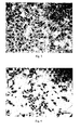
- Fig. 1 is a SEM micrograph of surface morphology of the stent described in example.5 intervened into a dog's femoral artery for 4 weeks.
- Fig. 2 is a SEM micrograph of surface morphology of the controlling sample of untreated pure iron stent intervened into the dog's femoral artery for 4 weeks. Comparing Fig. 1 with Fig.2 , it shows that after oxygen ions implantation, endothelial cells have covered on the modified iron stent completely, but almost no endothelial cells grew on the untreated iron stent. This result indicates that biocompatibility of the oxygen ions implanted iron stent is significantly improved.
- a biodegradable pure iron stent is treated by the following processes:
- Table 2 also shows the corrosion and in vitro degradation properties of the stents in these examples by polarization testing and immersion corrosion testing. It is proved that the corrosion resistance of the surface modified stent is significantly improved as showed by the significant lower weight loss after immersed in SBF for 9 days. And the lowest weight loss is 1/3 of that of the untreated one.
- a biodegradable pure iron stent is treated by the following processes:
- Table 3 also gives the results of polarization testing, immersion corrosion testing and activated partial thromboplastin time (APTT). It is proved that corrosion current and weight loss are significantly decreased and APTT is increased. These results show that La ion implantation modifies the blood compatibility, corrosion resistance and supporting ability of the iron stent.
- Example 19 Ion implantation 1 ⁇ 10 16 5 0.38 0.316 41.5
- Example 20 Plasma immersion ion implantation 1 ⁇ 10 16 40 0.41 0.342 41.0
- Example 21 Ion implantation 1 ⁇ 10 16 100 0.44 0.373 42.5
- Example 22 Plasma immersion ion implantation 1 ⁇ 10 17 5 0.24 0.234 41.5
- Example 23 Ion implantation 1 ⁇ 10 17 40 0.20 0.191 42.0
- Example 24 Ion implantation 1 ⁇ 10 17 100 0.25 0.238 42.0
- Example 25 Plasma immersion ion implantation 5 ⁇ 10 18 5 0.34 0.288 42.5
- Example 26 Plasma immersion ion implantation 5 ⁇ 10 18 40 0.24 0.226 41.0
- Example 27 Ion implantation 5 ⁇ 10 18 100 0.22 0.202 42.5
- a biodegradable pure iron stent is treated by the following processes:
- Table 4 also gives the results of polarization testing, immersion corrosion testing and activated partial thromboplastin time (APTT). It is proved that the corrosion current and the weight loss is significantly decreased and APTT is increased. These results show that Ce ion implantation modified the blood compatibility, corrosion resistance and supporting ability of the iron stent.
- a biodegradable pure iron stent is treated by the following processes:
- Table 5 also gives the results of polarization testing, immersion corrosion testing and activated partial thromboplastin time (APTT). It is proved that the corrosion current and the weight loss is significantly decreased and APTT is increased. These results show that Sr ion implantation modified the blood compatibility, corrosion resistance and supporting ability of the iron stent.
- a biodegradable pure iron stent is treated by the following processes:
- Table 6 also shows the result of average corrosion rate and APTT, the "average corrosion rate (mm/year) is deduced from the average data from testing for 216 hours. It is proved that the corrosion resistance and anticoagulation properties are significantly increased.
- Table 6 Film thickness (nm) Grain size (nm) Average corrosion rate (mm/year) APTT(s) Controlling sample Unmodified iron stent 0.039 39.5
- Example 46 10 10 0.028 41.5
- Example 47 200 10 0.012 43.0
- Example 48 500 10 0.016 42.5
- Example 51 200 100 0.019 42.5
- Example 52 500 100 0.024 42.2
- Example 53 1000 100 0.028 42.5
- Example 54 10 200 0.033 41.0
- Example 55 200 0.016 41.6
- Example 56 500 200 0.021 42.7
- Example 57 1000 200 0.022 42.1
- a biodegradable pure iron stent is treated by the following processes:
- Table 7 also gives the results of average corrosion rate and APTT of the surface modified stent. It is proved that the stent with Ce thin film has significantly increased corrosion resistance and anticoagulation properties. Table 7 Film thickness (nm) Grain size (nm) Average corrosion rate (mm/year) APTT(s) Controlling sample Unmodified iron stent 0.039 39.5
- Example 58 10 10 0.033 41.0
- Example 59 200 10 0.019 42.5
- Example 60 500 10 0.017 42.0
- Example 61 1000 10 0.020 42.5
- Example 65 1000 100 0.019 41.5
- Example 69 1000 200 0.017 41.7
- a biodegradable pure iron stent is treated by the following processes:
- Table 8 also gives the results of average corrosion rate and APTT of the stent with Sr thin film. It is proved that the stent with Sr thin film has significantly increased corrosion resistance and anticoagulation properties.
- Table 8 Film thickness (nm) Grain size (nm) Average corrosion rate (mm/year) APTT(s) Controlling sample Unmodified iron stent 0.039 39.5
- Example 70 10 10 0.031 41.5
- Example 71 200 10 0.023 43.2
- Example 72 500 10 0.017 42.6
- Example 73 1000 10 0.019 41.6
- Example 75 200 100 0.015 43.0
- Example 77 1000 100 0.021 41.9
- Example 80 500 200 0.018 42.6
- Example 81 1000 200 0.022 42.8
- a biodegradable pure iron stent is treated by the following processes:
- Table 9 also gives the results of average corrosion rate and APTT of the stent with Fe thin film. It is proved that the stent with Fe thin film has significantly increased corrosion resistance. Table 9 Film thickness (nm) Grain size (nm) Average corrosion rate (mm/year) Controlling sample Unmodified iron stent 0.039 Example 82 10 10 0.029 Example 83 100 10 0.014 Example 84 1000 10 0.011 Example 85 2000 10 0.013 Example 86 10 100 0.032 Example 87 100 100 0.021 Example 88 1000 100 0.023 Example 89 2000 100 0.024 Example 90 10 200 0.036 Example 91 100 200 0.033 Example 92 1000 200 0.032 Example 93 2000 200 0.035
- a biodegradable pure iron stent is treated by the following processes:
- Table 10 also gives the results of average corrosion rate and APTT. It is proved that the stent with iron oxide thin film has significantly increased corrosion resistance and anticoagulation properties.
- Table 10 Film thickness (nm) Grain size (nm) Corrosion current (mA/cm 2 ) APTT(s) Controlling sample Unmodified 0.58 39.5 iron stent
- Example 94 10 10 0.45 40.5
- Example 95 100 10 0.12 43.0
- Example 98 10 100 0.47 42.0
- Example 99 100 0.19 41.0
- Example 100 1000 100 0.25 42.6
- Example 101 2000 100 0.29 41.5
- Example 103 100 200 0.21 41.0
- Fig. 3 is surface morphology of the endothelial cells on the stent with iron oxide film described in Example 95 cultured with endothelial cells for 3 days.
- Fig. 4 is surface morphology of the endothelial cells on the stainless steel stent cultured with endothelial cells for 3 days.
- the iron stent after surface modification is treated further in magnetic field.
- the magnetic density is not lower than 100mT.
- the stent is implanted in the blood vessel and it is found that the stent is fully covered with ECs in 2-5 days. In comparison to magnetized stent, ECs covering on non-magnetized stent occurs in more than 10 days. It shows that magnetization of stent can help endothelialization on the iron stent.
- a biodegradable iron alloy stent contains La, Ce or Sr from 0.01 to 0.5 atom%.
- example 106 -114 about the iron alloy stents with different amount of alloying elements are given in Table 11.
- Polarization testing and immersion corrosion testing are performed in simulated body fluid solution (SBF, containing NaCl:8.04, KCl:0.23, NaHCO3:0.35, K2HPO4 ⁇ 3H2O:0.24, MgCl2 ⁇ 6H2O:0.31, CaCl2:0.29, Na2SO4:0.07, TRIS:6.12) to verify the corrosion and in vitro degradation properties of the stents and the results are also given in Table 11.
- the test results show that the corrosion currents and the weight loss rate in SBF for 187 hours all are decreased.
- a biodegradable iron stent contains La, Ce or Sr from 0.01 atom% to 0.5 atom%. Furthermore, oxygen or nitrogen ions are implanted into the stent surface by ion implantation or plasma immersion ion implantation. The doses are from 1 ⁇ 10 16 to 5 ⁇ 10 18 atoms/cm 2 the energy of the ions are from 5 to 100 KeV.
- example 115 -117 The treatment conditions and test results of example 115 -117 are given in Table 12. Table 12 Controlling sample Example 115 Example 116 Example 117 Alloy content Not added La 0.01% Ce0.25% Sr0.5% Method for surface treatment No-treated Ion implantation PIII Ion implantation Element implanted O+ N+ O+ doses atom/cm 2 1 ⁇ 10 16 5 ⁇ 10 17 5 ⁇ 10 18 energy KeV 5 50 100 Corrosion current (mA/cm 2 ) 0.57 0.37 0.12 0.10
- a biodegradable iron stent contains La, Ce or Sr from 0.01 to 0.5 atom%. Furthermore, La, Ce or Sr ions are implanted into the stent surface by ion implantation or plasma immersion ion implantation. The doses are from 1 ⁇ 10 16 to 5 ⁇ 10 18 atoms/cm 2 the energy of the ions are from 5 to 100 KeV.
- Example 118-120 The treatment conditions and test results of example 118-120 are given in Table 13. Table 13 Controlling sample Example 118 Example 119 Example 120 Alloy content Pure iron La 0.01% Ce0.25% Sr0.5% Method for surface treatment untreated Ion implantation Plasma immersion ion implantation Ion implantation Element implanted La Ce Sr doses atom/cm 2 1 ⁇ 10 16 5 ⁇ 10 17 5 ⁇ 10 18 energy KeV 5 50 100 Corrosion current (mA/cm 2 ) 0.57 0.39 0.23 0.20
- the thickness of the films is from 10 to 1000 nanometers, the grain size is from 10 to 200 nanometers.
- the treatment conditions and test results of example 121-129 are given in Table 14.
- the surface treated degradable iron stent is further magnetized in magnetic field.
- the magnetic density is not lower than 100mT.
- the stent is implanted in the blood vessel and it is found that the stent is fully covered with ECs in 2-5 days. In comparison to magnetized stent, ECs covering on non-magnetized stent occurs in more than 10 days. This proves that magnetization of stent helps endothelialization of iron stent.
Landscapes
- Health & Medical Sciences (AREA)
- Chemical & Material Sciences (AREA)
- Public Health (AREA)
- Vascular Medicine (AREA)
- Epidemiology (AREA)
- Life Sciences & Earth Sciences (AREA)
- Animal Behavior & Ethology (AREA)
- General Health & Medical Sciences (AREA)
- Surgery (AREA)
- Veterinary Medicine (AREA)
- Heart & Thoracic Surgery (AREA)
- Chemical Kinetics & Catalysis (AREA)
- Engineering & Computer Science (AREA)
- Materials Engineering (AREA)
- Mechanical Engineering (AREA)
- Metallurgy (AREA)
- Organic Chemistry (AREA)
- Inorganic Chemistry (AREA)
- Materials For Medical Uses (AREA)
Abstract
Description
- This present invention relates to biodegradable stents applied in treating vascular and brain blood vessel diseases.
- Percutaneous transluminal coronary angioplasty (PTCA) has been applied in clinic since 1977. PTCA is delivering a balloon into target lesion position of blood vessel through femoral artery, then applying a pressure to expand the balloon so that the inside dimension of the narrowed vascular can be increased to improve the blood supplying to the cardiac muscle. However the restenosis rate after PTCA is as high as more than 50%. The application of metal stents made of stainless steel, cobalt alloy or nickel titanium alloy can decrease the restenosis rate to about 20% to 30%. Since 2003, drug eluting stent (DES) coated with a polymer layer containing drugs can decease the restenosis rate to about 10%, DES is regarded as a milestone of interventional therapy of coronary heart diseases. However, all these stents are not degradable and will stay in the blood vessel permanently. There is a big difference in mechanical characteristics between the blood vessel and the stent, which may cause chronic damage of blood vessel, atrophy of middle layer of the blood vessel, aneurysm formation and endometrial hyperplasia. Furthermore, young patients who are still in the growth period, the permanent stent cannot meet the need of the growth of the blood vessel. Simultaneously, the harmful elements released from stents to blood vessel are also the issue of metal stents.
- An ideal stent should perform the function of efficient mechanical support after intervention into the target lesion blood vessel in an appropriate period of 3 to 6 months, simultaneously release the drug to realize the function of therapy, and after that time, be gradually degraded to decrease the lesion effect of the stent on the blood vessel to prevent restenosis. Therefore, biodegradable polymer stent, such as polylactic acid stent, has attracted much attention. Polylactic acid can be degraded to non-toxic water and CO2 which will be absorbed by the human body or breathed out, it has been approved by FDA as a biomaterial on the market. Completely biodegradable polymer stent which releases drugs during degrade process has also been reported. H. Tamai et al reported their results of intervention of 25 pieces of polylactic acid stents into hearts of 15 patients. Their results show that after 6 months the restenosis rate was at the same level of stainless steel stent. However, there are still problems with biodegradable polymer stents, such as:
- 1) The strength and the deformation behavior of polymer stents are much different from metal stents. Because the biodegradable polymer stent is not strong, the thickness of polylactic acid stent is double that of stainless steel stent to provide suitable support, which will bring about obstructing to the blood stream.
- 2) The polymer stents have to be heated to expand sufficiently, while heating may easily damage the blood vessel.
- 3) The rebound rate of polymer stents is high, and visibility of the polymer is poor inside the vessel.
- 4) The delivery system for biodegradable polymer stent is different from the delivery system which is widely used presently for metal stents. This would also affect the application of polymer stents.
- Biodegradable metal stents do not have those issues with polymer stents. Therefore, it is important to develop biodegradable metal stent and stent made of iron (Fe) is a new research subject. Iron is a common element existing in human body. The total amount of iron in an adult body is about 4 gram, and an adult will need to incept 10 to 15 mg of iron every day. Iron has better biocompatibility and mechanical properties. In 2001, Peuster firstly reported in vivo results of intervention of 16 iron stents (Fe content > 99.8%) into abdominal aorta of New Zealand rabbits for 6 to 18 months, most of the support bars of the stents degraded to some degree after 6 months and no obvious inflammation, restenosis and coagulation were found in 6 to 18 months experiments. [Peuster M, Wohlsein P, Brugmann M, et al, A novel approach to temporary stenting: degradable cardiovascular stents produced from corrodible metal-results 6 18 months after implantation into New Zealand white rabbits. Heart, 2001;86:563-569]
- Peuster further intervened stainless steel stents and iron stents into the abdominal aorta of mini-pigs in 2006. It was found that the degree of endometrial hyperplasia of both kinds of stents was about the same, and no excessive deposition and toxicity effect on the main organs of the pigs were found after histopathology analysis of these organs. And no toxic and side effects caused by corrosion product were found in the tissues close to the iron stent ribs.
- In 2006, Mueller et al used gene chip technology to reveal that one of the degradation products of pure iron stent, Fe2+, has an effect to inhibit the proliferation of smooth muscle cell. In 2008, Waksman et al intervened iron stents and Co-Cr alloy stents into pig's conorary blood vessel for 28 days and found that no obvious inflammation and embolism on surfaces of iron stents. And no obvious difference was found in intima thickness, intima area and occlusion rate between the two kinds of stents.
- In terms of strength, plasticity and processing ability, iron stent is close to stainless steel stent. Iron stent has better mechanical supporting ability than polymer stent. Comparing with Co-Cr alloy stent or stainless steel stent, iron stent has degradation ability and a better biocompatibility. The clinic applied balloon delivery system is also suitable for iron stent, it is easy for operation, and its cost is low.
- However there are still some shortcomings in iron stent: the low corrosion resistance of pure iron stent surface which can affect its mechanical support and be unhelpful to recovery of the lesion position of blood vessel, the low covering rate of endothelial cells on the stent surface in short time after the intervention into blood vessel, and the degradation rate of the iron stent which cannot be regulated to meet different needs.
- The present invention is about a degradable stent with a high corrosion resistivity and controllable degradation. In the 30 days after the stent intervened into blood vessel, the degradation rate of the stent maintains a low level and endue the surface with a good biocompatibility, which will be beneficial for endothelial cells to grow and cover the stent surface. And the stent can maintain a good mechanical supporting ability during 3 to 6 months and be degraded after 6 months.
- One part of the present invention is about pure iron stents and treated by surface modification using ion implantation technique, as follows:
- Implanting oxygen or nitrogen ions into the pure iron stent surface by ion implantation or plasma immersion ion implantation, the doses range is from 1×10¹⁶ to 5×10¹⁸ atoms/cm2, the energy range of the ions is from 5 to 100 KeV.
- Or implanting lanthanum (La), cerium (Ce) or strontium (Sr) ions into the pure iron stent surface by ion implantation or plasma immersion ion implantation, the doses range is from 1×10¹⁶ to 5×10¹⁸ atoms/cm2, the energy range of the ions is from 5 to 100 KeV.
- Or depositing a thin film of La, Ce, Sr, lanthana, ceria, strontia, iron or iron oxide on the pure iron stent using plasma immersion ion implantation. The thickness of the films is from 10 to 1000 nanometers, the grain size is from 10 to 200 nanometers.
- Comparing with existing technologies, the present invention of stents with O, N, La, Ce, Sr, lanthana, ceria, strontia, iron, iron oxide modified surface using ion implantation and/or plasma immersion ion implantation increases surface corrosion resistance of the stent. The elements for surface modification are non-toxic. The degradation rate of the iron stent can be effectively controlled and modulated. The stent maintains a low degradation rate in the 30 days after the intervention and endue the surface with a good biocompatibility which will be beneficial for endothelial cells to grow and cover the stent surface. The stent will provide sufficient mechanical supporting in 3 to 6 months period after intervention, and be degraded and gradually disappear after 6 months. Thus it will not have the issue of restenosis and late- thrombus formation etc. which may be caused by no-degradable permanent stent. The experiment results prove that the degradation rate of surface modified iron stent can go down more than 50% comparing with non- surface modified iron sten. Endothelial cells grow on the stent surface within 4 weeks, while on non- surface modified iron stent no endothelial cells existed but only degradation product. The degree of the degradation rate of iron stent can be modulated by controlling the dosage of implanted ions, energy of the ion implantion, thickness or grain sizes of the deposited thin films to meet the different needs of the stent.
- Above mentioned iron stent can be further magnetized in a magnetic field to become magnetic. The magnetized stent has the advantages to promote endothelial cells covering and prohibit smooth muscle cells growth, which is beneficial for prohibiting restenosis and coagulation. The mechanical supporting ability of the stent in 3 to 6 months period after intervention is sufficient, and the stent will be degraded and gradually disappear after 6 months.
- The other part of the present invention is fabricating of iron alloy stent. The iron alloy contains 0.01 to 0.5% of La, Ce or Sr. By adjusting the contents of La, Ce or Sr, the corrosion resistance/ degradation rate of the stent can be modulated. The stent maintains a low degradation rate in the first 30 days after the intervention and endue the surface with a good biocompatibility which will be beneficial for endothelial cells to grow and cover the stent surface. The stent will provide sufficient mechanical supporting in 3 to 6 months period after intervention, and be degraded and gradually disappear after 6 months. Thus it will not have the issue of restenosis and late- thrombus formation etc. which may be caused by no-degradable permanent stent. The experiment results prove that the degradation rate of the iron alloy stent can go down more than 50% in the first 30 days after being intervened into blood vessel, comparing with pure iron stent. The degradation rate of iron alloy stent can be modulated by controlling quantity of alloying elements to meet different needs of the stent.
- Above mentioned iron alloy stent is further treated by surface modification using ion implantation technique to further improve the surface corrosion resistance of stent and modulate the degradation rate. The surface modification treatment is as follows:
- Implanting oxygen or nitrogen ions into the iron alloy stent surface by ion implantation or plasma immersion ion implantation, the doses range is from 1×10¹⁶ to 5×10¹⁸ atoms/cm2, the energy range of the ions is from 5 to 100 KeV.
- Or implanting lanthanum (La), cerium (Ce) or strontium (Sr) ions into the iron alloy stent surface by ion implantation or plasma immersion ion implantation, the doses range is from 1×10¹⁶ to 5×10¹⁸ atoms/cm2, the energy range of the ions is from 5 to 100 KeV.
- Or depositing a thin film of La, Ce, Sr, lanthana, ceria, strontia, iron or iron oxide on the iron alloy stent using plasma immersion ion implantation. The thickness of the films is from 10 to 1000 nanometers, the grain size is from 10 to 200 nanometers.
- Above mentioned iron alloy stent can be further magnetized in a magnetic field. The magnetized stent has advantages to promote endothelial cells covering and prohibit smooth muscle cells growth, which can be beneficial to prohibit restenosis and coagulation.
- The invention is further demonstrated by the figures and the examples, in comparison of controlling samples of pure iron stent:
-
-
Fig. 1 is a SEM micrograph of surface morphology of the stent described in example 5 intervened into dog's femoral artery for 4 weeks. -
Fig. 2 is a SEM micrograph of surface morphology of untreated pure iron stent intervened into dog's femoral artery for 4 weeks. -
Fig. 3 is surface morphology of the stent in example 95 with iron oxide film cultured with endothelial cells for 3 days. -
Fig.4 is surface morphology of the stainless steel stent cultured with endothelial cells for 3 days. - The following EXAMPLES illustrate various aspects of the making the stent invention herein. They are not intended to limit the scope of the invention.
- A biodegradable pure iron stent is treated by the following plasma processes:
- Oxygen ions are implanted into the pure stent surface by ion implantation or plasma immersion ion implantation, the doses range is from 1×10¹⁶ to 5×10¹⁸ atoms/cm2, the energy range of the ions is from 5 to 100 KeV. The treatment parameters and test results of the example 1-9 are given in Table 1.
- For verifying the corrosion resistance and in vitro degradation properties of the stents, polarization testing and immersion corrosion testing are performed using simulated body fluid solution (SBF, containing NaCl:8.04, KCl:0.23, NaHCO3:0.35, K2HPO4-3H2O:0.24, MgCl2·6H2O:0.31, CaCl2:0.29, Na2SO4:0.07, TRIS:6.12). The results are given in Table 1. It is proved that the corrosion currents and the weight loss are all significantly decreased. Less weight loss also means a more stable mechanical supporting of the stent.
Table 1 method Implanted doses (atoms/cm2) Ion energy (KeV) Corrosion current (mA/cm2) Weight loss after immersion 216 hours (mg) Controlling sample Unmodified iron stent 0.59 0.460 Example 1 Ion implantation 1×10¹⁶ 5 0.40 0.340 Example 2 Plasma immersion ion implantation 1×10¹⁶ 40 0.45 0.390 Example 3 Ion implantation 1×10¹⁶ 100 0.49 0.416 Example 4 Plasma immersion ion implantation 1×10¹⁷ 5 0.27 0.258 Example 5 Plasma immersion ion implantation 1×10¹⁷ 40 0.08 0.092 Example 6 Ion implantation 1×10¹⁷ 100 0.12 0.112 Example 7 Plasma immersion ion implantation 5×10¹⁸ 5 0.36 0.306 Example 8 Ion implantation 5×10¹⁸ 40 0.13 0.121 Example 9 Plasma immersion ion implantation 5×10¹⁸ 100 0.11 0.105 -
Fig. 1 is a SEM micrograph of surface morphology of the stent described in example.5 intervened into a dog's femoral artery for 4 weeks.Fig. 2 is a SEM micrograph of surface morphology of the controlling sample of untreated pure iron stent intervened into the dog's femoral artery for 4 weeks. ComparingFig. 1 withFig.2 , it shows that after oxygen ions implantation, endothelial cells have covered on the modified iron stent completely, but almost no endothelial cells grew on the untreated iron stent. This result indicates that biocompatibility of the oxygen ions implanted iron stent is significantly improved. - A biodegradable pure iron stent is treated by the following processes:
- Nitrogen ions are implanted into the pure iron stent surface by ion implantation or plasma immersion ion implantation, the doses range is from 1×10¹⁶ to 5×10¹⁸ atoms/cm2, the energy range of the ions is from 5 to 100 KeV. The parameters for example 10-18 are given in Table 2.
- Table 2 also shows the corrosion and in vitro degradation properties of the stents in these examples by polarization testing and immersion corrosion testing. It is proved that the corrosion resistance of the surface modified stent is significantly improved as showed by the significant lower weight loss after immersed in SBF for 9 days. And the lowest weight loss is 1/3 of that of the untreated one.
Table 2 Surface modification method Implanted doses (atoms/cm2) Ion energy (KeV) Corrosion current (mA/cm2) Weight loss after immersion 216 hours (mg) Controlling sample Unmodified iron stent 0.59 0.460 example 10 Ion implantation 1×10¹⁶ 5 45 0.415 example 11 Plasma immersion ion implantation 1×10¹⁶ 40 0.51 0.438 example 12 Ion implantation 1×10¹⁶ 100 0.53 0.445 example 13 Plasma immersion ion implantation 1×10¹⁷ 5 0.35 0.318 example 14 Plasma immersion ion implantation 1×10¹⁷ 40 0.18 0.163 example 15 Ion implantation 1×10¹⁷ 100 0.22 0.239 example 16 Plasma immersion ion implantation 5×10¹⁸ 5 0.41 0.352 example 17 Plasma immersion ion implantation 5×10¹⁸ 40 0.20 0.169 example 18 Ion implantation 5×10¹⁸ 100 0.18 0.158 - A biodegradable pure iron stent is treated by the following processes:
- La ions are implanted into the pure iron stent surface by ion implantation or plasma immersion ion implantation, the doses range is from 1×10¹⁶ to 5×10¹⁸ atoms/cm2, the energy range of the ions is from 5 to 100 KeV. The parameters for example 19-27 are given in Table 3.
- Table 3 also gives the results of polarization testing, immersion corrosion testing and activated partial thromboplastin time (APTT). It is proved that corrosion current and weight loss are significantly decreased and APTT is increased. These results show that La ion implantation modifies the blood compatibility, corrosion resistance and supporting ability of the iron stent.
Table 3 Surface modification method Implanted doses (atoms/cm2) Ion energy (KeV) Corrosion current (mA/cm2) Weight loss after immersion 216 hours (mg) APTT(s) Controlling sample Untreated pure iron 0.57 0.481 39.5 Example 19 Ion implantation 1×10¹⁶ 5 0.38 0.316 41.5 Example 20 Plasma immersion ion implantation 1×10¹⁶ 40 0.41 0.342 41.0 Example 21 Ion implantation 1×10¹⁶ 100 0.44 0.373 42.5 Example 22 Plasma immersion ion implantation 1×10¹⁷ 5 0.24 0.234 41.5 Example 23 Ion implantation 1×10¹⁷ 40 0.20 0.191 42.0 Example 24 Ion implantation 1×10¹⁷ 100 0.25 0.238 42.0 Example 25 Plasma immersion ion implantation 5×10¹⁸ 5 0.34 0.288 42.5 Example 26 Plasma immersion ion implantation 5×10¹⁸ 40 0.24 0.226 41.0 Example 27 Ion implantation 5×10¹⁸ 100 0.22 0.202 42.5 - A biodegradable pure iron stent is treated by the following processes:
- Ce ions are implanted into the pure iron stent surface by ion implantation or plasma immersion ion implantation, the doses range is from 1×10¹⁶ to 5×10¹⁸ atoms/cm2, the energy range of the ions is from 5 to 100 KeV. The parameters for example 28-36 are given in Table 4.
- Table 4 also gives the results of polarization testing, immersion corrosion testing and activated partial thromboplastin time (APTT). It is proved that the corrosion current and the weight loss is significantly decreased and APTT is increased. These results show that Ce ion implantation modified the blood compatibility, corrosion resistance and supporting ability of the iron stent.
Table 4 Surface modification method Implanted doses (atoms/cm2) Ion energy (KeV) Corrosion current (mA/cm2) Weight loss after immersion 216 hours (mg) APTT(s) Controlling sample Unmodified pure iron 0.58 0.481 39.7 Example 28 Ion implantation 1×10¹⁶ 5 0.41 0.337 41.2 Example 29 Plasma immersion ion implantation 1×10¹⁶ 40 0.38 0.309 42.6 Example 30 Ion implantation 1×10¹⁶ 100 0.47 0.399 41.0 Example 31 Plasma immersion ion implantation 1×10¹⁷ 5 0.27 0.248 42.0 Example 32 Plasma immersion ion implantation 1×10¹⁷ 40 0.19 0.177 43.5 Example 33 Ion implantation 1×10¹⁷ 100 0.23 0.217 41.8 Example 34 Plasma immersion ion implantation 5×10¹⁸ 5 0.37 0.301 42.5 Example 35 Plasma immersion ion implantation 5×10¹⁸ 40 0.28 0.238 41.5 Example 36 lonimplantation 5×10¹⁸ 100 0.26 0.221 43.0 - A biodegradable pure iron stent is treated by the following processes:
- Sr ions are implanted into the pure iron stent surface by ion implantation or plasma immersion ion implantation, the doses range is from 1×10¹⁶ to 5×10¹⁸ atoms/cm2, the energy range of the ions is from 5 to 100 KeV. The parameters for example 37-45 are given in Table 5.
- Table 5 also gives the results of polarization testing, immersion corrosion testing and activated partial thromboplastin time (APTT). It is proved that the corrosion current and the weight loss is significantly decreased and APTT is increased. These results show that Sr ion implantation modified the blood compatibility, corrosion resistance and supporting ability of the iron stent.
Table 5 Surface modification method Implanted doses (atoms/cm2) Ion energy (KeV) Corrosion current (mA/cm2) weight loss after immersion 216 hours (mg) APTT(s) Controlling sample Unmodified pure iron 0.56 0.466 40.0 Example 37 Ion implantation 1×10¹⁶ 5 0.37 0.305 42.5 Example 38 Plasma immersion ion implantation 1×10¹⁶ 40 0.32 0.278 43.0 Example 39 Ion implantation 1×10¹⁶ 100 0.41 0.357 41.5 Example 40 Plasma immersion ion implantation 1×10¹⁷ 5 0.28 0.248 43.5 Example 41 Plasma immersion ion implantation 1×10¹⁷ 40 0.23 0.227 43.8 Example 42 Ion implantation 1×10¹⁷ 100 0.31 0.269 42.5 Example 43 Plasma immersion ion implantation 5×10¹⁸ 5 0.35 0.294 42.6 Example 44 Plasma immersion ion implantation 5×10¹⁸ 40 0.22 0.214 42.2 Example 45 Ion implantation 5×10¹⁸ 100 0.21 0.193 41.5 - A biodegradable pure iron stent is treated by the following processes:
- La thin films are deposited onto the pure iron stent surface by plasma deposition process. The film thickness range is from 10 to 1000 nanometer. The parameters for example 46- 57 are given in Table 6.
- Table 6 also shows the result of average corrosion rate and APTT, the "average corrosion rate (mm/year) is deduced from the average data from testing for 216 hours. It is proved that the corrosion resistance and anticoagulation properties are significantly increased.
Table 6 Film thickness (nm) Grain size (nm) Average corrosion rate (mm/year) APTT(s) Controlling sample Unmodified iron stent 0.039 39.5 Example 46 10 10 0.028 41.5 Example 47 200 10 0.012 43.0 Example 48 500 10 0.016 42.5 Example 49 1000 10 0.019 43.1 Example 50 10 100 0.030 41.1 Example 51 200 100 0.019 42.5 Example 52 500 100 0.024 42.2 Example 53 1000 100 0.028 42.5 Example 54 10 200 0.033 41.0 Example 55 200 200 0.016 41.6 Example 56 500 200 0.021 42.7 Example 57 1000 200 0.022 42.1 - A biodegradable pure iron stent is treated by the following processes:
- Ce thin films are deposited onto the pure iron stent surface by plasma deposition process. The film thickness range is from 10 to 1000 nanometer. The parameters for example 58-69 are given in Table 7.
- Table 7 also gives the results of average corrosion rate and APTT of the surface modified stent. It is proved that the stent with Ce thin film has significantly increased corrosion resistance and anticoagulation properties.
Table 7 Film thickness (nm) Grain size (nm) Average corrosion rate (mm/year) APTT(s) Controlling sample Unmodified iron stent 0.039 39.5 Example 58 10 10 0.033 41.0 Example 59 200 10 0.019 42.5 Example 60 500 10 0.017 42.0 Example 61 1000 10 0.020 42.5 Example 62 10 100 0.035 41.6 Example 63 200 100 0.018 42.0 Example 64 500 100 0.020 42.8 Example 65 1000 100 0.019 41.5 Example 66 10 200 0.036 41.6 Example 67 200 200 0.023 41.9 Example 68 500 200 0.018 43.6 Example 69 1000 200 0.017 41.7 - A biodegradable pure iron stent is treated by the following processes:
- Sr thin films are deposited onto the pure iron stent surface by plasma deposition process. The film thickness range is from 10 to 1000 nanometer. The parameters for example 70 - 81 are given in Table 8.
- Table 8 also gives the results of average corrosion rate and APTT of the stent with Sr thin film. It is proved that the stent with Sr thin film has significantly increased corrosion resistance and anticoagulation properties.
Table 8 Film thickness (nm) Grain size (nm) Average corrosion rate (mm/year) APTT(s) Controlling sample Unmodified iron stent 0.039 39.5 Example 70 10 10 0.031 41.5 Example 71 200 10 0.023 43.2 Example 72 500 10 0.017 42.6 Example 73 1000 10 0.019 41.6 Example 74 10 100 0.033 42.4 Example 75 200 100 0.015 43.0 Example 76 500 100 0.017 42.6 Example 77 1000 100 0.021 41.9 Example 78 10 200 0.035 42.1 Example 79 200 200 0.021 42.5 Example 80 500 200 0.018 42.6 Example 81 1000 200 0.022 42.8 - A biodegradable pure iron stent is treated by the following processes:
- Fe thin films are deposited onto the pure iron stent surface by plasma deposition process. The film thickness range is from 10 to 1000 nanometer. The parameters for example 82-93 are given in Table 9.
- Table 9 also gives the results of average corrosion rate and APTT of the stent with Fe thin film. It is proved that the stent with Fe thin film has significantly increased corrosion resistance.
Table 9 Film thickness (nm) Grain size (nm) Average corrosion rate (mm/year) Controlling sample Unmodified iron stent 0.039 Example 82 10 10 0.029 Example 83 100 10 0.014 Example 84 1000 10 0.011 Example 85 2000 10 0.013 Example 86 10 100 0.032 Example 87 100 100 0.021 Example 88 1000 100 0.023 Example 89 2000 100 0.024 Example 90 10 200 0.036 Example 91 100 200 0.033 Example 92 1000 200 0.032 Example 93 2000 200 0.035 - A biodegradable pure iron stent is treated by the following processes:
- Iron oxide thin films are deposited onto the pure iron stent surface by plasma deposition process. The film thickness range is from 10 to 1000 nanometer. The parameters for example 94-105 are given in Table 10.
- Table 10 also gives the results of average corrosion rate and APTT. It is proved that the stent with iron oxide thin film has significantly increased corrosion resistance and anticoagulation properties.
Table 10 Film thickness (nm) Grain size (nm) Corrosion current (mA/cm2) APTT(s) Controlling sample Unmodified 0.58 39.5 iron stent Example 94 10 10 0.45 40.5 Example 95 100 10 0.12 43.0 Example 96 1000 10 0.17 42.5 Example 97 2000 10 0.22 43.1 Example 98 10 100 0.47 42.0 Example 99 100 100 0.19 41.0 Example 100 1000 100 0.25 42.6 Example 101 2000 100 0.29 41.5 Example 102 10 200 0.49 42.0 Example 103 100 200 0.21 41.0 Example 104 1000 200 0.26 41.5 Example 105 2000 200 0.27 40.0 -
Fig. 3 is surface morphology of the endothelial cells on the stent with iron oxide film described in Example 95 cultured with endothelial cells for 3 days. -
Fig. 4 is surface morphology of the endothelial cells on the stainless steel stent cultured with endothelial cells for 3 days. - Comparing
Fig. 3 withFig.4 , it shows that endothelial cells grow more on the iron stent with iron oxide film. This means that the biocompatibility of iron oxide film coated stent is better. - The iron stent after surface modification is treated further in magnetic field. The magnetic density is not lower than 100mT. After magnetization the stent is implanted in the blood vessel and it is found that the stent is fully covered with ECs in 2-5 days. In comparison to magnetized stent, ECs covering on non-magnetized stent occurs in more than 10 days. It shows that magnetization of stent can help endothelialization on the iron stent.
- A biodegradable iron alloy stent contains La, Ce or Sr from 0.01 to 0.5 atom%. example 106 -114 about the iron alloy stents with different amount of alloying elements are given in Table 11. Polarization testing and immersion corrosion testing are performed in simulated body fluid solution (SBF, containing NaCl:8.04, KCl:0.23, NaHCO3:0.35, K2HPO4·3H2O:0.24, MgCl2·6H2O:0.31, CaCl2:0.29, Na2SO4:0.07, TRIS:6.12) to verify the corrosion and in vitro degradation properties of the stents and the results are also given in Table 11. The test results show that the corrosion currents and the weight loss rate in SBF for 187 hours all are decreased. (The corrosion rate in Table 11 is deduced from data of 187 hours immersion.)
Table 11 Alloy element Atom% Corrosion current (mA/cm2) Average corrosion rate (mm/year) Controlling sample Pure iron 0.57 0.038 Example 106 La 0.01% 0.46 0.032 Example 107 La 0.25% 0.31 0.023 Example 108 La 0.5% 0.37 0.029 Example 109 Ce 0.01% 0.44 0.032 Example 110 Ce 0.25% 0.27 0.020 Example 111 Ce 0.5% 0.39 0.030 Example 112 Sr 0.01% 0.38 0.028 Example 113 Sr 0.25% 0.36 0.025 Example 114 Sr 0.5% 0.47 0.033 - A biodegradable iron stent contains La, Ce or Sr from 0.01 atom% to 0.5 atom%. Furthermore, oxygen or nitrogen ions are implanted into the stent surface by ion implantation or plasma immersion ion implantation. The doses are from 1×10¹⁶ to 5×10¹⁸ atoms/cm2 the energy of the ions are from 5 to 100 KeV.
- The treatment conditions and test results of example 115 -117 are given in Table 12.
Table 12 Controlling sample Example 115 Example 116 Example 117 Alloy content Not added La 0.01% Ce0.25% Sr0.5% Method for surface treatment No-treated Ion implantation PIII Ion implantation Element implanted O+ N+ O+ doses atom/cm2 1×10¹⁶ 5×10¹⁷ 5×10¹⁸ energy KeV 5 50 100 Corrosion current (mA/cm2) 0.57 0.37 0.12 0.10 - A biodegradable iron stent contains La, Ce or Sr from 0.01 to 0.5 atom%. Furthermore, La, Ce or Sr ions are implanted into the stent surface by ion implantation or plasma immersion ion implantation. The doses are from 1×10¹⁶ to 5×10¹⁸ atoms/cm2 the energy of the ions are from 5 to 100 KeV.
- The treatment conditions and test results of example 118-120 are given in Table 13.
Table 13 Controlling sample Example 118 Example 119 Example 120 Alloy content Pure iron La 0.01% Ce0.25% Sr0.5% Method for surface treatment untreated Ion implantation Plasma immersion ion implantation Ion implantation Element implanted La Ce Sr doses atom/cm2 1×10¹⁶ 5×10¹⁷ 5×10¹⁸ energy KeV 5 50 100 Corrosion current (mA/cm2) 0.57 0.39 0.23 0.20 - A biodegradable iron stent containing La, Ce or Sr from 0.01 atom % to 0.5 atom%: the stent are further treated by plasma deposition of La, Ce, Sr, lanthana, ceria, strontia, iron or iron oxide thin film. The thickness of the films is from 10 to 1000 nanometers, the grain size is from 10 to 200 nanometers. The treatment conditions and test results of example 121-129 are given in Table 14.
Table 14 Alloy content Deposition material Grain size (nm) Thickness of the films (nm) Average corrosion rate (mm/year) Controlling sample Untreat pure iron 0.039 Example 121 La 0.01% La 10 10 0.027 Example 122 Ce 0.25 % Ce 100 700 0.016 Example 123 La 0.01% La 200 10 0.031 Example 124 Ce 0.25% strontia 10 500 0.015 Example 125 Sr 0.5 % Ceria 100 1000 0.017 Example 126 Sr 0.5% Lanthana 200 1000 0.020 Example 127 La 0.01% Fe 10 10 0.030 Example 128 Ce 0.3 % Iron oxide 100 1000 0.028 Example 129 Sr 0.4% Fe 200 1000 0.029 - The surface treated degradable iron stent is further magnetized in magnetic field. The magnetic density is not lower than 100mT. After magnetization the stent is implanted in the blood vessel and it is found that the stent is fully covered with ECs in 2-5 days. In comparison to magnetized stent, ECs covering on non-magnetized stent occurs in more than 10 days. This proves that magnetization of stent helps endothelialization of iron stent.
Claims (7)
- A degradable stent made of pure iron; doing surface modification using ion implantation and/or plasma surface modification methods, as follows:Implanting oxygen or nitrogen ions into the pure iron stent surface using ion implantation or plasma immersion ion implantation processes, the doses range is from 1×10¹⁶ to 5×10¹⁸ atoms/cm2, the energy range of the ions is from 5 to 100 KeV;Or implanting lanthanum (La), cerium (Ce) or strontium (Sr) ions into the pure iron stent surface using ion implantation or plasma immersion ion implantation processes, the doses range is from 1×10¹⁶ to 5×10¹⁸ atoms/cm2, the energy range of the ions is from 5 to 100 KeV;Or depositing a thin film with definite material on the pure iron stent surface, the thin film materials are La, Ce, Sr, lanthana, ceria, strontia, iron or iron oxide, The thickness of the films is from 10 to 1000 nanometers, the grain size is from 10 to 200 nanometers.
- The stent as set forth in claim 1 further magnetization treatment in a magnetic field.
- A degradable stent made of iron alloy containing lanthanum (La), cerium (Ce) or strontium (Sr) elements with content from 0.01 atom% to 0.5 atom%.
- The stent as set forth in claim 3 further Surface modification using ion implantation and/or plasma method, as follows:Implanting oxygen or nitrogen into surface of the iron alloy stent according to Claim 3 using ion implantation or plasma immersion ion implantation processes, the doses range is from 1×10¹⁶ to 5×10¹⁸ atoms/cm2, the energy range of the ions is from 5 to 100 KeV;
- The stent as set forth in claim 3 further Surface modification using ion implantation and/or plasma method, as follows:implanting lanthanum (La), cerium (Ce) or strontium (Sr) ions into surface of the iron alloy stent according to Claim 3 by ion implantation or plasma immersion ion implantation processes, the doses range is from 1×10¹⁶ to 5×10¹⁸ atoms/cm2, the energy range of the ions is from 5 to 100 KeV;
- The stent as set forth in claim 3 further Surface modification using ion implantation and/or plasma method, as follows:depositing a thin film on surface of the iron alloy stent according to Claim 3, The thin film materials are La, Ce, Sr, lanthana, ceria, strontia, iron or iron oxides, The thickness of the films is from 10 to 1000 nanometers and the grain size is from 10 to 200 nanometers.
- The stent as set forth in claim 3 further magnetization treatment in a magnetic field.
Applications Claiming Priority (2)
| Application Number | Priority Date | Filing Date | Title |
|---|---|---|---|
| CN200910058554.6A CN101496910B (en) | 2009-03-10 | 2009-03-10 | A kind of degradable blood vessel support |
| PCT/CN2010/070905 WO2010102549A1 (en) | 2009-03-10 | 2010-03-08 | Degradable stent |
Publications (2)
| Publication Number | Publication Date |
|---|---|
| EP2407183A1 true EP2407183A1 (en) | 2012-01-18 |
| EP2407183A4 EP2407183A4 (en) | 2014-03-12 |
Family
ID=40944319
Family Applications (1)
| Application Number | Title | Priority Date | Filing Date |
|---|---|---|---|
| EP10750345.0A Withdrawn EP2407183A4 (en) | 2009-03-10 | 2010-03-08 | DEGRADABLE STENT |
Country Status (3)
| Country | Link |
|---|---|
| EP (1) | EP2407183A4 (en) |
| CN (1) | CN101496910B (en) |
| WO (1) | WO2010102549A1 (en) |
Cited By (3)
| Publication number | Priority date | Publication date | Assignee | Title |
|---|---|---|---|---|
| CN104644297A (en) * | 2015-02-16 | 2015-05-27 | 上海微创医疗器械(集团)有限公司 | Bioabsorbable stent and preparation method thereof |
| EP2329853B1 (en) * | 2009-11-10 | 2015-07-22 | Biotronik VI Patent AG | Implant and method for manufacturing same |
| US20180280584A1 (en) * | 2015-12-22 | 2018-10-04 | Lifetech Scientific (Shenzhen) Co., Ltd | Absorbable Iron-based Alloy Implanted Medical Device |
Families Citing this family (7)
| Publication number | Priority date | Publication date | Assignee | Title |
|---|---|---|---|---|
| CN101496910B (en) * | 2009-03-10 | 2017-06-23 | 成都西南交大研究院有限公司 | A kind of degradable blood vessel support |
| CN102247623B (en) * | 2011-08-17 | 2014-07-23 | 上海微创医疗器械(集团)有限公司 | Multilayer degradable stent having shape memory and preparation method thereof |
| US20130066417A1 (en) * | 2011-09-08 | 2013-03-14 | Nan Huang | Biodegradable stent |
| CN104662191A (en) * | 2012-07-10 | 2015-05-27 | 韦恩堡金属研究产品公司 | Biodegradable alloy wire for medical devices |
| CN107805792A (en) * | 2017-11-06 | 2018-03-16 | 南京工程学院 | A kind of method for improving biologic medical magnesium surface property and corrosive wear behavior |
| CN110952038A (en) * | 2019-11-27 | 2020-04-03 | 苏州森锋医疗器械有限公司 | Biodegradable iron alloy, preparation method and device |
| CN112899618B (en) * | 2021-05-08 | 2021-07-16 | 中南大学湘雅医院 | Coating with catalytic capability on surface of intravascular stent and preparation method thereof |
Family Cites Families (11)
| Publication number | Priority date | Publication date | Assignee | Title |
|---|---|---|---|---|
| GB2075069B (en) * | 1979-12-03 | 1984-09-12 | Atomic Energy Authority Uk | Wear resistance of metals |
| FR2500010A1 (en) * | 1981-02-13 | 1982-08-20 | Inst Fiz Tverdogo Tela | Stainless steel, esp. for mfg. surgical instruments - has very high purity and very high corrosion resistance, obtd. esp. by electron beam melting of steel electrode made by powder metallurgy |
| SE461191B (en) * | 1988-04-21 | 1990-01-22 | Sandvik Ab | APPLICATION OF A STAINLESS FERRIT-AUSTENITIC STEEL ALLOY AS IMPLANT IN PHYSIOLOGICAL ENVIRONMENT |
| US6224536B1 (en) * | 1999-02-08 | 2001-05-01 | Advanced Cardiovascular Systems | Method for delivering radiation therapy to an intravascular site in a body |
| US6524466B1 (en) * | 2000-07-18 | 2003-02-25 | Applied Semiconductor, Inc. | Method and system of preventing fouling and corrosion of biomedical devices and structures |
| GB0209797D0 (en) * | 2002-04-29 | 2002-06-05 | Univ Birmingham | Method of surface hardening cobalt-chromium based alloy articles |
| CN100404724C (en) * | 2004-12-20 | 2008-07-23 | 北京师范大学 | Ion implantation treatment method for the surface of metal material implanted in human body |
| US8840660B2 (en) * | 2006-01-05 | 2014-09-23 | Boston Scientific Scimed, Inc. | Bioerodible endoprostheses and methods of making the same |
| WO2008034030A2 (en) * | 2006-09-15 | 2008-03-20 | Boston Scientific Limited | Magnetized bioerodible endoprosthesis |
| CN101496910B (en) * | 2009-03-10 | 2017-06-23 | 成都西南交大研究院有限公司 | A kind of degradable blood vessel support |
| EP2329853B1 (en) * | 2009-11-10 | 2015-07-22 | Biotronik VI Patent AG | Implant and method for manufacturing same |
-
2009
- 2009-03-10 CN CN200910058554.6A patent/CN101496910B/en active Active
-
2010
- 2010-03-08 WO PCT/CN2010/070905 patent/WO2010102549A1/en not_active Ceased
- 2010-03-08 EP EP10750345.0A patent/EP2407183A4/en not_active Withdrawn
Cited By (4)
| Publication number | Priority date | Publication date | Assignee | Title |
|---|---|---|---|---|
| EP2329853B1 (en) * | 2009-11-10 | 2015-07-22 | Biotronik VI Patent AG | Implant and method for manufacturing same |
| CN104644297A (en) * | 2015-02-16 | 2015-05-27 | 上海微创医疗器械(集团)有限公司 | Bioabsorbable stent and preparation method thereof |
| US20180280584A1 (en) * | 2015-12-22 | 2018-10-04 | Lifetech Scientific (Shenzhen) Co., Ltd | Absorbable Iron-based Alloy Implanted Medical Device |
| US11623028B2 (en) * | 2015-12-22 | 2023-04-11 | Biotyx Medical (Shenzhen) Co., Ltd. | Absorbable iron-based alloy implanted medical device |
Also Published As
| Publication number | Publication date |
|---|---|
| CN101496910A (en) | 2009-08-05 |
| WO2010102549A1 (en) | 2010-09-16 |
| EP2407183A4 (en) | 2014-03-12 |
| CN101496910B (en) | 2017-06-23 |
Similar Documents
| Publication | Publication Date | Title |
|---|---|---|
| CN106913916B (en) | Application of a degradable zinc-based alloy implant material in the preparation of vascular stents | |
| CN101496910B (en) | A kind of degradable blood vessel support | |
| Tezcaner et al. | Retinal pigment epithelium cell culture on surface modified poly (hydroxybutyrate-co-hydroxyvalerate) thin films | |
| Wu et al. | Surface modification of titanium implants by metal ions and nanoparticles for biomedical application | |
| US9421223B2 (en) | Nitric oxide generating medical devices | |
| US20040158318A1 (en) | Surface-modified bioactive suppressant surgical implants | |
| US8337878B2 (en) | Medical devices having coatings for therapeutic agent delivery | |
| Oliver et al. | Analysis of vascular inflammation against bioresorbable Zn-Ag based alloys | |
| EP2471497A1 (en) | Medical device for placement into a lumen and manufacturing method thereof | |
| US20060246107A1 (en) | Use of one or more elements from the group containing yttrium, neodymium and zirconium and pharmaceutical compositions containing said elements | |
| US20050071016A1 (en) | Medical metal implants that can be decomposed by corrosion | |
| US8895099B2 (en) | Endoprosthesis | |
| Loos et al. | In vitro and in vivo biocompatibility testing of absorbable metal stents | |
| CN110075355A (en) | Method for inhibiting support rack type heart valve or the narrow of bioprosthesis, obstruction or calcification | |
| JP2009543659A (en) | Drug eluting implantable medical device with free radical scavenger to protect the drug during sterilization and related methods | |
| EP3784302B1 (en) | Use of a substrate coating for decreasing leakage of matter | |
| US20130066417A1 (en) | Biodegradable stent | |
| CN103498129A (en) | method for carrying out surface modification upon biodegradable magnesium and magnesium alloy through iron ion implantation deposition | |
| US12458599B2 (en) | Bioactivatable devices and related methods | |
| US20130004548A1 (en) | Medical devices having pharmacological active agent releasing material | |
| Zhang et al. | Surface modification on Zn-based biodegradable metals: recent advances and future perspectives | |
| JP7254383B2 (en) | Metallic materials and articles with biological properties made therefrom | |
| Soares et al. | Cytotoxicity and antibacterial efficacy of silver deposited onto titanium plates by low-energy ion implantation | |
| CN118895445B (en) | A magnesium alloy powder for additive manufacturing of degradable medical implants and its preparation method and application | |
| CN105839067A (en) | Method for carrying out surface modification on biodegradable iron and iron alloy by zinc ion implantation |
Legal Events
| Date | Code | Title | Description |
|---|---|---|---|
| PUAI | Public reference made under article 153(3) epc to a published international application that has entered the european phase |
Free format text: ORIGINAL CODE: 0009012 |
|
| 17P | Request for examination filed |
Effective date: 20110915 |
|
| AK | Designated contracting states |
Kind code of ref document: A1 Designated state(s): AT BE BG CH CY CZ DE DK EE ES FI FR GB GR HR HU IE IS IT LI LT LU LV MC MK MT NL NO PL PT RO SE SI SK SM TR |
|
| DAX | Request for extension of the european patent (deleted) | ||
| A4 | Supplementary search report drawn up and despatched |
Effective date: 20140212 |
|
| RIC1 | Information provided on ipc code assigned before grant |
Ipc: A61L 27/04 20060101AFI20140206BHEP Ipc: C23C 14/14 20060101ALI20140206BHEP Ipc: C23C 14/48 20060101ALI20140206BHEP Ipc: A61L 31/02 20060101ALI20140206BHEP |
|
| RAP1 | Party data changed (applicant data changed or rights of an application transferred) |
Owner name: COMPANY WITH LIMITED LIABILITY OF MEDICAL SCIENCE Owner name: COMPANY WITH LIMITED LIABILITY OF THE NATIONAL UNI |
|
| STAA | Information on the status of an ep patent application or granted ep patent |
Free format text: STATUS: THE APPLICATION HAS BEEN WITHDRAWN |
|
| 18W | Application withdrawn |
Effective date: 20140813 |