EP2404622A1 - Nerve regeneration-inducing tube - Google Patents
Nerve regeneration-inducing tube Download PDFInfo
- Publication number
- EP2404622A1 EP2404622A1 EP09839205A EP09839205A EP2404622A1 EP 2404622 A1 EP2404622 A1 EP 2404622A1 EP 09839205 A EP09839205 A EP 09839205A EP 09839205 A EP09839205 A EP 09839205A EP 2404622 A1 EP2404622 A1 EP 2404622A1
- Authority
- EP
- European Patent Office
- Prior art keywords
- collagen
- cells
- nerve regeneration
- nerve
- sodium chloride
- Prior art date
- Legal status (The legal status is an assumption and is not a legal conclusion. Google has not performed a legal analysis and makes no representation as to the accuracy of the status listed.)
- Granted
Links
Images




Classifications
-
- A—HUMAN NECESSITIES
- A61—MEDICAL OR VETERINARY SCIENCE; HYGIENE
- A61L—METHODS OR APPARATUS FOR STERILISING MATERIALS OR OBJECTS IN GENERAL; DISINFECTION, STERILISATION OR DEODORISATION OF AIR; CHEMICAL ASPECTS OF BANDAGES, DRESSINGS, ABSORBENT PADS OR SURGICAL ARTICLES; MATERIALS FOR BANDAGES, DRESSINGS, ABSORBENT PADS OR SURGICAL ARTICLES
- A61L27/00—Materials for grafts or prostheses or for coating grafts or prostheses
- A61L27/14—Macromolecular materials
- A61L27/22—Polypeptides or derivatives thereof, e.g. degradation products
- A61L27/24—Collagen
-
- A—HUMAN NECESSITIES
- A61—MEDICAL OR VETERINARY SCIENCE; HYGIENE
- A61L—METHODS OR APPARATUS FOR STERILISING MATERIALS OR OBJECTS IN GENERAL; DISINFECTION, STERILISATION OR DEODORISATION OF AIR; CHEMICAL ASPECTS OF BANDAGES, DRESSINGS, ABSORBENT PADS OR SURGICAL ARTICLES; MATERIALS FOR BANDAGES, DRESSINGS, ABSORBENT PADS OR SURGICAL ARTICLES
- A61L2430/00—Materials or treatment for tissue regeneration
- A61L2430/32—Materials or treatment for tissue regeneration for nerve reconstruction
Definitions
- the present invention relates to a nerve regeneration-inducing tube by which peripheral nerve cut or excised by accident or surgical operation is reconnected utilizing the elongation of nerve cells. More particularly, the present invention relates to a nerve regeneration-inducing tube wherein collagen is used as a scaffold for nerve regeneration, and wherein nerve tissues cut or excised are fixed at their extended direction and are connected to each other without being inhibited by surrounding tissues.
- peripheral nerve caused by accident or the like cannot be completely restored.
- peripheral nerve must be excised as a result of surgical operations in general.
- autologous nerve grafting has been an only means besides a direct anastomosis.
- recovery of sensory perception and capacity for locomotion are bad and the aftereffect due to erroneous governing is noted as well.
- patients complaining not only the aftereffect such as pain and deficiency in sensory perception but also the abnormal sensory perception of the diseased area or, particularly, pain.
- a nerve regeneration tube comprising a biodegradable polymer as such
- an auxiliary material for nerve regeneration which comprises bundles of collagen fiber on which laminin and fibronectin are coated.
- a tube which comprises biodegradable and bioabsorbable materials and, in the lumen of the tube, a collagen body having gaps and penetrating the tube nearly in parallel to the axial line of said tube where the gap is filled with a matrix gel containing collagen, laminin, etc.
- an artificial nerve tube which comprises a tube comprising biodegradable and bioabsorbable materials and laminin-coated collagen fiber bundles inserted into the lumen of the tube nearly in parallel to the axial line of the tube.
- a substrate material for the reconstruction of nerves having a structure where fibers comprising a bioabsorbable material are bundled.
- a support such as sponge, tube or coil comprising collagen.
- Patent Document 6 there is a disclosure for a support which is composed of a spongy fine matrix comprising a biodegradable material or a bioabsorbable material and a linear biotissue induction path or a linear organ induction path.
- a nerve regeneration tube containing a sponge comprising a biodegradable polymer material and a reinforcing material comprising a biodegradable polymer having longer period for degradation and absorption than that of said sponge wherein the inner side thereof comprises sponge.
- All of those nerve regeneration tubes use collagen as a scaffold for nerve regeneration but their adhesive property, cell growth property and differentiation inducing property of collagen to nerve cells are not satisfactory.
- the present invention has been devised in view of the current status of the prior art as such and its object is to provide a nerve regeneration-inducing tube in which collagen having excellent adhesive property, cell growth property and differentiation inducing property to nerve cells is used as a scaffold for the nerve regeneration.
- the present inventor has investigated for a method of producing the collagen which is used as a scaffold of nerve regeneration and, as a result, the present inventor has found that sodium chloride which is inevitably contaminated in the stages of washing and salting-out during the production of collagen from materials such as pigskin badly affect the regeneration of nerve cells and that, when collagen where the sodium chloride concentration is lowered is used, growth of nerve cells and elongation of neurite are enhanced whereupon the present invention has been accomplished.
- the present invention relates, in a nerve regeneration-inducing tube where collagen is used as a scaffold for nerve regeneration, to a nerve regeneration-inducing tube which is characterized in using the collagen which is purified so as to make the concentration of sodium chloride contained therein not more than 2.0% by weight or, preferably, 0.1 to 1.5% by weight in a dry state.
- the nerve regeneration inducing tube of the present invention purification of collagen is carried out by means of an isoelectric precipitation where the pH is 6.0 or higher and is lower than 10.0, the nerve regeneration-inducing tube is formed by coating the collagen on a tubular body comprising a biodegradable polymer and filling the collagen into the tubular body, the biodegradable polymer is selected from the group consisting of polyglycolic acid, polylactic acid and a copolymer of lactic acid with caprolactone and the tubular body has an inner diameter of 0.1 to 20 mm, an outer diameter of 0.15 to 25 mm and a length of 1.0 to 150 mm.
- Fig. 1 is a microscopic picture showing the state of cells in collagen gel in the inventive example.
- the nerve regeneration-inducing tube of the present invention is characterized in using the collagen which is purified so as to make the concentration of sodium chloride contaminated therein during the production steps for collagen not more than 2% by weight in a dry state as a scaffold for nerve regeneration.
- collagen plays a role of a substrate for various kinds of cells, its affinity to the tissues is good when applied to living bodies as a medical material whereby it has been used already as a scaffold for the growth of nerve cells.
- the conventional collagen used as a scaffold for the nerve regeneration is usually prepared by the following manner.
- the pigskin collected and frozen in a meat testing factory is used as a starting material, a neutral protease is added thereto followed by warming, repeatedly washed with a sodium chloride solution, dehydrated, washed with isopropanol and acetone and dried in vacuo, the resulting defatted chip is added to an acetic acid solution, the pH is adjusted using hydrochloric acid, pepsin is added thereto to decompose, a sodium hydroxide solution is added to adjust to high pH (a virus inactivating step 1), hydrochloric acid is added to adjust to low pH (a virus inactivating step 2), the pH is adjusted to 2 to 3 using sodium hydroxide followed by filtering, a sodium chloride solution is added to the filtrate to salt out, a concentrating operation is conducted by means of centrifugal separation, the concentrated product is added to and dissolved in pure water, a sodium chloride solution is added thereto
- the conventionally used collagen as such includes the steps of washing with a sodium chloride solution and salting-out using a sodium chloride solution during its manufacturing process whereby the concentration of sodium chloride in the collagen, including that which is available in the market, is 4% by weight or more.
- the present inventors thought that the sodium chloride concentration in the collagen affects the living and the growth of nerve cells and that, when the concentration is too high, cell membrane is destroyed due to osmotic pressure. Therefore, when collagen which is purified so as to reduce the concentration of sodium chloride contained therein is used for a nerve regeneration-inducing tube, the resulting nerve regeneration-inducing tube achieves far better cell adhesive property and cell growth property than the one which uses the conventional collagen.
- the present invention uses pure collagen where the concentration of sodium chloride in terms of a dry state contained therein is reduced to 2.0% by weight or less, preferably 0.1 to 1.5% by weight as a scaffold of the nerve regeneration-inducing tube.
- concentration of sodium chloride is measured by means of atomic absorption spectrophotometry (by making into ash).
- the concentration of sodium chloride is measured by means of atomic absorption spectrophotometry (by making into ash).
- the concentration of sodium chloride is measured by means of atomic absorption spectrophotometry (by making into ash).
- the concentration of sodium chloride is measured by means of atomic absorption spectrophotometry (by making into ash).
- the concentration for a purpose of prevention of destruction of cell membrane by lowering of osmotic pressure, the lower the sodium chloride concentration, the better.
- about 0.1% by weight will be the lower limit.
- As a method for lowering the salt concentration there are isoelectric precipitation (concentration) method as described hereinafter
- any known method can be used.
- Measurement of sodium chloride concentration by means of atomic absorption spectrophotometry is carried out by such a manner that 1 to 4 g of a sample is taken into a quartz beaker, carbonized by a gradual rise of temperature on an electric heater and finally made into ash by treating in a Muffle furnace for 6 to 8 hours (500°C), and the residue is re-dissolved in a 10 wt% aqueous solution of hydrochloric acid and diluted so as to make the final concentration 1 wt% and then the measurement is conducted by means of a flame atomic absorption spectrophotometry using acetylene and air.
- the measuring wavelength is 589.6 nm.
- Collagen used in the nerve regeneration-inducing tube of the present invention may be produced by any of the conventionally known methods and, for example, it can be produced by such a manner where the conventional collagen which has been available in the market for medical use as mentioned above is used as a starting material, dissolved in distilled water for injection under cooling at 2 to 10°C, subjected to pH adjustment within a range of from 6.0 to lower than 10.0 using a sodium hydroxide solution, subjected to an isoelectric precipitation and centrifuged, the supernatant liquid is discarded and the precipitate is freeze-dried.
- the present inventors found that extremely superior cell differentiation inducing property can be exhibited by using collagen having such an isoelectric point range, whereby the present invention is completed.
- the cell differentiation inducing property is enhanced by the use of collagen having an isoelectric point of from 6.0 to lower than 10.0 as a scaffold for the nerve regeneration is not clear, there is a possibility that the fraction precipitated when pH is lower than 6.0 and 10.0 or higher contains a factor having low affinity to cells and, on the contrary, it is also likely that the collagen precipitated when pH is from 6.0 to lower than 10.0 has a particularly high affinity to cells.
- the unpurified collagen is constituted from type I collagen and type III collagen in a ratio of about 7: 3 and there may be an influence by changing this constituting ratio of type I to type III.
- range of isoelectric point is more preferably from pH 7.0 to lower than 9.5 and more preferably from pH 8.0 to lower than 9.0.
- the nerve regeneration-inducing tube of the present invention can be manufactured by the conventionally known methods and, for example, it can be formed by such a manner that collagen is coated on a tubular body comprising biodegradable polymer and then the collagen is filled in the inner area of the tubular body.
- the size of the tubular body may vary depending upon the site of the nerve to be regenerated and also upon the strength necessary therefor, it is usual that an inner diameter is 0.1 to 20 mm, an outer diameter is 0.15 to 25 mm and a length is 1.0 to 150 mm.
- biodegradable polymer constituting the tubular body examples include polyglycolic acid, polylactic acid, a lactic acid-caprolactone copolymer, a glycolic acid-caprolactone copolymer, polydioxanone and glycolic acid-trimethylenecarboxylic acid.
- polyglycolic acid polylactic acid or a lactic acid-caprolactone copolymer
- polydioxanone polydioxanone and glycolic acid-trimethylenecarboxylic acid.
- polyglycolic acid polylactic acid or a lactic acid-caprolactone copolymer
- polyglycolic acid a lactic acid-caprolactone copolymer
- Each of those biodegradable polymers may be used solely or two or more thereof may be used by mixing.
- the tubular body there may be used a body where the above-mentioned biodegradable polymer is made into porous tubular body or a body where plural ultrafine fibers of the above-mentioned biodegradable polymer are bundled followed by weaving into a tubular shape. Pore size and void fraction of the porous thing and the stitch (meshes) may be appropriately adjusted depending upon the aimed use and strength.
- Diameter of the ultrafine fiber comprising the biodegradable polymer is preferred to be from 1 to 50 ⁇ m.
- the fiber gap becomes dense whereby it may happen that collagen is hardly permeated into the tubular body or that flexibility of the tubular body lowers.
- the fiber diameter is too large, the retained amount of collagen becomes small whereby it may happen that the growing speed of the nerve does not rise or that the strength of the tubular body becomes insufficient.
- diameter of the ultrafine fiber is 3 to 40 ⁇ m, and further preferably 6 to 30 ⁇ m.
- the ultrafine fibers comprising the biodegradable polymer and having the above fiber diameter are bundled and alternately knitted as warps and woofs.
- the numbers of the ultrafine fibers to be bundled are too small, it may happen that the strength of the tubular body becomes insufficient or that a sufficient retained-amount of collagen cannot be secured.
- the numbers of the ultrafine fibers to be bundled are too many, it may happen that a tubular body in fine diameter cannot be prepared or that flexibility of the tubular body cannot be secured. More preferably, the numbers of the ultrafine fibers are 10 to 50, and further preferably 20 to 40.
- the pore size of the network is preferred to be about 5 to 300 ⁇ m, and more preferably 10 to 200 ⁇ m.
- the pore size of the network is too small, it may happen that growth of the cells and the tissues is inhibited due to the lowering of invasion of capillary blood vessel or due to the lowering of water permeability.
- it is more than about 300 ⁇ m, invasion of the tissues becomes excessive whereby growth of the cells and the tissues may be inhibited.
- the outer surface of the tubular body is coated by applying a collagen solution for several times by a method which has been known among persons skilled in the art while the inner area (lumen) of the tubular body is filled by charging collagen therein.
- the collagen solution used therein may contain laminin, heparan sulfate proteoglycan, entactin and growth factor.
- the growth factor include EGF (epidermal growth factor), ⁇ FGF (fibroblast growth factor), NGF (nerve growth factor), PDGF (platelet-derived growth factor), IGF-1 (insulin-like growth factor) and TGF- ⁇ (transforming growth factor).
- the collagen solution it is preferred that, after every one application thereof in a form of a solution in hydrochloric acid using a brush or a writing brush, the solution is completely dried and then the next application is conducted whereby a plurality of applications are done.
- the tubular body where collagen is coated or filled is subjected to freezing, freeze-drying and cross-linking treatments to cross-link the collagen.
- the freezing is carried out under the condition of preferably -10 to -196°C and more preferably -20 to -80°C for 3 to 48 hours.
- fine ice is formed among the collagen molecules and the collagen solution results in a phase separation to give sponge.
- the above frozen collagen solution is freeze-dried in vacuo at about -40 to -80°C of initial temperature and for about 12 to 48 hours.
- freeze-drying fine ice among the collagen molecules is evaporated and, at the same time, the collagen sponge becomes fine.
- the cross-linking method examples include ⁇ -ray cross-linking, ultraviolet cross-linking, electronic ray cross-linking, thermal dehydration cross-linking, glutaraldehyde cross-linking, epoxy cross-linking and water-soluble carbodiimide cross-linking and, among them, a thermal dehydration cross-linking where the cross-linking degree can be easily controlled and living body is not effected even by conducting the cross-linking treatment is preferred.
- the thermal dehydration cross-linking is conducted in vacuo at, for example, about 105 to 150°C, more preferably about 120 to 150°C, and further preferably about 140°C for about 6 to 24 hours, more preferably about 6 to 12 hours, and further preferably about 12 hours.
- the cross-linking temperature is too high, there is a possibility that the strength of the biodegradable and bioabsorbable material lowers while, when it is too low, there is a possibility that no sufficient cross-linking reaction takes place.
- Measurement of the concentration of sodium chloride by atomic absorption spectrometry is conducted in such a manner that 1 to 4 g of a sample is placed in a quartz beaker, carbonized on an electric heater by gradually raising the temperature and, finally, made into ash in a muffle furnace for 6 to 8 hours (500°C). The residue is re-dissolved in a 10 wt% aqueous solution of hydrochloric acid, diluted so as to make the final concentration 1% by weight and subjected to the measurement by means of a flame atomic absorption method using acetylene-air.
- the measuring wavelength is 589.6 nm.
- a two-dimensional incubation on the bottom of a well-plate is fundamental in the cell incubation experiments.
- behavior of the cells is greatly different from that in the case of the two-dimensional incubation and, in evaluating the nerve regeneration, it has been believed that the three-dimensional incubation is a system which is nearer the actual system.
- a three-dimensional incubation is carried out using collagen gel for a purpose of confirming whether the behavior of the incubated cells is different depending upon the type of the collagen.
- NMP Collagen PS manufactured by Nippon Ham was used as a collagen for the comparative examples.
- This collagen for the comparative examples is manufactured by such a manner that pigskin is used as a starting material and is subjected to the defatting and purifying treatments.
- the defatting treatment includes the repeated washing steps using a sodium chloride solution while the purifying treatment includes a salting-out step using sodium chloride.
- this collagen for the comparative examples was measured by means of an atomic absorption spectrometry (by making into ash), it contained 4.0% by weight of sodium chloride in its dry state.
- a part of the above collagen for the comparative examples was used as a starting material and was purified by means of an isoelectric precipitation where pH was 8 or higher and lower than 9 whereupon the collagen of the inventive examples was prepared.
- this collagen of the inventive examples was measured by means of an atomic absorption spectrometry (by making into ash), it contained 1. 0% by weight of sodium chloride in its dry state.
- Each of the above-prepared two kinds of collagen was dissolved in hydrochloric acid according to the conventional method to prepare a 0. 5 wt% solution of collagen in hydrochloric acid.
- Each 300 ⁇ l of the collagen solution of the inventive examples among the above was added to eight wells of a 24-well microplate (manufactured by Iwaki) while each 300 ⁇ l of the collagen solution of the comparative examples was added to other eight wells of the same plate. After that, the plate was allowed to stand at 37°C in an incubator for 30 minutes.
- the absorbance of the incubation using the collagen of the inventive examples is higher than the absorbance of the incubation using the collagen of the comparative examples to an extent of 39% in average and the difference as such is a significant difference in view of statistics as well (p ⁇ 0.01). Accordingly, from the result of Fig. 3 , it is noted that the collagen of the inventive examples has the significantly higher cell growth ability than the collagen of the comparative examples. (3) From the above results, it is concluded that the collagen of the inventive examples is better than the conventional collagen of the comparative examples in terms of the cell growth ability and the differentiation inducing ability.
- An object thereof is that, for comparing the cell adhesion property between the collagen of the inventive examples and the conventional collagen, the floating cells after the incubation in both collagens were sucked and removed using an aspirator, only the cells adhered to the plate were measured and their numbers were compared to confirm whether there was a significant difference in the adhesive property of the cells depending upon the type of the collagen.
- the absorbance which expresses the adhered cell numbers of the collagen coat of the inventive examples was higher than the absorbance of the collagen coat of the comparative examples to an extent of 49% in average and the difference as such was a difference which was significant in terms of statistics as well (0.01 ⁇ p ⁇ 0.05).
- the cell adhesive property is a very important factor for the scaffold in the regeneration medicine and, from the result of Fig. 4 , it is noted that the collagen of the present invention is suitable for using as a scaffold for the nerve regeneration as compared with the conventional collagen.
- An object thereof is to check the degree of influence of the difference in the concentrations of sodium chloride contained in collagen on the living and the growth of the cells.
- Experiment 4 Experiment for evaluating the cell differentiation by incubation of neural crest cells using collagens having different isoelectric points
- An object thereof is to cheek the degree of influence of the difference in the isoelectric points of collagen on the cell differentiation.
- Pigment cells derived from neural crest (manufactured by Kurabo Industries; Code No. KM-4009MP) was diluted with a medium exclusively therefor (manufactured by Kurabo Industries; Code No. M-254-500 + Code No. S-002-5) to make 3.75 ⁇ 10 4 cells per ml and then 500 ⁇ l thereof was sown onto each well.
- the cell differentiation rate is more preferably not less than 50%, more preferably not less than 60%, and more preferably not less than 70% .
- the nerve regeneration-inducing tube according to the present invention is excellent in terms of cell adhesion property, cell growth ability and cell differentiation inducing ability, its application in the nerve regeneration medicine is expanded and it is quite useful.
Landscapes
- Health & Medical Sciences (AREA)
- Life Sciences & Earth Sciences (AREA)
- Oral & Maxillofacial Surgery (AREA)
- Chemical & Material Sciences (AREA)
- Dermatology (AREA)
- Medicinal Chemistry (AREA)
- Biophysics (AREA)
- Transplantation (AREA)
- Epidemiology (AREA)
- Animal Behavior & Ethology (AREA)
- General Health & Medical Sciences (AREA)
- Public Health (AREA)
- Veterinary Medicine (AREA)
- Materials For Medical Uses (AREA)
- Peptides Or Proteins (AREA)
Abstract
Description
- The present invention relates to a nerve regeneration-inducing tube by which peripheral nerve cut or excised by accident or surgical operation is reconnected utilizing the elongation of nerve cells. More particularly, the present invention relates to a nerve regeneration-inducing tube wherein collagen is used as a scaffold for nerve regeneration, and wherein nerve tissues cut or excised are fixed at their extended direction and are connected to each other without being inhibited by surrounding tissues.
- There are many examples where damage of peripheral nerve caused by accident or the like cannot be completely restored. There are also many clinical examples where peripheral nerve must be excised as a result of surgical operations in general. In the damage of peripheral nerves, autologous nerve grafting has been an only means besides a direct anastomosis. However, the result thereof is not always satisfactory but recovery of sensory perception and capacity for locomotion are bad and the aftereffect due to erroneous governing is noted as well. In addition, there are many patients complaining not only the aftereffect such as pain and deficiency in sensory perception but also the abnormal sensory perception of the diseased area or, particularly, pain.
- An attempt for the regeneration of nerve by connection of gaps of peripheral nerve using a connecting tube made of artificial materials has been briskly carried out since early 1980's. However, all of the studies of connecting channels using non-absorptive synthetic artificial materials have resulted in failure. In order to solve the above, it is necessary to consider in the followings such as that invasion of connective tissues from outside is prevented during the regeneration of nerve bundles, that substance interchange inside and outside the channels or neogenesis of capillary blood vessels in channel walls is necessary, that a substance acting as a scaffold suitable for the growth of Schwann cells and axon in the channel is necessary and that, after the regeneration, the used material is degraded and absorbed. Taking those conditions into consideration, studies for artificial nerve connecting tube by a biodegradable and bioabsorbable material have been carried out thereafter.
- With regard to the regeneration of peripheral nerve, attempts for extending the distance between the stumps which can be regenerated using a silicone tube have been conducted since a silicone tube model was reported in 1982. However, since nutrients cannot permeate through the wall of silicone tube, there is a problem such as that the nutrients are not sufficiently provided to nerve axon whereby capillary blood vessel cannot be produced in silicone and no satisfactory nerve regeneration has been available even when a silicone tube is used. Further, even if the nerve can be regenerated, there is a problem that the silicone tube which is a foreign substance anyway must be removed by means of further surgical operation, etc.
- On the other hand, regeneration of peripheral nerve using a tube comprising a biodegradable polymer in place of a silicone tube has been attempted. When a nerve regeneration tube comprising a biodegradable polymer is used, the nerve regeneration tube is gradually degraded and absorbed in vivo by hydrolysis or by the action of enzymes after the nerve is regenerated whereby there is no need of taking out it by a means such as further surgical operation.
- With regard to a nerve regeneration tube comprising a biodegradable polymer as such, there is a disclosure in, for example the
Patent Document 1, for an auxiliary material for nerve regeneration which comprises bundles of collagen fiber on which laminin and fibronectin are coated. In thePatent Document 2, there is a disclosure for a tube which comprises biodegradable and bioabsorbable materials and, in the lumen of the tube, a collagen body having gaps and penetrating the tube nearly in parallel to the axial line of said tube where the gap is filled with a matrix gel containing collagen, laminin, etc. In thePatent Document 3, there is a disclosure for an artificial nerve tube which comprises a tube comprising biodegradable and bioabsorbable materials and laminin-coated collagen fiber bundles inserted into the lumen of the tube nearly in parallel to the axial line of the tube. In thePatent Document 4, there is a disclosure for a substrate material for the reconstruction of nerves having a structure where fibers comprising a bioabsorbable material are bundled. In thePatent Document 5, there is a disclosure for a support such as sponge, tube or coil comprising collagen. In thePatent Document 6, there is a disclosure for a support which is composed of a spongy fine matrix comprising a biodegradable material or a bioabsorbable material and a linear biotissue induction path or a linear organ induction path. In the Patent Document 7, there is a disclosure for a nerve regeneration tube containing a sponge comprising a biodegradable polymer material and a reinforcing material comprising a biodegradable polymer having longer period for degradation and absorption than that of said sponge wherein the inner side thereof comprises sponge. - All of those nerve regeneration tubes use collagen as a scaffold for nerve regeneration but their adhesive property, cell growth property and differentiation inducing property of collagen to nerve cells are not satisfactory.
- Patent Document 1: Japanese Patent Application Laid-Open (JP-A) No.
237139/93 - Patent Document 2:
WO 98/22155 - Patent Document 3:
WO 99/63908 - Patent Document 4: Japanese Patent Application Laid-Open (JP-A) No.
2000-325463 - Patent Document 5: Japanese Patent Application Laid-Open (JP-A) No.
2001-70436 - Patent Document 6: Japanese Patent Application Laid-Open (JP-A) No.
2002-320630 - Patent Document 7: Japanese Patent Application Laid-Open (JP-A) No.
2003-19196 - The present invention has been devised in view of the current status of the prior art as such and its object is to provide a nerve regeneration-inducing tube in which collagen having excellent adhesive property, cell growth property and differentiation inducing property to nerve cells is used as a scaffold for the nerve regeneration.
- In order to achieve such an object, the present inventor has investigated for a method of producing the collagen which is used as a scaffold of nerve regeneration and, as a result, the present inventor has found that sodium chloride which is inevitably contaminated in the stages of washing and salting-out during the production of collagen from materials such as pigskin badly affect the regeneration of nerve cells and that, when collagen where the sodium chloride concentration is lowered is used, growth of nerve cells and elongation of neurite are enhanced whereupon the present invention has been accomplished.
- Thus, the present invention relates, in a nerve regeneration-inducing tube where collagen is used as a scaffold for nerve regeneration, to a nerve regeneration-inducing tube which is characterized in using the collagen which is purified so as to make the concentration of sodium chloride contained therein not more than 2.0% by weight or, preferably, 0.1 to 1.5% by weight in a dry state.
- In a preferred embodiment of the nerve regeneration inducing tube of the present invention, purification of collagen is carried out by means of an isoelectric precipitation where the pH is 6.0 or higher and is lower than 10.0, the nerve regeneration-inducing tube is formed by coating the collagen on a tubular body comprising a biodegradable polymer and filling the collagen into the tubular body, the biodegradable polymer is selected from the group consisting of polyglycolic acid, polylactic acid and a copolymer of lactic acid with caprolactone and the tubular body has an inner diameter of 0.1 to 20 mm, an outer diameter of 0.15 to 25 mm and a length of 1.0 to 150 mm. Advantages of the Invention
- In accordance with the present invention, there is used pure collagen where the concentration of sodium chloride which is inevitably contaminated during the producing steps for collagen is lowered to not more than 2% by weigh whereby it is now possible to provide a nerve regeneration-inducing tube having excellent adhesive property, cell growth property and differentiation inducing property to nerve cells.
-
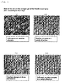
Fig. 1 is a microscopic picture showing the state of cells in collagen gel in the inventive example. -
Fig. 2 is a microscopic picture showing the state of cells in collagen gel in the comparative example. -
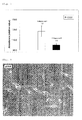
Fig. 3 is a graph of absorption (relative values) measured inExperiment 1. -
Fig. 4 is a graph of absorption (relative values) measured inExperiment 2. -
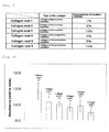
Fig. 5 shows the details of thecollagen coats 1 to 6 used inExperiment 3. -
Fig. 6 is a graph of absorption (relative values) measured inExperiment 3. -
Fig. 7 is a graph of absorption (relative values) measured inExperiment 3. -
Fig. 8 is a microscopic picture showing the state of cell differentiation cultured on a collagen (pH 5.5) coat plate ofExperiment 4. -
Fig. 9 is a microscopic picture showing the state of cell differentiation cultured on a collagen (pH 8.5) coat plate ofExperiment 4. -
Fig. 10 is a microscopic picture showing the state of cell differentiation cultured on a collagen (pH 10.2) coat plate ofExperiment 4. -
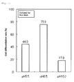
Fig. 11 is a graph of the cell differentiation rate after culturing for four days measured inExperiment 4. -
Fig. 12 is a graph of the cell differentiation rate after culturing for eleven days measured inExperiment 4. - The nerve regeneration-inducing tube of the present invention is characterized in using the collagen which is purified so as to make the concentration of sodium chloride contaminated therein during the production steps for collagen not more than 2% by weight in a dry state as a scaffold for nerve regeneration.
- Since collagen plays a role of a substrate for various kinds of cells, its affinity to the tissues is good when applied to living bodies as a medical material whereby it has been used already as a scaffold for the growth of nerve cells.
- The conventional collagen used as a scaffold for the nerve regeneration is usually prepared by the following manner. Thus, the pigskin collected and frozen in a meat testing factory is used as a starting material, a neutral protease is added thereto followed by warming, repeatedly washed with a sodium chloride solution, dehydrated, washed with isopropanol and acetone and dried in vacuo, the resulting defatted chip is added to an acetic acid solution, the pH is adjusted using hydrochloric acid, pepsin is added thereto to decompose, a sodium hydroxide solution is added to adjust to high pH (a virus inactivating step 1), hydrochloric acid is added to adjust to low pH (a virus inactivating step 2), the pH is adjusted to 2 to 3 using sodium hydroxide followed by filtering, a sodium chloride solution is added to the filtrate to salt out, a concentrating operation is conducted by means of centrifugal separation, the concentrated product is added to and dissolved in pure water, a sodium chloride solution is added thereto once again to salt out and the mixture is concentrated by means of centrifugal separation followed by freeze-drying.
- The conventionally used collagen as such includes the steps of washing with a sodium chloride solution and salting-out using a sodium chloride solution during its manufacturing process whereby the concentration of sodium chloride in the collagen, including that which is available in the market, is 4% by weight or more. The present inventors thought that the sodium chloride concentration in the collagen affects the living and the growth of nerve cells and that, when the concentration is too high, cell membrane is destroyed due to osmotic pressure. Therefore, when collagen which is purified so as to reduce the concentration of sodium chloride contained therein is used for a nerve regeneration-inducing tube, the resulting nerve regeneration-inducing tube achieves far better cell adhesive property and cell growth property than the one which uses the conventional collagen. On the basis of the finding as such, the present invention uses pure collagen where the concentration of sodium chloride in terms of a dry state contained therein is reduced to 2.0% by weight or less, preferably 0.1 to 1.5% by weight as a scaffold of the nerve regeneration-inducing tube. The concentration of sodium chloride is measured by means of atomic absorption spectrophotometry (by making into ash). For a purpose of prevention of destruction of cell membrane by lowering of osmotic pressure, the lower the sodium chloride concentration, the better. However, in view of technical standpoint and stability of collagen, about 0.1% by weight will be the lower limit. As a method for lowering the salt concentration, there are isoelectric precipitation (concentration) method as described hereinafter, dialysis method and so on. In the present invention, any known method can be used.
Measurement of sodium chloride concentration by means of atomic absorption spectrophotometry is carried out by such a manner that 1 to 4 g of a sample is taken into a quartz beaker, carbonized by a gradual rise of temperature on an electric heater and finally made into ash by treating in a Muffle furnace for 6 to 8 hours (500°C), and the residue is re-dissolved in a 10 wt% aqueous solution of hydrochloric acid and diluted so as to make thefinal concentration 1 wt% and then the measurement is conducted by means of a flame atomic absorption spectrophotometry using acetylene and air. The measuring wavelength is 589.6 nm. - Collagen used in the nerve regeneration-inducing tube of the present invention may be produced by any of the conventionally known methods and, for example, it can be produced by such a manner where the conventional collagen which has been available in the market for medical use as mentioned above is used as a starting material, dissolved in distilled water for injection under cooling at 2 to 10°C, subjected to pH adjustment within a range of from 6.0 to lower than 10.0 using a sodium hydroxide solution, subjected to an isoelectric precipitation and centrifuged, the supernatant liquid is discarded and the precipitate is freeze-dried. The present inventors found that extremely superior cell differentiation inducing property can be exhibited by using collagen having such an isoelectric point range, whereby the present invention is completed. Although a detailed reason why the cell differentiation inducing property is enhanced by the use of collagen having an isoelectric point of from 6.0 to lower than 10.0 as a scaffold for the nerve regeneration is not clear, there is a possibility that the fraction precipitated when pH is lower than 6.0 and 10.0 or higher contains a factor having low affinity to cells and, on the contrary, it is also likely that the collagen precipitated when pH is from 6.0 to lower than 10.0 has a particularly high affinity to cells. Alternatively, the unpurified collagen is constituted from type I collagen and type III collagen in a ratio of about 7: 3 and there may be an influence by changing this constituting ratio of type I to type III. In the present invention, range of isoelectric point is more preferably from pH 7.0 to lower than 9.5 and more preferably from pH 8.0 to lower than 9.0.
- The nerve regeneration-inducing tube of the present invention can be manufactured by the conventionally known methods and, for example, it can be formed by such a manner that collagen is coated on a tubular body comprising biodegradable polymer and then the collagen is filled in the inner area of the tubular body. Although the size of the tubular body may vary depending upon the site of the nerve to be regenerated and also upon the strength necessary therefor, it is usual that an inner diameter is 0.1 to 20 mm, an outer diameter is 0.15 to 25 mm and a length is 1.0 to 150 mm. Actually, it is preferred to previously prepare the nerve regeneration-inducing tubes comprising tubular bodies in various sizes in view of the time restriction and the production cost.
- Examples of the biodegradable polymer constituting the tubular body include polyglycolic acid, polylactic acid, a lactic acid-caprolactone copolymer, a glycolic acid-caprolactone copolymer, polydioxanone and glycolic acid-trimethylenecarboxylic acid. In view of easy availability and handling, it is preferred to use polyglycolic acid, polylactic acid or a lactic acid-caprolactone copolymer and it is particularly preferred to use polyglycolic acid. Each of those biodegradable polymers may be used solely or two or more thereof may be used by mixing.
- As to the tubular body, there may be used a body where the above-mentioned biodegradable polymer is made into porous tubular body or a body where plural ultrafine fibers of the above-mentioned biodegradable polymer are bundled followed by weaving into a tubular shape. Pore size and void fraction of the porous thing and the stitch (meshes) may be appropriately adjusted depending upon the aimed use and strength.
- Diameter of the ultrafine fiber comprising the biodegradable polymer is preferred to be from 1 to 50 µm. When the fiber diameter is too small, the fiber gap becomes dense whereby it may happen that collagen is hardly permeated into the tubular body or that flexibility of the tubular body lowers. On the contrary, when the fiber diameter is too large, the retained amount of collagen becomes small whereby it may happen that the growing speed of the nerve does not rise or that the strength of the tubular body becomes insufficient. More preferably, diameter of the ultrafine fiber is 3 to 40 µm, and further preferably 6 to 30 µm.
- In the formation of the tubular body, it is preferred that 5 to 60 of the ultrafine fibers comprising the biodegradable polymer and having the above fiber diameter are bundled and alternately knitted as warps and woofs. When the numbers of the ultrafine fibers to be bundled are too small, it may happen that the strength of the tubular body becomes insufficient or that a sufficient retained-amount of collagen cannot be secured. On the contrary, when the numbers of the ultrafine fibers to be bundled are too many, it may happen that a tubular body in fine diameter cannot be prepared or that flexibility of the tubular body cannot be secured. More preferably, the numbers of the ultrafine fibers are 10 to 50, and further preferably 20 to 40.
- When a tubular body is formed by an alternate knitting of the ultrafine fiber bundles, the pore size of the network is preferred to be about 5 to 300 µm, and more preferably 10 to 200 µm. When the pore size of the network is too small, it may happen that growth of the cells and the tissues is inhibited due to the lowering of invasion of capillary blood vessel or due to the lowering of water permeability. When it is more than about 300 µm, invasion of the tissues becomes excessive whereby growth of the cells and the tissues may be inhibited.
- In the present invention, the outer surface of the tubular body is coated by applying a collagen solution for several times by a method which has been known among persons skilled in the art while the inner area (lumen) of the tubular body is filled by charging collagen therein. The collagen solution used therein may contain laminin, heparan sulfate proteoglycan, entactin and growth factor. Examples of the growth factor include EGF (epidermal growth factor), βFGF (fibroblast growth factor), NGF (nerve growth factor), PDGF (platelet-derived growth factor), IGF-1 (insulin-like growth factor) and TGF-β (transforming growth factor). With regard to the collagen solution, it is preferred that, after every one application thereof in a form of a solution in hydrochloric acid using a brush or a writing brush, the solution is completely dried and then the next application is conducted whereby a plurality of applications are done.
- It is preferred that the tubular body where collagen is coated or filled is subjected to freezing, freeze-drying and cross-linking treatments to cross-link the collagen. The freezing is carried out under the condition of preferably -10 to -196°C and more preferably -20 to -80°C for 3 to 48 hours. As a result of the freezing, fine ice is formed among the collagen molecules and the collagen solution results in a phase separation to give sponge. After that, the above frozen collagen solution is freeze-dried in vacuo at about -40 to -80°C of initial temperature and for about 12 to 48 hours. As a result of freeze-drying, fine ice among the collagen molecules is evaporated and, at the same time, the collagen sponge becomes fine. Examples of the cross-linking method include γ-ray cross-linking, ultraviolet cross-linking, electronic ray cross-linking, thermal dehydration cross-linking, glutaraldehyde cross-linking, epoxy cross-linking and water-soluble carbodiimide cross-linking and, among them, a thermal dehydration cross-linking where the cross-linking degree can be easily controlled and living body is not effected even by conducting the cross-linking treatment is preferred. The thermal dehydration cross-linking is conducted in vacuo at, for example, about 105 to 150°C, more preferably about 120 to 150°C, and further preferably about 140°C for about 6 to 24 hours, more preferably about 6 to 12 hours, and further preferably about 12 hours. When the cross-linking temperature is too high, there is a possibility that the strength of the biodegradable and bioabsorbable material lowers while, when it is too low, there is a possibility that no sufficient cross-linking reaction takes place.
- Experiments which actually prove the superiority of the collagen of the present invention will be shown below.
- Measurement of the concentration of sodium chloride by atomic absorption spectrometry is conducted in such a manner that 1 to 4 g of a sample is placed in a quartz beaker, carbonized on an electric heater by gradually raising the temperature and, finally, made into ash in a muffle furnace for 6 to 8 hours (500°C). The residue is re-dissolved in a 10 wt% aqueous solution of hydrochloric acid, diluted so as to make the
final concentration 1% by weight and subjected to the measurement by means of a flame atomic absorption method using acetylene-air. Incidentally, the measuring wavelength is 589.6 nm. - Usually, a two-dimensional incubation on the bottom of a well-plate is fundamental in the cell incubation experiments. However, it has been said that, when a three-dimensional incubation is conducted, behavior of the cells is greatly different from that in the case of the two-dimensional incubation and, in evaluating the nerve regeneration, it has been believed that the three-dimensional incubation is a system which is nearer the actual system. Accordingly, in this experiment, a three-dimensional incubation is carried out using collagen gel for a purpose of confirming whether the behavior of the incubated cells is different depending upon the type of the collagen.
- "NMP Collagen PS" manufactured by Nippon Ham was used as a collagen for the comparative examples. This collagen for the comparative examples is manufactured by such a manner that pigskin is used as a starting material and is subjected to the defatting and purifying treatments. The defatting treatment includes the repeated washing steps using a sodium chloride solution while the purifying treatment includes a salting-out step using sodium chloride. When this collagen for the comparative examples was measured by means of an atomic absorption spectrometry (by making into ash), it contained 4.0% by weight of sodium chloride in its dry state.
- A part of the above collagen for the comparative examples was used as a starting material and was purified by means of an isoelectric precipitation where pH was 8 or higher and lower than 9 whereupon the collagen of the inventive examples was prepared. When this collagen of the inventive examples was measured by means of an atomic absorption spectrometry (by making into ash), it contained 1. 0% by weight of sodium chloride in its dry state.
- Each of the above-prepared two kinds of collagen was dissolved in hydrochloric acid according to the conventional method to prepare a 0. 5 wt% solution of collagen in hydrochloric acid. Each 300 µl of the collagen solution of the inventive examples among the above was added to eight wells of a 24-well microplate (manufactured by Iwaki) while each 300 µl of the collagen solution of the comparative examples was added to other eight wells of the same plate. After that, the plate was allowed to stand at 37°C in an incubator for 30 minutes.
-
- (1) PC12 cells (cells derived from the adrenal pheochromocytoma of rats manufactured by Dainippon Pharmaceutical Laboratory Products) were previously cultured on a DMEM medium until six passage numbers, the cells were recovered by centrifugal separation and suspended in 15 ml of the DMEM medium so as to adjust the cell numbers to 1 × 10⁶ cells and then 15 µl of a 50 µg/ml NGF (cell growth factor manufactured by R&D Systems Inc.; a phosphate buffer) was added thereto whereupon a culture solution was prepared.
Incidentally, the DMEM medium is such a one where 25 ml of fetal bovine serum (manufactured by Dainippon Pharmaceutical Laboratory Products), 50 ml of equine serum (manufactured by Dainippon Pharmaceutical Laboratory Products) and 5 ml of 200 mM glutamine solution (manufactured by Dainippon Pharmaceutical Laboratory Products; 29.23 mg/ml) are added to and mixed with 500 ml of an RPMI 1640 liquid medium (manufactured by Dainippon Pharmaceutical Laboratory Products; containing no glutamic acid and containing sodium bicarbonate). - (2) Each 300 µl of the culture solution prepared as such was dropped into the well of the previously-prepared collagen medium.
- (3) The well plate was incubated in an incubator (37°C; CO2 concentration: 5.0%) for four days.
- After incubating for four days, the state of the cells in the collagen gel was observed under a microscope and the pictures of the representative examples thereof were taken. The results are shown in
Fig. 1 andFig. 2 . -
- (1) In order to measure the living cell numbers after incubating for four days, each 50 µl of the 1 wt% solution of collagenase was dropped into a well and the mixture was gently stirred together with the well whereupon the collagen gel was dissolved at 37°C within 30 minutes.
- (2) After the collagen gel was dissolved, each 50 µl of an MTT assay solution was added to each well followed by being allowed to stand in an incubator for 30 minutes.
- (3) After being allowed to stand for 30 minutes, the absorbance at 450 nm was measured, average value and standard deviation were determined for each collagen gel from the values of the absorbance in eight wells and the result is shown as a graph in
Fig. 3 . Incidentally, in the graph ofFig. 3 , the absorbance of the collagen of the inventive examples is expressed in terms of a relative value where the average absorption of the collagen gels of the comparative examples is defined as 100. In the meanwhile, the absorption is in parallel to the living cell numbers. - As will be apparent from the comparison of
Fig. 1 withFig. 2 , the incubation using the collagen of the inventive examples (Fig. 1 ) shows better growth of the cells than the incubation using the collagen of the comparative examples (Fig. 2 ) and elongation of the neurite is significant as well. - As will be apparent from
Fig. 3 , the absorbance of the incubation using the collagen of the inventive examples is higher than the absorbance of the incubation using the collagen of the comparative examples to an extent of 39% in average and the difference as such is a significant difference in view of statistics as well (p<0.01). Accordingly, from the result ofFig. 3 , it is noted that the collagen of the inventive examples has the significantly higher cell growth ability than the collagen of the comparative examples.
(3) From the above results, it is concluded that the collagen of the inventive examples is better than the conventional collagen of the comparative examples in terms of the cell growth ability and the differentiation inducing ability. - An object thereof is that, for comparing the cell adhesion property between the collagen of the inventive examples and the conventional collagen, the floating cells after the incubation in both collagens were sucked and removed using an aspirator, only the cells adhered to the plate were measured and their numbers were compared to confirm whether there was a significant difference in the adhesive property of the cells depending upon the type of the collagen.
-
- (1) The collagen of the inventive examples and the collagen of the comparative examples were diluted with hydrochloric acid to make the concentration 0.05% by weight and each 300 µl of those collagen solutions was placed into each eight wells of a 24-well microplate (manufactured by Iwaki) followed by being allowed to stand in a refrigerator for one hour.
- (2) After being allowed to stand for one hour, the collagen solution in each well was sucked using an aspirator and the collagen coat adhered in the well was subjected to natural drying for 1 hour in a clean bench.
-
- (1) PC12 cells (cells derived from the adrenal pheochromocytoma of rats manufactured by Dainippon Pharmaceutical Laboratory Products) were previously cultured on a DMEM medium until six passage numbers, the cells were recovered by centrifugal separation and suspended in 25 ml of the DMEM medium so as to adjust the cell numbers to 5 × 10⁶ cells and then 25 µl of a 50 µg/ml NGF (cell growth factor manufactured by R&D Systems Inc.; a phosphate buffer) was added thereto whereupon a culture solution was prepared.
- (2) Each 300 µl of the culture solution prepared as such was dropped into the well of the previously-prepared collagen medium.
- (3) The well plate was incubated in an incubator (37°C; CO2 concentration: 5.0%) for five days.
-
- (1) In order to remove the floating cells and the cells which were not surely adhered, all of the media were sucked by inclining the well plate at an angle of 85°. Careful attention was paid at that time not to suck the adhered cells.
- (2) After that, each 300 µl of a DMEM medium and 30 µl of an MTT assay solution were added to each well followed by being allowed to stand in an incubator for 30 minutes.
- (3) After being allowed to stand for 30 minutes, the absorbance at 450 nm was measured, average value and standard deviation were determined for each collagen coat from the values of the absorbance in eight wells and the result is shown as a graph in
Fig. 4 . Incidentally, in the graph ofFig. 4 , the absorbance of the collagen coat of the inventive examples is expressed in terms of a relative value where the average absorption of the collagen coat of the comparative examples is defined as 100. - As will be apparent from
Fig. 4 , the absorbance which expresses the adhered cell numbers of the collagen coat of the inventive examples was higher than the absorbance of the collagen coat of the comparative examples to an extent of 49% in average and the difference as such was a difference which was significant in terms of statistics as well (0.01<p<0.05). The cell adhesive property is a very important factor for the scaffold in the regeneration medicine and, from the result ofFig. 4 , it is noted that the collagen of the present invention is suitable for using as a scaffold for the nerve regeneration as compared with the conventional collagen. - An object thereof is to check the degree of influence of the difference in the concentrations of sodium chloride contained in collagen on the living and the growth of the cells.
-
- (1) There were prepared the collagen of the inventive examples used in
Experiment 1; that where sodium chloride concentration was made 5% by weight or 10% by weight by addition of sodium chloride thereto; the collagen of the comparative examples used inExperiment 1; and that where sodium chloride concentration was made 5% by weight or 10% by weight by addition of sodium chloride thereto (refer tocollagen coats 1 to 6 ofFig. 5 ). Each of the collagens was made into a 0.01 wt% solution in hydrochloric acid and 300 µl of each collagen was dropped into four wells using two 24-well microplates (manufactured by Iwaki) followed by being allowed to stand in a refrigerator for one hour. - (2) After being allowed to stand for one hour, the collagen solution in each well was sucked using an aspirator and the collagen coat adhered in the well was subjected to natural drying for 1 hour in a clean bench.
-
- (1) PC12 cells (cells derived from the adrenal pheochromocytoma of rats manufactured by Dainippon Pharmaceutical Laboratory Products) were previously cultured on a DMEM medium until six passage numbers, the cells were recovered by centrifugal separation and suspended in 15 ml of the DMEM medium so as to adjust the cell numbers to 1 × 10⁶ cells and then 15 µl of a 50 µg/ml NGF (cell growth factor manufactured by R&D Systems Inc.; a phosphate buffer) was added thereto whereupon a culture solution was prepared.
- (2) Each 300 µl of the culture solution prepared as such was dropped into the well of the previously-prepared collagen medium.
- (3) The well plate was incubated in an incubator (37°C; CO2 concentration: 5.0%) for five days.
-
- (1) Each 30 µl of an MTT assay solution was added to each well followed by being allowed to stand in an incubator for 30 minutes.
- (2) After being allowed to stand for 30 minutes, the absorbance at 450 nm was measured, average value and standard deviation were determined for each collagen coat from the values of the absorbance in eight wells and the result is shown as graphs in
Figs. 6 and7 . Incidentally, in the graphs ofFigs. 6 and7 , the absorbance of the collagen coat of the inventive examples is expressed in terms of a relative value where the average absorption of the collagen coat of the comparative examples is defined as 100. - As will be apparent from
Figs. 6 and7 , there is a tendency that the higher the absorbance, the lower the sodium chloride concentration in collagen and the absorbance of the culture using the collagen of the inventive examples (collagen coat 1 (sodium chloride concentration: 1% by weight)) was higher than the absorbance of the culture using the collagen of the comparative examples (collagen coat 6 (sodium chloride concentration: 10% by weight)) to an extent of 27% in average where said difference was a difference which was significant in terms of statistic as well (p<0.01). From the results ofFigs. 6 and7 , it is noted that the collagen of the inventive examples containing low concentration of sodium chloride is superior in the cell growth ability as compared with the collagen of the comparative examples. - An object thereof is to cheek the degree of influence of the difference in the isoelectric points of collagen on the cell differentiation.
- 2. Preparation of concentrated collagen powder by means of isoelectric point
- (1) Milli Q water was added to 6 g of NMP Collagen PS to prepare 1, 000 ml, in total, of a 0.6 wt% solution of collagen.
- (2) It was stirred on ice for one to three day(s) so that the collagen was completely dissolved in water.
- (3) 1N NaOH was dropped thereinto and 200 ml of a collagen solution containing the precipitate in a state of pH 5.5 was recovered to another container.
- (4) Dropping of 1N NaOH was continued and, similarly, each 200 ml solution in a state of pH 8.5 and 10.2 was recovered to another container.
- (5) Each of the resulting three samples were transferred to a centrifugal tube and centrifuged at 3,000 rpm for 45 minutes.
- (6) Supernatant liquid in each centrifugal tube was discarded and the precipitate was frozen at -40°C through the night and treated for two days in a freeze-drying machine.
- (7) The resulting samples were called a sample 1 (pH 5.5), a sample 2 (pH 8.5) and a sample 3 (pH 10.2) successively.
-
- (1) To each 300 mg of the
above samples - (2) Mixing was conducted using Voltex followed by being allowed to stand at 4°C through the night so that collagen was completely dissolved.
- (3) To each 1 ml of the sample of 0.3 wt% concentration was added 9 ml of 0.001M HCl followed by well mixing to prepare a 0.03 wt% collagen solution.
- (4) Each 200 µl of the above 0.03 wt% collagen solution was placed in each well of a 24-well microplate (manufactured by Iwaki) followed by being allowed to stand at room temperature (20°C) for ten minutes.
- (5) The collagen solution was sucked, 1 ml of PBS(-) was added thereto and the mixture was sucked, washed and repeatedly washed.
- (6) The above was just allowed to stand in a clean bench to dry.
- Pigment cells derived from neural crest (manufactured by Kurabo Industries; Code No. KM-4009MP) was diluted with a medium exclusively therefor (manufactured by Kurabo Industries; Code No. M-254-500 + Code No. S-002-5) to make 3.75 × 10⁴ cells per ml and then 500 µl thereof was sown onto each well.
- (2) Incubation was conducted for four days at 37°C where CO2 concentration was 5.0%.
- (3) After sucking the medium, 500 µl of the exclusive medium was freshly added thereto and, after that, incubation was conducted for three days at 37°C where CO2 concentration was 5.0%.
- (4) After sucking the medium, 500 µl of the exclusive medium was freshly added thereto, incubation was conducted for four days more at 37°C where CO2 concentration was 5.0% and, after that, degree of cell differentiation in each well was observed.
- As will be apparent from
Figs. 8 to 10 , significant differences were noted in the cell differentiation levels depending upon the difference in the isoelectric point of collagen and it could be confirmed from the observation of the cell form that the differentiation was significantly promoted in the sample 2 (pH 8.5). - It was further confirmed from
Figs. 11 and12 that, when the rate of the differentiated cells in the total living cells was calculated, the cell differentiation rate (%) was high when pH was 8.5 as compared with the cases where pH was 5.5 and 10.2.
- In view of the fact that, in general, collagen is metabolized/absorbed in vivo within about two weeks, it is believed that, if 40% or more cell differentiation rate is available after the incubation for 11 days, good result is achieved in the actual field of nerve regeneration. The cell differentiation rate is more preferably not less than 50%, more preferably not less than 60%, and more preferably not less than 70% .
- Since the nerve regeneration-inducing tube according to the present invention is excellent in terms of cell adhesion property, cell growth ability and cell differentiation inducing ability, its application in the nerve regeneration medicine is expanded and it is quite useful.
Claims (4)
- A nerve regeneration-inducing tube, where collagen is used as a scaffold for nerve regeneration, characterized in using the collagen which is made the concentration of sodium chloride contained therein not more than 2.0% by weight in a dry state.
- The nerve regeneration-inducing tube according to claim 1, characterized in using the collagen which is made the concentration of sodium chloride contained therein 0.1 to 1.5% by weight in a dry state.
- The nerve regeneration-inducing tube according to claim 1 or 2, characterized in that the collagen is purified by means of an isoelectric precipitation where the pH is 6.0 or higher and is lower than 10.0.
- The nerve regeneration-inducing tube according to any one of claims 1 to 3, characterized in that it is formed by coating the collagen on a tubular body comprising a biodegradable polymer and filling the collagen into the tubular body.
Priority Applications (2)
| Application Number | Priority Date | Filing Date | Title |
|---|---|---|---|
| PL09839205T PL2404622T3 (en) | 2009-02-02 | 2009-02-02 | Nerve regeneration-inducing tube |
| EP15182940.5A EP2987508B1 (en) | 2009-02-02 | 2009-02-02 | Nerve regeneration-inducing tube |
Applications Claiming Priority (1)
| Application Number | Priority Date | Filing Date | Title |
|---|---|---|---|
| PCT/JP2009/051702 WO2010087015A1 (en) | 2009-02-02 | 2009-02-02 | Nerve regeneration-inducing tube |
Related Child Applications (2)
| Application Number | Title | Priority Date | Filing Date |
|---|---|---|---|
| EP15182940.5A Division EP2987508B1 (en) | 2009-02-02 | 2009-02-02 | Nerve regeneration-inducing tube |
| EP15182940.5A Division-Into EP2987508B1 (en) | 2009-02-02 | 2009-02-02 | Nerve regeneration-inducing tube |
Publications (3)
| Publication Number | Publication Date |
|---|---|
| EP2404622A1 true EP2404622A1 (en) | 2012-01-11 |
| EP2404622A4 EP2404622A4 (en) | 2013-01-23 |
| EP2404622B1 EP2404622B1 (en) | 2015-12-02 |
Family
ID=42395286
Family Applications (2)
| Application Number | Title | Priority Date | Filing Date |
|---|---|---|---|
| EP09839205.3A Not-in-force EP2404622B1 (en) | 2009-02-02 | 2009-02-02 | Nerve regeneration-inducing tube |
| EP15182940.5A Active EP2987508B1 (en) | 2009-02-02 | 2009-02-02 | Nerve regeneration-inducing tube |
Family Applications After (1)
| Application Number | Title | Priority Date | Filing Date |
|---|---|---|---|
| EP15182940.5A Active EP2987508B1 (en) | 2009-02-02 | 2009-02-02 | Nerve regeneration-inducing tube |
Country Status (7)
| Country | Link |
|---|---|
| US (1) | US8741328B2 (en) |
| EP (2) | EP2404622B1 (en) |
| JP (1) | JP4572996B2 (en) |
| CN (1) | CN102387821B (en) |
| ES (1) | ES2555978T3 (en) |
| PL (1) | PL2404622T3 (en) |
| WO (1) | WO2010087015A1 (en) |
Families Citing this family (7)
| Publication number | Priority date | Publication date | Assignee | Title |
|---|---|---|---|---|
| EP3288626B8 (en) | 2015-04-27 | 2025-12-10 | Tulavi Therapeutics, Inc. | Substances and compositions for use in blocking nerve regeneration |
| WO2017139487A1 (en) | 2016-02-09 | 2017-08-17 | Northwind Medical, Inc. | Methods, agents, and devices for local neuromodulation of autonomic nerves |
| AU2017290353B2 (en) | 2016-06-29 | 2023-05-18 | Tulavi Therapeutics, Inc. | Treatment of sepsis and related inflammatory conditions by local neuromodulation of the autonomic nervous system |
| US20210315587A1 (en) | 2018-07-02 | 2021-10-14 | Tulavi Therapeutics, Inc. | Methods and devices for in situ formed nerve cap with rapid release |
| AU2019299519B2 (en) | 2018-07-02 | 2025-03-06 | Incept Llc | Methods and devices for in situ formed nerve cap |
| CN111956869B (en) * | 2020-08-07 | 2024-03-08 | 中南大学湘雅医院 | A pump that slowly promotes nerve growth |
| CN115073586B (en) * | 2022-07-15 | 2025-05-16 | 成都奇璞生物科技有限公司 | Concentrated collagen and its concentration process and application |
Family Cites Families (28)
| Publication number | Priority date | Publication date | Assignee | Title |
|---|---|---|---|---|
| JPH0662679B2 (en) | 1985-06-21 | 1994-08-17 | 新田ゼラチン株式会社 | Tissue-friendly collagen and its manufacturing method |
| US4863668A (en) * | 1988-09-22 | 1989-09-05 | University Of Utah | Method of forming fibrin-collagen nerve and body tissue repair material |
| JP3457339B2 (en) | 1992-02-27 | 2003-10-14 | 日本ハム株式会社 | Nerve regeneration aid and method for producing the same |
| JP3317471B2 (en) * | 1995-04-07 | 2002-08-26 | 住友ベークライト株式会社 | Collagen gel treatment method |
| JPH08283667A (en) * | 1995-04-14 | 1996-10-29 | Sumitomo Bakelite Co Ltd | Formed collagen and its production |
| WO1998020130A2 (en) | 1996-11-01 | 1998-05-14 | Genetics Institute, Inc. | Secreted proteins and polynucleotides encoding them |
| TW528600B (en) | 1996-11-20 | 2003-04-21 | Yasuhiko Shimizu | Artificial neural canal |
| JP4968976B2 (en) * | 1998-03-06 | 2012-07-04 | 慶彦 清水 | Collagen material and production method thereof |
| KR20010052723A (en) * | 1998-06-10 | 2001-06-25 | 야스히꼬 시미즈 | Artificial Neural Tube |
| JP4721482B2 (en) | 1999-05-18 | 2011-07-13 | 株式会社高研 | Nerve reconstruction substrate |
| JP2001070436A (en) | 1999-09-02 | 2001-03-21 | Koken Co Ltd | Collagen support for forming biological tract structures |
| CZ301649B6 (en) * | 2000-06-28 | 2010-05-12 | Ed. Geistlich Soehne Ag Fur Chemische Industrie Incorporated Under The Laws Of Switzerland | Tube for regeneration of nerves and process for producing thereof |
| GB0030583D0 (en) * | 2000-12-15 | 2001-01-31 | Univ Nottingham | Nerve regeneration |
| JP3871525B2 (en) | 2001-04-26 | 2007-01-24 | ニプロ株式会社 | Biological tissue or organ regeneration device |
| JP4740482B2 (en) | 2001-07-09 | 2011-08-03 | グンゼ株式会社 | Nerve regeneration tube |
| JP3966045B2 (en) * | 2002-04-03 | 2007-08-29 | ニプロ株式会社 | Collagen nonwoven fabric, its production method, its treatment method and apparatus |
| JP4789284B2 (en) * | 2002-10-30 | 2011-10-12 | 太田 耕二 | Cosmetics with collagen |
| WO2004087012A1 (en) * | 2003-03-31 | 2004-10-14 | Teijin Limited | Composite of support substrate and collagen, and process for producing support substrate and composite |
| JP2005143979A (en) * | 2003-11-18 | 2005-06-09 | Gunze Ltd | Tube for neuroregeneration |
| JP4292094B2 (en) * | 2004-02-24 | 2009-07-08 | グンゼ株式会社 | Nerve regeneration tube |
| JP4587711B2 (en) * | 2004-06-04 | 2010-11-24 | ミドリホクヨー株式会社 | Soluble fish collagen, method for producing the same and collagen cosmetic |
| CN1312179C (en) * | 2004-09-23 | 2007-04-25 | 中国海洋大学 | Method for modifying freshwater fish skin collagen by adjusting alkalinity acidity |
| CN1891179A (en) * | 2005-07-01 | 2007-01-10 | 于海鹰 | Medical collagen nerve catheter and making technology |
| JP4765602B2 (en) | 2005-12-14 | 2011-09-07 | ニプロ株式会社 | Package for regenerative medical instrument and processing method for regenerative medical instrument |
| JP2007261966A (en) * | 2006-03-28 | 2007-10-11 | Fujifilm Corp | Method for producing iv-type collagen |
| JP2009034374A (en) | 2007-08-02 | 2009-02-19 | Toyobo Co Ltd | Nerve regeneration-inducing tube |
| JP4596335B2 (en) * | 2007-12-07 | 2010-12-08 | 東洋紡績株式会社 | Method for manufacturing nerve regeneration induction tube |
| JP5702515B2 (en) * | 2007-12-28 | 2015-04-15 | 東洋紡株式会社 | Nerve regeneration induction tube |
-
2009
- 2009-02-02 PL PL09839205T patent/PL2404622T3/en unknown
- 2009-02-02 WO PCT/JP2009/051702 patent/WO2010087015A1/en not_active Ceased
- 2009-02-02 EP EP09839205.3A patent/EP2404622B1/en not_active Not-in-force
- 2009-02-02 JP JP2009512087A patent/JP4572996B2/en active Active
- 2009-02-02 CN CN200980158621XA patent/CN102387821B/en not_active Expired - Fee Related
- 2009-02-02 ES ES09839205.3T patent/ES2555978T3/en active Active
- 2009-02-02 US US13/147,234 patent/US8741328B2/en active Active
- 2009-02-02 EP EP15182940.5A patent/EP2987508B1/en active Active
Also Published As
| Publication number | Publication date |
|---|---|
| US8741328B2 (en) | 2014-06-03 |
| PL2404622T3 (en) | 2016-06-30 |
| ES2555978T3 (en) | 2016-01-12 |
| EP2404622A4 (en) | 2013-01-23 |
| EP2987508A1 (en) | 2016-02-24 |
| EP2404622B1 (en) | 2015-12-02 |
| CN102387821A (en) | 2012-03-21 |
| EP2987508B1 (en) | 2019-07-03 |
| US20110288569A1 (en) | 2011-11-24 |
| JPWO2010087015A1 (en) | 2012-07-26 |
| JP4572996B2 (en) | 2010-11-04 |
| WO2010087015A1 (en) | 2010-08-05 |
| CN102387821B (en) | 2013-12-25 |
Similar Documents
| Publication | Publication Date | Title |
|---|---|---|
| EP2404622B1 (en) | Nerve regeneration-inducing tube | |
| EP2228036B1 (en) | Nerve regeneration-inducing tube | |
| Rohanizadeh et al. | Gelatin sponges (Gelfoam®) as a scaffold for osteoblasts | |
| Ladet et al. | Multi-membrane chitosan hydrogels as chondrocytic cell bioreactors | |
| JP5925185B2 (en) | Non-woven fabric containing bone filling material | |
| Ding et al. | Rapid repair and regeneration of damaged rabbit sciatic nerves by tissue-engineered scaffold made from nano-silver and collagen type I | |
| JPH10503098A (en) | Compositions and methods for bioartificial extracellular matrices | |
| WO2004003130A1 (en) | Chitosan/acidic biopolymer hybrid fiber and culture base for animal cells | |
| WO2014013188A1 (en) | Chitosan hydrogel for repairing nerve tissue | |
| CN114558170A (en) | Growth factor-containing skull repairing polyether-ether-ketone material and preparation method thereof | |
| JP6092091B2 (en) | Peptide having affinity for BMP-2 | |
| JP4626725B2 (en) | Collagen for cell culture | |
| KR101999098B1 (en) | Scaffold using material from marine organisms for bone tissue generation and method for preparing the same | |
| AU2006207618A1 (en) | Hyaluronic acid derivative and neural stem cells for SCI and PNT regeneration | |
| JP2007130118A (en) | Transplant material | |
| JP2009034374A (en) | Nerve regeneration-inducing tube | |
| KR20160034557A (en) | To induce bone regeneration using PLGA-Silk hybrid structure method of manufacturing | |
| CN115920135B (en) | Bone repair material and preparation method thereof | |
| US9345807B2 (en) | PLGA scaffold | |
| KR20030006787A (en) | Chitosan bead containing tricalcium phosphate for bone substitute | |
| He et al. | Silk Fibroin Scaffolds Facilitating the Repair of Rat Abdominal Wall Defect | |
| Peng et al. | Biological evaluation of porous chitosan/collagen scaffolds for periodontal tissue engineering | |
| van den Beucken | DNA-Based coatings in implantology: utilization of structural properties of the DNA-module to modulate cell and tissue response. | |
| WO2007066650A1 (en) | Culture substrate and culture method for cartilage tissue regeneration |
Legal Events
| Date | Code | Title | Description |
|---|---|---|---|
| PUAI | Public reference made under article 153(3) epc to a published international application that has entered the european phase |
Free format text: ORIGINAL CODE: 0009012 |
|
| 17P | Request for examination filed |
Effective date: 20110824 |
|
| AK | Designated contracting states |
Kind code of ref document: A1 Designated state(s): AT BE BG CH CY CZ DE DK EE ES FI FR GB GR HR HU IE IS IT LI LT LU LV MC MK MT NL NO PL PT RO SE SI SK TR |
|
| DAX | Request for extension of the european patent (deleted) | ||
| RAP1 | Party data changed (applicant data changed or rights of an application transferred) |
Owner name: TOYOBO CO., LTD. |
|
| A4 | Supplementary search report drawn up and despatched |
Effective date: 20121220 |
|
| RIC1 | Information provided on ipc code assigned before grant |
Ipc: A61L 27/24 20060101ALI20121214BHEP Ipc: A61L 27/00 20060101AFI20121214BHEP |
|
| 17Q | First examination report despatched |
Effective date: 20140806 |
|
| GRAP | Despatch of communication of intention to grant a patent |
Free format text: ORIGINAL CODE: EPIDOSNIGR1 |
|
| INTG | Intention to grant announced |
Effective date: 20150430 |
|
| GRAS | Grant fee paid |
Free format text: ORIGINAL CODE: EPIDOSNIGR3 |
|
| GRAR | Information related to intention to grant a patent recorded |
Free format text: ORIGINAL CODE: EPIDOSNIGR71 |
|
| RIN1 | Information on inventor provided before grant (corrected) |
Inventor name: KAJII, FUMIHIKO Inventor name: SUZUKI, MICHIKO Inventor name: TANAKA, HIDENORI Inventor name: KASHIWABARA, SUSUMU |
|
| GRAA | (expected) grant |
Free format text: ORIGINAL CODE: 0009210 |
|
| INTG | Intention to grant announced |
Effective date: 20151009 |
|
| AK | Designated contracting states |
Kind code of ref document: B1 Designated state(s): AT BE BG CH CY CZ DE DK EE ES FI FR GB GR HR HU IE IS IT LI LT LU LV MC MK MT NL NO PL PT RO SE SI SK TR |
|
| REG | Reference to a national code |
Ref country code: GB Ref legal event code: FG4D |
|
| REG | Reference to a national code |
Ref country code: AT Ref legal event code: REF Ref document number: 763322 Country of ref document: AT Kind code of ref document: T Effective date: 20151215 Ref country code: CH Ref legal event code: NV Representative=s name: BOHEST AG, CH Ref country code: CH Ref legal event code: EP |
|
| REG | Reference to a national code |
Ref country code: IE Ref legal event code: FG4D |
|
| REG | Reference to a national code |
Ref country code: ES Ref legal event code: FG2A Ref document number: 2555978 Country of ref document: ES Kind code of ref document: T3 Effective date: 20160112 |
|
| REG | Reference to a national code |
Ref country code: DE Ref legal event code: R096 Ref document number: 602009035155 Country of ref document: DE |
|
| REG | Reference to a national code |
Ref country code: RO Ref legal event code: EPE |
|
| REG | Reference to a national code |
Ref country code: FR Ref legal event code: PLFP Year of fee payment: 8 |
|
| REG | Reference to a national code |
Ref country code: NL Ref legal event code: FP |
|
| REG | Reference to a national code |
Ref country code: LT Ref legal event code: MG4D |
|
| REG | Reference to a national code |
Ref country code: AT Ref legal event code: MK05 Ref document number: 763322 Country of ref document: AT Kind code of ref document: T Effective date: 20151202 |
|
| PG25 | Lapsed in a contracting state [announced via postgrant information from national office to epo] |
Ref country code: HR Free format text: LAPSE BECAUSE OF FAILURE TO SUBMIT A TRANSLATION OF THE DESCRIPTION OR TO PAY THE FEE WITHIN THE PRESCRIBED TIME-LIMIT Effective date: 20151202 Ref country code: NO Free format text: LAPSE BECAUSE OF FAILURE TO SUBMIT A TRANSLATION OF THE DESCRIPTION OR TO PAY THE FEE WITHIN THE PRESCRIBED TIME-LIMIT Effective date: 20160302 Ref country code: LT Free format text: LAPSE BECAUSE OF FAILURE TO SUBMIT A TRANSLATION OF THE DESCRIPTION OR TO PAY THE FEE WITHIN THE PRESCRIBED TIME-LIMIT Effective date: 20151202 |
|
| PG25 | Lapsed in a contracting state [announced via postgrant information from national office to epo] |
Ref country code: AT Free format text: LAPSE BECAUSE OF FAILURE TO SUBMIT A TRANSLATION OF THE DESCRIPTION OR TO PAY THE FEE WITHIN THE PRESCRIBED TIME-LIMIT Effective date: 20151202 Ref country code: SE Free format text: LAPSE BECAUSE OF FAILURE TO SUBMIT A TRANSLATION OF THE DESCRIPTION OR TO PAY THE FEE WITHIN THE PRESCRIBED TIME-LIMIT Effective date: 20151202 Ref country code: LV Free format text: LAPSE BECAUSE OF FAILURE TO SUBMIT A TRANSLATION OF THE DESCRIPTION OR TO PAY THE FEE WITHIN THE PRESCRIBED TIME-LIMIT Effective date: 20151202 Ref country code: GR Free format text: LAPSE BECAUSE OF FAILURE TO SUBMIT A TRANSLATION OF THE DESCRIPTION OR TO PAY THE FEE WITHIN THE PRESCRIBED TIME-LIMIT Effective date: 20160303 Ref country code: FI Free format text: LAPSE BECAUSE OF FAILURE TO SUBMIT A TRANSLATION OF THE DESCRIPTION OR TO PAY THE FEE WITHIN THE PRESCRIBED TIME-LIMIT Effective date: 20151202 |
|
| PG25 | Lapsed in a contracting state [announced via postgrant information from national office to epo] |
Ref country code: IS Free format text: LAPSE BECAUSE OF FAILURE TO SUBMIT A TRANSLATION OF THE DESCRIPTION OR TO PAY THE FEE WITHIN THE PRESCRIBED TIME-LIMIT Effective date: 20151202 |
|
| PG25 | Lapsed in a contracting state [announced via postgrant information from national office to epo] |
Ref country code: CZ Free format text: LAPSE BECAUSE OF FAILURE TO SUBMIT A TRANSLATION OF THE DESCRIPTION OR TO PAY THE FEE WITHIN THE PRESCRIBED TIME-LIMIT Effective date: 20151202 |
|
| PG25 | Lapsed in a contracting state [announced via postgrant information from national office to epo] |
Ref country code: EE Free format text: LAPSE BECAUSE OF FAILURE TO SUBMIT A TRANSLATION OF THE DESCRIPTION OR TO PAY THE FEE WITHIN THE PRESCRIBED TIME-LIMIT Effective date: 20151202 Ref country code: SK Free format text: LAPSE BECAUSE OF FAILURE TO SUBMIT A TRANSLATION OF THE DESCRIPTION OR TO PAY THE FEE WITHIN THE PRESCRIBED TIME-LIMIT Effective date: 20151202 Ref country code: PT Free format text: LAPSE BECAUSE OF FAILURE TO SUBMIT A TRANSLATION OF THE DESCRIPTION OR TO PAY THE FEE WITHIN THE PRESCRIBED TIME-LIMIT Effective date: 20160404 Ref country code: IS Free format text: LAPSE BECAUSE OF FAILURE TO SUBMIT A TRANSLATION OF THE DESCRIPTION OR TO PAY THE FEE WITHIN THE PRESCRIBED TIME-LIMIT Effective date: 20160402 |
|
| REG | Reference to a national code |
Ref country code: DE Ref legal event code: R097 Ref document number: 602009035155 Country of ref document: DE |
|
| PG25 | Lapsed in a contracting state [announced via postgrant information from national office to epo] |
Ref country code: LU Free format text: LAPSE BECAUSE OF FAILURE TO SUBMIT A TRANSLATION OF THE DESCRIPTION OR TO PAY THE FEE WITHIN THE PRESCRIBED TIME-LIMIT Effective date: 20160202 Ref country code: MC Free format text: LAPSE BECAUSE OF FAILURE TO SUBMIT A TRANSLATION OF THE DESCRIPTION OR TO PAY THE FEE WITHIN THE PRESCRIBED TIME-LIMIT Effective date: 20151202 |
|
| PLBE | No opposition filed within time limit |
Free format text: ORIGINAL CODE: 0009261 |
|
| STAA | Information on the status of an ep patent application or granted ep patent |
Free format text: STATUS: NO OPPOSITION FILED WITHIN TIME LIMIT |
|
| PG25 | Lapsed in a contracting state [announced via postgrant information from national office to epo] |
Ref country code: DK Free format text: LAPSE BECAUSE OF FAILURE TO SUBMIT A TRANSLATION OF THE DESCRIPTION OR TO PAY THE FEE WITHIN THE PRESCRIBED TIME-LIMIT Effective date: 20151202 |
|
| 26N | No opposition filed |
Effective date: 20160905 |
|
| PG25 | Lapsed in a contracting state [announced via postgrant information from national office to epo] |
Ref country code: SI Free format text: LAPSE BECAUSE OF FAILURE TO SUBMIT A TRANSLATION OF THE DESCRIPTION OR TO PAY THE FEE WITHIN THE PRESCRIBED TIME-LIMIT Effective date: 20151202 |
|
| REG | Reference to a national code |
Ref country code: FR Ref legal event code: PLFP Year of fee payment: 9 |
|
| PG25 | Lapsed in a contracting state [announced via postgrant information from national office to epo] |
Ref country code: MT Free format text: LAPSE BECAUSE OF FAILURE TO SUBMIT A TRANSLATION OF THE DESCRIPTION OR TO PAY THE FEE WITHIN THE PRESCRIBED TIME-LIMIT Effective date: 20151202 |
|
| REG | Reference to a national code |
Ref country code: FR Ref legal event code: PLFP Year of fee payment: 10 |
|
| PG25 | Lapsed in a contracting state [announced via postgrant information from national office to epo] |
Ref country code: CY Free format text: LAPSE BECAUSE OF FAILURE TO SUBMIT A TRANSLATION OF THE DESCRIPTION OR TO PAY THE FEE WITHIN THE PRESCRIBED TIME-LIMIT Effective date: 20151202 Ref country code: HU Free format text: LAPSE BECAUSE OF FAILURE TO SUBMIT A TRANSLATION OF THE DESCRIPTION OR TO PAY THE FEE WITHIN THE PRESCRIBED TIME-LIMIT; INVALID AB INITIO Effective date: 20090202 |
|
| PG25 | Lapsed in a contracting state [announced via postgrant information from national office to epo] |
Ref country code: MT Free format text: LAPSE BECAUSE OF FAILURE TO SUBMIT A TRANSLATION OF THE DESCRIPTION OR TO PAY THE FEE WITHIN THE PRESCRIBED TIME-LIMIT Effective date: 20160229 Ref country code: MK Free format text: LAPSE BECAUSE OF FAILURE TO SUBMIT A TRANSLATION OF THE DESCRIPTION OR TO PAY THE FEE WITHIN THE PRESCRIBED TIME-LIMIT Effective date: 20151202 |
|
| PG25 | Lapsed in a contracting state [announced via postgrant information from national office to epo] |
Ref country code: BG Free format text: LAPSE BECAUSE OF FAILURE TO SUBMIT A TRANSLATION OF THE DESCRIPTION OR TO PAY THE FEE WITHIN THE PRESCRIBED TIME-LIMIT Effective date: 20151202 |
|
| PGFP | Annual fee paid to national office [announced via postgrant information from national office to epo] |
Ref country code: PL Payment date: 20191231 Year of fee payment: 12 |
|
| PGFP | Annual fee paid to national office [announced via postgrant information from national office to epo] |
Ref country code: RO Payment date: 20200116 Year of fee payment: 12 Ref country code: NL Payment date: 20200130 Year of fee payment: 12 Ref country code: IE Payment date: 20200210 Year of fee payment: 12 |
|
| PGFP | Annual fee paid to national office [announced via postgrant information from national office to epo] |
Ref country code: CH Payment date: 20200213 Year of fee payment: 12 Ref country code: BE Payment date: 20200114 Year of fee payment: 12 |
|
| PGFP | Annual fee paid to national office [announced via postgrant information from national office to epo] |
Ref country code: TR Payment date: 20200131 Year of fee payment: 12 |
|
| REG | Reference to a national code |
Ref country code: BE Ref legal event code: MM Effective date: 20210228 |
|
| PG25 | Lapsed in a contracting state [announced via postgrant information from national office to epo] |
Ref country code: CH Free format text: LAPSE BECAUSE OF NON-PAYMENT OF DUE FEES Effective date: 20210228 Ref country code: LI Free format text: LAPSE BECAUSE OF NON-PAYMENT OF DUE FEES Effective date: 20210228 |
|
| PG25 | Lapsed in a contracting state [announced via postgrant information from national office to epo] |
Ref country code: RO Free format text: LAPSE BECAUSE OF NON-PAYMENT OF DUE FEES Effective date: 20210202 |
|
| REG | Reference to a national code |
Ref country code: NL Ref legal event code: MM Effective date: 20210301 |
|
| PG25 | Lapsed in a contracting state [announced via postgrant information from national office to epo] |
Ref country code: NL Free format text: LAPSE BECAUSE OF NON-PAYMENT OF DUE FEES Effective date: 20210301 |
|
| PG25 | Lapsed in a contracting state [announced via postgrant information from national office to epo] |
Ref country code: IE Free format text: LAPSE BECAUSE OF NON-PAYMENT OF DUE FEES Effective date: 20210202 |
|
| PG25 | Lapsed in a contracting state [announced via postgrant information from national office to epo] |
Ref country code: BE Free format text: LAPSE BECAUSE OF NON-PAYMENT OF DUE FEES Effective date: 20210228 |
|
| PG25 | Lapsed in a contracting state [announced via postgrant information from national office to epo] |
Ref country code: PL Free format text: LAPSE BECAUSE OF NON-PAYMENT OF DUE FEES Effective date: 20210202 |
|
| P01 | Opt-out of the competence of the unified patent court (upc) registered |
Effective date: 20230517 |
|
| PGFP | Annual fee paid to national office [announced via postgrant information from national office to epo] |
Ref country code: ES Payment date: 20240304 Year of fee payment: 16 |
|
| PGFP | Annual fee paid to national office [announced via postgrant information from national office to epo] |
Ref country code: DE Payment date: 20231228 Year of fee payment: 16 Ref country code: GB Payment date: 20240108 Year of fee payment: 16 |
|
| PGFP | Annual fee paid to national office [announced via postgrant information from national office to epo] |
Ref country code: IT Payment date: 20240111 Year of fee payment: 16 Ref country code: FR Payment date: 20240103 Year of fee payment: 16 |
|
| PG25 | Lapsed in a contracting state [announced via postgrant information from national office to epo] |
Ref country code: TR Free format text: LAPSE BECAUSE OF NON-PAYMENT OF DUE FEES Effective date: 20210202 |
|
| REG | Reference to a national code |
Ref country code: DE Ref legal event code: R119 Ref document number: 602009035155 Country of ref document: DE |
|
| GBPC | Gb: european patent ceased through non-payment of renewal fee |
Effective date: 20250202 |
|
| PG25 | Lapsed in a contracting state [announced via postgrant information from national office to epo] |
Ref country code: DE Free format text: LAPSE BECAUSE OF NON-PAYMENT OF DUE FEES Effective date: 20250902 |
|
| PG25 | Lapsed in a contracting state [announced via postgrant information from national office to epo] |
Ref country code: GB Free format text: LAPSE BECAUSE OF NON-PAYMENT OF DUE FEES Effective date: 20250202 |
|
| PG25 | Lapsed in a contracting state [announced via postgrant information from national office to epo] |
Ref country code: FR Free format text: LAPSE BECAUSE OF NON-PAYMENT OF DUE FEES Effective date: 20250228 Ref country code: IT Free format text: LAPSE BECAUSE OF NON-PAYMENT OF DUE FEES Effective date: 20250202 |
|
| REG | Reference to a national code |
Ref country code: ES Ref legal event code: FD2A Effective date: 20260326 |
|
| PG25 | Lapsed in a contracting state [announced via postgrant information from national office to epo] |
Ref country code: ES Free format text: LAPSE BECAUSE OF NON-PAYMENT OF DUE FEES Effective date: 20250203 |