EP2399134B1 - Gdf15 as molecular tool to monitor and enhance phenotypic stability of articular chondrocytes. - Google Patents
Gdf15 as molecular tool to monitor and enhance phenotypic stability of articular chondrocytes. Download PDFInfo
- Publication number
- EP2399134B1 EP2399134B1 EP10708952.6A EP10708952A EP2399134B1 EP 2399134 B1 EP2399134 B1 EP 2399134B1 EP 10708952 A EP10708952 A EP 10708952A EP 2399134 B1 EP2399134 B1 EP 2399134B1
- Authority
- EP
- European Patent Office
- Prior art keywords
- expression
- gdf15
- cells
- cartilage
- chondrocytes
- Prior art date
- Legal status (The legal status is an assumption and is not a legal conclusion. Google has not performed a legal analysis and makes no representation as to the accuracy of the status listed.)
- Not-in-force
Links
- 210000001612 chondrocyte Anatomy 0.000 title claims description 114
- 102000000597 Growth Differentiation Factor 15 Human genes 0.000 title claims description 25
- 101150085449 Gdf15 gene Proteins 0.000 title description 2
- 230000014509 gene expression Effects 0.000 claims description 147
- 108010041834 Growth Differentiation Factor 15 Proteins 0.000 claims description 145
- 102100040896 Growth/differentiation factor 15 Human genes 0.000 claims description 117
- 210000004027 cell Anatomy 0.000 claims description 114
- 238000000034 method Methods 0.000 claims description 80
- 210000000845 cartilage Anatomy 0.000 claims description 52
- 230000008439 repair process Effects 0.000 claims description 28
- 239000003147 molecular marker Substances 0.000 claims description 23
- 238000012544 monitoring process Methods 0.000 claims description 22
- 102100040743 Alpha-crystallin B chain Human genes 0.000 claims description 21
- 101000891982 Homo sapiens Alpha-crystallin B chain Proteins 0.000 claims description 21
- 239000011159 matrix material Substances 0.000 claims description 20
- 201000008482 osteoarthritis Diseases 0.000 claims description 20
- 108010045100 HSP27 Heat-Shock Proteins Proteins 0.000 claims description 15
- 102100039165 Heat shock protein beta-1 Human genes 0.000 claims description 15
- 210000003035 hyaline cartilage Anatomy 0.000 claims description 15
- 108010037362 Extracellular Matrix Proteins Proteins 0.000 claims description 12
- 238000003908 quality control method Methods 0.000 claims description 11
- 102000010834 Extracellular Matrix Proteins Human genes 0.000 claims description 10
- 108060003393 Granulin Proteins 0.000 claims description 10
- 230000010261 cell growth Effects 0.000 claims description 10
- 230000007850 degeneration Effects 0.000 claims description 10
- 210000002744 extracellular matrix Anatomy 0.000 claims description 10
- 238000000338 in vitro Methods 0.000 claims description 7
- 230000015556 catabolic process Effects 0.000 claims description 6
- 210000002808 connective tissue Anatomy 0.000 claims description 6
- 239000000470 constituent Substances 0.000 claims description 5
- 238000006731 degradation reaction Methods 0.000 claims description 3
- 108090000623 proteins and genes Proteins 0.000 description 54
- 238000004113 cell culture Methods 0.000 description 34
- 102000004169 proteins and genes Human genes 0.000 description 31
- 102000000503 Collagen Type II Human genes 0.000 description 30
- 108010041390 Collagen Type II Proteins 0.000 description 30
- 235000018102 proteins Nutrition 0.000 description 30
- 108010067219 Aggrecans Proteins 0.000 description 26
- 102000016284 Aggrecans Human genes 0.000 description 26
- 108020004999 messenger RNA Proteins 0.000 description 19
- 210000001519 tissue Anatomy 0.000 description 17
- 239000011324 bead Substances 0.000 description 15
- 108091033319 polynucleotide Proteins 0.000 description 15
- 239000002157 polynucleotide Substances 0.000 description 15
- 102000040430 polynucleotide Human genes 0.000 description 15
- 108090000765 processed proteins & peptides Proteins 0.000 description 14
- 239000002299 complementary DNA Substances 0.000 description 13
- 150000007523 nucleic acids Chemical class 0.000 description 13
- 229940072056 alginate Drugs 0.000 description 12
- 235000010443 alginic acid Nutrition 0.000 description 12
- 229920000615 alginic acid Polymers 0.000 description 12
- 210000000988 bone and bone Anatomy 0.000 description 12
- 102000039446 nucleic acids Human genes 0.000 description 12
- 108020004707 nucleic acids Proteins 0.000 description 12
- 125000003729 nucleotide group Chemical group 0.000 description 12
- 102000004196 processed proteins & peptides Human genes 0.000 description 12
- FHVDTGUDJYJELY-UHFFFAOYSA-N 6-{[2-carboxy-4,5-dihydroxy-6-(phosphanyloxy)oxan-3-yl]oxy}-4,5-dihydroxy-3-phosphanyloxane-2-carboxylic acid Chemical compound O1C(C(O)=O)C(P)C(O)C(O)C1OC1C(C(O)=O)OC(OP)C(O)C1O FHVDTGUDJYJELY-UHFFFAOYSA-N 0.000 description 11
- 108010072582 Matrilin Proteins Proteins 0.000 description 11
- 102000055008 Matrilin Proteins Human genes 0.000 description 11
- 230000032459 dedifferentiation Effects 0.000 description 11
- 239000012634 fragment Substances 0.000 description 11
- 238000003018 immunoassay Methods 0.000 description 11
- 230000002648 chondrogenic effect Effects 0.000 description 10
- 150000001875 compounds Chemical class 0.000 description 10
- 229920001184 polypeptide Polymers 0.000 description 10
- 230000001052 transient effect Effects 0.000 description 10
- 102100024506 Bone morphogenetic protein 2 Human genes 0.000 description 9
- 210000001188 articular cartilage Anatomy 0.000 description 9
- 238000009396 hybridization Methods 0.000 description 9
- 239000002773 nucleotide Substances 0.000 description 9
- 108091032973 (ribonucleotides)n+m Proteins 0.000 description 8
- 102000004190 Enzymes Human genes 0.000 description 8
- 108090000790 Enzymes Proteins 0.000 description 8
- 238000003556 assay Methods 0.000 description 8
- 239000006285 cell suspension Substances 0.000 description 8
- 230000004069 differentiation Effects 0.000 description 8
- 229940088598 enzyme Drugs 0.000 description 8
- 238000002474 experimental method Methods 0.000 description 8
- 238000001727 in vivo Methods 0.000 description 8
- 239000000523 sample Substances 0.000 description 8
- 102000004887 Transforming Growth Factor beta Human genes 0.000 description 7
- 108090001012 Transforming Growth Factor beta Proteins 0.000 description 7
- 230000004048 modification Effects 0.000 description 7
- 238000012986 modification Methods 0.000 description 7
- 210000005065 subchondral bone plate Anatomy 0.000 description 7
- ZRKFYGHZFMAOKI-QMGMOQQFSA-N tgfbeta Chemical compound C([C@H](NC(=O)[C@H](C(C)C)NC(=O)CNC(=O)[C@H](CCC(O)=O)NC(=O)[C@H](CCCNC(N)=N)NC(=O)[C@H](CC(N)=O)NC(=O)[C@H](CC(C)C)NC(=O)[C@H]([C@@H](C)O)NC(=O)[C@H](CCC(O)=O)NC(=O)[C@H]([C@@H](C)O)NC(=O)[C@H](CC(C)C)NC(=O)CNC(=O)[C@H](C)NC(=O)[C@H](CO)NC(=O)[C@H](CCC(N)=O)NC(=O)[C@@H](NC(=O)[C@H](C)NC(=O)[C@H](C)NC(=O)[C@@H](NC(=O)[C@H](CC(C)C)NC(=O)[C@@H](N)CCSC)C(C)C)[C@@H](C)CC)C(=O)N[C@@H]([C@@H](C)O)C(=O)N[C@@H](C(C)C)C(=O)N[C@@H](CC=1C=CC=CC=1)C(=O)N[C@@H](C)C(=O)N1[C@@H](CCC1)C(=O)N[C@@H]([C@@H](C)O)C(=O)N[C@@H](CC(N)=O)C(=O)N[C@@H](CCC(O)=O)C(=O)N[C@@H](C)C(=O)N[C@@H](CC=1C=CC=CC=1)C(=O)N[C@@H](CCCNC(N)=N)C(=O)N[C@@H](C)C(=O)N[C@@H](CC(C)C)C(=O)N1[C@@H](CCC1)C(=O)N1[C@@H](CCC1)C(=O)N[C@@H](CCCNC(N)=N)C(=O)N[C@@H](CCC(O)=O)C(=O)N[C@@H](CCCNC(N)=N)C(=O)N[C@@H](CO)C(=O)N[C@@H](CCCNC(N)=N)C(=O)N[C@@H](CC(C)C)C(=O)N[C@@H](CC(C)C)C(O)=O)C1=CC=C(O)C=C1 ZRKFYGHZFMAOKI-QMGMOQQFSA-N 0.000 description 7
- 108010049931 Bone Morphogenetic Protein 2 Proteins 0.000 description 6
- 108020004414 DNA Proteins 0.000 description 6
- 150000001413 amino acids Chemical group 0.000 description 6
- 230000003247 decreasing effect Effects 0.000 description 6
- 230000007547 defect Effects 0.000 description 6
- 230000001419 dependent effect Effects 0.000 description 6
- 230000001965 increasing effect Effects 0.000 description 6
- 238000002955 isolation Methods 0.000 description 6
- 238000004519 manufacturing process Methods 0.000 description 6
- 238000004264 monolayer culture Methods 0.000 description 6
- 102000008186 Collagen Human genes 0.000 description 5
- 108010035532 Collagen Proteins 0.000 description 5
- 102000043168 TGF-beta family Human genes 0.000 description 5
- 108091085018 TGF-beta family Proteins 0.000 description 5
- 230000003321 amplification Effects 0.000 description 5
- 230000015572 biosynthetic process Effects 0.000 description 5
- 229920001436 collagen Polymers 0.000 description 5
- 238000001514 detection method Methods 0.000 description 5
- 238000011161 development Methods 0.000 description 5
- 230000018109 developmental process Effects 0.000 description 5
- 230000002708 enhancing effect Effects 0.000 description 5
- 210000000968 fibrocartilage Anatomy 0.000 description 5
- 230000012010 growth Effects 0.000 description 5
- 230000005499 meniscus Effects 0.000 description 5
- 238000010899 nucleation Methods 0.000 description 5
- 238000003199 nucleic acid amplification method Methods 0.000 description 5
- 239000013610 patient sample Substances 0.000 description 5
- 230000008569 process Effects 0.000 description 5
- 230000000638 stimulation Effects 0.000 description 5
- 239000000725 suspension Substances 0.000 description 5
- 238000002054 transplantation Methods 0.000 description 5
- YBJHBAHKTGYVGT-ZKWXMUAHSA-N (+)-Biotin Chemical compound N1C(=O)N[C@@H]2[C@H](CCCCC(=O)O)SC[C@@H]21 YBJHBAHKTGYVGT-ZKWXMUAHSA-N 0.000 description 4
- 102000051389 ADAMTS5 Human genes 0.000 description 4
- 108091005663 ADAMTS5 Proteins 0.000 description 4
- 229920000936 Agarose Polymers 0.000 description 4
- 102000016611 Proteoglycans Human genes 0.000 description 4
- 108010067787 Proteoglycans Proteins 0.000 description 4
- 239000000969 carrier Substances 0.000 description 4
- 230000008859 change Effects 0.000 description 4
- 239000003795 chemical substances by application Substances 0.000 description 4
- 230000000295 complement effect Effects 0.000 description 4
- 230000006870 function Effects 0.000 description 4
- 239000012528 membrane Substances 0.000 description 4
- 238000003127 radioimmunoassay Methods 0.000 description 4
- 238000003753 real-time PCR Methods 0.000 description 4
- 230000009467 reduction Effects 0.000 description 4
- 230000002829 reductive effect Effects 0.000 description 4
- 238000003786 synthesis reaction Methods 0.000 description 4
- 102100027995 Collagenase 3 Human genes 0.000 description 3
- 101000762366 Homo sapiens Bone morphogenetic protein 2 Proteins 0.000 description 3
- 101000577887 Homo sapiens Collagenase 3 Proteins 0.000 description 3
- 241000699670 Mus sp. Species 0.000 description 3
- 238000004458 analytical method Methods 0.000 description 3
- 238000010171 animal model Methods 0.000 description 3
- 239000006143 cell culture medium Substances 0.000 description 3
- 230000029087 digestion Effects 0.000 description 3
- 208000037265 diseases, disorders, signs and symptoms Diseases 0.000 description 3
- 230000000694 effects Effects 0.000 description 3
- 239000003102 growth factor Substances 0.000 description 3
- 230000035876 healing Effects 0.000 description 3
- 230000007246 mechanism Effects 0.000 description 3
- 239000002609 medium Substances 0.000 description 3
- 239000000203 mixture Substances 0.000 description 3
- 230000000921 morphogenic effect Effects 0.000 description 3
- -1 polypropylene Polymers 0.000 description 3
- 230000004481 post-translational protein modification Effects 0.000 description 3
- 230000035755 proliferation Effects 0.000 description 3
- 238000003757 reverse transcription PCR Methods 0.000 description 3
- 239000002356 single layer Substances 0.000 description 3
- 239000000758 substrate Substances 0.000 description 3
- 230000036962 time dependent Effects 0.000 description 3
- 238000001262 western blot Methods 0.000 description 3
- CVYPRDPBCXSVBN-WDZFZDKYSA-N (5z)-5-[[5-[(4-chlorophenyl)methylsulfanyl]-1-methyl-3-(trifluoromethyl)pyrazol-4-yl]methylidene]-2-sulfanylidene-1,3-thiazolidin-4-one Chemical compound C=1C=C(Cl)C=CC=1CSC=1N(C)N=C(C(F)(F)F)C=1\C=C1/SC(=S)NC1=O CVYPRDPBCXSVBN-WDZFZDKYSA-N 0.000 description 2
- AQQSXKSWTNWXKR-UHFFFAOYSA-N 2-(2-phenylphenanthro[9,10-d]imidazol-3-yl)acetic acid Chemical compound C1(=CC=CC=C1)C1=NC2=C(N1CC(=O)O)C1=CC=CC=C1C=1C=CC=CC=12 AQQSXKSWTNWXKR-UHFFFAOYSA-N 0.000 description 2
- 108090001008 Avidin Proteins 0.000 description 2
- 208000005623 Carcinogenesis Diseases 0.000 description 2
- 102000007260 Deoxyribonuclease I Human genes 0.000 description 2
- 108010008532 Deoxyribonuclease I Proteins 0.000 description 2
- 101000893549 Homo sapiens Growth/differentiation factor 15 Proteins 0.000 description 2
- 101001067833 Homo sapiens Peptidyl-prolyl cis-trans isomerase A Proteins 0.000 description 2
- 102000018251 Hypoxanthine Phosphoribosyltransferase Human genes 0.000 description 2
- 241001465754 Metazoa Species 0.000 description 2
- 241000699666 Mus <mouse, genus> Species 0.000 description 2
- 206010028980 Neoplasm Diseases 0.000 description 2
- 102100034539 Peptidyl-prolyl cis-trans isomerase A Human genes 0.000 description 2
- 102100033237 Pro-epidermal growth factor Human genes 0.000 description 2
- 238000011530 RNeasy Mini Kit Methods 0.000 description 2
- 238000001793 Wilcoxon signed-rank test Methods 0.000 description 2
- 208000027418 Wounds and injury Diseases 0.000 description 2
- 230000032683 aging Effects 0.000 description 2
- 230000001195 anabolic effect Effects 0.000 description 2
- 238000013459 approach Methods 0.000 description 2
- 238000003491 array Methods 0.000 description 2
- 230000004071 biological effect Effects 0.000 description 2
- 230000008827 biological function Effects 0.000 description 2
- 229960002685 biotin Drugs 0.000 description 2
- 235000020958 biotin Nutrition 0.000 description 2
- 239000011616 biotin Substances 0.000 description 2
- 230000036952 cancer formation Effects 0.000 description 2
- 231100000504 carcinogenesis Toxicity 0.000 description 2
- 238000006243 chemical reaction Methods 0.000 description 2
- 230000006378 damage Effects 0.000 description 2
- 208000035475 disorder Diseases 0.000 description 2
- 230000035194 endochondral ossification Effects 0.000 description 2
- 210000002919 epithelial cell Anatomy 0.000 description 2
- 210000002950 fibroblast Anatomy 0.000 description 2
- 239000001963 growth medium Substances 0.000 description 2
- 102000046181 human GDF15 Human genes 0.000 description 2
- 210000004276 hyalin Anatomy 0.000 description 2
- 239000007943 implant Substances 0.000 description 2
- 238000002513 implantation Methods 0.000 description 2
- 230000002401 inhibitory effect Effects 0.000 description 2
- 208000014674 injury Diseases 0.000 description 2
- 238000002372 labelling Methods 0.000 description 2
- 239000010410 layer Substances 0.000 description 2
- 230000003902 lesion Effects 0.000 description 2
- 210000004185 liver Anatomy 0.000 description 2
- 239000006166 lysate Substances 0.000 description 2
- 239000003550 marker Substances 0.000 description 2
- 238000005259 measurement Methods 0.000 description 2
- 210000003205 muscle Anatomy 0.000 description 2
- 230000000399 orthopedic effect Effects 0.000 description 2
- 230000011164 ossification Effects 0.000 description 2
- 230000003349 osteoarthritic effect Effects 0.000 description 2
- 239000008188 pellet Substances 0.000 description 2
- 239000008194 pharmaceutical composition Substances 0.000 description 2
- 230000003169 placental effect Effects 0.000 description 2
- 229920000642 polymer Polymers 0.000 description 2
- 239000002243 precursor Substances 0.000 description 2
- 238000012545 processing Methods 0.000 description 2
- 210000002307 prostate Anatomy 0.000 description 2
- 230000009145 protein modification Effects 0.000 description 2
- 238000000746 purification Methods 0.000 description 2
- 230000002285 radioactive effect Effects 0.000 description 2
- 108020003175 receptors Proteins 0.000 description 2
- 102000005962 receptors Human genes 0.000 description 2
- 238000002278 reconstructive surgery Methods 0.000 description 2
- 238000011160 research Methods 0.000 description 2
- 230000028327 secretion Effects 0.000 description 2
- 238000010186 staining Methods 0.000 description 2
- 230000001225 therapeutic effect Effects 0.000 description 2
- 238000002560 therapeutic procedure Methods 0.000 description 2
- 230000014616 translation Effects 0.000 description 2
- 230000009790 vascular invasion Effects 0.000 description 2
- UBWXUGDQUBIEIZ-UHFFFAOYSA-N (13-methyl-3-oxo-2,6,7,8,9,10,11,12,14,15,16,17-dodecahydro-1h-cyclopenta[a]phenanthren-17-yl) 3-phenylpropanoate Chemical compound CC12CCC(C3CCC(=O)C=C3CC3)C3C1CCC2OC(=O)CCC1=CC=CC=C1 UBWXUGDQUBIEIZ-UHFFFAOYSA-N 0.000 description 1
- QCUPYFTWJOZAOB-HWKANZROSA-N (e)-n-carbamoyl-2-ethylbut-2-enamide Chemical compound CC\C(=C/C)C(=O)NC(N)=O QCUPYFTWJOZAOB-HWKANZROSA-N 0.000 description 1
- JRYMOPZHXMVHTA-DAGMQNCNSA-N 2-amino-7-[(2r,3r,4s,5r)-3,4-dihydroxy-5-(hydroxymethyl)oxolan-2-yl]-1h-pyrrolo[2,3-d]pyrimidin-4-one Chemical compound C1=CC=2C(=O)NC(N)=NC=2N1[C@@H]1O[C@H](CO)[C@@H](O)[C@H]1O JRYMOPZHXMVHTA-DAGMQNCNSA-N 0.000 description 1
- 108010059616 Activins Proteins 0.000 description 1
- 102000005606 Activins Human genes 0.000 description 1
- 102000002260 Alkaline Phosphatase Human genes 0.000 description 1
- 108020004774 Alkaline Phosphatase Proteins 0.000 description 1
- 102000013142 Amylases Human genes 0.000 description 1
- 108010065511 Amylases Proteins 0.000 description 1
- 208000023275 Autoimmune disease Diseases 0.000 description 1
- 108091003079 Bovine Serum Albumin Proteins 0.000 description 1
- 108090000994 Catalytic RNA Proteins 0.000 description 1
- 102000053642 Catalytic RNA Human genes 0.000 description 1
- 241000282693 Cercopithecidae Species 0.000 description 1
- 206010061762 Chondropathy Diseases 0.000 description 1
- 102000012422 Collagen Type I Human genes 0.000 description 1
- 108010022452 Collagen Type I Proteins 0.000 description 1
- 102000004427 Collagen Type IX Human genes 0.000 description 1
- 108010042106 Collagen Type IX Proteins 0.000 description 1
- 102000030746 Collagen Type X Human genes 0.000 description 1
- 108010022510 Collagen Type X Proteins 0.000 description 1
- 102000009736 Collagen Type XI Human genes 0.000 description 1
- 108010034789 Collagen Type XI Proteins 0.000 description 1
- 108060005980 Collagenase Proteins 0.000 description 1
- 102000029816 Collagenase Human genes 0.000 description 1
- 230000006820 DNA synthesis Effects 0.000 description 1
- 229920002307 Dextran Polymers 0.000 description 1
- 206010061818 Disease progression Diseases 0.000 description 1
- 239000006144 Dulbecco’s modified Eagle's medium Substances 0.000 description 1
- 241001269524 Dura Species 0.000 description 1
- 102000016942 Elastin Human genes 0.000 description 1
- 108010014258 Elastin Proteins 0.000 description 1
- 101800003838 Epidermal growth factor Proteins 0.000 description 1
- 108700024394 Exon Proteins 0.000 description 1
- 102100024785 Fibroblast growth factor 2 Human genes 0.000 description 1
- 108090000379 Fibroblast growth factor 2 Proteins 0.000 description 1
- 102100027842 Fibroblast growth factor receptor 3 Human genes 0.000 description 1
- 101710182396 Fibroblast growth factor receptor 3 Proteins 0.000 description 1
- 108010067306 Fibronectins Proteins 0.000 description 1
- 102000016359 Fibronectins Human genes 0.000 description 1
- 102100031181 Glyceraldehyde-3-phosphate dehydrogenase Human genes 0.000 description 1
- 241000282412 Homo Species 0.000 description 1
- 101001066129 Homo sapiens Glyceraldehyde-3-phosphate dehydrogenase Proteins 0.000 description 1
- 101100231743 Homo sapiens HPRT1 gene Proteins 0.000 description 1
- 108010003272 Hyaluronate lyase Proteins 0.000 description 1
- 102000001974 Hyaluronidases Human genes 0.000 description 1
- 108010091358 Hypoxanthine Phosphoribosyltransferase Proteins 0.000 description 1
- 108010004250 Inhibins Proteins 0.000 description 1
- 102000002746 Inhibins Human genes 0.000 description 1
- UGQMRVRMYYASKQ-KQYNXXCUSA-N Inosine Chemical compound O[C@@H]1[C@H](O)[C@@H](CO)O[C@H]1N1C2=NC=NC(O)=C2N=C1 UGQMRVRMYYASKQ-KQYNXXCUSA-N 0.000 description 1
- 229930010555 Inosine Natural products 0.000 description 1
- 108091092195 Intron Proteins 0.000 description 1
- 206010067125 Liver injury Diseases 0.000 description 1
- 241000124008 Mammalia Species 0.000 description 1
- 108010025020 Nerve Growth Factor Proteins 0.000 description 1
- 102000007072 Nerve Growth Factors Human genes 0.000 description 1
- 239000000020 Nitrocellulose Substances 0.000 description 1
- 238000000636 Northern blotting Methods 0.000 description 1
- 108020004711 Nucleic Acid Probes Proteins 0.000 description 1
- 108091028043 Nucleic acid sequence Proteins 0.000 description 1
- 239000004677 Nylon Substances 0.000 description 1
- 108091034117 Oligonucleotide Proteins 0.000 description 1
- 102000015636 Oligopeptides Human genes 0.000 description 1
- 108010038807 Oligopeptides Proteins 0.000 description 1
- 241000283973 Oryctolagus cuniculus Species 0.000 description 1
- 201000009859 Osteochondrosis Diseases 0.000 description 1
- 102000003992 Peroxidases Human genes 0.000 description 1
- 102000004160 Phosphoric Monoester Hydrolases Human genes 0.000 description 1
- 108090000608 Phosphoric Monoester Hydrolases Proteins 0.000 description 1
- 108091000080 Phosphotransferase Proteins 0.000 description 1
- 239000004698 Polyethylene Substances 0.000 description 1
- 239000004743 Polypropylene Substances 0.000 description 1
- 239000004793 Polystyrene Substances 0.000 description 1
- 108010059712 Pronase Proteins 0.000 description 1
- 206010060862 Prostate cancer Diseases 0.000 description 1
- 208000000236 Prostatic Neoplasms Diseases 0.000 description 1
- 238000011529 RT qPCR Methods 0.000 description 1
- 206010063837 Reperfusion injury Diseases 0.000 description 1
- 108091028664 Ribonucleotide Proteins 0.000 description 1
- 208000034189 Sclerosis Diseases 0.000 description 1
- 210000001744 T-lymphocyte Anatomy 0.000 description 1
- 108020004566 Transfer RNA Proteins 0.000 description 1
- 102000009618 Transforming Growth Factors Human genes 0.000 description 1
- 108010009583 Transforming Growth Factors Proteins 0.000 description 1
- 239000007983 Tris buffer Substances 0.000 description 1
- GLNADSQYFUSGOU-GPTZEZBUSA-J Trypan blue Chemical compound [Na+].[Na+].[Na+].[Na+].C1=C(S([O-])(=O)=O)C=C2C=C(S([O-])(=O)=O)C(/N=N/C3=CC=C(C=C3C)C=3C=C(C(=CC=3)\N=N\C=3C(=CC4=CC(=CC(N)=C4C=3O)S([O-])(=O)=O)S([O-])(=O)=O)C)=C(O)C2=C1N GLNADSQYFUSGOU-GPTZEZBUSA-J 0.000 description 1
- 108010046334 Urease Proteins 0.000 description 1
- 206010052428 Wound Diseases 0.000 description 1
- JLCPHMBAVCMARE-UHFFFAOYSA-N [3-[[3-[[3-[[3-[[3-[[3-[[3-[[3-[[3-[[3-[[3-[[5-(2-amino-6-oxo-1H-purin-9-yl)-3-[[3-[[3-[[3-[[3-[[3-[[5-(2-amino-6-oxo-1H-purin-9-yl)-3-[[5-(2-amino-6-oxo-1H-purin-9-yl)-3-hydroxyoxolan-2-yl]methoxy-hydroxyphosphoryl]oxyoxolan-2-yl]methoxy-hydroxyphosphoryl]oxy-5-(5-methyl-2,4-dioxopyrimidin-1-yl)oxolan-2-yl]methoxy-hydroxyphosphoryl]oxy-5-(6-aminopurin-9-yl)oxolan-2-yl]methoxy-hydroxyphosphoryl]oxy-5-(6-aminopurin-9-yl)oxolan-2-yl]methoxy-hydroxyphosphoryl]oxy-5-(6-aminopurin-9-yl)oxolan-2-yl]methoxy-hydroxyphosphoryl]oxy-5-(6-aminopurin-9-yl)oxolan-2-yl]methoxy-hydroxyphosphoryl]oxyoxolan-2-yl]methoxy-hydroxyphosphoryl]oxy-5-(5-methyl-2,4-dioxopyrimidin-1-yl)oxolan-2-yl]methoxy-hydroxyphosphoryl]oxy-5-(4-amino-2-oxopyrimidin-1-yl)oxolan-2-yl]methoxy-hydroxyphosphoryl]oxy-5-(5-methyl-2,4-dioxopyrimidin-1-yl)oxolan-2-yl]methoxy-hydroxyphosphoryl]oxy-5-(5-methyl-2,4-dioxopyrimidin-1-yl)oxolan-2-yl]methoxy-hydroxyphosphoryl]oxy-5-(6-aminopurin-9-yl)oxolan-2-yl]methoxy-hydroxyphosphoryl]oxy-5-(6-aminopurin-9-yl)oxolan-2-yl]methoxy-hydroxyphosphoryl]oxy-5-(4-amino-2-oxopyrimidin-1-yl)oxolan-2-yl]methoxy-hydroxyphosphoryl]oxy-5-(4-amino-2-oxopyrimidin-1-yl)oxolan-2-yl]methoxy-hydroxyphosphoryl]oxy-5-(4-amino-2-oxopyrimidin-1-yl)oxolan-2-yl]methoxy-hydroxyphosphoryl]oxy-5-(6-aminopurin-9-yl)oxolan-2-yl]methoxy-hydroxyphosphoryl]oxy-5-(4-amino-2-oxopyrimidin-1-yl)oxolan-2-yl]methyl [5-(6-aminopurin-9-yl)-2-(hydroxymethyl)oxolan-3-yl] hydrogen phosphate Polymers Cc1cn(C2CC(OP(O)(=O)OCC3OC(CC3OP(O)(=O)OCC3OC(CC3O)n3cnc4c3nc(N)[nH]c4=O)n3cnc4c3nc(N)[nH]c4=O)C(COP(O)(=O)OC3CC(OC3COP(O)(=O)OC3CC(OC3COP(O)(=O)OC3CC(OC3COP(O)(=O)OC3CC(OC3COP(O)(=O)OC3CC(OC3COP(O)(=O)OC3CC(OC3COP(O)(=O)OC3CC(OC3COP(O)(=O)OC3CC(OC3COP(O)(=O)OC3CC(OC3COP(O)(=O)OC3CC(OC3COP(O)(=O)OC3CC(OC3COP(O)(=O)OC3CC(OC3COP(O)(=O)OC3CC(OC3COP(O)(=O)OC3CC(OC3COP(O)(=O)OC3CC(OC3COP(O)(=O)OC3CC(OC3COP(O)(=O)OC3CC(OC3CO)n3cnc4c(N)ncnc34)n3ccc(N)nc3=O)n3cnc4c(N)ncnc34)n3ccc(N)nc3=O)n3ccc(N)nc3=O)n3ccc(N)nc3=O)n3cnc4c(N)ncnc34)n3cnc4c(N)ncnc34)n3cc(C)c(=O)[nH]c3=O)n3cc(C)c(=O)[nH]c3=O)n3ccc(N)nc3=O)n3cc(C)c(=O)[nH]c3=O)n3cnc4c3nc(N)[nH]c4=O)n3cnc4c(N)ncnc34)n3cnc4c(N)ncnc34)n3cnc4c(N)ncnc34)n3cnc4c(N)ncnc34)O2)c(=O)[nH]c1=O JLCPHMBAVCMARE-UHFFFAOYSA-N 0.000 description 1
- 230000002159 abnormal effect Effects 0.000 description 1
- 230000035508 accumulation Effects 0.000 description 1
- 238000009825 accumulation Methods 0.000 description 1
- 239000000488 activin Substances 0.000 description 1
- 239000000654 additive Substances 0.000 description 1
- 230000000996 additive effect Effects 0.000 description 1
- 210000000577 adipose tissue Anatomy 0.000 description 1
- 238000000246 agarose gel electrophoresis Methods 0.000 description 1
- 235000019418 amylase Nutrition 0.000 description 1
- 229940025131 amylases Drugs 0.000 description 1
- 210000004102 animal cell Anatomy 0.000 description 1
- 239000003242 anti bacterial agent Substances 0.000 description 1
- 230000003160 anti-catabolic effect Effects 0.000 description 1
- 229940121363 anti-inflammatory agent Drugs 0.000 description 1
- 239000002260 anti-inflammatory agent Substances 0.000 description 1
- 230000003110 anti-inflammatory effect Effects 0.000 description 1
- 230000001857 anti-mycotic effect Effects 0.000 description 1
- 229940088710 antibiotic agent Drugs 0.000 description 1
- 239000000427 antigen Substances 0.000 description 1
- 102000036639 antigens Human genes 0.000 description 1
- 108091007433 antigens Proteins 0.000 description 1
- 239000002543 antimycotic Substances 0.000 description 1
- 230000006907 apoptotic process Effects 0.000 description 1
- 206010003246 arthritis Diseases 0.000 description 1
- 210000003719 b-lymphocyte Anatomy 0.000 description 1
- 230000033558 biomineral tissue development Effects 0.000 description 1
- 238000001574 biopsy Methods 0.000 description 1
- 210000004369 blood Anatomy 0.000 description 1
- 239000008280 blood Substances 0.000 description 1
- 230000010072 bone remodeling Effects 0.000 description 1
- 210000000621 bronchi Anatomy 0.000 description 1
- 239000000872 buffer Substances 0.000 description 1
- 201000011510 cancer Diseases 0.000 description 1
- 125000003178 carboxy group Chemical group [H]OC(*)=O 0.000 description 1
- 230000005961 cardioprotection Effects 0.000 description 1
- 239000005018 casein Substances 0.000 description 1
- BECPQYXYKAMYBN-UHFFFAOYSA-N casein, tech. Chemical compound NCCCCC(C(O)=O)N=C(O)C(CC(O)=O)N=C(O)C(CCC(O)=N)N=C(O)C(CC(C)C)N=C(O)C(CCC(O)=O)N=C(O)C(CC(O)=O)N=C(O)C(CCC(O)=O)N=C(O)C(C(C)O)N=C(O)C(CCC(O)=N)N=C(O)C(CCC(O)=N)N=C(O)C(CCC(O)=N)N=C(O)C(CCC(O)=O)N=C(O)C(CCC(O)=O)N=C(O)C(COP(O)(O)=O)N=C(O)C(CCC(O)=N)N=C(O)C(N)CC1=CC=CC=C1 BECPQYXYKAMYBN-UHFFFAOYSA-N 0.000 description 1
- 235000021240 caseins Nutrition 0.000 description 1
- 230000024245 cell differentiation Effects 0.000 description 1
- 230000032823 cell division Effects 0.000 description 1
- 239000013592 cell lysate Substances 0.000 description 1
- 230000004663 cell proliferation Effects 0.000 description 1
- 230000001413 cellular effect Effects 0.000 description 1
- 230000033077 cellular process Effects 0.000 description 1
- 229920002678 cellulose Polymers 0.000 description 1
- 235000010980 cellulose Nutrition 0.000 description 1
- 238000005119 centrifugation Methods 0.000 description 1
- 238000012512 characterization method Methods 0.000 description 1
- 238000007385 chemical modification Methods 0.000 description 1
- 239000003153 chemical reaction reagent Substances 0.000 description 1
- 238000002038 chemiluminescence detection Methods 0.000 description 1
- 230000035605 chemotaxis Effects 0.000 description 1
- 230000011227 chondrocyte hypertrophy Effects 0.000 description 1
- 230000007691 collagen metabolic process Effects 0.000 description 1
- 229960002424 collagenase Drugs 0.000 description 1
- 239000003283 colorimetric indicator Substances 0.000 description 1
- 230000002860 competitive effect Effects 0.000 description 1
- 230000021615 conjugation Effects 0.000 description 1
- 230000008878 coupling Effects 0.000 description 1
- 238000010168 coupling process Methods 0.000 description 1
- 238000005859 coupling reaction Methods 0.000 description 1
- 208000031513 cyst Diseases 0.000 description 1
- 230000009089 cytolysis Effects 0.000 description 1
- 230000003412 degenerative effect Effects 0.000 description 1
- 239000005547 deoxyribonucleotide Substances 0.000 description 1
- 125000002637 deoxyribonucleotide group Chemical group 0.000 description 1
- 230000008021 deposition Effects 0.000 description 1
- 229940042399 direct acting antivirals protease inhibitors Drugs 0.000 description 1
- 201000010099 disease Diseases 0.000 description 1
- 230000005750 disease progression Effects 0.000 description 1
- 238000009826 distribution Methods 0.000 description 1
- 210000003981 ectoderm Anatomy 0.000 description 1
- 210000001162 elastic cartilage Anatomy 0.000 description 1
- 229920002549 elastin Polymers 0.000 description 1
- 230000013020 embryo development Effects 0.000 description 1
- 230000005221 enamel hypoplasia Effects 0.000 description 1
- 238000005538 encapsulation Methods 0.000 description 1
- 210000001900 endoderm Anatomy 0.000 description 1
- 210000003038 endothelium Anatomy 0.000 description 1
- 238000005516 engineering process Methods 0.000 description 1
- 230000007613 environmental effect Effects 0.000 description 1
- 230000006862 enzymatic digestion Effects 0.000 description 1
- 230000002255 enzymatic effect Effects 0.000 description 1
- 229940116977 epidermal growth factor Drugs 0.000 description 1
- 210000002409 epiglottis Anatomy 0.000 description 1
- ZMMJGEGLRURXTF-UHFFFAOYSA-N ethidium bromide Chemical compound [Br-].C12=CC(N)=CC=C2C2=CC=C(N)C=C2[N+](CC)=C1C1=CC=CC=C1 ZMMJGEGLRURXTF-UHFFFAOYSA-N 0.000 description 1
- 229960005542 ethidium bromide Drugs 0.000 description 1
- 230000007717 exclusion Effects 0.000 description 1
- 230000001747 exhibiting effect Effects 0.000 description 1
- 201000010934 exostosis Diseases 0.000 description 1
- 238000000605 extraction Methods 0.000 description 1
- 210000003414 extremity Anatomy 0.000 description 1
- 239000012894 fetal calf serum Substances 0.000 description 1
- GNBHRKFJIUUOQI-UHFFFAOYSA-N fluorescein Chemical compound O1C(=O)C2=CC=CC=C2C21C1=CC=C(O)C=C1OC1=CC(O)=CC=C21 GNBHRKFJIUUOQI-UHFFFAOYSA-N 0.000 description 1
- 239000007850 fluorescent dye Substances 0.000 description 1
- 210000001035 gastrointestinal tract Anatomy 0.000 description 1
- 239000000499 gel Substances 0.000 description 1
- 238000001502 gel electrophoresis Methods 0.000 description 1
- 238000003633 gene expression assay Methods 0.000 description 1
- 238000011223 gene expression profiling Methods 0.000 description 1
- 210000001654 germ layer Anatomy 0.000 description 1
- 239000011521 glass Substances 0.000 description 1
- 230000002518 glial effect Effects 0.000 description 1
- 108020004445 glyceraldehyde-3-phosphate dehydrogenase Proteins 0.000 description 1
- PCHJSUWPFVWCPO-UHFFFAOYSA-N gold Chemical compound [Au] PCHJSUWPFVWCPO-UHFFFAOYSA-N 0.000 description 1
- 210000003709 heart valve Anatomy 0.000 description 1
- 210000003958 hematopoietic stem cell Anatomy 0.000 description 1
- 231100000753 hepatic injury Toxicity 0.000 description 1
- 210000003494 hepatocyte Anatomy 0.000 description 1
- 238000010231 histologic analysis Methods 0.000 description 1
- 230000002962 histologic effect Effects 0.000 description 1
- 230000013632 homeostatic process Effects 0.000 description 1
- 102000047486 human GAPDH Human genes 0.000 description 1
- 229960002773 hyaluronidase Drugs 0.000 description 1
- 238000005286 illumination Methods 0.000 description 1
- 238000010191 image analysis Methods 0.000 description 1
- 238000003384 imaging method Methods 0.000 description 1
- 230000001900 immune effect Effects 0.000 description 1
- 238000003364 immunohistochemistry Methods 0.000 description 1
- 238000001114 immunoprecipitation Methods 0.000 description 1
- 230000006872 improvement Effects 0.000 description 1
- 238000003017 in situ immunoassay Methods 0.000 description 1
- 238000000099 in vitro assay Methods 0.000 description 1
- 238000005462 in vivo assay Methods 0.000 description 1
- 238000011534 incubation Methods 0.000 description 1
- 230000001939 inductive effect Effects 0.000 description 1
- 230000004054 inflammatory process Effects 0.000 description 1
- 239000000893 inhibin Substances 0.000 description 1
- 229960003786 inosine Drugs 0.000 description 1
- 238000003780 insertion Methods 0.000 description 1
- 230000037431 insertion Effects 0.000 description 1
- 230000003834 intracellular effect Effects 0.000 description 1
- 230000002427 irreversible effect Effects 0.000 description 1
- 230000008407 joint function Effects 0.000 description 1
- 210000002510 keratinocyte Anatomy 0.000 description 1
- 210000003734 kidney Anatomy 0.000 description 1
- 210000003292 kidney cell Anatomy 0.000 description 1
- 210000003127 knee Anatomy 0.000 description 1
- 210000000867 larynx Anatomy 0.000 description 1
- 210000003041 ligament Anatomy 0.000 description 1
- 239000003446 ligand Substances 0.000 description 1
- 230000000670 limiting effect Effects 0.000 description 1
- 108700041430 link Proteins 0.000 description 1
- 238000011068 loading method Methods 0.000 description 1
- 230000033001 locomotion Effects 0.000 description 1
- 210000004072 lung Anatomy 0.000 description 1
- 210000004698 lymphocyte Anatomy 0.000 description 1
- 229920002521 macromolecule Polymers 0.000 description 1
- 238000012423 maintenance Methods 0.000 description 1
- 239000000463 material Substances 0.000 description 1
- 230000009245 menopause Effects 0.000 description 1
- 210000003716 mesoderm Anatomy 0.000 description 1
- 229910052751 metal Inorganic materials 0.000 description 1
- 239000002184 metal Substances 0.000 description 1
- 150000002739 metals Chemical class 0.000 description 1
- MYWUZJCMWCOHBA-VIFPVBQESA-N methamphetamine Chemical compound CN[C@@H](C)CC1=CC=CC=C1 MYWUZJCMWCOHBA-VIFPVBQESA-N 0.000 description 1
- 238000002493 microarray Methods 0.000 description 1
- 238000007479 molecular analysis Methods 0.000 description 1
- 238000010369 molecular cloning Methods 0.000 description 1
- 230000004879 molecular function Effects 0.000 description 1
- 210000002894 multi-fate stem cell Anatomy 0.000 description 1
- 210000000663 muscle cell Anatomy 0.000 description 1
- 230000009826 neoplastic cell growth Effects 0.000 description 1
- 210000000653 nervous system Anatomy 0.000 description 1
- 208000015122 neurodegenerative disease Diseases 0.000 description 1
- 239000003900 neurotrophic factor Substances 0.000 description 1
- 210000000440 neutrophil Anatomy 0.000 description 1
- 229920001220 nitrocellulos Polymers 0.000 description 1
- 230000036963 noncompetitive effect Effects 0.000 description 1
- 238000010606 normalization Methods 0.000 description 1
- 238000003499 nucleic acid array Methods 0.000 description 1
- 239000002853 nucleic acid probe Substances 0.000 description 1
- 238000011580 nude mouse model Methods 0.000 description 1
- 229920001778 nylon Polymers 0.000 description 1
- 238000005457 optimization Methods 0.000 description 1
- 210000000056 organ Anatomy 0.000 description 1
- 230000002188 osteogenic effect Effects 0.000 description 1
- 230000036961 partial effect Effects 0.000 description 1
- 239000000137 peptide hydrolase inhibitor Substances 0.000 description 1
- 108040007629 peroxidase activity proteins Proteins 0.000 description 1
- 230000026731 phosphorylation Effects 0.000 description 1
- 238000006366 phosphorylation reaction Methods 0.000 description 1
- 102000020233 phosphotransferase Human genes 0.000 description 1
- 239000013612 plasmid Substances 0.000 description 1
- 229920000747 poly(lactic acid) Polymers 0.000 description 1
- 229920002401 polyacrylamide Polymers 0.000 description 1
- 229920000573 polyethylene Polymers 0.000 description 1
- 239000004626 polylactic acid Substances 0.000 description 1
- 238000006116 polymerization reaction Methods 0.000 description 1
- 229920001155 polypropylene Polymers 0.000 description 1
- 229920002223 polystyrene Polymers 0.000 description 1
- 230000001124 posttranscriptional effect Effects 0.000 description 1
- 230000001323 posttranslational effect Effects 0.000 description 1
- 125000002924 primary amino group Chemical group [H]N([H])* 0.000 description 1
- 230000005892 protein maturation Effects 0.000 description 1
- 238000001243 protein synthesis Methods 0.000 description 1
- 238000013139 quantization Methods 0.000 description 1
- 238000011084 recovery Methods 0.000 description 1
- 238000010839 reverse transcription Methods 0.000 description 1
- 210000000614 rib Anatomy 0.000 description 1
- 239000002336 ribonucleotide Substances 0.000 description 1
- 125000002652 ribonucleotide group Chemical group 0.000 description 1
- 108020004418 ribosomal RNA Proteins 0.000 description 1
- 108091092562 ribozyme Proteins 0.000 description 1
- 235000002020 sage Nutrition 0.000 description 1
- 231100000241 scar Toxicity 0.000 description 1
- 230000036573 scar formation Effects 0.000 description 1
- 238000012216 screening Methods 0.000 description 1
- 238000007423 screening assay Methods 0.000 description 1
- 230000009758 senescence Effects 0.000 description 1
- 230000035945 sensitivity Effects 0.000 description 1
- 210000002966 serum Anatomy 0.000 description 1
- 230000011664 signaling Effects 0.000 description 1
- 150000003384 small molecules Chemical class 0.000 description 1
- NLJMYIDDQXHKNR-UHFFFAOYSA-K sodium citrate Chemical compound O.O.[Na+].[Na+].[Na+].[O-]C(=O)CC(O)(CC([O-])=O)C([O-])=O NLJMYIDDQXHKNR-UHFFFAOYSA-K 0.000 description 1
- 238000002415 sodium dodecyl sulfate polyacrylamide gel electrophoresis Methods 0.000 description 1
- 238000000527 sonication Methods 0.000 description 1
- 206010041823 squamous cell carcinoma Diseases 0.000 description 1
- 230000003637 steroidlike Effects 0.000 description 1
- 210000002784 stomach Anatomy 0.000 description 1
- 239000000126 substance Substances 0.000 description 1
- 230000009885 systemic effect Effects 0.000 description 1
- 230000008719 thickening Effects 0.000 description 1
- 238000011883 total knee arthroplasty Methods 0.000 description 1
- 210000003437 trachea Anatomy 0.000 description 1
- 238000013519 translation Methods 0.000 description 1
- 230000008733 trauma Effects 0.000 description 1
- LENZDBCJOHFCAS-UHFFFAOYSA-N tris Chemical compound OCC(N)(CO)CO LENZDBCJOHFCAS-UHFFFAOYSA-N 0.000 description 1
- VBEQCZHXXJYVRD-GACYYNSASA-N uroanthelone Chemical compound C([C@@H](C(=O)N[C@H](C(=O)N[C@@H](CS)C(=O)N[C@@H](CC(N)=O)C(=O)N[C@@H](CS)C(=O)N[C@H](C(=O)N[C@@H]([C@@H](C)CC)C(=O)NCC(=O)N[C@@H](CC=1C=CC(O)=CC=1)C(=O)N[C@@H](CO)C(=O)NCC(=O)N[C@@H](CC(O)=O)C(=O)N[C@@H](CCCNC(N)=N)C(=O)N[C@@H](CS)C(=O)N[C@@H](CCC(N)=O)C(=O)N[C@@H]([C@@H](C)O)C(=O)N[C@@H](CCCNC(N)=N)C(=O)N[C@@H](CC(O)=O)C(=O)N[C@@H](CC(C)C)C(=O)N[C@@H](CCCNC(N)=N)C(=O)N[C@@H](CC=1C2=CC=CC=C2NC=1)C(=O)N[C@@H](CC=1C2=CC=CC=C2NC=1)C(=O)N[C@@H](CCC(O)=O)C(=O)N[C@@H](CC(C)C)C(=O)N[C@@H](CCCNC(N)=N)C(O)=O)C(C)C)[C@@H](C)O)NC(=O)[C@H](CO)NC(=O)[C@H](CC(O)=O)NC(=O)[C@H](CC(C)C)NC(=O)[C@H](CO)NC(=O)[C@H](CCC(O)=O)NC(=O)[C@@H](NC(=O)[C@H](CC=1NC=NC=1)NC(=O)[C@H](CCSC)NC(=O)[C@H](CS)NC(=O)[C@@H](NC(=O)CNC(=O)CNC(=O)[C@H](CC(N)=O)NC(=O)[C@H](CC(C)C)NC(=O)[C@H](CS)NC(=O)[C@H](CC=1C=CC(O)=CC=1)NC(=O)CNC(=O)[C@H](CC(O)=O)NC(=O)[C@H](CC=1C=CC(O)=CC=1)NC(=O)[C@H](CO)NC(=O)[C@H](CO)NC(=O)[C@H]1N(CCC1)C(=O)[C@H](CS)NC(=O)CNC(=O)[C@H]1N(CCC1)C(=O)[C@H](CC=1C=CC(O)=CC=1)NC(=O)[C@H](CO)NC(=O)[C@@H](N)CC(N)=O)C(C)C)[C@@H](C)CC)C1=CC=C(O)C=C1 VBEQCZHXXJYVRD-GACYYNSASA-N 0.000 description 1
- 239000013598 vector Substances 0.000 description 1
- 238000012800 visualization Methods 0.000 description 1
- XLYOFNOQVPJJNP-UHFFFAOYSA-N water Substances O XLYOFNOQVPJJNP-UHFFFAOYSA-N 0.000 description 1
Images




Classifications
-
- G—PHYSICS
- G01—MEASURING; TESTING
- G01N—INVESTIGATING OR ANALYSING MATERIALS BY DETERMINING THEIR CHEMICAL OR PHYSICAL PROPERTIES
- G01N33/00—Investigating or analysing materials by specific methods not covered by groups G01N1/00 - G01N31/00
- G01N33/48—Biological material, e.g. blood, urine; Haemocytometers
- G01N33/50—Chemical analysis of biological material, e.g. blood, urine; Testing involving biospecific ligand binding methods; Immunological testing
- G01N33/68—Chemical analysis of biological material, e.g. blood, urine; Testing involving biospecific ligand binding methods; Immunological testing involving proteins, peptides or amino acids
- G01N33/6887—Chemical analysis of biological material, e.g. blood, urine; Testing involving biospecific ligand binding methods; Immunological testing involving proteins, peptides or amino acids from muscle, cartilage or connective tissue
Definitions
- the invention relates generally to the field of tissue engineering.
- the present invention relates to GDF15 as a molecular marker in in vitro assays determining phenotypic stability of articular chondrocytes and predicting the outcome of chondrocyte transplantation.
- the invention further provides methods and compositions related to the generation of a population of cells suitable for the repair of cartilage, in particular in the repair of cartilage degeneration associated with osteoarthritis.
- TGF- ⁇ The transforming growth factor- ⁇ (TGF- ⁇ ) superfamily consists of an increasing number of molecules that regulate a variety of cellular processes such as growth, differentiation and oncogenesis.
- TGF- ⁇ Members of the TGF- ⁇ superfamily have been classified into major family groupings which include TGF- ⁇ , morphogenic proteins (MP), bone morphogenic proteins (BMP), osteogenic proteins (OP), growth and differentiation factors (GDF), inhibins/activins, mullerian inhibitory substances (MIS) and glial derived neurotrophic factors (GDNF).
- TGF- ⁇ was first characterized for its effects on cell proliferation. It both stimulated the anchorage-independent growth of rat kidney fibroblasts and inhibited the growth of monkey kidney cells.
- TGF- ⁇ family members have been shown to have many diverse biological effects, e.g. they regulate bone formation, induce rat muscle cells to produce cartilage-specific macromolecules, inhibit the growth of early hematopoietic progenitor cells, T cells, B cells, mouse keratinocytes, and several human cancer cell lines.
- TGF- ⁇ family members increase the synthesis and secretion of collagen and fibronectin, accelerate healing of incisional wounds, suppress casein synthesis in mouse mammary explants, inhibit DNA synthesis in rat liver epithelial cells, stimulate the production of bFGF binding proteoglycans, modulate phosphorylation of the epidermal growth factor ("EGF") receptor and proliferation of epidermoid carcinoma cells and can lead to apoptosis in uterine epithelial cells, cultured hepatocytes and regressing liver.
- EGF- ⁇ s can mediate cardio-protection against reperfusion injury by inhibiting neutrophil adherence to endothelium and protect against experimental autoimmune diseases in mice.
- proteins of the TGF- ⁇ family are multifunctional, active growth factors and also have related biological activities such as chemotactic attraction of cells, promotion of cell differentiation and tissue-inducing capabilities. Differences in their structure and in their affmity for receptors lead to considerable variations in their exact biological function.
- Proteins of the TGF- ⁇ family are synthesized as large, inactive precursor (pro-form) proteins, which are proteolytically processed at a dibasic site (RXXR) to generate the mature, active form of the protein.
- Growth differentiation factor 15 is a distant member of the TGF- ⁇ family.
- the expression of the mature form of growth differentiation factor 15 (GDF15) protein is associated with early prostate carcinogenesis.
- GDF 15 has been described in the literature as macrophage inhibitory cytokine-1 (MIC-1), placental bone morphogenic protein (PLAB), placental transforming growth factor- ⁇ (PTGF- ⁇ ), prostate derived factor (PDF), and non-steroidal anti-inflammatory activated gene-1 (NAG-1) reflecting the different functions that has been implied for this protein.
- MIC-1 macrophage inhibitory cytokine-1
- PLAB placental bone morphogenic protein
- PTGF- ⁇ placental transforming growth factor- ⁇
- PDF prostate derived factor
- NAG-1 non-steroidal anti-inflammatory activated gene-1
- WO2001081928 published on 1 November 2001 discloses diagnostic assays and methods of treatment involving GDF15, and WO1999006445 published on 11 February 1999 discloses the GDF15 polynucleotide sequence and amino acid sequence.
- GDF-15 may have applications in the treatment of immunologic disorders.
- GDF-15 may be used as an anti-inflammatory agent or as a treatment for disorders related to abnormal proliferation or function of lymphocytes.
- Cartilage is a tissue composed by a cellular component, chondrocytes, and by an extra-cellular matrix typically rich in collagen type II and highly sulphated high molecular weight proteoglycan aggregates.
- the abundance of type II collagen, link protein, and proteoglycan aggrecan, along with the presence of minor collagens such as type IX and type XI collagen are hallmarks of cartilage tissue.
- Articular cartilage forms a specialized, smooth connective tissue that is weight bearing and that serves as a gliding surface allowing a lithe movement of the joints.
- cartilage contributes to the structure of several organs and systems like the articular surface of diarthrodial joints and other joint-associated structures (such as menisci), the ear, the nose, the larynx, the trachea, the bronchi, structures of the heart valves, part of the costae, synchondroses, entheses etc.
- locations e.g. entheses, the annulus fibrosus of the intervertebral disks, in the menisci, insertion of ligaments etc.
- collagens mostly type I collagen
- elastic cartilage In other locations (e.g. the pinna of the ear, epiglottis etc.) it is particularly rich of elastin and it is called elastic cartilage.
- hyaline cartilage In all the other structures, for its semitransparent, clear aspect it is called hyaline cartilage.
- cartilage During embryogenesis cartilage has a role in the development of long bones. Mesenchymal cells aggregate and differentiate to form cartilage anlagen, which provide the mold of the future long bones. These cartilage templates in development evolve, undergo endochondral bone formation through a cascade of events including chondrocyte hypertrophy, vascular invasion, mineralization, and are eventually replaced by bone except for a thin layer at the extremities of the bone elements that will differentiate into the articular surface of diarthrodial joints. In these locations cartilage tissue remains hyaline for all the life-span of the individual. With ageing, articular cartilage is well known to undergo a process of senescence, affecting its mechanical properties and its intrinsic resilience.
- Joint surface defects can be the result of various aetiologies such as inflammatory processes, neoplasias, post-traumatic and degenerative events etc. Whatever the cause, the mechanisms of repair and of subsequent evolution are largely common.
- Osteochondral (or full-thickness) articular surface defects include damage to the articular cartilage, the underlying subchondral bone tissue, and the calcified layer of cartilage located between the articular cartilage and the subchondral bone. They typically arise during severe trauma of the joint or during the late stages of degenerative joint diseases, e.g. during osteoarthritis (OA). These lesions disrupt the congruence between the joint surfaces and therefore can lead to OA, which can be painful and severely limit the joint function. Osteochondral defects can rely on an extrinsic mechanism for repair. Extrinsic healing uses mesenchymal elements from subchondral bone to participate in the formation of new connective tissue. The repair tissue , however, often consists of fibrocartilage or fibrous tissue. This scar tissue does not share the same biomechanical properties as hyaline cartilage and eventually degenerates with the development of osteoarthritis.
- Osteoarthritis is a complex, multifactorial, age-dependent degenerative disease of the synovial joints. It affects females at a higher rate than males, particularly after the menopause. As indicated above OA is characterized by changes to all the components of the joint, with degeneration and loss of articular cartilage and changes to the subchondral bone being constant factors in disease progression. Along with the breakdown of the cartilage and joint space narrowing, there is thickening and sclerosis of the subchondral bone, development of cysts and bony outgrowth at the margins of the joint. Despite an increase in bone volume fraction, the subchondral bone is mechanically weaker in OA because of hypomineralization, increased collagen metabolism and altered bone remodeling.
- GDF15 is a differential expressed protein between osteoarthritic and normal chondrocytes.
- chondrocytes are composed of some cells that retain their phenotypic stability, and others that still can proliferate but will not anymore contribute to cartilage repair.
- ACT autologues chondrocyte transplantation
- Chondrocytes are the only normal skeletal cells known to grow anchorage-independent in agarose cultures ( Benya and Shaffer. 1982, Cell 30:215-224 ). This culture system allows a recovery of some of the phenotypic traits that are lost with expansion in monolayer. Similar features as in agarose cultures, are observed in alginate bead cultures ( Benz K et al., 2002, Biochem Biophys Res Comm 2002;293:284-92 ; Häuselmann HJ et al. 1994, Biochem Biophys Res Comm ,2002;293:284-92 ; Wang J et al, Osteoarthritis Cartilage. 2003;11:801-9 ).
- type 2 collagen and the capacity to grow and rescue phenotypic traits in agarose culture are good assays to evaluate chondrocyte differentiation and the potential to differentiate respectively. However they do not measure the capacity of chondrocytes to form cartilage in vivo.
- EP1498146 published on 19 January, 2005 provides an in vivo assay to measure the capacity of isolated chondrocytes to produce cartilage in vivo using a nude mouse model. This capacity is linked to a set of molecular markers associated with the outcome of joint surface defects ("JSD") repair in well-standardised animal models of JSD.
- the set of molecular markers (both membrane-associated and/or non membrane-associated) are also used as a final quality control for the cell suspension to be used for ACT or the repair of the cartilaginous structures.
- Results indicate the high expression of BMP-2, FGFR-3, and type II collagen as positively associated to chondrocyte stability, whereas activin-like kinase (ALK)-1 and collagen type X expression are negatively associated.
- ALK activin-like kinase
- GDF15 optionally in combination with other markers for chondrocyte stability like CRYAB and HSP27, will be useful to optimize next generation chondrocyte expansion technologies, including expansion of chondrocyte-precursors, and chondrocyte derivatives, which express very low levels of or even no conventional chondrocyte differentiation markers such as BMP-2, COL2A1 and aggrecan.
- GDF15 again optionally in combination with other markers for chondrocyte stability like CRYAB and HSP27, will be useful to predict when cell expansion must be stopped, and especially to provide a quality control for chondrocytes to be used for ACT for lot release approval by the simple prelevation of cell culture medium, without the need to isolate precious cells.
- the present invention relates to a method for evaluating the phenotypic stability of isolated or expanded cells.
- the present invention provides a method for evaluating the ability of isolated or expanded cells to produce, synthesize, organize or reorganize matrix components, said method comprising monitoring the expression of the molecular marker GDF15.
- said method comprises monitoring a decrease; or a decrease and a subsequent increase in the expression of the molecular marker GDF-15. Said decrease can be down to about 10% to 30% of the initial GDF15 expression.
- matrix components refers to proteins synthesized by the chondrocyte that constitute the extracellular matrix of the cell such as aggrecan, collagens, proteoglycans and the like.
- the isolated or expanded cells are pluripotent cells and/or chondrocytes.
- said chondrocytes are derived from skeletal tissue, more in particular from hyaline cartilage or fibrocartilage.
- the matrix components are the typical constituents of the extracellular matrix of hyaline cartilage in particular said matrix components are COL2A1 and aggrecan.
- said method further comprises monitoring of the expression of the molecular markers CRYAB and HSP27.
- a cell culture of isolated pluripotent cells and/or chondrocytes is identified as a phenotypic stable cell culture, by monitoring the transient decrease in CRYAB expression, and wherein up to the increase of CRYAB expression, said cell culture is identified as a phenotypic stable cell culture.
- said transient decrease in CRYAB expression is at least 30% of the initial CRYAB expression; and in particular equals from about 40% to about 60% of the initial CRYAB expression in said cell culture of isolated pluripotent cells and/or chondrocytes.
- a cell culture of isolated pluripotent cells and/or chondrocytes is identified as a phenotypic stable cell culture, by monitoring HSP27 expression in said cell culture, and wherein an increase in HSP27 expression to a level of at least 140% of the initial HSP27 expression in said cell culture, is an indication of the dedifferentiation of said cell culture of isolated pluripotent cells and/or chondrocytes.
- a decrease in CRYAB expression down to about 70% or less of the initial CRYAB expression, an increase in HSP27 expression up to about 140% or more of the initial HSP27 expression, and a decrease down to about 20% of the initial GDF 15 expression identifies the point up to which an isolated or expanded cell suspension is still capable to synthesize a functional cartilage matrix
- the detection of expression of the molecular marker GDF 15, is performed with an immunoassay using antibodies, or fragments thereof, against GDF15.
- said immunoassay is performed on cell culture supernates or cell culture lysates.
- Monitoring said expression of the molecular marker GDF15 can also be performed using a technique selected from the group of semi-quantitative RT-PCR, Northern hybridisation, differential display, subtractive hybridization, subtracted libraries, cDNA chips and cDNA arrays.
- Another aspect of the invention relates to extending or enhancing the expression of matrix proteins of isolated or expanded cells, said method comprising adding GDF15 to said cells.
- said matrix proteins are from hyaline cartilage, more in particular said matrix proteins are COL2A1 or aggrecan.
- Said isolated or expanded cells can be pluripotent cells and/or chondrocytes.
- Another aspect of the invention relates to the use of the molecular marker GDF15 to predict the ability of isolated or expanded cells to synthesize matrix components and to determine when cell expansion must be stopped without loss of the ability of said cells to synthesize matrix components.
- the invention also provides the use of the molecular marker GDF15 in a cell quality control system or a quality control scoring algorithm.
- the described uses of the molecular marker GDF15 can be provided in combination with the expression of one, two or more of the molecular markers selected from the group consisting of COL2A1, BMP2 and aggrecan.
- cartilage phenotypic stability is meant the capacity of a cell obtained from cartilage tissue or from any other tissue containing cells with chondrogenic potential to produce, synthesize, organize or reorganize a functional cartilage matrix, more in particular the capacity to synthesize matrix components of hyaline cartilage, even more in particular the capacity to synthesize at least the matrix components COL2A and/or aggrecan.
- cartilage capacity is meant the capacity to promote or stimulate the production of a functional cartilage matrix, more in particular to retain the expression of matrix proteins of hyaline cartilage, even more in particular to extend or enhance COL2A1 or aggrecan expression.
- the term "functional cartilage matrix” refers to an extracellular matrix that provides enough elasticity and tensile strength as may be expected from hyaline cartilage. Typically, such matrix is rich in Collagen type II and proteoglycans.
- molecular marker is meant a polypeptide that distinguishes one cell (or set of cells) from another cell (or set of cells) in a population of cells and is associated to a peculiar biological function.
- phenotypic stability or “phenotypic stable” is meant the maintenance of the ability of any cell (e.g. a chondrocyte) to produce, synthesize, organize or reorganize, the components (e.g. matrix proteins) or the structure (e.g. matrix) of a specific tissue (e.g. cartilage).
- a cell e.g. a chondrocyte
- components e.g. matrix proteins
- a specific tissue e.g. cartilage
- stable cartilage cartilage not finally turning into bone, i.e. cartilage devoid of any signs of vascularization.
- the stable cartilage in accordance with the present invention is human adult or mature articular cartilage but may also include animal adult or mature cartilage. Contrary to stable cartilage, transient cartilage in the end will become bone tissue.
- cartilage is said to be stable if, even after e.g. seven weeks, any signs of bone formation are absent. Hence, hypertrophied chondrocytes do not form stable cartilage.
- GDF15 polypeptide is meant to be a protein encoded by a mammalian gdf15 gene, including allelic variants as well as biologically active fragments thereof containing conservative or non-conservative changes as well as artificial proteins that are substantially identical, i.e. 70%, 75%, 80%, 85%, 87%, 89%, 90%, 92%, 93%, 94%, 95%, 96%, 97%, 98%, or 99% identical to any one of the aforementioned GDF15 polypeptides.
- the GDF15 polypeptide is 70%, 75%, 80%, 85%, 87%, 89%, 90%, 92%, 93%, 94%, 95%, 96%, 97%, 98%, or 99% identical to the human GDF15 (encoded by Genbank Accession NM_004864(mRNA) or NC_000019.8 (genomic)).
- GDF15 polynucleotide is meant to include allelic variants as well as biologically active fragments thereof containing conservative or non-conservative changes as well as any nucleic acid molecule that is substantially identical, i.e. 70%, 75%, 80%, 85%, 87%, 89%, 90%, 92%, 93%, 94%, 95%, 96%, 97%, 98%, or 99% identical to any one of the aforementioned GDF15 encoding polynucleotides.
- the GDF15 polynucleotide is 70%, 75%, 80%, 85%, 87%, 89%, 90%, 92%, 93%, 94%, 95%, 96%, 97%, 98%, or 99% identical to the nucleic acid molecule encoding for human GDF15 (Genbank Acession N° NM_004864(mRNA) or NC_000019.8 (genomic)).
- polynucleotide and “nucleic acid” are used interchangeably to refer polynucleotides of any length, either deoxyribonucleotides or ribonucleotides or analogs (e.g., inosine, 7-deazaguanosine, etc.) thereof.
- Olionucleotides refer to polynucleotides of less than 100 nucleotides in length, preferably less than 50 nucleotides in length, and most preferably about 10-30 nucleotides in length. Polynucleotides can have any three-dimensional structure and may perform any function, known or unknown.
- polynucleotides a gene or gene fragment (for example, a probe, primer, EST or SAGE tag), exons, introns, messenger RNA (mRNA), transfer RNA, ribosomal RNA, ribozymes, cDNA, recombinant polynucleotides, branched polynucleotides, plasmids, vectors, isolated DNA of any sequence, isolated RNA of any sequence, nucleic acid probes and primers.
- a polynucleotide can include modified nucleotides, such as methylated nucleotides and nucleotide analogs.
- modifications to the nucleotide structure can be imparted before or after assembly of the polymer.
- the sequence of nucleotides can be interrupted by non-nucleotide components.
- a polynucleotide can be further modified after polymerization, such as by conjugation with a labeling component.
- the term also refers to both double- and single-stranded molecules. Unless otherwise specified or required, any embodiment of this invention that is a polynucleotide encompasses both the double-stranded form and each of two complementary single-stranded forms known or predicted to make up the double-stranded form.
- Polypeptide refers to any peptide or protein comprising amino acids joined to each other by peptide bonds or modified peptide bonds. “Polypeptide” refers to both short chains, commonly referred to as peptides, oligopeptides or oligomers, and to longer chains, generally referred to as proteins. Polypeptides may contain amino acids other than the 20 gene-encoded amino acids.
- Polypeptides include amino acid sequences modified either by natural processes, such as post-translational processing, or by chemical modification techniques which are well known in the art. Such modifications are well described in basic texts and in more detailed monographs, as well as in a voluminous research literature.
- Modifications may occur anywhere in a polypeptide, including the peptide backbone, the amino acid side-chains and the amino or carboxyl termini. It will be appreciated that the same type of modification may be present to the same or varying degrees at several sites in a given polypeptide. Also, a given polypeptide may contain many types of modifications (see, for instance, Proteins-Structure and Molecular Properties, 2nd Ed. , T. E. Creighton, W. H. Freeman and Company, New York, 1993 ; Wold, F. , Post- translational Protein Modifications: Perspectives and Prospects, pgs. 1-12 in Postranslational Covalent Modification of Proteins, B. C. Johnson, Ed.
- This invention is based on the identification of GDF 15 as molecular actor in the homeostasis of chondrocytes, and accordingly provides GDF15 as molecular marker useful in monitoring the phentotypic stability of isolated pluripotent cells in vitro.
- the present invention provides a method for evaluating the phenotypic stability of isolated or expanded cells, more in particular of isolated pluripotent cells and/or chondrocytes, said method comprising monitoring the expression levels of the molecular marker GDF 15 at regular time intervals.
- the methods include determining the expression levels of GDF 15, COL2A1 and/or aggrecan at regular time intervals.
- the term “expanded cells” refers to the population of cells derived through proliferation of the isolated pluripotent cells and/or chondrocytes.
- “Pluripotent cells” are cells that can be induced to differentiate into all cell types except for extra embryonic tissue. In cell biology, the definition of pluripotency has come to refer to a cell that has the potential to differentiate into any of the three germ layers: endoderm (interior stomach lining, gastrointestinal tract, the lungs), mesoderm (muscle, bone, blood, urogenital), or ectoderm (epidermal tissues and nervous system). As used herein, it is further meant to include multipotent cells that have the potential to give rise to cells from multiple, but a limited number of lineages.
- the "expression” generally refers to the process by which polynucleotides are transcribed into mRNA and/or the process by which the mRNA is subsequently translated into peptides, polypeptides or proteins.
- the "expression" of a gene product, in the present invention of GDF15, COL2A1 and aggrecan can be determined either at the nucleic acid level or the protein level.
- Detection can be by any appropriate method, including, e.g., detecting the quantity of mRNA transcribed from the gene or the quantity of nucleic acids derived from the mRNA transcripts.
- nucleic acids derived from an mRNA include a cDNA produced from the reverse transcription of the mRNA, an RNA transcribed from the cDNA, a DNA amplified from the cDNA, an RNA transcribed from the amplified cDNA, and the like.
- the amount of the derived nucleic acid should be proportional to the amount of the mRNA transcript from which it is derived.
- the mRNA expression level of a gene can be detected by any method, including hybridization (e.g., nucleic acid arrays, Northern blot analysis, etc.) and/or amplification procedures according to methods widely known in the art.
- the RNA in or from a sample can be detected directly or after amplification. Any suitable method of amplification may be used.
- cDNA is reversed transcribed from RNA, and then optionally amplified, for example, by PCR. After amplification, the resulting DNA fragments can for example, be detected by agarose gel electrophoresis followed by visualization with ethidium bromide staining and ultraviolet illumination.
- a specific amplification of differentially expressed genes of interest can be verified by demonstrating that the amplified DNA fragment has the predicted size, exhibits the predicated restriction digestion pattern and/or hybridizes to the correct cloned DNA sequence.
- a probe i.e. nucleic acid molecules having at least 10 nucleotides and exhibiting sequence complementarity or homology to the nucleic acid molecule to be determined. It is known in the art that a "perfectly matched" probe is not needed for a specific hybridization.
- a probe useful for detecting mRNA is at least about 80%, 85%, 90%, 95%, 97% or 99% identical to the homologous region in the nucleic acid molecule to be determined. In one aspect, a probe is about 50 to about 75, nucleotides or, alternatively, about 50 to about 100 nucleotides in length. These probes can be designed from the sequence of full-length genes.
- nucleic acid sequences as described herein in combination with an appropriate label for detecting hybridization and/or complementary sequences.
- appropriate labels, markers and/or reporters are known in the art, including fluorescent, radioactive, enzymatic or other ligands, such as avidin/biotin, which are capable of giving a detectable signal.
- fluorescent label or an enzyme tag such as urease, alkaline phosphatase or peroxidase, instead of radioactive or other environmental undesirable reagents.
- enzyme tags colorimetric indicator substrates are known that can be employed to provide a signal that is visible to the human eye or spectrophotometrically, to identify specific hybridization with complementary nucleic acid-containing samples.
- monitoring the expression of the molecular marker GDF15 is performed using a technique selected from the group of semi-quantitative RT-PCR, Northern hybridisation, differential display, subtractive hybridization, subtracted libraries, cDNA chips and cDNA arrays.
- Detection of the level of gene expression can also include detecting the quantity of the polypeptide or protein encoded by the gene.
- a variety of techniques are available in the art for protein analysis. They include but are not limited to radioimmunoassay (RIA), ELISA (enzyme linked immunoradiometric assays), "sandwich” immunoassays, immunoradiometric assays, in situ immunoassays (using e.g., colloidal gold, enzyme or radioisotope labels), western blot analysis, immunoprecipitation assays, immunofluorescent assays and PAGE- SDS.
- Antibodies that specifically recognize and bind to the protein products of these genes are required for these immunoassays. These may be purchased from commercial vendors or generated and screened using methods well known in the art. See e.g., Sambrook, Fritsch and Maniatis, MOLECULAR CLONING: A LABORATORY MANUAL, 2nd edition (1989 ); CURRENT PROTOCOLS IN MOLECULAR BIOLOGY (F. M. Ausubel et al. eds, (1987 )); the series METHODS IN ENZYMOLOGY (Academic Press, Inc. ): PCR 2: A PRACTICAL APPROACH (MJ. MacPherson, B.D. Hames and G.R. Taylor eds. (1995 )), Harlow and Lane, eds. (1988) ANTIBODIES, A LABORATORY MANUAL and ANIMAL CELL CULTURE (R.I. Freshney, ed. (1987 )).
- monitoring the decrease or increase in the expression of the molecular marker GDF 15 is performed with an immunoassay using antibodies, or fragments thereof, against GDF15.
- Anti-GDF15 antibodies and fragments thereof can be produced by any of the methods known to the art.
- the antibodies can be bound to many different carriers and used to detect the presence of a GDF15 antigen.
- the antibodies can be suitable for use, for example, in immunoassays.
- the antibodies of the invention can be bound to many different carriers.
- the nature of the carrier can be either soluble or insoluble. Examples of well-knowm carriers include glass, polystyrene, polypropylene, polyethylene, dextran, nylon, amylases, natural and modified celluloses, polyacrylamides, agaroses and magnetic beads. Those skilled in the art will know of other suitable carriers for binding antibodies, or will be able to ascertain such, using routine experimentation.
- the antibodies in these immunoassays can be detectably labeled in various ways.
- types of immunoassays which can utilize antibodies, are competitive and noncompetitive immunoassays in either a direct or indirect format.
- examples of such immunoassays are the radioimmunoassay (RIA) and the sandwich (immunometric) assay.
- labels there are many different labels and methods of labeling known to those of ordinary skill in the art.
- Examples of the types of labels, which can be used in the present invention include enzymes, radioisotopes, fluorescent compounds, colloidal metals, chemiluminescent compounds, phosphorescent compounds, and bioluminescent compounds.
- Those of ordinary skill in the art will know of other suitable labels for binding to an antibody, or will be able to ascertain such, using routine experimentation.
- Another technique which may also result in greater sensitivity, consists of coupling the antibodies to low molecular weight haptens. These haptens can then be specifically detected by means of a second reaction. For example, it is common to use such haptens as biotin, which reacts with avidin, or dinitrophenyl, puridoxal, and fluorescein, which can react with specific antihapten antibodies.
- GDF15 is a secreted protein thus the immunoassays may be performed on cell culture supernates or cell culture lysates.
- dedifferentiation of isolated chondrocytes is accompanied with a decrease in GDF15 expression.
- This is particularly useful in monitoring the phenotypic stability of isolated or expanded chondrocytes, wherein the lowest GDF15 expression marks the start of a phenotypic change of said cells.
- the decrease of GDF15 expression in a dedifferentiating suspension of isolated or expanded chondrocytes is accompanied with a decreased expression of positive markers of a chondrogenic phenotype, such as BMP-2 and matrix genes including collagen type II (COL2A1) and aggrecan.
- dedifferentiation is meant the reduction of gene expression of genes expressed by a 'normal' articular chondrocyte (such as Collagen type II).
- Redifferentation is characterised by the normalization of the gene expression (both protein and nucleic acid level) of genes, normally expressed by an articular chondrocytes (such as Collagen type II).
- the present invention encompasses a method for evaluating the phenotypic stability of isolated or expanded cells, said method comprising monitoring the expression of the molecular marker GDF15.
- said method comprises monitoring a transient decrease of the expression of the molecular marker GDF15, wherein the lowest GDF15 expression marks the start of the phenotypic change of said isolated or expanded cells, i.e. it marks the dedifferentiation of said isolated or expanded cells, characterized in a reduced expression and ultimate loss in capability of said cells to synthesize a functional cartilage matrix, more in particular a reduced capacity to synthesize matrix components of hyaline cartilage, even more in particular a reduced capacity to synthesize at least the matrix components COL2A and/or aggrecan.
- the lowest GDF15 expression marks the start of the phenotypic change of said isolated or expanded cells, i.e. it marks the dedifferentiation of said isolated or expanded cells, characterized in a reduced expression and ultimate loss in capability of said cells to synthesize a functional cartilage matrix, more in particular a reduced capacity to synthesize matrix components of hyaline cartilage, even more in particular a reduced capacity to synthesize at least the matrix components
- said transient decrease in GDF 15 expression can occur but is not limited to the first 24, 48, 72, 96 or 120 hours of isolation or expansion, and typically occurs within the first 48 hours of isolation.
- Said decrease without dedifferentiation can be down to about 20%, 30%, 40% or 50% of the initial GDF15 expression, in said isolated or expanded pluripotent cells and/or chondrocytes.
- the cells loose the capacity to synthesize a functional cartilage matrix, more in particular the capacity to synthesize matrix proteins of hyaline cartilage, even more in particular the capacity to synthesize at least COL2A and/or aggrecan.
- the method for evaluating the phenotypic stability of isolated or expanded cells and/or chondrocytes further comprise determining COL2A1, optionally in combination with aggrecan expression as molecular markers for chondrocyte phenotypic stability.
- the monitoring of the expression of the molecular marker GDF15 in the methods of the invention can be combined with the monitoring of the expression of CRYAB and HSP27.
- CRYAB expression down to about 70% or less of the initial CRYAB expression (the CRYAB expression measured at the start of the cell culture) and a decrease down to about 20% of the initial GDF15 expression; an increase in HSP27 expression up to about 140% or more of the initial HSP27 expression and a decrease down to about 20% of the initial GDF 15 expression; or a decrease in CRYAB expression down to about 70% or less of the initial CRYAB expression, an increase in HSP27 expression up to about 140% or more of the initial HSP27 expression, and a decrease down to about 20% of the initial GDF 15 expression.
- GDF15 GDF15
- Other embodiments of the invention are to the use of GDF15, either alone or in combination with other markers, to monitor cell expansion at different time points, namely to predict when cell expansion must be stopped and eventually to provide a means for quality control of chondrocytes to be used for cell transplantation ("ACT"), thus making chondrocyte suspensions for ACT a more reliable and consistent product.
- ACT cell transplantation
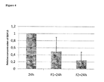
- FIG. 2 clearly shows that GDF-15 expression varies during dedifferentiation of a chondrocyte cell culture.
- the end of a time frame characterized by a transient decrease of GDF-15 expression, is indicative for the irreversible loss of the chondrocyte phenotype.
- the measurement of GDF-15 expression levels at one given time point within said time frame may be used in a quality control system for a(n) (expanded) chondrocyte culture.
- the expression level of GDF-15 at the specific time point can be between standardized intervals to pass quality control test.
- the standardized intervals will be specific for example but not limited to a culture condition, a laboratory or a procedure and can be determined by analyzing a group of samples under said specific culture conditions, of said specific laboratory or said specific procedure.
- the levels of GDF-15 expression can be used as such, relative to a reference gene/protein or related to a score. The latter can allow to combine GDF-15 expression levels with other gene expression levels in a general quality control scoring algorithm.
- the present invention provides for the use of the molecular marker GDF15 as a means for quality control of cells to be used for cell transplantation. Said use can be in combination with the expression of molecular marker COL2A1 and/or aggrecan.
- the outcome is linked to GDF15 expression levels.
- the predictive value of GDF15 is further optimized, by analyzing the effect of independent variables (age, gender, background, co-morbidities). This can be done storing in a database all the data of the individual patient together with the expression of the molecular markers and a score that describes the outcome of the procedure (based on pain, function of the joint, stiffness of the repair tissue by indentometry, and eventually histologic and molecular analysis of biopsy of the repair tissue).
- the expression levels determined using any one of the aforementioned methods can be normalized vis-à-vis the expression of a household gene such as GAPDH, HPRT, PPIA, Actine and the ribosomal proteins L19 and L32, and compared to the expression at start of the cell culture, isolation or expansion of the cell suspension (t0).
- the expression levels are determined at least twice, more in particular at least 3, 4, 5, 6, 7, 8, 9, or 10 times over the period required to double the cell count in the cell culture.
- the expression levels of GDF15, COL2A1, BMP2 and aggrecan are compared with the 'control' expression level(s) of said genes in chondrogenic stable cells.
- These 'control' expression levels typically consist of the mean expression level(s) of said genes as determined in a representative set of isolated pluripotent cells and/or chondrocytes; preferably said 'control' levels are predetermined.
- the method comprises the step of comparing the expression levels of said genes with the predetermined (pre-established) control expression levels of said genes.
- the latter may be presented as a combined value on an incremental scale reflecting the chondrogenic capacity of isolated pluripotent cells and/or chondrocytes, using art known scoring models such as for described in WO2008061804 : MARKER GENES FOR USE IN THE IDENTIFICATION OF CHONDROCYTE PHENOTYPIC STABILITY AND IN THE SCREENING OF FACTORS INFLUENCING CARTILAGE PRODUCTION.
- the method to monitor and assess the phenotypic stability of a cell expansion culture of isolated pluripotent cells and/or chondrocytes comprises the step of determining the expression level(s) of GDF15 and/or COL2A1 and/or BMP2 and/or aggrecan in said cells and comparing said expression level(s) with the predetermined (pre-established) control expression level(s) of said gene(s).
- the methods as provided herein are particularly useful in monitoring the phenotypic stability of isolated chondrocytes, i.e. chondrocytes derived from skeletal tissue, such as for example derived from hyaline cartilage or fibrocartilage.
- the methods provided herein are particularly useful in monitoring the phenotypic stability of isolated pluripotent cells and/or chondrocytes including but not limited to pluripotent cells from mesenchymal tissue such as adipose tissue and muscle tissue, hyaline knee cartilage chondrocytes, synovial fibroblasts and meniscal chondrocytes.
- GDF15 expression levels can be determined at different time points to monitor the expansion process under given experimental conditions (e.g. culture system, addition of growth factors, small molecules).
- the detection of an increase in GDF-15 expression, compared to a previous time point, is indicative for a loss of the chondrocyte phenotype.
- Said increase in GDF-15 expression after a decrease can be but is not limited to, at least 10%, 20% or 30% of the GDF-15 expression measured at a previous timepoint.
- Monitoring said increase in GDF-15 expression after a transient decrease may be particular useful in the optimization of for example but not limited to novel cell culture conditions or screening assays.
- said decrease without dedifferentiation can be down to about 5%, 10%, 15%, 20%, 30%, 40%, 50%, 60%, 70% or 80% of the initial GDF15 expression, in said isolated or expanded pluripotent cells and/or chondrocytes; and is typically down to about 10 to 30 % of the initial GDF 15 expression; more in particular down to about 15 to 25% of the initial GDF15 expression; even more in particular it can be down to about 20% of the initial GDF 15 expression.
- the pluripotent cells and/or chondrocytes thus characterized will still synthesize a functional cartilage matrix upon re-implantation in a 3-D environment.
- the present invention provides cell culture conditions or compounds capable of enhancing the phenotypic stability of a cell, said method comprising; applying the methods according to the invention in the presence and absence of the compound or culture condition to be tested, and determine whether said compound or cell culture condition is capable to prevent and/or delay the transient change in GDF-15 expression.
- the present invention provides a method to identify compounds or cell culture conditions capable of enhancing the chondrogenic capacity of a cell, said method comprising applying the methods according to the invention in the presence and absence of the compound or cell culture condition to be tested, and determine whether said compound or cell culture condition is capable to prevent and/or delay the transient decrease in GDF-15 expression, i.e. to extend the point up to which a cell expansion culture of said cells is identified as a phenotypic stable cell culture.
- the assays are used to identify specific markers co-expressed with GDF-15 and linked to the phenotypic stability of the cells.
- the present invention provides a method for enhancing the phenotypic stability of isolated or expanded cells or a method for influencing the chondrogenic capacity of a cell.
- the chondrogenic capacity of a cell is meant, the capacity to promote or stimulate the production of a functional cartilage matrix thus contributing to a stable cartilage tissue.
- Said method comprises adding GDF 15 to said suspension during isolation, expansion and/ or during orthopedic reconstructive surgery for the repair of connective tissue, in particular in the repair of cartilage, more in particular in the repair of cartilage degeneration associated with osteoarthritis.
- the capacity to promote or stimulate the production of a functional cartilage matrix is characterized in the capability to retain the expression of, matrix proteins such as but not limited to COL2A1 and aggrecan expression e.g. to extend or enhance COL2A1 and aggrecan expression.
- the cells can be pluripotent cells and/or chondrocytes.
- the present invention also provides a method for enhancing the phenotypic stability of isolated or expanded cells said method comprising adding GDF 15 to said cells, either directly or indirectly as part of the aforementioned orthopedic reconstructive surgery.
- GDF15 for use in the repair of connective tissue, more in particular for use in the repair of cartilage, even more in particular for use in the repair of cartilage degeneration associated with osteoarthritis.
- tissue-engineering protocols may include the application of bio-resorbable polymers (e.g. polylactic acid or polyglycolic avid) to fill the lesion.
- a composition could contain a matrix coated or mixed with further growth factors, with in particular GDF15.
- the agents, with in particular GDF 15 described herein can be packaged as a kit.
- one or more agents, with in particular GDF15 can be present in a first container, and the kit can optionally include one or more agents, with in particular GDF15, in a second container.
- the kit can include instructions describing the method of the present invention.
- the agents, cells, containers and/or the instructions can be present in a package.
- the contents of the kit can contain but is not limited to GDF15, growth medium, buffers, multiwell plates, antibodies and/or enzyme substrates.
- OA affected cartilage was obtained from patients within 24 h from total knee arthroplasty. The cartilage from each of these patients was separated in visually intact cartilage (NoOA) and cartilage showing OA-lesions (OAOA). This study was approved by the local Ethics Committee. The cartilage obtained was diced into small fragments and chondrocytes were isolated by sequential enzymatic digestion (hyaluronidase, pronase, collagenase (Sigma-Aldrich, Steinheim, Germany)) [4]. Trypan blue exclusion revealed that >95% of the cells were viable after isolation.
- GDF15 Concentration of GDF15 in culture supernates from alginate cultured chondrocytes isolated from visually intact (NoOA) and visually damaged zones (OAOA) of OA-cartilage were measured by standard ELISA techniques using the GDF15 ELISA DuoSet (R&D systems, Abingdon, UK) according to the manufacturer's instructions.
- GDF15 secreted by human articular chondroctes isolated from visually intact (NoOA) and visually damaged (OAOA) zones of OA articular cartilage respectively, was analysed by measuring GDF15 concentrations in culture supernates of 13 different paired samples.
- Figure 1A shows a consistent higher concentration in chondrocyte cell cultures from visually damaged zones compared to visually intact zones (p ⁇ 0,01; Wilcoxon signed rank test).
- Phenotypically stable articular chondrocytes isolated from 3 OA-patients were seeded in monolayer culture at low density (20.000 cells/Cm 2 ). When confluency was reached, cells were detached and reseeded at the initial density. At particular time points, cells were detached and encapsulated in alginate beads for 8 days, as described above. At indicated time points Trizol (Invitrogen) was added to the isolated cells, and RNA was extracted according to the manufacturer's instructions, followed by an additional purification step (RNeasy mini-kit (Qiagen)). This step included the digestion of DNA by deoxyribonuclease I (Invitrogen). cDNA was synthesized with oligo(dT) primers using the Superscript kit (Invitrogen).
- Phenotypically stable meniscus chondrocytes isolated from 2 OA-patients were seeded in monolayer culture at low density (20.000 cells/cm 2 ) and allowed to expand for about 170 hours. At indicated time points Trizol (Invitrogen) was added to the isolated cells, and RNA was extracted according to the manufacturer's instructions, followed by an additional purification step (RNeasy mini-kit (Qiagen)). This step included the digestion of DNA by deoxyribonuclease I (Invitrogen). cDNA was synthesized with oligo(dT) primers using the Superscript kit (Invitrogen).
- Cell lysates of articular chondrocytes were prepared by resuspending cell pellets in 40mM Tris from the ReadyPrep Sequential Extraction Kit (Bio-Rad, Hercules, CA, USA), supplemented with 0,1 % SDS, containing protease inhibitors (Roche Diagnostics, Mannheim, Germany) and a phosphatase inhibitor-cocktail (Sigma-Aldrich, Steinheim, Germany). Cells were lysed by sonication and the proteins were isolated by centrifugation. Equal amounts (30 ⁇ g as determined by 2-D Quant kit, GE Healthcare, Fairfield, USA) were loaded on 10% SDS-PAGE gel. Equal loading was verified by Ponceau S staining (data not shown).
- GDF15 expression decrease during dedifferentiation of phenotypically stable chondrocytes
- Chondrocytes were cultured in alginate beads as described above. After a 10-day culture period, chondrocyte cultures were stimulated with different concentrations of human recombinant GDF 15 (R&D systems, Abingdon, UK; Peprotech, London, UK) or vehicle only for 24 hours. Thereafter, culture medium was aspirated and frozen at - 80°C until further use. Cells were isolated and mRNA was extracted as described above. Gene expression was analysed by real-time RT-PCR as described earlier.
- Erogenous human recombinant GDF15 promotes the expression of extracellular matrix genes
- GDF15 may serve as an anabolic mediator in articular chondrocytes that may promote ECM synthesis.
- GDF15 may be used as additive in expansion/redifferentiation cultures, to obtain a higher quality chondrocyte cell culture, i.e. expressing higher levels of Col2A1 and aggrecan.
- GDF15 stimulated chondrocyte cultures from 2 different patient samples were analyzed for aggrecan catabolism by determining the expression of the 21kDa and 58kDa aggrecan fragments. Expression of both fragments decreased upon increasing GDF 15 concentrations (data not shown).
- GDF 15 stimulation not only induces the expression of matrix genes (COL2A1 and Aggr), but can also prevent its catabolism (Aggr). Furthermore, it contributes to the reduction of expression of genes involved in cartilage degeneration (MMP13 and ADAMTS5).
- MMP13 and ADAMTS5 genes involved in cartilage degeneration
- the examples show an anabolic and anti-catabolic activity of GDF 15 in vitro.
- GDF15 -/- mice Characterization of Growth-Differentiation Factor 15, a Transforming Growth Factor b Superfamily Member Induced following Liver Injury, EDWARD C. HSIAO, LEONIDAS G. KONIARIS, TERESA ZIMMERS-KONIARIS, SUZANNE M. SEBALD, THANH V. HUYNH, AND SE-JIN LEE May 2000, p. 3742-3751 Vol. 20, No. 10
- GDF15 +/- mice Characterization of Growth-Differentiation Factor 15, a Transforming Growth Factor b Superfamily Member Induced following Liver Injury, EDWARD C. HSIAO, LEONIDAS G. KONIARIS, TERESA ZIMMERS-KONIARIS, SUZANNE M. SEBALD, THANH V. HUYNH, AND SE-JIN LEE May 2000, p. 3742-3751 Vol. 20, No. 10
Landscapes
- Life Sciences & Earth Sciences (AREA)
- Health & Medical Sciences (AREA)
- Engineering & Computer Science (AREA)
- Biomedical Technology (AREA)
- Molecular Biology (AREA)
- Chemical & Material Sciences (AREA)
- Urology & Nephrology (AREA)
- Hematology (AREA)
- Immunology (AREA)
- Biotechnology (AREA)
- Microbiology (AREA)
- Cell Biology (AREA)
- Proteomics, Peptides & Aminoacids (AREA)
- Food Science & Technology (AREA)
- Medicinal Chemistry (AREA)
- Physics & Mathematics (AREA)
- Analytical Chemistry (AREA)
- Biochemistry (AREA)
- General Health & Medical Sciences (AREA)
- General Physics & Mathematics (AREA)
- Pathology (AREA)
- Measuring Or Testing Involving Enzymes Or Micro-Organisms (AREA)
Description
- The invention relates generally to the field of tissue engineering. The present invention relates to GDF15 as a molecular marker in in vitro assays determining phenotypic stability of articular chondrocytes and predicting the outcome of chondrocyte transplantation.
- The invention further provides methods and compositions related to the generation of a population of cells suitable for the repair of cartilage, in particular in the repair of cartilage degeneration associated with osteoarthritis.
- The transforming growth factor-β (TGF-β) superfamily consists of an increasing number of molecules that regulate a variety of cellular processes such as growth, differentiation and oncogenesis. Members of the TGF-β superfamily have been classified into major family groupings which include TGF-β, morphogenic proteins (MP), bone morphogenic proteins (BMP), osteogenic proteins (OP), growth and differentiation factors (GDF), inhibins/activins, mullerian inhibitory substances (MIS) and glial derived neurotrophic factors (GDNF). TGF-β was first characterized for its effects on cell proliferation. It both stimulated the anchorage-independent growth of rat kidney fibroblasts and inhibited the growth of monkey kidney cells. TGF-β family members have been shown to have many diverse biological effects, e.g. they regulate bone formation, induce rat muscle cells to produce cartilage-specific macromolecules, inhibit the growth of early hematopoietic progenitor cells, T cells, B cells, mouse keratinocytes, and several human cancer cell lines. TGF-β family members increase the synthesis and secretion of collagen and fibronectin, accelerate healing of incisional wounds, suppress casein synthesis in mouse mammary explants, inhibit DNA synthesis in rat liver epithelial cells, stimulate the production of bFGF binding proteoglycans, modulate phosphorylation of the epidermal growth factor ("EGF") receptor and proliferation of epidermoid carcinoma cells and can lead to apoptosis in uterine epithelial cells, cultured hepatocytes and regressing liver. TGF-βs can mediate cardio-protection against reperfusion injury by inhibiting neutrophil adherence to endothelium and protect against experimental autoimmune diseases in mice. On the whole, proteins of the TGF-β family are multifunctional, active growth factors and also have related biological activities such as chemotactic attraction of cells, promotion of cell differentiation and tissue-inducing capabilities. Differences in their structure and in their affmity for receptors lead to considerable variations in their exact biological function.
- Proteins of the TGF-β family are synthesized as large, inactive precursor (pro-form) proteins, which are proteolytically processed at a dibasic site (RXXR) to generate the mature, active form of the protein.
- Growth differentiation factor 15 is a distant member of the TGF-β family. The expression of the mature form of growth differentiation factor 15 (GDF15) protein is associated with early prostate carcinogenesis. GDF 15 has been described in the literature as macrophage inhibitory cytokine-1 (MIC-1), placental bone morphogenic protein (PLAB), placental transforming growth factor- β (PTGF- β), prostate derived factor (PDF), and non-steroidal anti-inflammatory activated gene-1 (NAG-1) reflecting the different functions that has been implied for this protein.
-
WO2001081928 published on 1 November 2001 discloses diagnostic assays and methods of treatment involving GDF15, andWO1999006445 published on 11 February 1999 discloses the GDF15 polynucleotide sequence and amino acid sequence. - In earlier studies on GDF15 expression in human normal and prostate cancer cells, it was found that the level of GDF15 transcript does not necessarily correlate well with that of the GDF15 protein (Noorali et al. Differentiation (2007) 75: 325-336). The reasons for these discrepancies could be due to differences in post-transcriptional modifications or processing of RNA, that influences its stability or translation, or post-translational modifications of the protein affecting protein maturation, accumulation or degradation.
- As indicated above, GDF-15 may have applications in the treatment of immunologic disorders. In particular, GDF-15 may be used as an anti-inflammatory agent or as a treatment for disorders related to abnormal proliferation or function of lymphocytes.
- Cartilage is a tissue composed by a cellular component, chondrocytes, and by an extra-cellular matrix typically rich in collagen type II and highly sulphated high molecular weight proteoglycan aggregates. The abundance of type II collagen, link protein, and proteoglycan aggrecan, along with the presence of minor collagens such as type IX and type XI collagen are hallmarks of cartilage tissue. Articular cartilage forms a specialized, smooth connective tissue that is weight bearing and that serves as a gliding surface allowing a lithe movement of the joints.
- In post-natal mammals, cartilage contributes to the structure of several organs and systems like the articular surface of diarthrodial joints and other joint-associated structures (such as menisci), the ear, the nose, the larynx, the trachea, the bronchi, structures of the heart valves, part of the costae, synchondroses, entheses etc. In some of the mentioned locations (e.g. entheses, the annulus fibrosus of the intervertebral disks, in the menisci, insertion of ligaments etc.) for the abundance of collagens (mostly type I collagen) and the peculiar distribution of the fibrous bundles it is called fibrocartilage. In other locations (e.g. the pinna of the ear, epiglottis etc.) it is particularly rich of elastin and it is called elastic cartilage. In all the other structures, for its semitransparent, clear aspect it is called hyaline cartilage.
- During embryogenesis cartilage has a role in the development of long bones. Mesenchymal cells aggregate and differentiate to form cartilage anlagen, which provide the mold of the future long bones. These cartilage templates in development evolve, undergo endochondral bone formation through a cascade of events including chondrocyte hypertrophy, vascular invasion, mineralization, and are eventually replaced by bone except for a thin layer at the extremities of the bone elements that will differentiate into the articular surface of diarthrodial joints. In these locations cartilage tissue remains hyaline for all the life-span of the individual. With ageing, articular cartilage is well known to undergo a process of senescence, affecting its mechanical properties and its intrinsic resilience.
- Joint surface defects can be the result of various aetiologies such as inflammatory processes, neoplasias, post-traumatic and degenerative events etc. Whatever the cause, the mechanisms of repair and of subsequent evolution are largely common.
- Osteochondral (or full-thickness) articular surface defects include damage to the articular cartilage, the underlying subchondral bone tissue, and the calcified layer of cartilage located between the articular cartilage and the subchondral bone. They typically arise during severe trauma of the joint or during the late stages of degenerative joint diseases, e.g. during osteoarthritis (OA). These lesions disrupt the congruence between the joint surfaces and therefore can lead to OA, which can be painful and severely limit the joint function. Osteochondral defects can rely on an extrinsic mechanism for repair. Extrinsic healing uses mesenchymal elements from subchondral bone to participate in the formation of new connective tissue. The repair tissue , however, often consists of fibrocartilage or fibrous tissue. This scar tissue does not share the same biomechanical properties as hyaline cartilage and eventually degenerates with the development of osteoarthritis.
- Superficial or partial-thickness injuries of the articular cartilage that do not penetrate the subchondral bone can only rely on an intrinsic mechanism for repair. Chondrocytes adjacent to the injured surfaces proliferate and increase the deposition of extracellular matrix synthesis. Despite these attempts at repair, there is no appreciable increase in the bulk of cartilage matrix and the repair process is rarely effective in healing the defects. Although initially sometimes painless, partial-thickness defects often degenerate into osteoarthritis of the involved joint.
- Osteoarthritis (OA) is a complex, multifactorial, age-dependent degenerative disease of the synovial joints. It affects females at a higher rate than males, particularly after the menopause. As indicated above OA is characterized by changes to all the components of the joint, with degeneration and loss of articular cartilage and changes to the subchondral bone being constant factors in disease progression. Along with the breakdown of the cartilage and joint space narrowing, there is thickening and sclerosis of the subchondral bone, development of cysts and bony outgrowth at the margins of the joint. Despite an increase in bone volume fraction, the subchondral bone is mechanically weaker in OA because of hypomineralization, increased collagen metabolism and altered bone remodeling.
- In a report of Hopwood et al. (Arthritis Research & Therapy (2007) 9: R100), microarray gene expression profiling of osteoarthritic bone suggests altered TGF-β/BMP signaling. GDF15 gene expression was downregulated in OA when compared with control bone.
- A recent report of Iliopoulos et al (PLoS ONE (2008) 3:e3740) describes that GDF15 is a differential expressed protein between osteoarthritic and normal chondrocytes.
- Repair of articular cartilage defects with suspensions of chondrocytes has been carried out in a variety of animal models and is now also employed in humans with degenerative joint disease. Autologous chondrocytes obtained from an unaffected area of the joint are released, expanded in vitro in the presence of autologous serum and subsequently injected in the cartilage defect. This procedure has led to a proven at least symptomatic amelioration. This conceptually promising approach has still wide margins for improvement, since it is known that in vitro expansion of chondrocytes results, after a limited number of cell divisions, in a loss of their phenotypic stability (as defined by the ability of chondrocytes to form hyaline cartilage in vivo) making the cell suspension to be injected unreliable. To date, however, it is not known how far it is possible to expand chondrocytes without hampering their phenotypic stability and therefore their capacity to form stable hyaline cartilage in vivo, resistant to vascular invasion and endochondral bone formation. Other factors that can affect the capacity of chondrocytes to form cartilage in vivo are the culture conditions, and several factors dependent on the donor such as age and pre-existing joint or systemic diseases. At the end of cell expansion the chondrocyte population is composed of some cells that retain their phenotypic stability, and others that still can proliferate but will not anymore contribute to cartilage repair. To obtain a consistent cell suspension for autologues chondrocyte transplantation (ACT), it is desirable to determine which is the actual capacity of the cells to form cartilage in vivo and, if necessary, to select stable chondrocytes within the expanded cell population. The importance of this issue is underscored by the large variability in the quality of the repair tissue obtained going from hyaline-like cartilage to fibrocartilage to no signs of repair.
- Chondrocytes are the only normal skeletal cells known to grow anchorage-independent in agarose cultures (Benya and Shaffer. 1982, Cell 30:215-224). This culture system allows a recovery of some of the phenotypic traits that are lost with expansion in monolayer. Similar features as in agarose cultures, are observed in alginate bead cultures (Benz K et al., 2002, Biochem Biophys Res Comm 2002;293:284-92; Häuselmann HJ et al. 1994, Biochem Biophys Res Comm ,2002;293:284-92; Wang J et al, Osteoarthritis Cartilage. 2003;11:801-9). The expression of
type 2 collagen and the capacity to grow and rescue phenotypic traits in agarose culture, are good assays to evaluate chondrocyte differentiation and the potential to differentiate respectively. However they do not measure the capacity of chondrocytes to form cartilage in vivo. -
EP1498146 published on 19 January, 2005 provides an in vivo assay to measure the capacity of isolated chondrocytes to produce cartilage in vivo using a nude mouse model. This capacity is linked to a set of molecular markers associated with the outcome of joint surface defects ("JSD") repair in well-standardised animal models of JSD. The set of molecular markers (both membrane-associated and/or non membrane-associated) are also used as a final quality control for the cell suspension to be used for ACT or the repair of the cartilaginous structures. Results indicate the high expression of BMP-2, FGFR-3, and type II collagen as positively associated to chondrocyte stability, whereas activin-like kinase (ALK)-1 and collagen type X expression are negatively associated. During in vitro expansion of these chondrocytes, the molecular markers can only be determined by RT-PCR or immunohistochemistry and thus requires (the lysis) of precious cells. - There is a need to identify molecular markers associated with chondrocytes that would allow the clinician to produce suitable implants and to regenerate and repair cartilage tissue with the appropriate phenotypic stable cells and avoid scar formation to the greatest possible extent In an earlier application (
PCT application WO2009/049913 ) the present applicant identified and characterized CRYAB and HSP27 as molecular markers in monitoring the chondrocyte stability of isolated or expanded cells. With the present invention, and in search of markers that can be determined in the cell culture media of the isolated or expanded cells, the applicant defines GDF15 as a secreted positive molecular marker for chondrocyte stability and as a tool to monitor, passage by passage, in vitro cell expansion and the manufacturing process of chondrocyte expansion. GDF15, optionally in combination with other markers for chondrocyte stability like CRYAB and HSP27, will be useful to optimize next generation chondrocyte expansion technologies, including expansion of chondrocyte-precursors, and chondrocyte derivatives, which express very low levels of or even no conventional chondrocyte differentiation markers such as BMP-2, COL2A1 and aggrecan. GDF15, again optionally in combination with other markers for chondrocyte stability like CRYAB and HSP27, will be useful to predict when cell expansion must be stopped, and especially to provide a quality control for chondrocytes to be used for ACT for lot release approval by the simple prelevation of cell culture medium, without the need to isolate precious cells. - The present invention relates to a method for evaluating the phenotypic stability of isolated or expanded cells. Hence the present invention provides a method for evaluating the ability of isolated or expanded cells to produce, synthesize, organize or reorganize matrix components, said method comprising monitoring the expression of the molecular marker GDF15. In particular said method comprises monitoring a decrease; or a decrease and a subsequent increase in the expression of the molecular marker GDF-15. Said decrease can be down to about 10% to 30% of the initial GDF15 expression.
- As used herein, the term "matrix components" refers to proteins synthesized by the chondrocyte that constitute the extracellular matrix of the cell such as aggrecan, collagens, proteoglycans and the like.
- In an embodiment of the method of the invention, the isolated or expanded cells are pluripotent cells and/or chondrocytes. In particular said chondrocytes are derived from skeletal tissue, more in particular from hyaline cartilage or fibrocartilage.
- Hence, in a further embodiment of the method of the invention, the matrix components are the typical constituents of the extracellular matrix of hyaline cartilage in particular said matrix components are COL2A1 and aggrecan.
- In still a further embodiment of the method of the invention, said method further comprises monitoring of the expression of the molecular markers CRYAB and HSP27.
- As further specified in
PCT application WO2009/049913 , a cell culture of isolated pluripotent cells and/or chondrocytes is identified as a phenotypic stable cell culture, by monitoring the transient decrease in CRYAB expression, and wherein up to the increase of CRYAB expression, said cell culture is identified as a phenotypic stable cell culture. In particular said transient decrease in CRYAB expression is at least 30% of the initial CRYAB expression; and in particular equals from about 40% to about 60% of the initial CRYAB expression in said cell culture of isolated pluripotent cells and/or chondrocytes. - As further specified in
PCT application WO2009/049913 , a cell culture of isolated pluripotent cells and/or chondrocytes is identified as a phenotypic stable cell culture, by monitoring HSP27 expression in said cell culture, and wherein an increase in HSP27 expression to a level of at least 140% of the initial HSP27 expression in said cell culture, is an indication of the dedifferentiation of said cell culture of isolated pluripotent cells and/or chondrocytes. - Thus in a particular embodiment of the present invention, a decrease in CRYAB expression down to about 70% or less of the initial CRYAB expression, an increase in HSP27 expression up to about 140% or more of the initial HSP27 expression, and a decrease down to about 20% of the initial GDF 15 expression, identifies the point up to which an isolated or expanded cell suspension is still capable to synthesize a functional cartilage matrix
- In another embodiment of the method of the invention, the detection of expression of the molecular marker GDF 15, is performed with an immunoassay using antibodies, or fragments thereof, against GDF15. In particular said immunoassay is performed on cell culture supernates or cell culture lysates. Monitoring said expression of the molecular marker GDF15 can also be performed using a technique selected from the group of semi-quantitative RT-PCR, Northern hybridisation, differential display, subtractive hybridization, subtracted libraries, cDNA chips and cDNA arrays. Another aspect of the invention, relates to extending or enhancing the expression of matrix proteins of isolated or expanded cells, said method comprising adding GDF15 to said cells. In particular said matrix proteins are from hyaline cartilage, more in particular said matrix proteins are COL2A1 or aggrecan. Said isolated or expanded cells can be pluripotent cells and/or chondrocytes.
- Another aspect of the invention relates to the use of the molecular marker GDF15 to predict the ability of isolated or expanded cells to synthesize matrix components and to determine when cell expansion must be stopped without loss of the ability of said cells to synthesize matrix components. The invention also provides the use of the molecular marker GDF15 in a cell quality control system or a quality control scoring algorithm. The described uses of the molecular marker GDF15 can be provided in combination with the expression of one, two or more of the molecular markers selected from the group consisting of COL2A1, BMP2 and aggrecan.
-
-
Figure 1 : Concentration of GDF15 in culture supernates from alginate cultured chondrocytes isolated from visually intact (NoOA) and visually damaged zones (OAOA) of OA-cartilage. A significant (p<0,01; Wilcoxon signed rank test) higher concentration was observed in the OAOA samples. -
Figure 2 : Relative expression of Collagen type II (COL2A1), BMP-2 and GDF15 during monolayer expansion cultures. At time point 192h part of the cells were recultured in 3-D alginate beads, as described in the materials and methods section. Data points represent the mean relative expression of 3 different patients. Decreasing COL2A1 and BMP-2 levels indicate the time-dependent dedifferentiation of chondrocytes in monolayer culture. Encapsulation of expanded chondrocytes in a 3-D environment induces an increase in GDF 15 expression. -
Figure 3 : Relative expression of GDF15 and CRYAB during monolayer expansion cultures. Meniscus chondrocytes were isolated and expanded as described. Expression levels of GDF15 and CRYAB were analyzed by QPCR as described. Data points represent the mean relative expression of 2 different patients. -
Figure 4 : GDF15 medium concentrations were determined by ELISA techniques at different time points: 24 hours after seeding of phenotypically stable chondrocytes, 24 hours after the first passage (F1 + 24 h) and 24 hours after the second passage (F2 + 24 h). Values are expressed as relative values to the first time point and represent the mean (± SD) relative concentration of 3 different patient samples. These data confirm the time dependent decreased protein secretion of GDF15 by expanding human articular chondrocytes. -
Figure 5 : Phenotypically stable human articular chondrocytes cultured in alginate beads were stimulated for 24 hours with hrGDF15 at different concentrations (ranging from 0 to 1000 ng/ml), demonstrating a concentration dependent increase in the expression of extracellular matrix genes upon GDF15 stimulation. Data points represent the mean (± SEM) relative expression of Collagen type II and aggrecan compared to unstimulated control cultures. Data represent the mean of 9 (except for 1000 ng/ml, n=6) independent experiments. -
Figure 6 : A. Relative expression of MMP13, as determined by Western Blot, shows a decreasing trend upon stimulation of the cell cultures with increasing GDF15 concentrations. Data represent mean expression (±SEM) of 3 patient samples. B. Relative expression of ADAMTS-5 as determined by qPCR. Cell cultures stimulated with 1000 ng/ml show a reduced expression of ADAMTS-5, a major enzyme involved in aggrecan degradation. Data represent mean expression (±SEM) of 3 patient samples. - With "chondrocyte phenotypic stability" is meant the capacity of a cell obtained from cartilage tissue or from any other tissue containing cells with chondrogenic potential to produce, synthesize, organize or reorganize a functional cartilage matrix, more in particular the capacity to synthesize matrix components of hyaline cartilage, even more in particular the capacity to synthesize at least the matrix components COL2A and/or aggrecan.
- With "chondrogenic capacity" is meant the capacity to promote or stimulate the production of a functional cartilage matrix, more in particular to retain the expression of matrix proteins of hyaline cartilage, even more in particular to extend or enhance COL2A1 or aggrecan expression.
- The term "functional cartilage matrix" refers to an extracellular matrix that provides enough elasticity and tensile strength as may be expected from hyaline cartilage. Typically, such matrix is rich in Collagen type II and proteoglycans.
- With "molecular marker" is meant a polypeptide that distinguishes one cell (or set of cells) from another cell (or set of cells) in a population of cells and is associated to a peculiar biological function.
- With "phenotypic stability" or "phenotypic stable" is meant the maintenance of the ability of any cell (e.g. a chondrocyte) to produce, synthesize, organize or reorganize, the components (e.g. matrix proteins) or the structure (e.g. matrix) of a specific tissue (e.g. cartilage).
- With " stable cartilage" is meant cartilage not finally turning into bone, i.e. cartilage devoid of any signs of vascularization. Particularly, the stable cartilage in accordance with the present invention is human adult or mature articular cartilage but may also include animal adult or mature cartilage. Contrary to stable cartilage, transient cartilage in the end will become bone tissue. In the context of the present invention, cartilage is said to be stable if, even after e.g. seven weeks, any signs of bone formation are absent. Hence, hypertrophied chondrocytes do not form stable cartilage.
- As used herein the "GDF15" polypeptide is meant to be a protein encoded by a mammalian gdf15 gene, including allelic variants as well as biologically active fragments thereof containing conservative or non-conservative changes as well as artificial proteins that are substantially identical, i.e. 70%, 75%, 80%, 85%, 87%, 89%, 90%, 92%, 93%, 94%, 95%, 96%, 97%, 98%, or 99% identical to any one of the aforementioned GDF15 polypeptides. In a particular embodiment the GDF15 polypeptide is 70%, 75%, 80%, 85%, 87%, 89%, 90%, 92%, 93%, 94%, 95%, 96%, 97%, 98%, or 99% identical to the human GDF15 (encoded by Genbank Accession NM_004864(mRNA) or NC_000019.8 (genomic)).
- By analogy, the "GDF15" polynucleotide is meant to include allelic variants as well as biologically active fragments thereof containing conservative or non-conservative changes as well as any nucleic acid molecule that is substantially identical, i.e. 70%, 75%, 80%, 85%, 87%, 89%, 90%, 92%, 93%, 94%, 95%, 96%, 97%, 98%, or 99% identical to any one of the aforementioned GDF15 encoding polynucleotides. In a particular embodiment the GDF15 polynucleotide is 70%, 75%, 80%, 85%, 87%, 89%, 90%, 92%, 93%, 94%, 95%, 96%, 97%, 98%, or 99% identical to the nucleic acid molecule encoding for human GDF15 (Genbank Acession N° NM_004864(mRNA) or NC_000019.8 (genomic)).
- As used herein, the terms "polynucleotide" and "nucleic acid" are used interchangeably to refer polynucleotides of any length, either deoxyribonucleotides or ribonucleotides or analogs (e.g., inosine, 7-deazaguanosine, etc.) thereof. "Oligonucleotides" refer to polynucleotides of less than 100 nucleotides in length, preferably less than 50 nucleotides in length, and most preferably about 10-30 nucleotides in length. Polynucleotides can have any three-dimensional structure and may perform any function, known or unknown. The following are non-limiting examples of polynucleotides: a gene or gene fragment (for example, a probe, primer, EST or SAGE tag), exons, introns, messenger RNA (mRNA), transfer RNA, ribosomal RNA, ribozymes, cDNA, recombinant polynucleotides, branched polynucleotides, plasmids, vectors, isolated DNA of any sequence, isolated RNA of any sequence, nucleic acid probes and primers. A polynucleotide can include modified nucleotides, such as methylated nucleotides and nucleotide analogs. If present, modifications to the nucleotide structure can be imparted before or after assembly of the polymer. The sequence of nucleotides can be interrupted by non-nucleotide components. A polynucleotide can be further modified after polymerization, such as by conjugation with a labeling component. The term also refers to both double- and single-stranded molecules. Unless otherwise specified or required, any embodiment of this invention that is a polynucleotide encompasses both the double-stranded form and each of two complementary single-stranded forms known or predicted to make up the double-stranded form.
- "Polypeptide" refers to any peptide or protein comprising amino acids joined to each other by peptide bonds or modified peptide bonds. "Polypeptide" refers to both short chains, commonly referred to as peptides, oligopeptides or oligomers, and to longer chains, generally referred to as proteins. Polypeptides may contain amino acids other than the 20 gene-encoded amino acids.
- "Polypeptides" include amino acid sequences modified either by natural processes, such as post-translational processing, or by chemical modification techniques which are well known in the art. Such modifications are well described in basic texts and in more detailed monographs, as well as in a voluminous research literature.
- Modifications may occur anywhere in a polypeptide, including the peptide backbone, the amino acid side-chains and the amino or carboxyl termini. It will be appreciated that the same type of modification may be present to the same or varying degrees at several sites in a given polypeptide. Also, a given polypeptide may contain many types of modifications (see, for instance, Proteins-Structure and Molecular Properties, 2nd Ed. , T. E. Creighton, W. H. Freeman and Company, New York, 1993; Wold, F. , Post- translational Protein Modifications: Perspectives and Prospects, pgs. 1-12 in Postranslational Covalent Modification of Proteins, B. C. Johnson, Ed. , Academic Press, New York, 1983; Seifter et al., "Analysis for protein modifications and nonprotein cofactors", Meth Enzymol (1990) 182: 626-646 and Rattan et al. , "Protein Synthesis: Post-translational Modifications and Aging", Ann NY Acad Sci (1992) 663: 4842).
- This invention is based on the identification of GDF 15 as molecular actor in the homeostasis of chondrocytes, and accordingly provides GDF15 as molecular marker useful in monitoring the phentotypic stability of isolated pluripotent cells in vitro.
- In a first objective, the present invention provides a method for evaluating the phenotypic stability of isolated or expanded cells, more in particular of isolated pluripotent cells and/or chondrocytes, said method comprising monitoring the expression levels of the molecular marker GDF 15 at regular time intervals. In a further embodiment the methods include determining the expression levels of GDF 15, COL2A1 and/or aggrecan at regular time intervals.
- As used herein, the term "expanded cells" refers to the population of cells derived through proliferation of the isolated pluripotent cells and/or chondrocytes. "Pluripotent cells" are cells that can be induced to differentiate into all cell types except for extra embryonic tissue. In cell biology, the definition of pluripotency has come to refer to a cell that has the potential to differentiate into any of the three germ layers: endoderm (interior stomach lining, gastrointestinal tract, the lungs), mesoderm (muscle, bone, blood, urogenital), or ectoderm (epidermal tissues and nervous system). As used herein, it is further meant to include multipotent cells that have the potential to give rise to cells from multiple, but a limited number of lineages.
- The "expression" generally refers to the process by which polynucleotides are transcribed into mRNA and/or the process by which the mRNA is subsequently translated into peptides, polypeptides or proteins. Hence the "expression" of a gene product, in the present invention of GDF15, COL2A1 and aggrecan can be determined either at the nucleic acid level or the protein level.
- Detection can be by any appropriate method, including, e.g., detecting the quantity of mRNA transcribed from the gene or the quantity of nucleic acids derived from the mRNA transcripts. Examples of nucleic acids derived from an mRNA include a cDNA produced from the reverse transcription of the mRNA, an RNA transcribed from the cDNA, a DNA amplified from the cDNA, an RNA transcribed from the amplified cDNA, and the like. In order to detect the level of mRNA expression, the amount of the derived nucleic acid should be proportional to the amount of the mRNA transcript from which it is derived. The mRNA expression level of a gene can be detected by any method, including hybridization (e.g., nucleic acid arrays, Northern blot analysis, etc.) and/or amplification procedures according to methods widely known in the art. For example, the RNA in or from a sample can be detected directly or after amplification. Any suitable method of amplification may be used. In one embodiment, cDNA is reversed transcribed from RNA, and then optionally amplified, for example, by PCR. After amplification, the resulting DNA fragments can for example, be detected by agarose gel electrophoresis followed by visualization with ethidium bromide staining and ultraviolet illumination. A specific amplification of differentially expressed genes of interest can be verified by demonstrating that the amplified DNA fragment has the predicted size, exhibits the predicated restriction digestion pattern and/or hybridizes to the correct cloned DNA sequence.
- In hybridization methods a probe, i.e. nucleic acid molecules having at least 10 nucleotides and exhibiting sequence complementarity or homology to the nucleic acid molecule to be determined, are used. It is known in the art that a "perfectly matched" probe is not needed for a specific hybridization. A probe useful for detecting mRNA is at least about 80%, 85%, 90%, 95%, 97% or 99% identical to the homologous region in the nucleic acid molecule to be determined. In one aspect, a probe is about 50 to about 75, nucleotides or, alternatively, about 50 to about 100 nucleotides in length. These probes can be designed from the sequence of full-length genes. In certain embodiments, it will be advantageous to employ nucleic acid sequences as described herein in combination with an appropriate label for detecting hybridization and/or complementary sequences. A wide variety of appropriate labels, markers and/or reporters are known in the art, including fluorescent, radioactive, enzymatic or other ligands, such as avidin/biotin, which are capable of giving a detectable signal. One can employ a fluorescent label or an enzyme tag, such as urease, alkaline phosphatase or peroxidase, instead of radioactive or other environmental undesirable reagents. In the case of enzyme tags, colorimetric indicator substrates are known that can be employed to provide a signal that is visible to the human eye or spectrophotometrically, to identify specific hybridization with complementary nucleic acid-containing samples.
- Hence in an embodiment of the invention, monitoring the expression of the molecular marker GDF15 is performed using a technique selected from the group of semi-quantitative RT-PCR, Northern hybridisation, differential display, subtractive hybridization, subtracted libraries, cDNA chips and cDNA arrays.
- Detection of the level of gene expression can also include detecting the quantity of the polypeptide or protein encoded by the gene. A variety of techniques are available in the art for protein analysis. They include but are not limited to radioimmunoassay (RIA), ELISA (enzyme linked immunoradiometric assays), "sandwich" immunoassays, immunoradiometric assays, in situ immunoassays (using e.g., colloidal gold, enzyme or radioisotope labels), western blot analysis, immunoprecipitation assays, immunofluorescent assays and PAGE- SDS.
- Antibodies that specifically recognize and bind to the protein products of these genes are required for these immunoassays. These may be purchased from commercial vendors or generated and screened using methods well known in the art. See e.g., Sambrook, Fritsch and Maniatis, MOLECULAR CLONING: A LABORATORY MANUAL, 2nd edition (1989); CURRENT PROTOCOLS IN MOLECULAR BIOLOGY (F. M. Ausubel et al. eds, (1987)); the series METHODS IN ENZYMOLOGY (Academic Press, Inc.): PCR 2: A PRACTICAL APPROACH (MJ. MacPherson, B.D. Hames and G.R. Taylor eds. (1995)), Harlow and Lane, eds. (1988) ANTIBODIES, A LABORATORY MANUAL and ANIMAL CELL CULTURE (R.I. Freshney, ed. (1987)).
- Hence, in an embodiment of the invention, monitoring the decrease or increase in the expression of the molecular marker GDF 15 is performed with an immunoassay using antibodies, or fragments thereof, against GDF15.
- Anti-GDF15 antibodies and fragments thereof can be produced by any of the methods known to the art.
- The antibodies can be bound to many different carriers and used to detect the presence of a GDF15 antigen. The antibodies can be suitable for use, for example, in immunoassays. The antibodies of the invention can be bound to many different carriers. The nature of the carrier can be either soluble or insoluble. Examples of well-knowm carriers include glass, polystyrene, polypropylene, polyethylene, dextran, nylon, amylases, natural and modified celluloses, polyacrylamides, agaroses and magnetic beads. Those skilled in the art will know of other suitable carriers for binding antibodies, or will be able to ascertain such, using routine experimentation.
- In addition, the antibodies in these immunoassays can be detectably labeled in various ways. Examples of types of immunoassays, which can utilize antibodies, are competitive and noncompetitive immunoassays in either a direct or indirect format. Examples of such immunoassays are the radioimmunoassay (RIA) and the sandwich (immunometric) assay.
- There are many different labels and methods of labeling known to those of ordinary skill in the art. Examples of the types of labels, which can be used in the present invention, include enzymes, radioisotopes, fluorescent compounds, colloidal metals, chemiluminescent compounds, phosphorescent compounds, and bioluminescent compounds. Those of ordinary skill in the art will know of other suitable labels for binding to an antibody, or will be able to ascertain such, using routine experimentation.
- Another technique, which may also result in greater sensitivity, consists of coupling the antibodies to low molecular weight haptens. These haptens can then be specifically detected by means of a second reaction. For example, it is common to use such haptens as biotin, which reacts with avidin, or dinitrophenyl, puridoxal, and fluorescein, which can react with specific antihapten antibodies.
- GDF15 is a secreted protein thus the immunoassays may be performed on cell culture supernates or cell culture lysates.
- It has been found that dedifferentiation of isolated chondrocytes is accompanied with a decrease in GDF15 expression. This is particularly useful in monitoring the phenotypic stability of isolated or expanded chondrocytes, wherein the lowest GDF15 expression marks the start of a phenotypic change of said cells. As is shown in the examples hereinafter, the decrease of GDF15 expression in a dedifferentiating suspension of isolated or expanded chondrocytes is accompanied with a decreased expression of positive markers of a chondrogenic phenotype, such as BMP-2 and matrix genes including collagen type II (COL2A1) and aggrecan.
- By "dedifferentiation" is meant the reduction of gene expression of genes expressed by a 'normal' articular chondrocyte (such as Collagen type II). "Redifferentation" is characterised by the normalization of the gene expression (both protein and nucleic acid level) of genes, normally expressed by an articular chondrocytes (such as Collagen type II).
- Thus in one embodiment the present invention encompasses a method for evaluating the phenotypic stability of isolated or expanded cells, said method comprising monitoring the expression of the molecular marker GDF15.
- In another embodiment of the present invention said method comprises monitoring a transient decrease of the expression of the molecular marker GDF15, wherein the lowest GDF15 expression marks the start of the phenotypic change of said isolated or expanded cells, i.e. it marks the dedifferentiation of said isolated or expanded cells, characterized in a reduced expression and ultimate loss in capability of said cells to synthesize a functional cartilage matrix, more in particular a reduced capacity to synthesize matrix components of hyaline cartilage, even more in particular a reduced capacity to synthesize at least the matrix components COL2A and/or aggrecan.
- As is evident from the examples herein, and in an embodiment of the present invention, said transient decrease in GDF 15 expression can occur but is not limited to the first 24, 48, 72, 96 or 120 hours of isolation or expansion, and typically occurs within the first 48 hours of isolation. Said decrease without dedifferentiation can be down to about 20%, 30%, 40% or 50% of the initial GDF15 expression, in said isolated or expanded pluripotent cells and/or chondrocytes.
- When the cell suspension of isolated pluripotent cells and/or chondrocytes cells is kept too long, the cells loose the capacity to synthesize a functional cartilage matrix, more in particular the capacity to synthesize matrix proteins of hyaline cartilage, even more in particular the capacity to synthesize at least COL2A and/or aggrecan.
- Thus, in an even further embodiment, the method for evaluating the phenotypic stability of isolated or expanded cells and/or chondrocytes further comprise determining COL2A1, optionally in combination with aggrecan expression as molecular markers for chondrocyte phenotypic stability.
- The monitoring of the expression of the molecular marker GDF15 in the methods of the invention can be combined with the monitoring of the expression of CRYAB and HSP27. In a particular embodiment, the point up to which an isolated or expanded cell suspension is identified as a phenotypic stable cell suspension, i.e. still capable to synthesize a functional cartilage matrix, can be characterized in a decrease in CRYAB expression down to about 70% or less of the initial CRYAB expression (the CRYAB expression measured at the start of the cell culture) and a decrease down to about 20% of the initial GDF15 expression; an increase in HSP27 expression up to about 140% or more of the initial HSP27 expression and a decrease down to about 20% of the initial GDF 15 expression; or a decrease in CRYAB expression down to about 70% or less of the initial CRYAB expression, an increase in HSP27 expression up to about 140% or more of the initial HSP27 expression, and a decrease down to about 20% of the initial GDF 15 expression.
- Other embodiments of the invention are to the use of GDF15, either alone or in combination with other markers, to monitor cell expansion at different time points, namely to predict when cell expansion must be stopped and eventually to provide a means for quality control of chondrocytes to be used for cell transplantation ("ACT"), thus making chondrocyte suspensions for ACT a more reliable and consistent product.
-
Figure 2 clearly shows that GDF-15 expression varies during dedifferentiation of a chondrocyte cell culture. The end of a time frame, characterized by a transient decrease of GDF-15 expression, is indicative for the irreversible loss of the chondrocyte phenotype. As a consequence the measurement of GDF-15 expression levels at one given time point within said time frame (determined by any of the methods mentioned above) may be used in a quality control system for a(n) (expanded) chondrocyte culture. The expression level of GDF-15 at the specific time point can be between standardized intervals to pass quality control test. The standardized intervals will be specific for example but not limited to a culture condition, a laboratory or a procedure and can be determined by analyzing a group of samples under said specific culture conditions, of said specific laboratory or said specific procedure. The levels of GDF-15 expression can be used as such, relative to a reference gene/protein or related to a score. The latter can allow to combine GDF-15 expression levels with other gene expression levels in a general quality control scoring algorithm. - Hence the present invention provides for the use of the molecular marker GDF15 as a means for quality control of cells to be used for cell transplantation. Said use can be in combination with the expression of molecular marker COL2A1 and/or aggrecan.
- Preferably, the outcome is linked to GDF15 expression levels. Preferably, the predictive value of GDF15 is further optimized, by analyzing the effect of independent variables (age, gender, background, co-morbidities). This can be done storing in a database all the data of the individual patient together with the expression of the molecular markers and a score that describes the outcome of the procedure (based on pain, function of the joint, stiffness of the repair tissue by indentometry, and eventually histologic and molecular analysis of biopsy of the repair tissue).
- In order to determine a change in expression and to compare the expression of GDF15, COL2A1 and aggrecan the expression levels determined using any one of the aforementioned methods can be normalized vis-à-vis the expression of a household gene such as GAPDH, HPRT, PPIA, Actine and the ribosomal proteins L19 and L32, and compared to the expression at start of the cell culture, isolation or expansion of the cell suspension (t0). In a particular embodiment the expression levels are determined at least twice, more in particular at least 3, 4, 5, 6, 7, 8, 9, or 10 times over the period required to double the cell count in the cell culture.
- As will be apparent to the skilled artisan, alternatively the expression levels of GDF15, COL2A1, BMP2 and aggrecan are compared with the 'control' expression level(s) of said genes in chondrogenic stable cells. These 'control' expression levels typically consist of the mean expression level(s) of said genes as determined in a representative set of isolated pluripotent cells and/or chondrocytes; preferably said 'control' levels are predetermined.
- Thus, in a particular embodiment, the method comprises the step of comparing the expression levels of said genes with the predetermined (pre-established) control expression levels of said genes. The latter may be presented as a combined value on an incremental scale reflecting the chondrogenic capacity of isolated pluripotent cells and/or chondrocytes, using art known scoring models such as for described in
WO2008061804 : MARKER GENES FOR USE IN THE IDENTIFICATION OF CHONDROCYTE PHENOTYPIC STABILITY AND IN THE SCREENING OF FACTORS INFLUENCING CARTILAGE PRODUCTION. In said embodiment the method to monitor and assess the phenotypic stability of a cell expansion culture of isolated pluripotent cells and/or chondrocytes, comprises the step of determining the expression level(s) of GDF15 and/or COL2A1 and/or BMP2 and/or aggrecan in said cells and comparing said expression level(s) with the predetermined (pre-established) control expression level(s) of said gene(s). - Using the methods of the present invention, it is now possible to identify cells that retain their full chondrogenic phenotype, and that are suitable for the repair of connective tissue, including cartilage, in particular in the repair of cartilage degeneration associated with osteoarthritis.
- The methods as provided herein, are particularly useful in monitoring the phenotypic stability of isolated chondrocytes, i.e. chondrocytes derived from skeletal tissue, such as for example derived from hyaline cartilage or fibrocartilage. The methods provided herein are particularly useful in monitoring the phenotypic stability of isolated pluripotent cells and/or chondrocytes including but not limited to pluripotent cells from mesenchymal tissue such as adipose tissue and muscle tissue, hyaline knee cartilage chondrocytes, synovial fibroblasts and meniscal chondrocytes.
- As can be seen in
figure 2 , the initial decrease of GDF 15 expression is followed by an increase in GDF-15 expression. During cell expansion such an increase of GFD-15 expression after the transient decrease indicates the loss of the chondrocyte phenotype. - Hence, in an embodiment of the invention, GDF15 expression levels can be determined at different time points to monitor the expansion process under given experimental conditions (e.g. culture system, addition of growth factors, small molecules). Herein, the detection of an increase in GDF-15 expression, compared to a previous time point, is indicative for a loss of the chondrocyte phenotype. Said increase in GDF-15 expression after a decrease, can be but is not limited to, at least 10%, 20% or 30% of the GDF-15 expression measured at a previous timepoint. Monitoring said increase in GDF-15 expression after a transient decrease, may be particular useful in the optimization of for example but not limited to novel cell culture conditions or screening assays. Hereby eliminating the need to re-implant the cell culture in an animal model or 3-D system to evaluate the chondrogenic capacity of the cell culture.
- Alternatively, said decrease without dedifferentiation can be down to about 5%, 10%, 15%, 20%, 30%, 40%, 50%, 60%, 70% or 80% of the initial GDF15 expression, in said isolated or expanded pluripotent cells and/or chondrocytes; and is typically down to about 10 to 30 % of the initial GDF 15 expression; more in particular down to about 15 to 25% of the initial GDF15 expression; even more in particular it can be down to about 20% of the initial GDF 15 expression. The pluripotent cells and/or chondrocytes thus characterized will still synthesize a functional cartilage matrix upon re-implantation in a 3-D environment.
- Based on the above, it is also an object of the present invention to provide cell culture conditions or compounds capable of enhancing the phenotypic stability of a cell, said method comprising; applying the methods according to the invention in the presence and absence of the compound or culture condition to be tested, and determine whether said compound or cell culture condition is capable to prevent and/or delay the transient change in GDF-15 expression. In a particular embodiment the present invention provides a method to identify compounds or cell culture conditions capable of enhancing the chondrogenic capacity of a cell, said method comprising applying the methods according to the invention in the presence and absence of the compound or cell culture condition to be tested, and determine whether said compound or cell culture condition is capable to prevent and/or delay the transient decrease in GDF-15 expression, i.e. to extend the point up to which a cell expansion culture of said cells is identified as a phenotypic stable cell culture.
- In a further embodiment of the present invention, the assays are used to identify specific markers co-expressed with GDF-15 and linked to the phenotypic stability of the cells.
- In a particular embodiment the present invention provides a method for enhancing the phenotypic stability of isolated or expanded cells or a method for influencing the chondrogenic capacity of a cell. As described previously with "the chondrogenic capacity" of a cell is meant, the capacity to promote or stimulate the production of a functional cartilage matrix thus contributing to a stable cartilage tissue. Said method comprises adding GDF 15 to said suspension during isolation, expansion and/ or during orthopedic reconstructive surgery for the repair of connective tissue, in particular in the repair of cartilage, more in particular in the repair of cartilage degeneration associated with osteoarthritis. The capacity to promote or stimulate the production of a functional cartilage matrix is characterized in the capability to retain the expression of, matrix proteins such as but not limited to COL2A1 and aggrecan expression e.g. to extend or enhance COL2A1 and aggrecan expression. The cells can be pluripotent cells and/or chondrocytes.
- Thus the present invention also provides a method for enhancing the phenotypic stability of isolated or expanded cells said method comprising adding GDF 15 to said cells, either directly or indirectly as part of the aforementioned orthopedic reconstructive surgery.
- It is thus an object of the present invention to provide GDF15 for use in the repair of connective tissue, more in particular for use in the repair of cartilage, even more in particular for use in the repair of cartilage degeneration associated with osteoarthritis.
- Thus one aspect of this embodiment includes a pharmaceutical composition comprising GDF15. For example, tissue-engineering protocols may include the application of bio-resorbable polymers (e.g. polylactic acid or polyglycolic avid) to fill the lesion. In such a use, a composition could contain a matrix coated or mixed with further growth factors, with in particular GDF15.
- Methods for preparing the pharmaceutical composition according to the present invention can be found in "Remington's Pharmaceutical Sciences", 20th ed., Mid.Publishing Co., Easton, Pa., USA. (2000)
- The agents, with in particular GDF 15 described herein can be packaged as a kit. Thus, one or more agents, with in particular GDF15 can be present in a first container, and the kit can optionally include one or more agents, with in particular GDF15, in a second container. The kit can include instructions describing the method of the present invention. The agents, cells, containers and/or the instructions can be present in a package.
- The contents of the kit can contain but is not limited to GDF15, growth medium, buffers, multiwell plates, antibodies and/or enzyme substrates.
- This invention will be better understood by reference to the Experimental Details that follow, but those skilled in the art will readily appreciate that these are only illustrative of the invention as described more fully in the claims. Additionally, throughout this application, various publications are cited.
- Human articular chondrocytes were isolated as described by Verbruggen et al. [4]. OA affected cartilage was obtained from patients within 24 h from total knee arthroplasty. The cartilage from each of these patients was separated in visually intact cartilage (NoOA) and cartilage showing OA-lesions (OAOA). This study was approved by the local Ethics Committee. The cartilage obtained was diced into small fragments and chondrocytes were isolated by sequential enzymatic digestion (hyaluronidase, pronase, collagenase (Sigma-Aldrich, Steinheim, Germany)) [4]. Trypan blue exclusion revealed that >95% of the cells were viable after isolation.
- Chondrocyte cultures in alginate beads were prepared as described by Guo et al [5], with some modifications [6]. The beads were maintained in a 6-well plate (20 beads/well; ± 50.000 chondroctyes/bead) containing DMEM (Gibco) with 10% fetal calf serum, antibiotics and antimycotics (Gibco) in an incubator at 37°C and in 5% CO2. Medium was replaced three times a week for 10 days. After the culture period, the medium was aspirated and immediately frozen, and the alginate beads were washed and dissolved by incubation in 55 mM tri-sodium citrate dihydrate pH 6.8, at room temperature. The resulting suspension was centrifuged at 1500 rpm for 10 min to separate cells with their CAM from the constituents of the interterritorial matrix. The resulting cell-pellet was washed three times with PBS.
- Concentration of GDF15 in culture supernates from alginate cultured chondrocytes isolated from visually intact (NoOA) and visually damaged zones (OAOA) of OA-cartilage were measured by standard ELISA techniques using the GDF15 ELISA DuoSet (R&D systems, Abingdon, UK) according to the manufacturer's instructions.
- GDF15, secreted by human articular chondroctes isolated from visually intact (NoOA) and visually damaged (OAOA) zones of OA articular cartilage respectively, was analysed by measuring GDF15 concentrations in culture supernates of 13 different paired samples.
Figure 1A shows a consistent higher concentration in chondrocyte cell cultures from visually damaged zones compared to visually intact zones (p<0,01; Wilcoxon signed rank test). - Phenotypically stable articular chondrocytes isolated from 3 OA-patients were seeded in monolayer culture at low density (20.000 cells/Cm2). When confluency was reached, cells were detached and reseeded at the initial density. At particular time points, cells were detached and encapsulated in alginate beads for 8 days, as described above. At indicated time points Trizol (Invitrogen) was added to the isolated cells, and RNA was extracted according to the manufacturer's instructions, followed by an additional purification step (RNeasy mini-kit (Qiagen)). This step included the digestion of DNA by deoxyribonuclease I (Invitrogen). cDNA was synthesized with oligo(dT) primers using the Superscript kit (Invitrogen).
- Phenotypically stable meniscus chondrocytes, isolated from 2 OA-patients were seeded in monolayer culture at low density (20.000 cells/cm2) and allowed to expand for about 170 hours. At indicated time points Trizol (Invitrogen) was added to the isolated cells, and RNA was extracted according to the manufacturer's instructions, followed by an additional purification step (RNeasy mini-kit (Qiagen)). This step included the digestion of DNA by deoxyribonuclease I (Invitrogen). cDNA was synthesized with oligo(dT) primers using the Superscript kit (Invitrogen).
- For both chondrocytes types, real-time PCR was performed using the ABI 7000 Sequence Detection System (Applied Biosystems). Each reaction utilized 5 µl of cDNA and a mixture of 20 µl of iTaq Supermix with Rox (Bio-Rad, Hercules, CA), TaqMan Gene expression assay (Applied Biosystems) and water. Each sample was performed in triplicate. The thermocycler conditions were 2 min at 50°C, followed by 2 min at 95°C and 45 cycles, each at 95°C for 15 s and 60°C for 1 min. Expression levels were normalized to those of human GAPDH, HPRT and PPIA. Relative quantization was calculated using the 2-ΔΔCt method [7,8].
- Cell lysates of articular chondrocytes were prepared by resuspending cell pellets in 40mM Tris from the ReadyPrep Sequential Extraction Kit (Bio-Rad, Hercules, CA, USA), supplemented with 0,1 % SDS, containing protease inhibitors (Roche Diagnostics, Mannheim, Germany) and a phosphatase inhibitor-cocktail (Sigma-Aldrich, Steinheim, Germany). Cells were lysed by sonication and the proteins were isolated by centrifugation. Equal amounts (30 µg as determined by 2-D Quant kit, GE Healthcare, Fairfield, USA) were loaded on 10% SDS-PAGE gel. Equal loading was verified by Ponceau S staining (data not shown). MagicMark (Invitrogen, Paisley, UK) protein standards were run as molecular weight markers. Following 1-D gel electrophoresis, proteins were transferred to nitrocellulose membranes (Bio-Rad). The resulting membranes were immunostained with rabbit anti-GDF15 (Abcam, Cambridge, UK) followed by an anti-rabbit HRP-conjugated secondary antibody (Pierce, Rockford, IL, USA) and ECL chemiluminescence detection (Supersignal West Dura Extended Duration Substrate, Pierce). Chemiluminescence images were recorded using the VersaDoc-imaging system (Bio-Rad). Image analysis was performed by Quantity One v 4.4.0 (Bio-Rad).
- To evaluate the differentiation status dependent expression of GDF15 in articular chondrocytes, cells were allowed to dedifferentiate by seeding in a monolayer culture system. This system is generally known to induce dedifferentiation of articular chondrocytes, and this is clearly demonstrated by the time-dependent decreased expression of the differentiation markers Collagen type II (COL2A1) and BMP-2 (
figure 2 ). As indicated infigure 2 seeding of phenotypically stable chondrocytes results in a quick and dramatic reduction in GDF15 mRNA expression, as determined by real-time RT-PCR. After 72 hours in monolayer culture, a limited increase in GDF15 expression is detected, which remains stable and which never reaches the initial GDF15 expression of phenotypically stable chondrocytes. - To imitate a 3-dimensional environment similar to re-implantation procedures associated with chondrocyte transplantation therapies, single passaged cell-cultures, which reached confluency were isolated and cultured in alginate beads. The 3-dimensional environment created by alginated beads, results in an increased expression of GDF15. Similar experiments were performed on additional patient samples, except that intracellular GDF15 protein levels were analyzed instead of mRNA transcript levels (data not shown). On the protein level, only phenotypically stable chondrocytes show an indisputable positive signal. As might be expected from the mRNA data, culture of expanded chondrocytes in a 3-D environment results in an increase in GDF 15 protein levels (data not shown). In addition,
figure 4 indicates that the expression of GDF15 may be analysed by the measurement of secreted protein concentrations. This opens up perspectives to follow culture quality by the simple prelevation of cell culture medium, without the need to isolate precious cells. - In a comparable experiment, the differentiation status dependent expression of GDF15 was assessed in meniscus chondrocytes. Thereto cells were allowed to dedifferentiate by seeding in a monolayer culture system. As indicated in
figure 3 seeding of phenotypically stable chondrocytes results in a quick and dramatic reduction in GDF 15 and CRYAB mRNA (complementary marker) expression, as determined by real-time RT-PCR. A minimal levels of expression of both genes was observed at 48 hours. - Chondrocytes were cultured in alginate beads as described above. After a 10-day culture period, chondrocyte cultures were stimulated with different concentrations of human recombinant GDF 15 (R&D systems, Abingdon, UK; Peprotech, London, UK) or vehicle only for 24 hours. Thereafter, culture medium was aspirated and frozen at - 80°C until further use. Cells were isolated and mRNA was extracted as described above. Gene expression was analysed by real-time RT-PCR as described earlier.
- At this time, the molecular function of GDF-15 in articular chondrocytes is unknown. To provide deeper insights herein, different concentrations of GDF15 were added to alginate cell cultures (ranging from 0 to 1000 ng/ml) from 9 different patients who underwent a total-knee prosthesis (except
concentration 1000 ng/ml, n=5). On average, a concentration dependent increase in Collagen type II and aggrecan expression was observed. As both are important constituents of the extracellular matrix (ECM), the functional unit of articular cartilage, these experiments indicate that GDF15 may serve as an anabolic mediator in articular chondrocytes that may promote ECM synthesis. Thus, indicating that GDF15 may be used as additive in expansion/redifferentiation cultures, to obtain a higher quality chondrocyte cell culture, i.e. expressing higher levels of Col2A1 and aggrecan. - In addition the effect of GDF15 stimulation on the expression of MMP13 and ADAMTS5 was assessed in chondrocyte cultures of 3 patients. The expression of these major enzymes involved in cartilage degeneration reduces upon increasing GDF concentration (
Figure 6 ). - Finally, GDF15 stimulated chondrocyte cultures from 2 different patient samples were analyzed for aggrecan catabolism by determining the expression of the 21kDa and 58kDa aggrecan fragments. Expression of both fragments decreased upon increasing GDF 15 concentrations (data not shown).
- In conclusion, these data indicate that GDF 15 stimulation not only induces the expression of matrix genes (COL2A1 and Aggr), but can also prevent its catabolism (Aggr). Furthermore, it contributes to the reduction of expression of genes involved in cartilage degeneration (MMP13 and ADAMTS5). In other words, the examples show an anabolic and anti-catabolic activity of GDF 15 in vitro. These experiments were conducted on human articular chondrocytes seeded in a 3-D alginate beads. It is generally accepted that the phenotype of cells cultured in these beads is representative for the in vivo phenotype. [9]
- Animal experiments will be conducted to confirm the therapeutic activity of GDF15 in vivo. GDF15 -/- mice (Characterization of Growth-Differentiation Factor 15, a Transforming Growth Factor b Superfamily Member Induced following Liver Injury, EDWARD C. HSIAO, LEONIDAS G. KONIARIS, TERESA ZIMMERS-KONIARIS, SUZANNE M. SEBALD, THANH V. HUYNH, AND SE-JIN LEE May 2000, p. 3742-3751 Vol. 20, No. 10) will be compared to heterozygous (GDF15 +/-) and wild-type littermates (GDF15 +/+). Several clinical, biochemical and histological parameters related to the development of cartilage degeneration will be analyzed at the age of 3, 6 and 12 months. Heterozygous mice serve as a control to check whether partial restoration of GDF15 expression slows down or inhibits cartilage degeneration.
-
- [1] Arnett, F. C., Edworthy, S. M., Bloch, D. A., McShane, D. J., et al., Arthritis Rheum. 1988, 31, 315-24.
- [2] Dougados, M., van-der-Linden, S., Juhlin, R., Huitfeldt, B., et al., Arthritis Rheum. 1991, 34, 218-27.
- [3] Altman, R., Asch, E., Bloch, D., Bole, G., et al., Arthritis Rheum. 1986, 29, 1039-49.
- [4] Verbruggen G, Wang J, Wang L, Elewaut D, Veys EM. Analysis of chondrocyte functional markers and pericellular matrix components by flow cytometry. Totowa, New Jersey: Humana Press; 2004.
- [5] Guo JF, Jourdian GW, MacCallum DK. Culture and growth characteristics of chondrocytes encapsulated in alginate beads. Connect Tissue Res. 1989;19(2-4):277-97.
- [6] Verbruggen G, Veys EM, Wieme N, Malfait AM, Gijselbrecht L, Nimmegeers J, et al. The synthesis and immobilisation of cartilage-specific proteoglycan by human chondrocytes in different concentrations of agarose. Clin Exp Rheumatol. 1990;8(4):371-8.
- [7] Livak KJ, Schmittgen TD. Analysis of relative gene expression data using real-time quantitative PCR and the 2(-Delta Delta C(T)) Method. Methods 2001;25(4):402-8.
- [8] Vandesompele J, De Preter K, Pattyn F, Poppe B, Van Roy N, De Paepe A, et al. Accurate normalization of real-time quantitative RT-PCR data by geometric averaging of multiple internal control genes. Genome Biol 2002;3(7):34.
- [9] Hafselmann et al., Am. J. Physiol., 1996; 271: C742-752.
Claims (11)
- An in vitro method for evaluating the ability of isolated or expanded cells to produce, synthesize, organize or reorganize matrix components, said method comprising monitoring the expression of the molecular marker growth differentiation factor 15 (GDF15).
- The method as claimed in claim 1 said method comprising monitoring a decrease in view of the initial GDF15 expression, or a decrease and a subsequent increase in view of the initial expression of the molecular marker GDF15.
- The method as claimed in any one of claims 1 to 2 wherein the cells are pluripotent cells and/or chondrocytes.
- The method as claimed in any one of claims 1 to 3 wherein the matrix components are the typical constituents of the extracellular matrix of hyaline cartilage.
- The method as claimed in any one of claims 1 to 4, further comprising monitoring the expression of the molecular markers CRYAB and HSP27.
- A method to extend or enhance the expression of matrix proteins or to suppress or slow down degradation of matrix proteins of isolated or expanded cells, said method comprising adding GDF15 to said cells.
- The method of claim 6 wherein said matrix proteins are the typical constituents of the extracellular matrix of hyaline cartilage.
- The method of claim 6 or 7 wherein the cells are pluripotent cells and/or chondrocytes.
- Use of the molecular marker GDF 15 to predict the ability of isolated or expanded cells to synthesize matrix components and to determine when cell expansion must be stopped without loss of the ability of said cells to synthesize matrix components.
- Use of the molecular marker GDF15 as claimed in claim 9 in a cell quality control system or a quality control scoring algorithm.
- GDF15 for medical use in the repair of connective tissue, in particular in the repair of cartilage, more in particular in the repair of cartilage degeneration associated with osteoarthritis.
Applications Claiming Priority (2)
| Application Number | Priority Date | Filing Date | Title |
|---|---|---|---|
| GBGB0902793.9A GB0902793D0 (en) | 2009-02-20 | 2009-02-20 | GDF15 as molecular tool to monitor and enhance phenotypic stability of articular chondrocytes |
| PCT/EP2010/051995 WO2010094711A1 (en) | 2009-02-20 | 2010-02-17 | Gdf15 as molecular tool to monitor and enhance phenotypic stability of articular chondrocytes. |
Publications (2)
| Publication Number | Publication Date |
|---|---|
| EP2399134A1 EP2399134A1 (en) | 2011-12-28 |
| EP2399134B1 true EP2399134B1 (en) | 2014-04-02 |
Family
ID=40565367
Family Applications (1)
| Application Number | Title | Priority Date | Filing Date |
|---|---|---|---|
| EP10708952.6A Not-in-force EP2399134B1 (en) | 2009-02-20 | 2010-02-17 | Gdf15 as molecular tool to monitor and enhance phenotypic stability of articular chondrocytes. |
Country Status (4)
| Country | Link |
|---|---|
| US (1) | US20110300548A1 (en) |
| EP (1) | EP2399134B1 (en) |
| GB (1) | GB0902793D0 (en) |
| WO (1) | WO2010094711A1 (en) |
Families Citing this family (1)
| Publication number | Priority date | Publication date | Assignee | Title |
|---|---|---|---|---|
| CA2896076C (en) | 2012-12-21 | 2022-12-06 | Aveo Pharmaceuticals, Inc. | Anti-gdf15 antibodies |
Family Cites Families (4)
| Publication number | Priority date | Publication date | Assignee | Title |
|---|---|---|---|---|
| AU8591198A (en) | 1997-07-31 | 1999-02-22 | Johns Hopkins University School Of Medicine, The | Growth differentiation factor-15 |
| DE122010000021I1 (en) | 1999-10-06 | 2012-09-06 | Tigenix Nv | IN VIVO ASSAY FOR THE TESTING OF PHENOTYPIC STABILITY |
| EP1914554B1 (en) | 2000-04-20 | 2010-06-09 | St Vincent's Hospital Sydney Limited | Diagnostic assay involving macrophage inhibitory cytokine-1 (MIC-1) |
| EP2094310B1 (en) | 2006-11-24 | 2013-12-25 | Tigenix N.V. | Marker genes for use in the identification of chondrocyte phenotypic stability and in the screening of factors influencing cartilage production |
-
2009
- 2009-02-20 GB GBGB0902793.9A patent/GB0902793D0/en not_active Ceased
-
2010
- 2010-02-17 EP EP10708952.6A patent/EP2399134B1/en not_active Not-in-force
- 2010-02-17 WO PCT/EP2010/051995 patent/WO2010094711A1/en not_active Ceased
- 2010-02-17 US US13/201,885 patent/US20110300548A1/en not_active Abandoned
Also Published As
| Publication number | Publication date |
|---|---|
| GB0902793D0 (en) | 2009-04-08 |
| WO2010094711A1 (en) | 2010-08-26 |
| US20110300548A1 (en) | 2011-12-08 |
| EP2399134A1 (en) | 2011-12-28 |
Similar Documents
| Publication | Publication Date | Title |
|---|---|---|
| Pullig et al. | Osteopontin is expressed by adult human osteoarthritic chondrocytes: protein and mRNA analysis of normal and osteoarthritic cartilage | |
| US20140274766A1 (en) | Methods of using f-spondin as a biomarker for cartilage degenerative conditions and bone diseases | |
| US20090232777A1 (en) | Expansion and Differentiation of Mesenchymal Stem Cells | |
| JP5455219B2 (en) | Marker genes for use in confirmation of chondrocyte phenotype stability and screening of factors affecting cartilage formation | |
| Fujisawa et al. | CCN family 2/connective tissue growth factor (CCN2/CTGF) stimulates proliferation and differentiation of auricular chondrocytes | |
| US20250270507A1 (en) | Methods for producing cartilage and bones | |
| He et al. | Enpp1 mutations promote upregulation of hedgehog signaling in heterotopic ossification with aging | |
| EP4397312A1 (en) | Therapeutic agent for arthropathy, and method for producing therapeutic agent for arthropathy | |
| US20110300548A1 (en) | Gdf15 as molecular tool to monitor and enhance phenotypic stability of articular chondrocytes | |
| Oshin et al. | Phenotypic maintenance of articular chondrocytes in vitro requires BMP activity | |
| US8921038B2 (en) | Method for evaluating regenerated cartilage | |
| US20250137072A1 (en) | Markers for skeletal stem cell and uses thereof | |
| van der Kraan et al. | Collagen type I antisense and collagen type IIA messenger RNA is expressed in adult murine articular cartilage | |
| US20100330036A1 (en) | Markers of matrix gene expression and cellular differentiation in chondrocytes | |
| JP2024508806A (en) | Isolation of skeletal stem cells and their use | |
| Itoh et al. | Evaluation of the association between runt-related transcription factor 2 expression and intervertebral disk aging in dogs | |
| Li | The roles of extracellular matrix molecules matrilins and aggrecan in bone development and articular cartilage functions | |
| US20240174979A1 (en) | Cartilage-derived progenitor cell lines | |
| Schwarz | The potential of Integrin α10β1-selected mesenchymal stem cells for therapy of post-traumatic osteoarthritis in a mouse model | |
| Fitzgerald | Fibro-adipogenic progenitor heterogeneity in healthy and diseased skeletal muscle | |
| Vail | Mapping the Way Toward an Engineered Articular Cartilage: A Complete Transcriptional Characterization of Native and MSC-Derived Cartilage | |
| Dinser et al. | Expression of Brachyury in mesenchymal progenitor cells leads to cartilage‐like tissue that is resistant to the destructive effect of rheumatoid arthritis synovial fibroblasts | |
| Finlay | Developing, characterising, and testing a humanised 3D in vitro bone model, capable of long-term osteocyte culture | |
| HK40111971A (en) | Therapeutic agent for arthropathy, and method for producing therapeutic agent for arthropathy | |
| Böcker et al. | The potential of Integrin α10β1-selected mesenchymal stem cells for therapy of post-traumatic osteoarthritis in a mouse model |
Legal Events
| Date | Code | Title | Description |
|---|---|---|---|
| PUAI | Public reference made under article 153(3) epc to a published international application that has entered the european phase |
Free format text: ORIGINAL CODE: 0009012 |
|
| 17P | Request for examination filed |
Effective date: 20110824 |
|
| AK | Designated contracting states |
Kind code of ref document: A1 Designated state(s): AT BE BG CH CY CZ DE DK EE ES FI FR GB GR HR HU IE IS IT LI LT LU LV MC MK MT NL NO PL PT RO SE SI SK SM TR |
|
| DAX | Request for extension of the european patent (deleted) | ||
| 17Q | First examination report despatched |
Effective date: 20130322 |
|
| GRAP | Despatch of communication of intention to grant a patent |
Free format text: ORIGINAL CODE: EPIDOSNIGR1 |
|
| INTG | Intention to grant announced |
Effective date: 20131017 |
|
| RIN1 | Information on inventor provided before grant (corrected) |
Inventor name: VERBRUGGEN, AUGUST Inventor name: ELEWAUT, DIRK Inventor name: LAMBRECHT, STIJN Inventor name: DEFORCE, DIETER |
|
| GRAS | Grant fee paid |
Free format text: ORIGINAL CODE: EPIDOSNIGR3 |
|
| GRAA | (expected) grant |
Free format text: ORIGINAL CODE: 0009210 |
|
| AK | Designated contracting states |
Kind code of ref document: B1 Designated state(s): AT BE BG CH CY CZ DE DK EE ES FI FR GB GR HR HU IE IS IT LI LT LU LV MC MK MT NL NO PL PT RO SE SI SK SM TR |
|
| REG | Reference to a national code |
Ref country code: GB Ref legal event code: FG4D |
|
| REG | Reference to a national code |
Ref country code: CH Ref legal event code: EP Ref country code: AT Ref legal event code: REF Ref document number: 660427 Country of ref document: AT Kind code of ref document: T Effective date: 20140415 |
|
| REG | Reference to a national code |
Ref country code: IE Ref legal event code: FG4D |
|
| REG | Reference to a national code |
Ref country code: DE Ref legal event code: R096 Ref document number: 602010014790 Country of ref document: DE Effective date: 20140515 |
|
| REG | Reference to a national code |
Ref country code: AT Ref legal event code: MK05 Ref document number: 660427 Country of ref document: AT Kind code of ref document: T Effective date: 20140402 |
|
| REG | Reference to a national code |
Ref country code: NL Ref legal event code: VDEP Effective date: 20140402 |
|
| REG | Reference to a national code |
Ref country code: LT Ref legal event code: MG4D |
|
| PG25 | Lapsed in a contracting state [announced via postgrant information from national office to epo] |
Ref country code: CY Free format text: LAPSE BECAUSE OF FAILURE TO SUBMIT A TRANSLATION OF THE DESCRIPTION OR TO PAY THE FEE WITHIN THE PRESCRIBED TIME-LIMIT Effective date: 20140402 Ref country code: IS Free format text: LAPSE BECAUSE OF FAILURE TO SUBMIT A TRANSLATION OF THE DESCRIPTION OR TO PAY THE FEE WITHIN THE PRESCRIBED TIME-LIMIT Effective date: 20140802 Ref country code: LT Free format text: LAPSE BECAUSE OF FAILURE TO SUBMIT A TRANSLATION OF THE DESCRIPTION OR TO PAY THE FEE WITHIN THE PRESCRIBED TIME-LIMIT Effective date: 20140402 Ref country code: NO Free format text: LAPSE BECAUSE OF FAILURE TO SUBMIT A TRANSLATION OF THE DESCRIPTION OR TO PAY THE FEE WITHIN THE PRESCRIBED TIME-LIMIT Effective date: 20140702 Ref country code: BG Free format text: LAPSE BECAUSE OF FAILURE TO SUBMIT A TRANSLATION OF THE DESCRIPTION OR TO PAY THE FEE WITHIN THE PRESCRIBED TIME-LIMIT Effective date: 20140702 Ref country code: FI Free format text: LAPSE BECAUSE OF FAILURE TO SUBMIT A TRANSLATION OF THE DESCRIPTION OR TO PAY THE FEE WITHIN THE PRESCRIBED TIME-LIMIT Effective date: 20140402 Ref country code: GR Free format text: LAPSE BECAUSE OF FAILURE TO SUBMIT A TRANSLATION OF THE DESCRIPTION OR TO PAY THE FEE WITHIN THE PRESCRIBED TIME-LIMIT Effective date: 20140703 Ref country code: CZ Free format text: LAPSE BECAUSE OF FAILURE TO SUBMIT A TRANSLATION OF THE DESCRIPTION OR TO PAY THE FEE WITHIN THE PRESCRIBED TIME-LIMIT Effective date: 20140402 Ref country code: NL Free format text: LAPSE BECAUSE OF FAILURE TO SUBMIT A TRANSLATION OF THE DESCRIPTION OR TO PAY THE FEE WITHIN THE PRESCRIBED TIME-LIMIT Effective date: 20140402 |
|
| PG25 | Lapsed in a contracting state [announced via postgrant information from national office to epo] |
Ref country code: LV Free format text: LAPSE BECAUSE OF FAILURE TO SUBMIT A TRANSLATION OF THE DESCRIPTION OR TO PAY THE FEE WITHIN THE PRESCRIBED TIME-LIMIT Effective date: 20140402 Ref country code: SE Free format text: LAPSE BECAUSE OF FAILURE TO SUBMIT A TRANSLATION OF THE DESCRIPTION OR TO PAY THE FEE WITHIN THE PRESCRIBED TIME-LIMIT Effective date: 20140402 Ref country code: PL Free format text: LAPSE BECAUSE OF FAILURE TO SUBMIT A TRANSLATION OF THE DESCRIPTION OR TO PAY THE FEE WITHIN THE PRESCRIBED TIME-LIMIT Effective date: 20140402 Ref country code: ES Free format text: LAPSE BECAUSE OF FAILURE TO SUBMIT A TRANSLATION OF THE DESCRIPTION OR TO PAY THE FEE WITHIN THE PRESCRIBED TIME-LIMIT Effective date: 20140402 Ref country code: HR Free format text: LAPSE BECAUSE OF FAILURE TO SUBMIT A TRANSLATION OF THE DESCRIPTION OR TO PAY THE FEE WITHIN THE PRESCRIBED TIME-LIMIT Effective date: 20140402 Ref country code: AT Free format text: LAPSE BECAUSE OF FAILURE TO SUBMIT A TRANSLATION OF THE DESCRIPTION OR TO PAY THE FEE WITHIN THE PRESCRIBED TIME-LIMIT Effective date: 20140402 |
|
| PG25 | Lapsed in a contracting state [announced via postgrant information from national office to epo] |
Ref country code: PT Free format text: LAPSE BECAUSE OF FAILURE TO SUBMIT A TRANSLATION OF THE DESCRIPTION OR TO PAY THE FEE WITHIN THE PRESCRIBED TIME-LIMIT Effective date: 20140804 |
|
| REG | Reference to a national code |
Ref country code: DE Ref legal event code: R097 Ref document number: 602010014790 Country of ref document: DE |
|
| PG25 | Lapsed in a contracting state [announced via postgrant information from national office to epo] |
Ref country code: EE Free format text: LAPSE BECAUSE OF FAILURE TO SUBMIT A TRANSLATION OF THE DESCRIPTION OR TO PAY THE FEE WITHIN THE PRESCRIBED TIME-LIMIT Effective date: 20140402 Ref country code: SK Free format text: LAPSE BECAUSE OF FAILURE TO SUBMIT A TRANSLATION OF THE DESCRIPTION OR TO PAY THE FEE WITHIN THE PRESCRIBED TIME-LIMIT Effective date: 20140402 Ref country code: RO Free format text: LAPSE BECAUSE OF FAILURE TO SUBMIT A TRANSLATION OF THE DESCRIPTION OR TO PAY THE FEE WITHIN THE PRESCRIBED TIME-LIMIT Effective date: 20140402 Ref country code: BE Free format text: LAPSE BECAUSE OF FAILURE TO SUBMIT A TRANSLATION OF THE DESCRIPTION OR TO PAY THE FEE WITHIN THE PRESCRIBED TIME-LIMIT Effective date: 20140402 Ref country code: DK Free format text: LAPSE BECAUSE OF FAILURE TO SUBMIT A TRANSLATION OF THE DESCRIPTION OR TO PAY THE FEE WITHIN THE PRESCRIBED TIME-LIMIT Effective date: 20140402 |
|
| PLBE | No opposition filed within time limit |
Free format text: ORIGINAL CODE: 0009261 |
|
| STAA | Information on the status of an ep patent application or granted ep patent |
Free format text: STATUS: NO OPPOSITION FILED WITHIN TIME LIMIT |
|
| 26N | No opposition filed |
Effective date: 20150106 |
|
| PG25 | Lapsed in a contracting state [announced via postgrant information from national office to epo] |
Ref country code: IT Free format text: LAPSE BECAUSE OF FAILURE TO SUBMIT A TRANSLATION OF THE DESCRIPTION OR TO PAY THE FEE WITHIN THE PRESCRIBED TIME-LIMIT Effective date: 20140402 |
|
| REG | Reference to a national code |
Ref country code: DE Ref legal event code: R097 Ref document number: 602010014790 Country of ref document: DE Effective date: 20150106 |
|
| PG25 | Lapsed in a contracting state [announced via postgrant information from national office to epo] |
Ref country code: SI Free format text: LAPSE BECAUSE OF FAILURE TO SUBMIT A TRANSLATION OF THE DESCRIPTION OR TO PAY THE FEE WITHIN THE PRESCRIBED TIME-LIMIT Effective date: 20140402 |
|
| REG | Reference to a national code |
Ref country code: DE Ref legal event code: R119 Ref document number: 602010014790 Country of ref document: DE |
|
| PG25 | Lapsed in a contracting state [announced via postgrant information from national office to epo] |
Ref country code: LU Free format text: LAPSE BECAUSE OF FAILURE TO SUBMIT A TRANSLATION OF THE DESCRIPTION OR TO PAY THE FEE WITHIN THE PRESCRIBED TIME-LIMIT Effective date: 20150217 |
|
| REG | Reference to a national code |
Ref country code: CH Ref legal event code: PL |
|
| GBPC | Gb: european patent ceased through non-payment of renewal fee |
Effective date: 20150217 |
|
| PG25 | Lapsed in a contracting state [announced via postgrant information from national office to epo] |
Ref country code: MC Free format text: LAPSE BECAUSE OF FAILURE TO SUBMIT A TRANSLATION OF THE DESCRIPTION OR TO PAY THE FEE WITHIN THE PRESCRIBED TIME-LIMIT Effective date: 20140402 Ref country code: LI Free format text: LAPSE BECAUSE OF NON-PAYMENT OF DUE FEES Effective date: 20150228 Ref country code: CH Free format text: LAPSE BECAUSE OF NON-PAYMENT OF DUE FEES Effective date: 20150228 |
|
| REG | Reference to a national code |
Ref country code: IE Ref legal event code: MM4A |
|
| REG | Reference to a national code |
Ref country code: FR Ref legal event code: ST Effective date: 20151030 |
|
| PG25 | Lapsed in a contracting state [announced via postgrant information from national office to epo] |
Ref country code: GB Free format text: LAPSE BECAUSE OF NON-PAYMENT OF DUE FEES Effective date: 20150217 Ref country code: DE Free format text: LAPSE BECAUSE OF NON-PAYMENT OF DUE FEES Effective date: 20150901 Ref country code: IE Free format text: LAPSE BECAUSE OF NON-PAYMENT OF DUE FEES Effective date: 20150217 |
|
| PG25 | Lapsed in a contracting state [announced via postgrant information from national office to epo] |
Ref country code: FR Free format text: LAPSE BECAUSE OF NON-PAYMENT OF DUE FEES Effective date: 20150302 |
|
| PG25 | Lapsed in a contracting state [announced via postgrant information from national office to epo] |
Ref country code: MT Free format text: LAPSE BECAUSE OF FAILURE TO SUBMIT A TRANSLATION OF THE DESCRIPTION OR TO PAY THE FEE WITHIN THE PRESCRIBED TIME-LIMIT Effective date: 20140402 |
|
| PG25 | Lapsed in a contracting state [announced via postgrant information from national office to epo] |
Ref country code: HU Free format text: LAPSE BECAUSE OF FAILURE TO SUBMIT A TRANSLATION OF THE DESCRIPTION OR TO PAY THE FEE WITHIN THE PRESCRIBED TIME-LIMIT; INVALID AB INITIO Effective date: 20100217 Ref country code: SM Free format text: LAPSE BECAUSE OF FAILURE TO SUBMIT A TRANSLATION OF THE DESCRIPTION OR TO PAY THE FEE WITHIN THE PRESCRIBED TIME-LIMIT Effective date: 20140402 |
|
| PG25 | Lapsed in a contracting state [announced via postgrant information from national office to epo] |
Ref country code: TR Free format text: LAPSE BECAUSE OF FAILURE TO SUBMIT A TRANSLATION OF THE DESCRIPTION OR TO PAY THE FEE WITHIN THE PRESCRIBED TIME-LIMIT Effective date: 20140402 |
|
| PG25 | Lapsed in a contracting state [announced via postgrant information from national office to epo] |
Ref country code: MK Free format text: LAPSE BECAUSE OF FAILURE TO SUBMIT A TRANSLATION OF THE DESCRIPTION OR TO PAY THE FEE WITHIN THE PRESCRIBED TIME-LIMIT Effective date: 20140402 |