EP2396017B1 - Apolipoprotein a-i mimics - Google Patents
Apolipoprotein a-i mimics Download PDFInfo
- Publication number
- EP2396017B1 EP2396017B1 EP10741801.4A EP10741801A EP2396017B1 EP 2396017 B1 EP2396017 B1 EP 2396017B1 EP 10741801 A EP10741801 A EP 10741801A EP 2396017 B1 EP2396017 B1 EP 2396017B1
- Authority
- EP
- European Patent Office
- Prior art keywords
- peptide
- leu
- amino acid
- acid residue
- glu
- Prior art date
- Legal status (The legal status is an assumption and is not a legal conclusion. Google has not performed a legal analysis and makes no representation as to the accuracy of the status listed.)
- Active
Links
Images


Classifications
-
- A—HUMAN NECESSITIES
- A61—MEDICAL OR VETERINARY SCIENCE; HYGIENE
- A61K—PREPARATIONS FOR MEDICAL, DENTAL OR TOILETRY PURPOSES
- A61K38/00—Medicinal preparations containing peptides
- A61K38/16—Peptides having more than 20 amino acids; Gastrins; Somatostatins; Melanotropins; Derivatives thereof
- A61K38/17—Peptides having more than 20 amino acids; Gastrins; Somatostatins; Melanotropins; Derivatives thereof from animals; from humans
- A61K38/1703—Peptides having more than 20 amino acids; Gastrins; Somatostatins; Melanotropins; Derivatives thereof from animals; from humans from vertebrates
- A61K38/1709—Peptides having more than 20 amino acids; Gastrins; Somatostatins; Melanotropins; Derivatives thereof from animals; from humans from vertebrates from mammals
-
- C—CHEMISTRY; METALLURGY
- C07—ORGANIC CHEMISTRY
- C07K—PEPTIDES
- C07K14/00—Peptides having more than 20 amino acids; Gastrins; Somatostatins; Melanotropins; Derivatives thereof
- C07K14/435—Peptides having more than 20 amino acids; Gastrins; Somatostatins; Melanotropins; Derivatives thereof from animals; from humans
- C07K14/775—Apolipopeptides
-
- A—HUMAN NECESSITIES
- A61—MEDICAL OR VETERINARY SCIENCE; HYGIENE
- A61K—PREPARATIONS FOR MEDICAL, DENTAL OR TOILETRY PURPOSES
- A61K38/00—Medicinal preparations containing peptides
- A61K38/16—Peptides having more than 20 amino acids; Gastrins; Somatostatins; Melanotropins; Derivatives thereof
-
- A—HUMAN NECESSITIES
- A61—MEDICAL OR VETERINARY SCIENCE; HYGIENE
- A61P—SPECIFIC THERAPEUTIC ACTIVITY OF CHEMICAL COMPOUNDS OR MEDICINAL PREPARATIONS
- A61P1/00—Drugs for disorders of the alimentary tract or the digestive system
- A61P1/18—Drugs for disorders of the alimentary tract or the digestive system for pancreatic disorders, e.g. pancreatic enzymes
-
- A—HUMAN NECESSITIES
- A61—MEDICAL OR VETERINARY SCIENCE; HYGIENE
- A61P—SPECIFIC THERAPEUTIC ACTIVITY OF CHEMICAL COMPOUNDS OR MEDICINAL PREPARATIONS
- A61P17/00—Drugs for dermatological disorders
-
- A—HUMAN NECESSITIES
- A61—MEDICAL OR VETERINARY SCIENCE; HYGIENE
- A61P—SPECIFIC THERAPEUTIC ACTIVITY OF CHEMICAL COMPOUNDS OR MEDICINAL PREPARATIONS
- A61P3/00—Drugs for disorders of the metabolism
- A61P3/06—Antihyperlipidemics
-
- A—HUMAN NECESSITIES
- A61—MEDICAL OR VETERINARY SCIENCE; HYGIENE
- A61P—SPECIFIC THERAPEUTIC ACTIVITY OF CHEMICAL COMPOUNDS OR MEDICINAL PREPARATIONS
- A61P43/00—Drugs for specific purposes, not provided for in groups A61P1/00-A61P41/00
-
- A—HUMAN NECESSITIES
- A61—MEDICAL OR VETERINARY SCIENCE; HYGIENE
- A61P—SPECIFIC THERAPEUTIC ACTIVITY OF CHEMICAL COMPOUNDS OR MEDICINAL PREPARATIONS
- A61P7/00—Drugs for disorders of the blood or the extracellular fluid
-
- A—HUMAN NECESSITIES
- A61—MEDICAL OR VETERINARY SCIENCE; HYGIENE
- A61P—SPECIFIC THERAPEUTIC ACTIVITY OF CHEMICAL COMPOUNDS OR MEDICINAL PREPARATIONS
- A61P9/00—Drugs for disorders of the cardiovascular system
-
- A—HUMAN NECESSITIES
- A61—MEDICAL OR VETERINARY SCIENCE; HYGIENE
- A61P—SPECIFIC THERAPEUTIC ACTIVITY OF CHEMICAL COMPOUNDS OR MEDICINAL PREPARATIONS
- A61P9/00—Drugs for disorders of the cardiovascular system
- A61P9/04—Inotropic agents, i.e. stimulants of cardiac contraction; Drugs for heart failure
-
- A—HUMAN NECESSITIES
- A61—MEDICAL OR VETERINARY SCIENCE; HYGIENE
- A61P—SPECIFIC THERAPEUTIC ACTIVITY OF CHEMICAL COMPOUNDS OR MEDICINAL PREPARATIONS
- A61P9/00—Drugs for disorders of the cardiovascular system
- A61P9/06—Antiarrhythmics
-
- A—HUMAN NECESSITIES
- A61—MEDICAL OR VETERINARY SCIENCE; HYGIENE
- A61P—SPECIFIC THERAPEUTIC ACTIVITY OF CHEMICAL COMPOUNDS OR MEDICINAL PREPARATIONS
- A61P9/00—Drugs for disorders of the cardiovascular system
- A61P9/10—Drugs for disorders of the cardiovascular system for treating ischaemic or atherosclerotic diseases, e.g. antianginal drugs, coronary vasodilators, drugs for myocardial infarction, retinopathy, cerebrovascula insufficiency, renal arteriosclerosis
-
- A—HUMAN NECESSITIES
- A61—MEDICAL OR VETERINARY SCIENCE; HYGIENE
- A61K—PREPARATIONS FOR MEDICAL, DENTAL OR TOILETRY PURPOSES
- A61K38/00—Medicinal preparations containing peptides
Definitions
- the invention provides a peptide, compositions thereof, and the peptide and compostions thereof for their use for treating or preventing dyslipidemia, a cardiovascular disease, endothelial dysfunction, a macrovascular disorder, or a microvascular disorder.
- Cholesterol circulating in the human body is carried by plasma lipoproteins, which are particles of complex lipid and protein composition that transport lipids in the blood.
- plasma lipoproteins Two types of plasma lipoproteins that carry cholesterol are low density lipoproteins (“LDL”) and high density lipoproteins (“HDL”).
- LDL particles are believed to be responsible for the delivery of cholesterol from the liver (where it is synthesized or obtained from dietary sources) to extrahepatic tissues in the body.
- HDL particles are believed to aid in the transport of cholesterol from the extrahepatic tissues to the liver, where the cholesterol is catabolized and eliminated.
- Such transport of cholesterol from the extrahepatic tissues to the liver is referred to as "reverse cholesterol transport.”
- the reverse cholesterol transport (“RCT”) pathway has three main steps: (i) cholesterol efflux, i.e. , the initial removal of cholesterol from various pools of peripheral cells; (ii) cholesterol esterification by the action of lecithin:cholesterol acyltransferase (“LCAT”), thereby preventing a re-entry of effluxed cholesterol into cells; and (iii) uptake of the cholesteryl ester by HDL and delivery of the HDL- cholesteryl ester complex to liver cells.
- LCAT lecithin:cholesterol acyltransferase
- the RCT pathway is mediated by HDL particles.
- Each HDL particle has a lipid component and a protein component.
- the lipid component of HDL can be a phospholipid, cholesterol (or a cholesterol ester), or a triglyceride.
- the protein component of HDL is primarily made up of ApoA-I. ApoA-I is synthesized by the liver and small intestine as preproapolipoprotein which is secreted as a proprotein that is rapidly cleaved to generate a mature polypeptide having 243 amino acid residues.
- ApoA-I is primarily made up of 6 to 8 different repeat units made up of 22 amino acid residues spaced by a linker moiety which is often proline, and in some cases is a moiety made up of several residues.
- ApoA-I forms three types of stable complexes with lipids: small, lipid-poor complexes referred to as pre ⁇ -1 HDL; flattened discoidal particles containing polar lipids (phospholipid and cholesterol) referred to as pre- ⁇ -2 HDL; and spherical particles containing both polar and nonpolar lipids, referred to as spherical or mature HDL (HDL 3 and HDL 2 ).
- ApoA-I is a large protein that is difficult and expensive to produce, and significant manufacturing and reproducibility problems must be overcome with respect to stability during storage, delivery of an active product and half-life in vivo .
- the present invention concerns the peptide or pharmaceutically acceptable salts thereof, the composition comprising the same and their uses as defined in the appended claims.
- 22- to 29-residue peptides having the following Formula I R 1 -Y 1 -X 1 -X 2 -X 3 -X 4 -X 5 -X 6 -X 7 -X 8 -X 9 -X 10 -X 11 -X 12 -X 13 -X 14 -X 15 -X 16 -X 17 -X 18 -X 19 -X 20 -X 21 -X 22 -X 23 -Y 2 -R 2 Formula I and pharmaceutically acceptable salts thereof, wherein:
- 15- to 22-residue peptides having the following Formula II R 1 -Y 1 -X 1 -X 2 -X 3 -X 4 -X 5 -X 6 -X 7 -X 8 -X 9 -X 10 -X 11 -X 12 -X 13 -X 14 -X 15 -X 16 -X 17 -X 18 -Y 2 R 2 , Formula II and pharmaceutically acceptable salts thereof, wherein:
- 22- to 29-residue peptides having the following Formula III R 1 -Y 1 -X 1 -X 2 -X 3 -X 4 -X 5 X 6 -X 7 -X 8 -X 9 -X 10 -X 11 -X 12 -X 13 -X 14 -X 15 -X 16 -X 17 -X 18 -X 19 -X 20 -X 21 -X 22 -X 23 -Y 2 -R 2 Formula III or a pharmaceutically acceptable salt thereof, wherein:
- a peptide of Formula I, II, or III, or a pharmaceutically acceptable salt thereof is useful for treating or preventing dyslipidemia, a cardiovascular disease, endothelial dysfunction, a macrovascular disorder, or a microvascular disorder (each being a "Condition").
- compositions comprising an effective amount of an ApoA-1 Mimic as claimed and a pharmaceutically acceptable carrier or vehicle.
- Alkyl refers to an optionally substituted saturated branched, straight chain or cyclic hydrocarbon radical.
- Typical alkyl groups are (C 1 -C 6 ) alkyl groups that include, but are not limited to, methyl, ethyl, propyl, isopropyl, butyl, isobutyl, t-butyl, pentyl, isopentyl, hexyl, and the like.
- the alkyl groups are (C 1 -C 4 ) alkyl. Unless specified otherwise, the alkyl is unsubstituted.
- alkenyl refers to an unsaturated branched, straight chain or cyclic non-aromatic hydrocarbon radical having one or more carbon-carbon double bonds. The one or more double bonds can be in either the cis or trans conformation.
- Typical alkenyl groups include, but are not limited to, ethenyl, propenyl, isopropenyl, butenyl, isobutenyl, tert-butenyl, pentenyl, hexenyl and the like.
- the alkenyl group is (C 2 -C 6 ) alkenyl.
- Alkynyl refers to an unsaturated branched or straight chain hydrocarbon radical having at least one carbon-carbon triple bond.
- Typical alkynyl groups include, but are not limited to, ethynyl, propynyl, butynyl, isobutynyl, pentynyl, hexynyl and the like.
- the alkynyl group is (C 2 -C 6 ) alkynyl.
- Aryl refers to an optionally substituted aromatic ring system in which each atom within the ring is C, O, N, or S, thus encompassing heterocyclic aromatic rings.
- Typical aryl groups include, but are not limited to benzyl, phenyl, naphthyl, anthracyl, furan, imidazole, indazole, indole, isoquinoline, isothiazole, isoxazole, pyran, pyrazine, pyrazole, pyridazine, pyridine, pyrimidine, pyrrole, pyrrolizine, quinazoline, quinoline, quinolizine, quinoxaline, thiazole, and thiophene.
- the aryl group is (C 5 -C 26 aryl).
- a heteroaryl group is a 5-20-membered heteroaryl.
- a heteroaryl group is 5-10-membered heteroaryl. Unless specified otherwise, the aryl is unsubstituted.
- Alkyl refers to an alkyl group substituted with an aryl group.
- Substituted Alkyl or Aryl refers to an alkyl or aryl group in which one or more of its hydrogen atoms are replaced with another substituent.
- Typical substituents include -OR a , -SR a , -NR 3 R 3 , -NO 2 , -CN, halogen, -SO 2 R a , - C(O)R a , -C(O)OR a and -C(O)NR a R a , where each R a is independently hydrogen, alkyl, or aryl.
- Hydrophilic face refers to a face of the helix having overall net hydrophilic character.
- Hydrophobic face refers to a face of the peptide having overall net hydrophobic character.
- the number of terminal -NH 2 groups is zero where R 1 is an amino protecting group and is 1 where R 1 is H.
- the number of terminal-COOH groups is zero where R 2 is a carboxyl protecting group and is 1 where R 2 is OH.
- an "effective amount,” when used in connection with an ApoA-I Mimic is an amount that is effective for treating or preventing a Condition.
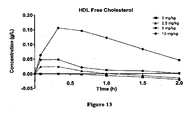
- HDL free cholesterol means the amount of cholesterol having a free hydroxyl group (“free cholesterol”) that is contained within HDL particles in the serum.
- the HDL particles can be formed from an ApoA-I Mimic/ lipid complex.
- HDL total cholesterol means the amount of free cholesterol plus the amount of cholesterol having a hydroxyl group that has been esterified (“esterified cholesterol”) that is contained within HDL particles in the serum.
- the HDL particles can be formed from an ApoA-I Mimic/ lipid complex.
- amino acid residue includes genetically encoded amino acid residues and non-genetically encoded amino acid residues.
- Non-genetically encoded amino acid residues include, but are not limited to, ⁇ -alanine ( ⁇ -Ala); 2,3-diaminopropionic acid (Dpr); nipecotic acid (Nip); pipecolic acid (Pip); ornithine (Orn); citrulline (Cit); t-butylalanine (t-BuA); 2-t-butylglycine (t-BuG); N-methylisoleucine (Melle); phenylglycine (PhG); cyclohexylalanine (ChA); norleucine (Nle); naphthylalanine (Nal); 4-chlorophenylalanine (Phe(4-Cl)); 2- fluorophenylalanine (Phe(2-F)); 3-fluorophenylalanine (Phe(3-F)); 4-fluorophenylalanine (Phe(4-F)); penicillamine (Pen); 1,2,3,4-
- Non-genetically encoded amino acid residues include 3- aminopropionic acid; 4-aminobutyric acid; isonipecotic acid (Inp); aza-pipecolic acid (azPip); aza-proline (azPro); ⁇ -aminoisobutyric acid (Aib); ⁇ -aminohexanoic acid (Aha); ⁇ -aminovaleric acid (Ava); N-methylglycine (MeGly).
- “Chiral,” as used herein to refer to an amino acid residue, means an amino acid residue having at least one chiral center.
- the chiral amino acid residue is an L-amino acid residue.
- L-amino acid residues include, but are not limited to, Ala, Arg, Asn, Asp, Cys, Gln, Glu, His, Ile, Leu, Lys, Met, Phe, Pro, Ser, Thr, Trp, Tyr, Val, ⁇ -Ala, Dpr, Nip, Orn, Cit, t-BuA, t-BuG, Melle, PhG, ChA, Nle, Nal, Phe(4-Cl), Phe(2-F), Phe(3-F), Phe(4-F), Pen, Tic, Thi, MSO, hArg, AcLys, Dbu, Dab, Phe(pNH 2 ), MeVal, hCys, hPhe, hSer, Hyp, and hPro.
- the chiral amino acid residue is a D-amino acid residue.
- D-amino acid residues include, but are not limited to D-Ala, D-Arg, D-Asn, D-Asp, D-Cys, D-Gln, D-Glu, D- His, D-Ile, D-Leu, D-Lys, D-Met, D-Phe, D-Pro, D-Ser, D-Thr, D-Trp, D-Tyr, D-Val, D- ⁇ -Ala, D-Dpr, D-Nip, D-Pip, D-Orn, D-Cit, D-t-BuA, D-t-BuG, D-MeIle, D-PhG, D-ChA, D-Nle, D-Nal, D-Phe(4-Cl), D-Phe(2-F), D-Phe(3-F), D-Phe(4-F), D-Pen, D-T
- Achiral as used herein to refer to an amino acid residue, means an amino acid residue that does not have a chiral center.
- Examples of achiral amino acid residues include, but are not limited to, Gly, Inp, Aib, Aha, Ava, MeGly, azPip, and azPro.
- Aliphatic amino acid residue refers to an amino acid residue having an aliphatic hydrocarbon side chain.
- Aliphatic amino acid residues include, but are not limited to, Ala (A), Val (V), Leu (L), Ile (I), Pro (P), azPro, Pip, azPip, ⁇ -Ala, Aib, t-BuA, t-BuG, MeIle, ChA, Nle, MeVal, Inp, Nip, hPro, D-Ala, D-Val, D-Leu, D-Ile, D-Pro, D- ⁇ -Ala, D-t-BuA, D-t-BuG, D-MeIle, D-Nle, D-MeVal, D-Nip, D-Pip, D-ChA, and D-hPro.
- the aliphatic amino acid residue is an L-amino acid residue. In another embodiment, the aliphatic amino acid residue is a D-amino acid residue. In another embodiment, the aliphatic amino acid residue is an achiral amino acid residue.
- Hydrophobic amino acid residue refers to an amino acid residue exhibiting a hydrophobicity of less than zero according to the normalized consensus hydrophobicity scale of Eisenberg et al., 1984, J. Mol. Biol. 179:125-142 .
- Hydrophilic amino acid residues include, but are not limited to, Pro (P), Gly (G), Thr (T), Ser (S), His (H), Glu (E), Asn (N), Gln (Q), Asp (D), Lys (K) Arg (R), Dpr, Orn, Cit, Pen, MSO, hArg, AcLys, Dbu, Dab, Phe(p-NH 2 ), hCys, hSer, Hyp, D-Pro, D-Thr, D-Ser, D-His, D-Glu, D-Asn, D-Gln, D-Asp, D-Lys, D-Arg, D-Dpr, D-Orn, D-Cit, D-Pen, D-MSO, D-hArg, D-AcLys, D-Dbu, D-Dab, D-Phe(p-NH 2 ), D- hCys, D-hSer, and D-Hyp.
- hydrophilic amino acid residues include, but are not limited to, C 1-4 lateral chain analogs having the following formulas: and wherein n is an integer from 1 to 4.
- the hydrophilic amino acid residue is an L-amino acid residue.
- the hydrophilic amino acid residue is a D-amino acid residue.
- the hydrophilic amino acid residue is an achiral amino acid residue.
- the hydrophilic amino acid residue is an acidic L-amino acid residue, an acidic D-amino acid residue, or an acidic achiral amino acid residue.
- the hydrophilic amino acid residue is a basic L-amino acid residue, a basic D-amino acid residue, or a basic achiral amino acid residue.
- Hydrophobic amino acid residue refers to an amino acid residue exhibiting a hydrophobicity of greater than zero according to the normalized consensus hydrophobicity scale of Eisenberg, 1984, J. Mol. Biol. 179:125-142 .
- Hydrophobic amino acid residues include, but are not limited to, Ile (I), Phe (F), Val (V), Leu (L), Trp (W), Met (M), Ala (A), Gly (G), Tyr (Y), ⁇ -Ala, Nip, t-BuA, t-BuG, MeIle, PhG, ChA, Nle, Nal, Phe(4-Cl), Phe(2-F), Phe(3-F), Phe(4-F), Tic, Thi, MeVal, hPhe, hPro, 3-aminopropionic acid, 4 aminobutryic acid, Inp, Aib, Aha, Ava, MeGly, D-Pro, D-Ile, D-Phe, D-Val, D-Leu, D-Trp, D-Met, D-Ala, D-Tyr, D- -Ala, D-Nip, D-t-BuA, D-t-BuG, D-Melle, D-PhG,
- hydrophobic amino acids include, but are not limited to, C 1-4 lateral chain analogs having the following formulas: and wherein n is an integer from 1 to 4.
- the hydrophobic amino acid residue is an L-amino acid residue.
- the hydrophobic amino acid residue is a D-amino acid residue.
- the hydrophobic amino acid residue is an achiral amino acid residue.
- Poly amino acid residue refers to a hydrophilic amino acid residue having a side chain that is uncharged at physiological pH, but which has at least one bond in which the pair of electrons shared in common by two atoms is held more closely by one of the atoms.
- Polar amino acid residues include, but are not limited to, Asn (N), Gln (Q), Ser (S), Thr (T), Cit, Pen, MSO, AcLys, hCys, hSer, Hyp, D-Asn, D-Gln, D-Ser, D-Thr, D-Cit, D-Pen, D-MSO, D-AcLys, D-hCys, D-hSer, and D-Hyp.
- Other polar amino acids include, but are not limited to, C 1-4 lateral chain analogs having the following formulas: wherein n is an integer from 1 to 4.
- the polar amino acid residue is an L-amino acid residue.
- the polar amino acid residue is a D-amino acid residue.
- the polar amino acid residue is an achiral amino acid residue.
- Acidic amino acid residue refers to a hydrophilic amino acid residue having a side chain pK value of less than 7. Acidic amino acid residues typically have negatively charged side chains at physiological pH due to loss of a hydrogen ion. Acidic amino acid residues include, but are not limited to, Glu (E), Asp (D), D-Glu, and D-Asp. Other acidic amino acids include, but are not limited to, C 1-4 lateral chain analogs having the following formula: wherein n is an integer from 1 to 4. In one embodiment, the acidic amino acid residue is an L-amino acid residue. In another embodiment, the acidic amino acid residue is a D-amino acid residue. In another embodiment, the acidic amino acid residue is an achiral amino acid residue.
- Basic amino acid residue refers to a hydrophilic amino acid residue having a side chain pK value of greater than 7.
- Basic amino acid residues typically have positively charged side chains at physiological pH due to association with a hydronium ion.
- Basic amino acid residues include, but are not limited to, His (H), Arg (R), Lys (K), Dpr, Orn, hArg, Dbu, Dab, Phe(p-NH 2 ), D-His, D- Arg, D-Lys, D-Dpr, D-Orn, D-hArg, D-Dbu, D-Dab, and D-Phe(p-NH 2 ).
- basic amino acid residues include, but are not limited to, C 1-4 lateral chain analogs having the following formulas: and wherein n is an integer from 1 to 4.
- the basic amino acid residue is an L-amino acid residue.
- the basic amino acid residue is a D-amino acid residue.
- the basic amino acid residue is an achiral amino acid residue.
- Nonpolar amino acid residue refers to a hydrophobic amino acid residue having a side chain that is uncharged at physiological pH and which has bonds in which the pair of electrons shared in common by two atoms is held substantially equally by each of the two atoms ( i.e. , the side chain is not polar).
- Non-polar amino acid residues include, but are not limited to, Leu (L), Val (V), Ile (I), Met (M), Gly (G), Ala (A), Pro (P), azPro, Pip, azPip, ⁇ -Ala, Nip, t-BuG, Melle, ChA, Nle, MeVal, hPro, 3-aminopropionic acid, 4-aminobutyric acid, Inp, Aib, Aha, Ava, MeGly, D-Leu, D-Val, D-Ile, D-Met, D-Ala, D-Pro, D- ⁇ -Ala, D-Inp, D-t- BuG, D-Melle, D-ChA, D-Nle, D-MeVal, D-Nip, D-Pip, and D-hPro.
- non-polar amino acid residues include, but are not limited to, C 1-4 lateral chain analogs having the following formulas: and wherein n is an integer from 1 to 4.
- the non-polar amino acid residue is an L-amino acid residue.
- the non-polar amino acid residue is a D-amino acid residue.
- the non-polar amino acid residue is an achiral amino acid residue.
- Aromatic amino acid residue refers to a hydrophobic amino acid residue with a side chain having at least one aromatic or heteroaromatic ring.
- the aromatic or heteroaromatic ring can contain one or more substituents such as -OH, -SH, -CN, -F, -Cl, -Br, -I, -NO 2 , -NO, -NH 2 , -NHR, -NRR,- C(O)R, -C(O)OH, - C(O)OR, -C(O)NH 2 , -C(O)NHR, -C(O)NRR where each R is independently (C 1 -C 6 ) alkyl, substituted (C 1 -C 6 ) alkyl, 5-26-membered aryl, and substituted 5-26-membered aryl.
- Aromatic amino acid residues include, but are not limited to, Phe (F), Tyr (Y), Trp (W), PhG, Nal, Phe(4-Cl), Phe(2-F), Phe(3-F), Phe(4-F), Tic, Thi, hPhe, D-Phe, D-Tyr and D-Trp, D-PhG, D-Nal, D-Phe(4-Cl), D-Phe(2-F), D- Phe(3-F), D- Phe(4-F), D-Tic, D-Thi, and D-hPhe.
- aromatic amino acid residues include, but are not limited to, C 1-4 lateral chain analogs having the following formulas: and wherein n is an integer from 1 to 4.
- the aromatic amino acid residue is an L-amino acid residue.
- the aromatic amino acid residue is a D-amino acid residue.
- the aromatic amino acid residue is an achiral amino acid residue.
- amino acid residues having side chains exhibiting two or more physical-chemical properties can be included in multiple categories.
- amino acid side chains having aromatic moieties that are further substituted with polar substituents, such as Tyr (Y) or its corresponding D-enantiomer can exhibit both aromatic hydrophobic properties and polar or hydrophilic properties, and can therefore be included in both the aromatic and polar categories.
- polar substituents such as Tyr (Y) or its corresponding D-enantiomer
- amino acid residue Cys (C) or its corresponding D- enantiomer can form disulfide bridges with other Cys (C) residues or their corresponding D-enantiomers or with other sulfanyl-containing amino acids.
- the ability of Cys (C) residues (and other amino acids with -SH containing side chains) to exist in a peptide in either the reduced free -SH or oxidized disulfide-bridged form affects whether Cys (C) residues or their corresponding D-enantiomers contribute net hydrophobic or hydrophilic character to a peptide.
- Cys (C) or its corresponding D-enantiomer exhibits a hydrophobicity of 0.29 according to the normalized consensus scale of Eisenberg (Eisenberg, 1984, supra), it is to be understood that for purposes of the present invention Cys (C) and its corresponding D-enantiomer are categorized as polar hydrophilic amino acids, notwithstanding the general classifications defined above.
- the invention provides an ApoA-I Mimic peptide as defined in the appended claims.
- 15- to 29-residue peptides having the following Formula I R 1- Y 1- X 1 -X 2 -X 3 -X 4 -X 5 -X 6 -X 7 -X 8 -X 9 -X 10 -X 11 -X 12 -X 13 -X 14 -X 15 -X 16 -X 17 -X 18 -X 19 -X 20 -X 21 -X 22 -X 23 -Y 2 -R 2 Formula I and pharmaceutically acceptable salts thereof, wherein:
- 22- to 29-residue peptides having the following Formula I R 1 -Y 1 -X 1 -X 2 -X 3 -X 4 -X 5 -X 6 -X 7 -X 8 -X 9 -X 10 -X 11 -X 12 -X 13 -X 14 -X 15 -X 16 -X 17 -X 18 -X 19 -X 20 -X 21 -X 22 -X 23 -Y 2 -R 2 Formula I and pharmaceutically acceptable salts thereof, wherein:
- 15- to 21-residue peptides having the following Formula I R 1 -Y 1 -X 1 -X 2 -X 3 -X 4 -X 5 -X 6 -X 7 -X 8 -X 9 -X 10 -X 11 -X 12 -X 13 -X 14 -X 15 -X 16 -X 17 -X 18 -X 19 -X 20 -X 21 -X 22 -X 23 -Y 2 -R 2 Formula I and pharmaceutically acceptable salts thereof, wherein:
- the peptide of Formula I or pharmaceutically acceptable salt thereof is 22 amino acid residues in length and X 1 is absent.
- X 2 and X 4 are both Lys, Orn, D-Lys, or D-Orn.
- X 5 is Gln, Lys, D-Gln, or D-Lys.
- X 9 is an acidic amino acid residue.
- X 12 is Glu, Asn, Gln, Arg, D-Glu, D- Asn, D-Gln, or D-Arg.
- X 13 is Glu, Asn, Gln, Arg, D-Glu, D- Asn, D-Gln, or D-Arg.
- X 16 is an acidic amino acid residue.
- X 17 is Arg, Lys, Orn, D-Arg, D-Lys, or D-Orn.
- X 21 is Leu or D-Leu.
- X 22 is Ala, Val, Leu, D-Ala, D-Val, or D-Leu.
- X 1 is absent;
- X 13 is an acidic amino acid residue, Arg, or D-Arg;
- X 14 is a basic amino acid residue, Asn, Glu, D-Asn, or D-Glu; and
- X 2 to X 12 and X 15 to X 23 are as defined above in Formula I.
- X 1 is absent;
- X 2 is Lys, Orn, D-Lys, or D-Orn;
- X 3 is Leu or D-Leu;
- X 4 is Lys, Orn, D-Lys, or D-Orn;
- X 5 is Lys, Orn, Gln, Asn, D-Lys, D- Orn, D-Gln, or D-Asn;
- X 6 is Lys, Orn, Gln, Asn, D-Lys, D-Orn, D-Gln, or D-Asn;
- X 7 is Leu, Gly, Nal, D-Leu, or D-Nal;
- X 8 is Ala, Trp, Gly, Leu, Phe, Nal, D-Ala, D-Trp, D-Leu, D-Phe, or D-Nal;
- X 9 is Asp, Glu, Gln, Lys, D-As
- X 1 is absent; X 9 is Glu or D-Glu; X 12 is Glu or D- Glu; X 13 is Asn, Glu, D-Asn, or D-Glu; X 14 is Leu or D-Leu; X 15 is Leu or D-Leu; X 16 is Glu or D-Glu; X 17 is Arg, Lys, D-Arg, or D-Lys; X 18 is Phe or D-Phe; X 19 is Leu or D-Leu; X 21 is Leu or D-Leu; and/or X 22 is Val or D-Val; and X 2 to X 8 , X 10 , X 11 , X 20 , and X 23 are as defined above in Formula I.
- X 1 is absent;
- X 2 is Lys, Orn, D-Lys, or D-Orn;
- X 3 is Leu or D-Leu;
- X 4 is Lys, Orn, D-Lys, or D-Orn;
- X 5 is Lys, Orn, Gln, Asn, D-Lys, D- Orn, D-Gln, or D-Asn;
- X 6 is Lys, Orn, Gln, Asn, D-Lys, D-Orn, D-Gln, or D-Asn;
- X 7 is Leu, Gly, Nal, D-Leu, or D-Nal;
- X 8 is Ala, Trp, Gly, Leu, Phe, Nal, D-Ala, D-Trp, D- Leu, D-Phe, or D-Nal;
- X 9 is Glu or D-Glu;
- X 11 is Leu, D
- X 1 is absent, only one of X 5 and X 6 is a basic amino acid residue, and the other of X 5 and X 6 is Gln, Asn, D-Gln, or D-Asn.
- Y1 or Y2 is absent or is a sequence having from one to seven amino acid residues.
- one or more of the amino acid residues of the amino acid sequence is an acidic amino acid residue.
- one or more of the amino acid residues of the amino acid sequence is a basic amino acid residue.
- one of X 5 and X 6 is Lys, Orn, D-Lys, or D-Orn, and the other of X 5 and X 6 is Gln, Asn, D-Gln, or D-Asn.
- each chiral amino acid residue is an L-amino acid residue.
- each chiral amino acid residue is a D-amino acid residue.
- X 1 is absent; one of X 7 , X 8 , X 10 , X 11 , X 14 and X 15 is Gly; and X 1 to X 6 , X 9 , X 12 , X 13 , and X 16 to X 23 are other than Gly.
- X 1 is absent; X 11 is Gly; and each of X 7 , X 8 , X 10 , X 14 , and X 15 is other than Gly.
- X 1 is absent;
- X 2 is Lys, Orn, D-Lys, or D-Orn;
- X 3 is Leu or D-Leu;
- X 4 is Lys, Orn, D-Lys, or D-Orn;
- X 5 is Gln or D-Gln;
- X 6 is Lys, Orn, D- Lys, or D-Orn;
- X 7 is Leu, Nal, D-Leu, or D-Nal;
- X 8 is Ala, Trp, D-Ala, or D-Trp;
- X 9 is Glu or D-Glu;
- X 10 is Leu or D-Leu;
- X 11 is Gly;
- X 12 is Glu or D-Glu;
- X 13 is Asn or D-Asn;
- X 14 is Leu, Trp, D-Leu, or D-Trp;
- X 15 is Leu or D-Le
- the peptide of Formula I is a peptide set forth in Table 3 below: Table 3.
- X 1 is absent; X 15 is Gly; and each of X 7 , X 8 , X 10 , X 11 , and X 14 is other than Gly.
- the peptide of Formula I is:
- X 1 is absent; X 14 is Gly; and each of X 7 , X 8 , X 10 , X 11 , and X 15 is other than Gly.
- the peptide of Formula I is:
- X 1 is absent; X 10 is Gly; and each of X 7 , X 8 , X 11 , X 14 , and X 15 is other than Gly.
- the peptide of Formula I is:
- X 1 is absent; X 8 is Gly; and each of X 7 , X 10 , X 11 , X 14 , and X 15 is other than Gly.
- the peptide of Formula I is:
- X 1 is absent; X 7 is Gly; and each of X 8 , X 10 , X 11 , X 14 , and X 15 is other than Gly.
- the peptide of Formula I is:
- X 1 is absent; and each of X 7 , X 8 , X 10 , X 11 , X 14 , and X 15 is other than Gly.
- X 1 is absent;
- X 2 is Lys, Orn, D-Lys, or D-Orn;
- X 3 is Leu or D-Leu;
- X 4 is Lys, Orn, D-Lys, or D-Orn;
- one of X 5 or X 6 is Gln or D-Gln and the other of X 5 or X 6 is Lys, Orn, D-Lys, or D-Orn;
- X 7 is Leu, Nal, D-Leu, or D-Nal;
- X 8 is Ala, Leu, Trp, Nal, D-Ala, D-Leu, D-Trp, or D-Nal;
- X 9 is Glu or D-Glu;
- X 10 is Leu, Trp, Nal, D-Leu, D-Trp, or D-Nal;
- X 11 is Leu, D-Leu or Aib;
- X 12 is Glu or
- X 1 is absent; X 2 is Lys or D-Lys; X 3 is Leu or D- Leu; X 4 is Lys or D-Lys; X 5 is Glu or D-Glu; X 6 is Lys or D-Lys; X 7 is Leu or D-Leu; X 8 is Ala or D-Ala; X 9 is Glu or D-Glu; X 10 is Leu or D-Leu; X 11 is Leu or D-Leu; X 12 is Glu or D-Glu; X 13 is Asn or D-Asn; X 14 is Leu or D-Leu; X 15 is Leu or D-Leu; X 16 is Glu or D-Glu; X 17 is Arg or D-Arg; X 18 is Phe or D-Phe; X 19 is Leu or D-Leu; X 20 is Asp or D-Asp; X 21 is Leu or D-Leu; X 22 is Val
- X 3 is other than Lys or D-Lys;
- X 9 is other than Trp or D-Trp;
- X 11 is other than Glu, Trp, D-Glu, or D-Trp;
- X 12 is other than Trp, Leu, D-Trp, or D-Leu;
- X 13 is other than Trp or D-Trp;
- X 15 is other than Lys, Trp, D-Lys, or D-Trp;
- X 16 is other than Trp or D-Trp;
- X 17 is other than Trp, Leu, D-Trp, or D-Leu;
- X 18 is other than Trp or D-Trp;
- the peptide of Formula I is one of the peptides set forth in Table 4 below: Table 4.
- Peptide 15 Peptide 16 Peptide 17 Peptide 18 Peptide 19 Peptide 20 Peptide 21 Peptide 22 Peptide 23 Peptide 24 Peptide 25 Peptide 26 Peptide 27 Peptide 28 Peptide 29 Peptide 30 Peptide 31 Peptide 32 Peptide 33 Peptide 34 Peptide 35 Peptide 36 Peptide 37 Peptide 38 Peptide 39 Peptide 40 Peptide 107 Peptide 108 Peptide 109 Peptide 110 Peptide 111 Peptide 112 Peptide 113 Peptide 114 Peptide 115 Peptide 116 Peptide 117 Peptide 118 Peptide 119 Peptide 120 Peptide 121 Peptide 122 Peptide 123 Peptide 124 Peptide 125 Peptide 126 Peptide 127 Peptide 128 Peptide 129 Peptide 130 Peptide 131 Pept
- 14- to 22-residue peptides having the following Formula II R 1 -Y 1 -X 1 -X 2 -X 3 -X 4 -X 5 -X 6 -X 7 -X 8 -X 9 -X 10 -X 11 -X 12 -X 13 -X 14 -X 15 -X 16 -X 17 -X 18 -Y 2 -R 2 , Formula II and pharmaceutically acceptable salts thereof, wherein:
- 15- to 22-residue peptides having the following Formula II R 1 -Y 1 -X 1 -X 2 -X 3 -X 4 -X 5 -X 6 -X 7 -X 8 -X 9 -X 10 -X 11 -X 12 -X 13 -X 14 -X 15 -X 16 -X 17 -X 18 -Y 2 -R 2 , Formula II and pharmaceutically acceptable salts thereof, wherein:
- 14-residue peptides having the following Formula II R 1 -Y 1 -X 1 -X 2 -X 3 -X 4 -X 5 -X 6 -X 7 -X 8 -X 9 -X 10 -X 11 -X 12 -X 13 -X 14 -X 15 -X 16 -X 17 -X 18 -Y 2 R 2 , Formula II and pharmaceutically acceptable salts thereof, wherein:
- the peptide of Formula II is an 18-residue peptide.
- the peptide of Formula II is a peptide set forth in Table 5 below. Table 5.
- Peptide 51 Glu-Leu-Leu-Glu-Asn-Leu-Leu-Glu-Arg-Phe-Leu-Asp-Leu-Val-Inp (SEQ. ID. NO. 51)
- Peptide 53 Lys-Leu-Lys-Gln-Leu-Leu-Glu-Asn-Leu-Leu-Glu-Arg-Phe-Leu-Asp-Leu-Val-Inp (SEQ. ID. NO.
- Peptide 54 Lys-Leu-Lys-Gln-Lys-Leu-Glu-Glu-Leu-Leu-Glu-Arg-Phe-Leu-Asp-Leu-Val-Inp (SEQ. ID. NO. 54)
- Peptide 55 Lys-Leu-Lys-Gln-Lys-Leu-Glu-Glu-Leu-Leu-Glu-Lys-Phe-Leu-Glu-Leu-Val-Inp (SEQ. ID. NO.
- Peptide 56 Lys-Leu-Lys-Gln-Lys-Leu-Leu-Glu-Leu-Leu-Glu-Arg-Phe-Leu-Asp-Leu-Val-Inp (SEQ. ID. NO. 56)
- Peptide 58 Lys-Leu-Lys-Lys-Gln-Leu-Glu-Glu-Leu-Leu-Glu-Arg-Phe-Leu-Asp-Leu-Val-Inp (SEQ. ID. NO. 58)
- Peptide 143 Glu-Leu-Leu-Glu-Asn-Leu-Leu-Glu-Arg-Phe-Leu-Asp-Leu-Val-Nip (SEQ. ID.
- Peptide 359 Lys-Leu-Lys-Gln-Lys-Leu-Glu-Glu-Leu-Leu-Glu-Lys-Phe-Leu-Glu-Leu-Val-Pip (SEQ. ID. NO. 359)
- Peptide 360 Lys-Leu-Lys-Gln-Lys-Leu-Leu-Glu-Leu-Leu-Glu-Arg-Phe-Leu-Asp-Leu-Val-Pip (SEQ. ID. NO.
- Peptide 362 Lys-Leu-Lys-Lys-Gln-Leu-Glu-Glu-Leu-Leu-Glu-Arg-Phe-Leu-Asp-Leu-Val-Pip (SEQ. ID. NO. 362)
- Peptide 447 Glu-Leu-Leu-Glu-Asn-Leu-Leu-Glu-Arg-Phe-Leu-Asp-Leu-Val-azPip (SEQ. ID. NO. 447)
- Peptide 449 Peptide 450 Peptide 451 Peptide 452 Peptide 454 or a pharmaceutically acceptable salt thereof.
- 15- to 29-residue peptides having the following Formula III R 1 -Y 1 -X 1 -X 2 -X 3 -X 4 -X 5 X 6 -X 7 -X 8 -X 9 -X 10 -X 11 -X 12 -X 13 -X 14 -X 15- X 16 -X 17 -X 18 -X 19 -X 20 -X 21 -X 22 -X 23 -Y 2 -R 2 Formula III or a pharmaceutically acceptable salt thereof, wherein:
- 22- to 29-residue peptides having the following Formula III R 1 -Y 1 -X 1 -X 2 -X 3 - X 4 -X 5 X 6 -X 7 -X 8 -X 9 -X 10 -X 11 -X 12 -X 13 -X 14 -X 15 -X 16 -X 17 -X 18 -X 19 -X 20 -X 21 -X 22 -X 23 -Y 2 -R 2 Formula III and pharmaceutically acceptable salts thereof, wherein:
- 15- to 21-residue peptides having the following Formula III R 1 - Y 1 -X 1 -X 2 -X 3 -X 4 -X 5 X 6 -X 7 -X 8 -X 9 -X 10 -X n- X 12 -X 13 -X 14 -X 15 -X 16 -X 17 -X 18 -X 19 -X 20 -X 21 -X 22 -X 23 -Y 2 -R 2 Formula III and pharmaceutically acceptable salts thereof, wherein:
- the peptide of Formula III is 22 amino acid residues in length and X 1 is absent.
- the peptide of Formula III is a peptide set forth in Table 6 below. Table 6.
- ApoA-I Mimics wherein one or more of its amide linkages is optionally replaced with a linkage other than amide, including, but not limited to, a substituted amide or an isostere of amide.
- a linkage other than amide including, but not limited to, a substituted amide or an isostere of amide.
- the nitrogen atom of one or more of the ApoA-I Mimics' amide linkages is substituted, such that the substituted amide linkage has the formula -C(O)NR'-, where R' is (C 1 -C 6 ) alkyl, (C 2 -C 6 ) alkenyl, (C 2 -C 6 ) alkynyl, (C 5 -C 20 ) aryl, (C 6 -C 26 ) alkaryl, 5-20 membered heteroaryl, or 6-26 membered alkheteroaryl.
- R' is substituted with -OR, -SR, -NRR, -NO 2 , -CN, halogen, - SO 2 R, -C(O)R, -C(O)OR and -C(O)NRR, where each R is independently hydrogen, alkyl, or aryl.
- Compounds having such non-amide linkages and methods for preparing such compounds are well-known in the art ( see , e.g. , Spatola, March 1983, Vega Data Vol.
- one or more of the ApoA-I Mimics' amide linkages can be replaced with one or more peptidomimetic or amide mimetic moieties that do not significantly interfere with the structure or activity of the peptides.
- Suitable amide mimetic moieties are described, for example, in Olson et al., 1993, J. Med. Chem. 36:3039-3049 .
- the ApoA-I Mimic is in the form of a pharmaceutically acceptable salt.
- the salt can be formed at the C-terminus or N-terminus or at an acidic or basic amino acid residue side chain.
- the pharmaceutically acceptable salt is a metal salt, organic amine salt, or acid addition salt.
- Metal salts can arise from the addition of an inorganic base to the peptide of Formula I, II, or III.
- the inorganic base consists of a metal cation paired with a basic couterion such as, for example, hydroxide, carbonate, bicarbonate, or phosphate.
- the metal may be an alkali metal, alkaline earth metal, transition metal, or main group metal.
- the metal is lithium, sodium, potassium, cerium, magnesium, manganese, iron, calcium, aluminum, or zinc.
- Organic amine salts can arise from the addition of an organic amine to the peptide of Formula I, II, or III.
- the organic amine is triethylamine, ethanolamine, diethanolamine, triethanolamine, morpholine, piperidine, N-methylpiperidine, N-ethylpiperidine, dibenzylamine, piperazine, pyridine, pyrazine, or pipyrazine.
- Acid addition salts arise from the addition of an acid to the peptide of Formula I, II, or III.
- the acid is organic.
- the acid is inorganic.
- the acid is hydrochloric acid, hydrobromic acid, hydroiodic acid, nitric acid, sulfuric acid, sulfurous acid, a phosphoric acid, isonicotinic acid, lactic acid, salicylic acid, tartaric acid, ascorbic acid, gentisinic acid, gluconic acid, glucaronic acid, saccaric acid, formic acid, benzoic acid, glutamic acid, pantothenic acid, acetic acid, fumaric acid, succinic acid, methanesulfonic acid, ethanesulfonic acid, benzenesulfonic acid, p-toluenesulfonic acid, citric acid, or maleic acid.
- the acid addition salt is a hydrochloride, hydrobromide, hydroiodide, nitrate, sulfate, sulfite, bisulfate, phosphate, acid phosphate, isonicotinate, lactate, salicylate, tartrate, bitartrate, ascorbate, gentisinate, gluconate, glucaronate, saccarate, formate, benzoate, glutamate, pantothenate, acetate, fumarate, succinate, methanesulfonate, ethanesulfonate, benzenesulfonate, p-toluylsulfonate, citrate, or maleate salt.
- R 1 is an amino protecting group.
- the amino protecting group is: (C 1 -C 6 ) alkyl, (C 2 -C 6 ) alkenyl, (C 2 -C 6 ) alkynyl, (C 5 -C 26 ) aryl, (C 6 -C 26 aralkyl), 5- to 20-membered heteroaryl, or 6- to 26-membered alkheteroaryl; --C(O)R;-C(O)OR; --SO 2 R; or -SR, wherein R is H or (C 1 -C 6 ) alkyl, (C 2 -C 6 ) alkenyl, (C 2 -C 6 ) alkynyl, (C 5 -C 26 ) aryl, (C 6 -C 26 aralkyl), 5- to 20-membered heteroaryl, or 6- to 26-membered alkheteroaryl.
- the (C 1 -C 6 ) alkyl, (C 2 -C 6 ) alkenyl, (C 2 -C 6 ) alkynyl, (C 5 -C 26 ) aryl, (C 6 -C 26 aralkyl), 5- to 20-membered heteroaryl, or 6- to 26-membered alkheteroaryl is substituted with one or more of -OR a , -SR a , -NR a R a , -NO 2 , -CN, halogen, -SO 2 R a , -C(O)R a , -C(O)OR a and -, C(O)NR a R a , where each R a is independently hydrogen, alkyl, or aryl.
- R 1 is H, the number of amino protecting groups in the ApoA-I Mimic is zero; and when R 1 is an amino protecting group, the number of amino protecting groups in the ApoA-
- the amino protecting group is: dansyl; methoxycarbonyl; ethoxycarbonyl; 9-fluorenylmethoxycarbonyl; 2-chloroethoxycarbonyl; 2,2,2-trichloroethoxycarbonyl; 2-phenylethoxycarbonyl; t -butoxycarbonyl; benzyloxycarbonyl; p -methoxybenzyloxycarbonyl; p -nitrobenzyloxycarbonyl; o-nitrobenzyloxycarbonyl; p -bromobenzyloxycarbonyl; p -chlorobenzyloxycarbonyl; p -iodobenzyloxycarbonyl; 2,4-dichlorobenzyloxycarbonyl; diphenylmethoxycarbonyl; 3,5-dimethoxybenzyloxycarbonyl; phenoxycarbonyl; 2,4,6-tri- t -butylpenoxycarbonyl; 2,4,6-tri
- R 2 is a carboxyl protecting group.
- the carboxyl protecting group is: O-(C 1 -C 6 ) alkyl, O-(C 2 -C 6 ) alkenyl, O-(C 2 -C 6 ) alkynyl, O-(C 5 -C 26 ) aryl, O-(C 6 -C 26 aralkyl), O-(5- to 20-membered heteroaryl), or O-(6- to 26-membered alkheteroaryl); or -NRR, wherein R is H or (C 1 -C 6 ) alkyl, (C 2 -C 6 ) alkenyl, (C 2 -C 6 ) alkynyl, (C 5 -C 26 ) aryl, (C 6 -C 26 aralkyl), 5- to 20-membered heteroaryl, or 6- to 26-membered alkheteroaryl.
- the (C 1 -C 6 ) alkyl, (C 2 -C 6 ) alkenyl, (C 2 -C 6 ) alkynyl, (C 5 -C 26 ) aryl, (C 6 -C 26 aralkyl), 5- to 20-membered heteroaryl, or 6- to 26-membered alkheteroaryl is substituted with one or more of -OR a , -SR a , -NR a R a , -NO 2 , -CN, halogen, -SO 2 R a , -C(O)R 8 , -C(O)OR a and - C(O)NR a R a , where each R a is independently hydrogen, alkyl, or aryl.
- R 1 is H, the number of carboxyl protecting groups in the ApoA-I Mimic is zero; and when R 1 is a carboxyl protecting group, the number of carboxyl protecting groups in
- the carboxyl protecting group is methoxy; ethoxy; 9-fluorenylmethoxy; methoxymethoxy; methylthiomethoxy; tetrahydropyranoxy; tetrahydrofuranoxy; methoxyethoxymethoxy; benzyloxymethoxy; phenacyloxy; p- bromophenacyloxy; ⁇ -methylphenacyloxy; p -methoxyphenacyloxy; desyloxy; 2-chloroethoxy; 2,2,2-thrichloroethoxy, 2-methylthioethoxy; 2-( p -toluenesulfonyl)methoxy; t -butoxy; cyclopentoxy; cyclohexoxy; allyloxy; methallyloxy; cinnamoxy; ⁇ -methylcinnamoxy; phenoxy; 2,6-dimethylphenoxy; 2,6-diisopropylphenoxy; benzyloxy;
- protected forms of the ApoA-I Mimic i.e. , forms of the ApoA-I Mimic in which one or more of its -NH 2 or -COOH groups are protected with a protecting group.
- one or more -NH 2 groups are protected with an amino protecting group as described above.
- one or more -COOH groups are protected with a carboxyl protecting group as described above.
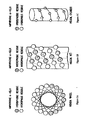
- the ApoA-I Mimics have the ability to form an amphipathic ⁇ -helix in the presence of one or more lipids.
- amphipathic is meant that the ⁇ -helix has opposing hydrophilic and hydrophobic faces oriented along its long axis, i.e. , one face of the helix projects mainly hydrophilic side chains while the opposite face projects mainly hydrophobic side chains.
- FIGS. 1A and 1B present two illustrative views of the opposing hydrophilic and hydrophobic faces of an exemplary idealized amphipathic ⁇ -helix.
- FIG. 1A is a Schiffer-Edmundson helical wheel diagram ( Schiffer and Edmundson, 1967, Biophys. J.
- FIG. 1B presents a helical net diagram of the idealized amphipathic helix of FIG. 1A .
- the ⁇ -helix is presented as a cylinder that has been cut along the center of its hydrophilic face and flattened.
- the center of the hydrophobic face determined by the hydrophobic moment of the helix ( Eisenberg et al., 1982, Nature 299:371-374 ) lies in the center of the figure and is oriented so as to rise out of the plane of the page.
- FIG. 1C An illustration of the helical cylinder prior to being cut and flattened is depicted in FIG. 1C . By cutting the cylinder along different planes, different views of the same amphipathic helix can be observed, and different information about the properties of the helix obtained.
- amphipathic helix formed by the ApoA-I Mimics can be important for activity. These properties include the degree of amphipathicity, overall hydrophobicity, mean hydrophobicity, hydrophobic and hydrophilic angles, hydrophobic moment, mean hydrophobic moment, and net charge of the ⁇ -helix.
- the degree of amphipathicity (degree of asymmetry of hydrophobicity) of the amphiphathic helix formed by the ApoA-I Mimics can be conveniently quantified by calculating the hydrophobic moment ( ⁇ H ) of the helix.
- ⁇ H hydrophobic moment
- Methods for calculating ⁇ H for a particular peptide sequence are well-known in the art, and are described, for example in Eisenberg, 1984, Ann. Rev. Biochem. 53:595-623 .
- the actual ⁇ H obtained for a particular peptide will depend on the total number of amino acid residues composing the peptide. Thus, it is generally not informative to directly compare ⁇ H for peptides of different lengths.
- ⁇ H > The amphipathicities of peptides of different lengths can be directly compared by way of the mean hydrophobic moment ( ⁇ H >).
- ApoA-I Mimics which exhibit a ⁇ H > in the range of 0.45 to 0.65, as determined using the normalized consensus hydrophobicity scale of Eisenberg ( Eisenberg, 1984, J. Mol. Biol. 179:125-142 ) are provided.
- ⁇ H > is in the range of 0.50 to 0.60.
- ApoA-I Mimics that exhibit a mean hydrophobicity in the range of -0.050 to -0.070, as determined using the normalized consensus hydrophobicity scale of Eisenberg ( Eisenberg, 1984, J. Mol. Biol. 179:125-142 ) are provided.
- the mean hydrophobicity is in the range of-0.030 to -0.055.
- the mean hydrophobicity of the hydrophobic face ( ⁇ H o pho >) is H o pho /N H where N H is as defined above.
- ApoA-I Mimics which exhibit a ⁇ H o pho > in the range of 0.90 to 1.20, as determined using the consensus hydrophobicity scale of Eisenberg (Eisenberg, 1984, supra; Eisenberg et al., 1982, supra) are provided.
- the ⁇ H o pho > is in the range of 0.94 to 1.10.
- the hydrophobic angle is generally defined as the angle or arc covered by the longest continuous stretch of hydrophobic amino acid residues when the peptide is arranged in the Schiffer-Edmundson helical wheel representation (i.e., the number of contiguous hydrophobic residues on the wheel multiplied by 20°).
- the hydrophilic angle is the difference between 360° and the pho angle (i.e., 360°-pho angle). Those of skill in the art will recognize that the pho and phi angles can depend, in part, on the number of amino acid residues in the peptide. For example, referring to FIG.
- a "continuous" stretch of hydrophobic amino acid residues is meant that at least one amino acid residue at positions along the wheel containing two or more amino acid residues is a hydrophobic amino acid residue.
- the pho angle is the arc covered by residues 16, 2, 6, 17, 10, 3, and 14 despite the occurrence of a hydrophilic residue at position 20, as the residue at position 2, which shares the same position on the wheel, is a hydrophobic residue.
- ApoA-I Mimics having a pho angle in the range of 160° to 220° are described. In some embodiments, the pho angle is in the range of 180° to 200°.
- negative charges in Peptide 16 or a pharmaceutically acceptable salt thereof are distributed on the rest of the hydrophilic face, with at least one negatively charged (acidic) amino acid residue per turn, resulting in a continuous stretch of negative charges along the hydrophilic face of the helix.
- One positive charge is located at residue 16, which potentially contributes to helix stability by forming a salt bridge with an acidic residue one turn away on the helix.
- hydrophobic cluster formed by residues 13, 14, 17, and 20 of Peptide 16 or a pharmaceutically acceptable salt thereof is significant in effecting lipid binding and LCAT activation.
- Amphipathic peptides are expected to bind phospholipids by pointing their hydrophobic faces towards the alkyl chains of the lipid moieties.
- this highly hydrophobic cluster contributes to the strong lipid affinities observed for the ApoA-I Mimics disclosed herein. Since lipid binding is a prerequisite for LCAT activation, it is believed that this hydrophobic cluster is also essential for LCAT activation.
- Aromatic residues can be important in anchoring peptides and proteins to lipids ( De Kruijff, 1990, Biosci. Rep. 10:127-130 ; O'Neil and De Grado, 1990, Science 250:645-651 ; Blondelle et al., 1993, Biochim. Biophys. Acta 1202:331-336 ).
- Phe-17 which is positioned at the center of the hydrophobic cluster, may also play a key role in anchoring Peptide 16 or a pharmaceutically acceptable salt thereof to a lipid.
- the long axis of the ⁇ -helix formed by the ApoA-I Mimics typically has an overall curved shape.
- the lengths of the hydrogen bonds of the hydrophilic and hydrophobic faces vary such that the hydrophobic side of the helix is concave ( Barlow and Thornton, 1988, J. Mol. Biol. 201:601-619 ; Zhou et al., 1992, J. Am. Chem. Soc. 33:11174-11183 ; Gesell et al., 1997, J. Biomol. NMR 9:127-135 ).
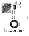
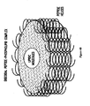
- the amphipathic ⁇ -helices are packed around the edge of the discoidal HDL (see, FIG. 4B ).
- the helices are assumed to be aligned with their hydrophobic faces pointing towards the lipid acyl chains ( Brasseur et al., 1990, Biochim. Biophys. Acta 1043:245-252 ).
- the helices are arranged in an antiparallel fashion, and a cooperative effect between the helices is thought to contribute to the stability of the discoidal HDL complex (Brasseur et al., supra).
- the peptides are considered not as a single entity, but as in interaction with at least two other neighboring peptide molecules ( FIG. 4B ).
- the ApoA-I Mimics have the ability to form intermolecular hydrogen-bonds with one another when aligned in an antiparallel fashion with their hydrophobic faces pointing in the same direction, such as would be the case when the peptides are bound to lipids.
- the ApoA-I Mimics also have the ability to form intramolecular hydrogen bonds or salt bridges near the N- and C-termini of the helix.
- the helices are closely packed; there is no steric hindrance preventing close contact between the helices.
- the ApoA-I Mimics have the ability to closely pack and ionically interact to form intra-and/or intermolecular salt bridges and/or hydrogen bonds when bound to lipids in an antiparallel fashion.
- the ApoA-I Mimics can self-associate.
- the self-association phenomenon depends on the conditions of pH, peptide concentration and ionic strength, and can result in several states of association, from monomeric to several multimeric forms ( FIG. 4A ).
- the hydrophobic core of peptide aggregates favors hydrophobic interactions with lipids.
- the ability of the peptides to aggregate even at very low concentrations may favor their binding to lipids. It is thought that in the core of the peptide aggregates peptide-peptide interactions also occur and may compete with lipid-peptide interactions.
- the hydrophobic core of the aggregates of the ApoA-I Mimics favors hydrophobic interactions with lipids.
- the ability of the ApoA-I Mimics to aggregate even at very low concentrations can favor their binding to lipids. Interactions between the ApoA-I Mimics and lipids lead to the formation of peptide-lipid complexes.
- the type of complex obtained can depend on the lipid:peptide molar ratio, with comicelles generally being formed at low lipid:peptide molar ratios and discoidal and vesicular or multilayer complexes being formed with increasing lipid:peptide molar ratios.
- Micelles are typically formed at ratios of about 2 moles of lipid: about 1 mole of ApoA-I or about 2 moles of lipid: about 6 to about 10 moles of ApoA-I Mimic.
- Discoidal complexes are typically formed at ratios of about 50-100 moles of lipid: about 1 mole of ApoA-I or about 6 to about 10 moles of ApoA-I Mimic.
- Vesicular complexes are typically formed at ratios of about 200 to about 300 moles of lipid: about 1 mole of ApoA-I or about 6 to about 10 moles of ApoA-I Mimic.
- amphipathic peptides Epand, The Amphipathic Helix, 1993
- ApoA-I Jones, 1992, Structure and Function of Apolipoproteins, Chapter 8, pp. 217-250 .
- the lipid:peptide molar ratio also determines the size and composition of the complexes.
- the ApoA-I Mimics have 22 amino acid residues or fewer. Indeed, truncated or internally deleted forms of Formula I, II, or III containing 21, 20, 19, 18, 17, 16, or even 15 amino acid residues that substantially retain the overall characteristics and properties of the amphipathic helix formed by the ApoA-I Mimics are disclosed.
- truncated forms of the ApoA-I Mimics are obtained by deleting one or more amino acid residues from the N-and/or C-terminus.
- Internally deleted forms of the ApoA-I Mimics are obtained by deleting one or more amino acid residues from internal positions within the ApoA-I Mimics.
- the internal amino acid residues deleted can be consecutive residues or non-consecutive residues.
- deleting an internal amino acid residue from an ApoA-I Mimic can cause the plane of the hydrophilic-hydrophobic interface of the helix to rotate by 100° at the point of the deletion. As such rotations can significantly alter the amphipathic properties of the resultant helix, in one embodiment, one or more amino acid residues are deleted so as to substantially retain the alignment of the plane of the hydrophilic-hydrophobic interface along the entire long axis of the helix.
- An idealized ⁇ -helix has 3.6 residues per turn.
- groups of 3-4 consecutive or non-consecutive amino acid residues are deleted. Whether 3 amino acid residues or 4 amino acid residues are deleted can depend upon the position within the helix of the first residue to be deleted. Determining the appropriate number of consecutive or non-consecutive amino acid residues that constitute one complete helical turn from any particular starting point within an amphipathic helix is well within the capabilities of those of skill in the art.
- the ApoA-I Mimics can also be extended at one or both termini or internally with additional amino acid residues that do not substantially interfere with, and in some embodiments even enhance, the structural and/or functional properties of the peptides. Indeed, extended ApoA-I Mimics containing as many as 23, 24, 25, 26, 27, 28, or 29 amino acid residues are also provided. Such extended ApoA-I Mimics may substantially retain the net amphipathicity and other properties of the ApoA-I Mimics.
- adding amino acid residues internally can rotate the plane of the hydrophobic-hydrophilic interface at the point of the insertion in a manner similar to that described above for internal deletions.
- the considerations discussed above in connection with internal deletions apply to internal additions, as well.
- the ApoA-I Mimics are extended at their N- and/or C-terminus by an amino acid sequence having from 1 to 7 residues.
- the ApoA-I Mimics are extended at their N- and/or C-terminus by least one helical turn. Such extensions stabilize the helical secondary structure in the presence of lipids, such as the end-cap amino acid residues and segments previously described.
- the ApoA-I Mimics are extended at the N-terminus by a single basic amino acid residue, such as Lys (K).
- removing the N- and/or C-terminal charges of the ApoA-I Mimics having 18 or fewer amino acid residues can result in mimics which approach, and in some embodiments even exceed, the activity of the unprotected form of the mimic.
- Typical N-terminal blocking groups include RC(O)-, where R is -H, (C 1 -C 6 ) alkyl, (C 2 -C 6 ) alkenyl, (C 2 -C 6 ) alkynyl, (C 5 -C 20 ) aryl, (C 6 -C 26 ) alkaryl, 5-20 membered heteroaryl or 6-26 membered alkheteroaryl.
- Particular N-terminal blocking groups include acetyl, formyl and dansyl.
- Typical C-terminal blocking groups include - C(O)NRR and -C(O)OR, where each R is independently defined as above.
- Particular C-terminal blocking groups include those where each R is independently methyl.
- the structure of native ApoA-I contains eight helical units that are thought to act in concert to bind lipids ( Nakagawa et al., 1985, J. Am. Chem. Soc. 107:7087-7092 ; Anantharamaiah et al., 1985, J. Biol. Chem. 260:10248-10262 ; Vanloo et al., 1991, J. Lipid Res. 32:1253-1264 ; Mendez et al., 1994, J. Clin. Invest. 94:1698-1705 ; Palgunari et al., 1996, Arterioscler. Thromb. Vasc. Biol.
- dimers, trimers, tetramers and even higher order polymers are dimers, trimers, tetramers and even higher order polymers ("multimers") of the ApoA-I Mimics.
- Such multimers may be in the form of tandem repeats, branched networks or combinations thereof.
- the ApoA-I Mimics may be directly attached to one another or separated by one or more linkers.
- the ApoA-I Mimics that comprise the multimers may be the peptides of Formula I, II, or III, analogs of Formula I, II, or III, altered forms of Formula I, II, or III, truncated or internally deleted forms of Formula I, II, or III, extended forms of Formula I, II, or III, and/or combinations thereof.
- the ApoA-I Mimics can be connected in a head-to-tail fashion ( i.e ., N-terminus to C-terminus), a head-to-head fashion, ( i.e., N-terminus to N-terminus), a tail-to-tail fashion ( i.e., C-terminus to C-terminus), or combinations thereof and pharmaceutically acceptable salts thereof.
- the multimers are tandem repeats of two, three, four and up to about ten ApoA-I Mimics. In one embodiment, the multimers are tandem repeats of from 2 to 8 peptides. Thus, in one embodiment, are disclosed multimers having the following structural formula: wherein:
- the linker LL can be any bifunctional molecule capable of covalently linking two peptides to one another.
- suitable linkers are bifunctional molecules in which the functional groups are capable of being covalently attached to the N- and/or C-terminus of a peptide.
- Functional groups suitable for attachment to the N- or C-terminus of peptides are well known in the art, as are suitable chemistries for effecting such covalent bond formation.
- the linker can be flexible, rigid or semi-rigid, depending on the desired properties of the multimer.
- Suitable linkers include, for example, amino acid residues such as Pro, azPro, Pip, azPip, or Gly or peptide segments containing from about 2 to about 5, 10, 15 or 20 or even more amino acid residues, bifunctional organic compounds such as H 2 N(CH 2 ) n COOH, HO(CH 2 )nCOOH, and HO(CH 2 CH 2 O)nCH 2 CH 2 COOH where n is an integer from 1 to 12, and the like.
- linkers examples include Hunig et al., 1974, Chem. Ber. 100:3039-3044 ; Basak et al., 1994, Bioconjug. Chem. 5(4):301-305 ).
- tandem repeats are internally punctuated by a single proline residue.
- LL can be Pro, D-Pro, azPro, Pip, D-Pip, or azPip and m is 1.
- cleavable linkers that permit the release of one or more helical segments (HH) under certain conditions.
- Suitable cleavable linkers include peptides having sequences of amino acid residues that are recognized by proteases, oligonucleotides that can be cleaved by endonucleases and organic compounds that can be cleaved via chemical means, such as under acidic, basic or other conditions.
- the cleavage conditions will be relatively mild so as not to denature or otherwise degrade the helical segments and/or non-cleaved linkers composing the multimers.
- Peptide and oligonucleotide linkers that can be selectively cleaved, as well as means for cleaving the linkers are well known and will be readily apparent to those of skill in the art.
- Suitable organic compound linkers that can be selectively cleaved will be apparent to those of skill in the art, and include those described, for example, in WO 94/08051 , as well as the references cited therein.
- the linkers employed are peptides that are substrates for endogenous circulatory enzymes, thereby permitting the multimers to be selectively cleaved in vivo.
- An endogenous enzyme suitable for cleaving the linkers is, for example, proapolipoprotein A-I propeptidase.
- Appropriate enzymes, as well as peptide segments that act as substrates for such enzymes, are well-known in the art ( see, e.g., Edelstein et al., 1983, J. Biol. Chem. 258:11430-11433 ; Zanis, 1983, Proc. Natl. Acad. Sci. USA 80:2574-2578 ).
- linkers of sufficient length and flexibility are used so as to permit the helical segments (HH) of structure (II) to align in an antiparallel fashion and form intermolecular hydrogen-bonds or salt bridges in the presence of lipids.
- Linkers of sufficient length and flexibility include, but are not limited to, a residue or radical of Pro, D-Pro, azPro, Pip, D-Pip, azPip, Gly, Cys-Cys, H 2 N(CH 2 ) n COOH, HO(CH 2 )nCOOH, or HO(CH 2 CH 2 O)nCH 2 CH 2 COOH where n is 1 to 12, or 4 to 6; H 2 N-aryl-COOH and carbohydrates.
- peptide linkers which correspond in primary sequence to the peptide segments connecting adjacent helices of the native apolipoproteins, including, for example, ApoA-I, ApoA-II, ApoA-IV, ApoC-I, ApoC-II, ApoC-III, ApoD, ApoE and ApoJ can be conveniently used to link the ApoA-I Mimics of Formula I.
- linkers which permit the formation of intermolecular hydrogen bonds or salt bridges between tandem repeats of antiparallel helical segments include peptide reverse turns such as ⁇ -turns and ⁇ -turns, as well as organic molecules that mimic the structures of peptide ⁇ -turns and/or ⁇ -turns.
- reverse turns are segments of peptide that reverse the direction of the polypeptide chain so as to allow a single polypeptide chain to adopt regions of antiparallel ⁇ -sheet or antiparallel ⁇ -helical structure.
- ⁇ -Turns generally are composed of four amino acid residues and ⁇ -turns are generally composed of three amino acid residues.
- ⁇ -turns depend primarily on the positions of certain amino acid residues in the turn (usually Gly, Asn or Pro).
- the type-I ⁇ -turn is compatible with any amino acid residue at positions 1 through 4 of the turn, except that Pro cannot occur at position 3.
- Gly predominates at position 4 and Pro predominates at position 2 of both type-I and type-II turns.
- Asp, Asn, Ser and Cys residues frequently occur at position 1, where their side chains often hydrogen-bond to the NH of residue 3.
- type-II turns Gly and Asn occur most frequently at position 3, as they adopt the required backbone angles most easily. Ideally, type-I' turns have Gly at positions 2 and 3, and type-II' turns have Gly at position 2.
- Type-III turns generally can have most amino acid residues, but type-III' turns usually require Gly at positions 2 and 3.
- Type-VIa and VIb turns generally have a cis peptide bond and Pro as an internal residue.
- the linker (LL) can comprise an organic molecule or moiety that mimics the structure of a peptide ⁇ -turn or ⁇ -turn.
- ⁇ -turn and/or ⁇ -turn mimetic moieties as well as methods for synthesizing peptides containing such moieties, are well known in the art, and include, among others, those described in Giannis and Kolter, 1993 Angew. Chem. Intl. Ed. Eng. 32:1244-1267 ; Kahn et al., 1988, J. Molecular Recognition 1:75-79 ; and Kahn et al., 1987, Tetrahedron Lett. 28:1623-1626 .
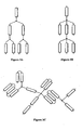
- the multimers are in the form of branched networks (see, e.g., FIG. 3 ).
- Such networks are conveniently obtained through the use of multifunction linking moieties that permit more than two helical units to be attached to a simple linking moiety.
- branched networks employ molecules having three, four or even more functional groups that are capable of covalently attaching to the N- and/or C-terminus of a peptide.
- Suitable linking moieties include, for example, residues of amino acids having side chains bearing hydroxyl, sulfanyl, amino, carboxyl, amide and/or ester functionalities, such as, for example, Ser (S), Thr (T), Cys (C), Tyr (Y), Asn (N), Gln (Q), Lys (K), Arg (R), Orn, Asp (D) and Glu (E); as well as the corresponding D-enantiomer of each of the foregoing; or residues of other organic molecules containing such functional groups.
- the helical segments attached to a single linking moiety need not be attached via like termini. Indeed, in some embodiments the helical segments are attached to a single linking moiety so as to be arranged in an antiparallel fashion, i.e. , some of the helices are attached via their N-termini, others via their C-termini.
- the helical segments can be attached directly to the linking moiety, or can be spaced from the linking moiety by way of one or more bifunctional linkers (LL), as previously described.
- LL bifunctional linkers
- a branched network can be described in terms of the number of "nodes” comprising the network, where each multifunctional linking moiety constitutes a node.
- helical segments i.e. , ApoA-I Mimics
- multifunctional linking moieties or nodes
- circles ⁇
- the number of nodes in the network will generally depend on the total desired number of helical segments, and will typically be from about 1 to 2. Of course, it will be appreciated that for a given number of desired helical segments, networks having higher order linking moieties will have fewer nodes. For example, referring to FIGS. 3A and 3B , a tertiary-order network (i.e. , a network having trifunctional linking moieties) of seven helical units has three nodes ( FIG. 3A ), whereas a quaternary order network ( i.e. , a network having tetrafunctional linking moieties) of seven helical units has only two nodes ( FIG. 3B ).
- the networks can be of uniform order, i.e. , networks in which all nodes are, for example, trifunctional or tetrafunctional linking moieties, or can be of mixed order, e.g. , networks in which the nodes are mixtures of, for example, trifunctional and tetrafunctional linking moieties.
- a tertiary order network can employ, for example, two, three, four or even more different trifunctional linking moieties.
- the helical segments comprising the branched network can be, but need not be, identical.
- FIG. 3C An example of such a mixed order branched network is illustrated in FIG. 3C .
- helical segments i.e., ApoA-I Mimics
- ⁇ multifunctional linking moieties
- Lines connecting helical segments represent bifunctional linkers LL, as previously described.
- Helical segments which comprise the branched networks can be tandem repeats of ApoA-I Mimics, as previously described.
- the branched networks are described by the formula: wherein:
- the branched network comprises a "Lys tree," i.e., a network wherein the multifunctional linking moiety is one or more Lys (K) residues (see, e.g., FIG. 3D ).
- Peptide 53 Peptide 54 Lys-Leu-Lys-Gln-Lys-Leu-Glu-Glu-Leu-Leu-Glu-Arg-Phe-Leu-Asp-Leu-Val-Inp (SEQ. ID. NO. 54) Peptide 55 Lys-Leu-Lys-Gln-Lys-Leu-Glu-Glu-Leu-Leu-Glu-Lys-Phe-Leu-Glu-Leu-Val-Inp (SEQ. ID. NO.
- Peptide 56 Lys-Leu-Lys-Gln-Lys-Leu-Leu-Glu-Leu-Leu-Glu-Arg-Phe-Leu-Asp-Leu-Val-Inp (SEQ. ID. NO. 56)
- Peptide 57 Lys-Leu-Lys-Gln-Trp-Leu-Glu-Asn-Leu-Leu-Glu-Arg-Phe-Leu-Asp-Leu-Val-Inp (SEQ. ID. NO.
- Peptide 58 Lys-Leu-Lys-Lys-Gln-Leu-Glu-Glu-Leu-Leu-Glu-Arg-Phe-Leu-Asp-Leu-Val-Inp (SEQ. ID. NO. 58)
- Peptide 59 Lys-Lys-Leu-Gln-Leu-Leu-Ala-Glu-Leu-Leu-Glu-Arg-Phe-Ala-Asp-Leu-Val-Inp (SEQ. ID. NO.
- Peptide 60 Lys-Lys-Leu-Gln-Ala-Leu-Ala-Glu-Leu-Leu-Glu-Arg-Phe-Ala-Asp-Leu-Val-Inp (SEQ. ID. NO. 60)
- Peptide 61 Lys-Leu-Lys-Gln-Lys-Leu-Glu-Glu-Trp-Gly-Glu-Arg-Phe-Leu-Asp-Leu-Val-Inp (SEQ. ID. NO.
- Peptide 62 Lys-Leu-Lys-Lys-Gln-Leu-Asp-Glu-Leu-Leu-Arg-Glu-Phe-Leu-Glu-Leu-Val-Inp (SEQ. ID. NO. 62)
- Peptide 63 Lys-Leu-Lys-Gln-Glu-Leu-Lys-Glu-Leu-Leu-Glu-Arg-Phe-Leu-Asp-Leu-Val-Inp (SEQ. ID. NO.
- Peptide 152 Lys-Lys-Leu-Gln-Ala-Leu-Ala-Glu-Leu-Leu-Glu-Arg-Phe-Ala-Asp-Leu-Val-Nip (SEQ. ID. NO. 152)
- Peptide 153 Lys-Leu-Lys-Gln-Lys-Leu-Glu-Glu-Trp-Gly-Glu-Arg-Phe-Leu-Asp-Leu-Val-Nip (SEQ. ID. NO.
- Peptide 361 Lys-Leu-Lys-Gln-Trp-Leu-Glu-Asn-Leu-Leu-Glu-Arg-Phe-Leu-Asp-Leu-Val-Pip (SEQ. ID. NO. 361)
- Peptide 363 Lys-Lys-Leu-Gln-Leu-Leu-Ala-Glu-Leu-Leu-Glu-Arg-Phe-Ala-Asp-Leu-Val-Pip (SEQ. ID. NO.
- Peptide 364 Lys-Lys-Leu-Gln-Ala-Leu-Ala-Glu-Leu-Leu-Glu-Arg-Phe-Ala-Asp-Leu-Val-Pip (SEQ. ID. NO. 364)
- Peptide 365 Lys-Leu-Lys-Gln-Lys-Leu-Glu-Glu-Trp-Gly-Glu-Arg-Phe-Leu-Asp-Leu-Val-Pip (SEQ. ID. NO.
- Peptide 366 Lys-Leu-Lys-Lys-Gln-Leu-Asp-Glu-Leu-Leu-Arg-Glu-Phe-Leu-Glu-Leu-V al-Pip (SEQ. ID. NO. 366)
- Peptide 367 Lys-Leu-Lys-Gln-Glu-Leu-Lys-Glu-Leu-Leu-Glu-Arg-Phe-Leu-Asp-Leu-Val-Pip (SEQ. ID. NO.
- the ApoA-I Mimics can be prepared using virtually any art-known technique for the preparation of peptides.
- the ApoA-I Mimics can be prepared using conventional step-wise solution or solid phase peptide syntheses, or recombinant DNA techniques.
- the ApoA-I Mimics can be prepared using conventional step-wise solution or solid phase synthesis (see, e.g., Chemical Approaches to the Synthesis of Peptides and Proteins, Williams et al., Eds., 1997, CRC Press, Boca Raton F la., and references cited therein; Solid Phase Peptide Synthesis: A Practical Approach, Atherton & Sheppard, Eds., 1989, IRL Press, Oxford, England, and references cited there in).
- the ApoA-I Mimics can be prepared by way of segment condensation, as described, for example, in Liu et al., 1996, Tetrahedron Lett. 37(7):933-936 ; Baca, et al., 1995, J. Am. Chem. Soc. 117:1881-1887 ; Tam et al., 1995, Int. J. Peptide Protein Res. 45:209-216 ; Schnolzer and Kent, 1992, Science 256:221-225 ; Liu and Tam, 1994, J. Am. Chem. Soc. 116(10):4149-4153 ; Liu and Tam, 1994, Proc. Natl. Acad. Sci.
- ApoA-I Mimics having N- and/or C-terminal capping groups can be prepared using standard techniques of organic chemistry. For example, methods for acylating the N-terminus of a peptide or amidating or esterifying the C-terminus of a peptide are well-known in the art. Modes of carrying other modifications at the N- and/or C-terminus will be apparent to those of skill in the art, as will modes of protecting any side-chain functionalities as can be necessary to attach terminal blocking groups.
- compositions can be conveniently prepared by ion-exchange chromatography or other methods as are well known in the art.
- ApoA-I Mimics that are in the form of tandem multimers can be conveniently synthesized by adding the linker(s) to the peptide chain at the appropriate step in the synthesis.
- the helical segments can be synthesized and each segment reacted with the linker.
- the actual method of synthesis will depend on the composition of the linker. Suitable protecting schemes and chemistries are well known, and will be apparent to those of skill in the art.
- ApoA-I Mimics that are in the form of branched networks can be conveniently synthesized using the trimeric and tetrameric resins and chemistries described in Tam, 1988, Proc. Natl. Acad. Sci. USA 85:5409-5413 and Demoor et al., 1996, Eur. J. Biochem. 239:74-84 . Modifying the synthetic resins and strategies to synthesize branched networks of higher or lower order, or which contain combinations of different ApoA-I Mimic helical segments, is well within the capabilities of those of skill in the art of peptide chemistry and/or organic chemistry.
- Formation of disulfide linkages can be conducted in the presence of mild oxidizing agents. Chemical oxidizing agents can be used, or the ApoA-I Mimics can simply be exposed to atmospheric oxygen to effect these linkages.
- oxidizing agents can be used, or the ApoA-I Mimics can simply be exposed to atmospheric oxygen to effect these linkages.
- Various methods are known in the art, including those described, for example, by Tam et al., 1979, Synthesis 955-957 ; Stewart et al., 1984, Solid Phase Peptide Synthesis, 2d Ed., Pierce Chemical Company Rockford, Ill .; Ahmed et al., 1975, J. Biol. Chem.
- ApoA-I Mimics having one or more internal glycine residues can be synthesized in relatively high yield by way of segment condensation, thereby providing advantages for large-scale production.
- Segment condensation i.e., the joining together of small constituent peptide chains to form a larger peptide chain, has been used to prepare many biologically active peptides, including 44-amino acid residue mimics of ApoA-I (see, e.g., Nakagawa et al., 1985, J. Am Chem. Soc. 107:7087-7083 ; Nokihara et al., 1989, Peptides 1988:166-168 ; Kneib-Cordonnier et al., 1990, Int. J. Pept. Protein Res. 35:527-538 ).
- Advantages of synthesis via segment condensation include the ability to condense pre-formed segments in the solution phase and the ease of purification of the final product.
- Drawbacks of the method include low coupling efficiency and yield at the condensation step and low solubility of certain peptide sequences.
- the coupling efficiency of the condensation step can be increased by increasing the coupling time. Typically, increasing the coupling time results in increased racemezation of the product ( Sieber et al., 1970, Hely. Chim. Acta 53:2135-2150 ). However, since glycine lacks a chiral center it does not undergo racemezation (proline residues, due to steric hindrance, also undergo little or no racemezation at long coupling times).
- embodiments containing internal glycine residues can be synthesized in bulk in high yield via segment condensation by synthesizing constituent segments which take advantage of the fact that glycine residues do not undergo racemezation.
- ApoA-I Mimics having one or more internal glycine residues provide synthetic advantages for large-scale bulk preparation.
- the ApoA-I Mimic is composed entirely of genetically-encoded amino acid residues, or a portion of it is so composed, the ApoA-I Mimic or the relevant portion can also be synthesized using conventional recombinant genetic engineering techniques.
- a polynucleotide sequence encoding the peptide is inserted into an appropriate expression vehicle, i.e., a vector which contains the necessary elements for the transcription and translation of the inserted coding sequence, or in the case of an RNA viral vector, the necessary elements for replication and translation.
- the expression vehicle is then transfected into a suitable target cell which will express the peptide.
- the expressed peptide is then isolated by procedures well-established in the art. Methods for recombinant protein and peptide production are well known in the art ( see, e.g.
- the polynucleotide can be designed to encode multiple units of the peptide separated by enzymatic cleavage sites--either homopolymers (repeating peptide units) or heteropolymers (different peptides strung together) can be engineered in this way.
- the resulting polypeptide can be cleaved ( e.g ., by treatment with the appropriate enzyme) in order to recover the peptide units. This can increase the yield of peptides driven by a single promoter.
- a polycistronic polynucleotide can be designed so that a single mRNA is transcribed which encodes multiple peptides (i.e ., homopolymers or heteropolymers) each coding region operatively linked to a cap-independent translation control sequence; e.g ., an internal ribosome entry site (IRES).
- a cap-independent translation control sequence e.g ., an internal ribosome entry site (IRES).
- IRES internal ribosome entry site
- the polycistronic construct directs the transcription of a single, large polycistronic mRNA which, in turn, directs the translation of multiple, individual peptides. This approach eliminates the production and enzymatic processing of polyproteins and can significantly increase yield of peptide driven by a single promoter.
- a variety of host-expression vector systems can be utilized to express the ApoA-I Mimics. These include, but are not limited to, microorganisms such as bacteria transformed with recombinant bacteriophage DNA or plasmid DNA expression vectors containing an appropriate coding sequence; yeast or filamentous fungi transformed with recombinant yeast or fungi expression vectors containing an appropriate coding sequence; insect cell systems infected with recombinant virus expression vectors (e.g., baculovirus) containing an appropriate coding sequence; plant cell systems infected with recombinant virus expression vectors (e.g ., cauliflower mosaic virus or tobacco mosaic virus) or transformed with recombinant plasmid expression vectors (e.g ., Ti plasmid) containing an appropriate coding sequence; or animal cell systems.
- microorganisms such as bacteria transformed with recombinant bacteriophage DNA or plasmid DNA expression vectors containing an appropriate coding sequence
- the expression elements of the expression systems can vary in their strength and specificities. Depending on the host/vector system utilized, any of a number of suitable transcription and translation elements, including constitutive and inducible promoters, can be used in the expression vector. For example, when cloning in bacterial systems, inducible promoters such as pL of bacteriophage ⁇ , plac, ptrp, ptac (ptrp-lac hybrid promoter) and the like can be used; when cloning in insect cell systems, promoters such as the baculovirus polyhedron promoter can be used; when cloning in plant cell systems, promoters derived from the genome of plant cells ( e.g., heat shock promoters; the promoter for the small subunit of RUBISCO; the promoter for the chlorophyll a/b binding protein) or from plant viruses ( e.g ., the 35S RNA promoter of CaMV; the coat protein promoter of TMV) can be used; when
- the expression of sequences encoding the ApoA-I Mimics can be driven by any of a number of promoters.
- viral promoters such as the 35S RNA and 19S RNA promoters of CaMV ( Brisson et al., 1984, Nature 310:511-514 ), or the coat protein promoter of TMV ( Takamatsu et al., 1987, EMBO J. 3:17 ) can be used; alternatively, plant promoters such as the small subunit of RUBISCO ( Coruzzi et al., 1984, EMBO J.
- Autographa californica, nuclear polyhidrosis virus (AcNPV) is used as a vector to express the foreign genes.
- the virus grows in Spodoptera frugiperda cells.
- a coding sequence can be cloned into non-essential regions (for example the polyhedron gene) of the virus and placed under control of an AcNPV promoter (for example, the polyhedron promoter).
- Successful insertion of a coding sequence will result in inactivation of the polyhedron gene and production of non-occluded recombinant virus (i.e ., virus lacking the proteinaceous coat coded for by the polyhedron gene).
- a number of viral-based expression systems can be utilized.
- a coding sequence can be ligated to an adenovirus transcription/translation control complex, e.g., the late promoter and tripartite leader sequence.
- This chimeric gene can then be inserted in the adenovirus genome by in vitro or in vivo recombination. Insertion in a non-essential region of the viral genome (e.g., region El or E3) will result in a recombinant virus that is viable and capable of expressing peptide in infected hosts.
- a non-essential region of the viral genome e.g., region El or E3
- the vaccinia 7.5 K promoter can be used, ( see, e.g., Mackett et al., 1982, Proc. Natl. Acad. Sci. (USA) 79:7415-7419 ; Mackett et al., 1984, J. Virol. 49:857-864 ; Panicali et al., 1982, Proc. Natl. Acad. Sci. 79:4927-4931 ).
- the ApoA-I Mimics can be purified by art-known techniques such as reverse phase chromatography, high performance liquid chromatography, ion exchange chromatography, gel electrophoresis, affinity chromatography and the like.
- the actual conditions used to purify a particular ApoA-I Mimic can depend, in part, on synthesis strategy and on factors such as net charge, hydrophobicity, hydrophilicity, etc., and will be apparent to those having skill in the art.
- Multimeric branched peptides can be purified, e.g. , by ion exchange or size exclusion chromatography.
- any antibody which specifically binds the ApoA-I Mimic can be used.
- various host animals including but not limited to rabbits, mice, rats, etc., can be immunized by injection with a peptide.
- the peptide can be attached to a suitable carrier, such as BSA, by means of a side chain functional group or linkers attached to a side chain functional group.
- adjuvants can be used to increase the immunological response, depending on the host species, including but not limited to Freund's (complete and incomplete), mineral gels such as aluminum hydroxide, surface active substances such as lysolecithin, pluronic polyols, polyanions, peptides, oil emulsions, keyhole limpet hemocyanin, dinitrophenol, and potentially useful human adjuvants such as BCG (bacilli Calmette-Guerin) and Corynebacterium parvum.
- BCG Bacilli Calmette-Guerin
- Corynebacterium parvum bacilli Calmette-Guerin
- Monoclonal antibodies to an ApoA-I Mimic can be prepared using any technique which provides for the production of antibody molecules by continuous cell lines in culture. These include but are not limited to the hybridoma technique described by Kohler and Milstein, 1975, Nature 256:495-497 , or Kaprowski, U.S. Pat. No. 4,376,110 which is incorporated by reference herein; the human B-cell hybridoma technique) Kosbor et al., 1983, Immunology Today 4:72 ; Cote et al., 1983, Proc. Natl. Acad. Sci. U.S.A.
- Antibody fragments which contain deletions of specific binding sites can be generated by known techniques.
- fragments include but are not limited to F(ab') 2 fragments, which can be produced by pepsin digestion of the antibody molecule and Fab fragments, which can be generated by reducing the disulfide bridges of the F(ab') 2 fragments.
- Fab expression libraries can be constructed ( Huse et al., 1989, Science 246:1275-1281 ) to allow rapid and easy identification of monoclonal Fab fragments with the desired specificity for the peptide of interest.
- the antibody or antibody fragment specific for the desired ApoA-I Mimic can be attached, for example, to agarose, and the antibody-agarose complex is used in immunochromatography to purify peptides described herein. See, Scopes, 1984, Protein Purification: Principles and Practice, Springer-Verlag New York, Inc., N.Y ., Livingstone, 1974, Methods In Enzymology: Immunoaffinity Chromatography of Proteins 34:723-731 .
- compositions comprising an effective amount of an ApoA-I Mimic and a pharmaceutically acceptable carrier or vehicle.
- compositions can be formulated for administration to a mammal by injection.
- injectable preparations include sterile suspensions, solutions or emulsions of the active ingredient in aqueous or oily vehicles.
- the compositions can also comprise formulating agents, such as suspending, stabilizing and/or dispersing agent.
- the formulations for injection can be presented in unit dosage form, e.g., in ampules or in multidose containers, and can contain added preservatives.
- the injectable formulation can be provided in powder form for reconstitution with a suitable vehicle, including but not limited to sterile pyrogen free water, buffer, dextrose solution, etc., before use.
- a suitable vehicle including but not limited to sterile pyrogen free water, buffer, dextrose solution, etc., before use.
- an ApoA-I Mimic can be lyophilized, or a co-lyophilized peptide-lipid complex can be prepared.
- the stored preparations can be supplied in unit dosage forms and reconstituted
- the composition can be formulated as a depot preparation, for administration by implantation; e.g., subcutaneous, intradermal, or intramuscular injection.
- the ApoA-I Mimic can be formulated with suitable polymeric or hydrophobic materials (e.g., as an emulsion in an acceptable oil) or ion exchange resins, or as sparingly soluble derivatives; e.g., as a sparingly soluble salt form of the ApoA-I Mimic.
- compositions are administered intravenously.
- transdermal delivery systems manufactured as an adhesive disc or patch which slowly releases the active ingredient for percutaneous absorption can be used.
- permeation enhancers can be used to facilitate transdermal penetration of the ApoA-I Mimic.
- a particular benefit can be achieved by incorporating the ApoA-I Mimic into a nitroglycerin patch for use in a mammal having a Condition such as ischemic heart disease or hypercholesterolemia.
- compositions can take the form of, for example, tablets or capsules prepared by conventional means with pharmaceutically acceptable excipients such as binding agents (e.g. , pregelatinised maize starch, polyvinylpyrrolidone or hydroxypropyl methylcellulose); fillers ( e.g ., lactose, microcrystalline cellulose or calcium hydrogen phosphate); lubricants (e.g., magnesium stearate, talc or silica); disintegrants (e.g., potato starch or sodium starch glycolate); or wetting agents (e.g ., sodium lauryl sulfate).
- binding agents e.g. , pregelatinised maize starch, polyvinylpyrrolidone or hydroxypropyl methylcellulose
- fillers e.g ., lactose, microcrystalline cellulose or calcium hydrogen phosphate
- lubricants e.g., magnesium stearate, talc or silica
- disintegrants e.g
- Liquid preparations for oral administration can take the form of, for example, solutions, syrups or suspensions, or they can be presented as a dry product for constitution with water or other suitable vehicle before use.
- Such liquid preparations can be prepared by conventional means with pharmaceutically acceptable additives such as suspending agents (e.g ., sorbitol syrup, cellulose derivatives or hydrogenated edible fats); emulsifying agents (e.g., lecithin or acacia); non-aqueous vehicles (e.g., almond oil, oily esters, ethyl alcohol or fractionated vegetable oils); and preservatives (e.g ., methyl or propyl-p-hydroxybenzoates or sorbic acid).
- the preparations can also contain buffer salts, flavoring, coloring and sweetening agents as appropriate.
- Preparations for oral administration can be suitably formulated to give controlled release of the ApoA-I Mimic.
- compositions can take the form of tablets or lozenges formulated in conventional manner.
- active ingredient can be formulated as solutions (for retention enemas) suppositories or ointments.
- the ApoA-I Mimic can be conveniently delivered in the form of an aerosol spray presentation from pressurized packs or a nebulizer, with the use of a suitable propellant, e.g ., dichlorodifluoromethane, trichlorofluoromethane, dichlorotetrafluoroethane, carbon dioxide or other suitable gas.
- a suitable propellant e.g ., dichlorodifluoromethane, trichlorofluoromethane, dichlorotetrafluoroethane, carbon dioxide or other suitable gas.
- the dosage unit can be determined by providing a valve to deliver a metered amount.
- Capsules and cartridges of e.g. gelatin for use in an inhaler or insufflator can be formulated containing a powder mix of the ApoA-I Mimic and a suitable powder base such as lactose or starch.
- compositions can, if desired, be presented in a pack or dispenser device which can contain one or more unit dosage forms containing the ApoA-I Mimic.
- the pack can for example comprise metal or plastic foil, such as a blister pack.
- the pack or dispenser device can be accompanied by instructions for administration.
- ApoA-I Mimic/lipid complexes, compositions thereof, and methods for their administration are described.
- the complexes can have several advantages since they can have an increased half-life in the circulation, particularly when the complex has a similar size and density to HDL, and especially the pre- ⁇ -1 or pre- ⁇ -2 HDL populations.
- the complexes can be conveniently be prepared using any of a number of methods described below. Stable preparations having a relatively long shelf life can be made by lyophilization, --the co-lyophilization procedure described below being one embodiment.
- the lyophilized complexes can be used to prepare bulk for pharmaceutical reformulation, or to prepare individual aliquots or dosage units which can be reconstituted by rehydration with sterile water or an appropriate buffered solution prior to administration to a subject.
- the ApoA-I Mimic can be cosonicated (using a bath or probe sonicator) with appropriate lipids to form complexes.
- the ApoA-I Mimic can be combined with preformed lipid vesicles resulting in the spontaneous formation of peptide-lipid complexes.
- the complexes can be formed by a detergent dialysis method; e.g., a mixture of the ApoA-I Mimic, lipid and detergent is dialyzed to remove the detergent and reconstitute or form complexes (see, e.g., Jonas et al., 1986, Methods in Enzymol. 128:553-582 ).
- a detergent dialysis method e.g., a mixture of the ApoA-I Mimic, lipid and detergent is dialyzed to remove the detergent and reconstitute or form complexes (see, e.g., Jonas et al., 1986, Methods in Enzymol. 128:553-582 ).
- the complexes can be prepared by the methods disclosed in U.S. Patent No. 6,004,925 ("'925 patent"), the entire disclosure of which is herein incorporated by reference.
- the ApoA-I Mimic and lipid are combined in a solvent system which co-solubilizes each ingredient and which can be completely removed by lyophilization.
- solvent pairs are selected to ensure co-solubility of both the ApoA-I Mimic and the lipid.
- the ApoA-I Mimic of the complex can be dissolved in an aqueous or organic solvent or mixture of solvents (solvent 1).
- the lipid, such as a phospholipid, component is dissolved in an aqueous or organic solvent or mixture of solvents (solvent 2) which is miscible with solvent 1, and the two solutions are mixed.
- solvent 2 a solvent or mixture of solvents
- the ApoA-I Mimic and lipid can be incorporated into a co-solvent system; i.e ., a mixture of the miscible solvents.
- the ApoA-I Mimic and lipid can be suspended in a solvent or mixture of solvents.
- the mixture of solvents is a mixture of organic solvent and water. Examples of organic solvents include, but are not limited to, acetic acid, xylene, cyclohexane, and methanol.
- solvent mixtures include, but are not limited to, acetic acid and xylene, acetic acid and cyclohexane, and methanol and xylene.
- a suitable proportion of ApoA-I Mimic to lipids can be first determined empirically so that the resultant complexes possess the appropriate physical and chemical properties; i.e ., usually (but not necessarily) similar in size to HDL.
- the resultant mixture is frozen and lyophilized to dryness. Sometimes an additional solvent is added to the mixture to facilitate lyophilization. This lyophilized product may be stored for long periods and will typically remain stable.
- the complexes can be prepared by co-lyophilization of the ApoA-I Mimic with peptide in solutions or suspensions.
- the homogeneous solution of peptide and phospholipids of choice in an organic solvent or organic solvent mixture can be lyophilized, and peptide/phospholipid complexes can be formed spontaneously by hydration of the lyophilized powder with an aqueous buffer.
- the lyophilized product may be reconstituted in order to obtain a solution or suspension of the complex.
- the lyophilized powder is rehydrated with an aqueous solution to a suitable volume (often 5-20 mg peptide/mL which is convenient for intravenous injection).
- the lyophilized powder is rehydrated with phosphate buffered saline, saline bicarbonate, or a physiological saline solution.
- the pH of the mixture can be adjusted to 7.5-8.5.
- the mixture can be agitated or vortexed to facilitate rehydration, and in most cases, the reconstitution step can be conducted at a temperature equal to or greater than the phase transition temperature of the lipid component of the complexes. Within minutes, a clear preparation of reconstituted lipid-protein complexes results.
- An aliquot of the resultant reconstituted preparation can be characterized to confirm that the complexes in the preparation have the desired size distribution; e.g ., the size distribution of HDL. Characterization of the reconstituted preparation can be performed using any method known in the art, including, but not limited to, size exclusion filtration chromatography, gel filtration chromatography, column filtration chromatography, gel permeation chromatography, and native page electrophoresis. In one embodiment, the reconstituted preparation is characterized by gel filtration chromatography. The size of the resultant complexes may be determinative of their efficacy. In the examples described below, a Pharmacia Superose 6 FPLC gel filtration chromatography system is used.
- the buffer that is used contains 150 mM NaCl in 50 mM phosphate buffer, pH about 7.0 to about 9, in one embodiment 7.5-8.5, in another embodiment 7.4.
- a typical sample volume is 20 to 200 microliters of complexes containing 5 mg peptide/mL.
- the column flow rate is 0.5 mL/min.
- a series of proteins of known molecular weight and Stokes's diameter as well as human HDL are used as standards to calibrate the column. The proteins and lipoprotein complexes are monitored by absorbance or scattering of light of wavelength 254 or 280 nm.
- the reconstituted preparation can also be characterized to determine the concentration, final pH and osmolality of resulting solution, as well as the concentration and integrities of peptide and individual lipids.
- ApoA-I Mimic and lipid concentration of the complexes can be measured by any method known in the art, including, but not limited to, protein and phospholipid assays, and chromatographic methods such as high performance liquid chromatography ("HPLC"), gel filtration chromatography, gas chromatography ("GC”).
- HPLC high performance liquid chromatography
- GC gas chromatography
- the chromatographs can be coupled with various detectors including, but not limited to, mass spectrometers, UV or diode-array, fluorescent, and elastic light scattering detectors.
- the integrity of the ApoA-I Mimic and lipid in the complexes can be determined by the chromatographic techniques described above, as well as by amino acid analysis, thin layer chromatography, and standard assays to determine lipid oxidation for lipids.
- the lipid of the ApoA-I Mimic/lipid complex can be one or more of a variety of lipids, including, but not limited to, saturated, unsaturated, natural and synthetic lipids and phospholipids, and pharmaceutically acceptable salts thereof.
- Typical salts include, but are not limited to, sodium, calcium, magnesium, and potassium salts.
- Suitable lipids of the ApoA-I Mimic/lipid complexes include, but are not limited to, (C 1 -C 6 ) alkyl chain phospholipids, phosphatidylcholine (PC), egg phosphatidylcholine, soybean phosphatidylcholine, dipalmitoylphosphatidylcholine, dimyristoylphosphatidylcholine, distearoylphosphatidylcholine 1-myristoyl-2- palmitoylphosphatidylcholine, 1-palmitoyl-2-myristoylphosphatidylcholine, 1- palmitoyl-2-stearoylphosphatidylcholine, 1-stearoyl-2-palmitoylphosphatidylcholine, 1- palmitoyl-2-oleoylphosphatidylcholine, 1-oleoyl-2-palmitylphosphatidylcholine, dioleoylphosphatidylcholine, dioleo
- the lipid of the ApoA-I Mimic/lipid complex is a neutral phospholipid.
- the neutral phospholipid can be any phospholipid that has a net charge of about zero at physiological pH. In some embodiments, the neutral phospholipid is a zwitterion that has a net charge of about zero at physiological pH.
- the neutral phospholipid is a lecithin (also known as phosphatidylcholine).
- the neutral phospholipid is a mixture of neutral phospholipids that comprises about 5 to about 100 wt % lecithin. In other embodiments, the mixture of neutral phospholipids comprises about 100 wt % lecithin. In some embodiments, the neutral phospholipid is a mixture of neutral phospholipids that comprises about 5 to about 100 mole % lecithin. In other embodiments, the mixture of neutral phospholipids comprises about 100 mole % lecithin.
- the neutral phospholipid is a sphingomyelin. In some embodiments, the neutral phospholipid is a mixture of neutral phospholipids that comprises about 5 to about 100 wt % sphingomyelin. In other embodiments, the neutral phospholipid is a mixture of neutral phospholipids that comprises about 100 wt % sphingomyelin. In some embodiments, the neutral phospholipid is a mixture of neutral phospholipids that comprises about 5 to about 100 mole % sphingomyelin. In other embodiments, the neutral phospholipid is a mixture of neutral phospholipids that comprises about 100 mole % sphingomyelin.
- the neutral phospholipid of the ApoA-I Mimic/lipid complex is a mixture of neutral phospholipids that comprises a lecithin and a sphingomyelin.
- the molar ratio of lecithin to sphingomyelin can vary, but typically ranges from about 20: about 1 to about 1: about 20. In some embodiments, the lecithin:sphingomyelin molar ratio ranges from about 10: about 3 to about 10: about 6. In other embodiments, the lecithin:sphingomyelin molar ratio ranges from about 1: about 20 to about 3: about 10.
- the neutral phospholipid of the ApoA-I Mimic/lipid complex is a mixture of neutral phospholipids that comprises lechitin, sphingomyelin and one or more additional neutral phospholipids.
- the additional neutral phospholipid comprises from about 5 to about 100 wt % of the mixture.
- the lipid of the ApoA-I Mimic/lipid complex is a charged phospholipid.
- Suitable charged phospholipids include, but are not limited to, phosphatidylinositol, phosphatidylserine, phosphatidylglycerol and phosphatidic acid.
- the lipid of the ApoA-I Mimic/lipid complex is a mixture of at least one neutral phospholipid and at least one charged phospholipid.
- the total amount of charged phospholipids(s) in the lipid mixture can vary, but typically ranges from about 0.2 to about 10 wt % of the lipid mixture.
- the total amount of charged phospholipids(s) in the lipid mixture is about 0.2 to about 2 wt %, about 0.2 to about 3 wt %, about 0.2 to about 4 wt %, about 0.2 to about 5 wt %, about 0.2 to about 6 wt %, about 0.2 to about 7 wt %, about 0.2 to about 8 wt % or about 0.2 to about 9 wt % of the lipid mixture.
- the total amount of charged phospholipids(s) in the lipid mixture is about 0.2, about 0.3, about 0.4, about 0.5, about 0.6, about 0.7, about 0.8, about 0.9, about 1.0, about 1.1, about 1.2, about 1.3, about 1.4, about 1.5, about 1.6, about 1.7, about 1.8, about 1.9, about 2.0, about 2.1, about 2.2, about 2.3, about 2.4, about 2.5, about 2.6, about 2.7, about 2.8, about 2.9 or about 3.0, about 4, about 5, about 6, about 7, about 8, about 9, or about 10 wt % of the lipid mixture.
- the total amount of neutral phospholipid(s) in the lipid mixture can also vary, and can depend upon the amount of charged phospholipid(s) and any other lipids included. In one embodiment, the total amount of neutral phospholipids(s) in the lipid mixture is about about 90 to 99.8 wt % of the lipid mixure. In one embodiment, the lipid of the ApoA-I Mimic/lipid complex is a mixture of sphingomyelin and a charged phospholipid. In another embodiment, the lipid of the ApoA-I Mimic/lipid complex is a mixture of sphingomyelin, dipalmitoylphosphatidylcholine ("DPPC"), and a charged phospholipid.
- DPPC dipalmitoylphosphatidylcholine
- the lipid of the ApoA-I Mimic/lipid complex is sphingomyelin.
- the spingomyelin is obtained from milk, egg or brain or made synthetically.
- the lipid of the ApoA-I Mimic/lipid complex is a sphingomyelin analog or derivative. Suitable sphingomyelin analogs or derivatives include, but are not limited to, palmitoylsphingomyelin, stearoylsphingomyelin, D-erythrose-sphingomyelin, and D-erythrose-dihydrosphingomyelin.
- the sphingomyelin is artificially enriched in one particular saturated or unsaturated acyl chain.
- milk sphingomyelin (Avanti Phospholipid, Alabaster, Ala.) has long saturated acyl chains.
- Milk sphingomyelin comprises about 20% of C16:0 (16 carbon, saturated) acyl chain compared with egg sphingomyelin, which comprises 80% of C16:0.
- milk sphingomyelin can be enriched in one particular acyl chain to obtain a composition having an acyl chain concentration comparable with, e.g., egg sphingomyelin.
- Acyl chains that may be utilized include, but are not limited to saturated acyl chains (such as dipalmitoyl, distearoyl, diarachidonyl, and dibenzoyl acyl chains), unsaturated chains (such as diolcoyl chains), mixed chains of saturated and unsaturated acyl chains (such as palmitoyl or oleoyl chains), saturated and/or unsaturated chains of mixed lengths, and ether analogs of saturated and unsaturated acyl chains.
- saturated acyl chains such as dipalmitoyl, distearoyl, diarachidonyl, and dibenzoyl acyl chains
- unsaturated chains such as diolcoyl chains
- mixed chains of saturated and unsaturated acyl chains such as palmitoyl or oleoyl chains
- saturated and/or unsaturated chains of mixed lengths such as palmitoyl or oleoyl chains
- the sphingomyelin may be semi-synthetic such that it has a particular acyl chain.
- milk sphingomyelin can first be purified from milk, then one particular acyl chain, e.g ., the C16:0 acyl chain, can be cleaved and replaced by another acyl chain (such as palmitic acid or oleic acid).
- Sphingomyelin can also be entirely synthesized, by e.g ., large-scale synthesis. See, e.g ., Dong et al, U.S. Pat. No. 5,220,043 ; Weis, 1999, Chem. Phys. Lipids 102(1-2):3-12 .
- a predefined saturation level and fatty acid composition is selected for the synthetic sphingomyelin.
- the lipid of the ApoA-I Mimc/lipid complex is a mixture of sphingomyelin and another lipid.
- the sphingomyelin typically comprises from about 25 to about 75 wt % of the mixture.
- the lipid of the ApoA-I Mimc/lipid complex is a mixture of sphingomyelin and DPPC. In another embodiment, the lipid of the ApoA-I Mimic/lipid complex is a mixture of sphingomyelin, DPPC, and dipalmitoylphosphatidylglycerol ("DPPG"). In one embodiment, DPPG is present at about 0 to about 10% mole or weight % of the mixture. In another embodiment, DPPG is present at about 2 to about 4 mole or weight % of the mixture. In another embodiment, sphingomyelin and DPPG are present in the mixture in a weight or molar ratio of about 1: about 1.
- the sphingomyelin, DPPC, and DPPG are present in a weight or molar ratio of about 1: about 1: about 0.06, respectively. In another embodiment, the sphingomyelin, DPPC, and DPPG are present in a molar ratio of 1.04: 1: 0.061, respectively. In another embodiment, the sphingomyelin, DPPC, and DPPG are present in a weight ratio of 1: 1: 0.062, respectively. In another embodiment, the mixture is about 48.5 mole or weight % sphingomyelin, about 48.5 mole or weight % DPPC, and about 3 mole or weight % DPPG.
- the ApoA-I Mimic/lipid complex comprises one or more additional peptides.
- the additional peptide is ApoA-I.
- the weight ratio of total peptide to lipid in each ApoA-I Mimic/lipid complex is about 1: about 0.5 to about 1: about 5. In another embodiment, the weight ratio of total peptide to lipid in each ApoA-I Mimic/lipid complex is about 1: about 1 to about 1: about 5. In another embodiment, the weight ratio of total peptide to lipid in each ApoA-I Mimic/lipid complex is about 1: about 2 to about 1: about 5. In another embodiment, the weight ratio of total peptide to lipid in each ApoA-I Mimic/lipid complex is about 1: about 2.5. In another embodiment, the weight ratio of total peptide to lipid in each ApoA-I Mimic/lipid complex is about 1: about 3 to 1: about 5.
- the molar ratio of total peptide to lipid in each ApoA-I Mimic/lipid complex is about 1: about 2.5 to about 1: about 20. In another embodiment, the molar ratio of total peptide to lipid in each ApoA-I mimic/lipid complex is about 1: about 9.2.
- the lipid of the ApoA-I Mimic/lipid complex is a mixture of sphingomyelin, DPPC, and DPPG
- the peptide: sphingomyelin: DPPC: DPPG weight ratio is typically about 1: about 1: about 1: about 0.08, respectively.
- the peptide: sphingomyelin: DPPC: DPPG weight ratio is 1: 1.2125: 1.2125: 0.075, respectively.
- the peptide: sphingomyelin: DPPC: DPPG molar ratio is typically about 1: about 4: about 4: about 0.03, respectively.
- the peptide: sphingomyelin: DPPC: DPPG molar ratio is 1: 4.55: 4.36: 0.27, respectively.
- the ApoA-I Mimic/lipid complex comprises about 40 to about 85 wt % lipid and about 15 to about 60 wt % peptide.
- each ApoA-I Mimic/lipid complex is about 2 to about 12 nm in diameter.
- the helix formed by the ApoA-I Mimics closely mimics the structural and functional properties of the amphipathic helical regions of native ApoA-I that are important for effecting lipid-binding, cholesterol efflux, and/or LCAT activation, thereby resulting in peptides that exhibit high ApoA-I-like activity.
- the ApoA-I Mimics function by forming amphipathic helices (in the presence of lipids), binding lipids, forming pre- ⁇ -like or HDL-like complexes, activating LCAT, increasing serum HDL concentration and promoting cholesterol efflux.
- the ApoA-I Mimics activate LCAT. In another embodiment, the ApoA-I Mimics do not activate LCAT. In another embodiment, the ApoA-I Mimics activate LCAT, but only to a degree that does not result in an acceleration of cholesterol esterification. In another embodiment, the ApoA-I Mimics activate LCAT, and thereby accelerate cholesterol esterification, but wherein the acceleration of cholesterol esterification due to LCAT activation, without more, is insufficient to treat or prevent a Condition.
- are disclosed methods for treating or preventing a Condition comprising administering an effective amount of an ApoA-I Mimic to a mammal in need thereof.
- dyslipidemia examples include any disorder for which increasing serum HDL concentration, activating LCAT, and promoting cholesterol efflux and RCT is beneficial.
- disorders include, but are not limited to, hyperproteinemia (such as hyperchlyomicronemia), high low-density lipoprotein serum concentration, high very low-density lipoprotein serum concentration, hyperlipidemia (such as hypercholesterolemia or hyperglyceridemia (such as hypertriglyceridemia)), low high density lipoprotein serum concentration, hypocholesterolemia, Abetalipoproteinemia, ApoA-I deficiency and Tangier disease.
- hyperproteinemia such as hyperchlyomicronemia
- hyperlipidemia such as hypercholesterolemia or hyperglyceridemia (such as hypertriglyceridemia)
- low high density lipoprotein serum concentration such as hypocholesterolemia, Abetalipoproteinemia, ApoA-I deficiency and Tangier disease.
- cardiovascular disease examples include, but are not limited to, metabolic syndrome, ischemic heart disease, atherosclerosis, restenosis (e.g., preventing or treating atherosclerotic plaques which develop as a consequence of medical procedures such as balloon angioplasty), endotoxemia (which often results in septic shock), congestive heart failure (such as chronic or acute heart failure), circulatory shock, cardiomyopathy, cardiac transplant, myocardial infarction, a cardiac arrhythmia (such as atrial fibrillation), supraventricular tachycardia, atrial flutter, paroxysmal atrial tachycardia, aneurysm, angina, cerebrovascular accident (stroke), peripheral vascular disease, cerebrovascular disease, kidney disease, atherogenesis, therosclerosis, acute pancreatitis, and coronary artery disease.
- metabolic syndrome e.g., preventing or treating atherosclerotic plaques which develop as a consequence of medical procedures such as balloon angioplasty), endotoxemia (which often
- Endothelial dysfunction is any imbalance between the vasodilating and vasoconstricting factors and growth-inhibiting and growth-promoting factors produced by the endothelium. Endothelial dysfunction typically impairs the blood vessels' ability to dilate.
- macrovascular disorders include any disorder of a large blood vessel. Such disorders include, but are not limited to, transient ischaemic attack, stroke, angina, myocardial infarction, cardiac failure, and peripheral vascular disease.
- microvascular disorders include any disorder of a small blood vessel.
- Such disorders include, but are not limited to, diabetic retinopathy (non-proliferative, proliferative, macular oedema), microalbuminuria, macroalbuminuria, end stage renal disease, erectile dysfunction, autonomic neuropathy, peripheral neuropathy, osteomyelitis and lower limb ischaemia.
- the ApoA-I Mimics can be administered alone or in combination with one or more other drugs that are useful for treating a Condition.
- Such therapies include, but are not limited to, simultaneous or sequential administration of the drugs involved.
- methods for treating or preventing a Condition can further comprise administering one or more drugs from one or more of the following classes: ACE (angiotensin converting enzyme) inhibitors, beta blockers, nitrates, calcium channel blockers, diuretics, thrombolytic agents, and blood cholesterol-lowering agents.
- ACE angiotensin converting enzyme
- the methods of treating or preventing a Condition further comprise administering one or more of: cholestyramine, colestipol, colesevelam, gemfibrozil, ciprofibrate, clofibrate, fenofibrate, bezafibrate, ezetimibe, ramipril, verapamil , nicardipine, diltiazem, carvedilol, nadolol, isosorbide mononitrate, propranolol, isosorbide dinitrate, digoxin, furosemide, metoprolol tartrate, trandolapril, nitroglycerin, amlodipine besylate, oxycodone, clopidogrel, nifedipine, atenolol, lisinopril, aspirin, and lanoxin.
- methods for treating or preventing a Condition can further comprise administering one or more of the cholesterol lowering drugs known to one of skill in the art; e.g., bile-acid resins, niacin, and/or statins, such as atorvastatin, simvastatin, pravastatin, fluvastatin and pitavastatin.
- statins such as atorvastatin, simvastatin, pravastatin, fluvastatin and pitavastatin.
- Such a regimen may produce particularly beneficial therapeutic effects since each drug acts on a different target in cholesterol synthesis and transport; i.e., bile-acid resins affect cholesterol recycling, the chylomicron and LDL population; niacin primarily affects the VLDL and LDL population; the statins inhibit cholesterol synthesis, decreasing the LDL population (and perhaps increasing LDL receptor expression); whereas the ApoA-I Mimics affect RCT, increase HDL, increase LCAT activity and promote cholesterol efflux.
- bile-acid resins affect cholesterol recycling, the chylomicron and LDL population
- niacin primarily affects the VLDL and LDL population
- the statins inhibit cholesterol synthesis, decreasing the LDL population (and perhaps increasing LDL receptor expression)
- the ApoA-I Mimics affect RCT, increase HDL, increase LCAT activity and promote cholesterol efflux.
- methods for treating or preventing a Condition can further comprise administering a fibrate, such as clinofibrate, clofibrate, simfibrate, fenofibrate, and benzafibrate.
- a fibrate such as clinofibrate, clofibrate, simfibrate, fenofibrate, and benzafibrate.
- methods for treating or preventing a Condition can further comprise administering an anti-microbial agent and/or an anti-inflammatory agent, for example, that is useful for treating septic shock induced by endotoxin.
- the ApoA-I Mimics can be administered by any suitable route that ensures bioavailability in the circulation. This may be achieved by parenteral routes of administration, including intravenous (IV), intramuscular (IM), intradermal, subcutaneous (SC) and intraperitoneal (IP) injections. However, other routes of administration can be used. For example, absorption through the gastrointestinal tract may be accomplished by oral routes of administration (including but not limited to ingestion, buccal and sublingual routes) provided appropriate formulations (e.g ., enteric coatings) are used to avoid or minimize degradation of the peptides, e.g ., in the harsh environments of the oral mucosa, stomach and/or small intestine.
- parenteral routes of administration including intravenous (IV), intramuscular (IM), intradermal, subcutaneous (SC) and intraperitoneal (IP) injections.
- IV intravenous
- IM intramuscular
- SC subcutaneous
- IP intraperitoneal
- other routes of administration can be used.
- administration via mucosal tissue such as vaginal and rectal modes of administration may be utilized to avoid or minimize degradation in the gastrointestinal tract.
- the formulations may be administered transcutaneously ( e.g ., transdermally), ocularly, or by inhalation. It will be appreciated that the route of administration chosen may vary with the condition, age and compliance of the recipient.
- the actual dose of the ApoA-I Mimic used can vary with the route of administration, and can be adjusted to achieve circulating plasma concentrations of ApoA-I Mimic of 100 mg/L to 2 g/L.
- the dose of ApoA-I Mimic is adjusted to achieve a serum level of free or complexed ApoA-I Mimic for at least 24 hours following administration that is in the range of about 10 mg/dL to 300 mg/dL higher than a baseline (initial) level prior to administration.
- the ApoA-I Mimics may be administered in a variety of different treatment regimens.
- the ApoA-I Mimic is administered by injection at a dose between 0.5 mg/kg to 100 mg/kg once a week.
- desirable serum levels may be maintained by continuous infusion or by intermittent infusion providing about 0.5 mg/kg/hr to 100 mg/kg/hr of the ApoA-I Mimic.
- the ApoA-I Mimic is administered at a dose of about 20 mg/kg.
- the ApoA-I Mime is administered by intravenous injection once or more per day. In another embodiment, the ApoA-I Mimic is administered by injection once every 3 to 15 days, once every 5 to 10 days, or once every 10 days. In another embodiment, the ApoA-I Mimic is administered in a series of maintenance injections, where the series of maintenance injections is administered once every 6 months to one year. The series of maintenance injections can be administered, for example, over one day (perfusion to maintain a specified plasma level of complexes), several days (e.g ., four injections over a period of eight days) or several weeks (e.g., four injections over a period of four weeks).
- an escalating dose of ApoA-I Mimic can be administered, starting with about 1 to 5 doses at amount of about 50 mg to about 200 mg per administration, then followed by repeated doses of about 200 mg to about 1 g per administration.
- administration can be by slow infusion with a duration of more than one hour, by rapid infusion of one hour or less, or by a single bolus injection.
- Toxicity and therapeutic efficacy of the ApoA-I Mimics may be determined using standard pharmaceutical procedures in cell culture or experimental animals for determining the LD 50 (the dose lethal to 50% of the population) and the ED 50 (the dose therapeutically effective in 50% of the population).
- the dose ratio between toxic and therapeutic effects is the therapeutic index and it can be expressed as the ratio LD 50 /ED 50 .
- the ApoA-I Mimics exhibit large therapeutic indices.
- the ApoA-I Mimics can be assayed for their ability to form cc-helices in the presence of lipids, to bind lipids, to form complexes with lipids, to activate LCAT, to promote cholesterol efflux, etc.
- the ability of the peptides and/or peptide analogues to activate LCAT can be readily determined using the LCAT activation described in the examples below
- the in vitro and in vivo assays described in the examples below can be used to evaluate the half-life, distribution, cholesterol efflux and effects on RCT.
- SUVs small unilamellar vesicles
- an ApoA-I Mimic to form an ⁇ -helix in the presence of lipids can be demonstrated using the CD assay described below.
- W fluorescent Trp
- Nal residue a fluorescent Trp
- the ApoA-I Mimics are screened for their ability to activate LCAT, as peptides which activate LCAT are particularly useful in the methods described herein.
- ApoA-I Mimics exhibit at least about 38% LCAT activation as compared with native human ApoA-I (as determined using the LCAT activation assay described herein).
- the ApoA-I Mimics exhibit 50%, 60%, 70%, 80% or even 90% LCAT activation as compared with native human ApoA-I.
- the ApoA-I Mimics are useful in assays in vitro to measure serum HDL, e.g., for diagnostic purposes. Because the ApoA-I Mimics typically associate with the HDL component of serum, the mimics may be used as "markers" for the HDL population. Accordingly, also disclosed are methods for measuring serum HDL concentration, comprising contacting serum HDL with an amount of ApoA-I Mimic that associates with the serum HDL and quantifying the amount of ApoA-I-associated HDL. Moreover, the ApoA-I Mimics may be used as markers for the subpopulation of HDL that is effective in reverse cholesterol transport ("RCT").
- RCT reverse cholesterol transport
- the ApoA-I Mimic may be added to or mixed with a mammalian serum sample comprising HDL; after an appropriate incubation time, the HDL component may be assayed by detecting the incorporated ApoA-I Mimic.
- This may be accomplished using labeled ApoA-I Mimic (e.g., radiolabels, fluorescent labels, enzyme labels, dyes, etc.), or by immunoassays using antibodies (or antibody fragments) specific for the ApoA-I Mimic.
- labeled ApoA-I Mimics are useful in imaging procedures (e.g., CAT scans, MRI scans) to visualize the circulatory system, or to monitor RCT, or to visualize accumulation of HDL at fatty streaks, atherosclerotic lesions, etc. (where the HDL should be active in cholesterol efflux).
- imaging procedures e.g., CAT scans, MRI scans
- HDL should be active in cholesterol efflux
- the ApoA-I Mimics are prepared by solid phase peptide synthesis (SPPS) using Fmoc (9-fluorenylmethyloxycarbonyl) chemistry.
- SPPS solid phase peptide synthesis
- the C-terminal residue is covalently bound to a 4-methylbenzhydrylamine (MBHA) resin.
- MBHA 4-methylbenzhydrylamine
- the other amino acid residues are then incorporated by repetitive removal of the Fmoc protecting group and coupling of protected amino acid.
- the peptide is cleaved from the resin with trifluoroacetic acid (TFA).
- TFA trifluoroacetic acid
- the crude peptide is recovered by precipitation and dried. The identity of the crude peptide is confirmed by MS analysis and amino acid analysis.
- the lyophilized peptide powder is re-solubilized in 90% water/10% acetonitrile and the counter ion is exchanged for acetate through ion exchange chromatography (Dowex resin, 90% water / 10% acetonitrile elution media).
- the purified peptide with acetate counter ion is filtered through a sterile 0.22 micrometer membrane and freeze dried.
- Peptide 16 was synthesized on a solid phase support using Fmoc (9-fluorenylmethyloxycarbonyl) chemistry. The C-terminal isonipecotinyl residue was covalently bound to resin via a Wang type linker.
- Protecting groups used for the amino acids were: t-Butyl group for Glu and Asp, Boc group for Lys, Pbf group for Arg, Trt group for Asn and Gln.
- the solid phase assembling of the peptide was performed manually in a 601 reactor equipped with a fritted disk, a mechanical stifling and nitrogen bubbling.
- the resin, p-methyl-benzhydrylamine resin polystyrene-1%-divinylbenzene
- DCM dichloromethane
- DMF dimethylformamide
- Incorporation of the C-terminal residue was achieved by coupling of the C-terminal isonipecotic acid esterified on a MPPA linker (Wang type linker).
- the coupling reaction was carried out with 1.35 eq of Fmoc-Inp-MPPA-linker, 1.35 eq.
- the peptide chain was assembled on the resin by repetitive removal of the Fmoc protecting group and coupling of protected amino acid. All amino acids were coupled following the same cycle: First, the Fmoc protecting group was removed in piperidine (35% in DMF) by three repeated cycles. (The Fmoc deprotection reaction took about 16 min.) After the removal of the Fmoc protecting group, the resin was washed with DMF in nine repeated cycles. The Fmoc protected amino acid residues (2eq) were then coupled with 2 equivalents of N,N'-diisopropylcarbodiimide (DIC)/HOBt in a mixture of DMF and DCM (50/50).
- DIC N,N'-diisopropylcarbodiimide
- the coupling step took about one hour to overnight.
- the ninhydrin test was used to determine whether the coupling reaction was complete. If the ninhydrin test indicated that the coupling reaction was incomplete, the coupling was repeated with a lower excess (0.5-1 eq) of amino acid, PYBOP, HOBt in DMF/DCM and DIEA. After the coupling step, the resin was washed with DMF in three repeated cycles.
- the peptide was then cleaved from the resin and deprotected. Cleavage from the resin and deprotection were performed by batches in a mixture of TFA/water/anisole (90/5/5 v/v/v) at a concentration of 5ml/g of peptide to resin for 2.5 hours at room temperature. During progressive addition of the resin to the reagent mixture, temperature was regulated to stay below 25°C. The peptide was soluble in TFA and was extracted from the resin by filtration through a fritted disc. After concentration on a rotary evaporator, the peptide was precipitated in cold methyl-t-butyl ether (MTBE) (0°C ⁇ 5°C) and filtered. The crude peptide was washed with MTBE and dried under reduced pressure in an oven at 30°C.
- TFA/water/anisole 90/5/5 v/v/v
- the peptide was treated with TFA/H 2 O for cleavage and removal of the side chain protecting groups. Crude peptide was then precipitated from cold ether and collected by filtration.
- Peptide 16 prepared according to Example 3 was purified by preparative reverse phase HPLC (high pressure liquid chromatography) with a C18 stationary phase using a water/acetonitrile gradient (with TFA counter ion).
- a Prochrom LC110 column was packed with a new or dedicated stationary C18 phase (grafted silica, 15 micron particle size, 120 Angstrom pore size). Packing of the column was controlled by a SST for the number of plates and the tailing factor.
- the amount of peptide injected for each run was around 15g of crude peptide dissolved in water/acetonitrile (80/20) at a concentration of approximately 75g/L.
- the impure fractions close to the pure ones were subjected to a recyling run on the same column.
- the "pure pools” were concentrated on a rotary evaporator to remove acetonitrile.
- the solvent volume for the whole peak was collected, concentrated on a rotary evaporator to remove acetonitrile and freeze dried on a bottle freeze dryer.
- the resultant freeze-dried pools of purified peptide were mixed in water/acetonitrile (90/10) at a concentration of 80 g/L and stirred to dissolve completely before ion exchange chromatography on Dowex acetate, strongly basic, 50-100 mesh resin.
- Dowex acetate was obtained by treating Dowex Cl resin with 1N NaOH, then rinsing with purified water, treatment with AcOH/H 2 O (25/75), and rinsing with purified water.
- the sample was eluted with water/acetonitrile (90/10).
- the solvent volume for thewhole peak was collected, and concentrated on a rotary evaporator if the elution volume was too large.
- the purified peptide solution was filtered through a sterile filtration capsule (0.22 micrometer), and lyophilized on a shelf freeze dryer.
- the purity of Peptide 16 was determined using analytical reverse phase HPLC analysis. Purity was established by integration of the areas of all peaks (area normalization). The analysis was performed using a Waters Alliance HPLC system with: a module 2695 composed of a dual piston pump, a degasser, an automatic injection system, a Peltier regulated column oven; a UV detector module 2487; and Empower Pro Version 5.00 software. The column used was a Symmetry C18 (5 ⁇ ) or equivalent, 250 x 4.6mm column. The column temperature was 60°C. Injections were eluted on a gradient profile at a flow rate of 1mL/min. Eluent A is 0.1% TFA (e.g.
- Peptide 16 was detected by UV absorbance at 210 nm. The run time was 45 min, with a delay of 22 min between injections for column wash out. Peptide 16 was weighed out in an HPLC vial and dissolved in purified water to provide a concentration of approximately 1.2 mg/mL. Peptide solutions were injected at 20 ⁇ L.
- a standard commercially available triple stage quadrupole mass spectrometer (model TSQ 700; Finnigan MAT, San Jose Calif., USA) is used for mass determination.
- a pneumatically assisted electrospray (ESI) interface is used for sample introduction to the atmospheric pressure ionization source of the mass spectrometer.
- the interface sprayer is operated at a positive potential of 4.5 kV.
- the temperature of the steel capillary is held at 200 °C, whereas the manifold is at 70 °C.
- Positive ions generated by this ion evaporation process enter the analyzer of the mass spectrometer.
- the multiplier is adjusted to 1000 V.
- the analyzer compartment of the mass spectrometer is at 4E-6. All acquisitions are performed at resolution ⁇ 1 ⁇ .
- ApoA-I Mimics are analyzed by direct infusion of the purified ApoA-I Mimics using an ABI (Applied Biosystems) microbore system consisting of a syringe pump (model 140B), an UV detector (model 785A) and an oven/injector (model 112A).
- the solvent system consists of water (solvent A) and acetonitrile (solvent B), each containing 0.1% TFA.
- ApoA-I Mimics are infused using either a gradient or isocratic conditions and are eluted from an Aquapore C18 column. The flow rate is typically 300 ⁇ L/min. Concentration of each ApoA-I Mimic is about 0.03 mg/mL, 20 ⁇ L of which is injected (e.g., 30 pmol).
- the mass spectral analysis was carried out using a Thermo-Finnigan LCQ Advantage instrument.
- the source was Electrospray Ionisation (ESI-MS).
- a test solution of 100[tg/mL solution of Peptide 16 in methanol/water/formic acid 47/47/6 v/v/v was analyzed (direct infusion into the MS at a flow rate of 5 ⁇ L/min injection with a 500 ⁇ L syringe). The result obtained after deconvolution was in agreement with the theoretical value.
- Amino acid analysis is performed using an ABI (Applied Biosystems) 420 Amino Acid Analyzer. This system consists of three modules: a hydrolysis and derivatisation instrument, a reverse-phase HPLC and a data system. Peptide samples are applied (3 times in triplicate) on porous glass slides and subsequently hydrolyzed under gas phase conditions (155 °C., 90 min.). After removal of the HCl, the resulting amino acids are converted to PTC-AA (Phenylthiocarbamoyl-amino acids) using PITC (Phenylisothiocyanate).
- ABI Applied Biosystems 420 Amino Acid Analyzer. This system consists of three modules: a hydrolysis and derivatisation instrument, a reverse-phase HPLC and a data system. Peptide samples are applied (3 times in triplicate) on porous glass slides and subsequently hydrolyzed under gas phase conditions (155 °C., 90 min.). After removal of the HCl, the resulting
- Peptide 16 (about 700 ⁇ g) was hydrolyzed by 100 [t.L 6N HCl (e.g. Pierce 24308) at 110°C for 20 hours into the constitutive amino acids which, after derivatization, were separated and quantified against a standard mixture of amino acids (amino acid Standard H e.g. Pierce 20088).
- the amino acids were derivatized using o-phtalaldehyde (OPA-reagent e.g. Fluka 5061-3335) and 9-fluorenylmethylchloroformate (Fmoc-reagent e.g. Fluka 5061-3337), then injected on a C-18 HPLC-column.
- OPA-reagent e.g. Fluka 5061-3335
- Fmoc-reagent e.g. Fluka 5061-3337
- an ApoA-I Mimic is dissolved in 1 mL of glacial acetic acid in a 1 mL clear glass vial with cap. Dissolution of the peptide is aided by occasional vortexing over a period of 10 minutes at room temperature.
- 50 mg of dipalmitoyl phosphatidylcholine (DPPC; Avanti Polar Lipids, 99% Purity, product #850355) and 50 mg of egg sphingomyelin (NOF) are dissolved in 1 mL of glacial acetic acid.
- DPPG is dissolved in 90% glacial acetic acid 10% water mixture (v/v) at a concentration of 10 mg/mL. DPPG dissolution is aided by incubation at 37°C.
- ApoA-I Mimic, sphingomyelin, DPPC and DPPG solutions are mixed to obtain weight ratio of ApoA-I Mimic: sphingomyelin: DPPC: DPPG of 1: 1.35: 1.35: 0.30, respectively.
- the resulting solution is frozen at -20°C and lyophilized for over 12h.
- the lyophilized powder is hydrated in bicarbonate saline buffer (20 mM sodium bicarbonate, 130 mM NaCl, pH 8.2) to obtain 10 mg/mL final concentration of ApoA-I Mimic.
- the mixture is agitated to facilitate rehydration.
- the pH is adjusted with 1N NaOH solution to pH 7.4.
- To aid complex formation hydrated powder is incubated in a water bath at 50°C for 15 minutes following by keeping it at room temperature for 15 min. The heating and cooling is repeated until clear to translucent solution of ApoA-I Mimic/phospholipid complexes in buffer is obtained.
- Example 12 Preparation of a Peptide 16/lipid complex by homogenization
- a sodium phosphate buffer (12mM, pH 8.2) was prepared and heated to 50°C.
- a DPPG dispersion was prepared by dispersing DPPG in buffer at a concentration of 45 mg/mL.
- a peptide solution was prepared by dissolving Peptide 16 in buffer at a concentration of 30 mg/ml.
- the pH of the peptide solution was adjusted to about 8.2 by addition of NaOH.
- the peptide solution was then combined with the DPPG dispersion and incubated at 50°C until a clear solution was observed, forming a peptide/DPPG solution.
- a sphingomyelin/DPPC dispersion was prepared by dispersing sphingomyelin and DPPC in buffer at a concentration of 38.3 mg/mL of each of sphingomyelin and DPPC. The sphingomyelin/DPPC dispersion was then mixed using high shear mixing.
- the peptide/DPPG solution and the sphingomyelin/DPPC solution were combined and homogenized using a high pressure homogenizer (Avestin C3) until the solution became translucent and complexes formed. Following homogenization, an isotonicity agent was added (130 mM NaCl). The solution was then sterile filtered and filled into glass vials. The final concentration of Peptide 16 in the solution was 15 mg/mL.
- Example 13 Analysis of a Peptide 16/ lipid complex
- the identity of the Peptide 16/ lipid complex prepared according to Example 12 was verified and the size distribution of the complexes was determined using Gel Permeation Chromatography (GPC).
- GPC Gel Permeation Chromatography
- Peptide 16 of the complex was determined using Ultra Performance Liquid Chromatography ("UPLC") with UV detection at 215 nm wavelength.
- UPLC Ultra Performance Liquid Chromatography
- Samples were prepared by 20x dilution and injected using a 7.5 ⁇ L injection volume.
- the combination of mobile-phase organic solvents dissolved the complexes and separated Peptide 16 from the lipids of the complex.
- the identity of Peptide 16 was confirmed by comparison of its retention time to the reference standard. Purity of Peptide 16 was reported as the percentage of total peak area in the chromatogram. Content of Peptide 16 was calculated using a calibration curve constructed from diluted solutions of Peptide 16 reference standard.
- the lipid content of the Peptide 16/ lipid complex prepared according to Example 12 was determined using an enzymatic assay utilizing the DAOS method.
- the assay kit was manufactured by Wako Pure Chemical Industries, Ltd (Phospholipids C kit). Samples were diluted 75x using phosphate buffer. The enzymes in the assay kit hydrolyzed sphingomyelin and DPPC to release choline, which then reacted with several other enzymes to activate a blue pigment. The blue pigment was detected spectraphotometrically. Samples were quantified from a calibration curve made from dilutions of sodium cholate and the blue pigment. The hydrolyzed sphingomyelin and DPPC both contained choline and are thus quantified by this method.
- Human HDL 2 is prepared as follows: 300 mL frozen human plasma (Mannheim Blutspendhow #1185190) is thawed, adjusted to density 1.25 using solid potassium bromide, and centrifuged for 45 hours at 40,000 PRM using a Ti45 rotor (Beckman) at 20°C. The resultant floating layer is collected, dialyzed against distilled water, adjusted to density 1.07 with solid potassium bromide, and centrifuged as described above for 70 hours. The bottom layer (at a level of 1 cm above the tube bottom) is collected, 0.01 % sodium azide is added, and the layer is stored at 4°C for 4 days.
- Example 15 Determination of Peptide 16 in Rat and Monkey Plasma using Protein Precipitation with Liquid Chromatography and Tandem Mass Spectrometric Detection (LC-MS/MS)
- Concentrations of Peptide 16 were determined in rat or monkey plasma over the concentration range 1 to 500 ⁇ g/mL range using blank matrix. Isotopically labeled Peptide 16 was used as an internal standard solution and added to thawed plasma samples. The samples were then subjected to protein precipitation using water:acetonitrile: TFA (70: 20: 10 v/v/v), followed by mixing and centrifugation. The supernatant was transferred to a clean 96 well plate and water:acetonitrile:TFA (70:30:0.1 v/v/v) was added to each well followed by mixing and centrifugation before LC-MS/MS analysis. The LC conditions were: Acquity UPLC and Turbo IonSpray (positive ion) (MS/MS), using a BEH Shield RP18 column running a gradient of water: acetonitrile: TFA 0.1%.
- Concentrations of Peptide 16 in calibration standards and QC samples were determined using least squares linear regression with the reciprocal of the concentration (1/x) as weighting.
- Example 16 Measurement of pharmacokinetics of a Peptide 16/ lipid complex in rats
- Animals in the vehicle control group received 130 mM sodium chloride in 12 mM phosphate buffer, pH 8.2, intravenously at 20 mL/Kg.
- Animals in the Peptide 16/ lipid complex treatment groups received 15, 30 or 60 mg/kg administered every other day by intravenous infusion.
- Approximately 0.5 mL of blood was drawn from the retro-orbital sinus under isoflorane anesthesia and collected in tubes containing Na 3 EDTA as an anticoagulant from cohorts of 3 animals per group at baseline and 0.0833, 0.5, 1, 2, 6, 12, 24 and 48 hours post-dose on Day 0 and Day 26.
- each cohort of animals had blood drawn at three different timepoints.
- the estimated half-life of circulating levels of Peptide 16 was between 0.5 and 5 hours in rats of both sexes, and it appeared to increase in a dose-dependent manner. The clearance and volume of distribution decreased with increasing dose. Based on the volume of distribution it could be inferred that the Peptide 16 (SEQ ID NO: 16)/ lipid complex was generally distributed in plasma.
- Example 17 Measurement of pharmacokinetics of a Peptide 16/ lipid complex in monkeys
- lipids being sphingomyelin, DPPC, and DPPG in a weight ratio of 1: 1.2125: 1.2125: 0.075, and the peptide: lipid weight ratio being 1: 2.5
- lipids being sphingomyelin, DPPC, and DPPG in a weight ratio of 1: 1.2125: 1.2125: 0.075, and the peptide: lipid weight ratio being 1: 2.5
- Animals in the vehicle control group received 130 mM sodium chloride in 12 mM phosphate buffer, pH 8.2, intravenously at 10 mL/Kg.
- Animals in the Peptide 16/ lipid complex treatment groups received 15, 30 or 60 mg/kg administered every other day by intravenous infusion. Blood was collected into tubes containing Na 3 EDTA as an anticoagulant, at baseline, at the end of infusion, and then at 1, 2, 6, 12, 24 and 48 hours post-dose. At each time point, approximately 1 mL of blood was drawn from the femoral vessel, while the animal was held restrained without any anesthesia. Plasma was separated following centrifugation and stored frozen at - 20 °C until analysis. Peptide 16 levels were analyzed by LC-MS/MS as described in Example 8.
- Peptide 16 was detected in plasma for up to 12 hr following the end of infusion in animals administered with the Peptide 16/ lipid complex at 15 mg/kg in both sexes. Detectable levels of Peptide 16 were observed up to 24 hrs in animals treated with 30 and 60 mg/kg. The phospholipid levels also increased post dose, then returned to baseline levels over a similar timeframe to that of Peptide 16. As expected for an intravenously administereddrug, the T max was immediate post dose. The estimated half-life of circulating levels of Peptide 16 was between 2 and 7 hours in monkeys of both sexes, and it appeared to increase in a dose-dependent manner. The clearance and volume of distribution decreased with increasing dose. Based on the volume of distribution it could be inferred that the Peptide 16/ lipid complex was distributed primarily in the plasma compartment.
- Peptide 16 was synthesized by F-moc synthesis according to Example 3 and purified by a reverse phase chromatography according to Example 4.
- Peptide 16 was then complexed with a mixture of sphingomyelin, DPPG, and DPPC by co-lyophilization of solutions of Peptide 16, sphingomyelin, DPPG, and DPPC in a glacial acetic acid: water mixture.
- the resultant lyophilized powder was reconstituted with buffer (sodium phosphate buffer, 12mM, pH 8.2) to form a suspension of Peptide 16/ lipid complex having a weight ratio of Peptide 16: sphingomyelin: DPPC: DPPG of 1: 1.35: 1.35: 0.30.
- New Zealand male rabbits weighing between 3 to 4 kg were used to demonstrate cholesterol mobilization by the Peptide 16/ lipid complex.
- the animal room conditions were as follows: temperature, 22 ⁇ 2C; relative humidity, 55 ⁇ 15%; and a 12 hour light/12 hour dark cycle.
- the animals were fasted overnight. The animals were weighed just before administration of the Peptide 16/ lipid complex. The Peptide 16/ lipid complex was administered intravenously at a dosage rate of 20 mg/kg. The volume administered was based on weight. Feeding was resumed approximately 6 hours after the administration of the Peptide 16/ lipid complex.
- Blood samples were withdrawn from the jugular vein or from the marginal vein of the ear. Blood was withdrawn from the jugular vein using a syringe mounted with a needle with EDTA (approximately 1 mL of blood per sampling time). Immediately after collection, blood samples were kept at approximately 4°C to avoid alteration of the blood sample. Blood specimens were centrifuged (3500 g for 10 minutes at approximately 5°C). Plasma specimens were separated and aliquoted (3 aliquots of at least 200 ⁇ L (aliquots A, B, C)) and stored at approximately -80°C. The remaining blood clot was discarded.
- Serum phospholipid Purpholipid B, Kit # 990-54009, Wako Chemicals GmbH, Neuss, Germany
- triglycerides Teriglycerides, Kit # 1488872, Boehringer Mannheim Corporation, Indianapolis, Ind.
- total cholesterol and unesterified cholesterol were determined using commercially available kits for a Hitachi 912 Automatic Analyzer (Roche Diagnostics Corporation, Indianapolis, Ind.).
- Lipoprotein profiles were analyzed using gel filtration chromatography on a Superose 6HR 1x30 cm column equipped with on-line detection for total or free cholesterol as described by Kieft et al. (J Lipid Res 1991; 32:859-866, 1991 ). The area under the peaks corresponding to lipoproteins with the sizes of VLDL, LDL and HDL were integrated. The fraction of the free or total cholesterol of each peak was multiplied by the total plasma cholesterol or free cholesterol determined by an automatic analyzer to determine VLDL, LDL and HDL free and total cholesterol. Esterified cholesterol in serum and in the lipoprotein fractions VLDL, LDL and HDL was calculated by subtracting free cholesterol from total cholesterol values.
- the increase in HDL fraction of total cholesterol following infusion of complexes was plotted as a function of time and is illustrated in Figure 6 .
- the rabbits' total HDL cholesterol increased upon administration of the Peptide 16/ lipid complex, indicating tissue cholesterol mobilization and transfer to HDL.
- the Peptide 16/ lipid complex was prepared according to Example 12.
- the Peptide 16/ lipid complex had a weight ratio of Peptide 16: sphingomyelin: DPPC: DPPG of 1: 1.2125: 1.2125: 0.075 and a weight ratio of peptide: lipid of 1:2.5.
- New Zealand male rabbits weighing between 3 to 4 kg were used to show an increase in HDL levels in rabbits by the Peptide 16/ lipid complex.
- the animal room conditions were as follows: temperature, 22 ⁇ 2°C; relative humidity, 55 ⁇ 15%; and a 12 hour light/12 hour dark cycle.
- the animals were fasted overnight. The animals were weighed just before administration of the Peptide 16/ lipid complex. To investigate the minimal dose at which cholesterol mobilization could be detected, the animals were dosed with 2.5, 5 and 10 mg/kg of the Peptide 16/ lipid complex or a phosphate buffered saline control. Four animals were studied per dose group. Feeding was resumed approximately 6 hours after the administration of the Peptide 16/ lipid complex or phosphate buffered saline control.
- Blood samples were withdrawn from the jugular vein or from the marginal vein of the ear. Blood was withdrawn from the jugular vein using a syringe mounted with a needle with EDTA (approximately 1 mL of blood per sampling time). Immediately after collection, blood samples were kept at approximately 4°C to avoid alteration of the blood sample. Blood specimens were centrifuged (3500 g for 10 minutes at approximately 5°C). Plasma specimens were separated and aliquoted (3 aliquots of at least 200 ⁇ L (aliquots A, B, C)) and stored at approximately -80°C. The remaining blood clot was discarded.
- Serum phospholipid Purpholipid B, Kit # 990-54009, Wako Chemicals GmbH, Neuss, Germany
- triglycerides Teriglycerides, Kit # 1488872, Boehringer Mannheim Corporation, Indianapolis, Ind.
- total cholesterol and unesterified cholesterol were determined with commercially available kits for a Hitachi 912 Automatic Analyzer (Roche Diagnostics Corporation, Indianapolis, Ind.).
- Lipoprotein profiles were analyzed using gel filtration chromatography on a Superose 6HR 1x30 cm column equipped with on-line detection for total or free cholesterol as described by Kieft et al. (J Lipid Res 1991; 32:859-866, 1991 ). The area under the peaks corresponding to lipoproteins with the sizes of VLDL, LDL and HDL were integrated. The fraction of the free or total cholesterol of each peak was multiplied by the total plasma cholesterol or free cholesterol determined by an automatic analyzer to determine VLDL, LDL and HDL free and total cholesterol. Esterified cholesterol in serum and in the lipoprotein fractions VLDL, LDL and HDL was calculated by subtracting free cholesterol from total cholesterol values.
- Example 20 Dose Response of a Peptide 16/ lipid complex
- Dose response of a Peptide 16/ lipid complex (the lipids being sphingomyelin, DPPC, and DPPG in a weight ratio of 1: 1.2125: 1.2125: 0.075, and the peptide: lipid weight ratio being 1: 2.5) was evaluated in New Zealand white rabbits.
- the Peptide 16/ lipid complex at concentrations of 5, 10, or 15 mg/mL (based upon the peptide concentration) or a phosphate buffered saline vehicle control were administered, intravenously, at a rate of 1 mL/min to fasted animals at an infusion volume of 2 mL/kg. There were three animals per dose group. The final doses were 0, 10, 20 or 30 mg/kg. Blood was collected at baseline, then 5 min, 15 min, 30 min, 1 hr, 2 hr, 4 hr, 6 hr, 8 hr, 30 hr and 34 hr after initiating the infusion. Plasma lipid and lipoprotein levels were then determined.
- Lipoprotein levels were determined by HPLC size exclusion fractionation with inline free and total cholesterol detection according to a method described by Usui, S., Hara, Y., Hosaki, S., and Okazaki, M., A new on-line dual enzymatic method for simultaneous quantification of cholesterol and triglycerides in lipoproteins by HPLC. J. Lipid Res. 43, 805-814 (2002 ). The area under the main peaks corresponding to lipoproteins with the sizes of VLDL, LDL and HDL were integrated. The fraction of the free or total cholesterol in each peak was multiplied by the total plasma cholesterol or free cholesterol to determine the level of cholesterol in each fraction. Cholesterol ester levels in each fraction were determined by subtracting the free cholesterol from the total cholesterol in each fraction. In this model, increases in plasma or HDL cholesterol levels are indicative of tissue cholesterol mobilization and transfer to HDL.
- Figure 9 shows the dose dependent increase plasma phospholipids following infusion of the Peptide 16/ lipid complex into rabbits.
- This increase reflects the circulating levels of the Peptide 16/ lipid complex, since phopholipid is a component of the Peptide 16/ lipid complex.
- Peptide 16 levels peaked within the first 30 minutes then decreased towards baseline levels.
- a dose dependent increase in cholesterol mobilization was also observed. This was evident by the increase in both the total plasma cholesterol ( Figure 10A ) and total HDL cholesterol levels ( Figure 11A ). The majority of the cholesterol increase was in the form of free cholesterol ( Figures 10 and 11 ).
- the minimal effective dose of a Peptide 16/ lipid complex (the lipids being sphingomyelin, DPPC, and DPPG in a weight ratio of 1: 1.2125: 1.2125: 0.075, and the peptide: lipid weight ratio being 1: 2.5) was evaluated in New Zealand white rabbits.
- Peptide 16/ lipid complex at a concentration of 10 mg/mL (based upon peptide concentration) or a phosphate buffered saline vehicle control was infused at a dose volume of 2 mL/kg at a rate of either 1 mL/min or 0.2 mL/min.
- the final dose of Peptide 16/ lipid complex was 20 mg/kg.
- Four animals were studied in the Peptide 16/ lipid complex groups and two animals in the vehicle control groups. The rabbits ranged in size from 2.2-2.8 kg.
- the pharmaockinetics of a Peptide 16/ lipid complex (the lipids being sphingomyelin, DPPC, and DPPG in a weight ratio of 1: 1.2125: 1.2125: 0.075, and the peptide: lipid weight ratio being 1: 2.5) was evaluated in rats and monkeys.
- the concentrations of the Peptide 16/ lipid complex in rat and monkey plasma were determined using a liquid chromatography with tandem mass spectrometry (LC-MS/MS) technique.
- Peptide 16 a component of the Peptide 16/ lipid complex, was extracted from plasma containing EDTA following precipitation of the protein fraction by acetonitrile.
- the method measures the extracted Peptide 16 and an internal standard of an isotopically labeled-Peptide 16.
- the extracts were reconstituted and the peptide assayed by ultra performance liquid chromatography combined with tandem mass spectrometers (MS/MS).
- the calibration range of the method is 1 - 500 ⁇ g/mL with a sample volume of 25 ⁇ L.
- the peptide extraction and LC-MS/MS methods were validated in accordance with general recommendations for bioanalytical method validation and in compliance with GLP (Good Laboratory Practice).
- the validation data showed that the methods were sensitive, specific, accurate and precise enough for the determination of Peptide 16/ lipid complex in rat and monkey plasma.
- mice 9 rats per sex per group were included for the evaluation of toxicokinetics of the Peptide 16/ Lipid complex following dose administration on Day 0 (First dose) and Day 26.
- Animals in the vehicle control group received 130 mM sodium chloride in 12 mM phosphate buffer, pH 8.2, intravenously at 20 mL/Kg.
- Animals in Peptide 16/ lipid complex treatment groups received 15, 30 or 60 mg/kg given every second day by intravenous infusion.
- the plasma levels of Peptide 16 increased rapidly post-dose, then were quantifiable up to 6 hr following the end of infusion in animals given the Peptide 16/ lipid complex at 15 and 30 mg/kg doses. Detectable levels of peptide were observed up to 12 hrs in animals treated with 60 mg/kg in both sexes.. The phospholipid levels increased post dose, then returned to baseline levels over a similar timeframe to that of the peptide. Free cholesterol (unesterified) increased post infusion in a dose dependent manner indicative of cholesterol mobilization. This was followed by a decrease in cholesterol indicating that the Peptide 16/ lipid complex particles efficiently remove cholesterol from circulation. Similar patterns were observed on Day 0 and Day 26.
- the T max was immediate post dose.
- the estimated half-life of circulating levels of Peptide 16 was between 0.835 and 2.56 hours in rats of both sexes, and it appeared to increase in a dose dependent manner.
- Table 12 Increase in AUC and C max with Increase in Dose of Peptide 16/ Lipid Complex Dose 15 mg/kg 30 mg/kg 60 mg/kg Males Female Males Females Males Females Day 0
- Dose Increment 1 1 2 2 4 4 Increase in AUC (0.12h) - - 2.69 3.14 8.81 9.96 Increase in C max - - 1.94 2.4 4.07 4.98 Day 26
- Dose Increment 1 1 2 2 4 4 Increase in AUC (0-12h) - - 2.71 2.96 8.4 9.28 Increase in C max - - 2.03 2.11 4.43 3.98
- the toxicokinetics of the Peptide 16/ lipid complex were evaluated following dose administration in monkeys on Day 0 (First dose) and Day 26.
- Animals in the vehicle control group received 130 mM sodium chloride in 12 mM phosphate buffer, pH 8.2, intravenously at 10 mL/Kg.
- Animals in the Peptide 16/ lipid complex treatment groups received 15, 30 or 60 mg/kg given every second day by intravenous infusion. Blood was collected into tubes containing Na 3 EDTA as an anticoagulant, at baseline, at the end of infusion, and then at 1, 2, 6, 12, 24 and 48 hours post-dose on Day 0 and Day 26. At each time point, approximately 1 mL of blood was drawn from the femoral vessel, while the animal was held restrained without any anesthesia.
- Peptide 16 could be detected in plasma for up to 12 hr following the end of infusion in animals given the Peptide 16/ lipid complex at 15 mg/kg in both sexes. Detectable levels of peptide were observed up to 24 hrs in animals treated with 30 and 60 mg/kg. The phospholipid levels also increased post dose, then returned to baseline levels over a similar timeframe to that of the peptide. Free cholesterol (unesterified) increased post infusion in a dose dependent manner indicative of cholesterol mobilization. This was followed by a decrease in cholesterol indicating that the Peptide 16/ lipid complex particles efficiently remove cholesterol from circulation. Similar patterns were observed on Day 0 and Day 26.
- the T max was immediate post dose.
- the estimated half-life of circulating levels of Peptide 16 was between 2.11 and 4.58 hours in monkeys of both sexes, and it appeared to increase in a dose dependent manner.
- Table 14 Increase in AUC and C max with Increase in Dose of Peptide 16/ Lipid Complex Dose 15 mg/kg 30 mg/kg 60 mg/kg Males Females Males Females Males Females Day 0
- Dose Increment 2 2 4 4 Increase in AUC (0-24h) - - 2.53 2.38 7.91 9.46 Increase in C max - - 1.86 1.68 3.78 3.94
- Total cholesterol, unesterified cholesterol and cholesterol ester (as the difference between total and unesterified cholesterol values) in plasma after injection of one of three peptide formulations were measured.
- Peptide formulations (A) Peptide 16; (B) Peptide 16/ DPPC complex (1:2 weight ratio); (C) Peptide 16/ DPPC complex (1:2.5 weight ratio).
- Formulations A, B, and C were each provided as solutions at a concentration of 15 mg/ml.
- mice 20 C57B1/6J mice were fasted for at least two weeks with a Rodent Diet with 60% kcal% fat (Reseach diets - D12492). The drinking water was supplemented with 5% glucose. Following 3 h fasting, the peptide formulations were dosed at 30mg/kg (IV injection - 50 ⁇ l) and the blood was sampled at 15, 30, 60, 120 and 240 minutes. One pre-dose sample was performed before the injection.
- Plasma samples were analyzed for total cholesterol and unesterified cholesterol (kits from Biolabo - CER00X-SOP002, CER00X-SOP003).
- the cholesterol ester was calculated as the difference between total cholesterol and unesterified cholesterol.
Description
- The invention provides a peptide, compositions thereof, and the peptide and compostions thereof for their use for treating or preventing dyslipidemia, a cardiovascular disease, endothelial dysfunction, a macrovascular disorder, or a microvascular disorder.
- Cholesterol circulating in the human body is carried by plasma lipoproteins, which are particles of complex lipid and protein composition that transport lipids in the blood. Two types of plasma lipoproteins that carry cholesterol are low density lipoproteins ("LDL") and high density lipoproteins ("HDL"). LDL particles are believed to be responsible for the delivery of cholesterol from the liver (where it is synthesized or obtained from dietary sources) to extrahepatic tissues in the body. HDL particles, on the other hand, are believed to aid in the transport of cholesterol from the extrahepatic tissues to the liver, where the cholesterol is catabolized and eliminated. Such transport of cholesterol from the extrahepatic tissues to the liver is referred to as "reverse cholesterol transport."
- The reverse cholesterol transport ("RCT") pathway has three main steps: (i) cholesterol efflux, i.e., the initial removal of cholesterol from various pools of peripheral cells; (ii) cholesterol esterification by the action of lecithin:cholesterol acyltransferase ("LCAT"), thereby preventing a re-entry of effluxed cholesterol into cells; and (iii) uptake of the cholesteryl ester by HDL and delivery of the HDL- cholesteryl ester complex to liver cells.
- The RCT pathway is mediated by HDL particles. Each HDL particle has a lipid component and a protein component. The lipid component of HDL can be a phospholipid, cholesterol (or a cholesterol ester), or a triglyceride. The protein component of HDL is primarily made up of ApoA-I. ApoA-I is synthesized by the liver and small intestine as preproapolipoprotein which is secreted as a proprotein that is rapidly cleaved to generate a mature polypeptide having 243 amino acid residues. ApoA-I is primarily made up of 6 to 8 different repeat units made up of 22 amino acid residues spaced by a linker moiety which is often proline, and in some cases is a moiety made up of several residues. ApoA-I forms three types of stable complexes with lipids: small, lipid-poor complexes referred to as preβ-1 HDL; flattened discoidal particles containing polar lipids (phospholipid and cholesterol) referred to as pre-β-2 HDL; and spherical particles containing both polar and nonpolar lipids, referred to as spherical or mature HDL (HDL3 and HDL2).
- Attempts have been made to recombinantly produce and administer ApoA-I to patients to protect against atherosclerotic disease. However, there are many pitfalls associated with the production and use of ApoA-I, making it less than ideal as a drug; e.g., ApoA-I is a large protein that is difficult and expensive to produce, and significant manufacturing and reproducibility problems must be overcome with respect to stability during storage, delivery of an active product and half-life in vivo.
- In view of these drawbacks, attempts have been made to produce peptides that can mimic the activity of ApoA-I in vivo. There is a need in the art for the development of additional peptides that can mimic the activity of ApoA-I in vivo, which are simple and cost-effective to produce.
- The present invention concerns the peptide or pharmaceutically acceptable salts thereof, the composition comprising the same and their uses as defined in the appended claims.
- Also disclosed herein are 22- to 29-residue peptides having the following Formula I
R1-Y1-X1-X2-X3-X4-X5-X6-X7-X8-X9-X10-X11-X12-X13-X14-X15-X16-X17-X18-X19-X20-X21-X22-X23-Y2-R2 Formula I
and pharmaceutically acceptable salts thereof, wherein: - X1 is absent or an achiral, D-, or L-basic amino acid residue;
- X2 is an achiral, D-, or L-basic amino acid residue;
- X3 is an achiral, D-, or L-aliphatic amino acid residue;
- X4 is an achiral, D-, or L-basic amino acid residue;
- X5 is Gln, Asn, D-Gln, D-Asn, or an achiral, D-, or L-basic amino acid residue;
- X6 is an achiral, D-, or L-basic amino acid residue;
- X7 is an achiral, D-, or L-hydrophobic amino acid residue;
- X8 is an achiral, D-, or L-hydrophobic amino acid residue;
- X9 is an achiral, D-, or L-hydrophilic amino acid residue;
- X10 is Leu, Trp, Gly, Nal, D-Leu, D-Trp, or D-Nal;
- X11 is Gly or an achiral, D-, or L-aliphatic amino acid residue;
- X12 is an achiral, D-, or L-hydrophilic amino acid residue;
- X13 is an achiral, D-, or L-hydrophilic amino acid residue;
- X14 is Leu, Trp, Gly, D-Leu, or D-Trp;
- X15 is Leu, Gly, or D-Leu;
- X16 is an achiral, D-, or L-acidic amino acid residue;
- X17 is an achiral, D-, or L-hydrophilic amino acid residue;
- X18 is Leu, Phe, D-Leu, or D-Phe;
- X19 is Leu, Phe, D-Leu, or D-Phe;
- X20 is an achiral, D-, or L-acidic amino acid residue;
- X21 is Leu, Phe, D-Leu, or D-Phe;
- X22 is an achiral, D-, or L-aliphatic amino acid residue; and
- X23 is Inp, Nip, azPro, Pip, azPip, D-Nip, or D-Pip;
- Y1 is absent or an amino acid sequence having from 1 to 7 residues;
- Y2 is absent or an amino acid sequence having from 1 to 7 residues;
- R1 is H or an amino protecting group;
- R2 is OH or a carboxyl protecting group;
- a) each chiral amino acid residue is an L-amino acid residue;
- b) each chiral amino acid residue is a D-amino acid residue;
- c) each chiral amino acid residue is an L-amino acid residue, except that one or more of each chiral terminal amino acid residue and each chiral amino acid residue immediately adjacent thereto is a D-amino acid residue; or
- d) each chiral amino acid residue is an D-amino acid residue, except that one or more of each chiral terminal amino acid residue and each chiral amino acid residue immediately adjacent thereto is an L-amino acid residue.
- In another embodiment, are disclosed 15- to 22-residue peptides having the following Formula II
R1-Y1-X1-X2-X3-X4-X5-X6-X7-X8-X9-X10-X11-X12-X13-X14-X15-X16-X17-X18-Y2 R2, Formula II
and pharmaceutically acceptable salts thereof, wherein: - X1 is an achiral, D-, or L-basic amino acid residue;
- X2 is Leu or D-Leu;
- X3 is an achiral, D-, or L-basic amino acid residue;
- X4 is Gln, Asn, D-Gln, or D-Asn;
- X5 is Leu, D-Leu, or an achiral, D-, or L-basic amino acid amino acid residue;
- X6 is Leu, Trp, Phe, D-Leu, D-Trp, or D-Phe;
- X7 is an achiral, D-, or L-acidic amino acid residue;
- X8 is Asn, D-Asn, or an achiral, D-, or L-acidic amino acid residue;
- X9 is Leu, Trp, D-Leu, or D-Trp;
- X10 is Leu, Trp, D-Leu, or D-Trp;
- X11 is an achiral, D-, or L-acidic amino acid residue;
- X12 is an achiral, D-, or L-basic amino acid residue;
- X13 is Leu, Phe, D-Leu, or D-Phe;
- X14 is Leu, Phe, D-Leu, or D-Phe;
- X15 is an achiral, D-, or L-acidic amino acid residue;
- X16 is Leu or D-Leu;
- X17 is an achiral, D-, or L-aliphatic amino acid residue;
- X18 is Inp, Nip, azPro, Pip, azPip, D-Nip, or D-Pip;
- Y1 is absent or an amino acid sequence having from 1 to 4 residues;
- Y2 is absent;
- R1 is H or an amino protecting group;
- R2 is OH or a carboxyl protecting group;
- a) each chiral amino acid residue is an L-amino acid residue;
- b) each chiral amino acid residue is a D-amino acid residue;
- c) each chiral amino acid residue is an L-amino acid residue, except that one or more of each chiral terminal amino acid residue and each chiral amino acid residue immediately adjacent thereto is a D-amino acid residue; or
- d) each chiral amino acid residue is an D-amino acid residue, except that one or more of each chiral terminal amino acid residue and each chiral amino acid residue immediately adjacent thereto is an L-amino acid residue.
- In another embodiment, are disclosed 22- to 29-residue peptides having the following Formula III
R1-Y1-X1-X2-X3-X4-X5X6-X7-X8-X9-X10-X11-X12-X13-X14-X15-X16-X17-X18-X19-X20-X21 -X22-X23-Y2-R2 Formula III
or a pharmaceutically acceptable salt thereof, wherein: - X1 is absent or an achiral, D-, or L-basic amino acid residue;
- X2 is an achiral, D-, or L-basic amino acid residue;
- X3 is Leu or D-Leu;
- X4 is an achiral, D-, or L-basic amino acid residue;
- X5 is an achiral, D-, or L-basic amino acid residue;
- X6 is Gln, Asn, D-Gln, or D-Asn;
- X7 is Leu or D-Leu;
- X8 is Ala or D-Ala;
- X9 is Asp or D-Asp;
- X10 is Leu, Phe, Gly, D-Leu, or D-Phe;
- X11 is Gly, Leu, or D-Leu;
- X12 is Arg or D-Arg;
- X13 is an achiral, D-, or L-acidic amino acid residue;
- X14 is Leu, Trp, Gly, D-Leu, or D-Trp;
- X15 is Leu or D-Leu;
- X16 is Gln or D-Gln;
- X17 is Glu, Leu, D-Glu, or D-Leu;
- X18 is Leu, Phe, D-Leu, or D-Phe;
- X19 is an achiral, D-, or L-aliphatic amino acid residue; X20 is Glu or D-Glu;
- X21 is Leu, Phe, D-Leu, or D-Phe;
- X22 is an achiral, D-, or L-aliphatic amino acid residue;
- X23 is Inp, Nip, azPro, Pip, azPip, D-Nip, or D-Pip;
- Y1 is absent or an amino acid sequence having from 1 to 7 residues;
- Y2 is absent or an amino acid sequence having from 1 to 7 residues;
- R1 is H or an amino protecting group;
- R2 is OH or a carboxyl protecting group;
- a) each chiral amino acid residue is an L-amino acid residue;
- b) each chiral amino acid residue is a D-amino acid residue;
- c) each chiral amino acid residue is an L-amino acid residue, except that one or more of each chiral terminal amino acid residue and each chiral amino acid residue immediately adjacent thereto is a D-amino acid residue; or
- d) each chiral amino acid residue is a D-amino acid residue, except that one or more of each chiral terminal amino acid residue and each chiral amino acid residue immediately adjacent thereto is an L-amino acid residue.
- A peptide of Formula I, II, or III, or a pharmaceutically acceptable salt thereof (an "ApoA-I Mimic") is useful for treating or preventing dyslipidemia, a cardiovascular disease, endothelial dysfunction, a macrovascular disorder, or a microvascular disorder (each being a "Condition").
- In another embodiment, the invention provides compositions comprising an effective amount of an ApoA-1 Mimic as claimed and a pharmaceutically acceptable carrier or vehicle.
- In another embodiment, also disclosed are methods for treating or preventing a Condition, comprising administering an effective amount of an ApoA-I Mimic to a mammal in need thereof.
-
-
FIG. 1A is a Schiffer-Edmundson helical wheel diagram of an idealized amphipathic α-helix in which open circles represent hydrophilic amino acid residues and shaded circles represent hydrophobic amino acid residues. -
FIG. 1B is a helical net diagram of the idealized amphipathic helix ofFIG. 1A . -
FIG. 1C is a helical cylinder diagram of the idealized amphipathic helix ofFIG. 1A . -
FIG. 2 is a Schiffer-Edmundson helical wheel diagram of Segrest's consensus 22-mer peptide (SEQ ID NO. 1) -
FIG. 3A illustrates a tertiary-order branched network of the ApoA-I Mimics. -
FIG. 3B illustrates a quaternary-order branched network of the ApoA-I Mimics. -
FIG. 3C illustrates a mixed-order branched network of the ApoA-I Mimics. -
FIG. 3D illustrates exemplary "Lys-tree" branched networks of the ApoA-I Mimics. -
FIG. 4A is a cartoon depicting the various aggregation states and peptide- lipid complexes that can be obtained with the ApoA-I Mimics disclosed herein. Left: Multimerization process of the peptides resulting from the interaction of several peptide helices and leading to the formation of oligomers in conditions of defined peptide concentration, pH and ionic strength. Center: The interaction of the ApoA-I Mimics (in any of these states of aggregation) with lipidic entities (such as small unilamellar vesicles ("SUVs")) leads to lipid reorganization. Right: By changing the lipid:peptide molar ratio, different types of peptide-lipid complexes can be obtained, from lipid-peptide comicelles at low lipid-peptide ratios, to discoidal particles and finally to large multilamellar complexes at increasingly higher lipid:peptide ratios. -
FIG. 4B illustrates the generally-accepted model for discoidal ApoA-I Mimic-lipid complexes formed in a defined range of lipid:ApoA-I Mimic ratios. Each ApoA-I Mimic surrounding the disc edge is in close contact with its two nearest neighbors. -
FIG. 5 is a representative gel permeation chromatogram for aPeptide 16/lipid complex (the lipids being sphingomyelin, DPPC, and DPPG). -
FIG. 6 is a plot of baseline increase in HDL fraction of total cholsterol following administration of aPeptide 16/lipid complex (the lipids being sphingomyelin, DPPC, and DPPG, and the components being present in a weight ratio of Peptide 16: sphingomyelin: DPPC: DPPG of 1: 1.35: 1.35: 0.30) to rabbits. -
FIG. 7 is a plot of increase in HDL fraction of free cholsterol following administration of aPeptide 16/ lipid complex (the lipids being sphingomyelin, DPPC, and DPPG in a weight ratio of 1: 1.2125: 1.2125: 0.075, and the peptide: lipid weight ratio being 1: 2.5) to rabbits. -
FIG 8 is a gel permeation chromatography elution profile at baseline (dark line) and 20 min after administration of 2.5 mg/kg of aPeptide 16/lipid complex (the lipids being sphingomyelin, DPPC, and DPPG in a weight ratio of 1: 1.2125: 1.2125:0.075, and the peptide: lipid weight ratio being 1: 2.5) to rabbits. -
FIG. 9 is a plot of increase in plasma phospholipid following infusion of aPeptide 16/lipid complex (the lipids being sphingomyelin, DPPC, and DPPG in a weight ratio of 1:1.2125: 1.2125:0.075, and the peptide: lipid weight ratio being 1:2.5) into fasted rabbits at doses of 0 (square), 10 (triangle), 20 (circle) or 30 (diamond) mg/kg. At various times post dose, plasma phospholipid levels were measured. Baseline values (ranging from 0.96 to 1.18 g/L for the four groups) were subtracted to determine the increase in plasma phospholipid levels. There were 3 animals per group. By 30- 34 hours post dose the values had returned to a value at or below baseline. -
FIG.10A is a plot of increase in in plasma total cholesterol following infusion of aPeptide 16/lipid complex (the lipids being sphingomyelin, DPPC, and DPPG in a weight ratio of 1:1.2125: 1.2125:0.075, and the peptide:lipid weight ratio being 1: 2.5) into fasted rabbits at doses of 0 (square), 10 (triangle), 20 (circle) or 30 (diamond) mg/kg. At various times post dose, plasma total cholesterol levels were measured. Baseline values were subtracted to determine the increase in cholesterol levels. The baseline values ranged from 0.59 to 0.77g/L. There were 3 animals per group. By 30-34 hours post dose the values had returned to a value at or below baseline. -
FIG. 10B is a plot of increase in in plasma free cholesterol following infusion of aPeptide 16/ lipid complex (the lipids being sphingomyelin, DPPC, and DPPG in a weight ratio of 1:1.2125: 1.2125:0.075, and the peptide: lipid weight ratio being 1: 2.5) into fasted rabbits at doses of 0 (square), 10 (triangle), 20 (circle) or 30 (diamond) mg/kg. At various times post dose, plasma free cholesterol levels were measured. Baseline values were subtracted to determine the increase in cholesterol levels. The baseline values ranged from 0.21 to 0.27 g/L. There were 3 animals per group. By 30-34 hours post dose the values had returned to a value at or below baseline. -
FIG. 10C is a plot of increase in in plasma esterified cholesterol following infusion of aPeptide 16/ lipid complex (the lipids being sphingomyelin, DPPC, and DPPG in a weight ratio of 1:1.2125: 1.2125:0.075, and the peptide: lipid weight ratio being 1:2.5) into fasted rabbits at doses of 0 (square), 10 (triangle), 20 (circle) or 30 (diamond) mg/kg. At various times post dose, plasma esterified cholesterol levels were measured. Baseline values were subtracted to determine the increase in cholesterol levels. The baseline values ranged from 0.39 to 0.52 g/L. There were 3 animals per group. By 30-34 hours post dose the values had returned to a value at or below baseline. -
FIG. 11A is a plot of increase in plasma HDL total cholesterol following infusion of aPeptide 16/ lipid complex (the lipids being sphingomyelin, DPPC, and DPPG in a weight ratio of 1: 1.2125: 1.2125: 0.075, and the peptide: lipid weight ratio being 1: 2.5) into fasted rabbits at doses of 0 (square), 10 (triangle), 20 (circle) or 30 (diamond) mg/kg. At various times post dose, plasma HDL total cholesterol was measured. Baseline values were subtracted to determine the increase in cholesterol levels. Baseline HDL total cholesterol ranged from 0.33 to 0.38 g/L. There were 3 animals per group. By 30- 34 hours post dose the values had returned to a value at or below baseline. -
FIG. 11B is a plot of increase in plasma HDL free cholesterol following infusion of aPeptide 16/ lipid complex (the lipids being sphingomyelin, DPPC, and DPPG in a weight ratio of 1: 1.2125: 1.2125: 0.075, and the peptide: lipid weight ratio being 1: 2.5) into fasted rabbits at doses of 0 (square), 10 (triangle), 20 (circle) or 30 (diamond) mg/kg. At various times post dose, plasma HDL free cholesterol was measured. Baseline values were subtracted to determine the increase in cholesterol levels. Baseline HDL free cholesterol ranged from 0.11 to 0.13 g/L. There were 3 animals per group. By 30- 34 hours post dose the values had returned to a value at or below baseline. -
FIG. 11C is a plot of increase in plasma LDL total cholesterol following infusion of aPeptide 16/ lipid complex (the lipids being sphingomyelin, DPPC, and DPPG in a weight ratio of 1: 1.2125: 1.2125: 0.075, and the peptide: lipid weight ratio being 1: 2.5) into fasted rabbits at doses of 0 (square), 10 (triangle), 20 (circle) or 30 (diamond) mg/kg. At various times post dose, plasma LDL total cholesterol was measured. Baseline values were subtracted to determine the increase in cholesterol levels. Baseline LDL total cholesterol ranged from 0.17 to 0.33 g/L. There were 3 animals per group. By 30- 34 hours post dose the values had returned to a value at or below baseline. -
FIG. 11D is a plot of increase in plasma LDL free cholesterol following infusion of aPeptide 16/ lipid complex (the lipids being sphingomyelin, DPPC, and DPPG in a weight ratio of 1: 1.2125: 1.2125: 0.075, and the peptide: lipid weight ratio being 1: 2.5) into fasted rabbits at doses of 0 (square), 10 (triangle), 20 (circle) or 30 (diamond) mg/kg. At various times post dose, plasma LDL free cholesterol was measured. Baseline values were subtracted to determine the increase in cholesterol levels. Baseline LDL free cholesterol ranged from 0.06 to 0.11 g/L. There were 3 animals per group. By 30- 34 hours post dose the values had returned to a value at or below baseline. -
FIG. 11E is a plot of increase in plasma VLDL total cholesterol following infusion of aPeptide 16/ lipid complex (the lipids being sphingomyelin, DPPC, and DPPG in a weight ratio of 1: 1.2125: 1.2125: 0.075, and the peptide: lipid weight ratio being 1: 2.5) into fasted rabbits at doses of 0 (square), 10 (triangle), 20 (circle) or 30 (diamond) mg/kg. At various times post dose, plasma VLDL total cholesterol was measured. Baseline values were subtracted to determine the increase in cholesterol levels. Baseline VLDL total cholesterol ranged from 0.04 to 0.11 g/L. There were 3 animals per group. By 30-34 hours post dose the values had returned to a value at or below baseline. -
FIG. 11F is a plot of increase in plasma VLDL free cholesterol following infusion of aPeptide 16/ lipid complex (the lipids being sphingomyelin, DPPC, and DPPG in a weight ratio of 1: 1.2125: 1.2125: 0.075, and the peptide: lipid weight ratio being 1: 2.5) into fasted rabbits at doses of 0 (square), 10 (triangle), 20 (circle) or 30 (diamond) mg/kg. At various times post dose, plasma VLDL free cholesterol was measured. Baseline values were subtracted to determine the increase in cholesterol levels. Baseline VLDL free cholesterol ranged from 0.02 to 0.04 g/L. There were 3 animals per group. By 30- 34 hours post dose the values had returned to a value at or below baseline. -
FIG. 12 is a plot of the increase in plasma triglyceride levels following infusion of aPeptide 16/ lipid complex (the lipids being sphingomyelin, DPPC, and DPPG in a weight ratio of 1: 1.2125: 1.2125: 0.075, and the peptide: lipid weight ratio being 1: 2.5) into fasted rabbits at doses of 0 (square), 10 (triangle), 20 (circle) or 30 (diamond) mg/kg. At various times post dose, plasma triglyceride levels were measured. Baseline values (ranging from 0.40 to 0.80 g/L for the four groups) were subtracted to determine the increase in plasma triglyceride levels. There were 3 animals per group. -
FIG. 13 is a plot of the increase in plasma HDL free cholesterol levels following infusion of aPeptide 16/ lipid complex (the lipids being sphingomyelin, DPPC, and DPPG in a weight ratio of 1: 1.2125: 1.2125: 0.075, and the peptide: lipid weight ratio being 1: 2.5) into fasted rabbits at doses of 0 (square), 2.5 (triangle), 5 (circle) or 10 (diamond) mg/kg. At baseline and 5, 20, 40, 60, 90 and 120 minutes after initiating the infusion, plasma HDL free cholesterol levels were measured. Baseline values were subtracted to determine the increase in plasma HDL free cholesterol levels. There were 4 animals per group. -
FIG. 14A is a plot of an HPLC gel permeation chromatography elution profile at baseline (dark line) and 20 min after infusion of 2.5 mg/kg Peptide 16/lipid complex (the lipids being sphingomyelin, DPPC, and DPPG in a weight ratio of 1: 1.2125: 1.2125: 0.075, and the peptide: lipid weight ratio being 1: 2.5). Shown on the Y-axis is the absorption from the inline free cholesterol assay of the lipoprotein fractions eluting from the HPLC gel permeation chromatography. The peaks from left to right correspond to the VLDL, LDL and HDL fractions. -
FIG. 14B is a plot of an HPLC gel permeation chromatography elution profile at baseline (dark line) and 20 min after infusion of 5.0 mg/kg Peptide 16/ lipid complex (the lipids being sphingomyelin, DPPC, and DPPG in a weight ratio of 1: 1.2125: 1.2125: 0.075, and the peptide: lipid weight ratio being 1: 2.5). Shown on theY-axis is the absorption from the inline free cholesterol assay of the lipoprotein fractions eluting from the HPLC gel permeation chromatography. The peaks from left to right correspond to the VLDL, LDL and HDL fractions. -
FIG. 15 is a plot of the increase in plasma HDL free cholesterol levels following infusion of aPeptide 16/ lipid complex (the lipids being sphingomyelin, DPPC, and DPPG in a weight ratio of 1: 1.2125: 1.2125: 0.075, and the peptide: lipid weight ratio being 1: 2.5) into fasted rabbits at a dose of 20 mg/kg at a rate of 1 mL/min (triangles) or 0.2 mL/min (diamonds). At various times post dose, plasma HDL free cholesterol levels were measured. Baseline values were subtracted to determine the increase in plasma HDL free cholesterol levels. There were 4 animals perPeptide 16/ lipid complex treatment group, while there were 2 animals per vehicle treatment group. -
FIG. 16 illustrates plots of the kinetic profiles of Peptide 16 (upper panels), free cholesterol (middle panels) and phospholipid (lower panels) in male and female rats following first dose administration ofPeptide 16/ lipid complex (the lipids being sphingomyelin, DPPC, and DPPG in a weight ratio of 1: 1.2125: 1.2125: 0.075, and the peptide: lipid weight ratio being 1: 2.5) onDay 0. The decrease ofPeptide 16 and phospholipid levels in plasma over time indicate the clearance ofPeptide 16/ lipid complex. The kinetics of free cholesterol are presented. Each data point represents the average ± SD (N=3 rats/group). -
FIG. 17 illustrates plots of the kinetic profiles of Peptide 16 (upper panels), free cholesterol (middle panels) and phospholipid (lower panels) in male and female rats following multiple dose administration ofPeptide 16/lipid complex (the lipids being sphingomyelin, DPPC, and DPPG in a weight ratio of 1: 1.2125: 1.2125: 0.075, and the peptide: lipid weight ratio being 1: 2.5) onDay 26. These animals receivedPeptide 16/ lipid complex every second day for 4 weeks. The decrease ofPeptide 16 and phospholipid levels in plasma over time indicate the clearance ofPeptide 16/ lipid complex. The kinetics of free cholesterol are presented. Each data point represents the average ± SD (N=3 rats/group). -
FIG. 18 illustrates plots of the kinetic profiles of Peptide 16 (upper panels), free cholesterol (middle panels) and phospholipid (lower panels) in male and female cynomolgus monkeys following first dose administration ofPeptide 16/ lipid complex (the lipids being sphingomyelin, DPPC, and DPPG in a weight ratio of 1: 1.2125: 1.2125: 0.075, and the peptide: lipid weight ratio being 1: 2.5) onDay 0. The decrease ofPeptide 16 and phospholipid levels in plasma over time indicate the clearance ofPeptide 16/lipid complex. The kinetics of free cholesterol are presented. Each data point represents the average ± SD (N=3 monkeys/group). -
FIG. 19 illustrates plots of the kinetic profiles of Peptide 16 (upper panels), free cholesterol (middle panels) and phospholipid (lower panels) in male and female cynomolgus monkeys following multiple dose administration ofPeptide 16/lipid complex (the lipids being sphingomyelin, DPPC, and DPPG in a weight ratio of 1: 1.2125: 1.2125: 0.075, and the peptide: lipid weight ratio being 1: 2.5) onDay 26. These animals receivedPeptide 16/lipid complex every second day for 4 weeks. The decrease ofPeptide 16 and phospholipid levels in plasma over time indicate the clearance ofPeptide 16/ lipid complex. The kinetics of free cholesterol are presented. Each data point represents the average ± SD (N=3 monkeys/group). -
FIG. 20A is a plot of the% of increase from the pre-dose value of plasma total cholesterol in C57B1/6J mice after treatment with the Formulation A, B, or C. 6 animals/per group were sequencially sampled at different time points. -
FIG. 20B is a plot of the increase in plasma total cholesterol in C57B1/6J mice after treatment with the Formulation A, B, or C. 6 animals/per group were sequencially sampled at different time points. -
FIG. 21A is a plot of the% of increase from the pre-dose value of plasma esterified cholesterol in C57B1/6J mice after treatment with the Formulation A, B, or C. 6 animals/per group were sequencially sampled at different time points. -
FIG. 21B is a plot of the increase in plasma esterified cholesterol in C57B1/6J mice after treatment with the Formulation A, B, or C. 6 animals/per group were sequencially sampled at different time points. - "About," when immediately preceding a number or numeral means that the number or numeral ranges plus or minus 10%. For example, "about 1:1" ranges from 0.9:1 to 1.1:1.
- "Alkyl," as used herein unless otherwise defined, refers to an optionally substituted saturated branched, straight chain or cyclic hydrocarbon radical. Typical alkyl groups are (C1-C6) alkyl groups that include, but are not limited to, methyl, ethyl, propyl, isopropyl, butyl, isobutyl, t-butyl, pentyl, isopentyl, hexyl, and the like. In some embodiments, the alkyl groups are (C1-C4) alkyl. Unless specified otherwise, the alkyl is unsubstituted.
- "Alkenyl," as used herein unless otherwise defined, refers to an unsaturated branched, straight chain or cyclic non-aromatic hydrocarbon radical having one or more carbon-carbon double bonds. The one or more double bonds can be in either the cis or trans conformation. Typical alkenyl groups include, but are not limited to, ethenyl, propenyl, isopropenyl, butenyl, isobutenyl, tert-butenyl, pentenyl, hexenyl and the like. In some embodiments, the alkenyl group is (C2-C6) alkenyl.
- "Alkynyl," as used herein unless otherwise defined, refers to an unsaturated branched or straight chain hydrocarbon radical having at least one carbon-carbon triple bond. Typical alkynyl groups include, but are not limited to, ethynyl, propynyl, butynyl, isobutynyl, pentynyl, hexynyl and the like. In some embodiments, the alkynyl group is (C2-C6) alkynyl.
- "Aryl," as used herein unless otherwise defined, refers to an optionally substituted aromatic ring system in which each atom within the ring is C, O, N, or S, thus encompassing heterocyclic aromatic rings. Typical aryl groups include, but are not limited to benzyl, phenyl, naphthyl, anthracyl, furan, imidazole, indazole, indole, isoquinoline, isothiazole, isoxazole, pyran, pyrazine, pyrazole, pyridazine, pyridine, pyrimidine, pyrrole, pyrrolizine, quinazoline, quinoline, quinolizine, quinoxaline, thiazole, and thiophene. In some embodiments, the aryl group is (C5-C26 aryl). In some embodiments, a heteroaryl group is a 5-20-membered heteroaryl. In other embodiments, a heteroaryl group is 5-10-membered heteroaryl. Unless specified otherwise, the aryl is unsubstituted.
- "Aralkyl," as used herein unless otherwise defined, refers to an alkyl group substituted with an aryl group.
- "Substituted Alkyl or Aryl," as used herein unless otherwise defined, refers to an alkyl or aryl group in which one or more of its hydrogen atoms are replaced with another substituent. Typical substituents include -ORa, -SRa, -NR3R3, -NO2, -CN, halogen, -SO2Ra, - C(O)Ra, -C(O)ORa and -C(O)NRaRa, where each Ra is independently hydrogen, alkyl, or aryl.
- "Hydrophilic face," as used herein unless otherwise defined, refers to a face of the helix having overall net hydrophilic character.
- "Hydrophobic face," as used herein unless otherwise defined, refers to a face of the peptide having overall net hydrophobic character.
- As used herein when referring to an ApoA-I Mimic, the number of terminal -NH2 groups is zero where R1 is an amino protecting group and is 1 where R1 is H.
- As used herein when referring to an ApoA-1 Mimic, the number of terminal-COOH groups is zero where R2 is a carboxyl protecting group and is 1 where R2 is OH.
- A "mammal," as used herein unless otherwise defined, refers to a human, mouse, rat, guinea pig, dog, cat, horse, cow, pig, or non-human primate, such as a monkey, chimpanzee, or baboon. In one embodiment, the mammal is a human.
- An "effective amount," when used in connection with an ApoA-I Mimic is an amount that is effective for treating or preventing a Condition.
- "HDL free cholesterol," as used herein means the amount of cholesterol having a free hydroxyl group ("free cholesterol") that is contained within HDL particles in the serum. The HDL particles can be formed from an ApoA-I Mimic/ lipid complex.
- "HDL total cholesterol," as used herein means the amount of free cholesterol plus the amount of cholesterol having a hydroxyl group that has been esterified ("esterified cholesterol") that is contained within HDL particles in the serum. The HDL particles can be formed from an ApoA-I Mimic/ lipid complex.
- "Amino acid residue," as used herein unless otherwise defined, includes genetically encoded amino acid residues and non-genetically encoded amino acid residues.
- Abbreviations for the genetically encoded amino acid residues as used herein are set forth in Table 1 below.
Table 1. Amino Acid One-Letter Abbreviation Three-Letter Abbreviation Alanine A Ala Arginine R Arg Asparagine N Asn Aspartic acid D Asp Cysteine C Cys Glutamine Q Gln Glutamic acid E Glu Glycine G Gly Histidine H His Isoleucine I Ile Leucine L Leu Lysine K Lys Methionine M Met Phenylalanine F Phe Proline P Pro Serine S Ser Threonine T Thr Tryptophan W Trp Tyrosine Y Thr Valine V Val - Non-genetically encoded amino acid residues include, but are not limited to, β-alanine (β-Ala); 2,3-diaminopropionic acid (Dpr); nipecotic acid (Nip); pipecolic acid (Pip); ornithine (Orn); citrulline (Cit); t-butylalanine (t-BuA); 2-t-butylglycine (t-BuG); N-methylisoleucine (Melle); phenylglycine (PhG); cyclohexylalanine (ChA); norleucine (Nle); naphthylalanine (Nal); 4-chlorophenylalanine (Phe(4-Cl)); 2- fluorophenylalanine (Phe(2-F)); 3-fluorophenylalanine (Phe(3-F)); 4-fluorophenylalanine (Phe(4-F)); penicillamine (Pen); 1,2,3,4-tetrahydroisoquinoline-3-carboxylic acid (Tic); β-2-thienylalanine (Thi); methionine sulfoxide (MSO); homoarginine (hArg); N-acetyl lysine (AcLys); 2,4-diaminobutyric acid (Dbu); 2,3-diaminobutyric acid (Dab); p-aminophenylalanine (Phe (pNH2)); N-methyl valine (MeVal); homocysteine (hCys), homophenylalanine (hPhe); homoserine (hSer); hydroxyproline (Hyp); homoproline (hPro); and the corresponding D-enantiomer of each of the foregoing, e.g., D- - Ala, D- Dpr, D-Nip, D-Orn, D-Cit, D-t-BuA, D-t-BuG, D-Melle, D-PhG, D-ChA, D-Nle, D-Nal, D-Phe(4-Cl), D-Phe(2-F), D-Phe(3-F), D-Phe(4-F), D-Pen, D-Tic, D-Thi, D-MSO, D-hArg, D-AcLys, D-Dbu, D-Dab, D-Phe(pNH2), D-MeVal, D-hCys, D-hPhe, D-hSer, D-Hyp, and D-hPro. Other non-genetically encoded amino acid residues include 3- aminopropionic acid; 4-aminobutyric acid; isonipecotic acid (Inp); aza-pipecolic acid (azPip); aza-proline (azPro); α-aminoisobutyric acid (Aib); ε-aminohexanoic acid (Aha); δ-aminovaleric acid (Ava); N-methylglycine (MeGly).
- "Chiral," as used herein to refer to an amino acid residue, means an amino acid residue having at least one chiral center. In one embodiment, the chiral amino acid residue is an L-amino acid residue. Examples of L-amino acid residues include, but are not limited to, Ala, Arg, Asn, Asp, Cys, Gln, Glu, His, Ile, Leu, Lys, Met, Phe, Pro, Ser, Thr, Trp, Tyr, Val, β-Ala, Dpr, Nip, Orn, Cit, t-BuA, t-BuG, Melle, PhG, ChA, Nle, Nal, Phe(4-Cl), Phe(2-F), Phe(3-F), Phe(4-F), Pen, Tic, Thi, MSO, hArg, AcLys, Dbu, Dab, Phe(pNH2), MeVal, hCys, hPhe, hSer, Hyp, and hPro. In one embodiment, the chiral amino acid residue is a D-amino acid residue. Examples of D-amino acid residues include, but are not limited to D-Ala, D-Arg, D-Asn, D-Asp, D-Cys, D-Gln, D-Glu, D- His, D-Ile, D-Leu, D-Lys, D-Met, D-Phe, D-Pro, D-Ser, D-Thr, D-Trp, D-Tyr, D-Val, D-β-Ala, D-Dpr, D-Nip, D-Pip, D-Orn, D-Cit, D-t-BuA, D-t-BuG, D-MeIle, D-PhG, D-ChA, D-Nle, D-Nal, D-Phe(4-Cl), D-Phe(2-F), D-Phe(3-F), D-Phe(4-F), D-Pen, D-Tic, D-Thi, D-MSO, D-hArg, D-AcLys, D-Dbu, D-Dab, D-Phe (pNH2), D-MeVal, D-hCys, D-hPhe, D-hSer, D-Hyp, and D-hPro.
- "Achiral," as used herein to refer to an amino acid residue, means an amino acid residue that does not have a chiral center. Examples of achiral amino acid residues include, but are not limited to, Gly, Inp, Aib, Aha, Ava, MeGly, azPip, and azPro.
- "Aliphatic amino acid residue," as used herein unless otherwise defined, refers to an amino acid residue having an aliphatic hydrocarbon side chain. Aliphatic amino acid residues include, but are not limited to, Ala (A), Val (V), Leu (L), Ile (I), Pro (P), azPro, Pip, azPip, β-Ala, Aib, t-BuA, t-BuG, MeIle, ChA, Nle, MeVal, Inp, Nip, hPro, D-Ala, D-Val, D-Leu, D-Ile, D-Pro, D-β-Ala, D-t-BuA, D-t-BuG, D-MeIle, D-Nle, D-MeVal, D-Nip, D-Pip, D-ChA, and D-hPro. In one embodiment, the aliphatic amino acid residue is an L-amino acid residue. In another embodiment, the aliphatic amino acid residue is a D-amino acid residue. In another embodiment, the aliphatic amino acid residue is an achiral amino acid residue.
- "Hydrophilic amino acid residue," as used herein unless otherwise defined, refers to an amino acid residue exhibiting a hydrophobicity of less than zero according to the normalized consensus hydrophobicity scale of Eisenberg et al., 1984, J. Mol. Biol. 179:125-142. Hydrophilic amino acid residues include, but are not limited to, Pro (P), Gly (G), Thr (T), Ser (S), His (H), Glu (E), Asn (N), Gln (Q), Asp (D), Lys (K) Arg (R), Dpr, Orn, Cit, Pen, MSO, hArg, AcLys, Dbu, Dab, Phe(p-NH2), hCys, hSer, Hyp, D-Pro, D-Thr, D-Ser, D-His, D-Glu, D-Asn, D-Gln, D-Asp, D-Lys, D-Arg, D-Dpr, D-Orn, D-Cit, D-Pen, D-MSO, D-hArg, D-AcLys, D-Dbu, D-Dab, D-Phe(p-NH2), D- hCys, D-hSer, and D-Hyp. Other hydrophilic amino acid residues include, but are not limited to, C1-4 lateral chain analogs having the following formulas:


- "Hydrophobic amino acid residue," as used herein unless otherwise defined, refers to an amino acid residue exhibiting a hydrophobicity of greater than zero according to the normalized consensus hydrophobicity scale of Eisenberg, 1984, J. Mol. Biol. 179:125-142. Hydrophobic amino acid residues include, but are not limited to, Ile (I), Phe (F), Val (V), Leu (L), Trp (W), Met (M), Ala (A), Gly (G), Tyr (Y), β-Ala, Nip, t-BuA, t-BuG, MeIle, PhG, ChA, Nle, Nal, Phe(4-Cl), Phe(2-F), Phe(3-F), Phe(4-F), Tic, Thi, MeVal, hPhe, hPro, 3-aminopropionic acid, 4 aminobutryic acid, Inp, Aib, Aha, Ava, MeGly, D-Pro, D-Ile, D-Phe, D-Val, D-Leu, D-Trp, D-Met, D-Ala, D-Tyr, D- -Ala, D-Nip, D-t-BuA, D-t-BuG, D-Melle, D-PhG, D-ChA, D-Nle, D-Nal, D-Phe(4-Cl), D- Phe(2-F), D-Phe(3-F), D-Phe(4-F), D-Tic, D-Thi, D-MeVal, D-hPhe, and D-hPro. Other hydrophobic amino acids include, but are not limited to, C1-4 lateral chain analogs having the following formulas:


- "Polar amino acid residue," as used herein unless otherwise defined, refers to a hydrophilic amino acid residue having a side chain that is uncharged at physiological pH, but which has at least one bond in which the pair of electrons shared in common by two atoms is held more closely by one of the atoms. Polar amino acid residues include, but are not limited to, Asn (N), Gln (Q), Ser (S), Thr (T), Cit, Pen, MSO, AcLys, hCys, hSer, Hyp, D-Asn, D-Gln, D-Ser, D-Thr, D-Cit, D-Pen, D-MSO, D-AcLys, D-hCys, D-hSer, and D-Hyp. Other polar amino acids include, but are not limited to, C1-4 lateral chain analogs having the following formulas:

- "Acidic amino acid residue," as used herein unless otherwise defined, refers to a hydrophilic amino acid residue having a side chain pK value of less than 7. Acidic amino acid residues typically have negatively charged side chains at physiological pH due to loss of a hydrogen ion. Acidic amino acid residues include, but are not limited to, Glu (E), Asp (D), D-Glu, and D-Asp. Other acidic amino acids include, but are not limited to, C1-4 lateral chain analogs having the following formula:

- "Basic amino acid residue," as used herein unless otherwise defined, refers to a hydrophilic amino acid residue having a side chain pK value of greater than 7. Basic amino acid residues typically have positively charged side chains at physiological pH due to association with a hydronium ion. Basic amino acid residues include, but are not limited to, His (H), Arg (R), Lys (K), Dpr, Orn, hArg, Dbu, Dab, Phe(p-NH2), D-His, D- Arg, D-Lys, D-Dpr, D-Orn, D-hArg, D-Dbu, D-Dab, and D-Phe(p-NH2). Other basic amino acid residues include, but are not limited to, C1-4 lateral chain analogs having the following formulas:


- "Nonpolar amino acid residue," as used herein unless otherwise defined, refers to a hydrophobic amino acid residue having a side chain that is uncharged at physiological pH and which has bonds in which the pair of electrons shared in common by two atoms is held substantially equally by each of the two atoms (i.e., the side chain is not polar). Non-polar amino acid residues include, but are not limited to, Leu (L), Val (V), Ile (I), Met (M), Gly (G), Ala (A), Pro (P), azPro, Pip, azPip, β-Ala, Nip, t-BuG, Melle, ChA, Nle, MeVal, hPro, 3-aminopropionic acid, 4-aminobutyric acid, Inp, Aib, Aha, Ava, MeGly, D-Leu, D-Val, D-Ile, D-Met, D-Ala, D-Pro, D-β-Ala, D-Inp, D-t- BuG, D-Melle, D-ChA, D-Nle, D-MeVal, D-Nip, D-Pip, and D-hPro. Other non-polar amino acid residues include, but are not limited to, C1-4 lateral chain analogs having the following formulas:


- "Aromatic amino acid residue," as used herein unless otherwise defined, refers to a hydrophobic amino acid residue with a side chain having at least one aromatic or heteroaromatic ring. The aromatic or heteroaromatic ring can contain one or more substituents such as -OH, -SH, -CN, -F, -Cl, -Br, -I, -NO2, -NO, -NH2, -NHR, -NRR,- C(O)R, -C(O)OH, - C(O)OR, -C(O)NH2, -C(O)NHR, -C(O)NRR where each R is independently (C1-C6) alkyl, substituted (C1-C6) alkyl, 5-26-membered aryl, and substituted 5-26-membered aryl. Aromatic amino acid residues include, but are not limited to, Phe (F), Tyr (Y), Trp (W), PhG, Nal, Phe(4-Cl), Phe(2-F), Phe(3-F), Phe(4-F), Tic, Thi, hPhe, D-Phe, D-Tyr and D-Trp, D-PhG, D-Nal, D-Phe(4-Cl), D-Phe(2-F), D- Phe(3-F), D- Phe(4-F), D-Tic, D-Thi, and D-hPhe. Other aromatic amino acid residues include, but are not limited to, C1-4 lateral chain analogs having the following formulas:


- The classifications of the genetically encoded and non-genetically encoded amino acid residues according to the categories defined above are summarized in the table below. It is to be understood that the following table is included for illustrative purposes only and does not purport to be an exhaustive list of amino acid residues that can be used in the ApoA-I Mimics described herein. Other amino acid residues not specifically disclosed herein can be readily categorized based on their observed physical and chemical properties in view of the definitions provided herein. Some classifications of amino acid residues are included in Table 2 below.
Table 2. Classifications of Some Amino Acid Residues Classification Genetically Encoded Non-Genetically Encoded Hydrophobic Aromatic F,Y,W PhG, Nal, Thi, Tic, Phe(4-Cl), Phe(2-F), Phe(3-F), Phe(4-F), hPhe Nonpolar L, V, I, M, G, A, P -Ala, t-BuA, t-BuG, Melle, Nle, MeVal, ChA, MeGly, Aib, Nip, hPro, Aliphatic A, V, L, I, P -Ala, Aib, t-BuA, t-BuG, Melle, ChA, Nle, MeVal, Nip, hPro, Inp, azPro, Pip, Hydrophilic Acidic D,E Basic H,K,R Dpr, Orn, hArg, Phe(p- NH2), Dbu, Dab, hArg Polar C, Q, N, S, T Cit, Pen, AcLys, MSO, bAla, hSer, hCys, hSer, Hyp Helix-Breaking P,G MeGly, L-amino acids (in D-peptides), D-Pro and other D-amino acids (in L- peptides), - As will be appreciated by those of skill in the art, the above-defined categories are not mutually exclusive. Thus, amino acid residues having side chains exhibiting two or more physical-chemical properties can be included in multiple categories. For example, amino acid side chains having aromatic moieties that are further substituted with polar substituents, such as Tyr (Y) or its corresponding D-enantiomer, can exhibit both aromatic hydrophobic properties and polar or hydrophilic properties, and can therefore be included in both the aromatic and polar categories. The appropriate categorization of any amino acid residue will be apparent to those of skill in the art, especially in view of the disclosure provided herein.
- In addition, the amino acid residue Cys (C) or its corresponding D- enantiomer can form disulfide bridges with other Cys (C) residues or their corresponding D-enantiomers or with other sulfanyl-containing amino acids. The ability of Cys (C) residues (and other amino acids with -SH containing side chains) to exist in a peptide in either the reduced free -SH or oxidized disulfide-bridged form affects whether Cys (C) residues or their corresponding D-enantiomers contribute net hydrophobic or hydrophilic character to a peptide. While Cys (C) or its corresponding D-enantiomer exhibits a hydrophobicity of 0.29 according to the normalized consensus scale of Eisenberg (Eisenberg, 1984, supra), it is to be understood that for purposes of the present invention Cys (C) and its corresponding D-enantiomer are categorized as polar hydrophilic amino acids, notwithstanding the general classifications defined above.
- The invention provides an ApoA-I Mimic peptide as defined in the appended claims.
- In one embodiment, are disclosed 15- to 29-residue peptides having the following Formula I
R1-Y1-X1-X2-X3-X4-X5-X6-X7-X8-X9-X10-X11-X12-X13-X14-X15-X16-X17-X18-X19-X20-X21-X22-X23-Y2-R2 Formula I
and pharmaceutically acceptable salts thereof, wherein: - X1 is absent or an achiral, D-, or L-basic amino acid residue;
- X2 is an achiral, D-, or L-basic amino acid residue;
- X3 is an achiral, D-, or L-aliphatic amino acid residue;
- X4 is an achiral, D-, or L-basic amino acid residue;
- X5 is Gln, Asn, D-Gln, D-Asn, or an achiral, D-, or L-basic amino acid residue;
- X6 is Gln, Asn, D-Gln, D-Asn, or an achiral, D-, or L-basic amino acid residue;
- X7 is an achiral, D-, or L-hydrophobic amino acid residue;
- X8 is an achiral, D-, or L-hydrophobic amino acid residue;
- X9 is an achiral, D-, or L-hydrophilic amino acid residue;
- X10 is Leu, Trp, Gly, Nal, D-Leu, D-Trp, or D-Nal;
- X11 is Gly or an achiral, D-, or L-aliphatic amino acid residue;
- X12 is an achiral, D-, or L-hydrophilic amino acid residue;
- X13 is an achiral, D-, or L-hydrophilic amino acid residue;
- X14 is Leu, Trp, Gly, D-Leu, or D-Trp;
- X15 is Leu, Gly, or D-Leu;
- X16 is an achiral, D-, or L-acidic amino acid residue or an achiral, D-, or L-basic amino acid residue;
- X17 is an achiral, D-, or L-hydrophilic amino acid residue;
- X18 is Leu, Phe, D-Leu, or D-Phe;
- X19 is Leu, Phe, D-Leu, or D-Phe;
- X20 is an achiral, D-, or L-acidic amino acid residue;
- X21 is Leu, Phe, D-Leu, or D-Phe;
- X22 is an achiral, D-, or L-aliphatic amino acid residue; and
- X23 is Inp, Nip, azPro, Pip, azPip, D-Nip, or D-Pip;
- Y1 is absent or an amino acid sequence having from 1 to 7 residues;
- Y2 is absent or an amino acid sequence having from 1 to 7 residues;
- R1 is H or an amino protecting group;
- R2 is OH or a carboxyl protecting group;
- a) each chiral amino acid residue is an L-amino acid residue;
- b) each chiral amino acid residue is a D-amino acid residue;
- c) each chiral amino acid residue is an L-amino acid residue, except that one or more of each chiral terminal amino acid residue and each chiral amino acid residue immediately adjacent thereto is a D-amino acid residue; or
- d) each chiral amino acid residue is an D-amino acid residue, except that one or more of each chiral terminal amino acid residue and each chiral amino acid residue immediately adjacent thereto is an L-amino acid residue.
- In another embodiment, are disclosed 22- to 29-residue peptides having the following Formula I
R1-Y1-X1-X2-X3-X4-X5-X6-X7-X8-X9-X10-X11-X12-X13-X14-X15-X16-X17-X18-X19-X20-X21-X22-X23-Y2-R2 Formula I
and pharmaceutically acceptable salts thereof, wherein: - X1 is absent or an achiral, D-, or L-basic amino acid residue;
- X2 is an achiral, D-, or L-basic amino acid residue;
- X3 is an achiral, D-, or L-aliphatic amino acid residue;
- X4 is an achiral, D-, or L-basic amino acid residue;
- X5 is Gln, Asn, D-Gln, D-Asn, or an achiral, D-, or L-basic amino acid residue;
- X6 is an achiral, D-, or L-basic amino acid residue;
- X7 is an achiral, D-, or L-hydrophobic amino acid residue;
- X8 is an achiral, D-, or L-hydrophobic amino acid residue;
- X9 is an achiral, D-, or L-hydrophilic amino acid residue;
- X10 is Leu, Trp, Gly, Nal, D-Leu, D-Trp, or D-Nal;
- X11 is Gly or an achiral, D-, or L-aliphatic amino acid residue;
- X12 is an achiral, D-, or L-hydrophilic amino acid residue;
- X13 is an achiral, D-, or L-hydrophilic amino acid residue;
- X14 is Leu, Trp, Gly, D-Leu, or D-Trp;
- X15 is Leu, Gly, or D-Leu;
- X16 is an achiral, D-, or L-acidic amino acid residue;
- X17 is an achiral, D-, or L-hydrophilic amino acid residue;
- X18 is Leu, Phe, D-Leu, or D-Phe;
- X19 is Leu, Phe, D-Leu, or D-Phe;
- X20 is an achiral, D-, or L-acidic amino acid residue;
- X21 is Leu, Phe, D-Leu, or D-Phe;
- X22 is an achiral, D-, or L-aliphatic amino acid residue; and
- X23 is Inp, Nip, azPro, Pip, azPip, D-Nip, or D-Pip;
- Y1 is absent or an amino acid sequence having from 1 to 7 residues;
- Y2 is absent or an amino acid sequence having from 1 to 7 residues;
- R1 is H or an amino protecting group;
- R2 is OH or a carboxyl protecting group;
- a) each chiral amino acid residue is an L-amino acid residue;
- b) each chiral amino acid residue is a D-amino acid residue;
- c) each chiral amino acid residue is an L-amino acid residue, except that one or more of each chiral terminal amino acid residue and each chiral amino acid residue immediately adjacent thereto is a D-amino acid residue; or
- d) each chiral amino acid residue is an D-amino acid residue, except that one or more of each chiral terminal amino acid residue and each chiral amino acid residue immediately adjacent thereto is an L-amino acid residue.
- In another embodiment, are disclosed 15- to 21-residue peptides having the following Formula I
R1-Y1-X1-X2-X3-X4-X5-X6-X7-X8-X9-X10-X11-X12-X13-X14-X15-X16-X17-X18-X19-X20-X21-X22-X23-Y2-R2 Formula I
and pharmaceutically acceptable salts thereof, wherein: - X1 is absent or an achiral, D-, or L-basic amino acid residue;
- X2 is an achiral, D-, or L-basic amino acid residue;
- X3 is an achiral, D-, or L-aliphatic amino acid residue;
- X4 is an achiral, D-, or L-basic amino acid residue;
- X5 is Gln, Asn, D-Gln, D-Asn, or an achiral, D-, or L-basic amino acid residue;
- X6 is an achiral, D-, or L-basic amino acid residue;
- X7 is an achiral, D-, or L-hydrophobic amino acid residue;
- X8 is an achiral, D-, or L-hydrophobic amino acid residue;
- X9 is an achiral, D-, or L-hydrophilic amino acid residue;
- X10 is Leu, Trp, Gly, Nal, D-Leu, D-Trp, or D-Nal;
- X11 is Gly or an achiral, D-, or L-aliphatic amino acid residue;
- X12 is an achiral, D-, or L-hydrophilic amino acid residue;
- X13 is an achiral, D-, or L-hydrophilic amino acid residue;
- X14 is Leu, Trp, Gly, D-Leu, or D-Trp;
- X15 is Leu, Gly, or D-Leu;
- X16 is an achiral, D-, or L-acidic amino acid residue;
- X17 is an achiral, D-, or L-hydrophilic amino acid residue;
- X18 is Leu, Phe, D-Leu, or D-Phe;
- X19 is Leu, Phe, D-Leu, or D-Phe;
- X20 is an achiral, D-, or L-acidic amino acid residue;
- X21 is Leu, Phe, D-Leu, or D-Phe;
- X22 is an achiral, D-, or L-aliphatic amino acid residue; and
- X23 is Inp, Nip, azPro, Pip, azPip, D-Nip, or D-Pip;
- Y1 is absent or an amino acid sequence having from 1 to 7 residues;
- Y2 is absent or an amino acid sequence having from 1 to 7 residues;
- R1 is H or an amino protecting group;
- R2 is OH or a carboxyl protecting group;
- a) each chiral amino acid residue is an L-amino acid residue;
- b) each chiral amino acid residue is a D-amino acid residue;
- c) each chiral amino acid residue is an L-amino acid residue, except that one or more of each chiral terminal amino acid residue and each chiral amino acid residue immediately adjacent thereto is a D-amino acid residue; or
- d) each chiral amino acid residue is an D-amino acid residue, except that one or more of each chiral terminal amino acid residue and each chiral amino acid residue immediately adjacent thereto is an L-amino acid residue.
- In another embodiment, the peptide of Formula I or pharmaceutically acceptable salt thereof is 22 amino acid residues in length and X1 is absent.
- The following embodiments relate to the ApoA-1 Mimics of Formula I, unless otherwise specified.
- In one embodiment, X2 and X4 are both Lys, Orn, D-Lys, or D-Orn. In another embodiment, X5 is Gln, Lys, D-Gln, or D-Lys. In another embodiment, X9 is an acidic amino acid residue. In another embodiment, X12 is Glu, Asn, Gln, Arg, D-Glu, D- Asn, D-Gln, or D-Arg. In another embodiment, X13 is Glu, Asn, Gln, Arg, D-Glu, D- Asn, D-Gln, or D-Arg. In another embodiment, X16 is an acidic amino acid residue. In another embodiment, X17 is Arg, Lys, Orn, D-Arg, D-Lys, or D-Orn. In another embodiment, X21 is Leu or D-Leu. In another embodiment, X22 is Ala, Val, Leu, D-Ala, D-Val, or D-Leu.
- In another embodiment, X1 is absent; X13 is an acidic amino acid residue, Arg, or D-Arg; X14 is a basic amino acid residue, Asn, Glu, D-Asn, or D-Glu; and X2 to X12 and X15 to X23 are as defined above in Formula I.
- In another embodiment, X1 is absent; X2 is Lys, Orn, D-Lys, or D-Orn; X3 is Leu or D-Leu; X4 is Lys, Orn, D-Lys, or D-Orn; X5 is Lys, Orn, Gln, Asn, D-Lys, D- Orn, D-Gln, or D-Asn; X6 is Lys, Orn, Gln, Asn, D-Lys, D-Orn, D-Gln, or D-Asn; X7 is Leu, Gly, Nal, D-Leu, or D-Nal; X8 is Ala, Trp, Gly, Leu, Phe, Nal, D-Ala, D-Trp, D-Leu, D-Phe, or D-Nal; X9 is Asp, Glu, Gln, Lys, D-Asp, D-Glu, D-Gln, or D-Lys; X11 is Leu, Gly, D-Leu, or Aib; X12 is Asp, Glu, Asn, D-Asp, D-Glu, or D-Asn; X13 is Asn, Gln, Glu, Arg, D-Asn, D-Gln, D-Glu, or D-Arg; X16 is Asp, Arg, Glu, D-Asp, D-Arg, or D-Glu; X17 is Lys, Arg, Orn, Asn, Glu, D-Lys, D-Arg, D-Orn, D-Asn, or D-Glu; X20 is Asp, Glu, D-Asp, or D-Glu; and/or X22 is Ala, Val, Leu, D-Ala, D-Val, or D-Leu; and X10, X14, X15, X19, X21, and X2 are as defined above in Formula I.
- In another embodiment, X1 is absent; X9 is Glu or D-Glu; X12 is Glu or D- Glu; X13 is Asn, Glu, D-Asn, or D-Glu; X14 is Leu or D-Leu; X15 is Leu or D-Leu; X16 is Glu or D-Glu; X17 is Arg, Lys, D-Arg, or D-Lys; X18 is Phe or D-Phe; X19 is Leu or D-Leu; X21 is Leu or D-Leu; and/or X22 is Val or D-Val; and X2 to X8, X10, X11, X20, and X23 are as defined above in Formula I.
- In another embodiment, X1 is absent; X2 is Lys, Orn, D-Lys, or D-Orn; X3 is Leu or D-Leu; X4 is Lys, Orn, D-Lys, or D-Orn; X5 is Lys, Orn, Gln, Asn, D-Lys, D- Orn, D-Gln, or D-Asn; X6 is Lys, Orn, Gln, Asn, D-Lys, D-Orn, D-Gln, or D-Asn; X7 is Leu, Gly, Nal, D-Leu, or D-Nal; X8 is Ala, Trp, Gly, Leu, Phe, Nal, D-Ala, D-Trp, D- Leu, D-Phe, or D-Nal; X9 is Glu or D-Glu; X11 is Leu, D-Leu, Gly, or Aib; X12 is Glu or D-Glu; X13 is Asn, Glu, D-Asn, or D-Glu; X14 is Leu or D-Leu; X15 is Leu or D-Leu; X16 is Glu or D-Glu; X17 is Arg, Lys, D-Arg, or D-Lys; X18 is Phe or D-Phe; X19 is Leu or D- Leu; X20 is Asp, Glu, D-Asp, or D-Glu; X21 is Leu or D-Leu; and/or X22 is Val or D-Val; and X10 and X23 are as defined above in Formula I.
- In another embodiment, X1 is absent, only one of X5 and X6 is a basic amino acid residue, and the other of X5 and X6 is Gln, Asn, D-Gln, or D-Asn.
- In another embodiment, Y1 or Y2 is absent or is a sequence having from one to seven amino acid residues. In another embodiment, one or more of the amino acid residues of the amino acid sequence is an acidic amino acid residue. In another embodiment, one or more of the amino acid residues of the amino acid sequence is a basic amino acid residue.
- In another embodiment, one of X5 and X6 is Lys, Orn, D-Lys, or D-Orn, and the other of X5 and X6 is Gln, Asn, D-Gln, or D-Asn.
- In another embodiment, each chiral amino acid residue is an L-amino acid residue.
- In another embodiment, each chiral amino acid residue is a D-amino acid residue.
- In another embodiment, X1 is absent; one of X7, X8, X10, X11, X14 and X15 is Gly; and X1 to X6, X9, X12, X13, and X16 to X23 are other than Gly.
- In another embodiment, X1 is absent; X11 is Gly; and each of X7, X8, X10, X14, and X15 is other than Gly.
- In another embodiment, X1 is absent; X2 is Lys, Orn, D-Lys, or D-Orn; X3 is Leu or D-Leu; X4 is Lys, Orn, D-Lys, or D-Orn; X5 is Gln or D-Gln; X6 is Lys, Orn, D- Lys, or D-Orn; X7 is Leu, Nal, D-Leu, or D-Nal; X8 is Ala, Trp, D-Ala, or D-Trp; X9 is Glu or D-Glu; X10 is Leu or D-Leu; X11 is Gly; X12 is Glu or D-Glu; X13 is Asn or D-Asn; X14 is Leu, Trp, D-Leu, or D-Trp; X15 is Leu or D-Leu; X16 is Glu or D-Glu; X17 is Arg or D-Arg; X18 is Phe or D-Phe; X19 is Leu, Phe, D-Leu, or D-Phe; X20 is Asp, Glu, D- Asp, or D-Glu; X21 is Leu or D-Leu; X22 is Val or D-Val; and X23 is Inp. In one embodiment, the peptide of Formula I is a peptide set forth in Table 3 below:
Table 3. Peptide 2
Peptide 3
Peptide 4
Peptide 5
Peptide 6
Peptide 7
Peptide 8
Peptide 9
Peptide 94 
Peptide 95 
Peptide 96 
Peptide 97 
Peptide 98 
Peptide 99 
Peptide 100
Peptide 101 
Peptide 214 
Peptide 215 
Peptide 216 
Peptide 217 
Peptide 218 
Peptide 219 
Peptide 220 
Peptide 221 
Peptide 306 
Peptide 307 
Peptide 308 
Peptide 309 
Peptide 310 
Peptide 311 
Peptide 312 
Peptide 313 
Peptide 398 
Peptide 399 
Peptide 400 
Peptide 401 
Peptide 402 
Peptide 403 
Peptide 404 
Peptide 405 
- In another embodiment, X1 is absent; X15 is Gly; and each of X7, X8, X10, X11, and X14 is other than Gly. In one embodiment, the peptide of Formula I is:
-
Peptide 10 Lys-Leu-Lys-Gln-Lys-Leu-Ala-Glu-Leu-Leu-Glu-Asn-Leu-Gly-Glu-Arg-Phe-Leu-Asp-Leu-Val-Inp (SEQ. ID. NO. 10); or - Peptide 102 Lys-Leu-Lys-Gln-Lys-Leu-Ala-Glu-Leu-Leu-Glu-Asn-Leu-Gly-Glu- Arg-Phe-Leu-Asp-Leu-Val-Nip (SEQ. ID. NO. 102)
- Peptide 222 Lys-Leu-Lys-Gln-Lys-Leu-Ala-Glu-Leu-Leu-Glu-Asn-Leu-Gly-Glu- Arg-Phe-Leu-Asp-Leu-Val-azPro (SEQ. ID. NO. 222)
- Peptide 314 Lys-Leu-Lys-Gln-Lys-Leu-Ala-Glu-Leu-Leu-Glu-Asn-Leu-Gly-Glu- Arg-Phe-Leu-Asp-Leu-Val-Pip (SEQ. ID. NO. 314)
- Peptide 406 Lys-Leu-Lys-Gln-Lys-Leu-Ala-Glu-Leu-Leu-Glu-Asn-Leu-Gly-Glu- Arg-Phe-Leu-Asp-Leu-Val-azPip (SEQ. ID. NO. 406)
- In another embodiment, X1 is absent; X14 is Gly; and each of X7, X8, X10, X11, and X15 is other than Gly. In one embodiment, the peptide of Formula I is:
-
Peptide 11 Lys-Leu-Lys-Gln-Lys-Leu-Ala-Glu-Leu-Leu-Glu-Asn-Gly-Leu-Glu- Arg-Phe-Leu-Asp-Leu-Val-Inp (SEQ. ID. NO. 11); or - Peptide 103 Lys-Leu-Lys-Gln-Lys-Leu-Ala-Glu-Leu-Leu-Glu-Asn-Gly-Leu-Glu- Arg-Phe-Leu-Asp-Leu-Val-Nip (SEQ. ID. NO. 103)
- Peptide 223 Lys-Leu-Lys-Gln-Lys-Leu-Ala-Glu-Leu-Leu-Glu-Asn-Gly-Leu-Glu- Arg-Phe-Leu-Asp-Leu-Val-azPro (SEQ. ID. NO. 223)
- Peptide 315 Lys-Leu-Lys-Gln-Lys-Leu-Ala-Glu-Leu-Leu-Glu-Asn-Gly-Leu-Glu- Arg-Phe-Leu-Asp-Leu-Val-Pip (SEQ. ID. NO. 315)
- Peptide 407 Lys-Leu-Lys-Gln-Lys-Leu-Ala-Glu-Leu-Leu-Glu-Asn-Gly-Leu-Glu- Arg-Phe-Leu-Asp-Leu-Val-azPip (SEQ. ID. NO. 407)
- In another embodiment, X1 is absent; X10 is Gly; and each of X7, X8, X11, X14, and X15 is other than Gly. In one embodiment, the peptide of Formula I is:
-
Peptide 12 Lys-Leu-Lys-Gln-Lys-Leu-Ala-Glu-Gly-Leu-Glu-Asn-Leu-Leu-Glu- Arg-Phe-Leu-Asp-Leu-Val-Inp (SEQ. ID. NO. 12); or - Peptide 104 Lys-Leu-Lys-Gln-Lys-Leu-Ala-Glu-Gly-Leu-Glu-Asn-Leu-Leu-Glu- Arg-Phe-Leu-Asp-Leu-Val-Nip (SEQ. ID. NO. 104)
- Peptide 224 Lys-Leu-Lys-Gln-Lys-Leu-Ala-Glu-Gly-Leu-Glu-Asn-Leu-Leu-Glu- Arg-Phe-Leu-Asp-Leu-Val-azPro (SEQ. ID. NO. 224)
- Peptide 316 Lys-Leu-Lys-Gln-Lys-Leu-Ala-Glu-Gly-Leu-Glu-Asn-Leu-Leu-Glu- Arg-Phe-Leu-Asp-Leu-Val-Pip (SEQ. ID. NO. 316)
- Peptide 408 Lys-Leu-Lys-Gln-Lys-Leu-Ala-Glu-Gly-Leu-Glu-Asn-Leu-Leu-Glu- Arg-Phe-Leu-Asp-Leu-Val-azPip (SEQ. ID. NO. 408)
- In another embodiment, X1 is absent; X8 is Gly; and each of X7, X10, X11, X14, and X15 is other than Gly. In one embodiment, the peptide of Formula I is:
-
Peptide 13 Lys-Leu-Lys-Gln-Lys-Leu-Gly-Glu-Leu-Leu-Glu-Asn-Leu-Leu-Glu- Arg-Phe-Leu-Asp-Leu-Val-Inp (SEQ. ID. NO. 13); or - Peptide 105 Lys-Leu-Lys-Gln-Lys-Leu-Gly-Glu-Leu-Leu-Glu-Asn-Leu-Leu-Glu- Arg-Phe-Leu-Asp-Leu-Val-Nip (SEQ. ID. NO. 105)
- Peptide 225 Lys-Leu-Lys-Gln-Lys-Leu-Gly-Glu-Leu-Leu-Glu-Asn-Leu-Leu-Glu- Arg-Phe-Leu-Asp-Leu-Val-azPro (SEQ. ID. NO. 225)
- Peptide 317 Lys-Leu-Lys-Gln-Lys-Leu-Gly-Glu-Leu-Leu-Glu-Asn-Leu-Leu-Glu- Arg-Phe-Leu-Asp-Leu-Val-Pip (SEQ. ID. NO. 317)
- Peptide 409 Lys-Leu-Lys-Gln-Lys-Leu-Gly-Glu-Leu-Leu-Glu-Asn-Leu-Leu-Glu- Arg-Phe-Leu-Asp-Leu-Val-azPip (SEQ. ID. NO. 409)
- In another embodiment, X1 is absent; X7 is Gly; and each of X8, X10, X11, X14, and X15 is other than Gly. In one embodiment, the peptide of Formula I is:
-
Peptide 14 Lys-Leu-Lys-Gln-Lys-Gly-Ala-Glu-Leu-Leu-Glu-Asn-Leu-Leu-Glu- Arg-Phe-Leu-Asp-Leu-Val-Inp (SEQ. ID. NO. 14); or - Peptide 106 Lys-Leu-Lys-Gln-Lys-Gly-Ala-Glu-Leu-Leu-Glu-Asn-Leu-Leu-Glu- Arg-Phe-Leu-Asp-Leu-Val-Nip (SEQ. ID. NO. 106)
- Peptide 226 Lys-Leu-Lys-Gln-Lys-Gly-Ala-Glu-Leu-Leu-Glu-Asn-Leu-Leu-Glu- Arg-Phe-Leu-Asp-Leu-Val-azPro (SEQ. ID. NO. 226)
- Peptide 318 Lys-Leu-Lys-Gln-Lys-Gly-Ala-Glu-Leu-Leu-Glu-Asn-Leu-Leu-Glu- Arg-Phe-Leu-Asp-Leu-Val-Pip (SEQ. ID. NO. 318)
- Peptide 410 Lys-Leu-Lys-Gln-Lys-Gly-Ala-Glu-Leu-Leu-Glu-Asn-Leu-Leu-Glu- Arg-Phe-Leu-Asp-Leu-Val-azPip (SEQ. ID. NO. 410)
- In another embodiment, X1 is absent; and each of X7, X8, X10, X11, X14, and X15 is other than Gly.
- In another embodiment, X1 is absent; X2 is Lys, Orn, D-Lys, or D-Orn; X3 is Leu or D-Leu; X4 is Lys, Orn, D-Lys, or D-Orn; one of X5 or X6 is Gln or D-Gln and the other of X5 or X6 is Lys, Orn, D-Lys, or D-Orn; X7 is Leu, Nal, D-Leu, or D-Nal; X8 is Ala, Leu, Trp, Nal, D-Ala, D-Leu, D-Trp, or D-Nal; X9 is Glu or D-Glu; X10 is Leu, Trp, Nal, D-Leu, D-Trp, or D-Nal; X11 is Leu, D-Leu or Aib; X12 is Glu or D-Glu; X13 is Asn, Gln, D-Asn, or D-Gln; X14 is Leu, Trp, D-Leu, or D-Trp; X15 is Leu or D-Leu; X16 is Glu or D-Glu; X17 is Arg, Lys, D-Arg, or D-Lys; X18 is Leu, Phe, D-Leu, or D-Phe; X19 is Leu, Phe, D-Leu, or D-Phe; X20 is Asp, Glu, D-Asp, or D-Glu; X21 is Leu or D-Leu; X22 is Val, Leu, D-Val, or D-Leu; and X23 is Inp.
- In another embodiment, X1 is absent; X2 is Lys or D-Lys; X3 is Leu or D- Leu; X4 is Lys or D-Lys; X5 is Glu or D-Glu; X6 is Lys or D-Lys; X7 is Leu or D-Leu; X8 is Ala or D-Ala; X9 is Glu or D-Glu; X10 is Leu or D-Leu; X11 is Leu or D-Leu; X12 is Glu or D-Glu; X13 is Asn or D-Asn; X14 is Leu or D-Leu; X15 is Leu or D-Leu; X16 is Glu or D-Glu; X17 is Arg or D-Arg; X18 is Phe or D-Phe; X19 is Leu or D-Leu; X20 is Asp or D-Asp; X21 is Leu or D-Leu; X22 is Val or D-Val; and/or X23 is Inp.
- In another embodiment, X3 is other than Lys or D-Lys; X9 is other than Trp or D-Trp; X11 is other than Glu, Trp, D-Glu, or D-Trp; X12 is other than Trp, Leu, D-Trp, or D-Leu; X13 is other than Trp or D-Trp; X15 is other than Lys, Trp, D-Lys, or D-Trp; X16 is other than Trp or D-Trp; X17 is other than Trp, Leu, D-Trp, or D-Leu; X18 is other than Trp or D-Trp; X19 is other than Lys, Glu, Trp, Nal, D-Lys, D-Glu, D-Trp, or D-Nal; and/or X22 is other than Val or D-Val.
- In another embodiment, the peptide of Formula I is one of the peptides set forth in Table 4 below:
Table 4. Peptide 15 
Peptide 16 
Peptide 17 
Peptide 18 
Peptide 19 
Peptide 20 
Peptide 21 
Peptide 22 
Peptide 23 
Peptide 24 
Peptide 25 
Peptide 26 
Peptide 27 
Peptide 28 
Peptide 29 
Peptide 30 
Peptide 31 
Peptide 32 
Peptide 33 
Peptide 34 
Peptide 35 
Peptide 36 
Peptide 37 
Peptide 38 
Peptide 39 
Peptide 40 
Peptide 107 
Peptide 108 
Peptide 109 
Peptide 110 
Peptide 111 
Peptide 112 
Peptide 113 
Peptide 114 
Peptide 115 
Peptide 116 
Peptide 117 
Peptide 118 
Peptide 119 
Peptide 120 
Peptide 121 
Peptide 122 
Peptide 123 
Peptide 124 
Peptide 125 
Peptide 126 
Peptide 127 
Peptide 128 
Peptide 129 
Peptide 130 
Peptide 131 
Peptide 132 
Peptide 227 
Peptide 228 
Peptide 229 
Peptide 230 
Peptide 231 
Peptide 232 
Peptide 233 
Peptide 234 
Peptide 235 
Peptide 236 
Peptide 237 
Peptide 238 
Peptide 239 
Peptide 240 
Peptide 241 
Peptide 242 
Peptide 243 
Peptide 244 
Peptide 245 
Peptide 246 
Peptide 247 
Peptide 248 
Peptide 249 
Peptide 250 
Peptide 251 
Peptide 252 
Peptide 319 
Peptide 320 
Peptide 321 
Peptide 322 
Peptide 323 
Peptide 324 
Peptide 325 
Peptide 326 
Peptide 327 
Peptide 328 
Peptide 329 
Peptide 330 
Peptide 331 
Peptide 332 
Peptide 333 
Peptide 334 
Peptide 335 
Peptide 336 
Peptide 337 
Peptide 338 
Peptide 339 
Peptide 340 
Peptide 341 
Peptide 342 
Peptide 343 
Peptide 344 
Peptide 411 
Peptide 412 
Peptide 413 
Peptide 414 
Peptide 415 
Peptide 416 
Peptide 417 
Peptide 418 
Peptide 419 
Peptide 420 
Peptide 421 
Peptide 422 
Peptide 423 
Peptide 424 
Peptide 425 
Peptide 426 
Peptide 427 
Peptide 428 
Peptide 429 
Peptide 430 
Peptide 431 
Peptide 432 
Peptide 433 
Peptide 434 
Peptide 435 
Peptide 436 
- In one embodiment, are disclosed 14- to 22-residue peptides having the following Formula II
R1-Y1-X1-X2-X3-X4-X5-X6-X7-X8-X9-X10-X11-X12-X13-X14-X15-X16-X17-X18-Y2-R2, Formula II
and pharmaceutically acceptable salts thereof, wherein: - X1 is an achiral, D-, or L-basic amino acid residue;
- X2 is Leu or D-Leu;
- X3 is an achiral, D-, or L-basic amino acid residue;
- X4 is Gln, Asn, D-Gln, or D-Asn;
- X5 is Leu, D-Leu, or an achiral, D-, or L-basic amino acid amino acid residue;
- X6 is Leu, Trp, Phe, D-Leu, D-Trp, or D-Phe;
- X7 is an achiral, D-, or L-acidic amino acid residue;
- X8 is Asn, D-Asn, or an achiral, D-, or L-acidic amino acid residue;
- X9 is Leu, Trp, D-Leu, or D-Trp;
- X10 is Leu, Trp, D-Leu, or D-Trp;
- X11 is an achiral, D-, or L-acidic amino acid residue;
- X12 is an achiral, D-, or L-basic amino acid residue;
- X13 is Leu, Phe, D-Leu, or D-Phe;
- X14 is Leu, Phe, D-Leu, or D-Phe;
- X15 is an achiral, D-, or L-acidic amino acid residue;
- X16 is Leu or D-Leu;
- X17 is an achiral, D-, or L-aliphatic amino acid residue;
- X18 is Inp, Nip, azPro, Pip, azPip, D-Nip, or D-Pip;
- Y1 is absent or an amino acid sequence having from 1 to 4 residues;
- Y2 is absent or an amino acide sequence having from 1 to 4 residues;
- R1 is H or an amino protecting group;
- R2 is OH or a carboxyl protecting group;
- a) each chiral amino acid residue is an L-amino acid residue;
- b) each chiral amino acid residue is a D-amino acid residue;
- c) each chiral amino acid residue is an L-amino acid residue, except that one or more of each chiral terminal amino acid residue and each chiral amino acid residue immediately adjacent thereto is a D-amino acid residue; or
- d) each chiral amino acid residue is an D-amino acid residue, except that one or more of each chiral terminal amino acid residue and each chiral amino acid residue immediately adjacent thereto is an L-amino acid residue.
- In another embodiment, are disclosed 15- to 22-residue peptides having the following Formula II
R1-Y1-X1-X2-X3-X4-X5-X6-X7-X8-X9-X10-X11-X12-X13-X14-X15-X16-X17-X18-Y2-R2, Formula II
and pharmaceutically acceptable salts thereof, wherein: - X1 is an achiral, D-, or L-basic amino acid residue;
- X2 is Leu or D-Leu;
- X3 is an achiral, D-, or L-basic amino acid residue;
- X4 is Gln, Asn, D-Gln, or D-Asn;
- X5 is Leu, D-Leu, or an achiral, D-, or L-basic amino acid amino acid residue;
- X6 is Leu, Trp, Phe, D-Leu, D-Trp, or D-Phe;
- X7 is an achiral, D-, or L-acidic amino acid residue;
- X8 is Asn, D-Asn, or an achiral, D-, or L-acidic amino acid residue;
- X9 is Leu, Trp, D-Leu, or D-Trp;
- X10 is Leu, Trp, D-Leu, or D-Trp;
- X11 is an achiral, D-, or L-acidic amino acid residue;
- X12 is an achiral, D-, or L-basic amino acid residue;
- X13 is Leu, Phe, D-Leu, or D-Phe;
- X14 is Leu, Phe, D-Leu, or D-Phe;
- X15 is an achiral, D-, or L-acidic amino acid residue;
- X16 is Leu or D-Leu;
- X17 is an achiral, D-, or L-aliphatic amino acid residue;
- X18 is Inp, Nip, azPro, Pip, azPip, D-Nip, or D-Pip;
- Y1 is absent or an amino acid sequence having from 1 to 4 residues;
- Y2 is absent;
- R1 is H or an amino protecting group;
- R2 is OH or a carboxyl protecting group;
- a) each chiral amino acid residue is an L-amino acid residue;
- b) each chiral amino acid residue is a D-amino acid residue;
- c) each chiral amino acid residue is an L-amino acid residue, except that one or more of each chiral terminal amino acid residue and each chiral amino acid residue immediately adjacent thereto is a D-amino acid residue; or
- d) each chiral amino acid residue is an D-amino acid residue, except that one or more of each chiral terminal amino acid residue and each chiral amino acid residue immediately adjacent thereto is an L-amino acid residue.
- In another embodiment, are disclosed 14-residue peptides having the following Formula II
R1-Y1-X1-X2-X3-X4-X5-X6-X7-X8-X9-X10-X11-X12-X13-X14-X15-X16-X17-X18-Y2 R2, Formula II
and pharmaceutically acceptable salts thereof, wherein: - X1 is an achiral, D-, or L-basic amino acid residue;
- X2 is Leu or D-Leu;
- X3 is an achiral, D-, or L-basic amino acid residue;
- X4 is Gln, Asn, D-Gln, or D-Asn;
- X5 is Leu, D-Leu, or an achiral, D-, or L-basic amino acid amino acid residue;
- X6 is Leu, Trp, Phe, D-Leu, D-Trp, or D-Phe;
- X7 is an achiral, D-, or L-acidic amino acid residue;
- X8 is Asn, D-Asn, or an achiral, D-, or L-acidic amino acid residue;
- X9 is Leu, Trp, D-Leu, or D-Trp;
- X10 is Leu, Trp, D-Leu, or D-Trp;
- X11 is an achiral, D-, or L-acidic amino acid residue;
- X12 is an achiral, D-, or L-basic amino acid residue;
- X13 is Leu, Phe, D-Leu, or D-Phe;
- X14 is Leu, Phe, D-Leu, or D-Phe;
- X15 is an achiral, D-, or L-acidic amino acid residue;
- X16 is Leu or D-Leu;
- X17 is an achiral, D-, or L-aliphatic amino acid residue;
- X18 is Inp, Nip, azPro, Pip, azPip, D-Nip, or D-Pip;
- Y1 is absent or an amino acid sequence having from 1 to 4 residues;
- Y2 is absent;
- R1 is H or an amino protecting group;
- R2 is OH or a carboxyl protecting group;
- a) each chiral amino acid residue is an L-amino acid residue;
- b) each chiral amino acid residue is a D-amino acid residue;
- c) each chiral amino acid residue is an L-amino acid residue, except that one or more
of each chiral terminal amino acid residue and each chiral amino acid residue immediately adjacent thereto is a D-amino acid residue; or - d) each chiral amino acid residue is an D-amino acid residue, except that one or more of each chiral terminal amino acid residue and each chiral amino acid residue immediately adjacent thereto is an L-amino acid residue.
- In one embodiment, the peptide of Formula II is an 18-residue peptide.
- In one embodiment, the peptide of Formula II is a peptide set forth in Table 5 below.
Table 5. Peptide 51 Glu-Leu-Leu-Glu-Asn-Leu-Leu-Glu-Arg-Phe-Leu-Asp-Leu-Val-Inp (SEQ. ID. NO. 51) Peptide 53 Lys-Leu-Lys-Gln-Leu-Leu-Glu-Asn-Leu-Leu-Glu-Arg-Phe-Leu-Asp-Leu-Val-Inp (SEQ. ID. NO. 53) Peptide 54 Lys-Leu-Lys-Gln-Lys-Leu-Glu-Glu-Leu-Leu-Glu-Arg-Phe-Leu-Asp-Leu-Val-Inp (SEQ. ID. NO. 54) Peptide 55 Lys-Leu-Lys-Gln-Lys-Leu-Glu-Glu-Leu-Leu-Glu-Lys-Phe-Leu-Glu-Leu-Val-Inp (SEQ. ID. NO. 55) Peptide 56 Lys-Leu-Lys-Gln-Lys-Leu-Leu-Glu-Leu-Leu-Glu-Arg-Phe-Leu-Asp-Leu-Val-Inp (SEQ. ID. NO. 56) Peptide 58 Lys-Leu-Lys-Lys-Gln-Leu-Glu-Glu-Leu-Leu-Glu-Arg-Phe-Leu-Asp-Leu-Val-Inp (SEQ. ID. NO. 58) Peptide 143 Glu-Leu-Leu-Glu-Asn-Leu-Leu-Glu-Arg-Phe-Leu-Asp-Leu-Val-Nip (SEQ. ID. NO. 143) Peptide 145 
Peptide 146 
Peptide 147 Lys-Leu-Lys-Gln-Lys-Leu-Glu-Glu-Leu-Leu-Glu-Lys-Phe-Leu-Glu-Leu-Val-Nip (SEQ. ID. NO. 147) Peptide 148 
Peptide 150 
Peptide 263 Glu-Leu-Leu-Glu-Asn-Leu-Leu-Glu-Arg-Phe-Leu-Asp-Leu- Val-azPro (SEQ. ID. NO. 263) Peptide 265 
Peptide 266 
Peptide 267 
Peptide 268 
Peptide 270 
Peptide 355 Glu-Leu-Leu-Glu-Asn-Leu-Leu-Glu-Arg-Phe-Leu-Asp-Leu-Val-Pip (SEQ. ID. NO. 355) Peptide 357 
Peptide 358 Lys-Leu-Lys-Gln-Lys-Leu-Glu-Glu-Leu-Leu-Glu-Arg-Phe-Leu-Asp-Leu-Val-Pip (SEQ. ID. NO. 358) Peptide 359 Lys-Leu-Lys-Gln-Lys-Leu-Glu-Glu-Leu-Leu-Glu-Lys-Phe-Leu-Glu-Leu-Val-Pip (SEQ. ID. NO. 359) Peptide 360 Lys-Leu-Lys-Gln-Lys-Leu-Leu-Glu-Leu-Leu-Glu-Arg-Phe-Leu-Asp-Leu-Val-Pip (SEQ. ID. NO. 360) Peptide 362 Lys-Leu-Lys-Lys-Gln-Leu-Glu-Glu-Leu-Leu-Glu-Arg-Phe-Leu-Asp-Leu-Val-Pip (SEQ. ID. NO. 362) Peptide 447 Glu-Leu-Leu-Glu-Asn-Leu-Leu-Glu-Arg-Phe-Leu-Asp-Leu-Val-azPip (SEQ. ID. NO. 447) Peptide 449 
Peptide 450 
Peptide 451 
Peptide 452 
Peptide 454 
- In one embodiment, are disclosed 15- to 29-residue peptides having the following Formula III
R1-Y1-X1-X2-X3-X4-X5X6-X7-X8-X9-X10-X11-X12-X13-X14-X15-X16-X17-X18-X19-X20-X21 -X22-X23-Y2-R2 Formula III
or a pharmaceutically acceptable salt thereof, wherein: - X1 is absent or an achiral, D-, or L-basic amino acid residue;
- X2 is an achiral, D-, or L-basic amino acid residue;
- X3 is an achiral, D-, or L-aliphatic amino acid residue;
- X4 is an achiral, D-, or L-basic amino acid residue;
- X5 is Gln, Asn, D-Gln, D-Asn, or an achiral, D-, or L-basic amino acid residue;
- X6 is Gln, Asn, D-Gln, D-Asn, or an achiral, D-, or L-basic amino acid residue;
- X7 is an achiral, D-, or L-hydrophobic amino acid residue;
- X8 is an achiral, D-, or L-hydrophobic amino acid residue;
- X9 is an achiral, D-, or L-hydrophilic amino acid residue;
- X10 is Leu, Trp, Gly, Nal, D-Leu, D-Trp, or D-Nal;
- X11 is Gly or an achiral, D-, or L-aliphatic amino acid residue;
- X12 is an achiral, D-, or L-hydrophilic amino acid residue;
- X13 is an achiral, D-, or L-hydrophilic amino acid residue;
- X14 is Leu, Trp, Gly, D-Leu, or D-Trp;
- X15 is Leu, Gly, or D-Leu;
- X16 is an an achiral, D-, or L-acidic amino acid residue or an achiral, D-, or L-basic amino acid residue;
- X17 is an achiral, D-, or L-hydrophilic amino acid residue;
- X18 is Leu, Phe, D-Leu, or D-Phe;
- X19 is Leu, Phe, D-Leu, or D-Phe;
- X20 is an achiral, D-, or L-acidic amino acid residue;
- X21 is Leu, Phe, D-Leu, or D-Phe;
- X22 is an achiral, D-, or L-aliphatic amino acid residue; and
- X23 is Inp, Nip, azPro, Pip, azPip, D-Nip, or D-Pip;
- Y1 is absent or an amino acid sequence having from 1 to 7 residues;
- Y2 is absent or an amino acid sequence having from 1 to 7 residues;
- R1 is H or an amino protecting group;
- R2 is OH or a carboxyl protecting group;
- a) each chiral amino acid residue is an L-amino acid residue;
- b) each chiral amino acid residue is a D-amino acid residue;
- c) each chiral amino acid residue is an L-amino acid residue, except that one or more of each chiral terminal amino acid residue and each chiral amino acid residue immediately adjacent thereto is a D-amino acid residue; or
- d) each chiral amino acid residue is an D-amino acid residue, except that one or more of each chiral terminal amino acid residue and each chiral amino acid residue immediately adjacent thereto is an L-amino acid residue.
- In another embodiment, are disclosed 22- to 29-residue peptides having the following Formula III
R1-Y1-X1-X2-X3 -X4-X5X6-X7-X8-X9-X10-X11-X12-X13-X14-X15-X16-X17-X18-X19-X20-X21 -X22-X23-Y2-R2 Formula III
and pharmaceutically acceptable salts thereof, wherein: - X1 is absent or an achiral, D-, or L-basic amino acid residue;
- X2 is an achiral, D-, or L-basic amino acid residue;
- X3 is an achiral, D-, or L-aliphatic amino acid residue;
- X4 is an achiral, D-, or L-basic amino acid residue;
- X5 is Gln, Asn, D-Gln, D-Asn, or an achiral D-, or L-basic amino acid residue;
- X6 is an achiral, D-, or L-basic amino acid residue;
- X7 is an achiral, D-, or L-hydrophobic amino acid residue;
- X8 is an achiral, D-, or L-hydrophobic amino acid residue;
- X9 is an achiral, D-, or L-hydrophilic amino acid residue;
- X10 is Leu, Trp, Gly, Nal, D-Leu, D-Trp, or D-Nal;
- X11 is Gly or an achiral, D-, or L-aliphatic amino acid residue;
- X12 is an achiral, D-, or L-hydrophilic amino acid residue;
- X13 is an achiral, D-, or L-hydrophilic amino acid residue;
- X14 is Leu, Trp, Gly, D-Leu, or D-Trp;
- X15 is Leu, Gly, or D-Leu;
- X16 is an achiral, D-, or L-acidic amino acid residue;
- X17 is an achiral, D-, or L-hydrophilic amino acid residue;
- X18 is Leu, Phe, D-Leu, or D-Phe;
- X19 is Leu, Phe, D-Leu, or D-Phe;
- X20 is an achiral, D-, or L-acidic amino acid residue;
- X21 is Leu, Phe, D-Leu, or D-Phe;
- X22 is an achiral, D-, or L-aliphatic amino acid residue; and
- X23 is Inp, Nip, azPro, Pip, azPip, D-Nip, or D-Pip;
- Y1 is absent or an amino acid sequence having from 1 to 7 residues;
- Y2 is absent or an amino acid sequence having from 1 to 7 residues;
- R1 is H or an amino protecting group;
- R2 is OH or a carboxyl protecting group;
- a) each chiral amino acid residue is an L-amino acid residue;
- b) each chiral amino acid residue is a D-amino acid residue;
- c) each chiral amino acid residue is an L-amino acid residue, except that one or more of each chiral terminal amino acid residue and each chiral amino acid residue immediately adjacent thereto is a D-amino acid residue; or
- d) each chiral amino acid residue is an D-amino acid residue, except that one or more of each chiral terminal amino acid residue and each chiral amino acid residue immediately adjacent thereto is an L-amino acid residue.
- In another embodiment, are disclosed 15- to 21-residue peptides having the following Formula III
R1- Y1-X1-X2-X3-X4-X5X6-X7-X8-X9-X10-Xn-X12-X13-X14-X15-X16-X17-X18-X19-X20-X21 -X22-X23-Y2-R2 Formula III
and pharmaceutically acceptable salts thereof, wherein: - X1 is absent or an achiral, D-, or L-basic amino acid residue;
- X2 is an achiral, D-, or L-basic amino acid residue;
- X3 is an achiral, D-, or L-aliphatic amino acid residue;
- X4 is an achiral, D-, or L-basic amino acid residue;
- X5 is Gln, Asn, D-Gln, D-Asn, or an achiral, D-, or L-basic amino acid residue;
- X6 is an achiral, D-, or L-basic amino acid residue;
- X7 is an achiral, D-, or L-hydrophobic amino acid residue;
- X8 is an achiral, D-, or L-hydrophobic amino acid residue;
- X9 is an achiral, D-, or L-hydrophilic amino acid residue;
- X10 is Leu, Trp, Gly, Nal, D-Leu, D-Trp, or D-Nal;
- X11 is Gly or an achiral, D-, or L-aliphatic amino acid residue;
- X12 is an achiral, D-, or L-hydrophilic amino acid residue;
- X13 is an achiral, D-, or L-hydrophilic amino acid residue;
- X14 is Leu, Trp, Gly, D-Leu, or D-Trp;
- X15 is Leu, Gly, or D-Leu;
- X16 is an achiral, D-, or L-acidic amino acid residue;
- X17 is an achiral, D-, or L-hydrophibic amino acid residue;
- X18 is Leu, Phe, D-Leu, or D-Phe;
- X19 is an Leu, Phe, D-Leu, or D-Phe;
- X20 is an achiral, D-, or L-acidic amino acid residue;
- X21 is Leu, Phe, D-Leu, or D-Phe;
- X22 is an achiral, D-, or L-aliphatic amino acid residue; and
- X23 is Inp, Nip, azPro, Pip, azPip, D-Nip, or D-Pip;
- Y1 is absent or an amino acid sequence having from 1 to 7 residues;
- Y2 is absent or an amino acid sequence having from 1 to 7 residues;
- R1 is H or an amino protecting group;
- R2 is OH or a carboxyl protecting group;
- a) each chiral amino acid residue is an L-amino acid residue;
- b) each chiral amino acid residue is a D-amino acid residue;
- c) each chiral amino acid residue is an L-amino acid residue, except that one or more of each chiral terminal amino acid residue and each chiral amino acid residue immediately adjacent thereto is a D-amino acid residue; or
- d) each chiral amino acid residue is an D-amino acid residue, except that one or more of each chiral terminal amino acid residue and each chiral amino acid residue immediately adjacent thereto is an L-amino acid residue.
- In another embodiment, the peptide of Formula III is 22 amino acid residues in length and X1 is absent.
- In one embodiment, the peptide of Formula III is a peptide set forth in Table 6 below.
Table 6. Peptide 186 
Peptide 187 
Peptide 188 
Peptide 189 
Peptide 190 
Peptide 191 
Peptide 192 
Peptide 193 
Peptide 194 
Peptide 195 
Peptide 196 
Peptide 197 
Peptide 198 
Peptide 199 
Peptide 200 
Peptide 201 
Peptide 202 
Peptide 203 
Peptide 204 
Peptide 205 
Peptide 206 
Peptide 207 
Peptide 208 
Peptide 209 
Peptide 210 
Peptide 211 
Peptide 212 
Peptide 213 
Peptide 490 
Peptide 491 
Peptide 492 
Peptide 493 
Peptide 494 
Peptide 495 
Peptide 496 
Peptide 497 
Peptide 498 
Peptide 499 
Peptide 500
Peptide 501 
Peptide 502 
Peptide 503 
Peptide 504 
Peptide 505 
Peptide 506 
Peptide 507 
Peptide 508 
Peptide 509 
Peptide 510 
Peptide 511 
Peptide 512 
Peptide 513 
Peptide 514 
Peptide 515 
Peptide 516 
Peptide 517 
Peptide 518 
Peptide 519 
Peptide 520 
Peptide 521 
Peptide 522 
Peptide 523 
Peptide 524 
Peptide 525 
Peptide 526 
Peptide 527 
Peptide 528 
Peptide 529 
Peptide 530 
Peptide 531 
- In other embodiments, are disclosed ApoA-I Mimics wherein one or more of its amide linkages is optionally replaced with a linkage other than amide, including, but not limited to, a substituted amide or an isostere of amide. Thus, while the various X1 to X23, Y1 and Y2 residues within Formulas I, II, and III are described in terms of amino acids, in particular embodiments of the invention, a non-amide linkage is present in place of one or more amide linkages.
- In another embodiment, the nitrogen atom of one or more of the ApoA-I Mimics' amide linkages is substituted, such that the substituted amide linkage has the formula -C(O)NR'-, where R' is (C1-C6) alkyl, (C2-C6) alkenyl, (C2-C6) alkynyl, (C5-C20) aryl, (C6-C26) alkaryl, 5-20 membered heteroaryl, or 6-26 membered alkheteroaryl. In another embodiment, R' is substituted with -OR, -SR, -NRR, -NO2, -CN, halogen, - SO2R, -C(O)R, -C(O)OR and -C(O)NRR, where each R is independently hydrogen, alkyl, or aryl.
- In another embodiment, a non-amide linkage replaces one or more of the ApoA-I Mimics' amide linkages and includes, but is not limited to, -CH2NH-, -CH2S-, -CH2CH2-, -CH=CH- (cis and trans), -C(O)CH2-, -CH(OH)CH2- and -CH2SO-. Compounds having such non-amide linkages and methods for preparing such compounds are well-known in the art (see, e.g., Spatola, March 1983, Vega Data Vol. 1, ; Spatola, 1983, "Peptide Backbone Modifications" In: Chemistry and Biochemistry of Amino Acids Peptides and Proteins, Weinstein, ed., Marcel Dekker, New York, p. 267 (general review); Morley, 1980, Trends Pharm. Sci. 1:463-468; Hudson et al., 1979, Int. J. Prot. Res. 14:177-185 (-CH2NH-, -CH2CH2-); Spatola et al., 1986, Life Sci. 38:1243-1249 (-CH2-S); Hann, 1982, J. Chem. Soc. Perkin Trans. I. 1:307-314 (-CH=CH-, cis and trans); Almquist et al., 1980, J. Med. Chem. 23:1392-1398 (-COCH2-); Jennings-White et al., Tetrahedron. Lett. 23:2533 (-COCH2-); European Patent Application
EP 45665 (1982 CA 97:39405 (-CH(OH)CH2-); Holladay et al., 1983, Tetrahedron Lett. 24:4401-4404 (-C(OH)CH2-); and Hruby, 1982, Life Sci. 31:189-199 (-CH2-S-). - Additionally, one or more of the ApoA-I Mimics' amide linkages can be replaced with one or more peptidomimetic or amide mimetic moieties that do not significantly interfere with the structure or activity of the peptides. Suitable amide mimetic moieties are described, for example, in Olson et al., 1993, J. Med. Chem. 36:3039-3049.
- In some embodiments, the ApoA-I Mimic is in the form of a pharmaceutically acceptable salt. The salt can be formed at the C-terminus or N-terminus or at an acidic or basic amino acid residue side chain.
- In some embodiments, the pharmaceutically acceptable salt is a metal salt, organic amine salt, or acid addition salt.
- Metal salts can arise from the addition of an inorganic base to the peptide of Formula I, II, or III. The inorganic base consists of a metal cation paired with a basic couterion such as, for example, hydroxide, carbonate, bicarbonate, or phosphate. The metal may be an alkali metal, alkaline earth metal, transition metal, or main group metal. In some embodiments, the metal is lithium, sodium, potassium, cerium, magnesium, manganese, iron, calcium, aluminum, or zinc.
- Organic amine salts can arise from the addition of an organic amine to the peptide of Formula I, II, or III. In some embodiments, the organic amine is triethylamine, ethanolamine, diethanolamine, triethanolamine, morpholine, piperidine, N-methylpiperidine, N-ethylpiperidine, dibenzylamine, piperazine, pyridine, pyrazine, or pipyrazine.
- Acid addition salts arise from the addition of an acid to the peptide of Formula I, II, or III. In some embodiments, the acid is organic. In some embodiments, the acid is inorganic. In other embodiments, the acid is hydrochloric acid, hydrobromic acid, hydroiodic acid, nitric acid, sulfuric acid, sulfurous acid, a phosphoric acid, isonicotinic acid, lactic acid, salicylic acid, tartaric acid, ascorbic acid, gentisinic acid, gluconic acid, glucaronic acid, saccaric acid, formic acid, benzoic acid, glutamic acid, pantothenic acid, acetic acid, fumaric acid, succinic acid, methanesulfonic acid, ethanesulfonic acid, benzenesulfonic acid, p-toluenesulfonic acid, citric acid, or maleic acid. In still other embodiments, the acid addition salt is a hydrochloride, hydrobromide, hydroiodide, nitrate, sulfate, sulfite, bisulfate, phosphate, acid phosphate, isonicotinate, lactate, salicylate, tartrate, bitartrate, ascorbate, gentisinate, gluconate, glucaronate, saccarate, formate, benzoate, glutamate, pantothenate, acetate, fumarate, succinate, methanesulfonate, ethanesulfonate, benzenesulfonate, p-toluylsulfonate, citrate, or maleate salt.
- In some embodiments, R1 is an amino protecting group. In some embodiments, the amino protecting group is: (C1-C6) alkyl, (C2-C6) alkenyl, (C2-C6) alkynyl, (C5-C26) aryl, (C6-C26 aralkyl), 5- to 20-membered heteroaryl, or 6- to 26-membered alkheteroaryl; --C(O)R;-C(O)OR; --SO2R; or -SR, wherein R is H or (C1-C6) alkyl, (C2-C6) alkenyl, (C2-C6) alkynyl, (C5-C26) aryl, (C6-C26 aralkyl), 5- to 20-membered heteroaryl, or 6- to 26-membered alkheteroaryl. In other embodiments, the (C1-C6) alkyl, (C2-C6) alkenyl, (C2-C6) alkynyl, (C5-C26) aryl, (C6-C26 aralkyl), 5- to 20-membered heteroaryl, or 6- to 26-membered alkheteroaryl is substituted with one or more of -ORa, -SRa, -NRaRa, -NO2, -CN, halogen, -SO2Ra, -C(O)Ra, -C(O)ORa and -, C(O)NRaRa, where each Ra is independently hydrogen, alkyl, or aryl. When R1 is H, the number of amino protecting groups in the ApoA-I Mimic is zero; and when R1 is an amino protecting group, the number of amino protecting groups in the ApoA-I Mimic is 1.
- In other embodiments, the amino protecting group is: dansyl; methoxycarbonyl; ethoxycarbonyl; 9-fluorenylmethoxycarbonyl; 2-chloroethoxycarbonyl; 2,2,2-trichloroethoxycarbonyl; 2-phenylethoxycarbonyl; t-butoxycarbonyl; benzyloxycarbonyl; p-methoxybenzyloxycarbonyl; p-nitrobenzyloxycarbonyl; o-nitrobenzyloxycarbonyl; p-bromobenzyloxycarbonyl; p-chlorobenzyloxycarbonyl; p-iodobenzyloxycarbonyl; 2,4-dichlorobenzyloxycarbonyl; diphenylmethoxycarbonyl; 3,5-dimethoxybenzyloxycarbonyl; phenoxycarbonyl; 2,4,6-tri-t-butylpenoxycarbonyl; 2,4,6-trimethylbenzyloxycarbonyl; formyl; acetyl; chloroacetyl; trichloroacetyl; trifluoroacetyl; phenylacetyl; picolinoyl; benzoyl; p-phenylbenzoyl; phthaloyl; methyl; t-butyl; allyl; [2-(trimethylsilyl)ethoxy]methyl; 2,4-dimethoxybenzyl; 2,4-dinitrophenyl; benzyl; 4-methoxybenzyl; diphenylmethyl; triphenylmethyl; benzenesulfenyl; o-nitrobenzenesulfenyl; 2,4-dinitrobenzenesulfenyl; p-toluenesulfonyl; benzenesulfonyl; 2,3,6-trimethyl-4-methoxybenzenesulfonyl; 2,4,6-trimethoxybenzenesulfonyl; 2,6-dimethyl-4-methoxybenzenesulfonyl; pentamethylbenzenesulfonyl; 4-methoxybenzenesulfonyl; 2,4,6-trimethylbenzenesulfonyl; or benzylsulfonyl. In other embodiments, the amino protecting group is acetyl, formyl, or dansyl.
- In some embodiments, R2 is a carboxyl protecting group. In some embodiments, the carboxyl protecting group is: O-(C1-C6) alkyl, O-(C2-C6) alkenyl, O-(C2-C6) alkynyl, O-(C5-C26) aryl, O-(C6-C26 aralkyl), O-(5- to 20-membered heteroaryl), or O-(6- to 26-membered alkheteroaryl); or -NRR, wherein R is H or (C1-C6) alkyl, (C2-C6) alkenyl, (C2-C6) alkynyl, (C5-C26) aryl, (C6-C26 aralkyl), 5- to 20-membered heteroaryl, or 6- to 26-membered alkheteroaryl. In other embodiments, the (C1-C6) alkyl, (C2-C6) alkenyl, (C2-C6) alkynyl, (C5-C26) aryl, (C6-C26 aralkyl), 5- to 20-membered heteroaryl, or 6- to 26-membered alkheteroaryl is substituted with one or more of -ORa, -SRa, -NRaRa, -NO2, -CN, halogen, -SO2Ra, -C(O)R8, -C(O)ORa and - C(O)NRaRa, where each Ra is independently hydrogen, alkyl, or aryl. When R1 is H, the number of carboxyl protecting groups in the ApoA-I Mimic is zero; and when R1 is a carboxyl protecting group, the number of carboxyl protecting groups in the ApoA-I Mimic is 1.
- In other embodiments, the carboxyl protecting group is methoxy; ethoxy; 9-fluorenylmethoxy; methoxymethoxy; methylthiomethoxy; tetrahydropyranoxy; tetrahydrofuranoxy; methoxyethoxymethoxy; benzyloxymethoxy; phenacyloxy; p-bromophenacyloxy; α-methylphenacyloxy; p-methoxyphenacyloxy; desyloxy; 2-chloroethoxy; 2,2,2-thrichloroethoxy, 2-methylthioethoxy; 2-(p-toluenesulfonyl)methoxy; t-butoxy; cyclopentoxy; cyclohexoxy; allyloxy; methallyloxy; cinnamoxy; α-methylcinnamoxy; phenoxy; 2,6-dimethylphenoxy; 2,6-diisopropylphenoxy; benzyloxy; triphenylmethoxy; diphenylmethoxy; 2,4,6-trimethylbenzyloxy; p-bromobenzyloxy; o-nitrobenzyloxy; N,N-dimethylamido; pyrrolidinyl; or piperidinyl.
- Also disclosed are protected forms of the ApoA-I Mimic, i.e., forms of the ApoA-I Mimic in which one or more of its -NH2 or -COOH groups are protected with a protecting group. In one embodiment, one or more -NH2 groups are protected with an amino protecting group as described above. In another embodiment, one or more -COOH groups are protected with a carboxyl protecting group as described above.
- In one embodiment, the ApoA-I Mimics have the ability to form an amphipathic α-helix in the presence of one or more lipids. By "amphipathic" is meant that the α-helix has opposing hydrophilic and hydrophobic faces oriented along its long axis, i.e., one face of the helix projects mainly hydrophilic side chains while the opposite face projects mainly hydrophobic side chains.
FIGS. 1A and 1B present two illustrative views of the opposing hydrophilic and hydrophobic faces of an exemplary idealized amphipathic α-helix.FIG. 1A is a Schiffer-Edmundson helical wheel diagram (Schiffer and Edmundson, 1967, Biophys. J. 7:121-135). In the wheel, the long axis of the helix is perpendicular to the page. Starting with the N-terminus, successive amino acid residues (represented by circles) are radially distributed about the perimeter of a circle at 100° intervals. Thus, amino acid residue n+1 is positioned 100° from residue n, residue n+2 is positioned 100° from residue n+1, and so forth. The 100° placement accounts for the 3.6 amino acid residues per turn that are typically observed in an idealized α-helix. InFIG. 1A , the opposing hydrophilic and hydrophobic faces of the helix are clearly visible; hydrophilic amino acid residues are represented as open circles and hydrophobic amino acid residues are represented as shaded circles. -
FIG. 1B presents a helical net diagram of the idealized amphipathic helix ofFIG. 1A . (Lim, 1978, FEBS Lett. 89:10-14). In a typical helical net diagram, the α-helix is presented as a cylinder that has been cut along the center of its hydrophilic face and flattened. Thus, the center of the hydrophobic face, determined by the hydrophobic moment of the helix (Eisenberg et al., 1982, Nature 299:371-374), lies in the center of the figure and is oriented so as to rise out of the plane of the page. An illustration of the helical cylinder prior to being cut and flattened is depicted inFIG. 1C . By cutting the cylinder along different planes, different views of the same amphipathic helix can be observed, and different information about the properties of the helix obtained. - While not being bound by any particular theory, it is believed that certain structural and/or physical properties of the amphipathic helix formed by the ApoA-I Mimics, can be important for activity. These properties include the degree of amphipathicity, overall hydrophobicity, mean hydrophobicity, hydrophobic and hydrophilic angles, hydrophobic moment, mean hydrophobic moment, and net charge of the α-helix.
- The degree of amphipathicity (degree of asymmetry of hydrophobicity) of the amphiphathic helix formed by the ApoA-I Mimics can be conveniently quantified by calculating the hydrophobic moment (µH) of the helix. Methods for calculating µH for a particular peptide sequence are well-known in the art, and are described, for example in Eisenberg, 1984, Ann. Rev. Biochem. 53:595-623. The actual µH obtained for a particular peptide will depend on the total number of amino acid residues composing the peptide. Thus, it is generally not informative to directly compare µH for peptides of different lengths.
- The amphipathicities of peptides of different lengths can be directly compared by way of the mean hydrophobic moment (<µH>). The mean hydrophobic moment can be obtained by dividing µH by the number of residues in the helix (i.e., <µH> = µH/N). ApoA-I Mimics which exhibit a <µH> in the range of 0.45 to 0.65, as determined using the normalized consensus hydrophobicity scale of Eisenberg (Eisenberg, 1984, J. Mol. Biol. 179:125-142) are provided. In one embodiment, <µH> is in the range of 0.50 to 0.60.
- The overall or total hydrophobicity (Ho) of a peptide can be conveniently calculated by taking the algebraic sum of the hydrophobicities of each amino acid residue in the peptide (i.e.,

- The total hydrophobicity of the hydrophobic face (Ho pho) of an amphipathic helix can be obtained by taking the sum of the hydrophobicities of the hydrophobic amino acid N residues which fall into the hydrophobic angle as defined below (i.e.,

- The hydrophobic angle (pho angle) is generally defined as the angle or arc covered by the longest continuous stretch of hydrophobic amino acid residues when the peptide is arranged in the Schiffer-Edmundson helical wheel representation (i.e., the number of contiguous hydrophobic residues on the wheel multiplied by 20°). The hydrophilic angle (phi angle) is the difference between 360° and the pho angle (i.e., 360°-pho angle). Those of skill in the art will recognize that the pho and phi angles can depend, in part, on the number of amino acid residues in the peptide. For example, referring to
FIG. 2 , it can be seen that only 18 amino acid residues fit around one rotation of the Schiffer-Edmundson helical wheel for Segrest's consensus 22-mer peptide Pro-Val-Leu-Asp-Glu-Phe-Arg-Glu-Lys-Leu-Asn-Glu-Glu-Leu-Glu-Ala-Leu-Lys-Gln-Lys-Leu-Lys (SEQ ID NO. 1). Fewer amino acid residues leave a gap in the wheel; more amino acid residues cause certain positions of the wheel to be occupied by more than one amino acid residue. - In the case of peptides having 15 or more amino acid residues, such as an ApoA-I Mimic having from 15 to 29 residues, a "continuous" stretch of hydrophobic amino acid residues is meant that at least one amino acid residue at positions along the wheel containing two or more amino acid residues is a hydrophobic amino acid residue. Thus, referring to
FIG. 2 , the pho angle is the arc covered byresidues position 20, as the residue atposition 2, which shares the same position on the wheel, is a hydrophobic residue. Typically, ApoA-I Mimics having a pho angle in the range of 160° to 220° are described. In some embodiments, the pho angle is in the range of 180° to 200°. - In Peptide 16 (Lys-Leu-Lys-Gln-Lys-Leu-Ala-Glu-Leu-Leu-Glu-Asn Leu-Leu-Glu-Arg-Phe-Leu-Asp-Leu-Val-Inp (SEQ. ID. NO. 16)) or a pharmaceutically acceptable salt thereof, an illustrative ApoA-I Mimic, positively-charged amino acid residues are clustered at the last N-terminal turn of the helix. While not being bound by any particular theory, it is believed that the cluster of basic residues at the N-terminus stabilizes the helix through charge (NH3 +)-helix dipole electrostatic interactions. It is also thought that stabilization occurs through hydrophobic interactions between lysine side chains and the helix core (see, Groebke et al., 1996, Proc. Natl. Acad. Sci. U.S.A. 93:4025-4029; Esposito et al., 1997, Biopolymers 41:27-35).
- With the exception of the positively-charged N-terminal cluster, negative charges in
Peptide 16 or a pharmaceutically acceptable salt thereof are distributed on the rest of the hydrophilic face, with at least one negatively charged (acidic) amino acid residue per turn, resulting in a continuous stretch of negative charges along the hydrophilic face of the helix. One positive charge is located atresidue 16, which potentially contributes to helix stability by forming a salt bridge with an acidic residue one turn away on the helix. - It is believed that NMR studies of
Peptide 16 or a pharmaceutically acceptable salt thereof would indicate thatamino acid residues - While not being bound by any particular theory, it is believed that the hydrophobic cluster formed by
residues Peptide 16 or a pharmaceutically acceptable salt thereof is significant in effecting lipid binding and LCAT activation. Amphipathic peptides are expected to bind phospholipids by pointing their hydrophobic faces towards the alkyl chains of the lipid moieties. Thus, it is believed that this highly hydrophobic cluster contributes to the strong lipid affinities observed for the ApoA-I Mimics disclosed herein. Since lipid binding is a prerequisite for LCAT activation, it is believed that this hydrophobic cluster is also essential for LCAT activation. - Aromatic residues can be important in anchoring peptides and proteins to lipids (De Kruijff, 1990, Biosci. Rep. 10:127-130; O'Neil and De Grado, 1990, Science 250:645-651; Blondelle et al., 1993, Biochim. Biophys. Acta 1202:331-336). Thus, it is further believed that Phe-17, which is positioned at the center of the hydrophobic cluster, may also play a key role in anchoring
Peptide 16 or a pharmaceutically acceptable salt thereof to a lipid. - The long axis of the α-helix formed by the ApoA-I Mimics typically has an overall curved shape. In typical amphipathic helices, it has been found that the lengths of the hydrogen bonds of the hydrophilic and hydrophobic faces vary such that the hydrophobic side of the helix is concave (Barlow and Thornton, 1988, J. Mol. Biol. 201:601-619; Zhou et al., 1992, J. Am. Chem. Soc. 33:11174-11183; Gesell et al., 1997, J. Biomol. NMR 9:127-135). While not being bound by theory, it is believed that the overall curvature of the hydrophobic face of the helix might be important in binding discoidal complexes--a curved helix permits the peptide to "fit" better around the edges of discoidal particles, thereby increasing the stability of the peptide-disc complex.
- In the generally accepted structural model of ApoA-I, the amphipathic α-helices are packed around the edge of the discoidal HDL (see,
FIG. 4B ). In this model, the helices are assumed to be aligned with their hydrophobic faces pointing towards the lipid acyl chains (Brasseur et al., 1990, Biochim. Biophys. Acta 1043:245-252). The helices are arranged in an antiparallel fashion, and a cooperative effect between the helices is thought to contribute to the stability of the discoidal HDL complex (Brasseur et al., supra). It has been proposed that one factor that contributes to the stability of the HDL discoidal complex is the existence of ionic interactions between acidic and basic residues resulting in the formation of intermolecular salt bridges or hydrogen bonds between residues on adjacent anti-parallel helices. In this model, the peptides are considered not as a single entity, but as in interaction with at least two other neighboring peptide molecules (FIG. 4B ). - It is also generally accepted that intramolecular hydrogen bond or salt bridge formation between acidic and basic residues, respectively, at positions i and i+3 of the helix stabilize the helical structure (Marqusee et al:, 1985, Proc. Natl. Acad. Sci. USA 84(24):8898-8902).
- Thus, the ApoA-I Mimics have the ability to form intermolecular hydrogen-bonds with one another when aligned in an antiparallel fashion with their hydrophobic faces pointing in the same direction, such as would be the case when the peptides are bound to lipids. The ApoA-I Mimics also have the ability to form intramolecular hydrogen bonds or salt bridges near the N- and C-termini of the helix.
- Furthermore, when arranged in this anti-parallel fashion, the helices are closely packed; there is no steric hindrance preventing close contact between the helices. The ApoA-I Mimics have the ability to closely pack and ionically interact to form intra-and/or intermolecular salt bridges and/or hydrogen bonds when bound to lipids in an antiparallel fashion.
- The ApoA-I Mimics can self-associate. The self-association phenomenon depends on the conditions of pH, peptide concentration and ionic strength, and can result in several states of association, from monomeric to several multimeric forms (
FIG. 4A ). The hydrophobic core of peptide aggregates favors hydrophobic interactions with lipids. The ability of the peptides to aggregate even at very low concentrations may favor their binding to lipids. It is thought that in the core of the peptide aggregates peptide-peptide interactions also occur and may compete with lipid-peptide interactions. - The hydrophobic core of the aggregates of the ApoA-I Mimics favors hydrophobic interactions with lipids. The ability of the ApoA-I Mimics to aggregate even at very low concentrations can favor their binding to lipids. Interactions between the ApoA-I Mimics and lipids lead to the formation of peptide-lipid complexes. As illustrated in
FIG. 4A , the type of complex obtained (comicelles, discs, vesicles or multilayers) can depend on the lipid:peptide molar ratio, with comicelles generally being formed at low lipid:peptide molar ratios and discoidal and vesicular or multilayer complexes being formed with increasing lipid:peptide molar ratios. Micelles are typically formed at ratios of about 2 moles of lipid: about 1 mole of ApoA-I or about 2 moles of lipid: about 6 to about 10 moles of ApoA-I Mimic. Discoidal complexes are typically formed at ratios of about 50-100 moles of lipid: about 1 mole of ApoA-I or about 6 to about 10 moles of ApoA-I Mimic. Vesicular complexes are typically formed at ratios of about 200 to about 300 moles of lipid: about 1 mole of ApoA-I or about 6 to about 10 moles of ApoA-I Mimic. This characteristic has been described for amphipathic peptides (Epand, The Amphipathic Helix, 1993) and for ApoA-I (Jones, 1992, Structure and Function of Apolipoproteins, ). The lipid:peptide molar ratio also determines the size and composition of the complexes. - In other embodiments, the ApoA-I Mimics have 22 amino acid residues or fewer. Indeed, truncated or internally deleted forms of Formula I, II, or III containing 21, 20, 19, 18, 17, 16, or even 15 amino acid residues that substantially retain the overall characteristics and properties of the amphipathic helix formed by the ApoA-I Mimics are disclosed.
- As disclosed, truncated forms of the ApoA-I Mimics are obtained by deleting one or more amino acid residues from the N-and/or C-terminus. Internally deleted forms of the ApoA-I Mimics are obtained by deleting one or more amino acid residues from internal positions within the ApoA-I Mimics. The internal amino acid residues deleted can be consecutive residues or non-consecutive residues.
- Those of skill in the art will recognize that deleting an internal amino acid residue from an ApoA-I Mimic can cause the plane of the hydrophilic-hydrophobic interface of the helix to rotate by 100° at the point of the deletion. As such rotations can significantly alter the amphipathic properties of the resultant helix, in one embodiment, one or more amino acid residues are deleted so as to substantially retain the alignment of the plane of the hydrophilic-hydrophobic interface along the entire long axis of the helix.
- This can be conveniently achieved by deleting a sufficient number of consecutive or non-consecutive amino acid residues such that one complete helical turn is deleted. An idealized α-helix has 3.6 residues per turn. Thus, in one embodiment, groups of 3-4 consecutive or non-consecutive amino acid residues are deleted. Whether 3 amino acid residues or 4 amino acid residues are deleted can depend upon the position within the helix of the first residue to be deleted. Determining the appropriate number of consecutive or non-consecutive amino acid residues that constitute one complete helical turn from any particular starting point within an amphipathic helix is well within the capabilities of those of skill in the art.
- The ApoA-I Mimics can also be extended at one or both termini or internally with additional amino acid residues that do not substantially interfere with, and in some embodiments even enhance, the structural and/or functional properties of the peptides. Indeed, extended ApoA-I Mimics containing as many as 23, 24, 25, 26, 27, 28, or 29 amino acid residues are also provided. Such extended ApoA-I Mimics may substantially retain the net amphipathicity and other properties of the ApoA-I Mimics. Of course, it will be recognized that adding amino acid residues internally can rotate the plane of the hydrophobic-hydrophilic interface at the point of the insertion in a manner similar to that described above for internal deletions. Thus, the considerations discussed above in connection with internal deletions apply to internal additions, as well.
- As disclosed, the ApoA-I Mimics are extended at their N- and/or C-terminus by an amino acid sequence having from 1 to 7 residues.
- As disclosed, the ApoA-I Mimics are extended at their N- and/or C-terminus by least one helical turn. Such extensions stabilize the helical secondary structure in the presence of lipids, such as the end-cap amino acid residues and segments previously described.
- As disclosed, the ApoA-I Mimics are extended at the N-terminus by a single basic amino acid residue, such as Lys (K).
- Also disclosed are "protected" forms of the ApoA-I Mimics, i.e., forms of the ApoA-I Mimics in which the R1 is an amino protecting group and/or R2 is a carboxy protecting group. It is believed that removing the N- and/or C-terminal charges of the ApoA-I Mimics having 18 or fewer amino acid residues (by synthesizing N-acylated peptide amides/ ester/ hydrazides/ alcohols and substitutions thereof) can result in mimics which approach, and in some embodiments even exceed, the activity of the unprotected form of the mimic. In some embodiments having 22 or more amino acid residues, it is believed that blocking the N-or C-terminus can result in ApoA-I Mimics that exhibit lower activity than the unblocked forms. However, protecting both the N- and C-termini of ApoA-I Mimics of 22 or more amino acid residues can restore activity. Thus, in one embodiment, either the N- and/or C-terminus (in another embodiment, both termini) of ApoA-I Mimics having 18 or fewer amino acid residues are protected, whereas the N- and C-termini of peptides having 22 or more amino acid residues are either both protected or both unprotected. Typical N-terminal blocking groups include RC(O)-, where R is -H, (C1-C6) alkyl, (C2-C6) alkenyl, (C2-C6) alkynyl, (C5-C20) aryl, (C6-C26) alkaryl, 5-20 membered heteroaryl or 6-26 membered alkheteroaryl. Particular N-terminal blocking groups include acetyl, formyl and dansyl. Typical C-terminal blocking groups include - C(O)NRR and -C(O)OR, where each R is independently defined as above. Particular C-terminal blocking groups include those where each R is independently methyl. While not being bound by any particular theory, it is believed that such terminal blocking groups stabilize the α-helix in the presence of lipids (see, e.g., Venkatachelapathi et al., 1993, PROTEINS: Structure, Function and Genetics 15:349-359).
- The structure of native ApoA-I contains eight helical units that are thought to act in concert to bind lipids (Nakagawa et al., 1985, J. Am. Chem. Soc. 107:7087-7092; Anantharamaiah et al., 1985, J. Biol. Chem. 260:10248-10262; Vanloo et al., 1991, J. Lipid Res. 32:1253-1264; Mendez et al., 1994, J. Clin. Invest. 94:1698-1705; Palgunari et al., 1996, Arterioscler. Thromb. Vasc. Biol. 16:328-338; Demoor et al., 1996, Eur. J. Biochem. 239:74-84). Thus, also included in the present invention are dimers, trimers, tetramers and even higher order polymers ("multimers") of the ApoA-I Mimics. Such multimers may be in the form of tandem repeats, branched networks or combinations thereof. The ApoA-I Mimics may be directly attached to one another or separated by one or more linkers.
- The ApoA-I Mimics that comprise the multimers may be the peptides of Formula I, II, or III, analogs of Formula I, II, or III, altered forms of Formula I, II, or III, truncated or internally deleted forms of Formula I, II, or III, extended forms of Formula I, II, or III, and/or combinations thereof. The ApoA-I Mimics can be connected in a head-to-tail fashion (i.e., N-terminus to C-terminus), a head-to-head fashion, (i.e., N-terminus to N-terminus), a tail-to-tail fashion (i.e., C-terminus to C-terminus), or combinations thereof and pharmaceutically acceptable salts thereof.
- As disclosed, the multimers are tandem repeats of two, three, four and up to about ten ApoA-I Mimics. In one embodiment, the multimers are tandem repeats of from 2 to 8 peptides. Thus, in one embodiment, are disclosed multimers having the following structural formula:

- each m is independently an integer from 0 to 1, and in one embodiment m is 1;
- n is an integer from 0 to 10, and in one embodiment n is an integer from 0 to 8;
- each "HH" is independently a radical derived from an ApoA-I Mimic; and
- each "LL" independently represents a linker.
- In structure (IV), the linker LL can be any bifunctional molecule capable of covalently linking two peptides to one another. Thus, suitable linkers are bifunctional molecules in which the functional groups are capable of being covalently attached to the N- and/or C-terminus of a peptide. Functional groups suitable for attachment to the N- or C-terminus of peptides are well known in the art, as are suitable chemistries for effecting such covalent bond formation.
- The linker can be flexible, rigid or semi-rigid, depending on the desired properties of the multimer. Suitable linkers include, for example, amino acid residues such as Pro, azPro, Pip, azPip, or Gly or peptide segments containing from about 2 to about 5, 10, 15 or 20 or even more amino acid residues, bifunctional organic compounds such as H2N(CH2)nCOOH, HO(CH2)nCOOH, and HO(CH2CH2O)nCH2CH2COOH where n is an integer from 1 to 12, and the like. Examples of such linkers, as well as methods of making such linkers and compounds incorporating such linkers are well-known in the art (see, e.g., Hunig et al., 1974, Chem. Ber. 100:3039-3044; Basak et al., 1994, Bioconjug. Chem. 5(4):301-305).
- As disclosed, the tandem repeats are internally punctuated by a single proline residue. In those instances where the ApoA-I Mimics do not contain an N- or C-terminal proline residue, LL can be Pro, D-Pro, azPro, Pip, D-Pip, or azPip and m is 1.
- In some embodiments, it can be desirable to employ cleavable linkers that permit the release of one or more helical segments (HH) under certain conditions. Suitable cleavable linkers include peptides having sequences of amino acid residues that are recognized by proteases, oligonucleotides that can be cleaved by endonucleases and organic compounds that can be cleaved via chemical means, such as under acidic, basic or other conditions. Typically, the cleavage conditions will be relatively mild so as not to denature or otherwise degrade the helical segments and/or non-cleaved linkers composing the multimers.
- Peptide and oligonucleotide linkers that can be selectively cleaved, as well as means for cleaving the linkers are well known and will be readily apparent to those of skill in the art. Suitable organic compound linkers that can be selectively cleaved will be apparent to those of skill in the art, and include those described, for example, in
WO 94/08051 - In one embodiment, the linkers employed are peptides that are substrates for endogenous circulatory enzymes, thereby permitting the multimers to be selectively cleaved in vivo. An endogenous enzyme suitable for cleaving the linkers is, for example, proapolipoprotein A-I propeptidase. Appropriate enzymes, as well as peptide segments that act as substrates for such enzymes, are well-known in the art (see, e.g., Edelstein et al., 1983, J. Biol. Chem. 258:11430-11433; Zanis, 1983, Proc. Natl. Acad. Sci. USA 80:2574-2578).
- In one embodiment, linkers of sufficient length and flexibility are used so as to permit the helical segments (HH) of structure (II) to align in an antiparallel fashion and form intermolecular hydrogen-bonds or salt bridges in the presence of lipids. Linkers of sufficient length and flexibility include, but are not limited to, a residue or radical of Pro, D-Pro, azPro, Pip, D-Pip, azPip, Gly, Cys-Cys, H2N(CH2)nCOOH, HO(CH2)nCOOH, or HO(CH2CH2O)nCH2CH2COOH where n is 1 to 12, or 4 to 6; H2N-aryl-COOH and carbohydrates.
- Alternatively, as the native apolipoproteins permit cooperative binding between antiparallel helical segments, peptide linkers which correspond in primary sequence to the peptide segments connecting adjacent helices of the native apolipoproteins, including, for example, ApoA-I, ApoA-II, ApoA-IV, ApoC-I, ApoC-II, ApoC-III, ApoD, ApoE and ApoJ can be conveniently used to link the ApoA-I Mimics of Formula I. These sequences are well known in the art (see, e.g., Rosseneu et al., "Analysis of the Primary and of the Secondary Structure of the Apolipoproteins," In: Structure and Function of Lipoproteins, Ch. 6, 159-183, CRC Press, Inc., 1992).
- Other linkers which permit the formation of intermolecular hydrogen bonds or salt bridges between tandem repeats of antiparallel helical segments include peptide reverse turns such as β-turns and γ-turns, as well as organic molecules that mimic the structures of peptide β-turns and/or γ-turns. Generally, reverse turns are segments of peptide that reverse the direction of the polypeptide chain so as to allow a single polypeptide chain to adopt regions of antiparallel β-sheet or antiparallel α-helical structure. β-Turns generally are composed of four amino acid residues and γ-turns are generally composed of three amino acid residues.
- The conformations and sequences of many peptide β-turns have been well-described in the art and include, by way of example and not limitation, type-I, type-I', type-II, type-II', type-III, type-III', type-IV, type-V, type-V', type-VIa, type-VIb, type-VII and type-VIII (see, Richardson, 1981, Adv. Protein Chem. 34:167-339; Rose et al., 1985, Adv. Protein Chem. 37:1-109; Wilmot et al., 1988, J. Mol. Biol. 203:221-232; Sibanda et al., 1989, J. Mol. Biol. 206:759-777; Tramontano et al., 1989, Proteins: Struct. Funct. Genet. 6:382-394).
- The specific conformations of short peptide turns such as β-turns depend primarily on the positions of certain amino acid residues in the turn (usually Gly, Asn or Pro). Generally, the type-I β-turn is compatible with any amino acid residue at
positions 1 through 4 of the turn, except that Pro cannot occur atposition 3. Gly predominates atposition 4 and Pro predominates atposition 2 of both type-I and type-II turns. Asp, Asn, Ser and Cys residues frequently occur atposition 1, where their side chains often hydrogen-bond to the NH ofresidue 3. - In type-II turns, Gly and Asn occur most frequently at
position 3, as they adopt the required backbone angles most easily. Ideally, type-I' turns have Gly atpositions position 2. Type-III turns generally can have most amino acid residues, but type-III' turns usually require Gly atpositions - The conformation and sequences of many peptide γ-turns have also been well-described in the art (see, e.g., Rose et al., 1985, Adv. Protein Chem. 37:1-109; Wilmer-White et al., 1987, Trends Biochem. Sci. 12:189-192; Wilmot et al., 1988, J. Mol. Biol. 203:221-232; Sibanda et al., 1989, J. Mol. Biol. 206:759-777; Tramontano et al., 1989, Proteins: Struct. Funct. Genet. 6:382-394). All of these types of β-turns and γ-turn structures and their corresponding sequences, as well as later discovered peptide β-turns and γ-turn structures and sequences, are specifically included.
- Alternatively, the linker (LL) can comprise an organic molecule or moiety that mimics the structure of a peptide β-turn or γ-turn. Such β-turn and/or γ-turn mimetic moieties, as well as methods for synthesizing peptides containing such moieties, are well known in the art, and include, among others, those described in Giannis and Kolter, 1993 Angew. Chem. Intl. Ed. Eng. 32:1244-1267; Kahn et al., 1988, J. Molecular Recognition 1:75-79; and Kahn et al., 1987, Tetrahedron Lett. 28:1623-1626.
- In still another embodiment, the multimers are in the form of branched networks (see, e.g.,
FIG. 3 ). Such networks are conveniently obtained through the use of multifunction linking moieties that permit more than two helical units to be attached to a simple linking moiety. Thus, branched networks employ molecules having three, four or even more functional groups that are capable of covalently attaching to the N- and/or C-terminus of a peptide. Suitable linking moieties include, for example, residues of amino acids having side chains bearing hydroxyl, sulfanyl, amino, carboxyl, amide and/or ester functionalities, such as, for example, Ser (S), Thr (T), Cys (C), Tyr (Y), Asn (N), Gln (Q), Lys (K), Arg (R), Orn, Asp (D) and Glu (E); as well as the corresponding D-enantiomer of each of the foregoing; or residues of other organic molecules containing such functional groups. - The helical segments attached to a single linking moiety need not be attached via like termini. Indeed, in some embodiments the helical segments are attached to a single linking moiety so as to be arranged in an antiparallel fashion, i.e., some of the helices are attached via their N-termini, others via their C-termini.
- The helical segments can be attached directly to the linking moiety, or can be spaced from the linking moiety by way of one or more bifunctional linkers (LL), as previously described.
- Referring to
FIGS. 3A and 3B , it can be seen that a branched network can be described in terms of the number of "nodes" comprising the network, where each multifunctional linking moiety constitutes a node. InFIGS. 3A and 3B , helical segments (i.e., ApoA-I Mimics) are illustrated as cylinders, and multifunctional linking moieties (or nodes) as circles (●), where the number of lines emanating from the circle indicates the "order" (or number of functional groups) of the multifunctional linking moiety. - The number of nodes in the network will generally depend on the total desired number of helical segments, and will typically be from about 1 to 2. Of course, it will be appreciated that for a given number of desired helical segments, networks having higher order linking moieties will have fewer nodes. For example, referring to
FIGS. 3A and 3B , a tertiary-order network (i.e., a network having trifunctional linking moieties) of seven helical units has three nodes (FIG. 3A ), whereas a quaternary order network (i.e., a network having tetrafunctional linking moieties) of seven helical units has only two nodes (FIG. 3B ). - The networks can be of uniform order, i.e., networks in which all nodes are, for example, trifunctional or tetrafunctional linking moieties, or can be of mixed order, e.g., networks in which the nodes are mixtures of, for example, trifunctional and tetrafunctional linking moieties. Of course, it is to be understood that even in uniform order networks the linking moieties need not be identical. A tertiary order network can employ, for example, two, three, four or even more different trifunctional linking moieties.
- Like the linear multimers, the helical segments comprising the branched network can be, but need not be, identical.
- An example of such a mixed order branched network is illustrated in
FIG. 3C . InFIG. 3C , helical segments (i.e., ApoA-I Mimics) are illustrated as cylinders and multifunctional linking moieties as circles (●), where the number of lines emanating from the circle indicates the "order" (or number of functional groups) of the multifunctional linking moiety. Lines connecting helical segments represent bifunctional linkers LL, as previously described. Helical segments which comprise the branched networks can be tandem repeats of ApoA-I Mimics, as previously described. - In one illustrative embodiment, the branched networks are described by the formula:

- each X is independently a radical derived from a multimer of the formula:

- each HH is independently a radical derived from an ApoA-I Mimic;
- each LL is independently a bifunctional linker;
- each m is independently an integer from 0 to 1;
- each n is independently an integer from 0 to 8;
- Nya and Nyb are each independently a multifunctional linking moiety where ya and yb represent the number of functional groups on Nya and Nyb, respectively;
- each ya or yb is independently an integer from 3 to 8; and
- p is an integer from 0 to 7.
- In one embodiment, the branched network comprises a "Lys tree," i.e., a network wherein the multifunctional linking moiety is one or more Lys (K) residues (see, e.g.,
FIG. 3D ). - In one illustrative embodiment, the "Lys tree" branched networks are described by the formulae:

- each X is independently a radical derived from a multimer of the formula:

- each HH is independently a radical derived from an ApoA-I Mimic of Formula I;
- each LL is independently a bifunctional linker;
- each n is independently an integer from 0 to 8;
- each m is independently an integer from 0 to 1;
- R1 is -OR or -NRR; and
- each R is independently -H, (C1-C6) alkyl, (C2-C6) alkenyl, (C2-C6) alkynyl; or (C5-C26) aryl.
- Some additional illustrative ApoA-I Mimics are set forth in Table 7 below:
Table 7. Peptide 41 
Peptide 42 
Peptide 43 
Peptide 44 
Peptide 45
Peptide 46 
Peptide 47 
Peptide 48 
Peptide 49 
Peptide 50
Peptide 51 Glu-Leu-Leu-Glu-Asn-Leu-Leu-Glu-Arg-Phe-Leu-Asp-Leu-Val-Inp (SEQ. ID. NO. 51) Peptide 52 Lys-Leu-Lys-Gln-Lys-Leu-Ala-Glu-Leu-Leu-Glu-Asn-Leu-Leu-Inp (SEQ. ID. NO. 52) Peptide 53 
Peptide 54 Lys-Leu-Lys-Gln-Lys-Leu-Glu-Glu-Leu-Leu-Glu-Arg-Phe-Leu-Asp-Leu-Val-Inp (SEQ. ID. NO. 54) Peptide 55 Lys-Leu-Lys-Gln-Lys-Leu-Glu-Glu-Leu-Leu-Glu-Lys-Phe-Leu-Glu-Leu-Val-Inp (SEQ. ID. NO. 55) Peptide 56 Lys-Leu-Lys-Gln-Lys-Leu-Leu-Glu-Leu-Leu-Glu-Arg-Phe-Leu-Asp-Leu-Val-Inp (SEQ. ID. NO. 56) Peptide 57 Lys-Leu-Lys-Gln-Trp-Leu-Glu-Asn-Leu-Leu-Glu-Arg-Phe-Leu-Asp-Leu-Val-Inp (SEQ. ID. NO. 57) Peptide 58 Lys-Leu-Lys-Lys-Gln-Leu-Glu-Glu-Leu-Leu-Glu-Arg-Phe-Leu-Asp-Leu-Val-Inp (SEQ. ID. NO. 58) Peptide 59 Lys-Lys-Leu-Gln-Leu-Leu-Ala-Glu-Leu-Leu-Glu-Arg-Phe-Ala-Asp-Leu-Val-Inp (SEQ. ID. NO. 59) Peptide 60Lys-Lys-Leu-Gln-Ala-Leu-Ala-Glu-Leu-Leu-Glu-Arg-Phe-Ala-Asp-Leu-Val-Inp (SEQ. ID. NO. 60) Peptide 61 Lys-Leu-Lys-Gln-Lys-Leu-Glu-Glu-Trp-Gly-Glu-Arg-Phe-Leu-Asp-Leu-Val-Inp (SEQ. ID. NO. 61) Peptide 62 Lys-Leu-Lys-Lys-Gln-Leu-Asp-Glu-Leu-Leu-Arg-Glu-Phe-Leu-Glu-Leu-Val-Inp (SEQ. ID. NO. 62) Peptide 63 Lys-Leu-Lys-Gln-Glu-Leu-Lys-Glu-Leu-Leu-Glu-Arg-Phe-Leu-Asp-Leu-Val-Inp (SEQ. ID. NO. 63) Peptide 133 
Peptide 134 
Peptide 135 
Peptide 136 
Peptide 137 
Peptide 138 
Peptide 139 
Peptide 140 
Peptide 141 
Peptide 142 
Peptide 143 Glu-Leu-Leu-Glu-Asn-Leu-Leu-Glu-Arg-Phe-Leu-Asp-Leu-Val-Nip (SEQ. ID. NO. 143) Peptide 144 Lys-Leu-Lys-Gln-Lys-Leu-Ala-Glu-Leu-Leu-Glu-Asn-Leu-Leu-Nip (SEQ. ID. NO. 144) Peptide 145 
Peptide 146 
Peptide 147 Lys-Leu-Lys-Gln-Lys-Leu-Glu-Glu-Leu-Leu-Glu-Lys-Phe-Leu-Glu-Leu-Val-Nip (SEQ. ID. NO. 147) Peptide 148 
Peptide 149 
Peptide 150 
Peptide 151 Lys-Lys-Leu-Gln-Leu-Leu-Ala-Glu-Leu-Leu-Glu-Arg-Phe-Ala-Asp-Leu-Val-Nip (SEQ. ID. NO. 151) Peptide 152 Lys-Lys-Leu-Gln-Ala-Leu-Ala-Glu-Leu-Leu-Glu-Arg-Phe-Ala-Asp-Leu-Val-Nip (SEQ. ID. NO. 152) Peptide 153 Lys-Leu-Lys-Gln-Lys-Leu-Glu-Glu-Trp-Gly-Glu-Arg-Phe-Leu-Asp-Leu-Val-Nip (SEQ. ID. NO. 153) Peptide 154 
Peptide 155 
Peptide 253 
Peptide 254 
Peptide 255 
Peptide 256 
Peptide 257 
Peptide 258 
Peptide 259 
Peptide 260 
Peptide 261 
Peptide 262 
Peptide 264 Lys-Leu-Lys-Gln-Lys-Leu-Ala-Glu-Leu-Leu-Glu-Asn-Leu-Leu-azPro (SEQ. ID. NO. 264) Peptide 269 
Peptide 271 
Peptide 272 
Peptide 273 
Peptide 274 
Peptide 275 
Peptide 345 
Peptide 346 
Peptide 347 
Peptide 348 
Peptide 349 
Peptide 350 
Peptide 351 
Peptide 352 
Peptide 353 
Peptide 354 
Peptide 356 Lys-Leu-Lys-Gln-Lys-Leu-Ala-Glu-Leu-Leu-Glu-Asn-Leu-Leu-Pip (SEQ. ID. NO. 356) Peptide 361 Lys-Leu-Lys-Gln-Trp-Leu-Glu-Asn-Leu-Leu-Glu-Arg-Phe-Leu-Asp-Leu-Val-Pip (SEQ. ID. NO. 361) Peptide 363 Lys-Lys-Leu-Gln-Leu-Leu-Ala-Glu-Leu-Leu-Glu-Arg-Phe-Ala-Asp-Leu-Val-Pip (SEQ. ID. NO. 363) Peptide 364 Lys-Lys-Leu-Gln-Ala-Leu-Ala-Glu-Leu-Leu-Glu-Arg-Phe-Ala-Asp-Leu-Val-Pip (SEQ. ID. NO. 364) Peptide 365 Lys-Leu-Lys-Gln-Lys-Leu-Glu-Glu-Trp-Gly-Glu-Arg-Phe-Leu-Asp-Leu-Val-Pip (SEQ. ID. NO. 365) Peptide 366 Lys-Leu-Lys-Lys-Gln-Leu-Asp-Glu-Leu-Leu-Arg-Glu-Phe-Leu-Glu-Leu-V al-Pip (SEQ. ID. NO. 366) Peptide 367 Lys-Leu-Lys-Gln-Glu-Leu-Lys-Glu-Leu-Leu-Glu-Arg-Phe-Leu-Asp-Leu-Val-Pip (SEQ. ID. NO. 367) Peptide 437 
Peptide 438 
Peptide 439 
Peptide 440 
Peptide 441 
Peptide 442 
Peptide 443 
Peptide 444 
Peptide 445 
Peptide 446 
Peptide 448 Lys-Leu-Lys-Gln-Lys-Leu-Ala-Glu-Leu-Leu-Glu-Asn-Leu-Leu-azPip (SEQ. ID. NO. 448) Peptide 453 
Peptide 455 
Peptide 456 
Peptide 457 
Peptide 458 
Peptide 459 
- Some illustrative ApoA-I Mimics having an acetylated N-terminus and an amidated C-terminus are set forth in Tables 8 and 9 below:
Table 8. Peptide 64 
Peptide 65 
Peptide 66 
Peptide 67 
Peptide 68 
Peptide 69 
Peptide 70 
Peptide 71 
Peptide 72 
Peptide 73 
Peptide 74 
Peptide 75 
Peptide 76 
Peptide 77 
Peptide 78 
Peptide 79 
Peptide 80 
Peptide 81 
Peptide 82 
Peptide 83 
Peptide 84 
Peptide 85 
Peptide 86 
Peptide 87 
Peptide 88 
Peptide 89 
Peptide 90 
Peptide 91 
Peptide 92 
Peptide 93 
Peptide 156 
Peptide 157 
Peptide 158 
Peptide 159 
Peptide 160 
Peptide 161 
Peptide 162 
Peptide 163 
Peptide 164 
Peptide 165 
Peptide 166 
Peptide 167 
Peptide 168 
Peptide 169 
Peptide 170 
Peptide 171 
Peptide 172 
Peptide 173 
Peptide 174 
Peptide 175 
Peptide 176 
Peptide 177 
Peptide 178 
Peptide 179 
Peptide 180 
Peptide 181 
Peptide 182 
Peptide 183 
Peptide 184 
Peptide 185 
Peptide 276 
Peptide 277 
Peptide 278 
Peptide 279 
Peptide 280 
Peptide 281 
Peptide 282 
Peptide 283 
Peptide 284 
Peptide 285 
Peptide 286 
Peptide 287 
Peptide 288 
Peptide 289 
Peptide 290 
Peptide 291 
Peptide 292 
Peptide 293 
Peptide 294 
Peptide 295 
Peptide 296 
Peptide 297 
Peptide 298 
Peptide 299 
Peptide 300 
Peptide 301 
Peptide 302 
Peptide 303 
Peptide 304 
Peptide 305 
Peptide 368 
Peptide 369 
Peptide 370 
Peptide 371 
Peptide 372 
Peptide 373 
Peptide 374 
Peptide 375 
Peptide 376 
Peptide 377 
Peptide 378 
Peptide 379 
Peptide 380 
Peptide 381 
Peptide 382 
Peptide 383 
Peptide 384 
Peptide 385 
Peptide 386 
Peptide 387 
Peptide 388 
Peptide 389 
Peptide 390 
Peptide 391 
Peptide 392 
Peptide 393 
Peptide 394 
Peptide 395 
Peptide 396 
Peptide 397 
Peptide 460 
Peptide 461 
Peptide 462 
Peptide 463 
Peptide 464 
Peptide 465 
Peptide 466 
Peptide 467 
Peptide 468 
Peptide 469 
Peptide 470 
Peptide 471 
Peptide 472 
Peptide 473 
Peptide 474 
Peptide 475 
Peptide 476 
Peptide 477 
Peptide 478 
Peptide 479 
Peptide 480 
Peptide 481 
Peptide 482 
Peptide 483 
Peptide 484 
Peptide 485 
Peptide 486 
Peptide 487 
Peptide 488 
Peptide 489 
Table 9. Peptide 65 
Peptide 66 
Peptide 67 
Peptide 68 
Peptide 69 
Peptide 70 
Peptide 71 
Peptide 72 
Peptide 73 
Peptide 74 
Peptide 75 
Peptide 76 
Peptide 77 
Peptide 78 
Peptide 79 
Peptide 80 
Peptide 81 
Peptide 82 
Peptide 83 
Peptide 84 
Peptide 87 
Peptide 88 
Peptide 89 
Peptide 90 
Peptide 91 
Peptide 93 
Peptide 157 
Peptide 158 
Peptide 159 
Peptide 160 
Peptide 161 
Peptide 162 
Peptide 163 
Peptide 164 
Peptide 165 
Peptide 166 
Peptide 167 
Peptide 168 
Peptide 169 
Peptide 170 
Peptide 171 
Peptide 172 
Peptide 173 
Peptide 174 
Peptide 175 
Peptide 176 
Peptide 179 
Peptide 180 
Peptide 181 
Peptide 182 
Peptide 183 
Peptide 185 
Peptide 277 
Peptide 278 
Peptide 279 
Peptide 280 
Peptide 281 
Peptide 282 
Peptide 283 
Peptide 284 
Peptide 285 
Peptide 286 
Peptide 287 
Peptide 288 
Peptide 289 
Peptide 290 
Peptide 291 
Peptide 292 
Peptide 293 
Peptide 294 
Peptide 295 
Peptide 296 
Peptide 299 
Peptide 300 
Peptide 301 
Peptide 302 
Peptide 303 
Peptide 305 
Peptide 369 
Peptide 370 
Peptide 371 
Peptide 372 
Peptide 373 
Peptide 374 
Peptide 375 
Peptide 376 
Peptide 377 
Peptide 378 
Peptide 379 
Peptide 380 
Peptide 381 
Peptide 382 
Peptide 383 
Peptide 384 
Peptide 385 
Peptide 386 
Peptide 387 
Peptide 388 
Peptide 391 
Peptide 392 
Peptide 393 
Peptide 394 
Peptide 395 
Peptide 397 
Peptide 461 
Peptide 462 
Peptide 463 
Peptide 464 
Peptide 465 
Peptide 466 
Peptide 467 
Peptide 468 
Peptide 469 
Peptide 470 
Peptide 471 
Peptide 472 
Peptide 473 
Peptide 474 
Peptide 475 
Peptide 476 
Peptide 477 
Peptide 478 
Peptide 479 
Peptide 480 
Peptide 483 
Peptide 484 
Peptide 385 
Peptide 486 
Peptide 487 
Peptide 489 
- The ApoA-I Mimics can be prepared using virtually any art-known technique for the preparation of peptides. For example, the ApoA-I Mimics can be prepared using conventional step-wise solution or solid phase peptide syntheses, or recombinant DNA techniques.
- The ApoA-I Mimics can be prepared using conventional step-wise solution or solid phase synthesis (see, e.g., Chemical Approaches to the Synthesis of Peptides and Proteins, Williams et al., Eds., 1997, CRC Press, Boca Raton Fla., and references cited therein; Solid Phase Peptide Synthesis: A Practical Approach, Atherton & Sheppard, Eds., 1989, IRL Press, Oxford, England, and references cited therein).
- Alternatively, the ApoA-I Mimics can be prepared by way of segment condensation, as described, for example, in Liu et al., 1996, Tetrahedron Lett. 37(7):933-936; Baca, et al., 1995, J. Am. Chem. Soc. 117:1881-1887; Tam et al., 1995, Int. J. Peptide Protein Res. 45:209-216; Schnolzer and Kent, 1992, Science 256:221-225; Liu and Tam, 1994, J. Am. Chem. Soc. 116(10):4149-4153; Liu and Tam, 1994, Proc. Natl. Acad. Sci. USA 91:6584-6588; Yamashiro and Li, 1988, Int. J. Peptide Protein Res. 31:322-334). This is particularly the case with peptides having a glycineresidue. Other methods useful for synthesizing the ApoA-I Mimics are described in Nakagawa et al., 1985, J. Am. Chem. Soc. 107:7087-7092.
- ApoA-I Mimics having N- and/or C-terminal capping groups can be prepared using standard techniques of organic chemistry. For example, methods for acylating the N-terminus of a peptide or amidating or esterifying the C-terminus of a peptide are well-known in the art. Modes of carrying other modifications at the N- and/or C-terminus will be apparent to those of skill in the art, as will modes of protecting any side-chain functionalities as can be necessary to attach terminal blocking groups.
- Pharmaceutically acceptable salts (counter ions) can be conveniently prepared by ion-exchange chromatography or other methods as are well known in the art.
- ApoA-I Mimics that are in the form of tandem multimers can be conveniently synthesized by adding the linker(s) to the peptide chain at the appropriate step in the synthesis. Alternatively, the helical segments can be synthesized and each segment reacted with the linker. Of course, the actual method of synthesis will depend on the composition of the linker. Suitable protecting schemes and chemistries are well known, and will be apparent to those of skill in the art.
- ApoA-I Mimics that are in the form of branched networks can be conveniently synthesized using the trimeric and tetrameric resins and chemistries described in Tam, 1988, Proc. Natl. Acad. Sci. USA 85:5409-5413 and Demoor et al., 1996, Eur. J. Biochem. 239:74-84. Modifying the synthetic resins and strategies to synthesize branched networks of higher or lower order, or which contain combinations of different ApoA-I Mimic helical segments, is well within the capabilities of those of skill in the art of peptide chemistry and/or organic chemistry.
- Formation of disulfide linkages, if desired, can be conducted in the presence of mild oxidizing agents. Chemical oxidizing agents can be used, or the ApoA-I Mimics can simply be exposed to atmospheric oxygen to effect these linkages. Various methods are known in the art, including those described, for example, by Tam et al., 1979, Synthesis 955-957; Stewart et al., 1984, Solid Phase Peptide Synthesis, 2d Ed., Pierce Chemical Company Rockford, Ill.; Ahmed et al., 1975, J. Biol. Chem. 250:8477-8482; and Pennington et al., 1991 Peptides 1990 164-166, Giralt and Andreu, Eds., ESCOM Leiden, The Netherlands. An additional alternative is described by Kamber et al., 1980, Hely. Chim. Acta 63:899-915. A method conducted on solid supports is described by Albericio, 1985, Int. J. Peptide Protein Res. 26:92-97. Any of these methods can be used to form disulfide linkages in the peptides described herein.
- ApoA-I Mimics having one or more internal glycine residues can be synthesized in relatively high yield by way of segment condensation, thereby providing advantages for large-scale production. Segment condensation, i.e., the joining together of small constituent peptide chains to form a larger peptide chain, has been used to prepare many biologically active peptides, including 44-amino acid residue mimics of ApoA-I (see, e.g., Nakagawa et al., 1985, J. Am Chem. Soc. 107:7087-7083; Nokihara et al., 1989, Peptides 1988:166-168; Kneib-Cordonnier et al., 1990, Int. J. Pept. Protein Res. 35:527-538).
- Advantages of synthesis via segment condensation include the ability to condense pre-formed segments in the solution phase and the ease of purification of the final product. Drawbacks of the method include low coupling efficiency and yield at the condensation step and low solubility of certain peptide sequences. The coupling efficiency of the condensation step can be increased by increasing the coupling time. Typically, increasing the coupling time results in increased racemezation of the product (Sieber et al., 1970, Hely. Chim. Acta 53:2135-2150). However, since glycine lacks a chiral center it does not undergo racemezation (proline residues, due to steric hindrance, also undergo little or no racemezation at long coupling times). Thus, embodiments containing internal glycine residues can be synthesized in bulk in high yield via segment condensation by synthesizing constituent segments which take advantage of the fact that glycine residues do not undergo racemezation. Thus, ApoA-I Mimics having one or more internal glycine residues provide synthetic advantages for large-scale bulk preparation.
- If the ApoA-I Mimic is composed entirely of genetically-encoded amino acid residues, or a portion of it is so composed, the ApoA-I Mimic or the relevant portion can also be synthesized using conventional recombinant genetic engineering techniques.
- For recombinant production, a polynucleotide sequence encoding the peptide is inserted into an appropriate expression vehicle, i.e., a vector which contains the necessary elements for the transcription and translation of the inserted coding sequence, or in the case of an RNA viral vector, the necessary elements for replication and translation. The expression vehicle is then transfected into a suitable target cell which will express the peptide. Depending on the expression system used, the expressed peptide is then isolated by procedures well-established in the art. Methods for recombinant protein and peptide production are well known in the art (see, e.g., Sambrook et al., 1989, Molecular Cloning A Laboratory Manual, Cold Spring Harbor Laboratory, N.Y.; and Ausubel et al., 1989, Current Protocols in Molecular Biology, Greene Publishing Associates and Wiley Interscience, N.Y. each of which is incorporated by reference herein in its entirety.)
- To increase efficiency of production, the polynucleotide can be designed to encode multiple units of the peptide separated by enzymatic cleavage sites--either homopolymers (repeating peptide units) or heteropolymers (different peptides strung together) can be engineered in this way. The resulting polypeptide can be cleaved (e.g., by treatment with the appropriate enzyme) in order to recover the peptide units. This can increase the yield of peptides driven by a single promoter. In one embodiment, a polycistronic polynucleotide can be designed so that a single mRNA is transcribed which encodes multiple peptides (i.e., homopolymers or heteropolymers) each coding region operatively linked to a cap-independent translation control sequence; e.g., an internal ribosome entry site (IRES). When used in appropriate viral expression systems, the translation of each peptide encoded by the mRNA is directed internally in the transcript; e.g., by the IRES. Thus, the polycistronic construct directs the transcription of a single, large polycistronic mRNA which, in turn, directs the translation of multiple, individual peptides. This approach eliminates the production and enzymatic processing of polyproteins and can significantly increase yield of peptide driven by a single promoter.
- A variety of host-expression vector systems can be utilized to express the ApoA-I Mimics. These include, but are not limited to, microorganisms such as bacteria transformed with recombinant bacteriophage DNA or plasmid DNA expression vectors containing an appropriate coding sequence; yeast or filamentous fungi transformed with recombinant yeast or fungi expression vectors containing an appropriate coding sequence; insect cell systems infected with recombinant virus expression vectors (e.g., baculovirus) containing an appropriate coding sequence; plant cell systems infected with recombinant virus expression vectors (e.g., cauliflower mosaic virus or tobacco mosaic virus) or transformed with recombinant plasmid expression vectors (e.g., Ti plasmid) containing an appropriate coding sequence; or animal cell systems.
- The expression elements of the expression systems can vary in their strength and specificities. Depending on the host/vector system utilized, any of a number of suitable transcription and translation elements, including constitutive and inducible promoters, can be used in the expression vector. For example, when cloning in bacterial systems, inducible promoters such as pL of bacteriophage λ, plac, ptrp, ptac (ptrp-lac hybrid promoter) and the like can be used; when cloning in insect cell systems, promoters such as the baculovirus polyhedron promoter can be used; when cloning in plant cell systems, promoters derived from the genome of plant cells (e.g., heat shock promoters; the promoter for the small subunit of RUBISCO; the promoter for the chlorophyll a/b binding protein) or from plant viruses (e.g., the 35S RNA promoter of CaMV; the coat protein promoter of TMV) can be used; when cloning in mammalian cell systems, promoters derived from the genome of mammalian cells (e.g., metallothionein promoter) or from mammalian viruses (e.g., the adenovirus late promoter; the vaccinia virus 7.5 K promoter) can be used; when generating cell lines that contain multiple copies of expression product, SV40-, BPV- and EBV-based vectors can be used with an appropriate selectable marker.
- In cases where plant expression vectors are used, the expression of sequences encoding the ApoA-I Mimics can be driven by any of a number of promoters. For example, viral promoters such as the 35S RNA and 19S RNA promoters of CaMV (Brisson et al., 1984, Nature 310:511-514), or the coat protein promoter of TMV (Takamatsu et al., 1987, EMBO J. 6:307-311) can be used; alternatively, plant promoters such as the small subunit of RUBISCO (Coruzzi et al., 1984, EMBO J. 3:1671-1680; Broglie et al., 1984, Science 224:838-843) or heat shock promoters, e.g., soybean hsp17.5-E or hsp17.3-B (Gurley et al., 1986, Mol. Cell. Biol. 6:559-565) can be used. These constructs can be introduced into plant cells using Ti plasmids, Ri plasmids, plant virus vectors, direct DNA transformation, microinjection, electroporation, etc. For reviews of such techniques see, e.g., Weissbach & Weissbach, 1988, Methods for Plant Molecular Biology, Academic Press, N.Y., Section VIII, pp. 421-463; and Grierson & Corey, 1988, Plant Molecular Biology, 2d Ed., Blackie, London, Ch. 7-9.
- In one insect expression system that can be used to produce the ApoA-I Mimics, Autographa californica, nuclear polyhidrosis virus (AcNPV) is used as a vector to express the foreign genes. The virus grows in Spodoptera frugiperda cells. A coding sequence can be cloned into non-essential regions (for example the polyhedron gene) of the virus and placed under control of an AcNPV promoter (for example, the polyhedron promoter). Successful insertion of a coding sequence will result in inactivation of the polyhedron gene and production of non-occluded recombinant virus (i.e., virus lacking the proteinaceous coat coded for by the polyhedron gene). These recombinant viruses are then used to infect Spodoptera frugiperda cells in which the inserted gene is expressed (e.g., see Smith et al., 1983, J. Virol. 46:584;
Smith, U.S. Pat. No. 4,215,051 ). Further examples of this expression system can be found in Current Protocols in Molecular Biology, Vol. 2, Ausubel et al., eds., Greene Publish. Assoc. & Wiley Interscience. - In mammalian host cells, a number of viral-based expression systems can be utilized. In cases where an adenovirus is used as an expression vector, a coding sequence can be ligated to an adenovirus transcription/translation control complex, e.g., the late promoter and tripartite leader sequence. This chimeric gene can then be inserted in the adenovirus genome by in vitro or in vivo recombination. Insertion in a non-essential region of the viral genome (e.g., region El or E3) will result in a recombinant virus that is viable and capable of expressing peptide in infected hosts. (e.g., See Logan & Shenk, 1984, Proc. Natl. Acad. Sci. (USA) 81:3655-3659). Alternatively, the vaccinia 7.5 K promoter can be used, (see, e.g., Mackett et al., 1982, Proc. Natl. Acad. Sci. (USA) 79:7415-7419; Mackett et al., 1984, J. Virol. 49:857-864; Panicali et al., 1982, Proc. Natl. Acad. Sci. 79:4927-4931).
- Other expression systems for producing the ApoA-I Mimics will be apparent to those having skill in the art.
- The ApoA-I Mimics can be purified by art-known techniques such as reverse phase chromatography, high performance liquid chromatography, ion exchange chromatography, gel electrophoresis, affinity chromatography and the like. The actual conditions used to purify a particular ApoA-I Mimic can depend, in part, on synthesis strategy and on factors such as net charge, hydrophobicity, hydrophilicity, etc., and will be apparent to those having skill in the art. Multimeric branched peptides can be purified, e.g., by ion exchange or size exclusion chromatography.
- For affinity chromatography purification, any antibody which specifically binds the ApoA-I Mimic can be used. For the production of antibodies, various host animals, including but not limited to rabbits, mice, rats, etc., can be immunized by injection with a peptide. The peptide can be attached to a suitable carrier, such as BSA, by means of a side chain functional group or linkers attached to a side chain functional group. Various adjuvants can be used to increase the immunological response, depending on the host species, including but not limited to Freund's (complete and incomplete), mineral gels such as aluminum hydroxide, surface active substances such as lysolecithin, pluronic polyols, polyanions, peptides, oil emulsions, keyhole limpet hemocyanin, dinitrophenol, and potentially useful human adjuvants such as BCG (bacilli Calmette-Guerin) and Corynebacterium parvum.
- Monoclonal antibodies to an ApoA-I Mimic can be prepared using any technique which provides for the production of antibody molecules by continuous cell lines in culture. These include but are not limited to the hybridoma technique described by Kohler and Milstein, 1975, Nature 256:495-497, or
Kaprowski, U.S. Pat. No. 4,376,110 which is incorporated by reference herein; the human B-cell hybridoma technique) Kosbor et al., 1983, Immunology Today 4:72; Cote et al., 1983, Proc. Natl. Acad. Sci. U.S.A. 80:2026-2030); and the EBV-hybridoma technique (Cole et al., 1985, Monoclonal Antibodies and Cancer Therapy, Alan R. Liss, Inc., pp. 77-96 (1985)). In addition, techniques developed for the production of "chimeric antibodies" Morrison et al., 1984, Proc. Natl. Acad. Sci. U.S.A. 81:6851-6855; Neuberger et al., 1984, Nature 312:604-608; Takeda et al., 1985, Nature 314:452-454,Boss, U.S. Pat. No. 4,816,397 ;Cabilly, U.S. Pat. No. 4,816,567 ; which are incorporated by reference herein) by splicing the genes from a mouse antibody molecule of appropriate antigen specificity together with genes from a human antibody molecule of appropriate biological activity can be used. Or "humanized" antibodies can be prepared (see, e.g.,Queen, U.S. Pat. No. 5,585,089 which is incorporated by reference herein). Alternatively, techniques described for the production of single chain antibodies (U.S. Pat. No. 4,946,778 ) can be adapted to produce peptide-specific single chain antibodies. - Antibody fragments which contain deletions of specific binding sites can be generated by known techniques. For example, such fragments include but are not limited to F(ab')2 fragments, which can be produced by pepsin digestion of the antibody molecule and Fab fragments, which can be generated by reducing the disulfide bridges of the F(ab')2 fragments. Alternatively, Fab expression libraries can be constructed (Huse et al., 1989, Science 246:1275-1281) to allow rapid and easy identification of monoclonal Fab fragments with the desired specificity for the peptide of interest.
- The antibody or antibody fragment specific for the desired ApoA-I Mimic can be attached, for example, to agarose, and the antibody-agarose complex is used in immunochromatography to purify peptides described herein. See, Scopes, 1984, Protein Purification: Principles and Practice, Springer-Verlag New York, Inc., N.Y., Livingstone, 1974, Methods In Enzymology: Immunoaffinity Chromatography of Proteins 34:723-731.
- In one embodiment, are disclosed compositions comprising an effective amount of an ApoA-I Mimic and a pharmaceutically acceptable carrier or vehicle.
- The compositions can be formulated for administration to a mammal by injection. Injectable preparations include sterile suspensions, solutions or emulsions of the active ingredient in aqueous or oily vehicles. The compositions can also comprise formulating agents, such as suspending, stabilizing and/or dispersing agent. The formulations for injection can be presented in unit dosage form, e.g., in ampules or in multidose containers, and can contain added preservatives. Alternatively, the injectable formulation can be provided in powder form for reconstitution with a suitable vehicle, including but not limited to sterile pyrogen free water, buffer, dextrose solution, etc., before use. To this end, an ApoA-I Mimic can be lyophilized, or a co-lyophilized peptide-lipid complex can be prepared. The stored preparations can be supplied in unit dosage forms and reconstituted prior to use in vivo.
- For prolonged delivery, the composition can be formulated as a depot preparation, for administration by implantation; e.g., subcutaneous, intradermal, or intramuscular injection. Thus, for example, the ApoA-I Mimic can be formulated with suitable polymeric or hydrophobic materials (e.g., as an emulsion in an acceptable oil) or ion exchange resins, or as sparingly soluble derivatives; e.g., as a sparingly soluble salt form of the ApoA-I Mimic.
- In other embodiment, the compositions are administered intravenously. Alternatively, transdermal delivery systems manufactured as an adhesive disc or patch which slowly releases the active ingredient for percutaneous absorption can be used. To this end, permeation enhancers can be used to facilitate transdermal penetration of the ApoA-I Mimic. A particular benefit can be achieved by incorporating the ApoA-I Mimic into a nitroglycerin patch for use in a mammal having a Condition such as ischemic heart disease or hypercholesterolemia.
- For oral administration, the compositions can take the form of, for example, tablets or capsules prepared by conventional means with pharmaceutically acceptable excipients such as binding agents (e.g., pregelatinised maize starch, polyvinylpyrrolidone or hydroxypropyl methylcellulose); fillers (e.g., lactose, microcrystalline cellulose or calcium hydrogen phosphate); lubricants (e.g., magnesium stearate, talc or silica); disintegrants (e.g., potato starch or sodium starch glycolate); or wetting agents (e.g., sodium lauryl sulfate). The tablets can be coated by methods well known in the art. Liquid preparations for oral administration can take the form of, for example, solutions, syrups or suspensions, or they can be presented as a dry product for constitution with water or other suitable vehicle before use. Such liquid preparations can be prepared by conventional means with pharmaceutically acceptable additives such as suspending agents (e.g., sorbitol syrup, cellulose derivatives or hydrogenated edible fats); emulsifying agents (e.g., lecithin or acacia); non-aqueous vehicles (e.g., almond oil, oily esters, ethyl alcohol or fractionated vegetable oils); and preservatives (e.g., methyl or propyl-p-hydroxybenzoates or sorbic acid). The preparations can also contain buffer salts, flavoring, coloring and sweetening agents as appropriate. Preparations for oral administration can be suitably formulated to give controlled release of the ApoA-I Mimic.
- For buccal administration, the compositions can take the form of tablets or lozenges formulated in conventional manner. For rectal and vaginal routes of administration, the active ingredient can be formulated as solutions (for retention enemas) suppositories or ointments.
- For administration by inhalation, the ApoA-I Mimic can be conveniently delivered in the form of an aerosol spray presentation from pressurized packs or a nebulizer, with the use of a suitable propellant, e.g., dichlorodifluoromethane, trichlorofluoromethane, dichlorotetrafluoroethane, carbon dioxide or other suitable gas. In the case of a pressurized aerosol the dosage unit can be determined by providing a valve to deliver a metered amount. Capsules and cartridges of e.g. gelatin for use in an inhaler or insufflator can be formulated containing a powder mix of the ApoA-I Mimic and a suitable powder base such as lactose or starch.
- The compositions can, if desired, be presented in a pack or dispenser device which can contain one or more unit dosage forms containing the ApoA-I Mimic. The pack can for example comprise metal or plastic foil, such as a blister pack. The pack or dispenser device can be accompanied by instructions for administration.
- In some embodiments, one can formulate or administer the ApoA-I Mimic as a complex with a lipid. Accordingly, ApoA-I Mimic/lipid complexes, compositions thereof, and methods for their administration are described. The complexes can have several advantages since they can have an increased half-life in the circulation, particularly when the complex has a similar size and density to HDL, and especially the pre-β-1 or pre-β-2 HDL populations. The complexes can be conveniently be prepared using any of a number of methods described below. Stable preparations having a relatively long shelf life can be made by lyophilization, --the co-lyophilization procedure described below being one embodiment. The lyophilized complexes can be used to prepare bulk for pharmaceutical reformulation, or to prepare individual aliquots or dosage units which can be reconstituted by rehydration with sterile water or an appropriate buffered solution prior to administration to a subject.
- A variety of methods well known to those skilled in the art can be used to prepare the complexes. For example, a number of available techniques for preparing liposomes or proteoliposomes can be used. For example, the ApoA-I Mimic can be cosonicated (using a bath or probe sonicator) with appropriate lipids to form complexes. Alternatively the ApoA-I Mimic can be combined with preformed lipid vesicles resulting in the spontaneous formation of peptide-lipid complexes. In yet another alternative, the complexes can be formed by a detergent dialysis method; e.g., a mixture of the ApoA-I Mimic, lipid and detergent is dialyzed to remove the detergent and reconstitute or form complexes (see, e.g., Jonas et al., 1986, Methods in Enzymol. 128:553-582).
- Alternatively, the complexes can be prepared by the methods disclosed in
U.S. Patent No. 6,004,925 ("'925 patent"), the entire disclosure of which is herein incorporated by reference. In the methods of the '925 patent, the ApoA-I Mimic and lipid are combined in a solvent system which co-solubilizes each ingredient and which can be completely removed by lyophilization. To this end, solvent pairs are selected to ensure co-solubility of both the ApoA-I Mimic and the lipid. In one embodiment, the ApoA-I Mimic of the complex can be dissolved in an aqueous or organic solvent or mixture of solvents (solvent 1). The lipid, such as a phospholipid, component is dissolved in an aqueous or organic solvent or mixture of solvents (solvent 2) which is miscible with solvent 1, and the two solutions are mixed. Alternatively, the ApoA-I Mimic and lipid can be incorporated into a co-solvent system; i.e., a mixture of the miscible solvents. Alternatively, the ApoA-I Mimic and lipid can be suspended in a solvent or mixture of solvents. In one embodiment, the mixture of solvents is a mixture of organic solvent and water. Examples of organic solvents include, but are not limited to, acetic acid, xylene, cyclohexane, and methanol. Examples of solvent mixtures include, but are not limited to, acetic acid and xylene, acetic acid and cyclohexane, and methanol and xylene. A suitable proportion of ApoA-I Mimic to lipids can be first determined empirically so that the resultant complexes possess the appropriate physical and chemical properties; i.e., usually (but not necessarily) similar in size to HDL. The resultant mixture is frozen and lyophilized to dryness. Sometimes an additional solvent is added to the mixture to facilitate lyophilization. This lyophilized product may be stored for long periods and will typically remain stable. - Alternatively, the complexes can be prepared by co-lyophilization of the ApoA-I Mimic with peptide in solutions or suspensions. The homogeneous solution of peptide and phospholipids of choice in an organic solvent or organic solvent mixture can be lyophilized, and peptide/phospholipid complexes can be formed spontaneously by hydration of the lyophilized powder with an aqueous buffer.
- The lyophilized product may be reconstituted in order to obtain a solution or suspension of the complex. To this end, the lyophilized powder is rehydrated with an aqueous solution to a suitable volume (often 5-20 mg peptide/mL which is convenient for intravenous injection). In one embodiment, the lyophilized powder is rehydrated with phosphate buffered saline, saline bicarbonate, or a physiological saline solution. The pH of the mixture can be adjusted to 7.5-8.5. The mixture can be agitated or vortexed to facilitate rehydration, and in most cases, the reconstitution step can be conducted at a temperature equal to or greater than the phase transition temperature of the lipid component of the complexes. Within minutes, a clear preparation of reconstituted lipid-protein complexes results.
- An aliquot of the resultant reconstituted preparation can be characterized to confirm that the complexes in the preparation have the desired size distribution; e.g., the size distribution of HDL. Characterization of the reconstituted preparation can be performed using any method known in the art, including, but not limited to, size exclusion filtration chromatography, gel filtration chromatography, column filtration chromatography, gel permeation chromatography, and native page electrophoresis. In one embodiment, the reconstituted preparation is characterized by gel filtration chromatography. The size of the resultant complexes may be determinative of their efficacy. In the examples described below, a
Pharmacia Superose 6 FPLC gel filtration chromatography system is used. The buffer that is used contains 150 mM NaCl in 50 mM phosphate buffer, pH about 7.0 to about 9, in one embodiment 7.5-8.5, in another embodiment 7.4. A typical sample volume is 20 to 200 microliters of complexes containing 5 mg peptide/mL. The column flow rate is 0.5 mL/min. A series of proteins of known molecular weight and Stokes's diameter as well as human HDL are used as standards to calibrate the column. The proteins and lipoprotein complexes are monitored by absorbance or scattering of light of wavelength 254 or 280 nm. - The reconstituted preparation can also be characterized to determine the concentration, final pH and osmolality of resulting solution, as well as the concentration and integrities of peptide and individual lipids. ApoA-I Mimic and lipid concentration of the complexes can be measured by any method known in the art, including, but not limited to, protein and phospholipid assays, and chromatographic methods such as high performance liquid chromatography ("HPLC"), gel filtration chromatography, gas chromatography ("GC"). The chromatographs can be coupled with various detectors including, but not limited to, mass spectrometers, UV or diode-array, fluorescent, and elastic light scattering detectors. The integrity of the ApoA-I Mimic and lipid in the complexes can be determined by the chromatographic techniques described above, as well as by amino acid analysis, thin layer chromatography, and standard assays to determine lipid oxidation for lipids.
- The lipid of the ApoA-I Mimic/lipid complex can be one or more of a variety of lipids, including, but not limited to, saturated, unsaturated, natural and synthetic lipids and phospholipids, and pharmaceutically acceptable salts thereof. Typical salts include, but are not limited to, sodium, calcium, magnesium, and potassium salts.
- Suitable lipids of the ApoA-I Mimic/lipid complexes include, but are not limited to, (C1-C6) alkyl chain phospholipids, phosphatidylcholine (PC), egg phosphatidylcholine, soybean phosphatidylcholine, dipalmitoylphosphatidylcholine, dimyristoylphosphatidylcholine, distearoylphosphatidylcholine 1-myristoyl-2- palmitoylphosphatidylcholine, 1-palmitoyl-2-myristoylphosphatidylcholine, 1- palmitoyl-2-stearoylphosphatidylcholine, 1-stearoyl-2-palmitoylphosphatidylcholine, 1- palmitoyl-2-oleoylphosphatidylcholine, 1-oleoyl-2-palmitylphosphatidylcholine, dioleoylphosphatidylcholine, dioleoylphosphatidylethanolamine, dilauroylphosphatidylglycerol phosphatidylcholine, phosphatidylserine, phosphatidylethanolamine, phosphatidylinositol, sphingomyelin, sphingolipids, phosphatidylglycerol, diphosphatidylglycerol, dimyristoylphosphatidylglycerol, dipalmitoylphosphatidylglycerol, distearoylphosphatidylglycerol, dioleoylphosphatidylglycerol, dimyristoylphosphatidic acid, dipalmitoylphosphatidic acid, dimyristoylphosphatidylethanolamine, dipalmitoylphosphatidylethanolamine, dimyristoylphosphatidylserine, dipalmitoylphosphatidylserine, brain phosphatidylserine, sphingomyelin, brain sphingomyelin, dipalmitoylsphingomyelin, distearoylsphingomyelin, phosphatidic acid, galactocerebroside, gangliosides, cerebrosides, dilaurylphosphatidylcholine, (1,3)-D-mannosyl-(1,3)diglyceride, aminophenylglycoside, 3-cholesteryl-6'-(glycosylthio)hexyl ether glycolipids, and cholesterol and its derivatives.
- In one embodiment, the lipid of the ApoA-I Mimic/lipid complex is a neutral phospholipid. The neutral phospholipid can be any phospholipid that has a net charge of about zero at physiological pH. In some embodiments, the neutral phospholipid is a zwitterion that has a net charge of about zero at physiological pH.
- In another embodiment, the neutral phospholipid is a lecithin (also known as phosphatidylcholine). In some embodiments, the neutral phospholipid is a mixture of neutral phospholipids that comprises about 5 to about 100 wt % lecithin. In other embodiments, the mixture of neutral phospholipids comprises about 100 wt % lecithin. In some embodiments, the neutral phospholipid is a mixture of neutral phospholipids that comprises about 5 to about 100 mole % lecithin. In other embodiments, the mixture of neutral phospholipids comprises about 100 mole % lecithin.
- In another embodiment, the neutral phospholipid is a sphingomyelin. In some embodiments, the neutral phospholipid is a mixture of neutral phospholipids that comprises about 5 to about 100 wt % sphingomyelin. In other embodiments, the neutral phospholipid is a mixture of neutral phospholipids that comprises about 100 wt % sphingomyelin. In some embodiments, the neutral phospholipid is a mixture of neutral phospholipids that comprises about 5 to about 100 mole % sphingomyelin. In other embodiments, the neutral phospholipid is a mixture of neutral phospholipids that comprises about 100 mole % sphingomyelin.
- In another embodiment, the neutral phospholipid of the ApoA-I Mimic/lipid complex is a mixture of neutral phospholipids that comprises a lecithin and a sphingomyelin. The molar ratio of lecithin to sphingomyelin can vary, but typically ranges from about 20: about 1 to about 1: about 20. In some embodiments, the lecithin:sphingomyelin molar ratio ranges from about 10: about 3 to about 10: about 6. In other embodiments, the lecithin:sphingomyelin molar ratio ranges from about 1: about 20 to about 3: about 10.
- In another embodiment, the neutral phospholipid of the ApoA-I Mimic/lipid complex is a mixture of neutral phospholipids that comprises lechitin, sphingomyelin and one or more additional neutral phospholipids. Typically, the additional neutral phospholipid comprises from about 5 to about 100 wt % of the mixture.
- In another embodiment, the lipid of the ApoA-I Mimic/lipid complex is a charged phospholipid. Suitable charged phospholipids include, but are not limited to, phosphatidylinositol, phosphatidylserine, phosphatidylglycerol and phosphatidic acid.
- In one embodiment, the lipid of the ApoA-I Mimic/lipid complex is a mixture of at least one neutral phospholipid and at least one charged phospholipid. The total amount of charged phospholipids(s) in the lipid mixture can vary, but typically ranges from about 0.2 to about 10 wt % of the lipid mixture. In some embodiments, the total amount of charged phospholipids(s) in the lipid mixture is about 0.2 to about 2 wt %, about 0.2 to about 3 wt %, about 0.2 to about 4 wt %, about 0.2 to about 5 wt %, about 0.2 to about 6 wt %, about 0.2 to about 7 wt %, about 0.2 to about 8 wt % or about 0.2 to about 9 wt % of the lipid mixture. In some embodiments, the total amount of charged phospholipids(s) in the lipid mixture is about 0.2, about 0.3, about 0.4, about 0.5, about 0.6, about 0.7, about 0.8, about 0.9, about 1.0, about 1.1, about 1.2, about 1.3, about 1.4, about 1.5, about 1.6, about 1.7, about 1.8, about 1.9, about 2.0, about 2.1, about 2.2, about 2.3, about 2.4, about 2.5, about 2.6, about 2.7, about 2.8, about 2.9 or about 3.0, about 4, about 5, about 6, about 7, about 8, about 9, or about 10 wt % of the lipid mixture. The total amount of neutral phospholipid(s) in the lipid mixture can also vary, and can depend upon the amount of charged phospholipid(s) and any other lipids included. In one embodiment, the total amount of neutral phospholipids(s) in the lipid mixture is about about 90 to 99.8 wt % of the lipid mixure. In one embodiment, the lipid of the ApoA-I Mimic/lipid complex is a mixture of sphingomyelin and a charged phospholipid. In another embodiment, the lipid of the ApoA-I Mimic/lipid complex is a mixture of sphingomyelin, dipalmitoylphosphatidylcholine ("DPPC"), and a charged phospholipid.
- In one embodiment, the lipid of the ApoA-I Mimic/lipid complex is sphingomyelin. In another embodiment, the spingomyelin is obtained from milk, egg or brain or made synthetically. In another embodiment, the lipid of the ApoA-I Mimic/lipid complex is a sphingomyelin analog or derivative. Suitable sphingomyelin analogs or derivatives include, but are not limited to, palmitoylsphingomyelin, stearoylsphingomyelin, D-erythrose-sphingomyelin, and D-erythrose-dihydrosphingomyelin.
- In another embodiment, the sphingomyelin is artificially enriched in one particular saturated or unsaturated acyl chain. For example, milk sphingomyelin (Avanti Phospholipid, Alabaster, Ala.) has long saturated acyl chains. Milk sphingomyelin comprises about 20% of C16:0 (16 carbon, saturated) acyl chain compared with egg sphingomyelin, which comprises 80% of C16:0. Using solvent extraction, milk sphingomyelin can be enriched in one particular acyl chain to obtain a composition having an acyl chain concentration comparable with, e.g., egg sphingomyelin. Acyl chains that may be utilized include, but are not limited to saturated acyl chains (such as dipalmitoyl, distearoyl, diarachidonyl, and dibenzoyl acyl chains), unsaturated chains (such as diolcoyl chains), mixed chains of saturated and unsaturated acyl chains (such as palmitoyl or oleoyl chains), saturated and/or unsaturated chains of mixed lengths, and ether analogs of saturated and unsaturated acyl chains.
- The sphingomyelin may be semi-synthetic such that it has a particular acyl chain. For example, milk sphingomyelin can first be purified from milk, then one particular acyl chain, e.g., the C16:0 acyl chain, can be cleaved and replaced by another acyl chain (such as palmitic acid or oleic acid).
- Sphingomyelin can also be entirely synthesized, by e.g., large-scale synthesis. See, e.g.,
Dong et al, U.S. Pat. No. 5,220,043 ; Weis, 1999, Chem. Phys. Lipids 102(1-2):3-12. In one embodiment, a predefined saturation level and fatty acid composition is selected for the synthetic sphingomyelin. - In another embodiment, the lipid of the ApoA-I Mimc/lipid complex is a mixture of sphingomyelin and another lipid. In this embodiment, the sphingomyelin typically comprises from about 25 to about 75 wt % of the mixture.
- In another embodiment, the lipid of the ApoA-I Mimc/lipid complex is a mixture of sphingomyelin and DPPC. In another embodiment, the lipid of the ApoA-I Mimic/lipid complex is a mixture of sphingomyelin, DPPC, and dipalmitoylphosphatidylglycerol ("DPPG"). In one embodiment, DPPG is present at about 0 to about 10% mole or weight % of the mixture. In another embodiment, DPPG is present at about 2 to about 4 mole or weight % of the mixture. In another embodiment, sphingomyelin and DPPG are present in the mixture in a weight or molar ratio of about 1: about 1. In another embodiment, the sphingomyelin, DPPC, and DPPG are present in a weight or molar ratio of about 1: about 1: about 0.06, respectively. In another embodiment, the sphingomyelin, DPPC, and DPPG are present in a molar ratio of 1.04: 1: 0.061, respectively. In another embodiment, the sphingomyelin, DPPC, and DPPG are present in a weight ratio of 1: 1: 0.062, respectively. In another embodiment, the mixture is about 48.5 mole or weight % sphingomyelin, about 48.5 mole or weight % DPPC, and about 3 mole or weight % DPPG.
- In another embodiment, the ApoA-I Mimic/lipid complex comprises one or more additional peptides. In one embodiment, the additional peptide is ApoA-I.
- In one embodiment, the weight ratio of total peptide to lipid in each ApoA-I Mimic/lipid complex is about 1: about 0.5 to about 1: about 5. In another embodiment, the weight ratio of total peptide to lipid in each ApoA-I Mimic/lipid complex is about 1: about 1 to about 1: about 5. In another embodiment, the weight ratio of total peptide to lipid in each ApoA-I Mimic/lipid complex is about 1: about 2 to about 1: about 5. In another embodiment, the weight ratio of total peptide to lipid in each ApoA-I Mimic/lipid complex is about 1: about 2.5. In another embodiment, the weight ratio of total peptide to lipid in each ApoA-I Mimic/lipid complex is about 1: about 3 to 1: about 5. In another embodiment, the molar ratio of total peptide to lipid in each ApoA-I Mimic/lipid complex is about 1: about 2.5 to about 1: about 20. In another embodiment, the molar ratio of total peptide to lipid in each ApoA-I mimic/lipid complex is about 1: about 9.2.
- Where the lipid of the ApoA-I Mimic/lipid complex is a mixture of sphingomyelin, DPPC, and DPPG, the peptide: sphingomyelin: DPPC: DPPG weight ratio is typically about 1: about 1: about 1: about 0.08, respectively. In one embodiment, the peptide: sphingomyelin: DPPC: DPPG weight ratio is 1: 1.2125: 1.2125: 0.075, respectively. The peptide: sphingomyelin: DPPC: DPPG molar ratio is typically about 1: about 4: about 4: about 0.03, respectively. In one embodiment, the peptide: sphingomyelin: DPPC: DPPG molar ratio is 1: 4.55: 4.36: 0.27, respectively.
- In another embodiment, the ApoA-I Mimic/lipid complex comprises about 40 to about 85 wt % lipid and about 15 to about 60 wt % peptide.
- In another embodiment, each ApoA-I Mimic/lipid complex is about 2 to about 12 nm in diameter.
- While not being bound by any particular theory, it is believed that the helix formed by the ApoA-I Mimics closely mimics the structural and functional properties of the amphipathic helical regions of native ApoA-I that are important for effecting lipid-binding, cholesterol efflux, and/or LCAT activation, thereby resulting in peptides that exhibit high ApoA-I-like activity. In one embodiment, the ApoA-I Mimics function by forming amphipathic helices (in the presence of lipids), binding lipids, forming pre-β-like or HDL-like complexes, activating LCAT, increasing serum HDL concentration and promoting cholesterol efflux.
- In one embodiment, the ApoA-I Mimics activate LCAT. In another embodiment, the ApoA-I Mimics do not activate LCAT. In another embodiment, the ApoA-I Mimics activate LCAT, but only to a degree that does not result in an acceleration of cholesterol esterification. In another embodiment, the ApoA-I Mimics activate LCAT, and thereby accelerate cholesterol esterification, but wherein the acceleration of cholesterol esterification due to LCAT activation, without more, is insufficient to treat or prevent a Condition.
- In one embodiment, are disclosed methods for treating or preventing a Condition, comprising administering an effective amount of an ApoA-I Mimic to a mammal in need thereof.
- Examples of dyslipidemia include any disorder for which increasing serum HDL concentration, activating LCAT, and promoting cholesterol efflux and RCT is beneficial. Such disorders include, but are not limited to, hyperproteinemia (such as hyperchlyomicronemia), high low-density lipoprotein serum concentration, high very low-density lipoprotein serum concentration, hyperlipidemia (such as hypercholesterolemia or hyperglyceridemia (such as hypertriglyceridemia)), low high density lipoprotein serum concentration, hypocholesterolemia, Abetalipoproteinemia, ApoA-I deficiency and Tangier disease.
- Examples of cardiovascular disease include, but are not limited to, metabolic syndrome, ischemic heart disease, atherosclerosis, restenosis (e.g., preventing or treating atherosclerotic plaques which develop as a consequence of medical procedures such as balloon angioplasty), endotoxemia (which often results in septic shock), congestive heart failure (such as chronic or acute heart failure), circulatory shock, cardiomyopathy, cardiac transplant, myocardial infarction, a cardiac arrhythmia (such as atrial fibrillation), supraventricular tachycardia, atrial flutter, paroxysmal atrial tachycardia, aneurysm, angina, cerebrovascular accident (stroke), peripheral vascular disease, cerebrovascular disease, kidney disease, atherogenesis, therosclerosis, acute pancreatitis, and coronary artery disease.
- Endothelial dysfunction is any imbalance between the vasodilating and vasoconstricting factors and growth-inhibiting and growth-promoting factors produced by the endothelium. Endothelial dysfunction typically impairs the blood vessels' ability to dilate.
- Examples of macrovascular disorders include any disorder of a large blood vessel. Such disorders include, but are not limited to, transient ischaemic attack, stroke, angina, myocardial infarction, cardiac failure, and peripheral vascular disease.
- Examples of microvascular disorders include any disorder of a small blood vessel. Such disorders include, but are not limited to, diabetic retinopathy (non-proliferative, proliferative, macular oedema), microalbuminuria, macroalbuminuria, end stage renal disease, erectile dysfunction, autonomic neuropathy, peripheral neuropathy, osteomyelitis and lower limb ischaemia.
- The ApoA-I Mimics can be administered alone or in combination with one or more other drugs that are useful for treating a Condition. Such therapies include, but are not limited to, simultaneous or sequential administration of the drugs involved.
- In one embodiment, methods for treating or preventing a Condition can further comprise administering one or more drugs from one or more of the following classes: ACE (angiotensin converting enzyme) inhibitors, beta blockers, nitrates, calcium channel blockers, diuretics, thrombolytic agents, and blood cholesterol-lowering agents. In another embodiment, the methods of treating or preventing a Condition further comprise administering one or more of: cholestyramine, colestipol, colesevelam, gemfibrozil, ciprofibrate, clofibrate, fenofibrate, bezafibrate, ezetimibe, ramipril, verapamil , nicardipine, diltiazem, carvedilol, nadolol, isosorbide mononitrate, propranolol, isosorbide dinitrate, digoxin, furosemide, metoprolol tartrate, trandolapril, nitroglycerin, amlodipine besylate, oxycodone, clopidogrel, nifedipine, atenolol, lisinopril, aspirin, and lanoxin.
- In yet another embodiment, methods for treating or preventing a Condition can further comprise administering one or more of the cholesterol lowering drugs known to one of skill in the art; e.g., bile-acid resins, niacin, and/or statins, such as atorvastatin, simvastatin, pravastatin, fluvastatin and pitavastatin. Such a regimen may produce particularly beneficial therapeutic effects since each drug acts on a different target in cholesterol synthesis and transport; i.e., bile-acid resins affect cholesterol recycling, the chylomicron and LDL population; niacin primarily affects the VLDL and LDL population; the statins inhibit cholesterol synthesis, decreasing the LDL population (and perhaps increasing LDL receptor expression); whereas the ApoA-I Mimics affect RCT, increase HDL, increase LCAT activity and promote cholesterol efflux.
- In another embodiment, methods for treating or preventing a Condition can further comprise administering a fibrate, such as clinofibrate, clofibrate, simfibrate, fenofibrate, and benzafibrate.
- In yet another embodiment, methods for treating or preventing a Condition can further comprise administering an anti-microbial agent and/or an anti-inflammatory agent, for example, that is useful for treating septic shock induced by endotoxin.
- The ApoA-I Mimics can be administered by any suitable route that ensures bioavailability in the circulation. This may be achieved by parenteral routes of administration, including intravenous (IV), intramuscular (IM), intradermal, subcutaneous (SC) and intraperitoneal (IP) injections. However, other routes of administration can be used. For example, absorption through the gastrointestinal tract may be accomplished by oral routes of administration (including but not limited to ingestion, buccal and sublingual routes) provided appropriate formulations (e.g., enteric coatings) are used to avoid or minimize degradation of the peptides, e.g., in the harsh environments of the oral mucosa, stomach and/or small intestine. Alternatively, administration via mucosal tissue such as vaginal and rectal modes of administration may be utilized to avoid or minimize degradation in the gastrointestinal tract. In yet another alternative, the formulations may be administered transcutaneously (e.g., transdermally), ocularly, or by inhalation. It will be appreciated that the route of administration chosen may vary with the condition, age and compliance of the recipient.
- The actual dose of the ApoA-I Mimic used can vary with the route of administration, and can be adjusted to achieve circulating plasma concentrations of ApoA-I Mimic of 100 mg/L to 2 g/L. In one embodiment, the dose of ApoA-I Mimic is adjusted to achieve a serum level of free or complexed ApoA-I Mimic for at least 24 hours following administration that is in the range of about 10 mg/dL to 300 mg/dL higher than a baseline (initial) level prior to administration.
- The ApoA-I Mimics may be administered in a variety of different treatment regimens. In one embodiment, the ApoA-I Mimic is administered by injection at a dose between 0.5 mg/kg to 100 mg/kg once a week. In another embodiment, desirable serum levels may be maintained by continuous infusion or by intermittent infusion providing about 0.5 mg/kg/hr to 100 mg/kg/hr of the ApoA-I Mimic. In one embodiment, the ApoA-I Mimic is administered at a dose of about 20 mg/kg.
- In another embodiment, the ApoA-I Mime is administered by intravenous injection once or more per day. In another embodiment, the ApoA-I Mimic is administered by injection once every 3 to 15 days, once every 5 to 10 days, or once every 10 days. In another embodiment, the ApoA-I Mimic is administered in a series of maintenance injections, where the series of maintenance injections is administered once every 6 months to one year. The series of maintenance injections can be administered, for example, over one day (perfusion to maintain a specified plasma level of complexes), several days (e.g., four injections over a period of eight days) or several weeks (e.g., four injections over a period of four weeks).
- In yet another embodiment, an escalating dose of ApoA-I Mimic can be administered, starting with about 1 to 5 doses at amount of about 50 mg to about 200 mg per administration, then followed by repeated doses of about 200 mg to about 1 g per administration. Depending on the needs of the patient, administration can be by slow infusion with a duration of more than one hour, by rapid infusion of one hour or less, or by a single bolus injection.
- Toxicity and therapeutic efficacy of the ApoA-I Mimics may be determined using standard pharmaceutical procedures in cell culture or experimental animals for determining the LD50 (the dose lethal to 50% of the population) and the ED50 (the dose therapeutically effective in 50% of the population). The dose ratio between toxic and therapeutic effects is the therapeutic index and it can be expressed as the ratio LD50 /ED50. In one embodiment, the ApoA-I Mimics exhibit large therapeutic indices.
- The ApoA-I Mimics can be assayed for their ability to form cc-helices in the presence of lipids, to bind lipids, to form complexes with lipids, to activate LCAT, to promote cholesterol efflux, etc.
- Methods and assays for analyzing the structure and/or function of the ApoA-I Mimics are well-known in the art. Several methods are provided below. For example, the circular dichroism (CD) and nuclear magnetic resonance (NMR) assays described in the examples below can be used to analyze the structure of the ApoA-I Mimics, and particularly the degree of helicity in the presence of lipids. The ability to bind lipids can be determined using the fluorescence spectroscopy assay described in the examples below. The ability of the peptides and/or peptide analogues to activate LCAT can be readily determined using the LCAT activation described in the examples below The in vitro and in vivo assays described in the examples below can be used to evaluate the half-life, distribution, cholesterol efflux and effects on RCT.
- Generally, ApoA-I Mimics that exhibit the properties listed in Table 10 below are considered to be particularly useful.
Table 10. Range 1Range 2% Helicity in the presence of lipids (R ¡ = 30) (unblocked peptides having 22 amino acid residues) > 60% > 80% % Helicity in the presence of lipids (R ¡ = 30) (unblocked peptides having 18 amino acid residues) > 40% > 60% % Helicity in the presence of lipids (R ¡ = 30) (blocked peptides having 18 or fewer amino acid residues) > 60% > 80% Lipid binding (in the presence of small unilamellar vesicles ("SUVs")) 0.5-10 µM peptide (R i = 1-50) LCAT activation > 38% > 80% R i is the lipid:peptide molar ratio. - The ability of an ApoA-I Mimic to form an α-helix in the presence of lipids can be demonstrated using the CD assay described below. Those peptides which are at least 40% helical (unblocked peptides containing 18 or fewer amino acid residues) or 60% helical (blocked peptides containing 18 or fewer amino acid residues; unblocked peptides containing 22 or more amino acid residues) and that bind to lipids (at a concentration of about 5 µM and a lipid:peptide molar ratio of about 30), particularly those ApoA-I Mimics which contain a fluorescent Trp (W) or Nal residue, can be identified using the fluorescence assay described below. However, for ApoA-I Mimics that do not contain fluorescent residues, binding to lipids is observed when helicity increases in the presence of lipids.
- In one embodiment, the ApoA-I Mimics, particularly those that exhibit lipid binding in the presence of SUVs (0.5-10 µM peptide; lipid:peptide molar ratio in the range of 1 to 50), are screened for their ability to activate LCAT, as peptides which activate LCAT are particularly useful in the methods described herein. In one embodiment, ApoA-I Mimics exhibit at least about 38% LCAT activation as compared with native human ApoA-I (as determined using the LCAT activation assay described herein). In another embodiment, the ApoA-I Mimics exhibit 50%, 60%, 70%, 80% or even 90% LCAT activation as compared with native human ApoA-I.
- The ApoA-I Mimics are useful in assays in vitro to measure serum HDL, e.g., for diagnostic purposes. Because the ApoA-I Mimics typically associate with the HDL component of serum, the mimics may be used as "markers" for the HDL population. Accordingly, also disclosed are methods for measuring serum HDL concentration, comprising contacting serum HDL with an amount of ApoA-I Mimic that associates with the serum HDL and quantifying the amount of ApoA-I-associated HDL. Moreover, the ApoA-I Mimics may be used as markers for the subpopulation of HDL that is effective in reverse cholesterol transport ("RCT"). To this end, the ApoA-I Mimic may be added to or mixed with a mammalian serum sample comprising HDL; after an appropriate incubation time, the HDL component may be assayed by detecting the incorporated ApoA-I Mimic. This may be accomplished using labeled ApoA-I Mimic (e.g., radiolabels, fluorescent labels, enzyme labels, dyes, etc.), or by immunoassays using antibodies (or antibody fragments) specific for the ApoA-I Mimic.
- Alternatively, labeled ApoA-I Mimics are useful in imaging procedures (e.g., CAT scans, MRI scans) to visualize the circulatory system, or to monitor RCT, or to visualize accumulation of HDL at fatty streaks, atherosclerotic lesions, etc. (where the HDL should be active in cholesterol efflux).
- Also disclosed are the following non-limiting, illustrative examples.
- The ApoA-I Mimics are prepared by solid phase peptide synthesis (SPPS) using Fmoc (9-fluorenylmethyloxycarbonyl) chemistry. The C-terminal residue is covalently bound to a 4-methylbenzhydrylamine (MBHA) resin. The other amino acid residues are then incorporated by repetitive removal of the Fmoc protecting group and coupling of protected amino acid. After solid phase assembling of the peptide, the peptide is cleaved from the resin with trifluoroacetic acid (TFA). The crude peptide is recovered by precipitation and dried. The identity of the crude peptide is confirmed by MS analysis and amino acid analysis.
- Purification of ApoA-I Mimics prepared according to Example 1 is performed by preparative reverse phase HPLC with a C18 stationary phase (grafted silica, 15 µm particle size, 120 Å pore size) using a water/acetonitrile gradient (with 0.1 % TFA counter ion). The eluting fractions are detected by UV absorbance at 220 nm. Each run processes approximately 15 g of crude peptide, with pure fractions being pooled and concentrated on a rotary evaporator. The peptide solution is further purified using the C18 HPLC column used in the first purification step. The peptide solution is then concentrated on a rotary evaporator to remove acetonitrile and freeze-dried.
- Next, the lyophilized peptide powder is re-solubilized in 90% water/10% acetonitrile and the counter ion is exchanged for acetate through ion exchange chromatography (Dowex resin, 90% water / 10% acetonitrile elution media). The purified peptide with acetate counter ion is filtered through a sterile 0.22 micrometer membrane and freeze dried.
-
Peptide 16 was synthesized on a solid phase support using Fmoc (9-fluorenylmethyloxycarbonyl) chemistry. The C-terminal isonipecotinyl residue was covalently bound to resin via a Wang type linker. Protecting groups used for the amino acids were: t-Butyl group for Glu and Asp, Boc group for Lys, Pbf group for Arg, Trt group for Asn and Gln. - The solid phase assembling of the peptide was performed manually in a 601 reactor equipped with a fritted disk, a mechanical stifling and nitrogen bubbling. The resin, p-methyl-benzhydrylamine resin (polystyrene-1%-divinylbenzene), was swelled and washed with dichloromethane (DCM)/dimethylformamide (DMF) (95/5). Incorporation of the C-terminal residue was achieved by coupling of the C-terminal isonipecotic acid esterified on a MPPA linker (Wang type linker). The coupling reaction was carried out with 1.35 eq of Fmoc-Inp-MPPA-linker, 1.35 eq. of N-hydroxybenzotriazole (HOBt), benzotriazol-1-yl-oxytripyrrolidinophosphonium hexafluorophosphate (PyBOP) and 4.05 eq. of diisopropylethylamine (DIEA) in DMF/DCM (50/50). After coupling, the resin was washed 3 times with DMF.
- The peptide chain was assembled on the resin by repetitive removal of the Fmoc protecting group and coupling of protected amino acid. All amino acids were coupled following the same cycle: First, the Fmoc protecting group was removed in piperidine (35% in DMF) by three repeated cycles. (The Fmoc deprotection reaction took about 16 min.) After the removal of the Fmoc protecting group, the resin was washed with DMF in nine repeated cycles. The Fmoc protected amino acid residues (2eq) were then coupled with 2 equivalents of N,N'-diisopropylcarbodiimide (DIC)/HOBt in a mixture of DMF and DCM (50/50). (The coupling step took about one hour to overnight.) The ninhydrin test was used to determine whether the coupling reaction was complete. If the ninhydrin test indicated that the coupling reaction was incomplete, the coupling was repeated with a lower excess (0.5-1 eq) of amino acid, PYBOP, HOBt in DMF/DCM and DIEA. After the coupling step, the resin was washed with DMF in three repeated cycles.
- The peptide was then cleaved from the resin and deprotected. Cleavage from the resin and deprotection were performed by batches in a mixture of TFA/water/anisole (90/5/5 v/v/v) at a concentration of 5ml/g of peptide to resin for 2.5 hours at room temperature. During progressive addition of the resin to the reagent mixture, temperature was regulated to stay below 25°C. The peptide was soluble in TFA and was extracted from the resin by filtration through a fritted disc. After concentration on a rotary evaporator, the peptide was precipitated in cold methyl-t-butyl ether (MTBE) (0°C±5°C) and filtered. The crude peptide was washed with MTBE and dried under reduced pressure in an oven at 30°C.
- After removal of the last Fmoc protecting group, the peptide was treated with TFA/H2O for cleavage and removal of the side chain protecting groups. Crude peptide was then precipitated from cold ether and collected by filtration.
-
Peptide 16 prepared according to Example 3 was purified by preparative reverse phase HPLC (high pressure liquid chromatography) with a C18 stationary phase using a water/acetonitrile gradient (with TFA counter ion). A Prochrom LC110 column was packed with a new or dedicated stationary C18 phase (grafted silica, 15 micron particle size, 120 Angstrom pore size). Packing of the column was controlled by a SST for the number of plates and the tailing factor. - On a Prochrom LC110 column, the amount of peptide injected for each run was around 15g of crude peptide dissolved in water/acetonitrile (80/20) at a concentration of approximately 75g/L. The column was run with a gradient of buffer B in buffer A (flow rate approximately 450mL/min and UV detection at 220nm): buffer A= 0.1% TFA in water; buffer B= acetonitrile/0.1% TFA in water (80/20), under the following conditions:
Column: Symmetry C Gradient: 40% buffer B to 55% buffer B in 30 minutes at 1 mL/min Temperature: 60°C Detector: 210 nm - Eluting fractions were analyzed by analytical HPLC and pooled in four categories: Waste, Front Impure, Pure, and Back Impure, according to preset specifications. The in-process HPLC purity specifications for classifying the fractions into pools are:
Waste: < 80% Pure: ≥ 95% Front and Back Impure: ≥ 80% to < 95% - To assure a better recovery yield of the product, the impure fractions close to the pure ones (front impure and back impure) were subjected to a recyling run on the same column. The "pure pools" were concentrated on a rotary evaporator to remove acetonitrile.
- The pure pools from Example 4 were mixed and stirred for homogenisation. Concentration of
pure Peptide 16 was performed using reverse phase HPLC on the preparative column that served for purification. On a Prochrom LC110 column, the volume of pure peptide injected for each run was around 20L at a concentration of approximately 5g/L. The column was run with a steep gradient of buffer B in buffer A (flow rate approximately 450ml/min and UV detection at 220nm): buffer A= 0.1 % TFA in water; buffer B= acetonitrile/0.1% TFA in water (80/20). - The solvent volume for the whole peak was collected, concentrated on a rotary evaporator to remove acetonitrile and freeze dried on a bottle freeze dryer. The resultant freeze-dried pools of purified peptide were mixed in water/acetonitrile (90/10) at a concentration of 80 g/L and stirred to dissolve completely before ion exchange chromatography on Dowex acetate, strongly basic, 50-100 mesh resin. (Dowex acetate was obtained by treating Dowex Cl resin with 1N NaOH, then rinsing with purified water, treatment with AcOH/H2O (25/75), and rinsing with purified water.) The sample was eluted with water/acetonitrile (90/10). The solvent volume for thewhole peak was collected, and concentrated on a rotary evaporator if the elution volume was too large. The purified peptide solution was filtered through a sterile filtration capsule (0.22 micrometer), and lyophilized on a shelf freeze dryer.
- The purity of
Peptide 16 was determined using analytical reverse phase HPLC analysis. Purity was established by integration of the areas of all peaks (area normalization). The analysis was performed using a Waters Alliance HPLC system with: a module 2695 composed of a dual piston pump, a degasser, an automatic injection system, a Peltier regulated column oven; a UV detector module 2487; and Empower Pro Version 5.00 software. The column used was a Symmetry C18 (5µ) or equivalent, 250 x 4.6mm column. The column temperature was 60°C. Injections were eluted on a gradient profile at a flow rate of 1mL/min. Eluent A is 0.1% TFA (e.g. Acros 13972) in milli-Q water, while eluent B is 0.1% TFA in acetonitrile HPLC gradient grade (e.g. SDS 00637G). The gradient profile is shown below:Time (min) Eluent A (%) Eluent B (%) 0.0 57 43 30.0 50 50 45.0 20 80 46.0 0 100 51.0 0 100 52.0 57 43 -
Peptide 16 was detected by UV absorbance at 210 nm. The run time was 45 min, with a delay of 22 min between injections for column wash out.Peptide 16 was weighed out in an HPLC vial and dissolved in purified water to provide a concentration of approximately 1.2 mg/mL. Peptide solutions were injected at 20µL. - A standard commercially available triple stage quadrupole mass spectrometer (model TSQ 700; Finnigan MAT, San Jose Calif., USA) is used for mass determination. A pneumatically assisted electrospray (ESI) interface is used for sample introduction to the atmospheric pressure ionization source of the mass spectrometer. The interface sprayer is operated at a positive potential of 4.5 kV. The temperature of the steel capillary is held at 200 °C, whereas the manifold is at 70 °C. Positive ions generated by this ion evaporation process enter the analyzer of the mass spectrometer. The multiplier is adjusted to 1000 V. The analyzer compartment of the mass spectrometer is at 4E-6. All acquisitions are performed at resolution <1 µ.
- ApoA-I Mimics are analyzed by direct infusion of the purified ApoA-I Mimics using an ABI (Applied Biosystems) microbore system consisting of a syringe pump (model 140B), an UV detector (model 785A) and an oven/injector (model 112A). The solvent system consists of water (solvent A) and acetonitrile (solvent B), each containing 0.1% TFA. ApoA-I Mimics are infused using either a gradient or isocratic conditions and are eluted from an Aquapore C18 column. The flow rate is typically 300 µL/min. Concentration of each ApoA-I Mimic is about 0.03 mg/mL, 20 µL of which is injected (e.g., 30 pmol).
- Full scan MS experiments are obtained by scanning
quadrupole 1 from m/z 500-1500 in 4 s. Data are acquired using an Alpha DEC station and are processed using the software package provided by Finnigan MAT (BIOWORKS). - The mass spectral analysis was carried out using a Thermo-Finnigan LCQ Advantage instrument. The source was Electrospray Ionisation (ESI-MS). Parameters MS: Nitrogen Gas Flow = 30 arbitrary Units, Spray Voltage = 5.2V, Capillary temperature = 270°C, Capillary voltage = 38V, Tube Lens Offset = 55V. A test solution of 100[tg/mL solution of
Peptide 16 in methanol/water/formic acid 47/47/6 v/v/v was analyzed (direct infusion into the MS at a flow rate of 5 µL/min injection with a 500µL syringe). The result obtained after deconvolution was in agreement with the theoretical value. - Amino acid analysis is performed using an ABI (Applied Biosystems) 420 Amino Acid Analyzer. This system consists of three modules: a hydrolysis and derivatisation instrument, a reverse-phase HPLC and a data system. Peptide samples are applied (3 times in triplicate) on porous glass slides and subsequently hydrolyzed under gas phase conditions (155 °C., 90 min.). After removal of the HCl, the resulting amino acids are converted to PTC-AA (Phenylthiocarbamoyl-amino acids) using PITC (Phenylisothiocyanate). After transfer to the HPC sample loop the resulting mixtures are fractionated on an Aquapore C18 column using the gradient mode (Solvent A: 50 mmol ammonium acetate (NH4Ac), pH 5.4, in water; Solvent B: 32 mmol of sodium acetate (NaOAc) in aqueous acetonitrile) under conditions of temperature control. The HPLC data are processed by the software package provided by Applied Biosystems. Quantification is performed relative to a peptide standard delivered by Applied Biosystems.
- Peptide 16 (about 700 µg) was hydrolyzed by 100 [t.L 6N HCl (e.g. Pierce 24308) at 110°C for 20 hours into the constitutive amino acids which, after derivatization, were separated and quantified against a standard mixture of amino acids (amino acid Standard H e.g. Pierce 20088). The amino acids were derivatized using o-phtalaldehyde (OPA-reagent e.g. Fluka 5061-3335) and 9-fluorenylmethylchloroformate (Fmoc-reagent e.g. Fluka 5061-3337), then injected on a C-18 HPLC-column. An Agilent 1100 HPLC with UV detector and Chemstation Software was used for the analysis. The column used was a Hypersil ODS column 200 x 2.1 mm, 5 µm. The gradient used was 0-60% B in 17 min up to 100%B for 7 min at a flow rate of 0.45 mL/min. Buffer A = 2.3g sodium acetate in 1000mL H2O + 180µL triethylamine, pH adjusted to 7.2 with 2% acetic acid solution + 3.3 ml tetrahydrofuran. Buffer B = 2.3g sodium acetate in 200ml H2O, pH adjusted to 7.2 with 2% acetic acid solution + 400 mL acetonitrile + 400mL methanol. Amino acid measurements were performed in triplicate, with amino acids detected by UV absorbance at 368 and 262 nm. Pierce standard solution was injected both before and after the triplicate injection of the peptide sample.
- 50 mg of an ApoA-I Mimic is dissolved in 1 mL of glacial acetic acid in a 1 mL clear glass vial with cap. Dissolution of the peptide is aided by occasional vortexing over a period of 10 minutes at room temperature. 50 mg of dipalmitoyl phosphatidylcholine (DPPC; Avanti Polar Lipids, 99% Purity, product #850355) and 50 mg of egg sphingomyelin (NOF) are dissolved in 1 mL of glacial acetic acid. DPPG is dissolved in 90% glacial
acetic acid 10% water mixture (v/v) at a concentration of 10 mg/mL. DPPG dissolution is aided by incubation at 37°C. ApoA-I Mimic, sphingomyelin, DPPC and DPPG solutions are mixed to obtain weight ratio of ApoA-I Mimic: sphingomyelin: DPPC: DPPG of 1: 1.35: 1.35: 0.30, respectively. The resulting solution is frozen at -20°C and lyophilized for over 12h. - The lyophilized powder is hydrated in bicarbonate saline buffer (20 mM sodium bicarbonate, 130 mM NaCl, pH 8.2) to obtain 10 mg/mL final concentration of ApoA-I Mimic. The mixture is agitated to facilitate rehydration. Following hydration the pH is adjusted with 1N NaOH solution to pH 7.4. To aid complex formation hydrated powder is incubated in a water bath at 50°C for 15 minutes following by keeping it at room temperature for 15 min. The heating and cooling is repeated until clear to translucent solution of ApoA-I Mimic/phospholipid complexes in buffer is obtained.
- A sodium phosphate buffer (12mM, pH 8.2) was prepared and heated to 50°C.
- A DPPG dispersion was prepared by dispersing DPPG in buffer at a concentration of 45 mg/mL. A peptide solution was prepared by dissolving
Peptide 16 in buffer at a concentration of 30 mg/ml. The pH of the peptide solution was adjusted to about 8.2 by addition of NaOH. The peptide solution was then combined with the DPPG dispersion and incubated at 50°C until a clear solution was observed, forming a peptide/DPPG solution. - A sphingomyelin/DPPC dispersion was prepared by dispersing sphingomyelin and DPPC in buffer at a concentration of 38.3 mg/mL of each of sphingomyelin and DPPC. The sphingomyelin/DPPC dispersion was then mixed using high shear mixing.
- The peptide/DPPG solution and the sphingomyelin/DPPC solution were combined and homogenized using a high pressure homogenizer (Avestin C3) until the solution became translucent and complexes formed. Following homogenization, an isotonicity agent was added (130 mM NaCl). The solution was then sterile filtered and filled into glass vials. The final concentration of
Peptide 16 in the solution was 15 mg/mL. - The identity of the
Peptide 16/ lipid complex prepared according to Example 12 was verified and the size distribution of the complexes was determined using Gel Permeation Chromatography (GPC). A Tosoh TSK-GEL G3000SWXL (7.8 mm ID, 30 cm length) was used for the separation. Injections were eluted using a 6mM phosphate buffer containing 150mM NaCl (pH 7.4) and an isocratic flow rate of 1 ml/min. Samples were prepared by 20x dilution with mobile phase and an injection volume of 100 µL was used. Column performance was checked before each run by injection of four molecular weight standards. The complex was detected by UV at 220 nm wavelength. Identity of the complex was confirmed by comparison of the retention time of the complex to the reference standard. Size distribution of the complex was reported as the percentage of total peak area in the chromatogram. A representative GPC chromatogram for thePeptide 16/ lipid complex prepared according to Example 12 is shown inFigure 5 . - The identity, purity and content of
Peptide 16 of the complex was determined using Ultra Performance Liquid Chromatography ("UPLC") with UV detection at 215 nm wavelength. AnAcquity BEH C18 100 mm column with I.D. of 2.1 mm and particle size of 1.7 µm was used for this separation. Injections were eluted using a binary gradient mobile phase of 0.1 % (v/v) TFA in methanol/acetonitrile/water at 52.5/22.5/22 (v/v/v) ratio and 0.1 % (v/v) TFA in methanol/acetonitrile/water at 56/24/20 (v/v/v) ratio. Samples were prepared by 20x dilution and injected using a 7.5 µL injection volume. The combination of mobile-phase organic solvents dissolved the complexes and separatedPeptide 16 from the lipids of the complex. The identity ofPeptide 16 was confirmed by comparison of its retention time to the reference standard. Purity ofPeptide 16 was reported as the percentage of total peak area in the chromatogram. Content ofPeptide 16 was calculated using a calibration curve constructed from diluted solutions ofPeptide 16 reference standard. - The lipid content of the
Peptide 16/ lipid complex prepared according to Example 12 was determined using an enzymatic assay utilizing the DAOS method. The assay kit was manufactured by Wako Pure Chemical Industries, Ltd (Phospholipids C kit). Samples were diluted 75x using phosphate buffer. The enzymes in the assay kit hydrolyzed sphingomyelin and DPPC to release choline, which then reacted with several other enzymes to activate a blue pigment. The blue pigment was detected spectraphotometrically. Samples were quantified from a calibration curve made from dilutions of sodium cholate and the blue pigment. The hydrolyzed sphingomyelin and DPPC both contained choline and are thus quantified by this method. - Human HDL2 is prepared as follows: 300 mL frozen human plasma (Mannheim Blutspendzentrale #1185190) is thawed, adjusted to density 1.25 using solid potassium bromide, and centrifuged for 45 hours at 40,000 PRM using a Ti45 rotor (Beckman) at 20°C. The resultant floating layer is collected, dialyzed against distilled water, adjusted to density 1.07 with solid potassium bromide, and centrifuged as described above for 70 hours. The bottom layer (at a level of 1 cm above the tube bottom) is collected, 0.01 % sodium azide is added, and the layer is stored at 4°C for 4 days. 20 µL of the HDL2 is loaded onto a
Pharmacia Superose 6 FPLC gel filtration chromatography system using 0.9% NaCl as column eluate. The column flow rate is 0.5 mL/min. The column eluate is monitored by absorbance or scattering of light of wavelength 254 nm. A series of proteins of known molecular weight and Stokes' diameter are used as standards to calibrate the column for the calculation of Stokes' diameters of the particles (Pharmacia Gel Filtration Calibration Kit Instruction Manual, Pharmacia Laboratory Separation, Piscataway, N.J., revised April 1985). - Concentrations of
Peptide 16 were determined in rat or monkey plasma over theconcentration range 1 to 500 µg/mL range using blank matrix. Isotopically labeledPeptide 16 was used as an internal standard solution and added to thawed plasma samples. The samples were then subjected to protein precipitation using water:acetonitrile: TFA (70: 20: 10 v/v/v), followed by mixing and centrifugation. The supernatant was transferred to a clean 96 well plate and water:acetonitrile:TFA (70:30:0.1 v/v/v) was added to each well followed by mixing and centrifugation before LC-MS/MS analysis. The LC conditions were: Acquity UPLC and Turbo IonSpray (positive ion) (MS/MS), using a BEH Shield RP18 column running a gradient of water: acetonitrile: TFA 0.1%. - Concentrations of
Peptide 16 in calibration standards and QC samples were determined using least squares linear regression with the reciprocal of the concentration (1/x) as weighting. - Pharmacokinetics of a
Peptide 16/ lipid complex (the lipids being sphingomyelin, DPPC, and DPPG in a weight ratio of 1: 1.2125: 1.2125: 0.075, and the peptide: lipid weight ratio being 1: 2.5) were evaluated in Windstar rats. - Nine animals per sex per group were included for the evaluation of pharmacokinetics. Animals in the vehicle control group received 130 mM sodium chloride in 12 mM phosphate buffer, pH 8.2, intravenously at 20 mL/Kg. Animals in the
Peptide 16/ lipid complex treatment groups received 15, 30 or 60 mg/kg administered every other day by intravenous infusion. Approximately 0.5 mL of blood was drawn from the retro-orbital sinus under isoflorane anesthesia and collected in tubes containing Na3EDTA as an anticoagulant from cohorts of 3 animals per group at baseline and 0.0833, 0.5, 1, 2, 6, 12, 24 and 48 hours post-dose onDay 0 andDay 26. Thus, each cohort of animals had blood drawn at three different timepoints. Plasma was separated following centrifugation and stored frozen at - 20 °C until analysis. Peptide levels were analyzed by LC-MS/MS as described in Example 8. Pharmacokinetics parameters were determined from individual plasma concentrations by non-compartmental analysis using Kinetica 4.4.1. The plasma levels ofPeptide 16 increased rapidly post-dose, then were quantifiable up to 6 hr following the end of infusion in animals that were administered thePeptide 16/ lipid complex at 15 and 30 mg/kg doses. Detectable levels ofPeptide 16 were observed up to 12 hrs in animals treated with 60 mg/kg in both sexes. As expected for an intravenously administereddrug, the Tmax was immediate post dose. The estimated half-life of circulating levels ofPeptide 16 was between 0.5 and 5 hours in rats of both sexes, and it appeared to increase in a dose-dependent manner. The clearance and volume of distribution decreased with increasing dose. Based on the volume of distribution it could be inferred that the Peptide 16 (SEQ ID NO: 16)/ lipid complex was generally distributed in plasma. - Pharmacokinetics of a
Peptide 16/ lipid complex (the lipids being sphingomyelin, DPPC, and DPPG in a weight ratio of 1: 1.2125: 1.2125: 0.075, and the peptide: lipid weight ratio being 1: 2.5) were evaluated in Cygomolus monkeys. - Animals in the vehicle control group received 130 mM sodium chloride in 12 mM phosphate buffer, pH 8.2, intravenously at 10 mL/Kg. Animals in the
Peptide 16/ lipid complex treatment groups received 15, 30 or 60 mg/kg administered every other day by intravenous infusion. Blood was collected into tubes containing Na3EDTA as an anticoagulant, at baseline, at the end of infusion, and then at 1, 2, 6, 12, 24 and 48 hours post-dose. At each time point, approximately 1 mL of blood was drawn from the femoral vessel, while the animal was held restrained without any anesthesia. Plasma was separated following centrifugation and stored frozen at - 20 °C until analysis.Peptide 16 levels were analyzed by LC-MS/MS as described in Example 8. Pharmacokinetics parameters were determined from individual plasma concentrations by non-compartmental analysis using Kinetica 4.4.1.Peptide 16 was detected in plasma for up to 12 hr following the end of infusion in animals administered with thePeptide 16/ lipid complex at 15 mg/kg in both sexes. Detectable levels ofPeptide 16 were observed up to 24 hrs in animals treated with 30 and 60 mg/kg. The phospholipid levels also increased post dose, then returned to baseline levels over a similar timeframe to that ofPeptide 16. As expected for an intravenously administereddrug, the Tmax was immediate post dose. The estimated half-life of circulating levels ofPeptide 16 was between 2 and 7 hours in monkeys of both sexes, and it appeared to increase in a dose-dependent manner. The clearance and volume of distribution decreased with increasing dose. Based on the volume of distribution it could be inferred that thePeptide 16/ lipid complex was distributed primarily in the plasma compartment. -
Peptide 16 was synthesized by F-moc synthesis according to Example 3 and purified by a reverse phase chromatography according to Example 4. -
Peptide 16 was then complexed with a mixture of sphingomyelin, DPPG, and DPPC by co-lyophilization of solutions ofPeptide 16, sphingomyelin, DPPG, and DPPC in a glacial acetic acid: water mixture. The resultant lyophilized powder was reconstituted with buffer (sodium phosphate buffer, 12mM, pH 8.2) to form a suspension ofPeptide 16/ lipid complex having a weight ratio of Peptide 16: sphingomyelin: DPPC: DPPG of 1: 1.35: 1.35: 0.30. - New Zealand male rabbits weighing between 3 to 4 kg were used to demonstrate cholesterol mobilization by the
Peptide 16/ lipid complex. - The animal room conditions were as follows: temperature, 22±2C; relative humidity, 55±15%; and a 12 hour light/12 hour dark cycle.
- Animals were acclimatized for at least 7 days before the beginning of the study. The animals received ad libitum a controlled pellet diet on a daily basis. Water was available ad libitum throughout the study.
- Before administration of the
Peptide 16/ lipid complex, the animals were fasted overnight. The animals were weighed just before administration of thePeptide 16/ lipid complex. ThePeptide 16/ lipid complex was administered intravenously at a dosage rate of 20 mg/kg. The volume administered was based on weight. Feeding was resumed approximately 6 hours after the administration of thePeptide 16/ lipid complex. - Prior to the collection of blood samples, the animals were fasted overnight. Blood was collected at baseline, then 5 min, 15 min, 30 min, 1 hr, 2 hr, 4 hr, 6 hr, 8 hr, 30 hr and 34 hr after initiating the infusion. Blood samples were withdrawn from the jugular vein or from the marginal vein of the ear. Blood was withdrawn from the jugular vein using a syringe mounted with a needle with EDTA (approximately 1 mL of blood per sampling time). Immediately after collection, blood samples were kept at approximately 4°C to avoid alteration of the blood sample. Blood specimens were centrifuged (3500 g for 10 minutes at approximately 5°C). Plasma specimens were separated and aliquoted (3 aliquots of at least 200 µL (aliquots A, B, C)) and stored at approximately -80°C. The remaining blood clot was discarded.
- Serum phospholipid (Phospholipid B, Kit # 990-54009, Wako Chemicals GmbH, Neuss, Germany), triglycerides (Triglycerides, Kit # 1488872, Boehringer Mannheim Corporation, Indianapolis, Ind.), total cholesterol and unesterified cholesterol were determined using commercially available kits for a Hitachi 912 Automatic Analyzer (Roche Diagnostics Corporation, Indianapolis, Ind.).
- Lipoprotein profiles were analyzed using gel filtration chromatography on a Superose 6HR 1x30 cm column equipped with on-line detection for total or free cholesterol as described by Kieft et al. (J Lipid Res 1991; 32:859-866, 1991). The area under the peaks corresponding to lipoproteins with the sizes of VLDL, LDL and HDL were integrated. The fraction of the free or total cholesterol of each peak was multiplied by the total plasma cholesterol or free cholesterol determined by an automatic analyzer to determine VLDL, LDL and HDL free and total cholesterol. Esterified cholesterol in serum and in the lipoprotein fractions VLDL, LDL and HDL was calculated by subtracting free cholesterol from total cholesterol values.
- The increase in HDL fraction of total cholesterol following infusion of complexes was plotted as a function of time and is illustrated in
Figure 6 . The rabbits' total HDL cholesterol increased upon administration of thePeptide 16/ lipid complex, indicating tissue cholesterol mobilization and transfer to HDL. - The
Peptide 16/ lipid complex was prepared according to Example 12. ThePeptide 16/ lipid complex had a weight ratio of Peptide 16: sphingomyelin: DPPC: DPPG of 1: 1.2125: 1.2125: 0.075 and a weight ratio of peptide: lipid of 1:2.5. - New Zealand male rabbits weighing between 3 to 4 kg were used to show an increase in HDL levels in rabbits by the
Peptide 16/ lipid complex. - The animal room conditions were as follows: temperature, 22±2°C; relative humidity, 55±15%; and a 12 hour light/12 hour dark cycle.
- Animals were acclimatized for at least 7 days before the beginning of the study. The animals received ad libitum a controlled pellet diet on a daily basis. Water was available ad libitum throughout the study.
- Before administration of the
Peptide 16/ lipid complex, the animals were fasted overnight. The animals were weighed just before administration of thePeptide 16/ lipid complex. To investigate the minimal dose at which cholesterol mobilization could be detected, the animals were dosed with 2.5, 5 and 10 mg/kg of thePeptide 16/ lipid complex or a phosphate buffered saline control. Four animals were studied per dose group. Feeding was resumed approximately 6 hours after the administration of thePeptide 16/ lipid complex or phosphate buffered saline control. - Prior to the collection of blood samples, the animals were fasted overnight. Blood was collected at baseline, then 5 min, 15 min, 30 min, 1 hr, 2 hr, 4 hr, 6 hr, 8 hr, 30 hr and 34 hr after initiating the infusion. Blood samples were withdrawn from the jugular vein or from the marginal vein of the ear. Blood was withdrawn from the jugular vein using a syringe mounted with a needle with EDTA (approximately 1 mL of blood per sampling time). Immediately after collection, blood samples were kept at approximately 4°C to avoid alteration of the blood sample. Blood specimens were centrifuged (3500 g for 10 minutes at approximately 5°C). Plasma specimens were separated and aliquoted (3 aliquots of at least 200 µL (aliquots A, B, C)) and stored at approximately -80°C. The remaining blood clot was discarded.
- Serum phospholipid (Phospholipid B, Kit # 990-54009, Wako Chemicals GmbH, Neuss, Germany), triglycerides (Triglycerides, Kit # 1488872, Boehringer Mannheim Corporation, Indianapolis, Ind.), total cholesterol and unesterified cholesterol were determined with commercially available kits for a Hitachi 912 Automatic Analyzer (Roche Diagnostics Corporation, Indianapolis, Ind.).
- Lipoprotein profiles were analyzed using gel filtration chromatography on a Superose 6HR 1x30 cm column equipped with on-line detection for total or free cholesterol as described by Kieft et al. (J Lipid Res 1991; 32:859-866, 1991). The area under the peaks corresponding to lipoproteins with the sizes of VLDL, LDL and HDL were integrated. The fraction of the free or total cholesterol of each peak was multiplied by the total plasma cholesterol or free cholesterol determined by an automatic analyzer to determine VLDL, LDL and HDL free and total cholesterol. Esterified cholesterol in serum and in the lipoprotein fractions VLDL, LDL and HDL was calculated by subtracting free cholesterol from total cholesterol values.
- The increase in HDL fraction of free cholesterol following infusion of complexes was plotted as a function of time and is illustrated in
Figure 7 . A clear increase in HDL free cholesterol over baseline was apparent at a dose of 2.5 mg/kg, indicating high potency of thePeptide 16/ lipid complex. At five and 20 minutes after starting the infusion, the cholesterol was increased 30% above baseline. These increases were statistically significant (p < 0.05 by a paired two tailed student T test). In contrast, there was no change from baseline in the placebo treatment group. - The pharmacological effect of the
Peptide 16/ lipid complex at a 2.5 mg/kg dose was further evident by comparing the original scans of the lipoprotein fractions eluting from the HPLC size exclusion column, which are illustrated inFigure 8 . There is a clear increase relative baseline in the HDL free cholesterol fraction of the HPLC chromatograms following injection. - Dose response of a
Peptide 16/ lipid complex (the lipids being sphingomyelin, DPPC, and DPPG in a weight ratio of 1: 1.2125: 1.2125: 0.075, and the peptide: lipid weight ratio being 1: 2.5) was evaluated in New Zealand white rabbits. - In a fasted New Zealand White rabbit cholesterol mobilization model, the
Peptide 16/ lipid complex at concentrations of 5, 10, or 15 mg/mL (based upon the peptide concentration) or a phosphate buffered saline vehicle control were administered, intravenously, at a rate of 1 mL/min to fasted animals at an infusion volume of 2 mL/kg. There were three animals per dose group. The final doses were 0, 10, 20 or 30 mg/kg. Blood was collected at baseline, then 5 min, 15 min, 30 min, 1 hr, 2 hr, 4 hr, 6 hr, 8 hr, 30 hr and 34 hr after initiating the infusion. Plasma lipid and lipoprotein levels were then determined. Lipoprotein levels were determined by HPLC size exclusion fractionation with inline free and total cholesterol detection according to a method described by Usui, S., Hara, Y., Hosaki, S., and Okazaki, M., A new on-line dual enzymatic method for simultaneous quantification of cholesterol and triglycerides in lipoproteins by HPLC. J. Lipid Res. 43, 805-814 (2002). The area under the main peaks corresponding to lipoproteins with the sizes of VLDL, LDL and HDL were integrated. The fraction of the free or total cholesterol in each peak was multiplied by the total plasma cholesterol or free cholesterol to determine the level of cholesterol in each fraction. Cholesterol ester levels in each fraction were determined by subtracting the free cholesterol from the total cholesterol in each fraction. In this model, increases in plasma or HDL cholesterol levels are indicative of tissue cholesterol mobilization and transfer to HDL. -
Figure 9 shows the dose dependent increase plasma phospholipids following infusion of thePeptide 16/ lipid complex into rabbits. This increase reflects the circulating levels of thePeptide 16/ lipid complex, since phopholipid is a component of thePeptide 16/ lipid complex.Peptide 16 levels peaked within the first 30 minutes then decreased towards baseline levels. A dose dependent increase in cholesterol mobilization was also observed. This was evident by the increase in both the total plasma cholesterol (Figure 10A ) and total HDL cholesterol levels (Figure 11A ). The majority of the cholesterol increase was in the form of free cholesterol (Figures 10 and11 ). - An increase in total and free cholesterol in the LDL fraction (
Figure 11C and 11D ) was observed at the two highest doses. The increase in free cholesterol was about equal to that of the total cholesterol, indicating little increase in cholesterol ester in this fraction. An increase in free cholesterol in the LDL fraction, in the absence of an increase in cholesterol ester, indicates this increase does not represent an increase in typical cholesterol ester rich LDL. The complexes appearing in this lipoprotein fraction are likely a product of the infusedPeptide 16 / lipid complex that has gained cholesterol through the cholesterol mobilization process. Observed increases in VLDL cholesterol were distributed between the esterified and unesterified cholesterol fractions. Triglyceride levels increased transiently over the first four to six hours at allPeptide 16/ lipid complex doses (Figure 12 ). There was no obvious relationship between the dose and triglyceride increase. - The minimal effective dose of a
Peptide 16/ lipid complex (the lipids being sphingomyelin, DPPC, and DPPG in a weight ratio of 1: 1.2125: 1.2125: 0.075, and the peptide: lipid weight ratio being 1: 2.5) was evaluated in New Zealand white rabbits. - The minimal dose at which cholesterol mobilization could be detected was investigated. Animals were dosed with 0, 2.5, 5 and 10 mg/kg of the
Peptide 16/ lipid complex. Four animals were studied per dose group. A pharmacological effect was most evident by the increase in free cholesterol in the HDL fraction compared to baseline levels (Figure 13 ). This was expected because the majority of the initial cholesterol increase after infusion of thePeptide 16/ lipid complex is free cholesterol in the HDL fraction. In addition, free cholesterol represents about one third of the total HDL cholesterol, making the increase in this fraction easier to detect. A clear increase in HDL free cholesterol over baseline was apparent at a 2.5 mg/kg dose. At five and 20 minutes after starting the infusion, the cholesterol was increased 30% above baseline. These increases were statistically significant (p < 0.05 by a paired two tailed student T-test). In contrast, there was no change from baseline in the control group. - The pharmacological effect of the
Peptide 16/ lipid complex at a 2.5 mg/kg or 5 mg/kg dose was further evident by comparing the original scans of the lipoprotein fractions eluting from the HPLC size exclusion column. As can be seen in these two examples, inFigure 14 , there is a clear increase in free cholesterol relative baseline in the HDL fraction of the HPLC chromatograms. This is shown by the increase in the area under the HDL peak for the sample collected at 20 minutes after initiating the infusion of thePeptide 16/ lipid complex (light line inFigure 14 ) compared to the area under the HDL peak at baseline (dark line inFigure 14 ). - The effect of infusion rate on efficacy of a
Peptide 16/ lipid complex (the lipids being sphingomyelin, DPPC, and DPPG in a weight ratio of 1: 1.2125: 1.2125: 0.075, and the peptide: lipid weight ratio being 1: 2.5) was evaluated in New Zealand white rabbits. - The effect of the rate of infusion of the
Peptide 16/ lipid complex on cholesterol mobilization was investigated.Peptide 16/ lipid complex at a concentration of 10 mg/mL (based upon peptide concentration) or a phosphate buffered saline vehicle control was infused at a dose volume of 2 mL/kg at a rate of either 1 mL/min or 0.2 mL/min. The final dose ofPeptide 16/ lipid complex was 20 mg/kg. Four animals were studied in thePeptide 16/ lipid complex groups and two animals in the vehicle control groups. The rabbits ranged in size from 2.2-2.8 kg. - The rate of increase in plasma phospholipid resulting from the infusion of the
Peptide 16/ lipid complex and rate of increase in plasma cholesterol resulting from the subsequent cholesterol mobilization was slower in the animals in which thePeptide 16/ lipid complex was infused at a slower rate. However, the peak phospholipid and cholesterol mobilization levels were similar.Figure 15 shows that the increase in HDL free cholesterol following infusion of thePeptide 16/ lipid complex at rate of 1 mL/min or 0.2 mL/min was similar. Thus in this model,Peptide 16/ lipid complex infusion rate, over the rates tested, had little or no effect on cholesterol mobilization. - The pharmaockinetics of a
Peptide 16/ lipid complex (the lipids being sphingomyelin, DPPC, and DPPG in a weight ratio of 1: 1.2125: 1.2125: 0.075, and the peptide: lipid weight ratio being 1: 2.5) was evaluated in rats and monkeys. - The concentrations of the
Peptide 16/ lipid complex in rat and monkey plasma were determined using a liquid chromatography with tandem mass spectrometry (LC-MS/MS) technique.Peptide 16, a component of thePeptide 16/ lipid complex, was extracted from plasma containing EDTA following precipitation of the protein fraction by acetonitrile. The method measures the extractedPeptide 16 and an internal standard of an isotopically labeled-Peptide 16. The extracts were reconstituted and the peptide assayed by ultra performance liquid chromatography combined with tandem mass spectrometers (MS/MS). The calibration range of the method is 1 - 500 µg/mL with a sample volume of 25 µL. The peptide extraction and LC-MS/MS methods were validated in accordance with general recommendations for bioanalytical method validation and in compliance with GLP (Good Laboratory Practice). The validation data showed that the methods were sensitive, specific, accurate and precise enough for the determination ofPeptide 16/ lipid complex in rat and monkey plasma. - 9 rats per sex per group were included for the evaluation of toxicokinetics of the
Peptide 16/ Lipid complex following dose administration on Day 0 (First dose) andDay 26. Animals in the vehicle control group received 130 mM sodium chloride in 12 mM phosphate buffer, pH 8.2, intravenously at 20 mL/Kg. Animals inPeptide 16/ lipid complex treatment groups received 15, 30 or 60 mg/kg given every second day by intravenous infusion. Approximately 0.5 mL of blood was drawn from the retro-orbital sinus under isoflorane anesthesia and collected in tubes containing Na3EDTA as an anticoagulant from cohorts of 3 animals per group at baseline and 0.0833, 0.5, 1, 2, 6, 12, 24 and 48 hours post-dose onDay 0 andDay 26. Thus, each cohort of animals had blood drawn at three different timepoints. Plasma was separated following centrifugation and stored frozen at - 20 °C until analyzed at Covance (UK). Peptide levels were analyzed by LC-MS/MS. Toxicokinetic parameters were determined from individual plasma concentrations by non-compartmental analysis using Kinetica 4.4.1. - As shown in the
Figures 16 and17 , the plasma levels ofPeptide 16 increased rapidly post-dose, then were quantifiable up to 6 hr following the end of infusion in animals given thePeptide 16/ lipid complex at 15 and 30 mg/kg doses. Detectable levels of peptide were observed up to 12 hrs in animals treated with 60 mg/kg in both sexes.. The phospholipid levels increased post dose, then returned to baseline levels over a similar timeframe to that of the peptide. Free cholesterol (unesterified) increased post infusion in a dose dependent manner indicative of cholesterol mobilization. This was followed by a decrease in cholesterol indicating that thePeptide 16/ lipid complex particles efficiently remove cholesterol from circulation. Similar patterns were observed onDay 0 andDay 26. - The mean toxicokinetic parameters for the
Peptide 16/ lipid complex on Day 0 (first dose) and Day 26 (last dose) are presented in Table 11 below:Table 11: Peptide 16/ lipid Complex Toxicokinetic Parameters in Rat†Day Dose (mg/Kg) Sex Cmax (µg/mL) Tmax (h) AUC0-12 (µg.h/mL) T1/2 (h) CL (mL/Kg/h) Vd (mL/Kg) Day 015 Male 341 0.0833 851 1.36 18.0 35.4 Day 030 Male 663 0.0833 2291 1.28 13.1 24.1 Day 060 Male 1390 0.0833 7497 2.16 7.90 24.7 Day 015 Female 287 0.0833 671 0.835 22.5 27.1 Day 030 Female 688 0.0833 2106 1.35 14.6 28.3 Day 060 Female 1427 0.0833 6689 1.72 8.93 22.1 Day 2615 Male 422 0.0833 1176 1.71 13.0 32.1 Day 2630 Male 858 0.0833 3188 1.62 9.37 21.9 Day 2660 Male 1870 1.00 9889 2.56 5.86 21.6 Day 2615 Female 386 0.0833 841 1.01 18.1 26.3 Day 2630 Female 815 0.0833 2490 1.41 12.3 25.1 Day 2660 Female 1537 0.0833 7804 1.79 7.64 19.7 † Parameters calculated from Peptide 16 levels in plasma over time. - The Tmax was immediate post dose. The estimated half-life of circulating levels of
Peptide 16 was between 0.835 and 2.56 hours in rats of both sexes, and it appeared to increase in a dose dependent manner. The clearance and volume of distribution decreased with increasing dose. Based on the volume of distribution it could be inferred that thePeptide 16/ lipid complex was generally distributed in plasma compartment (reference plasma volume in rat = 30 mL/Kg). See Davies, B and Morris, T. Physiological parameters in laboratory animals and human, Pharmaceutical Research, 10, 1093-1095, 1993. - The increase in AUC(0-12h) and Cmax with the increase in dose (based on the 15 mg/kg dose) is presented in Table 12. The Cmax values were dose proportional in both sexes where as AUC(0-12h) values increased more than a dose proportionally, suggesting longer residence times of the
Peptide 16/ lipid complex in the circulation with increasing dose.Table 12: Increase in AUC and Cmax with Increase in Dose of Peptide 16/Lipid Complex Dose 15 mg/ kg 30 mg/ kg 60 mg/kg Males Female Males Females Males Females Day 0 Dose Increment 1 1 2 2 4 4 Increase in AUC(0.12h) - - 2.69 3.14 8.81 9.96 Increase in Cmax - - 1.94 2.4 4.07 4.98 Day 26Dose Increment 1 1 2 2 4 4 Increase in AUC(0-12h) - - 2.71 2.96 8.4 9.28 Increase in Cmax - - 2.03 2.11 4.43 3.98 - There were no major sex-related differences in pharmacokinetic profiles, AUCs or Cmax values following single dose and multiple dose administration. Based on Cmax and AUCs no accumulation of
Peptide 16 orPeptide 16/ lipid complex was observed during the 4-week administration period. - The toxicokinetics of the
Peptide 16/ lipid complex were evaluated following dose administration in monkeys on Day 0 (First dose) andDay 26. Animals in the vehicle control group received 130 mM sodium chloride in 12 mM phosphate buffer, pH 8.2, intravenously at 10 mL/Kg. Animals in thePeptide 16/ lipid complex treatment groups received 15, 30 or 60 mg/kg given every second day by intravenous infusion. Blood was collected into tubes containing Na3EDTA as an anticoagulant, at baseline, at the end of infusion, and then at 1, 2, 6, 12, 24 and 48 hours post-dose onDay 0 andDay 26. At each time point, approximately 1 mL of blood was drawn from the femoral vessel, while the animal was held restrained without any anesthesia. Plasma was separated following centrifugation and stored frozen at - 20 °C until analyzed at Covance (UK). Peptide levels were analyzed by LC-MS/MS. Toxicokinetic parameters were determined from individual plasma concentrations by non-compartmental analysis using Kinetica 4.4.1. - As shown in the
Figures 18 and19 ,Peptide 16 could be detected in plasma for up to 12 hr following the end of infusion in animals given thePeptide 16/ lipid complex at 15 mg/kg in both sexes. Detectable levels of peptide were observed up to 24 hrs in animals treated with 30 and 60 mg/kg. The phospholipid levels also increased post dose, then returned to baseline levels over a similar timeframe to that of the peptide. Free cholesterol (unesterified) increased post infusion in a dose dependent manner indicative of cholesterol mobilization. This was followed by a decrease in cholesterol indicating that thePeptide 16/ lipid complex particles efficiently remove cholesterol from circulation. Similar patterns were observed onDay 0 andDay 26. - The mean toxicokinetic parameters for the
Peptide 16/ lipid complex on Day 0 (first dose) and Day 26 (last dose) are presented in Table 13 below:Table 13: Peptide 16/ Lipid Complex Toxicokinetic Parameters in MonkeysDay Dose (mg/Kg) Sex Cmax (µg/mL) Tmax (h) AUC0-24h (µg.h/mL) T1/2 (h) CL (mL/Kg/h) Vd (mL/Kg) Day 015 Male 341 0.0167 1346 2.42 11.50 39.6 Day 030 Male 735 0 4337 2.96 6.90 29.3 Day 060 Male 1540 0 13787 4.58 4.27 28.1 Day 015 Female 365 0 1383 2.37 11.4 38.3 Day 030 Female 736 0 4337 3.04 6.81 29.4 Day 060 Female 1508 0 13168 3.24 4.54 21.1 Day 2615 Male 443 0 1539 2.66 10.00 38.8 Day 2630 Male 824 0 3890 2.19 8.58 26.3 Day 2660 Male 1674 0 12182 2.82 5.07 20.8 Day 2615 Female 408 0 1437 2.11 10.90 32.8 Day 2630 Female 690 0 3416 2.50 8.85 32.0 Day 2660 Female 1608 0 13596 3.63 4.51 22.9 †Parameters calculated from Peptide 16 levels in plasma over time.
T=0 is at the end of infusion. - The Tmax was immediate post dose. The estimated half-life of circulating levels of
Peptide 16 was between 2.11 and 4.58 hours in monkeys of both sexes, and it appeared to increase in a dose dependent manner. The clearance and volume of distribution decreased with increasing dose. Based on the volume of distribution it could be inferred thatPeptide 16/ lipid complex is distributed primarily in the plasma compartment (plasma volume in primates = 45 mL/Kg). See Davies, B and Morris, T. Physiological parameters in laboratory animals and humans. Pharmaceutical Research, 10, 1093-1095, 1993. - The increase in AUC(0-24h) and Cmax with the increase in dose (based on the 15 mg/kg dose) is presented in Table 14. The Cmax values were dose proportional in both sexes where as AUC(0-24h) values increased in a more than a dose proportional manner, suggesting a longer residence time of the
Peptide 16/ lipid complex in the circulation with increasing dose.Table 14: Increase in AUC and Cmax with Increase in Dose of Peptide 16/Lipid Complex Dose 15 mg/ kg 30 mg/ kg 60 mg/kg Males Females Males Females Males Females Day 0 Dose Increment 1 1 2 2 4 4 Increase in AUC(0-24h) - - 3.22 3.31 10.2 9.52 Increase in Cmax - - 2.15 2.01 4.51 4.13 Day 26Dose Increment 2 2 4 4 Increase in AUC(0-24h) - - 2.53 2.38 7.91 9.46 Increase in Cmax - - 1.86 1.68 3.78 3.94 - There were no major sex-related differences in pharmacokinetic profiles, AUCs or Cmax values following single dose and multiple dose administration.
- Based on Cmax and AUCs no accumulation of the
Peptide 16/ lipid complex orPeptide 16 was observed during the 4-week administration period. - Total cholesterol, unesterified cholesterol and cholesterol ester (as the difference between total and unesterified cholesterol values) in plasma after injection of one of three peptide formulations were measured.
- Peptide formulations: (A)
Peptide 16; (B)Peptide 16/ DPPC complex (1:2 weight ratio); (C)Peptide 16/ DPPC complex (1:2.5 weight ratio). Formulations A, B, and C were each provided as solutions at a concentration of 15 mg/ml. - 20 C57B1/6J mice were fasted for at least two weeks with a Rodent Diet with 60% kcal% fat (Reseach diets - D12492). The drinking water was supplemented with 5% glucose. Following 3 h fasting, the peptide formulations were dosed at 30mg/kg (IV injection - 50 µl) and the blood was sampled at 15, 30, 60, 120 and 240 minutes. One pre-dose sample was performed before the injection.
- Plasma samples were analyzed for total cholesterol and unesterified cholesterol (kits from Biolabo - CER00X-SOP002, CER00X-SOP003). The cholesterol ester was calculated as the difference between total cholesterol and unesterified cholesterol.
- The results are shown in
Figures 21 and 22. - A number of references are disclosed herein, each of which is incorporated by reference herein in its entirety.
- The following are some illustrative embodiments:
- 1. A 22- to 29-residue peptide having the following Formula I
R1-Y1-X1-X2-X3-X4-X5-X6-X7-X8-X9-X10-X11 -X12-X13-X 14-X15-X16-X17-X18-X19-X20-X21-X22-X23-Y2-R2 Formula I
or a pharmaceutically acceptable salt thereof, wherein:- X1 is absent or a basic achiral amino acid residue, a basic D-amino acid residue, or a basic L- amino acid residue;
- X2 is a basic achiral amino acid residue, a basic D- amino acid residue, or a basic L-amino acid residue;
- X3 is an aliphatic achiral amino acid residue, an aliphatic D- amino acid residue, or an aliphatic L- amino acid residue;
- X4 is a basic achiral amino acid residue, a basic D- amino acid residue, or a basic L-amino acid residue;
- X5 is Gln, Asn, D-Gln, D-Asn, or a basic achiral amino acid residue, a basic D- amino acid residue, or a basic L- amino acid residue;
- X6 is a basic achiral amino acid residue, a basic D- amino acid residue, or a basic L-amino acid residue;
- X7 is a hydrophobic achiral amino acid residue, a hydrophobic D-amino acid residue, or a hydrophobic L-amino acid residue;
- X8 is a hydrophobic achiral amino acid residue, a hydrophobic D-amino acid residue, or a hydrophobic L-amino acid residue;
- X9 is a hydrophilic achiral amino acid residue, a hydrophilic D-amino acid residue, or a hydrophilic L-amino acid residue;
- X10 is Leu, Trp, Gly, Nal, D-Leu, D-Trp, or D-Nal;
- X11 is Gly or an aliphatic achiral amino acid residue, an aliphatic D- amino acid residue, or an aliphatic L-aliphatic amino acid residue;
- X12 is a hydrophilic achiral amino acid residue, a hydrophilic D-amino acid residue, or a hydrophilic L-amino acid residue;
- X13 is a hydrophilic achiral amino acid residue, a hydrophilic D-amino acid residue, or a hydrophilic L-amino acid residue;
- X14 is Leu, Trp, Gly, D-Leu, or D-Trp;
- X15 is Leu, Gly, or D-Leu;
- X16 is an acidic achiral amino acid residue, an acidic D-amino acid residue, or an acidic L-amino acid residue;
- X17 is a hydrophilic achiral amino acid residue, a hydrophilic D-amino acid residue, or a hydrophilic L-amino acid residue;
- X18 is Leu, Phe, D-Leu, or D-Phe;
- X19 is Leu, Phe, D-Leu, or D-Phe;
- X20 is an acidic achiral amino acid residue, an acidic D-amino acid residue, or an acidic L-amino acid residue;
- X21 is Leu, Phe, D-Leu, or D-Phe;
- X22 is an aliphatic achiral amino acid residue, an aliphatic D- amino acid residue, or an aliphatic L- amino acid residue; and
- X23 is Inp, Nip, azPro, Pip, azPip, D-Nip, or D-Pip;
- Y1 is absent or a sequence of 1 to 7 amino acid residues, wherein each residue of the sequence is independently an achiral, D-, or L-amino acid residue;
- Y2 is absent or a sequence of 1 to 7 amino acid residues, wherein each residue of the sequence is independently an achiral, D-, or L-amino acid residue;
- R1 is H or an amino protecting group; and
- R2 is OH or a carboxyl protecting group; and
- 2. The peptide or pharmaceutically acceptable salt of the peptide of
embodiment 1, wherein:- X3 is Leu or D-Leu;
- X7 is Leu, Gly, Nal, D-Leu, or D-Nal;
- X8 is Ala, Nal, Trp, Gly, Leu, Phe, D-Ala, D-Nal, D-Trp, D-Leu, or D-Phe;
- X11 is Leu, Gly, Aib, or D-Leu; and
- X22 is Ala, Leu, Val, D-Ala, D-Leu, or D-Val.
- 3. The peptide or pharmaceutically acceptable salt of the peptide of
embodiment 1, wherein:- X1 is absent, Lys, or D-Lys;
- X2 is Lys, Orn, D-Lys, or D-Orn;
- X4 is Lys, Orn, D-Lys, or D-Orn;
- X5 is Gln, Asn, Lys, Orn, D-Gln, D-Asn, D-Lys, or D-Orn;
- X6 is Gln, Asn, Lys, Orn, D-Gln, D-Asn, D-Lys, or D-Orn;
- X9 is Asp, Glu, D-Asp, or D-Glu;
- X12 is Glu, Asp, D-Asp, or D-Glu;
- X13 is Asn, Gln, D-Asn or D-Gln;
- X16 is Asp, Glu, D-Asp, or D-Glu;
- X17 is Lys, Arg, Orn, D-Lys, D-Arg, or D-Orn; and
- X20 is Asp, Glu, D-Asp, or D-Glu.
- 4. The peptide or pharmaceutically acceptable salt of the peptide of
embodiment 3, wherein X18 is Phe or D-Phe. - 5. The peptide or pharmaceutically acceptable salt of the peptide of
embodiment 1, wherein the peptide is a 22- or 23-residue peptide. - 6. The peptide or pharmaceutically acceptable salt of the peptide of
embodiment 5, wherein R1 is H and R2 is OH. - 7. The peptide or pharmaceutically acceptable salt of the peptide of
embodiment 5, wherein:- X1 is absent, Lys or D-Lys;
- X2 is Lys, Orn, D-Lys, or D-Orn;
- X3 is Leu or D-Leu;
- X4 is Lys, Orn, D-Lys, or D-Orn;
- X5 is Gln, Asn, Lys, Orn, D-Gln, D-Asn, D-Lys, or D-Orn;
- X6 is Lys, Orn, D-Lys, or D-Orn;
- X7 is Gly, Leu, Nal, D-Leu, or D-Nal;
- X8 is Ala, Nal, Trp, Leu, Phe, Gly, D-Ala, D-Nal, D-Trp, D-Leu, or D-Phe;
- X9 is Asp, Glu, D-Asp, or D-Glu;
- X11 is Gly, Leu, Aib, or D-Leu;
- X12 is Glu, Asp, D-Glu, or D-Asp;
- X13 is Asn, Gln, D-Asn, or D-Gln;
- X16 is Asp, Glu, D-Asp, or D-Glu;
- X17 is Lys, Arg, Orn, D-Lys, D-Arg, or D-Orn;
- X20 is Asp, Glu, D-Asp, or D-Glu; and
- X22 is Ala, Val, Leu, D-Ala, D-Val, or D-Leu.
- 8. The peptide or pharmaceutically acceptable salt of the peptide of
embodiment 7, wherein: X5 is Gln, Asn, D-Gln, or D-Asn and X6 is Lys, Orn, D-Lys, or D-Orn; or X5 is Lys, Orn, D-Lys, or D-Orn and X6 is Gln, Asn, D-Gln, or D-Asn. - 9. The peptide or pharmaceutically acceptable salt of the peptide of
embodiment 7, wherein X1 is absent and the peptide is a 22-residue peptide. - 10. The peptide or pharmaceutically acceptable salt of the peptide of
embodiment 9, wherein each of X7, X8, X10, X11, X14, and X15 is other than Gly. - 11. The peptide or pharmaceutically acceptable salt of the peptide of
embodiment 9, wherein only one of X7, X8, X10, X11, X14, and X15 is Gly. - 12. The peptide or pharmaceutically acceptable salt of the peptide of
embodiment 9, wherein:- X2 and X4 are both Lys, Orn, D-Lys, or D-Orn;
- X5 is Gln, Lys, D-Gln, or D-Lys;
- X9 is an acidic achiral amino acid residue, an acidic D-amino acid residue, or an acidic L-amino acid residue;
- X12 is Glu, Asn, Gln, Arg, D-Glu, D-Asn, D-Gln, or D-Arg;
- X13 is Glu, Asn, Gln, Arg, D-Glu, D-Asn, D-Gln, or D-Arg;
- X16 is an acidic achiral amino acid residue, an acidic D-amino acid residue, or an acidic L-amino acid residue;
- X17 is Arg, Lys, Orn, D-Arg, D-Lys, or D-Orn;
- X21 is Leu or D-Leu; and
- X22 is Ala, Val, Leu, D-Ala, D-Val, or D-Leu.
- 13. The peptide or pharmaceutically acceptable salt of the peptide of
embodiment 1, wherein the peptide is:- Lys-Leu-Lys-Gln-Lys-Leu-Trp-Glu-Leu-Leu-Glu-Asn-Leu-Leu-Glu-Arg-Phe-Leu-Asp-Leu-Val-Inp (SEQ. ID. NO. 2);
- Lys-Leu-Lys-Gln-Lys-Leu-Ala-Glu-Leu-Gly-Glu-Asn-Leu-Leu-Glu-Arg-Phe-Leu-Asp-Leu-Val-Inp (SEQ. ID. NO. 3);
- Lys-Leu-Lys-Gln-Lys-Nal-Ala-Glu-Leu-Gly-Glu-Asn-Leu-Leu-Glu-Arg-Phe-Leu-Asp-Leu-Val-Inp (SEQ. ID. NO. 4);
- Lys-Leu-Lys-Gln-Lys-Leu-Trp-Glu-Leu-Gly-Glu-Asn-Leu-Leu-Glu-Arg-Phe-Leu-Asp-Leu-Val-Inp (SEQ. ID. NO. 5);
- Lys-Leu-Lys-Gln-Lys-Leu-Ala-Glu-Leu-Gly-Glu-Asn-Trp-Leu-Glu-Arg-Phe-Leu-Asp-Leu-Val-Inp (SEQ. ID. NO. 6);
- Orn-Leu-Orn-Gln-Orn-Leu-Ala-Glu-Leu-Gly-Glu-Asn-Leu-Leu-Glu-Arg-Phe-Leu-Asp-Leu-Val-Inp (SEQ. ID. NO. 7);
- Lys-Leu-Lys-Gln-Lys-Leu-Ala-Glu-Leu-Gly-Glu-Asn-Leu-Leu-Glu-Arg-Phe-Phe-Asp-Leu-Val-Inp (SEQ. ID. NO. 8);
- Lys-Leu-Lys-Gln-Lys-Leu-Ala-Glu-Leu-Gly-Glu-Asn-Leu-Leu-Glu-Arg-Phe-Leu-Glu-Leu-Val-Inp (SEQ. ID. NO. 9);
- Lys-Leu-Lys-Gln-Lys-Leu-Ala-Glu-Leu-Leu-Glu-Asn-Leu-Gly-Glu-Arg-Phe-Leu-Asp-Leu-Val-Inp (SEQ. ID. NO. 10);
- Lys-Leu-Lys-Gln-Lys-Leu-Ala-Glu-Leu-Leu-Glu-Asn-Gly-Leu-Glu-Arg-Phe-Leu-Asp-Leu-Val-Inp (SEQ. ID. NO. 11);
- Lys-Leu-Lys-Gln-Lys-Leu-Ala-Glu-Gly-Leu-Glu-Asn-Leu-Leu-Glu-Arg-Phe-Leu-Asp-Leu-Val-Inp (SEQ. ID. NO. 12);
- Lys-Leu-Lys-Gln-Lys-Leu-Gly-Glu-Leu-Leu-Glu-Asn-Leu-Leu-Glu-Arg-Phe-Leu-Asp-Leu-Val-Inp (SEQ. ID. NO. 13);
- Lys-Leu-Lys-Gln-Lys-Gly-Ala-Glu-Leu-Leu-Glu-Asn-Leu-Leu-Glu-Arg-Phe-Leu-Asp-Leu-Val-Inp (SEQ. ID. NO. 14);
- Lys-Leu-Lys-Gln-Lys-Leu-Nal-Glu-Leu-Leu-Glu-Asn-Leu-Leu-Glu-Arg-Phe-Leu-Asp-Leu-Val-Inp (SEQ. ID. NO. 15);
- Lys-Leu-Lys-Gln-Lys-Leu-Ala-Glu-Leu-Leu-Glu-Asn-Leu-Leu-Glu-Arg-Phe-Leu-Asp-Leu-Val-Inp (SEQ. ID. NO. 16);
- Lys-Leu-Lys-Gln-Lys-Leu-Ala-Glu-Leu-Aib-Glu-Asn-Leu-Leu-Glu-Arg-Phe-Leu-Asp-Leu-Val-Inp (SEQ. ID. NO. 18);
- Lys-Leu-Lys-Gln-Lys-Leu-Ala-Glu-Leu-Leu-Glu-Asn-Leu-Leu-Glu-Lys-Phe-Leu-Asp-Leu-Val-Inp (SEQ. ID. NO. 19);
- Lys-Leu-Lys-Gln-Lys-Nal-Ala-Glu-Leu-Leu-Glu-Asn-Leu-Leu-Glu-Arg-Phe-Leu-Asp-Leu-Val-Inp (SEQ. ID. NO. 20);
- Lys-Leu-Lys-Gln-Lys-Leu-Ala-Glu-Leu-Leu-Glu-Gln-Leu-Leu-Glu-Lys-Phe-Leu-Asp-Leu-Val-Inp (SEQ. ID. NO. 21);
- Orn-Leu-Orn-Gln-Orn-Leu-Ala-Glu-Leu-Leu-Glu-Asn-Leu-Leu-Glu-Arg-Phe-Leu-Asp-Leu-Val-Inp (SEQ. ID. NO. 22);
- Lys-Leu-Lys-Gln-Lys-Leu-Ala-Glu-Leu-Leu-Glu-Asn-Trp-Leu-Glu-Arg-Phe-Leu-Asp-Leu-Val-Inp (SEQ. ID. NO. 23);
- Lys-Leu-Lys-Gln-Lys-Leu-Ala-Glu-Leu-Leu-Glu-Asn-Leu-Leu-Glu-Arg-Leu-Leu-Asp-Leu-Val-Inp (SEQ. ID. NO. 24);
- Lys-Leu-Lys-Gln-Lys-Leu-Ala-Glu-Leu-Leu-Glu-Gln-Leu-Leu-Glu-Lys-Phe-Leu-Glu-Leu-Val-Inp (SEQ. ID. NO. 25);
- Lys-Leu-Lys-Gln-Lys-Leu-Ala-Glu-Leu-Leu-Glu-Gln-Leu-Leu-Glu-Lys-Phe-Leu-Glu-Leu-Leu-Inp (SEQ. ID. NO. 26);
- Lys-Leu-Lys-Gln-Lys-Leu-Ala-Glu-Leu-Aib-Glu-Asn-Leu-Leu-Glu-Arg-Phe-Phe-Asp-Leu-Val-Inp (SEQ. ID. NO. 28);
- Lys-Leu-Lys-Gln-Lys-Leu-Leu-Glu-Leu-Leu-Glu-Asn-Leu-Leu-Glu-Arg-Phe-Leu-Asp-Leu-Val-Inp (SEQ. ID. NO. 29);
- Lys-Leu-Lys-Gln-Lys-Leu-Ala-Glu-Nal-Leu-Glu-Asn-Leu-Leu-Glu-Arg-Phe-Leu-Asp-Leu-Val-Inp (SEQ. ID. NO. 30);
- Lys-Leu-Lys-Gln-Lys-Leu-Ala-Glu-Trp-Leu-Glu-Asn-Leu-Leu-Glu-Arg-Phe-Leu-Asp-Leu-Val-Inp (SEQ. ID. NO. 31);
- Orn-Leu-Orn-Gln-Orn-Leu-Ala-Glu-Leu-Leu-Glu-Asn-Leu-Leu-Glu-Orn-Phe-Leu-Asp-Leu-Val-Inp (SEQ. ID. NO. 32);
- Lys-Leu-Lys-Gln-Lys-Leu-Phe-Glu-Leu-Leu-Glu-Asn-Leu-Leu-Glu-Arg-Phe-Leu-Asp-Leu-Val-Inp (SEQ. ID. NO. 33);
- L;ys-Leu-Lys-Gln-Arg-Leu-Ala-Asp-Leu-Leu-Glu-Asn-Leu-Leu-Glu-Lys-Phe-Leu-Glu-Leu-Val-Inp (SEQ. ID. NO. 36);
- Lys-Leu-Lys-Gln-Lys-Leu-Ala-Glu-Leu-Leu-Glu-Gln-Leu-Leu-Asp-Lys-Phe-Leu-Glu-Leu-Ala-Inp (SEQ. ID. NO. 40);
- Lys-Leu-Lys-Gln-Lys-Leu-Trp-Glu-Leu-Leu-Glu-Asn-Leu-Leu-Glu-Arg-Phe-Leu-Asp-Leu-Val-Nip (SEQ. ID. NO. 94);
- Lys-Leu-Lys-Gln-Lys-Leu-Ala-Glu-Leu-Gly-Glu-Asn-Leu-Leu-Glu-Arg-Phe-Leu-Asp-Leu-Val-Nip (SEQ. ID. NO. 95);
- Lys-Leu-Lys-Gln-Lys-Nal-Ala-Glu-Leu-Gly-Glu-Asn-Leu-Leu-Glu-Arg-Phe-Leu-Asp-Leu-Val-Nip (SEQ. ID. NO. 96);
- Lys-Leu-Lys-Gln-Lys-Leu-Trp-Glu-Leu-Gly-Glu-Asn-Leu-Leu-Glu-Arg-Phe-Leu-Asp-Leu-Val-Nip (SEQ. ID. NO. 97);
- Lys-Leu-Lys-Gln-Lys-Leu-Ala-Glu-Leu-Gly-Glu-Asn-Trp-Leu-Glu-Arg-Phe-Leu-Asp-Leu-Val-Nip (SEQ. ID. NO. 98);
- Orn-Leu-Orn-Gln-Orn-Leu-Ala-Glu-Leu-Gly-Glu-Asn-Leu-Leu-Glu-Arg-Phe-Leu-Asp-Leu-Val-Nip (SEQ. ID. NO. 99);
- Lys-Leu-Lys-Gln-Lys-Leu-Ala-Glu-Leu-Gly-Glu-Asn-Leu-Leu-Glu-Arg-Phe-Phe-Asp-Leu-Val-Nip (SEQ. ID. NO. 100);
- Lys-Leu-Lys-Gln-Lys-Leu-Ala-Glu-Leu-Gly-Glu-Asn-Leu-Leu-Glu-Arg-Phe-Leu-Glu-Leu-Val-Nip (SEQ. ID. NO. 101);
- Lys-Leu-Lys-Gln-Lys-Leu-Ala-Glu-Leu-Leu-Glu-Asn-Leu-Gly-Glu-Arg-Phe-Leu-Asp-Leu-Val-Nip (SEQ. ID. NO. 102);
- Lys-Leu-Lys-Gln-Lys-Leu-Ala-Glu-Leu-Leu-Glu-Asn-Gly-Leu-Glu-Arg-Phe-Leu-Asp-Leu-Val-Nip (SEQ. ID. NO. 103);
- Lys-Leu-Lys-Gln-Lys-Leu-Ala-Glu-Gly-Leu-Glu-As-n-Leu-Leu-Glu-Arg-Phe-Leu-Asp-Leu-Val-Nip (SEQ. ID. NO. 104);
- Lys-Leu-Lys-Gln-Lys-Leu-Gly-Glu-Leu-Leu-Glu-Asn-Leu-Leu-Glu-Arg-Phe-Leu-Asp-Leu-Val-Nip (SEQ. ID. NO. 105);
- Lys-Leu-Lys-Gln-Lys-Gly-Ala-Glu-Leu-Leu-Glu-Asn-Leu-Leu-Glu-Arg-Phe-Leu-Asp-Leu-Val-Nip (SEQ. ID. NO. 106);
- Lys-Leu-Lys-Gln-Lys-Leu-Nal-Glu-Leu-Leu-Glu-Asn-Leu-Leu-Glu-Arg-Phe-Leu-Asp-Leu-Val-Nip (SEQ. ID. NO. 107);
- Lys-Leu-Lys-Gln-Lys-Leu-Ala-Glu-Leu-Leu-Glu-Asn-Leu-Leu-Glu-Arg-Phe-Leu-Asp-Leu-Val-Nip (SEQ. ID. NO. 108);
- Lys-Leu-Lys-Gln-Lys-Leu-Ala-Glu-Leu-Aib-Glu-Asn-Leu-Leu-Glu-Arg-Phe-Leu-Asp-Leu-Val-Nip (SEQ. ID. NO. 110);
- Lys-Leu-Lys-Gln-Lys-Leu-Ala-Glu-Leu-Leu-Glu-Asn-Leu-Leu-Glu-Lys-Phe-Leu-Asp-Leu-Val-Nip (SEQ. ID. NO. 111);
- Lys-Leu-Lys-Gln-Lys-Nal-Ala-Glu-Leu-Leu-Glu-Asn-Leu-Leu-Glu-Arg-Phe-Leu-Asp-Leu-Val-Nip (SEQ. ID. NO. 112);
- Lys-Leu-Lys-Gln-Lys-Leu-Ala-Glu-Leu-Leu-Glu-Gln-Leu-Leu-Glu-Lys-Phe-Leu-Asp-Leu-Val-Nip (SEQ. ID. NO. 113);
- Orn-Leu-Orn-Gln-Orn-Leu-Ala-Glu-Leu-Leu-Glu-Asn-Leu-Leu-Glu-Arg-Phe-Leu-Asp-Leu-Val-Nip (SEQ. ID. NO. 114);
- Lys-Leu-Lys-Gln-Lys-Leu-Ala-Glu-Leu-Leu-Glu-Asn-Trp-Leu-Glu-Arg-Phe-Leu-Asp-Leu-Val-Nip (SEQ. ID. NO. 115);
- Lys-Leu-Lys-Gln-Lys-Leu-Ala-Glu-Leu-Leu-Glu-Asn-Leu-Leu-Glu-Arg-Leu-Leu-Asp-Leu-Val-Nip (SEQ. ID. NO. 116);
- Lys-Leu-Lys-Gln-Lys-Leu-Ala-Glu-Leu-Leu-Glu-Gln-Leu-Leu-Glu-Lys-Phe-Leu-Glu-Leu-Val-Nip (SEQ. ID. NO. 117);
- Lys-Leu-Lys-Gln-Lys-Leu-Ala-Glu-Leu-Leu-Glu-Gln-Leu-Leu-Glu-Lys-Phe-Leu-Glu-Leu-Leu-Nip (SEQ. ID. NO. 118);
- Lys-Leu-Lys-Gln-Lys-Leu-Ala-Glu-Leu-Aib-Glu-Asn-Leu-Leu-Glu-Arg-Phe-Phe-Asp-Leu-Val-Nip (SEQ. ID. NO. 120);
- Lys-Leu-Lys-Gln-Lys-Leu-Leu-Glu-Leu-Leu-Glu-Asn-Leu-Leu-Glu-Arg-Phe-Leu-Asp-Leu-Val-Nip (SEQ. ID. NO. 121);
- Lys-Leu-Lys-Gln-Lys-Leu-Ala-Glu-Nal-Leu-Glu-Asn-Leu-Leu-Glu-Arg-Phe-Leu-Asp-Leu-Val-Nip (SEQ. ID. NO. 122);
- Lys-Leu-Lys-Gln-Lys-Leu-Ala-Glu-Trp-Leu-Glu-Asn-Leu-Leu-Glu-Arg-Phe-Leu-Asp-Leu-Val-Nip (SEQ. ID. NO. 123);
- Orn-Leu-Orn-Gln-Orn-Leu-Ala-Glu-Leu-Leu-Glu-Asn-Leu-Leu-Glu-Om-Phe-Leu-Asp-Leu-Val-Nip (SEQ. ID. NO. 124);
- Lys-Leu-Lys-Gln-Lys-Leu-Phe-Glu-Leu-Leu-Glu-Asn-Leu-Leu-Glu-Arg-Phe-Leu-Asp-Leu-Val-Nip (SEQ. ID. NO. 125);
- Lys-Leu-Lys-Gln-Arg-Leu-Ala-Asp-Leu-Leu-Glu-Asn-Leu-Leu-Glu-Lys-Phe-Leu-Glu-Leu-Val-Nip (SEQ. ID. NO. 128); or
- Lys-Leu-Lys-Gln-Lys-Leu-Ala-Glu-Leu-Leu-Glu-Gln-Leu-Leu-Asp-Lys-Phe-Leu-Glu-Leu-Ala-Nip (SEQ. ID. NO. 132), or
- 14. The peptide or pharmaceutically acceptable salt of the peptide of
embodiment 5, wherein the peptide is a 23-residue peptide. - 15. The peptide or pharmaceutically acceptable salt of the peptide of
embodiment 14, wherein the peptide is:- Lys-Lys-Leu-Lys-Gln-Lys-Leu-Ala-Glu-Leu-Leu-Glu-Asn-Leu-Leu-Glu-Arg-Phe-Leu-Asp-Leu-Val-Inp (SEQ. ID. NO. 17); or
- Lys-Lys-Leu-Lys-Gln-Lys-Leu-Ala-Glu-Leu-Leu-Glu-Asn-Leu-Leu-Glu-Arg-Phe-Leu-Asp-Leu-Val-Nip (SEQ. ID. NO. 109), or
- a pharmaceutically acceptable salt of one of the foregoing.
- 16. The peptide or pharmaceutically acceptable salt of the peptide of
embodiment 5, wherein X1 is absent and the peptide is a 22-residue peptide. - 17. The peptide or pharmaceutically acceptable salt of the peptide of
embodiment 16, wherein the peptide is:- Lys-Leu-Lys-Lys-Gln-Leu-Ala-Glu-Leu-Leu-Glu-Arg-Leu-Leu-Asp-Asn-Phe-Leu-Glu-Leu-Val-Inp (SEQ. ID. NO. 34);
- Lys-Leu-Lys-Gln-Lys-Leu-Ala-Glu-Leu-Leu-Glu-Arg-Leu-Leu-Asp-Asn-Phe-Leu-Glu-Leu-Val-Inp (SEQ. ID. NO. 35);
- Lys-Leu-Lys-Lys-Gln-Leu-Ala-Glu-Leu-Leu-Glu-Arg-Leu-Leu-Asp-Asn-Phe-Leu-Glu-Leu-Val-Nip (SEQ. ID. NO. 126); or
- Lys-Leu-Lys-Gln-Lys-Leu-Ala-Glu-Leu-Leu-Glu-Arg-Leu-Leu-Asp-Asn-Phe-Leu-Glu-Leu-Val-Nip (SEQ. ID. NO. 127), or
- 18. The peptide or pharmaceutically acceptable salt of the peptide of
embodiment 16, wherein:- X9 is Gln, Lys, D-Gln, D-Lys, an acidic achiral amino acid residue, an acidic D-amino acid residue, or an acidic L-amino acid residue;
- X12 is Asn, D-Asn, an acidic achiral amino acid residue, an acidic D-amino acid residue, or an acidic L-amino acid residue; and
- X17 is Asn, Glu, D-Asn, D-Glu, a basic achiral amino acid residue, a basic D-amino acid residue, or a basic L-amino acid residue.
- 19. The peptide or pharmaceutically acceptable salt of the peptide of
embodiment 16, wherein:- X9 is Gln, Lys, D-Gln, D-Lys, an acidic achiral amino acid residue, an acidic D-amino acid residue, or an acidic L-amino acid residue;
- X12 is Asn, D-Asn, an acidic achiral amino acid residue, an acidic D-amino acid residue, or an acidic L-amino acid residue; and
- X17 is Asn, Glu, D-Asn, D-Glu, a basic achiral amino acid residue, a basic D-amino acid residue, or a basic L-amino acid residue.
- 20. The peptide or pharmaceutically acceptable salt of the peptide of
embodiment 16, wherein:- X2 is Lys, Orn, D-Lys, or D-Orn;
- X3 is Leu or D-Leu;
- X4 is Lys, Orn, D-Lys, or D-Orn;
- X5 is Lys, Orn, Gln, Asn, D-Lys, D-Orn, D-Gln, or D-Asn;
- X6 is Lys, Orn, D-Lys, or D-Orn;
- X7 is Leu, Gly, Nal, D-Leu, or D-Nal;
- X8 is Ala, Trp, Gly, Leu, Phe, Nal, D-Ala, D-Trp, D-Leu, D-Phe, or D-Nal;
- X9 is Asp, Glu, Gln, Lys, D-Asp, D-Glu, D-Gln, or D-Lys;
- X11 is Leu, Gly, Aib, or D-Leu;
- X12 is Asp, Glu, Asn, D-Asp, D-Glu, or D-Asn;
- X13 is Asn, Gln, Glu, Arg, D-Asn, D-Gln, D-Glu, or D-Arg;
- X16 is Asp, Glu, D-Asp, or D-Glu;
- X17 is Lys, Arg, Orn, Asn, Glu, D-Lys, D-Arg, D-Orn, D-Asn, or D-Glu;
- X20 is Asp, Glu, D-Asp, or D-Glu; and
- X22 is Ala, Val, Leu, D-Ala, D-Val, or D-Leu.
- 21. The peptide or pharmaceutically acceptable salt of the peptide of
embodiment 16, wherein:- X9 is Glu or D-Glu;
- X12 is Glu or D-Glu;
- X13 is Asn, Glu, D-Asn, or D-Glu;
- X14 is Leu or D-Leu;
- X15 is Leu or D-Leu;
- X16 is Glu or D-Glu;
- X17 is Arg, Lys, D-Arg, or D-Lys;
- X18 is Phe or D-Phe;
- X19 is Leu or D-Leu;
- X21 is Leu or D-Leu; and
- X22 is Val or D-Val.
- 22. The peptide or pharmaceutically acceptable salt of the peptide of
embodiment 16, wherein X11 is Gly and each of X7, X8, X10, X14, and X15 is other than Gly. - 23. The peptide or pharmaceutically acceptable salt of the peptide of
embodiment 16, wherein:- X2 is Lys, Orn, D-Lys, or D-Orn;
- X3 is Leu or D-Leu;
- X4 is Lys, Orn, D-Lys, or D-Orn;
- X5 is Gln or D-Gln;
- X6 is Lys, Orn, D-Lys, or D-Orn;
- X7 is Leu, Nal, D-Leu, or D-Nal;
- X8 is Ala, Trp, D-Ala, or D-Trp;
- X9 is Glu or D-Glu;
- X10 is Leu or D-Leu;
- X11 is Gly;
- X12 is Glu or D-Glu;
- X13 is Asn or D-Asn;
- X14 is Leu, Trp, D-Leu, or D-Trp;
- X15 is Leu or D-Leu;
- X16 is Glu or D-Glu;
- X17 is Arg or D-Arg;
- X18 is Phe or D-Phe;
- X19 is Leu, Phe, D-Leu, or D-Phe;
- X20 is Asp, Glu, D-Asp, or D-Glu;
- X21 is Leu or D-Leu; and
- X22 is Val or D-Val.
- 24. The peptide or pharmaceutically acceptable salt of the peptide of
embodiment 20, wherein the peptide is:- Lys-Leu-Lys-Gln-Lys-Leu-Trp-Glu-Leu-Leu-Glu-Asn-Leu-Leu-Glu-Arg-Phe-Leu-Asp-Leu-Val-Inp (SEQ. ID. NO. 2);
- Lys-Leu-Lys-Gln-Lys-Leu-Ala-Glu-Leu-Gly-Glu-Asn-Leu-Leu-Glu-Arg-Phe-Leu-Asp-Leu-Val-Inp (SEQ. ID. NO. 3);
- Lys-Leu-Lys-Gln-Lys-Nal-Ala-Glu-Leu-Gly-Glu-Asn-Leu-Leu-Glu-Arg-Phe-Leu-Asp-Leu-Val-Inp (SEQ. ID. NO. 4);
- Lys-Leu-Lys-Gln-Lys-Leu-Trp-Glu-Leu-Gly-Glu-Asn-Leu-Leu-Glu-Arg-Phe-Leu-Asp-Leu-Val-Inp (SEQ. ID. NO. 5);
- Lys-Leu-Lys-Gln-Lys-Leu-Ala-Glu-Leu-Gly-Glu-Asn-Trp-Leu-Glu-Arg-Phe-Leu-Asp-Leu-Val-Inp (SEQ. ID. NO. 6);
- Orn-Leu-Orn-Gln-Orn-Leu-Ala-Glu-Leu-Gly-Glu-Asn-Leu-Leu-Glu-Arg-Phe-Leu-Asp-Leu-Val-Inp (SEQ. ID. NO. 7);
- Lys-Leu-Lys-Gln-Lys-Leu-Ala-Glu-Leu-Gly-Glu-Asn-Leu-Leu-Glu-Arg-Phe-Phe-Asp-Leu-Val-Inp (SEQ. ID. NO. 8);
- Lys-Leu-Lys-Gln-Lys-Leu-Ala-Glu-Leu-Gly-Glu-Asn-Leu-Leu-Glu-Arg-Phe-Leu-Glu-Leu-Val-Inp (SEQ. ID. NO. 9);
- Lys-Leu-Lys-Gln-Lys-Leu-Trp-Glu-Leu-Leu-Glu-Asn-Leu-Leu-Glu-Arg-Phe-Leu-Asp-Leu-Val-Nip (SEQ. ID. NO. 94);
- Lys-Leu-Lys-Gln-Lys-Leu-Ala-Glu-Leu-Gly-Glu-Asn-Leu-Leu-Glu-Arg-Phe-Leu-Asp-Leu-Val-Nip (SEQ. ID. NO. 95);
- Lys-Leu-Lys-Gln-Lys-Nal-Ala-Glu-Leu-Gly-Glu-Asn-Leu-Leu-Glu-Arg-Phe-Leu-Asp-Leu-Val-Nip (SEQ. ID. NO. 96);
- Lys-Leu-Lys-Gln-Lys-Leu-Trp-Glu-Leu-Gly-Glu-Asn-Leu-Leu-Glu-Arg-Phe-Leu-Asp-Leu-Val-Nip (SEQ. ID. NO. 97);
- Lys-Leu-Lys-Gln-Lys-Leu-Ala-Glu-Leu-Gly-Glu-Asn-Trp-Leu-Glu-Arg-Phe-Leu-Asp-Leu-Val-Nip (SEQ. ID. NO. 98);
- Orn-Leu-Orn-Gln-Orn-Leu-Ala-Glu-Leu-Gly-Glu-Asn-Leu-Leu-Glu-Arg-Phe-Leu-Asp-Leu-Val-Nip (SEQ. ID. NO. 99);
- Lys-Leu-Lys-Gln-Lys-Leu-Ala-Glu-Leu-Gly-Glu-Asn-Leu-Leu-Glu-Arg-Phe-Phe-Asp-Leu-Val-Nip (SEQ. ID. NO. 100); or
- Lys-Leu-Lys-Gln-Lys-Leu-Ala-Glu-Leu-Gly-Glu-Asn-Leu-Leu-Glu-Arg-Phe-Leu-Glu-Leu-Val-Nip (SEQ. ID. NO. 101), or
- 25. The peptide or pharmaceutically acceptable salt of the peptide of
embodiment 16, wherein X15 is Gly and each of X7, X8, X10, X11 and X14 is other than Gly. - 26. The peptide or pharmaceutically acceptable salt of the peptide of
embodiment 25, wherein the peptide is:- Lys-Leu-Lys-Gln-Lys-Leu-Ala-Glu-Leu-Leu-Glu-Asn-Leu-Gly-Glu-Arg-Phe-Leu-Asp-Leu-Val-Inp (SEQ. ID. NO. 10); or
- Lys-Leu-Lys-Gln-Lys-Leu-Ala-Glu-Leu-Leu-Glu-Asn-Leu-Gly-Glu-Arg-Phe-Leu-Asp-Leu-Val-Nip (SEQ. ID. NO. 102), or
- a pharmaceutically acceptable salt of one of the foregoing.
- 27. The peptide or pharmaceutically acceptable salt of the peptide of
embodiment 16, wherein X14 is Gly and each of X7, X8, X10, X11, and X15 is other than Gly. - 28. The peptide or pharmaceutically acceptable salt of the peptide of embodiment 27, wherein the peptide is:
- Lys-Leu-Lys-Gln-Lys-Leu-Ala-Glu-Leu-Leu-Glu-Asn-Gly-Leu-Glu-Arg-Phe-Leu-Asp-Leu-Val-Inp (SEQ. ID. NO. 11); or
- Lys-Leu-Lys-Gln-Lys-Leu-Ala-Glu-Leu-Leu-Glu-Asn-Gly-Leu-Glu-Arg-Phe-Leu-Asp-Leu-Val-Nip (SEQ. ID. NO. 103), or
- a pharmaceutically acceptable salt of one of the foregoing.
- 29. The peptide or pharmaceutically acceptable salt of the peptide of
embodiment 16, wherein X10 is Gly and each of X7, X8, X11, X14, and X15 is other than Gly. - 30. The peptide or pharmaceutically acceptable salt of the peptide of embodiment 29, wherein the peptide is:
- Lys-Leu-Lys-Gln-Lys-Leu-Ala-Glu-Gly-Leu-Glu-Asn-Leu-Leu-Glu-Arg-Phe-Leu-Asp-Leu-Val-Inp (SEQ. ID. NO. 12); or
- Lys-Leu-Lys-Gln-Lys-Leu-Ala-Glu-Gly-Leu-Glu-Asn-Leu-Leu-Glu-Arg-Phe-Leu-Asp-Leu-Val-Nip (SEQ. ID. NO. 104), or
- a pharmaceutically acceptable salt of one of the foregoing.
- 31. The peptide or pharmaceutically acceptable salt of the peptide of
embodiment 16, wherein X8 is Gly and each of X7, X10, X11, X14, and X15 is other than Gly. - 32. The peptide or pharmaceutically acceptable salt of the peptide of embodiment 31, wherein the peptide is:
- Lys-Leu-Lys-Gln-Lys-Leu-Gly-Glu-Leu-Leu-Glu-Asn-Leu-Leu-Glu-Arg-Phe-Leu-Asp-Leu-Val-Inp (SEQ. ID. NO. 13); or
- Lys-Leu-Lys-Gln-Lys-Leu-Gly-Glu-Leu-Leu-Glu-Asn-Leu-Leu-Glu-Arg-Phe-Leu-Asp-Leu-Val-Nip (SEQ. ID. NO. 105), or
- a pharmaceutically acceptable salt of one of the foregoing.
- 33. The peptide or pharmaceutically acceptable salt of the peptide of
embodiment 16, wherein X7 is Gly and each of X8, X10, X11, X14, and X15 is other than Gly. - 34. The peptide or pharmaceutically acceptable salt of the peptide of embodiment 33, wherein the peptide is:
- Lys-Leu-Lys-Gln-Lys-Gly-Ala-Glu-Leu-Leu-Glu-Asn-Leu-Leu-Glu-Arg-Phe-Leu-Asp-Leu-Val-Inp (SEQ. ID. NO. 14); or
- Lys-Leu-Lys-Gln-Lys-Gly-Ala-Glu-Leu-Leu-Glu-Asn-Leu-Leu-Glu-Arg-Phe-Leu-Asp-Leu-Val-Nip (SEQ. ID. NO. 106), or
- a pharmaceutically acceptable salt of one of the foregoing.
- 35. The peptide or pharmaceutically acceptable salt of the peptide of
embodiment 1, wherein the peptide is:- Lys-Leu-Lys-Gln-Lys-Leu-Ala-Glu-Leu-Leu-Glu-Asn-Leu-Leu-Glu-Arg-Phe-Leu-Asp-Leu-Val-Inp (SEQ. ID. NO. 16); or
- Lys-Leu-Lys-Gln-Lys-Leu-Ala-Glu-Leu-Leu-Glu-Asn-Leu-Leu-Glu-Arg-Phe-Leu-Asp-Leu-Val-Nip (SEQ. ID. NO. 108), or
- a pharmaceutically acceptable salt of one of the foregoing.
- 36. A 15- to 22-residue peptide having the following Formula II
R1-Y1-X1-X2-X3-X4-X5-X6-X7-X8-X9-X10-X11-X12-X13-X14-X15-X16-X17-X18-Y2R2, Formula II
or a pharmaceutically acceptable salt thereof, wherein:- X1 is an achiral, D-, or L-basic amino acid residue;
- X2 is Leu or D-Leu;
- X3 is an achiral, D-, or L-basic amino acid residue;
- X4 is Gln, Asn, D-Gln, or D-Asn;
- X5 is Leu, D-Leu, or an achiral, D-, or L-basic amino acid amino acid residue;
- X6 is Leu, Trp, Phe, D-Leu, D-Trp, or D-Phe;
- X7 is an achiral, D-, or L-acidic amino acid residue;
- X8 is Asn, D-Asn, or an achiral, D-, or L-acidic amino acid residue;
- X9 is Leu, Trp, D-Leu, or D-Trp;
- X10 is Leu, Trp, D-Leu, or D-Trp;
- X11 is an achiral, D-, or L-acidic amino acid residue;
- X12 is an achiral, D-, or L-basic amino acid residue;
- X13 is Leu, Phe, D-Leu, or D-Phe;
- X14 is Leu, Phe, D-Leu, or D-Phe;
- X15 is an achiral, D-, or L-acidic amino acid residue;
- X16 is Leu or D-Leu;
- X17 is an achiral, D-, or L-aliphatic amino acid residue;
- X18 is Inp, Nip, azPro, Pip, azPip, D-Nip, or D-Pip;
- Y1 is absent or an amino acid sequence having from 1 to 4 residues;
- Y2 is absent;
- R1 is H or an amino protecting group;
- R2 is OH or a carboxyl protecting group;
wherein:- a) each chiral amino acid residue is an L-amino acid residue;
- b) each chiral amino acid residue is a D-amino acid residue;
- c) each chiral amino acid residue is an L-amino acid residue, except that one or more of each chiral terminal amino acid residue and each chiral amino acid residue immediately adjacent thereto is a D-amino acid residue; or
- d) each chiral amino acid residue is an D-amino acid residue, except that one or more of each chiral terminal amino acid residue and each chiral amino acid residue immediately adjacent thereto is an L-amino acid residue.
- 37. The peptide or pharmaceutically acceptable salt of the peptide of embodiment 36, wherein X17 is Ala, Leu, Val, D-Ala, D-Leu, or D-Val.
- 38. The peptide or pharmaceutically acceptable salt of the peptide of embodiment 36, wherein:
- X1 is His, Lys, Arg, D-His, D-Lys, or D-Arg;
- X3 is Lys, Arg, Orn, D-Lys, D-Arg, or D-Orn;
- X5 is Lys, Arg, Orn, D-Lys, D-Arg, or D-Orn;
- X7 is Glu or D-Glu;
- X8 is Asn, Glu, D-Asn, or D-Glu;
- X11 is Asp, Glu, D-Asp, or D-Glu;
- X12 is Arg, Lys, Orn, D-Arg, D-Lys, or D-Orn; and
- X15 is Asp, Glu, D-Asp, or D-Glu.
- 39. The peptide or pharmaceutically acceptable salt of the peptide of embodiment 38, wherein X13 is Phe or D-Phe.
- 40. The peptide or pharmaceutically acceptable salt of the peptide of embodiment 36, wherein the peptide is an 18-residue peptide.
- 41. The peptide or pharmaceutically acceptable salt of the peptide of
embodiment 40, wherein R1 is H and R2 is OH. - 42. The peptide or pharmaceutically acceptable salt of the peptide of embodiment 41, wherein:
- X1 is Arg, Lys, Orn, D-Arg, D-Lys, or D-Orn;
- X3 is Arg, Lys, Orn, D-Arg, D-Lys, or D-Orn;
- X5 is Arg, Lys, Orn, D-Arg, D-Lys, or D-Orn;
- X7 is Glu or D-Glu;
- X8 is Glu, Asn, D-Glu, or D-Asn;
- X11 is Glu, Asp, D-Glu, or D-Asp;
- X12 is Arg, Lys, Orn, D-Arg, D-Lys, or D-Orn;
- X15 is Asp, Glu, D-Asp, or D-Glu; and
- X17 is Ala, Val, Leu, D-Ala, D-Val, or D-Leu.
- 43. The peptide or pharmaceutically acceptable salt of the peptide of embodiment 36, wherein the peptide is:
- Lys-Leu-Lys-Gln-Leu-Leu-Glu-Asn-Leu-Leu-Glu-Arg-Phe-Leu-Asp-Leu-Val-Inp (SEQ. ID. NO. 53);
- Lys-Leu-Lys-Gln-Lys-Leu-Glu-Glu-Leu-Leu-Glu-Arg-Phe-Leu-Asp-Leu-Val-Inp (SEQ. ID. NO. 54);
- Lys-Leu-Lys-Gln-Leu-Leu-Glu-Asn-Leu-Leu-Glu-Arg-Phe-Leu-Asp-Leu-Val-Nip (SEQ. ID. NO. 145); or
- Lys-Leu-Lys-Gln-Lys-Leu-Glu-Glu-Leu-Leu-Glu-Arg-Phe-Leu-Asp-Leu-Val-Nip (SEQ. ID. NO. 146),
- 44. The peptide or pharmaceutically acceptable salt of the peptide of embodiment 36, wherein the peptide is:
- H3C(O)C-Lys-Leu-Lys-Gln-Lys-Leu-Glu-Glu-Leu-Leu-Glu-Arg-Leu-Leu-Asp-Leu-Val-Inp-NH2 (SEQ. ID. NO. 65);
- H3C(O)C-Lys-Leu-Lys-Gln-Lys-Leu-Glu-Glu-Leu-Leu-Glu-Lys-Phe-Leu-Asp-Leu-Val-Inp-NH2 (SEQ. ID. NO. 66);
- H3C(O)C-Lys-Leu-Lys-Gln-Lys-Leu-Glu-Glu-Leu-Leu-Glu-Arg-Phe-Leu-Asp-Leu-Val-Inp-NH2 (SEQ. ID. NO. 67);
- H3C(O)C-Lys-Leu-Lys-Gln-Lys-Leu-Glu-Glu-Leu-Leu-Glu-Arg-Phe-Leu-Glu-Leu-Val-Inp-NH2 (SEQ. ID. NO. 68);
- H3C(O)C-Lys-Leu-Lys-Gln-Lys-Leu-Glu-Glu-Leu-Leu-Glu-Lys-Phe-Leu-Glu-Leu-Val-Inp-NH2 (SEQ. ID. NO. 69);
- H3C(O)C-Lys-Leu-Lys-Asn-Lys-Leu-Glu-Glu-Leu-Leu-Glu-Arg-Phe-Leu-Asp-Leu-Val-Inp-NH2 (SEQ. ID. NO. 70);
- H3C(O)C-Lys-Leu-Lys-Gln-Lys-Leu-Glu-Glu-Leu-Leu-Glu-Arg-Phe-Leu-Asp-Leu-Leu-Inp-NH2 (SEQ. ID. NO. 71);
- H3C(O)C-Lys-Leu-Lys-Gln-Lys-Leu-Glu-Glu-Trp-Leu-Glu-Arg-Phe-Leu-Asp-Leu-Val-Inp-NH2 (SEQ. ID. NO. 72);
- H3C(O)C-Lys-Leu-Lys-Gln-Lys-Leu-Glu-Glu-Leu-Leu-Glu-Lys-Phe-Leu-Asp-Leu-Leu-Inp-NH2 (SEQ. ID. NO. 73);
- H3C(O)C-Arg-Leu-Lys-Gln-Arg-Leu-Glu-Glu-Leu-Leu-Asp-Lys-Phe-Leu-Glu-Leu-Ala-Inp-NH2 (SEQ. ID. NO. 74);
- H3C(O)C-Lys-Leu-Lys-Gln-Lys-Leu-Glu-Glu-Leu-Leu-Glu-Arg-Phe-Phe-Asp-Leu-Val-Inp-NH2 (SEQ. ID. NO. 75);
- H3C(O)C-Lys-Leu-Lys-Gln-Lys-Leu-Glu-Glu-Leu-Trp-Glu-Arg-Phe-Leu-Asp-Leu-Val-Inp-NH2 (SEQ. ID. NO. 76);
- H3C(O)C-Lys-Leu-Lys-Gln-Lys-Leu-Glu-Glu-Leu-Leu-Glu-Lys-Leu-Leu-Glu-Leu-Leu-Inp-NH2 (SEQ. ID. NO. 77);
- H3C(O)C-Lys-Leu-Lys-Gln-Lys-Leu-Glu-Glu-Leu-Leu-Glu-Lys-Leu-Leu-Glu-Leu-Val-Inp-NH2 (SEQ. ID. NO. 78);
- H3C(O)C-Lys-Leu-Arg-Gln-Arg-Leu-Glu-Glu-Leu-Leu-Asp-Lys-Phe-Leu-Glu-Leu-Ala-Inp-NH2 (SEQ. ID. NO. 79);
- H3C(O)C-Orn-Leu-Orn-Gln-Orn-Leu-Glu-Glu-Leu-Leu-Glu-Arg-Phe-Leu-Asp-Leu-Val-Inp-NH2 (SEQ. ID. NO. 80);
- H3C(O)C-Lys-Leu-Orn-Gln-Orn-Leu-Glu-Glu-Leu-Leu-Glu-Orn-Phe-Leu-Asp-Leu-Val-Inp-NH2 (SEQ. ID. NO. 81);
- H3C(O)C-Lys-Leu-Arg-Gln-Arg-Phe-Glu-Glu-Leu-Leu-Asp-Lys-Phe-Leu-Glu-Leu-Ala-Inp-NH2 (SEQ. ID. NO. 82);
- H3C(O)C-Lys-Leu-Lys-Gln-Lys-Trp-Glu-Glu-Leu-Leu-Glu-Arg-Phe-Leu-Asp-Leu-Val-Inp-NH2 (SEQ. ID. NO. 83);
- H3C(O)C-Lys-Leu-Lys-Gln-Leu-Leu-Glu-Asn-Leu-Leu-Glu-Arg-Phe-Leu-Asp-Leu-Val-Inp-NH2 (SEQ. ID. NO. 84);
- H3C(O)C-Lys-Leu-Lys-Gln-Lys-Leu-Glu-Glu-Leu-Leu-Glu-Lys-Phe-Leu-Glu-Leu-Leu-Inp-NH2 (SEQ. ID. NO. 87);
- H3C(O)C-Lys-Leu-Lys-Gln-Glu-Leu-Leu-Glu-Arg-Phe-Leu-Asp-Leu-Val-Inp-NH2 (SEQ. ID. NO. 88);
- H3C(O)C-Lys-Gln-Lys-Leu-Glu-Glu-Leu-Leu-Glu-Arg-Phe-Leu-Asp-Leu-Val-Inp-NH2 (SEQ. ID. NO. 89);
- H3C(O)C-Lys-Leu-Lys-Gln-Lys-Leu-Glu-Glu-Leu-Leu-Glu-Arg-Leu-Leu-Asp-Leu-Val-Nip-NH2 (SEQ. ID. NO. 157);
- H3C(O)C-Lys-Leu-Lys-Gln-Lys-Leu-Glu-Glu-Leu-Leu-Glu-Lys-Phe-Leu-Asp-Leu-Val-Nip-NH2 (SEQ. ID. NO. 158);
- H3C(O)C-Lys-Leu-Lys-Gln-Lys-Leu-Glu-Glu-Leu-Leu-Glu-Arg-Phe-Leu-Asp-Leu-Val-Nip-NH2 (SEQ. ID. NO. 159);
- H3C(O)C-Lys-Leu-Lys-Gln-Lys-Leu-Glu-Glu-Leu-Leu-Glu-Arg-Phe-Leu-Glu-Leu-Val-Nip-NH2 (SEQ. ID. NO. 160);
- H3C(O)C-Lys-Leu-Lys-Gln-Lys-Leu-Glu-Glu-Leu-Leu-Glu-Lys-Phe-Leu-Glu-Leu-Val-Nip-NH2 (SEQ. ID. NO. 161);
- H3C(O)C-Lys-Leu-Lys-Asn-Lys-Leu-Glu-Glu-Leu-Leu-Glu-Arg-Phe-Leu-Asp-Leu-Val-Nip-NH2 (SEQ. ID. NO. 162);
- H3C(O)C-Lys-Leu-Lys-Gln-Lys-Leu-Glu-Glu-Leu-Leu-Glu-Arg-Phe-Leu-Asp-Leu-Leu-Nip-NH2 (SEQ. ID. NO. 163);
- H3C(O)C-Lys-Leu-Lys-Gln-Lys-Leu-Glu-Glu-Trp-Leu-Glu-Arg-Phe-Leu-Asp-Leu-Val-Nip-NH2 (SEQ. ID. NO. 164);
- H3C(O)C-Lys-Leu-Lys-Gln-Lys-Leu-Glu-Glu-Leu-Leu-Glu-Lys-Phe-Leu-Asp-Leu-Leu-Nip-NH2 (SEQ. ID. NO. 165);
- H3C(O)C-Arg-Leu-Lys-Gln-Arg-Leu-Glu-Glu-Leu-Leu-Asp-Lys-Phe-Leu-Glu-Leu-Ala-Nip-NH2 (SEQ. ID. NO. 166);
- H3C(O)C-Lys-Leu-Lys-Gln-Lys-Leu-Glu-Glu-Leu-Leu-Glu-Arg-Phe-Phe-Asp-Leu-Val-Nip-NH2 (SEQ. ID. NO. 167);
- H3C(O)C-Lys-Leu-Lys-Gln-Lys-Leu-Glu-Glu-Leu-Leu-Glu-Lys-Leu-Leu-Glu-Leu-Leu-Nip-NH2 (SEQ. ID. NO. 168);
- H3C(O)C-Lys-Leu-Lys-Gln-Lys-Leu-Glu-Glu-Leu-Leu-Glu-Lys-Leu-Leu-Glu-Leu-Val-Nip-NH2 (SEQ. ID. NO. 169);
- H3C(O)C-Lys-Leu-Arg-Gln-Arg-Leu-Glu-Glu-Leu-Leu-Asp-Lys-Phe-Leu-Glu-Leu-Ala-Nip-NH2 (SEQ. ID. NO. 170);
- H3C(O)C-Orn-Leu-Orn-Gln-Orn-Leu-Glu-Glu-Leu-Leu-Glu-Arg-Phe-Leu-Asp-Leu-Val-Nip-NH2 (SEQ. ID. NO. 171);
- H3C(O)C-Lys-Leu-Orn-Gln-Orn-Leu-Glu-Glu-Leu-Leu-Glu-Orn-Phe-Leu-Asp-Leu-Val-Nip-NH2 (SEQ. ID. NO. 172);
- H3C(O)C-Lys-Leu-Arg-Gln-Arg-Phe-Glu-Glu-Leu-Leu-Asp-Lys-Phe-Leu-Glu-Leu-Ala-Nip-NH2 (SEQ. ID. NO. 173);
- H3C(O)C-Lys-Leu-Lys-Gln-Lys-Trp-Glu-Glu-Leu-Leu-Glu-Arg-Phe-Leu-Asp-Leu-Val-Nip-NH2 (SEQ. ID. NO. 174);
- H3C(O)C-Lys-Leu-Lys-Gln-Leu-Leu-Glu-Asn-Leu-Leu-Glu-Arg-Phe-Leu-Asp-Leu-Val-Nip-NH2 (SEQ. ID. NO. 175);
- H3C(O)C-Lys-Leu-Lys-Gln-Lys-Leu-Glu-Glu-Gly-Leu-Glu-Arg-Phe-Leu-Asp-Leu-Val-Nip-NH2 (SEQ. ID. NO. 176);
- H3C(O)C-Lys-Leu-Lys-Gln-Lys-Leu-Glu-Glu-Leu-Leu-Glu-Lys-Phe-Leu-Glu-Leu-Leu-Nip-NH2 (SEQ. ID. NO. 179);
- H3C(O)C-Lys-Leu-Lys-Gln-Glu-Leu-Leu-Glu-Arg-Phe-Leu-Asp-Leu-Val-Nip-NH2 (SEQ. ID. NO. 180); or
- H3C(O)H3C(O)H3C(O)H3C(O)C-Lys-Gln-Lys-Leu-Glu-Glu-Leu-Leu-Glu-Arg-Phe-Leu-Asp-Leu-Val-Nip-NH2 (SEQ. ID. NO. 181),
- 45. A 22- to 29-residue peptide having the following Formula III
R1-Y1-X1-X2-X3-X4-X5X6-X7-X8-X9-X10-X11-X12-X13-X14-X15-X16-X17-X18-X19-X20-X21 -X22-X23-Y2-R2 Formula III
or a pharmaceutically acceptable salt thereof, wherein:- X1 is absent or an achiral, D-, or L-basic amino acid residue;
- X2 is an achiral, D-, or L-basic amino acid residue;
- X3 is Leu or D-Leu;
- X4 is an achiral, D-, or L-basic amino acid residue;
- X5 is an achiral, D-, or L-basic amino acid residue;
- X6 is Gln, Asn, D-Gln, or D-Asn;
- X7 is Leu or D-Leu;
- X8 is Ala or D-Ala;
- X9 is Asp or D-Asp;
- X10 is Leu, Phe, Gly, D-Leu, or D-Phe;
- X11 is Gly, Leu, or D-Leu;
- X12 is Arg or D-Arg;
- X13 is an achiral, D-, or L-acidic amino acid residue;
- X14 is Leu, Trp, Gly, D-Leu, or D-Trp;
- X15 is Leu or D-Leu;
- X16 is Gln or D-Gln;
- X17 is Glu, Leu, D-Glu, or D-Leu;
- X18 is Leu, Phe, D-Leu, or D-Phe;
- X19 is an achiral, D-, or L-aliphatic amino acid residue;
- X20 is Glu or D-Glu;
- X21 is Leu, Phe, D-Leu, or D-Phe;
- X22 is an achiral, D-, or L-aliphatic amino acid residue;
- X23 is Inp, Nip, azPro, Pip, azPip, D-Nip, or D-Pip;
- Y1 is absent or an amino acid sequence having from 1 to 7 residues;
- Y2 is absent or an amino acid sequence having from 1 to 7 residues;
- R1 is H or an amino protecting group;
- R2 is OH or a carboxyl protecting group;
- a) each chiral amino acid residue is an L-amino acid residue;
- b) each chiral amino acid residue is a D-amino acid residue;
- c) each chiral amino acid residue is an L-amino acid residue, except that one or more of each chiral terminal amino acid residue and each chiral amino acid residue immediately adjacent thereto is a D-amino acid residue; or
- d) each chiral amino acid residue is an D-amino acid residue, except that one or more of each chiral terminal amino acid residue and each chiral amino acid residue immediately adjacent thereto is an L-amino acid residue.
- 46. The peptide or pharmaceutically acceptable salt of the peptide of
embodiment 45, wherein the peptide is a 22- or 23-residue peptide. - 47. The peptide or pharmaceutically acceptable salt of the peptide of embodiment 46, wherein the peptide is a 22-residue peptide.
- 48. The peptide or pharmaceutically acceptable salt of the peptide of embodiment 47, wherein X22 is Val, Leu, D-Val, or D-Leu.
- 49. The peptide or pharmaceutically acceptable salt of the peptide of embodiment 47, wherein:
- X2 is Lys or D-Lys;
- X4 is Lys or D-Lys;
- X5 is Lys or D-Lys;
- X13 is Glu or D-Glu;
- X18 is Phe or D-Phe;
- X19 is Leu or D-Leu; and
- X22 is Ala, Leu, Val, D-Ala, D-Leu, or D-Val.
- 50. The peptide or pharmaceutically acceptable salt of the peptide of embodiment 47, wherein:
- X2 is Lys or D-Lys;
- X4 is Lys or D-Lys;
- X5 is Lys or D-Lys;
- X13 is Glu or D-Glu;
- X18 is Phe or D-Phe;
- X19 is Leu or D-Leu; and
- X22 is Ala, Leu, Val, D-Ala, D-Leu, or D-Val.
- 51. The peptide or pharmaceutically acceptable salt of the peptide of embodiment 47, wherein X13 and X17 are Glu or D-Glu.
- 52. The peptide or pharmaceutically acceptable salt of the peptide of embodiment 46, wherein:
- X1 is absent;
- X2 is Lys or D-Lys;
- X4 is Lys or D-Lys;
- X5 is Lys or D-Lys;
- X18 is Phe or D-Phe;
- X19 is Leu or D-Leu; and
- X22 is Val or D-Val.
- 53. The peptide or pharmaceutically acceptable salt of the peptide of embodiment 47, wherein X13 or X17 is Glu or D-Glu.
- 54. The peptide or pharmaceutically acceptable salt of the peptide of embodiment 47, wherein X22 is Val or D-Val and X6 is Gln or D-Gln.
- 55. The peptide or pharmaceutically acceptable salt of the peptide of embodiment 47, wherein X22 is Val or D-Val or X6 is Gln or D-Gln.
- 56. The peptide or pharmaceutically acceptable salt of the peptide of embodiment 47, wherein only one of X10, X11 and X14 is Gly.
- 57. The peptide or pharmaceutically acceptable salt of the peptide of
embodiment 45, wherein the peptide is:- Lys-Leu-Lys-Lys-Gln-Leu-Ala-Asp-Leu-Leu-Arg-Glu-Leu-Leu-Gln-Glu-Phe-Leu-Glu-Leu-Val-Inp (SEQ. ID. NO. 197); or
- Lys-Leu-Lys-Lys-Gln-Leu-Ala-Asp-Leu-Leu-Arg-Glu-Leu-Leu-Gln-Glu-Phe-Leu-Glu-Leu-Val-Nip (SEQ. ID. NO. 211),
- or a pharmaceutically acceptable salt of one of the foregoing.
- 58. The peptide or pharmaceutically acceptable salt of the peptide of embodiment 47, wherein X10 is Gly and X17 is Glu or D-Glu.
- 59. The peptide or pharmaceutically acceptable salt of the peptide of embodiment 47, wherein each of X10, X11 and X14 is other than Gly.
- 60. The peptide or pharmaceutically acceptable salt of the peptide of embodiment 47, wherein X17 is Leu or D-Leu.
- 61. The peptide or pharmaceutically acceptable salt of the peptide of
embodiment 60, wherein X14 is Trp or D-Trp and X10 is Leu, Phe, D-Leu, or D-Phe. - 62. The peptide or pharmaceutically acceptable salt of the peptide of
embodiment 60, wherein X14 is Trp or D-Trp or X10 is Leu, Phe, D-Leu, or D-Phe. - 63. The peptide or pharmaceutically acceptable salt of the peptide of
embodiment 45, wherein R1 is H and R2 is OH. - 64. The peptide of any one of
embodiments 1 to 63, wherein the peptide is in the form of a pharmaceutically acceptable salt. - 65. The peptide of embodiment 64, wherein the salt is a metal salt or organic amine salt.
- 66. The peptide of embodiment 65, wherein the metal is an alkali metal or alkaline earth metal.
- 67. The peptide of embodiment 65, wherein the metal is lithium, sodium, potassium, magnesium, calcium, aluminum or zinc.
- 68. The peptide of embodiment 65, wherein the organic amine is triethylamine, ethanolamine, diethanolamine, triethanolamine, morpholine, N-methylpiperidine, N-ethylpiperidine, or dibenzylamine.
- 69. The peptide of embodiment 64, wherein the salt is an acid addition salt.
- 70. The peptide of embodiment 69, wherein the acid addition salt is a hydrochloride, hydrobromide, hydroiodide, nitrate, sulfate, sulfite, bisulfate, phosphate, acid phosphate, isonicotinate, lactate, salicylate, tartrate, bitartrate, ascorbate, gentisinate, gluconate, glucaronate, saccarate, formate, benzoate, glutamate, pantothenate, acetate, fumarate, succinate, methanesulfonate, ethanesulfonate, benzenesulfonate, p-toluylsulfonate, citrate, or maleate salt.
- 71. The peptide or pharmaceutically acceptable salt of the peptide of any one of
embodiments 1 to 63, wherein R1 is an amino protecting group. - 72. The peptide or pharmaceutically acceptable salt of the peptide of claim 71, wherein the amino protecting group is dansyl; methoxycarbonyl; ethoxycarbonyl; 9-fluorenylmethoxycarbonyl; 2-chloroethoxycarbonyl; 2,2,2-trichloroethoxycarbonyl; 2-phenylethoxycarbonyl; t-butoxycarbonyl; benzyloxycarbonyl; p-methoxybenzyloxycarbonyl; p-nitrobenzyloxycarbonyl; o-nitrobenzyloxycarbonyl; p-bromobenzyloxycarbonyl; p-chlorobenzyloxycarbonyl; p-iodobenzyloxycarbonyl; 2,4- dichlorobenzyloxycarbonyl; diphenylmethoxycarbonyl; 3,5- dimethoxybenzyloxycarbonyl; phenoxycarbonyl; 2,4,6-tri-t-butylpenoxycarbonyl; 2,4,6- trimethylbenzyloxycarbonyl; formyl; acetyl; chloroacetyl; trichloroacetyl; trifluoroacetyl; phenylacetyl; picolinoyl; benzoyl; p-phenylbenzoyl; phthaloyl; methyl; t-butyl; allyl; [2- (trimethylsilyl)ethoxy]methyl; 2,4-dimethoxybenzyl; 2,4-dinitrophenyl; benzyl; ; 4- methoxybenzyl; diphenylmethyl; triphenylmethyl; benzenesulfenyl; o-nitrobenzenesulfenyl; 2,4-dinitrobenzenesulfenyl; p-toluenesulfonyl; benzenesulfonyl; 2,3,6-trimethyl-4-methoxybenzenesulfonyl; 2,4,6-trimethoxybenzenesulfonyl; 2,6-dimethyl-4-methoxybenzenesulfonyl; pentamethylbenzenesulfonyl; 4- methoxybenzenesulfonyl; 2,4,6-trimethylbenzenesulfonyl; or benzylsulfonyl.
- 73. The peptide or pharmaceutically acceptable salt of the peptide of any one of
embodiments 1 to 63, wherein R2 is a carboxyl protecting group. - 74. The peptide or pharmaceutically acceptable salt of the peptide of claim 73, wherein the carboxyl protecting group is methoxy; ethoxy; 9-fluorenylmethoxy; methoxymethoxy; methylthiomethoxy; tetrahydropyranoxy; tetrahydrofuranoxy; methoxyethoxymethoxy; benzyloxymethoxy; phenacyloxy; p-bromophenacyloxy; cc-methylphenacyloxy; p-methoxyphenacyloxy; desyloxy; 2-chloroethoxy; 2,2,2- thrichloroethoxy, 2-methylthioethoxy; 2-(ptoluenesulfonyl)methoxy; t-butoxy; cyclopentoxy; cyclohexoxy; allyloxy; methallyloxy; cinnamoxy; cc-methylcinnamoxy; phenoxy; 2,6-dimethylphenoxy; 2,6-diisopropylphenoxy; benzyloxy; triphenylmethoxy; diphenylmethoxy; 2,4,6-trimethylbenzyloxy; p-bromobenzyloxy; o-nitrobenzyloxy; N,N-dimethylamido; pyrrolidinyl; or piperidinyl.
- 75. The peptide or pharmaceutically acceptable salt of the peptide of any one of
embodiments 1 to 63, wherein one or more of the peptide's -NH2 or -COOH groups are protected with a protecting group. - 76. A composition comprising an effective amount of the peptide or pharmaceutically acceptable salt of the peptide of any one of
embodiments 1 to 75, and a pharmaceutically acceptable carrier or vehicle. - 77. A method for treating or preventing dyslipidemia, comprising administering an effective amount of the peptide or a pharmaceutically acceptable salt of the peptide of any one of
embodiments 1 to 75 to a mammal in need thereof. - 78. The method of embodiment 77, wherein the dyslipidemia is hyperproteinemia, high low-density lipoprotein serum concentration, high very low-density lipoprotein serum concentration, hyperlipidemia, low high-density lipoprotein serum concentration, hypocholesterolemia, Abetalipoproteinemia, ApoA-I deficiency, or Tangier disease.
- 79. The method of embodiment 77, wherein the dyslipidemia is hyperlipidemia, hypercholesterolemia, ApoA-I deficiency, or hypertriglyceridemia.
- 80. The method of embodiment 77, wherein the treating comprises increasing serum high density lipoprotein concentration.
- 81. A method for treating or preventing a cardiovascular disease, comprising administering an effective amount of the peptide or pharmaceutically acceptable salt of the peptide of any one of
embodiments 1 to 75 to a mammal in need thereof. - 82. The method of claim 81, wherein the cardiovascular disease is metabolic syndrome, ischemic heart disease, atherosclerosis, restenosis, endotoxemia, congestive heart failure, circulatory shock, cardiomyopathy, cardiac transplant, myocardial infarction, a cardiac arrhythmia, supraventricular tachycardia, atrial flutter, paroxysmal atrial tachycardia, aneurysm, angina, cerebrovascular accident, peripheral vascular disease, cerebrovascular disease, kidney disease, atherogenesis, atherosclerosis, acute pancreatitis, or coronary artery disease.
- 83. The method of claim 81, wherein the cardiovascular disease is atherosclerosis, restenosis, or a metabolic syndrome.
- 84. A method for treating or preventing endothelial dysfunction, comprising administering an effective amount of the peptide or a pharmaceutically acceptable salt of the peptide of any one of
embodiments 1 to 75 to a mammal in need thereof. - 85. A method for treating or preventing a macrovascular disorder, comprising administering an effective amount of the peptide or a pharmaceutically acceptable salt of the peptide of any one of
embodiments 1 to 75 to a mammal in need thereof. - 86. The method of claim 85, wherein the macrovascular disorder is transient ischaemic attack, stroke, angina, myocardial infarction, cardiac failure, or peripheral vascular disease.
- 87. A method for treating or preventing a microvascular disorder, comprising administering an effective amount of the peptide or a pharmaceutically acceptable salt of the peptide of any one of
embodiments 1 to 75 to a mammal in need thereof. - 88. The method of claim 87, wherein the microvascular disorder is diabetic retinopathy, microalbuminuria, macroalbuminuria, end stage renal disease, erectile dysfunction, autonomic neuropathy, peripheral neuropathy, osteomyelitis, or lower limb ischaemia.
- 89. The method of any one of embodiments 77 to 88, wherein the mammal is a human.
- 90. The method of any one of embodiments 77 to 89, wherein the administering is done orally, intravenously, intramuscularly, intrathecally, subcutaneously, sublingually, nasally, cutaneously, transdermally, ocularly, or by inhalation.
wherein:
wherein:
wherein:
wherein:
wherein:
wherein:
wherein:
wherein:
Claims (15)
- A peptide or pharmaceutically acceptable salt thereof, wherein the peptide is:Lys-Leu-Lys-Gln-Lys-Leu-Ala-Glu-Leu-Leu-Glu-Asn-Leu-Leu-Glu-Arg-Phe-Leu-Asp-Leu-Val-Inp (SEQ. ID. NO. 16).
- The peptide of claim 1 or a pharmaceutically acceptable salt thereof, wherein the peptide or pharmaceutically acceptable salt of the peptide is in a complex with a lipid.
- The peptide or pharmaceutically acceptable salt of the peptide of claim 2, wherein the lipid is a phospholipid.
- The peptide or pharmaceutically acceptable salt of the peptide of claim 3, wherein the phospholipid is selected from sphingomyelin, (C1-C10) alkyl chain phospholipids, phosphatidylcholine (PC), egg phosphatidylcholine, soybean phosphatidylcholine, dipalmitoylphosphatidylcholine (DPPC), dimyristoylphosphatidylcholine, distearoylphosphatidylcholine l-myristoyl-2-palmitoylphosphatidylcholine, 1-palmitoy 1-2-myristoylphosphatidylcholine, 1-palmitoyl-2-stearoylphosphatidylcholine, l-stearoyl-2-palmitoylphosphatidylcholine, 1-palmitoy 1-2-oleoylphosphatidylcholine, 1-oleoy 1-2-palmitylphosphatidylcholine, dioleoylphosphatidylcholine, dioleoylphosphatidylethanolamine, dilauroylphosphatidylglycerol phosphatidylcholine, phosphatidylserine, phosphatidylethanolamine, phosphatidylinositol, sphingolipids, phosphatidylglycerol, diphosphatidylglycerol, dimyristoylphosphatidylglycerol, dipalmitoylphosphatidylglycerol (DPPG), distearoylphosphatidylglycerol, dioleoylphosphatidylglycerol, dimyristoylphosphatidic acid, dipalmitoylphosphatidic acid, dimyristoylphosphatidylethanolamine, dipalmitoylphosphatidylethanolamine, dimyristoylphosphatidylserine, dipalmitoylphosphatidylserine, brain phosphatidylserine, sphingomyelin, brain sphingomyelin, dipalmitoylsphingomyelin, distearoylsphingomyelin, phosphatidic acid, galactocerebroside, gangliosides, cerebrosides, dilaurylphosphatidylcholine, (l,3)-D-mannosyl-(l,3)diglyceride, aminophenylglycoside, 3-cholesteryl-6'-(glycosylthio)hexyl ether glycolipids, cholesterol, or combinations thereof.
- The peptide or pharmaceutically acceptable salt of the peptide of claim 4, wherein the lipid is a mixture of sphingomyelin and DPPC or DPPG.
- The peptide or pharmaceutically acceptable salt of the peptide of any one of claims 2 to 5, wherein the weight ratio of total peptide to lipid is about 1: about 0.5 to about 1: about 5.
- The peptide or pharmaceutically acceptable salt of the peptide of claim 3, wherein the phospholipid comprises a neutral phospholipid.
- The peptide or pharmaceutically acceptable salt of the peptide of claim 4, wherein the lipid is a mixture of sphingomyelin, dipalmitoylphosphatidylcholine (DPPC) and dipalmitoylphosphatidylglycerol (DPPG).
- The peptide or pharmaceutically acceptable salt of the peptide of claim 4, wherein the lipid is a mixture of sphingomyelin, dipalmitoylphosphatidylcholine (DPPC) and a charged phospholipid.
- The peptide or pharmaceutically acceptable salt of the peptide of claim 8, wherein the peptide:sphingomyelin:DPPC:DPPG weight ratio is 1:1.2125:1.2125:0.075.
- The peptide or pharmaceutically acceptable salt of the peptide of claim 8, wherein the sphingomyelin:DPPC:DPPG weight ratio is 48.5:48.5:3.
- The peptide or pharmaceutically acceptable salt of the peptide of any one of claims 7 to 11, wherein the weight ratio of total peptide to lipid is about 1: about 0.5 to about 1: about 5.
- The peptide or pharmaceutically acceptable salt of the peptide of claim 12, wherein the weight ratio of total peptide to lipid is 1:2.5.
- A composition comprising an effective amount of the peptide or pharmaceutically acceptable salt of the peptide of any one of claims 1, 2-6 and 7-13, and a pharmaceutically acceptable carrier or vehicle.
- The peptide or pharmaceutically acceptable salt of the peptide of any one of claims 1, 2-6 and 7-13, or the composition of claim 14, for use in treating or preventing dyslipidemia, a cardiovascular disease, endothelial dysfunction, a macrovascular disorder, and/or a microvascular disorder in a mammal.
Priority Applications (7)
| Application Number | Priority Date | Filing Date | Title |
|---|---|---|---|
| PL10741801T PL2396017T3 (en) | 2009-02-16 | 2010-02-12 | Apolipoprotein a-i mimics |
| EP15157397.9A EP2939683B1 (en) | 2009-02-16 | 2010-02-12 | Apolipoprotein A-I Mimics |
| SI201031020T SI2396017T1 (en) | 2009-02-16 | 2010-02-12 | Apolipoprotein a-i mimics |
| DK15157397.9T DK2939683T3 (en) | 2009-02-16 | 2010-02-12 | Apolipoprotein A-I mimetics |
| HRP20150998TT HRP20150998T1 (en) | 2009-02-16 | 2015-09-21 | Apolipoprotein a-i mimics |
| CY20151100841T CY1116717T1 (en) | 2009-02-16 | 2015-09-24 | A-I EXPERIMENTAL MIMICS |
| CY20171100374T CY1118809T1 (en) | 2009-02-16 | 2017-03-27 | APLIPOPROTEIN MIMICS A |
Applications Claiming Priority (4)
| Application Number | Priority Date | Filing Date | Title |
|---|---|---|---|
| US15296009P | 2009-02-16 | 2009-02-16 | |
| US15296209P | 2009-02-16 | 2009-02-16 | |
| US15296609P | 2009-02-16 | 2009-02-16 | |
| PCT/US2010/024096 WO2010093918A1 (en) | 2009-02-16 | 2010-02-12 | Apolipoprotein a-i mimics |
Related Child Applications (2)
| Application Number | Title | Priority Date | Filing Date |
|---|---|---|---|
| EP15157397.9A Division-Into EP2939683B1 (en) | 2009-02-16 | 2010-02-12 | Apolipoprotein A-I Mimics |
| EP15157397.9A Division EP2939683B1 (en) | 2009-02-16 | 2010-02-12 | Apolipoprotein A-I Mimics |
Publications (3)
| Publication Number | Publication Date |
|---|---|
| EP2396017A1 EP2396017A1 (en) | 2011-12-21 |
| EP2396017A4 EP2396017A4 (en) | 2013-03-27 |
| EP2396017B1 true EP2396017B1 (en) | 2015-07-01 |
Family
ID=42562076
Family Applications (2)
| Application Number | Title | Priority Date | Filing Date |
|---|---|---|---|
| EP10741801.4A Active EP2396017B1 (en) | 2009-02-16 | 2010-02-12 | Apolipoprotein a-i mimics |
| EP15157397.9A Active EP2939683B1 (en) | 2009-02-16 | 2010-02-12 | Apolipoprotein A-I Mimics |
Family Applications After (1)
| Application Number | Title | Priority Date | Filing Date |
|---|---|---|---|
| EP15157397.9A Active EP2939683B1 (en) | 2009-02-16 | 2010-02-12 | Apolipoprotein A-I Mimics |
Country Status (26)
| Country | Link |
|---|---|
| US (4) | US8378068B2 (en) |
| EP (2) | EP2396017B1 (en) |
| JP (2) | JP5719783B2 (en) |
| KR (1) | KR101688547B1 (en) |
| CN (2) | CN102395375B (en) |
| AU (1) | AU2010213568B2 (en) |
| CA (2) | CA2982157A1 (en) |
| CY (1) | CY1118809T1 (en) |
| DK (2) | DK2396017T3 (en) |
| ES (2) | ES2620478T3 (en) |
| HK (2) | HK1165987A1 (en) |
| HR (2) | HRP20150998T1 (en) |
| HU (2) | HUE033661T2 (en) |
| IL (2) | IL214576B (en) |
| LT (1) | LT2939683T (en) |
| MX (2) | MX2011008599A (en) |
| NZ (1) | NZ594516A (en) |
| PH (1) | PH12016501912A1 (en) |
| PL (2) | PL2939683T3 (en) |
| PT (2) | PT2396017E (en) |
| RU (2) | RU2532222C2 (en) |
| SG (1) | SG173624A1 (en) |
| SI (2) | SI2396017T1 (en) |
| SM (1) | SMT201500318B (en) |
| WO (1) | WO2010093918A1 (en) |
| ZA (1) | ZA201105997B (en) |
Families Citing this family (29)
| Publication number | Priority date | Publication date | Assignee | Title |
|---|---|---|---|---|
| MX2008015337A (en) * | 2006-06-01 | 2009-11-26 | Inst Cardiologie Montreal | Method and compound for the treatment of valvular stenosis. |
| US20120141576A1 (en) * | 2007-03-15 | 2012-06-07 | Benjamin Johnson | Treatment of Dermatologic Skin Disorders |
| WO2008141230A1 (en) | 2007-05-09 | 2008-11-20 | Lawrence Livermore National Security, Llc | Methods and systems for monitoring production of a target protein in a nanolipoprotein particle |
| US9688718B2 (en) | 2008-01-11 | 2017-06-27 | Lawrence Livermore National Security, Llc | Nanolipoprotein particles comprising hydrogenases and related products, methods and systems |
| AU2010213568B2 (en) | 2009-02-16 | 2014-11-20 | Cerenis Therapeutics Holding Sa | Apolipoprotein A-I Mimics |
| WO2012101142A1 (en) | 2011-01-26 | 2012-08-02 | INSERM (Institut National de la Santé et de la Recherche Médicale) | Method for assessing a subject's risk of having a cardiovascular disease. |
| AU2015271986B2 (en) * | 2011-02-07 | 2018-02-22 | Cerenis Therapeutics Holding Sa | Lipoprotein complexes and manufacturing and uses thereof |
| PT2673296T (en) | 2011-02-07 | 2019-01-31 | Cerenis Therapeutics Holding Sa | Lipoprotein complexes and manufacturing and uses thereof |
| US9644038B2 (en) | 2011-12-21 | 2017-05-09 | The Regents Of The University Of California | Apolipoprotein nanodiscs with telodendrimer |
| WO2013188879A1 (en) | 2012-06-16 | 2013-12-19 | Atherotech, Inc. | Measurement of serum lipoproteins |
| US9239280B2 (en) * | 2012-06-16 | 2016-01-19 | Atherotech, Inc. | Measurement of serum lipoproteins |
| EP3137899A2 (en) | 2014-05-02 | 2017-03-08 | Cerenis Therapeutics Holding SA | Hdl therapy markers |
| WO2016049061A1 (en) | 2014-09-22 | 2016-03-31 | Lawrence Livermore National Security, Llc | Electrochemical flow-cell for hydrogen production and nicotinamide co-factor dependent target reduction, and related methods and systems |
| AU2016238290B9 (en) | 2015-03-25 | 2019-06-13 | The Regents Of The University Of Michigan | Compositions and methods for delivery of biomacromolecule agents |
| WO2017044899A1 (en) * | 2015-09-11 | 2017-03-16 | Lawrence Livermore National Security, Llc | Synthetic apolipoproteins, and related compositions methods and systems for nanolipoprotein particles formation |
| US20190201540A1 (en) * | 2016-05-24 | 2019-07-04 | Stc Unm | Immunogens, compositions, and methods for treating dyslipidemia |
| WO2018204421A2 (en) | 2017-05-02 | 2018-11-08 | Lawrence Livermore National Security, Llc | Momp telonanoparticles, and related compositions, methods and systems |
| EP3668549A1 (en) * | 2017-08-10 | 2020-06-24 | Abionyx Pharma SA | Apomers |
| EP3668600A1 (en) * | 2017-08-10 | 2020-06-24 | Abionyx Pharma SA | Cargomers |
| AU2021254856A1 (en) * | 2020-04-16 | 2022-11-10 | Abionyx Pharma Sa | CER-001 therapy for treating kidney disease |
| WO2021209808A1 (en) * | 2020-04-16 | 2021-10-21 | Abionyx Pharma Sa | Cer-001 therapy for treating kidney disease |
| IL297336A (en) | 2020-04-16 | 2022-12-01 | Abionyx Pharma Sa | Methods for treating acute conditions using lipid binding protein-based complexes |
| MX2023003877A (en) | 2020-10-01 | 2023-04-18 | Abionyx Pharma Sa | Compositions comprising lipid binding protein-based complexes for use for treating eye diseases. |
| AU2022258815A1 (en) | 2021-04-15 | 2023-10-19 | Abionyx Pharma Sa | Use of lipid binding protein-based complexes in organ preservation solutions |
| CN114940711B (en) * | 2021-10-25 | 2023-05-12 | 中山大学 | Apolipoprotein A-I mimetic peptides and uses thereof |
| WO2023194797A1 (en) | 2022-04-06 | 2023-10-12 | Abionyx Pharma Sa | Methods for treating eye diseases using lipid binding protein-based complexes |
| WO2023194798A1 (en) | 2022-04-06 | 2023-10-12 | Abionyx Pharma Sa | Methods for treating leukocytosis, endothelial dysfunction and carditis using lipid binding protein-based complexes |
| WO2023237927A2 (en) | 2022-06-10 | 2023-12-14 | Abionyx Pharma Sa | Methods for treating hyperinflammatory conditions using lipid binding protein -based complexes |
| WO2023237935A2 (en) | 2022-06-10 | 2023-12-14 | Abionyx Pharma Sa | Methods for treating acute conditions using lipid binding protein-based complexes |
Family Cites Families (51)
| Publication number | Priority date | Publication date | Assignee | Title |
|---|---|---|---|---|
| CH621479A5 (en) * | 1977-08-05 | 1981-02-13 | Battelle Memorial Institute | |
| CA1173360A (en) * | 1979-06-22 | 1984-08-28 | Jurg Schrank | Pharmaceutical preparations |
| US4215051A (en) | 1979-08-29 | 1980-07-29 | Standard Oil Company (Indiana) | Formation, purification and recovery of phthalic anhydride |
| US4376110A (en) | 1980-08-04 | 1983-03-08 | Hybritech, Incorporated | Immunometric assays using monoclonal antibodies |
| NO812612L (en) | 1980-08-06 | 1982-02-08 | Ferring Pharma Ltd | ENZYME inhibitor. |
| GB8308235D0 (en) | 1983-03-25 | 1983-05-05 | Celltech Ltd | Polypeptides |
| US4816567A (en) | 1983-04-08 | 1989-03-28 | Genentech, Inc. | Recombinant immunoglobin preparations |
| US4643988A (en) | 1984-05-15 | 1987-02-17 | Research Corporation | Amphipathic peptides |
| US4880635B1 (en) * | 1984-08-08 | 1996-07-02 | Liposome Company | Dehydrated liposomes |
| JPS61152632A (en) | 1984-12-26 | 1986-07-11 | Dai Ichi Seiyaku Co Ltd | Antiarteriosclerotic agent |
| US4857319A (en) * | 1985-01-11 | 1989-08-15 | The Regents Of The University Of California | Method for preserving liposomes |
| US4946778A (en) | 1987-09-21 | 1990-08-07 | Genex Corporation | Single polypeptide chain binding molecules |
| US5972890A (en) | 1988-05-02 | 1999-10-26 | New England Deaconess Hospital Corporation | Synthetic peptides for arterial imaging |
| US5530101A (en) | 1988-12-28 | 1996-06-25 | Protein Design Labs, Inc. | Humanized immunoglobulins |
| US5220043A (en) | 1991-03-21 | 1993-06-15 | Ohio University | Synthesis of D-erythro-sphingomyelins |
| JPH0559081A (en) | 1991-08-29 | 1993-03-09 | Kirin Brewery Co Ltd | New sphingoglycolipid, its production and use |
| ATE171192T1 (en) * | 1992-06-12 | 1998-10-15 | Innogenetics Nv | PEPTIDES AND PROTEINS, METHOD FOR THE PRODUCTION THEREOF AND THE USE AS CHOLESTEROL ACCEPTORS |
| TW261533B (en) | 1992-07-16 | 1995-11-01 | Kirin Brewery | |
| US5674855A (en) | 1992-08-12 | 1997-10-07 | The Rogosin Institute | Methods and compositions useful in prophylaxis and therapy of endotoxin related conditions |
| HUT72495A (en) | 1992-10-01 | 1996-05-28 | Cold Spring Harbor Lab | Complex combinatorial chemical libraries encoded with tags |
| SE9203753D0 (en) | 1992-12-11 | 1992-12-11 | Kabi Pharmacia Ab | EXPRESSION SYSTEM FOR PRODUCING APOLIPOPROTEIN AI-M |
| FR2734568B1 (en) | 1995-05-22 | 1997-06-20 | Rhone Poulenc Rorer Sa | NEW VARIANTS OF APOLIPOPROTEIN |
| US6037323A (en) | 1997-09-29 | 2000-03-14 | Jean-Louis Dasseux | Apolipoprotein A-I agonists and their use to treat dyslipidemic disorders |
| US6004925A (en) | 1997-09-29 | 1999-12-21 | J. L. Dasseux | Apolipoprotein A-I agonists and their use to treat dyslipidemic disorders |
| US6518412B1 (en) | 1997-09-29 | 2003-02-11 | Jean-Louis Dasseux | Gene therapy approaches to supply apolipoprotein A-I agonists and their use to treat dyslipidemic disorders |
| US6046166A (en) * | 1997-09-29 | 2000-04-04 | Jean-Louis Dasseux | Apolipoprotein A-I agonists and their use to treat dyslipidemic disorders |
| US6287590B1 (en) | 1997-10-02 | 2001-09-11 | Esperion Therapeutics, Inc. | Peptide/lipid complex formation by co-lyophilization |
| EP1053243B1 (en) | 1998-02-12 | 2011-03-30 | Emory University | Sphingolipid derivatives and their methods of use |
| WO2000059855A1 (en) | 1999-04-01 | 2000-10-12 | Esperion Therapeutics, Inc. | Ether compounds, compositions, and uses thereof |
| MXPA02010324A (en) * | 2000-04-21 | 2003-04-25 | Amgen Inc | Apo ai aii peptide derivatives. |
| US7144862B2 (en) * | 2000-08-24 | 2006-12-05 | The Regents Of The University Of California | Orally administered peptides to ameliorate atherosclerosis |
| US6664230B1 (en) * | 2000-08-24 | 2003-12-16 | The Regents Of The University Of California | Orally administered peptides to ameliorate atherosclerosis |
| CA2460787A1 (en) | 2001-09-28 | 2003-04-03 | Esperion Therapeutics, Inc. | Prevention and treatment of restenosis by local administration of drug |
| AU2003239489A1 (en) * | 2002-05-17 | 2003-12-02 | Esperion Therapeutics, Inc. | Method of treating dyslipidemic disorders |
| DE10237571A1 (en) | 2002-08-13 | 2004-02-26 | Biotronik Meß- und Therapiegeräte GmbH & Co. Ingenieurbüro Berlin | Endovascular implant with active coating |
| CA2530063A1 (en) | 2003-06-26 | 2005-01-06 | Migenix Inc. | Compositions of lipopeptide antibiotic derivatives and methods of use thereof |
| US7569546B2 (en) | 2004-07-16 | 2009-08-04 | Trustees Of Tufts College | Apolipoprotein A1 mimetics and uses thereof |
| US8206750B2 (en) * | 2005-03-24 | 2012-06-26 | Cerenis Therapeutics Holding S.A. | Charged lipoprotein complexes and their uses |
| AU2006264567A1 (en) * | 2005-05-05 | 2007-01-11 | Valorisation Hsj, Societe En Commandite | Cytokine receptor modulators and uses thereof |
| TW200744627A (en) | 2006-02-21 | 2007-12-16 | Pfizer Prod Inc | Methods for the treatment of macular degeneration and related eye conditions |
| MX2008015337A (en) * | 2006-06-01 | 2009-11-26 | Inst Cardiologie Montreal | Method and compound for the treatment of valvular stenosis. |
| WO2008039843A2 (en) * | 2006-09-26 | 2008-04-03 | Lipid Sciences, Inc. | Novel peptides that promote lipid efflux |
| WO2008094905A2 (en) | 2007-01-29 | 2008-08-07 | Lipid Sciences, Inc. | Encapsulated hdl mimetic peptides |
| US7956035B2 (en) | 2007-03-01 | 2011-06-07 | Csl Limited | Treatment of endothelial dysfunction in diabetic patients |
| US20110046056A1 (en) | 2007-06-20 | 2011-02-24 | Elisabetta Bianchi | Apoa-1 peptide mimetics |
| US9173890B2 (en) | 2007-09-20 | 2015-11-03 | Abbott Cardiovascular Systems Inc. | Sustained release of Apo A-I mimetic peptides and methods of treatment |
| WO2010083611A1 (en) | 2009-01-23 | 2010-07-29 | Institut De Cardiologie De Montreal | Method for the prevention and treatment of diastolic dysfunction employing an apolipoproteina1 (apoa1) mimetic peptide/phospholipid complex |
| AU2010213568B2 (en) | 2009-02-16 | 2014-11-20 | Cerenis Therapeutics Holding Sa | Apolipoprotein A-I Mimics |
| EP2598158A4 (en) * | 2010-07-28 | 2014-03-12 | Inst Cardiologie Montreal | Pharmaceutical compositions for the treatment of left ventricular diastolic dysfunction comprising an apolipoprotein peptide/phospholipid complex |
| US9708354B2 (en) | 2013-03-15 | 2017-07-18 | Cerenis Therapeutics Holding Sa | Methods for the synthesis of sphingomyelins and dihydrosphingomyelins |
| EP2970344A2 (en) | 2013-03-15 | 2016-01-20 | Cerenis Therapeutics Holding SA | Methods for the synthesis of sphingomyelins and dihydrosphingomyelins |
-
2010
- 2010-02-12 AU AU2010213568A patent/AU2010213568B2/en active Active
- 2010-02-12 US US12/705,094 patent/US8378068B2/en active Active
- 2010-02-12 DK DK10741801.4T patent/DK2396017T3/en active
- 2010-02-12 HU HUE15157397A patent/HUE033661T2/en unknown
- 2010-02-12 JP JP2011550271A patent/JP5719783B2/en active Active
- 2010-02-12 KR KR1020117021736A patent/KR101688547B1/en active IP Right Grant
- 2010-02-12 CN CN201080016764.XA patent/CN102395375B/en active Active
- 2010-02-12 CA CA2982157A patent/CA2982157A1/en not_active Abandoned
- 2010-02-12 SI SI201031020T patent/SI2396017T1/en unknown
- 2010-02-12 CN CN201410745103.0A patent/CN104530223A/en active Pending
- 2010-02-12 MX MX2011008599A patent/MX2011008599A/en active IP Right Grant
- 2010-02-12 SG SG2011057569A patent/SG173624A1/en unknown
- 2010-02-12 CA CA2752182A patent/CA2752182C/en active Active
- 2010-02-12 EP EP10741801.4A patent/EP2396017B1/en active Active
- 2010-02-12 PT PT107418014T patent/PT2396017E/en unknown
- 2010-02-12 PT PT151573979T patent/PT2939683T/en unknown
- 2010-02-12 WO PCT/US2010/024096 patent/WO2010093918A1/en active Application Filing
- 2010-02-12 LT LTEP15157397.9T patent/LT2939683T/en unknown
- 2010-02-12 ES ES15157397.9T patent/ES2620478T3/en active Active
- 2010-02-12 NZ NZ594516A patent/NZ594516A/en not_active IP Right Cessation
- 2010-02-12 DK DK15157397.9T patent/DK2939683T3/en active
- 2010-02-12 SI SI201031428A patent/SI2939683T1/en unknown
- 2010-02-12 ES ES10741801.4T patent/ES2554853T3/en active Active
- 2010-02-12 PL PL15157397T patent/PL2939683T3/en unknown
- 2010-02-12 HU HUE10741801A patent/HUE026062T2/en unknown
- 2010-02-12 PL PL10741801T patent/PL2396017T3/en unknown
- 2010-02-12 RU RU2011138011/10A patent/RU2532222C2/en not_active IP Right Cessation
- 2010-02-12 MX MX2014006888A patent/MX343654B/en unknown
- 2010-02-12 EP EP15157397.9A patent/EP2939683B1/en active Active
-
2011
- 2011-08-10 IL IL214576A patent/IL214576B/en active IP Right Grant
- 2011-08-16 ZA ZA2011/05997A patent/ZA201105997B/en unknown
-
2012
- 2012-06-20 HK HK12106075.1A patent/HK1165987A1/en unknown
-
2013
- 2013-02-13 US US13/766,561 patent/US8993597B2/en active Active
-
2014
- 2014-08-21 RU RU2014134333A patent/RU2014134333A/en not_active Application Discontinuation
-
2015
- 2015-02-05 US US14/614,771 patent/US9388232B2/en active Active
- 2015-03-23 JP JP2015058887A patent/JP2015110677A/en active Pending
- 2015-08-04 IL IL240357A patent/IL240357A/en not_active IP Right Cessation
- 2015-09-21 HR HRP20150998TT patent/HRP20150998T1/en unknown
- 2015-10-16 HK HK15110156.2A patent/HK1209436A1/en unknown
- 2015-12-18 SM SM201500318T patent/SMT201500318B/en unknown
-
2016
- 2016-07-11 US US15/207,158 patent/US9981008B2/en active Active
- 2016-09-27 PH PH12016501912A patent/PH12016501912A1/en unknown
-
2017
- 2017-02-22 HR HRP20170294TT patent/HRP20170294T1/en unknown
- 2017-03-27 CY CY20171100374T patent/CY1118809T1/en unknown
Also Published As
Similar Documents
| Publication | Publication Date | Title |
|---|---|---|
| US9981008B2 (en) | Apolipoprotein A-I mimics | |
| AU2005201985B2 (en) | Apolipoprotein A-I agonists and their use to treat dyslipidemic disorders | |
| AU746686B2 (en) | Apolipoprotein A-I agonists and their use to treat dyslipidemic disorders | |
| AU747823B2 (en) | Apolipoprotein A-I agonists and their use to treat dyslipidemic disorders | |
| AU2014268255B2 (en) | Apolipoprotein a-i mimics |
Legal Events
| Date | Code | Title | Description |
|---|---|---|---|
| PUAI | Public reference made under article 153(3) epc to a published international application that has entered the european phase |
Free format text: ORIGINAL CODE: 0009012 |
|
| 17P | Request for examination filed |
Effective date: 20110816 |
|
| AK | Designated contracting states |
Kind code of ref document: A1 Designated state(s): AT BE BG CH CY CZ DE DK EE ES FI FR GB GR HR HU IE IS IT LI LT LU LV MC MK MT NL NO PL PT RO SE SI SK SM TR |
|
| DAX | Request for extension of the european patent (deleted) | ||
| REG | Reference to a national code |
Ref country code: HK Ref legal event code: DE Ref document number: 1165987 Country of ref document: HK |
|
| A4 | Supplementary search report drawn up and despatched |
Effective date: 20130227 |
|
| RIC1 | Information provided on ipc code assigned before grant |
Ipc: C07K 14/00 20060101ALN20130221BHEP Ipc: A61K 38/16 20060101ALN20130221BHEP Ipc: A61K 38/00 20060101AFI20130221BHEP |
|
| 17Q | First examination report despatched |
Effective date: 20131113 |
|
| RAP1 | Party data changed (applicant data changed or rights of an application transferred) |
Owner name: CERENIS THERAPEUTICS HOLDING SA |
|
| RIC1 | Information provided on ipc code assigned before grant |
Ipc: C07K 14/00 20060101ALN20140911BHEP Ipc: A61K 38/16 20060101ALN20140911BHEP Ipc: A61K 38/00 20060101AFI20140911BHEP |
|
| GRAP | Despatch of communication of intention to grant a patent |
Free format text: ORIGINAL CODE: EPIDOSNIGR1 |
|
| INTG | Intention to grant announced |
Effective date: 20141107 |
|
| RIC1 | Information provided on ipc code assigned before grant |
Ipc: A61K 38/16 20060101ALN20150312BHEP Ipc: A61K 38/00 20060101AFI20150312BHEP Ipc: C07K 14/00 20060101ALN20150312BHEP |
|
| GRAS | Grant fee paid |
Free format text: ORIGINAL CODE: EPIDOSNIGR3 |
|
| GRAP | Despatch of communication of intention to grant a patent |
Free format text: ORIGINAL CODE: EPIDOSNIGR1 |
|
| INTG | Intention to grant announced |
Effective date: 20150429 |
|
| RIC1 | Information provided on ipc code assigned before grant |
Ipc: A61K 38/00 20060101AFI20150417BHEP Ipc: C07K 14/00 20060101ALN20150417BHEP Ipc: A61K 38/16 20060101ALN20150417BHEP |
|
| GRAA | (expected) grant |
Free format text: ORIGINAL CODE: 0009210 |
|
| AK | Designated contracting states |
Kind code of ref document: B1 Designated state(s): AT BE BG CH CY CZ DE DK EE ES FI FR GB GR HR HU IE IS IT LI LT LU LV MC MK MT NL NO PL PT RO SE SI SK SM TR |
|
| REG | Reference to a national code |
Ref country code: GB Ref legal event code: FG4D |
|
| REG | Reference to a national code |
Ref country code: AT Ref legal event code: REF Ref document number: 733532 Country of ref document: AT Kind code of ref document: T Effective date: 20150715 Ref country code: CH Ref legal event code: EP |
|
| REG | Reference to a national code |
Ref country code: IE Ref legal event code: FG4D |
|
| REG | Reference to a national code |
Ref country code: DE Ref legal event code: R096 Ref document number: 602010025571 Country of ref document: DE |
|
| REG | Reference to a national code |
Ref country code: DK Ref legal event code: T3 Effective date: 20150911 |
|
| REG | Reference to a national code |
Ref country code: CH Ref legal event code: NV Representative=s name: ARNOLD AND SIEDSMA AG, CH |
|
| REG | Reference to a national code |
Ref country code: HR Ref legal event code: TUEP Ref document number: P20150998 Country of ref document: HR |
|
| REG | Reference to a national code |
Ref country code: SE Ref legal event code: TRGR |
|
| REG | Reference to a national code |
Ref country code: RO Ref legal event code: EPE |
|
| REG | Reference to a national code |
Ref country code: PT Ref legal event code: SC4A Free format text: AVAILABILITY OF NATIONAL TRANSLATION Effective date: 20150930 |
|
| REG | Reference to a national code |
Ref country code: EE Ref legal event code: FG4A Ref document number: E010981 Country of ref document: EE Effective date: 20151001 |
|
| REG | Reference to a national code |
Ref country code: HR Ref legal event code: T1PR Ref document number: P20150998 Country of ref document: HR |
|
| REG | Reference to a national code |
Ref country code: NO Ref legal event code: T2 Effective date: 20150701 |
|
| REG | Reference to a national code |
Ref country code: NL Ref legal event code: FP |
|
| REG | Reference to a national code |
Ref country code: HK Ref legal event code: GR Ref document number: 1165987 Country of ref document: HK |
|
| REG | Reference to a national code |
Ref country code: GR Ref legal event code: EP Ref document number: 20150401942 Country of ref document: GR Effective date: 20151022 |
|
| REG | Reference to a national code |
Ref country code: ES Ref legal event code: FG2A Ref document number: 2554853 Country of ref document: ES Kind code of ref document: T3 Effective date: 20151223 |
|
| REG | Reference to a national code |
Ref country code: PL Ref legal event code: T3 |
|
| REG | Reference to a national code |
Ref country code: FR Ref legal event code: PLFP Year of fee payment: 7 |
|
| REG | Reference to a national code |
Ref country code: SK Ref legal event code: T3 Ref document number: E 19423 Country of ref document: SK |
|
| REG | Reference to a national code |
Ref country code: DE Ref legal event code: R097 Ref document number: 602010025571 Country of ref document: DE |
|
| PLBE | No opposition filed within time limit |
Free format text: ORIGINAL CODE: 0009261 |
|
| STAA | Information on the status of an ep patent application or granted ep patent |
Free format text: STATUS: NO OPPOSITION FILED WITHIN TIME LIMIT |
|
| REG | Reference to a national code |
Ref country code: HU Ref legal event code: AG4A Ref document number: E026062 Country of ref document: HU |
|
| 26N | No opposition filed |
Effective date: 20160404 |
|
| REG | Reference to a national code |
Ref country code: FR Ref legal event code: PLFP Year of fee payment: 8 |
|
| REG | Reference to a national code |
Ref country code: AT Ref legal event code: UEP Ref document number: 733532 Country of ref document: AT Kind code of ref document: T Effective date: 20150701 |
|
| REG | Reference to a national code |
Ref country code: FR Ref legal event code: PLFP Year of fee payment: 9 |
|
| PGFP | Annual fee paid to national office [announced via postgrant information from national office to epo] |
Ref country code: FI Payment date: 20180209 Year of fee payment: 9 |
|
| PGFP | Annual fee paid to national office [announced via postgrant information from national office to epo] |
Ref country code: MT Payment date: 20180214 Year of fee payment: 9 |
|
| REG | Reference to a national code |
Ref country code: HR Ref legal event code: ODRP Ref document number: P20150998 Country of ref document: HR Payment date: 20190108 Year of fee payment: 10 |
|
| PGFP | Annual fee paid to national office [announced via postgrant information from national office to epo] |
Ref country code: PL Payment date: 20181213 Year of fee payment: 10 |
|
| PGFP | Annual fee paid to national office [announced via postgrant information from national office to epo] |
Ref country code: BG Payment date: 20181220 Year of fee payment: 10 |
|
| REG | Reference to a national code |
Ref country code: NO Ref legal event code: CHAD Owner name: CERENIS THERAPEUTICS HOLDING SA, FR |
|
| PGFP | Annual fee paid to national office [announced via postgrant information from national office to epo] |
Ref country code: LU Payment date: 20190124 Year of fee payment: 10 |
|
| REG | Reference to a national code |
Ref country code: DE Ref legal event code: R082 Ref document number: 602010025571 Country of ref document: DE Ref country code: DE Ref legal event code: R081 Ref document number: 602010025571 Country of ref document: DE Owner name: CERENIS THERAPEUTICS HOLDING SA, FR Free format text: FORMER OWNER: CERENIS THERAPEUTICS HOLDING SA, LABEGE, FR |
|
| PGFP | Annual fee paid to national office [announced via postgrant information from national office to epo] |
Ref country code: IS Payment date: 20190109 Year of fee payment: 10 Ref country code: LT Payment date: 20190128 Year of fee payment: 10 Ref country code: CZ Payment date: 20190118 Year of fee payment: 10 |
|
| PGFP | Annual fee paid to national office [announced via postgrant information from national office to epo] |
Ref country code: GR Payment date: 20190123 Year of fee payment: 16 Ref country code: EE Payment date: 20190122 Year of fee payment: 10 Ref country code: LV Payment date: 20190206 Year of fee payment: 10 Ref country code: HU Payment date: 20190107 Year of fee payment: 10 Ref country code: SI Payment date: 20190117 Year of fee payment: 10 |
|
| REG | Reference to a national code |
Ref country code: CH Ref legal event code: PCOW Free format text: NEW ADDRESS: 33.43 AVENUE GEORGES POMPIDOU, 31130 BALMA (FR) |
|
| PGFP | Annual fee paid to national office [announced via postgrant information from national office to epo] |
Ref country code: MK Payment date: 20190128 Year of fee payment: 10 Ref country code: SK Payment date: 20190114 Year of fee payment: 10 |
|
| PGFP | Annual fee paid to national office [announced via postgrant information from national office to epo] |
Ref country code: PT Payment date: 20190212 Year of fee payment: 10 |
|
| PG25 | Lapsed in a contracting state [announced via postgrant information from national office to epo] |
Ref country code: FI Free format text: LAPSE BECAUSE OF NON-PAYMENT OF DUE FEES Effective date: 20190212 |
|
| REG | Reference to a national code |
Ref country code: HR Ref legal event code: PBON Ref document number: P20150998 Country of ref document: HR Effective date: 20200212 |
|
| REG | Reference to a national code |
Ref country code: EE Ref legal event code: MM4A Ref document number: E010981 Country of ref document: EE Effective date: 20200228 |
|
| PG25 | Lapsed in a contracting state [announced via postgrant information from national office to epo] |
Ref country code: MC Free format text: LAPSE BECAUSE OF NON-PAYMENT OF DUE FEES Effective date: 20200302 Ref country code: SM Free format text: LAPSE BECAUSE OF NON-PAYMENT OF DUE FEES Effective date: 20200901 Ref country code: EE Free format text: LAPSE BECAUSE OF NON-PAYMENT OF DUE FEES Effective date: 20200229 Ref country code: GR Free format text: LAPSE BECAUSE OF NON-PAYMENT OF DUE FEES Effective date: 20200909 Ref country code: SK Free format text: LAPSE BECAUSE OF NON-PAYMENT OF DUE FEES Effective date: 20200212 Ref country code: CZ Free format text: LAPSE BECAUSE OF NON-PAYMENT OF DUE FEES Effective date: 20200212 Ref country code: RO Free format text: LAPSE BECAUSE OF NON-PAYMENT OF DUE FEES Effective date: 20200212 Ref country code: LU Free format text: LAPSE BECAUSE OF NON-PAYMENT OF DUE FEES Effective date: 20200212 Ref country code: PT Free format text: LAPSE BECAUSE OF NON-PAYMENT OF DUE FEES Effective date: 20200914 Ref country code: CY Free format text: LAPSE BECAUSE OF NON-PAYMENT OF DUE FEES Effective date: 20200212 |
|
| REG | Reference to a national code |
Ref country code: SK Ref legal event code: MM4A Ref document number: E 19423 Country of ref document: SK Effective date: 20200212 |
|
| REG | Reference to a national code |
Ref country code: LT Ref legal event code: MM4D Effective date: 20200212 |
|
| PG25 | Lapsed in a contracting state [announced via postgrant information from national office to epo] |
Ref country code: LV Free format text: LAPSE BECAUSE OF NON-PAYMENT OF DUE FEES Effective date: 20200212 Ref country code: SI Free format text: LAPSE BECAUSE OF NON-PAYMENT OF DUE FEES Effective date: 20200213 Ref country code: HU Free format text: LAPSE BECAUSE OF NON-PAYMENT OF DUE FEES Effective date: 20200213 Ref country code: BG Free format text: LAPSE BECAUSE OF NON-PAYMENT OF DUE FEES Effective date: 20200831 |
|
| PG25 | Lapsed in a contracting state [announced via postgrant information from national office to epo] |
Ref country code: LT Free format text: LAPSE BECAUSE OF NON-PAYMENT OF DUE FEES Effective date: 20200212 Ref country code: HR Free format text: LAPSE BECAUSE OF NON-PAYMENT OF DUE FEES Effective date: 20200212 |
|
| PG25 | Lapsed in a contracting state [announced via postgrant information from national office to epo] |
Ref country code: MT Free format text: LAPSE BECAUSE OF FAILURE TO SUBMIT A TRANSLATION OF THE DESCRIPTION OR TO PAY THE FEE WITHIN THE PRESCRIBED TIME-LIMIT Effective date: 20190212 |
|
| PG25 | Lapsed in a contracting state [announced via postgrant information from national office to epo] |
Ref country code: PL Free format text: LAPSE BECAUSE OF NON-PAYMENT OF DUE FEES Effective date: 20200212 |
|
| PGFP | Annual fee paid to national office [announced via postgrant information from national office to epo] |
Ref country code: NL Payment date: 20230226 Year of fee payment: 14 |
|
| PGFP | Annual fee paid to national office [announced via postgrant information from national office to epo] |
Ref country code: NO Payment date: 20230227 Year of fee payment: 14 Ref country code: IE Payment date: 20230227 Year of fee payment: 14 Ref country code: FR Payment date: 20230223 Year of fee payment: 14 Ref country code: ES Payment date: 20230301 Year of fee payment: 14 Ref country code: DK Payment date: 20230227 Year of fee payment: 14 Ref country code: CH Payment date: 20230307 Year of fee payment: 14 Ref country code: AT Payment date: 20230119 Year of fee payment: 14 |
|
| PGFP | Annual fee paid to national office [announced via postgrant information from national office to epo] |
Ref country code: TR Payment date: 20230209 Year of fee payment: 14 Ref country code: SE Payment date: 20230227 Year of fee payment: 14 Ref country code: IT Payment date: 20230221 Year of fee payment: 14 Ref country code: GB Payment date: 20230227 Year of fee payment: 14 Ref country code: DE Payment date: 20230223 Year of fee payment: 14 Ref country code: BE Payment date: 20230227 Year of fee payment: 14 |
|
| P01 | Opt-out of the competence of the unified patent court (upc) registered |
Effective date: 20230516 |