EP2392361A1 - Connective Tissue Repair - Google Patents
Connective Tissue Repair Download PDFInfo
- Publication number
- EP2392361A1 EP2392361A1 EP11158817A EP11158817A EP2392361A1 EP 2392361 A1 EP2392361 A1 EP 2392361A1 EP 11158817 A EP11158817 A EP 11158817A EP 11158817 A EP11158817 A EP 11158817A EP 2392361 A1 EP2392361 A1 EP 2392361A1
- Authority
- EP
- European Patent Office
- Prior art keywords
- tissue
- stem cell
- human
- cartilage
- cells
- Prior art date
- Legal status (The legal status is an assumption and is not a legal conclusion. Google has not performed a legal analysis and makes no representation as to the accuracy of the status listed.)
- Withdrawn
Links
- 210000002808 connective tissue Anatomy 0.000 title claims description 10
- 230000017423 tissue regeneration Effects 0.000 title claims description 10
- 210000000130 stem cell Anatomy 0.000 claims abstract description 60
- 210000000845 cartilage Anatomy 0.000 claims abstract description 45
- 210000004027 cell Anatomy 0.000 claims description 52
- 210000001612 chondrocyte Anatomy 0.000 claims description 38
- 210000001519 tissue Anatomy 0.000 claims description 32
- 210000001188 articular cartilage Anatomy 0.000 claims description 28
- 102000016359 Fibronectins Human genes 0.000 claims description 20
- 108010067306 Fibronectins Proteins 0.000 claims description 20
- 238000000034 method Methods 0.000 claims description 15
- 101000985653 Homo sapiens Homeobox protein MSX-1 Proteins 0.000 claims description 8
- 239000012634 fragment Substances 0.000 claims description 7
- 238000002955 isolation Methods 0.000 claims description 7
- 102100028707 Homeobox protein MSX-1 Human genes 0.000 claims description 6
- 108010059712 Pronase Proteins 0.000 claims description 6
- 102000029816 Collagenase Human genes 0.000 claims description 5
- 108060005980 Collagenase Proteins 0.000 claims description 5
- 229960002424 collagenase Drugs 0.000 claims description 5
- 210000001608 connective tissue cell Anatomy 0.000 claims description 5
- 108090000790 Enzymes Proteins 0.000 claims description 3
- 102000004190 Enzymes Human genes 0.000 claims description 3
- 229940088598 enzyme Drugs 0.000 claims description 3
- 239000007943 implant Substances 0.000 claims description 3
- 239000011159 matrix material Substances 0.000 description 12
- 102000008186 Collagen Human genes 0.000 description 9
- 108010035532 Collagen Proteins 0.000 description 9
- 239000006144 Dulbecco’s modified Eagle's medium Substances 0.000 description 9
- 229920001436 collagen Polymers 0.000 description 9
- 230000008439 repair process Effects 0.000 description 8
- 102000016611 Proteoglycans Human genes 0.000 description 6
- 108010067787 Proteoglycans Proteins 0.000 description 6
- 235000018102 proteins Nutrition 0.000 description 6
- 102000004169 proteins and genes Human genes 0.000 description 6
- 108090000623 proteins and genes Proteins 0.000 description 6
- 241000283690 Bos taurus Species 0.000 description 5
- 230000012010 growth Effects 0.000 description 5
- 239000010410 layer Substances 0.000 description 5
- 238000011282 treatment Methods 0.000 description 5
- 102000012422 Collagen Type I Human genes 0.000 description 4
- 108010022452 Collagen Type I Proteins 0.000 description 4
- 230000001464 adherent effect Effects 0.000 description 4
- 102000010834 Extracellular Matrix Proteins Human genes 0.000 description 3
- 108010037362 Extracellular Matrix Proteins Proteins 0.000 description 3
- 210000000988 bone and bone Anatomy 0.000 description 3
- 230000007547 defect Effects 0.000 description 3
- 210000002744 extracellular matrix Anatomy 0.000 description 3
- 239000000463 material Substances 0.000 description 3
- 210000002901 mesenchymal stem cell Anatomy 0.000 description 3
- 230000002503 metabolic effect Effects 0.000 description 3
- 238000002054 transplantation Methods 0.000 description 3
- 102000000503 Collagen Type II Human genes 0.000 description 2
- 108010041390 Collagen Type II Proteins 0.000 description 2
- LFQSCWFLJHTTHZ-UHFFFAOYSA-N Ethanol Chemical compound CCO LFQSCWFLJHTTHZ-UHFFFAOYSA-N 0.000 description 2
- CEAZRRDELHUEMR-URQXQFDESA-N Gentamicin Chemical compound O1[C@H](C(C)NC)CC[C@@H](N)[C@H]1O[C@H]1[C@H](O)[C@@H](O[C@@H]2[C@@H]([C@@H](NC)[C@@](C)(O)CO2)O)[C@H](N)C[C@@H]1N CEAZRRDELHUEMR-URQXQFDESA-N 0.000 description 2
- 229930182566 Gentamicin Natural products 0.000 description 2
- DHMQDGOQFOQNFH-UHFFFAOYSA-N Glycine Chemical compound NCC(O)=O DHMQDGOQFOQNFH-UHFFFAOYSA-N 0.000 description 2
- TWRXJAOTZQYOKJ-UHFFFAOYSA-L Magnesium chloride Chemical compound [Mg+2].[Cl-].[Cl-] TWRXJAOTZQYOKJ-UHFFFAOYSA-L 0.000 description 2
- NPPQSCRMBWNHMW-UHFFFAOYSA-N Meprobamate Chemical compound NC(=O)OCC(C)(CCC)COC(N)=O NPPQSCRMBWNHMW-UHFFFAOYSA-N 0.000 description 2
- 230000015572 biosynthetic process Effects 0.000 description 2
- 230000001332 colony forming effect Effects 0.000 description 2
- ZZUFCTLCJUWOSV-UHFFFAOYSA-N furosemide Chemical compound C1=C(Cl)C(S(=O)(=O)N)=CC(C(O)=O)=C1NCC1=CC=CO1 ZZUFCTLCJUWOSV-UHFFFAOYSA-N 0.000 description 2
- 239000000203 mixture Substances 0.000 description 2
- 230000011664 signaling Effects 0.000 description 2
- 210000005065 subchondral bone plate Anatomy 0.000 description 2
- 239000006228 supernatant Substances 0.000 description 2
- 238000003786 synthesis reaction Methods 0.000 description 2
- 210000002435 tendon Anatomy 0.000 description 2
- 230000025366 tissue development Effects 0.000 description 2
- 230000008467 tissue growth Effects 0.000 description 2
- IPJDHSYCSQAODE-UHFFFAOYSA-N 5-chloromethylfluorescein diacetate Chemical compound O1C(=O)C2=CC(CCl)=CC=C2C21C1=CC=C(OC(C)=O)C=C1OC1=CC(OC(=O)C)=CC=C21 IPJDHSYCSQAODE-UHFFFAOYSA-N 0.000 description 1
- 102000016284 Aggrecans Human genes 0.000 description 1
- 108010067219 Aggrecans Proteins 0.000 description 1
- 239000004475 Arginine Substances 0.000 description 1
- UXVMQQNJUSDDNG-UHFFFAOYSA-L Calcium chloride Chemical compound [Cl-].[Cl-].[Ca+2] UXVMQQNJUSDDNG-UHFFFAOYSA-L 0.000 description 1
- 206010061762 Chondropathy Diseases 0.000 description 1
- 241000447437 Gerreidae Species 0.000 description 1
- 239000004471 Glycine Substances 0.000 description 1
- 229920002527 Glycogen Polymers 0.000 description 1
- 229920002683 Glycosaminoglycan Polymers 0.000 description 1
- ODKSFYDXXFIFQN-BYPYZUCNSA-P L-argininium(2+) Chemical compound NC(=[NH2+])NCCC[C@H]([NH3+])C(O)=O ODKSFYDXXFIFQN-BYPYZUCNSA-P 0.000 description 1
- CKLJMWTZIZZHCS-REOHCLBHSA-N L-aspartic acid Chemical compound OC(=O)[C@@H](N)CC(O)=O CKLJMWTZIZZHCS-REOHCLBHSA-N 0.000 description 1
- 102100028965 Proteoglycan 4 Human genes 0.000 description 1
- 108091023040 Transcription factor Proteins 0.000 description 1
- 102000040945 Transcription factor Human genes 0.000 description 1
- 230000004931 aggregating effect Effects 0.000 description 1
- 230000004075 alteration Effects 0.000 description 1
- 235000001014 amino acid Nutrition 0.000 description 1
- 150000001413 amino acids Chemical class 0.000 description 1
- 230000019552 anatomical structure morphogenesis Effects 0.000 description 1
- ODKSFYDXXFIFQN-UHFFFAOYSA-N arginine Natural products OC(=O)C(N)CCCNC(N)=N ODKSFYDXXFIFQN-UHFFFAOYSA-N 0.000 description 1
- 235000003704 aspartic acid Nutrition 0.000 description 1
- 238000003556 assay Methods 0.000 description 1
- OQFSQFPPLPISGP-UHFFFAOYSA-N beta-carboxyaspartic acid Natural products OC(=O)C(N)C(C(O)=O)C(O)=O OQFSQFPPLPISGP-UHFFFAOYSA-N 0.000 description 1
- 230000033228 biological regulation Effects 0.000 description 1
- 210000001185 bone marrow Anatomy 0.000 description 1
- 239000001110 calcium chloride Substances 0.000 description 1
- 229910001628 calcium chloride Inorganic materials 0.000 description 1
- 239000002771 cell marker Substances 0.000 description 1
- 210000000170 cell membrane Anatomy 0.000 description 1
- 230000001413 cellular effect Effects 0.000 description 1
- 238000005119 centrifugation Methods 0.000 description 1
- 210000003837 chick embryo Anatomy 0.000 description 1
- 230000006835 compression Effects 0.000 description 1
- 238000007906 compression Methods 0.000 description 1
- 239000000470 constituent Substances 0.000 description 1
- 238000012258 culturing Methods 0.000 description 1
- 238000011161 development Methods 0.000 description 1
- 230000004069 differentiation Effects 0.000 description 1
- 230000029087 digestion Effects 0.000 description 1
- 210000002257 embryonic structure Anatomy 0.000 description 1
- 238000002474 experimental method Methods 0.000 description 1
- 238000000605 extraction Methods 0.000 description 1
- 210000000968 fibrocartilage Anatomy 0.000 description 1
- MHMNJMPURVTYEJ-UHFFFAOYSA-N fluorescein-5-isothiocyanate Chemical compound O1C(=O)C2=CC(N=C=S)=CC=C2C21C1=CC=C(O)C=C1OC1=CC(O)=CC=C21 MHMNJMPURVTYEJ-UHFFFAOYSA-N 0.000 description 1
- 229940096919 glycogen Drugs 0.000 description 1
- 239000003102 growth factor Substances 0.000 description 1
- 210000003035 hyaline cartilage Anatomy 0.000 description 1
- KIUKXJAPPMFGSW-MNSSHETKSA-N hyaluronan Chemical compound CC(=O)N[C@H]1[C@H](O)O[C@H](CO)[C@@H](O)C1O[C@H]1[C@H](O)[C@@H](O)[C@H](O[C@H]2[C@@H](C(O[C@H]3[C@@H]([C@@H](O)[C@H](O)[C@H](O3)C(O)=O)O)[C@H](O)[C@@H](CO)O2)NC(C)=O)[C@@H](C(O)=O)O1 KIUKXJAPPMFGSW-MNSSHETKSA-N 0.000 description 1
- 229920002674 hyaluronan Polymers 0.000 description 1
- 229940099552 hyaluronan Drugs 0.000 description 1
- 230000001506 immunosuppresive effect Effects 0.000 description 1
- 238000002513 implantation Methods 0.000 description 1
- 238000000338 in vitro Methods 0.000 description 1
- 238000001727 in vivo Methods 0.000 description 1
- 230000008595 infiltration Effects 0.000 description 1
- 238000001764 infiltration Methods 0.000 description 1
- 230000003993 interaction Effects 0.000 description 1
- 230000003834 intracellular effect Effects 0.000 description 1
- 238000002372 labelling Methods 0.000 description 1
- 210000003041 ligament Anatomy 0.000 description 1
- 150000002632 lipids Chemical class 0.000 description 1
- 230000033001 locomotion Effects 0.000 description 1
- 230000007774 longterm Effects 0.000 description 1
- 108010009030 lubricin Proteins 0.000 description 1
- 210000001365 lymphatic vessel Anatomy 0.000 description 1
- 229920002521 macromolecule Polymers 0.000 description 1
- 229910001629 magnesium chloride Inorganic materials 0.000 description 1
- 238000012423 maintenance Methods 0.000 description 1
- 238000004519 manufacturing process Methods 0.000 description 1
- 239000003550 marker Substances 0.000 description 1
- 210000001724 microfibril Anatomy 0.000 description 1
- 210000000110 microvilli Anatomy 0.000 description 1
- 238000004264 monolayer culture Methods 0.000 description 1
- 230000000877 morphologic effect Effects 0.000 description 1
- 210000003205 muscle Anatomy 0.000 description 1
- 238000010899 nucleation Methods 0.000 description 1
- 210000003463 organelle Anatomy 0.000 description 1
- 238000012856 packing Methods 0.000 description 1
- 230000037361 pathway Effects 0.000 description 1
- 238000007747 plating Methods 0.000 description 1
- 230000008569 process Effects 0.000 description 1
- 108020003175 receptors Proteins 0.000 description 1
- 230000003362 replicative effect Effects 0.000 description 1
- 238000011160 research Methods 0.000 description 1
- 210000004739 secretory vesicle Anatomy 0.000 description 1
- 230000009758 senescence Effects 0.000 description 1
- 210000002966 serum Anatomy 0.000 description 1
- 210000003491 skin Anatomy 0.000 description 1
- 230000035882 stress Effects 0.000 description 1
- 210000001258 synovial membrane Anatomy 0.000 description 1
- 230000007306 turnover Effects 0.000 description 1
- XLYOFNOQVPJJNP-UHFFFAOYSA-N water Substances O XLYOFNOQVPJJNP-UHFFFAOYSA-N 0.000 description 1
Images




Classifications
-
- A—HUMAN NECESSITIES
- A61—MEDICAL OR VETERINARY SCIENCE; HYGIENE
- A61L—METHODS OR APPARATUS FOR STERILISING MATERIALS OR OBJECTS IN GENERAL; DISINFECTION, STERILISATION OR DEODORISATION OF AIR; CHEMICAL ASPECTS OF BANDAGES, DRESSINGS, ABSORBENT PADS OR SURGICAL ARTICLES; MATERIALS FOR BANDAGES, DRESSINGS, ABSORBENT PADS OR SURGICAL ARTICLES
- A61L27/00—Materials for grafts or prostheses or for coating grafts or prostheses
- A61L27/36—Materials for grafts or prostheses or for coating grafts or prostheses containing ingredients of undetermined constitution or reaction products thereof, e.g. transplant tissue, natural bone, extracellular matrix
- A61L27/38—Materials for grafts or prostheses or for coating grafts or prostheses containing ingredients of undetermined constitution or reaction products thereof, e.g. transplant tissue, natural bone, extracellular matrix containing added animal cells
- A61L27/3804—Materials for grafts or prostheses or for coating grafts or prostheses containing ingredients of undetermined constitution or reaction products thereof, e.g. transplant tissue, natural bone, extracellular matrix containing added animal cells characterised by specific cells or progenitors thereof, e.g. fibroblasts, connective tissue cells, kidney cells
- A61L27/3817—Cartilage-forming cells, e.g. pre-chondrocytes
-
- A—HUMAN NECESSITIES
- A61—MEDICAL OR VETERINARY SCIENCE; HYGIENE
- A61P—SPECIFIC THERAPEUTIC ACTIVITY OF CHEMICAL COMPOUNDS OR MEDICINAL PREPARATIONS
- A61P21/00—Drugs for disorders of the muscular or neuromuscular system
-
- C—CHEMISTRY; METALLURGY
- C12—BIOCHEMISTRY; BEER; SPIRITS; WINE; VINEGAR; MICROBIOLOGY; ENZYMOLOGY; MUTATION OR GENETIC ENGINEERING
- C12N—MICROORGANISMS OR ENZYMES; COMPOSITIONS THEREOF; PROPAGATING, PRESERVING, OR MAINTAINING MICROORGANISMS; MUTATION OR GENETIC ENGINEERING; CULTURE MEDIA
- C12N5/00—Undifferentiated human, animal or plant cells, e.g. cell lines; Tissues; Cultivation or maintenance thereof; Culture media therefor
- C12N5/06—Animal cells or tissues; Human cells or tissues
- C12N5/0602—Vertebrate cells
- C12N5/0652—Cells of skeletal and connective tissues; Mesenchyme
- C12N5/0662—Stem cells
- C12N5/0668—Mesenchymal stem cells from other natural sources
-
- A—HUMAN NECESSITIES
- A61—MEDICAL OR VETERINARY SCIENCE; HYGIENE
- A61L—METHODS OR APPARATUS FOR STERILISING MATERIALS OR OBJECTS IN GENERAL; DISINFECTION, STERILISATION OR DEODORISATION OF AIR; CHEMICAL ASPECTS OF BANDAGES, DRESSINGS, ABSORBENT PADS OR SURGICAL ARTICLES; MATERIALS FOR BANDAGES, DRESSINGS, ABSORBENT PADS OR SURGICAL ARTICLES
- A61L2430/00—Materials or treatment for tissue regeneration
- A61L2430/06—Materials or treatment for tissue regeneration for cartilage reconstruction, e.g. meniscus
-
- C—CHEMISTRY; METALLURGY
- C12—BIOCHEMISTRY; BEER; SPIRITS; WINE; VINEGAR; MICROBIOLOGY; ENZYMOLOGY; MUTATION OR GENETIC ENGINEERING
- C12N—MICROORGANISMS OR ENZYMES; COMPOSITIONS THEREOF; PROPAGATING, PRESERVING, OR MAINTAINING MICROORGANISMS; MUTATION OR GENETIC ENGINEERING; CULTURE MEDIA
- C12N2509/00—Methods for the dissociation of cells, e.g. specific use of enzymes
-
- C—CHEMISTRY; METALLURGY
- C12—BIOCHEMISTRY; BEER; SPIRITS; WINE; VINEGAR; MICROBIOLOGY; ENZYMOLOGY; MUTATION OR GENETIC ENGINEERING
- C12N—MICROORGANISMS OR ENZYMES; COMPOSITIONS THEREOF; PROPAGATING, PRESERVING, OR MAINTAINING MICROORGANISMS; MUTATION OR GENETIC ENGINEERING; CULTURE MEDIA
- C12N2533/00—Supports or coatings for cell culture, characterised by material
- C12N2533/50—Proteins
- C12N2533/52—Fibronectin; Laminin
Definitions
- the invention relates to a novel human stem cell and in particular an articular cartilage stem cell; a population of cells derived therefrom; a method for the production of said stem cell and said population of stem cells; the use of said stem cell and population of cells in tissue repair, and particularly connective tissue repair and more specifically joint repair; and an implant comprising or including said stem cell or said population of cells.
- Articular cartilage is avascular, aneural and contains no lymphatic vessels with a low level of metabolic activity compared with that of other connective tissues such as muscle but can be considered active for a cell that relies largely on glycolosis for energy. It also has an extensive extracellular matrix, which it relies upon to provide cartilage with its characteristic properties of low friction pain-free articulation.
- the two main constituents of articular cartilage are the highly specialised chondrocyte, which is unique to cartilage and the matrix, composed of a complex, interconnecting arrangement of proteoglycans, collagens and non-collagenous proteins (Buckwalter and Hunziker, 1999).
- Articular cartilage can be divided into four main zones through its depth. These are the superficial; transitional; upper and lower radial; and calcified cartilage zones running from the outer articular surface to the deep subchondral bone, respectively. Although named zones are present, there are no 'actual' boundaries, which can be visualised between the zones. In each zone there are biomechanical and morphological variations (Dowthwaite et al, 2004), which include differences in cell morphology (size and shape), cell packing, metabolic activity and the thickness of the layers. Differences in matrix composition also exist between zones, with variations in the types and quantities of various collagens, proteoglycans, and non-collagenous proteins.
- articular cartilage is only a few millimetres thick, but it can reach up to 7mm in large joints such as the hip. In spite of being only a few millimetres thick, it still manages to provide resistance to compression and displays the ability to distribute loads, thus in turn, reducing high stresses placed upon subchondral bone (Buckwalter and Hunziker, 1999).
- articular cartilage contains one cell type, the highly specialised chondrocyte surrounded by extracellular matrix (Buckwalter, 1998). In the majority of cases, the chondrocyte is "cytoplasmically isolated" (Archer and Francis-West, 2003) from its adjacent cells, seldom forming cell-cell contacts except in the most superficial part of the tissue. Each chondrocyte, therefore, is completely surrounded by matrix with which it freely interacts. Chondrocytes differ in their morphology and metabolic activities between the zones of articular cartilage. Generally the chondrocyte has a rounded or polygonal morphology, except at tissue boundaries where it may appear flattened or discoid, i.e. at the articular surface of joints (Archer and Francis-West, 2003).
- the principal role of the chondrocyte is in the maintenance of the intricate extracellular matrix of cartilage in particular the soluble, hydrophilic structures such as hyaluronan and aggrecan (Knudson, 2003).
- the chondrocyte contains organelles that are typical of that of a metabolically active cell (Archer and Francis-West, 2003) that play a pivotal role in matrix synthesis, continually working to synthesise and turnover a large matrix to volume ratio, primarily composed of proteoglycans, glycosaminoglycans and collagens (Buckwalter and Hunziker, 1999).
- Some chondrocytes also contain short processes or microvilli, which can detect mechanical alterations in the matrix.
- Intracytoplastic filaments, lipid, glycogen and secretory vesicles enable chondrocytes to interact with the matrix.
- Mature chondrocytes are easily distinguished from other cells as they have a spheroidal morphology. They also have abundant amounts of type II collagen, large aggregating proteoglycans and specific non-collagenous proteins interwoven within a meshwork, which forms a cartilaginous matrix that covers and binds to their cell membranes (Buckwalter and Hunziker, 1999).
- the superficial zone ( Figure 2 ) is extremely thin and consists of two layers.
- the most superficial layer is acellular and consists of a thin, clear film of amorphous material known as the lamina splendens which overlies a sheet of fine, densely packed collagen type II microfibrils (Buckwalter and Hunziker, 1999) and comprises largely lubricin.
- the deeper cellular layer is composed of flattened, discoid chondrocytes enclosed within a collagen-rich matrix, which lie parallel to the articular surface (Dowthwaite et al, 2003). These cells synthesise matrix, which is abundant in collagen, fibronectin and water, and low in proteoglycans content compared to that of the deeper zones.
- the dense layer of collagen fibrils have an orientation parallel to that of the surface and provide cartilage with its characteristic mechanical properties which include having high tensile strength and being able to resist shear force put upon it (Buckwalter and Hunziker, 1999).
- the meshwork of collagen fibrils also permits the movement of molecules into and out of cartilage such as antibodies and large cartilage molecules respectively.
- the surface zone has also been shown in vivo to be responsible for the appositional growth of articular cartilage (Hayes et al, 2001) and recent in vitro studies have shown that the surface zone of articular cartilage contains a progenitor cell population (Dowthwaite et al, 2004).
- US patent 2006/0239980 teaches that articular cartilage obtained from the surface zone of human cartilage tissue can be enzymatically digested to produce a population of chondrocytes which; through culturing, can be dedifferentiated into chondroprogenitor tissue.
- tissue represents isolated stem cells and therefore the use of this dedifferentiated tissue as a reliable source of material for tissue repair is questionable.
- bovine chondroprogenitor cells engrafted into various chick connective tissue lineages such as tendon and bone, in addition to cartilage, and the engrafted cells expressed characteristic tissue markers such as type I collagen and, further, orientated in a functional manner.
- a further surprising aspect to our work is the fact that our stem cells were derived from aged human tissue. Aged human cartilage tends to be thinner than normal cartilage and is metabolically less active.
- bovine chondroprogenitors appear immortal in that they have now exceeded 80 population doublings and remain viable. This is in contrast to the above bovine chondroprogenitors that we previously isolated which were characterised by a population doubling of approximately 50 when a number of characteristics of telomerase-dependant senescence was evident. These facts indicate that our bovine chondroprogenitors were not stem cells.
- novel stem cells exhibit phenotypic plasticity in that these cells can be functionally engrafted into various connective tissue types in order to produce different sorts of connective tissue.
- the isolation of our novel tissue involves the exploitation of a unique tissue characteristic. That being the expression of a protein sequence which selectively binds with exceptionally high affinity to an RGD sequence in fibronectin.
- the RGD sequence in fibronectin comprises the amino acids Arginine, Glycine and Aspartic Acid.
- a number of cells have the ability to bind to this sequence if cultured for a sufficient length of time (typically hours).
- our stem cells even after fairly aggressive enzyme isolation, bind the sequence within minutes and, indeed, after 20 minutes, it is possible to isolate a colony comprising our human stem cells.
- the differentiated tissue from this stem cell source expresses at least one structurally relevant protein, namely Type I collagen when engrafted into developing chick connective tissue.
- stem cell includes reference to a cell that can continuously produce unaltered daughters and also has the ability to produce daughter cells that have different more restricted properties.
- progenitor cell includes reference to a dividing cell with the capacity to differentiate, it includes putative stem cells in which self-renewal has not yet been demonstrated.
- cartilage tissue has a limited capacity for self-repair.
- Chondral repair tissue has an intermediate structure and composition between hyaline cartilage and fibrocartilage, rarely, if ever replicating the actual structure of articular cartilage.
- a major factor contributing to the low reparative capacities of articular cartilage is that the tissue is avascular and aneural.
- Treatments are being developed to try and overcome the problems that are faced when trying to treat articular cartilage defects.
- Potential treatments need to successfully integrate a tissue into a defect that has the same mechanical and structural properties as articular cartilage.
- Current cell based transplantation treatments involve the use of expanded autologous chondrocytes for transplantation into the defect to generate a repair tissue hopefully similar to that of the native articular cartilage.
- This cell based transplantation treatment is known as Autologous Chondrocyte Implantation (ACI) and was described by Brittberg et al (1994) for the treatment of full-thickness cartilage defects.
- ACI Autologous Chondrocyte Implantation
- the problem with this technique is that it involves the extraction of healthy articular cartilage from a non-injured, non-weight bearing region of the joint.
- MSCs mesenchymal stem cells
- stem cells have significant use in cartilage repair.
- our stem cells could be used for the repair of other forms of connective tissue such as ligament, skin or bone.
- stem cells are suited to autologous repair, particularly cartilage repair, these cells also could be used allogeneically since many other stem cells have been shown to be immunosuppressive.
- a human stem cell isolated from aged human cartilage.
- Reference herein to aged human cartilage includes reference to cartilage from an adult and most preferably either a mature adult or an adult whose cartilage tends to be thinner than normal cartilage and/or is metabolically less active.
- a human stem cell isolated from the full depth of cartilage tissue.
- said human stem cell is isolated from either the surface or mid zone of cartilage tissue or both.
- Reference herein to the 'full depth of cartilage tissue' means the whole of the tissue depth from surface to base is used as a source for the stem cell.
- a human stem population as deposited at the National Institute for Biological Standards and Control (NIBSC) at Blanche Lane, South Mimms, Potters Bar, Hertfordshire EN6 3QG under Accession No. A ---.
- an implant for use in tissue repair comprising a stem cell, or a population of cells derived therefrom, as described herein.
- a method for isolating a human stem cell comprising:
- said released chondrocytes are cultured on fibronectin or a RGD sequence, such as a fragment of fibronectin containing same.
- said articular cartilage is digested with a combination of pronase and collagenase and more preferably the articular cartilage is exposed to 70 units/ml of pronase for 3 hours at 37°C followed by exposure to 300 units/ml of collagenase for a longer period, typically overnight, at 37°C.
- the digested chondrocytes are either filtered or centrifuged in order to isolate the chondrocytic tissue.
- Isolated chondrocytes preferably, are then seeded into wells coated with fibronectin.
- the articular cartilage tissue is aged tissue.
- Tissue was obtained from patients undergoing hemiarthrotomy with full institutional ethical approval.
- Full depth cartilage was removed from the grossly normal femoral condyle and incubated in 1:1 DMEM/F12 (Gibco) containing 10% FCS (Gibco) overnight. Chondrocytes were then isolated by sequential pronase (Roche)/collagenase (Sigma) digestion as previously described (Dowthwaite et al 2004). Briefly cartilage chips were incubated with pronase (70 units ml -1 in DMEM/F12 containing 5% FCS) for 3 hours at 37°C.
- pronase 70 units ml -1 in DMEM/F12 containing 5% FCS
- chondrocytes 1,000 ml -1 were seeded into individual wells of 12 well plates and incubated at 37°C for 20 minutes in 1:1 DMEM/F12 containing 0.1% Gentamycin (DMEM/F12-).
- initial chondrocyte adhesion was assayed by counting the total number of cells adhering to the bottom of the dish using an inverted microscope equipped with phase contrast optics and expressed as a percentage of the initial seeding density. Colonies of chondrocytes consisting of more than 32 cells were counted using the same microscope at 8, 12, 14 and 17 days. Colony forming efficiency (CFE) was calculated by dividing the number of colonies by the initial number of adherent cells.
- CFE Colony forming efficiency
- Clones consisting of more than 32 cells had formed, they were identified under a light microscope. Clones were trypsinised (0.25%; Gibco) and extensively subcultured in DMEM/F12 + 10% FCS. Cell numbers were calculated at each passage and population dynamics plotted.
Landscapes
- Health & Medical Sciences (AREA)
- Life Sciences & Earth Sciences (AREA)
- Engineering & Computer Science (AREA)
- Biomedical Technology (AREA)
- Chemical & Material Sciences (AREA)
- Cell Biology (AREA)
- Zoology (AREA)
- General Health & Medical Sciences (AREA)
- Organic Chemistry (AREA)
- Bioinformatics & Cheminformatics (AREA)
- Rheumatology (AREA)
- Genetics & Genomics (AREA)
- Biotechnology (AREA)
- Wood Science & Technology (AREA)
- Developmental Biology & Embryology (AREA)
- Medicinal Chemistry (AREA)
- Public Health (AREA)
- Veterinary Medicine (AREA)
- Animal Behavior & Ethology (AREA)
- Chemical Kinetics & Catalysis (AREA)
- Epidemiology (AREA)
- Transplantation (AREA)
- Urology & Nephrology (AREA)
- Oral & Maxillofacial Surgery (AREA)
- Dermatology (AREA)
- Botany (AREA)
- Microbiology (AREA)
- Biochemistry (AREA)
- General Engineering & Computer Science (AREA)
- General Chemical & Material Sciences (AREA)
- Neurology (AREA)
- Orthopedic Medicine & Surgery (AREA)
- Physical Education & Sports Medicine (AREA)
- Nuclear Medicine, Radiotherapy & Molecular Imaging (AREA)
- Pharmacology & Pharmacy (AREA)
- Materials For Medical Uses (AREA)
- Micro-Organisms Or Cultivation Processes Thereof (AREA)
Abstract
Description
- The invention relates to a novel human stem cell and in particular an articular cartilage stem cell; a population of cells derived therefrom; a method for the production of said stem cell and said population of stem cells; the use of said stem cell and population of cells in tissue repair, and particularly connective tissue repair and more specifically joint repair; and an implant comprising or including said stem cell or said population of cells.
- Articular cartilage is avascular, aneural and contains no lymphatic vessels with a low level of metabolic activity compared with that of other connective tissues such as muscle but can be considered active for a cell that relies largely on glycolosis for energy. It also has an extensive extracellular matrix, which it relies upon to provide cartilage with its characteristic properties of low friction pain-free articulation. The two main constituents of articular cartilage are the highly specialised chondrocyte, which is unique to cartilage and the matrix, composed of a complex, interconnecting arrangement of proteoglycans, collagens and non-collagenous proteins (Buckwalter and Hunziker, 1999).
- Articular cartilage can be divided into four main zones through its depth. These are the superficial; transitional; upper and lower radial; and calcified cartilage zones running from the outer articular surface to the deep subchondral bone, respectively. Although named zones are present, there are no 'actual' boundaries, which can be visualised between the zones. In each zone there are biomechanical and morphological variations (Dowthwaite et al, 2004), which include differences in cell morphology (size and shape), cell packing, metabolic activity and the thickness of the layers. Differences in matrix composition also exist between zones, with variations in the types and quantities of various collagens, proteoglycans, and non-collagenous proteins.
- At most, articular cartilage is only a few millimetres thick, but it can reach up to 7mm in large joints such as the hip. In spite of being only a few millimetres thick, it still manages to provide resistance to compression and displays the ability to distribute loads, thus in turn, reducing high stresses placed upon subchondral bone (Buckwalter and Hunziker, 1999).
- Normal articular cartilage contains one cell type, the highly specialised chondrocyte surrounded by extracellular matrix (Buckwalter, 1998). In the majority of cases, the chondrocyte is "cytoplasmically isolated" (Archer and Francis-West, 2003) from its adjacent cells, seldom forming cell-cell contacts except in the most superficial part of the tissue. Each chondrocyte, therefore, is completely surrounded by matrix with which it freely interacts. Chondrocytes differ in their morphology and metabolic activities between the zones of articular cartilage. Generally the chondrocyte has a rounded or polygonal morphology, except at tissue boundaries where it may appear flattened or discoid, i.e. at the articular surface of joints (Archer and Francis-West, 2003). The principal role of the chondrocyte is in the maintenance of the intricate extracellular matrix of cartilage in particular the soluble, hydrophilic structures such as hyaluronan and aggrecan (Knudson, 2003). Intracellularly, the chondrocyte contains organelles that are typical of that of a metabolically active cell (Archer and Francis-West, 2003) that play a pivotal role in matrix synthesis, continually working to synthesise and turnover a large matrix to volume ratio, primarily composed of proteoglycans, glycosaminoglycans and collagens (Buckwalter and Hunziker, 1999). Some chondrocytes also contain short processes or microvilli, which can detect mechanical alterations in the matrix. This is achieved as they extend from the cell directly into the matrix. Intracytoplastic filaments, lipid, glycogen and secretory vesicles enable chondrocytes to interact with the matrix. Mature chondrocytes are easily distinguished from other cells as they have a spheroidal morphology. They also have abundant amounts of type II collagen, large aggregating proteoglycans and specific non-collagenous proteins interwoven within a meshwork, which forms a cartilaginous matrix that covers and binds to their cell membranes (Buckwalter and Hunziker, 1999).
- The superficial zone (
Figure 2 ) is extremely thin and consists of two layers. The most superficial layer is acellular and consists of a thin, clear film of amorphous material known as the lamina splendens which overlies a sheet of fine, densely packed collagen type II microfibrils (Buckwalter and Hunziker, 1999) and comprises largely lubricin. The deeper cellular layer is composed of flattened, discoid chondrocytes enclosed within a collagen-rich matrix, which lie parallel to the articular surface (Dowthwaite et al, 2003). These cells synthesise matrix, which is abundant in collagen, fibronectin and water, and low in proteoglycans content compared to that of the deeper zones. - The dense layer of collagen fibrils have an orientation parallel to that of the surface and provide cartilage with its characteristic mechanical properties which include having high tensile strength and being able to resist shear force put upon it (Buckwalter and Hunziker, 1999). The meshwork of collagen fibrils also permits the movement of molecules into and out of cartilage such as antibodies and large cartilage molecules respectively.
- Various studies have shown that the surface zone of articular cartilage is involved in the regulation of tissue development and growth. Developmental studies in our laboratory have identified that articular cartilage grows by appositional growth from the articular surface (Hayes et al 2001) and that this method of growth allows for the distinct zonal architecture of this heterogeneous tissue to be established. These studies also showed that growth is driven by a slowly dividing population of chondrocytes in the surface zone of articular cartilage and a more rapidly dividing population of cells in the transitional zone (Hayes et al 2001). Not only do these observations account for the appositional nature of articular cartilage growth and zonal variation, they also suggest the presence of a specific articular chondrocyte progenitor cell population in the surface zone and a population of transit amplifying cells in the transitional zone. Further, the surface zone has been found to be a signalling centre due to the expression of various growth factors and their receptors, which play a pivotal role in the morphogenesis of the diarthrodial joint via differential matrix synthesis (Dowthwaite et al, 2003). The surface zone has also been shown in vivo to be responsible for the appositional growth of articular cartilage (Hayes et al, 2001) and recent in vitro studies have shown that the surface zone of articular cartilage contains a progenitor cell population (Dowthwaite et al, 2004).
- Additionally,
US patent 2006/0239980 teaches that articular cartilage obtained from the surface zone of human cartilage tissue can be enzymatically digested to produce a population of chondrocytes which; through culturing, can be dedifferentiated into chondroprogenitor tissue. However, there is no data in this document concerning the phenotypic stability of this tissue and there is no assertion or indication that the tissue represents isolated stem cells and therefore the use of this dedifferentiated tissue as a reliable source of material for tissue repair is questionable. - In the past we have identified a population of chondroprogenitor cells within the surface zone of bovine articular cartilage (Dowthwaite et al 2004). This population was obtained by exploiting the differential adhesion of bovine chondrocytes to fibronectin. The chondroprogenitors, as well as being characterised by their increased adhesion to fibronectin, also had increased colony-forming efficiency. Further, this chondroprogenitor population exhibited plasticity in terms of its differentiation pathway when assayed in ovo. The bovine chondroprogenitor cells engrafted into various chick connective tissue lineages such as tendon and bone, in addition to cartilage, and the engrafted cells expressed characteristic tissue markers such as type I collagen and, further, orientated in a functional manner.
- Recently, we have extended the afore studies and discovered, to our surprise, that it is possible to isolate a population of human stem cells, from the entire depth of human cartilage tissue. This goes against conventional wisdom which teaches that it is the superficial, or surface, zone of cartilage that is responsible for tissue growth and development.
- Indeed, our experiments have shown that it is possible to isolate a population of human stem cells from the mid as well as the surface zone of cartilage tissue.
- A further surprising aspect to our work is the fact that our stem cells were derived from aged human tissue. Aged human cartilage tends to be thinner than normal cartilage and is metabolically less active.
- Moreover, we have discovered that our isolated human stem cells appear immortal in that they have now exceeded 80 population doublings and remain viable. This is in contrast to the above bovine chondroprogenitors that we previously isolated which were characterised by a population doubling of approximately 50 when a number of characteristics of telomerase-dependant senescence was evident. These facts indicate that our bovine chondroprogenitors were not stem cells.
- In contrast, our stem cells show self-renewal; as evidenced by the continual population doublings. In order to put our current figure of 80 population doublings into context, one must bear in mind that the Hayflick figure for population doubling is 52. Leonard Hayflick, in 1965, observed that cells dividing in cell culture divide about 50 times before dying. Stem cells which have the ability to continually regenerate new cells survive the entire life span of the endogenous organism.
- Furthermore, we have shown that these our human stem cells can still produce cartilage when cultured in permissive pellet culture conditions after 70 population doublings.
- It follows that our isolated human stem cells have the capacity to undergo massive expansion with a view to providing new connective tissue and, indeed, the amount of expansion is such that one can replace an entire piece or section of human tissue, such as, cartilage using this single stem cell resource.
- Additionally, our novel stem cells exhibit phenotypic plasticity in that these cells can be functionally engrafted into various connective tissue types in order to produce different sorts of connective tissue.
- The isolation of our novel tissue involves the exploitation of a unique tissue characteristic. That being the expression of a protein sequence which selectively binds with exceptionally high affinity to an RGD sequence in fibronectin. The RGD sequence in fibronectin comprises the amino acids Arginine, Glycine and Aspartic Acid. A number of cells have the ability to bind to this sequence if cultured for a sufficient length of time (typically hours). In contrast, our stem cells, even after fairly aggressive enzyme isolation, bind the sequence within minutes and, indeed, after 20 minutes, it is possible to isolate a colony comprising our human stem cells.
- Finally, the differentiated tissue from this stem cell source expresses at least one structurally relevant protein, namely Type I collagen when engrafted into developing chick connective tissue.
- Reference herein to the term stem cell includes reference to a cell that can continuously produce unaltered daughters and also has the ability to produce daughter cells that have different more restricted properties.
- Reference herein to a progenitor cell includes reference to a dividing cell with the capacity to differentiate, it includes putative stem cells in which self-renewal has not yet been demonstrated.
- As will be apparent to those skilled in the art, cartilage tissue has a limited capacity for self-repair. There are several limitations on the ability of cartilage to repair itself in terms of restoring a long-term functional diarthrodial joint. Chondral repair tissue has an intermediate structure and composition between hyaline cartilage and fibrocartilage, rarely, if ever replicating the actual structure of articular cartilage. There is disruption to the orientation and organisation of the collagen fibrils, failure to make important interactions between macromolecules, in particular the proteoglycans and the collagen fibrillar network, thus resulting in a decrease in stiffness and in the ability to resist compressive loads. A major factor contributing to the low reparative capacities of articular cartilage is that the tissue is avascular and aneural.
- Treatments are being developed to try and overcome the problems that are faced when trying to treat articular cartilage defects. Potential treatments need to successfully integrate a tissue into a defect that has the same mechanical and structural properties as articular cartilage. Current cell based transplantation treatments involve the use of expanded autologous chondrocytes for transplantation into the defect to generate a repair tissue hopefully similar to that of the native articular cartilage. This cell based transplantation treatment is known as Autologous Chondrocyte Implantation (ACI) and was described by Brittberg et al (1994) for the treatment of full-thickness cartilage defects. The problem with this technique is that it involves the extraction of healthy articular cartilage from a non-injured, non-weight bearing region of the joint. Contemporary research is looking into the potential use of mesenchymal stem cells (MSCs) as a cell source for use in tissue engineering and their infiltration into biodegradable scaffolds. Bone marrow derived MSCs have been focused on extensively but many other tissue types are now being considered as MSC sources such as cartilage and synovium.
- It follows that our stem cells have significant use in cartilage repair. However, our stem cells could be used for the repair of other forms of connective tissue such as ligament, skin or bone.
- Further, although our stem cells are suited to autologous repair, particularly cartilage repair, these cells also could be used allogeneically since many other stem cells have been shown to be immunosuppressive.
- According to a first aspect of the invention there is provided a human stem cell isolated from aged human cartilage.
- Reference herein to aged human cartilage includes reference to cartilage from an adult and most preferably either a mature adult or an adult whose cartilage tends to be thinner than normal cartilage and/or is metabolically less active.
- According to a further aspect of the invention there is provided a human stem cell isolated from the full depth of cartilage tissue.
- In a preferred embodiment of the invention said human stem cell is isolated from either the surface or mid zone of cartilage tissue or both.
- Reference herein to the 'full depth of cartilage tissue' means the whole of the tissue depth from surface to base is used as a source for the stem cell.
- According to a yet further aspect of the invention there is provided a human stem cell which is characterised by any one or more of the following
- 3. a population doubling in excess of 52;
- 4. the ability to differentiate into any connective tissue type;
- 5. the expression of any one or more of the following stem cell markers: STRO1, MSX1 or
notch 1; - 6. its isolation from human aged cartilage.
- According to a further aspect of the invention there is provided a human stem population as deposited at the National Institute for Biological Standards and Control (NIBSC) at Blanche Lane, South Mimms, Potters Bar, Hertfordshire EN6 3QG under Accession No. A ---.
- According to a yet further aspect of the invention there is provided a population of human connective tissue cells derived from the afore stem cell.
- According to a further aspect of the invention there is provided the use of the afore described stem cell in tissue repair.
- According to a further aspect of the invention there is provided the use of a population of human connective tissue cells derived from the stem cell described herein in tissue repair.
- According to a further aspect of the invention there is provided an implant for use in tissue repair comprising a stem cell, or a population of cells derived therefrom, as described herein.
- According to a further aspect of the invention there is provided a method for isolating a human stem cell comprising:
- a) obtaining human articular cartilage tissue from the full depth of the cartilage tissue;
- b) digesting the tissue to release chondrocytes, by using enzymes;
- c) exposing the isolated chondrocytes to fibronectin and/or a fragment thereof containing RGD sequence; and
- d) isolating those cells that bind fibronectin or said fragment.
- In a preferred method of the invention said released chondrocytes are cultured on fibronectin or a RGD sequence, such as a fragment of fibronectin containing same.
- In a further preferred method of the invention said articular cartilage is digested with a combination of pronase and collagenase and more preferably the articular cartilage is exposed to 70 units/ml of pronase for 3 hours at 37°C followed by exposure to 300 units/ml of collagenase for a longer period, typically overnight, at 37°C.
- In yet a further aspect of the invention the digested chondrocytes are either filtered or centrifuged in order to isolate the chondrocytic tissue.
- Where centrifugation takes place it is undertaken at 2000 rpm for 5 minutes.
- Isolated chondrocytes, preferably, are then seeded into wells coated with fibronectin.
- In a preferred method of the invention the articular cartilage tissue is aged tissue.
- The invention will now be described with reference to the following figures wherein:
-
Figure 1 shows a section through cartilage tissue -
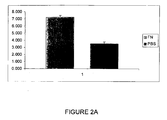
Figure 2A shows the initial adhesion of human chondrocytes to fibronectin. It was noted that a cohort of chondrocytes adhered to fibronectin within 20 minutes compared to controls (p < 0.05); -
Figure 2B shows chondrocytes that adhere to fibronectin within 20 minutes can form colonies consisting of more than 32 cells by 8 days and, further the number of colonies increases with time in culture compared with controls (p < 0.05); -
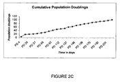
Figure 2C shows clonally derived human stem cells or chondroprogenitors can be extensively sub-cultured and undergo more than 80 population doublings; -
Figure 3 shows views of clonal cells derived from differential adhesion to fibronectin and unselected cells from the same patient which were immunolabelled with antibodies to the cell surface signallingmolecule Notch 1, the stem cell marker STRO1 and the transcription factor MSX1. In clonal cell lines, every cell labelled with antibody toNotch 1 andStro 1 and the majority of cells labelled with antibody to MSX1. In contrast, monolayer cultures contained fewer cells labelled with antibodies to stem cell markers STRO1,Notch 1 and MSX1 (Fig 3A-C ); -
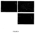
Figure 4 shows that using an in ovo assay, clonally derived cartilage stem cells were shown to engraft into various chick tissues including, bone, tendon and cartilage and such engrafted stem cells were shown to synthesise a structurally relevant protein, namely human type I collagen (Figure 4 ); and -
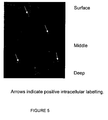
Figure 5 shows wax sections of aged human tissue were labelled with antibody to the transcription factor MSX1 (N20, Santa Cruz Biotechnology; 5ug ml-1 in PBS for 1 hour) and detected with anti-rabbit FITC conjugated secondary antibody (Sigma; 1:100 in PBS for 1 hour).MSX 1 positive cells were present in the surface and middle zones of the tissue suggesting that this is where the stem cells reside within the tissue. Arrows indicate positive intracellular labelling. - 12 well plates were coated with 10µg ml-1 plasma fibronectin (Sigma, UK) in PBS containing 1mM MgCl2 and 1mM CaCl2 (PBS+) overnight at 4°C. Dishes were blocked with 1% BSA (Sigma) in PBS+ before chondrocytes were added. Control dishes were treated with PBS+ overnight at 4°C.
- Tissue was obtained from patients undergoing hemiarthrotomy with full institutional ethical approval. Full depth cartilage was removed from the grossly normal femoral condyle and incubated in 1:1 DMEM/F12 (Gibco) containing 10% FCS (Gibco) overnight. Chondrocytes were then isolated by sequential pronase (Roche)/collagenase (Sigma) digestion as previously described (Dowthwaite et al 2004). Briefly cartilage chips were incubated with pronase (70 units ml-1 in DMEM/F12 containing 5% FCS) for 3 hours at 37°C. Pronase was removed and cartilage incubated with collagenase (300 units ml-1 in DMEM/F12 containing 5% FCS) overnight at 37°C. Chondrocytes were centrifuged at 2000 rpm for 5 minutes, supernatant removed and resuspended in serum free DMEM/F12 and counted. After isolation, chondrocytes (1,000 ml-1) were seeded into individual wells of 12 well plates and incubated at 37°C for 20 minutes in 1:1 DMEM/F12 containing 0.1% Gentamycin (DMEM/F12-). After 20 minutes, media (and non-adherent cells) was removed and placed in a second dish for 40 minutes at 37°C before this media (and non-adherent cells) was removed and placed in a third dish. After removal of media at 20 and 40 minutes, fresh 1:1 DMEM/F12 containing 0.1% Gentamycin and 10% FCS (DMEM/F12+) was added to the remaining adherent cells which were maintained in culture for up to 17 days. Controls comprised cells subjected to differential adhesion on dishes coated with 1% BSA in PBS+.
- Within 3 hours of plating, initial chondrocyte adhesion was assayed by counting the total number of cells adhering to the bottom of the dish using an inverted microscope equipped with phase contrast optics and expressed as a percentage of the initial seeding density. Colonies of chondrocytes consisting of more than 32 cells were counted using the same microscope at 8, 12, 14 and 17 days. Colony forming efficiency (CFE) was calculated by dividing the number of colonies by the initial number of adherent cells.
- Once colonies consisting of more than 32 cells had formed, they were identified under a light microscope. Clones were trypsinised (0.25%; Gibco) and extensively subcultured in DMEM/F12 + 10% FCS. Cell numbers were calculated at each passage and population dynamics plotted.
- Clonal cell lines were immunolabelled with antibodies to Notch 1 (C20, 5ug ml-1; Santa Cruz Biotechnology), STRO1 (neat TC supernatant, gift from R Oreffo, Southampton University) and MSX1 (N20, 5ug ml-1; Santa Cruz Biotechnology) after fixing for 5 minutes in either 95% EtOH (
Notch 1, STRO1) or 10% NBFS (MSX1). Primary antibodies were localised using relevant fluorescently conjugated antibodies and observed under a fluorescent microscope. - Clonal cell lines were labelled with 10uM cell tracker green (Invitrogen) following the manufacturers instructions and injected into the wing bud of 3 day old (HH St 12-14) chick embryos which had been previously windowed. Embryos were resealed with sellotape and incubated for various times up to day 10 (HH St 36-37) and wings were fixed in 10% NBFS and processed for wax embedding. Samples were sectioned at 10µm, dewaxed and mounted in DPX before being examined under a fluorescent microscope. Additional sections were immunolabelled with antibody 5D8 (anti human type I collagen; Abcam) and observed under a fluorescence microscope.
- As shown in
Figure 5 , wax sections of aged human tissue that have been labelled with antibody to thetranscription factor MSX 1, a marker for stem cells, showed that stem cells were present in both the surface and middle zones of cartilage tissue. This goes against conventional wisdom which presumed that stem cells were only present in the surface of cartilage tissue. -
- Archer C and Francis-West P (2003) The chondrocyte. Int. J Biochem Cell Biol. 35, 401-404.
- Brittberg, M, Lindahl, A, Nilsson, A, Ohlsson, C, Isakssin, O, Peterson, L. (1994) Treatment of deep cartilage defects in the knee with autologous chondrocyte transplantation. N Engl J Med. 331, 889-895.
- Dowthwaite, GP, Flannery, CR, Lewthwaite, J, Flannelly, J, Archer, CW and Pitsillides AA. (2003) A mechanism underlying the movement requirement for synovial joint cavitation. Matrix Biol. 22, 311-322.
- Dowthwaite, GP, Bishop JC, Redman SN, Khan IM, Rooney P, Evans DJ, Haughton L, Bayram Z, Boyer S, Thomson B, Wolfe MS, Archer CW. (2004). The surface of articular cartilage contains a progenitor cell population. J Cell Sci.117,889-897
- Flannery CR, Hughes CE, Schumacher BL, Tudor D, Aydelotte MB, Kuettner KE, Caterson B. (1999) Articular cartilage superficial zone protein (SZP) is homologous to megakaryocyte stimulating factor precursor and Is a multifunctional proteoglycan with potential growth-promoting, cytoprotective, and lubricating properties in cartilage metabolism. Biochem Biophys Res Commun.254, 535-41.
- Hayes, AJ, MacPherson, S, Morrison, H, Dowthwaite, GP and Archer, CW (2001). The development of articular cartilage: evidence for an appositional growth mechanism. Anat Embryol. 203, 469-79.
- Knudson CB (2003). Hyaluronan and CD44: strategic players for cellmatrix interactions during chondrogenesis and matrix assembly. Birth Defects Res C Embryo Today. 69, 174-96.
Claims (14)
- A human stem cell isolated from aged human cartilage.
- A stem cell according to claim 1 wherein said cell is isolated from the full depth of cartilage tissue.
- A stem cell according to claim 1 wherein said stem cell is isolated from either the surface or mid-zone of cartilage tissue, or both.
- A human stem cell which is characterised by any one or more of the following traits:(1) its isolation from the full depth of human articular cartilage;(2) its isolation by high affinity adhesion to fibronectin or a fragment containing the RGD sequence;(3) a population doubling in excess of 52;(4) the ability to differentiate into any connective tissue type;(5) the expression of any one or more of the following stem cell markers: STRO1, MSX1 or notch 1;(6) its isolation from human aged cartilage.
- A population of human connective tissue cells derived from the stem cell according to claims 1 to 4.
- The use of a stem cell according to claims 1-4 in tissue repair.
- The use of human connective tissue cells according to claim 5 for use in tissue repair.
- An implant for use in tissue repair comprising a stem cell according to claims 1 to 4 or a population of cells according to claim 5.
- A method for isolating a human stem cell comprising:(a) obtaining human articular cartilage tissue from the full depth of the cartilage tissue;(b) digesting the tissue to release chondrocytes, by using enzymes;(c) exposing the isolated chondrocytes to fibronectin and/or a fragment thereof containing RGD sequence; and(d) isolating those cells that bind fibronectin or said fragment.
- A method according to claim 9 wherein said released chondrocytes are cultured on fibronectin or a RGD sequence, such as a fragment of fibronectin containing same.
- A method according to claims 9 or 10 wherein said articular cartilage is digested with a combination of pronase and collagenase.
- A method according to claims 9-11 wherein said digested chondrocytes are either filtered or centrifuged in order to isolate chondrocyte tissue.
- A method according to claims 9-12 wherein the articular cartilage tissue referred to is aged tissue.
- A human stem cell, or a population of human connective tissue cells, or the use of said stem cell or said tissue cells, or a method for isolating a human stem cell as substantially herein described with reference to the written description and the figures contained herein.
Applications Claiming Priority (2)
| Application Number | Priority Date | Filing Date | Title |
|---|---|---|---|
| GBGB0702401.1A GB0702401D0 (en) | 2007-02-08 | 2007-02-08 | Connective tissue repair |
| EP08702053A EP2111447B1 (en) | 2007-02-08 | 2008-02-06 | Method for isolating a human stem cell from the full depth of the cartilage tissue |
Related Parent Applications (2)
| Application Number | Title | Priority Date | Filing Date |
|---|---|---|---|
| WOPCT/GB2008/000393 Previously-Filed-Application | 2008-02-06 | ||
| EP08702053.3 Division | 2008-02-06 |
Publications (1)
| Publication Number | Publication Date |
|---|---|
| EP2392361A1 true EP2392361A1 (en) | 2011-12-07 |
Family
ID=37898927
Family Applications (2)
| Application Number | Title | Priority Date | Filing Date |
|---|---|---|---|
| EP08702053A Not-in-force EP2111447B1 (en) | 2007-02-08 | 2008-02-06 | Method for isolating a human stem cell from the full depth of the cartilage tissue |
| EP11158817A Withdrawn EP2392361A1 (en) | 2007-02-08 | 2008-02-06 | Connective Tissue Repair |
Family Applications Before (1)
| Application Number | Title | Priority Date | Filing Date |
|---|---|---|---|
| EP08702053A Not-in-force EP2111447B1 (en) | 2007-02-08 | 2008-02-06 | Method for isolating a human stem cell from the full depth of the cartilage tissue |
Country Status (9)
| Country | Link |
|---|---|
| US (2) | US8357534B2 (en) |
| EP (2) | EP2111447B1 (en) |
| JP (1) | JP2010517553A (en) |
| CN (1) | CN101646765A (en) |
| AU (1) | AU2008212720B2 (en) |
| CA (1) | CA2677548A1 (en) |
| ES (1) | ES2399300T3 (en) |
| GB (1) | GB0702401D0 (en) |
| WO (1) | WO2008096118A1 (en) |
Citations (1)
| Publication number | Priority date | Publication date | Assignee | Title |
|---|---|---|---|---|
| US20060239980A1 (en) | 2003-06-12 | 2006-10-26 | Antonio Bernad Miana | Cartilage-derived stem cells and applications thereof |
-
2007
- 2007-02-08 GB GBGB0702401.1A patent/GB0702401D0/en not_active Ceased
-
2008
- 2008-02-06 US US12/526,194 patent/US8357534B2/en not_active Expired - Fee Related
- 2008-02-06 WO PCT/GB2008/000393 patent/WO2008096118A1/en not_active Ceased
- 2008-02-06 AU AU2008212720A patent/AU2008212720B2/en not_active Ceased
- 2008-02-06 EP EP08702053A patent/EP2111447B1/en not_active Not-in-force
- 2008-02-06 CA CA002677548A patent/CA2677548A1/en not_active Abandoned
- 2008-02-06 JP JP2009548732A patent/JP2010517553A/en active Pending
- 2008-02-06 CN CN200880006167A patent/CN101646765A/en active Pending
- 2008-02-06 EP EP11158817A patent/EP2392361A1/en not_active Withdrawn
- 2008-02-06 ES ES08702053T patent/ES2399300T3/en active Active
-
2013
- 2013-01-16 US US13/742,699 patent/US20130136723A1/en not_active Abandoned
Patent Citations (1)
| Publication number | Priority date | Publication date | Assignee | Title |
|---|---|---|---|---|
| US20060239980A1 (en) | 2003-06-12 | 2006-10-26 | Antonio Bernad Miana | Cartilage-derived stem cells and applications thereof |
Non-Patent Citations (12)
| Title |
|---|
| ALSALAMEH SAIFEDDIN ET AL: "Identification of mesenchymal progenitor cells in normal and osteoarthritic human articular cartilage.", ARTHRITIS AND RHEUMATISM MAY 2004, vol. 50, no. 5, May 2004 (2004-05-01), pages 1522 - 1532, XP002478826, ISSN: 0004-3591 * |
| ARCHER C, FRANCIS-WEST P, THE CHONDROCYTE. INT. J BIOCHEM CELL BIOL., vol. 35, 2003, pages 401 - 404 |
| BRITTBERG, M, LINDAHL, A, NILSSON, A, OHLSSON, C, ISAKSSIN, O, PETERSON, L.: "Treatment of deep cartilage defects in the knee with autologous chondrocyte transplantation", N ENGL J MED., vol. 331, 1994, pages 889 - 895 |
| DOWTHWAITE GARY P ET AL: "The surface of articular cartilage contains a progenitor cell population.", JOURNAL OF CELL SCIENCE, vol. 117, no. 6, 29 February 2004 (2004-02-29), pages 889 - 897, XP002478828, ISSN: 0021-9533 * |
| DOWTHWAITE, GP, BISHOP JC, REDMAN SN, KHAN IM, ROONEY P, EVANS DJ, HAUGHTON L, BAYRAM Z, BOYER S, THOMSON B: "The surface of articular cartiiage contains a progenitor cell population", J CELL SCI., vol. 117, 2004, pages 889 - 897, XP002478828, DOI: doi:10.1242/jcs.00912 |
| DOWTHWAITE, GP, FLANNERY, CR, LEWTHWAITE, J, FLANNELLY, J, ARCHER, CW, PITSILLIDES AA.: "A mechanism underlying the movement requirement for synovial joint cavitation", MATRIX BIOL., vol. 22, 2003, pages 311 - 322 |
| FICKERT STEFAN ET AL: "Identification of subpopulations with characteristics of mesenchymal progenitor cells from human osteoarthritic cartilage using triple staining for cell surface markers.", ARTHRITIS RESEARCH & THERAPY 2004, vol. 6, no. 5, 2004, pages R422 - R432, XP002478827, ISSN: 1478-6362 * |
| FLANNERY CR, HUGHES CE, SCHUMACHER BL, TUDOR D, AYDELOTTE MB, KUETTNER KE, CATERSON B.: "Articular cartilage superficial zone protein (SZP) is homologous to megakaryocyte stimulating factor precursor and Is a multifunctional proteoglycan with potential growth-promoting, cytoprotective, and lubricating properties in cartilage metabolism", BIOCHEM BIOPHYS RES COMMUN., vol. 254, 1999, pages 535 - 41, XP002168598, DOI: doi:10.1006/bbrc.1998.0104 |
| HAYES, AJ, MACPHERSON, S, MORRISON, H, DOWTHWAITE, GP, ARCHER, CW: "The development of articular cartilage: evidence for an appositional growth mechanism", ANAT EMBRYOL., vol. 203, 2001, pages 469 - 79 |
| HENG B C ET AL: "Directing stem cell differentiation into the chondrogenic lineage in vitro", STEM CELLS, ALPHAMED PRESS, DAYTON, OH, US, vol. 22, no. 7, 1 January 2004 (2004-01-01), pages 1152 - 1167, XP002369108, ISSN: 1066-5099 * |
| KNUDSON CB: "Hyaluronan and CD44: strategic players for cell- matrix interactions during chondrogenesis and matrix assembly", BIRTH DEFECTS RES C EMBRYO TODAY., vol. 69, 2003, pages 174 - 96 |
| SU W ET AL: "Cartilage-derived stromal cells: Is it a novel cell resource for cell therapy to regenerate infarcted myocardium?", STEM CELLS 200602 US, vol. 24, no. 2, February 2006 (2006-02-01), pages 349 - 356, XP002478825, ISSN: 1066-5099 * |
Also Published As
| Publication number | Publication date |
|---|---|
| EP2111447A1 (en) | 2009-10-28 |
| AU2008212720A1 (en) | 2008-08-14 |
| ES2399300T3 (en) | 2013-03-27 |
| JP2010517553A (en) | 2010-05-27 |
| US20100098736A1 (en) | 2010-04-22 |
| US8357534B2 (en) | 2013-01-22 |
| GB0702401D0 (en) | 2007-03-21 |
| WO2008096118A1 (en) | 2008-08-14 |
| CN101646765A (en) | 2010-02-10 |
| AU2008212720B2 (en) | 2013-09-26 |
| EP2111447B1 (en) | 2012-11-14 |
| CA2677548A1 (en) | 2008-08-14 |
| US20130136723A1 (en) | 2013-05-30 |
Similar Documents
| Publication | Publication Date | Title |
|---|---|---|
| Giovannini et al. | Micromass co-culture of human articular chondrocytes and human bone marrow mesenchymal stem cells to investigate stable neocartilage tissue formation in vitro | |
| CN101589139B (en) | Artificial cartilage comprising chondrocytes obtained from costal cartilage and method for producing the same | |
| KR100944204B1 (en) | Stratified cartilage tissue and how to manipulate it | |
| Awad et al. | Repair of patellar tendon injuries using a cell–collagen composite | |
| Chua et al. | Insulin-transferrin-selenium prevent human chondrocyte dedifferentiation and promote the formation of high quality tissue engineered human hyaline cartilage | |
| Ibsirlioglu et al. | Decellularized biological scaffold and stem cells from autologous human adipose tissue for cartilage tissue engineering | |
| Jakob et al. | Enzymatic digestion of adult human articular cartilage yields a small fraction of the total available cells | |
| Yates et al. | Phenotypic analysis of bovine chondrocytes cultured in 3D collagen sponges: effect of serum substitutes | |
| Van Osch et al. | Considerations on the use of ear chondrocytes as donor chondrocytes for cartilage tissue engineering | |
| CA2650649C (en) | Methods and compositions for repairing cartilage using type b synoviocytes | |
| Elsaesser et al. | Characterization of a migrative subpopulation of adult human nasoseptal chondrocytes with progenitor cell features and their potential for in vivo cartilage regeneration strategies | |
| Xue et al. | Isolation, identification, and comparison of cartilage stem progenitor/cells from auricular cartilage and perichondrium | |
| JP2003517259A (en) | Method for isolating bone and precursor chondrocytes | |
| Wu et al. | Regional-specific meniscal extracellular matrix hydrogels and their effects on cell–matrix interactions of fibrochondrocytes | |
| Remya et al. | Mechanoresponsiveness of human umbilical cord mesenchymal stem cells in in vitro chondrogenesis—a comparative study with growth factor induction | |
| EP2111447B1 (en) | Method for isolating a human stem cell from the full depth of the cartilage tissue | |
| Weekes et al. | Characterization of temporomandibular joint articular disc progenitor cell clones | |
| EP1819290A4 (en) | SCAFFOLD-FREE CONSTRUCTIONS FOR ARTICULAR CARTILAGE TISSUE GENIE | |
| AU2013213695A1 (en) | Connective tissue repair | |
| US10655103B2 (en) | Equine progenitor cells | |
| Class et al. | Patent application title: CONNECTIVE TISSUE REPAIR Inventors: Charles William Archer (Aberdare, GB) University College Cardiff Consultants Limited (Cardiff, GB) Samantha Nichola Haven (Cardiff, GB) Gary Dowthwaite (Cardiff, GB) Assignees: University College Cardiff Consultants Limited | |
| Dabaghi et al. | Allografts for partial meniscus repair: an in vitro and ex vivo meniscus culture study | |
| Negri et al. | Tissue engineering: chondrocyte cultures on type I collagen support. Cytohistological and immunohistochemical study | |
| Duan et al. | Combination of chondrocytes with chondrons improves Proteoglycan and Collagen-2 production to promote the repairs of defective knee cartilage in rabbits | |
| O’Sullivan | Glycosaminoglycan Dependent Isolation of Mesenchymal Chondroprogenitor Populations from Human Bone Marrow |
Legal Events
| Date | Code | Title | Description |
|---|---|---|---|
| 17P | Request for examination filed |
Effective date: 20110318 |
|
| AC | Divisional application: reference to earlier application |
Ref document number: 2111447 Country of ref document: EP Kind code of ref document: P |
|
| AK | Designated contracting states |
Kind code of ref document: A1 Designated state(s): AT BE BG CH CY CZ DE DK EE ES FI FR GB GR HR HU IE IS IT LI LT LU LV MC MT NL NO PL PT RO SE SI SK TR |
|
| PUAI | Public reference made under article 153(3) epc to a published international application that has entered the european phase |
Free format text: ORIGINAL CODE: 0009012 |
|
| STAA | Information on the status of an ep patent application or granted ep patent |
Free format text: STATUS: THE APPLICATION IS DEEMED TO BE WITHDRAWN |
|
| 18D | Application deemed to be withdrawn |
Effective date: 20160901 |