EP2392352B1 - Cd93 or use of soluble fragment thereof - Google Patents
Cd93 or use of soluble fragment thereof Download PDFInfo
- Publication number
- EP2392352B1 EP2392352B1 EP10735985.3A EP10735985A EP2392352B1 EP 2392352 B1 EP2392352 B1 EP 2392352B1 EP 10735985 A EP10735985 A EP 10735985A EP 2392352 B1 EP2392352 B1 EP 2392352B1
- Authority
- EP
- European Patent Office
- Prior art keywords
- cells
- antibody
- scd93
- thp
- soluble fragment
- Prior art date
- Legal status (The legal status is an assumption and is not a legal conclusion. Google has not performed a legal analysis and makes no representation as to the accuracy of the status listed.)
- Not-in-force
Links
- 239000012634 fragment Substances 0.000 title claims description 79
- 101150089199 Cd93 gene Proteins 0.000 title 1
- 101000933665 Homo sapiens Complement component C1q receptor Proteins 0.000 claims description 155
- 102100025877 Complement component C1q receptor Human genes 0.000 claims description 149
- 108090000623 proteins and genes Proteins 0.000 claims description 63
- 102000004169 proteins and genes Human genes 0.000 claims description 50
- 239000003550 marker Substances 0.000 claims description 26
- 206010039073 rheumatoid arthritis Diseases 0.000 claims description 24
- 238000000034 method Methods 0.000 claims description 23
- 238000006243 chemical reaction Methods 0.000 claims description 21
- 230000027455 binding Effects 0.000 claims description 15
- 239000012472 biological sample Substances 0.000 claims description 14
- 238000002474 experimental method Methods 0.000 claims description 14
- 230000001939 inductive effect Effects 0.000 claims description 14
- 210000001179 synovial fluid Anatomy 0.000 claims description 14
- 238000001262 western blot Methods 0.000 claims description 13
- 210000001258 synovial membrane Anatomy 0.000 claims description 11
- 239000012528 membrane Substances 0.000 claims description 8
- 230000001105 regulatory effect Effects 0.000 claims description 8
- 239000003593 chromogenic compound Substances 0.000 claims description 7
- 238000001962 electrophoresis Methods 0.000 claims description 7
- 229940127121 immunoconjugate Drugs 0.000 claims description 7
- 238000003127 radioimmunoassay Methods 0.000 claims description 7
- 238000003118 sandwich ELISA Methods 0.000 claims description 7
- GEYOCULIXLDCMW-UHFFFAOYSA-N 1,2-phenylenediamine Chemical compound NC1=CC=CC=C1N GEYOCULIXLDCMW-UHFFFAOYSA-N 0.000 claims description 6
- 238000002965 ELISA Methods 0.000 claims description 6
- 239000011521 glass Substances 0.000 claims description 5
- 238000003757 reverse transcription PCR Methods 0.000 claims description 5
- 108010001336 Horseradish Peroxidase Proteins 0.000 claims description 4
- 229920002981 polyvinylidene fluoride Polymers 0.000 claims description 4
- 238000002198 surface plasmon resonance spectroscopy Methods 0.000 claims description 4
- 239000000020 Nitrocellulose Substances 0.000 claims description 3
- 239000007795 chemical reaction product Substances 0.000 claims description 3
- 239000011248 coating agent Substances 0.000 claims description 3
- 238000000576 coating method Methods 0.000 claims description 3
- 238000001514 detection method Methods 0.000 claims description 3
- 229920001220 nitrocellulos Polymers 0.000 claims description 3
- 239000000758 substrate Substances 0.000 claims description 3
- 102000002260 Alkaline Phosphatase Human genes 0.000 claims description 2
- 108020004774 Alkaline Phosphatase Proteins 0.000 claims description 2
- 238000000018 DNA microarray Methods 0.000 claims description 2
- 238000000636 Northern blotting Methods 0.000 claims description 2
- 239000002033 PVDF binder Substances 0.000 claims description 2
- ZTOJFFHGPLIVKC-CLFAGFIQSA-N abts Chemical compound S/1C2=CC(S(O)(=O)=O)=CC=C2N(CC)C\1=N\N=C1/SC2=CC(S(O)(=O)=O)=CC=C2N1CC ZTOJFFHGPLIVKC-CLFAGFIQSA-N 0.000 claims description 2
- 230000004520 agglutination Effects 0.000 claims description 2
- OHDRQQURAXLVGJ-HLVWOLMTSA-N azane;(2e)-3-ethyl-2-[(e)-(3-ethyl-6-sulfo-1,3-benzothiazol-2-ylidene)hydrazinylidene]-1,3-benzothiazole-6-sulfonic acid Chemical compound [NH4+].[NH4+].S/1C2=CC(S([O-])(=O)=O)=CC=C2N(CC)C\1=N/N=C1/SC2=CC(S([O-])(=O)=O)=CC=C2N1CC OHDRQQURAXLVGJ-HLVWOLMTSA-N 0.000 claims description 2
- 239000000084 colloidal system Substances 0.000 claims description 2
- 238000004737 colorimetric analysis Methods 0.000 claims description 2
- MHMNJMPURVTYEJ-UHFFFAOYSA-N fluorescein-5-isothiocyanate Chemical compound O1C(=O)C2=CC(N=C=S)=CC=C2C21C1=CC=C(O)C=C1OC1=CC(O)=CC=C21 MHMNJMPURVTYEJ-UHFFFAOYSA-N 0.000 claims description 2
- PCHJSUWPFVWCPO-UHFFFAOYSA-N gold Chemical compound [Au] PCHJSUWPFVWCPO-UHFFFAOYSA-N 0.000 claims description 2
- 239000010931 gold Substances 0.000 claims description 2
- 229910052737 gold Inorganic materials 0.000 claims description 2
- 238000010166 immunofluorescence Methods 0.000 claims description 2
- 238000011532 immunohistochemical staining Methods 0.000 claims description 2
- 238000001114 immunoprecipitation Methods 0.000 claims description 2
- 229920000915 polyvinyl chloride Polymers 0.000 claims description 2
- 239000011347 resin Substances 0.000 claims description 2
- 229920005989 resin Polymers 0.000 claims description 2
- YVSWPCCVTYEEHG-UHFFFAOYSA-N rhodamine B 5-isothiocyanate Chemical compound [Cl-].C=12C=CC(=[N+](CC)CC)C=C2OC2=CC(N(CC)CC)=CC=C2C=1C1=CC=C(N=C=S)C=C1C(O)=O YVSWPCCVTYEEHG-UHFFFAOYSA-N 0.000 claims description 2
- UAIUNKRWKOVEES-UHFFFAOYSA-N 3,3',5,5'-tetramethylbenzidine Chemical compound CC1=C(N)C(C)=CC(C=2C=C(C)C(N)=C(C)C=2)=C1 UAIUNKRWKOVEES-UHFFFAOYSA-N 0.000 claims 2
- 125000003275 alpha amino acid group Chemical group 0.000 claims 2
- 210000004027 cell Anatomy 0.000 description 134
- 230000014509 gene expression Effects 0.000 description 53
- 239000002158 endotoxin Substances 0.000 description 51
- 229920006008 lipopolysaccharide Polymers 0.000 description 50
- 108020004414 DNA Proteins 0.000 description 42
- 108060008682 Tumor Necrosis Factor Proteins 0.000 description 37
- 102000000852 Tumor Necrosis Factor-alpha Human genes 0.000 description 35
- MZOFCQQQCNRIBI-VMXHOPILSA-N (3s)-4-[[(2s)-1-[[(2s)-1-[[(1s)-1-carboxy-2-hydroxyethyl]amino]-4-methyl-1-oxopentan-2-yl]amino]-5-(diaminomethylideneamino)-1-oxopentan-2-yl]amino]-3-[[2-[[(2s)-2,6-diaminohexanoyl]amino]acetyl]amino]-4-oxobutanoic acid Chemical compound OC[C@@H](C(O)=O)NC(=O)[C@H](CC(C)C)NC(=O)[C@H](CCCN=C(N)N)NC(=O)[C@H](CC(O)=O)NC(=O)CNC(=O)[C@@H](N)CCCCN MZOFCQQQCNRIBI-VMXHOPILSA-N 0.000 description 32
- 239000004055 small Interfering RNA Substances 0.000 description 32
- 241000282414 Homo sapiens Species 0.000 description 30
- PHEDXBVPIONUQT-UHFFFAOYSA-N Cocarcinogen A1 Natural products CCCCCCCCCCCCCC(=O)OC1C(C)C2(O)C3C=C(C)C(=O)C3(O)CC(CO)=CC2C2C1(OC(C)=O)C2(C)C PHEDXBVPIONUQT-UHFFFAOYSA-N 0.000 description 26
- PHEDXBVPIONUQT-RGYGYFBISA-N phorbol 13-acetate 12-myristate Chemical compound C([C@]1(O)C(=O)C(C)=C[C@H]1[C@@]1(O)[C@H](C)[C@H]2OC(=O)CCCCCCCCCCCCC)C(CO)=C[C@H]1[C@H]1[C@]2(OC(C)=O)C1(C)C PHEDXBVPIONUQT-RGYGYFBISA-N 0.000 description 26
- 230000001965 increasing effect Effects 0.000 description 25
- 108091032973 (ribonucleotides)n+m Proteins 0.000 description 24
- 241000699666 Mus <mouse, genus> Species 0.000 description 24
- 230000002441 reversible effect Effects 0.000 description 24
- 208000027866 inflammatory disease Diseases 0.000 description 22
- 210000002540 macrophage Anatomy 0.000 description 21
- LOKCTEFSRHRXRJ-UHFFFAOYSA-I dipotassium trisodium dihydrogen phosphate hydrogen phosphate dichloride Chemical compound P(=O)(O)(O)[O-].[K+].P(=O)(O)([O-])[O-].[Na+].[Na+].[Cl-].[K+].[Cl-].[Na+] LOKCTEFSRHRXRJ-UHFFFAOYSA-I 0.000 description 20
- 239000000203 mixture Substances 0.000 description 20
- 239000002953 phosphate buffered saline Substances 0.000 description 20
- 239000013604 expression vector Substances 0.000 description 18
- 239000000243 solution Substances 0.000 description 17
- 238000005406 washing Methods 0.000 description 17
- 108091027967 Small hairpin RNA Proteins 0.000 description 16
- 108020004459 Small interfering RNA Proteins 0.000 description 16
- 210000004408 hybridoma Anatomy 0.000 description 16
- 206010061218 Inflammation Diseases 0.000 description 15
- 230000004054 inflammatory process Effects 0.000 description 15
- 108090001005 Interleukin-6 Proteins 0.000 description 14
- CWERGRDVMFNCDR-UHFFFAOYSA-M thioglycolate(1-) Chemical compound [O-]C(=O)CS CWERGRDVMFNCDR-UHFFFAOYSA-M 0.000 description 14
- 150000001413 amino acids Chemical class 0.000 description 13
- 239000002299 complementary DNA Substances 0.000 description 13
- 230000004069 differentiation Effects 0.000 description 13
- 239000013642 negative control Substances 0.000 description 13
- 241001465754 Metazoa Species 0.000 description 12
- 206010057249 Phagocytosis Diseases 0.000 description 12
- 230000003110 anti-inflammatory effect Effects 0.000 description 12
- 201000008482 osteoarthritis Diseases 0.000 description 12
- 230000008782 phagocytosis Effects 0.000 description 12
- 238000011002 quantification Methods 0.000 description 12
- 239000000126 substance Substances 0.000 description 12
- 239000013598 vector Substances 0.000 description 12
- 239000004480 active ingredient Substances 0.000 description 11
- 239000003330 peritoneal dialysis fluid Substances 0.000 description 11
- 210000002966 serum Anatomy 0.000 description 11
- 101000946889 Homo sapiens Monocyte differentiation antigen CD14 Proteins 0.000 description 10
- 102100035877 Monocyte differentiation antigen CD14 Human genes 0.000 description 10
- 238000007796 conventional method Methods 0.000 description 10
- 239000013641 positive control Substances 0.000 description 10
- 102000043136 MAP kinase family Human genes 0.000 description 9
- 108091054455 MAP kinase family Proteins 0.000 description 9
- 230000000694 effects Effects 0.000 description 9
- 238000001943 fluorescence-activated cell sorting Methods 0.000 description 9
- 238000004519 manufacturing process Methods 0.000 description 9
- 239000002609 medium Substances 0.000 description 9
- 108020004999 messenger RNA Proteins 0.000 description 9
- 239000002773 nucleotide Substances 0.000 description 9
- 125000003729 nucleotide group Chemical group 0.000 description 9
- 210000004989 spleen cell Anatomy 0.000 description 9
- 241000283707 Capra Species 0.000 description 8
- 102000004127 Cytokines Human genes 0.000 description 8
- 108090000695 Cytokines Proteins 0.000 description 8
- 206010035226 Plasma cell myeloma Diseases 0.000 description 8
- FAPWRFPIFSIZLT-UHFFFAOYSA-M Sodium chloride Chemical compound [Na+].[Cl-] FAPWRFPIFSIZLT-UHFFFAOYSA-M 0.000 description 8
- UQLDLKMNUJERMK-UHFFFAOYSA-L di(octadecanoyloxy)lead Chemical compound [Pb+2].CCCCCCCCCCCCCCCCCC([O-])=O.CCCCCCCCCCCCCCCCCC([O-])=O UQLDLKMNUJERMK-UHFFFAOYSA-L 0.000 description 8
- 239000000499 gel Substances 0.000 description 8
- 210000005087 mononuclear cell Anatomy 0.000 description 8
- 201000000050 myeloid neoplasm Diseases 0.000 description 8
- 210000001519 tissue Anatomy 0.000 description 8
- 230000002163 immunogen Effects 0.000 description 7
- 230000002757 inflammatory effect Effects 0.000 description 7
- 238000000746 purification Methods 0.000 description 7
- 238000009007 Diagnostic Kit Methods 0.000 description 6
- 241000588724 Escherichia coli Species 0.000 description 6
- 108060003951 Immunoglobulin Proteins 0.000 description 6
- 239000012980 RPMI-1640 medium Substances 0.000 description 6
- 210000000628 antibody-producing cell Anatomy 0.000 description 6
- 239000000872 buffer Substances 0.000 description 6
- 230000036039 immunity Effects 0.000 description 6
- 102000018358 immunoglobulin Human genes 0.000 description 6
- 210000003819 peripheral blood mononuclear cell Anatomy 0.000 description 6
- 239000013612 plasmid Substances 0.000 description 6
- 238000003753 real-time PCR Methods 0.000 description 6
- 102100029438 Nitric oxide synthase, inducible Human genes 0.000 description 5
- 101710089543 Nitric oxide synthase, inducible Proteins 0.000 description 5
- 239000000427 antigen Substances 0.000 description 5
- 108091007433 antigens Proteins 0.000 description 5
- 102000036639 antigens Human genes 0.000 description 5
- 230000000903 blocking effect Effects 0.000 description 5
- 239000012228 culture supernatant Substances 0.000 description 5
- 238000013467 fragmentation Methods 0.000 description 5
- 238000006062 fragmentation reaction Methods 0.000 description 5
- 230000006698 induction Effects 0.000 description 5
- -1 leader sequence Chemical class 0.000 description 5
- 230000001404 mediated effect Effects 0.000 description 5
- 238000002360 preparation method Methods 0.000 description 5
- 230000000770 proinflammatory effect Effects 0.000 description 5
- 230000028327 secretion Effects 0.000 description 5
- 239000011780 sodium chloride Substances 0.000 description 5
- 102000040650 (ribonucleotides)n+m Human genes 0.000 description 4
- 108091023037 Aptamer Proteins 0.000 description 4
- 102100028667 C-type lectin domain family 4 member A Human genes 0.000 description 4
- 101100150085 Caenorhabditis elegans sphk-1 gene Proteins 0.000 description 4
- 102100031780 Endonuclease Human genes 0.000 description 4
- LFQSCWFLJHTTHZ-UHFFFAOYSA-N Ethanol Chemical compound CCO LFQSCWFLJHTTHZ-UHFFFAOYSA-N 0.000 description 4
- 102100031181 Glyceraldehyde-3-phosphate dehydrogenase Human genes 0.000 description 4
- 101000766908 Homo sapiens C-type lectin domain family 4 member A Proteins 0.000 description 4
- 108010021625 Immunoglobulin Fragments Proteins 0.000 description 4
- 102000008394 Immunoglobulin Fragments Human genes 0.000 description 4
- 108010065805 Interleukin-12 Proteins 0.000 description 4
- JGSARLDLIJGVTE-MBNYWOFBSA-N Penicillin G Chemical compound N([C@H]1[C@H]2SC([C@@H](N2C1=O)C(O)=O)(C)C)C(=O)CC1=CC=CC=C1 JGSARLDLIJGVTE-MBNYWOFBSA-N 0.000 description 4
- 230000004913 activation Effects 0.000 description 4
- 230000001154 acute effect Effects 0.000 description 4
- 229940024606 amino acid Drugs 0.000 description 4
- 230000024245 cell differentiation Effects 0.000 description 4
- 230000007910 cell fusion Effects 0.000 description 4
- 230000005754 cellular signaling Effects 0.000 description 4
- 230000003247 decreasing effect Effects 0.000 description 4
- 238000011161 development Methods 0.000 description 4
- 230000018109 developmental process Effects 0.000 description 4
- 230000004927 fusion Effects 0.000 description 4
- 108020004445 glyceraldehyde-3-phosphate dehydrogenase Proteins 0.000 description 4
- 210000000265 leukocyte Anatomy 0.000 description 4
- 239000000047 product Substances 0.000 description 4
- 230000002829 reductive effect Effects 0.000 description 4
- 238000002415 sodium dodecyl sulfate polyacrylamide gel electrophoresis Methods 0.000 description 4
- DAEPDZWVDSPTHF-UHFFFAOYSA-M sodium pyruvate Chemical compound [Na+].CC(=O)C([O-])=O DAEPDZWVDSPTHF-UHFFFAOYSA-M 0.000 description 4
- UCSJYZPVAKXKNQ-HZYVHMACSA-N streptomycin Chemical compound CN[C@H]1[C@H](O)[C@@H](O)[C@H](CO)O[C@H]1O[C@@H]1[C@](C=O)(O)[C@H](C)O[C@H]1O[C@@H]1[C@@H](NC(N)=N)[C@H](O)[C@@H](NC(N)=N)[C@H](O)[C@H]1O UCSJYZPVAKXKNQ-HZYVHMACSA-N 0.000 description 4
- 239000006228 supernatant Substances 0.000 description 4
- 102100025279 C-X-C motif chemokine 11 Human genes 0.000 description 3
- 241000282693 Cercopithecidae Species 0.000 description 3
- 108091026890 Coding region Proteins 0.000 description 3
- 108010047041 Complementarity Determining Regions Proteins 0.000 description 3
- 102000004190 Enzymes Human genes 0.000 description 3
- 108090000790 Enzymes Proteins 0.000 description 3
- 208000007882 Gastritis Diseases 0.000 description 3
- PEDCQBHIVMGVHV-UHFFFAOYSA-N Glycerine Chemical compound OCC(O)CO PEDCQBHIVMGVHV-UHFFFAOYSA-N 0.000 description 3
- 101000858060 Homo sapiens C-X-C motif chemokine 11 Proteins 0.000 description 3
- 241000124008 Mammalia Species 0.000 description 3
- 102000002274 Matrix Metalloproteinases Human genes 0.000 description 3
- 108010000684 Matrix Metalloproteinases Proteins 0.000 description 3
- 241000699670 Mus sp. Species 0.000 description 3
- 241000283973 Oryctolagus cuniculus Species 0.000 description 3
- 241001494479 Pecora Species 0.000 description 3
- 108091030071 RNAI Proteins 0.000 description 3
- 241000700159 Rattus Species 0.000 description 3
- 238000001042 affinity chromatography Methods 0.000 description 3
- 238000000137 annealing Methods 0.000 description 3
- 210000004369 blood Anatomy 0.000 description 3
- 239000008280 blood Substances 0.000 description 3
- 238000010276 construction Methods 0.000 description 3
- 238000003745 diagnosis Methods 0.000 description 3
- 238000002405 diagnostic procedure Methods 0.000 description 3
- 239000003814 drug Substances 0.000 description 3
- 239000003623 enhancer Substances 0.000 description 3
- 229940088598 enzyme Drugs 0.000 description 3
- 210000003743 erythrocyte Anatomy 0.000 description 3
- 238000012757 fluorescence staining Methods 0.000 description 3
- 230000009368 gene silencing by RNA Effects 0.000 description 3
- 238000003780 insertion Methods 0.000 description 3
- 230000037431 insertion Effects 0.000 description 3
- 238000004255 ion exchange chromatography Methods 0.000 description 3
- 210000004379 membrane Anatomy 0.000 description 3
- 210000001616 monocyte Anatomy 0.000 description 3
- 230000008488 polyadenylation Effects 0.000 description 3
- 238000006116 polymerization reaction Methods 0.000 description 3
- 108091033319 polynucleotide Proteins 0.000 description 3
- 102000040430 polynucleotide Human genes 0.000 description 3
- 239000002157 polynucleotide Substances 0.000 description 3
- 230000006798 recombination Effects 0.000 description 3
- 238000011160 research Methods 0.000 description 3
- 230000004044 response Effects 0.000 description 3
- 238000010839 reverse transcription Methods 0.000 description 3
- 235000020183 skimmed milk Nutrition 0.000 description 3
- JKMHFZQWWAIEOD-UHFFFAOYSA-N 2-[4-(2-hydroxyethyl)piperazin-1-yl]ethanesulfonic acid Chemical compound OCC[NH+]1CCN(CCS([O-])(=O)=O)CC1 JKMHFZQWWAIEOD-UHFFFAOYSA-N 0.000 description 2
- TVZGACDUOSZQKY-LBPRGKRZSA-N 4-aminofolic acid Chemical compound C1=NC2=NC(N)=NC(N)=C2N=C1CNC1=CC=C(C(=O)N[C@@H](CCC(O)=O)C(O)=O)C=C1 TVZGACDUOSZQKY-LBPRGKRZSA-N 0.000 description 2
- 102000007469 Actins Human genes 0.000 description 2
- 108010085238 Actins Proteins 0.000 description 2
- 241000894006 Bacteria Species 0.000 description 2
- 201000004624 Dermatitis Diseases 0.000 description 2
- IAZDPXIOMUYVGZ-UHFFFAOYSA-N Dimethylsulphoxide Chemical compound CS(C)=O IAZDPXIOMUYVGZ-UHFFFAOYSA-N 0.000 description 2
- 239000006144 Dulbecco’s modified Eagle's medium Substances 0.000 description 2
- 108010042407 Endonucleases Proteins 0.000 description 2
- 241000282575 Gorilla Species 0.000 description 2
- ZRALSGWEFCBTJO-UHFFFAOYSA-N Guanidine Chemical compound NC(N)=N ZRALSGWEFCBTJO-UHFFFAOYSA-N 0.000 description 2
- 239000007995 HEPES buffer Substances 0.000 description 2
- 101001046686 Homo sapiens Integrin alpha-M Proteins 0.000 description 2
- 101000851058 Homo sapiens Neutrophil elastase Proteins 0.000 description 2
- 102100022338 Integrin alpha-M Human genes 0.000 description 2
- 102000003777 Interleukin-1 beta Human genes 0.000 description 2
- 108090000193 Interleukin-1 beta Proteins 0.000 description 2
- ZDXPYRJPNDTMRX-VKHMYHEASA-N L-glutamine Chemical compound OC(=O)[C@@H](N)CCC(N)=O ZDXPYRJPNDTMRX-VKHMYHEASA-N 0.000 description 2
- 229930182816 L-glutamine Natural products 0.000 description 2
- 241000713666 Lentivirus Species 0.000 description 2
- 102100030412 Matrix metalloproteinase-9 Human genes 0.000 description 2
- 108060004795 Methyltransferase Proteins 0.000 description 2
- 102100033174 Neutrophil elastase Human genes 0.000 description 2
- 241000282577 Pan troglodytes Species 0.000 description 2
- 229930040373 Paraformaldehyde Natural products 0.000 description 2
- 102000035195 Peptidases Human genes 0.000 description 2
- 108091005804 Peptidases Proteins 0.000 description 2
- 229920002873 Polyethylenimine Polymers 0.000 description 2
- 229920001213 Polysorbate 20 Polymers 0.000 description 2
- 239000004365 Protease Substances 0.000 description 2
- 108010092799 RNA-directed DNA polymerase Proteins 0.000 description 2
- 108020005091 Replication Origin Proteins 0.000 description 2
- 240000004808 Saccharomyces cerevisiae Species 0.000 description 2
- 235000014680 Saccharomyces cerevisiae Nutrition 0.000 description 2
- QAOWNCQODCNURD-UHFFFAOYSA-N Sulfuric acid Chemical compound OS(O)(=O)=O QAOWNCQODCNURD-UHFFFAOYSA-N 0.000 description 2
- IQFYYKKMVGJFEH-XLPZGREQSA-N Thymidine Chemical compound O=C1NC(=O)C(C)=CN1[C@@H]1O[C@H](CO)[C@@H](O)C1 IQFYYKKMVGJFEH-XLPZGREQSA-N 0.000 description 2
- 102100040247 Tumor necrosis factor Human genes 0.000 description 2
- 239000002671 adjuvant Substances 0.000 description 2
- 229960003896 aminopterin Drugs 0.000 description 2
- 210000004102 animal cell Anatomy 0.000 description 2
- 239000003963 antioxidant agent Substances 0.000 description 2
- 235000006708 antioxidants Nutrition 0.000 description 2
- 206010003246 arthritis Diseases 0.000 description 2
- 238000003149 assay kit Methods 0.000 description 2
- 210000004899 c-terminal region Anatomy 0.000 description 2
- 238000010805 cDNA synthesis kit Methods 0.000 description 2
- 230000020411 cell activation Effects 0.000 description 2
- 238000004587 chromatography analysis Methods 0.000 description 2
- 238000010367 cloning Methods 0.000 description 2
- 108010062119 complement 1q receptor Proteins 0.000 description 2
- 230000007423 decrease Effects 0.000 description 2
- 230000002950 deficient Effects 0.000 description 2
- 238000004925 denaturation Methods 0.000 description 2
- 230000036425 denaturation Effects 0.000 description 2
- 201000010099 disease Diseases 0.000 description 2
- 208000037265 diseases, disorders, signs and symptoms Diseases 0.000 description 2
- 238000012137 double-staining Methods 0.000 description 2
- 229940079593 drug Drugs 0.000 description 2
- 230000000487 effect on differentiation Effects 0.000 description 2
- 230000002884 effect on inflammation Effects 0.000 description 2
- 230000006870 function Effects 0.000 description 2
- UYTPUPDQBNUYGX-UHFFFAOYSA-N guanine Chemical compound O=C1NC(N)=NC2=C1N=CN2 UYTPUPDQBNUYGX-UHFFFAOYSA-N 0.000 description 2
- 208000006454 hepatitis Diseases 0.000 description 2
- 230000003053 immunization Effects 0.000 description 2
- 238000003018 immunoassay Methods 0.000 description 2
- 238000001727 in vivo Methods 0.000 description 2
- 230000002401 inhibitory effect Effects 0.000 description 2
- 230000000670 limiting effect Effects 0.000 description 2
- 210000001165 lymph node Anatomy 0.000 description 2
- 230000007246 mechanism Effects 0.000 description 2
- 238000010369 molecular cloning Methods 0.000 description 2
- 108020004707 nucleic acids Proteins 0.000 description 2
- 102000039446 nucleic acids Human genes 0.000 description 2
- 150000007523 nucleic acids Chemical class 0.000 description 2
- 230000003287 optical effect Effects 0.000 description 2
- 229920002866 paraformaldehyde Polymers 0.000 description 2
- 230000037361 pathway Effects 0.000 description 2
- 229940056360 penicillin g Drugs 0.000 description 2
- 210000003024 peritoneal macrophage Anatomy 0.000 description 2
- 239000008363 phosphate buffer Substances 0.000 description 2
- 239000013600 plasmid vector Substances 0.000 description 2
- 239000000256 polyoxyethylene sorbitan monolaurate Substances 0.000 description 2
- 235000010486 polyoxyethylene sorbitan monolaurate Nutrition 0.000 description 2
- 238000012257 pre-denaturation Methods 0.000 description 2
- 239000003755 preservative agent Substances 0.000 description 2
- 230000002265 prevention Effects 0.000 description 2
- 230000008569 process Effects 0.000 description 2
- 108090000765 processed proteins & peptides Proteins 0.000 description 2
- 230000017854 proteolysis Effects 0.000 description 2
- 108010054624 red fluorescent protein Proteins 0.000 description 2
- 206010039083 rhinitis Diseases 0.000 description 2
- 230000011664 signaling Effects 0.000 description 2
- 229940054269 sodium pyruvate Drugs 0.000 description 2
- 230000009870 specific binding Effects 0.000 description 2
- 239000010421 standard material Substances 0.000 description 2
- 229960005322 streptomycin Drugs 0.000 description 2
- 210000005222 synovial tissue Anatomy 0.000 description 2
- 238000012360 testing method Methods 0.000 description 2
- RWQNBRDOKXIBIV-UHFFFAOYSA-N thymine Chemical compound CC1=CNC(=O)NC1=O RWQNBRDOKXIBIV-UHFFFAOYSA-N 0.000 description 2
- 230000001988 toxicity Effects 0.000 description 2
- 231100000419 toxicity Toxicity 0.000 description 2
- 238000001890 transfection Methods 0.000 description 2
- 239000000080 wetting agent Substances 0.000 description 2
- OALHHIHQOFIMEF-UHFFFAOYSA-N 3',6'-dihydroxy-2',4',5',7'-tetraiodo-3h-spiro[2-benzofuran-1,9'-xanthene]-3-one Chemical compound O1C(=O)C2=CC=CC=C2C21C1=CC(I)=C(O)C(I)=C1OC1=C(I)C(O)=C(I)C=C21 OALHHIHQOFIMEF-UHFFFAOYSA-N 0.000 description 1
- QFVHZQCOUORWEI-UHFFFAOYSA-N 4-[(4-anilino-5-sulfonaphthalen-1-yl)diazenyl]-5-hydroxynaphthalene-2,7-disulfonic acid Chemical compound C=12C(O)=CC(S(O)(=O)=O)=CC2=CC(S(O)(=O)=O)=CC=1N=NC(C1=CC=CC(=C11)S(O)(=O)=O)=CC=C1NC1=CC=CC=C1 QFVHZQCOUORWEI-UHFFFAOYSA-N 0.000 description 1
- LKDMKWNDBAVNQZ-UHFFFAOYSA-N 4-[[1-[[1-[2-[[1-(4-nitroanilino)-1-oxo-3-phenylpropan-2-yl]carbamoyl]pyrrolidin-1-yl]-1-oxopropan-2-yl]amino]-1-oxopropan-2-yl]amino]-4-oxobutanoic acid Chemical compound OC(=O)CCC(=O)NC(C)C(=O)NC(C)C(=O)N1CCCC1C(=O)NC(C(=O)NC=1C=CC(=CC=1)[N+]([O-])=O)CC1=CC=CC=C1 LKDMKWNDBAVNQZ-UHFFFAOYSA-N 0.000 description 1
- 102100026802 72 kDa type IV collagenase Human genes 0.000 description 1
- 101710151806 72 kDa type IV collagenase Proteins 0.000 description 1
- 206010000871 Acute monocytic leukaemia Diseases 0.000 description 1
- 206010002556 Ankylosing Spondylitis Diseases 0.000 description 1
- 201000001320 Atherosclerosis Diseases 0.000 description 1
- 238000011725 BALB/c mouse Methods 0.000 description 1
- 244000063299 Bacillus subtilis Species 0.000 description 1
- 235000014469 Bacillus subtilis Nutrition 0.000 description 1
- 208000008035 Back Pain Diseases 0.000 description 1
- 208000035143 Bacterial infection Diseases 0.000 description 1
- DWRXFEITVBNRMK-UHFFFAOYSA-N Beta-D-1-Arabinofuranosylthymine Natural products O=C1NC(=O)C(C)=CN1C1C(O)C(O)C(CO)O1 DWRXFEITVBNRMK-UHFFFAOYSA-N 0.000 description 1
- 101150066577 CD14 gene Proteins 0.000 description 1
- 101000898643 Candida albicans Vacuolar aspartic protease Proteins 0.000 description 1
- 101000898783 Candida tropicalis Candidapepsin Proteins 0.000 description 1
- 102000004173 Cathepsin G Human genes 0.000 description 1
- 108090000617 Cathepsin G Proteins 0.000 description 1
- 206010008909 Chronic Hepatitis Diseases 0.000 description 1
- 208000006545 Chronic Obstructive Pulmonary Disease Diseases 0.000 description 1
- 108020004705 Codon Proteins 0.000 description 1
- 206010010741 Conjunctivitis Diseases 0.000 description 1
- 101000898784 Cryphonectria parasitica Endothiapepsin Proteins 0.000 description 1
- FBPFZTCFMRRESA-FSIIMWSLSA-N D-Glucitol Natural products OC[C@H](O)[C@H](O)[C@@H](O)[C@H](O)CO FBPFZTCFMRRESA-FSIIMWSLSA-N 0.000 description 1
- FBPFZTCFMRRESA-KVTDHHQDSA-N D-Mannitol Chemical compound OC[C@@H](O)[C@@H](O)[C@H](O)[C@H](O)CO FBPFZTCFMRRESA-KVTDHHQDSA-N 0.000 description 1
- FBPFZTCFMRRESA-JGWLITMVSA-N D-glucitol Chemical compound OC[C@H](O)[C@@H](O)[C@H](O)[C@H](O)CO FBPFZTCFMRRESA-JGWLITMVSA-N 0.000 description 1
- 238000012270 DNA recombination Methods 0.000 description 1
- KCXVZYZYPLLWCC-UHFFFAOYSA-N EDTA Chemical compound OC(=O)CN(CC(O)=O)CCN(CC(O)=O)CC(O)=O KCXVZYZYPLLWCC-UHFFFAOYSA-N 0.000 description 1
- 102000012545 EGF-like domains Human genes 0.000 description 1
- 108050002150 EGF-like domains Proteins 0.000 description 1
- 208000004232 Enteritis Diseases 0.000 description 1
- 241000283073 Equus caballus Species 0.000 description 1
- 206010015150 Erythema Diseases 0.000 description 1
- 208000001640 Fibromyalgia Diseases 0.000 description 1
- 206010017533 Fungal infection Diseases 0.000 description 1
- 241000233866 Fungi Species 0.000 description 1
- 108010010803 Gelatin Proteins 0.000 description 1
- 102000006395 Globulins Human genes 0.000 description 1
- 108010044091 Globulins Proteins 0.000 description 1
- 206010018367 Glomerulonephritis chronic Diseases 0.000 description 1
- WQZGKKKJIJFFOK-GASJEMHNSA-N Glucose Natural products OC[C@H]1OC(O)[C@H](O)[C@@H](O)[C@@H]1O WQZGKKKJIJFFOK-GASJEMHNSA-N 0.000 description 1
- 201000005569 Gout Diseases 0.000 description 1
- WZUVPPKBWHMQCE-UHFFFAOYSA-N Haematoxylin Chemical compound C12=CC(O)=C(O)C=C2CC2(O)C1C1=CC=C(O)C(O)=C1OC2 WZUVPPKBWHMQCE-UHFFFAOYSA-N 0.000 description 1
- 206010019233 Headaches Diseases 0.000 description 1
- HTTJABKRGRZYRN-UHFFFAOYSA-N Heparin Chemical compound OC1C(NC(=O)C)C(O)OC(COS(O)(=O)=O)C1OC1C(OS(O)(=O)=O)C(O)C(OC2C(C(OS(O)(=O)=O)C(OC3C(C(O)C(O)C(O3)C(O)=O)OS(O)(=O)=O)C(CO)O2)NS(O)(=O)=O)C(C(O)=O)O1 HTTJABKRGRZYRN-UHFFFAOYSA-N 0.000 description 1
- 241000238631 Hexapoda Species 0.000 description 1
- 102100026122 High affinity immunoglobulin gamma Fc receptor I Human genes 0.000 description 1
- 208000017604 Hodgkin disease Diseases 0.000 description 1
- 208000010747 Hodgkins lymphoma Diseases 0.000 description 1
- 241001272567 Hominoidea Species 0.000 description 1
- 101000690301 Homo sapiens Aldo-keto reductase family 1 member C4 Proteins 0.000 description 1
- 101000913074 Homo sapiens High affinity immunoglobulin gamma Fc receptor I Proteins 0.000 description 1
- 101000990902 Homo sapiens Matrix metalloproteinase-9 Proteins 0.000 description 1
- 101001116548 Homo sapiens Protein CBFA2T1 Proteins 0.000 description 1
- 101000617130 Homo sapiens Stromal cell-derived factor 1 Proteins 0.000 description 1
- 102000018251 Hypoxanthine Phosphoribosyltransferase Human genes 0.000 description 1
- 108010091358 Hypoxanthine Phosphoribosyltransferase Proteins 0.000 description 1
- 102000018071 Immunoglobulin Fc Fragments Human genes 0.000 description 1
- 108010091135 Immunoglobulin Fc Fragments Proteins 0.000 description 1
- 108700005091 Immunoglobulin Genes Proteins 0.000 description 1
- 108010067060 Immunoglobulin Variable Region Proteins 0.000 description 1
- 102000017727 Immunoglobulin Variable Region Human genes 0.000 description 1
- 238000012404 In vitro experiment Methods 0.000 description 1
- 206010065390 Inflammatory pain Diseases 0.000 description 1
- 239000012098 Lipofectamine RNAiMAX Substances 0.000 description 1
- 108010031801 Lipopolysaccharide Receptors Proteins 0.000 description 1
- 102000005482 Lipopolysaccharide Receptors Human genes 0.000 description 1
- 208000008930 Low Back Pain Diseases 0.000 description 1
- 229930195725 Mannitol Natural products 0.000 description 1
- 108010015302 Matrix metalloproteinase-9 Proteins 0.000 description 1
- 102000005741 Metalloproteases Human genes 0.000 description 1
- 108010006035 Metalloproteases Proteins 0.000 description 1
- 208000019695 Migraine disease Diseases 0.000 description 1
- 208000035489 Monocytic Acute Leukemia Diseases 0.000 description 1
- 208000031888 Mycoses Diseases 0.000 description 1
- CHJJGSNFBQVOTG-UHFFFAOYSA-N N-methyl-guanidine Natural products CNC(N)=N CHJJGSNFBQVOTG-UHFFFAOYSA-N 0.000 description 1
- 108030001564 Neutrophil collagenases Proteins 0.000 description 1
- 102000056189 Neutrophil collagenases Human genes 0.000 description 1
- 206010030113 Oedema Diseases 0.000 description 1
- 208000002193 Pain Diseases 0.000 description 1
- 108010067372 Pancreatic elastase Proteins 0.000 description 1
- 102000016387 Pancreatic elastase Human genes 0.000 description 1
- 206010033645 Pancreatitis Diseases 0.000 description 1
- 108090000526 Papain Proteins 0.000 description 1
- 102000057297 Pepsin A Human genes 0.000 description 1
- 108090000284 Pepsin A Proteins 0.000 description 1
- 108010002747 Pfu DNA polymerase Proteins 0.000 description 1
- 239000002202 Polyethylene glycol Substances 0.000 description 1
- 102100038280 Prostaglandin G/H synthase 2 Human genes 0.000 description 1
- 108050003267 Prostaglandin G/H synthase 2 Proteins 0.000 description 1
- 206010037660 Pyrexia Diseases 0.000 description 1
- 206010039085 Rhinitis allergic Diseases 0.000 description 1
- 101000933133 Rhizopus niveus Rhizopuspepsin-1 Proteins 0.000 description 1
- 101000910082 Rhizopus niveus Rhizopuspepsin-2 Proteins 0.000 description 1
- 101000910079 Rhizopus niveus Rhizopuspepsin-3 Proteins 0.000 description 1
- 101000910086 Rhizopus niveus Rhizopuspepsin-4 Proteins 0.000 description 1
- 101000910088 Rhizopus niveus Rhizopuspepsin-5 Proteins 0.000 description 1
- 101000898773 Saccharomyces cerevisiae (strain ATCC 204508 / S288c) Saccharopepsin Proteins 0.000 description 1
- 206010039705 Scleritis Diseases 0.000 description 1
- 229920002684 Sepharose Polymers 0.000 description 1
- VMHLLURERBWHNL-UHFFFAOYSA-M Sodium acetate Chemical compound [Na+].CC([O-])=O VMHLLURERBWHNL-UHFFFAOYSA-M 0.000 description 1
- 102100021669 Stromal cell-derived factor 1 Human genes 0.000 description 1
- 102000006601 Thymidine Kinase Human genes 0.000 description 1
- 108020004440 Thymidine kinase Proteins 0.000 description 1
- 102100023935 Transmembrane glycoprotein NMB Human genes 0.000 description 1
- 206010067584 Type 1 diabetes mellitus Diseases 0.000 description 1
- 208000025865 Ulcer Diseases 0.000 description 1
- 206010046851 Uveitis Diseases 0.000 description 1
- 101100068489 Vicia faba AGPC gene Proteins 0.000 description 1
- 208000036142 Viral infection Diseases 0.000 description 1
- 241000700605 Viruses Species 0.000 description 1
- 208000027418 Wounds and injury Diseases 0.000 description 1
- 210000000683 abdominal cavity Anatomy 0.000 description 1
- 230000005856 abnormality Effects 0.000 description 1
- 230000009471 action Effects 0.000 description 1
- 230000001464 adherent effect Effects 0.000 description 1
- 239000013566 allergen Substances 0.000 description 1
- 230000000172 allergic effect Effects 0.000 description 1
- 201000010105 allergic rhinitis Diseases 0.000 description 1
- 230000004075 alteration Effects 0.000 description 1
- BFNBIHQBYMNNAN-UHFFFAOYSA-N ammonium sulfate Chemical compound N.N.OS(O)(=O)=O BFNBIHQBYMNNAN-UHFFFAOYSA-N 0.000 description 1
- 229910052921 ammonium sulfate Inorganic materials 0.000 description 1
- 235000011130 ammonium sulphate Nutrition 0.000 description 1
- 230000003321 amplification Effects 0.000 description 1
- 208000037769 antenatal Bartter syndrome Diseases 0.000 description 1
- 239000003242 anti bacterial agent Substances 0.000 description 1
- 229940121363 anti-inflammatory agent Drugs 0.000 description 1
- 239000002260 anti-inflammatory agent Substances 0.000 description 1
- 230000000692 anti-sense effect Effects 0.000 description 1
- 229940088710 antibiotic agent Drugs 0.000 description 1
- 230000000890 antigenic effect Effects 0.000 description 1
- 230000006907 apoptotic process Effects 0.000 description 1
- 101150010487 are gene Proteins 0.000 description 1
- 210000003567 ascitic fluid Anatomy 0.000 description 1
- 238000003556 assay Methods 0.000 description 1
- 208000006673 asthma Diseases 0.000 description 1
- QVGXLLKOCUKJST-UHFFFAOYSA-N atomic oxygen Chemical compound [O] QVGXLLKOCUKJST-UHFFFAOYSA-N 0.000 description 1
- 208000010668 atopic eczema Diseases 0.000 description 1
- 210000003719 b-lymphocyte Anatomy 0.000 description 1
- 230000001580 bacterial effect Effects 0.000 description 1
- 208000022362 bacterial infectious disease Diseases 0.000 description 1
- WQZGKKKJIJFFOK-VFUOTHLCSA-N beta-D-glucose Chemical compound OC[C@H]1O[C@@H](O)[C@H](O)[C@@H](O)[C@@H]1O WQZGKKKJIJFFOK-VFUOTHLCSA-N 0.000 description 1
- IQFYYKKMVGJFEH-UHFFFAOYSA-N beta-L-thymidine Natural products O=C1NC(=O)C(C)=CN1C1OC(CO)C(O)C1 IQFYYKKMVGJFEH-UHFFFAOYSA-N 0.000 description 1
- 230000033228 biological regulation Effects 0.000 description 1
- 230000015572 biosynthetic process Effects 0.000 description 1
- 210000001772 blood platelet Anatomy 0.000 description 1
- 210000001124 body fluid Anatomy 0.000 description 1
- 239000010839 body fluid Substances 0.000 description 1
- 238000010804 cDNA synthesis Methods 0.000 description 1
- 125000003178 carboxy group Chemical group [H]OC(*)=O 0.000 description 1
- 238000004113 cell culture Methods 0.000 description 1
- 239000006143 cell culture medium Substances 0.000 description 1
- 230000011712 cell development Effects 0.000 description 1
- 230000012292 cell migration Effects 0.000 description 1
- 239000006285 cell suspension Substances 0.000 description 1
- 238000005119 centrifugation Methods 0.000 description 1
- 210000000349 chromosome Anatomy 0.000 description 1
- 208000023652 chronic gastritis Diseases 0.000 description 1
- 201000009151 chronic rhinitis Diseases 0.000 description 1
- 239000003086 colorant Substances 0.000 description 1
- 230000000295 complement effect Effects 0.000 description 1
- 210000004748 cultured cell Anatomy 0.000 description 1
- 230000006378 damage Effects 0.000 description 1
- 230000004665 defense response Effects 0.000 description 1
- 230000007812 deficiency Effects 0.000 description 1
- 238000012217 deletion Methods 0.000 description 1
- 230000037430 deletion Effects 0.000 description 1
- 210000001787 dendrite Anatomy 0.000 description 1
- 239000008121 dextrose Substances 0.000 description 1
- 238000010586 diagram Methods 0.000 description 1
- 238000003113 dilution method Methods 0.000 description 1
- SWSQBOPZIKWTGO-UHFFFAOYSA-N dimethylaminoamidine Natural products CN(C)C(N)=N SWSQBOPZIKWTGO-UHFFFAOYSA-N 0.000 description 1
- XEYBRNLFEZDVAW-ARSRFYASSA-N dinoprostone Chemical compound CCCCC[C@H](O)\C=C\[C@H]1[C@H](O)CC(=O)[C@@H]1C\C=C/CCCC(O)=O XEYBRNLFEZDVAW-ARSRFYASSA-N 0.000 description 1
- 238000010494 dissociation reaction Methods 0.000 description 1
- 230000005593 dissociations Effects 0.000 description 1
- 238000009826 distribution Methods 0.000 description 1
- 239000003937 drug carrier Substances 0.000 description 1
- 239000013583 drug formulation Substances 0.000 description 1
- 238000013399 early diagnosis Methods 0.000 description 1
- 230000008030 elimination Effects 0.000 description 1
- 238000003379 elimination reaction Methods 0.000 description 1
- 238000004945 emulsification Methods 0.000 description 1
- 239000003995 emulsifying agent Substances 0.000 description 1
- 210000002889 endothelial cell Anatomy 0.000 description 1
- 238000006911 enzymatic reaction Methods 0.000 description 1
- 231100000321 erythema Toxicity 0.000 description 1
- 210000003527 eukaryotic cell Anatomy 0.000 description 1
- 239000012091 fetal bovine serum Substances 0.000 description 1
- 230000027950 fever generation Effects 0.000 description 1
- 239000000706 filtrate Substances 0.000 description 1
- 238000001914 filtration Methods 0.000 description 1
- 238000000684 flow cytometry Methods 0.000 description 1
- 238000005194 fractionation Methods 0.000 description 1
- 239000012737 fresh medium Substances 0.000 description 1
- 238000001641 gel filtration chromatography Methods 0.000 description 1
- 239000008273 gelatin Substances 0.000 description 1
- 229920000159 gelatin Polymers 0.000 description 1
- 238000007804 gelatin zymography Methods 0.000 description 1
- 235000019322 gelatine Nutrition 0.000 description 1
- 235000011852 gelatine desserts Nutrition 0.000 description 1
- 238000002523 gelfiltration Methods 0.000 description 1
- 230000030279 gene silencing Effects 0.000 description 1
- 238000012226 gene silencing method Methods 0.000 description 1
- 239000001963 growth medium Substances 0.000 description 1
- 231100000869 headache Toxicity 0.000 description 1
- 210000003958 hematopoietic stem cell Anatomy 0.000 description 1
- 229960002897 heparin Drugs 0.000 description 1
- 229920000669 heparin Polymers 0.000 description 1
- 238000004128 high performance liquid chromatography Methods 0.000 description 1
- HNDVDQJCIGZPNO-UHFFFAOYSA-N histidine Natural products OC(=O)C(N)CC1=CN=CN1 HNDVDQJCIGZPNO-UHFFFAOYSA-N 0.000 description 1
- 230000009097 homeostatic mechanism Effects 0.000 description 1
- 102000054751 human RUNX1T1 Human genes 0.000 description 1
- 238000009396 hybridization Methods 0.000 description 1
- 230000002209 hydrophobic effect Effects 0.000 description 1
- 210000002865 immune cell Anatomy 0.000 description 1
- 230000028993 immune response Effects 0.000 description 1
- 238000002649 immunization Methods 0.000 description 1
- 230000002998 immunogenetic effect Effects 0.000 description 1
- 230000005847 immunogenicity Effects 0.000 description 1
- 230000002055 immunohistochemical effect Effects 0.000 description 1
- 238000003364 immunohistochemistry Methods 0.000 description 1
- 230000006872 improvement Effects 0.000 description 1
- 238000000338 in vitro Methods 0.000 description 1
- 239000012678 infectious agent Substances 0.000 description 1
- 208000015181 infectious disease Diseases 0.000 description 1
- 230000008595 infiltration Effects 0.000 description 1
- 238000001764 infiltration Methods 0.000 description 1
- 239000004615 ingredient Substances 0.000 description 1
- 230000005764 inhibitory process Effects 0.000 description 1
- 238000002347 injection Methods 0.000 description 1
- 239000007924 injection Substances 0.000 description 1
- 230000008611 intercellular interaction Effects 0.000 description 1
- 239000000543 intermediate Substances 0.000 description 1
- 230000003834 intracellular effect Effects 0.000 description 1
- 238000010255 intramuscular injection Methods 0.000 description 1
- 239000007927 intramuscular injection Substances 0.000 description 1
- 239000007928 intraperitoneal injection Substances 0.000 description 1
- 238000010253 intravenous injection Methods 0.000 description 1
- 201000004614 iritis Diseases 0.000 description 1
- 208000002551 irritable bowel syndrome Diseases 0.000 description 1
- 238000002955 isolation Methods 0.000 description 1
- 238000011813 knockout mouse model Methods 0.000 description 1
- 238000011068 loading method Methods 0.000 description 1
- 206010025135 lupus erythematosus Diseases 0.000 description 1
- 210000004698 lymphocyte Anatomy 0.000 description 1
- 239000006166 lysate Substances 0.000 description 1
- 230000002934 lysing effect Effects 0.000 description 1
- 239000012139 lysis buffer Substances 0.000 description 1
- 210000004962 mammalian cell Anatomy 0.000 description 1
- 239000000594 mannitol Substances 0.000 description 1
- 235000010355 mannitol Nutrition 0.000 description 1
- 239000000463 material Substances 0.000 description 1
- 239000011159 matrix material Substances 0.000 description 1
- 230000035800 maturation Effects 0.000 description 1
- 210000000274 microglia Anatomy 0.000 description 1
- 206010027599 migraine Diseases 0.000 description 1
- 238000012544 monitoring process Methods 0.000 description 1
- 201000006417 multiple sclerosis Diseases 0.000 description 1
- 201000009240 nasopharyngitis Diseases 0.000 description 1
- 210000000822 natural killer cell Anatomy 0.000 description 1
- 201000008383 nephritis Diseases 0.000 description 1
- 210000000440 neutrophil Anatomy 0.000 description 1
- 208000037916 non-allergic rhinitis Diseases 0.000 description 1
- 239000002736 nonionic surfactant Substances 0.000 description 1
- 238000003199 nucleic acid amplification method Methods 0.000 description 1
- 229910052760 oxygen Inorganic materials 0.000 description 1
- 239000001301 oxygen Substances 0.000 description 1
- 229940055729 papain Drugs 0.000 description 1
- 235000019834 papain Nutrition 0.000 description 1
- 239000012188 paraffin wax Substances 0.000 description 1
- 229940111202 pepsin Drugs 0.000 description 1
- 210000004976 peripheral blood cell Anatomy 0.000 description 1
- 206010034674 peritonitis Diseases 0.000 description 1
- 102000013415 peroxidase activity proteins Human genes 0.000 description 1
- 108040007629 peroxidase activity proteins Proteins 0.000 description 1
- 238000002823 phage display Methods 0.000 description 1
- 150000008300 phosphoramidites Chemical class 0.000 description 1
- 230000006461 physiological response Effects 0.000 description 1
- 239000006187 pill Substances 0.000 description 1
- 229920001223 polyethylene glycol Polymers 0.000 description 1
- 238000004393 prognosis Methods 0.000 description 1
- 210000001236 prokaryotic cell Anatomy 0.000 description 1
- 235000019419 proteases Nutrition 0.000 description 1
- 229940024999 proteolytic enzymes for treatment of wounds and ulcers Drugs 0.000 description 1
- 208000005069 pulmonary fibrosis Diseases 0.000 description 1
- 239000012521 purified sample Substances 0.000 description 1
- 239000011541 reaction mixture Substances 0.000 description 1
- 238000011084 recovery Methods 0.000 description 1
- 238000010079 rubber tapping Methods 0.000 description 1
- 239000000523 sample Substances 0.000 description 1
- 238000012216 screening Methods 0.000 description 1
- 238000012163 sequencing technique Methods 0.000 description 1
- 239000012679 serum free medium Substances 0.000 description 1
- 230000019491 signal transduction Effects 0.000 description 1
- RWVGQQGBQSJDQV-UHFFFAOYSA-M sodium;3-[[4-[(e)-[4-(4-ethoxyanilino)phenyl]-[4-[ethyl-[(3-sulfonatophenyl)methyl]azaniumylidene]-2-methylcyclohexa-2,5-dien-1-ylidene]methyl]-n-ethyl-3-methylanilino]methyl]benzenesulfonate Chemical compound [Na+].C1=CC(OCC)=CC=C1NC1=CC=C(C(=C2C(=CC(C=C2)=[N+](CC)CC=2C=C(C=CC=2)S([O-])(=O)=O)C)C=2C(=CC(=CC=2)N(CC)CC=2C=C(C=CC=2)S([O-])(=O)=O)C)C=C1 RWVGQQGBQSJDQV-UHFFFAOYSA-M 0.000 description 1
- 239000000600 sorbitol Substances 0.000 description 1
- 125000006850 spacer group Chemical group 0.000 description 1
- 230000007480 spreading Effects 0.000 description 1
- 238000003892 spreading Methods 0.000 description 1
- 238000010186 staining Methods 0.000 description 1
- 238000003860 storage Methods 0.000 description 1
- 150000005846 sugar alcohols Polymers 0.000 description 1
- 239000000725 suspension Substances 0.000 description 1
- 208000024891 symptom Diseases 0.000 description 1
- 238000003786 synthesis reaction Methods 0.000 description 1
- 239000006188 syrup Substances 0.000 description 1
- 235000020357 syrup Nutrition 0.000 description 1
- 230000009885 systemic effect Effects 0.000 description 1
- 239000003826 tablet Substances 0.000 description 1
- 229940104230 thymidine Drugs 0.000 description 1
- 229940113082 thymine Drugs 0.000 description 1
- 108091007466 transmembrane glycoproteins Proteins 0.000 description 1
- 108091005703 transmembrane proteins Proteins 0.000 description 1
- 102000035160 transmembrane proteins Human genes 0.000 description 1
- 230000001960 triggered effect Effects 0.000 description 1
- 230000006433 tumor necrosis factor production Effects 0.000 description 1
- 238000000108 ultra-filtration Methods 0.000 description 1
- 238000005199 ultracentrifugation Methods 0.000 description 1
- 230000003827 upregulation Effects 0.000 description 1
- 238000011144 upstream manufacturing Methods 0.000 description 1
- 210000003556 vascular endothelial cell Anatomy 0.000 description 1
- 210000003462 vein Anatomy 0.000 description 1
- 230000009385 viral infection Effects 0.000 description 1
- XLYOFNOQVPJJNP-UHFFFAOYSA-N water Substances O XLYOFNOQVPJJNP-UHFFFAOYSA-N 0.000 description 1
Images


Classifications
-
- A—HUMAN NECESSITIES
- A61—MEDICAL OR VETERINARY SCIENCE; HYGIENE
- A61K—PREPARATIONS FOR MEDICAL, DENTAL OR TOILETRY PURPOSES
- A61K31/00—Medicinal preparations containing organic active ingredients
- A61K31/70—Carbohydrates; Sugars; Derivatives thereof
- A61K31/7088—Compounds having three or more nucleosides or nucleotides
- A61K31/7105—Natural ribonucleic acids, i.e. containing only riboses attached to adenine, guanine, cytosine or uracil and having 3'-5' phosphodiester links
-
- A—HUMAN NECESSITIES
- A61—MEDICAL OR VETERINARY SCIENCE; HYGIENE
- A61P—SPECIFIC THERAPEUTIC ACTIVITY OF CHEMICAL COMPOUNDS OR MEDICINAL PREPARATIONS
- A61P1/00—Drugs for disorders of the alimentary tract or the digestive system
-
- A—HUMAN NECESSITIES
- A61—MEDICAL OR VETERINARY SCIENCE; HYGIENE
- A61P—SPECIFIC THERAPEUTIC ACTIVITY OF CHEMICAL COMPOUNDS OR MEDICINAL PREPARATIONS
- A61P1/00—Drugs for disorders of the alimentary tract or the digestive system
- A61P1/04—Drugs for disorders of the alimentary tract or the digestive system for ulcers, gastritis or reflux esophagitis, e.g. antacids, inhibitors of acid secretion, mucosal protectants
-
- A—HUMAN NECESSITIES
- A61—MEDICAL OR VETERINARY SCIENCE; HYGIENE
- A61P—SPECIFIC THERAPEUTIC ACTIVITY OF CHEMICAL COMPOUNDS OR MEDICINAL PREPARATIONS
- A61P1/00—Drugs for disorders of the alimentary tract or the digestive system
- A61P1/16—Drugs for disorders of the alimentary tract or the digestive system for liver or gallbladder disorders, e.g. hepatoprotective agents, cholagogues, litholytics
-
- A—HUMAN NECESSITIES
- A61—MEDICAL OR VETERINARY SCIENCE; HYGIENE
- A61P—SPECIFIC THERAPEUTIC ACTIVITY OF CHEMICAL COMPOUNDS OR MEDICINAL PREPARATIONS
- A61P1/00—Drugs for disorders of the alimentary tract or the digestive system
- A61P1/18—Drugs for disorders of the alimentary tract or the digestive system for pancreatic disorders, e.g. pancreatic enzymes
-
- A—HUMAN NECESSITIES
- A61—MEDICAL OR VETERINARY SCIENCE; HYGIENE
- A61P—SPECIFIC THERAPEUTIC ACTIVITY OF CHEMICAL COMPOUNDS OR MEDICINAL PREPARATIONS
- A61P11/00—Drugs for disorders of the respiratory system
-
- A—HUMAN NECESSITIES
- A61—MEDICAL OR VETERINARY SCIENCE; HYGIENE
- A61P—SPECIFIC THERAPEUTIC ACTIVITY OF CHEMICAL COMPOUNDS OR MEDICINAL PREPARATIONS
- A61P11/00—Drugs for disorders of the respiratory system
- A61P11/02—Nasal agents, e.g. decongestants
-
- A—HUMAN NECESSITIES
- A61—MEDICAL OR VETERINARY SCIENCE; HYGIENE
- A61P—SPECIFIC THERAPEUTIC ACTIVITY OF CHEMICAL COMPOUNDS OR MEDICINAL PREPARATIONS
- A61P11/00—Drugs for disorders of the respiratory system
- A61P11/06—Antiasthmatics
-
- A—HUMAN NECESSITIES
- A61—MEDICAL OR VETERINARY SCIENCE; HYGIENE
- A61P—SPECIFIC THERAPEUTIC ACTIVITY OF CHEMICAL COMPOUNDS OR MEDICINAL PREPARATIONS
- A61P13/00—Drugs for disorders of the urinary system
- A61P13/12—Drugs for disorders of the urinary system of the kidneys
-
- A—HUMAN NECESSITIES
- A61—MEDICAL OR VETERINARY SCIENCE; HYGIENE
- A61P—SPECIFIC THERAPEUTIC ACTIVITY OF CHEMICAL COMPOUNDS OR MEDICINAL PREPARATIONS
- A61P17/00—Drugs for dermatological disorders
-
- A—HUMAN NECESSITIES
- A61—MEDICAL OR VETERINARY SCIENCE; HYGIENE
- A61P—SPECIFIC THERAPEUTIC ACTIVITY OF CHEMICAL COMPOUNDS OR MEDICINAL PREPARATIONS
- A61P17/00—Drugs for dermatological disorders
- A61P17/02—Drugs for dermatological disorders for treating wounds, ulcers, burns, scars, keloids, or the like
-
- A—HUMAN NECESSITIES
- A61—MEDICAL OR VETERINARY SCIENCE; HYGIENE
- A61P—SPECIFIC THERAPEUTIC ACTIVITY OF CHEMICAL COMPOUNDS OR MEDICINAL PREPARATIONS
- A61P19/00—Drugs for skeletal disorders
- A61P19/02—Drugs for skeletal disorders for joint disorders, e.g. arthritis, arthrosis
-
- A—HUMAN NECESSITIES
- A61—MEDICAL OR VETERINARY SCIENCE; HYGIENE
- A61P—SPECIFIC THERAPEUTIC ACTIVITY OF CHEMICAL COMPOUNDS OR MEDICINAL PREPARATIONS
- A61P19/00—Drugs for skeletal disorders
- A61P19/06—Antigout agents, e.g. antihyperuricemic or uricosuric agents
-
- A—HUMAN NECESSITIES
- A61—MEDICAL OR VETERINARY SCIENCE; HYGIENE
- A61P—SPECIFIC THERAPEUTIC ACTIVITY OF CHEMICAL COMPOUNDS OR MEDICINAL PREPARATIONS
- A61P21/00—Drugs for disorders of the muscular or neuromuscular system
-
- A—HUMAN NECESSITIES
- A61—MEDICAL OR VETERINARY SCIENCE; HYGIENE
- A61P—SPECIFIC THERAPEUTIC ACTIVITY OF CHEMICAL COMPOUNDS OR MEDICINAL PREPARATIONS
- A61P25/00—Drugs for disorders of the nervous system
-
- A—HUMAN NECESSITIES
- A61—MEDICAL OR VETERINARY SCIENCE; HYGIENE
- A61P—SPECIFIC THERAPEUTIC ACTIVITY OF CHEMICAL COMPOUNDS OR MEDICINAL PREPARATIONS
- A61P25/00—Drugs for disorders of the nervous system
- A61P25/04—Centrally acting analgesics, e.g. opioids
-
- A—HUMAN NECESSITIES
- A61—MEDICAL OR VETERINARY SCIENCE; HYGIENE
- A61P—SPECIFIC THERAPEUTIC ACTIVITY OF CHEMICAL COMPOUNDS OR MEDICINAL PREPARATIONS
- A61P25/00—Drugs for disorders of the nervous system
- A61P25/06—Antimigraine agents
-
- A—HUMAN NECESSITIES
- A61—MEDICAL OR VETERINARY SCIENCE; HYGIENE
- A61P—SPECIFIC THERAPEUTIC ACTIVITY OF CHEMICAL COMPOUNDS OR MEDICINAL PREPARATIONS
- A61P27/00—Drugs for disorders of the senses
- A61P27/02—Ophthalmic agents
-
- A—HUMAN NECESSITIES
- A61—MEDICAL OR VETERINARY SCIENCE; HYGIENE
- A61P—SPECIFIC THERAPEUTIC ACTIVITY OF CHEMICAL COMPOUNDS OR MEDICINAL PREPARATIONS
- A61P29/00—Non-central analgesic, antipyretic or antiinflammatory agents, e.g. antirheumatic agents; Non-steroidal antiinflammatory drugs [NSAID]
-
- A—HUMAN NECESSITIES
- A61—MEDICAL OR VETERINARY SCIENCE; HYGIENE
- A61P—SPECIFIC THERAPEUTIC ACTIVITY OF CHEMICAL COMPOUNDS OR MEDICINAL PREPARATIONS
- A61P31/00—Antiinfectives, i.e. antibiotics, antiseptics, chemotherapeutics
- A61P31/04—Antibacterial agents
-
- A—HUMAN NECESSITIES
- A61—MEDICAL OR VETERINARY SCIENCE; HYGIENE
- A61P—SPECIFIC THERAPEUTIC ACTIVITY OF CHEMICAL COMPOUNDS OR MEDICINAL PREPARATIONS
- A61P31/00—Antiinfectives, i.e. antibiotics, antiseptics, chemotherapeutics
- A61P31/10—Antimycotics
-
- A—HUMAN NECESSITIES
- A61—MEDICAL OR VETERINARY SCIENCE; HYGIENE
- A61P—SPECIFIC THERAPEUTIC ACTIVITY OF CHEMICAL COMPOUNDS OR MEDICINAL PREPARATIONS
- A61P31/00—Antiinfectives, i.e. antibiotics, antiseptics, chemotherapeutics
- A61P31/12—Antivirals
-
- A—HUMAN NECESSITIES
- A61—MEDICAL OR VETERINARY SCIENCE; HYGIENE
- A61P—SPECIFIC THERAPEUTIC ACTIVITY OF CHEMICAL COMPOUNDS OR MEDICINAL PREPARATIONS
- A61P31/00—Antiinfectives, i.e. antibiotics, antiseptics, chemotherapeutics
- A61P31/12—Antivirals
- A61P31/14—Antivirals for RNA viruses
-
- A—HUMAN NECESSITIES
- A61—MEDICAL OR VETERINARY SCIENCE; HYGIENE
- A61P—SPECIFIC THERAPEUTIC ACTIVITY OF CHEMICAL COMPOUNDS OR MEDICINAL PREPARATIONS
- A61P35/00—Antineoplastic agents
-
- A—HUMAN NECESSITIES
- A61—MEDICAL OR VETERINARY SCIENCE; HYGIENE
- A61P—SPECIFIC THERAPEUTIC ACTIVITY OF CHEMICAL COMPOUNDS OR MEDICINAL PREPARATIONS
- A61P37/00—Drugs for immunological or allergic disorders
- A61P37/08—Antiallergic agents
-
- A—HUMAN NECESSITIES
- A61—MEDICAL OR VETERINARY SCIENCE; HYGIENE
- A61P—SPECIFIC THERAPEUTIC ACTIVITY OF CHEMICAL COMPOUNDS OR MEDICINAL PREPARATIONS
- A61P43/00—Drugs for specific purposes, not provided for in groups A61P1/00-A61P41/00
-
- A—HUMAN NECESSITIES
- A61—MEDICAL OR VETERINARY SCIENCE; HYGIENE
- A61P—SPECIFIC THERAPEUTIC ACTIVITY OF CHEMICAL COMPOUNDS OR MEDICINAL PREPARATIONS
- A61P9/00—Drugs for disorders of the cardiovascular system
- A61P9/10—Drugs for disorders of the cardiovascular system for treating ischaemic or atherosclerotic diseases, e.g. antianginal drugs, coronary vasodilators, drugs for myocardial infarction, retinopathy, cerebrovascula insufficiency, renal arteriosclerosis
-
- G—PHYSICS
- G01—MEASURING; TESTING
- G01N—INVESTIGATING OR ANALYSING MATERIALS BY DETERMINING THEIR CHEMICAL OR PHYSICAL PROPERTIES
- G01N33/00—Investigating or analysing materials by specific methods not covered by groups G01N1/00 - G01N31/00
- G01N33/48—Biological material, e.g. blood, urine; Haemocytometers
- G01N33/50—Chemical analysis of biological material, e.g. blood, urine; Testing involving biospecific ligand binding methods; Immunological testing
- G01N33/68—Chemical analysis of biological material, e.g. blood, urine; Testing involving biospecific ligand binding methods; Immunological testing involving proteins, peptides or amino acids
- G01N33/6893—Chemical analysis of biological material, e.g. blood, urine; Testing involving biospecific ligand binding methods; Immunological testing involving proteins, peptides or amino acids related to diseases not provided for elsewhere
-
- G—PHYSICS
- G01—MEASURING; TESTING
- G01N—INVESTIGATING OR ANALYSING MATERIALS BY DETERMINING THEIR CHEMICAL OR PHYSICAL PROPERTIES
- G01N2333/00—Assays involving biological materials from specific organisms or of a specific nature
- G01N2333/435—Assays involving biological materials from specific organisms or of a specific nature from animals; from humans
- G01N2333/705—Assays involving receptors, cell surface antigens or cell surface determinants
- G01N2333/70596—Molecules with a "CD"-designation not provided for elsewhere in G01N2333/705
Definitions
- the present invention relates to a diagnostic method for rhuematoid arthritis prepared by using antibody specifically binding to CD93 or soluble fragment of the same.
- Human CD93 (C1qRp, AA4.1(mouse)) is type 1 transmembrane glycoprotein located on chromosome 20, p11.21 ( Malhotra, R.et al., 1993. Immunology 78: 341-348 ; Nepomuceno, R. R. et al., 1997. Immunity 6: 119-129 ; Steinberger, P. et al., 2002. Biol. 71: 133-140 ). According to the previous reports, this protein is involved in cell-cell interaction during B cell development and phagocytosis ( Petrenko, O. et al., 1999. Immunity 10: 691-700 ; Norsworthy, P. J. et al., 2004. J. Immunol.
- CD93 is composed of 652 amino acids including leader sequence, c-type carbohydrate-sensing domain, 5 EGF-like domains, mucin domain, one transmembrane domain, and intracellular domain of 47 amino acids ( Nepomuceno R. R. et al., 1997. Immunity 6:119-129 ; Park, M. et al., 2003. J. Cell Physiol. 196: 512-522 ; Nepomuceno, R. R. et al., J. Immunol. 162:3583 ).
- CD93 is expressed in myeloid lineages, hematopoietic stem cells, NK cells, platelets, microglia, and endothelial cells, etc ( Nepomuceno, R. R. et al., 1997. Immunity 6:119 ; Nepomuceno, R. R. et al., 1998. J. Immunol. 160:1929 ; Danet G. H. et al., Proc Natl Acad Sci USA 2002; 99:10441-5 ; Lovik G. et al., J Immunol 2000; 30:3355-62 ; Fonseca M. I. et al., J Leukoc Biol 2001; 70:793-800 ; Webster, S. D.
- CD93 was not expressed in tissue macrophages but expressed in vascular endothelial cells in a large scale ( Petrenko, O. et al., 1999. Immunity 10:691 ; Dean, Y. D. et al., J. Immunol. 31:1370 ; Fonseca, M. I. et al., 2001. J. Leukocyte Biol. 70:793 .). CD93 was presumed to be involved in C1q-mediated enhancement of phagocytosis, according to a research using anti-CD93 antibody, suggesting that CD93 might be acting as C1q receptor.
- the present inventors studied the possibility of the involvement of CD93 or its soluble fragment in inflammation.
- the present inventors confirmed that a soluble fragment of CD93 has excellent anti-inflammatory effect and CD93 or its soluble fragment is over-expressed in the presence of inflammation inducing substances or inflammatory environment, so that the inventors further completed this invention by proving that a soluble fragment of CD3 can be used as an active ingredient of an anti-inflammatory composition and CD93 or a soluble fragment thereof can also be used as a diagnostic marker for inflammatory disease.
- An anti-inflammatory composition prepared by using soluble fragment of CD93 and a diagnostic kit for inflammatory disease are disclosed. It is an object of the present invention to provide a diagnostic method for rheumatoid arthritis using CD93 or soluble fragment of the same.
- the present invention provides an ex vivo method for diagnosing rheumatoid arthritis comprising the following steps:
- an anti-inflammatory composition containing antibody specific to the soluble fragment of CD93 composed of the amino acid sequence represented by SEQ. ID. NO: 1 as an active ingredient.
- an anti-inflammatory composition containing one or more substances selected from the group consisting of siRNA, shRNA, and shRNA expression vector capable of suppressing the expression of CD93 gene composed of the nucleotide sequence represented by SEQ. ID. NO: 4 as an active ingredient.
- diagnostic kit for inflammatory disease containing antibody or aptamer specifically binding to CD93 or soluble fragment of the same.
- an anti-inflammatory composition containing antibody specific to the soluble fragment of CD93 as an active ingredient is disclosed, and the said composition can be effectively used for the prevention or treatment of inflammatory disease.
- Use of CD93 or its soluble fragment as a diagnostic marker for inflammatory disease is disclosed, and this marker can be effectively used for diagnosis or monitoring of inflammatory disease.
- an anti-inflammatory composition containing antibody specific to sCD93, the soluble fragment of CD93, as an active ingredient.
- THP-1 cells were differentiated into macrophages mediated by the activation of mitogen-activated protein kinase (MAPK), phagocytosis was induced, secretion of inflammatory cytokines such as TNF- ⁇ , IL-1 ⁇ , and IL-6 was increased, and secretion of matrix metalloproteinase (MMP) was accelerated, when THP-1 (human derived mononuclear cell line) was treated with sCD93.
- MAPK mitogen-activated protein kinase
- MMP matrix metalloproteinase
- the said MAPK is activated by inflammation inducing substances such as bacterial lipopolysaccarides, etc. Once it is activated, it accelerates the secretion of inflammatory cytokines. So, the secretion of MMP is accelerated in the process of inflammation response.
- the present inventors studied and confirmed that the cytokine TNF- ⁇ known to be generated primarily by activated mononuclear cells or macrophages was decreased when THP-1 cells were treated with anti-sCD93 antibody.
- the anti-inflammatory composition characteristically contains sCD93 specific antibody as an active ingredient.
- sCD93 indicates the soluble fragment of CD93 which is fallen apart from the whole CD93, and the amino acid sequence and nucleotide sequence of the fragment are shown in ⁇ Figure 1B > and represented by ⁇ SEQ. ID. NO: 1 and NO:2>.
- the target protein indicates sCD93, unless stated otherwise.
- the sequence of total length CD93 is shown in ⁇ Figure 1A > and represented by ⁇ SEQ. ID. NO: 3>.
- the nucleotide sequence of the gene of the protein is represented by ⁇ SEQ. ID. NO: 4>.
- anti-inflammatory indicates prevention, improvement, treatment, and delay of inflammatory disease.
- the said "inflammatory disease” indicates any state defined by local or systemic defense response against infection with foreign infectious agents such as physical or chemical stimuli, bacteria, fungi, virus, and various allergens, etc.
- This response accompanies a series of complicated physiological responses including activation of various inflammatory mediators and immune cell related enzymes (iNOS, COX-2, etc), secretion of inflammatory mediators (NO, TNF- ⁇ , IL-6, IL-1 ⁇ , and PGE 2 ), infiltration of body fluid, cell migration, tissue destruction, etc, and is shown as such symptoms as erythema, pain, edema, pyrexia, functional deficiency or loss, etc.
- an active ingredient indicates an ingredient that is active by itself or with a pharmaceutically acceptable non-active carrier.
- pharmaceutically acceptable is explained hereinafter.
- binding indicates that antibody is conjugated with its target protein sCD93 to produce antigen-antibody complex but is not practically conjugated with any other proteins.
- practically means it can produce a nonspecific conjugate with low chance, yet.
- binding can also be described as the binding that can be determined by antigenic determinant site, the specific protein structure called epitope.
- epitope indicates the amino acid region (antigen determinant site) having antigenicity or immunogenicity in sCD93, the target protein.
- Epitope generally includes at least 10 amino acids.
- the epitope can be identified by the conventional method, such as phage display and reverse immunogenetics, which have been well-informed to those in the art.
- antibody includes every kinds that can be specifically bind to sCD93, the target protein, which is exemplified not only by monoclonal antibody, polyclonal antibody, multispecific antibody (antibody that has specificity against at least two or more antigens or epitopes, ex, bispecific antibody), humanized antibody, human antibody, but also by antibody fragment, recombinant antibody, and chemically modified antibody that are able to bind specifically to sCD93, the target protein.
- the humanized antibody indicates the antibody in which complementarity determining region (CDR) of human immunoglobulin is substituted with non-human CDR such as CDR of mouse, rabbit, rat, and apes, so as to minimize immunorejection.
- CDR complementarity determining region
- the preparation method of such antibody has been well-informed to those in the art. Details can be found in the following references ( Riechmann L. et al., Nature, 332: 323-327, 1988 ; Nakatani T. et al., Protein Engineering, 7: 435-443, 1994 ; Jones et al., Nature, 321: 522-525, 1986 ; Presta, Curr. Op. Struct. Biol., 2: 593-596, 1992 ).
- the human antibody indicates the antibody obtained from an animal immunized with a specific antigen. In the animal such as mouse, the gene responsible for producing endogenous immunoglobulin has been destroyed and human immunoglobulin gene is inserted therein. Details can be found in the following references ( Jakobovits et al., Proc, Natl. Acad, Sci. USA, 90: 2551, 1993 ; Jakobovits et al., Nature, 362: 255-258, 1993 ; Bruggermann et al., Year in Immuno., 7: 33, 1993 ; Hoogenboom et al., J. Mol. Biol., 227: 381, 1991 ; Marks et al., J. Mol. Bio., 222: 581-597, 1991 ).
- the antibody fragment herein is exemplified by Fab, F(ab'), F(ab') 2 , scFv (antibody whose Fv of heavy chain or light chain is connected by a proper linker), Fv fragment, Fab/c (antibody having one Fab and complete Fc), linear antibody ( Zapata et al., Protein Eng. 8(10):1057-1062(1995 )), antibody fragments obtained by treating antibody with protease such as papain and pepsin, and those antibody fragments obtained by genetic recombination technique characterized by introduction of gene corresponding to the fragment to host cells and expression therein.
- Globulin type of the said antibody is not limited as long as it can be specifically bound to the target protein sCD93, which can be selected from the group consisting of IgG, IgM, IgA, IgE, IgY, and IgD.
- the polyclonal antibody can be prepared by immunizing such animal as birds (for example, fowl, etc), and mammals (for example, rabbit, goat, horse, sheep, rat, non-human apes like monkey, chimpanzee, and gorilla) with sCD93, the target protein.
- antibody titer is measured by enzyme immunoassay (EIA and ELISA) or radioimmunoassay (RIA) after 6 - 60 days from the final immunization. When the antibody titer reaches the peak, blood work is performed.
- Antibody is purified from the blood sample by the conventional method such as ion exchange chromatography and affinity chromatography, etc.
- the monoclonal antibody can be obtained from the hybridoma cell line producing sCD93 specific monoclonal antibody.
- an animal for example, mouse
- spleen cells are extracted from the immunized animal.
- the spleen cells are fused with myeloma cells, leading to the production of hybridoma cells.
- the hybridoma cell line producing monoclonal antibody is identified. Isolation or recovery of monoclonal antibody from the hybridoma cell line can be performed by the conventional method well-informed to those in the art.
- the target protein sCD93 is administered to mammals, for example, rat, mouse, rabbit, monkey, goat, and non human apes (monkey, chimpanzee, and gorilla), etc.
- mammals for example, rat, mouse, rabbit, monkey, goat, and non human apes (monkey, chimpanzee, and gorilla), etc.
- One time dose of immunogen is determined by those in the art with considering the kind of test animal, administration pathway, etc, which should be appropriate and generally acceptable. In general, one-time dose can be 50 - 200 ⁇ g per animal.
- immunogen is diluted with or suspended in PBS (phosphate-buffered saline) or saline, which is then mixed with a proper adjuvant. After emulsification, it is administered hypodermically or intraperitoneally.
- Administration is preferably performed 2 - 10 times at a few days or a few weeks intervals, more preferably 1 - 4 weeks intervals, and most preferably 3 - 4 times at that intervals.
- antibody titer of the immunized animal serum is measured by ELISA.
- the immunogen is administered intravenously or intraperitoneally one last time.
- antibody producing cells are collected.
- the antibody producing cells can be exemplified by spleen cells, lymph node cells, peripheral blood cells, etc, but spleen cells or lymph node cells are preferred.
- hybridoma cell line producing the monoclonal antibody specific to the target protein sCD93, the immunogen introduced therein is constructed.
- Such hybridoma cell line can be produced and identified by the conventional method well-informed to those in the art.
- antibody producing cells preferably spleen cells are extracted from the immunized animal, and then the extracted spleen cells are fused with myeloma cells to construct hybridoma cells, followed by identification of the hybridoma cell line producing monoclonal antibody specifically binding to the immunogen.
- myeloma cell line used for fusion with the antibody producing cells the cell line originated from an animal such as a mouse can be used. Such cell line can also be purchased.
- the preferable myeloma cell line is the one originated from the animal which is the same line of the immunized animal, has drug selectivity to antibiotics, etc, and cannot survive in HAT selection medium containing hypoxantine, aminopterine, and thymine without being fused with spleen cells, but only can survive as being fused with spleen cells.
- the myeloma cell line is exemplified by P3X63 (ATCC TIB9), the HGPRT - (hypoxantine guanine phosporibosyl-transferase negative) cell line originated from BALB/c mouse.
- the fusion of antibody producing spleen cells with myeloma cell line is performed in serum-free DMEM or RPMI-1640 at a proper ratio (1:1 - 20:1) in the presence of cell fusion accelerator.
- cell fusion accelerator poly ethylene glycol (average molecular weight: 1500 - 4000) can be used at the concentration of 10 - 80%.
- adjuvant as dimethylsulfoxide can be added.
- commercial cell fusion device can also be used for the fusion.
- target hybridoma After cell fusion, target hybridoma is selected.
- cell suspension is properly diluted with RPMI-1640 supplemented with FBS, followed by distribution on micro-titer plate at the density of 20,000 cells per well.
- Selection medium is added to each well and the medium is replaced with the same selection medium properly to provide fresh medium for culture.
- Preferable culture temperature is 20 - 40°C.
- the myeloma cell line is HGPRT deficient or thymidine kinase deficient cell line
- the selection medium containing hypoxantine, aminopterine and thymidine (HAT medium) is used to culture and proliferate hybridoma cells of antibody producing cells and myeloma cell line selectively.
- hybridoma cells can be obtained.
- Target antibody is screened with the supernatant of the growing hybridoma culture solution.
- the screening can be performed by the conventional method well-known to those in the art. For example, enzyme immunoassay (EIA and ELISA) and radioimmunoassay, etc can be used. Cloning of fused cell can be performed by limiting dilution method.
- the cloned hybridoma is cultured in animal cell culture medium such as RPMI-1640 containing 10% FBS, DMEM, or serum-free medium under the conventional culture condition (For example, 37°C, 5% CO 2 ). Duration of culture is preferably 2 - 10 days. Monoclonal antibody can be obtained from the supernatant of the culture solution.
- Collection of monoclonal antibody can be performed by the conventional method well-known to those in the art, which is selected from the group consisting of ammonium sulfate fractionation, ion exchange chromatography, affinity chromatography, gel filtration chromatography, and combinations of those.
- genetic recombination technique that is the process comprising cloning antibody gene from hybridoma; introducing the gene into a proper vector; transfecting host cells with the vector; and expressing the gene, can be used ( Vandamme, A. M. et al., Eur. J. Biochem., 192, 767-775, 1990 ).
- mRNA encoding variable region (V region) of the antibody used in the present invention is obtained from the hybridoma producing the antibody used in the invention.
- Total RNA is obtained by the conventional method well-informed to those in the art such as guanidine ultracentrifugation ( Chirgwin, J. M. et al., Biochemistry., Vol 18, 5294-5299, 1979 ) and AGPC ( Chomczynski, P. et al., Anal. Biochem., 162, 156-159 ), etc.
- target mRNA is obtained from the total RNA by using mRNA purification kit (Pharmacia).
- mRNA purification kit Pharmacia
- QuickPrep mRNA purification kit can also be used to obtain mRNA directly.
- cDNA of variable region of the antibody is synthesized by using reverse transcriptase. If necessary, RACE PCR can be used for cDNA synthesis or amplification.
- the synthesized cDNA encoding variable region is introduced into an expression vector comprising DNA encoding constant region (C region).
- This expression vector can contain regulatory sequence such as promoter, enhancer, replication origin, polyadenylation signal, and ribosome binding site, etc.
- host cells Once host cells are transformed with this expression vector, they can produce antibody.
- DNA encoding antibody heavy chain (H chain) or light chain (L chain) is inserted in the expression vector respectively to transform host cells. Or it is still possible to introduce DNA encoding heavy chain and light chain in the expression vector to transform host cells ( WO 94/11523 ).
- the immunogen used for the production of antibody used in the present invention can be prepared by the conventional genetic recombination technique well-known to those in the art.
- cDNA of the target protein sCD93 is constructed, which is then inserted in an expression vector.
- Prokaryotic or eukaryotic host cells are transformed with the expression vector, which are then cultured in proper medium.
- the target protein is obtained from the culture solution or cultured cells.
- the said cDNA can be constructed by the conventional method based on gene sequences provided by gene/protein database such as GenBank, etc or sequences provided by this description.
- DNA synthesizer using phosphoramidite, RT-PCR, or hybridization using cDNA library can be used. If necessary, target cDNA can be amplified by PCR.
- the expression vector herein can be purchased from one of biotech companies such as Novagen, Dakara Shuzo, Qiagen, Stratagene, Promega, Roche Diagnositics, Invitrogen, and Genetics Institute, etc.
- Such expression vector can include control elements such as promoter, enhancer, polyadenylation signal, ribosome binding site, replication origin, terminator, selection marker, and marker peptide sequence for deletion or purification (for example, nucleotide sequence encoding histidine repeats), in addition to DNA encoding the target protein sCD93.
- control elements such as promoter, enhancer, polyadenylation signal, ribosome binding site, replication origin, terminator, selection marker, and marker peptide sequence for deletion or purification (for example, nucleotide sequence encoding histidine repeats), in addition to DNA encoding the target protein sCD93.
- prokaryotic cells including bacteria (for example, E. coli and Bacillus subtilis ) and eukaryotic cells including yeast (for example, Saccharomyces cerevisiae ), insect cells (for example, sf cells), mammalian cells (for example, COS, CHO, BHK), etc can be used.
- bacteria for example, E. coli and Bacillus subtilis
- eukaryotic cells including yeast (for example, Saccharomyces cerevisiae ), insect cells (for example, sf cells), mammalian cells (for example, COS, CHO, BHK), etc.
- yeast for example, Saccharomyces cerevisiae
- insect cells for example, sf cells
- mammalian cells for example, COS, CHO, BHK
- ultrafiltration For the purification of the target protein used in the present invention from host cells or culture solution thereof, ultrafiltration, gel filtration, ion exchange chromatography, affinity chromatography (if marker peptide is conjugated), HPLC, hydrophobic chromatography, isoelectric chromatography, or their combinations can be used.
- sCD93 fragment or total length CD93 can be used because antibody obtained by using either fragment or full-length protein is capable of being bound specifically to the target protein used in the present invention.
- an anti-inflammatory composition containing one or more substances selected from the group consisting of siRNA, shRNA, and shRNA expression vectors capable of suppressing the expression of CD93 gene composed of the nucleotide sequence represented by SEQ. ID. NO: 4 as an active ingredient.
- the present inventors confirmed that inflammatory cytokines such as TNF- ⁇ and IL-6 which are increased usually by the treatment of LPS (lipopolysaccharide) were down-regulated when siRNA against CD93 gene was treated to THP-1.
- LPS lipopolysaccharide
- siRNA small interfering RNA
- shRNA short hairpin RNA
- RISC RNA induced gene silencing complex
- RNAi Nature 391, 806-811, 1998 ; Genes Dev. 15, 485-490, 2001 ; Nature 411, 494-498, 2001 ; Science 287, 2494-2497, 2000 ; Cell 101, 25-33, 2000 ).
- Helicase and endonuclease are involved in RNAi. Particularly, double stranded RNA of siRNA is untied by helicase.
- Antisense strand of them is bound to mRNA of target gene and the binding site is hydrolyzed by endonuclease ( Cell 101, 25-33, 2000 ; Science 296, 1265-1269, 2002 ; J. Biol. Chem. 2003 Feb 28:278(9):7108-18 ).
- siRNA indicates double stranded RNA comprising 10 - 40 base pairs, more preferably 15 - 30 base pairs, and most preferably 20 - 22 base pairs capable of inducing RNAi phenomenon against target gene RNA.
- This siRNA has either blunt end or overhang. When it has overhang, it can be both on 3'and 5'ends, but 3'end is preferred.
- the number of nucleotides on overhang is 1 - 5, and more preferably 2 - 3.
- shRNA indicates hairpin structured double stranded RNA having loop region comprising 2 - 10 nucleotides. Nucleotides of the loop region can be selected as ones well-known to those in the art ( Proc. Natl. Acad. Sci. US A 99(8): 5515-5520, 2002 ; Nature Biotechnology 20: 505-508, 2002 ; Nature Biotechnology 20 : 500-505, 2002 ; Nat Cell Biol. 5:489-490, 2003 ; Proc. Natl. Acad. Sci. USA 99(9):6047-6052, 2002 ). Constitution of double stranded region of the shRNA is same as that of the siRNA, and thus no further explanations are required.
- shRNA expression vector indicates the vector able to express shRNA in host cells by containing shRNA coding sequence therein.
- the said shRNA coding sequence is composed of a part of CD93 gene, spacer sequence composing loop region, and complementary sequence to the part of CD93 gene.
- the shRNA coding sequence can be either RNA or DNA, depending on the expression vector to be used, and is operatively linked to regulatory sequence of the expression vector (for example, promoter, polyadenylation signal, etc).
- the expression vector can be plasmid vector or lentivirus vector.
- the plasmid vector can be purchased from one of biotech companies such as Promega (Cat. Nos.
- Genscript Cat. Nos. SD1201, SD1202, SD1207, etc.
- the use of lentivirus vector is described in WO2004/06549 .
- composition disclosed herein can include pharmaceutically acceptable carriers in addition to anti-sCD93 antibody, siRNA, and shRNA.
- pharmaceutically acceptable indicates that the composition has no more toxicity than applicable to a treatment subject (low and safe enough toxicity) without limiting activity of the active ingredient.
- carrier is exemplified by water, saline, dextrose, glycerol, ethanol, polyalcohol (for example, mannitol, sorbitol, etc), and sodium chloride, etc.
- composition disclosed herein can also include preservatives, anti-oxidants, wetting agents, emulsifying agents, buffers, and non-ionic surfactants to extend shelf-life or to reserve effectiveness.
- preservatives anti-oxidants
- wetting agents emulsifying agents
- buffers emulsifying agents
- non-ionic surfactants to extend shelf-life or to reserve effectiveness.
- those preservatives, anti-oxidants, and wetting agents are well-informed to those in the art.
- composition disclosed herein can be formulated in a variety of forms, for example, solution, suspension, syrup, tablet, pill, etc, but not always limited thereto.
- composition disclosed herein can be administered orally or parenterally to mammals including human.
- the composition disclosed herein can be administered by intravenous injection, hypodermic injection, intraperitoneal injection, and intramuscular injection, but not always limited thereto.
- the effective dose of the composition disclosed herein can be determined by an expert in the field of medicine considering effectiveness, severity of disease, patient weight, drug formulation, administration pathway and duration, etc, but generally it is determined in the range of 0.01 ⁇ g/kg/day - 100 mg/kg/day, but not always limited thereto.
- a diagnostic marker for inflammatory disease comprising CD93 or its soluble fragment as an active ingredient.
- the said CD93 has preferably the amino acid sequence represented by SEQ. ID. NO: 33, but not always limited thereto.
- the soluble fragment of CD93 is preferably the 95kDa ectodomain of CD93, the transmembrane protein, released after cell culture, but not always limited thereto.
- CD93 used in this invention was increased in the human mononuclear cell line THP-1 not by the treatment of PMA but by the treatment of LPS and TNF- ⁇ .
- the amount of CD93 soluble fragment was increased by the treatment of LPS, TNF- ⁇ , and PMA as well.
- CD93 expression in mouse macrophages was confirmed to increase mostly by LPS treatment and the treatment of thioglycollate brought a similar result.
- the amount of CD93 soluble fragment in serum was not changed much by the treatment of LPS or thioglycollate.
- the amount of CD93 soluble fragment in peritoneal lavage fluid was significantly increased by the treatment of LPS or thioglycollate.
- CD93 expression was significantly high in synovial membrane tissue obtained from rheumatoid arthritis patient, compared with in synovial membrane tissue obtained from osteo-arthritis patient. Consistently, the amount of CD93 soluble fragment was significantly high in synovial fluid obtained from rheumatoid arthritis patient, compared with in that in synovial fluid obtained from osteo-arthritis patient.
- the CD93 or its soluble fragment disclosed herein is up-regulated under inflammatory environment, that is in the presence of inflammation inducing substances. Therefore, the CD93 or its soluble fragment disclosed herein can be effectively used as a diagnostic marker for inflammatory disease.
- the inflammatory disease herein is exemplified by asthma, allergic and non-allergic rhinitis, acute and chronic rhinitis, acute and chronic gastritis or enteritis, ulcerative gastritis, acute and chronic nephritis, acute and chronic hepatitis, chronic obstructive pulmonary disease, pulmonary fibrosis, irritable bowel syndrome, inflammatory pain, migraine, headache, lumbago, fibromyalgia, myofascial disease, viral infection, bacterial infection, fungal infection, burn, wound by surgical or dental operation, hyper-prostaglandin E syndrome, atherosclerosis, gout, arthritis, rheumatoid arthritis, ankylosing spondylitis, Hodgkin's disease, pancreatitis, conjunctivitis, ulceris, scleritis, uveitis, dermatitis, eczema, and multiple sclerosis, etc, but not always limited thereto.
- diagnostic kit for inflammatory disease containing antibody or aptamer specifically binding to CD93 or soluble fragment of the same.
- the diagnostic kit for inflammatory disease herein can be easily prepared by the conventional method well-known to those in the art using the CD93 or its soluble fragment marker used in the present invention.
- the said aptamer is single stranded nucleic acid (DNA, RNA or modified nucleic acid) characterized by the stable tertiary structure, high affinity to target molecule, and specificity to target.
- the diagnostic kit for inflammatory disease herein can include antibody specifically binding to CD93 or its soluble fragment, secondary antibody conjugate conjugated with a marker colored by reaction with substrate, chromogenic substrate solution to respond with the marker, washing solution, and enzyme reaction terminating solution, etc.
- the present invention provides a diagnostic method for rheumatoid arthritis using the CD93 or its soluble fragment marker.
- the said method is performed by the following steps:
- the biological sample herein is one or more materials selected from synovial fluid or synovial tissue.
- the secondary antibody conjugate marker is preferably the conventional coloring agent appropriate for color development, which is exemplified by such fluoresceins as HRP (horseradish peroxidase), alkaline phosphatase, colloid gold, FITC (poly L-lysine-fluorescein isothiocyanate), RITC (rhodamine-B-isothiocyanate, and dye, etc.
- fluoresceins as HRP (horseradish peroxidase), alkaline phosphatase, colloid gold, FITC (poly L-lysine-fluorescein isothiocyanate), RITC (rhodamine-B-isothiocyanate, and dye, etc.
- the chromogenic substrate solution can be used according to the marker, which is exemplified by TMB (3,3',5,5'-tetramethyl bezidine), ABTS [2,2'-azino-bis(3-ethylbenzothiazoline-6-sulfonic acid)], and OPD (o-phenylenediamine), etc.
- the chromogenic substrate is preferably provided as dissolved in buffer (0.1M NaOAc, pH 5.5).
- the washing solution herein preferably includes phosphate buffer, NaCl, and Tween 20, and is more preferably the buffer (PBST) composed of 0.02 M phosphate buffer, 0.13 M NaCl, and 0.05% tween 20.
- PBST buffer
- This washing solution is added to the fixture after the reaction of secondary antibody with the antigen-antibody conjugate, followed by washing 3 - 6 times, but not always limited thereto.
- sulfuric acid solution can be used as the reaction terminating solution.
- early diagnosis or prognosis of inflammatory disease can be possible by detecting CD93 or its soluble fragment by antigen-antibody reaction using the antibody specifically binding to CD93 or its soluble fragment in biological sample.
- the biological sample herein is selected from synovial fluid or synovial tissue.
- CD93 or its soluble fragment included in a biological sample is fragmented by SDS-PAGE, and the resultant fragment is transferred onto the fixture.
- the antibody specifically binding to the fixed CD93 or its soluble fragment is added to induce antigen/antibody reaction.
- the expression of CD93 or its soluble fragment is measured. If the expression of CD93 or its soluble fragment in the biological sample is high, it can be diagnosed as inflammatory disease or high risk of getting inflammatory disease.
- the fixture used for antigen-antibody reaction can be nitrocellulose membrane, PVDF membrane (polyvinylidene difluoride membrane), 96-well plate made of polyvinyl resin or polystylene resin, and glass slide glass, etc.
- the antigen/antibody reaction can be measured by the conventional ELISA, radioimmunoassay (RIA), sandwich ELISA, Northern blotting, Western blotting, immunoprecipitation, immunohistochemical staining, immunofluorescence method, enzyme-substrate colorimetric method, antigen-antibody agglutination, SPR (surface plasmon resonance), biochip (DNA, RNA), electrophoresis, PCR, and RT-PCR, etc.
- RIA radioimmunoassay
- sandwich ELISA Western blotting
- immunoprecipitation immunohistochemical staining
- immunofluorescence method enzyme-substrate colorimetric method
- antigen-antibody agglutination SPR (surface plasmon resonance), biochip (DNA, RNA), electrophoresis, PCR, and RT-PCR, etc.
- PCR was performed using pMEP4 (Invitrogen, USA) as a template with a forward primer (5'-gt agatct gcaggaaaggacaagc-3'; SEQ. ID. NO: 27) and a reverse primer (5'-cg agatct ggttgacttccctaatgt-3'; SEQ. ID. NO: 28) with the cycle of 95°C for 30 seconds, 60°C for 30 seconds, and 72°C for 1 minute (30 cycles). Then, 2187 bp PCR product was purified by using PCR purification kit (Promega, USA).
- the PCR product was digested with Bgl II (Invitrogen, USA), and the fragment was purified.
- the purified fragment was inserted into the Bgl II recognition site located upstream of CMV promoter of pcDNA3.1 (Invitrogen, USA) linearized by Bgl II (Invitrogen, USA).
- Transformant pcDNA3.1-oriP
- plasmid was extracted by the conventional method. Then, the insertion of oriP was confirmed by nucleotide sequencing.
- PCR was performed using pLCN-MoH vector (Korean Patent No. 450266 ) as a template with a forward primer (5'-cgg gat cc g gcc gtg ggg gcc gac aaa a ct cac aca tgc c-3'; SEQ. ID.
- PCR product was purified by using PCR purification kit (Promega, USA). The fragment was inserted in pcDNA3.1-oriP vector predigested with BamH I and Xba I, resulting in the construction of pcDNA3.1-oriP-Fc. E. coli (DH5a) was transfected with the constructed recombinant plasmid. Then, the resultant transformant was isolated. Plasmid was extracted and investigated to confirm whether or not the polynucleotide encoding Fc was correctly inserted. As a result, pYW600 was constructed.
- the constructed pYW600 containing human IgG1 Fc fragment was digested with Sfi I, to which CD93 fragment obtained by PCR and digested with Sfi I was inserted.
- PCR was performed using CD93 plasmid (Kugi, hMU003993, Kugi No. IRAT-49-A06) as a template with a forward primer (5'-CAGGGGGCCGTGGGGGCCACGGGAGCTGACACGGAGGC-3'; SEQ. ID. NO: 31) and a reverse primer (5'-TAGCGGCCGACGCGGCCAAGTCCACACAGTCCAGCTGAC-3'; SEQ. ID. NO: 32).
- PCR 100 ng of the template, 10 pmol of each primer, and 0.5 ul of Pfu DNA polymerase (2.5 unit/ul) were used. Total volume of the reaction mixture was 50 ul.
- the conditions for CD93 PCR were as follows: predenaturation at 94°C for 2 minutes, denaturation at 94°C for 30 seconds, annealing at 55°C for 30 seconds, polymerization at 72°C for 1 minute, 30 cycles from denaturation to polymerization, and final extension at 72°C for 10 minutes.
- PCR product was digested with SfiI, which was inserted in pYK 602 vector wherein CMV I.E enhancer/promoter, leader sequence, CD93 gene, 6X His tag, Fc, Myc, and 8X His tag were sequentially located.
- pYK-CD93_Fc vector was constructed.
- THP-1 cells the human originated acute monocytic leukemia cells, are eventually differentiated into macrophages via adherence, spreading, and maturation by chemical substances such as PMA, etc.
- induction of THP-1 cell differentiation into macrophages by sCD93 was investigated.
- THP-1 cells purchased from ATCC
- Negative control THP-1 cells were treated with PBS, Fc (5 ⁇ g/ml), and normal human IgG (hIgG, 5 ⁇ g/ml).
- Positive control THP-1 cells were treated with PMA (Sigma, 0.1 ⁇ g/ml).
- Experimental group THP-1 cells were treated with sCD93_Fc at the different concentrations of 5, 1, 0.2, and 0.04 ⁇ g/ml. After the treatment, they stayed in 37°C incubator for 30 minutes. Then, all the groups were taken out to eliminate non-attached cells with tapping carefully not to lose any attached cells.
- THP-1 cell differentiation effect of sCD93_Fc on THP-1 cell differentiation was investigated by the same manner as described hereinbefore.
- negative control THP-1 cells were treated with PBS(-) and 5 ⁇ g/ml of hIgG.
- Positive control THP-1 cells were treated with 0.1 ⁇ g/ml of PMA.
- Experimental group THP-1 cells were treated with 5 ⁇ g/ml of sCD93_Fc. Each group was cultured for three days, followed by taking pictures under optical microscope. Differentiation was not induced in the negative control.
- THP-1 cells treated with sCD93_Fc were differentiated to macrophage-like cells similarly to the cells treated with PMA ( Figure 5 ).
- THP-1 cells the human mononuclear cells
- RNA expressions of CD14, CD1a and CD64 the markers of macrophages, dendrites, and neutrophils.
- Negative control THP-1 cells were treated with PBS(-) and 5 ⁇ g/ml of hIgG
- positive control THP-1 cells were treated with 0.1 ⁇ g/ml of PMA.
- Experimental group THP-1 cells were treated with 5 ⁇ g/ml of sCD93_Fc. 24 hours later, LPS was treated thereto to investigate susceptibility. 24 hours after the treatment, RNA was extracted and purified (Qiagen RNA miniprep kit).
- RNA 1 ⁇ g of the purified RNA proceeded to reverse transcription (iScript cDNA synthesis kit, Bio-Rad) to synthesize cDNA. Quantitative-PCR was performed using the synthesized cDNA, the primers shown in Table 1, and iG SYBR Green supermix (Bio-Rad) with CFX-96 Real-time system (Bio-Rad).
- Example 3 Induction of mitogen-activated protein kinase (MAPK) activity by sCD93 protein
- MAPK mitogen-activated protein kinase
- negative control THP-1 cells were treated with PBS(-), 5 ⁇ g/ml of Fc, and 5 ⁇ g/ml of hIgG.
- Positive control THP-1 cells were treated with 0.1 ⁇ g/ml of PMA.
- Experimental group THP-1 cells were treated with sCD93_Fc at the different concentrations of 5, 1, 0.2, and 0.04 ⁇ g/ml. They were stayed in 37 ⁇ incubator for 30 minutes after the treatment. Then, the cells were lysed by lysis buffer, followed by Western blotting.
- THP-1 cells were treated with 5 ⁇ g/ml of sCD93_Fc for 0, 10, 30, 60, and 180 minutes respectively by the same manner as described above, followed by Western blotting.
- Positive control THP-1 cells were treated with 0.1 ⁇ g/ml of PMA and 1 ⁇ g/ml of LPS by the same manner as described above, followed by Western blotting. From 10 minutes after the treatment of sCD93_Fc, phospho-Erk 1/2 level began to increase and when 30 minutes passed, the increase reached top. From 60 minutes after the treatment, the level began to slightly decrease ( Figure 8 ).
- RNA 1 ⁇ g of the purified RNA proceeded to reverse transcription (iScript cDNA synthesis kit, Bio-Rad) to synthesize cDNA. Quantitative-PCR was performed using the synthesized cDNA, the primers shown in Table 2, and iG SYBR Green supermix (Bio-Rad) with CFX-96 Real-time system (Bio-Rad).
- TNF- ⁇ expression at protein level was investigated according to THP-1 cell differentiation and MAPK signaling changes.
- THP-1 cells were treated with PBS(-) and 5 ⁇ g/ml of hIgG.
- Positive control THP-1 cells were treated with 0.1 ⁇ g/ml of PMA.
- experimental group THP-1 cells were treated with sCD93_Fc.
- LPS was treated thereto to investigate LPS susceptibility.
- TNF- ⁇ level in the culture supernatant was measured by using Quantikine TNF-a assay kit (R&D system).
- R&D system Quantikine TNF-a assay kit
- THP-1 cells Effect of sCD93_Fc on matrix metalloproteinase (MMP) expression in THP-1 cells was investigated. Particularly, negative control THP-1 cells were treated with 5 ⁇ g/ml of hIgG, while positive control THP-1 cells were treated with 0.1 ⁇ g/ml of PMA. In the meantime experimental group THP-1 cells were treated with sCD93_Fc at the different concentrations of 5, 1, and 0.2 ⁇ g/ml. 24 hours after the treatment, Gelatin Zymography assay was performed to investigate expressions of MMP9 (92 kDa) and MMP-2 (72 kDa) in the culture supernatant.
- MMP9 matrix metalloproteinase
- the supernatant obtained from 24 hour culture was electrophoresed on 8% SDS-PAGE gel containing 2 mg/ml of gelatin (Sigma). Then, the gel was stained with Coomassie blue. The gel was destained with destaining solution, followed by taking pictures.
- MMP-9 level was increased by the treatment of sCD_Fc dose-dependently ( Figure 13 ).
- Phagocytosis was measured using E. coli expressing mRFP to investigate phagocytosis activity according to the differentiation of THP-1 cells to macrophage-like cells.
- THP-1 cells were treated with 5 ⁇ g/ml of sCD93_Fc, followed by culture for 3 days.
- Pre-cultured E. coli transformed with RFP (red fluorescent protein) expression vector were added to the differentiated THP-1 cells. 12 hours later, phagocytosis of E. coli by THP-1 cells was confirmed under fluorescent microscope ( Figure 14 ). Those cells were isolated by using cell dissociation buffer (EDTA buffer), which proceeded to FACS (Coolter) ( Figure 15 ).
- EDTA buffer cell dissociation buffer
- FACS Coolter
- phagocytosis in the differentiated cells by the treatment of sCD93_Fc was far greater with showing fluorescence than in the negative control cells treated with hIgG.
- THP-1 cells were treated with anti-CD93 antibody, and the effect on inflammation and differentiation was examined ( Figure 16 ). Particularly, negative control THP-1 cells were treated with PBS(-) and 5 ⁇ g/ml of hIgG. Positive control THP-1 cells were treated with 0.1 ⁇ g/ml of PMA. In the meantime, experimental group THP-1 cells were treated with 5 ⁇ g/ml of sCD93_Fc and 10 ⁇ g/ml of anti-CD93 polyclonal antibody (R&D system).
- TNF- ⁇ As a result, the amount of TNF- ⁇ was slightly increased in the experimental group treated with sCD93_Fc alone. In the meantime, TNF- ⁇ level was higher in the experimental group treated with both sCD93_Fc and LPS together than in the experimental group treated with LPS alone. However, the treatment of anti-CD93 antibody inhibited such increase of TNF- ⁇ at least 50 ⁇ 60%.
- CD93 in THP-1 was knock-downed by using CD93 siRNA, and changes of various cytokines were investigated by real-time PCR.
- THP-1 cells (medium: 10% FBS (Hyclone) + RPMI1640 (Hyclone)) were distributed in 24-well plate, to which 1 ul of Lipofectamine RNAiMax (Invitrogen) and 10 pmol of CD93 siRNA (siCD93, Bioneer) were added, which stood for 10 minutes to induce transfection of the THP-1 cells.
- negative control negative control siRNA (Bioneer) was used.
- full-length CD93 (CD93_Full) gene Kugi, hMU003993, Kugi No. IRAT-49-A06 was used, followed by transfection by the same manner as described above.
- the cells were cultured for 12 hours in CO 2 incubator, followed by LPS treatment.
- the cells were cultured for 12 more hours.
- Real-time PCR was performed with TNF- ⁇ and IL-6 by the same manner as described in Examples 2 and 3.
- Example 8 CD93 expression and CD93 soluble fragment level in THP-1 cells, the human mononuclear cells
- CD93 expression in THP- cells the human mononuclear cells, and CD93 soluble fragment level in the culture supernatant were measured.
- THP-1 cells were cultured in RPMI 1640 supplemented with 10% FBS, penicillin G (100 IU/ml), streptomycin (100 ⁇ g/ml), L-glutamine (2 mM), HEPES (10 mM), and sodium pyruvate (1.0 mM) in a 37 ⁇ incubator.
- the cultured THP-1 cells were treated with PBS, LPS (1.0 ⁇ g/ml), TNF- ⁇ (0.5 ⁇ g/ml) or PMA (0.1 ⁇ g/ml) for 24 hours, followed by fixation in 4% paraformaldehyde for 20 minutes. After washing with PBS once, blocking was performed with 3% NGS (normal goat serum) for 30 minutes, followed by washing three times.
- the cells were reacted with ice-cold goat anti-human CD93 antibody (R&D systems) for 1 hour. After washing with PBS three times again, the cells were reacted with ice-cold anti-goat IgG-FITC antibody for 1 hour for fluorescence staining. Control groups were treated with anti-CD14-FITC antibody respectively for fluorescence staining. The stained cells were analyzed by flow cytometry (EPICS Elite, Coulter).
- CD93 expression on the cell surface of THP-1 was increased by LPS and TNF- ⁇ but hardly increased by PMA ( Figure 18 ).
- the expression of CD14 used for the control group on the cell surface was decreased by LPS, TNF- ⁇ , and PMA, which was assumed to be attributed to CD14 fragmentation.
- CD14 fragmentation has been known as an important mechanism in down-modulation of monocyte-macrophage activation ( Schutt, C. 1999. Cd14. Int. J. Biochem. Cell Biol. 31:545 . 23. Le-Barillec, K., M. Si-Tahar, V. Balloy, and M. Chignard. 1999.
- THP-1 cells were cultured in RPMI 1640 supplemented with 10% FBS, penicillin G (100 IU/ml), streptomycin (100 ⁇ g/ml), L-glutamine (2 mM), HEPES (10 mM), and sodium pyruvate (1.0 mM) in a 37 ⁇ incubator. After treating the cultured THP-1 cells with PBS, LPS (1.0 ⁇ g/ml), TNF- ⁇ (0.5 ⁇ g/ml) or PMA (0.1 ⁇ g/ml) for 24 hours, lysate and supernatant were obtained. The samples were boiled for 5 minutes and frozen for storage. After loading the samples on 8% SDS-PAGE gel, electrophoresis was performed.
- the gel was transferred onto nitrocellulose membrane, followed by blocking with 4% skim milk.
- the gel was reacted with goat anti-CD93 antibody (0.5 ⁇ g/ml), R&D systems) at room temperature for 1 hour.
- the gel was reacted with anti-goat IgG-HRP antibody (0.5 ⁇ g/ml), Santa Cruz) at room temperature for 1 hour.
- anti- ⁇ actin Sura Cruz
- anti-goat IgG-HRP Santa Cruz
- Experiment was performed using ECL kit (ECL Plus, Amersham, USA) according to the manufacturer's instructions.
- THP-1 cells were cultured in 6 well-plate, and then treated with PBS, LPS (1.0 ⁇ g/ml, InvivoGen), and PMA (0.1 ⁇ g/ml, Sigma) respectively. 24 hours after the treatment, RNA was identified by using RNA identification kit (Qiagen). For reverse transcription, reverse transcriptase (Bio-Rad) and dNTP were mixed, leading to reaction for one hour at 42 ⁇ to obtain cDNA of each. For real-time PCR, CFX96 system (Bio-Rad) was used. PCR was performed as follows: predenaturation at 95°C ⁇ for 3 minutes, annealing at 95°C for 15 seconds, polymerization at 60°C for 1 minute, 40 ⁇ 45 cycles.
- CD93 forward primer CTC TGG GGC TAC TGG TCT ATC (SEQ. ID. NO: 34), reverse primer TGT CGG ACT GTA CTG GTT CTC (SEQ. ID. NO: 35); GAPDH: forward primer CAT GTT CGT CAT GGG TGT GAA (SEQ. ID. NO: 36), reverse primer GGA CTG TGG TCA TGA GTC CTT (SEQ. ID. NO: 37).
- Example 9 CD93 expression in mouse peripheral blood mononuclear cells and peritoneal cells and quantification of CD93 soluble fragment in serum and peritoneal lavage fluid (in vivo)
- mice at 6 weeks of age were injected with 1 ml of PBS, 4% thioglycollate or 0.1 mg/ml of LPS.
- blood was drawn from retro-orbital vein of the mice using heparin-coated glass capillary tube.
- serum and PBMC cells were obtained by centrifugation.
- peritoneal lavage fluid (PLF) and peritoneal cells were obtained from the abdominal cavity.
- PPF peritoneal lavage fluid
- peritoneal cells mostly intraabdominal macrophages
- Fluorescence staining was performed by the same manner as described in Example ⁇ 8-1>. At that time, anti-mouse CD93 antibody (R&D systems) and anti-goat IgG-FITC (Santa Cruz) were used. To investigate macrophage differentiation, double staining was performed by using CD11b antibody (BD Sciences), the differentiation marker.
- Example ⁇ 9-1> The remaining half of the cells not used in Example ⁇ 9-1> was lysed, followed by Western blotting.
- Western blotting was performed by the same manner as described in Example ⁇ 8-2> except that sheep anti-mouse CD93 antibody (R&D systems) and anti-sheep IgG-HRP (Santa Cruz) were used to detect mouse CD93.
- R&D systems sheep anti-mouse CD93 antibody
- anti-sheep IgG-HRP Santa Cruz
- double staining was performed by using CD11b antibody (BD Sciences), the differentiation marker.
- anti- ⁇ actin As internal controls, anti- ⁇ actin (Santa Cruz) and anti-goat IgG-HRP (Santa Cruz) were used.
- ECL kit ECL Plus, Amersham, USA
- the inventors After confirming by FACS and Western blotting that the amount of CD93 molecule itself was increased, the inventors further investigated the changes of CD93 soluble fragment. Particularly, the amount of CD93 soluble fragment in serum and peritoneal lavage fluid (PLF) was measured by sandwich ELISA.
- PPF peritoneal lavage fluid
- 93-well falcon plate (Becton Dickinson) was coated with rat anti-mouse CD93 antibody (R&D Systems) at the concentration of 100 ng/well for overnight. After washing the plate with PBST three times, blocking was performed with 3% skim milk at room temperature for 30 minutes. After washing the plate with PBST three times again, each of mouse serum and peritoneal lavage fluid was loaded thereto (100 ⁇ l/well), which stood for 2 hours. As standard material for quantification, mouse CD93 soluble fragment was used. After washing the plate 3 times, 1 ⁇ g/ml of sheep anti-mouse CD93 antibody (R&D systems) was added thereto, which stood for 1 hour.
- R&D systems sheep anti-mouse CD93 antibody
- Synovial membrane (synovium) tissues were obtained from three rheumatoid-arthritis (RA) patients and 2 osteo-arthritis (OA) patients, which were fixed in 4% paraformaldehyde for overnight.
- the tissues were dehydrated with alcohol and embedded in paraffin, followed by sectioning. To eliminate peroxidase activity therein, the tissues were reacted with methanolic H 2 O 2 .
- NGS normal goat serum for 2 hours.
- the tissues were reacted with goat anti-human CD93 antibody (R&D), viotinylated anti-goat IgG, and streptavidine-peroxidase complex (Vector, Peterborough, UK) stepwise for 1 hour each.
- the tissues were counter-stained with hematoxyline, followed by observation under Olympus microscope (Tokyo, Japan).
- CD93 up-regulation was more significant in synovial membrane tissues obtained from rheumatoid arthritis patients than in synovial membrane tissues from osteo-arthritis patients ( Figure 24 ).
- CD93 expression was significantly increased in synovial lining where lymphocytes are infiltrated, sublining, and perivascular region.
- the amount of CD93 soluble fragment in synovial fluid obtained from 8 rheumatoid arthritis patients and 8 osteo-arthritis patients was measured by sandwich ELISA.
- 93-well falcon plate (Becton Dickinson) was coated with rat anti-human CD93 antibody (R&D Systems) at the concentration of 100 ng/well for overnight. After washing the plate with PBST three times, blocking was performed with 3% skim milk at room temperature for 30 minutes. After washing the plate with PBST three times again, each of synovial fluid (SF) obtained from 8 rheumatoid arthritis patients and 8 osteo-arthritis patients was loaded thereto (100 ⁇ l/well), which stood for 2 hours. As standard material for quantification, human soluble CD93 was used. After washing the plate with 3 times, 1 ⁇ g/ml of goat anti-human CD93 antibody (R&D systems) was added thereto, which stood for 1 hour.
- SF synovial fluid
- CD93 molecule or its soluble fragment could be effectively used as a diagnostic marker for rheumatoid arthritis
Landscapes
- Health & Medical Sciences (AREA)
- Life Sciences & Earth Sciences (AREA)
- Chemical & Material Sciences (AREA)
- General Health & Medical Sciences (AREA)
- Medicinal Chemistry (AREA)
- Pharmacology & Pharmacy (AREA)
- Veterinary Medicine (AREA)
- Public Health (AREA)
- Animal Behavior & Ethology (AREA)
- Engineering & Computer Science (AREA)
- Organic Chemistry (AREA)
- Nuclear Medicine, Radiotherapy & Molecular Imaging (AREA)
- General Chemical & Material Sciences (AREA)
- Chemical Kinetics & Catalysis (AREA)
- Bioinformatics & Cheminformatics (AREA)
- Molecular Biology (AREA)
- Biomedical Technology (AREA)
- Immunology (AREA)
- Urology & Nephrology (AREA)
- Pulmonology (AREA)
- Hematology (AREA)
- Biochemistry (AREA)
- Pain & Pain Management (AREA)
- Communicable Diseases (AREA)
- Oncology (AREA)
- Neurology (AREA)
- Virology (AREA)
- Physical Education & Sports Medicine (AREA)
- Neurosurgery (AREA)
- Rheumatology (AREA)
- Analytical Chemistry (AREA)
- Physics & Mathematics (AREA)
- General Physics & Mathematics (AREA)
- Pathology (AREA)
- Proteomics, Peptides & Aminoacids (AREA)
- Biotechnology (AREA)
- Cell Biology (AREA)
- Epidemiology (AREA)
- Microbiology (AREA)
- Food Science & Technology (AREA)
Description
- The present invention relates to a diagnostic method for rhuematoid arthritis prepared by using antibody specifically binding to CD93 or soluble fragment of the same.
- Human CD93 (C1qRp, AA4.1(mouse)) is
type 1 transmembrane glycoprotein located onchromosome 20, p11.21 (Malhotra, R.et al., 1993. Immunology 78: 341-348; Nepomuceno, R. R. et al., 1997. Immunity 6: 119-129; Steinberger, P. et al., 2002. Biol. 71: 133-140). According to the previous reports, this protein is involved in cell-cell interaction during B cell development and phagocytosis (Petrenko, O. et al., 1999. Immunity 10: 691-700; Norsworthy, P. J. et al., 2004. J. Immunol. 172: 3406-3414; Nepomuceno, R. R. et al., 1997. Immunity 6: 119-129; McGreal, E. P. et al., 2002. J. Immunol. 168: 5222-5232). - CD93 is composed of 652 amino acids including leader sequence, c-type carbohydrate-sensing domain, 5 EGF-like domains, mucin domain, one transmembrane domain, and intracellular domain of 47 amino acids (Nepomuceno R. R. et al., 1997. Immunity 6:119-129; Park, M. et al., 2003. J. Cell Physiol. 196: 512-522; Nepomuceno, R. R. et al., J. Immunol. 162:3583). CD93 is expressed in myeloid lineages, hematopoietic stem cells, NK cells, platelets, microglia, and endothelial cells, etc (Nepomuceno, R. R. et al., 1997. Immunity 6:119; Nepomuceno, R. R. et al., 1998. J. Immunol. 160:1929 ; Danet G. H. et al., Proc Natl Acad Sci USA 2002; 99:10441-5; Lovik G. et al., J Immunol 2000; 30:3355-62; Fonseca M. I. et al., J Leukoc Biol 2001; 70:793-800; Webster, S. D. et al., 2000. J. Leukocyte Biol. 67:109). A previous report stated that CD93 was not expressed in tissue macrophages but expressed in vascular endothelial cells in a large scale (Petrenko, O. et al., 1999. Immunity 10:691; Dean, Y. D. et al., J. Immunol. 31:1370; Fonseca, M. I. et al., 2001. J. Leukocyte Biol. 70:793.). CD93 was presumed to be involved in C1q-mediated enhancement of phagocytosis, according to a research using anti-CD93 antibody, suggesting that CD93 might be acting as C1q receptor. However, consequent experiments proved that CD93 did not bind directly to C1q (Nepomuceno, R. R. et al. 1999. J. Immunol. 162:3583 ; McGreal, E. P. et al., 2002. J. Immunol. 168:5222 ; Steinberger, P. et al., 2002. J. Leukocyte Biol. 71:133).
- In the experiment with CD93 knock-out mouse, a problem in eliminating killed cells was caused by the absence of CD93. However, in the in vitro experiment, no functional abnormality was observed (Guan, E. et al., 1994. J. Immunol. 152: 4005-4016; Norsworthy, P. J. et al., 2004. J. Immunol. 172: 3406-3414; Norsworthy, P. J. et al., 2004. J. Immunol. 172: 3406-3414). Phagocytosis plays an important role in the early stage of immune response, that is the function is very important in eliminating killed cells (Brown, E. J. (1995) Phagocytosis. Bioessays 17, 109-117).
- Recent studies provided some clues to answer the questions about such functions. For example, it has been reported that shedding of CD93 occurs in the pro-inflammatory state of TNF-α and LPS, etc., so that ectodomain of CD93 is cut off (Park, M. et al., 2003. J. Cell Physiol. 196: 512-522.; Suzanne S. Bohlson et al., 2005. J. immunol. 175: 1239-1247). It has also been reported that CD93 is fragmentized into soluble fragments in U937 cells by the treatment of PMA (phorbol-12-myristate-13-acetate) (Ikewaki N., Tamauchi H., and Inoko H. 2007. Decrease in CD93 expression in a human monocyte-like cell line (U937) treated with various apoptosis-inducing chemical substances. Microbiol. Immunol., 51(12):1189-1200). Besides,
WO 2008/082519 states that CD93-related polymorphs are very useful for the diagnosis of type-1 diabetes and Lupus Erythematosus. However, there have no reports made yet about the use of CD93 for the treatment or diagnosis of inflammatory disease. - Thus, the present inventors studied the possibility of the involvement of CD93 or its soluble fragment in inflammation. As a result, the present inventors confirmed that a soluble fragment of CD93 has excellent anti-inflammatory effect and CD93 or its soluble fragment is over-expressed in the presence of inflammation inducing substances or inflammatory environment, so that the inventors further completed this invention by proving that a soluble fragment of CD3 can be used as an active ingredient of an anti-inflammatory composition and CD93 or a soluble fragment thereof can also be used as a diagnostic marker for inflammatory disease.
- An anti-inflammatory composition prepared by using soluble fragment of CD93 and a diagnostic kit for inflammatory disease are disclosed. It is an object of the present invention to provide a diagnostic method for rheumatoid arthritis using CD93 or soluble fragment of the same.
- The present invention provides an ex vivo method for diagnosing rheumatoid arthritis comprising the following steps:
- 1) coating protein derived from biological sample obtained from synovium tissues or synovial fluid and control respectively on a fixture;
- 2) inducing antigen-antibody reaction by adding an antibody specifically binding to CD93 or CD93 soluble fragment to the above fixture,
wherein
CD93 is composed of the amino acid sequence represented by SEQ. ID. NO: 33, and
CD93 soluble fragment is composed of the amino acid sequence of SEQ ID NO: 1; - 3) detecting the antigen-antibody reaction product produced by the above antigen-antibody reaction by using a secondary antibody conjugate marker and chromogenic substrate solution;
- 4) comparing the detection results between the biological sample of experiment group and the control; and
- 5) diagnosing rheumatoid arthritis or high risk of rheumatoid arthritis when CD93 or CD93 soluble fragment is up-regulated in the biological sample of the experiment group, compared with the level in the control.
- To achieve the above object, there is disclosed an anti-inflammatory composition containing antibody specific to the soluble fragment of CD93 composed of the amino acid sequence represented by SEQ. ID. NO: 1 as an active ingredient.
- There is also disclosed an anti-inflammatory composition containing one or more substances selected from the group consisting of siRNA, shRNA, and shRNA expression vector capable of suppressing the expression of CD93 gene composed of the nucleotide sequence represented by SEQ. ID. NO: 4 as an active ingredient.
- There is also disclosed a method for treating inflammation containing the step of administering the said composition to a subject having inflammation.
- There is also disclosed a method for preventing inflammation containing the step of administering the said composition to a subject.
- There is also disclosed a diagnostic kit for inflammatory disease containing antibody or aptamer specifically binding to CD93 or soluble fragment of the same.
- As explained hereinbefore, an anti-inflammatory composition containing antibody specific to the soluble fragment of CD93 as an active ingredient is disclosed, and the said composition can be effectively used for the prevention or treatment of inflammatory disease. Use of CD93 or its soluble fragment as a diagnostic marker for inflammatory disease is disclosed, and this marker can be effectively used for diagnosis or monitoring of inflammatory disease.
- The application of the preferred embodiments of the present invention is best understood with reference to the accompanying drawings, wherein:
-
Figure 1 is a set of diagrams showing the amino acid sequence of full length CD93 protein (Figure 1A ) and the amino acid sequence of sCD93 protein (Figure 1B ). -
Figure 2 is an electrophoresis photograph illustrating sCD93_Fc expressed in HEK293E cells and purified thereafter. Herein, N indicates non-reduced protein and R indicates reduced protein. -
Figure 3 - Figure 6 illustrate the differentiation of THP-1 cells into macrophages induced by sCD93. -
Figure 3 is a set of photographs illustrating the adherence of THP-1 cells on the surface of dish. -
Figure 4 is a graph illustrating the adherence rate of THP-1 cells. -
Figure 5 is a set of photographs illustrating the differentiation of THP-1 cells into macrophages. -
Figure 6 is a set of graphs illustrating the expressions of macrophage differentiation markers. -
Figure 7 - Figure 11 illustrate the effect of sCD93 protein on MAPK signal activation. -
Figure 7 is a set of electrophoresis photographs illustrating the quantification of phospho-Erk 1/2, phospho-p38 and whole p38 by Western blotting. -
Figure 8 is a set of electrophoresis photographs illustrating the quantification of phospho-Erk 1/2 over the treatment time of sCD93 protein. -
Figure 9 is a set of graphs illustrating the expressions of TNF-α and IL-6 measured at RNA level after the treatment of sCD93 protein. -
Figure 10 is a set of graphs illustrating the expressions of IL-1β and iNOS measured at protein level after the treatment of sCD93 protein. -
Figure 11 is a set of graphs illustrating the expressions of IL-12, CXCL12, SPHK-1 and CLEC4A measured at protein level after the treatment of sCD93 protein. -
Figure 12 is a graph illustrating the expression of TNF-α measured at protein level after the treatment of sCD93 protein. -
Figure 13 is an electrophoresis photograph illustrating the effect of sCD93 protein on the expression of matrix metalloprotease (MMP). -
Figure 14 is a set of photographs illustrating the inducing effect of sCD93 protein on phagocytosis. -
Figure 15 is a FACS graph illustrating the inducing effect of sCD93 protein on phagocytosis. -
Figure 16 is a graph illustrating the inhibitory effect of anti-CD93 antibody on the production of TNF-α. -
Figure 17 is a graph illustrating the inhibitory effect of CD93 siRNA on the productions of TNF-α and IL-6. -
Figure 18 is a set of graphs illustrating the expression of CD93 on the cell surface after treating THP-1 cells (human mononuclear cells) with LPS, TNF-α or PMA, measured by FACS. -
Figure 19 is a set of photographs illustrating the expressions of CD93 and its soluble fragment on the cell surface after treating THP-1 cells with LPS, TNF-α or PMA, measured by Western blotting. -
Figure 20 is a graph illustrating the expression of CD93 mRNA on the cell surface after treating THP-1 cells with LPS or PMA, measured by RT-PCR. -
Figure 21 is a graph illustrating the expression of mouse CD93 in peritoneal macrophages after injecting PBS, thioglycollate or LPS to a mouse, analyzed by FACS. -
Figure 22 is a set of photographs illustrating the expressions of mouse CD93 in PBMC and peritoneal macrophages after injecting PBS, thioglycollate or LPS to a mouse, observed by Western blotting. -
Figure 23 is a graph illustrating the amount of mouse CD93 soluble fragment in serum and peritoneal lavage fluid after injecting PBS, thioglycollate or LPS to a mouse, measured by sandwich ELISA. -
Figure 24 is a set of photographs illustrating the expression of CD93 in synovial membrane tissue obtained from a patient with rheumatoid arthritis (RA) or degenerative arthritis (OA), analyzed by immunohistochemical technique after staining the tissue with anti-CD93 antibody. -
Figure 24 is a graph illustrating the amount of CD93 soluble fragment in synovial fluid obtained from a patient with rheumatoid arthritis (RA) or degenerative arthritis (OA), measured by sandwich ELISA. - Hereinafter, the present invention is described in detail.
- There is disclosed an anti-inflammatory composition containing antibody specific to sCD93, the soluble fragment of CD93, as an active ingredient.
- There is also disclosed a use of the antibody specific to sCD93, the soluble fragment of CD93, for the preparation of the anti-inflammatory composition.
- The present inventors confirmed that THP-1 cells were differentiated into macrophages mediated by the activation of mitogen-activated protein kinase (MAPK), phagocytosis was induced, secretion of inflammatory cytokines such as TNF-α, IL-1β, and IL-6 was increased, and secretion of matrix metalloproteinase (MMP) was accelerated, when THP-1 (human derived mononuclear cell line) was treated with sCD93.
- The said MAPK is activated by inflammation inducing substances such as bacterial lipopolysaccarides, etc. Once it is activated, it accelerates the secretion of inflammatory cytokines. So, the secretion of MMP is accelerated in the process of inflammation response.
- Considering such inflammation response inducing effect of sCD93, the present inventors studied and confirmed that the cytokine TNF-α known to be generated primarily by activated mononuclear cells or macrophages was decreased when THP-1 cells were treated with anti-sCD93 antibody.
- The present invention has been built based on the result of the experiment proving the statement above. Therefore, the anti-inflammatory composition characteristically contains sCD93 specific antibody as an active ingredient.
- In this description, "sCD93" indicates the soluble fragment of CD93 which is fallen apart from the whole CD93, and the amino acid sequence and nucleotide sequence of the fragment are shown in <
Figure 1B > and represented by <SEQ. ID. NO: 1 and NO:2>. In this description, the target protein indicates sCD93, unless stated otherwise. The sequence of total length CD93 is shown in <Figure 1A > and represented by <SEQ. ID. NO: 3>. The nucleotide sequence of the gene of the protein is represented by <SEQ. ID. NO: 4>. - In this description, "anti-inflammatory" indicates prevention, improvement, treatment, and delay of inflammatory disease.
- In this description, the said "inflammatory disease" indicates any state defined by local or systemic defense response against infection with foreign infectious agents such as physical or chemical stimuli, bacteria, fungi, virus, and various allergens, etc. This response accompanies a series of complicated physiological responses including activation of various inflammatory mediators and immune cell related enzymes (iNOS, COX-2, etc), secretion of inflammatory mediators (NO, TNF-α, IL-6, IL-1β, and PGE2), infiltration of body fluid, cell migration, tissue destruction, etc, and is shown as such symptoms as erythema, pain, edema, pyrexia, functional deficiency or loss, etc.
- In this description, "an active ingredient" indicates an ingredient that is active by itself or with a pharmaceutically acceptable non-active carrier. The meaning of "pharmaceutically acceptable" is explained hereinafter.
- In this description, "specific binding" indicates that antibody is conjugated with its target protein sCD93 to produce antigen-antibody complex but is not practically conjugated with any other proteins. Herein, "practically" means it can produce a nonspecific conjugate with low chance, yet. Herein, "specific binding" can also be described as the binding that can be determined by antigenic determinant site, the specific protein structure called epitope.
- In this description, "epitope" indicates the amino acid region (antigen determinant site) having antigenicity or immunogenicity in sCD93, the target protein. Epitope generally includes at least 10 amino acids. The epitope can be identified by the conventional method, such as phage display and reverse immunogenetics, which have been well-informed to those in the art.
- In this description, "antibody" includes every kinds that can be specifically bind to sCD93, the target protein, which is exemplified not only by monoclonal antibody, polyclonal antibody, multispecific antibody (antibody that has specificity against at least two or more antigens or epitopes, ex, bispecific antibody), humanized antibody, human antibody, but also by antibody fragment, recombinant antibody, and chemically modified antibody that are able to bind specifically to sCD93, the target protein.
- The humanized antibody indicates the antibody in which complementarity determining region (CDR) of human immunoglobulin is substituted with non-human CDR such as CDR of mouse, rabbit, rat, and apes, so as to minimize immunorejection. The preparation method of such antibody has been well-informed to those in the art. Details can be found in the following references (Riechmann L. et al., Nature, 332: 323-327, 1988; Nakatani T. et al., Protein Engineering, 7: 435-443, 1994; Jones et al., Nature, 321: 522-525, 1986; Presta, Curr. Op. Struct. Biol., 2: 593-596, 1992).
- The human antibody indicates the antibody obtained from an animal immunized with a specific antigen. In the animal such as mouse, the gene responsible for producing endogenous immunoglobulin has been destroyed and human immunoglobulin gene is inserted therein. Details can be found in the following references (Jakobovits et al., Proc, Natl. Acad, Sci. USA, 90: 2551, 1993; Jakobovits et al., Nature, 362: 255-258, 1993; Bruggermann et al., Year in Immuno., 7: 33, 1993; Hoogenboom et al., J. Mol. Biol., 227: 381, 1991; Marks et al., J. Mol. Bio., 222: 581-597, 1991).
- The antibody fragment herein is exemplified by Fab, F(ab'), F(ab')2, scFv (antibody whose Fv of heavy chain or light chain is connected by a proper linker), Fv fragment, Fab/c (antibody having one Fab and complete Fc), linear antibody (Zapata et al., Protein Eng. 8(10):1057-1062(1995)), antibody fragments obtained by treating antibody with protease such as papain and pepsin, and those antibody fragments obtained by genetic recombination technique characterized by introduction of gene corresponding to the fragment to host cells and expression therein. Globulin type of the said antibody is not limited as long as it can be specifically bound to the target protein sCD93, which can be selected from the group consisting of IgG, IgM, IgA, IgE, IgY, and IgD.
- In the meantime, the polyclonal antibody can be prepared by immunizing such animal as birds (for example, fowl, etc), and mammals (for example, rabbit, goat, horse, sheep, rat, non-human apes like monkey, chimpanzee, and gorilla) with sCD93, the target protein. In general, antibody titer is measured by enzyme immunoassay (EIA and ELISA) or radioimmunoassay (RIA) after 6 - 60 days from the final immunization. When the antibody titer reaches the peak, blood work is performed. Antibody is purified from the blood sample by the conventional method such as ion exchange chromatography and affinity chromatography, etc.
- The monoclonal antibody can be obtained from the hybridoma cell line producing sCD93 specific monoclonal antibody. To generate such hybridoma cell line, an animal (for example, mouse) is immunized with the target protein sCD93, and then spleen cells are extracted from the immunized animal. The spleen cells are fused with myeloma cells, leading to the production of hybridoma cells. Finally, the hybridoma cell line producing monoclonal antibody is identified. Isolation or recovery of monoclonal antibody from the hybridoma cell line can be performed by the conventional method well-informed to those in the art.
- The preparation of the monoclonal antibody is described in detail hereinafter.
- First, to obtain the monoclonal antibody, the target protein sCD93 is administered to mammals, for example, rat, mouse, rabbit, monkey, goat, and non human apes (monkey, chimpanzee, and gorilla), etc. One time dose of immunogen is determined by those in the art with considering the kind of test animal, administration pathway, etc, which should be appropriate and generally acceptable. In general, one-time dose can be 50 - 200 µg per animal. For the administration, immunogen is diluted with or suspended in PBS (phosphate-buffered saline) or saline, which is then mixed with a proper adjuvant. After emulsification, it is administered hypodermically or intraperitoneally. Administration is preferably performed 2 - 10 times at a few days or a few weeks intervals, more preferably 1 - 4 weeks intervals, and most preferably 3 - 4 times at that intervals. During the administration, antibody titer of the immunized animal serum is measured by ELISA. When the antibody titer reaches plateau, the immunogen is administered intravenously or intraperitoneally one last time. After 2 - 5 days from the final administration, antibody producing cells are collected. The antibody producing cells can be exemplified by spleen cells, lymph node cells, peripheral blood cells, etc, but spleen cells or lymph node cells are preferred.
- After collecting antibody producing cells, hybridoma cell line producing the monoclonal antibody specific to the target protein sCD93, the immunogen introduced therein, is constructed. Such hybridoma cell line can be produced and identified by the conventional method well-informed to those in the art. In general, antibody producing cells, preferably spleen cells are extracted from the immunized animal, and then the extracted spleen cells are fused with myeloma cells to construct hybridoma cells, followed by identification of the hybridoma cell line producing monoclonal antibody specifically binding to the immunogen. As the myeloma cell line used for fusion with the antibody producing cells, the cell line originated from an animal such as a mouse can be used. Such cell line can also be purchased. The preferable myeloma cell line is the one originated from the animal which is the same line of the immunized animal, has drug selectivity to antibiotics, etc, and cannot survive in HAT selection medium containing hypoxantine, aminopterine, and thymine without being fused with spleen cells, but only can survive as being fused with spleen cells. The myeloma cell line is exemplified by P3X63 (ATCC TIB9), the HGPRT- (hypoxantine guanine phosporibosyl-transferase negative) cell line originated from BALB/c mouse.
- The fusion of antibody producing spleen cells with myeloma cell line is performed in serum-free DMEM or RPMI-1640 at a proper ratio (1:1 - 20:1) in the presence of cell fusion accelerator. As the cell fusion accelerator, poly ethylene glycol (average molecular weight: 1500 - 4000) can be used at the concentration of 10 - 80%. To increase fusion efficiency in some cases, such adjuvant as dimethylsulfoxide can be added. Or commercial cell fusion device can also be used for the fusion.
- After cell fusion, target hybridoma is selected. In general, cell suspension is properly diluted with RPMI-1640 supplemented with FBS, followed by distribution on micro-titer plate at the density of 20,000 cells per well. Selection medium is added to each well and the medium is replaced with the same selection medium properly to provide fresh medium for culture. Preferable culture temperature is 20 - 40°C. When the myeloma cell line is HGPRT deficient or thymidine kinase deficient cell line, the selection medium containing hypoxantine, aminopterine and thymidine (HAT medium) is used to culture and proliferate hybridoma cells of antibody producing cells and myeloma cell line selectively. Approximately around 14 days from beginning of the culture in such selection medium, hybridoma cells can be obtained. Target antibody is screened with the supernatant of the growing hybridoma culture solution. The screening can be performed by the conventional method well-known to those in the art. For example, enzyme immunoassay (EIA and ELISA) and radioimmunoassay, etc can be used. Cloning of fused cell can be performed by limiting dilution method.
- The cloned hybridoma is cultured in animal cell culture medium such as RPMI-1640 containing 10% FBS, DMEM, or serum-free medium under the conventional culture condition (For example, 37°C, 5% CO2). Duration of culture is preferably 2 - 10 days. Monoclonal antibody can be obtained from the supernatant of the culture solution.
- Collection of monoclonal antibody can be performed by the conventional method well-known to those in the art, which is selected from the group consisting of ammonium sulfate fractionation, ion exchange chromatography, affinity chromatography, gel filtration chromatography, and combinations of those.
- For the production of the monoclonal antibody used in the present invention, genetic recombination technique, that is the process comprising cloning antibody gene from hybridoma; introducing the gene into a proper vector; transfecting host cells with the vector; and expressing the gene, can be used (Vandamme, A. M. et al., Eur. J. Biochem., 192, 767-775, 1990).
- Particularly, mRNA encoding variable region (V region) of the antibody used in the present invention is obtained from the hybridoma producing the antibody used in the invention. Total RNA is obtained by the conventional method well-informed to those in the art such as guanidine ultracentrifugation (Chirgwin, J. M. et al., Biochemistry., Vol 18, 5294-5299, 1979) and AGPC (Chomczynski, P. et al., Anal. Biochem., 162, 156-159), etc. Then, target mRNA is obtained from the total RNA by using mRNA purification kit (Pharmacia). As an alternative, QuickPrep mRNA purification kit (Pharmacia) can also be used to obtain mRNA directly.
- From the obtained mRNA, cDNA of variable region of the antibody is synthesized by using reverse transcriptase. If necessary, RACE PCR can be used for cDNA synthesis or amplification. The synthesized cDNA encoding variable region is introduced into an expression vector comprising DNA encoding constant region (C region). This expression vector can contain regulatory sequence such as promoter, enhancer, replication origin, polyadenylation signal, and ribosome binding site, etc. Once host cells are transformed with this expression vector, they can produce antibody. For the expression of antibody gene, DNA encoding antibody heavy chain (H chain) or light chain (L chain) is inserted in the expression vector respectively to transform host cells. Or it is still possible to introduce DNA encoding heavy chain and light chain in the expression vector to transform host cells (
WO 94/11523 - The immunogen used for the production of antibody used in the present invention, that is the target protein of the invention sCD93, can be prepared by the conventional genetic recombination technique well-known to those in the art. In general, cDNA of the target protein sCD93 is constructed, which is then inserted in an expression vector. Prokaryotic or eukaryotic host cells are transformed with the expression vector, which are then cultured in proper medium. Then, the target protein is obtained from the culture solution or cultured cells. The said cDNA can be constructed by the conventional method based on gene sequences provided by gene/protein database such as GenBank, etc or sequences provided by this description.
- For the construction of cDNA, DNA synthesizer using phosphoramidite, RT-PCR, or hybridization using cDNA library can be used. If necessary, target cDNA can be amplified by PCR.
- The expression vector herein can be purchased from one of biotech companies such as Novagen, Dakara Shuzo, Qiagen, Stratagene, Promega, Roche Diagnositics, Invitrogen, and Genetics Institute, etc.
- Such expression vector can include control elements such as promoter, enhancer, polyadenylation signal, ribosome binding site, replication origin, terminator, selection marker, and marker peptide sequence for deletion or purification (for example, nucleotide sequence encoding histidine repeats), in addition to DNA encoding the target protein sCD93.
- As the host cells, prokaryotic cells including bacteria (for example, E. coli and Bacillus subtilis) and eukaryotic cells including yeast (for example, Saccharomyces cerevisiae), insect cells (for example, sf cells), mammalian cells (for example, COS, CHO, BHK), etc can be used.
- For the purification of the target protein used in the present invention from host cells or culture solution thereof, ultrafiltration, gel filtration, ion exchange chromatography, affinity chromatography (if marker peptide is conjugated), HPLC, hydrophobic chromatography, isoelectric chromatography, or their combinations can be used.
- Production of the target protein used in the present invention based on DNA recombination technique is referred to the following references (Sambrook et al, Molecular Cloning A Laboratory Manual, Cold Spring Harbor Laboratory Press, US(1989); Ausubel et al, Current Protocols in Molecular Biology, Jon Willey & Sons, US(1993); and Sambrook, J. & Russel, D., Molecular Cloning, A LABORATORY MANUAL, Cold Spring Harbor Laboratory Press, 1:7.42-7.45, 2:8.9-8.17, 2001).
- As the immunogen used for the production of antibody used in the present invention, sCD93 fragment or total length CD93 can be used because antibody obtained by using either fragment or full-length protein is capable of being bound specifically to the target protein used in the present invention.
- There is also disclosed an anti-inflammatory composition containing one or more substances selected from the group consisting of siRNA, shRNA, and shRNA expression vectors capable of suppressing the expression of CD93 gene composed of the nucleotide sequence represented by SEQ. ID. NO: 4 as an active ingredient.
- There is also disclosed a use of one or more substances selected from the group consisting of siRNA, shRNA, and shRNA expression vectors capable of suppressing the expression of CD93 gene for the production of an anti-inflammatory composition.
- The present inventors confirmed that inflammatory cytokines such as TNF-α and IL-6 which are increased usually by the treatment of LPS (lipopolysaccharide) were down-regulated when siRNA against CD93 gene was treated to THP-1.
- When siRNA (small interfering RNA) or shRNA (short hairpin RNA) which are gene specific short double stranded RNAs are introduced in cells, they are processed by ribonuclease dicer to be bound to RISC (RNA induced gene silencing complex) to induce RNAi (Nature 391, 806-811, 1998; Genes Dev. 15, 485-490, 2001; Nature 411, 494-498, 2001; Science 287, 2494-2497, 2000; Cell 101, 25-33, 2000). Helicase and endonuclease are involved in RNAi. Particularly, double stranded RNA of siRNA is untied by helicase. Antisense strand of them is bound to mRNA of target gene and the binding site is hydrolyzed by endonuclease (Cell 101, 25-33, 2000; Science 296, 1265-1269, 2002; J. Biol. Chem. 2003 Feb 28:278(9):7108-18).
- In this description, "siRNA" indicates double stranded RNA comprising 10 - 40 base pairs, more preferably 15 - 30 base pairs, and most preferably 20 - 22 base pairs capable of inducing RNAi phenomenon against target gene RNA. This siRNA has either blunt end or overhang. When it has overhang, it can be both on 3'and 5'ends, but 3'end is preferred. The number of nucleotides on overhang is 1 - 5, and more preferably 2 - 3.
- In this description, "shRNA" indicates hairpin structured double stranded RNA having loop region comprising 2 - 10 nucleotides. Nucleotides of the loop region can be selected as ones well-known to those in the art (Proc. Natl. Acad. Sci. US A 99(8): 5515-5520, 2002; Nature Biotechnology 20: 505-508, 2002; Nature Biotechnology 20 : 500-505, 2002; Nat Cell Biol. 5:489-490, 2003; Proc. Natl. Acad. Sci. USA 99(9):6047-6052, 2002). Constitution of double stranded region of the shRNA is same as that of the siRNA, and thus no further explanations are required.
- In this description, "shRNA expression vector" indicates the vector able to express shRNA in host cells by containing shRNA coding sequence therein. The said shRNA coding sequence is composed of a part of CD93 gene, spacer sequence composing loop region, and complementary sequence to the part of CD93 gene. The shRNA coding sequence can be either RNA or DNA, depending on the expression vector to be used, and is operatively linked to regulatory sequence of the expression vector (for example, promoter, polyadenylation signal, etc). The expression vector can be plasmid vector or lentivirus vector. The plasmid vector can be purchased from one of biotech companies such as Promega (Cat. Nos. C8750, C8760, C8770, C8780, C8790, C7800, etc) and Genscript (Cat. Nos. SD1201, SD1202, SD1207, etc), or can be constructed. The use of lentivirus vector is described in
WO2004/06549 - The composition disclosed herein can include pharmaceutically acceptable carriers in addition to anti-sCD93 antibody, siRNA, and shRNA. Herein, "pharmaceutically acceptable" indicates that the composition has no more toxicity than applicable to a treatment subject (low and safe enough toxicity) without limiting activity of the active ingredient. Such carrier is exemplified by water, saline, dextrose, glycerol, ethanol, polyalcohol (for example, mannitol, sorbitol, etc), and sodium chloride, etc.
- The composition disclosed herein can also include preservatives, anti-oxidants, wetting agents, emulsifying agents, buffers, and non-ionic surfactants to extend shelf-life or to reserve effectiveness. Proper examples of those preservatives, anti-oxidants, and wetting agents are well-informed to those in the art.
- The composition disclosed herein can be formulated in a variety of forms, for example, solution, suspension, syrup, tablet, pill, etc, but not always limited thereto.
- The composition disclosed herein can be administered orally or parenterally to mammals including human. In general, like any other bio medicines including antibody as an active ingredient, the composition disclosed herein can be administered by intravenous injection, hypodermic injection, intraperitoneal injection, and intramuscular injection, but not always limited thereto.
- The effective dose of the composition disclosed herein can be determined by an expert in the field of medicine considering effectiveness, severity of disease, patient weight, drug formulation, administration pathway and duration, etc, but generally it is determined in the range of 0.01 µg/kg/day - 100 mg/kg/day, but not always limited thereto.
- There is disclosed a diagnostic marker for inflammatory disease comprising CD93 or its soluble fragment as an active ingredient.
- The said CD93 has preferably the amino acid sequence represented by SEQ. ID. NO: 33, but not always limited thereto.
- The soluble fragment of CD93 is preferably the 95kDa ectodomain of CD93, the transmembrane protein, released after cell culture, but not always limited thereto.
- The expression of CD93 used in this invention was increased in the human mononuclear cell line THP-1 not by the treatment of PMA but by the treatment of LPS and TNF-α. However, the amount of CD93 soluble fragment was increased by the treatment of LPS, TNF-α, and PMA as well. After injecting PBS, thioglycollate, and LPS to a mouse, CD93 expression in mouse macrophages was confirmed to increase mostly by LPS treatment and the treatment of thioglycollate brought a similar result. The amount of CD93 soluble fragment in serum was not changed much by the treatment of LPS or thioglycollate. However, the amount of CD93 soluble fragment in peritoneal lavage fluid was significantly increased by the treatment of LPS or thioglycollate.
- In the meantime, CD93 expression was significantly high in synovial membrane tissue obtained from rheumatoid arthritis patient, compared with in synovial membrane tissue obtained from osteo-arthritis patient. Consistently, the amount of CD93 soluble fragment was significantly high in synovial fluid obtained from rheumatoid arthritis patient, compared with in that in synovial fluid obtained from osteo-arthritis patient.
- As explained hereinbefore, the CD93 or its soluble fragment disclosed herein is up-regulated under inflammatory environment, that is in the presence of inflammation inducing substances. Therefore, the CD93 or its soluble fragment disclosed herein can be effectively used as a diagnostic marker for inflammatory disease.
- The inflammatory disease herein is exemplified by asthma, allergic and non-allergic rhinitis, acute and chronic rhinitis, acute and chronic gastritis or enteritis, ulcerative gastritis, acute and chronic nephritis, acute and chronic hepatitis, chronic obstructive pulmonary disease, pulmonary fibrosis, irritable bowel syndrome, inflammatory pain, migraine, headache, lumbago, fibromyalgia, myofascial disease, viral infection, bacterial infection, fungal infection, burn, wound by surgical or dental operation, hyper-prostaglandin E syndrome, atherosclerosis, gout, arthritis, rheumatoid arthritis, ankylosing spondylitis, Hodgkin's disease, pancreatitis, conjunctivitis, iritis, scleritis, uveitis, dermatitis, eczema, and multiple sclerosis, etc, but not always limited thereto.
- There is disclosed a diagnostic kit for inflammatory disease containing antibody or aptamer specifically binding to CD93 or soluble fragment of the same.
- There is also disclosed a use of antibody or aptamer specifically binding to CD93 or its soluble fragment for the preparation of the diagnostic kit for inflammatory disease.
- The diagnostic kit for inflammatory disease herein can be easily prepared by the conventional method well-known to those in the art using the CD93 or its soluble fragment marker used in the present invention. The said aptamer is single stranded nucleic acid (DNA, RNA or modified nucleic acid) characterized by the stable tertiary structure, high affinity to target molecule, and specificity to target.
- The diagnostic kit for inflammatory disease herein can include antibody specifically binding to CD93 or its soluble fragment, secondary antibody conjugate conjugated with a marker colored by reaction with substrate, chromogenic substrate solution to respond with the marker, washing solution, and enzyme reaction terminating solution, etc.
- The present invention provides a diagnostic method for rheumatoid arthritis using the CD93 or its soluble fragment marker.
- Particularly, the said method is performed by the following steps:
- 1) coating biological sample and control protein on a fixture;
- 2) inducing antigen-antibody reaction by adding the antibody specifically binding to CD93 or its soluble fragment, the diagnostic marker for inflammatory disease, to the above fixture, wherein CD93 is composed of the amino acid sequence represented by SEQ. ID. NO: 33, and CD93 soluble fragment is composed of the amino acid sequence of SEQ ID NO: 1;
- 3) detecting the antigen-antibody reaction product produced by the above antigen-antibody reaction by using a secondary antibody conjugate marker and chromogenic substrate solution; and
- 4) comparing the detection results between the biological sample of experiment group and the control; and
- 5) diagnosing rheumatoid arthritis or high risk of rheumatoid arthritis when CD93 or CD93 soluble fragment is up-regulated in the biological sample of the experiment group, compared with the level in the control.
- The biological sample herein is one or more materials selected from synovial fluid or synovial tissue.
- In the above method, the secondary antibody conjugate marker is preferably the conventional coloring agent appropriate for color development, which is exemplified by such fluoresceins as HRP (horseradish peroxidase), alkaline phosphatase, colloid gold, FITC (poly L-lysine-fluorescein isothiocyanate), RITC (rhodamine-B-isothiocyanate, and dye, etc.
- In this method, the chromogenic substrate solution can be used according to the marker, which is exemplified by TMB (3,3',5,5'-tetramethyl bezidine), ABTS [2,2'-azino-bis(3-ethylbenzothiazoline-6-sulfonic acid)], and OPD (o-phenylenediamine), etc. At this time, the chromogenic substrate is preferably provided as dissolved in buffer (0.1M NaOAc, pH 5.5).
- The washing solution herein preferably includes phosphate buffer, NaCl, and
Tween 20, and is more preferably the buffer (PBST) composed of 0.02 M phosphate buffer, 0.13 M NaCl, and 0.05% tween 20. This washing solution is added to the fixture after the reaction of secondary antibody with the antigen-antibody conjugate, followed by washing 3 - 6 times, but not always limited thereto. As the reaction terminating solution, sulfuric acid solution can be used. - According to the present invention, early diagnosis or prognosis of inflammatory disease can be possible by detecting CD93 or its soluble fragment by antigen-antibody reaction using the antibody specifically binding to CD93 or its soluble fragment in biological sample. The biological sample herein is selected from synovial fluid or synovial tissue. Particularly, CD93 or its soluble fragment included in a biological sample is fragmented by SDS-PAGE, and the resultant fragment is transferred onto the fixture. The antibody specifically binding to the fixed CD93 or its soluble fragment is added to induce antigen/antibody reaction. Then, the expression of CD93 or its soluble fragment is measured. If the expression of CD93 or its soluble fragment in the biological sample is high, it can be diagnosed as inflammatory disease or high risk of getting inflammatory disease.
- In this method, the fixture used for antigen-antibody reaction can be nitrocellulose membrane, PVDF membrane (polyvinylidene difluoride membrane), 96-well plate made of polyvinyl resin or polystylene resin, and glass slide glass, etc.
- In this method, the antigen/antibody reaction can be measured by the conventional ELISA, radioimmunoassay (RIA), sandwich ELISA, Northern blotting, Western blotting, immunoprecipitation, immunohistochemical staining, immunofluorescence method, enzyme-substrate colorimetric method, antigen-antibody agglutination, SPR (surface plasmon resonance), biochip (DNA, RNA), electrophoresis, PCR, and RT-PCR, etc.
- Practical and presently preferred embodiments of the present invention are illustrative as shown in the following Examples, Experimental Examples and Manufacturing Examples.
- First, to clone oriP, PCR was performed using pMEP4 (Invitrogen, USA) as a template with a forward primer (5'-gtagatctgcaggaaaaggacaagc-3'; SEQ. ID. NO: 27) and a reverse primer (5'-cgagatctggttgacttccctaatgt-3'; SEQ. ID. NO: 28) with the cycle of 95°C for 30 seconds, 60°C for 30 seconds, and 72°C for 1 minute (30 cycles). Then, 2187 bp PCR product was purified by using PCR purification kit (Promega, USA). Next, the PCR product was digested with BglII (Invitrogen, USA), and the fragment was purified. The purified fragment was inserted into the BglII recognition site located upstream of CMV promoter of pcDNA3.1 (Invitrogen, USA) linearized by BglII (Invitrogen, USA). Transformant (pcDNA3.1-oriP) was obtained by transfecting E. coli with the vector. From the transformant, plasmid was extracted by the conventional method. Then, the insertion of oriP was confirmed by nucleotide sequencing.
- To insert polynucleotide encoding human antibody Fc into pcDNA3.1 vector in which oriP was introduced, PCR was performed using pLCN-MoH vector (Korean Patent No.
450266 - Then, the PCR product was purified by using PCR purification kit (Promega, USA). The fragment was inserted in pcDNA3.1-oriP vector predigested with BamHI and XbaI, resulting in the construction of pcDNA3.1-oriP-Fc. E. coli (DH5a) was transfected with the constructed recombinant plasmid. Then, the resultant transformant was isolated. Plasmid was extracted and investigated to confirm whether or not the polynucleotide encoding Fc was correctly inserted. As a result, pYW600 was constructed.
- To express/purify sCD93_Fc in the animal cell line HEK293E, an expression vector capable of expressing sCD93_Fc was constructed.
- The constructed pYW600 containing human IgG1 Fc fragment was digested with SfiI, to which CD93 fragment obtained by PCR and digested with SfiI was inserted. To amplify CD93 fragment, PCR was performed using CD93 plasmid (Kugi, hMU003993, Kugi No. IRAT-49-A06) as a template with a forward primer (5'-CAGGGGGCCGTGGGGGCCACGGGAGCTGACACGGAGGC-3'; SEQ. ID. NO: 31) and a reverse primer (5'-TAGCGGCCGACGCGGCCAAGTCCACACAGTCCAGCTGAC-3'; SEQ. ID. NO: 32). For the PCR, 100 ng of the template, 10 pmol of each primer, and 0.5 ul of Pfu DNA polymerase (2.5 unit/ul) were used. Total volume of the reaction mixture was 50 ul. The conditions for CD93 PCR were as follows: predenaturation at 94°C for 2 minutes, denaturation at 94°C for 30 seconds, annealing at 55°C for 30 seconds, polymerization at 72°C for 1 minute, 30 cycles from denaturation to polymerization, and final extension at 72°C for 10 minutes. The PCR product was digested with SfiI, which was inserted in pYK 602 vector wherein CMV I.E enhancer/promoter, leader sequence, CD93 gene, 6X His tag, Fc, Myc, and 8X His tag were sequentially located. As a result, pYK-CD93_Fc vector was constructed.
- 20 µg of the pCD93_Fc constructed above was mixed with 40 µg of polyethylenimine (PEI, Polysciences), which was introduced in HEK293E cells grown in 150 mm dish in advance. The culture medium was collected every other day for 8 days, followed by filtering. The filtrate was purified using protein A sepharose (Amersham). The purified sample was quantified by PEIRCE (Coomassie protein assay reagent), and the size was investigated on SDS-PAGE gel (
Figure 2 ). As reduced (R), sCD93_Fc was 70 kDA and as non-reduced (N) it was 140 ∼ 150 kDa. - THP-1 cells, the human originated acute monocytic leukemia cells, are eventually differentiated into macrophages via adherence, spreading, and maturation by chemical substances such as PMA, etc. In this experiment, induction of THP-1 cell differentiation into macrophages by sCD93 was investigated.
- First, 5x10³ THP-1 cells (purchased from ATCC) were distributed in 96-well plate. Negative control THP-1 cells were treated with PBS, Fc (5 µg/ml), and normal human IgG (hIgG, 5 µg/ml). Positive control THP-1 cells were treated with PMA (Sigma, 0.1 µg/ml). Experimental group THP-1 cells were treated with sCD93_Fc at the different concentrations of 5, 1, 0.2, and 0.04 µg/ml. After the treatment, they stayed in 37°C incubator for 30 minutes. Then, all the groups were taken out to eliminate non-attached cells with tapping carefully not to lose any attached cells. After washing twice with PBS, photographs were taken under optical microscope (Leika) (
Figure 3 ). Non-attached cells in the culture supernatant were counted using hematocytometer (Marienfeld, Germany). Non-attached cell number was subtracted from the original cell number and the produced number was presented as %. THP-1 cells were not attached in the negative control, but highly attached in the experimental group treated with sCD93_Fc dose-dependently (presented by %) (Figure 4 ). - Next, effect of sCD93_Fc on THP-1 cell differentiation was investigated by the same manner as described hereinbefore. Particularly, negative control THP-1 cells were treated with PBS(-) and 5 µg/ml of hIgG. Positive control THP-1 cells were treated with 0.1 µg/ml of PMA. Experimental group THP-1 cells were treated with 5 µg/ml of sCD93_Fc. Each group was cultured for three days, followed by taking pictures under optical microscope. Differentiation was not induced in the negative control. THP-1 cells treated with sCD93_Fc were differentiated to macrophage-like cells similarly to the cells treated with PMA (
Figure 5 ). - To investigate whether THP-1 cells, the human mononuclear cells, were actually differentiated to macrophages, real-time PCR was performed to measure RNA expressions of CD14, CD1a and CD64, the markers of macrophages, dendrites, and neutrophils. Negative control THP-1 cells were treated with PBS(-) and 5 µg/ml of hIgG, and positive control THP-1 cells were treated with 0.1 µg/ml of PMA. Experimental group THP-1 cells were treated with 5 µg/ml of sCD93_Fc. 24 hours later, LPS was treated thereto to investigate susceptibility. 24 hours after the treatment, RNA was extracted and purified (Qiagen RNA miniprep kit). 1 µg of the purified RNA proceeded to reverse transcription (iScript cDNA synthesis kit, Bio-Rad) to synthesize cDNA. Quantitative-PCR was performed using the synthesized cDNA, the primers shown in Table 1, and iG SYBR Green supermix (Bio-Rad) with CFX-96 Real-time system (Bio-Rad).
- As a result, it was confirmed that THP-1 cells could be differentiated to macrophages by the treatment of sCD93. In the meantime, LPS treatment triggered the increase of CD14 RNA, the differentiation marker (
Figure 6 ).[Table 1] Primer sequence Differen tiation Marker Forward Reverse CD14-New 
CCAGCCCAGCGAACGACAG (SEQ. ID. NO: 6) CD64-New 

CD1a-New 


- To investigate molecular action involved in THP-1 cell differentiation, MAPK (mitogen-activated protein kinase) signaling pathway was investigated.
- Particularly, negative control THP-1 cells were treated with PBS(-), 5 µg/ml of Fc, and 5 µg/ml of hIgG. Positive control THP-1 cells were treated with 0.1 µg/ml of PMA. Experimental group THP-1 cells were treated with sCD93_Fc at the different concentrations of 5, 1, 0.2, and 0.04 µg/ml. They were stayed in 37□ incubator for 30 minutes after the treatment. Then, the cells were lysed by lysis buffer, followed by Western blotting. For the blotting, phospho-
Erk 1/2 (cell signaling) and phospho-p38 (cell signaling) were used as primary antibodies (cell signaling), and total-p38 (cell signaling) was used for quantification. As a result, it was confirmed that phospho-Erk 1/2 and phospho-p38 were increased by sCD93_Fc dose dependently (Figure 7 ). - To observe changes of phosphor-
Erk 1/2 level over the time, THP-1 cells were treated with 5 µg/ml of sCD93_Fc for 0, 10, 30, 60, and 180 minutes respectively by the same manner as described above, followed by Western blotting. Positive control THP-1 cells were treated with 0.1 µg/ml of PMA and 1 µg/ml of LPS by the same manner as described above, followed by Western blotting. From 10 minutes after the treatment of sCD93_Fc, phospho-Erk 1/2 level began to increase and when 30 minutes passed, the increase reached top. From 60 minutes after the treatment, the level began to slightly decrease (Figure 8 ). - To investigate cytokine changes according to THP-1 differentiation and MAPK signaling changes, expressions of various pro-inflammatory cytokines were measured at RNA level. Particularly, negative control THP-1 cells were treated with PBS(-) and 5 µg/ml of hIgG. Positive control THP-1 cells were treated with 0.1 µg/ml of PMA. In the meantime, experimental group THP-1 cells were treated with sCD93_Fc. 24 hours later, LPS was treated thereto to investigate LPS susceptibility. 24 hours after the treatment, RNA was extracted and purified (Qiagen RNA miniprep kit). 1 µg of the purified RNA proceeded to reverse transcription (iScript cDNA synthesis kit, Bio-Rad) to synthesize cDNA. Quantitative-PCR was performed using the synthesized cDNA, the primers shown in Table 2, and iG SYBR Green supermix (Bio-Rad) with CFX-96 Real-time system (Bio-Rad).
- As a result, sCD93_Fc treatment caused the increase of pro-inflammatory cytokines TNF-α, IL-1b, and IL-6 at RNA level and this increase was more significant when LPS was additionally treated thereto (
Figures 9 - 11 ).[Table 2] Primer sequence Cytokine Forward Reverse IL-6 

IL-12 
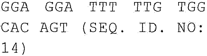
iNOS 
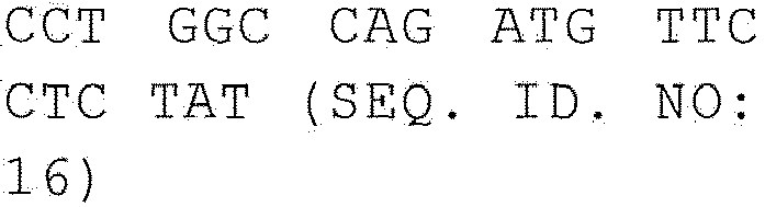
CXCL11 

CLEC4A 

SPHK-1 

IL-1β 

TNF-α 

- Next, changes of TNF-α expression at protein level was investigated according to THP-1 cell differentiation and MAPK signaling changes.
- Particularly, negative control THP-1 cells were treated with PBS(-) and 5 µg/ml of hIgG. Positive control THP-1 cells were treated with 0.1 µg/ml of PMA. In the meantime, experimental group THP-1 cells were treated with sCD93_Fc. 24 hours later, LPS was treated thereto to investigate LPS susceptibility. 24 hours after the LPS treatment, TNF-α level in the culture supernatant was measured by using Quantikine TNF-a assay kit (R&D system). As a result, TNF-α was slightly increased in the experimental group treated with sCD93_Fc alone, while TNF-α was significantly increased in the experimental group treated with both sCD93_Fc and LPS together. This result indicates that THP-1 cells were differentiated to macrophages by the treatment of sCD93_Fc with increasing susceptibility to TLR signal (
Figure 12 ). - Effect of sCD93_Fc on matrix metalloproteinase (MMP) expression in THP-1 cells was investigated. Particularly, negative control THP-1 cells were treated with 5 µg/ml of hIgG, while positive control THP-1 cells were treated with 0.1 µg/ml of PMA. In the meantime experimental group THP-1 cells were treated with sCD93_Fc at the different concentrations of 5, 1, and 0.2 µg/ml. 24 hours after the treatment, Gelatin Zymography assay was performed to investigate expressions of MMP9 (92 kDa) and MMP-2 (72 kDa) in the culture supernatant. The supernatant obtained from 24 hour culture was electrophoresed on 8% SDS-PAGE gel containing 2 mg/ml of gelatin (Sigma). Then, the gel was stained with Coomassie blue. The gel was destained with destaining solution, followed by taking pictures.
- As a result, MMP-9 level was increased by the treatment of sCD_Fc dose-dependently (
Figure 13 ). - Phagocytosis was measured using E. coli expressing mRFP to investigate phagocytosis activity according to the differentiation of THP-1 cells to macrophage-like cells. Particularly, THP-1 cells were treated with 5 µg/ml of sCD93_Fc, followed by culture for 3 days. Pre-cultured E. coli transformed with RFP (red fluorescent protein) expression vector were added to the differentiated THP-1 cells. 12 hours later, phagocytosis of E. coli by THP-1 cells was confirmed under fluorescent microscope (
Figure 14 ). Those cells were isolated by using cell dissociation buffer (EDTA buffer), which proceeded to FACS (Coolter) (Figure 15 ). - As a result, phagocytosis in the differentiated cells by the treatment of sCD93_Fc was far greater with showing fluorescence than in the negative control cells treated with hIgG.
- To investigate the possibility of development of an anti-inflammatory agent using the antibody against sCD93_Fc based on its effect on inflammation and differentiation, THP-1 cells were treated with anti-CD93 antibody, and the effect on inflammation and differentiation was examined (
Figure 16 ). Particularly, negative control THP-1 cells were treated with PBS(-) and 5 µg/ml of hIgG. Positive control THP-1 cells were treated with 0.1 µg/ml of PMA. In the meantime, experimental group THP-1 cells were treated with 5 µg/ml of sCD93_Fc and 10 µg/ml of anti-CD93 polyclonal antibody (R&D system). 24 hours later, 1 µg/ml of LPS was treated thereto to investigate LPS susceptibility. 24 hours after the LPS treatment, the amount of TNF-α in the culture supernatant was measured by suing Quantikine TNF-a assay kit (R&D system). - As a result, the amount of TNF-α was slightly increased in the experimental group treated with sCD93_Fc alone. In the meantime, TNF-α level was higher in the experimental group treated with both sCD93_Fc and LPS together than in the experimental group treated with LPS alone. However, the treatment of anti-CD93 antibody inhibited such increase of TNF-α at least 50 ∼ 60%.
- CD93 in THP-1 was knock-downed by using CD93 siRNA, and changes of various cytokines were investigated by real-time PCR.
- First, 2 x 10⁵ THP-1 cells (medium: 10% FBS (Hyclone) + RPMI1640 (Hyclone)) were distributed in 24-well plate, to which 1 ul of Lipofectamine RNAiMax (Invitrogen) and 10 pmol of CD93 siRNA (siCD93, Bioneer) were added, which stood for 10 minutes to induce transfection of the THP-1 cells. For negative control, negative control siRNA (Bioneer) was used. For positive control, full-length CD93 (CD93_Full) gene (Kugi, hMU003993, Kugi No. IRAT-49-A06) was used, followed by transfection by the same manner as described above. The cells were cultured for 12 hours in CO2 incubator, followed by LPS treatment. The cells were cultured for 12 more hours. Real-time PCR was performed with TNF-α and IL-6 by the same manner as described in Examples 2 and 3.
- As a result, levels of the two pro-inflammatory cytokines TNF-α and IL-6 were decreased by the treatment of siCD93. The increase by LPS treatment was also decreased. On the other hand, the levels of TNF-α and IL-6 were increased in the experiment group treated with CD93_Full. The levels of TNF-α and IL-6 were more increased by the treatment of LPS (
Figure 17 ). - To investigate the expression of CD93 and its fragmentation by various inflammation inducing substances or under inflammation environment, CD93 expression in THP- cells, the human mononuclear cells, and CD93 soluble fragment level in the culture supernatant were measured.
- THP-1 cells were cultured in RPMI 1640 supplemented with 10% FBS, penicillin G (100 IU/mℓ), streptomycin (100 µg/mℓ), L-glutamine (2 mM), HEPES (10 mM), and sodium pyruvate (1.0 mM) in a 37□ incubator. The cultured THP-1 cells were treated with PBS, LPS (1.0 µg/mℓ), TNF-α (0.5 µg/mℓ) or PMA (0.1 µg/mℓ) for 24 hours, followed by fixation in 4% paraformaldehyde for 20 minutes. After washing with PBS once, blocking was performed with 3% NGS (normal goat serum) for 30 minutes, followed by washing three times. The cells were reacted with ice-cold goat anti-human CD93 antibody (R&D systems) for 1 hour. After washing with PBS three times again, the cells were reacted with ice-cold anti-goat IgG-FITC antibody for 1 hour for fluorescence staining. Control groups were treated with anti-CD14-FITC antibody respectively for fluorescence staining. The stained cells were analyzed by flow cytometry (EPICS Elite, Coulter).
- As a result, as shown in
Figure 18 , CD93 expression on the cell surface of THP-1 was increased by LPS and TNF-α but hardly increased by PMA (Figure 18 ). The expression of CD14 used for the control group on the cell surface was decreased by LPS, TNF-α, and PMA, which was assumed to be attributed to CD14 fragmentation. CD14 fragmentation has been known as an important mechanism in down-modulation of monocyte-macrophage activation (Schutt, C. 1999. Cd14. Int. J. Biochem. Cell Biol. 31:545 . 23. Le-Barillec, K., M. Si-Tahar, V. Balloy, and M. Chignard. 1999. Proteolysis of monocyte CD14 by human leukocyte elastase inhibits lipopolysaccharide-mediated cell activation. J. Clin. Invest. 103:1039 . Bazil, V., and J. L. Strominger. 1991. Shedding as a mechanism of down-modulation of CD14 on stimulated human monocytes. J. Immunol. 147:1567). According to many previous research results, collagenase, neutrophil elastase, and cathepsin G can induce fragmentation of CD14 on cell surface (Bryniarski, K., K. Maresz, M. Szczepanik, M. Ptak, and W. Ptak. 2003. Modulation of macrophage activity by proteolytic enzymes. Differential regulation of IL-6 and reactive oxygen intermediates (ROIs) synthesis as a possible homeostatic mechanism in the control of inflammation. In flammation 27:333 . Le-Barillec, K., M. Si-Tahar, V. Balloy, and M. Chignard. 1999. Proteolysis of monocyte CD14 by human leukocyte elastase inhibits lipopolysaccharide-mediated cell activation. J. Clin. Invest. 103:1039 . Coyne, C. P., T. Howell III, H. Smodlaka, C. Willetto, B. W. Fenwick, and E. Chenney. 2002. Alterations in membrane-associated CD14 expression and the simultaneous liberation of soluble CD14 fragment in adherent macrophages mediated by a leukocyte carboxyl/aspartate protease. J. Endotoxin. Res. 8:273 .). - THP-1 cells were cultured in RPMI 1640 supplemented with 10% FBS, penicillin G (100 IU/ml), streptomycin (100 µg/ml), L-glutamine (2 mM), HEPES (10 mM), and sodium pyruvate (1.0 mM) in a 37□ incubator. After treating the cultured THP-1 cells with PBS, LPS (1.0 µg/ml), TNF-α (0.5 µg/ml) or PMA (0.1 µg/ml) for 24 hours, lysate and supernatant were obtained. The samples were boiled for 5 minutes and frozen for storage. After loading the samples on 8% SDS-PAGE gel, electrophoresis was performed. The gel was transferred onto nitrocellulose membrane, followed by blocking with 4% skim milk. The gel was reacted with goat anti-CD93 antibody (0.5 µg/ml), R&D systems) at room temperature for 1 hour. After washing with PBST three times, the gel was reacted with anti-goat IgG-HRP antibody (0.5 µg/ml), Santa Cruz) at room temperature for 1 hour. As internal controls, anti-β actin (Santa Cruz) and anti-goat IgG-HRP (Santa Cruz) were used. Experiment was performed using ECL kit (ECL Plus, Amersham, USA) according to the manufacturer's instructions.
- As a result, as shown in
Figure 19 , the expression of CD93 itself was increased by the treatment of LPS and TNF-α, which was consistant with the result of FACS, but it was not increased by the treatment of PMA. In the meantime, the expression of CD93 soluble fragment was increased by the treatment of LPS, TNF-α and even PMA (Figure 19 ). The above results indicate that not only CD93 itself but also CD93 soluble fragment are up-regulated in inflammation condition or by inflammation inducing substances in THP-1 and Jukat cells. Therefore, CD93 or its soluble fragment is expected to be an effective diagnostic marker for inflammatory disease. - THP-1 cells were cultured in 6 well-plate, and then treated with PBS, LPS (1.0 µg/ml, InvivoGen), and PMA (0.1 µg/ml, Sigma) respectively. 24 hours after the treatment, RNA was identified by using RNA identification kit (Qiagen). For reverse transcription, reverse transcriptase (Bio-Rad) and dNTP were mixed, leading to reaction for one hour at 42□ to obtain cDNA of each. For real-time PCR, CFX96 system (Bio-Rad) was used. PCR was performed as follows: predenaturation at 95°C□ for 3 minutes, annealing at 95°C for 15 seconds, polymerization at 60°C for 1 minute, 40 ∼ 45 cycles. Fluorescence was measured at each annealing stage and data was recorded automatically stage by stage. Particularly, 2 µl of cDNA template and CD93 primer were mixed with 23 iQ SYBR Supermix (Bio-Rad) for reaction. Every reaction was repeated twice. To measure the relative mRNA expression, average threshold cycle (Ct) value was set as standard value. Every Ct value was standardized by GAPDH. Primer sets used herein are as follows: CD93: forward primer CTC TGG GGC TAC TGG TCT ATC (SEQ. ID. NO: 34), reverse primer TGT CGG ACT GTA CTG GTT CTC (SEQ. ID. NO: 35); GAPDH: forward primer CAT GTT CGT CAT GGG TGT GAA (SEQ. ID. NO: 36), reverse primer GGA CTG TGG TCA TGA GTC CTT (SEQ. ID. NO: 37).
- As a result, as shown in
Figure 20 , CD93 expression in THP-1 cells was significantly increased by the treatment of LPS and PMA (Figure 20 ). - Like the above in vitro quantification of CD93 expression, in vivo experiment was performed as follows to investigate CD93 expression and fragmentation thereof. Particularly, peritonitis was induced in the test mouse or inflammation inducing substance (thioglycollate or LPS) was injected to the mouse. Then, CD93 expression in the mouse peripheral blood mononuclear cells and peritoneal cells was investigated and at the same time the amount of CD93 soluble fragment in serum and peritoneal lavage fluid (PLF) was measured.
- Balb/c mice at 6 weeks of age were injected with 1 ml of PBS, 4% thioglycollate or 0.1 mg/ml of LPS. Three days later, blood was drawn from retro-orbital vein of the mice using heparin-coated glass capillary tube. Then, serum and PBMC cells were obtained by centrifugation. After scarifying the mice, peritoneal lavage fluid (PLF) and peritoneal cells (mostly intraabdominal macrophages) were obtained from the abdominal cavity. To eliminate red blood cells from the cells, 10 ml of red blood cell lysing buffer (Sigma) was added, followed by reaction twice at 37□, resulting in the complete elimination of red blood cells. Fluorescence staining was performed by the same manner as described in Example <8-1>. At that time, anti-mouse CD93 antibody (R&D systems) and anti-goat IgG-FITC (Santa Cruz) were used. To investigate macrophage differentiation, double staining was performed by using CD11b antibody (BD Sciences), the differentiation marker.
- As a result, as shown in
Figure 21 , CD93 expression in macrophages of peritoneal cells of the mouse was significantly increased by the treatment of LPS. The expression was similarly increased by the treatment of thioglycollate (Figure 21 ). Therefore, it was confirmed that macrophages existing in PBMCs were moved into peritoneal fluid by the treatment of thioglycollate and LPS. - The remaining half of the cells not used in Example <9-1> was lysed, followed by Western blotting. Western blotting was performed by the same manner as described in Example <8-2> except that sheep anti-mouse CD93 antibody (R&D systems) and anti-sheep IgG-HRP (Santa Cruz) were used to detect mouse CD93. To investigate macrophage differentiation, double staining was performed by using CD11b antibody (BD Sciences), the differentiation marker. As internal controls, anti-β actin (Santa Cruz) and anti-goat IgG-HRP (Santa Cruz) were used. Experiment was performed using ECL kit (ECL Plus, Amersham, USA) according to the manufacturer's instructions.
- As a result, as shown in
Figure 22 , CD93 expression was significantly increased in mouse peritoneal cells by the treatment of thioglycollate and LPS (Figure 22 ), which was consistent with the result of FACS above. - After confirming by FACS and Western blotting that the amount of CD93 molecule itself was increased, the inventors further investigated the changes of CD93 soluble fragment. Particularly, the amount of CD93 soluble fragment in serum and peritoneal lavage fluid (PLF) was measured by sandwich ELISA.
- First, 93-well falcon plate (Becton Dickinson) was coated with rat anti-mouse CD93 antibody (R&D Systems) at the concentration of 100 ng/well for overnight. After washing the plate with PBST three times, blocking was performed with 3% skim milk at room temperature for 30 minutes. After washing the plate with PBST three times again, each of mouse serum and peritoneal lavage fluid was loaded thereto (100 µl/well), which stood for 2 hours. As standard material for quantification, mouse CD93 soluble fragment was used. After washing the
plate 3 times, 1 µg/ml of sheep anti-mouse CD93 antibody (R&D systems) was added thereto, which stood for 1 hour. One µg/ml of anti-sheep IgG-HPR antibody (Santa Cruz) was added thereto, followed by reaction for 1 hour. After washing the plate with PBST three times again, color development was induced by using OPD (Sigma). OD was measured by suing spectrophotometer (VERSAmax tunable microplate reader, Molecular Devices). Absolute quantification was performed with the measured value using standard curve. - As a result, as shown in
Figure 23 , the amount of CD93 soluble fragment in serum was hardly changed by the treatment of LPS or thioglycollate. However, the amount of CD93 soluble fragment in peritoneal lavage fluid was significantly increased by the treatment of LPS or thioglycollate (Figure 23 ). Therefore, it was suggested that CD93 soluble fragment could be effectively used as a diagnostic marker for inflammatory disease. - Synovial membrane (synovium) tissues were obtained from three rheumatoid-arthritis (RA) patients and 2 osteo-arthritis (OA) patients, which were fixed in 4% paraformaldehyde for overnight. The tissues were dehydrated with alcohol and embedded in paraffin, followed by sectioning. To eliminate peroxidase activity therein, the tissues were reacted with methanolic H2O2. To eliminate non-specific reaction, blocking was performed with 3% NGS (normal goat serum for 2 hours. The tissues were reacted with goat anti-human CD93 antibody (R&D), viotinylated anti-goat IgG, and streptavidine-peroxidase complex (Vector, Peterborough, UK) stepwise for 1 hour each. The tissues were counter-stained with hematoxyline, followed by observation under Olympus microscope (Tokyo, Japan).
- As a result, as shown in
Figure 24 , CD93 up-regulation was more significant in synovial membrane tissues obtained from rheumatoid arthritis patients than in synovial membrane tissues from osteo-arthritis patients (Figure 24 ). Particularly, CD93 expression was significantly increased in synovial lining where lymphocytes are infiltrated, sublining, and perivascular region. - The amount of CD93 soluble fragment in synovial fluid obtained from 8 rheumatoid arthritis patients and 8 osteo-arthritis patients was measured by sandwich ELISA.
- First, 93-well falcon plate (Becton Dickinson) was coated with rat anti-human CD93 antibody (R&D Systems) at the concentration of 100 ng/well for overnight. After washing the plate with PBST three times, blocking was performed with 3% skim milk at room temperature for 30 minutes. After washing the plate with PBST three times again, each of synovial fluid (SF) obtained from 8 rheumatoid arthritis patients and 8 osteo-arthritis patients was loaded thereto (100 µl/well), which stood for 2 hours. As standard material for quantification, human soluble CD93 was used. After washing the plate with 3 times, 1 µg/ml of goat anti-human CD93 antibody (R&D systems) was added thereto, which stood for 1 hour. One µg/ml of anti-goat IgG-HPR antibody (Santa Cruz) was added thereto, followed by reaction for 1 hour. After washing the plate with PBST three times again, color development was induced by using OPD (Sigma). OD was measured by suing spectrophotometer. Absolute quantification was performed with the measured value using standard curve.
- As a result, as shown in
Figure 25 , the amount of CD93 soluble fragment in synovial fluid obtained from rheumatoid arthritis patients was significantly higher than that in synovial fluid obtained from osteo-arthritis patients (Figure 25 ). - Therefore, it was suggested that CD93 molecule or its soluble fragment could be effectively used as a diagnostic marker for rheumatoid arthritis
- <110> Korea Research Institute of Bioscience and Biotechnology
- <120> Use of CD93 or soluble fragment thereof
- <130> 9fpo-12-10
- <150>
KR10-2009-0006592
<151> 2009-01-28 - <150>
KR10-2009-0116723
<151> 2009-11-30 - <160> 37
- <170> KopatentIn 1.71
- <210> 1
<211> 325
<212> PRT
<213> Homo sapiens - <400> 1


- <210> 2
<211> 975
<212> DNA
<213> Homo sapiens - <400> 2

- <210> 3
<211> 652
<212> PRT
<213> Homo sapiens - <400> 3


- <210> 4
<211> 6701
<212> DNA
<213> Homo sapiens - <400> 4


- <210> 5
<211> 22
<212> DNA
<213> Artificial Sequence - <220>
<223> CD14-New forward primer - <400> 5
gcctagacct cagccacaac tc 22 - <210> 6
<211> 19
<212> DNA
<213> Artificial Sequence - <220>
<223> CD14-New reverse primer - <400> 6
ccagcccagc gaacgacag 19 - <210> 7
<211> 21
<212> DNA
<213> Artificial Sequence - <220>
<223> CD64-New forward primer - <400> 7
gggtcagcgt gttccaagag g 21 - <210> 8
<211> 25
<212> DNA
<213> Artificial Sequence - <220>
<223> CD64-New reverse primer - <400> 8
gcacctgtat tcaccactgt cattg 25 - <210> 9
<211> 21
<212> DNA
<213> Artificial Sequence - <220>
<223> CDla-New forward primer - <400> 9
actcatacct gggacagcaa t 21 - <210> 10
<211> 21
<212> DNA
<213> Artificial Sequence - <220>
<223> CD1a-New reverse primer - <400> 10
ctgcaattca tgggcgtatc t 21 - <210> 11
<211> 21
<212> DNA
<213> Artificial Sequence - <220>
<223> IL-6 forward primer - <400> 11
tggctgaaaa agatggatgc t 21 - <210> 12
<211> 21
<212> DNA
<213> Artificial Sequence - <220>
<223> IL-6 reverse primer - <400> 12
tctggcttgt tcctcactac t 21 - <210> 13
<211> 21
<212> DNA
<213> Artificial Sequence - <220>
<223> IL-12 forward primer - <400> 13
tgtaccaggt ggagttcaag a 21 - <210> 14
<211> 21
<212> DNA
<213> Artificial Sequence - <220>
<223> IL-12 reverse primer - <400> 14
ggaggatttt tgtggcacag t 21 - <210> 15
<211> 21
<212> DNA
<213> Artificial Sequence - <220>
<223> iNOS forward primer - <400> 15
atcttgcctg gggtccatta t 21 - <210> 16
<211> 21
<212> DNA
<213> Artificial Sequence - <220>
<223> iNOS reverse primer - <400> 16
cctggccaga tgttcctcta t 21 - <210> 17
<211> 21
<212> DNA
<213> Artificial Sequence - <220>
<223> CXCL11 forward primer - <400> 17
cctggggtaa aagcagtgaa a 21 - <210> 18
<211> 21
<212> DNA
<213> Artificial Sequence - <220>
<223> CXCL11 reverse primer - <400> 18
ttgcttgctt cgatttggga t 21 - <210> 19
<211> 21
<212> DNA
<213> Artificial Sequence - <220>
<223> CLEC4A forward primer - <400> 19
ttggcaagac agtgagaagg a 21 - <210> 20
<211> 21
<212> DNA
<213> Artificial Sequence - <220>
<223> CLEC4A reverse primer - <400> 20
aatgtcgctg accttctgga t 21 - <210> 21
<211> 21
<212> DNA
<213> Artificial Sequence - <220>
<223> SPHK-1 forward primer - <400> 21
ctgtaggaag agtgggttcc a 21 - <210> 22
<211> 20
<212> DNA
<213> Artificial Sequence - <220>
<223> SPHK-1 reverse primer - <400> 22
gtgccaggac tagcacaaag 20 - <210> 23
<211> 21
<212> DNA
<213> Artificial Sequence - <220>
<223> IL-1 beta forward primer - <400> 23
acagctggag agtgtagatc c 21 - <210> 24
<211> 21
<212> DNA
<213> Artificial Sequence - <220>
<223> IL-1 beta reverse primer - <400> 24
ttttctgctt gagaggtgct g 21 - <210> 25
<211> 21
<212> DNA
<213> Artificial Sequence - <220>
<223> TNF-alpha forward primer - <400> 25
tagcccatgt tgtagcaaac c 21 - <210> 26
<211> 21
<212> DNA
<213> Artificial Sequence - <220>
<223> TNF-alpha reverse primer - <400> 26
agaggacctg ggagtagatg a 21 - <210> 27
<211> 25
<212> DNA
<213> Artificial Sequence - <220>
<223> pMEP4 forward primer - <400> 27
gtagatctgc aggaaaagga caagc 25 - <210> 28
<211> 26
<212> DNA
<213> Artificial Sequence - <220>
<223> pMEP4 reverse primer - <400> 28
cgagatctgg ttgacttccc taatgt 26 - <210> 29
<211> 40
<212> DNA
<213> Artificial Sequence - <220>
<223> Bam HI SfiI human immunoglobulin Fc N terminal sequence forward primer - <400> 29
cgggatccgg ccgtgggggc cgacaaaact cacacatgcc 40 - <210> 30
<211> 27
<212> DNA
<213> Artificial Sequence - <220>
<223> XbaI stop codon human immunoglobulin Fc C terminal sequence reverse primer - <400> 30
cgagtctcat ttacccggag acaggga 27 - <210> 31
<211> 38
<212> DNA
<213> Artificial Sequence - <220>
<223> CD93 plasmid forward primer - <400> 31
cagggggccg tgggggccac gggagctgac acggaggc 38 - <210> 32
<211> 39
<212> DNA
<213> Artificial Sequence - <220>
<223> CD93 plasmid reverse primer - <400> 32
tagcggccga cgcggccaag tccacacagt ccagctgac 39 - <210> 33
<211> 652
<212> PRT
<213> Homo sapiens - <400> 33


- <210> 34
<211> 21
<212> DNA
<213> Artificial Sequence - <220>
<223> CD93 forward primer - <400> 34
ctctggggct actggtctat c 21 - <210> 35
<211> 21
<212> DNA
<213> Artificial Sequence - <220>
<223> CD93 reverse primer - <400> 35
tgtcggactg tactggttct c 21 - <210> 36
<211> 21
<212> DNA
<213> Artificial Sequence - <220>
<223> GAPDH forward primer - <400> 36
catgttcgtc atgggtgtga a 21 - <210> 37
<211> 21
<212> DNA
<213> Artificial Sequence - <220>
<223> GAPDH reverse primer - <400> 37
ggactgtggt catgagtcct t 21
Claims (5)
- An ex vivo method for diagnosing rheumatoid arthritis comprising the following steps:1) coating protein derived from biological sample obtained from synovium tissues or synovial fluid and control respectively on a fixture;2) inducing antigen-antibody reaction by adding the antibody specifically binding to CD93 or CD93 soluble fragment to the above fixture,
wherein
CD93 is composed of the amino acid sequence represented by SEQ. ID. NO: 33, and CD93 soluble fragment is composed of the amino acid sequence of SEQ ID NO: 1;3) detecting the antigen-antibody reaction product produced by the above antigen-antibody reaction by using a secondary antibody conjugate marker and chromogenic substrate solution;4) comparing the detection results between the biological sample of experiment group and the control; and5) diagnosing rheumatoid arthritis or high risk of rheumatoid arthritis when CD93 or CD93 soluble fragment is up-regulated in the biological sample of the experiment group, compared with the level in the control. - The ex vivo method according to claim 1, wherein the fixture is the one selected from the group consisting of nitrocellulose membrane, PVDF membrane (polyvinylidene difluoride membrane), 96-well plate made of polyvinyl resin or polystylene resin, and glass slide glass.
- The ex vivo method according to claim 1, wherein the antigen-antibody reaction is measured by the method selected from the group consisting of ELISA, radioimmunoassay (RIA), sandwich ELISA, Northern blotting, Western blotting, immunoprecipitation, immunohistochemical staining, immunofluorescence method, enzyme-substrate colorimetric method, antigen-antibody agglutination, SPR (surface plasmon resonance), biochip (DNA, RNA), electrophoresis, PCR, and RT-PCR.
- The ex vivo method according to claim 1, wherein the secondary antibody conjugate marker is the one selected from the group consisting of HRP (horseradish peroxidase), alkaline phosphatase, colloid gold, FITC (poly L-lysine-fluorescein isothiocyanate), RITC (rhodamine-B-isothiocyanate), and dye.
- The ex vivo method according to claim 1, wherein the chromogenic substrate solution is the one selected from the group consisting of TMB (3,3',5,5'-tetramethyl benzidine), ABTS [2,2'-azino-bis(3-ethylbenzothiazoline-6-sulfonic acid)], and OPD (o-phenylenediamine).
Applications Claiming Priority (3)
| Application Number | Priority Date | Filing Date | Title |
|---|---|---|---|
| KR1020090006592A KR101526307B1 (en) | 2009-01-28 | 2009-01-28 | Anti-inflammatory pharmaceutical composition |
| KR1020090116723A KR101398545B1 (en) | 2009-11-30 | 2009-11-30 | Marker for diagnosis of inflammatory disease comprising CD93 or soluble fragment thereof |
| PCT/KR2010/000315 WO2010087594A2 (en) | 2009-01-28 | 2010-01-18 | Cd93 or use of soluble fragment thereof |
Publications (3)
| Publication Number | Publication Date |
|---|---|
| EP2392352A2 EP2392352A2 (en) | 2011-12-07 |
| EP2392352A4 EP2392352A4 (en) | 2013-06-26 |
| EP2392352B1 true EP2392352B1 (en) | 2017-10-25 |
Family
ID=42396160
Family Applications (1)
| Application Number | Title | Priority Date | Filing Date |
|---|---|---|---|
| EP10735985.3A Not-in-force EP2392352B1 (en) | 2009-01-28 | 2010-01-18 | Cd93 or use of soluble fragment thereof |
Country Status (4)
| Country | Link |
|---|---|
| US (1) | US8993343B2 (en) |
| EP (1) | EP2392352B1 (en) |
| JP (1) | JP5752052B2 (en) |
| WO (1) | WO2010087594A2 (en) |
Families Citing this family (13)
| Publication number | Priority date | Publication date | Assignee | Title |
|---|---|---|---|---|
| WO2015038963A1 (en) * | 2013-09-12 | 2015-03-19 | Teva Pharmaceutical Industries, Ltd. | Gene expression biomarkers of laquinimod responsiveness |
| WO2016123100A1 (en) | 2015-01-26 | 2016-08-04 | Fate Therapeutics, Inc. | Methods and compositions for inducing hematopoietic cell differentiation |
| CN108368520B (en) | 2015-11-04 | 2023-01-17 | 菲特治疗公司 | Genome engineering of pluripotent cells |
| WO2017078807A1 (en) | 2015-11-04 | 2017-05-11 | Fate Therapeutics, Inc. | Methods and compositions for inducing hematopoietic cell differentiation |
| CN110389165A (en) * | 2018-04-23 | 2019-10-29 | 江成鸿 | Application of the CD93 in preparation early warning infant hemangioma bleeding of the umbilicus detection kit and therapeutic agent |
| WO2020180706A1 (en) * | 2019-03-02 | 2020-09-10 | The Board Of Trustees Of The Leland Stanford Junior University | Therapeutic antigen binding proteins specific for cd93 and methods of use thereof |
| CN116529260A (en) * | 2020-06-02 | 2023-08-01 | 当康生物技术有限责任公司 | anti-CD 93 constructs and uses thereof |
| AU2021283345A1 (en) * | 2020-06-02 | 2023-02-02 | Dynamicure Biotechnology Llc | Anti-CD93 constructs and uses thereof |
| US20230322935A1 (en) * | 2020-07-29 | 2023-10-12 | Dynamicure Biotechnology Llc | Anti-cd93 constructs and uses thereof |
| WO2022032592A1 (en) * | 2020-08-13 | 2022-02-17 | Tsinghua University | Interleukin-17d and cd93 as a new cytokine-receptor pair in the immune system |
| CA3249492A1 (en) * | 2022-04-22 | 2023-10-26 | The Regents Of The University Of Colorado, A Body Corporate | Compositions and methods for treating idiopathic nephrotic syndrome |
| WO2025035451A1 (en) * | 2023-08-17 | 2025-02-20 | 上海君实生物医药科技股份有限公司 | Anti-cd93 antibody, and composition and use thereof |
| CN120173108A (en) * | 2023-12-19 | 2025-06-20 | 广东菲鹏制药股份有限公司 | Anti-CD93 antibody or antigen-binding fragment thereof and use thereof |
Family Cites Families (14)
| Publication number | Priority date | Publication date | Assignee | Title |
|---|---|---|---|---|
| US5648267A (en) | 1992-11-13 | 1997-07-15 | Idec Pharmaceuticals Corporation | Impaired dominant selectable marker sequence and intronic insertion strategies for enhancement of expression of gene product and expression vector systems comprising same |
| US5965439A (en) * | 1996-11-18 | 1999-10-12 | The Regents Of The University Of California | Host defense enhancement |
| US20040101874A1 (en) * | 2002-04-12 | 2004-05-27 | Mitokor Inc. | Targets for therapeutic intervention identified in the mitochondrial proteome |
| JP4338364B2 (en) | 2002-07-02 | 2009-10-07 | ソニー株式会社 | Portable information communication terminal, program, and recording medium recording the program |
| JP4190223B2 (en) * | 2002-07-19 | 2008-12-03 | 照剛 上野 | Cell disruption device, cell disruption method, treatment device, and selective separation method of cell components |
| CN1694961A (en) * | 2002-09-10 | 2005-11-09 | 内蒂穆恩公司 | Collectin-complement activating protein chimeras |
| US7482117B2 (en) * | 2002-12-20 | 2009-01-27 | Celera Corporation | Genetic polymorphisms associated with myocardial infarction, methods of detection and uses thereof |
| CA2516128A1 (en) * | 2003-02-14 | 2004-09-02 | Sagres Discovery, Inc. | Therapeutic targets in cancer |
| WO2004074837A1 (en) * | 2003-02-24 | 2004-09-02 | Digitalbiotech Co., Ltd. | Method for measuring the level of anti-beta-amyloid antibody in body fluids and diagnostic kit for alzheimer’s disease using same |
| EP2214014A1 (en) * | 2004-05-11 | 2010-08-04 | The University of Pittsburgh | Monitoring immunologic, hematologic and inflammatory diseases |
| DE102005013013A1 (en) * | 2005-03-21 | 2006-09-28 | Sirs-Lab Gmbh | Use of gene activity classifiers for the in vitro classification of gene expression profiles of patients with infectious / non-infectious multi-organ failure |
| US8362224B2 (en) * | 2006-12-19 | 2013-01-29 | The Trustees Of The University Of Pennsylvania | Screening for CD93 (C1qRp)-associated polymorphism(S) in the diagnosis, prevention and treatment of autoimmune diseases |
| JP5414678B2 (en) * | 2007-10-12 | 2014-02-12 | マックス−デルブルック−セントラム フール モレクラーレ メディツィン(エムディーシー) | Methods and kits for rapid isolation of human Foxp3 + Treg cells |
| KR200450266Y1 (en) | 2008-02-05 | 2010-09-16 | 신호근 | Spacer Determination spacer for building finishing materials |
-
2010
- 2010-01-18 US US13/146,876 patent/US8993343B2/en not_active Expired - Fee Related
- 2010-01-18 JP JP2011547769A patent/JP5752052B2/en not_active Expired - Fee Related
- 2010-01-18 WO PCT/KR2010/000315 patent/WO2010087594A2/en not_active Ceased
- 2010-01-18 EP EP10735985.3A patent/EP2392352B1/en not_active Not-in-force
Non-Patent Citations (1)
| Title |
|---|
| None * |
Also Published As
| Publication number | Publication date |
|---|---|
| WO2010087594A2 (en) | 2010-08-05 |
| EP2392352A2 (en) | 2011-12-07 |
| US20120039911A1 (en) | 2012-02-16 |
| EP2392352A4 (en) | 2013-06-26 |
| JP5752052B2 (en) | 2015-07-22 |
| JP2012516322A (en) | 2012-07-19 |
| WO2010087594A9 (en) | 2011-11-10 |
| WO2010087594A3 (en) | 2010-10-21 |
| US8993343B2 (en) | 2015-03-31 |
Similar Documents
| Publication | Publication Date | Title |
|---|---|---|
| US8993343B2 (en) | CD93 or use of soluble fragment thereof | |
| EP2370593B1 (en) | Methods for determining the efficacy of pd-1 antagonists | |
| US20110086906A1 (en) | Methods for treatment or prevention of diseases associated with functional disorder of regulatory t cells | |
| WO2015120138A2 (en) | AGENTS THAT MODULATE RGMb-NEOGENIN-BMP SIGNALING AND METHODS OF USE THEREOF | |
| JP7330174B2 (en) | Methods and pharmaceutical compositions for modulating autophagy | |
| US20070065438A1 (en) | Methods of inducing and maintaining immune tolerance | |
| US20240158861A1 (en) | Methods and compositions for treating cell senescence accumulation related disease | |
| US8748406B2 (en) | Preventive for adhesion following abdominal surgery | |
| Yan et al. | Cell targeting and immunostimulatory properties of a novel Fcγ-receptor-independent agonistic anti-CD40 antibody in rhesus macaques | |
| KR101526307B1 (en) | Anti-inflammatory pharmaceutical composition | |
| US20260035468A1 (en) | New anti-itgb8 antibodies and its uses thereof | |
| JPWO2018074610A1 (en) | Composition for the treatment of fulminant acute pneumonia comprising a CD69 antagonist | |
| RU2779308C2 (en) | Therapeutic use of inhibitors of t-cell activation or stimulation | |
| WO2018046877A1 (en) | Treatment of acute liver failure | |
| JP2023055804A (en) | Treatment, diagnosis and screening with CARD14 | |
| EP4713371A1 (en) | Anti-cathepsin-d antibodies | |
| WO2021085295A1 (en) | Immune response suppressor | |
| Nassar et al. | Impaired wound healing in SAD mice: a model to decipher the pathophysiology of sickle cell leg ulcers | |
| JP2019085370A (en) | Therapeutic or preventive agent for SLC28 or 29 gene abnormality | |
| HK1138021A (en) | Compositions and methods for the treatment of infections and tumors |
Legal Events
| Date | Code | Title | Description |
|---|---|---|---|
| PUAI | Public reference made under article 153(3) epc to a published international application that has entered the european phase |
Free format text: ORIGINAL CODE: 0009012 |
|
| 17P | Request for examination filed |
Effective date: 20110825 |
|
| AK | Designated contracting states |
Kind code of ref document: A2 Designated state(s): AT BE BG CH CY CZ DE DK EE ES FI FR GB GR HR HU IE IS IT LI LT LU LV MC MK MT NL NO PL PT RO SE SI SK SM TR |
|
| DAX | Request for extension of the european patent (deleted) | ||
| A4 | Supplementary search report drawn up and despatched |
Effective date: 20130524 |
|
| RIC1 | Information provided on ipc code assigned before grant |
Ipc: A61P 29/00 20060101ALI20130517BHEP Ipc: A61K 31/7105 20060101ALI20130517BHEP Ipc: G01N 33/53 20060101ALI20130517BHEP Ipc: A61K 39/395 20060101AFI20130517BHEP Ipc: A61P 37/08 20060101ALI20130517BHEP |
|
| 17Q | First examination report despatched |
Effective date: 20150203 |
|
| GRAJ | Information related to disapproval of communication of intention to grant by the applicant or resumption of examination proceedings by the epo deleted |
Free format text: ORIGINAL CODE: EPIDOSDIGR1 |
|
| GRAP | Despatch of communication of intention to grant a patent |
Free format text: ORIGINAL CODE: EPIDOSNIGR1 |
|
| INTG | Intention to grant announced |
Effective date: 20170322 |
|
| GRAJ | Information related to disapproval of communication of intention to grant by the applicant or resumption of examination proceedings by the epo deleted |
Free format text: ORIGINAL CODE: EPIDOSDIGR1 |
|
| INTC | Intention to grant announced (deleted) | ||
| GRAR | Information related to intention to grant a patent recorded |
Free format text: ORIGINAL CODE: EPIDOSNIGR71 |
|
| GRAS | Grant fee paid |
Free format text: ORIGINAL CODE: EPIDOSNIGR3 |
|
| GRAA | (expected) grant |
Free format text: ORIGINAL CODE: 0009210 |
|
| INTG | Intention to grant announced |
Effective date: 20170913 |
|
| AK | Designated contracting states |
Kind code of ref document: B1 Designated state(s): AT BE BG CH CY CZ DE DK EE ES FI FR GB GR HR HU IE IS IT LI LT LU LV MC MK MT NL NO PL PT RO SE SI SK SM TR |
|
| REG | Reference to a national code |
Ref country code: GB Ref legal event code: FG4D |
|
| REG | Reference to a national code |
Ref country code: CH Ref legal event code: EP |
|
| REG | Reference to a national code |
Ref country code: AT Ref legal event code: REF Ref document number: 939312 Country of ref document: AT Kind code of ref document: T Effective date: 20171115 |
|
| REG | Reference to a national code |
Ref country code: IE Ref legal event code: FG4D |
|
| REG | Reference to a national code |
Ref country code: DE Ref legal event code: R096 Ref document number: 602010046211 Country of ref document: DE |
|
| REG | Reference to a national code |
Ref country code: NL Ref legal event code: MP Effective date: 20171025 |
|
| REG | Reference to a national code |
Ref country code: LT Ref legal event code: MG4D |
|
| REG | Reference to a national code |
Ref country code: AT Ref legal event code: MK05 Ref document number: 939312 Country of ref document: AT Kind code of ref document: T Effective date: 20171025 |
|
| PG25 | Lapsed in a contracting state [announced via postgrant information from national office to epo] |
Ref country code: NL Free format text: LAPSE BECAUSE OF FAILURE TO SUBMIT A TRANSLATION OF THE DESCRIPTION OR TO PAY THE FEE WITHIN THE PRESCRIBED TIME-LIMIT Effective date: 20171025 |
|
| PG25 | Lapsed in a contracting state [announced via postgrant information from national office to epo] |
Ref country code: NO Free format text: LAPSE BECAUSE OF FAILURE TO SUBMIT A TRANSLATION OF THE DESCRIPTION OR TO PAY THE FEE WITHIN THE PRESCRIBED TIME-LIMIT Effective date: 20180125 Ref country code: FI Free format text: LAPSE BECAUSE OF FAILURE TO SUBMIT A TRANSLATION OF THE DESCRIPTION OR TO PAY THE FEE WITHIN THE PRESCRIBED TIME-LIMIT Effective date: 20171025 Ref country code: LT Free format text: LAPSE BECAUSE OF FAILURE TO SUBMIT A TRANSLATION OF THE DESCRIPTION OR TO PAY THE FEE WITHIN THE PRESCRIBED TIME-LIMIT Effective date: 20171025 Ref country code: SE Free format text: LAPSE BECAUSE OF FAILURE TO SUBMIT A TRANSLATION OF THE DESCRIPTION OR TO PAY THE FEE WITHIN THE PRESCRIBED TIME-LIMIT Effective date: 20171025 Ref country code: ES Free format text: LAPSE BECAUSE OF FAILURE TO SUBMIT A TRANSLATION OF THE DESCRIPTION OR TO PAY THE FEE WITHIN THE PRESCRIBED TIME-LIMIT Effective date: 20171025 |
|
| PGFP | Annual fee paid to national office [announced via postgrant information from national office to epo] |
Ref country code: GB Payment date: 20180117 Year of fee payment: 9 |
|
| PG25 | Lapsed in a contracting state [announced via postgrant information from national office to epo] |
Ref country code: HR Free format text: LAPSE BECAUSE OF FAILURE TO SUBMIT A TRANSLATION OF THE DESCRIPTION OR TO PAY THE FEE WITHIN THE PRESCRIBED TIME-LIMIT Effective date: 20171025 Ref country code: BG Free format text: LAPSE BECAUSE OF FAILURE TO SUBMIT A TRANSLATION OF THE DESCRIPTION OR TO PAY THE FEE WITHIN THE PRESCRIBED TIME-LIMIT Effective date: 20180125 Ref country code: IS Free format text: LAPSE BECAUSE OF FAILURE TO SUBMIT A TRANSLATION OF THE DESCRIPTION OR TO PAY THE FEE WITHIN THE PRESCRIBED TIME-LIMIT Effective date: 20180225 Ref country code: AT Free format text: LAPSE BECAUSE OF FAILURE TO SUBMIT A TRANSLATION OF THE DESCRIPTION OR TO PAY THE FEE WITHIN THE PRESCRIBED TIME-LIMIT Effective date: 20171025 Ref country code: GR Free format text: LAPSE BECAUSE OF FAILURE TO SUBMIT A TRANSLATION OF THE DESCRIPTION OR TO PAY THE FEE WITHIN THE PRESCRIBED TIME-LIMIT Effective date: 20180126 Ref country code: LV Free format text: LAPSE BECAUSE OF FAILURE TO SUBMIT A TRANSLATION OF THE DESCRIPTION OR TO PAY THE FEE WITHIN THE PRESCRIBED TIME-LIMIT Effective date: 20171025 |
|
| REG | Reference to a national code |
Ref country code: DE Ref legal event code: R097 Ref document number: 602010046211 Country of ref document: DE |
|
| PG25 | Lapsed in a contracting state [announced via postgrant information from national office to epo] |
Ref country code: EE Free format text: LAPSE BECAUSE OF FAILURE TO SUBMIT A TRANSLATION OF THE DESCRIPTION OR TO PAY THE FEE WITHIN THE PRESCRIBED TIME-LIMIT Effective date: 20171025 Ref country code: DK Free format text: LAPSE BECAUSE OF FAILURE TO SUBMIT A TRANSLATION OF THE DESCRIPTION OR TO PAY THE FEE WITHIN THE PRESCRIBED TIME-LIMIT Effective date: 20171025 Ref country code: CZ Free format text: LAPSE BECAUSE OF FAILURE TO SUBMIT A TRANSLATION OF THE DESCRIPTION OR TO PAY THE FEE WITHIN THE PRESCRIBED TIME-LIMIT Effective date: 20171025 Ref country code: CY Free format text: LAPSE BECAUSE OF FAILURE TO SUBMIT A TRANSLATION OF THE DESCRIPTION OR TO PAY THE FEE WITHIN THE PRESCRIBED TIME-LIMIT Effective date: 20171025 Ref country code: SK Free format text: LAPSE BECAUSE OF FAILURE TO SUBMIT A TRANSLATION OF THE DESCRIPTION OR TO PAY THE FEE WITHIN THE PRESCRIBED TIME-LIMIT Effective date: 20171025 |
|
| REG | Reference to a national code |
Ref country code: DE Ref legal event code: R119 Ref document number: 602010046211 Country of ref document: DE |
|
| PG25 | Lapsed in a contracting state [announced via postgrant information from national office to epo] |
Ref country code: PL Free format text: LAPSE BECAUSE OF FAILURE TO SUBMIT A TRANSLATION OF THE DESCRIPTION OR TO PAY THE FEE WITHIN THE PRESCRIBED TIME-LIMIT Effective date: 20171025 Ref country code: SM Free format text: LAPSE BECAUSE OF FAILURE TO SUBMIT A TRANSLATION OF THE DESCRIPTION OR TO PAY THE FEE WITHIN THE PRESCRIBED TIME-LIMIT Effective date: 20171025 Ref country code: RO Free format text: LAPSE BECAUSE OF FAILURE TO SUBMIT A TRANSLATION OF THE DESCRIPTION OR TO PAY THE FEE WITHIN THE PRESCRIBED TIME-LIMIT Effective date: 20171025 Ref country code: IT Free format text: LAPSE BECAUSE OF FAILURE TO SUBMIT A TRANSLATION OF THE DESCRIPTION OR TO PAY THE FEE WITHIN THE PRESCRIBED TIME-LIMIT Effective date: 20171025 |
|
| PLBE | No opposition filed within time limit |
Free format text: ORIGINAL CODE: 0009261 |
|
| REG | Reference to a national code |
Ref country code: CH Ref legal event code: PL |
|
| STAA | Information on the status of an ep patent application or granted ep patent |
Free format text: STATUS: NO OPPOSITION FILED WITHIN TIME LIMIT |
|
| 26N | No opposition filed |
Effective date: 20180726 |
|
| PG25 | Lapsed in a contracting state [announced via postgrant information from national office to epo] |
Ref country code: DE Free format text: LAPSE BECAUSE OF NON-PAYMENT OF DUE FEES Effective date: 20180801 Ref country code: FR Free format text: LAPSE BECAUSE OF NON-PAYMENT OF DUE FEES Effective date: 20180131 Ref country code: LU Free format text: LAPSE BECAUSE OF NON-PAYMENT OF DUE FEES Effective date: 20180118 |
|
| REG | Reference to a national code |
Ref country code: IE Ref legal event code: MM4A |
|
| REG | Reference to a national code |
Ref country code: FR Ref legal event code: ST Effective date: 20180928 |
|
| REG | Reference to a national code |
Ref country code: BE Ref legal event code: MM Effective date: 20180131 |
|
| PG25 | Lapsed in a contracting state [announced via postgrant information from national office to epo] |
Ref country code: SI Free format text: LAPSE BECAUSE OF FAILURE TO SUBMIT A TRANSLATION OF THE DESCRIPTION OR TO PAY THE FEE WITHIN THE PRESCRIBED TIME-LIMIT Effective date: 20171025 Ref country code: LI Free format text: LAPSE BECAUSE OF NON-PAYMENT OF DUE FEES Effective date: 20180131 Ref country code: BE Free format text: LAPSE BECAUSE OF NON-PAYMENT OF DUE FEES Effective date: 20180131 Ref country code: CH Free format text: LAPSE BECAUSE OF NON-PAYMENT OF DUE FEES Effective date: 20180131 |
|
| PG25 | Lapsed in a contracting state [announced via postgrant information from national office to epo] |
Ref country code: IE Free format text: LAPSE BECAUSE OF NON-PAYMENT OF DUE FEES Effective date: 20180118 |
|
| PG25 | Lapsed in a contracting state [announced via postgrant information from national office to epo] |
Ref country code: MC Free format text: LAPSE BECAUSE OF FAILURE TO SUBMIT A TRANSLATION OF THE DESCRIPTION OR TO PAY THE FEE WITHIN THE PRESCRIBED TIME-LIMIT Effective date: 20171025 |
|
| GBPC | Gb: european patent ceased through non-payment of renewal fee |
Effective date: 20190118 |
|
| PG25 | Lapsed in a contracting state [announced via postgrant information from national office to epo] |
Ref country code: GB Free format text: LAPSE BECAUSE OF NON-PAYMENT OF DUE FEES Effective date: 20190118 |
|
| PG25 | Lapsed in a contracting state [announced via postgrant information from national office to epo] |
Ref country code: MT Free format text: LAPSE BECAUSE OF NON-PAYMENT OF DUE FEES Effective date: 20180118 |
|
| PG25 | Lapsed in a contracting state [announced via postgrant information from national office to epo] |
Ref country code: TR Free format text: LAPSE BECAUSE OF FAILURE TO SUBMIT A TRANSLATION OF THE DESCRIPTION OR TO PAY THE FEE WITHIN THE PRESCRIBED TIME-LIMIT Effective date: 20171025 |
|
| PG25 | Lapsed in a contracting state [announced via postgrant information from national office to epo] |
Ref country code: PT Free format text: LAPSE BECAUSE OF FAILURE TO SUBMIT A TRANSLATION OF THE DESCRIPTION OR TO PAY THE FEE WITHIN THE PRESCRIBED TIME-LIMIT Effective date: 20171025 Ref country code: HU Free format text: LAPSE BECAUSE OF FAILURE TO SUBMIT A TRANSLATION OF THE DESCRIPTION OR TO PAY THE FEE WITHIN THE PRESCRIBED TIME-LIMIT; INVALID AB INITIO Effective date: 20100118 |
|
| PG25 | Lapsed in a contracting state [announced via postgrant information from national office to epo] |
Ref country code: MK Free format text: LAPSE BECAUSE OF NON-PAYMENT OF DUE FEES Effective date: 20171025 |