EP2385105A2 - Polymer blends - Google Patents
Polymer blends Download PDFInfo
- Publication number
- EP2385105A2 EP2385105A2 EP20110002580 EP11002580A EP2385105A2 EP 2385105 A2 EP2385105 A2 EP 2385105A2 EP 20110002580 EP20110002580 EP 20110002580 EP 11002580 A EP11002580 A EP 11002580A EP 2385105 A2 EP2385105 A2 EP 2385105A2
- Authority
- EP
- European Patent Office
- Prior art keywords
- poly
- pei
- plla
- pcl
- polyethylenimine
- Prior art date
- Legal status (The legal status is an assumption and is not a legal conclusion. Google has not performed a legal analysis and makes no representation as to the accuracy of the status listed.)
- Withdrawn
Links
- 229920002959 polymer blend Polymers 0.000 title abstract description 85
- -1 poly(L-lactic acid) Polymers 0.000 claims abstract description 255
- 229920002873 Polyethylenimine Polymers 0.000 claims abstract description 236
- 229920001661 Chitosan Polymers 0.000 claims abstract description 168
- 239000000203 mixture Substances 0.000 claims abstract description 156
- 229920001610 polycaprolactone Polymers 0.000 claims abstract description 115
- 229920000642 polymer Polymers 0.000 claims abstract description 109
- JVTAAEKCZFNVCJ-REOHCLBHSA-N L-lactic acid Chemical compound C[C@H](O)C(O)=O JVTAAEKCZFNVCJ-REOHCLBHSA-N 0.000 claims abstract description 98
- 229920001432 poly(L-lactide) Polymers 0.000 claims abstract description 92
- 229920002689 polyvinyl acetate Polymers 0.000 claims abstract description 58
- 239000011118 polyvinyl acetate Substances 0.000 claims abstract description 58
- 229920003171 Poly (ethylene oxide) Polymers 0.000 claims abstract description 51
- 229920003213 poly(N-isopropyl acrylamide) Polymers 0.000 claims abstract description 43
- 229920000249 biocompatible polymer Polymers 0.000 claims abstract description 40
- 239000007943 implant Substances 0.000 claims abstract description 36
- 229920002338 polyhydroxyethylmethacrylate Polymers 0.000 claims abstract description 34
- 229920002301 cellulose acetate Polymers 0.000 claims abstract description 27
- 229920001606 poly(lactic acid-co-glycolic acid) Polymers 0.000 claims abstract description 23
- WOBHKFSMXKNTIM-UHFFFAOYSA-N Hydroxyethyl methacrylate Chemical compound CC(=C)C(=O)OCCO WOBHKFSMXKNTIM-UHFFFAOYSA-N 0.000 claims abstract description 20
- 229920000118 poly(D-lactic acid) Polymers 0.000 claims abstract description 18
- WHBMMWSBFZVSSR-GSVOUGTGSA-N (R)-3-hydroxybutyric acid Chemical compound C[C@@H](O)CC(O)=O WHBMMWSBFZVSSR-GSVOUGTGSA-N 0.000 claims abstract description 13
- WHBMMWSBFZVSSR-UHFFFAOYSA-N R3HBA Natural products CC(O)CC(O)=O WHBMMWSBFZVSSR-UHFFFAOYSA-N 0.000 claims abstract description 13
- 239000000017 hydrogel Substances 0.000 claims description 38
- 210000000988 bone and bone Anatomy 0.000 claims description 30
- 210000001519 tissue Anatomy 0.000 claims description 28
- 239000000463 material Substances 0.000 claims description 26
- 230000008439 repair process Effects 0.000 claims description 13
- 210000000845 cartilage Anatomy 0.000 claims description 8
- 108090000623 proteins and genes Proteins 0.000 claims description 8
- XLYOFNOQVPJJNP-UHFFFAOYSA-N water Substances O XLYOFNOQVPJJNP-UHFFFAOYSA-N 0.000 claims description 7
- 239000003814 drug Substances 0.000 claims description 6
- 238000000338 in vitro Methods 0.000 claims description 5
- 239000003102 growth factor Substances 0.000 claims description 4
- 108090000765 processed proteins & peptides Proteins 0.000 claims description 4
- 102000004169 proteins and genes Human genes 0.000 claims description 4
- 108091032973 (ribonucleotides)n+m Proteins 0.000 claims description 3
- 108020004414 DNA Proteins 0.000 claims description 3
- 239000006143 cell culture medium Substances 0.000 claims description 3
- 201000010099 disease Diseases 0.000 claims description 3
- 208000037265 diseases, disorders, signs and symptoms Diseases 0.000 claims description 3
- 238000011534 incubation Methods 0.000 claims description 3
- 102000004196 processed proteins & peptides Human genes 0.000 claims description 3
- 229940124597 therapeutic agent Drugs 0.000 claims description 3
- 230000012010 growth Effects 0.000 abstract description 25
- RBMHUYBJIYNRLY-UHFFFAOYSA-N 2-[(1-carboxy-1-hydroxyethyl)-hydroxyphosphoryl]-2-hydroxypropanoic acid Chemical compound OC(=O)C(O)(C)P(O)(=O)C(C)(O)C(O)=O RBMHUYBJIYNRLY-UHFFFAOYSA-N 0.000 abstract description 11
- 229920001434 poly(D-lactide) Polymers 0.000 abstract description 11
- 230000001413 cellular effect Effects 0.000 abstract description 9
- 230000004069 differentiation Effects 0.000 abstract description 9
- 238000001727 in vivo Methods 0.000 abstract description 7
- 239000011159 matrix material Substances 0.000 abstract description 7
- 230000017423 tissue regeneration Effects 0.000 abstract description 5
- 210000004027 cell Anatomy 0.000 description 148
- 238000002493 microarray Methods 0.000 description 37
- 239000000243 solution Substances 0.000 description 26
- 239000000499 gel Substances 0.000 description 22
- 230000007547 defect Effects 0.000 description 21
- 230000001605 fetal effect Effects 0.000 description 20
- 210000000130 stem cell Anatomy 0.000 description 20
- 238000002156 mixing Methods 0.000 description 16
- 238000012360 testing method Methods 0.000 description 16
- 238000000034 method Methods 0.000 description 14
- 239000002904 solvent Substances 0.000 description 14
- 238000001878 scanning electron micrograph Methods 0.000 description 13
- 238000004458 analytical method Methods 0.000 description 12
- 210000000689 upper leg Anatomy 0.000 description 11
- 230000010261 cell growth Effects 0.000 description 10
- 239000000758 substrate Substances 0.000 description 10
- 210000001185 bone marrow Anatomy 0.000 description 9
- IPJDHSYCSQAODE-UHFFFAOYSA-N 5-chloromethylfluorescein diacetate Chemical compound O1C(=O)C2=CC(CCl)=CC=C2C21C1=CC=C(OC(C)=O)C=C1OC1=CC(OC(=O)C)=CC=C21 IPJDHSYCSQAODE-UHFFFAOYSA-N 0.000 description 8
- QTBSBXVTEAMEQO-UHFFFAOYSA-N Acetic acid Chemical compound CC(O)=O QTBSBXVTEAMEQO-UHFFFAOYSA-N 0.000 description 8
- HEDRZPFGACZZDS-UHFFFAOYSA-N Chloroform Chemical compound ClC(Cl)Cl HEDRZPFGACZZDS-UHFFFAOYSA-N 0.000 description 8
- SECXISVLQFMRJM-UHFFFAOYSA-N N-Methylpyrrolidone Chemical compound CN1CCCC1=O SECXISVLQFMRJM-UHFFFAOYSA-N 0.000 description 8
- 239000012620 biological material Substances 0.000 description 8
- 125000006850 spacer group Chemical group 0.000 description 8
- 229920000936 Agarose Polymers 0.000 description 7
- 238000003491 array Methods 0.000 description 7
- 238000004519 manufacturing process Methods 0.000 description 7
- 238000004626 scanning electron microscopy Methods 0.000 description 7
- HEMHJVSKTPXQMS-UHFFFAOYSA-M Sodium hydroxide Chemical compound [OH-].[Na+] HEMHJVSKTPXQMS-UHFFFAOYSA-M 0.000 description 6
- 230000003993 interaction Effects 0.000 description 6
- 102000012422 Collagen Type I Human genes 0.000 description 5
- 108010022452 Collagen Type I Proteins 0.000 description 5
- 210000002805 bone matrix Anatomy 0.000 description 5
- 230000010478 bone regeneration Effects 0.000 description 5
- 239000011521 glass Substances 0.000 description 5
- 238000004264 monolayer culture Methods 0.000 description 5
- 210000000963 osteoblast Anatomy 0.000 description 5
- 239000012071 phase Substances 0.000 description 5
- 239000011148 porous material Substances 0.000 description 5
- 238000002360 preparation method Methods 0.000 description 5
- 230000035755 proliferation Effects 0.000 description 5
- 231100000331 toxic Toxicity 0.000 description 5
- 230000002588 toxic effect Effects 0.000 description 5
- 230000006399 behavior Effects 0.000 description 4
- 230000015572 biosynthetic process Effects 0.000 description 4
- 230000015556 catabolic process Effects 0.000 description 4
- 230000004663 cell proliferation Effects 0.000 description 4
- 230000002648 chondrogenic effect Effects 0.000 description 4
- 238000006731 degradation reaction Methods 0.000 description 4
- 230000000694 effects Effects 0.000 description 4
- 238000002474 experimental method Methods 0.000 description 4
- 238000000799 fluorescence microscopy Methods 0.000 description 4
- 230000006870 function Effects 0.000 description 4
- 238000001879 gelation Methods 0.000 description 4
- 229920001519 homopolymer Polymers 0.000 description 4
- 230000009818 osteogenic differentiation Effects 0.000 description 4
- 201000008968 osteosarcoma Diseases 0.000 description 4
- 210000003491 skin Anatomy 0.000 description 4
- 230000007480 spreading Effects 0.000 description 4
- 238000003892 spreading Methods 0.000 description 4
- FWBHETKCLVMNFS-UHFFFAOYSA-N 4',6-Diamino-2-phenylindol Chemical compound C1=CC(C(=N)N)=CC=C1C1=CC2=CC=C(C(N)=N)C=C2N1 FWBHETKCLVMNFS-UHFFFAOYSA-N 0.000 description 3
- 108010025020 Nerve Growth Factor Proteins 0.000 description 3
- 229960000583 acetic acid Drugs 0.000 description 3
- 229920006125 amorphous polymer Polymers 0.000 description 3
- 238000013459 approach Methods 0.000 description 3
- 230000008901 benefit Effects 0.000 description 3
- 238000004113 cell culture Methods 0.000 description 3
- 238000013461 design Methods 0.000 description 3
- 229940079593 drug Drugs 0.000 description 3
- 238000001035 drying Methods 0.000 description 3
- 229920001971 elastomer Polymers 0.000 description 3
- 210000002950 fibroblast Anatomy 0.000 description 3
- 230000003328 fibroblastic effect Effects 0.000 description 3
- 238000010438 heat treatment Methods 0.000 description 3
- 238000011835 investigation Methods 0.000 description 3
- 239000003550 marker Substances 0.000 description 3
- 210000003205 muscle Anatomy 0.000 description 3
- 210000005036 nerve Anatomy 0.000 description 3
- 238000006386 neutralization reaction Methods 0.000 description 3
- 230000011164 ossification Effects 0.000 description 3
- 238000012545 processing Methods 0.000 description 3
- 238000012800 visualization Methods 0.000 description 3
- RYHBNJHYFVUHQT-UHFFFAOYSA-N 1,4-Dioxane Chemical compound C1COCCO1 RYHBNJHYFVUHQT-UHFFFAOYSA-N 0.000 description 2
- PRDFBSVERLRRMY-UHFFFAOYSA-N 2'-(4-ethoxyphenyl)-5-(4-methylpiperazin-1-yl)-2,5'-bibenzimidazole Chemical compound C1=CC(OCC)=CC=C1C1=NC2=CC=C(C=3NC4=CC(=CC=C4N=3)N3CCN(C)CC3)C=C2N1 PRDFBSVERLRRMY-UHFFFAOYSA-N 0.000 description 2
- 102000016284 Aggrecans Human genes 0.000 description 2
- 108010067219 Aggrecans Proteins 0.000 description 2
- CIWBSHSKHKDKBQ-JLAZNSOCSA-N Ascorbic acid Chemical compound OC[C@H](O)[C@H]1OC(=O)C(O)=C1O CIWBSHSKHKDKBQ-JLAZNSOCSA-N 0.000 description 2
- IJGRMHOSHXDMSA-UHFFFAOYSA-N Atomic nitrogen Chemical compound N#N IJGRMHOSHXDMSA-UHFFFAOYSA-N 0.000 description 2
- 102000007350 Bone Morphogenetic Proteins Human genes 0.000 description 2
- 108010007726 Bone Morphogenetic Proteins Proteins 0.000 description 2
- 108091003079 Bovine Serum Albumin Proteins 0.000 description 2
- 101150008656 COL1A1 gene Proteins 0.000 description 2
- 101150082216 COL2A1 gene Proteins 0.000 description 2
- 102000003951 Erythropoietin Human genes 0.000 description 2
- 108090000394 Erythropoietin Proteins 0.000 description 2
- 102000010834 Extracellular Matrix Proteins Human genes 0.000 description 2
- 108010037362 Extracellular Matrix Proteins Proteins 0.000 description 2
- 102000018233 Fibroblast Growth Factor Human genes 0.000 description 2
- 108050007372 Fibroblast Growth Factor Proteins 0.000 description 2
- WSFSSNUMVMOOMR-UHFFFAOYSA-N Formaldehyde Chemical compound O=C WSFSSNUMVMOOMR-UHFFFAOYSA-N 0.000 description 2
- 102000004269 Granulocyte Colony-Stimulating Factor Human genes 0.000 description 2
- 108010017080 Granulocyte Colony-Stimulating Factor Proteins 0.000 description 2
- 108010017213 Granulocyte-Macrophage Colony-Stimulating Factor Proteins 0.000 description 2
- 102100039620 Granulocyte-macrophage colony-stimulating factor Human genes 0.000 description 2
- 102000004858 Growth differentiation factor-9 Human genes 0.000 description 2
- 108090001086 Growth differentiation factor-9 Proteins 0.000 description 2
- 102100039939 Growth/differentiation factor 8 Human genes 0.000 description 2
- 102000003745 Hepatocyte Growth Factor Human genes 0.000 description 2
- 108090000100 Hepatocyte Growth Factor Proteins 0.000 description 2
- 108090000723 Insulin-Like Growth Factor I Proteins 0.000 description 2
- 108010056852 Myostatin Proteins 0.000 description 2
- 102000015336 Nerve Growth Factor Human genes 0.000 description 2
- 101150008755 PCNA gene Proteins 0.000 description 2
- 108010038512 Platelet-Derived Growth Factor Proteins 0.000 description 2
- 102000010780 Platelet-Derived Growth Factor Human genes 0.000 description 2
- 229920002125 Sokalan® Polymers 0.000 description 2
- 102000013275 Somatomedins Human genes 0.000 description 2
- 229910000831 Steel Inorganic materials 0.000 description 2
- 102000036693 Thrombopoietin Human genes 0.000 description 2
- 108010041111 Thrombopoietin Proteins 0.000 description 2
- 102000046299 Transforming Growth Factor beta1 Human genes 0.000 description 2
- 101800002279 Transforming growth factor beta-1 Proteins 0.000 description 2
- 108010073929 Vascular Endothelial Growth Factor A Proteins 0.000 description 2
- 102000005789 Vascular Endothelial Growth Factors Human genes 0.000 description 2
- 108010019530 Vascular Endothelial Growth Factors Proteins 0.000 description 2
- 239000007640 basal medium Substances 0.000 description 2
- 229920002988 biodegradable polymer Polymers 0.000 description 2
- 239000004621 biodegradable polymer Substances 0.000 description 2
- 229940112869 bone morphogenetic protein Drugs 0.000 description 2
- 230000024245 cell differentiation Effects 0.000 description 2
- 230000003833 cell viability Effects 0.000 description 2
- 238000012512 characterization method Methods 0.000 description 2
- 239000011557 critical solution Substances 0.000 description 2
- 238000005538 encapsulation Methods 0.000 description 2
- 229940105423 erythropoietin Drugs 0.000 description 2
- 238000011156 evaluation Methods 0.000 description 2
- 210000002744 extracellular matrix Anatomy 0.000 description 2
- 239000012091 fetal bovine serum Substances 0.000 description 2
- 229940126864 fibroblast growth factor Drugs 0.000 description 2
- 239000010408 film Substances 0.000 description 2
- MHMNJMPURVTYEJ-UHFFFAOYSA-N fluorescein-5-isothiocyanate Chemical compound O1C(=O)C2=CC(N=C=S)=CC=C2C21C1=CC=C(O)C=C1OC1=CC(O)=CC=C21 MHMNJMPURVTYEJ-UHFFFAOYSA-N 0.000 description 2
- 238000004108 freeze drying Methods 0.000 description 2
- 230000035876 healing Effects 0.000 description 2
- 229910052739 hydrogen Inorganic materials 0.000 description 2
- 230000008595 infiltration Effects 0.000 description 2
- 238000001764 infiltration Methods 0.000 description 2
- 238000002955 isolation Methods 0.000 description 2
- 239000010410 layer Substances 0.000 description 2
- 210000004962 mammalian cell Anatomy 0.000 description 2
- 238000013508 migration Methods 0.000 description 2
- 230000005012 migration Effects 0.000 description 2
- 229940053128 nerve growth factor Drugs 0.000 description 2
- 229910052757 nitrogen Inorganic materials 0.000 description 2
- 238000010899 nucleation Methods 0.000 description 2
- 230000002188 osteogenic effect Effects 0.000 description 2
- 238000005191 phase separation Methods 0.000 description 2
- OXCMYAYHXIHQOA-UHFFFAOYSA-N potassium;[2-butyl-5-chloro-3-[[4-[2-(1,2,4-triaza-3-azanidacyclopenta-1,4-dien-5-yl)phenyl]phenyl]methyl]imidazol-4-yl]methanol Chemical compound [K+].CCCCC1=NC(Cl)=C(CO)N1CC1=CC=C(C=2C(=CC=CC=2)C2=N[N-]N=N2)C=C1 OXCMYAYHXIHQOA-UHFFFAOYSA-N 0.000 description 2
- 239000000047 product Substances 0.000 description 2
- 238000011160 research Methods 0.000 description 2
- 239000010959 steel Substances 0.000 description 2
- 238000003860 storage Methods 0.000 description 2
- UCSJYZPVAKXKNQ-HZYVHMACSA-N streptomycin Chemical compound CN[C@H]1[C@H](O)[C@@H](O)[C@H](CO)O[C@H]1O[C@@H]1[C@](C=O)(O)[C@H](C)O[C@H]1O[C@@H]1[C@@H](NC(N)=N)[C@H](O)[C@@H](NC(N)=N)[C@H](O)[C@H]1O UCSJYZPVAKXKNQ-HZYVHMACSA-N 0.000 description 2
- 239000000126 substance Substances 0.000 description 2
- 230000007704 transition Effects 0.000 description 2
- 102000002260 Alkaline Phosphatase Human genes 0.000 description 1
- 108020004774 Alkaline Phosphatase Proteins 0.000 description 1
- FERIUCNNQQJTOY-UHFFFAOYSA-N Butyric acid Natural products CCCC(O)=O FERIUCNNQQJTOY-UHFFFAOYSA-N 0.000 description 1
- 102000000503 Collagen Type II Human genes 0.000 description 1
- 108010041390 Collagen Type II Proteins 0.000 description 1
- 239000006144 Dulbecco’s modified Eagle's medium Substances 0.000 description 1
- 238000005033 Fourier transform infrared spectroscopy Methods 0.000 description 1
- 108060003393 Granulin Proteins 0.000 description 1
- 102000016921 Integrin-Binding Sialoprotein Human genes 0.000 description 1
- 108010028750 Integrin-Binding Sialoprotein Proteins 0.000 description 1
- 102000007072 Nerve Growth Factors Human genes 0.000 description 1
- 108090000573 Osteocalcin Proteins 0.000 description 1
- 102000004067 Osteocalcin Human genes 0.000 description 1
- 102000009890 Osteonectin Human genes 0.000 description 1
- 108010077077 Osteonectin Proteins 0.000 description 1
- 102000004264 Osteopontin Human genes 0.000 description 1
- 108010081689 Osteopontin Proteins 0.000 description 1
- 229930182555 Penicillin Natural products 0.000 description 1
- JGSARLDLIJGVTE-MBNYWOFBSA-N Penicillin G Chemical compound N([C@H]1[C@H]2SC([C@@H](N2C1=O)C(O)=O)(C)C)C(=O)CC1=CC=CC=C1 JGSARLDLIJGVTE-MBNYWOFBSA-N 0.000 description 1
- 239000004743 Polypropylene Substances 0.000 description 1
- 238000011529 RT qPCR Methods 0.000 description 1
- 206010040943 Skin Ulcer Diseases 0.000 description 1
- 102000056172 Transforming growth factor beta-3 Human genes 0.000 description 1
- 108090000097 Transforming growth factor beta-3 Proteins 0.000 description 1
- 208000027418 Wounds and injury Diseases 0.000 description 1
- 239000003929 acidic solution Substances 0.000 description 1
- 230000002293 adipogenic effect Effects 0.000 description 1
- 239000003242 anti bacterial agent Substances 0.000 description 1
- 229940088710 antibiotic agent Drugs 0.000 description 1
- 229910052586 apatite Inorganic materials 0.000 description 1
- 239000012736 aqueous medium Substances 0.000 description 1
- 239000008346 aqueous phase Substances 0.000 description 1
- 239000007864 aqueous solution Substances 0.000 description 1
- 229940072107 ascorbate Drugs 0.000 description 1
- 235000010323 ascorbic acid Nutrition 0.000 description 1
- 239000011668 ascorbic acid Substances 0.000 description 1
- 239000012298 atmosphere Substances 0.000 description 1
- 239000000560 biocompatible material Substances 0.000 description 1
- 230000003592 biomimetic effect Effects 0.000 description 1
- 210000002449 bone cell Anatomy 0.000 description 1
- 210000004271 bone marrow stromal cell Anatomy 0.000 description 1
- 239000001506 calcium phosphate Substances 0.000 description 1
- 229910000389 calcium phosphate Inorganic materials 0.000 description 1
- 235000011010 calcium phosphates Nutrition 0.000 description 1
- 229920006317 cationic polymer Polymers 0.000 description 1
- 230000003915 cell function Effects 0.000 description 1
- 239000006285 cell suspension Substances 0.000 description 1
- 230000008859 change Effects 0.000 description 1
- 210000001612 chondrocyte Anatomy 0.000 description 1
- 230000009816 chondrogenic differentiation Effects 0.000 description 1
- 229940096422 collagen type i Drugs 0.000 description 1
- 150000001875 compounds Chemical class 0.000 description 1
- 210000002808 connective tissue Anatomy 0.000 description 1
- 210000001608 connective tissue cell Anatomy 0.000 description 1
- 238000002508 contact lithography Methods 0.000 description 1
- 238000004132 cross linking Methods 0.000 description 1
- 239000002178 crystalline material Substances 0.000 description 1
- 238000012258 culturing Methods 0.000 description 1
- 230000006378 damage Effects 0.000 description 1
- 230000001419 dependent effect Effects 0.000 description 1
- 238000011161 development Methods 0.000 description 1
- 230000018109 developmental process Effects 0.000 description 1
- UREBDLICKHMUKA-CXSFZGCWSA-N dexamethasone Chemical compound C1CC2=CC(=O)C=C[C@]2(C)[C@]2(F)[C@@H]1[C@@H]1C[C@@H](C)[C@@](C(=O)CO)(O)[C@@]1(C)C[C@@H]2O UREBDLICKHMUKA-CXSFZGCWSA-N 0.000 description 1
- 229960003957 dexamethasone Drugs 0.000 description 1
- 238000012377 drug delivery Methods 0.000 description 1
- 230000005684 electric field Effects 0.000 description 1
- 230000001747 exhibiting effect Effects 0.000 description 1
- 210000000604 fetal stem cell Anatomy 0.000 description 1
- 210000003754 fetus Anatomy 0.000 description 1
- 239000000835 fiber Substances 0.000 description 1
- 235000013305 food Nutrition 0.000 description 1
- 239000012634 fragment Substances 0.000 description 1
- 239000007789 gas Substances 0.000 description 1
- 239000012362 glacial acetic acid Substances 0.000 description 1
- 230000009477 glass transition Effects 0.000 description 1
- ZDXPYRJPNDTMRX-UHFFFAOYSA-N glutamine Natural products OC(=O)C(N)CCC(N)=O ZDXPYRJPNDTMRX-UHFFFAOYSA-N 0.000 description 1
- 239000001963 growth medium Substances 0.000 description 1
- 238000011540 hip replacement Methods 0.000 description 1
- 239000001257 hydrogen Substances 0.000 description 1
- 230000002209 hydrophobic effect Effects 0.000 description 1
- 238000005286 illumination Methods 0.000 description 1
- 238000003384 imaging method Methods 0.000 description 1
- 229910052738 indium Inorganic materials 0.000 description 1
- APFVFJFRJDLVQX-UHFFFAOYSA-N indium atom Chemical compound [In] APFVFJFRJDLVQX-UHFFFAOYSA-N 0.000 description 1
- 230000006698 induction Effects 0.000 description 1
- 208000014674 injury Diseases 0.000 description 1
- 229910052500 inorganic mineral Inorganic materials 0.000 description 1
- 238000007689 inspection Methods 0.000 description 1
- 239000011872 intimate mixture Substances 0.000 description 1
- 238000012804 iterative process Methods 0.000 description 1
- 230000007774 longterm Effects 0.000 description 1
- 238000002826 magnetic-activated cell sorting Methods 0.000 description 1
- 238000005259 measurement Methods 0.000 description 1
- 239000002609 medium Substances 0.000 description 1
- 238000002844 melting Methods 0.000 description 1
- 230000008018 melting Effects 0.000 description 1
- 210000002901 mesenchymal stem cell Anatomy 0.000 description 1
- 229910052751 metal Inorganic materials 0.000 description 1
- 239000002184 metal Substances 0.000 description 1
- 238000001000 micrograph Methods 0.000 description 1
- 230000003278 mimic effect Effects 0.000 description 1
- 239000011707 mineral Substances 0.000 description 1
- 238000012986 modification Methods 0.000 description 1
- 230000004048 modification Effects 0.000 description 1
- 230000000921 morphogenic effect Effects 0.000 description 1
- 230000000877 morphologic effect Effects 0.000 description 1
- 230000004660 morphological change Effects 0.000 description 1
- 230000000399 orthopedic effect Effects 0.000 description 1
- 230000003349 osteoarthritic effect Effects 0.000 description 1
- 230000001582 osteoblastic effect Effects 0.000 description 1
- 230000001009 osteoporotic effect Effects 0.000 description 1
- 210000004663 osteoprogenitor cell Anatomy 0.000 description 1
- 229940049954 penicillin Drugs 0.000 description 1
- VSIIXMUUUJUKCM-UHFFFAOYSA-D pentacalcium;fluoride;triphosphate Chemical compound [F-].[Ca+2].[Ca+2].[Ca+2].[Ca+2].[Ca+2].[O-]P([O-])([O-])=O.[O-]P([O-])([O-])=O.[O-]P([O-])([O-])=O VSIIXMUUUJUKCM-UHFFFAOYSA-D 0.000 description 1
- 238000000053 physical method Methods 0.000 description 1
- 229920001245 poly(D,L-lactide-co-caprolactone) Polymers 0.000 description 1
- 229920002401 polyacrylamide Polymers 0.000 description 1
- 229920001223 polyethylene glycol Polymers 0.000 description 1
- 229920001155 polypropylene Polymers 0.000 description 1
- 229920002635 polyurethane Polymers 0.000 description 1
- 239000004814 polyurethane Substances 0.000 description 1
- 239000002244 precipitate Substances 0.000 description 1
- 230000035935 pregnancy Effects 0.000 description 1
- 230000008569 process Effects 0.000 description 1
- 230000002035 prolonged effect Effects 0.000 description 1
- 238000010926 purge Methods 0.000 description 1
- 238000000746 purification Methods 0.000 description 1
- 239000012781 shape memory material Substances 0.000 description 1
- 231100000019 skin ulcer Toxicity 0.000 description 1
- 210000004872 soft tissue Anatomy 0.000 description 1
- 239000007787 solid Substances 0.000 description 1
- 229960005322 streptomycin Drugs 0.000 description 1
- 238000001356 surgical procedure Methods 0.000 description 1
- 239000000725 suspension Substances 0.000 description 1
- 238000001308 synthesis method Methods 0.000 description 1
- 230000002123 temporal effect Effects 0.000 description 1
- 230000001225 therapeutic effect Effects 0.000 description 1
- 239000010409 thin film Substances 0.000 description 1
- 231100000419 toxicity Toxicity 0.000 description 1
- 230000001988 toxicity Effects 0.000 description 1
- 230000002463 transducing effect Effects 0.000 description 1
- 229940099456 transforming growth factor beta 1 Drugs 0.000 description 1
- QORWJWZARLRLPR-UHFFFAOYSA-H tricalcium bis(phosphate) Chemical compound [Ca+2].[Ca+2].[Ca+2].[O-]P([O-])([O-])=O.[O-]P([O-])([O-])=O QORWJWZARLRLPR-UHFFFAOYSA-H 0.000 description 1
- 238000009281 ultraviolet germicidal irradiation Methods 0.000 description 1
- VBEQCZHXXJYVRD-GACYYNSASA-N uroanthelone Chemical compound C([C@@H](C(=O)N[C@H](C(=O)N[C@@H](CS)C(=O)N[C@@H](CC(N)=O)C(=O)N[C@@H](CS)C(=O)N[C@H](C(=O)N[C@@H]([C@@H](C)CC)C(=O)NCC(=O)N[C@@H](CC=1C=CC(O)=CC=1)C(=O)N[C@@H](CO)C(=O)NCC(=O)N[C@@H](CC(O)=O)C(=O)N[C@@H](CCCNC(N)=N)C(=O)N[C@@H](CS)C(=O)N[C@@H](CCC(N)=O)C(=O)N[C@@H]([C@@H](C)O)C(=O)N[C@@H](CCCNC(N)=N)C(=O)N[C@@H](CC(O)=O)C(=O)N[C@@H](CC(C)C)C(=O)N[C@@H](CCCNC(N)=N)C(=O)N[C@@H](CC=1C2=CC=CC=C2NC=1)C(=O)N[C@@H](CC=1C2=CC=CC=C2NC=1)C(=O)N[C@@H](CCC(O)=O)C(=O)N[C@@H](CC(C)C)C(=O)N[C@@H](CCCNC(N)=N)C(O)=O)C(C)C)[C@@H](C)O)NC(=O)[C@H](CO)NC(=O)[C@H](CC(O)=O)NC(=O)[C@H](CC(C)C)NC(=O)[C@H](CO)NC(=O)[C@H](CCC(O)=O)NC(=O)[C@@H](NC(=O)[C@H](CC=1NC=NC=1)NC(=O)[C@H](CCSC)NC(=O)[C@H](CS)NC(=O)[C@@H](NC(=O)CNC(=O)CNC(=O)[C@H](CC(N)=O)NC(=O)[C@H](CC(C)C)NC(=O)[C@H](CS)NC(=O)[C@H](CC=1C=CC(O)=CC=1)NC(=O)CNC(=O)[C@H](CC(O)=O)NC(=O)[C@H](CC=1C=CC(O)=CC=1)NC(=O)[C@H](CO)NC(=O)[C@H](CO)NC(=O)[C@H]1N(CCC1)C(=O)[C@H](CS)NC(=O)CNC(=O)[C@H]1N(CCC1)C(=O)[C@H](CC=1C=CC(O)=CC=1)NC(=O)[C@H](CO)NC(=O)[C@@H](N)CC(N)=O)C(C)C)[C@@H](C)CC)C1=CC=C(O)C=C1 VBEQCZHXXJYVRD-GACYYNSASA-N 0.000 description 1
- 230000035899 viability Effects 0.000 description 1
- 210000001835 viscera Anatomy 0.000 description 1
- 238000005406 washing Methods 0.000 description 1
Images






Classifications
-
- C—CHEMISTRY; METALLURGY
- C12—BIOCHEMISTRY; BEER; SPIRITS; WINE; VINEGAR; MICROBIOLOGY; ENZYMOLOGY; MUTATION OR GENETIC ENGINEERING
- C12N—MICROORGANISMS OR ENZYMES; COMPOSITIONS THEREOF; PROPAGATING, PRESERVING, OR MAINTAINING MICROORGANISMS; MUTATION OR GENETIC ENGINEERING; CULTURE MEDIA
- C12N5/00—Undifferentiated human, animal or plant cells, e.g. cell lines; Tissues; Cultivation or maintenance thereof; Culture media therefor
- C12N5/0068—General culture methods using substrates
-
- A—HUMAN NECESSITIES
- A61—MEDICAL OR VETERINARY SCIENCE; HYGIENE
- A61L—METHODS OR APPARATUS FOR STERILISING MATERIALS OR OBJECTS IN GENERAL; DISINFECTION, STERILISATION OR DEODORISATION OF AIR; CHEMICAL ASPECTS OF BANDAGES, DRESSINGS, ABSORBENT PADS OR SURGICAL ARTICLES; MATERIALS FOR BANDAGES, DRESSINGS, ABSORBENT PADS OR SURGICAL ARTICLES
- A61L27/00—Materials for grafts or prostheses or for coating grafts or prostheses
- A61L27/14—Macromolecular materials
- A61L27/26—Mixtures of macromolecular compounds
-
- A—HUMAN NECESSITIES
- A61—MEDICAL OR VETERINARY SCIENCE; HYGIENE
- A61L—METHODS OR APPARATUS FOR STERILISING MATERIALS OR OBJECTS IN GENERAL; DISINFECTION, STERILISATION OR DEODORISATION OF AIR; CHEMICAL ASPECTS OF BANDAGES, DRESSINGS, ABSORBENT PADS OR SURGICAL ARTICLES; MATERIALS FOR BANDAGES, DRESSINGS, ABSORBENT PADS OR SURGICAL ARTICLES
- A61L27/00—Materials for grafts or prostheses or for coating grafts or prostheses
- A61L27/50—Materials characterised by their function or physical properties, e.g. injectable or lubricating compositions, shape-memory materials, surface modified materials
- A61L27/52—Hydrogels or hydrocolloids
-
- A—HUMAN NECESSITIES
- A61—MEDICAL OR VETERINARY SCIENCE; HYGIENE
- A61L—METHODS OR APPARATUS FOR STERILISING MATERIALS OR OBJECTS IN GENERAL; DISINFECTION, STERILISATION OR DEODORISATION OF AIR; CHEMICAL ASPECTS OF BANDAGES, DRESSINGS, ABSORBENT PADS OR SURGICAL ARTICLES; MATERIALS FOR BANDAGES, DRESSINGS, ABSORBENT PADS OR SURGICAL ARTICLES
- A61L27/00—Materials for grafts or prostheses or for coating grafts or prostheses
- A61L27/50—Materials characterised by their function or physical properties, e.g. injectable or lubricating compositions, shape-memory materials, surface modified materials
- A61L27/54—Biologically active materials, e.g. therapeutic substances
-
- A—HUMAN NECESSITIES
- A61—MEDICAL OR VETERINARY SCIENCE; HYGIENE
- A61L—METHODS OR APPARATUS FOR STERILISING MATERIALS OR OBJECTS IN GENERAL; DISINFECTION, STERILISATION OR DEODORISATION OF AIR; CHEMICAL ASPECTS OF BANDAGES, DRESSINGS, ABSORBENT PADS OR SURGICAL ARTICLES; MATERIALS FOR BANDAGES, DRESSINGS, ABSORBENT PADS OR SURGICAL ARTICLES
- A61L31/00—Materials for other surgical articles, e.g. stents, stent-grafts, shunts, surgical drapes, guide wires, materials for adhesion prevention, occluding devices, surgical gloves, tissue fixation devices
- A61L31/04—Macromolecular materials
- A61L31/041—Mixtures of macromolecular compounds
-
- A—HUMAN NECESSITIES
- A61—MEDICAL OR VETERINARY SCIENCE; HYGIENE
- A61L—METHODS OR APPARATUS FOR STERILISING MATERIALS OR OBJECTS IN GENERAL; DISINFECTION, STERILISATION OR DEODORISATION OF AIR; CHEMICAL ASPECTS OF BANDAGES, DRESSINGS, ABSORBENT PADS OR SURGICAL ARTICLES; MATERIALS FOR BANDAGES, DRESSINGS, ABSORBENT PADS OR SURGICAL ARTICLES
- A61L31/00—Materials for other surgical articles, e.g. stents, stent-grafts, shunts, surgical drapes, guide wires, materials for adhesion prevention, occluding devices, surgical gloves, tissue fixation devices
- A61L31/14—Materials characterised by their function or physical properties, e.g. injectable or lubricating compositions, shape-memory materials, surface modified materials
- A61L31/145—Hydrogels or hydrocolloids
-
- A—HUMAN NECESSITIES
- A61—MEDICAL OR VETERINARY SCIENCE; HYGIENE
- A61L—METHODS OR APPARATUS FOR STERILISING MATERIALS OR OBJECTS IN GENERAL; DISINFECTION, STERILISATION OR DEODORISATION OF AIR; CHEMICAL ASPECTS OF BANDAGES, DRESSINGS, ABSORBENT PADS OR SURGICAL ARTICLES; MATERIALS FOR BANDAGES, DRESSINGS, ABSORBENT PADS OR SURGICAL ARTICLES
- A61L31/00—Materials for other surgical articles, e.g. stents, stent-grafts, shunts, surgical drapes, guide wires, materials for adhesion prevention, occluding devices, surgical gloves, tissue fixation devices
- A61L31/14—Materials characterised by their function or physical properties, e.g. injectable or lubricating compositions, shape-memory materials, surface modified materials
- A61L31/16—Biologically active materials, e.g. therapeutic substances
-
- A—HUMAN NECESSITIES
- A61—MEDICAL OR VETERINARY SCIENCE; HYGIENE
- A61P—SPECIFIC THERAPEUTIC ACTIVITY OF CHEMICAL COMPOUNDS OR MEDICINAL PREPARATIONS
- A61P19/00—Drugs for skeletal disorders
-
- A—HUMAN NECESSITIES
- A61—MEDICAL OR VETERINARY SCIENCE; HYGIENE
- A61P—SPECIFIC THERAPEUTIC ACTIVITY OF CHEMICAL COMPOUNDS OR MEDICINAL PREPARATIONS
- A61P19/00—Drugs for skeletal disorders
- A61P19/04—Drugs for skeletal disorders for non-specific disorders of the connective tissue
-
- A—HUMAN NECESSITIES
- A61—MEDICAL OR VETERINARY SCIENCE; HYGIENE
- A61P—SPECIFIC THERAPEUTIC ACTIVITY OF CHEMICAL COMPOUNDS OR MEDICINAL PREPARATIONS
- A61P19/00—Drugs for skeletal disorders
- A61P19/08—Drugs for skeletal disorders for bone diseases, e.g. rachitism, Paget's disease
-
- A—HUMAN NECESSITIES
- A61—MEDICAL OR VETERINARY SCIENCE; HYGIENE
- A61L—METHODS OR APPARATUS FOR STERILISING MATERIALS OR OBJECTS IN GENERAL; DISINFECTION, STERILISATION OR DEODORISATION OF AIR; CHEMICAL ASPECTS OF BANDAGES, DRESSINGS, ABSORBENT PADS OR SURGICAL ARTICLES; MATERIALS FOR BANDAGES, DRESSINGS, ABSORBENT PADS OR SURGICAL ARTICLES
- A61L2300/00—Biologically active materials used in bandages, wound dressings, absorbent pads or medical devices
-
- C—CHEMISTRY; METALLURGY
- C12—BIOCHEMISTRY; BEER; SPIRITS; WINE; VINEGAR; MICROBIOLOGY; ENZYMOLOGY; MUTATION OR GENETIC ENGINEERING
- C12N—MICROORGANISMS OR ENZYMES; COMPOSITIONS THEREOF; PROPAGATING, PRESERVING, OR MAINTAINING MICROORGANISMS; MUTATION OR GENETIC ENGINEERING; CULTURE MEDIA
- C12N2533/00—Supports or coatings for cell culture, characterised by material
- C12N2533/30—Synthetic polymers
-
- C—CHEMISTRY; METALLURGY
- C12—BIOCHEMISTRY; BEER; SPIRITS; WINE; VINEGAR; MICROBIOLOGY; ENZYMOLOGY; MUTATION OR GENETIC ENGINEERING
- C12N—MICROORGANISMS OR ENZYMES; COMPOSITIONS THEREOF; PROPAGATING, PRESERVING, OR MAINTAINING MICROORGANISMS; MUTATION OR GENETIC ENGINEERING; CULTURE MEDIA
- C12N2533/00—Supports or coatings for cell culture, characterised by material
- C12N2533/30—Synthetic polymers
- C12N2533/40—Polyhydroxyacids, e.g. polymers of glycolic or lactic acid (PGA, PLA, PLGA); Bioresorbable polymers
-
- C—CHEMISTRY; METALLURGY
- C12—BIOCHEMISTRY; BEER; SPIRITS; WINE; VINEGAR; MICROBIOLOGY; ENZYMOLOGY; MUTATION OR GENETIC ENGINEERING
- C12N—MICROORGANISMS OR ENZYMES; COMPOSITIONS THEREOF; PROPAGATING, PRESERVING, OR MAINTAINING MICROORGANISMS; MUTATION OR GENETIC ENGINEERING; CULTURE MEDIA
- C12N2533/00—Supports or coatings for cell culture, characterised by material
- C12N2533/70—Polysaccharides
- C12N2533/72—Chitin, chitosan
-
- Y—GENERAL TAGGING OF NEW TECHNOLOGICAL DEVELOPMENTS; GENERAL TAGGING OF CROSS-SECTIONAL TECHNOLOGIES SPANNING OVER SEVERAL SECTIONS OF THE IPC; TECHNICAL SUBJECTS COVERED BY FORMER USPC CROSS-REFERENCE ART COLLECTIONS [XRACs] AND DIGESTS
- Y10—TECHNICAL SUBJECTS COVERED BY FORMER USPC
- Y10T—TECHNICAL SUBJECTS COVERED BY FORMER US CLASSIFICATION
- Y10T428/00—Stock material or miscellaneous articles
- Y10T428/31504—Composite [nonstructural laminate]
-
- Y—GENERAL TAGGING OF NEW TECHNOLOGICAL DEVELOPMENTS; GENERAL TAGGING OF CROSS-SECTIONAL TECHNOLOGIES SPANNING OVER SEVERAL SECTIONS OF THE IPC; TECHNICAL SUBJECTS COVERED BY FORMER USPC CROSS-REFERENCE ART COLLECTIONS [XRACs] AND DIGESTS
- Y10—TECHNICAL SUBJECTS COVERED BY FORMER USPC
- Y10T—TECHNICAL SUBJECTS COVERED BY FORMER US CLASSIFICATION
- Y10T428/00—Stock material or miscellaneous articles
- Y10T428/31504—Composite [nonstructural laminate]
- Y10T428/31725—Of polyamide
-
- Y—GENERAL TAGGING OF NEW TECHNOLOGICAL DEVELOPMENTS; GENERAL TAGGING OF CROSS-SECTIONAL TECHNOLOGIES SPANNING OVER SEVERAL SECTIONS OF THE IPC; TECHNICAL SUBJECTS COVERED BY FORMER USPC CROSS-REFERENCE ART COLLECTIONS [XRACs] AND DIGESTS
- Y10—TECHNICAL SUBJECTS COVERED BY FORMER USPC
- Y10T—TECHNICAL SUBJECTS COVERED BY FORMER US CLASSIFICATION
- Y10T428/00—Stock material or miscellaneous articles
- Y10T428/31504—Composite [nonstructural laminate]
- Y10T428/31786—Of polyester [e.g., alkyd, etc.]
-
- Y—GENERAL TAGGING OF NEW TECHNOLOGICAL DEVELOPMENTS; GENERAL TAGGING OF CROSS-SECTIONAL TECHNOLOGIES SPANNING OVER SEVERAL SECTIONS OF THE IPC; TECHNICAL SUBJECTS COVERED BY FORMER USPC CROSS-REFERENCE ART COLLECTIONS [XRACs] AND DIGESTS
- Y10—TECHNICAL SUBJECTS COVERED BY FORMER USPC
- Y10T—TECHNICAL SUBJECTS COVERED BY FORMER US CLASSIFICATION
- Y10T428/00—Stock material or miscellaneous articles
- Y10T428/31504—Composite [nonstructural laminate]
- Y10T428/31855—Of addition polymer from unsaturated monomers
- Y10T428/31935—Ester, halide or nitrile of addition polymer
-
- Y—GENERAL TAGGING OF NEW TECHNOLOGICAL DEVELOPMENTS; GENERAL TAGGING OF CROSS-SECTIONAL TECHNOLOGIES SPANNING OVER SEVERAL SECTIONS OF THE IPC; TECHNICAL SUBJECTS COVERED BY FORMER USPC CROSS-REFERENCE ART COLLECTIONS [XRACs] AND DIGESTS
- Y10—TECHNICAL SUBJECTS COVERED BY FORMER USPC
- Y10T—TECHNICAL SUBJECTS COVERED BY FORMER US CLASSIFICATION
- Y10T428/00—Stock material or miscellaneous articles
- Y10T428/31504—Composite [nonstructural laminate]
- Y10T428/31971—Of carbohydrate
Definitions
- the present invention relates to the provision of improved polymers, especially mixtures of polymers that can find use as biocompatible materials, which support cell attachment, growth and differentiation, and tissue regeneration in vivo.
- Biomaterials have been defined as substrates other than foods or drugs of natural or synthetic origins, that comprises whole or part of a living structure or biomedical device which performs, augments, or replaces a natural function.
- the use of polymers as biomaterials is having an enormous impact in the world of medical science 1-3 .
- polymer-coated stents is a potential technique to achieve high local tissue concentrations of an effective drug at the precise site during vessel injury.
- Thiolated poly(acrylic acid) coated stents sustain release of a model peptide drug 5 .
- Combining polymers with mammalian cells it is now possible to regenerate skin for patients who have burns or skin ulcers; various other polymer/cell combinations are in clinical trials 7 .
- the search for ideal biomaterials is typically an iterative process for tissue regeneration, where the properties of the scaffolds employed to support cells are dictated concurrently by a number of factors. These include the kinetics of cell growth, scaffold degradation and the mechanical properties required at the site of implant, as well as cell-material interactions 8 .
- hBMSCs human bone marrow mesenchymal stem cells
- stromal lineage such as adipogenic (fat), chondrogenic (cartilage), osteoblastic (bone), myoblastic (muscle) and fibroblastic (connective tissue) cells and are able to generate intermediate progenitors with a degree of plasticity.
- hBMSCs can give rise to a hierarchy of bone cell populations with a range of developmental stages, including the MSC, determined osteoprogenitor cell, preosteoblast, osteoblast and, ultimately, the osteocyte 19-22 .
- An ideal orthopaedic repair material would thus influence this heterogeneous progenitor cell mix in vivo to produce mature osteoblasts, rather than connective tissue cell types.
- the generation of mature osteoblast populations would allow the production of osteoid including collagen type I and calcium phosphate (apatite) mineral, which is essential for new bone formation.
- the present invention provides a biocompatible polymer mixture for use as a matrix for cellular attachment comprising a mixture of at least two polymers, selected from the group consisting of: chitosan (CS), polyethylenimine (PEI), poly(L-lactic acid) (PLLA), poly(D-lactic acid)(PDLA), poly(2-hydroxyethyl methacrylate) (PHEMA), poly(s-caprolactone) (PCL), poly(vinyl acetate) (PVAc), poly(ethylene oxide) (PEO), poly[(R)-3-hydroxybutyric acid] (PHB), cellulose acetate (CA), poly(lactide-co-glycolide) (PLGA) and poly(N-isopropylacrylamide)(PNIPAM).
- CS chitosan
- PEI polyethylenimine
- PLA poly(L-lactic acid)
- PDLA poly(D-lactic acid)(PDLA), poly(2-hydroxyethyl methacrylate)
- Blends comprising more than three polymer components may also be made.
- the biocompatible polymer mixture may support cell growth.
- the biocompatible polymer mixture supports cell differentiation i.e. the matrix allows cells such as stem cells to grow and differentiate in a manner akin to that found in nature.
- the biocompatible polymer mixtures of the invention may be employed to support the growth of both soft (for example muscle, skin and nerves) and hard (for example bone, spine, cartilage) tissues.
- the biocompatible polymer mixture provides a matrix to promote bone formation, repair spine and cartilage.
- polymer mixtures which have structures suitable for use as scaffolding for the formation of tissue, for example bone, by making an appropriate mixture of polymers. It is not necessary to react (co-polymerise) the polymer components together to obtain a new material with useful structural properties.
- the intimate mixtures of the two or more polymer components provided by the present invention can provide scaffold structures that are both biocompatible and biodegradeable.
- the polymer mixtures may be prepared by dissolving the selected polymer components in a suitable solvent and then removing the solvent from the mixture e.g. by heating to evaporate the solvent and thus provide the polymer mixture with the desired composition.
- the polymer components are dissolved separately in suitable solvents and the resulting solutions mixed in a selected ratio to provide a mixed polymer solution that is then processed to remove the solvent.
- the ratios described herein are in terms of weight: weight of each component.
- Alternative methods of production may be employed, for example dissolving all the polymer components in a suitable solvent and then processing to a dried product.
- the polymer mixture is biocompatible. It will be understood that the term biocompatible means that the polymer mixture is at least not toxic to cells or living tissue.
- PEI polyethylenimine
- the polymer mixture provides a scaffold that promotes superior cell growth and differentiation in comparison to growth and differentiation in similar conditions but in the absence of the polymer.
- Suitable binary (two component) polymer mixtures identified as providing at least good cell attachment in some embodiments of the invention include: polyethylenimine/ poly(vinyl acetate)(PEI/PVAc), polyethylenimine/poly(2-hydroxyethyl methacrylate) (PEI/PHEMA), polyethylenimine/poly(ethylene oxide) (PEI/PEO),polyethylenimine /chitosan (PEI/CS), polyethyleneimine/ poly( ⁇ -caprolactone)(PEI/PCL), poly(1-lactic acid)/poly( ⁇ -caprolactone) (PLLA/PCL), poly(D-lactic acid)/chitosan (PDLA/CS), poly(D-lactic acid)/polyethylenimine (PDLA/PEI) poly[(R)-3-hydroxybutyric acid)]/chitosan (PHB/CS), poly[(R)-3-hydroxybutyric acid)] / polyethylenimine (PH
- a suitable binary (two polymer component) mixture of polymers may be one of: polyethylenimine/ poly(vinyl acetate)(PEI/PVAc), polyethylenimine/poly(2-hydroxyethyl methacrylate) (PEI/PHEMA), polyethylenimine/poly(ethylene oxide) (PEI/PEO),polyethylenimine /chitosan (PEI/CS), polyethyleneimine/ poly( ⁇ -caprolactone) (PEI/PCL) and poly(L-lactic acid) / poly ( ⁇ -caprolactone) (PLLA/PCL).
- the ratio of the two components of the binary mixtures may be from 5:95 to 95:5.
- PEI polyethylenimine
- Ternary mixtures that exhibit good cell attachment include:
- Typical ternary mixtures may be prepared with up to 90 parts of one component and five parts of each of the other two (5:5:90). Most of these mixtures support both cell attachment and growth, for example, CS/PVAc/PEI, PEI/pHEMA/PCL and PEI/PVAc/PEO.
- Binary polymer mixtures of poly(L-lactic acid)/poly( ⁇ -caprolactone) (PLLA/PCL) and mixtures of polyethylenimine/chitosan (PEI/CS) have been found to have a particularly useful structure. These two types of mixture have been found to exhibit micro porous structures that can resemble, on microscopic inspection, the micro porous structure of bone.
- fetal stem cells may be successfully attached to a polymer mixture and if the composition is placed in a suitable growth environment then the cells will grow for periods of at least four weeks, as described later with reference to specific embodiments.
- a mixture of PLLA/PCL (for example in the ratio 20:80) was found to exhibit a particularly good porous structure, comparable to natural bone porosity and has been shown to aid bone healing in femur defects of mouse test subjects.
- polymer mixtures of the invention for example a PLLA/PCL (20:80) mixture can support the generation of new bone/osteoid to restore the lost bone tissue in the defect region.
- Bone regeneration was observed in subjects fitted with a PLLA/PCL (20:80) scaffold alone after 28 days. Where the PLLA/PCL (20:80) scaffold was seeded with human skeletal stem cells, the results were significantly improved. (The indices of bone histomorphometry were significantly enhanced).
- PCL and PLLA are FDA approved materials for clinical use
- PCL is known to have a slow degradation rate (several years depending on the processing conditions)
- PLLA is known to have relatively fast degradation rate.
- the example PLLA/PCL (20:80) scaffold integrates the properties of both polymers. Modifying the ratio of components and/or adding a further component or components to the mixture, for example, can thus be employed to tune the degradation rate or the prosity.
- BMPs bone morphogenetic proteins
- EGF Epidermal growth factor
- FGF Fibroblast growth factor
- EPO Erythropoietin
- G-CSF Granulocyte-colony stimulating factor
- GM-CSF Granulocyte-macrophage colony stimulating factor
- GDF9 Growth differentiation factor-9
- HGF Hepatocyte growth factor
- IGF Insulin-like growth factor
- GDF-8 Myostatin
- PDGF Platelet-derived growth factor
- TPO Transforming growth factor- ⁇ 1
- TGF- ⁇ 1 Vascular endothelial growth factor
- VEGF Vascular endothelial growth factor
- biocompatible hydrogels comprising polyethylenimine (PEI) chitosan (CS) and water are of particular interest and constitute a second aspect of the invention.
- PEI polyethylenimine
- Other polymer components may be included to form three or more component mixtures.
- Preferred binary mixtures contain chitosan/PEI in ratios of from 10:90 to 90:10.
- a most preferred blend has a chitosan/PEI ratio of 40:60.
- the hydrogels may be dried (substantially freed from water) for storage prior to use, if desired.
- the drying process employed may be freeze drying. Freeze drying has the advantage of preserving the three dimensional porous or fibrous structure of the polymer part (polymer mixture) of the hydrogel.
- Hydrogels have attracted considerable attention as so-called “smart biomaterials” due to the various and often interesting physical and chemical phenomena they can display when subjected to a variety of external stimuli, such as changes in pH, solvent, ionic strength, temperature, light and electric field. 1-2 As a result, hydrogels have also been applied as fundamental components in a variety of medical devices such as tissue engineering scaffolds 3 and controlled drug-delivery systems 4 and as soft linear actuators, sensors, and energy transducing devices 1-2 , 5 in a range of novel applications.
- Known hydrogel synthesis methods include the cross-linking of linear poly (N-isopropylacrylamide) (PNIPAAm) 5 , poly(ethylene glycol) 6 , poly (acrylamide) 7 , poly(acrylic acid)-based polymers 8 and their copolymers 9,10 .
- PNIPAAm linear poly (N-isopropylacrylamide)
- thermoreversible system polymer solutions having an upper critical solution temperature (UCST) which shrink when cooled below UCST
- negative thermoreversible system polymer solutions having a lower critical solution temperature (LCST) which contract by heating above the LCST.
- UCST upper critical solution temperature
- LCST lower critical solution temperature
- Chitosan on its own has been used to form so called “hydrogels” via the neutralization of acidic solutions of chitosan.
- the resulting materials are opaque, with a granular morphology which lacks real physical stability and are more akin to a precipitate than a real "hydrogel". They also show a crystalline morphology.
- 11 For use as a biomaterial chitosan and PEI have also been chemically grafted together to give materials with enhanced gene carrier abilities. 12
- hydrogels with a stable morphology and the ability to support cell growth were readily prepared by the simple expedient of mixing of aqueous chitosan (CS) and polyethylenimine (PEI) solutions, as described hereafter with reference to specific examples.
- CS aqueous chitosan
- PEI polyethylenimine
- the gels may be dried prior to use and rehydrated when seeded with cells, for example.
- the gels were found to have good thermal stability, for example a chitosan/PEI (40/60) hydrogel showed little change in volume at temperatures between 22 and 40°C. Substantial shrinkage did occur on heating this gel to higher temperatures (up to 90°C) but there was no tendency for the gel to collapse or fragment. Importantly for use in biological applications the gels were found to be stable at physiological pH (7.4) for several weeks.
- the hydrogel mixtures may be formed by mixing appropriate aqueous solutions of chitosan and polyethylenimine.
- the hydrogels may be formed in a cell culture medium.
- the hydrogels may be readily seeded with cells which can be incubated in the culture medium, growing and supported by the matrix or scaffold provided by the polymer mixture's structure.
- Hydrogels have been used as three dimensional (3D) scaffolds in tissue engineering applications to mimic the natural extra cellular matrix (ECM) and to support cellular adhesion, migration and proliferation.
- the novel CS/PEI hydrogels supported the attachment and growth of mammalian cells as shown by using a microarray of the present invention as described below.
- the hydrogels were examined alongside samples consisting of chitosan and PEI alone.
- the homopolymers (chitosan and PEI) alone did not support cell attachments or spreading, while chitosan/PEI mixture showed significant cell attachment.
- a preferred mixture (chitosan/PEI: 40/60) supported cell attachment and spreading.
- labelled cells were seeded in the chitosan/PEI gel by mixing with the appropriate chitosan /PEI mixture, before full gelation had occurred. Adding cells to the hydrogel in this manner homogeneously distributed the cells throughout the resulting scaffold. The cells were found to be viable in the gels when the culture was continued for up to four weeks.
- the polymer mixtures and hydrogel polymer mixtures of the invention are biodegradable i.e. the polymer mixture when placed in a subject will degrade over a period of time so that polymer is eliminated from the subjects body.
- a biodegradable polymer mixture has the advantage that when a portion of polymer mixture of the invention is placed within a subject to replace damaged tissue, for example bone, cartilage and nerve, the polymer mixture structure will be replaced by growing cells, conforming to the scaffold provided by the portion of polymer mixture.
- Polymer mixtures of the invention that are biodegradable include but are not limited to CS/PEI, PLA/PCL, CS/PLLA, CS/PCL, PEI/PCL and PEI/PLLA.
- biodegrading after a period sufficient to allow the growth of living tissue, the portion of polymer mixture can act as a temporary structure, which will be replaced in time by grown tissue.
- the biodegradable polymer mixture thus affords a method for repair or replacement of damaged tissue by natural or modified (for example genetically engineered) natural tissue, appropriate to the part of the body being repaired or replaced, rather than by replacement with a manufactured substitute.
- the present invention provides an implant for the repair or replacement of living tissue comprising a biocompatible polymer mixture according to the first or second aspects of the invention.
- the implant may include living cells attached to the biocompatible polymer mixture.
- living cells For example adult human bone marrow-derived skeletal stem/ progenitor cells, human fetal skeletal progenitor cells or human articular chondrocytes.
- the implant may be incubated with suitable cells, in vitro, prior to use, to provide an implant comprising tissue, which may be natural tissue or modified or genetically engineered natural tissue.
- the implant may be used without attached cells or tissue whereupon it may be colonised by the subject's own cells, providing a matrix or scaffold for growth of the cells.
- Tissues that may be repaired or replaced by the implant of the invention include bone or cartilage.
- Other tissues for example soft tissues such as muscle, skin or nerve may also be repaired or replaced.
- the implant may simply consist of a polymer mixture of the invention with or without attached cells.
- Other components may be included in the implant.
- the implant may include DNA, RNA, proteins, peptides or therapeutic agents for treatment of disease conditions.
- the implant may also include biodegradable and non-biodegradable components.
- the implant may be a stent of a manufactured non-biodegradable material but coated with a selected polymer mixture of the invention and optionally seeded with appropriate cells.
- the implant may be used for replacement of bone and may include a permanent support such as a steel plate or pin and a portion made from a polymer mixture of the invention and seeded with bone producing cells.
- a permanent support such as a steel plate or pin
- the polymer mixture acts as a scaffold but degrades following the desired growth of bone tissue.
- the implants of the invention may be used to effect tissue repair or replacement.
- a method for repair or replacement of tissue comprising: providing an implant according to the third aspect of the invention; and locating the implant on or in the body of a subject.
- the implant may be placed on the body of a subject when skin tissue is being repaired.
- the implant may be placed within a subject when bone or an internal organ is being repaired.
- a microarray of polymer mixture samples has been found to be a highly convenient tool for testing and comparing samples of the polymer mixtures of the invention.
- the microarrays constitute a fifth aspect of the present invention.
- the microarray of polymer blends may be prepared by the following method.
- a plurality of polymer solutions is prepared. Each solution contains a mixture of at least two polymers prepared in selected concentrations.
- the solutions are then printed as an array of microspots onto a substrate and the microspots are dried to form a microarray of dried polymer spots attached to the substrate.
- the substrate is preferably agarose or another substrate that cells do not tend to adhere to.
- Use of a substrate which cells do not adhere to has the advantage of avoiding false positives on testing caused by cells adhering to the substrate rather than to the polymer blend under test.
- Agarose supported on a glass plate has been found to be a satisfactory base for printing the microarray.
- the microarray can be used of testing the ability of a large number of polymer mixtures to support cell attachment, growth and/or differentiation.
- an assessment of the amount of cell attachment and growth may be made.
- the assessments of the suitability of a given polymer mixture of the attachment and /or the growth of a particular cell type may be carried out in a number of ways.
- the microarray may be examined visually by microscope.
- the assessment may be automated.
- the microarray may be examined by a vision system, which assesses the growth of cells by comparing images of each microspot before and after the incubation step.
- the cells may be marked, for example with a fluorescent marker by methods well known to those skilled in the art. For example on illumination with appropriate light the microspots of polymer mixture will exhibit fluorescence that is dependent on the quantity of cells attached to and growing on a given microspot.
- the measurement of the amount of fluorescence can be automated by fluorescence microscopy combined with analysis using an appropriate software program in the usual fashion.
- the present invention provides a biocompatible polymer mixture for use as a matrix for cellular attachment comprising a mixture of at least two polymers selected from the group consisting of:
- the biocompatible polymer mixture may comprise two polymer components, selected from the group consisting of polyethylenimine/ poly(vinyl acetate)(PEI/PVAc), polyethylenimine/poly(2-hydroxyethyl methacrylate) (PEI/PHEMA), polyethylenimine/poly(ethylene oxide) (PEI/PEO),polyethylenimine /chitosan (PEI/CS), polyethyleneimine/ poly( ⁇ -caprolactone)(PEI/PCL), poly(L-lactic acid)/poly( ⁇ -caprolactone) (PLLA/PCL), poly(D-Lactic acid)/chitosan (PDLA/CS), poly(D-Lactic acid)/polyethylenimine (PDLA/PEI) poly[(R)-3-hydroxybutyric acid)]/chitosan (PHB/CS), poly[(R)-3-hydroxybutyric acid)] / polyethylenimine (PHB/PEI
- the two polymer components may be selected from the group consisting of: polyethylenimine/ poly(vinyl acetate)(PEI/PVAc), polyethylenimine/poly(2-hydroxyethyl methacrylate) (PEI/PHEMA), polyethylenimine/poly(ethylene oxide) (PEI/PEO),polyethylenimine /chitosan (PEI/CS), polyethyleneimine/ poly( ⁇ -caprolactone)(PEI/PCL), poly(L-lactic acid)/poly( ⁇ -caprolactone) (PLLA/PCL).
- the biocompatible polymer mixture comprising two polymer components may have a microporous structure and comprise two polymer components selected from the group consisting of poly(L-lactic acid)/poly( ⁇ -caprolactone) (PLLA/PCL) and polyethylenimine/chitosan (PEI/CS).
- PLLA/PCL poly(L-lactic acid)/poly( ⁇ -caprolactone)
- PEI/CS polyethylenimine/chitosan
- Such a biocompatible polymer mixture may be one where the polymer components of the mixture are poly(L-lactic acid)/poly( ⁇ -caprolactone) (PLLA/PCL) having a ratio in the range from 10:90 to 60:40.
- PLLA/PCL poly(L-lactic acid)/poly( ⁇ -caprolactone)
- the poly(L-lactic acid)/poly( ⁇ -caprolactone) may be in the ratio of 20:80.
- the mixture may be a two component mixture containing polyethylenimine (PEI) and having a ratio in the range from 10:90 to 90:10 of PEI to the second component.
- PEI polyethylenimine
- the mixture may comprise three polymer components selected from the group consisting of PEI/PCL/PLLA, PEI/PEO/PLLA, PEI/PVAc/PEO, PEI/pHEMA/PCL, PLLA/PEI/pHEMA, PLLA/PEO/PCL, PLLA/PVAc/pHEMA, PLLA/pHEMA/CS, PLLA/PCL/CS, CS/PEI/PLLA, CS/PEO/PEI, CS/PVAc/PEI, CS/PVAc/PEO, CS/PVAc/PCL, CS/pHEMA/PEI, CS/PCL/PEI and CS/PLLA/PEO.
- the mixture may comprise polyethylenimine (PEI), chitosan (CS) and water to form a hydrogel.
- PEI polyethylenimine
- CS chitosan
- the biocompatible polymer mixture forming a hydrogel may comprise only chitosan (CS) and polyethyleneimine (PEI) as polymer components, present in the ratio of from 10:90 to 90:10.
- CS chitosan
- PEI polyethyleneimine
- the biocompatible polymer mixture forming a hydrogel may be formed in a cell culture medium.
- the present invention provides an implant for use in the repair or replacement of living tissue comprising a biocompatible polymer mixture according to the description of the sixth aspect given above.
- the implant according may further comprise living cells attached to the biocompatible polymer mixture.
- the implant may further comprise tissue produced by incubation in vitro.
- the implant may further comprise additional components selected from the group consisting of growth factors, DNA,RNA, proteins, peptides and therapeutic agents for treatment of disease conditions.
- the implant may be of a biocompatible polymer mixture that is biodegradable and also further comprise a non-biodegradable component.
- Such an implant may be a a stent of a non-biodegradable material that is coated with the biodegradable biocompatible polymer mixture.
- the present invention provides a method for repair or replacement of tissue comprising: providing an implant of the seventh aspect and locating the implant on or in the body of a subject.
- the present invention provides a microarray comprising a plurality of polymer mixtures according to the sixth aspect.
- the microarray may have agarose as the substrate.
- the microarray may be made by a method comprising:
- the invention provides a method of making a biocompatible polymer mixture according to the invention comprising:
- Chitosan(CS) [75% deacetylated], polyethylenimine (PEI) and poly(N-isopropylacryalmide) (PNIPAM)and poly(lactide-co-glycolide)(PLGA) were purchased from Aldrich, poly(L-lactic acid) (PLLA), poly(D-lactic acid) (PDLA, poly[(R)-3- hydroxyl butyric acid] (PHB) from Fluka and the other polymers (PCL, PHEMA, PEO, CA, PVAc) from Sp 2 scientific polymer products, INC., and used without further purification.
- the silane-prepTM slides were deep coated with agarose (1% w/v) solution and dried at ambient conditions for at least 24 hrs to obtain a stable film on the slide before being use for polymer microarray fabrication.
- the thickness of the agarose film was about 1.2 ⁇ m as measured by scanning electron microscopy.
- Polymer Microarray Fabrication The arrays of polymer mixtures were fabricated by contact printing (Qarray mini, Genetix) with 16 aQu solid pins (K2785; Genetix; UK) using 1.0 % polymer solutions (w/v) placed into polypropylene 384-wells micro plates. The following printing conditions were used, 5 stamping per spot, 200 ms inking time and 10 ms stamping time.
- the typical spot sizes was 300-400 ⁇ m in diameter with spot to spot distances of either 750 ⁇ m or 1120 ⁇ m (x-direction) and 900 ⁇ m or 2250 ⁇ m (y-direction) allowing up to 960 polymer spots to be printed on a standard 25 x 75 mm slide.
- the polymer mixtures were made into two arrays, one of which consisted of 142 mixtures comprising binary polymers with different proportions and the second array comprising ternary polymer mixtures with a total of 57, printed onto a conventional (25 x 75 mm) glass slide. These arrays can be used to immobilize a number of cell lines including fetal and human bone marrow stem cells onto individual polymer spots. Once printed, the slides were dried for 24 hrs under vacuum at 40 °C and sterilized under UV irradiation for 30 minutes prior to cell seeding.
- Scanning Electron Microscopy Scanning electron microscopy (Philips XL30CP SEM) was used to examine the surface morphology of the polymer spots.
- the glass slides containing the polymer arrays were covered with a thin layer of Au and glued to a metal-base specimen holder to achieve good electrical contact with the grounded electrode.
- the micrographs were taken at 10 kV in a secondary electron imaging mode.
- Protocol for studying cell - polymer interactions using polymer microarrays The polymer microarrays were sterilized under UV light for 30 min. STRO-1+ bone marrow-derived adult skeletal progenitor cells, human fetal skeletal progenitors and the two osteosarcoma cell lines were fluorescently labelled with Cell Tracker Green/ CMFDA. Suspensions of these cell populations in 1.5 ml basal media were pipetted onto the microarrays (10 6 cells/ microarray slide), so that the entire microarray surface was covered with a thin film of cell suspension. The cells were incubated on the microarrays overnight at 37°C, 5% CO 2 in humidified atmosphere.
- microarrays were thoroughly washed using PBS to remove any unbound/ loosely-bound cells and the cells bound to the polyurethane spots were fixed using 4% formalin solution in PBS. Nuclei of cells bound to the polymer spots were stained using DAPI and the microarrays were stored in PBS at 4°C until analysis.
- phase transition behaviors of the polymers were determined by differential scanning calorimeter (DSC).
- polymer mixtures showed at least two transition temperature phases with distinct values of individual components.
- a polymer containing crystalline components e.g., PLLA, PCL and PEO
- amorphous polymers PHEMA, CS, PEI and PVAc
- the driving force of the miscibility of the polymers is believed to be the hydrogen bonding, Van der Waal's, hydrophilic and hydrophobic interactions between the polymer chains.
- morphological changes take place in the mixtures, resulting in the improved properties for cell attachment and growth.
- FIG. 1A shows an SEM of an array of binary polymer mixtures.
- the blending of amorphous polymers, especially blending of PEI with PHEMA and PVAc shows a clear and transparent morphology.
- Blends of PEI with CS formed complex fibrous networks (see Fig 1F a 50:50 mixture).
- the blending of a crystalline polymer with an amorphous polymer produced crystalline morphology in the mixture, depending on the compositions.
- Fig 1 C-F shows the following binary mixtures in closer detail:-
- C PLLA/PEO: 50/50 blend showing the phase separation.
- D PLLA/PHEMA (30/70) mixture showing crystalline phase separation of PLLA.
- E PLLA/PCL (50/50) blend.
- F CS/PEI (50/50) blend.
- the average size of the polymer spots was ⁇ 500 ⁇ m, in the case of ternary polymer mixtures (the average spot sizes was ⁇ 350 ⁇ m for binary mixtures). Spot size can be adjusted as desired by changing the printing conditions employed.
- the solubility of each polymer in the solvents can be different, which can lead to different phases (regions containing different proportions of polymer component) within the polymer mixture produced.
- the two-component polymer mixtures and three-component polymer mixtures were printed on separate chips to avoid merging spots.
- the polymer microarray chips were examined for cell attachment and growth, by testing the arrays with osteioprogenitor cell lines such as fetal femur and mesenchymal stem (STRO-1 + ) cells.
- osteioprogenitor cell lines such as fetal femur and mesenchymal stem (STRO-1 + ) cells.
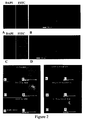
- STRO-1 + cells were immobilized on the polymer blends microarrays with results shown in Figure 2 .
- Human skeletal stem cells (STRO-1 + ) were grown on the polymer blends arrays, with one million cells immobilized with Cell Tracker Green on the polymer microarray slide for 18 hours followed by washing with PBS. Cells were then fixed and nuclei stained with Hoechst-33342.
- Figure 2 shows:-(A) STRO-1 + cells bound to the array of 142 binary polymer mixtures with six replicates.
- B) Shows the STRO-1 + cells attached on the six replicates of the same polymer mixtures (highlighted box in A, PLLA/PCL: 20/80).
- C) STRO-1 + cells bound to the array of 57 ternary polymer mixtures with four replicates.
- D) shows quadruplicates of PEI/PCL/PLLA: 20/40/40.
- FIGS 2 illustrate the range of high to low binding affinities found for cells together with corresponding SEM images of the polymer spots.
- High binding affinity was found for (E) a CS/PCL (30/70) mixture and (K) CS/PVAc/PEI (50/25/25) with corresponding SEM images of the spots also shown (F, L).
- a medium binding affinity was found for (G) PLLA/PCL (20/80) and (M) PEI/PHEMA/PCL (50/25/25) with corresponding SEM images of the spots also shown (H, N).
- Low binding affinity was found for (I) PEI/PVAc (10/90) and (O) PEI/PVAc/PEO (20/40/40) with corresponding SEM images of the spots also shown(J,P).
- Figure 2A shows the STRO-1 + attachment and spreading (growth) on the 142 binary polymer blends. With six replicates, it was calculated that 50% of the blends supported STRO-1 + cell attachments. Cell attachment on the ternary polymer blends was found to be more efficient (65% ternary blends showed STRO-1 + cells attachment) with respect to the binary blends ( Fig 2C ). Results ( Figs 2B & 2D ) indicated that the affinity of STRO-1 + binding on the polymer spots is highly reproducible and selective.
- Cell compatibility was evaluated in terms of the total number of cells immobilised onto each polymer spot which was identified using the DAPI and FITC channels using PathfinderTM software.
- the cell attachment and spreading on the replicates of identical polymer mixtures are very similar, examples are PLLA/PCL (20/80) ( Fig. 2B , average number of cells 315) and PEI/PCL/PLLA (20/40/40) ( Figure 2D , average number of cells 565).
- the analysis of the number of Stro-1+ cells immobilized on each polymer spot was evaluated, and plotted ( Fig.3A binary mixtures, Fig.3B ternary mixtures) as a function of compositional gradient of the polymer. It shows that the PEI/PVAc, PEI/PHEMA, PEI/PEO, PLLA/PCL and CS/PEI blends have an exceptionally high number of cells attached on the polymer spots.
- suitable materials that selectively support the attachment and growth of skeletal progenitors using these microarrays can be readily used to determine tissue engineering constructs by employing different polymer blends and cell-types.
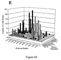
- Figure 4 shows the results of experiments, using skeletal fetal cells, carried out in similar fashion to those of figure 3 .
- Figure 4 shows:-(A) fetal cells bound to the array of 142 binary polymer mixtures with six replicates.
- C Cells bound to the array of 57 ternary polymer mixtures with four replicates.
- microarrays of polymer blends of the invention were seeded with human osteosarcoma (SaOs) and osteoblast-like (MG63) and ATDC5 cell lines.
- SaOs and MG63 cells attached to most of the mixtures, including PEI based mixtures, in the same way as the STRO-1 + cells.
- the behaviour of the ATDC-5 differed significantly; these cells grew only on a few mixtures.
- Figure 2 indicates that most of the mixtures prepared by mixing of PEI with other polymers gave significant cell attachment. The binding efficiency of these cells was noticeably lower in the case of CS-based mixtures as compared to those of PEI-based materials ( Figure 3 ). Moreover, some mixtures prepared from PLLA and PCL showed significantly high levels cellular attachment, whereas the corresponding homopolymers alone (PLLA and PCL) did not support skeletal cell attachment.
- Mixtures were prepared with high concentrations of polymers in suitable solvents, typical concentration was 10%, and coated on a glass surface using a spin coater, with different ratios of the polymers to investigate the morphological structure of these materials.
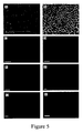
- Figure 5 illustrates the results as follows:- Figures 5A and 5B show SEM images of a PLLA /PCL: 50/50 mixture with a porous morphology.
- Figures 5C and 5D show (by fluorescence microscopy) Stro-1+ cells growing within a PLLA/PCL: 20/80 scaffold on culture day 12 (C) and culture day 21 (D).
- Figures 5 E and 5F show growth within a PLLA/PCL: 50/50 scaffold on culture day 12 (E) and culture day 21 (F).
- Figures 5G and 5H show growth within a PLLA/PCL: 80/20 scaffold on culture day 12 (G) and culture day 21 (H).
- FIG. 5 A,B A porous morphology was observed, as depicted by the SEM micrographs ( Figure 5 A,B ). Most of the pore sizes in the PLLA/PCL mixtures (20/80, 50/50, 80/20) were found to be in the range of between 10 ⁇ m and 25 ⁇ m, which is regarded as suitable for cell encapsulation, growth enhancement, proliferation and differentiation for tissue regeneration purposes.
- 3D scaffolds of PLLA/PCL functioned as excellent biomimetic templates and promoted skeletal stem cell attachment and growth in long-term (21-day) in vitro cultures with excellent viability observed.
- culture of STRO-1 + skeletal stem cells on these polymer blend scaffolds were observed to undergo osteogenic differentiation, evidenced by the expression of alkaline phosphatase - an early marker of osteogenic differentiation.
- osteogenic differentiation of the STRO-1 + skeletal progenitors into mature osteoblasts initiated by recombinant human bone morphogenic protein (rhBMP-2) ascorbate and dexamethasone, was supported by this polymer blend scaffold as illustrated by the expression of collagenous and non collagenous bone matrix proteins namely, Type I collagen, Bone Sialoprotein, Osteopontin, Osteonectin and Osteocalcin by cells from day 28 explants.
- rhBMP-2 recombinant human bone morphogenic protein
- polymer blend scaffolds composed of the PLLA/PCL (20/80) facilitated the generation of a mature osteoblast population from the STRO-1 + skeletal stem cell population indicating the suitability of the PLLA/PCL (20/80) scaffold as a temporal substrate supporting fundamental activities of skeletal stem cells including attachment, migration, proliferation and osteogenic differentiation.
- Defect regions in femora implanted with scaffolds seeded with human skeletal stem cells demonstrated significant bone regeneration, confirmed by histology, which illustrated significant region cell infiltration to the defect implanted with the scaffold and extensive cell-generated unmineralized bone matrix/ osteoid.
- the defect regions in femora implanted with scaffolds alone exhibited some degree of bone regeneration, evidenced by histological analysis of the defect region, which demonstrated remnants of the PLLA/PCL blend scaffold and newly synthesized unmineralized bone matrix/ osteoid.
- negligible bone repair was seen in femoral defects implanted with rubber spacers.
- High resolution ⁇ CT scans at day 28 clearly indicated the extent of new bone formation in the defect regions implanted with the PLLA/PCL blend scaffolds seeded with STRO-1 + skeletal stem cells, compared to the PLLA/PCL blend scaffold alone group and the control (rubber spacer) group.
- a significant increase in bone volume (BV) was observed in the defect regions implanted with cell-seeded scaffolds (5.399mm 3 ⁇ 0.625) compared to PLLA/PCL scaffold only (3.351mm 3 ⁇ 0.676, * P ⁇ 0.05) and the control (spacer) groups (3.115mm 3 ⁇ 1.379, * P ⁇ 0.05) ( Fig. 8a ).
- the ratio of bone volume/ total volume showed a significant increase in the PLLA/PCL scaffolds seeded with STRO-1 + cells (0.049 ⁇ 0.009) compared to the PLLA/PCL scaffold only group (0.032 ⁇ 0.007, * P ⁇ 0.05) and the control (spacer) group (0.025 ⁇ 0.009, *** P ⁇ 0.001) ( Fig. 8b ).
- Tb No trabecular number
- Polymer mixtures were generated by mixing chitosan (partially hydrolyzed, Mw 250 kDa, 1% aqueous acetic acid, pH - 4.0) and polyethylenimine (Mw 300 kDa, 10% in water, pH -11) in various molar ratios (90/10 to 10/90).
- the resulting solutions pH -7.5
- All compositions showed gelation, however these varied from less opaque (chitosan/PEI: 10/90) to more opaque gels (chitosan/PEI: 40/60).
- the resulting hydrogels were examined by a range of physical techniques, including scanning electron microscopy (SEM), IR, Rheometer and XRD.
- SEM scanning electron microscopy
- IR IR
- Rheometer XRD
- the hydrogel prepared from chitosan/PEI (40/60) displayed a sponge-like, microporous morphology, radically different to that found in "normal" chitosan gels (chitosan homopolymer) prepared by neutralization of solubilised chitosan and suggested possible application as a cellular support/scaffold.
- SEM images demonstrate the porous structure of the mixtures prepared by using chitosan and PEI.
- the blending of PEI with chitosan can form hydrogels, in an aqueous phase.
- FIG. 6(E) illustrates the morphology of a chitosan/PEI mixture having a ratio of 10/90.
- Figures 6 (A & B) and 6(C & D) show images of preferred chitosan/PEI mixtures(30/70 and 40/60 ratios respectively).
- Figure 6(F) shows a chitosan only gel (chitosan solution neutralized with NaOH).
- the preferred mixture (CS/PEI: 40/60) showed very good cell encapsulation and proliferation which may be attributed to the open network structure shown in figures 6 (C, D).
- HeLa and primary human fetal skeletal cells 20 labelled with Cell-Tracker Green were seeded and cultured within chitosan/PEI hydrogels over a period of 28 days.
- the cell culture procedure was as follows:
- Human skeletal cells were harvested from day 12 monolayer cultures grown in ⁇ -MEM supplemented with 10% FCS and labeled with Cell Tracker Green/ CMFDA (Invitrogen) following the manufacturer's instructions. 100,000 labelled cells were suspended in the sterile PEI solution and this solution was added immediately to the sterile chitosan solution, previously added to wells of a 24-well plate. Gelation of the mixture of the two solutions (chitosan/PEI: 40/60) took place within two minutes encapsulating the cells within the gel. Once this was achieved, 1 ml of media ( ⁇ -MEM containing 10% FCS) was added to each well containing the gel, and the cells were cultured for a period of 28 days in this culture system.
- media ⁇ -MEM containing 10% FCS
- RPMI-CM media supplemented with 10% fetal bovine serum (FBS), glutamine (4 mM), and antibiotics (penicillin and streptomycin, 100 units/ml) was used. The media was changed every two days.
- FBS fetal bovine serum
- glutamine 4 mM
- antibiotics penicillin and streptomycin, 100 units/ml
- the human fetal skeletal cells derived from predominantly cartilaginous fetal femora 20 exhibited a chondrocyte-like spherical morphology, as opposed to the fibroblastic morphology found in monolayer culture.
- Expression of Pcna, a marker of cell proliferation, 26 by fetal skeletal cells was analysed (RT qPCR) to determine the effect of culture within chitosan/PEI hydrogels on cell proliferation.
- the culture in the absence of chondrogenic differentiation media, resulted in a significant increase in Pcna expression on days 14 (P ⁇ 0.01), 21 ( P ⁇ 0.001) and 28 ( P ⁇ 0.05) of culture in comparison to day 7 of culture, thus skeletal cells proliferated actively over the 28-day culture period within chitosan/PEI hydrogels.
- Col1a1 Type I collagen
- Col2a1 Type II collagen
- Aggrecan indicative of de-differentiation of the fetal skeletal cells under conditions of monolayer culture.
- the culture within a 3D gel environment as opposed to a 2D monolayer culture environment thus prevented de-differentiation of the fetal skeletal cells into fibroblasts by maintaining these cells in a chondrocyte-like spherical morphology. Furthermore, the 3D chitosan/PEI hydrogel culture system in combination with the chondrogenic growth factor TGF- ⁇ 3, was essential to stimulate the differentiation of the fetal skeletal cell population along the chondrogenic lineage.
Landscapes
- Health & Medical Sciences (AREA)
- Life Sciences & Earth Sciences (AREA)
- Chemical & Material Sciences (AREA)
- General Health & Medical Sciences (AREA)
- Public Health (AREA)
- Animal Behavior & Ethology (AREA)
- Veterinary Medicine (AREA)
- Engineering & Computer Science (AREA)
- Medicinal Chemistry (AREA)
- Epidemiology (AREA)
- Biomedical Technology (AREA)
- Organic Chemistry (AREA)
- Bioinformatics & Cheminformatics (AREA)
- Dermatology (AREA)
- Vascular Medicine (AREA)
- Surgery (AREA)
- Heart & Thoracic Surgery (AREA)
- Transplantation (AREA)
- Oral & Maxillofacial Surgery (AREA)
- Genetics & Genomics (AREA)
- Molecular Biology (AREA)
- Physical Education & Sports Medicine (AREA)
- Dispersion Chemistry (AREA)
- Zoology (AREA)
- Wood Science & Technology (AREA)
- Biotechnology (AREA)
- General Chemical & Material Sciences (AREA)
- Nuclear Medicine, Radiotherapy & Molecular Imaging (AREA)
- Pharmacology & Pharmacy (AREA)
- Chemical Kinetics & Catalysis (AREA)
- Microbiology (AREA)
- Cell Biology (AREA)
- Biochemistry (AREA)
- General Engineering & Computer Science (AREA)
- Rheumatology (AREA)
- Orthopedic Medicine & Surgery (AREA)
- Materials For Medical Uses (AREA)
Abstract
chitosan (CS), polyethylenimine (PEI), poly(L-lactic acid) (PLLA), poly(D-lactic acid)(PDLA), poly(2-hydroxyethyl methacrylate) (PHEMA), poly(ε-caprolactone) (PCL), poly(vinyl acetate) (PVAc), poly (ethylene oxide) (PEO), poly[(R)-3-hydroxybutyric acid)] (PHB), cellulose acetate (CA), poly(lactide-co-glycolide) (PLGA) and poly(N-isopropylacrylamide)(PNIPAM). Implants making use of the polymer mixtures can support cell attachment, growth and differentiation, and tissue regeneration in vivo.
Description
- The present invention relates to the provision of improved polymers, especially mixtures of polymers that can find use as biocompatible materials, which support cell attachment, growth and differentiation, and tissue regeneration in vivo.
- Biomaterials have been defined as substrates other than foods or drugs of natural or synthetic origins, that comprises whole or part of a living structure or biomedical device which performs, augments, or replaces a natural function. The use of polymers as biomaterials is having an enormous impact in the world of medical science1-3. For example the use of polymer-coated stents, is a potential technique to achieve high local tissue concentrations of an effective drug at the precise site during vessel injury.4 Thiolated poly(acrylic acid) coated stents sustain release of a model peptide drug5. Combining polymers with mammalian cells, it is now possible to regenerate skin for patients who have burns or skin ulcers; various other polymer/cell combinations are in clinical trials 7. The search for ideal biomaterials is typically an iterative process for tissue regeneration, where the properties of the scaffolds employed to support cells are dictated concurrently by a number of factors. These include the kinetics of cell growth, scaffold degradation and the mechanical properties required at the site of implant, as well as cell-material interactions8.
- Current methods for the development of biomaterials require the preparation and characterization of individual materials; this is neither efficient nor cost effective. Alternatively the application of a high-throughput approach to material design and discovery 16-18 can be used to accelerate both the discovery and selection of suitable materials. An approach that shows promise in creating new biomaterials is that of polymer blends 13-15 . An attractive feature of blending (mixing) is the ease of combining several materials of a vast variety of characteristics into architectures with properties that are often not accessible with individual components. For instance, the cell attachment and proliferation on the surface of polymeric materials can be improved with respect to those of single materials by blending them with judiciously selected other polymers8. For functional performance, the design of a material is important, for example materials (polymers) having two separate phases being used to create devices for specific medical applications 19, which are known as shape-memory material.
- It is well recognized that human bone marrow mesenchymal stem cells (hBMSCs) can give rise to cells of the stromal lineage, such as adipogenic (fat), chondrogenic (cartilage), osteoblastic (bone), myoblastic (muscle) and fibroblastic (connective tissue) cells and are able to generate intermediate progenitors with a degree of plasticity. Thus, hBMSCs can give rise to a hierarchy of bone cell populations with a range of developmental stages, including the MSC, determined osteoprogenitor cell, preosteoblast, osteoblast and, ultimately, the osteocyte19-22. An ideal orthopaedic repair material would thus influence this heterogeneous progenitor cell mix in vivo to produce mature osteoblasts, rather than connective tissue cell types. The generation of mature osteoblast populations would allow the production of osteoid including collagen type I and calcium phosphate (apatite) mineral, which is essential for new bone formation.
- It is an object of the invention to provide polymer blends with improved properties with respect to supporting at least one of cell attachment growth and differentiation. It is an object of the present invention to provide implants for use in the reconstruction of tissue, especially orthopedic tissue for the formation or repair of bone, spine, etc. It is an object of the present invention to provide methods for obtaining improved polymer blends and for testing their application as scaffolds for cell attachment and growth.
- According to a first aspect, the present invention provides a biocompatible polymer mixture for use as a matrix for cellular attachment comprising a mixture of at least two polymers, selected from the group consisting of: chitosan (CS), polyethylenimine (PEI), poly(L-lactic acid) (PLLA), poly(D-lactic acid)(PDLA), poly(2-hydroxyethyl methacrylate) (PHEMA), poly(s-caprolactone) (PCL), poly(vinyl acetate) (PVAc), poly(ethylene oxide) (PEO), poly[(R)-3-hydroxybutyric acid] (PHB), cellulose acetate (CA), poly(lactide-co-glycolide) (PLGA) and poly(N-isopropylacrylamide)(PNIPAM).
- These polymers have been found to be effective in binary (two component) and ternary (three component) mixtures, which can provide good cell attachment, growth and differentiation. Blends comprising more than three polymer components may also be made.
- The structures of these polymers are indicated in
Scheme 1 below.

- In addition to allowing cellular attachment the biocompatible polymer mixture may support cell growth. Preferably the biocompatible polymer mixture supports cell differentiation i.e. the matrix allows cells such as stem cells to grow and differentiate in a manner akin to that found in nature. Thus the biocompatible polymer mixtures of the invention may be employed to support the growth of both soft (for example muscle, skin and nerves) and hard (for example bone, spine, cartilage) tissues.
- Preferably the biocompatible polymer mixture provides a matrix to promote bone formation, repair spine and cartilage.
- Surprisingly, it has been found that it is possible to prepare polymer mixtures (blends), which have structures suitable for use as scaffolding for the formation of tissue, for example bone, by making an appropriate mixture of polymers. It is not necessary to react (co-polymerise) the polymer components together to obtain a new material with useful structural properties. The intimate mixtures of the two or more polymer components provided by the present invention can provide scaffold structures that are both biocompatible and biodegradeable.
- The polymer mixtures may be prepared by dissolving the selected polymer components in a suitable solvent and then removing the solvent from the mixture e.g. by heating to evaporate the solvent and thus provide the polymer mixture with the desired composition. Conveniently the polymer components are dissolved separately in suitable solvents and the resulting solutions mixed in a selected ratio to provide a mixed polymer solution that is then processed to remove the solvent. The ratios described herein are in terms of weight: weight of each component. Alternative methods of production may be employed, for example dissolving all the polymer components in a suitable solvent and then processing to a dried product.
- The polymer mixture is biocompatible. It will be understood that the term biocompatible means that the polymer mixture is at least not toxic to cells or living tissue.
- Surprisingly it has been found possible to produce biocompatible polymer mixtures that include relatively high amounts of a polymer that is, on it's own, toxic to some cell lines. For example a polymer mixture containing significant amounts of polyethylenimine (PEI), which is toxic to certain cells. PEI has the further disadvantage that, on its own, it is soluble in aqueous systems, which makes it generally unsuitable for use as an implant, except when made into a polymer mixture of the invention.
- Advantageously the polymer mixture provides a scaffold that promotes superior cell growth and differentiation in comparison to growth and differentiation in similar conditions but in the absence of the polymer.
- Suitable binary (two component) polymer mixtures identified as providing at least good cell attachment in some embodiments of the invention include: polyethylenimine/ poly(vinyl acetate)(PEI/PVAc), polyethylenimine/poly(2-hydroxyethyl methacrylate) (PEI/PHEMA),
polyethylenimine/poly(ethylene oxide)
(PEI/PEO),polyethylenimine /chitosan (PEI/CS), polyethyleneimine/ poly(ε-caprolactone)(PEI/PCL), poly(1-lactic acid)/poly(ε-caprolactone) (PLLA/PCL), poly(D-lactic acid)/chitosan (PDLA/CS), poly(D-lactic
acid)/polyethylenimine (PDLA/PEI) poly[(R)-3-hydroxybutyric acid)]/chitosan (PHB/CS), poly[(R)-3-hydroxybutyric acid)] / polyethylenimine (PHB/PEI), cellulose acetate / polyethylenimine (CA/PEI), cellulose acetate/chitosan (CA/CS), poly(Lactide-co-glycolide)/ chitosan (PLGA/CS), poly(Lactide-co-glycolide)/polyethylenimine (PLGA/PEI), poly(N-isopropylacrylamide)/chitosan(PNIPAM/CS), poly(N-isopropylacrylamide)/ polyethylenimine (PNIPAM/PEI), poly(N-isopropylacrylamide)/ poly(ε-caprolactone) ( PNIPAM/PCL) , poly(N-isopropylacrylamide)/ poly(L-lactic acid)(PNIPAM/PLLA), poly(N-isopropylacrylamide)/ poly(D-lactic acid)(PNIPAM/PDLA), poly(N-isopropylacrylamide) / poly[(R)-3-hydroxybutyric acid)](PNIPAM/PHB), poly(N-isopropylacrylamide)/cellulose acetate(PNIPAM/CA), poly(N-isopropylacrylamide)/ poly(2-hydroxyethyl
methacrylate)(PNIPAM/PHEMA), and poly(N-isopropylacrylamide)/ poly(vinyl acetate)(PNIPAM/PVAc). - In other embodiments of the invention a suitable binary (two polymer component) mixture of polymers may be one of: polyethylenimine/ poly(vinyl acetate)(PEI/PVAc), polyethylenimine/poly(2-hydroxyethyl methacrylate)
(PEI/PHEMA), polyethylenimine/poly(ethylene oxide)
(PEI/PEO),polyethylenimine /chitosan (PEI/CS), polyethyleneimine/ poly(ε-caprolactone) (PEI/PCL) and poly(L-lactic acid) / poly (ε-caprolactone) (PLLA/PCL). - Typically the ratio of the two components of the binary mixtures may be from 5:95 to 95:5.
- For example, mixtures containing polyethylenimine (PEI) having a ratio of the two components in the range from 10:90 to 90:10 of PEI to the second component have been shown to exhibit good cell attachment, in contrast to PEI alone, which did not show good cell attachment and is toxic to certain cells.
- For example, in tests good attachment of human bone marrow-derived STRO-1+ progenitor cells has been found for PEI/PCL mixtures with ratios of the two components varying from 40:60 to 90:10. Comparable results were obtained for PLLA/PCL mixtures with ratios of 10:90 to 60:40. PEI/CS mixtures also gave good attachment results with STRO-1+ cells.
- Ternary mixtures that exhibit good cell attachment include:
- PEI/PCL/PLLA, PEI/PEO/PLLA, PEI/PVAc/PEO, PEI/pHEMA/PCL, PLLA/PEI/pHEMA, PLLA/PEO/PCL, PLLA/PVAc/pHEMA, PLLA/pHEMA/CS, PLLA/PCL/CS, CS/PEI/PLLA, CS/PEO/PEI, CS/PVAc/PEI, CS/PVAc/PEO, CS/PVAc/PCL, CS/pHEMA/PEI, CS/PCL/PEI and CS/PLLA/PEO.
- Typical ternary mixtures may be prepared with up to 90 parts of one component and five parts of each of the other two (5:5:90). Most of these mixtures support both cell attachment and growth, for example, CS/PVAc/PEI, PEI/pHEMA/PCL and PEI/PVAc/PEO.
- Binary polymer mixtures of poly(L-lactic acid)/poly(ε-caprolactone) (PLLA/PCL) and mixtures of polyethylenimine/chitosan (PEI/CS) have been found to have a particularly useful structure. These two types of mixture have been found to exhibit micro porous structures that can resemble, on microscopic inspection, the micro porous structure of bone.
- This apparent similarity to a natural bone structure is continued when these mixtures are tested with regard to their ability to attach cells and allow them to grow and differentiate. For example fetal stem cells may be successfully attached to a polymer mixture and if the composition is placed in a suitable growth environment then the cells will grow for periods of at least four weeks, as described later with reference to specific embodiments.
- A mixture of PLLA/PCL (for example in the ratio 20:80) was found to exhibit a particularly good porous structure, comparable to natural bone porosity and has been shown to aid bone healing in femur defects of mouse test subjects.
- The results demonstrate that polymer mixtures of the invention, for example a PLLA/PCL (20:80) mixture can support the generation of new bone/osteoid to restore the lost bone tissue in the defect region. Bone regeneration was observed in subjects fitted with a PLLA/PCL (20:80) scaffold alone after 28 days. Where the PLLA/PCL (20:80) scaffold was seeded with human skeletal stem cells, the results were significantly improved. (The indices of bone histomorphometry were significantly enhanced).
- Although PCL and PLLA are FDA approved materials for clinical use, PCL is known to have a slow degradation rate (several years depending on the processing conditions), while PLLA is known to have relatively fast degradation rate. The example PLLA/PCL (20:80) scaffold integrates the properties of both polymers. Modifying the ratio of components and/or adding a further component or components to the mixture, for example, can thus be employed to tune the degradation rate or the prosity.
- Further modifications may be made by seeding the polymer mixture scaffold with other components, such as growth factors. For example bone morphogenetic proteins (BMPs), Epidermal growth factor (EGF), Fibroblast growth factor (FGF), Erythropoietin (EPO), Granulocyte-colony stimulating factor (G-CSF) , Granulocyte-macrophage colony stimulating factor (GM-CSF) , Growth differentiation factor-9 (GDF9), Hepatocyte growth factor (HGF) , Insulin-like growth factor (IGF), Myostatin (GDF-8), Nerve growth factor (NGF) and other neurotrophins, Platelet-derived growth factor (PDGF), Thrombopoietin (TPO), Transforming growth factor- β1 (TGF-β1) and Vascular endothelial growth factor (VEGF).
- Mixtures were made from polyethylenimine (PEI) and chitosan (CS), formed from an aqueous system, and resulted in the formation of hydrogels. The combination of these two components from an aqueous system resulted in a mixture that incorporated substantial quantities of water producing a gel structure.
- These biocompatible hydrogels comprising polyethylenimine (PEI) chitosan (CS) and water are of particular interest and constitute a second aspect of the invention. Other polymer components may be included to form three or more component mixtures. Preferred binary mixtures (containing only chitosan and polyethylenimine as polymer components) contain chitosan/PEI in ratios of from 10:90 to 90:10. A most preferred blend has a chitosan/PEI ratio of 40:60.
- The hydrogels may be dried (substantially freed from water) for storage prior to use, if desired. The drying process employed may be freeze drying. Freeze drying has the advantage of preserving the three dimensional porous or fibrous structure of the polymer part (polymer mixture) of the hydrogel.
- Hydrogels have attracted considerable attention as so-called "smart biomaterials" due to the various and often intriguing physical and chemical phenomena they can display when subjected to a variety of external stimuli, such as changes in pH, solvent, ionic strength, temperature, light and electric field.1-2 As a result, hydrogels have also been applied as fundamental components in a variety of medical devices such as tissue engineering scaffolds3 and controlled drug-delivery systems4 and as soft linear actuators, sensors, and energy transducing devices1-2, 5 in a range of novel applications.
- Known hydrogel synthesis methods include the cross-linking of linear poly (N-isopropylacrylamide) (PNIPAAm)5, poly(ethylene glycol)6, poly (acrylamide)7, poly(acrylic acid)-based polymers8 and their copolymers9,10.
- These types of hydrogel follow either a positive thermoreversible system (polymer solutions having an upper critical solution temperature (UCST) which shrink when cooled below UCST) or negative thermoreversible system (polymer solutions having a lower critical solution temperature (LCST) which contract by heating above the LCST). However, the use of previously known polymers in cell therapeutic applications is limited due to their toxicity and non-biodegradability. 5
- Chitosan on its own has been used to form so called "hydrogels" via the neutralization of acidic solutions of chitosan. However, the resulting materials are opaque, with a granular morphology which lacks real physical stability and are more akin to a precipitate than a real "hydrogel". They also show a crystalline morphology.11 For use as a biomaterial chitosan and PEI have also been chemically grafted together to give materials with enhanced gene carrier abilities.12
- Surprisingly, biologically compatible hydrogels, with a stable morphology and the ability to support cell growth were readily prepared by the simple expedient of mixing of aqueous chitosan (CS) and polyethylenimine (PEI) solutions, as described hereafter with reference to specific examples. The gels may be dried prior to use and rehydrated when seeded with cells, for example.
- The gels were found to have good thermal stability, for example a chitosan/PEI (40/60) hydrogel showed little change in volume at temperatures between 22 and 40°C. Substantial shrinkage did occur on heating this gel to higher temperatures (up to 90°C) but there was no tendency for the gel to collapse or fragment. Importantly for use in biological applications the gels were found to be stable at physiological pH (7.4) for several weeks.
- The hydrogel mixtures may be formed by mixing appropriate aqueous solutions of chitosan and polyethylenimine. Advantageously for some applications the hydrogels may be formed in a cell culture medium. By this means the hydrogels may be readily seeded with cells which can be incubated in the culture medium, growing and supported by the matrix or scaffold provided by the polymer mixture's structure.
- Hydrogels have been used as three dimensional (3D) scaffolds in tissue engineering applications to mimic the natural extra cellular matrix (ECM) and to support cellular adhesion, migration and proliferation. The novel CS/PEI hydrogels supported the attachment and growth of mammalian cells as shown by using a microarray of the present invention as described below. The hydrogels were examined alongside samples consisting of chitosan and PEI alone. The homopolymers (chitosan and PEI) alone did not support cell attachments or spreading, while chitosan/PEI mixture showed significant cell attachment.
- A preferred mixture (chitosan/PEI: 40/60) supported cell attachment and spreading. In experiments, labelled cells were seeded in the chitosan/PEI gel by mixing with the appropriate chitosan /PEI mixture, before full gelation had occurred. Adding cells to the hydrogel in this manner homogeneously distributed the cells throughout the resulting scaffold. The cells were found to be viable in the gels when the culture was continued for up to four weeks.
- Advantageously, the polymer mixtures and hydrogel polymer mixtures of the invention are biodegradable i.e. the polymer mixture when placed in a subject will degrade over a period of time so that polymer is eliminated from the subjects body. A biodegradable polymer mixture has the advantage that when a portion of polymer mixture of the invention is placed within a subject to replace damaged tissue, for example bone, cartilage and nerve, the polymer mixture structure will be replaced by growing cells, conforming to the scaffold provided by the portion of polymer mixture.
- Polymer mixtures of the invention that are biodegradable include but are not limited to CS/PEI, PLA/PCL, CS/PLLA, CS/PCL, PEI/PCL and PEI/PLLA. By biodegrading after a period sufficient to allow the growth of living tissue, the portion of polymer mixture can act as a temporary structure, which will be replaced in time by grown tissue. The biodegradable polymer mixture thus affords a method for repair or replacement of damaged tissue by natural or modified (for example genetically engineered) natural tissue, appropriate to the part of the body being repaired or replaced, rather than by replacement with a manufactured substitute.
- According to a third aspect, the present invention provides an implant for the repair or replacement of living tissue comprising a biocompatible polymer mixture according to the first or second aspects of the invention.
- The implant may include living cells attached to the biocompatible polymer mixture. For example adult human bone marrow-derived skeletal stem/ progenitor cells, human fetal skeletal progenitor cells or human articular chondrocytes.
- Alternatively the implant may be incubated with suitable cells, in vitro, prior to use, to provide an implant comprising tissue, which may be natural tissue or modified or genetically engineered natural tissue.
- Alternatively the implant may be used without attached cells or tissue whereupon it may be colonised by the subject's own cells, providing a matrix or scaffold for growth of the cells.
- Tissues that may be repaired or replaced by the implant of the invention include bone or cartilage. Other tissues, for example soft tissues such as muscle, skin or nerve may also be repaired or replaced.
- The implant may simply consist of a polymer mixture of the invention with or without attached cells. Other components may be included in the implant. For example, the implant may include DNA, RNA, proteins, peptides or therapeutic agents for treatment of disease conditions. The implant may also include biodegradable and non-biodegradable components.
- For example, the implant may be a stent of a manufactured non-biodegradable material but coated with a selected polymer mixture of the invention and optionally seeded with appropriate cells.
- For further example, the implant may be used for replacement of bone and may include a permanent support such as a steel plate or pin and a portion made from a polymer mixture of the invention and seeded with bone producing cells. In use the steel plate or pin remains as a structural support, whilst the polymer mixture acts as a scaffold but degrades following the desired growth of bone tissue.
- The implants of the invention may be used to effect tissue repair or replacement. Thus according to a fourth aspect the present invention, provides a method for repair or replacement of tissue comprising: providing an implant according to the third aspect of the invention; and locating the implant on or in the body of a subject.
- For example, the implant may be placed on the body of a subject when skin tissue is being repaired. For further example, the implant may be placed within a subject when bone or an internal organ is being repaired.
- Selecting suitable biocompatible polymer mixtures for different applications can be onerous, requiring the preparation of a large number of samples and careful testing to ensure that the polymer mixture complies with all the relevant criteria for successful use such as good attachment and growth of cells. Accordingly high throughput methods have been adapted for use in testing large numbers of polymer mixture samples. A microarray of polymer mixture samples has been found to be a highly convenient tool for testing and comparing samples of the polymer mixtures of the invention. The microarrays constitute a fifth aspect of the present invention.
- The microarray of polymer blends may be prepared by the following method. A plurality of polymer solutions is prepared. Each solution contains a mixture of at least two polymers prepared in selected concentrations. The solutions are then printed as an array of microspots onto a substrate and the microspots are dried to form a microarray of dried polymer spots attached to the substrate.
- The substrate is preferably agarose or another substrate that cells do not tend to adhere to. Use of a substrate which cells do not adhere to has the advantage of avoiding false positives on testing caused by cells adhering to the substrate rather than to the polymer blend under test. Agarose supported on a glass plate has been found to be a satisfactory base for printing the microarray.
- The microarray can be used of testing the ability of a large number of polymer mixtures to support cell attachment, growth and/or differentiation.
- After growing the cells on the microarray an assessment of the amount of cell attachment and growth may be made. The assessments of the suitability of a given polymer mixture of the attachment and /or the growth of a particular cell type may be carried out in a number of ways. The microarray may be examined visually by microscope. Alternatively the assessment may be automated. For example the microarray may be examined by a vision system, which assesses the growth of cells by comparing images of each microspot before and after the incubation step.
- The cells may be marked, for example with a fluorescent marker by methods well known to those skilled in the art. For example on illumination with appropriate light the microspots of polymer mixture will exhibit fluorescence that is dependent on the quantity of cells attached to and growing on a given microspot. The measurement of the amount of fluorescence can be automated by fluorescence microscopy combined with analysis using an appropriate software program in the usual fashion.
- Polymer mixtures exhibiting good cellular attachment and potential for cell growth such as those described above have been found by making use of these microarrays in a testing programme.
- According to a sixth aspect the present invention provides a biocompatible polymer mixture for use as a matrix for cellular attachment comprising a mixture of at least two polymers selected from the group consisting of:
- chitosan (CS), polyethylenimine (PEI), poly(L-lactic acid) (PLLA), poly(D-lactic acid)(PDLA), poly(2-hydroxyethyl methacrylate) (PHEMA), poly(ε-caprolactone) (PCL), poly(vinyl acetate) (PVAc), poly (ethylene oxide) (PEO), poly[(R)-3-hydroxybutyric acid)] (PHB), cellulose acetate (CA), poly(lactide-co-glycolide) (PLGA) and poly(N-isopropylacrylamide)(PNIPAM).
- The biocompatible polymer mixture may comprise two polymer components, selected from the group consisting of polyethylenimine/ poly(vinyl acetate)(PEI/PVAc), polyethylenimine/poly(2-hydroxyethyl methacrylate)
(PEI/PHEMA), polyethylenimine/poly(ethylene oxide) (PEI/PEO),polyethylenimine /chitosan (PEI/CS), polyethyleneimine/ poly(ε-caprolactone)(PEI/PCL), poly(L-lactic acid)/poly(ε-caprolactone) (PLLA/PCL), poly(D-Lactic acid)/chitosan (PDLA/CS), poly(D-Lactic
acid)/polyethylenimine (PDLA/PEI) poly[(R)-3-hydroxybutyric acid)]/chitosan (PHB/CS), poly[(R)-3-hydroxybutyric acid)] / polyethylenimine (PHB/PEI), cellulose acetate / polyethylenimine (CA/PEI), cellulose acetate/chitosan (CA/CS), poly(Lactide-co-glycolide)/ chitosan (PLGA/CS), poly(Lactide-co-glycolide)/polyethylenimine (PLGA/PEI), poly(N-isopropylacrylamide)/chitosan(PNIPAM/CS), poly(N-isopropylacrylamide)/ polyethylenimine (PNIPAM/PEI), poly(N-isopropylacrylamide)/ poly(ε-caprolactone)
(PNIPAM/PCL), poly(N-isopropylacrylamide)/ poly(L-lactic acid)(PNIPAM/PLLA), poly(N-isopropylacrylamide)/ poly(D-Lactic acid)(PNIPAM/PDLA), poly(N-isopropylacrylamide) / poly[(R)-3-hydroxybutyric acid)](PNIPAM/PHB), poly(N-isopropylacrylamide)/cellulose acetate(PNIPAM/CA), poly(N-isopropylacrylamide)/ poly(2-hydroxyethyl
methacrylate)(PNIPAM/PHEMA), and poly(N-isopropylacrylamide)/ poly(vinyl acetate)(PNIPAM/PVAc). - The two polymer components may be selected from the group consisting of: polyethylenimine/ poly(vinyl acetate)(PEI/PVAc), polyethylenimine/poly(2-hydroxyethyl methacrylate) (PEI/PHEMA), polyethylenimine/poly(ethylene oxide) (PEI/PEO),polyethylenimine /chitosan (PEI/CS), polyethyleneimine/ poly(ε-caprolactone)(PEI/PCL), poly(L-lactic acid)/poly(ε-caprolactone) (PLLA/PCL).
- The biocompatible polymer mixture comprising two polymer components may have a microporous structure and comprise two polymer components selected from the group consisting of poly(L-lactic acid)/poly(ε-caprolactone) (PLLA/PCL) and polyethylenimine/chitosan (PEI/CS).
- Such a biocompatible polymer mixture may be one where the polymer components of the mixture are poly(L-lactic acid)/poly(ε-caprolactone) (PLLA/PCL) having a ratio in the range from 10:90 to 60:40.
- The poly(L-lactic acid)/poly(ε-caprolactone) (PLLA/PCL) may be in the ratio of 20:80.
- In the biocompatible polymer mixture according to the sixth aspect the mixture may be a two component mixture containing polyethylenimine (PEI) and having a ratio in the range from 10:90 to 90:10 of PEI to the second component.
- In the biocompatible polymer mixture according the sixth aspect the mixture may comprise three polymer components selected from the group consisting of PEI/PCL/PLLA, PEI/PEO/PLLA, PEI/PVAc/PEO, PEI/pHEMA/PCL, PLLA/PEI/pHEMA, PLLA/PEO/PCL, PLLA/PVAc/pHEMA, PLLA/pHEMA/CS, PLLA/PCL/CS, CS/PEI/PLLA, CS/PEO/PEI, CS/PVAc/PEI, CS/PVAc/PEO, CS/PVAc/PCL, CS/pHEMA/PEI, CS/PCL/PEI and CS/PLLA/PEO.
- In the biocompatible polymer mixture according to the sixth aspect the mixture may comprise polyethylenimine (PEI), chitosan (CS) and water to form a hydrogel.
- The biocompatible polymer mixture forming a hydrogel may comprise only chitosan (CS) and polyethyleneimine (PEI) as polymer components, present in the ratio of from 10:90 to 90:10.
- The biocompatible polymer mixture forming a hydrogel may be formed in a cell culture medium.
- According to a seventh aspect the present invention provides an implant for use in the repair or replacement of living tissue comprising a biocompatible polymer mixture according to the description of the sixth aspect given above.
- The implant according may further comprise living cells attached to the biocompatible polymer mixture.
- Alternatively the implant may further comprise tissue produced by incubation in vitro.
- The implant may further comprise additional components selected from the group consisting of growth factors, DNA,RNA, proteins, peptides and therapeutic agents for treatment of disease conditions.
- The implant may be of a biocompatible polymer mixture that is biodegradable and also further comprise a non-biodegradable component.
- Such an implant may be a a stent of a non-biodegradable material that is coated with the biodegradable biocompatible polymer mixture.
- According to an eighth aspect the present invention providesa method for repair or replacement of tissue comprising: providing an implant of the seventh aspect and locating the implant on or in the body of a subject. According to a ninth aspect the present invention provides a microarray comprising a plurality of polymer mixtures according to the sixth aspect.
- The microarray may have agarose as the substrate.
- The microarray may be made by a method comprising:
- preparing a plurality of solutions comprising the polymer components; and
- printing said solutions as an array of microspots onto a substrate; and
- drying the microspots.
- According to another aspect the invention provides a method of making a biocompatible polymer mixture according to the invention comprising:
- preparing a solution of selected polymer components in a suitable solvent; and
- removing the solvent from the mixture.
-
-
Figs. 1 (A to H) shows scanning electron micrographs of microarrays of polymer mixtures; -
Figs. 2 (A to D) show visualisation of cells attached to a microarray of polymer mixtures; -
Figs. 2 (E to P) show visualisation of cells attached to microarray spots of various polymer mixtures and their corresponding SEM images; -
Figs. 3 (A and B) show graphically the extent of cell attachment for Stro-1+ cells found for various polymer mixtures of the invention; -
Figs. 4 (A to D) show visualisation of skeletal fetal cells attached to microarrays of polymer mixtures; -
Figs. 4 (E and F) show graphically the extent of cell attachment for skeletal fetal cells found for various polymer mixtures of the invention; -
Figs. 5 (A and B) show detailed SEM images of polymer mixtures of the invention; -
Figs. 5 (C to H) show images of skeletal cells (Stro-1+ labelled with cell tracker green) growing in polymer mixtures of the invention; -
Figs. 6 (A to F) show SEM images of polymers containing chitosan; -
Fig. 7 shows HeLa cells labelled with Cell Tracker Green growing in a chitosan /polyethylenimine polymer (gel) mixture; and -
Fig.8 (a to f) shows graphically results from in vivo tests using polymer mixtures of the invention in bone repair. - Materials: Chitosan(CS) [75% deacetylated], polyethylenimine (PEI) and poly(N-isopropylacryalmide) (PNIPAM)and poly(lactide-co-glycolide)(PLGA) were purchased from Aldrich, poly(L-lactic acid) (PLLA), poly(D-lactic acid) (PDLA, poly[(R)-3- hydroxyl butyric acid] (PHB) from Fluka and the other polymers (PCL, PHEMA, PEO, CA, PVAc) from Sp2 scientific polymer products, INC., and used without further purification. The solvents chloroform (CHCl3), glacial acetic acid and N-methyl-2-pyrrolidinone (NMP) were analytical grade, purchased form Fisher scientific. 1, 4 Dioxane was purchased from across organics. Agarose (Type 1-B) and microscopic slides (Silane-Prep™) were obtained from Sigma-Aldrich.
- Polymer Solution Preparation: Chitosan (1% w/v) was dissolved in 2% acetic acid solution in an aqueous medium, and this solution was then filtered to remove any foreign materials. The polyethylenimine and poly (2-hydroxyethyl methacrylate), poly(ethylene oxide)(PEO) and PNIPAM were dissolved in NMP. The other polymers (PCL, PLLA, PDLA, PHB, PLGA and PVAc) were dissolved in CHCl3 or Dioxane and NMP with a ratio of 50/50 (v/v). All polymers were dissolved at a concentration of 1% in the solvents. These solutions were blended with different proportions and printed on agarose coated microscopic slides. The silane-prep™ slides were deep coated with agarose (1% w/v) solution and dried at ambient conditions for at least 24 hrs to obtain a stable film on the slide before being use for polymer microarray fabrication. The thickness of the agarose film was about 1.2 µm as measured by scanning electron microscopy.
- Polymer Microarray Fabrication: The arrays of polymer mixtures were fabricated by contact printing (Qarray mini, Genetix) with 16 aQu solid pins (K2785; Genetix; UK) using 1.0 % polymer solutions (w/v) placed into polypropylene 384-wells micro plates. The following printing conditions were used, 5 stamping per spot, 200 ms inking time and 10 ms stamping time. The typical spot sizes was 300-400 µm in diameter with spot to spot distances of either 750 µm or 1120 µm (x-direction) and 900 µm or 2250 µm (y-direction) allowing up to 960 polymer spots to be printed on a standard 25 x 75 mm slide. The polymer mixtures were made into two arrays, one of which consisted of 142 mixtures comprising binary polymers with different proportions and the second array comprising ternary polymer mixtures with a total of 57, printed onto a conventional (25 x 75 mm) glass slide. These arrays can be used to immobilize a number of cell lines including fetal and human bone marrow stem cells onto individual polymer spots. Once printed, the slides were dried for 24 hrs under vacuum at 40 °C and sterilized under UV irradiation for 30 minutes prior to cell seeding.
- Scanning Electron Microscopy: Scanning electron microscopy (Philips XL30CP SEM) was used to examine the surface morphology of the polymer spots. The glass slides containing the polymer arrays were covered with a thin layer of Au and glued to a metal-base specimen holder to achieve good electrical contact with the grounded electrode. The micrographs were taken at 10 kV in a secondary electron imaging mode.
- Differential Scanning Calorimeter: Glass transitions (Tg) and the melting temperatures were determined using a TA Instruments DSC 2010 with both heat flow and temperature scales calibrated against indium metal. Nitrogen was used as purge gas and samples were scanned at 10 °C min-1.
- Human bone marrow-derived STRO-1 immunoselected progenitor cells: Bone marrow samples (n=3) were obtained from haematologically normal osteoarthritic and osteoporotic individuals undergoing routine hip replacement surgeries, with approval of the Southampton & South West Hampshire Local Research Ethics Committee. Skeletal progenitors from these bone marrow samples were immunoselected using the STRO-1 antibody, followed by isolation using MACS as described before19. The isolated STRO-1+ skeletal progenitors were expanded by culturing them to confluence in monolayers supplemented with 10% FCS in α-MEM.
- Human fetal skeletal progenitor cells: Cartilaginous femora (n = 5) from human fetuses (age: 8-11 weeks post-conception) were obtained after the termination of pregnancies according to the guidelines issued by the Polkinghome Report and with ethical approval from Southampton & South West Hampshire Local Research Ethics Committee. Cells were isolated from these predominantly cartilaginous femora as described previously18 and cultured to confluence for 12 days in monolayers in basal medium (α-MEM containing 10% FCS).
- Human Osteosarcoma Cell Lines: The two human osteosarcoma cell lines namely, MG63 and SaOs were cultured in monolayers in basal medium (10% FCS in DMEM and α-MEM respectively) up until cultures were -70% confluent. Cells harvested from these cultures were incubated on the polymer microarrays (n = 2 for each cell line).
- Protocol for studying cell - polymer interactions using polymer microarrays: The polymer microarrays were sterilized under UV light for 30 min. STRO-1+ bone marrow-derived adult skeletal progenitor cells, human fetal skeletal progenitors and the two osteosarcoma cell lines were fluorescently labelled with Cell Tracker Green/ CMFDA. Suspensions of these cell populations in 1.5 ml basal media were pipetted onto the microarrays (106 cells/ microarray slide), so that the entire microarray surface was covered with a thin film of cell suspension. The cells were incubated on the microarrays overnight at 37°C, 5% CO2 in humidified atmosphere. The following day, microarrays were thoroughly washed using PBS to remove any unbound/ loosely-bound cells and the cells bound to the polyurethane spots were fixed using 4% formalin solution in PBS. Nuclei of cells bound to the polymer spots were stained using DAPI and the microarrays were stored in PBS at 4°C until analysis.
- Image Capture and High Throughput Analysis: Images of arrays were captured with a
Zeiss Axiovert 200 fluorescence microscope at x 20 magnifications and analyzed using Pathfinder™ software (IMSTAR S.A., Paris, France). - The phase transition behaviors of the polymers were determined by differential scanning calorimeter (DSC).
- Most of the polymer mixtures showed at least two transition temperature phases with distinct values of individual components. As expected, a polymer containing crystalline components (e.g., PLLA, PCL and PEO), showed variation of crystallinity when blended with another polymer. In the case of amorphous polymers (PHEMA, CS, PEI and PVAc), the Tg of the polymer changes. These behaviors are due to the interactions between the components present in the mixture.
- The driving force of the miscibility of the polymers is believed to be the hydrogen bonding, Van der Waal's, hydrophilic and hydrophobic interactions between the polymer chains. In comparison to the individual (pure) polymers morphological changes take place in the mixtures, resulting in the improved properties for cell attachment and growth.
- The morphology and phase behavior of the polymer mixtures were also examined by SEM. In a first set of experiments, compounds containing two-component polymers (a binary mixture) were studied.
Figure 1A shows an SEM of an array of binary polymer mixtures. The blending of amorphous polymers, especially blending of PEI with PHEMA and PVAc shows a clear and transparent morphology. Blends of PEI with CS formed complex fibrous networks (seeFig 1F a 50:50 mixture). In some cases, the blending of a crystalline polymer with an amorphous polymer produced crystalline morphology in the mixture, depending on the compositions.Fig 1 C-F shows the following binary mixtures in closer detail:- (C) PLLA/PEO: 50/50 blend showing the phase separation. (D) PLLA/PHEMA (30/70) mixture showing crystalline phase separation of PLLA. (E) PLLA/PCL (50/50) blend. (F) CS/PEI (50/50) blend. - During the processing of these blends, it was noted the blending of CS with PEI for all compositions produced hydrogels. When dried an interpenetrated fibre formation was observed (
Fig. 1 F ). - In a second set of experiments, mixtures with three component polymers (ternary blends) were prepared and their SEM images are presented in an array format (
Figure 1B ) which show some differences in morphology as compared to those of binary mixtures.Figures 1G and H show the following mixtures in closer detail:- (G) CS/PEO/PEI: 20/40/40. (H) PEI/PCL/PLLA: 50/25/25. - The average size of the polymer spots was ∼500 µm, in the case of ternary polymer mixtures (the average spot sizes was ∼350 µm for binary mixtures). Spot size can be adjusted as desired by changing the printing conditions employed.
- In the solution blending process employed, the solubility of each polymer in the solvents can be different, which can lead to different phases (regions containing different proportions of polymer component) within the polymer mixture produced.
- The two-component polymer mixtures and three-component polymer mixtures were printed on separate chips to avoid merging spots.
- The polymer microarray chips were examined for cell attachment and growth, by testing the arrays with osteioprogenitor cell lines such as fetal femur and mesenchymal stem (STRO-1+) cells.
- STRO-1+ cells were immobilized on the polymer blends microarrays with results shown in
Figure 2 . Human skeletal stem cells (STRO-1+) were grown on the polymer blends arrays, with one million cells immobilized with Cell Tracker Green on the polymer microarray slide for 18 hours followed by washing with PBS. Cells were then fixed and nuclei stained with Hoechst-33342. -
Figure 2 shows:-(A) STRO-1+ cells bound to the array of 142 binary polymer mixtures with six replicates. (B) Shows the STRO-1+ cells attached on the six replicates of the same polymer mixtures (highlighted box in A, PLLA/PCL: 20/80). (C) STRO-1+ cells bound to the array of 57 ternary polymer mixtures with four replicates. (D) shows quadruplicates of PEI/PCL/PLLA: 20/40/40. -
Figures 2 (E-P) illustrate the range of high to low binding affinities found for cells together with corresponding SEM images of the polymer spots. High binding affinity was found for (E) a CS/PCL (30/70) mixture and (K) CS/PVAc/PEI (50/25/25) with corresponding SEM images of the spots also shown (F, L). A medium binding affinity was found for (G) PLLA/PCL (20/80) and (M) PEI/PHEMA/PCL (50/25/25) with corresponding SEM images of the spots also shown (H, N). Low binding affinity was found for (I) PEI/PVAc (10/90) and (O) PEI/PVAc/PEO (20/40/40) with corresponding SEM images of the spots also shown(J,P). -
Figure 2A shows the STRO-1+ attachment and spreading (growth) on the 142 binary polymer blends. With six replicates, it was calculated that 50% of the blends supported STRO-1+ cell attachments. Cell attachment on the ternary polymer blends was found to be more efficient (65% ternary blends showed STRO-1+ cells attachment) with respect to the binary blends (Fig 2C ). Results (Figs 2B & 2D ) indicated that the affinity of STRO-1+ binding on the polymer spots is highly reproducible and selective. - Cell compatibility was evaluated in terms of the total number of cells immobilised onto each polymer spot which was identified using the DAPI and FITC channels using Pathfinder™ software. The cell attachment and spreading on the replicates of identical polymer mixtures are very similar, examples are PLLA/PCL (20/80) (
Fig. 2B , average number of cells 315) and PEI/PCL/PLLA (20/40/40) (Figure 2D , average number of cells 565). - The analysis of the number of Stro-1+ cells immobilized on each polymer spot was evaluated, and plotted (
Fig.3A binary mixtures,Fig.3B ternary mixtures) as a function of compositional gradient of the polymer. It shows that the PEI/PVAc, PEI/PHEMA, PEI/PEO, PLLA/PCL and CS/PEI blends have an exceptionally high number of cells attached on the polymer spots. The identification of suitable materials that selectively support the attachment and growth of skeletal progenitors using these microarrays can be readily used to determine tissue engineering constructs by employing different polymer blends and cell-types. -
Figure 4 shows the results of experiments, using skeletal fetal cells, carried out in similar fashion to those offigure 3 .Figure 4 shows:-(A) fetal cells bound to the array of 142 binary polymer mixtures with six replicates. (B) Cells attached on the polymer spot CS/PCL:30/70 mixture (highlighted box in A) showing high-resolution pictures of cells nuclei stained with Hoechst-33342 (DAPI), FITC and their combinations. (C) Cells bound to the array of 57 ternary polymer mixtures with four replicates. (D) Four ternary polymers mixtures: CS/PVAc/PLLA: 20/40/40 (1), CS/PVAc/PCL: 80/10/10 (2), PEI/PVAc/PEO: 20/40/40 (3), PEI/PCL/PLLA: 80/10/10 (4) (as highlighted in 4C). (E) and (F) shows graphical analysis of the cell attachment as a function of binary and ternary polymer compositions. - For both binary and ternary polymer mixtures fetal cell attachment and growth was only observed on the polymer mixtures. The analysis of the cell attachment to the polymer spots was performed by calculating the number of cells and plotting them as a function of compositional gradients (
Fig 4E binary mixtures,Fig 4F ternary mixtures). The results indicate that many of the ternary polymer mixtures have the same ability to support human fetal skeletal cells attachment as do binary mixtures. - To examine the effect of the polymer mixtures on the other cell types, microarrays of polymer blends of the invention were seeded with human osteosarcoma (SaOs) and osteoblast-like (MG63) and ATDC5 cell lines. The SaOs and MG63 cells attached to most of the mixtures, including PEI based mixtures, in the same way as the STRO-1+ cells. Unlike the other cell lines, the behaviour of the ATDC-5 differed significantly; these cells grew only on a few mixtures.
- The high efficiency of cell attachment to certain mixtures can be due to a wide range of physical and chemical factors of the polymer surface. However this result emphasises the ability of the microarray approach to quickly allow evaluation of different cell lines against different polymer mixtures.
- It is important to design cell-compatible biomaterials, which are resistant to aqueous based conditions i.e. which do not simply dissolve in an aqueous environment. PEI alone is soluble in water and also toxic for cells. Blending with other polymers such as PVAc, PEO, etc., resulted in mixtures with improved aqueous resistance and allowed compatibility with cells.
-
Figure 2 indicates that most of the mixtures prepared by mixing of PEI with other polymers gave significant cell attachment. The binding efficiency of these cells was noticeably lower in the case of CS-based mixtures as compared to those of PEI-based materials (Figure 3 ). Moreover, some mixtures prepared from PLLA and PCL showed significantly high levels cellular attachment, whereas the corresponding homopolymers alone (PLLA and PCL) did not support skeletal cell attachment. - From this investigation, preferred mixtures such as CS/PEI, PEI/PVAc, PEI/pHEMA, PEI/PEO and PLLA/PCL were selected for further investigations for tissue engineering applications.
- Mixtures were prepared with high concentrations of polymers in suitable solvents, typical concentration was 10%, and coated on a glass surface using a spin coater, with different ratios of the polymers to investigate the morphological structure of these materials.
- Materials of interest prepared from PLLA and PCL, chosen by their ability to support skeletal cell growth in the first screen (
Figures 2-4 ), were tested for use in scaffold fabrication. Both these polymers were dissolved in a common solvent (CHCl3) and blended at room temperature. The solution of the mixture was coated on a glass surface by spin coater and followed by drying at ambient conditions. -
Figure 5 illustrates the results as follows:-Figures 5A and 5B show SEM images of a PLLA /PCL: 50/50 mixture with a porous morphology.Figures 5C and 5D show (by fluorescence microscopy) Stro-1+ cells growing within a PLLA/PCL: 20/80 scaffold on culture day 12 (C) and culture day 21 (D).Figures 5 E and 5F show growth within a PLLA/PCL: 50/50 scaffold on culture day 12 (E) and culture day 21 (F).Figures 5G and 5H show growth within a PLLA/PCL: 80/20 scaffold on culture day 12 (G) and culture day 21 (H). - A porous morphology was observed, as depicted by the SEM micrographs (
Figure 5 A,B ). Most of the pore sizes in the PLLA/PCL mixtures (20/80, 50/50, 80/20) were found to be in the range of between 10 µm and 25 µm, which is regarded as suitable for cell encapsulation, growth enhancement, proliferation and differentiation for tissue regeneration purposes. - After preparing the 3-D porous structure of polymer mixture, human skeletal cells were seeded for a culture period of up to three weeks. Cell viability and growth were monitored by fluorescence microscopy with the results as shown in
figures 5C to H . - Further studies showed that 3D scaffolds of PLLA/PCL functioned as excellent biomimetic templates and promoted skeletal stem cell attachment and growth in long-term (21-day) in vitro cultures with excellent viability observed. In the absence of osteogenic induction factors, culture of STRO-1+ skeletal stem cells on these polymer blend scaffolds (for 21 days) were observed to undergo osteogenic differentiation, evidenced by the expression of alkaline phosphatase - an early marker of osteogenic differentiation. Moreover, further osteogenic differentiation of the STRO-1+ skeletal progenitors into mature osteoblasts, initiated by recombinant human bone morphogenic protein (rhBMP-2) ascorbate and dexamethasone, was supported by this polymer blend scaffold as illustrated by the expression of collagenous and non collagenous bone matrix proteins namely, Type I collagen, Bone Sialoprotein, Osteopontin, Osteonectin and Osteocalcin by cells from day 28 explants. Thus, under osteogenic conditions in vitro, polymer blend scaffolds composed of the PLLA/PCL (20/80) facilitated the generation of a mature osteoblast population from the STRO-1+ skeletal stem cell population indicating the suitability of the PLLA/PCL (20/80) scaffold as a temporal substrate supporting fundamental activities of skeletal stem cells including attachment, migration, proliferation and osteogenic differentiation.
- Other mixtures (e.g. PEI blended with PEO, PHEMA and PVAc) were also further investigated. All these materials could be useful in controlling cellular architecture as well as in manipulating cellular function.
- To investigate the bone regeneration potential of human bone marrow-derived STRO-1+ skeletal stem cells seeded onto 3D scaffolds of PLLA/PCL (20/80) in vivo, an innovative mouse critical-sized femur defect model (load-bearing) was employed27. Two-dimensional in vivo digital X-rays and 3D detailed images of entire femora generated by µCT analysis were utilised for the initial evaluation of the extent and degree of bone healing in femoral defects, 28 days postoperatively. Defects were implanted with either scaffolds seeded with cells or scaffolds without cells or inert spacers as control.
- Defect regions in femora implanted with scaffolds seeded with human skeletal stem cells demonstrated significant bone regeneration, confirmed by histology, which illustrated significant region cell infiltration to the defect implanted with the scaffold and extensive cell-generated unmineralized bone matrix/ osteoid. The defect regions in femora implanted with scaffolds alone exhibited some degree of bone regeneration, evidenced by histological analysis of the defect region, which demonstrated remnants of the PLLA/PCL blend scaffold and newly synthesized unmineralized bone matrix/ osteoid. In comparison to the femoral defects implanted with the cell-seeded scaffolds or scaffolds alone, negligible bone repair was seen in femoral defects implanted with rubber spacers.
- High resolution µCT scans at day 28 clearly indicated the extent of new bone formation in the defect regions implanted with the PLLA/PCL blend scaffolds seeded with STRO-1+ skeletal stem cells, compared to the PLLA/PCL blend scaffold alone group and the control (rubber spacer) group. A significant increase in bone volume (BV) was observed in the defect regions implanted with cell-seeded scaffolds (5.399mm3 ± 0.625) compared to PLLA/PCL scaffold only (3.351mm3 ± 0.676, *P< 0.05) and the control (spacer) groups (3.115mm3 ± 1.379, *P< 0.05) (
Fig. 8a ). Similarly, the ratio of bone volume/ total volume (BV/ TV) showed a significant increase in the PLLA/PCL scaffolds seeded with STRO-1+ cells (0.049 ± 0.009) compared to the PLLA/PCL scaffold only group (0.032 ± 0.007, *P< 0.05) and the control (spacer) group (0.025 ± 0.009, ***P< 0.001) (Fig. 8b ). Furthermore, examination of trabecular number (Tb No) showed an increase in the number of bone trabeculae in the defect regions associated with the cell-seeded PLLA/PCL scaffolds (0.540 ± 0.076) compared to the PLLA/PCL scaffold alone group (0.355 ± 0.155, P< 0.076) and a highly significant increase in comparison to the control (spacer) group (0.247 ± 0.061, ***P< 0.001) (Fig. 8e ). In support of the observed increase in BV, BV/ TV and Tb No, a decrease in trabecular spacing (Tb Sp corresponding to the distance between adjacent trabeculae) was observed in the femoral defect region implanted with the cell-seeded PLLA/PCL scaffolds (1.820 ± 0.204) compared to the PLLA/PCL scaffold alone group (4.004 ± 1.952, P< 0.0676) and the control (spacer) group (4.905 ± 1.328, *P<0.05) (Fig. 8f ). Examination of bone surface to bone volume (BS/BV) (Fig. 8c ) and trabecular thickness (Tb Th) (Fig. 8d ) showed no significant differences between the groups. - Histological analysis of defect regions in femora implanted with scaffolds seeded with human skeletal stem cells demonstrated substantial cell infiltration in the region of the defect and extensive cell-generated unmineralized bone matrix/ osteoid rich in Type I collagen. The defect regions in femora implanted with scaffolds alone also exhibited some degree of bone regeneration, evidenced by histological analysis of the defect region, which demonstrated layers of cells surrounding remnants of the PLLA/ PCL scaffold and newly synthesized unmineralized bone matrix/ osteoid immunostained for the presence of Type I collagen. In comparison to the femoral defects implanted with the cell-seeded scaffolds or scaffolds alone, histologically, negligible bone repair was seen in femoral defects implanted with rubber spacers.
- Polymer mixtures were generated by mixing chitosan (partially hydrolyzed, Mw 250 kDa, 1% aqueous acetic acid, pH - 4.0) and polyethylenimine (
Mw 300 kDa, 10% in water, pH -11) in various molar ratios (90/10 to 10/90). The resulting solutions (pH -7.5) became, over a period of 5 minutes, gels stable to inversion and manipulation. All compositions showed gelation, however these varied from less opaque (chitosan/PEI: 10/90) to more opaque gels (chitosan/PEI: 40/60). - The resulting hydrogels were examined by a range of physical techniques, including scanning electron microscopy (SEM), IR, Rheometer and XRD. The hydrogel prepared from chitosan/PEI (40/60) displayed a sponge-like, microporous morphology, radically different to that found in "normal" chitosan gels (chitosan homopolymer) prepared by neutralization of solubilised chitosan and suggested possible application as a cellular support/scaffold.
- SEM images (
Figure 6 A to D ) demonstrate the porous structure of the mixtures prepared by using chitosan and PEI. The blending of PEI with chitosan can form hydrogels, in an aqueous phase. - It was also noted that both small pores and large pores were produced. The number of large pores and the sizes of the pores increase with increasing CS to PEI ratio. These results show that changing the proportion and concentrations of PEI in chitosan can be used control the porous morphology.
- The SEM image shown in
Figure 6(E) illustrates the morphology of a chitosan/PEI mixture having a ratio of 10/90.Figures 6 (A & B) and 6(C & D) show images of preferred chitosan/PEI mixtures(30/70 and 40/60 ratios respectively).Figure 6(F) shows a chitosan only gel (chitosan solution neutralized with NaOH). - Gel samples were also characterized by XRD which indicated that a gel prepared by NaOH neutralization of chitosan, gave a crystalline material, which was radically different to the gel based materials made by blending the two cationic polymers (PEI and chitosan). FTIR analysis of the gels also indicated major changes following gelation, indicating strong interactions between two components. This strong interaction on forming the hydrogels was also confirmed by mechanical testing which showed that the 40:60 chitosan/PEI hydrogel had a high compressive modulus and a high storage modulus (relative to the individual polymers) when formed.
- Microarray testing of chitosan polymer mixtures as described above showed that most mixtures tested showed significant cell attachment, in contrast to the homopolymers.
- The preferred mixture (CS/PEI: 40/60) showed very good cell encapsulation and proliferation which may be attributed to the open network structure shown in
figures 6 (C, D). - To investigate this utility further HeLa and primary human fetal skeletal cells20 labelled with Cell-Tracker Green, were seeded and cultured within chitosan/PEI hydrogels over a period of 28 days.
- The cell culture procedure was as follows:
- Human skeletal cells were harvested from day 12 monolayer cultures grown in α-MEM supplemented with 10% FCS and labeled with Cell Tracker Green/ CMFDA (Invitrogen) following the manufacturer's instructions. 100,000 labelled cells were suspended in the sterile PEI solution and this solution was added immediately to the sterile chitosan solution, previously added to wells of a 24-well plate. Gelation of the mixture of the two solutions (chitosan/PEI: 40/60) took place within two minutes encapsulating the cells within the gel. Once this was achieved, 1 ml of media (α-MEM containing 10% FCS) was added to each well containing the gel, and the cells were cultured for a period of 28 days in this culture system. The media was replaced every two days, and cell viability and growth monitored on
days - The cells were found to be homogeneously distributed throughout the hydrogel scaffold (
Figure 7 shows HeLa cells in a confocal image) and were viable, healthy and proliferated throughout the four-week culture period. It was noticed that the gels started to disintegrate when cell culture was prolonged beyond this. - Throughout the course of the 28-day culture period within the chitosan/PEI hydrogels, the human fetal skeletal cells, derived from predominantly cartilaginous fetal femora 20 exhibited a chondrocyte-like spherical morphology, as opposed to the fibroblastic morphology found in monolayer culture. Expression of Pcna, a marker of cell proliferation,26 by fetal skeletal cells was analysed (RT qPCR) to determine the effect of culture within chitosan/PEI hydrogels on cell proliferation.
- The culture, in the absence of chondrogenic differentiation media, resulted in a significant increase in Pcna expression on days 14 (P<0.01), 21 (P<0.001) and 28 (P<0.05) of culture in comparison to
day 7 of culture, thus skeletal cells proliferated actively over the 28-day culture period within chitosan/PEI hydrogels. - To investigate the influence of the three dimensional (3D) chitosan/PEI hydrogel system on the culture of fetal skeletal cells compared to a standard mono-layer culture, analysis of key genes involved in
cellular proliferation 25 and differentiation was carried out25 . The 3D gel facilitated cellular proliferation yet prevented de-differentiation of these cells into fibroblasts as demonstrated by their spherical morphology and low levels of Col1a1 expression (a gene prolifically expressed by fibroblasts). In addition, there was a steady increase in the expression of two fibroblastic markers (Col2a1 and Aggrecan was observed. In contrast, expression of Col1a1 (Type I collagen) by fetal skeletal cells cultured in monolayers, increased steadily with time with negligible expression of the two chondrogenic genes (Col2a1 (Type II collagen) and Aggrecan indicative of de-differentiation of the fetal skeletal cells under conditions of monolayer culture. - The culture within a 3D gel environment as opposed to a 2D monolayer culture environment, thus prevented de-differentiation of the fetal skeletal cells into fibroblasts by maintaining these cells in a chondrocyte-like spherical morphology. Furthermore, the 3D chitosan/PEI hydrogel culture system in combination with the chondrogenic growth factor TGF-β3, was essential to stimulate the differentiation of the fetal skeletal cell population along the chondrogenic lineage.
-
- 1. Shi D. Biomaterials and Tissue Enginering: Bioactive Materials and Processing and Biocomposite Materials for Biotechnology (Springer, 2004).
- 2. Ratner, B. D., Hoffman, A. S., Schoen, F. J. & J. E. Lemons, Biomaterials Science, (Elsevier, San Diego, 2nd Ed 2004).
- 3. Langer, R. Where a pill won't reach. Scientific American 288, 50-57 (2003).
- 4. Morice, M. C. et al. A randomized comparison of a sirolimus-eluting stent with a standard stent for coronary revuscularization. N. Engl. J. Med. 346, 1773-1780 (2002).
- 5. Shirazi, M. et al. Design and in vitro evaluation of polymer-coated drug-eluting intracoronary stents, 30th Annual Meeting & Exposition of the Controlled Release Society, Glasgow, UK, 2003, p. 476.
- 6. Bernkop-Schnűrch, A., Hornof, M. & Guggi, D. Thiolated chitosan. Eur. J. Pharm. Biopharm. 57, 9-17 (2004).
- 7. Vacanti, J. P. & Langer, R. Tissue engineering: the design and fabrication of living replacement devices for surgical reconstruction and transplantation. Lancet 354, 32-34 (1999).
- 8. Ng, K. W., Khor, H. L. & Hutmacher, D. W. In vitro characterization of natural and synthertic dermal matrices cultured with human dermal fibroblasts. ).
- 9. Suh, J.-K. F. & Matthew, H. W. T. Application of chitosan-based polysaccharide biomaterials in cartilage tissue engineering: a review. Biomaterials 21, 2589-2598 (2000)
- 10. Qin, C. et al. Water-solubility of chitosan and its antimicrobial activity. Carbohydrate Polymers 63, 367 (2006)
- 11. Hubbell, J. A. Biomaterials in Tissue Engineering. NatureBiotechnol. 13, 565-576 (1995).
- 12. Karabanova, L.V. et al. Polyurethane/poly(hydroxyethyl methacrylate) semi-interpenetrating polymer networks for biomedical applications. J. Mater. Sci.: Mater. Med. 17, 1283-1296 (2006).
- 13. Simon Jr., C. G. et al. Combinatorial screening of cell proliferation on poly(L-lactic acid)/poly(D,L-lactic acid) blends. Biomaterials 26, 6906-6915(2005).
- 14. Yang X. B. et al. Evaluation of human bone marrow stromal cell growth on biodegradable polymer/bioglass composites. Biochem. Biophys. Res. Commun. 342, 1098-1107 (2006).
- 15. Dalby, M. J., Riehle, M.O., Johnstone, H., Affrossman, S. & Curtis, A. S. G. In vitro reaction of endothelial cells to polymer demixed nanotopography. Biomaterials 23, 2945-2954 (2002).
- 16. Hoogenboom, R., Meier, M. A. R. & Schubert, U. S. Combinatorial methods, automated synthesis and high-throughput screening in polymer research: past and present. Macromol. Rapid Commun. 24, 15-32 (2003).
- 17. Smith, J. R., Seyda, A., Weber, N., Knight, D., Abramson, S. & Kohn, J. Integration of combinatorial synthesis, rapid screening, and computational modeling in biomaterials development. Macromol. Rapid Commun. 25, 127-140 (2004).
- 18. Akinc, A., Lynn, D. M., Anderson, D. G. & Langer, R. Parallel synthesis and biophysical characterization of a degradable polymer library for gene delivery. J. Am. Chem. Soc.125, 5316-5323 (2003).
- 19. Yang, X. et al. Induction of human osteoprogenitor chemotaxis, proliferation, differentiation, and bone formation by osteoblast stimulating factor-1/pleiotrophin: Osteoconductive biomimetic scaffolds for tissue engineering. J. Bone Miner. Res. 18, 47-57 (2003).
- 20. Mirmalek-Sani, S.-H. et al. Characterization and multipotentiality of human fetal femur-derived cells-implications for skeletal tissue regeneration. Stem Cells 24, 1042-1053 (2006).
- 21. Dalby, M. J. et al. The control of human mesenchymal cell differentiation using nanoscale symmetry and disorder. Nature Mater. 6, 997-103 (2007).
- 22. Richardson, T. P., Peters, M. C., Ennett, A. B. & Mooney, D. J. Polymeric system for dual growth factor delivery. Nature Biotechnol. 19, 1029-1034 (2001).
- 23. (a) Ganji, F.; Abdekhodaie M. J.; A.-Ramazani S.A. J Sol-Gel Sci Techn. 2007, 42, 47-53. (b) Suh, F. J.-K.; Matthew, H. W.T. Biomaterials 2000, 21, 2589-2598.
- 24. Phillips, H.J.; Terryberry, J. E. Exp. Cell Res. 1957, 13, 341-347.
- 25. P. D. Benya, J. D. Shaffer, .
- 26. D. Jaskulski, J. K. deRiel, W. E. Mercer, B. Calabretta, R. Baserga, Science 1988, 240, 1544-1546.
- 27. Kanczler, J.M. et al. The effect of mesenchymal populations and vascular endothelial growth factor delivered from biodegradable polymer scaffolds on bone formation. Biomaterials 29, 1892-1900 (2008).
Claims (15)
- A biocompatible polymer mixture comprising a mixture of at least two polymers selected from the group consisting of:poly (L-lactic acid) (PLLA), poly (ε-caprolactone) (PCL) , poly(D-lactic acid) (PDLA) , poly(2-hydroxyethyl methacrylate) (PHEMA), poly (vinyl acetate) (PVAc), poly [(R)-3-hydroxybutyric acid)] (PHB), poly(lactide-co-glycolide) (PLGA) , poly(N-isopropylacrylamide) (PNIPAM), cellulose acetate (CA), poly(ethylene oxide) (PEO), polyethylenimine (PEI), and chitosan (CS);for use in the repair or replacement of bone or cartilage.
- The biocompatible polymer mixture for use according to claim 1 wherein the mixture comprises two polymer components, selected from the group consisting of poly(L-lactic acid)/poly(ε-caprolactone) (PLLA/PCL), polyethylenimine/ poly(vinyl acetate)(PEI/PVAc), polyethylenimine/poly(2-hydroxyethyl methacrylate) (PEI/PHEMA), polyethylenimine/poly(ethylene oxide) (PEI/PEO),polyethylenimine /chitosan (PEI/CS), polyethyleneimine/ poly(ε-caprolactone)(PEI/PCL), poly(D-Lactic acid)/chitosan (PDLA/CS), poly(D-Lactic acid)/polyethylenimine (PDLA/PEI) poly[(R)-3-hydroxybutyric acid)]/chitosan (PHB/CS), poly[(R)-3-hydroxybutyric acid)]/ polyethylenimine (PHB/PEI), cellulose acetate / polyethylenimine (CA/PEI), cellulose acetate/chitosan (CA/CS), poly(Lactide-co-glycolide)/ chitosan (PLGA/CS), poly(Lactide-co-glycolide)/polyethylenimine (PLGA/PEI), poly(N-isopropylacrylamide)/chitosan(PNIPAM/CS), poly(N-isopropylacrylamide)/ polyethylenimine (PNIPAM/PEI), poly(N-isopropylacrylamide)/ poly(ε-caprolactone) (PNIPAM/PCL) , poly(N-isopropylacrylamide)/ poly(L-lactic acid)(PNIPAM/PLLA), poly(N-isopropylacrylamide)/ poly(D-Lactic acid)(PNIPAM/PDLA), poly(N-isopropylacrylamide)/ poly[(R)-3-hydroxybutyric acid)](PNIPAM/PHB), poly(N-isopropylacrylamide)/cellulose acetate(PNIPAM/CA), poly(N-isopropylacrylamide)/ poly(2-hydroxyethyl methacrylate)(PNIPAM/PHEMA), and poly(N-isopropylacrylamide)/ poly(vinyl acetate)(PNIPAM/PVAc).
- The biocompatible polymer mixture for use according to claim 2 wherein the two polymer components are selected from the group consisting of: poly(L-lactic acid)/poly(ε-caprolactone) (PLLA/PCL), polyethylenimine/ poly(vinyl acetate)(PEI/PVAc), polyethylenimine/poly(2-hydroxyethyl methacrylate) (PEI/PHEMA), polyethylenimine/poly(ethylene oxide) (PEI/PEO),polyethylenimine /chitosan (PEI/CS), polyethyleneimine/ poly(ε-caprolactone)(PEI/PCL).
- The biocompatible polymer mixture for use according to claim 2 wherein the mixture has a microporous structure and comprises two polymer components: poly(L-lactic acid) (PLLA) and poly(ε-caprolactone) (PCL); in particular wherein the polymer components of the mixture are poly(L-lactic acid)/poly(ε-caprolactone) (PLLA/PCL) having a ratio in the range from 10:90 to 60:40.
- The biocompatible polymer mixture for use according to claim 4 wherein the polymer components of the mixture are poly(L-lactic acid)/poly(ε-caprolactone) and the poly(L-lactic acid)/poly(ε-caprolactone) (PLLA/PCL) is in the ratio of 20:80.
- The biocompatible polymer mixture for use according to claim 1 wherein the mixture has a microporous structure and comprises two polymer components:polyethylenimine (PEI) and chitosan (CS).
- The biocompatible polymer mixture for use according to claim 1 wherein the biocompatible polymer mixture is a two component mixture containing polyethylenimine (PEI) and having a ratio in the range from 10:90 to 90:10 of PEI to the second component.
- The biocompatible polymer mixture for use according to claim 1 wherein the mixture comprises three polymer components selected from the group consisting of PEI/PCL/PLLA, PEI/PEO/PLLA, PEI/PVAc/PEO, PEI/pHEMA/PCL, PLLA/PEI/pHEMA, PLLA/PEO/PCL, PLLA/PVAc/pHEMA, PLLA/pHEMA/CS, PLLA/PCL/CS, CS/PEI/PLLA,CS/PVAc/PLLA, CS/PEO/PEI, CS/PVAc/PEI, CS/PVAc/PEO, CS/PVAc/PCL, CS/pHEMA/PEI, CS/PCL/PEI and CS/PLLA/PEO.
- The biocompatible polymer mixture for use according to claim 1 wherein the biocompatible polymer mixture comprises polyethylenimine (PEI), chitosan (CS) and water to form a hydrogel; and optionally,
wherein the hydrogel is formed in a cell culture medium. - The biocompatible polymer mixture for use according to claim 9 wherein the mixture comprises only chitosan (CS) and polyethyleneimine (PEI) as polymer components and they are present in the ratio of from 10:90 to 90:10.
- A bone or cartilage implant, for use in the repair or replacement of bone or cartilage, comprising a biocompatible polymer mixture as described in anyone of claims 1 to 10.
- The implant according to claim 11 further comprising living cells attached to the biocompatible polymer mixture, or tissue produced by incubation in vitro.
- The implant according to claim 11 or to claim 12 further comprising additional components selected from the group consisting of growth factors, DNA,RNA, proteins, peptides and therapeutic agents for treatment of disease conditions.
- The implant according to any one of claims 11 to 13 wherein the biocompatible polymer mixture is biodegradable and the implant further comprises a non-biodegradable component.
- The implant according to any one of claims 11 to 14 wherein the implant is a stent of a non-biodegradable material that is coated with the biodegradable biocompatible polymer mixture.
Applications Claiming Priority (2)
| Application Number | Priority Date | Filing Date | Title |
|---|---|---|---|
| GB0815883A GB0815883D0 (en) | 2008-09-01 | 2008-09-01 | Polymer blends |
| EP09785038 | 2009-09-01 |
Related Parent Applications (1)
| Application Number | Title | Priority Date | Filing Date |
|---|---|---|---|
| EP09785038.2 Division | 2009-09-01 |
Publications (2)
| Publication Number | Publication Date |
|---|---|
| EP2385105A2 true EP2385105A2 (en) | 2011-11-09 |
| EP2385105A3 EP2385105A3 (en) | 2011-12-14 |
Family
ID=39866053
Family Applications (1)
| Application Number | Title | Priority Date | Filing Date |
|---|---|---|---|
| EP20110002580 Withdrawn EP2385105A3 (en) | 2008-09-01 | 2009-09-01 | Polymer blends |
Country Status (4)
| Country | Link |
|---|---|
| US (1) | US20120003271A1 (en) |
| EP (1) | EP2385105A3 (en) |
| GB (1) | GB0815883D0 (en) |
| WO (1) | WO2010023463A2 (en) |
Cited By (6)
| Publication number | Priority date | Publication date | Assignee | Title |
|---|---|---|---|---|
| CN104894063A (en) * | 2015-05-27 | 2015-09-09 | 中国科学院广州生物医药与健康研究院 | Stem cell large-scale culture method |
| CN108853058A (en) * | 2018-07-06 | 2018-11-23 | 东华大学 | A kind of chitosan nano particle of modified with folic acid and its preparation and application |
| KR20200103763A (en) * | 2017-12-27 | 2020-09-02 | 세키스이가가쿠 고교가부시키가이샤 | Scaffold material for stem cell culture and stem cell culture method using the same |
| US11357718B2 (en) | 2016-07-28 | 2022-06-14 | eXion labs Inc. | Polymer-based antimicrobial compositions and methods of use thereof |
| US12152228B2 (en) | 2017-12-27 | 2024-11-26 | Sekisui Chemical Co., Ltd. | Scaffolding material for cell cultures and cell culture method using same |
| US12157875B2 (en) | 2017-12-27 | 2024-12-03 | Sekisui Chemical Co., Ltd. | Scaffolding material for cell cultures and cell culture method using same |
Families Citing this family (7)
| Publication number | Priority date | Publication date | Assignee | Title |
|---|---|---|---|---|
| CN102469782A (en) | 2010-06-18 | 2012-05-23 | 哈洛资源公司 | Formulations and methods comprising solid state blends of chitosan |
| GB201110042D0 (en) * | 2011-06-14 | 2011-07-27 | Univ Edinburgh | Growth of cells |
| US20150064141A1 (en) | 2012-04-05 | 2015-03-05 | The Regents Of The University Of California | Regenerative sera cells and mesenchymal stem cells |
| WO2014093790A1 (en) * | 2012-12-14 | 2014-06-19 | Ecd Medical | Materials for short-term use in mammals |
| KR101740827B1 (en) | 2014-12-19 | 2017-05-29 | 주식회사 와이드벤티지 | Method for displaying content with magnet and user terminal for performing the same |
| WO2017158148A1 (en) | 2016-03-17 | 2017-09-21 | Centro Cardiologico Monzino | Polymers and uses thereof in manufacturing of 'living' heart valves |
| US11277689B2 (en) | 2020-02-24 | 2022-03-15 | Logitech Europe S.A. | Apparatus and method for optimizing sound quality of a generated audible signal |
Family Cites Families (4)
| Publication number | Priority date | Publication date | Assignee | Title |
|---|---|---|---|---|
| EP0513803A2 (en) * | 1991-05-17 | 1992-11-19 | Japan Vilene Company, Ltd. | Carrier for immobilization of animal cells, process for manufacture thereof, and methods for cultivation |
| US8007823B2 (en) * | 2003-04-03 | 2011-08-30 | Corporation De L'ecole Polytechnique De Montreal | Microporous articles comprising biodegradable medical polymers, method of preparation thereof and method of use thereof |
| GB2425074A (en) * | 2006-03-23 | 2006-10-18 | Ilika Technologies Ltd | Selective cellular adhesion on polymer microarrays |
| TW200825176A (en) * | 2006-08-22 | 2008-06-16 | Univ Columbia | Progenitor cell replication and differentiation in 3D |
-
2008
- 2008-09-01 GB GB0815883A patent/GB0815883D0/en not_active Ceased
-
2009
- 2009-09-01 WO PCT/GB2009/002110 patent/WO2010023463A2/en not_active Ceased
- 2009-09-01 US US13/061,510 patent/US20120003271A1/en not_active Abandoned
- 2009-09-01 EP EP20110002580 patent/EP2385105A3/en not_active Withdrawn
Non-Patent Citations (28)
| Title |
|---|
| AKINC, A., LYNN, D. M., ANDERSON, D. G., LANGER, R.: "Parallel synthesis and biophysical characterization of a degradable polymer library for gene delivery", J. AM. CHEM. SOC., vol. 125, 2003, pages 5316 - 5323 |
| BERNKOP-SCHNÜRCH, A., HORNOF, M., GUGGI, D.: "Thiolated chitosan", EUR. J. PHARM. BIOPHARM., vol. 57, 2004, pages 9 - 17 |
| D. JASKULSKI, J. K. DERIEL, W. E. MERCER, B. CALABRETTA, R. BASERGA, SCIENCE, vol. 240, 1988, pages 1544 - 1546 |
| DALBY, M. J. ET AL.: "The control of human mesenchymal cell differentiation using nanoscale symmetry and disorder", NATURE MATER., vol. 6, 2007, pages 997 - 103 |
| DALBY, M. J., RIEHLE, M.O., JOHNSTONE, H., AFFROSSMAN, S., CURTIS, A. S. G.: "In vitro reaction of endothelial cells to polymer demixed nanotopography", BIOMATERIALS, vol. 23, 2002, pages 2945 - 2954 |
| GANJI, F., ABDEKHODAIE M. J., A.-RAMAZANI S.A., J SOL-GEL SCI TECHN., vol. 42, 2007, pages 47 - 53 |
| HOOGENBOOM, R., MEIER, M. A. R., SCHUBERT, U. S.: "Combinatorial methods, automated synthesis and high- throughput screening in polymer research: past and present", MACROMOL. RAPID COMMUN., vol. 24, 2003, pages 15 - 32 |
| HUBBELL, J. A.: "Biomaterials in Tissue Engineering", NATUREBIOTECHNOL., vol. 13, 1995, pages 565 - 576 |
| KANCZLER, J.M.: "The effect of mesenchymal populations and vascular endothelial growth factor delivered from biodegradable polymer scaffolds on bone formation", BIOMATERIALS, vol. 29, 2008, pages 1892 - 1900 |
| KARABANOVA, L.V. ET AL.: "Polyurethane/poly(hydroxyethyl methacrylate) semi-interpenetrating polymer networks for biomedical applications", J. MATER. SCI.: MATER. MED., vol. 17, 2006, pages 1283 - 1296 |
| LANGER, R.: "Where a pill won't reach", SCIENTIFIC AMERICAN, vol. 288, 2003, pages 50 - 57 |
| MIRMALEK-SANI, S.-H. ET AL.: "Characterization and multipotentiality of human fetal femur-derived cells- implications for skeletal tissue regeneration", STEM CELLS, vol. 24, 2006, pages 1042 - 1053 |
| MORICE, M. C. ET AL.: "A randomized comparison of a sirolimus-eluting stent with a standard stent for coronary revuscularization", N. ENGL. J. MED., vol. 346, 2002, pages 1773 - 1780 |
| NG, K. W., KHOR, H. L., HUTMACHER, D. W.: "In vitro characterization of natural and synthertic dermal matrices cultured with human dermal fibroblasts", BIOMATERIALS, vol. 25, 2004, pages 2807 - 2818 |
| P. D. BENYA, J. D. SHAFFER, CELL, vol. 30, 1982, pages 215 - 224 |
| PHILLIPS, H.J., TERRYBERRY, J. E., EXP. CELL RES, vol. 13, 1957, pages 341 - 347 |
| QIN, C. ET AL.: "Water-solubility of chitosan and its antimicrobial activity", CARBOHYDRATE POLYMERS, vol. 63, 2006, pages 367 |
| RATNER, B. D., HOFFMAN, A. S., SCHOEN, F. J., J. E. LEMONS: "Biomaterials Science", 2004, ELSEVIER |
| RICHARDSON, T. P., PETERS, M. C., ENNETT, A. B., MOONEY, D. J.: "Polymeric system for dual growth factor delivery", NATURE BIOTECHNOL., vol. 19, 2001, pages 1029 - 1034 |
| SHI D.: "Biomaterials and Tissue Enginering: Bloactive Materials and Processing and Biocomposite Materials for Biotechnology", 2004, SPRINGER |
| SHIRAZI, M. ET AL.: "Design and in vitro evaluation of polymer-coated drug-eluting intracoronary stents", 30TH ANNUAL MEETING & EXPOSITION OF THE CONTROLLED RELEASE SOCIETY, 2003, pages 476 |
| SIMON JR., C. G. ET AL.: "Combinatorial screening of cell proliferation on poly(L-lactic acid)/poly(D,L-lactic acid) blends", BIOMATERIALS, vol. 26, 2005, pages 6906 - 6915 |
| SMITH, J. R., SEYDA, A., WEBER, N., KNIGHT, D., ABRAMSON, S., KOHN, J.: "Integration of combinatorial synthesis, rapid screening, and computational modeling in biomaterials development", MACROMOL. RAPID COMMUN., vol. 25, 2004, pages 127 - 140 |
| SUH, F. J.-K., MATTHEW, H. W.T., BIOMATERIALS, vol. 21, 2000, pages 2589 - 2598 |
| SUH, J.-K. F., MATTHEW, H. W. T.: "Application of chitosan-based polysaccharide biomaterials in cartilage tissue engineering: a review", BIOMATERIALS, vol. 21, 2000, pages 2589 - 2598 |
| VACANTI, J. P., LANGER, R.: "Tissue engineering: the design and fabrication of living replacement devices for surgical reconstruction and transplantation", LANCET, vol. 354, 1999, pages 32 - 34 |
| YANG X. B. ET AL.: "Evaluation of human bone marrow stromal cell growth on biodegradable polymer/bioglass composites", BIOCHEM. EIOPHYS. RES. COMMUN., vol. 342, 2006, pages 1098 - 1107 |
| YANG, X. ET AL.: "Induction of human osteoprogenitor chemotaxis, proliferation, differentiation, and bone formation by osteoblast stimulating factor-l/pleiotrophin: Osteoconductive biomimetic scaffolds for tissue engineering", J. BONE MINER. RES., vol. 18, 2003, pages 47 - 57 |
Cited By (14)
| Publication number | Priority date | Publication date | Assignee | Title |
|---|---|---|---|---|
| CN104894063A (en) * | 2015-05-27 | 2015-09-09 | 中国科学院广州生物医药与健康研究院 | Stem cell large-scale culture method |
| US11426343B2 (en) | 2016-07-28 | 2022-08-30 | eXion labs Inc. | Polymer-based antimicrobial compositions and methods of use thereof |
| US11357718B2 (en) | 2016-07-28 | 2022-06-14 | eXion labs Inc. | Polymer-based antimicrobial compositions and methods of use thereof |
| KR20200103762A (en) * | 2017-12-27 | 2020-09-02 | 세키스이가가쿠 고교가부시키가이샤 | Scaffold material for stem cell culture and stem cell culture method using the same |
| EP3733835A4 (en) * | 2017-12-27 | 2021-11-10 | Sekisui Chemical Co., Ltd. | FRAMEWORK MATERIAL FOR STEM CELL CULTURES AND PROCESS FOR STEM CELL CULTURE USING IT |
| EP3733833A4 (en) * | 2017-12-27 | 2021-11-10 | Sekisui Chemical Co., Ltd. | SCAFFOLDING MATERIAL FOR STEM CELL CULTURES AND PROCESS FOR STEM CELL CULTURE USING IT |
| KR20200103763A (en) * | 2017-12-27 | 2020-09-02 | 세키스이가가쿠 고교가부시키가이샤 | Scaffold material for stem cell culture and stem cell culture method using the same |
| AU2018398052B2 (en) * | 2017-12-27 | 2024-03-28 | Sekisui Chemical Co., Ltd. | Scaffolding material for stem cell cultures and stem cell culture method using same |
| US12116557B2 (en) | 2017-12-27 | 2024-10-15 | Sekisui Chemical Co., Ltd. | Scaffolding material for stem cell cultures and stem cell culture method using same |
| US12152228B2 (en) | 2017-12-27 | 2024-11-26 | Sekisui Chemical Co., Ltd. | Scaffolding material for cell cultures and cell culture method using same |
| US12157875B2 (en) | 2017-12-27 | 2024-12-03 | Sekisui Chemical Co., Ltd. | Scaffolding material for cell cultures and cell culture method using same |
| US12180450B2 (en) | 2017-12-27 | 2024-12-31 | Sekisui Chemical Co., Ltd. | Scaffolding material for stem cell cultures and stem cell culture method using same |
| US12365860B2 (en) | 2017-12-27 | 2025-07-22 | Sekisui Chemical Co., Ltd. | Scaffolding material for stem cell cultures and stem cell culture method using same |
| CN108853058A (en) * | 2018-07-06 | 2018-11-23 | 东华大学 | A kind of chitosan nano particle of modified with folic acid and its preparation and application |
Also Published As
| Publication number | Publication date |
|---|---|
| WO2010023463A2 (en) | 2010-03-04 |
| WO2010023463A3 (en) | 2010-05-27 |
| GB0815883D0 (en) | 2008-10-08 |
| US20120003271A1 (en) | 2012-01-05 |
| EP2385105A3 (en) | 2011-12-14 |
Similar Documents
| Publication | Publication Date | Title |
|---|---|---|
| US20120003271A1 (en) | Polymer blends | |
| Khan et al. | Strategies for cell manipulation and skeletal tissue engineering using high-throughput polymer blend formulation and microarray techniques | |
| Wu et al. | Improvement of PHBV scaffolds with bioglass for cartilage tissue engineering | |
| CA2701858C (en) | Method for preparing porous scaffold for tissue engineering, cell culture and cell delivery | |
| Gupta et al. | Meniscal tissue engineering via 3D printed PLA monolith with carbohydrate based self-healing interpenetrating network hydrogel | |
| Zhu et al. | Protein bonding on biodegradable poly (L-lactide-co-caprolactone) membrane for esophageal tissue engineering | |
| Hong et al. | Collagen-coated polylactide microspheres as chondrocyte microcarriers | |
| Park et al. | Accelerated chondrocyte functions on NaOH-treated PLGA scaffolds | |
| Ahmed et al. | A combinatorial approach towards the design of nanofibrous scaffolds for chondrogenesis | |
| Venugopal et al. | Electrospun‐modified nanofibrous scaffolds for the mineralization of osteoblast cells | |
| Francis et al. | Simultaneous electrospin–electrosprayed biocomposite nanofibrous scaffolds for bone tissue regeneration | |
| Park et al. | PCL/β-TCP composite scaffolds exhibit positive osteogenic differentiation with mechanical stimulation | |
| Liu et al. | Porous polycaprolactone/nanohydroxyapatite tissue engineering scaffolds fabricated by combining NaCl and PEG as co‐porogens: Structure, property, and chondrocyte–scaffold interaction in vitro | |
| Camarero-Espinosa et al. | Additive manufactured, highly resilient, elastic, and biodegradable poly (ester) urethane scaffolds with chondroinductive properties for cartilage tissue engineering | |
| Garcia Cruz et al. | Differentiation of mesenchymal stem cells in chitosan scaffolds with double micro and macroporosity | |
| Yu et al. | Biocompatibility of Poly-ε-caprolactone-hydroxyapatite composite on mouse bone marrow-derived osteoblasts and endothelial cells | |
| Guo et al. | Three-dimensional printing of click functionalized, peptide patterned scaffolds for osteochondral tissue engineering | |
| Zhou et al. | Cartilage tissue engineering using PHBV and PHBV/Bioglass scaffolds | |
| Xu et al. | In situ ornamenting poly (ε-caprolactone) electrospun fibers with different fiber diameters using chondrocyte-derived extracellular matrix for chondrogenesis of mesenchymal stem cells | |
| Bao et al. | Preparation and characterization of a novel 3D scaffold from poly (ɛ-caprolactone)/biphasic calcium phosphate hybrid composite microspheres adhesion | |
| Khan et al. | Discovery and evaluation of a functional ternary polymer blend for bone repair: translation from a microarray to a clinical model | |
| Dhania et al. | Fabricated polyhydroxyalkanoates blend scaffolds enhance cell viability and cell proliferation | |
| Li et al. | Simultaneously promoting adhesion and osteogenic differentiation of bone marrow-derived mesenchymal cells by a functional electrospun scaffold | |
| Helgeland et al. | Dual-crosslinked 3D printed gelatin scaffolds with potential for temporomandibular joint cartilage regeneration | |
| Huang et al. | Bone marrow stromal cells cultured on poly (lactide‐co‐glycolide)/nano‐hydroxyapatite composites with chemical immobilization of Arg‐Gly‐Asp peptide and preliminary bone regeneration of mandibular defect thereof |
Legal Events
| Date | Code | Title | Description |
|---|---|---|---|
| AK | Designated contracting states |
Kind code of ref document: A2 Designated state(s): AT BE BG CH CY CZ DE DK EE ES FI FR GB GR HR HU IE IS IT LI LT LU LV MC MK MT NL NO PL PT RO SE SI SK SM TR |
|
| AX | Request for extension of the european patent |
Extension state: AL BA RS |
|
| PUAL | Search report despatched |
Free format text: ORIGINAL CODE: 0009013 |
|
| PUAI | Public reference made under article 153(3) epc to a published international application that has entered the european phase |
Free format text: ORIGINAL CODE: 0009012 |
|
| AK | Designated contracting states |
Kind code of ref document: A3 Designated state(s): AT BE BG CH CY CZ DE DK EE ES FI FR GB GR HR HU IE IS IT LI LT LU LV MC MK MT NL NO PL PT RO SE SI SK SM TR |
|
| AX | Request for extension of the european patent |
Extension state: AL BA RS |
|
| RIC1 | Information provided on ipc code assigned before grant |
Ipc: A61P 19/00 20060101ALI20111104BHEP Ipc: A61L 27/58 20060101ALI20111104BHEP Ipc: A61L 27/52 20060101ALI20111104BHEP Ipc: A61L 27/48 20060101ALI20111104BHEP Ipc: C12N 5/00 20060101AFI20111104BHEP |
|
| STAA | Information on the status of an ep patent application or granted ep patent |
Free format text: STATUS: THE APPLICATION IS DEEMED TO BE WITHDRAWN |
|
| 18D | Application deemed to be withdrawn |
Effective date: 20120615 |