EP2380972A1 - Methods and compositions for the expansion of somatic stem cells and progenitor cells - Google Patents
Methods and compositions for the expansion of somatic stem cells and progenitor cells Download PDFInfo
- Publication number
- EP2380972A1 EP2380972A1 EP10160288A EP10160288A EP2380972A1 EP 2380972 A1 EP2380972 A1 EP 2380972A1 EP 10160288 A EP10160288 A EP 10160288A EP 10160288 A EP10160288 A EP 10160288A EP 2380972 A1 EP2380972 A1 EP 2380972A1
- Authority
- EP
- European Patent Office
- Prior art keywords
- cyclin
- cells
- vector
- stem cells
- somatic stem
- Prior art date
- Legal status (The legal status is an assumption and is not a legal conclusion. Google has not performed a legal analysis and makes no representation as to the accuracy of the status listed.)
- Granted
Links
- 210000000130 stem cell Anatomy 0.000 title claims abstract description 95
- 210000001988 somatic stem cell Anatomy 0.000 title claims abstract description 77
- 239000000203 mixture Substances 0.000 title claims abstract description 21
- 238000000034 method Methods 0.000 title claims abstract description 18
- 210000004027 cell Anatomy 0.000 claims abstract description 124
- 210000001178 neural stem cell Anatomy 0.000 claims abstract description 98
- 108090000623 proteins and genes Proteins 0.000 claims abstract description 86
- 108050006400 Cyclin Proteins 0.000 claims abstract description 82
- 102000016736 Cyclin Human genes 0.000 claims abstract description 78
- 210000005155 neural progenitor cell Anatomy 0.000 claims abstract description 76
- 239000013598 vector Substances 0.000 claims abstract description 72
- 238000001727 in vivo Methods 0.000 claims abstract description 34
- 230000014509 gene expression Effects 0.000 claims description 46
- 230000002018 overexpression Effects 0.000 claims description 37
- 230000004927 fusion Effects 0.000 claims description 35
- 230000000694 effects Effects 0.000 claims description 34
- 230000001939 inductive effect Effects 0.000 claims description 34
- 210000003169 central nervous system Anatomy 0.000 claims description 31
- 102000004169 proteins and genes Human genes 0.000 claims description 27
- 230000035755 proliferation Effects 0.000 claims description 23
- 108090000765 processed proteins & peptides Proteins 0.000 claims description 21
- 108010091086 Recombinases Proteins 0.000 claims description 20
- 102000018120 Recombinases Human genes 0.000 claims description 20
- 230000004069 differentiation Effects 0.000 claims description 18
- 238000010361 transduction Methods 0.000 claims description 18
- 230000026683 transduction Effects 0.000 claims description 17
- 241001465754 Metazoa Species 0.000 claims description 13
- 208000037265 diseases, disorders, signs and symptoms Diseases 0.000 claims description 12
- 208000014674 injury Diseases 0.000 claims description 12
- 108090000266 Cyclin-dependent kinases Proteins 0.000 claims description 11
- 102000003903 Cyclin-dependent kinases Human genes 0.000 claims description 11
- 150000001875 compounds Chemical class 0.000 claims description 11
- 238000001890 transfection Methods 0.000 claims description 10
- 208000027418 Wounds and injury Diseases 0.000 claims description 9
- 230000006378 damage Effects 0.000 claims description 9
- 206010028980 Neoplasm Diseases 0.000 claims description 8
- 238000011160 research Methods 0.000 claims description 8
- 201000010099 disease Diseases 0.000 claims description 6
- 208000035475 disorder Diseases 0.000 claims description 6
- 230000004770 neurodegeneration Effects 0.000 claims description 6
- 208000015122 neurodegenerative disease Diseases 0.000 claims description 6
- 230000008733 trauma Effects 0.000 claims description 6
- 208000024827 Alzheimer disease Diseases 0.000 claims description 3
- 208000012239 Developmental disease Diseases 0.000 claims description 3
- 206010019196 Head injury Diseases 0.000 claims description 3
- 208000023105 Huntington disease Diseases 0.000 claims description 3
- 208000015439 Lysosomal storage disease Diseases 0.000 claims description 3
- 208000018737 Parkinson disease Diseases 0.000 claims description 3
- 208000006011 Stroke Diseases 0.000 claims description 3
- 208000030886 Traumatic Brain injury Diseases 0.000 claims description 3
- 208000029028 brain injury Diseases 0.000 claims description 3
- 201000011510 cancer Diseases 0.000 claims description 3
- 206010015037 epilepsy Diseases 0.000 claims description 3
- 230000006698 induction Effects 0.000 claims description 3
- 201000006417 multiple sclerosis Diseases 0.000 claims description 3
- 208000020431 spinal cord injury Diseases 0.000 claims description 3
- 239000002773 nucleotide Substances 0.000 claims 1
- 125000003729 nucleotide group Chemical group 0.000 claims 1
- 238000000338 in vitro Methods 0.000 abstract description 25
- 101100005789 Caenorhabditis elegans cdk-4 gene Proteins 0.000 abstract description 11
- 210000001161 mammalian embryo Anatomy 0.000 abstract description 9
- 108091000080 Phosphotransferase Proteins 0.000 abstract description 2
- 102000020233 phosphotransferase Human genes 0.000 abstract description 2
- 239000005090 green fluorescent protein Substances 0.000 description 57
- 208000015181 infectious disease Diseases 0.000 description 31
- 210000002569 neuron Anatomy 0.000 description 29
- 210000001519 tissue Anatomy 0.000 description 27
- NKANXQFJJICGDU-QPLCGJKRSA-N Tamoxifen Chemical compound C=1C=CC=CC=1C(/CC)=C(C=1C=CC(OCCN(C)C)=CC=1)/C1=CC=CC=C1 NKANXQFJJICGDU-QPLCGJKRSA-N 0.000 description 24
- 241000714177 Murine leukemia virus Species 0.000 description 23
- WOVKYSAHUYNSMH-RRKCRQDMSA-N 5-bromodeoxyuridine Chemical compound C1[C@H](O)[C@@H](CO)O[C@H]1N1C(=O)NC(=O)C(Br)=C1 WOVKYSAHUYNSMH-RRKCRQDMSA-N 0.000 description 19
- 241000699666 Mus <mouse, genus> Species 0.000 description 19
- WOVKYSAHUYNSMH-UHFFFAOYSA-N BROMODEOXYURIDINE Natural products C1C(O)C(CO)OC1N1C(=O)NC(=O)C(Br)=C1 WOVKYSAHUYNSMH-UHFFFAOYSA-N 0.000 description 18
- 229950004398 broxuridine Drugs 0.000 description 18
- 230000001965 increasing effect Effects 0.000 description 16
- 241000699670 Mus sp. Species 0.000 description 15
- 210000001320 hippocampus Anatomy 0.000 description 15
- 101000720704 Homo sapiens Neuronal migration protein doublecortin Proteins 0.000 description 12
- 241000725303 Human immunodeficiency virus Species 0.000 description 12
- 102100025929 Neuronal migration protein doublecortin Human genes 0.000 description 12
- 229960001603 tamoxifen Drugs 0.000 description 12
- SGKRLCUYIXIAHR-AKNGSSGZSA-N (4s,4ar,5s,5ar,6r,12ar)-4-(dimethylamino)-1,5,10,11,12a-pentahydroxy-6-methyl-3,12-dioxo-4a,5,5a,6-tetrahydro-4h-tetracene-2-carboxamide Chemical compound C1=CC=C2[C@H](C)[C@@H]([C@H](O)[C@@H]3[C@](C(O)=C(C(N)=O)C(=O)[C@H]3N(C)C)(O)C3=O)C3=C(O)C2=C1O SGKRLCUYIXIAHR-AKNGSSGZSA-N 0.000 description 11
- 241000700605 Viruses Species 0.000 description 11
- 230000022131 cell cycle Effects 0.000 description 11
- 229960003722 doxycycline Drugs 0.000 description 11
- 239000003814 drug Substances 0.000 description 11
- 238000011830 transgenic mouse model Methods 0.000 description 11
- 101100247004 Rattus norvegicus Qsox1 gene Proteins 0.000 description 10
- 238000013459 approach Methods 0.000 description 10
- 210000004556 brain Anatomy 0.000 description 10
- 238000002347 injection Methods 0.000 description 10
- 239000007924 injection Substances 0.000 description 10
- 230000003612 virological effect Effects 0.000 description 10
- 210000003958 hematopoietic stem cell Anatomy 0.000 description 9
- 239000002245 particle Substances 0.000 description 9
- 102000004196 processed proteins & peptides Human genes 0.000 description 9
- 230000006798 recombination Effects 0.000 description 9
- 238000005215 recombination Methods 0.000 description 9
- 108020004414 DNA Proteins 0.000 description 8
- 229940079593 drug Drugs 0.000 description 8
- 102000005962 receptors Human genes 0.000 description 8
- 108020003175 receptors Proteins 0.000 description 8
- 238000002560 therapeutic procedure Methods 0.000 description 8
- 108010051219 Cre recombinase Proteins 0.000 description 7
- 108060001084 Luciferase Proteins 0.000 description 7
- 239000005089 Luciferase Substances 0.000 description 7
- 208000036142 Viral infection Diseases 0.000 description 7
- 150000001413 amino acids Chemical class 0.000 description 7
- 238000002474 experimental method Methods 0.000 description 7
- 230000002062 proliferating effect Effects 0.000 description 7
- 230000009385 viral infection Effects 0.000 description 7
- 241000713666 Lentivirus Species 0.000 description 6
- 241000699660 Mus musculus Species 0.000 description 6
- 102000040945 Transcription factor Human genes 0.000 description 6
- 108091023040 Transcription factor Proteins 0.000 description 6
- 238000004458 analytical method Methods 0.000 description 6
- 210000001185 bone marrow Anatomy 0.000 description 6
- 230000001404 mediated effect Effects 0.000 description 6
- 230000004766 neurogenesis Effects 0.000 description 6
- 238000011002 quantification Methods 0.000 description 6
- 239000004098 Tetracycline Substances 0.000 description 5
- 125000000539 amino acid group Chemical group 0.000 description 5
- 238000010171 animal model Methods 0.000 description 5
- 230000008859 change Effects 0.000 description 5
- 230000001351 cycling effect Effects 0.000 description 5
- 210000003754 fetus Anatomy 0.000 description 5
- 230000001537 neural effect Effects 0.000 description 5
- 210000004940 nucleus Anatomy 0.000 description 5
- 229960002180 tetracycline Drugs 0.000 description 5
- 229930101283 tetracycline Natural products 0.000 description 5
- 235000019364 tetracycline Nutrition 0.000 description 5
- 150000003522 tetracyclines Chemical class 0.000 description 5
- 108091029865 Exogenous DNA Proteins 0.000 description 4
- 102000003974 Fibroblast growth factor 2 Human genes 0.000 description 4
- 108090000379 Fibroblast growth factor 2 Proteins 0.000 description 4
- 108010052160 Site-specific recombinase Proteins 0.000 description 4
- 241000282485 Vulpes vulpes Species 0.000 description 4
- 210000000805 cytoplasm Anatomy 0.000 description 4
- 230000001419 dependent effect Effects 0.000 description 4
- 230000013020 embryo development Effects 0.000 description 4
- 239000012634 fragment Substances 0.000 description 4
- 108020004999 messenger RNA Proteins 0.000 description 4
- 230000011278 mitosis Effects 0.000 description 4
- 210000004498 neuroglial cell Anatomy 0.000 description 4
- 239000002243 precursor Substances 0.000 description 4
- 230000001172 regenerating effect Effects 0.000 description 4
- 210000003491 skin Anatomy 0.000 description 4
- 230000004083 survival effect Effects 0.000 description 4
- 231100000331 toxic Toxicity 0.000 description 4
- 230000002588 toxic effect Effects 0.000 description 4
- 230000009261 transgenic effect Effects 0.000 description 4
- 208000031886 HIV Infections Diseases 0.000 description 3
- 208000037357 HIV infectious disease Diseases 0.000 description 3
- 102000008730 Nestin Human genes 0.000 description 3
- 108010088225 Nestin Proteins 0.000 description 3
- 108010077850 Nuclear Localization Signals Proteins 0.000 description 3
- 230000018199 S phase Effects 0.000 description 3
- 108700019146 Transgenes Proteins 0.000 description 3
- 210000004504 adult stem cell Anatomy 0.000 description 3
- 230000008901 benefit Effects 0.000 description 3
- 230000002457 bidirectional effect Effects 0.000 description 3
- 230000003511 endothelial effect Effects 0.000 description 3
- 230000006870 function Effects 0.000 description 3
- 229940088597 hormone Drugs 0.000 description 3
- 239000005556 hormone Substances 0.000 description 3
- 208000033519 human immunodeficiency virus infectious disease Diseases 0.000 description 3
- 238000003780 insertion Methods 0.000 description 3
- 230000037431 insertion Effects 0.000 description 3
- 230000000670 limiting effect Effects 0.000 description 3
- 230000004807 localization Effects 0.000 description 3
- 239000003550 marker Substances 0.000 description 3
- 230000000394 mitotic effect Effects 0.000 description 3
- 210000005055 nestin Anatomy 0.000 description 3
- 239000013612 plasmid Substances 0.000 description 3
- 230000009290 primary effect Effects 0.000 description 3
- 230000010076 replication Effects 0.000 description 3
- 230000004043 responsiveness Effects 0.000 description 3
- 230000001225 therapeutic effect Effects 0.000 description 3
- 238000013518 transcription Methods 0.000 description 3
- 230000035897 transcription Effects 0.000 description 3
- 238000013519 translation Methods 0.000 description 3
- 238000002054 transplantation Methods 0.000 description 3
- 241001529453 unidentified herpesvirus Species 0.000 description 3
- 108091032973 (ribonucleotides)n+m Proteins 0.000 description 2
- FWBHETKCLVMNFS-UHFFFAOYSA-N 4',6-Diamino-2-phenylindol Chemical compound C1=CC(C(=N)N)=CC=C1C1=CC2=CC=C(C(N)=N)C=C2N1 FWBHETKCLVMNFS-UHFFFAOYSA-N 0.000 description 2
- 208000003174 Brain Neoplasms Diseases 0.000 description 2
- 108010051109 Cell-Penetrating Peptides Proteins 0.000 description 2
- 102000020313 Cell-Penetrating Peptides Human genes 0.000 description 2
- 108010058544 Cyclin D2 Proteins 0.000 description 2
- 108010058545 Cyclin D3 Proteins 0.000 description 2
- 102100024185 G1/S-specific cyclin-D2 Human genes 0.000 description 2
- 102100037859 G1/S-specific cyclin-D3 Human genes 0.000 description 2
- 241001663880 Gammaretrovirus Species 0.000 description 2
- 102100039289 Glial fibrillary acidic protein Human genes 0.000 description 2
- 101710193519 Glial fibrillary acidic protein Proteins 0.000 description 2
- 241000282412 Homo Species 0.000 description 2
- 101001092197 Homo sapiens RNA binding protein fox-1 homolog 3 Proteins 0.000 description 2
- OUYCCCASQSFEME-QMMMGPOBSA-N L-tyrosine Chemical compound OC(=O)[C@@H](N)CC1=CC=C(O)C=C1 OUYCCCASQSFEME-QMMMGPOBSA-N 0.000 description 2
- 102000004058 Leukemia inhibitory factor Human genes 0.000 description 2
- 108090000581 Leukemia inhibitory factor Proteins 0.000 description 2
- 241001529936 Murinae Species 0.000 description 2
- 102000007354 PAX6 Transcription Factor Human genes 0.000 description 2
- 101150081664 PAX6 gene Proteins 0.000 description 2
- RJKFOVLPORLFTN-LEKSSAKUSA-N Progesterone Chemical compound C1CC2=CC(=O)CC[C@]2(C)[C@@H]2[C@@H]1[C@@H]1CC[C@H](C(=O)C)[C@@]1(C)CC2 RJKFOVLPORLFTN-LEKSSAKUSA-N 0.000 description 2
- 102100035530 RNA binding protein fox-1 homolog 3 Human genes 0.000 description 2
- 241000283984 Rodentia Species 0.000 description 2
- 108090000704 Tubulin Proteins 0.000 description 2
- 102000004243 Tubulin Human genes 0.000 description 2
- 108700005077 Viral Genes Proteins 0.000 description 2
- 230000003213 activating effect Effects 0.000 description 2
- 239000012190 activator Substances 0.000 description 2
- 230000006907 apoptotic process Effects 0.000 description 2
- 210000004436 artificial bacterial chromosome Anatomy 0.000 description 2
- 210000004369 blood Anatomy 0.000 description 2
- 239000008280 blood Substances 0.000 description 2
- 231100000504 carcinogenesis Toxicity 0.000 description 2
- 101150073031 cdk2 gene Proteins 0.000 description 2
- 230000032823 cell division Effects 0.000 description 2
- 208000015114 central nervous system disease Diseases 0.000 description 2
- 230000003247 decreasing effect Effects 0.000 description 2
- 238000004520 electroporation Methods 0.000 description 2
- 210000001671 embryonic stem cell Anatomy 0.000 description 2
- 238000005516 engineering process Methods 0.000 description 2
- 210000005046 glial fibrillary acidic protein Anatomy 0.000 description 2
- 210000005260 human cell Anatomy 0.000 description 2
- 238000003364 immunohistochemistry Methods 0.000 description 2
- 238000012744 immunostaining Methods 0.000 description 2
- 230000002779 inactivation Effects 0.000 description 2
- 238000011835 investigation Methods 0.000 description 2
- 208000032839 leukemia Diseases 0.000 description 2
- 238000004519 manufacturing process Methods 0.000 description 2
- 210000002901 mesenchymal stem cell Anatomy 0.000 description 2
- 238000010172 mouse model Methods 0.000 description 2
- 210000003205 muscle Anatomy 0.000 description 2
- JTJMJGYZQZDUJJ-UHFFFAOYSA-N phencyclidine Chemical compound C1CCCCN1C1(C=2C=CC=CC=2)CCCCC1 JTJMJGYZQZDUJJ-UHFFFAOYSA-N 0.000 description 2
- 108010011110 polyarginine Proteins 0.000 description 2
- 102000003998 progesterone receptors Human genes 0.000 description 2
- 108090000468 progesterone receptors Proteins 0.000 description 2
- 230000009467 reduction Effects 0.000 description 2
- 230000002441 reversible effect Effects 0.000 description 2
- 210000001082 somatic cell Anatomy 0.000 description 2
- 230000000392 somatic effect Effects 0.000 description 2
- 238000010186 staining Methods 0.000 description 2
- 230000002123 temporal effect Effects 0.000 description 2
- 101150024821 tetO gene Proteins 0.000 description 2
- 230000007306 turnover Effects 0.000 description 2
- OUYCCCASQSFEME-UHFFFAOYSA-N tyrosine Natural products OC(=O)C(N)CC1=CC=C(O)C=C1 OUYCCCASQSFEME-UHFFFAOYSA-N 0.000 description 2
- VBEQCZHXXJYVRD-GACYYNSASA-N uroanthelone Chemical compound C([C@@H](C(=O)N[C@H](C(=O)N[C@@H](CS)C(=O)N[C@@H](CC(N)=O)C(=O)N[C@@H](CS)C(=O)N[C@H](C(=O)N[C@@H]([C@@H](C)CC)C(=O)NCC(=O)N[C@@H](CC=1C=CC(O)=CC=1)C(=O)N[C@@H](CO)C(=O)NCC(=O)N[C@@H](CC(O)=O)C(=O)N[C@@H](CCCNC(N)=N)C(=O)N[C@@H](CS)C(=O)N[C@@H](CCC(N)=O)C(=O)N[C@@H]([C@@H](C)O)C(=O)N[C@@H](CCCNC(N)=N)C(=O)N[C@@H](CC(O)=O)C(=O)N[C@@H](CC(C)C)C(=O)N[C@@H](CCCNC(N)=N)C(=O)N[C@@H](CC=1C2=CC=CC=C2NC=1)C(=O)N[C@@H](CC=1C2=CC=CC=C2NC=1)C(=O)N[C@@H](CCC(O)=O)C(=O)N[C@@H](CC(C)C)C(=O)N[C@@H](CCCNC(N)=N)C(O)=O)C(C)C)[C@@H](C)O)NC(=O)[C@H](CO)NC(=O)[C@H](CC(O)=O)NC(=O)[C@H](CC(C)C)NC(=O)[C@H](CO)NC(=O)[C@H](CCC(O)=O)NC(=O)[C@@H](NC(=O)[C@H](CC=1NC=NC=1)NC(=O)[C@H](CCSC)NC(=O)[C@H](CS)NC(=O)[C@@H](NC(=O)CNC(=O)CNC(=O)[C@H](CC(N)=O)NC(=O)[C@H](CC(C)C)NC(=O)[C@H](CS)NC(=O)[C@H](CC=1C=CC(O)=CC=1)NC(=O)CNC(=O)[C@H](CC(O)=O)NC(=O)[C@H](CC=1C=CC(O)=CC=1)NC(=O)[C@H](CO)NC(=O)[C@H](CO)NC(=O)[C@H]1N(CCC1)C(=O)[C@H](CS)NC(=O)CNC(=O)[C@H]1N(CCC1)C(=O)[C@H](CC=1C=CC(O)=CC=1)NC(=O)[C@H](CO)NC(=O)[C@@H](N)CC(N)=O)C(C)C)[C@@H](C)CC)C1=CC=C(O)C=C1 VBEQCZHXXJYVRD-GACYYNSASA-N 0.000 description 2
- 239000013603 viral vector Substances 0.000 description 2
- 238000001262 western blot Methods 0.000 description 2
- 101800002011 Amphipathic peptide Proteins 0.000 description 1
- 239000004475 Arginine Substances 0.000 description 1
- 108010077805 Bacterial Proteins Proteins 0.000 description 1
- 108060000903 Beta-catenin Proteins 0.000 description 1
- 102000015735 Beta-catenin Human genes 0.000 description 1
- 102000016843 Calbindin 2 Human genes 0.000 description 1
- 108010028326 Calbindin 2 Proteins 0.000 description 1
- 208000005623 Carcinogenesis Diseases 0.000 description 1
- 108091026890 Coding region Proteins 0.000 description 1
- 108020004705 Codon Proteins 0.000 description 1
- 238000000116 DAPI staining Methods 0.000 description 1
- 101000694320 Drosophila melanogaster RuvB-like helicase 2 Proteins 0.000 description 1
- 241001269524 Dura Species 0.000 description 1
- 101100379080 Emericella variicolor andB gene Proteins 0.000 description 1
- 102000004190 Enzymes Human genes 0.000 description 1
- 108090000790 Enzymes Proteins 0.000 description 1
- 102400001368 Epidermal growth factor Human genes 0.000 description 1
- 101800003838 Epidermal growth factor Proteins 0.000 description 1
- 230000010190 G1 phase Effects 0.000 description 1
- 108010043121 Green Fluorescent Proteins Proteins 0.000 description 1
- 102000004144 Green Fluorescent Proteins Human genes 0.000 description 1
- HVLSXIKZNLPZJJ-TXZCQADKSA-N HA peptide Chemical compound C([C@@H](C(=O)N[C@@H](CC(O)=O)C(=O)N[C@@H](C(C)C)C(=O)N1[C@@H](CCC1)C(=O)N[C@@H](CC(O)=O)C(=O)N[C@@H](CC=1C=CC(O)=CC=1)C(=O)N[C@@H](C)C(O)=O)NC(=O)[C@H]1N(CCC1)C(=O)[C@@H](N)CC=1C=CC(O)=CC=1)C1=CC=C(O)C=C1 HVLSXIKZNLPZJJ-TXZCQADKSA-N 0.000 description 1
- 108010093488 His-His-His-His-His-His Proteins 0.000 description 1
- 101000882584 Homo sapiens Estrogen receptor Proteins 0.000 description 1
- 101001092185 Homo sapiens Regulator of cell cycle RGCC Proteins 0.000 description 1
- 108010070875 Human Immunodeficiency Virus tat Gene Products Proteins 0.000 description 1
- 108700003968 Human immunodeficiency virus 1 tat peptide (49-57) Proteins 0.000 description 1
- KDXKERNSBIXSRK-UHFFFAOYSA-N Lysine Natural products NCCCCC(N)C(O)=O KDXKERNSBIXSRK-UHFFFAOYSA-N 0.000 description 1
- 239000004472 Lysine Substances 0.000 description 1
- 241000124008 Mammalia Species 0.000 description 1
- 210000002361 Megakaryocyte Progenitor Cell Anatomy 0.000 description 1
- 241000713869 Moloney murine leukemia virus Species 0.000 description 1
- 101100272806 Mus musculus Btg2 gene Proteins 0.000 description 1
- 101000874159 Mus musculus Succinate dehydrogenase [ubiquinone] iron-sulfur subunit, mitochondrial Proteins 0.000 description 1
- 101100368144 Mus musculus Synb gene Proteins 0.000 description 1
- 241000282339 Mustela Species 0.000 description 1
- 102100038895 Myc proto-oncogene protein Human genes 0.000 description 1
- 101710135898 Myc proto-oncogene protein Proteins 0.000 description 1
- 102100032063 Neurogenic differentiation factor 1 Human genes 0.000 description 1
- 108050000588 Neurogenic differentiation factor 1 Proteins 0.000 description 1
- 108091028043 Nucleic acid sequence Proteins 0.000 description 1
- 241000283973 Oryctolagus cuniculus Species 0.000 description 1
- 208000006735 Periostitis Diseases 0.000 description 1
- 101710149951 Protein Tat Proteins 0.000 description 1
- 241000700159 Rattus Species 0.000 description 1
- 102000007056 Recombinant Fusion Proteins Human genes 0.000 description 1
- 108010008281 Recombinant Fusion Proteins Proteins 0.000 description 1
- 102100035542 Regulator of cell cycle RGCC Human genes 0.000 description 1
- 201000000582 Retinoblastoma Diseases 0.000 description 1
- 208000005074 Retroviridae Infections Diseases 0.000 description 1
- 102000007451 Steroid Receptors Human genes 0.000 description 1
- 108010085012 Steroid Receptors Proteins 0.000 description 1
- IQFYYKKMVGJFEH-XLPZGREQSA-N Thymidine Chemical class O=C1NC(=O)C(C)=CN1[C@@H]1O[C@H](CO)[C@@H](O)C1 IQFYYKKMVGJFEH-XLPZGREQSA-N 0.000 description 1
- 101710150448 Transcriptional regulator Myc Proteins 0.000 description 1
- 108090000848 Ubiquitin Proteins 0.000 description 1
- 102000044159 Ubiquitin Human genes 0.000 description 1
- 241000251539 Vertebrata <Metazoa> Species 0.000 description 1
- 230000001133 acceleration Effects 0.000 description 1
- 230000004913 activation Effects 0.000 description 1
- 230000001154 acute effect Effects 0.000 description 1
- 239000000443 aerosol Substances 0.000 description 1
- 229940046836 anti-estrogen Drugs 0.000 description 1
- 230000001833 anti-estrogenic effect Effects 0.000 description 1
- ODKSFYDXXFIFQN-UHFFFAOYSA-N arginine Natural products OC(=O)C(N)CCCNC(N)=N ODKSFYDXXFIFQN-UHFFFAOYSA-N 0.000 description 1
- 210000004507 artificial chromosome Anatomy 0.000 description 1
- 102000005936 beta-Galactosidase Human genes 0.000 description 1
- 108010005774 beta-Galactosidase Proteins 0.000 description 1
- 230000015572 biosynthetic process Effects 0.000 description 1
- 238000010322 bone marrow transplantation Methods 0.000 description 1
- 230000003925 brain function Effects 0.000 description 1
- 210000005013 brain tissue Anatomy 0.000 description 1
- 230000036952 cancer formation Effects 0.000 description 1
- 230000003197 catalytic effect Effects 0.000 description 1
- 230000006369 cell cycle progression Effects 0.000 description 1
- 230000001413 cellular effect Effects 0.000 description 1
- 238000012512 characterization method Methods 0.000 description 1
- BHONFOAYRQZPKZ-LCLOTLQISA-N chembl269478 Chemical compound C([C@H](NC(=O)[C@H](CC=1C2=CC=CC=C2NC=1)NC(=O)[C@H]([C@@H](C)CC)NC(=O)[C@H](CCCCN)NC(=O)[C@@H](NC(=O)[C@H](CCC(N)=O)NC(=O)[C@@H](N)CCCNC(N)=N)[C@@H](C)CC)C(=O)N[C@@H](CCC(N)=O)C(=O)N[C@@H](CC(N)=O)C(=O)N[C@@H](CCCNC(N)=N)C(=O)N[C@@H](CCCNC(N)=N)C(=O)N[C@@H](CCSC)C(=O)N[C@@H](CCCCN)C(=O)N[C@@H](CC=1C2=CC=CC=C2NC=1)C(=O)N[C@@H](CCCCN)C(=O)N[C@@H](CCCCN)C(O)=O)C1=CC=CC=C1 BHONFOAYRQZPKZ-LCLOTLQISA-N 0.000 description 1
- 238000006243 chemical reaction Methods 0.000 description 1
- 210000001612 chondrocyte Anatomy 0.000 description 1
- 238000003776 cleavage reaction Methods 0.000 description 1
- 230000004186 co-expression Effects 0.000 description 1
- 238000012761 co-transfection Methods 0.000 description 1
- 239000002299 complementary DNA Substances 0.000 description 1
- 230000001086 cytosolic effect Effects 0.000 description 1
- 230000034994 death Effects 0.000 description 1
- 230000007850 degeneration Effects 0.000 description 1
- 238000001514 detection method Methods 0.000 description 1
- 238000003745 diagnosis Methods 0.000 description 1
- 239000012895 dilution Substances 0.000 description 1
- 238000010790 dilution Methods 0.000 description 1
- 210000003981 ectoderm Anatomy 0.000 description 1
- 210000001900 endoderm Anatomy 0.000 description 1
- 229940116977 epidermal growth factor Drugs 0.000 description 1
- 229940011871 estrogen Drugs 0.000 description 1
- 239000000262 estrogen Substances 0.000 description 1
- 239000000328 estrogen antagonist Substances 0.000 description 1
- 230000001605 fetal effect Effects 0.000 description 1
- 210000002950 fibroblast Anatomy 0.000 description 1
- 108091006047 fluorescent proteins Proteins 0.000 description 1
- 102000034287 fluorescent proteins Human genes 0.000 description 1
- 210000001035 gastrointestinal tract Anatomy 0.000 description 1
- 238000001415 gene therapy Methods 0.000 description 1
- 210000001654 germ layer Anatomy 0.000 description 1
- 230000004022 gliogenesis Effects 0.000 description 1
- 230000003394 haemopoietic effect Effects 0.000 description 1
- 210000003780 hair follicle Anatomy 0.000 description 1
- 230000002209 hydrophobic effect Effects 0.000 description 1
- 238000011065 in-situ storage Methods 0.000 description 1
- 230000000415 inactivating effect Effects 0.000 description 1
- 238000010348 incorporation Methods 0.000 description 1
- 210000004263 induced pluripotent stem cell Anatomy 0.000 description 1
- 230000010354 integration Effects 0.000 description 1
- 210000004347 intestinal mucosa Anatomy 0.000 description 1
- 238000007912 intraperitoneal administration Methods 0.000 description 1
- 239000007928 intraperitoneal injection Substances 0.000 description 1
- 101150066555 lacZ gene Proteins 0.000 description 1
- 108020001756 ligand binding domains Proteins 0.000 description 1
- 230000007774 longterm Effects 0.000 description 1
- 238000003670 luciferase enzyme activity assay Methods 0.000 description 1
- 238000012423 maintenance Methods 0.000 description 1
- 230000007257 malfunction Effects 0.000 description 1
- 210000005074 megakaryoblast Anatomy 0.000 description 1
- 210000003866 melanoblast Anatomy 0.000 description 1
- 210000003716 mesoderm Anatomy 0.000 description 1
- 238000000386 microscopy Methods 0.000 description 1
- 208000024191 minimally invasive lung adenocarcinoma Diseases 0.000 description 1
- 230000009456 molecular mechanism Effects 0.000 description 1
- 210000000214 mouth Anatomy 0.000 description 1
- 210000001167 myeloblast Anatomy 0.000 description 1
- 239000013642 negative control Substances 0.000 description 1
- 210000000653 nervous system Anatomy 0.000 description 1
- 210000003061 neural cell Anatomy 0.000 description 1
- 230000003988 neural development Effects 0.000 description 1
- 210000003757 neuroblast Anatomy 0.000 description 1
- 230000001272 neurogenic effect Effects 0.000 description 1
- 230000016273 neuron death Effects 0.000 description 1
- 238000010606 normalization Methods 0.000 description 1
- 210000003924 normoblast Anatomy 0.000 description 1
- 210000001331 nose Anatomy 0.000 description 1
- 210000000633 nuclear envelope Anatomy 0.000 description 1
- 238000012758 nuclear staining Methods 0.000 description 1
- 210000004248 oligodendroglia Anatomy 0.000 description 1
- 210000000056 organ Anatomy 0.000 description 1
- 210000000963 osteoblast Anatomy 0.000 description 1
- 238000004806 packaging method and process Methods 0.000 description 1
- 230000001717 pathogenic effect Effects 0.000 description 1
- 108010043655 penetratin Proteins 0.000 description 1
- MCYTYTUNNNZWOK-LCLOTLQISA-N penetratin Chemical compound C([C@H](NC(=O)[C@H](CC=1C2=CC=CC=C2NC=1)NC(=O)[C@H]([C@@H](C)CC)NC(=O)[C@H](CCCCN)NC(=O)[C@@H](NC(=O)[C@H](CCC(N)=O)NC(=O)[C@@H](N)CCCNC(N)=N)[C@@H](C)CC)C(=O)N[C@@H](CCC(N)=O)C(=O)N[C@@H](CC(N)=O)C(=O)N[C@@H](CCCNC(N)=N)C(=O)N[C@@H](CCCNC(N)=N)C(=O)N[C@@H](CCSC)C(=O)N[C@@H](CCCCN)C(=O)N[C@@H](CC=1C2=CC=CC=C2NC=1)C(=O)N[C@@H](CCCCN)C(=O)N[C@@H](CCCCN)C(N)=O)C1=CC=CC=C1 MCYTYTUNNNZWOK-LCLOTLQISA-N 0.000 description 1
- 210000003460 periosteum Anatomy 0.000 description 1
- 210000001428 peripheral nervous system Anatomy 0.000 description 1
- 239000013600 plasmid vector Substances 0.000 description 1
- 230000003389 potentiating effect Effects 0.000 description 1
- 238000002360 preparation method Methods 0.000 description 1
- 238000012545 processing Methods 0.000 description 1
- 239000000186 progesterone Substances 0.000 description 1
- 229960003387 progesterone Drugs 0.000 description 1
- 230000002250 progressing effect Effects 0.000 description 1
- 230000008929 regeneration Effects 0.000 description 1
- 238000011069 regeneration method Methods 0.000 description 1
- 230000003252 repetitive effect Effects 0.000 description 1
- 210000002345 respiratory system Anatomy 0.000 description 1
- 230000001177 retroviral effect Effects 0.000 description 1
- 210000003705 ribosome Anatomy 0.000 description 1
- 230000007017 scission Effects 0.000 description 1
- 230000011664 signaling Effects 0.000 description 1
- 210000001057 smooth muscle myoblast Anatomy 0.000 description 1
- 239000000243 solution Substances 0.000 description 1
- 239000007921 spray Substances 0.000 description 1
- 238000010561 standard procedure Methods 0.000 description 1
- 239000000126 substance Substances 0.000 description 1
- 238000001356 surgical procedure Methods 0.000 description 1
- 230000008685 targeting Effects 0.000 description 1
- 230000017423 tissue regeneration Effects 0.000 description 1
- 210000003437 trachea Anatomy 0.000 description 1
- 230000002463 transducing effect Effects 0.000 description 1
- 230000001131 transforming effect Effects 0.000 description 1
- 238000003146 transient transfection Methods 0.000 description 1
- 230000001228 trophic effect Effects 0.000 description 1
- 230000005748 tumor development Effects 0.000 description 1
Images





Classifications
-
- C—CHEMISTRY; METALLURGY
- C12—BIOCHEMISTRY; BEER; SPIRITS; WINE; VINEGAR; MICROBIOLOGY; ENZYMOLOGY; MUTATION OR GENETIC ENGINEERING
- C12N—MICROORGANISMS OR ENZYMES; COMPOSITIONS THEREOF; PROPAGATING, PRESERVING, OR MAINTAINING MICROORGANISMS; MUTATION OR GENETIC ENGINEERING; CULTURE MEDIA
- C12N5/00—Undifferentiated human, animal or plant cells, e.g. cell lines; Tissues; Cultivation or maintenance thereof; Culture media therefor
- C12N5/06—Animal cells or tissues; Human cells or tissues
- C12N5/0602—Vertebrate cells
- C12N5/0618—Cells of the nervous system
- C12N5/0623—Stem cells
-
- C—CHEMISTRY; METALLURGY
- C12—BIOCHEMISTRY; BEER; SPIRITS; WINE; VINEGAR; MICROBIOLOGY; ENZYMOLOGY; MUTATION OR GENETIC ENGINEERING
- C12N—MICROORGANISMS OR ENZYMES; COMPOSITIONS THEREOF; PROPAGATING, PRESERVING, OR MAINTAINING MICROORGANISMS; MUTATION OR GENETIC ENGINEERING; CULTURE MEDIA
- C12N2501/00—Active agents used in cell culture processes, e.g. differentation
- C12N2501/40—Regulators of development
- C12N2501/405—Cell cycle regulated proteins, e.g. cyclins, cyclin-dependant kinases
-
- C—CHEMISTRY; METALLURGY
- C12—BIOCHEMISTRY; BEER; SPIRITS; WINE; VINEGAR; MICROBIOLOGY; ENZYMOLOGY; MUTATION OR GENETIC ENGINEERING
- C12N—MICROORGANISMS OR ENZYMES; COMPOSITIONS THEREOF; PROPAGATING, PRESERVING, OR MAINTAINING MICROORGANISMS; MUTATION OR GENETIC ENGINEERING; CULTURE MEDIA
- C12N2510/00—Genetically modified cells
Definitions
- the present invention relates generally to the fields of medicine and life science research, in particular regenerative medicine, neurobiology, cell biology, and stem cell bioengineering. More particularly, it concerns methods and compositions for the expansion of somatic stem and progenitor cells, preferably neural stem cells and neural progenitor cells, in particular in vivo .
- Stem cells of the adult body can expand and generate cell biologically identical somatic stem cells while preserving the ability to differentiate, given the proper stimulus, to generate all cells of a given tissue.
- Stem cells can undergo two types of cell division. Proliferative division gives rise to at least one identical daughter cell endowed with stem cell properties. Differentiative division, on the other hand, produces only progenitor cells with limited self-renewal potential or terminally differentiated postmitotic cells.
- Somatic stem cells are particularly important for renewal of tissues with high turnover such as skin, blood, or the intestinal epithelium, or may be involved in tissue-repair upon injury, which underlies great hopes for regenerative medicine (Orkin and Zon, 2008; Fuchs, 2009; Li and Clevers, 2010).
- organs with a particularly low turnover such as the central nervous system (CNS)
- NSC neural stem cells
- NSC neural stem cells
- NPC neural progenitors
- terminally differentiated cells such as neurons or glia (Kriegstein and Alvarez-Buylla, 2009).
- Neural stem cells from the developing embryo or fetus can be expanded by different means.
- WO2005089420 A2 describes the addition of leukemia inhibitory factor (LIF) and several publications have reported similar effects after manipulation of transcription or trophic factors and several signaling moleculs such as basic fibroblast growth factor (bFGF) (Lukaszewicz et al., 2002), ⁇ -catenin (Chenn and Walsh 2002), Notch (Kageyama et al., 2008), cell cycle regulators such as cdk4/cyclinD1 (Lange et al., 2009) and many others factors (reviewed in Salomoni and Calegari 2010).
- LIF leukemia inhibitory factor
- NSC cultures are normally grown as free floating clusters of cells (neurospheres), but the neurospheres cannot proliferate indefinitely in the presence of bFGF and EGF alone and major concerns were recently raised on the various unphysiological conditions used in this, and other, culture systems (Reynolds and Vescovi, 2009; Woolard and Fine, 2009).
- stem cells in contrast to stem cells of the developing embryo, revealed particularly difficult to expand both in vitro and in vivo (Lindvall and Kokaia, 2006; Okano et al., 2007; Orkin and Zon, 2008; Fuchs, 2009; Li and Clevers, 2010). This is probably due to the different cell biology of adult somatic stem cells in comparison to stem cells of the developing embryo, and in particular to their quiescent state.
- progenitor cells are also difficult to expand because, even if they constantly progress through the cell cycle without entering quiescence, their proliferative potential is very limited leading to only few cell division before final consumption to generate differentiated, postmitotic cells (Lindvall and Kokaia, 2006; Okano et al., 2007; Orkin and Zon, 2008; Fuchs, 2009; Li and Clevers, 2010).
- somatic stem cells As an additional limitation to the study of somatic stem cells, they essentially stop proliferating in vivo by entering quiescence (Orkin and Zon, 2008; Fuchs, 2009; Li and Clevers, 2010; Salomoni and Calegari, 2010).
- attempts made in vivo to force somatic stem cells to leave quiescence and enter the cell cycle revealed unsuccessful leading to complete depletion of the stem cell pool both in the CNS and in hematopoietic stem cells Choeng et al., 2000; Kippin et al., 2005; Orford and Scadden, 2008).
- no efficient system has yet been found to inducibly control the expansion of adult stem or progenitor cells as a means to ultimately increase the generation of their differentiated progeny.
- the object of the invention is to provide compositions and methods that allow the expansion of somatic stem and progenitor cells, in particular neural stem cells (NSC) and neural progenitor cells (NPC), in vitro and in particular in vivo .
- somatic stem and progenitor cells in particular neural stem cells (NSC) and neural progenitor cells (NPC)
- NSC neural stem cells
- NPC neural progenitor cells
- the object of the invention is solved by a vector that contains genes encoding a cyclin dependet kinase (cdk) and a cyclin forming a G1-specific cdk/cyclin protein complex, preferably cdk4 and cyclinD1 (cycD1).
- cdk cyclin dependet kinase
- cycD1 cyclinD1
- Other preferred cdks are chosen from cdk6 and cdk2.
- Other preferred cyclins are chosen cyclinD2, cyclinD3 and cyclinE.
- Preferred combinations of cdks and cyclins are chosen from:
- the transfection or transduction of somatic stem cells by said vector allows the expansion of somatic stem cells (increasing the number of somatic stem cells) in vitro and in particular in vivo by increasing the proliferation of the transfected or transduced somatic stem cells without affecting their stem cells characteristics, or influencing their multipotency, thus without affecting their potential to differentiate.
- the vector will similarly lead to their expansion.
- somatic stem cells are expanded by exposing them to a composition containing a cdk and a cyclin forming a G1-specific cdk/cyclin protein complex preferably selected as described above.
- proteins cdk and cyclin are preferably conjugated to a cell penetrating peptide (CPP).
- CPPs typically have an amino acid composition containing either a high relative abundance of positively charged amino acids (preferably 30 to 90% of the residues) such as lysine or arginine, or have sequences that contain an alternating pattern of polar/charged amino acids and non-polar, hydrophobic amino acids.
- CPPs have a length of 8 to 20 amino acid residues and are in particular chosen from tat-petides, model amphipathic peptides, transportans (derived from Galaninin) or poly-arginine, SynB (derived from Protegrin) und cis- ⁇ -amino-L-Prolin- restroomn Peptiden.
- transportans derived from Galaninin
- poly-arginine derived from Protegrin
- somatic stem cells or progenitor cells By exposing the somatic stem cells or progenitor cells to recombinant proteins conjugated to a short peptide, such as a HIV-tat or poly-arginine, cell transduction can be mediated and proteins delivered into cells (Zhou et al., 2009). Though the exact molecular mechanism underlying this transduction is not yet fully elucidated, proteins will by taken up by the somatic stem cells or progenitor cells resulting in a proliferation similar as described above.
- a short peptide such as a HIV-tat or poly-arginine
- somatic stem cell and/or progenitor cells will divide to proliferate and generate more daughter cells that preserve their multipotency (thus the potential to forming more than one differentiated cell type).
- somatic stem cells can unlimitedly expand and generate cell biologically identical somatic stem cells while preserving the ability to differentiate, given the proper stimulus, to generate all cells of a given tissue.
- somatic or adult stem cell refers to any stem cell other than an embryonic stem cell or stem cell derived from embryo or fetus.
- Somatic stem cells have two properties: the ability to divide and create another cell like itself and also divide and create a cell more differentiated than itself.
- somatic stem exludes cells from embryo or fetus and includes cells that can be found in children, as well as in adults. However, in the invention somatic stem cells of adults are preferred. Somatic stem cells differ from pluripotent or totipotent embryonic stem cells in that they can not give rise to an organism and have lost the potential to differentiate into all three germ layers: endoderm, mesoderm and ectoderm.
- Somatic stem cells are able to generate a restricted number of lineages ( multipotent ) and may be referred to by their tissue of origin (e.g. bone marrow stem cells) or by their differentiation potential (hematopoietic stem cell, neural stem cell, mesenchymal stem cell, endothelial stem cell, etc.).
- tissue of origin e.g. bone marrow stem cells
- differentiation potential hematopoietic stem cell, neural stem cell, mesenchymal stem cell, endothelial stem cell, etc.
- the somatic stem cells transfected or transduced with the vector used in the invention differ from the so called induced pluripotent stem cells (iPS cells).
- iPS cells induced pluripotent stem cells
- progenitor cells refers to a type of partially differentiated, usually unipotent cell that has lost most or all of the stem cell multipotency. Usually progenitor cells are cells capable of differentiating into one or two closely related final differentiated cells types.
- progenitor cells are hosen from pancreatic progenitor cells thymocytes, meiocytes, megakaryoblasts, promegakaryocytes, lymphoblasts, normoblasts, bone marrow precursor cells, progenitor cells from the periosteum that are capable to develop into osteoblasts and chondroblasts, myeloblasts, satellite cells found in muscles, endothelial precursor cells, melanoblasts, neural progenitor cells, oligodendrocyte progenitor cells and myeloid precursor cells. Most preferred are neural progenitor cells (NPC).
- NPC neural progenitor cells
- progenitor cells as used in the invention preferably does not include progenitor cells obtained from the embryo and preferably not from the fetus.
- Preferred somatic stem cells used in the invention are chosen from neural stem cells, hematopoietic stem cells, mesenchymal stem cells, endothelial stem cells and any other tissue-specific stem cell of the skin, gut, muscles, hair follicle and so on.
- neural stem cells most preferred are neural stem cells from the adult central nervous system, in particular the brain (isolated or in vivo ).
- NSC are defined as a cell with unlimited potential to generate identical NSC as well as differentiated cells of the neural lineage such as NPC, neurons or glia.
- NSC are defined also by cellular markers such as Sox2+, Nestin+, GFAP+ and Pax6+ and can be distinguished from NPC that express additional markers such as Tbr2+, DCX+, Tis21+ as well as from neurons (that are Sox2-, Nestin-, GFAP- and Pax6+) by yet other markers such as NeuN+, NeuroD+, calretinin+, calbinding+ and many others (reviewed in.Kempermann, 2004; Zhao et al., 2008).
- the term neural stem cells as used in the invention do not include so called neural stem cells obtained from the embryo and preferably not from the fetus.
- a NPC is defined as a cell with limited proliferation potential that can only generate differentiated cells such as neurons or glia.
- neuroblasts and terminally-committed progenitors are included.
- the invention can be applied to both NSC and NPC. In both cases the expansion of the cells will result in an increase the number of neurons.
- a cycling cell is a cell progressing through the cell cycle until completion of mitosis.
- a quiescent cell is a cell whose cell cycle is temporarily arrested.
- a postmitotic cell is a cell whose cell cycle is irreversibly blocked and never divides.
- a newborn neuron is a postmitotic neuron up to two weeks old identified by doublecortin immunostaining. For further definitions see Smith, 2006; Salomoni and Calegari, 2010.
- the vector used in the invention can in principle be a plasmid or an artificial chromosome, which are useful for in vitro transfection.
- In vitro means into a cell or tissue, that is in tissue culture and/or isolated from a multicellular organism.
- the vectors are used to transfect or transduce somatic stem and/or progenitor cells, preferably NSC and/or NPC, in particular in the adult brain, in vivo .
- transfection is used for introduction of plasmid vectors into a target cell.
- transduction and infection are used with the same meaning.
- In vivo means in a living adult animal, preferably a vertebrate, in particular a mammal, e.g. a rodent, a human or a non-human animal, preferably an animal that is used as an animal model for neuroscience research, e.g. mice, rats, rabbits, ferrets and non human primates.
- the preferred route for delivery of DNA vectors into tissues of the central nervous system in vivo is the generation and stereotaxic injection of viral particles but alternatives approaches are also possible such as in vivo electroporation (Barnabé-Heider et a., 2008).
- somatic stem and/or progenitor cells outside the central nervous system many other routes of delivery are possible including aerosol sprays for the peripheral nervous system and mucosae of mouth, nose, trachea and respiratory tracts or direct injection, electroporation or application of viral or DNA vectors for the skin, blood and bone marrow.
- composition containing cdk and cyclin proteins or compounds are preferably applied in a similar manner.
- the cdk and cyclin proteins form a 1:1 complex, thus a protein complex, in which both proteins are present in a similar or preferably equimolar ratio.
- Other solutions may include the overexpression or activation of only one of the two proteins, the cdk or the cyclin, but the presence of both at 1:1 ratio to form a cdk/cyclin complex will increase efficiency and magnitude of the desired effect. This is preferably achieved by an equimolar or approximately equimolar overexpression of the cdk and the cyclin by the vector or by providing the proteins in the composition in an approximately equimolar ratio. Approximately meaning that one protein may be present in excess of 50 mol-%, preferably 20 mol-%, most preferred 10 mol-% in relation to the other.
- a lentiviral vector or herpes virus based vector is used in the invention for the transduction of stem cells.
- the lentiviral vector is preferably a self inactivating vector that can infect dividing and non-dividing cells, but is not capable of producing virus particles in the infected cells as the vector lacks viral genes necessary for virus production.
- the virus particles used for infection are produced in packaging cells expressing those genes necessary for virus production.
- a retroviral vector in particular a Moloney murine leukemia-derived genetically modified, replication incompetent gammaretroviruses, is preferred.
- a lentiviral vector or herpes virus based vector can also be applied.
- viral vectors such as lentiviral or herpes virus based vector are preferred.
- vector includes also any other transfection system with plasmids or DNA/RNA fragments, delivery of proteins or, finally, use of drugs acting specifically on the expression or activity of cdk/cyclin complexes.
- the vector comprises a fusion gene wherein the genes encoding the cdk and the cyclin are separated by a 2A peptide encoding sequence (DeFelipe et al., 2005).
- 2A self-cleaving peptide sequences which are 15 to 25 amino acids residues long, are know (see http://www.st-andrews.ac.uk/ryanlab/Index.htm for a detailed list) but due to degeneration of the DNA-to-protein translation code, literally millions of DNA fragments for 2A peptides can be generated (DeFelipe et al., 2005). So, in essence, any of these sequences for DNA vectors would allow correct processing and generation of multiple proteins, in particular cdk/cyclins as for this invention, from a single mRNA.
- Preferred 2A peptides are chosen from peptides containing one of the following peptide motives: ExNPG SEQ ID No. 4 DxExNPG SEQ ID No. 5 GxExNPG SEQ ID No. 6 DVExNPG SEQ ID No. 7 DIExNPG SEQ ID No. 8 DLExNPG SEQ ID No. 9 GVExNPG SEQ ID No. 10 GIExNPG SEQ ID No. 11 GLExNPG SEQ ID No. 12 DVExNPGP SEQ ID No. 13 DIExNPGP SEQ ID No. 14 DLExNPGP SEQ ID No. 15 GVExNPGP SEQ ID No. 16 GIExNPGP SEQ ID No. 17 GLExNPGP SEQ ID No.
- NFDLLKLAG DV ZS NPG SEQ ID No. 19 EGRGSLLTCG DVE ENPGP SEQ ID No. 20 GIFNAHYAGYFADLLIHDIETNPGP SEQ ID No. 21 KAVRGYHADYYKQRLIHDVEMNPGP SEQ ID No. 22 LLNFDLLKLAGDVESNPGP SEQ ID No. 23 LTNFDLLKLAGDVESNPGP SEQ ID No.
- the transcription of the fusion gene results in a bi- or polycistronic expression, meaning that the fusion gene is transcribed into a single messenger RNA.
- the 2A peptide sequence results in the co-translational cleavage of proteins. They are more reliable than classical internal ribosomal entry sites and lead to expression of the single genes multiple cistrons at equimolar levels.
- the fusion gene with the 2A peptide encoding sequence advantageously results not only in a simultaneously but equimolar overexpression of the cdk, the cyclin proteins and, eventually, any other third, or fourth gene such as an expression reporter, a selection marker or recombination gene.
- the fusion gene is under the control of either a constitutive or inducible promoter.
- a constitutive promoter is chosen, most preferred the ubiquitin promoter.
- the fusion gene (encoding the cdk and the cyclin) is preferably inserted as an inducible knock on or inducible knock out construct.
- the inducible knock out has the advantage that the fusion gene can be knocked out under specific well defined conditions, eliminating or deactivating the fusion gene (encoding the cdk and the cyclin). Consequently the stem cells stop proliferating and start to differentiate after knock out. Similar the inducible knock on has the advantage that the fusion gene can be activated (knocked on) under specific well defined conditions.
- Cre-lox system Inducible knock on and knock out constructs are know from the literature.
- a preferred example is the Cre-lox system.
- a stop cassette (knock on) or the fusion gene (encoding the cdk and the cyclin) (knock out) is flanked by lox-sites (one up-stream, one down-stream) thereby allowing to activate, or inactivate, cdk/cyclin expression by removal of the stop, or cdk/cyclin, cassette respectively.
- the lentiviral vector results in an insertion of the fusion gene into the genome of the infected cell.
- the lox-sites advantageously allow the inactivation or excision of the fusion gene by an enzyme called Cre recombinase.
- the lox-sites are preferably both in the same orientation, so that Cre recombinase performs an excision and eliminates the fusion gene from the infected cells. If the lox-sites are in opposite orientation to each other Cre recombinase inactivates the fusion gene by performing an inversion. Thus, by bringing the fusion gene into contact with the Cre protein, the fusion gene is either eliminated or inactivated. Consequently the cells stop proliferating.
- Preferred lox sites are chosen from loxP (ATAACTTCGTAT - SEQ ID No. 37, ATAACTTCGTATA - SEQ ID No. 38, reverse sequences and multimers thereof, like AGCATACATTATACGAAGTTAT - SEQ ID No. 39), x2272, and loxN.
- loxP ATAACTTCGTAT - SEQ ID No. 37, ATAACTTCGTATA - SEQ ID No. 38, reverse sequences and multimers thereof, like AGCATACATTATACGAAGTTAT - SEQ ID No. 39
- x2272 and loxN.
- recombinases DNA recombinases
- SSRs Tyrosine site-specific recombinases
- Tet-Off system which is comprised of a tet transactivator tTA and a tetO responsive Tyrosine site-specific recombinases (SSRs), such as Cre recombinase. Binding of tTA, in the absence of doxycycline (dox), to the tet promoter causes expression of the SSR, e. g. Cre. Thus the fusion gene (encoding cdk4 and cycD1) will only be active in the presence of doxycycline (dox) or another tetracycline analogue.
- SSRs Tyrosine site-specific recombinases
- Tet-On is comprised of a tet transactivator tTA and a tetO operator.
- the rtTA protein is only capable of binding the operator when bound by doxycycline or another tetracycline analogue.
- the 'tet'O operator will activate a promoter coupled to the 'tet'O operator, activating the transcription of the fusion gene (encoding the cdk and the cyclin).
- the fusion gene encoding cdk4 and cycD1
- the Tet-On system has a faster responsiveness than the Tet-Off system.
- the recombinase e.g. an SSR like Cre
- the gene encoding the recombinase is fused to a gene encoding a hormone binding domain of a receptor (further referred to as receptor fusions), preferably a hormone binding domain of a steroid receptor or a mutated version thereof.
- receptor fusions a hormone binding domain of a receptor
- Receptor fusions are preferably selected from CreERt or CreER T2 (Cre fused to the ligand binding domain of a mutated human estrogen receptor that recognizes anti-estrogen 4- hydroxyltamoxifen), Mer-CreMer or CreP (Cre fused to the progesterone receptor) or DreP (Dre fused to the progesterone receptor).
- SSR in particular Cre or Dre activity can be induced by administering substances binding to the hormone binding domain of the receptor fusion e.g. administering 4- hydroxyltamoxifen or progesterone.
- the recombinase encoding gene is preferably inserted into the same vector as the fusion gene - preferably as a separate gene.
- this receptor fusion can be either on a separate gene or as part of the fusion gene (encoding the cdk and the cyclin) preferably separated by an 2A peptide.
- somatic stem cells in particular NSC and/or NPC
- somatic stem cells are used that are previously transduced with a construct allowing the inducible expression of the recombinase.
- the invention is performed in vivo on animals that are transgenic for a construct allowing the inducible expression of the recombinase in all cells or at least in the somatic stem cells and/or progenitor cells, in particular NSC and/or NPC.
- a receptor fusion like CreERt or CreER T2
- the recombinase activity can advantageously be induced by the administration of a drug, like 4-hydroxyltamoxifen (4-OHT).
- a drug like 4-hydroxyltamoxifen (4-OHT).
- an orally administered or intraperitoneally injected drug like 4-OHT, can induce recombinase activity in somatic stem cells and/or progenitor cells, in particular NSC and/or NPC in vivo in the adult brain, leading to an inactivation of the fusion gene (encoding the cdk and the cyclin) ( Fig 6 ) and the cells to stop proliferating.
- transient transfections or transfections in which transfected genes remain episomal and/or their protein products can be removed overtime by direct manipulation or simply by dilution over consecutive divisions are an alternative to the inducible constructs (e.g. knock on or knock out constructs).
- the vector additionally contains a gene encoding a reporter protein and/or a sequence encoding an epitope tag or any other reporter for distinguishing transfected cells from non transfected including for example, selection and survival genes providing resistance to toxic molecules.
- a reporter protein is one that can easily be detected preferably by measuring fluorescence, light emission or a color reaction or any other reporter for distinguishing transfected cells from non transfected.
- Preferred reporter proteins chosen from fluorescent proteins (like GFP), luciferase or ⁇ -galactosidase (encoded by the bacterial gene lacZ).
- An epitope is a portion of a molecule to which an antibody binds.
- Preferred epitope tags are peptides with a length of range from 10 to 15 amino acids long and are designed to easily detect a protein. Non limiting examples include c-myc (EQKLISEEDL - SEQ ID No. 40), HA (YPYDVPDYA -SEQ ID No. 41) and His6 (SEQ ID No.
- the gene encoding the reporter protein and/or the sequence encoding an epitope tag is preferably also part of the fusion gene (encoding the cdk and the cyclin) and preferably separated from the genes encoding the cdk and the cyclin by an 2A peptide encoding sequence.
- the cleaved fragment of 2A peptides fused to cdk or cyclin can themselves is used as a tag.
- Another object of the invention are somatic stem cell and/or progenitor cells, in particular NSC and/or NPC transfected or transduced by a vector as defined in the invention.
- the invention comprises an animal model wherein somatic stem cells and/or progenitor cells, in particular NSC, or NPC are transfected or transduced by a vector as defined in the invention.
- a preferred example is transgenic mice for the inducible expression of cdk and a cyclin allowing the inducible expansion of stem cells and progenitor cells. This is achieved by combining available technologies for inducible control of gene expression in vivo , preferably as described above.
- transgenic animals allow to directly influence the fate of all cells of a given tissue and not only the subpopulation that is being targeted by viral infection or the other methods described above.
- tissue-specific, tetracycline-inducible (Tet-On) cdk/cyclin mouse lines for the temporal and spatial manipulation of cdk/cyclin expression in NSC thus controlling their proliferation vs. differentiation during embryonic development or adulthood.
- Tet-On tissue-specific, tetracycline-inducible
- cdk/cyclin mouse lines for the temporal and spatial manipulation of cdk/cyclin expression in NSC thus controlling their proliferation vs. differentiation during embryonic development or adulthood.
- a bidirectional Tet-promoter drives simultaneous expression of, in one direction, a cyclin and a GFP and, in the other direction, a cdk and a gene encoding a reporter, as luciferase, which expression can advantageously be used for precise quantification of responsiveness upon Doxycycline administration ( Fig 1E ).
- the GFP and/or the luciferase can be replaced by any other desired gene for cell selection, identification or genetic recombination as described above.
- these Tet-On responder mouse lines were crossed with several other transgenic mouse lines in which Tet expression is under the control of cell-specific promoters.
- These bi-genic mice (and any other additional crossing of the responder with any Tet expressing line), allows to overcome the only limitation of the vector transduction systems described until now, which, as efficient as they can be, only allow transduction in a in subpopulation (relatively small) of cells rather than in all the relevant cells of a given tissue.
- the responder Dox-inducible cdk/cyclin mouse lines generated by the inventors can be crossed with any of the many available tissue-specific Tet-On activator lines thus allowing the expansion/differentiation of any somatic stem or progenitor cell in vivo by simple addition/removal of Doxycycline.
- this system advantageously allows to trigger much greater effects on expansion that can be applied for a multitude of different purposes and contexts e.g.: during i) embryonic development, ii) adulthood, or iii) in mouse model of neurodegenerative diseases or CNS injuries.
- Expansion of NSC or NPC during adulthood allows addressing the hitherto elusive role of adult neurogenesis for elaborate function such as memory and learning.
- Expansion of NSC or NPC in models of CNS disease or injury can be use to establish platforms for the investigation of novel approaches of regenerative therapies in animal models.
- the invention also comprises a method for the expansion of somatic stem cells and/or progenitor cells, in particular NSC and/or NPC with the steps:
- somatic stem cells and/or progenitor cells in particular NSC and/or NPC, with the steps:
- Step b.) is either carried out in vitro or in particular in vivo by applying the vector to an animal as described above.
- step a') is carried out by applying the composition or compound to isolated cells in vitro or in vivo to an animal as described above.
- the invention comprises also somatic stem cells and/or progenitor cells, in particular NSC and/or NPC, prepared by the method described above.
- somatic stem cells and/or progenitor cells transfected or transduced with the vector according to the invention contain either the vector or after induced knock-out remaining (flanking) sequences of the vector.
- a preferred way of carrying out the method of the invention comprises the additional step of an induced knock out of the overexpressed cdk and the cyclin genes, preferably by inducing the recombinase activity, after the number of adult neural stem have been increased (step c.) of the method of the invention).
- the induced knock out preferably by inducing recombinase activity, will excise or inactivate the cdk and the cyclin genes in the transfected or transduced cells. Consequently the cdk and the cyclin induced proliferation of the cells (stem cells and/or progenitor cells) will be stopped.
- the protein composition containing cdk and a cyclin or compound is supplied as long as the stem cells and/or progenitor cells shall expand.
- the proliferation is simply stopped by not further supplying the composition or by rinsing the cells.
- the drug inducing the gene expression (e. g. tetracycline) is applied as long as the stem cells and/or progenitor cells shall expand and the expression is stopped by not further supplying the drug (e. g. tetracycline).
- the overexpression of the cdk and the cyclin protein complex is not toxic and increases proliferation, without affecting the multipotency of the somatic stem cells, in particular NSC.
- a further advantage of the system of the invention is that the induced proliferation does not lead to tumor development or carcinogenesis. As the overexpression of the cdk and the cyclin can be stopped efficiently (e. g. by inducing the knock out of the overexpressed cdk and the cyclin genes) the system is safe.
- the invention is useful for life science, neuroscience and/or pharmaceutical research, in particular to study the role and function of somatic stem cells and/or progenitor cells, in particular NSC and/or NPC in the adult brain. Further the invention is preferably applied to animal models for diseases, disorders or conditions of the central nervous system.
- somatic stem cells and/or progenitor cells in particular NSC and/or NPC
- NSC and/or NPC are somatic stem cells and/or progenitor cells, in particular NSC and/or NPC, in vitro.
- somatic stem cells during adulthood are known to be quiescent in essentially all tissues, it follows that the invention can be applied to any other somatic stem cell such as hematopoietic or stem cells of the skin, which can be targeted by various means as already described.
- HSC hematopoietic stem cells
- the major limiting factor of this technique is the very limited amount of bone marrow that can be collected from a compatible donor.
- expansion of HSC, prior to of following transplantation will allow to greatly improve the efficiency of stem cells based therapies.
- transduction of HSC with cdk/cyclins vectors can be preformed either in vivo by direct injection in the bone marrow or, alternatively, in vitro upon removal of bone marrow tissue from a donor to be later transfused in a receiving patient or back to the donor.
- a fluorescent reporter such as GFP
- CreER T2 the recombinase (like CreER T2 ) using 2A sequences as this will allow to remove by FAC-sorting the few cells in which recombination may eventually not occur due to intrinsic limitations of the various recombination systems.
- vectors as defined in the invention are thus the preferred approach for the inducible and temporally controlled expansion of any somatic stem cell which allows manipulations to be performed completely in vitro while simultaneously allowing selection of absolutely pure population of cells for transplantation that are completely devoid of any exogenous DNA.
- the invention also comprises the vector or composition or compound as defined in the invention for several possible applications in basic scientific research and their use in the expansion of somatic stem cells and/or progenitor cells, in particular NSC and/or NPC, for therapies for the treatment of a human or an animal having a disease, disorder or condition of the central nervous system (CNS), preferably selected from the group consisting of a neurodegenerative diseases, developmental disorders of the CNS, or injury of the CNS caused by accidents or tumors, in particular Huntington's disease, Alzheimer's disease, Parkinson's disease, brain injury or trauma, spinal cord injury or trauma, stroke, multiple sclerosis, cancer, CNS lysosomal storage diseases and head trauma and epilepsy.
- CNS central nervous system
- the invention includes a method of treating a human or an animal having a disease, disorder or condition of the CNS.
- the disease, disorder or condition of the CNS is preferably selected from the group consisting of a neurodegenerative diseases, developmental disorders of the CNS, or injury of the CNS caused by accidents or tumors, in particular Huntington's disease, Alzheimer's disease, Parkinson's disease, brain injury or trauma, spinal cord injury or trauma, stroke, multiple sclerosis, cancer, CNS lysosomal storage diseases and head trauma, epilepsy and any other malfunctions of the CNS.
- the disease, disorder or condition is injury to the tissue or cells of the CNS, which might be the result of the removal of a brain tumor or cancer.
- the treatment is preferably carried out by applying a vector or composition defined according to the invention to the human or animal as defined above to expand somatic stem cells and/or progenitor cells, in particular NSC and/or NPC, in vivo .
- somatic stem cells and/or progenitor cells, in particular NSC and/or NPC are isolated from the human or animal or differentiated from other cells and expanded in vitro by transforming or transducing them with a vector as defined in the invention or by contact with the composition as defined in the invention.
- In vitro expanded cells are administered to the CNS.
- cells are transformed or transduced in vitro and administered directly to the CNS and replicate in the CNS.
- Viral infection is one of the most intensely studied approach for gene therapy in humans thank to the characterization of several strains of non pathogenic viruses (Cameron and McKay, 2001; Lowenstein and Lau, 2002) and, thus, the findings of the invention are directly relevant for therapy in the human CNS.
- the vector contains preferably additionally to the fusiongene encoding the cdk/cyclin complex a gene encoding a recombinase, preferably a receptor fusion as described above to mediate the removal of the exogenous DNA from human cell at the desired time by administration of a drug.
- the vector is designed as to allow its efficient removal at any time point after infection.
- HIV HIV-derived genetically modified, replication incompetent lentiviruses
- MLV Moloney murine leukemia-derived genetically modified, replication incompetent gammaretroviruses
- MLV MLV protein expression
- the inventors generated 4D-expressing HIV and MLV and also designed a system that allows us to conditionally terminate 4D overexpression in infected cells.
- the inventors generated polycistronic constructs used in the experiments that encode for i) a GFP with a nuclear localization signal (nls), ii) mouse cdk4, and iii) mouse cyclinD1 ( Fig. 1 ).
- Original empty viral backbones were used as described (Wurm et al., 2010) for the HIV lentiviral construct and MLV viruses (Zhao et al., 2006), respectively.
- cDNA for mouse cdk4 and cyclinD1 (together referred to as 4D) were obtained from the RIKEN consortium (Japan). cdk4 and cyclinD1 are from now on referred to as as 4D.
- the three genes (GFP nls /cdk4/cyclinD1; from now on referred to as G4D) were linked by the 2A sequences ( Fig 1A ) 5'-GAGGGCAGAGGAAGTCTTCTAACATGCGGTGACGTGGAGGAGAATCCCGGCCCT-3'(SEQ ID No.
- Viruses were generated and purified according to standard procedures (Tashiro et al., 2006) and injected in the hippocampus of either wildtype C57/B16 or transgenic mice expressing the tamoxifen-dependent Cre recombinase (CreER T2 ) in NSC.
- This latter mouse line, the nestinCreERT2/- line Jackson laboratory, Bar Harbor, Maine, USA Strain B6.Cg(SJL)-TgN(NesCre)1Kln, stock number 003771
- mice were aged between 6 and 11 weeks and infections were performed by using a stereotaxic injection system (coordinates: ⁇ 1.6 ML; -1.9 DV; -1.9 AP from dura) (Kopf instruments) following standard surgical procedures (Tashiro et al., 2006). After 1, 2, 3 or 5 weeks, mice were sacrificed and brains collected. Eventually, prior to sacrifice mice were exposed to BrdU (1 mg/mouse injected intraperitoneally (IP) at 12 hours interval for 7 days), tamoxifen (5 mg/mouse IP at 12 hours interval for 2 days), or, finally, tamoxifen followed 3 days later by BrdU..
- IP intraperitoneally
- Viral infection allows the coexpression of GFP nls with catalytically active 4D complex:
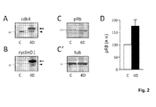
- the inventors found that the ectopic 4D complex was catalytically active as revealed by an increased phosphorilation of its downstream target retinoblastoma ( Fig 2C and D ). Altogether, these experiments showed that the HIV and the MLV here described could be used to temporally control the overexpression of catalytically active 4D complex while being able to distinguish individual 4D overexpressing cells by the expression and localization of GFP.
- GFP nls Histological analyses one week after injection in the adult mouse hippocampus with HIV for GFP nls or G4D revealed that about 10-30% of cells in granular zone expressed GFP and that GFP fluorescence was restricted to the nucleus. Moreover, upon infection with HIV, GFP nls positive cells were evenly distributed in the various layers of the granular zone including neurons, glial cells as well as NSC and NPC ( Fig 3A-F ).
- the increased proportion of BrdU+ cells in the hippocampus upon 4D overexpression can be explained by fourth different possibilities, or a combination thereof.
- 4D overexpression could induce acceleration of the cell cycle with no effect neither on quiescence nor differentiation as, in fact, a shorter G1 would induce cycling cells to enter S phase more often leading to an increase in the proportion of BrdU+ cells.
- second, 4D overexpression could inhibit the differentiation of NSC and/or NPC inducing them to generate more stem/progenitor cells instead of postmitotic neurons, thus increasing the proportion of BrdU+ cells.
- third, 4D overexpression could induce quiescent NSC to enter the cell cycle and thus divide, which also would increase the proportion of BrdU+ cells.
- fourth, 4D overexpression may be toxic in postmitotic cells thereby triggering apoptosis in neurons leading to an increased proportion of cycling cells through their increased survival. The inventors aimed at discriminating between these possibilities by a series of experiments.
- the inventors first assessed the proportion of NSC or NPC within the population of GFP nls + cells 3 weeks after infection with either GFP nls or G4D lentiviruses i.e.: when effects on proliferation were stronger. Immunohistochemistry for the molecular markers Sox2 and Tbr2 were used to identify NSC and NPC, respectively (Kempermann et al., 2004; Zhao et al., 2008) ( Fig 3D and G ).
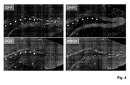
- the inventors assessed the proportion of newborn neurons that were generated during the three weeks interval from the infection with either GFP nls or G4D viruses. To identify newborn neurons, the inventors performed immunohistochemistry for doublecortin (DCX), a marker being transitorily expressed for two weeks after neuronal birth (Kempermann et al., 2004; Zhao et al., 2008).
- DCX doublecortin
- 4D overexpression leads to a reduction of newborn neurons, which is consistent with the increase in NSC and NPC previously described and corroborated the hypotesis that one of the effects of 4D overexpression is to inhibit differentiation and promote proliferation.
- the inventors performed similar manipulations using G4D MLV particles that, in contrast to HIV, can only integrate their genome upon mitosis. Since in normal conditions the vast majority of NSC are quiescent, only mitotic NPC would be targeted. As a result, daughters of the infected, dividing NPC, i.e. mostly newborn neurons, will start to overexpress the 4D cassette, which they inherited from their mother. That is, by this approach the inventors can investigate the effects of 4D overexpression in the subpopulation of newborn neurons generated soon after infection and in the residual population of NPC that did not generate neurons but generated more NPC instead. Importantly, the use of MLV would not influence NSC due to their quiescent state.
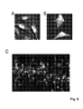
- MLV were injected in the hippocampus and effects on BrdU, DCX and cell morphology investigated 4 days, 1 or 2 weeks after infection with either GFP nls or G4D MLV ( Fig 5 ). In no case could the inventors see any change between control and 4D viruses in any of the parameters analyzed, which always returned proportions consistent with the ones reported in the literature in control conditions ( Fig 5B ) (Jagasia et al., 2009; Esposito et al., 2005).
- the inventors have shown that 4D overexpression increases the proportion of NSC. Thus, the inventors reasoned, if the inventors should be able to stop the expression of the ectopic 4D complex by, for example, removing the 4D cassette from the genome of infected cells, NSC would now be able to undergo physiological differentiation. To investigate this, the inventors generated lentiviral constructs containing LoxP sites flanking the last codon of GFP and cyclinD1 such as to excise the nls of GFP and the whole 4D cassette upon Cre-mediated recombination (see Fig 1B and C for a map of the G Lox 4D Lox and control constructs, respectively) that revealed to be effective in HeLa cells ( Fig 6A ).
- mice were administered tamoxifen or vehicle for four days. Brains were collected ten days later for analyses, which revealed redistribution of GFP fluorescence from the nucleus to the cytoplasm of putative NSC ( Fig 6B ; compare with Fig 3A ).
- the lentiviral vector as described in Example 1 can also be used to expand NSC or NPC in vitro .
- in vitro applications not only viral particles but also any other transfection system with plasmids or DNA/RNA fragments or delivery of proteins can be used, including the use of drugs that pharmacologically act to promote cdk/cyclin expression or their catalytic activity.
- a MLV as described in example 1 is preferred.
- Example 3 construct for the viral infection of human NSC (or NPC) in vivo :
- NSC fluorescent reporter GFP
- CreER T2 For the infection of NSC (or NPC) in the human brain the fluorescent reporter GFP is replaced directly by CreER T2 to mediate the complete removal of the exogenous DNA from human cell at the desired time by administration of tamoxifen.
- An example of a vector for use in humans is shown in Fig 1D (without GFP insertion), where, importantly, expression of CreER T2 will lead to the complete removal of the entire vector upon tamoxifen administration thereby removing any exogenous DNA from the genome of the patient. In essence the vector is designer as to allow its efficient removal at any time point after infection.
- the CreER T2 /GFP/cdk/cyclin vector shown in Fig. 1D with GFP insertion is preferred for the expansion of NSC (or NPC) in vitro for cell transplantation approaches.
- NSC the lentivirus and for NPC the MLV is preffered.
- Example 4 Generation of transgenic mice for the inducible expansion of neural stem cells:
- transgenic mice for the inducible expansion of stem cells. This can be achieved by combining the many technologies available for inducible control of gene expression in vivo , some of which described above, as a means to directly influence the fate of all cells of a given tissue and not only the subpopulation that is being targeted targeted by viral infection or the other methods described above.
- tissue-specific, tetracycline-inducible (Tet-On) cdk/cyclin mouse lines for the temporal and spatial manipulation of cdk/cyclin expression in NSC thus controlling their proliferation vs. differentiation during embryonic development or adulthood.
- Tet-On tissue-specific, tetracycline-inducible
- cdk/cyclin mouse lines for the temporal and spatial manipulation of cdk/cyclin expression in NSC thus controlling their proliferation vs. differentiation during embryonic development or adulthood.
- a bidirectional Tet-promoter drives simultaneous expression of, in one direction, a cyclin and a GFP and, in the other direction, a cdk and a luciferase, which expression can be used for precise quantification of responsiveness upon Doxycycline administration ( Fig 1E ).
- the GFP and/or the luciferase can be replaced by any other desired gene for cell selection, identification or genetic recombination as described above.
- these Tet-On responder mouse lines were crossed with several other transgenic mouse lines in which Tet expression in under the control of cell-specific promoters.
- These bi-genic mice (and any other additional crossing of the responder with any Tet expressing line), allows to overcome the only limitation of the vector trasnduction systems described until now, which, as efficient as they can be, only allow transduction in a in subpopulation (relatively small) of cells rather than in all the relevant cells of a given tissue.
- this system will trigger much greater effects on expansion that can be applied for a multitude of different purposes and contexts e.g.: during i) embryonic development, ii) adulthood, or iii) in mouse model of neurodegenerative diseases or CNS injuries.
- NSC neurotrophic factor
- expansion of NSC during adulthood may allow to address the hitherto elusive role of adult neurogenesis for elaborate function such as memory and learning.
- Expansion of NSC in models of CNS disease or injury can be use to establish platforms for the investigation of novel approaches of regenerative therapies in animal models.
- the responder Dox-inducible cdk/cyclin mouse lines generated by the inventors can be crossed with any of the many available tissue-specific Tet-On activator lines thus allowing the expansion/differentiation of any somatic stem cell in vivo by simple addition/removal of Doxycycline.
- BAC bacterial artificial chromosomes covering the genomic locus of NSC/NPC cell markers, such as GFAP and Sox2, in which the endogenous gene was replaced with a rtTA cassette.
- BAC bacterial artificial chromosomes
- vectors were generated in which a bidirectional Tet-Op promoter drives the expression of a G1-specific cyclin, its cdk, and two reportes, such as RFP and luciferase which are linked by 2A peptides (a similar construct with GFP is shown in Fig 1E ). These vectors were also used to generate responder transgenic lines by male pronuclear injection.
- double transactivator/responder bigenic lines were obtained and overexpression of the transgenes confirmed by detection of RFP in vivo .
- the responder lines are crossed with other available tissue-specific transactivator lines to allow inducible and reversible overexpression of cdk/cyclins in virtually any tissue in vivo.
- This system is also combined with Cre-recombination by crossing the responder mice (Tet-On cdk/cyclin ) with a transgenic line in which rtTA expression in activated after Cre-mediated removal of a stop cassette ( lox stop lox -rtTA).
- the crossing of the bigenic Tet-On cdk/cyclin / lox stop lox -rtTA with any tissue-specific Cre-expressing system further extend the possibily to study any tissue of interest using the already available mouse lines expressing Cre.
- CNS central nervous system CNS central nervous system, NSC neural stem cell, NPC neural progenitor cell, Cdk cyclin dependent kinase, 4D cdk4/cyclinD 1 complex, GFP green fluorescent protein, NIs nuclear localization signal, G4D polycistronic construct encoding for GFP nls , cdk4 and cyclinD1, G Lox 4D Lox G4D construct containing LoxP sites at the 3' end GFP, prior to the nls, and cyclinD1, CreER T2 tamoxifen-dependent Cre recombinase, IP intraperitoneal, BrdU bromodeoxyuridine, HIV human immunodeficiency virus (or vector derived from), MLV Moloney murine leukemia virus (or vector derived from).
Landscapes
- Health & Medical Sciences (AREA)
- Engineering & Computer Science (AREA)
- Biomedical Technology (AREA)
- Life Sciences & Earth Sciences (AREA)
- Wood Science & Technology (AREA)
- Organic Chemistry (AREA)
- Chemical & Material Sciences (AREA)
- Biotechnology (AREA)
- Zoology (AREA)
- Bioinformatics & Cheminformatics (AREA)
- Genetics & Genomics (AREA)
- Neurology (AREA)
- Microbiology (AREA)
- Cell Biology (AREA)
- Neurosurgery (AREA)
- Biochemistry (AREA)
- General Engineering & Computer Science (AREA)
- General Health & Medical Sciences (AREA)
- Developmental Biology & Embryology (AREA)
- Micro-Organisms Or Cultivation Processes Thereof (AREA)
Abstract
Description
- The present invention relates generally to the fields of medicine and life science research, in particular regenerative medicine, neurobiology, cell biology, and stem cell bioengineering. More particularly, it concerns methods and compositions for the expansion of somatic stem and progenitor cells, preferably neural stem cells and neural progenitor cells, in particular in vivo.
- Stem cells of the adult body (somatic stem cells) can expand and generate cell biologically identical somatic stem cells while preserving the ability to differentiate, given the proper stimulus, to generate all cells of a given tissue. Stem cells can undergo two types of cell division. Proliferative division gives rise to at least one identical daughter cell endowed with stem cell properties. Differentiative division, on the other hand, produces only progenitor cells with limited self-renewal potential or terminally differentiated postmitotic cells.
- Somatic stem cells are particularly important for renewal of tissues with high turnover such as skin, blood, or the intestinal epithelium, or may be involved in tissue-repair upon injury, which underlies great hopes for regenerative medicine (Orkin and Zon, 2008; Fuchs, 2009; Li and Clevers, 2010). Also organs with a particularly low turnover, such as the central nervous system (CNS), contain neural stem cells (NSC) that have the capacity to generate i) other NSC, ii) more committed neural progenitors (NPC) or, finally, iii) terminally differentiated cells such as neurons or glia (Kriegstein and Alvarez-Buylla, 2009). In fact, a great deal of interest recently arose form studies of stem cells in the nervous system, not only because of their importance for understanding neural development and brain function but also for their therapeutic potential in the treatment of neurodegenerative diseases (Lindvall and Kokaia, 2006; Okano et al., 2007).
- Neural stem cells from the developing embryo or fetus can be expanded by different means.
WO2005089420 A2 describes the addition of leukemia inhibitory factor (LIF) and several publications have reported similar effects after manipulation of transcription or trophic factors and several signaling moleculs such as basic fibroblast growth factor (bFGF) (Lukaszewicz et al., 2002), β-catenin (Chenn and Walsh 2002), Notch (Kageyama et al., 2008), cell cycle regulators such as cdk4/cyclinD1 (Lange et al., 2009) and many others factors (reviewed in Salomoni and Calegari 2010). - In particular, even if different methods have been designed to purify and culture NSC in vitro (
WO 93/01275 WO 94/09119 WO 94/10292 WO 94/16718 - However, as perhaps the major obstacle in basic stem cell research and their use in therapy, adult stem cells, in contrast to stem cells of the developing embryo, revealed particularly difficult to expand both in vitro and in vivo (Lindvall and Kokaia, 2006; Okano et al., 2007; Orkin and Zon, 2008; Fuchs, 2009; Li and Clevers, 2010). This is probably due to the different cell biology of adult somatic stem cells in comparison to stem cells of the developing embryo, and in particular to their quiescent state. Similarly, progenitor cells are also difficult to expand because, even if they constantly progress through the cell cycle without entering quiescence, their proliferative potential is very limited leading to only few cell division before final consumption to generate differentiated, postmitotic cells (Lindvall and Kokaia, 2006; Okano et al., 2007; Orkin and Zon, 2008; Fuchs, 2009; Li and Clevers, 2010).
- As an additional limitation to the study of somatic stem cells, they essentially stop proliferating in vivo by entering quiescence (Orkin and Zon, 2008; Fuchs, 2009; Li and Clevers, 2010; Salomoni and Calegari, 2010). In addition, attempts made in vivo to force somatic stem cells to leave quiescence and enter the cell cycle revealed unsuccessful leading to complete depletion of the stem cell pool both in the CNS and in hematopoietic stem cells (Cheng et al., 2000; Kippin et al., 2005; Orford and Scadden, 2008). In essence, to our knowledge no efficient system has yet been found to inducibly control the expansion of adult stem or progenitor cells as a means to ultimately increase the generation of their differentiated progeny.
- Because of their low numbers, no direct examination of the biological and molecular properties or the physiological relevance of neural stem cells and neural progenitor cells in the adult was possible up to know.
- Thus the object of the invention is to provide compositions and methods that allow the expansion of somatic stem and progenitor cells, in particular neural stem cells (NSC) and neural progenitor cells (NPC), in vitro and in particular in vivo.
- The object of the invention is solved by a vector that contains genes encoding a cyclin dependet kinase (cdk) and a cyclin forming a G1-specific cdk/cyclin protein complex, preferably cdk4 and cyclinD1 (cycD1). Other preferred cdks are chosen from cdk6 and cdk2. Other preferred cyclins are chosen cyclinD2, cyclinD3 and cyclinE.
- Preferred combinations of cdks and cyclins are chosen from:
- cdk4 and cyclinD1, cyclin D2 or cyclin D3,
- cdk2 and cyclinE,
- cdk6 and cyclinD1, cyclin D2 or cyclin D3.
- Advantageously, the transfection or transduction of somatic stem cells by said vector allows the expansion of somatic stem cells (increasing the number of somatic stem cells) in vitro and in particular in vivo by increasing the proliferation of the transfected or transduced somatic stem cells without affecting their stem cells characteristics, or influencing their multipotency, thus without affecting their potential to differentiate. Applied to progenitor cells the vector will similarly lead to their expansion.
- In an alternative way to carry out the invention the somatic stem cells (or progenitor cells) are expanded by exposing them to a composition containing a cdk and a cyclin forming a G1-specific cdk/cyclin protein complex preferably selected as described above. Here the proteins cdk and cyclin are preferably conjugated to a cell penetrating peptide (CPP). CPPs typically have an amino acid composition containing either a high relative abundance of positively charged amino acids (preferably 30 to 90% of the residues) such as lysine or arginine, or have sequences that contain an alternating pattern of polar/charged amino acids and non-polar, hydrophobic amino acids. These two types of structures are referred to as polycationic or amphipathic, respectively. Preferred CPPs have a length of 8 to 20 amino acid residues and are in particular chosen from tat-petides, model amphipathic peptides, transportans (derived from Galaninin) or poly-arginine, SynB (derived from Protegrin) und cis-γ-amino-L-Prolin-haltigen Peptiden.Examples are:
- RQIKIWFQNRRMKWKK SEQ ID No. 1 (a penetratin),
- KLALKLALKALKAALKLA SEQ I D No. 2 (amphipathic model peptide)
- RKKRRQRRR SEQ I D No. 3 (a tat peptide).
- Other CPPs are mentioned in Zhou et al., 2009 and Lange 2006 included by reference in this respect.
- By exposing the somatic stem cells or progenitor cells to recombinant proteins conjugated to a short peptide, such as a HIV-tat or poly-arginine, cell transduction can be mediated and proteins delivered into cells (Zhou et al., 2009). Though the exact molecular mechanism underlying this transduction is not yet fully elucidated, proteins will by taken up by the somatic stem cells or progenitor cells resulting in a proliferation similar as described above.
- This way for carrying out the invention is in particular suitable for in vitro expansion, but can also by applied in vivo.
- Finally, equally effective will be the use and exposure to drugs or other compounds which main effect is to promote the expression and/or activity of cdk/cyclin complexes in G1 thereby triggering similar effects as the ones described for the vector or protein composition. Preferred examples of compounds activating cdk/cyclins in G1 are estrogens (Prall OW et al. 1997, J Biol Chem. 272:10882-94).
- During the effect induced by the invention, somatic stem cell and/or progenitor cells will divide to proliferate and generate more daughter cells that preserve their multipotency (thus the potential to forming more than one differentiated cell type). By using the invention somatic stem cells can unlimitedly expand and generate cell biologically identical somatic stem cells while preserving the ability to differentiate, given the proper stimulus, to generate all cells of a given tissue.
- The term somatic or adult stem cell refers to any stem cell other than an embryonic stem cell or stem cell derived from embryo or fetus. Somatic stem cells have two properties: the ability to divide and create another cell like itself and also divide and create a cell more differentiated than itself. The term somatic stem exludes cells from embryo or fetus and includes cells that can be found in children, as well as in adults. However, in the invention somatic stem cells of adults are preferred. Somatic stem cells differ from pluripotent or totipotent embryonic stem cells in that they can not give rise to an organism and have lost the potential to differentiate into all three germ layers: endoderm, mesoderm and ectoderm. Somatic stem cells are able to generate a restricted number of lineages (multipotent) and may be referred to by their tissue of origin (e.g. bone marrow stem cells) or by their differentiation potential (hematopoietic stem cell, neural stem cell, mesenchymal stem cell, endothelial stem cell, etc.).
- As the multipotency of the somatic stem cells is not altered by the invention, the somatic stem cells transfected or transduced with the vector used in the invention differ from the so called induced pluripotent stem cells (iPS cells).
- The term progenitor cells refers to a type of partially differentiated, usually unipotent cell that has lost most or all of the stem cell multipotency. Usually progenitor cells are cells capable of differentiating into one or two closely related final differentiated cells types. Preferred examples of progenitor cells are hosen from pancreatic progenitor cells thymocytes, meiocytes, megakaryoblasts, promegakaryocytes, lymphoblasts, normoblasts, bone marrow precursor cells, progenitor cells from the periosteum that are capable to develop into osteoblasts and chondroblasts, myeloblasts, satellite cells found in muscles, endothelial precursor cells, melanoblasts, neural progenitor cells, oligodendrocyte progenitor cells and myeloid precursor cells. Most preferred are neural progenitor cells (NPC). As the invention relates to somatic cells, the term progenitor cells as used in the invention preferably does not include progenitor cells obtained from the embryo and preferably not from the fetus.
- Preferred somatic stem cells used in the invention are chosen from neural stem cells, hematopoietic stem cells, mesenchymal stem cells, endothelial stem cells and any other tissue-specific stem cell of the skin, gut, muscles, hair follicle and so on.
- Particularly preferred are neural stem cells (NSC), most preferred are neural stem cells from the adult central nervous system, in particular the brain (isolated or in vivo). NSC are defined as a cell with unlimited potential to generate identical NSC as well as differentiated cells of the neural lineage such as NPC, neurons or glia. NSC are defined also by cellular markers such as Sox2+, Nestin+, GFAP+ and Pax6+ and can be distinguished from NPC that express additional markers such as Tbr2+, DCX+, Tis21+ as well as from neurons (that are Sox2-, Nestin-, GFAP- and Pax6+) by yet other markers such as NeuN+, NeuroD+, calretinin+, calbinding+ and many others (reviewed in.Kempermann, 2004; Zhao et al., 2008). As the invention relates to somatic cells, the term neural stem cells as used in the invention do not include so called neural stem cells obtained from the embryo and preferably not from the fetus.
- In contrast, a NPC is defined as a cell with limited proliferation potential that can only generate differentiated cells such as neurons or glia. In this definition neuroblasts and terminally-committed progenitors are included.
- The invention can be applied to both NSC and NPC. In both cases the expansion of the cells will result in an increase the number of neurons.
- Further terms used in the description of the invention are defined as follows: a cycling cell is a cell progressing through the cell cycle until completion of mitosis. A quiescent cell is a cell whose cell cycle is temporarily arrested. A postmitotic cell is a cell whose cell cycle is irreversibly blocked and never divides. A newborn neuron is a postmitotic neuron up to two weeks old identified by doublecortin immunostaining. For further definitions see Smith, 2006; Salomoni and Calegari, 2010.
- The vector used in the invention can in principle be a plasmid or an artificial chromosome, which are useful for in vitro transfection. In vitro means into a cell or tissue, that is in tissue culture and/or isolated from a multicellular organism.
- Preferably the vectors are used to transfect or transduce somatic stem and/or progenitor cells, preferably NSC and/or NPC, in particular in the adult brain, in vivo. The term transfection is used for introduction of plasmid vectors into a target cell. For the introduction of viral vectors into the target cell the terms transduction and infection are used with the same meaning. In vivo means in a living adult animal, preferably a vertebrate, in particular a mammal, e.g. a rodent, a human or a non-human animal, preferably an animal that is used as an animal model for neuroscience research, e.g. mice, rats, rabbits, ferrets and non human primates. The preferred route for delivery of DNA vectors into tissues of the central nervous system in vivo is the generation and stereotaxic injection of viral particles but alternatives approaches are also possible such as in vivo electroporation (Barnabé-Heider et a., 2008). In particular for somatic stem and/or progenitor cells outside the central nervous system, many other routes of delivery are possible including aerosol sprays for the peripheral nervous system and mucosae of mouth, nose, trachea and respiratory tracts or direct injection, electroporation or application of viral or DNA vectors for the skin, blood and bone marrow.
- The composition containing cdk and cyclin proteins or compounds are preferably applied in a similar manner.
- For the invention it is preferred that the cdk and cyclin proteins form a 1:1 complex, thus a protein complex, in which both proteins are present in a similar or preferably equimolar ratio. Other solutions may include the overexpression or activation of only one of the two proteins, the cdk or the cyclin, but the presence of both at 1:1 ratio to form a cdk/cyclin complex will increase efficiency and magnitude of the desired effect. This is preferably achieved by an equimolar or approximately equimolar overexpression of the cdk and the cyclin by the vector or by providing the proteins in the composition in an approximately equimolar ratio. Approximately meaning that one protein may be present in excess of 50 mol-%, preferably 20 mol-%, most preferred 10 mol-% in relation to the other.
- In particular for in vivo applications preferably a lentiviral vector or herpes virus based vector is used in the invention for the transduction of stem cells. The lentiviral vector is preferably a self inactivating vector that can infect dividing and non-dividing cells, but is not capable of producing virus particles in the infected cells as the vector lacks viral genes necessary for virus production. The virus particles used for infection are produced in packaging cells expressing those genes necessary for virus production. For progenitor cells, in particular NPC, a retroviral vector, in particular a Moloney murine leukemia-derived genetically modified, replication incompetent gammaretroviruses, is preferred. However, a lentiviral vector or herpes virus based vector can also be applied.
- For in vitro applications viral vectors such as lentiviral or herpes virus based vector are preferred. Here, however, the term vector includes also any other transfection system with plasmids or DNA/RNA fragments, delivery of proteins or, finally, use of drugs acting specifically on the expression or activity of cdk/cyclin complexes.
- Preferably the vector comprises a fusion gene wherein the genes encoding the cdk and the cyclin are separated by a 2A peptide encoding sequence (DeFelipe et al., 2005). Specifically, more than thirty different 2A self-cleaving peptide sequences, which are 15 to 25 amino acids residues long, are know (see http://www.st-andrews.ac.uk/ryanlab/Index.htm for a detailed list) but due to degeneration of the DNA-to-protein translation code, literally millions of DNA fragments for 2A peptides can be generated (DeFelipe et al., 2005). So, in essence, any of these sequences for DNA vectors would allow correct processing and generation of multiple proteins, in particular cdk/cyclins as for this invention, from a single mRNA.
- Preferred 2A peptides are chosen from peptides containing one of the following peptide motives:
ExNPG SEQ ID No. 4 DxExNPG SEQ ID No. 5 GxExNPG SEQ ID No. 6 DVExNPG SEQ ID No. 7 DIExNPG SEQ ID No. 8 DLExNPG SEQ ID No. 9 GVExNPG SEQ ID No. 10 GIExNPG SEQ ID No. 11 GLExNPG SEQ ID No. 12 DVExNPGP SEQ ID No. 13 DIExNPGP SEQ ID No. 14 DLExNPGP SEQ ID No. 15 GVExNPGP SEQ ID No. 16 GIExNPGP SEQ ID No. 17 GLExNPGP SEQ ID No. 18 NFDLLKLAGDVZSNPG SEQ ID No. 19 EGRGSLLTCGDVEENPGP SEQ ID No. 20 GIFNAHYAGYFADLLIHDIETNPGP SEQ ID No. 21 KAVRGYHADYYKQRLIHDVEMNPGP SEQ ID No. 22 LLNFDLLKLAGDVESNPGP SEQ ID No. 23 LTNFDLLKLAGDVESNPGP SEQ ID No. 24 CTNYSLLKLAGDVESNPGP SEQ ID No. 25 GATNFSLLKLAGDVELNPGP SEQ ID No. 26 GPGATNFSLLKQAGDVEENPGP SEQ ID No. 27 FLRKRTQLLMSGDVESNPGP SEQ ID No. 28 EAARQMLLLLSGDVETNPGP SEQ ID No. 29 GSWTDILLLLSGDVETNPGP SEQ ID No. 30 TRAEIEDELIRAGIESNPGP SEQ ID No. 31 RAEGRGSLLTCGDVEENPGP SEQ ID No. 32 AKFQIDKILISGDVELNPGP SEQ ID No. 33 SKFQIDKILISGDIELNPGP SEQ ID No. 34 SSIIRTKMLVSGDVEENPGP SEQ ID No. 35 CDAQRQKLLLSGDIEQNPGP SEQ ID No. 36 - The transcription of the fusion gene results in a bi- or polycistronic expression, meaning that the fusion gene is transcribed into a single messenger RNA. The 2A peptide sequence results in the co-translational cleavage of proteins. They are more reliable than classical internal ribosomal entry sites and lead to expression of the single genes multiple cistrons at equimolar levels. Thus, the fusion gene with the 2A peptide encoding sequence advantageously results not only in a simultaneously but equimolar overexpression of the cdk, the cyclin proteins and, eventually, any other third, or fourth gene such as an expression reporter, a selection marker or recombination gene.
- The fusion gene is under the control of either a constitutive or inducible promoter. Preferably a constitutive promoter is chosen, most preferred the ubiquitin promoter. In particular when a constitutive promoter is used the fusion gene (encoding the cdk and the cyclin) is preferably inserted as an inducible knock on or inducible knock out construct. The inducible knock out has the advantage that the fusion gene can be knocked out under specific well defined conditions, eliminating or deactivating the fusion gene (encoding the cdk and the cyclin). Consequently the stem cells stop proliferating and start to differentiate after knock out. Similar the inducible knock on has the advantage that the fusion gene can be activated (knocked on) under specific well defined conditions.
- Inducible knock on and knock out constructs are know from the literature. A preferred example is the Cre-lox system. In the Cre-lox system a stop cassette (knock on) or the fusion gene (encoding the cdk and the cyclin) (knock out) is flanked by lox-sites (one up-stream, one down-stream) thereby allowing to activate, or inactivate, cdk/cyclin expression by removal of the stop, or cdk/cyclin, cassette respectively.
- The lentiviral vector results in an insertion of the fusion gene into the genome of the infected cell. The lox-sites advantageously allow the inactivation or excision of the fusion gene by an enzyme called Cre recombinase. The lox-sites are preferably both in the same orientation, so that Cre recombinase performs an excision and eliminates the fusion gene from the infected cells. If the lox-sites are in opposite orientation to each other Cre recombinase inactivates the fusion gene by performing an inversion. Thus, by bringing the fusion gene into contact with the Cre protein, the fusion gene is either eliminated or inactivated. Consequently the cells stop proliferating.
- Preferred lox sites are chosen from loxP (ATAACTTCGTAT - SEQ ID No. 37, ATAACTTCGTATA - SEQ ID No. 38, reverse sequences and multimers thereof, like AGCATACATTATACGAAGTTAT - SEQ ID No. 39), x2272, and loxN. Several systems have been developed that allow the inducible activity of Cre protein.
- Other systems are based on different DNA recombinases (further referred to as recombinases), preferably chosen form other Tyrosine site-specific recombinases (SSRs), such as Dre (Anastassiadis et al., 2009) or Tre (Buchholz, 2009). A preferred alternative to the Cre-lox-System is the Dre-rox system (Anastassiadis et al., 2009).
- Another preferred example for such an inducible knock out system is the Tet-Off system, which is comprised of a tet transactivator tTA and a tetO responsive Tyrosine site-specific recombinases (SSRs), such as Cre recombinase. Binding of tTA, in the absence of doxycycline (dox), to the tet promoter causes expression of the SSR, e. g. Cre. Thus the fusion gene (encoding cdk4 and cycD1) will only be active in the presence of doxycycline (dox) or another tetracycline analogue.
- Another preferred example for an inducible knock on system is the Tet-On system, which is comprised of a tet transactivator tTA and a tetO operator. In this system the rtTA protein is only capable of binding the operator when bound by doxycycline or another tetracycline analogue. Once bound the 'tet'O operator will activate a promoter coupled to the 'tet'O operator, activating the transcription of the fusion gene (encoding the cdk and the cyclin). Thus the fusion gene (encoding cdk4 and cycD1) will only be active in the presence of doxycycline (dox) or another tetracycline analogue. The Tet-On system has a faster responsiveness than the Tet-Off system.
- In other inducible knock out systems the recombinase, e.g. an SSR like Cre, encoding gene is placed under control of an inducible promoter. In a preferred system the gene encoding the recombinase is fused to a gene encoding a hormone binding domain of a receptor (further referred to as receptor fusions), preferably a hormone binding domain of a steroid receptor or a mutated version thereof. Receptor fusions are preferably selected from CreERt or CreERT2 (Cre fused to the ligand binding domain of a mutated human estrogen receptor that recognizes anti-estrogen 4- hydroxyltamoxifen), Mer-CreMer or CreP (Cre fused to the progesterone receptor) or DreP (Dre fused to the progesterone receptor). In these systems SSR (in particular Cre or Dre) activity can be induced by administering substances binding to the hormone binding domain of the receptor fusion e.g. administering 4- hydroxyltamoxifen or progesterone.
- These systems can be applied in the invention in several ways:
- 1. Co-transfection or co-transduction the somatic stem and/or progenitor cells, in particular NSC and/or NPC, with a construct allowing the induction of activity of a recombinase, preferably an SSR like Cre, Dre or Tre;
- 2. Transfecting or transduction of somatic stem and/or progenitor cells, in particular NSC and/or NPC, that already contain a construct allowing the induction of activity of a recombinase, preferably an SSR like Cre, Dre or TreCre protein;
- 3. A second transfection or transduction of the somatic stem and/or progenitor cells, in particular NSC and/or NPC, with a vector encoding a recombinase, preferably an SSR like Cre, Dre or Tre.
- In the first case the recombinase encoding gene is preferably inserted into the same vector as the fusion gene - preferably as a separate gene. When a receptor fusion is chosen as recombinase, this receptor fusion can be either on a separate gene or as part of the fusion gene (encoding the cdk and the cyclin) preferably separated by an 2A peptide.
- In the second case preferably somatic stem cells, in particular NSC and/or NPC, are used that are previously transduced with a construct allowing the inducible expression of the recombinase. Alternatively the invention is performed in vivo on animals that are transgenic for a construct allowing the inducible expression of the recombinase in all cells or at least in the somatic stem cells and/or progenitor cells, in particular NSC and/or NPC. Another option is to use somatic stem cells and/or progenitor cells, in particular NSC and/or NPC isolated from these transgenic mice in vitro.
- When a receptor fusion, like CreERt or CreERT2, is used the recombinase activity can advantageously be induced by the administration of a drug, like 4-hydroxyltamoxifen (4-OHT). Thus, by bringing the fusion gene into contact with the recombinase, the fusion gene is either eliminated or inactivated. Consequently the cells stop proliferating.
- It has been shown by the inventors that an orally administered or intraperitoneally injected drug, like 4-OHT, can induce recombinase activity in somatic stem cells and/or progenitor cells, in particular NSC and/or NPC in vivo in the adult brain, leading to an inactivation of the fusion gene (encoding the cdk and the cyclin) (
Fig 6 ) and the cells to stop proliferating. - Especially for in vitro applications transient transfections or transfections in which transfected genes remain episomal and/or their protein products can be removed overtime by direct manipulation or simply by dilution over consecutive divisions are an alternative to the inducible constructs (e.g. knock on or knock out constructs).
- Especially for use in research the vector additionally contains a gene encoding a reporter protein and/or a sequence encoding an epitope tag or any other reporter for distinguishing transfected cells from non transfected including for example, selection and survival genes providing resistance to toxic molecules.
- A reporter protein is one that can easily be detected preferably by measuring fluorescence, light emission or a color reaction or any other reporter for distinguishing transfected cells from non transfected. Preferred reporter proteins chosen from fluorescent proteins (like GFP), luciferase or β-galactosidase (encoded by the bacterial gene lacZ). An epitope is a portion of a molecule to which an antibody binds. Preferred epitope tags are peptides with a length of range from 10 to 15 amino acids long and are designed to easily detect a protein. Non limiting examples include c-myc (EQKLISEEDL - SEQ ID No. 40), HA (YPYDVPDYA -SEQ ID No. 41) and His6 (SEQ ID No. 42 - HHHHHH). The gene encoding the reporter protein and/or the sequence encoding an epitope tag is preferably also part of the fusion gene (encoding the cdk and the cyclin) and preferably separated from the genes encoding the cdk and the cyclin by an 2A peptide encoding sequence. Preferably the cleaved fragment of 2A peptides fused to cdk or cyclin can themselves is used as a tag.
- Another object of the invention are somatic stem cell and/or progenitor cells, in particular NSC and/or NPC transfected or transduced by a vector as defined in the invention.
- In another aspect the invention comprises an animal model wherein somatic stem cells and/or progenitor cells, in particular NSC, or NPC are transfected or transduced by a vector as defined in the invention. A preferred example is transgenic mice for the inducible expression of cdk and a cyclin allowing the inducible expansion of stem cells and progenitor cells. This is achieved by combining available technologies for inducible control of gene expression in vivo, preferably as described above. Advantageously, transgenic animals allow to directly influence the fate of all cells of a given tissue and not only the subpopulation that is being targeted by viral infection or the other methods described above.
- In a preferred example, using the Doxycycline-inducible system the inventors have generated tissue-specific, tetracycline-inducible (Tet-On) cdk/cyclin mouse lines for the temporal and spatial manipulation of cdk/cyclin expression in NSC thus controlling their proliferation vs. differentiation during embryonic development or adulthood. Specifically transgenic mouse lines are preferred in which a bidirectional Tet-promoter drives simultaneous expression of, in one direction, a cyclin and a GFP and, in the other direction, a cdk and a gene encoding a reporter, as luciferase, which expression can advantageously be used for precise quantification of responsiveness upon Doxycycline administration (
Fig 1E ). Clearly, the GFP and/or the luciferase can be replaced by any other desired gene for cell selection, identification or genetic recombination as described above. Importantly, these Tet-On responder mouse lines were crossed with several other transgenic mouse lines in which Tet expression is under the control of cell-specific promoters. These bi-genic mice (and any other additional crossing of the responder with any Tet expressing line), allows to overcome the only limitation of the vector transduction systems described until now, which, as efficient as they can be, only allow transduction in a in subpopulation (relatively small) of cells rather than in all the relevant cells of a given tissue. - Advantageously the responder Dox-inducible cdk/cyclin mouse lines generated by the inventors can be crossed with any of the many available tissue-specific Tet-On activator lines thus allowing the expansion/differentiation of any somatic stem or progenitor cell in vivo by simple addition/removal of Doxycycline.
- If applied to NSC or NPC, this system advantageously allows to trigger much greater effects on expansion that can be applied for a multitude of different purposes and contexts e.g.: during i) embryonic development, ii) adulthood, or iii) in mouse model of neurodegenerative diseases or CNS injuries.
- Expansion of NSC or NPC during adulthood allows addressing the hitherto elusive role of adult neurogenesis for elaborate function such as memory and learning. Expansion of NSC or NPC in models of CNS disease or injury can be use to establish platforms for the investigation of novel approaches of regenerative therapies in animal models.
- The invention also comprises a method for the expansion of somatic stem cells and/or progenitor cells, in particular NSC and/or NPC with the steps:
- a.) providing a vector as defined in the invention,
- b.) introducing said vector into somatic stem cells and/or progenitor cells by transfection or transduction,
- c.) overexpression of cdk and the cyclin protein complex in the transfected or transduced somatic stem cells and/or progenitor cells, resulting in proliferation of the transfected or transduced somatic stem cells and/or progenitor cells.
- In an alternative way to carry out the invention comprises a method for the expansion of somatic stem cells and/or progenitor cells, in particular NSC and/or NPC, with the steps:
- a') contacting somatic stem cells with a protein composition containing cdk and a cyclin as defined above or a compound as defined in the invention resulting in proliferation of the somatic stem cells and/or progenitor cells.
- Step b.) is either carried out in vitro or in particular in vivo by applying the vector to an animal as described above. Alternatively step a') is carried out by applying the composition or compound to isolated cells in vitro or in vivo to an animal as described above.
- The invention comprises also somatic stem cells and/or progenitor cells, in particular NSC and/or NPC, prepared by the method described above. The somatic stem cells and/or progenitor cells transfected or transduced with the vector according to the invention contain either the vector or after induced knock-out remaining (flanking) sequences of the vector.
- A preferred way of carrying out the method of the invention comprises the additional step of an induced knock out of the overexpressed cdk and the cyclin genes, preferably by inducing the recombinase activity, after the number of adult neural stem have been increased (step c.) of the method of the invention). As described above the induced knock out, preferably by inducing recombinase activity, will excise or inactivate the cdk and the cyclin genes in the transfected or transduced cells. Consequently the cdk and the cyclin induced proliferation of the cells (stem cells and/or progenitor cells) will be stopped.
- Alternatively the protein composition containing cdk and a cyclin or compound is supplied as long as the stem cells and/or progenitor cells shall expand. Here the proliferation is simply stopped by not further supplying the composition or by rinsing the cells.
- Similar in case of a knock on construct (like the Tet-On system) the drug inducing the gene expression (e. g. tetracycline) is applied as long as the stem cells and/or progenitor cells shall expand and the expression is stopped by not further supplying the drug (e. g. tetracycline).
- In Summary the inventions provides a system that allows:
- 1. a controlled increase of the somatic stem cell population and/or progenitor cell population, preferably NSC and/or NPC, in particular in vivo (in the adult brain) by inducing their proliferation,
- 2. to stop the proliferation of the somatic stem cell population and/or progenitor cell population (in particular NSC and/or NPC), which allows the somatic stem cells and/or progenitor cell population,to differentiate and to regenerate tissue, in case of NSC and/or NPC to perform neurogenesis or gliogenesis.
- Consequently regeneration of tissue, e.g. neurogenesis, is increased by the invention by increasing the somatic stem cell population and/or progenitor cells. Advantageously, the overexpression of the cdk and the cyclin protein complex is not toxic and increases proliferation, without affecting the multipotency of the somatic stem cells, in particular NSC. A further advantage of the system of the invention is that the induced proliferation does not lead to tumor development or carcinogenesis. As the overexpression of the cdk and the cyclin can be stopped efficiently (e. g. by inducing the knock out of the overexpressed cdk and the cyclin genes) the system is safe.
- The invention is useful for life science, neuroscience and/or pharmaceutical research, in particular to study the role and function of somatic stem cells and/or progenitor cells, in particular NSC and/or NPC in the adult brain. Further the invention is preferably applied to animal models for diseases, disorders or conditions of the central nervous system.
- Also expansion of somatic stem cells and/or progenitor cells, in particular NSC and/or NPC, in vitro can advantageously be used for various purposes in basic research, diagnosis or in cell-based therapeutic applications by cell transplantions or any other means.
- The observation that exit from quiescence and entry in the cell cycle can be induced by cdk/cyclin overexpression is unprecedented. Importantly, since somatic stem cells during adulthood are known to be quiescent in essentially all tissues, it follows that the invention can be applied to any other somatic stem cell such as hematopoietic or stem cells of the skin, which can be targeted by various means as already described.
- For instance, bone marrow transplantation is a common and well established approach in many therapeutic applications of hematopoietic stem cells (HSC). However, the major limiting factor of this technique is the very limited amount of bone marrow that can be collected from a compatible donor. Thus, expansion of HSC, prior to of following transplantation, will allow to greatly improve the efficiency of stem cells based therapies. In essence, transduction of HSC with cdk/cyclins vectors can be preformed either in vivo by direct injection in the bone marrow or, alternatively, in vitro upon removal of bone marrow tissue from a donor to be later transfused in a receiving patient or back to the donor. In particular for the later approach, expansion of HSC by transduction of cdk/cyclin vectors in vitro seems preferable as this will allow subsequent tamoxifen treatment and Cre-mediated recombination to occur outside the patient clearly avoiding any potential side effect due to tamoxifen administration in vivo. In this case, a fluorescent reporter, such as GFP, is preferably also inserted together with the recombinase (like CreERT2) using 2A sequences as this will allow to remove by FAC-sorting the few cells in which recombination may eventually not occur due to intrinsic limitations of the various recombination systems.
- The use of vectors as defined in the invention (like CreERT2/GFP/cdk/cyclin) is thus the preferred approach for the inducible and temporally controlled expansion of any somatic stem cell which allows manipulations to be performed completely in vitro while simultaneously allowing selection of absolutely pure population of cells for transplantation that are completely devoid of any exogenous DNA.
- The invention also comprises the vector or composition or compound as defined in the invention for several possible applications in basic scientific research and their use in the expansion of somatic stem cells and/or progenitor cells, in particular NSC and/or NPC, for therapies for the treatment of a human or an animal having a disease, disorder or condition of the central nervous system (CNS), preferably selected from the group consisting of a neurodegenerative diseases, developmental disorders of the CNS, or injury of the CNS caused by accidents or tumors, in particular Huntington's disease, Alzheimer's disease, Parkinson's disease, brain injury or trauma, spinal cord injury or trauma, stroke, multiple sclerosis, cancer, CNS lysosomal storage diseases and head trauma and epilepsy.
- The invention includes a method of treating a human or an animal having a disease, disorder or condition of the CNS. The disease, disorder or condition of the CNS is preferably selected from the group consisting of a neurodegenerative diseases, developmental disorders of the CNS, or injury of the CNS caused by accidents or tumors, in particular Huntington's disease, Alzheimer's disease, Parkinson's disease, brain injury or trauma, spinal cord injury or trauma, stroke, multiple sclerosis, cancer, CNS lysosomal storage diseases and head trauma, epilepsy and any other malfunctions of the CNS.
- In yet another aspect, the disease, disorder or condition is injury to the tissue or cells of the CNS, which might be the result of the removal of a brain tumor or cancer.
- The treatment is preferably carried out by applying a vector or composition defined according to the invention to the human or animal as defined above to expand somatic stem cells and/or progenitor cells, in particular NSC and/or NPC, in vivo. In another aspect, somatic stem cells and/or progenitor cells, in particular NSC and/or NPC are isolated from the human or animal or differentiated from other cells and expanded in vitro by transforming or transducing them with a vector as defined in the invention or by contact with the composition as defined in the invention. In vitro expanded cells are administered to the CNS. Alternatively, cells are transformed or transduced in vitro and administered directly to the CNS and replicate in the CNS.
- Viral infection is one of the most intensely studied approach for gene therapy in humans thank to the characterization of several strains of non pathogenic viruses (Cameron and McKay, 2001; Lowenstein and Castro, 2002) and, thus, the findings of the invention are directly relevant for therapy in the human CNS. For the infection of NSC and/or NPC in the human brain the vector contains preferably additionally to the fusiongene encoding the cdk/cyclin complex a gene encoding a recombinase, preferably a receptor fusion as described above to mediate the removal of the exogenous DNA from human cell at the desired time by administration of a drug. In essence the vector is designed as to allow its efficient removal at any time point after infection.
- It is important to stress that while many tumors were found to overexpress cyclins and cdks, the overexpression of the cdk/cyclin complex used in the invention was not shown to cause tumorogenesis. This was confirmed both in the embryo as well and in the adult, were no obvious formation of tumors were observed at any stage of analyses. Groups of mice were subjected to cyclin and cdk overexpression for several months to investigate this important issue. Further, the possibility to conditionally remove (or knock out) the cyclin and cdk genes allows to minimize, if not completely exclude, possible long term side effects of the ectopic genes.
- The following figures and examples are presented in order to more fully illustrate the preferred embodiments of the invention. These examples should in no way be construed as limiting the scope of the invention.
-
Fig. 1 shows the maps of the viral constructs for (A) G4D, (B)G Lox4DLox, and respective (C) GFPnls controls (left and right, respectively). Similar 2A peptides (T2A with degenerated DNA sequence) and promoters (CMV) were used. The black triangles represent the loxP sites. (D) map of a construct to be used in therapy (C4D) in which GFP has been replaced by, or is inserted in addition to (inset indicated by lines), an inducible CreERT2 and LoxP sites were inserted as to remove the whole exogenous vector upon tamoxifen administrartion. (E) map of a vector used for the generation of Tet-responder transgenic mice for the inducible, Doxycycline-dependent expression of cdk/cyclins/GFP/luciferase. These mice were crossed with tissue-specific Tet-expressing mice (not shown). -
Fig. 2 shows the viral-mediated expression of catalytically active 4D (A-C') in Western blot analyses of HeLa cells infected with 4D-overexpressing viruses after immunostaining with cdk4 (A), cyclinD1 (B), phosphorylated-specific Rb (C), or tubulin (C') antibodies compared to GFPnls control (C). Note the presence of two bands for cdk4 (A) and cyclinD1 (B) representing the endogenous cdk4 or cyclinD1 (arrowhead) or fusion products with 2A peptides (arrow). (D) Quantification of phosphorylated Rb after tubulin normalization as in C and C'. -
Fig. 3 demonstrates that expression of G4D by lentiviral infection induces the expansion of NSC/NPC and inhibits neurogenesis: (A and A') Fluorescent pictures of the mouse hippocampus infected with G4D lentiviruses showing individual infected cells (B) and DAPI staining (A'). (B) High-power view as in A after staining for GFP (left), BrdU (right. Arrows indicate a GFP/BrdU+ cell. (C) Quantifications as in C at 1, 2, or 3 weeks after infection with GFPnls (white boxes) and G4D (black boxes). (D) High-power view as in A after staining for GFP (left), Sox2 (middle), and Tbr2 (right) . (E and F) Quantification of Sox2 (E) and Tbr2 (F) as in D at 3 weeks from infection with GFPnls (white boxes) and G4D (black boxes). -
Fig. 4 shows fluorescence pictures of the mouse hippocampus infected with G4D lentiviruses by detecting GFP (upper left), DCX (lower left), DAPI (upper right) immunoreactivity, or merged (lower right). The increased density of DCX+ cells in the uninfected portion of hippocampus is indicated by arrowheads. -
Fig. 5 demonstrates that expression of G4D by MLV retrovirus infection has no effect on newborn neurons. (A) Fluorescence picture of the mouse hippocampus after infection with 4D-overexpressing and RFP expressing MLV. (B and B') High power view as in A showing the morphology of newborn neurons 0.5, 1, or 2 weeks after infection with control MLV (B; up) or 4D-overexpressing MLV (B'; bottom) particles. (C and D) quantification of DCX+ (C) or NeuN, a marker of more mature neurons, (D) after infection with RFP, control MLC (C, white symbols) or 4D-overexpressing MLV (4D, black symbols) particles. None of the differences are significant. The MLV enters in a mitotic NPC. This infected NPC soon after divides to generate neural cell daughters. Thus, expression of cdk/cyclin starts in the newborn neuron and NOT in the mother NPC. These data are important to exclude the possibility that overexpression of cdk/cyclin is toxic to neurons -
Fig. 6 shows the results of conditional excision of theG Lox4DLox cassette (A andB). Nuclear (A) and cytoplasmic (B) localization of GFP after infection withG Lox4DLox-expressing viruses in HeLa cells wild type (A) or HeLa cells expressing Cre recombinase (B). (C) fluorescent pictures showing redistribution of GFP fluorescence to the cytoplasm upon injection ofG Lox4DLox-expressing MLV in nestin CreERT2/- mice followed by tamoxifen administration for 4 days. - To manipulate gene expression in NSC and NPC in vivo the inventors generated and stereotaxically injected i) HIV-derived genetically modified, replication incompetent lentiviruses (from now on referred to as HIV) or ii) Moloney murine leukemia-derived genetically modified, replication incompetent gammaretroviruses (from now on referred to as MLV). Upon infection, the viral genome irreversibly integrate into the genome of the host cells with the critical difference that MLV cannot transduce non mitotic cells because the viral integration complex can entry into the nucleus only if the nuclear envelope of the infected cell is disassembled, which only occurs in mitosis (Roe et al., 1993). Thus, while HIV allows the infection and rapid expression of viral genes in all cycling, quiescent and postmitotic cells, the use of MLV only allows the infection of the subpopulation of cells that at the time of viral exposure was undergoing mitosis, thus, excluding quiescent and postmitotic ones. Of note, the onset of MLV protein expression will occur in daughters of infected cells.
- Thus, to thoroughly characterize the effects of 4D overexpression on adult neurogenesis the inventors generated 4D-expressing HIV and MLV and also designed a system that allows us to conditionally terminate 4D overexpression in infected cells. The inventors generated polycistronic constructs used in the experiments that encode for i) a GFP with a nuclear localization signal (nls), ii) mouse cdk4, and iii) mouse cyclinD1 (
Fig. 1 ). Original empty viral backbones were used as described (Wurm et al., 2010) for the HIV lentiviral construct and MLV viruses (Zhao et al., 2006), respectively. cDNA for mouse cdk4 and cyclinD1 (together referred to as 4D) were obtained from the RIKEN consortium (Japan). cdk4 and cyclinD1 are from now on referred to as as 4D. The three genes (GFPnls/cdk4/cyclinD1; from now on referred to as G4D) were linked by the 2A sequences (Fig 1A ) 5'-GAGGGCAGAGGAAGTCTTCTAACATGCGGTGACGTGGAGGAGAATCCCGGCCCT-3'(SEQ ID No. 43) or a degenerated code encoding for the same peptide 5'-GAAGGTAGGGGCAGTCTACTTACCTGTGGAGATGTAGAAGAAAACCCAGGACCG-3' (SEQ ID No. 44) that allow the stoichiometric translation of individual proteins from a single mRNA (Tang et al., 2009). Similar constructs were also generated that contain LoxP sites (5'-ATAACTTCGTATAGCATACATTATACGAAGTTAT-3' - SEQ ID No. 45) at the end of the coding region of GFP and cyclinD1 (from now on referred to asG Lox4DLox) (Fig 1B ). By this approach, excision of the 4D cassette upon Cre-mediated recombination could be assessed in individual cells by redistribution of GFP fluorescence from the nucleus to the cytoplasm due to loss of the nls. For control HIV and MLV particles encoding only GFPnls (G), eventually with LoxP sites flanking the nuclear localization signal (nls), were used (Fig 1C ). - Viruses were generated and purified according to standard procedures (Tashiro et al., 2006) and injected in the hippocampus of either wildtype C57/B16 or transgenic mice expressing the tamoxifen-dependent Cre recombinase (CreERT2) in NSC. This latter mouse line, the nestinCreERT2/- line (Jackson laboratory, Bar Harbor, Maine, USA Strain B6.Cg(SJL)-TgN(NesCre)1Kln, stock number 003771) was used as described in Imayoshi et al., 2008. Mice were aged between 6 and 11 weeks and infections were performed by using a stereotaxic injection system (coordinates: ±1.6 ML; -1.9 DV; -1.9 AP from dura) (Kopf instruments) following standard surgical procedures (Tashiro et al., 2006). After 1, 2, 3 or 5 weeks, mice were sacrificed and brains collected. Eventually, prior to sacrifice mice were exposed to BrdU (1 mg/mouse injected intraperitoneally (IP) at 12 hours interval for 7 days), tamoxifen (5 mg/mouse IP at 12 hours interval for 2 days), or, finally, tamoxifen followed 3 days later by BrdU..
- 40 µm thick vibratome sections were performed through the entire hippocampus and sections grouped serially every six according to standard stereological methods. Each group, containing 7-8 sections covering the entire hippocampus from its rostral-to-caudal boundary, was used to assess the number of GFPnls+ cells immunoreactive for the markers of S phase and proliferation bromodeoxyuridine (BrdU) or positive for established molecular markers of NSC, NPC or newborn neurons such as Sox2, Tbr2 and DCX, respectively (Kempermann et al., 2004; Zhao et al., 2008). DAPI was used for nuclear staining.
- The results of these experiments are summarized as follows:
- Infection of HeLa cells with i) GFPnls, or ii) G4D, or
G Lox4DLox HIV or MLV particles followed by assessment of endogenous GFP fluorescence by microscopy revealed the correct localization of GFPnls into the nucleus and its redistribution to the cytoplasm in HeLa cells coexpressing Cre (Fig 6 ). Moreover, similar experiments followed by western blot analyses for GFP, mouse cdk4, or mouse cyclinD1 showed the efficient co-expression of the three transgenes and the translation of the three individual proteins from a single polycistronic mRNA (Fig 2A and B ). Finally, the inventors found that the ectopic 4D complex was catalytically active as revealed by an increased phosphorilation of its downstream target retinoblastoma (Fig 2C and D ). Altogether, these experiments showed that the HIV and the MLV here described could be used to temporally control the overexpression of catalytically active 4D complex while being able to distinguish individual 4D overexpressing cells by the expression and localization of GFP. - Histological analyses one week after injection in the adult mouse hippocampus with HIV for GFPnls or G4D revealed that about 10-30% of cells in granular zone expressed GFP and that GFP fluorescence was restricted to the nucleus. Moreover, upon infection with HIV, GFPnls positive cells were evenly distributed in the various layers of the granular zone including neurons, glial cells as well as NSC and NPC (
Fig 3A-F ). - To investigate possible effects of 4D overexpression on proliferation of NSC or NPC in the hippocampus, the inventors exposed infected mice to repetitive IP injections with the thymidine analogue BrdU, which is irreversibly incorporated in the DNA during S phase and thus allows distinguishing cycling cells from quiescent or postmitotic ones. Infection with control GFPnls viruses showed a proportion of GFP/BrdU+ cells (1-2%) that was in line with the value reported in the literature (
Fig 3B and C; white symbols) (Mao et al., 2009). However, the inventors observed that already 1 week after 4D overexpression the proportion of GFP/BrdU+ cells had increased by ca. 70% in G4D as compared to with GFPnls viral infection (Fig 3C ). Importantly, while in control conditions the proportion of BrdU+ cells remained overall constant overtime, the increase in the proportion of BrdU+ cells after 4D overexpression become more prominent at longer survival times reaching, at the third week, an increase by over 100% as compared to control (Fig 3C ). - Importantly, the increased proportion of BrdU+ cells in the hippocampus upon 4D overexpression can be explained by fourth different possibilities, or a combination thereof. Specifically, first, 4D overexpression could induce acceleration of the cell cycle with no effect neither on quiescence nor differentiation as, in fact, a shorter G1 would induce cycling cells to enter S phase more often leading to an increase in the proportion of BrdU+ cells. Alternatively, second, 4D overexpression could inhibit the differentiation of NSC and/or NPC inducing them to generate more stem/progenitor cells instead of postmitotic neurons, thus increasing the proportion of BrdU+ cells. Moreover, third, 4D overexpression could induce quiescent NSC to enter the cell cycle and thus divide, which also would increase the proportion of BrdU+ cells. Finally, fourth, 4D overexpression may be toxic in postmitotic cells thereby triggering apoptosis in neurons leading to an increased proportion of cycling cells through their increased survival. The inventors aimed at discriminating between these possibilities by a series of experiments.
- The inventors first assessed the proportion of NSC or NPC within the population of GFPnls+
cells 3 weeks after infection with either GFPnls or G4D lentiviruses i.e.: when effects on proliferation were stronger. Immunohistochemistry for the molecular markers Sox2 and Tbr2 were used to identify NSC and NPC, respectively (Kempermann et al., 2004; Zhao et al., 2008) (Fig 3D and G ). - Infection with control, GFPnls HIV showed a proportion of Sox2+ (ca. 1.8%) and Tbr2+ (0.8%) cells that was consistent with what reported in the literature (Suh et al., 2007; Hodge et al., 2008) indicating that infection per se does not alter the proportion of NSC or NPC. In contrast, the inventors found that upon G4D infection the proportion of Sox2+ and Tbr2+ cells had increased by ca. 140% and 80%, respectively
(Fig 3E and F ). This finding excludes the possibility that the primary effect of 4D overexpression is solely to change the length of the G1 phase of the cell cycle of NPC without influencing quiescence of NSC or differentiation of either kind of precursor. In fact, a shorter G1, without a change in neither quiescence nor differentiation, while still changing the rate of BrdU+ incorporation cannot lead to a change in the abundance of different cell types. The inventors thus conclude that the primary effect of 4D overexpression is to induce exit from quiescence of NSC and inhibit differentiation. - To validate the conclusion that a possible effect of 4D overexpression is to inhibit the differentiation of NSC and/or NPC, the inventors assessed the proportion of newborn neurons that were generated during the three weeks interval from the infection with either GFPnls or G4D viruses. To identify newborn neurons, the inventors performed immunohistochemistry for doublecortin (DCX), a marker being transitorily expressed for two weeks after neuronal birth (Kempermann et al., 2004; Zhao et al., 2008).
- Particularly instructive for this analysis were sections in which GFPnls+ cells after G4D infection were more concentrated, or occasionally even limited, to a given region of the hippocampus such as its most dorsal or ventral portion. In essence, this allowed the comparison, within the same section, of the abundance of DCX+ cells in the 4D-targeted and internal negative control part of the hippocampus. The inventors consistently found an evident reduction of DCX+ cells in the G4D-targeted region of the hippocampus
(Fig 4G ), which was independent on whether this portion was dorsal or ventral. - In essence, 4D overexpression leads to a reduction of newborn neurons, which is consistent with the increase in NSC and NPC previously described and corroborated the hypotesis that one of the effects of 4D overexpression is to inhibit differentiation and promote proliferation.
- The previous experiments show that the increased proliferation is caused by a decreased differentiation of NSC and NPC. However, it still remains unclear whether quiescence of NSC is at all influenced by this manipulation. More importantly, it should be noticed that the increased proportion of Sox2+ and Tbr2+ cells and decreased proportion DCX+ neurons could simply be caused by neuronal death after 4D overexpression. In fact, the 4D complex is considered to be a potent positive regulator of cell cycle progression and it cannot be excluded that overexpression of 4D in postmitotic neurons may lead to cell stress and apoptosis. In other words, all phenotypes described so far could simply be explained by death of neurons overexpressing 4D.
- To exclude this possibility, the inventors performed similar manipulations using G4D MLV particles that, in contrast to HIV, can only integrate their genome upon mitosis. Since in normal conditions the vast majority of NSC are quiescent, only mitotic NPC would be targeted. As a result, daughters of the infected, dividing NPC, i.e. mostly newborn neurons, will start to overexpress the 4D cassette, which they inherited from their mother. That is, by this approach the inventors can investigate the effects of 4D overexpression in the subpopulation of newborn neurons generated soon after infection and in the residual population of NPC that did not generate neurons but generated more NPC instead. Importantly, the use of MLV would not influence NSC due to their quiescent state.
- MLV were injected in the hippocampus and effects on BrdU, DCX and cell morphology investigated 4 days, 1 or 2 weeks after infection with either GFPnls or G4D MLV (
Fig 5 ). In no case could the inventors see any change between control and 4D viruses in any of the parameters analyzed, which always returned proportions consistent with the ones reported in the literature in control conditions (Fig 5B ) (Jagasia et al., 2009; Esposito et al., 2005). - In essence, overexpression of 4D in newborn neurons does not impair their survival and targeting of NPC does not change their commitment to generate neurons. Considering that i) most of the increase in the proportion of BrdU+ cells was observed to occur already during the first week after infection (
Fig 3C ) and ii) that the increase in the proportion of NSC was more prominent than that observed in NPC (Fig 3E versus 3F), the inventors conclude that the primary effect of 4D overexpression is to induce quiescent NSC to enter the cell cycle and generate more NSC and, thus, to expand. - Having shown that an acute and tissue-specific 4D overexpression in the adult mouse hippocampus triggers the expansion of NSC, the fundamental question remains as to whether or not this effect can be temporally restricted in order to increase the neuronal output in the brain. In fact, the main hope for using somatic stem cells in therapy is not only that their expansion could be increased but also that their differentiation could be temporally controlled.
- As to now, the inventors have shown that 4D overexpression increases the proportion of NSC. Thus, the inventors reasoned, if the inventors should be able to stop the expression of the ectopic 4D complex by, for example, removing the 4D cassette from the genome of infected cells, NSC would now be able to undergo physiological differentiation. To investigate this, the inventors generated lentiviral constructs containing LoxP sites flanking the last codon of GFP and cyclinD1 such as to excise the nls of GFP and the whole 4D cassette upon Cre-mediated recombination (see
Fig 1B and C for a map of theG Lox4DLox and control constructs, respectively) that revealed to be effective in HeLa cells (Fig 6A ). Injections were performed in nestin CreERT2/- mice, in which tamoxifen-dependent Cre-recombination was shown to occur specifically in NSC and NPC (Imayoshi et al., 2008). Three weeks after infection, mice were administered tamoxifen or vehicle for four days. Brains were collected ten days later for analyses, which revealed redistribution of GFP fluorescence from the nucleus to the cytoplasm of putative NSC (Fig 6B ; compare withFig 3A ). - Altogether these data show that the inventors can acutely and tissue-specifically overexpress the 4D complex in the adult neurogenic niche while maintaining the ability to conditionally stop this effect. In essence, the inventors developed as system that allows to temporally control the expansion of NSC and their switch to differentiation in vivo.
- The lentiviral vector as described in Example 1 can also be used to expand NSC or NPC in vitro. However, for in vitro applications, not only viral particles but also any other transfection system with plasmids or DNA/RNA fragments or delivery of proteins can be used, including the use of drugs that pharmacologically act to promote cdk/cyclin expression or their catalytic activity. For the expansion of NPC a MLV as described in example 1 is preferred.
- For the infection of NSC (or NPC) in the human brain the fluorescent reporter GFP is replaced directly by CreERT2 to mediate the complete removal of the exogenous DNA from human cell at the desired time by administration of tamoxifen. An example of a vector for use in humans is shown in
Fig 1D (without GFP insertion), where, importantly, expression of CreERT2 will lead to the complete removal of the entire vector upon tamoxifen administration thereby removing any exogenous DNA from the genome of the patient. In essence the vector is designer as to allow its efficient removal at any time point after infection. - The CreERT2/GFP/cdk/cyclin vector shown in
Fig. 1D with GFP insertion is preferred for the expansion of NSC (or NPC) in vitro for cell transplantation approaches. For NSC the lentivirus and for NPC the MLV is preffered. - Another direct application of the invention is the generation of transgenic mice for the inducible expansion of stem cells. This can be achieved by combining the many technologies available for inducible control of gene expression in vivo, some of which described above, as a means to directly influence the fate of all cells of a given tissue and not only the subpopulation that is being targeted targeted by viral infection or the other methods described above.
- For example, using the Doxycycline-inducible system the inventors have generated tissue-specific, tetracycline-inducible (Tet-On) cdk/cyclin mouse lines for the temporal and spatial manipulation of cdk/cyclin expression in NSC thus controlling their proliferation vs. differentiation during embryonic development or adulthood. Specifically transgenic mouse lines were created in which a bidirectional Tet-promoter drives simultaneous expression of, in one direction, a cyclin and a GFP and, in the other direction, a cdk and a luciferase, which expression can be used for precise quantification of responsiveness upon Doxycycline administration (
Fig 1E ). Clearly, the GFP and/or the luciferase can be replaced by any other desired gene for cell selection, identification or genetic recombination as described above. Importantly, these Tet-On responder mouse lines were crossed with several other transgenic mouse lines in which Tet expression in under the control of cell-specific promoters. These bi-genic mice (and any other additional crossing of the responder with any Tet expressing line), allows to overcome the only limitation of the vector trasnduction systems described until now, which, as efficient as they can be, only allow transduction in a in subpopulation (relatively small) of cells rather than in all the relevant cells of a given tissue. - If applied to NSC, this system will trigger much greater effects on expansion that can be applied for a multitude of different purposes and contexts e.g.: during i) embryonic development, ii) adulthood, or iii) in mouse model of neurodegenerative diseases or CNS injuries.
- For instance, expansion of NSC during adulthood may allow to address the hitherto elusive role of adult neurogenesis for elaborate function such as memory and learning. Expansion of NSC in models of CNS disease or injury can be use to establish platforms for the investigation of novel approaches of regenerative therapies in animal models.
- Finally, the responder Dox-inducible cdk/cyclin mouse lines generated by the inventors can be crossed with any of the many available tissue-specific Tet-On activator lines thus allowing the expansion/differentiation of any somatic stem cell in vivo by simple addition/removal of Doxycycline.
- METHOD: Using RedET recombination (Vintersten et al., 2008), the inventors generated bacterial artificial chromosomes (BAC) covering the genomic locus of NSC/NPC cell markers, such as GFAP and Sox2, in which the endogenous gene was replaced with a rtTA cassette. These modified BACs were then used to generate transactivator transgenic mouse lines by male pronuclear injection (Vintersten et al., 2008). In addition, vectors were generated in which a bidirectional Tet-Op promoter drives the expression of a G1-specific cyclin, its cdk, and two reportes, such as RFP and luciferase which are linked by 2A peptides (a similar construct with GFP is shown in
Fig 1E ). These vectors were also used to generate responder transgenic lines by male pronuclear injection. - RESULTS: Of the many transactivator lines generated we found, in particular for Sox2-rtTA, some faithfully reproducing the Sox2 pattern and tissue-specificity for rtTA expression. This was confirmed by in situ hybrydization of embryonic and adult brain tissue. Similarly, various cdk/cyclin responder lines were screened by transfecting primary fibroblast cultures obtained their from for luciferase assay and RFP expression upon rtTA transduction and administration of doxycycline. Western blott analysis for cdk and cyclins, similarly to the experiments described above and in
Fig 2 upon viral infection, corroborated the correct expression of the transgenes upon rtTA expression and doxycycline administration. Finally, double transactivator/responder bigenic lines were obtained and overexpression of the transgenes confirmed by detection of RFP in vivo. The responder lines are crossed with other available tissue-specific transactivator lines to allow inducible and reversible overexpression of cdk/cyclins in virtually any tissue in vivo. This system is also combined with Cre-recombination by crossing the responder mice (Tet-Oncdk/cyclin) with a transgenic line in which rtTA expression in activated after Cre-mediated removal of a stop cassette (loxstoplox-rtTA). The crossing of the bigenic Tet-Oncdk/cyclin/ loxstoplox-rtTA with any tissue-specific Cre-expressing system further extend the possibily to study any tissue of interest using the already available mouse lines expressing Cre. - The following abbreviations are used in the description:
CNS central nervous system, NSC neural stem cell, NPC neural progenitor cell, Cdk cyclin dependent kinase, 4D cdk4/ cyclinD 1 complex,GFP green fluorescent protein, NIs nuclear localization signal, G4D polycistronic construct encoding for GFPnls, cdk4 and cyclinD1, G Lox4DLoxG4D construct containing LoxP sites at the 3' end GFP, prior to the nls, and cyclinD1, CreERT2 tamoxifen-dependent Cre recombinase, IP intraperitoneal, BrdU bromodeoxyuridine, HIV human immunodeficiency virus (or vector derived from), MLV Moloney murine leukemia virus (or vector derived from). - The following non-patent literature is cited in the description:
- Anastassiadis K et al. 2009. Dis Model Mech 2:508-515.
- Barnabé-Heider F et al. 2008. Nat Methods 5:189-196.
- Bordignon C, Roncarolo MG 2002. Nat Immunol 3:318-321.
- Buchholz F 2009. Curr Opin Biotechnol 20:383-389.
- Calegari F, Huttner WB 2003. J Cell Sci 116:4947-4955.
- Cameron HA, McKay RD 2001. J Comp Neurol 435:406-417.
- Cheng T et al. 2000. Science 287:1804-1808.
- Chenn A, Walsh CA 2002. Science 297:365-369
- De Felipe P et al. 2005 Trends Biotechnol 24:68-75
- Deiters A 2009. Curr Opin Chem Biol 13:678-686.
- Espósito MS et al. 2005. J Neurosci 25:10074-86.
- Fuchs E 2009. Cell 137:811-819.
- Hodge RD et al. 2008. J Neurosci 28:3707-17.
- Imayoshi I et al. 2008. Nat Neurosci 11:1153-1161.
- Jagasia R et al. 2009. J Neurosci 29:7966-77.
- Kageyama R et al. 2008 Nat. Neurosci. 11:1247-1251.
- Kempermann G et al. 2004. Trends Neurosci 27:447-452.
- Kippin TE et al. 2005. Genes Dev 19:756-767.
- Kriegstein A, Alvarez-Buylla A 2009. Annu Rev Neurosci 32:149-184.
- Lange C et al. 2009. Cell Stem Cell 5:320-331.
- Langel U 2006. Handbook of Cell-Penetrating Peptides 5-28 (CRC -Taylor & Francis Group).
- Li L, Clevers H 2010. Science 327:542-545.
- Lindvall O, Kokaia Z 2006. Nature 441:1094-1096.
- Lowenstein PR, Castro MG 2002. Curr Opin Mol Ther 4:359-371.
- Lukaszewicz A et al 2002. J Neurosci 22:6610-6622
- Mao Y et al. 2009. Cell 136:1017-31.
- Okano H, Temple S 2009. Curr Opin Neurobiol 19:112-119.
- Okano H et al. 2007. J Neurochem 102:1459-1465.
- Orford KW, Scadden DT 2008. Nat Rev Genet 9:115-128.
- Orkin SH, Zon LI 2008. Cell 132:631-644.
- Prall OW et al. 1997. J Biol Chem. 272:10882-94.
- Reynolds BA, Vescovi AL 2009. Cell Stem Cell 5:466-467; author reply 468-469.
- Roe T, Reynolds TC, Yu G, Brown PO 1993. EMBO J 12:2099-108.
- Salomoni P, Calegari F 2010. Trends Cell Biol doi:10.1016/j.tcb.2010.01.006.
- Singh AM, Dalton S 2009. Cell Stem Cell 5:141-149.
- Smith A 2006. Nature 441:1060.
- Suh H et al.. 2009. Cell Stem Cell. 1:515-28.
- Tang W et al. 2009. J Neurosci 29:8621-8629.
- Tashiro A et al. 2006. Nat Protoc 1:3049-3055.
- Taupin P et al. 2000. Neuron 28:385-397.
- Vescovi AL, Snyder EY 1999. Brain Pathol 9:569-598.
- Vintersten K et al. 2008 Methods Mol Biol. 415:83-100.
- Woolard K, Fine HA 2009. Cell Stem Cell 4:466-467.
- Wurm M at al. 2010 J Gene Med 12:137-146.
- Zhao C et al. 2006 J Neurosci 26:3-11.
- Zhao C et al. 2008. Cell 132:645-660.
- Zhou H et al. 2009. Cell Stem Cell 4:381-384.


Claims (16)
- Use of a vector that contains genes encoding a cyclin-dependent kinase and/or a cyclin or a composition containing cyclin-dependent kinase and/or a cyclin or a compound that promotes the expression and/or activity of cdk/cyclin complexes for the expansion of somatic stem cells and/or progenitor cells, whereby the transfection or transduction of somatic stem cells and/or progenitor cells by said vector or the contact with the composition or compound induces the proliferation of somatic stem cells and/or progenitor cells.
- Use according to claim 1, characterized in that somatic stem cells and/or progenitor cells are expanded in vivo.
- Use according to claim 1 or 2, characterized in that the somatic stem cells are neural stem cells.
- Use according to one of the claims 1 to 3, characterized in that the vector comprises a fusion gene wherein the genes encoding the cyclin-dependent kinase and the cyclin resulting in an equimolar over expression of the cyclin-dependent kinase and the cyclin or characterized in that the composition contains cyclin-dependent kinase and a cyclin in the form of an 1:1 complex.
- Use according to one of the claims 1 to 4, characterized in that the vector is a lentiviral vector.
- Use according to one of the claims 1 to 5, characterized in that the vector comprises a fusion gene wherein the genes encoding the cyclin-dependent kinase and the cyclin are separated by a 2A peptide encoding sequence.
- Use according to one of the claims 1 to 6, characterized in that the vector comprises a construct that allows an inducible expression of the genes encoding the cyclin-dependent kinase and the cyclin, which is preferably an inducible knock on or inducible knock out construct.
- Use according to claim 7, characterized in that the vector contains at least two lox sites, flanking the genes encoding the cyclin-dependent kinase and the cyclin.
- Use according to claim 7 or 8, characterized in that the vector additionally contains a gene that allows to induce a DNA recombinase activity.
- Use according to one of the claims 1 to 9, characterized in that the vector additionally contains a gene encoding a reporter protein and/or a nucleotide sequence encoding an epitope tag.
- Somatic stem cell and/or progenitor cells transfected or transduced by a vector as defined in one of the claims 1 to 10.
- Animal transfected or transduced by a vector as defined in one of the claims 1 to 10 or comprising somatic stem cells and/or progenitor cells according claim 11.
- Use according claim 1 to 10, somatic stem cells and/or progenitor cells according to claim 11 or animal according to claim 12 in life science, neuroscience and/or pharmaceutical research.
- Method for the expansion of somatic stem cells and/or progenitor cells with the steps:a.) providing a vector as defined in one of the claims 1 to 10,b.) introducing said vector into somatic stem cells and/or progenitor cells by transfection or transduction,c.) overexpression of the cyclin-dependent kinase and the cyclin in the cells transfected or transduced in step b., resulting in proliferation of the cells transfected or transduced in step b.ora') exposing somatic stem cells and/or progenitor cells to a composition or compound as defined in one of the claims 1 to 4 resulting in proliferation of the cells.
- Method according to claim 14 using a vector as defined in claim 7 to 9 with the additional step of inducing the knockout of the cyclin-dependent kinase and the cyclin genes inserted into the somatic stem cells and/or progenitor cells after Step b., preferably by induction of a DNA recombinase activity, to stop the induced proliferation of the transfected or transduced cells and to start differentiation of the stem cells and/or progenitor cells.
- Vector as defined in one of the claims 1 to 10 or composition or compound as defined in one of the claims 1 to 4 for use in the expansion of neural stem cells and/or progenitor cells for the treatment of a human or an animal having a disease, disorder or condition of the central nervous system, preferably selected from the group consisting of a neurodegenerative diseases, developmental disorders of the central nervous system (CNS), or injury of the CNS caused by accidents or tumors, in particular Huntington's disease, Alzheimer's disease, Parkinson's disease, brain injury or trauma, spinal cord injury or trauma, stroke, multiple sclerosis, cancer, CNS lysosomal storage diseases and head trauma and epilepsy.
Priority Applications (1)
| Application Number | Priority Date | Filing Date | Title |
|---|---|---|---|
| EP20100160288 EP2380972B1 (en) | 2010-04-19 | 2010-04-19 | Methods and compositions for the expansion of somatic stem cells and progenitor cells |
Applications Claiming Priority (1)
| Application Number | Priority Date | Filing Date | Title |
|---|---|---|---|
| EP20100160288 EP2380972B1 (en) | 2010-04-19 | 2010-04-19 | Methods and compositions for the expansion of somatic stem cells and progenitor cells |
Publications (2)
| Publication Number | Publication Date |
|---|---|
| EP2380972A1 true EP2380972A1 (en) | 2011-10-26 |
| EP2380972B1 EP2380972B1 (en) | 2012-09-19 |
Family
ID=42227788
Family Applications (1)
| Application Number | Title | Priority Date | Filing Date |
|---|---|---|---|
| EP20100160288 Not-in-force EP2380972B1 (en) | 2010-04-19 | 2010-04-19 | Methods and compositions for the expansion of somatic stem cells and progenitor cells |
Country Status (1)
| Country | Link |
|---|---|
| EP (1) | EP2380972B1 (en) |
Cited By (3)
| Publication number | Priority date | Publication date | Assignee | Title |
|---|---|---|---|---|
| WO2014038655A1 (en) * | 2012-09-07 | 2014-03-13 | 国立大学法人京都大学 | Method of producing intestinal epithelium-derived somatic stem cells |
| WO2014038652A1 (en) * | 2012-09-07 | 2014-03-13 | 国立大学法人京都大学 | Production method for pancreas-derived somatic stem cells |
| CN108359014A (en) * | 2018-02-08 | 2018-08-03 | 四川农业大学 | The polyprotein of polypeptide and its mediation containing DHAV-1 2A1-P co-expresses method |
Citations (6)
| Publication number | Priority date | Publication date | Assignee | Title |
|---|---|---|---|---|
| WO1993001275A1 (en) | 1991-07-08 | 1993-01-21 | Neurospheres Ltd. | NOVEL GROWTH FACTOR-RESPONSIVE PROGENITOR CELLS WHICH CAN BE PROLIFERATED $i(IN VITRO) |
| WO1994009119A1 (en) | 1992-10-16 | 1994-04-28 | Neurospheres Ltd. | Remyelination using neural stem cells |
| WO1994010292A1 (en) | 1992-10-28 | 1994-05-11 | Neurospheres Ltd. | Biological factors and neural stem cells |
| WO1994016718A1 (en) | 1991-07-08 | 1994-08-04 | Neurospheres Ltd. | Genetic modification of neural stem cells |
| WO2005089420A2 (en) | 2004-03-16 | 2005-09-29 | Theradigm, Inc. | Expansion of neural stem cells with lif |
| WO2008137122A2 (en) * | 2007-05-04 | 2008-11-13 | Shiloh Laoratories, Inc. | Inducing premature senescence to stabilize stem cell feeder layer cells |
-
2010
- 2010-04-19 EP EP20100160288 patent/EP2380972B1/en not_active Not-in-force
Patent Citations (6)
| Publication number | Priority date | Publication date | Assignee | Title |
|---|---|---|---|---|
| WO1993001275A1 (en) | 1991-07-08 | 1993-01-21 | Neurospheres Ltd. | NOVEL GROWTH FACTOR-RESPONSIVE PROGENITOR CELLS WHICH CAN BE PROLIFERATED $i(IN VITRO) |
| WO1994016718A1 (en) | 1991-07-08 | 1994-08-04 | Neurospheres Ltd. | Genetic modification of neural stem cells |
| WO1994009119A1 (en) | 1992-10-16 | 1994-04-28 | Neurospheres Ltd. | Remyelination using neural stem cells |
| WO1994010292A1 (en) | 1992-10-28 | 1994-05-11 | Neurospheres Ltd. | Biological factors and neural stem cells |
| WO2005089420A2 (en) | 2004-03-16 | 2005-09-29 | Theradigm, Inc. | Expansion of neural stem cells with lif |
| WO2008137122A2 (en) * | 2007-05-04 | 2008-11-13 | Shiloh Laoratories, Inc. | Inducing premature senescence to stabilize stem cell feeder layer cells |
Non-Patent Citations (51)
| Title |
|---|
| ANASTASSIADIS K ET AL., DIS MODEL MECH, vol. 2, 2009, pages 508 - 515 |
| BAMABE-HEIDER F ET AL., NAT METHODS, vol. 5, 2008, pages 189 - 196 |
| BORDIGNON C; RONCAROLO MG, NAT IMMUNOL, vol. 3, 2002, pages 318 - 321 |
| BRYJA V ET AL: "Lineage specific composition of cyclin D-CDK4/CDK6-p27 complexes reveals distinct functions of CDK4, CDK6 and individual D-type cyclins in differentiating cells of embryonic origin", CELL PROLIFERATION, vol. 41, no. 6, December 2008 (2008-12-01), pages 875 - 893, XP002587273, ISSN: 0960-7722 * |
| BUCHHOLZ F, CURR OPIN BIOTECHNOL, vol. 20, 2009, pages 383 - 389 |
| CALEGARI F; HUTTNER WB, J CELL SCI, vol. 116, 2003, pages 4947 - 4955 |
| CAMERON HA; MCKAY RD, J COMP NEUROL, vol. 435, 2001, pages 406 - 417 |
| CHENG T ET AL., SCIENCE, vol. 287, 2000, pages 1804 - 1808 |
| CHENN A; WALSH CA, SCIENCE, vol. 297, 2002, pages 365 - 369 |
| DE FELIPE P ET AL., TRENDS BIOTECHNOL, vol. 24, 2005, pages 68 - 75 |
| DEITERS A, CURR OPIN CHEM BIOL, vol. 13, 2009, pages 678 - 686 |
| ESPOSITO MS ET AL., J NEUROSCI, vol. 25, 2005, pages 10074 - 86 |
| FUCHS E, CELL, vol. 137, 2009, pages 811 - 819 |
| HODGE RD ET AL., J NEUROSCI, vol. 28, 2008, pages 3707 - 17 |
| IMAYOSHI I ET AL., NAT NEUROSCI, vol. 11, 2008, pages 1153 - 1161 |
| JAGASIA R ET AL., J NEUROSCI, vol. 29, 2009, pages 7966 - 77 |
| KAGEYAMA R ET AL., NAT. NEUROSCI., vol. 11, 2008, pages 1247 - 1251 |
| KEMPERMANN G ET AL., TRENDS NEUROSCI, vol. 27, 2004, pages 447 - 452 |
| KIPPIN TE ET AL., GENES DEV, vol. 19, 2005, pages 756 - 767 |
| KRIEGSTEIN A; ALVAREZ-BUYLLA A, ANNU REV NEUROSCI, vol. 32, 2009, pages 149 - 184 |
| LANGE C ET AL., CELL STEM CELL, vol. 5, 2009, pages 320 - 331 |
| LANGE CHRISTIAN ET AL: "Cdk4/cyclinD1 overexpression in neural stem cells shortens G1, delays neurogenesis, and promotes the generation and expansion of basal progenitors.", CELL STEM CELL 4 SEP 2009 LNKD- PUBMED:19733543, vol. 5, no. 3, 4 September 2009 (2009-09-04), pages 320 - 331, XP009134639, ISSN: 1875-9777 * |
| LANGEL U: "Handbook of Cell-Penetrating Peptides 5-28", 2006, CRC -TAYLOR & FRANCIS GROUP |
| LI L; CLEVERS H, SCIENCE, vol. 327, 2010, pages 542 - 545 |
| LINDVALL 0; KOKAIA Z, NATURE, vol. 441, 2006, pages 1094 - 1096 |
| LOWENSTEIN PR; CASTRO MG, CURR OPIN MOL THER, vol. 4, 2002, pages 359 - 371 |
| LUKASZEWICZ A ET AL., J NEUROSCI, vol. 22, 2002, pages 6610 - 6622 |
| MAO Y ET AL., CELL, vol. 136, 2009, pages 1017 - 31 |
| OKANO H ET AL., J NEUROCHEM, vol. 102, 2007, pages 1459 - 1465 |
| OKANO H; TEMPLE S, CURR OPIN NEUROBIOL, vol. 19, 2009, pages 112 - 119 |
| ORFORD KW; SCADDEN DT, NAT REV GENET, vol. 9, 2008, pages 115 - 128 |
| ORKIN SH; ZON LI, CELL, vol. 132, 2008, pages 631 - 644 |
| PRALL OW ET AL., J BIOL CHEM., vol. 272, 1997, pages 10882 - 94 |
| REYNOLDS BA; VESCOVI AL, CELL STEM CELL, vol. 5, 2009, pages 466 - 467,468-469 |
| ROE T; REYNOLDS TC; YU G; BROWN PO, EMBO J, vol. 12, 1993, pages 2099 - 108 |
| SALOMONI P; CALEGARI F, TRENDS CELL BIOL, 2010 |
| SALOMONI PAOLO ET AL: "Cell cycle control of mammalian neural stem cells: putting a speed limit on G1.", TRENDS IN CELL BIOLOGY MAY 2010 LNKD- PUBMED:20153966, vol. 20, no. 5, 12 February 2010 (2010-02-12), pages 233 - 243, XP002587274, ISSN: 1879-3088, [retrieved on 20100212] * |
| SINGH AM; DALTON S, CELL STEM CELL, vol. 5, 2009, pages 141 - 149 |
| SMITH A, NATURE, vol. 441, 2006, pages 1060 |
| SUH H ET AL., CELL STEM CELL, vol. 1, 2009, pages 515 - 28 |
| TANG W ET AL., J NEUROSCI, vol. 29, 2009, pages 8621 - 8629 |
| TASHIRO A ET AL., NAT PROTOC, vol. 1, 2006, pages 3049 - 3055 |
| TAUPIN P ET AL., NEURON, vol. 28, 2000, pages 385 - 397 |
| VESCOVI AL; SNYDER EY, BRAIN PATHOL, vol. 9, 1999, pages 569 - 598 |
| VINTERSTEN K ET AL., METHODS MOL BIOL., vol. 415, 2008, pages 83 - 100 |
| WOOLARD K; FINE HA, CELL STEM CELL, vol. 4, 2009, pages 466 - 467 |
| WURM M, J GENE MED, vol. 12, 2010, pages 137 - 146 |
| ZHAO C ET AL., CELL, vol. 132, 2008, pages 645 - 660 |
| ZHAO C ET AL., J NEUROSCI, vol. 26, 2006, pages 3 - 11 |
| ZHAO CHUNNIAN ET AL: "MicroRNA let-7b regulates neural stem cell proliferation and differentiation by targeting nuclear receptor TLX signaling", PROCEEDINGS OF THE NATIONAL ACADEMY OF SCIENCES OF THE UNITED STATES OF AMERICA, vol. 107, no. 5, February 2010 (2010-02-01), pages 1876 - 1881, XP009134658, ISSN: 0027-8424 * |
| ZHOU H ET AL., CELL STEM CELL, vol. 4, 2009, pages 381 - 384 |
Cited By (3)
| Publication number | Priority date | Publication date | Assignee | Title |
|---|---|---|---|---|
| WO2014038655A1 (en) * | 2012-09-07 | 2014-03-13 | 国立大学法人京都大学 | Method of producing intestinal epithelium-derived somatic stem cells |
| WO2014038652A1 (en) * | 2012-09-07 | 2014-03-13 | 国立大学法人京都大学 | Production method for pancreas-derived somatic stem cells |
| CN108359014A (en) * | 2018-02-08 | 2018-08-03 | 四川农业大学 | The polyprotein of polypeptide and its mediation containing DHAV-1 2A1-P co-expresses method |
Also Published As
| Publication number | Publication date |
|---|---|
| EP2380972B1 (en) | 2012-09-19 |
Similar Documents
| Publication | Publication Date | Title |
|---|---|---|
| CA2930590C (en) | Engineering neural stem cells using homologous recombination | |
| ES2862676T3 (en) | Production of multilineage hematopoietic stem cells through genetic programming | |
| US20170369848A1 (en) | Engineering mesenchymal stem cells using homologous recombination | |
| JP2022553953A (en) | IPSC-derived cortical neural progenitor cells | |
| WO2021253078A1 (en) | Process to produce photoreceptor cells | |
| CN111484977B (en) | Method for reprogramming to generate functional noradrenergic neurons | |
| US11046932B2 (en) | Method of producing renal cells from differentiated cells | |
| WO2004108907A1 (en) | Nerve cell obtained by electrically pulse-treating es cell | |
| EP2380972B1 (en) | Methods and compositions for the expansion of somatic stem cells and progenitor cells | |
| US12180510B2 (en) | Methods and compositions for rapid generation of single and multiplexed reporters in cells | |
| Maherali et al. | Induced pluripotency of mouse and human somatic cells | |
| Schultz et al. | Geminin-deficient neural stem cells exhibit normal cell division and normal neurogenesis | |
| KR20260058911A (en) | Method of generating fat cells | |
| EP2501803B1 (en) | Methods of enhancing pluripotentcy | |
| WO2021032271A1 (en) | Induction of functional astrocytes from pluripotent stem cells | |
| JP2024540974A (en) | Humanized chimeras for the prospective evaluation of cell addition and replacement therapy | |
| WO2018223119A1 (en) | Engineered cells, and methods of using the same | |
| WO2011005580A2 (en) | Adult animals generated from induced pluripotent cells | |
| JP2007536901A (en) | Reversibly immortalized olfactory nerve sheath glia and their use to promote neuronal regeneration | |
| Galante | Factors determining competence for in vivo glia-to-neuron conversion | |
| US20180245038A1 (en) | Method for generating somatic stem cells | |
| Satake | Epstein–Barr virus-based plasmid enables inheritable transgene expression in mouse cerebral cortex | |
| JP2026505771A (en) | Competitive replacement of glial cells | |
| Zimmermann | Deciphering application potentials and basic principles of neural stem cells-implications for BDNF and CB1 | |
| Lehotská | Buněčné reprogramování astrocytů do neuronů pomocí neurogenních transkripčních faktorů//iv/v |
Legal Events
| Date | Code | Title | Description |
|---|---|---|---|
| AK | Designated contracting states |
Kind code of ref document: A1 Designated state(s): AT BE BG CH CY CZ DE DK EE ES FI FR GB GR HR HU IE IS IT LI LT LU LV MC MK MT NL NO PL PT RO SE SI SK SM TR |
|
| AX | Request for extension of the european patent |
Extension state: AL BA ME RS |
|
| PUAI | Public reference made under article 153(3) epc to a published international application that has entered the european phase |
Free format text: ORIGINAL CODE: 0009012 |
|
| RIN1 | Information on inventor provided before grant (corrected) |
Inventor name: WASKOW, CLAUDIA, DR. Inventor name: GRINENKO, TATYANA, DR. Inventor name: ARTEGIANI, BENEDETTA Inventor name: CALEGARI, FEDERICO, PROF. |
|
| RIN1 | Information on inventor provided before grant (corrected) |
Inventor name: WASKOW, CLAUDIA, DR. Inventor name: CALEGARI, FEDERICO, DR. Inventor name: GRINENKO, TATYANA, DR. Inventor name: ARTEGIANI, BENEDETTA |
|
| 17P | Request for examination filed |
Effective date: 20120118 |
|
| GRAP | Despatch of communication of intention to grant a patent |
Free format text: ORIGINAL CODE: EPIDOSNIGR1 |
|
| RIN1 | Information on inventor provided before grant (corrected) |
Inventor name: CALEGARI, FEDERICO, PROF. Inventor name: ARTEGIANI, BENEDETTA Inventor name: GRINENKO, TATYANA, DR. Inventor name: WASKOW, CLAUDIA, DR. |
|
| GRAJ | Information related to disapproval of communication of intention to grant by the applicant or resumption of examination proceedings by the epo deleted |
Free format text: ORIGINAL CODE: EPIDOSDIGR1 |
|
| GRAS | Grant fee paid |
Free format text: ORIGINAL CODE: EPIDOSNIGR3 |
|
| RIN1 | Information on inventor provided before grant (corrected) |
Inventor name: GRINENKO, TATYANA, DR. Inventor name: ARTEGIANI, BENEDETTA Inventor name: WASKOW, CLAUDIA, DR. Inventor name: CALEGARI, FEDERICO, DR. |
|
| GRAP | Despatch of communication of intention to grant a patent |
Free format text: ORIGINAL CODE: EPIDOSNIGR1 |
|
| GRAA | (expected) grant |
Free format text: ORIGINAL CODE: 0009210 |
|
| AK | Designated contracting states |
Kind code of ref document: B1 Designated state(s): AT BE BG CH CY CZ DE DK EE ES FI FR GB GR HR HU IE IS IT LI LT LU LV MC MK MT NL NO PL PT RO SE SI SK SM TR |
|
| REG | Reference to a national code |
Ref country code: GB Ref legal event code: FG4D |
|
| REG | Reference to a national code |
Ref country code: CH Ref legal event code: EP |
|
| REG | Reference to a national code |
Ref country code: IE Ref legal event code: FG4D |
|
| REG | Reference to a national code |
Ref country code: AT Ref legal event code: REF Ref document number: 576041 Country of ref document: AT Kind code of ref document: T Effective date: 20121015 |
|
| REG | Reference to a national code |
Ref country code: DE Ref legal event code: R096 Ref document number: 602010002836 Country of ref document: DE Effective date: 20121115 |
|
| PG25 | Lapsed in a contracting state [announced via postgrant information from national office to epo] |
Ref country code: LT Free format text: LAPSE BECAUSE OF FAILURE TO SUBMIT A TRANSLATION OF THE DESCRIPTION OR TO PAY THE FEE WITHIN THE PRESCRIBED TIME-LIMIT Effective date: 20120919 Ref country code: NO Free format text: LAPSE BECAUSE OF FAILURE TO SUBMIT A TRANSLATION OF THE DESCRIPTION OR TO PAY THE FEE WITHIN THE PRESCRIBED TIME-LIMIT Effective date: 20121219 Ref country code: HR Free format text: LAPSE BECAUSE OF FAILURE TO SUBMIT A TRANSLATION OF THE DESCRIPTION OR TO PAY THE FEE WITHIN THE PRESCRIBED TIME-LIMIT Effective date: 20120919 Ref country code: FI Free format text: LAPSE BECAUSE OF FAILURE TO SUBMIT A TRANSLATION OF THE DESCRIPTION OR TO PAY THE FEE WITHIN THE PRESCRIBED TIME-LIMIT Effective date: 20120919 |
|
| REG | Reference to a national code |
Ref country code: NL Ref legal event code: VDEP Effective date: 20120919 |
|
| REG | Reference to a national code |
Ref country code: AT Ref legal event code: MK05 Ref document number: 576041 Country of ref document: AT Kind code of ref document: T Effective date: 20120919 |
|
| REG | Reference to a national code |
Ref country code: LT Ref legal event code: MG4D Effective date: 20120919 |
|
| PG25 | Lapsed in a contracting state [announced via postgrant information from national office to epo] |
Ref country code: SE Free format text: LAPSE BECAUSE OF FAILURE TO SUBMIT A TRANSLATION OF THE DESCRIPTION OR TO PAY THE FEE WITHIN THE PRESCRIBED TIME-LIMIT Effective date: 20120919 Ref country code: SI Free format text: LAPSE BECAUSE OF FAILURE TO SUBMIT A TRANSLATION OF THE DESCRIPTION OR TO PAY THE FEE WITHIN THE PRESCRIBED TIME-LIMIT Effective date: 20120919 Ref country code: LV Free format text: LAPSE BECAUSE OF FAILURE TO SUBMIT A TRANSLATION OF THE DESCRIPTION OR TO PAY THE FEE WITHIN THE PRESCRIBED TIME-LIMIT Effective date: 20120919 Ref country code: GR Free format text: LAPSE BECAUSE OF FAILURE TO SUBMIT A TRANSLATION OF THE DESCRIPTION OR TO PAY THE FEE WITHIN THE PRESCRIBED TIME-LIMIT Effective date: 20121220 |
|
| PG25 | Lapsed in a contracting state [announced via postgrant information from national office to epo] |
Ref country code: BE Free format text: LAPSE BECAUSE OF FAILURE TO SUBMIT A TRANSLATION OF THE DESCRIPTION OR TO PAY THE FEE WITHIN THE PRESCRIBED TIME-LIMIT Effective date: 20120919 Ref country code: NL Free format text: LAPSE BECAUSE OF FAILURE TO SUBMIT A TRANSLATION OF THE DESCRIPTION OR TO PAY THE FEE WITHIN THE PRESCRIBED TIME-LIMIT Effective date: 20120919 Ref country code: CZ Free format text: LAPSE BECAUSE OF FAILURE TO SUBMIT A TRANSLATION OF THE DESCRIPTION OR TO PAY THE FEE WITHIN THE PRESCRIBED TIME-LIMIT Effective date: 20120919 Ref country code: IS Free format text: LAPSE BECAUSE OF FAILURE TO SUBMIT A TRANSLATION OF THE DESCRIPTION OR TO PAY THE FEE WITHIN THE PRESCRIBED TIME-LIMIT Effective date: 20130119 Ref country code: RO Free format text: LAPSE BECAUSE OF FAILURE TO SUBMIT A TRANSLATION OF THE DESCRIPTION OR TO PAY THE FEE WITHIN THE PRESCRIBED TIME-LIMIT Effective date: 20120919 Ref country code: EE Free format text: LAPSE BECAUSE OF FAILURE TO SUBMIT A TRANSLATION OF THE DESCRIPTION OR TO PAY THE FEE WITHIN THE PRESCRIBED TIME-LIMIT Effective date: 20120919 |
|
| PG25 | Lapsed in a contracting state [announced via postgrant information from national office to epo] |
Ref country code: SK Free format text: LAPSE BECAUSE OF FAILURE TO SUBMIT A TRANSLATION OF THE DESCRIPTION OR TO PAY THE FEE WITHIN THE PRESCRIBED TIME-LIMIT Effective date: 20120919 Ref country code: PT Free format text: LAPSE BECAUSE OF FAILURE TO SUBMIT A TRANSLATION OF THE DESCRIPTION OR TO PAY THE FEE WITHIN THE PRESCRIBED TIME-LIMIT Effective date: 20130121 Ref country code: PL Free format text: LAPSE BECAUSE OF FAILURE TO SUBMIT A TRANSLATION OF THE DESCRIPTION OR TO PAY THE FEE WITHIN THE PRESCRIBED TIME-LIMIT Effective date: 20120919 |
|
| PG25 | Lapsed in a contracting state [announced via postgrant information from national office to epo] |
Ref country code: AT Free format text: LAPSE BECAUSE OF FAILURE TO SUBMIT A TRANSLATION OF THE DESCRIPTION OR TO PAY THE FEE WITHIN THE PRESCRIBED TIME-LIMIT Effective date: 20120919 |
|
| PLBE | No opposition filed within time limit |
Free format text: ORIGINAL CODE: 0009261 |
|
| STAA | Information on the status of an ep patent application or granted ep patent |
Free format text: STATUS: NO OPPOSITION FILED WITHIN TIME LIMIT |
|
| PG25 | Lapsed in a contracting state [announced via postgrant information from national office to epo] |
Ref country code: BG Free format text: LAPSE BECAUSE OF FAILURE TO SUBMIT A TRANSLATION OF THE DESCRIPTION OR TO PAY THE FEE WITHIN THE PRESCRIBED TIME-LIMIT Effective date: 20121219 Ref country code: DK Free format text: LAPSE BECAUSE OF FAILURE TO SUBMIT A TRANSLATION OF THE DESCRIPTION OR TO PAY THE FEE WITHIN THE PRESCRIBED TIME-LIMIT Effective date: 20120919 |
|
| 26N | No opposition filed |
Effective date: 20130620 |
|
| PG25 | Lapsed in a contracting state [announced via postgrant information from national office to epo] |
Ref country code: IT Free format text: LAPSE BECAUSE OF FAILURE TO SUBMIT A TRANSLATION OF THE DESCRIPTION OR TO PAY THE FEE WITHIN THE PRESCRIBED TIME-LIMIT Effective date: 20120919 |
|
| REG | Reference to a national code |
Ref country code: DE Ref legal event code: R097 Ref document number: 602010002836 Country of ref document: DE Effective date: 20130620 |
|
| PG25 | Lapsed in a contracting state [announced via postgrant information from national office to epo] |
Ref country code: ES Free format text: LAPSE BECAUSE OF FAILURE TO SUBMIT A TRANSLATION OF THE DESCRIPTION OR TO PAY THE FEE WITHIN THE PRESCRIBED TIME-LIMIT Effective date: 20121230 |
|
| PG25 | Lapsed in a contracting state [announced via postgrant information from national office to epo] |
Ref country code: MC Free format text: LAPSE BECAUSE OF FAILURE TO SUBMIT A TRANSLATION OF THE DESCRIPTION OR TO PAY THE FEE WITHIN THE PRESCRIBED TIME-LIMIT Effective date: 20120919 Ref country code: CY Free format text: LAPSE BECAUSE OF FAILURE TO SUBMIT A TRANSLATION OF THE DESCRIPTION OR TO PAY THE FEE WITHIN THE PRESCRIBED TIME-LIMIT Effective date: 20120919 |
|
| REG | Reference to a national code |
Ref country code: IE Ref legal event code: MM4A |
|
| PG25 | Lapsed in a contracting state [announced via postgrant information from national office to epo] |
Ref country code: IE Free format text: LAPSE BECAUSE OF NON-PAYMENT OF DUE FEES Effective date: 20130419 |
|
| REG | Reference to a national code |
Ref country code: CH Ref legal event code: PL |
|
| PG25 | Lapsed in a contracting state [announced via postgrant information from national office to epo] |
Ref country code: CH Free format text: LAPSE BECAUSE OF NON-PAYMENT OF DUE FEES Effective date: 20140430 Ref country code: LI Free format text: LAPSE BECAUSE OF NON-PAYMENT OF DUE FEES Effective date: 20140430 |
|
| PG25 | Lapsed in a contracting state [announced via postgrant information from national office to epo] |
Ref country code: MT Free format text: LAPSE BECAUSE OF FAILURE TO SUBMIT A TRANSLATION OF THE DESCRIPTION OR TO PAY THE FEE WITHIN THE PRESCRIBED TIME-LIMIT Effective date: 20120919 |
|
| PG25 | Lapsed in a contracting state [announced via postgrant information from national office to epo] |
Ref country code: SM Free format text: LAPSE BECAUSE OF FAILURE TO SUBMIT A TRANSLATION OF THE DESCRIPTION OR TO PAY THE FEE WITHIN THE PRESCRIBED TIME-LIMIT Effective date: 20120919 |
|
| PG25 | Lapsed in a contracting state [announced via postgrant information from national office to epo] |
Ref country code: TR Free format text: LAPSE BECAUSE OF FAILURE TO SUBMIT A TRANSLATION OF THE DESCRIPTION OR TO PAY THE FEE WITHIN THE PRESCRIBED TIME-LIMIT Effective date: 20120919 |
|
| PG25 | Lapsed in a contracting state [announced via postgrant information from national office to epo] |
Ref country code: LU Free format text: LAPSE BECAUSE OF NON-PAYMENT OF DUE FEES Effective date: 20130419 Ref country code: HU Free format text: LAPSE BECAUSE OF FAILURE TO SUBMIT A TRANSLATION OF THE DESCRIPTION OR TO PAY THE FEE WITHIN THE PRESCRIBED TIME-LIMIT; INVALID AB INITIO Effective date: 20100419 Ref country code: MK Free format text: LAPSE BECAUSE OF FAILURE TO SUBMIT A TRANSLATION OF THE DESCRIPTION OR TO PAY THE FEE WITHIN THE PRESCRIBED TIME-LIMIT Effective date: 20120919 |
|
| REG | Reference to a national code |
Ref country code: FR Ref legal event code: PLFP Year of fee payment: 7 |
|
| REG | Reference to a national code |
Ref country code: FR Ref legal event code: PLFP Year of fee payment: 8 |
|
| PGFP | Annual fee paid to national office [announced via postgrant information from national office to epo] |
Ref country code: FR Payment date: 20170221 Year of fee payment: 8 |
|
| PGFP | Annual fee paid to national office [announced via postgrant information from national office to epo] |
Ref country code: GB Payment date: 20170210 Year of fee payment: 8 |
|
| PGFP | Annual fee paid to national office [announced via postgrant information from national office to epo] |
Ref country code: DE Payment date: 20170125 Year of fee payment: 8 |
|
| REG | Reference to a national code |
Ref country code: DE Ref legal event code: R119 Ref document number: 602010002836 Country of ref document: DE |
|
| GBPC | Gb: european patent ceased through non-payment of renewal fee |
Effective date: 20180419 |
|
| PG25 | Lapsed in a contracting state [announced via postgrant information from national office to epo] |
Ref country code: DE Free format text: LAPSE BECAUSE OF NON-PAYMENT OF DUE FEES Effective date: 20181101 |
|
| PG25 | Lapsed in a contracting state [announced via postgrant information from national office to epo] |
Ref country code: GB Free format text: LAPSE BECAUSE OF NON-PAYMENT OF DUE FEES Effective date: 20180419 |
|
| PG25 | Lapsed in a contracting state [announced via postgrant information from national office to epo] |
Ref country code: FR Free format text: LAPSE BECAUSE OF NON-PAYMENT OF DUE FEES Effective date: 20180430 |