EP2376648B1 - Epigenetic markers for the identification of cd3 positive t-lymphocytes - Google Patents
Epigenetic markers for the identification of cd3 positive t-lymphocytes Download PDFInfo
- Publication number
- EP2376648B1 EP2376648B1 EP09771304.4A EP09771304A EP2376648B1 EP 2376648 B1 EP2376648 B1 EP 2376648B1 EP 09771304 A EP09771304 A EP 09771304A EP 2376648 B1 EP2376648 B1 EP 2376648B1
- Authority
- EP
- European Patent Office
- Prior art keywords
- lymphocytes
- dna
- methylation
- cells
- homo sapiens
- Prior art date
- Legal status (The legal status is an assumption and is not a legal conclusion. Google has not performed a legal analysis and makes no representation as to the accuracy of the status listed.)
- Active
Links
Images





Classifications
-
- C—CHEMISTRY; METALLURGY
- C12—BIOCHEMISTRY; BEER; SPIRITS; WINE; VINEGAR; MICROBIOLOGY; ENZYMOLOGY; MUTATION OR GENETIC ENGINEERING
- C12Q—MEASURING OR TESTING PROCESSES INVOLVING ENZYMES, NUCLEIC ACIDS OR MICROORGANISMS; COMPOSITIONS OR TEST PAPERS THEREFOR; PROCESSES OF PREPARING SUCH COMPOSITIONS; CONDITION-RESPONSIVE CONTROL IN MICROBIOLOGICAL OR ENZYMOLOGICAL PROCESSES
- C12Q1/00—Measuring or testing processes involving enzymes, nucleic acids or microorganisms; Compositions therefor; Processes of preparing such compositions
- C12Q1/68—Measuring or testing processes involving enzymes, nucleic acids or microorganisms; Compositions therefor; Processes of preparing such compositions involving nucleic acids
- C12Q1/6876—Nucleic acid products used in the analysis of nucleic acids, e.g. primers or probes
- C12Q1/6881—Nucleic acid products used in the analysis of nucleic acids, e.g. primers or probes for tissue or cell typing, e.g. human leukocyte antigen [HLA] probes
-
- C—CHEMISTRY; METALLURGY
- C12—BIOCHEMISTRY; BEER; SPIRITS; WINE; VINEGAR; MICROBIOLOGY; ENZYMOLOGY; MUTATION OR GENETIC ENGINEERING
- C12Q—MEASURING OR TESTING PROCESSES INVOLVING ENZYMES, NUCLEIC ACIDS OR MICROORGANISMS; COMPOSITIONS OR TEST PAPERS THEREFOR; PROCESSES OF PREPARING SUCH COMPOSITIONS; CONDITION-RESPONSIVE CONTROL IN MICROBIOLOGICAL OR ENZYMOLOGICAL PROCESSES
- C12Q2600/00—Oligonucleotides characterized by their use
- C12Q2600/156—Polymorphic or mutational markers
Definitions
- the present invention relates to a method, in particular an in vitro method, for identifying CD3 + CD4 + , and/or CD3 + CD8 + naive and/or memory T-lymphocytes in a sample derived from a mammal comprising T cells, comprising a) analysing the methylation status of at least one CpG position in an amplicon comprising amplification with at least one primer pair suitably designed based on oligomers according to any of SEQ ID NO: 2 to 5, and b) identifying CD3 + CD4 + , and/or CD3 + CD8 + naive and/or memory T-lymphocytes based on said methylation status, wherein a demethylation of at least one CpG position in said amplicon to at least 90% in said sample is indicative for a CD3 + CD4 + , and/or CD3 + CD8 + naive and/or memoryT-lymphocyte cell.
- the method may further comprise analyzing the methylation status of at least one CpG position in the genes SLA2, CHRNA3, C16orf24, LCK, FASLG, CD7, SIT1, IL32, CXCR6, UBASH3A, GRAP2, ITGB7 and TXK which also allows the unambiguously identification of all CD3 positive T lymphocytes.
- the equivalent analysis GNGT2, CRTAM, IL2RB and ZBTB32 can be employed.
- these markers are capable to segregate between CD8 and CD4 positive cells.
- FLJ00060, FLJ38379, PPP6C, CD226, ZBTB7B and TNFAIP8 are capable of positively identifying CD4 expressing cells in whole blood and segregate between CD4 and CD8 positive CD3 positive cells.
- the present invention provides a more robust means to quantitatively detect and measure particular subsets of the blood within any solid organs or any body fluid of a mammal.
- the inventors provide for novel, not previously known means of determining, quantitating and routinely measuring T lymphocytes.
- T-lymphocytes are a major component of the mammalian immune system. Both CD4 and CD8 T-cells are responsible for proper functioning of said immune system. Whereas CD8 T-cells mediate the cytotoxic immune defence, CD4 cells - the so called helper T-cells - assist both the humoral and the cell mediated immune defence.
- the heterodimeric T-cell antigen receptor (TCR) is bound to a monomorphic protein complex called CD3.
- CD3 consists of subelements CD3 ⁇ , - ⁇ , and - ⁇ , and is expressed on all peripheral T-cells, but only on a subset of the thymocytes. The genes for CD3 ⁇ , - ⁇ , and - ⁇ are encoded on neighbouring loci on chromosom 11 (11q23).
- the function of CD3-chains is both the formation and the transport of the full CD3 complex to the cell surface, as well as the signal transduction upon stimulation through the heterodimeric T-cell antigen receptor.
- the amino acid sequence of the cytoplasmatic part of CD3 ⁇ , - ⁇ , - ⁇ each contains a motif that becomes phosphorylated upon stimulation, and thus activated. This so called ITAM (immunoreceptor tyrosine-based activated motif) serves as docking point for different tyrosine kinases.
- ITAM immunoimmunoreceptor tyrosine-based activated motif
- the primary target of methylation is the two-nucleotide sequence Cytosine-Guanine (a 'CpG site'); within this context cytosine (C) can undergo a simple chemical modification to become 5-methyl-cytosine.
- C Cytosine-Guanine
- the CG sequence is much rarer than expected, except in certain relatively dense clusters called 'CpG islands'.
- CpG islands are frequently associated with gene promoters, and it has been estimated that more than half of the human genes have CpG islands ( Antequera and Bird, Proc Natl Acad Sci USA 90: 11995-9, 1993 ).
- Methylation profiles have been recognized to be tumour specific (i.e., changes in the methylation pattern of particular genes or even individual CpGs are diagnostic of particular tumour types), and there is now an extensive collection of diagnostic markers for bladder, breast, colon, oesophagus, stomach, liver, lung, and prostate cancers (summarized, for example, by Laird, Nature Reviews/Cancer 3:253-266, 2003 ).
- Flanagan et al. (in Flanagan BF, Wotton D, Tuck-Wah S, Owen MJ. DNase hypersensitivity and methylation of the human CD3G and D genes during T-cell development. Immunogenetics. 1990;31(1):13-20 .) describe that the mouse and human CD3G and D genes are organized in opposite transcriptional orientation, their 5' ends being separated by about 1.6 kilobases (kb) of DNA. The molecular basis of the tissue-specific regulation of expression of the human CD3G and D genes were examined using DNase I hypersensitivity and CpG methylation analysis in the mouse.
- the gene was then screened for T lymphocyte-specific cis-acting elements in transient chloramphenicol transferase assays.
- the promoter (-228 to +100) functioned irrespective of cell type.
- a 1225 bp enhancer with strict T cell-specificity was found in a DNase I hypersensitive site downstream of the last exon, 12 kb from the promoter. This site was present in T cells only.
- the CD3-epsilon enhancer did not display sequence similarity with the T cell-specific enhancer of CD3-delta, a related gene co-regulated with CD3-epsilon during intrathymic differentiation.
- the CD3-epsilon enhancer was unusual in that it constituted a CpG island, and was hypomethylated independent of tissue type.
- Two HTLV I-transformed T cell lines were identified in which the CD3-epsilon gene was not expressed, and in which the enhancer was inactive.
- Clevers et al. analyze a remotely located enhancer region of 3' to the TCR/CD3-epsilon gene.
- EP 1 213 360 describes a method of identifying a cell, tissue or nucleus, comprising collecting information on the methylation pattern of DNA isolated from the cell, tissue or nucleus and analyzing the resultant information.
- WO 2004/050706 describes a sub-group of T-cells, and relates to characteristics of regulatory T-cells which define them as such.
- the application also describes the uses of such T-cells, compositions comprising them, and chemokines which recruit them in the modulation of an immune response.
- EP 1 826 279 describes a method, in particular an in vitro method, for identifying FoxP3-positive regulatory T cells, preferably CD25 + CD4 + regulatory T cells of a mammal, comprising analyzing the methylation status of at least one CpG position in the gene foxp3 or an orthologous or paralogous gene thereof, and the use of DNA-methylation analysis of the gene of the transcription factor FoxP3 for a detection and quality assurance and control of regulatory T cells.
- CD4 and CD8 cells While the measurement and determination of CD4 and CD8 cells is generally easy and is usually achieved through analyzing the expression of said antigens on the cellular surface, clinically, it remains challenging to determine these cell types, since for the commonly used FACS analysis the cell samples need to be freshly isolated or immediately fixated in order to keep the cell entities intact. Thus, the detection of T lymphocytes, while desirous, is problematic, particularly for routine applications.
- the present invention solves the above object by providing a method for identifying CD3 + CD4 + , and/or CD3 + CD8 + naive and/or memory T-lymphocytes in a sample derived from a mammal comprising T cells, comprising a) analysing the methylation status of at least one CpG position in an amplicon comprising amplification with at least one primer pair suitably designed based on oligomers according to any of SEQ ID NO: 2 to 5, and b) identifying CD3 + CD4 + , and/or CD3 + CD8 + naive and/or memory T-lymphocytes based on said methylation status, wherein a demethylation of at least one CpG position in said amplicon to at least 90% in said sample is indicative for a CD3 + CD4 + , and/or CD3 + CD8 + naive and/or memoryT-lymphocyte cell.
- the present invention is based on the surprising finding of the inventors that the identification of CD3 gene as a specific epigenetic marker can greatly facilitate the clinical routine application of the analysis of the above markers.
- the respective measurement(s) can be done independent of purification, storage and to quite some extend also to tissue quality.
- said at least one CpG position in said sample is demethylated to more than 91% and preferably more than 92% and most preferred more than 95%.
- CD3 demethylation represents a surrogate marker for T-lymphocyte counts in blood or tissues.
- the present inventors have thus identified particular regions within the CD3 gene, which are functionally involved in, or mandatory associated with, the existence of CD3 positive T-lymphocytes.
- one very good region is either the promoter or the TLSDR with e.g. the nucleotide sequence according to SEQ ID No.
- the inventors could demonstrate that in CD3 + cells the CpG motifs are almost completely demethylated (i.e. to more than 90% and most preferred more than 95%), whereas the same motifs are completely methylated in all CD3- cells.
- the differential methylation of the CpG motifs within the aforementioned region correlates with CD3 expression.
- determination of the methylation status of the CD3 locus could become a valuable tool to identify T-lymphocytes, such as will be required/or at least of some value for measuring T-lymphocytes in autoimmune diseases, transplant rejections, cancer, allergy, or just the T-lymphocytes related immune status in any envisionable context, when desired.
- the assays allows measurement if T-lymphocytes without purification or any staining procedures. It even reports in solid tumors or other solid tissues the number of cells demethylated in said region, thus showing the total amount of CD3 positive tumor infiltrating T-lymphocytes.
- the inventors have shown that the potential for constitutive expression of CD3 in T-lymphocytes coincides with epigenetic, i.e., DNA methylation based regulation.
- DNA methylation is a biologically and chemically stable epigenetic modification, resulting in long-term gene expression changes.
- the inventors found demethylation at the human CD3 locus to be restricted to T-lymphocytes when tested against all major peripheral blood cell types and a selection of non-blood cells.
- the methylation status of at least one CpG position in the genes SLA2, CHRNA3, C16orf24, LCK, FASLG, CD7, SIT1, IL32, CXCR6, UBASH3A, GRAP2, ITGB7 and TXK is analysed in analogy to what is described herein for CD3. These genes thus also allow the unambiguous identification of all CD3 positive T lymphocytes.
- said at least one CpG position is present in the 5' region upstream from the transcription start, promoter region, intron, and/or exon/intron border within the gene(s) SLA2, CHRNA3, C16orf24, LCK, FASLG, CD7, SIT1, IL32, CXCR6, UBASH3A, GRAP2, ITGB7 and TXK.
- the present invention in another preferred aspect thereof provides the equivalent analysis of the genes GNGT2, CRTAM, IL2RB and ZBTB32 among the CD3 positive T lymphocytes, as these markers are capable to segregate between CD8 and CD4 positive cells. This analysis is preferably performed simultaneously or subsequently to the analysis for the CD3 phenotype of the T-lymphocytes.
- FLJ00060, FLJ38379, PPP6C, CD226, ZBTB7B and TNFAIP8 are capable of positively identifying CD4 expressing cells in whole blood and segregate between CD4 and CD8 positive CD3 positive cells.
- Another preferred aspect of the method according to the present invention is directed at the use of DNA-methylation analysis of the genes CD3 ⁇ / ⁇ / ⁇ , or SLA2, CHRNA3, C16orf24, LCK, FASLG, CD7, SIT1, IL32, CXCR6, UBASH3A, GRAP2, ITGB7 and TXK or GNGT2, CRTAM, IL2RB and ZBTB32 or FLJ00060, FLJ38379, PPP6C, CD226, ZBTB7B and TNFAIP8 for the detection and quality assurance and control of T lymphocytes.
- Said analysis of the methylation status comprises amplification with at least one primer of suitable primer pairs that can be suitably designed based on oligomers according to SEQ ID No. 2 to 5.
- the amplification involves a polymerase enzyme, a PCR or chemical amplification reaction, or other amplification methods as known to the person of skill as described below, e.g. in the context of MSP, HeavyMethyl, Scorpion, MS-SNUPE, MethylLight, bisulfite sequencing, methyl specific restriction assays.
- amplification the amplicon of the TLSDR or any other region in the CD3 gene or any paralog or ortholog as described herein is produced that is a particularly preferred "tool" for performing the method(s) according to the present invention.
- the person of skill will furthermore be able to select specific subsets of CpG positions in order to minimise the amount of sites to be analyzed.
- the positions are numerically counted from the 5'-end of the amplicon as generated and analysed.
- Preferred are combinations of 4, 5, 6, or 7 positions, which are producing enough information in order to be informative in the context of the present invention.
- the analysis of the methylation status comprises a method selected from methylation specific enzymatic digests, bisulphite sequencing, analysis selected from promoter methylation, CpG island methylation, MSP, HeavyMethyl, MethyLight, Ms-SNuPE or other methods relying on a detection of amplified DNA.
- said method is suitable for routine application, for example on a DNA-chip.
- the identification comprises a distinction of said T-lymphocytes from all major peripheral blood cell types or non-blood cells, preferably, but not limited to, CD14, CD15, CD19, and CD56.
- the sample is selected from a mammalian body fluid, including human blood samples, or a tissue, organ or cell type blood sample, a sample of blood lymphocytes or a fraction thereof
- a mammal is a mouse, rat, monkey or human.
- the samples can be suitably pooled, if required.
- Another preferred aspect of the method according to the present invention then further comprises the step of concluding on the immune status of said mammal based on said T-lymphocytes as identified.
- the general lymphocyte population can be quantified and either be used as a benchmark to relatively quantify further detailed subpopulations such as the CD3CD4CD25 positive regulatory T cells or it can be used to finally detect this population to determine the overall immune activity status.
- the mammal suffers from or is likely to suffer from autoimmune diseases, transplant rejections, cancer, and/or allergy.
- an oligomer according to any of SEQ ID No. 2 to 5 or an amplicon as amplified by a primer pair based on SEQ ID No. 1 and designed as described above, in particular the amplicon No. 1405 (SEQ ID No. 6), amplicon No. 1406 (SEQ ID No. 7), and amplicon No. 1408 (SEQ ID No. 8).
- kits comprising materials for performing the method for identifying CD3 + CD4 + , and/or CD3 + CD8 + naive and/or memory T-lymphocytes in a sample according to a method according the present invention as described herein, said kit comprising materials for performing a methylation analysis for CpG positions in an amplicon comprising amplification with at least one primer pair suitably designed based on oligomers according to any of SEQ ID NO: 2 to 5.
- CD3 marker the inventors very specifically identified (but not differentiated) both CD3/CD4 positive as well as CD3/CD8 positive T lymphocytes.
- CD8beta for example CD8 positive lymphocytes could then be distinguished from CD4 lymphocytes.
- CD4 and CD8 cells can be specifically distinguished. This was not possible before the invention, since all CD4 looked identical to non-T lymphocytes.
- SEQ ID No. 1 shows the sequence of the region as considered to have a specific methylation pattern in CD3 cells.
- SEQ ID No. 2 to 5 and 9 show the sequences of oligonucleotides used in the Illumina Chip-Fragment assay for CD3 ⁇ and CD3 ⁇ and CD3 ⁇ .
- SEQ ID No. 6 to 8 show the reference sequences of amplicon No. 1405 (SEQ ID No. 6), amplicon No. 1406 (SEQ ID No. 7), and amplicon No. 1408 (SEQ ID No. 8).
- SEQ ID No. 10 to 38 show the sequences around the CpG positions as analyzed in other preferred T-lymphocyte markers of the present invention according to example 2.
- SEQ ID No. 39 to 65 show the sequences as depicted in the alignments of figures 2 to 5 . If not stated otherwise, the sequences are ordered in the succession of their appearance in figures 2 to 5 .
- the inventors have purified various blood subsets, including CD3/CD4, CD3/CD8 naive and memory T lymphocytes, CD56 natural killer cells, CD19 naive and memory B cells, CD14 monocytes and CD 15 granulocytes.
- DNA from the purified cells was bisulfite-treated and analysed at various CpG dinucleotide motifs. The inventors then compared the methylation status (finding C as for Cytosine that was methylated in the original sequence versus T for cytosine that was unmethylated in the original sequence).
- the data showed various CpG motifs and areas in the CD3 ⁇ , ⁇ and ⁇ that were demethylated in all CD3CD4 and CD3CD8 cell types while methylated in all other blood cell types.
- the differentially methylated gene regions as found for CD3 ⁇ , ⁇ and ⁇ are shown below in Figure 1 and are indicated in bold as "blast hits"
- S1 ovarian tissue
- S2 whole blood
- S4 granulocytes
- S5 monocytes
- S6 NK cells
- S7 na ⁇ ve T helper cells
- S8 memory T help
- the inventors verified the methylation properties using bisulfite sequencing. For that, the inventors bisulfite treated the samples using the Qiagen EpiTect kit, and sequenced the samples using an ABI 3100 prism sequencer. For data interpretation, the KB base calling software supplied by ABI was used.
- markers have been identified that identify the subset of the CD8 and CD4 positive cells in the group of CD3 positive T lymphocytes.
- the genes for GNGT2, CRTAM, IL2RB and ZBTB32 have been found to segregate between CD8 and CD4 positive cells.
- FLJ00060, FLJ38379, PPP6C, CD226, ZBTB7B and TNFAIP8 are capable of positively identifying CD4 expressing cells in whole blood and segregate between CD4 and CD8 positive CD3 positive cells.
- the following table 2 summarizes the Illumina-data as obtained for the above markers at selected CpG positions. It can be seen that several other markers can be used in order to selectively identify CD4+ (for CD3+ lymphocyte subset-identification), namely FLJ00060; FLJ38379; PPP6C; CD226; ZBTB7B and/or TNFAIP8, in order to selectively identify CD8+ (for CD3+ lymphocyte subset-identification), namely GNGT2; CRTAM; IL2RB and/or ZBTB32, and in order to selectively identify CD3+ lymphocytes, namely CD3D; CD3G, and/or CD3E, and/or SLA2, CHRNA3, C16orf24; LCK; FASLG; FASLG; CD7; SIT1; IL32; CXCR6; UBASH3A; GRAP2; ITGB7 and/or TXK, as shown with selected CpG sites as preferred examples.
- CD4+ for CD3+ lymphocyte sub
Landscapes
- Life Sciences & Earth Sciences (AREA)
- Chemical & Material Sciences (AREA)
- Proteomics, Peptides & Aminoacids (AREA)
- Health & Medical Sciences (AREA)
- Organic Chemistry (AREA)
- Zoology (AREA)
- Analytical Chemistry (AREA)
- Wood Science & Technology (AREA)
- Immunology (AREA)
- Engineering & Computer Science (AREA)
- Biotechnology (AREA)
- Microbiology (AREA)
- Molecular Biology (AREA)
- Biophysics (AREA)
- Physics & Mathematics (AREA)
- Cell Biology (AREA)
- Biochemistry (AREA)
- Bioinformatics & Cheminformatics (AREA)
- General Engineering & Computer Science (AREA)
- General Health & Medical Sciences (AREA)
- Genetics & Genomics (AREA)
- Measuring Or Testing Involving Enzymes Or Micro-Organisms (AREA)
- Investigating Or Analysing Biological Materials (AREA)
Description
- The present invention relates to a method, in particular an in vitro method, for identifying CD3+ CD4+, and/or CD3+ CD8+ naive and/or memory T-lymphocytes in a sample derived from a mammal comprising T cells, comprising a) analysing the methylation status of at least one CpG position in an amplicon comprising amplification with at least one primer pair suitably designed based on oligomers according to any of SEQ ID NO: 2 to 5, and b) identifying CD3+ CD4+, and/or CD3+ CD8+ naive and/or memory T-lymphocytes based on said methylation status, wherein a demethylation of at least one CpG position in said amplicon to at least 90% in said sample is indicative for a CD3+ CD4+, and/or CD3+ CD8+ naive and/or memoryT-lymphocyte cell. The method may further comprise analyzing the methylation status of at least one CpG position in the genes SLA2, CHRNA3, C16orf24, LCK, FASLG, CD7, SIT1, IL32, CXCR6, UBASH3A, GRAP2, ITGB7 and TXK which also allows the unambiguously identification of all CD3 positive T lymphocytes. For further unambiguous identification of all CD8 cells, also the equivalent analysis GNGT2, CRTAM, IL2RB and ZBTB32 can be employed. Among the CD3 positive T lymphocytes, these markers are capable to segregate between CD8 and CD4 positive cells. Equivalently, FLJ00060, FLJ38379, PPP6C, CD226, ZBTB7B and TNFAIP8 are capable of positively identifying CD4 expressing cells in whole blood and segregate between CD4 and CD8 positive CD3 positive cells.
The present invention provides a more robust means to quantitatively detect and measure particular subsets of the blood within any solid organs or any body fluid of a mammal. - Employing this method, the inventors provide for novel, not previously known means of determining, quantitating and routinely measuring T lymphocytes.
- T-lymphocytes are a major component of the mammalian immune system. Both CD4 and CD8 T-cells are responsible for proper functioning of said immune system. Whereas CD8 T-cells mediate the cytotoxic immune defence, CD4 cells - the so called helper T-cells - assist both the humoral and the cell mediated immune defence. The heterodimeric T-cell antigen receptor (TCR) is bound to a monomorphic protein complex called CD3. CD3 consists of subelements CD3 γ, -δ, and -ε, and is expressed on all peripheral T-cells, but only on a subset of the thymocytes. The genes for CD3 γ, -δ, and -ε are encoded on neighbouring loci on chromosom 11 (11q23).
- Role of CD3 in signal transduction - The function of the CD3-chains is both the formation and the transport of the full CD3 complex to the cell surface, as well as the signal transduction upon stimulation through the heterodimeric T-cell antigen receptor. The amino acid sequence of the cytoplasmatic part of CD3γ, -δ, -ε each contains a motif that becomes phosphorylated upon stimulation, and thus activated. This so called ITAM (immunoreceptor tyrosine-based activated motif) serves as docking point for different tyrosine kinases. Individuals with defective CD3γ or-ε-chains suffer from a severe clinical autoimmune deficiency.
- Even though almost all cells in an individual contain the exact same complement of DNA code, higher organisms must impose and maintain different patterns of gene expression in the various types of tissue. Most gene regulation is transitory, depending on the current state of the cell and changes in external stimuli. Persistent regulation, on the other hand, is a primary role of epigenetics - heritable regulatory patterns that do not alter the basic genetic coding of the DNA. DNA methylation is the archetypical form of epigenetic regulation; it serves as the stable memory for cells and performs a crucial role in maintaining the long-term identity of various cell types.
- The primary target of methylation is the two-nucleotide sequence Cytosine-Guanine (a 'CpG site'); within this context cytosine (C) can undergo a simple chemical modification to become 5-methyl-cytosine. In the human genome, the CG sequence is much rarer than expected, except in certain relatively dense clusters called 'CpG islands'. CpG islands are frequently associated with gene promoters, and it has been estimated that more than half of the human genes have CpG islands (Antequera and Bird, Proc Natl Acad Sci USA 90: 11995-9, 1993).
- Aberrant methylation of DNA is frequently associated with the transformation from healthy to cancerous cells. Among the observed effects are genome-wide hypomethylation, increased methylation of tumour suppressor genes, and hypomethylation of many oncogenes (reviewed, for example, by Jones and Laird, Nature Genetics 21:163-167, 1999; Esteller, Oncogene 21:5427-5440, 2002; and Laird, Nature Reviews/Cancer 3:253-266, 2003). Methylation profiles have been recognized to be tumour specific (i.e., changes in the methylation pattern of particular genes or even individual CpGs are diagnostic of particular tumour types), and there is now an extensive collection of diagnostic markers for bladder, breast, colon, oesophagus, stomach, liver, lung, and prostate cancers (summarized, for example, by Laird, Nature Reviews/Cancer 3:253-266, 2003).
- Flanagan et al. (in Flanagan BF, Wotton D, Tuck-Wah S, Owen MJ. DNase hypersensitivity and methylation of the human CD3G and D genes during T-cell development. Immunogenetics. 1990;31(1):13-20.) describe that the mouse and human CD3G and D genes are organized in opposite transcriptional orientation, their 5' ends being separated by about 1.6 kilobases (kb) of DNA. The molecular basis of the tissue-specific regulation of expression of the human CD3G and D genes were examined using DNase I hypersensitivity and CpG methylation analysis in the mouse. The authors try to define areas 3' to the D gene and within the intergenic region which contain regulatory elements that influence both CD3D and G expression and show that transcription from the CD3D and G genes may occur initially from a methylated promoter. Significantly, the 3' regulatory region was shown to adopt an open chromatin structure prior to lineage commitment and before CD3 transcription. The quite limited enzymatic analysis of Flanagan is based o a region in the mouse that has no homology to any region as found in the human. Furthermore, the paper does not identify the regions as analyzed as suitable for the identification of DC3 lymphocytes.
- Clevers et al. (in Clevers H, Lonberg N, Dunlap S, Lacy E, Terhorst C. An enhancer located in a CpG-island 3' to the TCR/CD3-epsilon gene confers T lymphocyte-specificity to its promoter. EMBO J. 1989 Sep;8(9):2527-35.) describe that the gene encoding the CD3-epsilon chain of the T cell receptor (TCR/CD3) complex is uniquely transcribed in all T lymphocyte lineage cells. The human CD3-epsilon gene, when introduced into the mouse germ line, was expressed in correct tissue-specific fashion. The gene was then screened for T lymphocyte-specific cis-acting elements in transient chloramphenicol transferase assays. The promoter (-228 to +100) functioned irrespective of cell type. A 1225 bp enhancer with strict T cell-specificity was found in a DNase I hypersensitive site downstream of the last exon, 12 kb from the promoter. This site was present in T cells only. The CD3-epsilon enhancer did not display sequence similarity with the T cell-specific enhancer of CD3-delta, a related gene co-regulated with CD3-epsilon during intrathymic differentiation. The CD3-epsilon enhancer was unusual in that it constituted a CpG island, and was hypomethylated independent of tissue type. Two HTLV I-transformed T cell lines were identified in which the CD3-epsilon gene was not expressed, and in which the enhancer was inactive. In contrast to the preferred embodiment of the present invention, Clevers et al. analyze a remotely located enhancer region of 3' to the TCR/CD3-epsilon gene.
- Hamerman et al. (in: Hamerman JA, Page ST, Pullen AM. Distinct methylation states of the CD8 beta gene in peripheral T cells and intraepithelial lymphocytes. J Immunol. 1997 Aug 1;159(3):1240-6) distinguish between CD4 and CD8 T-lymphocytes.
-
EP 1 213 360 describes a method of identifying a cell, tissue or nucleus, comprising collecting information on the methylation pattern of DNA isolated from the cell, tissue or nucleus and analyzing the resultant information. -
WO 2004/050706 describes a sub-group of T-cells, and relates to characteristics of regulatory T-cells which define them as such. The application also describes the uses of such T-cells, compositions comprising them, and chemokines which recruit them in the modulation of an immune response. - Finally,
EP 1 826 279 describes a method, in particular an in vitro method, for identifying FoxP3-positive regulatory T cells, preferably CD25+ CD4+ regulatory T cells of a mammal, comprising analyzing the methylation status of at least one CpG position in the gene foxp3 or an orthologous or paralogous gene thereof, and the use of DNA-methylation analysis of the gene of the transcription factor FoxP3 for a detection and quality assurance and control of regulatory T cells. - While the measurement and determination of CD4 and CD8 cells is generally easy and is usually achieved through analyzing the expression of said antigens on the cellular surface, clinically, it remains challenging to determine these cell types, since for the commonly used FACS analysis the cell samples need to be freshly isolated or immediately fixated in order to keep the cell entities intact. Thus, the detection of T lymphocytes, while desirous, is problematic, particularly for routine applications.
- In view of the above, it is an object of the present invention to provide an improved and in particular robust method based on DNA methylation analysis as a superior tool in order to more conveniently and reliably identify T-lymphocytes.
- The present invention solves the above object by providing a method for identifying CD3+ CD4+, and/or CD3+ CD8+ naive and/or memory T-lymphocytes in a sample derived from a mammal comprising T cells, comprising a) analysing the methylation status of at least one CpG position in an amplicon comprising amplification with at least one primer pair suitably designed based on oligomers according to any of SEQ ID NO: 2 to 5, and b) identifying CD3+ CD4+, and/or CD3+ CD8+ naive and/or memory T-lymphocytes based on said methylation status, wherein a demethylation of at least one CpG position in said amplicon to at least 90% in said sample is indicative for a CD3+ CD4+, and/or CD3+ CD8+ naive and/or memoryT-lymphocyte cell.
- The present invention is based on the surprising finding of the inventors that the identification of CD3 gene as a specific epigenetic marker can greatly facilitate the clinical routine application of the analysis of the above markers. In contrast to FACS and mRNA measurements, the respective measurement(s) can be done independent of purification, storage and to quite some extend also to tissue quality.
- In another preferred embodiment of the method according to the present invention, said at least one CpG position in said sample is demethylated to more than 91% and preferably more than 92% and most preferred more than 95%.
- This concept is based on specific demethylation of the CD3 regions in CD3 positive T-lymphocytes. Using a simple and precise quantitative PCR method, the inventors show that CD3 demethylation represents a surrogate marker for T-lymphocyte counts in blood or tissues. The present inventors have thus identified particular regions within the CD3 gene, which are functionally involved in, or mandatory associated with, the existence of CD3 positive T-lymphocytes. In one preferred embodiment one very good region is either the promoter or the TLSDR with e.g. the nucleotide sequence according to SEQ ID No. 1 and others, containing many CpG motifs, which display a differential methylation status when cells expressing CD3 in either CD4+ or CD8+ cells compared with all other cells, not expressing CD3 if, for example, the bisulphite sequencing method is used.
- The inventors could demonstrate that in CD3+ cells the CpG motifs are almost completely demethylated (i.e. to more than 90% and most preferred more than 95%), whereas the same motifs are completely methylated in all CD3- cells. The differential methylation of the CpG motifs within the aforementioned region correlates with CD3 expression. Thus, determination of the methylation status of the CD3 locus could become a valuable tool to identify T-lymphocytes, such as will be required/or at least of some value for measuring T-lymphocytes in autoimmune diseases, transplant rejections, cancer, allergy, or just the T-lymphocytes related immune status in any envisionable context, when desired. The assays allows measurement if T-lymphocytes without purification or any staining procedures. It even reports in solid tumors or other solid tissues the number of cells demethylated in said region, thus showing the total amount of CD3 positive tumor infiltrating T-lymphocytes.
- The inventors have shown that the potential for constitutive expression of CD3 in T-lymphocytes coincides with epigenetic, i.e., DNA methylation based regulation. DNA methylation is a biologically and chemically stable epigenetic modification, resulting in long-term gene expression changes. The inventors found demethylation at the human CD3 locus to be restricted to T-lymphocytes when tested against all major peripheral blood cell types and a selection of non-blood cells. These data indicated that epigenetic modifications in the CD3 locus serve as valuable marker for the identification of cells with the phenotype of T-lymphocyte, regardless of the expression of the specific delta or gamma sub-chains.
- In another preferred aspect of the method according to the present invention the methylation status of at least one CpG position in the genes SLA2, CHRNA3, C16orf24, LCK, FASLG, CD7, SIT1, IL32, CXCR6, UBASH3A, GRAP2, ITGB7 and TXK is analysed in analogy to what is described herein for CD3. These genes thus also allow the unambiguous identification of all CD3 positive T lymphocytes. Thus, in a preferred embodiment of the method according to the present invention, said at least one CpG position is present in the 5' region upstream from the transcription start, promoter region, intron, and/or exon/intron border within the gene(s) SLA2, CHRNA3, C16orf24, LCK, FASLG, CD7, SIT1, IL32, CXCR6, UBASH3A, GRAP2, ITGB7 and TXK.
- In order to further unambiguously identify all CD8 cells, the present invention in another preferred aspect thereof provides the equivalent analysis of the genes GNGT2, CRTAM, IL2RB and ZBTB32 among the CD3 positive T lymphocytes, as these markers are capable to segregate between CD8 and CD4 positive cells. This analysis is preferably performed simultaneously or subsequently to the analysis for the CD3 phenotype of the T-lymphocytes.
- Equivalently, FLJ00060, FLJ38379, PPP6C, CD226, ZBTB7B and TNFAIP8 are capable of positively identifying CD4 expressing cells in whole blood and segregate between CD4 and CD8 positive CD3 positive cells.
- Another preferred aspect of the method according to the present invention is directed at the use of DNA-methylation analysis of the genes CD3χ/δ/ε, or SLA2, CHRNA3, C16orf24, LCK, FASLG, CD7, SIT1, IL32, CXCR6, UBASH3A, GRAP2, ITGB7 and TXK or GNGT2, CRTAM, IL2RB and ZBTB32 or FLJ00060, FLJ38379, PPP6C, CD226, ZBTB7B and TNFAIP8 for the detection and quality assurance and control of T lymphocytes.
- Said analysis of the methylation status comprises amplification with at least one primer of suitable primer pairs that can be suitably designed based on oligomers according to SEQ ID No. 2 to 5.
- Preferably, the amplification involves a polymerase enzyme, a PCR or chemical amplification reaction, or other amplification methods as known to the person of skill as described below, e.g. in the context of MSP, HeavyMethyl, Scorpion, MS-SNUPE, MethylLight, bisulfite sequencing, methyl specific restriction assays. With the amplification, the amplicon of the TLSDR or any other region in the CD3 gene or any paralog or ortholog as described herein is produced that is a particularly preferred "tool" for performing the method(s) according to the present invention.
- The person of skill will furthermore be able to select specific subsets of CpG positions in order to minimise the amount of sites to be analyzed. The positions are numerically counted from the 5'-end of the amplicon as generated and analysed. Preferred are combinations of 4, 5, 6, or 7 positions, which are producing enough information in order to be informative in the context of the present invention.
- In order to analyze the methylation status of CpG positions, any known method to analyse DNA methylation can be used. In a preferred embodiment of the method according to the present invention, the analysis of the methylation status comprises a method selected from methylation specific enzymatic digests, bisulphite sequencing, analysis selected from promoter methylation, CpG island methylation, MSP, HeavyMethyl, MethyLight, Ms-SNuPE or other methods relying on a detection of amplified DNA. These methods are well known to the person of skill, and can be found in the respective literature.
- In a preferred embodiment of the method according to the present invention, said method is suitable for routine application, for example on a DNA-chip. Based on the above information and the respective literature, the person of skill will be able to adjust the method as above to such settings.
- In another preferred embodiment of the method according to the present invention, the identification comprises a distinction of said T-lymphocytes from all major peripheral blood cell types or non-blood cells, preferably, but not limited to, CD14, CD15, CD19, and CD56.
- In yet another preferred embodiment of the method according to the present invention, the sample is selected from a mammalian body fluid, including human blood samples, or a tissue, organ or cell type blood sample, a sample of blood lymphocytes or a fraction thereof Preferably, said mammal is a mouse, rat, monkey or human. The samples can be suitably pooled, if required.
- Another preferred aspect of the method according to the present invention then further comprises the step of concluding on the immune status of said mammal based on said T-lymphocytes as identified. The general lymphocyte population can be quantified and either be used as a benchmark to relatively quantify further detailed subpopulations such as the CD3CD4CD25 positive regulatory T cells or it can be used to finally detect this population to determine the overall immune activity status.
- In yet another preferred embodiment of the methods according to the present invention, the mammal suffers from or is likely to suffer from autoimmune diseases, transplant rejections, cancer, and/or allergy.
- Disclosed is an oligomer according to any of SEQ ID No. 2 to 5 or an amplicon as amplified by a primer pair based on SEQ ID No. 1 and designed as described above, in particular the amplicon No. 1405 (SEQ ID No. 6), amplicon No. 1406 (SEQ ID No. 7), and amplicon No. 1408 (SEQ ID No. 8).
- Yet another preferred aspect of the present invention then relates to the use of a kit comprising materials for performing the method for identifying CD3+ CD4+, and/or CD3+ CD8+ naive and/or memory T-lymphocytes in a sample according to a method according the present invention as described herein, said kit comprising materials for performing a methylation analysis for CpG positions in an amplicon comprising amplification with at least one primer pair suitably designed based on oligomers according to any of SEQ ID NO: 2 to 5.
- In summary, using the CD3 marker, the inventors very specifically identified (but not differentiated) both CD3/CD4 positive as well as CD3/CD8 positive T lymphocytes. Using the marker CD8beta, for example CD8 positive lymphocytes could then be distinguished from CD4 lymphocytes. This, when using a combination of the present marker(s) and the CD8beta marker, CD4 and CD8 cells can be specifically distinguished. This was not possible before the invention, since all CD4 looked identical to non-T lymphocytes.
- The invention will now be further described based on the following examples and with reference to the accompanying figures and the sequence protocol, without being limited thereto. In the Figures and Sequences,
-
Figure 1 shows the differentially methylated gene region as found for CD3 g and d and are indicated by red lines as "blast hits". -
Figure 2 shows the analysis of amplicon No. 1405 (SEQ ID No. 6),bisulfite strand 2, sequencing direction: forward. Relevant positions are indicated in bold. -
Figure 3 shows the analysis of amplicon No. 1406,bisulfite strand 2, sequencing direction: reverse. Relevant positions are indicated in bold. -
Figure 4 shows the analysis of amplicon No. 1406 (SEQ ID No. 7), bisulfite strand: 2, sequencing direction: forward. Relevant positions are indicated in bold. -
Figure 5 shows the analysis of amplicon No. 1408 (SEQ ID No. 8), bisulfite strand: 2, sequencing direction: forward. Relevant positions are indicated in bold. -
Figure 6 shows the regulatory regions of the γ,δ-T-cell receptor, including regions Nr. 1405 1406 und 1408 on chromosome 11 (NCBI36:11:117714000:117730500:1). Exon sequences are underlined for CD3 δ, and double underlined for CD3 γ. CGs are in bold. - SEQ ID No. 1 shows the sequence of the region as considered to have a specific methylation pattern in CD3 cells.
- SEQ ID No. 2 to 5 and 9 show the sequences of oligonucleotides used in the Illumina Chip-Fragment assay for CD3γ and CD3δ and CD3ε.
- SEQ ID No. 6 to 8 show the reference sequences of amplicon No. 1405 (SEQ ID No. 6), amplicon No. 1406 (SEQ ID No. 7), and amplicon No. 1408 (SEQ ID No. 8).
- SEQ ID No. 10 to 38 show the sequences around the CpG positions as analyzed in other preferred T-lymphocyte markers of the present invention according to example 2.
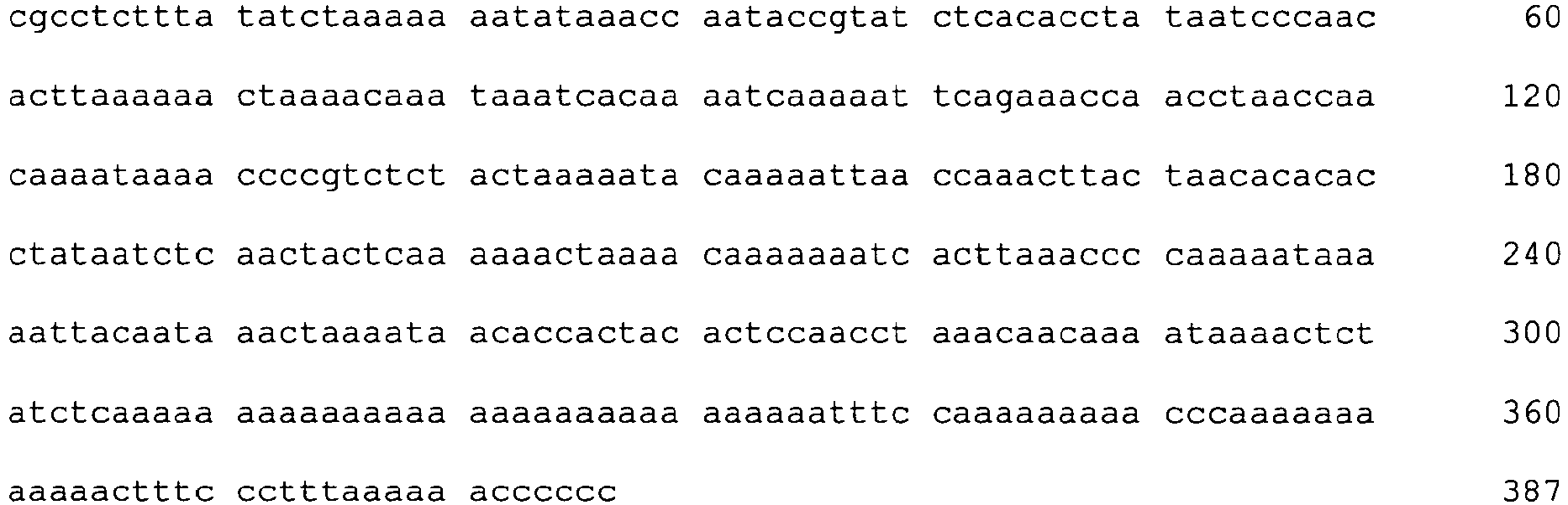
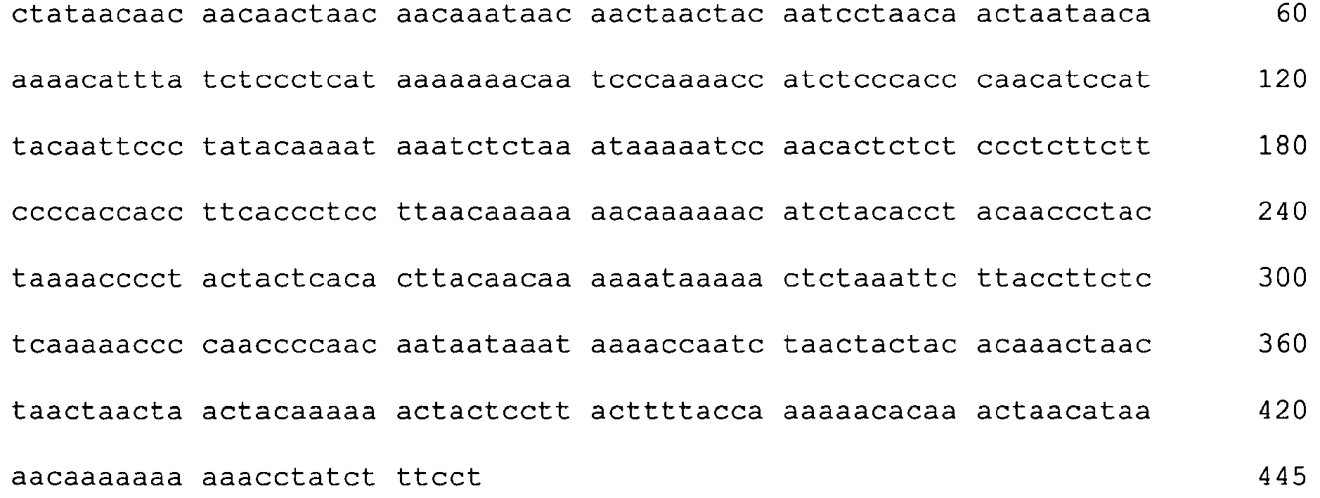
- SEQ ID No. 39 to 65 show the sequences as depicted in the alignments of
figures 2 to 5 . If not stated otherwise, the sequences are ordered in the succession of their appearance infigures 2 to 5 . - The inventors have purified various blood subsets, including CD3/CD4, CD3/CD8 naive and memory T lymphocytes, CD56 natural killer cells, CD19 naive and memory B cells, CD14 monocytes and CD 15 granulocytes. DNA from the purified cells was bisulfite-treated and analysed at various CpG dinucleotide motifs. The inventors then compared the methylation status (finding C as for Cytosine that was methylated in the original sequence versus T for cytosine that was unmethylated in the original sequence).
- The data showed various CpG motifs and areas in the CD3 γ, δ and ε that were demethylated in all CD3CD4 and CD3CD8 cell types while methylated in all other blood cell types. The differentially methylated gene regions as found for CD3 γ, δ and ε are shown below in
Figure 1 and are indicated in bold as "blast hits" - The data, as observed with Illumina Golden Gate technology, show that all CD4 and CD8 positive memory (0.06 and 0.06 respectively) and naive (0.03 and 0.06, respectively) T cells are subject to much lower methylation rates than all other tested cell types, including CD15 (0.92), CD 14 (0.90), CD 19 memory and naive (0.81 and 0.67, respectively), CD56 (0.86) positive blood cells as well as cells derived (0.78) from non-blood tissues. The experimental results are further depicted in the following table.
Table 1: Methylation analysis results Gene Chr. S1 S2 S3 S4 S5 S6 S7 S8 S9 S10 S11 S12 n=12; female n=5; male Pool; female Pool; (5 male) Pool; (5 male) Pool; (5 male) Pool; (5 male) Pool; (5 male) Pool; (5 male) Pool; (5 male) Pool; (5 male) Pool; (5 male) CD3D 11 0.8 0.6 0.6 0.9 0.9 0.9 0.0 0.1 0.1 0.1 0.7 0.8 0.9 0.7 0.7 0.9 0.9 0.9 0.1 0.1 0.2 0.2 0.7 0.8 CD3G 11 0.7 0.6 0.7 0.9 0.9 0.8 0.1 0.1 0.1 0.1 0.8 0.9 0.4 0.6 0.5 0.7 0.8 0.7 0.1 0.1 0.1 0.1 0.3 0.6 S1 = ovarian tissue, S2 = whole blood, S3 peripheral blood mononuclear cells, S4 = granulocytes, S5 = monocytes,S6 = NK cells, S7 = naïve T helper cells, S8 = memory T helper cells, S9 = naive cytotoxic T cells, S10 = memory cytotoxic T cells, S11 = naive B-cells, S12 = memory B-cells., Chr = chromosome - The data showing the high specificity were obtained using a Illumina Golden Gate technology, with the following genomic CpGs regions analysed
-
- cg15880738 (SEQ ID No. 2)
(+1) CG GAAAAACAAAAGGCATCTGCACCTGCAGCCCTGCTGAGGCCCCTGCTG

-
- cg24841244 (SEQ ID No. 4)
(-1) TGGAACATAGCA CG TTTCTCTCTGGCCTGGTACTGGCTACCCTTCTCT CG

-
- cg24612198 (Seq ID No. 9)
AGTCATCTGTTTTGCTTTTTTTCCAGAAGTAGTAAGTCTGCTGGCCTCCG - Since the Illumina technology does only allow the analysis of a single CpG (or rather 2 CpGs per gene locus) the inventors verified the methylation properties using bisulfite sequencing. For that, the inventors bisulfite treated the samples using the Qiagen EpiTect kit, and sequenced the samples using an ABI 3100 prism sequencer. For data interpretation, the KB base calling software supplied by ABI was used.
- While not providing entirely quantitative results calculating the percentile of methylated (i.e., CG signal on the plus strand sequence) versus unmethylated (i.e., TG on the plus strand sequence), the data were unambiguous for the purified cell types. All T-lymphocytes were overwhelmingly demethylated at all CG positions analysed, whereas all other analysed cell types were methylated at identical positions.
- In order to identify further suitable markers distingushing and monitoring T-lymphocytes, other markers in addition to CD3 have been identified and tested through methylation analysis. It was found that methylation in the CpG positions in the genes for SLA2, CHRNA3, C16orf24, LCK, FASLG, CD7, SIT1, IL32, CXCR6, UBASH3A, GRAP2, ITGB7 and TXK can also be used in the context of the present invention, as these markers are also able to identify CD3 positive T lymphocytes.
- Furthermore, other markers have been identified that identify the subset of the CD8 and CD4 positive cells in the group of CD3 positive T lymphocytes. The genes for GNGT2, CRTAM, IL2RB and ZBTB32 have been found to segregate between CD8 and CD4 positive cells. Equivalently, FLJ00060, FLJ38379, PPP6C, CD226, ZBTB7B and TNFAIP8 are capable of positively identifying CD4 expressing cells in whole blood and segregate between CD4 and CD8 positive CD3 positive cells.
- The following table 2 summarizes the Illumina-data as obtained for the above markers at selected CpG positions. It can be seen that several other markers can be used in order to selectively identify CD4+ (for CD3+ lymphocyte subset-identification), namely FLJ00060; FLJ38379; PPP6C; CD226; ZBTB7B and/or TNFAIP8, in order to selectively identify CD8+ (for CD3+ lymphocyte subset-identification), namely GNGT2; CRTAM; IL2RB and/or ZBTB32, and in order to selectively identify CD3+ lymphocytes, namely CD3D; CD3G, and/or CD3E, and/or SLA2, CHRNA3, C16orf24; LCK; FASLG; FASLG; CD7; SIT1; IL32; CXCR6; UBASH3A; GRAP2; ITGB7 and/or TXK, as shown with selected CpG sites as preferred examples. Based on the table, the person of skill will be able to extend the teaching regarding CD3 as herein to these markers and their CpG sites.
Table 2: Illumina-data as obtained selected markers at CpG positions Chr. = chromosome CD4+ marker CpG-ID Gene Name Ch r. SEQ ID No. / Accession No 10/ Ovar Tissue (mean, n=12) Whole Blood (Pool) PBMC (Promega) BCST1 8 Granulocyte (CD15 +) BCST1 9 Monocyte (CD14 +) BCST2 0 NK (CD56 +) BCST21 T naive (CD4+C D27 +CD45R A+) BCST22 T mem (CD4+ CD27+ CD45RA -) BCST23 CTL naive (CD8+C D27+ CD45RA +) BCST24 CTL mem (CD8+C D27+ CD45RA -) BCST2 5 B naive (CD19 +) BCST2 6 B mem (CD19 +) Mean Value Target Cell Type Mean Value Rest Delta Meth (Target-Rest) cg03602 500 FLJ000 60 19 NM_0332 06.1 0.802 0.728 0.779 0.858 0.851 0.873 0.235 0.279 0.521 0.687 0.656 0.747 0.257 0.75 -0.493 cg16173 109 FLJ383 79 2 11/ XR_00102 6.1 0.614 0.675 0.719 0.833 0.844 0.784 0.1141 0.113 0.341 0.383 0.418 0.385 0.113 0.599 -0.486 cg00620 024 PPP6C 9 12/ NM_0027 21.3 0.641 0.619 0.692 0.801 0.851 0.836 0.107 0.169 0.269 0.420 0.404 0.502 0.138 0.603 -0.465 cg13164 537 CD226 18 13/ NM_0065 66.1 0.458 0.470 0.508 0.582 0.557 0.601 0.029 0.071 0.351 0.543 0.366 0.501 0.05 0.494 -0.443 cg01782 486 ZBTB7 B 1 14/ NM_0158 72.1 0.789 0.725 0.720 0.823 0.809 0.886 0.190 0.428 0.875 0.864 0.586 0.414 0.309 0.749 -0.44 eg07086 380 TNFAI P8 5 15/ NM_0143 50.1 0.728 0.579 0.635 0.857 0.733 0.793 0.110 0.106 0.288 0.148 0.309 0.208 0.108 0.528 -0.419 CD8+ marker CpG-ID Gene Name Ch r SEQ ID No. / Accession No Ovar Tissue (mean, n=12) Whole Blood (Pool) PBMC (Pro-mega) BCST1 8 Granulocyte (CD15 +) BCST1 9 Monocyte (CD14 +) BCST2 0 NK (CD56 +) BCST21 T naive (CD4+C D27 +CD45R A+) BCST22 T mem (CD4+ CD27+ CD45RA -) BCST23 CTL naive (CD8+C D27+ CD45RA +) BCST24 CTL mem (CD8+C D27+ CD45RA -) BCST2 5 B naive (CD19 +) BCST2 6 B mem (CD19 +) Mean Value Target Cell Type Mean Value Rest Delta Meth (Target-Rest) cg17839 611 GNGT 217 16/ NM_0314 98.1 0.779 0.629 0.688 0.828 0.867 0.207 0.340 0.562 0.09 0.119 0.854 0.722 0.104 0.648 -0.543 cg22512 531 CRTA M 11 17/ NM_0196 04.2 0.651 0.634 0.709 0.835 0.735 0.5 0.363 0.630 0.079 0.082 0.194 0.336 0.08 0.559 -0.478 cg26757 673 IL2RB 22 18/ NM_0008 78.2 0.799 0.662 0.752 0.878 0.9 0.092 0.573 0.086 0.157 0.061 0.411 0.403 0.109 0.555 -0.446 cg08539 991 ZBTB3 219 19/ NM_0143 83.1 0.823 0.736 0.791 0.912 0.926 0.674 0.563 0.153 0.303 0.085 0.648 0.140 0.194 0.637 -0.442 CD4+ / CD8+ marker CpG-ID Gene Name Ch r SEQ ID No. / Accession No 20/ Ovar Tissue (mean, n=12) Whole Blood (Pool) PBMC (Promega) BCST1 8 Granulocyte (CD15 +) BCST1 9 Monocyte (CD14 +) BCST2 0 NK (CD56 +) BCST21 T naive (CD4+C D27 +CD45R A+) BCST22 T mem (CD4+ CD27+ CD45RA -) BCST23 CTL naive (CD8+C D27+ CD45RA +) BCST24 CTL mem (CD8+C D27+ CD45RA -) BCST2 5 B naive (CD19 +) BCST2 6 B mem (CD19 +) Mean Value Target Cell Type Mean Value Rest Delta Meth (Target-Rest) cg24841 244 CD3D 11 NM_0007 32.3 0.783 0.582 0.644 0.917 0.904 0.857 0.025 0.057 0.057 0.058 0.673 0.810 0.049 0.771 -0.721 cg07728 874 CD3D 21/ NM_0007 32.3 0.854 0.683 0.727 0.911 0.901 0.881 0.128 0.149 0.175 0.155 0.744 0.807 0.152 0.814 -0.661 cg15880 738 CD3G 11 22/ NM_0000 73.1 0.714 0.616 0.682 0.874 0.876 0.836 0.061 0.091 0.093 0.103 0.811 0.898 0.087 0.788 -0.701 cg07545 925 CD3G 23/ NM_0000 73.1 0.370 0.552 0.545 0.737 0.771 0.739 0.133 0.149 0.132 0.131 0.303 0.601 0.132 0.49 -0.358 cg24612 198 CD3E 11 24/ NM_0007 33.2 0.679 0.485 0.563 0.794 0.793 0.698 0.064 0.036 0.093 0.051 0.206 0.279 0.061 0.562 -0.501 cg04759 756 SLA2 20 25/ NM_0322 14.2 0.857 0.673 0.754 0.892 0.925 0.711 0.363 0.228 0.129 0.211 0.756 0.849 0.233 0.802 -0.569 cg22670 733 CHRN A3 15 26/ NM_0007 43.2 0.792 0.733 0.721 0.911 0.888 0.8 0.122 0.180 0.092 0.315 0.456 0.668 0.177 0.746 -0.568 cg09830 866 C16orf 24 16 27/ NM_0239 33.1 0.493 0.629 0.647 0.842 0.79 0.076 0.041 0.056 0.033 0.058 0.72 0.510 0.047 0.588 -0.541 cg17078 393 LCK 1 28/ NM_0053 56.2 0.807 0.575 0.666 0.920 0.884 0.26 0.05 0.037 0.036 0.041 0.144 0.27 0.041 0.566 -0.524 cg10161 121 FASLG 1 29/ NM_0006 39.1 0.704 0.587 0.697 0.905 0.892 0.068 0.051 0.06 0.088 0.06 0.353 0.441 0.065 0.581 -0.516 cg00071 250 FASLG 1 30/ NM_0006 39.1 0.659 0.498 0.602 0.871 0.838 0.075 0.061 0.069 0.139 0.051 0.391 0.4144 6208 0.08 0.543 -0.463 cg02473 123 CD7 17 31/ NM_0061 0.811 0.681 0.767 0.942 0.898 0.339 0.102 0.328 0.168 0.372 0.767 0.81 0.243 0.752 -0.509 cg15518 883 SIT1 9 37.6 32/ NM_0144 50.2 0.817 0.601 0.696 0.911 0.885 0.9033 0.11 0.142 0.154 0.358 0.392 0.373 0.191 0.697 -0.506 cg18350 391 IL32 16 33/ NM_0010 12631.1 0.332 0.724 0.794 0.937 0.901 0.658 0.296 0.169 0.15 0.192 0.881 0.406 0.202 0.704 -0.502 cg25226 014 CXCR 6 3 34/ NM_0065 64.1 0.702 0.481 0.548 0.789 0.89 0.35 0.028 0.046 0.046 0.193 0.258 0.514 0.078 0.567 -0.488 cg13578 652 UBAS H3A 21 35/ NM_0189 61.2 0.564 0.444 0.475 0.537 0.703 0.169 0.027 0.04 0.035 0.05 0.602 0.673 0.038 0.521 -0.483 cg25712 380 GRAP2 22 36/ NM_0048 10.2 0.633 0.602 0.639 0.824 0.83 0.506 0.097 0.093 0.089 0.1 0.179 0.310 0.095 0.565 -0.470 cg19812 619 ITGB7 12 37/ NM_0008 89.1 0.819 0.651 0.785 0.874 0.897 0.645 0.3374 0.275 0.199 0.177 0.608 0.4337 0.247 0.714 -0.467 cg02600 394 TXK 4 38/ NM_0033 28.1 0.775 0.576 0.682 0.867 0.909 0.082 0.036 0.075 0.051 0.354 0.376 0.484 0.129 0.594 -0.465 -
- <110> Epiontis GmbH
- <120> Epigenetic markers for the identification of blood sub-cells of type 1
- <130> E31285PCT
- <150>
EP08021838.1-2403
<151> 2008-12-16 - <160> 65
- <170> PatentIn version 3.5
- <210> 1
<211> 16501
<212> DNA
<213> Homo sapiens - <900> 1


- <210> 2
<211> 50
<212> DNA
<213> Homo sapiens - <400> 2
agctgctgca caggctggct ggctggctgg ctgctaaggg ctgctccacg 50 - <210> 3
<211> 50
<212> DNA
<213> Homo sapiens - <400> 3
cggaaaaaca aaaggcatct gcacctgcag ccctgctgag gcccctgctg 50 - <210> 4
<211> 50
<212> DNA
<213> Homo sapiens - <400> 4
acccaggctg atagttcggt gacctggctt tatctactgg atgagttccg 50 - <210> 5
<211> 50
<212> DNA
<213> Homo sapiens - <400> 5
tggaacatag cacgtttctc tctggcctgg tactggctac ccttctctcg 50 - <210> 6
<211> 435
<212> DNA
<213> Homo sapiens - <400> 6


- <210> 7
<211> 438
<212> DNA
<213> Homo sapiens - <400> 7

- <210> 8
<211> 472
<212> DNA
<213> Homo sapiens - <400> 8

- <210> 9
<211> 50
<212> DNA
<213> Homo sapiens - <400> 9
agtcatctgt tttgcttttt ttccagaagt agtaagtctg ctggcctccg 50 - <210> 10
<211> 50
<212> DNA
<213> Homo sapiens - <400> 10
cgcccagcct cgctgttctt atcttggcag cagattccga atgtcggctg 50 - <210> 11
<211> 50
<212> DNA
<213> Homo sapiens - <400> 11
cgctcatcag gacttcagca ctgcccgtcc atggggacgt ctgcactcac 50 - <210> 12
<211> 50
<212> DNA
<213> Homo sapiens - <400> 12
cgccttgagc tgtacctggc acatctttgt tgctcagcaa agagtggttg 50 - <210> 13
<211> 50
<212> DNA
<213> Homo sapiens - <400> 13
tcttcccatc tgcgtcttgg ttaaggactt ttatcgtatc ctgtttatcg 50 - <210> 14
<211> 50
<212> DNA
<213> Homo sapiens - <400> 14
aagatctgct ggaggccagg cacagacagg tgctttaact caagttatcg 50 - <210> 15
<211> 50
<212> DNA
<213> Homo sapiens - <400> 15
agtaactgca gcaatgagtg cccgggctgt gcttggagta ccagtgctcg 50 - <210> 16
<211> 50
<212> DNA
<213> Homo sapiens - <400> 16
cgccccaagt actaagtggc acagcttggt gagcaaggac atttttattg 50 - <210> 17
<211> 50
<212> DNA
<213> Homo sapiens - <400> 17
cggcaagatg aaacaagctt attaggctct gtcttttaag ggcataccag 50 - <210> 18
<211> 50
<212> DNA
<213> Homo sapiens - <400> 18
cgcaacacag ggctgcctcc cccggtatat gggccccact ccacagaggt 50 - <210> 19
<211> 50
<212> DNA
<213> Homo sapiens - <400> 19
cggcagagca cacagctgca gaagtaaaaa ggattgaaac atttggatcc 50 - <210> 20
<211> 50
<212> DNA
<213> Homo sapiens - <400> 20
acccaggctg atagttcggt gacctggctt tatctactgg atgagttccg 50 - <210> 21
<211> 50
<212> DNA
<213> Homo sapiens - <400> 21
tggaacatag cacgtttctc tctggcctgg tactggctac ccttctctcg 50 - <210> 22
<211> 50
<212> DNA
<213> Homo sapiens - <400> 22
agctgctgca caggctggct ggctggctgg ctgctaaggg ctgctccacg 50 - <210> 23
<211> 50
<212> DNA
<213> Homo sapiens - <400> 23
cggaaaaaca aaaggcatct gcacctgcag ccctgctgag gcccctgctg 50 - <210> 24
<211> 50
<212> DNA
<213> Homo sapiens - <400> 24
agtcatctgt tttgcttttt ttccagaagt agtaagtctg ctggcctccg 50 - <210> 25
<211> 50
<212> DNA
<213> Homo sapiens - <400> 25
cgctgcagtc agactccaaa gtcaggaacg tgagggctac catctctcaa 50 - <210> 26
<211> 50
<212> DNA
<213> Homo sapiens - <400> 26
cgaaggcccc cagctgtggg agcccacgct atttattggt gatcaaagaa 50 - <210> 27
<211> 50
<212> DNA
<213> Homo sapiens - <400> 27
cgcaggcact tacgatcctg ccgtcgccag agcccagatc caccgttttt 50 - <210> 28
<211> 50
<212> DNA
<213> Homo sapiens - <400> 28
gggctcccgg gctgggcagg taaggagcgc tggtattggg ggcgcaggcg 50 - <210> 29
<211> 50
<212> DNA
<213> Homo sapiens - <400> 29
ttccgcccac aattttctga taacagcctt caaggcctca gttgctgtcg 50 - <210> 30
<211> 50
<212> DNA
<213> Homo sapiens - <400> 30
cgggaccctg ttgctgactg ctcaagagga ggcaagctgg atctctctta 50 - <210> 31
<211> 50
<212> DNA
<213> Homo sapiens - <400> 31
taaatgatgt cttggggctg tggcccgagc tgcctcaggt agatcccacg 50 - <210> 32
<211> 50
<212> DNA
<213> Homo sapiens - <400> 32
ggggactgtg ggtcctctta ggggctgtga cgctgctatt tctcatctcg 50 - <210> 33
<211> 50
<212> DNA
<213> Homo sapiens - <400> 33
cgcactcagc aaggcctcct gccctgagag aggctccgcc cactaccccc 50 - <210> 34
<211> 50
<212> DNA
<213> Homo sapiens - <400> 34
ctcatgaagc aaggtctgcc tctacttctg tgatgtggtt catgtttccg 50 - <210> 35
<211> 50
<212> DNA
<213> Homo sapiens - <400> 35
gacctttagc ctcaaactcc cgtggtggaa acagttagga ttggtggacg 50 - <210> 36
<211> 50
<212> DNA
<213> Homo sapiens - <400> 36
gagagacaat acccttccaa aatacacttc aacataaagt tttcttttcg 50 - <210> 37
<211> 50
<212> DNA
<213> Homo sapiens - <400> 37
cggtatacct gccctacttc ggtatagtcc ttggcacatg gcaggcactc 50 - <210> 38
<211> 50
<212> DNA
<213> Homo sapiens - <900> 38
atggtagccc cttctgcggg gagcacacaa cagtcttcag ttcttctgcg 50 - <210> 39
<211> 402
<212> DNA
<213> Homo sapiens - <400> 39

- <210> 40
<211> 394
<212> DNA
<213> Homo sapiens - <400> 40

- <210> 41
<211> 435
<212> DNA
<213> Homo sapiens - <400> 41


- <210> 42
<211> 399
<212> DNA
<213> Homo sapiens - <400> 42

- <210> 43
<211> 394
<212> DNA
<213> Homo sapiens - <400> 43

- <210> 44
<211> 396
<212> DNA
<213> Homo sapiens - <400> 44


- <210> 45
<211> 387
<212> DNA
<213> Homo sapiens - <400> 45
- <210> 46
<211> 391
<212> DNA
<213> Homo sapiens - <400> 46

- <210> 47
<211> 328
<212> DNA
<213> Homo sapiens - <400> 47


- <210> 48
<211> 388
<212> DNA
<213> Homo sapiens - <400> 48

- <210> 49
<211> 401
<212> DNA
<213> Homo sapiens - <400> 49

- <210> 50
<211> 387
<212> DNA
<213> Homo sapiens - <400> 50


- <210> 51
<211> 420
<212> DNA
<213> Homo sapiens - <400> 51

- <210> 52
<211> 416
<212> DNA
<213> Homo sapiens - <400> 52

- <210> 53
<211> 416
<212> DNA
<213> Homo sapiens - <400> 53


- <210> 54
<211> 418
<212> DNA
<213> Homo sapiens - <400> 54

- <210> 55
<211> 419
<212> DNA
<213> Homo sapiens - <400> 55

- <210> 56
<211> 418
<212> DNA
<213> Homo sapiens - <400> 56


- <210> 57
<211> 419
<212> DNA
<213> Homo sapiens - <400> 57

- <210> 58
<211> 416
<212> DNA
<213> Homo sapiens - <400> 58

- <210> 59
<211> 438
<212> DNA
<213> Homo sapiens - <400> 59


- <210> 60
<211> 442
<212> DNA
<213> Homo sapiens - <400> 60

- <210> 61
<211> 445
<212> DNA
<213> Homo sapiens - <400> 61
- <210> 62
<211> 437
<212> DNA
<213> Homo sapiens - <400> 62


- <210> 63
<211> 450
<212> DNA
<213> Homo sapiens - <400> 63

- <210> 64
<211> 446
<212> DNA
<213> Homo sapiens - <400> 64

- <210> 65
<211> 448
<212> DNA
<213> Homo sapiens - <400> 65

Claims (10)
- A method for identifying CD3+ CD4+, and/or CD3+ CD8+ naive and/or memory T-lymphocytes in a sample derived from a mammal comprising T cells, comprisinga) analysing the methylation status of at least one CpG position in an amplicon comprising amplification with at least one primer pair suitably designed based on oligomers according to any of SEQ ID NO: 2 to 5, andb) identifying CD3+ CD4+, and/or CD3+ CD8+ naive and/or memory T-lymphocytes based on said methylation status, wherein a demethylation of at least one CpG position in said amplicon to at least 90% in said sample is indicative for a CD3+ CD4+, and/or CD3+ CD8+ naive and/or memoryT-lymphocyte cell.
- The method according to claim 1, wherein said at least one CpG position in said sample is demethylated to more than 91% and preferably more than 92% and most preferred more than 95%.
- The method according to claim 1 or 2, wherein the analysis of the methylation status comprises a method selected from methylation specific enzymatic digests, bisulphite sequencing, analysis selected from promoter methylation, CpG island methylation, MSP, HeavyMethyl, MethyLight, Ms-SNuPE or other methods relying on a detection of amplified DNA.
- The method according to any of claims 1 to 3, further comprising a methylation analysis of at least one CpG position in the genes for CD4+ and/or CD8, in particular CD8 beta, or in the genes for GNGT2, CRTAM, IL2RB, and ZBTB32, or FLJ00060, FLJ38379, PPP6C, CD226, ZBTB7B, and TNFAIP8, or in the genes SLA2, CHRNA3, C16orf24, LCK, FASLG, CD7, SIT1, IL32, CXCR6, UBASH3A, GRAP2, ITGB7 and TXK
- The method according to any of claims 1 to 4, wherein said method is suitable for routine application, for example on a DNA-chip.
- The method according to any of claims 1 to 5, wherein said identification comprises a distinction of said T-lymphocytes from all major peripheral blood cell types or non-blood cells.
- The method according to any of claims 1 to 6, wherein said sample is selected from a mammalian body fluid, including human blood samples, or a tissue, organ or cell type blood sample, a sample of blood lymphocytes or a fraction thereof.
- The method according to any of claims 1 to 7, further comprising the step of concluding on the immune status of said mammal based on said T-lymphocytes as identified.
- The method according to any of claims 1 to 8, wherein said mammal suffers from or is likely to suffer from autoimmune diseases, transplant rejections, cancer, and/or allergy.
- Use of a kit comprising materials for performing the method for identifying CD3+ CD4+, and/or CD3+ CD8+ naive and/or memoryT-lymphocytes in a sample according to claim 1, said kit comprisinga) a bisulfite reagent, andb) materials for performing a methylation analysis for CpG positions in an amplicon comprising amplification with at least one primer pair suitably designed based on oligomers according to any of SEQ NO: 2 to 5for identifying CD3+ CD4+, and/or CD3+ CD8+ naive and/or memoryT-lymphocytes in a mammal.
Priority Applications (2)
| Application Number | Priority Date | Filing Date | Title |
|---|---|---|---|
| PL09771304T PL2376648T3 (en) | 2008-12-16 | 2009-12-08 | Epigenetic markers for the identification of cd3 positive t-lymphocytes |
| EP09771304.4A EP2376648B1 (en) | 2008-12-16 | 2009-12-08 | Epigenetic markers for the identification of cd3 positive t-lymphocytes |
Applications Claiming Priority (3)
| Application Number | Priority Date | Filing Date | Title |
|---|---|---|---|
| EP20080021838 EP2199411B1 (en) | 2008-12-16 | 2008-12-16 | Epigenetic markers for the identification of CD3 positive T-lymphocytes |
| PCT/EP2009/008764 WO2010069499A2 (en) | 2008-12-16 | 2009-12-08 | Epigenetic markers for the identification of blood sub-cells of type 1 |
| EP09771304.4A EP2376648B1 (en) | 2008-12-16 | 2009-12-08 | Epigenetic markers for the identification of cd3 positive t-lymphocytes |
Publications (2)
| Publication Number | Publication Date |
|---|---|
| EP2376648A2 EP2376648A2 (en) | 2011-10-19 |
| EP2376648B1 true EP2376648B1 (en) | 2016-09-14 |
Family
ID=40428037
Family Applications (2)
| Application Number | Title | Priority Date | Filing Date |
|---|---|---|---|
| EP20080021838 Active EP2199411B1 (en) | 2008-12-16 | 2008-12-16 | Epigenetic markers for the identification of CD3 positive T-lymphocytes |
| EP09771304.4A Active EP2376648B1 (en) | 2008-12-16 | 2009-12-08 | Epigenetic markers for the identification of cd3 positive t-lymphocytes |
Family Applications Before (1)
| Application Number | Title | Priority Date | Filing Date |
|---|---|---|---|
| EP20080021838 Active EP2199411B1 (en) | 2008-12-16 | 2008-12-16 | Epigenetic markers for the identification of CD3 positive T-lymphocytes |
Country Status (8)
| Country | Link |
|---|---|
| US (1) | US20120107810A1 (en) |
| EP (2) | EP2199411B1 (en) |
| JP (1) | JP2012511917A (en) |
| CA (1) | CA2747312C (en) |
| DK (2) | DK2199411T3 (en) |
| ES (1) | ES2606350T3 (en) |
| PL (1) | PL2376648T3 (en) |
| WO (1) | WO2010069499A2 (en) |
Families Citing this family (14)
| Publication number | Priority date | Publication date | Assignee | Title |
|---|---|---|---|---|
| US20170233807A1 (en) | 2008-12-16 | 2017-08-17 | Epiontis Gmbh | Epigenetic Markers for the Identification of Blood Sub-cells of Type 1 |
| WO2012021867A2 (en) * | 2010-08-13 | 2012-02-16 | The Johns Hopkins University | A comprehensive methylome map of myeloid and lymphoid commitment from hematopoietic progenitors |
| EP2625288B1 (en) | 2010-10-08 | 2016-05-25 | Epiontis GmbH | Method for determining cancer patient survival based on analyzing tumor-infiltrating overall t-lymphocytes |
| US20130296328A1 (en) * | 2011-01-20 | 2013-11-07 | Université Libre de Bruxelles | Epigenetic portraits of human breast cancers |
| WO2012162660A2 (en) * | 2011-05-25 | 2012-11-29 | Brown University | Methods using dna methylation for identifying a cell or a mixture of cells for prognosis and diagnosis of diseases, and for cell remediation therapies |
| US20140178348A1 (en) | 2011-05-25 | 2014-06-26 | The Regents Of The University Of California | Methods using DNA methylation for identifying a cell or a mixture of cells for prognosis and diagnosis of diseases, and for cell remediation therapies |
| WO2013135454A1 (en) * | 2012-03-12 | 2013-09-19 | Epiontis Gmbh | Epigenetic marker for the identification of cd3cd4 positive t lymphocytes |
| GB201221133D0 (en) * | 2012-11-23 | 2013-01-09 | Epiontis Gmbh | Epigenetic method for the identification of subpopulations of CD8 and T lympocytes, in particular CD8 alpha and beta T lymphocytes |
| EP2986735B1 (en) * | 2013-04-19 | 2019-03-06 | Epiontis GmbH | Method for identifying the quantitative composition of blood cells in a sample |
| EP3360971B1 (en) * | 2017-02-09 | 2026-05-20 | Precision for Medicine GmbH | IMPROVED METHOD FOR EPIGENETIC IMMUNE CELL COUNTING |
| EP3382033B1 (en) * | 2017-03-30 | 2020-08-05 | Rheinisch-Westfälische Technische Hochschule (RWTH) Aachen | Method for determining blood counts based on dna methylation |
| DE102017125019B4 (en) * | 2017-10-25 | 2019-10-17 | Epiontis Gmbh | PDCD1 as an epigenetic marker for the identification of immune cells, in particular PD1 + cells |
| DE102018116353B4 (en) * | 2018-07-05 | 2020-06-10 | Epiontis Gmbh | Method for the detection and enumeration of epigenetic immune cells in human blood samples for immunodiagnostics and newborn screening |
| CN116949156B (en) * | 2023-09-19 | 2023-12-08 | 美迪西普亚医药科技(上海)有限公司 | Analysis method for detecting human T cells in general way based on nucleic acid variants |
Family Cites Families (3)
| Publication number | Priority date | Publication date | Assignee | Title |
|---|---|---|---|---|
| JP2002171973A (en) * | 2000-12-07 | 2002-06-18 | Univ Tokyo | Cell identification method by DNA methylation pattern |
| WO2004050706A2 (en) | 2002-12-03 | 2004-06-17 | Medical Research Council | Regulatory t-cells |
| ATE508207T1 (en) | 2006-02-28 | 2011-05-15 | Charite Universitaetsmedizin | DETECTION AND QUALITY CONTROL OF REGULATOR T CELLS USING DNA METHYLATION ANALYSIS OF THE FOXP3 GENE |
-
2008
- 2008-12-16 DK DK08021838.1T patent/DK2199411T3/en active
- 2008-12-16 EP EP20080021838 patent/EP2199411B1/en active Active
-
2009
- 2009-12-08 WO PCT/EP2009/008764 patent/WO2010069499A2/en not_active Ceased
- 2009-12-08 ES ES09771304.4T patent/ES2606350T3/en active Active
- 2009-12-08 DK DK09771304.4T patent/DK2376648T3/en active
- 2009-12-08 JP JP2011541165A patent/JP2012511917A/en active Pending
- 2009-12-08 US US13/139,808 patent/US20120107810A1/en not_active Abandoned
- 2009-12-08 CA CA2747312A patent/CA2747312C/en active Active
- 2009-12-08 PL PL09771304T patent/PL2376648T3/en unknown
- 2009-12-08 EP EP09771304.4A patent/EP2376648B1/en active Active
Also Published As
| Publication number | Publication date |
|---|---|
| HK1163189A1 (en) | 2012-09-07 |
| US20120107810A1 (en) | 2012-05-03 |
| EP2199411B1 (en) | 2015-05-06 |
| EP2376648A2 (en) | 2011-10-19 |
| WO2010069499A3 (en) | 2010-08-26 |
| PL2376648T3 (en) | 2017-05-31 |
| JP2012511917A (en) | 2012-05-31 |
| CA2747312C (en) | 2018-11-06 |
| DK2376648T3 (en) | 2017-01-02 |
| WO2010069499A2 (en) | 2010-06-24 |
| DK2199411T3 (en) | 2015-08-03 |
| EP2199411A1 (en) | 2010-06-23 |
| CA2747312A1 (en) | 2010-06-24 |
| ES2606350T3 (en) | 2017-03-23 |
Similar Documents
| Publication | Publication Date | Title |
|---|---|---|
| EP2199411B1 (en) | Epigenetic markers for the identification of CD3 positive T-lymphocytes | |
| US10876163B2 (en) | Detection and quality control of regulatory T cells through DNA-methylation analysis of the Foxp3 gene | |
| JP6224631B2 (en) | Epigenetic markers for identification of CD3CD4-positive T lymphocytes | |
| CA2760518C (en) | Epigenetic marker for the identification of natural killer cells | |
| US10294527B2 (en) | Epigenetic marker for the identification of natural killer cells | |
| US12286675B2 (en) | Epigenetic markers for the identification of blood sub-cells of type 1 | |
| CN108026577A (en) | MVD is as the epigenetic marker for identifying immunocyte particularly CD56+NK cells | |
| HK1163189B (en) | Epigenetic markers for the identification of cd3 positive t-lymphocytes | |
| EP4143340B1 (en) | Myh11/nde1 region as epigenetic marker for the identification of endothelial progenitor cells (epcs) | |
| CA3080060C (en) | Pdcd1 as epigenetic marker for the identification of immune cells, in particular pd1+ cells | |
| EP3669003B1 (en) | Pdcd1 as epigenetic marker for the identification of immune cells, in particular pd1+ cells | |
| WO2024245828A1 (en) | Itga4 as epigenetic marker for the identification of immune cells |
Legal Events
| Date | Code | Title | Description |
|---|---|---|---|
| PUAI | Public reference made under article 153(3) epc to a published international application that has entered the european phase |
Free format text: ORIGINAL CODE: 0009012 |
|
| 17P | Request for examination filed |
Effective date: 20110715 |
|
| AK | Designated contracting states |
Kind code of ref document: A2 Designated state(s): AT BE BG CH CY CZ DE DK EE ES FI FR GB GR HR HU IE IS IT LI LT LU LV MC MK MT NL NO PL PT RO SE SI SK SM TR |
|
| DAX | Request for extension of the european patent (deleted) | ||
| REG | Reference to a national code |
Ref country code: HK Ref legal event code: DE Ref document number: 1163189 Country of ref document: HK |
|
| 17Q | First examination report despatched |
Effective date: 20130222 |
|
| GRAP | Despatch of communication of intention to grant a patent |
Free format text: ORIGINAL CODE: EPIDOSNIGR1 |
|
| INTG | Intention to grant announced |
Effective date: 20160609 |
|
| GRAS | Grant fee paid |
Free format text: ORIGINAL CODE: EPIDOSNIGR3 |
|
| GRAA | (expected) grant |
Free format text: ORIGINAL CODE: 0009210 |
|
| AK | Designated contracting states |
Kind code of ref document: B1 Designated state(s): AT BE BG CH CY CZ DE DK EE ES FI FR GB GR HR HU IE IS IT LI LT LU LV MC MK MT NL NO PL PT RO SE SI SK SM TR |
|
| REG | Reference to a national code |
Ref country code: GB Ref legal event code: FG4D |
|
| REG | Reference to a national code |
Ref country code: CH Ref legal event code: EP |
|
| REG | Reference to a national code |
Ref country code: IE Ref legal event code: FG4D |
|
| REG | Reference to a national code |
Ref country code: AT Ref legal event code: REF Ref document number: 829041 Country of ref document: AT Kind code of ref document: T Effective date: 20161015 |
|
| REG | Reference to a national code |
Ref country code: DE Ref legal event code: R096 Ref document number: 602009041151 Country of ref document: DE |
|
| REG | Reference to a national code |
Ref country code: CH Ref legal event code: NV Representative=s name: KATZAROV S.A., CH |
|
| REG | Reference to a national code |
Ref country code: SE Ref legal event code: TRGR |
|
| REG | Reference to a national code |
Ref country code: NL Ref legal event code: FP |
|
| REG | Reference to a national code |
Ref country code: FR Ref legal event code: PLFP Year of fee payment: 8 |
|
| REG | Reference to a national code |
Ref country code: DK Ref legal event code: T3 Effective date: 20161221 |
|
| REG | Reference to a national code |
Ref country code: LT Ref legal event code: MG4D |
|
| REG | Reference to a national code |
Ref country code: NO Ref legal event code: T2 Effective date: 20160914 |
|
| PG25 | Lapsed in a contracting state [announced via postgrant information from national office to epo] |
Ref country code: HR Free format text: LAPSE BECAUSE OF FAILURE TO SUBMIT A TRANSLATION OF THE DESCRIPTION OR TO PAY THE FEE WITHIN THE PRESCRIBED TIME-LIMIT Effective date: 20160914 Ref country code: LT Free format text: LAPSE BECAUSE OF FAILURE TO SUBMIT A TRANSLATION OF THE DESCRIPTION OR TO PAY THE FEE WITHIN THE PRESCRIBED TIME-LIMIT Effective date: 20160914 |
|
| PG25 | Lapsed in a contracting state [announced via postgrant information from national office to epo] |
Ref country code: GR Free format text: LAPSE BECAUSE OF FAILURE TO SUBMIT A TRANSLATION OF THE DESCRIPTION OR TO PAY THE FEE WITHIN THE PRESCRIBED TIME-LIMIT Effective date: 20161215 Ref country code: LV Free format text: LAPSE BECAUSE OF FAILURE TO SUBMIT A TRANSLATION OF THE DESCRIPTION OR TO PAY THE FEE WITHIN THE PRESCRIBED TIME-LIMIT Effective date: 20160914 |
|
| PG25 | Lapsed in a contracting state [announced via postgrant information from national office to epo] |
Ref country code: RO Free format text: LAPSE BECAUSE OF FAILURE TO SUBMIT A TRANSLATION OF THE DESCRIPTION OR TO PAY THE FEE WITHIN THE PRESCRIBED TIME-LIMIT Effective date: 20160914 Ref country code: EE Free format text: LAPSE BECAUSE OF FAILURE TO SUBMIT A TRANSLATION OF THE DESCRIPTION OR TO PAY THE FEE WITHIN THE PRESCRIBED TIME-LIMIT Effective date: 20160914 |
|
| PG25 | Lapsed in a contracting state [announced via postgrant information from national office to epo] |
Ref country code: SM Free format text: LAPSE BECAUSE OF FAILURE TO SUBMIT A TRANSLATION OF THE DESCRIPTION OR TO PAY THE FEE WITHIN THE PRESCRIBED TIME-LIMIT Effective date: 20160914 Ref country code: PT Free format text: LAPSE BECAUSE OF FAILURE TO SUBMIT A TRANSLATION OF THE DESCRIPTION OR TO PAY THE FEE WITHIN THE PRESCRIBED TIME-LIMIT Effective date: 20170116 Ref country code: BG Free format text: LAPSE BECAUSE OF FAILURE TO SUBMIT A TRANSLATION OF THE DESCRIPTION OR TO PAY THE FEE WITHIN THE PRESCRIBED TIME-LIMIT Effective date: 20161214 Ref country code: SK Free format text: LAPSE BECAUSE OF FAILURE TO SUBMIT A TRANSLATION OF THE DESCRIPTION OR TO PAY THE FEE WITHIN THE PRESCRIBED TIME-LIMIT Effective date: 20160914 |
|
| REG | Reference to a national code |
Ref country code: HK Ref legal event code: GR Ref document number: 1163189 Country of ref document: HK |
|
| REG | Reference to a national code |
Ref country code: DE Ref legal event code: R097 Ref document number: 602009041151 Country of ref document: DE |
|
| PLBE | No opposition filed within time limit |
Free format text: ORIGINAL CODE: 0009261 |
|
| STAA | Information on the status of an ep patent application or granted ep patent |
Free format text: STATUS: NO OPPOSITION FILED WITHIN TIME LIMIT |
|
| 26N | No opposition filed |
Effective date: 20170615 |
|
| PG25 | Lapsed in a contracting state [announced via postgrant information from national office to epo] |
Ref country code: MC Free format text: LAPSE BECAUSE OF FAILURE TO SUBMIT A TRANSLATION OF THE DESCRIPTION OR TO PAY THE FEE WITHIN THE PRESCRIBED TIME-LIMIT Effective date: 20160914 |
|
| PG25 | Lapsed in a contracting state [announced via postgrant information from national office to epo] |
Ref country code: SI Free format text: LAPSE BECAUSE OF FAILURE TO SUBMIT A TRANSLATION OF THE DESCRIPTION OR TO PAY THE FEE WITHIN THE PRESCRIBED TIME-LIMIT Effective date: 20160914 |
|
| REG | Reference to a national code |
Ref country code: FR Ref legal event code: PLFP Year of fee payment: 9 |
|
| REG | Reference to a national code |
Ref country code: CH Ref legal event code: PCAR Free format text: NEW ADDRESS: AVENUE DES MORGINES 12, 1213 PETIT-LANCY (CH) |
|
| PG25 | Lapsed in a contracting state [announced via postgrant information from national office to epo] |
Ref country code: HU Free format text: LAPSE BECAUSE OF FAILURE TO SUBMIT A TRANSLATION OF THE DESCRIPTION OR TO PAY THE FEE WITHIN THE PRESCRIBED TIME-LIMIT; INVALID AB INITIO Effective date: 20091208 Ref country code: CY Free format text: LAPSE BECAUSE OF FAILURE TO SUBMIT A TRANSLATION OF THE DESCRIPTION OR TO PAY THE FEE WITHIN THE PRESCRIBED TIME-LIMIT Effective date: 20160914 |
|
| PG25 | Lapsed in a contracting state [announced via postgrant information from national office to epo] |
Ref country code: MK Free format text: LAPSE BECAUSE OF FAILURE TO SUBMIT A TRANSLATION OF THE DESCRIPTION OR TO PAY THE FEE WITHIN THE PRESCRIBED TIME-LIMIT Effective date: 20160914 |
|
| PG25 | Lapsed in a contracting state [announced via postgrant information from national office to epo] |
Ref country code: MT Free format text: LAPSE BECAUSE OF NON-PAYMENT OF DUE FEES Effective date: 20161208 |
|
| REG | Reference to a national code |
Ref country code: AT Ref legal event code: UEP Ref document number: 829041 Country of ref document: AT Kind code of ref document: T Effective date: 20160914 |
|
| PGFP | Annual fee paid to national office [announced via postgrant information from national office to epo] |
Ref country code: LU Payment date: 20191125 Year of fee payment: 11 |
|
| PGFP | Annual fee paid to national office [announced via postgrant information from national office to epo] |
Ref country code: FI Payment date: 20191209 Year of fee payment: 11 Ref country code: NL Payment date: 20191212 Year of fee payment: 11 Ref country code: CZ Payment date: 20191202 Year of fee payment: 11 Ref country code: NO Payment date: 20191210 Year of fee payment: 11 Ref country code: IE Payment date: 20191209 Year of fee payment: 11 Ref country code: SE Payment date: 20191210 Year of fee payment: 11 |
|
| PGFP | Annual fee paid to national office [announced via postgrant information from national office to epo] |
Ref country code: DK Payment date: 20191210 Year of fee payment: 11 Ref country code: PL Payment date: 20191128 Year of fee payment: 11 Ref country code: IS Payment date: 20191126 Year of fee payment: 11 Ref country code: IT Payment date: 20191209 Year of fee payment: 11 Ref country code: BE Payment date: 20191125 Year of fee payment: 11 |
|
| PGFP | Annual fee paid to national office [announced via postgrant information from national office to epo] |
Ref country code: TR Payment date: 20191206 Year of fee payment: 11 Ref country code: AT Payment date: 20191125 Year of fee payment: 11 |
|
| PGFP | Annual fee paid to national office [announced via postgrant information from national office to epo] |
Ref country code: ES Payment date: 20200102 Year of fee payment: 11 |
|
| REG | Reference to a national code |
Ref country code: FI Ref legal event code: MAE |
|
| REG | Reference to a national code |
Ref country code: DK Ref legal event code: EBP Effective date: 20201231 |
|
| REG | Reference to a national code |
Ref country code: NO Ref legal event code: MMEP |
|
| PG25 | Lapsed in a contracting state [announced via postgrant information from national office to epo] |
Ref country code: FI Free format text: LAPSE BECAUSE OF NON-PAYMENT OF DUE FEES Effective date: 20201208 Ref country code: CZ Free format text: LAPSE BECAUSE OF NON-PAYMENT OF DUE FEES Effective date: 20201208 |
|
| REG | Reference to a national code |
Ref country code: SE Ref legal event code: EUG |
|
| REG | Reference to a national code |
Ref country code: NL Ref legal event code: MM Effective date: 20210101 |
|
| REG | Reference to a national code |
Ref country code: AT Ref legal event code: MM01 Ref document number: 829041 Country of ref document: AT Kind code of ref document: T Effective date: 20201208 |
|
| REG | Reference to a national code |
Ref country code: BE Ref legal event code: MM Effective date: 20201231 |
|
| PG25 | Lapsed in a contracting state [announced via postgrant information from national office to epo] |
Ref country code: NL Free format text: LAPSE BECAUSE OF NON-PAYMENT OF DUE FEES Effective date: 20210101 |
|
| PG25 | Lapsed in a contracting state [announced via postgrant information from national office to epo] |
Ref country code: LU Free format text: LAPSE BECAUSE OF NON-PAYMENT OF DUE FEES Effective date: 20201208 Ref country code: AT Free format text: LAPSE BECAUSE OF NON-PAYMENT OF DUE FEES Effective date: 20201208 Ref country code: IT Free format text: LAPSE BECAUSE OF NON-PAYMENT OF DUE FEES Effective date: 20201208 Ref country code: IE Free format text: LAPSE BECAUSE OF NON-PAYMENT OF DUE FEES Effective date: 20201208 |
|
| PG25 | Lapsed in a contracting state [announced via postgrant information from national office to epo] |
Ref country code: SE Free format text: LAPSE BECAUSE OF NON-PAYMENT OF DUE FEES Effective date: 20201209 Ref country code: NO Free format text: LAPSE BECAUSE OF NON-PAYMENT OF DUE FEES Effective date: 20201231 |
|
| PG25 | Lapsed in a contracting state [announced via postgrant information from national office to epo] |
Ref country code: DK Free format text: LAPSE BECAUSE OF NON-PAYMENT OF DUE FEES Effective date: 20201231 |
|
| REG | Reference to a national code |
Ref country code: ES Ref legal event code: FD2A Effective date: 20220209 |
|
| PG25 | Lapsed in a contracting state [announced via postgrant information from national office to epo] |
Ref country code: ES Free format text: LAPSE BECAUSE OF NON-PAYMENT OF DUE FEES Effective date: 20201209 |
|
| PG25 | Lapsed in a contracting state [announced via postgrant information from national office to epo] |
Ref country code: TR Free format text: LAPSE BECAUSE OF NON-PAYMENT OF DUE FEES Effective date: 20201208 |
|
| PG25 | Lapsed in a contracting state [announced via postgrant information from national office to epo] |
Ref country code: BE Free format text: LAPSE BECAUSE OF NON-PAYMENT OF DUE FEES Effective date: 20201231 |
|
| PG25 | Lapsed in a contracting state [announced via postgrant information from national office to epo] |
Ref country code: PL Free format text: LAPSE BECAUSE OF NON-PAYMENT OF DUE FEES Effective date: 20201208 |
|
| REG | Reference to a national code |
Ref country code: DE Ref legal event code: R082 Ref document number: 602009041151 Country of ref document: DE Representative=s name: KRAUSS, JAN, DR., DE |
|
| P01 | Opt-out of the competence of the unified patent court (upc) registered |
Effective date: 20230525 |
|
| PG25 | Lapsed in a contracting state [announced via postgrant information from national office to epo] |
Ref country code: IS Free format text: LAPSE BECAUSE OF NON-PAYMENT OF DUE FEES Effective date: 20210628 |
|
| REG | Reference to a national code |
Ref country code: CH Ref legal event code: U11 Free format text: ST27 STATUS EVENT CODE: U-0-0-U10-U11 (AS PROVIDED BY THE NATIONAL OFFICE) Effective date: 20260101 |
|
| PGFP | Annual fee paid to national office [announced via postgrant information from national office to epo] |
Ref country code: GB Payment date: 20251218 Year of fee payment: 17 |
|
| PGFP | Annual fee paid to national office [announced via postgrant information from national office to epo] |
Ref country code: FR Payment date: 20251217 Year of fee payment: 17 |
|
| PGFP | Annual fee paid to national office [announced via postgrant information from national office to epo] |
Ref country code: DE Payment date: 20251222 Year of fee payment: 17 |
|
| PGFP | Annual fee paid to national office [announced via postgrant information from national office to epo] |
Ref country code: CH Payment date: 20260101 Year of fee payment: 17 |