EP2353651A2 - Use of apoptosis inhibiting compounds in degenerative neurological disorders - Google Patents
Use of apoptosis inhibiting compounds in degenerative neurological disorders Download PDFInfo
- Publication number
- EP2353651A2 EP2353651A2 EP10013758A EP10013758A EP2353651A2 EP 2353651 A2 EP2353651 A2 EP 2353651A2 EP 10013758 A EP10013758 A EP 10013758A EP 10013758 A EP10013758 A EP 10013758A EP 2353651 A2 EP2353651 A2 EP 2353651A2
- Authority
- EP
- European Patent Office
- Prior art keywords
- vector
- protein
- disease
- apoptosis
- cells
- Prior art date
- Legal status (The legal status is an assumption and is not a legal conclusion. Google has not performed a legal analysis and makes no representation as to the accuracy of the status listed.)
- Withdrawn
Links
Images


Classifications
-
- C—CHEMISTRY; METALLURGY
- C12—BIOCHEMISTRY; BEER; SPIRITS; WINE; VINEGAR; MICROBIOLOGY; ENZYMOLOGY; MUTATION OR GENETIC ENGINEERING
- C12N—MICROORGANISMS OR ENZYMES; COMPOSITIONS THEREOF; PROPAGATING, PRESERVING, OR MAINTAINING MICROORGANISMS; MUTATION OR GENETIC ENGINEERING; CULTURE MEDIA
- C12N15/00—Mutation or genetic engineering; DNA or RNA concerning genetic engineering, vectors, e.g. plasmids, or their isolation, preparation or purification; Use of hosts therefor
- C12N15/09—Recombinant DNA-technology
- C12N15/63—Introduction of foreign genetic material using vectors; Vectors; Use of hosts therefor; Regulation of expression
- C12N15/79—Vectors or expression systems specially adapted for eukaryotic hosts
- C12N15/85—Vectors or expression systems specially adapted for eukaryotic hosts for animal cells
- C12N15/86—Viral vectors
-
- A—HUMAN NECESSITIES
- A61—MEDICAL OR VETERINARY SCIENCE; HYGIENE
- A61P—SPECIFIC THERAPEUTIC ACTIVITY OF CHEMICAL COMPOUNDS OR MEDICINAL PREPARATIONS
- A61P25/00—Drugs for disorders of the nervous system
-
- A—HUMAN NECESSITIES
- A61—MEDICAL OR VETERINARY SCIENCE; HYGIENE
- A61P—SPECIFIC THERAPEUTIC ACTIVITY OF CHEMICAL COMPOUNDS OR MEDICINAL PREPARATIONS
- A61P25/00—Drugs for disorders of the nervous system
- A61P25/14—Drugs for disorders of the nervous system for treating abnormal movements, e.g. chorea, dyskinesia
- A61P25/16—Anti-Parkinson drugs
-
- A—HUMAN NECESSITIES
- A61—MEDICAL OR VETERINARY SCIENCE; HYGIENE
- A61P—SPECIFIC THERAPEUTIC ACTIVITY OF CHEMICAL COMPOUNDS OR MEDICINAL PREPARATIONS
- A61P25/00—Drugs for disorders of the nervous system
- A61P25/28—Drugs for disorders of the nervous system for treating neurodegenerative disorders of the central nervous system, e.g. nootropic agents, cognition enhancers, drugs for treating Alzheimer's disease or other forms of dementia
-
- C—CHEMISTRY; METALLURGY
- C07—ORGANIC CHEMISTRY
- C07K—PEPTIDES
- C07K14/00—Peptides having more than 20 amino acids; Gastrins; Somatostatins; Melanotropins; Derivatives thereof
- C07K14/435—Peptides having more than 20 amino acids; Gastrins; Somatostatins; Melanotropins; Derivatives thereof from animals; from humans
- C07K14/46—Peptides having more than 20 amino acids; Gastrins; Somatostatins; Melanotropins; Derivatives thereof from animals; from humans from vertebrates
- C07K14/47—Peptides having more than 20 amino acids; Gastrins; Somatostatins; Melanotropins; Derivatives thereof from animals; from humans from vertebrates from mammals
- C07K14/4701—Peptides having more than 20 amino acids; Gastrins; Somatostatins; Melanotropins; Derivatives thereof from animals; from humans from vertebrates from mammals not used
- C07K14/4747—Apoptosis related proteins
-
- C—CHEMISTRY; METALLURGY
- C12—BIOCHEMISTRY; BEER; SPIRITS; WINE; VINEGAR; MICROBIOLOGY; ENZYMOLOGY; MUTATION OR GENETIC ENGINEERING
- C12N—MICROORGANISMS OR ENZYMES; COMPOSITIONS THEREOF; PROPAGATING, PRESERVING, OR MAINTAINING MICROORGANISMS; MUTATION OR GENETIC ENGINEERING; CULTURE MEDIA
- C12N9/00—Enzymes; Proenzymes; Compositions thereof; Processes for preparing, activating, inhibiting, separating or purifying enzymes
- C12N9/93—Ligases (6)
-
- A—HUMAN NECESSITIES
- A61—MEDICAL OR VETERINARY SCIENCE; HYGIENE
- A61K—PREPARATIONS FOR MEDICAL, DENTAL OR TOILETRY PURPOSES
- A61K48/00—Medicinal preparations containing genetic material which is inserted into cells of the living body to treat genetic diseases; Gene therapy
-
- C—CHEMISTRY; METALLURGY
- C07—ORGANIC CHEMISTRY
- C07K—PEPTIDES
- C07K2319/00—Fusion polypeptide
- C07K2319/01—Fusion polypeptide containing a localisation/targetting motif
- C07K2319/02—Fusion polypeptide containing a localisation/targetting motif containing a signal sequence
-
- C—CHEMISTRY; METALLURGY
- C12—BIOCHEMISTRY; BEER; SPIRITS; WINE; VINEGAR; MICROBIOLOGY; ENZYMOLOGY; MUTATION OR GENETIC ENGINEERING
- C12N—MICROORGANISMS OR ENZYMES; COMPOSITIONS THEREOF; PROPAGATING, PRESERVING, OR MAINTAINING MICROORGANISMS; MUTATION OR GENETIC ENGINEERING; CULTURE MEDIA
- C12N2750/00—MICROORGANISMS OR ENZYMES; COMPOSITIONS THEREOF; PROPAGATING, PRESERVING, OR MAINTAINING MICROORGANISMS; MUTATION OR GENETIC ENGINEERING; CULTURE MEDIA ssDNA viruses
- C12N2750/00011—Details
- C12N2750/14011—Parvoviridae
- C12N2750/14111—Dependovirus, e.g. adenoassociated viruses
- C12N2750/14141—Use of virus, viral particle or viral elements as a vector
- C12N2750/14143—Use of virus, viral particle or viral elements as a vector viral genome or elements thereof as genetic vector
-
- C—CHEMISTRY; METALLURGY
- C12—BIOCHEMISTRY; BEER; SPIRITS; WINE; VINEGAR; MICROBIOLOGY; ENZYMOLOGY; MUTATION OR GENETIC ENGINEERING
- C12N—MICROORGANISMS OR ENZYMES; COMPOSITIONS THEREOF; PROPAGATING, PRESERVING, OR MAINTAINING MICROORGANISMS; MUTATION OR GENETIC ENGINEERING; CULTURE MEDIA
- C12N2800/00—Nucleic acids vectors
- C12N2800/10—Plasmid DNA
- C12N2800/106—Plasmid DNA for vertebrates
- C12N2800/107—Plasmid DNA for vertebrates for mammalian
-
- C—CHEMISTRY; METALLURGY
- C12—BIOCHEMISTRY; BEER; SPIRITS; WINE; VINEGAR; MICROBIOLOGY; ENZYMOLOGY; MUTATION OR GENETIC ENGINEERING
- C12N—MICROORGANISMS OR ENZYMES; COMPOSITIONS THEREOF; PROPAGATING, PRESERVING, OR MAINTAINING MICROORGANISMS; MUTATION OR GENETIC ENGINEERING; CULTURE MEDIA
- C12N2830/00—Vector systems having a special element relevant for transcription
- C12N2830/15—Vector systems having a special element relevant for transcription chimeric enhancer/promoter combination
-
- C—CHEMISTRY; METALLURGY
- C12—BIOCHEMISTRY; BEER; SPIRITS; WINE; VINEGAR; MICROBIOLOGY; ENZYMOLOGY; MUTATION OR GENETIC ENGINEERING
- C12N—MICROORGANISMS OR ENZYMES; COMPOSITIONS THEREOF; PROPAGATING, PRESERVING, OR MAINTAINING MICROORGANISMS; MUTATION OR GENETIC ENGINEERING; CULTURE MEDIA
- C12N2830/00—Vector systems having a special element relevant for transcription
- C12N2830/48—Vector systems having a special element relevant for transcription regulating transport or export of RNA, e.g. RRE, PRE, WPRE, CTE
Definitions
- the invention is generally in the field of methods and compositions for treating neurodegenerative diseases characterized by excess buildup of intracellular protein aggregates such as Parkinson's disease (PD), using viral and non-viral delivery systems that deliver therapeutic agents to specific regions of the brain. More specifically, using an adeno-associated viral vector to deliver a nucleotide sequence encoding an inhibitor of apoptosis protein (IAP) to specific regions of the brain associated with such neurodegenerative diseases.
- PD Parkinson's disease
- IAP apoptosis protein
- Neurodegenerative diseases are generally characterized by a degeneration of neurons in either the brain or the nervous system of an individual. Neuronal cell death can occur as a result of a variety of conditions including traumatic injury, ischemia, degenerative disease (e.g., Parkinson's disease, ALS, or SMA), or as a normal part of tissue development and maintenance. In addition to Parkinson's disease, various other diseases, such as Huntington's disease, Alzheimer's disease and Multiple Sclerosis, ALS, fall within this category. These diseases are debilitating and the damage that they cause is often irreversible. Moreover, in the case of a number of these diseases, the outcome is invariably fatal.
- degenerative disease e.g., Parkinson's disease, ALS, or SMA
- Apoptosis is a naturally occurring process thought to play a critical role in establishing appropriate neuronal connections in the developing central nervous system (CNS).
- Apoptosis is characterized morphologically by condensation of the chromatin followed by shrinkage of the cell body. Biochemically, the hallmark of apoptosis is the degradation of nuclear DNA into oligonucleosomal fragments. DNA laddering precedes cell death and may be a key event leading to death.
- the invention is based, at least in part, on the discovery that localized delivery of a vector comprising an apoptosis inhibiting agent to a specific region of the brain associated with a neurodegenerative diseases characterized by excess buildup of intracellular protein aggregates, can promote the improvement of the neurodegenerative disease.
- the apoptosis inhibiting agent can either be an inhibitor of apoptosis protein, or a nucleic acid molecule that inhibits expression of a target protein involved in apoptosis, such as RNA (e.g., RNA interference).
- the invention pertains to methods and compositions used to deliver a vector, (e.g ., an adeno-associated virus vector (AAV)) comprising a nucleotide sequence encoding an inhibitor of apoptosis protein (IAP) e.g., X-linked inhibitor of apoptosis protein (XIAP) to target cells, (e.g ., the substantia nigra pars compact).
- a vector e.g ., an adeno-associated virus vector (AAV)
- AAV adeno-associated virus vector
- IAP inhibitor of apoptosis protein
- XIAP X-linked inhibitor of apoptosis protein
- the invention provides a method for inhibiting death of a cell of the nervous system, e.g., a neuron.
- compositions comprising an inhibitor of apoptosis, e.g., an X-linked inhibitor of apoptosis protein (XIAP), which act as a potent inhibitor of caspases, e.g., caspases 9, 3 and 7, provide a therapeutic neuroprotective effect.
- the methods include increasing the biological activity (e.g., levels or neuroprotective effects) of a inhibitor of apoptosis protein in a region of the central nervous system.
- the invention pertains to a method for treating a neurodegenerative disease in a subject by identifying a target site in the central nervous system that requires modification, and delivering a vector comprising a nucleotide sequence encoding an inhibitor of apoptosis protein (IAP), or a fragment thereof, to the target site in the central nervous system.
- the fragment encodes a peptide.
- the IAP is expressed in the target site to treat or reduce the neurodegenerative disease.
- the neurodegenerative disease is preferably with a neurodegenerative diseases characterized by excess buildup of intracellular protein aggregates. Examples of such neurodegenerative disease include, but are not limited to, Parkinson's disease, Huntington's disease, Alzheimer's disease, senile dementia, and Amyloid Lateral Schlerosis (ALS).
- the invention pertains to methods and compositions used to deliver a vector, (e.g., an adeno-associated virus vector (AAV)) comprising a nucleotide sequence encoding inhibitor of apoptosis protein (IAP), or a peptide fragment thereof, to target cells, e.g ., substantia nigra pars compacta.
- apoptosis protein includes, but is not limited to, X-linked inhibitor of apoptosis protein (XIAP), NIAP, cIAP-1 and cIAP-2, and is preferably, XIAP.
- the inhibitor of apoptosis protein is a peptide fragment of XIAP, such as a peptide fragment comprising the BIR-3 domain.
- Particularly preferred methods of delivering the vector to specific regions of the brain are those techniques that are simple, safe, and have a lower risk associated with them than lesioning, electrode implantation or cell transplantation.
- Delivery of the vector using the method of the invention results in minimal immunological or inflammatory responses within the regions of the brain, thus eliminating the need for immunosupression.
- regional dispersion and/or diffusion of vector occurs ensuring local distribution of gene and stable gene expression.
- Suitable vectors for delivery include viral vectors and non-viral delivery methods such as liposome-mediated delivery vector.
- viral vectors include, but are not limited to, adeno-associated viral vector adenovirus vectors, herpes virus vectors, parvovirus vectors, and lentivirus vectors, preferably, adeno-associated viral vector.
- the regions of the brain that can be targeted are any regions typically associated with neurodegenerative diseases, and which can be targeted using standard procedures such as stereotaxic delivery.
- Examples of brain regions include, but are not limited to, the basal ganglia, subthalmic nucleus (STN), pedunculopontine nucleus (PPN), substantia nigra (SN), thalmus, hippocampus, the substantia nigra pars compacta, cortex, and combinations thereof.
- the invention in another aspect, pertains to a method for treating Parkinson's disease in a subject by identifying one or more regions of the brain that require modification, and delivering a vector comprising a nucleotide sequence encoding an inhibitor of apoptosis protein (IAP) to the region of the brain.
- IAP apoptosis protein
- the invention in another aspect, pertains to a method for treating Huntington's disease in a subject by identifying one or more regions of the brain that require modification, and delivering a vector comprising a nucleotide sequence encoding an inhibitor of apoptosis protein (IAP) to the region of the brain.
- IAP apoptosis protein
- the invention features a vector for expression of an IAP in cells of the central nervous system comprising a tissue specific promoter operably linked to a nucleic acid encoding an IAP, and a post-transcriptional regulatory element.
- the promoter is specific for central nervous system cells and tissues, such as the cells and tissues of the brain.
- the promoter is the CMV/CBA promoter.
- the vector also preferably comprises post-transcriptional regulatory elements to enhance expression of the encoded protein.
- the post-transcriptional regulatory element is the woodchuck post-transcriptional regulatory element.
- the invention pertains to a chimeric peptide for modulating a neurodegenerative disease comprising a peptide fragment of an inhibitor of apoptosis protein (IAP) operably linked to a signal peptide.
- the chimeric peptide may further comprise a TAT domain.
- the inhibitor of apoptosis protein (IAP) can be X-linked inhibitor of apoptosis protein (XIAP) or a fragment thereof.
- the peptide fragment can comprise the BIR3 domain of X1AP.
- the peptide fragment can also be an XIAP fragment comprising a mutation. The mutation may occur in the or around the BIR3 domain of XIAP.
- Mutant fragments can comprise at least one mutation at an amino acid position selected from the group consisting of D148, H343, D214, E314, and W310, or combinations thereof. In one embodiment, least one mutation results in the chimeric peptide having neuroprotective properties. In another embodiment, least one mutation results in a chimeric peptide that blocks Smac activity.
- the invention pertains to a chimeric peptide for modulating a neurodegenerative disease comprising a BIR3 peptide fragment of X-linked inhibitor of apoptosis protein (XIAY) operably linked to a signal peptide and a TAT domain.
- the BIR3 peptide fragment comprises at least one mutation at an amino acid position selected from the group consisting of D148, H343, D214, E314, and W310, or combinations thereof. At leat one of these mutations results in a chimeric peptide having neuroprotective properties and/or one that blocks Smac activity.
- apoptosis inhibiting agent refers to a molecule that is an inhibitor of apoptosis protein, or a nucleic acid molecule that inhibits expression of a target protein involved in apoptosis, such as RNA (e.g., RNA interference).
- RNA e.g., RNA interference
- Examples of apoptosis inhibiting agents include but are not limited to a class of compounds from the BIR family, e.g . SURVIVIN and BRUCE.
- Apoptosis refers to the art recognized use of the term for an active process of programmed cell death characterized by morphological changes in the cell. Apoptosis is characterized by membrane blebbing and nuclear DNA fragmentation. Apoptosis can occur via two pathways, the caspase-dependent pathway, which involves caspases, an inhibitor of apoptosis protein (IAP) and activation of the caspase pathway. Alternatively, apoptosis can occur via the caspase-independent pathway, which does not involve caspases.
- zymogen refers to the inactive proform of an enzyme e.g. a caspase, which is typically activated by proteolysis.
- caspase refers to a cysteine protease that specifically cleaves proteins after Asp residues.
- Caspases exist as inactive proenzymes which undergo proteolytic processing at conserved aspartic residues to produce 2 subunits, large and small, that dimerize to form the active enzyme. This protein was shown to cleave and activated caspases 6, 7 and 9, and itself could be processed by caspases 8, 9 and 10.
- Caspases are initially expressed as zymogens, in which a large subunit is N-terminal to a small subunit. Caspases are generally activated by cleavage at internal Asp residues.
- Caspases are found in a myriad of organisms, including human, mouse, insect (e.g., Drosophila), and other invertebrates (e.g., C. elegans).
- the caspases include, but are not limited to, Caspase-1 (also known as “ICE”), Caspase-2 (also known as “ICH-1"), Caspase-3 (also known as “CPP32,” “Yama,” “apopain”), Caspase-4 (also known as “ICE. relll “; “TX,” “ICH-2”), Caspase-5 (also known as "ICE.
- Caspase-6 also known as “Mch2”
- Caspase-7 also known as “Mch3,” “ICE-LAP3” “CMH-1”
- Caspase-8 also known as “FLICE;” “MACH;” 'Mch5"
- Caspase-9 also known as “ICE-LAP6;” “Mch6”
- Caspase-10 also known as “Mch4,” “FLICE-2”
- apoptosis inhibiting compound refers to an agent that can reduce apoptosis by a detectable amount by acting on a pathway involved in apoptosis.
- the agent can act by inhibiting or blocking a particular step in the apoptotic pathway, for example by blocking or inhibiting the activity of protein involved in the pathway.
- the apoptotic pathway includes both caspase-dependent apoptosis, as well as caspase-independent apoptosis.
- the term “inhibit” or “inhibiting” as used herein refers to a measurable reduction of apoptotic activity that leads to at least a 10% preferably or 20%, increase in the likelihood that a cell will survive following an event which normally causes cell death (relative to an untreated control cell).
- the cells being compared are neural cells normally susceptible to ischemic cell death, neurodegeneration, or axotomy.
- the decrease in the likelihood that a cell will die is 80%, more preferably 2-fold, most preferably, 5-fold.
- IAP Inhibitor of Apoptosis Protein
- NAIP truncated NAIP
- HIAP1 HIAP2
- XIAP XIAP
- such a polypeptide has an amino acid sequence which is at least 45%, preferably 60%, and most preferably 85% or even 95% identical to at least one of the amino acid sequences of the NAIP, truncated NAIP, HIAP1 HIAP2, or XIAP.
- mutant refers to any alteration of the gene encoding an inhibitor of apoptosis protein.
- the mutation can alter the functionality of the protein produced by that gene.
- Such mutations can include, but are not limited to, an amino acid substitution wherein a native amino acid is replaced with an alanine or other biologically comparable amino acid residue, including, but not limited to glycine, valine, and leucine.
- Amino acid substitutions can be introduced into the nucleic acid sequence by standard molecular biology methods.
- the term “mutation” also includes a deletion or addition of nucleotide sequences to any portion of gene that encodes an inhibitor of apoptosis protein.
- the mutation produces a protein or fragment that no longer binds to a caspase. In another embodiment, the mutation produces a protein or fragment that blocks the interaction of a protein such as Smac. In a preferred embodiment, the mutation produces a protein or fragment that is neuroprotective.
- portion refers to an amino acid sequence that has fewer amino acids than the entire sequence of the inhibitor of apoptosis protein.
- a fragment can comprise any desired domain, such as a BIR-3 domain. Sizes of peptide fragments can be designed to be less than about 200 amino acids, less than about 100 amino acids, less than about 80 amino acids, less than about 60 amino acids, less than about 40 amino acids, less than about 20 amino acids, and less than about 10 amino acids, so long as the peptide fragment retains a desired activity.
- modulate or “modify” are used interchangeably herein and refer to an alleviation of at least one target protein or gene involved in the caspase -dependent pathway for apoptosis. Such that apoptosis is inhibited or reduced. A modification in apoptosis can be assessed by monitoring cell blebbing DNA fragmentation, and the like.
- neurodegeneration which causes morphological and/or functional abnormality of a neural cell or a population of neural cells.
- Non-limiting examples of morphological and functional abnormalities include physical deterioration and/or death of neural cells, abnormal growth patterns of neural cells, abnormalities in the physical connection between neural cells, under-or over production of a substance or substances, e.g ., a neurotransmitter, by neural cells, failure of neural cells to produce a substance or substances which it normally produces, production of substances, e.g ., neurotransmitters, and/or transmission of electrical impulses in abnormal patterns or at abnormal times.
- Neurodegeneration can occur in any area of the brain of a subject and is seen with many disorders including, for example, Amyotrophic Lateral Sclerosis (ALS), multiple sclerosis, Huntington's disease, Parkinson's disease, and Alzheimer's disease.
- ALS Amyotrophic Lateral Sclerosis
- multiple sclerosis Huntington's disease
- Parkinson's disease Parkinson's disease
- Alzheimer's disease Alzheimer's disease.
- therapeutically effective amount refers to an amount effective, at dosages and for periods of time necessary, to achieve the desired therapeutic result.
- a therapeutically effective amount of the proteasome modulating pharmacological agent may vary according to factors such as the disease state, age, sex, and weight of the individual, and the ability of the pharmacological agent to elicit a desired response in the individual.
- a therapeutically effective amount is also one in which any toxic or detrimental effects of the pharmacological agent are outweighed by the therapeutically beneficial effects.
- prophylactically effective amount refers to an amount effective, at dosages and for periods of time necessary, to achieve the desired prophylactic result. Typically, since a prophylactic dose is used in subjects prior to or at an earlier stage of disease, the prophylactically effective amount will be less than the therapeutically effective amount.
- subject refers to any living organism capable of eliciting an immune response.
- subject includes, but is not limited to, humans, nonhuman primates such as chimpanzees and other apes and monkey species; farm animals such as cattle, sheep, pigs, goats and horses; domestic mammals such as dogs and cats; laboratory animals including rodents such as mice, rats and guinea pigs, and the like.
- farm animals such as cattle, sheep, pigs, goats and horses
- domestic mammals such as dogs and cats
- laboratory animals including rodents such as mice, rats and guinea pigs, and the like.
- the term does not denote a particular age or sex. Thus, adult and newborn subjects, as well as fetuses, whether male or female, are intended to be covered.
- proteasome is the piece of biological machinery that is responsible for most normal degradation of proteins found inside cells. Age-related loss of function, or change in function of the proteasome is now thought to be at the heart of many neurodegenerative conditions, including, for example, Alzheimer's disease, Parkinson's disease, Huntington's disease, Multiple Sclerosis and amyotrophic lateral sclerosis (ALS), each of which is described below.
- ALS amyotrophic lateral sclerosis
- Huntington's disease is a hereditary disorder caused by the degeneration of neurons in certain areas of the brain. This degeneration is genetically programmed to occur in certain areas of the brain, including the cells of the basal ganglia, the structures that are responsible for coordinating movement. Within the basal ganglia, Huntington's disease specifically targets nerve cells in the striatum, as well as cells of the cortex, or outer surface of the brain, which control thought, perception and memory. Neuron degeneration due to HD can result in uncontrolled movements, loss of intellectual capacity and faculties, and emotional disturbance, such as, for example, mood swings or uncharacteristic irritability or depression.
- the inherited mutation in HD is an expansion of the natural CAG repeats within the sequence of exon 1 of the human HD gene. This leads to an abnormally long stretch of polyglutamines. The length of the polyglutamine repeats correlates with the severity of the disease.
- One of the pathological hallmarks of HD is a buildup of intracellular protein aggregates composed of these abnormal HD proteins with long polyglutamine repeats.
- the results in the Examples section show that expression of this abnormal HD gene (called Huntington) in cultured neurons leads to cell death, while co-expression of the anti-apoptotic gene XIAP blocks this death. This demonstrates that expression of an anti-apoptotic gene can protect from mutant Huntington-induced neuronal death.
- MS Multiple Sclerosis
- myelin the fatty substance that forms a sheath or covering that insulates nerve cell fibers in the brain and spinal cord.
- Myelin facilitates the smooth, high-speed transmission of electrochemical messages between the brain, spinal cord, and the rest of the body. Damage to the myelin sheath can slow or completely block the transmission of these electrochemical messages, which can result in diminished or lost bodily function.
- MS The most common course of MS manifests itself as a series of attacks, which are followed by either complete or partial remission, during which the symptoms lessen only to return at some later point in time. This type of MS is commonly referred to as “relapsing-remitting MS.”
- Another form of MS called “primary-progressive MS,” is characterized by a gradual decline into the disease state, with no distinct remissions and only temporary plateaus or minor relief from the symptoms.
- secondary-progressive MS starts as a relapsing-remitting course, but later deteriorates into a primary-progressive course of MS.
- the symptoms of MS can be mild or severe, acute or of a long duration, and may appear in various combinations. These symptoms can include vision problems such as blurred or double vision, red-green color distortion, or even blindness in one eye, muscle weakness in the extremities, coordination and balance problems, muscle spasticity, muscle fatigue, paresthesias, fleeting abnormal sensory feelings such as numbness, prickling, or "pins and needles" sensations, and in the worst cases, partial or complete paralysis. About half of the people suffering from MS also experience cognitive impairments, such as for example, poor concentration, attention, memory and/or judgment. These cognitive symptoms occur when lesions develop in those areas of the brain that are responsible for information processing.
- Alzheimer's disease is a progressive, neurodegenerative disease that affects the portions of the brain that control thought, memory and language. This disease is characterized by progressive dementia that eventually results in substantial impairment of both cognition and behavior. The disease manifests itself by the presence of abnormal extracellular protein deposits in brain tissue, known as "amyloid plaques," and tangled bundles of fibers accumulated within the neurons, known as “neurofibrillary tangles," and by the loss of neuronal cells.
- the areas of the brain affected by Alzheimer's disease can vary, but the areas most commonly affected include the association cortical and limbic regions, Symptoms of Alzheimer's disease include memory loss, deterioration of language skills, impaired visuospatial skills, and impaired judgment, yet those suffering from Alzheimer's retain motor function.
- Parkinson's disease is characterized by death of dopaminergic neurons in the substantia nigra (SNr), leading to a disturbance in the basal ganglia network which regulates movement.
- SNr substantia nigra
- other brainstem cell populations can die or become dysfunctional.
- One of the pathological hallmarks of PD in humans is the Lewy body, which contains abnormal protein aggregates which include the protein alpha-synuclein. While there are many therapies available to treat the symptoms of Parkinson's disease, including medical therapy and surgical therapies, there is no current treatment which will stop the death of neurons and ultimately cure this disorder.
- GDNF neurotrophic factor
- proteasome inhibitor is complex, multi-unit enzymes within the cell which are critical for metabolizing and removing proteins which are misfolded, dysfunctional and/or no longer desirable. These are essential for protein turnover, which is crucial for proper regulation of cellular physiology. Proteins which are targeted to the proteasome are usually modified by addition of a ubiquitin group. Ubiquinated proteins can then enter the proteasome for ultimate degradation.
- this model causes a very slow neuronal degeneration which is much more analogous to human disease.
- loss or dysfunctional of other neuronal populations are seen which also mimic the human disorder.
- intracellular protein aggregates are seen which are highly analogous to the Lewy body. None of these features are present in the dopamine toxin models, and all of them are found to some degree in the human disorder, indicating that this is a far more relevant model of the actual mechanism of cell death in human PD.
- X-linked inhibitor of apoptosis is a potent inhibitor of caspases 9, 3 and 7 and thus an attractive candidate as a potentially therapeutic neuroprotective factor.
- ALS Amyotrophic Lateral Sclerosis
- ALS has both familial (5-10%) and sporadic forms and the familial forms have now been linked to several distinct genetic loci ( Deng, H.X., et al., "Two novel SOD1 mutations in patients with familial amyotrophic lateral sclerosis," Hum. Mol. Genet., 4(6): 1113-16 (1995 ); Siddique, T. and A. Hentati, "Familial amyotrophic lateral sclerosis," Clin.
- Glutamate is a neurotransmitter that is released by glutaminergic neurons, and is taken up into glial cells where it is converted into glutamine by the enzyme glutamine synthetase, glutamine then re-enters the neurons and is hydrolyzed by glutaminase to form glutamate, thus replenishing the neurotransmitter pool.
- EAAT2 excitatory amino acid transporter type 2
- EAAT2 is spliced aberrantly ( Lin et al., Neuron, 20: 589-602 (1998 )).
- the aberrant splicing produces a splice variant with a deletion of 45 to 107 amino acids located in the C-terminal region of the EAAT2 protein ( Meyer et al., Neureosci Lett. 241: 68-70 (1998 )).
- Due to the lack of, or defectiveness of EAAT2 extracellular glutamate accumulates, causing neurons to fire continuously. The accumulation of glutamate has a toxic effect on neuronal cells because continual firing of the neurons leads to early cell death.
- the invention pertains to using an inhibitor of apoptosis protein (IAP), e.g., XIAP, for the amelioration or treatment of neurological and/or neurodegenerative disorders and diseases associated with abnormal proteasome function.
- IAP apoptosis protein
- proteasome is a multi-unit protein complex that plays a key role in protein degradation within a cell.
- the function of this key process ranges from ridding the cell of old and misfolded proteins to the degradation of key regulatory proteins and antigen generation for immune surveillance.
- proteolysis is involved in the regulation of numerous cellular processes including progression of the cell cycle, oncogenesis, transcription, development, growth and atrophy of developed tissues, flow of substrates through metabolic pathways, selective elimination of abnormal proteins and antigen processing ( DeMartino, G., et al., "The Proteasome, a Novel Protease Regulated by Multiple Mechanisms," J. Biol.
- proteasome undergoes extensive modification to suit its different function. It does so by adding and replacing the individual subunits and by restructuring. At the core of all configurations is the 208 proteasome, which provides the proteasome its catalytic degradation power. 20S proteasomes are combined with various regulatory caps such as PA700 and PA28, which are thought to control the entry to 20S as well as the disposition of end products.
- the core of the 20S proteasome consists of two copies each of seven different ⁇ and ⁇ subunits, which are arranged in four stacked rings ( ⁇ 7 ⁇ 7 ⁇ 7 ⁇ 7 ) ( Verma et al., "Proteasomal Proteomics: Identification of Nucleotide-sensitive Proteasome-interacting Proteins by Mass Spectrometric Analysis of Affinity-purified Proteasomes," Mol. Biol, Cell., 11: 3425-39 (2000 )).
- the interior of the ring structure contains a cavity consisting of three contiguous chambers joined by narrow constrictions ( DeMartino, G., et al., "The Proteasome, a Novel Protease Regulated by Multiple Mechanisms," J. Biol. Chem., 274(32): 22123-126 (1999 )).
- the 7 beta subunits of the 20S proteasome provide the bulk of its peptide cleaving abilities.
- X ( ⁇ 5), Y ( ⁇ 1), and Z ( ⁇ 2) can be replaced with inducible counterparts LMP2, LMP7, and MECL-1, which causes the proteasome to cleave peptides in a manner more specific for MHC I antigen presentation ( Toes, R.E., et al., "Discrete cleavage motifs of constitutive and immunoproteasomes revealed by quantitative analysis of cleavage products," J. Exp. Med., 194(1): 1-12 (2001 )). These proteins are selectively induced under certain conditions, including treatment of cells with gamma-interferon.
- the LMP2, LMP7 and MECL-1 subunits assembly to form proteasomes with distinct subunit compositions and altered catalytic characteristics (DeMartino, G., et al., "The Proteasome, a Novel Protease Regulated by Multiple Mechanisms," J. Biol . 'immunoproteasome' and is commonly presented in response to viral infection.
- the proteasome is the cell machinery responsible for normal protein degradation. Oxidative stress is thought to contribute to this process of protein degradation through oxidation and nitration of intracellular proteins, which makes proteins prone to cross-linking and aggregation ( Davies, K.J., "Degradation of oxidized proteins by the 20S proteasome," Biochimie, 83(3-4): 301-10 (2001 ); Squier, T.C., "Oxidative stress and protein aggregation during biological aging," Exp. Gerontol., 36(9): 1539-50 (2001 )).
- Such aggregated proteins are more resistant to degradation in the proteasome and may cause inhibition of proteasomal function through irreversible binding to the proteasome ( Davies, K.J., “Degradation of oxidized proteins by the 20S proteasome," Biochimie, 83(3-4): 301-10 (2001 ); Squier, T.C., "Oxidative stress and protein aggregation during biological aging," Exp. Gerontol., 36(9): 1539-50 (2001 )).
- decreased proteasomal activity may be caused more directly by oxidation of the proteasome itself ( Keller, J.N, et al., "Possible involvement of proteasome inhibition in aging: implications for oxidative stress," Mech.
- proteasomal inhibition is a common feature in neurodegenerative diseases. Accordingly, proteasomal dysfunction can alter the progression of diseases such as Parkinson's disease and Huntington's disease by a variety of ways. It is believed that proteasome alteration modulates important factors involved in cell cycle regulation, apoptosis, inflammation, and antigen presentation, which individually or in combination can lead to disease propagation.
- the invention pertains to reducing, inhibiting, preventing or altering apoptosis associated with abnormal proteasome function using an inhibitor of apoptosis protein (IAP), e.g., XAIP.
- IAP apoptosis protein
- Apoptosis or programmed cell death was originally described by Kerr et al., in 1972 ( Kerr, et al. (1972) Br J Cancer, 26, 239-57 ), as a new cell-autonomous mechanism of death aimed at removing damaged, mutated or aged cells.
- Mitochondria are directly linked to the process of apoptosis to the extent that they are now termed "killer organelles" ( Ravagnan, et al.
- Mitochondria contain several proteins in their inter-membrane space which are released in response to pro-apoptotic stimuli. These diverse proteins include cytochrome c, AIF, Smac/Diablo, endonuclease G and Omi/HtrA2 ( See e.g., Van Loo, et al. (2002) Cell Death Differ, 9, 1031-42 ). Each of these proteins has a distinct function but they are all pro-apoptotic and, invariably, their release from the mitochondria induces cell death.
- gene products that modulate the apoptotic process have been identified. Although these products can, in general, be separated into two basic categories, gene products from each category can function to either inhibit or induce apoptosis.
- One family of gene products is the Bcl-2 family of proteins.
- a second family of gene products, the caspase family is related genetically to the C. elegans ced-3 gene product, which is required for apoptosis in the roundworm, C . elegans.
- the caspase family includes, for example, caspase-1, caspase-2, caspase-3, caspase-4, caspase-5, caspase-6, caspase-7, caspase-8, caspase-9 and caspase-10.
- inhibitor of apoptosis protein examples include, but are not limited to, X-linked inhibitor of apoptosis protein (XIAP), NIAP, cIAP-1 and cIAP-2.
- XIAP X-linked inhibitor of apoptosis protein
- NIAP X-linked inhibitor of apoptosis protein
- cIAP-1 X-linked inhibitor of apoptosis protein
- cIAP-2 X-linked inhibitor of apoptosis protein
- an IAP cDNA expression is directed from any suitable promoter (e.g., the human cytomegalovirus, simian virus 40, or metallothionein promoters), and its production is regulated by any desired mammalian regulatory element.
- promoter e.g., the human cytomegalovirus, simian virus 40, or metallothionein promoters
- enhancers known to direct preferential gene expression in neuronal cells may be used to direct expression of an IAP.
- Fragments or derivatives of the IAP may also be administered by retroviral gene transfer therapy or another suitable viral vector system.
- Useful fragments or derivatives of IAP, e.g., XIAP may be adminstered by inserting the nucleic acids encoding these fragments or derivatives in place of the complete IAP, e.g., XIAP gene in a gene therapy vector.
- the sequence of IAP proteins and nucleic acids may be obtained from U.S. Pat. No. 5,919,912 ; U.S. Pat. No. 6,156,535 ; and U.S. Pat. No. 6,709,866 .
- the fragment can comprise any desired domain, such as a BIR-3 domain.
- Sizes of peptide fragments can be less than about 200 amino acids, less than about 100 amino acids, less than about 80 amino acids, less than about 60 amino acids, less than about 40 amino acids, less than about 20 amino acids, and less than about 10 amino acids, so long as the peptide fragment retains a desired activity.
- the peptides fragments can be chemically synthesized using a peptide synthesizer based on the available sequences. These chemically synthesized fragments can be synthesized to include a mutation.
- the peptide fragments can also be mutated using standard molecular biology techniques. With this approach, the nucleotide sequence encoding the peptide fragment is altered by insertion, deletion or addition of nucleotides.
- the peptides are chemically synthesized, they can be modified by adding signal I peptides or other regulatory sequences.
- Signal peptides play an important role in protein transport and sorting to the different compartment of the cell. Although signal peptides have varying lengths and do not have a consensus sequence, almost allpossess a common three-region structure: the positively charged n-region, the hydrophobic N-region,and the C-region where the cleavage site occurs ( Nakai, (2000) Adv. in Protein Chem. 54:277-344 ). Signal peptides are cleaved while proteins are still being processed.
- signal peptides include, but are not limited to, N-terminus signal peptides that often target the mitochondrial matrix; and C-terminus signal peptides that function similarly to N-terminus signal peptides, and Suomen Kristillinen Liitto (SKL). If the IAP peptides are being expressed, the nucleotide sequence that encodes a signal peptide can be coupled to the nucleotide sequence encoding the IAP peptide.
- the nucleotide sequence encoding the IAP peptides can also be modified by adding transactivating regulatory protein (TAT) protein of HIV acts as transcriptional regulator of viral gene expression by binding to the transactivating responsive sequence (TAR) RNA element. Binding to the TAR RNA element initiates viral transcription and/or elongation from LTR promoter ( Feng (1988) Nature 334:165-167 and Roy (1990) Genes Dev 4: 1365 ); upregulates expression of all viral genes; promotes the elongation phase of HIV-1 transcription, allowing full-length transcripts to be produced; and represses cellular promoters.
- TAT transactivating regulatory protein
- TAR transactivating responsive sequence
- the vectors of the invention can be delivered to the cells of the central nervous system by using viral vectors or by using non-viral vectors.
- the invention uses adeno-associated viral (AAV) vectors comprising the a nucleotide sequence encoding IAP for gene delivery.
- AAV vectors can be constructed using known techniques to provide at least the operatively linked components of control elements including a transcriptional initiation region, a exogenous nucleic acid molecule, a transcriptional termination region and at least one post-transcriptional regulatory sequence.
- the control elements are selected to be functional in the targeted cell.
- the resulting construct which contains the operatively linked components is flanked at the 5' and 3' region with functional AAV ITR sequences.
- AAV ITR regions are known.
- the ITR sequences for AAV-2 are described, for example by Kotin et al. (1994) Human Gene Therapy 5:793-801 ; Berns "Parvoviridae and their Replication" in Fundamental Virology, 2nd Edition, (B. N. Fields and D. M. Knipe, eds .)
- AAV ITR's can be modified using standard molecular biology techniques. Accordingly, AAV ITRs used in the vectors of the invention need not have a wild-type nucleotide sequence, and may be altered, e.g ., by the insertion, deletion or substitution of nucleotides.
- AAV ITRs may be derived from any of several AAV serotypes, including but not limited to, AAV-1, AAV-2, AAV-3, AAV-4, AAV-5, AAVX7, and the like.
- 5' and 3' ITRs which flank a selected nucleotide sequence in an AAV expression vector need not necessarily be identical or derived from the same AAV serotype or isolate, so long as the ITR's function as intended, i.e ., to AAV rep gene products are present in the cell.
- regulatory sequences can often be provided from commonly used promoters derived from viruses such as, polyoma, Adenovirus 2, cytomegalovirus and Simian Virus 40.
- Use of viral regulatory elements to direct expression of the protein can allow for high level constitutive expression of the protein in a variety of host cells.
- Ubiquitously expressing promoters can also be used include, for example, the early cytomegalovirus promoter Boshart et al. (1985) Cell 41:521-530 , herpesvirus thymidine kinase (HSV-TK) promoter ( McKnight et al.
- ⁇ -actin promoters e.g ., the human ⁇ -actin promoter as described by Ng et al. (1985) Mol. Cell Biol. 5: 2720-2732
- CSF-1 colony stimulating factor-1
- the regulatory sequences of the AAV vector can direct expression of the gene preferentially in a particular cell type, i.e., tissue-specific regulatory elements can be used.
- tissue-specific promoters which can be used include, central nervous system (CNS) specific promoters such as, neuron-specific promoters (e.g., the neurofilament promoter; Byrne and Ruddle (1989) Proc. Natl. Acad Sci. USA 86:5473-5477 ) and glial specific promoters ( Morii et al. (1991) Biochem. Biophys Res. Commun. 175: 185-191 ).
- CNS central nervous system
- the promoter is tissue specific and is essentially not active outside the central nervous system, or the activity of the promoter is higher in the central nervous system that in other systems.
- the promoter may be specific for particular cell types, such as neurons or glial cells in the CNS.
- glial cells it may be specific for astrocytes, oligodentrocytes, ependymal cells, Schwann cells, or microglia. If it is active in neurons, it may be specific for particular types of neurons, e.g ., motor neurons, sensory neurons, or interneurons.
- the promoter is specific for cells in particular regions of the brain, for example, the cortex, stratium, nigra and hippocampus.
- Suitable neuronal specific promoters include, but are not limited to,CMV/CBA, neuron specific enolase (NSE) ( Arlington et al. (1991) Genomics 10: 157-165 , GenBank Accession No: X51956), and human neurofilament light chain promoter (NEFL) ( Rogaev et al. (1992) Hum. Mol. Genet. 1: 781 , GenBank Accession No: L04147).
- Glial specific promoters include, but are not limited to, glial fibrillary acidic protein (GFAP) promoter ( Morii et al. (1991) Biochem. Biophys Res.
- the gene is flanked upstream (i.e., 5') by the neuron specific enolase (NSE) promoter.
- the gene of interest is flanked upstream (i.e., 5') by the elongation factor 1 alpha (EF) promoter.
- the AAV vector harboring the nucleotide sequence encoding a protein of interest, e.g., GAD, and a post-transcriptional regulatory sequence (PRE) flanked by AAV ITRs can be constructed by directly inserting the nucleotide sequence encoding the protein of interest and the PRE into an AAV genome which has had the major AAV open reading frames ("ORFs") excised therefrom.
- ORFs major AAV open reading frames
- Other portions of the AAV genome can also be deleted, as long as a sufficient portion of the ITRs remain to allow for replication and packaging functions.
- These constructs can be designed using techniques well known in the art. (See, e.g., Lebkowski et al. (1988) Molec. Cell. Biol.
- AAV ITRs can be excised from the viral genome or from an AAV vector containing the same and fused 5' and 3' of a selected nucleic acid construct that is present in another vector using standard ligation techniques, such as those described in Sambrook et al. , ,Supra.
- AAV vectors are available from the American Type Culture Collection (“ATCC") under Accession Numbers 53222, 53223, 53224, 53225 and 53226.
- an AAV vector can be introduced into a suitable host cell using known techniques, such as by transfection.
- transfection techniques are generally known in the art. See, e.g., Graham et al. (1973) Virology, 52:456 , Sambrook et al. (1989) Molecular Cloning, a laboratory manual, Cold Spring Harbor Laboratories, N. Y ., Davis et al. (1986) Basic Methods in Molecular Biology, Elsevier , and Chu et al.. (1981) Gene 13:197 .
- Particularly suitable transfection methods include calcium phosphate co-precipitation ( Graham et al. (1973) Virol.
- Suitable host cells for producing recombinant AAV partic les include, but are not limited to, microorganisms, yeast cells, insect cells, and mammalian cells, that can be, or have been, used as recipients of a exogenous nucleic acid molecule.
- a "host cell” as used herein generally refers to a cell which has been transfected with an exogenous nucleic acid molecule.
- the host cell includes any eukaryotic cell or cell line so long as the cell or cell line is not incompatible with the protein to be expressed, the selection system chosen or the fermentation system employed.
- Non-limiting examples include CHO dhfr- cells ( Urlaub and Chasin (1980) Proc. Natl. Acad. Sci.
- cells from the stable human cell line, 293 are preferred in the practice of the present invention.
- the human cell line 293 which is a human embryonic kidney cell line that has been transformed with adenovirus type-5 DNA fragments ( Graham et al. (1977) J. Gen. Virol. 36:59 ), and expresses the adenoviral E1a and E1b genes ( Aiello et al. (1979) Virology 94:460 ).
- the 293 cell line is readily transfected, and provides a particularly convenient platform in which to produce rAAV virions.
- AAV helper functions are generally AAV-derived coding sequences which can be expressed to provide AAV gene products that, in turn, function in trans for productive AAV replication.
- AAV helper functions are used herein to complement necessary AAV functions that are missing from the AAV vectors.
- AAV helper functions include one, or both of the major AAV open reading frames (ORFs), namely the rep and cap coding regions, or functional homologues thereof.
- the AAV rep coding region of the AAV genome encodes the replication proteins Rep 78, Rep 68, Rep 52 and Rep 40. These Rep expression products have been shown to possess many functions, including recognition, binding and nicking of the AAV origin of DNA replication, DNA helicase activity and modulation of transcription from AAV (or other exogenous) promoters. The Rep expression products are collectively required for replicating the AAV genome.
- the AAV cap coding region of the AAV genome encodes the capsid proteins VP1, VP2, and VP3, or functional homologues thereof.
- AAV helper functions can be introduced into the host cell by transfecting the host cell with an AAV helper construct either prior to, or concurrently with, the transfection of the AAV vector comprising the expression cassette, AAV helper constructs are thus used to provide at least transient expression of AAV rep and/or cap genes to complement missing AAV functions that are necessary for productive AAV infection.
- AAV helper constructs lack AAV ITRs and can neither replicate nor package themselves. These constructs can be in the form of a plasmid, phage, transposon, cosmid, virus, or virion.
- a number of AAV helper constructs have been described, such as the commonly used plasmids pAAV/Ad and pIM29+45 which encode both Rep and Cap expression products.
- the AAV Rep and/or Cap proteins are produced.
- the Rep proteins also serve to duplicate the AAV genome.
- the expressed Cap proteins assemble into capsids, and the AAV genome is packaged into the capsids. This results the AAV being packaged into recombinant AAV particles comprising the expression cassette.
- recombinant AAV particles can be purified from the host cell using a variety of conventional purification methods, such as CsCl gradients. The resulting recombinant AAV particles are then ready for use for gene delivery to various cell types.
- a vector of the invention can be a virus other than the adeno-associated virus, or portion thereof, which allows for expression of a nucleic acid molecule introduced into the viral nucleic acid.
- replication defective retroviruses, adenoviruses and lentivirus can be used. Protocols for producing recombinant retroviruses and for infecting cells in vitro or in vivo with such viruses can be found in Current Protocols in Molecular Biology, Ausubel et al. (eds.) Greene Publishing Associates, (1989), Sections 9.10-9.14 and other standard laboratory manuals.
- suitable retroviruses include pLJ, pZIP, pWE and pEM which are well known to those skilled in the art.
- Suitable packaging virus lines include Crip, Cre, 2 and Am.
- the genome of adenovirus can be manipulated such that it encodes and expresses the protein of interest but is inactivated in terms of its ability to replicate in a normal lytic viral life cycle. See e.g., Berkner et al. (1988) BioTechniques 6:616 ; Rosenfeld et al. (1991) Science 252:431-434 ; and Rosenfeld et al. (1992) Cell 68:143-155 .
- Suitable adenoviral vectors derived from the adenovirus strain Ad type 5 dl324 or other strains of adenovirus are well known to those skilled in the art.
- the vector can be delivered using a non-viral delivery system.
- Liposomes are artificial membrane vesicles which are useful as delivery vehicles in vitro and in vivo.
- the following characteristics should be present: (1) encapsulation of the genetic material at high efficiency while not compromising the biological activity; (2) preferential and substantial binding to a target cell in comparison to non-target cells; (3) delivery of the aqueous contents of the vesicle to the target cell cytoplasm at high efficiency; and (4) accurate and effective expression of genetic information ( Mannino, et al. (1988) Biotechniques, 6:682 ).
- lipids liposomes production examples include phosphatidyl compounds, such as phosphatidylglycerol, phosphatidylcholine, phosphatidylserine, phosphatidylethanolamine, sphingolipids, cerebrosides, and gangliosides. Additional examples of lipids include, but are not limited to, polylysine, protamine, sulfate and 3b -[N- (N',N' dimethylaminoethane) carbamoyl] cholesterol.
- the vector can be coupled with a carrier for delivery
- a carrier for delivery Exemplary and preferred carriers are keyhole limpet hemocyanin (KLH) and human serum albumin.
- Other carriers may include a variety of lymphokines and adjuvants such as INF, IL-2, IL-4, IL-8 and others.
- Means for conjugating a peptide to a carrier protein are well known in the art and include glutaraldehyde, m-maleimidobenzoyl-N-hydroxysuccinimide ester, carbodiimyde and bis-biazotized benzidine.
- the vector can be conjugated to a carrier by genetic engineering techniques that are well known in the art. ( See e.g., U.S. Pat. Nos. 4,608,251 ; 4,601,903 ; 4,599,231 ; 4,599,230 ; 4,596,792 ; and 4,5 78,770 ).
- particle-mediated delivery using a gene-gun can be used as a method to deliver the vector.
- Suitable particles for gene gun-based delivery of include gold particles.
- the vector can be delivered as naked DNA.
- Gene gun based delivery is described, for example by, Braun et al. (1999) Virology 265:46-56 ; Drew et al. (1999) Vaccine 18:692-702 ; Degano et al. (1999) Vaccine 18:623-632 ; and Robinson (1999) Int J Mol Med 4:549-555 ; Lai et al. (1998) Crit Rev Immunol 18:449-84 ; See e.g., Accede et al.
- Delivery systems include methods of in vitro, in vivo and ex vivo delivery of the vector.
- the vector can be administered to a subject in a pharmaceutically acceptable carrier.
- pharmaceutically acceptable carrier refers to any physiologically acceptable carrier for in vivo administration of the vectors of the present invention. Such carriers do not induce an immune response harmful to the individual receiving the composition, and are discussed below.
- vector can be distributed throughout a wide region of the CNS, by injecting the vector into the cerebrospinal fluid, e.g., by lumbar puncture ( See e.g., Kapadia et al. (1996) Neurosurg 10: 585-587 ).
- stereotactic microinjection techniques For example, the subject being treated can be placed within a stereotactic frame base (MRI-compatible) and then imaged using high resolution MRI to determine the three-dimensional positioning of the particular region to be treated. The MRI images can then be transferred to a computer having the appropriate stereotactic software, and a number of images are used to determine a target site and trajectory for antibody microinjection. The software translates the trajectory into three-dimensional coordinates that are precisely registered for the stereotactic frame.
- MRI-compatible stereotactic frame base
- MRI-compatible high resolution MRI
- MRI images can then be transferred to a computer having the appropriate stereotactic software, and a number of images are used to determine a target site and trajectory for antibody microinjection.
- the software translates the trajectory into three-dimensional coordinates that are precisely registered for the stereotactic frame.
- the vector can be delivered to regions, such as the cells of the spinal cord, brainstem, (medulla, pons, and midbrain), cerebellum, diencephalon (thalamus, hypothalamus), telencephalon (corpus stratium, cerebral cortex, or within the cortex, the occipital, temporal, parietal or frontal lobes), or combinations, thereof.
- the vector is delivered using other delivery methods suitable for localized delivery, such as localized permeation of the blood-brain barrier. Particularly preferred delivery methods are those that deliver the vector to regions of the brain that require modification.
- the vector of the invention can be incorporated into pharmaceutical compositions suitable for administration to a subject.
- the pharmaceutical composition comprises the vector of the invention and a pharmaceutically acceptable carrier.
- pharmaceutically acceptable carrier includes any and all solvents, dispersion media, coatings, antibacterial and antifungal agents, isotonic and absorption delaying agents, and the like that are physiologically compatible.
- pharmaceutically acceptable carriers include one or more of water, saline, phosphate buffered saline, dextrose, glycerol, ethanol and the like, as well as combinations thereof.
- isotonic agents for example, sugars, polyalcohols such as mannitol, sorbitol, or sodium chloride in the composition.
- Pharmaceutically acceptable carriers may further comprise minor amounts of auxiliary substances such as wetting or emulsifying agents, preservatives or buffers, which enhance the shelf life or effectiveness of the antibody or antibody portion.
- compositions of this invention may be in a variety of forms. These include, for example, liquid, semi-solid and solid dosage forms, such as liquid solutions (e.g., injectable and infusible solutions), dispersions or suspensions, tablets, pills, powders, liposomes and suppositories.
- liquid solutions e.g., injectable and infusible solutions
- dispersions or suspensions tablets, pills, powders, liposomes and suppositories.
- the preferred fonn depends on the intended mode of administration and therapeutic application. Typical preferred compositions are in the form of injectable or infusible solutions, such as compositions similar to those used for passive immunization of humans.
- the preferred mode of administration is parenteral (e,g., intravenous, subcutaneous, intraperitoneal, intramuscular).
- the vector is administered by intravenous infusion or injection.
- the vector is administered by intramuscular or subcutaneous injection.
- the vector is administered perorally.
- the vector is delivered
- compositions typically must be sterile and stable under the conditions of manufacture and storage.
- the composition can be formulated as a soluti on, microemulsion, dispersion, liposome, or other ordered structure suitable to high drug concentration.
- Sterile injectable solutions can be prepared by incorporating the active compound (i.e., antigen, antibody or antibody portion) in the required amount in an appropriate solvent with one or a combination of ingredients enumerated above, as required, followed by filtered sterilization.
- dispersions are prepared by incorporating the active compound into a sterile vehicle that contains a basic dispersion medium and the required other ingredients from those enumerated above.
- a sterile vehicle that contains a basic dispersion medium and the required other ingredients from those enumerated above.
- the preferred methods of preparation are vacuum drying and spray-drying that yields a powder of the active ingredient plus any additional desired ingredient from a previously sterile-filtered solution thereof.
- the proper fluidity of a solution can be maintained, for example, by the use of a coating such as lecithin, by the maintenance of the required particle size in the case of dispersion and by the use of surfactants.
- Prolonged absorption of injectable compositions can be brought about by including in the composition an agent that delays absorption, for example, monostearate salts and gelatin.
- the vector of the present invention can be administered by a variety of methods known in the art. As will be appreciated by the skilled artisan, the route and/or mode of administration will vary depending upon the desired results.
- the active compound may be prepared with a carrier that will protect the compound against rapid release, such as a controlled release formulation, including implants, transdermal patches, and microencapsulated delivery systems.
- a controlled release formulation including implants, transdermal patches, and microencapsulated delivery systems.
- Biodegradable, biocompatible polymers can be used, such as ethylene vinyl acetate, polyanhydrides, polyglycolic acid, collagen, polyorthoesters, and polylactic acid. Many methods for the preparation of such formulations are patented or generally known to those skilled in the art. See, e.g., Sustained and Controlled Release Drug Delivery Systems, J.R.
- compositions of the invention may include a "therapeutically effective amount” or a “prophylactically effective amount” of the vectors of the invention.
- a “therapeutically effective amount” refers to an amount effective, at dosages and for periods of time necessary, to achieve the desired therapeutic result.
- a therapeutically effective amount of the vector may vary according to factors such as the disease state, age, sex, and weight of the individual, and the ability of the vector to elicit a desired response in the individual.
- a therapeutically effective amount is also one in which any toxic or detrimental effects of the vector are outweighed by the therapeutically beneficial effects.
- prophylactically effective amount refers to an amount effective, at dosages and for periods of time necessary, to achieve the desired prophylactic result. Typically, since a prophylactic dose is used in subjects prior to or at an earlier stage of disease, the prophylactically effective amount will be less than the therapeutically effective amount.
- Dosage regimens may be adjusted to provide the optimum desired response (e.g., a therapeutic or prophylactic response). For example, a single bolus may be administered, several divided doses may be administered over time or the dose may be proportionally reduced or increased as indicated by the exigencies of the therapeutic situation. It is especially advantageous to formulate parenteral compositions in dosage unit form for ease of administration and uniformity of dosage.
- Dosage unit form as used herein refers to physically discrete units suited as unitary dosages for the mammalian subjects to be treated; each unit containing a predetermined quantity of active compound calculated to produce the desired therapeutic effect in association with the required pharmaceutical carrier.
- a full length human XIAP cDNA was amplified from U87-MG cells and cloned into pcDNA4. To produce pdXIAP, C-terminal 48 amino acid comprising the RING domain were removed in a second round of PCR. The integrity of all constructs was verified by sequencing.
- AAV.dXIAP To generate AAV.dXIAP, the XIAP ORF lacking C-terminal 48 amino acid was PCR-amplified and cloned into an AAV expression plasmid ( Fig.1 ). It was engineered to contain the Kozak consensus translation start site.
- a control vector was generated by subcloning the EGFP cDNA into the same AAV backbone.
- Virus stocks were prepared by packaging the vector plasmids into AAV cerotype 2 particles using helper-free plasmid transfection system. The vectors were purified using heparin affinity chromatography and dialyzed against PBS. rAAV titers were determined by quantitative PCR using CMV-enhancer-specific primers and adjusted to 1012 genomic particles per ml.
- Fig. 1 A schematic representation of the rAAV vectors is shown in Fig. 1 .
- Major elements include: AAV-2 inverted terminal repeats (ITR), hybrid CMV enhancer/chicken ⁇ -actin promoter (CMV/CBA), composite chicken ⁇ -actin promoter/rabbit ⁇ -globin intron (CBA-RBG), enhanced green fluorescent protein (GFP) or dXIAP, wood chuck posttranscriptional regulatory element (WPRE), and bovine growth hormone polyadenylation signal (BGH poly(A)).
- ITR AAV-2 inverted terminal repeats
- CMV/CBA hybrid CMV enhancer/chicken ⁇ -actin promoter
- CBA-RBG composite chicken ⁇ -actin promoter/rabbit ⁇ -globin intron
- GFP enhanced green fluorescent protein
- WPRE wood chuck posttranscriptional regulatory element
- BGH poly(A) bovine growth hormone polyadenylation signal
- mice Male rats (250-300g) were used in this study. After a rat was placed in a stereotaxic frame, 2 ⁇ l of each vector (2x10 9 genomic particles) in PBS was injected into the substantia nigra pars compacta over 10 min using a 10-ml syringe and an infusion pump (World Precision Instruments). Animals received injections of AAV.GFP and AAV.dXIAP on either side. Three months after surgery, rats were treated with DMSO (control) or a proteasome inhibitor PSI (3 m.g/kg, i.p., every other day, eight injections total). The rats were sacrificed four weeks after the last PSI injection. The animals were perfused with 4% paraformaldehyde and brains were analyzed by immunocytochemistry using a free floating sections method.
- DMSO control
- PSI proteasome inhibitor
- Example 2 XIAP protects SH-SY5Y cells from 6-OHDA-induced apoptosis
- This example describes how an inhibitor of apoptosis protein, XIAP, protects from 6-OHDA-induced apoptosis.
- SH-SY5Y cells were transfected with indicated plasmids, treated with 70 nM 6-OHDA and stained with propidium iodide. The results from the study are shown in Fig. 2A . Note a significant reduction of cell death by either XIAP or dXIAP compared to a control. All transfections were performed in triplicates. * p ⁇ 0.001.
- apoptotic proteins The location of apoptotic proteins was detennined in an apoptosis assay using the pCaspase3-Sensor vector shown in Fig. 2B .
- the caspase-3/7 cleavage site appears in the DEVD region of the vector.
- the vector was transfected into cells and the results are shown in Fig. 2C .
- This fusion protein resides predominantly in the cytoplasm in normal cells (green).
- NES nuclear exclusion signal
- caspase-3 the nuclear localization signal
- SH-SY5Y cells were co-transfected with indicated plasmids and pCaspase3-Sensor vector for 48h, treated with 50 uM 6-OHDA, and scored using propidium iodide staining in 6h. Both XIAP and dXIAP significantly inhibited caspase-3 activation as shown in Fig. 2D . All transfections were performed in triplicates. * p ⁇ 0.001.
- Example 3 XIAP is protective in In vitro models of familial Parkinson's and Huntington's diseases
- This example describes how an inhibitor of apoptosis protein, XIAP, is protective in in vitro models of familial Parkinson's and Huntington's diseases.
- pAAV.GFP and pAAV.dXIAP were cotransfected into SH-SY5Y cells with p ⁇ -syn-RFP ( Fig. 3A ) or pQ111-RFP encoding for RFP fusions of the murine ⁇ -synuclein and the first exon of the mouse Huntington gene containing additional polyglutamine repeats, respectively.
- Apoptotic cells were scored 24 h post-transfection using YO-PRO-1 vital DNA dye ( Fig. 3B ).
- dXIAP is a potent inhibitor of apoptosis in several in vitro models of Parkinson's disease including 6-OHDA model, ⁇ -synuclein model as well as a proteasome inhibitor model.
- XIAP is protective in an in vitro model of Huntington's disease.
- Example 4 rAAV-mediated dXIAP delivery protects against celt death induced by inhibition of the proteasome pathway
- U87-MG cells were infected with AAV.GFP or AAV.dXIAP for 48h and treated with proteasome inhibitors MG-132 or PSI for additional 48h.
- Staurosporine a compound with a well-characterized apoptotic effect, was included as a positive control.
- Cell survival is depicted in Figs. 4A-4C .
- the expression of recombinant proteins is shown in Fig 4D . * p ⁇ 0.001.
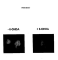
- Example 5 XIAP protects nigral neurons in a PSI model of Parkinson's disease
- rats were injected with AAV.GFP and AAV.dXIAP. These vectors were injected into the substantia nigra pars compacta (SNc) on each side and treated with DMSO (vehicle) or PSI as described in Example 1(iv). The results from the study are shown in Figs. 5A-5F , as wells as the bar graph in Fig. 5G , Note a significant reduction in the number of TH-immunoreactive cells in SNc injected with the control virus following PSI treatment while AAV.dXIAP virtually completely prevented cell loss. * p ⁇ 0.001 as determined using ANOVA.
- peptide fragments selected amino acid substitutions were made, including the following: D 148A (Aspartate to Alanine); D214S (Aspartate to Serine); W310A (Tryptophan to Alanine); E314S (Glutamic Acid to Serine); and H343A (Histidine to Alanine) in dXIAP.
- the D148A and H343A substitutions were made both singly and jointly.
- a XIAP mutant containing both the D214S and E314S substitutions was made and tested.
- a mutant containing the D148A, D214S and W310A substitutions was made and tested. It should understood that the XIAP mutations of the invention include any of the disclosed mutations either alone or in combination with other disclosed mutations.
- Figure 6A shows the known amino acid sequence of human XIAP (SEQ ID NO: 1) including the BIR1, Linker, BIR2, BIR3 and RING domains.
- Figure 6B shows the amino acid sequence of the BIR3 domain of human XIAP (SEQ ID NO: 2). The locations of the various mutations performed on XIAP are shown in Figure 6C .
- FIGS 7-10 show that XIAP point mutants that can no longer bind caspases are still protective in PSI-induced-, and polyglutamine induced cell death models, respectively.
- Smac is a novel protein which promotes caspase activation in the cytochrome c/Apaf-1/caspase-9 pathway. Smac promotes caspase-9 activation by binding to inhibitor of apoptosis proteins, IAPs, and removing their inhibitory activity.
- Smac is normally a mitochondrial protein but is released into the cytosol when cells undergo apoptosis. Mitochondrial import and cleavage of its signal peptide are required for Smac to gain its apoptotic activity.
- XIAP RING domain of XIAP appears to have no significant effect in these studies and, as such, is not necessary for the purposes of neuroprotection.
- the data in this example further demonstrates that the function of XIAP can be localized to a small portion of XIAP (BIR3 domain).
- small neuroprotective peptides comprising this domain can be generated. These peptides can be modified by adding leader signal peptides and peptides resulting in a chimeric peptide.
- a signal peptide and a TAT domain can be attached to the XIAP peptide fragment to allow it be secreted and make it cell permeable.
- a chimeric peptide construct is a signal peptide-BIR3-TAT construct. This chimeric protein can then be expressed by a viral vector or can be produced in vitro and injected systemically.
Abstract
Description
- The invention is generally in the field of methods and compositions for treating neurodegenerative diseases characterized by excess buildup of intracellular protein aggregates such as Parkinson's disease (PD), using viral and non-viral delivery systems that deliver therapeutic agents to specific regions of the brain. More specifically, using an adeno-associated viral vector to deliver a nucleotide sequence encoding an inhibitor of apoptosis protein (IAP) to specific regions of the brain associated with such neurodegenerative diseases.
- Neurodegenerative diseases are generally characterized by a degeneration of neurons in either the brain or the nervous system of an individual. Neuronal cell death can occur as a result of a variety of conditions including traumatic injury, ischemia, degenerative disease (e.g., Parkinson's disease, ALS, or SMA), or as a normal part of tissue development and maintenance. In addition to Parkinson's disease, various other diseases, such as Huntington's disease, Alzheimer's disease and Multiple Sclerosis, ALS, fall within this category. These diseases are debilitating and the damage that they cause is often irreversible. Moreover, in the case of a number of these diseases, the outcome is invariably fatal.
- Developmental cell death, or apoptosis has been implicated in neurodegenerative diseases. Apoptosis is a naturally occurring process thought to play a critical role in establishing appropriate neuronal connections in the developing central nervous system (CNS). Apoptosis is characterized morphologically by condensation of the chromatin followed by shrinkage of the cell body. Biochemically, the hallmark of apoptosis is the degradation of nuclear DNA into oligonucleosomal fragments. DNA laddering precedes cell death and may be a key event leading to death.
- Progress is being made on many fronts to find agents that can arrest the progress of these diseases. Nonetheless, the present therapies for most, if not all, of these diseases provide very little relief. One problem has been the relevance of current animal models to human disease. To date, the cause of neuronal death has remained elusive. The gold-standard animal models for PD involve rapid destruction of dopamine neurons using chemicals which are fairly specific for dopamine neurons. These chemical toxins, which include 6-hydroxydopamine (60HDA) and MPTP, cause oxidative damage to dopamine neurons in both rodents and primates. These models can be useful to test the efficacy of new therapies designed to improve the symptoms of PD, since such treatments are designed to intervene after cells have died or become dysfunctional, regardless of the cause of cell death. In order to test the value of protective or curative strategies, however, the mechanism of cell death must be relevant to human disease otherwise successful experimental studies will not translate into effective human therapy.
- Accordingly, a need exists to develop therapies that can alter the course of neurode generative diseases. More generally, a need exists for better methods and compositions for the treatment of neurodegenerative diseases in order to improve the quality of the lives of those afflicted by such diseases.
- The invention is based, at least in part, on the discovery that localized delivery of a vector comprising an apoptosis inhibiting agent to a specific region of the brain associated with a neurodegenerative diseases characterized by excess buildup of intracellular protein aggregates, can promote the improvement of the neurodegenerative disease. The apoptosis inhibiting agent can either be an inhibitor of apoptosis protein, or a nucleic acid molecule that inhibits expression of a target protein involved in apoptosis, such as RNA (e.g., RNA interference). In particular, the invention pertains to methods and compositions used to deliver a vector, (e.g., an adeno-associated virus vector (AAV)) comprising a nucleotide sequence encoding an inhibitor of apoptosis protein (IAP) e.g., X-linked inhibitor of apoptosis protein (XIAP) to target cells, (e.g., the substantia nigra pars compact).
- It appears that abnormal proteasome activity in neuronal cells is a contributing factor in neurodegenerative diseases such that the cells lose their ability to adequately degrade proteins, especially the mutated or misfolded proteins that may be pathological components of neurodegenerative diseases. Insofar as loss of function, or change in function, of the proteasome is a contributing factor in neuron degeneration. It has been discovered that blocking apoptotic cell death protects neurons from death following proteasome inhibition in vivo. The invention provides a method for inhibiting death of a cell of the nervous system, e.g., a neuron. Compositions comprising an inhibitor of apoptosis, e.g., an X-linked inhibitor of apoptosis protein (XIAP), which act as a potent inhibitor of caspases, e.g.,
caspases - Accordingly, in one aspect, the invention pertains to a method for treating a neurodegenerative disease in a subject by identifying a target site in the central nervous system that requires modification, and delivering a vector comprising a nucleotide sequence encoding an inhibitor of apoptosis protein (IAP), or a fragment thereof, to the target site in the central nervous system. The fragment encodes a peptide. The IAP is expressed in the target site to treat or reduce the neurodegenerative disease. The neurodegenerative disease is preferably with a neurodegenerative diseases characterized by excess buildup of intracellular protein aggregates. Examples of such neurodegenerative disease include, but are not limited to, Parkinson's disease, Huntington's disease, Alzheimer's disease, senile dementia, and Amyloid Lateral Schlerosis (ALS).
- In particular, the invention pertains to methods and compositions used to deliver a vector, (e.g., an adeno-associated virus vector (AAV)) comprising a nucleotide sequence encoding inhibitor of apoptosis protein (IAP), or a peptide fragment thereof, to target cells, e.g., substantia nigra pars compacta. Examples of apoptosis protein (IAP) includes, but is not limited to, X-linked inhibitor of apoptosis protein (XIAP), NIAP, cIAP-1 and cIAP-2, and is preferably, XIAP. In a preferred embodiment, the inhibitor of apoptosis protein is a peptide fragment of XIAP, such as a peptide fragment comprising the BIR-3 domain.
- Particularly preferred methods of delivering the vector to specific regions of the brain are those techniques that are simple, safe, and have a lower risk associated with them than lesioning, electrode implantation or cell transplantation. For example, delivery of the vector using stereotactic microinjection techniques, or delivery of the vector using specialized probes, or percutaneous delivery via disruption of the blood-brain barrier. Delivery of the vector using the method of the invention results in minimal immunological or inflammatory responses within the regions of the brain, thus eliminating the need for immunosupression. After delivery of the vector to a specific region of the brain, regional dispersion and/or diffusion of vector occurs ensuring local distribution of gene and stable gene expression.
- Suitable vectors for delivery include viral vectors and non-viral delivery methods such as liposome-mediated delivery vector. Examples of viral vectors include, but are not limited to, adeno-associated viral vector adenovirus vectors, herpes virus vectors, parvovirus vectors, and lentivirus vectors, preferably, adeno-associated viral vector.
- The regions of the brain that can be targeted are any regions typically associated with neurodegenerative diseases, and which can be targeted using standard procedures such as stereotaxic delivery. Examples of brain regions include, but are not limited to, the basal ganglia, subthalmic nucleus (STN), pedunculopontine nucleus (PPN), substantia nigra (SN), thalmus, hippocampus, the substantia nigra pars compacta, cortex, and combinations thereof.
- In another aspect, the invention pertains to a method for treating Parkinson's disease in a subject by identifying one or more regions of the brain that require modification, and delivering a vector comprising a nucleotide sequence encoding an inhibitor of apoptosis protein (IAP) to the region of the brain. The IAP is expressed in the region of the brain to treat or reduce Parkinson's disease.
- In another aspect, the invention pertains to a method for treating Huntington's disease in a subject by identifying one or more regions of the brain that require modification, and delivering a vector comprising a nucleotide sequence encoding an inhibitor of apoptosis protein (IAP) to the region of the brain. The IAP is expressed in the region of the brain to treat or reduce Huntington's disease.
- In yet another aspect, the invention features a vector for expression of an IAP in cells of the central nervous system comprising a tissue specific promoter operably linked to a nucleic acid encoding an IAP, and a post-transcriptional regulatory element.
- In one embodiment, the promoter is specific for central nervous system cells and tissues, such as the cells and tissues of the brain. In a preferred embodiment, the promoter is the CMV/CBA promoter. The vector also preferably comprises post-transcriptional regulatory elements to enhance expression of the encoded protein. In a preferred embodiment, the post-transcriptional regulatory element is the woodchuck post-transcriptional regulatory element.
- In another aspect, the invention pertains to a chimeric peptide for modulating a neurodegenerative disease comprising a peptide fragment of an inhibitor of apoptosis protein (IAP) operably linked to a signal peptide. The chimeric peptide may further comprise a TAT domain. The inhibitor of apoptosis protein (IAP) can be X-linked inhibitor of apoptosis protein (XIAP) or a fragment thereof. The peptide fragment can comprise the BIR3 domain of X1AP. The peptide fragment can also be an XIAP fragment comprising a mutation. The mutation may occur in the or around the BIR3 domain of XIAP. Mutant fragments can comprise at least one mutation at an amino acid position selected from the group consisting of D148, H343, D214, E314, and W310, or combinations thereof. In one embodiment, least one mutation results in the chimeric peptide having neuroprotective properties. In another embodiment, least one mutation results in a chimeric peptide that blocks Smac activity.
- In yet another aspect, the invention pertains to a chimeric peptide for modulating a neurodegenerative disease comprising a BIR3 peptide fragment of X-linked inhibitor of apoptosis protein (XIAY) operably linked to a signal peptide and a TAT domain. The BIR3 peptide fragment comprises at least one mutation at an amino acid position selected from the group consisting of D148, H343, D214, E314, and W310, or combinations thereof. At leat one of these mutations results in a chimeric peptide having neuroprotective properties and/or one that blocks Smac activity.
-
-
Figure 1 is a schematic representation of the rAAV vectors of the application. -
Figure 2A illustrates the reduction of cell death that occurs in cells transfected with XIAP plasmids. -
Figure 2B is a schematic representation of the pCAspace3 -Sensor vector. -
Figure 2C illustrates the location of apoptotic proteins through an apoptosis assay using the vector represented inFigure 2B . -
Figure 2D illustrates the results of an assay to determine the effect of XIAP and dXIAP on caspase-3 inactivation. -
Figures 3A-3D show the effect of XIAP administration in in vitro models of familial Parkinson's and Huntington's disease. -
Figures 4A-4D illustrate the effects of rAAV-mediated dXIAP delivery on cell survival of cells treated with proteosome inhibitors. -
Figures 5A-5G illustrate XIAP's protective effect on neurons of the susbtantia nigra in a PSI (rat) model of Parkinson's disease. -
Figure 6A is the known amino acid sequence of human XIAP including the BIR1, Linker, BIR2, BIR3 and RING domains. -
Figure 6B is the amino acid sequence of the BIR3 domain of human XIAP. -
Figure 6C shows locations of the various mutations performed on XIAP. -
Figure 7 illustrates the effects of XIAP point mutants on polyglutamine-induced cell death. -
Figure 8 illustrates the effects of XIAP point mutants on PSI-induced cell death. -
Figure 9 illustrates the effects caspase inhibition on polyglutamine induced cell death. -
Figure 10 illustrates the effects caspase inhibition on PSI induced cell death. - The practice of the present invention employs, unless otherwise indicated, conventional methods of virology, microbiology, molecular biology and recombinant DNA techniques within the skill of the art. Such techniques are explained fully in the literature. (See, e.g., Sambrook, et al. Molecular Cloning: A Laboratory Manual (Current Edition); DNA Cloning: A Practical Approach, Vol. I & II (D. Glover, ed.); Oligonucleotide Synthesis (N. Gait, ed., Current Edition); Nucleic Acid Hybridization (B. Hames & S. Higgins, eds., Current Edition); Transcription and Translation (B. Hames & S. Higgins, eds., Current Edition); CRC Handbook of Parvoviruses, Vol. I & II (P. Tijessen, ed.); Fundamental Virology, 2nd Edition, Vol. I & II (B. N Fields and D. M. Knipe, eds.)).
- So that the invention is more clearly understood, the following terms are defined:
- The phrase "apoptosis inhibiting agent" as used herein refers to a molecule that is an inhibitor of apoptosis protein, or a nucleic acid molecule that inhibits expression of a target protein involved in apoptosis, such as RNA (e.g., RNA interference). Examples of apoptosis inhibiting agents include but are not limited to a class of compounds from the BIR family, e.g. SURVIVIN and BRUCE.
- The term "apoptosis" as used herein refers to the art recognized use of the term for an active process of programmed cell death characterized by morphological changes in the cell. Apoptosis is characterized by membrane blebbing and nuclear DNA fragmentation. Apoptosis can occur via two pathways, the caspase-dependent pathway, which involves caspases, an inhibitor of apoptosis protein (IAP) and activation of the caspase pathway. Alternatively, apoptosis can occur via the caspase-independent pathway, which does not involve caspases.
- The term "zymogen" as used herein refers to the inactive proform of an enzyme e.g. a caspase, which is typically activated by proteolysis.
- The term "caspase" as used herein refers to a cysteine protease that specifically cleaves proteins after Asp residues. Caspases exist as inactive proenzymes which undergo proteolytic processing at conserved aspartic residues to produce 2 subunits, large and small, that dimerize to form the active enzyme. This protein was shown to cleave and activated
caspases caspases - The term "apoptosis inhibiting compound" as used herein refers to an agent that can reduce apoptosis by a detectable amount by acting on a pathway involved in apoptosis. The agent can act by inhibiting or blocking a particular step in the apoptotic pathway, for example by blocking or inhibiting the activity of protein involved in the pathway. The apoptotic pathway includes both caspase-dependent apoptosis, as well as caspase-independent apoptosis.
- The term "inhibit" or "inhibiting" as used herein refers to a measurable reduction of apoptotic activity that leads to at least a 10% preferably or 20%, increase in the likelihood that a cell will survive following an event which normally causes cell death (relative to an untreated control cell). Preferably, the cells being compared are neural cells normally susceptible to ischemic cell death, neurodegeneration, or axotomy. Preferably, the decrease in the likelihood that a cell will die is 80%, more preferably 2-fold, most preferably, 5-fold.
- The phrase "Inhibitor of Apoptosis Protein" or "IAP" is refers to an amino acid sequence which has homology to baculovirus inhibitors of apoptosis. For example, NAIP, truncated NAIP, HIAP1, HIAP2 and XIAP are specifically included (see
U.S. Pat. No. 5,919,912 ;U.S. Pat. No. 6,156,535 ; andU.S. Pat no. 6,709,866 ). Preferably, such a polypeptide has an amino acid sequence which is at least 45%, preferably 60%, and most preferably 85% or even 95% identical to at least one of the amino acid sequences of the NAIP, truncated NAIP, HIAP1 HIAP2, or XIAP. - The term "mutation," as used herein, refers to any alteration of the gene encoding an inhibitor of apoptosis protein. The mutation can alter the functionality of the protein produced by that gene. Such mutations can include, but are not limited to, an amino acid substitution wherein a native amino acid is replaced with an alanine or other biologically comparable amino acid residue, including, but not limited to glycine, valine, and leucine. Amino acid substitutions can be introduced into the nucleic acid sequence by standard molecular biology methods. The term "mutation" also includes a deletion or addition of nucleotide sequences to any portion of gene that encodes an inhibitor of apoptosis protein. In one embodiment, the mutation produces a protein or fragment that no longer binds to a caspase. In another embodiment, the mutation produces a protein or fragment that blocks the interaction of a protein such as Smac. In a preferred embodiment, the mutation produces a protein or fragment that is neuroprotective.
- The term "portion" or "fragment" as used herein refers to an amino acid sequence that has fewer amino acids than the entire sequence of the inhibitor of apoptosis protein. A fragment can comprise any desired domain, such as a BIR-3 domain. Sizes of peptide fragments can be designed to be less than about 200 amino acids, less than about 100 amino acids, less than about 80 amino acids, less than about 60 amino acids, less than about 40 amino acids, less than about 20 amino acids, and less than about 10 amino acids, so long as the peptide fragment retains a desired activity.
- The term "modulate" or "modify" are used interchangeably herein and refer to an alleviation of at least one target protein or gene involved in the caspase -dependent pathway for apoptosis. Such that apoptosis is inhibited or reduced. A modification in apoptosis can be assessed by monitoring cell blebbing DNA fragmentation, and the like.
- The terms "neurological disorder" or "neurodegenerative disorder" are used interchangeably herein and refer to an impairment or absence of a normal neurological function or presence of an abnormal neurological function in a subject. For example, neurological disorders can be the result of disease, injury, and/or aging. As used herein, neurological disorder also includes neurodegeneration which causes morphological and/or functional abnormality of a neural cell or a population of neural cells. Non-limiting examples of morphological and functional abnormalities include physical deterioration and/or death of neural cells, abnormal growth patterns of neural cells, abnormalities in the physical connection between neural cells, under-or over production of a substance or substances, e.g., a neurotransmitter, by neural cells, failure of neural cells to produce a substance or substances which it normally produces, production of substances, e.g., neurotransmitters, and/or transmission of electrical impulses in abnormal patterns or at abnormal times. Neurodegeneration can occur in any area of the brain of a subject and is seen with many disorders including, for example, Amyotrophic Lateral Sclerosis (ALS), multiple sclerosis, Huntington's disease, Parkinson's disease, and Alzheimer's disease.
- The term "therapeutically effective amount" refers to an amount effective, at dosages and for periods of time necessary, to achieve the desired therapeutic result. A therapeutically effective amount of the proteasome modulating pharmacological agent may vary according to factors such as the disease state, age, sex, and weight of the individual, and the ability of the pharmacological agent to elicit a desired response in the individual. A therapeutically effective amount is also one in which any toxic or detrimental effects of the pharmacological agent are outweighed by the therapeutically beneficial effects.
- The term "prophylactically effective amount" refers to an amount effective, at dosages and for periods of time necessary, to achieve the desired prophylactic result. Typically, since a prophylactic dose is used in subjects prior to or at an earlier stage of disease, the prophylactically effective amount will be less than the therapeutically effective amount.
- The term "subject" as used herein refers to any living organism capable of eliciting an immune response. The term subject includes, but is not limited to, humans, nonhuman primates such as chimpanzees and other apes and monkey species; farm animals such as cattle, sheep, pigs, goats and horses; domestic mammals such as dogs and cats; laboratory animals including rodents such as mice, rats and guinea pigs, and the like. The term does not denote a particular age or sex. Thus, adult and newborn subjects, as well as fetuses, whether male or female, are intended to be covered.
- The invention is described in more detail in the following subsections:
- Evidence is accumulating that as a result of the normal aging process the body increasingly loses the ability to adequately degrade mutated or misfolded proteins. The proteasome is the piece of biological machinery that is responsible for most normal degradation of proteins found inside cells. Age-related loss of function, or change in function of the proteasome is now thought to be at the heart of many neurodegenerative conditions, including, for example, Alzheimer's disease, Parkinson's disease, Huntington's disease, Multiple Sclerosis and amyotrophic lateral sclerosis (ALS), each of which is described below.
- Huntington's disease (HD) is a hereditary disorder caused by the degeneration of neurons in certain areas of the brain. This degeneration is genetically programmed to occur in certain areas of the brain, including the cells of the basal ganglia, the structures that are responsible for coordinating movement. Within the basal ganglia, Huntington's disease specifically targets nerve cells in the striatum, as well as cells of the cortex, or outer surface of the brain, which control thought, perception and memory. Neuron degeneration due to HD can result in uncontrolled movements, loss of intellectual capacity and faculties, and emotional disturbance, such as, for example, mood swings or uncharacteristic irritability or depression.
- As discussed above, neuron degeneration due to HD is genetically programmed to occur in certain areas of the brain. Studies have shown that Huntington's disease is caused by a genetic defect on
chromosome 4, and in particular, people with HD have an abnormal repetition of the genetic sequence CAG in the HD gene, which has been termed ITI5. The IT15 gene is located on the short arm ofchromosome 4 and encodes a protein called huntingtin. Exon I of the ITI5 gene contains a polymorphic stretch of consecutive glutamine residues, known as the polyglutamine tract (D. Rubinsztein, "Lessons from Animal Models of Huntington's Disease," TRENDS in Genetics, 18(4): 202-9 (April 2002)). Asymptomatic individuals typically contain fewer than 35 CAG repeats in the polyglutamine tract. - The inherited mutation in HD is an expansion of the natural CAG repeats within the sequence of
exon 1 of the human HD gene. This leads to an abnormally long stretch of polyglutamines. The length of the polyglutamine repeats correlates with the severity of the disease. One of the pathological hallmarks of HD is a buildup of intracellular protein aggregates composed of these abnormal HD proteins with long polyglutamine repeats. The results in the Examples section show that expression of this abnormal HD gene (called Huntington) in cultured neurons leads to cell death, while co-expression of the anti-apoptotic gene XIAP blocks this death. This demonstrates that expression of an anti-apoptotic gene can protect from mutant Huntington-induced neuronal death. - Multiple Sclerosis (MS) is a chronic disease that is characterized by "attacks," during which areas of white matter of the central nervous system, known as plaques, become inflamed. Inflammation of these areas of plaque is followed by destruction of myelin, the fatty substance that forms a sheath or covering that insulates nerve cell fibers in the brain and spinal cord. Myelin facilitates the smooth, high-speed transmission of electrochemical messages between the brain, spinal cord, and the rest of the body. Damage to the myelin sheath can slow or completely block the transmission of these electrochemical messages, which can result in diminished or lost bodily function.
- The most common course of MS manifests itself as a series of attacks, which are followed by either complete or partial remission, during which the symptoms lessen only to return at some later point in time. This type of MS is commonly referred to as "relapsing-remitting MS." Another form of MS, called "primary-progressive MS," is characterized by a gradual decline into the disease state, with no distinct remissions and only temporary plateaus or minor relief from the symptoms. A third form of MS, known as "secondary-progressive MS," starts as a relapsing-remitting course, but later deteriorates into a primary-progressive course of MS.
- The symptoms of MS can be mild or severe, acute or of a long duration, and may appear in various combinations. These symptoms can include vision problems such as blurred or double vision, red-green color distortion, or even blindness in one eye, muscle weakness in the extremities, coordination and balance problems, muscle spasticity, muscle fatigue, paresthesias, fleeting abnormal sensory feelings such as numbness, prickling, or "pins and needles" sensations, and in the worst cases, partial or complete paralysis. About half of the people suffering from MS also experience cognitive impairments, such as for example, poor concentration, attention, memory and/or judgment. These cognitive symptoms occur when lesions develop in those areas of the brain that are responsible for information processing.
- Alzheimer's disease is a progressive, neurodegenerative disease that affects the portions of the brain that control thought, memory and language. This disease is characterized by progressive dementia that eventually results in substantial impairment of both cognition and behavior. The disease manifests itself by the presence of abnormal extracellular protein deposits in brain tissue, known as "amyloid plaques," and tangled bundles of fibers accumulated within the neurons, known as "neurofibrillary tangles," and by the loss of neuronal cells. The areas of the brain affected by Alzheimer's disease can vary, but the areas most commonly affected include the association cortical and limbic regions, Symptoms of Alzheimer's disease include memory loss, deterioration of language skills, impaired visuospatial skills, and impaired judgment, yet those suffering from Alzheimer's retain motor function.
- Parkinson's disease (PD) is characterized by death of dopaminergic neurons in the substantia nigra (SNr), leading to a disturbance in the basal ganglia network which regulates movement. In addition, other brainstem cell populations can die or become dysfunctional. One of the pathological hallmarks of PD in humans is the Lewy body, which contains abnormal protein aggregates which include the protein alpha-synuclein. While there are many therapies available to treat the symptoms of Parkinson's disease, including medical therapy and surgical therapies, there is no current treatment which will stop the death of neurons and ultimately cure this disorder.
- To date, the cause of neuronal death has remained elusive. One problem has been the relevance of current animal models to human disease. The gold-standard animal models for PD involve rapid destruction of dopamine neurons using chemicals which are fairly specific for dopamine neurons. These chemical toxins, which include 6-hydroxydopamine (6OHDA) and MPTP, cause oxidative damage to dopamine neurons in both rodents and primates. These models can be useful to test the efficacy of new therapies designed to improve the symptoms ofPD, since such treatments are designed to intervene after cells have died or become dysfunctional, regardless of the cause of cell death. In order to test the value of protective or curative strategies, however, the mechanism of cell death must be relevant to human disease otherwise successful experimental studies will not translate into effective human therapy.
- Many features of the animal models have been questioned for protective strategies. First, these toxins usually cause near complete destruction of dopamine neurons within 24-48hrs., while PD is a slowly degenerative disease which can take many years or more to have even partial loss of cells. Also, these do not cause protein inclusions similar to the Lewy bodies seen in human PD. These toxins are also only specific to dopamine neurons, while in human PD other cell populations are affected. There is also little convincing evidence in human disease that the oxidative damage mechanism is the primary cause of PD. Nonetheless, several factors have been shown to protect animal cells from these toxins, including anti-apoptotic genes and growth factors such as glial-derived neurotrophic factor (GDNF). This is understandable, since the result of such oxidative damage is usually apoptotic cell death.
- The history of GDNF highlights the problems in translating promising data from these models to human disease. Several animal studies over many years suggested that GDNF could afford substantial protection to dopamine neurons when exposed to either 60HDA or MPTP. Similar data has been obtained regardless of the mode of delivery of GDNF, including both intraventricular and direct intrastriatal infusion of recombinant GDNF protein, as well as GDNF produced from a viral vector following gene therapy. Nonetheless, multiple GDNF studies in human have failed. The first studies involved infusion of GDNF into cerebrospinal fluid via an intraventricular catheter. This was stopped due to adverse effects. It was then hypothesized that direct infusion of GDNF into the striatum, where dopamine neuron terminals reside, would limit side effects and improve efficacy as was seen in the above mentioned animal models. This was also recently halted due to failure to demonstrate any meaningful effect in human patients compared to controls. This only serves to highlight problems with developing neuroprotective therapeutics using these models, In fact, the only similarity between these models and human PD is the loss of dopamine neurons. This, however, can also be achieved by many other means, including thermal destruction or destruction of these cells using other chemicals such as ibotinic acid. Therefore, there is no good evidence that any protection of neurons using these models has any value to human PD.
- Recently, a new model was described which not only appears to be more relevant to human PD, but which also is consistent with most of the known features of human disease (Kevin ct al, Annals of Neurology (2004) 56, 149-162). The model involves repeated administration of proteasome inhibitor. Proteasomes are complex, multi-unit enzymes within the cell which are critical for metabolizing and removing proteins which are misfolded, dysfunctional and/or no longer desirable. These are essential for protein turnover, which is crucial for proper regulation of cellular physiology. Proteins which are targeted to the proteasome are usually modified by addition of a ubiquitin group. Ubiquinated proteins can then enter the proteasome for ultimate degradation. Unlike the dopamine toxin model, this model causes a very slow neuronal degeneration which is much more analogous to human disease. In addition to dopamine neuronal loss in the SNr, loss or dysfunctional of other neuronal populations are seen which also mimic the human disorder. Most interestingly, intracellular protein aggregates are seen which are highly analogous to the Lewy body. None of these features are present in the dopamine toxin models, and all of them are found to some degree in the human disorder, indicating that this is a far more relevant model of the actual mechanism of cell death in human PD.
- Those few forms of human PD for which a cause is known further support the relevance of this model for neuroprotection studies. A minority of PD cases are caused by inherited mutations in a single gene. To date, four such genes have been identified. While the function of one gene remains unknown, the other three directly support the concept that ubiquitin-proteasome dysfunction is the key cause of cell death PD. Two of these ganes, parkin and UCHL-1, are involved in ubiquination of proteins and loss of function causes human PD. The third gene, alpha-synuclein, causes a dominant form of PD and, as mentioned earlier, is a key component of the intracellular inclusions called Lewy bodies. Therefore, the major known causes of inherited human PD support the pathological findings in the new proteasome inhibitor model of PD as being the only available model which accurately replicates the human disorder.
- The factors triggering dopaminergic neuronal loss in Parkinson's disease are still largely unknown. Neuronal cell death is thought to occur via apoptosis and the involvement of several caspases at the late stages of this process is well documented. X-linked inhibitor of apoptosis (XIAP) is a potent inhibitor of
caspases - The effects of XIAP in several in vitro models of Parkinson's disease was examined and the results shown in the Example section. Most of the experiments were performed using a more stable truncated from of XIAP lacking the RING domain at the C-terminus (dXTAP). To test the in vivo effect of XIAP, a recombinant AAV (rAAV) vector expressing dXIAP was generated to investigate therapeutic intervention in the novel in vivo rat model of Parkinson's disease which is triggered by inhibition of proteasome machinery. The data demonstrates that dXIAP maybe used for neuroprotection.
- Amyotrophic Lateral Sclerosis (ALS) is a universally fatal neurodegenerative condition in which patients progressively lose all motor function - unable to walk, speak, or breathe on their own, ALS patients die within two to five years of diagnosis. The incidence of ALS increases substantially in the fifth decade of life. Evidence is accumulating that as a result of the normal aging process the body increasingly loses the ability to adequately degrade mutated or misfolded proteins. The proteasome is the piece of biological machinery responsible for most normal degradation of proteins inside cells. Age related loss of function or change of function of the proteasome is now thought to be at the heart of many neurodegenerative conditions, including Alzheimer's disease, Parkinson's disease, Huntington's disease, and ALS.
- The cardinal feature of ALS is the loss of spinal motor neurons, which causes the muscles under their control to weaken and waste away leading to paralysis. ALS has both familial (5-10%) and sporadic forms and the familial forms have now been linked to several distinct genetic loci (Deng, H.X., et al., "Two novel SOD1 mutations in patients with familial amyotrophic lateral sclerosis," Hum. Mol. Genet., 4(6): 1113-16 (1995); Siddique, T. and A. Hentati, "Familial amyotrophic lateral sclerosis," Clin. Neurosci., 3(6): 338-47(1995); Siddique, T., et al., "Familial amyotrophic lateral sclerosis," J. Neural Transm. Suppl., 49: 219-33(1997); Ben Hamida, et al., "Hereditary motor system diseases (chronic juvenile amyotrophic lateral sclerosis). Conditions combining a bilateral pyramidal syndrome with limb and bulbar arnyotrophy," Brain, 113(2): 347-63 (1990); Yang, Y., et al., "The gene encoding alsin, a protein with three guanine-nucleotide exchange factor domains, is mutated in a form of recessive amyotrophic lateral sclerosis," Nat. Genet., 29(2): 160-65 (2001); Hadano, S., et al., "A gene encoding a putative GTPase regulator is mutated in familial amyotrophic )). About 15-20% of familial cases are due to mutations in the gene encoding Cu/Zn superoxide dismutase 1 (SODI) (Siddique, T., et al., "Linkage of a gene causing familial amyotrophic lateral sclerosis to chromosome 21 and evidence of genetic-locus heterogeneity," N. Engl. J. Med., 324(20): 1381-84 (1991); Rosen, D.R., et al., "Mutations in Cu/Zn superoxide dismutase gene are associated with familial amyotrophic lateral sclerosis." Nature, 362(6415): 59-62 (1993)).
- Although the etiology of the disease is unknown, the dominant theory is that neuronal cell death in ALS is the result of over-excitement of neuronal cells due to excess extracellular glutamate. Glutamate is a neurotransmitter that is released by glutaminergic neurons, and is taken up into glial cells where it is converted into glutamine by the enzyme glutamine synthetase, glutamine then re-enters the neurons and is hydrolyzed by glutaminase to form glutamate, thus replenishing the neurotransmitter pool. In a normal spinal cord and brain stem, the level of extracellular glutamate is kept at low micromolar levels in the extracellular fluid because glial cells, which function in part to support neurons, use the excitatory amino acid transporter type 2 (EAAT2) protein to absorb glutamate immediately. A deficiency in the normal EAAT2 protein in patients with ALS, was identified as being important in the pathology of the disease (See e.g., Meyer et al., J. Neurol. Neurosurg. Psychiatry, 65: 594-596 (1998); Aoki et al., Ann. Neurol. 43: 645-653 (1998); Bristol et al., Ann Neurol. 39: 676-679 (1996)). One explanation for the reduced levels of EAAT2 is that EAAT2 is spliced aberrantly (Lin et al., Neuron, 20: 589-602 (1998)). The aberrant splicing produces a splice variant with a deletion of 45 to 107 amino acids located in the C-terminal region of the EAAT2 protein (Meyer et al., Neureosci Lett. 241: 68-70 (1998)). Due to the lack of, or defectiveness of EAAT2, extracellular glutamate accumulates, causing neurons to fire continuously. The accumulation of glutamate has a toxic effect on neuronal cells because continual firing of the neurons leads to early cell death.
- In one aspect, the invention pertains to using an inhibitor of apoptosis protein (IAP), e.g., XIAP, for the amelioration or treatment of neurological and/or neurodegenerative disorders and diseases associated with abnormal proteasome function.
- The proteasome is a multi-unit protein complex that plays a key role in protein degradation within a cell. The function of this key process ranges from ridding the cell of old and misfolded proteins to the degradation of key regulatory proteins and antigen generation for immune surveillance. In particular, proteolysis is involved in the regulation of numerous cellular processes including progression of the cell cycle, oncogenesis, transcription, development, growth and atrophy of developed tissues, flow of substrates through metabolic pathways, selective elimination of abnormal proteins and antigen processing (DeMartino, G., et al., "The Proteasome, a Novel Protease Regulated by Multiple Mechanisms," J. Biol. Chem., 274(32): 22123-126 (1999); Ottosen, S., et al., "Protease Parts at Gene Promoters," Science, 296: 479-81 (2002)). The antigen-generating function of the proteasome allows targeted killing of defective and virally infected cells by the Cytotoxic T-cells and Natural killer cells.
- The proteasome undergoes extensive modification to suit its different function. It does so by adding and replacing the individual subunits and by restructuring. At the core of all configurations is the 208 proteasome, which provides the proteasome its catalytic degradation power. 20S proteasomes are combined with various regulatory caps such as PA700 and PA28, which are thought to control the entry to 20S as well as the disposition of end products. The core of the 20S proteasome consists of two copies each of seven different α and β subunits, which are arranged in four stacked rings (α7β7β7α7) (Verma et al., "Proteasomal Proteomics: Identification of Nucleotide-sensitive Proteasome-interacting Proteins by Mass Spectrometric Analysis of Affinity-purified Proteasomes," Mol. Biol, Cell., 11: 3425-39 (2000)). The interior of the ring structure contains a cavity consisting of three contiguous chambers joined by narrow constrictions (DeMartino, G., et al., "The Proteasome, a Novel Protease Regulated by Multiple Mechanisms," J. Biol. Chem., 274(32): 22123-126 (1999)). The 7 beta subunits of the 20S proteasome provide the bulk of its peptide cleaving abilities. Three of these subunits, X (β5), Y (β1), and Z (β2) can be replaced with inducible counterparts LMP2, LMP7, and MECL-1, which causes the proteasome to cleave peptides in a manner more specific for MHC I antigen presentation (Toes, R.E., et al., "Discrete cleavage motifs of constitutive and immunoproteasomes revealed by quantitative analysis of cleavage products," J. Exp. Med., 194(1): 1-12 (2001)). These proteins are selectively induced under certain conditions, including treatment of cells with gamma-interferon. The LMP2, LMP7 and MECL-1 subunits assembly to form proteasomes with distinct subunit compositions and altered catalytic characteristics (DeMartino, G., et al., "The Proteasome, a Novel Protease Regulated by Multiple Mechanisms," J. Biol. 'immunoproteasome' and is commonly presented in response to viral infection.
- Increasing evidence is accumulating that as a result of the norman aging process the body increasingly loses the ability to adequately degrade mutated or misfolded proteins. The proteasome is the cell machinery responsible for normal protein degradation. Oxidative stress is thought to contribute to this process of protein degradation through oxidation and nitration of intracellular proteins, which makes proteins prone to cross-linking and aggregation (Davies, K.J., "Degradation of oxidized proteins by the 20S proteasome," Biochimie, 83(3-4): 301-10 (2001); Squier, T.C., "Oxidative stress and protein aggregation during biological aging," Exp. Gerontol., 36(9): 1539-50 (2001)). Such aggregated proteins are more resistant to degradation in the proteasome and may cause inhibition of proteasomal function through irreversible binding to the proteasome (Davies, K.J., "Degradation of oxidized proteins by the 20S proteasome," Biochimie, 83(3-4): 301-10 (2001); Squier, T.C., "Oxidative stress and protein aggregation during biological aging," Exp. Gerontol., 36(9): 1539-50 (2001)). Alternatively or additionally, decreased proteasomal activity may be caused more directly by oxidation of the proteasome itself (Keller, J.N, et al., "Possible involvement of proteasome inhibition in aging: implications for oxidative stress," Mech. Ageing Dev., 113(1): 61-70 (2000)). Aggregates of misfolded proteins can induce a number of changes in the proteasome that can lead to aberrant immune activation and apoptotic cell death. Age related loss of function or impediment of the proteasome is now thought to be at the heart of many neurodegenerative conditions such as Alzheimer's disease, Parkinson's disease, Huntington's disease, and ALS (Goldberg, et al., "The cellular chamber of doom," Sci. Am., 284(1): 68-73 (2001); Johnston, J.A., et al., "Formation of high molecular weight complexes of mutant Cu, Zn- superoxide dismutase in a mouse model for familial amyotrophic lateral sclerosis," Proc. Natl. Acad. Sci. U SA, 97(23): 12571-76 (2000); Kopito, R.R., "Aggresomes, inclusion bodies and protein aggregation," Trends Cell. Biol., 10(12): 524-30 (2000)).
- Inhibition of proteasomal activity increases abnormal protein accumulation, and accumulation of abnormal proteins contribute to inhibition of proteasomal activity. Proteasomal inhibition is a common feature in neurodegenerative diseases. Accordingly, proteasomal dysfunction can alter the progression of diseases such as Parkinson's disease and Huntington's disease by a variety of ways. It is believed that proteasome alteration modulates important factors involved in cell cycle regulation, apoptosis, inflammation, and antigen presentation, which individually or in combination can lead to disease propagation.
- In one aspect, the invention pertains to reducing, inhibiting, preventing or altering apoptosis associated with abnormal proteasome function using an inhibitor of apoptosis protein (IAP), e.g., XAIP. Apoptosis or programmed cell death was originally described by Kerr et al., in 1972 (Kerr, et al. (1972) Br J Cancer, 26, 239-57), as a new cell-autonomous mechanism of death aimed at removing damaged, mutated or aged cells. Mitochondria, are directly linked to the process of apoptosis to the extent that they are now termed "killer organelles" (Ravagnan, et al. (2002) J Cell Physiol, 192, 131-7). Mitochondria contain several proteins in their inter-membrane space which are released in response to pro-apoptotic stimuli. These diverse proteins include cytochrome c, AIF, Smac/Diablo, endonuclease G and Omi/HtrA2 (See e.g., Van Loo, et al. (2002) Cell Death Differ, 9, 1031-42). Each of these proteins has a distinct function but they are all pro-apoptotic and, invariably, their release from the mitochondria induces cell death.
- Several gene products that modulate the apoptotic process have been identified. Although these products can, in general, be separated into two basic categories, gene products from each category can function to either inhibit or induce apoptosis. One family of gene products is the Bcl-2 family of proteins. A second family of gene products, the caspase family, is related genetically to the C. elegans ced-3 gene product, which is required for apoptosis in the roundworm, C. elegans. The caspase family includes, for example, caspase-1, caspase-2, caspase-3, caspase-4, caspase-5, caspase-6, caspase-7, caspase-8, caspase-9 and caspase-10.
- Examples of inhibitor of apoptosis protein include, but are not limited to, X-linked inhibitor of apoptosis protein (XIAP), NIAP, cIAP-1 and cIAP-2.
- In the gene therapy constructs, an IAP cDNA expression is directed from any suitable promoter (e.g., the human cytomegalovirus,
simian virus 40, or metallothionein promoters), and its production is regulated by any desired mammalian regulatory element. For example, if desired, enhancers known to direct preferential gene expression in neuronal cells may be used to direct expression of an IAP. - Fragments or derivatives of the IAP, e.g., XIAP, may also be administered by retroviral gene transfer therapy or another suitable viral vector system. Useful fragments or derivatives of IAP, e.g., XIAP may be adminstered by inserting the nucleic acids encoding these fragments or derivatives in place of the complete IAP, e.g., XIAP gene in a gene therapy vector. The sequence of IAP proteins and nucleic acids may be obtained from
U.S. Pat. No. 5,919,912 ;U.S. Pat. No. 6,156,535 ; andU.S. Pat. No. 6,709,866 . The fragment can comprise any desired domain, such as a BIR-3 domain. Sizes of peptide fragments can be less than about 200 amino acids, less than about 100 amino acids, less than about 80 amino acids, less than about 60 amino acids, less than about 40 amino acids, less than about 20 amino acids, and less than about 10 amino acids, so long as the peptide fragment retains a desired activity. - The peptides fragments can be chemically synthesized using a peptide synthesizer based on the available sequences. These chemically synthesized fragments can be synthesized to include a mutation. The peptide fragments can also be mutated using standard molecular biology techniques. With this approach, the nucleotide sequence encoding the peptide fragment is altered by insertion, deletion or addition of nucleotides.
- If the peptides are chemically synthesized, they can be modified by adding signal I peptides or other regulatory sequences. Signal peptides play an important role in protein transport and sorting to the different compartment of the cell. Although signal peptides have varying lengths and do not have a consensus sequence, almost allpossess a common three-region structure: the positively charged n-region, the hydrophobic N-region,and the C-region where the cleavage site occurs (Nakai, (2000) Adv. in Protein Chem. 54:277-344). Signal peptides are cleaved while proteins are still being processed. Examples of signal peptides include, but are not limited to, N-terminus signal peptides that often target the mitochondrial matrix; and C-terminus signal peptides that function similarly to N-terminus signal peptides, and Suomen Kristillinen Liitto (SKL). If the IAP peptides are being expressed, the nucleotide sequence that encodes a signal peptide can be coupled to the nucleotide sequence encoding the IAP peptide.
- The nucleotide sequence encoding the IAP peptides can also be modified by adding transactivating regulatory protein (TAT) protein of HIV acts as transcriptional regulator of viral gene expression by binding to the transactivating responsive sequence (TAR) RNA element. Binding to the TAR RNA element initiates viral transcription and/or elongation from LTR promoter (Feng (1988) Nature 334:165-167 and Roy (1990) Genes Dev 4: 1365); upregulates expression of all viral genes; promotes the elongation phase of HIV-1 transcription, allowing full-length transcripts to be produced; and represses cellular promoters.
- The vectors of the invention can be delivered to the cells of the central nervous system by using viral vectors or by using non-viral vectors. In a preferred embodiment, the invention uses adeno-associated viral (AAV) vectors comprising the a nucleotide sequence encoding IAP for gene delivery. AAV vectors can be constructed using known techniques to provide at least the operatively linked components of control elements including a transcriptional initiation region, a exogenous nucleic acid molecule, a transcriptional termination region and at least one post-transcriptional regulatory sequence. The control elements are selected to be functional in the targeted cell. The resulting construct which contains the operatively linked components is flanked at the 5' and 3' region with functional AAV ITR sequences.
- The nucleotide sequences of AAV ITR regions are known. The ITR sequences for AAV-2 are described, for example by Kotin et al. (1994) Human Gene Therapy 5:793-801; Berns "Parvoviridae and their Replication" in Fundamental Virology, 2nd Edition, (B. N. Fields and D. M. Knipe, eds.) The skilled artisan will appreciate that AAV ITR's can be modified using standard molecular biology techniques. Accordingly, AAV ITRs used in the vectors of the invention need not have a wild-type nucleotide sequence, and may be altered, e.g., by the insertion, deletion or substitution of nucleotides. Additionally, AAV ITRs may be derived from any of several AAV serotypes, including but not limited to, AAV-1, AAV-2, AAV-3, AAV-4, AAV-5, AAVX7, and the like. Furthermore, 5' and 3' ITRs which flank a selected nucleotide sequence in an AAV expression vector need not necessarily be identical or derived from the same AAV serotype or isolate, so long as the ITR's function as intended, i.e., to AAV rep gene products are present in the cell.
- The skilled artisan can appreciate that regulatory sequences can often be provided from commonly used promoters derived from viruses such as, polyoma,
Adenovirus 2, cytomegalovirus andSimian Virus 40. Use of viral regulatory elements to direct expression of the protein can allow for high level constitutive expression of the protein in a variety of host cells. Ubiquitously expressing promoters can also be used include, for example, the early cytomegalovirus promoter Boshart et al. (1985) Cell 41:521-530, herpesvirus thymidine kinase (HSV-TK) promoter (McKnight et al. (1984) Cell 37: 253-262), β-actin promoters (e.g., the human β-actin promoter as described by Ng et al. (1985) Mol. Cell Biol. 5: 2720-2732) and colony stimulating factor-1 (CSF-1) promoter (Ladner et al. (1987) EMBO J. 6: 2693-2698). - Alternatively, the regulatory sequences of the AAV vector can direct expression of the gene preferentially in a particular cell type, i.e., tissue-specific regulatory elements can be used. Non-limiting examples of tissue-specific promoters which can be used include, central nervous system (CNS) specific promoters such as, neuron-specific promoters (e.g., the neurofilament promoter; Byrne and Ruddle (1989) Proc. Natl. Acad Sci. USA 86:5473-5477) and glial specific promoters (Morii et al. (1991) Biochem. Biophys Res. Commun. 175: 185-191). Preferably, the promoter is tissue specific and is essentially not active outside the central nervous system, or the activity of the promoter is higher in the central nervous system that in other systems. For example, a promoter specific for the spinal cord, brainstem, (medulla, pons, and midbrain), cerebellum, diencephalon (thalamus, hypothalamus), telencephalon (corpus stratium, cerebral cortex, or within the cortex, the occipital, temporal, parietal or frontal lobes), or combinations, thereof. The promoter may be specific for particular cell types, such as neurons or glial cells in the CNS. If it is active in glial cells, it may be specific for astrocytes, oligodentrocytes, ependymal cells, Schwann cells, or microglia. If it is active in neurons, it may be specific for particular types of neurons, e.g., motor neurons, sensory neurons, or interneurons. Preferably, the promoter is specific for cells in particular regions of the brain, for example, the cortex, stratium, nigra and hippocampus.
- Suitable neuronal specific promoters include, but are not limited to,CMV/CBA, neuron specific enolase (NSE) (Olivia et al. (1991) Genomics 10: 157-165, GenBank Accession No: X51956), and human neurofilament light chain promoter (NEFL) (Rogaev et al. (1992) Hum. Mol. Genet. 1: 781, GenBank Accession No: L04147). Glial specific promoters include, but are not limited to, glial fibrillary acidic protein (GFAP) promoter (Morii et al. (1991) Biochem. Biophys Res. Commun, 175: 185-191, GenBank Accession No:M65210), S100 promoter (Morii et al (1991) Biochem. Biophys Res. Commun. 175: 185-191, GenBank Accession No: M65210) and glutamine synthase promoter (Van den et al. (1991) Biochem. Biophys. Acta. 2: 249-251, GenBank Accession No: X59834). In a preferred embodiment, the gene is flanked upstream (i.e., 5') by the neuron specific enolase (NSE) promoter. In another preferred embodiment, the gene of interest is flanked upstream (i.e., 5') by the
elongation factor 1 alpha (EF) promoter. - The AAV vector harboring the nucleotide sequence encoding a protein of interest, e.g., GAD, and a post-transcriptional regulatory sequence (PRE) flanked by AAV ITRs, can be constructed by directly inserting the nucleotide sequence encoding the protein of interest and the PRE into an AAV genome which has had the major AAV open reading frames ("ORFs") excised therefrom. Other portions of the AAV genome can also be deleted, as long as a sufficient portion of the ITRs remain to allow for replication and packaging functions. These constructs can be designed using techniques well known in the art. (See, e.g., Lebkowski et al. (1988) Molec. Cell. Biol. 8:3988-3996; Vincent et al. (1990) Vaccines 90 (Cold Spring Harbor Laboratory Press); Carter (1992) Current Opinion in Biotechnology 3:533-539; Muzyczka (1992) Current Topics in Microbiol. and Immunol. 158:97-129; Kotin (1994) Human Gene Therapy 5:793-801; Shelling et al. (1994) Gene Therapy 1:165-169; and Zhou et al. (1994) J. Exp. Med. 179:1867-1875).
- Alternatively, AAV ITRs can be excised from the viral genome or from an AAV vector containing the same and fused 5' and 3' of a selected nucleic acid construct that is present in another vector using standard ligation techniques, such as those described in Sambrook et al. , ,Supra. Several AAV vectors are available from the American Type Culture Collection ("ATCC") under Accession Numbers 53222, 53223, 53224, 53225 and 53226.
- In order to produce recombinant AAV particles, an AAV vector can be introduced into a suitable host cell using known techniques, such as by transfection. A number of transfection techniques are generally known in the art. See, e.g., Graham et al. (1973) Virology, 52:456, Sambrook et al. (1989) Molecular Cloning, a laboratory manual, Cold Spring Harbor Laboratories, N. Y., Davis et al. (1986) Basic Methods in Molecular Biology, Elsevier, and Chu et al.. (1981) Gene 13:197. Particularly suitable transfection methods include calcium phosphate co-precipitation (Graham et al. (1973) Virol. 52:456-467), direct micro-injection into cultured cells (Capecchi (1980) Cell 22:479-488), electroporation (Shigekawa et al. (1988) BioTechniques 6:742-751), liposome mediated gene transfer (Mannino et al, (1988) BioTechniques 6:682-690), lipid-mediated transduction (Felgner et al, (1987) Proc. Natl. Acad. Sci. USA 84:7413-7417), and nucleic acid delivery using high-velocity microprojectiles (Klein et a/. (1987) Nature 327:70-73).
- Suitable host cells for producing recombinant AAV partic les include, but are not limited to, microorganisms, yeast cells, insect cells, and mammalian cells, that can be, or have been, used as recipients of a exogenous nucleic acid molecule. Thus, a "host cell" as used herein generally refers to a cell which has been transfected with an exogenous nucleic acid molecule. The host cell includes any eukaryotic cell or cell line so long as the cell or cell line is not incompatible with the protein to be expressed, the selection system chosen or the fermentation system employed. Non-limiting examples include CHO dhfr- cells (Urlaub and Chasin (1980) Proc. Natl. Acad. Sci. USA 77:4216-4220), 293 cells (Graham et al. (1977) J. Gen. Virol. 36: 59) or myeloma cells like SP2 or NS0 (Galfre and Milstein (1981) Meth Enzymol 73(B):3-46).
- In one embodiment, cells from the stable human cell line, 293 (readily available through, e.g., the ATCC under Accession No. ATCC CRL1573) are preferred in the practice of the present invention. Particularly, the human cell line 293, which is a human embryonic kidney cell line that has been transformed with adenovirus type-5 DNA fragments (Graham et al. (1977) J. Gen. Virol. 36:59), and expresses the adenoviral E1a and E1b genes (Aiello et al. (1979) Virology 94:460). The 293 cell line is readily transfected, and provides a particularly convenient platform in which to produce rAAV virions.
- Host cells containing the above-described AAV vectors must be rendered capable of providing AAV helper functions in order to replicate and encapsidate the expression cassette flanked by the AAV ITRs to produce recombinant AAV particles. AAV helper functions are generally AAV-derived coding sequences which can be expressed to provide AAV gene products that, in turn, function in trans for productive AAV replication. AAV helper functions are used herein to complement necessary AAV functions that are missing from the AAV vectors. Thus, AAV helper functions include one, or both of the major AAV open reading frames (ORFs), namely the rep and cap coding regions, or functional homologues thereof.
- The AAV rep coding region of the AAV genome encodes the replication proteins Rep 78, Rep 68, Rep 52 and
Rep 40. These Rep expression products have been shown to possess many functions, including recognition, binding and nicking of the AAV origin of DNA replication, DNA helicase activity and modulation of transcription from AAV (or other exogenous) promoters. The Rep expression products are collectively required for replicating the AAV genome. The AAV cap coding region of the AAV genome encodes the capsid proteins VP1, VP2, and VP3, or functional homologues thereof. AAV helper functions can be introduced into the host cell by transfecting the host cell with an AAV helper construct either prior to, or concurrently with, the transfection of the AAV vector comprising the expression cassette, AAV helper constructs are thus used to provide at least transient expression of AAV rep and/or cap genes to complement missing AAV functions that are necessary for productive AAV infection. AAV helper constructs lack AAV ITRs and can neither replicate nor package themselves. These constructs can be in the form of a plasmid, phage, transposon, cosmid, virus, or virion. A number of AAV helper constructs have been described, such as the commonly used plasmids pAAV/Ad and pIM29+45 which encode both Rep and Cap expression products. (See, e.g., Samulski et al. (1989) J. Virol. 63:3822-3828; and McCarty et al. (1991) J. Virol. 65:2936-2945). A number of other vectors have been described which encode Rep and/or Cap expression products. See, e.g.,U.S. Pat. No, 5,139,941 . - As a consequence of the infection of the host cell with a helper virus, the AAV Rep and/or Cap proteins are produced. The Rep proteins also serve to duplicate the AAV genome. The expressed Cap proteins assemble into capsids, and the AAV genome is packaged into the capsids. This results the AAV being packaged into recombinant AAV particles comprising the expression cassette. Following recombinant AAV replication, recombinant AAV particles can be purified from the host cell using a variety of conventional purification methods, such as CsCl gradients. The resulting recombinant AAV particles are then ready for use for gene delivery to various cell types.
- Alternatively, a vector of the invention can be a virus other than the adeno-associated virus, or portion thereof, which allows for expression of a nucleic acid molecule introduced into the viral nucleic acid. For example, replication defective retroviruses, adenoviruses and lentivirus can be used. Protocols for producing recombinant retroviruses and for infecting cells in vitro or in vivo with such viruses can be found in Current Protocols in Molecular Biology, Ausubel et al. (eds.) Greene Publishing Associates, (1989), Sections 9.10-9.14 and other standard laboratory manuals. Examples of suitable retroviruses include pLJ, pZIP, pWE and pEM which are well known to those skilled in the art. Examples of suitable packaging virus lines include Crip, Cre, 2 and Am. The genome of adenovirus can be manipulated such that it encodes and expresses the protein of interest but is inactivated in terms of its ability to replicate in a normal lytic viral life cycle. See e.g., Berkner et al. (1988) BioTechniques 6:616; Rosenfeld et al. (1991) Science 252:431-434; and Rosenfeld et al. (1992) Cell 68:143-155. Suitable adenoviral vectors derived from the adenovirus
strain Ad type 5 dl324 or other strains of adenovirus (e.g., Ad2, Ad3, Ad7 etc.) are well known to those skilled in the art. - Alternatively, the vector can be delivered using a non-viral delivery system. This includes delivery of the vector to the desired tissues in colloidal dispersion systems that include, for example, macromolecule complexes, nanocapsules, microspheres, beads, and lipid-based systems including oil-in-water emulsions, micelles, mixed micelles, and liposomes.
- Liposomes are artificial membrane vesicles which are useful as delivery vehicles in vitro and in vivo. In order for a liposome to be an efficient gene transfer vehicle, the following characteristics should be present: (1) encapsulation of the genetic material at high efficiency while not compromising the biological activity; (2) preferential and substantial binding to a target cell in comparison to non-target cells; (3) delivery of the aqueous contents of the vesicle to the target cell cytoplasm at high efficiency; and (4) accurate and effective expression of genetic information (Mannino, et al. (1988) Biotechniques, 6:682). Examples of suitable lipids liposomes production include phosphatidyl compounds, such as phosphatidylglycerol, phosphatidylcholine, phosphatidylserine, phosphatidylethanolamine, sphingolipids, cerebrosides, and gangliosides. Additional examples of lipids include, but are not limited to, polylysine, protamine, sulfate and 3b -[N- (N',N' dimethylaminoethane) carbamoyl] cholesterol.
- Alternatively, the vector can be coupled with a carrier for delivery Exemplary and preferred carriers are keyhole limpet hemocyanin (KLH) and human serum albumin. Other carriers may include a variety of lymphokines and adjuvants such as INF, IL-2, IL-4, IL-8 and others. Means for conjugating a peptide to a carrier protein are well known in the art and include glutaraldehyde, m-maleimidobenzoyl-N-hydroxysuccinimide ester, carbodiimyde and bis-biazotized benzidine. The vector can be conjugated to a carrier by genetic engineering techniques that are well known in the art. (See e.g.,
U.S. Pat. Nos. 4,608,251 ;4,601,903 ;4,599,231 ;4,599,230 ;4,596,792 ; and4,5 78,770 ). - In one embodiment, particle-mediated delivery using a gene-gun can be used as a method to deliver the vector. Suitable particles for gene gun-based delivery of include gold particles. In one embodiment, the vector can be delivered as naked DNA. Gene gun based delivery is described, for example by, Braun et al. (1999) Virology 265:46-56; Drew et al. (1999) Vaccine 18:692-702; Degano et al. (1999) Vaccine 18:623-632; and Robinson (1999) Int J Mol Med 4:549-555; Lai et al. (1998) Crit Rev Immunol 18:449-84; See e.g., Accede et al. (1991) Nature 332: 815-818; and Wolff et al. (1990) Science 247:1465-1468 Murashatsu et al. , (1998) Int. J. Mol. Med. 1: 55-62; Agracetus et al. (1996) J. Biotechnol. 26: 37-42; Johnson et al. (1993) Genet. Eng.15: 225-236). Also within the scope of the invention is the delivery of the vector in one or more combinations of the above delivery methods.
- Delivery systems include methods of in vitro, in vivo and ex vivo delivery of the vector. For in vivo delivery, the vector can be administered to a subject in a pharmaceutically acceptable carrier. The term "pharmaceutically acceptable carrier", as used herein, refers to any physiologically acceptable carrier for in vivo administration of the vectors of the present invention. Such carriers do not induce an immune response harmful to the individual receiving the composition, and are discussed below.
- In one embodiment, vector can be distributed throughout a wide region of the CNS, by injecting the vector into the cerebrospinal fluid, e.g., by lumbar puncture (See e.g., Kapadia et al. (1996) Neurosurg 10: 585-587).
- Alternatively, precise delivery of the vector into specific sites of the brain, can be conducted using stereotactic microinjection techniques. For example, the subject being treated can be placed within a stereotactic frame base (MRI-compatible) and then imaged using high resolution MRI to determine the three-dimensional positioning of the particular region to be treated. The MRI images can then be transferred to a computer having the appropriate stereotactic software, and a number of images are used to determine a target site and trajectory for antibody microinjection. The software translates the trajectory into three-dimensional coordinates that are precisely registered for the stereotactic frame. In the case of intracranial delivery, the skull will be exposed, burr holes will be drilled above the entry site, and the stereotactic apparatus used to position the needle and ensure implantation at a predetermined depth. The vector can be delivered to regions, such as the cells of the spinal cord, brainstem, (medulla, pons, and midbrain), cerebellum, diencephalon (thalamus, hypothalamus), telencephalon (corpus stratium, cerebral cortex, or within the cortex, the occipital, temporal, parietal or frontal lobes), or combinations, thereof. In another preferred embodiment, the vector is delivered using other delivery methods suitable for localized delivery, such as localized permeation of the blood-brain barrier. Particularly preferred delivery methods are those that deliver the vector to regions of the brain that require modification.
- The vector of the invention can be incorporated into pharmaceutical compositions suitable for administration to a subject. Typically, the pharmaceutical composition comprises the vector of the invention and a pharmaceutically acceptable carrier. As used herein, "pharmaceutically acceptable carrier" includes any and all solvents, dispersion media, coatings, antibacterial and antifungal agents, isotonic and absorption delaying agents, and the like that are physiologically compatible. Examples of pharmaceutically acceptable carriers include one or more of water, saline, phosphate buffered saline, dextrose, glycerol, ethanol and the like, as well as combinations thereof. In many cases, it will be preferable to include isotonic agents, for example, sugars, polyalcohols such as mannitol, sorbitol, or sodium chloride in the composition. Pharmaceutically acceptable carriers may further comprise minor amounts of auxiliary substances such as wetting or emulsifying agents, preservatives or buffers, which enhance the shelf life or effectiveness of the antibody or antibody portion.
- The compositions of this invention may be in a variety of forms. These include, for example, liquid, semi-solid and solid dosage forms, such as liquid solutions (e.g., injectable and infusible solutions), dispersions or suspensions, tablets, pills, powders, liposomes and suppositories. The preferred fonn depends on the intended mode of administration and therapeutic application. Typical preferred compositions are in the form of injectable or infusible solutions, such as compositions similar to those used for passive immunization of humans. The preferred mode of administration is parenteral (e,g., intravenous, subcutaneous, intraperitoneal, intramuscular). In one embodiment, the vector is administered by intravenous infusion or injection. In another embodiment, the vector is administered by intramuscular or subcutaneous injection. In another embodiment, the vector is administered perorally. In the most preferred embodiment, the vector is delivered to a specific location using stereostatic delivery.
- Therapeutic compositions typically must be sterile and stable under the conditions of manufacture and storage. The composition can be formulated as a soluti on, microemulsion, dispersion, liposome, or other ordered structure suitable to high drug concentration. Sterile injectable solutions can be prepared by incorporating the active compound (i.e., antigen, antibody or antibody portion) in the required amount in an appropriate solvent with one or a combination of ingredients enumerated above, as required, followed by filtered sterilization.
- Generally, dispersions are prepared by incorporating the active compound into a sterile vehicle that contains a basic dispersion medium and the required other ingredients from those enumerated above. In the case of sterile, lyophilized powders for the preparation of sterile injectable solutions, the preferred methods of preparation are vacuum drying and spray-drying that yields a powder of the active ingredient plus any additional desired ingredient from a previously sterile-filtered solution thereof. The proper fluidity of a solution can be maintained, for example, by the use of a coating such as lecithin, by the maintenance of the required particle size in the case of dispersion and by the use of surfactants. Prolonged absorption of injectable compositions can be brought about by including in the composition an agent that delays absorption, for example, monostearate salts and gelatin.
- The vector of the present invention can be administered by a variety of methods known in the art. As will be appreciated by the skilled artisan, the route and/or mode of administration will vary depending upon the desired results. In certain embodiments, the active compound may be prepared with a carrier that will protect the compound against rapid release, such as a controlled release formulation, including implants, transdermal patches, and microencapsulated delivery systems. Biodegradable, biocompatible polymers can be used, such as ethylene vinyl acetate, polyanhydrides, polyglycolic acid, collagen, polyorthoesters, and polylactic acid. Many methods for the preparation of such formulations are patented or generally known to those skilled in the art. See, e.g., Sustained and Controlled Release Drug Delivery Systems, J.R. Robinson, ed., Marcel Dekker, Inc., New York, 1978. The pharmaceutical compositions of the invention may include a "therapeutically effective amount" or a "prophylactically effective amount" of the vectors of the invention. A "therapeutically effective amount" refers to an amount effective, at dosages and for periods of time necessary, to achieve the desired therapeutic result. A therapeutically effective amount of the vector may vary according to factors such as the disease state, age, sex, and weight of the individual, and the ability of the vector to elicit a desired response in the individual. A therapeutically effective amount is also one in which any toxic or detrimental effects of the vector are outweighed by the therapeutically beneficial effects. A "prophylactically effective amount" refers to an amount effective, at dosages and for periods of time necessary, to achieve the desired prophylactic result. Typically, since a prophylactic dose is used in subjects prior to or at an earlier stage of disease, the prophylactically effective amount will be less than the therapeutically effective amount.
- Dosage regimens may be adjusted to provide the optimum desired response (e.g., a therapeutic or prophylactic response). For example, a single bolus may be administered, several divided doses may be administered over time or the dose may be proportionally reduced or increased as indicated by the exigencies of the therapeutic situation. It is especially advantageous to formulate parenteral compositions in dosage unit form for ease of administration and uniformity of dosage. Dosage unit form as used herein refers to physically discrete units suited as unitary dosages for the mammalian subjects to be treated; each unit containing a predetermined quantity of active compound calculated to produce the desired therapeutic effect in association with the required pharmaceutical carrier. The specification for the dosage unit forms of the invention are dictated by and directly dependent on (a) the unique characteristics of the active compound and the particular therapeutic or prophylactic effect to be achieved, and (b) the limitations inherent in the art of compounding such an active compound for the treatment of sensitivity in individuals.
- One skilled in the art will appreciate further features and advantages of the invention based on the above-described embodiments. Accordingly, the invention is not to be limited by what has been particularly shown and described, except as indicated by the appended claims. All publications and references cited herein are expressly incorporated herein by reference in their entirety.
- A full length human XIAP cDNA was amplified from U87-MG cells and cloned into pcDNA4. To produce pdXIAP, C-terminal 48 amino acid comprising the RING domain were removed in a second round of PCR. The integrity of all constructs was verified by sequencing.
- To generate AAV.dXIAP, the XIAP ORF lacking C-terminal 48 amino acid was PCR-amplified and cloned into an AAV expression plasmid (
Fig.1 ). It was engineered to contain the Kozak consensus translation start site. A control vector was generated by subcloning the EGFP cDNA into the same AAV backbone. Virus stocks were prepared by packaging the vector plasmids intoAAV cerotype 2 particles using helper-free plasmid transfection system. The vectors were purified using heparin affinity chromatography and dialyzed against PBS. rAAV titers were determined by quantitative PCR using CMV-enhancer-specific primers and adjusted to 1012 genomic particles per ml. - A schematic representation of the rAAV vectors is shown in
Fig. 1 . Major elements include: AAV-2 inverted terminal repeats (ITR), hybrid CMV enhancer/chicken β-actin promoter (CMV/CBA), composite chicken β-actin promoter/rabbit β-globin intron (CBA-RBG), enhanced green fluorescent protein (GFP) or dXIAP, wood chuck posttranscriptional regulatory element (WPRE), and bovine growth hormone polyadenylation signal (BGH poly(A)). -
- (a) 6-OHDA model. Human neuroblastoma SH-SY5Y cells were transfected with pcDNA4, pXIAP or pdXIAP together with pCaspase3-Sensor vector (Clonetech). In 48 h cells were treated with 70nM 6-OHDA for 6h, fixed, stained with propidium iodide and apoptotic cells with condensed nuclei were counted. In a separate set of experiments, SH-SY5Y cells were co-transfected with XIAP and EGFP expression plasmids for 48h. The cells were then treated with 50nM 6-OHDA and apoptotic cells with nuclear YGFP fluorescence were scored 6h later.
- (b) MG-132 model. Human glioblastoma U87-MG cells were infected with AAV.GFP or AAV.dXIAP at multiplicity of infection (moi) 1,000 for 48h. This experimental paradigm yields up to 100% transduction efficiency. Cells were then treated with MG-132 for 48h and viability was determined using Cell Titer 96 Aqueous assay (Promega).
- (c) a-synuclein model. Mouse a-synuclein coding region was PCR-amplified from a mouse brain cDNA and fused at the C-terminus with a red fluorescent protein (RFP) dsRed2 to generate pa-syn-RFP. This plasmid was co-transfected with pAAV.GFP or pAAV.dXIAP into SH-SY5Y cells and apoptotic cells were scored in 24h using YO-PRO-1 dye (Molecular Probes).
- Male rats (250-300g) were used in this study. After a rat was placed in a stereotaxic frame, 2 µl of each vector (2x10⁹ genomic particles) in PBS was injected into the substantia nigra pars compacta over 10 min using a 10-ml syringe and an infusion pump (World Precision Instruments). Animals received injections of AAV.GFP and AAV.dXIAP on either side. Three months after surgery, rats were treated with DMSO (control) or a proteasome inhibitor PSI (3 m.g/kg, i.p., every other day, eight injections total). The rats were sacrificed four weeks after the last PSI injection. The animals were perfused with 4% paraformaldehyde and brains were analyzed by immunocytochemistry using a free floating sections method.
- This example describes how an inhibitor of apoptosis protein, XIAP, protects from 6-OHDA-induced apoptosis. SH-SY5Y cells were transfected with indicated plasmids, treated with 70 nM 6-OHDA and stained with propidium iodide. The results from the study are shown in
Fig. 2A . Note a significant reduction of cell death by either XIAP or dXIAP compared to a control. All transfections were performed in triplicates. * p<0.001. - The location of apoptotic proteins was detennined in an apoptosis assay using the pCaspase3-Sensor vector shown in
Fig. 2B . The caspase-3/7 cleavage site appears in the DEVD region of the vector. The vector was transfected into cells and the results are shown inFig. 2C . This fusion protein resides predominantly in the cytoplasm in normal cells (green). During apoptosis a nuclear exclusion signal (NES) at the N-terminus is cleaved by caspase-3 and the protein quickly translocates into the nucleus due to the presence of the nuclear localization signal (NLS) at the C-terminus. - To determine the effect on caspase- 3 activation, SH-SY5Y cells were co-transfected with indicated plasmids and pCaspase3-Sensor vector for 48h, treated with 50 uM 6-OHDA, and scored using propidium iodide staining in 6h. Both XIAP and dXIAP significantly inhibited caspase-3 activation as shown in
Fig. 2D . All transfections were performed in triplicates. * p<0.001. - This example describes how an inhibitor of apoptosis protein, XIAP, is protective in in vitro models of familial Parkinson's and Huntington's diseases. pAAV.GFP and pAAV.dXIAP were cotransfected into SH-SY5Y cells with pα-syn-RFP (
Fig. 3A ) or pQ111-RFP encoding for RFP fusions of the murine α-synuclein and the first exon of the mouse Huntington gene containing additional polyglutamine repeats, respectively. Apoptotic cells were scored 24 h post-transfection using YO-PRO-1 vital DNA dye (Fig. 3B ). Note that live cells shown in the top panel (dashed arrow) are impermeable to YO-PRO-1 while cells undergoing apoptosis (solid arrow) are stained positive. * p<0.001. Phase contrast is shown inFig 3C . The percentage of cells undergoing apoptosis is shown inFig. 3D . - These results demonstrate that dXIAP is a potent inhibitor of apoptosis in several in vitro models of Parkinson's disease including 6-OHDA model, α-synuclein model as well as a proteasome inhibitor model. In addition, XIAP is protective in an in vitro model of Huntington's disease.
- To test the ability of an inhibitor of apoptosis protein protecting against cell death induced by inhibition of a proteasome pathway, U87-MG cells were infected with AAV.GFP or AAV.dXIAP for 48h and treated with proteasome inhibitors MG-132 or PSI for additional 48h. Staurosporine, a compound with a well-characterized apoptotic effect, was included as a positive control. Cell survival is depicted in
Figs. 4A-4C . The expression of recombinant proteins is shown inFig 4D . * p<0.001. - To determine whether an inhibitor of apoptosis protein protects neuron cells, rats were injected with AAV.GFP and AAV.dXIAP. These vectors were injected into the substantia nigra pars compacta (SNc) on each side and treated with DMSO (vehicle) or PSI as described in Example 1(iv). The results from the study are shown in
Figs. 5A-5F , as wells as the bar graph inFig. 5G , Note a significant reduction in the number of TH-immunoreactive cells in SNc injected with the control virus following PSI treatment while AAV.dXIAP virtually completely prevented cell loss. * p<0.001 as determined using ANOVA. - These results demonstrate that dXIAP delivered by a rAAV vector provides a long-term protection of dopaminergic neurons in a in vivo PSI model of Parkinson's disease. Slowly progressing nigral degeneration triggered by proteasome inhibition is believed to be a close recapitulations of the events that mark sporadic Parkinson's disease in humans. This study is the first demonstration that neuronal loss can be prevented in this model.
- To determine the effect of point mutations in XIAP
- peptide fragments, selected amino acid substitutions were made, including the following: D 148A (Aspartate to Alanine); D214S (Aspartate to Serine); W310A (Tryptophan to Alanine); E314S (Glutamic Acid to Serine); and H343A (Histidine to Alanine) in dXIAP. The D148A and H343A substitutions were made both singly and jointly. A XIAP mutant containing both the D214S and E314S substitutions was made and tested. Finally, a mutant containing the D148A, D214S and W310A substitutions was made and tested. It should understood that the XIAP mutations of the invention include any of the disclosed mutations either alone or in combination with other disclosed mutations.
-
Figure 6A shows the known amino acid sequence of human XIAP (SEQ ID NO: 1) including the BIR1, Linker, BIR2, BIR3 and RING domains.Figure 6B shows the amino acid sequence of the BIR3 domain of human XIAP (SEQ ID NO: 2). The locations of the various mutations performed on XIAP are shown inFigure 6C . - The mutant peptides were tested in the assays described above and the results are shown in
Figures 7-10 .Figures 7 and8 show that XIAP point mutants that can no longer bind caspases are still protective in PSI-induced-, and polyglutamine induced cell death models, respectively. In contrast, when the ability of XIAP to bind and inactivate Smac is blocked (the last two mutants inFigure 7 and8 ), XIAP is no longer protective. Smac is a novel protein which promotes caspase activation in the cytochrome c/Apaf-1/caspase-9 pathway. Smac promotes caspase-9 activation by binding to inhibitor of apoptosis proteins, IAPs, and removing their inhibitory activity. Smac is normally a mitochondrial protein but is released into the cytosol when cells undergo apoptosis. Mitochondrial import and cleavage of its signal peptide are required for Smac to gain its apoptotic activity. These data suggest that the main function of XIAP in these two models of neurodegenerative diseases is to block Smac or its homologues with similar functions but not caspases. Additional data inFigures 9 and10 show that synthetic inhibitors ofcaspases - The previous data demonstrates that the RING domain of XIAP appears to have no significant effect in these studies and, as such, is not necessary for the purposes of neuroprotection. The data in this example, further demonstrates that the function of XIAP can be localized to a small portion of XIAP (BIR3 domain). Based on this discovery, small neuroprotective peptides comprising this domain can be generated. These peptides can be modified by adding leader signal peptides and peptides resulting in a chimeric peptide. For example, a signal peptide and a TAT domain can be attached to the XIAP peptide fragment to allow it be secreted and make it cell permeable. One example of a chimeric peptide construct is a signal peptide-BIR3-TAT construct. This chimeric protein can then be expressed by a viral vector or can be produced in vitro and injected systemically.
-
- 1. Use of a vector comprising an apoptosis inhibiting agent, or a peptide fragment thereof in the manufacture of a medicament for treating a neurodegenerative disease in a subject, wherein the disease is treated by identifying a target site in the central nervous system that requires modification and expressing the apoptosis inhibiting agent in the target site to treat or reduce the neurodegenerative disease.
- 2. The use of
Aspect 1, where the fragment encodes a peptide. - 3. The use of
Aspect 1, wherein the neurodegenerative disease is associated with protein aggregates; optionally wherein the neurodegenerative disease is selected from the group consisting of Parkinson's disease, Huntington's disease, Alzheimer's disease, senile dementia, and Amyloid Lateral Schlerosis (ALS). - 4. The use of
Aspect 1, wherein the apoptosis inhibiting agent is selected from the group consisting of an inhibitor of apoptosis protein (IAP) and nucleic acid molecule that inhibits expression of a target protein involved in apoptosis; optionally (a) wherein the inhibitor of apoptosis protein (IAP) is (i) selected from the group consisting of XIAP, NIAP, cIAP-1 and cIAP-2 or (ii) X-linked inhibitor of apoptosis protein (XIAP); or (b) wherein the nucleic acid molecule that inhibits expression of a target protein involved in apoptosis is RNAi. - 5. The use of
Aspect 1, wherein the vector is a viral vector; optionally wherein the a viral vector is (i) selected from the group consisting of adenovirus vectors, herpes virus vectors, parvovirus vectors, and lentivirus vectors; or (ii) an adeno-associated viral vector. - 6. The use of
Aspect 1, wherein the vector is a non-viral vector; optionally wherein the non-viral vector is a liposome-mediated delivery vector. - 7. The use of
Aspect 1, wherein the vector is delivered using stereotaxic delivery. - 8. The use of
Aspect 1, wherein the target site in the central nervous system is a region of the brain; optionally wherein the region of the brain (i) is selected from the group consisting of basal ganglia, subthalmic nucleus (STN), pedunculopontine nucleus (PPN), substantia nigra (SN), thalamus, hippocampus, cortex, and combinations thereof; or (ii) is the substantia nigra pars compacta. - 9. Use of a vector comprising a nucleotide sequence encoding an inhibitor of apoptosis protein (IAP), or a fragment thereof, in the manufacture of a medicament for treating Parkinson's disease in a subject, wherein the disease is treated by identifying one or more regions of the brain that require modification and expressing the IAP in the region of the brain to treat or reduce Parkinson's disease.
- 10. The use of
Aspect 9, wherein the fragment encodes a peptide. - 11. The use of
Aspect 9, wherein the inhibitor of apoptosis protein (IAP) is selected from the group consisting of XIAP, NIAP, cIAP-1 and cIAP-2. - 12. The use of
Aspect 9, wherein the inhibitor of apoptosis protein (IAP) is X-linked inhibitor of apoptosis protein (XIAP). - 13. The use of
Aspect 9, wherein the vector is a viral vector; optionally wherein the a viral vector (i) is selected from the group consisting of adenovirus vectors, herpes virus vectors, parvovirus vectors, and lentivirus vectors; or (ii) is an adeno-associated viral vector. - 14. The use of
Aspect 9, wherein the vector is a non-viral vector; optionally wherein the non-viral vector is a liposome-mediated delivery vector. - 15. The use of
Aspect 9, wherein the region of the brain is selected from the group consisting of basal ganglia, subthalmic nucleus (STN), pedunculopontine nucleus (PPN), substantia nigra (SN), thalamus, hippocampus, cortex, and combinations thereof. - 16. The use of
Aspect 9, wherein the region of brain is the substantia nigra pars compacta. - 17. Use of a vector comprising a nucleotide sequence encoding an inhibitor of apoptosis protein (IAP), or a fragment thereof, in the manufacture of a medicament for treating Huntington's disease in a subject, wherein the disease is treated by identifying one or more regions of the brain that require modification and expressing the IAP in the region of the brain to treat or reduce Huntington's disease.
- 18. The use of Aspect 17, wherein the fragment encodes a peptide.
- 19. The use of Aspect 17, wherein the inhibitor of apoptosis protein (IAP) is selected from the group consisting of XIAP, NIAP, cIAP-1 and cIAP-2.
- 20. The use of Aspect 17, wherein the inhibitor of apoptosis protein (IAP) is X-linked inhibitor of apoptosis protein (XIAP).
- 21. The use of Aspect 17, wherein the vector is a viral vector; optionally wherein the a viral vector is (i) selected from the group consisting of adenovirus vectors, herpes virus vectors, parvovirus vectors, and lentivirus vectors; or (ii) an adeno-associated viral vector.
- 22. The use of Aspect 17, wherein the vector is a non-viral vector; optionally wherein the non-viral vector is a liposome-mediated delivery vector.
- 23. The use of Aspect 17, wherein the region of the brain is (i) selected from the group consisting of basal ganglia, subthalmic nucleus (STN), pedunculopontine nucleus (PPN), substantia nigra (SN), thalamus, hippocampus, cortex, and combinations thereof; or (ii) the substantia nigra pars compacta.
- 24. Use of an inhibitor of apoptosis protein (IAP), or a fragment thereof, in the manufacture of a medicament for treating a neurodegenerative disease in a subject, wherein the treatment comprises identifying a target site in the central nervous system that requires modification and expressing the IAP in the target site to treat or reduce the neurodegenerative disease.
- 25. The use of Aspect 24, wherein the fragment encodes a peptide.
- 26. The use of Aspect 24, wherein the neurodegenerative disease is associated with protein aggregates; optionally wherein the neurodegenerative disease is selected from the group consisting of Parkinson's disease, Huntington's disease, Alzheimer's disease, senile dementia, and Amyloid Lateral Schlerosis (ALS).
- 27. The use of Aspect 24, wherein the inhibitor of apoptosis protein (IAP) is selected from the group consisting of XIAP, NIAP, cIAP-1 and cIAP-2; optionally wherein the inhibitor of apoptosis protein (IAP) is X-linked inhibitor of apoptosis protein (XIAP).
- 28. The use of Aspect 24, wherein the IAP is delivered using a liposome-mediated delivery vector.
- 29. The use of Aspect 24, wherein the vector is delivered using stereotaxic delivery.
- 30. The method of Aspect 24, wherein the target site in the central nervous system is a region of the brain; optionally wherein the region of the brain is (i) selected from the group consisting of basal ganglia, subthalmic nucleus (STN), pedunculopontine nucleus (PPN), substantia nigra (SN), thalamus, hippocampus, cortex, and combinations thereof; or (ii) the substantia nigra pars compacta.
- 31. A vector for expression of inhibitor of apoptosis protein (IAP), or a fragment thereof, in cells of the central nervous system comprising:
- a tissue specific promoter operably linked to a nucleotide sequence encoding an IAP, or fragment thereof; and
- a post-transcriptional regulatory element.
- 32. The vector of Aspect 31, wherein the vector is (i) selected from the group consisting of adenovirus vectors, herpes virus vectors, parvovirus vectors, and lentivirus vectors; or (ii) an adeno-associated vector.
- 33. The vector of Aspect 31, wherein the promoter is the CMV/CBA promoter.
- 34. The vector of Aspect 31, wherein the post-transcriptional regulatory element is the woodchuck post-transcriptional regulatory element.
- 35. The vector of Aspect 31, wherein the inhibitor of apoptosis protein (IAP) is (i) selected from the group consisting of XIAP, NIAP, cIAP-1 and cIAP-2; or (ii) X-linked inhibitor of apoptosis protein (XIAP).
- 36. A chimeric peptide for modulating a neurodegenerative disease comprising a peptide fragment of an inhibitor of apoptosis protein (IAP) operably linked to a signal peptide.
- 37. The chimeric peptide of Aspect 36 further comprising a TAT domain.
- 38. The chimeric peptide of Aspect 36, wherein the inhibitor of apoptosis protein (IAP) is X-linked inhibitor of apoptosis protein (XIAP); optionally wherein the peptide fragment is (i) an X-linked inhibitor of apoptosis protein (XIAP) fragment; optionally wherein the peptide fragment comprises the BIR3 domain of X-linked inhibitor of apoptosis protein (XIAP); or (ii) an X-linked inhibitor of apoptosis protein (XIAP) fragment comprising a mutation; optionally wherein the mutant fragment comprises (a) the BIR3 domain of X-linked inhibitor of apoptosis protein (XIAP); or (b) at least one mutation at an amino acid position selected from the group consisting of D148, H343, D214, E314, and W310, or combinations thereof.
- 39. The chimeric peptide of Aspect 36, wherein the chimeric peptide is neuroprotective.
- 40. The chimeric peptide of Aspect 36, wherein the chimeric peptide blocks Smac activity.
- 41. A chimeric peptide for modulating a neurodegenerative disease comprising a BIR3 peptide fragment of X-linked inhibitor of apoptosis protein (XIAP) operably linked to a signal peptide and a TAT domain.
- 42. The chimeric peptide of Aspect 41, wherein the peptide fragment comprises at least one mutation at an amino acid position selected from the group consisting of D148, H343, D214, E314, and W310, or combinations thereof.
- 43. The chimeric peptide of Aspect 41, wherein the chimeric peptide is neuroprotective.
- 44. The chimeric peptide of Aspect 41, wherein the chimeric peptide blocks Smac activity.
- 45. An in vitro method for modulating Smac activity comprising:
- identifying a target site;
- delivering a vector comprising SEQ ID NO: 2, or a peptide fragment thereof, to the target site; and
- expressing the vector to the target site to alter Smac activity; optionally wherein the method is for modulating Smac activity in one or more cells and the method comprises delivering a vector comprising SEQ ID NO: 2, or a peptide fragment thereof, to one or more cells; and
- expressing the vector to the target site to alter Smac activity.
Claims (12)
- A vector for expression of inhibitor of apoptosis protein (IAP), or a fragment thereof, in cells of the central nervous system comprising:a tissue specific promoter operably linked to a nucleotide sequence encoding an IAP, or a fragment thereof; anda post-transcriptional regulatory element.
- The vector according to claim 1, wherein the nucleotide sequence encoding an IAP is a nucleotide sequence encoding a 449 amino acid C-terminal deletion fragment of X-linked inhibitor of apoptosis protein (dXIAP).
- The vector according to claim 2, wherein the vector is (i) selected from the group consisting of adenovirus vectors, herpes virus vectors, parvovirus vectors, and lentivirus vectors; or (ii) an adeno-associated vector.
- The vector according to claim 2, wherein the vector is a non-viral vector; optionally wherein the non-viral vector is a liposome-mediated delivery vector.
- The vector according to claim 2, wherein the vector is a viral vector; optionally wherein the viral vector is an adeno-associated vector.
- The vector according to claim 2, wherein the promoter is a CMV enhancer/chicken β-actin (CBA) promoter.
- The vector according to claim 2, wherein the post-transcriptional regulatory element is the woodchuck hepatitis virus post-transcriptional regulatory element.
- Use of the vector according to claim 2 in the manufacture of a medicament that is formulated for delivery to a target site in the central nervous system or brain for expressing dXIAP at the target site.
- The use according to claim 8, wherein the vector is a viral vector; optionally wherein the a viral vector is (i) selected from the group consisting of adenovirus vectors, herpes virus vectors, parvovirus vectors, and lentivirus vectors; or (ii) an adeno-associated viral vector.
- The use according to claim 8, wherein the vector is formulated for stereotaxic delivery.
- The use according to claim 8, wherein the vector is a non-viral vector and formulated for delivery by a liposome-mediated delivery vector.
- The use according to claim 8, wherein the target site is a region of the brain; optionally wherein the region of the brain (i) is selected from the group consisting of basal ganglia, subthalmic nucleus (STN), pedunculopontine nucleus (PPN), substantia nigra (SN), thalamus, hippocampus, cortex, and combinations thereof; or (ii) is the substantia nigra pars compacta.
Applications Claiming Priority (3)
| Application Number | Priority Date | Filing Date | Title |
|---|---|---|---|
| US62130704P | 2004-10-22 | 2004-10-22 | |
| US68658805P | 2005-06-02 | 2005-06-02 | |
| EP05816205A EP1807157A2 (en) | 2004-10-22 | 2005-10-21 | Use of apoptosis inhibiting compounds in degenerative neurological disorders |
Related Parent Applications (1)
| Application Number | Title | Priority Date | Filing Date |
|---|---|---|---|
| EP05816205.8 Division | 2005-10-21 |
Publications (2)
| Publication Number | Publication Date |
|---|---|
| EP2353651A2 true EP2353651A2 (en) | 2011-08-10 |
| EP2353651A3 EP2353651A3 (en) | 2011-09-07 |
Family
ID=36228268
Family Applications (2)
| Application Number | Title | Priority Date | Filing Date |
|---|---|---|---|
| EP05816205A Withdrawn EP1807157A2 (en) | 2004-10-22 | 2005-10-21 | Use of apoptosis inhibiting compounds in degenerative neurological disorders |
| EP10013758A Withdrawn EP2353651A3 (en) | 2004-10-22 | 2005-10-21 | Use of apoptosis inhibiting compounds in degenerative neurological disorders |
Family Applications Before (1)
| Application Number | Title | Priority Date | Filing Date |
|---|---|---|---|
| EP05816205A Withdrawn EP1807157A2 (en) | 2004-10-22 | 2005-10-21 | Use of apoptosis inhibiting compounds in degenerative neurological disorders |
Country Status (4)
| Country | Link |
|---|---|
| US (2) | US8017385B2 (en) |
| EP (2) | EP1807157A2 (en) |
| CA (1) | CA2584986A1 (en) |
| WO (1) | WO2006047250A2 (en) |
Cited By (1)
| Publication number | Priority date | Publication date | Assignee | Title |
|---|---|---|---|---|
| CN101962412A (en) * | 2010-02-26 | 2011-02-02 | 桂林医学院 | Recombinant TAT-XIAP fusion protein |
Families Citing this family (25)
| Publication number | Priority date | Publication date | Assignee | Title |
|---|---|---|---|---|
| US7829694B2 (en) | 2002-11-26 | 2010-11-09 | Medtronic, Inc. | Treatment of neurodegenerative disease through intracranial delivery of siRNA |
| US7618948B2 (en) | 2002-11-26 | 2009-11-17 | Medtronic, Inc. | Devices, systems and methods for improving and/or cognitive function through brain delivery of siRNA |
| US7605249B2 (en) | 2002-11-26 | 2009-10-20 | Medtronic, Inc. | Treatment of neurodegenerative disease through intracranial delivery of siRNA |
| US7994149B2 (en) | 2003-02-03 | 2011-08-09 | Medtronic, Inc. | Method for treatment of Huntington's disease through intracranial delivery of sirna |
| US7732591B2 (en) * | 2003-11-25 | 2010-06-08 | Medtronic, Inc. | Compositions, devices and methods for treatment of huntington's disease through intracranial delivery of sirna |
| US20060253068A1 (en) * | 2005-04-20 | 2006-11-09 | Van Bilsen Paul | Use of biocompatible in-situ matrices for delivery of therapeutic cells to the heart |
| WO2006121960A2 (en) * | 2005-05-06 | 2006-11-16 | Medtronic, Inc. | Methods and sequences to suppress primate huntington gene expression |
| US7902352B2 (en) * | 2005-05-06 | 2011-03-08 | Medtronic, Inc. | Isolated nucleic acid duplex for reducing huntington gene expression |
| US9133517B2 (en) | 2005-06-28 | 2015-09-15 | Medtronics, Inc. | Methods and sequences to preferentially suppress expression of mutated huntingtin |
| US20080280843A1 (en) * | 2006-05-24 | 2008-11-13 | Van Bilsen Paul | Methods and kits for linking polymorphic sequences to expanded repeat mutations |
| US9273356B2 (en) | 2006-05-24 | 2016-03-01 | Medtronic, Inc. | Methods and kits for linking polymorphic sequences to expanded repeat mutations |
| CN1884530B (en) * | 2006-07-04 | 2010-05-12 | 苏先狮 | Human XIAP target gene siRNA expression template |
| US9375440B2 (en) * | 2006-11-03 | 2016-06-28 | Medtronic, Inc. | Compositions and methods for making therapies delivered by viral vectors reversible for safety and allele-specificity |
| US8324367B2 (en) | 2006-11-03 | 2012-12-04 | Medtronic, Inc. | Compositions and methods for making therapies delivered by viral vectors reversible for safety and allele-specificity |
| US7988668B2 (en) * | 2006-11-21 | 2011-08-02 | Medtronic, Inc. | Microsyringe for pre-packaged delivery of pharmaceuticals |
| US7819842B2 (en) | 2006-11-21 | 2010-10-26 | Medtronic, Inc. | Chronically implantable guide tube for repeated intermittent delivery of materials or fluids to targeted tissue sites |
| US8173614B2 (en) * | 2007-01-03 | 2012-05-08 | Medtronic, Inc. | Therapeuting compositions comprising an RNAi agent and a neurotrophic factor and methods of use thereof |
| WO2008086079A2 (en) * | 2007-01-03 | 2008-07-17 | Medtronic, Inc. | Compositions comprising rnai and a neurotrophic factor and uses thereof |
| US8865881B2 (en) | 2011-02-22 | 2014-10-21 | California Institute Of Technology | Delivery of proteins using adeno-associated virus (AAV) vectors |
| WO2013052498A2 (en) * | 2011-10-04 | 2013-04-11 | Albert Einstein College Of Medicine Of Yeshiva University | Caspase 9 inhibition and bri2 peptides for treating dementia |
| WO2013175469A1 (en) * | 2012-05-20 | 2013-11-28 | Technion Research And Development Foundation Ltd. | Compositions and methods for the treatment and diagnosis of neurodegenerative disorders |
| US9200068B2 (en) | 2012-12-18 | 2015-12-01 | Regents Of The University Of Minnesota | Compositions and methods related to tauopathy |
| US10441654B2 (en) | 2014-01-24 | 2019-10-15 | Children's Hospital Of Eastern Ontario Research Institute Inc. | SMC combination therapy for the treatment of cancer |
| JP6420852B2 (en) * | 2015-01-28 | 2018-11-07 | 堺ディスプレイプロダクト株式会社 | Inversion device and method of manufacturing liquid crystal display panel |
| AU2018317807A1 (en) * | 2017-08-16 | 2020-02-06 | Roxiant ApS | VTFT isoform of a BPIFB4 protein for use in neuronal diseases and injuries |
Citations (10)
| Publication number | Priority date | Publication date | Assignee | Title |
|---|---|---|---|---|
| US4578770A (en) | 1982-08-30 | 1986-03-25 | Musashi Engineering Kabushiki Kaisha | Method of discriminating sheet |
| US4596792A (en) | 1981-09-04 | 1986-06-24 | The Regents Of The University Of California | Safe vaccine for hepatitis containing polymerized serum albumin |
| US4599230A (en) | 1984-03-09 | 1986-07-08 | Scripps Clinic And Research Foundation | Synthetic hepatitis B virus vaccine including both T cell and B cell determinants |
| US4599231A (en) | 1984-03-09 | 1986-07-08 | Scripps Clinic And Research Foundation | Synthetic hepatitis B virus vaccine including both T cell and B cell determinants |
| US4601903A (en) | 1985-05-01 | 1986-07-22 | The United States Of America As Represented By The Department Of Health And Human Services | Vaccine against Neisseria meningitidis Group B serotype 2 invasive disease |
| US4608251A (en) | 1984-11-09 | 1986-08-26 | Pitman-Moore, Inc. | LHRH analogues useful in stimulating anti-LHRH antibodies and vaccines containing such analogues |
| US5139941A (en) | 1985-10-31 | 1992-08-18 | University Of Florida Research Foundation, Inc. | AAV transduction vectors |
| US5919912A (en) | 1995-08-04 | 1999-07-06 | University Of Ottawa | Mammalian IAP antibodies and diagnostic kits |
| US6156535A (en) | 1995-08-04 | 2000-12-05 | University Of Ottawa | Mammalian IAP gene family, primers, probes, and detection methods |
| US6709866B2 (en) | 1996-04-26 | 2004-03-23 | Aegera Therapeutics, Inc. | Methods for enhancing survival of a neuron |
Family Cites Families (2)
| Publication number | Priority date | Publication date | Assignee | Title |
|---|---|---|---|---|
| US6133437A (en) * | 1997-02-13 | 2000-10-17 | Apoptogen, Inc. | Modulation of IAPs for the treatment of proliferative diseases |
| US20020065236A1 (en) * | 1998-09-09 | 2002-05-30 | Yew Nelson S. | CpG reduced plasmids and viral vectors |
-
2005
- 2005-10-21 US US11/255,637 patent/US8017385B2/en active Active
- 2005-10-21 EP EP05816205A patent/EP1807157A2/en not_active Withdrawn
- 2005-10-21 WO PCT/US2005/037822 patent/WO2006047250A2/en active Application Filing
- 2005-10-21 EP EP10013758A patent/EP2353651A3/en not_active Withdrawn
- 2005-10-21 CA CA002584986A patent/CA2584986A1/en not_active Abandoned
-
2011
- 2011-08-17 US US13/211,832 patent/US20120039991A1/en not_active Abandoned
Patent Citations (10)
| Publication number | Priority date | Publication date | Assignee | Title |
|---|---|---|---|---|
| US4596792A (en) | 1981-09-04 | 1986-06-24 | The Regents Of The University Of California | Safe vaccine for hepatitis containing polymerized serum albumin |
| US4578770A (en) | 1982-08-30 | 1986-03-25 | Musashi Engineering Kabushiki Kaisha | Method of discriminating sheet |
| US4599230A (en) | 1984-03-09 | 1986-07-08 | Scripps Clinic And Research Foundation | Synthetic hepatitis B virus vaccine including both T cell and B cell determinants |
| US4599231A (en) | 1984-03-09 | 1986-07-08 | Scripps Clinic And Research Foundation | Synthetic hepatitis B virus vaccine including both T cell and B cell determinants |
| US4608251A (en) | 1984-11-09 | 1986-08-26 | Pitman-Moore, Inc. | LHRH analogues useful in stimulating anti-LHRH antibodies and vaccines containing such analogues |
| US4601903A (en) | 1985-05-01 | 1986-07-22 | The United States Of America As Represented By The Department Of Health And Human Services | Vaccine against Neisseria meningitidis Group B serotype 2 invasive disease |
| US5139941A (en) | 1985-10-31 | 1992-08-18 | University Of Florida Research Foundation, Inc. | AAV transduction vectors |
| US5919912A (en) | 1995-08-04 | 1999-07-06 | University Of Ottawa | Mammalian IAP antibodies and diagnostic kits |
| US6156535A (en) | 1995-08-04 | 2000-12-05 | University Of Ottawa | Mammalian IAP gene family, primers, probes, and detection methods |
| US6709866B2 (en) | 1996-04-26 | 2004-03-23 | Aegera Therapeutics, Inc. | Methods for enhancing survival of a neuron |
Non-Patent Citations (93)
| Title |
|---|
| "CRC Handbook of Parvoviruses" |
| "Current Protocols in Molecular Biology", 1989, GREENE PUBLISHING ASSOCIATES |
| "DNA Cloning: A Practical Approach", vol. I, II |
| "Fundamental Virology", vol. I, II |
| "Nucleic Acid Hybridization" |
| "Oligonucleotide Synthesis" |
| "Sustained and Controlled Release Drug Delivery Systems", 1978, MARCEL DELDCER, INC. |
| "Transcription and Translation" |
| ACCEDE ET AL., NATURE, vol. 332, 1991, pages 815 - 818 |
| AGRACETUS ET AL., J. BIOTECHNOL., vol. 26, 1996, pages 37 - 42 |
| AIELLO ET AL., VIROLOGY, vol. 94, 1979, pages 460 |
| AOKI ET AL., ANN. NEUROL., vol. 43, 1998, pages 645 - 653 |
| BEN HAMIDA ET AL.: "Hereditary motor system diseases (chronic juvenile amyotrophic lateral sclerosis). Conditions combining a bilateral pyramidal syndrome with limb and bulbar armyotrophy", BRAIN, vol. 113, no. 2, 1990, pages 347 - 63 |
| BERKNER ET AL., BIOTECHNIQUES, vol. 6, 1988, pages 616 |
| BERNS: "Fundamental Virology", article "Parvoviridae and their Replication" |
| BOSHART ET AL., CELL, vol. 41, 1985, pages 521 - 530 |
| BRAUN ET AL., VIROLOGY, vol. 265, 1999, pages 46 - 56 |
| BRISTOL ET AL., ANN NEUROL., vol. 39, 1996, pages 676 - 679 |
| BYRNE; RUDDLE, PROC. NATL. ACAD SCI., 1989 |
| CAPECEHI, CELL, vol. 22, 1980, pages 479 - 488 |
| CARTER, CURRENT OPINION IN BIOTECHNOLOGY, vol. 3, 1992, pages 533 - 539 |
| CHU ET AL., GENE, vol. 13, 1981, pages 197 |
| D. RUBINSZTEIN: "Lessons from Animal Models of Huntington's Disease", TRENDS IN GENETICS, vol. 18, no. 4, April 2002 (2002-04-01), pages 202 - 9 |
| DAVIES, K.J.: "Degradation of oxidized proteins by the 20S proteasome", BIOCHIMIE, vol. 83, no. 3-4, 2001, pages 301 - 10 |
| DAVIS ET AL.: "Basic Methods in Molecular Biology", 1986, ELSEVIER |
| DEGANO ET AL., VACCINE, vol. 18, 1999, pages 623 - 632 |
| DEMARTINO, G. ET AL.: "The Proteasome, a Novel Protease Regulated by Multiple Mechanisms", J BIOL. CHEM., vol. 274, no. 32, 1999, pages 22123 - 126 |
| DEMARTINO, G. ET AL.: "The Proteasome, a Novel Protease Regulated by Multiple Mechanisms", J. BIOL. CHEM., vol. 274, no. 32, 1999, pages 22123 - 126 |
| DEMARTINO, G. ET AL.: "The Proteasome, a Novel Protease Regulated by Multiple Mechanisms", J.BIOL. CHEM., vol. 274, no. 32, 1999, pages 22123 - 126 |
| DENG, H.X. ET AL.: "Two novel SOD1 mutations in patients with familial amyotrophic lateral sclerosis", HUM. MOL. GENET., vol. 4, no. 6, 1995, pages 1113 - 16 |
| DREW ET AL., VACCINE, vol. 18, 1999, pages 692 - 702 |
| FELGNER ET AL., PROC. NATL. ACAD. SCI., vol. 84, 1987, pages 7413 - 7417 |
| FENG, NATURE, vol. 334, 1988, pages 165 - 167 |
| GALFRE; MILSTEIN, METH ENZYMOL., vol. 73, no. B, 1981, pages 3 - 46 |
| GOLDBERG ET AL.: "The cellular chamber of doom", SCI. AM., vol. 284, no. 1, 2001, pages 68 - 73 |
| GRAHAM ET AL., J. GEN. VIROL., vol. 36, 1977, pages 59 |
| GRAHAM ET AL., VIROL., vol. 52, 1973, pages 456 - 467 |
| GRAHAM ET AL., VIROLOGY, vol. 52, 1973, pages 456 |
| HADANO, S. ET AL.: "A gene encoding a putative GTPase regulator is mutated in familial amyotrophic lateral sclerosis 2", NAT. GENET., vol. 29, no. 2, 2001, pages 166 - 73 |
| JOHNSON ET AL., GENET. ENG., vol. 15, 1993, pages 225 - 236 |
| JOHNSTON, J.A. ET AL.: "Formation of high molecular weight complexes of mutant Cu, Zn- superoxide dismutase in a mouse model for familial amyotrophic lateral sclerosis", PROC. NATL. ACAD SCI. USA, vol. 97, no. 23, 2000, pages 12571 - 76 |
| KAPADIA ET AL., NEUROSURG, vol. 10, 1996, pages 585 - 587 |
| KELLER, J.N ET AL.: "Possible involvement of proteasome inhibition in aging: implications for oxidative stress", MECH. AGEING DEV., vol. 113, no. 1, 2000, pages 61 - 70 |
| KERR ET AL., BR J CANCER, vol. 26, no. 2, 1972, pages 39 - 57 |
| KEVIN, ANNALS OF NEUROLOGY, vol. 56, 2004, pages 149 - 162 |
| KLEIN ET AL., NATURE, vol. 327, 1987, pages 70 - 73 |
| KOPITO, R.R.: "Aggresomes, inclusion bodies and protein aggregation", TRENDS CELL. BIOL., vol. 10, no. 12, 2000, pages 524 - 30 |
| KOTIN ET AL., HUMAN GENE THERAPY, vol. 5, 1994, pages 793 - 801 |
| KOTIN, HUMON GENE THERAPY, vol. 5, 1994, pages 793 - 801 |
| LADNER ET AL., EMBO J., vol. 6, 1987, pages 2693 - 2698 |
| LAI ET AL., CRIT REV IMMUNOL, vol. 18, 1998, pages 449 - 84 |
| LEBKOWSKI ET AL., MOLEC. CELL. BIOL., vol. 8, 1988, pages 3988 - 3996 |
| LIN ET AL., NEURON, vol. 20, 1998, pages 589 - 602 |
| MANNINO ET AL., BIOTECHNIQUES, vol. 6, 1988, pages 682 - 690 |
| MANNINO ET AL., BIOTECHNIQUES, vol. 6, no. 682, 1988 |
| MCCARTY ET AL., J. VIROL., vol. 65, 1991, pages 2936 - 2945 |
| MCKNIGHT ET AL., CELL, vol. 37, 1984, pages 253 - 262 |
| MEYER ET AL., J. NEUROL. NEUROSURG. PSYCHIATRY, vol. 65, 1998, pages 594 - 596 |
| MEYER ET AL., NEUREOSCI LETT., vol. 241, 1998, pages 68 - 70 |
| MORII ET AL., BIOCHEM. BIOPHYS RES. COMMUN., vol. 175, 1991, pages 185 - 191 |
| MORII ET AL., BIOCHEN:, BIOPHYS RES. COMMUN., vol. 175, 1991, pages 185 - 191 |
| MURASHATSU ET AL., INT. J. MOL. MED., vol. 1, 1998, pages 55 - 62 |
| MUZYCZKA, CURRENT TOPICS IN MICROBIOL. AND IMMUNOL., vol. 158, 1992, pages 97 - 129 |
| NAKAI, ADV. IN PROTEIN CHEM., vol. 54, 2000, pages 277 - 344 |
| NG ET AL., MOL. CELL BIOL., vol. 5, 1985, pages 2720 - 2732 |
| OLIVIA ET AL., GENOMICS, vol. 10, 1991, pages 157 - 165 |
| OTTOSEN, S. ET AL.: "Protease Parts at Gene Promoters", SCIENCE, vol. 296, 2002, pages 479 - 81 |
| RAVAGNAN ET AL., J CELL PHYSIOL, vol. 192, 2002, pages 131 - 7 |
| ROBINSON, INT J MOL MED, vol. 4, 1999, pages 549 - 555 |
| ROGAEV ET AL., HUM. MOL. GENET., vol. 1, 1992, pages 781 |
| ROSEN, D.R. ET AL.: "Mutations in Cu/Zn superoxide dismutase gene are associated with familial amyotrophic lateral sclerosis.", NATURE, vol. 362, no. 6415, 1993, pages 59 - 62 |
| ROSENFELD ET AL., CELL, vol. 68, 1992, pages 143 - 155 |
| ROSENFELD ET AL., SCIENCE, vol. 252, 1991, pages 431 - 434 |
| ROY, GENES DEV, vol. 4, 1990, pages 1365 |
| SAMBROOK ET AL., MOLECULAR CLONING: A LABORATORY MANUAL |
| SAMBROOK ET AL.: "Molecular Cloning, a laboratory manual", 1989, COLD SPRING HARBOR LABORATORIES |
| SAMULSKI ET AL., J. VIROL., vol. 63, 1989, pages 3822 - 3828 |
| SHELLING ET AL., GENE THERAPY, vol. 1, 1994, pages 165 - 169 |
| SHIGEKAWA ET AL., BIOTECHNFQUES, vol. 6, 1988, pages 742 - 751 |
| SIDDIQUE, T. ET AL.: "Familial amyotrophic lateral sclerosis", J. NEURAL TRANSM. SUPPL., vol. 49, 1997, pages 219 - 33 |
| SIDDIQUE, T. ET AL.: "Linkage of a gene causing familial amyotrophic lateral sclerosis to chromosome 21 and evidence of genetic-locus heterogeneity", N. ENGL. J. MED., vol. 324, no. 20, 1991, pages 1381 - 84 |
| SIDDIQUE, T.; A. HENTATI: "Familial amyotrophic lateral sclerosis", CLIN. NEUROSCI., vol. 3, no. 6, 1995, pages 338 - 47 |
| SQUIER, T.C.: "Oxidative stress and protein aggregation during biological aging", EXP. GEIONTOL., vol. 36, no. 9, 2001, pages 1539 - 50 |
| SQUIER, T.C.: "Oxidative stress and protein aggregation during biological aging", EXP. GERONTOL., vol. 36, no. 9, 2001, pages 1539 - 50 |
| TOES, R.E. ET AL.: "Discrete cleavage motifs of constitutive and immunoproteasomes revealed by quantitative analysis of cleavage products", J. EXP. MED., vol. 194, no. 1, 2001, pages 1 - 12 |
| URLAUB; CHASIN, PROC. NATL. ACAD. SCI. USA, vol. 77, 1980, pages 4216 - 4220 |
| VAN DEN ET AL., BIOCHEM. BIOPHYS. ACTA., vol. 2, 1991, pages 249 - 251 |
| VAN LOO ET AL., CELL DEATH DIFFER, vol. 9, 2002, pages 1031 - 42 |
| VERMA ET AL.: "Proteasomal Proteomics: Identification ofNucleotide- sensitive Proteasome-interacting Proteins by Mass Spectrometric Analysis of Affinity- purified Proteasomes", MOL. BIOL. CELL., vol. 11, 2000, pages 3425 - 39 |
| VINCENT ET AL.: "Vaccines 90", 1990, COLD SPRING HARBOR LABORATORY PRESS |
| WOLFF ET AL., SCIENCE, vol. 247, 1990, pages 1465 - 1468 |
| YANG, Y. ET AL.: "The gene encoding alsin, a protein with three guanine-nucleotide exchange factor domains, is mutated in a form of recessive amyotrophic lateral sclerosis", NAT. GENET., vol. 29, no. 2, 2001, pages 160 - 65 |
| ZHOU ET AL., J. EXP. MED., vol. 179, 1994, pages 1867 - 1875 |
Cited By (1)
| Publication number | Priority date | Publication date | Assignee | Title |
|---|---|---|---|---|
| CN101962412A (en) * | 2010-02-26 | 2011-02-02 | 桂林医学院 | Recombinant TAT-XIAP fusion protein |
Also Published As
| Publication number | Publication date |
|---|---|
| WO2006047250A2 (en) | 2006-05-04 |
| EP2353651A3 (en) | 2011-09-07 |
| CA2584986A1 (en) | 2006-05-04 |
| US20060210538A1 (en) | 2006-09-21 |
| US8017385B2 (en) | 2011-09-13 |
| US20120039991A1 (en) | 2012-02-16 |
| EP1807157A2 (en) | 2007-07-18 |
| WO2006047250A3 (en) | 2006-11-23 |
Similar Documents
| Publication | Publication Date | Title |
|---|---|---|
| US8017385B2 (en) | Use of apotosis inhibiting compounds in degenerative neurological disorders | |
| US10920245B2 (en) | Gene therapy for amyotrophic lateral sclerosis and other spinal cord disorders | |
| US10087224B2 (en) | Gene therapy for Alzheimer's and other neurodegenerative diseases and conditions | |
| JP4843663B2 (en) | AAV-mediated delivery of DNA to cells of the nervous system | |
| JP2021502058A (en) | Compositions and methods for editing RNA | |
| US20210395777A1 (en) | EXPRESSION VECTORS FOR LARGE-SCALE PRODUCTION OF rAAV IN THE BACULOVIRUS/Sf9 SYSTEM | |
| US20120071546A1 (en) | Novel Glutamic Acid Decarboxylase (GAD) Chimera And Methods Of Use | |
| WO2019241486A1 (en) | Engineered 5' untranslated regions (5' utr) for aav production | |
| US20130209410A1 (en) | Expression vector for cholesterol 24 -hydrolase in therapy of huntington's disease | |
| KR20220003566A (en) | A novel type of enzyme composition | |
| US20060228776A1 (en) | PINK-1 promoter | |
| WO2022214635A1 (en) | Nucleic acid molecules for compensation of stxbp1 haploinsufficiency and their use in the treatment of stxbp1-related disorders | |
| WO2006116220A1 (en) | Use of adenoviral protein e1b-19k in degenerative disorders | |
| AU2019354793A1 (en) | Engineered nucleic acid constructs encoding AAV production proteins | |
| US20220111077A1 (en) | Gene therapy delivery of parkin mutants having increased activity to treat parkinson's disease | |
| WO2008097875A1 (en) | Novel methods for treating neurodegenerative disorders | |
| JP2005507927A (en) | Methods and compositions for treating Parkinson's disease | |
| Naidoo | Characterisation of a protease-dependent gene regulation system for gene therapy applications |
Legal Events
| Date | Code | Title | Description |
|---|---|---|---|
| PUAI | Public reference made under article 153(3) epc to a published international application that has entered the european phase |
Free format text: ORIGINAL CODE: 0009012 |
|
| PUAL | Search report despatched |
Free format text: ORIGINAL CODE: 0009013 |
|
| 17P | Request for examination filed |
Effective date: 20101108 |
|
| AC | Divisional application: reference to earlier application |
Ref document number: 1807157 Country of ref document: EP Kind code of ref document: P |
|
| AK | Designated contracting states |
Kind code of ref document: A2 Designated state(s): AT BE BG CH CY CZ DE DK EE ES FI FR GB GR HU IE IS IT LI LT LU LV MC NL PL PT RO SE SI SK TR |
|
| AK | Designated contracting states |
Kind code of ref document: A3 Designated state(s): AT BE BG CH CY CZ DE DK EE ES FI FR GB GR HU IE IS IT LI LT LU LV MC NL PL PT RO SE SI SK TR |
|
| RIC1 | Information provided on ipc code assigned before grant |
Ipc: A61K 38/53 20060101ALI20110729BHEP Ipc: C12N 9/00 20060101ALI20110729BHEP Ipc: C07K 14/47 20060101ALI20110729BHEP Ipc: C12N 15/864 20060101ALI20110729BHEP Ipc: A61P 25/28 20060101AFI20110729BHEP Ipc: A61K 48/00 20060101ALI20110729BHEP |
|
| STAA | Information on the status of an ep patent application or granted ep patent |
Free format text: STATUS: THE APPLICATION IS DEEMED TO BE WITHDRAWN |
|
| 18D | Application deemed to be withdrawn |
Effective date: 20120308 |