EP2344533B1 - Tmprss4-specific human antibody - Google Patents
Tmprss4-specific human antibody Download PDFInfo
- Publication number
- EP2344533B1 EP2344533B1 EP08877439.3A EP08877439A EP2344533B1 EP 2344533 B1 EP2344533 B1 EP 2344533B1 EP 08877439 A EP08877439 A EP 08877439A EP 2344533 B1 EP2344533 B1 EP 2344533B1
- Authority
- EP
- European Patent Office
- Prior art keywords
- tmprss4
- cancer
- seq
- artificial sequence
- prt
- Prior art date
- Legal status (The legal status is an assumption and is not a legal conclusion. Google has not performed a legal analysis and makes no representation as to the accuracy of the status listed.)
- Not-in-force
Links
Images


Classifications
-
- C—CHEMISTRY; METALLURGY
- C07—ORGANIC CHEMISTRY
- C07K—PEPTIDES
- C07K16/00—Immunoglobulins [IG], e.g. monoclonal or polyclonal antibodies
- C07K16/40—Immunoglobulins [IG], e.g. monoclonal or polyclonal antibodies against enzymes
-
- A—HUMAN NECESSITIES
- A61—MEDICAL OR VETERINARY SCIENCE; HYGIENE
- A61P—SPECIFIC THERAPEUTIC ACTIVITY OF CHEMICAL COMPOUNDS OR MEDICINAL PREPARATIONS
- A61P35/00—Antineoplastic agents
-
- C—CHEMISTRY; METALLURGY
- C12—BIOCHEMISTRY; BEER; SPIRITS; WINE; VINEGAR; MICROBIOLOGY; ENZYMOLOGY; MUTATION OR GENETIC ENGINEERING
- C12N—MICROORGANISMS OR ENZYMES; COMPOSITIONS THEREOF; PROPAGATING, PRESERVING, OR MAINTAINING MICROORGANISMS; MUTATION OR GENETIC ENGINEERING; CULTURE MEDIA
- C12N15/00—Mutation or genetic engineering; DNA or RNA concerning genetic engineering, vectors, e.g. plasmids, or their isolation, preparation or purification; Use of hosts therefor
- C12N15/09—Recombinant DNA-technology
- C12N15/63—Introduction of foreign genetic material using vectors; Vectors; Use of hosts therefor; Regulation of expression
-
- C—CHEMISTRY; METALLURGY
- C12—BIOCHEMISTRY; BEER; SPIRITS; WINE; VINEGAR; MICROBIOLOGY; ENZYMOLOGY; MUTATION OR GENETIC ENGINEERING
- C12N—MICROORGANISMS OR ENZYMES; COMPOSITIONS THEREOF; PROPAGATING, PRESERVING, OR MAINTAINING MICROORGANISMS; MUTATION OR GENETIC ENGINEERING; CULTURE MEDIA
- C12N9/00—Enzymes; Proenzymes; Compositions thereof; Processes for preparing, activating, inhibiting, separating or purifying enzymes
- C12N9/14—Hydrolases (3)
- C12N9/48—Hydrolases (3) acting on peptide bonds (3.4)
- C12N9/50—Proteinases, e.g. Endopeptidases (3.4.21-3.4.25)
- C12N9/64—Proteinases, e.g. Endopeptidases (3.4.21-3.4.25) derived from animal tissue
- C12N9/6421—Proteinases, e.g. Endopeptidases (3.4.21-3.4.25) derived from animal tissue from mammals
- C12N9/6424—Serine endopeptidases (3.4.21)
-
- A—HUMAN NECESSITIES
- A61—MEDICAL OR VETERINARY SCIENCE; HYGIENE
- A61K—PREPARATIONS FOR MEDICAL, DENTAL OR TOILETRY PURPOSES
- A61K39/00—Medicinal preparations containing antigens or antibodies
- A61K2039/505—Medicinal preparations containing antigens or antibodies comprising antibodies
-
- C—CHEMISTRY; METALLURGY
- C07—ORGANIC CHEMISTRY
- C07K—PEPTIDES
- C07K2317/00—Immunoglobulins specific features
- C07K2317/20—Immunoglobulins specific features characterized by taxonomic origin
- C07K2317/21—Immunoglobulins specific features characterized by taxonomic origin from primates, e.g. man
-
- C—CHEMISTRY; METALLURGY
- C07—ORGANIC CHEMISTRY
- C07K—PEPTIDES
- C07K2317/00—Immunoglobulins specific features
- C07K2317/50—Immunoglobulins specific features characterized by immunoglobulin fragments
- C07K2317/52—Constant or Fc region; Isotype
-
- C—CHEMISTRY; METALLURGY
- C07—ORGANIC CHEMISTRY
- C07K—PEPTIDES
- C07K2317/00—Immunoglobulins specific features
- C07K2317/60—Immunoglobulins specific features characterized by non-natural combinations of immunoglobulin fragments
- C07K2317/62—Immunoglobulins specific features characterized by non-natural combinations of immunoglobulin fragments comprising only variable region components
- C07K2317/622—Single chain antibody (scFv)
-
- C—CHEMISTRY; METALLURGY
- C07—ORGANIC CHEMISTRY
- C07K—PEPTIDES
- C07K2317/00—Immunoglobulins specific features
- C07K2317/70—Immunoglobulins specific features characterized by effect upon binding to a cell or to an antigen
- C07K2317/73—Inducing cell death, e.g. apoptosis, necrosis or inhibition of cell proliferation
-
- C—CHEMISTRY; METALLURGY
- C07—ORGANIC CHEMISTRY
- C07K—PEPTIDES
- C07K2317/00—Immunoglobulins specific features
- C07K2317/70—Immunoglobulins specific features characterized by effect upon binding to a cell or to an antigen
- C07K2317/76—Antagonist effect on antigen, e.g. neutralization or inhibition of binding
-
- C—CHEMISTRY; METALLURGY
- C07—ORGANIC CHEMISTRY
- C07K—PEPTIDES
- C07K2319/00—Fusion polypeptide
Definitions
- the present disclosure relates to a transmembrane protease, serine 4 (TMPRSS4) -specific human antibody.
- TMPRSS4 transmembrane protease, serine 4
- TMPRSS4 performs in cancer have been revealed ( Jung H et al. Oncogene 17; 27 (18) : 2635-2647, 2007 ). This study suggests that TMPRSS4 is an important mediator for invasion, metastasis, migration, and adhesion of human cancer cells and epithelial mesenchymal transition (EMT) in human epithelial cancer cells, and is a new potential target for cancer.
- EMT epithelial mesenchymal transition
- the present inventors have selected 13 kinds of human antibodies specifically bound to TMPRSS4 expressing on the surface of a colorectal cancer cell line, confirmed that the human antibody has binding capacity similar to those of the conventional nonhuman-derived antibodies, and have made the present invention.
- Monoclonal antibodies against OVR115 are disclosed by WO2008/103701 and WO2009/157623 .
- One object of the present invention is to provide a TMPRSS4-specific human antibody, selected from the group consisting of an antibody comprising a heavy chain comprising a heavy chain variable region of an amino acid sequence of SEQ ID No. 57; and a light chain comprising a light chain variable region of an amino acid sequence of SEQ ID No. 109.
- a polynucleotide encoding a heavy chain of the human antibody or a fragment thereof, and an expression vector including the polynucleotide and a constant region of human heavy chain and a polynucleotide encoding a light chain of the human antibody or a fragment thereof, and an expression vector including the polynucleotide and a constant region of human light chain.
- transformant prepared by introducing an expression vector including a polynucleotide encoding the heavy chain of the human antibody or an immunologically active fragment thereof into a host cell, a transformant prepared by introducing an expression vector including a polynucleotide encoding the light chain of the human antibody or an immunologically active fragment thereof into a host cell, a transformant prepared by introducing an expression vector including a polynucleotide encoding the heavy chain of the human antibody or a fragment thereof and an expression vector including a polynucleotide encoding the light chain or a fragment thereof simultaneously into a host cell and a method for preparing a TMPRSS4-specific human antibody by incubating the transformant.
- Another object of the present invention is to provide the human antibody of the invention for use in the prevention or treatment of a TMPRSS4-overexpressed cancer.
- the method includes administering a pharmaceutically effective amount of the human antibody to a subject with the TMPRSS4-overexpressed cancer.
- Still another object of the present invention is to provide a composition including the human antibody and a radioactive isotope, for use in the radioimmuno treatment or in vivo detection of a TMPRSS4-overexpressed cancer.
- Even another object of the present invention is to provide an in vitro immunodetection method for detecting aTMPRSS4-overexpressed cancer, including contacting a composition for detection of the cancer with a cancer cell.
- Disclosed herein is a method for imaging an in vivo TMPRSS4-overexpressed cancer, including administering a diagnostically effective amount of the composition for detection of the cancer to a subject and a method for prognostic evaluation of a cancer treatment using a composition for detection.
- the disclosed method for treating an in vivo TMPRSS4-overexpressed cancer includes:
- the disclosed method for prognostic evaluation of a cancer patient includes:
- Also disclosed herein is a method for imaging an in vivo TMPRSS4-overexpressed cancer, the method including:
- a TMPRSS4-specific human antibody including a heavy chain including a heavy chain variable region (V H ) including a heavy chain complementarity determining region (hereinafter, HCDR) 1 having an amino acid sequence selected from the group consisting of SEQ ID Nos. 7 to 18, HCDR 2 having an amino acid sequence selected from the group consisting of SEQ ID Nos. 19 to 31, and HCDR 3 having an amino acid sequence selected from the group consisting of SEQ ID Nos. 32 to 44, or a fragment thereof; and a light chain including a light chain variable region (V L ) including a light chain complementarity determining region (hereinafter, LCDR) 1 having an amino acid sequence selected from the group consisting of SEQ ID Nos.
- V H heavy chain variable region
- HCDR 3 having an amino acid sequence selected from the group consisting of SEQ ID Nos. 32 to 44, or a fragment thereof
- LCDR light chain complementarity determining region
- LCDR 2 having an amino acid sequence selected from the group consisting of SEQ ID Nos. 71 to 83
- LCDR 3 having an amino acid sequence selected from the group consisting of SEQ ID Nos. 84 to 96, or a fragment thereof.
- the present invention provides a TMPRSS4-specific human antibody selected from the group consisting of an antibody comprising a heavy chain including a heavy chain variable region (V H ) of an amino acid sequence of SEQ ID No. 57 and a light chain including a light chain variable region (V L ) of an amino acid sequence of SEQ ID No. 109.
- the present invention also provides a composition including the human antibody of the invention and a radioactive isotope for use in the radioimmuno treatment or in vivo detection of a TMPRSS4-overexpressed cancer.
- the radioactive isotope is selected from the group consisting of 3 H, 11 C, 14 C, 18 F, 64 Cu, 76 Br, 86 Y, 99m Tc, 111 In, 123 I, 177 Lu, and a mixture or combination thereof.
- the radioactive isotope is bound to a human antibody or included in a carrier to which the human antibody is bound.
- the present invention also provides the human antibody of the invention for use in the prevention or treatment of a TMPRSS4-overexpressed cancer.
- the method includes administering a pharmaceutically effective amount of the human antibody to a subject with the cancer.
- the present invention also provides an in vitro immunodetection method for detecting TMPRSS4-overexpressed cancer, the method comprising contacting the human antibody of claim 1 and a radioactive isotope with a cancer cell in vitro.
- the TMPRSS4-overexpressed cancer is selected from the group consisting of colorectal cancer, lung cancer, liver cancer, pancreatic cancer, gastric cancer, and malignant thyroid neoplasms.
- the TMPRSS4-specific human antibody expressed in colorectal cancer cells of the present invention may be used in diagnosis of the TMPRSS4-overexpression cancers, classification of the diseases, visualization, treatment, and prognostic evaluation.
- Variable region means a region of an antibody molecule which specifically binds to an antigen and demonstrates modifications in sequence, which is exemplified by CDRI, CDR2, and CDR3. Between the CDRs, there is a framework region (FR) which supports the CDR loop.
- FR framework region
- “Complementarity determining region” is a loop-shaped site involved in antigen recognition, and specificity of an antibody against antigen depends on modification in that site. “Panning” refers to a process of selecting only a phage expressing a peptide which binds to a target molecule (antibody, enzyme, cell-surface receptor, etc.) on the coat of the phage from a phage library displaying the peptide on the coat.
- a target molecule antibody, enzyme, cell-surface receptor, etc.
- the present invention provides TMPRSS4-specific human antibody comprising a heavy chain comprising a heavy chain variable region (V H ) of an amino acid sequence of SEQ ID No.57; and a light chain including a light chain variable region (V L ) of an amino acid sequence of SEQ ID No. 109.
- the antibody includes not only a whole antibody, but also a functional fragment of the antibody molecule.
- the whole antibody has a structure with two full-length light chains and two full-length heavy chains, and each light chain is linked to heavy chain by disulfide bond.
- the functional fragment of an antibody molecule indicates a fragment retaining a antigen- binding function
- examples of the antibody fragment include (i) Fab fragment consisting of light chain variable region (V L ), heavy chain variable region (V H ), light chain constant region (C L ), and heavy chain 1 st constant region (C H1 ); (ii) Fd fragment consisting of V H and C H1 domains; (iii) Fv fragment consisting of V L and V H domains of a monoclonal antibody; (iv) dAb fragment consisting of V H domain ( Ward ES et al., Nature 341:544-546 (1989 )); (v) separated CDR region; (vi) F(ab')2 fragment including two linked Fab fragments, as a divalent fragment; (vii) single chain Fv molecule (scFv) in which V H and V L domains are linked by a peptide linker to form an antigen binding site; (viii) bi-specific single chain Fv dimmer ( PC
- TMPRSS4 human antibody against TMPRSS4 was obtained as scFV by using phage display technology and screened as a mono phage clone. As a result, 13 kinds of TMPRSS4-specific monoclonal phages were obtained, including the antibody of the present invention
- the activity (see FIGs. 3 and 4 ) of TMPRSS4 obtained through recombinant technology was identified and used in preparation of monoclonal antibodies (see FIG. 5 ) and monoclonal antibodies.
- the TMPRSS4 was reacted with a library phage constructed from human naive scFV library cells having diversity, followed by panning and screening of monoclones strongly binding to the TMPRSS4 antigen (see Tables 2 & 3 and FIG. 6 ).
- the selected monoclones were identified by fingerprinting (see FIG. 7 ), followed by sequencing to identify CDR regions of V H and V L of the antibody (see Table 6 and FIG. 8 ).
- the Ig BLAST program of NCBI (/ /www. ncbi.nlm.nih.gov/igblast/) was used for identification of similarity between the antibody and a germ line antibody group (see Table 7).
- 13 kinds of TMPRSS4- specific phage antibodies were obtained.
- the selected monoclonal antibodies had lower signal intensities than polyclonal antibodies.
- about 30 kDa of antigen proteins were detected clearly without any non-specific binding (see FIG. 9 ), and TMPRSS4 was specifically recognized and bound in a TMPRSS4-overexpressed colorectal cell line (see FIG. 10 ).
- TMPRSS4 obtained by recombinant technology was used to screen monoclones strongly binding to TMPRSS4 antigens (see Tables 2 & 3 and FIG. 6 ).
- the selected monoclones were identified by fingerprinting (see FIG. 7 ), followed by sequencing to identify CDR regions of V H and V L of the antibody (see Table 6 and FIG. 8 ).
- the identification of similarity between the antibody and a germ line antibody group was performed (see FIG. 7 ).
- 13 kinds of TMPRSS4-specific phage antibodies were obtained, including the antibody of the present invention.
- about 30 kDa of antigen proteins were detected clearly without any non-specific binding (see FIG. 9 ), and TMPRSS4 was specifically recognized and bound in a TMPRSS4- overexpressed colorectal cell line (see FIG. 10 ).
- nucleotides may be modified by substitution, deletion, insertion, or a combination thereof as long as the polynucleotide of the present invention encodes a protein with an equivalent activity thereof, and they are also included in the present invention.
- the sequence of the polynucleotide may be a single or double chain, and a DNA or RNA (mRNA) molecule.
- an expression control sequence such as a promoter, a terminator, an enhancer, etc., a sequence for membrane targeting or secretion, etc. may be appropriately selected according to a kind of host cell in which light and heavy chains of the human antibody or a fragment thereof are to be produced, and may be variously combined according to its purpose.
- the expression vector disclosed herein includes, but is not limited to, a plasmid vector, a cosmid vector, a bacteriophage, and a viral vector.
- a suitable expression vector may include expression regulatory elements such as a promoter, an operator, an initiation codon, a stop codon, a polyadenylation signal, and an enhancer and a signal sequence or leader sequence for membrane targeting or secretion, and may be variously prepared according to its purpose.
- a promoter of the expression vector may be constitutive or inductive.
- the signal sequence for use may include, but is not limited to, a PhoA signal sequence and an QmpA signal sequence for genus Escherichia hosts; an ⁇ -amylase signal sequence and a subtilicin signal sequence for genus Bacillus hosts; an MFa signal sequence and an SUC2 signal sequence for yeast hosts; and an insulin signal sequence, an ⁇ -interferon signal sequence, and an antibody molecule signal sequence for animal cell hosts.
- the expression vector may include a selection marker for selecting host cells containing the vector, and a replication origin when it is a replicable expression vector.
- genes encoding light and heavy chains of a monoclonal phage were obtained and linked to a vector, respectively, and then a whole human IgG antibody expressed by introducing the expression vectors simultaneously into a host cell was identified (see FIG. 12 ).
- the human antibody in the form of whole IgG was purified (see FIG. 13 ), and then the binding capacity to TMPRSS4 was identified by FACS (see FIG. 14 ).
- the expression vector disclosed herein may be transformed into a suitable host cell, for example, E. coli or yeast cell, and the transformed host cell may be incubated to produce light and heavy chains of the human antibody of the present invention or a fragment thereof in mass quantities. Incubation methods and media conditions suitable for a kind of host cell may be easily chosen from those known to those skilled in the art.
- the host cell may be a prokaryote such as E. coli or Bacillus subtilis.
- it may be a eukaryotic cell derived from yeast such as Saccharomyces cerevisiae, an insect cell, a vegetable cell, and an animal cell. More preferably, the animal cell may be an autologous or allogeneic animal cell.
- a transformant prepared through introduction into an autologous or allogeneic animal cell may be administered to a subject for use in cellular therapy for cancer.
- a method for introducing an expression vector into the host cell it is possible to use any method if it is known to those skilled in the art.
- the TMPRSS4-specific human antibody of the present invention may be made by incubating the transformant.
- the method for preparing a TMPRSS4-specific human antibody of the present invention includes:
- the culture medium it is desirable to select and use a culture medium suitable for the transformant among those known to those skilled in the art.
- the method for purifying human antibodies it is possible to use any method known to those skilled in the art.
- genes encoding light and heavy chains of a monoclonal phage were obtained and linked to a vector, respectively, and then a whole human IgG antibody expressed by introducing the expression vectors simultaneously into a host cell was identified (see FIG. 12 ).
- the human antibody in the form of whole IgG was purified by protein A-affinity chromatography (see FIG. 13 ), and then the binding capacity to TMPRSS4 was identified by FACS (see FIG. 14 ).
- the present invention also provides a composition including the human antibody of the present invention and a radioactive isotope for use in the radioimmuno treatment or in vivo detection of a TMPRSS4-overexpressed cancer.
- the human antibody of the present invention is for use in the prevention or treatment of a TMPRSS4-overexpressed cancer.
- the TMPRSS4-overexpressed cancer is preferably one selected from the group consisting of, but not limited to, colorectal cancer, lung cancer, liver cancer, pancreatic cancer, gastric cancer, and malignant thyroid neoplasms, and includes all the TMPRSS4-overexpressed cancers.
- TMPRSS4 monoclonal antibodies inhibited an invasion in colorectal cancer cell line more significantly by 50% or more than rabbit and human normal IgGs (see FIGs. 15 , 16, and 17 ), and migration of colorectal cancer cell line caused by TMPRSS4 was inhibited by TMPRSS4-specific polyclonal and monoclonal antibodies (see FIGs. 18 and 19 ). Furthermore, it was confirmed that the monoclonal antibody of the present invention caused the proliferation of TMPRSS4-overexpressed colorectal cancer cell line to be inhibited. Thus, the monoclonal antibody of the present invention may be used for prevention and treatment of TMPRSS4-overexpressed cancers.
- the pharmaceutical composition of the present invention may additionally contain one or more effective ingredients exhibiting functions identical or similar to those of the ingredient.
- the pharmaceutical composition of the present invention may be formulated by including one or more pharmaceutically acceptable carriers in addition to the effective ingredients described above.
- the pharmaceutically acceptable carrier includes saline solution, sterilized water, Ringer's solution, buffered saline solution, dextrose solution, maltodextrin solution, glycerol, ethanol, liposome, and at least one combination thereof, and other general additives such as antioxidants, buffer solution, bacteriostatic agents, etc. may be added if necessary.
- it may be formulated in the form of an injectable formulation such as aqueous solution, suspension, emulsion, etc.
- composition may be preferably formulated according to each disease or ingredient using a suitable method in the art or a method which is taught in Remington's Pharmaceutical Science, Mack Publishing Company, Easton PA .
- the pharmaceutical composition of the present invention may be parenterally administered, and the parenteral administration is effected by subcutaneous injection, intravenous injection, intramuscular injection, or intrapleural injection.
- parenteral administration the pharmaceutical composition of the present invention may be mixed with a stabilizer or buffer to prepare a solution or suspension, which may then be provided as ampoules or vials each containing a unit dosage form.
- the pharmaceutical composition of the present invention may be prepared in various forms according to the route of administration.
- the pharmaceutical composition of the present invention may be formulated to a sterilized aqueous solution or dispersion for injection, or may be prepared in a freeze-dried form through a freeze-drying technique.
- the freeze-dried pharmaceutical composition may be stored typically at about 4°C and may be reconstituted with a stabilizer that may contain an adjuvant such as saline solution and/or HEPE.
- Factors affecting the amount of the pharmaceutical composition to be administered include, but are not limited to, administration mode, administration frequency, specific disease under treatment, severity of disease, history of disease, whether the subject is under treatment in combination with other therapeutics, the subject's age, height, weight, health, and physical conditions. As the patient's weight under treatment increases, the pharmaceutical composition of the present invention may preferably be administered in increasing amounts.
- the present invention also provides the antibody of the present invention for use in the prevention or treatment of a TMPRSS4-overexpressed cancer.
- the method including administering a pharmaceutically active amount of the human antibody to a subject with the cancer.
- the TMPRSS4-overexpressed cancer is preferably one selected from the group consisting of, but not limited to colorectal cancer, lung cancer, liver cancer, gastric cancer, and malignant thyroid neoplasms, and includes all the TMPRSS4- overexpressed cancers.
- TMPRSS4 monoclonal antibodies inhibited invasion, migration, and proliferation in a colorectal cancer cell line (see FIGs. 15 to 19 ).
- the monoclonal antibody of the present invention may be useful for prevention and treatment of TMPRSS4-overexpressed cancers.
- the subject applicable in the present invention is a vertebrate, preferably a mammal, more preferably an experimental animal such as mouse, rabbit, guinea pig, hamster, dog, and cat, and most preferably a primate such as chimpanzee and gorilla.
- the method for administering the human antibody may be conducted by parenteral administration (for example, intravenous, subcutaneous, intraperitoneal, or local administration) according to the purpose of use, and preferably by intravenous administration.
- parenteral administration for example, intravenous, subcutaneous, intraperitoneal, or local administration
- local administration may be often preferable for rapid and facilitated access of the antibody.
- the dose may vary depending on weight, age, sex, and health condition of a patient, diet, administration time, administration method, excretion rate, and severity of disease.
- the single dose is in the range of 5 to 500 mg/nf, which may be administered daily or weekly.
- the effective amount may be controlled at the discretion of a doctor treating the patient.
- the human antibody of the present invention may be used alone or in combination with surgery, hormone therapy, chemical therapy, and a biological response controller for treatment of a patient.
- the present invention also provides a composition including the human antibody of the present invention and a radioactive isotope for use in the radioimmuno treatment or in vivo detection of a TMPRSS4-overexpressed cancer.
- the monoclonal TMPRSS4 antibody specifically recognized TMPRSS4 and were bound to it in a TMPRSS4-overexpressed colorectal cell line (see FIG. 10 ).
- the monoclonal antibody of the present invention may be useful as a composition for detection of a TMPRSS4-overexpressed cancer.
- the composition may be useful for radioimmuno treatment and detection of a TMPRSS4-overexpressed cancer.
- the TMPRSS4- overexpressed cancer is preferably one selected from the group consisting of, but not limited to, colorectal cancer, lung cancer, liver cancer, pancreatic cancer, gastric cancer, and malignant thyroid neoplasms, and includes all the TMPRSS4- overexpressed cancers.
- radioactive isotopes examples include 3 H, 11 C, 14 C, 18 F, 64 Cu, 76 Br, 86 Y, 99m Tc, 111 In, 123 I, 177 Lu, and a mixture or combination thereof.
- the radioactive isotope is characterized by the fact that it is bound to a human antibody and included in a carrier to which the human antibody is bound.
- the present invention also provides an in vitro immunodetection method for detecting an ex vivo TMPRSS4-overexpressed cancer, the method including: contacting the human antibody of the present invention and a radioactive isotope with cancer cells in vitro.
- the TMPRSS4-overexpressed cancer is preferably one selected from the group consisting of, but not limited to, colorectal cancer, lung cancer, liver cancer, pancreatic cancer, gastric cancer, and malignant thyroid neoplasms, and includes all the TMPRSS4-overexpressed cancers.
- the monoclonal TMPRSS4 antibody specifically recognized TMPRSS4 and were bound to it in a TMPRSS4- overexpressed colorectal cell line (see FIG. 10 ).
- the monoclonal antibody of the present invention may be useful as a composition for detection of a TMPRSS4-overexpressed cancer.
- the composition including the radioactive isotope may be bound to a solid substrate in order to facilitate the subsequent steps such as washing or separation of complexes.
- the solid substrate includes, for example, synthetic resin, nitrocellulose, glass substrate, metal substrate, glass fiber, microsphere, microbead, etc.
- the synthetic resin includes polyester, polyvinyl chloride, polystyrene, polypropylene, PVDF, nylon, etc.
- cancer cell may be appropriately diluted before it is contacted with the composition for detection.
- a method for imaging a TMPRSS4-overexpressed cancer includes 1) administering a diagnostically effective amount of a compound including the radioactive isotope to a subject; and
- the monoclonal TMPRSS4 antibody specifically recognized TMPRSS4 and were bound to it in a TMPRSS4- overexpressed colorectal cell line (see FIG. 10 ).
- the monoclonal antibody of the present invention may be useful as a composition for detection of a TMPRSS4-overexpressed cancer.
- the detection image is characterized by the fact that it is obtained by near-infrared light imaging, PET, MRI, or ultrasonic imaging.
- the present invention also provides the human antibody of the present invention for use in a method for treating an in vivo TMPRSS4-overexpressed cancer, preferably the method including:
- the TMPRSS4 overexpressed cancer is preferably one selected from the group consisting of, but not limited to, colorectal cancer, lung cancer, pancreatic cancer, gastric cancer, and malignant thyroid neoplasms, and includes all the TMPRSS4-overexpressed cancers.
- TMPRSS4 monoclonal antibodies inhibited invasion, migration, and proliferation in a colorectal cancer cell line (see FIGs. 15 to 19 ).
- the monoclonal antibody of the present invention may be useful for prevention and treatment of TMPRSS4-overexpressed cancers.
- the present invention provides the human antibody of the present invention for use in a method for prognostic evaluation of a cancer patient, the method including:
- a plasmid (IRAU-61-E06; Clone ID: hMU011286) containing a human TMPRSS4 gene was provided from KUGI (Korean UniGene Information) of the Center for Functional Analysis of Human Genome in Korea Research Institute of Bioscience and Biotechnology.
- the plasmid was used as a template DNA.
- a forward primer SEQ ID No. 1: 5'-GAGGAGCATATGGATTATAAAGATCATGATATTGATTATAAAGATGATGATGATAAAGTG GT GGGTGGGGAGGAG-3'
- a reverse primer SEQ ID No.
- PCR conditions are as follows: when a total reaction reagent was 50 ⁇ l, 100 ng of the template was introduced and a reaction at 94 °C for 2 minutes, 30 cycles of reactions at 94°C for 30 seconds, at 55°C for 30 seconds, and at 72 °C for a half minutes, and at a reaction at 72 °C for 10 minutes were performed to get a PCR product. Furthermore, the base sequence of the subcloned vector was identified ( FIG. 1 ).
- the subcloned vector was transformed with BL21 (DE3).
- the vector was inoculated in an LB (+amp) medium and incubated overnight, followed by dilution at 1:100 in 500 ml of LB (+amp) medium for inoculation.
- the mixture was additionally incubated at 37 °C for 2 hours until OD reached 0.5 and treated with IPTG at a concentration of 1 mM, followed by incubation for 4 hours.
- E. coli was obtained through centrifugation at 5000 rpm for 10 minutes and suspended in 10 ml of Bug buster solution for 15 minutes, followed by centrifugation at 12000 rpm for 30 minutes to separate the mixture into an aqueous fraction and an insoluble fraction. SDS-PAGE analysis showed that TMPRSS4 protein was present in the insoluble fraction.
- the insoluble fraction was dissolved in 8 M urea solution (0.1 M NaH 2 PO 4 , 0.01 M TrisCl, pH 7.9), bound to 1 mL of Ni-NTA resin (Qiagen, USA), washed with 10 mL of washing buffer (8 M urea, 0.1 M NaH 2 PO 4 , 0.01 M TrisCl, pH 5.9), and eluted with 5 mL of elution buffer (8 M urea, 0.1 M NaH 2 PO 4 , 0.01 M TrisCl, pH 4.5).
- TMPRSS4 protein (TMPS4-FLAG) was dialyzed with PBS (+10% glycerol) and electrophresized in a 10% SDS- PAGE gel, followed by coomassie staining to confirm that it was about 35 kDa in size ( FIG. 2 ).
- TMPS4-FLAG protein As shown in FIG. 3a , 1 mL, of the purified TMPS4-FLAG protein was reacted with 40 ng of enterokinase (NEB, USA) at room temperature for 6 hours. Ni-NTA resin was used to purify only TMPRSS4 (TM-EK). The purified protein was electrophoresized in a 10% SDS-PAGE gel, followed by coomassie staining to confirm that it was about 31 kDa in size ( FIG. 3b ).
- Enterokinase (0.09 ng) was added into the mixture and a Victor3 plate reader (PerkinElmer, USA) was used to measure fluorescent signals produced by hydrolysis of peptide substrate at 380/460 nm.
- TM-EK active TMPRSS4
- the cells were infected with helper phage, followed by incubation in a medium containing 2 x YTCMK [2 XYT CM, 70 ⁇ g/ml of Kanamycin (Sigma, K1876), 1 mM IPTG (ELPISBIO, IPTG025)] at 30°C for 16 hours.
- the incubated cells were centrifuged (4500 rpm, 15 min, 4°C) to obtain a supernatant.
- the supernatant was treated with PEG (Fluka, 81253) and NaCl (Sigma, S7653) until the two reagents became 4% and 3%, respectively.
- the reactant was centrifuged again (8000 rpm, 20 min, 4°C).
- the pellet was dissolved in PBS, which proceeded to centrifugation again (12000 rpm, 10 min, 4°C). As a result, the supernatant containing library phage was obtained, which was transferred to a new tube and stored at 4°C.
- TMPS4-FLAG Abfrontier (Korea) was requested to use TMPS4-FLAG as an antigen.
- the antigen was injected three times into two rabbits to obtain a polyclonal antibody serum.
- Antigen specific affinity purification was again performed with the serum to obtain 1 ml of a polyclonal antibody specifically bound to TMPS4-FLAG at 2 mg/mL.
- the obtained polyclonal antibody was identified by 10% SDS-PAGE under non-reducing conditions.
- An immunosorb tube (Nunc 470319) was coated with each of 30 ⁇ g of the purified TMPRSS4-antigens (TMPS4-FLAG and TM-EK) obtained in Example 2 using 4 ml of a coating buffer [1.59 g of Na 2 CO 3 (Sigma, S7795), 2.93 g of NaHCO 3 (Sigma, S8875), 0.2 g of NaN 3 (Sigma, S2002)] at 4°C for 16 hours with rotator. Then, the antigen was dissolved in PBS at room temperature for 2 hours, followed by blocking in the immunotube using skim milk [(BD, 232100) -4% in IXPBS].
- Example 3 2 ml of library phage constructed in Example 3 was added into the immunotube, followed by reaction at room temperature for 2 hours. The immunotube was washed five times with PBST (0.05%) and twice with PBS. After washing, antigen specific scFV-phage was eluted using 100 mM TEA (Sigma T-0886). E. coli (XLI-blue, stratagene, 200249) was transfected with the eluted phage, followed by amplification. The 2nd and 3rd pannings was performed on the phage amplified at the first panning by the same manner as described above except that washing times with PBST were increased (2nd: 13 times, 3rd: 23 times).
- the incubated cells were centrifuged (4500 rpm, 15 min, 4°C), and the supernatant was transferred to a new tube (1st ⁇ 3rd panning poly scFv-phage).
- a 96-well immuno-plate (NUNC 439454) was coated with two kinds of antigens (100 ng/well) using a coating buffer at 4°C for 16 hours, followed by blocking with skim milk dissolved in PBS (4%). Each well of the 96-well immuno-plate was washed with 0.2 of PBS-tween20 (0.05%). 100 ⁇ l of the 1st - 3rd panning poly scFV-phage was added into each well, followed by reaction at room temperature for 2 hours.
- Colonies obtained from a polyclonal antibody group (the 3rd panning) having strong binding capacity were incubated in a 96-deep well plate (Bioneer, 90030) containing 1 ml of a medium supplemented with 2XYTCM, 2% glucose and 5 mM MgCl 2 at 37°C for 16 hours. 100-200 ul of the solution was incubated in 1 of a medium supplemented with 2XYTCM, 5 mM MgCl 2 , and 1 mM IPTG, which was loaded in a 96-deep well plate at 37 °C for 2-3 hours, followed by inoculation at an initial OD 600 value of 0.1.
- the cultured cells were centrifuged (4500 rpm, 15 min, 4°C) and a supernatant was obtained, to which 4% PEG 6000 and 3% NaCl were added. Upon completion of dissolving, reaction was induced in ice for 1 hour. The reactant was centrifuged (8000 rpm. 20 min, 4 °C) and pellet was dissolved in PBS.
- a 96-well immuno-plate was coated with the two antigens (100 ng/well) at 4°C for 16 hours, followed by blocking with skim milk dissolved in PBS (4%). Each well of the 96-well immuno-plate was washed with 0.2 mL of PBS-tween20 (0.05%). 100 ⁇ L of the 3rd panning monoclonal scFV-phage was added to each well, followed by reaction at room temperature for 2 hours. Each well was washed four times with 0.2 ml of PBS- tween20 (0.05%). The secondary antibody anti-MI3-HRP was diluted at 1:2000, followed by reaction at room temperature for 1 hour. The plate was washed with 0.2 ml of PBS-tween20 (0.05%), followed by color development. The optical density was measured at 490 ran.
- the colony PCR product was identified on a 1% agarose gel (Seakem LE, CAMERES 50004). 0.2 ⁇ l of BstNI(RocheII288075001, 10 U / ⁇ l) was added to perform a reaction at 37 °C for 2-3 hours. Reaction conditions are as shown in Table 5. The fragmented product was identified on an 8% DNA polyacrylamide gel. [Table 5] 10X Buffer 3 ⁇ l colony PCR product 10 ⁇ l BstNI (10U/ ⁇ l) 0.2 ⁇ l Distilled water 16.8 ⁇ l
- a supernatant of the monoclonal phage antibody selected in Example 5 was diluted at 1:50 in skim milk dissolved in TBST, followed by reaction at room temperature for 1 and a half hours. The dilution was washed five times with TBST, each of anti-mouse IgG-HRP (Sigma) and anti-MI3-HRP (Amersham bioscience) was used for dilution at 1:3000 in skim milk in TBST (4%), followed by reaction at room temperature for 30 minutes. Then, it was washed by the same manner. After washing, developments were performed (Intron, Cat. No. 12145) to compare amounts of antigen proteins which could be detected by a polyclonal antibody and a monoclonal phage antibody.
- the signal intensity in a TE-6C phage antibody was lower than that in a polyclonal antibody.
- about 30 KDa of antigen protein was obtained without any non-specific binding.
- Colorectal cancer cell line (colo205; ATCC), known to overexpress TMPRSS4, was washed twice with PBS in a 100 mm plate.
- An enzyme-free PBS-based buffer (Gibco) was added into the plate, followed by incubation at 37°C for 10 minutes. Subsequently, cells were collected by a scrapper and centrifuged at 1300 rpm for 3 minutes. The pellet was washed twice with a 2% PBF solution (IXPBS supplemented with 2% FBS), followed by resuspension with 2% PBF solution at a concentration of ⁇ 5 X 10 5 cells.
- 100 ⁇ L of the monoclonal phage antibody of the present invention was concentrated 10 times by PEG, followed by dilution at 1:2. The dilution was mixed and stirred with the cells. The mixture was reacted in ice for 1 hour, followed by centrifugation at 1300 rpm at 4°C for 3 minutes to remove a supernatant. The precipitate was washed three times with 200 ⁇ L of a 2% PBF solution. 100 ⁇ L of anti-g8p antibody (Abeam) diluted at 1:200 in a 2% PBF solution was mixed and stirred with the resulting solution, followed by reaction in ice for 30 minutes.
- Abeam anti-g8p antibody
- the reactant was centrifuged at 1300 rpm at 4°C for 3 minutes for removal of a supernatant, followed by washing three times with 200 ⁇ L of a 2% PBF solution.
- 100 ⁇ L I of FITC-linked anti-mouse IgG diluted at 1:1000 in a 2% PBF solution was mixed with each specimen, followed by reaction in ice for 30 minutes. After a washing was additionally performed, 500 ⁇ L of a 2% PBF solution was added into it. The mixture was transferred to a tube for FACS(Falcon) and vortexed, followed by analysis of stained cells by flow cytometer (Beckman Coulter).
- monoclonal phage antibodies T2-6G, T2-12A, ALC T1-9F, etc. specifically recognizing and bound to TMPRSS4 in a TMPRSS4 overexpressed colorectal cancer cell line were selected.
- T2-6C, T2-3A, T2-8F, etc. were selected, but only the results were not described in the specification.
- the mixture was smeared in LB Amp liquid medium, followed by incubation at 37 °C for 16 hours.
- Single colony was inoculated into 5 ml of LB Amp liquid medium, followed by incubation at 37 °C for 16 hours.
- a DNA-prep kit (Nuclogen) was used for the medium to extract a DNA.
- pNATAB L vector (FIG. lib) was used by the same manner to extract a DNA of the light chain. Sequencing of the obtained DNA was performed by using a CMV-proF primer (SEQ ID No. 3: AAA TGG GCG GTA GGC GTG) (Solgent).
- Protein A-affinity chromatography column (Pharmacia, GE, USA) was used to purify T2-6C and T2-6G whole form IgGs among the four clone phages ( FIG. 13 ), and then binding capacities to TMPRSS4 were identified by FACS by the same manner as in Example 6-2 ( FIG. 14 ).
- Colo205 cells were collected with trypsin (Gibco 25300), washed twice with RPMI invasion medium supplemented with 10 mM HEPES and 0.5% BSA, and suspended at a concentration of 2 X 10 6 /mL in the invasion medium.
- Each of purified TMPRSS4 polyclonal antibody and monoclonal T2-6C antibody was diluted at 30 ng/50 mL and 75 ng/50 ml, respectively with the invasion media. Then, 50 ⁇ l of the cell suspension and 50 ul of TMPRSS4 antibody solution were mixed, followed by pre- incubation at 37 °C for 2 hours.
- a 24-well transwell plate (8 ⁇ m pore size, costar 3422) was coated on the upper side of an insert at room temperature for 1 hour using a solution produced by dilution of matrigel (BD 354234) in 1 mg/ml of serum-free medium (RPMI, 10 mM HEPES). After 1 hour, matrigel in the insert was removed and the insert was washed with serum-free medium. Subsequently, 600 ⁇ l of RPMI invasion medium supplemented with 5% FBS was placed into a chamber. Sterilized forceps were used to place the insert into a chamber including the medium. 100 ⁇ l of a mixture containing pre-reacted cells and antibodies was introduced into the insert and incubated in 37°C/5% CO 2 for 24 hours.
- RPMI serum-free medium
- the upper side of the insert was cleaned with a swab dipped in PBS and the insert was placed into a chamber including 500 ⁇ L of 3.7% paraformaldehyde (Sigma HT50), followed by immobilization at room temperature for 30 minutes. Subsequently, the insert was stained with 500 ⁇ l of 1% crystal violet (Sigma C3886)/100 mM NaBorate (Sigma S9640), washed with water, and dried to count cells with a microscope of magnification x 100. As a result, as shown in FIG.
- Colo205 and Sw480 (ATCC, CCL-228) cell lines known to overexpress and underexpress TMPRSS4, respectively were collected with trypsin, washed twice with RPMI migration medium supplemented with 10 mM HEPES and 0.5% BSA, and suspended at a concentration of 8 X 10 5 ml in the medium.50 ⁇ l of the cell suspension and 50 ul of each of polyclonal TMPRSS4 antibody solutions diluted at three different concentrations (0, 1, and 2 ⁇ M) and monoclonal T2- 6C and T2-6G antibodies (TMPRSS4 antibodies were diluted at 1000 ng/50 ⁇ l with migration medium) were each mixed, followed by pre-incubation at 37 °C for 2 hours.
- a 24-well transwell plate was coated on the lower side of an insert at room temperature for 1 hour using 0.05% gelatin (Sigma G1393). After 1 hour, matrigel in the insert was removed and the insert was washed with PBS. On completion of the process, 600 ⁇ L of RPMI migration medium supplemented with 5% FBS was placed into a chamber. Sterilized forceps were used to place the insert into a chamber. 100 ul of a mixture containing pre-reacted cells and antibodies was introduced into the insert and incubated in 37°C/5% CO 2 for 24 hours.
- the upper side of the insert was cleaned with a swab dipped in PBS and the insert was placed into a chamber including 500 ul of 3.7% paraformaldehyde (Sigma HT50), followed by immobilization at room temperature for 30 minutes. Subsequently, the insert was stained with 500 ul of 1% crystal violet/100 mM NaBorate, washed with water, and dried to count cells with a microscope of magnification X 100. As a result, as shown in 18, it was confirmed that the two colorectal cancer cell lines make a significant difference in migration, and that the migration caused by TMPRSS4 as a target antigen was inhibited by TMPRSS4-specific polyclonal antibodies.
- Colo205 cells were collected with trypsin, washed twice with RPMI medium supplemented with 2% FBS, and suspended at a concentration of 2 X 10 5 /ml in serum-free medium (RPMI, 10 mM HEPES).
- Purified TMPRSS4 antibodies diluted at 250, 500, and 1000 ng/40 ul, respectively in serum-free medium, 50 ul of the cell suspension, and 50 ⁇ l of TMPRSS4 T2-6C antibody solution were mixed, followed by pre-incubation at 37 °C for 2 hours. 10 ul of FBS was added into 90 ul of a mixture containing cells after the reaction and antibodies and introduced into a 96- well plate (100 ⁇ l/well).
- Binding capacities of antibodies against TMPRSS4 antigens were measured by ELISA and analyzed by GraphPad PRISM 4.0 program. As a result, it was confirmed that the binding constant value K D was measured at about 1.03 X 10 -9 M.
Landscapes
- Health & Medical Sciences (AREA)
- Chemical & Material Sciences (AREA)
- Life Sciences & Earth Sciences (AREA)
- Organic Chemistry (AREA)
- Genetics & Genomics (AREA)
- Engineering & Computer Science (AREA)
- Biomedical Technology (AREA)
- Zoology (AREA)
- General Health & Medical Sciences (AREA)
- Bioinformatics & Cheminformatics (AREA)
- Wood Science & Technology (AREA)
- Medicinal Chemistry (AREA)
- Molecular Biology (AREA)
- Biochemistry (AREA)
- General Engineering & Computer Science (AREA)
- Biotechnology (AREA)
- Microbiology (AREA)
- Biophysics (AREA)
- Proteomics, Peptides & Aminoacids (AREA)
- Immunology (AREA)
- General Chemical & Material Sciences (AREA)
- Public Health (AREA)
- Chemical Kinetics & Catalysis (AREA)
- Plant Pathology (AREA)
- Nuclear Medicine, Radiotherapy & Molecular Imaging (AREA)
- Pharmacology & Pharmacy (AREA)
- Animal Behavior & Ethology (AREA)
- Physics & Mathematics (AREA)
- Veterinary Medicine (AREA)
- Peptides Or Proteins (AREA)
- Medicines Containing Antibodies Or Antigens For Use As Internal Diagnostic Agents (AREA)
- Preparation Of Compounds By Using Micro-Organisms (AREA)
- Micro-Organisms Or Cultivation Processes Thereof (AREA)
- Medicines That Contain Protein Lipid Enzymes And Other Medicines (AREA)
Description
- The present disclosure relates to a transmembrane protease, serine 4 (TMPRSS4) -specific human antibody.
- It has been confirmed that transmembrane protease, serine 4 (TMPRSS4) is significantly upregulated and expressed in lung cancer, liver cancer, colorectal cancer, pancreatic cancer, and gastric cancer and is overexpressed in most pancreatic cancer cell lines, and it has been proposed that due to its overexpression in malignant thyroid neoplasms, the gene be used as a marker for diagnosis and prognostic evaluation of these types of tumors (Kebebew E et al., Ann Surg 242(3): 353- 361, 2005; Kebebew E et al., Cancer 106 (12) :2592-2597, 2006).
- What biological functions TMPRSS4 performs in cancer have been revealed (Jung H et al. Oncogene 17; 27 (18) : 2635-2647, 2007). This study suggests that TMPRSS4 is an important mediator for invasion, metastasis, migration, and adhesion of human cancer cells and epithelial mesenchymal transition (EMT) in human epithelial cancer cells, and is a new potential target for cancer. Although much research has not been conducted on TMPRSS4, there is also a need for development of antibodies against TMPRSS4 as a target for cancer due to its potentialities as a strong and independent prognostic marker and as a target for inhibition of tumor invasion and metastasis.
- Thus, the present inventors have selected 13 kinds of human antibodies specifically bound to TMPRSS4 expressing on the surface of a colorectal cancer cell line, confirmed that the human antibody has binding capacity similar to those of the conventional nonhuman-derived antibodies, and have made the present invention.
- Monoclonal antibodies against OVR115 are disclosed by
WO2008/103701 andWO2009/157623 . - One object of the present invention is to provide a TMPRSS4-specific human antibody, selected from the group consisting of an antibody comprising a heavy chain comprising a heavy chain variable region of an amino acid sequence of SEQ ID No. 57; and a light chain comprising a light chain variable region of an amino acid sequence of SEQ ID No. 109.
- Disclosed herein is a polynucleotide encoding a heavy chain of the human antibody or a fragment thereof, and an expression vector including the polynucleotide and a constant region of human heavy chain and a polynucleotide encoding a light chain of the human antibody or a fragment thereof, and an expression vector including the polynucleotide and a constant region of human light chain.
- Further disclosed is a transformant prepared by introducing an expression vector including a polynucleotide encoding the heavy chain of the human antibody or an immunologically active fragment thereof into a host cell, a transformant prepared by introducing an expression vector including a polynucleotide encoding the light chain of the human antibody or an immunologically active fragment thereof into a host cell, a transformant prepared by introducing an expression vector including a polynucleotide encoding the heavy chain of the human antibody or a fragment thereof and an expression vector including a polynucleotide encoding the light chain or a fragment thereof simultaneously into a host cell and a method for preparing a TMPRSS4-specific human antibody by incubating the transformant.
- Another object of the present invention is to provide the human antibody of the invention for use in the prevention or treatment of a TMPRSS4-overexpressed cancer. Preferably, the method includes administering a pharmaceutically effective amount of the human antibody to a subject with the TMPRSS4-overexpressed cancer.
- Still another object of the present invention is to provide a composition including the human antibody and a radioactive isotope, for use in the radioimmuno treatment or in vivo detection of a TMPRSS4-overexpressed cancer.
- Even another object of the present invention is to provide an in vitro immunodetection method for detecting aTMPRSS4-overexpressed cancer, including contacting a composition for detection of the cancer with a cancer cell.
- Disclosed herein is a method for imaging an in vivo TMPRSS4-overexpressed cancer, including administering a diagnostically effective amount of the composition for detection of the cancer to a subject and a method for prognostic evaluation of a cancer treatment using a composition for detection.
- The disclosed method for treating an in vivo TMPRSS4-overexpressed cancer includes:
- 1) intravenously administering a composition including the radioactive isotope to a subject;
- 2) detecting the composition of Step 1) to identify tumor cells; and
- 3) eliminating the tumor cells identified in Step 2) by surgical operation.
- The disclosed method for prognostic evaluation of a cancer patient, includes:
- 1) intravenously administering a composition including the radioactive isotope to a patient whose tumor has been eliminated;
- 2) detecting the composition of Step 1) to identify tumor cells; and
- 3) judging that all tumor cells have been eliminated when tumor cells are not detected in step 2).
- Also disclosed herein is a method for imaging an in vivo TMPRSS4-overexpressed cancer, the method including:
- 1) administering a diagnostically effective amount of a composition for detection of the cancer to a subject; and
- 2) obtaining a detection image for the subject.
- Further disclosed herein is a TMPRSS4-specific human antibody including a heavy chain including a heavy chain variable region (VH) including a heavy chain complementarity determining region (hereinafter, HCDR) 1 having an amino acid sequence selected from the group consisting of SEQ ID Nos. 7 to 18,
HCDR 2 having an amino acid sequence selected from the group consisting of SEQ ID Nos. 19 to 31, andHCDR 3 having an amino acid sequence selected from the group consisting of SEQ ID Nos. 32 to 44, or a fragment thereof; and a light chain including a light chain variable region (VL) including a light chain complementarity determining region (hereinafter, LCDR) 1 having an amino acid sequence selected from the group consisting of SEQ ID Nos. 58 to 70,LCDR 2 having an amino acid sequence selected from the group consisting of SEQ ID Nos. 71 to 83, andLCDR 3 having an amino acid sequence selected from the group consisting of SEQ ID Nos. 84 to 96, or a fragment thereof. - To achieve the objects, the present invention provides a TMPRSS4-specific human antibody selected from the group consisting of an antibody comprising a heavy chain including a heavy chain variable region (VH) of an amino acid sequence of SEQ ID No. 57 and a light chain including a light chain variable region (VL) of an amino acid sequence of SEQ ID No. 109.
- The present invention also provides a composition including the human antibody of the invention and a radioactive isotope for use in the radioimmuno treatment or in vivo detection of a TMPRSS4-overexpressed cancer.
- Preferably, the radioactive isotope is selected from the group consisting of 3H, 11C, 14C, 18F, 64Cu, 76Br, 86Y, 99mTc, 111In, 123I, 177Lu, and a mixture or combination thereof.
- Preferably, the radioactive isotope is bound to a human antibody or included in a carrier to which the human antibody is bound.
- The present invention also provides the human antibody of the invention for use in the prevention or treatment of a TMPRSS4-overexpressed cancer. Preferably the method includes administering a pharmaceutically effective amount of the human antibody to a subject with the cancer.
- The present invention also provides an in vitro immunodetection method for detecting TMPRSS4-overexpressed cancer, the method comprising contacting the human antibody of
claim 1 and a radioactive isotope with a cancer cell in vitro. - Preferably, the TMPRSS4-overexpressed cancer is selected from the group consisting of colorectal cancer, lung cancer, liver cancer, pancreatic cancer, gastric cancer, and malignant thyroid neoplasms.
- The TMPRSS4-specific human antibody expressed in colorectal cancer cells of the present invention may be used in diagnosis of the TMPRSS4-overexpression cancers, classification of the diseases, visualization, treatment, and prognostic evaluation.
- The above and other objects, features and other advantages of the present invention will be more clearly understood from the following detailed description taken in conjunction with the accompanying drawings, in which:
-
FIG. 1 is a cleavage map of TMPRSS4-FLAG expression vector. -
FIG. 2 is a photo illustrating results of a purified 2XFLAG-TMPRSS4 identified by SDS-PAGE. -
FIG. 3 is a group of drawings illustrating results of a purified TM-EK with FLAG removed, identified by SDS-PAGE: a: a schematic diagram of TM-EK construction; and b: a photo of a purified TM-EK, identified by SDS-PAGE. -
FIG. 4 is a group of graphs illustrating results of measurement of proteolytic activities of TMPRSS4. -
FIG. 5 is a graph illustrating results of phage screening in 1st to 3rd pannings. -
FIG. 6 is a group of graphs illustrating results of screening of phage antibodies in the lst-3rd pannings, identified by SDS-PAGE: a: TMPS4-EK; and b: TMPS4-FLAG. -
FIG. 7 is a photo illustrating results of diversity of monoclonal phage antibodies against TMPRSS4, identified by fingerprinting. -
FIG. 8 is a list of sequences illustrating analysis results of polypeptides used in heavy chain and light chain CDRs of monoclonal phage antibodies against TMPRSS4 : a: heavy chain; and b: light chain. -
FIG. 9 is a group of photos illustrating results comparing binding specificities of TMPRSS4 polyclonal antibody and a monoclonal antibody: - a: polyclonal antibody; and
- b: monoclonal antibody.
-
FIG. 10 is a group of graphs illustrating results, confirming that a TMPRSS4 polyclonal antibody and monoclonal antibodies specifically bind to colorectal cancer cell lines:- a: polyclonal antibody;
- b: monoclonal antibody T2-6G;
- c: monoclonal antibody T2-12A; and
- d: monoclonal antibody T1-9F.
-
FIG. 11 is a group of cleavage maps of pNATAB H vector and pNATAB L vector:- a: pNATAB H vector; and
- b: pNATAB L vector.
-
FIG. 12 is a group of photos illustrating results of expressed and purified whole form IgGs, identified by Western blot- a: monoclonal antibody T2-6C; and
- b: monoclonal antibodies T2-6G, T2-3A, and T2-8F.
-
FIG. 13 is a photo illustrating of purified monoclonal antibodies T2-6C and T2-6G, identified by SDS-PAGE. -
FIG. 14 is a group of graphs illustrating results, confirming that TMPRSS4 polyclonal antibody and purified monoclonal antibodies T2-6C and T2-6G specifically bind to colorectal cancer cell lines:- a: polyclonal antibody;
- b: monoclonal antibody T2-6C; and
- c: monoclonal antibody T2-6G.
-
FIG. 15 is a group of photos and a graph illustrating results, confirming that TMPRSS4 polyclonal antibodies inhibit the invasion of colorectal cancer cell line Colo205. -
FIG. 16 is a group of photos and a graph illustrating results, confirming that monoclonal antibody T2-6C inhibits the invasion of colorectal cancer cell line Colo205. -
FIG. 17 is a group of photos and a graph illustrating results, confirming that monoclonal antibody T2-6G inhibits the invasion of colorectal cancer cell line Colo205. -
FIG. 18 is a group of photos and a graph illustrating results, confirming that TMPRSS4 polyclonal antibody had effects on the migration of TMPRSS4-overexpressed cell line Colo205 and TMPRSS4-underexpressed cell line Sw480. -
FIG. 19 is a group of photos and a graph illustrating results, confirming that monoclonal antibody T2-6C inhibits the migration of TMPRSS4-overexpressed cell line Colo205. -
FIG. 20 is a group of photos and a graph illustrating results, confirming that monoclonal antibody T2-6G inhibits the migration of TMPRSS4-overexpressed cell line Colo205. - Hereinafter, the terms of the present invention will be described.
- "Variable region" means a region of an antibody molecule which specifically binds to an antigen and demonstrates modifications in sequence, which is exemplified by CDRI, CDR2, and CDR3. Between the CDRs, there is a framework region (FR) which supports the CDR loop.
- "Complementarity determining region" is a loop-shaped site involved in antigen recognition, and specificity of an antibody against antigen depends on modification in that site. "Panning" refers to a process of selecting only a phage expressing a peptide which binds to a target molecule (antibody, enzyme, cell-surface receptor, etc.) on the coat of the phage from a phage library displaying the peptide on the coat.
- Hereinafter, the present invention will be described in detail.
- The present invention provides TMPRSS4-specific human antibody comprising a heavy chain comprising a heavy chain variable region (VH) of an amino acid sequence of SEQ ID No.57; and a light chain including a light chain variable region (VL) of an amino acid sequence of SEQ ID No. 109. The antibody includes not only a whole antibody, but also a functional fragment of the antibody molecule. The whole antibody has a structure with two full-length light chains and two full-length heavy chains, and each light chain is linked to heavy chain by disulfide bond. The functional fragment of an antibody molecule indicates a fragment retaining a antigen- binding function, and examples of the antibody fragment include (i) Fab fragment consisting of light chain variable region (VL), heavy chain variable region (VH), light chain constant region (CL), and
heavy chain 1st constant region (CH1); (ii) Fd fragment consisting of VH and CH1 domains; (iii) Fv fragment consisting of VL and VH domains of a monoclonal antibody; (iv) dAb fragment consisting of VH domain (Ward ES et al., Nature 341:544-546 (1989)); (v) separated CDR region; (vi) F(ab')2 fragment including two linked Fab fragments, as a divalent fragment; (vii) single chain Fv molecule (scFv) in which VH and VL domains are linked by a peptide linker to form an antigen binding site; (viii) bi-specific single chain Fv dimmer (PCT/US92/09965 WO94/13804 - In a specific example of the present invention, the activity (see
FIGs. 3 and 4 ) of TMPRSS4 (seeFIGs. 1 and 2 ) obtained through recombinant technology was identified and used in preparation of monoclonal antibodies (seeFIG. 5 ) and monoclonal antibodies. The TMPRSS4 was reacted with a library phage constructed from human naive scFV library cells having diversity, followed by panning and screening of monoclones strongly binding to the TMPRSS4 antigen (see Tables 2 & 3 andFIG. 6 ). The selected monoclones were identified by fingerprinting (seeFIG. 7 ), followed by sequencing to identify CDR regions of VH and VL of the antibody (see Table 6 andFIG. 8 ). The Ig BLAST program of NCBI (/ /www. ncbi.nlm.nih.gov/igblast/) was used for identification of similarity between the antibody and a germ line antibody group (see Table 7). As a result, 13 kinds of TMPRSS4- specific phage antibodies were obtained. The selected monoclonal antibodies had lower signal intensities than polyclonal antibodies. However, about 30 kDa of antigen proteins were detected clearly without any non-specific binding (seeFIG. 9 ), and TMPRSS4 was specifically recognized and bound in a TMPRSS4-overexpressed colorectal cell line (seeFIG. 10 ). - In a specific embodiment of the present invention, TMPRSS4 obtained by recombinant technology was used to screen monoclones strongly binding to TMPRSS4 antigens (see Tables 2 & 3 and
FIG. 6 ). The selected monoclones were identified by fingerprinting (seeFIG. 7 ), followed by sequencing to identify CDR regions of VH and VL of the antibody (see Table 6 andFIG. 8 ). The identification of similarity between the antibody and a germ line antibody group was performed (seeFIG. 7 ). As a result, 13 kinds of TMPRSS4-specific phage antibodies were obtained, including the antibody of the present invention. In the selected monoclonal antibodies, about 30 kDa of antigen proteins were detected clearly without any non-specific binding (seeFIG. 9 ), and TMPRSS4 was specifically recognized and bound in a TMPRSS4- overexpressed colorectal cell line (seeFIG. 10 ). - In the polynucleotide encoding a light and heavy chain of the human antibody of the present invention or a fragment thereof, due to degeneracy of the codon or in consideration of a preferred codon in an organism where light and heavy chains of the human antibody or a fragment thereof are to be expressed, various modifications may be made in a coding region within a scope that the amino acid sequences of light and heavy chains or a fragment thereof are not changed, and various changes or modifications may be made even in portions other than the coding region within a scope that the gene expression is not affected. It will be appreciated by those skilled in the art that these modified genes are also included within the scope of the present invention. That is, one or more nucleotides may be modified by substitution, deletion, insertion, or a combination thereof as long as the polynucleotide of the present invention encodes a protein with an equivalent activity thereof, and they are also included in the present invention. The sequence of the polynucleotide may be a single or double chain, and a DNA or RNA (mRNA) molecule.
- In preparation of the expression vector disclosed herein, an expression control sequence such as a promoter, a terminator, an enhancer, etc., a sequence for membrane targeting or secretion, etc. may be appropriately selected according to a kind of host cell in which light and heavy chains of the human antibody or a fragment thereof are to be produced, and may be variously combined according to its purpose.
- The expression vector disclosed herein includes, but is not limited to, a plasmid vector, a cosmid vector, a bacteriophage, and a viral vector. A suitable expression vector may include expression regulatory elements such as a promoter, an operator, an initiation codon, a stop codon, a polyadenylation signal, and an enhancer and a signal sequence or leader sequence for membrane targeting or secretion, and may be variously prepared according to its purpose. A promoter of the expression vector may be constitutive or inductive. Examples of the signal sequence for use may include, but is not limited to, a PhoA signal sequence and an QmpA signal sequence for genus Escherichia hosts; an α-amylase signal sequence and a subtilicin signal sequence for genus Bacillus hosts; an MFa signal sequence and an SUC2 signal sequence for yeast hosts; and an insulin signal sequence, an α-interferon signal sequence, and an antibody molecule signal sequence for animal cell hosts. In addition, the expression vector may include a selection marker for selecting host cells containing the vector, and a replication origin when it is a replicable expression vector.
- In a specific example of the present invention, genes encoding light and heavy chains of a monoclonal phage were obtained and linked to a vector, respectively, and then a whole human IgG antibody expressed by introducing the expression vectors simultaneously into a host cell was identified (see
FIG. 12 ). The human antibody in the form of whole IgG was purified (seeFIG. 13 ), and then the binding capacity to TMPRSS4 was identified by FACS (seeFIG. 14 ). - The expression vector disclosed herein may be transformed into a suitable host cell, for example, E. coli or yeast cell, and the transformed host cell may be incubated to produce light and heavy chains of the human antibody of the present invention or a fragment thereof in mass quantities. Incubation methods and media conditions suitable for a kind of host cell may be easily chosen from those known to those skilled in the art. The host cell may be a prokaryote such as E. coli or Bacillus subtilis. In addition, it may be a eukaryotic cell derived from yeast such as Saccharomyces cerevisiae, an insect cell, a vegetable cell, and an animal cell. More preferably, the animal cell may be an autologous or allogeneic animal cell. A transformant prepared through introduction into an autologous or allogeneic animal cell may be administered to a subject for use in cellular therapy for cancer. As for a method for introducing an expression vector into the host cell, it is possible to use any method if it is known to those skilled in the art.
- The TMPRSS4-specific human antibody of the present invention may be made by incubating the transformant.
- Specifically, the method for preparing a TMPRSS4-specific human antibody of the present invention includes:
- 1) incubating the transformant; and
- 2) purifying the human antibody from the medium.
- As for the culture medium, it is desirable to select and use a culture medium suitable for the transformant among those known to those skilled in the art. As for the method for purifying human antibodies, it is possible to use any method known to those skilled in the art.
- In a specific example of the present invention, genes encoding light and heavy chains of a monoclonal phage were obtained and linked to a vector, respectively, and then a whole human IgG antibody expressed by introducing the expression vectors simultaneously into a host cell was identified (see
FIG. 12 ). The human antibody in the form of whole IgG was purified by protein A-affinity chromatography (seeFIG. 13 ), and then the binding capacity to TMPRSS4 was identified by FACS (seeFIG. 14 ). - The present invention also provides a composition including the human antibody of the present invention and a radioactive isotope for use in the radioimmuno treatment or in vivo detection of a TMPRSS4-overexpressed cancer.
- The human antibody of the present invention is for use in the prevention or treatment of a TMPRSS4-overexpressed cancer. The TMPRSS4-overexpressed cancer is preferably one selected from the group consisting of, but not limited to, colorectal cancer, lung cancer, liver cancer, pancreatic cancer, gastric cancer, and malignant thyroid neoplasms, and includes all the TMPRSS4-overexpressed cancers.
- In a specific example of the present invention, it was confirmed that TMPRSS4 monoclonal antibodies inhibited an invasion in colorectal cancer cell line more significantly by 50% or more than rabbit and human normal IgGs (see
FIGs. 15 ,16, and 17 ), and migration of colorectal cancer cell line caused by TMPRSS4 was inhibited by TMPRSS4-specific polyclonal and monoclonal antibodies (seeFIGs. 18 and19 ). Furthermore, it was confirmed that the monoclonal antibody of the present invention caused the proliferation of TMPRSS4-overexpressed colorectal cancer cell line to be inhibited. Thus, the monoclonal antibody of the present invention may be used for prevention and treatment of TMPRSS4-overexpressed cancers. - The pharmaceutical composition of the present invention may additionally contain one or more effective ingredients exhibiting functions identical or similar to those of the ingredient. For administration, the pharmaceutical composition of the present invention may be formulated by including one or more pharmaceutically acceptable carriers in addition to the effective ingredients described above. For example, the pharmaceutically acceptable carrier includes saline solution, sterilized water, Ringer's solution, buffered saline solution, dextrose solution, maltodextrin solution, glycerol, ethanol, liposome, and at least one combination thereof, and other general additives such as antioxidants, buffer solution, bacteriostatic agents, etc. may be added if necessary. In addition, it may be formulated in the form of an injectable formulation such as aqueous solution, suspension, emulsion, etc. by additionally adding diluent, dispersing agent, surfactant, binder and lubricant, and antibodies and other ligands specific to a target cell may be used in combination with the carrier to be specifically reacted with the target cell. Furthermore, the composition may be preferably formulated according to each disease or ingredient using a suitable method in the art or a method which is taught in Remington's Pharmaceutical Science, Mack Publishing Company, Easton PA.
- The pharmaceutical composition of the present invention may be parenterally administered, and the parenteral administration is effected by subcutaneous injection, intravenous injection, intramuscular injection, or intrapleural injection. For parenteral administration, the pharmaceutical composition of the present invention may be mixed with a stabilizer or buffer to prepare a solution or suspension, which may then be provided as ampoules or vials each containing a unit dosage form.
- The pharmaceutical composition of the present invention may be prepared in various forms according to the route of administration. For example, the pharmaceutical composition of the present invention may be formulated to a sterilized aqueous solution or dispersion for injection, or may be prepared in a freeze-dried form through a freeze-drying technique. The freeze-dried pharmaceutical composition may be stored typically at about 4°C and may be reconstituted with a stabilizer that may contain an adjuvant such as saline solution and/or HEPE.
- Factors affecting the amount of the pharmaceutical composition to be administered include, but are not limited to, administration mode, administration frequency, specific disease under treatment, severity of disease, history of disease, whether the subject is under treatment in combination with other therapeutics, the subject's age, height, weight, health, and physical conditions. As the patient's weight under treatment increases, the pharmaceutical composition of the present invention may preferably be administered in increasing amounts.
- The present invention also provides the antibody of the present invention for use in the prevention or treatment of a TMPRSS4-overexpressed cancer. Preferably, the method including administering a pharmaceutically active amount of the human antibody to a subject with the cancer.
- The TMPRSS4-overexpressed cancer is preferably one selected from the group consisting of, but not limited to colorectal cancer, lung cancer, liver cancer, gastric cancer, and malignant thyroid neoplasms, and includes all the TMPRSS4- overexpressed cancers.
- In a specific example of the present invention, it was confirmed that TMPRSS4 monoclonal antibodies inhibited invasion, migration, and proliferation in a colorectal cancer cell line (see
FIGs. 15 to 19 ). Thus, the monoclonal antibody of the present invention may be useful for prevention and treatment of TMPRSS4-overexpressed cancers. - The subject applicable in the present invention is a vertebrate, preferably a mammal, more preferably an experimental animal such as mouse, rabbit, guinea pig, hamster, dog, and cat, and most preferably a primate such as chimpanzee and gorilla.
- The method for administering the human antibody may be conducted by parenteral administration (for example, intravenous, subcutaneous, intraperitoneal, or local administration) according to the purpose of use, and preferably by intravenous administration. In administration for solid cancer, local administration may be often preferable for rapid and facilitated access of the antibody. The dose may vary depending on weight, age, sex, and health condition of a patient, diet, administration time, administration method, excretion rate, and severity of disease. The single dose is in the range of 5 to 500 mg/nf, which may be administered daily or weekly. The effective amount may be controlled at the discretion of a doctor treating the patient.
- The human antibody of the present invention may be used alone or in combination with surgery, hormone therapy, chemical therapy, and a biological response controller for treatment of a patient.
- The present invention also provides a composition including the human antibody of the present invention and a radioactive isotope for use in the radioimmuno treatment or in vivo detection of a TMPRSS4-overexpressed cancer.
- In a specific example of the present invention, it was confirmed that the monoclonal TMPRSS4 antibody specifically recognized TMPRSS4 and were bound to it in a TMPRSS4-overexpressed colorectal cell line (see
FIG. 10 ). Thus, the monoclonal antibody of the present invention may be useful as a composition for detection of a TMPRSS4-overexpressed cancer. - The composition may be useful for radioimmuno treatment and detection of a TMPRSS4-overexpressed cancer. The TMPRSS4- overexpressed cancer is preferably one selected from the group consisting of, but not limited to, colorectal cancer, lung cancer, liver cancer, pancreatic cancer, gastric cancer, and malignant thyroid neoplasms, and includes all the TMPRSS4- overexpressed cancers.
- Examples of preferred radioactive isotopes include 3H, 11C, 14C, 18F, 64Cu, 76Br, 86Y, 99mTc, 111In, 123I, 177Lu, and a mixture or combination thereof. The radioactive isotope is characterized by the fact that it is bound to a human antibody and included in a carrier to which the human antibody is bound.
- The present invention also provides an in vitro immunodetection method for detecting an ex vivo TMPRSS4-overexpressed cancer, the method including: contacting the human antibody of the present invention and a radioactive isotope with cancer cells in vitro.
- The TMPRSS4-overexpressed cancer is preferably one selected from the group consisting of, but not limited to, colorectal cancer, lung cancer, liver cancer, pancreatic cancer, gastric cancer, and malignant thyroid neoplasms, and includes all the TMPRSS4-overexpressed cancers.
- It was confirmed that the monoclonal TMPRSS4 antibody specifically recognized TMPRSS4 and were bound to it in a TMPRSS4- overexpressed colorectal cell line (see
FIG. 10 ). Thus, the monoclonal antibody of the present invention may be useful as a composition for detection of a TMPRSS4-overexpressed cancer. - The composition including the radioactive isotope may be bound to a solid substrate in order to facilitate the subsequent steps such as washing or separation of complexes. The solid substrate includes, for example, synthetic resin, nitrocellulose, glass substrate, metal substrate, glass fiber, microsphere, microbead, etc. The synthetic resin includes polyester, polyvinyl chloride, polystyrene, polypropylene, PVDF, nylon, etc.
- In addition, cancer cell may be appropriately diluted before it is contacted with the composition for detection.
- A method is disclosed for imaging a TMPRSS4-overexpressed cancer includes 1) administering a diagnostically effective amount of a compound including the radioactive isotope to a subject; and
- 2) obtaining a detection image for the subject. The TMPRSS4-overexpressed cancer is preferably one selected from the group consisting of, but not limited to, colorectal cancer, lung cancer, liver cancer, pancreatic cancer, and malignant thyroid neoplasms, and includes all the TMPRSS4-overexpressed cancers.
- It was confirmed that the monoclonal TMPRSS4 antibody specifically recognized TMPRSS4 and were bound to it in a TMPRSS4- overexpressed colorectal cell line (see
FIG. 10 ). Thus, the monoclonal antibody of the present invention may be useful as a composition for detection of a TMPRSS4-overexpressed cancer. - The detection image is characterized by the fact that it is obtained by near-infrared light imaging, PET, MRI, or ultrasonic imaging.
- The present invention also provides the human antibody of the present invention for use in a method for treating an in vivo TMPRSS4-overexpressed cancer, preferably the method including:
- 1) intravenously administering a composition including the radioactive isotope to a subject;
- 2) detecting the composition of Step 1) to identify tumor cells; and 3) eliminating the tumor cells identified in Step 2) by surgical operation.
- The TMPRSS4 overexpressed cancer is preferably one selected from the group consisting of, but not limited to, colorectal cancer, lung cancer, pancreatic cancer, gastric cancer, and malignant thyroid neoplasms, and includes all the TMPRSS4-overexpressed cancers.
- In a specific example of the present invention, it was confirmed that TMPRSS4 monoclonal antibodies inhibited invasion, migration, and proliferation in a colorectal cancer cell line (see
FIGs. 15 to 19 ). Thus, the monoclonal antibody of the present invention may be useful for prevention and treatment of TMPRSS4-overexpressed cancers. - Furthermore, the present invention provides the human antibody of the present invention for use in a method for prognostic evaluation of a cancer patient, the method including:
- 1) intravenously administering a composition including the radioactive isotope to a patient whose tumor has been eliminated;
- 2) detecting the composition of Step 1) to identify tumor cells; and
- 3) judging that all tumor cells have been eliminated when tumor cells are not detected in step 2).
- Hereinafter, the present invention will be described in more detail with reference to examples.
- However, the following examples are provided for illustrative purposes only, and the scope of the present invention should not be limited thereto in any manner.
- A plasmid (IRAU-61-E06; Clone ID: hMU011286) containing a human TMPRSS4 gene was provided from KUGI (Korean UniGene Information) of the Center for Functional Analysis of Human Genome in Korea Research Institute of Bioscience and Biotechnology. The plasmid was used as a template DNA. In order to express only the protease domain (aa205~434) of the TMPRSS4, a forward primer (SEQ ID No. 1: 5'-GAGGAGCATATGGATTATAAAGATCATGATATTGATTATAAAGATGATGATGATAAAGTG GT GGGTGGGGAGGAG-3') and a reverse primer (SEQ ID No. 2: 5'-GAGGAGCTCGAGCAGCTCAGCCTTCCAGAC-3') were used to amplify the gene under the following conditions. The gene was treated with Miel and Xhol, followed by subcloning into a pET21c (Novagen, USA) using a ligase. PCR conditions are as follows: when a total reaction reagent was 50 µl, 100 ng of the template was introduced and a reaction at 94 °C for 2 minutes, 30 cycles of reactions at 94°C for 30 seconds, at 55°C for 30 seconds, and at 72 °C for a half minutes, and at a reaction at 72 °C for 10 minutes were performed to get a PCR product. Furthermore, the base sequence of the subcloned vector was identified (
FIG. 1 ). - The subcloned vector was transformed with BL21 (DE3). The vector was inoculated in an LB (+amp) medium and incubated overnight, followed by dilution at 1:100 in 500 ml of LB (+amp) medium for inoculation. The mixture was additionally incubated at 37 °C for 2 hours until OD reached 0.5 and treated with IPTG at a concentration of 1 mM, followed by incubation for 4 hours. E. coli was obtained through centrifugation at 5000 rpm for 10 minutes and suspended in 10 ml of Bug buster solution for 15 minutes, followed by centrifugation at 12000 rpm for 30 minutes to separate the mixture into an aqueous fraction and an insoluble fraction. SDS-PAGE analysis showed that TMPRSS4 protein was present in the insoluble fraction.
- The insoluble fraction was dissolved in 8 M urea solution (0.1 M NaH2PO4, 0.01 M TrisCl, pH 7.9), bound to 1 mL of Ni-NTA resin (Qiagen, USA), washed with 10 mL of washing buffer (8 M urea, 0.1 M NaH2PO4, 0.01 M TrisCl, pH 5.9), and eluted with 5 mL of elution buffer (8 M urea, 0.1 M NaH2PO4, 0.01 M TrisCl, pH 4.5). The eluted TMPRSS4 protein (TMPS4-FLAG) was dialyzed with PBS (+10% glycerol) and electrophresized in a 10% SDS- PAGE gel, followed by coomassie staining to confirm that it was about 35 kDa in size (
FIG. 2 ). - As shown in
FIG. 3a ,1 mL, of the purified TMPS4-FLAG protein was reacted with 40 ng of enterokinase (NEB, USA) at room temperature for 6 hours. Ni-NTA resin was used to purify only TMPRSS4 (TM-EK). The purified protein was electrophoresized in a 10% SDS-PAGE gel, followed by coomassie staining to confirm that it was about 31 kDa in size (FIG. 3b ). - In order to measure a trypsin-like proteolytic activity in the extracellular domain of TMPRSS4, Boc-Gln-Ala-Arg-Amc (t-butyloxycarbonylv(t-Boc) -Gln-Ala-Arg-7amido-4- methylcoumarin; B4153, Sigma, USA) as a fluorescent peptide trypsin substrate and Z-Phe-Arg-Amc (Z-Phe-Arg7-amido4- methylcoumarin hydrochloride; C9521, Sigma, USA) as a kallikrein substrate were each dissolved in substrate buffer (50 mM tris, 10 mM CaCl2, 1 U M ZnCl2) at a concentration of 100 µ M and then mixed with TMPS4-FLAG protein (2.25 µg).
- Enterokinase (0.09 ng) was added into the mixture and a Victor3 plate reader (PerkinElmer, USA) was used to measure fluorescent signals produced by hydrolysis of peptide substrate at 380/460 nm.
- As a result, as shown in
FIG. 4 , it was confirmed that hydrolysis of the substrate by trypsin-like activity of active TMPRSS4 (TM-EK) showed activities over time compared to a control group, and that TMPRSS4 was successfully synthesized as a target antigen. - 2.7 x 10¹⁰ human naive scFv library cells having diversity were incubated in a medium (3 L) containing 2XYTCM [17 g of Tryptone (CONDA, 1612.00), 10 g of yeast extract (CONDA, 1702.00), 5 g of NaCl (Sigma, S7653-5 kg), 34 µg/ml of chloramphenicol (Sigma, C0857)], 2% glucose (Sigma, G5400), and 5 mM MgCl2 (Sigma, M2393) at 37°C for 2-3 hours (OD600=0.5~0.7). Then, the cells were infected with helper phage, followed by incubation in a medium containing 2 x YTCMK [2 XYT CM, 70 µg/ml of Kanamycin (Sigma, K1876), 1 mM IPTG (ELPISBIO, IPTG025)] at 30°C for 16 hours. The incubated cells were centrifuged (4500 rpm, 15 min, 4°C) to obtain a supernatant. The supernatant was treated with PEG (Fluka, 81253) and NaCl (Sigma, S7653) until the two reagents became 4% and 3%, respectively. The reactant was centrifuged again (8000 rpm, 20 min, 4°C). The pellet was dissolved in PBS, which proceeded to centrifugation again (12000 rpm, 10 min, 4°C). As a result, the supernatant containing library phage was obtained, which was transferred to a new tube and stored at 4°C.
- Abfrontier (Korea) was requested to use TMPS4-FLAG as an antigen. The antigen was injected three times into two rabbits to obtain a polyclonal antibody serum. Antigen specific affinity purification was again performed with the serum to obtain 1 ml of a polyclonal antibody specifically bound to TMPS4-FLAG at 2 mg/mL. The obtained polyclonal antibody was identified by 10% SDS-PAGE under non-reducing conditions.
- As a result, as shown in
FIG. 5 , a purified antibody was identified. Subsequently, this was used as a positive control group. - An immunosorb tube (Nunc 470319) was coated with each of 30 µg of the purified TMPRSS4-antigens (TMPS4-FLAG and TM-EK) obtained in Example 2 using 4 ml of a coating buffer [1.59 g of Na2CO3 (Sigma, S7795), 2.93 g of NaHCO3 (Sigma, S8875), 0.2 g of NaN3 (Sigma, S2002)] at 4°C for 16 hours with rotator. Then, the antigen was dissolved in PBS at room temperature for 2 hours, followed by blocking in the immunotube using skim milk [(BD, 232100) -4% in IXPBS]. 2 ml of library phage constructed in Example 3 was added into the immunotube, followed by reaction at room temperature for 2 hours. The immunotube was washed five times with PBST (0.05%) and twice with PBS. After washing, antigen specific scFV-phage was eluted using 100 mM TEA (Sigma T-0886). E. coli (XLI-blue, stratagene, 200249) was transfected with the eluted phage, followed by amplification. The 2nd and 3rd pannings was performed on the phage amplified at the first panning by the same manner as described above except that washing times with PBST were increased (2nd: 13 times, 3rd: 23 times).
- As a result, as shown in Table 1, it was confirmed that colony titer of the phage against the antigen was increased at least 100 times in the 3rd panning.
[Table 1] Target antigen Panning Initial phage number Binding phage number TMPRSS4- FLAG 1st 4 x 10¹³ 4.6 x 10⁶ 2nd 7.7 x 10¹² 5 x 10⁷ 3rd 7.2 x 10¹² 1.9 x 10⁹ TM- EK 1st 2.3 x 10¹³ 5 x 10⁶ 2nd 1.2 x 10¹³ 4.8 x 10⁶ 3rd 1.24 x 10¹³ 2.96 x 10⁸ - Cell stocks obtained from the Ist-3rd pannings and stored as frozen were dissolved in a medium containing 5 mL of 2XYTCM, 2% glucose, and 5 mM MgCl2 to make OD600 as 0.1. Then, the cells were incubated at 37°C for 2-3 hours (OD600=0.5-0.7), which were infected with MI helper phage. Then, the cells were incubated in a medium containing 2XYTCMK, 5 mM MgCl2 and 1 mM IPTG at 30°C for 16 hours. The incubated cells were centrifuged (4500 rpm, 15 min, 4°C), and the supernatant was transferred to a new tube (1st ~ 3rd panning poly scFv-phage). A 96-well immuno-plate (NUNC 439454) was coated with two kinds of antigens (100 ng/well) using a coating buffer at 4°C for 16 hours, followed by blocking with skim milk dissolved in PBS (4%). Each well of the 96-well immuno-plate was washed with 0.2 of PBS-tween20 (0.05%). 100 µl of the 1st - 3rd panning poly scFV-phage was added into each well, followed by reaction at room temperature for 2 hours. Again, each well was washed four times with 0.2 ml. of PBS-tween20 (0.05%). The secondary antibody anti-MI3-HRP (Amersham 27-9421-01) was diluted at 1:2000, followed by reaction at room temperature for 1 hour. An OPD tablet (Sigma 8787-TAB) was added into a PC buffer [5.1 g of C6H807H2O (Sigma, C0706), 7.3 g of Na2HPO4 (Sigma, S7907)] to make a substrate solution, which was added into each well by 100 ul /well, followed by color development for 10 minutes. The optical density was measured at 490 inn by using a spectrophotometer (MolecularDevice, USA).
- As a result, as shown in
Figure 6 , it was confirmed that binding capacities to the antigen were enhanced in the 3rd panning. - Colonies obtained from a polyclonal antibody group (the 3rd panning) having strong binding capacity were incubated in a 96-deep well plate (Bioneer, 90030) containing 1 ml of a medium supplemented with 2XYTCM, 2% glucose and 5 mM MgCl2 at 37°C for 16 hours. 100-200 ul of the solution was incubated in 1 of a medium supplemented with 2XYTCM, 5 mM MgCl2, and 1 mM IPTG, which was loaded in a 96-deep well plate at 37 °C for 2-3 hours, followed by inoculation at an initial OD600 value of 0.1. The cells were infected with MI helper phage (MOI=I: 20) and the infected cells were cultured in a medium supplemented with 2XYTCMK, 5 mM MgCl2, and 1 mM IPTG at 30°C for 16 hours. The cultured cells were centrifuged (4500 rpm, 15 min, 4°C) and a supernatant was obtained, to which 4
% PEG 6000 and 3% NaCl were added. Upon completion of dissolving, reaction was induced in ice for 1 hour. The reactant was centrifuged (8000 rpm. 20 min, 4 °C) and pellet was dissolved in PBS. Centrifugation (12000 rpm, 10 min, 4°C) was performed again and a supernatant was obtained, from which the 3rd panning monoclonal scFv phage was obtained. The phage was transferred to a new tube and stored at 4°C. - A 96-well immuno-plate was coated with the two antigens (100 ng/well) at 4°C for 16 hours, followed by blocking with skim milk dissolved in PBS (4%). Each well of the 96-well immuno-plate was washed with 0.2 mL of PBS-tween20 (0.05%). 100 µL of the 3rd panning monoclonal scFV-phage was added to each well, followed by reaction at room temperature for 2 hours. Each well was washed four times with 0.2 ml of PBS- tween20 (0.05%). The secondary antibody anti-MI3-HRP was diluted at 1:2000, followed by reaction at room temperature for 1 hour. The plate was washed with 0.2 ml of PBS-tween20 (0.05%), followed by color development. The optical density was measured at 490 ran.
- As a result, a total of 50 monoclonal phages having strong binding capacities to each antigen (15 phages against TMPS4-FLAG (Table 2) and 35 phages against TMPS4-EK (Table 3) were selected.


- 1 µl of the fifty monoclonal cells firstly selected, 0.2 µl of Taq DNA polymerase (Gendocs, Korea) (5 U/ul), 0.2 µl of each forward primer (peIB5, SEQ. ID. No. 5: 5'-CTAGATAACGAGGGCAAATCATG-3') and reverse primer (cla3, SEQ. ID.No. 6: 5'-CGTCACCAATGAAACCATC-3') at 50 p/µl, 3 µl of 10X buffer, 0.6 µl of 10 mM dNTP mix, and 24.8 µl of distilled water were mixed to perform a colony PCR (iCycler iQ, BIO-RAD). PCR conditions are as shown in Table 4.
[Table 4] Temperature Time Cycle 95° C 5 min 95° C 30 sec 30 56° C 30 sec 72° C 1 min 72° C 10 min 4°C - The colony PCR product was identified on a 1% agarose gel (Seakem LE, CAMERES 50004). 0.2 µl of BstNI(RocheII288075001, 10 U /µl) was added to perform a reaction at 37 °C for 2-3 hours. Reaction conditions are as shown in Table 5. The fragmented product was identified on an 8% DNA polyacrylamide gel.
[Table 5] 10X Buffer 3 µℓ colony PCR product 10 µℓ BstNI (10U/µℓ) 0.2 µℓ Distilled water 16.8 µℓ - As a result, as shown in Table 7, fragments of monoclonal phage antibodies digested by BstNI were proved to have diversity.
- 50 kinds of the monoclonal phages were incubated in a medium (5 ml) supplemented with 2XYTCM, 2% glucose, and 5 mM MgCl2 at 37°C for 16 hours. A DNA purification kit (Nuclogen 5112) was used for the incubated monoclones to obtain a DNA, and then sequencing of the obtained DNA was performed by using a peIB5 primer of SEQ ID No. 5 (Solgent, Korea). As a result, as shown in Table 6 and
FIG. 8 , CDR regions of VH and VL of the selected antibody were identified. - Similarity between the antibody and germ line antibody group was investigated by Ig BLAST program of NCBI (//www.ncbi.nlm.nih.gov/igblastl). As a result, 13 kinds of TMPRSS4 specific phage antibodies were obtained, and the result was summarized and presented in Table 7.
[Table 6] Heavy Chain Light Chain Group Clone name CDR1 CDR2 CDR3 CDR1 CDR2 CDR3 2 T1-11 G RYTMH : SEQ ID No. 7 VISSMDGSKKYY GDSVKG: SEQ ID No. 19 GGGKGHWLDT : SED ID No. 32 RASQSISKWLA : SEQ ID No. 58 AASNLQS : SEQ ID No. 71 LQSNSLPIT : SEQ ID No. 84 3 T1-12 C NYGMH : SEQ ID No. 8 VISYDGSTKYY ADSVRG: SEQ ID No. 20 GSDVAY : SEQ ID No. 33 RSSQSLVYSDG NTYLN : SEQ ID No. 59 KVSNRDS : SGQ ID No. 72 MQSLRTPLT: SEQ ID No. 85 3 T2-9G NYGMH : SEQ ID No. 8 VISYDGSTKYY ADSVRG: SEQ ID No. 20 GSDVAY : SEQ ID No. 33 RSSQSLVYSDG NTYLN : SEQ ID No. 59 KVSNRDS : SEQ ID No. 72 MQSLRTPLT: SEQ ID No. 85 4 T1-9F SYAMS : SEQ ID No. 9 AITGSGGSTFY ADSVKG: SEQ ID No. 21 GGNLDV : SEQ ID No. 34 RSSQSLVHSNG NTYLT : SEQ ID No. 60 KISKRFS : SEQ ID No. 77 MQLTQFPLT: SEQ ID No. 86 4 T2-12 F SYANS : SEQ ID No. 9 AITGSGGSIFY ADSVKG: SEQ ID No. 21 GGNLDV : SEQ ID No. 34 RSSQSLVHSNG NTYLT : SEQ ID No. 60 KISKRFS : SEQ ID No. 77 MQLTQFPLT : SEQ ID No. 86 9 T2-8F NYANN : SEQ ID No. 10 AISGSGGSTYY ADSYKG: SEQ ID No. 22 LRGAFDI : SEQ ID No. 35 RSSQSLLHSNG YNYLD : SEQ ID No. 64 LGSKRAA : SEQ ID No. 74 MQALQTPT : SEQ ID No. 87 12 T2-12 C RYGHI : SEQ ID No. 11 VISYDGNIKYY ADSVKG: SEQ ID No. 23 LWRQSAADAFD 1 : SEQ ID No. 36 TGTSSDVGGSS YVS : SEQ ID No. 62 DVTRRPS : SEQ ID No. 75 ASYAGSHYL: SEQ ID No. 88 5 T2-3A SYAMII : SEQ ID No. 12 SISWSSNNIRY ADSVKG: SEQ ID No. 24 RAAAKAFDI : SEQ ID No. 37 TGTSTDIGGYN YVS : SEQ ID No. 63 DVNNRPS : SEQ ID No. 76 SSYTSSSFV : SEQ ID No. 89 8 T2-7B DSVAWN: SEQ ID No. 13 RTYYKSKWYND YAVSVRS: SEQ ID No. 25 GGGKGMDV : SEQ ID No. 38 TGTSCDIGGFN YVS : SEQ ID No. 64 DVSRRPS : SEQ ID No. 77 ASYAGTKFWL: SEQ ID No. 90 7 T2-6G NYGMH : SEQ ID No. 8 VISYDGSKKYY ADSVKG: SEQ ID No. 26 GTTMDV : SEQ ID No. 39 SGSNSNIGSNT VN : SEQ ID No. 65 GHNQRPS : SEQ ID No. 78 ASWDDTVSGPK W: SEQ ID No. 91 10 T2-10 E DYAMH : SEQ ID No. 14 GISWNSGSIGY ADSVKG: SEQ ID No. 27 GLRGLRYRNYY YGMDV : SEQ ID No. 40 QASQDITNYLN : SEQ ID No. 66 AASSLHT : SEQ ID No. 79 QQSHSPPFT : SEQ ID No. 92 11 T2-6C DYAIII : SEQ ID No. 15 GISKNSEIVGY GDSVKG: SEQ ID No. 28 GSSGRAFDI : SEQ ID No. 41 RASQSISTYLN : SEQ ID No. 67 GATSLQS : SEQ ID No. 80 QQSYNLPRT : SEQ ID No. 93 1 T1-5G DHYMS : SEQ YISNRGYSIYY DLRSSDAHTWG RASQSISSWLA KASSLES : QQKNNNLFS : ID No. 16 ADSVKD: SEQ ID No. 29 GVDAFDI : SEQ ID No. 42 : SEQ ID No. 68 SEQ ID No. 81 SEQ ID No. 94 6 T2-6A SYDVH : SEQ ID No. 17 WVNPNSGNADY AQKFQG: SEQ ID No. 30 GRFGAFDV : SEQ ID No. 43 RASQGISRWLA : SEQ ID No. 69 AASNLQS : SEQ ID No. 82 QQANSFPLT: SEQ ID No. 95 13 T2-12 A NYAMS: SEQ ID No. 18 AISGSGASTNY ADSVKG : SEQ ID No. 31 LGREQYLAR GYFEH: SEQ ID No. 44 QGDSLRSYYAS : SEQ ID No. 70 GKNNRPS : SEQ ID No. 83 SSRDSSGNH LV : SEQ ID No. 96 

- Two sheets of 10% SDS-PAGE gel into which the antigen TMPRSS4-FLAG is loaded (0.1 - 200 ng/well) were electrophoresized at 100 V for 2 hours and transfered to NC membrane (Millipore Cat. No. HATFOOOIO) at 85 V for 2 hours, followed by blocking with skim milk dissolved in TBST (4%) at 4°C overnight. Subsequently, polyclonal α -TMPRSS4 antibody (1 mg/ml) constructed in Example 4 was diluted at 1:2000 in skim milk dissolved in TBST. A supernatant of the monoclonal phage antibody selected in Example 5 was diluted at 1:50 in skim milk dissolved in TBST, followed by reaction at room temperature for 1 and a half hours. The dilution was washed five times with TBST, each of anti-mouse IgG-HRP (Sigma) and anti-MI3-HRP (Amersham bioscience) was used for dilution at 1:3000 in skim milk in TBST (4%), followed by reaction at room temperature for 30 minutes. Then, it was washed by the same manner. After washing, developments were performed (Intron, Cat. No. 12145) to compare amounts of antigen proteins which could be detected by a polyclonal antibody and a monoclonal phage antibody.
- As a result, as shown in
FIG. 9 , the signal intensity in a TE-6C phage antibody was lower than that in a polyclonal antibody. However, about 30 KDa of antigen protein was obtained without any non-specific binding. - Colorectal cancer cell line (colo205; ATCC), known to overexpress TMPRSS4, was washed twice with PBS in a 100 mm plate. An enzyme-free PBS-based buffer (Gibco) was added into the plate, followed by incubation at 37°C for 10 minutes. Subsequently, cells were collected by a scrapper and centrifuged at 1300 rpm for 3 minutes. The pellet was washed twice with a 2% PBF solution (IXPBS supplemented with 2% FBS), followed by resuspension with 2% PBF solution at a concentration of ≥ 5
X 105 cells. 100 µL of the monoclonal phage antibody of the present invention was concentrated 10 times by PEG, followed by dilution at 1:2. The dilution was mixed and stirred with the cells. The mixture was reacted in ice for 1 hour, followed by centrifugation at 1300 rpm at 4°C for 3 minutes to remove a supernatant. The precipitate was washed three times with 200 µL of a 2% PBF solution. 100 µL of anti-g8p antibody (Abeam) diluted at 1:200 in a 2% PBF solution was mixed and stirred with the resulting solution, followed by reaction in ice for 30 minutes. The reactant was centrifuged at 1300 rpm at 4°C for 3 minutes for removal of a supernatant, followed by washing three times with 200 µL of a 2% PBF solution. 100 µL I of FITC-linked anti-mouse IgG diluted at 1:1000 in a 2% PBF solution was mixed with each specimen, followed by reaction in ice for 30 minutes. After a washing was additionally performed, 500 µL of a 2% PBF solution was added into it. The mixture was transferred to a tube for FACS(Falcon) and vortexed, followed by analysis of stained cells by flow cytometer (Beckman Coulter). In each experiment, monoclonal phage antibodies were treated with a specimen under the same conditions and used as an internal control group. WINMDI2.9 software (//facs. scripps.edu/software.html, The Scripps Research Institute) was used to analyze the data. - As a result, as shown in
FIG. 10 , monoclonal phage antibodies T2-6G, T2-12A, ALC T1-9F, etc. specifically recognizing and bound to TMPRSS4 in a TMPRSS4 overexpressed colorectal cancer cell line were selected. Besides, T2-6C, T2-3A, T2-8F, etc. were selected, but only the results were not described in the specification. - To covert monoclonal phage antibodies against TMPRSS4 into whole IgG vectors in phages, 1 ul of monoclonal DNA, 10 pmole/µl of each of heavy chain forward primer and reverse primer in Table 8, 5 µl of 10X buffer, 1 ul of 10 roM dNTP mix, 0.5 ul of pfu DNA polymerase (Solgent, 2.5 U/µl), and distilled water were mixed to perform a colony PCR (iCycler iQ, BIO-RAD). In addition, light chain forward and reverse primers in Table 8 were used to perform a colony PCR by the same manner.
[Table 8] Clone name Heavy Chain Light Chain Forward primer (Sfi I) Reverse primer (Nhe I) Forward primer (Sfi I) Reverse primer (Bgl II) T2-3A NATVH1-2; SEQ ID No. 110 
NATJH-ALL: SEQ ID No. 114 
NATVL4: SEQ ID No. 115 
NATUL1-R: SEQ ID No, 119 
T2-6C NATVH1-2: SEQ ID No. 111 
NATVK1-1: SEQ ID NO. 116 
NATUK-R5: SEQ ID No. 120 
T2-6G NATVH3-2: SEQ ID No. 112 
NATVL4 : SEQ ID No. 117 
NATUL2-R: SEQ ID No. 121 
T2-8F NATVH3-2: SEQ ID No. 113 
NATVK3: SEQ ID No. 118 
NATUK-R4: SEQ ID No. 4 
- After a heavy chain gene obtained through PCR was purified with DNA-gel extraction kit (Qiagen), 1 µi of pNATAB H vector (FIG. Ha) (10 ng), 15 µL of heavy chain (100-200 ng), 2 µl of 10X buffer, 1 µl of ligase (1 U/µl), and distilled water were mixed with the gene and left still at room temperature for 1-2 hours for linkage to the vector. The vector was left still in ice for 30 minutes along with a cell for transformation (XL1-blue), followed by heat shock at 42 °C for 90 sec for transfection. It was again left still in ice for 5 minutes and 1 ml of LB medium was injected, followed by incubation at 37 °C for 1 hour. The mixture was smeared in LB Amp liquid medium, followed by incubation at 37 °C for 16 hours. Single colony was inoculated into 5 ml of LB Amp liquid medium, followed by incubation at 37 °C for 16 hours. A DNA-prep kit (Nuclogen) was used for the medium to extract a DNA.
- In addition, pNATAB L vector (FIG. lib) was used by the same manner to extract a DNA of the light chain. Sequencing of the obtained DNA was performed by using a CMV-proF primer (SEQ ID No. 3: AAA TGG GCG GTA GGC GTG) (Solgent).
- As a result, it was confirmed that the sequences of heavy and light chains of the 4 clone phages against TMPRSS4 converted into whole IgG were identical to those of the phage antibodies.
- 40 fig of PEI (Cat# 23966, Polysciences, Inc) and 10 fig of each antibody heavy chain DNA and light chain DNA in the whole form were added into 293E cells (Invitrogen) for co-transfection to obtain a supernatant, which was identified by Western blot. Normal human IgG (Jacson Lab) was used as a control group.
- As a result, as shown in
FIG. 12 , it was confirmed that four clone phages were successfully converted into whole IgG form compared to a control group. - Protein A-affinity chromatography column (Pharmacia, GE, USA) was used to purify T2-6C and T2-6G whole form IgGs among the four clone phages (
FIG. 13 ), and then binding capacities to TMPRSS4 were identified by FACS by the same manner as in Example 6-2 (FIG. 14 ). - Colo205 cells were collected with trypsin (Gibco 25300), washed twice with RPMI invasion medium supplemented with 10 mM HEPES and 0.5% BSA, and suspended at a concentration of 2
X 106/mL in the invasion medium. Each of purified TMPRSS4 polyclonal antibody and monoclonal T2-6C antibody was diluted at 30 ng/50 mL and 75 ng/50 ml, respectively with the invasion media. Then, 50 µl of the cell suspension and 50 ul of TMPRSS4 antibody solution were mixed, followed by pre- incubation at 37 °C for 2 hours. A 24-well transwell plate (8 µm pore size, costar 3422) was coated on the upper side of an insert at room temperature for 1 hour using a solution produced by dilution of matrigel (BD 354234) in 1 mg/ml of serum-free medium (RPMI, 10 mM HEPES). After 1 hour, matrigel in the insert was removed and the insert was washed with serum-free medium. Subsequently, 600 µl of RPMI invasion medium supplemented with 5% FBS was placed into a chamber. Sterilized forceps were used to place the insert into a chamber including the medium. 100 µl of a mixture containing pre-reacted cells and antibodies was introduced into the insert and incubated in 37°C/5% CO2 for 24 hours. In order to measure cells invading through the matrigel, the upper side of the insert was cleaned with a swab dipped in PBS and the insert was placed into a chamber including 500 µL of 3.7% paraformaldehyde (Sigma HT50), followed by immobilization at room temperature for 30 minutes. Subsequently, the insert was stained with 500 µl of 1% crystal violet (Sigma C3886)/100 mM NaBorate (Sigma S9640), washed with water, and dried to count cells with a microscope of magnification x 100. As a result, as shown inFIG. 15 ,16, and 17 , it was observed that purified polyclonal and TMPRSS4 monoclonal antibodies (T2-6C and T2-6G) significantly inhibited invasion in Colo205, a colorectal cancer cell line by 50% or more than rabbit (FIG. 15 ) and human normal IGg (FIG. 16 and 17 ) antibodies. - Colo205 and Sw480 (ATCC, CCL-228) cell lines known to overexpress and underexpress TMPRSS4, respectively were collected with trypsin, washed twice with RPMI migration medium supplemented with 10 mM HEPES and 0.5% BSA, and suspended at a concentration of 8 X 10⁵ml in the medium.50 µl of the cell suspension and 50 ul of each of polyclonal TMPRSS4 antibody solutions diluted at three different concentrations (0, 1, and 2 µM) and monoclonal T2- 6C and T2-6G antibodies (TMPRSS4 antibodies were diluted at 1000 ng/50 µl with migration medium) were each mixed, followed by pre-incubation at 37 °C for 2 hours. A 24-well transwell plate was coated on the lower side of an insert at room temperature for 1 hour using 0.05% gelatin (Sigma G1393). After 1 hour, matrigel in the insert was removed and the insert was washed with PBS. On completion of the process, 600 µL of RPMI migration medium supplemented with 5% FBS was placed into a chamber. Sterilized forceps were used to place the insert into a chamber. 100 ul of a mixture containing pre-reacted cells and antibodies was introduced into the insert and incubated in 37°C/5% CO2 for 24 hours. In order to measure the migration of cells, the upper side of the insert was cleaned with a swab dipped in PBS and the insert was placed into a chamber including 500 ul of 3.7% paraformaldehyde (Sigma HT50), followed by immobilization at room temperature for 30 minutes. Subsequently, the insert was stained with 500 ul of 1% crystal violet/100 mM NaBorate, washed with water, and dried to count cells with a microscope of
magnification X 100. As a result, as shown in 18, it was confirmed that the two colorectal cancer cell lines make a significant difference in migration, and that the migration caused by TMPRSS4 as a target antigen was inhibited by TMPRSS4-specific polyclonal antibodies. It was observed that purified monoclonal T2-6C (FIG. 19 ) and T2-6G (FIG. 20 ) antibodies as well as polyclonal antibodies against TMPRSS4 significantly inhibited invasion in Colo205, an overexpressed colorectal cancer cell line by 50% or more, respectively than human normal IGg. - Colo205 cells were collected with trypsin, washed twice with RPMI medium supplemented with 2% FBS, and suspended at a concentration of 2
X 105/ml in serum-free medium (RPMI, 10 mM HEPES). Purified TMPRSS4 antibodies diluted at 250, 500, and 1000 ng/40 ul, respectively in serum-free medium, 50 ul of the cell suspension, and 50 µl of TMPRSS4 T2-6C antibody solution were mixed, followed by pre-incubation at 37 °C for 2 hours. 10 ul of FBS was added into 90 ul of a mixture containing cells after the reaction and antibodies and introduced into a 96- well plate (100 µl/well). Incubations were performed in 37 °C /5% CO2 for 24, 48, 72 hours, respectively. Each of 10 µl of PreMix WST-I cell proliferation solution (takara, MK400) was added into well at each time point, followed by reaction at 37 °C for 2 hours. The optical density of each sample was measured at 440 nm on a VERSA max microplate reader. - As a result, it was confirmed that the purified TMPRSS4 T2-6C antibodies induced a significant inhibition of Colo205 cell proliferation (data not shown).
- Binding capacities of antibodies against TMPRSS4 antigens were measured by ELISA and analyzed by GraphPad PRISM 4.0 program. As a result, it was confirmed that the binding constant value KD was measured at about 1.03 X 10⁻⁹ M.
-
- <110> Korea Research Institute of Bioscience and Biotechnology
- <120> TMPRSS4 specific human antibody
- <130> 8FP0-09-36
- <150>
KR10-2008-0101575
<151> 2008-10-16 - <160> 121
- <170> Kopatentln 1.71
- <210> 1
<211> 75
<212> DNA
<213> Artificial Sequence - <220>
<223> TMPRSS4 forward primer - <400> 1

- <210> 2
<211> 30
<212> DNA
<213> Artificial Sequence - <220>
<223> TMPRSS4 reverse primer - <400> 2
gaggagctcg agcagctcag ccttccagac 30 - <210> 3
<211> 18
<212> DNA
<213> Artificial Sequence - <220>
<223> CMV-proF primer - <400> 3
aaatgggcgg taggcgtg 18 - <210> 4
<211> 30
<212> DNA
<213> Artificial Sequence - <220>
<223> NATJK-R4 - <400> 4
gaggagagat cttttgattt ccaccttggt 30 - <210> 5
<211> 23
<212> DNA
<213> Artificial Sequence - <220>
<223> peIB5 - <400> 5
ctagataacg agggcaaatc atg 23 - <210> 6
<211> 19
<212> DNA
<213> Artificial Sequence - <220>
<223> cla3 - <400> 6
cgtcaccaat gaaaccatc 19 - <210> 7
<211> 5
<212> PRT
<213> Artificial Sequence - <220>
<223> T1-11G VH CDR 1 - <400> 7

- <210> 8
<211> 5
<212> PRT
<213> Artificial Sequence - <220>
<223> T1-12C VH CDR 1 - <400> 8

- <210> 9
<211> 5
<212> PRT
<213> Artificial Sequence - <220>
<223> T1-9F VH CDR 1 - <400> 9
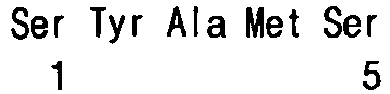
- <210> 10
<211> 5
<212> PRT
<213> Artificial Sequence - <220>
<223> T2-8F VH CDR 1 - <400> 10

- <210> 11
<211> 5
<212> PRT
<213> Artificial Sequence - <220>
<223> T2-12C VH CDR 1 - <400> 11

- <210> 12
<211> 5
<212> PRT
<213> Artificial Sequence - <220>
<223> T2-3A VH CDR 1 - <400> 12

- <210> 13
<211> 6
<212> PRT
<213> Artificial Sequence - <220>
<223> T2-7B VH CDR 1 - <400> 13

- <210> 14
<211> 5
<212> PRT
<213> Artificial Sequence - <220>
<223> T2-10E VH CDR 1 - <400> 14

- <210> 15
<211> 5
<212> PRT
<213> Artificial Sequence - <220>
<223> T2-6C VH CDR 1 - <400> 15

- <210> 16
<211> 5
<212> PRT
<213> Artificial Sequence - <220>
<223> T1-5G VH CDR 1 - <400> 16

- <210> 17
<211> 5
<212> PRT
<213> Artificial Sequence - <220>
<223> T2-6A VH CDR 1 - <400> 17

- <210> 18
<211> 5
<212> PRT
<213> Artificial Sequence - <220>
<223> T2-12A VH CDR 1 - <400> 18

- <210> 19
<211> 17
<212> PRT
<213> Artificial Sequence - <220>
<223> T1-11G VH CDR 2 - <400> 19

- <210> 20
<211> 17
<212> PRT
<213> Artificial Sequence - <220>
<223> T1-12C VH CDR 2 - <400> 20

- <210> 21
<211> 17
<212> PRT
<213> Artificial Sequence - <220>
<223> T1-9F VH COR 2 - <400> 21

- <210> 22
<211> 17
<212> PRT
<213> Artificial Sequence - <220>
<223> T2-8F VH CDR 2 - <400> 22

- <210> 23
<211> 17
<212> PRT
<213> Artificial Sequence - <220>
<223> T2-12C VH CDR 2 - <400> 23

- <210> 24
<211> 17
<212> PRT
<213> Artificial Sequence - <220>
<223> T2-3A VH CDR 2 - <400> 24

- <210> 25
<211> 18
<212> PRT
<213> Artificial Sequence - <220>
<223> T2-7B VH CDR 2 - <400> 25

- <210> 26
<211> 17
<212> PRT
<213> Artificial Sequence - <220>
<223> T2-6G VH CDR 2 - <400> 26

- <210> 27
<211> 17
<212> PRT
<213> Artificial Sequence - <220>
<223> T2-10E VH CDR 2 - <400> 27

- <210> 28
<211> 17
<212> PRT
<213> Artificial Sequence - <220>
<223> T2-6C VH CDR 2 - <400> 28

- <210> 29
<211> 17
<212> PRT
<213> Artificial Sequence - <220>
<223> T1-5G VH CDR 2 - <400> 29

- <210> 30
<211> 17
<212> PRT
<213> Artificial Sequence - <220>
<223> T2-6A VH CDR 2 - <400> 30

- <210> 31
<211> 17
<212> PRT
<213> Artificial Sequence - <220>
<223> T2-12A VH CDR 2 - <400> 31

- <210> 32
<211> 10
<212> PRT
<213> Artificial Sequence - <220>
<223> T1-116 VH CDR 3 - <400> 32

- <210> 33
<211> 6
<212> PRT
<213> Artificial Sequence - <220>
<223> T1-12C VH CDR 3 - <400> 33

- <210> 34
<211> 6
<212> PRT
<213> Artificial Sequence - <220>
<223> T1-9F VH CDR 3 - <400> 34

- <210> 35
<211> 7
<212> PRT
<213> Artificial Sequence - <220>
<223> T2-8F VH CDR 3 - <400> 35

- <210> 36
<211> 12
<212> PRT
<213> Artificial Sequence - <220>
<223> T2-12C VH CDR 3 - <400> 36

- <210> 37
<211> 9
<212> PRT
<213> Artificial Sequence - <220>
<223> T2-3A VH CDR 3 - <400> 37

- <210> 38
<211> 8
<212> PRT
<213> Artificial Sequence - <220>
<223> T2-7B VH CDR 3 - <400> 38

- <210> 39
<211> 6
<212> PRT
<213> Artificial Sequence - <220>
<223> T2-6G VH CDR 3 - <400> 39

- <210> 40
<211> 16
<212> PRT
<213> Artificial Sequence - <220>
<223> T2-10E VH CDR 3 - <400> 40

- <210> 41
<211> 9
<212> PRT
<213> Artificial Sequence - <220>
<223> T2-6C VH CDR 3 - <400> 41

- <210> 42
<211> 18
<212> PRT
<213> Artificial Sequence - <220>
<223> T1-5G VH CDR 3 - <400> 42

- <210> 43
<211> 8
<212> PRT
<213> Artificial Sequence - <220>
<223> T2-6A VH CDR 3 - <400> 43

- <210> 44
<211> 14
<212> PRT
<213> Artificial Sequence - <220>
<223> T2-12A VH CDR 3 - <400> 44

- <210> 45
<211> 139
<212> PRT
<213> Artificial Sequence - <220>
<223> T1-11G heavy chain - <400> 45

- <210> 46
<211> 137
<212> PRT
<213> Artificial Sequence - <220>
<223> T2- 6A heavy chain - <400> 46


- <210> 47
<211> 135
<212> PRT
<213> Artificial Sequence - <220>
<223> T1-12C heavy chain - <400> 47

- <210> 48
<211> 135
<212> PRT
<213> Artificial Sequence - <220>
<223> T1- 9F heavy chain - <400> 48
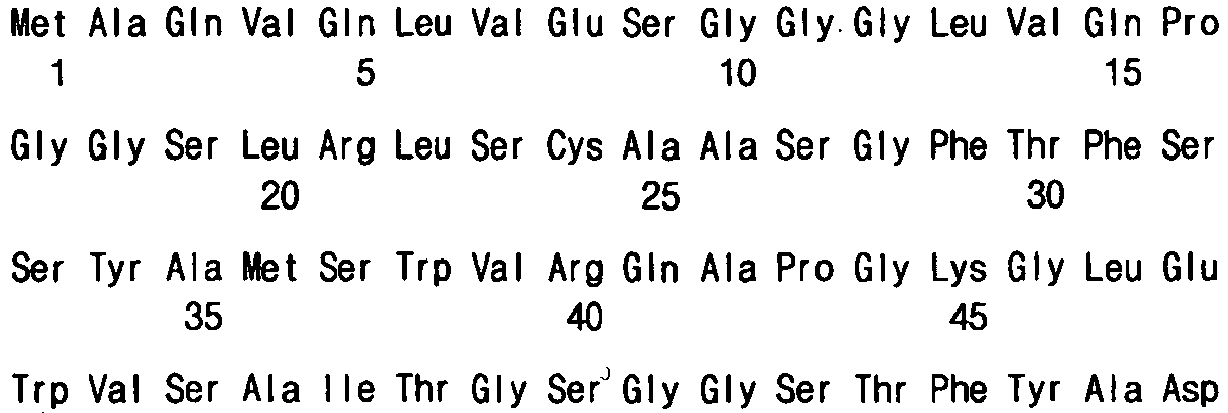

- <210> 49
<211> 135
<212> PRT
<213> Artificial Sequence - <220>
<223> T2-12F heavy chain - <400> 49

- <210> 50
<211> 136
<212> PRT
<213> Artificial Sequence - <220>
<223> T2- 8F heavy chain - <400> 50

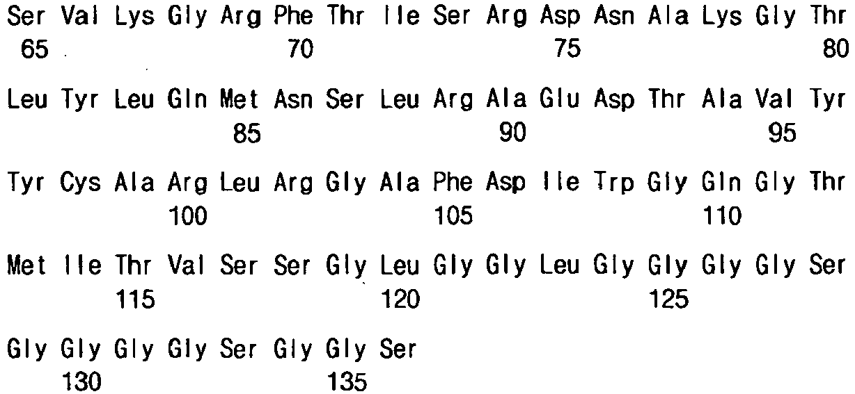
- <210> 51
<211> 143
<212> PRT
<213> Artificial Sequence - <220>
<223> T2-12A heavy chain - <400> 51

- <210> 52
<211> 141
<212> PRT
<213> Artificial Sequence - <220>
<223> T2-12C heavy chain - <400> 52


- <210> 53
<211> 138
<212> PRT
<213> Artificial Sequence - <220>
<223> T2- 3A heavy chain - <400> 53

- <210> 54
<211> 140
<212> PRT
<213> Artificial Sequence - <220>
<223> T2- 7B heavy chain - <400> 54


- <210> 55
<211> 135
<212> PRT
<213> Artificial Sequence - <220>
<223> T2- 6G heavy chain - <400> 55

- <210> 56
<211> 145
<212> PRT
<213> Artificial Sequence - <220>
<223> T2-10E heavy chain - <400> 56


- <210> 57
<211> 138
<212> PRT
<213> Artificial Sequence - <220>
<223> T2- 6C heavy chain - <400> 57

- <210> 58
<211> 11
<212> PRT
<213> Artificial Sequence - <220>
<223> T1-11G VL CDR 1 - <400> 58

- <210> 59
<211> 16
<212> PRT
<213> Artificial Sequence - <220>
<223> T1-12C VL CDR 1 - <400> 59


- <210> 60
<211> 16
<212> PRT
<213> Artificial Sequence - <220>
<223> T1-9F VL CDR 1 - <400> 60

- <210> 61
<211> 16
<212> PRT
<213> Artificial Sequence - <220>
<223> T2-8F VL CDR 1 - <400> 61

- <210> 62
<211> 14
<212> PRT
<213> Artificial Sequence - <220>
<223> T2-12C VL CDR 1 - <400> 62

- <210> 63
<211> 14
<212> PRT
<213> Artificial Sequence - <220>
<223> T2-3A VL CDR 1 - <400> 63

- <210> 64
<211> 14
<212> PRT
<213> Artificial Sequence - <220>
<223> T2-7B VL CDR 1 - <400> 64

- <210> 65
<211> 13
<212> PRT
<213> Artificial Sequence - <220>
<223> T2-6G VL CDR 1 - <400> 65

- <210> 66
<211> 11
<212> PRT
<213> Artificial Sequence - <220>
<223> T2-10E VL CDR 1 - <400> 66

- <210> 67
<211> 11
<212> PRT
<213> Artificial Sequence - <220>
<223> T2-6C VL CDR 1 - <400> 67

- <210> 68
<211> 11
<212> PRT
<213> Artificial Sequence - <220>
<223> T1-5G VL CDR 1 - <400> 68

- <210> 69
<211> 11
<212> PRT
<213> Artificial Sequence - <220>
<223> T2-6A VL CDR 1 - <400> 69

- <210> 70
<211> 11
<212> PRT
<213> Artificial Sequence - <220>
<223> T2-12A VL CDR 1 - <400> 70

- <210> 71
<211> 7
<212> PRT
<213> Artificial Sequence - <220>
<223> T1-11G VL CDR 2 - <400> 71

- <210> 72
<211> 7
<212> PRT
<213> Artificial Sequence - <220>
<223> T1-12C VL CDR 2 - <400> 72

- <210> 73
<211> 7
<212> PRT
<213> Artificial Sequence - <220>
<223> T1-9F VL CDR 2 - <400> 73

- <210> 74
<211> 7
<212> PRT
<213> Artificial Sequence - <220>
<223> T2-8F VL CDR 2 - <400> 74

- <210> 75
<211> 7
<212> PRT
<213> Artificial Sequence - <220>
<223> T2-12C VL CDR 2 - <400> 75

- <210> 76
<211> 7
<212> PRT
<213> Artificial Sequence - <220>
<223> T2-3A VL CDR 2 - <400> 76

- <210> 77
<211> 7
<212> PRT
<213> Artificial Sequence - <220>
<223> T2-7B VL CDR 2 - <400> 77

- <210> 78
<211> 7
<212> PRT
<213> Artificial Sequence - <220>
<223> T2-6G VL CDR 2 - <400> 78

- <210> 79
<211> 7
<212> PRT
<213> Artificial Sequence - <220>
<223> T2-10E VL CDR 2 - <400> 79

- <210> 80
<211> 7
<212> PRT
<213> Artificial Sequence - <220>
<223> T2-6C VL CDR 2 - <400> 80

- <210> 81
<211> 7
<212> PRT
<213> Artificial Sequence - <220>
<223> T1-5G VL CDR 2 - <400> 81

- <210> 82
<211> 7
<212> PRT
<213> Artificial Sequence - <220>
<223> T2-6A VL CDR 2 - <400> 82

- <210> 83
<211> 7
<212> PRT
<213> Artificial Sequence - <220>
<223> T2-12A VL CDR 2 - <400> 83

- <210> 84
<211> 9
<212> PRT
<213> Artificial Sequence - <220>
<223> T1-11G VL CDR 3 - <400> 84

- <210> 85
<211> 9
<212> PRT
<213> Artificial Sequence - <220>
<223> T1-12C VL CDR 3 - <400> 85

- <210> 86
<211> 9
<212> PRT
<213> Artificial Sequence - <220>
<223> T1-9F VL CDR 3 - <400> 86

- <210> 87
<211> 8
<212> PRT
<213> Artificial Sequence - <220>
<223> T2-8F VL CDR 3 - <400> 87

- <210> 88
<211> 9
<212> PRT
<213> Artificial Sequence - <220>
<223> T2-12C VL CDR 3 - <400> 88

- <210> 89
<211> 9
<212> PRT
<213> Artificial Sequence - <220>
<223> T2-3A VL CDR 3 - <400> 89

- <210> 90
<211> 10
<212> PRT
<213> Artificial Sequence - <220>
<223> T2-7B VL CDR 3 - <400> 90

- <210> 91
<211> 13
<212> PRT
<213> Artificial Sequence - <220>
<223> T2-6G VL CDR 3 - <400> 91

- <210> 92
<211> 9
<212> PRT
<213> Artificial Sequence - <220>
<223> T2-10E VL CDR 3 - <400> 92

- <210> 93
<211> 9
<212> PRT
<213> Artificial Sequence - <220>
<223> T2-6C VL CDR 3 - <400> 93

- <210> 94
<211> 9
<212> PRT
<213> Artificial Sequence - <220>
<223> T1-5G VL CDR 3 - <400> 94

- <210> 95
<211> 9
<212> PRT
<213> Artificial Sequence - <220>
<223> T2-6A VL CDR 3 - <400> 95

- <210> 96
<211> 11
<212> PRT
<213> Artificial Sequence - <220>
<223> T2-12A VL CDR 3 - <400> 96

- <210> 97
<211> 131
<212> PRT
<213> Artificial Sequence - <220>
<223> T1-11G light chain - <400> 97


- <210> 98
<211> 131
<212> PRT
<213> Artificial Sequence - <220>
<223> T2- 6A light chain - <400> 98

- <210> 99
<211> 136
<212> PRT
<213> Artificial Sequence - <220>
<223> T1-12C light chain - <400> 99


- <210> 100
<211> 136
<212> PRT
<213> Artificial Sequence - <220>
<223> T1- 9F light chain - <400> 100

- <210> 101
<211> 136
<212> PRT
<213> Artificial Sequence - <220>
<223> T2-12F light chain - <400> 101


- <210> 102
<211> 135
<212> PRT
<213> Artificial Sequence - <220>
<223> T2- 8F light chain - <400> 102

- <210> 103
<211> 132
<212> PRT
<213> Artificial Sequence - <220>
<223> T2-12A light chain - <400> 103


- <210> 104
<211> 133
<212> PRT
<213> Artificial Sequence - <220>
<223> T2-12C light chain - <400> 104

- <210> 105
<211> 133
<212> PRT
<213> Artificial Sequence - <220>
<223> T2- 3A light chain - <400> 105

- <210> 106
<211> 134
<212> PRT
<213> Artificial Sequence - <220>
<223> T2- 7B light chain - <400> 106
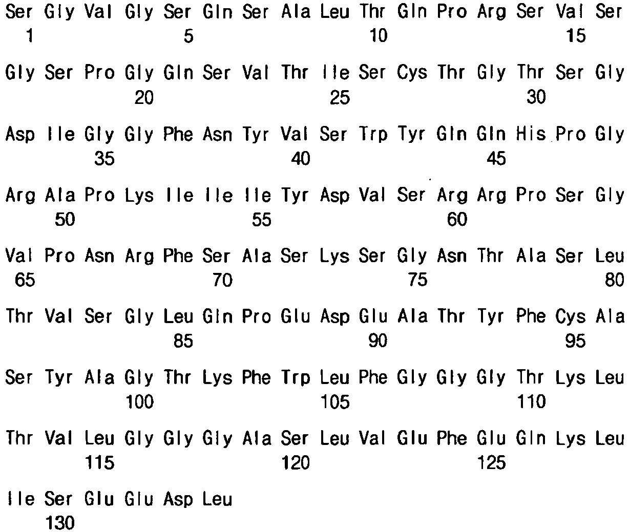
- <210> 107
<211> 136
<212> PRT
<213> Artificial Sequence - <220>
<223> T2- 6G light chain - <400> 107

- <210> 108
<211> 131
<212> PRT
<213> Artificial Sequence - <220>
<223> T2-10E light chain - <400> 108

- <210> 109
<211> 131
<212> PRT
<213> Artificial Sequence - <220>
<223> T2- 6C light chain - <400> 109

- <210> 110
<211> 50
<212> DNA
<213> Artificial Sequence - <220>
<223> NATVH1-2 - <400> 110
ttggtggcca cagcggccga tgtccactcg cagatgcagc tggtgcagtc 50 - <210> 111
<211> 50
<212> DNA
<213> Artificial Sequence - <220>
<223> NATVH1-2 - <400> 111
ttggtggcca cagcggccga tgtccactcg cagatgcagc tggtgcagtc 50 - <210> 112
<211> 50
<212> DNA
<213> Artificial Sequence - <220>
<223> NATVH3-2 - <400> 112
ttggtggcca cagcggccga tgtccactcg caggtgcagc tggtggagtc 50 - <210> 113
<211> 50
<212> DNA
<213> Artificial Sequence - <220>
<223> NATVH3-2 - <400> 113
ttggtggcca cagcggccga tgtccactcg caggtgcagc tggtggagtc 50 - <210> 114
<211> 26
<212> DNA
<213> Artificial Sequence - <220>
<223> NATJH-ALL - <400> 114
gaggaggcta gctgaggaga cggtga 26 - <210> 115
<211> 50
<212> DNA
<213> Artificial Sequence - <220>
<223> NATVL4 - <400> 115
ttggtggcca cagcggccga tgtccactcg cagtctgccc tgactcagcc 50 - <210> 116
<211> 50
<212> DNA
<213> Artificial Sequence - <220>
<223> NATVK1-1 - <400> 116
ttggtggcca cagcggccga tgtccactcg gacatccaga tgacccagtc 50 - <210> 117
<211> 50
<212> DNA
<213> Artificial Sequence - <220>
<223> NATVL4 - <400> 117
ttggtggcca cagcggccga tgtccactcg cagtctgccc tgactcagcc 50 - <210> 118
<211> 53
<212> DNA
<213> Artificial Sequence - <220>
<223> NATVK3 - <400> 118
ttggtggcca cagcggccga tgtccactcg gatattgtga tgacccagac tcc 53 - <210> 119
<211> 33
<212> DNA
<213> Artificial Sequence - <220>
<223> NATJL1-R - <400> 119
gaggagagat cttaggacgg tgaccttggtccc 33 - <210> 120
<211> 30
<212> DNA
<213> Artificial Sequence - <220>
<223> NATJK-R5 - <400> 120
gaggagagat cttttgattt ccagcttggt 30 - <210> 121
<211> 33
<212> DNA
<213> Artificial Sequence - <220>
<223> NATJL2-R - <400> 121
gaggagagat cttaggacgg tcagcttggtccc 33
Claims (9)
- A TMPRSS4-specific human antibody selected from the group consisting of
an antibody comprising a heavy chain comprising a heavy chain variable region of an amino acid sequence of SEQ ID No. 57; and a light chain comprising a light chain variable region of an amino acid sequence of SEQ ID No. 109. - The human antibody of claim 1 for use in the prevention or treatment of a TMPRSS4-overexpressed cancer.
- The human antibody for use as set forth in claim 2, wherein the TMPRSS4-overexpressed cancer is selected from the group consisting of colorectal cancer, lung cancer, liver cancer, pancreatic cancer, gastric cancer, and malignant thyroid neoplasms.
- A composition comprising the human antibody of claim 1 and a radioactive isotope for use in the radioimmuno treatment or in vivo detection of a TMPRSS4-overexpressed cancer.
- The composition as set forth in claim 4, wherein the TMPRSS4-overexpressed cancer is selected from the group consisting of colorectal cancer, lung cancer, liver cancer, pancreatic cancer, gastric cancer, and malignant thyroid neoplasms.
- The composition as set forth in claim 4, wherein the radioactive isotope is selected from the group consisting of 3H, 11C, 14C, 18F, 64Cu, 76Br, 86Y, 99mTc, 111In, 123I, 177Lu, and a mixture or combination thereof.
- The composition as set forth in any one of claims 4 to 6, wherein the radioactive isotope is bound to a human antibody or included in a carrier to which the human antibody is bound.
- An in vitro immunodetection method for detecting a TMPRSS4-overexpressed cancer, the method comprising contacting the human antibody of claim 1 and a radioactive isotope with a cancer cell in vitro.
- The method as set forth in claim 8, wherein the TMPRSS4-overexpressed cancer is selected from the group consisting of colorectal cancer, lung cancer, liver cancer, pancreatic cancer, gastric cancer, and malignant thyroid neoplasms
Applications Claiming Priority (2)
| Application Number | Priority Date | Filing Date | Title |
|---|---|---|---|
| KR1020080101575A KR101039751B1 (en) | 2008-10-16 | 2008-10-16 | TPMPRS4-specific human antibodies |
| PCT/KR2008/006614 WO2010044506A2 (en) | 2008-10-16 | 2008-11-10 | Tmprss4-specific human antibody |
Publications (3)
| Publication Number | Publication Date |
|---|---|
| EP2344533A2 EP2344533A2 (en) | 2011-07-20 |
| EP2344533A4 EP2344533A4 (en) | 2013-06-19 |
| EP2344533B1 true EP2344533B1 (en) | 2015-07-22 |
Family
ID=42106995
Family Applications (1)
| Application Number | Title | Priority Date | Filing Date |
|---|---|---|---|
| EP08877439.3A Not-in-force EP2344533B1 (en) | 2008-10-16 | 2008-11-10 | Tmprss4-specific human antibody |
Country Status (5)
| Country | Link |
|---|---|
| US (1) | US8501175B2 (en) |
| EP (1) | EP2344533B1 (en) |
| JP (1) | JP5484476B2 (en) |
| KR (1) | KR101039751B1 (en) |
| WO (1) | WO2010044506A2 (en) |
Families Citing this family (7)
| Publication number | Priority date | Publication date | Assignee | Title |
|---|---|---|---|---|
| CN101277974A (en) | 2005-09-30 | 2008-10-01 | 阿伯特有限及两合公司 | Binding domains of proteins of the Repulsive Guide Molecule (RGM) protein family, functional fragments thereof and their uses |
| AU2008317724B2 (en) | 2007-11-02 | 2011-06-02 | Novartis Ag | Improved Nogo-A binding molecules and pharmaceutical use thereof |
| US8962803B2 (en) | 2008-02-29 | 2015-02-24 | AbbVie Deutschland GmbH & Co. KG | Antibodies against the RGM A protein and uses thereof |
| KR20120118002A (en) | 2009-12-08 | 2012-10-25 | 애보트 게엠베하 운트 콤파니 카게 | Monoclonal antibodies against the rgm a protein for use in the treatment of retinal nerve fiber layer degeneration |
| KR101500670B1 (en) * | 2010-11-02 | 2015-03-10 | 인하대학교 산학협력단 | Antigen peptide for suppressing metastasis, anti-metastatic vaccine including same, composition for suppressing and treating metastasis, and method for screening metastasis-related target materials and metastasis suppressant |
| EP3369746A1 (en) | 2012-01-27 | 2018-09-05 | AbbVie Deutschland GmbH & Co KG | Composition and method for diagnosis and treatment of diseases associated with neurite degeneration |
| WO2025199346A1 (en) * | 2024-03-20 | 2025-09-25 | Arsenal Biosciences, Inc. | Antigen binding proteins that bind tmprss4 and methods of use thereof |
Family Cites Families (4)
| Publication number | Priority date | Publication date | Assignee | Title |
|---|---|---|---|---|
| WO2004097034A2 (en) * | 2003-05-02 | 2004-11-11 | Bayer Healthcare Ag | Diagnostics and therapeutics for diseases associated with human transmembrane serine protease 4 (tmprss4) |
| KR100906145B1 (en) * | 2006-05-30 | 2009-07-03 | 한국생명공학연구원 | Anticancer agent containing TPMPS4 inhibitor as active ingredient |
| US8287867B2 (en) * | 2007-02-20 | 2012-10-16 | Diadexus, Inc. | Ovr115 antibody compositions and methods of use |
| US8197809B2 (en) * | 2008-06-25 | 2012-06-12 | Korea Research Institute Of Bioscience And Biotechnology | CD9-specific human antibodies |
-
2008
- 2008-10-16 KR KR1020080101575A patent/KR101039751B1/en not_active Expired - Fee Related
- 2008-11-10 EP EP08877439.3A patent/EP2344533B1/en not_active Not-in-force
- 2008-11-10 US US13/124,603 patent/US8501175B2/en not_active Expired - Fee Related
- 2008-11-10 JP JP2011532001A patent/JP5484476B2/en not_active Expired - Fee Related
- 2008-11-10 WO PCT/KR2008/006614 patent/WO2010044506A2/en not_active Ceased
Also Published As
| Publication number | Publication date |
|---|---|
| US8501175B2 (en) | 2013-08-06 |
| KR20100042432A (en) | 2010-04-26 |
| WO2010044506A3 (en) | 2010-07-15 |
| JP2012505650A (en) | 2012-03-08 |
| KR101039751B1 (en) | 2011-06-09 |
| EP2344533A4 (en) | 2013-06-19 |
| EP2344533A2 (en) | 2011-07-20 |
| US20110189087A1 (en) | 2011-08-04 |
| WO2010044506A2 (en) | 2010-04-22 |
| JP5484476B2 (en) | 2014-05-07 |
Similar Documents
| Publication | Publication Date | Title |
|---|---|---|
| JP5289285B2 (en) | VEGF-specific human antibodies | |
| CN109096395B (en) | Blocking type CD47 nano antibody and application thereof | |
| KR101227971B1 (en) | CD9―specific human antibody | |
| EP2385955B1 (en) | Modified antibody compositions, methods of making and using thereof | |
| KR101559599B1 (en) | Anti-mesothelin antibodies and uses therefor | |
| US8501175B2 (en) | TMPRSS4-specific human antibody | |
| KR100910962B1 (en) | ENO1-specific human antibodies | |
| EP4635981A1 (en) | Anti-cll1 single-domain antibody and use thereof | |
| CN111825767B (en) | Single-domain antibody of human epidermal growth factor receptor 2, detection kit and application thereof | |
| EP2620449A1 (en) | Anti-ephrin-b2 antibody and use thereof | |
| WO2022134276A1 (en) | Anti-human osteopontin antibody and use thereof | |
| KR101473328B1 (en) | Cytokaratin 17-specific human antibody | |
| EP4324848A1 (en) | Human antibody targeting covid-19 virus | |
| CN113788896A (en) | Anti-MSLN monoclonal internalizing antibody and preparation method and application thereof | |
| CN112079928B (en) | anti-PD-L1 monoclonal antibody | |
| JP7453694B2 (en) | An antibody that specifically binds to the N-terminal region of lysyl-tRNA synthetase exposed on the extracellular membrane | |
| WO2026057055A1 (en) | Psca-targeting nanobody and use thereof | |
| KR101169336B1 (en) | Derivative of CD9-specific human antibody | |
| HK40131721A (en) | Anti-cll1 single-domain antibody and use thereof | |
| CN120271712A (en) | Nectin-4 targeted nanobody and application thereof | |
| CN120271711A (en) | Nectin-4 targeting antibodies and uses thereof | |
| JP2026509914A (en) | Combotope Antibody Library | |
| HK1164341B (en) | Modified antibody compositions, methods of making and using thereof | |
| KR20080068108A (en) | Identification and Characterization of Action-Blocking Anti-ED-IX-Fibronectin Antibodies |
Legal Events
| Date | Code | Title | Description |
|---|---|---|---|
| PUAI | Public reference made under article 153(3) epc to a published international application that has entered the european phase |
Free format text: ORIGINAL CODE: 0009012 |
|
| 17P | Request for examination filed |
Effective date: 20110510 |
|
| AK | Designated contracting states |
Kind code of ref document: A2 Designated state(s): AT BE BG CH CY CZ DE DK EE ES FI FR GB GR HR HU IE IS IT LI LT LU LV MC MT NL NO PL PT RO SE SI SK TR |
|
| AX | Request for extension of the european patent |
Extension state: AL BA MK RS |
|
| DAX | Request for extension of the european patent (deleted) | ||
| A4 | Supplementary search report drawn up and despatched |
Effective date: 20130522 |
|
| RIC1 | Information provided on ipc code assigned before grant |
Ipc: C07K 16/18 20060101AFI20130515BHEP |
|
| 17Q | First examination report despatched |
Effective date: 20140509 |
|
| GRAP | Despatch of communication of intention to grant a patent |
Free format text: ORIGINAL CODE: EPIDOSNIGR1 |
|
| INTG | Intention to grant announced |
Effective date: 20150205 |
|
| GRAS | Grant fee paid |
Free format text: ORIGINAL CODE: EPIDOSNIGR3 |
|
| GRAA | (expected) grant |
Free format text: ORIGINAL CODE: 0009210 |
|
| AK | Designated contracting states |
Kind code of ref document: B1 Designated state(s): AT BE BG CH CY CZ DE DK EE ES FI FR GB GR HR HU IE IS IT LI LT LU LV MC MT NL NO PL PT RO SE SI SK TR |
|
| REG | Reference to a national code |
Ref country code: GB Ref legal event code: FG4D |
|
| REG | Reference to a national code |
Ref country code: CH Ref legal event code: EP |
|
| REG | Reference to a national code |
Ref country code: IE Ref legal event code: FG4D |
|
| REG | Reference to a national code |
Ref country code: AT Ref legal event code: REF Ref document number: 737859 Country of ref document: AT Kind code of ref document: T Effective date: 20150815 |
|
| REG | Reference to a national code |
Ref country code: DE Ref legal event code: R096 Ref document number: 602008039184 Country of ref document: DE |
|
| REG | Reference to a national code |
Ref country code: FR Ref legal event code: PLFP Year of fee payment: 8 |
|
| REG | Reference to a national code |
Ref country code: AT Ref legal event code: MK05 Ref document number: 737859 Country of ref document: AT Kind code of ref document: T Effective date: 20150722 |
|
| REG | Reference to a national code |
Ref country code: LT Ref legal event code: MG4D |
|
| REG | Reference to a national code |
Ref country code: NL Ref legal event code: MP Effective date: 20150722 |
|
| PG25 | Lapsed in a contracting state [announced via postgrant information from national office to epo] |
Ref country code: GR Free format text: LAPSE BECAUSE OF FAILURE TO SUBMIT A TRANSLATION OF THE DESCRIPTION OR TO PAY THE FEE WITHIN THE PRESCRIBED TIME-LIMIT Effective date: 20151023 Ref country code: LV Free format text: LAPSE BECAUSE OF FAILURE TO SUBMIT A TRANSLATION OF THE DESCRIPTION OR TO PAY THE FEE WITHIN THE PRESCRIBED TIME-LIMIT Effective date: 20150722 Ref country code: LT Free format text: LAPSE BECAUSE OF FAILURE TO SUBMIT A TRANSLATION OF THE DESCRIPTION OR TO PAY THE FEE WITHIN THE PRESCRIBED TIME-LIMIT Effective date: 20150722 Ref country code: FI Free format text: LAPSE BECAUSE OF FAILURE TO SUBMIT A TRANSLATION OF THE DESCRIPTION OR TO PAY THE FEE WITHIN THE PRESCRIBED TIME-LIMIT Effective date: 20150722 Ref country code: NO Free format text: LAPSE BECAUSE OF FAILURE TO SUBMIT A TRANSLATION OF THE DESCRIPTION OR TO PAY THE FEE WITHIN THE PRESCRIBED TIME-LIMIT Effective date: 20151022 |
|
| PG25 | Lapsed in a contracting state [announced via postgrant information from national office to epo] |
Ref country code: SE Free format text: LAPSE BECAUSE OF FAILURE TO SUBMIT A TRANSLATION OF THE DESCRIPTION OR TO PAY THE FEE WITHIN THE PRESCRIBED TIME-LIMIT Effective date: 20150722 Ref country code: PL Free format text: LAPSE BECAUSE OF FAILURE TO SUBMIT A TRANSLATION OF THE DESCRIPTION OR TO PAY THE FEE WITHIN THE PRESCRIBED TIME-LIMIT Effective date: 20150722 Ref country code: HR Free format text: LAPSE BECAUSE OF FAILURE TO SUBMIT A TRANSLATION OF THE DESCRIPTION OR TO PAY THE FEE WITHIN THE PRESCRIBED TIME-LIMIT Effective date: 20150722 Ref country code: IS Free format text: LAPSE BECAUSE OF FAILURE TO SUBMIT A TRANSLATION OF THE DESCRIPTION OR TO PAY THE FEE WITHIN THE PRESCRIBED TIME-LIMIT Effective date: 20151122 Ref country code: PT Free format text: LAPSE BECAUSE OF FAILURE TO SUBMIT A TRANSLATION OF THE DESCRIPTION OR TO PAY THE FEE WITHIN THE PRESCRIBED TIME-LIMIT Effective date: 20151123 Ref country code: ES Free format text: LAPSE BECAUSE OF FAILURE TO SUBMIT A TRANSLATION OF THE DESCRIPTION OR TO PAY THE FEE WITHIN THE PRESCRIBED TIME-LIMIT Effective date: 20150722 Ref country code: AT Free format text: LAPSE BECAUSE OF FAILURE TO SUBMIT A TRANSLATION OF THE DESCRIPTION OR TO PAY THE FEE WITHIN THE PRESCRIBED TIME-LIMIT Effective date: 20150722 |
|
| REG | Reference to a national code |
Ref country code: DE Ref legal event code: R097 Ref document number: 602008039184 Country of ref document: DE |
|
| PG25 | Lapsed in a contracting state [announced via postgrant information from national office to epo] |
Ref country code: CZ Free format text: LAPSE BECAUSE OF FAILURE TO SUBMIT A TRANSLATION OF THE DESCRIPTION OR TO PAY THE FEE WITHIN THE PRESCRIBED TIME-LIMIT Effective date: 20150722 Ref country code: IT Free format text: LAPSE BECAUSE OF FAILURE TO SUBMIT A TRANSLATION OF THE DESCRIPTION OR TO PAY THE FEE WITHIN THE PRESCRIBED TIME-LIMIT Effective date: 20150722 Ref country code: DK Free format text: LAPSE BECAUSE OF FAILURE TO SUBMIT A TRANSLATION OF THE DESCRIPTION OR TO PAY THE FEE WITHIN THE PRESCRIBED TIME-LIMIT Effective date: 20150722 Ref country code: SK Free format text: LAPSE BECAUSE OF FAILURE TO SUBMIT A TRANSLATION OF THE DESCRIPTION OR TO PAY THE FEE WITHIN THE PRESCRIBED TIME-LIMIT Effective date: 20150722 Ref country code: EE Free format text: LAPSE BECAUSE OF FAILURE TO SUBMIT A TRANSLATION OF THE DESCRIPTION OR TO PAY THE FEE WITHIN THE PRESCRIBED TIME-LIMIT Effective date: 20150722 |
|
| PLBE | No opposition filed within time limit |
Free format text: ORIGINAL CODE: 0009261 |
|
| STAA | Information on the status of an ep patent application or granted ep patent |
Free format text: STATUS: NO OPPOSITION FILED WITHIN TIME LIMIT |
|
| PG25 | Lapsed in a contracting state [announced via postgrant information from national office to epo] |
Ref country code: RO Free format text: LAPSE BECAUSE OF FAILURE TO SUBMIT A TRANSLATION OF THE DESCRIPTION OR TO PAY THE FEE WITHIN THE PRESCRIBED TIME-LIMIT Effective date: 20150722 |
|
| REG | Reference to a national code |
Ref country code: DE Ref legal event code: R119 Ref document number: 602008039184 Country of ref document: DE |
|
| 26N | No opposition filed |
Effective date: 20160425 |
|
| PG25 | Lapsed in a contracting state [announced via postgrant information from national office to epo] |
Ref country code: MC Free format text: LAPSE BECAUSE OF FAILURE TO SUBMIT A TRANSLATION OF THE DESCRIPTION OR TO PAY THE FEE WITHIN THE PRESCRIBED TIME-LIMIT Effective date: 20150722 Ref country code: LU Free format text: LAPSE BECAUSE OF FAILURE TO SUBMIT A TRANSLATION OF THE DESCRIPTION OR TO PAY THE FEE WITHIN THE PRESCRIBED TIME-LIMIT Effective date: 20151110 |
|
| REG | Reference to a national code |
Ref country code: CH Ref legal event code: PL |
|
| PG25 | Lapsed in a contracting state [announced via postgrant information from national office to epo] |
Ref country code: LI Free format text: LAPSE BECAUSE OF NON-PAYMENT OF DUE FEES Effective date: 20151130 Ref country code: CH Free format text: LAPSE BECAUSE OF NON-PAYMENT OF DUE FEES Effective date: 20151130 |
|
| REG | Reference to a national code |
Ref country code: IE Ref legal event code: MM4A |
|
| PG25 | Lapsed in a contracting state [announced via postgrant information from national office to epo] |
Ref country code: SI Free format text: LAPSE BECAUSE OF FAILURE TO SUBMIT A TRANSLATION OF THE DESCRIPTION OR TO PAY THE FEE WITHIN THE PRESCRIBED TIME-LIMIT Effective date: 20150722 |
|
| PG25 | Lapsed in a contracting state [announced via postgrant information from national office to epo] |
Ref country code: IE Free format text: LAPSE BECAUSE OF NON-PAYMENT OF DUE FEES Effective date: 20151110 Ref country code: DE Free format text: LAPSE BECAUSE OF NON-PAYMENT OF DUE FEES Effective date: 20160601 |
|
| REG | Reference to a national code |
Ref country code: FR Ref legal event code: PLFP Year of fee payment: 9 |
|
| PG25 | Lapsed in a contracting state [announced via postgrant information from national office to epo] |
Ref country code: BE Free format text: LAPSE BECAUSE OF FAILURE TO SUBMIT A TRANSLATION OF THE DESCRIPTION OR TO PAY THE FEE WITHIN THE PRESCRIBED TIME-LIMIT Effective date: 20150722 |
|
| PG25 | Lapsed in a contracting state [announced via postgrant information from national office to epo] |
Ref country code: HU Free format text: LAPSE BECAUSE OF FAILURE TO SUBMIT A TRANSLATION OF THE DESCRIPTION OR TO PAY THE FEE WITHIN THE PRESCRIBED TIME-LIMIT; INVALID AB INITIO Effective date: 20081110 Ref country code: BG Free format text: LAPSE BECAUSE OF FAILURE TO SUBMIT A TRANSLATION OF THE DESCRIPTION OR TO PAY THE FEE WITHIN THE PRESCRIBED TIME-LIMIT Effective date: 20150722 |
|
| PG25 | Lapsed in a contracting state [announced via postgrant information from national office to epo] |
Ref country code: NL Free format text: LAPSE BECAUSE OF FAILURE TO SUBMIT A TRANSLATION OF THE DESCRIPTION OR TO PAY THE FEE WITHIN THE PRESCRIBED TIME-LIMIT Effective date: 20150722 Ref country code: CY Free format text: LAPSE BECAUSE OF FAILURE TO SUBMIT A TRANSLATION OF THE DESCRIPTION OR TO PAY THE FEE WITHIN THE PRESCRIBED TIME-LIMIT Effective date: 20150722 |
|
| PG25 | Lapsed in a contracting state [announced via postgrant information from national office to epo] |
Ref country code: TR Free format text: LAPSE BECAUSE OF FAILURE TO SUBMIT A TRANSLATION OF THE DESCRIPTION OR TO PAY THE FEE WITHIN THE PRESCRIBED TIME-LIMIT Effective date: 20150722 Ref country code: MT Free format text: LAPSE BECAUSE OF FAILURE TO SUBMIT A TRANSLATION OF THE DESCRIPTION OR TO PAY THE FEE WITHIN THE PRESCRIBED TIME-LIMIT Effective date: 20150722 |
|
| REG | Reference to a national code |
Ref country code: FR Ref legal event code: PLFP Year of fee payment: 10 |
|
| REG | Reference to a national code |
Ref country code: FR Ref legal event code: PLFP Year of fee payment: 11 |
|
| PGFP | Annual fee paid to national office [announced via postgrant information from national office to epo] |
Ref country code: FR Payment date: 20210927 Year of fee payment: 14 |
|
| PGFP | Annual fee paid to national office [announced via postgrant information from national office to epo] |
Ref country code: GB Payment date: 20210927 Year of fee payment: 14 |
|
| GBPC | Gb: european patent ceased through non-payment of renewal fee |
Effective date: 20221110 |
|
| PG25 | Lapsed in a contracting state [announced via postgrant information from national office to epo] |
Ref country code: GB Free format text: LAPSE BECAUSE OF NON-PAYMENT OF DUE FEES Effective date: 20221110 |
|
| PG25 | Lapsed in a contracting state [announced via postgrant information from national office to epo] |
Ref country code: FR Free format text: LAPSE BECAUSE OF NON-PAYMENT OF DUE FEES Effective date: 20221130 |