EP2332971B1 - Epitope analogs - Google Patents
Epitope analogs Download PDFInfo
- Publication number
- EP2332971B1 EP2332971B1 EP10180780.8A EP10180780A EP2332971B1 EP 2332971 B1 EP2332971 B1 EP 2332971B1 EP 10180780 A EP10180780 A EP 10180780A EP 2332971 B1 EP2332971 B1 EP 2332971B1
- Authority
- EP
- European Patent Office
- Prior art keywords
- peptide
- analogs
- amino acid
- binding
- xaa
- Prior art date
- Legal status (The legal status is an assumption and is not a legal conclusion. Google has not performed a legal analysis and makes no representation as to the accuracy of the status listed.)
- Not-in-force
Links
Images


Classifications
-
- C—CHEMISTRY; METALLURGY
- C07—ORGANIC CHEMISTRY
- C07K—PEPTIDES
- C07K14/00—Peptides having more than 20 amino acids; Gastrins; Somatostatins; Melanotropins; Derivatives thereof
- C07K14/435—Peptides having more than 20 amino acids; Gastrins; Somatostatins; Melanotropins; Derivatives thereof from animals; from humans
-
- A—HUMAN NECESSITIES
- A61—MEDICAL OR VETERINARY SCIENCE; HYGIENE
- A61P—SPECIFIC THERAPEUTIC ACTIVITY OF CHEMICAL COMPOUNDS OR MEDICINAL PREPARATIONS
- A61P31/00—Antiinfectives, i.e. antibiotics, antiseptics, chemotherapeutics
- A61P31/12—Antivirals
-
- C—CHEMISTRY; METALLURGY
- C07—ORGANIC CHEMISTRY
- C07K—PEPTIDES
- C07K7/00—Peptides having 5 to 20 amino acids in a fully defined sequence; Derivatives thereof
- C07K7/04—Linear peptides containing only normal peptide links
- C07K7/06—Linear peptides containing only normal peptide links having 5 to 11 amino acids
-
- A—HUMAN NECESSITIES
- A61—MEDICAL OR VETERINARY SCIENCE; HYGIENE
- A61P—SPECIFIC THERAPEUTIC ACTIVITY OF CHEMICAL COMPOUNDS OR MEDICINAL PREPARATIONS
- A61P35/00—Antineoplastic agents
-
- A—HUMAN NECESSITIES
- A61—MEDICAL OR VETERINARY SCIENCE; HYGIENE
- A61P—SPECIFIC THERAPEUTIC ACTIVITY OF CHEMICAL COMPOUNDS OR MEDICINAL PREPARATIONS
- A61P35/00—Antineoplastic agents
- A61P35/02—Antineoplastic agents specific for leukemia
-
- A—HUMAN NECESSITIES
- A61—MEDICAL OR VETERINARY SCIENCE; HYGIENE
- A61P—SPECIFIC THERAPEUTIC ACTIVITY OF CHEMICAL COMPOUNDS OR MEDICINAL PREPARATIONS
- A61P37/00—Drugs for immunological or allergic disorders
- A61P37/02—Immunomodulators
- A61P37/04—Immunostimulants
-
- C—CHEMISTRY; METALLURGY
- C07—ORGANIC CHEMISTRY
- C07K—PEPTIDES
- C07K14/00—Peptides having more than 20 amino acids; Gastrins; Somatostatins; Melanotropins; Derivatives thereof
- C07K14/435—Peptides having more than 20 amino acids; Gastrins; Somatostatins; Melanotropins; Derivatives thereof from animals; from humans
- C07K14/46—Peptides having more than 20 amino acids; Gastrins; Somatostatins; Melanotropins; Derivatives thereof from animals; from humans from vertebrates
- C07K14/47—Peptides having more than 20 amino acids; Gastrins; Somatostatins; Melanotropins; Derivatives thereof from animals; from humans from vertebrates from mammals
- C07K14/4701—Peptides having more than 20 amino acids; Gastrins; Somatostatins; Melanotropins; Derivatives thereof from animals; from humans from vertebrates from mammals not used
- C07K14/4748—Tumour specific antigens; Tumour rejection antigen precursors [TRAP], e.g. MAGE
-
- A—HUMAN NECESSITIES
- A61—MEDICAL OR VETERINARY SCIENCE; HYGIENE
- A61K—PREPARATIONS FOR MEDICAL, DENTAL OR TOILETRY PURPOSES
- A61K38/00—Medicinal preparations containing peptides
Definitions
- the invention disclosed herein relates to analogs of peptides corresponding to class I MHC-restricted T cell epitopes and methods for their generation. These analogs can contain amino acid substitutions at residues that directly interact with MHC molecules, and can confer improved, modified or useful immunologic properties.
- epitope analogs from the tumor-associated antigens SSX-2, NY-ESO-1, PRAME, PSMA, tyrosinase, and melan-A are identified.
- classes of analogs, in which the various substitutions comprise the non-standard residues norleucine and/or norvaline, are disclosed.
- T lymphocytes are antigen-specific immune cells that function in response to specific antigen signals. B lymphocytes and the antibodies they produce are also antigen-specific entities. However, unlike B lymphocytes, T cells do not respond to antigens in a free or soluble form. For a T cell to respond to an antigen, it requires the antigen to be bound to a presenting complex known as the major histocompatibility complex (MHC).
- MHC major histocompatibility complex
- MHC proteins provide the means by which T cells differentiate native or "self" cells from foreign cells.
- MHC molecules are a category of immune receptors that present potential peptide epitopes to be monitored subsequently by the T cells.
- MHC There are two types of MHC, class I MHC and class II MHC.
- CD4+T cells interact with class II MHC proteins and predominately have a helper phenotype while CD8+ T cells interact with class I MHC proteins and predominately have a cytolytic phenotype, but each of them can also exhibit regulatory, particularly suppressive, function.
- Both MHC are transmembrane proteins with a majority of their structure on the external surface of the cell. Additionally, both classes of MHC have a peptide binding cleft on their external portions. It is in this cleft that small fragments of proteins, native or foreign, are bound and presented to the extracellular environment.
- APCs antigen presenting cells
- T cells can recognize an antigen, if it is presented on the MHC. This requirement is called MHC restriction. If an antigen is not displayed by a recognizable MHC, the T cell will not recognize and act on the antigen signal.
- T cells specific for the peptide bound to a recognizable MHC bind to these MHC-peptide complexes and proceed to the next stages of the immune response.
- WO 01/36453 A2 describes derivatives of the NY-ESO-1 epitope 157-165 in which the C terminal cysteine is substituted with hydrophobic amino acids containing non-polar side chains.
- WO 02/102299 A2 discloses novel immune molecules comprising a tumor-targeting recombinant antibody fragment or ligand which is fused to a single chain HLA molecule that is folded around an HLA-A2-restricted peptide which immune molecules are used to recruit cytotoxic T lymphocytes for tumor cell killing.
- WO 2004/016643 A2 discloses tumor-associated antigen peptides obtainable from different tumor-associated antigens.
- Bakker et al. (1997) International Journal of Cancer 70(3): 302-309 discloses derivatives of an epitope of the gp100 antigen which have amino acid substitutions to improve immunogenicity.
- Embodiments relate to analogs of the MHC class I-restricted T cell epitope NY-ESO-1 157-165 , SLLMWITQC (SEQ ID NO. 1), polypeptides that include these analogs that can be processed by pAPC to present the epitope analogs, and nucleic acids that express the analogs.
- the analogs can have similar or improved immunological properties compared to the wild-type epitope.
- One embodiment relates to an isolated NY-ESO-1 157-165 peptide having a sequence comprising 1 or more amino acid substitutions of the sequence SLLMWITQC (SEQ ID NO:1), in an amount sufficient to elicit cytokine production from a T cell line generated by immunization against an epitope with the sequence SLLMWITQC (SEQ ID NO:1).
- the amount sufficient can be not more than 10 uM.
- the amount can be not more than 3 uM.
- the amount can be not more than 1 uM.
- the amount is not more than 0.3 uM.
- the substitutions can include a standard amino acid.
- the substitutions can include a non-standard amino acid.
- the non-standard amino acid can be, for example, Tyr, Val, Leu, Ala, Ile, Met, Nle, Nva, Trp, Phe, Asp, Asn, Ser, Abu, and a D-stereoisomer of a standard amino acid.
- the substitutions can include a modified terminal amino acid.
- the modified terminal amino acid can be an amidated C-terminal amino acid.
- at least one of the substitutions can be the addition of an amino acid, wherein the addition is a C-terminal addition.
- One embodiment relates to an isolated peptide consisting of the sequence SILMWITQ ⁇ C, V, L, A ⁇ , FVLMWITQ ⁇ V, L, I ⁇ , Y ⁇ I, Nva, Nle ⁇ LMWITQV, or TVLMWITQV.
- Still another embodiment relates to an isolated peptide having a sequence SNvaLMWITQV.
- a further embodiment relates to a class I MHC/peptide complex wherein the peptide is any of the peptides in the embodiments described above or elsewhere herein.
- the complex can be cross-reactive with a TCR that recognizes a class I MHC/NY-ESO-1 157-165 complex.
- the complex can be an HLA-A2/NY-ESO-1 157-165 complex.
- the peptide can have affinity for a class I MHC peptide binding cleft, such as HLA-A2.
- a further embodiment relates to a polypeptide comprising the peptide sequence of any of the embodiments in association with a liberation sequence.
- a further embodiment relates to an immunogenic composition that includes any of the peptide embodiments.
- the peptide can have a sequence as set forth herein.
- a further embodiment relates to a nucleic acid encoding any of the peptide embodiments, but preferably those which do not have non-standard amino acid substitutions.
- the nucleic acid can be encoded in a vector.
- a further embodiment relates to an immunogenic composition that includes the nucleic acid encoding any of the peptide embodiments.
- a further embodiment relates to a method of inducing a CTL response by intranodal administration of any of the compositions or peptides of the embodiments.
- the method can allow for maintaining a CTL.
- the method can allow for amplifying a class I MHC-restricted T cell response.
- the method can allow for entraining a class I MHC-restricted T cell response.
- the method also can include an immunopotentiating agent.
- isolated peptides having a sequence comprising 1 to 3 or 4 amino acid substitutions in a native epitope sequence, wherein a concentration of the peptide required to elicit cytokine production from a T cell line generated by immunization against an epitope with the sequence is not more than a particular concentration, for example, 10 uM, 1 uM, 0.3 uM, and the like.
- the substitutions can include a standard amino acid, a non-standard amino acid, and the like.
- the non-standard amino acid can be any of those described herein, for example, a D-stereoisomer of a standard amino acid, Nva, or Nle.
- the substitutions can include a modified terminal amino acid, and the modified terminal amino acid can be an amidated C-terminal amino acid.
- One of the substitutions can be the addition of an amino acid, for example, the addition can be a C-terminal addition.
- peptides having an amino acid sequence that includes at least one difference from a sequence of a segment of a target-associated antigen, the segment having known or predicted affinity for the peptide binding cleft of a MHC protein, wherein the at least one difference can be a Nle or Nva residue replacing a residue at an MHC-binding motif anchor position in said segment.
- the anchor position can be a primary anchor position, for example, P2 or P ⁇ .
- the anchor position can be an auxiliary anchor position.
- the difference can include a Nle or Nva residue replacing a hydrophobic residue in said segment.
- I, L, or V can be a preferred residue in the MHC-binding motif anchor position.
- the peptide can have a length of about 8 to about 14 amino acids or more preferably a length of 9 to 10 amino acids, for example.
- the protein can be a human MHC protein, for example, class I MHC protein.
- the MHC protein can be, for example, a type such as HLA-A2, A3, A24, A30, A66, A68, A69, B7, B8, B15, B27, B35, B37, B38, B39, B40, B48, B51, B52, B53, B 60, B61, B62, B63, B67, B70, B71, B75, B77, C4, Cw1, Cw3, Cw4, Cw6, Cw7, and Cw10.
- the MHC protein can be HLA-A2 or A24.
- the MHC can have an anchor residue binding pocket, wherein the pocket is homologous to the B- or F-pocket of HLA-A*0201.
- the MHC residues responsible for forming binding pockets, and which binding pockets accommodate epitope anchor residues and thus define the binding specificity of the MHC molecule, are well understood in the art.
- One compilation of such information is found at the FIMM (Functional Immunology) web site at the hypertext transfer protocol (http://) "sdmc.lit.org.sg:8080/fimm/.” See also Schönbach C., Koh J.L.Y., Sheng X., Wong L., and V.Brusic. FIMM, a database of functional molecular immunology.
- the peptide can have at least one binding characteristic that is substantially the same as, or better than, a corresponding characteristic of said segment for said MHC.
- the binding characteristic can be elevated compared with that of said segment.
- the binding characteristic can be affinity or stability of binding for example.
- the peptide can have an immunogenicity that is substantially the same as, or better than, the immunogenicity of the segment.
- the immunogenicity can be increased.
- the immunogenicity can evoke an immune response that is cross-reactive to said segment or can evoke a CTL response.
- the immunogenicity can be assessed, for example, using an MHC-tetramer assay, a cytokine assay, a cytotoxicity assay, by measuring an immune response recognizing the peptide, by measuring an immune response recognizing said segment, using an in vitro immunizations system, or any other suitable method.
- the immunization system can include human cells.
- the immunogenicity can be assessed using an in vivo immunization system, for example, one that includes a transgenic mouse.
- the peptide can have an at least similar binding characteristic as said segment for said MHC.
- what is considered to be “similar” can be determined based upon the instant disclosure.
- “similarity” can be based upon, for example, peptide concentration for half-maximal binding, relative affinity, stability (half time of dissociation) and cross-reactivity/functional avidity.
- a peptide can be considered similar if it has results or characteristics that are within twofold, even threefold, four, five or 10 fold of the value for the native peptide.
- for cross-reactivity/functional avidity a similar result can be one where the data are within three and 10-fold of the native peptide.
- percentage of binding values can be considered similar when within 2, 3,4, 5, 6, 7, 10, 15 or 20% of the native peptide.
- ED50 values can be considered similar in some aspects when within 2- or 3-fold of native sequence. Similar halftime of dissociation can be for example within 2- or 3- fold.
- for cross-reactivity a value that is about 2-fold different from wild-type can be considered similar.
- the peptides can be immunologically cross-reactive with the segment.
- the cross-reactivity can be assessed by immunizing with the segment and assaying recognition of the peptide.
- the cross-reactivity can be assessed by immunizing with the peptide and assaying recognition of the segment.
- the peptide as described above and elsewhere herein can be modified to include two differences, for example. In some instances each difference independently can include a Nle or Nva residue. In some instances one difference can not include a Nle or Nva residue. Also, the peptide as described above and elsewhere herein can include three or more differences.
- the target-associated antigen can be a tumor-associated antigen.
- the target-associated antigen can be a pathogen-associated antigen.
- compositions that include the instant peptides as described above and elsewhere herein.
- methods of immunization that include administering such compositions to a mammal, for example, administering directly to the lymphatic system.
- Still other embodiments relate to methods of making a T cell epitope analogue.
- the methods can include providing an amino acid sequence of a segment of a target-associated antigen, the segment can have known or predicted affinity for the peptide binding cleft of a MHC protein; changing at least one amino acid of the sequence corresponding to an anchor position of a MHC binding motif to Nle or Nva; and synthesizing a peptide comprising the changed sequence.
- the synthesis can be for example, chemical synthesis or any other synthetic method.
- Some embodiments relate toT cell epitopes peptide analogue wherein the analogue differs from a native epitope peptide by replacement of at least one native residue corresponding to an anchor position of a MHC binding motif with a Nle or Nva residue.
- compositions representing peptides that are immune active and carry unnatural amino acids at one or multiple MHC anchor residues.
- Peptides encompassing T cell epitopes are usually poor immunogens or immune modulators due to one of multiple factors: a suboptimal pharmacokinetics profile, limited binding to MHC molecules (reduced K on and increased K off ), decreased intrinsic recognition by T cells present in the normal immune repertoire (e.g, through various forms of tolerance).
- Various strategies have been pursued to improve the immunologic properties of peptides, particularly the screening and use of peptides in which the sequence differs from the natural epitope.
- Such analogs are known by various names in the art, such as heteroclytic peptides and altered peptide ligands (APL). The generation of such analogs has most often utilized amino acids from the standard set of genetically encoded residues (see for example Valmori, D.
- analogs can be categorized into the following two main classes: (1) modification of peptide anchor residues to achieve better HLA binding profiles and higher immune responses, and (2) modification of peptide anchor residues and TCR contact residues to circumvent T cell tolerance for self-antigens.
- Some embodiments relate to analogs that have at least one of the following retained or improved properties, including but not limited to:
- Some embodiments relate to peptide sequences, including analogs, where the amino acids of the sequence are referred to with a position designator, for example P1, P2, P3, P ⁇ , etc.
- a position designator for example P1, P2, P3, P ⁇ , etc.
- liberation sequence refers to a peptide comprising or encoding an epitope or an analog, which is embedded in a larger sequence that provides a context allowing the epitope or analog to be liberated by immunoproteasomal processing, directly or in combination with N-terminal trimming or other physiologic processes.
- the analog or epitope can be designed or engineered.
- inventions relate to epitope arrays and other polypeptides that include the epitope analog sequences that can be processed to liberate the analog.
- Further embodiments relate to nucleic acids, particularly DNA plasmids, encoding such polypeptides, or simply an analog, and their expression therefrom.
- the analogs, the polypeptides comprising them, and the encoding nucleic acids can all be components of immunogenic compositions, particularly compositions suitable for intralymphatic delivery, all of which relate to further embodiments.
- Peptide analogs with improved immunologic properties can be designed by modifying the anchor residues involved in the interaction with MHC molecules, so as to increase the binding and stabilize the formation of MHC-peptide complexes. Such modifications can be guided by knowledge of the binding motif or preferred anchor residues of the restricting MHC molecule. There further exist various rules, indexes and algorithms that can be used to predict the properties of analogs bearing various substitutions with the limitation that the substitution is selected from the standard set of genetically encodable amino acids.
- non-standard amino acids norleucine (Nle) and norvaline (Nva) can be advantageously substituted into the anchor residue positions of MHC-binding peptides. It is preferred that they be placed in a position favorably occupied by a hydrophobic or a large amino acid, especially I, L, or V.
- MHC-binding motifs are generally defined in terms of preferred residue side chains at nominal positions within a span of 8 to 10 amino acids (see for example Rammensee et al., "MHC Ligands and Peptide Motifs,” (Molecular Biology Intelligence Unit), Springer-Verlag, Germany, 1997 Austin, Tex as; and Parker, et al., "Scheme for ranking potential HLA-A2 binding peptides based on independent binding of individual peptide side-chains," J. Immunol. 152:163-175 . Website algorithms are also available which can be used to predict MHC binding.
- the 2 nd position, P2 is also often a primary anchor or, alternatively, P3 and/or P5 can serve this role.
- Positions P2 through P7 have all been recognized as secondary or auxiliary anchor positions for one or another MHC (see Rammensee et al., and see Table 6 from U.S. Patent Application Publication No. 2003-0215425 ).
- For class II-restricted epitopes P1, P4, P6, P7, and P9 have been recognized as anchor positions.
- the foregoing is intended as a general guide and should be considered exemplary and not exhaustive or limiting. Many analyses and compilations of binding motifs, anchor residues, and the like are available in the scientific and patent literature and over the internet. Their conventions and results further provide those of skill in the art useful guide to the design of epitope analogs, when coupled with the teaching herein.
- the length of the peptide actually bound to the presenting MHC molecule can be longer than the nominal motif sequence.
- the ends of the binding cleft for class II MHC molecules are open so that the bound peptide can be extended at either end of the core motif.
- the binding cleft is closed at both ends in class I MHC molecules so that the ends of the bound peptide must generally correspond to the motif, however significant variation in length can be accommodated through bulging and folding of the central region of the bound peptide, so that peptides of up to at least about 14 amino acids in length can be presented (see for example Probst-Kepper, M. et al., J. Immunol. 173:5610-5616, 2004 ).
- Epitope analogs can have improved K on and K off related to the interaction with class I MHC molecules, as well as preserved or increased interaction with T cell receptors recognizing the original epitope, modified or improved in vivo or ex vivo activity reflected in enhanced expansion of specific T cell populations, improved cytokine production by specific T cells, or in vivo or in vitro cytotoxicity against targets carrying natural epitopes, mediated by T cells that reacted with the peptide.
- such analogs may interact in a more optimal fashion with multiple distinct MHC class I molecules.
- Such peptide analogs with improved immune properties may encompass one or multiple substitutions, including one or multiple non-standard amino acids.
- substitutions for primary anchor residues consisting of norvaline or norleucine are preferred since, as exemplified below, they can not only improve on the interaction with MHC class I, but can also preserve cross-reactivity with TCR specific for the native epitope and show improved in vivo immune profile.
- mutating the P2 amino acid residue from A, L or V to norvaline or norleucine improved immune properties is thus preferred.
- analogs that encompass multiple substitutions at primary and/or secondary anchor residues including norvaline and/or norleucine at P2 or P ⁇ can be associated with improved immune properties.
- the peptide is an analog of a peptide derived from an NS-specific antigen that is immunogenic but not encephalitogenic.
- the most suitable peptides for this purpose are those in which an encephalitogenic self-peptide is modified at the T-cell receptor (TCR) binding site and not at the MHC binding site(s), so that the immune response is activated but not anergized (Karin et al, 1998; Vergelli et al, 1996).
- HLA-A2.1-restricted peptides incorporating Nle disclosed in the '776 publication are derived from CEA, p53, and MAGE-3.
- Nle is present at the P2 position.
- No teaching about the general usefulness of norleucine is provided and no disclosure is provided indicating how or if these substitutions altered the properties of the analogs as compared to the native sequence.
- Some of the instant embodiments relate to epitope analogs that incorporate Nva and/or Nle at a position promoting binding to MHC. Some embodiments specifically exclude the use Nle and/or Nva in HLA-A2.1-restricted epitopes, HLA-A2.1 epitopes from CEA, p53, and/or MAGE-3, or other peptides derived from MAGE-3, CEA, and/or p53. In some embodiments, one or more of the specific sequences as disclosed in the above referenced patent references are specifically excluded.
- exemplary embodiments include the use of Nle and/or Nva at P3, P5, and/or P ⁇ anchor positions, at an auxiliary anchor position, to make an analog of a non-A2- or non-A2.1-HLA restricted epitope, in an anchor position of a peptide that is not derived from an oncogene or oncofetal protein, and in an anchor position of a peptide derived from a CT antigen.
- such analogs may be useful for immunotherapy and/or prophylaxis of various diseases such as infectious, cancerous or inflammatory, as single agents or in combination therapies, due to their optimized interaction with MHC molecules and T cell receptors, key to onset and regulation of immune responses.
- the analogs may be produced using any method known to one of skill in the art, including manufacturing the peptides using a method of peptide synthesis or expressing nucleic acids that code for the desired peptide analogs. Thus, when the analogs include one or more non-standard amino acids, it is more likely that they will be produced by a method of peptide production. When the analogs include only one or more substitutions with standard amino acids, they may be expressed from an expression vector using any method known to one of skill in the art. Alternatively, the peptides may be expressed using a method of gene therapy.
- a useful peptide may contain other properties such as being useful in a tolerized patient or resistant to proteolysis.
- a peptide can be found to have a clear improvement in binding to the TCR, binding to the MHC molecule, and an improved immune response or any other biological activity.
- the peptide may be found not to be improved when using a murine test system, but because of the differences in the human immune system, may be improved when tested in a human.
- the usefulness may stem from a potential to break tolerance in a tolerized human.
- the usefulness may stem from the ability to use the peptide as a base for further substitutions to identify improved analogs.
- peptide binding affinity for HLA-A*0201 peptide binding affinity for HLA-A*0201
- peptide-HLA-A*0201 complex stability assay cross-reactivity assay (recognition of peptide analogs by wild-type peptide specific CTL or recognition of wild-type peptide by CTL generated using peptide analogs); immunogenicity assays, such as an IFN- ⁇ secretion assay, a cytotoxicity assay, and/or an Elispot assay; antigenicity assays such as an in vitro tumor cell lysis assay, an ex vivo tumor cell lysis, and an in vivo tumor cell lysis; and proteolysis assays to identify increased resistance to proteolysis. Details of exemplary assays are presented in the Examples.
- antigens epitopes of which can be recognized by T cells in an MHC-restricted manner, for which manipulation of an immune response directed against them has therapeutic or prophylactic potential.
- the principles for making analogs of MHC-binding peptides described herein are generally applicable to any of these antigens and their epitopes.
- a particular focus of the present disclosure is epitopes from the tumor-associated antigens (TuAA) SSX-2, NY-ESO-1, PRAME, PSMA, tyrosinase, and Melan-A.
- SSX-2 also know as Hom-Mel-40, is a member of a family of highly conserved cancer-testis antigens ( Gure, A.O. et al. Int. J. Cancer 72:965-971, 1997 ). Its identification as the TuAA antigen is taught in U.S. Patent 6,025,191 . Cancer-testis antigens are found in a variety of tumors, but are generally absent from normal adult tissues except testis. SSX-2 is expressed in many different types of tumors, including synovial sarcomas, melanoma, head and neck cancers, breast, colon and ovarian cancers. In addition to its widespread expression in a variety of cancers, it is also immunogenic in patients with late stage disease.
- NY-ESO-1 is a cancer-testis antigen found in a wide variety of tumors and is also known as CTAG-1 (Cancer-Testis Antigen-1) and CAG-3 (Cancer Antigen-3).
- CTAG-1 Cancer-Testis Antigen-1
- CAG-3 Cancer Antigen-3
- NY-ESO-1 as a tumor-associated antigen (TuAA) is disclosed in U.S. Patent 5,804,381 .
- a paralogous locus encoding antigens with extensive sequence identity, LAGE-1a/s and LAGE-1b/L have been disclosed in publicly available assemblies of the human genome, and have been concluded to arise through alternate splicing.
- CT-2 or CTAG-2, Cancer-Testis Antigen-2) appears to be either an allele, a mutant, or a sequencing discrepancy of LAGE-1b/L.
- NY-ESO-1 Due to the extensive sequence identity, many epitopes from NY-ESO-1 can also induce immunity to tumors expressing these other antigens.
- the proteins are virtually identical through amino acid 70. From 71-134 the longest run of identities between NY-ESO-1 and LAGE is 6 residues, but potentially cross-reactive sequences are present. And from 135-180 NY-ESO and LAGE-1a/s are identical except for a single residue, but LAGE-1b/L is unrelated due to the alternate splice.
- the CAMEL and LAGE-2 antigens appear to derive from the LAGE-1 mRNA, but from alternate reading frames, thus giving rise to unrelated protein sequences.
- GenBank Accession AF277315.5 Homo sapiens chromosome X clone RP5-865E18, RP5-1087L19, complete sequence, reports three independent loci in this region that are labeled as LAGE1 (corresponding to CTAG-2 in the genome assemblies), plus LAGE2-A and LAGE2-B (both corresponding to CTAG-1 in the genome assemblies).
- NY-ESO-1 157-165 is identified as an HLA-A2 restricted epitope in U.S. Patent No. 6,274 , and U.S. Patent Application Pub. No. 20030220239 reports that this C-terminus is generated by the housekeeping proteasome in an in vitro assay. Analogs substituting A,V,L,I,P,F,M,W, or G at P ⁇ , alone or in combination with A at another position, are disclosed in U.S. Patent Nos. 6,417,165 and 6,605,711 .
- PRAME also know as MAPE, DAGE, and OIP4, was originally observed as a melanoma antigen. Subsequently, it has been recognized as a CT antigen, but unlike many CT antigens (e.g., MAGE, GAGE, and BAGE) it is expressed in acute myeloid leukemias.
- PRAME is a member of the MAPE family which consists largely of hypothetical proteins with which it shares limited sequence similarity. The usefulness of PRAME as a TuAA is taught in U.S. Patent No. 5,830,753 .
- U.S. Patent Application Pub. No. 2003/0186355 identifies a variety of potential epitopes, including PRAME 425-433 , using in vitro digestion with immunoproteasome.
- PSMA prostate-specific membranes antigen
- TuAA described in U.S. Patent 5,538,866
- PSMA can thus form the basis for vaccines directed to both prostate cancer and to the neovasculature of other tumors. This later concept is more fully described in U.S. Patent Publication No. 20030046714 ; PCT Publication No. WO 02/069907 , and U.S. Application Pub. No. 2003/0046714 .
- PSMA-like protein Genbank accession number AF261715 is nearly identical to amino acids 309-750 of PSMA and has a different expression profile. Thus the more preferred epitopes are those with an N-terminus located from amino acid 58 to 308.
- PSMA 288-297 was identified as possessing an HLA-A2 binding motif in WO 01/62776 . Its production in vitro by digestion with housekeeping proteasome and actual binding to HLA-A2 was disclosed in U.S. Patent Application Publication No. 2003-0220239 .
- Tyrosinase is a melanin biosynthetic enzyme that is considered one of the most specific markers of melanocytic differentiation. Tyrosinase is expressed in few cell types, primarily in melanocytes, and high levels are often found in melanomas. The usefulness of tyrosinase as a TuAA is taught in U.S. Patent 5,747,271 .
- Melan-A also called MART-1 (Melanoma Antigen Recognized by T cells), is another melanin biosynthetic protein expressed at high levels in melanomas.
- MART-1 Melan-A/MART-1
- the usefulness of Melan-A/MART-1 as a TuAA is taught in U.S. Patent Nos. 5,874,560 and 5,994,523 , as well as U.S. Patent No. 5,620,886 .
- the immunodominant HLA-A2 restricted epitope from this TuAA is Melan-A 26-35 . It has been shown to be produced by the housekeeping proteasome ( Morel, S. et al. Immunity 12:107-117, 2000 ).
- SSX-2 41-49 the natural immune response to SSX-2 in cancer patients, including the response to SSX-2 41-49 , may not be effective in controlling cancer. Additionally, wild-type SSX-2 41-49 is only a moderately immunogenic peptide that can further limit its clinical potential. Stronger SSX-2 specific immune responses induced by the use of superagonist analogs results in clinical benefits for patients with SSX-2 positive tumor.
- the analogs can be used in compositions to stimulate the immune response of a subject to mount an immune response against a target cell displaying the target antigen.
- the embodiment is contemplated to have utility in the treatment and prevention of neoplastic and viral disease.
- the wild-type SSX-2 41-49 is only a moderately immunogenic peptide that may prevent it from eliminating tumors effectively in vivo
- a method was used to de novo design SSX-2 41-49 variants that were more potent or had a variety of improved properties.
- By using a more immunogenic SSX-2 analog peptide it was possible to stimulate a stronger immune response and/or to amplify the naturally occurring immune response to achieve a better chance of clinical response.
- the binding properties affinity and HLA-A*0201/peptide complexes stability
- immunogenicity, antigenicity and cross-reactivity to the wild-type epitope were analyzed for each of the analogs to identify an improved property.
- the analog can be better used for some purpose than the wild-type.
- the analog need not exhibit improved binding, stability, or activity to be improved and may even show a reduced ability to mediate certain parts of the process, but still be improved for use in another way.
- analogs that retain some activity, but not all activity may be better in human systems that are tolerized to the wild-type antigen.
- the modified A27L Melan A 26-35 peptide analog has demonstrated unequivocally increased binding profiles with HLA-A2 molecules and greater immunogenicity than its wild-type counterpart. Immunizing patients with this analog can generate strong T cell immune responses that were able to recognize the wild-type epitope presented at the cell surfaces. Similar modifications have been obtained successfully with many other tumor-associated epitopes such as GP100 209-217 ( Parkhurst et al., "Improved induction of melanoma-reactive CTL with peptides from the melanoma antigen gp100 modified at HLA-A*0201-binding residues," J Immunol.
- the improved properties include, but are not limited to, binding to class I MHC and T cell receptor (TCR) molecules, and biological responses such as IFN- ⁇ secretion, cytotoxicity, and tumor cell lysis.
- TCR T cell receptor
- Peptides with improved potency that retained cross-reactivity with the wild-type epitope were identified.
- these analogs some have been demonstrated to be the superagonist variants of the wild-type SSX-241-49 peptide, some of which analogs have been shown to have much higher affinity with HLA-A*0201 molecule, and the peptide-HLA complex possessed extended stability. When the mice were immunized with these analogs, they were able to induce enhanced CTL immune responses in HHD transgenic mice.
- the resulting CTLs could effectively lyse A2+ and SSX-2+ tumor cell lines both in vivo and in vitro, which indicated that the CTLs generated using the analogs were able to recognize the wild-type SSX-241-49 epitope that naturally presented at the cell surfaces. In comparison with the wild-type SSX-241-49 epitope, the analogs are better candidates for the development of cancer vaccines.
- SSX-2 41-49 Disclosed herein are families of one or more peptides of 9 or 10 amino acids in length related by sequence to amino acids 41-49 of the human cancer testis (CT) antigen SSX-2 (SSX-2 41-49 ).
- CT human cancer testis
- SSX-2 41-49 The individual peptide embodiments have one to several defined amino acid substitutions in the wild-type sequence.
- the substituted amino acids are, variously, other members of the standard set of amino acids commonly genetically encoded, derivatives thereof, their D-stereoisomers, or other non-standard L-amino acids. These analogs are useful for investigating the interaction of the wild-type epitope with class I MHC and TCR molecules and other components of the immune response, and for designing additional analogs with further optimized immunologic properties.
- Some analogs have at least similar immunologic properties to the wild-type epitope in the HLA-transgenic mouse model in which they have been tested.
- Such peptides can be useful in humans, as SSX-2 is a self-antigen to which a degree of tolerance may be expected, and the amino acid differences of the analogs can help to stimulate populations of T cells that have avoided negative selection but are cross-reactive with the wild-type epitope.
- Various peptidescan have one or more improved immunologic properties in that they possess greater affinity for MHC or greater stability of binding to MHC, elicit greater cytokine production or require lower peptide concentrations to elicit similar cytokine production from T cells that recognize the wild-type epitope, are more immunogenic, can induce or amplify a cross-reactive cytolytic response to the wild-type epitope, or can break tolerance.
- the analogs can have at least one substitution at a residue selected from the group consisting of, P1, P2, P4, P6, P8, P9 and P10.
- the analogs can have at least two substitutions at residues selected from the group consisting of: P1, P2, P4, P6, P8, P9 and P10.
- the analogs can have at least three substitutions at residues selected from the group consisting of: P1, P2, P4, P6, P8, P9 and P10.
- the analogs can have substitutions at positions P2 and P9.
- the peptides can have substitutions at residues P1, P2, and P9.
- the peptide analogs can have substitutions at residues P1, P2, and P4.
- the peptide analogs can have substitutions at residues P1, P2, and P6.
- the peptide analogs can have substitutions at residues P1, P2, and P8. Two substitutions can produce improved properties. One substitution can produce improved properties. Three substitutions can produce improved properties. The one or more substitutions can produce improved properties but are still recognized by a TCR that recognizes the wild-type sequence (still cross-react with the wild-type sequence).
- epitope arrays and other polypeptides comprising the epitope analog sequences that can be processed to liberate the analog.
- nucleic acids particularly DNA plasmids, encoding such polypeptides, or simply an analog, and their expression therefrom.
- the analogs, the polypeptides comprising them, and the encoding nucleic acids can all be components of immunogenic compositions, particularly compositions suitable for intralymphatic delivery.
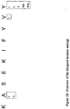
- the analog can be generally an analog of the SSX-2 41-50 decamer peptide with the sequence KASEKIFYVY.
- the residues or amino acids that make up the peptide are referred to herein as P1-P9 or P1-P10 to designate the position within the peptide as numbered from the N- to the C-terminus, P1 corresponding to the N-terminal Lysine and P9 corresponding to the C-terminal Valine in the nonamer.
- the residues may be referred to by the primary activity of the molecule that they are involved in.
- residue P2 is described as the N-terminal primary anchor molecule, while P9 (or P10 in the decamer) is described as the primary C-terminal anchor.
- Residues P4, P6 and P8 are primarily involved in TCR interactions.
- Substitutions can use any amino acids, including standard and non-standard amino acids, known to one of skill in the art. A number of exemplary amino acids are disclosed herein, however, the substitutions disclosed herein are not meant to be a list that includes all imagined substitutions, but are exemplary of the substitutions that are possible. One of skill in the art may find a number of other non-standard amino acids in catalogs and references that may be purchased or chemically produced for use with the analogs herein.
- a number of possible analogs were produced by modification of peptide anchor residues to achieve better HLA binding profiles and higher immune responses, including at the N-terminal primary anchor (P2 position), at the N-terminal secondary anchor (P1 position), at the N-terminal primary and secondary anchor (P1 and P2 positions), and at the N-terminal primary/secondary anchor (P1 and P2 positions) and C-terminal primary anchor (P9 position).
- peptides with modifications at the anchor residues and TCR contact residues were produced to circumvent T cell tolerance for self-antigens, these modifications included modifications at the N-terminal primary/secondary anchor (P1 and P2 positions) and secondary TCR recognition sites (P4, P6 and/or P8 positions), modifications at the N-terminal primary/secondary anchors (P1 and P2 position), and modifications at the C-terminal primary anchor (P9) and at secondary TCR recognition sites (P4, P6 and/or P8 positions). Further, decamer analogs were produced.
- residues are primarily involved in a specific interaction and some are secondarily or even tertiarily involved. Thus, the knowledge of how the residues are involved in the binding to these molecules was involved in the analysis. Further some of the wild-type residues are preferred, meaning that they work well for the intended interaction, while others are non-preferred, meaning that they work poorly for the interaction. Thus, the non-preferred residues can be substituted.
- the valine at the C-terminus is generally a preferred anchor residue because it produces a strong interaction with the HLA molecule and, thus, it was less preferred to substitute this residue.
- modifications of wild-type tumor-associated peptide epitopes by incorporating favorable anchor residues have generated analogs with improved binding profiles with HLA molecules and enhanced immunogenicity.
- One of the most successful examples is the A27L peptide analog of Melan-A 26-35 epitope ( Valmori D, et al. J Immunol. 160(4): 1750-8,1998 ). The original epitope failed to form a stable complex with HLA-A2 molecules since it lacked an optimal anchor residue at position 2.
- the choice of how many residues to substitute involves a desire to substitute better residues while still retaining enough of the qualities of the epitope that it will still be recognized by T cells which recognize the wild-type epitope.
- one or two substitutions can be made to the wild-type peptide.
- more than two substitutions can be made to the wild-type peptide, while still retaining cross-reactivity with the wild-type peptide.
- the part of the peptide that is involved in TCR recognition is desirably substituted to produce improved immunogenicity while still cross-reacting with the wild-type epitope.
- a peptide that shows increased immunogenicity is preferred.
- the P2 or second amino acid at the N-terminal end is believed to be primarily involved in the process of producing improved immunogenicity, primarily through improved binding properties, it is a preferred substitution site and a number of modifications were made in the exemplary analogs to identify desired substitutions. Similar considerations apply to the carboxy-terminal position, P ⁇ which also can be important for MHC binding.
- the analog can include a substitution at the P2 residue that substitutes a more hydrophobic residue for the wild-type alanine.
- the hydrophobic residue also can possess a more bulky side chain.
- the residue at P1 can be substituted with a more hydrophobic residue.
- residues P1 and P2 both can be substituted with more hydrophobic residues.
- at least one residue at P1, P2, and P9 can be substituted.
- at least two residues at P1, P2 and P9 can be substituted.
- at least two residues at P1, P2, P9 P4 and P6 can be substituted including one or more residues involved in TCR binding.
- substitutions of those residues only secondarily involved in binding to TCR or the MHC molecule can be advantageous.
- substitution of secondary TCR binding amino acids can generate analogs that still bind and produce a response and do not interfere with the binding to the MHC molecule, but preferably overcome the tolerance issues of self-antigens. This is useful because a patient who has cancer may be partially tolerized to the antigen.
- an analog that retains some activity can be preferable to an analog with more improved immunogenicity, because it will be less likely to be recognized as "self" by the immune system.
- the N-terminal primary anchor is the 2 nd N-terminal amino acid of the peptide and is the N-terminal proximal primary anchor. It is primarily involved in the interaction with the MHC molecule and substitutions can result in improved binding and stability. However, it may be secondarily involved in TCR interactions also. Thus, substitutions at this site can result in a peptide with improved interaction with MHC molecules as well as improved interaction with the TCR.
- the alanine found at this position in the wild-type sequence is generally believed to be non-preferred for the interaction with the MHC molecule.
- the analogs have a substitution at this position.
- the original Ala 42 found in the wild-type sequence can be substituted with a more hydrophobic amino acid. Any more hydrophobic amino acid may be used including any that is available or known to one of skill in the art, including standard amino acids and non-standard amino acids. Further, the original Ala 42 is substituted with a more hydrophobic amino acid also possessing a bulky side chain. Examples of more hydrophobic amino acids includes, but are not limited to: Leu, Val, lie, Met, ⁇ -aminobutyric acid, Norleucine and Norvaline.
- the N-terminal secondary anchor is the first amino acid at the N-terminus.
- This residue is Lys 41 and is defined as a secondary anchor residue in interacting with the HLA-A*0201 molecule.
- it is also engaged in the interaction with the T cell receptors to a certain degree. Therefore, modifications of this position can generate some heteroclitic analogs that are more immunogenic and more suitable for the development of tumor vaccines.
- the lysine at this position is generally considered to be favored, substitutions can result in highly improved properties.
- the original Lys 43 found in the wild-type sequence can be substituted with a more hydrophobic amino acid.
- Any more hydrophobic amino acid can be used, including any that is available or known to one of skill in the art, including standard amino acids and non-standard amino acids.
- the Lys 43 can be substituted with an aromatic amino acid. Examples of more hydrophobic amino acids include, but are not limited to: Phe, Tyr, Trp, and D-Lys.
- Both primary and secondary anchor residues were substituted to result in improved binding affinity to the HLA molecule. Further, the double substitution produced improved stability of binding to the HLA molecule. Further, the binding and/or stability was not improved and may have even been reduced, but other properties of the molecule were improved, such as activity or recognition by a tolerized individual.
- N-terminal primary/secondary anchor and C-terminal primary modification P2, P1 and P9
- the C-terminal Val of the wild-type peptide is generally a preferred anchor residue and primarily involved in the interaction with the MHC molecule. However, substitutions were carried out to identify which amino acids improve the analogs having primary and secondary N-terminal modifications. These C-terminal substitutions can be used in the absence of one or more N-terminal modifications also.
- the TCR sites are generally recognized as residues P4, P6, and P8 and are the primary residues involved in the binding to the TCR. However, other residues may also be involved in the interaction to a lesser extent.
- One or more of the sites primarily involved in TCR interaction can be substituted to increase the interaction. Preferably, these substitutions can generate heteroclitic analogs that do not interfere with binding to the MHC molecule, but overcome the tolerance issues of the wild-type peptides.
- at least one TCR substitution can be included with at least one substitution at position P1, P2, and/or P9. Further, the substitution at any one or more of the P4, P6, and P8 positions can be a polar amino acid. Further, the substitution can be an aromatic amino acid at position P8.
- substitution can be an amino acid with a large aliphatic side chain at position P6. Further, the substitution can be an amino acid which has a larger side chain to preserve the interaction.
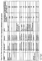
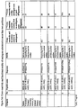
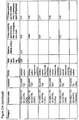
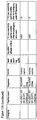
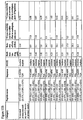
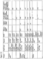
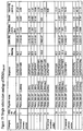
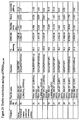
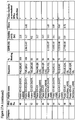
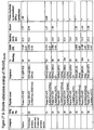
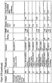
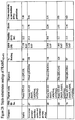
- Table 5 N-TERMINAL PRIMARY/SECONDARY ANCHOR AND TCR SITES MODIFICATION Catergory Peptide name Sequence Predictive Scores (R/NIH) Half-maximal Binding (mM) Relative affinity (1/RA) Stability (T1/2) (Hrs) Cross-reactivity and fct avidity (native to analogs)* Native SSX2 41-49 KASEKIFYV 22 / 1017 14.64 1.0 11 1 N-terminal PrimarylSecondary Anchor, TCR sites SSX2 41-49 (K41F, A42V, E44D) FVSDKIFYV 21/8421 13.18 1.1 N/A >10 SSX2 41-49 (K41F, A42V, E44N) FVSNKIFYV 20/2054
- the C-terminal residue can be modified to contain an amide in the place of the free carboxylic acid.
- the P9 residue can be modified.
- the peptide is a 10-mer (decamer) the P10 residue can be modified.
- this results in a peptide or analog that has increased stability in biological media, including but not limited to blood, lymph, and CNS.
- the peptides can retain the other necessary activities to result in an analog usable for vaccination or as an immunogen.
- the length of typical MHC binding peptides can vary from about 8 to about 11 amino acids in length.
- most of the previously used HLA-A*0201 are 9-mers (nonamers) or 10-mers (decamers).
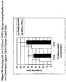
- the analog can be an analog of the wild-type sequence SSX-2 41-50 .
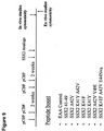
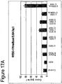
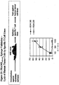
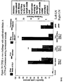
- the wild-type 10-mer does not have the correct binding motif and showed no immunological activity, a 10-mer was created by substituting amino acids at the P10 position and identifying the effect of various wild-type and analogs (see Figure 1 ).
- any residues can also be substituted with conservative amino acids.
- Conservative substitutions can be paired with any of the above substitutions that can produce an effect.
- conservative substitutions can be specifically at residues that are not believed to be involved in any of the activities at a primary, secondary, or even tertiary level.
- residues include P3, P5 and P7.
- the Serine at position P3 can be substituted with an alanine or threonine to produce an analog.
- conservative substitutions do not significantly affect the activity of the analog, however, in some embodiments they can increase certain activities or decrease certain activities.
- Embodiments relate to analogs of the MHC class I-restricted T cell epitope NY-ESO-1 157-165 , SLLMWITQC (SEQ ID NO. 1), polypeptides comprising these analogs that can be processed by pAPC to present the epitope analogs, and nucleic acids that express the analogs.
- the analogs can have similar or improved immunological properties compared to the wild-type epitope.
- the analogs can contain at least one substitution, but can have multiple substitutions comprising standard or non-standard amino acids singly or in various combinations.
- the analogs can result in peptides with retained or improved properties.
- the epitope NY-ESO-1 157-165 has been shown to be presented by NY-ESO-1 expressing cell lines, by measuring the epitope specific T cell activity against such cells ( Jaeger, E. et al., J. Exp. Med. 187:265-270, 1998 ; U.S. Patent No. 6,274,145 ). Methodologies to improve the physico-chemical properties of the peptide NY-ESO-1 157-165 have been described ( U.S. Patent No. 6,417,165 ) and can consist of replacement of the terminal cysteine with other amino acids that preserve or enhance the interaction with MHC and are devoid of the deleterious property of disulfide C-C bond formation interfering with the activity.
- Embodiments relate to families of one or more peptides of 9 or 10 amino acids in length related by sequence to amino acids 157-165 of the human cancer testis (CT) antigen NY-ESO-1 (NY-ESO-1 157-165 ).
- CT human cancer testis
- the analog is generally an analog of the NY-ESO-1 157-165 , with the sequence SLLMWITQC (SEQ ID NO:1). Analysis of whether wild-type amino acids are preferred or non-preferred used previous analyses of other peptide-MHC or TCR interactions.
- the Cysteine at the C-terminus is generally a non-preferred anchor residue because it does not produce a strong interaction with the HLA molecule and, thus, it was highly preferred to substitute this residue.
- the Serine at position P1 is generally preferred, it was found that substituting an aromatic could produce a peptide with improved properties.
- the Leucine at position P2 is generally acceptable, but substituting a hydrophobic and/or bulky amino acid resulted in a peptide with improved properties.
- the residues which are primarily involved in the interaction with the TCR (P4, P6 and P8) showed a preference generally for some polarity, and in the case of P8 an aromatic generally produced peptides with favorable properties.
- the substitution can be a hydrophobic residue. More preferably, the substitution can be a bulky hydrophobic residue.
- the residue at P1 can be substituted with a more hydrophobic residue.
- residues P1 and P2 can be both substituted with more hydrophobic residues.
- at least one residue at P1, P2, and P9 can be substituted.
- at least two residues at P1, P2 and P9 can be substituted.
- at least two residues at P1, P2, P9, P4, and P6 can be substituted, including one or more residues involved in TCR binding.
- the residue at P8 can be substituted with an aromatic. Examples of the following substitutions are shown in Figures 13A-13C .
- the N-terminal primary anchor is the 2 nd N-terminal amino acid of the peptide, thus, it is the N-terminal proximal primary anchor.
- the original Leucine 158 is not considered "non-preferred" for binding to the MHC molecule, substitutions can produce a peptide with improved binding.
- the original Leu 158 found in the wild-type sequence can be substituted with a similarly or more hydrophobic amino acid. Any hydrophobic amino acid may be used, including one that is available to or that is known to one of skill in the art, including standard amino acids and non-standard amino acids.
- the original Leu 158 can be substituted with a more hydrophobic amino acid also possessing a bulky side chain.
- hydrophobic amino acids examples include, but are not limited to: Leu, Val, lie, Met, ⁇ -aminobutyric acid, Norleucine and Norvaline.
- a naphthal side chain can also be substituted.
- the substitution results in improved binding and stability with the HLA molecule.
- this residue may be secondarily or tertiarily involved in TCR interactions, and substitutions may also result in improved recognition by the TCR.
- the N-terminal secondary anchor is the first amino acid at the N-terminus or P1. This residue is involved in a number of interactions.
- the residue of Ser 157 was defined as a secondary anchor residue in interacting with HLA-A*0201 molecule, it also engaged in the interaction with the T cell receptors to a certain degree. Therefore, modifications of this position generate some heteroclitic analogs that are more immunogenic and more suitable for the development of tumor vaccines. Thus, substitutions can result in a variety of improved qualities.
- the Serine is not considered "non-preferred," a number of substitutions can result in improved qualities of the peptide.
- the original Ser 157 found in the wild-type sequence can be substituted with a more hydrophobic amino acid.
- Any more hydrophobic amino acid can be used, including one that is available to or that is known to one of skill in the art, including standard amino acids and non-standard amino acids. Examples of more hydrophobic amino acids include, but are not limited to: Phe, Tyr, Trp, and D-Lys.
- both primary and secondary anchor residues were substituted to result in improved binding affinity to the HLA molecule.
- the double substitution produced improved stability of binding to the HLA molecule.
- the binding and/or stability was not improved and may have even been reduced, but other properties of the molecule were improved, such as activity or recognition by a tolerized individual.
- N-terminal primary/secondary anchor and C-terminal primary modification P2, P1 and P9
- the C-terminal cysteine of the wild-type peptide is generally a non-preferred anchor residue. Because this residue is generally primarily involved in the interaction with the MHC molecule, it can be preferred to substitute residues that result in a stronger interaction with the MHC molecule. Thus, substitutions were shown to improve binding affinity and stability and in some cases resulted in analogs with decreased cross-reactivity. In some embodiments; the substitution to the C-terminus can result in a peptide with improved binding and/or stability without decreased cross-reactivity. However, in other embodiments the substitution to the C-terminus can result in a peptide with improved binding and/or stability with equal or decreased cross-reactivity.
- the C-terminal substitution can be paired with at least one other substitution.
- amino acid substitutions to the C-terminus include, but are not limited to, valine, lysine, alanine, and isoleucine.
- the primary residues involved in the interaction with the TCR are generally recognized as residues P4, P6, and P8. However, other residues may also be involved in the interaction to a lesser extent.
- one or more of the sites primarily involved in TCR interaction can be substituted to result in an improved interaction. Preferably, these substitutions generate heteroclitic analogs that do not interfere with binding to the MHC molecule, but overcome the tolerance issues of the wild-type peptides.
- at least one TCR substitution can be included with at least one substitution at position P1, P2, and/or P9.
- amino acids with some polarity can be substituted at P4, P6, and P8.
- amino acids which are aromatic can be substituted at the P8 position.
- the C-terminal residue can be modified to contain an amide in the place of the free carboxylic acid.
- the P9 residue can be modified.
- the peptide is a 10-mer (decamer) the P10 residue can be modified.
- this results in a peptide or analog that has increased stability in biological media, including but not limited to blood, lymph, and CNS.
- the peptides retain the other activities to result in an analog usable for vaccination or as an immunogen.
- the length of typical MHC binding peptides varies from about 8 to about 11 amino acids in length.
- most of the previously used HLA-A*0201 are 9-mers (nonamers) or 10-mers (decamers).
- the analog can be a 10-mer of the wild-type sequence NY-ESO-1 157-166 .
- the wild-type 10-mer does not have the correct binding motif and showed no immunological activity, a 10-mer was created by substituting amino acids at the P10 position and identifying the effect of various wild-type and analogs (see Figures 13A-13C ).
- the residues that were added or substituted for the wild-type at the C-terminus can be selected from the group consisting of norvaline, leucine, isoleucine, valine, and alanine.
- any residues can also be substituted with conservative amino acids.
- Conservative substitutions can be paired with any of the above substitutions that can produce an effect.
- conservative substitutions can be specifically at residues that are not believed to be involved in any of the activities at a primary, secondary, or even tertiary level.
- Such residues can include P3, P5 and/or P7.
- Conservative substitutions are known to those of skill in the art, but, for example, the Leucine at position P3 can be substituted with an alanine or threonine to produce an analog.
- conservative substitutions do not significantly affect the activity of the analog. However, in some embodiments they may increase certain activities or decrease certain activities. Because of the known interactions, it is unlikely that such conservative substitutions will have a significant effect on any of the activities.
- the analogs can have similar or improved immunological properties compared to the wild-type epitope. Evidence validating the presentation of this epitope by human cancer cells is presented in Example 32 below.
- analogs of PSMA 288-297 can contain at least one substitution, but can have multiple substitutions comprising standard or non-standard amino acids singly or in various combinations.
- the analogs may result in peptides with retained or improved properties.
- the disclosure relates to families of one or more peptides of 9 or 10 amino acids in length related by sequence to amino acids 288-297 of the human PSMA.
- the PSMA 288-297 analog can contain substitutions of the sequence GLPSIPVHPI.
- Reference to binding motif data such as presented in table 7 in example 2 below, indicates that the P2 anchor residue can make the largest individual contribution to affinity of any position in an A2.1-restricted epitope. In this case the amino acid at the P2 position is the optimally preferred leucine.
- the P ⁇ anchor residue, isoleucine is favorable.
- In vitro binding studies using the T2 cell assay system (not shown) have indicated that the native peptide has generally superior binding characteristics, particularly as compared to the SSX-2 and NY-ESO-1 epitopes. The epitope exhibited significant binding at relatively low concentrations, although this was paired with a relatively shallow rise toward saturation.
- the wild-type epitope can be improved. Analyses such as that represented by tables 7 and 8 are averages and the behavior of a given residue in a particular sequence may diverge from the average. Consistent with the favorable results obtained with Nle and Nva for the SSX-2 and NY-ESO-1 epitopes discussed above, Nle and Nva also can be successfully used for the instant PSMA epitope. Finally, even similar binding characteristics, if paired with alterations that help circumvent whatever tolerance to the epitope may exist, can increase the effective immunogenicity of the peptide. In the transgenic mouse model the native peptide is poorly immunogenic (see Example 35 for instance) which may reflect tolerance to the epitope; the region of PSMA from which this epitope is derived is identical between mouse and human PSMA.
- N-terminus proximal primary anchor modification P2
- the native residue at the P2 position of this epitope is generally the optimal residue among genetically encoded amino acids, the effect of substituting other preferred or bulky hydrophobic residues were examined for potential improvement of binding, tolerance breaking and cross-reactive immunity.
- exemplary substitutions can include Met, Ile, Gln, Val, Nva, Nle, and aminobutyric acid (Abu).
- the N-terminal secondary anchor is the first amino acid at the N-terminus.
- the native Gly is only marginally preferred at this position.
- amino acids with potential to improve the epitope include Ala, Ser, Abu and sarkosine (Sar, that is, N-methylglycine).
- the native Ile at this position is generally a preferred but not optimal residue. Substitution at this position can improve binding. Exemplary substitutions can include Val, Leu, Nva, and Nle.
- the penultimate position (P ⁇ -1) can serve both as a secondary anchor and a TCR interacting position. Substitution of Ala, Leu, Ser, and Thr can be have their primary effect on TCR interaction, though they can also contribute to improved binding. P3 is another position that can effect both binding and immunogenicity. Substitution of Trp at this position can improve both.
- the analogs can have similar or improved immunological properties compared to the wild-type epitope. Evidence validating the presentation of this epitope by human cancer cells is presented in Example 39 below.
- the analogs can contain at least one substitution, but can have multiple substitutions comprising standard or non-standard amino acids singly or in various combinations.
- the analogs can result in peptides with retained or improved properties.
- analogs of the PRAME 425-433 which can contain substitutions of the sequence SLLQHLIGL.
- Reference to binding motif data such as presented in table 7 in Example 2 below, indicates that the P2 anchor residue can make the largest individual contribution to affinity of any position in an A2.1-restricted epitope. In this case the amino acid at the P2 position is the optimally preferred leucine.
- the P ⁇ anchor residue, leucine is favorable, though not as strongly preferred.
- Analyses such as that represented by tables 7 and 8 are averages and the behavior of a given residue in a particular sequence can diverge from the average, nor is the wild type P ⁇ residue necessarily the most preferred for that position.
- substitutions investigated for the PRAME 425-433 epitope follow the same logic and are disclosed in the examples 40-42 and Figures 25-27 .
- Substitutions were made at the primary anchor positions P2 and P ⁇ (P9), the secondary anchor positions P1 and P ⁇ -1 (P8).
- Substitutions were also made in the TCR interacting positions (in addition to secondary anchor positions) P3 and P6.
- Selected substitutions have impact on binding and/or stability of MHC class I - peptide complexes, key features in determining the immunological properties of peptides.
- substitutions that retain the capability of analogs to interact with T cell receptors recognizing native peptides can be of practical value.
- the following examples provide analogs and methods of identifying analogs.
- the analogs can be used, for example, as immunogens, vaccines, and/or treatment of a variety of cancers.
- the analogs were produced as in Example 1.
- SSX-2 41-49 analogs were identified as shown in Example 2, those produced listed in Example 3 and tested for improved properties as in Examples 4-21.
- the testing of NY-ESO-1 157-165 analogs were tested for improved properties as in Examples 22-30.
- Peptides were synthesized on either a Symphony multiple peptide synthesizer (PTI technologies, MA) or an ABI 433A peptide synthesizer (Applied Biosystems, Foster City, CA) at 0.05 - 0.1mmole scale using standard Fmoc solid phase chemistry.
- C-terminal free acid peptides were synthesized using pre-load PEG-PS resins (on Symphony) or Wang resin (on ABI).
- C-terminal amidated peptides were synthesized on Fmoc-PAL-PEG-PS resin. All resins were purchased from Applied Biosystems (Foster City, CA).
- the Fmoc-amino acids used in peptide syntheses were purchased from Novabiochem (San Diego, CA) and AnaSpec (San Jose, CA). Post-synthesis cleavage was carried on by the standard protocol.
- Peptide purification was carried out on either semi-preparative HPLC columns or SPE cartridges (Phenomenex, Torrance, CA). The purity of all peptides was ⁇ 90%. The identity of each peptide was verified by Maldi-TOF MS (Voyager DE, Applied Biosystems) and analytical HPLCs (Varian orShimazu) using a Synergi C12 column (Phenomenex, Torrance, CA).
- Structural modification of a moderately antigenic peptide can considerably improve peptide-MHC binding, CTL recognition, and/or immunogenicity.
- General guidelines regarding how to modify a wild-type epitope in order to achieve a peptide analog with enhanced potency are known in the art.
- An appreciated strategy is to optimize the residues at the so-called anchor positions for binding to the particular MHC molecule at issue.
- a marked preference for hydrophobic residues at the P2 and P ⁇ positions has been observed, particularly L, and M at P2, and V at P ⁇ . (P ⁇ denotes the C-terminal residue of the epitope.
- Nle norleucine
- Nva norvaline
- Phg phenylglycine
- Phe(4-F) 4-fluorophenylalanine
- Phe(4-NO 2 ) 4-nitrophenylalanine
- Abu ⁇ -aminobutyric acid
- Aib ⁇ -aminoisobutyric acid
- MeLeu methyl-leucine
- MeVal methylvaline
- ⁇ -(3-benzothienyl)Ala ⁇ -(3-benzothienyl)-alanine
- O-methyl-Tyr O-methyltyorosine
- Cha cyclohexylalanine
- Nal-1 ⁇ -(1-napthyl)-alanine
- Nal-2 ⁇ -(2-napthyl)-alanine
- -NH2 indicates that the carboxy terminus has been modified to the amide.
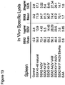
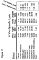
- Example 3 The analogs produced in Example 3 were tested for activity, such as binding and biological effect as follows in Examples 4-21:
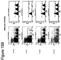
- the T2 cells that lack expression of TAP and thus do not assemble stable MHC class I on the cell surface were pulsed with different concentrations of peptides (controls or analogs) overnight at 37°C, washed extensively, stained with fluorescently tagged antibody recognizing MHC class I (A2 allele) and run through a FacsScan analyzer.
- the difference between the MFI (mean fluorescence intensity) corresponding to a given concentration of analog and the negative control (non-MHC binder) is a function of how many stabilized complexes between MHC and peptide are displayed on the surface of T2 cells.
- the binding was quantified by two factors that are mathematically related: half maximal binding (the peptide concentration giving 50% of the signal corresponding to saturation) and relative affinity (1/RA).
- Relative affinity RA is binding normalized to a reference (wild-type peptide); for example, the ratio between half max binding of control relative to peptide analog. The higher the 1/RA index and the lower the half maximal binding, the higher the K on of the interaction between the analog and the MHC. Fifty three analogs were identified with these binding parameters improved relative to the wild-type peptide.
- the T2 cells that lack expression of TAP and thus do not assemble stable MHC class I on the cell surface were pulsed with a concentration of peptide (controls or analogs) known to achieve maximal loading of MHC class I ("saturation") overnight at 37°C, washed extensively, and chased for different intervals in the presence of emetine, which blocks endogenous protein synthesis. After extensive washing, the cells were stained with fluorescently tagged antibody recognizing MHC class I (A2 allele) and run through a FacsScan analyzer.
- the difference between the MFI (mean fluorescence intensity) corresponding to a given concentration of analog and the negative control (non-MHC binder) is a function of how many stabilized complexes between MHC and peptide are displayed on the surface of T2 cells.
- the decay of the signal over time was mathematically expressed as stability index 50% relative to the binding at 0 hours (at the beginning of the chase interval).
- Such improved analogs can carry one, two, three or multiple substitutions (including standard and/or non-standard amino acids) involving positions that are known to participate in the interaction with MHC and/or TCR, with an overall effect on MHC stability that is dependent on the modification.
- Such peptide analogs can be useful in therapeutic compositions or as a platform to further derive therapeutic compositions. Forty three of the analogs have increased stability relative to the natural peptide.
- the immunologic properties of peptides can be described as a function of binding to MHC molecules (K on and K off ) and TCR (affinity of interaction between TCR and MHC-peptide complexes). Modifications of primary MHC anchor residues generally have a significant degree of predictability in regard to overall impact on binding to MHC molecules.
- Modifications of secondary MHC anchor residues may impact the affinity of interaction of the MHC-peptide complex to TCR along with the K on and K off relative to peptide-MHC interaction.
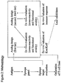
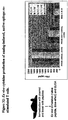
- a methodology was devised that allowed rapid and rational screening of peptide analogs in a fashion coherent with proposed methods of use and modeling the overall immunologic properties (K on and K off relative to MHC interaction and TCR binding properties in an integrated fashion).
- This method can include generating T cell lines against a natural (non-mutated) epitope (SSX-2 41-49 ) using an immunization strategy potent enough to generate a useful response in transgenic mice carrying human MHC (such as the A2 allele).
- Peptide analogs were interrogated ex vivo in the presence of competent APCs and the functional impact of T cells specific for natural (non-mutated) epitope measured.
- analogs that result in reduced values associated with parameters #1 and 3 but increased #2 can be useful.
- Use of natural epitope and unrelated non-cross reactive peptides as references is valuable in identifying classes of analogs of potential value.
- Analogs that display properties quantitatively comparable to or even modestly attenuated from those of natural epitopes are still deemed useful in light of the fact that while they retain cross-reactivity, they may display immunologic properties that are distinct from those of the natural peptide - for example lower propensity to induce AICD or ability to break tolerance or restore responsiveness in vivo.
- Some advantages of this screening strategy include the practicality and rapidity, use of more relevant polyclonal T cell lines instead of potentially biased T cell clones as a read out, and the composite value, integrating parameters such as K on , K off and TCR affinity that may translate into cross-reactivity and functional avidity of peptide-MHC complexes relative to TCR. These parameters can be predictive of the in vivo immunologic properties and thus can delineate useful panels of peptide analogs to undergo further evaluation, optimization and practical applications. Analogs that bind to MHC and retain cross-reactivity against TCR specific for the nominal wild-type peptide are predicted to trigger a measurable effect in this assay. The overall methodology is presented in Figure 2 .
- mice carrying an A2 human allele ( Pascolo et al. J. Exp Med. 185(12):2043-51, 1997 ) were immunized with 50ug of SSX-2 natural epitope (41-49) admixed with 25ug of pIpC at day 0, 4, 14 and 18 by bilateral administration into the inguinal lymph nodes.
- mice were sacrificed and a suspension of splenocytes prepared at 5x10 6 million cells/ml in complete HL-1 medium.
- Coordinated modifications at position 1 and 2 have a variable effect on the activity of analogs. For example, substitution of K41 with Y, F or W corroborated with substitution of A42 with V, M or I, and resulted in preserved or enhanced activity of the analogs relative to the wild-type peptide.
- Such doubly mutated peptides offer an increased opportunity to impact the interaction with TCR in a fashion that results in tolerance breaking (thus being useful for practical application), since the P1 residue participates to a certain extent in binding to TCR.
- F and V at positions 41 and 42 respectively, combined with I or A at position 49 resulted in improved or similar activity relative to the wild-type epitope.
- L or M at position 49 resulted in heavily diminished activity.
- Triple mutants comprising the non-standard amino acids Nva, Abu or MeVal at the last position resulted in retention or improvement of immune activity.
- Such peptides are extremely useful due to increased in vivo stability and resistance to enzymatic degradation.
- Modification of amino acid residues within the putative TCR binding region can result in peptides of considerable value, that retain binding to MHC along with cross-reactivity and thus be useful for restoration of immune responsiveness or tolerance breaking since their conformation in the MHC groove is slightly different from that of natural peptides. Additional substitutions at position 44 (Q, Nva or Nle), position 46 (L, V, Nle or Nva) or 48 (F or Phe-4F) resulted in active analogs, whereas D, N, S orT at position 44, M at 46 or T, S, Phg at position 48 or L at position 46 with T at 48, resulted in analogs devoid of activity. Finally, two analogs with 5 substitutions showed no activity ( Figure 5 ).
- the decamer SSX-2 41-50 was significantly less active in stimulating the T cell line specific for the 41-49 nonamer, relative to the latter. Modification of the Y residue at position 50 to I or L, but to a lesser or no extent to V, Nle or Nva, resulted in restoration of activity in this assay. Further optimization of the activity of decameric analogs can be obtained by modification of the A at position 2 with L or V. The A42L substitution rescued the activity of the Y50Nva decamer.
- Peptide analogs of similar or reduced activity in vitro (but retained cross-reactivity) compared with the natural peptide are still useful for induction or boost of immune responses due to: i) more limited AICD; ii) potentially higher in vivo activity due to increased stability on class I MHC and/or slightly modified interaction with TCR which is can be important for tolerance breaking.
- the schedule of immunization is shown in Figure 7 .
- splenocytes were stimulated ex vivo with SSX-2 41-49 natural peptide and tested against 51 Cr-labeled target cells (T2 cells) at various E:T ratios ( Figure 8 ).
- EAA negative control peptide
- splenocytes were isolated from littermate control HHD mice and incubated with 20 ⁇ g/mL or 1ug/ml of natural peptide for 2 hours. These cells were then stained with CFSE hi fluorescence (4.0 ⁇ M or 1 ⁇ M for 15 minutes) and intravenously co-injected into immunized mice with an equal number of control splenocytes stained with CFSE lo fluorescence (0.4 ⁇ M). Eighteen hours later the specific elimination of target cells was measured by removing spleen and PBMC from challenged animals and measuring CFSE fluorescence by flow cytometry.
- EAA negative control peptide
- splenocytes were stimulated ex vivo with SSX-2 41-49 wild-type peptide and tested against 51 Cr-labeled human tumor cells (624.38 melanoma cells) at various E:T ratios ( Figure 12 ).
- Analog A42V and K41F A42V V49I elicited immune responses that mediated increased cytotoxicity against human tumor cells expressing the natural SSX-2 41-49 epitope.
- the N-terminal secondary anchor is the first amino acid at the N-terminus.
- the original Lys 43 found in the wild-type sequence is substituted with a more hydrophobic and bulky amino acid.
- Any more hydrophobic and bulky amino acid also can be used, including any available to or that is known to one of skill in the art, including standard amino acids and non-standard amino acids. Examples of more hydrophobic amino acids include, but are not limited to: Phe, Tyr, Trp, and D-Lys.
- Lys 41 was defined as a secondary anchor residue in interacting with HLA-A*0201 molecule, and it also engaged in the interaction with the T cell receptors to a certain degree. Therefore, modifications of this position can generate some heteroclitic analogs that are more immunogenic and more suitable for the development of tumor vaccines.
- Lys 41 to Phg substitution has resulted in an analog with improved affinity and extended stability with the HLA-A*0201 molecule, however, its cross-reactivity with wild-type peptide specific CTL was fairly poor.
- the para-nitrophenylalanine analog was shown to induce the IFN- ⁇ secretion of the wild-type peptide specific CTL at a much lower concentration, although its affinity with the HLA-A*0201 molecule was about the same as that of wild-type peptide.
- the C-terminal Val of the wild-type peptide was a preferred anchor residue. However, improved potency was observed when it was mutated to lie, having one additional - CH2 group; similar improvement was also observed with a Val to Abu substitution. Although the other analogs showed improved binding affinity and stability with the MHC molecule, their cross-reactivity results were poor. The results of these analogs indicated that the peptide C-terminal anchor residue also plays a critical role in the recognition of T cells. (Table 4).
- Substitutions of secondary TCR binding amino acid residues preferably generate heteroclitic analogs that did not interfere with the binding to the MHC molecule, but overcame the tolerance issues of self-antigens.
- N-terminal primary/secondary anchor residues K41F and A42V
- analogs were generated with improved binding affinity and stability (Table 6).
- Some of these analogs induced the IFN- ⁇ production of the wild-type peptide specific CTL at lower concentrations, such as K41F, A42V, E44(Nva)/(Nle) mutants and K41F, A42V, I46L/(Nva) /(Nle) mutants.
- the length of typical MHC binding peptides varies from 8-mer to 11-mer, and most HLA-A*0201 binding peptides are 9-mers or 10-mers.
- a 9-mer and 10-mer from a natural sequence were both found to possess a binding motif for the same MHC, and had the same N-terminus. From the standpoint of proteasomal processing they are distinct epitopes, but were nonetheless antigenically cross-reactive.
- the epitope is a 9-mer peptide and the 10-mer peptide, SSX-2 41-50 , lacks the appropriate MHC binding motif and showed no immunological activity.
- the wild-type epitope was therefore lengthened to a 10-mer with amino acids that could create the appropriate binding motif.
- analogs SSX-2 41-50 (A42L, Y50L/V/Nle/Nva) showed improved binding affinity with the HLA-A*0201 molecule.
- analogs can represent an improvement over the wild-type epitope is in increased immunogenicity in a human system and tolerance breaking. Differences in the TCR repertoire, whether due to germ line differences or differences in negative selection, have the potential to give anomalous results. To address such issues the analogs are used in an in vitro immunization of HLA-A2* blood to generate CTL. Techniques for in vitro immunization, even using naive donors, are known in the field, for example, Stauss et al., Proc. Natl. Acad. Sci. USA 89:7871-7875,1992 ; Salgaller et al. Cancer Res. 55:4972-4979, 1995 ; Tsai et al., J. Immunol. 158:1796-1802, 1997 ; and Chung et al., J. Immunother. 22:279-287, 1999 .
- PBMCs from normal donors were purified by centrifugation in Ficoll-Hypaque from buffy coats. All cultures were carried out using autologous plasma (AP) to avoid exposure to potential xenogeneic pathogens and recognition of FBS peptides.
- AP autologous plasma
- DCs autologous dendritic cells
- APCs autologous dendritic cells
- monocyte-enriched cell fractions were cultured for 5 days with GM-CSF and IL-4 and were cultured for 2 additional days in culture media with 2 ⁇ g/ml CD40 ligand to induce maturation.
- 2 x10 6 CD8+-enriched T lymphocytes/well and 2 x10 5 peptide-pulsed DCs/well were co-cultured in 24-well plates in 2 ml RPMI supplemented with 10% AP, 10 ng/ml IL-7 and 20 IU/ml IL-2. Cultures were restimulated on days 7 and 14 with autologous irradiated peptide-pulsed DCs. Immunogenicity was assayed using the in vitro cytotoxicity and cytokine production assays described herein.
- Example 6 The strategy from above (Example 6) was applied to scan through a library of analogs bearing single substitutions relative to the wild-type NY-ESO-1 157-165 epitope in its native (or wild-type) version ( Figure 13 ). Strong inverse correlation was found between the minimal required amount of analog to elicit IFN-gamma production ex vivo and the maximal amount of cytokine production at any concentration of analog.
- Peptides that have substitutions in the N-terminal position or elsewhere, and present with retained but not increased activity in this assay relative to the wild-type peptide can be useful in humans.
- Example 6 The strategy from above (Example 6) was applied to scan through a library of analogs bearing two substitutions relative to the wild-type NY-ESO-1 157-165 epitope. Simultaneous semi-conservative modifications at position 2 and 9 were shown to have profound effects on the immune properties of analogs, depending on the precise identity of the analogs. Combining L158I with C165V or C165L further increased their activity relative to the wild-type peptide. Similarly, L158V improved on the activity of the C165V or C165L analogs, further increasing such activity relative to wild-type peptide. L158V partially retained the activity of C165A or C165I analogs, showing an interesting effect of double mutation of primary anchor residues. Similarly, L158I partially retained the activity of the C165A analog.
- Combinations between S157F and V, L, I, and to a lesser extent A at the 9 th position retained strong activity of the analog and may be more useful than single mutants at position 9 due to the participation of the first residue in the interaction with TCR.
- Combinations between S157K and V, L, I and to a lesser extent A at the 9 th position retained strong activity of the analog and may be more useful than single mutants at position 9 due to the participation of the first residue in the interaction with TCR and the overall beneficial effect on the peptide solubility of K at position 1.
- Example 6 The strategy from above (Example 6) was applied to scan through a library of analogs bearing multiple substitutions relative to the wild-type NY-ESO-1 epitope.
- S157Y L158I C165V displayed increased activity relative to the wild-type peptide and S157V with C165V or C165I; and S157I with C165L or I, retained MHC binding and cross-reactivity with T cells specific for the wild-type peptide.
- Triple substitutions comprising Y and V at positions 157 and 165, respectively, in addition to L or N at 160; A, L, V, or N at 162; or E, D or T at 164, retained the activity of the peptide in this cross-reactivity assay, making these analogs useful compounds for breaking T cell tolerance in vivo since positions 160, 162 and 164 participate in the interaction with TCR.
- Triple substitutions comprising 157F and 158V plus V, L, I at the position 165 showed activity in the assay described in Example 2.
- triple mutants encompassing S157F and L158I plus V or A at position 165 retained activity.
- triple mutants comprising S157W and to a higher extent S157T together with 158V and 165V, showed retained or increased activity respectively, relative to the wild-type peptide.
- Example 6 The strategy from above (Example 6) was applied to scan through a library of analogs of a decamer encompassing the nominal NY-ESO-1 157-165 peptide. While the decamer itself lacked significant in vitro activity, various substitutions at this position partially rescued activity, such as L at 166, or L, I, Nle at 166 combined with Y at 157 and V at 165.
- Peptide analogs with similar or reduced activity in vitro (but with retained cross-reactivity) compared to the wild-type peptide are still useful for induction or boost of immune responses due to: i) more limited AICD (antigen-induced cell death); ii) higher in vivo activity due to increased stability on class I MHC and/or slightly modified interaction with TCR. Thus, these analogs are useful for breaking tolerance.
- T2 cell based assay The affinity of peptide analogs and the wild-type epitope to HLA-A*0201 was evaluated by T2 cell based assay (ref. Regner M, et al., Exp Clin Immunogenet. 1996;13(1):30-5 ).
- T2 cells that lacked expression of TAP and thus do not assemble stable MHC class I on the cell surface, were pulsed with different concentrations of peptides (controls or analogs) overnight at 37°C, washed extensively, stained with fluorescently tagged antibody recognizing MHC class I (A2 allele) and run through a FacsScan analyzer. Peptides that bind A2 stabilize its presence at the cell surface.
- the difference between the MFI (mean fluorescence intensity) corresponding to a given concentration of analog and the negative control (a non-MHC binding peptide) is a function of how many stabilized complexes between MHC and peptide are displayed on the surface of T2 cells.
- this is a measurement of K on mostly and at saturation levels of peptide that is a measurement of both K on and K off .
- the binding is quantified by two factors that are mathematically related: Half maximal binding (the peptide concentration giving 50% of the signal corresponding to saturation) and relative affinity (1/RA), that is binding normalized to a reference (wild-type peptide)-i.e. the ratio between half maximal binding of control relative to peptide analog.
- HBVc negative control peptide
- splenocytes were isolated from littermate control HHD mice and incubated with 20 ⁇ g/mL or 1 ⁇ g/ml of wild-type peptide for 2 hours. These cells were then stained with CFSE hi and CFSE med fluorescence (4.0 ⁇ M or 1 ⁇ M, respectively, for 15 minutes) and intravenously co-injected into immunized mice with an equal number of control splenocytes stained with CFSE lo fluorescence (0.4 ⁇ M). Eighteen hours later the specific elimination of target cells was measured by removing the spleens and PBMC from challenged animals and measuring CFSE fluorescence by flow cytometry.
- FIG. 15A shows the lack of in vivo cytotoxicity in mice receiving the negative control peptide.
- Figure 15B shows the variable cytotoxicity in mice immunized with plasmid and amplified with wild-type peptide.
- Figure 15C shows the substantial, constant cytotoxicity in mice immunized with plasmid and amplified with the analog L158Nva C165V.
- the S157K triple mutant can ameliorate the poor solubility of the wild-type peptide or other analogs, with direct practical implications.
- splenocytes were isolated, pooled and stimulated in vitro with 10 ⁇ M of wild-type peptide NY-ESO-1 157-165 for 3 and 6 days respectively. Supernatants were harvested and the concentration of IFN- ⁇ measured by ELISA. Analog L158Nva C165V induced T cells that produced large levels of IFN-gamma more rapidly upon ex vivo stimulation ( Fig. 17A ).
- Avidin-coated microtiter plates containing class I monomer loaded with a so-called placeholder peptide were used to evaluate peptide binding, affinity and off-rate.
- the monomer-coated plates were supplied as part of the iTopia Epitope Discovery System Kit (Beckman Ciulter, Inc., San Diego, CA, USA).
- Assay buffers, anti-MHC-FITC mAb and beta2-microglobulin and control peptides were also supplied with the kits.
- Native peptide and analogs were first evaluated for their ability to bind each MHC molecule by binding assay. This assay measures the ability of individual peptides to bind HLA molecules under standardized optimal binding conditions. Monomer-coated plates were first stripped, releasing the placeholder peptide and leaving only the MHC heavy chain bound to the plate. Test peptides were then introduced under optimal folding conditions, along with the anti-MHC-FITC mAb. Plates were incubated for 18hrs at 21 °C. The anti-MHC-FITC mAb binds preferentially to a refolded MHC complex. Therefore, the fluorescence intensity resulted from each peptide was related to the peptide's ability to form complex with MHC molecule.
- the plates were washed after 18 hrs incubation at 21 °C to remove the excess amount of peptide. The plates were then incubated on the allele-specific monomer plates at 37 C. The plates were measured at multiple time points (0, 0.5, 1, 1.5, 2, 4, 6 and 8 hrs) for relative fluorescence intensity. The time required for 50% of the peptide to dissociate from the MHC monomer is defined as the T1/2 value (hrs).
- the iScore is a multi-parameter calculation provided within the iTopia software. Its value was calculated based on the binding, affinity and stability data.
- PSMA 288-297 peptide 25 ⁇ g in 25 ⁇ l of PBS, plus 12.5 ⁇ g of pI:C to each lymph node
- splenocytes were stimulated ex vivo with the native PSMA 288-297 peptide and tested against 51 Cr-labeled human tumor cells (PSMA + A2 + LnCap cells; or as negative control, LnCap cells coated with MHC class I-blocking antibody) at various E:T ratios.
- Val, Leu, Nva, and Nle were substituted for the Ile at the P ⁇ primary anchor position. All four had similar binding affinity.
- the Val and Nva substitutions improved the stability of binding and increased the amount of IFN-gamma produced, indicating cross-reactivity and that the analogs can have improved immunogenicity.
- the Ser, Sar, and Abu substitutions at P1 maintained similar binding characteristics but had marginally similar cross-reactivity.
- the Ala, Leu, Ser, and Thr substitutions at the P ⁇ -1 position also maintained similar binding characteristics.
- the Trp substitution at P3 exhibited affinity and stability of binding that were both increased about twofold and IFN-gamma production that was within twofold of the native peptide, all generally similar values.
- peptide natural epitope PSMA 288-297 , or analogs bearing substitutions at primary or secondary anchor residues
- mice were sacrificed at 10 days after the last boost, splenocytes prepared and assessed for IFN- ⁇ production by ELISPOT analysis.
- Various numbers of splenocytes / well were stimulated with 10 ⁇ g/ml of native peptide in ELISPOT plates coated with anti-IFN- ⁇ antibody.
- the assay was developed and the frequency of cytokine-producing T cells that recognized native PSMA 288-297 peptide was automatically counted.
- the data were represented in Figure 22 as the number of spot forming colonies / well (mean of triplicates ⁇ SD).
- the data show increased priming of immune responses against the native epitope achieved by the I297V and P290W analogs, with the other analogs showing slightly higher (but significant) activity than the native peptide (I297Nva or G288Abu or L289Nle I297Nva).
- the improved activity of these analogs represents tolerance breaking.
- mice were sacrificed at 10 days after the last boost, splenocytes prepared and assessed for IFN- ⁇ production by ELISPOT analysis.
- Various numbers of splenocytes / well were stimulated with 10ug/ml of native peptide in ELISPOT plates coated with anti-IFN- ⁇ antibody.
- the assay was developed and the frequency of cytokine-producing T cells that recognized the PSMA 288-297 peptide was automatically counted.
- the data were represented in Figure 23 as frequency of specific T cells normalized to 0.5 million responder cells (mean of triplicates + SD). The data show that irrespective of the number of splenocytes / well, the frequency of native epitope - specific T cells was considerably higher in the mouse group immunized with the I297V analog.
- splenocytes were stimulated ex vivo with the native PSMA 288-297 peptide and tested overnight against 51 Cr-labeled human tumor cells (Lncap, A2' PSMA + ; or 624.38 A2 + PSMA - or control 624.28 cells A2 - PSMA - ) at various E:T ratios.
- the resulting immunity was effective in mediating cytotoxicity against Lncap ( Figure 24 ).
- PRAME 425-433 peptide 25 ⁇ g in 25 ⁇ l of PBS, plus 12.5 ⁇ g of pI:C to each lymph node
- splenocytes were stimulated ex vivo with the native PRAME 425-433 peptide and tested against 51 Cr-labeled human tumor cells (PRAME + A2 + 624.38 melanoma cells; or negative control 624.38 cells, deficient in A2 expression) at various E:T ratios.
- Lys P1 analog which elicited 40% of the IFN- ⁇ of the native peptide, all of these analogs were considered cross-reactive as they elicited IFN- ⁇ production within twofold of the native peptide.
- Double substitution analogs were created at P1 and P2, P2 and P ⁇ , and P1 and P ⁇ using various combinations of the single substitutions above ( Figures 27A and 27B ). None of the P1-P2 double substitutions examined had radical changes to binding affinity or stability, but none of the exhibited significant cross-reactivity in the IFN- ⁇ assay. A similar pattern is seen with the P2-P ⁇ double substitution analogs, however, the L426Nva L433Nle analog shows a significant level of cross-reactivity with the native peptide in the IFN- ⁇ assay along with its similar, somewhat improved binding characteristics.
- the examined analogs also conformed to the general pattern of having at least similar binding characteristics, but eliciting negligible IFN- ⁇ in the cross-reactivity assay.
- the exception in this grouping were the S425T L433Nle analog, which exhibited somewhat improved binding stability and significant cross-reactivity, and the S425F L433Nle analog, which had a more that fourfold reduced ED 50 , a nearly doubled halftime of dissociation, and elicited more IFN- ⁇ than the native peptide.
- pCTLR2 described in example 49 below
- mice were sacrificed at 10 days after the last boost, splenocytes prepared and assessed for IFN- ⁇ production after in vitro stimulation at 0.5x10 6 cells / well, with 10ug/ml of native peptide. At 48 hours after incubation, the supernatant was harvested and the concentration of IFN- ⁇ produced in response to the PRAME 425-433 peptide was measured by ELISA. The data were represented in Figure 29 and show a significant enhancement of IFN- ⁇ production in mice boosted with the PRAME 425-433 L426Nva L433Nle analog.
- splenocytes were isolated from littermate control HHD mice and incubated with 20 ⁇ g/mL or 1 ⁇ g/ml of native peptide for 2 hours. These cells were then stained with CFSE hi and CFSE med fluorescence (4.0 ⁇ M or 1 ⁇ M, respectively, for 15 minutes) and intravenously co-injected into immunized mice with an equal number of control splenocytes stained with CFSE lo fluorescence (0.4 ⁇ M). Eighteen hours later the specific elimination of target cells was measured by removing the spleens and PBMC from challenged animals and measuring CFSE fluorescence by flow cytometry.
- splenocytes were isolated from littermate control HHD mice and incubated with 0.2ug/ml or 20ug/ml of native peptide for 2 hours. These cells were then stained with CFSE fluorescence (1 and 2.5 ⁇ M respectively, for 15 minutes) and intravenously co-injected into immunized mice with an equal number of control splenocytes stained with CFSE lo fluorescence (0.4 ⁇ M). Eighteen hours later the specific elimination of target cells was measured by removing the spleen from challenged animals and measuring CFSE fluorescence in the resulting cell suspensions, by flow cytometry.
- the relative depletion of the populations corresponding to peptide-loaded splenocytes was calculated relative to the control (unloaded) population and expressed as % specific lysis.
- the frequency of PRAME 425-433 -specific T cells was evaluated by tetramer / CD8 co-staining.
- the boost with analogs encompassing mutations at primary or secondary anchor residues showed comparable immune activity as compared to the native peptide, based on in vivo cytotoxicity and tetramer staining.
- the analogs were capable of amplifying the immune response as shown by comparison with the "EAA" group, boosted with an irrelevant peptide.
- analogs comprising S425F, L33Nle, L426Nva L433Nle, S425T L433Nle, or S425T L426Nva L433Nle were all capable of expanding the immunity against the native epitope, as assessed by in vivo cytotoxicity.
- L433Nle, L426Nva L433Nle, and S425T L426Nva L433Nle analogs expanded the subset of T cells specific against the native epitope to a level significantly higher than in mice primed with plasmid and boosted with the negative control peptide ( Figure 31 ).
- mice were sacrificed at 10 days after the last boost, splenocytes prepared and assessed for IFN- ⁇ production by ELISA at 48 hours after incubation with 10 ⁇ g/ml of native peptide.
- the data were represented in Figure 32 as cytokine concentration in pg/ml (mean of triplicates ⁇ SD).
- the data showed ex vivo cytokine production by splenocytes from mice boosted with both analogs, and greater response to L426Nva L433Nle than to S425T L426Nva L433Nle.
- splenocytes were stimulated ex vivo with the native peptide and tested against 51 Cr-labeled human tumor cells (PRAME + 624.38 melanoma cells pretreated or not with IFN- ⁇ ; or negative control 624.38 cells, deficient in HLA-A2 expression) at various E:T ratios.
- the analog L426Nva L433Nle elicited immune responses that mediated significant cytotoxicity against human tumor cells expressing A2 (624.38), slightly elevated upon their pre-treatment with IFN ⁇ . In contrast, no significant activity was measured against A2- 624.28 control cells. See Figure 33 .
- PBMCs Peripheral blood mononuclear cells
- HLA-A*0201 + healthy donors
- FMCs peripheral blood mononuclear cells
- Fresh PBMCs 2.5 x 10 6
- 5 ng/ml PRAME 425-433 or peptide analog were plated in T-cell culture medium.
- 20 IU/ml of interleukin 2 was added to each well after 72 and 96 hours and addition peptide (5 ng/ml) was added at day 7. Cultures were maintained for an additional 10 days before effector cells were harvested and used in tetramer staining.
- IVS PBMCs were labeled with PRAME 425-433 tetramer and analyzed on the FACSCalibur(BD, San Jose, CA). Quadrants were set based on negative controls, stained with irrelevant HBV tetramer and SSX2 tetramer, and a minimum of 10,000 gated events were captured. Tetramer-positive cells are expressed as a percentage of the lymphocyte population.
- PRAME 425-433 -specific tetramers was significantly enhanced following IVS with peptide analog as compared with native peptide. See Figure 35 . This demonstrates that the analog can be a preferable immunogen.
- PCTLR2 A PLASMID EXPRESSING THE PRAME 425-433 EPITOPE
- pCTLR2 is a recombinant DNA plasmid vaccine which encodes one polypeptide with an HLA A2-specific CTL epitope, SLLQHLIGL, from PRAME amino acid residues 425-433, and an epitope cluster region of PRAME, amino acids 422-509.
- the cDNA sequence for the polypeptide in the plasmid is under the control of promoter/enhancer sequence from cytomegalovirus (CMVp) which allows efficient transcription of messenger for the polypeptide upon uptake by antigen presenting cells.
- CMVp cytomegalovirus
- the bovine growth hormone polyadenylation signal (BGH polyA) at the 3' end of the encoding sequence provides signal for polyadenylation of the messenger to increase its stability as well as translocation out of nucleus into the cytoplasm.
- BGH polyA bovine growth hormone polyadenylation signal
- NIS nuclear import sequence
- CpG immunostimulatory motif is engineered into the plasmid to further boost immune responses.
- two prokaryotic genetic elements in the plasmid are responsible for amplification in E.coli, the kanamycin resistance gene (Kan R) and the pMB bacterial origin of replication. (See Figure 36 ).
- amino acid sequence of the encoded polypeptide (150 amino acid residues in length) is given below.
- the first 89 amino acid residues are an epitope cluster region representing PRAME 422-509. Within this epitope cluster region, a number of potential HLA A2-specific CTL epitopes have been found using a variety of epitope prediction algorithms.
- Amino acid residues 90-150 are an epitope liberation (SYNCHROTOPE TM ) sequence with four copies of PRAME 425-433 CTL epitope ( boldface ) embedded. Flanking the defined PRAME CTL epitope are short amino acid sequences that have been shown to play an important role in the processing of the PRAME CTL epitope.
- the amino acid sequence ispekeeqyia (corresponding to PRAME amino acid 276-286, in italics ) is engineered into the sting-of-beads region to facilitate the detection of expression of encoded polypeptide.
- Stepwise ligation of sets of long complementary oligonucleotides resulted in generation of cDNA encoding amino acid residues in the "String-of-Beads" epitope liberation sequence (amino acids 90-150).
- These cDNA bore appropriate cohesive ends for restriction enzymes that can be used for further ligation with cDNA encoding the PRAME epitope cluster region (amino acid 1-89), which were amplified by performing PCR on cDNA encoding PRAME as template.
- the entire insert was then ligated into vector backbone between Afl II and EcoR I sites. The entire coding sequence was verified by DNA sequencing.
- H-2 class I-negative, HLA-A2.1-transgenic HHD mice were housed under pathogen-free conditions and used for evaluation of the immunogenicity of HLA-A2.1-restricted human tumor-associated cytotoxic T lymphocyte (CTL) epitopes.
- CTL cytotoxic T lymphocyte
- Female mice 8-12 weeks of age were used for intralymphatic immunization and for isolation of splenocytes for in vivo cytotoxicity studies. The mice were immunized via bilateral inguinal lymph node injection. Mice were anesthetized by inhalation of isofluorane and surgeries were conducted under aseptic conditions. Following preparation for surgery, an incision 0.5 cm in length was made in the inguinal fold and the inguinal lymph node was exposed. A maximum volume of 25 ⁇ l (25 ⁇ g) of plasmid DNA vaccine or peptide was injected directly into the lymph node using a 0.5 mL insulin syringe. The wound was closed with sterile 6-0 nylon skin su
- splenocytes were stimulated ex vivo with the native melan-A 26-35 peptide and tested against 51 Cr-labeled human tumor cells (624.38 cells) at various E:T ratios.
- the resulting immunity after boosting with the A27L or A27Nva analogs was comparable and more effective than the native peptide EAAGIGILTV ( Figure 37 ). Since the priming plasmid expresses the A27L analog the experiment had a potential bias in favor that peptide, so that the substantial cytotoxicity obtained with the A27Nva analog may be an underestimate of its potency if priming made use of that same sequence.
- Enumeration of CD8+ antigen-specific T cells requires cognate recognition of the T cell receptor (TCR) by a Class I MHC / peptide complex. This can be done using Class I MHC tetramers which are composed of a complex of four HLA MHC Class I molecules each bound to the specific peptide and conjugated with a fluorescent protein. Thus tetramer assays allow quantitation of the total T cell population specific for a given peptide complexed in a particular MHC molecule. Furthermore, since binding does not depend on functional pathways, this population includes all specific CD8+T cells regardless of functional status.
- the CTL response in immunized animals was measured by co-staining mononuclear cells isolated from peripheral blood after density centrifugation (Lympholyte Mammal, Cedarlane Labs) with HLA-A*0201 MART1 (ELAGIGILTV)-PE MHC tetramer (Beckman Coulter, T01008) or a Tyrosinase 369-377 (YMDGTMSQV) specific tetramer reagent (HLA-A*0201 Tyrosinase-PE, Beckman Coulter) and FITC conjugated rat anti-mouse CD8a (Ly-2) monoclonal antibody (BD Biosciences). Data was collected using a BD FACS Calibur flow cytometer and analysed using cellquest software by gating on the lymphocyte population and calculating the percent of tetramer + cells within the CD8 + CTL population.
- HLA-A*0201 Tyrosinase-PE Beckman Coulter
- mice were bled via the retroorbital sinus vein and PBMC were isolated using density centrifugation (Lympholyte Mammal, Cedarlane Labs) at 2000rpm for 25 minutes.
- PBMC were co-stained with a mouse specific antibody to CD8 (BD Biosciences) and the Tyrosinase tetramer reagent and specific percentages were determined by flow cytometry using a FACS caliber flow cytometer (BD).
- the percentages of Tyrosinase specific CD8 + cells show that replacement of the native peptide with the analog, preserved the expansion of Tyrosinase-specific subset. The trend indicates that the analog can improve on the expansion of Tyrosinase specific T cells ( Figure 38 ).
- mice immunized with plasmid (pSEM) expressing Tyrosinase 369-377 and Melan-A 26-35 A27L epitopes were immunized with plasmid (pSEM) expressing Tyrosinase 369-377 and Melan-A 26-35 A27L epitopes, by direct inoculation into the inguinal lymph nodes of 25ug of plasmid in 25ul of PBS per lymph node at day 0, 3, 14 and 17. This was followed by two peptide boosts (similar amount) at days 28 and 31, of Melan-A 26-35 A27L into the left inguinal lymph node and Tyrosinase 369-377 analogs, bearing substitutions at the primary and/or secondary anchor residues, into the right lymph node. As controls, mice immunized with plasmid only or naive mice were used.
- splenocytes were isolated from littermate control HHD mice and incubated separately, with 20ug/ml of natural peptide (Melan-A 26-35 or Tyrosinase 369-377 ) for 2 hours in HL-1 serum free medium (Cambrex) at a concentration of 20 x 10 6 cells/mL.
- CFSE Vector CFDA SE cell tracer kit, Molecular Probes
- CFSE lo fluorescence 0.4 ⁇ M
- Tyrosinase 369-377 - and Melan-A 26-35 -specific T cells was evaluated by tetramer / CD8 co-staining (HLA-A*0201-tetramers, Beckman Coulter).
- the tyrosinase analog V377Nva was capable of expanding the population of tyrosinase-specific T cells and amplifying cytotoxic immunity, similarly to the native peptide and greater than the Tyrosinase analog M370V V377Nva ( Figure 39 ).
- plasmid expressing the Tyrosinase 369-377 epitope
- splenocytes were stimulated ex vivo with the native Tyrosinase 369-377 peptide and assayed against 51 Cr-labeled human tumor cells (624.38 cells) at various E:T ratios. Both the native peptide and the M370V V377Nva analog generated robust cytotoxicity against 624.38 cells ( Figure 41 ). Whereas there was some dilution of cytolytic activity with the native peptide there was none with the analog reinforcing the indication of greater immunogenicity gained from the tetramer results in Example 52.
Description
- In certain embodiments, the invention disclosed herein relates to analogs of peptides corresponding to class I MHC-restricted T cell epitopes and methods for their generation. These analogs can contain amino acid substitutions at residues that directly interact with MHC molecules, and can confer improved, modified or useful immunologic properties. In particular, epitope analogs from the tumor-associated antigens SSX-2, NY-ESO-1, PRAME, PSMA, tyrosinase, and melan-A are identified. Additionally classes of analogs, in which the various substitutions comprise the non-standard residues norleucine and/or norvaline, are disclosed.
- T lymphocytes (T cells) are antigen-specific immune cells that function in response to specific antigen signals. B lymphocytes and the antibodies they produce are also antigen-specific entities. However, unlike B lymphocytes, T cells do not respond to antigens in a free or soluble form. For a T cell to respond to an antigen, it requires the antigen to be bound to a presenting complex known as the major histocompatibility complex (MHC).
- MHC proteins provide the means by which T cells differentiate native or "self" cells from foreign cells. MHC molecules are a category of immune receptors that present potential peptide epitopes to be monitored subsequently by the T cells. There are two types of MHC, class I MHC and class II MHC. CD4+T cells interact with class II MHC proteins and predominately have a helper phenotype while CD8+ T cells interact with class I MHC proteins and predominately have a cytolytic phenotype, but each of them can also exhibit regulatory, particularly suppressive, function. Both MHC are transmembrane proteins with a majority of their structure on the external surface of the cell. Additionally, both classes of MHC have a peptide binding cleft on their external portions. It is in this cleft that small fragments of proteins, native or foreign, are bound and presented to the extracellular environment.
- Cells called antigen presenting cells (APCs) display antigens to T cells using the MHC. T cells can recognize an antigen, if it is presented on the MHC. This requirement is called MHC restriction. If an antigen is not displayed by a recognizable MHC, the T cell will not recognize and act on the antigen signal. T cells specific for the peptide bound to a recognizable MHC bind to these MHC-peptide complexes and proceed to the next stages of the immune response.
-
WO 01/36453 A2 - Bownds et al. (2001) Journal of Immunotherapy 24(1): 1-9 describes the induction of cytotoxic T lymphocytes with derivatives of NY-ESO-1 157-165. In particular, these derivatives contain a substitution of the C-terminal cysteine with valine.
-
WO 02/102299 A2 -
WO 2004/016643 A2 discloses tumor-associated antigen peptides obtainable from different tumor-associated antigens. - Bakker et al. (1997) International Journal of Cancer 70(3): 302-309 discloses derivatives of an epitope of the gp100 antigen which have amino acid substitutions to improve immunogenicity.
- Embodiments relate to analogs of the MHC class I-restricted T cell epitope NY-ESO-1157-165, SLLMWITQC (SEQ ID NO. 1), polypeptides that include these analogs that can be processed by pAPC to present the epitope analogs, and nucleic acids that express the analogs. The analogs can have similar or improved immunological properties compared to the wild-type epitope.
- One embodiment relates to an isolated NY-ESO-1157-165 peptide having a sequence comprising 1 or more amino acid substitutions of the sequence SLLMWITQC (SEQ ID NO:1), in an amount sufficient to elicit cytokine production from a T cell line generated by immunization against an epitope with the sequence SLLMWITQC (SEQ ID NO:1). For example, in one aspect, the amount sufficient can be not more than 10 uM. In a further aspect, the amount can be not more than 3 uM. Also, in a further aspect, the amount can be not more than 1 uM. In a further aspect, the amount is not more than 0.3 uM. In one aspect, the substitutions can include a standard amino acid. In a further aspect, the substitutions can include a non-standard amino acid. In one aspect, the non-standard amino acid can be, for example, Tyr, Val, Leu, Ala, Ile, Met, Nle, Nva, Trp, Phe, Asp, Asn, Ser, Abu, and a D-stereoisomer of a standard amino acid. In a further aspect, the substitutions can include a modified terminal amino acid. In one aspect, the modified terminal amino acid can be an amidated C-terminal amino acid. In a further aspect at least one of the substitutions can be the addition of an amino acid, wherein the addition is a C-terminal addition.
- One embodiment relates to an isolated peptide consisting of the sequence SILMWITQ{C, V, L, A}, FVLMWITQ{V, L, I}, Y{I, Nva, Nle}LMWITQV, or TVLMWITQV.
- A further embodiment relates to an isolated peptide consisting of the sequence FVLMWITQV or SNvaLMWITQV
- Still another embodiment relates to an isolated peptide having a sequence SNvaLMWITQV.
- A further embodiment relates to a class I MHC/peptide complex wherein the peptide is any of the peptides in the embodiments described above or elsewhere herein. In one aspect, the complex can be cross-reactive with a TCR that recognizes a class I MHC/NY-ESO-1157-165 complex. In a further aspect, the complex can be an HLA-A2/NY-ESO-1157-165 complex.
- In one aspect of the above embodiments, the peptide can have affinity for a class I MHC peptide binding cleft, such as HLA-A2.
- A further embodiment relates to a polypeptide comprising the peptide sequence of any of the embodiments in association with a liberation sequence.
- A further embodiment relates to an immunogenic composition that includes any of the peptide embodiments. In one aspect the peptide can have a sequence as set forth herein.
- A further embodiment relates to a nucleic acid encoding any of the peptide embodiments, but preferably those which do not have non-standard amino acid substitutions. In a further aspect, the nucleic acid can be encoded in a vector.
- A further embodiment relates to an immunogenic composition that includes the nucleic acid encoding any of the peptide embodiments.
- A further embodiment relates to a method of inducing a CTL response by intranodal administration of any of the compositions or peptides of the embodiments. In a further aspect, the method can allow for maintaining a CTL. In a further aspect, the method can allow for amplifying a class I MHC-restricted T cell response. In a further aspect, the method can allow for entraining a class I MHC-restricted T cell response. In a further aspect, the method also can include an immunopotentiating agent.
- Disclosed herein are isolated peptides having a sequence comprising 1 to 3 or 4 amino acid substitutions in a native epitope sequence, wherein a concentration of the peptide required to elicit cytokine production from a T cell line generated by immunization against an epitope with the sequence is not more than a particular concentration, for example, 10 uM, 1 uM, 0.3 uM, and the like. The substitutions can include a standard amino acid, a non-standard amino acid, and the like. The non-standard amino acid can be any of those described herein, for example, a D-stereoisomer of a standard amino acid, Nva, or Nle. The substitutions can include a modified terminal amino acid, and the modified terminal amino acid can be an amidated C-terminal amino acid. One of the substitutions can be the addition of an amino acid, for example, the addition can be a C-terminal addition.
- Disclosed herein are peptides having an amino acid sequence that includes at least one difference from a sequence of a segment of a target-associated antigen, the segment having known or predicted affinity for the peptide binding cleft of a MHC protein, wherein the at least one difference can be a Nle or Nva residue replacing a residue at an MHC-binding motif anchor position in said segment. The anchor position can be a primary anchor position, for example, P2 or PΩ. The anchor position can be an auxiliary anchor position. The difference can include a Nle or Nva residue replacing a hydrophobic residue in said segment. In some aspects I, L, or V can be a preferred residue in the MHC-binding motif anchor position. In some aspects the peptide can have a length of about 8 to about 14 amino acids or more preferably a length of 9 to 10 amino acids, for example.
- The protein can be a human MHC protein, for example, class I MHC protein. The MHC protein can be, for example, a type such as HLA-A2, A3, A24, A30, A66, A68, A69, B7, B8, B15, B27, B35, B37, B38, B39, B40, B48, B51, B52, B53,
B 60, B61, B62, B63, B67, B70, B71, B75, B77, C4, Cw1, Cw3, Cw4, Cw6, Cw7, and Cw10. In some aspects, the MHC protein can be HLA-A2 or A24. The MHC can have an anchor residue binding pocket, wherein the pocket is homologous to the B- or F-pocket of HLA-A*0201. The MHC residues responsible for forming binding pockets, and which binding pockets accommodate epitope anchor residues and thus define the binding specificity of the MHC molecule, are well understood in the art. One compilation of such information is found at the FIMM (Functional Immunology) web site at the hypertext transfer protocol (http://) "sdmc.lit.org.sg:8080/fimm/." See also Schönbach C., Koh J.L.Y., Sheng X., Wong L., and V.Brusic. FIMM, a database of functional molecular immunology. Nucleic Acids Research, 2000, Vol. 28, No. 1 222-224; and Schönbach C., Koh JL, Flower DR, Wong L., and Brusic V. FIMM, a database of functional molecular immunology; update 2002. Nucleic Acids Research, 2002, Vol. 30, No. 1226-229. Also, the peptide can have at least one binding characteristic that is substantially the same as, or better than, a corresponding characteristic of said segment for said MHC. For example, the binding characteristic can be elevated compared with that of said segment. Also, the binding characteristic can be affinity or stability of binding for example. - The peptide can have an immunogenicity that is substantially the same as, or better than, the immunogenicity of the segment. The immunogenicity can be increased. The immunogenicity can evoke an immune response that is cross-reactive to said segment or can evoke a CTL response. The immunogenicity can be assessed, for example, using an MHC-tetramer assay, a cytokine assay, a cytotoxicity assay, by measuring an immune response recognizing the peptide, by measuring an immune response recognizing said segment, using an in vitro immunizations system, or any other suitable method. The immunization system can include human cells. The immunogenicity can be assessed using an in vivo immunization system, for example, one that includes a transgenic mouse. The peptide can have an at least similar binding characteristic as said segment for said MHC. For example, in some aspects what is considered to be "similar" can be determined based upon the instant disclosure. In some particular aspects "similarity" can be based upon, for example, peptide concentration for half-maximal binding, relative affinity, stability (half time of dissociation) and cross-reactivity/functional avidity. As an example, a peptide can be considered similar if it has results or characteristics that are within twofold, even threefold, four, five or 10 fold of the value for the native peptide. Also, as an example, for cross-reactivity/functional avidity a similar result can be one where the data are within three and 10-fold of the native peptide. As another example, percentage of binding values can be considered similar when within 2, 3,4, 5, 6, 7, 10, 15 or 20% of the native peptide. Also, ED50 values can be considered similar in some aspects when within 2- or 3-fold of native sequence. Similar halftime of dissociation can be for example within 2- or 3- fold. As still another example, for cross-reactivity a value that is about 2-fold different from wild-type can be considered similar. These similar values are exemplary only and given in the context of some aspects of some embodiments. Other "similar" values can be determined based upon the other experiments and teachings herein.
- The peptides can be immunologically cross-reactive with the segment. The cross-reactivity can be assessed by immunizing with the segment and assaying recognition of the peptide. The cross-reactivity can be assessed by immunizing with the peptide and assaying recognition of the segment.
- The peptide as described above and elsewhere herein can be modified to include two differences, for example. In some instances each difference independently can include a Nle or Nva residue. In some instances one difference can not include a Nle or Nva residue. Also, the peptide as described above and elsewhere herein can include three or more differences.
- The target-associated antigen can be a tumor-associated antigen. The target-associated antigen can be a pathogen-associated antigen.
- Other embodiments relate to immunogenic composition that include the instant peptides as described above and elsewhere herein. Further embodiments relate to methods of immunization that include administering such compositions to a mammal, for example, administering directly to the lymphatic system.
- Still other embodiments relate to methods of making a T cell epitope analogue. The methods can include providing an amino acid sequence of a segment of a target-associated antigen, the segment can have known or predicted affinity for the peptide binding cleft of a MHC protein; changing at least one amino acid of the sequence corresponding to an anchor position of a MHC binding motif to Nle or Nva; and synthesizing a peptide comprising the changed sequence. The synthesis can be for example, chemical synthesis or any other synthetic method.
- Some embodiments relate toT cell epitopes peptide analogue wherein the analogue differs from a native epitope peptide by replacement of at least one native residue corresponding to an anchor position of a MHC binding motif with a Nle or Nva residue.
- Further embodiments relate to methods to generate resulting compositions representing peptides that are immune active and carry unnatural amino acids at one or multiple MHC anchor residues.
-
-
Figures 1A andB summarize the substitutions that have been explored for SSX-241-49analogs at each individual amino acid position for nonamers and decamers, respectively. -
Figure 2 is a schematic diagram of the methodology of a preferred embodiment for identifying analogs. -
Figure 3 is a table showing the cross-reactivity and functional avidity of SSX-241-49analogs substituted at a single position. -
Figure 4 is a table showing the cross-reactivity and functional avidity of SSX-241-49 analogs substituted at two positions. -
Figure 5 is a table showing the cross-reactivity and functional avidity of SSX-241-49 analogs substituted at more than two positions. -
Figure 6 is a table showing the cross-reactivity and functional avidity of SSX-241-49 decamer analogs encompassing the nominal 41-49 peptide. -
Figure 7 is a diagram showing the injection schedule of the SSX-241-49 analogs. -
Figure 8 shows the activity of the SSX-241-49 A42V, A42L analogs and wild-type in lysis of tumor cells. -
Figure 9 shows the injections schedule for in vivo cytotoxicity studies and ex vivo cytotoxicity studies as well as which SSX-241-49 analog peptide was used for the boost. -
Figure 10 is a table showing the in vivo specific lysis results for a number of the analogs as compared to a control (wild-type peptide) and EAA (Melan A 26-35). -
Figure 11 is a table showing the in vivo specific lysis results for a number of the SSX-241-49 analogs as compared to a control (wild-type peptide) and EAA as well as MHC binding and MHC stability. -
Figure 12 shows the percent specific lysis of tumor cells (624.38 human tumor cells) achieved following immunization with a number of analogs as compared to a wild-type control. -
Figures 13A-C summarize the substitutions that have been explored at each individual amino acid position for nonamers and decamers, respectively, as well as the results that were obtained. -
Figure 14 is a diagram showing the injection schedule used for analysis and testing of the NY-ESO-1 analogs. -
Figures 15A-C show the specific elimination of target cells as measured by removing the spleens and PBMC from challenged animals and measuring CFSE fluorescence by flow cytometry. -
Figures 16A andB show the in vivo cytotoxicity against target cells coated with wild-type peptide after boost with NY-ESO-1 analogs. -
Figures 17A andB show an ex vivo analysis of the ability of the analogs to trigger enhanced immunity against the wild-type epitope as assessed by cytokine production. -
Figure 18 illustrates a protocol for validating the antigenicity of the PSMA288-297 epitope, as well as the results of the testing. -
Figure 19 is a table showing the cross-reactivity and functional avidity of PSMA288-297 analogs substituted at a single position. -
Figure 20 is a table showing the cross-reactivity and functional avidity of PSMA288-297 analogs substituted at two positions. -
Figure 21 is a table showing the cross-reactivity and functional avidity of PSMA288-297 analogs substituted at more than two positions. -
Figure 22 shows the immunogenicity of various PSMA288-297 analogs measured by Elispot. -
Figure 23 shows the results of an assay regarding the amplification of anti-PSMA288-297 response by the I297V analog measured by Elispot. -
Figure 24 shows the results of boosting with the I297V analog. The assay showed that the boosting resulted in cytotoxic immunity against a PSMA+ human tumor line. -
Figure 25 illustrates a protocol for validating the antigenicity of the PRAME425-433 epitope, as well as the results of the testing. -
Figure 26 is a table showing the cross-reactivity and functional avidity of PRAME425-433 analogs substituted at a single position. -
Figure 27 includes two tables (A and B), which show the cross-reactivity and functional avidity of PRAME425-433 analogs substituted at two positions. -
Figure 28 is a table showing the cross-reactivity and functional avidity of PRAME425-433 analogs substituted at more than two positions. -
Figure 29 shows the immunogenicity of a PRAME425-433 analog measured by Elispot. -
Figure 30 shows the results of boosting with the L426Nva L433Nle analog. The assay showed that the boosting resulted in cytotoxic immunity against native epitope coated cells. -
Figure 31 shows a protocol for the in vivo evaluation of PRAME analogs, as well as the results of the evaluation. -
Figure 32 shows a protocol for the ex vivo stimulation of cytokine production in analog induced, native epitope re-stimulated T cells. -
Figure 33 shows the results of boosting with the L426Nva L433Nle analog. The assay showed that the boosting resulted in cytotoxic immunity against a human tumor cell line. -
Figure 34 depicts a protocol for in vitro immunization to PRAME425-433. -
Figure 35 shows the tetramer analysis results after in vitro immunization with PRAME425-433 analogs. -
Figure 36 depicts the structure of the plasmid, pCTLR2, a plasmid that expresses the PRAME425-433 epitope. -
Figure 37 shows the assay results for an experiment in which humor tumor cells were lysed by T cells primed with plasmid DNA and boosted with peptides. -
Figure 38 shows the tetramer analysis results after plasmid prime with Tyr369-377 and peptide boost with the V377Nva analog. -
Figure 39 -
Figure 40 shows a schematic representation of an Tyrosinase analog immunogenicity evaluation protocol. -
Figure 41 shows the immune response results against 624.38 cells contacted with effector cells from HHD primed with plasmid and boosted with Tyr369-377 analogs. - Peptides encompassing T cell epitopes are usually poor immunogens or immune modulators due to one of multiple factors: a suboptimal pharmacokinetics profile, limited binding to MHC molecules (reduced Kon and increased Koff), decreased intrinsic recognition by T cells present in the normal immune repertoire (e.g, through various forms of tolerance). Various strategies have been pursued to improve the immunologic properties of peptides, particularly the screening and use of peptides in which the sequence differs from the natural epitope. Such analogs are known by various names in the art, such as heteroclytic peptides and altered peptide ligands (APL). The generation of such analogs has most often utilized amino acids from the standard set of genetically encoded residues (see for example Valmori, D. et al., J. Immunol. 160:1750-1758, 1998). Use of non-standard amino acids has typically been associated with efforts to improve the biochemical stability of the peptide (see, for example, Blanchet, J.-S. et al., J. Immunol. 167:5852-5861, 2001).
- Generally, analogs can be categorized into the following two main classes: (1) modification of peptide anchor residues to achieve better HLA binding profiles and higher immune responses, and (2) modification of peptide anchor residues and TCR contact residues to circumvent T cell tolerance for self-antigens.
- Some embodiments relate to analogs that have at least one of the following retained or improved properties, including but not limited to:
- 1. Cross-reactivity and functional avidity to TCR
- 2. Affinity for and stability of binding to MHC class I
- 3. In vivo effect on immunity assessed by cytotoxicity
- 4. In vivo effect on immunity assessed by ex vivo production of IFN-gamma
- 5. Increased resistance to proteolysis.
- Some embodiments relate to peptide sequences, including analogs, where the amino acids of the sequence are referred to with a position designator, for example P1, P2, P3, PΩ, etcThe embodiments encompass no additional amino acids on the N-terminus or the C-terminus.
- The MHC residues responsible for forming binding pockets, and which binding pockets accommodate epitope anchor residues and thus define the binding specificity of the MHC molecule, are well understood in the art. One compilation of such information is found at the FIMM (Functional Immunology) web site at the hypertext transfer protocol (http://) "sdmc.lit.org.sg:8080/fimm/." See also Schönbach C., Koh J.L.Y., Sheng X., Wong L., and V.Brusic. FIMM, a database of functional molecular immunology. Nucleic Acids Research, 2000, Vol. 28, No. 1 222-224; and Schönbach C., Koh JL, Flower DR, Wong L., and Brusic V. FIMM, a database of functional molecular immunology; update 2002. Nucleic Acids Research, 2002, Vol. 30, No. 1 226-229.
- The phrase "liberation sequence" refers to a peptide comprising or encoding an epitope or an analog, which is embedded in a larger sequence that provides a context allowing the epitope or analog to be liberated by immunoproteasomal processing, directly or in combination with N-terminal trimming or other physiologic processes. In some aspects, the analog or epitope can be designed or engineered.
- Other embodiments relate to epitope arrays and other polypeptides that include the epitope analog sequences that can be processed to liberate the analog. Further embodiments relate to nucleic acids, particularly DNA plasmids, encoding such polypeptides, or simply an analog, and their expression therefrom. The analogs, the polypeptides comprising them, and the encoding nucleic acids can all be components of immunogenic compositions, particularly compositions suitable for intralymphatic delivery, all of which relate to further embodiments.
- Peptide analogs with improved immunologic properties can be designed by modifying the anchor residues involved in the interaction with MHC molecules, so as to increase the binding and stabilize the formation of MHC-peptide complexes. Such modifications can be guided by knowledge of the binding motif or preferred anchor residues of the restricting MHC molecule. There further exist various rules, indexes and algorithms that can be used to predict the properties of analogs bearing various substitutions with the limitation that the substitution is selected from the standard set of genetically encodable amino acids.
- However, there are no databases or algorithms to predict the outcome of replacing anchor residues with non-standard amino acids and their usefulness is previously not well explored. It is herein disclosed that the non-standard amino acids norleucine (Nle) and norvaline (Nva) can be advantageously substituted into the anchor residue positions of MHC-binding peptides. It is preferred that they be placed in a position favorably occupied by a hydrophobic or a large amino acid, especially I, L, or V.
- MHC-binding motifs are generally defined in terms of preferred residue side chains at nominal positions within a span of 8 to 10 amino acids (see for example Rammensee et al., "MHC Ligands and Peptide Motifs," (Molecular Biology Intelligence Unit), Springer-Verlag, Germany, 1997 Landes Bioscience, Austin, Texas; and Parker, et al., "Scheme for ranking potential HLA-A2 binding peptides based on independent binding of individual peptide side-chains," J. Immunol. 152:163-175. Website algorithms are also available which can be used to predict MHC binding. See for example, the world wide web page of Hans-Georg Rammensee, Jutta Bachmann, Niels Emmerich, Stefan Stevanovic: SYFPEITHI: An Internet Database for MHC Ligands and Peptide Motifs (hypertext transfer protocol access via: syfpeithi.bmi-heidelberg.com/scripts/MHCServer.dll/home.htm) and another is "bimas.dcrt.nih.gov/molbio/hla_bind." For class I-restricted epitopes the C-terminal position, PΩ, is typically a primary anchor. The 2nd position, P2, is also often a primary anchor or, alternatively, P3 and/or P5 can serve this role. Positions P2 through P7 have all been recognized as secondary or auxiliary anchor positions for one or another MHC (see Rammensee et al., and see Table 6 from
U.S. Patent Application Publication No. 2003-0215425 ). For class II-restricted epitopes P1, P4, P6, P7, and P9 have been recognized as anchor positions. The foregoing is intended as a general guide and should be considered exemplary and not exhaustive or limiting. Many analyses and compilations of binding motifs, anchor residues, and the like are available in the scientific and patent literature and over the internet. Their conventions and results further provide those of skill in the art useful guide to the design of epitope analogs, when coupled with the teaching herein. - The length of the peptide actually bound to the presenting MHC molecule can be longer than the nominal motif sequence. The ends of the binding cleft for class II MHC molecules are open so that the bound peptide can be extended at either end of the core motif. In contrast the binding cleft is closed at both ends in class I MHC molecules so that the ends of the bound peptide must generally correspond to the motif, however significant variation in length can be accommodated through bulging and folding of the central region of the bound peptide, so that peptides of up to at least about 14 amino acids in length can be presented (see for example Probst-Kepper, M. et al., J. Immunol. 173:5610-5616, 2004).
- Epitope analogs can have improved Kon and Koff related to the interaction with class I MHC molecules, as well as preserved or increased interaction with T cell receptors recognizing the original epitope, modified or improved in vivo or ex vivo activity reflected in enhanced expansion of specific T cell populations, improved cytokine production by specific T cells, or in vivo or in vitro cytotoxicity against targets carrying natural epitopes, mediated by T cells that reacted with the peptide. In addition, such analogs may interact in a more optimal fashion with multiple distinct MHC class I molecules.
- Such peptide analogs with improved immune properties may encompass one or multiple substitutions, including one or multiple non-standard amino acids. Among non-standard amino acids, substitutions for primary anchor residues consisting of norvaline or norleucine are preferred since, as exemplified below, they can not only improve on the interaction with MHC class I, but can also preserve cross-reactivity with TCR specific for the native epitope and show improved in vivo immune profile. More specifically, mutating the P2 amino acid residue from A, L or V to norvaline or norleucine improved immune properties and is thus preferred. In addition, modifying the C terminal residue to norvaline or preferably norleucine improved immune features of the analogs. In addition, analogs that encompass multiple substitutions at primary and/or secondary anchor residues including norvaline and/or norleucine at P2 or PΩ, can be associated with improved immune properties.
- Certain uses of norvaline (Nva) and norleucine (Nle) are mentioned in
U.S. Patent No. 6,685,947 ,PCT Publication Nos. WO 03/076585 A2 WO 01/62776 A1 U.S. Patent Publication No. 20040253218A1 . None of these references teaches the general usefulness of Nva or Nle substituted at an anchor position of a MHC-biding peptide to improve an immunological property. The '218 publication teaches that the substituted residues should be incorporated at TCR-interacting positions and not at MHC-interacting positions: - In still another embodiment of the invention, the peptide is an analog of a peptide derived from an NS-specific antigen that is immunogenic but not encephalitogenic. The most suitable peptides for this purpose are those in which an encephalitogenic self-peptide is modified at the T-cell receptor (TCR) binding site and not at the MHC binding site(s), so that the immune response is activated but not anergized (Karin et al, 1998; Vergelli et al, 1996).
- HLA-A2.1-restricted peptides incorporating Nle disclosed in the '776 publication are derived from CEA, p53, and MAGE-3. In the CEA peptide I(Nle)GVLVGV and the p53 peptide S(Nle)PPPGTRV, Nle is present at the P2 position. No teaching about the general usefulness of norleucine is provided and no disclosure is provided indicating how or if these substitutions altered the properties of the analogs as compared to the native sequence.
- Some of the instant embodiments relate to epitope analogs that incorporate Nva and/or Nle at a position promoting binding to MHC. Some embodiments specifically exclude the use Nle and/or Nva in HLA-A2.1-restricted epitopes, HLA-A2.1 epitopes from CEA, p53, and/or MAGE-3, or other peptides derived from MAGE-3, CEA, and/or p53. In some embodiments, one or more of the specific sequences as disclosed in the above referenced patent references are specifically excluded. Other exemplary embodiments include the use of Nle and/or Nva at P3, P5, and/or PΩ anchor positions, at an auxiliary anchor position, to make an analog of a non-A2- or non-A2.1-HLA restricted epitope, in an anchor position of a peptide that is not derived from an oncogene or oncofetal protein, and in an anchor position of a peptide derived from a CT antigen.
- In general, such analogs may be useful for immunotherapy and/or prophylaxis of various diseases such as infectious, cancerous or inflammatory, as single agents or in combination therapies, due to their optimized interaction with MHC molecules and T cell receptors, key to onset and regulation of immune responses.
- The analogs may be produced using any method known to one of skill in the art, including manufacturing the peptides using a method of peptide synthesis or expressing nucleic acids that code for the desired peptide analogs. Thus, when the analogs include one or more non-standard amino acids, it is more likely that they will be produced by a method of peptide production. When the analogs include only one or more substitutions with standard amino acids, they may be expressed from an expression vector using any method known to one of skill in the art. Alternatively, the peptides may be expressed using a method of gene therapy.
- The usefulness and/or the activity of the analogs was identified. In this way useful and/or improved analogs can be identified. To be useful, an analog may not necessarily be found to be improved in the identified assays. Thus, a useful peptide may contain other properties such as being useful in a tolerized patient or resistant to proteolysis. To be improved, a peptide can be found to have a clear improvement in binding to the TCR, binding to the MHC molecule, and an improved immune response or any other biological activity. To be useful, the peptide may be found not to be improved when using a murine test system, but because of the differences in the human immune system, may be improved when tested in a human. Alternatively, the usefulness may stem from a potential to break tolerance in a tolerized human. Alternatively, the usefulness may stem from the ability to use the peptide as a base for further substitutions to identify improved analogs.
- In order to evaluate usefulness, improved properties and to compare the analogs in any way to the wild-type, one or more of the following assays were conducted: peptide binding affinity for HLA-A*0201; peptide-HLA-A*0201 complex stability assay; cross-reactivity assay (recognition of peptide analogs by wild-type peptide specific CTL or recognition of wild-type peptide by CTL generated using peptide analogs); immunogenicity assays, such as an IFN-γ secretion assay, a cytotoxicity assay, and/or an Elispot assay; antigenicity assays such as an in vitro tumor cell lysis assay, an ex vivo tumor cell lysis, and an in vivo tumor cell lysis; and proteolysis assays to identify increased resistance to proteolysis. Details of exemplary assays are presented in the Examples.
- Useful methods for using the disclosed analogs in inducing, entraining, maintaining, modulating and amplifying class I MHC-restricted T cell responses, and particularly effector and memory CTL responses to antigen, are described in
U.S. Patent Nos. 6,994,851 and6,977,074 ; andU.S. Patent Application Pub. No. 2005 0079152 . The analogs can also be used in research to obtain further optimized analogs. Numerous housekeeping epitopes are provided inU.S. Application Pub. Nos. 20030220239 A1 , and2004-0180354 , and inPCT Application Pub. No. WO04022709A2 U.S. Patent application Pub. No. 20020007173 A1 ), and inPCT Application Pub. No. WO9902183A2 U.S. Patent Application Pub. No. 20030215425 A1 ;;US patent 6,861,234 ; Pub. No.20030046714 A1 20030220239 A1 WO02081646A2 PCT Application Pub. No. WO04022709A2 U.S. Patent Application Pub. No.20030228634 A1 Pub No.2003-0138808 PCT Application Pub. No. WO 2004/018666 ; andU.S. Patent No. 6,709,844 . Specific antigenic combinations of particular benefit in directing an immune response against particular cancers are disclosed inU.S. Patent Application Pub. No. 2005/0118186 andPCT Patent Application Pub. No. WO 2004/112825 . Antigens associated with tumor neovasculature (e.g., PSMA, VEGFR2, Tie-2) are also useful in connection with cancerous diseases, as is disclosed inU.S. Patent Application Pub. No. 20030046714 A1 Exemplary diseases, organisms and antigens and epitopes associated with target organisms, cells and diseases are described inU.S. Application Pub. No. 20020007173 . The immunogenic polypeptide encoding vectors are disclosed inU.S. Patent application Pub. No. 20030228634 A1 - There are many antigens, epitopes of which can be recognized by T cells in an MHC-restricted manner, for which manipulation of an immune response directed against them has therapeutic or prophylactic potential. The principles for making analogs of MHC-binding peptides described herein are generally applicable to any of these antigens and their epitopes. A particular focus of the present disclosure is epitopes from the tumor-associated antigens (TuAA) SSX-2, NY-ESO-1, PRAME, PSMA, tyrosinase, and Melan-A.
- SSX-2, also know as Hom-Mel-40, is a member of a family of highly conserved cancer-testis antigens (Gure, A.O. et al. Int. J. Cancer 72:965-971, 1997). Its identification as the TuAA antigen is taught in
U.S. Patent 6,025,191 . Cancer-testis antigens are found in a variety of tumors, but are generally absent from normal adult tissues except testis. SSX-2 is expressed in many different types of tumors, including synovial sarcomas, melanoma, head and neck cancers, breast, colon and ovarian cancers. In addition to its widespread expression in a variety of cancers, it is also immunogenic in patients with late stage disease. Further, there is evidence of spontaneous humoral and cellular immune responses towards this antigen in metastatic tumor patients (Ayyoub M, et al., Cancer Res. 63(17): 5601-6, 2003; Ayyoub M, et al. J Immunol. 168(4): 1717-22, 2002). Two HLA-A2 restricted T cell epitopes have been identified recently using reverse T-cell immunology, namely SSX-241-49 (Ayyoub M, et al. J Immunol. 168(4): 1717-22, 2002;U.S. Patent No.6548064 ;U.S. Patent Application Pub. No. 2003/0220239 ) and SSX-2103-111 (Wagner C, et al. Cancer Immunity 3:18, 2003. The C-termini of both epitopes can be efficiently generated by in vitro proteasome digestion. Isolated HLA-A*0201/SSX-241-49 multimer+ CD8+ T cells from tumor-infiltrated lymph nodes of SSX-2 positive patients exhibited high functional avidity and can effectively recognize SSX-2 positive tumors; however, the spontaneously occurring immunological responses were not sufficient for stopping tumor growth, possibly because these immune response did not develop until fairly late in the disease progression, and the activated T cells were not numerous enough.U.S. Patent No. 6,548,064 further describes substituting a T or A residue at both the P2 and PΩ position of an SSX-2 epitope. - NY-ESO-1 is a cancer-testis antigen found in a wide variety of tumors and is also known as CTAG-1 (Cancer-Testis Antigen-1) and CAG-3 (Cancer Antigen-3). NY-ESO-1 as a tumor-associated antigen (TuAA) is disclosed in
U.S. Patent 5,804,381 . A paralogous locus encoding antigens with extensive sequence identity, LAGE-1a/s and LAGE-1b/L, have been disclosed in publicly available assemblies of the human genome, and have been concluded to arise through alternate splicing. Additionally, CT-2 (or CTAG-2, Cancer-Testis Antigen-2) appears to be either an allele, a mutant, or a sequencing discrepancy of LAGE-1b/L. Due to the extensive sequence identity, many epitopes from NY-ESO-1 can also induce immunity to tumors expressing these other antigens. The proteins are virtually identical throughamino acid 70. From 71-134 the longest run of identities between NY-ESO-1 and LAGE is 6 residues, but potentially cross-reactive sequences are present. And from 135-180 NY-ESO and LAGE-1a/s are identical except for a single residue, but LAGE-1b/L is unrelated due to the alternate splice. The CAMEL and LAGE-2 antigens appear to derive from the LAGE-1 mRNA, but from alternate reading frames, thus giving rise to unrelated protein sequences. More recently, GenBank Accession AF277315.5 , Homo sapiens chromosome X clone RP5-865E18, RP5-1087L19, complete sequence, reports three independent loci in this region that are labeled as LAGE1 (corresponding to CTAG-2 in the genome assemblies), plus LAGE2-A and LAGE2-B (both corresponding to CTAG-1 in the genome assemblies). - NY-ESO-1157-165 is identified as an HLA-A2 restricted epitope in
U.S. Patent No. 6,274 , andU.S. Patent Application Pub. No. 20030220239 reports that this C-terminus is generated by the housekeeping proteasome in an in vitro assay. Analogs substituting A,V,L,I,P,F,M,W, or G at PΩ, alone or in combination with A at another position, are disclosed inU.S. Patent Nos. 6,417,165 and6,605,711 . - PRAME, also know as MAPE, DAGE, and OIP4, was originally observed as a melanoma antigen. Subsequently, it has been recognized as a CT antigen, but unlike many CT antigens (e.g., MAGE, GAGE, and BAGE) it is expressed in acute myeloid leukemias. PRAME is a member of the MAPE family which consists largely of hypothetical proteins with which it shares limited sequence similarity. The usefulness of PRAME as a TuAA is taught in
U.S. Patent No. 5,830,753 .U.S. Patent Application Pub. No. 2003/0186355 identifies a variety of potential epitopes, including PRAME425-433, using in vitro digestion with immunoproteasome. - PSMA (prostate-specific membranes antigen), a TuAA described in
U.S. Patent 5,538,866 , is expressed by normal prostate epithelium and, at a higher level, in prostatic cancer. It has also been found in the neovasculature of non-prostatic tumors. PSMA can thus form the basis for vaccines directed to both prostate cancer and to the neovasculature of other tumors. This later concept is more fully described inU.S. Patent Publication No. 20030046714 ;PCT Publication No. WO 02/069907 U.S. Application Pub. No. 2003/0046714 . The teachings and embodiments disclosed in said publications and applications are contemplated as supporting principals and embodiments related to and useful in connection with the present invention. Briefly, as tumors grow they recruit ingrowth of new blood vessels. This is understood to be necessary to sustain growth as the centers of unvascularized tumors are generally necrotic and angiogenesis inhibitors have been reported to cause tumor regression. Such new blood vessels, or neovasculature, express antigens not found in established vessels, and thus can be specifically targeted. By inducing CTL against neovascular antigens the vessels can be disrupted, interrupting the flow of nutrients to (and removal of wastes from) tumors, leading to regression. - Alternate splicing of the PSMA mRNA also leads to a protein with an apparent start at Met58, thereby deleting the putative membrane anchor region of PSMA as described in
U.S. Patent 5,935,818 . A protein termed PSMA-like protein, Genbank accession number AF261715 is nearly identical to amino acids 309-750 of PSMA and has a different expression profile. Thus the more preferred epitopes are those with an N-terminus located fromamino acid 58 to 308. PSMA288-297 was identified as possessing an HLA-A2 binding motif inWO 01/62776 U.S. Patent Application Publication No. 2003-0220239 . - Tyrosinase is a melanin biosynthetic enzyme that is considered one of the most specific markers of melanocytic differentiation. Tyrosinase is expressed in few cell types, primarily in melanocytes, and high levels are often found in melanomas. The usefulness of tyrosinase as a TuAA is taught in
U.S. Patent 5,747,271 . - Melan-A, also called MART-1 (Melanoma Antigen Recognized by T cells), is another melanin biosynthetic protein expressed at high levels in melanomas. The usefulness of Melan-A/MART-1 as a TuAA is taught in
U.S. Patent Nos. 5,874,560 and5,994,523 , as well asU.S. Patent No. 5,620,886 . The immunodominant HLA-A2 restricted epitope from this TuAA is Melan-A26-35. It has been shown to be produced by the housekeeping proteasome (Morel, S. et al. Immunity 12:107-117, 2000). Various analogs incorporating standard amino acids, including an improved analog substituting L at P2, are disclosed inU.S. Patent No. 6,025,470 . The use of analogs incorporating non-standard amino acids with a primary goal of improving biochemical stability is reported by Blanchet, J.-S. et al., J. Immunol. 167:5852-5861, 2001. - As noted above, the natural immune response to SSX-2 in cancer patients, including the response to SSX-241-49, may not be effective in controlling cancer. Additionally, wild-type SSX-241-49 is only a moderately immunogenic peptide that can further limit its clinical potential. Stronger SSX-2 specific immune responses induced by the use of superagonist analogs results in clinical benefits for patients with SSX-2 positive tumor.
- Thus, the analogs can be used in compositions to stimulate the immune response of a subject to mount an immune response against a target cell displaying the target antigen. The embodiment is contemplated to have utility in the treatment and prevention of neoplastic and viral disease.
- Since the wild-type SSX-241-49 is only a moderately immunogenic peptide that may prevent it from eliminating tumors effectively in vivo, a method was used to de novo design SSX-241-49 variants that were more potent or had a variety of improved properties. By using a more immunogenic SSX-2 analog peptide, it was possible to stimulate a stronger immune response and/or to amplify the naturally occurring immune response to achieve a better chance of clinical response. Thus, the binding properties (affinity and HLA-A*0201/peptide complexes stability), immunogenicity, antigenicity and cross-reactivity to the wild-type epitope were analyzed for each of the analogs to identify an improved property. In some embodiments, by improved property it is meant generally, that the analog can be better used for some purpose than the wild-type. Thus, the analog need not exhibit improved binding, stability, or activity to be improved and may even show a reduced ability to mediate certain parts of the process, but still be improved for use in another way. For example, analogs that retain some activity, but not all activity may be better in human systems that are tolerized to the wild-type antigen.
- Previously, modifications of natural tumor-associated peptide epitopes by incorporating favorable anchor residues have generated analogs with improved binding profiles with HLA molecules and enhanced immunogenicity. One of the most successful examples is the A27L peptide analog of Melan-A 26-35 epitope. Valmori et al., "Enhanced generation of specific tumor-reactive CTL in vitro by selected Meian-A/MART-1 immunodominant peptide analogs," J Immunol. 1998, 160(4): 1750-8. The original epitope failed to form a stable complex with HLA-A2 molecules since it lacks optimum anchor amino acid residue at
position 2. The modified A27L Melan A 26-35 peptide analog has demonstrated unequivocally increased binding profiles with HLA-A2 molecules and greater immunogenicity than its wild-type counterpart. Immunizing patients with this analog can generate strong T cell immune responses that were able to recognize the wild-type epitope presented at the cell surfaces. Similar modifications have been obtained successfully with many other tumor-associated epitopes such as GP100 209-217 (Parkhurst et al., "Improved induction of melanoma-reactive CTL with peptides from the melanoma antigen gp100 modified at HLA-A*0201-binding residues," J Immunol. 1996, 157(6): 2539-48), Her-2 369-377 (Vertuani et al., "Improved immunogenicity of an immunodominant epitope of the HER-2/neu protooncogene by alterations of MHC contact residues," J Immunol. 2004, 172(6): 3501-8). - Up to this point no SSX-241-49 analogs have been designed and studied even though they hold great promise for development of SSX-2-based vaccines to treat a variety of cancers, particularly SSX-2 positive cancers and/or tumors. Thus, methods are disclosed herein that can be used for the identification and production of analogs to a Synovial sarcoma X breakpoint 2 (SSX-2) wild-type sequence. Using the methods, a panel of 95 novel SSX-241-49 analogs based on the wild-type sequence from amino acids 241-249 were identified with a variety of improved properties. The improved properties include, but are not limited to, binding to class I MHC and T cell receptor (TCR) molecules, and biological responses such as IFN-γ secretion, cytotoxicity, and tumor cell lysis. Peptides with improved potency that retained cross-reactivity with the wild-type epitope were identified. Among these analogs, some have been demonstrated to be the superagonist variants of the wild-type SSX-241-49 peptide, some of which analogs have been shown to have much higher affinity with HLA-A*0201 molecule, and the peptide-HLA complex possessed extended stability. When the mice were immunized with these analogs, they were able to induce enhanced CTL immune responses in HHD transgenic mice. The resulting CTLs could effectively lyse A2+ and SSX-2+ tumor cell lines both in vivo and in vitro, which indicated that the CTLs generated using the analogs were able to recognize the wild-type SSX-241-49 epitope that naturally presented at the cell surfaces. In comparison with the wild-type SSX-241-49 epitope, the analogs are better candidates for the development of cancer vaccines.
- Disclosed herein are families of one or more peptides of 9 or 10 amino acids in length related by sequence to amino acids 41-49 of the human cancer testis (CT) antigen SSX-2 (SSX-241-49). The individual peptide embodiments have one to several defined amino acid substitutions in the wild-type sequence. The substituted amino acids are, variously, other members of the standard set of amino acids commonly genetically encoded, derivatives thereof, their D-stereoisomers, or other non-standard L-amino acids. These analogs are useful for investigating the interaction of the wild-type epitope with class I MHC and TCR molecules and other components of the immune response, and for designing additional analogs with further optimized immunologic properties. Some analogs have at least similar immunologic properties to the wild-type epitope in the HLA-transgenic mouse model in which they have been tested. Such peptides can be useful in humans, as SSX-2 is a self-antigen to which a degree of tolerance may be expected, and the amino acid differences of the analogs can help to stimulate populations of T cells that have avoided negative selection but are cross-reactive with the wild-type epitope. Various peptidescan have one or more improved immunologic properties in that they possess greater affinity for MHC or greater stability of binding to MHC, elicit greater cytokine production or require lower peptide concentrations to elicit similar cytokine production from T cells that recognize the wild-type epitope, are more immunogenic, can induce or amplify a cross-reactive cytolytic response to the wild-type epitope, or can break tolerance.
- The analogs can have at least one substitution at a residue selected from the group consisting of, P1, P2, P4, P6, P8, P9 and P10. The analogs can have at least two substitutions at residues selected from the group consisting of: P1, P2, P4, P6, P8, P9 and P10. The analogs can have at least three substitutions at residues selected from the group consisting of: P1, P2, P4, P6, P8, P9 and P10. The analogs can have substitutions at positions P2 and P9. The peptides can have substitutions at residues P1, P2, and P9. The peptide analogs can have substitutions at residues P1, P2, and P4. The peptide analogs can have substitutions at residues P1, P2, and P6. The peptide analogs can have substitutions at residues P1, P2, and P8. Two substitutions can produce improved properties. One substitution can produce improved properties. Three substitutions can produce improved properties. The one or more substitutions can produce improved properties but are still recognized by a TCR that recognizes the wild-type sequence (still cross-react with the wild-type sequence).
- Disclosed herein are epitope arrays and other polypeptides comprising the epitope analog sequences that can be processed to liberate the analog. Also disclosed herein are nucleic acids, particularly DNA plasmids, encoding such polypeptides, or simply an analog, and their expression therefrom. The analogs, the polypeptides comprising them, and the encoding nucleic acids can all be components of immunogenic compositions, particularly compositions suitable for intralymphatic delivery.
- Disclosed herein is the SSX-241-49 peptide which contains substitutions of the sequence KASEKIFYV (See
Figure 1 ). The analog can be generally an analog of the SSX-241-50 decamer peptide with the sequence KASEKIFYVY. The residues or amino acids that make up the peptide are referred to herein as P1-P9 or P1-P10 to designate the position within the peptide as numbered from the N- to the C-terminus, P1 corresponding to the N-terminal Lysine and P9 corresponding to the C-terminal Valine in the nonamer. Alternatively, the residues may be referred to by the primary activity of the molecule that they are involved in. For example, residue P2 is described as the N-terminal primary anchor molecule, while P9 (or P10 in the decamer) is described as the primary C-terminal anchor. Residues P4, P6 and P8 are primarily involved in TCR interactions. Substitutions can use any amino acids, including standard and non-standard amino acids, known to one of skill in the art. A number of exemplary amino acids are disclosed herein, however, the substitutions disclosed herein are not meant to be a list that includes all imagined substitutions, but are exemplary of the substitutions that are possible. One of skill in the art may find a number of other non-standard amino acids in catalogs and references that may be purchased or chemically produced for use with the analogs herein. - A number of possible analogs were produced by modification of peptide anchor residues to achieve better HLA binding profiles and higher immune responses, including at the N-terminal primary anchor (P2 position), at the N-terminal secondary anchor (P1 position), at the N-terminal primary and secondary anchor (P1 and P2 positions), and at the N-terminal primary/secondary anchor (P1 and P2 positions) and C-terminal primary anchor (P9 position). Further, peptides with modifications at the anchor residues and TCR contact residues were produced to circumvent T cell tolerance for self-antigens, these modifications included modifications at the N-terminal primary/secondary anchor (P1 and P2 positions) and secondary TCR recognition sites (P4, P6 and/or P8 positions), modifications at the N-terminal primary/secondary anchors (P1 and P2 position), and modifications at the C-terminal primary anchor (P9) and at secondary TCR recognition sites (P4, P6 and/or P8 positions). Further, decamer analogs were produced.
- The choice of which residues would best produce analogs with improved properties involved analysis of studies of MHC peptide interactions, studies of TCR peptide interactions and previous analogs that were known in the art. Some residues are primarily involved in a specific interaction and some are secondarily or even tertiarily involved. Thus, the knowledge of how the residues are involved in the binding to these molecules was involved in the analysis. Further some of the wild-type residues are preferred, meaning that they work well for the intended interaction, while others are non-preferred, meaning that they work poorly for the interaction. Thus, the non-preferred residues can be substituted. For example, the valine at the C-terminus is generally a preferred anchor residue because it produces a strong interaction with the HLA molecule and, thus, it was less preferred to substitute this residue. However, modifications of wild-type tumor-associated peptide epitopes by incorporating favorable anchor residues have generated analogs with improved binding profiles with HLA molecules and enhanced immunogenicity. One of the most successful examples is the A27L peptide analog of Melan-A 26-35 epitope (Valmori D, et al. J Immunol. 160(4): 1750-8,1998). The original epitope failed to form a stable complex with HLA-A2 molecules since it lacked an optimal anchor residue at
position 2. In contrast the modified Melan A26-35 A27L peptide analog demonstrated unequivocally increased binding profiles with HLA-A2 molecules and greater immunogenicity than its wild-type counterpart. Immunizing patients with this analog generated strong T cell immune responses that were able to recognize the wild-type epitope presented at the cell surfaces. Similar modifications were obtained successfully with many other tumor-associated epitopes such as GP100 209-217 (Parkhurst MR, et al. J Immunol. 157(6): 2539-48, 1996), Her-2 369-377 (Vertuani S, et al. J Immunol. 172(6): 3501-8, 2004). - The choice of how many residues to substitute involves a desire to substitute better residues while still retaining enough of the qualities of the epitope that it will still be recognized by T cells which recognize the wild-type epitope. Thus, one or two substitutions can be made to the wild-type peptide. Further, more than two substitutions can be made to the wild-type peptide, while still retaining cross-reactivity with the wild-type peptide.
- Thus, generally, the part of the peptide that is involved in TCR recognition is desirably substituted to produce improved immunogenicity while still cross-reacting with the wild-type epitope. For example, a peptide that shows increased immunogenicity is preferred. Because the P2 or second amino acid at the N-terminal end is believed to be primarily involved in the process of producing improved immunogenicity, primarily through improved binding properties, it is a preferred substitution site and a number of modifications were made in the exemplary analogs to identify desired substitutions. Similar considerations apply to the carboxy-terminal position, PΩ which also can be important for MHC binding.
- Thus, the analog can include a substitution at the P2 residue that substitutes a more hydrophobic residue for the wild-type alanine. The hydrophobic residue also can possess a more bulky side chain. Further, the residue at P1 can be substituted with a more hydrophobic residue. Further, residues P1 and P2 both can be substituted with more hydrophobic residues. Further, at least one residue at P1, P2, and P9 can be substituted. Further, at least two residues at P1, P2 and P9 can be substituted. Further, at least two residues at P1, P2, P9 P4 and P6 can be substituted including one or more residues involved in TCR binding.
- Further, substitutions of those residues only secondarily involved in binding to TCR or the MHC molecule can be advantageous. For example, substitution of secondary TCR binding amino acids can generate analogs that still bind and produce a response and do not interfere with the binding to the MHC molecule, but preferably overcome the tolerance issues of self-antigens. This is useful because a patient who has cancer may be partially tolerized to the antigen. Thus, in order to overcome that tolerance, an analog that retains some activity can be preferable to an analog with more improved immunogenicity, because it will be less likely to be recognized as "self" by the immune system.
- The N-terminal primary anchor is the 2nd N-terminal amino acid of the peptide and is the N-terminal proximal primary anchor. It is primarily involved in the interaction with the MHC molecule and substitutions can result in improved binding and stability. However, it may be secondarily involved in TCR interactions also. Thus, substitutions at this site can result in a peptide with improved interaction with MHC molecules as well as improved interaction with the TCR.
- The alanine found at this position in the wild-type sequence is generally believed to be non-preferred for the interaction with the MHC molecule. Thus, the analogs have a substitution at this position. The
original Ala 42 found in the wild-type sequence can be substituted with a more hydrophobic amino acid. Any more hydrophobic amino acid may be used including any that is available or known to one of skill in the art, including standard amino acids and non-standard amino acids. Further, theoriginal Ala 42 is substituted with a more hydrophobic amino acid also possessing a bulky side chain. Examples of more hydrophobic amino acids includes, but are not limited to: Leu, Val, lie, Met, α-aminobutyric acid, Norleucine and Norvaline.TABLE 1 N-TERMINAL PROXIMAL PRIMARY ANCHOR MODIFICATION Category Peptide name Sequence Predictive Scores (R/NIH) Half-maximal Binding (mM) Relative affinity (1/RA) Stability (T1/2)(Hrs) Cross-reactivity and fct avidity (native to analogs)* Native peptide SSX241-49 KASEKIFYV 22/1017 14.64 1.0 11 1 N-terminal Primary Anchor SSX2 41-49 (A42L) KLSEKIFYV 28/73228 8.89 1.6 19 0.03 SSX2 41-49 (A42V) KVSEKIFYV 22/6407 5.2 2.8 20 0.03 SSX2 41-49 (A42I) KISEKIFYV 26/10068 8.8 1.7 22.5 3 SSX2 41-49 (A42M) KMSEKIFYV 26 / 52887 8.8 1.7 22.5 0.1 SSX2 41-49 (A42(D-Ala)) K(D-Ala)SEKIFYV NA N/B N/B N/B 10 SSX241-49(A42(D-Leu)) K(D-Leu)SEKIFYV NA N/B N/B N/B N/T SSX2 41-49 (A42(D-Val)) K(D-Val)SEKIFYV NA N/B N/B N/B 3 SSX2 41-49 (A42(Nal-1)) KNal-1SEKIFYV NA N/B N/B N/B >10 SSX2 41-49 (A42(Nal-2)) KNal-2SEKIFYV NA 13.9 1.1 N/A 3 SSX2 41-49 (A42(Abu)) KAbuSEKIFYV NA 7.56 1.9 N/A 0.3 SSX2 41-49 (A42(Nle)) KNleSEKFYV NA 5.82 2.5 24 0.1 SSX2 41-49 (A42(Nva)) KNvaSEKIFYV NA 11.4 1.3 N/A 0.1 SSX2 41-49 (A42(Aib)) KAibSEKFYV NA 18.4 0.8 N/A 3 - The N-terminal secondary anchor is the first amino acid at the N-terminus. This residue is
Lys 41 and is defined as a secondary anchor residue in interacting with the HLA-A*0201 molecule. However, it is also engaged in the interaction with the T cell receptors to a certain degree. Therefore, modifications of this position can generate some heteroclitic analogs that are more immunogenic and more suitable for the development of tumor vaccines. Although the lysine at this position is generally considered to be favored, substitutions can result in highly improved properties. - Thus, the
original Lys 43 found in the wild-type sequence can be substituted with a more hydrophobic amino acid. Any more hydrophobic amino acid can be used, including any that is available or known to one of skill in the art, including standard amino acids and non-standard amino acids. Further, theLys 43 can be substituted with an aromatic amino acid. Examples of more hydrophobic amino acids include, but are not limited to: Phe, Tyr, Trp, and D-Lys.Table 2 N-TERMINAL SECONDARY ANCHOR MODIFICATIONS Category Peptide name Sequence Predictive Scores (R/NIH) Half-maxinal Binding (mM) Relative affinity (1/RA) Stability (T1/2) (Hrs) Cross reactivity and fcf avidity (native to analogs)* Native SSX2 41-49 KASEKIFYV 22/1017 14.64 1.0 11 1 N-terminal Secondary Anchor SSX2 41-49 (K41F) FASEKIFYV 23/1336 9.55 1.5 >24 0.3 SSX2 41-49 (K41W) WASEKIFYV 22/1336 27.07 0.5 N/A >10 SSX2 41-49 (K41Y) YASEKIFYV 21/1336 8.74 1.7 >24 3 SSX2 41-49(K41(D-Lys)) (D-Lys)ASEKIFYV NA N/B N/B N/B >10 SSX2 41-49 (K41(Phg)) PhgASEKIFYV NA 5.83 2.5 >24 0.1 SSX2 41-49 (K41 (Cha)) ChaASEKIFYV NA N/B N/B N/B >10 SSX2 41-49 (K41(Phe-4F)) Phe(4-F)ASEKIFYV NA 6.72 2.2 >24 3 SSX241-49(K41(Phe-4N02)) Phe(4-NO2)ASEKIFYV NA 12.8 1.1 N/A 3 SSX2 41-49 (K41(O-methyl Tyr)) O-methyl-TyrASEKIFYV NA 19.5 0.8 20 3 SSX241-49(K41(b-(3-benzothienyl)Ala)) b-(3-benzothlenyl)AlaASEKIFYV NA 24.1 0.6 N/A 10 - Both primary and secondary anchor residues were substituted to result in improved binding affinity to the HLA molecule. Further, the double substitution produced improved stability of binding to the HLA molecule. Further, the binding and/or stability was not improved and may have even been reduced, but other properties of the molecule were improved, such as activity or recognition by a tolerized individual.
Table 3 N-TERMINAL PRIMARY AND SECONDARY ANCHOR MODIFICATION Catergory Peptide name Sequence Predictive Scores (R/NIH) Half-maximal Binding (mM) Relative affinity (1/RA) Stability (T1/2) (Hrs) Cross-reactivity and fct avidity (native to analogs)* Native SSX2 41-49 KASEKIFYV 22/1017 14.64 1.0 11 1 N-terminal PrimarylSecon dary-Anchor SSX2 41-49 (K41Y, A42L) YLSEKIFYV 29/96243 11.8 1.2 >24 N/T SSX2 41-49 (K41Y, A42V) YVSEKIFYV 23/8421 14.6 1.0 >24 0.1 SSX2 41-49 (K41Y, A42M) YMSEKIFYV 27/69508 25 0.6 >24 3 SSX2 41-49 (K41Y, A42I) YISEKIFYV 27/13233 6.5 2.3 N/A 1 SSX2 41-49 (K41F, A42L) FLSEKIFYV 28 / 96243 4.9 3.0 >24 0.3 SSX2 41-49 (K41F, A42V) FVSEKIFYV 22 / 8421 4.675 3.1 24 0.1 SSX2 41-49 (K41F, A42M) FMSEKIFYV 26 / 69508 6.58 2.2 >24 3 SSX2 41-49 (K41F, A42I) FISEKIFW 26 / 13233 5.368 2.7 >24 0.3 SSX2 41-49 (K41W, A42L) WLSEKIFYV 27 / 96243 4.472 3.3 >24 0.3 SSX2 41-49 (K41W, A42V) WVSEKIFYV 21 / 8421 4.82 3.0 >24 1 SSX2 41-49 (K41W, A42M) WMSEKIFYV 25 / 69508 5.13 2.9 >24 1 SSX2 41-49 (K41W, A42I) WISEKIFYV 25 / 13233 6.98 2.1 >24 0.1 SSX2 41-49 (K41(D-Lys), A42L) (D-Lys)LSEKFIYV N/A 2.5 5.9 15 10 SSX2 41-49 (K41 (D-Lys), A42V) (D-Lys)VSEKIFYV N/A 24.5 0.6 N/A 10 - The C-terminal Val of the wild-type peptide is generally a preferred anchor residue and primarily involved in the interaction with the MHC molecule. However, substitutions were carried out to identify which amino acids improve the analogs having primary and secondary N-terminal modifications. These C-terminal substitutions can be used in the absence of one or more N-terminal modifications also.
- These modifications were shown to improve binding affinity and stability and in some cases resulted in analogs with decreased cross-reactivity. Thus, the substitution to the C-terminus resulted in a peptide with improved binding and/or stability without decreased cross-reactivity. However, the substitution to the C-terminus resulted in a peptide with improved binding and/or stability with equal or decreased cross-reactivity. Each of the molecules can be of use in certain cases or in certain patients. The valine at the C-terminus is substituted with a large aliphatic amino acid.
Table 4 N-TERMINAL PRIMARY/SECONDARY ANCHOR AND C-TERMINAL PRIMARY MODIFICATIONS Catergory Peptide name Sequence Predictive Scores (R/NIH) Half-maximal Binding (M) Relative affinity (1/RA) Stability (T1/2) (Hrs) Cross-reactivity and fct avidity (native to analogs) Native SSX2 41-49 KASEKIFYV 22 / 1017 14.64 1.0 11 1 N-terminal Primary/Seconda ry Anchor, C-terminal Primary Anchor SSX2 41-49 (K41F, A42V, V49L) FVSEKIFYL 22 / 2586 10.7 1.4 17 >10 SSX2 41-9 (K41F, A42V, V49I) FVSEKIFYI 20/1263 9 1.6 24 0.3 SSX2 41-49 (K41F, A42V, V49A) FVSEKIFYA 16 / 601 6.9 2.1 16 1 SSX2 41-49 (K41F, A42V, V49M) FVSEKIFYM 16 / 601 17.8 0.8 22 >10 SSX2 41-49 (K41F, A42V, V49Nle) FVSEKIFY(Nle) N/A 5.59 2.6 >24 >10 SSX2 41-49 (K41 F, A42V, V49Nva) FVSEKIFY(Nva) N/A 1.89 7.7 20 0.1 SSX2 41-49 (K41F, A42V, V49MeVal) FVSEKIFY(MeV al) N/A 17.9 0.8 22 10 SSX2 41-49 (K41F, A42V, V49Aib) FVSEKIFY(Aib) N/A N/A N/A N/A >10 SSX2 4149 (K41F, A42V, V49Abu) FVSEKIFY(Abu) N/A 3.43 4.3 20 1 N-terminal Primary Anchor, C-terminal Primary Anchor SSX2 41-49 (A42V, V49I) KVSEKIFYI 20/961 13.9 1.1 N/A 0.3 SSX2 41-49 (A42L, V49I) KLSEKIFYI 26 / 10984 5.682 2.6 N/A 0.03 SSX2 41-49 (A42a, V49v) K(D-Ala)SEKFY(D-Val) N/A N/B N/B N/B >10 C-termlnal Primary Anchor SSX2 41-49 (V49I) KASEKIFYI 20 / 152.56 14 1.0 N/A 10 - The TCR sites are generally recognized as residues P4, P6, and P8 and are the primary residues involved in the binding to the TCR. However, other residues may also be involved in the interaction to a lesser extent. One or more of the sites primarily involved in TCR interaction can be substituted to increase the interaction. Preferably, these substitutions can generate heteroclitic analogs that do not interfere with binding to the MHC molecule, but overcome the tolerance issues of the wild-type peptides. Further, at least one TCR substitution can be included with at least one substitution at position P1, P2, and/or P9. Further, the substitution at any one or more of the P4, P6, and P8 positions can be a polar amino acid. Further, the substitution can be an aromatic amino acid at position P8. Further, the substitution can be an amino acid with a large aliphatic side chain at position P6. Further, the substitution can be an amino acid which has a larger side chain to preserve the interaction.
Table 5 N-TERMINAL PRIMARY/SECONDARY ANCHOR AND TCR SITES MODIFICATION Catergory Peptide name Sequence Predictive Scores (R/NIH) Half-maximal Binding (mM) Relative affinity (1/RA) Stability (T1/2) (Hrs) Cross-reactivity and fct avidity (native to analogs)* Native SSX2 41-49 KASEKIFYV 22 / 1017 14.64 1.0 11 1 N-terminal PrimarylSecondary Anchor, TCR sites SSX2 41-49 (K41F, A42V, E44D) FVSDKIFYV 21/8421 13.18 1.1 N/A >10 SSX2 41-49 (K41F, A42V, E44N) FVSNKIFYV 20/2054 8.97 1.6 N/A >10 SSX2 41-49 (K41F, A42V, E44S) FVSSKIFYV 20 / 2054 17.5 0.8 N/A >10 SSX2 4149 (K41F, A42V, E44T) FVSTKIFYV 20/2054 12.94 1.1 N/A >10 SSX2 41-49 (K41F, A42V, E44Q) FVSQKIFYV 20/2054 40.8 0.4 N/A 10 SSX2 41-49 (K41F, A42V, E44Nle) FVS(Nle)KIFYV N/A 13 1.1 N/A 10 SSX2 41-49 (K41F, A42V, E44Nva) FVS(Nva)KIFY V N/A 3.8 3.9 >24 3 SSX2 4149 (K41F, A42V, I46L) FVSEKLFYV 22 / 8421 7.8 1.9 24 3 SSX2 41-49 (K41F, A42V, I46V) FVSEKVFYV 22 / 8421 N/A N/A 24 1 SSX2 41-49 (K41 F, AA2V, I46M) FVSEKMFYV 18 / 8421 9.2 1.6 22 >10 SSX2 41-49 (K41F, A42V, I46Nle) FVSEK(Nle)FY V N/A 12.8 1.1 19 10 SSX2 41-49 (K41F, A42V, I46Nva) FVSEK(Nva)FY V N/A 6.21 2.4 >24 1 SSX2 4149 (K41F, A42V, Y48T) FVSEKIFTV 24 / 1531 3.9 3.8 24 >10 SSX2 41-49 (K41F, A42V, Y48F) FVSEKIFFV 22 / 8421 8.8 1.7 20 10 SSX2 4149 (K41F, A42V, Y48S) FVSEKIFSV 24 / 1531 3.8 3.9 20 >10 Catergory Peptide name Sequence Predictive Scores (R/N)IH) Half-maximal Binding (mM) Relative (1/RA) Stability (T1/2)(Hrs) Cross-reactivity and fct avidity (native to analogs)* SSX2 41-49 (K41F, A42V, Y48(Phe-4F)) FVSEKIF(Phe-4F)V N/A 10.6 1.4 24 10 SSX2 41-49 (K41F, A42V, Y48Phg) FVSEKIF(Phg) V N/A 5.85 2.5 >24 >10 SSX2 41-49 (K41F, A42V, 146L, Y48T) FVSEKLFTV 24 / 1531 5.67 2.6 24 >10 SSX2 41-49 (K41F, A42V, I46L, Y48S) FVSEKLFSV 24 / 1531 N/A N/A N/A N/T N-terminal Primary/Secondar y Anchor, C-terminal Primary Anchor, TCR sites SSX2 41-49 (K41F, A42V, I46L, Y48T, V49A) FVSEKLFTA 18 /109 6.3 2.3 12 >10 SSX2 41-49 (K41F, A42V, I46L, Y48S, V49A) FVSEKLFSA 18 /109 6.2 2.4 N/A >10 - The C-terminal residue can be modified to contain an amide in the place of the free carboxylic acid. Thus, for example, if the peptide is a 9-mer (nonamer) the P9 residue can be modified. If the peptide is a 10-mer (decamer) the P10 residue can be modified. Preferably this results in a peptide or analog that has increased stability in biological media, including but not limited to blood, lymph, and CNS. Preferably, the peptides can retain the other necessary activities to result in an analog usable for vaccination or as an immunogen.
Table 6 C-TERMINAL AMIDE Catergory Peptide name Sequence Pedictive Scores (R/NIH) Half-maximal Binding (M) amni Relative affinity (1/RA) Stability (T1/2) (Hrs) Cross-reactivity and fct avidity (native to analogs) Native SSX241-49 KASEKIFYV 22 / 1017 14.64 1.0 11 1 C-terminal amide SSX2 41-49-NH2 KASEKIFYV-NH2 N/A N/B N/B N/T >10 SSX2 41-49-NH2 (A42L) KLSEKIFYV-NH2 N/A N/B N/B N/ T 3 SSX2 41-49-NH2 (A42V) KVSEKIFYV-NH2 N/A N/B N/B N/ T 10 - The length of typical MHC binding peptides can vary from about 8 to about 11 amino acids in length. However, most of the previously used HLA-A*0201 are 9-mers (nonamers) or 10-mers (decamers). Thus, the analog can be an analog of the wild-type sequence SSX-241-50. However, because the wild-type 10-mer does not have the correct binding motif and showed no immunological activity, a 10-mer was created by substituting amino acids at the P10 position and identifying the effect of various wild-type and analogs (see
Figure 1 ). - With reference to
Figures 1A and1B , any residues can also be substituted with conservative amino acids. Conservative substitutions can be paired with any of the above substitutions that can produce an effect. Alternatively, conservative substitutions can be specifically at residues that are not believed to be involved in any of the activities at a primary, secondary, or even tertiary level. Such residues include P3, P5 and P7. For example, the Serine at position P3 can be substituted with an alanine or threonine to produce an analog. Typically, such conservative substitutions do not significantly affect the activity of the analog, however, in some embodiments they can increase certain activities or decrease certain activities. - Many features regarding a variety of embodiments and aspects of analog design are disclosed above, either generally or as applied to the SSX-2 epitope. It is to be understood that such disclosure is also applicable to this and subsequent epitopes. Explicit restatement of such disclosure will be minimized for the sake of brevity.
- Embodiments relate to analogs of the MHC class I-restricted T cell epitope NY-ESO-1157-165, SLLMWITQC (SEQ ID NO. 1), polypeptides comprising these analogs that can be processed by pAPC to present the epitope analogs, and nucleic acids that express the analogs. The analogs can have similar or improved immunological properties compared to the wild-type epitope.
- Disclosed herein are methods to derivatize and improve analogs of NY-ESO-1157-165, along with specific sequences that encompass substitutions. The analogs can contain at least one substitution, but can have multiple substitutions comprising standard or non-standard amino acids singly or in various combinations. The analogs can result in peptides with retained or improved properties.
- The epitope NY-ESO-1157-165 has been shown to be presented by NY-ESO-1 expressing cell lines, by measuring the epitope specific T cell activity against such cells (Jaeger, E. et al., J. Exp. Med. 187:265-270, 1998;
U.S. Patent No. 6,274,145 ). Methodologies to improve the physico-chemical properties of the peptide NY-ESO-1157-165 have been described (U.S. Patent No. 6,417,165 ) and can consist of replacement of the terminal cysteine with other amino acids that preserve or enhance the interaction with MHC and are devoid of the deleterious property of disulfide C-C bond formation interfering with the activity. However, sole manipulation of the C terminal cysteine residue ignores the advantages of optimizing multiple residues throughout the peptide for major histocompatibility (MHC) and/or T cell Receptor (TCR) binding. Thus, beyond the practicality of mutating the Cys residue, there is considerable opportunity in mutating additional amino acids throughout the peptide. For example, substitutions can be used to further optimize the binding to MHC and /or TCR in a fashion that enables more effective application in clinics. - Embodiments relate to families of one or more peptides of 9 or 10 amino acids in length related by sequence to amino acids 157-165 of the human cancer testis (CT) antigen NY-ESO-1 (NY-ESO-1157-165).
- The analog is generally an analog of the NY-ESO-1157-165, with the sequence SLLMWITQC (SEQ ID NO:1). Analysis of whether wild-type amino acids are preferred or non-preferred used previous analyses of other peptide-MHC or TCR interactions. For example, the Cysteine at the C-terminus is generally a non-preferred anchor residue because it does not produce a strong interaction with the HLA molecule and, thus, it was highly preferred to substitute this residue. However, although the Serine at position P1 is generally preferred, it was found that substituting an aromatic could produce a peptide with improved properties. Further the Leucine at position P2 is generally acceptable, but substituting a hydrophobic and/or bulky amino acid resulted in a peptide with improved properties. The residues which are primarily involved in the interaction with the TCR (P4, P6 and P8) showed a preference generally for some polarity, and in the case of P8 an aromatic generally produced peptides with favorable properties.
- One preferred embodiment relates to an analog that has a substitution at the P2 position. Preferably, the substitution can be a hydrophobic residue. More preferably, the substitution can be a bulky hydrophobic residue. In a further embodiment, the residue at P1 can be substituted with a more hydrophobic residue. In a further embodiment, residues P1 and P2 can be both substituted with more hydrophobic residues. In further embodiments at least one residue at P1, P2, and P9 can be substituted. In a further embodiment, at least two residues at P1, P2 and P9 can be substituted. In a further embodiment at least two residues at P1, P2, P9, P4, and P6 can be substituted, including one or more residues involved in TCR binding. In a further embodiment, the residue at P8 can be substituted with an aromatic. Examples of the following substitutions are shown in
Figures 13A-13C . - The N-terminal primary anchor is the 2nd N-terminal amino acid of the peptide, thus, it is the N-terminal proximal primary anchor. Although the original Leucine 158 is not considered "non-preferred" for binding to the MHC molecule, substitutions can produce a peptide with improved binding. Thus, in one embodiment, the original Leu 158 found in the wild-type sequence can be substituted with a similarly or more hydrophobic amino acid. Any hydrophobic amino acid may be used, including one that is available to or that is known to one of skill in the art, including standard amino acids and non-standard amino acids. In a further embodiment, the original Leu 158 can be substituted with a more hydrophobic amino acid also possessing a bulky side chain. Examples of more hydrophobic amino acids include, but are not limited to: Leu, Val, lie, Met, α-aminobutyric acid, Norleucine and Norvaline. Further, a naphthal side chain can also be substituted. Preferably, the substitution results in improved binding and stability with the HLA molecule. However, this residue may be secondarily or tertiarily involved in TCR interactions, and substitutions may also result in improved recognition by the TCR.
- The N-terminal secondary anchor is the first amino acid at the N-terminus or P1. This residue is involved in a number of interactions. The residue of
Ser 157 was defined as a secondary anchor residue in interacting with HLA-A*0201 molecule, it also engaged in the interaction with the T cell receptors to a certain degree. Therefore, modifications of this position generate some heteroclitic analogs that are more immunogenic and more suitable for the development of tumor vaccines. Thus, substitutions can result in a variety of improved qualities. - Although the Serine is not considered "non-preferred," a number of substitutions can result in improved qualities of the peptide. Thus, in one embodiment, the
original Ser 157 found in the wild-type sequence can be substituted with a more hydrophobic amino acid. Any more hydrophobic amino acid can be used, including one that is available to or that is known to one of skill in the art, including standard amino acids and non-standard amino acids. Examples of more hydrophobic amino acids include, but are not limited to: Phe, Tyr, Trp, and D-Lys. - In one embodiment, both primary and secondary anchor residues were substituted to result in improved binding affinity to the HLA molecule. In a further embodiment, the double substitution produced improved stability of binding to the HLA molecule. In further embodiments, the binding and/or stability was not improved and may have even been reduced, but other properties of the molecule were improved, such as activity or recognition by a tolerized individual.
- The C-terminal cysteine of the wild-type peptide is generally a non-preferred anchor residue. Because this residue is generally primarily involved in the interaction with the MHC molecule, it can be preferred to substitute residues that result in a stronger interaction with the MHC molecule. Thus, substitutions were shown to improve binding affinity and stability and in some cases resulted in analogs with decreased cross-reactivity. In some embodiments; the substitution to the C-terminus can result in a peptide with improved binding and/or stability without decreased cross-reactivity. However, in other embodiments the substitution to the C-terminus can result in a peptide with improved binding and/or stability with equal or decreased cross-reactivity. Because substitution of this residue have been previously shown to provide improved peptides, it can be preferable produce peptides that were more improved in the interaction with the MHC molecule as well as other interactions, such as the recognition by the TCR. Thus, in some embodiments, the C-terminal substitution can be paired with at least one other substitution. Examples of amino acid substitutions to the C-terminus include, but are not limited to, valine, lysine, alanine, and isoleucine.
- The primary residues involved in the interaction with the TCR are generally recognized as residues P4, P6, and P8. However, other residues may also be involved in the interaction to a lesser extent. In one embodiment, one or more of the sites primarily involved in TCR interaction can be substituted to result in an improved interaction. Preferably, these substitutions generate heteroclitic analogs that do not interfere with binding to the MHC molecule, but overcome the tolerance issues of the wild-type peptides. In one embodiment, at least one TCR substitution can be included with at least one substitution at position P1, P2, and/or P9. In one embodiment, amino acids with some polarity can be substituted at P4, P6, and P8. In a further embodiment, amino acids which are aromatic can be substituted at the P8 position.
- The C-terminal residue can be modified to contain an amide in the place of the free carboxylic acid. Thus, for example if the peptide is a 9-mer (nonamer) the P9 residue can be modified. If the peptide is a 10-mer (decamer) the P10 residue can be modified. Preferably this results in a peptide or analog that has increased stability in biological media, including but not limited to blood, lymph, and CNS. Preferably, the peptides retain the other activities to result in an analog usable for vaccination or as an immunogen.
- The length of typical MHC binding peptides varies from about 8 to about 11 amino acids in length. However, most of the previously used HLA-A*0201 are 9-mers (nonamers) or 10-mers (decamers). Thus, in one embodiment, the analog can be a 10-mer of the wild-type sequence NY-ESO-1157-166. However, because the wild-type 10-mer does not have the correct binding motif and showed no immunological activity, a 10-mer was created by substituting amino acids at the P10 position and identifying the effect of various wild-type and analogs (see
Figures 13A-13C ). In one embodiment, the residues that were added or substituted for the wild-type at the C-terminus can be selected from the group consisting of norvaline, leucine, isoleucine, valine, and alanine. - With reference to
Figures 13A and13C , any residues can also be substituted with conservative amino acids. Conservative substitutions can be paired with any of the above substitutions that can produce an effect. Alternatively, conservative substitutions can be specifically at residues that are not believed to be involved in any of the activities at a primary, secondary, or even tertiary level. Such residues can include P3, P5 and/or P7. Conservative substitutions are known to those of skill in the art, but, for example, the Leucine at position P3 can be substituted with an alanine or threonine to produce an analog. Typically, such conservative substitutions do not significantly affect the activity of the analog. However, in some embodiments they may increase certain activities or decrease certain activities. Because of the known interactions, it is unlikely that such conservative substitutions will have a significant effect on any of the activities. - Many features regarding the variety of embodiments and aspects of analog design are disclosed above, either generally or as applied to particular epitopes. It is to be understood that such disclosure is also applicable to this and subsequent epitopes. Explicit restatement of such disclosure will be minimized for the sake of brevity.
- Disclosed herein are analogs of the MHC class I-restricted T cell epitope PSMA288-297, GLPSIPVHPI, polypeptides comprising these analogs that can be processed by pAPC to present the epitope analogs, and nucleic acids that express the analogs. The analogs can have similar or improved immunological properties compared to the wild-type epitope. Evidence validating the presentation of this epitope by human cancer cells is presented in Example 32 below.
- Also disclosed herein are methods to derivatize and improve analogs of PSMA288-297, along with specific sequences that encompass substitutions. The analogs can contain at least one substitution, but can have multiple substitutions comprising standard or non-standard amino acids singly or in various combinations. The analogs may result in peptides with retained or improved properties.
- The disclosure relates to families of one or more peptides of 9 or 10 amino acids in length related by sequence to amino acids 288-297 of the human PSMA.
- The PSMA288-297 analog can contain substitutions of the sequence GLPSIPVHPI. Reference to binding motif data, such as presented in table 7 in example 2 below, indicates that the P2 anchor residue can make the largest individual contribution to affinity of any position in an A2.1-restricted epitope. In this case the amino acid at the P2 position is the optimally preferred leucine. The PΩ anchor residue, isoleucine, is favorable. In vitro binding studies using the T2 cell assay system (not shown) have indicated that the native peptide has generally superior binding characteristics, particularly as compared to the SSX-2 and NY-ESO-1 epitopes. The epitope exhibited significant binding at relatively low concentrations, although this was paired with a relatively shallow rise toward saturation. The wild-type epitope can be improved. Analyses such as that represented by tables 7 and 8 are averages and the behavior of a given residue in a particular sequence may diverge from the average. Consistent with the favorable results obtained with Nle and Nva for the SSX-2 and NY-ESO-1 epitopes discussed above, Nle and Nva also can be successfully used for the instant PSMA epitope. Finally, even similar binding characteristics, if paired with alterations that help circumvent whatever tolerance to the epitope may exist, can increase the effective immunogenicity of the peptide. In the transgenic mouse model the native peptide is poorly immunogenic (see Example 35 for instance) which may reflect tolerance to the epitope; the region of PSMA from which this epitope is derived is identical between mouse and human PSMA.
- As noted above, although the native residue at the P2 position of this epitope is generally the optimal residue among genetically encoded amino acids, the effect of substituting other preferred or bulky hydrophobic residues were examined for potential improvement of binding, tolerance breaking and cross-reactive immunity. Exemplary substitutions can include Met, Ile, Gln, Val, Nva, Nle, and aminobutyric acid (Abu).
- The N-terminal secondary anchor is the first amino acid at the N-terminus. The native Gly is only marginally preferred at this position. Various observations (see tables 7 and 8 for example) show that amino acids with potential to improve the epitope include Ala, Ser, Abu and sarkosine (Sar, that is, N-methylglycine).
- The native Ile at this position is generally a preferred but not optimal residue. Substitution at this position can improve binding. Exemplary substitutions can include Val, Leu, Nva, and Nle.
- The penultimate position (PΩ-1) can serve both as a secondary anchor and a TCR interacting position. Substitution of Ala, Leu, Ser, and Thr can be have their primary effect on TCR interaction, though they can also contribute to improved binding. P3 is another position that can effect both binding and immunogenicity. Substitution of Trp at this position can improve both.
- Further disclosed herein are combinations of substitutions at multiple positions in order to combine, synergize, and counteract the various effects obtained with the single substitutions.
- Many features regarding the aspects of analog design are disclosed above, either generally or as applied to particular epitopes. It is to be understood that such disclosure is also applicable to this and subsequent epitopes. Explicit restatement of such disclosure will be minimized for the sake of brevity.
- Disclosed herein are analogs of the MHC class I-restricted T cell epitope PRAME425-433, SLLQHLIGL, polypeptides comprising these analogs that can be processed by pAPC to present the epitope analogs, and nucleic acids that express the analogs. The analogs can have similar or improved immunological properties compared to the wild-type epitope. Evidence validating the presentation of this epitope by human cancer cells is presented in Example 39 below.
- Also disclosed herein are methods to derivatize and improve analogs of PRAME425-433, along with specific sequences that encompass substitutions. The analogs can contain at least one substitution, but can have multiple substitutions comprising standard or non-standard amino acids singly or in various combinations. The analogs can result in peptides with retained or improved properties.
- Disclosed herein are families of one or more peptides of 9 or 10 amino acids in length related by sequence to amino acids 425-433 of the human PRAME sequence.
- Disclosed herein are analogs of the PRAME425-433 which can contain substitutions of the sequence SLLQHLIGL. Reference to binding motif data, such as presented in table 7 in Example 2 below, indicates that the P2 anchor residue can make the largest individual contribution to affinity of any position in an A2.1-restricted epitope. In this case the amino acid at the P2 position is the optimally preferred leucine. The PΩ anchor residue, leucine, is favorable, though not as strongly preferred. Analyses such as that represented by tables 7 and 8 are averages and the behavior of a given residue in a particular sequence can diverge from the average, nor is the wild type PΩ residue necessarily the most preferred for that position. Consistent with the favorable results obtained with Nle and Nva for the other epitopes, similar improvements can be obtained substituting Nle and Nva with this sequence. Finally, even similar binding characteristics, if paired with alterations that help circumvent whatever tolerance to the epitope may exist, can increase the effective immunogenicity of the peptide.
- The rationale for various substitutions has been set forth above. The particular substitutions investigated for the PRAME425-433 epitope follow the same logic and are disclosed in the examples 40-42 and
Figures 25-27 . Substitutions were made at the primary anchor positions P2 and PΩ (P9), the secondary anchor positions P1 and PΩ-1 (P8). Substitutions were also made in the TCR interacting positions (in addition to secondary anchor positions) P3 and P6. Selected substitutions have impact on binding and/or stability of MHC class I - peptide complexes, key features in determining the immunological properties of peptides. In addition, due to T cell repertoire considerations and to circumvent mechanisms responsible for the limited immunity to native epitopes, substitutions that retain the capability of analogs to interact with T cell receptors recognizing native peptides, can be of practical value. - The following examples provide analogs and methods of identifying analogs. The analogs can be used, for example, as immunogens, vaccines, and/or treatment of a variety of cancers. The analogs were produced as in Example 1. SSX-241-49 analogs were identified as shown in Example 2, those produced listed in Example 3 and tested for improved properties as in Examples 4-21. The testing of NY-ESO-1157-165 analogs were tested for improved properties as in Examples 22-30.
- Peptides were synthesized on either a Symphony multiple peptide synthesizer (PTI technologies, MA) or an ABI 433A peptide synthesizer (Applied Biosystems, Foster City, CA) at 0.05 - 0.1mmole scale using standard Fmoc solid phase chemistry. C-terminal free acid peptides were synthesized using pre-load PEG-PS resins (on Symphony) or Wang resin (on ABI). C-terminal amidated peptides were synthesized on Fmoc-PAL-PEG-PS resin. All resins were purchased from Applied Biosystems (Foster City, CA). The Fmoc-amino acids used in peptide syntheses were purchased from Novabiochem (San Diego, CA) and AnaSpec (San Jose, CA). Post-synthesis cleavage was carried on by the standard protocol.
- Peptide purification was carried out on either semi-preparative HPLC columns or SPE cartridges (Phenomenex, Torrance, CA). The purity of all peptides was ≥ 90%. The identity of each peptide was verified by Maldi-TOF MS (Voyager DE, Applied Biosystems) and analytical HPLCs (Varian orShimazu) using a Synergi C12 column (Phenomenex, Torrance, CA).
- Structural modification of a moderately antigenic peptide can considerably improve peptide-MHC binding, CTL recognition, and/or immunogenicity. General guidelines regarding how to modify a wild-type epitope in order to achieve a peptide analog with enhanced potency are known in the art. An appreciated strategy is to optimize the residues at the so-called anchor positions for binding to the particular MHC molecule at issue. In the case of HLA-A2 a marked preference for hydrophobic residues at the P2 and PΩ positions has been observed, particularly L, and M at P2, and V at PΩ. (PΩ denotes the C-terminal residue of the epitope. For HLA-A2 that is P9 or P10 depending on the length of the peptide.) Replacing the P1 position with aromatic residues, such as F, Y and W can also be advantageous.
Table 7. Coefficients used by the BIMAS algorithm (Algorithm available by hypertext transfer protocol: //bimas.cit.nih.gov/molbio/hla_bind/) 9-mer Coefficient Table for HLA_A_0201 Amino Acid Type Position 1st 2nd 3rd 4th 5th 6th 7th 8th 9th A 1.000 1.000 1.000 1.000 1.000 1.000 1.000 1.000 1.000 C 1.000 0.470 1.000 1.000 1.000 1.000 1.000 1.000 1.000 D 0.075 0.100 0.400 4.100 1.000 1.000 0.490 1.000 0.003 E 0.075 1.400 0.064 4.100 1.000 1.000 0.490 1.000 0.003 F 4.600 0.050 3.700 1.000 3.800 1.900 5.800 5.500 0.015 G 1.000 0.470 1.000 1.000 1.000 1.000 0.130 1.000 0.015 H 0.034 0.050 1.000 1.000 1.000 1.000 1.000 1.000 0.015 I 1.700 9.900 1.000 1.000 1.000 2.300 1.000 0.410 2.100 K 3.500 0.100 0.035 1.000 1.000 1.000 1.000 1.000 0.003 L 1.700 72.000 3.700 1.000 1.000 2.300 1.000 1.000 4.300 M 1.700 52.000 3.700 1.000 1.000 2.300 1.000 1.000 1.000 N 1.000 0.470 1.000 1.000 1.000 1.000 1.000 1.000 0.015 P 0.022 0.470 1.000 1.000 1.000 1.000 1.000 1.000 0.003 Q 1.000 7.300 1.000 1.000 1.000 1.000 1.000 1.000 0.003 R 1.000 0.010 0.076 1.000 1.000 1.000 0.200 1.000 0.003 S 1.000 0.470 1.000 1.000 1.000 1.000 1.000 1.000 0.015 T 1.000 1.000 1.000 1.000 1.000 1.000 1.000 1.000 1.500 V 1.700 6.300 1.000 1.000 1.000 2.300 1.000 0.410 14.000 W 4.600 0.010 8.300 1.000 1.000 1.700 7.500 5.500 0.015 Y 4.600 0.010 3.200 1.000 1.000 1.500 1.000 5.500 0.015 final constant 0.069 Table 8. Scoring Pattern for HLA-A*0201 used by the SYFPEITHI Algorithm (9-mers) (Algorithm available by hypertext transfer protocol: //syfpeithi.bmi-heidelberg.com/scripts/MHCServer.dII/home.htm) AA P1 P2 P3 P4 P5 P6 P7 P8 P9 A 2 6 2 0 0 0 2 1 6 C 0 0 0 0 0 0 0 0 0 D -1 0 0 1 0 0 0 0 0 E -1 0 -1 2 0 0 0 2 0 F 1 0 1 -1 1 0 0 0 0 G 1 0 0 2 2 0 0 1 0 H 0 0 0 0 0 0 1 0 0 I 2 8 2 0 0 6 0 0 8 K 1 0 -1 0 1 0 -1 2 0 L 2 10 2 0 1 6 1 0 10 M 0 8 1 0 0 0 0 0 6 N 0 0 1 0 0 0 1 0 0 P 0 0 0 2 1 0 1 0 0 Q 0 0 0 0 0 0 0 0 0 R 0 0 0 0 0 0 0 0 0 S 2 0 0 0 0 0 0 2 0 T 0 6 -1 0 0 2 0 2 6 V 1 6 0 0 0 6 2 0 10 W 0 0 1 0 0 0 0 0 0 X 0 0 0 0 0 0 0 0 0 Y 2 0 1 -1 1 0 1 0 0 - Adapted from: Rammensee, Bachmann, Stevanovic: MHC ligands and peptide motifs. Landes Bioscience 1997
- The following analogs were produced using the predictions in example 1.
Table 9. Category SEQ ID Number Peptide name Sequence wild-type 1 SSX-2 41-49 KASEKIFYV N-terminal Primary Anchor 2 SSX-2 41-49 (A42L) KLSEKIFYV 3 SSX-2 41-49 (A42V) KVSEKIFYV 4 SSX-2 41-49 (A42I) KISEKIFYV 5 SSX-2 41-49 (A42M) KMSEKIFYV 6 SSX-2 41-49 (A42(D-Ala)) K(D-Ala)SEKIFYV 7 SSX-2 41-49 (A42(D-Leu)) K(D-Leu)SEKIFYV 8 SSX-2 41-49 (A42(D-Val)) K(D-Val)SEKIFYV 9 SSX-2 41-49 (A42(Nal-1)) KNal-1SEKIFYV 10 SSX-2 41-49 (A42(Nal-2)) KNal-2SEKIFYV 11 SSX-2 41-49 (A42(Abu)) KAbuSEKIFYV 12 SSX-2 41-49 (A42(Nle)) KNleSEKIFYV 13 SSX-2 41-49 (A42(Nva)) KNvaSEKIFYV 14 SSX-2 41-49 (A42(Aib)) KAibSEKIFYV N-terminal Secondary Anchor 15 SSX-2 41-49 (K41F) FASEKIFYV 16 SSX-2 41-49 (K41W) WASEKIFYV 17 SSX-2 41-49 (K41Y) YASEKIFYV 18 SSX-2 41-49(K41(D-Lys)) (D-Lys)ASEKIFYV 19 SSX-2 41-49 (K41(Phg)) PhgASEKIFYV 20 SSX-2 41-49 (K41(Cha)) ChaASEKIFYV 21 SSX-2 41-49 (K41(Phe-4F)) Phe(4-F)ASEKIFYV 22 KSX-2 41-49 (K41(Phe-4NO2)) Phe(4-NO 2 )ASEKIFYV 23 SSX-2 41-49 (K41(O-methyl Tyr)) O-methyl-TyrASEKIFYV 24 SSX-2 41-49 (K41(β-(3-benzothienyl)Ala)) β-(3-benzothienyl)AlaASEKIFYV N-terminal Primary/Secondary Anchor 25 SSX-2 41-49 (K41Y, A42L) YLSEKIFYV 26 SSX-2 41-49 (K41Y, A42V) YVSEKIFYV 27 SSX-2 41-49 (K41Y, A42M) YMSEKIFYV 28 SSX-2 41-49 (K41Y, A42I) YISEKIFYV 29 SSX-2 41-49 (K41F, A42L) FLSEKIFYV 30 SSX-2 41-49 (K41F, A42V) FVSEKIFYV 31 SSX-2 41-49 (K41F, A42M) MSEKIFYV 32 SSX-2 41-49 (K41F, A42I) FISEKIFYV 33 SSX-2 41-49 (K41W, A42L) WLSEKIFYV 34 SSX-2 41-49 (K41W, A42V) WVSEKIFYV 35 SSX-2 41-49 (K41W, A42M) WMSEKIFYV 36 SSX-2 41-49 (K41W, A42I) WISEKIFYV 37 SSX-2 41-49 (K41(D-Lys), A42L) (D-Lys)LSEKIFYV 38 SSX-2 41-49 (K41(D-Lys), A42V) (D-Lys)VSEKIFYV N-terminal Primary/Secondary Anchor, C-terminal Primary Anchor 39 SSX-2 41-49 (K41F, A42V, V49L) FVSEKIFYL 40 SSX-2 41-49 (K41F, A42V, V49I) FVSEKIFYI 41 SSX-2 41-49 (K41F, A42V, V49A) FVSEKIFYA 42 SSX-2 41-49 (K41F, A42V, V49M) FVSEKIFYM 43 SSX-2 41-49 (K41F, A42V, V49Nle) FVSEKIFY(Nle) 44 SSX-2 41-49 (K41F, A42V, V49Nva) FVSEKIFY(Nva) 45 SSX-2 41-49 (K41F, A42V, V49MeVal) FVSEKIFY(MeVal) 46 SSX-2 41-49 (K41F, A42V, V49MeLeu) FVEKIFY(MeLeu) 47 SSX-2 41-49 (K41F, A42V, V49Aib) FVSEKIFY(Aib) 48 SSX-2 41-49 (K41F, A42V, V49Abu) FVSEKIFY(Abu) N-terminal Primary/Secondary Anchor, TCR sites 49 SSX-2 41-49 (K41F, A42V, E44D) FVSDKIFYV 50 SSX-2 41-49 (K41F, A42V, E44N) FSNKIFYV 51 SSX-2 41-49 (K41F, A42V, E44S) FVSSKIFYV 52 SSX-2 41-49 (K41F, A42V, E44T) FVSTKIFYV 53 SSX-2 41-49 (K41F, A42V, E44Q) FVSQKIFYV 54 SSX-2 41-49 (K41F, A42V, E44(Nle)) FVS(Nle)KIFYV 55 SSX-2 41-49 (K41F, A42V, E44(Nva)) FVS(Nva)KIFYV 56 SSX-2 41-49 (K41F, A42V, I46L) FVSEKLFYV 57 SSX-2 41-49 (K41F, A42V, I46V) FVSEKVFYV 58 SSX-2 41-49 (K41F, A42V, I46M) FVSEKMFYV 59 SSX-2 41-49 (K41F, A42V, I46(Nle)) FVSEK(Nle)FYV 60 SSX-2 41-49 (K41F, A42V, I46(Nva)) FVSEK(Nva)FYV 61 SSX-2 41-49 (K41F, A42V, Y48T) FVSEKIFTV 62 SSX-2 41-49 (K41F, A42V, Y48F) FVSEKIFFV 63 SSX-2 41-49 (K41F, A42V, Y48S) FVSEKIFSV 64 SSX-2 41-49 (K41F, A42V, Y48(Phe-4F)) FVSEKIF(Phe4-F)V 65 SSX-2 41-49 (K41F, A42V, Y48(Phg)) FVSEKIF(Phg)V 66 SSX-2 41-49 (K41F, A42V, I46L, Y48T) FVSEKLFTV 67 SSX-2 41-49 (K41F, A42V, I46L, Y48S) FVSEKLFSV N-terminal Pri mary/Secondary Anchor, C-terminal Primary Anchor, TCR sites 68 SSX-2 41-49 (K41F, A42V, I46L,Y48T, V49A) FVSEKLFTA 69 SSX-2 41-49 (K41F, A42V, I46L,Y48S, V49A) FVSEKLFSA N-terminal Primary Anchor, C-terminal Primary Anchor 70 SSX-2 41-49 (A42V, V49I) KVSEKIFYI 71 SSX-2 41-49 (A42L, V49I) KLSEKIFYI 72 SSX-2 41-49 (A42(D-Ala), V49(D-Val)) K(D-Ala)SEKIFY(D-Val) 73 SSX-2 41-49 (A42(D-Leu), V49(D-Val)) K(D-Leu)SEK)FY(D-Val) 74 SSX-2 41-49 (A42(D-Val), V49(D-Val)) K(D-Val)SEKIFY(D-Val) C-terminal Primary Anchor 75 SSX-2 41-49 (V49I) KASEKIFYI C-terminal amide 76 SSX-2 41-49-NH2 KASEKIFYV-NH2 77 SSX-2 41-49-NH2 (A42L) KLSEKIFYV-NH2 78 SSX-2 41-49-NH2 (A42V) KVSEKIFYV-NH2 Decamers 79 SSX-2 41-50 KASEKIFYVY 80 SSX-2 41-50 (Y50I) KASEKIFYVI 81 SSX-2 41-50 (Y50L) KASEKIFYVL 82 SSX-2 41-50 (Y50V) CASEKIFYVV 83 SSX-2 41-50 (Y50 (Nle)) KASEKIFYV(Nle) 84 SSX-2 41-50 (Y50 (Nva)) KASEKIFYV(Nva) 85 SSX-2 41-50 (A42V,Y50I) KVSEKIFYVI 86 SSX-2 41-50 (A42L,Y50I) KLSEKIFYVI 87 SSX-2 41-50 (A42V,Y50L) KVSEKIFYVL 88 SSX-2 41-50 (A42L,Y50L) KLSEKIFYVL 89 SSX-2 41-50 (A42V,Y50V) KVSEKIFYVV 90 SSX-2 41-50 (A42L,Y50V) KLSEKIFYVV 91 SSX-2 41-50 (A42V,Y50(Nle)) KVSEKIFYV(Nle) 92 SSX-2 41-50 (A42L,Y50(Nle)) KLSEKIFYV(Nle) 93 SSX-2 41-50 (A42V,Y50(Nva)) KVSEKIFYV(Nva) 94 SSX-2 41-50 (A42L,Y50(Nva)) KLSEKIFYV(Nva) 95 SSX-2 41-50 (A42V,V49I,Y50I) XVSEKIFYII 96 SSX-2 41-50 (A42L,V49I,Y50I) XLSEKIFYII 97 SSX-2 41-50 (V49I,Y50I) KASEKIFYII - Abbreviations for non-standard amino acids: Nle, norleucine; Nva, norvaline; Phg, phenylglycine; Phe(4-F), 4-fluorophenylalanine; Phe(4-NO2), 4-nitrophenylalanine; Abu, α-aminobutyric acid; Aib, α-aminoisobutyric acid; MeLeu, methyl-leucine; MeVal, methylvaline; β-(3-benzothienyl)Ala, β-(3-benzothienyl)-alanine; O-methyl-Tyr, O-methyltyorosine; Cha, cyclohexylalanine; Nal-1, β-(1-napthyl)-alanine; Nal-2, β-(2-napthyl)-alanine; -NH2 indicates that the carboxy terminus has been modified to the amide.
- The analogs produced in Example 3 were tested for activity, such as binding and biological effect as follows in Examples 4-21:
- The affinity of peptide analogs and the wild-type epitope to HLA-A*0201 was evaluated using a T2 cell based assay (Regner M, et al., Exp Clin Immunogenet. 1996;13(1):30-5.
- For the binding assay, in brief, the T2 cells that lack expression of TAP and thus do not assemble stable MHC class I on the cell surface, were pulsed with different concentrations of peptides (controls or analogs) overnight at 37°C, washed extensively, stained with fluorescently tagged antibody recognizing MHC class I (A2 allele) and run through a FacsScan analyzer. The difference between the MFI (mean fluorescence intensity) corresponding to a given concentration of analog and the negative control (non-MHC binder) is a function of how many stabilized complexes between MHC and peptide are displayed on the surface of T2 cells. Thus, at limiting concentrations of peptide, this is a measurement of Kon mostly and at saturation levels of peptide that is a measurement of both Kon and Koff. The binding was quantified by two factors that are mathematically related: half maximal binding (the peptide concentration giving 50% of the signal corresponding to saturation) and relative affinity (1/RA). Relative affinity RA is binding normalized to a reference (wild-type peptide); for example, the ratio between half max binding of control relative to peptide analog. The higher the 1/RA index and the lower the half maximal binding, the higher the Kon of the interaction between the analog and the MHC. Fifty three analogs were identified with these binding parameters improved relative to the wild-type peptide. These improved binders carry one, two, three or multiple substitutions (including standard and/or non-standard amino acids) involving positions that are known to participate in the interaction with MHC and/or TCR. However, the overall effect on MHC binding was dependent on the modification. Such peptide analogs can be useful in therapeutic compositions or as a platform to further derive therapeutic compositions.
- Peptide stability (Koff) on MHC generally cannot be solely inferred from binding (Kon). In addition, along with binding, the stability of peptides on MHC class I is notoriously important in regard to the immunological properties of such peptides, since the activation of T cells depends on the duration of "
signal 1" (MHC peptide complex interaction with T cell receptor). For the stability assay, in brief, the T2 cells that lack expression of TAP and thus do not assemble stable MHC class I on the cell surface, were pulsed with a concentration of peptide (controls or analogs) known to achieve maximal loading of MHC class I ("saturation") overnight at 37°C, washed extensively, and chased for different intervals in the presence of emetine, which blocks endogenous protein synthesis. After extensive washing, the cells were stained with fluorescently tagged antibody recognizing MHC class I (A2 allele) and run through a FacsScan analyzer. The difference between the MFI (mean fluorescence intensity) corresponding to a given concentration of analog and the negative control (non-MHC binder) is a function of how many stabilized complexes between MHC and peptide are displayed on the surface of T2 cells. The decay of the signal over time was mathematically expressed asstability index 50% relative to the binding at 0 hours (at the beginning of the chase interval). - Such improved analogs can carry one, two, three or multiple substitutions (including standard and/or non-standard amino acids) involving positions that are known to participate in the interaction with MHC and/or TCR, with an overall effect on MHC stability that is dependent on the modification. Such peptide analogs can be useful in therapeutic compositions or as a platform to further derive therapeutic compositions. Forty three of the analogs have increased stability relative to the natural peptide.
- The analogs that showed both improved binding and stability are useful in improved compositions or as a platform to generate improved compositions of therapeutic benefit.
- The immunologic properties of peptides can be described as a function of binding to MHC molecules (Kon and Koff) and TCR (affinity of interaction between TCR and MHC-peptide complexes). Modifications of primary MHC anchor residues generally have a significant degree of predictability in regard to overall impact on binding to MHC molecules.
- Modifications of secondary MHC anchor residues may impact the affinity of interaction of the MHC-peptide complex to TCR along with the Kon and Koff relative to peptide-MHC interaction.
- A methodology was devised that allowed rapid and rational screening of peptide analogs in a fashion coherent with proposed methods of use and modeling the overall immunologic properties (Kon and Koff relative to MHC interaction and TCR binding properties in an integrated fashion). This method can include generating T cell lines against a natural (non-mutated) epitope (SSX-241-49) using an immunization strategy potent enough to generate a useful response in transgenic mice carrying human MHC (such as the A2 allele). Peptide analogs were interrogated ex vivo in the presence of competent APCs and the functional impact of T cells specific for natural (non-mutated) epitope measured. The evaluation was done at various concentrations of analog, since the expected effect was biphasic in the case of cross reactive peptides (activating at limited concentrations and inhibiting at higher concentrations, due to antigen-induced cell death, AICD). Measurement of the following three parameters can define basic and useful characteristics of peptide analogs:
- 1. Minimal required concentration of peptide analog to trigger effects indicative of T cell activation (e.g. cytokine production);
- 2. Maximal (peak value) effect (e.g. cytokine production) at any analog concentration;
- 3. Analog concentration at peak value of activating effect (e.g., cytokine concentration)
- For example, analogs that result in reduced values associated with
parameters # - Some advantages of this screening strategy include the practicality and rapidity, use of more relevant polyclonal T cell lines instead of potentially biased T cell clones as a read out, and the composite value, integrating parameters such as Kon, Koff and TCR affinity that may translate into cross-reactivity and functional avidity of peptide-MHC complexes relative to TCR. These parameters can be predictive of the in vivo immunologic properties and thus can delineate useful panels of peptide analogs to undergo further evaluation, optimization and practical applications. Analogs that bind to MHC and retain cross-reactivity against TCR specific for the nominal wild-type peptide are predicted to trigger a measurable effect in this assay. The overall methodology is presented in
Figure 2 . - The method used for the generation of T cell lines was the following: HHD transgenic mice carrying an A2 human allele (Pascolo et al. J. Exp Med. 185(12):2043-51, 1997) were immunized with 50ug of SSX-2 natural epitope (41-49) admixed with 25ug of pIpC at
day - The strategy from above (Example 6,
Figure 2 ) was applied to scan through a library of analogs bearing single substitutions relative to the natural SSX-241-49 epitope (KASEKIFYV) in its wild-type version (Figure 3 ). Strong inverse correlation was found between the minimal required amount of analog to elicit IFN-gamma production ex vivo and the maximal amount of cytokine production at any concentration of analog. - Substitution of A42 with L, V or M improved on the immunologic properties of the peptide, assessed in this assay. L and V mutants were active. M was more active than the natural epitope. The I mutant retained cross-reactivity to the TCR recognizing the wild-type epitope.
- Replacement of the A at
position 42 with non-standard amino acids Abu, Nle or Nva improved on the immunologic properties of the peptide relative to the wild-type epitope, both in terms of the minimal amount of analog required to trigger cytokine production and the peak amount of cytokine produced. Mutants encompassing D-Ala, D-Val, Nal-2 or Aib display retained cross-reactivity and reduced immune activity in this assay relative to the natural peptide, but can still be useful for further derivitization to adjust or enhance their properties. An Nal-1 atposition 42 abrogated the activity. - Changes of the first residue K41 showed that, while replacement with F or Phg improved on the activity, W, D-Lys, and Cha obliterated the immunologic properties in this assay. Replacement of K with Y, Phe-4F, Phe(4-NO2), O-methyl-Tyr or beta-(3-benzothienyl-Ala) retained activity.
- Modification of position V49 (C-terminal residue) by replacement with I retained the activity at a lower level compared to the original epitope. Modification of the last residue by addition of an -NH2 moiety obliterated the activity of the peptide that was subsequently rescued by modifying the A at
position 42 with L or V. This illustrates directly that analogs with activity that is lower than that of the wild-type peptide are still useful for further derivatization. - The strategy from above (Example 6,
Figure 2 ) has been applied to scan through a library of analogs bearing two substitutions, relative to the wild-type SSX-241-49 epitope in its wild-type version (Figure 4 ). - Coordinated modifications at
position position 41 and I atposition 42, or W atposition 41 and V or M at 42, conferred an activity similar to that of wild-type peptide. Replacement of K with D-lysine atposition 41 reduced resulted in analogs with retained activity in this assay. Such peptides can be very useful since the metabolic degradation of such peptides encompassing non-standard amino acids is decreased in vivo. - Combinations between V or L at
position 42 and I atposition 49 resulted in increased activity over the natural peptide. - The strategy from above (Example 6,
Figure 2 ) has been applied to scan through a library of analogs bearing three or more substitutions relative to the natural SSX-241-49 epitope in its wild-type version (Figure 5 ). - F and V at
positions position 49 resulted in improved or similar activity relative to the wild-type epitope. In contrast, L or M atposition 49 resulted in heavily diminished activity. - Triple mutants comprising the non-standard amino acids Nva, Abu or MeVal at the last position resulted in retention or improvement of immune activity. Such peptides are extremely useful due to increased in vivo stability and resistance to enzymatic degradation.
- Modification of amino acid residues within the putative TCR binding region can result in peptides of considerable value, that retain binding to MHC along with cross-reactivity and thus be useful for restoration of immune responsiveness or tolerance breaking since their conformation in the MHC groove is slightly different from that of natural peptides. Additional substitutions at position 44 (Q, Nva or Nle), position 46 (L, V, Nle or Nva) or 48 (F or Phe-4F) resulted in active analogs, whereas D, N, S orT at
position 44, M at 46 or T, S, Phg atposition 48 or L atposition 46 with T at 48, resulted in analogs devoid of activity. Finally, two analogs with 5 substitutions showed no activity (Figure 5 ). - The strategy from above (Example 6,
Figure 2 ) has been applied to scan through a library of analogs of a decamer encompassing the nominal SSX-241-49 peptide (Figure 6 ). - The decamer SSX-2 41-50 was significantly less active in stimulating the T cell line specific for the 41-49 nonamer, relative to the latter. Modification of the Y residue at
position 50 to I or L, but to a lesser or no extent to V, Nle or Nva, resulted in restoration of activity in this assay. Further optimization of the activity of decameric analogs can be obtained by modification of the A atposition 2 with L or V. The A42L substitution rescued the activity of the Y50Nva decamer. Peptide analogs of similar or reduced activity in vitro (but retained cross-reactivity) compared with the natural peptide are still useful for induction or boost of immune responses due to: i) more limited AICD; ii) potentially higher in vivo activity due to increased stability on class I MHC and/or slightly modified interaction with TCR which is can be important for tolerance breaking. - Three groups of mice (n=4) were immunized with a plasmid expressing SSX-241-49 natural epitope, by direct inoculation into the inguinal lymph nodes with 25ug in 25ul of PBS/each lymph node at
day day Figure 7 . One week after the boost, splenocytes were stimulated ex vivo with SSX-241-49 natural peptide and tested against 51Cr-labeled target cells (T2 cells) at various E:T ratios (Figure 8 ). The results showed that the analog A42V triggered a higher response against target cells expressing the natural peptide, compared to the analog A42L or the wild-type peptide itself, as boost agents. This correlated with the binding and stability parameters determined by ex vivo experimentation. - Eight groups of mice (n=4) were immunized with plasmid expressing SSX-241-49 natural epitope, by direct inoculation into the inguinal lymph nodes with 25ug in 25ul of PBS/each lymph node at
day day Figure 9 . - To evaluate the in vivo response against natural peptide, splenocytes were isolated from littermate control HHD mice and incubated with 20µg/mL or 1ug/ml of natural peptide for 2 hours. These cells were then stained with CFSEhi fluorescence (4.0µM or 1µM for 15 minutes) and intravenously co-injected into immunized mice with an equal number of control splenocytes stained with CFSElo fluorescence (0.4µM). Eighteen hours later the specific elimination of target cells was measured by removing spleen and PBMC from challenged animals and measuring CFSE fluorescence by flow cytometry. The relative depletion of the populations corresponding to peptide loaded splenocytes was calculated relative to the control (unloaded) population and expressed as % specific lysis.
Figure 10 (spleen) and 11 (blood) show the in vivo cytotoxicity elicited by the regimens described inFigure 7 . Three of the tested peptides (A42V, K41F and K41Y) showed increased activity relative to the natural peptide, both in spleen and blood, against target cells coated with 20 as well as 1ug/ml of natural peptide. Interestingly, there was only limited correlation between binding, stability of analogs in regard to the interaction with MHC, and the capability to generate in vivo immunity against natural peptide (Figure 11 ). - Eight groups of mice (n=4) were immunized with plasmid expressing SSX-241-49 natural epitope, by direct inoculation into the inguinal lymph nodes with 25ug in 25ul of PBS/each lymph node at
day day Figure 9 . - One week after the boost, splenocytes were stimulated ex vivo with SSX-241-49 wild-type peptide and tested against 51Cr-labeled human tumor cells (624.38 melanoma cells) at various E:T ratios (
Figure 12 ). Analog A42V and K41F A42V V49I elicited immune responses that mediated increased cytotoxicity against human tumor cells expressing the natural SSX-241-49 epitope. - When the substituted analogs shown in Table 3 were tested, the analogs showed improved binding and stability profiles in comparison with the wild-type peptide epitope. However, the magnitude of improvement for each analog varied, and the substitution of A42V showed the highest improvement in terms of binding affinity with HLA-A*0201 molecule. Further, the stability of the A42V-HLA-A*0201 complex was better than the complex formed between wild-type peptide and HLA-A*0201: the T1/2 extended from 11.5 hrs to 20 hrs. The peptides with 42 A to L, V and M substitutions were able to induce the IFN-γ secretion of wild-type peptide specific CTL at remarkable lower concentrations. The 42A to I substitution generated an analog with improved binding and stability profile. The residue at the P2 position can also be engaged in the interaction with TCR to a certain degree. This observation was also supported by the results with the 42 A to Aib analog, which possessed a similar binding affinity with HLA-A*0201 relative to the wild-type epitope.
- The N-terminal secondary anchor is the first amino acid at the N-terminus. Thus, in one embodiment, the
original Lys 43 found in the wild-type sequence is substituted with a more hydrophobic and bulky amino acid. Any more hydrophobic and bulky amino acid also can be used, including any available to or that is known to one of skill in the art, including standard amino acids and non-standard amino acids. Examples of more hydrophobic amino acids include, but are not limited to: Phe, Tyr, Trp, and D-Lys. - The residue of
Lys 41 was defined as a secondary anchor residue in interacting with HLA-A*0201 molecule, and it also engaged in the interaction with the T cell receptors to a certain degree. Therefore, modifications of this position can generate some heteroclitic analogs that are more immunogenic and more suitable for the development of tumor vaccines. - From Table 3, one could see that by replacing
Lys 41 to Tyr, Phe or Phe derivatives (Phenylglycine, Para-fluorophenylalanine, Para-nitrophenylalanine), the resulting analogs have higher affinity with the HLA-A*0201 molecule and form more stable complexes. On the other hand, the Lys to Trp or Trp derivatives analogs have shown significantly decreased affinity with the HLA-A*0201 molecule although based on the predicted algorithms, the Trp analog should have a similar affinity to that of the Tyr and Phe analogs. The experimental data have demonstrated the limitation of the predicted algorithms. For examples:Lys 41 to Phg substitution has resulted in an analog with improved affinity and extended stability with the HLA-A*0201 molecule, however, its cross-reactivity with wild-type peptide specific CTL was fairly poor. On the other hand, the para-nitrophenylalanine analog was shown to induce the IFN-γ secretion of the wild-type peptide specific CTL at a much lower concentration, although its affinity with the HLA-A*0201 molecule was about the same as that of wild-type peptide. - When both primary and secondary anchor residues at the N-terminal were modified, a general trend was that resulting analogs demonstrated improved affinity and extended stability with the HLA-A*0201 molecule (Table 3), with only a few exceptions: (K41Y, A42V), (K41Y, A42M) and (K41(D-Lys), A42V). Additionally, they had very good cross-reactivity with the wild-type peptide specific CTL. Combining the K41W substitution with A42V or A42L improved the binding/stability profile, these analogs and also had desirable cross-reactivity activity with the wild-type peptide. The combination modifications of N-terminal primary anchor and secondary anchor changed the peptide structure and conformation to a greater degree.
- The C-terminal Val of the wild-type peptide was a preferred anchor residue. However, improved potency was observed when it was mutated to lie, having one additional - CH2 group; similar improvement was also observed with a Val to Abu substitution. Although the other analogs showed improved binding affinity and stability with the MHC molecule, their cross-reactivity results were poor. The results of these analogs indicated that the peptide C-terminal anchor residue also plays a critical role in the recognition of T cells. (Table 4).
- Substitutions of secondary TCR binding amino acid residues preferably generate heteroclitic analogs that did not interfere with the binding to the MHC molecule, but overcame the tolerance issues of self-antigens. By combining the substitutions of N-terminal primary/secondary anchor residues (K41F and A42V) and the TCR sites, analogs were generated with improved binding affinity and stability (Table 6). Some of these analogs induced the IFN-γ production of the wild-type peptide specific CTL at lower concentrations, such as K41F, A42V, E44(Nva)/(Nle) mutants and K41F, A42V, I46L/(Nva) /(Nle) mutants.
- Replacing the peptide's free carboxylic acid C-terminus with an amide improved the peptide's stability in biological media by conferring stability to proteolysis and conferred dipeptidyl carboxypeptidase resistance to the peptide. However, some of the resultant analogs lost a significant amount of their affinity with MHC molecules, as well as immunogenicity and antigenicity. Interestingly, although the three analogs (Table 7) disclosed in this application lost their binding capability with MHC molecule, SSX-241-49-NH2 (A42V) retained its reactivity with wild-type peptide specific CTLs as indicated by its capability of inducing the secretion of IFN-γ at a similar concentration to that of the wild-type peptide. SSX-241-49-NH2 (A42L) was, however, able to stimulate the IFN-γ production at a lower concentration.
- The length of typical MHC binding peptides varies from 8-mer to 11-mer, and most HLA-A*0201 binding peptides are 9-mers or 10-mers. In previous observations, a 9-mer and 10-mer from a natural sequence were both found to possess a binding motif for the same MHC, and had the same N-terminus. From the standpoint of proteasomal processing they are distinct epitopes, but were nonetheless antigenically cross-reactive. In the case of the wild-type epitope SSX-241-49, the epitope is a 9-mer peptide and the 10-mer peptide, SSX-241-50, lacks the appropriate MHC binding motif and showed no immunological activity. Thus, the wild-type epitope was therefore lengthened to a 10-mer with amino acids that could create the appropriate binding motif. As shown in Table 8, while many 10-mer analogs have a lower binding affinity with the HLA-A*0201 molecule, analogs SSX-241-50 (A42L, Y50L/V/Nle/Nva) showed improved binding affinity with the HLA-A*0201 molecule. Two 10-mer analogs in particular, A42L and Y50 Nle/Nva, were able to induce IFN-γ production from T cells immunized against the wild-type peptide at lower concentrations than the wild-type peptide.
- One aspect in which the analogs can represent an improvement over the wild-type epitope is in increased immunogenicity in a human system and tolerance breaking. Differences in the TCR repertoire, whether due to germ line differences or differences in negative selection, have the potential to give anomalous results. To address such issues the analogs are used in an in vitro immunization of HLA-A2* blood to generate CTL. Techniques for in vitro immunization, even using naive donors, are known in the field, for example, Stauss et al., Proc. Natl. Acad. Sci. USA 89:7871-7875,1992; Salgaller et al. Cancer Res. 55:4972-4979, 1995; Tsai et al., J. Immunol. 158:1796-1802, 1997; and Chung et al., J. Immunother. 22:279-287, 1999.
- Specifically, PBMCs from normal donors were purified by centrifugation in Ficoll-Hypaque from buffy coats. All cultures were carried out using autologous plasma (AP) to avoid exposure to potential xenogeneic pathogens and recognition of FBS peptides. To favor the in vitro generation of peptide-specific CTL, autologous dendritic cells (DCs) were employed as APCs. DCs were generated and the CTLs were induced with DCs and peptides from PBMCs as described in Keogh et al., 2001. Briefly, monocyte-enriched cell fractions were cultured for 5 days with GM-CSF and IL-4 and were cultured for 2 additional days in culture media with 2 µg/ml CD40 ligand to induce maturation. 2 x10⁶ CD8+-enriched T lymphocytes/well and 2 x10⁵ peptide-pulsed DCs/well were co-cultured in 24-well plates in 2 ml RPMI supplemented with 10% AP, 10 ng/ml IL-7 and 20 IU/ml IL-2. Cultures were restimulated on
days - The analogs listed in
Figure 13 were tested for activity, such as binding and biological effect as follows in Examples 22-30: - The strategy from above (Example 6) was applied to scan through a library of analogs bearing single substitutions relative to the wild-type NY-ESO-1157-165 epitope in its native (or wild-type) version (
Figure 13 ). Strong inverse correlation was found between the minimal required amount of analog to elicit IFN-gamma production ex vivo and the maximal amount of cytokine production at any concentration of analog. - Substitution of S157 with F or K, resulted in analogs that partially retained MHC binding and cross-reactivity with the T cells specific for the nominal epitope. Substitution of L158 with I improved the immunologic features of the peptide as assessed by this methodology; whereas L158V resulted in partial retention of activity. Modification of C165 with any of the amino acids V, L, A, or I resulted in improved immune properties.
- Peptides that have substitutions in the N-terminal position or elsewhere, and present with retained but not increased activity in this assay relative to the wild-type peptide, can be useful in humans. In addition, they are material for further derivatization to improve on their properties, as described below.
- The strategy from above (Example 6) was applied to scan through a library of analogs bearing two substitutions relative to the wild-type NY-ESO-1157-165 epitope. Simultaneous semi-conservative modifications at
position - Simultaneous modifications at
positions position 9 resulted in substantially improved activity over S157Y alone or the wild-type peptide. The C165V mutant also rescued the activity of the S157Y mutant. V-NH2 or L-NH2 atposition 9 partially rescued the activity of the S157Y analog - however, A-NH2 failed to do so. Combinations between S157F and V, L, I, and to a lesser extent A at the 9th position retained strong activity of the analog and may be more useful than single mutants atposition 9 due to the participation of the first residue in the interaction with TCR. Combinations between S157K and V, L, I and to a lesser extent A at the 9th position, retained strong activity of the analog and may be more useful than single mutants atposition 9 due to the participation of the first residue in the interaction with TCR and the overall beneficial effect on the peptide solubility of K atposition 1. - The strategy from above (Example 6) was applied to scan through a library of analogs bearing multiple substitutions relative to the wild-type NY-ESO-1 epitope.
- L158Nva or L158Nle considerably improved on the activity of the S157Y C165V mutant. Combinations between V or I at position 158 and V, L, A or I at 165 can partially restore the potency of analogs relative to the wild-type peptide. S157Y L158I C165V displayed increased activity relative to the wild-type peptide and S157V with C165V or C165I; and S157I with C165L or I, retained MHC binding and cross-reactivity with T cells specific for the wild-type peptide.
- Triple substitutions comprising Y and V at
positions positions 160, 162 and 164 participate in the interaction with TCR. - Triple substitutions comprising 157F and 158V plus V, L, I at the
position 165 showed activity in the assay described in Example 2. In addition, triple mutants encompassing S157F and L158I plus V or A atposition 165 retained activity. Together, these data underline the complex interactive and non-linear nature of multiple substitutions. - Finally, triple mutants comprising S157W and to a higher extent S157T together with 158V and 165V, showed retained or increased activity respectively, relative to the wild-type peptide.
- The strategy from above (Example 6) was applied to scan through a library of analogs of a decamer encompassing the nominal NY-ESO-1157-165 peptide. While the decamer itself lacked significant in vitro activity, various substitutions at this position partially rescued activity, such as L at 166, or L, I, Nle at 166 combined with Y at 157 and V at 165.
- Peptide analogs with similar or reduced activity in vitro (but with retained cross-reactivity) compared to the wild-type peptide are still useful for induction or boost of immune responses due to: i) more limited AICD (antigen-induced cell death); ii) higher in vivo activity due to increased stability on class I MHC and/or slightly modified interaction with TCR. Thus, these analogs are useful for breaking tolerance.
- The affinity of peptide analogs and the wild-type epitope to HLA-A*0201 was evaluated by T2 cell based assay (ref. Regner M, et al., Exp Clin Immunogenet. 1996;13(1):30-5). For the binding assay, in brief, T2 cells, that lacked expression of TAP and thus do not assemble stable MHC class I on the cell surface, were pulsed with different concentrations of peptides (controls or analogs) overnight at 37°C, washed extensively, stained with fluorescently tagged antibody recognizing MHC class I (A2 allele) and run through a FacsScan analyzer. Peptides that bind A2 stabilize its presence at the cell surface. The difference between the MFI (mean fluorescence intensity) corresponding to a given concentration of analog and the negative control (a non-MHC binding peptide) is a function of how many stabilized complexes between MHC and peptide are displayed on the surface of T2 cells. Thus, at limiting concentrations of peptide, this is a measurement of Kon mostly and at saturation levels of peptide that is a measurement of both Kon and Koff. In
Figure 13 the binding is quantified by two factors that are mathematically related: Half maximal binding (the peptide concentration giving 50% of the signal corresponding to saturation) and relative affinity (1/RA), that is binding normalized to a reference (wild-type peptide)-i.e. the ratio between half maximal binding of control relative to peptide analog. The higher the 1/RA index and the lower the half maximal binding, the higher the Kon of the interaction between an analog and the MHC molecules. InFigure 13 , there are 39 analogs described with such binding parameters improved relative to the wild-type peptide. Such improved binders carry one, two, three, or more substitutions of standard and/or non-standard amino acids at positions that are known to participate in the interaction with MHC and/or TCR, with an overall effect on MHC binding that is dependent on precise / conjugated modification. Such peptide analogs are useful in therapeutic compositions or as a platform to further derive therapeutic compositions. - Eight groups of mice (n=4) were immunized with a plasmid expressing the wild-type NY-ESO-1157-165 epitope, by direct inoculation into the inguinal lymph nodes with 25ug in 25ul of PBS into each lymph node at
days day Figure 14 . - To evaluate the in vivo responses obtained against the wild-type epitope, splenocytes were isolated from littermate control HHD mice and incubated with 20µg/mL or 1µg/ml of wild-type peptide for 2 hours. These cells were then stained with CFSEhi and CFSEmed fluorescence (4.0µM or 1µM, respectively, for 15 minutes) and intravenously co-injected into immunized mice with an equal number of control splenocytes stained with CFSElo fluorescence (0.4µM). Eighteen hours later the specific elimination of target cells was measured by removing the spleens and PBMC from challenged animals and measuring CFSE fluorescence by flow cytometry. The relative depletion of the populations corresponding to peptide loaded splenocytes was calculated relative to the control (unloaded) population and expressed as % specific lysis.
Figure 15A shows the lack of in vivo cytotoxicity in mice receiving the negative control peptide.Figure 15B shows the variable cytotoxicity in mice immunized with plasmid and amplified with wild-type peptide.Figure 15C shows the substantial, constant cytotoxicity in mice immunized with plasmid and amplified with the analog L158Nva C165V. - In the context of the immunization protocol described above (Example 8) and using the methodology described in the Example 9, in vivo cytotoxicity against target cells coated with limited (1µM;
Fig.16A ) or supraoptimal amounts of wild-type peptide (20µM,Fig. 16B ) was evaluated subsequent to the entrain and amplify protocol using plasmid and peptide analog respectively for the two stages. Results expressed as average % specific lysis +/- SE showed that the analog L158V C165Nva induced the highest activity and that the analogs L158V C165V, L158V C165Nva and S157K L158V C165V showed an effect in the same range with wild-type peptide or the C165V mutant. Since multiple substitutions may alter the TCR binding site, such analogs can be more useful than the wild-type peptide in breaking tolerance against a self epitope. In addition, the S157K triple mutant can ameliorate the poor solubility of the wild-type peptide or other analogs, with direct practical implications. - In the context of the immunization protocol described above (Example 27), and following the challenge described in Example 28, splenocytes were isolated, pooled and stimulated in vitro with 10µM of wild-type peptide NY-ESO-1157-165 for 3 and 6 days respectively. Supernatants were harvested and the concentration of IFN-γ measured by ELISA. Analog L158Nva C165V induced T cells that produced large levels of IFN-gamma more rapidly upon ex vivo stimulation (
Fig. 17A ). Other analogs such as S157F L158V C165V, L158V C165Nva, and L158V C165V induced T cells that produced increased amounts of IFN-gamma upon ex vivo re-stimulation with wild-type peptide (Fig. 17B ). In contrast, C165V failed to induce increased capability of T cells to produce IFN-γ, relative to the wild-type peptide following the protocols described in Examples 27-28. - Avidin-coated microtiter plates containing class I monomer loaded with a so-called placeholder peptide were used to evaluate peptide binding, affinity and off-rate. The monomer-coated plates were supplied as part of the iTopia Epitope Discovery System Kit (Beckman Ciulter, Inc., San Diego, CA, USA). Assay buffers, anti-MHC-FITC mAb and beta2-microglobulin and control peptides were also supplied with the kits.
- Native peptide and analogs were first evaluated for their ability to bind each MHC molecule by binding assay. This assay measures the ability of individual peptides to bind HLA molecules under standardized optimal binding conditions. Monomer-coated plates were first stripped, releasing the placeholder peptide and leaving only the MHC heavy chain bound to the plate. Test peptides were then introduced under optimal folding conditions, along with the anti-MHC-FITC mAb. Plates were incubated for 18hrs at 21 °C. The anti-MHC-FITC mAb binds preferentially to a refolded MHC complex. Therefore, the fluorescence intensity resulted from each peptide was related to the peptide's ability to form complex with MHC molecule. Each peptide's binding was evaluated relative to the positive control peptide provided in the kit, and the results were expressed '% binding'. The analogs identified as 'better-binders' in relative to the native peptide were subsequently analyzed in the affinity and off-rate assays.
- For the affinity assay, after the initial stripping of the placeholder peptide, increasing concentrations (
range 10-4 to 10-8 M) of each test peptides for a given allele were added to a series of wells and incubated under the conditions described previously. Plates were read on the fluorometer. Sigmoidal dose response curves were generated using Prism software. The amount of peptide required to achieve 50% of the maximum was recorded as ED50 value. - For the off-rate assay, the plates were washed after 18 hrs incubation at 21 °C to remove the excess amount of peptide. The plates were then incubated on the allele-specific monomer plates at 37 C. The plates were measured at multiple time points (0, 0.5, 1, 1.5, 2, 4, 6 and 8 hrs) for relative fluorescence intensity. The time required for 50% of the peptide to dissociate from the MHC monomer is defined as the T1/2 value (hrs).
- The iScore is a multi-parameter calculation provided within the iTopia software. Its value was calculated based on the binding, affinity and stability data.
- HHD transgenic mice (n=4) were immunized with PSMA288-297 peptide (25µg in 25µl of PBS, plus 12.5µg of pI:C to each lymph node) at
day Figure 18 ). - The analogs listed in
Figures 19 and20 were tested for various properties such as improved affinity and stability of binding, cross-reactivity with the native epitope, and immunogenicity as follows in Examples 33-38. - Using the procedures described in Example 31 the binding characteristics of PSMA288-297 and analogs were assessed in comparison to each other (see
Figure 19 ). The positive control for binding was melan-A26-35 A27L. Cross reactivity with the native epitope was assessed by using the analog peptides to stimulate IFG-gamma secretion from a T cell lines specific for the native epitope, essentially as described in example 6. The data shown inFigure 19 was generated by stimulating with 10µg/ml of analog (approximately 10µM). This concentration generally resulted in maximal or near-maximal IFN-gamma production for the analogs and thus was chosen to represent cross-reactivity. - The observed affinities of the analogs are reported in
Figure 19 as ED50s. Met, Ile, Gln, Val, Nva, Nle, and Abu were substituted at the P2 position. These generally resulted in similar affinity. The Nle and Met substitutions also maintained similar stability of binding, measured as half-time of dissociation in hours. The Val, Nva, and Abu analogs elicited a similar level of IFN-gamma production. - Val, Leu, Nva, and Nle were substituted for the Ile at the PΩ primary anchor position. All four had similar binding affinity. The Val and Nva substitutions improved the stability of binding and increased the amount of IFN-gamma produced, indicating cross-reactivity and that the analogs can have improved immunogenicity.
- The Ser, Sar, and Abu substitutions at P1 maintained similar binding characteristics but had marginally similar cross-reactivity. The Ala, Leu, Ser, and Thr substitutions at the PΩ-1 position also maintained similar binding characteristics. Finally the Trp substitution at P3 exhibited affinity and stability of binding that were both increased about twofold and IFN-gamma production that was within twofold of the native peptide, all generally similar values.
- The pattern seen above, that substitutions in this epitope did not greatly impair binding affinity, continued with the double substitutions examined (
Figure 20 ) which uniformly displayed similar or improved binding affinity compared to the native peptide. Among the analogs with substitutions at both primary anchor positions those with Nva of Nle at P2 and Val at PΩ, and Val at P2 and Nva at PΩ displayed improved binding stability and the former two increased IFN-gamma production (data not available for the 3rd analog). The Val and Nva substitutions at PΩ were also paired with Ala and Abu substitutions at P1. These analogs all had robust binding stability and IFN-gamma production that was improved compared to the single PΩ substitutions, thus further improving the P1 substitutions. The PΩ Nva substitution was also able to restore similar cross-reactivity to the P3 Trp substitution. - Triple substitutions as P1, P2, and P3; P1, P2, and PΩ; P2, P3, and PΩ; and P1, P3, and PΩ were made (
Figure 21 ). In all cases the P1 substitution was Ala, the P3 substitution was Trp, and the PΩ substitution Val or Nva. As above affinity at least similar to the native peptide was maintained. For the P1, P2, P3 class Nva and Nle at P2 improved the stability of binding. This P2 Nva analog elicited a similar amount of IFN-gamma while the Nle analog showed a substantial increase. - For the P1, P2, PΩ class, Nva and Val at P2 and PΩ in either combination improved binding stability. This P2 Nva PΩ Val analog also showed a substantial increase in IFN-gamma production. (No data on the other). Val at both P2 and PΩ in this triple substitution showed binding stability and IFN-gamma production that was nearly halved from that of the native peptide.
- For the P2, P3, PΩ group only the Nva/W/V analog showed improved binding or IFN-gamma production. For the two P1, P3, PΩ analogs examined PΩ of Val or Nva improved binding stability but poor cross-reactivity.
- Groups of HHD transgenic mice (n=8) were immunized with peptide (natural epitope PSMA288-297, or analogs bearing substitutions at primary or secondary anchor residues), by direct inoculation into the inguinal lymph nodes, with 25µg in 25µl of PBS + 12.5µg of pI:C to each lymph node at
day - Mice were sacrificed at 10 days after the last boost, splenocytes prepared and assessed for IFN-γ production by ELISPOT analysis. Various numbers of splenocytes / well were stimulated with 10µg/ml of native peptide in ELISPOT plates coated with anti-IFN-γ antibody. At 48 hours after incubation, the assay was developed and the frequency of cytokine-producing T cells that recognized native PSMA288-297 peptide was automatically counted. The data were represented in
Figure 22 as the number of spot forming colonies / well (mean of triplicates ± SD). The data show increased priming of immune responses against the native epitope achieved by the I297V and P290W analogs, with the other analogs showing slightly higher (but significant) activity than the native peptide (I297Nva or G288Abu or L289Nle I297Nva). To the extent that the poor immunogenicity of the native epitope reflects tolerance, the improved activity of these analogs represents tolerance breaking. - Two groups of HHD transgenic mice (n=8) were immunized with plasmid expressing PSMA288-297, by direct inoculation into the inguinal lymph nodes with 25µg in 25µl of PBS to each lymph node at
day day - Mice were sacrificed at 10 days after the last boost, splenocytes prepared and assessed for IFN-γ production by ELISPOT analysis. Various numbers of splenocytes / well were stimulated with 10ug/ml of native peptide in ELISPOT plates coated with anti-IFN-γ antibody. At 48 hours after incubation, the assay was developed and the frequency of cytokine-producing T cells that recognized the PSMA288-297 peptide was automatically counted. The data were represented in
Figure 23 as frequency of specific T cells normalized to 0.5 million responder cells (mean of triplicates + SD). The data show that irrespective of the number of splenocytes / well, the frequency of native epitope - specific T cells was considerably higher in the mouse group immunized with the I297V analog. - HHD transgenic mice (n=4) were immunized with plasmid expressing the PSMA288-297 epitope, by direct inoculation into the inguinal lymph nodes with 25µg in 25µl of PBS to each lymph node at
day day Figure 24 ). - HHD transgenic mice (n=4) were immunized with PRAME425-433 peptide (25µg in 25µl of PBS, plus 12.5µg of pI:C to each lymph node) at
day Figure 25 ). - The analogs listed in
Figures 26-28 were tested for various properties such as improved affinity and stability of binding, cross-reactivity with the native epitope, and immunogenicity as follows in Examples 40-48. Using the procedures described in Example 31 the HLA-A*0201 binding characteristics of PRAME425-433 and 69 analogs were assessed in comparison to each other. The positive control for binding was melan-A26-35 A27L. The observed affinities of the analogs are reported as % binding (compared to the positive control) and ED50s. Stability of binding as half time of dissociation. Cross reactivity with the native epitope was assessed by using the analog peptides to stimulate IFG-gamma secretion from a T cell lines specific for the native epitope, essentially as described in Example 6. The data shown inFigures 26-28 were generated by stimulating with analog peptide at approximately 0.3µM. The results were collected from three separate experiments and were normalized to the amount of IFN-γ elicited by the native peptide in each. In some cases the reported values are the average of two determinations. An asterisk "*" indicates that IFN-γ production was not distinguishable from background. - Single substitutions of Val, Met, Ile, Nle, Nva, and Abu were made for the Leu at the P2 primary anchor position. All of these analogs exhibited % binding within 20% of the native peptide. The ED50 was determined for the Met and Nva analogs. The former had an affinity somewhat improved but comparable to the native peptide while the latter's affinity was reduced about 3-fold, but was still comparable to the PSMA288-287 epitope. All of the P2 substitutions maintained binding stability at least similar to the native peptide. The Met, Nle, and Nva analogs elicited IFN-γ production within twofold of the native peptide and the Val analog somewhat less.
- Single substitution of Lys, Phe, Tyr, Thr, Orn (ornithine), and Hse (homoserine) were made for the Ser at the P1 position. All of these analogs exhibited % binding within 20% of the native peptide except for the Phe analog which exceeded that range on the high side. The ED50 for the Lys analog has not been determined, but the other five analogs had affinities within threefold of the native peptide. Stability of binding was generally similar to the native peptide with the Phe P1 analog showing greatest binding stability in this group with a half time of dissociation of 17.7 hours compared to 12.2 hours for the native peptide. With the exception of the Lys P1 analog, which elicited 40% of the IFN-γ of the native peptide, all of these analogs were considered cross-reactive as they elicited IFN-γ production within twofold of the native peptide.
- Single substitutions of Val, Ile, Ala, Nle, Nva, Abu, were made to the PΩ anchor position, as well as modifying the carboxy-terminus by the addition of an amide group. Measured binding affinities were at least similar to native peptide. Stability of binding was also generally similar with outliers of the Nva analog at the high end, t1/2 of 17.2 hours, and the C-terminal amide at the low end with a significantly reduced t1/2 of only 3 hours. The Val, Ile, Ala, and Abu PΩ analogs exhibit less preferred cross-reactivity, but the others elicited IFN-γ production within twofold of the native peptide.
- Single substitutions at positions primarily affecting TCR interactions were also made: Nle, Nva, and Abu at P3 and P6, and Ala, Ser, and Sar at P8. The P6 Nva analog produced IFN-γ within twofold of that of the native peptide, though the P6 Abu analog was close at 44%.
- Double substitution analogs were created at P1 and P2, P2 and PΩ, and P1 and PΩ using various combinations of the single substitutions above (
Figures 27A and27B ). None of the P1-P2 double substitutions examined had radical changes to binding affinity or stability, but none of the exhibited significant cross-reactivity in the IFN-γ assay. A similar pattern is seen with the P2-PΩ double substitution analogs, however, the L426Nva L433Nle analog shows a significant level of cross-reactivity with the native peptide in the IFN-γ assay along with its similar, somewhat improved binding characteristics. Finally for the P1-PΩ double substitutions, the examined analogs also conformed to the general pattern of having at least similar binding characteristics, but eliciting negligible IFN-γ in the cross-reactivity assay. The exception in this grouping were the S425T L433Nle analog, which exhibited somewhat improved binding stability and significant cross-reactivity, and the S425F L433Nle analog, which had a more that fourfold reduced ED50, a nearly doubled halftime of dissociation, and elicited more IFN-γ than the native peptide. - Four triple substitution analogs were investigated, having Phe or Thr at P1, Nva or Met at P2, and Nle at PΩ. The S425T L426M L433Nle analog had similar affinity whereas the affinity was improved for the other three analogs. Both analogs with P2 Nva substitutions displayed increased stability of binding and significant levels of cross-reactivity. See
Figure 28 . - Two groups of HHD transgenic mice (n=8) were immunized with a plasmid, pCTLR2 described in example 49 below, expressing PRAME425-433 by direct inoculation into the inguinal lymph nodes of 25µg in 25µl of PBS to each lymph node at
day day - Mice were sacrificed at 10 days after the last boost, splenocytes prepared and assessed for IFN-γ production after in vitro stimulation at 0.5x10⁶ cells / well, with 10ug/ml of native peptide. At 48 hours after incubation, the supernatant was harvested and the concentration of IFN-γ produced in response to the PRAME425-433 peptide was measured by ELISA. The data were represented in
Figure 29 and show a significant enhancement of IFN-γ production in mice boosted with the PRAME425-433 L426Nva L433Nle analog. - Two groups of HHD transgenic mice (n=8) were immunized as described in Example 43 above.
- To evaluate the in vivo responses obtained against the native epitope, splenocytes were isolated from littermate control HHD mice and incubated with 20µg/mL or 1µg/ml of native peptide for 2 hours. These cells were then stained with CFSEhi and CFSEmed fluorescence (4.0µM or 1µM, respectively, for 15 minutes) and intravenously co-injected into immunized mice with an equal number of control splenocytes stained with CFSElo fluorescence (0.4µM). Eighteen hours later the specific elimination of target cells was measured by removing the spleens and PBMC from challenged animals and measuring CFSE fluorescence by flow cytometry. The relative depletion of the populations corresponding to peptide loaded splenocytes was calculated relative to the control (unloaded) population and expressed as % specific lysis. The results in
Figure 30 showed preserved induction of cytotoxicity when the analog replaced the natural peptide as a booster agent. The trend indicates that the analog can improve on induction of cytotoxic immunity. - Seven groups of HHD transgenic mice (n=4) were immunized with a plasmid, pCTLR2, expressing PRAME425-433 by direct inoculation into the inguinal lymph nodes of 25µg in 25µl of PBS to each lymph node at
day day - To evaluate the in vivo response against native peptide, splenocytes were isolated from littermate control HHD mice and incubated with 0.2ug/ml or 20ug/ml of native peptide for 2 hours. These cells were then stained with CFSE fluorescence (1 and 2.5µM respectively, for 15 minutes) and intravenously co-injected into immunized mice with an equal number of control splenocytes stained with CFSElo fluorescence (0.4µM). Eighteen hours later the specific elimination of target cells was measured by removing the spleen from challenged animals and measuring CFSE fluorescence in the resulting cell suspensions, by flow cytometry. The relative depletion of the populations corresponding to peptide-loaded splenocytes was calculated relative to the control (unloaded) population and expressed as % specific lysis. In addition, the frequency of PRAME425-433-specific T cells, was evaluated by tetramer / CD8 co-staining. The boost with analogs encompassing mutations at primary or secondary anchor residues showed comparable immune activity as compared to the native peptide, based on in vivo cytotoxicity and tetramer staining. The analogs were capable of amplifying the immune response as shown by comparison with the "EAA" group, boosted with an irrelevant peptide. In that regard, analogs comprising S425F, L33Nle, L426Nva L433Nle, S425T L433Nle, or S425T L426Nva L433Nle were all capable of expanding the immunity against the native epitope, as assessed by in vivo cytotoxicity. However, only the L433Nle, L426Nva L433Nle, and S425T L426Nva L433Nle analogs expanded the subset of T cells specific against the native epitope to a level significantly higher than in mice primed with plasmid and boosted with the negative control peptide (
Figure 31 ). - Three groups of HHD transgenic mice (n=4) were immunized with a plasmid, pCTLR2, expressing PRAME425-433 by direct inoculation into the inguinal lymph nodes of 25µg in 25µl of PBS to each lymph node at
day day - Mice were sacrificed at 10 days after the last boost, splenocytes prepared and assessed for IFN-γ production by ELISA at 48 hours after incubation with 10µg/ml of native peptide. The data were represented in
Figure 32 as cytokine concentration in pg/ml (mean of triplicates ± SD). The data showed ex vivo cytokine production by splenocytes from mice boosted with both analogs, and greater response to L426Nva L433Nle than to S425T L426Nva L433Nle. - HHD transgenic mice (n=4) were immunized with a plasmid, pCTLR2, expressing PRAME425-433 by direct inoculation into the inguinal lymph nodes of 25µg in 25µl of PBS to each lymph node at
day day Figure 33 . - In vitro immunization was carried out according to the general scheme presented in
Figure 34 . Peripheral blood mononuclear cells (PBMCs) were obtained from healthy donors (HLA-A*0201+) by Ficoll-separation. Fresh PBMCs (2.5 x 10⁶), together with 5 ng/ml PRAME425-433 or peptide analog were plated in T-cell culture medium. Subsequently 20 IU/ml ofinterleukin 2 was added to each well after 72 and 96 hours and addition peptide (5 ng/ml) was added atday 7. Cultures were maintained for an additional 10 days before effector cells were harvested and used in tetramer staining. IVS PBMCs were labeled with PRAME425-433 tetramer and analyzed on the FACSCalibur(BD, San Jose, CA). Quadrants were set based on negative controls, stained with irrelevant HBV tetramer and SSX2 tetramer, and a minimum of 10,000 gated events were captured. Tetramer-positive cells are expressed as a percentage of the lymphocyte population. PRAME425-433-specific tetramers was significantly enhanced following IVS with peptide analog as compared with native peptide. SeeFigure 35 . This demonstrates that the analog can be a preferable immunogen. - pCTLR2 is a recombinant DNA plasmid vaccine which encodes one polypeptide with an HLA A2-specific CTL epitope, SLLQHLIGL, from PRAME amino acid residues 425-433, and an epitope cluster region of PRAME, amino acids 422-509. The cDNA sequence for the polypeptide in the plasmid is under the control of promoter/enhancer sequence from cytomegalovirus (CMVp) which allows efficient transcription of messenger for the polypeptide upon uptake by antigen presenting cells. The bovine growth hormone polyadenylation signal (BGH polyA) at the 3' end of the encoding sequence provides signal for polyadenylation of the messenger to increase its stability as well as translocation out of nucleus into the cytoplasm. To facilitate plasmid transport into the nucleus, a nuclear import sequence (NIS) from
Simian virus 40 has been inserted in the plasmid backbone. One copy of CpG immunostimulatory motif is engineered into the plasmid to further boost immune responses. Lastly, two prokaryotic genetic elements in the plasmid are responsible for amplification in E.coli, the kanamycin resistance gene (Kan R) and the pMB bacterial origin of replication. (SeeFigure 36 ). - The amino acid sequence of the encoded polypeptide (150 amino acid residues in length) is given below.

- The first 89 amino acid residues are an epitope cluster region representing PRAME 422-509. Within this epitope cluster region, a number of potential HLA A2-specific CTL epitopes have been found using a variety of epitope prediction algorithms. Amino acid residues 90-150 are an epitope liberation (SYNCHROTOPE™) sequence with four copies of PRAME 425-433 CTL epitope (boldface) embedded. Flanking the defined PRAME CTL epitope are short amino acid sequences that have been shown to play an important role in the processing of the PRAME CTL epitope. In addition, the amino acid sequence ispekeeqyia (corresponding to PRAME amino acid 276-286, in italics) is engineered into the sting-of-beads region to facilitate the detection of expression of encoded polypeptide.
- Using a variety of immunological assays including tetramer, ELISPOT, ELISA, and cytotoxicity, strong CTL responses specific for epitope PRAME425-433 have been detected from HLA-A2 transgenic mice immunized with the pCTLR2 plasmid, suggesting immunogenic potency for pCTLR2. These data indicated that the plasmid has been taken up by antigen presenting cells, the encoded polypeptide has been synthesized and proteolytically processed to produce the nonamer epitope peptide, and become HLA-A2 bound for presentation.
- Stepwise ligation of sets of long complementary oligonucleotides resulted in generation of cDNA encoding amino acid residues in the "String-of-Beads" epitope liberation sequence (amino acids 90-150). These cDNA bore appropriate cohesive ends for restriction enzymes that can be used for further ligation with cDNA encoding the PRAME epitope cluster region (amino acid 1-89), which were amplified by performing PCR on cDNA encoding PRAME as template. The entire insert was then ligated into vector backbone between Afl II and EcoR I sites. The entire coding sequence was verified by DNA sequencing.
- H-2 class I-negative, HLA-A2.1-transgenic HHD mice were housed under pathogen-free conditions and used for evaluation of the immunogenicity of HLA-A2.1-restricted human tumor-associated cytotoxic T lymphocyte (CTL) epitopes. Female mice 8-12 weeks of age were used for intralymphatic immunization and for isolation of splenocytes for in vivo cytotoxicity studies. The mice were immunized via bilateral inguinal lymph node injection. Mice were anesthetized by inhalation of isofluorane and surgeries were conducted under aseptic conditions. Following preparation for surgery, an incision 0.5 cm in length was made in the inguinal fold and the inguinal lymph node was exposed. A maximum volume of 25 µl (25µg) of plasmid DNA vaccine or peptide was injected directly into the lymph node using a 0.5 mL insulin syringe. The wound was closed with sterile 6-0 nylon skin sutures.
- HHD transgenic mice (n=4/group) were immunized with the plasmid pSEM (described more fully in
U.S. Patent Application Pub. No.20030228634 A1 ) expressing melan-A26-35 A27L epitope analog, by direct inoculation into the inguinal lymph nodes with 25ug in 25ul of PBS/each lymph node atday day Figure 37 ). Since the priming plasmid expresses the A27L analog the experiment had a potential bias in favor that peptide, so that the substantial cytotoxicity obtained with the A27Nva analog may be an underestimate of its potency if priming made use of that same sequence. - Enumeration of CD8+ antigen-specific T cells requires cognate recognition of the T cell receptor (TCR) by a Class I MHC / peptide complex. This can be done using Class I MHC tetramers which are composed of a complex of four HLA MHC Class I molecules each bound to the specific peptide and conjugated with a fluorescent protein. Thus tetramer assays allow quantitation of the total T cell population specific for a given peptide complexed in a particular MHC molecule. Furthermore, since binding does not depend on functional pathways, this population includes all specific CD8+T cells regardless of functional status. The CTL response in immunized animals was measured by co-staining mononuclear cells isolated from peripheral blood after density centrifugation (Lympholyte Mammal, Cedarlane Labs) with HLA-A*0201 MART1 (ELAGIGILTV)-PE MHC tetramer (Beckman Coulter, T01008) or a Tyrosinase369-377 (YMDGTMSQV) specific tetramer reagent (HLA-A*0201 Tyrosinase-PE, Beckman Coulter) and FITC conjugated rat anti-mouse CD8a (Ly-2) monoclonal antibody (BD Biosciences). Data was collected using a BD FACS Calibur flow cytometer and analysed using cellquest software by gating on the lymphocyte population and calculating the percent of tetramer+ cells within the CD8+CTL population.
- Two groups of HHD transgenic mice (n=8) were immunized with plasmid expressing Tyrosinase 369-377, by direct inoculation into the inguinal lymph nodes with 25ug in 25ul of PBS/each lymph node at
day day Figure 38 ). - Four groups of HHD transgenic mice (n=6) were immunized with plasmid (pSEM) expressing Tyrosinase369-377 and Melan-A26-35 A27L epitopes, by direct inoculation into the inguinal lymph nodes of 25ug of plasmid in 25ul of PBS per lymph node at
day days - To evaluate the in vivo response against natural Tyrosinase and Melan A epitopes, splenocytes were isolated from littermate control HHD mice and incubated separately, with 20ug/ml of natural peptide (Melan-A26-35 or Tyrosinase369-377) for 2 hours in HL-1 serum free medium (Cambrex) at a concentration of 20 x 106 cells/mL. These cells were then stained with CFSE (Vybrant CFDA SE cell tracer kit, Molecular Probes) (1 and 2.5µM respectively, for 15 minutes) and intravenously co-injected into immunized or naïve control HHD mice with an equal number of control non-peptide coated splenocytes stained with CFSElo fluorescence (0.4µM). Eighteen hours later the specific elimination of target cells was measured by removing the spleen from challenged animals and measuring CFSE fluorescence in the resulting cell suspensions, by flow cytometry. The relative depletion of the populations corresponding to peptide-loaded splenocytes was calculated relative to the control (unloaded) population and expressed as % specific lysis. In addition, the frequency of Tyrosinase369-377- and Melan-A26-35-specific T cells, was evaluated by tetramer / CD8 co-staining (HLA-A*0201-tetramers, Beckman Coulter).
- The tyrosinase analog V377Nva was capable of expanding the population of tyrosinase-specific T cells and amplifying cytotoxic immunity, similarly to the native peptide and greater than the Tyrosinase analog M370V V377Nva (
Figure 39 ). - HHD transgenic mice (n=4/group) were immunized (according to the general protocol in
Figure 40 ) with plasmid (pSEM) expressing the Tyrosinase369-377 epitope, by direct inoculation into the inguinal lymph nodes of 25ug of plasmid in 25ul of PBS per lymph node atday day Figure 41 ). Whereas there was some dilution of cytolytic activity with the native peptide there was none with the analog reinforcing the indication of greater immunogenicity gained from the tetramer results in Example 52. Together with the preceding example, this observation illustrates the usefulness of complementing more stringent assays (in vivo cytotoxicity and tetramer staining) with more sensitive assays (ex vivo cytotoxicity after in vitro stimulation), to outline potentially useful analogs. -
- <110> Mannkind Corporation
- <120> Epitope Analogs
- <130> M 10030/RN
- <140>
11156253
<141> 2005-06-17 - <150>
60/581,001
<151> 2004-06-17 - <150>
60/580,962
<151> 2004-06-17 - <160> 157
- <170> FastSEQ for windows version 4.0
- <210> 1
<211> 9
<212> PRT
<213> Artificial Sequence - <220>
<223> Synthetically prepared amino acid sequence - <400> 1

- <210> 2
<211> 9
<212> PRT
<213> Artificial Sequence - <220>
<223> Synthetically prepared amino acid sequence - <400> 2

- <210> 3
<211> 9
<212> PRT
<213> Artificial Sequence - <220>
<223> Synthetically prepared amino acid sequence - <400> 3

- <210> 4
<211> 9
<212> PRT
<213> Artificial Sequence - <220>
<223> Synthetically prepared amino acid sequence - <400> 4

- <210> 5
<211> 9
<212> PRT
<213> Artificial Sequence - <220>
<223> Synthetically prepared amino acid sequence - <400> 5

- <210> 6
<211> 9
<212> PRT
<213> Artificial Sequence - <220>
<223> Synthetically prepared amino acid sequence - <221> VARIANT
<222> 2
<223> Xaa = D-Ala - <400> 6

- <210> 7
<211> 9
<212> PRT
<213> Artificial Sequence - <220>
<223> Synthetically prepared amino acid sequence - <221> VARIANT
<222> 2
<223> Xaa = D-Leu - <400> 7

- <210> 8
<211> 9
<212> PRT
<213> Artificial Sequence - <220>
<223> Synthetically prepared amino acid sequence - <221> VARIANT
<222> 2
<223> Xaa = D-val - <400> 8

- <210> 9
<211> 9
<212> PRT
<213> Artificial Sequence - <220>
<223> Synthetically prepared amino acid sequence - <221> VARIANT
<222> 2
<223> Xaa = Beta-(1-napthyl)-alanine - <400> 9

- <210> 10
<211> 9
<212> PRT
<213> Artificial sequence - <220>
<223> Synthetically prepared amino acid sequence - <221> VARIANT
<222> 2
<223> Xaa = Beta-(2-napthyl)-alanine - <400> 10

- <210> 11
<211> 9
<212> PRT
<213> Artificial sequence - <220>
<223> Synthetically prepared amino acid sequence - <221> VARIANT
<222> 2
<223> Xaa = Alpha- aminobutyric acid - <400> 11

- <210> 12
<211> 9
<212> PRT
<213> Artificial sequence - <220>
<223> Synthetically prepared amino acid sequence - <221> VARIANT
<222> 2
<223> Xaa = Nle - <400> 12


- <210> 13
<211> 9
<212> PRT
<213> Artificial Sequence - <220>
<223> Synthetically prepared amino acid sequence - <221> VARIANT
<222> 2
<223> Xaa = Nva - <400> 13

- <210> 14
<211> 9
<212> PRT
<213> Artificial sequence - <220>
<223> Synthetically prepared amino acid sequence - <221> VARIANT
<222> 2
<223> Xaa =Alpha-aminoisobutyric acid - <400> 14

- <210> 15
<211> 9
<212> PRT
<213> Artificial Sequence - <220>
<223> Synthetically prepared amino acid sequence - <400> 15

- <210> 16
<211> 9
<212> PRT
<213> Artificial sequence - <220>
<223> Synthetically prepared amino acid sequence - <400> 16

- <210> 17
<211> 9
<212> PRT
<213> Artificial Sequence - <220>
<223> Synthetically prepared amino acid sequence - <400> 17

- <210> 18
<211> 9
<212> PRT
<213> Artificial Sequence - <220>
<223> Synthetically prepared amino acid sequence - <221> VARIANT
<222> 1
<223> Xaa = D-Lys - <400> 18

- <210> 19
<211> 9
<212> PRT
<213> Artificial Sequence - <220>
<223> Synthetically prepared amino acid sequence - <221> VARIANT
<222> 1
<223> Xaa = phenylglycine - <400> 19

- <210> 20
<211> 9
<212> PRT
<213> Artificial Sequence - <220>
<223> Synthetically prepared amino acid sequence - <221> VARIANT
<222> 1
<223> Xaa = cyclohexylalanine - <400> 20

- <210> 21
<211> 9
<212> PRT
<213> Artificial Sequence - <220>
<223> Synthetically prepared amino acid sequence - <221> VARIANT
<222> 1
<223> Xaa = 4-fluorophenylalanine - <400> 21

- <210> 22
<211> 9
<212> PRT
<213> Artificial Sequence - <220>
<223> Synthetically prepared amino acid sequence - <221> VARIANT
<222> 1
<223> Xaa = 4-nitrophenylalanine - <400> 22

- <210> 23
<211> 9
<212> PRT
<213> Artificial Sequence - <220>
<223> Synthetically prepared amino acid sequence - <221> VARIANT
<222> 1
<223> Xaa = o-methyltyrosine - <400> 23

- <210> 24
<211> 9
<212> PRT
<213> Artificial sequence - <220>
<223> Synthetically prepared amino acid sequence - <221> VARIANT
<222> 1
<223> Xaa = Beta-(3-benzothienyl)-alanine - <400> 24

- <210> 25
<211> 9
<212> PRT
<213> Artificial Sequence - <220>
<223> Synthetically prepared amino acid sequence - <400> 25

- <210> 26
<211> 9
<212> PRT
<213> Artificial Sequence - <220>
<223> Synthetically prepared amino acid sequence - <400> 26

- <210> 27
<211> 9
<212> PRT
<213> Artificial Sequence - <220>
<223> Synthetically prepared amino acid sequence - <400> 27

- <210> 28
<211> 9
<212> PRT
<213> Artificial Sequence - <220>
<223> Synthetically prepared amino acid sequence - <400> 28

- <210> 29
<211> 9
<212> PRT
<213> Artificial Sequence - <220>
<223> Synthetically prepared amino acid sequence - <400> 29

- <210> 30
<211> 9
<212> PRT
<213> Artificial Sequence - <220>
<223> Synthetically prepared amino acid sequence - <400> 30

- <210> 31
<211> 9
<212> PRT
<213> Artificial Sequence - <220>
<223> Synthetically prepared amino acid sequence - <400> 31

- <210> 32
<211> 9
<212> PRT
<213> Artificial Sequence - <220>
<223> Synthetically prepared amino acid sequence - <400> 32

- <210> 33
<211> 9
<212> PRT
<213> Artificial Sequence - <220>
<223> Synthetically prepared amino acid sequence - <400> 33

- <210> 34
<211> 9
<212> PRT
<213> Artificial Sequence - <220>
<223> Synthetically prepared amino acid sequence - <400> 34

- <210> 35
<211> 9
<212> PRT
<213> Artificial Sequence - <220>
<223> Synthetically prepared amino acid sequence - <400> 35

- <210> 36
<211> 9
<212> PRT
<213> Artificial Sequence - <220>
<223> Synthetically prepared amino acid sequence - <400> 36

- <210> 37
<211> 9
<212> PRT
<213> Artificial Sequence - <220>
<223> synthetically prepared amino acid sequence - <221> VARIANT
<222> 1
<223> Xaa = D-Lys - <400> 37

- <210> 38
<211> 9
<212> PRT
<213> Artificial Sequence - <220>
<223> Synthetically prepared amino acid sequence - <221> VARIANT
<222> 1
<223> Xaa = D-Lys - <400> 38

- <210> 39
<211> 9
<212> PRT
<213> Artificial Sequence - <220>
<223> Synthetically prepared amino acid sequence - <400> 39

- <210> 40
<211> 9
<212> PRT
<213> Artificial Sequence - <220>
<223> synthetically prepared amino acid sequence - <400> 40

- <210> 41
<211> 9
<212> PRT
<213> Artificial Sequence - <220>
<223> Synthetically prepared amino acid sequence - <400> 41

- <210> 42
<211> 9
<212> PRT
<213> Artificial Sequence - <220>
<223> Synthetically prepared amino acid sequence - <400> 42

- <210> 43
<211> 9
<212> PRT
<213> Artificial Sequence - <220>
<223> Synthetically prepared amino acid sequence - <221> VARIANT
<222> 9
<223> Xaa = Nle - <400> 43

- <210> 44
<211> 9
<212> PRT
<213> Artificial Sequence - <220>
<223> Synthetically prepared amino acid sequence - <221> VARIANT
<222> 9
<223> Xaa = Nva - <400> 44

- <210> 45
<211> 9
<212> PRT
<213> Artificial Sequence - <220>
<223> Synthetically prepared amino acid sequence - <221> VARIANT
<222> 9
<223> Xaa = Methylvaline - <400> 45

- <210> 46
<211> 9
<212> PRT
<213> Artificial Sequence - <220>
<223> Synthetically prepared amino acid sequence - <221> VARIANT
<222> 9
<223> Xaa = Methyl-leucine - <400> 46

- <210> 47
<211> 9
<212> PRT
<213> Artificial Sequence - <220>
<223> Synthetically prepared amino acid sequence - <221> VARIANT
<222> 9
<223> Xaa = Alpha-aminoisobutyric acid - <400> 47

- <210> 48
<211> 9
<212> PRT
<213> Artificial Sequence - <220>
<223> Synthetically prepared amino acid sequence - <221> VARIANT
<222> 9
<223> Xaa = Alpha-aminobutyric acid - <400> 48

- <210> 49
<211> 9
<212> PRT
<213> Artificial Sequence - <220>
<223> Synthetically prepared amino acid sequence - <400> 49

- <210> 50
<211> 9
<212> PRT
<213> Artificial Sequence - <220>
<223> Synthetically prepared amino acid sequence - <400> 50

- <210> 51
<211> 9
<212> PRT
<213> Artificial Sequence - <220>
<223> Synthetically prepared amino acid sequence - <400> 51

- <210> 52
<211> 9
<212> PRT
<213> Artificial Sequence - <220>
<223> Synthetically prepared amino acid sequence - <400> 52

- <210> 53
<211> 9
<212> PRT
<213> Artificial Sequence - <220>
<223> Synthetically prepared amino acid sequence - <400> 53

- <210> 54
<211> 9
<212> PRT
<213> Artificial Sequence - <220>
<223> Synthetically prepared amino acid sequence - <221> VARIANT
<222> 4
<223> Xaa = Nle - <400> 54

- <210> 55
<211> 9
<212> PRT
<213> Artificial Sequence - <220>
<223> Synthetically prepared amino acid sequence - <221> VARIANT
<222> 4
<223> Xaa = Nva - <400> 55

- <210> 56
<211> 9
<212> PRT
<213> Artificial Sequence - <220>
<223> synthetically prepared amino acid sequence - <400> 56

- <210> 57
<211> 9
<212> PRT
<213> Artificial sequence - <220>
<223> Synthetically prepared amino acid sequence - <400> 57

- <210> 58
<211> 9
<212> PRT
<213> Artificial Sequence - <220>
<223> Synthetically prepared amino acid sequence - <400> 58

- <210> 59
<211> 9
<212> PRT
<213> Artificial Sequence - <220>
<223> Synthetically prepared amino acid sequence - <221> VARIANT
<222> 6
<223> Xaa = Nle - <400> 59

- <210> 60
<211> 9
<212> PRT
<213> Artificial Sequence - <220>
<223> Synthetically prepared amino acid sequence - <221> VARIANT
<222> 6
<223> Xaa = Nva - <400> 60

- <210> 61
<211> 9
<212> PRT
<213> Artificial Sequence - <220>
<223> Synthetically prepared amino acid sequence - <400> 61

- <210> 62
<211> 9
<212> PRT
<213> Artificial Sequence - <220>
<223> Synthetically prepared amino acid sequence - <400> 62

- <210> 63
<211> 9
<212> PRT
<213> Artificial Sequence - <220>
<223> synthetically prepared amino acid sequence - <400> 63

- <210> 64
<211> 9
<212> PRT
<213> Artificial Sequence - <220>
<223> Synthetically prepared amino acid sequence - <221> VARIANT
<222> 8
<223> Xaa = 4-fluorophenylalanine - <400> 64

- <210> 65
<211> 9
<212> PRT
<213> Artificial Sequence - <220>
<223> Synthetically prepared amino acid sequence - <221> VARIANT
<222> 8
<223> Xaa = phenylglycine - <400> 65

- <210> 66
<211> 9
<212> PRT
<213> Artificial Sequence - <220>
<223> Synthetically prepared amino acid sequence - <400> 66

- <210> 67
<211> 9
<212> PRT
<213> Artificial sequence - <220>
<223> Synthetically prepared amino acid sequence - <400> 67

- <210> 68
<211> 9
<212> PRT
<213> Artificial Sequence - <220>
<223> Synthetically prepared amino acid sequence - <400> 68

- <210> 69
<211> 9
<212> PRT
<213> Artificial Sequence - <220>
<223> Synthetically prepared amino acid sequence - <400> 69

- <210> 70
<211> 9
<212> PRT
<213> Artificial Sequence - <220>
<223> Synthetically prepared amino acid sequence - <400> 70

- <210> 71
<211> 9
<212> PRT
<213> Artificial Sequence - <220>
<223> Synthetically prepared amino acid sequence - <400> 71

- <210> 72
<211> 9
<212> PRT
<213> Artificial Sequence - <220>
<223> Synthetically prepared amino acid sequence - <221> VARIANT
<222> 2
<223> Xaa = D-Ala - <221> VARIANT
<222> 9
<223> Xaa = D-Val - <400> 72

- <210> 73
<211> 9
<212> PRT
<213> Artificial Sequence - <220>
<223> Synthetically prepared amino acid sequence - <221> VARIANT
<222> 2
<223> Xaa = D-Leu - <221> VARIANT
<222> 9
<223> Xaa = D-Val - <400> 73

- <210> 74
<211> 9
<212> PRT
<213> Artificial Sequence - <220>
<223> Synthetically prepared amino acid sequence - <221> VARIANT
<222> 2
<223> Xaa = D-Val - <221> VARIANT
<222> 9
<223> Xaa = D-Val - <400> 74

- <210> 75
<211> 9
<212> PRT
<213> Artificial Sequence - <220>
<223> Synthetically prepared amino acid sequence - <400> 75

- <210> 76
<211> 9
<212> PRT
<213> Artificial Sequence - <220>
<223> Synthetically prepared amino acid sequence - <221> VARIANT
<222> 9
<223> xaa = Valine-NH2 - <400> 76

- <210> 77
<211> 9
<212> PRT
<213> Artificial Sequence - <220>
<223> Synthetically prepared amino acid sequence - <221> VARIANT
<222> 9
<223> Xaa = Valine-NH2 - <400> 77

- <210> 78
<211> 9
<212> PRT
<213> Artificial Sequence - <220>
<223> Synthetically prepared amino acid sequence - <221> VARIANT
<222> 9
<223> Xaa = Valine-NH2 - <400> 78

- <210> 79
<211> 10
<212> PRT
<213> Artificial Sequence - <220>
<223> Synthetically prepared amino acid sequence - <400> 79

- <210> 80
<211> 10
<212> PRT
<213> Artificial Sequence - <220>
<223> Synthetically prepared amino acid sequence - <400> 80

- <210> 81
<211> 10
<212> PRT
<213> Artificial Sequence - <220>
<223> Synthetically prepared amino acid sequence - <400> 81

- <210> 82
<211> 10
<212> PRT
<213> Artificial Sequence - <220>
<223> Synthetically prepared amino acid sequence - <400> 82

- <210> 83
<211> 10
<212> PRT
<213> Artificial Sequence - <220>
<223> Synthetically prepared amino acid sequence - <221> VARIANT
<222> 10
<223> Xaa = Nle - <400> 83

- <210> 84
<211> 10
<212> PRT
<213> Artificial Sequence - <220>
<223> Synthetically prepared amino acid sequence - <221> VARIANT
<222> 10
<223> Xaa = Nva - <400> 84

- <210> 85
<211> 10
<212> PRT
<213> Artificial Sequence - <220>
<223> Synthetically prepared amino acid sequence - <400> 85

- <210> 86
<211> 10
<212> PRT
<213> Artificial Sequence - <220>
<223> Synthetically prepared amino acid sequence - <400> 86

- <210> 87
<211> 10
<212> PRT
<213> Artificial Sequence - <220>
<223> Synthetically prepared amino acid sequence - <400> 87

- <210> 88
<211> 10
<212> PRT
<213> Artificial Sequence - <220>
<223> Synthetically prepared amino acid sequence - <400> 88

- <210> 89
<211> 10
<212> PRT
<213> Artificial sequence - <220>
<223> Synthetically prepared amino acid sequence - <400> 89

- <210> 90
<211> 10
<212> PRT
<213> Artificial Sequence - <220>
<223> Synthetically prepared amino acid sequence - <400> 90

- <210> 91
<211> 10
<212> PRT
<213> Artificial Sequence - <220>
<223> Synthetically prepared amino acid sequence - <221> VARIANT
<222> 10
<223> Xaa = Nle - <400> 91

- <210> 92
<211> 10
<212> PRT
<213> Artificial Sequence - <220>
<223> Synthetically prepared amino acid sequence - <221> VARIANT
<222> 10
<223> Xaa = Nle - <400> 92

- <210> 93
<211> 10
<212> PRT
<213> Artificial Sequence - <220>
<223> Synthetically prepared amino acid sequence - <221> VARIANT
<222> 10
<223> Xaa = Nva - <400> 93

- <210> 94
<211> 10
<212> PRT
<213> Artificial Sequence - <220>
<223> synthetically prepared amino acid sequence - <221> VARIANT
<222> 10
<223> Xaa = Nva - <400> 94

- <210> 95
<211> 10
<212> PRT
<213> Artificial Sequence - <220>
<223> Synthetically prepared amino acid sequence - <400> 95

- <210> 96
<211> 10
<212> PRT
<213> Artificial Sequence - <220>
<223> Synthetically prepared amino acid sequence - <400> 96

- <210> 97
<211> 10
<212> PRT
<213> Artificial sequence - <220>
<223> Synthetically prepared amino acid sequence - <400> 97

- <210> 98
<211> 9
<212> PRT
<213> Artificial Sequence - <220>
<223> Synthetically prepared amino acid sequence - <400> 98

- <210> 99
<211> 9
<212> PRT
<213> Artificial Sequence - <220>
<223> Synthetically prepared amino acid sequence - <221> VARIANT
<222> 9
<223> Xaa = C, V, I, L, or A - <400> 99

- <210> 100
<211> 9
<212> PRT
<213> Artificial Sequence - <220>
<223> Synthetically prepared amino acid sequence - <400> 100

- <210> 101
<211> 9
<212> PRT
<213> Artificial Sequence - <220>
<223> Synthetically prepared amino acid sequence - <221> VARIANT
<222> 9
<223> Xaa = L or I - <400> 101

- <210> 102
<211> 8
<212> PRT
<213> Artificial Sequence - <220>
<223> Synthetically prepared amino acid sequence - <400> 102

- <210> 103
<211> 8
<212> PRT
<213> Artificial Sequence - <220>
<223> Synthetically prepared amino acid sequence - <221> VARIANT
<222> 8
<223> Xaa = I or L - <400> 103

- <210> 104
<211> 10
<212> PRT
<213> Artificial Sequence - <220>
<223> Synthetically prepared amino acid sequence - <221> VARIANT
<222> 1
<223> Xaa = S or Y - <221> VARIANT
<222> 9
<223> Xaa = C or V - <221> VARIANT
<222> 10
<223> Xaa = L, I, or Nle - <400> 104

- <210> 105
<211> 9
<212> PRT
<213> Artificial Sequence - <220>
<223> Synthetically prepared amino acid sequence - <221> VARIANT
<222> 9
<223> Xaa = C, V, L, or A - <400> 105

- <210> 106
<211> 9
<212> PRT
<213> Artificial Sequence - <220>
<223> Synthetically prepared amino acid sequence - <221> VARIANT
<222> 9
<223> Xaa = Nva or Nle - <400> 106

- <210> 107
<211> 9
<212> PRT
<213> Artificial Sequence - <220>
<223> Synthetically prepared amino acid sequence - <221> VARIANT
<222> 2
<223> Xaa = L or V - <221> VARIANT
<222> 9
<223> Xaa = V, L, or I - <400> 107

- <210> 108
<211> 9
<212> PRT
<213> Artificial Sequence - <220>
<223> synthetically prepared amino acid sequence - <221> VARIANT
<222> 2
<223> Xaa = I, Nva, or Nle - <400> 108

- <210> 109
<211> 9
<212> PRT
<213> Artificial Sequence - <220>
<223> Synthetically prepared amino acid sequence - <400> 109

- <210> 110
<211> 9
<212> PRT
<213> Artificial Sequence - <220>
<223> Synthetically prepared amino acid sequence - <400> 110

- <210> 111
<211> 9
<212> PRT
<213> Artificial Sequence - <220>
<223> Synthetically prepared amino acid sequence - <221> VARIANT
<222> 1
<223> Xaa = S or F - <400> 111

- <210> 112
<211> 8
<212> PRT
<213> Artificial Sequence - <220>
<223> Synthetically prepared amino acid sequence - <221> VARIANT
<222> 8
<223> Xaa = Nva - <400> 112

- <210> 113
<211> 9
<212> PRT
<213> Artificial Sequence - <220>
<223> Synthetically prepared amino acid sequence - <221> VARIANT
<222> 2
<223> Xaa = Nva - <400> 113

- <210> 114
<211> 10
<212> PRT
<213> Artificial sequence - <220>
<223> Synthetically prepared amino acid sequence - <400> 114

- <210> 115
<211> 9
<212> PRT
<213> Artificial Sequence - <220>
<223> Synthetically prepared amino acid sequence - <400> 115

- <210> 116
<211> 9
<212> PRT
<213> Artificial Sequence - <220>
<223> Synthetically prepared amino acid sequence - <221> VARIANT
<222> 2
<223> Xaa = Leu, Val, Met, Ile, D-Ala, D-Val, Nal-2, Aib, Abu, Nle, or Nva - <400> 116

- <210> 117
<211> 9
<212> PRT
<213> Artificial Sequence - <220>
<223> synthetically prepared amino acid sequence - <221> VARIANT
<222> 1
<223> Xaa = Phe, Phg, Tyr, Phe(4-F), Phe(4-NO2), O-methyl-Tyr, or Beta-(3-benzothienyl-Ala - <400> 117

- <210> 118
<211> 9
<212> PRT
<213> Artificial Sequence - <220>
<223> Synthetically prepared amino acid sequence - <221> VARIANT
<222> 1
<223> Xaa = Tyr, Phe, or Trp - <221> VARIANT
<222> 2
<223> Xaa = Val, Met, or Ile - <400> 118

- <210> 119
<211> 9
<212> PRT
<213> Artificial Sequence - <220>
<223> Synthetically prepared amino acid sequence - <221> VARIANT
<222> 1
<223> Xaa = Phe, Trp - <400> 119

- <210> 120
<211> 9
<212> PRT
<213> Artificial Sequence - <220>
<223> Synthetically prepared amino acid sequence - <221> VARIANT
<222> 2
<223> Xaa = Ala, Val, or Leu - <400> 120

- <210> 121
<211> 9
<212> PRT
<213> Artificial Sequence - <220>
<223> Synthetically prepared amino acid sequence - <221> VARIANT
<222> 2
<223> Xaa = Leu or Val - <221> VARIANT
<222> 9
<223> Xaa = Valine-NH2 - <400> 121

- <210> 122
<211> 9
<212> PRT
<213> Artificial Sequence - <220>
<223> Synthetically prepared amino acid sequence - <221> VARIANT
<222> 9
<223> Xaa = Ile, Ala, Nva, Abu, or Meval - <400> 122

- <210> 123
<211> 9
<212> PRT
<213> Artificial sequence - <220>
<223> Synthetically prepared amino acid sequence - <221> VARIANT
<222> 4
<223> Xaa = Gln, Nle, Nva - <400> 123

- <210> 124
<211> 9
<212> PRT
<213> Artificial Sequence - <220>
<223> synthetically prepared amino acid sequence - <221> VARIANT
<222> 6
<223> Xaa = Leu, Nle, or Nva - <400> 124

- <210> 125
<211> 9
<212> PRT
<213> Artificial Sequence - <220>
<223> Synthetically prepared amino acid sequence - <221> VARIANT
<222> 8
<223> Xaa = Phe, Phe(4-F) - <400> 125

- <210> 126
<211> 10
<212> PRT
<213> Artificial Sequence - <220>
<223> Synthetically prepared amino acid sequence - <221> VARIANT
<222> 9
<223> Xaa = Ile, Leu - <400> 126

- <210> 127
<211> 10
<212> PRT
<213> Artificial sequence - <220>
<223> Synthetically prepared amino acid sequence - <221> VARIANT
<222> 10
<223> Xaa = Ile, Leu, Val, or Nle - <400> 127

- <210> 128
<211> 10
<212> PRT
<213> Artificial Sequence - <220>
<223> synthetically prepared amino acid sequence - <221> VARIANT
<222> 10
<223> Xaa = Leu, Val Nle, or Nva - <400> 128

- <210> 129
<211> 9
<212> PRT
<213> Artificial Sequence - <220>
<223> Synthetically prepared amino acid sequence - <221> VARIANT
<222> 2
<223> xaa = Leu, Val, Met, Abu, Nle, or Nva - <400> 129

- <210> 130
<211> 9
<212> PRT
<213> Artificial sequence - <220>
<223> Synthetically prepared amino acid sequence - <221> VARIANT
<222> 1
<223> Xaa = Phe or Phg - <400> 130

- <210> 131
<211> 9
<212> PRT
<213> Artificial Sequence - <220>
<223> Synthetically prepared amino acid sequence - <400> 131

- <210> 132
<211> 9
<212> PRT
<213> Artificial Sequence - <220>
<223> Synthetically prepared amino acid sequence - <221> VARIANT
<222> 2
<223> Xaa = Leu, Val, or Ile - <400> 132

- <210> 133
<211> 9
<212> PRT
<213> Artificial Sequence - <220>
<223> Synthetically prepared amino acid sequence - <221> VARIANT
<222> 2
<223> Xaa = Leu or Ile - <400> 133

- <210> 134
<211> 9
<212> PRT
<213> Artificial Sequence - <220>
<223> Synthetically prepared amino acid sequence - <221> VARIANT
<222> 2
<223> Xaa = Val or Leu - <400> 134

- <210> 135
<211> 9
<212> PRT
<213> Artificial Sequence - <220>
<223> Synthetically prepared amino acid sequence - <221> VARIANT
<222> 9
<223> Xaa = Ile or Nva - <400> 135

- <210> 136
<211> 9
<212> PRT
<213> Artificial Sequence - <220>
<223> Synthetically prepared amino acid sequence - <221> VARIANT
<222> 2
<223> Xaa = Val or Leu - <400> 136

- <210> 137
<211> 9
<212> PRT
<213> Artificial Sequence - <220>
<223> Synthetically prepared amino acid sequence - <221> VARIANT
<222> 1
<223> xaa = Phe or Tyr - <400> 137

- <210> 138
<211> 9
<212> PRT
<213> Artificial Sequence - <220>
<223> Synthetically prepared amino acid sequence - <400> 138

- <210> 139
<211> 9
<212> PRT
<213> Artificial Sequence - <220>
<223> Synthetically prepared amino acid sequence - <400> 139

- <210> 140
<211> 9
<212> PRT
<213> Artificial Sequence - <220>
<223> Synthetically prepared amino acid sequence - <221> VARIANT
<222> 1
<223> Xaa = Lys, Phe, Tyr, Trp, Phg, Phe(4-F), Phe(4-NO2), MeTyr, Beta-(3-benzothienyl)-lAla, or D-Lys - <221> VARIANT
<222> 2
<223> Xaa = Ala, Leu, Val, Ile, Met, D-Ala, Nal-2, Abu, Aib, Nle, or Nva - <221> VARIANT
<222> 4
<223> Xaa = Glu, Gln, Nle, or Nva - <221> VARIANT
<222> 6
<223> Xaa = Ile, Leu, VAl, Nle, or Nva - <221> VARIANT
<222> 8
<223> Xaa = Tyr, Phe, Phe(4-F) - <221> VARIANT
<222> (9)...(9)
<223> Xaa = Val, Ile, Ala, Nva, Meval, Abu, or V-NH2 - <400> 140

- <210> 141
<211> 10
<212> PRT
<213> Artificial Sequence - <220>
<223> Synthetically prepared amino acid sequence - <221> VARIANT
<222> 1
<223> xaa = Lys, Phe, Tyr, Trp, Phg, Phe(4-f), Phe(4-No2), MeTyr, Beta-(3-benzothienyl)-Ala, or D-Lys - <221> VARIANT
<222> 2
<223> Xaa = Ala, Leu, Val, Ile, Met, D-Ala, Nal-2, Abu, Aib, Nle, or Nva - <221> VARIANT
<222> 4
<223> Xaa = Glu, Gln, Nle, or Nva - <221> VARIANT
<222> 6
<223> Xaa = Ile, Leu, Val, Nle, or Nva - <221> VARIANT
<222> 8, 9
<223> Xaa at 8 = Tyr, Phe, Phe(4-F); Xaa at 9 = Ile, Leu, Val, Nle, or Nva - <400> 141

- <210> 142
<211> 9
<212> PRT
<213> Artificial Sequence - <220>
<223> synthetically prepared amino acid sequence - <221> VARIANT
<222> 1
<223> xaa = Ser, Phe, Lys, Trp, or Tyr - <221> VARIANT
<222> 2
<223> Xaa = Leu, Ile, Val, Nle, or Nva - <221> VARIANT
<222> 4
<223> Xaa = Met, Leu, or Asn - <221> VARIANT
<222> 6
<223> Xaa = Ile, Ala, Leu, Val, or Asn - <221> VARIANT
<222> 8, 9
<223> xaa at 8 = Gln, Asp, Asp, or Thr; xaa at 9 = Cys, Val, Ile, Leu, Ala, Nva, nle, V-NH2, or L-NH2 - <400> 142

- <210> 143
<211> 8
<212> PRT
<213> Artificial Sequence - <220>
<223> Synthetically prepared amino acid sequence - <221> MOD_RES
<222> 2
<223> Xaa =Nle - <400> 143

- <210> 144
<211> 9
<212> PRT
<213> Artificial Sequence - <220>
<223> Synthetically prepared amino acid sequence - <221> MOD_RES
<222> 2
<223> X=Nle - <400> 144

- <210> 145
<211> 10
<212> PRT
<213> Artificial Sequence - <220>
<223> Synthetically prepared amino acid sequence - <400> 145

- <210> 146
<211> 9
<212> PRT
<213> Artificial Sequence - <220>
<223> Synthetically prepared amino acid sequence - <400> 146
Ser Leu Leu Gln His Leu Ile Gly Leu - 1 5
- <210> 147
<211> 150
<212> PRT
<213> Artificial Sequence - <220>
<223> Synthetically prepared amino acid sequence - <400> 147


- <210> 148
<211> 10
<212> PRT
<213> Artificial Sequence - <220>
<223> Synthetically prepared amino acid sequence - <400> 148

- <210> 149
<211> 9
<212> PRT
<213> Artificial sequence - <220>
<223> Synthetically prepared amino acid sequence - <400> 149

- <210> 150
<211> 11
<212> PRT
<213> Artificial Sequence - <220>
<223> Synthetically prepared amino acid sequence - <400> 150

- <210> 151
<211> 16
<212> PRT
<213> Artificial Sequence - <220>
<223> synthetically prepared amino acid sequence - <221> VARIANT
<222> 1
<223> Xaa = any amino acid or no amino acid - <221> VARIANT
<222> 2
<223> Xaa = any amino acid or no amino acid - <221> VARIANT
<222> 3
<223> Xaa = any amino acid or no amino acid - <221> VARIANT
<222> 4
<223> Xaa = Lys, Phe, Tyr, Trp, Phg, Phe(4-F), Phe(4-No2), MeTyr, Beta-(3-benzothienyl)-Ala, or D-Lys - <221> VARIANT
<222> 5
<223> Xaa = Ala, Leu, Val, Ile, Met, D-Ala, Nal-2, Abu, Aib, Nle, or Nva - <221> VARIANT
<222> 7
<223> Xaa = Glu, Gln, Nle, or Nva - <221> VARIANT
<222> 9
<223> xaa = Ile, Leu, Val, Nle, or Nva - <221> VARIANT
<222> 11
<223> Xaa = Tyr, Phe, Phe(4-F) - <221> VARIANT
<222> 13
<223> Xaa = Ile, Leu, Val, Nle, or Nva - <221> VARIANT
<222> 14
<223> Xaa = any amino acid or no amino acid - <221> VARIANT
<222> 15
<223> Xaa = any amino acid or no amino acid - <221> VARIANT
<222> 16
<223> Xaa = any amino acid or no amino acid - <400> 151

- <210> 152
<211> 15
<212> PRT
<213> Artificial Sequence - <220>
<223> Synthetically prepared amino acid sequence - <221> VARIANT
<222> 1
<223> Xaa = any amino acid or no amino acid - <221> VARIANT
<222> 2
<223> xaa = any amino acid or no amino acid - <221> VARIANT
<222> 3
<223> Xaa = any amino acid or no amino acid - <221> VARIANT
<222> 4
<223> Xaa = Lys, Phe, Tyr, Trp, Phg, Phe(4-F), Phe(4-No2), MeTyr, Beta-(3-benzothienyl)-Ala, or D-Lys - <221> VARIANT
<222> 5
<223> Xaa = Ala, Leu, Val, Ile, Met, D-Ala, Nal-2, Abu, Aib, Nle, or Nva - <221> VARIANT
<222> 7
<223> Xaa = Glu, Gln, Nle, or Nva - <221> VARIANT
<222> 9
<223> Xaa = Ile, Leu, Val, Nle, or Nva - <221> VARIANT
<222> 11
<223> Xaa = Tyr, Phe, Phe(4-F) - <221> VARIANT
<222> 12
<223> Xaa = Ile, Nva, MeVal, Abu or V-NH2 - <221> VARIANT
<222> 13
<223> Xaa = any amino acid or no amino acid - <221> VARIANT
<222> 14
<223> Xaa = any amino acid or no amino-acid - <221> VARIANT
<222> 15
<223> Xaa = any amino acid or no amino acid - <400> 152

- <210> 153
<211> 15
<212> PRT
<213> Artificial Sequence - <220>
<223> Synthetically prepared amino acid sequence - <221> VARIANT
<222> 1
<223> xaa = any amino acid or no amino acid - <221> VARIANT
<222> 2
<223> Xaa = any amino acid or no amino acid - <221> VARIANT
<222> 3
<223> Xaa = any amino acid or no amino acid - <221> VARIANT
<222> 4
<223> Xaa = Phe, Tyr, Trp, Phg, Phe(4-F), Phe(4-No2), MeTyr, Beta-(3-benzothienyl)-Ala, or D-Lys - <221> VARIANT
<222> 5
<223> Xaa = Ala, Leu, Val, Ile, Met, D-Ala, Nal-2, Abu, Aib, Nle, or Nva - <221> VARIANT
<222> 7
<223> xaa = Glu, Gln, Nle, or Nva - <221> VARIANT
<222> 9
<223> Xaa = Ile, Leu, Val, Nle, or Nva - <221> VARIANT
<222> 11
<223> Xaa = Tyr, Phe, Phe(4-F) - <221> VARIANT
<222> 12
<223> Xaa = Val, Ala, Ile, Nva, MeVal, Abu or V-NH2 - <221> VARIANT
<222> 13
<223> Xaa = any amino acid or no amino acid - <221> VARIANT
<222> 14
<223> Xaa = any amino acid or no amino acid - <221> VARIANT
<222> 15
<223> Xaa = any amino acid or no amino acid - <400> 153

- <210> 154
<211> 15
<212> PRT
<213> Artificial Sequence - <220>
<223> Synthetically prepared amino acid sequence - <221> VARIANT
<222> 1
<223> Xaa = any amino acid or no amino acid - <221> VARIANT
<222> 2
<223> xaa = any amino acid or no amino acid - <221> VARIANT
<222> 3
<223> Xaa = any amino acid or no amino acid - <221> VARIANT
<222> 4
<223> Xaa = Lys, Phe, Tyr, Trp, Phg, Phe(4-F), Phe(4-No2), MeTyr, Beta-(3-benzothienyl)-Ala, or D-Lys - <221> VARIANT
<222> 5
<223> Xaa = Leu, Val, Ile, Met, D-Ala, Nal-2, Abu, Aib, Nle, or Nva - <221> VARIANT
<222> 7
<223> Xaa = Glu, Gln, Nle, or Nva - <221> VARIANT
<222> 9
<223> Xaa = Ile, Leu, Val, Nle, or Nva - <221> VARIANT
<222> 11
<223> Xaa = Tyr, Phe, Phe(4-F) - <221> VARIANT
<222> 12
<223> Xaa = Val, Ala, Ile, Nva, Meval, Abu or V-NH2 - <221> VARIANT
<222> 13
<223> Xaa = any amino acid or no amino acid - <221> VARIANT
<222> 14
<223> Xaa = any amino acid or no amino acid - <221> VARIANT
<222> 15
<223> Xaa = any amino acid or no amino acid - <400> 154

- <210> 155
<211> 15
<212> PRT
<213> Artificial Sequence - <220>
<223> Synthetically prepared amino acid sequence - <221> VARIANT
<222> 1
<223> Xaa = any amino acid or no amino acid - <221> VARIANT
<222> 2
<223> Xaa = any amino acid or no amino acid - <221> VARIANT
<222> 3
<223> Xaa = any amino acid or no amino acid - <221> VARIANT
<222> 4
<223> xaa = Lys, Phe, Tyr, Trp, Phg, Phe(4-F), Phe(4-No2), MeTyr, Beta-(3-benzothienyl)-Ala, or D-Lys - <221> VARIANT
<222> 5
<223> Xaa = Ala, Leu, Val, Ile, Met, D-Ala, Nal-2, Abu, Aib, Nle, or Nva - <221> VARIANT
<222> 7
<223> Xaa = Gln, Nle, or Nva - <221> VARIANT
<222> 9
<223> Xaa = Ile, Leu, Val, Nle, or Nva - <221> VARIANT
<222> 11
<223> xaa = Tyr, Phe, Phe(4-F) - <221> VARIANT
<222> 12
<223> Xaa = Val, Ala, Ile, Nva, MeVal, Abu or V-NH2 - <221> VARIANT
<222> 13
<223> Xaa = any amino acid or no amino acid - <221> VARIANT
<222> 14
<223> Xaa = any amino acid or no amino acid - <221> VARIANT
<222> 15
<223> Xaa = any amino acid or no amino acid - <400> 155

- <210> 156
<211> 15
<212> PRT
<213> Artificial Sequence - <220>
<223> Synthetically prepared amino acid sequence - <221> VARIANT
<222> 1
<223> Xaa = any amino acid or no amino acid - <221> VARIANT
<222> 2
<223> Xaa = any amino acid or no amino acid - <221> VARIANT
<222> 3
<223> Xaa = any amino acid or no amino acid - <221> VARIANT
<222> 4
<223> Xaa = Lys, Phe, Tyr, Trp, Phg, Phe(4-F), Phe(4-No2), MeTyr, Beta-(3-benzothienyl)-Ala, or D-Lys - <221> VARIANT
<222> 5
<223> Xaa = Ala, Leu, Val, Ile, Met, D-Ala, Nal-2, Abu, Aib, Nle, or Nva - <221> VARIANT
<222> 7
<223> Xaa = Glu, Gln, Nle, or Nva - <221> VARIANT
<222> 9
<223> Xaa = Leu, Val, Nle, or Nva - <221> VARIANT
<222> 11
<223> Xaa = Tyr, Phe, Phe(4-F) - <221> VARIANT
<222> 12
<223> Xaa = Val, Ala, Ile, Nva, MeVal, Abu or V-NH2 - <221> VARIANT
<222> 13
<223> Xaa = any amino acid or no amino acid - <221> VARIANT
<222> 14
<223> Xaa = any amino acid or no amino acid - <221> VARIANT
<222> 15
<223> Xaa = any amino acid or no amino acid - <400> 156

- <210> 157
<211> 15
<212> PRT
<213> Artificial Sequence - <220>
<223> Synthetically prepared amino acid sequence - <221> VARIANT
<222> 1
<223> Xaa = any amino acid or no amino acid - <221> VARIANT
<222> 2
<223> xaa = any amino acid or no amino acid - <221> VARIANT
<222> 3
<223> Xaa = any amino acid or no amino acid - <221> VARIANT
<222> 4
<223> Xaa = Lys, Phe, Tyr, Trp, Phg, Phe(4-F), Phe(4-No2), MeTyr, Beta-(3-benzothienyl)-Ala, or D-Lys - <221> VARIANT
<222> 5
<223> Xaa = Ala, Leu, Val, Ile, Met, D-Ala, Nal-2, Abu, Aib, Nle, or Nva - <221> VARIANT
<222> 7
<223> Xaa = Glu, Gln, Nle, or Nva - <221> VARIANT
<222> 9
<223> Xaa = Ile, Leu, Val, Nle, or Nva - <221> VARIANT
<222> 11
<223> Xaa = Phe or Phe(4-F) - <221> VARIANT
<222> 12
<223> Xaa = Val, Ala, Ile, Nva, MeVal, Abu or V-NH2 - <221> VARIANT
<222> 13
<223> Xaa = any amino acid or no amino acid - <221> VARIANT
<222> 14
<223> Xaa = any amino acid or no amino acid - <221> VARIANT
<222> 15
<223> Xaa = any amino acid or no amino acid - <400> 157

Claims (13)
- An isolated peptide consisting of the sequence SILMWITQC, SILMWITQV, SILMWITQL, SILMWITQA, FVLMWITQV, FVLMWITQL, FVLMWITQI, YILMWITQV, YNvaLMWITQV, YNleLMWITQV or TVLMWITQV.
- An isolated peptide consisting of the sequence FVLMWITQV or SNvaLMWITQV.
- The isolated peptide of claim 2, consisting of the sequence SNvaLMWITQV.
- The isolated peptide of claim 1, wherein the peptide has affinity for a class I MHC peptide binding cleft.
- A class I MHC/peptide complex wherein the peptide is the peptide of claim 1.
- The class I MHC/peptide complex of claim 5 that is cross-reactive with a TCR that recognizes a class I MHC/NY-ESO-1157-165 complex.
- A polypeptide consisting of the peptide sequence of claim 1 embedded in a liberation sequence.
- An immunogenic composition comprising the peptide of claim 1.
- An immunogenic composition comprising the polypeptide of claim 7.
- A nucleic acid encoding the polypeptide of claim 7.
- An immunogenic composition comprising the nucleic acid of claim 10.
- Use of the composition of claim 8, claim 9, or claim 11 in the manufacture of a medicament for inducing, maintaining, or amplifying a CTL response, wherein the medicament is for intranodal administration of the composition.
- Use of the composition of claim 8, claim 9, or claim 11 in the manufacture of a medicament for entraining a class I MHC-restricted T cell response, wherein the medicament is for intranodal administration of the composition of claim 8, claim 9 or claim 11.
Applications Claiming Priority (3)
| Application Number | Priority Date | Filing Date | Title |
|---|---|---|---|
| US58096204P | 2004-06-17 | 2004-06-17 | |
| US58100104P | 2004-06-17 | 2004-06-17 | |
| EP05762506A EP1765858B1 (en) | 2004-06-17 | 2005-06-17 | Epitope analogs |
Related Parent Applications (2)
| Application Number | Title | Priority Date | Filing Date |
|---|---|---|---|
| EP05762506A Division EP1765858B1 (en) | 2004-06-17 | 2005-06-17 | Epitope analogs |
| EP05762506.3 Division | 2005-06-17 |
Publications (2)
| Publication Number | Publication Date |
|---|---|
| EP2332971A1 EP2332971A1 (en) | 2011-06-15 |
| EP2332971B1 true EP2332971B1 (en) | 2016-02-17 |
Family
ID=35198081
Family Applications (2)
| Application Number | Title | Priority Date | Filing Date |
|---|---|---|---|
| EP05762506A Not-in-force EP1765858B1 (en) | 2004-06-17 | 2005-06-17 | Epitope analogs |
| EP10180780.8A Not-in-force EP2332971B1 (en) | 2004-06-17 | 2005-06-17 | Epitope analogs |
Family Applications Before (1)
| Application Number | Title | Priority Date | Filing Date |
|---|---|---|---|
| EP05762506A Not-in-force EP1765858B1 (en) | 2004-06-17 | 2005-06-17 | Epitope analogs |
Country Status (10)
| Country | Link |
|---|---|
| US (5) | US8354380B2 (en) |
| EP (2) | EP1765858B1 (en) |
| JP (2) | JP5166025B2 (en) |
| KR (2) | KR20070056042A (en) |
| AU (1) | AU2005265182B2 (en) |
| CA (1) | CA2571168A1 (en) |
| IL (2) | IL180101A (en) |
| MX (1) | MXPA06014768A (en) |
| SG (1) | SG153827A1 (en) |
| WO (1) | WO2006009920A2 (en) |
Families Citing this family (36)
| Publication number | Priority date | Publication date | Assignee | Title |
|---|---|---|---|---|
| US6977074B2 (en) * | 1997-07-10 | 2005-12-20 | Mannkind Corporation | Method of inducing a CTL response |
| US7178491B2 (en) * | 2003-06-05 | 2007-02-20 | Caterpillar Inc | Control system and method for engine valve actuator |
| KR20070056042A (en) * | 2004-06-17 | 2007-05-31 | 맨카인드 코포레이션 | Epitope analogs |
| MXPA06014769A (en) * | 2004-06-17 | 2007-03-26 | Mannkind Corp | Improved efficacy of immunotherapy by integrating diagnostic with therapeutic methods. |
| US20060159689A1 (en) * | 2004-06-17 | 2006-07-20 | Chih-Sheng Chiang | Combinations of tumor-associated antigens in diagnostics for various types of cancers |
| US20060008468A1 (en) * | 2004-06-17 | 2006-01-12 | Chih-Sheng Chiang | Combinations of tumor-associated antigens in diagnostics for various types of cancers |
| WO2006071983A2 (en) | 2004-12-29 | 2006-07-06 | Mannkind Corporation | Use of compositions comprising various tumor-associated antigens as anti-cancer vaccines |
| EP2351577A1 (en) | 2004-12-29 | 2011-08-03 | Mannkind Corporation | Methods to trigger, maintain and manipulate immune responses by targeted administration of biological response modifiers into lymphoid organs |
| CN101146550B (en) * | 2004-12-29 | 2013-04-17 | 曼康公司 | Use of immunogenicity composition in preparing kit for initiating and enhancing immune responses |
| WO2006071990A2 (en) * | 2004-12-29 | 2006-07-06 | Mannkind Corporation | Methods to bypass cd+4 cells in the induction of an immune response |
| NZ564359A (en) | 2005-06-17 | 2011-09-30 | Mannkind Corp | Analogs of pepetides corresponding to class I MHC-restricted T cell epitopes |
| BRPI0611795A2 (en) * | 2005-06-17 | 2010-09-28 | Mannkind Corp | multivalent drag-and-amplify immunotherapy for carcinoma |
| CN101198620A (en) | 2005-06-17 | 2008-06-11 | 曼康公司 | Methods and compositions to elicit multivalent immune responses against dominant and subdominant epitopes, expressed on cancer cells and tumor stroma |
| US20070192033A1 (en) * | 2006-02-16 | 2007-08-16 | Microsoft Corporation | Molecular interaction predictors |
| US8396671B2 (en) * | 2006-02-16 | 2013-03-12 | Microsoft Corporation | Cluster modeling, and learning cluster specific parameters of an adaptive double threading model |
| US8706421B2 (en) * | 2006-02-16 | 2014-04-22 | Microsoft Corporation | Shift-invariant predictions |
| CN1850855B (en) * | 2006-05-26 | 2012-08-29 | 厦门大学 | Method and composition for immuning or preparing monoclonal antibody |
| JP2009544610A (en) * | 2006-07-14 | 2009-12-17 | マンカインド コーポレイション | Methods for eliciting, enhancing and retaining immune responses against MHC class I restricted epitopes for prophylactic or therapeutic purposes |
| EP2059256B1 (en) | 2006-08-11 | 2016-08-03 | Life Sciences Research Partners VZW | Immunogenic peptides and their use in immune disorders |
| US8121797B2 (en) * | 2007-01-12 | 2012-02-21 | Microsoft Corporation | T-cell epitope prediction |
| CA2678353A1 (en) * | 2007-02-15 | 2008-08-21 | Mannkind Corporation | A method for enhancing t cell response |
| CA2996732C (en) | 2007-03-26 | 2020-10-27 | Academisch Ziekenhuis Leiden H.O.D.N. Lumc | Prame derived peptides and immunogenic compositions comprising these |
| MX2009012635A (en) * | 2007-05-23 | 2012-09-13 | Mannkind Corp | Multicistronic vectors and methods for their design. |
| CA2715488C (en) | 2008-02-14 | 2019-09-24 | Life Sciences Research Partners Vzw | Immunogenic control of tumours and tumour cells |
| CA2778707A1 (en) * | 2009-10-23 | 2011-04-28 | Mannkind Corporation | Cancer immunotherapy and method of treatment |
| US8846872B2 (en) | 2009-11-18 | 2014-09-30 | Mannkind Corporation | Monoclonal antibodies and diagnostic uses thereof |
| CN102884190B (en) | 2010-03-11 | 2016-03-23 | 肿瘤疗法科学股份有限公司 | HJURP peptide and comprise their vaccine |
| US9109007B2 (en) | 2010-08-18 | 2015-08-18 | Purdue Pharma L.P. | MHC-I restricted epitopes containing non-natural amino acid residues |
| JP6133209B2 (en) | 2010-09-21 | 2017-05-24 | アメリカ合衆国 | Anti-SSX-2T cell receptor and related materials and methods of use |
| EP2643345B1 (en) | 2010-11-25 | 2021-02-24 | Imnate Sarl | Immunogenic peptides for use in the prevention and/or treatment of infectious diseases, autoimmune diseases, immune responses to allofactors, allergic diseases, tumors, graft rejection and immune responses against viral vectors used for gene therapy or gene vaccination |
| WO2012076059A1 (en) * | 2010-12-09 | 2012-06-14 | Stichting Het Nederlands Kanker Instituut | Immune restricted peptides with increased efficacy |
| EP2654779B1 (en) | 2010-12-23 | 2018-02-28 | Ludwig Institute for Cancer Research, Ltd. | Liposomal formulation of nonglycosidic ceramides and uses thereof |
| GB201201511D0 (en) * | 2012-01-30 | 2012-03-14 | Univ Leuven Kath | Modified epitopes for boosting CD4+ T-cell responses |
| GB201309469D0 (en) | 2013-05-28 | 2013-07-10 | Imcyse Sa | Detection of CD4+ T lymphocytes |
| US10729791B2 (en) | 2015-05-18 | 2020-08-04 | Imcyse Sa | Animal models for evaluating pharmaceutical compounds |
| WO2020051566A1 (en) * | 2018-09-07 | 2020-03-12 | Immunotope, Inc. | Vaccines with enhanced immune response and methods for their preparation |
Family Cites Families (59)
| Publication number | Priority date | Publication date | Assignee | Title |
|---|---|---|---|---|
| US5241047A (en) * | 1988-12-08 | 1993-08-31 | Biochem Pharma Inc. | Synthetic peptides and mixtures thereof for detecting HIV antibodies |
| US6417165B1 (en) * | 1988-03-27 | 2002-07-09 | Ludwig Institute For Cancer Research | NY-ESO-1-peptide derivatives, and uses thereof |
| US5804381A (en) * | 1996-10-03 | 1998-09-08 | Cornell Research Foundation | Isolated nucleic acid molecule encoding an esophageal cancer associated antigen, the antigen itself, and uses thereof |
| DE4128684A1 (en) * | 1991-08-29 | 1993-03-04 | Behringwerke Ag | HCMV-SPECIFIC PEPTIDES, MEDIUM IN THIS AND THEIR USE |
| US5662907A (en) * | 1992-08-07 | 1997-09-02 | Cytel Corporation | Induction of anti-tumor cytotoxic T lymphocytes in humans using synthetic peptide epitopes |
| ATE342356T1 (en) | 1992-11-05 | 2006-11-15 | Sloan Kettering Inst Cancer | PROSTATE-SPECIFIC MEMBRANE ANTIGEN |
| US5747271A (en) * | 1992-12-22 | 1998-05-05 | Ludwig Institute For Cancer Research | Method for identifying individuals suffering from a cellular abnormality some of whose abnormal cells present complexes of HLA-A2/tyrosinase derived peptides, and methods for treating said individuals |
| US5620886A (en) * | 1993-03-18 | 1997-04-15 | Ludwig Institute For Cancer Research | Isolated nucleic acid sequence coding for a tumor rejection antigen precursor processed to at least one tumor rejection antigen presented by HLA-A2 |
| US5935818A (en) * | 1995-02-24 | 1999-08-10 | Sloan-Kettering Institute For Cancer Research | Isolated nucleic acid molecule encoding alternatively spliced prostate-specific membrane antigen and uses thereof |
| US5874560A (en) | 1994-04-22 | 1999-02-23 | The United States Of America As Represented By The Department Of Health And Human Services | Melanoma antigens and their use in diagnostic and therapeutic methods |
| US5830753A (en) * | 1994-09-30 | 1998-11-03 | Ludwig Institute For Cancer Research | Isolated nucleic acid molecules coding for tumor rejection antigen precursor dage and uses thereof. |
| US5821122A (en) * | 1995-06-07 | 1998-10-13 | Inserm (Institute Nat'l De La Sante Et De La Recherche . .) | Isolated nucleic acid molecules, peptides which form complexes with MHC molecule HLA-A2 and uses thereof |
| US6025191A (en) | 1995-06-07 | 2000-02-15 | Ludwig Institute For Cancer Research | Isolated nucleic acid molecules which encode a melanoma specific antigen and uses thereof |
| US6548064B1 (en) * | 1997-05-05 | 2003-04-15 | Ludwig Institute For Cancer Research | Isolated peptides consisting of amino acid sequences found in SSX or NY-ESO-1 molecules, which bind to HLA molecules |
| US6025470A (en) * | 1997-06-23 | 2000-02-15 | Ludwig Institute For Cancer Research | Isolated nona- and decapeptides which bind to HLA molecules, and the use thereof |
| PT1003548E (en) | 1997-07-10 | 2006-09-29 | Mannkind Corp | DEVICE FOR THE INDUCTION OF A CTL RESPONSE |
| US6994851B1 (en) * | 1997-07-10 | 2006-02-07 | Mannkind Corporation | Method of inducing a CTL response |
| US6977074B2 (en) * | 1997-07-10 | 2005-12-20 | Mannkind Corporation | Method of inducing a CTL response |
| US6576755B1 (en) * | 1997-09-10 | 2003-06-10 | Zymogenetics, Inc. | Beta-defensins |
| US6709844B1 (en) * | 2000-11-16 | 2004-03-23 | Mannkind Corporation | Avoidance of undesirable replication intermediates in plasmid propagation |
| US20030138808A1 (en) | 1998-02-19 | 2003-07-24 | Simard John J.L. | Expression vectors encoding epitopes of target-associated antigens |
| US20020072493A1 (en) * | 1998-05-19 | 2002-06-13 | Yeda Research And Development Co. Ltd. | Activated T cells, nervous system-specific antigens and their uses |
| US6777388B1 (en) * | 1998-08-21 | 2004-08-17 | Clf Medical Technology Acceleration Program, Inc. | Leptin-related peptides |
| GB9818938D0 (en) * | 1998-08-28 | 1998-10-21 | Alpharma As | Bioactive peptides |
| EP2103691A1 (en) * | 1999-02-05 | 2009-09-23 | The University Of Melbourne | T helper cell epitopes |
| US7157091B1 (en) * | 1999-06-18 | 2007-01-02 | Ludwig Institute For Cancer Research | MAGE-A1 peptides presented by HLA class II molecules |
| EA004739B1 (en) * | 1999-11-05 | 2004-08-26 | Эксоникс, Инк. | PEPTIDE ANALOGS AND MIMETICS SUITABLE FOR IN VIVO USE IN THE TREATMENT OF DISEASES AS ASSOCIATED WITH ABNORMAL PROTEIN FOLDING INTO AMYLOID-LIKE DEPOSITS OR beta-SHEET RICH PATHOLOGICAL PRECURSOR THEREOF |
| US6605711B1 (en) * | 1999-11-15 | 2003-08-12 | Ludwig Institute For Cancer Research | NY-ESO-1 peptide derivatives, and uses thereof |
| AU776058B2 (en) * | 1999-11-15 | 2004-08-26 | Chancellor, Masters And Scholars Of The University Of Oxford, The | NY-ESO-1 nonapeptide derivatives, and uses thereof |
| JP4776131B2 (en) * | 1999-11-18 | 2011-09-21 | エピミューン インコーポレイテッド | Heteroclitic analogs and related methods |
| EP1118860A1 (en) * | 2000-01-21 | 2001-07-25 | Rijksuniversiteit te Leiden | Methods for selecting and producing T cell peptide epitopes and vaccines incorporating said selected epitopes |
| MXPA02008219A (en) | 2000-02-23 | 2005-06-30 | Epimmune Inc | Hla binding peptides and their uses. |
| JP2003530554A (en) * | 2000-04-05 | 2003-10-14 | ブイ.アイ.テクノロジーズ,インコーポレイテッド | Prion binding peptide ligands and methods of using the same |
| US20030215425A1 (en) * | 2001-12-07 | 2003-11-20 | Simard John J. L. | Epitope synchronization in antigen presenting cells |
| US6861234B1 (en) * | 2000-04-28 | 2005-03-01 | Mannkind Corporation | Method of epitope discovery |
| CN100589845C (en) | 2001-03-07 | 2010-02-17 | 麦康公司 | Anti-neovasculature preparations for cancer |
| WO2003008537A2 (en) * | 2001-04-06 | 2003-01-30 | Mannkind Corporation | Epitope sequences |
| US20030148973A1 (en) * | 2001-05-23 | 2003-08-07 | Peter Emtage | MAGE-A1 peptides for treating or preventing cancer |
| US20030017134A1 (en) | 2001-06-19 | 2003-01-23 | Technion Research And Development Foundation Ltd. | Methods and pharmaceutical compositions for immune deception, particularly useful in the treatment of cancer |
| ES2358642T3 (en) * | 2001-11-07 | 2011-05-12 | Mannkind Corporation | EXPRESSION VECTORS CODING ANTIGENS EPITHOPES AND METHODS FOR THEIR DESIGN. |
| EP1472279A1 (en) * | 2002-01-29 | 2004-11-03 | Antisense Pharma GmbH | A method for inhibiting "melanoma inhibitory activity" mia |
| US6992176B2 (en) * | 2002-02-13 | 2006-01-31 | Technion Research & Development Foundation Ltd. | Antibody having a T-cell receptor-like specificity, yet higher affinity, and the use of same in the detection and treatment of cancer, viral infection and autoimmune disease |
| AU2003230603A1 (en) * | 2002-03-08 | 2003-09-22 | Board Of Regents, The University Of Texas System | Controlled modulation of amino acid side chain length of peptide antigens |
| CN100418981C (en) * | 2002-06-10 | 2008-09-17 | 瓦西尼斯公司 | Gene differentially expressed in breast and bladder cancer and encoded polypeptides |
| US20070105779A1 (en) | 2002-07-31 | 2007-05-10 | Danila Valmori | Isolated, ssx-2 and ssx-2 related peptides useful as hla binders and ctl epitopes, and uses thereof |
| US7906620B2 (en) * | 2002-08-16 | 2011-03-15 | Yeda Research And Development Co. Ltd. | Tumor associated antigen, peptides thereof, and use of same as anti-tumor vaccines |
| JP2005537800A (en) | 2002-09-06 | 2005-12-15 | マンカインド コーポレイション | Epitope sequence |
| AU2003294220A1 (en) * | 2002-09-17 | 2004-04-23 | Antigen Express, Inc. | Ii-KEY/ANTIGENIC EPITOPE HYBRID PEPTIDE VACCINES |
| US20050079152A1 (en) | 2003-06-17 | 2005-04-14 | Bot Adrian Ian | Methods of elicit, enhance and sustain immune responses against MHC class I-restricted epitopes, for prophylactic or therapeutic purposes |
| EP2338506A3 (en) * | 2003-06-17 | 2011-10-12 | Mannkind Corporation | Combinations of tumor-associated antigens for the treatment of various types of cancers |
| US20060008468A1 (en) * | 2004-06-17 | 2006-01-12 | Chih-Sheng Chiang | Combinations of tumor-associated antigens in diagnostics for various types of cancers |
| KR20070056042A (en) * | 2004-06-17 | 2007-05-31 | 맨카인드 코포레이션 | Epitope analogs |
| US20060159689A1 (en) * | 2004-06-17 | 2006-07-20 | Chih-Sheng Chiang | Combinations of tumor-associated antigens in diagnostics for various types of cancers |
| MXPA06014769A (en) * | 2004-06-17 | 2007-03-26 | Mannkind Corp | Improved efficacy of immunotherapy by integrating diagnostic with therapeutic methods. |
| EP2351577A1 (en) * | 2004-12-29 | 2011-08-03 | Mannkind Corporation | Methods to trigger, maintain and manipulate immune responses by targeted administration of biological response modifiers into lymphoid organs |
| CN101146550B (en) * | 2004-12-29 | 2013-04-17 | 曼康公司 | Use of immunogenicity composition in preparing kit for initiating and enhancing immune responses |
| WO2006071983A2 (en) * | 2004-12-29 | 2006-07-06 | Mannkind Corporation | Use of compositions comprising various tumor-associated antigens as anti-cancer vaccines |
| US9469902B2 (en) | 2014-02-18 | 2016-10-18 | Lam Research Corporation | Electroless deposition of continuous platinum layer |
| EP3636238B1 (en) | 2018-10-12 | 2021-04-28 | Liko Research & Development AB | Gates with transition ramps for overhead lifting rails |
-
2005
- 2005-06-17 KR KR1020077001143A patent/KR20070056042A/en not_active Application Discontinuation
- 2005-06-17 US US11/155,929 patent/US8354380B2/en not_active Expired - Fee Related
- 2005-06-17 CA CA002571168A patent/CA2571168A1/en not_active Abandoned
- 2005-06-17 US US11/156,253 patent/US8202841B2/en not_active Expired - Fee Related
- 2005-06-17 KR KR1020107013889A patent/KR20100092031A/en not_active Application Discontinuation
- 2005-06-17 SG SG200904127-8A patent/SG153827A1/en unknown
- 2005-06-17 EP EP05762506A patent/EP1765858B1/en not_active Not-in-force
- 2005-06-17 WO PCT/US2005/021609 patent/WO2006009920A2/en active Application Filing
- 2005-06-17 EP EP10180780.8A patent/EP2332971B1/en not_active Not-in-force
- 2005-06-17 AU AU2005265182A patent/AU2005265182B2/en not_active Ceased
- 2005-06-17 US US11/156,369 patent/US20060057673A1/en not_active Abandoned
- 2005-06-17 MX MXPA06014768A patent/MXPA06014768A/en active IP Right Grant
- 2005-06-17 JP JP2007516810A patent/JP5166025B2/en not_active Expired - Fee Related
-
2006
- 2006-12-14 IL IL180101A patent/IL180101A/en not_active IP Right Cessation
-
2008
- 2008-06-13 JP JP2008155749A patent/JP2008283979A/en not_active Ceased
-
2010
- 2010-12-09 IL IL209859A patent/IL209859A0/en unknown
-
2011
- 2011-02-28 US US13/037,350 patent/US20110301327A1/en not_active Abandoned
-
2013
- 2013-01-14 US US13/741,232 patent/US20130195901A1/en not_active Abandoned
Also Published As
| Publication number | Publication date |
|---|---|
| JP2008283979A (en) | 2008-11-27 |
| JP2008510453A (en) | 2008-04-10 |
| EP1765858B1 (en) | 2012-11-21 |
| US20060063913A1 (en) | 2006-03-23 |
| US20130195901A1 (en) | 2013-08-01 |
| WO2006009920A3 (en) | 2006-06-15 |
| AU2005265182A1 (en) | 2006-01-26 |
| JP5166025B2 (en) | 2013-03-21 |
| KR20100092031A (en) | 2010-08-19 |
| IL180101A0 (en) | 2007-05-15 |
| US20110301327A1 (en) | 2011-12-08 |
| EP2332971A1 (en) | 2011-06-15 |
| US8202841B2 (en) | 2012-06-19 |
| CA2571168A1 (en) | 2006-01-26 |
| IL209859A0 (en) | 2011-02-28 |
| US20060057673A1 (en) | 2006-03-16 |
| US8354380B2 (en) | 2013-01-15 |
| AU2005265182B2 (en) | 2012-06-21 |
| IL180101A (en) | 2014-02-27 |
| US20060094661A1 (en) | 2006-05-04 |
| WO2006009920A2 (en) | 2006-01-26 |
| SG153827A1 (en) | 2009-07-29 |
| EP1765858A2 (en) | 2007-03-28 |
| KR20070056042A (en) | 2007-05-31 |
| MXPA06014768A (en) | 2007-06-22 |
Similar Documents
| Publication | Publication Date | Title |
|---|---|---|
| EP2332971B1 (en) | Epitope analogs | |
| EP1896498B1 (en) | Epitope analogues | |
| AU1834095A (en) | Induction of anti-tumor cytotoxic T lymphocytes in humans using synthetic peptide epitopes | |
| Bei et al. | TAA polyepitope DNA-based vaccines: a potential tool for cancer therapy | |
| CN101001868A (en) | Epitope analogs | |
| NZ583226A (en) | Epitope analogs | |
| AU2013201661A1 (en) | Epitope Analogues |
Legal Events
| Date | Code | Title | Description |
|---|---|---|---|
| PUAI | Public reference made under article 153(3) epc to a published international application that has entered the european phase |
Free format text: ORIGINAL CODE: 0009012 |
|
| AC | Divisional application: reference to earlier application |
Ref document number: 1765858 Country of ref document: EP Kind code of ref document: P |
|
| AK | Designated contracting states |
Kind code of ref document: A1 Designated state(s): AT BE BG CH CY CZ DE DK EE ES FI FR GB GR HU IE IS IT LI LT LU MC NL PL PT RO SE SI SK TR |
|
| 17P | Request for examination filed |
Effective date: 20111215 |
|
| REG | Reference to a national code |
Ref country code: HK Ref legal event code: DE Ref document number: 1159124 Country of ref document: HK |
|
| 17Q | First examination report despatched |
Effective date: 20120627 |
|
| GRAP | Despatch of communication of intention to grant a patent |
Free format text: ORIGINAL CODE: EPIDOSNIGR1 |
|
| INTG | Intention to grant announced |
Effective date: 20150619 |
|
| GRAS | Grant fee paid |
Free format text: ORIGINAL CODE: EPIDOSNIGR3 |
|
| RAP1 | Party data changed (applicant data changed or rights of an application transferred) |
Owner name: MANNKIND CORPORATION |
|
| INTG | Intention to grant announced |
Effective date: 20151209 |
|
| GRAA | (expected) grant |
Free format text: ORIGINAL CODE: 0009210 |
|
| AC | Divisional application: reference to earlier application |
Ref document number: 1765858 Country of ref document: EP Kind code of ref document: P |
|
| AK | Designated contracting states |
Kind code of ref document: B1 Designated state(s): AT BE BG CH CY CZ DE DK EE ES FI FR GB GR HU IE IS IT LI LT LU MC NL PL PT RO SE SI SK TR |
|
| REG | Reference to a national code |
Ref country code: GB Ref legal event code: FG4D |
|
| REG | Reference to a national code |
Ref country code: CH Ref legal event code: EP |
|
| REG | Reference to a national code |
Ref country code: IE Ref legal event code: FG4D |
|
| REG | Reference to a national code |
Ref country code: AT Ref legal event code: REF Ref document number: 775628 Country of ref document: AT Kind code of ref document: T Effective date: 20160315 |
|
| REG | Reference to a national code |
Ref country code: DE Ref legal event code: R096 Ref document number: 602005048484 Country of ref document: DE |
|
| REG | Reference to a national code |
Ref country code: NL Ref legal event code: FP |
|
| REG | Reference to a national code |
Ref country code: SE Ref legal event code: TRGR |
|
| REG | Reference to a national code |
Ref country code: LT Ref legal event code: MG4D |
|
| PGFP | Annual fee paid to national office [announced via postgrant information from national office to epo] |
Ref country code: NL Payment date: 20160512 Year of fee payment: 12 |
|
| REG | Reference to a national code |
Ref country code: FR Ref legal event code: PLFP Year of fee payment: 12 |
|
| REG | Reference to a national code |
Ref country code: AT Ref legal event code: MK05 Ref document number: 775628 Country of ref document: AT Kind code of ref document: T Effective date: 20160217 |
|
| PG25 | Lapsed in a contracting state [announced via postgrant information from national office to epo] |
Ref country code: IT Free format text: LAPSE BECAUSE OF FAILURE TO SUBMIT A TRANSLATION OF THE DESCRIPTION OR TO PAY THE FEE WITHIN THE PRESCRIBED TIME-LIMIT Effective date: 20160217 Ref country code: GR Free format text: LAPSE BECAUSE OF FAILURE TO SUBMIT A TRANSLATION OF THE DESCRIPTION OR TO PAY THE FEE WITHIN THE PRESCRIBED TIME-LIMIT Effective date: 20160518 Ref country code: ES Free format text: LAPSE BECAUSE OF FAILURE TO SUBMIT A TRANSLATION OF THE DESCRIPTION OR TO PAY THE FEE WITHIN THE PRESCRIBED TIME-LIMIT Effective date: 20160217 Ref country code: FI Free format text: LAPSE BECAUSE OF FAILURE TO SUBMIT A TRANSLATION OF THE DESCRIPTION OR TO PAY THE FEE WITHIN THE PRESCRIBED TIME-LIMIT Effective date: 20160217 |
|
| PGFP | Annual fee paid to national office [announced via postgrant information from national office to epo] |
Ref country code: DE Payment date: 20160415 Year of fee payment: 12 Ref country code: IE Payment date: 20160520 Year of fee payment: 12 Ref country code: GB Payment date: 20160617 Year of fee payment: 12 Ref country code: CH Payment date: 20160607 Year of fee payment: 12 |
|
| PG25 | Lapsed in a contracting state [announced via postgrant information from national office to epo] |
Ref country code: PT Free format text: LAPSE BECAUSE OF FAILURE TO SUBMIT A TRANSLATION OF THE DESCRIPTION OR TO PAY THE FEE WITHIN THE PRESCRIBED TIME-LIMIT Effective date: 20160617 Ref country code: PL Free format text: LAPSE BECAUSE OF FAILURE TO SUBMIT A TRANSLATION OF THE DESCRIPTION OR TO PAY THE FEE WITHIN THE PRESCRIBED TIME-LIMIT Effective date: 20160217 Ref country code: LT Free format text: LAPSE BECAUSE OF FAILURE TO SUBMIT A TRANSLATION OF THE DESCRIPTION OR TO PAY THE FEE WITHIN THE PRESCRIBED TIME-LIMIT Effective date: 20160217 Ref country code: AT Free format text: LAPSE BECAUSE OF FAILURE TO SUBMIT A TRANSLATION OF THE DESCRIPTION OR TO PAY THE FEE WITHIN THE PRESCRIBED TIME-LIMIT Effective date: 20160217 |
|
| PGFP | Annual fee paid to national office [announced via postgrant information from national office to epo] |
Ref country code: SE Payment date: 20160615 Year of fee payment: 12 |
|
| PG25 | Lapsed in a contracting state [announced via postgrant information from national office to epo] |
Ref country code: DK Free format text: LAPSE BECAUSE OF FAILURE TO SUBMIT A TRANSLATION OF THE DESCRIPTION OR TO PAY THE FEE WITHIN THE PRESCRIBED TIME-LIMIT Effective date: 20160217 Ref country code: EE Free format text: LAPSE BECAUSE OF FAILURE TO SUBMIT A TRANSLATION OF THE DESCRIPTION OR TO PAY THE FEE WITHIN THE PRESCRIBED TIME-LIMIT Effective date: 20160217 |
|
| REG | Reference to a national code |
Ref country code: DE Ref legal event code: R097 Ref document number: 602005048484 Country of ref document: DE |
|
| PG25 | Lapsed in a contracting state [announced via postgrant information from national office to epo] |
Ref country code: SK Free format text: LAPSE BECAUSE OF FAILURE TO SUBMIT A TRANSLATION OF THE DESCRIPTION OR TO PAY THE FEE WITHIN THE PRESCRIBED TIME-LIMIT Effective date: 20160217 Ref country code: CZ Free format text: LAPSE BECAUSE OF FAILURE TO SUBMIT A TRANSLATION OF THE DESCRIPTION OR TO PAY THE FEE WITHIN THE PRESCRIBED TIME-LIMIT Effective date: 20160217 Ref country code: RO Free format text: LAPSE BECAUSE OF FAILURE TO SUBMIT A TRANSLATION OF THE DESCRIPTION OR TO PAY THE FEE WITHIN THE PRESCRIBED TIME-LIMIT Effective date: 20160217 |
|
| PLBE | No opposition filed within time limit |
Free format text: ORIGINAL CODE: 0009261 |
|
| STAA | Information on the status of an ep patent application or granted ep patent |
Free format text: STATUS: NO OPPOSITION FILED WITHIN TIME LIMIT |
|
| PG25 | Lapsed in a contracting state [announced via postgrant information from national office to epo] |
Ref country code: BE Free format text: LAPSE BECAUSE OF FAILURE TO SUBMIT A TRANSLATION OF THE DESCRIPTION OR TO PAY THE FEE WITHIN THE PRESCRIBED TIME-LIMIT Effective date: 20160217 |
|
| 26N | No opposition filed |
Effective date: 20161118 |
|
| PG25 | Lapsed in a contracting state [announced via postgrant information from national office to epo] |
Ref country code: MC Free format text: LAPSE BECAUSE OF FAILURE TO SUBMIT A TRANSLATION OF THE DESCRIPTION OR TO PAY THE FEE WITHIN THE PRESCRIBED TIME-LIMIT Effective date: 20160217 |
|
| PG25 | Lapsed in a contracting state [announced via postgrant information from national office to epo] |
Ref country code: SI Free format text: LAPSE BECAUSE OF FAILURE TO SUBMIT A TRANSLATION OF THE DESCRIPTION OR TO PAY THE FEE WITHIN THE PRESCRIBED TIME-LIMIT Effective date: 20160217 Ref country code: BG Free format text: LAPSE BECAUSE OF FAILURE TO SUBMIT A TRANSLATION OF THE DESCRIPTION OR TO PAY THE FEE WITHIN THE PRESCRIBED TIME-LIMIT Effective date: 20160517 |
|
| PGFP | Annual fee paid to national office [announced via postgrant information from national office to epo] |
Ref country code: FR Payment date: 20160708 Year of fee payment: 12 |
|
| REG | Reference to a national code |
Ref country code: DE Ref legal event code: R119 Ref document number: 602005048484 Country of ref document: DE |
|
| REG | Reference to a national code |
Ref country code: SE Ref legal event code: EUG |
|
| REG | Reference to a national code |
Ref country code: CH Ref legal event code: PL |
|
| REG | Reference to a national code |
Ref country code: NL Ref legal event code: MM Effective date: 20170701 |
|
| GBPC | Gb: european patent ceased through non-payment of renewal fee |
Effective date: 20170617 |
|
| PG25 | Lapsed in a contracting state [announced via postgrant information from national office to epo] |
Ref country code: SE Free format text: LAPSE BECAUSE OF NON-PAYMENT OF DUE FEES Effective date: 20170618 |
|
| REG | Reference to a national code |
Ref country code: IE Ref legal event code: MM4A |
|
| PG25 | Lapsed in a contracting state [announced via postgrant information from national office to epo] |
Ref country code: NL Free format text: LAPSE BECAUSE OF NON-PAYMENT OF DUE FEES Effective date: 20170701 |
|
| REG | Reference to a national code |
Ref country code: FR Ref legal event code: ST Effective date: 20180228 |
|
| PG25 | Lapsed in a contracting state [announced via postgrant information from national office to epo] |
Ref country code: IE Free format text: LAPSE BECAUSE OF NON-PAYMENT OF DUE FEES Effective date: 20170617 Ref country code: LI Free format text: LAPSE BECAUSE OF NON-PAYMENT OF DUE FEES Effective date: 20170630 Ref country code: GB Free format text: LAPSE BECAUSE OF NON-PAYMENT OF DUE FEES Effective date: 20170617 Ref country code: CH Free format text: LAPSE BECAUSE OF NON-PAYMENT OF DUE FEES Effective date: 20170630 Ref country code: DE Free format text: LAPSE BECAUSE OF NON-PAYMENT OF DUE FEES Effective date: 20180103 |
|
| PG25 | Lapsed in a contracting state [announced via postgrant information from national office to epo] |
Ref country code: CY Free format text: LAPSE BECAUSE OF FAILURE TO SUBMIT A TRANSLATION OF THE DESCRIPTION OR TO PAY THE FEE WITHIN THE PRESCRIBED TIME-LIMIT Effective date: 20160217 Ref country code: HU Free format text: LAPSE BECAUSE OF FAILURE TO SUBMIT A TRANSLATION OF THE DESCRIPTION OR TO PAY THE FEE WITHIN THE PRESCRIBED TIME-LIMIT; INVALID AB INITIO Effective date: 20050617 Ref country code: FR Free format text: LAPSE BECAUSE OF NON-PAYMENT OF DUE FEES Effective date: 20170630 |
|
| PG25 | Lapsed in a contracting state [announced via postgrant information from national office to epo] |
Ref country code: LU Free format text: LAPSE BECAUSE OF NON-PAYMENT OF DUE FEES Effective date: 20160617 Ref country code: TR Free format text: LAPSE BECAUSE OF FAILURE TO SUBMIT A TRANSLATION OF THE DESCRIPTION OR TO PAY THE FEE WITHIN THE PRESCRIBED TIME-LIMIT Effective date: 20160217 Ref country code: IS Free format text: LAPSE BECAUSE OF FAILURE TO SUBMIT A TRANSLATION OF THE DESCRIPTION OR TO PAY THE FEE WITHIN THE PRESCRIBED TIME-LIMIT Effective date: 20160217 |
|
| REG | Reference to a national code |
Ref country code: HK Ref legal event code: WD Ref document number: 1159124 Country of ref document: HK |