EP2324757A1 - Access device including an integrated light source - Google Patents
Access device including an integrated light source Download PDFInfo
- Publication number
- EP2324757A1 EP2324757A1 EP10251955A EP10251955A EP2324757A1 EP 2324757 A1 EP2324757 A1 EP 2324757A1 EP 10251955 A EP10251955 A EP 10251955A EP 10251955 A EP10251955 A EP 10251955A EP 2324757 A1 EP2324757 A1 EP 2324757A1
- Authority
- EP
- European Patent Office
- Prior art keywords
- anchor member
- seal anchor
- light source
- light
- leading
- Prior art date
- Legal status (The legal status is an assumption and is not a legal conclusion. Google has not performed a legal analysis and makes no representation as to the accuracy of the status listed.)
- Withdrawn
Links
- 239000000835 fiber Substances 0.000 claims abstract description 8
- 238000003780 insertion Methods 0.000 claims description 8
- 230000037431 insertion Effects 0.000 claims description 8
- 239000000463 material Substances 0.000 claims description 5
- 230000007704 transition Effects 0.000 claims description 4
- 210000001519 tissue Anatomy 0.000 description 27
- 238000000034 method Methods 0.000 description 14
- 238000005286 illumination Methods 0.000 description 6
- 238000002324 minimally invasive surgery Methods 0.000 description 4
- 230000001360 synchronised effect Effects 0.000 description 3
- 210000001015 abdomen Anatomy 0.000 description 2
- 230000004913 activation Effects 0.000 description 2
- 239000000853 adhesive Substances 0.000 description 2
- 230000001070 adhesive effect Effects 0.000 description 2
- 210000000436 anus Anatomy 0.000 description 2
- 230000008901 benefit Effects 0.000 description 2
- 239000000090 biomarker Substances 0.000 description 2
- 150000001875 compounds Chemical class 0.000 description 2
- 238000001514 detection method Methods 0.000 description 2
- 239000000975 dye Substances 0.000 description 2
- 239000011521 glass Substances 0.000 description 2
- 239000000677 immunologic agent Substances 0.000 description 2
- 229940124541 immunological agent Drugs 0.000 description 2
- 239000000565 sealant Substances 0.000 description 2
- 210000001215 vagina Anatomy 0.000 description 2
- 108010010803 Gelatin Proteins 0.000 description 1
- 206010028980 Neoplasm Diseases 0.000 description 1
- 238000004873 anchoring Methods 0.000 description 1
- 238000013528 artificial neural network Methods 0.000 description 1
- 230000000712 assembly Effects 0.000 description 1
- 238000000429 assembly Methods 0.000 description 1
- 230000003247 decreasing effect Effects 0.000 description 1
- 125000004386 diacrylate group Chemical group 0.000 description 1
- 201000010099 disease Diseases 0.000 description 1
- 208000037265 diseases, disorders, signs and symptoms Diseases 0.000 description 1
- 230000000694 effects Effects 0.000 description 1
- 239000007789 gas Substances 0.000 description 1
- 229920000159 gelatin Polymers 0.000 description 1
- 239000008273 gelatin Substances 0.000 description 1
- 235000019322 gelatine Nutrition 0.000 description 1
- 235000011852 gelatine desserts Nutrition 0.000 description 1
- 230000023597 hemostasis Effects 0.000 description 1
- 208000014674 injury Diseases 0.000 description 1
- 210000002751 lymph Anatomy 0.000 description 1
- 238000012986 modification Methods 0.000 description 1
- 230000004048 modification Effects 0.000 description 1
- 210000000214 mouth Anatomy 0.000 description 1
- 210000005036 nerve Anatomy 0.000 description 1
- 229920001223 polyethylene glycol Polymers 0.000 description 1
- 238000011084 recovery Methods 0.000 description 1
- 230000001105 regulatory effect Effects 0.000 description 1
- 238000001356 surgical procedure Methods 0.000 description 1
- 239000003106 tissue adhesive Substances 0.000 description 1
- 229940075469 tissue adhesives Drugs 0.000 description 1
- 230000008733 trauma Effects 0.000 description 1
- 210000005166 vasculature Anatomy 0.000 description 1
- 230000000007 visual effect Effects 0.000 description 1
- 238000012800 visualization Methods 0.000 description 1
Images

Classifications
-
- A—HUMAN NECESSITIES
- A61—MEDICAL OR VETERINARY SCIENCE; HYGIENE
- A61B—DIAGNOSIS; SURGERY; IDENTIFICATION
- A61B1/00—Instruments for performing medical examinations of the interior of cavities or tubes of the body by visual or photographical inspection, e.g. endoscopes; Illuminating arrangements therefor
- A61B1/32—Devices for opening or enlarging the visual field, e.g. of a tube of the body
-
- A—HUMAN NECESSITIES
- A61—MEDICAL OR VETERINARY SCIENCE; HYGIENE
- A61B—DIAGNOSIS; SURGERY; IDENTIFICATION
- A61B1/00—Instruments for performing medical examinations of the interior of cavities or tubes of the body by visual or photographical inspection, e.g. endoscopes; Illuminating arrangements therefor
- A61B1/00163—Optical arrangements
- A61B1/00165—Optical arrangements with light-conductive means, e.g. fibre optics
- A61B1/00167—Details of optical fibre bundles, e.g. shape or fibre distribution
-
- A—HUMAN NECESSITIES
- A61—MEDICAL OR VETERINARY SCIENCE; HYGIENE
- A61B—DIAGNOSIS; SURGERY; IDENTIFICATION
- A61B1/00—Instruments for performing medical examinations of the interior of cavities or tubes of the body by visual or photographical inspection, e.g. endoscopes; Illuminating arrangements therefor
- A61B1/06—Instruments for performing medical examinations of the interior of cavities or tubes of the body by visual or photographical inspection, e.g. endoscopes; Illuminating arrangements therefor with illuminating arrangements
- A61B1/0661—Endoscope light sources
- A61B1/0676—Endoscope light sources at distal tip of an endoscope
-
- A—HUMAN NECESSITIES
- A61—MEDICAL OR VETERINARY SCIENCE; HYGIENE
- A61B—DIAGNOSIS; SURGERY; IDENTIFICATION
- A61B1/00—Instruments for performing medical examinations of the interior of cavities or tubes of the body by visual or photographical inspection, e.g. endoscopes; Illuminating arrangements therefor
- A61B1/06—Instruments for performing medical examinations of the interior of cavities or tubes of the body by visual or photographical inspection, e.g. endoscopes; Illuminating arrangements therefor with illuminating arrangements
- A61B1/0661—Endoscope light sources
- A61B1/0684—Endoscope light sources using light emitting diodes [LED]
-
- A—HUMAN NECESSITIES
- A61—MEDICAL OR VETERINARY SCIENCE; HYGIENE
- A61B—DIAGNOSIS; SURGERY; IDENTIFICATION
- A61B17/00—Surgical instruments, devices or methods
- A61B17/34—Trocars; Puncturing needles
- A61B17/3417—Details of tips or shafts, e.g. grooves, expandable, bendable; Multiple coaxial sliding cannulas, e.g. for dilating
- A61B17/3421—Cannulas
- A61B17/3423—Access ports, e.g. toroid shape introducers for instruments or hands
- A61B2017/3429—Access ports, e.g. toroid shape introducers for instruments or hands having a unitary compressible body, e.g. made of silicone or foam
-
- A—HUMAN NECESSITIES
- A61—MEDICAL OR VETERINARY SCIENCE; HYGIENE
- A61B—DIAGNOSIS; SURGERY; IDENTIFICATION
- A61B90/00—Instruments, implements or accessories specially adapted for surgery or diagnosis and not covered by any of the groups A61B1/00 - A61B50/00, e.g. for luxation treatment or for protecting wound edges
- A61B90/30—Devices for illuminating a surgical field, the devices having an interrelation with other surgical devices or with a surgical procedure
- A61B2090/306—Devices for illuminating a surgical field, the devices having an interrelation with other surgical devices or with a surgical procedure using optical fibres
Definitions
- the present disclosure relates to medical devices including an integrated light source. More particularly, the present disclosure relates to an access device including an integrated light source.
- Minimally invasive procedures are procedures that are performed through small incisions in a patient's skin. Such procedures have several advantages over traditional, open surgeries, such as reduced trauma to the patient and a faster recovery time. Generally, such procedures are referred to as “endoscopic”, unless performed on the patient's abdomen. Procedures that are performed on the abdomen are referred to as “laparoscopic”. Throughout the present disclosure, the term “minimally invasive” should be understood to encompass both endoscopic and laparoscopic procedures.
- surgical objects such as surgical access devices (e.g., trocar and cannula assemblies) or endoscopes, are inserted into the patient's body through an incision in tissue or into a naturally occurring orifice (e.g ., mouth, anus, or vagina).
- surgical access devices e.g., trocar and cannula assemblies
- endoscopes e.g., endoscopes
- insufflation gases are used to enlarge the area surrounding the target surgical site to create a larger, more accessible work area.
- Minimally invasive procedures are complicated by a number of visual limitations.
- the surgeon's view is limited to what is directly in front of and is illuminated by a scope inserted in the incision.
- the seal anchor member includes a housing that includes leading and trailing ends. One or both of the trailing and leading ends may define surfaces that are substantially convex or concave to assist in the insertion of seal anchor member the within tissue tract.
- An intermediate portion is positioned between the leading and trailing ends and is adapted to be positioned within the tissue tract in a substantially sealed relation with tissue surfaces that define the tissue tract.
- At least one longitudinal port extends between the leading and trailing ends, and is adapted to receive an instrument therein in a substantially sealed relation.
- the seal anchor member may be formed from a compressible material that is adapted to transition between a first expanded condition and a second compressed condition.
- the first expanded condition facilitates securing of the seal anchor member within the tissue tract.
- the second compressed condition facilitates at least partial insertion of the seal anchor member within the tissue tract.
- At least one light source is operatively associated with the leading end.
- the light source may be operatively coupled to either an external or an internal power source, e.g ., a battery.
- the at least one light source may be a light emitting diode (LED) or may be a fiber optic cable that is operatively coupled to an external light source.
- LED light emitting diode
- Other light sources as are known in the art may be used as well.
- the light source may be selectively positionable, i.e .., reoriented with respect to the leading end, to facilitate the illumination of particular areas of interest within the body cavity.
- the light source may emit light having any known wavelength.
- the light source may emit visible, ultraviolet, or near-infrared light.
- the light source may emit light adapted to polymerize a sealant or an adhesive.
- the light source may emit light adapted to excite a biomarker or an immunological agent or dye.
- the light sources may emit more than one type, i.e, wavelength of light.
- the intensity of the light emitted from the light sources may be adjusted before or during a procedure, i.e ., decreased or increased as desired, or otherwise regulated.
- the intensity and wavelength, as well as the activation, of the light sources may be synchronized to the use or activation of other instruments, e.g ., camera or light detection systems.
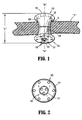
- Fig. 1 is a front perspective view of an embodiment of a seal anchor member in accordance with the present disclosure shown positioned within tissue;
- Fig. 2 is a bottom view of the seal anchor member of Fig. 1 ;
- Fig. 3 is a front perspective view of an access system including seal anchor member and a light source in accordance with the present disclosure.
- proximal will refer to the end of the apparatus which is closest to the clinician during use, while the term “distal” will refer to the end which is furthest from the clinician, as is traditional and known in the art.
- Seal anchor member 100 is configured and adapted to be inserted within a tissue tract 12 defined by tissue surfaces formed in tissue "T", e.g., an incision.
- tissue tract 12 defined by tissue surfaces formed in tissue "T"
- the seal anchor member may be used through a naturally occurring opening (e.g., anus or vagina) or any incision in a patient's skin.
- Seal anchor member 100 defines a longitudinal axis "A" and has respective trailing (proximal) and leading (distal) ends 102, 104. Intermediate portion 106 is disposed between the trailing and leading ends 102, 104. As depicted in Fig. 1 , trailing and leading ends 102, 104 define substantially planar surfaces. However, in other embodiments, either or both of the trailing and leading ends 102, 104 define surfaces that are substantially convex or concave to assist in the insertion of seal anchor member 100 within tissue tract 12.
- a working chamber 107 may include one or more lumens 108 that are longitudinally disposed between the trailing and leading ends 102, 104 and are adapted to receive a surgical instrument in a substantially sealed relation.
- Intermediate portion 106 defines a radial dimension "R” and extends longitudinally between the trailing and leading ends 102, 104 to define an axial dimension or length "L".
- the radial dimension "R” of intermediate portion 106 varies along the length "L", i.e., the cross-sectional dimension may vary along length "L” and may have an hour-glass configuration.
- the hour-glass configuration of the intermediate portion 106 facilitates anchoring of the seal anchor member 100 within tissue tract 12 defined by tissue surfaces formed in tissue "T".
- the radial dimension "R” may remain substantially uniform along the length "L”.
- the seal anchor members 100 may be formed from a compressible material and may be adapted to transition between a first expanded condition to facilitate securing of the seal anchor member 100 within the tissue tract 12 and in a substantial sealed relation with tissue surfaces "T" defining the tissue tract 12, and a second compressed condition to facilitate at least partial insertion of the seal anchor member 100 within the tissue tract 12.
- one or more light sources 101 may be disposed on the distal end 104 of the seal anchor member 100.
- Light sources 101 may be chosen from any known light source, including but not limited to, light emitting diodes (LEDs) and incandescent lights.
- the light sources 101 are positioned within the leading end 104.
- the one or more light sources 101 may be selectively positioned with respect to the leading end 104.
- one or more of the light sources 101 may be rotated or axially repositioned with respect to the distal end 104 of the seal anchor member.
- one or more of the light sources 101 may be radially repositionable with respect to the seal anchor member 100.
- the light source 101 provides a larger illumination field thereby increasing the illumination of the working space beyond the distal end 104 of the seal anchor member 100.
- An internal battery "B", shown in Fig. 1 may power the light sources 101 and may be operatively coupled to the light sources through power leads 109.
- each of the light sources 101 may have its own power source.
- the light sources 101 may be powered by a power source that is external to the seal anchor member 100.
- Light source 101 may be selected to have a desired wavelength depending on its intended use. For example, some procedures may involve the use of light sensitive materials that are responsive to certain wavelengths of light and the light source 101 may be selected accordingly. Some procedures may involve the application of light sensitive sealants and/or adhesives. For example, photocurable surgical tissue adhesives that are composed of photoreactive gelatin and poly (ethylene glycol) diacrylate may be used in laparoscopic procedures to effect prompt and effective hemostasis. For such procedures, light sources 101 may emit light of any known wavelength of light including, but not limited to, ultraviolet, visible, and near-infrared light.
- the light sources 101 are integrated into the seal anchor member 100 and may emit light having the same, different, or variable wavelengths depending on the application. Therefore, two or more light sources 101 having different wavelengths and/or properties may be included within a single seal anchor member 100.
- one light source 101 may be adapted to emit visible light, e.g., white light, for traditional illumination, while other light sources 101 may be adapted to excite biomarkers or immunological agents/dyes adapted to mark certain structures, e.g., vasculature, lymph, nerve, and neural networks, or conditions, e.g., tumors or other diseases.
- a clinician may activate, deactivate, and regulate the light emitted from light sources 101.
- the intensity of the light emitted from the light sources 101 may be adjusted during the procedure.
- Some procedures may include the application of biologically active compounds that are light-responsive. By adjusting the light exposure, the concentration of the active form of such compounds may be controlled.
- Some molecules can either be can either be irreversibly activated with light or reversibly switched between active and inactive states.
- the light sources 101 may be adapted to be synchronized with other instruments such as camera and/or light detection systems (not shown) to provide on demand direct local illumination and visualization as needed.
- other instruments such as camera and/or light detection systems (not shown) to provide on demand direct local illumination and visualization as needed.
- operation of the camera may be synchronized with the emission of light into the body space on which the procedure is being performed.
- Wireless or other means may be used to synchronize the light sources 101 with the other instruments.
- an access system 200 includes a seal anchor member 201, a light source 210, and fiber optic cables 211.
- the seal anchor member 201 is substantially similar to the seal anchor member 100 described above, and is adapted to be inserted within a tissue tract in a substantially identical manner as described above with reference to seal anchor member 100.
- the seal anchor member 201 includes trailing and leading ends 102, 104, an intermediate portion 106, a working chamber 107 may include one or more lumens 108, and one or more fiber optics disposed within the seal anchor member 201 operatively coupled to an external light source 210 and terminating in the leading end 104.
- light source 210 is shown external to the seal anchor member 201, in other embodiments the light source to which the fiber optic cables 211 are connected may be integral to the seal anchor member 201. Each of the fiber optics 211 may be adapted to emit light of a wavelength suitable for its intended use.
Landscapes
- Health & Medical Sciences (AREA)
- Life Sciences & Earth Sciences (AREA)
- Surgery (AREA)
- Physics & Mathematics (AREA)
- Optics & Photonics (AREA)
- Engineering & Computer Science (AREA)
- Biomedical Technology (AREA)
- Medical Informatics (AREA)
- Pathology (AREA)
- Radiology & Medical Imaging (AREA)
- Biophysics (AREA)
- Veterinary Medicine (AREA)
- Heart & Thoracic Surgery (AREA)
- Nuclear Medicine, Radiotherapy & Molecular Imaging (AREA)
- Molecular Biology (AREA)
- Animal Behavior & Ethology (AREA)
- General Health & Medical Sciences (AREA)
- Public Health (AREA)
- Microelectronics & Electronic Packaging (AREA)
- Endoscopes (AREA)
- Surgical Instruments (AREA)
Abstract
Description
- This application claims priority to, and benefit of,
U.S. Provisional Application Serial Number 61/262,642, filed November 19, 2009 - The present disclosure relates to medical devices including an integrated light source. More particularly, the present disclosure relates to an access device including an integrated light source.
- Minimally invasive procedures are procedures that are performed through small incisions in a patient's skin. Such procedures have several advantages over traditional, open surgeries, such as reduced trauma to the patient and a faster recovery time. Generally, such procedures are referred to as "endoscopic", unless performed on the patient's abdomen. Procedures that are performed on the abdomen are referred to as "laparoscopic". Throughout the present disclosure, the term "minimally invasive" should be understood to encompass both endoscopic and laparoscopic procedures.
- During a typical minimally invasive procedure, surgical objects, such as surgical access devices (e.g., trocar and cannula assemblies) or endoscopes, are inserted into the patient's body through an incision in tissue or into a naturally occurring orifice (e.g., mouth, anus, or vagina). In general, prior to the introduction of the surgical object into the patient's body, insufflation gases are used to enlarge the area surrounding the target surgical site to create a larger, more accessible work area.
- Minimally invasive procedures are complicated by a number of visual limitations. In particular, the surgeon's view is limited to what is directly in front of and is illuminated by a scope inserted in the incision. To inhibit damage to internal body structures, it is desirable to illuminate the surgeon's viewing field to a greater degree than is currently possible.
- Disclosed herein is a seal anchor member that is configured and adapted for placement within a tissue tract. The seal anchor member includes a housing that includes leading and trailing ends. One or both of the trailing and leading ends may define surfaces that are substantially convex or concave to assist in the insertion of seal anchor member the within tissue tract. An intermediate portion is positioned between the leading and trailing ends and is adapted to be positioned within the tissue tract in a substantially sealed relation with tissue surfaces that define the tissue tract. At least one longitudinal port extends between the leading and trailing ends, and is adapted to receive an instrument therein in a substantially sealed relation.
- The seal anchor member may be formed from a compressible material that is adapted to transition between a first expanded condition and a second compressed condition. The first expanded condition facilitates securing of the seal anchor member within the tissue tract. The second compressed condition facilitates at least partial insertion of the seal anchor member within the tissue tract.
- At least one light source is operatively associated with the leading end. The light source may be operatively coupled to either an external or an internal power source, e.g., a battery. The at least one light source may be a light emitting diode (LED) or may be a fiber optic cable that is operatively coupled to an external light source. Other light sources as are known in the art may be used as well. The light source may be selectively positionable, i.e.., reoriented with respect to the leading end, to facilitate the illumination of particular areas of interest within the body cavity.
- The light source may emit light having any known wavelength. For example, the light source may emit visible, ultraviolet, or near-infrared light. In an embodiment, the light source may emit light adapted to polymerize a sealant or an adhesive. In another embodiment, the light source may emit light adapted to excite a biomarker or an immunological agent or dye. In addition, in certain applications, the light sources may emit more than one type, i.e, wavelength of light. Moreover, the intensity of the light emitted from the light sources may be adjusted before or during a procedure, i.e., decreased or increased as desired, or otherwise regulated. In addition, the intensity and wavelength, as well as the activation, of the light sources may be synchronized to the use or activation of other instruments, e.g., camera or light detection systems.
- These and other features of the apparatus disclosed herein will become more readily apparent to those skilled in the art from the following detailed description of various embodiments of the present disclosure.
- Various embodiments of the present disclosure are described hereinbelow with reference to the drawings, wherein:
-
Fig. 1 is a front perspective view of an embodiment of a seal anchor member in accordance with the present disclosure shown positioned within tissue; -
Fig. 2 is a bottom view of the seal anchor member ofFig. 1 ; and -
Fig. 3 is a front perspective view of an access system including seal anchor member and a light source in accordance with the present disclosure. - In the drawings and in the description which follows, in which like references numerals identify similar or identical elements, the term "proximal" will refer to the end of the apparatus which is closest to the clinician during use, while the term "distal" will refer to the end which is furthest from the clinician, as is traditional and known in the art.
- A
seal anchor member 100 will now be described with reference toFigs. 1-2 .Seal anchor member 100 is configured and adapted to be inserted within atissue tract 12 defined by tissue surfaces formed in tissue "T", e.g., an incision. Although the presently describedseal anchor member 100 is discussed in connection with minimally invasive procedures, it is within the scope of the present disclosure that the seal anchor member may be used through a naturally occurring opening (e.g., anus or vagina) or any incision in a patient's skin. -
Seal anchor member 100 defines a longitudinal axis "A" and has respective trailing (proximal) and leading (distal)ends Intermediate portion 106 is disposed between the trailing and leadingends Fig. 1 , trailing and leadingends ends seal anchor member 100 withintissue tract 12. A workingchamber 107 may include one ormore lumens 108 that are longitudinally disposed between the trailing and leadingends -
Intermediate portion 106 defines a radial dimension "R" and extends longitudinally between the trailing and leadingends Fig. 1 , the radial dimension "R" ofintermediate portion 106 varies along the length "L", i.e., the cross-sectional dimension may vary along length "L" and may have an hour-glass configuration. The hour-glass configuration of theintermediate portion 106 facilitates anchoring of theseal anchor member 100 withintissue tract 12 defined by tissue surfaces formed in tissue "T". However, in other embodiments, the radial dimension "R" may remain substantially uniform along the length "L". - The
seal anchor members 100 may be formed from a compressible material and may be adapted to transition between a first expanded condition to facilitate securing of theseal anchor member 100 within thetissue tract 12 and in a substantial sealed relation with tissue surfaces "T" defining thetissue tract 12, and a second compressed condition to facilitate at least partial insertion of theseal anchor member 100 within thetissue tract 12. - As shown in
Figs. 1 and 2 , one ormore light sources 101 may be disposed on thedistal end 104 of theseal anchor member 100.Light sources 101 may be chosen from any known light source, including but not limited to, light emitting diodes (LEDs) and incandescent lights. - As shown in
Fig. 2 , thelight sources 101 are positioned within the leadingend 104. The one or morelight sources 101 may be selectively positioned with respect to the leadingend 104. As such, one or more of thelight sources 101 may be rotated or axially repositioned with respect to thedistal end 104 of the seal anchor member. It is also contemplated that one or more of thelight sources 101 may be radially repositionable with respect to theseal anchor member 100. In this configuration, thelight source 101 provides a larger illumination field thereby increasing the illumination of the working space beyond thedistal end 104 of theseal anchor member 100. By adjusting the orientation of thelight sources 101 with respect theleading end 104, the illumination of particular internal body structures is facilitated. - An internal battery "B", shown in
Fig. 1 , may power thelight sources 101 and may be operatively coupled to the light sources through power leads 109. In other embodiments, each of thelight sources 101 may have its own power source. In still other embodiments, thelight sources 101 may be powered by a power source that is external to theseal anchor member 100. -
Light source 101 may be selected to have a desired wavelength depending on its intended use. For example, some procedures may involve the use of light sensitive materials that are responsive to certain wavelengths of light and thelight source 101 may be selected accordingly. Some procedures may involve the application of light sensitive sealants and/or adhesives. For example, photocurable surgical tissue adhesives that are composed of photoreactive gelatin and poly (ethylene glycol) diacrylate may be used in laparoscopic procedures to effect prompt and effective hemostasis. For such procedures,light sources 101 may emit light of any known wavelength of light including, but not limited to, ultraviolet, visible, and near-infrared light. - The
light sources 101 are integrated into theseal anchor member 100 and may emit light having the same, different, or variable wavelengths depending on the application. Therefore, two or morelight sources 101 having different wavelengths and/or properties may be included within a singleseal anchor member 100. For example, onelight source 101 may be adapted to emit visible light, e.g., white light, for traditional illumination, while otherlight sources 101 may be adapted to excite biomarkers or immunological agents/dyes adapted to mark certain structures, e.g., vasculature, lymph, nerve, and neural networks, or conditions, e.g., tumors or other diseases. - During a procedure, a clinician may activate, deactivate, and regulate the light emitted from
light sources 101. For example, the intensity of the light emitted from thelight sources 101 may be adjusted during the procedure. Some procedures may include the application of biologically active compounds that are light-responsive. By adjusting the light exposure, the concentration of the active form of such compounds may be controlled. Some molecules can either be can either be irreversibly activated with light or reversibly switched between active and inactive states. - In a further embodiment, the
light sources 101 may be adapted to be synchronized with other instruments such as camera and/or light detection systems (not shown) to provide on demand direct local illumination and visualization as needed. For example, operation of the camera may be synchronized with the emission of light into the body space on which the procedure is being performed. Wireless or other means may be used to synchronize thelight sources 101 with the other instruments. - In yet another embodiment, as illustrated in
Fig. 3 , anaccess system 200 includes aseal anchor member 201, alight source 210, andfiber optic cables 211. Theseal anchor member 201 is substantially similar to theseal anchor member 100 described above, and is adapted to be inserted within a tissue tract in a substantially identical manner as described above with reference to sealanchor member 100. - The differences between the
seal anchor members seal anchor member 201 includes trailing and leading ends 102, 104, anintermediate portion 106, a workingchamber 107 may include one ormore lumens 108, and one or more fiber optics disposed within theseal anchor member 201 operatively coupled to an externallight source 210 and terminating in theleading end 104. - Although
light source 210 is shown external to theseal anchor member 201, in other embodiments the light source to which thefiber optic cables 211 are connected may be integral to theseal anchor member 201. Each of thefiber optics 211 may be adapted to emit light of a wavelength suitable for its intended use. - Although the illustrative embodiments of the present disclosure have been described herein with reference to the accompanying drawings, the above description, disclosure, and figures should not be construed as limiting, but merely as exemplifications of particular embodiments. It is to be understood that the disclosure is not limited to those precise embodiments, and that various other changes and modifications may be effected therein by one skilled in the art without departing from the scope or spirit of the disclosure.
- The invention may be described by reference to the following numbered paragraphs:-
- 1. A seal anchor member comprising a housing including leading and trailing ends; an intermediate portion positioned between the leading and trailing ends, the intermediate portion being adapted to be positioned within a tissue tract in a substantially sealed relation with tissue surfaces defining the tissue tract; at least one longitudinal port extending between the leading and trailing ends, the at least one longitudinal port being adapted to receive an instrument therein in a substantially sealed relation; and at least one light source operatively associated with the leading end.
- 2. The seal anchor member of
paragraph 1, wherein the at least one light source is operatively coupled to a power source that is integrated into the housing. - 3. The seal anchor member of paragraph 2, wherein the power source is a battery.
- 4. The seal anchor member of
paragraph 1, wherein the at least one light source is operatively coupled to a power source that is external to the housing. - 5. The seal anchor member of
paragraph 1, wherein the at least one light source is a light emitting diode. - 6. The seal anchor member of
paragraph 1, wherein the at least one light source is a fiber optic cable that is operatively coupled to an external light source. - 7. The seal anchor member of
paragraph 1, wherein the at least one light source is selectively positionable. - 8. The seal anchor member of
paragraph 1, wherein the seal anchor is further comprised of a compressible material and is adapted to transition between a first expanded condition to facilitate securing of the seal anchor member within the tissue tract and second compressed condition to facilitate at least partial insertion of the seal anchor member within the tissue tract. - 9. The seal anchor member of
paragraph 1, wherein at least one of the leading and trailing ends has a concave configuration to facilitate insertion of the seal anchor member within the tissue tract. - 10. The seal anchor member of
paragraph 1, wherein the at least one light source emits light selected from the group consisting of: visible, ultraviolet, and near-infrared light. - 11. The seal anchor member of
paragraph 1, wherein during use of the seal anchor member, the wavelength of the light emitted from the light source is adjustable. - 12. The seal anchor member of
paragraph 1, wherein a plurality of light sources emit light having at least two different wavelengths. - 13. The seal anchor member of
paragraph 11, wherein intensity of the light emitted is adjustable.
Claims (12)
- A seal anchor member comprising:a housing including leading and trailing ends;an intermediate portion positioned between the leading and trailing ends, the intermediate portion being adapted to be positioned within a tissue tract in a substantially sealed relation with tissue surfaces defining the tissue tract;at least one longitudinal port extending between the leading and trailing ends, the at least one longitudinal port being adapted to receive an instrument therein in a substantially sealed relation; andat least one light source operatively associated with the leading end; andwherein the seal anchor is further comprised of a compressible material and is adapted to transition between a first expanded condition to facilitate securing of the seal anchor member within the tissue tract and second compressed condition to facilitate at least partial insertion of the seal anchor member within the tissue tract.
- The seal anchor member of claim 1, wherein the at least one light source is operatively coupled to a power source that is integrated into the housing.
- The seal anchor member of claim 2, wherein the power source is a battery.
- The seal anchor member of claim 1, wherein the at least one light source is operatively coupled to a power source that is external to the housing.
- The seal anchor member of any preceding claim, wherein the at least one light source is a light emitting diode.
- The seal anchor member of any of claims 1 to 4, wherein the at least one light source is a fiber optic cable that is operatively coupled to an external light source.
- The seal anchor member of any preceding claim, wherein the at least one light source is selectively positionable.
- The seal anchor member of any preceding claim, wherein at least one of the leading and trailing ends has a concave configuration to facilitate insertion of the seal anchor member within the tissue tract.
- The seal anchor member of any preceding claim, wherein the at least one light source emits light selected from the group consisting of: visible, ultraviolet, and near-infrared light.
- The seal anchor member of any preceding claim, wherein during use of the seal anchor member, the wavelength of the light emitted from the light source is adjustable.
- The seal anchor member of any preceding claim, wherein a plurality of light sources emit light having at least two different wavelengths.
- The seal anchor member of claim 10, wherein intensity of the light emitted is adjustable.
Applications Claiming Priority (2)
| Application Number | Priority Date | Filing Date | Title |
|---|---|---|---|
| US26264209P | 2009-11-19 | 2009-11-19 | |
| US12/915,108 US20110118553A1 (en) | 2009-11-19 | 2010-10-29 | Access device including an integrated light source |
Publications (1)
| Publication Number | Publication Date |
|---|---|
| EP2324757A1 true EP2324757A1 (en) | 2011-05-25 |
Family
ID=43571292
Family Applications (1)
| Application Number | Title | Priority Date | Filing Date |
|---|---|---|---|
| EP10251955A Withdrawn EP2324757A1 (en) | 2009-11-19 | 2010-11-18 | Access device including an integrated light source |
Country Status (5)
| Country | Link |
|---|---|
| US (1) | US20110118553A1 (en) |
| EP (1) | EP2324757A1 (en) |
| JP (1) | JP2011104366A (en) |
| AU (1) | AU2010241333A1 (en) |
| CA (1) | CA2719832A1 (en) |
Families Citing this family (40)
| Publication number | Priority date | Publication date | Assignee | Title |
|---|---|---|---|---|
| WO2009046164A1 (en) | 2007-10-05 | 2009-04-09 | Tyco Healthcare Group Lp | Seal anchor for use in surgical procedures |
| USD738500S1 (en) | 2008-10-02 | 2015-09-08 | Covidien Lp | Seal anchor for use in surgical procedures |
| CN102469920B (en) * | 2010-06-25 | 2016-03-09 | 柯尼卡美能达精密光学株式会社 | Probe, diagnostic device and method of use thereof |
| US20120109304A1 (en) * | 2010-10-29 | 2012-05-03 | Warsaw Orthopedic, Inc. | Medical implant and method for photodynamic therapy |
| US8668642B2 (en) * | 2010-11-23 | 2014-03-11 | Covidien Lp | Port device including retractable endoscope cleaner |
| US8864659B2 (en) | 2010-11-23 | 2014-10-21 | Covidien Lp | Seal anchor for use in surgical procedures |
| US8753267B2 (en) | 2011-01-24 | 2014-06-17 | Covidien Lp | Access assembly insertion device |
| US9549758B2 (en) * | 2011-03-23 | 2017-01-24 | Covidien Lp | Surgical access assembly with adapter |
| US9662018B2 (en) * | 2012-03-30 | 2017-05-30 | Covidien Lp | Integrated self-fixating visualization devices, systems and methods |
| US9480855B2 (en) * | 2012-09-26 | 2016-11-01 | DePuy Synthes Products, Inc. | NIR/red light for lateral neuroprotection |
| US10064649B2 (en) | 2014-07-07 | 2018-09-04 | Covidien Lp | Pleated seal for surgical hand or instrument access |
| US9707011B2 (en) | 2014-11-12 | 2017-07-18 | Covidien Lp | Attachments for use with a surgical access device |
| US11160682B2 (en) | 2017-06-19 | 2021-11-02 | Covidien Lp | Method and apparatus for accessing matter disposed within an internal body vessel |
| US10828065B2 (en) | 2017-08-28 | 2020-11-10 | Covidien Lp | Surgical access system |
| US10675056B2 (en) | 2017-09-07 | 2020-06-09 | Covidien Lp | Access apparatus with integrated fluid connector and control valve |
| US11389193B2 (en) | 2018-10-02 | 2022-07-19 | Covidien Lp | Surgical access device with fascial closure system |
| US11457949B2 (en) | 2018-10-12 | 2022-10-04 | Covidien Lp | Surgical access device and seal guard for use therewith |
| US10736659B2 (en) | 2018-10-23 | 2020-08-11 | Covidien Lp | Optical trocar assembly |
| US11166748B2 (en) | 2019-02-11 | 2021-11-09 | Covidien Lp | Seal assemblies for surgical access assemblies |
| US10792071B2 (en) | 2019-02-11 | 2020-10-06 | Covidien Lp | Seals for surgical access assemblies |
| US11000313B2 (en) | 2019-04-25 | 2021-05-11 | Covidien Lp | Seals for surgical access devices |
| US11413068B2 (en) | 2019-05-09 | 2022-08-16 | Covidien Lp | Seal assemblies for surgical access assemblies |
| US11357542B2 (en) | 2019-06-21 | 2022-06-14 | Covidien Lp | Valve assembly and retainer for surgical access assembly |
| US11259840B2 (en) | 2019-06-21 | 2022-03-01 | Covidien Lp | Valve assemblies for surgical access assemblies |
| US11259841B2 (en) | 2019-06-21 | 2022-03-01 | Covidien Lp | Seal assemblies for surgical access assemblies |
| US11413065B2 (en) | 2019-06-28 | 2022-08-16 | Covidien Lp | Seal assemblies for surgical access assemblies |
| US11399865B2 (en) | 2019-08-02 | 2022-08-02 | Covidien Lp | Seal assemblies for surgical access assemblies |
| US11523842B2 (en) | 2019-09-09 | 2022-12-13 | Covidien Lp | Reusable surgical port with disposable seal assembly |
| US11432843B2 (en) | 2019-09-09 | 2022-09-06 | Covidien Lp | Centering mechanisms for a surgical access assembly |
| US11812991B2 (en) | 2019-10-18 | 2023-11-14 | Covidien Lp | Seal assemblies for surgical access assemblies |
| US11464540B2 (en) | 2020-01-17 | 2022-10-11 | Covidien Lp | Surgical access device with fixation mechanism |
| US12324606B2 (en) | 2020-01-28 | 2025-06-10 | Covidien Lp | Seal assemblies for surgical access assemblies |
| US11576701B2 (en) | 2020-03-05 | 2023-02-14 | Covidien Lp | Surgical access assembly having a pump |
| US11642153B2 (en) | 2020-03-19 | 2023-05-09 | Covidien Lp | Instrument seal for surgical access assembly |
| US11541218B2 (en) | 2020-03-20 | 2023-01-03 | Covidien Lp | Seal assembly for a surgical access assembly and method of manufacturing the same |
| US11446058B2 (en) | 2020-03-27 | 2022-09-20 | Covidien Lp | Fixture device for folding a seal member |
| US11717321B2 (en) | 2020-04-24 | 2023-08-08 | Covidien Lp | Access assembly with retention mechanism |
| US11622790B2 (en) | 2020-05-21 | 2023-04-11 | Covidien Lp | Obturators for surgical access assemblies and methods of assembly thereof |
| US11751908B2 (en) | 2020-06-19 | 2023-09-12 | Covidien Lp | Seal assembly for surgical access assemblies |
| JP7830446B2 (en) * | 2020-10-08 | 2026-03-16 | アプライド メディカル リソーシーズ コーポレイション | Surgical access system with lighting |
Citations (4)
| Publication number | Priority date | Publication date | Assignee | Title |
|---|---|---|---|---|
| US20020183594A1 (en) * | 1997-04-30 | 2002-12-05 | University Of Massachuetts, A Massachusetts Corporation | Surgical access port |
| WO2004075930A2 (en) * | 2003-02-25 | 2004-09-10 | Applied Medical Resources Corporation | Apparatus and method for illuminating a peritoneal cavity during laparoscopic surgery |
| WO2004075741A2 (en) * | 2003-02-25 | 2004-09-10 | Applied Medical Resources Corporation | Wound retractor for use in hand assisted laparoscopic surgery |
| US20090093752A1 (en) * | 2007-10-05 | 2009-04-09 | Tyco Healthcare Group Lp | Seal anchor for use in surgical procedures |
Family Cites Families (86)
| Publication number | Priority date | Publication date | Assignee | Title |
|---|---|---|---|---|
| US4016884A (en) * | 1975-07-02 | 1977-04-12 | Kwan Gett Clifford S | Atriotomy access device |
| US4183357A (en) * | 1976-08-02 | 1980-01-15 | Bentley Laboratories, Inc. | Chronic transcutaneous implant assembly for enterostomies |
| GB2161389B (en) * | 1984-07-05 | 1988-06-08 | Wolf Gmbh Richard | Instrument insert for a uretero-renoscope |
| FR2641692A1 (en) * | 1989-01-17 | 1990-07-20 | Nippon Zeon Co | Plug for closing an opening for a medical application, and device for the closure plug making use thereof |
| US5176697A (en) * | 1989-04-06 | 1993-01-05 | Hasson Harrith M | Laparoscopic cannula |
| US5514153A (en) * | 1990-03-02 | 1996-05-07 | General Surgical Innovations, Inc. | Method of dissecting tissue layers |
| US5990382A (en) * | 1990-08-29 | 1999-11-23 | Biomedical Enterprises, Inc. | Method and implant for surgical manipulation of bone |
| US5082005A (en) * | 1990-12-18 | 1992-01-21 | New England Deaconess Hospital | Surgical access device |
| US5183471A (en) * | 1992-01-24 | 1993-02-02 | Wilk Peter J | Laparoscopic cannula |
| GR930100244A (en) * | 1992-06-30 | 1994-02-28 | Ethicon Inc | Flexible endoscopic surgical port |
| US5511564A (en) * | 1992-07-29 | 1996-04-30 | Valleylab Inc. | Laparoscopic stretching instrument and associated method |
| US5312391A (en) * | 1992-07-29 | 1994-05-17 | Wilk Peter J | Laparoscopic instrument assembly |
| US5395367A (en) * | 1992-07-29 | 1995-03-07 | Wilk; Peter J. | Laparoscopic instrument with bendable shaft and removable actuator |
| US5507758A (en) * | 1993-08-25 | 1996-04-16 | Inlet Medical, Inc. | Insertable suture grasping probe guide, and methodology for using same |
| US5480410A (en) * | 1994-03-14 | 1996-01-02 | Advanced Surgical, Inc. | Extracorporeal pneumoperitoneum access bubble |
| US5569205A (en) * | 1994-07-14 | 1996-10-29 | Hart; Charles C. | Multiport trocar |
| US5514133A (en) * | 1994-08-26 | 1996-05-07 | Golub; Robert | Access device for endoscopic surgery |
| US5653705A (en) * | 1994-10-07 | 1997-08-05 | General Surgical Innovations, Inc. | Laparoscopic access port for surgical instruments or the hand |
| US5713869A (en) * | 1995-03-08 | 1998-02-03 | Morejon; Orlando | Trocar assembly |
| US5713858A (en) * | 1995-04-28 | 1998-02-03 | Medtronic, Inc. | Permanently implantable guiding catheter |
| US5741298A (en) * | 1995-04-28 | 1998-04-21 | Macleod; Cathel | Method and devices for video-assisted surgical techniques |
| US5899208A (en) * | 1995-05-08 | 1999-05-04 | Gaya Limited | Hand access port |
| US5601581A (en) * | 1995-05-19 | 1997-02-11 | General Surgical Innovations, Inc. | Methods and devices for blood vessel harvesting |
| US5634937A (en) * | 1995-05-19 | 1997-06-03 | General Surgical Innovations, Inc. | Skin seal with inflatable membrane |
| US5997515A (en) * | 1995-05-19 | 1999-12-07 | General Surgical Innovations, Inc. | Screw-type skin seal with inflatable membrane |
| US6605063B2 (en) * | 1998-09-21 | 2003-08-12 | Gerald G. Bousquet | Transcutaneous access device |
| US5894843A (en) * | 1996-02-20 | 1999-04-20 | Cardiothoracic Systems, Inc. | Surgical method for stabilizing the beating heart during coronary artery bypass graft surgery |
| US5951588A (en) * | 1996-02-29 | 1999-09-14 | Moenning; Stephen P. | Apparatus and method for protecting a port site opening in the wall of a body cavity |
| US6048309A (en) * | 1996-03-04 | 2000-04-11 | Heartport, Inc. | Soft tissue retractor and delivery device therefor |
| US6814700B1 (en) * | 1996-03-04 | 2004-11-09 | Heartport, Inc. | Soft tissue retractor and method for providing surgical access |
| EP0900051A1 (en) * | 1996-05-08 | 1999-03-10 | Salviac Limited | An occluder device |
| DE69735146T2 (en) * | 1996-05-09 | 2006-09-28 | Olympus Corporation | Surgical tool for holding a cavity |
| US6706050B1 (en) * | 1996-05-10 | 2004-03-16 | Emmanuil Giannadakis | System of laparoscopic-endoscopic surgery |
| US5741234A (en) * | 1996-07-16 | 1998-04-21 | Aboul-Hosn; Walid Nagib | Anatomical cavity access sealing condit |
| US5728103A (en) * | 1996-08-23 | 1998-03-17 | Applied Medical Technology, Inc. | Implantable subcutaneous access device and method of using same |
| IL119131A (en) * | 1996-08-26 | 2002-04-21 | Oridion Medical Ltd | Multiple channel sample port for airway adaptor |
| US5735791A (en) * | 1997-01-31 | 1998-04-07 | Research Medical, Inc. | Inflatable heart elevation apparatus and method |
| US5971919A (en) * | 1997-04-03 | 1999-10-26 | Davis; James M. | Light intensity and color adjustable endoscope |
| US5865817A (en) * | 1997-04-29 | 1999-02-02 | Moenning; Stephen P. | Apparatus and method for securing a medical instrument to a cannula of a trocar assembly |
| US5906577A (en) * | 1997-04-30 | 1999-05-25 | University Of Massachusetts | Device, surgical access port, and method of retracting an incision into an opening and providing a channel through the incision |
| AU742503B2 (en) * | 1997-06-25 | 2002-01-03 | Biotap A/S | Intercutaneous implant device |
| US6382211B1 (en) * | 1997-07-21 | 2002-05-07 | Medical Creative Technologies, Inc. | Surgical retractor liner appliance |
| JPH1199156A (en) * | 1997-07-29 | 1999-04-13 | Olympus Optical Co Ltd | Surgical access device |
| US6042573A (en) * | 1997-12-11 | 2000-03-28 | Smith & Nephew, Inc. | Surgical valve |
| US6187000B1 (en) * | 1998-08-20 | 2001-02-13 | Endius Incorporated | Cannula for receiving surgical instruments |
| US7344547B2 (en) * | 1998-09-15 | 2008-03-18 | Phavel Systems, Inc. | Laparoscopic instruments and trocar systems and related surgical method |
| US7195590B2 (en) * | 1998-12-01 | 2007-03-27 | Atropos Limited | Surgical device |
| AU773994B2 (en) * | 1998-12-01 | 2004-06-10 | Atropos Limited | A surgical device for retracting and/or sealing an incision |
| IE991010A1 (en) * | 1998-12-01 | 2000-07-12 | Atropos Ltd | A Device |
| DE19902036C1 (en) * | 1999-01-20 | 2000-03-16 | Storz Karl Gmbh & Co Kg | Device to hold trocar tube aligned in various spaces; is designed to fasten trocar tube in different adjustable in situ positions on patient |
| US6033428A (en) * | 1999-01-26 | 2000-03-07 | Sardella; William V. | Laparoscopic surgery device |
| SE9900454D0 (en) * | 1999-02-10 | 1999-02-10 | Safe Conduct Ab | trocar |
| IES990218A2 (en) * | 1999-03-18 | 2000-11-15 | Gaya Ltd | A surgical device |
| US20020055714A1 (en) * | 1999-05-18 | 2002-05-09 | Rothschild Richard B. | Method and apparatus for the non-surgical placement of a subcutaneously tunnelled catheter through the skin into a central vein |
| WO2001064124A1 (en) * | 2000-03-01 | 2001-09-07 | Surgical Navigation Technologies, Inc. | Multiple cannula image guided tool for image guided procedures |
| US6551270B1 (en) * | 2000-08-30 | 2003-04-22 | Snowden Pencer, Inc. | Dual lumen access port |
| WO2002017771A2 (en) * | 2000-09-01 | 2002-03-07 | Advanced Vascular Technologies, Llc | Multi-fastener surgical apparatus and method |
| WO2002034108A2 (en) * | 2000-10-19 | 2002-05-02 | Applied Medical Resources Corporation | Surgical access apparatus and method |
| US6890295B2 (en) * | 2002-10-31 | 2005-05-10 | Medtronic, Inc. | Anatomical space access tools and methods |
| US6958037B2 (en) * | 2001-10-20 | 2005-10-25 | Applied Medical Resources Corporation | Wound retraction apparatus and method |
| US7052454B2 (en) * | 2001-10-20 | 2006-05-30 | Applied Medical Resources Corporation | Sealed surgical access device |
| US7192436B2 (en) * | 2001-11-08 | 2007-03-20 | Sub-Q, Inc. | Pledget-handling system and method for delivering hemostasis promoting material to a blood vessel puncture site by fluid pressure |
| US6723088B2 (en) * | 2001-12-20 | 2004-04-20 | Board Of Regents, The University Of Texas | Laparoscopic porting |
| JP2003199755A (en) * | 2001-12-28 | 2003-07-15 | Olympus Optical Co Ltd | Endoscopic surgery trocar |
| JP2003204920A (en) * | 2002-01-11 | 2003-07-22 | Olympus Optical Co Ltd | Insertion aid |
| US6878110B2 (en) * | 2002-01-14 | 2005-04-12 | Seung Choul Yang | Surgical instruments and method for creating anatomic working space in minilaparotomy procedure |
| WO2003066129A2 (en) * | 2002-02-08 | 2003-08-14 | Taut, Inc. | Introducer assembly for medical instruments |
| US6723044B2 (en) * | 2002-03-14 | 2004-04-20 | Apple Medical Corporation | Abdominal retractor |
| WO2003088920A2 (en) * | 2002-04-22 | 2003-10-30 | The Children's Hospital Of Philadelphia | Low profile combination device for gastrostomy or jejunostomy applications having anti-granuloma formation characteristics |
| CA2488118C (en) * | 2002-06-05 | 2012-01-10 | Applied Medical Resources Corporation | Wound retractor |
| US9271753B2 (en) * | 2002-08-08 | 2016-03-01 | Atropos Limited | Surgical device |
| AU2003273510A1 (en) * | 2002-09-19 | 2004-04-08 | Atropos Limited | A wound retractor system |
| US7217277B2 (en) * | 2002-09-30 | 2007-05-15 | Ethicon, Inc. | Device for providing intracardiac access in an open chest |
| FR2846298A1 (en) * | 2002-10-29 | 2004-04-30 | Cit Alcatel | ARTICULATED ASSEMBLY OF SOLAR GENERATOR PANELS AND SPATIAL VEHICLE |
| US20050020884A1 (en) * | 2003-02-25 | 2005-01-27 | Hart Charles C. | Surgical access system |
| US7645232B2 (en) * | 2003-05-16 | 2010-01-12 | Zimmer Spine, Inc. | Access device for minimally invasive surgery |
| US6916310B2 (en) * | 2003-05-30 | 2005-07-12 | Codman & Shurtleff, Inc. | Percutaneous access device |
| US7753901B2 (en) * | 2004-07-21 | 2010-07-13 | Tyco Healthcare Group Lp | Laparoscopic instrument and cannula assembly and related surgical method |
| US20050070935A1 (en) * | 2003-09-30 | 2005-03-31 | Ortiz Mark S. | Single lumen access deployable ring for intralumenal anastomosis |
| US20050096695A1 (en) * | 2003-11-03 | 2005-05-05 | Olich Jack M. | Flexible foam seal assembly |
| US7452351B2 (en) * | 2004-04-16 | 2008-11-18 | Kyphon Sarl | Spinal diagnostic methods and apparatus |
| US7223257B2 (en) * | 2004-08-31 | 2007-05-29 | Igor Shubayev | Percutaneous vascular access device |
| US8360968B2 (en) * | 2004-12-01 | 2013-01-29 | Vision—Sciences Inc. | Endoscopic sheath with illumination |
| WO2007010511A1 (en) * | 2005-07-15 | 2007-01-25 | Atropos Limited | A wound retractor |
| EP1933732B1 (en) * | 2005-10-14 | 2009-12-02 | Applied Medical Resources Corporation | Wound retractor with gel cap |
| US9011319B2 (en) * | 2006-03-03 | 2015-04-21 | Axcess Instruments, Inc. | Conical laparoscopic apparatus for minimally invasive surgery |
-
2010
- 2010-10-29 US US12/915,108 patent/US20110118553A1/en not_active Abandoned
- 2010-11-03 CA CA2719832A patent/CA2719832A1/en not_active Abandoned
- 2010-11-09 AU AU2010241333A patent/AU2010241333A1/en not_active Abandoned
- 2010-11-11 JP JP2010253303A patent/JP2011104366A/en active Pending
- 2010-11-18 EP EP10251955A patent/EP2324757A1/en not_active Withdrawn
Patent Citations (4)
| Publication number | Priority date | Publication date | Assignee | Title |
|---|---|---|---|---|
| US20020183594A1 (en) * | 1997-04-30 | 2002-12-05 | University Of Massachuetts, A Massachusetts Corporation | Surgical access port |
| WO2004075930A2 (en) * | 2003-02-25 | 2004-09-10 | Applied Medical Resources Corporation | Apparatus and method for illuminating a peritoneal cavity during laparoscopic surgery |
| WO2004075741A2 (en) * | 2003-02-25 | 2004-09-10 | Applied Medical Resources Corporation | Wound retractor for use in hand assisted laparoscopic surgery |
| US20090093752A1 (en) * | 2007-10-05 | 2009-04-09 | Tyco Healthcare Group Lp | Seal anchor for use in surgical procedures |
Also Published As
| Publication number | Publication date |
|---|---|
| CA2719832A1 (en) | 2011-05-19 |
| AU2010241333A1 (en) | 2011-06-02 |
| JP2011104366A (en) | 2011-06-02 |
| US20110118553A1 (en) | 2011-05-19 |
Similar Documents
| Publication | Publication Date | Title |
|---|---|---|
| US20110118553A1 (en) | Access device including an integrated light source | |
| CA2677965C (en) | Access sheath with removable optical penetrating member | |
| AU2007338691B2 (en) | Surgical visual obturator | |
| AU2007267794B2 (en) | Illuminated surgical access system including a surgical access device and integrated light emitter | |
| ES2381753T3 (en) | Surgical port and breakable introducer assembly | |
| EP2514354A1 (en) | Trocar with integrated light and/or scope optical fibers | |
| JP5503527B2 (en) | Endoscope barrel with connector | |
| BRPI0404237A (en) | Single lumen anastomosis applicator for self-folding closure. | |
| JP2012110685A (en) | Seal anchor for use in surgical procedure | |
| CA2716060A1 (en) | Seal anchor with non-parallel lumens | |
| US20110118551A1 (en) | Collateral soft tissue protection surgical device | |
| US20130225933A1 (en) | Adjustable height port including retention elements | |
| JP2021084032A (en) | Surgical instruments with integrated lighting systems | |
| US20140235946A1 (en) | Optical obturator visualization system | |
| US20200268411A1 (en) | Optical trocar assembly | |
| EP3322373A1 (en) | Lighted gastric medical device assembly and method | |
| KR101010289B1 (en) | Resection surgery positioning device | |
| JP2010172694A (en) | Suture management system for surgical portal apparatus including internal tube | |
| US20100261976A1 (en) | Apparatus and System for Performing Surgery | |
| JP7056847B2 (en) | Multi-camera surgical lighting device with variable diameter | |
| US20100262079A1 (en) | Bendable veress needle assembly | |
| CN222238520U (en) | Fluorescent positioning stomach tube for gastric resection operation | |
| US20200069170A1 (en) | Mathis laparoscopic inguinal hernia illuminator | |
| CN121285342A (en) | Surgical stapler with illumination | |
| BR102012021227A2 (en) | SINGLE ACCESS PORTAL FOR SURGERY |
Legal Events
| Date | Code | Title | Description |
|---|---|---|---|
| PUAI | Public reference made under article 153(3) epc to a published international application that has entered the european phase |
Free format text: ORIGINAL CODE: 0009012 |
|
| AK | Designated contracting states |
Kind code of ref document: A1 Designated state(s): AL AT BE BG CH CY CZ DE DK EE ES FI FR GB GR HR HU IE IS IT LI LT LU LV MC MK MT NL NO PL PT RO RS SE SI SK SM TR |
|
| AX | Request for extension of the european patent |
Extension state: BA ME |
|
| 17P | Request for examination filed |
Effective date: 20111027 |
|
| 17Q | First examination report despatched |
Effective date: 20120430 |
|
| 17Q | First examination report despatched |
Effective date: 20120507 |
|
| GRAP | Despatch of communication of intention to grant a patent |
Free format text: ORIGINAL CODE: EPIDOSNIGR1 |
|
| STAA | Information on the status of an ep patent application or granted ep patent |
Free format text: STATUS: THE APPLICATION IS DEEMED TO BE WITHDRAWN |
|
| 18D | Application deemed to be withdrawn |
Effective date: 20130216 |