EP2320900B1 - Opioids for use in the treatment of resistant cancer patients - Google Patents
Opioids for use in the treatment of resistant cancer patients Download PDFInfo
- Publication number
- EP2320900B1 EP2320900B1 EP09776348.6A EP09776348A EP2320900B1 EP 2320900 B1 EP2320900 B1 EP 2320900B1 EP 09776348 A EP09776348 A EP 09776348A EP 2320900 B1 EP2320900 B1 EP 2320900B1
- Authority
- EP
- European Patent Office
- Prior art keywords
- cells
- cancer
- leukaemia
- apoptosis
- methadone
- Prior art date
- Legal status (The legal status is an assumption and is not a legal conclusion. Google has not performed a legal analysis and makes no representation as to the accuracy of the status listed.)
- Active
Links
Images









Classifications
-
- A—HUMAN NECESSITIES
- A61—MEDICAL OR VETERINARY SCIENCE; HYGIENE
- A61K—PREPARATIONS FOR MEDICAL, DENTAL OR TOILETRY PURPOSES
- A61K31/00—Medicinal preparations containing organic active ingredients
- A61K31/33—Heterocyclic compounds
- A61K31/395—Heterocyclic compounds having nitrogen as a ring hetero atom, e.g. guanethidine or rifamycins
- A61K31/435—Heterocyclic compounds having nitrogen as a ring hetero atom, e.g. guanethidine or rifamycins having six-membered rings with one nitrogen as the only ring hetero atom
- A61K31/47—Quinolines; Isoquinolines
- A61K31/485—Morphinan derivatives, e.g. morphine, codeine
-
- A—HUMAN NECESSITIES
- A61—MEDICAL OR VETERINARY SCIENCE; HYGIENE
- A61K—PREPARATIONS FOR MEDICAL, DENTAL OR TOILETRY PURPOSES
- A61K31/00—Medicinal preparations containing organic active ingredients
- A61K31/13—Amines
- A61K31/135—Amines having aromatic rings, e.g. ketamine, nortriptyline
- A61K31/137—Arylalkylamines, e.g. amphetamine, epinephrine, salbutamol, ephedrine or methadone
-
- A—HUMAN NECESSITIES
- A61—MEDICAL OR VETERINARY SCIENCE; HYGIENE
- A61K—PREPARATIONS FOR MEDICAL, DENTAL OR TOILETRY PURPOSES
- A61K31/00—Medicinal preparations containing organic active ingredients
- A61K31/33—Heterocyclic compounds
- A61K31/395—Heterocyclic compounds having nitrogen as a ring hetero atom, e.g. guanethidine or rifamycins
- A61K31/435—Heterocyclic compounds having nitrogen as a ring hetero atom, e.g. guanethidine or rifamycins having six-membered rings with one nitrogen as the only ring hetero atom
- A61K31/47—Quinolines; Isoquinolines
- A61K31/475—Quinolines; Isoquinolines having an indole ring, e.g. yohimbine, reserpine, strychnine, vinblastine
-
- A—HUMAN NECESSITIES
- A61—MEDICAL OR VETERINARY SCIENCE; HYGIENE
- A61K—PREPARATIONS FOR MEDICAL, DENTAL OR TOILETRY PURPOSES
- A61K31/00—Medicinal preparations containing organic active ingredients
- A61K31/33—Heterocyclic compounds
- A61K31/395—Heterocyclic compounds having nitrogen as a ring hetero atom, e.g. guanethidine or rifamycins
- A61K31/495—Heterocyclic compounds having nitrogen as a ring hetero atom, e.g. guanethidine or rifamycins having six-membered rings with two or more nitrogen atoms as the only ring heteroatoms, e.g. piperazine or tetrazines
- A61K31/505—Pyrimidines; Hydrogenated pyrimidines, e.g. trimethoprim
- A61K31/519—Pyrimidines; Hydrogenated pyrimidines, e.g. trimethoprim ortho- or peri-condensed with heterocyclic rings
-
- A—HUMAN NECESSITIES
- A61—MEDICAL OR VETERINARY SCIENCE; HYGIENE
- A61K—PREPARATIONS FOR MEDICAL, DENTAL OR TOILETRY PURPOSES
- A61K31/00—Medicinal preparations containing organic active ingredients
- A61K31/70—Carbohydrates; Sugars; Derivatives thereof
- A61K31/7028—Compounds having saccharide radicals attached to non-saccharide compounds by glycosidic linkages
- A61K31/7034—Compounds having saccharide radicals attached to non-saccharide compounds by glycosidic linkages attached to a carbocyclic compound, e.g. phloridzin
- A61K31/704—Compounds having saccharide radicals attached to non-saccharide compounds by glycosidic linkages attached to a carbocyclic compound, e.g. phloridzin attached to a condensed carbocyclic ring system, e.g. sennosides, thiocolchicosides, escin, daunorubicin
-
- A—HUMAN NECESSITIES
- A61—MEDICAL OR VETERINARY SCIENCE; HYGIENE
- A61K—PREPARATIONS FOR MEDICAL, DENTAL OR TOILETRY PURPOSES
- A61K31/00—Medicinal preparations containing organic active ingredients
- A61K31/70—Carbohydrates; Sugars; Derivatives thereof
- A61K31/7042—Compounds having saccharide radicals and heterocyclic rings
- A61K31/7052—Compounds having saccharide radicals and heterocyclic rings having nitrogen as a ring hetero atom, e.g. nucleosides, nucleotides
- A61K31/706—Compounds having saccharide radicals and heterocyclic rings having nitrogen as a ring hetero atom, e.g. nucleosides, nucleotides containing six-membered rings with nitrogen as a ring hetero atom
- A61K31/7064—Compounds having saccharide radicals and heterocyclic rings having nitrogen as a ring hetero atom, e.g. nucleosides, nucleotides containing six-membered rings with nitrogen as a ring hetero atom containing condensed or non-condensed pyrimidines
- A61K31/7068—Compounds having saccharide radicals and heterocyclic rings having nitrogen as a ring hetero atom, e.g. nucleosides, nucleotides containing six-membered rings with nitrogen as a ring hetero atom containing condensed or non-condensed pyrimidines having oxo groups directly attached to the pyrimidine ring, e.g. cytidine, cytidylic acid
-
- A—HUMAN NECESSITIES
- A61—MEDICAL OR VETERINARY SCIENCE; HYGIENE
- A61K—PREPARATIONS FOR MEDICAL, DENTAL OR TOILETRY PURPOSES
- A61K33/00—Medicinal preparations containing inorganic active ingredients
- A61K33/24—Heavy metals; Compounds thereof
- A61K33/243—Platinum; Compounds thereof
-
- A—HUMAN NECESSITIES
- A61—MEDICAL OR VETERINARY SCIENCE; HYGIENE
- A61K—PREPARATIONS FOR MEDICAL, DENTAL OR TOILETRY PURPOSES
- A61K41/00—Medicinal preparations obtained by treating materials with wave energy or particle radiation ; Therapies using these preparations
-
- A—HUMAN NECESSITIES
- A61—MEDICAL OR VETERINARY SCIENCE; HYGIENE
- A61K—PREPARATIONS FOR MEDICAL, DENTAL OR TOILETRY PURPOSES
- A61K45/00—Medicinal preparations containing active ingredients not provided for in groups A61K31/00 - A61K41/00
- A61K45/06—Mixtures of active ingredients without chemical characterisation, e.g. antiphlogistics and cardiaca
-
- A—HUMAN NECESSITIES
- A61—MEDICAL OR VETERINARY SCIENCE; HYGIENE
- A61P—SPECIFIC THERAPEUTIC ACTIVITY OF CHEMICAL COMPOUNDS OR MEDICINAL PREPARATIONS
- A61P35/00—Antineoplastic agents
-
- A—HUMAN NECESSITIES
- A61—MEDICAL OR VETERINARY SCIENCE; HYGIENE
- A61P—SPECIFIC THERAPEUTIC ACTIVITY OF CHEMICAL COMPOUNDS OR MEDICINAL PREPARATIONS
- A61P35/00—Antineoplastic agents
- A61P35/02—Antineoplastic agents specific for leukemia
-
- A—HUMAN NECESSITIES
- A61—MEDICAL OR VETERINARY SCIENCE; HYGIENE
- A61P—SPECIFIC THERAPEUTIC ACTIVITY OF CHEMICAL COMPOUNDS OR MEDICINAL PREPARATIONS
- A61P43/00—Drugs for specific purposes, not provided for in groups A61P1/00-A61P41/00
Definitions
- the invention relates to novel strategies for the treatment of resistant cancer patients.
- Anti-cancer therapies are frequently ineffective due to resistance of the tumor cells to radio- and/or chemotherapy.
- the resistance When the resistance is acquired during therapy, it often manifests either in a diminished amount of tumor regression for the same dose (either of the radiation or the cytotoxic substance) or an increased dose which is necessary for an equal amount of tumor regression.
- the resistance When the resistance is intrinsic, i.e. not acquired or induced due to the anti-cancer treatment, the tumor cells already originally lack sensitivity to one or more anti-cancer drugs or ionizing radiation.
- chemosensitivity of cancer cells often vary on an individual basis. For pancreas carcinoma e.g. it is known, that only approximately 25% of all patients benefit from the anti-cancer drug gemcitabine. The other 75% are intrinsically resistant to this chemotherapy. Further examples for tumor cells with intrinsic chemo- and radioresistance are glioblastoma or melanoma cells.
- the intrinsic or acquired resistance (or non-response) of tumor cells to radio- and/or chemotherapy can have multiple reasons and can - as exemplified above - vary on an individual basis. Despite intensive research the exact mechanisms still remain elusive. However, it is known that either a single mutation, e.g. at the drug substance binding site or within the cellular detoxification process, can be responsible for the lack of or reduced chemosensitivity. Also the manifestation of cross-resistances to several anticancer drugs often limits the efficacy of anticancer treatments.
- MDR multi-drug resistance
- ABSC ATP binding cassette
- MRP multi-drug resistance associated proteins
- Resistances are not limited to chemotherapeutics or anticancer drugs; cancer patients can also exhibit either an intrinsic or acquired resistance to ionising irradiation applied in radiotherapy.
- An intrinsic radioresistance is known e.g. from melanoma and glioblastoma cells.
- Radioresistance may also be induced by exposure to small or fractionated doses of ionizing radiation.
- ionizing radiation Several studies have documented this effect in vitro even in human cells as well as in several animal models. Different cellular radioprotection mechanisms may be involved, such as alterations in the levels of some cytoplasmatic and nuclear proteins, increased gene expression or DNA repair processes.
- opioids capable of inhibiting the cell proliferation and or growth of cancer cells can overcome resistances in these cancer cells. Therefore these opioids provide novel strategies for treating patients, who so far are considered to be non-treatable or not effectively treatable by conventional therapeutic anticancer approaches.
- This group of alleged non-treatable cancer patients can also be called “non-responders”, “poor-responder” or “non-chemosensitive” or “non-radiosensitive” cancer patients.
- opioids can overcome apoptosis resistance of cancer cells, and thus can effectively be clinically applied as anticancer substances.
- opioids - in particular methadone - were as effective as the conventional chemotherapy (e.g. doxorubicin) and radiation treatments against non-resistant (i.e. sensitive) leukaemia cells, and that normal peripheral blood lymphocytes survived after this treatment.
- opioids are also effective in killing tumor cells, but do not substantially affect normal healthy cells of the patient.
- opioid is defined as a chemical heterogeneous group of natural, synthetic or semi-synthetic substances, working agonistic or antagonistic which all can bind to the well known opioid receptors, preferably to the ⁇ opioid receptor and which are capable of arresting cancer cell proliferation.
- the group of opioids includes natural opiates such as alkaloids like morphine, dihydrocodein, codeine and thebaine, as well as semi-synthetic opiates, derived from the natural opiates (e.g.
- hydromorphone hydrocodone, oxycodone, oxymorphone, desomorphine, diacetylmorphine (Heroin), nicomorphine, dipropanoylmorphine, benzylmorphine and ethylmorphine), or fully synthetic opioids, such as fentanyl, pethidine and methadone, tramadol or propoxyphene. It also includes endogenous opioid peptides, which may be produced naturally in the body as endorphins, dynorphins or enkephalins but which can also be synthesized.
- Opioids are known for their use as analgetics.
- opioid receptors especially ⁇ opioid receptors are involved in the activation of signalling pathways leading to apoptosis.
- opioids can promote apoptosis (Hatsukari et al. 2003).
- opioids it was further discussed to use opioids for the induction of apoptosis in small lung cancer cells (Heusch & Maneckjee 1999).
- the underlying mechanisms were not unrevealed, nor do those results suggest employing opioids for overcoming resistances to conventional anticancer treatments.
- the opioid is capable of inhibiting cancer cell proliferation and/or growth.
- This activity can include e.g. cytostatic or cytotoxic activity as well as arresting growth of cells and/or tumors.
- Cancer cell proliferation is the result of the inhibition of cell division.
- opioids induce cell death in tumors.
- Cell death in the context of the invention includes all types of cells death. This can include necrotic as well as apoptotic cell death or autophagy.
- the cell death is induced by the activation of the caspases-dependent or caspases-independent pathway.
- opioids can induce cell death via various pathways.
- opioids induce apoptosis in cancer cells.
- the "death receptor pathway” (or extrinsic pathway) includes the TNF-receptor-induced (tumor necrosis factor) model and the Fas-receptor-induced model (the Fas-receptor is also known as Apo-1 or CD95). Bindings to these receptors result in the formation of death-inducing signalling pathways in the cell, including the activation of caspases-8.
- the "mitochondrial pathway” (or intrinsic pathway) involves the release of cytochrom c from mitochondria, binding of Apaf-1 and activation of procaspase-9.
- Several regulators are known to activate or deactivate the apoptosis pathways, such as the pro-apoptotic proteins Bax and Bak or the anti-apoptotic proteins Bcl-2, Bcl XL or XIAP.
- the opioid is a member of the methadone group, comprising D-/L-methadone, levomethadone, levacetylmethadol and piritramide. All these opioids can be used as salts.
- the racemic form of D-/L-methadone is preferably provided in form of a hydrochloride.
- the opioid methadone induces apoptosis in cancer cells via the mitochondrial pathway.
- the terms “resistance”, “radioresistance” or “chemoresistance” are defined as a reduced sensitivity of a cancer cell to at least one conventional cancer therapy, i.e. either an anticancer drug or radiotherapy.
- a patient suffering from such a cancer is determined as a "resistant” cancer patient. Since the resistance can be intrinsic or acquired the observed reduction in sensitivity is either compared to fully sensitive "normal” cancer cells, which are responsive to the therapeutically effective dosage of the applied anticancer drug and/or radiation compared to the original sensitivity upon therapy onset. In the later case the resistance manifests either in a diminished amount of tumour regression for the same dose (either of the radiation or the anticancer drug) or an increased dose which is necessary for an equal amount of tumor regression.
- a “resistance” can either be total or partly; in other words, the patients considered treatable according to the invention can exhibit a reduced sensitivity or even a full lack of sensitivity to conventional anticancer treatments. These patients can also be determined as “non-responders” or “poor-responders”.
- a further synonym for a "resistant” cancer or tumor is a "refractory” type of cancer, which can also be either completely or partly refractory. Intrinsic resistance can thus also be determined as a "primary refractory cancer".
- a particular form of refractory or resistant cancer cells ar the so called “kinetically refractory cells”; a phenomenon known e.g. from leukaemia cells, when the cells are at first killed, but reproduce fast that an effective treatment is hardly possible.
- the term "conventional” treatment or therapy refers to the currently accepted and widely used therapeutic treatment of a certain type of cancer, based on the results of past researches and/or regulatory approval.
- Conventional anticancer drugs include cytotoxic and cytostatic agents, which kill the cancer cells or reduce and/or stop their growth or proliferation.
- the modes of action of these anticancer drugs can vary; examples are antimetabolites (e.g. cytarabine, methotrexate, mercaptopurine or clofarabine), DNA cross-linking agents (e.g. cisplatine and its derivates), DNA intercalating substances (e.g. doxorubicin), Topoisomerase poisons (e.g. etoposide), kinase inhibitors (e.g. cetuximab), steroids (e.g. dexamethasone) or mitotic inhibitors (e.g. vincristine).
- antimetabolites e.g. cytarabine, methotrexate, mercaptopurine or clofarabine
- DNA cross-linking agents e.g. cisplatine and its derivates
- DNA intercalating substances e.
- the conventional radiotherapy can also include radiation therapy, which means the use of high-energy radiation from x-rays, alpha, beta and gamma rays, Auger electrons, Ultraviolet rays, neutrons, protons, and other sources to kill cancer cells and shrink tumors.
- Radiation may originate from an outside the body device (external-beam radiation therapy), or it may originate from radioactive sources placed in the body in the vicinity of the cancer cells (internal radiation therapy).
- Systemic radiation therapy uses a radioactive substance, such as a radiolabeled monoclonal antibody, that travels in the blood stream to the target tissue. Radioresistant cancer cells do not or only partly respond to these treatments.
- opioids are applied for overcoming or "breaking" the intrinsic or acquired resistance of cancer cells to conventional anticancer treatments and/or radiation treatment or apoptosis resistance.
- cancer cells considered treatable according to the invention express an opioid receptor, in particular the the ⁇ opioid receptor.
- the group of cancers include, but is not limited to leukaemia, breast cancer, glioblastoma, prostate cancer, lung cancer, non-small cell lung cancer (NSCLC), brain cancer, colon cancer, colorectal cancer.
- NSCLC non-small cell lung cancer
- Examples for cancer types, considered to be treatable according to the inventin, with intrinsic resistance to irradiation are glioblastoma, melanoma or pancreas cancer cells.
- Breast cancer, bladder cancer or leukaemia are often resistant to chemotherapeutics.
- Examples of cancer types which often acquire resistance are melanoma, colon carcinoma, brain tumors glioblastoma, brain cancer, pancreatic cancer, liver cancer, ovarian cancer, cancer of mamma, lung cancer, chronic leukaemia or osteosarkoma.
- the opioids can be used in combination - i.e. as a composite - with conventional anticancer substances or treatments, e.g. cytostatic or cytotoxic substances or radiotherapy.
- the opioids can be combined for example with natural and/or synthetic anticancer substances, natural and/or synthetic cytotoxic substances, antibiotics, cytotoxic antibodies, hormones, psycho-pharmaca, naturally or genetically modified organisms and substances from organisms (e.g. plants, microorganisms, fruits), substances for pain, and/or different sorts of radiation unbound or bound to substances (e.g. antibodies).
- the patient can either be resistant or not resistant to this treatment.
- a “composite” means a pharmaceutical preparation comprising a therapeutically effective amount of any of the opioids (component A) as defined according to the invention and at least one further anticancer substance (component B).
- This "composite” can constitute a single composition or at least two compositions, which can be administered to the patients either concomitantly or subsequently.
- the above mentioned substances are preferably combined with methadone.
- the composite of the invention can be advantageous for the effective treatment of cancer cells, since it can exhibit synergistic effects compared to the single compositions.
- composite with methadone as component A and one of the agents as component B as follows is possible: methotrexate, cytarabine, cisplatine, etoposide, vincristine.
- combinatorial treatment also comprising irradiation treatments is possible.
- opioids are used to treat either resistant or sensitive non-solid cancers, i.e. all haematological malignancies affecting blood, bone marrow and lymph nodes, including acute lymphoblastic leukaemia, B-cell lymphatic leukaemia, acute myeloid leukaemia, chronic myeloid leukaemia, chronic lymphocytic leukaemia and all pro-forms of leukaemias, hairy cell leukaemia, Hodgkin's disease, Non-Hodgkin lymphoma and multiple myeloma.
- Example 1 Use of methadone for the treatment of leukaemia cells and especially for the treatment of leukaemia cells, which anticancer drugs commonly used in conventional therapies failed to kill.
- the human myeloid leukaemia cell line HL-60 and the human lymphoblastic leukaemia T-cell line CEM were grown in RPMI 1640 (GIBCO, Invitrogen, Düsseldorf, Germany) containing 10% fetal calf serum (Biochrom, Berlin, Germany), 10 mM HEPES, pH 7.3 (Biochrom), 100 U/mL penicillin (GIBCO), 100 ⁇ g/mL streptomycin (GIBCO) and 2 mM L-glutamine (Biochrom) at 37°C and 5% CO 2 .
- CEM CD95R are resistant to 1 ⁇ g/mL anti-CD95 (Friesen et al.
- CEM CD95R is apoptosis-resistant and multi-drug resistant.
- CEM CD95R is cross-resistant to several anticancer drugs such as methotrexate, cytarabine, cisplatine, etoposide, vincristine and to gamma- and beta-irradiation (Friesen et al. 1996, Los et al. 1997, Friesen et al. 2007). All cell lines used in this study were mycoplasma free.
- Leukaemia cells (1 x 10 5 cells/mL) were treated with 30, 20, 15, 10 ⁇ M methadone in 150 mL flasks or 96 well plates. After 24h and 48h, quantification of apoptosis was measured by flow cytometry as described (Carbonari et al. 1994, Friesen et al. 2003). In brief, to determine apoptosis, cells were lysed with Nicoletti-buffer containing 0.1 % sodium citrate plus 0.1 % Triton X-100 and propidium iodide 50 ⁇ g/mL as described by Nicoletti et al. (1991).
- the percentage of apoptotic cells was measured by hypodiploid DNA (subG1) or FSC/SSC analysis (Nicoletti et al. 1991, Carbonari et al. 1994). Propidium iodide (PI) stained nuclei or forward scatter/side scatter (FSC/SSC) profiles of cells were analyzed by flow cytometry (FACSCalibur, Becton Dickinson, Heidelberg, Germany).
- PBLs Peripheral blood lymphocytes
- Peroxidase-conjugated goat-antimouse IgG or peroxidase-conjugated goat-anti-rabbit IgG (1:5000, Santa-Cruz) as secondary antibody were used for the enhanced chemoluminescence system (ECL, Amersham-Pharmacia, Freiburg, Germany). Equal protein loading was controlled by ß-actin detection.
- PBLs peripheral blood lymphocytes
- Fig. 1C Isolated PBLs were incubated with different concentrations of methadone (30, 20, 15, 10 ⁇ M). 24h and 48h after methadone treatment it was found that methadone could not kill PBLs at comparable concentrations using for the treatment of leukaemia cells such as CEM ( Fig. 1C ). This demonstrates that methadone induces apoptosis in leukaemia cells with non-toxic effects on non-leukaemic PBLs.
- Methadone breaks doxorubicin-resistance, CD95-resistance, and multidrug-resistance in leukaemia cells
- CEM DoxoR Doxorubicin-resistant CEM leukaemia cells
- CD95-resistant leukaemia cells CEM CD95R which were resistant to several anticancer drugs such as etoposide, cisplatine, methotrexate, cytarabine, doxorubicin, vincristine, were treated with different concentrations of methadone (30, 20, 15, 10 ⁇ M). After treatment with 30, 20, 15 ⁇ M methadone, a strong induction of apoptosis in CD95-resistant leukaemia cells CEM CD95R was measured after 24h and 48h, which was similar to that in sensitive leukaemia cells CEM ( Fig. 2 ).
- methadone induces not only a strong induction of apoptosis in sensitive leukaemia cells, but it also kills doxorubicin- and CD95-resistant leukaemia cells, which commonly used anticancer drugs failed to kill (Friesen et al. 1996, Los et al. 1997).
- Methadone induces caspases-dependent cell death and activates mitochondria in sensitive, doxorubicin-resistant, CD95-resistant, and multidrug-resistant leukaemia cells
- Methadone induces apoptosis in sensitive and in resistant leukaemia cells with unknowing molecular mechanisms. Therefore, the mechanism and effector molecules which may be altered by methadone-triggered cell death in leukaemia cells were to be examined.
- caspase-8 which anticancer drugs have shown to induce in leukaemia cells, was not found after methadone treatment.
- CEM and HL-60 cells were preincubated with the broad spectrum inhibitor of caspases zVAD.fmk. Incubation with zVAD.fmk almost completely inhibited methadone-induced apoptosis ( Fig. 3C ) suggesting that caspases are central for methadone-induced apoptosis in leukaemia cells.
- Anticancer-drugs have been shown to activate the mitochondrial pathway as well as the ligand/receptor pathway in leukaemia and tumour cells (Kaufmann & Earnshow 2000). It was investigated if mitochondria could also play a role methadone-induced apoptosis in leukaemia cells.
- CEM DoxoR and CD95-resistant leukaemia cells CEM CD95R were treated with different concentrations of methadone (20, 15 ⁇ M) ( Fig. 4 ).
- a strong cleavage (37 kDa fragment) of caspase-9 and a strong down regulation of the caspases inhibiting protein XIAP (X-linked inhibitory-of-apoptosis protein) was found in HL-60 and CEM leukaemia cells ( Fig. 4A ) as well as in doxorubicin-resistant leukaemia cells CEM DoxoR and in CD95-resistant leukaemia cells CEM CD95R ( Fig. 4B ), which were multidrug- and apoptosis-resistant.
- Mitochondrial changes are regulated by pro- and anti-apoptotic Bcl-2 family members.
- a strong down regulation of Bcl-x L were found in HL-60 and in CEM leukaemia cells ( Fig. 4A ) as well as in doxorubicin-resistant leukaemia cells CEM DoxoR and in CD95-resistant leukaemia cells CEM CD95R ( Fig. 4B ) after treatment with different concentrations of methadone (20, 15 ⁇ M). Up regulation of Bax was not found after methadone treatment in leukaemia cells.
- Example 2 Methadone induces apoptosis in glioblastoma cells
- Glioblastoma is the most aggressive and the most common type of primary brain tumours. Glioblastoma cells are known to be highly resistant to chemotherapeutics and irradiation. Treatment may involve chemotherapy and radiotherapy but these are only palliative measures and do not provide a cure. Moreover, many drugs cannot cross the blood brain barrier and are therefore useless in the treatment of glioblastoma. Methadone is able to cross the blood-brain barrier. The effect of methadone on glioblastoma cells was examined.
- the human glioblastoma cell line A172 was grown in DMEN (Invitrogen, Düsseldorf, Germany) containing 10% fetal calf serum (Biochrom, Berlin, Germany), 10 mM HEPES, pH 7,3 (Biochrom), 100 U/mL penicillin (Invitrogen), 100 ⁇ g/mL streptomycin (Invitrogen) and 2mM L-glutamine (Biochrom) at 37°C and 5% CO 2 . Before methadone treatment, glioblastoma cells were seeded in a density of 7000 cells/cm 2 and treatment was performed 24h after cell seeding.
- Glioblastoma cells A172 (7000 cells/cm 2 ) were treated with 30, 20 ⁇ g/mL methadone in 75 cm 2 flasks. After 120h, 144h and 168h quantification of apoptosis was measured by flow cytometry. To determine apoptosis, cells were lysed with Nicoletti buffer containing 0,1 % sodium citrate plus 0,1% Triton X-100 and propidium iodide 50 ⁇ g/mL. Propidium iodide (PI) stained nuclei were analysed by flow cytometry (FACSCalibur, Becton Dickinson, Heidelberg, Germany).
- Glioblastoma cells A172 (7000 cells/cm 2 ) were treated with a therapeutical concentration of 0.1 ⁇ g/mL doxorubicin (doxorubicin, white columns), with a low concentration of 1 ⁇ g/mL methadone (methadone, black columns), and with 0.1 ⁇ g/mL doxorubicin in addition of 1 ⁇ g/mL methadone (doxorubicin + methadone, hatched columns). After 72h quantification of apoptosis was measured by flow cytometry.
- Example 4 Induction of apoptosis in cancer cells isolated from patients ex vivo using D,L-methadone alone or in combination with fludarabine
- B-ALL B-Cell lymphatic leukaemia
- CLL chronic lymphatic leukaemia
- B-ALL B-Cell lymphatic leukaemia
- 30000 cells/100 ⁇ l were treated with 30, 20, 15, 10 ⁇ M methadone.
- flow cytometry After 48h and 72h quantification of apoptosis was measured by flow cytometry. Results indicated that already 10 ⁇ M methadone were sufficient to induce apoptosis in up to over 80% of the cells treated for 72h (see Fig. 8 ).
- CLL chronic lymphatic leukaemia
- CLL chronic lymphatic leukaemia
- methadone alone or in combination with a cytostatic treatment is successful in cancer cells isolated from patients ex vivo.
- Example 5 Induction of apoptosis in leukaemia cells using buprenorphine in combination with doxorubicin
- Buprenorphine was used successfully in combination with doxorubicin to induce apoptosis in HL-60 leukaemia cells in vitro.
- Buprenorphine is a semi-synthetic opioid drug, also used as analgesic. It is a partial ⁇ -opioid receptor agonist.
- Human acute myeloid leukaemia HL-60 cell line (5000 cells/100 ⁇ l) were treated with 30,10, 5, 3, 1, 0.5, 0.3, 0.1 ⁇ g/mL buprenorphine alone or in addition of 0.003 ⁇ g/mL or 0.001 ⁇ g/mL doxorubicin. After 144h or 168h quantification of apoptosis was measured by flow cytometry.
- Example 6 Induction of apoptosis in leukaemia cells using fentanyl in combination with doxorubicin
- Fentanyl was used successfully in combination with doxorubicin to induce apoptosis in CEM leukaemia cells in vitro.
- Fentanyl is a synthetic opioid, which is frequently used as analgesic in the treatment of cancer pain.
- Fentanyl is a ⁇ opioid receptor agonist.
- Human T-Cell leukaemia CEM cell line (10000 cells/100 ⁇ l) were treated with 30,10, 5, 3,1, 0.5, 0.3, 0.1 ⁇ g/mL fentanyl alone or in addition of 0.02 ⁇ g/mL doxorubicin. After 48h and 72h quantification of apoptosis was measured by flow cytometry.
- Example 7 Induction of apoptosis in leukaemia cells using morphine
- Morphine was used successfully alone and in combination with doxorubicin for the induction of apoptosis in HL-60 leukaemia cells.
- Morphine is an opiate which is currently used as analgesic in the treatment of cancer pain.
- Morphine is a ⁇ opioid receptor agonist.
- human acute myeloid leukaemia HL-60 cells (5000 cells/100 ⁇ l) were treated with 30,10, 5, 3, 1, 0.5, 0.3, 0.1, 0.03, 0.01 ⁇ g/mL morphine.
- 120h or 144h quantification of apoptosis was measured by flow cytometry. Results indicated that after 144h apoptosis was induced in 40% of the cells when applying 40 ⁇ g/mL morphine see Fig. 15 ).
- human acute myeloid leukaemia HL-60 cells (5000 cells/100 ⁇ l) were treated with 1, 0.5, 0.3, 0.1, 0.03, 0.01 ⁇ g/mL morphine.
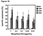
- 96h, 120h, 144h and 168h quantification of apoptosis was measured by flow cytometry. Results indicated that after 168h apoptosis was induced in more than 50% of the cells, when applying 1 ⁇ g/mL morphine (see Fig. 16 ).
- human acute myeloid leukaemia HL-60 cells (5000 cells/100 ⁇ l) were treated with 30,10, 5, 3, 1, 0.5, 0.3, 0.1 ⁇ g/mL morphine alone or in addition of 0.003 ⁇ g/mL or 0.001 ⁇ g/mL doxorubicin.
- apoptosis was induced in 50% of the cells when applying 1 ⁇ g/mL morphine in combination with 0.001 ⁇ g/mL doxorubicin (see Fig. 17 ).
- the opiate and ⁇ receptor agonist morphine can be used successfully to induce apoptosis in leukaemia cells.
- Human acute myeloid leukaemia HL-60 cell line (5000 cells/100 ⁇ l) were treated with 30,10, 5, 3, 1, 0.5, 0.3, 0.1 ⁇ g/mL morphine alone (white bars) or in addition of 0.003 ⁇ g/mL (black bars) or 0.001 ⁇ g/mL doxorubicin (grey bars). After 168h quantification of apoptosis was measured by flow cytometry.
- opioids are suggested for the manufacture of a medicament for the treatment of resistant cancer patients.
Landscapes
- Health & Medical Sciences (AREA)
- Life Sciences & Earth Sciences (AREA)
- General Health & Medical Sciences (AREA)
- Veterinary Medicine (AREA)
- Chemical & Material Sciences (AREA)
- Animal Behavior & Ethology (AREA)
- Medicinal Chemistry (AREA)
- Pharmacology & Pharmacy (AREA)
- Public Health (AREA)
- Epidemiology (AREA)
- Emergency Medicine (AREA)
- Molecular Biology (AREA)
- General Chemical & Material Sciences (AREA)
- Chemical Kinetics & Catalysis (AREA)
- Organic Chemistry (AREA)
- Nuclear Medicine, Radiotherapy & Molecular Imaging (AREA)
- Inorganic Chemistry (AREA)
- Hematology (AREA)
- Bioinformatics & Cheminformatics (AREA)
- Engineering & Computer Science (AREA)
- Oncology (AREA)
- Pharmaceuticals Containing Other Organic And Inorganic Compounds (AREA)
- Medicines That Contain Protein Lipid Enzymes And Other Medicines (AREA)
- Acyclic And Carbocyclic Compounds In Medicinal Compositions (AREA)
- Hydrogenated Pyridines (AREA)
- Nitrogen Condensed Heterocyclic Rings (AREA)
Description
- The invention relates to novel strategies for the treatment of resistant cancer patients.
- Anti-cancer therapies are frequently ineffective due to resistance of the tumor cells to radio- and/or chemotherapy. When the resistance is acquired during therapy, it often manifests either in a diminished amount of tumor regression for the same dose (either of the radiation or the cytotoxic substance) or an increased dose which is necessary for an equal amount of tumor regression. When the resistance is intrinsic, i.e. not acquired or induced due to the anti-cancer treatment, the tumor cells already originally lack sensitivity to one or more anti-cancer drugs or ionizing radiation.
- The chemosensitivity of cancer cells often vary on an individual basis. For pancreas carcinoma e.g. it is known, that only approximately 25% of all patients benefit from the anti-cancer drug gemcitabine. The other 75% are intrinsically resistant to this chemotherapy. Further examples for tumor cells with intrinsic chemo- and radioresistance are glioblastoma or melanoma cells.
- The intrinsic or acquired resistance (or non-response) of tumor cells to radio- and/or chemotherapy can have multiple reasons and can - as exemplified above - vary on an individual basis. Despite intensive research the exact mechanisms still remain elusive. However, it is known that either a single mutation, e.g. at the drug substance binding site or within the cellular detoxification process, can be responsible for the lack of or reduced chemosensitivity. Also the manifestation of cross-resistances to several anticancer drugs often limits the efficacy of anticancer treatments.
- Of significant clinical importance is the phenomenon of the so called multi-drug resistance (MDR). According to this concept, membrane proteins, namely members of the ATP binding cassette (ABC) transporter proteins, such as the P-glycoprotein or the multi-drug resistance associated proteins (MRP) are increasingly expressed, which leads to an enhanced efflux of drug substances through active transportation via the cell membrane. Patients exhibiting a multi-drug resistance most often are resistant to a wide spectrum of cytotoxic drugs.
- Resistances are not limited to chemotherapeutics or anticancer drugs; cancer patients can also exhibit either an intrinsic or acquired resistance to ionising irradiation applied in radiotherapy. An intrinsic radioresistance is known e.g. from melanoma and glioblastoma cells.
- Radioresistance may also be induced by exposure to small or fractionated doses of ionizing radiation. Several studies have documented this effect in vitro even in human cells as well as in several animal models. Different cellular radioprotection mechanisms may be involved, such as alterations in the levels of some cytoplasmatic and nuclear proteins, increased gene expression or DNA repair processes.
- As a method for treating drug resistant cancer, the international patent application
WO 2006/1244113 - Thus in oncology there is a great need for novel strategies, which render cancer treatments more effective. In particular it is the objective of the present invention to provide novel means for treating cancer patients, which exhibit a resistance to conventional anticancer therapies, such as anticancer drugs (chemotherapy) or radiotherapy or for treating cancer patients with apoptose resistant cells.
- This objective is solved by using opioids in the treatment of radiotherapy and/or chemotherapy resistant cancer patients, since now it was found that opioids capable of inhibiting the cell proliferation and or growth of cancer cells can overcome resistances in these cancer cells. Therefore these opioids provide novel strategies for treating patients, who so far are considered to be non-treatable or not effectively treatable by conventional therapeutic anticancer approaches. This group of alleged non-treatable cancer patients can also be called "non-responders", "poor-responder" or "non-chemosensitive" or "non-radiosensitive" cancer patients.
- It was furthermore found that opioids can overcome apoptosis resistance of cancer cells, and thus can effectively be clinically applied as anticancer substances. In particular, most surprisingly, it was found that opioids - in particular methadone - were as effective as the conventional chemotherapy (e.g. doxorubicin) and radiation treatments against non-resistant (i.e. sensitive) leukaemia cells, and that normal peripheral blood lymphocytes survived after this treatment. Hence, according to one embodiment of the invention, opioids are also effective in killing tumor cells, but do not substantially affect normal healthy cells of the patient.
- In the context of the present invention the term "opioid" is defined as a chemical heterogeneous group of natural, synthetic or semi-synthetic substances, working agonistic or antagonistic which all can bind to the well known opioid receptors, preferably to the µ opioid receptor and which are capable of arresting cancer cell proliferation. The group of opioids includes natural opiates such as alkaloids like morphine, dihydrocodein, codeine and thebaine, as well as semi-synthetic opiates, derived from the natural opiates (e.g. hydromorphone, hydrocodone, oxycodone, oxymorphone, desomorphine, diacetylmorphine (Heroin), nicomorphine, dipropanoylmorphine, benzylmorphine and ethylmorphine), or fully synthetic opioids, such as fentanyl, pethidine and methadone, tramadol or propoxyphene. It also includes endogenous opioid peptides, which may be produced naturally in the body as endorphins, dynorphins or enkephalins but which can also be synthesized.
- Opioids are known for their use as analgetics. The fact that opioid receptors, especially µ opioid receptors are involved in the activation of signalling pathways leading to apoptosis was previously known (Polakiewicz et al. 1998). In the past decade it was found that opioids can promote apoptosis (Hatsukari et al. 2003). It was further discussed to use opioids for the induction of apoptosis in small lung cancer cells (Heusch & Maneckjee 1999). However, the underlying mechanisms were not unrevealed, nor do those results suggest employing opioids for overcoming resistances to conventional anticancer treatments.
- According to the invention the opioid is capable of inhibiting cancer cell proliferation and/or growth. This activity can include e.g. cytostatic or cytotoxic activity as well as arresting growth of cells and/or tumors. Cancer cell proliferation is the result of the inhibition of cell division. In particular opioids induce cell death in tumors. Cell death in the context of the invention includes all types of cells death. This can include necrotic as well as apoptotic cell death or autophagy. In one embodiment of the invention the cell death is induced by the activation of the caspases-dependent or caspases-independent pathway. However, opioids can induce cell death via various pathways. In a preferred embodiment of the invention, opioids induce apoptosis in cancer cells.
- Generally, it is known, that apoptosis can be induced via two main biochemical pathways. The "death receptor pathway" (or extrinsic pathway) includes the TNF-receptor-induced (tumor necrosis factor) model and the Fas-receptor-induced model (the Fas-receptor is also known as Apo-1 or CD95). Bindings to these receptors result in the formation of death-inducing signalling pathways in the cell, including the activation of caspases-8. The "mitochondrial pathway" (or intrinsic pathway) involves the release of cytochrom c from mitochondria, binding of Apaf-1 and activation of procaspase-9. Several regulators are known to activate or deactivate the apoptosis pathways, such as the pro-apoptotic proteins Bax and Bak or the anti-apoptotic proteins Bcl-2, BclXL or XIAP.
- In one embodiment of the invention opioids induce apoptosis by one or more of the following mechanisms:
- i. cleavage of caspase-3 and PARP in the tumour cell
- ii. cleavage of caspase-9 and down regulation of XIAP
- iii. down regulation of BclXL
- According to a preferred embodiment of the invention, the opioid is a member of the methadone group, comprising D-/L-methadone, levomethadone, levacetylmethadol and piritramide. All these opioids can be used as salts. The racemic form of D-/L-methadone is preferably provided in form of a hydrochloride. In a preferred embodiment of the invention, the opioid methadone induces apoptosis in cancer cells via the mitochondrial pathway.
- According to the invention, the terms "resistance", "radioresistance" or "chemoresistance" are defined as a reduced sensitivity of a cancer cell to at least one conventional cancer therapy, i.e. either an anticancer drug or radiotherapy. A patient suffering from such a cancer is determined as a "resistant" cancer patient. Since the resistance can be intrinsic or acquired the observed reduction in sensitivity is either compared to fully sensitive "normal" cancer cells, which are responsive to the therapeutically effective dosage of the applied anticancer drug and/or radiation compared to the original sensitivity upon therapy onset. In the later case the resistance manifests either in a diminished amount of tumour regression for the same dose (either of the radiation or the anticancer drug) or an increased dose which is necessary for an equal amount of tumor regression.
- In a particularly preferred embodiment the opioids are used to treat cancer patients who exhibit one or more of the subsequent resistances:
- o apoptosis resistance
- o multi-drug resistance
- o anticancer drug resistance
- o cytotoxic drug resistance
- o resistance to reactive oxygen species
- o resistance to DNA-damaging agents
- o resistance to toxic antibodies
- o doxorubicin resistance
- o single or cross resistance, in particular to one or more of the following drug substances: methotrexate, cytarabine, cisplatin, etoposide, vincristine, paclitaxel (taxol), carboplatin, teniposide, dexamethasone, prednisolone, cyclophosphamide, iphosphamide, doxorubicin, epirubicin, daunorubicin, mercaptopurine, fludarabine, 5-fluoruracil
- o irradiation resistance (e.g. alpha, beta, gamma or Auger electrons)
- Accordingly, in the context of the present invention a "resistance" can either be total or partly; in other words, the patients considered treatable according to the invention can exhibit a reduced sensitivity or even a full lack of sensitivity to conventional anticancer treatments. These patients can also be determined as "non-responders" or "poor-responders".
- A further synonym for a "resistant" cancer or tumor is a "refractory" type of cancer, which can also be either completely or partly refractory. Intrinsic resistance can thus also be determined as a "primary refractory cancer". A particular form of refractory or resistant cancer cells ar the so called "kinetically refractory cells"; a phenomenon known e.g. from leukaemia cells, when the cells are at first killed, but reproduce fast that an effective treatment is hardly possible.
- As used in the context of the present invention the term "conventional" treatment or therapy refers to the currently accepted and widely used therapeutic treatment of a certain type of cancer, based on the results of past researches and/or regulatory approval.
- Conventional anticancer drugs include cytotoxic and cytostatic agents, which kill the cancer cells or reduce and/or stop their growth or proliferation. The modes of action of these anticancer drugs can vary; examples are antimetabolites (e.g. cytarabine, methotrexate, mercaptopurine or clofarabine), DNA cross-linking agents (e.g. cisplatine and its derivates), DNA intercalating substances (e.g. doxorubicin), Topoisomerase poisons (e.g. etoposide), kinase inhibitors (e.g. cetuximab), steroids (e.g. dexamethasone) or mitotic inhibitors (e.g. vincristine). One example for a conventional anticancer treatment of leukaemia is the administration of doxorubicin.
- The conventional radiotherapy can also include radiation therapy, which means the use of high-energy radiation from x-rays, alpha, beta and gamma rays, Auger electrons, Ultraviolet rays, neutrons, protons, and other sources to kill cancer cells and shrink tumors. Radiation may originate from an outside the body device (external-beam radiation therapy), or it may originate from radioactive sources placed in the body in the vicinity of the cancer cells (internal radiation therapy). Systemic radiation therapy uses a radioactive substance, such as a radiolabeled monoclonal antibody, that travels in the blood stream to the target tissue. Radioresistant cancer cells do not or only partly respond to these treatments.
- As outlined in detail above, according to one embodiment of the invention opioids are applied for overcoming or "breaking" the intrinsic or acquired resistance of cancer cells to conventional anticancer treatments and/or radiation treatment or apoptosis resistance. In one embodiment of the invention cancer cells considered treatable according to the invention express an opioid receptor, in particular the the µ opioid receptor.
- In a further embodiment the group of cancers include, but is not limited to leukaemia, breast cancer, glioblastoma, prostate cancer, lung cancer, non-small cell lung cancer (NSCLC), brain cancer, colon cancer, colorectal cancer.
- Examples for cancer types, considered to be treatable according to the inventin, with intrinsic resistance to irradiation are glioblastoma, melanoma or pancreas cancer cells. Breast cancer, bladder cancer or leukaemia are often resistant to chemotherapeutics. Examples of cancer types which often acquire resistance are melanoma, colon carcinoma, brain tumors glioblastoma, brain cancer, pancreatic cancer, liver cancer, ovarian cancer, cancer of mamma, lung cancer, chronic leukaemia or osteosarkoma.
- In a further embodiment of the invention, the opioids can be used in combination - i.e. as a composite - with conventional anticancer substances or treatments, e.g. cytostatic or cytotoxic substances or radiotherapy. The opioids can be combined for example with natural and/or synthetic anticancer substances, natural and/or synthetic cytotoxic substances, antibiotics, cytotoxic antibodies, hormones, psycho-pharmaca, naturally or genetically modified organisms and substances from organisms (e.g. plants, microorganisms, fruits), substances for pain, and/or different sorts of radiation unbound or bound to substances (e.g. antibodies). The patient can either be resistant or not resistant to this treatment.
- A "composite" means a pharmaceutical preparation comprising a therapeutically effective amount of any of the opioids (component A) as defined according to the invention and at least one further anticancer substance (component B). This "composite" can constitute a single composition or at least two compositions, which can be administered to the patients either concomitantly or subsequently. The above mentioned substances are preferably combined with methadone.
- The composite of the invention can be advantageous for the effective treatment of cancer cells, since it can exhibit synergistic effects compared to the single compositions. In particular composite with methadone as component A and one of the agents as component B as follows is possible: methotrexate, cytarabine, cisplatine, etoposide, vincristine. Moreover combinatorial treatment also comprising irradiation treatments is possible.
- In a preferred embodiment of the invention opioids are used to treat either resistant or sensitive non-solid cancers, i.e. all haematological malignancies affecting blood, bone marrow and lymph nodes, including acute lymphoblastic leukaemia, B-cell lymphatic leukaemia, acute myeloid leukaemia, chronic myeloid leukaemia, chronic lymphocytic leukaemia and all pro-forms of leukaemias, hairy cell leukaemia, Hodgkin's disease, Non-Hodgkin lymphoma and multiple myeloma.
- D, L-methadone hydrochloride (methadone, Sigma, Taufkirchen, Germany) was freshly dissolved in sterile distilled water prior to each experiment to ensure constant quality of the preparations.
- The human myeloid leukaemia cell line HL-60 and the human lymphoblastic leukaemia T-cell line CEM were grown in RPMI 1640 (GIBCO, Invitrogen, Karlsruhe, Germany) containing 10% fetal calf serum (Biochrom, Berlin, Germany), 10 mM HEPES, pH 7.3 (Biochrom), 100 U/mL penicillin (GIBCO), 100µg/mL streptomycin (GIBCO) and 2 mM L-glutamine (Biochrom) at 37°C and 5% CO2. CEMCD95R are resistant to 1 µg/mL anti-CD95 (Friesen et al. 1996) and CEMDOXOR are resistant to 0.1 µg/mL doxorubicin (Friesen et al. 2004). CEMCD95R is apoptosis-resistant and multi-drug resistant. CEMCD95R is cross-resistant to several anticancer drugs such as methotrexate, cytarabine, cisplatine, etoposide, vincristine and to gamma- and beta-irradiation (Friesen et al. 1996, Los et al. 1997, Friesen et al. 2007). All cell lines used in this study were mycoplasma free.
- Leukaemia cells (1 x 10⁵ cells/mL) were treated with 30, 20, 15, 10 µM methadone in 150 mL flasks or 96 well plates. After 24h and 48h, quantification of apoptosis was measured by flow cytometry as described (Carbonari et al. 1994, Friesen et al. 2003). In brief, to determine apoptosis, cells were lysed with Nicoletti-buffer containing 0.1 % sodium citrate plus 0.1 % Triton X-100 and
propidium iodide 50 µg/mL as described by Nicoletti et al. (1991). The percentage of apoptotic cells was measured by hypodiploid DNA (subG1) or FSC/SSC analysis (Nicoletti et al. 1991, Carbonari et al. 1994). Propidium iodide (PI) stained nuclei or forward scatter/side scatter (FSC/SSC) profiles of cells were analyzed by flow cytometry (FACSCalibur, Becton Dickinson, Heidelberg, Germany). - Peripheral blood lymphocytes (PBLs) were isolated from fresh blood of healthy persons. PBLs (1 x 10⁶ cells in 1 mL) were treated with 30, 20, 15, 10 µM methadone in 96 well plates. After 24h and 48h quantification of apoptosis was measured by flow cytometry as described (Carbonari et al. 1994).
- Inhibition of caspases activation was performed as previously described (Friesen et al. 2007). In brief, the broad spectrum tripeptide inhibitor of caspases zVAD.fmk (benzoylcarbonyl-Val-Ala-Asp-fluoromethyl ketone, Enzyme Systems Products, Dubli, USA) was used at a concentration of 50 µmol/L. HL-60 and CEM cells were preincubated with zVAD.fmk 1 h before methadone treatment. After 24h and 48h the percentage of apoptotic cells was measured by hypodiploid DNA (subG1) or FSC/SSC analysis. Propidium iodide (PI) stained nuclei (Nicoletti et al. 1991) or forward scatter/side scatter (FSC/SSC) profile of cells (Carbonari et al. 1994) were analyzed by flow cytometry (FACSCalibur, Becton Dickinson, Heidelberg, Germany).
- Western blot analyses were done as described (Friesen et al. 2004, Friesen et al. 2003). Immunodetection of PARP, caspase-3, caspase-9, caspase-8, XIAP, CD95, CD95-L, Bax, Bcl-xL and ß-actin was done using rabbit anti-PARP polyclonal antibody (1:5000, Roche), mouse-anti-caspase-3 monoclonal antibody (1:1000, Cell-Signalling), mouse-anti-caspase-8 monoclonal antibody (1:1000, Cell-Signalling), rabbit- anti-active-caspase-9 polyclonal antibody (1:1000, Cell-Signalling), mouse-anti-XIAP monoclonal antibody (1:1000, Transduction-Laboratories, Lexington, Kentucky), mouse-anti-Fas (anti-CD95) monoclonal antibody (1:1000, Transduction-Laboratories), mouse-anti-Fas ligand (anti-CD95-ligand) monoclonal antibody (1:250, BD, Pharmingen), rabbit-anti-Bax polyclonal antibody (1:250, Oncogene, Cambridge), rabbit-anti-Bcl-XS/L polyclonal antibody (1:1000, Santa-Cruz-Biotechnology, Santa-Cruz, CA), rabbit-anti-p21 polyclonal antibody (1:1000, Santa-Cruz), and mouse anti-ß-actin monoclonal antibody (Sigma). Peroxidase-conjugated goat-antimouse IgG or peroxidase-conjugated goat-anti-rabbit IgG (1:5000, Santa-Cruz) as secondary antibody were used for the enhanced chemoluminescence system (ECL, Amersham-Pharmacia, Freiburg, Germany). Equal protein loading was controlled by ß-actin detection.
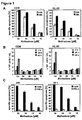
- In leukaemias and solid tumours anticancer drugs have been shown to induce apoptosis and to inhibit proliferation (Kaufmann & Earnshaw 2000). Therefore, it was analyzed if the therapeutic opioid drug methadone can also inhibit proliferation and trigger apoptosis in the human lymphoblastic leukaemia T-cell line CEM and the human myeloid leukaemia cell line HL-60 comparable to the well-known established anticancer drugs (
Fig. 1A, B ). 24h and 48h after treatment with different concentrations of methadone (30, 20, 15, 10 µM) a strong induction of apoptosis (Fig. 1A ) and a strong inhibition of growth (Fig. 1B ) were detected in CEM and HL-60 cells. It was next analyzed if methadone induces apoptosis also in non-leukaemic peripheral blood lymphocytes (PBLs) (Fig. 1C ). Isolated PBLs were incubated with different concentrations of methadone (30, 20, 15, 10 µM). 24h and 48h after methadone treatment it was found that methadone could not kill PBLs at comparable concentrations using for the treatment of leukaemia cells such as CEM (Fig. 1C ). This demonstrates that methadone induces apoptosis in leukaemia cells with non-toxic effects on non-leukaemic PBLs. - Resistance to anticancer drugs is a limiting factor in treatment of leukaemia and tumour patients (Bergman & Harris 1997, Friesen et al. 2003). It has been found that methadone exhibits a potent anti-leukaemic activity and efficiently kills leukaemia cells. Therefore it was analyzed if methadone can also induce cell death in doxorubicin-resistant leukaemia cells, which were apoptosis-resistant. Doxorubicin-resistant CEM leukaemia cells (CEMDoxoR) were treated with different concentrations of methadone (30, 20, 15, 10 µM). 24h and 48h after methadone treatment, cell death was measured by flow cytometry. After treatment with 30, 20, 15 µM methadone, a strong induction of apoptosis was measured in doxorubicin-resistant cells CEMDoxoR, which was similar to that in sensitive leukaemia cells CEM (
Fig. 2 ), indicating that methadone overcomes doxorubicin-resistance and apoptosis-resistance. - It was next examined if methadone can also kill leukaemia cells, which were multidrug-resistant. Therefore CD95-resistant leukaemia cells CEMCD95R, which were resistant to several anticancer drugs such as etoposide, cisplatine, methotrexate, cytarabine, doxorubicin, vincristine, were treated with different concentrations of methadone (30, 20, 15, 10 µM). After treatment with 30, 20, 15 µM methadone, a strong induction of apoptosis in CD95-resistant leukaemia cells CEMCD95R was measured after 24h and 48h, which was similar to that in sensitive leukaemia cells CEM (
Fig. 2 ). This suggests that methadone induces not only a strong induction of apoptosis in sensitive leukaemia cells, but it also kills doxorubicin- and CD95-resistant leukaemia cells, which commonly used anticancer drugs failed to kill (Friesen et al. 1996, Los et al. 1997). - Methadone induces apoptosis in sensitive and in resistant leukaemia cells with unknowing molecular mechanisms. Therefore, the mechanism and effector molecules which may be altered by methadone-triggered cell death in leukaemia cells were to be examined.
- Caspases play a critical role in apoptosis induction by anticancer drugs (Kaufmann & Earnshow 2000, Hengartner 2000). Therefore Western blot analysis was used to examine if methadone activates caspases in HL-60 and CEM leukaemia cells as well as in doxorubicin-resistant leukaemia cells CEMDoxoR, which were apoptosis-resistant and in CD95-resistant leukaemia cells CEMC695R, which were multidrug-resistant and apoptosis-resistant. After treatment with different concentrations of methadone (20, 15 µM) caspase-3, and PARP were cleaved in HL-60 and CEM leukaemia cells (
Fig. 3A ) as well as in doxorubicin-resistant leukaemia cells CEMDoxoR and in CD95-resistant leukaemia cells CEMCD95R (Fig. 3B ). Activation of caspase-8, which anticancer drugs have shown to induce in leukaemia cells, was not found after methadone treatment. To investigate the critical role of methadone in activation of caspases, CEM and HL-60 cells were preincubated with the broad spectrum inhibitor of caspases zVAD.fmk. Incubation with zVAD.fmk almost completely inhibited methadone-induced apoptosis (Fig. 3C ) suggesting that caspases are central for methadone-induced apoptosis in leukaemia cells. - Anticancer-drugs have been shown to activate the mitochondrial pathway as well as the ligand/receptor pathway in leukaemia and tumour cells (Kaufmann & Earnshow 2000). It was investigated if mitochondria could also play a role methadone-induced apoptosis in leukaemia cells. CEM, HL-60, doxorubicin-resistant leukaemia cells CEMDoxoR and CD95-resistant leukaemia cells CEMCD95R were treated with different concentrations of methadone (20, 15 µM) (
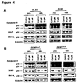
Fig. 4 ). After 24h and 48h a strong cleavage (37 kDa fragment) of caspase-9 and a strong down regulation of the caspases inhibiting protein XIAP (X-linked inhibitory-of-apoptosis protein) was found in HL-60 and CEM leukaemia cells (Fig. 4A ) as well as in doxorubicin-resistant leukaemia cells CEMDoxoR and in CD95-resistant leukaemia cells CEMCD95R (Fig. 4B ), which were multidrug- and apoptosis-resistant. - Mitochondrial changes are regulated by pro- and anti-apoptotic Bcl-2 family members. After 24h and 48h a strong down regulation of Bcl-xL were found in HL-60 and in CEM leukaemia cells (
Fig. 4A ) as well as in doxorubicin-resistant leukaemia cells CEMDoxoR and in CD95-resistant leukaemia cells CEMCD95R (Fig. 4B ) after treatment with different concentrations of methadone (20, 15 µM). Up regulation of Bax was not found after methadone treatment in leukaemia cells.
Furthermore, up regulation of death-inducing ligands and death-inducing receptors such as CD95, which anticancer drugs have shown to up regulate, were not found after methadone treatment in leukaemia cells (data not shown). This indicates that methadone induces apoptosis by directly activation of the intrinsic mitochondrial pathway in sensitive as well as in resistant leukaemia cells. - Glioblastoma is the most aggressive and the most common type of primary brain tumours. Glioblastoma cells are known to be highly resistant to chemotherapeutics and irradiation. Treatment may involve chemotherapy and radiotherapy but these are only palliative measures and do not provide a cure. Moreover, many drugs cannot cross the blood brain barrier and are therefore useless in the treatment of glioblastoma. Methadone is able to cross the blood-brain barrier. The effect of methadone on glioblastoma cells was examined.
- Furthermore, the effect of methadone in combination with therapeutical concentrations of doxorubicin on glioblastoma cells was tested.
- The human glioblastoma cell line A172 was grown in DMEN (Invitrogen, Karlsruhe, Germany) containing 10% fetal calf serum (Biochrom, Berlin, Germany), 10 mM HEPES, pH 7,3 (Biochrom), 100 U/mL penicillin (Invitrogen), 100 µg/mL streptomycin (Invitrogen) and 2mM L-glutamine (Biochrom) at 37°C and 5% CO2.
Before methadone treatment, glioblastoma cells were seeded in a density of 7000 cells/cm2 and treatment was performed 24h after cell seeding. - Glioblastoma cells A172 (7000 cells/cm2) were treated with 30, 20 µg/mL methadone in 75 cm2 flasks. After 120h, 144h and 168h quantification of apoptosis was measured by flow cytometry. To determine apoptosis, cells were lysed with Nicoletti buffer containing 0,1 % sodium citrate plus 0,1% Triton X-100 and
propidium iodide 50 µg/mL. Propidium iodide (PI) stained nuclei were analysed by flow cytometry (FACSCalibur, Becton Dickinson, Heidelberg, Germany). - After 120h, 144h and 168h, the percentages of apoptotic cells were measured by hypodiploid DNA analysis. The percentage of specific cell death was calculated as follows: 100 x (experimentally dead cells (%) - spontaneous dead cells in cell medium (%)/ (100% - spontaneous dead cells in medium (%)). Treatment with methadone resulted in more than 20% dead cells after 120h and more than 80% dead cells after 168h after treatment with 20 µg/mL methadone. Treatment with methadone resulted in more than 85% dead cells after 120h and nearly 100% dead cells after 168h after treatment with 30 µg/mL methadone (
Fig. 5 ). Similar results were obtained in tree independent experiments. This demonstrates that methadone induces high rates of apoptosis in glioblastoma cells. - Glioblastoma cells A172 (7000 cells/cm2) were treated with a therapeutical concentration of 0.1 µg/mL doxorubicin (doxorubicin, white columns), with a low concentration of 1 µg/mL methadone (methadone, black columns), and with 0.1 µg/mL doxorubicin in addition of 1 µg/mL methadone (doxorubicin + methadone, hatched columns). After 72h quantification of apoptosis was measured by flow cytometry. To determine apoptosis, cells were lysed with Nicoletti-buffer containing 0.1 % sodium citrate plus 0.1 % Triton X-100 and
propidium iodide 50 µg/mL. Propidium iodide (PI) stained nuclei were analyzed by flow cytometry (FACSCalibur, Becton Dickinson, Heidelberg, Germany). - The combination of a therapeutical concentration of doxorubicin with low concentrations of methadone had a strong apoptotic effect on the glioblastoma cells tested (
Fig. 6 ). This assay shows, that methadone reveals synergistic effects when applied together with other chemotherapeutics, here doxorubicin. - B-ALL (B-Cell lymphatic leukaemia) and CLL (chronic lymphatic leukaemia) cells were isolated from patients ex vivo. These cells were treated with different concentrations of D,L-methadone or D,L-methadone in addition of fludarabine.
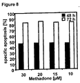
- In a first study, B-ALL (B-Cell lymphatic leukaemia) cells (50000 cells/100 µl) were treated with 30, 20, 15, 10 µM methadone. After 48h and 72h quantification of apoptosis was measured by flow cytometry. Results indicated that already 10 µM methadone were sufficient to induce apoptosis in up to over 80% of the cells treated for 72h (see
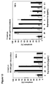
Fig. 8 ). - In a second study, CLL (chronic lymphatic leukaemia) cells (50000 cells/200 µl) were treated with 30, 10, 5, 3, 1, 0.5, 0.3 and 0.1 µg/mL methadone alone or in addition of 0.1 µM fludarabine. After 24h and 48h quantification of apoptosis was measured by flow cytometry. Results indicated that apoptosis was induced in 100% of the cells already after 24h, when combining 3 µg/mL methadone with 0.1 µM fludarabine (see
Fig. 9 ). - In a third study, CLL (chronic lymphatic leukaemia) cells (50000 cells/200 µl) were treated with 30, 10, 5, 3, 1, 0.5, 0.3, 0.1 µg/mL methadone alone or in addition of 0.1 µM fludarabine. After 24h and 48h quantification of apoptosis was measured by flow cytometry. Results indicated that apoptosis was induced in nearly 50% of the cells after 48 hours, when combining 3 µg/mL methadone with 0.1 µM fludarabine. Apoptosis was induced in more than 90% of the cells after 48h, when combining 30 µg/mL methadone with 0.1 µM fludarabine (see
Fig. 10 ). - Thus, as suggested by these studies, methadone alone or in combination with a cytostatic treatment is successful in cancer cells isolated from patients ex vivo.
- Buprenorphine was used successfully in combination with doxorubicin to induce apoptosis in HL-60 leukaemia cells in vitro. Buprenorphine is a semi-synthetic opioid drug, also used as analgesic. It is a partial µ-opioid receptor agonist.
- Human acute myeloid leukaemia HL-60 cell line (5000 cells/100 µl) were treated with 30,10, 5, 3, 1, 0.5, 0.3, 0.1 µg/mL buprenorphine alone or in addition of 0.003µg/mL or 0.001µg/mL doxorubicin. After 144h or 168h quantification of apoptosis was measured by flow cytometry.
- Results indicated that 20 µg/mL buprenorphine were sufficient to induce apoptosis in more than 90% of the cells after 144h. The same result was obtained when adding 0.003 µg/mL doxorubicin. Apoptosis was induced in nearly 100% of the cells when combining 10 µg/mL buprenorphine with 0.003 µg/mL doxorubicin and incubating the cells for 168h (see
Fig. 13 ). - Fentanyl was used successfully in combination with doxorubicin to induce apoptosis in CEM leukaemia cells in vitro. Fentanyl is a synthetic opioid, which is frequently used as analgesic in the treatment of cancer pain. Fentanyl is a µ opioid receptor agonist.
- Human T-Cell leukaemia CEM cell line (10000 cells/100 µl) were treated with 30,10, 5, 3,1, 0.5, 0.3, 0.1 µg/mL fentanyl alone or in addition of 0.02µg/mL doxorubicin. After 48h and 72h quantification of apoptosis was measured by flow cytometry.
- Results indicated that fentanyl successfully induced apoptosis in more than 85% of the cells after 72h, when combining 30 µg/mL fentanyl with 0.02 µg/mL doxorubicin (see
Fig. 14 ). - Morphine was used successfully alone and in combination with doxorubicin for the induction of apoptosis in HL-60 leukaemia cells. Morphine is an opiate which is currently used as analgesic in the treatment of cancer pain. Morphine is a µ opioid receptor agonist.
- In a first study, human acute myeloid leukaemia HL-60 cells (5000 cells/100 µl) were treated with 30,10, 5, 3, 1, 0.5, 0.3, 0.1, 0.03, 0.01 µg/mL morphine. After 120h or 144h quantification of apoptosis was measured by flow cytometry. Results indicated that after 144h apoptosis was induced in 40% of the cells when applying 40 µg/mL morphine see
Fig. 15 ). - In a second study, human acute myeloid leukaemia HL-60 cells (5000 cells/100 µl) were treated with 1, 0.5, 0.3, 0.1, 0.03, 0.01 µg/mL morphine. After 96h, 120h, 144h and 168h quantification of apoptosis was measured by flow cytometry. Results indicated that after 168h apoptosis was induced in more than 50% of the cells, when applying 1µg/mL morphine (see
Fig. 16 ). - In a third study, human acute myeloid leukaemia HL-60 cells (5000 cells/100 µl) were treated with 30,10, 5, 3, 1, 0.5, 0.3, 0.1 µg/mL morphine alone or in addition of 0.003µg/mL or 0.001µg/mL doxorubicin. After 168h quantification of apoptosis was measured by flow cytometry. Results indicated that apoptosis was induced in 50% of the cells when applying 1µg/mL morphine in combination with 0.001 µg/mL doxorubicin (see
Fig. 17 ). - Thus, as suggested by these studies, the opiate and µ receptor agonist morphine can be used successfully to induce apoptosis in leukaemia cells.
-
- Bergmann JP, Harris D. Radioresistance, chemoresistance and apoptosis resistance. Radiation Oncology 1997; 27:47-57.
- Carbonari M, Cibati M, Cherchi M, et a/. Detection and characterization of apoptotic peripheral blood lymphocytes in human immunodeficiency virus-infection and cancer chemotherapy by a novel flow immunocytometric method. Blood 1994; 83:1268-77.
- Friesen C, Glatting G, Koop B, et al. Breaking chemo- and radioresistance with [213Bi]anti-CD45 antibodies in leukaemia cells. Cancer Res 2007; 67(5):1950-8.
- Friesen C, Herr I, Krammer PH, Debatin KM. Involvement of the CD95 (APO-1/FAS) receptor/ligand system in drug-induced apoptosis in leukaemia cells. Nat Med 1996; 2(5):574-7.
- Friesen C, Kiess Y, Debatin KM. A critical role of glutathione in determining apoptosis sensitivity and resistance in leukaemia cells. Cell Death Differ 2004; 11 (Suppl 1 ):S73-85.
- Friesen C, Lubatschofski A, Kotzerke J, Buchmann I, Reske SN, Debatin KM. Beta-irradiation used for systemic radioimmunotherapy induces apoptosis and activates apoptosis pathways in leukaemia cells. Eur J Nucl Med 2003; 30:1251-61.
- Hatsukari I, Hitosugi N, Matsumoto I, Nagasaka H, Sakagami H. Induction of early apoptosis marker by morphine in human lung and breast carcinoma cell lines. Anticancer Res. 2003 May-Jun; 23(3B):2413-7.
- Hengartner MO. The biochemistry of apoptosis. Nature 2000; 407(6805):770-6.
- Heusch WL, Maneckjee R. Effects of bombesin on methadone-induced apoptosis in lung cancer cells. Cancer Lett 1999; 136:177-85.
- Kaufmann SH, Earnshaw WC. Induction of apoptosis by cancer therapy. Experimental Cell Research 2000; 256: 42-9.
- Los M, Herr I, Friesen C, Fulda S, Schulze-Osthoff K, Debatin K-M. Cross-resistance of CD95- and drug-induced apoptosis as a consequence of deficient activation of caspases (ICE/Ced-3 proteases). Blood 1997; 90(8):3118-29.
- Milas L, Raju U, Liao Z, Ajani J. Targeting molecular determinants of tumour chemo-radioresistance. Semin Oncol 2005; 32:78-81.
- Nicoletti I, Migliorati G, Pagliacci MC, Grignani F, Riccardi C. A rapid and simple method for measuring thymocyte apoptosis by propidium iodide staining and flow cytometry. J Immunol Meth 1991;139:271-9.
- Polakiewicz RD, Schieferl SM, Gingras AC, Sonenberg N, Comb MJ. Mu-Opioid receptor activates signalling pathways implicated in cell survival and translational control. J Biol Chem. 1998 Sep 4; 273(36):23534-41.
-
-
Figure 1 :
Figure 1 indicates that the opioid methadone effectively induces apoptosis in leukaemia cells, but does not affect non-leucaemic, healthy PBL cells.- 1A: CEM and HL-60 cells were treated with different concentrations of methadone as indicated. After 24h (white bars) and 48h (black bars) the percentages of apoptotic cells were measured by hypodiploid DNA analysis. The percentage of specific cell death was calculated as follows: 100 x (experimental dead cells (%)- spontaneous dead cells in medium (%)) / (100 % - spontaneous dead cells in medium (%)). Data are given as mean of triplicates with a standard deviation (SD) of less than 10 %. Similar results were obtained in three independent experiments.
- 1B: CEM and HL-60 cells (2 x 10⁵ cells/mL) were treated with different concentrations of methadone as indicated or left untreated (Co, control). After 0h (white bars), 24h (black bars) and 48h (hatched bars) number of cells in 1 mL was counted. Data are given as mean of triplicates with a standard deviation (SD) of less than 10 %. Similar results were obtained in three independent experiments.
- 1C: CEM cells (black bars) and PBLs (white bars) were treated with different concentrations of methadone as indicated. After 24h and 48h the percentages of apoptotic cells were measured by hypodiploid DNA analysis. The percentage of specific cell death was calculated as described in
Figure 1A . Data are given as mean of triplicates with a standard deviation (SD) of less than 10 %. Similar results were obtained in three independent experiments.
-
Figure 2 :
Figure 2 shows that the opioid methadone induces apoptosis in CD95-resistant (CEMCD95R) and in doxorubicin-resistant (CEMDoxoR) with comparable apoptosis rates to parental sensitive CEM leukaemia cells.
CEM (black bars), CEMCD95R (white bars) and CEMDoxoR (hatched bars) leukaemia cells were treated with different concentrations of methadone as indicated. After 24h and 48h the percentages of apoptotic cells was measured by the hypodiploid DNA analysis. The percentage of specific cell death was calculated as described inFigure 1 A . Data are given as mean of triplicates with a standard deviation (SD) of less than 10 %. Similar results were obtained in three independent experiments. -
Figure 3 :
Figure 3 shows that the opioid methadone induces caspases-dependent death in sensitive (HL-60, CEM), in doxorubicin-resistant (CEMDoxoR) and in CD95-resistant (CEMCD95R) leukaemia cells, which were multidrug-resistant and apoptosis-resistant.- 3A: and 3B: Methadone induced activation of caspase-3 and PARP cleavage in HL-60, CEM, CEMDoxoR and CEMCD95Rcells. A, HL-60, CEM, B, CEMDoxoR, CEMCD95R cells were treated with different concentrations of methadone as indicated or left untreated (Control). After 24h and 48h Western blot analyses for caspase-3 and PARP were performed. The active fragment of caspase-3 was detected at -19 and 17 kDa and the cleaved product of PARP at ~ 85 kDa. Equal protein loading was controlled by anti-beta-actin antibody.
- 3C: Inhibition of caspase activation with zVAD.fmk blocks methadone induced apoptosis in CEM and HL-60 cells. CEM and HL-60 cells were treated with different concentrations of methadone as indicated in the absence (black bars, medium) or presence (white bars, 50 µM zVAD.fmk) of 50 µM zVAD.fmk. After 48h the percentages of apoptotic cells were measured by hypodiploid DNA analysis. The percentage of specific cell death was calculated as described in
Figure 1A . Data are given as mean of triplicates with a standard deviation (SD) of less than 10 %. Similar results were obtained in three independent experiments.
-
Figure 4 :
Figure 4 shows that the opioid methadone activates the mitochondrial pathway in sensitive (HL-60, CEM), in doxorubicin-resistant (CEMDoxoR) and in CD95-resistant (CEMCD95R) leukaemia cells, which were multidrug-resistant and apoptosis-resistant.- 4A and 4B: Methadone induced activation of caspase-9, down regulation of XIAP and down regulation of Bcl-xL in HL-60, CEM, CEMDoxoR and CEMCD95R cells.
HL-60, CEM (4A), CEMDoxoR, CEMCD95R cells (4B) were treated with different concentrations of methadone as indicated or left untreated (Control). After 24h and 48h Western blot analyses for caspase-9, XIAP, and Bcl-xL were performed. The active fragment of caspase-9 was detected at -37 kDa, XIAP was detected at ∼58 kDa and Bcl-xL was detected at ∼30 kDa. Equal protein loading was controlled by anti-beta-actin antibody. -
Figure 5 :
Figure 5 indicates that the opioid methadone induces apoptosis in glioblastoma cells.
A172 glioblastoma cells were treated with different concentrations of methadone as indicated. After 120h (white columns), 144h (black columns) and 168h (hatched columns) the percentages of apoptotic cells were measured by hypodiploid DNA analysis.
The percentage of specific cell death was calculated as follows: 100 x (experimental dead cells (%) - spontaneous dead cells in medium (%)) / (100 % - spontaneous dead cells in medium (%)). Data are given as mean of triplicates with a standard deviation (SD) of less than 10 %. Similar results were obtained in three independent experiments. -
Figure 6 :
Figure 6 indicates that apoptosis can be successfully induced in glioblastoma cells by using combination of therapeutical concentrations of doxorubicin and low concentrations of methadone.
Glioblastom cells A172 (7000 cells/cm2) were treated with a therapeutical concentration of 0.1 µg/mL doxorubicin (doxorubicin, white columns), with a low concentration of 1 µg/mL methadone (methadone, black columns), and with 0.1 µg/mL doxorubicin in addition of 1 µg/mL methadone (doxorubicin + methadone, hatched columns). After 72h quantification of apoptosis was measured by flow cytometry. To determine apoptosis, cells were lysed with Nicoletti-buffer containing 0.1 % sodium citrate plus 0.1 % Triton X-100 andpropidium iodide 50 µg/mL Propidium iodide (PI) stained nuclei were analyzed by flow cytometry (FACSCalibur, Becton Dickinson, Heidelberg, Germany). -
Figure 7 :
Figure 7 indicates that apoptosis can be successfully induced in CEM cells, using the opioid cocaine.
CEM (white column) and HL-60 (black column) cells were treated with 1000 µg/mL of cocaine. After 48h the percentages of apoptotic cells were measured by hypodiploid DNA analysis. The percentage of specific cell death was calculated as follows: 100 x (experimental dead cells (%) - spontaneous dead cells in medium (%)) / (100 % - spontaneous dead cells in medium (%)). Data are given as mean of triplicates with a standard deviation (SD) of less than 10%. Similar results were obtained in three independent experiments. -
Figure 8 :
Figure 8 indicates that D,L-methadone induces cell death in pre B-ALL (B-Cell lymphatic leukaemia) cells isolated from patients ex vivo.
B-ALL (B-Cell lymphatic leukaemia) cells (50000 cells/100 µl) were treated with 30, 20, 15, 10 µM methadone. After 48h (black bars) and 72h (white bars) quantification of apoptosis was measured by flow cytometry. -
Figure 9 :
Figure 9 indicates that D,L-methadone in combination with fludarabine induces cell death in resistant CLL (chronic lymphatic leukaemia) cells isolated from patients ex vivo.
CLL (chronic lymphatic leukaemia) cells (50000 cells/200 µl) were treated with 30, 10, 5, 3, 1, 0.5, 0.3, 0.1 µg/mL methadone alone (white bars) or in addition of 0.1 µM fludarabine (black bars). After 24h and 48h quantification of apoptosis was measured by flow cytometry. -
Figure 10 :
Figure 10 indicates that D,L-methadone in combination with fludarabine induces cell death in resistant CLL (chronic lymphatic leukaemia) cells isolated from patients ex vivo.
CLL (chronic lymphatic leukaemia) cells (50000 cells/200 µl) were treated with 30, 10, 5, 3, 1, 0.5, 0.3, 0.1 µg/mL methadone alone (white bars) or in addition of 0.1 µM fludarabine (black bars). After 24h and 48h quantification of apoptosis was measured by flow cytometry. -
Figure 11 :
Figure 11 shows the induction of apoptosis in the Human B cell leukaemia cell line Tanoue treated with doxorubicin + methadone in vitro.
B-ALL (B-Cell lymphatic leukaemia) cell line Tanoue (5000 cells/100 µl) were treated with 30,10, 5, 3, 1 µg/mL methadone alone (white bars) or in addition of 0.03 µg/mL doxorubicin (black bars). After 96h quantification of apoptosis was measured by flow cytometry. -
Figure 12 :
Figure 12 shows the induction of apoptosis in the human B cell precursor leukaemia cell line Nalm6 treated with doxorubicin + methadone in vitro.
B-ALL (B-Cell lymphatic leukaemia) cell line Nalm6 (5000 cells/100 µl) were treated with 30,10, 5, 3, 1 µg/mL methadone alone (white bars) or in addition of 0.01 µg/mL doxorubicin (black bars). After 96h, 120h, 144h, 168h quantification of apoptosis was measured by flow cytometry. -
Figure 13 :
Figure 13 shows the induction of apoptosis in the human acute myeloid leukaemia cell line HL-60 treated with doxorubicin + buprenorphine in vitro.
Human acute myeloid leukaemia HL-60 cell line (5000 cells/100 µl) were treated with 30,10, 5, 3, 1, 0.5, 0.3, 0.1 µg/mL buprenorphine alone (white bars) or in addition of 0.003µg/mL or 0.001µg/mL doxorubicin (black bars). After 144h or 168h quantification of apoptosis was measured by flow cytometry. -
Figure 14 :
Figure 14 shows the induction of apoptosis in the human T cell leukaemia cell line CEM treated with doxorubicin + fentanyl in vitro.
Human T-Cell leukaemia CEM cell line (10000 cells/100 µl) were treated with 30,10, 5, 3,1, 0.5, 0.3, 0.1 µg/mL fentanyl alone (white bars) or in addition of 0.02µg/mL doxorubicin (black bars). After 48h and 72h quantification of apoptosis was measured by flow cytometry. -
Figure 15 :
Figure 15 shows the induction of apoptosis in the human acute myeloid leukaemia cell line HL-60 treated with morphine in vitro.
Human acute myeloid leukaemia HL-60 cell line (5000 cells/100 µl) were treated with 30,10, 5, 3, 1, 0.5, 0.3, 0.1, 0.03, 0.01 µg/mL morphine. After 120h (white bars) or 144h (black bars) quantification of apoptosis was measured by flow cytometry. -
Figure 16 :
Figure 16 shows the induction of apoptosis in the human acute myeloid leukaemia cell line HL-60 treated with morphine in vitro.
Human acute myeloid leukaemia HL-60 cell line (5000 cells/100 µl) were treated with 1, 0.5, 0.3, 0.1, 0.03, 0.01 µg/mL morphine. After 96h (white bars), 120h (black bars), 144h (hatched bars) and 168h (grey bars) quantification of apoptosis was measured by flow cytometry. -
Figure 17 :
Figure 17 shows the induction of apoptosis in the human acute myeloid leukaemia HL-60 treated with doxorubicin + morphine in vitro. - Human acute myeloid leukaemia HL-60 cell line (5000 cells/100 µl) were treated with 30,10, 5, 3, 1, 0.5, 0.3, 0.1 µg/mL morphine alone (white bars) or in addition of 0.003µg/mL (black bars) or 0.001µg/mL doxorubicin (grey bars). After 168h quantification of apoptosis was measured by flow cytometry.
- The use of opioids is suggested for the manufacture of a medicament for the treatment of resistant cancer patients.
Claims (13)
- Use of opioids capable of inhibiting cancer cell proliferation for preparing a drug for the treatment of resistant cancer patients.
- Use according to claim 1, wherein the cancer patient exhibits at least one resistance from the following group: apoptosis resistance, chemoresistance, radioresistance.
- Use according to at least one of the preceding claims, wherein the patient exhibits either an intrinsic or an acquired resistance.
- Use according to any of the above claims wherein the patient exhibits one or more of the subsequent resistances:i. apoptosis resistanceii. multi-drug resistanceiii. anticancer drug resistanceiv. cytotoxic drug resistancev. resistance to reactive oxygen speciesvi. resistance to DNA damaging agentsvii. resistance to toxic antibodiesviii. doxorubicin resistanceix. single or cross resistance, in particular to one or more of the following drug substances: methotrexate, cytarabine, cisplatine, etoposide, vincristine, paclitaxel, carboplatin, teniposide, dexamethasone, prednisolone, cyclophosphamide, diphosphamide, doxorubicin, epirubicin, daunorubicin, mercaptopurine.x. Irradiation resistance (e.g, alpha, beta, gamma or Auger electrons)
- Use according to at least one of the preceding claims, wherein the patients suffer from one of the following cancers: leukaemia, brain cancer, melanoma, pancreatic cancer, breast cancer, bladder cancer, colon carcinoma, liver cancer, ovarian cancer, cancer of mamma, lung cancer, chronic leukaemia or osteosarkoma.
- Use according to at least one of the preceding claims, wherein the patients suffer from non-solid tumors from the group consisting of acute lymphoblastic leukaemia, acute myeloid leukaemia, chronic myeloid leukaemia, chronic lymphocytic leukaemia, pro-forms of leukaemias, hairy cell leukaemia, Hodgkin's disease, Non-Hodgkin lymphoma and multiple myeloma.
- Use according to at least one of the preceding claims, wherein said opioid is capable of inducing the mitochondrial pathway of apoptosis in the cancer cells.
- Use according to claim 7, wherein said opioid is capable of inducing apoptosis by at least one of the following reactions:i. cleavage of caspase-3 and PARP in the tumour cellii. cleavage of caspase-9 and down regulation of XIAPiii. down regulation of BclXL
- Use according to at least one of the preceding claims, wherein said opioid belongs to the methadone group.
- Use to at least one of the preceding claims, wherein said opioid is the hydrochloride form of D-/L-methadone.
- A pharmaceutical preparation, comprising a therapeutically effective amount of an opioid (component A) and at least one further anticancer agent (component B which is selected from the group consisting of methotrexate, cytarabine, cisplatine, etoposide, vincristine, especially doxorubicin or beta irradiation or gamma irradiation; for use in the treatement of resistant cancer patients.
- Use according to at least one of the claims 1 to 8, wherein said opioid is selected from the group consisting of fentanyl, buprenorphine and morphine.
- Opioids capable of inhibiting cancer cell proliferation for use in the treatment of resistant cancer patients.
Priority Applications (1)
| Application Number | Priority Date | Filing Date | Title |
|---|---|---|---|
| EP09776348.6A EP2320900B1 (en) | 2008-07-31 | 2009-01-29 | Opioids for use in the treatment of resistant cancer patients |
Applications Claiming Priority (3)
| Application Number | Priority Date | Filing Date | Title |
|---|---|---|---|
| EP08013741.7A EP2149372B1 (en) | 2008-07-31 | 2008-07-31 | Use of opioids of the methadone group for the treatment of resistant cancer patients |
| EP09776348.6A EP2320900B1 (en) | 2008-07-31 | 2009-01-29 | Opioids for use in the treatment of resistant cancer patients |
| PCT/EP2009/000584 WO2010012319A1 (en) | 2008-07-31 | 2009-01-29 | Use of opioids or opioid mimetics for the treatment of resistant cancer patients |
Publications (2)
| Publication Number | Publication Date |
|---|---|
| EP2320900A1 EP2320900A1 (en) | 2011-05-18 |
| EP2320900B1 true EP2320900B1 (en) | 2016-07-06 |
Family
ID=40011012
Family Applications (2)
| Application Number | Title | Priority Date | Filing Date |
|---|---|---|---|
| EP08013741.7A Active EP2149372B1 (en) | 2008-07-31 | 2008-07-31 | Use of opioids of the methadone group for the treatment of resistant cancer patients |
| EP09776348.6A Active EP2320900B1 (en) | 2008-07-31 | 2009-01-29 | Opioids for use in the treatment of resistant cancer patients |
Family Applications Before (1)
| Application Number | Title | Priority Date | Filing Date |
|---|---|---|---|
| EP08013741.7A Active EP2149372B1 (en) | 2008-07-31 | 2008-07-31 | Use of opioids of the methadone group for the treatment of resistant cancer patients |
Country Status (13)
| Country | Link |
|---|---|
| US (1) | US8901175B2 (en) |
| EP (2) | EP2149372B1 (en) |
| JP (1) | JP5804945B2 (en) |
| CN (1) | CN102159212B (en) |
| BR (1) | BRPI0916555B1 (en) |
| CA (1) | CA2732497C (en) |
| DK (2) | DK2149372T3 (en) |
| ES (2) | ES2482168T3 (en) |
| HU (1) | HUE031337T2 (en) |
| PL (2) | PL2149372T3 (en) |
| PT (2) | PT2149372E (en) |
| RU (1) | RU2520636C2 (en) |
| WO (1) | WO2010012319A1 (en) |
Families Citing this family (8)
| Publication number | Priority date | Publication date | Assignee | Title |
|---|---|---|---|---|
| WO2011009020A2 (en) | 2009-07-16 | 2011-01-20 | Mallinckrodt Inc. | Compounds and compositions for use in phototherapy and in treatment of ocular neovascular disease and cancers |
| EP2716291B1 (en) * | 2012-10-08 | 2019-11-27 | Universität Ulm | Combination of opioids and anticancer drugs for cancer treatment |
| AU2018351502B2 (en) * | 2017-10-20 | 2024-09-05 | Purdue Pharma L.P. | Pharmaceutical dosage forms |
| WO2019197015A1 (en) | 2018-04-09 | 2019-10-17 | Peter Truog | Composition comprising 4-phenylbutyric acid derivatives & opioids |
| CN110090303B (en) * | 2019-05-07 | 2022-09-16 | 浙江大学 | Use of opioid receptor agonists for the treatment of malignant tumors |
| WO2020239948A1 (en) | 2019-05-28 | 2020-12-03 | Isanas Ag | A CENTRALLY ACTING µ-OPIOID RECEPTOR AGONIST IN COMBINATION WITH A CURCUMINOID FOR USE IN THE TREATMENT OF A TUMOUR OF THE CENTRAL NERVOUS SYSTEM |
| CN115844869B (en) * | 2022-12-19 | 2024-08-16 | 新乡医学院 | Application of sulforaphane in reversing drug resistance of cytarabine-resistant leukemia cells and pharmaceutical composition |
| CN116173212B (en) * | 2023-02-07 | 2025-02-14 | 杭州添帆生物科技有限公司 | Application of opioids in the preparation of melanoma therapeutic drugs |
Family Cites Families (2)
| Publication number | Priority date | Publication date | Assignee | Title |
|---|---|---|---|---|
| US5173486A (en) * | 1991-08-26 | 1992-12-22 | Bristol-Myers Squibb Company | Dibenz[b,f][1,4]oxazepin-11(10H)-ones for multidrug resistance reversing agents |
| WO2006124413A2 (en) * | 2005-05-13 | 2006-11-23 | Novartis Ag | Methods for treating drug resistant cancer |
-
2008
- 2008-07-31 PL PL08013741T patent/PL2149372T3/en unknown
- 2008-07-31 ES ES08013741.7T patent/ES2482168T3/en active Active
- 2008-07-31 EP EP08013741.7A patent/EP2149372B1/en active Active
- 2008-07-31 PT PT80137417T patent/PT2149372E/en unknown
- 2008-07-31 DK DK08013741.7T patent/DK2149372T3/en active
-
2009
- 2009-01-29 EP EP09776348.6A patent/EP2320900B1/en active Active
- 2009-01-29 DK DK09776348.6T patent/DK2320900T3/en active
- 2009-01-29 JP JP2011520333A patent/JP5804945B2/en active Active
- 2009-01-29 HU HUE09776348A patent/HUE031337T2/en unknown
- 2009-01-29 BR BRPI0916555-0A patent/BRPI0916555B1/en active IP Right Grant
- 2009-01-29 ES ES09776348.6T patent/ES2596363T3/en active Active
- 2009-01-29 RU RU2011107280/15A patent/RU2520636C2/en active
- 2009-01-29 PL PL09776348T patent/PL2320900T3/en unknown
- 2009-01-29 US US13/056,918 patent/US8901175B2/en active Active
- 2009-01-29 PT PT97763486T patent/PT2320900T/en unknown
- 2009-01-29 CN CN200980136278.9A patent/CN102159212B/en active Active
- 2009-01-29 CA CA2732497A patent/CA2732497C/en active Active
- 2009-01-29 WO PCT/EP2009/000584 patent/WO2010012319A1/en not_active Ceased
Also Published As
| Publication number | Publication date |
|---|---|
| EP2149372B1 (en) | 2014-04-02 |
| CA2732497A1 (en) | 2010-02-04 |
| EP2149372A1 (en) | 2010-02-03 |
| EP2320900A1 (en) | 2011-05-18 |
| US8901175B2 (en) | 2014-12-02 |
| PL2149372T3 (en) | 2015-03-31 |
| CN102159212A (en) | 2011-08-17 |
| BRPI0916555B1 (en) | 2021-06-29 |
| US20110270011A1 (en) | 2011-11-03 |
| ES2596363T3 (en) | 2017-01-09 |
| PT2320900T (en) | 2016-10-13 |
| PT2149372E (en) | 2014-07-25 |
| HUE031337T2 (en) | 2017-07-28 |
| PL2320900T3 (en) | 2017-01-31 |
| BRPI0916555A2 (en) | 2015-11-10 |
| DK2149372T3 (en) | 2014-07-07 |
| WO2010012319A1 (en) | 2010-02-04 |
| CN102159212B (en) | 2015-09-16 |
| RU2520636C2 (en) | 2014-06-27 |
| JP2011529454A (en) | 2011-12-08 |
| RU2011107280A (en) | 2012-09-10 |
| JP5804945B2 (en) | 2015-11-04 |
| DK2320900T3 (en) | 2016-10-24 |
| CA2732497C (en) | 2017-01-17 |
| ES2482168T3 (en) | 2014-08-01 |
Similar Documents
| Publication | Publication Date | Title |
|---|---|---|
| US11052082B2 (en) | Combination of opioids and anticancer drugs for cancer treatment | |
| US8901175B2 (en) | Use of opioids or opioid mimetics for the treatment of resistant cancer patients | |
| Friesen et al. | Cell death sensitization of leukemia cells by opioid receptor activation | |
| Pesakhov et al. | Cancer-selective cytotoxic Ca2+ overload in acute myeloid leukemia cells and attenuation of disease progression in mice by synergistically acting polyphenols curcumin and carnosic acid | |
| EP3076977B1 (en) | Combination therapy for treating cancer | |
| Berger et al. | NF-κB is required for Smac mimetic-mediated sensitization of glioblastoma Cells for γ-irradiation–induced apoptosis | |
| US8828451B2 (en) | Akt sensitization of cancer cells | |
| Amaresan et al. | Cell surface GRP78: a potential mechanism of therapeutic resistant tumors | |
| Guo et al. | Brusatol modulates diverse cancer hallmarks and signaling pathways as a potential cancer therapeutic | |
| Stehle et al. | Eribulin synergizes with Polo-like kinase 1 inhibitors to induce apoptosis in rhabdomyosarcoma | |
| Goda et al. | Preclinical evaluation of bortezomib/dipyridamole novel combination as a potential therapeutic modality for hematologic malignancies | |
| CN108472376A (en) | Reasonable combination therapy for treating cancer | |
| HK40061501A (en) | Combination therapy for treating cancer | |
| Kimple et al. | Radiosensitization of EGFR/HER2 positive pancreatic cancer is mediated by inhibition of Akt independent of Ras mutational status | |
| Viertl et al. | The SMAC-mimetic Debio 1143 efficiently enhanced radiotherapy in head and neck squamous cell carcinoma models | |
| Ihnen et al. | 515 Therapeutic advantage of chemotherapy drugs in combination with PARP inhibitor PF-01367338 (AG-014699) in human ovarian cancer cells | |
| Bachmann et al. | 231 POSTER Profiling of cancer cell signaling pathways activated by a novel proteasome inhibitor class (syrbactins) in human neuroblastoma | |
| Salnikov et al. | 322 Efficacy of Amanitin Conjugated Chimeric Anti-EpCAM Monoclonal Antibody in Experimental Carcinomas | |
| Ortíz-Ruíz et al. | 83 Effect of TG02, a kinase inhibitor targeting Erk5, on triple negative breast cancer cells | |
| Ono et al. | 81 Potential clinical application of a novel Heat Shock Protein 90 inhibitor CH5164840: preclinical efficacy in mono therapy and combination therapy | |
| Fairbrother et al. | 82 Pharmacokinetic–pharmacodynamic modeling of the effect of GDC-0152, a selective antagonist of the inhibitor of apoptosis (IAP) proteins, on monocyte chemotactic protein-1 (MCP-1) indicates species differences in MCP-1 response | |
| HK1229709B (en) | Combination therapy for treating cancer | |
| TW201125868A (en) | Method of treating cancer using combination of a bifunctional alkylating agent and DNA repair inhibitors |
Legal Events
| Date | Code | Title | Description |
|---|---|---|---|
| PUAI | Public reference made under article 153(3) epc to a published international application that has entered the european phase |
Free format text: ORIGINAL CODE: 0009012 |
|
| 17P | Request for examination filed |
Effective date: 20110228 |
|
| AK | Designated contracting states |
Kind code of ref document: A1 Designated state(s): AT BE BG CH CY CZ DE DK EE ES FI FR GB GR HR HU IE IS IT LI LT LU LV MC MK MT NL NO PL PT RO SE SI SK TR |
|
| AX | Request for extension of the european patent |
Extension state: AL BA RS |
|
| DAX | Request for extension of the european patent (deleted) | ||
| 17Q | First examination report despatched |
Effective date: 20130612 |
|
| RIC1 | Information provided on ipc code assigned before grant |
Ipc: A61K 31/475 20060101ALI20151015BHEP Ipc: A61K 33/24 20060101ALI20151015BHEP Ipc: A61K 41/00 20060101ALI20151015BHEP Ipc: A61K 31/519 20060101ALI20151015BHEP Ipc: A61K 31/485 20060101AFI20151015BHEP Ipc: A61K 31/7068 20060101ALI20151015BHEP Ipc: A61K 45/06 20060101ALI20151015BHEP Ipc: A61K 31/704 20060101ALI20151015BHEP Ipc: A61K 31/137 20060101ALI20151015BHEP Ipc: A61P 35/00 20060101ALI20151015BHEP |
|
| GRAP | Despatch of communication of intention to grant a patent |
Free format text: ORIGINAL CODE: EPIDOSNIGR1 |
|
| INTG | Intention to grant announced |
Effective date: 20160125 |
|
| GRAS | Grant fee paid |
Free format text: ORIGINAL CODE: EPIDOSNIGR3 |
|
| GRAA | (expected) grant |
Free format text: ORIGINAL CODE: 0009210 |
|
| AK | Designated contracting states |
Kind code of ref document: B1 Designated state(s): AT BE BG CH CY CZ DE DK EE ES FI FR GB GR HR HU IE IS IT LI LT LU LV MC MK MT NL NO PL PT RO SE SI SK TR |
|
| REG | Reference to a national code |
Ref country code: GB Ref legal event code: FG4D |
|
| REG | Reference to a national code |
Ref country code: AT Ref legal event code: REF Ref document number: 810203 Country of ref document: AT Kind code of ref document: T Effective date: 20160715 Ref country code: CH Ref legal event code: EP |
|
| REG | Reference to a national code |
Ref country code: IE Ref legal event code: FG4D |
|
| REG | Reference to a national code |
Ref country code: DE Ref legal event code: R096 Ref document number: 602009039621 Country of ref document: DE |
|
| REG | Reference to a national code |
Ref country code: NL Ref legal event code: FP |
|
| REG | Reference to a national code |
Ref country code: PT Ref legal event code: SC4A Ref document number: 2320900 Country of ref document: PT Date of ref document: 20161013 Kind code of ref document: T Free format text: AVAILABILITY OF NATIONAL TRANSLATION Effective date: 20161006 |
|
| REG | Reference to a national code |
Ref country code: DK Ref legal event code: T3 Effective date: 20161021 |
|
| REG | Reference to a national code |
Ref country code: SE Ref legal event code: TRGR |
|
| REG | Reference to a national code |
Ref country code: LT Ref legal event code: MG4D |
|
| REG | Reference to a national code |
Ref country code: ES Ref legal event code: FG2A Ref document number: 2596363 Country of ref document: ES Kind code of ref document: T3 Effective date: 20170109 |
|
| REG | Reference to a national code |
Ref country code: FR Ref legal event code: PLFP Year of fee payment: 9 |
|
| PG25 | Lapsed in a contracting state [announced via postgrant information from national office to epo] |
Ref country code: HR Free format text: LAPSE BECAUSE OF FAILURE TO SUBMIT A TRANSLATION OF THE DESCRIPTION OR TO PAY THE FEE WITHIN THE PRESCRIBED TIME-LIMIT Effective date: 20160706 Ref country code: NO Free format text: LAPSE BECAUSE OF FAILURE TO SUBMIT A TRANSLATION OF THE DESCRIPTION OR TO PAY THE FEE WITHIN THE PRESCRIBED TIME-LIMIT Effective date: 20161006 Ref country code: LT Free format text: LAPSE BECAUSE OF FAILURE TO SUBMIT A TRANSLATION OF THE DESCRIPTION OR TO PAY THE FEE WITHIN THE PRESCRIBED TIME-LIMIT Effective date: 20160706 Ref country code: IS Free format text: LAPSE BECAUSE OF FAILURE TO SUBMIT A TRANSLATION OF THE DESCRIPTION OR TO PAY THE FEE WITHIN THE PRESCRIBED TIME-LIMIT Effective date: 20161106 |
|
| PG25 | Lapsed in a contracting state [announced via postgrant information from national office to epo] |
Ref country code: LV Free format text: LAPSE BECAUSE OF FAILURE TO SUBMIT A TRANSLATION OF THE DESCRIPTION OR TO PAY THE FEE WITHIN THE PRESCRIBED TIME-LIMIT Effective date: 20160706 |
|
| REG | Reference to a national code |
Ref country code: GR Ref legal event code: EP Ref document number: 20160402453 Country of ref document: GR Effective date: 20170117 |
|
| REG | Reference to a national code |
Ref country code: DE Ref legal event code: R097 Ref document number: 602009039621 Country of ref document: DE |
|
| PG25 | Lapsed in a contracting state [announced via postgrant information from national office to epo] |
Ref country code: RO Free format text: LAPSE BECAUSE OF FAILURE TO SUBMIT A TRANSLATION OF THE DESCRIPTION OR TO PAY THE FEE WITHIN THE PRESCRIBED TIME-LIMIT Effective date: 20160706 Ref country code: EE Free format text: LAPSE BECAUSE OF FAILURE TO SUBMIT A TRANSLATION OF THE DESCRIPTION OR TO PAY THE FEE WITHIN THE PRESCRIBED TIME-LIMIT Effective date: 20160706 |
|
| PLBE | No opposition filed within time limit |
Free format text: ORIGINAL CODE: 0009261 |
|
| STAA | Information on the status of an ep patent application or granted ep patent |
Free format text: STATUS: NO OPPOSITION FILED WITHIN TIME LIMIT |
|
| PG25 | Lapsed in a contracting state [announced via postgrant information from national office to epo] |
Ref country code: BG Free format text: LAPSE BECAUSE OF FAILURE TO SUBMIT A TRANSLATION OF THE DESCRIPTION OR TO PAY THE FEE WITHIN THE PRESCRIBED TIME-LIMIT Effective date: 20161006 Ref country code: SK Free format text: LAPSE BECAUSE OF FAILURE TO SUBMIT A TRANSLATION OF THE DESCRIPTION OR TO PAY THE FEE WITHIN THE PRESCRIBED TIME-LIMIT Effective date: 20160706 Ref country code: CZ Free format text: LAPSE BECAUSE OF FAILURE TO SUBMIT A TRANSLATION OF THE DESCRIPTION OR TO PAY THE FEE WITHIN THE PRESCRIBED TIME-LIMIT Effective date: 20160706 |
|
| 26N | No opposition filed |
Effective date: 20170407 |
|
| REG | Reference to a national code |
Ref country code: HU Ref legal event code: AG4A Ref document number: E031337 Country of ref document: HU |
|
| PG25 | Lapsed in a contracting state [announced via postgrant information from national office to epo] |
Ref country code: SI Free format text: LAPSE BECAUSE OF FAILURE TO SUBMIT A TRANSLATION OF THE DESCRIPTION OR TO PAY THE FEE WITHIN THE PRESCRIBED TIME-LIMIT Effective date: 20160706 |
|
| REG | Reference to a national code |
Ref country code: FR Ref legal event code: PLFP Year of fee payment: 10 |
|
| PG25 | Lapsed in a contracting state [announced via postgrant information from national office to epo] |
Ref country code: MT Free format text: LAPSE BECAUSE OF NON-PAYMENT OF DUE FEES Effective date: 20170129 |
|
| REG | Reference to a national code |
Ref country code: AT Ref legal event code: UEP Ref document number: 810203 Country of ref document: AT Kind code of ref document: T Effective date: 20160706 |
|
| PG25 | Lapsed in a contracting state [announced via postgrant information from national office to epo] |
Ref country code: CY Free format text: LAPSE BECAUSE OF NON-PAYMENT OF DUE FEES Effective date: 20160706 |
|
| PG25 | Lapsed in a contracting state [announced via postgrant information from national office to epo] |
Ref country code: MK Free format text: LAPSE BECAUSE OF FAILURE TO SUBMIT A TRANSLATION OF THE DESCRIPTION OR TO PAY THE FEE WITHIN THE PRESCRIBED TIME-LIMIT Effective date: 20160706 |
|
| REG | Reference to a national code |
Ref country code: CH Ref legal event code: U11 Free format text: ST27 STATUS EVENT CODE: U-0-0-U10-U11 (AS PROVIDED BY THE NATIONAL OFFICE) Effective date: 20260201 |
|
| PGFP | Annual fee paid to national office [announced via postgrant information from national office to epo] |
Ref country code: NL Payment date: 20260121 Year of fee payment: 18 Ref country code: LU Payment date: 20260121 Year of fee payment: 18 |
|
| PGFP | Annual fee paid to national office [announced via postgrant information from national office to epo] |
Ref country code: HU Payment date: 20260126 Year of fee payment: 18 |
|
| PGFP | Annual fee paid to national office [announced via postgrant information from national office to epo] |
Ref country code: SE Payment date: 20260121 Year of fee payment: 18 |
|
| PGFP | Annual fee paid to national office [announced via postgrant information from national office to epo] |
Ref country code: GB Payment date: 20260122 Year of fee payment: 18 |
|
| PGFP | Annual fee paid to national office [announced via postgrant information from national office to epo] |
Ref country code: ES Payment date: 20260217 Year of fee payment: 18 Ref country code: MC Payment date: 20260120 Year of fee payment: 18 |
|
| PGFP | Annual fee paid to national office [announced via postgrant information from national office to epo] |
Ref country code: DE Payment date: 20260120 Year of fee payment: 18 Ref country code: IE Payment date: 20260121 Year of fee payment: 18 Ref country code: DK Payment date: 20260115 Year of fee payment: 18 |
|
| PGFP | Annual fee paid to national office [announced via postgrant information from national office to epo] |
Ref country code: AT Payment date: 20260119 Year of fee payment: 18 |
|
| PGFP | Annual fee paid to national office [announced via postgrant information from national office to epo] |
Ref country code: BE Payment date: 20260121 Year of fee payment: 18 Ref country code: FI Payment date: 20260120 Year of fee payment: 18 Ref country code: IT Payment date: 20260130 Year of fee payment: 18 |
|
| PGFP | Annual fee paid to national office [announced via postgrant information from national office to epo] |
Ref country code: FR Payment date: 20260123 Year of fee payment: 18 |
|
| PGFP | Annual fee paid to national office [announced via postgrant information from national office to epo] |
Ref country code: TR Payment date: 20260126 Year of fee payment: 18 |
|
| PGFP | Annual fee paid to national office [announced via postgrant information from national office to epo] |
Ref country code: PT Payment date: 20260128 Year of fee payment: 18 Ref country code: CH Payment date: 20260201 Year of fee payment: 18 |
|
| PGFP | Annual fee paid to national office [announced via postgrant information from national office to epo] |
Ref country code: GR Payment date: 20260119 Year of fee payment: 18 Ref country code: PL Payment date: 20260120 Year of fee payment: 18 |