EP2314683A1 - Fluorescent protein and chromoprotein - Google Patents
Fluorescent protein and chromoprotein Download PDFInfo
- Publication number
- EP2314683A1 EP2314683A1 EP10182121A EP10182121A EP2314683A1 EP 2314683 A1 EP2314683 A1 EP 2314683A1 EP 10182121 A EP10182121 A EP 10182121A EP 10182121 A EP10182121 A EP 10182121A EP 2314683 A1 EP2314683 A1 EP 2314683A1
- Authority
- EP
- European Patent Office
- Prior art keywords
- dna
- protein
- seq
- amino acid
- present
- Prior art date
- Legal status (The legal status is an assumption and is not a legal conclusion. Google has not performed a legal analysis and makes no representation as to the accuracy of the status listed.)
- Granted
Links
Images









Classifications
-
- G—PHYSICS
- G01—MEASURING; TESTING
- G01N—INVESTIGATING OR ANALYSING MATERIALS BY DETERMINING THEIR CHEMICAL OR PHYSICAL PROPERTIES
- G01N33/00—Investigating or analysing materials by specific methods not covered by groups G01N1/00 - G01N31/00
- G01N33/48—Biological material, e.g. blood, urine; Haemocytometers
- G01N33/50—Chemical analysis of biological material, e.g. blood, urine; Testing involving biospecific ligand binding methods; Immunological testing
- G01N33/53—Immunoassay; Biospecific binding assay; Materials therefor
- G01N33/536—Immunoassay; Biospecific binding assay; Materials therefor with immune complex formed in liquid phase
- G01N33/542—Immunoassay; Biospecific binding assay; Materials therefor with immune complex formed in liquid phase with steric inhibition or signal modification, e.g. fluorescent quenching
-
- C—CHEMISTRY; METALLURGY
- C07—ORGANIC CHEMISTRY
- C07K—PEPTIDES
- C07K14/00—Peptides having more than 20 amino acids; Gastrins; Somatostatins; Melanotropins; Derivatives thereof
- C07K14/37—Peptides having more than 20 amino acids; Gastrins; Somatostatins; Melanotropins; Derivatives thereof from fungi
-
- G—PHYSICS
- G01—MEASURING; TESTING
- G01N—INVESTIGATING OR ANALYSING MATERIALS BY DETERMINING THEIR CHEMICAL OR PHYSICAL PROPERTIES
- G01N33/00—Investigating or analysing materials by specific methods not covered by groups G01N1/00 - G01N31/00
- G01N33/48—Biological material, e.g. blood, urine; Haemocytometers
- G01N33/50—Chemical analysis of biological material, e.g. blood, urine; Testing involving biospecific ligand binding methods; Immunological testing
- G01N33/53—Immunoassay; Biospecific binding assay; Materials therefor
- G01N33/531—Production of immunochemical test materials
- G01N33/532—Production of labelled immunochemicals
- G01N33/533—Production of labelled immunochemicals with fluorescent label
-
- G—PHYSICS
- G01—MEASURING; TESTING
- G01N—INVESTIGATING OR ANALYSING MATERIALS BY DETERMINING THEIR CHEMICAL OR PHYSICAL PROPERTIES
- G01N33/00—Investigating or analysing materials by specific methods not covered by groups G01N1/00 - G01N31/00
- G01N33/48—Biological material, e.g. blood, urine; Haemocytometers
- G01N33/50—Chemical analysis of biological material, e.g. blood, urine; Testing involving biospecific ligand binding methods; Immunological testing
- G01N33/58—Chemical analysis of biological material, e.g. blood, urine; Testing involving biospecific ligand binding methods; Immunological testing involving labelled substances
- G01N33/582—Chemical analysis of biological material, e.g. blood, urine; Testing involving biospecific ligand binding methods; Immunological testing involving labelled substances with fluorescent label
-
- C—CHEMISTRY; METALLURGY
- C07—ORGANIC CHEMISTRY
- C07K—PEPTIDES
- C07K2319/00—Fusion polypeptide
-
- G—PHYSICS
- G01—MEASURING; TESTING
- G01N—INVESTIGATING OR ANALYSING MATERIALS BY DETERMINING THEIR CHEMICAL OR PHYSICAL PROPERTIES
- G01N21/00—Investigating or analysing materials by the use of optical means, i.e. using sub-millimetre waves, infrared, visible or ultraviolet light
- G01N21/62—Systems in which the material investigated is excited whereby it emits light or causes a change in wavelength of the incident light
- G01N21/63—Systems in which the material investigated is excited whereby it emits light or causes a change in wavelength of the incident light optically excited
- G01N21/64—Fluorescence; Phosphorescence
- G01N21/6428—Measuring fluorescence of fluorescent products of reactions or of fluorochrome labelled reactive substances, e.g. measuring quenching effects, using measuring "optrodes"
- G01N2021/6439—Measuring fluorescence of fluorescent products of reactions or of fluorochrome labelled reactive substances, e.g. measuring quenching effects, using measuring "optrodes" with indicators, stains, dyes, tags, labels, marks
-
- G—PHYSICS
- G01—MEASURING; TESTING
- G01N—INVESTIGATING OR ANALYSING MATERIALS BY DETERMINING THEIR CHEMICAL OR PHYSICAL PROPERTIES
- G01N2500/00—Screening for compounds of potential therapeutic value
- G01N2500/10—Screening for compounds of potential therapeutic value involving cells
Definitions
- the present invention relates to a novel chromoprotein More specifically, the present invention relates to a novel chromoprotein derived from Anthopleura inornata, and the use thereof.
- the present invention relates to a novel fluorescent protein More specifically, the present invention relates to a novel fluorescent protein derived from Trachyphyllia geoffroyi and Scolymia vitiensis and the use thereof.
- Green fluorescent protein. (GFP) derived from Aequorea victoria, a jellyfish,has many purposes in biological systems. Recently, various GFP mutants have been produced based on the random and semi-rational mutagenesis, wherein a color is changed, a folding property is improved, luminance is enhanced, or pH sensitivity is modified. Fluorescent proteins such as GFP are fused with other proteins by gene recombinant technique, and monitoring of the expression and transportation of the fusion proteins is carried out.
- YFP Yellow fluorescent protein
- YFF Yellow fluorescent protein
- cyan fluorescent, protein is another example of the GFF mutant.
- ECFP enhanced cyan fluorescent protein
- RFP red fluorescent: protein
- DasRed has been known.
- 4 types of fluorescent proteins that are, green fluorescent protein, yellow fluorescent protein, cyan fluorescent protein, and red fluorescent protein, have successively been developed. The range of the spectrum has significantly been expanded.
- a chromoprotein is a protein regarding which the quantum yield of the conventional fluorescent protein is reduced to close to zero. Since such a chromoprotein is capable of introducing into cells molecules that convert light energy into another type of energy, it can be applied to various purposes, However, there have been only a few reports regarding the absorption wavelength properties of such a chromoprotein.
- the present have conducted intensive studies directed towards achieving the aforementioned object. They have designed suitable primers based on information regarding the amino acid sequences of known fluorescent proteins. Using these primers, they have succeeded in the amplification and of genes encoding novel chromoproteins form the cDNA library of Anthopleura inornata exhibiting a green color. The present inventors have further analyzed the light-absorbing properties and pH sensitivity of the obtained chromoprotein derived from inornat a.
- the present inventors have performed, expression cloning by using cDNA library derived from Trachyllia geoffroyi and Scolymia vitiensis , and, have succeeded in cloning genes encoding novel fluorescent proteins.
- the present inventors have examined the fluorescent properties of the obtained proteins, and as a result, they have found that these fluorescent proteins particular fluorescent properties.
- the present invention has been based on these findings.
- the present invention provides a chromoprotein derived from. Anthopleura inorntata having the following properties:
- chromoprotein having either one of the following amino acid sequences:
- chromoprotein derived from Anthopleura inornata having the following properties:
- chromoprotein having either one of the following amino acid sequences:
- the present invention provides a recombinant vector having the DNA of the present invention.
- the present invention provides a having the DNA or vector of the present invention.
- the present invention provides a fusion protein composed of the of the present invention and another protein.
- the present invention provides a method for analyzing a physiologically active substance, which is characterized in that the FRET (fluorescence energy transfer) method is applied using the chromoprotein of the present invention as an acceptor protein.
- FRET fluorescence energy transfer
- the present invention provides a light-absorbing reagent kit comprising the chromoprotein, DNA, recombinant vector, transformant,or fusion protein of the present invention.
- the present provides a fluorescent protein derived from Trachyphyllia geoffroyi , which has the following properties:
- a fluorescent protein having either of the following amino acid sequence:
- a fluorescent protein having an amino acid sequence shown in SEQ ID NO: 7.
- a fluorescent protein having either one of the following amino acid sequences:
- a fluorescent protein having an amino acid sequence shown in any of SEQ ID NO: 11, 13, 15 or 17.
- a recombinant vector having any of the DNA of the present invention.
- transformant having the DNA or recombinant vector of the present invention.
- a fusion fluorescent protein consisting of the fluorescent protein of the present invention and another protein.
- said another protein is one that localizes in the cell, and more preferably one specific to an intracellular organella.
- a method for analyzing the localization or dynamics of a protein in cells characterized in that the fusion of the present invention, is allowed to be expressed in cells.
- a fluorescent reagent kit which comprises the proteins, DNAs, recombinant vector, transformant fusion protein of the present invention.
- Chromoprotein of the present invention Chromoprotein of the present invention
- the first chromoprotein of the present invention characterized in that it is derived from Anthopleura inornata, and has the following properties:
- the second chromoprotein of the present invention is characterized in that it is derived from Anthopleura inornata, and has the following properties:
- Anthopleura inornata 96 tentacles, which are regularly aligned.
- this species is characterized in that it has an oral disc having the same color was that of the tentacles and that the surrounding portion of the month has a reddish brown color.
- 98 lines of adhesive warts are found on the body wall, and such wards are distributed up to the lower end of the body wall.
- the color of the body wall is largely varied, and brown, blue, and pink body colors have been known.
- Such Anthopleura inornata ranges from the southern part of Hokkaido to Kyushu, and many of them live in the intertidal zone.
- a chromoprotein. having the aforementioned properties was isolated from Anthopleura inornata that was used as a starting material
- the chromoprotein of the present invention can be obtained from a sea anemone other than Anthopleura inornata. The thus obtained chromoprotein is also included in the scope of the present invention.
- the first chromoprotein. (Be-G) of the present invention has an absorption maximum wavelength of 605 nm, the molar absorption coefficient is 47, 550 at 605 nm.
- the second chromoprotein (Be-R) of the present has an absorption maximum wavelength of 553 nm, and the molar absorption coefficient is 25,300 at 553 nm.
- the molar absorption coefficient the amount of absorbed photons per mole of molecule, and the quantum yield represents a numerical value showing the amount of the absorbed photons that can be emitted, as fluorescence. Since the chromoproteins of the present invention have an extremely low quantum yield, they hardly emit fluorescence at all. Due to this property, the chromoproteins of the present invention can be used: (1) as an acceptor molecule (energy receptor) in FRET; (2) in development of a system for converting the energy of applied light into energy other than the light; and (3) in introduction of a mutation into the amino acid sequence of the protein to modify it so that it emits fluorescence.
- energy receptor energy receptor
- the chromoproteins of the present invention are characterized in that the pH sensitivity of light-absorbing properties is stable at between pH 5 and pH 10, That is to say, in the case of the chromoproteins of the present invention, the peak value of the absorption spectrum does not significantly fluctuate in the range between pH 5 and pH 10. Accordingly, even under the same conditions, the chromoproteins of the present invention can be used in a broad range of pH environments, and thus, the use of the chromoproteins in vivo have few restrictions.
- the examples of the chromoprotein of the present invention include a chromoprotein having either one of the following ammo acid sequences:
- an amino acid sequence comprising a deletion, substitution and/or addition of one or several amino acids is not particularly limited in the present specification. For example, it means 1 to 20, preferably 1 to 10, more preferably 1 to 7, further preferably 1 to 5, and particularly preferably 1 to 3.
- light-absorbing properties is used in the present specification to mean properties capable of absorbing light having a certain wavelength.
- an absorption maximum wavelength may be 605 nm or 553 nm as in the case of the chromoproteins described in the present specification, or the value of the absorption maximum wavelength may also be shifted. It is preferable that the pH sensitivity of light-absorbing properties is stable at between pH 5 and pH 10.
- the chromoproteins of the present invention having the amino acid sequence shown in SEQ ID NO: 1 or 3 in the sequence listing hardly emit fluorescence at all.
- one or several amino acids are deleted, substituted, and/or added with respect to the amino acid sequence shown in SEQ ID NO: 1 or 3, so as to produce a protein having stronger fluorescence.
- the thus produced proteins are also included in the scope of the present invention.
- the method of obtaining the chromoproteins of the present invention is not particularly limited.
- the proteins may be either a protein synthesized by chemosynthesis, or recombinant protein produced by a gene recombination technique.
- a recombinant protein is produced, it is neccessary to obtain DNA encoding the protein.
- Appropriate primers are designed by using information regarding the amino acid sequence shown in SEQ ID NO: 1 or 3 of the sequence listing of the present specification and the nucleotide sequence shown in SEQ ID NO: 2 or 4 thereof. Using these primers, PCR is carried out by using cDNA library derived from Anthopleura inornata as a template, so that DNA encoding the chromoprotein of the present invention can be obtained.
- the chromoprotein of the present invention can be produced by introducing this DNA into an appropriate expression system. Expression in an expression system will be described later in the present specification.
- Trachyphyllia geoffroyi is one type of onidarian sea anemone, and it is characterized in that it emits extremely colorful fluorescence. Trachyphyllia geoffroyi mainly ranges over the area below the midland of Honshu Island, Japan. This sea anemone lives in the sludge in the gulf. At night, it extends its tentacles to capture plankton and the like. In terms of a color variation, green, brown, and red examples are found.
- the color of the first fluorescent protein of the present invention is changed from green to red by irradiation of ultraviolet ray; the excitation maximum wavelength is 508 nm (green) and 572 nm (red); and the fluorescence maximum wavelength is 518 nm (green) and 581 nm (red).
- the molar absorption coefficient (green) at 508 nm is 98800 M -1 cm -1 ; and the molar absorption coefficient (red) at 572 nm is 60400 M -1 cm -1 :
- the first fluorescent protein of the present invention is characterized in that its color changes due to ultraviolet rays.
- optical marking can be carried out on specific cells or organs thereof.
- the examples of the first fluorescent protein of the present invention include a fluorescent protein having either one of the following amino acid sequences:
- first fluorescent protein of the present invention examples include a fluorescent protein having an amino acid sequence shown in SEQ ID NO: 7.
- Scolymia vitiensis is a sessile single coral.
- a small individual has a pot-like round shape. However, after it has matured, it has an oval shape.
- a large spongy columella is found in the center of a coral individual, and a long barrier extends at an almost constant slope from the theca towards the center.
- a large serration is located on the barrier, and such serration can be seen from the outside.
- This coral does not open its polyp during the day. It generally has a dark green color, but it also has a red color in rare cases.
- Approximately 4 types of Scolymia vitiensis have been known. Among them, only one type ranges in the ocean areas surrounding Japan.
- the color of the second fluorescent protein of the present invention is changed from green to red by irradiation of ultraviolet ray; the excitation maximum wavelength is 508 nm (green) and 578 nm (red); and the fluorescence maximum wavelength is 518 nm (green) and 588 nm (red).
- the molar absorption coefficient (green) at 508 nm is 102250 M -1 cm -1 ; and the molar absorption coefficient (red) at 578 nm is 76950 M -1 cm -1 .
- the first fluorescent protein of the present invention is characterized in that its color changes due to ultraviolet rays.
- optical marking can be carried out on specific cells or organs thereof.
- the examples of the second fluorescent protein of the present invention include a fluorescent protein having either one of the following amino acid sequences:
- first fluorescent protein of the present invention examples include a fluorescent protein having an amino acid sequence shown in any of SEQ ID NO: 11, 13, 15 or 17.
- an amino acid sequence comprising a deletion, substitution and/or addition of one or several amino acids is not particularly limited in the present specification. For example, it means 1 to 20, preferably 1 to 10, more preferably 1 to 7, further preferably 1 to 5, and particularly preferably 1 to 3.
- the term "having fluorescent properties" covers all of the cases where any fluorescence is given. Various properties such as fluorescence intensity, excitation wavelength, fluorescence wavelength or pH sensitivity, may be changed or may remain unchanged.
- the method of obtaining the fluorescent protein of the present invention is not particularly limited.
- the protein may be either a protein synthesized by chemosynthesis, or recombinant protein produced by a gene recombination technique.
- a recombinant protein is produced, it is necessary to obtain DNA encoding the protein.
- Appropriate primers are designed by using information regarding the amino acid sequence shown in SEQ ID NOS: 5, 7, 9, 11, 13, 15 or 17 of the sequence listing of the present specification and the nucleotide sequence shown in SEQ ID NOS: 6, 8, 10, 12, 14, 16 or 18 thereof. Using these primers, PCR is carried out by using cDNA clones of the above-described various types of known fluorescent proteins as a template, so that DNA encoding the fluorescent protein of the present invention can be obtained.
- the produced DNA fragments are ligated to one another by a gene recombination technique, so that DNA encoding the desired fluorescent protein can be obtained.
- the fluorescent protein of the present invention can be produced by introducing this DNA into an appropriate expression system. Expression in an expression system will be described later in the present specification.
- genes encoding the chromoproteins and fluorescent proteins of the present invention are provided.
- DNA encoding the first chromoprotein of the present invention may include, either one of the following DNAs:
- DNA encoding the first of the present invention may include either one of the following DNA:
- DNA encoding the second chromoprotein of the present invention may include either one of the following DNAs:
- DNA encoding the second chromoprotein of the present invention may include either one of the following DNAs:
- DNA encoding the first fluorescent, protein of the present invention may include either one of the following DNAs:
- DNA encoding the first fluorescent of the present invention may include either one of the following DNAs:
- DNA encoding the second fluorescent protein of the present invention may include either one of the following DNAs:
- DNA encoding the second fluorescent protein of the present invention may include either one of the following DNAs:
- the DNA of the present invention can be synthesized by, for example, the phosphoamidite method, or it can also be produced by polymerase chain reaction (PCR) using specific primers.
- the DNA of the present invention is produced by the method described above in the specification.
- a method of introducing a desired mutation into a certain nucleic acid sequence is blown, to a person, skilled in the art.
- known techniques such as a site-directed mutagenesis, PCR using degenerated oligonucleotides, or the exposure of cells containing nucleic acid to mutagens or radioactive rays, are appropriately used, so as to construct DNA having a
- Such known techniques are described in, for example, Molecular Cloning: A Laboratory Manual, 2nd Ed., Cold Spring Harbor Laboratory, Cold Spring Harbor, NY., 1989 ; and Current Protocols in Molecular Biology, Supplements 1 to 38, John Wiley & Sons (1987-1997 ).
- the DNA of the present invention can be inserted into a suitable vector and used.
- the type of a vector used in the present invention is not particularly limited. For example, it may be either a vector that can autonomously replicate (e.g., a plasmid, etc.), or vector that is incorporated into the genomes of host cells when it is introduced into the host cells and is then replicated together with the chromosome into which it is incorporated.
- the vector used in the present invention is preferably an expression vector.
- elements necessary for transcription e.g., a promoter, etc.
- the promoter is a DNA sequence which shows a transcriptional activity in host cells, and it is appropriately selected depending on the type of host cells.
- Examples of a promoter which can operate in bacterial cells may include a Bacillus stearothermophilus maltogenic amylase gene promoter, a Bacillus licheniformis alpha-amylase gene promoter, a Bacillus amyloliquefaciens BAN amylase gene promoter, a Bacillus subtilis alkaline protease gene promoter, a Bacillus pumilus xylosidase gene promoter, P R and P L promoters of phage rhamda, and lac, trp and tac promoters of Escherichia coli.
- Examples of a promoter which can operate in mammalian cells may include an SV40 promoter, an MT-1 gene) promoter, and an adenovirus-2 major late promoter.
- Examples of a promoter: which can operate in insect cells may include a polyhedrin promoter, a P10 promoter, an Autographa californica polyhedrosis basic protein promoter, a baculovirus immediate-early gene 1 promoter, and a baculovirus 39K delayed-early gene promoter.
- Examples of a promoter which can be operate in yeast host cells may include promoters derived yeast glycolytic genes, an alcohol dehydrogenase gene promoter, a TP11 promoter, and an ADH2-4c promoter.
- Examples of a promoter which can operate in filamentous may include an ABH3 promoter and a tpiA promoter.
- an appropriate terminator such as a human growth hormone terminator, or a TP11 terminator or ADH3 terminator for fungal may be functionally hound to the DNA of the present invention, as necessary.
- the recombinant vector of the present invention may further have elements such as a polyadenylation signal (e,g., one derived from SV40 or the adeaovrE-us 5Elb region), a transcription enhal1cer sequence (e.g., an SV40 enhancer), or a translation enhancer sequence (e.g., one encoding the adenovirus VA RNA).
- the recombinant vector of the present invention may further comprise a DNA sequence which enables the replication of the recombinant vector in host cells.
- SV40 replication Origin is an example of such a sequence (when the host cells are mammalian cells).
- the recombinant vector of the present, invention may further comprise a selective marker.
- a selective marker may include genes, complements of which are absent from host cells, such as a dihydrofolate reductase (DHFR) gene of a Shizosaccharomyces pombe TPI gene, and drug resistant genes such as ampicillin, kanamycim, tetracycline, chloramphenicol, neomycin of hygrcunyein-resistant genes.
- DHFR dihydrofolate reductase
- a method for ligating the DNA of the present invention promotes and, as desired, a terminator and/or a secretory signal sequence to one another and inserting these items into a suitable vector is known to a person skilled in the art.
- a transformant can be produced by introducing the DNA or recombinant vector of the present into a suitable host.
- Any cell can be used as a host cell into which the DNA or recombinant vector of the present invention is introduced, as long as the DNA construct of the present invention can be expressed therein.
- Examples of such a cell may include bacteria, yeasts, fungal cells, and higher eukaryotic cells.
- bacteria may include Gram-positive bacteria such as Bacillus or Streptomyces, and Gram-negative bacteria such as Escherichia coli. These bacteria may be transformed by the protoplast method or other known methods, using competent cells.
- mammalian cells may include HEK 293 cells, HeLa cells, CDS cells, BHK cells, CHL cells, and CHO cel-is,
- a method of transforming: mammalian cells and expressing the introduced DNA sequence in the cells is also known. Examples of such a method may include the electroporation, the calcium phosphate method, and the lipofection method.
- yeast cells may include those, belonging to Saccharomyces of Shizosaccharomyces. Examples of such cells may include Saccharomyces cerevisiae and Saccharomyces kluyveri. Examples of a method of introducing a recombinant vector into yeast host cells may include the electroporation, the spheroplast method, and the lithium acetate method.
- Examples of other fungal cells may include those belonging to Filamentous fungi such as Aspergillus, Neurospora, Fusarium or Trichoderma. Where Filamentous fungi are used as host cells, transformation can be carried out by incorporating DNA constructs into host chromosomes, so as to obtain recombinant host cells. Incorporation of DNA constructs into the host chromosomes is carried out by knows methods, and such known methods may include homologous recombination and heterologous recombination.
- both a vector into which a recombinant gene is introduced and a baculovirus are co-introduced into insect cells, and a recombinant virus is obtained in the culture supernatant of the insect cells. Thereafter, insect cells are infected with the recombinant virus, so as to allow the cells to express proteins (described in, for example, Baculovirus Expression Vectors, A Laboratory Manual ; and Current Protocols in Molecular Biology, Bio/Technology, 6, 47 (1988 ))
- the Autographa californica nuclear polyhedrosis virus which is a virus infecting to insects belonging to Barathra, brassicae , can be used as baculovirus.
- insect cells used herein may include Sf9 and Sf21, which are Spodoptera frugiperda ovarian cells [ Baculovirus Expression Vectors, A Laboratory Manual, W. H. Freeman & Company, New York, (1992 )], and HiFive (manufactured by Invitrogen), which are Trichoplusia ni ovarian cells.
- Examples of the method of co-introducing both a vector into which a recombinant gene has been introduced and the above into insect cells to prepare a recombinant virus may include the calcium phosphate method and the lipofection method.
- the above transformant is cultured in an appropriate nutritive medium under conditions enabling the introduced DNA to be expressed.
- common methods of isolating and purifying proteins may be used.
- the protein of the present invention is expressed in a state dissolved in cells
- cells are recovered by centrifugal separation, and the recovered cells are suspended in a water type buffer. Thereafter, the cells are disintegrated using an ultrasonic disintegrator or the like, so as to obtain a cell-free extract.
- a supernatant is obtained by centrifuging the cell-free extract, and then, a purified sample can be obtained from the supernatant by applying, singly or in combination, the following ordinary protein isolation and purification methods: the solvent extraction, the salting-out method using ammonium sulfate or the like, the desalting method, the precipitation method using an organic solvent, the anion exchange chromatography using resins such as diethylaminoethyl (DEAE) sepharose, the cation exchange; chromatography using resins such as S-Sepharose FF (manufactured by Pharmacia), the hydrophobic chromatography using resins such as butyl sepharose or phenyl sepharose, the gel filtration method using a molecular sieve, the affinity chromatography, the chromatofocusing method, and the electrophoresis such as isoelectric focusing.
- the solvent extraction the salting-out method using ammonium sulfate or the like
- the desalting method the
- the chromoprotein of the present invention can be fused with another protein, so as to construct a fusion protein.
- the type of said another protein to be fused to the chromoprotein of the present invention is not particularly limited, and preferred examples may include a protein which interacts with another molecule.
- the examples may include a receptor protein or ligand thereof, antigen, antibody and the like.
- a method of obtaining the fusion protein of the present invention is not particularly limited. It may be either a protein synthesized by chemosynthesis, or recombinant protein produced by a gene recombination technique.
- DNA encoding the chromoprotein of the present invention and the DNA encoding the another protein to be fused to the chromoprotein can be obtained by the method as mentioned above in this specification or by the method similar to it. Then, these DNA fragments are ligated to one another by a gene recombination technique, so that DNA encoding the desired fusion protein can be obtained. This DNA is then introduced into an appropriate expression system, so that the fusion protein of the present invention can be produced.
- FRET fluorescence resonance energy transfer
- FRET fluorescence resonance energy transfer
- a first molecule labeled with a cyan fluorescent protein (CFP) acting as a fluorescent protein is allowed to coexist with a second molecule labeled with a yellow fluorescent protein (YFP) acting as a second fluorescent protein, so as to allow the yellow fluorescent protein (YFP) to act as an acceptor molecule and to allow the cyan fluorescent protein (CFP) to act as a donor molecule.
- FRET fluorescence resonance energy transfer
- FRET fluorescence resonance energy transfer
- One dyes with a higher energy level (a donor molecule) is selectively excited, and the fluorescence of the dye is measured. Long-wavelength fluorescence from the other dye (an acceptor molecule) is also measured. The interaction between the molecules is visualized by using the difference between the amounts of both fluorescences. Only when both dyes are adjacent to each other due to the interaction, of the two types of molecules, a decrease in the fluorescence of the donor molecule and an increase in the fluorescence of the acceptor molecule are observed by single wavelength excitation dual wavelength photometry.
- a decrease in the fluorescence of the donor molecule occurs only when both dyes are adjacent to each other by the interaction of the two types of molecules. Such a decrease can be observed by single wavelength excitation single wavelength photometry.
- the use of the chromoprotein of the present invention enables facilitation of measurement apparatuses.
- the chromoprotein of the present invention is particularly advantageous when it is used as an acceptor molecule in FRET (fluorescence resonance energy transfer). That is to say, a fused form (a first fused of the chromoprotein of the present invention and a test substance is first produced. Then, a fused form (a second fused form) of another test substance interacting with the above test substance and another fluorescent protein is produced. Thereafter, the first fused form is allowed to interact with the second fused form, and the generated fluorescence is analyzed, so that the interaction between the aforementioned two types of test substances can be analysed.
- FRET fluorescence resonance energy transfer
- using the chromoprotein of the present invention may be carried out either in a test tube or in a cell.
- the fluorescent protein of the present invention can be fused with another protein, so as to construct a fusion fluorescent protein.
- a method of obtaining the fusion fluorescent protein of the present invention is not particularly limited. It may be either a protein synthesized by chemosynthesis, or recombinant protein produced by a gene recombination technique.
- DNA encoding the protein is produced, it is necessary to obtain DNA encoding the protein.
- Appropriate primers are desired using the information regarding the amino acid sequence shown in SEQ ID NO: 5, 7, 9, 11, 13, 15 or 17 of the sequence listing of the present specification and the nucleotide sequence shown in SEQ ID NO: 6, 8, 10, 12, 14, 16 or 18 thereof. Using these primers, PCR is carried out using a DNA fragment containing the gene of the fluorescent protein of the present invention as a template, so as to produce DNA fragments necessary for construction of the DNA encoding the fluorescent protein of the present invention. Moreover, DNA fragment encoding a protein to be fused is also obtained in the same above
- DNA fragments are ligated to one another by a gene recombination technique, so that DNA encoding the desired fusion fluorescent protein can be obtained.
- This DNA is then introduced into an appropriate expression system, so that the fusion fluorescent protein of the present invention can be produced.
- the fluorescent protein of the present invention has an extremely high utility value as a marker.
- the fluorescent protein of the present invention is purified as a fusion protein with an amino acid sequence to be tested, and the fusion protein is introduced into cells by methods such as the microinjection. By observing the distribution of the fusion protein over time, targeting activity of the amino acid sequence to be tested can be detected in the cells.
- the type of another protein is not particularly limited. Preferred examples may include proteins localizing in cells, proteins specific for intracellular organelles, and targeting signals (e.g., a nuclear transport signal, a mitochondrial presequence, etc.).
- the fluorescent protein of the present invention can be expressed in cells and used, as well as being introduced into cells by the microinjection or the like. In this case, a vector into which the DNA encoding the fluorescent protein of the present invention is inserted in such a way that it can be expressed, is introduced into host cells.
- the fluorescent protein of the present invention can also be used as a reporter protein to determine promoter activity.
- a vector is constructed such that DNA encoding the fluorescent protein of the present invention is located downstream of a promoter to be tested, and the vector is then introduced into host cells. By detecting the fluorescence of the fluorescent protein of the present invention which is emitted from the cells, the activity of the promoter to be tested can be determined.
- the type of a promoter to be tested is not particularly limited, was long as it operates in host cells.
- a vector used to detect the targeting activity of the above amino acid sequence to be tested or to determine promoter activity is not particularly limited.
- Examples of a vector preferably used for animal cells may include pNEO (P. Southern, and P. Berg (1982) J. Mol Appl. Genet. 1: 327), pCAGGS ( H. Niwa, K. Yamamura, and J. Miyazaki, Gene 108, 193-200 (1991 )), pRc/CMV (manufactured by lnvitrogen), and pCDM8 (manufactured by Invitrogen).
- Examples of a vector preferably used for yeasts may include pRS303, pRS304, pRS305, pRS306, pRS313, pRS314, pRS315, pRS316 ( R. S. Sikorski and P. Hieter (1989) Genetics 122: 19-27 ), pRS423, pRS424, pRS425, pRS426 ( T. W. Christianson, R. S. Sikorski, M. Dante, J. H, Shero, and P. Hieter (1992) Gene 110: 119-122 ).
- the type of cells used herein is also not particularly limited.
- Various types of animal cells such as L cells, BalbC ⁇ 3T3 cells, NIH3T3 cells, CHO (Chinese hamster ovary) cells, HeLa cells or NRK (normal rat kidney) cells, yeast cells such as Saccharomyces cerevisiae, Escherichia coli cells, or the like can be used.
- Vector can be introduced into host cells by common methods such as the calcium phosphate method or the electroporation.
- the above obtained fusion fluorescent protein of the present invention wherein the fluorescent protein of the present is fused with another protein (referred to as a protein X) is allowed to be expressed in cells.
- a protein X another protein
- By monitoring a fluorescence emitted it becomes possible to analyze the localization or dynamics of the protein X in cells. That is, cells transformed or transfected with DNA encoding the fusion fluorescent protein of the present invention are observed with a fluorescence microscope, so that the localization and dynamics of the protein X in the cells can be visualised and thus analyzed.
- a protein specific for an intracellular organella as a protein X, the distribution and movement of a nucleus, a mitochondria, an endoplasmic reticulum, a Golgi body, a secretory vesicle, a peroxisome, etc., can be observed.
- axis cylinders or dendrites of the nerve cells show an extremely complicated change in strikes in an individual who is under development Accordingly, fluorescent labeling of these sites enables a dynamic analysis.
- the fluorescence of the fluorescent protein of the present invention can be detected with a viable cell.
- detection can be carried out using, for example, a fluorescence microscope (Axiophoto Filter Set 09 by Carl Zeiss) or an image analyzer (Digital Image Analyzer manufactured by ATTO).
- the type of a microscope can be appropriately selected depending on purposes. Where frequent observation such as pursuit of a change over time is carried out, an ordinary incident-light fluorescence microscope is preferable. Where observation is carried out white resolution is emphasized, for example, in the case of searching localization in cells specifically, a confocal laser scanning microscope is preferable. In terms of maintenance of the physiological state of cells and prevention from contamination, an inverted microscope, is preferable as a microscope system. When an erecting microscope with a high-powered is used, a water immersion can bye used.
- a filter set can be selected depending on the fluorescence wavelength of fluorescent protein.
- a filter having excitation light between approximately 490 and 510 nm and a fluorescence between approximately 510 and 530 mn can be preferably used.
- a filter having an excitation light between approximately 560 and 575 nm and a fluorescence between approximately 575 and 590 nm can be preferably used.
- a filter having an excitation light between approximately 490 and 510 any a fluorescence between approximately 510 and 530 nm can he preferably used.
- a filter having an excitation light between approximately 570 and 580 nm and a fluorescence between approximately 580 and 595 nm can be preferably used,
- a fluorescence microscope When viable cells are observed over time a fluorescence microscope, a high sensitive cooled CCD camera is used, since photography is carried out in a short time. In the case of the cooled CCD camera, CCD is cooled to decrease thermal noise, so that a weak fuorescence image can be clearly by exposure in a short time.
- the present invention provides a light-absorbing reagent kit comprising at least one which is selected from the chromoprotein, fusion protein, DNA, recombinant vector or transformant, which are described in the present specification. Further, the present invention provides a kit for analyzing the localization of intracellularlar components and/or analyzing physiologically active substances, which, is characterized in that it comprises at least one selected from the fluorescent protein, the fusion fluorescent protein, the DNA, the recombinant vector, or the transformant, which are described in the present specification.
- the kit of the present invention can be produced from commonly used materials that are known per se, by using common methods.
- Reagents such as the chromoprotein, the fluorescent protein or the DNA are dissolved in an appropriate solvent, so that the reagents can be prepared in a form suitable for conservation.
- Water, ethanol, various types of buffer solution, etc. can be used as such a solvent,
- Example A-1 Isolation of gene encoding novel chromoprotein from sea anemone
- a chromoprotein gene was isolated from sea anemone.
- One individual of Anthopleura inornata emitting a green color was used as a material.
- Frozen Anthopleura inornata was crushed in a mortar.
- 7.5 ml of "TRIzol” (GIBCO BRL) was added to 1 g (wet weight) of Anthopleura inornata, and the mixture was homogenized, followed by centrifugation at 1,500 x g for 10 minutes.
- 1.5 ml of chloroform was added to the supernatant. The mixture was stirred for 15 seconds and then left at rest for 3 minutes. The resultant was centrifuged at 7,508 x g for 15 minutes.
- RNA dissolved in the DEPC water was 100 times diluted, and the values of O.D.260 and O.D.280 were measured, so as to determine the concentration of RNA, 2.2 mg of the total RNA was obtained.
- cDNA (33 ⁇ l) was synthesized from 4 ⁇ g of the total RNA using a kit for synthesizing first strand cDNA, "Ready To Go” (Amersham Pharmacia).
- the purified DNA fragment was ligated to a pT7-blue vector (Novagen). Escherichia coli (TG1) was transformed with the vector, and the obtained transformants were subjected to blue white selection. Thereafter, plasmid DNA was purified from white colonies of Escherichia coli .
- the nucleotide sequence of the inserted DNA fragment was determined by a DNA sequencer.
- the obtained nucleotide sequence was compared with the nucleotide sequences of other fluorescent protein genes to confirm that the nucleotide sequence of the DNA was derived from a fluorescent protein. 5'-RACE and 3-RACE methods were applied to a gene that had been confirmed to be a part of a fluorescent protein gene, so as to carry out the of a full-length gene.
- the 5'-RACE method was applied using 5'-RACE System for Rapid Amplification of cDNA Ends, Version 2.0 (GIBCO BRL), 4 ⁇ g of the total RNA prepared in (1) above was used as a, template.
- primers 5'-ggccacgcgtcgactagtac-3' (primer5) (SEQ ID NO:23) 5'-AAAATATCGTACGCAAAGGG-3' (primer6) (SEQ ID NO:24) PCR reaction conditions and the like were determined in accordance with the protocols attached with the kit,
- the 200-bp amplified band was cut out by agarose gel electrophoresis and then purified.
- the purified DNA fragment was ligated to a pT7-blue vector (Novagen).
- Escherichia coli (TG1) was transformed with the vector, and the obtained transformants were subjected to blue white selection. Thereafter, plasmid DNA was purified from white colonies of Escherichia coli .
- the nucleotide sequence of the inserted DNA fragment was determined by a DNA sequencer.
- the 3'-terminal portion of the DNA fragment obtained by degenerated PCR was obtained by PCR, using the primer produced based on the information obtained by sequencing of the nucleotide sequence in (4) above and an oligo dT primer. 3 ⁇ l of the first strand cDNA prepared in (2) above was used as a template.
- the produced primer is shown below:
- An amplified band of approximately 1,000 bp was cut out by agarose gel electrophoresis and then purified.
- the purified DNA fragment was ligated to a pT7 ⁇ blue vector (Novagen).
- Escherichia coli (TG1) was transformed with the vector, and the obtained transformants were subjected to blue white selection. Thereafter, plasmid DNA was purified from white colonies of Escherichia coli
- the nucleotide sequence of the inserted DNA fragment was determined by a DNA sequencer.
- the obtained fun-length nucleotide sequence is shown in SEQ ID NO: 2, and the obtained full-length amino acid sequence is shown in SEQ ID NO: 1.
- Example A-2 Expression of protein in Escherichia coli
- a primer the N-terminal of the protein was produced from the obtained full-length nucleotide sequence, and an oligo dT primer was used for C-terminal PCR was carried out using these primers and the first strand cDNA prepared in Example A-1 (2) as a template.
- the used primers are as follows
- Example A-3 Property of the chromoprotein (Be-G)
- Example B-1 isolation of gene encoding novel chromoprotein from sea anemone
- a chromoprotein gene was isolated from sea anemone.
- One individual of Anthopleura inornata emitting a red color was used as a material.
- Frozen Anthopleura inornata was crushed in a mortar.
- 7.5 ml of "TRIzol” (GIBCO BRL) was added to 1 g (wet weight) of Anthopleura inornata, and the mixture was homogenized, followed by centrifugation at 1,500 x g for 10 minutes.
- 1.5 ml of chloroform was added to the supernatant. The mixture was stirred for 15 seconds and then left at rest for 3 minutes. The resultant product was centrifuged at 7,500 x g for 15 minutes.
- cDNA (33 ⁇ l) was synthesized from 4 ⁇ g of the total RNA using a kit for synthesizing first strand cDNA, "Ready To Go” (Amersham Pharmacia).
- the purified DNA fragment was ligated to a pT7-blue vector (Novagen). Escherichia coli (TG1) was transformed with the vector, and the obtained transformants were subjected to blue white selection. Thereafter, plasmid DNA was purified from white colonies of Escherichia coli.
- the nucleotide sequence of the inserted DNA fragment was determined by a DNA sequencer.
- the obtained nucleotide sequence was compared with the nucleotide sequences of other fluorescent protein genes to confirm that the nucleotide sequence of the DNA was derived from a fluorescent protein. 5' ⁇ RACE and 3'-RACE methods were applied to a gene that had been confirmed to be a part of a fluorescent protein gene, so as to carry out the cloning of a full-lenght gene.
- the 5'-RACE method was applied using 5'-RACE System for Rapid Amplification of cDNA Ends, Version 2.0 (GIBCO BRL)4 ⁇ g of the total RNA prepared in (1) above was used as a template.
- the 200-bp amplified band was cut out by agarose gel and then purified.
- the purified DNA fragment ways ligated to a pT7-blue vector (Novagen).
- Escherichia coli (TG1) was transformed with the vector, and the obtained transformants were subjected to blue white selection. Thereafter, plasmid DNA was purified from while colonies of Escherichia coli The nucleotide sequence of the inserted DNA fragment was determined by a DNA sequencer.
- the 3'-terminal portion of the DNA fragment obtained by degenerated PCR was obtained by PCR, using the primer produced based on the information obtained by sequencing of the nucleotide sequence in (4) above and an oligo dT primer. 3 ⁇ l of the first strand cDNA prepared in (2) above was used as a template.
- the produced primer is shown below:
- Example B-2 Expression of protein in Escherichia coli
- a primer corresponding to the N-terminalof the protein was produced from the obtained full-length nucleotide sequence, and an oligo dT primer was used for C-terminal. PCR was carried out using these primers and the first strand cDNA prepared in Examples B-1 (2) as a template.
- the used primers are as follows:
- Example B-3 Property of the chromoprotein (Be-R)
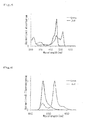
- the absorption spectrum of the protein was measured in the following 50 mM buffer solution ( Figure. 4 ).
- the following buffer solutions were used for each pH:
- Example C-1 Isolation of novel fluorescent protein gene (Kaede) from coral
- a fluorescent protein gene was isolated from Trachyphyllia geoffroyi which emits fluorescence with rich colors by the following procedures.
- Total RNAw as extracted by acidic guanidium/phenol/chloroform method.
- RNA from Trachyphyllia geoffroyi was crushed in a denaturation solution by using a Multi-Beads Shocker (Yasui Kikai), and then phenol/chloroform was added thereto, followed by centrifugation to separate RNA from protein and DNA.
- a water phase containing RNA was added to isopropanol, and the mixture was centrifuged, so as to obtain total RNA as a precipitate.
- Obligotex-dT30 ⁇ super> was added to the total RNA, and the mixture was then heated, so as to destroy the secondary structure of the RNA. Thereafter, the RNA was bound to Oligotex-dT at 37°C. After washing, the resultant product was heated and centrifuged, so as to obtain a supernatant eluted from the mRNA. Oligotex-dt was eliminated from the and then, mRNA was allowed be precipitated with ethanol and NaCl. The mRNA was then dissolved in water.
- a cDNA fragment was prepared using TimeSaver and Directional Cloning Toolbox (both of which were manufactured by Amersham Pharmacia).
- the mRNA was heated to destroy the secondary structure thereof. Thereafter, the mRNA, DTT, and a Not I-dT primer were added to First-Strand Reaction Mix, so as to synthesize a first strand. This was then added to Second-Strand Reaction Mix, so as to synthesize a second strand.
- the synthesized second strand was purified with a span column attached with the kit. Eco RI adaptors were added to both termini of the purified double-stranded cDNA, and only the 3'-side thereof was cleaved, with Not I . It was purified again with the span column, so as to obtain a CDNA fragment ( Eco RI- Not I).
- Kaede The amino acid sequence of Kaede is shown in SEQ ID NO: 5 of the sequence listing, and the nucleotide sequence of Kaede is shown in SEQ ID NO: 6.
- a Bam HI site was added to the N-terminus of the obtained full-length cDNA, and an Eco RI site was added to the C-terminus thereof. Thereafter, it was subcloned in frame into pRSETB (manufactured by Invitrogen), and it was then expressed in Escherichia coli JM109 DE3. The expressed protein was purified with Ni-Agarose gel (manufactured by QIAGEN), utilizing an His-tag at the N-terminus thereof.
- FIG. 5 shows the absorption spectrum of a purified protein before and after UV irradiation (the solid represents the absorption spectrum before UV irradiation, and the dotted line represents the absorption spectrum after UV irradiation).
- the molar absorption coefficient was obtained from the concentration of the protein and the absorbance at absorption maximum (Table 3).
- the fluorescence properties of Kaede are shown in the following Table 3.
- Table 3 Fluorescence properties of Kaede Excitation maximum (nm) Fluorescence maximum(nm) Molar absorption coefficient (M -1 /cm -1 ) Quantum yield pH sensitivity (pKa) Number of amino acids Green: 508nm Green: 518 nm Green: 98,800 Green: 0.80 Green: 5.7 Red: 572 nm Red: 581nm (508 nm) Red 0.33 Red. 5.7 225 Red: 60,400 (572 nm)
- Example C-2 Introduction of novel fluorescent protein gene into mammalian cells
- the Kaede gene was introduced into HeLa cells, using LIPOFECTIN Reagent (Gibco).
- Figure 7 shows the result obtained by exciting the cells at 470 nm and measuring them with the fluorescence at 510 nm. Fluorescence can be confirmed approximately 9 hours utter the introduction. Fluorescence shifts to a long wavelength even in mammalian cells, when the cells are irradiated with UV.
- Kaede forms a tetramer, there may be cases where its expression pattern becomes abnormal when another protein is fused with Kaede.
- threonine (T) at position 158 was substituted with arginine (R), and alanine (A) at position 160 was substituted with glutamic acid (E), so as to produce a dimeric mutant.
- This mutant was named as Kaede 2.
- the amino acid sequence and nucleotide sequence of Kaede 2 are shown in SEQ ID NOS: 7 and 8 in the sequence listing, respectively.
- Kaede 2 In electrophoresis on 12.5% acrylamide gel where sample was not boiled (Pseudonative SDS/PAGE), a band of Kaede 2 was detected as a molecule with a lower molecular weight than that of Kaede ( Figure 8 ).
- Kaede and Kaede 2 were allowed to express in the plasma membrane of HeLa cells, Kaede exhibited an apparently abnormal expression pattern.
- Kaede 2 exhibited a normal expression pattern showing that it was properly targeted into the plasma membrane ( Figure 9 ).
- Example C-4 Ultraviolet ray irradiation-dependent cleavage of Kaede protein
- Such properties can be used for a technique of controlling the cleavage of a protein by light.
- a technique of cleaving a protein by light has not yet been reported.
- such a cleavage is associated with a ⁇ -elimination reaction in a protein.
- such a reaction in a protein, or a ⁇ -elimination reaction in which an amide group acts as a leaving group has not yet been reported.
- Example D-1 Isolation of novel fluorescent protein gene from coral
- a fluorescent protein gene was isolated from coral emitting fluorescence. Scolymia vitiensis was used as a material, Scolymia vitiensis was crushed by hammer, and 15ml of "TRIzol” (GIBCO BRL) was added to 10 g of the crushed coral, and the mixture was stirred, followed by centrifugation at 1,500 x g for 10 minutes. 3 ml of chloroform was added to the supernatant, The mixture was stirred for 15 seconds and then left at rest for 3 minutes. The resultant product, was centrifuged at 7,500 x g for 15 minutes. 7,5 ml of isopropanol was added to the supernatant. The was stirred for 15 seconds and then left at rest for 10 minutes.
- the resultant product was centrifuged at 17,000 x g for 10 minutes The supernatant was discarded, and 6 ml of 70% ethanol was added thereto. The obtained mixture was centrifuged at 17,000 x g for 10 minutes, The was discarded, and the was dissolved in 200 ⁇ l of DEPC water. Total RNA dissolved in the DEPC water was 100 times diluted, .and the values of O.D.260 and O.D.280 were measured, so as to? determine the concentration of RNA. 230 ⁇ g of the total RNA. was obtained.
- cDNA (33 ⁇ l) was synthesized from 3 ⁇ g of the total RNA using a kit for synthesizing first strand cDNA, "Ready, To Go” (Amersham Pharmacia).
- the purified DNA fragment was ligated to pT7-blue vector (Novagen). Escherichia coli (TGI) was transformed with the vector, and the obtained transformants were subjected to blue white selection in the presence of X-gaL Thereafter, plasmid DNA was purified from white colonies of Escherichia coli.
- the nucleotide sequence of the inserted DNA fragment was determined by a DNA sequencer.
- the obtained nucleotide sequence was compared with the nucleotide sequences of other fluorescent protein genes to confirm that the nucleotide sequence of the DNA was derived from a fluorescent protein. 5'-RACE and 3'-RACE methods were applied to a gene that had been confirmed to be a part of a fluorescent protein gene, so as to carry out the cloning of a fun-length gene.
- the 5'-RACE method was applied using 5'-RACE System for Rapid Amplification of cDNA Ends. Version 2 . 0 (GIBCO BRL). 3 ⁇ g of the total RNA prepared in (1) above was used as a template.
- the 400-bp amplified hand was cut out by agarose gel electrophoresis and then purified, The purified DNA fragment was ligated to a pT7-blue (Novagen). Escherichia coli (TG1) was transformed with the vector, and the obtained transformants were subjected to blue white selection in the presence of X-gal. Thereafter, plasmid DNA was purified from white colonies of Escherichia coli. The nucleotide sequence of the inserted. DNA fragment was determined by a. DNA sequencer.
- the 3'-terminai portion of the DNA fragment obtained by degenerated PCR was obtained by PCK using the primer produced based on the information obtained by sequencing of the nucleotide sequence in (4) above and an oligo dT primer. 3 ⁇ l of the first strand cDNA prepared in above was used as a template.
- the produced primer is shown below:
- An amplified band of approximately 500 bp was cut out by agorose gel electrophoresis and then purified.
- the purified DNA fragment was ligated to a pT7 vector (Novagen).
- Escherichia coli (TGI) was transformed with the vector, and the obtained transformants were subjected to blue white selection in the presence of X-gaL Thereafter, plasmid DNA was purified white colonies of Escherichia coli.
- the nucleotide sequence of the inserted DNA fragment was determined by a DNA sequencer.
- Example D-2 Expresssion of protein in Escherichia coli
- a band of 700 bp amplified in. D-1 (7) was cut out by agarose gel electrophoresis and then purified.
- the purified DNA fragment was subcloned into the Ncol-Xhol site of a pET 28 vector (Novagen), and it was then allowed to express in Escherichia coli (JM109-DE3) Since the expressed protein was constructed such that His-tag was attached to the N-terminus thereof, it was purified with Ni-Agarose gel (QIAGEN), was carried out in accordance with the attached protocols.
- the-property of the purified protein (Momiji) was analyzed.
- the fluorescent protein was diluted with the same above buffer solution such that the absorption at 450 nm became 0.002, and the fluorescent spectrum obtained by excitation at 450 nm was measured. Likewise, EGFP(CLONTECH) was diluted such that the absorption at.
- the fluorescence and absorption spectra of the fluorescent protein (Momiji) of the present invention change by irradiation with UV (around 365 nm). Such changes were measured.
- the fluorescent protein was diluted with a solution consisting of 50 mM HEPES (pH 7.4) and 150 mM KCI. The diluted solution was irradiated with light at 365 nm, and the fluorescence spectrum obtained by excitation at 365 nm was then measured.
- a solution consisting of a 20 ⁇ M fluorescent protein, 50 mM HEPES (pH 7.4), and 150 mM KCI was used, and the absorption spectrum thereof was measured after irradiation with light at 365 nm.
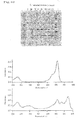
- the measurement results are shown in Figures 13 and 14 . Since the amount of light at 365 nm was different between the measurement of the fluorescence spectrum and that of the absorption spectrum, change and time in the fluorescent spectrum did not correspond to those in the absorption spectrum.
- Example D-5 Improvement of light-irridiation-dependent fluorescence properties of Momiji
- Momiji 4 isoleucine (I) at position 197 was substituted with methionine (M). This mutant was named as Momiji 4.
- the amino acid sequence and nucleotide sequence of Momiji 4 are shown in SEQ ID NOS: 13 and 14, respectively.
- An ultraviolet ray-irradiation-dependent change in the fluorescence from green, to red was observed in Momiji 4 from 1 minute after the irradiation with light at 365 nm.
- 10 minutes after the irradiation with light at 365 nm corresponded to 1 minute after the irradiation therewith in Momiji 4.
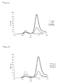
- Momiji 4 had a light-irradiation-dependent fluorescence properties conversion efficiency that was clearly higher than that of the wild type (Momiji) ( Figures 16 , 20 , and 22 ).
- Momiji and Momiji 4 were-allowed to express in HeLa cells, and a change in florescence properties from green to red (red/green) by irradiation with light at 410 nm was measured in the cells.
- Momiji 4 had higher light-irradiation-dependent fluorescence property conversion efficiency than the wild type.
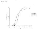
- the molecules emitting red fluorescence whose fluorescence properties ultraviolet ray-irradiation-dependently changed, had pH resistivity lower than that of the wild type (Momiji).
- the molecules emitting green fluorescence before irradiation with light for conversion of the properties had low pH sensitivity ( Figures 17 and 19 ).
- the present invention provides novel chromoproteins derived from Anthopleura inornata .
- the chromoprotein of the present invention has certain absorption properties and has low pH sensitivity. Thus, it is useful for molecular biology analysis.
- the absorbance (molecular absorption coefficient) of the chromoprotein of the present invention is significantly high, it enables highly efficient light energy conversion.
- the present invention provides a fluorescent protein having a novel primary structure derived from Trachyphyllia geoffroyi.
- the fluorescent protein of the present invention is characterized in that its color changes from green to red by ultraviolet rays. It is possible to carry out optical marking on specific cells or organs by light.
- the present invention provides a fluorescent protein having a novel primary structure derived from Scolymia vitiensis.
- the fluorescent protein of the present invention is characterized in that its color changes from green to red by ultraviolet rays.
- the fluorescent protein of the present invention is capable of changing its color from green to red by ultraviolet rays, extremely simply, efficiently, and specifically. This protein is practical because it is extremely stable and bright in both the green and red states before and after the conversion of the color.
- the present invention provides :
Landscapes
- Health & Medical Sciences (AREA)
- Life Sciences & Earth Sciences (AREA)
- Immunology (AREA)
- Engineering & Computer Science (AREA)
- Chemical & Material Sciences (AREA)
- Molecular Biology (AREA)
- Biomedical Technology (AREA)
- Hematology (AREA)
- Urology & Nephrology (AREA)
- Medicinal Chemistry (AREA)
- General Health & Medical Sciences (AREA)
- Biochemistry (AREA)
- Microbiology (AREA)
- Food Science & Technology (AREA)
- Physics & Mathematics (AREA)
- Analytical Chemistry (AREA)
- Cell Biology (AREA)
- Biotechnology (AREA)
- General Physics & Mathematics (AREA)
- Pathology (AREA)
- Organic Chemistry (AREA)
- Gastroenterology & Hepatology (AREA)
- Biophysics (AREA)
- Genetics & Genomics (AREA)
- Proteomics, Peptides & Aminoacids (AREA)
- Mycology (AREA)
- Peptides Or Proteins (AREA)
- Micro-Organisms Or Cultivation Processes Thereof (AREA)
- Investigating Or Analysing Biological Materials (AREA)
- Measuring Or Testing Involving Enzymes Or Micro-Organisms (AREA)
- Investigating Or Analysing Materials By The Use Of Chemical Reactions (AREA)
- Medicines That Contain Protein Lipid Enzymes And Other Medicines (AREA)
Abstract
(a) an amino acid sequence shown in SEQ ID NO: 5; or
(b) an amino acid sequence comprising a deletion, substitution and/or addition of I to 20 amino acids with respect to the amino acid sequence shown in SEQ ID NO: 5, and having fluorescent properties;
and to a. fluorescent protein having an amino acid sequence shown in SEQ ID NO:7.
Description
- The present invention relates to a novel chromoprotein More specifically, the present invention relates to a novel chromoprotein derived from Anthopleura inornata, and the use thereof. The present invention relates to a novel fluorescent protein More specifically, the present invention relates to a novel fluorescent protein derived from Trachyphyllia geoffroyi and Scolymia vitiensis and the use thereof.
- Green fluorescent protein. (GFP) derived from Aequorea victoria, a jellyfish,has many purposes in biological systems. Recently, various GFP mutants have been produced based on the random and semi-rational mutagenesis, wherein a color is changed, a folding property is improved, luminance is enhanced, or pH sensitivity is modified. Fluorescent proteins such as GFP are fused with other proteins by gene recombinant technique, and monitoring of the expression and transportation of the fusion proteins is carried out.
- One of the most commonly used types of GFP mutant is Yellow fluorescent protein (YFP), Among Aequorea-derived GFP mutants, YFF exhibits the fluorescence with the longest wavelength. The values ε and Φ of the majority of YEPs are 60,000 to 100,000 m-1cm-1 and 0.6 to 0.8, respectively (Tsien, R. Y. (1998). Ann Rev. Biochem. 67, .509-544). These values are comparable to whose of the general fluorescent group (fluorescein, rhodamine, etc.). Accordingly, improvement of the absolute luminance of YFP is nearly appcoaching: its limit
- In addition, cyan fluorescent, protein (CFP) is another example of the GFF mutant. Of this type of protein. ECFP (enhanced cyan fluorescent protein) has been known, Moreover, red fluorescent: protein (RFP) has been isolated from sea anemone (Discoma sp.). Of this type of protein, DasRed has been known. Thus, 4 types of fluorescent proteins, that are, green fluorescent protein, yellow fluorescent protein, cyan fluorescent protein, and red fluorescent protein, have successively been developed. The range of the spectrum has significantly been expanded.
- A chromoprotein is a protein regarding which the quantum yield of the conventional fluorescent protein is reduced to close to zero. Since such a chromoprotein is capable of introducing into cells molecules that convert light energy into another type of energy, it can be applied to various purposes, However, there have been only a few reports regarding the absorption wavelength properties of such a chromoprotein.
- It is an object of the present invention to provide a novel chromoprotein derived from Anthopleura inornata, which absorbs a light having a specific wavelength. It is another object of the present invention to provide a fluorescent protein having a novel primary structure, which is derived from Cnidaria, and particularly from Trachyphyllia geoffroyi and Scolymia vitiensis belonging to Scleractinia.
- The present have conducted intensive studies directed towards achieving the aforementioned object. They have designed suitable primers based on information regarding the amino acid sequences of known fluorescent proteins. Using these primers, they have succeeded in the amplification and of genes encoding novel chromoproteins form the cDNA library of Anthopleura inornata exhibiting a green color. The present inventors have further analyzed the light-absorbing properties and pH sensitivity of the obtained chromoprotein derived from inornata.
- Further, the present inventors have performed, expression cloning by using cDNA library derived from Trachyllia geoffroyi and Scolymia vitiensis, and, have succeeded in cloning genes encoding novel fluorescent proteins. The present inventors have examined the fluorescent properties of the obtained proteins, and as a result, they have found that these fluorescent proteins particular fluorescent properties.
- The present invention has been based on these findings.
- Thus, the present invention provides a chromoprotein derived from. Anthopleura inorntata having the following properties:
- (1) the absorption maximum wavelength is 605 nm;
- (2) the molar absorption coefficient is 47,550 at 6:05 nm; :and
- (3) the pH sensitivity of light-absorbing property is stable at between
pH 3 andpH 10. - In another aspect of the present invention, there is provided a chromoprotein having either one of the following amino acid sequences:
- (a) the amino acid sequence shown in SEQ ID NO: 1; and
- (b) an amino acid sequence comprising a deletion, substitution and/or addition of one or several amino acids with respect to the amino acid sequence shown in SEQ ID NO: 1, and having light-absorbing properties.
- In another aspect of the present invention, there, is provided a chromoprotein derived from Anthopleura inornata having the following properties:
- (1) the absorption maximium wavelength is 553 nm;
- (2) the molar absorption coefficient is 25,300 at 553 nm; and
- (3) the pH sensitivity of light-absorbing property is stable at between
pH 5 andpH 10. - In another aspect of the present, invention, there is provided a chromoprotein having either one of the following amino acid sequences:
- (a) the amino acid sequence shown in SEQ ID NO: 3; and
- (b) an amino acid sequence comprising a deletion, substitution and/or addition of one or several amino acids with respect to the amino acid sequence shown in SEQ ID NO: 3, and having light-absorbing properties.
- In further another aspect of the present invention, there is provided a DNA encoding the protein of the present invention.
- In further another aspect of the present invention, there is provided a DNA of either one of followings;
- (a) DNA encoding the amino acid sequence shown in SEQ ID NO: 1; and
- (b) DNA encoding a protein which has an amino acid sequence comprising a deletion, substitution and/or addition of one or several amino acids with respect to the amino acid sequence shown in SEQ ID NO: 1, and has light-absorbing properties.
- In further another aspect of the present invention, there is provided a DNA of either one of followings:
- (a) DNA encoding the amino acid sequence shown in SEQ ID NO: 3; and
- (b) DNA encoding a protein which has an amino acid sequence comprising a deletion, substitution and/or addition of one or several amino acids with respect to the amino acid sequence shown in SEQ ID NO: 3, and has light-absorbing properties.
- In further another aspect of the present invention, there is provided a DNA having either one of the following nucleotide sequences:
- (a) the nucleotide sequence shown in SEQ ID NO: 2; and
- (b) a nucleotide sequence comprising a deletion, substitution and/or addition of one or several nucleotides with respect to the nucleotide sequence shown in
SEQ ID NO : 2, and encoding a protein having light-absorbing properties. - In further another aspect of the present invention, there is provided a DNA having either one of the following nucleotide sequences:
- (a) the nucleotide sequence shown in SEQ ID NO: 4; and
- (b) a nucleotide sequence comprising a deletion, substitution and/or addition of one or several nucleotides with respect to the nucleotide sequence shown in SEQ ID NO: 4, and encoding a protein having light-absorbing properties.
- In further another the present invention provides a recombinant vector having the DNA of the present invention.
- In further another aspect, the present invention provides a having the DNA or vector of the present invention.
- In further another aspect, the present invention provides a fusion protein composed of the of the present invention and another protein.
- In further another aspect, the present invention provides a method for analyzing a physiologically active substance, which is characterized in that the FRET (fluorescence energy transfer) method is applied using the chromoprotein of the present invention as an acceptor protein.
- In further another aspect, the present invention provides a light-absorbing reagent kit comprising the chromoprotein, DNA, recombinant vector, transformant,or fusion protein of the present invention.
- Further, the present provides a fluorescent protein derived from Trachyphyllia geoffroyi, which has the following properties:
- (1) the color is changed from green to red by irradiation of ultraviolet the excitation maximum wavelength is 508 nm (green) and 572 nm (red); and the fluorescence maximum wavelength is 518 nm (green) and 581 mn (red);
- (2) the molar absorption coefficient at 508 nm is 98800 M-1cm-1 and the molar absorption coefficient (red) at 572 nm is 60400 M-1cm-1;
- (3) the quantum yield is 0.80 (green) and 0.33 (red); and
- (4) pKa regarding the pH sensitivity of the green and red are both 5.7.
- in another aspect of the present invention, there is provided a fluorescent protein having either of the following amino acid sequence:
- (a) an amino acid sequence shown in SEQ ID NO: 5; or
- (b) an amino acid sequence comprising a deletion, substitution and/or addition of one or several amino acids with respect to the amino acid sequence shown in SEQ ID NO: 5, and having fluorescent properties,
- In another aspect of the present invention, there is provided a fluorescent protein having an amino acid sequence shown in SEQ ID NO: 7.
- In further another aspect of the present invention, there is provided a fluorescent protein derived from Scolymia vitiensis Which has the following properties:
- (1) the color is changed from green to red by irradiation of ultraviolet ray; the excitation maximum wavelength is 508 nm (green) and 578 nm (red); and the fluorescence maximum wavelength is 518 nm (green) and 588 nm (red);
- (2) the molar absorption coefficient (green) at 508 nm is 102250 M-1cm-1; and the molar absorption coefficient (red) at 578 nm is 76950 M-1cm -1
- (3) the quantum yield (fluorescence) is 0.43 (green) and 0.51 (red); and
- (4) pKa regarding the pH sensitivity of the green (508 nm) is 5.8; and pKa regarding the pH sensitivity of the red (57 8 nm) is 6.5
- In another aspect of the present, invention, there is provided a fluorescent protein having either one of the following amino acid sequences:
- (a) an amino acid sequence shown in SEQ ID NO: 9; or
- (b) an amino acid sequence a deletion, substitution and/or addition of one or several amino acids with respect to the amino acid sequence shown in SEQ ID NO: 9, and having fluorescent properties.
- In another aspect of the present invention, there is provided a fluorescent protein having an amino acid sequence shown in any of SEQ ID NO: 11, 13, 15 or 17.
- In further another aspect of the present invention, there is provided a DNA of either one of following:
- (a) DNA which encodes the amino acid sequence shown in SEQ ID NO: 5; or
- (b) DNA which encodes an amino acid sequence comprising a deletion, substitution and/or addition of one or several amino acids with respect to the amino acid sequence shown in SEQ ID NO: 5, and encodes a fluorescent protein:
- (c) DNA having a sequence shown in
SEQ ID NO : 6; or - (d) DNA having a nucleotide sequence comprising a deletion, substitution and/or addition of one or several nucleotides with respect of the nucleotide sequence shown in SEQ ID NO: 6, and encoding a fluorescent protein.
- In further another aspect of the present invention, there is provided a DNA of either one of the following:
- (a) DNA which encodes the amino acid sequence shown in SEQ ID NO: 7: or
- (b) DNA having a nucleotide sequence shown in SEQ ID NO: 8.
- In further another aspect of the present invention, there is provided a DNA of either one of the following:
- (a) DNA which encodes the amino acid sequence shown in
SEQ ID NO 9; or - (b) DNA which encodes an amino acid sequence comprising a deletion, substitution and/or addition of one or several amino acids with respect to the amino acid sequence shown in SEQ ID NO: 9, and encodes a fluorescent protein:
- (c) DNA having a nucleotide sequence shown in SEQ ID NO: 10, or
- (d) DNA having a nucleotide sequence comprising a deletion, substitution and/or addition of one or several nucleotides with respect of the nucleotide sequence shown in SEQ ID NO: 10, and encoding a fluorescent protein.
- In further another aspect of the present invention, there is provided a DNA of either one of the following:
- (a) DNA which encodes the amino acid sequence shows in SEQ ID NO: 11, 13, 15 or 17; or
- (b) DNA having a nucleotide sequence shown in SEQ ID NO: 12, 14, 16 or 18,
- In further another aspect of the present invention, there is provided a recombinant vector having any of the DNA of the present invention.
- In further another aspect of the present invention, there is provided a transformant having the DNA or recombinant vector of the present invention.
- In further another aspect of the present invention, there is provided a fusion fluorescent protein consisting of the fluorescent protein of the present invention and another protein. Preferably, said another protein is one that localizes in the cell, and more preferably one specific to an intracellular organella.
- In further another aspect of the present invention, there is provided a method for analyzing the localization or dynamics of a protein in cells, characterized in that the fusion of the present invention, is allowed to be expressed in cells.
- In further another aspect of the present invention, there is provided a fluorescent reagent kit which comprises the proteins, DNAs, recombinant vector, transformant fusion protein of the present invention.
-
-
Figure 1 shows the results obtained by measuring the absorption spectrum of the chromoprotein (Be-G) derived from Anthopleura inornata of the present invention. The horizontal axis represents the wavelength of a light absorbed. The longitudinal axis represents absorbance. -
Figure 2 shows the pH sensitivity of the absorption spectrum of the chromoprotein (Be-G) derived from Anthopleura inornata of the present invention. The axis represents pH value, and the longitudinal axis represents absorbance. 605 nm shows an absorbance specific for the chromoprotein (Be-G) derived from Anthopleura inornata of the present invention. 277 nm shows an absorbance that is generally used in quantification of protein (absorbance of aromatic amino acids). That is to say, the value at 277 nm shows that the protein mass is constant, and the value at 605 mn shows that the absorbance specific for the chromoprotein (Be-G) derived from Anthopleura inornata of the present invention hardly changes in the range betweenpH 5 andpH 10. -
Figure 3 snows the results, obtained, by measuring the absorption of the chromoprotein (Be-R) derived from Anthopleura inornata of the present The horizontal axis represents the wavelength of a light absorbed. The longitudinal axis represents absorbance. -
Figure 4 shows the pH sensitivity of the absorption spectrum of the chromoprotein (Be-R) derived from Anthopleura inornata of the present invention. The horizontal axis represents pH value, and the longitudinal axis represents absorbance 553 nm shows an absorbance specific for the chromoprotein (Be-R) derived from Anthopleura inornata of the present invention. 277 nm shows an absorbance that is generally used in quantification of protein (absorbance of aromatic amino acids). That is to say, the value at 277 nm shows that the protein mass is constant, and the value at 553 nm shows that the absorbance specific for the chromoprotein (Be-R) derived from Anthopleura inornata of the present invention hardly changes in the range betweenpH 5 andpH 10. -
Figure 5 is the absorption spectrum of the fluorescent protein (Kaede) of the present invention. -
Figure 6 is the fluorescence spectrum of the fluorescent protein (Kaede) of the present invention. -
Figure 7 shows the results obtained by exciting at 470 nm, HeLa cells into which the gene of the fluorescent protein (Kaede) of the present invention has been introduced, and measuring them with the fluorescence at 510 nm. -
Figure 8 is the electrophoretic patterns of Kaede andKaede 2 in 12.5% acrylamide.Kaede 2 band appears as one having a lower molecular weight than that of Kaede. -
Figure 9 is an expression pattern obtained when Kaede (upper figure) and Kaede 2 (lower figure) were targeted to the plasma membrane of HeLa cells. -
Figure 10 shows the experimental results showing the ultraviolet ray-irradiation-dependent cleavage of a Kaede protein. The upper figure shows a transition in the ultraviolet ray-irradiation-dependent cleavage of peptide chain in the form of an electropherogram on 12.5% acrylamide gel. A Kaede protein solution was irradiated with light at 365 nm, sampling was conducted every 20 minutes, and SDS/PAGE was carried out. The middle figure shows the absorption spectrum observed before irradiation with light at 365 nm (0 minute). The lower figure shows the absorption spectrum observed 140 minutes after irradiation with light at 365 nm. -
Figure 11 shows the excitation spectrum and fluorescence spectrum of the fluorescent protein (Momiji) of the present invention. -
Figure 12 shows: the pH sensitivity of the fluorescent protein (Momiji) of the present invention. -
Figure 13 shows a change in the fluorescence spectrum when the fluorescent protein (Momiji) of the present invention is irradiated with 365 nm UV. -
Figure 14 shows a change in the absorption spectrum when the fluorescent protein (Momiji) of the present invention is irradiated with 365 nm UV, -
Figure 15 shows a change in the absorption spectrum before and after irradiation with UV (Momiji 2). -
Figure 16 shows a change in the absorption spectrum before and after irradiation with UV (Momiji 4). -
Figure 17 shows a change in the absorption spectrum due to pH before and after irradiation with UV (Momiji). Before irradiation: 508 nm; and after irradiation: 578 nm. -
Figure 18 shows a change in the absorption spectrum due to pH before and after irradiation with UV (Momiji 2). Before irradiation: 598 nm; and after irradiation 576 nm. -
Figure 19 shows a change in the absorption spectrum due to pH before and after irradiation with UV (Momiji 4). Before irradiation: 508 nm; and after irradiation: 583 nm. -
Figure 20 shows a transition in a change in fluorescence properties depending on irradiation time with light at 365 nm (Momiji). Measurement was carried out on a 6 µg/mlprotein solution. In order to clearly differentiate the situation from that of a mutant, light exchange was carried out with extremely weak light at 365 nm, and the fluorescence spectrum from 470 nm to 650 nm was measured. -
Figure 21 shows a transition in a change in fluorescence properties depending on irradiation time with light at 365 nm (Momiji 2). Measurement was carried out on a 6 µg/ml protein solution. In order to clearly differentiate the situation from that of a mutant, light exchange was carried out with extremely weak light at 365 mn, and the fluorescence spectrum from 470 nm to 650 nm was measured. -
Figure 22 shows a transition in a change in fluorescence properties depending on irradiation time with light at 365 nm (Momiji 4), Measurement was carried out on a 6 µg/ml protein solution. In order to clearly differentiate the situation from that of a mutant, light exchange was carried out with extremely weak light at 365 nm, and the fluorescence spectrum from 470 nm to 650 nm was measured. -
Figures 23 shows a transition in fluorescence properties in HeLa cells.- Exposure time: 410
nm 100 ms - Green: 400ms, Red: 50 ms
- DM 420DCLP
- Green Ex 475AF20, Em 330DF35
- Red Ex 550DF35, Em 575ALP
- Object lens; X40 Uapo/340
Measurement was carried out under the aforementioned conditions, and the fluorescence values of the red fluorescence and the green fluorescence were expressed in a graph. - Exposure time: 410
-
Figure 24 shows the results obtained by exciting with a blue light the electrophoretic pattern on 12.5% acrylamide, gel (Pseudo-native SDS/PAGE) and then photographing the resultant with a digital camera.- WT (Momiji): tetramer
- d16: dimer
- m16: monomer
-
Figure 25 shows the fluorescence spectrum (d16) before and after irradiation with light at 365 nm.- Green fluorescence peak: 518 nm
- Red fluorescence peak: 591 nm
-
Figure 26 shows the fluorescence spectrum(m16) before, and after irradiation with light at 365 nm.- Green fluorescence peak: 518 nm
- Red fluorescence peak: 587 nm
- The embodiments of the present invention will be described in detail below.
- The first chromoprotein of the present invention characterized in that it is derived from Anthopleura inornata, and has the following properties:
- (1) the absorption maximum: wavelength is 605 nm;
- (2) the molar absorption coefficient is 47,550 at 605 nm; and
- (3) the pH sensitivity of light-absorbing property is stable at between
pH 5 andpH 10. - The second chromoprotein of the present invention is characterized in that it is derived from Anthopleura inornata, and has the following properties:
- (1) toe absorption maximum wavelength is 553 nm;
- (2) the molar absorption coefficient is 25,300 at 553 nm; and
- (3) the pH sensitivity of light-absorbing property is stable at between
pH 5 andpH 10. - Anthopleura inornata 96 tentacles, which are regularly aligned. In addition, this species is characterized in that it has an oral disc having the same color was that of the tentacles and that the surrounding portion of the month has a reddish brown color. 98 lines of adhesive warts are found on the body wall, and such wards are distributed up to the lower end of the body wall. The color of the body wall is largely varied, and brown, blue, and pink body colors have been known. Such Anthopleura inornata ranges from the southern part of Hokkaido to Kyushu, and many of them live in the intertidal zone.
- In the examples described later in the present specification, a chromoprotein. having the aforementioned properties was isolated from Anthopleura inornata that was used as a starting material However, there may also be cases where the chromoprotein of the present invention can be obtained from a sea anemone other than Anthopleura inornata. The thus obtained chromoprotein is also included in the scope of the present invention.
- As described in the examples below, the first chromoprotein. (Be-G) of the present invention has an absorption maximum wavelength of 605 nm, the molar absorption coefficient is 47, 550 at 605 nm.
- As described in the examples below, the second chromoprotein (Be-R) of the present has an absorption maximum wavelength of 553 nm, and the molar absorption coefficient is 25,300 at 553 nm.
- The molar absorption coefficient the amount of absorbed photons per mole of molecule, and the quantum yield represents a numerical value showing the amount of the absorbed photons that can be emitted, as fluorescence. Since the chromoproteins of the present invention have an extremely low quantum yield, they hardly emit fluorescence at all. Due to this property, the chromoproteins of the present invention can be used: (1) as an acceptor molecule (energy receptor) in FRET; (2) in development of a system for converting the energy of applied light into energy other than the light; and (3) in introduction of a mutation into the amino acid sequence of the protein to modify it so that it emits fluorescence.
- In addition, the chromoproteins of the present invention are characterized in that the pH sensitivity of light-absorbing properties is stable at between
pH 5 andpH 10, That is to say, in the case of the chromoproteins of the present invention, the peak value of the absorption spectrum does not significantly fluctuate in the range betweenpH 5 andpH 10. Accordingly, even under the same conditions, the chromoproteins of the present invention can be used in a broad range of pH environments, and thus, the use of the chromoproteins in vivo have few restrictions. - The examples of the chromoprotein of the present invention include a chromoprotein having either one of the following ammo acid sequences:
- (a) the amino acid sequence shown in SEQ ID NO: 1 or 3; and
- (b) an amino acid sequence comprising a deletion, substitution and/or addition of one or several amino acids with respect to the amino acid sequence shown in SEQ ID NO: 1 or 3, and having light-absorbing properties.
- The scope of "one or several" in the phrase "an amino acid sequence comprising a deletion, substitution and/or addition of one or several amino acids" is not particularly limited in the present specification. For example, it means 1 to 20, preferably 1 to 10, more preferably 1 to 7, further preferably 1 to 5, and particularly preferably 1 to 3.
- The term "light-absorbing properties" is used in the present specification to mean properties capable of absorbing light having a certain wavelength. For example, an absorption maximum wavelength may be 605 nm or 553 nm as in the case of the chromoproteins described in the present specification, or the value of the absorption maximum wavelength may also be shifted. It is preferable that the pH sensitivity of light-absorbing properties is stable at between
pH 5 andpH 10. - The chromoproteins of the present invention having the amino acid sequence shown in SEQ ID NO: 1 or 3 in the sequence listing hardly emit fluorescence at all. In the present invention, one or several amino acids are deleted, substituted, and/or added with respect to the amino acid sequence shown in SEQ ID NO: 1 or 3, so as to produce a protein having stronger fluorescence. The thus produced proteins are also included in the scope of the present invention.
- The method of obtaining the chromoproteins of the present invention is not particularly limited. The proteins may be either a protein synthesized by chemosynthesis, or recombinant protein produced by a gene recombination technique.
- Where a recombinant protein is produced, it is neccessary to obtain DNA encoding the protein. Appropriate primers are designed by using information regarding the amino acid sequence shown in SEQ ID NO: 1 or 3 of the sequence listing of the present specification and the nucleotide sequence shown in SEQ ID NO: 2 or 4 thereof. Using these primers, PCR is carried out by using cDNA library derived from Anthopleura inornata as a template, so that DNA encoding the chromoprotein of the present invention can be obtained. The chromoprotein of the present invention can be produced by introducing this DNA into an appropriate expression system. Expression in an expression system will be described later in the present specification.
- The first fluorescent protein of the present invention is characterized in that it is derived from Trachyphyllia geoffroyi and has the following properties:
- (1) the color is changed from green to red by irradiation of ultraviolet ray; the excitation maximum wavelength is 508 nm (green) and 572 nm (red); and the fluorescence maximum wavelength is 518 nm(green) and 581 nm (red);
- (2) the molar absorption coefficient (green) at 508 nm is 98800 M-1cm-1; and the molar absorption coefficient (red) at 572 nm is 60400 M-1cm-1;
- (3) the quantum yield is 0.80 (green) and 0.33 (red); and
- (4) pKa regarding the pH sensitivity of the green and red are both 5.7.
- Trachyphyllia geoffroyi is one type of onidarian sea anemone, and it is characterized in that it emits extremely colorful fluorescence. Trachyphyllia geoffroyi mainly ranges over the area below the midland of Honshu Island, Japan. This sea anemone lives in the sludge in the gulf. At night, it extends its tentacles to capture plankton and the like. In terms of a color variation, green, brown, and red examples are found.
- As shown in the examples later, the color of the first fluorescent protein of the present invention is changed from green to red by irradiation of ultraviolet ray; the excitation maximum wavelength is 508 nm (green) and 572 nm (red); and the fluorescence maximum wavelength is 518 nm (green) and 581 nm (red). The molar absorption coefficient (green) at 508 nm is 98800 M-1cm-1; and the molar absorption coefficient (red) at 572 nm is 60400 M-1cm-1:
- The first fluorescent protein of the present invention is characterized in that its color changes due to ultraviolet rays. Thus, optical marking can be carried out on specific cells or organs thereof.
- The examples of the first fluorescent protein of the present invention include a fluorescent protein having either one of the following amino acid sequences:
- (a) an amino acid sequence shown in SEQ ID NO: 5; or
- (b) an amino acid sequence comprising a deletion, substitution and/or addition of one or several amino acids with respect to the amino acid sequence shown in SEQ ID NO: 5, and having fluorescent properties.
- Other examples of the first fluorescent protein of the present invention include a fluorescent protein having an amino acid sequence shown in SEQ ID NO: 7.
- The second fluorescent protein of the present invention is characterized in that it is derived from Scolymia vitiensis and has the following properties:
- (1) the color is changed from green to red by irradiation of ultraviolet ray; the excitation maximum wavelength is 508 nm (green) and 578 nm (red); and the fluorescence maximum wavelength is 518 nm (green) and 588 nm (red);
- (2) the molar absorption coefficient (green) at 508 nm is 102250 M-1cm-1; and the molar absorption coefficient (red) at 578 nm is 76950 M-1cm-1;
- (3) the quantum yield (fluorescence) is 0.43 (green) and 0.51 (red); and
- (4) pKa regarding the pH sensitivity of the green (508 nm) is 5.8; and pKa regarding the pH sensitivity of the red (578 nm) is 6.5.
- Scolymia vitiensis is a sessile single coral. A small individual has a pot-like round shape. However, after it has matured, it has an oval shape. A large spongy columella is found in the center of a coral individual, and a long barrier extends at an almost constant slope from the theca towards the center. A large serration is located on the barrier, and such serration can be seen from the outside. This coral does not open its polyp during the day. It generally has a dark green color, but it also has a red color in rare cases. Approximately 4 types of Scolymia vitiensis have been known. Among them, only one type ranges in the ocean areas surrounding Japan.
- As shown in the examples later, the color of the second fluorescent protein of the present invention is changed from green to red by irradiation of ultraviolet ray; the excitation maximum wavelength is 508 nm (green) and 578 nm (red); and the fluorescence maximum wavelength is 518 nm (green) and 588 nm (red). The molar absorption coefficient (green) at 508 nm is 102250 M-1cm-1; and the molar absorption coefficient (red) at 578 nm is 76950 M-1cm-1.
- The first fluorescent protein of the present invention is characterized in that its color changes due to ultraviolet rays. Thus, optical marking can be carried out on specific cells or organs thereof.
- The examples of the second fluorescent protein of the present invention include a fluorescent protein having either one of the following amino acid sequences:
- (a) an amino acid sequence shown in SEQ ID NO: 9; or
- (b) an amino acid sequence comprising a deletion, substitution and/or addition of one or several amino acids with respect to the amino acid sequence shown in SEQ ID NO: 9, and having fluorescent properties.
- Other examples of the first fluorescent protein of the present invention include a fluorescent protein having an amino acid sequence shown in any of SEQ ID NO: 11, 13, 15 or 17.
- The scope of "one or several" in the phrase "an amino acid sequence comprising a deletion, substitution and/or addition of one or several amino acids" is not particularly limited in the present specification. For example, it means 1 to 20, preferably 1 to 10, more preferably 1 to 7, further preferably 1 to 5, and particularly preferably 1 to 3.
- The term "having fluorescent properties" covers all of the cases where any fluorescence is given. Various properties such as fluorescence intensity, excitation wavelength, fluorescence wavelength or pH sensitivity, may be changed or may remain unchanged.
- The method of obtaining the fluorescent protein of the present invention is not particularly limited. The protein may be either a protein synthesized by chemosynthesis, or recombinant protein produced by a gene recombination technique.
- Where a recombinant protein is produced, it is necessary to obtain DNA encoding the protein. Appropriate primers are designed by using information regarding the amino acid sequence shown in SEQ ID NOS: 5, 7, 9, 11, 13, 15 or 17 of the sequence listing of the present specification and the nucleotide sequence shown in SEQ ID NOS: 6, 8, 10, 12, 14, 16 or 18 thereof. Using these primers, PCR is carried out by using cDNA clones of the above-described various types of known fluorescent proteins as a template, so that DNA encoding the fluorescent protein of the present invention can be obtained. Where a partial fragment of DNA encoding the fluorescent protein of the present invention obtained by the above-described PCR, the produced DNA fragments are ligated to one another by a gene recombination technique, so that DNA encoding the desired fluorescent protein can be obtained. The fluorescent protein of the present invention can be produced by introducing this DNA into an appropriate expression system. Expression in an expression system will be described later in the present specification.
- According to the present invention, genes encoding the chromoproteins and fluorescent proteins of the present invention are provided.
- Specific examples of DNA encoding the first chromoprotein of the present invention may include, either one of the following DNAs:
- (a) DNA encoding the amino acid sequence shown in SEQ ID NO: 1: and
- (b) DNA encoding a protein which has an amino acid sequence comprising a deletion, substitution and/or addition of one or several amino acids with respect to the amino acid sequence shown in SEQ ID NO: 1, and has light-absorbing properties.
- Other examples of DNA encoding the first of the present invention: may include either one of the following DNA:
- (a) the nucleotide sequence shown in SEQ ID NO: 2; and
- (b) a nucleotide sequence comprising a deletion, substitution and/or addition of one or several nucleotides with respect to the nucleotide sequence shown in SEQ ID NO: 2, and encoding a protein having light-absorbing properties.
- Specific examples of DNA encoding the second chromoprotein of the present invention may include either one of the following DNAs:
- (a) DNA encoding the amino acid sequence shown in SEQ ID NO: 3; and
- (b) DNA encoding a protein which has an amino acid sequence comprising a deletion, substitution and/or addition of one or several amino acids with respect to the acid sequence shown in SEQ ID NO: 3, and has light-absorbing properties.
- Other examples of DNA encoding the second chromoprotein of the present invention may include either one of the following DNAs:
- (a) the nucleotide sequence shown in SEQ ID NO: 4; and
- (b) a nucleotide sequence comprising a deletion, substitution and/or addition of one or several with respect to the nucleotide sequence shown in SEQ ID NO: 4, and encoding a protein having light-absorbing properties.
- Specific examples of DNA encoding the first fluorescent, protein of the present invention may include either one of the following DNAs:
- (a) DNA which encodes the amino acid sequence shown in
SEQ ID NO : 5; or - (b) DNA which encodes an amino acid sequence comprising a deletion, substitution and/or addition of one or several amino acids with respect to the amino acid sequence shown in SEQ ID NO: 5, and encodes a fluorescent protein:
- (c) DNA having a nucleotide sequence, shown in SEQ ID NO: 6; or
- (d) DNA having a nucleotide sequence comprising a deletion, substitution and/or addition of one or several nucleotides with respect of the nucleotide sequence shown in SEQ ID NO: 6, and a fluorescent protein.
- Other examples of DNA encoding the first fluorescent of the present invention may include either one of the following DNAs:
- (a) DNA which encodes the amino acid sequence shown in SEQ ID NO: 7; or
- (b) DNA. having a nucleotide sequence shown in SEQ ID NO: 8.
- Specific examples of DNA encoding the second fluorescent protein of the present invention may include either one of the following DNAs:
- (a) DNA which encodes the amino acid sequence shown in SEQ ID NO: 9; or
- (b) DNA which encodes an amino acid sequence comprising a deletion, substitution and/or addition of one or several amino acids with respect to the amino acid sequence shown in SEQ. ID NO: 9. and encodes a fluorescent protein:
- (c) DNA having a nucleotide sequence shown in SEQ ID NO: 10; or
- (d) DNA having a nucleotide sequence comprising a deletion, substitution and/or addition of one or several with respect of the nucleotide sequence shown in SEQ ID NO: 10, and encoding a fluorescent protein.
- Other examples of DNA encoding the second fluorescent protein of the present invention may include either one of the following DNAs:
- (a) DNA which encodes the amino acid sequence shown in SEQ ID NO: 11, 13 15 or 17; or
- (b) DNA having nucleotide sequence shown in SEQ ID NO: 12, 14, 16 or 18.
- The DNA of the present invention can be synthesized by, for example, the phosphoamidite method, or it can also be produced by polymerase chain reaction (PCR) using specific primers. The DNA of the present invention is produced by the method described above in the specification.
- A method of introducing a desired mutation into a certain nucleic acid sequence is blown, to a person, skilled in the art. For example known techniques such as a site-directed mutagenesis, PCR using degenerated oligonucleotides, or the exposure of cells containing nucleic acid to mutagens or radioactive rays, are appropriately used, so as to construct DNA having a Such known techniques are described in, for example, Molecular Cloning: A Laboratory Manual, 2nd Ed., Cold Spring Harbor Laboratory, Cold Spring Harbor, NY., 1989; and Current Protocols in Molecular Biology, Supplements 1 to 38, John Wiley & Sons (1987-1997).
- The DNA of the present invention can be inserted into a suitable vector and used. The type of a vector used in the present invention is not particularly limited. For example, it may be either a vector that can autonomously replicate (e.g., a plasmid, etc.), or vector that is incorporated into the genomes of host cells when it is introduced into the host cells and is then replicated together with the chromosome into which it is incorporated.
- The vector used in the present invention is preferably an expression vector. In an expression vector, elements necessary for transcription (e.g., a promoter, etc.) are functionally ligated to the DNA of the present invention. The promoter is a DNA sequence which shows a transcriptional activity in host cells, and it is appropriately selected depending on the type of host cells.
- Examples of a promoter which can operate in bacterial cells may include a Bacillus stearothermophilus maltogenic amylase gene promoter, a Bacillus licheniformis alpha-amylase gene promoter, a Bacillus amyloliquefaciens BAN amylase gene promoter, a Bacillus subtilis alkaline protease gene promoter, a Bacillus pumilus xylosidase gene promoter, PR and PL promoters of phage rhamda, and lac, trp and tac promoters of Escherichia coli.
- Examples of a promoter which can operate in mammalian cells may include an SV40 promoter, an MT-1 gene) promoter, and an adenovirus-2 major late promoter. Examples of a promoter: which can operate in insect cells may include a polyhedrin promoter, a P10 promoter, an Autographa californica polyhedrosis basic protein promoter, a baculovirus immediate-
early gene 1 promoter, and a baculovirus 39K delayed-early gene promoter. Examples of a promoter which can be operate in yeast host cells may include promoters derived yeast glycolytic genes, an alcohol dehydrogenase gene promoter, a TP11 promoter, and an ADH2-4c promoter. - Examples of a promoter which can operate in filamentous may include an ABH3 promoter and a tpiA promoter.
- In addition, an appropriate terminator such as a human growth hormone terminator, or a TP11 terminator or ADH3 terminator for fungal may be functionally hound to the DNA of the present invention, as necessary. The recombinant vector of the present invention may further have elements such as a polyadenylation signal (e,g., one derived from SV40 or the adeaovrE-us 5Elb region), a transcription enhal1cer sequence (e.g., an SV40 enhancer), or a translation enhancer sequence (e.g., one encoding the adenovirus VA RNA).
- The recombinant vector of the present invention may further comprise a DNA sequence which enables the replication of the recombinant vector in host cells. SV40 replication Origin is an example of such a sequence (when the host cells are mammalian cells).
- The recombinant vector of the present, invention may further comprise a selective marker. Examples of such a selective marker may include genes, complements of which are absent from host cells, such as a dihydrofolate reductase (DHFR) gene of a Shizosaccharomyces pombe TPI gene, and drug resistant genes such as ampicillin, kanamycim, tetracycline, chloramphenicol, neomycin of hygrcunyein-resistant genes.
- A method for ligating the DNA of the present invention, promotes and, as desired, a terminator and/or a secretory signal sequence to one another and inserting these items into a suitable vector is known to a person skilled in the art.
- A transformant can be produced by introducing the DNA or recombinant vector of the present into a suitable host.
- Any cell can be used as a host cell into which the DNA or recombinant vector of the present invention is introduced, as long as the DNA construct of the present invention can be expressed therein. Examples of such a cell may include bacteria, yeasts, fungal cells, and higher eukaryotic cells.
- Examples of bacteria may include Gram-positive bacteria such as Bacillus or Streptomyces, and Gram-negative bacteria such as Escherichia coli. These bacteria may be transformed by the protoplast method or other known methods, using competent cells.
- Examples of mammalian cells may include HEK 293 cells, HeLa cells, CDS cells, BHK cells, CHL cells, and CHO cel-is, A method of transforming: mammalian cells and expressing the introduced DNA sequence in the cells is also known. Examples of such a method may include the electroporation, the calcium phosphate method, and the lipofection method.
- Examples of yeast cells may include those, belonging to Saccharomyces of Shizosaccharomyces. Examples of such cells may include Saccharomyces cerevisiae and Saccharomyces kluyveri. Examples of a method of introducing a recombinant vector into yeast host cells may include the electroporation, the spheroplast method, and the lithium acetate method.
- Examples of other fungal cells may include those belonging to Filamentous fungi such as Aspergillus, Neurospora, Fusarium or Trichoderma. Where Filamentous fungi are used as host cells, transformation can be carried out by incorporating DNA constructs into host chromosomes, so as to obtain recombinant host cells. Incorporation of DNA constructs into the host chromosomes is carried out by knows methods, and such known methods may include homologous recombination and heterologous recombination.
- Where insect cells are used as host cells, both a vector into which a recombinant gene is introduced and a baculovirus are co-introduced into insect cells, and a recombinant virus is obtained in the culture supernatant of the insect cells. Thereafter, insect cells are infected with the recombinant virus, so as to allow the cells to express proteins (described in, for example, Baculovirus Expression Vectors, A Laboratory Manual; and Current Protocols in Molecular Biology, Bio/Technology, 6, 47 (1988))
- The Autographa californica nuclear polyhedrosis virus, which is a virus infecting to insects belonging to Barathra, brassicae, can be used as baculovirus.
- Examples of insect cells used herein may include Sf9 and Sf21, which are Spodoptera frugiperda ovarian cells [Baculovirus Expression Vectors, A Laboratory Manual, W. H. Freeman & Company, New York, (1992)], and HiFive (manufactured by Invitrogen), which are Trichoplusia ni ovarian cells.
- Examples of the method of co-introducing both a vector into which a recombinant gene has been introduced and the above into insect cells to prepare a recombinant virus may include the calcium phosphate method and the lipofection method.
- The above transformant is cultured in an appropriate nutritive medium under conditions enabling the introduced DNA to be expressed. In order to isolate and purify the protein of the present invention from the culture product of the transformant, common methods of isolating and purifying proteins may be used.
- For example, where the protein of the present invention is expressed in a state dissolved in cells, after completion of the culture, cells are recovered by centrifugal separation, and the recovered cells are suspended in a water type buffer. Thereafter, the cells are disintegrated using an ultrasonic disintegrator or the like, so as to obtain a cell-free extract. A supernatant is obtained by centrifuging the cell-free extract, and then, a purified sample can be obtained from the supernatant by applying, singly or in combination, the following ordinary protein isolation and purification methods: the solvent extraction, the salting-out method using ammonium sulfate or the like, the desalting method, the precipitation method using an organic solvent, the anion exchange chromatography using resins such as diethylaminoethyl (DEAE) sepharose, the cation exchange; chromatography using resins such as S-Sepharose FF (manufactured by Pharmacia), the hydrophobic chromatography using resins such as butyl sepharose or phenyl sepharose, the gel filtration method using a molecular sieve, the affinity chromatography, the chromatofocusing method, and the electrophoresis such as isoelectric focusing.
- The chromoprotein of the present invention can be fused with another protein, so as to construct a fusion protein. The type of said another protein to be fused to the chromoprotein of the present invention is not particularly limited, and preferred examples may include a protein which interacts with another molecule. The examples may include a receptor protein or ligand thereof, antigen, antibody and the like.
- A method of obtaining the fusion protein of the present invention is not particularly limited. It may be either a protein synthesized by chemosynthesis, or recombinant protein produced by a gene recombination technique.
- Where a recombinant fusion protein is produced, it is necessary to obtain DNA encoding the protein. The DNA encoding the chromoprotein of the present invention and the DNA encoding the another protein to be fused to the chromoprotein, can be obtained by the method as mentioned above in this specification or by the method similar to it. Then, these DNA fragments are ligated to one another by a gene recombination technique, so that DNA encoding the desired fusion protein can be obtained. This DNA is then introduced into an appropriate expression system, so that the fusion protein of the present invention can be produced.
- FRET (fluorescence resonance energy transfer) has been known as a means for analyzing the interaction between molecules. In FRET, for example, a first molecule labeled with a cyan fluorescent protein (CFP) acting as a fluorescent protein is allowed to coexist with a second molecule labeled with a yellow fluorescent protein (YFP) acting as a second fluorescent protein, so as to allow the yellow fluorescent protein (YFP) to act as an acceptor molecule and to allow the cyan fluorescent protein (CFP) to act as a donor molecule. Thus, FRET (fluorescence resonance energy transfer) is allowed to take place between both molecules, so as to visualize the interaction between the first and second molecules. Namely, in FRET, different dyes are introduced into two types of molecules. One dyes with a higher energy level (a donor molecule) is selectively excited, and the fluorescence of the dye is measured. Long-wavelength fluorescence from the other dye (an acceptor molecule) is also measured. The interaction between the molecules is visualized by using the difference between the amounts of both fluorescences. Only when both dyes are adjacent to each other due to the interaction, of the two types of molecules, a decrease in the fluorescence of the donor molecule and an increase in the fluorescence of the acceptor molecule are observed by single wavelength excitation dual wavelength photometry. However, in a case where a chromoprotein is used as an acceptor molecule, a decrease in the fluorescence of the donor molecule occurs only when both dyes are adjacent to each other by the interaction of the two types of molecules. Such a decrease can be observed by single wavelength excitation single wavelength photometry. Thus, the use of the chromoprotein of the present invention enables facilitation of measurement apparatuses.
- The chromoprotein of the present invention is particularly advantageous when it is used as an acceptor molecule in FRET (fluorescence resonance energy transfer). That is to say, a fused form (a first fused of the chromoprotein of the present invention and a test substance is first produced. Then, a fused form (a second fused form) of another test substance interacting with the above test substance and another fluorescent protein is produced. Thereafter, the first fused form is allowed to interact with the second fused form, and the generated fluorescence is analyzed, so that the interaction between the aforementioned two types of test substances can be analysed. FRET (fluorescence resonance energy transfer) using the chromoprotein of the present invention may be carried out either in a test tube or in a cell.
- The fluorescent protein of the present invention can be fused with another protein, so as to construct a fusion fluorescent protein.
- A method of obtaining the fusion fluorescent protein of the present invention is not particularly limited. It may be either a protein synthesized by chemosynthesis, or recombinant protein produced by a gene recombination technique.
- Where a recombinant protein is produced, it is necessary to obtain DNA encoding the protein. Appropriate primers are desired using the information regarding the amino acid sequence shown in SEQ ID NO: 5, 7, 9, 11, 13, 15 or 17 of the sequence listing of the present specification and the nucleotide sequence shown in SEQ ID NO: 6, 8, 10, 12, 14, 16 or 18 thereof. Using these primers, PCR is carried out using a DNA fragment containing the gene of the fluorescent protein of the present invention as a template, so as to produce DNA fragments necessary for construction of the DNA encoding the fluorescent protein of the present invention. Moreover, DNA fragment encoding a protein to be fused is also obtained in the same above
- Subsequently, the thus obtained DNA fragments are ligated to one another by a gene recombination technique, so that DNA encoding the desired fusion fluorescent protein can be obtained. This DNA is then introduced into an appropriate expression system, so that the fusion fluorescent protein of the present invention can be produced.
- The fluorescent protein of the present invention has an extremely high utility value as a marker. This is to say, the fluorescent protein of the present invention is purified as a fusion protein with an amino acid sequence to be tested, and the fusion protein is introduced into cells by methods such as the microinjection. By observing the distribution of the fusion protein over time, targeting activity of the amino acid sequence to be tested can be detected in the cells.
- The type of another protein (an ammo acid sequence to be which the fluorescent protein of the present invention is fused is not particularly limited. Preferred examples may include proteins localizing in cells, proteins specific for intracellular organelles, and targeting signals (e.g., a nuclear transport signal, a mitochondrial presequence, etc.). In addition, the fluorescent protein of the present invention can be expressed in cells and used, as well as being introduced into cells by the microinjection or the like. In this case, a vector into which the DNA encoding the fluorescent protein of the present invention is inserted in such a way that it can be expressed, is introduced into host cells.
- Moreover, the fluorescent protein of the present invention can also be used as a reporter protein to determine promoter activity. This is to say, a vector is constructed such that DNA encoding the fluorescent protein of the present invention is located downstream of a promoter to be tested, and the vector is then introduced into host cells. By detecting the fluorescence of the fluorescent protein of the present invention which is emitted from the cells, the activity of the promoter to be tested can be determined. The type of a promoter to be tested is not particularly limited, was long as it operates in host cells.
- A vector used to detect the targeting activity of the above amino acid sequence to be tested or to determine promoter activity is not particularly limited. Examples of a vector preferably used for animal cells may include pNEO (P. Southern, and P. Berg (1982) J. Mol Appl. Genet. 1: 327), pCAGGS (H. Niwa, K. Yamamura, and J. Miyazaki, Gene 108, 193-200 (1991)), pRc/CMV (manufactured by lnvitrogen), and pCDM8 (manufactured by Invitrogen). Examples of a vector preferably used for yeasts may include pRS303, pRS304, pRS305, pRS306, pRS313, pRS314, pRS315, pRS316 (R. S. Sikorski and P. Hieter (1989) Genetics 122: 19-27), pRS423, pRS424, pRS425, pRS426 (T. W. Christianson, R. S. Sikorski, M. Dante, J. H, Shero, and P. Hieter (1992) Gene 110: 119-122).
- In addition, the type of cells used herein is also not particularly limited. Various types of animal cells such as L cells, BalbC∼3T3 cells, NIH3T3 cells, CHO (Chinese hamster ovary) cells, HeLa cells or NRK (normal rat kidney) cells, yeast cells such as Saccharomyces cerevisiae, Escherichia coli cells, or the like can be used. Vector can be introduced into host cells by common methods such as the calcium phosphate method or the electroporation.
- The above obtained fusion fluorescent protein of the present invention wherein the fluorescent protein of the present is fused with another protein (referred to as a protein X) is allowed to be expressed in cells. By monitoring a fluorescence emitted, it becomes possible to analyze the localization or dynamics of the protein X in cells. That is, cells transformed or transfected with DNA encoding the fusion fluorescent protein of the present invention are observed with a fluorescence microscope, so that the localization and dynamics of the protein X in the cells can be visualised and thus analyzed.
- For example, by using a protein specific for an intracellular organella as a protein X, the distribution and movement of a nucleus, a mitochondria, an endoplasmic reticulum, a Golgi body, a secretory vesicle, a peroxisome, etc., can be observed.
- Moreover, for example, axis cylinders or dendrites of the nerve cells show an extremely complicated change in strikes in an individual who is under development Accordingly, fluorescent labeling of these sites enables a dynamic analysis.
- The fluorescence of the fluorescent protein of the present invention can be detected with a viable cell. Such detection can be carried out using, for example, a fluorescence microscope (Axiophoto Filter Set 09 by Carl Zeiss) or an image analyzer (Digital Image Analyzer manufactured by ATTO).
- The type of a microscope can be appropriately selected depending on purposes. Where frequent observation such as pursuit of a change over time is carried out, an ordinary incident-light fluorescence microscope is preferable. Where observation is carried out white resolution is emphasized, for example, in the case of searching localization in cells specifically, a confocal laser scanning microscope is preferable. In terms of maintenance of the physiological state of cells and prevention from contamination, an inverted microscope, is preferable as a microscope system. When an erecting microscope with a high-powered is used, a water immersion can bye used.
- A filter set can be selected depending on the fluorescence wavelength of fluorescent protein. In the case of the first fluorescent protein of the present invention, when the green having the excitation maximum wavelength of 508 nm and the fluorescence maximum wavelength of 518 nm is detected, a filter having excitation light between approximately 490 and 510 nm and a fluorescence between approximately 510 and 530 mn can be preferably used. Also, when the red having the excitation maximum wavelength of 572 nm and the fluorescence maximum, wavelength of 581 nm is detected, a filter having an excitation light between approximately 560 and 575 nm and a fluorescence between approximately 575 and 590 nm can be preferably used.
- In the case of the second flourescent protein of the present invention, when the green having the excitation maximum wavelength of 508 run and the fluorescence maximum wavelength of 518 nm is detected, a filter having an excitation light between approximately 490 and 510 any a fluorescence between approximately 510 and 530 nm can he preferably used. Also, when the red having the excitation maximum wavelength of 578 nm and the fluorescence maximum wavelength of 588 nm is detected, a filter having an excitation light between approximately 570 and 580 nm and a fluorescence between approximately 580 and 595 nm can be preferably used,
- When viable cells are observed over time a fluorescence microscope, a high sensitive cooled CCD camera is used, since photography is carried out in a short time. In the case of the cooled CCD camera, CCD is cooled to decrease thermal noise, so that a weak fuorescence image can be clearly by exposure in a short time.
- The present invention provides a light-absorbing reagent kit comprising at least one which is selected from the chromoprotein, fusion protein, DNA, recombinant vector or transformant, which are described in the present specification. Further, the present invention provides a kit for analyzing the localization of intracelullar components and/or analyzing physiologically active substances, which, is characterized in that it comprises at least one selected from the fluorescent protein, the fusion fluorescent protein, the DNA, the recombinant vector, or the transformant, which are described in the present specification. The kit of the present invention can be produced from commonly used materials that are known per se, by using common methods.
- Reagents such as the chromoprotein, the fluorescent protein or the DNA are dissolved in an appropriate solvent, so that the reagents can be prepared in a form suitable for conservation. Water, ethanol, various types of buffer solution, etc. can be used as such a solvent,
- The present invention will be further described in the following examples. However, the present invention is not limited by these examples.
- A chromoprotein gene was isolated from sea anemone. One individual of Anthopleura inornata emitting a green color was used as a material. Frozen Anthopleura inornata was crushed in a mortar. 7.5 ml of "TRIzol" (GIBCO BRL) was added to 1 g (wet weight) of Anthopleura inornata, and the mixture was homogenized, followed by centrifugation at 1,500 x g for 10 minutes. 1.5 ml of chloroform was added to the supernatant. The mixture was stirred for 15 seconds and then left at rest for 3 minutes. The resultant was centrifuged at 7,508 x g for 15 minutes. 3.75 ml of isopropanol was added to the supernatant. The mixture was stirred for 15 seconds and then left at rest for 10 minutes. The resultant product was centrifuged at 17,000 x g for 10 minutes. The supernatant was discarded, and 6 ml of 70% ethanol was added thereto. The obtained nature was centrifuged at 17,000 x g for 10 minutes. The supernatant was discarded, and the precipitate was dissolved in 200 µ1 of DEPC water. Total RNA dissolved in the DEPC water was 100 times diluted, and the values of O.D.260 and O.D.280 were measured, so as to determine the concentration of RNA, 2.2 mg of the total RNA was obtained.
- cDNA (33 µl) was synthesized from 4 µg of the total RNA using a kit for synthesizing first strand cDNA, "Ready To Go" (Amersham Pharmacia).
- Using 3 µl out of the synthesized first strand cDNA (33 µl) as a template, PCR was carried out. Primers were designed and produced by comparing the amino acid sequences of known fluorescent proteins, extracting similar portions, and converting them into nucleotide sequences. The sequences of the used primers are shown below:
- 5'- GAGGRTGYGTCAAYGGRCAY -3' (primer1) (SEQ ID NO:19)
- 5'- ACVGGDCCATYDGVAAGAAARTT -3' (primer2) (SEQ ID N0:20)
-
Template (first strand cDNA) 3 µl X 10 taq buffer 5 µl 2.5 mM dNTPs 4 µl 100 uM primer 11 µl 100 uM primer 21 µl Milli Q 35 µl Taq polyemerase (5 U/ µl) 1 µl - RCR reaction conditions
- 94°C, 1 minute (PAD.)
- 94°C, 30 seconds (denaturation)
- 52ºC, 30 seconds (annealing of the primers to the
- 72ºC, 1 minute (elongation of the primers)
30 cycles consisting of the above 3 steps: were carried out. The annealing temperature was decreased 0.3°C per cycle. That is to say, the annealing temperature in the 30th cycle was 43ºC. - 72ºC, 7 minutes (final elongation)
- Retention at 4°C
- Using 1 µl of an amplified product obtained as a result of the first PCR reaction as a template, PCR was carried out once again under the same conditions. A 350-bp fragment was cut out by agarose gel electrophoresis and then purified.
- The purified DNA fragment was ligated to a pT7-blue vector (Novagen). Escherichia coli (TG1) was transformed with the vector, and the obtained transformants were subjected to blue white selection. Thereafter, plasmid DNA was purified from white colonies of Escherichia coli. The nucleotide sequence of the inserted DNA fragment was determined by a DNA sequencer. The obtained nucleotide sequence was compared with the nucleotide sequences of other fluorescent protein genes to confirm that the nucleotide sequence of the DNA was derived from a fluorescent protein. 5'-RACE and 3-RACE methods were applied to a gene that had been confirmed to be a part of a fluorescent protein gene, so as to carry out the of a full-length gene.
- ln order to determine the nucleotide sequence of the 5'.-terminal side of the DNA fragment obtained by degenerated PCR, the 5'-RACE method was applied using 5'-RACE System for Rapid Amplification of cDNA Ends, Version 2.0 (GIBCO BRL), 4 µg of the total RNA prepared in (1) above was used as a, template.
- For the first amplification of dC- tailed cDNA, the following primers were used; 5'-ggecacgcgtcgactagtacgggiigggiigggiig-3' (primer3) (SEQ ID NO:21) 5'- AAGAGACTCCTTGAAGTAATCGGCA -3' (primer4) (SEQ ID NO: 22) wherein I represents inosine.
- For the second amplification, the following primers were used: 5'-ggccacgcgtcgactagtac-3' (primer5) (SEQ ID NO:23) 5'-AAAATATCGTACGCAAAGGG-3' (primer6) (SEQ ID NO:24) PCR reaction conditions and the like were determined in accordance with the protocols attached with the kit,
- The 200-bp amplified band was cut out by agarose gel electrophoresis and then purified. The purified DNA fragment was ligated to a pT7-blue vector (Novagen). Escherichia coli (TG1) was transformed with the vector, and the obtained transformants were subjected to blue white selection. Thereafter, plasmid DNA was purified from white colonies of Escherichia coli . The nucleotide sequence of the inserted DNA fragment was determined by a DNA sequencer.
- The 3'-terminal portion of the DNA fragment obtained by degenerated PCR was obtained by PCR, using the primer produced based on the information obtained by sequencing of the nucleotide sequence in (4) above and an oligo dT primer. 3 µl of the first strand cDNA prepared in (2) above was used as a template. The produced primer is shown below:
- 5'- AGGAGGTCCGCTACCCTTTG -3' (primer7) (SEQ ID NO: 25)
-
Template (first strand cDNA) 3 µl X 10 taq buffer 5 µl 2.5 mM dNTPs 4 µl 20 µM primer 71 µl 10 µM oligo dT primer 1 µl Milli Q 35 µl Taq polymerase (5 U/µl) 1 µl - RCR reaction conditions
- 94°C, 1 minute (PAD)
- 94°C, 30 seconds (denaturation)
- 52°C, 30 seconds (annealing of the primers to the template)
- 71°
C 1 minute (elongation of the primers)
30 cycles consisting of the above 3 steps were carried out. - 72°C, 7 minutes (final elongation)
- Retention at 4°C
- An amplified band of approximately 1,000 bp was cut out by agarose gel electrophoresis and then purified. The purified DNA fragment was ligated to a pT7∼blue vector (Novagen). Escherichia coli (TG1) was transformed with the vector, and the obtained transformants were subjected to blue white selection. Thereafter, plasmid DNA was purified from white colonies of Escherichia coli The nucleotide sequence of the inserted DNA fragment was determined by a DNA sequencer. The obtained fun-length nucleotide sequence is shown in SEQ ID NO: 2, and the obtained full-length amino acid sequence is shown in SEQ ID NO: 1.
- A primer the N-terminal of the protein was produced from the obtained full-length nucleotide sequence, and an oligo dT primer was used for C-terminal PCR was carried out using these primers and the first strand cDNA prepared in Example A-1 (2) as a template. The used primers are as follows
- 5'- CCCGGATCCGACCATGGCTACCTTGGTTAAAGA -3' (primer8) (SEQ ID NO:26)
-
Template (first cDNA) 3 µl X 10 pyrobest buffer 5 µl 2.5 mM dNTPs 4 µl 100 µM primer 81 µl 100 µM oligo dT primer 1 µl Milli Q 35 µl Pyrobest polymerase (5 U/µl) 1 µl - RCR reaction conditions
- 94°C, 1 minute (PAD)
- 94°C, 30 seconds (denaturation)
- 52°C, 30 seconds (annealing of the primers to the template)
- 72°C, 1 minute (elongation of the primers)
30 cycles consisting of the above 3 steps were carried out. - 72°C, 7 minutes (final elongation)
- Retention at 4°C
- An amplified band of approximately 1100 bp was cut out by agarose gel electrophoresis and then purified The purified DNA fragment was subcloned into the BamH1-EcorI site of a pRESET vector (Invítrogen), and it was then allowed to express in Escherichia coli (JM109-DE3). Since the expressed protein was constructed such that His-tag was attached to the N-terminus thereof, it was purified with Ni-Agarose gel (QIAGEN). Purification was carried out in accordance with the attached protocols. This chromoprotein was named as Be-G. In the following Example A-3, the property of the purified protein was analyzed,
- An absorption spectrum was measured using a 50 mM HEPES solution (pH 7.9) containing a 20 µM chromoprotein (Be-G). A molar absorption coefficient was calculated from the peak value of this spectrum. In the case of the chromoprotein (Be-G) derived from the green individual, the absorption peak was observed at 605 nm (Table 1,
Figure 1 ).Table 1 Absorption maximum Fluorescence maximum Molar absorption coefficient quantum yield pH sensitivity Number of amino acids Be-G 605 nm - 47.550 - None at 229 (605 nm) >pH5 - The absorption spectrum of the protein was measured in the following 50 mM buffer solution (
Figure 2 ).
The following buffer solutions were used for each pH: -
pH 4 and 5: Acetate buffer - pH 6: Phosphate buffer
- pH7 and 8: HEPES buffer
-
pH 9 and 10: Glycine buffer - As is understood from the results of
Figure 2 , the peak value was stable atpH 5 to 10. - A chromoprotein gene was isolated from sea anemone. One individual of Anthopleura inornata emitting a red color was used as a material. Frozen Anthopleura inornata was crushed in a mortar. 7.5 ml of "TRIzol" (GIBCO BRL) was added to 1 g (wet weight) of Anthopleura inornata, and the mixture was homogenized, followed by centrifugation at 1,500 x g for 10 minutes. 1.5 ml of chloroform was added to the supernatant. The mixture was stirred for 15 seconds and then left at rest for 3 minutes. The resultant product was centrifuged at 7,500 x g for 15 minutes. 3.75 ml of isopropanol added to the supernatant. The mixture was stirred for 15 seconds and then left at rest for 10 minutes. The resultant product was centrifuged at 17,000 x g for 10 minutes. The supernatant was discarded, and 6 ml of 70% ethanol was added thereto. The obtained mixture was centrifuged at 17,000 x g for 10 minutes. The supernatant was discarded, and the precipitate was dissolved in 200 µl of DEPC water. Total RNA dissolved in the DEPC water was 100 times diluted, and the values of O.D.260 and O.D.280 were measured, so as to determine the concentration of RNA. 3 mg of the total RNA was obtained.
- cDNA (33 µl) was synthesized from 4 µg of the total RNA using a kit for synthesizing first strand cDNA, "Ready To Go" (Amersham Pharmacia).
- Using 3 µl out of the synthesized first strand cDNA (33 µl) as a template, PCR was carried out. Primers were designed and produced by comparing the amino acid sequences of known fluorescent proteins, extracting similar portions, and converting them into nucleotide sequences. The sequences of the used primers are shown below:
- 5'-GAAGGRTGYGTCAAYGGRCAY-3' (primer1) (SEQ ID NO:19)
- 5'-ACYGGDCCATYDGVAAGAAARTT -3'(primer2) (SEQ ID NO:20)
-
Template (first strand cDNA) 3 µl X 10 taq buffer 5 µl 2,5 mM dNTPs 4 µl 100 uM primer 11 µl 100 uM primer 21 µl Milli Q 35 µl Taq polymerase (5 U/µl) 1µl - RCR reaction conditions
- 94°C, 1 minute (PAD)
- 94°C 30 seconds (denaturation)
- 52°C, 30 seconds (annealing of the primers to the template)
- 72°C, 1 minute (elongation of the primers)
30 cycles consisting of the above 3 steps were carried out. The annealing temperature was decreased 0.3°C per cycle. That is to say the annealing temperature in the cycle, was 43°C. - 72°C, 7 minutes (final elongation)
-
Retention ut 4°C - Using 1 µl of an amplified product obtained as a result of the first PCR reaction as a template, PCR was carried out once again under the same conditions. A 350-bp, fragment was cut out by agarose gel electrophoresis and then purified.
- The purified DNA fragment was ligated to a pT7-blue vector (Novagen). Escherichia coli (TG1) was transformed with the vector, and the obtained transformants were subjected to blue white selection. Thereafter, plasmid DNA was purified from white colonies of Escherichia coli. The nucleotide sequence of the inserted DNA fragment was determined by a DNA sequencer. The obtained nucleotide sequence was compared with the nucleotide sequences of other fluorescent protein genes to confirm that the nucleotide sequence of the DNA was derived from a fluorescent protein. 5'∼RACE and 3'-RACE methods were applied to a gene that had been confirmed to be a part of a fluorescent protein gene, so as to carry out the cloning of a full-lenght gene.
- In order to determine the nucleotide sequence of the 5'-terminal side of the DNA fragment obtained by degenerated PCR, the 5'-RACE method was applied using 5'-RACE System for Rapid Amplification of cDNA Ends, Version 2.0 (GIBCO BRL)4 µg of the total RNA prepared in (1) above was used as a template.
- For the first amplification of DC-tailed cDNA of the red individual, the following primers were used:
- 5'-ggccacgcgtcgactagtacgggiigggiigggiig-3' (primer3) (SEQ ID NO: 21)
- 5' - AAGAGACTCCTTGAAGTAATCGGGA -3' (primer4) (SEQ ID NO: 22)
- For the second amplification, the following primers were used:
- 5'―ggccacgcgtcgactagtac―3' (SEQ ID NO: 23)
- 5'― AAAATATCGTACGCAAAGGG ―3' (primer6) (SEQ ID NO: 24)
- The 200-bp amplified band was cut out by agarose gel and then purified. The purified DNA fragment ways ligated to a pT7-blue vector (Novagen). Escherichia coli (TG1) was transformed with the vector, and the obtained transformants were subjected to blue white selection. Thereafter, plasmid DNA was purified from while colonies of Escherichia coli The nucleotide sequence of the inserted DNA fragment was determined by a DNA sequencer.
- The 3'-terminal portion of the DNA fragment obtained by degenerated PCR was obtained by PCR, using the primer produced based on the information obtained by sequencing of the nucleotide sequence in (4) above and an oligo dT primer. 3 µl of the first strand cDNA prepared in (2) above was used as a template. The produced primer is shown below:
- 5'- AGGAGGTCCGCTACCCTTTG -3' (primer7) (SEQ ID NO: 25)
-
Template (first strand cDNA) 3 µl X 10 taq buffer 5 µl 2.5 mM dNTPs 4) µl 20 µM primer 71 µl 10 µM oligo dT primer 1 µl Milli Q 35 µl Taq polymerase (5 U/µl) 1 µl - RCR reaction conditions
- 94°C, 1 minute (PAD)
- 94°C 30 seconds (denaturation) the primers to the template)
- 52°C, 30 seconds (annealing of the primers to the template)
- 72°C, 1 minute (elongation of the primers)
30 cycles consisting of the above 3 steps were carried out. - 72°C, 7 minutes (final elongation)
- Retention at 4°C
- An amplified hand of approximately 1,000 bp was cut out by agarose gel electrophoresis and then purified. The purified DNA fragment was ligated to a pT7∼blue vector (Novagen). Escherichia coli (TG1) was transformed with the vector, and the obtained tranformants were subjected to blue white selection. Thereafter, plasmid DNA was purified from white colonies of Escherichia coli. The nucleotide sequence of the inserted DNA fragment was determined by a DNA sequence. This obtained full-length nucleotide sequence is shown in SEQ ID NO: 4, and the obtained full-length amino acid sequence is shown in SEQ ID NO: 3.
- A primer corresponding to the N-terminalof the protein was produced from the obtained full-length nucleotide sequence, and an oligo dT primer was used for C-terminal. PCR was carried out using these primers and the first strand cDNA prepared in Examples B-1 (2) as a template. The used primers are as follows:
- 5'- CCCGG-TCCGACCATGGCTACCTTGGTTAAAGA -3` (primer8) (SEQ ID NO:26)
-
Template (first strand cDNA) 3 µl X 10 pyrobest buffer 5 µl 2.5 mM dNTPs 4 µl 100 µM primer 81 µl 100 µM oligo dT primer 1 µl Milli Q 35 µl Pyrobest polymerase (5 U/µl) 1 µl - RCR reaction conditions
- 94°C. 1 minute (PAD)
- 94°C, 30 seconds (denaturation)
- 52°C, 30 seconds (annealing of the primers to the template)
- 72°C, 1 minute (elongation of the primers)
30 cycles consisting of the above 3 steps were carried out. - 72°C, 7 minutes (final elongation)
- Retention at 4ºC
- An amplified band of approximately 1100 bp was cut out by agarose gel electrophoresis and then purified. The purified DNA fragment was subcloned into the BumHI-EcoRl site of a pRSET vector (Invitrogen), and it was then allowed to express in Escherichia coli (JM109-DE3). Since the expressed protein was constructed such that His-tag was attached to the N-terminus thereof, it was purified with Ni-Agarose gel (QIAGEN). Purification was carried out in accordance with the attached protocol This chromoprotein was named as Be-R. In the following Example B-3, the property of the purified protein was analyzed.
- An absorption spectrum was measured using a 50 mM HEPES solution (pH 7.9) containing a 20 µM chromoprotein. A molar absorption coefficient was calculated from the peak value of this spectrum. In the case of the chromoprotein (Be-R) derived from the red individual, the absorption peak was observed at 553 am (Table 2,
Figure 3 ),Table 2 Absorption maximum Fluorescence maximum Molar absorption coefficient quantum yield pH sensitivity Number of amino acids Be-R 553 nm - 25,300 - None at 229 (553 nm) >pH5 - The absorption spectrum of the protein was measured in the following 50 mM buffer solution (
Figure. 4 ).
The following buffer solutions were used for each pH: -
pH 4. and 5: Acetate buffer - pH 6: Phosphate buffer
-
pH 7 and 8: HEPES buffer -
pH 9 and 10: Glycine buffer - As is understood from the results of
Figure 4 , the peak value was stable atpH 5 to 10. - A fluorescent protein gene was isolated from Trachyphyllia geoffroyi which emits fluorescence with rich colors by the following procedures.
- Total RNAw:as extracted by acidic guanidium/phenol/chloroform method.
- Frozen Trachyphyllia geoffroyi was crushed in a denaturation solution by using a Multi-Beads Shocker (Yasui Kikai), and then phenol/chloroform was added thereto, followed by centrifugation to separate RNA from protein and DNA. A water phase containing RNA was added to isopropanol, and the mixture was centrifuged, so as to obtain total RNA as a precipitate.
- Using Oligotex-dT30 (manufactured by Roche), mRNA was separated from the total RNA.
- Obligotex-dT30<super> was added to the total RNA, and the mixture was then heated, so as to destroy the secondary structure of the RNA. Thereafter, the RNA was bound to Oligotex-dT at 37°C. After washing, the resultant product was heated and centrifuged, so as to obtain a supernatant eluted from the mRNA. Oligotex-dt was eliminated from the and then, mRNA was allowed be precipitated with ethanol and NaCl. The mRNA was then dissolved in water.
- A cDNA fragment was prepared using TimeSaver and Directional Cloning Toolbox (both of which were manufactured by Amersham Pharmacia).
- The mRNA was heated to destroy the secondary structure thereof. Thereafter, the mRNA, DTT, and a NotI-dT primer were added to First-Strand Reaction Mix, so as to synthesize a first strand. This was then added to Second-Strand Reaction Mix, so as to synthesize a second strand. The synthesized second strand was purified with a span column attached with the kit. EcoRI adaptors were added to both termini of the purified double-stranded cDNA, and only the 3'-side thereof was cleaved, with NotI. It was purified again with the span column, so as to obtain a CDNA fragment (EcoRI-NotI).
- An site was made in pRSETB (manufactured by Invitrogen), and the prepared cDNA was inserted into the site. Thereafter, the thus prepared vector was introduced into Escherichia coli JM109 DM3, followed by culture on an LA plate, Since a protein is synthesized in this strain, colonies that emit fluorescence when UV is applied were isolated.
- As a
result 2 colonies emitting fluorescence were obtained from approximately 130,000 colonies. The nucleotide sequence thereof was determined with a DNA sequencer. This clone was named as Kaede. The amino acid sequence of Kaede is shown in SEQ ID NO: 5 of the sequence listing, and the nucleotide sequence of Kaede is shown in SEQ ID NO: 6. - A BamHI site was added to the N-terminus of the obtained full-length cDNA, and an EcoRI site was added to the C-terminus thereof. Thereafter, it was subcloned in frame into pRSETB (manufactured by Invitrogen), and it was then expressed in Escherichia coli JM109 DE3. The expressed protein was purified with Ni-Agarose gel (manufactured by QIAGEN), utilizing an His-tag at the N-terminus thereof.
- When this fluorescent protein is irradiated with UV, the absorption and fluorescence thereof shift to long wavelengths, from green to red.
Figure 5 shows the absorption spectrum of a purified protein before and after UV irradiation (the solid represents the absorption spectrum before UV irradiation, and the dotted line represents the absorption spectrum after UV irradiation). The molar absorption coefficient was obtained from the concentration of the protein and the absorbance at absorption maximum (Table 3). - The fluorescence spectrum was measured by exciting the protein at 480 nm before and after UV irradiation (
Figure 6 ), and the quantum yield was calculated by comparison with Fluorescein (manufactured by Molecular Probes) (Table 3). - The fluorescence properties of Kaede are shown in the following Table 3.
Table 3 Fluorescence properties of Kaede Excitation maximum (nm) Fluorescence maximum(nm) Molar absorption coefficient (M-1/cm-1) Quantum yield pH sensitivity (pKa) Number of amino acids Green: 508nm Green: 518 nm Green: 98,800 Green: 0.80 Green: 5.7 Red: 572 nm Red: 581nm (508 nm) Red 0.33 Red. 5.7 225 Red: 60,400 (572 nm) - The absorption spectra of green and red were measured in a buffer each having
pH 4 to 11. In both cases of green and red, the absorption level gradually decreases on reachingpH 9. pKa calculated from a change in the absorption maximum is shown in the above Table 3. - The Kaede gene was introduced into HeLa cells, using LIPOFECTIN Reagent (Gibco).
Figure 7 shows the result obtained by exciting the cells at 470 nm and measuring them with the fluorescence at 510 nm. Fluorescence can be confirmed approximately 9 hours utter the introduction. Fluorescence shifts to a long wavelength even in mammalian cells, when the cells are irradiated with UV. - Since Kaede forms a tetramer, there may be cases where its expression pattern becomes abnormal when another protein is fused with Kaede. Thus, in Kaede, threonine (T) at position 158 was substituted with arginine (R), and alanine (A) at position 160 was substituted with glutamic acid (E), so as to produce a dimeric mutant. This mutant was named as
Kaede 2. The amino acid sequence and nucleotide sequence ofKaede 2 are shown in SEQ ID NOS: 7 and 8 in the sequence listing, respectively. In electrophoresis on 12.5% acrylamide gel where sample was not boiled (Pseudonative SDS/PAGE), a band ofKaede 2 was detected as a molecule with a lower molecular weight than that of Kaede (Figure 8 ). In addition, when Kaede andKaede 2 were allowed to express in the plasma membrane of HeLa cells, Kaede exhibited an apparently abnormal expression pattern. However,Kaede 2 exhibited a normal expression pattern showing that it was properly targeted into the plasma membrane (Figure 9 ). - Change in the fluorescence, properties of Kaede from green to red by means of irradiation with ultraviolet rays involves the cleavage of the protein. The results from 12.5% acrylamide gel electrophoresis show that 27 kDa Kaede molecule is cleaved into a 17 kDa peptide chain and a 10 kDa peptide chain by irradiation with light at 365 am (the upper column in
Figure 10 ). The results also show that the absorption value at 508 nm is reduced and the absorption value at 572 nm is increased by irradiation with light at 365 nm, thereby that the fluorescence properties are changed from green to red (the middle and lower columns inFigure 10 ). Such properties can be used for a technique of controlling the cleavage of a protein by light. A technique of cleaving a protein by light has not yet been reported. Moreover, such a cleavage is associated with a β-elimination reaction in a protein. However, such a reaction in a protein, or a β-elimination reaction in which an amide group acts as a leaving group, has not yet been reported. - A fluorescent protein gene was isolated from coral emitting fluorescence. Scolymia vitiensis was used as a material, Scolymia vitiensis was crushed by hammer, and 15ml of "TRIzol" (GIBCO BRL) was added to 10 g of the crushed coral, and the mixture was stirred, followed by centrifugation at 1,500 x g for 10 minutes. 3 ml of chloroform was added to the supernatant, The mixture was stirred for 15 seconds and then left at rest for 3 minutes. The resultant product, was centrifuged at 7,500 x g for 15 minutes. 7,5 ml of isopropanol was added to the supernatant. The was stirred for 15 seconds and then left at rest for 10 minutes. The resultant product was centrifuged at 17,000 x g for 10 minutes The supernatant was discarded, and 6 ml of 70% ethanol was added thereto. The obtained mixture was centrifuged at 17,000 x g for 10 minutes, The was discarded, and the was dissolved in 200 µl of DEPC water. Total RNA dissolved in the DEPC water was 100 times diluted, .and the values of O.D.260 and O.D.280 were measured, so as to? determine the concentration of RNA. 230 µ g of the total RNA. was obtained.
- cDNA (33 µl) was synthesized from 3 µg of the total RNA using a kit for synthesizing first strand cDNA, "Ready, To Go" (Amersham Pharmacia).
- Using 3 µl out of the synthesized first strand cDNA (33 µl) as aPCR was carried out. Primers were designed and produced by comparing the amino acid sequences of known fluorescent proteins, extracting similar portions, and converting them into nucleotide sequences. The sequences of the used primers are shown below-,
- 5'- ATCAAGNTNWRYATAGGAAGG-3' (primer1 (SEQ ID NO:27)
- 5'- acVggDccatYDgVaagaaaRtt-3' (prímer2) (SEQ ID N0:28)
-
Template (first stand CDNA) 3 ul X 10 taq buffer 5 µl 2.5mM dNTPs 4 µl 100 uM primer 1µl 100 uM primer 21 µl Milli Q 35 µ1 Taq polymerase (5 U/µl) 1 µl - RCR reaction conditions
- 94°C, 1 minute (PAD)
- 94°C, 30 seconds (denaturation)
- 52ºC 30 seconds (annealing of the primers to the template)
- 72°C, 1 minute (elongation of the primers)
30 cycles consisting of the above 3 steps were carried out. The annealing temperature was decreased 0.3°C per cycle. That is to say, the annealing temperature in the 30th cycle was 43°C. - 72°C, 7 minutes (final elongation)
- Retention at 4°C
- Using 1 µl of an amplified product obtained as a result of the first PCR reaction as a template PCR was carried out once again under the same conditions. A 350-bp fragment was cut out by agarose gel electrophoresis and then purified.
- The purified DNA fragment was ligated to pT7-blue vector (Novagen). Escherichia coli (TGI) was transformed with the vector, and the obtained transformants were subjected to blue white selection in the presence of X-gaL Thereafter, plasmid DNA was purified from white colonies of Escherichia coli. The nucleotide sequence of the inserted DNA fragment was determined by a DNA sequencer. The obtained nucleotide sequence was compared with the nucleotide sequences of other fluorescent protein genes to confirm that the nucleotide sequence of the DNA was derived from a fluorescent protein. 5'-RACE and 3'-RACE methods were applied to a gene that had been confirmed to be a part of a fluorescent protein gene, so as to carry out the cloning of a fun-length gene.
- In order to determine the nucleotide sequence of the 5'-terminal side of the DNA, fragment obtained by degenerated PCR, the 5'-RACE method was applied using 5'-RACE System for Rapid Amplification of cDNA Ends.
Version 2.0 (GIBCO BRL). 3 µg of the total RNA prepared in (1) above was used as a template. - For the first amplification of DC-tailed cDNA, the following primers were used;
- 5'-ggccacgcgtcgactagtacgggiigggiigggiig-3' (primer3) .(SEQ ID NO:29)
- 5'- AGTTCACACCATGATATTCAATATCATA -3' (Primer4) (SEQ ID N0:30.)
- For the second amplification, the following primers were used:
- 5'-ggccacgcgtcgactagtac-3' (primer5) (SEQ ID NO:31)
- 5'-TCTTCGTAAGTCATGCTTGTTC-3' (primer6) (SEQ ID NO:32)
- The 400-bp amplified hand was cut out by agarose gel electrophoresis and then purified, The purified DNA fragment was ligated to a pT7-blue (Novagen). Escherichia coli (TG1) was transformed with the vector, and the obtained transformants were subjected to blue white selection in the presence of X-gal. Thereafter, plasmid DNA was purified from white colonies of Escherichia coli. The nucleotide sequence of the inserted. DNA fragment was determined by a. DNA sequencer.
- The 3'-terminai portion of the DNA fragment obtained by degenerated PCR was obtained by PCK using the primer produced based on the information obtained by sequencing of the nucleotide sequence in (4) above and an oligo dT primer. 3 µl of the first strand cDNA prepared in above was used as a template. The produced primer is shown below:
- 5'- GGTATTCGCCAAATACCCAAA-3'(primer7) (SEQ ID NO:33)
-
Template (first strand cDNA) 3 µl X 10 taq buffer 5 µl 2.5 mM dNTPs 4 µl 20 µM primer 71 µl 10 µM oligo dT primer 1 µl Milli Q 35 µl Tag polymerase (5 U/µl) 1 µl - RCR reaction conditions
- 94°
C 1 minute (PAD) - 94°C 30 seconds (denaturation)
- 52°C 30 seconds (annealing of the primers to the template)
- 72°C, 1 minute (elongation of the primers)
30 cycles consisting of the above 3 steps were carried out. - 72°C, 7 minutes(final, elongation)
- Retention at 4°C
- An amplified band of approximately 500 bp was cut out by agorose gel electrophoresis and then purified. The purified DNA fragment was ligated to a pT7 vector (Novagen). Escherichia coli (TGI) was transformed with the vector, and the obtained transformants were subjected to blue white selection in the presence of X-gaL Thereafter, plasmid DNA was purified white colonies of Escherichia coli. The nucleotide sequence of the inserted DNA fragment was determined by a DNA sequencer.
- An open reading frame encoding a fluorescent, protein was determined based on the obtained fun-length nucleotide sequence. Primers were produced from portions corresponding to the. N-terminus and the C-terminus. PCR was carried out using the First strand cDNA prepared in (2) above as a template, so as to obtain full-length cDNA, This clone was named as Momiji. The amino acid sequence of is shown in SEQ ID NO: 9, and the nucleotide sequence thereof is shown in SEQ ID NO, 10,
The primers used are shown below: - 5'-CCCGGATCCGACCATGGTGAGTGTGATTAAGGACGAAATG-3'(primer8) (SEQ ID NO:34)
- 3'-CCGCTCGAGTTGTTGTTGTTTCTCTTTGTCCTG -3' (primer9) (SEQ 1D N0:35)
-
Template (first strand cDNA) 3 µl X 10 taq buffer 5 µl 2.5 mM dNTPs 4 µl 20 µM primer 41 µl 20 µM primer 51 µl Milli Q 35 µl Taq polymerase (5 U/µl) 1 µl - RCR reaction conditions
- 94°C, 1 minute (PAD)
- 94°C. 30 seconds (denaturation)
- 52°C, 30 seconds of the primers to the template)
- 72°C, 1 minute (elongation of the primers)
30 cycles consisting of the above 3 steps were carried out. - 72°C, 7 minutes (final elongation)
- Retention at 4°C
- A band of 700 bp amplified in. D-1 (7) was cut out by agarose gel electrophoresis and then purified. The purified DNA fragment was subcloned into the Ncol-Xhol site of a pET 28 vector (Novagen), and it was then allowed to express in Escherichia coli (JM109-DE3) Since the expressed protein was constructed such that His-tag was attached to the N-terminus thereof, it was purified with Ni-Agarose gel (QIAGEN), was carried out in accordance with the attached protocols. In the following Example D-3, the-property of the purified protein (Momiji) was analyzed.
- The absorption spectrum of a solution consisting of a 20 µm fluorescent protein (Momiji), 50 mM HEPES (
pH 7,4), and. 150 mM KCI was, measured. Thereafter, the solution was 20 times diluted with the buffer and the spectrum and excitation spectrum thereof, were measured. Separately, molar absorption coefficients at 508 nm and at 578 nm were calculated based on the protein concentration obtained by the Bradford method and the peak value of the absorption spectrum. The fluorescent protein was diluted with the same above buffer solution such that the absorption at 450 nm became 0.002, and the fluorescent spectrum obtained by excitation at 450 nm was measured. Likewise, EGFP(CLONTECH) was diluted such that the absorption at. 450 nm became 0.002, and the fluorescence spectrum was measured. Thereafter, the quantum yield of fluorescence in the cloned fluorescent protein was obtained from the area ratio between both the spectra. The quantum yield of fluorescence in EGFP was set at 0.6. The measurement results are shown in Table 4 andFigure 11 .Table 4 Momiji EGFP Excitation maximum 508 nm. 578 nm 490 nm Fluorescence maximum 518 nm, 588 nm 509 nm Molar absorption coefficient 102250 (508 nm),76950 (578 nm) 48850 (490 nm) Quantum yield (fluorescence) 0.43 (518 nm). 0.51 (588 nm) 0.600 pH sensitivity pKa =5.8 (508 nm), pKa = 6.0 pKa =6.5 (578 nm) Number of amino adds 229 a.a. 238 a.a. - The fluorescence and absorption spectra of the fluorescent protein (Momiji) of the present invention change by irradiation with UV (around 365 nm). Such changes were measured. The fluorescent protein was diluted with a solution consisting of 50 mM HEPES (pH 7.4) and 150 mM KCI. The diluted solution was irradiated with light at 365 nm, and the fluorescence spectrum obtained by excitation at 365 nm was then measured. In addition a solution consisting of a 20 µM fluorescent protein, 50 mM HEPES (pH 7.4), and 150 mM KCI was used, and the absorption spectrum thereof was measured after irradiation with light at 365 nm. The measurement results are shown in
Figures 13 and14 . Since the amount of light at 365 nm was different between the measurement of the fluorescence spectrum and that of the absorption spectrum, change and time in the fluorescent spectrum did not correspond to those in the absorption spectrum. - The absorption spectrum was obtained in the buffer solutions of
pH 4 to 11, and the pH sensitivity (pKa) was measured.
The following buffer solutions were used for each pH: -
pH 4 and 5: Acetate buffer -
pH 6 and 11: Phosphate buffer -
pH 7 and 8: HEPES buffer -
pH 9 and 10: Glycine buffer - The results of measurements and the like are shown in Table 4 and
Figure 12 . - In Momiji, gluramic acid (E) at position 86 was substituted with lysine (K), aspartic acid (D) at position 111 was substituted with glutamic acid (E), glutamine (Q) at position 142 was substituted with proline (P), and arginine (R) at position 203 was substituted with histidine (H). By such substitutions, the molar absorption coefficient increased from 102250 (50S nm) to 127200 (505 nm). Thus, the green fluorescence became brighter, while retaining its fluorescence properties that ultraviolet ray-irradiation-dependently change from green to red (Table 5, and
Figures 15 and21 ), Moreover, the molecule emitting red fluorescence, whose fluorescence properties ultraviolet ray-irradiation-dependently changed, had pH resistivity that was lower than that of the wild type (Momiji) (Figures17 and 18 ). This mutant was named asMomiji 2. The amino acid sequence and nucleotide sequence ofMomiji 2 are shown in SEQ ID NOS: 11 and 12. respectively.Table 5: Fluorescence property mutants Mutant Excitation maximum before UV irradiation Fluorescence maximum before UV irradiation Excitation maximum after UV irradiation Fluorescence maximum after UV irradiation Molar absorption coefficient (green) Molar absorption coefficient (red) Momiji 508 nm 518 nm 578 nm 588 nm 102250 (508 nm) 76950 578 nm Momiji 2 505 nm 518 nm 576 nm 587 nm 127200 (505 nm)' 51450 (576 nm) Momiji 4507 nm 518 nm 583 nm 596 nm 27200 (507 nm) 32050 (583 nm) - In Momiji, isoleucine (I) at position 197 was substituted with methionine (M). This mutant was named as
Momiji 4. The amino acid sequence and nucleotide sequence ofMomiji 4 are shown in SEQ ID NOS: 13 and 14, respectively. An ultraviolet ray-irradiation-dependent change in the fluorescence from green, to red was observed inMomiji 4 from 1 minute after the irradiation with light at 365 nm. In the wild type (Momiji), however, 10 minutes after the irradiation with light at 365 nm corresponded to 1 minute after the irradiation therewith inMomiji 4. Thus, it was found thatMomiji 4 had a light-irradiation-dependent fluorescence properties conversion efficiency that was clearly higher than that of the wild type (Momiji) (Figures 16 ,20 , and22 ). Momiji andMomiji 4 were-allowed to express in HeLa cells, and a change in florescence properties from green to red (red/green) by irradiation with light at 410 nm was measured in the cells. As a result, it was also found thatMomiji 4 had higher light-irradiation-dependent fluorescence property conversion efficiency than the wild type. The molecules emitting red fluorescence, whose fluorescence properties ultraviolet ray-irradiation-dependently changed, had pH resistivity lower than that of the wild type (Momiji). The molecules emitting green fluorescence before irradiation with light for conversion of the properties had low pH sensitivity (Figures 17 and19 ). - By performing electrophoresis (Pseudonative SDS/PAGE) using a 12.5% acrylamide gel without boiling Momiji, it was found that Momiji forms a tetramer. In Momiji, asparagine (N) at position II was substituted with arginine (R), leucine (L) at position 29 was substituted with threonine (T), tyrosine (Y) at position 122 was substituted with acid (E), lysine (K) at position 136 was substituted, with asparagine (N), glutamine (Q) at position 138 was substituted with leucine (L), glutamine (Q) at position 142 was substituted with proline (P), asparagine (N) at position 159 was substituted with aspartic acid (D), alanine (A) at position 161 was substituted with serine (S), tyrosine (Y) at position 190 was substituted with threonine (T), phenylalanine (F) at position 192 was substituted with tyrosine (Y), cysteine (C) at position 196 was substituted with serine (S), and 6 amino acids from position 224 to position 229 were deleted. By such operations, the above Momiji became a dimer (
Figure 24 ). This dimer had fluorescence properties that ultraviolet ray-irradiation-dependently change from green to red, which were equivalent to those of Momiji (Figure 25 ). This mutant was named as d16. The amino acid sequence and nucleotide sequence of d16 are shown in SEQ ID NOS: 15 and 16, respectively. - In Momiji, asparagine (N) at position 11 was substituted with arginine (R), leucine (L) at position 29 was substituted with threonine (T), isoleucine (I) at position 95 was substituted with threonine (T), isoleucine (I) at position 101 was substituted with valine (V), isoleucine (I) at position 103 was substituted with threonine (T), tyrosine (Y) at position 122 was substituted with glutamic acid (E), valine, (V) at position 124 was substituted with threonine (T), glutamine (Q) at position 138 was substituted with leucine (L), glutamine (Q) at position 142 was substituted with proline (P), asparagine (N) at position 159 was substituted with aspartic acid (D), alanine (A) at position 161 was substituted with serine (S), phenylalanine (F) at position 174 was substituted with methionine (M), tyrosine (Y) at position 190 was substituted with threonine (T), phenylalanine (F) at position 192 was substituted with tyrosine (Y), cysteine (C) at position 196 was substituted with serine (S), phenylalanine (F) at position 212 was substituted with tyrosine (Y), and 6 amino acids from position 224 to position 229 were deleted. By such operations, the above Momiji became a monomer (
Figure 24 ). This monomer had fluorescence properties that ultraviolet ray-irradiation-dependently change from green to red, which were equivalent to those of Momiji (Figure 26 ). This mutant was named as m16. The amino acid sequence and nucleotide sequence of m16 are shown in SEQ ID NOS: 17 and 18, respectively. - The present invention provides novel chromoproteins derived from Anthopleura inornata. The chromoprotein of the present invention has certain absorption properties and has low pH sensitivity. Thus, it is useful for molecular biology analysis. In addition, since the absorbance (molecular absorption coefficient) of the chromoprotein of the present invention is significantly high, it enables highly efficient light energy conversion. Moreover, it is also possible to reduce the quantum yield of the chromoprotein of the present invention to close to I by genetic engineering techniques. In such a case, a novel fluorescent protein can be produced.
- Furthermore, the present invention provides a fluorescent protein having a novel primary structure derived from Trachyphyllia geoffroyi. The fluorescent protein of the present invention is characterized in that its color changes from green to red by ultraviolet rays. It is possible to carry out optical marking on specific cells or organs by light. Still further, the present invention provides a fluorescent protein having a novel primary structure derived from Scolymia vitiensis. The fluorescent protein of the present invention is characterized in that its color changes from green to red by ultraviolet rays. Thus, it is possible to carry out optical marking on specific cells, organs, or proteins by lights. Still further, the fluorescent protein of the present invention is capable of changing its color from green to red by ultraviolet rays, extremely simply, efficiently, and specifically. This protein is practical because it is extremely stable and bright in both the green and red states before and after the conversion of the color.
- The present invention provides :
- 1. A chromoprotein derived from Anthopleura inornata having the following properties:
- (1) the absorption maximum wavelength is 605 nm:
- (2) the molar absorption coefficient is 47,550 at. 605 nm; and
- (3) the pH sensitivity of ligth-absorbing property is stable at between
pH 5 and (pH 10.
- 2. A chromoprotein having either one of the following amino acid sequences:
- (a) the amino acid sequence shown in SEQ ID NO: 1; and
- (b) an amino acid sequence comprising a deletion, substitution and/or addition of one or several amino acids with respect to the amino acid sequence shown in SEQ ID NO: 1, and having light-absorbing properties.
- 3. A chromoprotein derived from Anthopleura inornata having the following properties:
- (1) the absorption maximum wavelength is 553 nm;
- (2) the molar absorption coefficient is 25,300 at 553 nm; and
- (3) the pH sensitivity of light-absorbing property is stable at between
pH 5 andpH 10.
- 4. A chromoprotein having either one of the following amino acid sequences:
- (a) the amino acid sequence shown in SEQ ID NO: 3; and
- (b) an amino acid sequence comprising a deletion, substitution and/or addition of one or several amino acids with respect to the amino acid sequence in SEQ ID NO: 3 and having light-absorbing properties.
- 5. A DNA encoding the protein of any of
claims 1 to 4. - 6. A DNA of either one of followings:
- (a) DNA encoding the amino acid sequence shown in SEQ ID NO: 1; and
- (b) DNA encoding a protein which has an amino acid sequence comprising a deletion, substitution and/or addition of one or several amino acids with respect to the amino acid sequence shown in SEQ ID NO: 1, and has light-absorbing properties.
- 7. A DNA of either one of followings:
- (a) DNA encoding the amino acid sequence shown in SEQ ID NO: 3; and
- (b) DNA encoding a protein which has an amino acid sequence comprising a deletion, substitution and/or addition of one of several amino acids with respect to the amino acid sequence shown in SEQ ID NO: 3, and has light-absorbing properties.
- 8. A DNA having either one of the following nucleotide sequences:
- (a) the nucleotide sequence shown in SEQ ID NO: 2; and
- (b) a nucleotide sequence comprising a deletion, substitution and/or addition of one or several nucleotides with respect to the nucleotide sequence shown in SEQ ID NO: 2, and encoding a protein having light-absorbing properties.
- 9. A DNA having either one of the following nucleotide sequences:
- (a) the nucleotide sequence shown in SEQ ID NO: 4; and
- (b) a nucleotide sequence comprising a deletion, substitution and/or addition of one or several nucleotides with respect to the nucleotide sequence shown in SEQ ID NO: 4, and encoding a protein having light-absorbing properties.
- 10. A recombinant vector having the DNA of any of 6 to 9.
- 11. A transformant having the DNA of any of 6 to 10 or the recombinant vector of 10.
- 12. A fusion protein composed of the chromoprotein of any of 1 to 4 and another protein.
- 13. A method for analyzing a physiologically active substance, which is characterized in that the FRET fluorescence resonance energy transfer) method is applied using the chromoprotein of any of 1 to 4 as an acceptor protein.
- 14. A light-absorbing reagent kit comprising the chromoprotein of any of 1 to 4, the DNA of any of
claims 5 to 9, the recombinant vector of 10, the transformant of 11, or the fusion protein of 12. - 15. A fluorescent protein derived from Trachyphyllia geoffroyi, which has the following properties:
- (1) the color is changed from green to red by irradiation of ultraviolet the excitation maximum wavelength is 508 nm (green) and 572 nm (red); and the fluorescence maximum wavelength is 518 nm (green) and 581 nm (red);
- (2) the molar absorption coefficient (green) at 508 nm is 98800 M-1cm-1; and the molar absorption coefficient (red) at 572 nm is 60400 M-1cm-1;
- (3) the quantum yield is 0.80 (green) and 0.33 (red); and
- (4) pKa regarding the pH sensitivity of the green and red are both 5.7.
- 16. A fluorescent protein having either one of the following amino acid sequences:
- (a) an amino acid sequence shown in SEQ ID NO: 5; or
- (b) an amino acid sequence comprising a deletion, and/or addition of one or several amino acids with respect to the amino acid sequence shown in
SEQ ID NO : 5, and having fluorescent, properties.
- 17. A fluorescent protein having an amino acid sequence shown in SEQ ID NO: 7.
- 18. A fluorescent protein derived from Scolymia vitiensis, which has the following properties:
- (1) the color is changed from green to red by irradiation of ultraviolet ray; the excitation maximum wavelength is 508 nm (green) and 578 nm (red); and the fluorescence maximum wavelength is 518 nm (green) and nm (red);
- (2) the molar absorption coefficient (green) at 508 nm is 102250 M-1cm-1; and the molar absorption coefficient (red) at 578 nm is 76950 M-1cm-1;
- (3) the quantum yield (fluorescence) is 0.43 (green) and 0.51 (red); and
- (4) pKa regarding the pH sensitivity of the green (508 nm) is 5.8; and pKa regarding the pH sensitivity of the red (578 nm) is 6.5.
- 19. A fluorescent protein having either one of the following amino acid sequences:
- (a) an amino acid sequence shown in SEQ ID NO: 9; or
- (b) an amino acid sequence comprising a deletion, substitutionand/or addition of one or several amino acids with respect to the amino acid sequence shown in SEQ ID NO: 9, and having fluorescent properties.
- 20. A fluorescent protein having an amino acid sequence shown in any of SEQ ID NO: 11, 13, 15 or 17.
- 21. A DNA of either one of the following:
- (a) DNA which encodes the amino acid sequence shown in SEQ ID NO: 5; or
- (b) DNA which encodes an amino acid sequence comprising a deletion, substitution and/or addition of one or several amino acids with respect to the amino acid sequence shown in SEQ ID NO: 5, and encodes a fluorescent protein:
- (c) DNA having a nucleotide sequence shown in SEQ ID NO: 6; or
- (d) DNA having a nucleotide sequence comprising a deletion, substitution and/or addition of one or several nucleotides with respect of the nucleotide sequence shows in SEQ ID NO: 6, and encoding a fluorescent protein.
- 22. A DNA of either one of the following:
- (a) DNA which encodes the amino acid sequence shown in SEQ ID NO: 7; or
- (b) DNA having a nucleotide sequence shown in SEQ ID NO: 8.
- 23. A DNA of either one of the following:
- (a) DNA which encodes the amino acid sequence shown in SEQ ID NO:9; or
- (b) DNA which encodes an amino acid sequence comprising a deletion, substitution and/or addition of one or several amino acids with respect to the amino acid sequence shown in SEQ ID NO: 9, and encodes a fluorescent protein:
- (c) DNA having a nucleotide sequence shown in SEQ ID NO: 10; or
- (d) DNA having a nucleotide sequence comprising a deletion, substitution and/or addition of one or several nucleotides with respect of the nucleotide sequence shown in SEQ ID NO: 10, and encoding a fluorescent protein.
- 24. A DNA of either one of the following:
- (a) DNA which encodes the amino acid sequence shown in SEQ ID NO: 11, 13, 15 or 17; or
- (b) DNA having a nucleotide sequence shown in SEQ ID NO: 12, 14, 16 or 18.
- 25. A recombinant vector having the DNA of any of 21 to 24.
- 26. A transformant having any of the DNA of any of 21 to 24 or the recombinant vector of 25.
- 27. A fusion fluorescent protein consisting of the fluorescent protein of any of 15 to 20 and another protein.
- 28. The fusion fluorescent protein of 27 wherein said another protein is one that localizes in the cell.
- 29. The fusion fluorescent protein of 27 or 28 wherein said another protein is one specific to an intracellular organella.
- 30. A method for analyzing the localization or dynamics of a protein in cells, characterized in that the fusion protein of any of 27 to 29 is allowed to be expressed in cells.
- 31. A fluorescent which comprises the fluorescent protein of any of 15 to 20, the DNA of any of 21 to 24, the recombinant vector of 25, the transformant of 26, or the fusion fluorescent protein of any of 27 to 29.


Claims (11)
- A fluorescent protein having either one of the following amino acid sequences:(a) an amino acid sequence shown in SEQ ID NO: 5; or(b) an amino acid sequence comprising a deletion, substitution and/or addition of 1 to 20 amino acids with respect to the amino acid sequence shown in SEQ ID NO: 5, and having fluorescent properties.
- A fluorescent protein having an amino acid sequence shown in SEQ ID NO: 7.
- A DNA of either one of the following:(a) DNA which encodes the amino acid sequence shown in SEQ ID NO: 5; or(b) DNA which encodes an amino acid sequence comprising a deletion, substitution and/or addition of 1 to 20 amino acids with respect to the amino acid sequence shown in SEQ ID NO: 5, and encodes a fluorescent protein: or(c) DNA having a nucleotide sequence shown in SEQ ID NO: 6;
- A DNA of either one of the following:(a) DNA which encodes the amino acid sequence shown in SEQ ID NO: 7; or(b) DNA having a nucleotide sequence shown in SEQ ID NO: 8.
- A recombinant vector having the DNA of claim 3 or 4.
- A transformant having any of the DNA of claim 3 or 4 or the recombinant vector of claim 5 .
- A fusion fluorescent protein consisting of the fluorescent protein of claim 1 or 2 and another protein.
- The fusion fluorescent protein of claim 7 wherein said another protein is one that localizes in the cell,
- The fusion fluorescent protein of claim 7 or 8 wherein said another protein is one specific to an intracellular organella.
- A method for analyzing the localization or dynamics of a protein in cells, characterized in that the fusion protein of any of claims 7 to 9 is allowed to be expressed in cells,
- A fluorescent reagent kit which comprises the fluorescent protein of claim 1 or 2, the DNA of claim 3 or 4, the recombinant vector of claim 5, the transformant of claim 6, or the fusion fluorescent protein of any of claims 7 to 9.
Applications Claiming Priority (5)
| Application Number | Priority Date | Filing Date | Title |
|---|---|---|---|
| JP2002243337 | 2002-08-23 | ||
| JP2002243338 | 2002-08-23 | ||
| JP2002274266 | 2002-09-20 | ||
| JP2002280118 | 2002-09-26 | ||
| EP03792798A EP1548107B1 (en) | 2002-08-23 | 2003-08-22 | Chromoprotein and fluoroproteins |
Related Parent Applications (2)
| Application Number | Title | Priority Date | Filing Date |
|---|---|---|---|
| EP03792798.5 Division | 2003-08-22 | ||
| EP03792798A Division EP1548107B1 (en) | 2002-08-23 | 2003-08-22 | Chromoprotein and fluoroproteins |
Publications (2)
| Publication Number | Publication Date |
|---|---|
| EP2314683A1 true EP2314683A1 (en) | 2011-04-27 |
| EP2314683B1 EP2314683B1 (en) | 2012-10-24 |
Family
ID=31950739
Family Applications (3)
| Application Number | Title | Priority Date | Filing Date |
|---|---|---|---|
| EP03792798A Expired - Lifetime EP1548107B1 (en) | 2002-08-23 | 2003-08-22 | Chromoprotein and fluoroproteins |
| EP10182220A Withdrawn EP2314684A1 (en) | 2002-08-23 | 2003-08-22 | Fluorescent protein and chromoprotein |
| EP10182121A Expired - Lifetime EP2314683B1 (en) | 2002-08-23 | 2003-08-22 | Fluorescent protein and chromoprotein |
Family Applications Before (2)
| Application Number | Title | Priority Date | Filing Date |
|---|---|---|---|
| EP03792798A Expired - Lifetime EP1548107B1 (en) | 2002-08-23 | 2003-08-22 | Chromoprotein and fluoroproteins |
| EP10182220A Withdrawn EP2314684A1 (en) | 2002-08-23 | 2003-08-22 | Fluorescent protein and chromoprotein |
Country Status (8)
| Country | Link |
|---|---|
| US (4) | US7345157B2 (en) |
| EP (3) | EP1548107B1 (en) |
| JP (3) | JP4452619B2 (en) |
| AT (1) | ATE495247T1 (en) |
| AU (1) | AU2003262274B2 (en) |
| CA (1) | CA2499755A1 (en) |
| DE (1) | DE60335726D1 (en) |
| WO (1) | WO2004018671A1 (en) |
Families Citing this family (19)
| Publication number | Priority date | Publication date | Assignee | Title |
|---|---|---|---|---|
| EP1452591B1 (en) | 2001-10-11 | 2009-12-02 | Riken | Fluorescent protein |
| AU2003242180B2 (en) | 2002-06-10 | 2008-04-03 | Medical & Biological Laboratories Co., Ltd. | Chromoprotein |
| US7345157B2 (en) * | 2002-08-23 | 2008-03-18 | Riken | Fluorescent protein and chromoprotein |
| ATE527280T1 (en) * | 2003-06-16 | 2011-10-15 | Riken | FLUORESCENT PROTEIN AND CHROMEPROTEIN |
| ATE506434T1 (en) | 2003-06-16 | 2011-05-15 | Riken | FLUORESCENT PROTEIN |
| DE602004023970D1 (en) | 2003-12-03 | 2009-12-17 | Riken Wako | FLUORESCENT PROTEINS |
| WO2005113772A1 (en) * | 2004-05-20 | 2005-12-01 | Riken | Fluorescent protein |
| JP4557685B2 (en) * | 2004-11-15 | 2010-10-06 | 独立行政法人理化学研究所 | Fluorescent protein |
| JP5019771B2 (en) * | 2005-03-29 | 2012-09-05 | 独立行政法人理化学研究所 | Analysis method of protein interaction using fluorescent protein |
| JP2007085927A (en) * | 2005-09-22 | 2007-04-05 | Olympus Corp | Method of imaging biological information, method and device for imaging interaction in vivo, program for executing device, software, analytical method, and reagent kit |
| WO2009107876A1 (en) * | 2008-02-29 | 2009-09-03 | 独立行政法人科学技術振興機構 | Probe reagent for measuring oxidative stress |
| SG172321A1 (en) | 2009-01-29 | 2011-07-28 | Commw Scient Ind Res Org | Measuring g protein coupled receptor activation |
| JP4743329B2 (en) | 2009-03-31 | 2011-08-10 | 株式会社デンソー | Apparatus and method for forming stator coil conductor wire |
| MX2009013486A (en) | 2009-12-10 | 2011-06-14 | Univ Guanajuato | Synthesis of 8-aminoboron-dipyrromethenes having blue fluorescence. |
| US10828378B2 (en) | 2011-01-21 | 2020-11-10 | Riken | Nucleic acid construct for expression of oxidative stress indicator and use thereof |
| EP2714921A4 (en) | 2011-05-26 | 2015-05-13 | Univ Michigan | EPIGENETIC CORREPRESSORS OF THE GAMMAGLOBULIN GENE AND METHODS OF USE THEREOF |
| CA2935587A1 (en) | 2014-02-11 | 2015-08-20 | The Governors Of The University Of Alberta | Genetically encoded photocleavable proteins |
| JP6347355B2 (en) * | 2016-12-21 | 2018-06-27 | 大阪ウイントン株式会社 | Bird repellent paint |
| WO2026004362A1 (en) * | 2024-06-27 | 2026-01-02 | 浜松ホトニクス株式会社 | Method for producing mammalian cells labeled by chromoprotein, and method for screening chromoprotein |
Citations (2)
| Publication number | Priority date | Publication date | Assignee | Title |
|---|---|---|---|---|
| WO2002030965A2 (en) * | 2000-10-12 | 2002-04-18 | Clontech Laboratories, Inc. | Nucleic acids encoding stichodactylidae chromoproteins |
| WO2002059309A2 (en) * | 2000-12-13 | 2002-08-01 | Clontech Laboratories, Inc. | Anthozoa derived chromoproteins, fluorescent mutants thereof and methods for using the same |
Family Cites Families (13)
| Publication number | Priority date | Publication date | Assignee | Title |
|---|---|---|---|---|
| ATE502116T1 (en) * | 1998-12-11 | 2011-04-15 | Clontech Lab Inc | FLUORESCENT PROTEINS FROM NON-BIOLUMINESCENT SPECIES OF THE CLASS ANTHOZOA, GENES ENCODING THESE PROTEINS AND THEIR USES |
| CA2383642C (en) | 1999-10-14 | 2014-03-25 | Clontech Laboratories, Inc. | Anthozoa derived chromophores/fluorophores and methods for using the same |
| AU2002333654A1 (en) * | 2001-09-14 | 2003-04-01 | Rutgers, The State University Of New Jersey | Mcfp encoding nucleic acids, polypeptides, antibodies and methods of use thereof |
| EP1452591B1 (en) | 2001-10-11 | 2009-12-02 | Riken | Fluorescent protein |
| EP2371953A1 (en) | 2001-12-20 | 2011-10-05 | Riken | Fluorescent protein |
| AU2003242182A1 (en) | 2002-06-10 | 2003-12-22 | Medical & Biological Laboratories Co., Ltd | Chromoprotein |
| AU2003242180B2 (en) | 2002-06-10 | 2008-04-03 | Medical & Biological Laboratories Co., Ltd. | Chromoprotein |
| US7345157B2 (en) * | 2002-08-23 | 2008-03-18 | Riken | Fluorescent protein and chromoprotein |
| ATE506434T1 (en) | 2003-06-16 | 2011-05-15 | Riken | FLUORESCENT PROTEIN |
| ATE527280T1 (en) | 2003-06-16 | 2011-10-15 | Riken | FLUORESCENT PROTEIN AND CHROMEPROTEIN |
| US7496658B2 (en) * | 2003-07-08 | 2009-02-24 | Hewlett-Packard Development Company, L.P. | Systems and methods for testing network services |
| DE602004023970D1 (en) | 2003-12-03 | 2009-12-17 | Riken Wako | FLUORESCENT PROTEINS |
| JP5019771B2 (en) | 2005-03-29 | 2012-09-05 | 独立行政法人理化学研究所 | Analysis method of protein interaction using fluorescent protein |
-
2003
- 2003-08-22 US US10/525,365 patent/US7345157B2/en not_active Expired - Lifetime
- 2003-08-22 WO PCT/JP2003/010628 patent/WO2004018671A1/en not_active Ceased
- 2003-08-22 EP EP03792798A patent/EP1548107B1/en not_active Expired - Lifetime
- 2003-08-22 JP JP2004530602A patent/JP4452619B2/en not_active Expired - Fee Related
- 2003-08-22 CA CA002499755A patent/CA2499755A1/en not_active Abandoned
- 2003-08-22 DE DE60335726T patent/DE60335726D1/en not_active Expired - Lifetime
- 2003-08-22 EP EP10182220A patent/EP2314684A1/en not_active Withdrawn
- 2003-08-22 AT AT03792798T patent/ATE495247T1/en not_active IP Right Cessation
- 2003-08-22 AU AU2003262274A patent/AU2003262274B2/en not_active Ceased
- 2003-08-22 EP EP10182121A patent/EP2314683B1/en not_active Expired - Lifetime
-
2008
- 2008-01-18 US US12/016,903 patent/US7547528B2/en not_active Expired - Fee Related
-
2009
- 2009-05-08 US US12/463,271 patent/US7892791B2/en not_active Expired - Fee Related
- 2009-10-05 JP JP2009231169A patent/JP5117465B2/en not_active Expired - Fee Related
- 2009-10-05 JP JP2009231168A patent/JP5117464B2/en not_active Expired - Lifetime
-
2010
- 2010-12-22 US US12/976,579 patent/US7981637B2/en not_active Expired - Fee Related
Patent Citations (2)
| Publication number | Priority date | Publication date | Assignee | Title |
|---|---|---|---|---|
| WO2002030965A2 (en) * | 2000-10-12 | 2002-04-18 | Clontech Laboratories, Inc. | Nucleic acids encoding stichodactylidae chromoproteins |
| WO2002059309A2 (en) * | 2000-12-13 | 2002-08-01 | Clontech Laboratories, Inc. | Anthozoa derived chromoproteins, fluorescent mutants thereof and methods for using the same |
Non-Patent Citations (6)
| Title |
|---|
| ANDO R ET AL: "An optical marker based on the UV-induced green-to-red photoconversion of a fluorescent protein", PROCEEDINGS OF THE NATIONAL ACADEMY OF SCIENCES OF THE UNITED STATES (PNAS), NATIONAL ACADEMY OF SCIENCE, US, vol. 99, no. 20, 1 October 2002 (2002-10-01), pages 12651 - 12656, XP002961250, ISSN: 0027-8424, DOI: 10.1073/PNAS.202320599 * |
| GURSKAYA N G ET AL: "GFP-like chromoproteins as a source of far-red fluorescent proteins", FEBS LETTERS, ELSEVIER, AMSTERDAM, NL, vol. 507, no. 1, 19 October 2001 (2001-10-19), pages 16 - 20, XP004311103, ISSN: 0014-5793 * |
| LABAS Y A ET AL: "Diversity and evolution of the green fluorescent protein family", PROCEEDINGS OF THE NATIONAL ACADEMY OF SCIENCES OF THE UNITED STATES (PNAS), NATIONAL ACADEMY OF SCIENCE, US, vol. 99, no. 7, 2 April 2002 (2002-04-02), pages 4256 - 4261, XP002961251, ISSN: 0027-8424, DOI: 10.1073/PNAS.062552299 * |
| MATZ M V ET AL: "FLUORESCENT PROTEINS FROM NONBIOLUMINESCENT ANTHOZOA SPECIES", NATURE BIOTECHNOLOGY, NATURE PUBLISHING GROUP, NEW YORK, NY, US, vol. 17, no. 10, 1 January 1999 (1999-01-01), pages 969 - 973, XP000882891, ISSN: 1087-0156, DOI: 10.1038/13657 * |
| MIZUNO H ET AL: "Photo-induced peptide cleavage in the green-to-red conversion of a fluorescent protein", MOLECULAR CELL, CELL PRESS, CAMBRIDGE, MA, US, vol. 12, 1 October 2003 (2003-10-01), pages 1051 - 1058, XP003011506, ISSN: 1097-2765, DOI: 10.1016/S1097-2765(03)00393-9 * |
| YANUSHEVICHA Y G ET AL: "A strategy for the generation of non-aggregating mutants of Anthozoa fluorescent proteins", FEBS LETTERS, ELSEVIER, AMSTERDAM, NL, vol. 511, no. 1-3, 30 January 2002 (2002-01-30), pages 11 - 14, XP004334763, ISSN: 0014-5793 * |
Also Published As
| Publication number | Publication date |
|---|---|
| EP1548107A4 (en) | 2006-07-26 |
| EP2314683B1 (en) | 2012-10-24 |
| JP5117464B2 (en) | 2013-01-16 |
| US20060160990A1 (en) | 2006-07-20 |
| US7345157B2 (en) | 2008-03-18 |
| WO2004018671A1 (en) | 2004-03-04 |
| JPWO2004018671A1 (en) | 2005-12-08 |
| EP2314684A1 (en) | 2011-04-27 |
| EP1548107B1 (en) | 2011-01-12 |
| US20080227097A1 (en) | 2008-09-18 |
| ATE495247T1 (en) | 2011-01-15 |
| EP1548107A1 (en) | 2005-06-29 |
| US7892791B2 (en) | 2011-02-22 |
| US20110104740A1 (en) | 2011-05-05 |
| US20100304497A1 (en) | 2010-12-02 |
| US7981637B2 (en) | 2011-07-19 |
| DE60335726D1 (en) | 2011-02-24 |
| AU2003262274A1 (en) | 2004-03-11 |
| AU2003262274B2 (en) | 2008-03-20 |
| JP2010035558A (en) | 2010-02-18 |
| JP5117465B2 (en) | 2013-01-16 |
| JP2010035559A (en) | 2010-02-18 |
| CA2499755A1 (en) | 2004-03-04 |
| JP4452619B2 (en) | 2010-04-21 |
| US7547528B2 (en) | 2009-06-16 |
Similar Documents
| Publication | Publication Date | Title |
|---|---|---|
| US7892791B2 (en) | Fluorescent protein and chromoprotein | |
| US8420781B2 (en) | Fluorescent protein | |
| US8207322B2 (en) | Fluorescent protein and chromoprotein | |
| US20080166799A1 (en) | Fluorescent protein | |
| US7504491B2 (en) | Pigment protein from Cnidopus japonicus | |
| EP1512745A1 (en) | Pigment protein | |
| AU2008200732A1 (en) | Fluorescent protein and chromoprotein |
Legal Events
| Date | Code | Title | Description |
|---|---|---|---|
| PUAI | Public reference made under article 153(3) epc to a published international application that has entered the european phase |
Free format text: ORIGINAL CODE: 0009012 |
|
| AC | Divisional application: reference to earlier application |
Ref document number: 1548107 Country of ref document: EP Kind code of ref document: P |
|
| AK | Designated contracting states |
Kind code of ref document: A1 Designated state(s): AT BE BG CH CY CZ DE DK EE ES FI FR GB GR HU IE IT LI LU MC NL PT RO SE SI SK TR |
|
| RAP1 | Party data changed (applicant data changed or rights of an application transferred) |
Owner name: MEDICAL AND BIOLOGICAL LABORATORIES CO., LTD. Owner name: RIKEN |
|
| 17P | Request for examination filed |
Effective date: 20111025 |
|
| GRAP | Despatch of communication of intention to grant a patent |
Free format text: ORIGINAL CODE: EPIDOSNIGR1 |
|
| GRAS | Grant fee paid |
Free format text: ORIGINAL CODE: EPIDOSNIGR3 |
|
| GRAA | (expected) grant |
Free format text: ORIGINAL CODE: 0009210 |
|
| AC | Divisional application: reference to earlier application |
Ref document number: 1548107 Country of ref document: EP Kind code of ref document: P |
|
| AK | Designated contracting states |
Kind code of ref document: B1 Designated state(s): AT BE BG CH CY CZ DE DK EE ES FI FR GB GR HU IE IT LI LU MC NL PT RO SE SI SK TR |
|
| REG | Reference to a national code |
Ref country code: GB Ref legal event code: FG4D |
|
| REG | Reference to a national code |
Ref country code: CH Ref legal event code: EP |
|
| REG | Reference to a national code |
Ref country code: AT Ref legal event code: REF Ref document number: 581000 Country of ref document: AT Kind code of ref document: T Effective date: 20121115 |
|
| REG | Reference to a national code |
Ref country code: IE Ref legal event code: FG4D |
|
| REG | Reference to a national code |
Ref country code: DE Ref legal event code: R096 Ref document number: 60342464 Country of ref document: DE Effective date: 20121220 |
|
| REG | Reference to a national code |
Ref country code: AT Ref legal event code: MK05 Ref document number: 581000 Country of ref document: AT Kind code of ref document: T Effective date: 20121024 |
|
| REG | Reference to a national code |
Ref country code: NL Ref legal event code: VDEP Effective date: 20121024 |
|
| PG25 | Lapsed in a contracting state [announced via postgrant information from national office to epo] |
Ref country code: NL Free format text: LAPSE BECAUSE OF FAILURE TO SUBMIT A TRANSLATION OF THE DESCRIPTION OR TO PAY THE FEE WITHIN THE PRESCRIBED TIME-LIMIT Effective date: 20121024 Ref country code: ES Free format text: LAPSE BECAUSE OF FAILURE TO SUBMIT A TRANSLATION OF THE DESCRIPTION OR TO PAY THE FEE WITHIN THE PRESCRIBED TIME-LIMIT Effective date: 20130204 Ref country code: FI Free format text: LAPSE BECAUSE OF FAILURE TO SUBMIT A TRANSLATION OF THE DESCRIPTION OR TO PAY THE FEE WITHIN THE PRESCRIBED TIME-LIMIT Effective date: 20121024 Ref country code: SE Free format text: LAPSE BECAUSE OF FAILURE TO SUBMIT A TRANSLATION OF THE DESCRIPTION OR TO PAY THE FEE WITHIN THE PRESCRIBED TIME-LIMIT Effective date: 20121024 |
|
| PG25 | Lapsed in a contracting state [announced via postgrant information from national office to epo] |
Ref country code: SI Free format text: LAPSE BECAUSE OF FAILURE TO SUBMIT A TRANSLATION OF THE DESCRIPTION OR TO PAY THE FEE WITHIN THE PRESCRIBED TIME-LIMIT Effective date: 20121024 Ref country code: GR Free format text: LAPSE BECAUSE OF FAILURE TO SUBMIT A TRANSLATION OF THE DESCRIPTION OR TO PAY THE FEE WITHIN THE PRESCRIBED TIME-LIMIT Effective date: 20130125 Ref country code: BE Free format text: LAPSE BECAUSE OF FAILURE TO SUBMIT A TRANSLATION OF THE DESCRIPTION OR TO PAY THE FEE WITHIN THE PRESCRIBED TIME-LIMIT Effective date: 20121024 Ref country code: PT Free format text: LAPSE BECAUSE OF FAILURE TO SUBMIT A TRANSLATION OF THE DESCRIPTION OR TO PAY THE FEE WITHIN THE PRESCRIBED TIME-LIMIT Effective date: 20130225 |
|
| PG25 | Lapsed in a contracting state [announced via postgrant information from national office to epo] |
Ref country code: AT Free format text: LAPSE BECAUSE OF FAILURE TO SUBMIT A TRANSLATION OF THE DESCRIPTION OR TO PAY THE FEE WITHIN THE PRESCRIBED TIME-LIMIT Effective date: 20121024 |
|
| PG25 | Lapsed in a contracting state [announced via postgrant information from national office to epo] |
Ref country code: SK Free format text: LAPSE BECAUSE OF FAILURE TO SUBMIT A TRANSLATION OF THE DESCRIPTION OR TO PAY THE FEE WITHIN THE PRESCRIBED TIME-LIMIT Effective date: 20121024 Ref country code: CZ Free format text: LAPSE BECAUSE OF FAILURE TO SUBMIT A TRANSLATION OF THE DESCRIPTION OR TO PAY THE FEE WITHIN THE PRESCRIBED TIME-LIMIT Effective date: 20121024 Ref country code: DK Free format text: LAPSE BECAUSE OF FAILURE TO SUBMIT A TRANSLATION OF THE DESCRIPTION OR TO PAY THE FEE WITHIN THE PRESCRIBED TIME-LIMIT Effective date: 20121024 Ref country code: BG Free format text: LAPSE BECAUSE OF FAILURE TO SUBMIT A TRANSLATION OF THE DESCRIPTION OR TO PAY THE FEE WITHIN THE PRESCRIBED TIME-LIMIT Effective date: 20130124 Ref country code: EE Free format text: LAPSE BECAUSE OF FAILURE TO SUBMIT A TRANSLATION OF THE DESCRIPTION OR TO PAY THE FEE WITHIN THE PRESCRIBED TIME-LIMIT Effective date: 20121024 |
|
| PG25 | Lapsed in a contracting state [announced via postgrant information from national office to epo] |
Ref country code: RO Free format text: LAPSE BECAUSE OF FAILURE TO SUBMIT A TRANSLATION OF THE DESCRIPTION OR TO PAY THE FEE WITHIN THE PRESCRIBED TIME-LIMIT Effective date: 20121024 Ref country code: IT Free format text: LAPSE BECAUSE OF FAILURE TO SUBMIT A TRANSLATION OF THE DESCRIPTION OR TO PAY THE FEE WITHIN THE PRESCRIBED TIME-LIMIT Effective date: 20121024 |
|
| PLBE | No opposition filed within time limit |
Free format text: ORIGINAL CODE: 0009261 |
|
| STAA | Information on the status of an ep patent application or granted ep patent |
Free format text: STATUS: NO OPPOSITION FILED WITHIN TIME LIMIT |
|
| 26N | No opposition filed |
Effective date: 20130725 |
|
| PGFP | Annual fee paid to national office [announced via postgrant information from national office to epo] |
Ref country code: DE Payment date: 20130814 Year of fee payment: 11 |
|
| REG | Reference to a national code |
Ref country code: DE Ref legal event code: R097 Ref document number: 60342464 Country of ref document: DE Effective date: 20130725 |
|
| PG25 | Lapsed in a contracting state [announced via postgrant information from national office to epo] |
Ref country code: CY Free format text: LAPSE BECAUSE OF FAILURE TO SUBMIT A TRANSLATION OF THE DESCRIPTION OR TO PAY THE FEE WITHIN THE PRESCRIBED TIME-LIMIT Effective date: 20121024 |
|
| PGFP | Annual fee paid to national office [announced via postgrant information from national office to epo] |
Ref country code: FR Payment date: 20130808 Year of fee payment: 11 Ref country code: GB Payment date: 20130821 Year of fee payment: 11 |
|
| REG | Reference to a national code |
Ref country code: DE Ref legal event code: R082 Ref document number: 60342464 Country of ref document: DE Representative=s name: GODEMEYER BLUM LENZE PATENTANWAELTE, PARTNERSC, DE Ref country code: DE Ref legal event code: R082 Ref document number: 60342464 Country of ref document: DE Representative=s name: GODEMEYER BLUM LENZE PARTNERSCHAFT, PATENTANWA, DE |
|
| REG | Reference to a national code |
Ref country code: CH Ref legal event code: PL |
|
| PG25 | Lapsed in a contracting state [announced via postgrant information from national office to epo] |
Ref country code: LI Free format text: LAPSE BECAUSE OF NON-PAYMENT OF DUE FEES Effective date: 20130831 Ref country code: MC Free format text: LAPSE BECAUSE OF FAILURE TO SUBMIT A TRANSLATION OF THE DESCRIPTION OR TO PAY THE FEE WITHIN THE PRESCRIBED TIME-LIMIT Effective date: 20121024 Ref country code: CH Free format text: LAPSE BECAUSE OF NON-PAYMENT OF DUE FEES Effective date: 20130831 |
|
| REG | Reference to a national code |
Ref country code: IE Ref legal event code: MM4A |
|
| PG25 | Lapsed in a contracting state [announced via postgrant information from national office to epo] |
Ref country code: IE Free format text: LAPSE BECAUSE OF NON-PAYMENT OF DUE FEES Effective date: 20130822 |
|
| REG | Reference to a national code |
Ref country code: DE Ref legal event code: R119 Ref document number: 60342464 Country of ref document: DE |
|
| GBPC | Gb: european patent ceased through non-payment of renewal fee |
Effective date: 20140822 |
|
| REG | Reference to a national code |
Ref country code: DE Ref legal event code: R119 Ref document number: 60342464 Country of ref document: DE Effective date: 20150303 |
|
| REG | Reference to a national code |
Ref country code: FR Ref legal event code: ST Effective date: 20150430 |
|
| PG25 | Lapsed in a contracting state [announced via postgrant information from national office to epo] |
Ref country code: TR Free format text: LAPSE BECAUSE OF FAILURE TO SUBMIT A TRANSLATION OF THE DESCRIPTION OR TO PAY THE FEE WITHIN THE PRESCRIBED TIME-LIMIT Effective date: 20121024 |
|
| PG25 | Lapsed in a contracting state [announced via postgrant information from national office to epo] |
Ref country code: HU Free format text: LAPSE BECAUSE OF FAILURE TO SUBMIT A TRANSLATION OF THE DESCRIPTION OR TO PAY THE FEE WITHIN THE PRESCRIBED TIME-LIMIT; INVALID AB INITIO Effective date: 20030822 Ref country code: LU Free format text: LAPSE BECAUSE OF NON-PAYMENT OF DUE FEES Effective date: 20130822 Ref country code: DE Free format text: LAPSE BECAUSE OF NON-PAYMENT OF DUE FEES Effective date: 20150303 Ref country code: GB Free format text: LAPSE BECAUSE OF NON-PAYMENT OF DUE FEES Effective date: 20140822 |
|
| PG25 | Lapsed in a contracting state [announced via postgrant information from national office to epo] |
Ref country code: FR Free format text: LAPSE BECAUSE OF NON-PAYMENT OF DUE FEES Effective date: 20140901 |