EP2304442B1 - Blood serum marker for detection and diagnosis of alzheimer's disease - Google Patents
Blood serum marker for detection and diagnosis of alzheimer's disease Download PDFInfo
- Publication number
- EP2304442B1 EP2304442B1 EP09761735A EP09761735A EP2304442B1 EP 2304442 B1 EP2304442 B1 EP 2304442B1 EP 09761735 A EP09761735 A EP 09761735A EP 09761735 A EP09761735 A EP 09761735A EP 2304442 B1 EP2304442 B1 EP 2304442B1
- Authority
- EP
- European Patent Office
- Prior art keywords
- disease
- alzheimer
- diagnosis
- dementia
- patients
- Prior art date
- Legal status (The legal status is an assumption and is not a legal conclusion. Google has not performed a legal analysis and makes no representation as to the accuracy of the status listed.)
- Not-in-force
Links
- 208000024827 Alzheimer disease Diseases 0.000 title claims description 65
- 210000002966 serum Anatomy 0.000 title claims description 13
- 238000001514 detection method Methods 0.000 title claims description 8
- 238000003745 diagnosis Methods 0.000 title description 14
- 239000003550 marker Substances 0.000 title description 4
- 150000004676 glycans Chemical group 0.000 claims description 15
- 238000000034 method Methods 0.000 claims description 8
- 238000012360 testing method Methods 0.000 claims description 4
- 150000001875 compounds Chemical class 0.000 claims description 3
- 206010012289 Dementia Diseases 0.000 description 30
- 210000001175 cerebrospinal fluid Anatomy 0.000 description 25
- 108090000623 proteins and genes Proteins 0.000 description 13
- 102000004169 proteins and genes Human genes 0.000 description 13
- 108010026424 tau Proteins Proteins 0.000 description 12
- 102000013498 tau Proteins Human genes 0.000 description 12
- 108010064539 amyloid beta-protein (1-42) Proteins 0.000 description 10
- 238000004458 analytical method Methods 0.000 description 10
- 208000037265 diseases, disorders, signs and symptoms Diseases 0.000 description 9
- 238000011830 transgenic mouse model Methods 0.000 description 9
- 241000699660 Mus musculus Species 0.000 description 7
- 241000699670 Mus sp. Species 0.000 description 7
- 239000000090 biomarker Substances 0.000 description 7
- 201000010099 disease Diseases 0.000 description 7
- 201000004810 Vascular dementia Diseases 0.000 description 6
- 201000011240 Frontotemporal dementia Diseases 0.000 description 5
- DZHSAHHDTRWUTF-SIQRNXPUSA-N amyloid-beta polypeptide 42 Chemical compound C([C@@H](C(=O)N[C@@H](C)C(=O)N[C@@H](CCC(O)=O)C(=O)N[C@@H](CC(O)=O)C(=O)N[C@H](C(=O)NCC(=O)N[C@@H](CO)C(=O)N[C@@H](CC(N)=O)C(=O)N[C@@H](CCCCN)C(=O)NCC(=O)N[C@@H](C)C(=O)N[C@H](C(=O)N[C@@H]([C@@H](C)CC)C(=O)NCC(=O)N[C@@H](CC(C)C)C(=O)N[C@@H](CCSC)C(=O)N[C@@H](C(C)C)C(=O)NCC(=O)NCC(=O)N[C@@H](C(C)C)C(=O)N[C@@H](C(C)C)C(=O)N[C@@H]([C@@H](C)CC)C(=O)N[C@@H](C)C(O)=O)[C@@H](C)CC)C(C)C)NC(=O)[C@H](CC=1C=CC=CC=1)NC(=O)[C@@H](NC(=O)[C@H](CC(C)C)NC(=O)[C@H](CCCCN)NC(=O)[C@H](CCC(N)=O)NC(=O)[C@H](CC=1N=CNC=1)NC(=O)[C@H](CC=1N=CNC=1)NC(=O)[C@@H](NC(=O)[C@H](CCC(O)=O)NC(=O)[C@H](CC=1C=CC(O)=CC=1)NC(=O)CNC(=O)[C@H](CO)NC(=O)[C@H](CC(O)=O)NC(=O)[C@H](CC=1N=CNC=1)NC(=O)[C@H](CCCNC(N)=N)NC(=O)[C@H](CC=1C=CC=CC=1)NC(=O)[C@H](CCC(O)=O)NC(=O)[C@H](C)NC(=O)[C@@H](N)CC(O)=O)C(C)C)C(C)C)C1=CC=CC=C1 DZHSAHHDTRWUTF-SIQRNXPUSA-N 0.000 description 5
- 230000001575 pathological effect Effects 0.000 description 5
- 101710137189 Amyloid-beta A4 protein Proteins 0.000 description 4
- 101710151993 Amyloid-beta precursor protein Proteins 0.000 description 4
- 206010067889 Dementia with Lewy bodies Diseases 0.000 description 4
- 201000002832 Lewy body dementia Diseases 0.000 description 4
- 208000018737 Parkinson disease Diseases 0.000 description 4
- 238000003759 clinical diagnosis Methods 0.000 description 4
- 230000035945 sensitivity Effects 0.000 description 4
- 238000011282 treatment Methods 0.000 description 4
- 102100022704 Amyloid-beta precursor protein Human genes 0.000 description 3
- 210000004369 blood Anatomy 0.000 description 3
- 239000008280 blood Substances 0.000 description 3
- 210000004556 brain Anatomy 0.000 description 3
- 210000004027 cell Anatomy 0.000 description 3
- 208000026106 cerebrovascular disease Diseases 0.000 description 3
- 230000000694 effects Effects 0.000 description 3
- 210000002682 neurofibrillary tangle Anatomy 0.000 description 3
- 230000002981 neuropathic effect Effects 0.000 description 3
- 238000005070 sampling Methods 0.000 description 3
- 108010090849 Amyloid beta-Peptides Proteins 0.000 description 2
- 102000013455 Amyloid beta-Peptides Human genes 0.000 description 2
- 108010025628 Apolipoproteins E Proteins 0.000 description 2
- 102000013918 Apolipoproteins E Human genes 0.000 description 2
- 208000020406 Creutzfeldt Jacob disease Diseases 0.000 description 2
- 208000003407 Creutzfeldt-Jakob Syndrome Diseases 0.000 description 2
- 208000010859 Creutzfeldt-Jakob disease Diseases 0.000 description 2
- 102000003886 Glycoproteins Human genes 0.000 description 2
- 108090000288 Glycoproteins Proteins 0.000 description 2
- 206010061216 Infarction Diseases 0.000 description 2
- 241000699666 Mus <mouse, genus> Species 0.000 description 2
- OVRNDRQMDRJTHS-UHFFFAOYSA-N N-acelyl-D-glucosamine Natural products CC(=O)NC1C(O)OC(CO)C(O)C1O OVRNDRQMDRJTHS-UHFFFAOYSA-N 0.000 description 2
- MBLBDJOUHNCFQT-LXGUWJNJSA-N N-acetylglucosamine Natural products CC(=O)N[C@@H](C=O)[C@@H](O)[C@H](O)[C@H](O)CO MBLBDJOUHNCFQT-LXGUWJNJSA-N 0.000 description 2
- 208000010291 Primary Progressive Nonfluent Aphasia Diseases 0.000 description 2
- 230000032683 aging Effects 0.000 description 2
- 238000003556 assay Methods 0.000 description 2
- 238000011888 autopsy Methods 0.000 description 2
- 238000005251 capillar electrophoresis Methods 0.000 description 2
- 150000001720 carbohydrates Chemical class 0.000 description 2
- 235000014633 carbohydrates Nutrition 0.000 description 2
- 210000003169 central nervous system Anatomy 0.000 description 2
- 230000002596 correlated effect Effects 0.000 description 2
- 238000013211 curve analysis Methods 0.000 description 2
- 230000003247 decreasing effect Effects 0.000 description 2
- 238000011161 development Methods 0.000 description 2
- 230000018109 developmental process Effects 0.000 description 2
- 208000035475 disorder Diseases 0.000 description 2
- 230000006870 function Effects 0.000 description 2
- 230000002068 genetic effect Effects 0.000 description 2
- 238000003384 imaging method Methods 0.000 description 2
- 230000007574 infarction Effects 0.000 description 2
- 210000001428 peripheral nervous system Anatomy 0.000 description 2
- 239000000243 solution Substances 0.000 description 2
- 238000007619 statistical method Methods 0.000 description 2
- 108700028369 Alleles Proteins 0.000 description 1
- 208000037259 Amyloid Plaque Diseases 0.000 description 1
- 102100029470 Apolipoprotein E Human genes 0.000 description 1
- 101710095339 Apolipoprotein E Proteins 0.000 description 1
- 102000014461 Ataxins Human genes 0.000 description 1
- 108010078286 Ataxins Proteins 0.000 description 1
- 208000006373 Bell palsy Diseases 0.000 description 1
- 102000004506 Blood Proteins Human genes 0.000 description 1
- 108010017384 Blood Proteins Proteins 0.000 description 1
- 206010006100 Bradykinesia Diseases 0.000 description 1
- 208000014644 Brain disease Diseases 0.000 description 1
- 206010008025 Cerebellar ataxia Diseases 0.000 description 1
- SHZGCJCMOBCMKK-UHFFFAOYSA-N D-mannomethylose Natural products CC1OC(O)C(O)C(O)C1O SHZGCJCMOBCMKK-UHFFFAOYSA-N 0.000 description 1
- WQZGKKKJIJFFOK-QTVWNMPRSA-N D-mannopyranose Chemical compound OC[C@H]1OC(O)[C@@H](O)[C@@H](O)[C@@H]1O WQZGKKKJIJFFOK-QTVWNMPRSA-N 0.000 description 1
- 108020004414 DNA Proteins 0.000 description 1
- 238000001712 DNA sequencing Methods 0.000 description 1
- 238000002965 ELISA Methods 0.000 description 1
- 238000008157 ELISA kit Methods 0.000 description 1
- 206010070246 Executive dysfunction Diseases 0.000 description 1
- PNNNRSAQSRJVSB-SLPGGIOYSA-N Fucose Natural products C[C@H](O)[C@@H](O)[C@H](O)[C@H](O)C=O PNNNRSAQSRJVSB-SLPGGIOYSA-N 0.000 description 1
- 208000003736 Gerstmann-Straussler-Scheinker Disease Diseases 0.000 description 1
- 206010072075 Gerstmann-Straussler-Scheinker syndrome Diseases 0.000 description 1
- 108700023372 Glycosyltransferases Proteins 0.000 description 1
- 208000006083 Hypokinesia Diseases 0.000 description 1
- SHZGCJCMOBCMKK-DHVFOXMCSA-N L-fucopyranose Chemical compound C[C@@H]1OC(O)[C@@H](O)[C@H](O)[C@@H]1O SHZGCJCMOBCMKK-DHVFOXMCSA-N 0.000 description 1
- 102000004856 Lectins Human genes 0.000 description 1
- 108090001090 Lectins Proteins 0.000 description 1
- 208000008930 Low Back Pain Diseases 0.000 description 1
- 102000009664 Microtubule-Associated Proteins Human genes 0.000 description 1
- 108010020004 Microtubule-Associated Proteins Proteins 0.000 description 1
- 208000002740 Muscle Rigidity Diseases 0.000 description 1
- OVRNDRQMDRJTHS-RTRLPJTCSA-N N-acetyl-D-glucosamine Chemical compound CC(=O)N[C@H]1C(O)O[C@H](CO)[C@@H](O)[C@@H]1O OVRNDRQMDRJTHS-RTRLPJTCSA-N 0.000 description 1
- OVRNDRQMDRJTHS-FMDGEEDCSA-N N-acetyl-beta-D-glucosamine Chemical compound CC(=O)N[C@H]1[C@H](O)O[C@H](CO)[C@@H](O)[C@@H]1O OVRNDRQMDRJTHS-FMDGEEDCSA-N 0.000 description 1
- 241000282320 Panthera leo Species 0.000 description 1
- 206010036105 Polyneuropathy Diseases 0.000 description 1
- 206010071390 Resting tremor Diseases 0.000 description 1
- 208000009415 Spinocerebellar Ataxias Diseases 0.000 description 1
- 238000000692 Student's t-test Methods 0.000 description 1
- 206010044565 Tremor Diseases 0.000 description 1
- 206010072731 White matter lesion Diseases 0.000 description 1
- 230000002159 abnormal effect Effects 0.000 description 1
- 230000009471 action Effects 0.000 description 1
- 230000007131 anti Alzheimer effect Effects 0.000 description 1
- 238000003149 assay kit Methods 0.000 description 1
- 201000004562 autosomal dominant cerebellar ataxia Diseases 0.000 description 1
- 210000004227 basal ganglia Anatomy 0.000 description 1
- 230000008901 benefit Effects 0.000 description 1
- 239000003150 biochemical marker Substances 0.000 description 1
- 230000015572 biosynthetic process Effects 0.000 description 1
- 238000009534 blood test Methods 0.000 description 1
- 210000004204 blood vessel Anatomy 0.000 description 1
- 230000006931 brain damage Effects 0.000 description 1
- 231100000874 brain damage Toxicity 0.000 description 1
- 208000029028 brain injury Diseases 0.000 description 1
- 210000005013 brain tissue Anatomy 0.000 description 1
- 230000001413 cellular effect Effects 0.000 description 1
- 230000002490 cerebral effect Effects 0.000 description 1
- 238000003776 cleavage reaction Methods 0.000 description 1
- 230000035071 co-translational protein modification Effects 0.000 description 1
- 230000002153 concerted effect Effects 0.000 description 1
- 238000011461 current therapy Methods 0.000 description 1
- 230000003412 degenerative effect Effects 0.000 description 1
- 230000008021 deposition Effects 0.000 description 1
- 239000000104 diagnostic biomarker Substances 0.000 description 1
- 238000003748 differential diagnosis Methods 0.000 description 1
- 239000003085 diluting agent Substances 0.000 description 1
- 239000012895 dilution Substances 0.000 description 1
- 238000010790 dilution Methods 0.000 description 1
- 230000003292 diminished effect Effects 0.000 description 1
- 238000011979 disease modifying therapy Methods 0.000 description 1
- 238000013399 early diagnosis Methods 0.000 description 1
- 230000000550 effect on aging Effects 0.000 description 1
- 238000001962 electrophoresis Methods 0.000 description 1
- 210000001652 frontal lobe Anatomy 0.000 description 1
- 229930182830 galactose Natural products 0.000 description 1
- 125000002791 glucosyl group Chemical group C1([C@H](O)[C@@H](O)[C@H](O)[C@H](O1)CO)* 0.000 description 1
- 230000013595 glycosylation Effects 0.000 description 1
- 102000045442 glycosyltransferase activity proteins Human genes 0.000 description 1
- 108700014210 glycosyltransferase activity proteins Proteins 0.000 description 1
- 230000036541 health Effects 0.000 description 1
- 230000007166 healthy aging Effects 0.000 description 1
- 230000001771 impaired effect Effects 0.000 description 1
- 238000001727 in vivo Methods 0.000 description 1
- 230000000977 initiatory effect Effects 0.000 description 1
- 230000003993 interaction Effects 0.000 description 1
- 230000002427 irreversible effect Effects 0.000 description 1
- 239000002523 lectin Substances 0.000 description 1
- 230000000670 limiting effect Effects 0.000 description 1
- 238000012423 maintenance Methods 0.000 description 1
- 238000013507 mapping Methods 0.000 description 1
- 239000000463 material Substances 0.000 description 1
- 230000004060 metabolic process Effects 0.000 description 1
- 229950006780 n-acetylglucosamine Drugs 0.000 description 1
- 230000001537 neural effect Effects 0.000 description 1
- 230000004770 neurodegeneration Effects 0.000 description 1
- 208000015122 neurodegenerative disease Diseases 0.000 description 1
- 230000000926 neurological effect Effects 0.000 description 1
- 210000002569 neuron Anatomy 0.000 description 1
- 230000003557 neuropsychological effect Effects 0.000 description 1
- 108020004707 nucleic acids Proteins 0.000 description 1
- 102000039446 nucleic acids Human genes 0.000 description 1
- 150000007523 nucleic acids Chemical class 0.000 description 1
- 229920001542 oligosaccharide Polymers 0.000 description 1
- 150000002482 oligosaccharides Polymers 0.000 description 1
- 230000010412 perfusion Effects 0.000 description 1
- 230000002093 peripheral effect Effects 0.000 description 1
- 230000004962 physiological condition Effects 0.000 description 1
- 238000006116 polymerization reaction Methods 0.000 description 1
- 230000007824 polyneuropathy Effects 0.000 description 1
- 230000001144 postural effect Effects 0.000 description 1
- 230000003449 preventive effect Effects 0.000 description 1
- 238000012545 processing Methods 0.000 description 1
- 230000000750 progressive effect Effects 0.000 description 1
- 208000020016 psychiatric disease Diseases 0.000 description 1
- 238000012950 reanalysis Methods 0.000 description 1
- 230000011514 reflex Effects 0.000 description 1
- 230000007017 scission Effects 0.000 description 1
- 238000002603 single-photon emission computed tomography Methods 0.000 description 1
- 208000024891 symptom Diseases 0.000 description 1
- 238000012353 t test Methods 0.000 description 1
- 210000002700 urine Anatomy 0.000 description 1
- 230000000007 visual effect Effects 0.000 description 1
- 210000004885 white matter Anatomy 0.000 description 1
- 238000010626 work up procedure Methods 0.000 description 1
Images




Classifications
-
- G—PHYSICS
- G01—MEASURING; TESTING
- G01N—INVESTIGATING OR ANALYSING MATERIALS BY DETERMINING THEIR CHEMICAL OR PHYSICAL PROPERTIES
- G01N33/00—Investigating or analysing materials by specific methods not covered by groups G01N1/00 - G01N31/00
- G01N33/48—Biological material, e.g. blood, urine; Haemocytometers
- G01N33/50—Chemical analysis of biological material, e.g. blood, urine; Testing involving biospecific ligand binding methods; Immunological testing
- G01N33/66—Chemical analysis of biological material, e.g. blood, urine; Testing involving biospecific ligand binding methods; Immunological testing involving blood sugars, e.g. galactose
-
- G—PHYSICS
- G01—MEASURING; TESTING
- G01N—INVESTIGATING OR ANALYSING MATERIALS BY DETERMINING THEIR CHEMICAL OR PHYSICAL PROPERTIES
- G01N2400/00—Assays, e.g. immunoassays or enzyme assays, involving carbohydrates
- G01N2400/10—Polysaccharides, i.e. having more than five saccharide radicals attached to each other by glycosidic linkages; Derivatives thereof, e.g. ethers, esters
- G01N2400/12—Homoglycans, i.e. polysaccharides having a main chain consisting of one single sugar
-
- G—PHYSICS
- G01—MEASURING; TESTING
- G01N—INVESTIGATING OR ANALYSING MATERIALS BY DETERMINING THEIR CHEMICAL OR PHYSICAL PROPERTIES
- G01N2400/00—Assays, e.g. immunoassays or enzyme assays, involving carbohydrates
- G01N2400/10—Polysaccharides, i.e. having more than five saccharide radicals attached to each other by glycosidic linkages; Derivatives thereof, e.g. ethers, esters
- G01N2400/38—Heteroglycans, i.e. polysaccharides having more than one sugar residue in the main chain in either alternating or less regular sequence, e.g. gluco- or galactomannans, Konjac gum, Locust bean gum or Guar gum
-
- G—PHYSICS
- G01—MEASURING; TESTING
- G01N—INVESTIGATING OR ANALYSING MATERIALS BY DETERMINING THEIR CHEMICAL OR PHYSICAL PROPERTIES
- G01N2800/00—Detection or diagnosis of diseases
- G01N2800/28—Neurological disorders
- G01N2800/2814—Dementia; Cognitive disorders
- G01N2800/2821—Alzheimer
Definitions
- the present invention relates to a blood marker for early detection and diagnosis of Alzheimer's disease. More specifically, it relates to an analysis of the protein glycan profile in the blood, and the identification of specific peak ratio's when compared to healthy aged matched subjects that are typical for Alzheimer's dementia.
- AD Alzheimer's disease
- the most common type of dementia is a progressive, degenerative brain disease, accounting for 65-70% of all cases (Blennow et al., 2006).
- the incidence of AD increases substantially after the age of 70 and represents a major health concern and economic burden worldwide.
- AD primarily affects older people, it is not a normal part of aging and is not something that inevitably happens in later life (Wilson, 2008). Neither preventive nor curative measures for AD are currently available because the cause of the disease is unknown.
- AD extracellular ⁇ -amyloid protein
- a ⁇ extracellular ⁇ -amyloid protein
- APP ubiquitous amyloid precursor protein
- CSF cerebrospinal fluid
- a ⁇ -42 stands for a subtype of amyloid beta peptide that is produced following the metabolism of an amyloid precursor protein.
- Low levels of A ⁇ -42 in the CSF have been associated with AD, perhaps because A ⁇ -42 is deposited in the amyloid plaques instead of remaining in solution.
- the tau protein is a microtubule-associated molecule that is found in the neurofibrillary tangles that are typical of AD. Tau protein is related to degenerating and dying neurons, and high levels of total tau (t-tau) proteins in the CSF have been associated with AD.
- p-tau phosphorylated tau protein
- CSF t-tau or p-tau protein are the only biomarkers with enough sensitivity and specificity to serve as useful diagnostic biomarker capable of distinguishing AD from other dementias in the early stages (Growdon, 1998).
- CSF biomarkers there is significant overlap in the values of CSF biomarkers in AD and non-AD patients, which limits their usefulness.
- Neural thread protein is another protein that is associated with the neurofibrillary tangles of AD, and both CSF and urine levels of this protein have been investigated as a biochemical markers of AD.
- apolipoprotein E ⁇ 4 allele (ApoE ⁇ 4) is a genetic risk factor (Myers et al, 1996), we recently found that is no associations between the levels of CSF biomarkers and ApoE ⁇ 4 in AD (Engelborghs et al., 2007a).
- Protein glycosylation the most common co-translational modification, plays important biological roles by influencing the functions of glycoproteins (Kukuruzinska and Lennon, 1998). Glycoproteins are important for initiation of various cellular recognition signals that are essential for the maintenance of the ordered social life of each cell within a multi-cellular organism (Raman et al., 2005). Because the biosynthesis of glycans is not controlled by interaction with a template but depends on the complicated concerted action of glycosyltransferases, the structures of glycans are much more variable than those of proteins and nucleic acids, and they can be easily altered by the physiological conditions of the cells.
- WO 2007044471 discloses methods and products related to the improved analysis of carbohydrates, and indicates that the profiles can be used in diagnosis of diseases, such as neurodegenerative diseases including Alzheimer's.
- diseases such as neurodegenerative diseases including Alzheimer's.
- the application does not disclose specific changes and has no indication of which glycan structure(s) could be used for the diagnosis of Alzheimer disease.
- Laroy et al (2006) disclose a method for glycome mapping on DNA sequencing equipment, but there is no indication that the glycome analysis can be used for the detection of follow up of Alzheimer.
- Akasaka-Manya et al. (2008) disclosed that C17 cells, transfected with either the wild type or mutant amyloid precursor protein gene, and indicated that there is an increase of bisecting and core-fucosylated N-glycans on the mutant form.
- there is no indication that the same effect is seen in vivo, neither that the analysis can be carried out on serum of patients.
- a first aspect of the invention is a method for the detection of Alzheimer's disease in female human subjects, comprising the determination of bigalactosylated, care- ⁇ +1,6-fucosylated biantennary glycan structures (NA2F) in serum and comparing said concentration with the concentration of a healthy reference subject.
- Healthy reference means that the subject does not suffer of dementia.
- NA2F and/or NA3Fb any method know to the person skilled in the art can be used.
- the amount can be determined by ELISA, using specific antibodies, of by the use of specific lectins.
- the concentration is determined by determining the specific peak height of the peaks in the profile generated by DSA-FACE.
- Another aspect of the invention is the use of the analysis of the glycan structure, according to the invention, to follow up the development of Alzheimer's disease and/or evolution during treatment of Alzheimer's disease.
- the glycan analysis according to the invention is not only useful to follow the effect of the treatment in time, but it can also be used to evaluate the efficacity of a candidate compound for the treatment of Alzheimer disease.
- Serum samples from 79 demented patients whose diagnosis was confirmed by autopsy were used in this study. All sera samples were selected from the Biobank, Institute Born-Bunge, Antwerp, Belgium. Two control groups were included: 50 samples from people older than 60 years (from the Biobank, Institute Born-Bunge) and 100 samples from people 20 to 60 years old (from the Red Cross, Ghent). Age and gender were recorded. For subjects with dementia, we also recorded the scores of the Mini-Mental State Examination (MMSE) (Folstein et al., 1975) at the time of CSF sampling, year of clinical diagnosis, clinical and pathological diagnoses, year of death, date of autopsy and type of sampling tube. The study was approved by the local ethics committee (CME Middelheim).
- MMSE Mini-Mental State Examination
- Serum samples from R406W mutant Tau transgenic mice and wild type mice with the same genetic background were obtained from Remynd, Belgium. Samples were obtained from mice at different age, to exclude possible age effects.
- Vascular dementia was diagnosed according to the NINDS-AIREN criteria of vascular dementia (Roman et al., 1993).
- FTD frontotemporal dementia
- PPA progressive non-fluent aphasia
- An extensive neuropsychological assessment revealing a characteristic neurocognitive profile of disproportionate executive dysfunction indicating frontal lobe involvement and brain perfusion SPECT were used to support the clinical diagnosis of FTD as described earlier Pickut et al., 1997).
- Dementia with Lewy bodies was diagnosed by applying the clinical diagnostic criteria of McKeith, et al. (1996).
- Parkinson's disease dementia (PDD) was diagnosed when patients with idiopathic Parkinson's disease (PD) developed dementia following a dementia-free interval of at least two years.
- the criteria for the diagnosis of idiopathic PD included the presence of at least two out of four motor manifestations that characterize the disease (resting tremor, bradykinesia, muscular rigidity and impaired postural reflexes) and an insidious onset (Hoehn and Yahr, 1967). Creutzfeldt-Jakob disease (CJD) was diagnosed according to the criteria of Weber (2000).
- control group was Gerstmann-Straussler-Scheinker disease or spinocerebellar ataxia as described in detail by Fujigasaki et al. (2001).
- the inclusion criteria for the control group were: (1) no neurological or psychiatric antecedents and (2) no organic disease involving the central nervous system following extensive clinical examination.
- CSF analysis was performed at the R&D facilities of Innogenetics NV (Ghent, Belgium) following relabeling of the CSF vials (01-001 to 01-100; control samples: 01-101 to 01- 200). Before analysis, samples were randomized to obtain a proportional distribution of samples from controls and demented patients on each microtiter plate (40 samples per plate). The laboratory technician was blinded for the expected test outcome in terms of clinical and definitive pathological diagnoses when performing and interpreting the tests.
- N-glycans attached to the proteins in 2 ⁇ l of serum were released, labeled, and analyzed as described (Vanhooren et al., 2008; Liu et al., 2007).
- Labeled N-glycans were analyzed by DNA Sequencer Assisted Fluorophore Assisted Carbohydrate Electrophoresis (DSA-FACE) technology, using a capillary electrophoresis (CE)-based ABI3130 sequencer. Data were analyzed with the GeneMapper v3.7 software (Applied Biosystems, Foster City, CA). We measured the heights of the peaks that were detected in all the samples, obtained a numerical description of the profiles, and analyzed these data with SPSS 12.0 statistical software.
- Example 2 Altered N-glycan profiles in the female dementias
- peaks which represent the relative concentrations of the oligosaccharide structures, were analyzed statistically for correlation between glycans and dementias.
- the level of peak 9 was significantly higher (p ⁇ 0.006) in the female dementia patients (AD and non-AD) compared to age-matched controls ( Fig. 3 ).
- ROC curve analysis showed an accuracy of 86.6% ⁇ 3.5% for peak 9 with a 90% of specificity and 60% of sensitivity for identifying dementia patients from controls ( Fig. 4 ).
- the level of peak 6 (NA2F) was substantially decreased only in AD patients, not in the non-AD and control groups ( Fig. 5 ).
- ROC curve analysis showed an accuracy of 90.2% ⁇ 3.1% for peak 6 with a 90% of specificity and 70% sensitivity for identifying AD from and non-AD and controls ( Fig. 6 ).
- Example 4 The correlation of glycans with MMSE stage and CSF markers
- the correlation of glycans with MMSE scores and CSF biomarker levels were compared (Table 2).
- the level of peak 6 (NA2F) showed no correlation with MMSE score, but most correlation with the CSF markers in female patients.
Landscapes
- Health & Medical Sciences (AREA)
- Life Sciences & Earth Sciences (AREA)
- Engineering & Computer Science (AREA)
- Hematology (AREA)
- Molecular Biology (AREA)
- Urology & Nephrology (AREA)
- Biomedical Technology (AREA)
- Chemical & Material Sciences (AREA)
- Immunology (AREA)
- Food Science & Technology (AREA)
- Analytical Chemistry (AREA)
- Microbiology (AREA)
- Cell Biology (AREA)
- Biotechnology (AREA)
- Medicinal Chemistry (AREA)
- Physics & Mathematics (AREA)
- Diabetes (AREA)
- Biochemistry (AREA)
- General Health & Medical Sciences (AREA)
- General Physics & Mathematics (AREA)
- Pathology (AREA)
- Investigating Or Analysing Biological Materials (AREA)
- Measuring Or Testing Involving Enzymes Or Micro-Organisms (AREA)
Description
- The present invention relates to a blood marker for early detection and diagnosis of Alzheimer's disease. More specifically, it relates to an analysis of the protein glycan profile in the blood, and the identification of specific peak ratio's when compared to healthy aged matched subjects that are typical for Alzheimer's dementia.
- Alzheimer's disease (AD), the most common type of dementia, is a progressive, degenerative brain disease, accounting for 65-70% of all cases (Blennow et al., 2006). The incidence of AD increases substantially after the age of 70 and represents a major health concern and economic burden worldwide. Though AD primarily affects older people, it is not a normal part of aging and is not something that inevitably happens in later life (Wilson, 2008). Neither preventive nor curative measures for AD are currently available because the cause of the disease is unknown.
- The main pathological characteristics of AD are defined mainly by extracellular β-amyloid protein (Aβ) deposition in the brain parenchyma and cerebral blood vessels and by the presence of neurofibrillary tangles. These deposits are derived from dysfunctional cleavage of the ubiquitous amyloid precursor protein (APP) Selkoe and Schenk, 2003). Abnormal levels of an β-amyloid peptide (such as Aβ-42) and the tau protein in cerebrospinal fluid (CSF) have been found in patients with AD, and thus these two proteins have been investigated for their diagnostic utility (Hampel et al., 2003; Maddalena et al., 2003; Ibach et al., 2006). Aβ-42 stands for a subtype of amyloid beta peptide that is produced following the metabolism of an amyloid precursor protein. Low levels of Aβ-42 in the CSF have been associated with AD, perhaps because Aβ-42 is deposited in the amyloid plaques instead of remaining in solution. The tau protein is a microtubule-associated molecule that is found in the neurofibrillary tangles that are typical of AD. Tau protein is related to degenerating and dying neurons, and high levels of total tau (t-tau) proteins in the CSF have been associated with AD. Recently, phosphorylated tau protein (p-tau) was recently investigated as a more specific marker for AD (Hampel et al., 2003; Buerger et al., 2002). The combination of elevated CSF t-tau or p-tau protein and diminished CSF Aβ-42 are the only biomarkers with enough sensitivity and specificity to serve as useful diagnostic biomarker capable of distinguishing AD from other dementias in the early stages (Growdon, 1998). However, there is significant overlap in the values of CSF biomarkers in AD and non-AD patients, which limits their usefulness. Neural thread protein is another protein that is associated with the neurofibrillary tangles of AD, and both CSF and urine levels of this protein have been investigated as a biochemical markers of AD. Although the apolipoprotein E ε4 allele (ApoE ε4) is a genetic risk factor (Myers et al, 1996), we recently found that is no associations between the levels of CSF biomarkers and ApoE ε4 in AD (Engelborghs et al., 2007a).
- Several investigators have tried to use structure biomarker (CT or MRI measures) to help in the a diagnosis of AD (Heckemann et al., 2008). Diagnostic decisions in clinical imaging of suspected dementia patients currently rely almost exclusively on visual image interpretation, which can lead to uncertainty when some of the changes resemble those of normal aging. Various indices for AD have been developed with different sensitivities and specificities (De Carli et al., 1990; Nagy et al., 1999; Mattman et al., 1997; De Leon et al., 1997; Frisoni et al., 2002; Brand et al., 2007; Barkhof et al., 2007; Blasko et al., 2008).
- No specific "blood test" or imaging technique is currently used for the definitive diagnosis of AD (Blasko et al., 2006). The only way to ascertain that someone had AD is by microscopic examination of a sample of brain tissue after death (Dubois et al., 2007). Before death, only a "possible" or "probable" diagnosis of the disease can be made using current criteria (Knopman et al., 2001; Waldemar et al., 2007). The reported accuracy of clinical diagnosis of AD is quite variable, and a reliable biochemical marker is needed to help in the accurate and early diagnosis of AD (Galton, 2000). Current therapies are initiated only after diagnosis; their modest benefit, in part, may be explained by the fact that some irreversible brain damage had already occurred by the time dementia is recognized (van Marum, 2008). The development of valid and reliable biomarkers for AD will not only aid clinicians in recognizing the disease in its earliest symptomatic stages, but may also may help identify the illness before dementia or other symptoms appear. The detection of preclinical AD will be especially important should effective disease-modifying therapies be developed to allow optimal intervention.
- Protein glycosylation, the most common co-translational modification, plays important biological roles by influencing the functions of glycoproteins (Kukuruzinska and Lennon, 1998). Glycoproteins are important for initiation of various cellular recognition signals that are essential for the maintenance of the ordered social life of each cell within a multi-cellular organism (Raman et al., 2005). Because the biosynthesis of glycans is not controlled by interaction with a template but depends on the complicated concerted action of glycosyltransferases, the structures of glycans are much more variable than those of proteins and nucleic acids, and they can be easily altered by the physiological conditions of the cells.
-
WO 2007044471 discloses methods and products related to the improved analysis of carbohydrates, and indicates that the profiles can be used in diagnosis of diseases, such as neurodegenerative diseases including Alzheimer's. However, the application does not disclose specific changes and has no indication of which glycan structure(s) could be used for the diagnosis of Alzheimer disease. Laroy et al (2006) disclose a method for glycome mapping on DNA sequencing equipment, but there is no indication that the glycome analysis can be used for the detection of follow up of Alzheimer. Akasaka-Manya et al. (2008) disclosed that C17 cells, transfected with either the wild type or mutant amyloid precursor protein gene, and indicated that there is an increase of bisecting and core-fucosylated N-glycans on the mutant form. However, there is no indication that the same effect is seen in vivo, neither that the analysis can be carried out on serum of patients. - Our recent study showed that the level of N-glycans in serum changes during healthy aging (Vanhooren et al., 2007). Surprisingly we found that there is also a difference in glycan profile between Alzheimer's disease patients and the age matched healthy control. Even more surprisingly, the profile allows to discriminate between Alzheimer's dementia and non-Alzheimer's dementia.
- A first aspect of the invention is a method for the detection of Alzheimer's disease in female human subjects, comprising the determination of bigalactosylated, care-α+1,6-fucosylated biantennary glycan structures (NA2F) in serum and comparing said concentration with the concentration of a healthy reference subject. Healthy reference as used here, means that the subject does not suffer of dementia. For the determination of the amount of NA2F and/or NA3Fb, any method know to the person skilled in the art can be used. As a non limiting example, the amount can be determined by ELISA, using specific antibodies, of by the use of specific lectins. Preferably the concentration is determined by determining the specific peak height of the peaks in the profile generated by DSA-FACE.
- Another aspect of the invention is the use of the analysis of the glycan structure, according to the invention, to follow up the development of Alzheimer's disease and/or evolution during treatment of Alzheimer's disease. The glycan analysis according to the invention is not only useful to follow the effect of the treatment in time, but it can also be used to evaluate the efficacity of a candidate compound for the treatment of Alzheimer disease.
-
-
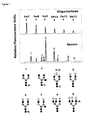
Figure 1 : The malto-oligosaccharides are shown at the top as sugar mass reference. The number of glucose units (DP, degree of polymerization) in these structures is indicated. A typical desialylated N-glycan profile is shown for total serum proteins. The structures of the N-glycan peaks are shown below the panels.
Peak 1 is an agalactosylated, core-α-1,6-fucosylated biantennary glycan (NGA2F),peak 2 is an agalactosylated, core-α-1,6-fucosylated bisecting biantennary (NGA2FB),peak 3 and 4 are mono galactosylated, core-α-1,6-fucosylated biantennary (NG1A2F),peak 5 is a bigalactosylated, biantennary glycan (NA2),peak 6 is a bigalactosylated, core-α-1,6-fucosylated biantennary (NA2F), peak 7 is a bigalactosylated, core-α-1,6-fucosylated bisecting biantennary (NA2FB),peak 8 is a trigalactosylated, tri-antennary (NA3) andpeak 9 is a branching α-1,3-fucosylated trigalactosylated tri-antennary (NA3F(b)). The symbols used in the structural formulas are: ■ N-acetyl-glucosamine (GlcNAc); ↓ β-linked galactose; ↖ α-1,3/6-linked fucose; ● α/β-linked mannose. -
Figure 2 : N-glycan values of total serum N-glycans from two controls (age <60 and 60 plus) and two dementia groups (AD and non-AD) are shown as peaks. The vertical axis represents the 95% confidence interval for mean of the percentage of total N-glycan peak height: -
Figure 3 : N-glycan value of total serum peak 9 (NA3Fb) from the females of controls (n=99 of =<60 years; n=50 of >60 years) and dementias (n=79). The vertical axis represents the 95% confidence interval for mean of the percentage of total N-glycan peak height. The differences between the dementia group and the 60+ year old group is statistically significant (p = 0.006). -
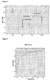
Figure 4 : Receiver operating characteristic (ROC) curve for prediction of clinically significant for detection of dementia female group using the values of peak 9 (NA3Fb). Area under the curve (AUC) shows the diagnosis power of peak 9 (0.866 ± 0.035). -
Figure 5 : N-glycan value of total serum peak 6 (NA2F) from the females of controls (n=50 of =<60 years; n=25 of >60 years), AD (n=24) and non-AD (n=13) groups. The level of N-glycan concentration from the AD is comparable to the age group of age <60 years, 60 plus years and non-AD patients. The vertical axis represents the 95% confidence interval for mean of the percentage of total N-glycan peak height. The statistical significance of differences between AD and 60+ years control, between AD and non-AD group is indicated as p value. -
Figure 6 : Receiver operating characteristic (ROC) curve for prediction of clinically significant for detection of AD female group using the values of peak 6 (NA2F). Area under the curve (AUC) shows the diagnosis power of peak 6 (0.902 ± 0.031). -
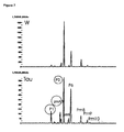
Figure 7 : Typical glycan profile for a wild type mouse (W) and for a R406W mutant Tau transgenic mouse (TAU). Peaks are labeled; P1, P5 and P6 are identical to peaks occurring in human, pm peaks are mouse specific. -
Figure 8 : Evolution of the peaks P1, pma and P5 in function of age, for wild type mice (W) and R406W mutant Tau transgenic mice (PTT). The age is indicated in months. - Serum samples from 79 demented patients whose diagnosis was confirmed by autopsy were used in this study. All sera samples were selected from the Biobank, Institute Born-Bunge, Antwerp, Belgium. Two control groups were included: 50 samples from people older than 60 years (from the Biobank, Institute Born-Bunge) and 100 samples from people 20 to 60 years old (from the Red Cross, Ghent). Age and gender were recorded. For subjects with dementia, we also recorded the scores of the Mini-Mental State Examination (MMSE) (Folstein et al., 1975) at the time of CSF sampling, year of clinical diagnosis, clinical and pathological diagnoses, year of death, date of autopsy and type of sampling tube. The study was approved by the local ethics committee (CME Middelheim).
- Serum samples from R406W mutant Tau transgenic mice and wild type mice with the same genetic background were obtained from Remynd, Belgium. Samples were obtained from mice at different age, to exclude possible age effects.
- All dementia patients were diagnosed according to strict clinical diagnostic criteria (Engelborghs et al. 2007b). The diagnosis of probable AD was established using NINCDS-ADRDA criteria (McKhann et al., 1984). All patients also fulfilled the Diagnostic and Statistical Manual of Mental Diseases (DSM-IV) criteria (APA, 1994). Mixed dementia (MXD) was diagnosed when patients fulfilled the criteria of probable AD according to NINCDS/ADRDA criteria and, in addition, displayed cerebrovascular disease (CVD) on brain CT and/or MRI but did not meet the criteria for relevant CVD according to NINDS-AIREN criteria for vascular dementia (Roman et al., 1993). In that way we excluded multiple large-vessel infarcts, strategically placed infarcts, multiple basal ganglia and white matter lacunes or extensive white matter lesions. Vascular dementia (VAD) was diagnosed according to the NINDS-AIREN criteria of vascular dementia (Roman et al., 1993). For the diagnosis of probable frontotemporal dementia (FTD) and progressive non-fluent aphasia (PPA), the criteria described by Neary et al. (1998) were applied. An extensive neuropsychological assessment revealing a characteristic neurocognitive profile of disproportionate executive dysfunction indicating frontal lobe involvement and brain perfusion SPECT were used to support the clinical diagnosis of FTD as described earlier Pickut et al., 1997). Dementia with Lewy bodies (DLB) was diagnosed by applying the clinical diagnostic criteria of McKeith, et al. (1996). Parkinson's disease dementia (PDD) was diagnosed when patients with idiopathic Parkinson's disease (PD) developed dementia following a dementia-free interval of at least two years. The criteria for the diagnosis of idiopathic PD included the presence of at least two out of four motor manifestations that characterize the disease (resting tremor, bradykinesia, muscular rigidity and impaired postural reflexes) and an insidious onset (Hoehn and Yahr, 1967). Creutzfeldt-Jakob disease (CJD) was diagnosed according to the criteria of Weber (2000). In one case, clinical differential diagnosis was Gerstmann-Straussler-Scheinker disease or spinocerebellar ataxia as described in detail by Fujigasaki et al. (2001). The inclusion criteria for the control group were: (1) no neurological or psychiatric antecedents and (2) no organic disease involving the central nervous system following extensive clinical examination. The age-matched control group (age >60, n = 50) consisted of patients with mechanical low back pain requiring a selective lumbar radiculography, patients with disorders of the peripheral nervous system (polyneuropathy, peripheral facial nerve palsy) and patients with subjective complaints in whom disorders of the central and peripheral nervous system were ruled out by an extensive clinical work-up.
- All diagnoses were established by the same neuropathologist who was blinded for the cerebrospinal fluid (CSF) results. For AD patients, the neuropathological criteria of Braak and Braak (1991) and of Jellinger (1998) were applied, whereas FTD patients were neuropathologically diagnosed according to Jackson and Lowe (1988) and Markesbery (1998). The pathological criteria of Kosaka et al. (1988) were applied for diagnosing DLB. MXD and VAD were diagnosed according to Markesbery (1998).
- CSF analysis was performed at the R&D facilities of Innogenetics NV (Ghent, Belgium) following relabeling of the CSF vials (01-001 to 01-100; control samples: 01-101 to 01- 200). Before analysis, samples were randomized to obtain a proportional distribution of samples from controls and demented patients on each microtiter plate (40 samples per plate). The laboratory technician was blinded for the expected test outcome in terms of clinical and definitive pathological diagnoses when performing and interpreting the tests.
- CSF levels of Aβ_1-42, T-tau and P-tau181P were determined with commercially available single-parameter ELISA kits [respectively, INNOTEST® β-AMYLOID(1-42), INNOTEST® hTAU Ag, INNOTEST® PHOSPHOTAU(181P)]; Innogenetics, Ghent, Belgium). With each assay, the clinical samples, together with a blank (sample diluent), the (prepared) calibrator solutions and the appropriate controls, were tested with strict adherence to the test instructions provided in the kit inserts. All samples were run in duplicate.
- If the intra-assay coefficient of variation was greater than 30% (calculated as (max - min)×100/average), or if the concentrations obtained were out-of-range (OD values not between mean OD values of highest and lowest calibrator concentration), samples were retested (by extension of the calibrator concentration range for some samples). Dilution of samples that gave values above the highest calibration concentration for possible reanalysis was not performed, as it is not recommended in the manufacturer's instructions. The concentration ranges of the test kits are described in the package inserts (P-tau181P: 15.6-500 pg/ml, T-tau: 75-1200 pg/ml, Aβ_1-42: 125-2000 pg/ml).
- The N-glycans attached to the proteins in 2 µl of serum were released, labeled, and analyzed as described (Vanhooren et al., 2008; Liu et al., 2007). Labeled N-glycans were analyzed by DNA Sequencer Assisted Fluorophore Assisted Carbohydrate Electrophoresis (DSA-FACE) technology, using a capillary electrophoresis (CE)-based ABI3130 sequencer. Data were analyzed with the GeneMapper v3.7 software (Applied Biosystems, Foster City, CA). We measured the heights of the peaks that were detected in all the samples, obtained a numerical description of the profiles, and analyzed these data with SPSS 12.0 statistical software.
- Statistical analyses were performed with SPSS for Windows software (SPSS, Chicago, IL, USA). Results are presented as means ±SD. All reported p-values are two-tailed, using a t-test for independent samples. Pearson's coefficients of correlation (with 95% confidence intervals and their associated probability, p) were used to evaluate the relationships between parameters. The Receiver Operating Characteristics (ROC) curve was used as an index of accuracy; values close to 1.0 indicate high diagnostic accuracy.
- The demographic, main clinical and neuropathological data of the patients are summarized in Table 1. Clinical diagnosis at CSF sampling in the dementia patients consisted of 48 AD patients (n=24 females, n=24 males). The other dementias including FTD, DLB and VAD, were pooled as a group of non-AD (n=13 female, n=18 male).
Table 1. Clinical and neuropathological data group age MMSE score T-tau P_tau Aß-42 age =<60 yrs Mean 40.202 N 99 Std. Deviation 14.1407 age >60 yrs Mean 77.999 27.8 447 63.5 599 N 50 10 2 2 2 Std. Deviation 5.0656 1.398 48.08326 7.77817 69.29646 AD Mean 79.979 10.48 823.6957 83.4217 361.413 N 48 40 46 46 46 Std. Deviation 8.6134 5.435 751.9652 41.30617 103.4179 non-AD Mean 74.71 12.88 554.2414 58.1828 504.2759 N 31 25 29 29 29 Std. Deviation 9.6305 9.198 538.0035 34.73765 171.7263 - We used DSA-FACE to determine the N-glycome profiles of desialylated sera obtained from dementia patients with AD (n=48) and non-AD (n=31). We also analyzed serum from healthy donors (n=150). The glycan fingerprint is shown in
Figure 1 . The individual N-glycan structures are shown inFigure 1 as well. The N-glycans detected in serum are represented as peaks and the relative concentration was quantified by normalizing its height to the sum of the heights of all peaks in the profile (Figure 2 ). - The sizes of the peaks, which represent the relative concentrations of the oligosaccharide structures, were analyzed statistically for correlation between glycans and dementias. Two glycan structures, NA2F (peak 6) and NA3Fb (peak 9), were found to related dementia, but more pronounced in females.
- The level of peak 9 (NA3Fb) was significantly higher (p<0.006) in the female dementia patients (AD and non-AD) compared to age-matched controls (
Fig. 3 ). ROC curve analysis showed an accuracy of 86.6% ± 3.5% forpeak 9 with a 90% of specificity and 60% of sensitivity for identifying dementia patients from controls (Fig. 4 ). - The level of peak 6 (NA2F) was substantially decreased only in AD patients, not in the non-AD and control groups (
Fig. 5 ). ROC curve analysis showed an accuracy of 90.2% ± 3.1% forpeak 6 with a 90% of specificity and 70% sensitivity for identifying AD from and non-AD and controls (Fig. 6 ). - Table 2 showed correlation glycan markers with CSF marker and MMSE score in the female dementias and controls. In this study, we did not found any correlation between CSF markers and MMSE score, and between β-amyloid peptide and p-tan or t-tau protein in the dementias or AD patients (including female and male). Only correlation was found is that p-tau positively to t-tau in both female (γ=0.656, p<0.0001) and male (γ=0.504, p<0.0001) dementia patients.
- The correlation of glycans with MMSE scores and CSF biomarker levels were compared (Table 2). The level of
peak 9 had no correlation with MMSE score nor CSF biomarker levels both in female and male patients, while only negatively correlated to the level ofpeak 6 in the female patients (γ=-0.611, p<0.0001). The level of peak 6 (NA2F) showed no correlation with MMSE score, but most correlation with the CSF markers in female patients. The level ofpeak 6 was positively correlated to the CSF level of Aβ-42 (γ=0.387, p<0.022) and negatively to the level of p-tau protein (γ=-0.407, p<0.015) in the female dementia patients. No significant correlation was found betweenpeak 6 and t-tau protein.Table 2. The correlation coefficient between glycan markers and CSF markers and MMS MMSE t-tau Aβ-42 p-tau peak6 peak9 MMSE Pearson Correlation 1 0.045 0.044 -0.059 0.286 -0.182 Sig. (2-tailed) 0.813 0.819 0.758 0.091 0.288 t-tau Pearson Correlation 0.045 1 -0.081 .656(**) -0.31 0.011 Sig. (2-tailed) 0.813 0.637 0.0001 0.07 0.951 Aβ-42 Pearson Correlation 0.044 -0.081 1 0.027 .387(*) -0.096 Sig. (2-tailed) 0.819 0.637 0.874 0.022 0.582 p-tau Pearson Correlation -0.059 .656(**) 0.027 1 -.407(*) 0.137 Sig. (2-tailed) 0.758 0.0001 0.874 0.015 0.433 peak6 Pearson Correlation 0.286 -0.31 .387(*) -.407(*) 1 -.611(**) Sig. (2-tailed) 0.091 0.07 0.022 0.015 0.0001 peak9 Pearson Correlation -0.182 0.011 -0.096 0.137 -.611(**) 1 Sig. (2-tailed) 0.288 0.951 0.582 0.433 0.0001 ** Correlation is significant at the 0.01 level (2-tailed).
* Correlation is significant at the 0.05 level (2-tailed). - Samples from 13 wild type mice and 21 R406W mutant transgenic mice ((Zhang et al., 2004) (2 different age groups for wild type, 3 different age groups for the transgenic mice) were analyzed. A typical profile for wild type and Tau transgenic mice, with indication of the peaks, is given in
Figure 7 . Both wild type and tau mice peaks show some shift with age and/or evolution of the disease, but at all ages, there is a significant increase in P1 and pmA, and a significant decrease of P5 in tau transgenic mice, compared with the wild type (Figure 8 ). One of those peaks, or a combination thereof, can be used to judge the severity of the Alzheimer disease, and/or to follow the effect of a treatment and/or to screen the efficiency of an anti Alzheimer compound. -
- Akasaka-Manya, K., Manya, H., Sakurai, Y., Wojczyk, B.S., Spitalnik, S.L. and Endo, T. Increased bisecting and core-Fucosylated N-glycans on mutant human amyloid precursor proteins. Glycoconj. J. 25, 775-786 (2008)
- American Psychiatric Association, 1994. DSM-IV: Diagnostic and Statistical Manual of Mental Disorders, fourth ed. American Psychiatric Association, Washington, DC.
- Barkhof, F., et al. The significance of medial temporal lobe atrophy: a postmortem MRI study in the very old. Neurology 69, 1521-1527 (2007).
- Blasko, I., et al. Measurement of thirteen biological markers in CSF of patients with Alzheimer's disease and other dementias. Dementia and geriatric cognitive disorders 21, 9-15 (2006).
- Blasko, I., et al. Conversion from cognitive health to mild cognitive impairment and Alzheimer's disease: prediction by plasma amyloid beta 42, medial temporal lobe atrophy and homocysteine. Neurobiology of aging 29, 1-11 (2008).
- Blennow, K., de Leon, M.J. & Zetterberg, H. Alzheimer's disease. Lancet 368, 387-403 (2006).
- Braak, H. & Braak, E. Neuropathological stageing of Alzheimer-related changes. Acta neuropathologica 82, 239-259 (1991).
- Brand, J.P., et al. An adaptive alpha spending algorithm improves the power of statistical inference in microarray data analysis. ).
- Buerger, K., et al. Differential diagnosis of Alzheimer disease with cerebrospinal fluid levels of tau protein phosphorylated at threonine 231. Archives of neurology 59, 1267-1272 (2002).
- DeCarli, C., Kaye, J.A., Horwitz, B. & Rapoport, S.I. Critical analysis of the use of computer-assisted transverse axial tomography to study human brain in aging and dementia of the Alzheimer type. Neurology 40, 872-883 (1990).
- De Leon, M.J., et al. Frequency of hippocampal formation atrophy in normal aging and Alzheimer's disease. Neurobiologyofaging 18, 1-11 (1997).
- Dewachter, I, van Dorpe, J., Spittaels, K., Tesseur, I., Van Den Haute, C., Moechars, D., Van Leuven, F. Modelling Alzheimer's disease in trangenic mice: effect of age and of presenilin1 on amyloid biochemlistry and pathology in APP/London mice. Exp Gerontol. 35, 831-841 (2000).
- Dubois, B., et al. Research criteria for the diagnosis of Alzheimer's disease: revising the NINCDS-ADRDA criteria. ).
- Engelborghs, S., et al. No association of CSF biomarkers with APOEepsilon4, plaque and tangle burden in definite Alzheimer's disease. Brain 130, 2320-2326 (2007a).
- Engelborghs, S., et al. Diagnostic performance of a CSF-biomarker panel in autopsy-confirmed dementia. Neurobiology of aging (2007b).
- Folstein, M.F., Folstein, S.E. & McHugh, P.R. "Mini-mental state". A practical method for grading the cognitive state of patients for the clinician. Journal of psychiatric research 12, 189-198 (1975).
- Frisoni, G.B., et al. Radial width of the temporal horn: a sensitive measure in Alzheimer disease. Ajnr 23, 35-47 (2002).
- Fujigasaki, H., et al. CAG repeat expansion in the TATA box-binding protein gene causes autosomal dominant cerebellar ataxia. Brain 124, 1939-1947 (2001).
- Galton, C. Alzheimer's disease from basic research to clinical applications. Journal of neurology, neurosurgery, and psychiatry 68, 123B (2000).
- Growdon, J.H. To tap or not to tap: cerebrospinal fluid biomarkers of Alzheimer's disease. Annals of neurology 44, 6-7 (1998).
- Hampel, H., Goemitz, A. & Buerger, K. Advances in the development of biomarkers for Alzheimer's disease: from CSF total tau and Abeta(1-42) proteins to phosphorylated tau protein. Brain research bulletin 61, 243-253 (2003).
- Heckemann, R.A., et al. Automatic volumetry on MR brain images can support diagnostic decision making. BMC ).
- Hoehn, M.M. & Yahr, M.D. Parkinsonism: onset, progression and mortality. Neurology 17, 427-442 (1967).
- Ibach, B., et al. Cerebrospinal fluid tau and beta-amyloid in Alzheimer patients, disease controls and an age-matched random sample. Neurobiology of aging 27, 1202-1211 (2006).
- Jackson, M. & Lowe, J. The new neuropathology of degenerative frontotemporal dementias. Acta neuropathologica 91, 127-134 (1996).
- Jellinger, K.A. The neuropathological diagnosis of Alzheimer disease. Journal of neural transmission 53, 97-118 (1998).
- Knopman, D.S., et al. Practice parameter: diagnosis of dementia (an evidence-based review). Report of the Quality Standards Subcommittee of the American Academy of Neurology. Neurology 56, 1143-1153 (2001).
- Kosaka, K., Tsuchiya, K. & Yoshimura, M. Lewy body disease with and without dementia: a clinicopathological study of 35 cases. Clinical neuropathology 7, 299-305 (1988).
- Kukuruzinska, M.A. & Lennon, K. Protein N-glycosylation: molecular genetics and functional significance. Crit Rev ).
- Laroy, W, Contreras, R. and Callewaert, N. glycome mapping on DNA sequencing equipment.
- Liu, X.E., et al. N-glycomic changes in hepatocellular carcinoma patients with liver cirrhosis induced by hepatitis B virus. Hepatology 46, 1426-1435 (2007).
- Liu, Y., Studzinski, C., Beckett, T., Guan, H., Hersh, M.A., Murphy, M.P., Klein, R and Hersh, L.B. Expression of neprilysin in skeletal muscle reduces amyloid burden in a transgenic mouse model of Alzheimer disease. Mol. Ther. (2009)
- Maddalena, A., et al. Biochemical diagnosis of Alzheimer disease by measuring the cerebrospinal fluid ratio of phosphorylated tau protein to beta-amyloid peptide42. Archives of ).
- Markesbery,W.R., 1998. Neuropathology of Dementing Disorders. Arnold, London.
- Mattman, A., et al. Regional HmPAO SPECT and CT measurements in the diagnosis of Alzheimer's disease. The Canadian journal of neurological sciences 24, 22-28 (1997).
- McKhann, G., et al. Clinical diagnosis of Alzheimer's disease: report of the NINCDS-ADRDA Work Group under the auspices of Department of Health and Human Services Task Force on Alzheimer's Disease. Neurology 34, 939-944 (1984).
- McKeith, I.G., et al. Consensus guidelines for the clinical and pathologic diagnosis of dementia with Lewy bodies (DLB): report of the consortium on DLB international workshop. Neurology 47, 1113-1124 (1996).
- Nagy, Z., et al. Relationship between clinical and radiological diagnostic criteria for Alzheimer's disease and the extent of neuropathology as reflected by 'stages': a prospective study. Dementia and geriatric ).
- Neary, D., et al. Frontotemporal lobar degeneration: a consensus on clinical diagnostic criteria. Neurology 51, 1546-1554 (1998).
- Pickut, B.A., et al. Discriminative use of SPECT in frontal lobe-type dementia versus (senile) dementia of the Alzheimer's type. J Nucl Med 38, 929-934 (1997).
- Pype, S., Moechars, D., Dillen, L., and Mercken, M. Characterization of amyloid beta peptides from brain extracts of transgenic mice overexpressing the London mutat of human amyloid precursor protein. J Neurochem. 84, 602-609 (2003).
- Raman, R., Raguram, S., Venkataraman, G., Paulson, J.C. & Sasisekharan, R. Glycomics: an integrated systems approach to structure-function relationships of glycans. ).
- Roman, G.C., et al. Vascular dementia: diagnostic criteria for research studies. Report of the NINDS-AIREN International Workshop. Neurology 43, 250-260 (1993).
- Selkoe, D.J. & Schenk, D. Alzheimer's disease: molecular understanding predicts amyloid-based therapeutics. Annual review of pharmacology and toxicology 43, 545-584 (2003).
- Tang, J., Song, M., Wang, Y., Fan, X., Xu, H. and Bai, Y. Noggin and BMP4 co-modulate adult hippocampal neurogenesis in the APP(swe)/PS1(DeltaE9) transgenic mouse model of Alzheimer disease. Biochem. Biophys. Res. Commun. (2009)
- Vanhooren, V., et al. N-glycomic changes in serum proteins during human aging. ).
- Vanhooren, V., Laroy, W., Libert, C. & Chen, C. N-Glycan profiling in the study of human aging. Biogerontology (2008).
- van Marum, R.J. Current and future therapy in Alzheimer's disease. Fundamental & clinical pharmacology 22, 265-274 (2008).
- Waldemar, G., et al. Recommendations for the diagnosis and management of Alzheimer's disease and other disorders associated with dementia: EFNS guideline. Eur J Neurol 14, e1-26 (2007).
- Weber, T. Clinical and laboratory diagnosis of Creutzfeldt-Jakob disease. Clinical neuropathology 19, 249-250 (2000).
- Wilson, R.J. Towards a cure for dementia: the role of axonal transport in Alzheimer's disease. Science progress 91, 65-80 (2008).
- Zhang, B., Higuchi, M., Yoshimaya, Y., Ishihara, T., Forman, M.S., Martinez, D., Joyce, S., Trojanowski, J.Q. and Lee, V.M.Y. Retarded axonal transport of R406W mutant Tau in transgenic mice with a neurodegenerative tauopathy. J. Neuroscience 24, 4657-4667(2004).
Claims (2)
- A method for the detection of Alzheimer's disease in a female human subject, comprising the determination of the concentration of bigalactosylated, core-α-1,6-fucosylated biantennary glycan structures in serum and comparing said concentration with the concentration of a healthy reference subject.
- The method according to claim 1, whereby said method is used to test the efficiency of a candidate compound against Alzheimer's.
Priority Applications (1)
| Application Number | Priority Date | Filing Date | Title |
|---|---|---|---|
| EP09761735A EP2304442B1 (en) | 2008-06-11 | 2009-06-10 | Blood serum marker for detection and diagnosis of alzheimer's disease |
Applications Claiming Priority (3)
| Application Number | Priority Date | Filing Date | Title |
|---|---|---|---|
| EP08158010 | 2008-06-11 | ||
| PCT/EP2009/057162 WO2009150172A1 (en) | 2008-06-11 | 2009-06-10 | Blood serum marker for detection and diagnosis of alzheimer's disease |
| EP09761735A EP2304442B1 (en) | 2008-06-11 | 2009-06-10 | Blood serum marker for detection and diagnosis of alzheimer's disease |
Publications (2)
| Publication Number | Publication Date |
|---|---|
| EP2304442A1 EP2304442A1 (en) | 2011-04-06 |
| EP2304442B1 true EP2304442B1 (en) | 2012-11-14 |
Family
ID=40973165
Family Applications (1)
| Application Number | Title | Priority Date | Filing Date |
|---|---|---|---|
| EP09761735A Not-in-force EP2304442B1 (en) | 2008-06-11 | 2009-06-10 | Blood serum marker for detection and diagnosis of alzheimer's disease |
Country Status (5)
| Country | Link |
|---|---|
| US (1) | US20110100816A1 (en) |
| EP (1) | EP2304442B1 (en) |
| AU (1) | AU2009256601A1 (en) |
| CA (1) | CA2727289A1 (en) |
| WO (1) | WO2009150172A1 (en) |
Families Citing this family (4)
| Publication number | Priority date | Publication date | Assignee | Title |
|---|---|---|---|---|
| US20150369825A1 (en) * | 2013-02-26 | 2015-12-24 | Sophia SCHEDIN WEISS | Diagnosis of Alzheimer Disease, Cognitive Decline and Dementia |
| CN109100507A (en) * | 2017-06-20 | 2018-12-28 | 江苏先思达生物科技有限公司 | The method for building up of the seroglycoid N- sugar group spectrum model of chronic hepatitis hepatic injury |
| CN109596842B (en) * | 2018-12-29 | 2022-02-22 | 江苏先思达生物科技有限公司 | Alzheimer disease detection reagent and application thereof in Alzheimer disease detection |
| CN111380979A (en) * | 2020-03-31 | 2020-07-07 | 中国科学院昆明动物研究所 | Healthy aged diagnosis marker and application |
Family Cites Families (5)
| Publication number | Priority date | Publication date | Assignee | Title |
|---|---|---|---|---|
| US4801533A (en) * | 1986-06-25 | 1989-01-31 | Fudenberg H Hugh | Method of using alpha-1 acid glycoprotein on T-cells as a marker for alzheimer's disease |
| US20090093000A1 (en) * | 2001-11-23 | 2009-04-09 | George Jackowski | Glycoprotein and apolipoprotein biopolymer markers predictive of Alzheimers disease |
| US20060057638A1 (en) * | 2004-04-15 | 2006-03-16 | Massachusetts Institute Of Technology | Methods and products related to the improved analysis of carbohydrates |
| US20060127950A1 (en) * | 2004-04-15 | 2006-06-15 | Massachusetts Institute Of Technology | Methods and products related to the improved analysis of carbohydrates |
| JP4385149B2 (en) * | 2005-05-11 | 2009-12-16 | 独立行政法人理化学研究所 | A diagnostic agent for Alzheimer's disease using serum glycoprotein as a biomarker |
-
2009
- 2009-06-10 AU AU2009256601A patent/AU2009256601A1/en not_active Abandoned
- 2009-06-10 WO PCT/EP2009/057162 patent/WO2009150172A1/en not_active Ceased
- 2009-06-10 US US12/737,142 patent/US20110100816A1/en not_active Abandoned
- 2009-06-10 EP EP09761735A patent/EP2304442B1/en not_active Not-in-force
- 2009-06-10 CA CA2727289A patent/CA2727289A1/en not_active Abandoned
Also Published As
| Publication number | Publication date |
|---|---|
| EP2304442A1 (en) | 2011-04-06 |
| WO2009150172A1 (en) | 2009-12-17 |
| AU2009256601A1 (en) | 2009-12-17 |
| US20110100816A1 (en) | 2011-05-05 |
| CA2727289A1 (en) | 2009-12-17 |
Similar Documents
| Publication | Publication Date | Title |
|---|---|---|
| Hansson et al. | Blood biomarkers for Alzheimer’s disease in clinical practice and trials | |
| Groot et al. | Tau PET imaging in neurodegenerative disorders | |
| Santos et al. | Amyloid-β oligomers in cerebrospinal fluid are associated with cognitive decline in patients with Alzheimer's disease | |
| Tremblay et al. | Biochemical characterization of Aβ and tau pathologies in mild cognitive impairment and Alzheimer's disease | |
| Bouwman et al. | CSF biomarkers and medial temporal lobe atrophy predict dementia in mild cognitive impairment | |
| Slaets et al. | Amyloid pathology influences aβ1-42 cerebrospinal fluid levels in dementia with lewy bodies | |
| Stefani et al. | CSF markers in Alzheimer disease patients are not related to the different degree of cognitive impairment | |
| Jongbloed et al. | Discriminatory and predictive capabilities of enzyme-linked immunosorbent assay and multiplex platforms in a longitudinal Alzheimer’s disease study | |
| CN108431608B (en) | Lactoferrin for use in the diagnosis or prognosis of alzheimer's disease or for use in the diagnosis of parkinson's disease | |
| Bouwman et al. | New research criteria for the diagnosis of Alzheimer’s disease applied in a memory clinic population | |
| US20110100816A1 (en) | Blood serum marker for detection and diagnosis of alzheimer's disease | |
| EP2990801B1 (en) | Diagnostic kit for diagnosis of alzheimer's disease using dissociation of protein aggregates | |
| US20150369825A1 (en) | Diagnosis of Alzheimer Disease, Cognitive Decline and Dementia | |
| Lönneborg | Biomarkers for Alzheimer disease in cerebrospinal fluid, urine, and blood | |
| Lauriola et al. | Erythrocyte associated amyloid-β as potential biomarker to diagnose dementia | |
| De Ninno et al. | Current perspectives on Alzheimer’s disease fluid biomarkers and future challenges: a narrative review | |
| US20200081019A1 (en) | Identification of signatures for neurodegeneration diseases diagnoses | |
| Urbanelli et al. | New perspectives for the diagnosis of Alzheimer's disease | |
| Kamogawa et al. | Potential Utility of Soluble p3-Alcadeinα Plasma Levels as a Biomarker for Sporadic Alzheimer's Disease | |
| EP1356299B1 (en) | Method for the diagnosis of alzheimer's disease | |
| Delacourte et al. | The Biochemical Spreading of Tau and Amyloid-Precursor Protein Pathologies in Aging and Sporadic Alzheimer’s Disease | |
| Gawor et al. | Amygdala-predominant α-synuclein pathology exacerbates hippocampal neuron loss in Alzheimer’s disease | |
| Youh et al. | Biomarkers and Applications in Alzheimer's Disease | |
| Lussier et al. | Plasma levels of phosphorylated tau 181 are associated with cerebral metabolic dysfunction in cognitively impaired individuals | |
| Na et al. | Improvement of Clinical Diagnosis Accuracy of Alzheimer’s Disease Using Multiple Blood Biomarkers |
Legal Events
| Date | Code | Title | Description |
|---|---|---|---|
| PUAI | Public reference made under article 153(3) epc to a published international application that has entered the european phase |
Free format text: ORIGINAL CODE: 0009012 |
|
| 17P | Request for examination filed |
Effective date: 20110110 |
|
| AK | Designated contracting states |
Kind code of ref document: A1 Designated state(s): AT BE BG CH CY CZ DE DK EE ES FI FR GB GR HR HU IE IS IT LI LT LU LV MC MK MT NL NO PL PT RO SE SI SK TR |
|
| AX | Request for extension of the european patent |
Extension state: AL BA RS |
|
| 17Q | First examination report despatched |
Effective date: 20110711 |
|
| DAX | Request for extension of the european patent (deleted) | ||
| GRAP | Despatch of communication of intention to grant a patent |
Free format text: ORIGINAL CODE: EPIDOSNIGR1 |
|
| GRAS | Grant fee paid |
Free format text: ORIGINAL CODE: EPIDOSNIGR3 |
|
| GRAA | (expected) grant |
Free format text: ORIGINAL CODE: 0009210 |
|
| AK | Designated contracting states |
Kind code of ref document: B1 Designated state(s): AT BE BG CH CY CZ DE DK EE ES FI FR GB GR HR HU IE IS IT LI LT LU LV MC MK MT NL NO PL PT RO SE SI SK TR |
|
| REG | Reference to a national code |
Ref country code: GB Ref legal event code: FG4D |
|
| REG | Reference to a national code |
Ref country code: AT Ref legal event code: REF Ref document number: 584248 Country of ref document: AT Kind code of ref document: T Effective date: 20121115 Ref country code: CH Ref legal event code: EP |
|
| REG | Reference to a national code |
Ref country code: IE Ref legal event code: FG4D |
|
| REG | Reference to a national code |
Ref country code: DE Ref legal event code: R096 Ref document number: 602009011226 Country of ref document: DE Effective date: 20130110 |
|
| REG | Reference to a national code |
Ref country code: NL Ref legal event code: VDEP Effective date: 20121114 |
|
| REG | Reference to a national code |
Ref country code: AT Ref legal event code: MK05 Ref document number: 584248 Country of ref document: AT Kind code of ref document: T Effective date: 20121114 |
|
| REG | Reference to a national code |
Ref country code: LT Ref legal event code: MG4D |
|
| PG25 | Lapsed in a contracting state [announced via postgrant information from national office to epo] |
Ref country code: ES Free format text: LAPSE BECAUSE OF FAILURE TO SUBMIT A TRANSLATION OF THE DESCRIPTION OR TO PAY THE FEE WITHIN THE PRESCRIBED TIME-LIMIT Effective date: 20130225 Ref country code: HR Free format text: LAPSE BECAUSE OF FAILURE TO SUBMIT A TRANSLATION OF THE DESCRIPTION OR TO PAY THE FEE WITHIN THE PRESCRIBED TIME-LIMIT Effective date: 20121114 Ref country code: NO Free format text: LAPSE BECAUSE OF FAILURE TO SUBMIT A TRANSLATION OF THE DESCRIPTION OR TO PAY THE FEE WITHIN THE PRESCRIBED TIME-LIMIT Effective date: 20130214 Ref country code: FI Free format text: LAPSE BECAUSE OF FAILURE TO SUBMIT A TRANSLATION OF THE DESCRIPTION OR TO PAY THE FEE WITHIN THE PRESCRIBED TIME-LIMIT Effective date: 20121114 Ref country code: LT Free format text: LAPSE BECAUSE OF FAILURE TO SUBMIT A TRANSLATION OF THE DESCRIPTION OR TO PAY THE FEE WITHIN THE PRESCRIBED TIME-LIMIT Effective date: 20121114 Ref country code: SE Free format text: LAPSE BECAUSE OF FAILURE TO SUBMIT A TRANSLATION OF THE DESCRIPTION OR TO PAY THE FEE WITHIN THE PRESCRIBED TIME-LIMIT Effective date: 20121114 |
|
| PG25 | Lapsed in a contracting state [announced via postgrant information from national office to epo] |
Ref country code: PL Free format text: LAPSE BECAUSE OF FAILURE TO SUBMIT A TRANSLATION OF THE DESCRIPTION OR TO PAY THE FEE WITHIN THE PRESCRIBED TIME-LIMIT Effective date: 20121114 Ref country code: GR Free format text: LAPSE BECAUSE OF FAILURE TO SUBMIT A TRANSLATION OF THE DESCRIPTION OR TO PAY THE FEE WITHIN THE PRESCRIBED TIME-LIMIT Effective date: 20130215 Ref country code: SI Free format text: LAPSE BECAUSE OF FAILURE TO SUBMIT A TRANSLATION OF THE DESCRIPTION OR TO PAY THE FEE WITHIN THE PRESCRIBED TIME-LIMIT Effective date: 20121114 Ref country code: PT Free format text: LAPSE BECAUSE OF FAILURE TO SUBMIT A TRANSLATION OF THE DESCRIPTION OR TO PAY THE FEE WITHIN THE PRESCRIBED TIME-LIMIT Effective date: 20130314 Ref country code: LV Free format text: LAPSE BECAUSE OF FAILURE TO SUBMIT A TRANSLATION OF THE DESCRIPTION OR TO PAY THE FEE WITHIN THE PRESCRIBED TIME-LIMIT Effective date: 20121114 |
|
| PG25 | Lapsed in a contracting state [announced via postgrant information from national office to epo] |
Ref country code: AT Free format text: LAPSE BECAUSE OF FAILURE TO SUBMIT A TRANSLATION OF THE DESCRIPTION OR TO PAY THE FEE WITHIN THE PRESCRIBED TIME-LIMIT Effective date: 20121114 |
|
| PG25 | Lapsed in a contracting state [announced via postgrant information from national office to epo] |
Ref country code: DK Free format text: LAPSE BECAUSE OF FAILURE TO SUBMIT A TRANSLATION OF THE DESCRIPTION OR TO PAY THE FEE WITHIN THE PRESCRIBED TIME-LIMIT Effective date: 20121114 Ref country code: SK Free format text: LAPSE BECAUSE OF FAILURE TO SUBMIT A TRANSLATION OF THE DESCRIPTION OR TO PAY THE FEE WITHIN THE PRESCRIBED TIME-LIMIT Effective date: 20121114 Ref country code: BG Free format text: LAPSE BECAUSE OF FAILURE TO SUBMIT A TRANSLATION OF THE DESCRIPTION OR TO PAY THE FEE WITHIN THE PRESCRIBED TIME-LIMIT Effective date: 20130214 Ref country code: CZ Free format text: LAPSE BECAUSE OF FAILURE TO SUBMIT A TRANSLATION OF THE DESCRIPTION OR TO PAY THE FEE WITHIN THE PRESCRIBED TIME-LIMIT Effective date: 20121114 Ref country code: EE Free format text: LAPSE BECAUSE OF FAILURE TO SUBMIT A TRANSLATION OF THE DESCRIPTION OR TO PAY THE FEE WITHIN THE PRESCRIBED TIME-LIMIT Effective date: 20121114 |
|
| PG25 | Lapsed in a contracting state [announced via postgrant information from national office to epo] |
Ref country code: IT Free format text: LAPSE BECAUSE OF FAILURE TO SUBMIT A TRANSLATION OF THE DESCRIPTION OR TO PAY THE FEE WITHIN THE PRESCRIBED TIME-LIMIT Effective date: 20121114 Ref country code: RO Free format text: LAPSE BECAUSE OF FAILURE TO SUBMIT A TRANSLATION OF THE DESCRIPTION OR TO PAY THE FEE WITHIN THE PRESCRIBED TIME-LIMIT Effective date: 20121114 Ref country code: NL Free format text: LAPSE BECAUSE OF FAILURE TO SUBMIT A TRANSLATION OF THE DESCRIPTION OR TO PAY THE FEE WITHIN THE PRESCRIBED TIME-LIMIT Effective date: 20121114 |
|
| PLBE | No opposition filed within time limit |
Free format text: ORIGINAL CODE: 0009261 |
|
| STAA | Information on the status of an ep patent application or granted ep patent |
Free format text: STATUS: NO OPPOSITION FILED WITHIN TIME LIMIT |
|
| 26N | No opposition filed |
Effective date: 20130815 |
|
| PG25 | Lapsed in a contracting state [announced via postgrant information from national office to epo] |
Ref country code: CY Free format text: LAPSE BECAUSE OF FAILURE TO SUBMIT A TRANSLATION OF THE DESCRIPTION OR TO PAY THE FEE WITHIN THE PRESCRIBED TIME-LIMIT Effective date: 20121114 |
|
| REG | Reference to a national code |
Ref country code: DE Ref legal event code: R097 Ref document number: 602009011226 Country of ref document: DE Effective date: 20130815 |
|
| BERE | Be: lapsed |
Owner name: UNIVERSITEIT GENT Effective date: 20130630 Owner name: VIB VZW Effective date: 20130630 |
|
| PG25 | Lapsed in a contracting state [announced via postgrant information from national office to epo] |
Ref country code: MC Free format text: LAPSE BECAUSE OF FAILURE TO SUBMIT A TRANSLATION OF THE DESCRIPTION OR TO PAY THE FEE WITHIN THE PRESCRIBED TIME-LIMIT Effective date: 20121114 |
|
| REG | Reference to a national code |
Ref country code: CH Ref legal event code: PL |
|
| GBPC | Gb: european patent ceased through non-payment of renewal fee |
Effective date: 20130610 |
|
| REG | Reference to a national code |
Ref country code: IE Ref legal event code: MM4A |
|
| REG | Reference to a national code |
Ref country code: DE Ref legal event code: R119 Ref document number: 602009011226 Country of ref document: DE Effective date: 20140101 |
|
| REG | Reference to a national code |
Ref country code: FR Ref legal event code: ST Effective date: 20140228 |
|
| PG25 | Lapsed in a contracting state [announced via postgrant information from national office to epo] |
Ref country code: BE Free format text: LAPSE BECAUSE OF NON-PAYMENT OF DUE FEES Effective date: 20130630 |
|
| PG25 | Lapsed in a contracting state [announced via postgrant information from national office to epo] |
Ref country code: CH Free format text: LAPSE BECAUSE OF NON-PAYMENT OF DUE FEES Effective date: 20130630 Ref country code: DE Free format text: LAPSE BECAUSE OF NON-PAYMENT OF DUE FEES Effective date: 20140101 Ref country code: LI Free format text: LAPSE BECAUSE OF NON-PAYMENT OF DUE FEES Effective date: 20130630 Ref country code: IE Free format text: LAPSE BECAUSE OF NON-PAYMENT OF DUE FEES Effective date: 20130610 Ref country code: GB Free format text: LAPSE BECAUSE OF NON-PAYMENT OF DUE FEES Effective date: 20130610 |
|
| PG25 | Lapsed in a contracting state [announced via postgrant information from national office to epo] |
Ref country code: FR Free format text: LAPSE BECAUSE OF NON-PAYMENT OF DUE FEES Effective date: 20130701 |
|
| PG25 | Lapsed in a contracting state [announced via postgrant information from national office to epo] |
Ref country code: MT Free format text: LAPSE BECAUSE OF FAILURE TO SUBMIT A TRANSLATION OF THE DESCRIPTION OR TO PAY THE FEE WITHIN THE PRESCRIBED TIME-LIMIT Effective date: 20121114 |
|
| PG25 | Lapsed in a contracting state [announced via postgrant information from national office to epo] |
Ref country code: TR Free format text: LAPSE BECAUSE OF FAILURE TO SUBMIT A TRANSLATION OF THE DESCRIPTION OR TO PAY THE FEE WITHIN THE PRESCRIBED TIME-LIMIT Effective date: 20121114 |
|
| PG25 | Lapsed in a contracting state [announced via postgrant information from national office to epo] |
Ref country code: MK Free format text: LAPSE BECAUSE OF FAILURE TO SUBMIT A TRANSLATION OF THE DESCRIPTION OR TO PAY THE FEE WITHIN THE PRESCRIBED TIME-LIMIT Effective date: 20121114 Ref country code: LU Free format text: LAPSE BECAUSE OF NON-PAYMENT OF DUE FEES Effective date: 20130610 Ref country code: HU Free format text: LAPSE BECAUSE OF FAILURE TO SUBMIT A TRANSLATION OF THE DESCRIPTION OR TO PAY THE FEE WITHIN THE PRESCRIBED TIME-LIMIT; INVALID AB INITIO Effective date: 20090610 |
|
| PG25 | Lapsed in a contracting state [announced via postgrant information from national office to epo] |
Ref country code: IS Free format text: LAPSE BECAUSE OF FAILURE TO SUBMIT A TRANSLATION OF THE DESCRIPTION OR TO PAY THE FEE WITHIN THE PRESCRIBED TIME-LIMIT Effective date: 20121114 |