EP2299977B1 - Liposomes for drug delivery and methods for preparation thereof - Google Patents
Liposomes for drug delivery and methods for preparation thereof Download PDFInfo
- Publication number
- EP2299977B1 EP2299977B1 EP09749940A EP09749940A EP2299977B1 EP 2299977 B1 EP2299977 B1 EP 2299977B1 EP 09749940 A EP09749940 A EP 09749940A EP 09749940 A EP09749940 A EP 09749940A EP 2299977 B1 EP2299977 B1 EP 2299977B1
- Authority
- EP
- European Patent Office
- Prior art keywords
- liposome
- liposomes
- formulation
- mol
- poly
- Prior art date
- Legal status (The legal status is an assumption and is not a legal conclusion. Google has not performed a legal analysis and makes no representation as to the accuracy of the status listed.)
- Active
Links
Images









Classifications
-
- A—HUMAN NECESSITIES
- A61—MEDICAL OR VETERINARY SCIENCE; HYGIENE
- A61K—PREPARATIONS FOR MEDICAL, DENTAL OR TOILETRY PURPOSES
- A61K9/00—Medicinal preparations characterised by special physical form
- A61K9/10—Dispersions; Emulsions
- A61K9/127—Synthetic bilayered vehicles, e.g. liposomes or liposomes with cholesterol as the only non-phosphatidyl surfactant
- A61K9/1271—Non-conventional liposomes, e.g. PEGylated liposomes or liposomes coated or grafted with polymers
-
- A—HUMAN NECESSITIES
- A61—MEDICAL OR VETERINARY SCIENCE; HYGIENE
- A61K—PREPARATIONS FOR MEDICAL, DENTAL OR TOILETRY PURPOSES
- A61K31/00—Medicinal preparations containing organic active ingredients
- A61K31/185—Acids; Anhydrides, halides or salts thereof, e.g. sulfur acids, imidic, hydrazonic or hydroximic acids
- A61K31/19—Carboxylic acids, e.g. valproic acid
-
- A—HUMAN NECESSITIES
- A61—MEDICAL OR VETERINARY SCIENCE; HYGIENE
- A61K—PREPARATIONS FOR MEDICAL, DENTAL OR TOILETRY PURPOSES
- A61K31/00—Medicinal preparations containing organic active ingredients
- A61K31/185—Acids; Anhydrides, halides or salts thereof, e.g. sulfur acids, imidic, hydrazonic or hydroximic acids
- A61K31/19—Carboxylic acids, e.g. valproic acid
- A61K31/20—Carboxylic acids, e.g. valproic acid having a carboxyl group bound to a chain of seven or more carbon atoms, e.g. stearic, palmitic, arachidic acids
- A61K31/203—Retinoic acids ; Salts thereof
-
- A—HUMAN NECESSITIES
- A61—MEDICAL OR VETERINARY SCIENCE; HYGIENE
- A61K—PREPARATIONS FOR MEDICAL, DENTAL OR TOILETRY PURPOSES
- A61K31/00—Medicinal preparations containing organic active ingredients
- A61K31/33—Heterocyclic compounds
- A61K31/335—Heterocyclic compounds having oxygen as the only ring hetero atom, e.g. fungichromin
- A61K31/337—Heterocyclic compounds having oxygen as the only ring hetero atom, e.g. fungichromin having four-membered rings, e.g. taxol
-
- A—HUMAN NECESSITIES
- A61—MEDICAL OR VETERINARY SCIENCE; HYGIENE
- A61K—PREPARATIONS FOR MEDICAL, DENTAL OR TOILETRY PURPOSES
- A61K31/00—Medicinal preparations containing organic active ingredients
- A61K31/33—Heterocyclic compounds
- A61K31/395—Heterocyclic compounds having nitrogen as a ring hetero atom, e.g. guanethidine or rifamycins
- A61K31/435—Heterocyclic compounds having nitrogen as a ring hetero atom, e.g. guanethidine or rifamycins having six-membered rings with one nitrogen as the only ring hetero atom
- A61K31/47—Quinolines; Isoquinolines
- A61K31/475—Quinolines; Isoquinolines having an indole ring, e.g. yohimbine, reserpine, strychnine, vinblastine
-
- A—HUMAN NECESSITIES
- A61—MEDICAL OR VETERINARY SCIENCE; HYGIENE
- A61K—PREPARATIONS FOR MEDICAL, DENTAL OR TOILETRY PURPOSES
- A61K31/00—Medicinal preparations containing organic active ingredients
- A61K31/33—Heterocyclic compounds
- A61K31/395—Heterocyclic compounds having nitrogen as a ring hetero atom, e.g. guanethidine or rifamycins
- A61K31/495—Heterocyclic compounds having nitrogen as a ring hetero atom, e.g. guanethidine or rifamycins having six-membered rings with two or more nitrogen atoms as the only ring heteroatoms, e.g. piperazine or tetrazines
- A61K31/505—Pyrimidines; Hydrogenated pyrimidines, e.g. trimethoprim
- A61K31/513—Pyrimidines; Hydrogenated pyrimidines, e.g. trimethoprim having oxo groups directly attached to the heterocyclic ring, e.g. cytosine
-
- A—HUMAN NECESSITIES
- A61—MEDICAL OR VETERINARY SCIENCE; HYGIENE
- A61K—PREPARATIONS FOR MEDICAL, DENTAL OR TOILETRY PURPOSES
- A61K31/00—Medicinal preparations containing organic active ingredients
- A61K31/33—Heterocyclic compounds
- A61K31/395—Heterocyclic compounds having nitrogen as a ring hetero atom, e.g. guanethidine or rifamycins
- A61K31/495—Heterocyclic compounds having nitrogen as a ring hetero atom, e.g. guanethidine or rifamycins having six-membered rings with two or more nitrogen atoms as the only ring heteroatoms, e.g. piperazine or tetrazines
- A61K31/505—Pyrimidines; Hydrogenated pyrimidines, e.g. trimethoprim
- A61K31/519—Pyrimidines; Hydrogenated pyrimidines, e.g. trimethoprim ortho- or peri-condensed with heterocyclic rings
-
- A—HUMAN NECESSITIES
- A61—MEDICAL OR VETERINARY SCIENCE; HYGIENE
- A61K—PREPARATIONS FOR MEDICAL, DENTAL OR TOILETRY PURPOSES
- A61K31/00—Medicinal preparations containing organic active ingredients
- A61K31/33—Heterocyclic compounds
- A61K31/555—Heterocyclic compounds containing heavy metals, e.g. hemin, hematin, melarsoprol
-
- A—HUMAN NECESSITIES
- A61—MEDICAL OR VETERINARY SCIENCE; HYGIENE
- A61K—PREPARATIONS FOR MEDICAL, DENTAL OR TOILETRY PURPOSES
- A61K31/00—Medicinal preparations containing organic active ingredients
- A61K31/70—Carbohydrates; Sugars; Derivatives thereof
- A61K31/7028—Compounds having saccharide radicals attached to non-saccharide compounds by glycosidic linkages
- A61K31/7034—Compounds having saccharide radicals attached to non-saccharide compounds by glycosidic linkages attached to a carbocyclic compound, e.g. phloridzin
- A61K31/704—Compounds having saccharide radicals attached to non-saccharide compounds by glycosidic linkages attached to a carbocyclic compound, e.g. phloridzin attached to a condensed carbocyclic ring system, e.g. sennosides, thiocolchicosides, escin, daunorubicin
-
- A—HUMAN NECESSITIES
- A61—MEDICAL OR VETERINARY SCIENCE; HYGIENE
- A61K—PREPARATIONS FOR MEDICAL, DENTAL OR TOILETRY PURPOSES
- A61K33/00—Medicinal preparations containing inorganic active ingredients
- A61K33/24—Heavy metals; Compounds thereof
- A61K33/243—Platinum; Compounds thereof
-
- A—HUMAN NECESSITIES
- A61—MEDICAL OR VETERINARY SCIENCE; HYGIENE
- A61K—PREPARATIONS FOR MEDICAL, DENTAL OR TOILETRY PURPOSES
- A61K38/00—Medicinal preparations containing peptides
- A61K38/04—Peptides having up to 20 amino acids in a fully defined sequence; Derivatives thereof
- A61K38/14—Peptides containing saccharide radicals; Derivatives thereof, e.g. bleomycin, phleomycin, muramylpeptides or vancomycin
-
- A—HUMAN NECESSITIES
- A61—MEDICAL OR VETERINARY SCIENCE; HYGIENE
- A61P—SPECIFIC THERAPEUTIC ACTIVITY OF CHEMICAL COMPOUNDS OR MEDICINAL PREPARATIONS
- A61P29/00—Non-central analgesic, antipyretic or antiinflammatory agents, e.g. antirheumatic agents; Non-steroidal antiinflammatory drugs [NSAID]
-
- A—HUMAN NECESSITIES
- A61—MEDICAL OR VETERINARY SCIENCE; HYGIENE
- A61P—SPECIFIC THERAPEUTIC ACTIVITY OF CHEMICAL COMPOUNDS OR MEDICINAL PREPARATIONS
- A61P31/00—Antiinfectives, i.e. antibiotics, antiseptics, chemotherapeutics
-
- A—HUMAN NECESSITIES
- A61—MEDICAL OR VETERINARY SCIENCE; HYGIENE
- A61P—SPECIFIC THERAPEUTIC ACTIVITY OF CHEMICAL COMPOUNDS OR MEDICINAL PREPARATIONS
- A61P31/00—Antiinfectives, i.e. antibiotics, antiseptics, chemotherapeutics
- A61P31/04—Antibacterial agents
-
- A—HUMAN NECESSITIES
- A61—MEDICAL OR VETERINARY SCIENCE; HYGIENE
- A61P—SPECIFIC THERAPEUTIC ACTIVITY OF CHEMICAL COMPOUNDS OR MEDICINAL PREPARATIONS
- A61P31/00—Antiinfectives, i.e. antibiotics, antiseptics, chemotherapeutics
- A61P31/10—Antimycotics
-
- A—HUMAN NECESSITIES
- A61—MEDICAL OR VETERINARY SCIENCE; HYGIENE
- A61P—SPECIFIC THERAPEUTIC ACTIVITY OF CHEMICAL COMPOUNDS OR MEDICINAL PREPARATIONS
- A61P35/00—Antineoplastic agents
Definitions
- Liposomes are microscopic spheres which were developed as drug delivery vehicles/systems in the 1980s. The first liposome-based pharmaceuticals were approved for commercial use in the 1990s.
- Liposomes have three distinct compartments that can be used to carry various compounds such as, e.g. drugs: The interior aqueous compartment; the hydrophobic bilayer; and the polar inter-phase of the inner and outer leaflet. Depending on the chemical nature of the compound to be encapsulated it will be localised to either of the compartments.
- drugs encapsulated in liposome's are, e.g. doxorubicin (Doxil), doxorubicin (Myocet) and daunorubicin (DaunoXone).
- drugs intercalated in the liposome membrane are, e.g. amphotericin B (AmBisome), amphotericin (Albelcet B), benzoporphyrin (Visudyne) and muramyltripeptide-phosphatidylethanolamine (Junovan).
- AmBisome amphotericin B
- Albelcet B amphotericin
- benzoporphyrin Visudyne
- muramyltripeptide-phosphatidylethanolamine Junovan
- the liposome technology has thus provided intelligent solutions to solve challenges in pharmacology such as e.g. increase drug solubility, reduce drug toxicity, improve targeted drug release, etc.
- liposomes as drug delivery vehicles is crucially dependent on their surface charge, permeability, solubility, stability etc. which is significantly influenced by the lipids comprised in the liposome composition.
- the drug to be encapsulated in the liposome may need further requirements to be considered in preparing a stable liposome formulation.
- liposome formulations maintain their properties, i.e. remain stable, from the time of preparation until administration. Furthermore, it is desirable that such formulations are intact during the transport in the treated subject until they reach the target site where the drug is specifically released. Thus there is still a need for obtaining improved liposome formulations.
- WO 90/11781 describes liposomes comprising phosphatidylcholine (50 mol%), phosphatidylglycerol (30 mol%) and cholesterol (20 mol%).
- the therapeutic agents which can be delivered comprise growth factors, steroids, antioxidants, nonsteroidal anti-inflammatory agents and antibiotics.
- the liposomes are prepared by the film method and can be hydrated with buffer (containing phosphate, 0.9 mM calcium and 0.49 mM magnesium) after removal of the organic solvent.
- DSPC distearoylphosphatidylcholine
- DSPG distearoylphosphatidylglycerol
- the present invention provides liposomes that are useful for delivery of bioactive agents such as therapeutics according to claim 1.
- the liposomes of the invention are capable of delivering their payload at sites of increased secretory phospholipase A 2 (sPLA 2 ) activity, because phospholipase A 2 (PLA 2 ) will hydrolyse lipids of the liposome.
- sPLA 2 secretory phospholipase A 2
- PLA 2 phospholipase A 2
- the liposomes of the invention may e.g. be used in relation to cancer therapy.
- a second aspect of the invention is a liposomal formulation comprising the liposome of the invention.
- Still another aspect is a method of producing a liposomal formulation of the invention.
- sPLA 2 secretory phospholipase A 2
- An object of the present invention is to provide liposomes and liposome formulations of improved stability which may deliver their payload (e.g. a drug) at the target site with reduced uncontrolled delivery and/or too early release due to leakiness of the liposome membrane. Another object is to provide liposomes and liposomal formulations with an increased stability during storage.
- payload e.g. a drug
- Anionic lipids of the liposome Anionic lipids of the liposome.
- the liposome comprises between 25% and 45% (mol/mol) of an anionic lipid.
- anionic lipid affects important characteristics of the liposome, such as the rate of sPLA 2 mediated lipid hydrolysis of the liposome and also the immune response toward the liposome.
- anionic lipid content As the content of anionic lipid increases, so does the rate of lipid hydrolysis by sPLA 2 (and the release of drug). It has been demonstrated that a reasonable rate of hydrolysis can be achieved by anionic lipid content between 25% and 45%. Thus, in one embodiment, the content of anionic lipid is at least 25%. In another embodiment, the content of anionic lipid is no more than 45%. In yet another embodiment, the anionic lipid content of the liposome is selected from the group consisting of between 25% and 45%, 25-42%, 28% and 42%, 30% and 40%, 32% and 38% and 34% and 36%. When referring to % content, reference is to mol/mol %, unless specifically mentioned otherwise.
- the immune response toward the liposomes is affected by the content of anionic lipid.
- the clearance rate of the liposome in body reduced by keeping the content of the anionic lipid in the liposome below a certain level and the present inventors have recognized that the content of anionic lipid in the liposome can be used to strike a balance between hydrolysis rate of sPLA 2 and clearance by the reticuloendothelial system.
- the anionic lipid is a phospholipid and preferably, the phospholipid is selected from the group consisting of PI (phosphatidyl inositol), PS (phosphatidyl serine), DPG (bisphosphatidyl glycerol), PA (phosphatidic acid), PEOH (phosphatidyl alcohol), and PG (phosphatidyl glycerol). More preferably, the anionic phospholipid is PG.
- PI phosphatidyl inositol
- PS phosphatidyl serine
- DPG bisphosphatidyl glycerol
- PA phosphatidic acid
- PEOH phosphatidyl alcohol
- PG phosphatidyl glycerol
- the liposome further comprises a hydrophilic polymer selected from the group consisting of PEG [poly(ethylene glycol)], PAcM [poly(N-acryloylmorpholine)], PVP [poly(vinylpyrrolidone)], PLA [poly(lactide)], PG [poly(glycolide)], POZO [poly(2-methyl-2-oxazoline)], PVA [poly(vinyl alcohol)], HPMC (hydroxypropylmethylcellulose), PEO [poly(ethylene oxide)], chitosan [poly(D-glucosamine)], PAA [poly(aminoacid)], polyHEMA [Poly(2-hydroxyethylmethacrylate)] and co-polymers thereof.
- a hydrophilic polymer selected from the group consisting of PEG [poly(ethylene glycol)], PAcM [poly(N-acryloylmorpholine)], PVP [poly(vinylpyrrolidone)],
- the polymer is PEG with a molecular weight between 100 Da and 10 kDa.
- PEG sizes are particularly preferred.
- liposomes The inclusion of polymers on liposomes is well known to the skilled artisan and can be used to increase the half-life of the liposomes in the bloodstream, presumably by reducing clearance by the reticuloendothelial system.
- the polymer is conjugated to the head group of phospatidyl ethanolamine.
- Another option is ceramide (even though this lipid is not hydrolyzable by PLA 2 ).
- the polymer-conjugated lipid is preferably present at an amount of at least 2%. More preferably, the amount is at least 5% and no more than 15%. Even more preferably, the amount of polymer-conjugated lipid is at least 3% and no more than 6%.
- Liposomes containing anionic phospholipids and ⁇ 2.5 % DSPE-PEG2000 have increased tendency to aggregate in the presence of calcium. This can usually be observed by formation of high viscous gel. Liposomes containing anionic phospholipids and >7.5 % causes the liposomes to sediment or phase separate. Thus, another preferred window is between 2,5% and 7,5%.
- the liposome also comprises an uncharged phospholipid selected from the group consisting of zwitterionic phospholipids comprising PC (phosphatidyl choline) and PE (phosphatidylethanolamine).
- the zwitterionic phospholipid is PC.
- zwitterionic phospholipid serves as a charge neutral sPLA 2 -hydrolyzable lipid component in the liposome.
- the amount of zwitterionic phospholipid in the liposome is preferably between 40% and 75% and more preferably between 50 and 70%.
- Some or all of the phospholipids may be ether-phospholipids.
- ether phospholipids may be seen as pro-drugs of mono-ether lyso-phospholipids and liposomes of the invention can be used to deliver such pro-drugs to the sPLA 2 -enhanced environment of cancer cells, where the pro-drugs are activated by sPLA 2 hydrolysis.
- Ether-phospholipids have been described in EP 1254143 and WO 2006/048017 .
- a liposome may comprise pro-drugs of mono-ether lysolipids, pro-drugs released from the lipid by sPLA 2 and other therapeutic agents, as further outlined below.
- the liposome may also be stabilized by the inclusion of cholesterol as membrane component in the liposome.
- cholesterol as membrane component in the liposome.
- high amounts of cholesterol in the liposome have a negative effect on hydrolysis by PLA 2 and therefore the liposome comprises no more than 10% cholesterol.
- the liposome comprises less than 1% cholesterol, less than 0,1 % or does not comprise any cholesterol at all.
- the alkyl chain length of the lipids comprising the liposome may be adjusted for optimal PLA 2 hydrolysis rate and minimum leakage of entrapped compound out of the liposome.
- the alkyl chains are C18 or C16 saturated chains.
- the liposomes of the invention are prepared by the method of the third aspect, wherein liposomes are stabilized by exposure to divalent cations.
- the liposomes may comprise pro-drugs of mono-ether lyso-lipids and/or of the moiety released from the lipid by sPLA 2 to create the lysolipid.
- the liposomes comprise a bioactive compound such as a therapeutic agent (drug), which is not a pro-drug of mono-ether lysophospholipid or mono-ether lysophospholipid.
- the liposome may also comprise pro-drugs of mono-ether lysophospholipid and a therapeutic agent.
- the bioactive compounds are small molecules
- a class of compounds of particular interest is small molecule antitumour agents such as anthracyclin derivatives, cisplatin, oxaliplatin, carboplatin, doxorubicin, paclitaxel, 5-fluoruracil, exisulind, cis-retinoic acid, suldinac sulphide, methotrexate, bleomycin and vincristine.
- the liposome may comprise 1, 2, 3 or more different bioactive compounds. In a preferred embodiment, the liposome comprise only 1 bioactive component.
- the liposome may comprise a diagnostic agent.
- diagnostic agent is meant an agent that supports the localisation of the target tissue and/or the diagnosis of the disease and/or condition. Non-limiting examples could be contrast agents, microparticles, radioactive agents, target specific agents such as e.g. agents that bind specifically to markers associated with the disease and/or condition, etc. It is clear to a skilled person that in some embodiments the invention relates to a liposome formulation wherein the liposome comprises at least one drug as well as a diagnostic agent.
- the liposome can be unilamellar or multilamellar. Most preferably, the liposome is unilamellar.
- the diameter of the liposome should be between 50 and 400 nm, preferably between 80 and 160 nm and most preferable between 90 and 120nm.
- the Poly Dispersity Index (PDI) of the liposomal formulation of the second aspect of the invention should not exceed 0.2 and more preferable be 0,15 or less or even more preferably 0.10 or less.
- a PDI value in this range expresses a relatively narrow particle size-distribution in the formulation.
- At least one of the lipids comprising the liposome is a substrate for sPLA 2 when present in the liposome.
- the liposome comprises lipids which are hydrolysed by sPLA 2 at the sn-3 position instead of at the sn-2 position.
- lipids which are hydrolysed by sPLA 2 at the sn-3 position instead of at the sn-2 position.
- the liposomes are present in the liposomal formulation of the second aspect. Thus, they have been stabilized by exposure to divalent cations as described in the second aspect. In one embodiment, the liposomes have only been exposed to divalent cations after formation. I.e. the interior of the liposomes does not contain divalent cations. In another embodiment, only the interior of the liposomes comprise divalent cations.
- a DSC-scan of liposomal oxaliplatin (70/25/5 mol% DSPC/DSPG/DSPE-PEG2000) in the absence of a divalent cation gives a single transition temperature observed as one peak. If the scan is repeated, the transition temperature is shifted towards higher temperatures, which might be due to the release of oxaliplatin to exterior of the liposomes when passing transition temperature ( Figure 19 ). Repeated DSC scans of liposomes that have been exposed to a divalent cation have a more constant transition temperature.
- the liposomes of the invention are characteristic in that repeated DSC-scans of the liposome gives a transition temperature that differs by no more than 2 °C between the first and the second scan.
- the liposomes of the invention are characteristic in that repeated DSC-scans of the liposomes gives a transition temperature that differs less between the first and second scan than will the transition temperature of control liposomes of the same composition.
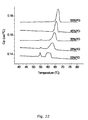
- a DSC-scan of liposomal oxaliplatin (70/25/5 mol% DSPC/DSPG/DSPE-PEG2000) stabilized by exposure to a divalent cation gives a phase-separated transition temperature (two peaks in the scan; see e.g. Figure 22 ).
- the liposomes are characteristic in that a DSC-scan of the liposomes gives a phase-separated transition temperature.
- the transition temperature of liposomes is shifted towards higher temperatures by exposure to divalent cations and one method of determining whether (test) liposomes have been exposed to a divalent cation such as calcium is by determining the transition temperature of control liposomes of the same composition as the test liposomes, wherein said control liposomes have not been exposed to a divalent cation.
- the liposomes of the invention are characteristic in that they have a higher transition temperature than control liposomes of the same composition which have not been exposed to divalent cations.
- the liposomes of the invention are characteristic in that the mean liposome size does not decrease more than 10% and more preferably not more than 5%, when they are exposed to 1 mM calcium. If they have not been previously exposed to calcium, they will shrink when exposed to calcium.
- One way of testing of testing whether test liposomes have been exposed to calcium is by comparison to control liposomes of the same composition, where it is known that they have not been exposed to calcium.
- the liposomes of the invention are characteristic in that they display a degree of shrinkage when exposed to 1 mM calcium that is smaller than the degree of shrinkage for control liposomes of the same composition, which have not been previously exposed to divalent cations.
- a second aspect of the invention is a liposomal formulation comprising liposomes of the invention.
- the formulation also comprise a divalent cation at a concentration of at least 0,1 mM.
- the present inventors have discovered that the presence of a divalent cation (or previous exposure to) stabilizes the liposomes of the formulation leading to reduced leakage of bioactive compound out of the liposomes.
- concentration of divalent cation should not exceed 10 mM and not exceed 5 mM as such concentrations can lead to aggregation of the liposomes and undesirable high viscosities.
- the concentration of divalent cation is not above 1 mM and preferably the concentration of divalent cation is between 0,1 mM and 1 mM ( Figure 3 ).
- a preferred divalent cation is calcium.
- the divalent cation is selected from the group consisting of magnesium (Mg 2+ ), iron (Fe 2+ ), calcium (Ca 2+ ), beryllium (Be 2+ ), magnesium (Mg 2+ ), strontium (Sr 2+ ), barium (Ba 2+ ), and radon (Ra 2+ ).
- the counterion of Ca 2+ is selected from the group consisting of bulky anions such as an organic salt, preferably selected from the group consisting of gluconate, propionate or lactate. More preferably the counterion is gluconate.
- the divalent cation may be distributed at the interior of the liposome, at the exterior of the liposome or both at the interior and the exterior of the liposome.
- the divalent cation may therefore be present in the hydration solution and/or in the solution wherein the liposome formulation is purified, suspended and/or stored.
- the divalent cation is distributed at the exterior of the liposome, but not at the interior of the liposome.
- the divalent cation may be added after liposome formation.
- the concentration of calcium salt to be employed may depend on the individual liposome formulation and the drug. Salts such as e.g. CaCl 2 or NaCl are often required at certain concentrations to stabilize the liposomes. However, oxaliplatin is unstable in the presence of some salts, such as e.g. NaCl, and cisplatin is unstable in the presence of salts of phosphates or in pure water. Thus, the type of salt selected and their concentration will have a significant impact on the vesicle forming properties, and accordingly, depending on the drug to be encapsulated various salts must be selected and different salt concentrations used for the preparation of a liposome formulation.
- the Poly Dispersity Index (PDI) of the liposomal formulation should not exceed 0.2 and more preferable be 0.10 or less.
- a PDI value in this range expresses a relatively narrow particle size-distribution in the formulation.
- the liposomal formulation further comprises a cryoand/or lyo-protecting agent.
- the phospholipids may undergo hydrolysis.
- One simple way of preventing decomposition of the phospholipids in the liposome formulation is by freezing or freeze-drying.
- the liposome formulation may further comprise a cryo-protecting agent.
- cryo-protecting agents may without limitation be disaccharides such as sucrose, maltose and/or trehalose. Such agents may be used at various concentrations depending on the preparation and the selected agent such as to obtain an isotonic solution.
- the liposome can also be freeze-dried, stored and the reconstituted such that a substantial portion of its internal contents are retained.
- Liposome dehydration generally requires use of a lyo-protecting agent such as a disaccharide (sucrose, maltose or trehalose) at both the inside and outside interfaces of the liposome bilayer.
- a lyo-protecting agent such as a disaccharide (sucrose, maltose or trehalose) at both the inside and outside interfaces of the liposome bilayer.
- This hydrophilic compound is generally believed to prevent the rearrangement of the lipids in the liposome formulation, so that the size and contents are maintained during the drying procedure and through subsequent reconstitution.
- Appropriate qualities for such drying protecting agents are that they possess stereo chemical features that preserve the intermolecular spacing of the liposome bilayer components.
- a third aspect of the invention is a method of preparing a liposomal formulation comprising the steps
- the organic solvent is removed before addition of hydration solvent.
- the method of further comprising of high sheer mixing to reduce the size of the liposomes.
- the method may further comprise a step of extruding the liposomes produced in step c) through a filter to produce liposomes of a certain mean size.
- the method may also comprise a step of sonicating the liposomal formulation to produce liposomes of a certain size.
- Liposomes may be loaded with at least one therapeutic agent by solubilizing the drug in the organic solvent or hydration solvent used to prepare the liposomes.
- ionisable therapeutic agent can be loaded into liposomes by first forming the liposomes, establishing an electrochemical potential, e.g. by way of a pH gradient, across the outermost liposome layer, and then adding the ionisable therapeutic agent to the aqueous medium external to the liposome.
- the hydration solvent comprises a divalent cation at a concentration of at least 0,1 mM and more preferably at a concentration between 0,1 mM and 5 mM and most preferably between 0,1 mM and 1 mM.
- the divalent cation is Ca 2+ .
- the hydration solvent does not comprise a divalent cation.
- the method further comprises a step of changing the exterior water phase of the formulation.
- the water phase will comprise the hydration solvent.
- the exterior water phase may be changed by centrifugation, ultrafiltration, dialysis or similar in order to prepare a liposomal formulation comprising liposomes in a solution of defined composition of the exterior water phase.
- bioactive compounds therapeutic agents
- the drug encapsulation in the liposomes should be >70%, more preferably >95% and most preferably >99%.
- the degree of drug encapsulation is the ratio of drug encapsulated to the total amount of drug in the formulation.
- the exterior water phase is changed to another exterior water phase comprising a divalent cation at a concentration of at least 0,1 mM and more preferably at a concentration between 0,1 mM and 5 mM and most preferably between 0,1 mM and 1 mM.
- the divalent cation is Ca 2+ .
- liposomes initially leak entrapped compound when being exposed to calcium. Moreover, the liposomes condense to give smaller particle diameters. However, after initial leakage the liposomes exposed to Ca 2+ displays reduced leakage as seen e.g. in case of incubation in cell media (e.g. McCoy media; Figure 3 ). Because of initial leakage, dialysis and/or centrifugation is typically done to separate liposomes from leaked material. Also filtration may be done.
- the liposomal formulation is produced by the method of the third aspect.
- a fifth aspect of the invention is the liposome of the first aspect or the liposomal formulation of the second aspect for use as a medicament.
- a sixth aspect of the invention is the liposome of the first aspect or the liposomal formulation of the second aspect used for treatment of conditions, wherein PLA 2 activity is increased.
- conditions are e.g. cancer and inflammatory diseases.
- Example 1 Preparation of Liposome encapsulated cisplatin and Liposome encapsulated oxaliplatin.
- Liposome encapsulated cisplatin and Liposome encapsulated oxaliplatin are liposome-drug formulations wherein the drug cisplatin or oxaliplatin is encapsulated in the aqueous compartment of the liposome.
- the liposome drug formulations are composed of the drug encapsulated in a lipid mixture made of 5 mol% mPEG2000-disteoryl-phospahtidylethanolamine (DSPE-PEG2000); 25 mol% disterorylphosphatidylglycerol (DSPG); and 70 mol% disteorylphosphatidylcholine (DSPC).
- DSPE-PEG2000 5 mol% mPEG2000-disteoryl-phospahtidylethanolamine
- DSPG disterorylphosphatidylglycerol
- DSPC disteorylphosphatidylcholine
- Phospholipids were dissolved in 9:1 (v/v) chloroform/methanol. The solvent of the dissolved lipid mixtures were then evaporated in a 65°C hot water bath until visual dryness, under a stream of nitrogen gas. The samples were further dried under vacuum overnight.
- lipid suspensions were kept at 65-70°C for at least 30 min. in order to ensure complete hydration. During this period, the lipid suspensions were vortex every 5 min. Large unilamellar vesicles (LUV) were prepared by 5 min. sonication at 65°C of the MLV followed by extrusion through a 100 nm pore size polycarbonate filters at 65-70°C. LUV were subsequently transferred to dialysis cassettes (MWCO: 10 kDa) in order to remove untrapped cisplatin.
- MWCO dialysis cassettes
- Phospholipids were dissolved in 9:1 chloroform/methanol. The dissolved lipid mixtures were then evaporated in a 65°C water bath until visual dryness, under a stream of nitrogen. The samples were further dried under vacuum overnight.
- lipid suspensions were kept at 65-70°C for at least 30 min. in order to ensure complete hydration. During this period, the lipid suspensions were vortex every 5 min. large unilamellar vesicles (LUV) were subsequently prepared by 5 min. sonication at 65-70°C of the MLV followed by extrusion through a 100 nm pore size polycarbonate filters ten times at 65-70°C. LUV were subsequently transferred to dialysis cassettes (MWCO: 10 kDA) in order to remove un-trapped oxaliplatin.
- LUV large unilamellar vesicles
- Example 2 Stability of oxaliplatin in hydration solution.
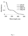
- Oxaliplatin was dissolved in a hydration solution containing 10% sucrose and 1 mM Calcium gluconate (10 mg/ml). Sucrose was added to obtain an osmolarity approximately corresponding to physiological salt concentration. Stability of oxaliplatin in the hydration solution was followed by UV measurement. Samples were taken continually during storage at room temperature. Sample concentration measured was 0.1 mg/ml oxaliplatin. In order to follow the oxaliplatin stability a scan was performed from 200-350 nm ( Figure 1 ) and the absorbance at 254 nm was compared during storage ( Figure 2 ).
- Example 3 Stability of liposome encapsulated oxaliplatin.
- This cell media was used for the subsequent evaluation of the cytotoxic effect of Liposome encapsulated oxaliplatin on HT-29 colon carcinoma cells. Increased leakage from the liposomes upon storage in the cell media results in unspecific cytotoxicity. Formulation should have minimal cytotoxicity when it is has not undergone sPLA 2 hydrolysis. Varying concentrations of calcium gluconate (0-5 mM) in the Liposome encapsulated oxaliplatin formulation were examined for their ability to stabilize the liposomes ( Figure 5 ).
- liposome formulation was affected by the presence of calcium gluconate on interior or exterior only. As demonstrated in the table below, it was quite evident that liposome formulation containing calcium on the interior only is not very good at stabilizing the liposome oxaliplatin formulation. Having calcium on the exterior only was demonstrated to stabilize the liposome formulation quite well.
- Ether phospholipids were used to prepare a liposome formulation of oxaliplatin encapsulated in a lipid mixture (25 mol% 1-O-octadecyl-2-octadecanoyl-sn-glycero-3-phosphoglycerol (1-O-DSPG), 70 mol% 1-O-octadecyl-2-octadecanoyl-sn-glycero-3-phosphocholine (1-O-DSPC) and 5 mol% dioctadecanoyl-sn-glycero-3-phosphoethanolamine-N-[methoxy-(polyethylene glycol)2000] (DSPE-PEG2000)) in a solution containing 10% sucrose and 1 mM calcium gluconate.
- a lipid mixture 25 mol% 1-O-octadecyl-2-octadecanoyl-sn-glycero-3-phosphoglycerol (1-O-
- Example 5 Cytotoxicity of liposome encapsulated oxaliplatin.
- HT-29 colon carcinoma cells
- PLA 2 from external source such as tear fluid was added to show that full release of oxaliplatin had occurred. With stable liposome formulation the HT-29 should not be affected by presence of such. In order to release the oxaliplatin from the liposome presence of a PLA 2 source is required.
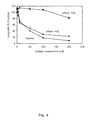
- This cell media was used for the subsequent evaluation of the cytotoxic effect of Liposome encapsulated cisplatin on HT-29 colon carcinoma cells ( Figure 4 and Figure 6 ).
- Aim To examine the effect of adding or diluting Liposome encapsulated oxaliplatin formulation with a calcium gluconate solution.
- Liposome encapsulated oxaliplatin formulation was prepared without calcium (10% sucrose on interior and exterior). Liposome encapsulated oxaliplatin formulation was diluted in varying concentrations of calcium (Lipid concentration maintained at 0.84 mM). Samples were equilibrated 24h, and degree of encapsulation (DOE; %) and particle sizes were measured.
- a suspension of liposomes (70/25/5 mol%, PC/PG/PE-PEG) containing 0.5 mol% of 1,2-dipalmitoyl-phosphatidylethanolamine with the fluorescent probe 7-nitrobenz-2-oxa-1,3-diazol-4-yl covalently linked to the head group (NBD-PE) (Fluka, Buchs, Switzerland) was prepared in 10% sucrose.
- NBD-PE head group
- DSPG and DSPC contents were varied in the liposomal formulations as following:
- Formulations were prepared without calcium on the interior, and with or without calcium on the exterior.
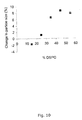
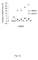
- Particle sizes are observed to increase by raised PG content up to 45% PG in solutions only containing sucrose ( figure 10 ). Contrary to this, by adding 1mM calcium gluconate on the exterior the particle sizes are observed to decrease ( Figure 11 ). Formulation having the highest content of PG was observed to have largest variation in particle sizes in the absence of Ca 2+ ( Figures 11 +12).
- DOE (%) is usually higher for formulation with lower content of PG when dialyzed the same amount of time.
- Liposomes dialyzed in solution containing calcium gluconate are stable in McCoy media, whereas formulation dialyzed in 10% sucrose had major leakage of oxaliplatin when exposed to McCoy Media ( Figure 18 ).
- Transition temperature for Liposome encapsulated oxaliplatin formulation is in the range 60-70°C. It is therefore recommended that the extrusion temperature is maintained at 70°C.
- DSPC and DSPG contents were varied in liposomal formulation as following:
- Formulations were prepared with calcium on both the interior and exterior.
- DOE (%) is usually higher for formulation with lower content of DSPG, when dialyzed the same amount of time.
Landscapes
- Health & Medical Sciences (AREA)
- Life Sciences & Earth Sciences (AREA)
- Chemical & Material Sciences (AREA)
- Veterinary Medicine (AREA)
- Public Health (AREA)
- General Health & Medical Sciences (AREA)
- Medicinal Chemistry (AREA)
- Animal Behavior & Ethology (AREA)
- Pharmacology & Pharmacy (AREA)
- Epidemiology (AREA)
- Nuclear Medicine, Radiotherapy & Molecular Imaging (AREA)
- General Chemical & Material Sciences (AREA)
- Chemical Kinetics & Catalysis (AREA)
- Organic Chemistry (AREA)
- Communicable Diseases (AREA)
- Oncology (AREA)
- Proteomics, Peptides & Aminoacids (AREA)
- Inorganic Chemistry (AREA)
- Molecular Biology (AREA)
- Gastroenterology & Hepatology (AREA)
- Engineering & Computer Science (AREA)
- Bioinformatics & Cheminformatics (AREA)
- Immunology (AREA)
- Dispersion Chemistry (AREA)
- Pain & Pain Management (AREA)
- Rheumatology (AREA)
- Medicinal Preparation (AREA)
- Pharmaceuticals Containing Other Organic And Inorganic Compounds (AREA)
- Acyclic And Carbocyclic Compounds In Medicinal Compositions (AREA)
- Medicines That Contain Protein Lipid Enzymes And Other Medicines (AREA)
Abstract
Description
- Liposomes are microscopic spheres which were developed as drug delivery vehicles/systems in the 1980s. The first liposome-based pharmaceuticals were approved for commercial use in the 1990s.
- Liposomes have three distinct compartments that can be used to carry various compounds such as, e.g. drugs: The interior aqueous compartment; the hydrophobic bilayer; and the polar inter-phase of the inner and outer leaflet. Depending on the chemical nature of the compound to be encapsulated it will be localised to either of the compartments. Currently, there are several parenteral liposome-drug formulations available on the market. Water soluble drugs tend to be localised in the aqueous compartment of liposomes, and examples of drugs encapsulated in liposome's are, e.g. doxorubicin (Doxil), doxorubicin (Myocet) and daunorubicin (DaunoXone). Examples of drugs intercalated in the liposome membrane are, e.g. amphotericin B (AmBisome), amphotericin (Albelcet B), benzoporphyrin (Visudyne) and muramyltripeptide-phosphatidylethanolamine (Junovan).
- The liposome technology has thus provided intelligent solutions to solve challenges in pharmacology such as e.g. increase drug solubility, reduce drug toxicity, improve targeted drug release, etc.
- The property of liposomes as drug delivery vehicles is crucially dependent on their surface charge, permeability, solubility, stability etc. which is significantly influenced by the lipids comprised in the liposome composition. In addition, the drug to be encapsulated in the liposome may need further requirements to be considered in preparing a stable liposome formulation.
- Considerations regarding safety and drug efficacy require that liposome formulations maintain their properties, i.e. remain stable, from the time of preparation until administration. Furthermore, it is desirable that such formulations are intact during the transport in the treated subject until they reach the target site where the drug is specifically released. Thus there is still a need for obtaining improved liposome formulations.
-
WO 90/11781 - In
WO 2004/087105 liposomes comprising distearoylphosphatidylcholine (DSPC) and distearoylphosphatidylglycerol (DSPG) are described in which carboplatin is entrapped. - In a first aspect, the present invention provides liposomes that are useful for delivery of bioactive agents such as therapeutics according to
claim 1. Among others, the liposomes of the invention are capable of delivering their payload at sites of increased secretory phospholipase A2 (sPLA2) activity, because phospholipase A2 (PLA2) will hydrolyse lipids of the liposome. Thus, the liposomes of the invention may e.g. be used in relation to cancer therapy. A second aspect of the invention is a liposomal formulation comprising the liposome of the invention. Still another aspect is a method of producing a liposomal formulation of the invention. - The invention is explained in detail below with reference to the drawing(s), in which
-
Figure 1 illustrates the UV spectra of oxaliplatin stored in solution containing 10% sucrose and 1 mM calcium gluconate during storage at room temperature. -
Figure 2 illustrates the relative absorbance (day/day 0) at 254 nm for oxaliplatin stored in a solution containing 10% sucrose and 1 mM calcium gluconate during storage at room temperature (Figure 1 ). -
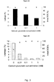
Figure 3 illustrates the effect of varying calcium gluconate concentrations on leakage from liposomes (70/25/5 mol% DSPC/DSPG/DSPE-PEG2000) containing cisplatin (A) or oxaliplatin (B) after 24 hours storage in cell media (McCoy) at 37°C (primary axis). The initial degree of encapsulation (DOE) is marked on the secondary axis. -
Figure 4 illustrates the cytotoxicity of liposome (70/25/5 mol% DSPC/DSPG/DSPE-PEG2000) encapsulated cisplatin containing 1 mM calcium gluconate. HT-29 colon carcinoma cells were treated for 6 hours (37°C) with cisplatin or Liposome encapsulated cisplatin in the presence or absence of sPLA2. -
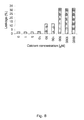
Figure 5 illustrates the cytotoxicity of liposome (70/25/5 mol% DSPC/DSPG/DSPE-PEG2000) encapsulated oxaliplatin containing varying concentrations of calcium gluconate (A) 5 mM calcium gluconate, (B) 1 mM calcium gluconate, (C) 0.1 mM calcium gluconate, (D) 0.01 mM calcium gluconate, and (E) 0 mM calcium gluconate. HT-29 colon carcinoma cells were treated for 6 hours (37°C) with oxaliplatin or Liposome encapsulated oxaliplatin in the presence or absence of sPLA2. -
Figure 6 illustrates the cytotoxicity of liposome (70/25/5 mol% DSPC/DSPG/DSPE-PEG2000) encapsulated cisplatin containing varying concentrations of calcium gluconate. HT-29 colon carcinoma cells were treated for 24 hours (37°C) with cisplatin in the presence or absence of sPLA2. -
Figure 7 . Changes in particle size as function of calcium concentration for oxaliplatin encapsulated liposomes (70/25/5 mol% DSPC/DSPG/DSPE-PEG2000) containing no calcium after 24h at room temperature. Lipid concentration maintained at 0.84 mM. -
Figure 8 . Leakage from Liposome encapsulated oxaliplatin formulation (70/25/5 mol% DSPC/DSPG/DSPE-PEG2000) containing no calcium after 24h at room temperature as a function of calcium concentration. Lipid concentration maintained at 0.84 mM. -
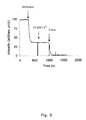
Figure 9 . The time-dependence of fluorescence intensity of DSPC/DSPG/DSPE-PEG LUV containing 0.5mol% NBD-PE at 25°C. Arrow indicates the addition of dithionite, calcium and Triton-X100. -
Figure 10 . Changes in particle size from sonication step to after 1st dialysis step (10% sucrose) as a function of DSPG in the liposomal oxaliplatin formulations containing 5 mol% DSPE-PEG2000 and DSPC. -
Figure 11 . Changes in particle size from 1st dialysis step (10% sucrose) to after 2nd dialysis step (10% sucrose + calcium gluconate) as a function of DSPG in the liposomal oxaliplatin formulations containing 5 mol% DSPE-PEG2000 and DSPC. -
Figure 12 . Changes in particle size from sonication to after 2nd dialysis step (10% sucrose +/- calcium gluconate) as a function of DSPG in the liposomal oxaliplatin formulations containing DSPC and 5 mol% DSPE-PEG2000. -
Figure 13 . Ion count ratio (Pt195/P31) for liposomal oxaliplatin formulations containing 5 mol% DSPE-PEG2000 and varying amounts of DSPC and DSPG after 1st dialysis step (10% sucrose solution) . -
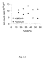
Figure 14 . Ion count ratio (Pt195/P31) for liposomal oxaliplatin formulations containing 5mol% DSPE-PEG2000 and varying amounts of DSPC and DSPG after 2nd dialysis step (10% sucrose solution ± 1mM calcium gluconate). -
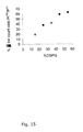
Figure 15 . %-difference in the ion count ratio (Pt195/P31) for formulations dialyzed in solution without calcium compared to formulations dialyzed with calcium (seeFigure 14 ). -
Figure 16 . Pt concentration in dialysate after 2nd dialysis (10% sucrose solution ± 1 mM calcium gluconate) of liposomal oxaliplatin formulations containing 5mol% DSPE-PEG2000 and varying amounts of DSPC and DSPG . -
Figure 17 . Correlation between the final liposome size and concentration of oxaliplatin in formulations. Liposomal oxaliplatin formulations (5mol% DSPE-PEG2000, and varying amounts of DSPC and DSPG) were prepared with or without 1mM calcium gluconate during dialysis (seeFigure 14 ). -
Figure 18 . Stability of liposomal oxaliplatin formulations (5mol% DSPE-PEG2000, and varying concentrations of DSPC and DSPG) in McCoy cell media (24h incubation at 37°C). -
Figure 19 .1 .-6. DSC scans of Liposome encapsulated oxaliplatin (60/35/5 mol% DSPC/DSPG/DSPE-PEG2000) formulation without the presence calcium gluconate. Scan speed: 20°C/h. -
Figure 20 . DSC scan (1st scan) of Liposome encapsulated oxaliplatin formulations containing 5mol% DSPE-PEG2000 and varying DSPC and DSPG concentrations and without the presence of calcium gluconate. Scan speed: 20°C/h. -
Figure 21 .1 .-6. DSC scans of Liposome encapsulated oxaliplatin (60/35/5 mol% DSPC/DSPG/DSPE-PEG2000) formulation with 1mM calcium gluconate on the exterior. Scan speed: 20°C/h. -
Figure 22 . DSC scan (1st scan) of Liposome encapsulated oxaliplatin formulations containing 5mol% DSPE-PEG2000 and varying DSPC and DSPG concentration with 1mM calcium gluconate on the exterior. Scan speed: 20°C/h. -
Figure 23 . Pt concentration in dialysate after a) 1st dialysis step and b) 2nd dialysis step (10% sucrose solution containing 1mM calcium gluconate) of oxaliplatin encapsulated liposomes. -
Figure 24 .1 .-6. DSC scans of Liposome encapsulated oxaliplatin (60/35/5 mol% DSPC/DSPG/DSPE-PEG2000) formulation with 1 mM calcium gluconate on both the interior and exterior. Scan speed: 20°C/h. -
Figure 25 . DSC scan (1st scan) of Liposome encapsulated oxaliplatin formulations containing 5mol% DSPE-PEG2000 and varying DSPC and DSPG concentrations with 1 mM calcium on both the interior and exterior. Scan speed: 20°C/h. - One approach to obtaining triggered drug release in e.g. cancerous diseases is to utilize elevated levels of secretory phospholipase A2 (sPLA2) in the proximity of cancerous tissues. Thus, by carefully designing the lipid composition of the liposomes, they can be made degradable by sPLA2 once accumulated in the tumour to give triggered release.
- An object of the present invention is to provide liposomes and liposome formulations of improved stability which may deliver their payload (e.g. a drug) at the target site with reduced uncontrolled delivery and/or too early release due to leakiness of the liposome membrane. Another object is to provide liposomes and liposomal formulations with an increased stability during storage.
- Anionic lipids of the liposome.
- In a first aspect, the liposome comprises between 25% and 45% (mol/mol) of an anionic lipid. The present inventors have taken into account that the content of anionic lipid affects important characteristics of the liposome, such as the rate of sPLA2 mediated lipid hydrolysis of the liposome and also the immune response toward the liposome.
- As the content of anionic lipid increases, so does the rate of lipid hydrolysis by sPLA2 (and the release of drug). It has been demonstrated that a reasonable rate of hydrolysis can be achieved by anionic lipid content between 25% and 45%. Thus, in one embodiment, the content of anionic lipid is at least 25%. In another embodiment, the content of anionic lipid is no more than 45%. In yet another embodiment, the anionic lipid content of the liposome is selected from the group consisting of between 25% and 45%, 25-42%, 28% and 42%, 30% and 40%, 32% and 38% and 34% and 36%. When referring to % content, reference is to mol/mol %, unless specifically mentioned otherwise.
- As mentioned, also the immune response toward the liposomes is affected by the content of anionic lipid. Thus, the clearance rate of the liposome in body reduced by keeping the content of the anionic lipid in the liposome below a certain level and the present inventors have recognized that the content of anionic lipid in the liposome can be used to strike a balance between hydrolysis rate of sPLA2 and clearance by the reticuloendothelial system.
- Preferably the anionic lipid is a phospholipid and preferably, the phospholipid is selected from the group consisting of PI (phosphatidyl inositol), PS (phosphatidyl serine), DPG (bisphosphatidyl glycerol), PA (phosphatidic acid), PEOH (phosphatidyl alcohol), and PG (phosphatidyl glycerol). More preferably, the anionic phospholipid is PG.
- In a preferred embodiment, the liposome further comprises a hydrophilic polymer selected from the group consisting of PEG [poly(ethylene glycol)], PAcM [poly(N-acryloylmorpholine)], PVP [poly(vinylpyrrolidone)], PLA [poly(lactide)], PG [poly(glycolide)], POZO [poly(2-methyl-2-oxazoline)], PVA [poly(vinyl alcohol)], HPMC (hydroxypropylmethylcellulose), PEO [poly(ethylene oxide)], chitosan [poly(D-glucosamine)], PAA [poly(aminoacid)], polyHEMA [Poly(2-hydroxyethylmethacrylate)] and co-polymers thereof.
- Most preferably the polymer is PEG with a molecular weight between 100 Da and 10 kDa. Particular preferred are PEG sizes of 2-5 kDa (PEG2000 to PEG5000), and most preferred is PEG2000.
- The inclusion of polymers on liposomes is well known to the skilled artisan and can be used to increase the half-life of the liposomes in the bloodstream, presumably by reducing clearance by the reticuloendothelial system.
- Preferably, the polymer is conjugated to the head group of phospatidyl ethanolamine. Another option is ceramide (even though this lipid is not hydrolyzable by PLA2).
- The polymer-conjugated lipid is preferably present at an amount of at least 2%. More preferably, the amount is at least 5% and no more than 15%. Even more preferably, the amount of polymer-conjugated lipid is at least 3% and no more than 6%. Liposomes containing anionic phospholipids and ≤2.5 % DSPE-PEG2000 have increased tendency to aggregate in the presence of calcium. This can usually be observed by formation of high viscous gel. Liposomes containing anionic phospholipids and >7.5 % causes the liposomes to sediment or phase separate. Thus, another preferred window is between 2,5% and 7,5%.
- Preferably, the liposome also comprises an uncharged phospholipid selected from the group consisting of zwitterionic phospholipids comprising PC (phosphatidyl choline) and PE (phosphatidylethanolamine). Most preferably, the zwitterionic phospholipid is PC.
- In contrast to anionic phospholipid, zwitterionic phospholipid serves as a charge neutral sPLA2-hydrolyzable lipid component in the liposome. By combining zwitterionic- and anionic phospholipid in the same liposome, it is possible to adjust to a desired surface charge density which complies with both sufficiently high sPLA2 hydrolysis and a low clearance rate in the blood.
- The amount of zwitterionic phospholipid in the liposome is preferably between 40% and 75% and more preferably between 50 and 70%.
- Some or all of the phospholipids may be ether-phospholipids.
- Thus, they may harbour an ether-bond instead of an ester-bond at the sn-1 position of the glycerol backbone of the phospholipid. When sPLA2 hydrolyze this particular type of phospholipids, mono-ether lyso-phospholipids are produced and these are toxic to e.g. cancer cells. I.e. ether phospholipids may be seen as pro-drugs of mono-ether lyso-phospholipids and liposomes of the invention can be used to deliver such pro-drugs to the sPLA2-enhanced environment of cancer cells, where the pro-drugs are activated by sPLA2 hydrolysis. Ether-phospholipids have been described in
EP 1254143 andWO 2006/048017 . - The moiety released from the lipid by sPLA2 to create a lysolipid may also be a drug. Thus, a liposome may comprise pro-drugs of mono-ether lysolipids, pro-drugs released from the lipid by sPLA2 and other therapeutic agents, as further outlined below.
- The liposome may also be stabilized by the inclusion of cholesterol as membrane component in the liposome. However, high amounts of cholesterol in the liposome have a negative effect on hydrolysis by PLA2 and therefore the liposome comprises no more than 10% cholesterol. Preferably, the liposome comprises less than 1% cholesterol, less than 0,1 % or does not comprise any cholesterol at all.
- The alkyl chain length of the lipids comprising the liposome may be adjusted for optimal PLA2 hydrolysis rate and minimum leakage of entrapped compound out of the liposome. Preferably, the alkyl chains are C18 or C16 saturated chains.
- The liposomes of the invention are prepared by the method of the third aspect, wherein liposomes are stabilized by exposure to divalent cations.
- As described above, the liposomes may comprise pro-drugs of mono-ether lyso-lipids and/or of the moiety released from the lipid by sPLA2 to create the lysolipid.
- The liposomes comprise a bioactive compound such as a therapeutic agent (drug), which is not a pro-drug of mono-ether lysophospholipid or mono-ether lysophospholipid. The liposome may also comprise pro-drugs of mono-ether lysophospholipid and a therapeutic agent. The bioactive compounds are small molecules A class of compounds of particular interest is small molecule antitumour agents such as anthracyclin derivatives, cisplatin, oxaliplatin, carboplatin, doxorubicin, paclitaxel, 5-fluoruracil, exisulind, cis-retinoic acid, suldinac sulphide, methotrexate, bleomycin and vincristine. Another class of particular interest is antibiotics and antifungals and yet another class is anti-inflammatory agents such as steroids and non-stereoids. The liposome may comprise 1, 2, 3 or more different bioactive compounds. In a preferred embodiment, the liposome comprise only 1 bioactive component. The liposome may comprise a diagnostic agent. By "diagnostic agent" is meant an agent that supports the localisation of the target tissue and/or the diagnosis of the disease and/or condition. Non-limiting examples could be contrast agents, microparticles, radioactive agents, target specific agents such as e.g. agents that bind specifically to markers associated with the disease and/or condition, etc. It is clear to a skilled person that in some embodiments the invention relates to a liposome formulation wherein the liposome comprises at least one drug as well as a diagnostic agent.
- The liposome can be unilamellar or multilamellar. Most preferably, the liposome is unilamellar. The diameter of the liposome should be between 50 and 400 nm, preferably between 80 and 160 nm and most preferable between 90 and 120nm.
- Preferably, the Poly Dispersity Index (PDI) of the liposomal formulation of the second aspect of the invention should not exceed 0.2 and more preferable be 0,15 or less or even more preferably 0.10 or less. A PDI value in this range expresses a relatively narrow particle size-distribution in the formulation.
- As will be clear from the above, it is preferred that at least one of the lipids comprising the liposome is a substrate for sPLA2 when present in the liposome.
- In one embodiment, the liposome comprises lipids which are hydrolysed by sPLA2 at the sn-3 position instead of at the sn-2 position. Such unnatural lipids and liposomes comprising unnatural lipids have been disclosed in
WO 2006/048017 - The liposomes are present in the liposomal formulation of the second aspect. Thus, they have been stabilized by exposure to divalent cations as described in the second aspect. In one embodiment, the liposomes have only been exposed to divalent cations after formation. I.e. the interior of the liposomes does not contain divalent cations. In another embodiment, only the interior of the liposomes comprise divalent cations.
- The presence of divalent cations associated with the liposomes of the invention is not directly verifiable. However, the present inventors have described parameters that are indicative of whether the liposomes have been stabilized by divalent cations.
- A DSC-scan of liposomal oxaliplatin (70/25/5 mol% DSPC/DSPG/DSPE-PEG2000) in the absence of a divalent cation gives a single transition temperature observed as one peak. If the scan is repeated, the transition temperature is shifted towards higher temperatures, which might be due to the release of oxaliplatin to exterior of the liposomes when passing transition temperature (
Figure 19 ). Repeated DSC scans of liposomes that have been exposed to a divalent cation have a more constant transition temperature. Thus, in one embodiment, the liposomes of the invention are characteristic in that repeated DSC-scans of the liposome gives a transition temperature that differs by no more than 2 °C between the first and the second scan. In another embodiment, the liposomes of the invention are characteristic in that repeated DSC-scans of the liposomes gives a transition temperature that differs less between the first and second scan than will the transition temperature of control liposomes of the same composition. - A DSC-scan of liposomal oxaliplatin (70/25/5 mol% DSPC/DSPG/DSPE-PEG2000) stabilized by exposure to a divalent cation gives a phase-separated transition temperature (two peaks in the scan; see e.g.
Figure 22 ). Thus, in one embodiment of the liposomes of the invention, the liposomes are characteristic in that a DSC-scan of the liposomes gives a phase-separated transition temperature. - Moreover, the transition temperature of liposomes is shifted towards higher temperatures by exposure to divalent cations and one method of determining whether (test) liposomes have been exposed to a divalent cation such as calcium is by determining the transition temperature of control liposomes of the same composition as the test liposomes, wherein said control liposomes have not been exposed to a divalent cation. Thus, in one embodiment, the liposomes of the invention are characteristic in that they have a higher transition temperature than control liposomes of the same composition which have not been exposed to divalent cations.
- In another embodiment, the liposomes of the invention are characteristic in that the mean liposome size does not decrease more than 10% and more preferably not more than 5%, when they are exposed to 1 mM calcium. If they have not been previously exposed to calcium, they will shrink when exposed to calcium. One way of testing of testing whether test liposomes have been exposed to calcium is by comparison to control liposomes of the same composition, where it is known that they have not been exposed to calcium. Thus, in one embodiment, the liposomes of the invention are characteristic in that they display a degree of shrinkage when exposed to 1 mM calcium that is smaller than the degree of shrinkage for control liposomes of the same composition, which have not been previously exposed to divalent cations.
- A second aspect of the invention is a liposomal formulation comprising liposomes of the invention. Preferably, the formulation also comprise a divalent cation at a concentration of at least 0,1 mM. The present inventors have discovered that the presence of a divalent cation (or previous exposure to) stabilizes the liposomes of the formulation leading to reduced leakage of bioactive compound out of the liposomes. However, it is experienced that the concentration of divalent cation should not exceed 10 mM and not exceed 5 mM as such concentrations can lead to aggregation of the liposomes and undesirable high viscosities. The concentration of divalent cation is not above 1 mM and preferably the concentration of divalent cation is between 0,1 mM and 1 mM (
Figure 3 ). A preferred divalent cation is calcium. - In a preferred embodiment, the divalent cation is selected from the group consisting of magnesium (Mg2+), iron (Fe2+), calcium (Ca2+), beryllium (Be2+), magnesium (Mg2+), strontium (Sr2+), barium (Ba2+), and radon (Ra2+).
- Various divalent cations have been tested (data not shown) and the best effects are seen with Ca2+, wherefore Ca2+ is preferred in the formulation.
- Furthermore, it is experienced that also the counterion of Ca2+ is important in some instances. Therefore, in one embodiment, the counterion is selected from the group consisting of bulky anions such as an organic salt, preferably selected from the group consisting of gluconate, propionate or lactate. More preferably the counterion is gluconate.
- The divalent cation may be distributed at the interior of the liposome, at the exterior of the liposome or both at the interior and the exterior of the liposome. The divalent cation may therefore be present in the hydration solution and/or in the solution wherein the liposome formulation is purified, suspended and/or stored. In a preferred embodiment, the divalent cation is distributed at the exterior of the liposome, but not at the interior of the liposome. In this embodiment, the divalent cation may be added after liposome formation.
- The concentration of calcium salt to be employed may depend on the individual liposome formulation and the drug. Salts such as e.g. CaCl2 or NaCl are often required at certain concentrations to stabilize the liposomes. However, oxaliplatin is unstable in the presence of some salts, such as e.g. NaCl, and cisplatin is unstable in the presence of salts of phosphates or in pure water. Thus, the type of salt selected and their concentration will have a significant impact on the vesicle forming properties, and accordingly, depending on the drug to be encapsulated various salts must be selected and different salt concentrations used for the preparation of a liposome formulation.
- Preferably, the Poly Dispersity Index (PDI) of the liposomal formulation should not exceed 0.2 and more preferable be 0.10 or less. A PDI value in this range expresses a relatively narrow particle size-distribution in the formulation.
- In a preferred embodiment, the liposomal formulation further comprises a cryoand/or lyo-protecting agent.
- During storage of liposomes the phospholipids may undergo hydrolysis. One simple way of preventing decomposition of the phospholipids in the liposome formulation is by freezing or freeze-drying.
- Freezing may however induce leakage of the liposome formulation and result in release of the encapsulated drug. Addition of a cryo-protecting agent may be necessary in order to prevent or reduce leakage from the liposome preparation after freezing. Thus, the liposome formulation may further comprise a cryo-protecting agent. Examples of agents that may be used as cryo-protecting agents may without limitation be disaccharides such as sucrose, maltose and/or trehalose. Such agents may be used at various concentrations depending on the preparation and the selected agent such as to obtain an isotonic solution.
- The liposome can also be freeze-dried, stored and the reconstituted such that a substantial portion of its internal contents are retained. Liposome dehydration generally requires use of a lyo-protecting agent such as a disaccharide (sucrose, maltose or trehalose) at both the inside and outside interfaces of the liposome bilayer. This hydrophilic compound is generally believed to prevent the rearrangement of the lipids in the liposome formulation, so that the size and contents are maintained during the drying procedure and through subsequent reconstitution. Appropriate qualities for such drying protecting agents are that they possess stereo chemical features that preserve the intermolecular spacing of the liposome bilayer components.
- A third aspect of the invention is a method of preparing a liposomal formulation comprising the steps
- a) Preparing a lipid mixture by dissolving selected lipids in an organic solvent
- b) Hydrating the product of step a) with an aqueous hydration solvent so as to form liposomes
- c) Removing the organic solvent of step a) either before addition of the aqueous hydration solvent or after the addition of the aqueous hydration solvent
- Preferably, the organic solvent is removed before addition of hydration solvent.
- The method of further comprising of high sheer mixing to reduce the size of the liposomes.
- The method may further comprise a step of extruding the liposomes produced in step c) through a filter to produce liposomes of a certain mean size.
- The method may also comprise a step of sonicating the liposomal formulation to produce liposomes of a certain size.
- Liposomes may be loaded with at least one therapeutic agent by solubilizing the drug in the organic solvent or hydration solvent used to prepare the liposomes.
- Alternatively, ionisable therapeutic agent can be loaded into liposomes by first forming the liposomes, establishing an electrochemical potential, e.g. by way of a pH gradient, across the outermost liposome layer, and then adding the ionisable therapeutic agent to the aqueous medium external to the liposome.
- In a preferred embodiment, the hydration solvent comprises a divalent cation at a concentration of at least 0,1 mM and more preferably at a concentration between 0,1 mM and 5 mM and most preferably between 0,1 mM and 1 mM. Preferably, the divalent cation is Ca2+. In another embodiment, the hydration solvent does not comprise a divalent cation. In this embodiment, it is preferred that the exterior water phase is changed to another exterior water phase comprising a divalent cation as described below.
- In another embodiment, the method further comprises a step of changing the exterior water phase of the formulation. Initially, the water phase will comprise the hydration solvent. The exterior water phase may be changed by centrifugation, ultrafiltration, dialysis or similar in order to prepare a liposomal formulation comprising liposomes in a solution of defined composition of the exterior water phase. Preferably, bioactive compounds (therapeutic agents) are only present inside or attached to the liposomes and not as free compounds in solution. Preferably, the drug encapsulation in the liposomes should be >70%, more preferably >95% and most preferably >99%. The degree of drug encapsulation is the ratio of drug encapsulated to the total amount of drug in the formulation.
- In a preferred embodiment, the exterior water phase is changed to another exterior water phase comprising a divalent cation at a concentration of at least 0,1 mM and more preferably at a concentration between 0,1 mM and 5 mM and most preferably between 0,1 mM and 1 mM. Preferably, the divalent cation is Ca2+.
- The present inventors have discovered that liposomes initially leak entrapped compound when being exposed to calcium. Moreover, the liposomes condense to give smaller particle diameters. However, after initial leakage the liposomes exposed to Ca2+ displays reduced leakage as seen e.g. in case of incubation in cell media (e.g. McCoy media;
Figure 3 ). Because of initial leakage, dialysis and/or centrifugation is typically done to separate liposomes from leaked material. Also filtration may be done. - In a preferred embodiment of the second aspect, the liposomal formulation is produced by the method of the third aspect.
- A fifth aspect of the invention is the liposome of the first aspect or the liposomal formulation of the second aspect for use as a medicament.
- A sixth aspect of the invention is the liposome of the first aspect or the liposomal formulation of the second aspect used for treatment of conditions, wherein PLA2 activity is increased. Such conditions are e.g. cancer and inflammatory diseases.
- Liposome encapsulated cisplatin and Liposome encapsulated oxaliplatin are liposome-drug formulations wherein the drug cisplatin or oxaliplatin is encapsulated in the aqueous compartment of the liposome. The liposome drug formulations are composed of the drug encapsulated in a lipid mixture made of 5 mol% mPEG2000-disteoryl-phospahtidylethanolamine (DSPE-PEG2000); 25 mol% disterorylphosphatidylglycerol (DSPG); and 70 mol% disteorylphosphatidylcholine (DSPC). The specific procedure for each formulation is outlined below.
- Phospholipids were dissolved in 9:1 (v/v) chloroform/methanol. The solvent of the dissolved lipid mixtures were then evaporated in a 65°C hot water bath until visual dryness, under a stream of nitrogen gas. The samples were further dried under vacuum overnight.
- Hydration liquid (solution of sodium chloride and calcium gluconate both of varying concentrations) containing cisplatin were added to the dried lipid mixture at a temperature of 65°C-70°C for the preparation of multilamellar vesicles (MLV). The lipid suspensions were kept at 65-70°C for at least 30 min. in order to ensure complete hydration. During this period, the lipid suspensions were vortex every 5 min. Large unilamellar vesicles (LUV) were prepared by 5 min. sonication at 65°C of the MLV followed by extrusion through a 100 nm pore size polycarbonate filters at 65-70°C. LUV were subsequently transferred to dialysis cassettes (MWCO: 10 kDa) in order to remove untrapped cisplatin.
- Phospholipids were dissolved in 9:1 chloroform/methanol. The dissolved lipid mixtures were then evaporated in a 65°C water bath until visual dryness, under a stream of nitrogen. The samples were further dried under vacuum overnight.
- Hydration buffer (10% sucrose solution with varying concentrations of different calcium salts) containing oxaliplatin were added to the dried lipid mixture for the preparation of multilamellar vesicles (MLV). The lipid suspensions were kept at 65-70°C for at least 30 min. in order to ensure complete hydration. During this period, the lipid suspensions were vortex every 5 min. large unilamellar vesicles (LUV) were subsequently prepared by 5 min. sonication at 65-70°C of the MLV followed by extrusion through a 100 nm pore size polycarbonate filters ten times at 65-70°C. LUV were subsequently transferred to dialysis cassettes (MWCO: 10 kDA) in order to remove un-trapped oxaliplatin.
- Estimates for the Pt content exterior to Liposome encapsulated cisplatin or Liposome encapsulated oxaliplatin is based on sample equilibration followed by a step of separation by centrifuge filtration. The Pt contents are quantified by use of ICP-MS.
TABLE 1: Cisplatin content in liposomal cisplatin formulation with varying content of Calcium gluconate. Calcium gluconate (mM) Cisplatin content (mg/ml) 0 1.10 0.10 0.49 1.00 0.77 TABLE 2: Oxaliplatin content in liposomal oxaliplatin formulation with varying content of Calcium gluconate. Calcium gluconate (mM) Oxaliplatin content (mg/ml) 0 0.40 0.01 0.45 0.10 0.82 1.00 1.24 5.00 1.01 - Oxaliplatin was dissolved in a hydration solution containing 10% sucrose and 1 mM Calcium gluconate (10 mg/ml). Sucrose was added to obtain an osmolarity approximately corresponding to physiological salt concentration. Stability of oxaliplatin in the hydration solution was followed by UV measurement. Samples were taken continually during storage at room temperature. Sample concentration measured was 0.1 mg/ml oxaliplatin. In order to follow the oxaliplatin stability a scan was performed from 200-350 nm (
Figure 1 ) and the absorbance at 254 nm was compared during storage (Figure 2 ). Major changes in absorbance at 254 nm are usually related to oxaliplatin decomposition, which can easily be observed by dissolving oxaliplatin in solutions containing hydroxide and/or chloride ions. Several common buffer components such as phosphate, citrate, acetate, ethanolamine were also tested, however they were observed to decrease the absorbance at 254 nm (data not shown). Oxaliplatin dissolved in 10% sucrose solution containing 1 mM calcium gluconate showed to have practically no decomposition during storage at room temperature for at least 35 days. - Stability of Liposome encapsulated oxaliplatin formulations (70/25/5 mol% DSPC/DSPG/DSPE-PEG2000) was examined by measuring passive leakage of platinum species from the formulation stored in cell media (McCoy;
Figure 5A ) at 37°C. Samples were taken at t = 0 and t = 24 hours. Leakage from liposomes was reported as the difference between the platinum content on exterior water phase immediately after addition to the cell media and after 24 hours incubation at 37°C. - Determination of degree of encapsulation:
- 1. 50 µl liposomal formulation is diluted in McCoy to a total volume of 5 ml.
- 2. Tube containing the diluted formulation is stored at 37°C for 24h.
- 3. 2 ml diluted sample (0h and 24h) is loaded onto Millipore centricon YM-30 filters, which are spun for 20 min at 15°C and 2500g.
- 4. 200µl filtrate is collected and diluted to a total volume of 2 ml using McCoy.
- 5. 200 µl diluted sample is further diluted into a total volume of 2 ml.
- 6. The Pt content of samples is measured by ICP-MS. Standard curve is prepared in McCoy Media.
- This cell media was used for the subsequent evaluation of the cytotoxic effect of Liposome encapsulated oxaliplatin on HT-29 colon carcinoma cells. Increased leakage from the liposomes upon storage in the cell media results in unspecific cytotoxicity. Formulation should have minimal cytotoxicity when it is has not undergone sPLA2 hydrolysis. Varying concentrations of calcium gluconate (0-5 mM) in the Liposome encapsulated oxaliplatin formulation were examined for their ability to stabilize the liposomes (
Figure 5 ). - It was clearly demonstrated that formulations prepared with increasing concentration of calcium gluconate up to 5 mM increased stability of Liposome encapsulated oxaliplatin in McCoy media as the leakage from the liposomes decreased (
Figure 3 ). The initial degree of encapsulation (measured immediately after addition to the cell media) also increased with increasing concentrations of calcium gluconate. In order to examine if the stabilizing effect observed from calcium gluconate on the liposome formulation was related to calcium or gluconate, it was further examined whether other calcium or gluconate containing compounds were able to stabilize the Liposome encapsulated oxaliplatin formulation. As demonstrated in the table below it was evident that other calcium compounds were also capable of stabilizing the liposome formulation, whereas sodium gluconate was not. Calcium lactate and calcium propionate has similar stabilizing effect on the formulation as compared to calcium gluconate. These results suggest that calcium is responsible for the stabilizing effect on the liposome formulations.TABLE 3: Test of different salts Liposome encapsulated oxaliplatin formulation (70/25/5 mol% DSPD/DSPG/DSPE-PEG2000) Oxaliplatin (mg/ml) DOE t=0h (%) DOE t=24h (%) Leakage (%) 1 mM Sodium gluconate 0,64 68 55 13 1 mM Calcium lactate 0,80 99 98 1 1 mM Calcium propionate 1,22 99 98 1 - It was furthermore examined how the liposome formulation was affected by the presence of calcium gluconate on interior or exterior only. As demonstrated in the table below, it was quite evident that liposome formulation containing calcium on the interior only is not very good at stabilizing the liposome oxaliplatin formulation. Having calcium on the exterior only was demonstrated to stabilize the liposome formulation quite well.
TABLE 4: Test of localization of calcium gluconate Liposome encapsulated oxaliplatin formulation (70/25/5 mol% DSPD/DSPG/DSPE-PEG2000) Oxaliplatin (mg/ml) DOE t=0 (%) DOE t=24h (%) Leakage (%) 1 mM Calcium gluconate, interior 0,61 68 46 22 1 mM Calcium gluconate, exterior 0,64 96 92 4 1 mM Calcium gluconate, interior + exterior 1,51 100 99 1 - Ether phospholipids were used to prepare a liposome formulation of oxaliplatin encapsulated in a lipid mixture (25 mol% 1-O-octadecyl-2-octadecanoyl-sn-glycero-3-phosphoglycerol (1-O-DSPG), 70 mol% 1-O-octadecyl-2-octadecanoyl-sn-glycero-3-phosphocholine (1-O-DSPC) and 5 mol% dioctadecanoyl-sn-glycero-3-phosphoethanolamine-N-[methoxy-(polyethylene glycol)2000] (DSPE-PEG2000)) in a solution containing 10% sucrose and 1 mM calcium gluconate.
- After storage in McCoy media at 37°C for 24 hours only 4% leakage of oxaliplatin was observed.
- Cytotoxic activity of liposome encapsulated oxaliplatin was evaluated in colon carcinoma cells (HT-29) (
Figure 5 ). HT-29 cell line does not have the capability of secreting PLA2. Free oxaliplatin was used as reference. HT-29 cells were treated for 6h with oxaliplatin or Liposome encapsulated oxaliplatin in the presence or absence of PLA2. PLA2 from external source such as tear fluid was added to show that full release of oxaliplatin had occurred. With stable liposome formulation the HT-29 should not be affected by presence of such. In order to release the oxaliplatin from the liposome presence of a PLA2 source is required. However with unstable liposome formulations leakage may occur, and thus affect HT-29 cells despite the absence of PLA2. It is evident that when the concentration of calcium gluconate in the formulation is increased the leakage from the liposome decrease. The results are well correlated to the in vitro stability study (Figure 3B ) were it was observed that leakage from liposomes can be decreased by increasing the concentration of calcium gluconate. - To examine the stability of liposomal cisplatin formulations (70/25/5 mol% DSPC/DSPG/DSPE-PEG2000) containing calcium gluconate in McCoy media. The goal is to find a formulation that has a minimum of leakage in McCoy media.
- Liposome formulations (50 µl) were mixed with 4950 µl McCoy media and incubated at 37°C for 24h. Samples were taken at t=0 and t=24h. Cisplatin formulation is prepared with varying concentrations of calcium gluconate.
- Degree of encapsulation:
- 1. 50 µl liposomal formulation is diluted in McCoy to a total volume of 5 ml.
- 2. Tube containing the diluted formulation is stored at 37°C for 24h.
- 3. 2 ml diluted sample (0h and 24h) is loaded onto Millipore centricon YM-30 filters, which are spun for 20 min at 15°C and 2500g.
- 4. 200µl filtrate is collected and diluted to a total volume of 2 ml using McCoy.
- 5. 200 µl diluted sample is further diluted into a total volume of 2 ml.
- 6. The Pt content of samples is measured by ICP-MS. Standard curve is prepared in McCoy Media.
-
Formulation Outside t=0 (ppm) DOE t=0h (%) Outside ppm 24 DOE t=24h (%) Total (ppm) Leakage (%) Liposomal cisplatin 100 uM Calcium gluconate 9 98 30 92 390 5 Liposomal cisplatin 1 mM Calcium gluconate 2 99 12 96 338 3 - Liposome encapsulated cisplatin formulations containing 100 µM and 1 mM calcium gluconate had a reduced degree of leakage as compared to previous observations made with formulations prepared without the presence of calcium gluconate.
- This cell media was used for the subsequent evaluation of the cytotoxic effect of Liposome encapsulated cisplatin on HT-29 colon carcinoma cells (
Figure 4 andFigure 6 ). - It is evident that when the concentration of calcium gluconate in the formulation is increased the leakage from the liposome decrease. The results are well correlated to the in vitro stability study (
Figure 3A ) were it was observed that leakage from liposomes can be decreased by increasing the concentration of calcium gluconate. - Aim: To examine the effect of adding or diluting Liposome encapsulated oxaliplatin formulation with a calcium gluconate solution.
- Liposome encapsulated oxaliplatin formulation was prepared without calcium (10% sucrose on interior and exterior). Liposome encapsulated oxaliplatin formulation was diluted in varying concentrations of calcium (Lipid concentration maintained at 0.84 mM). Samples were equilibrated 24h, and degree of encapsulation (DOE; %) and particle sizes were measured.
- A suspension of liposomes (70/25/5 mol%, PC/PG/PE-PEG) containing 0.5 mol% of 1,2-dipalmitoyl-phosphatidylethanolamine with the fluorescent probe 7-nitrobenz-2-oxa-1,3-diazol-4-yl covalently linked to the head group (NBD-PE) (Fluka, Buchs, Switzerland) was prepared in 10% sucrose. The NBD probe molecules were excited at 465 nm and the peak of the emission wavelength was observed at 535 nm using a SLM DMX-1000 spectrofluorometer. A 2.5 mL aliquote of 0.1 mM freshly prepared lipid suspension was placed in a cuvette at constant temperature (25°C), and equilibrated for 5 min prior to addition of irreversible dithionite quencher to the outer aqueous phase. Rapid sample mixing was attained in the cuvettes with magnetic stir bar. The time dependent decay of the fluorescence intensity was monitored after the addition of 30 µL freshly prepared sodium dithionite (1M Na2S2O4 in 1M Trizma buffer stock solution). After 300 sec 30µl 10 mM Calcium gluconate is added. Subsequent addition of 10
µl 10% Triton X-100 to the liposome preparations resulted in complete quenching of NBD-PE fluorescence within seconds. -
Table 1. Particle size analysis of liposomal oxaliplatin formulation (70/25/5 mol% DSPC/DSPG/DSPE-PEG2000) stored in various concentrations of calcium gluconate (24h at room temperature). Lipid content maintained at 0.84 mM. Ca2+ conc. [µM] Sample 1Sample 2Sample 3Mean SD 0 115.9 111.4 111 112.7667 2.720907 1 114.3 113.1 113.1 113.5 0.69282 5 109.7 110.2 110.1 110 0.264575 10 108.5 107.5 107.5 107.8333 0.57735 50 103.8 101.5 101.9 102.4 1.228821 100 101.2 102.9 100.5 101.5333 1.234234 500 97.2 98.48 99.05 98.24333 0.947435 1000 101.8 103.5 101.9 102.4 0.953939 5000 109.4 107.4 106.5 107.7667 1.484363 Table 2. Pt analysis of Liposome encapsulated oxaliplatin (70/25/5 mol% DSPC/DSPG/DSPE-PEG2000) stored in various concentrations of calcium gluconate (24h at room temperature). Lipid content maintained at 0.84 mM. Ca2+ conc. [µM] Outside [ppm] Total [ppm] Leakage (%) 0 1.41 61.06 2.32 1 1.42 64.87 2.19 5 1.59 62.41 2.55 10 1.90 62.20 3.06 50 7.60 61.83 12.29 100 12.73 100.49 12.67 500 22.41 68.89 32.53 1000 21.67 67.89 31.91 5000 22.03 67.67 32.55 - It could be observed that the particle size of liposomes decreased upon storage in solution containing varying concentrations of calcium (
Figure 7 ). Leakage from the formulation increased with an increase of calcium on the exterior (Figure 8 ). Adding calcium to formulation containing NBD-PE probe, which had been quenched did not induce further quenching (Figure 9 ). Results indicate that the addition of calcium does not induce transient permeability. These data indicate that calcium condensates the liposomal membrane, which causes particle size to decrease and release drug. - To examine the effect of only having calcium on the exterior water phase of various liposomal formulations containing DSPC, DSPG and DSPE-PEG2000 in terms of particle size, total Pt, DOE, and stability. To follow possible interaction between Ca2+ and oxaliplatin with the membranes, DSC were conducted on the different formulations.
- Five different liposomal formulations of oxaliplatin were prepared without calcium on the interior. DSPG and DSPC contents were varied in the liposomal formulations as following:
- 1. 80:15:5 mol% PC/PG/PE-PEG
- 2. 70:25:5 mol% PC/PG/PE-PEG
- 3. 60:35:5 mol% PC/PG/PE-PEG
- 4. 50:45:5 mol% PC/PG/PE-PEG
- 5. 40:55:5 mol% PC/PG/PE-PEG
- 4 ml Liposome encapsulated oxaliplatin was sonicated (1 min/ml) at 75°C, followed by cooling in order to precipitate excess oxaliplatin. Supernatant was transferred to dialysis chambers (MWCO: 10 kDa), and dialyzed against 10% sucrose solution. 1.75 ml was filled in each chamber. The dialyzed sample was split into two portions and one was dialyzed further in beaker containing 10% sucrose solutions, and the other was dialyzed in beaker containing 10% sucrose solutions containing 1 mM calcium gluconate.
- ■ Particle sizes were followed after sonication, and after each step of dialysis.
- ■ The thermograms of the liposomal formulations (+/- Calcium gluconate) are compared.
- ■ The leakage during the 1st and 2nd dialysis step was monitored for liposomal formulations
- ■ The stability of liposomal oxaliplatin formulations were tested in McCoy media (24h, 37°C).
- See
Figures 10-22 . -
%PG After sonication 1st dialysis step (-calcium) 2nd dialysis step (-calcium) 2nd dialysis step (+calcium) 15 100.2 ± 1.6 98.5 ± 1.5 102.8 ± 1.5 94.8 ± 1.4 25 97.4 ± 0.6 98.5 ± 0.5 112.6 ± 1.3 94.8 ± 0.5 35 105.7 ± 0.7 112.6 ± 1.7 122.5 ± 0.8 103.5 ± 1.1 45 80.8 ± 0.9 87.7 ± 0.5 98.3 ± 0.6 80.5 ± 0.8 55 82.5 ± 0.5 89.0 ± 0.6 104.4 ± 1.9 81.4 ± 1.3 -
1st dialysis step (-calcium) %PG Outside (ppm) DOE (%) Total (ppm) Ion count ratio (pt195/p31) 15 25.24 96.3 674.1 10.55 25 31.673 96.6 941.9 12.18 35 39.472 96.6 1178.1 14.54 45 54.115 94.2 927.4 11.07 55 43.60 95.1 898.0 10.93 2nd dialysis step (-calcium) %PG Outside (ppm) DOE (%) Total (ppm) Ion count ratio (Pt195/P31) 15 16.046 97.6 680 10.46 25 21.027 97.1 733 11.52 35 24.574 97.0 815 13.58 45 24.478 95.0 485 10.85 55 28.71 96.1 743 11.06 2nd dialysis step (+calcium) %PG Outside (ppm) DOE (%) Total (ppm) Ion count ratio (Pt195/P31) 15 9.919 98.0 495 8.49 25 14.137 97.3 517 7.18 35 11.258 98.1 599 7.91 45 18.464 94.3 324 4.45 55 17.036 94.0 285 4.09 -
Without calcium in dialysis solution %PG Outside t=0 (ppm) DOE t=0 (%) Outside t=24h (ppm) DOE t= 24h (%) Total (ppm) 15 95.5 84.70 149.3 76.08 624.2 25 214.7 27.37 166.3 43.74 295.7 35 184.1 55.77 271.6 34.74 416.2 45 168.3 58.73 242.0 40.65 407.7 55 320.5 47.51 356.2 41.65 610.5 With calcium in dialysis solution %PG Outside t=0 (ppm) DOE t=0 (%) Otside t=24h (ppm) DOE t= 24h (%) Total (ppm) 15 2.8 99.44 13.2 97 .36 498 .8 25 2.5 99.35 12.1 96 .80 37 7.2 35 6.4 98.07 13.4 95 .94 33 0.9 45 9.8 96.33 46 .7 82 .58 26 8.0 55 19.3 90.73 23 .0 88 .95 20 7.6 - Formulations were prepared without calcium on the interior, and with or without calcium on the exterior.
- Particle sizes are observed to increase by raised PG content up to 45% PG in solutions only containing sucrose (
figure 10 ). Contrary to this, by adding 1mM calcium gluconate on the exterior the particle sizes are observed to decrease (Figure 11 ). Formulation having the highest content of PG was observed to have largest variation in particle sizes in the absence of Ca2+ (Figures 11 +12). - There is practically no loss of oxaliplatin between 1st and 2nd dialysis if the dialysis solution is 10% sucrose in both dialysis steps (based on Ion count ratio (Pt195/P31)). Comparing the Pt/P levels of
Figure 14 for formulations containing calcium there is observed a decrease between 1st and 2nd dialysis step. - If calcium is present during dialysis there is observed a loss of oxaliplatin in comparison to formulation dialyzed in 10% sucrose solution. With increasing PG content in the formulation there is observed a decrease in oxaliplatin.
- DOE (%) is usually higher for formulation with lower content of PG when dialyzed the same amount of time.
- Plotting the particle size and total Pt concentration (
Figure 17 ) there seems to be a correlation. With increasing particle size there is observed a higher Pt content regardless lipid composition (varying DSPC and DSPG content). - Liposomes dialyzed in solution containing calcium gluconate are stable in McCoy media, whereas formulation dialyzed in 10% sucrose had major leakage of oxaliplatin when exposed to McCoy Media (
Figure 18 ). - DSC scan of a Liposome encapsulated oxaliplatin formulation of 35% PG showed that it was not possible to repeat the scan of the formulation when no calcium was present (
Figure 19 ). For each new scan the transition temperature continuously increased. When calcium was present on the exterior of the formulation, the transition temperature remained constant for each new scan (Figure 21 ). Furthermore, with higher content of PG there was an increase in transition temperature (Figure 20 ). - Transition temperature for Liposome encapsulated oxaliplatin formulation is in the range 60-70°C. It is therefore recommended that the extrusion temperature is maintained at 70°C.
- To examine the effect of having calcium on both the interior and exterior of various liposomal formulations containing DSPC, DSPG and DSPE-PEG2000 in terms of particle size, total Pt, DOE, and stability. To follow possible interaction between Ca2+ and oxaliplatin with the membranes, DSC were conducted on the different formulations.
- Four different liposomal formulations of oxaliplatin were prepared with calcium gluconate on the interior. DSPC and DSPG contents were varied in liposomal formulation as following:
- 6. 70:25:5 mol% PC/PG/PE-PEG
- 7. 60:35:5 mol% PC/PG/PE-PEG
- 8. 50:45:5 mol% PC/PG/PE-PEG
- 9. 40:55:5 mol% PC/PG/PE-PEG
- 4 ml Liposome encapsulated oxaliplatin was sonicated (1 min/ml) at 75°C, followed by cooling in order to precipitate excess oxaliplatin. Supernatant is transferred to dialysis chambers (MWCO 3,500), and dialyzed against 10% sucrose solution containing 1 mM calcium gluconate. 3ml was filled in each chamber. After 24h the dialysis chamber is transfer to a new beaker containing 10% sucrose and 1 mM calcium gluconate.
- ■ Particle size is followed after sonication, and after each step of dialysis.
- ■ The thermograms of the liposomal formulations are compared.
- ■ The leakage during the 1st and 2nd dialysis step is monitored for liposomal formulations
- ■ The stability of liposomal oxaliplatin formulations are tested in McCoy media (24h, 37°C).
- See
Figures 23-25 . -
%PG After sonication 1st dialysis step (+calcium) 2nd dialysis step (+calcium) 25 102.8±1.18 95.5± 0.81 96.2 ± 0.91 35 143.1±3.78 123.9±1.52 127.7 ±0.70 45 95.5±0.48 87.5±0.49 89.9 ± 0.75 55 93.9±0.99 93.9±0.50 99.9 ± 0.62 -


- Formulations were prepared with calcium on both the interior and exterior.
- After the first dialysis step the particle sizes of the liposomes decrease for all formulations. From the first to the second dialysis step there is seen an increase in the particle size. Apparently, there is no correlation between the lipid composition and changes in particle sizes.
- For all the formulations prepared there was observed higher Pt/P-ratio when calcium gluconate was included on the interior.
- DSC scan of a Liposome encapsulated oxaliplatin formulation of 35% DSPG showed that the transition temperature remained constant for each new scan (
Figure 24 ). Furthermore, with higher content of PG there seems to be an increase in transition temperature (Figure 25 ). - DOE (%) is usually higher for formulation with lower content of DSPG, when dialyzed the same amount of time.
Claims (26)
- A liposome comprising• between 25% and 45% (mol/mol) of an anionic lipid,• less than 10 % cholesterol (mol/mol) and• a therapeutic agent selected from the group consisting of small molecule antitumour agents, antibiotics, antifungals, and anti-inflammatory agents -wherein the liposome has been exposed to a divalent cation at a concentration between 0,1 mM and 1 mM.
- The liposome of claim 1 comprising less than 1 % cholesterol (mol/mol).
- The liposome of any of the preceding claims, wherein the anionic lipid is selected from a group consisting of phosphatidyl inositol phosphatidyl serine, bisphosphatidyl glycerol, phosphatidic acid, phosphatidyl alcohol, and phosphatidyl glycerol
- The liposome of any of the preceding claims, wherein the anionic lipid is phosphatidylglycerol.
- The liposome of any of the preceding claims comprising a hydrophilic polymer selected from the group of poly(ethylene glycol), poly(N-acryloylmorpholine) , poly(viny)pyrrolidone), poly(lactide), poly(glycolide), poly(2-methyl-2-oxazoline), poly(vinyl alcohol) hydroxypropylmethylcellulose, poly(ethylene oxide), chitosan poly(D-glucosamine), poly(aminoacid), Poly(2-hydroxyethylmethacrylate) and co-polymers thereof.
- The liposome of claim 5, wherein the polymer is PEG with a molecular weight between 100 Da and 10 kDa.
- The liposome of any of claims 5 and 6, wherein polymer is conjugated to the head group of phospatidyl ethanolamine.
- The liposome of any of claims 7, wherein the amount of polymer-conjugated lipid is between 2,5% and 7,5% (mol/mol).
- The liposome of any of the preceding claims further comprising an uncharged phospholipid selected from the group consisting of phosphatidyl choline and phosphatidylethanolamine.
- The liposome of any of the preceding claims comprising no cholesterol.
- The liposome of any of the preceding claims, wherein the alkyl chains of the lipids are C18 saturated chains.
- The liposome of any of the preceding claims, wherein the therapeutic agent is a small molecule antitumour agent selected from the group consisting of anthracyclin derivatives, cisplatin, oxaliplatin, carboplatin, doxorubicin, paclitaxel, 5-fluoruracil, exisulind, cis-retinoic acid, suldinac sulphide, methotrexate, bleomycin and vincristine.
- The liposome of any of the preceding claims, wherein the therapeutic agent is oxaliplatin or cisplatin.
- The liposome of any of the preceding claims, wherein the liposome is a Large Unilamellar Vesicle
- The liposome of any the preceding claims, wherein the liposome has a diameter between 80 and 120 nm.
- The liposome of any of the preceding claims, wherein at least one of the lipids in the liposome is a substrate for sPLA2.
- The liposome of any of the preceding claims, wherein the divalent cation is Ca2+.
- A liposome formulation comprising liposomes according to any of claims 1-16 and between 0,1 mM and 1 mM of a divalent cation.
- The liposome formulation of claim 18, wherein the divalent cation is Ca2+.
- The liposome formulation of any of claims 18-19, wherein the Poly Dispersity Index is 0,20 or less.
- A method for the preparation of a liposomal formulation according to any of claims 18-20 comprising the steps of:a. Preparing a lipid mixture by dissolving selected lipids in an organic solventb. Hydrating the product of step a) with an aqueous hydration solvent so as to form liposomesc. Removing the organic solvent of step a) either before addition of the aqueous hydration solvent or after the addition of the aqueous hydration solvent
- The method of claim 21, wherein organic solvent is removed before addition of hydration solvent.
- The method of any of claims 21-22 further comprising a step of sonicating the liposomal formulation to produce liposomes of a certain size.
- The method of any of claims 21-23, wherein the hydration solvent comprises a divalent cation at a concentration between 0,1 mM and 1 mM.
- The method of any of claims 21-24 further comprises a step of changing the exterior water phase to another exterior water phase comprising a divalent cation at a concentration 0,1 mM and 1 mM.
- The method of any of claims 21-25, wherein the divalent cation is Ca2+.
Priority Applications (4)
| Application Number | Priority Date | Filing Date | Title |
|---|---|---|---|
| PL09749940T PL2299977T3 (en) | 2008-05-23 | 2009-05-25 | Liposomes for drug delivery and methods for preparation thereof |
| SI200930343T SI2299977T1 (en) | 2008-05-23 | 2009-05-25 | Liposomes for drug delivery and methods for preparation thereof |
| EP09749940A EP2299977B1 (en) | 2008-05-23 | 2009-05-25 | Liposomes for drug delivery and methods for preparation thereof |
| CY20121100997T CY1115325T1 (en) | 2008-05-23 | 2012-10-23 | Liposomes for dispensing medicines and methods for their preparation |
Applications Claiming Priority (4)
| Application Number | Priority Date | Filing Date | Title |
|---|---|---|---|
| DKPA200800717 | 2008-05-23 | ||
| EP08014793A EP2123258A1 (en) | 2008-05-23 | 2008-08-20 | Liposomes for drug delivery |
| PCT/EP2009/056297 WO2009141450A2 (en) | 2008-05-23 | 2009-05-25 | Liposomes for drug delivery and methods for preparation thereof |
| EP09749940A EP2299977B1 (en) | 2008-05-23 | 2009-05-25 | Liposomes for drug delivery and methods for preparation thereof |
Publications (2)
| Publication Number | Publication Date |
|---|---|
| EP2299977A2 EP2299977A2 (en) | 2011-03-30 |
| EP2299977B1 true EP2299977B1 (en) | 2012-08-15 |
Family
ID=40221427
Family Applications (2)
| Application Number | Title | Priority Date | Filing Date |
|---|---|---|---|
| EP08014793A Withdrawn EP2123258A1 (en) | 2008-05-23 | 2008-08-20 | Liposomes for drug delivery |
| EP09749940A Active EP2299977B1 (en) | 2008-05-23 | 2009-05-25 | Liposomes for drug delivery and methods for preparation thereof |
Family Applications Before (1)
| Application Number | Title | Priority Date | Filing Date |
|---|---|---|---|
| EP08014793A Withdrawn EP2123258A1 (en) | 2008-05-23 | 2008-08-20 | Liposomes for drug delivery |
Country Status (14)
| Country | Link |
|---|---|
| US (2) | US20120009243A1 (en) |
| EP (2) | EP2123258A1 (en) |
| JP (1) | JP5767580B2 (en) |
| CN (1) | CN102065840B (en) |
| AU (1) | AU2009248673B2 (en) |
| CA (1) | CA2725529C (en) |
| CY (1) | CY1115325T1 (en) |
| DK (1) | DK2299977T3 (en) |
| ES (1) | ES2393170T3 (en) |
| HR (1) | HRP20120776T1 (en) |
| PL (1) | PL2299977T3 (en) |
| PT (1) | PT2299977E (en) |
| SI (1) | SI2299977T1 (en) |
| WO (1) | WO2009141450A2 (en) |
Families Citing this family (22)
| Publication number | Priority date | Publication date | Assignee | Title |
|---|---|---|---|---|
| EP2123258A1 (en) | 2008-05-23 | 2009-11-25 | Liplasome Pharma A/S | Liposomes for drug delivery |
| DK177532B1 (en) * | 2009-09-17 | 2013-09-08 | Bio Bedst Aps | Medical use of sPLA2 hydrolysable liposomes |
| DK177529B1 (en) * | 2009-10-23 | 2013-09-08 | Bio Bedst Aps | Liposomes with improved storage stability |
| WO2011098578A2 (en) | 2010-02-12 | 2011-08-18 | Bioneer A/S | Liposome system for ocular administration |
| SG2014010409A (en) * | 2011-05-10 | 2014-05-29 | Penn State Res Found | Ceramide anionic liposome compositions |
| CN102579350B (en) * | 2012-03-02 | 2013-04-24 | 海南灵康制药有限公司 | Pidotimod liposome solid preparation |
| EP2883542B1 (en) | 2012-08-10 | 2019-05-01 | Taiho Pharmaceutical Co., Ltd. | Stable oxaliplatin-encapsulating liposome aqueous dispersion and method for stabilizing same |
| MA37931A1 (en) * | 2012-08-13 | 2016-07-29 | Teni Boulikas | Improved methods for treating cancer with reduced renal toxicity |
| CN103169659A (en) * | 2012-11-23 | 2013-06-26 | 杭州师范大学 | Oxaliplatin long-circulating liposome and application thereof |
| EP2970433B1 (en) * | 2013-03-13 | 2019-09-18 | Bioasis Technologies Inc. | Fragments of p97 and uses thereof |
| US20140271923A1 (en) | 2013-03-14 | 2014-09-18 | Christopher Brian Reid | Compositions & formulations for preventing and treating chronic diseases that cluster in patients such as cardiovascular disease, diabetes, obesity, polycystic ovary syndrome, hyperlipidemia and hypertension, as well as for preventing and treating other diseases and conditions |
| KR101683635B1 (en) * | 2014-12-29 | 2016-12-09 | 가천대학교 산학협력단 | Pharmaceutical composition for treating cancer comprising lactate metallic salts |
| CN104758250B (en) * | 2015-03-04 | 2021-06-01 | 王海龙 | Paclitaxel liposome and preparation method and application thereof |
| EP3308779B1 (en) * | 2015-06-10 | 2019-08-07 | Teikyo University | Theranostic bubble preparation (tb), and method for using same |
| US9725769B1 (en) | 2016-10-07 | 2017-08-08 | Oncology Venture ApS | Methods for predicting drug responsiveness in cancer patients |
| AU2017258901A1 (en) | 2016-12-30 | 2018-07-19 | Allarity Therapeutics Europe ApS | Methods for predicting drug responsiveness in cancer patients |
| US11357743B2 (en) | 2017-04-19 | 2022-06-14 | Mayo Foundation For Medical Education And Research | Ketamine and propofol admixture |
| EP3740211A4 (en) * | 2018-01-18 | 2021-04-14 | Endoprotech, Inc | TREATMENT OF MICROVASCULAR DYSFUNCTION |
| AU2019200325A1 (en) * | 2018-01-31 | 2019-08-15 | Liplasome Pharma Aps | Methods for treating cancer and predicting drug responsiveness in cancer patients |
| CN110522728B (en) * | 2019-10-11 | 2022-03-15 | 上海市同仁医院 | Mesoporous silica nano drug delivery system, preparation method and application thereof |
| WO2021262892A1 (en) * | 2020-06-23 | 2021-12-30 | The Trustees Of The University Of Pennsylvania | Compositions and methods of using a pla2-responsive drug delivery system |
| WO2025072609A1 (en) * | 2023-09-30 | 2025-04-03 | Fordoz Pharma Corp. | Liposomal amphotericin b compositions and methods |
Family Cites Families (47)
| Publication number | Priority date | Publication date | Assignee | Title |
|---|---|---|---|---|
| US3993754A (en) * | 1974-10-09 | 1976-11-23 | The United States Of America As Represented By The United States Energy Research And Development Administration | Liposome-encapsulated actinomycin for cancer chemotherapy |
| US4880635B1 (en) | 1984-08-08 | 1996-07-02 | Liposome Company | Dehydrated liposomes |
| US4963362A (en) | 1987-08-07 | 1990-10-16 | Regents Of The University Of Minnesota | Freeze-dried liposome mixture containing cyclosporin |
| JPH04505319A (en) * | 1989-04-04 | 1992-09-17 | アルコン ラボラトリーズ インコーポレイテッド | Use of liposomes as delivery of therapeutic agents for wounds, cuts and abrasions |
| US5227170A (en) * | 1989-06-22 | 1993-07-13 | Vestar, Inc. | Encapsulation process |
| WO1996010585A1 (en) | 1994-09-30 | 1996-04-11 | Inex Pharmaceuticals Corp. | Glycosylated protein-liposome conjugates and methods for their preparation |
| US5858397A (en) | 1995-10-11 | 1999-01-12 | University Of British Columbia | Liposomal formulations of mitoxantrone |
| ATE273320T1 (en) * | 1996-04-11 | 2004-08-15 | Univ British Columbia | FUSOGENIC LIPOSOMES |
| US6120751A (en) * | 1997-03-21 | 2000-09-19 | Imarx Pharmaceutical Corp. | Charged lipids and uses for the same |
| AU1656799A (en) | 1997-12-12 | 1999-07-05 | Inex Pharmaceuticals Corp. | Cationic drugs encapsulated in anionic liposomes |
| US6911306B1 (en) | 1999-10-18 | 2005-06-28 | Emory University | TMS1 compositions and methods of use |
| US6593308B2 (en) * | 1999-12-03 | 2003-07-15 | The Regents Of The University Of California | Targeted drug delivery with a hyaluronan ligand |
| ES2230268T3 (en) * | 2000-02-10 | 2005-05-01 | Liplasome Pharma A/S | LIBERATION SYSTEM OF LIPID BASED PHARMACOS. |
| WO2002067857A2 (en) * | 2001-02-21 | 2002-09-06 | Surromed, Inc. | Modified annexin proteins and methods for preventing thrombosis |
| US20030026831A1 (en) * | 2001-04-20 | 2003-02-06 | Aparna Lakkaraju | Anionic liposomes for delivery of bioactive agents |
| CA2383259A1 (en) | 2002-04-23 | 2003-10-23 | Celator Technologies Inc. | Synergistic compositions |
| US20050118250A1 (en) | 2001-11-13 | 2005-06-02 | Paul Tardi | Lipid carrier compositions with enhanced blood stability |
| US20050118249A1 (en) * | 2001-11-13 | 2005-06-02 | Murray Webb | Lipid carrier compositions and methods for improved drug retention |
| US7273620B1 (en) | 2002-05-20 | 2007-09-25 | University Of British Columbia | Triggered release of liposomal drugs following mixing of cationic and anionic liposomes |
| US20040018525A1 (en) | 2002-05-21 | 2004-01-29 | Bayer Aktiengesellschaft | Methods and compositions for the prediction, diagnosis, prognosis, prevention and treatment of malignant neoplasma |
| US20040022842A1 (en) * | 2002-06-03 | 2004-02-05 | Mebiopharm Co., Ltd. | Liposome preparations containing oxaliplatin |
| DE60335469D1 (en) | 2002-07-02 | 2011-02-03 | Univ Texas | RADIOACTIVELY MARKED COMPOUNDS AND LIPOSOME AND THEIR MANUFACTURING AND APPLICATION METHOD |
| WO2004087105A1 (en) * | 2003-04-02 | 2004-10-14 | Celator Pharmaceuticals, Inc. | Combination formulations of platinum agents and fluoropyrimidines |
| US20040203069A1 (en) * | 2003-04-10 | 2004-10-14 | Chris Reutelingsperger | Method for determining and/or isolating lipid-binding compounds |
| US20070160656A1 (en) * | 2003-05-02 | 2007-07-12 | Dziewiszek Krzysztof J | Lipid platinum complexes and methods of use thereof |
| EP1643971A2 (en) * | 2003-05-22 | 2006-04-12 | Neopharm, Inc. | Liposomal formulations comprising a combination of two or more active agents |
| US20050002999A1 (en) * | 2003-06-04 | 2005-01-06 | Rahul Mehta | Long-circulating liposomal compositions |
| CA2528669A1 (en) | 2003-06-09 | 2005-01-20 | The Regents Of The University Of Michigan | Compositions and methods for treating and diagnosing cancer |
| EP1547674A1 (en) | 2003-12-23 | 2005-06-29 | MediGene Oncology GmbH | Method for producing colloidal nanoparticles |
| CA2554818A1 (en) | 2004-02-09 | 2005-08-25 | Thomas Jefferson University | Diagnosis and treatment of cancers with microrna located in or near cancer-associated chromosomal features |
| CA2821167C (en) | 2004-05-03 | 2016-06-28 | Merrimack Pharmaceuticals, Inc. | Drug delivery liposomes containing anionic polyols or anionic sugars |
| WO2006025411A1 (en) | 2004-08-31 | 2006-03-09 | Astellas Pharm. Inc. | Liposome improving intracellular drug delivery |
| DE602005012155D1 (en) | 2004-11-03 | 2009-02-12 | Liplasome Pharma As | LIPID BASED DRUG DISPOSAL SYSTEMS USING UNSATURATIVE PHOSPHOLIPASE-A2 ABBAUBARANT LIPID DERIVATIVES AND THEIR THERAPEUTIC USE |
| PE20070360A1 (en) * | 2005-09-01 | 2007-04-19 | Novartis Ag | LIPOSOME COMPOSITIONS |
| JP5687411B2 (en) | 2005-10-25 | 2015-03-18 | セレーター ファーマスーティカルズ、インク. | Treatment of fixed rate combination drugs for solid tumors |
| US20070148194A1 (en) * | 2005-11-29 | 2007-06-28 | Amiji Mansoor M | Novel nanoemulsion formulations |
| EP2484782B1 (en) | 2006-01-05 | 2015-11-18 | The Ohio State University Research Foundation | MicroRNA-based methods and compositions for the diagnosis and treatment of solid cancers |
| GR20060100144A (en) * | 2006-03-03 | 2007-10-17 | Cancer treatment using oxaliplatin encapsulated into liposomes and co-encapsulation into the liposome particle of more than one pharmaceutical preparations, or genes | |
| WO2007123691A2 (en) * | 2006-03-30 | 2007-11-01 | The Research Foundation Of State University Of New York | Method for treating blood coagulation disorders |
| US20080085295A1 (en) * | 2006-07-14 | 2008-04-10 | Fmc Biopolymer As | Hydrogels containing low molecular weight alginates and biostructures made therefrom |
| US20080069778A1 (en) * | 2006-09-20 | 2008-03-20 | Imarx Therapeutics, Inc. | Polymer-encapsulated microspheres |
| US20090162425A1 (en) | 2007-09-19 | 2009-06-25 | University Of Tennessee Research Foundation | Methods and compositions for inhibiting undesirable cellular proliferation by targeted liposome delivery of active agents |
| EP2123258A1 (en) | 2008-05-23 | 2009-11-25 | Liplasome Pharma A/S | Liposomes for drug delivery |
| DK177532B1 (en) | 2009-09-17 | 2013-09-08 | Bio Bedst Aps | Medical use of sPLA2 hydrolysable liposomes |
| DK177529B1 (en) | 2009-10-23 | 2013-09-08 | Bio Bedst Aps | Liposomes with improved storage stability |
| WO2011098578A2 (en) | 2010-02-12 | 2011-08-18 | Bioneer A/S | Liposome system for ocular administration |
| AU2017258901A1 (en) | 2016-12-30 | 2018-07-19 | Allarity Therapeutics Europe ApS | Methods for predicting drug responsiveness in cancer patients |
-
2008
- 2008-08-20 EP EP08014793A patent/EP2123258A1/en not_active Withdrawn
-
2009
- 2009-05-25 JP JP2011510005A patent/JP5767580B2/en active Active
- 2009-05-25 PT PT09749940T patent/PT2299977E/en unknown
- 2009-05-25 ES ES09749940T patent/ES2393170T3/en active Active
- 2009-05-25 DK DK09749940.4T patent/DK2299977T3/en active
- 2009-05-25 US US12/994,031 patent/US20120009243A1/en not_active Abandoned
- 2009-05-25 SI SI200930343T patent/SI2299977T1/en unknown
- 2009-05-25 AU AU2009248673A patent/AU2009248673B2/en active Active
- 2009-05-25 PL PL09749940T patent/PL2299977T3/en unknown
- 2009-05-25 HR HRP20120776AT patent/HRP20120776T1/en unknown
- 2009-05-25 EP EP09749940A patent/EP2299977B1/en active Active
- 2009-05-25 CA CA2725529A patent/CA2725529C/en active Active
- 2009-05-25 WO PCT/EP2009/056297 patent/WO2009141450A2/en not_active Ceased
- 2009-05-25 CN CN200980118892.2A patent/CN102065840B/en active Active
-
2012
- 2012-10-23 CY CY20121100997T patent/CY1115325T1/en unknown
-
2020
- 2020-02-10 US US16/786,194 patent/US11173178B2/en not_active Expired - Fee Related
Also Published As
| Publication number | Publication date |
|---|---|
| JP5767580B2 (en) | 2015-08-19 |
| US11173178B2 (en) | 2021-11-16 |
| CN102065840A (en) | 2011-05-18 |
| ES2393170T3 (en) | 2012-12-19 |
| EP2123258A1 (en) | 2009-11-25 |
| PL2299977T3 (en) | 2013-01-31 |
| US20200171080A1 (en) | 2020-06-04 |
| DK2299977T3 (en) | 2012-11-05 |
| JP2011521913A (en) | 2011-07-28 |
| CA2725529C (en) | 2016-07-05 |
| CN102065840B (en) | 2014-12-10 |
| AU2009248673B2 (en) | 2014-08-21 |
| CA2725529A1 (en) | 2009-11-26 |
| SI2299977T1 (en) | 2012-11-30 |
| AU2009248673A1 (en) | 2009-11-26 |
| HRP20120776T1 (en) | 2012-10-31 |
| WO2009141450A3 (en) | 2010-07-08 |
| PT2299977E (en) | 2012-09-07 |
| CY1115325T1 (en) | 2017-01-04 |
| US20120009243A1 (en) | 2012-01-12 |
| WO2009141450A2 (en) | 2009-11-26 |
| EP2299977A2 (en) | 2011-03-30 |
Similar Documents
| Publication | Publication Date | Title |
|---|---|---|
| US11173178B2 (en) | Liposomes for drug delivery and methods for preparation thereof | |
| US11071713B2 (en) | Liposome composition | |
| EP2490671B1 (en) | Spla2 hydrolysable liposomes with improved storag stability | |
| EP3362049A1 (en) | Stabilizing camptothecin pharmaceutical compositions | |
| IL161187A (en) | Liposome compositions containing metal ions and therapeutic agents | |
| EP1759699B1 (en) | Liposome preparation containing slightly water-soluble camptothecin | |
| RU2476216C1 (en) | Liposomal composition | |
| WO2024111564A1 (en) | Combination medicine comprising liposome composition having topotecan or salt thereof encapsulated therein and dna damage repair inhibitor | |
| HK40045737A (en) | Combination medicine comprising drug-encapsulating liposome composition and platinum preparation | |
| HK1165707B (en) | Liposome composition |
Legal Events
| Date | Code | Title | Description |
|---|---|---|---|
| PUAI | Public reference made under article 153(3) epc to a published international application that has entered the european phase |
Free format text: ORIGINAL CODE: 0009012 |
|
| 17P | Request for examination filed |
Effective date: 20101222 |
|
| AK | Designated contracting states |
Kind code of ref document: A2 Designated state(s): AT BE BG CH CY CZ DE DK EE ES FI FR GB GR HR HU IE IS IT LI LT LU LV MC MK MT NL NO PL PT RO SE SI SK TR |
|
| AX | Request for extension of the european patent |
Extension state: AL BA RS |
|
| DAX | Request for extension of the european patent (deleted) | ||
| GRAP | Despatch of communication of intention to grant a patent |
Free format text: ORIGINAL CODE: EPIDOSNIGR1 |
|
| RIC1 | Information provided on ipc code assigned before grant |
Ipc: A61K 31/704 20060101ALI20111216BHEP Ipc: A61K 31/555 20060101ALI20111216BHEP Ipc: A61K 31/519 20060101ALI20111216BHEP Ipc: A61K 31/513 20060101ALI20111216BHEP Ipc: A61K 31/475 20060101ALI20111216BHEP Ipc: A61K 31/337 20060101ALI20111216BHEP Ipc: A61K 31/203 20060101ALI20111216BHEP Ipc: A61K 31/192 20060101ALI20111216BHEP Ipc: A61K 31/19 20060101ALI20111216BHEP Ipc: A61K 31/282 20060101ALI20111216BHEP Ipc: A61K 33/24 20060101ALI20111216BHEP Ipc: A61K 9/127 20060101AFI20111216BHEP |
|
| GRAS | Grant fee paid |
Free format text: ORIGINAL CODE: EPIDOSNIGR3 |
|
| GRAA | (expected) grant |
Free format text: ORIGINAL CODE: 0009210 |
|
| AK | Designated contracting states |
Kind code of ref document: B1 Designated state(s): AT BE BG CH CY CZ DE DK EE ES FI FR GB GR HR HU IE IS IT LI LT LU LV MC MK MT NL NO PL PT RO SE SI SK TR |
|
| REG | Reference to a national code |
Ref country code: AT Ref legal event code: REF Ref document number: 570423 Country of ref document: AT Kind code of ref document: T Effective date: 20120815 Ref country code: GB Ref legal event code: FG4D Ref country code: CH Ref legal event code: EP |
|
| REG | Reference to a national code |
Ref country code: PT Ref legal event code: SC4A Free format text: AVAILABILITY OF NATIONAL TRANSLATION Effective date: 20120831 |
|
| REG | Reference to a national code |
Ref country code: IE Ref legal event code: FG4D |
|
| REG | Reference to a national code |
Ref country code: HR Ref legal event code: TUEP Ref document number: P20120776 Country of ref document: HR Ref country code: RO Ref legal event code: EPE |
|
| REG | Reference to a national code |
Ref country code: NL Ref legal event code: T3 |
|
| REG | Reference to a national code |
Ref country code: CH Ref legal event code: NV Representative=s name: TROESCH SCHEIDEGGER WERNER AG |
|
| REG | Reference to a national code |
Ref country code: DE Ref legal event code: R096 Ref document number: 602009009025 Country of ref document: DE Effective date: 20121018 |
|
| REG | Reference to a national code |
Ref country code: HR Ref legal event code: T1PR Ref document number: P20120776 Country of ref document: HR |
|
| REG | Reference to a national code |
Ref country code: DK Ref legal event code: T3 |
|
| REG | Reference to a national code |
Ref country code: SE Ref legal event code: TRGR |
|
| REG | Reference to a national code |
Ref country code: NO Ref legal event code: T2 Effective date: 20120815 Ref country code: EE Ref legal event code: FG4A Ref document number: E007054 Country of ref document: EE Effective date: 20121025 |
|
| REG | Reference to a national code |
Ref country code: ES Ref legal event code: FG2A Ref document number: 2393170 Country of ref document: ES Kind code of ref document: T3 Effective date: 20121219 |
|
| REG | Reference to a national code |
Ref country code: SK Ref legal event code: T3 Ref document number: E 12811 Country of ref document: SK |
|
| REG | Reference to a national code |
Ref country code: GR Ref legal event code: EP Ref document number: 20120402231 Country of ref document: GR Effective date: 20121122 |
|
| REG | Reference to a national code |
Ref country code: PL Ref legal event code: T3 |
|
| PLBE | No opposition filed within time limit |
Free format text: ORIGINAL CODE: 0009261 |
|
| STAA | Information on the status of an ep patent application or granted ep patent |
Free format text: STATUS: NO OPPOSITION FILED WITHIN TIME LIMIT |
|
| 26N | No opposition filed |
Effective date: 20130516 |
|
| REG | Reference to a national code |
Ref country code: DE Ref legal event code: R097 Ref document number: 602009009025 Country of ref document: DE Effective date: 20130516 |
|
| REG | Reference to a national code |
Ref country code: HU Ref legal event code: AG4A Ref document number: E016650 Country of ref document: HU |
|
| PG25 | Lapsed in a contracting state [announced via postgrant information from national office to epo] |
Ref country code: BG Free format text: LAPSE BECAUSE OF NON-PAYMENT OF DUE FEES Effective date: 20140630 |
|
| PG25 | Lapsed in a contracting state [announced via postgrant information from national office to epo] |
Ref country code: BG Free format text: LAPSE BECAUSE OF NON-PAYMENT OF DUE FEES Effective date: 20130531 |
|
| REG | Reference to a national code |
Ref country code: FR Ref legal event code: PLFP Year of fee payment: 7 |
|
| REG | Reference to a national code |
Ref country code: FR Ref legal event code: PLFP Year of fee payment: 8 |
|
| REG | Reference to a national code |
Ref country code: FR Ref legal event code: PLFP Year of fee payment: 9 |
|
| REG | Reference to a national code |
Ref country code: FR Ref legal event code: PLFP Year of fee payment: 10 |
|
| REG | Reference to a national code |
Ref country code: HR Ref legal event code: ODRP Ref document number: P20120776 Country of ref document: HR Payment date: 20190425 Year of fee payment: 11 |
|
| REG | Reference to a national code |
Ref country code: HR Ref legal event code: ODRP Ref document number: P20120776 Country of ref document: HR Payment date: 20200505 Year of fee payment: 12 |
|
| REG | Reference to a national code |
Ref country code: HR Ref legal event code: ODRP Ref document number: P20120776 Country of ref document: HR Payment date: 20210427 Year of fee payment: 13 |
|
| PGFP | Annual fee paid to national office [announced via postgrant information from national office to epo] |
Ref country code: LU Payment date: 20210520 Year of fee payment: 13 |
|
| PGFP | Annual fee paid to national office [announced via postgrant information from national office to epo] |
Ref country code: HR Payment date: 20210427 Year of fee payment: 13 Ref country code: GR Payment date: 20210524 Year of fee payment: 13 Ref country code: MC Payment date: 20210525 Year of fee payment: 13 Ref country code: LT Payment date: 20210420 Year of fee payment: 13 Ref country code: NO Payment date: 20210521 Year of fee payment: 13 Ref country code: NL Payment date: 20210520 Year of fee payment: 13 Ref country code: CY Payment date: 20210506 Year of fee payment: 13 Ref country code: CZ Payment date: 20210428 Year of fee payment: 13 Ref country code: SK Payment date: 20210503 Year of fee payment: 13 Ref country code: PT Payment date: 20210426 Year of fee payment: 13 Ref country code: RO Payment date: 20210428 Year of fee payment: 13 Ref country code: FI Payment date: 20210519 Year of fee payment: 13 Ref country code: EE Payment date: 20210525 Year of fee payment: 13 |
|
| PGFP | Annual fee paid to national office [announced via postgrant information from national office to epo] |
Ref country code: BE Payment date: 20210520 Year of fee payment: 13 Ref country code: AT Payment date: 20210525 Year of fee payment: 13 Ref country code: TR Payment date: 20210429 Year of fee payment: 13 Ref country code: SI Payment date: 20210429 Year of fee payment: 13 Ref country code: LV Payment date: 20210519 Year of fee payment: 13 Ref country code: IE Payment date: 20210518 Year of fee payment: 13 Ref country code: HU Payment date: 20210501 Year of fee payment: 13 Ref country code: PL Payment date: 20210427 Year of fee payment: 13 Ref country code: SE Payment date: 20210520 Year of fee payment: 13 Ref country code: DK Payment date: 20210520 Year of fee payment: 13 Ref country code: CH Payment date: 20210519 Year of fee payment: 13 |
|
| PGFP | Annual fee paid to national office [announced via postgrant information from national office to epo] |
Ref country code: IS Payment date: 20210521 Year of fee payment: 13 Ref country code: MK Payment date: 20210427 Year of fee payment: 13 |
|
| PGFP | Annual fee paid to national office [announced via postgrant information from national office to epo] |
Ref country code: MT Payment date: 20210507 Year of fee payment: 13 |
|
| REG | Reference to a national code |
Ref country code: HR Ref legal event code: PBON Ref document number: P20120776 Country of ref document: HR Effective date: 20220525 |
|
| REG | Reference to a national code |
Ref country code: EE Ref legal event code: MM4A Ref document number: E007054 Country of ref document: EE Effective date: 20220531 |
|
| REG | Reference to a national code |
Ref country code: DK Ref legal event code: EBP Effective date: 20220531 |
|
| REG | Reference to a national code |
Ref country code: LT Ref legal event code: MM4D Effective date: 20220525 Ref country code: NO Ref legal event code: MMEP |
|
| REG | Reference to a national code |
Ref country code: CH Ref legal event code: PL |
|
| REG | Reference to a national code |
Ref country code: SE Ref legal event code: EUG |
|
| REG | Reference to a national code |
Ref country code: NL Ref legal event code: MM Effective date: 20220601 |
|
| REG | Reference to a national code |
Ref country code: SK Ref legal event code: MM4A Ref document number: E 12811 Country of ref document: SK Effective date: 20220525 |
|
| REG | Reference to a national code |
Ref country code: AT Ref legal event code: MM01 Ref document number: 570423 Country of ref document: AT Kind code of ref document: T Effective date: 20220525 |
|
| REG | Reference to a national code |
Ref country code: BE Ref legal event code: MM Effective date: 20220531 |
|
| PG25 | Lapsed in a contracting state [announced via postgrant information from national office to epo] |
Ref country code: SK Free format text: LAPSE BECAUSE OF NON-PAYMENT OF DUE FEES Effective date: 20220525 Ref country code: SE Free format text: LAPSE BECAUSE OF NON-PAYMENT OF DUE FEES Effective date: 20220526 Ref country code: RO Free format text: LAPSE BECAUSE OF NON-PAYMENT OF DUE FEES Effective date: 20220525 Ref country code: PT Free format text: LAPSE BECAUSE OF NON-PAYMENT OF DUE FEES Effective date: 20221125 Ref country code: NO Free format text: LAPSE BECAUSE OF NON-PAYMENT OF DUE FEES Effective date: 20220531 Ref country code: MC Free format text: LAPSE BECAUSE OF NON-PAYMENT OF DUE FEES Effective date: 20220531 Ref country code: LV Free format text: LAPSE BECAUSE OF NON-PAYMENT OF DUE FEES Effective date: 20220525 Ref country code: LU Free format text: LAPSE BECAUSE OF NON-PAYMENT OF DUE FEES Effective date: 20220525 Ref country code: LT Free format text: LAPSE BECAUSE OF NON-PAYMENT OF DUE FEES Effective date: 20220525 Ref country code: LI Free format text: LAPSE BECAUSE OF NON-PAYMENT OF DUE FEES Effective date: 20220531 Ref country code: HU Free format text: LAPSE BECAUSE OF NON-PAYMENT OF DUE FEES Effective date: 20220526 Ref country code: FI Free format text: LAPSE BECAUSE OF NON-PAYMENT OF DUE FEES Effective date: 20220525 Ref country code: EE Free format text: LAPSE BECAUSE OF NON-PAYMENT OF DUE FEES Effective date: 20220531 Ref country code: CZ Free format text: LAPSE BECAUSE OF NON-PAYMENT OF DUE FEES Effective date: 20220525 Ref country code: CY Free format text: LAPSE BECAUSE OF NON-PAYMENT OF DUE FEES Effective date: 20220525 Ref country code: CH Free format text: LAPSE BECAUSE OF NON-PAYMENT OF DUE FEES Effective date: 20220531 Ref country code: AT Free format text: LAPSE BECAUSE OF NON-PAYMENT OF DUE FEES Effective date: 20220525 |
|
| PG25 | Lapsed in a contracting state [announced via postgrant information from national office to epo] |
Ref country code: SI Free format text: LAPSE BECAUSE OF NON-PAYMENT OF DUE FEES Effective date: 20220526 Ref country code: HR Free format text: LAPSE BECAUSE OF NON-PAYMENT OF DUE FEES Effective date: 20220525 Ref country code: GR Free format text: LAPSE BECAUSE OF NON-PAYMENT OF DUE FEES Effective date: 20221208 |
|
| PG25 | Lapsed in a contracting state [announced via postgrant information from national office to epo] |
Ref country code: IE Free format text: LAPSE BECAUSE OF NON-PAYMENT OF DUE FEES Effective date: 20220525 Ref country code: DK Free format text: LAPSE BECAUSE OF NON-PAYMENT OF DUE FEES Effective date: 20220531 |
|
| PG25 | Lapsed in a contracting state [announced via postgrant information from national office to epo] |
Ref country code: BE Free format text: LAPSE BECAUSE OF NON-PAYMENT OF DUE FEES Effective date: 20220531 |
|
| PG25 | Lapsed in a contracting state [announced via postgrant information from national office to epo] |
Ref country code: NL Free format text: LAPSE BECAUSE OF NON-PAYMENT OF DUE FEES Effective date: 20220601 |
|
| P01 | Opt-out of the competence of the unified patent court (upc) registered |
Effective date: 20230525 |
|
| PG25 | Lapsed in a contracting state [announced via postgrant information from national office to epo] |
Ref country code: PL Free format text: LAPSE BECAUSE OF NON-PAYMENT OF DUE FEES Effective date: 20220525 |
|
| PG25 | Lapsed in a contracting state [announced via postgrant information from national office to epo] |
Ref country code: MT Free format text: LAPSE BECAUSE OF NON-PAYMENT OF DUE FEES Effective date: 20220525 |
|
| PG25 | Lapsed in a contracting state [announced via postgrant information from national office to epo] |
Ref country code: TR Free format text: LAPSE BECAUSE OF NON-PAYMENT OF DUE FEES Effective date: 20220525 |
|
| PGFP | Annual fee paid to national office [announced via postgrant information from national office to epo] |
Ref country code: DE Payment date: 20250529 Year of fee payment: 17 |
|
| PGFP | Annual fee paid to national office [announced via postgrant information from national office to epo] |
Ref country code: GB Payment date: 20250527 Year of fee payment: 17 Ref country code: ES Payment date: 20250602 Year of fee payment: 17 |
|
| PGFP | Annual fee paid to national office [announced via postgrant information from national office to epo] |
Ref country code: IT Payment date: 20250521 Year of fee payment: 17 |
|
| PGFP | Annual fee paid to national office [announced via postgrant information from national office to epo] |
Ref country code: FR Payment date: 20250526 Year of fee payment: 17 |
|
| PG25 | Lapsed in a contracting state [announced via postgrant information from national office to epo] |
Ref country code: IS Free format text: LAPSE BECAUSE OF NON-PAYMENT OF DUE FEES Effective date: 20221205 |