EP2292663A2 - Anticorps antagonistes monoclonaux humains specifiques á LIGHT humaine - Google Patents
Anticorps antagonistes monoclonaux humains specifiques á LIGHT humaine Download PDFInfo
- Publication number
- EP2292663A2 EP2292663A2 EP10172151A EP10172151A EP2292663A2 EP 2292663 A2 EP2292663 A2 EP 2292663A2 EP 10172151 A EP10172151 A EP 10172151A EP 10172151 A EP10172151 A EP 10172151A EP 2292663 A2 EP2292663 A2 EP 2292663A2
- Authority
- EP
- European Patent Office
- Prior art keywords
- antibody
- hlight
- pta
- seq
- nos
- Prior art date
- Legal status (The legal status is an assumption and is not a legal conclusion. Google has not performed a legal analysis and makes no representation as to the accuracy of the status listed.)
- Granted
Links
- 241000282414 Homo sapiens Species 0.000 title claims abstract description 238
- 230000003042 antagnostic effect Effects 0.000 title description 7
- 230000001404 mediated effect Effects 0.000 claims abstract description 119
- 238000000034 method Methods 0.000 claims abstract description 98
- 150000007523 nucleic acids Chemical class 0.000 claims abstract description 67
- 102000039446 nucleic acids Human genes 0.000 claims abstract description 54
- 108020004707 nucleic acids Proteins 0.000 claims abstract description 54
- 102100036848 C-C motif chemokine 20 Human genes 0.000 claims abstract description 31
- 101000713099 Homo sapiens C-C motif chemokine 20 Proteins 0.000 claims abstract description 31
- 230000028327 secretion Effects 0.000 claims abstract description 29
- 102100032367 C-C motif chemokine 5 Human genes 0.000 claims abstract description 26
- 108010055166 Chemokine CCL5 Proteins 0.000 claims abstract description 26
- 239000013598 vector Substances 0.000 claims abstract description 21
- 108090001007 Interleukin-8 Proteins 0.000 claims abstract description 18
- 102000004890 Interleukin-8 Human genes 0.000 claims abstract description 18
- 230000002401 inhibitory effect Effects 0.000 claims abstract description 16
- 238000004519 manufacturing process Methods 0.000 claims abstract description 10
- 125000003275 alpha amino acid group Chemical group 0.000 claims description 295
- 210000004027 cell Anatomy 0.000 claims description 172
- 239000000203 mixture Substances 0.000 claims description 159
- 230000027455 binding Effects 0.000 claims description 116
- 210000004408 hybridoma Anatomy 0.000 claims description 115
- 239000000427 antigen Substances 0.000 claims description 108
- 108091007433 antigens Proteins 0.000 claims description 108
- 102000036639 antigens Human genes 0.000 claims description 108
- 239000012634 fragment Substances 0.000 claims description 94
- -1 LTβR Proteins 0.000 claims description 62
- 208000009329 Graft vs Host Disease Diseases 0.000 claims description 45
- 208000024908 graft versus host disease Diseases 0.000 claims description 45
- 125000003729 nucleotide group Chemical group 0.000 claims description 39
- 208000024891 symptom Diseases 0.000 claims description 39
- 239000002773 nucleotide Substances 0.000 claims description 38
- 102000012220 Member 14 Tumor Necrosis Factor Receptors Human genes 0.000 claims description 35
- 108010061593 Member 14 Tumor Necrosis Factor Receptors Proteins 0.000 claims description 35
- 239000005557 antagonist Substances 0.000 claims description 31
- 102000005962 receptors Human genes 0.000 claims description 30
- 108020003175 receptors Proteins 0.000 claims description 30
- 108020001507 fusion proteins Proteins 0.000 claims description 27
- 102000037865 fusion proteins Human genes 0.000 claims description 27
- 101000597785 Homo sapiens Tumor necrosis factor receptor superfamily member 6B Proteins 0.000 claims description 24
- 208000022559 Inflammatory bowel disease Diseases 0.000 claims description 24
- 102100035284 Tumor necrosis factor receptor superfamily member 6B Human genes 0.000 claims description 24
- 231100000673 dose–response relationship Toxicity 0.000 claims description 22
- 230000003247 decreasing effect Effects 0.000 claims description 18
- 230000000295 complement effect Effects 0.000 claims description 15
- 108091028043 Nucleic acid sequence Proteins 0.000 claims description 10
- 108010054477 Immunoglobulin Fab Fragments Proteins 0.000 claims description 6
- 102000001706 Immunoglobulin Fab Fragments Human genes 0.000 claims description 6
- 239000002253 acid Substances 0.000 claims description 4
- 101100153533 Mus musculus Ltbr gene Proteins 0.000 claims 1
- 238000012258 culturing Methods 0.000 claims 1
- 208000037265 diseases, disorders, signs and symptoms Diseases 0.000 abstract description 122
- 201000010099 disease Diseases 0.000 abstract description 116
- 108090000765 processed proteins & peptides Proteins 0.000 abstract description 100
- 102000004196 processed proteins & peptides Human genes 0.000 abstract description 85
- 229920001184 polypeptide Polymers 0.000 abstract description 82
- 230000004071 biological effect Effects 0.000 abstract description 28
- 230000000694 effects Effects 0.000 abstract description 26
- 238000001514 detection method Methods 0.000 abstract description 9
- 238000000338 in vitro Methods 0.000 abstract description 8
- 230000001627 detrimental effect Effects 0.000 abstract description 3
- 230000003472 neutralizing effect Effects 0.000 abstract description 2
- 101000830594 Homo sapiens Tumor necrosis factor ligand superfamily member 14 Proteins 0.000 description 631
- 102000051198 human TNFSF14 Human genes 0.000 description 631
- 108010047041 Complementarity Determining Regions Proteins 0.000 description 78
- 238000011282 treatment Methods 0.000 description 70
- 238000007726 management method Methods 0.000 description 56
- 230000002265 prevention Effects 0.000 description 54
- 108090000623 proteins and genes Proteins 0.000 description 46
- 235000001014 amino acid Nutrition 0.000 description 45
- 239000003795 chemical substances by application Substances 0.000 description 44
- 239000003814 drug Substances 0.000 description 44
- 125000000539 amino acid group Chemical group 0.000 description 41
- 238000009472 formulation Methods 0.000 description 41
- 150000001875 compounds Chemical class 0.000 description 37
- 230000000069 prophylactic effect Effects 0.000 description 36
- 238000002560 therapeutic procedure Methods 0.000 description 36
- 229940124597 therapeutic agent Drugs 0.000 description 35
- 235000018102 proteins Nutrition 0.000 description 32
- 102000004169 proteins and genes Human genes 0.000 description 32
- 239000000243 solution Substances 0.000 description 32
- 239000008194 pharmaceutical composition Substances 0.000 description 24
- 238000002360 preparation method Methods 0.000 description 22
- 239000000126 substance Substances 0.000 description 22
- 239000001963 growth medium Substances 0.000 description 21
- PEDCQBHIVMGVHV-UHFFFAOYSA-N Glycerine Chemical compound OCC(O)CO PEDCQBHIVMGVHV-UHFFFAOYSA-N 0.000 description 20
- 150000001413 amino acids Chemical class 0.000 description 20
- 238000001415 gene therapy Methods 0.000 description 20
- 238000006467 substitution reaction Methods 0.000 description 20
- 230000001225 therapeutic effect Effects 0.000 description 20
- 235000002639 sodium chloride Nutrition 0.000 description 19
- 239000000725 suspension Substances 0.000 description 19
- XLYOFNOQVPJJNP-UHFFFAOYSA-N water Substances O XLYOFNOQVPJJNP-UHFFFAOYSA-N 0.000 description 19
- 229940024606 amino acid Drugs 0.000 description 18
- 239000007788 liquid Substances 0.000 description 18
- 238000002965 ELISA Methods 0.000 description 17
- LFQSCWFLJHTTHZ-UHFFFAOYSA-N Ethanol Chemical compound CCO LFQSCWFLJHTTHZ-UHFFFAOYSA-N 0.000 description 17
- 230000005764 inhibitory process Effects 0.000 description 17
- 108060003951 Immunoglobulin Proteins 0.000 description 16
- 230000001154 acute effect Effects 0.000 description 16
- 102000018358 immunoglobulin Human genes 0.000 description 16
- 238000001727 in vivo Methods 0.000 description 16
- 239000007924 injection Substances 0.000 description 16
- 238000002347 injection Methods 0.000 description 16
- 239000002502 liposome Substances 0.000 description 16
- 241001465754 Metazoa Species 0.000 description 15
- 239000000839 emulsion Substances 0.000 description 15
- 238000013268 sustained release Methods 0.000 description 15
- 239000012730 sustained-release form Substances 0.000 description 15
- FAPWRFPIFSIZLT-UHFFFAOYSA-M Sodium chloride Chemical compound [Na+].[Cl-] FAPWRFPIFSIZLT-UHFFFAOYSA-M 0.000 description 14
- 230000000903 blocking effect Effects 0.000 description 14
- 239000000463 material Substances 0.000 description 14
- 239000002775 capsule Substances 0.000 description 13
- 210000004922 colonic epithelial cell Anatomy 0.000 description 13
- 239000003937 drug carrier Substances 0.000 description 13
- 230000014509 gene expression Effects 0.000 description 13
- 239000003446 ligand Substances 0.000 description 13
- 210000001744 T-lymphocyte Anatomy 0.000 description 12
- 230000001684 chronic effect Effects 0.000 description 12
- 239000002552 dosage form Substances 0.000 description 12
- 210000004185 liver Anatomy 0.000 description 12
- 239000003826 tablet Substances 0.000 description 12
- WQZGKKKJIJFFOK-GASJEMHNSA-N Glucose Natural products OC[C@H]1OC(O)[C@H](O)[C@@H](O)[C@@H]1O WQZGKKKJIJFFOK-GASJEMHNSA-N 0.000 description 11
- 241000699666 Mus <mouse, genus> Species 0.000 description 11
- WQZGKKKJIJFFOK-VFUOTHLCSA-N beta-D-glucose Chemical compound OC[C@H]1O[C@@H](O)[C@H](O)[C@@H](O)[C@@H]1O WQZGKKKJIJFFOK-VFUOTHLCSA-N 0.000 description 11
- 239000000546 pharmaceutical excipient Substances 0.000 description 11
- 239000007787 solid Substances 0.000 description 11
- 239000002904 solvent Substances 0.000 description 11
- 210000001519 tissue Anatomy 0.000 description 11
- CZMRCDWAGMRECN-UGDNZRGBSA-N Sucrose Chemical compound O[C@H]1[C@H](O)[C@@H](CO)O[C@@]1(CO)O[C@@H]1[C@H](O)[C@@H](O)[C@H](O)[C@@H](CO)O1 CZMRCDWAGMRECN-UGDNZRGBSA-N 0.000 description 10
- 229930006000 Sucrose Natural products 0.000 description 10
- 239000000969 carrier Substances 0.000 description 10
- 229920001577 copolymer Polymers 0.000 description 10
- 206010022000 influenza Diseases 0.000 description 10
- 239000008176 lyophilized powder Substances 0.000 description 10
- 230000009467 reduction Effects 0.000 description 10
- 150000003839 salts Chemical class 0.000 description 10
- 239000011780 sodium chloride Substances 0.000 description 10
- 239000005720 sucrose Substances 0.000 description 10
- 241000283707 Capra Species 0.000 description 9
- 238000004458 analytical method Methods 0.000 description 9
- 238000003556 assay Methods 0.000 description 9
- 230000007423 decrease Effects 0.000 description 9
- 229940079593 drug Drugs 0.000 description 9
- 239000000843 powder Substances 0.000 description 9
- 239000000523 sample Substances 0.000 description 9
- 210000002966 serum Anatomy 0.000 description 9
- 210000003491 skin Anatomy 0.000 description 9
- 210000000130 stem cell Anatomy 0.000 description 9
- 241000701161 unidentified adenovirus Species 0.000 description 9
- 108020004414 DNA Proteins 0.000 description 8
- 108091058560 IL8 Proteins 0.000 description 8
- 241001529936 Murinae Species 0.000 description 8
- 241000699670 Mus sp. Species 0.000 description 8
- 238000011161 development Methods 0.000 description 8
- 230000018109 developmental process Effects 0.000 description 8
- 239000003995 emulsifying agent Substances 0.000 description 8
- 235000019441 ethanol Nutrition 0.000 description 8
- 239000000796 flavoring agent Substances 0.000 description 8
- 210000001035 gastrointestinal tract Anatomy 0.000 description 8
- 235000011187 glycerol Nutrition 0.000 description 8
- 239000004615 ingredient Substances 0.000 description 8
- 229920001223 polyethylene glycol Polymers 0.000 description 8
- 230000001177 retroviral effect Effects 0.000 description 8
- 239000004094 surface-active agent Substances 0.000 description 8
- 238000012360 testing method Methods 0.000 description 8
- 238000012546 transfer Methods 0.000 description 8
- 239000003981 vehicle Substances 0.000 description 8
- 239000000080 wetting agent Substances 0.000 description 8
- 206010061218 Inflammation Diseases 0.000 description 7
- 241000124008 Mammalia Species 0.000 description 7
- 206010028980 Neoplasm Diseases 0.000 description 7
- 241000283973 Oryctolagus cuniculus Species 0.000 description 7
- DNIAPMSPPWPWGF-UHFFFAOYSA-N Propylene glycol Chemical class CC(O)CO DNIAPMSPPWPWGF-UHFFFAOYSA-N 0.000 description 7
- 230000001413 cellular effect Effects 0.000 description 7
- 238000013270 controlled release Methods 0.000 description 7
- 230000006870 function Effects 0.000 description 7
- 239000008187 granular material Substances 0.000 description 7
- 230000002519 immonomodulatory effect Effects 0.000 description 7
- 230000028993 immune response Effects 0.000 description 7
- 208000015181 infectious disease Diseases 0.000 description 7
- 230000004054 inflammatory process Effects 0.000 description 7
- 238000001990 intravenous administration Methods 0.000 description 7
- 239000003755 preservative agent Substances 0.000 description 7
- 235000020357 syrup Nutrition 0.000 description 7
- 239000006188 syrup Substances 0.000 description 7
- 230000008685 targeting Effects 0.000 description 7
- 208000011231 Crohn disease Diseases 0.000 description 6
- AOJJSUZBOXZQNB-TZSSRYMLSA-N Doxorubicin Chemical compound O([C@H]1C[C@@](O)(CC=2C(O)=C3C(=O)C=4C=CC=C(C=4C(=O)C3=C(O)C=21)OC)C(=O)CO)[C@H]1C[C@H](N)[C@H](O)[C@H](C)O1 AOJJSUZBOXZQNB-TZSSRYMLSA-N 0.000 description 6
- DHMQDGOQFOQNFH-UHFFFAOYSA-N Glycine Chemical compound NCC(O)=O DHMQDGOQFOQNFH-UHFFFAOYSA-N 0.000 description 6
- 102000008394 Immunoglobulin Fragments Human genes 0.000 description 6
- 108010021625 Immunoglobulin Fragments Proteins 0.000 description 6
- WHUUTDBJXJRKMK-VKHMYHEASA-N L-glutamic acid Chemical compound OC(=O)[C@@H](N)CCC(O)=O WHUUTDBJXJRKMK-VKHMYHEASA-N 0.000 description 6
- 239000002202 Polyethylene glycol Substances 0.000 description 6
- 239000004480 active ingredient Substances 0.000 description 6
- 230000037396 body weight Effects 0.000 description 6
- 201000011510 cancer Diseases 0.000 description 6
- 150000001720 carbohydrates Chemical class 0.000 description 6
- 238000000576 coating method Methods 0.000 description 6
- 230000000875 corresponding effect Effects 0.000 description 6
- 239000013604 expression vector Substances 0.000 description 6
- 210000001508 eye Anatomy 0.000 description 6
- 229960002989 glutamic acid Drugs 0.000 description 6
- 238000009396 hybridization Methods 0.000 description 6
- 239000012528 membrane Substances 0.000 description 6
- 210000004379 membrane Anatomy 0.000 description 6
- 239000003094 microcapsule Substances 0.000 description 6
- 239000013642 negative control Substances 0.000 description 6
- 238000007911 parenteral administration Methods 0.000 description 6
- 230000007170 pathology Effects 0.000 description 6
- 108091033319 polynucleotide Proteins 0.000 description 6
- 102000040430 polynucleotide Human genes 0.000 description 6
- 239000002157 polynucleotide Substances 0.000 description 6
- GUBGYTABKSRVRQ-XLOQQCSPSA-N Alpha-Lactose Chemical compound O[C@@H]1[C@@H](O)[C@@H](O)[C@@H](CO)O[C@H]1O[C@@H]1[C@@H](CO)O[C@H](O)[C@H](O)[C@H]1O GUBGYTABKSRVRQ-XLOQQCSPSA-N 0.000 description 5
- FBPFZTCFMRRESA-KVTDHHQDSA-N D-Mannitol Chemical compound OC[C@@H](O)[C@@H](O)[C@H](O)[C@H](O)CO FBPFZTCFMRRESA-KVTDHHQDSA-N 0.000 description 5
- VGGSQFUCUMXWEO-UHFFFAOYSA-N Ethene Chemical compound C=C VGGSQFUCUMXWEO-UHFFFAOYSA-N 0.000 description 5
- 239000005977 Ethylene Substances 0.000 description 5
- WHUUTDBJXJRKMK-UHFFFAOYSA-N Glutamic acid Natural products OC(=O)C(N)CCC(O)=O WHUUTDBJXJRKMK-UHFFFAOYSA-N 0.000 description 5
- 241000282412 Homo Species 0.000 description 5
- 101000648507 Homo sapiens Tumor necrosis factor receptor superfamily member 14 Proteins 0.000 description 5
- DGAQECJNVWCQMB-PUAWFVPOSA-M Ilexoside XXIX Chemical class C[C@@H]1CC[C@@]2(CC[C@@]3(C(=CC[C@H]4[C@]3(CC[C@@H]5[C@@]4(CC[C@@H](C5(C)C)OS(=O)(=O)[O-])C)C)[C@@H]2[C@]1(C)O)C)C(=O)O[C@H]6[C@@H]([C@H]([C@@H]([C@H](O6)CO)O)O)O.[Na+] DGAQECJNVWCQMB-PUAWFVPOSA-M 0.000 description 5
- HNDVDQJCIGZPNO-YFKPBYRVSA-N L-histidine Chemical compound OC(=O)[C@@H](N)CC1=CN=CN1 HNDVDQJCIGZPNO-YFKPBYRVSA-N 0.000 description 5
- KDXKERNSBIXSRK-YFKPBYRVSA-N L-lysine Chemical compound NCCCC[C@H](N)C(O)=O KDXKERNSBIXSRK-YFKPBYRVSA-N 0.000 description 5
- GUBGYTABKSRVRQ-QKKXKWKRSA-N Lactose Natural products OC[C@H]1O[C@@H](O[C@H]2[C@H](O)[C@@H](O)C(O)O[C@@H]2CO)[C@H](O)[C@@H](O)[C@H]1O GUBGYTABKSRVRQ-QKKXKWKRSA-N 0.000 description 5
- KDXKERNSBIXSRK-UHFFFAOYSA-N Lysine Natural products NCCCCC(N)C(O)=O KDXKERNSBIXSRK-UHFFFAOYSA-N 0.000 description 5
- 239000004472 Lysine Substances 0.000 description 5
- 229930195725 Mannitol Natural products 0.000 description 5
- 239000004698 Polyethylene Substances 0.000 description 5
- DBMJMQXJHONAFJ-UHFFFAOYSA-M Sodium laurylsulphate Chemical compound [Na+].CCCCCCCCCCCCOS([O-])(=O)=O DBMJMQXJHONAFJ-UHFFFAOYSA-M 0.000 description 5
- 229920002472 Starch Polymers 0.000 description 5
- 102100040247 Tumor necrosis factor Human genes 0.000 description 5
- 230000002411 adverse Effects 0.000 description 5
- 239000007864 aqueous solution Substances 0.000 description 5
- 239000000872 buffer Substances 0.000 description 5
- 239000002738 chelating agent Substances 0.000 description 5
- 239000012707 chemical precursor Substances 0.000 description 5
- KRKNYBCHXYNGOX-UHFFFAOYSA-N citric acid Natural products OC(=O)CC(O)(C(O)=O)CC(O)=O KRKNYBCHXYNGOX-UHFFFAOYSA-N 0.000 description 5
- 230000002860 competitive effect Effects 0.000 description 5
- 231100000599 cytotoxic agent Toxicity 0.000 description 5
- 238000012217 deletion Methods 0.000 description 5
- 230000037430 deletion Effects 0.000 description 5
- 239000008121 dextrose Substances 0.000 description 5
- 239000003085 diluting agent Substances 0.000 description 5
- LOKCTEFSRHRXRJ-UHFFFAOYSA-I dipotassium trisodium dihydrogen phosphate hydrogen phosphate dichloride Chemical compound P(=O)(O)(O)[O-].[K+].P(=O)(O)([O-])[O-].[Na+].[Na+].[Cl-].[K+].[Cl-].[Na+] LOKCTEFSRHRXRJ-UHFFFAOYSA-I 0.000 description 5
- 239000000975 dye Substances 0.000 description 5
- LYCAIKOWRPUZTN-UHFFFAOYSA-N ethylene glycol Natural products OCCO LYCAIKOWRPUZTN-UHFFFAOYSA-N 0.000 description 5
- 235000003599 food sweetener Nutrition 0.000 description 5
- 239000000499 gel Substances 0.000 description 5
- 229920000159 gelatin Polymers 0.000 description 5
- 239000008273 gelatin Substances 0.000 description 5
- 210000004602 germ cell Anatomy 0.000 description 5
- 239000008103 glucose Substances 0.000 description 5
- 235000013922 glutamic acid Nutrition 0.000 description 5
- 239000004220 glutamic acid Substances 0.000 description 5
- 230000036541 health Effects 0.000 description 5
- HNDVDQJCIGZPNO-UHFFFAOYSA-N histidine Natural products OC(=O)C(N)CC1=CN=CN1 HNDVDQJCIGZPNO-UHFFFAOYSA-N 0.000 description 5
- 102000052793 human TNFRSF14 Human genes 0.000 description 5
- 230000001900 immune effect Effects 0.000 description 5
- 238000001802 infusion Methods 0.000 description 5
- 239000008101 lactose Substances 0.000 description 5
- 235000018977 lysine Nutrition 0.000 description 5
- 239000000594 mannitol Substances 0.000 description 5
- 235000010355 mannitol Nutrition 0.000 description 5
- 238000002703 mutagenesis Methods 0.000 description 5
- 231100000350 mutagenesis Toxicity 0.000 description 5
- 230000035772 mutation Effects 0.000 description 5
- 239000002953 phosphate buffered saline Substances 0.000 description 5
- 229920000573 polyethylene Polymers 0.000 description 5
- 239000000047 product Substances 0.000 description 5
- 239000006215 rectal suppository Substances 0.000 description 5
- 238000012552 review Methods 0.000 description 5
- 235000015424 sodium Nutrition 0.000 description 5
- 238000010186 staining Methods 0.000 description 5
- 239000008107 starch Substances 0.000 description 5
- 235000019698 starch Nutrition 0.000 description 5
- 229940032147 starch Drugs 0.000 description 5
- 235000000346 sugar Nutrition 0.000 description 5
- 239000003765 sweetening agent Substances 0.000 description 5
- 230000009261 transgenic effect Effects 0.000 description 5
- YBJHBAHKTGYVGT-ZKWXMUAHSA-N (+)-Biotin Chemical compound N1C(=O)N[C@@H]2[C@H](CCCCC(=O)O)SC[C@@H]21 YBJHBAHKTGYVGT-ZKWXMUAHSA-N 0.000 description 4
- CIWBSHSKHKDKBQ-JLAZNSOCSA-N Ascorbic acid Chemical compound OC[C@H](O)[C@H]1OC(=O)C(O)=C1O CIWBSHSKHKDKBQ-JLAZNSOCSA-N 0.000 description 4
- CURLTUGMZLYLDI-UHFFFAOYSA-N Carbon dioxide Chemical compound O=C=O CURLTUGMZLYLDI-UHFFFAOYSA-N 0.000 description 4
- 206010009900 Colitis ulcerative Diseases 0.000 description 4
- 102000004127 Cytokines Human genes 0.000 description 4
- 108090000695 Cytokines Proteins 0.000 description 4
- FBPFZTCFMRRESA-FSIIMWSLSA-N D-Glucitol Natural products OC[C@H](O)[C@H](O)[C@@H](O)[C@H](O)CO FBPFZTCFMRRESA-FSIIMWSLSA-N 0.000 description 4
- FBPFZTCFMRRESA-JGWLITMVSA-N D-glucitol Chemical compound OC[C@H](O)[C@@H](O)[C@H](O)[C@H](O)CO FBPFZTCFMRRESA-JGWLITMVSA-N 0.000 description 4
- 206010012735 Diarrhoea Diseases 0.000 description 4
- BWGNESOTFCXPMA-UHFFFAOYSA-N Dihydrogen disulfide Chemical compound SS BWGNESOTFCXPMA-UHFFFAOYSA-N 0.000 description 4
- 208000010201 Exanthema Diseases 0.000 description 4
- 108010010803 Gelatin Proteins 0.000 description 4
- 101000611183 Homo sapiens Tumor necrosis factor Proteins 0.000 description 4
- 241000288906 Primates Species 0.000 description 4
- MTCFGRXMJLQNBG-UHFFFAOYSA-N Serine Natural products OCC(N)C(O)=O MTCFGRXMJLQNBG-UHFFFAOYSA-N 0.000 description 4
- 201000006704 Ulcerative Colitis Diseases 0.000 description 4
- 230000021736 acetylation Effects 0.000 description 4
- 238000006640 acetylation reaction Methods 0.000 description 4
- 239000000443 aerosol Substances 0.000 description 4
- 230000001668 ameliorated effect Effects 0.000 description 4
- 239000003708 ampul Substances 0.000 description 4
- 239000003963 antioxidant agent Substances 0.000 description 4
- 235000006708 antioxidants Nutrition 0.000 description 4
- 239000011230 binding agent Substances 0.000 description 4
- DQLATGHUWYMOKM-UHFFFAOYSA-L cisplatin Chemical compound N[Pt](N)(Cl)Cl DQLATGHUWYMOKM-UHFFFAOYSA-L 0.000 description 4
- 239000011248 coating agent Substances 0.000 description 4
- 239000003086 colorant Substances 0.000 description 4
- 229940127089 cytotoxic agent Drugs 0.000 description 4
- 201000005884 exanthem Diseases 0.000 description 4
- 235000019634 flavors Nutrition 0.000 description 4
- 238000000684 flow cytometry Methods 0.000 description 4
- 235000013355 food flavoring agent Nutrition 0.000 description 4
- 235000019322 gelatine Nutrition 0.000 description 4
- 235000011852 gelatine desserts Nutrition 0.000 description 4
- 238000003018 immunoassay Methods 0.000 description 4
- 230000001939 inductive effect Effects 0.000 description 4
- 150000002484 inorganic compounds Chemical class 0.000 description 4
- 229910010272 inorganic material Inorganic materials 0.000 description 4
- 230000000670 limiting effect Effects 0.000 description 4
- 238000012417 linear regression Methods 0.000 description 4
- 150000002632 lipids Chemical class 0.000 description 4
- 239000003550 marker Substances 0.000 description 4
- 229910052751 metal Inorganic materials 0.000 description 4
- 239000002184 metal Substances 0.000 description 4
- 229910021645 metal ion Inorganic materials 0.000 description 4
- MYWUZJCMWCOHBA-VIFPVBQESA-N methamphetamine Chemical compound CN[C@@H](C)CC1=CC=CC=C1 MYWUZJCMWCOHBA-VIFPVBQESA-N 0.000 description 4
- 125000002496 methyl group Chemical group [H]C([H])([H])* 0.000 description 4
- 239000011859 microparticle Substances 0.000 description 4
- 231100000252 nontoxic Toxicity 0.000 description 4
- 230000003000 nontoxic effect Effects 0.000 description 4
- 238000011275 oncology therapy Methods 0.000 description 4
- 150000002894 organic compounds Chemical class 0.000 description 4
- 239000000825 pharmaceutical preparation Substances 0.000 description 4
- 239000006187 pill Substances 0.000 description 4
- 229920001200 poly(ethylene-vinyl acetate) Polymers 0.000 description 4
- 229920000642 polymer Polymers 0.000 description 4
- QELSKZZBTMNZEB-UHFFFAOYSA-N propylparaben Chemical compound CCCOC(=O)C1=CC=C(O)C=C1 QELSKZZBTMNZEB-UHFFFAOYSA-N 0.000 description 4
- 238000000746 purification Methods 0.000 description 4
- 206010037844 rash Diseases 0.000 description 4
- 239000011734 sodium Substances 0.000 description 4
- 229910052708 sodium Inorganic materials 0.000 description 4
- 239000000600 sorbitol Substances 0.000 description 4
- 235000010356 sorbitol Nutrition 0.000 description 4
- 239000003381 stabilizer Substances 0.000 description 4
- 210000002784 stomach Anatomy 0.000 description 4
- 238000003860 storage Methods 0.000 description 4
- 239000000375 suspending agent Substances 0.000 description 4
- 238000003786 synthesis reaction Methods 0.000 description 4
- 230000000699 topical effect Effects 0.000 description 4
- 239000013603 viral vector Substances 0.000 description 4
- STQGQHZAVUOBTE-UHFFFAOYSA-N 7-Cyan-hept-2t-en-4,6-diinsaeure Natural products C1=2C(O)=C3C(=O)C=4C(OC)=CC=CC=4C(=O)C3=C(O)C=2CC(O)(C(C)=O)CC1OC1CC(N)C(O)C(C)O1 STQGQHZAVUOBTE-UHFFFAOYSA-N 0.000 description 3
- 208000004998 Abdominal Pain Diseases 0.000 description 3
- 239000004475 Arginine Substances 0.000 description 3
- 241000416162 Astragalus gummifer Species 0.000 description 3
- WVDDGKGOMKODPV-UHFFFAOYSA-N Benzyl alcohol Chemical compound OCC1=CC=CC=C1 WVDDGKGOMKODPV-UHFFFAOYSA-N 0.000 description 3
- 241000283690 Bos taurus Species 0.000 description 3
- 102000000844 Cell Surface Receptors Human genes 0.000 description 3
- 108010001857 Cell Surface Receptors Proteins 0.000 description 3
- 229920000623 Cellulose acetate phthalate Polymers 0.000 description 3
- 241000282693 Cercopithecidae Species 0.000 description 3
- KRKNYBCHXYNGOX-UHFFFAOYSA-K Citrate Chemical compound [O-]C(=O)CC(O)(CC([O-])=O)C([O-])=O KRKNYBCHXYNGOX-UHFFFAOYSA-K 0.000 description 3
- 239000004471 Glycine Substances 0.000 description 3
- 108010074328 Interferon-gamma Proteins 0.000 description 3
- ODKSFYDXXFIFQN-BYPYZUCNSA-P L-argininium(2+) Chemical compound NC(=[NH2+])NCCC[C@H]([NH3+])C(O)=O ODKSFYDXXFIFQN-BYPYZUCNSA-P 0.000 description 3
- ROHFNLRQFUQHCH-YFKPBYRVSA-N L-leucine Chemical compound CC(C)C[C@H](N)C(O)=O ROHFNLRQFUQHCH-YFKPBYRVSA-N 0.000 description 3
- 235000010643 Leucaena leucocephala Nutrition 0.000 description 3
- 240000007472 Leucaena leucocephala Species 0.000 description 3
- ROHFNLRQFUQHCH-UHFFFAOYSA-N Leucine Natural products CC(C)CC(N)C(O)=O ROHFNLRQFUQHCH-UHFFFAOYSA-N 0.000 description 3
- NWIBSHFKIJFRCO-WUDYKRTCSA-N Mytomycin Chemical compound C1N2C(C(C(C)=C(N)C3=O)=O)=C3[C@@H](COC(N)=O)[C@@]2(OC)[C@@H]2[C@H]1N2 NWIBSHFKIJFRCO-WUDYKRTCSA-N 0.000 description 3
- KDLHZDBZIXYQEI-UHFFFAOYSA-N Palladium Chemical compound [Pd] KDLHZDBZIXYQEI-UHFFFAOYSA-N 0.000 description 3
- 206010037660 Pyrexia Diseases 0.000 description 3
- 241000700159 Rattus Species 0.000 description 3
- HEMHJVSKTPXQMS-UHFFFAOYSA-M Sodium hydroxide Chemical compound [OH-].[Na+] HEMHJVSKTPXQMS-UHFFFAOYSA-M 0.000 description 3
- 229920001615 Tragacanth Polymers 0.000 description 3
- ZMANZCXQSJIPKH-UHFFFAOYSA-N Triethylamine Chemical compound CCN(CC)CC ZMANZCXQSJIPKH-UHFFFAOYSA-N 0.000 description 3
- DHKHKXVYLBGOIT-UHFFFAOYSA-N acetaldehyde Diethyl Acetal Natural products CCOC(C)OCC DHKHKXVYLBGOIT-UHFFFAOYSA-N 0.000 description 3
- 150000001241 acetals Chemical class 0.000 description 3
- RJURFGZVJUQBHK-UHFFFAOYSA-N actinomycin D Chemical compound CC1OC(=O)C(C(C)C)N(C)C(=O)CN(C)C(=O)C2CCCN2C(=O)C(C(C)C)NC(=O)C1NC(=O)C1=C(N)C(=O)C(C)=C2OC(C(C)=CC=C3C(=O)NC4C(=O)NC(C(N5CCCC5C(=O)N(C)CC(=O)N(C)C(C(C)C)C(=O)OC4C)=O)C(C)C)=C3N=C21 RJURFGZVJUQBHK-UHFFFAOYSA-N 0.000 description 3
- 230000004913 activation Effects 0.000 description 3
- 238000007792 addition Methods 0.000 description 3
- 230000002776 aggregation Effects 0.000 description 3
- 238000004220 aggregation Methods 0.000 description 3
- 230000000890 antigenic effect Effects 0.000 description 3
- 230000004596 appetite loss Effects 0.000 description 3
- ODKSFYDXXFIFQN-UHFFFAOYSA-N arginine Natural products OC(=O)C(N)CCCNC(N)=N ODKSFYDXXFIFQN-UHFFFAOYSA-N 0.000 description 3
- 235000009697 arginine Nutrition 0.000 description 3
- 239000002585 base Substances 0.000 description 3
- 239000012620 biological material Substances 0.000 description 3
- 230000015572 biosynthetic process Effects 0.000 description 3
- 238000001815 biotherapy Methods 0.000 description 3
- 210000004369 blood Anatomy 0.000 description 3
- 239000008280 blood Substances 0.000 description 3
- 239000001768 carboxy methyl cellulose Substances 0.000 description 3
- 210000004534 cecum Anatomy 0.000 description 3
- 230000015271 coagulation Effects 0.000 description 3
- 238000005345 coagulation Methods 0.000 description 3
- 210000001072 colon Anatomy 0.000 description 3
- 230000001276 controlling effect Effects 0.000 description 3
- 239000002254 cytotoxic agent Substances 0.000 description 3
- STQGQHZAVUOBTE-VGBVRHCVSA-N daunorubicin Chemical compound O([C@H]1C[C@@](O)(CC=2C(O)=C3C(=O)C=4C=CC=C(C=4C(=O)C3=C(O)C=21)OC)C(C)=O)[C@H]1C[C@H](N)[C@H](O)[C@H](C)O1 STQGQHZAVUOBTE-VGBVRHCVSA-N 0.000 description 3
- 235000014113 dietary fatty acids Nutrition 0.000 description 3
- 210000002249 digestive system Anatomy 0.000 description 3
- 208000035475 disorder Diseases 0.000 description 3
- 239000002270 dispersing agent Substances 0.000 description 3
- 229960004679 doxorubicin Drugs 0.000 description 3
- 238000012377 drug delivery Methods 0.000 description 3
- 238000005538 encapsulation Methods 0.000 description 3
- 150000002148 esters Chemical class 0.000 description 3
- 238000002474 experimental method Methods 0.000 description 3
- 229930195729 fatty acid Natural products 0.000 description 3
- 239000000194 fatty acid Substances 0.000 description 3
- 150000004665 fatty acids Chemical class 0.000 description 3
- GNBHRKFJIUUOQI-UHFFFAOYSA-N fluorescein Chemical compound O1C(=O)C2=CC=CC=C2C21C1=CC=C(O)C=C1OC1=CC(O)=CC=C21 GNBHRKFJIUUOQI-UHFFFAOYSA-N 0.000 description 3
- 230000004927 fusion Effects 0.000 description 3
- 239000007903 gelatin capsule Substances 0.000 description 3
- 229940093915 gynecological organic acid Drugs 0.000 description 3
- 210000003958 hematopoietic stem cell Anatomy 0.000 description 3
- 238000002744 homologous recombination Methods 0.000 description 3
- 230000006801 homologous recombination Effects 0.000 description 3
- 239000000017 hydrogel Substances 0.000 description 3
- WGCNASOHLSPBMP-UHFFFAOYSA-N hydroxyacetaldehyde Natural products OCC=O WGCNASOHLSPBMP-UHFFFAOYSA-N 0.000 description 3
- 230000002163 immunogen Effects 0.000 description 3
- 230000001965 increasing effect Effects 0.000 description 3
- 208000027866 inflammatory disease Diseases 0.000 description 3
- 230000002757 inflammatory effect Effects 0.000 description 3
- 238000003780 insertion Methods 0.000 description 3
- 230000037431 insertion Effects 0.000 description 3
- 230000000968 intestinal effect Effects 0.000 description 3
- 238000007918 intramuscular administration Methods 0.000 description 3
- 125000005647 linker group Chemical group 0.000 description 3
- 239000003589 local anesthetic agent Substances 0.000 description 3
- 208000019017 loss of appetite Diseases 0.000 description 3
- 235000021266 loss of appetite Nutrition 0.000 description 3
- 210000004072 lung Anatomy 0.000 description 3
- HQKMJHAJHXVSDF-UHFFFAOYSA-L magnesium stearate Substances [Mg+2].CCCCCCCCCCCCCCCCCC([O-])=O.CCCCCCCCCCCCCCCCCC([O-])=O HQKMJHAJHXVSDF-UHFFFAOYSA-L 0.000 description 3
- 238000002156 mixing Methods 0.000 description 3
- 210000003205 muscle Anatomy 0.000 description 3
- 239000002736 nonionic surfactant Substances 0.000 description 3
- 239000003921 oil Substances 0.000 description 3
- 239000006186 oral dosage form Substances 0.000 description 3
- 150000007524 organic acids Chemical class 0.000 description 3
- 235000005985 organic acids Nutrition 0.000 description 3
- 239000006179 pH buffering agent Substances 0.000 description 3
- 230000037361 pathway Effects 0.000 description 3
- 230000000144 pharmacologic effect Effects 0.000 description 3
- 229920000747 poly(lactic acid) Polymers 0.000 description 3
- 229920002451 polyvinyl alcohol Polymers 0.000 description 3
- 229920000036 polyvinylpyrrolidone Polymers 0.000 description 3
- 235000013855 polyvinylpyrrolidone Nutrition 0.000 description 3
- 125000002924 primary amino group Chemical group [H]N([H])* 0.000 description 3
- 230000002285 radioactive effect Effects 0.000 description 3
- 238000003127 radioimmunoassay Methods 0.000 description 3
- 230000002829 reductive effect Effects 0.000 description 3
- 230000000241 respiratory effect Effects 0.000 description 3
- CVHZOJJKTDOEJC-UHFFFAOYSA-N saccharin Chemical compound C1=CC=C2C(=O)NS(=O)(=O)C2=C1 CVHZOJJKTDOEJC-UHFFFAOYSA-N 0.000 description 3
- 238000009738 saturating Methods 0.000 description 3
- 239000007909 solid dosage form Substances 0.000 description 3
- 239000007921 spray Substances 0.000 description 3
- 230000002195 synergetic effect Effects 0.000 description 3
- 230000009885 systemic effect Effects 0.000 description 3
- 238000011200 topical administration Methods 0.000 description 3
- 150000003626 triacylglycerols Chemical class 0.000 description 3
- 229940117958 vinyl acetate Drugs 0.000 description 3
- 230000003612 virological effect Effects 0.000 description 3
- 239000001993 wax Substances 0.000 description 3
- 238000009736 wetting Methods 0.000 description 3
- HDTRYLNUVZCQOY-UHFFFAOYSA-N α-D-glucopyranosyl-α-D-glucopyranoside Natural products OC1C(O)C(O)C(CO)OC1OC1C(O)C(O)C(O)C(CO)O1 HDTRYLNUVZCQOY-UHFFFAOYSA-N 0.000 description 2
- ZGGHKIMDNBDHJB-NRFPMOEYSA-M (3R,5S)-fluvastatin sodium Chemical compound [Na+].C12=CC=CC=C2N(C(C)C)C(\C=C\[C@@H](O)C[C@@H](O)CC([O-])=O)=C1C1=CC=C(F)C=C1 ZGGHKIMDNBDHJB-NRFPMOEYSA-M 0.000 description 2
- 108091032973 (ribonucleotides)n+m Proteins 0.000 description 2
- ICLYJLBTOGPLMC-KVVVOXFISA-N (z)-octadec-9-enoate;tris(2-hydroxyethyl)azanium Chemical compound OCCN(CCO)CCO.CCCCCCCC\C=C/CCCCCCCC(O)=O ICLYJLBTOGPLMC-KVVVOXFISA-N 0.000 description 2
- GVJHHUAWPYXKBD-UHFFFAOYSA-N (±)-α-Tocopherol Chemical compound OC1=C(C)C(C)=C2OC(CCCC(C)CCCC(C)CCCC(C)C)(C)CCC2=C1C GVJHHUAWPYXKBD-UHFFFAOYSA-N 0.000 description 2
- ZORQXIQZAOLNGE-UHFFFAOYSA-N 1,1-difluorocyclohexane Chemical compound FC1(F)CCCCC1 ZORQXIQZAOLNGE-UHFFFAOYSA-N 0.000 description 2
- VBICKXHEKHSIBG-UHFFFAOYSA-N 1-monostearoylglycerol Chemical compound CCCCCCCCCCCCCCCCCC(=O)OCC(O)CO VBICKXHEKHSIBG-UHFFFAOYSA-N 0.000 description 2
- WGIMXKDCVCTHGW-UHFFFAOYSA-N 2-(2-hydroxyethoxy)ethyl dodecanoate Chemical compound CCCCCCCCCCCC(=O)OCCOCCO WGIMXKDCVCTHGW-UHFFFAOYSA-N 0.000 description 2
- HZAXFHJVJLSVMW-UHFFFAOYSA-N 2-Aminoethan-1-ol Chemical compound NCCO HZAXFHJVJLSVMW-UHFFFAOYSA-N 0.000 description 2
- FKOKUHFZNIUSLW-UHFFFAOYSA-N 2-Hydroxypropyl stearate Chemical compound CCCCCCCCCCCCCCCCCC(=O)OCC(C)O FKOKUHFZNIUSLW-UHFFFAOYSA-N 0.000 description 2
- RTQWWZBSTRGEAV-PKHIMPSTSA-N 2-[[(2s)-2-[bis(carboxymethyl)amino]-3-[4-(methylcarbamoylamino)phenyl]propyl]-[2-[bis(carboxymethyl)amino]propyl]amino]acetic acid Chemical compound CNC(=O)NC1=CC=C(C[C@@H](CN(CC(C)N(CC(O)=O)CC(O)=O)CC(O)=O)N(CC(O)=O)CC(O)=O)C=C1 RTQWWZBSTRGEAV-PKHIMPSTSA-N 0.000 description 2
- XZIIFPSPUDAGJM-UHFFFAOYSA-N 6-chloro-2-n,2-n-diethylpyrimidine-2,4-diamine Chemical compound CCN(CC)C1=NC(N)=CC(Cl)=N1 XZIIFPSPUDAGJM-UHFFFAOYSA-N 0.000 description 2
- MSLYUFAHRVRLKU-UHFFFAOYSA-N 7,10,15-trihydroxypentacyclo[10.7.1.02,11.03,8.016,20]icosa-1,3(8),4,6,10,12(20),13,15,18-nonaene-9,17-dione Chemical compound Oc1ccc2c3c(O)c(=O)c4c(O)cccc4c3c3ccc(=O)c1c23 MSLYUFAHRVRLKU-UHFFFAOYSA-N 0.000 description 2
- GEFUXTHVYBRRRD-FKKAOLSPSA-N 7,20-dihydroxy-19-[(2S,3R,4R,5S,6R)-3,4,5-trihydroxy-6-(hydroxymethyl)oxan-2-yl]-3,13,23-triazahexacyclo[14.7.0.02,10.04,9.011,15.017,22]tricosa-1(23),2,4(9),7,10,12,15,17(22),20-nonaene-5,14-dione Chemical compound OC[C@H]1O[C@H]([C@H](O)[C@@H](O)[C@@H]1O)C1Cc2c(C=C1O)nc1c3nc4c(C=C(O)CC4=O)c3c3cnc(=O)c3c21 GEFUXTHVYBRRRD-FKKAOLSPSA-N 0.000 description 2
- 102000002260 Alkaline Phosphatase Human genes 0.000 description 2
- 108020004774 Alkaline Phosphatase Proteins 0.000 description 2
- 201000004384 Alopecia Diseases 0.000 description 2
- 206010003445 Ascites Diseases 0.000 description 2
- DCXYFEDJOCDNAF-UHFFFAOYSA-N Asparagine Natural products OC(=O)C(N)CC(N)=O DCXYFEDJOCDNAF-UHFFFAOYSA-N 0.000 description 2
- XUKUURHRXDUEBC-KAYWLYCHSA-N Atorvastatin Chemical compound C=1C=CC=CC=1C1=C(C=2C=CC(F)=CC=2)N(CC[C@@H](O)C[C@@H](O)CC(O)=O)C(C(C)C)=C1C(=O)NC1=CC=CC=C1 XUKUURHRXDUEBC-KAYWLYCHSA-N 0.000 description 2
- XUKUURHRXDUEBC-UHFFFAOYSA-N Atorvastatin Natural products C=1C=CC=CC=1C1=C(C=2C=CC(F)=CC=2)N(CCC(O)CC(O)CC(O)=O)C(C(C)C)=C1C(=O)NC1=CC=CC=C1 XUKUURHRXDUEBC-UHFFFAOYSA-N 0.000 description 2
- 102100022717 Atypical chemokine receptor 1 Human genes 0.000 description 2
- 239000004322 Butylated hydroxytoluene Substances 0.000 description 2
- NLZUEZXRPGMBCV-UHFFFAOYSA-N Butylhydroxytoluene Chemical compound CC1=CC(C(C)(C)C)=C(O)C(C(C)(C)C)=C1 NLZUEZXRPGMBCV-UHFFFAOYSA-N 0.000 description 2
- OYPRJOBELJOOCE-UHFFFAOYSA-N Calcium Chemical compound [Ca] OYPRJOBELJOOCE-UHFFFAOYSA-N 0.000 description 2
- 241000282836 Camelus dromedarius Species 0.000 description 2
- 241000282472 Canis lupus familiaris Species 0.000 description 2
- OKTJSMMVPCPJKN-UHFFFAOYSA-N Carbon Chemical compound [C] OKTJSMMVPCPJKN-UHFFFAOYSA-N 0.000 description 2
- DLGOEMSEDOSKAD-UHFFFAOYSA-N Carmustine Chemical compound ClCCNC(=O)N(N=O)CCCl DLGOEMSEDOSKAD-UHFFFAOYSA-N 0.000 description 2
- 206010009944 Colon cancer Diseases 0.000 description 2
- 108020004635 Complementary DNA Proteins 0.000 description 2
- 206010011224 Cough Diseases 0.000 description 2
- 101710112752 Cytotoxin Proteins 0.000 description 2
- 108010092160 Dactinomycin Proteins 0.000 description 2
- RTZKZFJDLAIYFH-UHFFFAOYSA-N Diethyl ether Chemical compound CCOCC RTZKZFJDLAIYFH-UHFFFAOYSA-N 0.000 description 2
- KCXVZYZYPLLWCC-UHFFFAOYSA-N EDTA Chemical compound OC(=O)CN(CC(O)=O)CCN(CC(O)=O)CC(O)=O KCXVZYZYPLLWCC-UHFFFAOYSA-N 0.000 description 2
- 102100025137 Early activation antigen CD69 Human genes 0.000 description 2
- 241000196324 Embryophyta Species 0.000 description 2
- 102000004190 Enzymes Human genes 0.000 description 2
- 108090000790 Enzymes Proteins 0.000 description 2
- 241000283086 Equidae Species 0.000 description 2
- 108010080865 Factor XII Proteins 0.000 description 2
- 102000000429 Factor XII Human genes 0.000 description 2
- 241000282326 Felis catus Species 0.000 description 2
- 102000004269 Granulocyte Colony-Stimulating Factor Human genes 0.000 description 2
- 108010017080 Granulocyte Colony-Stimulating Factor Proteins 0.000 description 2
- 108010017213 Granulocyte-Macrophage Colony-Stimulating Factor Proteins 0.000 description 2
- 102100039620 Granulocyte-macrophage colony-stimulating factor Human genes 0.000 description 2
- 229940121710 HMGCoA reductase inhibitor Drugs 0.000 description 2
- WZUVPPKBWHMQCE-UHFFFAOYSA-N Haematoxylin Chemical compound C12=CC(O)=C(O)C=C2CC2(O)C1C1=CC=C(O)C(O)=C1OC2 WZUVPPKBWHMQCE-UHFFFAOYSA-N 0.000 description 2
- 101710154606 Hemagglutinin Proteins 0.000 description 2
- 108010000487 High-Molecular-Weight Kininogen Proteins 0.000 description 2
- 108010093488 His-His-His-His-His-His Proteins 0.000 description 2
- 101000678879 Homo sapiens Atypical chemokine receptor 1 Proteins 0.000 description 2
- 101000934374 Homo sapiens Early activation antigen CD69 Proteins 0.000 description 2
- VEXZGXHMUGYJMC-UHFFFAOYSA-N Hydrochloric acid Chemical compound Cl VEXZGXHMUGYJMC-UHFFFAOYSA-N 0.000 description 2
- QIGBRXMKCJKVMJ-UHFFFAOYSA-N Hydroquinone Chemical compound OC1=CC=C(O)C=C1 QIGBRXMKCJKVMJ-UHFFFAOYSA-N 0.000 description 2
- 108700005091 Immunoglobulin Genes Proteins 0.000 description 2
- 102000008070 Interferon-gamma Human genes 0.000 description 2
- 102000000589 Interleukin-1 Human genes 0.000 description 2
- 108010002352 Interleukin-1 Proteins 0.000 description 2
- 102000003814 Interleukin-10 Human genes 0.000 description 2
- 108090000174 Interleukin-10 Proteins 0.000 description 2
- 102000013462 Interleukin-12 Human genes 0.000 description 2
- 108010065805 Interleukin-12 Proteins 0.000 description 2
- 102000003812 Interleukin-15 Human genes 0.000 description 2
- 108090000172 Interleukin-15 Proteins 0.000 description 2
- 102000000588 Interleukin-2 Human genes 0.000 description 2
- 108010002350 Interleukin-2 Proteins 0.000 description 2
- 102000013264 Interleukin-23 Human genes 0.000 description 2
- 108010065637 Interleukin-23 Proteins 0.000 description 2
- 102000000743 Interleukin-5 Human genes 0.000 description 2
- 108010002616 Interleukin-5 Proteins 0.000 description 2
- 102000004889 Interleukin-6 Human genes 0.000 description 2
- 108090001005 Interleukin-6 Proteins 0.000 description 2
- 102100021592 Interleukin-7 Human genes 0.000 description 2
- 108010002586 Interleukin-7 Proteins 0.000 description 2
- 102000000585 Interleukin-9 Human genes 0.000 description 2
- 108010002335 Interleukin-9 Proteins 0.000 description 2
- 102100035792 Kininogen-1 Human genes 0.000 description 2
- DCXYFEDJOCDNAF-REOHCLBHSA-N L-asparagine Chemical compound OC(=O)[C@@H](N)CC(N)=O DCXYFEDJOCDNAF-REOHCLBHSA-N 0.000 description 2
- CKLJMWTZIZZHCS-REOHCLBHSA-N L-aspartic acid Chemical compound OC(=O)[C@@H](N)CC(O)=O CKLJMWTZIZZHCS-REOHCLBHSA-N 0.000 description 2
- ZDXPYRJPNDTMRX-VKHMYHEASA-N L-glutamine Chemical compound OC(=O)[C@@H](N)CCC(N)=O ZDXPYRJPNDTMRX-VKHMYHEASA-N 0.000 description 2
- AGPKZVBTJJNPAG-WHFBIAKZSA-N L-isoleucine Chemical compound CC[C@H](C)[C@H](N)C(O)=O AGPKZVBTJJNPAG-WHFBIAKZSA-N 0.000 description 2
- FFEARJCKVFRZRR-BYPYZUCNSA-N L-methionine Chemical compound CSCC[C@H](N)C(O)=O FFEARJCKVFRZRR-BYPYZUCNSA-N 0.000 description 2
- COLNVLDHVKWLRT-QMMMGPOBSA-N L-phenylalanine Chemical compound OC(=O)[C@@H](N)CC1=CC=CC=C1 COLNVLDHVKWLRT-QMMMGPOBSA-N 0.000 description 2
- AYFVYJQAPQTCCC-GBXIJSLDSA-N L-threonine Chemical compound C[C@@H](O)[C@H](N)C(O)=O AYFVYJQAPQTCCC-GBXIJSLDSA-N 0.000 description 2
- QIVBCDIJIAJPQS-VIFPVBQESA-N L-tryptophane Chemical compound C1=CC=C2C(C[C@H](N)C(O)=O)=CNC2=C1 QIVBCDIJIAJPQS-VIFPVBQESA-N 0.000 description 2
- OUYCCCASQSFEME-QMMMGPOBSA-N L-tyrosine Chemical compound OC(=O)[C@@H](N)CC1=CC=C(O)C=C1 OUYCCCASQSFEME-QMMMGPOBSA-N 0.000 description 2
- KZSNJWFQEVHDMF-BYPYZUCNSA-N L-valine Chemical compound CC(C)[C@H](N)C(O)=O KZSNJWFQEVHDMF-BYPYZUCNSA-N 0.000 description 2
- 108010000817 Leuprolide Proteins 0.000 description 2
- GQYIWUVLTXOXAJ-UHFFFAOYSA-N Lomustine Chemical compound ClCCN(N=O)C(=O)NC1CCCCC1 GQYIWUVLTXOXAJ-UHFFFAOYSA-N 0.000 description 2
- 108091007491 NSP3 Papain-like protease domains Proteins 0.000 description 2
- 206010028813 Nausea Diseases 0.000 description 2
- KKMPSGJPCCJYRV-UHFFFAOYSA-N Nitidine Chemical compound C1=C2C3=[N+](C)C=C4C=C(OC)C(OC)=CC4=C3C=CC2=CC2=C1OCO2 KKMPSGJPCCJYRV-UHFFFAOYSA-N 0.000 description 2
- 239000004677 Nylon Substances 0.000 description 2
- 108700026244 Open Reading Frames Proteins 0.000 description 2
- 101710093908 Outer capsid protein VP4 Proteins 0.000 description 2
- 101710135467 Outer capsid protein sigma-1 Proteins 0.000 description 2
- 229910019142 PO4 Inorganic materials 0.000 description 2
- 208000002193 Pain Diseases 0.000 description 2
- 235000019483 Peanut oil Nutrition 0.000 description 2
- ISWSIDIOOBJBQZ-UHFFFAOYSA-N Phenol Natural products OC1=CC=CC=C1 ISWSIDIOOBJBQZ-UHFFFAOYSA-N 0.000 description 2
- NBIIXXVUZAFLBC-UHFFFAOYSA-N Phosphoric acid Chemical compound OP(O)(O)=O NBIIXXVUZAFLBC-UHFFFAOYSA-N 0.000 description 2
- 108090000113 Plasma Kallikrein Proteins 0.000 description 2
- 229920003171 Poly (ethylene oxide) Polymers 0.000 description 2
- 229920000954 Polyglycolide Polymers 0.000 description 2
- WCUXLLCKKVVCTQ-UHFFFAOYSA-M Potassium chloride Chemical compound [Cl-].[K+] WCUXLLCKKVVCTQ-UHFFFAOYSA-M 0.000 description 2
- ZTHYODDOHIVTJV-UHFFFAOYSA-N Propyl gallate Chemical compound CCCOC(=O)C1=CC(O)=C(O)C(O)=C1 ZTHYODDOHIVTJV-UHFFFAOYSA-N 0.000 description 2
- 101710176177 Protein A56 Proteins 0.000 description 2
- 208000003251 Pruritus Diseases 0.000 description 2
- 208000037656 Respiratory Sounds Diseases 0.000 description 2
- 239000008156 Ringer's lactate solution Substances 0.000 description 2
- 108010071390 Serum Albumin Proteins 0.000 description 2
- 102000007562 Serum Albumin Human genes 0.000 description 2
- 229920001800 Shellac Polymers 0.000 description 2
- VYPSYNLAJGMNEJ-UHFFFAOYSA-N Silicium dioxide Chemical compound O=[Si]=O VYPSYNLAJGMNEJ-UHFFFAOYSA-N 0.000 description 2
- PXIPVTKHYLBLMZ-UHFFFAOYSA-N Sodium azide Chemical compound [Na+].[N-]=[N+]=[N-] PXIPVTKHYLBLMZ-UHFFFAOYSA-N 0.000 description 2
- UIIMBOGNXHQVGW-UHFFFAOYSA-M Sodium bicarbonate Chemical compound [Na+].OC([O-])=O UIIMBOGNXHQVGW-UHFFFAOYSA-M 0.000 description 2
- 241000282887 Suidae Species 0.000 description 2
- FEWJPZIEWOKRBE-UHFFFAOYSA-N Tartaric acid Natural products [H+].[H+].[O-]C(=O)C(O)C(O)C([O-])=O FEWJPZIEWOKRBE-UHFFFAOYSA-N 0.000 description 2
- AYFVYJQAPQTCCC-UHFFFAOYSA-N Threonine Natural products CC(O)C(N)C(O)=O AYFVYJQAPQTCCC-UHFFFAOYSA-N 0.000 description 2
- 239000004473 Threonine Substances 0.000 description 2
- HDTRYLNUVZCQOY-WSWWMNSNSA-N Trehalose Natural products O[C@@H]1[C@@H](O)[C@@H](O)[C@@H](CO)O[C@@H]1O[C@@H]1[C@H](O)[C@@H](O)[C@@H](O)[C@@H](CO)O1 HDTRYLNUVZCQOY-WSWWMNSNSA-N 0.000 description 2
- QIVBCDIJIAJPQS-UHFFFAOYSA-N Tryptophan Natural products C1=CC=C2C(CC(N)C(O)=O)=CNC2=C1 QIVBCDIJIAJPQS-UHFFFAOYSA-N 0.000 description 2
- YJQCOFNZVFGCAF-UHFFFAOYSA-N Tunicamycin II Natural products O1C(CC(O)C2C(C(O)C(O2)N2C(NC(=O)C=C2)=O)O)C(O)C(O)C(NC(=O)C=CCCCCCCCCC(C)C)C1OC1OC(CO)C(O)C(O)C1NC(C)=O YJQCOFNZVFGCAF-UHFFFAOYSA-N 0.000 description 2
- VGQOVCHZGQWAOI-UHFFFAOYSA-N UNPD55612 Natural products N1C(O)C2CC(C=CC(N)=O)=CN2C(=O)C2=CC=C(C)C(O)=C12 VGQOVCHZGQWAOI-UHFFFAOYSA-N 0.000 description 2
- 208000025865 Ulcer Diseases 0.000 description 2
- KZSNJWFQEVHDMF-UHFFFAOYSA-N Valine Natural products CC(C)C(N)C(O)=O KZSNJWFQEVHDMF-UHFFFAOYSA-N 0.000 description 2
- XTXRWKRVRITETP-UHFFFAOYSA-N Vinyl acetate Chemical compound CC(=O)OC=C XTXRWKRVRITETP-UHFFFAOYSA-N 0.000 description 2
- 206010047924 Wheezing Diseases 0.000 description 2
- DPXJVFZANSGRMM-UHFFFAOYSA-N acetic acid;2,3,4,5,6-pentahydroxyhexanal;sodium Chemical compound [Na].CC(O)=O.OCC(O)C(O)C(O)C(O)C=O DPXJVFZANSGRMM-UHFFFAOYSA-N 0.000 description 2
- 230000002378 acidificating effect Effects 0.000 description 2
- 230000009471 action Effects 0.000 description 2
- 239000011149 active material Substances 0.000 description 2
- 239000013543 active substance Substances 0.000 description 2
- 238000011374 additional therapy Methods 0.000 description 2
- 239000002671 adjuvant Substances 0.000 description 2
- 125000000217 alkyl group Chemical group 0.000 description 2
- 230000000735 allogeneic effect Effects 0.000 description 2
- HDTRYLNUVZCQOY-LIZSDCNHSA-N alpha,alpha-trehalose Chemical compound O[C@@H]1[C@@H](O)[C@H](O)[C@@H](CO)O[C@@H]1O[C@@H]1[C@H](O)[C@@H](O)[C@H](O)[C@@H](CO)O1 HDTRYLNUVZCQOY-LIZSDCNHSA-N 0.000 description 2
- 230000009435 amidation Effects 0.000 description 2
- 238000007112 amidation reaction Methods 0.000 description 2
- 208000007502 anemia Diseases 0.000 description 2
- 229940069428 antacid Drugs 0.000 description 2
- 239000003159 antacid agent Substances 0.000 description 2
- VGQOVCHZGQWAOI-HYUHUPJXSA-N anthramycin Chemical compound N1[C@@H](O)[C@@H]2CC(\C=C\C(N)=O)=CN2C(=O)C2=CC=C(C)C(O)=C12 VGQOVCHZGQWAOI-HYUHUPJXSA-N 0.000 description 2
- 239000003242 anti bacterial agent Substances 0.000 description 2
- 229940088710 antibiotic agent Drugs 0.000 description 2
- 239000004599 antimicrobial Substances 0.000 description 2
- 239000002246 antineoplastic agent Substances 0.000 description 2
- 238000013459 approach Methods 0.000 description 2
- 239000008135 aqueous vehicle Substances 0.000 description 2
- 239000008122 artificial sweetener Substances 0.000 description 2
- 235000010323 ascorbic acid Nutrition 0.000 description 2
- 239000011668 ascorbic acid Substances 0.000 description 2
- 229960005070 ascorbic acid Drugs 0.000 description 2
- 235000009582 asparagine Nutrition 0.000 description 2
- 229960001230 asparagine Drugs 0.000 description 2
- 235000003704 aspartic acid Nutrition 0.000 description 2
- 229960005370 atorvastatin Drugs 0.000 description 2
- 239000000440 bentonite Substances 0.000 description 2
- 235000012216 bentonite Nutrition 0.000 description 2
- 229910000278 bentonite Inorganic materials 0.000 description 2
- SVPXDRXYRYOSEX-UHFFFAOYSA-N bentoquatam Chemical compound O.O=[Si]=O.O=[Al]O[Al]=O SVPXDRXYRYOSEX-UHFFFAOYSA-N 0.000 description 2
- 229960000686 benzalkonium chloride Drugs 0.000 description 2
- UREZNYTWGJKWBI-UHFFFAOYSA-M benzethonium chloride Chemical compound [Cl-].C1=CC(C(C)(C)CC(C)(C)C)=CC=C1OCCOCC[N+](C)(C)CC1=CC=CC=C1 UREZNYTWGJKWBI-UHFFFAOYSA-M 0.000 description 2
- 229960001950 benzethonium chloride Drugs 0.000 description 2
- WPYMKLBDIGXBTP-UHFFFAOYSA-N benzoic acid Chemical compound OC(=O)C1=CC=CC=C1 WPYMKLBDIGXBTP-UHFFFAOYSA-N 0.000 description 2
- CADWTSSKOVRVJC-UHFFFAOYSA-N benzyl(dimethyl)azanium;chloride Chemical compound [Cl-].C[NH+](C)CC1=CC=CC=C1 CADWTSSKOVRVJC-UHFFFAOYSA-N 0.000 description 2
- OQFSQFPPLPISGP-UHFFFAOYSA-N beta-carboxyaspartic acid Natural products OC(=O)C(N)C(C(O)=O)C(O)=O OQFSQFPPLPISGP-UHFFFAOYSA-N 0.000 description 2
- QZPQTZZNNJUOLS-UHFFFAOYSA-N beta-lapachone Chemical compound C12=CC=CC=C2C(=O)C(=O)C2=C1OC(C)(C)CC2 QZPQTZZNNJUOLS-UHFFFAOYSA-N 0.000 description 2
- 230000008512 biological response Effects 0.000 description 2
- 229960002685 biotin Drugs 0.000 description 2
- 235000020958 biotin Nutrition 0.000 description 2
- 239000011616 biotin Substances 0.000 description 2
- 210000000601 blood cell Anatomy 0.000 description 2
- 210000001185 bone marrow Anatomy 0.000 description 2
- DQXBYHZEEUGOBF-UHFFFAOYSA-N but-3-enoic acid;ethene Chemical compound C=C.OC(=O)CC=C DQXBYHZEEUGOBF-UHFFFAOYSA-N 0.000 description 2
- 235000010354 butylated hydroxytoluene Nutrition 0.000 description 2
- 229940095259 butylated hydroxytoluene Drugs 0.000 description 2
- 239000011575 calcium Substances 0.000 description 2
- 229910052791 calcium Inorganic materials 0.000 description 2
- 239000001506 calcium phosphate Substances 0.000 description 2
- 235000014633 carbohydrates Nutrition 0.000 description 2
- 229910052799 carbon Inorganic materials 0.000 description 2
- 239000001569 carbon dioxide Substances 0.000 description 2
- 229910002092 carbon dioxide Inorganic materials 0.000 description 2
- 230000015556 catabolic process Effects 0.000 description 2
- YCIMNLLNPGFGHC-UHFFFAOYSA-N catechol Chemical compound OC1=CC=CC=C1O YCIMNLLNPGFGHC-UHFFFAOYSA-N 0.000 description 2
- 150000001768 cations Chemical class 0.000 description 2
- 230000005754 cellular signaling Effects 0.000 description 2
- 235000010980 cellulose Nutrition 0.000 description 2
- 229920002678 cellulose Polymers 0.000 description 2
- 239000001913 cellulose Substances 0.000 description 2
- 229940081734 cellulose acetate phthalate Drugs 0.000 description 2
- WHTCLLAVOBBKHK-ISCYQWKGSA-N chembl2074831 Chemical compound O[C@@H]1[C@@H](O)[C@H](O)[C@@H](CO)O[C@H]1N1C2=C3NC4=C(O)C=CC=C4C3=C3C(=O)N(NC=O)C(=O)C3=C2C2=CC=CC(O)=C21 WHTCLLAVOBBKHK-ISCYQWKGSA-N 0.000 description 2
- 238000007385 chemical modification Methods 0.000 description 2
- 238000006243 chemical reaction Methods 0.000 description 2
- OSASVXMJTNOKOY-UHFFFAOYSA-N chlorobutanol Chemical compound CC(C)(O)C(Cl)(Cl)Cl OSASVXMJTNOKOY-UHFFFAOYSA-N 0.000 description 2
- HVYWMOMLDIMFJA-DPAQBDIFSA-N cholesterol Chemical compound C1C=C2C[C@@H](O)CC[C@]2(C)[C@@H]2[C@@H]1[C@@H]1CC[C@H]([C@H](C)CCCC(C)C)[C@@]1(C)CC2 HVYWMOMLDIMFJA-DPAQBDIFSA-N 0.000 description 2
- 238000003776 cleavage reaction Methods 0.000 description 2
- 208000029742 colonic neoplasm Diseases 0.000 description 2
- 238000004040 coloring Methods 0.000 description 2
- 238000002648 combination therapy Methods 0.000 description 2
- 238000012875 competitive assay Methods 0.000 description 2
- 235000008504 concentrate Nutrition 0.000 description 2
- 239000012141 concentrate Substances 0.000 description 2
- 230000001268 conjugating effect Effects 0.000 description 2
- 230000021615 conjugation Effects 0.000 description 2
- 239000000599 controlled substance Substances 0.000 description 2
- 235000012343 cottonseed oil Nutrition 0.000 description 2
- 239000002385 cottonseed oil Substances 0.000 description 2
- 239000006071 cream Substances 0.000 description 2
- 210000001151 cytotoxic T lymphocyte Anatomy 0.000 description 2
- 239000002619 cytotoxin Substances 0.000 description 2
- 230000006378 damage Effects 0.000 description 2
- 229960000975 daunorubicin Drugs 0.000 description 2
- 238000006731 degradation reaction Methods 0.000 description 2
- 239000003405 delayed action preparation Substances 0.000 description 2
- CFCUWKMKBJTWLW-UHFFFAOYSA-N deoliosyl-3C-alpha-L-digitoxosyl-MTM Natural products CC=1C(O)=C2C(O)=C3C(=O)C(OC4OC(C)C(O)C(OC5OC(C)C(O)C(OC6OC(C)C(O)C(C)(O)C6)C5)C4)C(C(OC)C(=O)C(O)C(C)O)CC3=CC2=CC=1OC(OC(C)C1O)CC1OC1CC(O)C(O)C(C)O1 CFCUWKMKBJTWLW-UHFFFAOYSA-N 0.000 description 2
- 238000001212 derivatisation Methods 0.000 description 2
- 239000008355 dextrose injection Substances 0.000 description 2
- 235000013870 dimethyl polysiloxane Nutrition 0.000 description 2
- 150000002016 disaccharides Chemical class 0.000 description 2
- 238000010494 dissociation reaction Methods 0.000 description 2
- 230000005593 dissociations Effects 0.000 description 2
- 206010013781 dry mouth Diseases 0.000 description 2
- 235000013399 edible fruits Nutrition 0.000 description 2
- 239000012636 effector Substances 0.000 description 2
- 238000004520 electroporation Methods 0.000 description 2
- 210000002889 endothelial cell Anatomy 0.000 description 2
- 238000005516 engineering process Methods 0.000 description 2
- 230000002708 enhancing effect Effects 0.000 description 2
- 229940088598 enzyme Drugs 0.000 description 2
- 210000002919 epithelial cell Anatomy 0.000 description 2
- 239000005038 ethylene vinyl acetate Substances 0.000 description 2
- VJJPUSNTGOMMGY-MRVIYFEKSA-N etoposide Chemical compound COC1=C(O)C(OC)=CC([C@@H]2C3=CC=4OCOC=4C=C3[C@@H](O[C@H]3[C@@H]([C@@H](O)[C@@H]4O[C@H](C)OC[C@H]4O3)O)[C@@H]3[C@@H]2C(OC3)=O)=C1 VJJPUSNTGOMMGY-MRVIYFEKSA-N 0.000 description 2
- 229960005420 etoposide Drugs 0.000 description 2
- 206010016256 fatigue Diseases 0.000 description 2
- 239000007888 film coating Substances 0.000 description 2
- 238000009501 film coating Methods 0.000 description 2
- 238000013467 fragmentation Methods 0.000 description 2
- 238000006062 fragmentation reaction Methods 0.000 description 2
- 238000004108 freeze drying Methods 0.000 description 2
- ZDXPYRJPNDTMRX-UHFFFAOYSA-N glutamine Natural products OC(=O)C(N)CCC(N)=O ZDXPYRJPNDTMRX-UHFFFAOYSA-N 0.000 description 2
- 150000002334 glycols Chemical class 0.000 description 2
- 230000013595 glycosylation Effects 0.000 description 2
- 238000006206 glycosylation reaction Methods 0.000 description 2
- 229920001477 hydrophilic polymer Polymers 0.000 description 2
- 229960001001 ibritumomab tiuxetan Drugs 0.000 description 2
- 210000000987 immune system Anatomy 0.000 description 2
- 239000007943 implant Substances 0.000 description 2
- 230000010354 integration Effects 0.000 description 2
- 230000003993 interaction Effects 0.000 description 2
- 230000003834 intracellular effect Effects 0.000 description 2
- 229960000310 isoleucine Drugs 0.000 description 2
- AGPKZVBTJJNPAG-UHFFFAOYSA-N isoleucine Natural products CCC(C)C(N)C(O)=O AGPKZVBTJJNPAG-UHFFFAOYSA-N 0.000 description 2
- 239000007951 isotonicity adjuster Substances 0.000 description 2
- JVTAAEKCZFNVCJ-UHFFFAOYSA-N lactic acid Chemical compound CC(O)C(O)=O JVTAAEKCZFNVCJ-UHFFFAOYSA-N 0.000 description 2
- 210000002414 leg Anatomy 0.000 description 2
- RGLRXNKKBLIBQS-XNHQSDQCSA-N leuprolide acetate Chemical compound CC(O)=O.CCNC(=O)[C@@H]1CCCN1C(=O)[C@H](CCCNC(N)=N)NC(=O)[C@H](CC(C)C)NC(=O)[C@@H](CC(C)C)NC(=O)[C@@H](NC(=O)[C@H](CO)NC(=O)[C@H](CC=1C2=CC=CC=C2NC=1)NC(=O)[C@H](CC=1N=CNC=1)NC(=O)[C@H]1NC(=O)CC1)CC1=CC=C(O)C=C1 RGLRXNKKBLIBQS-XNHQSDQCSA-N 0.000 description 2
- 239000008297 liquid dosage form Substances 0.000 description 2
- 229960005015 local anesthetics Drugs 0.000 description 2
- 239000006210 lotion Substances 0.000 description 2
- 239000000314 lubricant Substances 0.000 description 2
- 210000004698 lymphocyte Anatomy 0.000 description 2
- RLSSMJSEOOYNOY-UHFFFAOYSA-N m-cresol Chemical compound CC1=CC=CC(O)=C1 RLSSMJSEOOYNOY-UHFFFAOYSA-N 0.000 description 2
- 235000019359 magnesium stearate Nutrition 0.000 description 2
- 230000014759 maintenance of location Effects 0.000 description 2
- 239000011159 matrix material Substances 0.000 description 2
- 230000007246 mechanism Effects 0.000 description 2
- 238000002844 melting Methods 0.000 description 2
- 230000008018 melting Effects 0.000 description 2
- GLVAUDGFNGKCSF-UHFFFAOYSA-N mercaptopurine Chemical compound S=C1NC=NC2=C1NC=N2 GLVAUDGFNGKCSF-UHFFFAOYSA-N 0.000 description 2
- 230000002503 metabolic effect Effects 0.000 description 2
- 229930182817 methionine Natural products 0.000 description 2
- 239000004292 methyl p-hydroxybenzoate Substances 0.000 description 2
- 235000010270 methyl p-hydroxybenzoate Nutrition 0.000 description 2
- OSWPMRLSEDHDFF-UHFFFAOYSA-N methyl salicylate Chemical compound COC(=O)C1=CC=CC=C1O OSWPMRLSEDHDFF-UHFFFAOYSA-N 0.000 description 2
- 229960002216 methylparaben Drugs 0.000 description 2
- 239000004005 microsphere Substances 0.000 description 2
- 239000002480 mineral oil Substances 0.000 description 2
- 235000010446 mineral oil Nutrition 0.000 description 2
- CFCUWKMKBJTWLW-BKHRDMLASA-N mithramycin Chemical compound O([C@@H]1C[C@@H](O[C@H](C)[C@H]1O)OC=1C=C2C=C3C[C@H]([C@@H](C(=O)C3=C(O)C2=C(O)C=1C)O[C@@H]1O[C@H](C)[C@@H](O)[C@H](O[C@@H]2O[C@H](C)[C@H](O)[C@H](O[C@@H]3O[C@H](C)[C@@H](O)[C@@](C)(O)C3)C2)C1)[C@H](OC)C(=O)[C@@H](O)[C@@H](C)O)[C@H]1C[C@@H](O)[C@H](O)[C@@H](C)O1 CFCUWKMKBJTWLW-BKHRDMLASA-N 0.000 description 2
- 229960004857 mitomycin Drugs 0.000 description 2
- 238000010369 molecular cloning Methods 0.000 description 2
- 150000002772 monosaccharides Chemical class 0.000 description 2
- 210000004877 mucosa Anatomy 0.000 description 2
- 230000008693 nausea Effects 0.000 description 2
- 239000006199 nebulizer Substances 0.000 description 2
- 230000007935 neutral effect Effects 0.000 description 2
- 210000004940 nucleus Anatomy 0.000 description 2
- 229920001778 nylon Polymers 0.000 description 2
- 235000019198 oils Nutrition 0.000 description 2
- LXCFILQKKLGQFO-UHFFFAOYSA-N p-hydroxybenzoic acid methyl ester Natural products COC(=O)C1=CC=C(O)C=C1 LXCFILQKKLGQFO-UHFFFAOYSA-N 0.000 description 2
- 238000004806 packaging method and process Methods 0.000 description 2
- 230000036961 partial effect Effects 0.000 description 2
- 239000000312 peanut oil Substances 0.000 description 2
- 230000006320 pegylation Effects 0.000 description 2
- AQIXEPGDORPWBJ-UHFFFAOYSA-N pentan-3-ol Chemical compound CCC(O)CC AQIXEPGDORPWBJ-UHFFFAOYSA-N 0.000 description 2
- 238000002823 phage display Methods 0.000 description 2
- ZQBAKBUEJOMQEX-UHFFFAOYSA-N phenyl salicylate Chemical compound OC1=CC=CC=C1C(=O)OC1=CC=CC=C1 ZQBAKBUEJOMQEX-UHFFFAOYSA-N 0.000 description 2
- COLNVLDHVKWLRT-UHFFFAOYSA-N phenylalanine Natural products OC(=O)C(N)CC1=CC=CC=C1 COLNVLDHVKWLRT-UHFFFAOYSA-N 0.000 description 2
- NBIIXXVUZAFLBC-UHFFFAOYSA-K phosphate Chemical compound [O-]P([O-])([O-])=O NBIIXXVUZAFLBC-UHFFFAOYSA-K 0.000 description 2
- 239000010452 phosphate Substances 0.000 description 2
- WTJKGGKOPKCXLL-RRHRGVEJSA-N phosphatidylcholine Chemical compound CCCCCCCCCCCCCCCC(=O)OC[C@H](COP([O-])(=O)OCC[N+](C)(C)C)OC(=O)CCCCCCCC=CCCCCCCCC WTJKGGKOPKCXLL-RRHRGVEJSA-N 0.000 description 2
- 150000003904 phospholipids Chemical class 0.000 description 2
- 230000026731 phosphorylation Effects 0.000 description 2
- 238000006366 phosphorylation reaction Methods 0.000 description 2
- 229960003171 plicamycin Drugs 0.000 description 2
- 229920000435 poly(dimethylsiloxane) Polymers 0.000 description 2
- 229920003229 poly(methyl methacrylate) Polymers 0.000 description 2
- 229920002401 polyacrylamide Polymers 0.000 description 2
- 229920000139 polyethylene terephthalate Polymers 0.000 description 2
- 239000005020 polyethylene terephthalate Substances 0.000 description 2
- 229920002338 polyhydroxyethylmethacrylate Polymers 0.000 description 2
- 238000003752 polymerase chain reaction Methods 0.000 description 2
- 239000004926 polymethyl methacrylate Substances 0.000 description 2
- 235000010482 polyoxyethylene sorbitan monooleate Nutrition 0.000 description 2
- 239000000244 polyoxyethylene sorbitan monooleate Substances 0.000 description 2
- 229920000136 polysorbate Polymers 0.000 description 2
- 229920000053 polysorbate 80 Polymers 0.000 description 2
- 229920000915 polyvinyl chloride Polymers 0.000 description 2
- 239000004800 polyvinyl chloride Substances 0.000 description 2
- 239000001267 polyvinylpyrrolidone Substances 0.000 description 2
- 229960004919 procaine Drugs 0.000 description 2
- MFDFERRIHVXMIY-UHFFFAOYSA-N procaine Chemical compound CCN(CC)CCOC(=O)C1=CC=C(N)C=C1 MFDFERRIHVXMIY-UHFFFAOYSA-N 0.000 description 2
- AQHHHDLHHXJYJD-UHFFFAOYSA-N propranolol Chemical compound C1=CC=C2C(OCC(O)CNC(C)C)=CC=CC2=C1 AQHHHDLHHXJYJD-UHFFFAOYSA-N 0.000 description 2
- 235000010232 propyl p-hydroxybenzoate Nutrition 0.000 description 2
- 239000004405 propyl p-hydroxybenzoate Substances 0.000 description 2
- QQONPFPTGQHPMA-UHFFFAOYSA-N propylene Natural products CC=C QQONPFPTGQHPMA-UHFFFAOYSA-N 0.000 description 2
- RUOJZAUFBMNUDX-UHFFFAOYSA-N propylene carbonate Chemical compound CC1COC(=O)O1 RUOJZAUFBMNUDX-UHFFFAOYSA-N 0.000 description 2
- 229940093625 propylene glycol monostearate Drugs 0.000 description 2
- 125000004805 propylene group Chemical group [H]C([H])([H])C([H])([*:1])C([H])([H])[*:2] 0.000 description 2
- 229960003415 propylparaben Drugs 0.000 description 2
- 230000006337 proteolytic cleavage Effects 0.000 description 2
- RXWNCPJZOCPEPQ-NVWDDTSBSA-N puromycin Chemical compound C1=CC(OC)=CC=C1C[C@H](N)C(=O)N[C@H]1[C@@H](O)[C@H](N2C3=NC=NC(=C3N=C2)N(C)C)O[C@@H]1CO RXWNCPJZOCPEPQ-NVWDDTSBSA-N 0.000 description 2
- 230000010837 receptor-mediated endocytosis Effects 0.000 description 2
- 230000001105 regulatory effect Effects 0.000 description 2
- GHMLBKRAJCXXBS-UHFFFAOYSA-N resorcinol Chemical compound OC1=CC=CC(O)=C1 GHMLBKRAJCXXBS-UHFFFAOYSA-N 0.000 description 2
- 229940081974 saccharin Drugs 0.000 description 2
- 235000019204 saccharin Nutrition 0.000 description 2
- 239000000901 saccharin and its Na,K and Ca salt Substances 0.000 description 2
- CGFVUVWMYIHGHS-UHFFFAOYSA-N saintopin Chemical compound C1=C(O)C=C2C=C(C(=O)C=3C(=C(O)C=C(C=3)O)C3=O)C3=C(O)C2=C1O CGFVUVWMYIHGHS-UHFFFAOYSA-N 0.000 description 2
- 230000007017 scission Effects 0.000 description 2
- 239000003352 sequestering agent Substances 0.000 description 2
- 239000008159 sesame oil Substances 0.000 description 2
- 235000011803 sesame oil Nutrition 0.000 description 2
- 239000004208 shellac Substances 0.000 description 2
- 229940113147 shellac Drugs 0.000 description 2
- ZLGIYFNHBLSMPS-ATJNOEHPSA-N shellac Chemical compound OCCCCCC(O)C(O)CCCCCCCC(O)=O.C1C23[C@H](C(O)=O)CCC2[C@](C)(CO)[C@@H]1C(C(O)=O)=C[C@@H]3O ZLGIYFNHBLSMPS-ATJNOEHPSA-N 0.000 description 2
- 235000013874 shellac Nutrition 0.000 description 2
- 229920002379 silicone rubber Polymers 0.000 description 2
- 238000011125 single therapy Methods 0.000 description 2
- 235000020183 skimmed milk Nutrition 0.000 description 2
- 150000003384 small molecules Chemical class 0.000 description 2
- CDBYLPFSWZWCQE-UHFFFAOYSA-L sodium carbonate Substances [Na+].[Na+].[O-]C([O-])=O CDBYLPFSWZWCQE-UHFFFAOYSA-L 0.000 description 2
- 235000019812 sodium carboxymethyl cellulose Nutrition 0.000 description 2
- 229920001027 sodium carboxymethylcellulose Polymers 0.000 description 2
- 239000001509 sodium citrate Substances 0.000 description 2
- NLJMYIDDQXHKNR-UHFFFAOYSA-K sodium citrate Chemical compound O.O.[Na+].[Na+].[Na+].[O-]C(=O)CC(O)(CC([O-])=O)C([O-])=O NLJMYIDDQXHKNR-UHFFFAOYSA-K 0.000 description 2
- 238000002415 sodium dodecyl sulfate polyacrylamide gel electrophoresis Methods 0.000 description 2
- 229910000162 sodium phosphate Inorganic materials 0.000 description 2
- 230000000392 somatic effect Effects 0.000 description 2
- 229940035044 sorbitan monolaurate Drugs 0.000 description 2
- 235000011069 sorbitan monooleate Nutrition 0.000 description 2
- 239000001593 sorbitan monooleate Substances 0.000 description 2
- 229940035049 sorbitan monooleate Drugs 0.000 description 2
- 241000894007 species Species 0.000 description 2
- 210000000952 spleen Anatomy 0.000 description 2
- 230000006641 stabilisation Effects 0.000 description 2
- 238000011105 stabilization Methods 0.000 description 2
- 238000011146 sterile filtration Methods 0.000 description 2
- 239000008174 sterile solution Substances 0.000 description 2
- 239000008223 sterile water Substances 0.000 description 2
- 239000000829 suppository Substances 0.000 description 2
- 238000007910 systemic administration Methods 0.000 description 2
- 239000000454 talc Substances 0.000 description 2
- 229910052623 talc Inorganic materials 0.000 description 2
- 235000012222 talc Nutrition 0.000 description 2
- 235000002906 tartaric acid Nutrition 0.000 description 2
- 230000008719 thickening Effects 0.000 description 2
- WYWHKKSPHMUBEB-UHFFFAOYSA-N tioguanine Chemical compound N1C(N)=NC(=S)C2=C1N=CN2 WYWHKKSPHMUBEB-UHFFFAOYSA-N 0.000 description 2
- 238000004448 titration Methods 0.000 description 2
- 229960000303 topotecan Drugs 0.000 description 2
- UCFGDBYHRUNTLO-QHCPKHFHSA-N topotecan Chemical compound C1=C(O)C(CN(C)C)=C2C=C(CN3C4=CC5=C(C3=O)COC(=O)[C@]5(O)CC)C4=NC2=C1 UCFGDBYHRUNTLO-QHCPKHFHSA-N 0.000 description 2
- 229960005267 tositumomab Drugs 0.000 description 2
- 239000003053 toxin Substances 0.000 description 2
- 231100000765 toxin Toxicity 0.000 description 2
- 108700012359 toxins Proteins 0.000 description 2
- 235000010487 tragacanth Nutrition 0.000 description 2
- 239000000196 tragacanth Substances 0.000 description 2
- 229940116362 tragacanth Drugs 0.000 description 2
- 238000001890 transfection Methods 0.000 description 2
- 229940117013 triethanolamine oleate Drugs 0.000 description 2
- LWIHDJKSTIGBAC-UHFFFAOYSA-K tripotassium phosphate Chemical compound [K+].[K+].[K+].[O-]P([O-])([O-])=O LWIHDJKSTIGBAC-UHFFFAOYSA-K 0.000 description 2
- ZHSGGJXRNHWHRS-VIDYELAYSA-N tunicamycin Chemical compound O([C@H]1[C@@H]([C@H]([C@@H](O)[C@@H](CC(O)[C@@H]2[C@H]([C@@H](O)[C@@H](O2)N2C(NC(=O)C=C2)=O)O)O1)O)NC(=O)/C=C/CC(C)C)[C@H]1O[C@H](CO)[C@@H](O)[C@H](O)[C@H]1NC(C)=O ZHSGGJXRNHWHRS-VIDYELAYSA-N 0.000 description 2
- MEYZYGMYMLNUHJ-UHFFFAOYSA-N tunicamycin Natural products CC(C)CCCCCCCCCC=CC(=O)NC1C(O)C(O)C(CC(O)C2OC(C(O)C2O)N3C=CC(=O)NC3=O)OC1OC4OC(CO)C(O)C(O)C4NC(=O)C MEYZYGMYMLNUHJ-UHFFFAOYSA-N 0.000 description 2
- OUYCCCASQSFEME-UHFFFAOYSA-N tyrosine Natural products OC(=O)C(N)CC1=CC=C(O)C=C1 OUYCCCASQSFEME-UHFFFAOYSA-N 0.000 description 2
- 231100000397 ulcer Toxicity 0.000 description 2
- 239000004474 valine Substances 0.000 description 2
- 235000015112 vegetable and seed oil Nutrition 0.000 description 2
- 239000008158 vegetable oil Substances 0.000 description 2
- 235000013311 vegetables Nutrition 0.000 description 2
- MTCFGRXMJLQNBG-REOHCLBHSA-N (2S)-2-Amino-3-hydroxypropansäure Chemical compound OC[C@H](N)C(O)=O MTCFGRXMJLQNBG-REOHCLBHSA-N 0.000 description 1
- XMQUEQJCYRFIQS-YFKPBYRVSA-N (2s)-2-amino-5-ethoxy-5-oxopentanoic acid Chemical compound CCOC(=O)CC[C@H](N)C(O)=O XMQUEQJCYRFIQS-YFKPBYRVSA-N 0.000 description 1
- FDKXTQMXEQVLRF-ZHACJKMWSA-N (E)-dacarbazine Chemical compound CN(C)\N=N\c1[nH]cnc1C(N)=O FDKXTQMXEQVLRF-ZHACJKMWSA-N 0.000 description 1
- IAKHMKGGTNLKSZ-INIZCTEOSA-N (S)-colchicine Chemical compound C1([C@@H](NC(C)=O)CC2)=CC(=O)C(OC)=CC=C1C1=C2C=C(OC)C(OC)=C1OC IAKHMKGGTNLKSZ-INIZCTEOSA-N 0.000 description 1
- BJEPYKJPYRNKOW-REOHCLBHSA-N (S)-malic acid Chemical compound OC(=O)[C@@H](O)CC(O)=O BJEPYKJPYRNKOW-REOHCLBHSA-N 0.000 description 1
- 101150084750 1 gene Proteins 0.000 description 1
- TZCPCKNHXULUIY-RGULYWFUSA-N 1,2-distearoyl-sn-glycero-3-phosphoserine Chemical compound CCCCCCCCCCCCCCCCCC(=O)OC[C@H](COP(O)(=O)OC[C@H](N)C(O)=O)OC(=O)CCCCCCCCCCCCCCCCC TZCPCKNHXULUIY-RGULYWFUSA-N 0.000 description 1
- NFGXHKASABOEEW-UHFFFAOYSA-N 1-methylethyl 11-methoxy-3,7,11-trimethyl-2,4-dodecadienoate Chemical compound COC(C)(C)CCCC(C)CC=CC(C)=CC(=O)OC(C)C NFGXHKASABOEEW-UHFFFAOYSA-N 0.000 description 1
- IIZPXYDJLKNOIY-JXPKJXOSSA-N 1-palmitoyl-2-arachidonoyl-sn-glycero-3-phosphocholine Chemical compound CCCCCCCCCCCCCCCC(=O)OC[C@H](COP([O-])(=O)OCC[N+](C)(C)C)OC(=O)CCC\C=C/C\C=C/C\C=C/C\C=C/CCCCC IIZPXYDJLKNOIY-JXPKJXOSSA-N 0.000 description 1
- OWEGMIWEEQEYGQ-UHFFFAOYSA-N 100676-05-9 Natural products OC1C(O)C(O)C(CO)OC1OCC1C(O)C(O)C(O)C(OC2C(OC(O)C(O)C2O)CO)O1 OWEGMIWEEQEYGQ-UHFFFAOYSA-N 0.000 description 1
- PDYBUYVOPAJLKP-UHFFFAOYSA-M 2,3,10,11-tetramethoxy-8-methylisoquinolino[2,1-b]isoquinolin-7-ium;chloride Chemical compound [Cl-].C1=C(OC)C(OC)=CC2=CC3=C(C=C(C(OC)=C4)OC)C4=CC=[N+]3C(C)=C21 PDYBUYVOPAJLKP-UHFFFAOYSA-M 0.000 description 1
- SMZOUWXMTYCWNB-UHFFFAOYSA-N 2-(2-methoxy-5-methylphenyl)ethanamine Chemical compound COC1=CC=C(C)C=C1CCN SMZOUWXMTYCWNB-UHFFFAOYSA-N 0.000 description 1
- OEPOKWHJYJXUGD-UHFFFAOYSA-N 2-(3-phenylmethoxyphenyl)-1,3-thiazole-4-carbaldehyde Chemical compound O=CC1=CSC(C=2C=C(OCC=3C=CC=CC=3)C=CC=2)=N1 OEPOKWHJYJXUGD-UHFFFAOYSA-N 0.000 description 1
- MIJDSYMOBYNHOT-UHFFFAOYSA-N 2-(ethylamino)ethanol Chemical compound CCNCCO MIJDSYMOBYNHOT-UHFFFAOYSA-N 0.000 description 1
- GOJUJUVQIVIZAV-UHFFFAOYSA-N 2-amino-4,6-dichloropyrimidine-5-carbaldehyde Chemical group NC1=NC(Cl)=C(C=O)C(Cl)=N1 GOJUJUVQIVIZAV-UHFFFAOYSA-N 0.000 description 1
- CFWRDBDJAOHXSH-SECBINFHSA-N 2-azaniumylethyl [(2r)-2,3-diacetyloxypropyl] phosphate Chemical compound CC(=O)OC[C@@H](OC(C)=O)COP(O)(=O)OCCN CFWRDBDJAOHXSH-SECBINFHSA-N 0.000 description 1
- XBBVURRQGJPTHH-UHFFFAOYSA-N 2-hydroxyacetic acid;2-hydroxypropanoic acid Chemical compound OCC(O)=O.CC(O)C(O)=O XBBVURRQGJPTHH-UHFFFAOYSA-N 0.000 description 1
- ODJQKYXPKWQWNK-UHFFFAOYSA-N 3,3'-Thiobispropanoic acid Chemical compound OC(=O)CCSCCC(O)=O ODJQKYXPKWQWNK-UHFFFAOYSA-N 0.000 description 1
- VVEPUJCLNRDIEQ-UHFFFAOYSA-N 3,8,9-trimethoxy-5-methylbenzo[c]phenanthridin-5-ium-2-ol;chloride Chemical compound [Cl-].C1=C(OC)C(OC)=CC2=C[N+](C)=C3C(C=C(C(=C4)O)OC)=C4C=CC3=C21 VVEPUJCLNRDIEQ-UHFFFAOYSA-N 0.000 description 1
- MJKVTPMWOKAVMS-UHFFFAOYSA-N 3-hydroxy-1-benzopyran-2-one Chemical class C1=CC=C2OC(=O)C(O)=CC2=C1 MJKVTPMWOKAVMS-UHFFFAOYSA-N 0.000 description 1
- ALEVUYMOJKJJSA-UHFFFAOYSA-N 4-hydroxy-2-propylbenzoic acid Chemical class CCCC1=CC(O)=CC=C1C(O)=O ALEVUYMOJKJJSA-UHFFFAOYSA-N 0.000 description 1
- PXRKCOCTEMYUEG-UHFFFAOYSA-N 5-aminoisoindole-1,3-dione Chemical compound NC1=CC=C2C(=O)NC(=O)C2=C1 PXRKCOCTEMYUEG-UHFFFAOYSA-N 0.000 description 1
- CJIJXIFQYOPWTF-UHFFFAOYSA-N 7-hydroxycoumarin Natural products O1C(=O)C=CC2=CC(O)=CC=C21 CJIJXIFQYOPWTF-UHFFFAOYSA-N 0.000 description 1
- ZCYVEMRRCGMTRW-UHFFFAOYSA-N 7553-56-2 Chemical compound [I] ZCYVEMRRCGMTRW-UHFFFAOYSA-N 0.000 description 1
- FUXVKZWTXQUGMW-FQEVSTJZSA-N 9-Aminocamptothecin Chemical compound C1=CC(N)=C2C=C(CN3C4=CC5=C(C3=O)COC(=O)[C@]5(O)CC)C4=NC2=C1 FUXVKZWTXQUGMW-FQEVSTJZSA-N 0.000 description 1
- 206010000060 Abdominal distension Diseases 0.000 description 1
- 206010000087 Abdominal pain upper Diseases 0.000 description 1
- 108010066676 Abrin Proteins 0.000 description 1
- QTBSBXVTEAMEQO-UHFFFAOYSA-M Acetate Chemical compound CC([O-])=O QTBSBXVTEAMEQO-UHFFFAOYSA-M 0.000 description 1
- 102000012440 Acetylcholinesterase Human genes 0.000 description 1
- 108010022752 Acetylcholinesterase Proteins 0.000 description 1
- 108010000239 Aequorin Proteins 0.000 description 1
- 229920001817 Agar Polymers 0.000 description 1
- ZGCSNRKSJLVANE-UHFFFAOYSA-N Aglycone-Rebeccamycin Natural products N1C2=C3NC4=C(Cl)C=CC=C4C3=C(C(=O)NC3=O)C3=C2C2=C1C(Cl)=CC=C2 ZGCSNRKSJLVANE-UHFFFAOYSA-N 0.000 description 1
- 108010088751 Albumins Proteins 0.000 description 1
- 102000009027 Albumins Human genes 0.000 description 1
- OGSPWJRAVKPPFI-UHFFFAOYSA-N Alendronic Acid Chemical compound NCCCC(O)(P(O)(O)=O)P(O)(O)=O OGSPWJRAVKPPFI-UHFFFAOYSA-N 0.000 description 1
- 239000005995 Aluminium silicate Substances 0.000 description 1
- QGZKDVFQNNGYKY-UHFFFAOYSA-O Ammonium Chemical compound [NH4+] QGZKDVFQNNGYKY-UHFFFAOYSA-O 0.000 description 1
- 102000012936 Angiostatins Human genes 0.000 description 1
- 108010079709 Angiostatins Proteins 0.000 description 1
- 108020000948 Antisense Oligonucleotides Proteins 0.000 description 1
- 206010003694 Atrophy Diseases 0.000 description 1
- 241000972773 Aulopiformes Species 0.000 description 1
- 208000023275 Autoimmune disease Diseases 0.000 description 1
- 241000271566 Aves Species 0.000 description 1
- 108090001008 Avidin Proteins 0.000 description 1
- OLCWFLWEHWLBTO-HSZRJFAPSA-N BMS-214662 Chemical compound C=1C=CSC=1S(=O)(=O)N([C@@H](C1)CC=2C=CC=CC=2)CC2=CC(C#N)=CC=C2N1CC1=CN=CN1 OLCWFLWEHWLBTO-HSZRJFAPSA-N 0.000 description 1
- 239000005711 Benzoic acid Substances 0.000 description 1
- 229940122361 Bisphosphonate Drugs 0.000 description 1
- 108010006654 Bleomycin Proteins 0.000 description 1
- 206010065687 Bone loss Diseases 0.000 description 1
- COVZYZSDYWQREU-UHFFFAOYSA-N Busulfan Chemical compound CS(=O)(=O)OCCCCOS(C)(=O)=O COVZYZSDYWQREU-UHFFFAOYSA-N 0.000 description 1
- VTYYLEPIZMXCLO-UHFFFAOYSA-L Calcium carbonate Chemical class [Ca+2].[O-]C([O-])=O VTYYLEPIZMXCLO-UHFFFAOYSA-L 0.000 description 1
- KLWPJMFMVPTNCC-UHFFFAOYSA-N Camptothecin Natural products CCC1(O)C(=O)OCC2=C1C=C3C4Nc5ccccc5C=C4CN3C2=O KLWPJMFMVPTNCC-UHFFFAOYSA-N 0.000 description 1
- 229920002134 Carboxymethyl cellulose Polymers 0.000 description 1
- 241000700199 Cavia porcellus Species 0.000 description 1
- 239000004709 Chlorinated polyethylene Substances 0.000 description 1
- 102000009016 Cholera Toxin Human genes 0.000 description 1
- 108010049048 Cholera Toxin Proteins 0.000 description 1
- PTOAARAWEBMLNO-KVQBGUIXSA-N Cladribine Chemical compound C1=NC=2C(N)=NC(Cl)=NC=2N1[C@H]1C[C@H](O)[C@@H](CO)O1 PTOAARAWEBMLNO-KVQBGUIXSA-N 0.000 description 1
- 108091026890 Coding region Proteins 0.000 description 1
- 208000002881 Colic Diseases 0.000 description 1
- 108010035532 Collagen Proteins 0.000 description 1
- 102000008186 Collagen Human genes 0.000 description 1
- 102100031162 Collagen alpha-1(XVIII) chain Human genes 0.000 description 1
- 208000035473 Communicable disease Diseases 0.000 description 1
- 229920002261 Corn starch Polymers 0.000 description 1
- 241000557626 Corvus corax Species 0.000 description 1
- 241000699800 Cricetinae Species 0.000 description 1
- 229920000858 Cyclodextrin Polymers 0.000 description 1
- UHDGCWIWMRVCDJ-CCXZUQQUSA-N Cytarabine Chemical compound O=C1N=C(N)C=CN1[C@H]1[C@@H](O)[C@H](O)[C@@H](CO)O1 UHDGCWIWMRVCDJ-CCXZUQQUSA-N 0.000 description 1
- IGXWBGJHJZYPQS-SSDOTTSWSA-N D-Luciferin Chemical compound OC(=O)[C@H]1CSC(C=2SC3=CC=C(O)C=C3N=2)=N1 IGXWBGJHJZYPQS-SSDOTTSWSA-N 0.000 description 1
- WQZGKKKJIJFFOK-QTVWNMPRSA-N D-mannopyranose Chemical compound OC[C@H]1OC(O)[C@@H](O)[C@@H](O)[C@@H]1O WQZGKKKJIJFFOK-QTVWNMPRSA-N 0.000 description 1
- 102000011724 DNA Repair Enzymes Human genes 0.000 description 1
- 108010076525 DNA Repair Enzymes Proteins 0.000 description 1
- 239000012626 DNA minor groove binder Substances 0.000 description 1
- XPDXVDYUQZHFPV-UHFFFAOYSA-N Dansyl Chloride Chemical compound C1=CC=C2C(N(C)C)=CC=CC2=C1S(Cl)(=O)=O XPDXVDYUQZHFPV-UHFFFAOYSA-N 0.000 description 1
- WEAHRLBPCANXCN-UHFFFAOYSA-N Daunomycin Natural products CCC1(O)CC(OC2CC(N)C(O)C(C)O2)c3cc4C(=O)c5c(OC)cccc5C(=O)c4c(O)c3C1 WEAHRLBPCANXCN-UHFFFAOYSA-N 0.000 description 1
- CYCGRDQQIOGCKX-UHFFFAOYSA-N Dehydro-luciferin Natural products OC(=O)C1=CSC(C=2SC3=CC(O)=CC=C3N=2)=N1 CYCGRDQQIOGCKX-UHFFFAOYSA-N 0.000 description 1
- 241000702421 Dependoparvovirus Species 0.000 description 1
- 239000004375 Dextrin Substances 0.000 description 1
- 229920001353 Dextrin Polymers 0.000 description 1
- FEWJPZIEWOKRBE-JCYAYHJZSA-N Dextrotartaric acid Chemical compound OC(=O)[C@H](O)[C@@H](O)C(O)=O FEWJPZIEWOKRBE-JCYAYHJZSA-N 0.000 description 1
- 206010012741 Diarrhoea haemorrhagic Diseases 0.000 description 1
- 235000019739 Dicalciumphosphate Nutrition 0.000 description 1
- 102000016607 Diphtheria Toxin Human genes 0.000 description 1
- 108010053187 Diphtheria Toxin Proteins 0.000 description 1
- 206010013786 Dry skin Diseases 0.000 description 1
- 208000000059 Dyspnea Diseases 0.000 description 1
- 206010013975 Dyspnoeas Diseases 0.000 description 1
- MBYXEBXZARTUSS-QLWBXOBMSA-N Emetamine Natural products O(C)c1c(OC)cc2c(c(C[C@@H]3[C@H](CC)CN4[C@H](c5c(cc(OC)c(OC)c5)CC4)C3)ncc2)c1 MBYXEBXZARTUSS-QLWBXOBMSA-N 0.000 description 1
- 108010079505 Endostatins Proteins 0.000 description 1
- 241000283074 Equus asinus Species 0.000 description 1
- 241000283073 Equus caballus Species 0.000 description 1
- IMROMDMJAWUWLK-UHFFFAOYSA-N Ethenol Chemical compound OC=C IMROMDMJAWUWLK-UHFFFAOYSA-N 0.000 description 1
- DBVJJBKOTRCVKF-UHFFFAOYSA-N Etidronic acid Chemical compound OP(=O)(O)C(O)(C)P(O)(O)=O DBVJJBKOTRCVKF-UHFFFAOYSA-N 0.000 description 1
- 108010014172 Factor V Proteins 0.000 description 1
- 102000009109 Fc receptors Human genes 0.000 description 1
- 108010087819 Fc receptors Proteins 0.000 description 1
- BWGVNKXGVNDBDI-UHFFFAOYSA-N Fibrin monomer Chemical group CNC(=O)CNC(=O)CN BWGVNKXGVNDBDI-UHFFFAOYSA-N 0.000 description 1
- 102100024785 Fibroblast growth factor 2 Human genes 0.000 description 1
- 108090000379 Fibroblast growth factor 2 Proteins 0.000 description 1
- 206010016654 Fibrosis Diseases 0.000 description 1
- BJGNCJDXODQBOB-UHFFFAOYSA-N Fivefly Luciferin Natural products OC(=O)C1CSC(C=2SC3=CC(O)=CC=C3N=2)=N1 BJGNCJDXODQBOB-UHFFFAOYSA-N 0.000 description 1
- PXGOKWXKJXAPGV-UHFFFAOYSA-N Fluorine Chemical compound FF PXGOKWXKJXAPGV-UHFFFAOYSA-N 0.000 description 1
- GHASVSINZRGABV-UHFFFAOYSA-N Fluorouracil Chemical compound FC1=CNC(=O)NC1=O GHASVSINZRGABV-UHFFFAOYSA-N 0.000 description 1
- 229930091371 Fructose Natural products 0.000 description 1
- RFSUNEUAIZKAJO-ARQDHWQXSA-N Fructose Chemical compound OC[C@H]1O[C@](O)(CO)[C@@H](O)[C@@H]1O RFSUNEUAIZKAJO-ARQDHWQXSA-N 0.000 description 1
- 239000005715 Fructose Substances 0.000 description 1
- GYHNNYVSQQEPJS-UHFFFAOYSA-N Gallium Chemical compound [Ga] GYHNNYVSQQEPJS-UHFFFAOYSA-N 0.000 description 1
- 241000287828 Gallus gallus Species 0.000 description 1
- 208000005577 Gastroenteritis Diseases 0.000 description 1
- 206010060891 General symptom Diseases 0.000 description 1
- JZNWSCPGTDBMEW-UHFFFAOYSA-N Glycerophosphorylethanolamin Natural products NCCOP(O)(=O)OCC(O)CO JZNWSCPGTDBMEW-UHFFFAOYSA-N 0.000 description 1
- ZWZWYGMENQVNFU-UHFFFAOYSA-N Glycerophosphorylserin Natural products OC(=O)C(N)COP(O)(=O)OCC(O)CO ZWZWYGMENQVNFU-UHFFFAOYSA-N 0.000 description 1
- 108010026389 Gramicidin Proteins 0.000 description 1
- 108010051696 Growth Hormone Proteins 0.000 description 1
- 208000032843 Hemorrhage Diseases 0.000 description 1
- 244000043261 Hevea brasiliensis Species 0.000 description 1
- 238000011993 High Performance Size Exclusion Chromatography Methods 0.000 description 1
- 229940122957 Histamine H2 receptor antagonist Drugs 0.000 description 1
- 101000679857 Homo sapiens Tumor necrosis factor receptor superfamily member 3 Proteins 0.000 description 1
- 108010001336 Horseradish Peroxidase Proteins 0.000 description 1
- 108091006905 Human Serum Albumin Proteins 0.000 description 1
- 102000008100 Human Serum Albumin Human genes 0.000 description 1
- UFHFLCQGNIYNRP-UHFFFAOYSA-N Hydrogen Chemical compound [H][H] UFHFLCQGNIYNRP-UHFFFAOYSA-N 0.000 description 1
- 229920000663 Hydroxyethyl cellulose Polymers 0.000 description 1
- 239000004354 Hydroxyethyl cellulose Substances 0.000 description 1
- 206010020751 Hypersensitivity Diseases 0.000 description 1
- MPBVHIBUJCELCL-UHFFFAOYSA-N Ibandronate Chemical compound CCCCCN(C)CCC(O)(P(O)(O)=O)P(O)(O)=O MPBVHIBUJCELCL-UHFFFAOYSA-N 0.000 description 1
- 108010067060 Immunoglobulin Variable Region Proteins 0.000 description 1
- 102000017727 Immunoglobulin Variable Region Human genes 0.000 description 1
- 206010022004 Influenza like illness Diseases 0.000 description 1
- 238000012695 Interfacial polymerization Methods 0.000 description 1
- 102100037850 Interferon gamma Human genes 0.000 description 1
- 102000006992 Interferon-alpha Human genes 0.000 description 1
- 108010047761 Interferon-alpha Proteins 0.000 description 1
- 102100036705 Interleukin-23 subunit alpha Human genes 0.000 description 1
- 206010023126 Jaundice Diseases 0.000 description 1
- XUJNEKJLAYXESH-REOHCLBHSA-N L-Cysteine Chemical compound SC[C@H](N)C(O)=O XUJNEKJLAYXESH-REOHCLBHSA-N 0.000 description 1
- ONIBWKKTOPOVIA-BYPYZUCNSA-N L-Proline Chemical compound OC(=O)[C@@H]1CCCN1 ONIBWKKTOPOVIA-BYPYZUCNSA-N 0.000 description 1
- QNAYBMKLOCPYGJ-REOHCLBHSA-N L-alanine Chemical compound C[C@H](N)C(O)=O QNAYBMKLOCPYGJ-REOHCLBHSA-N 0.000 description 1
- FBOZXECLQNJBKD-ZDUSSCGKSA-N L-methotrexate Chemical compound C=1N=C2N=C(N)N=C(N)C2=NC=1CN(C)C1=CC=C(C(=O)N[C@@H](CCC(O)=O)C(O)=O)C=C1 FBOZXECLQNJBKD-ZDUSSCGKSA-N 0.000 description 1
- 241000218194 Laurales Species 0.000 description 1
- NNJVILVZKWQKPM-UHFFFAOYSA-N Lidocaine Chemical compound CCN(CC)CC(=O)NC1=C(C)C=CC=C1C NNJVILVZKWQKPM-UHFFFAOYSA-N 0.000 description 1
- 206010024971 Lower respiratory tract infections Diseases 0.000 description 1
- 108060001084 Luciferase Proteins 0.000 description 1
- 239000005089 Luciferase Substances 0.000 description 1
- DDWFXDSYGUXRAY-UHFFFAOYSA-N Luciferin Natural products CCc1c(C)c(CC2NC(=O)C(=C2C=C)C)[nH]c1Cc3[nH]c4C(=C5/NC(CC(=O)O)C(C)C5CC(=O)O)CC(=O)c4c3C DDWFXDSYGUXRAY-UHFFFAOYSA-N 0.000 description 1
- 241000195947 Lycopodium Species 0.000 description 1
- 108010091221 Lymphotoxin beta Receptor Proteins 0.000 description 1
- 102000018170 Lymphotoxin beta Receptor Human genes 0.000 description 1
- 241000282560 Macaca mulatta Species 0.000 description 1
- 239000004907 Macro-emulsion Substances 0.000 description 1
- FYYHWMGAXLPEAU-UHFFFAOYSA-N Magnesium Chemical compound [Mg] FYYHWMGAXLPEAU-UHFFFAOYSA-N 0.000 description 1
- GUBGYTABKSRVRQ-PICCSMPSSA-N Maltose Natural products O[C@@H]1[C@@H](O)[C@H](O)[C@@H](CO)O[C@@H]1O[C@@H]1[C@@H](CO)OC(O)[C@H](O)[C@H]1O GUBGYTABKSRVRQ-PICCSMPSSA-N 0.000 description 1
- 101710085938 Matrix protein Proteins 0.000 description 1
- 244000246386 Mentha pulegium Species 0.000 description 1
- 235000016257 Mentha pulegium Nutrition 0.000 description 1
- 235000004357 Mentha x piperita Nutrition 0.000 description 1
- CERQOIWHTDAKMF-UHFFFAOYSA-N Methacrylic acid Chemical compound CC(=C)C(O)=O CERQOIWHTDAKMF-UHFFFAOYSA-N 0.000 description 1
- 229920000168 Microcrystalline cellulose Polymers 0.000 description 1
- VFKZTMPDYBFSTM-KVTDHHQDSA-N Mitobronitol Chemical compound BrC[C@@H](O)[C@@H](O)[C@H](O)[C@H](O)CBr VFKZTMPDYBFSTM-KVTDHHQDSA-N 0.000 description 1
- 229930192392 Mitomycin Natural products 0.000 description 1
- ZOKXTWBITQBERF-AKLPVKDBSA-N Molybdenum Mo-99 Chemical compound [99Mo] ZOKXTWBITQBERF-AKLPVKDBSA-N 0.000 description 1
- PCZOHLXUXFIOCF-UHFFFAOYSA-N Monacolin X Natural products C12C(OC(=O)C(C)CC)CC(C)C=C2C=CC(C)C1CCC1CC(O)CC(=O)O1 PCZOHLXUXFIOCF-UHFFFAOYSA-N 0.000 description 1
- 229920000715 Mucilage Polymers 0.000 description 1
- 206010028116 Mucosal inflammation Diseases 0.000 description 1
- 201000010927 Mucositis Diseases 0.000 description 1
- WHNWPMSKXPGLAX-UHFFFAOYSA-N N-Vinyl-2-pyrrolidone Chemical compound C=CN1CCCC1=O WHNWPMSKXPGLAX-UHFFFAOYSA-N 0.000 description 1
- GXCLVBGFBYZDAG-UHFFFAOYSA-N N-[2-(1H-indol-3-yl)ethyl]-N-methylprop-2-en-1-amine Chemical compound CN(CCC1=CNC2=C1C=CC=C2)CC=C GXCLVBGFBYZDAG-UHFFFAOYSA-N 0.000 description 1
- 238000005481 NMR spectroscopy Methods 0.000 description 1
- 108091061960 Naked DNA Proteins 0.000 description 1
- 108010025020 Nerve Growth Factor Proteins 0.000 description 1
- 102000015336 Nerve Growth Factor Human genes 0.000 description 1
- 206010048886 Onychoclasis Diseases 0.000 description 1
- 206010031009 Oral pain Diseases 0.000 description 1
- 240000007594 Oryza sativa Species 0.000 description 1
- 235000007164 Oryza sativa Nutrition 0.000 description 1
- 239000002033 PVDF binder Substances 0.000 description 1
- 229930012538 Paclitaxel Natural products 0.000 description 1
- 208000031481 Pathologic Constriction Diseases 0.000 description 1
- 241001494479 Pecora Species 0.000 description 1
- 108091005804 Peptidases Proteins 0.000 description 1
- 108010004729 Phycoerythrin Proteins 0.000 description 1
- 229920012485 Plasticized Polyvinyl chloride Polymers 0.000 description 1
- 108010038512 Platelet-Derived Growth Factor Proteins 0.000 description 1
- 102000010780 Platelet-Derived Growth Factor Human genes 0.000 description 1
- 229920001054 Poly(ethylene‐co‐vinyl acetate) Polymers 0.000 description 1
- 229920002732 Polyanhydride Polymers 0.000 description 1
- 239000005062 Polybutadiene Substances 0.000 description 1
- 229920002367 Polyisobutene Polymers 0.000 description 1
- 229920001710 Polyorthoester Polymers 0.000 description 1
- 239000004743 Polypropylene Substances 0.000 description 1
- 229920001213 Polysorbate 20 Polymers 0.000 description 1
- 239000004793 Polystyrene Substances 0.000 description 1
- 239000004372 Polyvinyl alcohol Substances 0.000 description 1
- ZLMJMSJWJFRBEC-UHFFFAOYSA-N Potassium Chemical compound [K] ZLMJMSJWJFRBEC-UHFFFAOYSA-N 0.000 description 1
- TUZYXOIXSAXUGO-UHFFFAOYSA-N Pravastatin Natural products C1=CC(C)C(CCC(O)CC(O)CC(O)=O)C2C(OC(=O)C(C)CC)CC(O)C=C21 TUZYXOIXSAXUGO-UHFFFAOYSA-N 0.000 description 1
- HCBIBCJNVBAKAB-UHFFFAOYSA-N Procaine hydrochloride Chemical compound Cl.CCN(CC)CCOC(=O)C1=CC=C(N)C=C1 HCBIBCJNVBAKAB-UHFFFAOYSA-N 0.000 description 1
- ONIBWKKTOPOVIA-UHFFFAOYSA-N Proline Natural products OC(=O)C1CCCN1 ONIBWKKTOPOVIA-UHFFFAOYSA-N 0.000 description 1
- 239000004365 Protease Substances 0.000 description 1
- 108010029485 Protein Isoforms Proteins 0.000 description 1
- 102000001708 Protein Isoforms Human genes 0.000 description 1
- 101710132653 Protein M2 Proteins 0.000 description 1
- 108010094028 Prothrombin Proteins 0.000 description 1
- 102100027378 Prothrombin Human genes 0.000 description 1
- 101000762949 Pseudomonas aeruginosa (strain ATCC 15692 / DSM 22644 / CIP 104116 / JCM 14847 / LMG 12228 / 1C / PRS 101 / PAO1) Exotoxin A Proteins 0.000 description 1
- 241000231663 Puffinus auricularis Species 0.000 description 1
- QEHOIJJIZXRMAN-UHFFFAOYSA-N Rebeccamycin Natural products OC1C(O)C(OC)C(CO)OC1N1C2=C3NC4=C(Cl)C=CC=C4C3=C3C(=O)NC(=O)C3=C2C2=CC=CC(Cl)=C21 QEHOIJJIZXRMAN-UHFFFAOYSA-N 0.000 description 1
- 108020004511 Recombinant DNA Proteins 0.000 description 1
- 102100037486 Reverse transcriptase/ribonuclease H Human genes 0.000 description 1
- 108010039491 Ricin Proteins 0.000 description 1
- IIDJRNMFWXDHID-UHFFFAOYSA-N Risedronic acid Chemical compound OP(=O)(O)C(P(O)(O)=O)(O)CC1=CC=CN=C1 IIDJRNMFWXDHID-UHFFFAOYSA-N 0.000 description 1
- 241000283984 Rodentia Species 0.000 description 1
- 238000011579 SCID mouse model Methods 0.000 description 1
- 239000012722 SDS sample buffer Substances 0.000 description 1
- AUVVAXYIELKVAI-UHFFFAOYSA-N SJ000285215 Natural products N1CCC2=CC(OC)=C(OC)C=C2C1CC1CC2C3=CC(OC)=C(OC)C=C3CCN2CC1CC AUVVAXYIELKVAI-UHFFFAOYSA-N 0.000 description 1
- RYMZZMVNJRMUDD-UHFFFAOYSA-N SJ000286063 Natural products C12C(OC(=O)C(C)(C)CC)CC(C)C=C2C=CC(C)C1CCC1CC(O)CC(=O)O1 RYMZZMVNJRMUDD-UHFFFAOYSA-N 0.000 description 1
- 208000013738 Sleep Initiation and Maintenance disease Diseases 0.000 description 1
- VMHLLURERBWHNL-UHFFFAOYSA-M Sodium acetate Chemical compound [Na+].CC([O-])=O VMHLLURERBWHNL-UHFFFAOYSA-M 0.000 description 1
- 229920002125 Sokalan® Polymers 0.000 description 1
- 102100038803 Somatotropin Human genes 0.000 description 1
- 235000021355 Stearic acid Nutrition 0.000 description 1
- 108010090804 Streptavidin Proteins 0.000 description 1
- ZSJLQEPLLKMAKR-UHFFFAOYSA-N Streptozotocin Natural products O=NN(C)C(=O)NC1C(O)OC(CO)C(O)C1O ZSJLQEPLLKMAKR-UHFFFAOYSA-N 0.000 description 1
- 238000000692 Student's t-test Methods 0.000 description 1
- NINIDFKCEFEMDL-UHFFFAOYSA-N Sulfur Chemical compound [S] NINIDFKCEFEMDL-UHFFFAOYSA-N 0.000 description 1
- 206010042674 Swelling Diseases 0.000 description 1
- 230000005867 T cell response Effects 0.000 description 1
- DOLKUORRHHIIJM-UHFFFAOYSA-N TAN-1518 A Natural products COC1(C)C2CC3C(OC(=O)c4ccccc4O)C(=C(C(=O)N)C(=O)C3(O)C(=C2C(=O)c5c(O)c(ccc15)C6CC(O)C(OC(=O)C(=C/C)C)C(C)O6)O)O DOLKUORRHHIIJM-UHFFFAOYSA-N 0.000 description 1
- HVFAJVYSVMYBQY-UHFFFAOYSA-N TAN-1518 B Natural products CCC(=CC)C(=O)OC1C(C)OC(CC1O)c2ccc3c(C(=O)C4=C(O)C5(OC)C(CC4C3(C)OC)C(OC(=O)c6ccccc6O)C(=C(C(=O)N)C5=O)O)c2O HVFAJVYSVMYBQY-UHFFFAOYSA-N 0.000 description 1
- WDLRUFUQRNWCPK-UHFFFAOYSA-N Tetraxetan Chemical compound OC(=O)CN1CCN(CC(O)=O)CCN(CC(O)=O)CCN(CC(O)=O)CC1 WDLRUFUQRNWCPK-UHFFFAOYSA-N 0.000 description 1
- 240000006474 Theobroma bicolor Species 0.000 description 1
- 239000003490 Thiodipropionic acid Substances 0.000 description 1
- 108010000499 Thromboplastin Proteins 0.000 description 1
- 102000002262 Thromboplastin Human genes 0.000 description 1
- DKJJVAGXPKPDRL-UHFFFAOYSA-N Tiludronic acid Chemical compound OP(O)(=O)C(P(O)(O)=O)SC1=CC=C(Cl)C=C1 DKJJVAGXPKPDRL-UHFFFAOYSA-N 0.000 description 1
- 102000003978 Tissue Plasminogen Activator Human genes 0.000 description 1
- 108090000373 Tissue Plasminogen Activator Proteins 0.000 description 1
- YZCKVEUIGOORGS-NJFSPNSNSA-N Tritium Chemical compound [3H] YZCKVEUIGOORGS-NJFSPNSNSA-N 0.000 description 1
- 108060008682 Tumor Necrosis Factor Proteins 0.000 description 1
- 102000012883 Tumor Necrosis Factor Ligand Superfamily Member 14 Human genes 0.000 description 1
- 108010065158 Tumor Necrosis Factor Ligand Superfamily Member 14 Proteins 0.000 description 1
- 102100024586 Tumor necrosis factor ligand superfamily member 14 Human genes 0.000 description 1
- 102100031988 Tumor necrosis factor ligand superfamily member 6 Human genes 0.000 description 1
- 108050002568 Tumor necrosis factor ligand superfamily member 6 Proteins 0.000 description 1
- 102100022156 Tumor necrosis factor receptor superfamily member 3 Human genes 0.000 description 1
- GBOGMAARMMDZGR-UHFFFAOYSA-N UNPD149280 Natural products N1C(=O)C23OC(=O)C=CC(O)CCCC(C)CC=CC3C(O)C(=C)C(C)C2C1CC1=CC=CC=C1 GBOGMAARMMDZGR-UHFFFAOYSA-N 0.000 description 1
- 102000005789 Vascular Endothelial Growth Factors Human genes 0.000 description 1
- 108010019530 Vascular Endothelial Growth Factors Proteins 0.000 description 1
- JXLYSJRDGCGARV-WWYNWVTFSA-N Vinblastine Natural products O=C(O[C@H]1[C@](O)(C(=O)OC)[C@@H]2N(C)c3c(cc(c(OC)c3)[C@]3(C(=O)OC)c4[nH]c5c(c4CCN4C[C@](O)(CC)C[C@H](C3)C4)cccc5)[C@@]32[C@H]2[C@@]1(CC)C=CCN2CC3)C JXLYSJRDGCGARV-WWYNWVTFSA-N 0.000 description 1
- 208000036142 Viral infection Diseases 0.000 description 1
- 229930003427 Vitamin E Natural products 0.000 description 1
- 229930003448 Vitamin K Natural products 0.000 description 1
- 206010047700 Vomiting Diseases 0.000 description 1
- FHNFHKCVQCLJFQ-NJFSPNSNSA-N Xenon-133 Chemical compound [133Xe] FHNFHKCVQCLJFQ-NJFSPNSNSA-N 0.000 description 1
- TVXBFESIOXBWNM-UHFFFAOYSA-N Xylitol Natural products OCCC(O)C(O)C(O)CCO TVXBFESIOXBWNM-UHFFFAOYSA-N 0.000 description 1
- 240000008042 Zea mays Species 0.000 description 1
- 235000005824 Zea mays ssp. parviglumis Nutrition 0.000 description 1
- 235000002017 Zea mays subsp mays Nutrition 0.000 description 1
- SNWJINGOFNDVIF-GIDUJCDVSA-N [3-carbamoyl-4,4a,6,7-tetrahydroxy-8-[4-hydroxy-6-methyl-5-[(E)-2-methylbut-2-enoyl]oxyoxan-2-yl]-11-methoxy-11-methyl-2,5-dioxo-1,11a,12,12a-tetrahydrotetracen-1-yl] 2-hydroxybenzoate Chemical compound O=C1C(C(N)=O)=C(O)C2(O)C(=O)C3=C(O)C4=C(O)C(C5OC(C)C(OC(=O)C(\C)=C\C)C(O)C5)=CC=C4C(OC)(C)C3CC2C1OC(=O)C1=CC=CC=C1O SNWJINGOFNDVIF-GIDUJCDVSA-N 0.000 description 1
- 210000001015 abdomen Anatomy 0.000 description 1
- 230000003187 abdominal effect Effects 0.000 description 1
- 230000001594 aberrant effect Effects 0.000 description 1
- 230000002159 abnormal effect Effects 0.000 description 1
- 238000010521 absorption reaction Methods 0.000 description 1
- SPEUIVXLLWOEMJ-UHFFFAOYSA-N acetaldehyde dimethyl acetal Natural products COC(C)OC SPEUIVXLLWOEMJ-UHFFFAOYSA-N 0.000 description 1
- 229940022698 acetylcholinesterase Drugs 0.000 description 1
- 239000003929 acidic solution Substances 0.000 description 1
- 150000007513 acids Chemical class 0.000 description 1
- 229930183665 actinomycin Natural products 0.000 description 1
- RJURFGZVJUQBHK-IIXSONLDSA-N actinomycin D Chemical compound C[C@H]1OC(=O)[C@H](C(C)C)N(C)C(=O)CN(C)C(=O)[C@@H]2CCCN2C(=O)[C@@H](C(C)C)NC(=O)[C@H]1NC(=O)C1=C(N)C(=O)C(C)=C2OC(C(C)=CC=C3C(=O)N[C@@H]4C(=O)N[C@@H](C(N5CCC[C@H]5C(=O)N(C)CC(=O)N(C)[C@@H](C(C)C)C(=O)O[C@@H]4C)=O)C(C)C)=C3N=C21 RJURFGZVJUQBHK-IIXSONLDSA-N 0.000 description 1
- 239000000654 additive Substances 0.000 description 1
- 230000000996 additive effect Effects 0.000 description 1
- 239000002487 adenosine deaminase inhibitor Substances 0.000 description 1
- 239000000853 adhesive Substances 0.000 description 1
- 238000001261 affinity purification Methods 0.000 description 1
- 239000008272 agar Substances 0.000 description 1
- 235000010419 agar Nutrition 0.000 description 1
- 235000004279 alanine Nutrition 0.000 description 1
- 230000001476 alcoholic effect Effects 0.000 description 1
- 150000001298 alcohols Chemical class 0.000 description 1
- 229940062527 alendronate Drugs 0.000 description 1
- 235000010443 alginic acid Nutrition 0.000 description 1
- 239000000783 alginic acid Substances 0.000 description 1
- 229920000615 alginic acid Polymers 0.000 description 1
- 229960001126 alginic acid Drugs 0.000 description 1
- 150000004781 alginic acids Chemical class 0.000 description 1
- 229940045714 alkyl sulfonate alkylating agent Drugs 0.000 description 1
- 150000008052 alkyl sulfonates Chemical class 0.000 description 1
- 229940100198 alkylating agent Drugs 0.000 description 1
- 239000002168 alkylating agent Substances 0.000 description 1
- 231100000360 alopecia Toxicity 0.000 description 1
- BJEPYKJPYRNKOW-UHFFFAOYSA-N alpha-hydroxysuccinic acid Natural products OC(=O)C(O)CC(O)=O BJEPYKJPYRNKOW-UHFFFAOYSA-N 0.000 description 1
- VREFGVBLTWBCJP-UHFFFAOYSA-N alprazolam Chemical compound C12=CC(Cl)=CC=C2N2C(C)=NN=C2CN=C1C1=CC=CC=C1 VREFGVBLTWBCJP-UHFFFAOYSA-N 0.000 description 1
- PNEYBMLMFCGWSK-UHFFFAOYSA-N aluminium oxide Inorganic materials [O-2].[O-2].[O-2].[Al+3].[Al+3] PNEYBMLMFCGWSK-UHFFFAOYSA-N 0.000 description 1
- 229910000147 aluminium phosphate Inorganic materials 0.000 description 1
- 235000012211 aluminium silicate Nutrition 0.000 description 1
- 239000003098 androgen Substances 0.000 description 1
- 229940030486 androgens Drugs 0.000 description 1
- 239000004037 angiogenesis inhibitor Substances 0.000 description 1
- 238000010171 animal model Methods 0.000 description 1
- 125000000129 anionic group Chemical group 0.000 description 1
- 239000003945 anionic surfactant Substances 0.000 description 1
- 150000001450 anions Chemical class 0.000 description 1
- 210000003423 ankle Anatomy 0.000 description 1
- 208000022531 anorexia Diseases 0.000 description 1
- 239000000730 antalgic agent Substances 0.000 description 1
- MWPLVEDNUUSJAV-UHFFFAOYSA-N anthracene Chemical compound C1=CC=CC2=CC3=CC=CC=C3C=C21 MWPLVEDNUUSJAV-UHFFFAOYSA-N 0.000 description 1
- 229940045799 anthracyclines and related substance Drugs 0.000 description 1
- 230000001458 anti-acid effect Effects 0.000 description 1
- 230000000919 anti-host Effects 0.000 description 1
- 230000000340 anti-metabolite Effects 0.000 description 1
- 230000005875 antibody response Effects 0.000 description 1
- 229940100197 antimetabolite Drugs 0.000 description 1
- 239000002256 antimetabolite Substances 0.000 description 1
- 229940041181 antineoplastic drug Drugs 0.000 description 1
- 239000000074 antisense oligonucleotide Substances 0.000 description 1
- 238000012230 antisense oligonucleotides Methods 0.000 description 1
- 230000001640 apoptogenic effect Effects 0.000 description 1
- 239000012062 aqueous buffer Substances 0.000 description 1
- 206010003246 arthritis Diseases 0.000 description 1
- 125000003118 aryl group Chemical group 0.000 description 1
- FZCSTZYAHCUGEM-UHFFFAOYSA-N aspergillomarasmine B Natural products OC(=O)CNC(C(O)=O)CNC(C(O)=O)CC(O)=O FZCSTZYAHCUGEM-UHFFFAOYSA-N 0.000 description 1
- 208000006673 asthma Diseases 0.000 description 1
- 230000037444 atrophy Effects 0.000 description 1
- 230000002238 attenuated effect Effects 0.000 description 1
- 108010044540 auristatin Proteins 0.000 description 1
- 108010008739 auristatin PHE Proteins 0.000 description 1
- 210000003719 b-lymphocyte Anatomy 0.000 description 1
- 230000001580 bacterial effect Effects 0.000 description 1
- 239000013602 bacteriophage vector Substances 0.000 description 1
- 230000003385 bacteriostatic effect Effects 0.000 description 1
- 238000005452 bending Methods 0.000 description 1
- 230000009286 beneficial effect Effects 0.000 description 1
- 230000008901 benefit Effects 0.000 description 1
- 235000010233 benzoic acid Nutrition 0.000 description 1
- 229960004365 benzoic acid Drugs 0.000 description 1
- 235000019445 benzyl alcohol Nutrition 0.000 description 1
- 229960004217 benzyl alcohol Drugs 0.000 description 1
- 102000005936 beta-Galactosidase Human genes 0.000 description 1
- 108010005774 beta-Galactosidase Proteins 0.000 description 1
- GUBGYTABKSRVRQ-QUYVBRFLSA-N beta-maltose Chemical compound OC[C@H]1O[C@H](O[C@H]2[C@H](O)[C@@H](O)[C@H](O)O[C@@H]2CO)[C@H](O)[C@@H](O)[C@@H]1O GUBGYTABKSRVRQ-QUYVBRFLSA-N 0.000 description 1
- 239000012472 biological sample Substances 0.000 description 1
- HUTDDBSSHVOYJR-UHFFFAOYSA-H bis[(2-oxo-1,3,2$l^{5},4$l^{2}-dioxaphosphaplumbetan-2-yl)oxy]lead Chemical compound [Pb+2].[Pb+2].[Pb+2].[O-]P([O-])([O-])=O.[O-]P([O-])([O-])=O HUTDDBSSHVOYJR-UHFFFAOYSA-H 0.000 description 1
- 150000004663 bisphosphonates Chemical class 0.000 description 1
- 230000000740 bleeding effect Effects 0.000 description 1
- 229960001561 bleomycin Drugs 0.000 description 1
- OYVAGSVQBOHSSS-UAPAGMARSA-O bleomycin A2 Chemical compound N([C@H](C(=O)N[C@H](C)[C@@H](O)[C@H](C)C(=O)N[C@@H]([C@H](O)C)C(=O)NCCC=1SC=C(N=1)C=1SC=C(N=1)C(=O)NCCC[S+](C)C)[C@@H](O[C@H]1[C@H]([C@@H](O)[C@H](O)[C@H](CO)O1)O[C@@H]1[C@H]([C@@H](OC(N)=O)[C@H](O)[C@@H](CO)O1)O)C=1N=CNC=1)C(=O)C1=NC([C@H](CC(N)=O)NC[C@H](N)C(N)=O)=NC(N)=C1C OYVAGSVQBOHSSS-UAPAGMARSA-O 0.000 description 1
- 208000024330 bloating Diseases 0.000 description 1
- 238000004820 blood count Methods 0.000 description 1
- 210000001124 body fluid Anatomy 0.000 description 1
- 239000010839 body fluid Substances 0.000 description 1
- 230000036760 body temperature Effects 0.000 description 1
- 238000006664 bond formation reaction Methods 0.000 description 1
- 210000000988 bone and bone Anatomy 0.000 description 1
- 210000004556 brain Anatomy 0.000 description 1
- MJQUEDHRCUIRLF-TVIXENOKSA-N bryostatin 1 Chemical compound C([C@@H]1CC(/[C@@H]([C@@](C(C)(C)/C=C/2)(O)O1)OC(=O)/C=C/C=C/CCC)=C\C(=O)OC)[C@H]([C@@H](C)O)OC(=O)C[C@H](O)C[C@@H](O1)C[C@H](OC(C)=O)C(C)(C)[C@]1(O)C[C@@H]1C\C(=C\C(=O)OC)C[C@H]\2O1 MJQUEDHRCUIRLF-TVIXENOKSA-N 0.000 description 1
- 229960005539 bryostatin 1 Drugs 0.000 description 1
- 229960002092 busulfan Drugs 0.000 description 1
- LRHPLDYGYMQRHN-UHFFFAOYSA-N butyl alcohol Substances CCCCO LRHPLDYGYMQRHN-UHFFFAOYSA-N 0.000 description 1
- 125000000484 butyl group Chemical group [H]C([*])([H])C([H])([H])C([H])([H])C([H])([H])[H] 0.000 description 1
- 229920005549 butyl rubber Polymers 0.000 description 1
- 235000019282 butylated hydroxyanisole Nutrition 0.000 description 1
- 210000004899 c-terminal region Anatomy 0.000 description 1
- 229910000389 calcium phosphate Inorganic materials 0.000 description 1
- 235000011010 calcium phosphates Nutrition 0.000 description 1
- CJZGTCYPCWQAJB-UHFFFAOYSA-L calcium stearate Chemical compound [Ca+2].CCCCCCCCCCCCCCCCCC([O-])=O.CCCCCCCCCCCCCCCCCC([O-])=O CJZGTCYPCWQAJB-UHFFFAOYSA-L 0.000 description 1
- 235000013539 calcium stearate Nutrition 0.000 description 1
- 239000008116 calcium stearate Substances 0.000 description 1
- 244000309466 calf Species 0.000 description 1
- 229940127093 camptothecin Drugs 0.000 description 1
- VSJKWCGYPAHWDS-FQEVSTJZSA-N camptothecin Chemical compound C1=CC=C2C=C(CN3C4=CC5=C(C3=O)COC(=O)[C@]5(O)CC)C4=NC2=C1 VSJKWCGYPAHWDS-FQEVSTJZSA-N 0.000 description 1
- 238000001818 capillary gel electrophoresis Methods 0.000 description 1
- 125000003178 carboxy group Chemical group [H]OC(*)=O 0.000 description 1
- 235000010948 carboxy methyl cellulose Nutrition 0.000 description 1
- 239000008112 carboxymethyl-cellulose Substances 0.000 description 1
- 229960005243 carmustine Drugs 0.000 description 1
- 125000002091 cationic group Chemical group 0.000 description 1
- 239000003093 cationic surfactant Substances 0.000 description 1
- 230000007910 cell fusion Effects 0.000 description 1
- 210000003169 central nervous system Anatomy 0.000 description 1
- 238000005119 centrifugation Methods 0.000 description 1
- 229960005110 cerivastatin Drugs 0.000 description 1
- SEERZIQQUAZTOL-ANMDKAQQSA-N cerivastatin Chemical compound COCC1=C(C(C)C)N=C(C(C)C)C(\C=C\[C@@H](O)C[C@@H](O)CC(O)=O)=C1C1=CC=C(F)C=C1 SEERZIQQUAZTOL-ANMDKAQQSA-N 0.000 description 1
- HZCWPKGYTCJSEB-UHFFFAOYSA-N chembl118841 Chemical compound C12=CC(OC)=CC=C2NC2=C([N+]([O-])=O)C=CC3=C2C1=NN3CCCN(C)C HZCWPKGYTCJSEB-UHFFFAOYSA-N 0.000 description 1
- SMNPLAKEGAEPJD-UHFFFAOYSA-N chembl34922 Chemical compound Cl.Cl.Cl.C1CN(C)CCN1C1=CC=C(NC(=N2)C=3C=C4N=C(NC4=CC=3)C=3C=CC(O)=CC=3)C2=C1 SMNPLAKEGAEPJD-UHFFFAOYSA-N 0.000 description 1
- NDAYQJDHGXTBJL-MWWSRJDJSA-N chembl557217 Chemical compound C1=CC=C2C(C[C@H](NC(=O)[C@@H](CC(C)C)NC(=O)[C@H](CC=3C4=CC=CC=C4NC=3)NC(=O)[C@@H](CC(C)C)NC(=O)[C@H](CC=3C4=CC=CC=C4NC=3)NC(=O)[C@@H](CC(C)C)NC(=O)[C@H](CC=3C4=CC=CC=C4NC=3)NC(=O)[C@@H](C(C)C)NC(=O)[C@H](C(C)C)NC(=O)[C@@H](C(C)C)NC(=O)[C@H](C)NC(=O)[C@H](NC(=O)CNC(=O)[C@@H](NC=O)C(C)C)CC(C)C)C(=O)NCCO)=CNC2=C1 NDAYQJDHGXTBJL-MWWSRJDJSA-N 0.000 description 1
- 125000003636 chemical group Chemical group 0.000 description 1
- 238000002512 chemotherapy Methods 0.000 description 1
- 229940112822 chewing gum Drugs 0.000 description 1
- 235000015218 chewing gum Nutrition 0.000 description 1
- JCKYGMPEJWAADB-UHFFFAOYSA-N chlorambucil Chemical compound OC(=O)CCCC1=CC=C(N(CCCl)CCCl)C=C1 JCKYGMPEJWAADB-UHFFFAOYSA-N 0.000 description 1
- 229960004630 chlorambucil Drugs 0.000 description 1
- 229960004926 chlorobutanol Drugs 0.000 description 1
- 235000012000 cholesterol Nutrition 0.000 description 1
- 210000000349 chromosome Anatomy 0.000 description 1
- 230000007882 cirrhosis Effects 0.000 description 1
- 208000019425 cirrhosis of liver Diseases 0.000 description 1
- 229960004316 cisplatin Drugs 0.000 description 1
- ACSIXWWBWUQEHA-UHFFFAOYSA-N clodronic acid Chemical compound OP(O)(=O)C(Cl)(Cl)P(O)(O)=O ACSIXWWBWUQEHA-UHFFFAOYSA-N 0.000 description 1
- 229960002286 clodronic acid Drugs 0.000 description 1
- 238000005354 coacervation Methods 0.000 description 1
- 229940110456 cocoa butter Drugs 0.000 description 1
- 235000019868 cocoa butter Nutrition 0.000 description 1
- 206010009887 colitis Diseases 0.000 description 1
- 229920001436 collagen Polymers 0.000 description 1
- 229940075614 colloidal silicon dioxide Drugs 0.000 description 1
- 230000000112 colonic effect Effects 0.000 description 1
- 239000002299 complementary DNA Substances 0.000 description 1
- 210000002808 connective tissue Anatomy 0.000 description 1
- 238000010276 construction Methods 0.000 description 1
- 235000005822 corn Nutrition 0.000 description 1
- 235000005687 corn oil Nutrition 0.000 description 1
- 239000002285 corn oil Substances 0.000 description 1
- 239000008120 corn starch Substances 0.000 description 1
- 229940099112 cornstarch Drugs 0.000 description 1
- 230000004940 costimulation Effects 0.000 description 1
- 230000000139 costimulatory effect Effects 0.000 description 1
- 230000008878 coupling Effects 0.000 description 1
- 238000010168 coupling process Methods 0.000 description 1
- 238000005859 coupling reaction Methods 0.000 description 1
- 150000001896 cresols Chemical class 0.000 description 1
- 229940097362 cyclodextrins Drugs 0.000 description 1
- HPXRVTGHNJAIIH-UHFFFAOYSA-N cyclohexanol Chemical compound OC1CCCCC1 HPXRVTGHNJAIIH-UHFFFAOYSA-N 0.000 description 1
- 235000018417 cysteine Nutrition 0.000 description 1
- XUJNEKJLAYXESH-UHFFFAOYSA-N cysteine Natural products SCC(N)C(O)=O XUJNEKJLAYXESH-UHFFFAOYSA-N 0.000 description 1
- 229960000684 cytarabine Drugs 0.000 description 1
- GBOGMAARMMDZGR-TYHYBEHESA-N cytochalasin B Chemical compound C([C@H]1[C@@H]2[C@@H](C([C@@H](O)[C@@H]3/C=C/C[C@H](C)CCC[C@@H](O)/C=C/C(=O)O[C@@]23C(=O)N1)=C)C)C1=CC=CC=C1 GBOGMAARMMDZGR-TYHYBEHESA-N 0.000 description 1
- GBOGMAARMMDZGR-JREHFAHYSA-N cytochalasin B Natural products C[C@H]1CCC[C@@H](O)C=CC(=O)O[C@@]23[C@H](C=CC1)[C@H](O)C(=C)[C@@H](C)[C@@H]2[C@H](Cc4ccccc4)NC3=O GBOGMAARMMDZGR-JREHFAHYSA-N 0.000 description 1
- 231100000409 cytocidal Toxicity 0.000 description 1
- 230000000445 cytocidal effect Effects 0.000 description 1
- 239000000824 cytostatic agent Substances 0.000 description 1
- 230000001085 cytostatic effect Effects 0.000 description 1
- 229960000640 dactinomycin Drugs 0.000 description 1
- 206010061428 decreased appetite Diseases 0.000 description 1
- 230000002950 deficient Effects 0.000 description 1
- 230000018044 dehydration Effects 0.000 description 1
- 238000006297 dehydration reaction Methods 0.000 description 1
- 210000004443 dendritic cell Anatomy 0.000 description 1
- 230000001419 dependent effect Effects 0.000 description 1
- 239000007933 dermal patch Substances 0.000 description 1
- 238000013461 design Methods 0.000 description 1
- 235000019425 dextrin Nutrition 0.000 description 1
- 238000003745 diagnosis Methods 0.000 description 1
- 238000002405 diagnostic procedure Methods 0.000 description 1
- NEFBYIFKOOEVPA-UHFFFAOYSA-K dicalcium phosphate Chemical compound [Ca+2].[Ca+2].[O-]P([O-])([O-])=O NEFBYIFKOOEVPA-UHFFFAOYSA-K 0.000 description 1
- 229910000390 dicalcium phosphate Inorganic materials 0.000 description 1
- 229940038472 dicalcium phosphate Drugs 0.000 description 1
- SBZXBUIDTXKZTM-UHFFFAOYSA-N diglyme Chemical compound COCCOCCOC SBZXBUIDTXKZTM-UHFFFAOYSA-N 0.000 description 1
- 230000010339 dilation Effects 0.000 description 1
- 238000010790 dilution Methods 0.000 description 1
- 239000012895 dilution Substances 0.000 description 1
- 239000007919 dispersible tablet Substances 0.000 description 1
- 238000004090 dissolution Methods 0.000 description 1
- 239000012990 dithiocarbamate Substances 0.000 description 1
- 150000004659 dithiocarbamates Chemical class 0.000 description 1
- 239000002934 diuretic Substances 0.000 description 1
- 229940030606 diuretics Drugs 0.000 description 1
- 208000002173 dizziness Diseases 0.000 description 1
- VSJKWCGYPAHWDS-UHFFFAOYSA-N dl-camptothecin Natural products C1=CC=C2C=C(CN3C4=CC5=C(C3=O)COC(=O)C5(O)CC)C4=NC2=C1 VSJKWCGYPAHWDS-UHFFFAOYSA-N 0.000 description 1
- 239000003534 dna topoisomerase inhibitor Substances 0.000 description 1
- 229940126534 drug product Drugs 0.000 description 1
- 230000037336 dry skin Effects 0.000 description 1
- 238000001035 drying Methods 0.000 description 1
- 201000006549 dyspepsia Diseases 0.000 description 1
- 230000002526 effect on cardiovascular system Effects 0.000 description 1
- 238000001493 electron microscopy Methods 0.000 description 1
- 239000002895 emetic Substances 0.000 description 1
- AUVVAXYIELKVAI-CKBKHPSWSA-N emetine Chemical compound N1CCC2=CC(OC)=C(OC)C=C2[C@H]1C[C@H]1C[C@H]2C3=CC(OC)=C(OC)C=C3CCN2C[C@@H]1CC AUVVAXYIELKVAI-CKBKHPSWSA-N 0.000 description 1
- 229960002694 emetine Drugs 0.000 description 1
- AUVVAXYIELKVAI-UWBTVBNJSA-N emetine Natural products N1CCC2=CC(OC)=C(OC)C=C2[C@H]1C[C@H]1C[C@H]2C3=CC(OC)=C(OC)C=C3CCN2C[C@H]1CC AUVVAXYIELKVAI-UWBTVBNJSA-N 0.000 description 1
- 230000001804 emulsifying effect Effects 0.000 description 1
- 210000001163 endosome Anatomy 0.000 description 1
- 238000009505 enteric coating Methods 0.000 description 1
- 239000002702 enteric coating Substances 0.000 description 1
- 230000007613 environmental effect Effects 0.000 description 1
- 239000002532 enzyme inhibitor Substances 0.000 description 1
- YQGOJNYOYNNSMM-UHFFFAOYSA-N eosin Chemical compound [Na+].OC(=O)C1=CC=CC=C1C1=C2C=C(Br)C(=O)C(Br)=C2OC2=C(Br)C(O)=C(Br)C=C21 YQGOJNYOYNNSMM-UHFFFAOYSA-N 0.000 description 1
- FPJQGFLUORYYPE-UHFFFAOYSA-N epiberberine Chemical compound C1=C2C=C(C3=C(C=C(C(=C3)OC)OC)CC3)[N+]3=CC2=C2OCOC2=C1 FPJQGFLUORYYPE-UHFFFAOYSA-N 0.000 description 1
- 229920005558 epichlorohydrin rubber Polymers 0.000 description 1
- 210000003238 esophagus Anatomy 0.000 description 1
- 239000003797 essential amino acid Substances 0.000 description 1
- 235000020776 essential amino acid Nutrition 0.000 description 1
- 229940011871 estrogen Drugs 0.000 description 1
- 239000000262 estrogen Substances 0.000 description 1
- HQQADJVZYDDRJT-UHFFFAOYSA-N ethene;prop-1-ene Chemical group C=C.CC=C HQQADJVZYDDRJT-UHFFFAOYSA-N 0.000 description 1
- ZMMJGEGLRURXTF-UHFFFAOYSA-N ethidium bromide Chemical compound [Br-].C12=CC(N)=CC=C2C2=CC=C(N)C=C2[N+](CC)=C1C1=CC=CC=C1 ZMMJGEGLRURXTF-UHFFFAOYSA-N 0.000 description 1
- 229960005542 ethidium bromide Drugs 0.000 description 1
- 229940009626 etidronate Drugs 0.000 description 1
- 238000001704 evaporation Methods 0.000 description 1
- ZVYVPGLRVWUPMP-FYSMJZIKSA-N exatecan Chemical compound C1C[C@H](N)C2=C(CN3C4=CC5=C(C3=O)COC(=O)[C@]5(O)CC)C4=NC3=CC(F)=C(C)C1=C32 ZVYVPGLRVWUPMP-FYSMJZIKSA-N 0.000 description 1
- 229950009429 exatecan Drugs 0.000 description 1
- 238000013213 extrapolation Methods 0.000 description 1
- 208000030533 eye disease Diseases 0.000 description 1
- 239000003925 fat Substances 0.000 description 1
- 235000019197 fats Nutrition 0.000 description 1
- 239000010685 fatty oil Substances 0.000 description 1
- 210000004700 fetal blood Anatomy 0.000 description 1
- 230000001605 fetal effect Effects 0.000 description 1
- 239000000835 fiber Substances 0.000 description 1
- 108010073651 fibrinmonomer Proteins 0.000 description 1
- 210000002950 fibroblast Anatomy 0.000 description 1
- 238000001914 filtration Methods 0.000 description 1
- 235000013312 flour Nutrition 0.000 description 1
- GIUYCYHIANZCFB-FJFJXFQQSA-N fludarabine phosphate Chemical compound C1=NC=2C(N)=NC(F)=NC=2N1[C@@H]1O[C@H](COP(O)(O)=O)[C@@H](O)[C@@H]1O GIUYCYHIANZCFB-FJFJXFQQSA-N 0.000 description 1
- 229960005304 fludarabine phosphate Drugs 0.000 description 1
- 239000011737 fluorine Substances 0.000 description 1
- 229910052731 fluorine Inorganic materials 0.000 description 1
- 229960002949 fluorouracil Drugs 0.000 description 1
- 229960003765 fluvastatin Drugs 0.000 description 1
- 239000006260 foam Substances 0.000 description 1
- 210000002683 foot Anatomy 0.000 description 1
- 230000005714 functional activity Effects 0.000 description 1
- 230000001408 fungistatic effect Effects 0.000 description 1
- 230000000799 fusogenic effect Effects 0.000 description 1
- 229910052733 gallium Inorganic materials 0.000 description 1
- 229940044627 gamma-interferon Drugs 0.000 description 1
- WIGCFUFOHFEKBI-UHFFFAOYSA-N gamma-tocopherol Natural products CC(C)CCCC(C)CCCC(C)CCCC1CCC2C(C)C(O)C(C)C(C)C2O1 WIGCFUFOHFEKBI-UHFFFAOYSA-N 0.000 description 1
- 230000002496 gastric effect Effects 0.000 description 1
- 239000003193 general anesthetic agent Substances 0.000 description 1
- 230000002068 genetic effect Effects 0.000 description 1
- 210000004907 gland Anatomy 0.000 description 1
- 239000011521 glass Substances 0.000 description 1
- 239000003862 glucocorticoid Substances 0.000 description 1
- 235000001727 glucose Nutrition 0.000 description 1
- 125000000291 glutamic acid group Chemical group N[C@@H](CCC(O)=O)C(=O)* 0.000 description 1
- YQEMORVAKMFKLG-UHFFFAOYSA-N glycerine monostearate Natural products CCCCCCCCCCCCCCCCCC(=O)OC(CO)CO YQEMORVAKMFKLG-UHFFFAOYSA-N 0.000 description 1
- 229960005150 glycerol Drugs 0.000 description 1
- SVUQHVRAGMNPLW-UHFFFAOYSA-N glycerol monostearate Natural products CCCCCCCCCCCCCCCCC(=O)OCC(O)CO SVUQHVRAGMNPLW-UHFFFAOYSA-N 0.000 description 1
- 150000002337 glycosamines Chemical group 0.000 description 1
- 210000003714 granulocyte Anatomy 0.000 description 1
- 239000003102 growth factor Substances 0.000 description 1
- 239000000122 growth hormone Substances 0.000 description 1
- 208000024963 hair loss Diseases 0.000 description 1
- 230000003676 hair loss Effects 0.000 description 1
- 239000000185 hemagglutinin Substances 0.000 description 1
- 210000003494 hepatocyte Anatomy 0.000 description 1
- 229940088597 hormone Drugs 0.000 description 1
- 239000005556 hormone Substances 0.000 description 1
- 235000001050 hortel pimenta Nutrition 0.000 description 1
- 150000002430 hydrocarbons Chemical group 0.000 description 1
- 229910052739 hydrogen Inorganic materials 0.000 description 1
- 239000001257 hydrogen Substances 0.000 description 1
- 229920001600 hydrophobic polymer Polymers 0.000 description 1
- 229960004337 hydroquinone Drugs 0.000 description 1
- 150000004679 hydroxides Chemical class 0.000 description 1
- 125000002887 hydroxy group Chemical group [H]O* 0.000 description 1
- 235000019447 hydroxyethyl cellulose Nutrition 0.000 description 1
- 229920003063 hydroxymethyl cellulose Polymers 0.000 description 1
- 229940031574 hydroxymethyl cellulose Drugs 0.000 description 1
- 239000002471 hydroxymethylglutaryl coenzyme A reductase inhibitor Substances 0.000 description 1
- 235000010979 hydroxypropyl methyl cellulose Nutrition 0.000 description 1
- 239000001866 hydroxypropyl methyl cellulose Substances 0.000 description 1
- 229920003088 hydroxypropyl methyl cellulose Polymers 0.000 description 1
- UFVKGYZPFZQRLF-UHFFFAOYSA-N hydroxypropyl methyl cellulose Chemical compound OC1C(O)C(OC)OC(CO)C1OC1C(O)C(O)C(OC2C(C(O)C(OC3C(C(O)C(O)C(CO)O3)O)C(CO)O2)O)C(CO)O1 UFVKGYZPFZQRLF-UHFFFAOYSA-N 0.000 description 1
- 239000007946 hypodermic tablet Substances 0.000 description 1
- 229940015872 ibandronate Drugs 0.000 description 1
- 208000009326 ileitis Diseases 0.000 description 1
- 238000003384 imaging method Methods 0.000 description 1
- 229960003685 imatinib mesylate Drugs 0.000 description 1
- YLMAHDNUQAMNNX-UHFFFAOYSA-N imatinib methanesulfonate Chemical compound CS(O)(=O)=O.C1CN(C)CCN1CC1=CC=C(C(=O)NC=2C=C(NC=3N=C(C=CN=3)C=3C=NC=CC=3)C(C)=CC=2)C=C1 YLMAHDNUQAMNNX-UHFFFAOYSA-N 0.000 description 1
- 210000002865 immune cell Anatomy 0.000 description 1
- 230000036039 immunity Effects 0.000 description 1
- 230000003053 immunization Effects 0.000 description 1
- 238000002649 immunization Methods 0.000 description 1
- 229940127121 immunoconjugate Drugs 0.000 description 1
- 230000009851 immunogenic response Effects 0.000 description 1
- 230000005847 immunogenicity Effects 0.000 description 1
- 230000016784 immunoglobulin production Effects 0.000 description 1
- 229940072221 immunoglobulins Drugs 0.000 description 1
- 238000010324 immunological assay Methods 0.000 description 1
- 230000003308 immunostimulating effect Effects 0.000 description 1
- 229960003444 immunosuppressant agent Drugs 0.000 description 1
- 239000003018 immunosuppressive agent Substances 0.000 description 1
- 238000002513 implantation Methods 0.000 description 1
- 230000001976 improved effect Effects 0.000 description 1
- 239000012535 impurity Substances 0.000 description 1
- 238000000099 in vitro assay Methods 0.000 description 1
- 238000010874 in vitro model Methods 0.000 description 1
- 229910052738 indium Inorganic materials 0.000 description 1
- APFVFJFRJDLVQX-UHFFFAOYSA-N indium atom Chemical compound [In] APFVFJFRJDLVQX-UHFFFAOYSA-N 0.000 description 1
- 239000000411 inducer Substances 0.000 description 1
- 238000002664 inhalation therapy Methods 0.000 description 1
- 239000003112 inhibitor Substances 0.000 description 1
- 229910017053 inorganic salt Inorganic materials 0.000 description 1
- 206010022437 insomnia Diseases 0.000 description 1
- 229960003130 interferon gamma Drugs 0.000 description 1
- 229940076144 interleukin-10 Drugs 0.000 description 1
- 229940117681 interleukin-12 Drugs 0.000 description 1
- 229940124829 interleukin-23 Drugs 0.000 description 1
- 229940100602 interleukin-5 Drugs 0.000 description 1
- 229940100601 interleukin-6 Drugs 0.000 description 1
- 229940100994 interleukin-7 Drugs 0.000 description 1
- 229940118526 interleukin-9 Drugs 0.000 description 1
- 210000004347 intestinal mucosa Anatomy 0.000 description 1
- 210000000936 intestine Anatomy 0.000 description 1
- 238000001361 intraarterial administration Methods 0.000 description 1
- 238000007912 intraperitoneal administration Methods 0.000 description 1
- 239000007928 intraperitoneal injection Substances 0.000 description 1
- 230000002601 intratumoral effect Effects 0.000 description 1
- 238000010253 intravenous injection Methods 0.000 description 1
- 239000011630 iodine Substances 0.000 description 1
- 229910052740 iodine Inorganic materials 0.000 description 1
- 125000003010 ionic group Chemical group 0.000 description 1
- 229920000554 ionomer Polymers 0.000 description 1
- 150000002500 ions Chemical class 0.000 description 1
- 229960004768 irinotecan Drugs 0.000 description 1
- UWKQSNNFCGGAFS-XIFFEERXSA-N irinotecan Chemical compound C1=C2C(CC)=C3CN(C(C4=C([C@@](C(=O)OC4)(O)CC)C=4)=O)C=4C3=NC2=CC=C1OC(=O)N(CC1)CCC1N1CCCCC1 UWKQSNNFCGGAFS-XIFFEERXSA-N 0.000 description 1
- 238000003973 irrigation Methods 0.000 description 1
- 230000002262 irrigation Effects 0.000 description 1
- JJWLVOIRVHMVIS-UHFFFAOYSA-N isopropylamine Chemical compound CC(C)N JJWLVOIRVHMVIS-UHFFFAOYSA-N 0.000 description 1
- 239000000644 isotonic solution Substances 0.000 description 1
- QRWUHWVLCGLLOK-UHFFFAOYSA-N ist 622 Chemical compound C=1C2=C(C=3C4=5)OC(=O)C4=C(C)C=CC=5OC(=O)C=3C(OC(=O)CCOCC)=C2C=CC=1OC1OC(C)C2OC(C=3C=CC=CC=3)OC2C1OC1OC(C)C(O)C(OC)C1O QRWUHWVLCGLLOK-UHFFFAOYSA-N 0.000 description 1
- 238000005304 joining Methods 0.000 description 1
- NLYAJNPCOHFWQQ-UHFFFAOYSA-N kaolin Chemical compound O.O.O=[Al]O[Si](=O)O[Si](=O)O[Al]=O NLYAJNPCOHFWQQ-UHFFFAOYSA-N 0.000 description 1
- 210000002510 keratinocyte Anatomy 0.000 description 1
- 208000017169 kidney disease Diseases 0.000 description 1
- 201000006370 kidney failure Diseases 0.000 description 1
- 229940043355 kinase inhibitor Drugs 0.000 description 1
- 235000014655 lactic acid Nutrition 0.000 description 1
- 239000004310 lactic acid Substances 0.000 description 1
- 239000012633 leachable Substances 0.000 description 1
- 235000010445 lecithin Nutrition 0.000 description 1
- 239000000787 lecithin Substances 0.000 description 1
- 229940067606 lecithin Drugs 0.000 description 1
- 229940095570 lescol Drugs 0.000 description 1
- 229960004338 leuprorelin Drugs 0.000 description 1
- 229960004194 lidocaine Drugs 0.000 description 1
- 230000029226 lipidation Effects 0.000 description 1
- 238000001638 lipofection Methods 0.000 description 1
- 239000012669 liquid formulation Substances 0.000 description 1
- 239000006193 liquid solution Substances 0.000 description 1
- 239000006194 liquid suspension Substances 0.000 description 1
- 208000019423 liver disease Diseases 0.000 description 1
- 230000033001 locomotion Effects 0.000 description 1
- 229960002247 lomustine Drugs 0.000 description 1
- 229960004844 lovastatin Drugs 0.000 description 1
- PCZOHLXUXFIOCF-BXMDZJJMSA-N lovastatin Chemical compound C([C@H]1[C@@H](C)C=CC2=C[C@H](C)C[C@@H]([C@H]12)OC(=O)[C@@H](C)CC)C[C@@H]1C[C@@H](O)CC(=O)O1 PCZOHLXUXFIOCF-BXMDZJJMSA-N 0.000 description 1
- QLJODMDSTUBWDW-UHFFFAOYSA-N lovastatin hydroxy acid Natural products C1=CC(C)C(CCC(O)CC(O)CC(O)=O)C2C(OC(=O)C(C)CC)CC(C)C=C21 QLJODMDSTUBWDW-UHFFFAOYSA-N 0.000 description 1
- 208000030208 low-grade fever Diseases 0.000 description 1
- 239000007937 lozenge Substances 0.000 description 1
- HWYHZTIRURJOHG-UHFFFAOYSA-N luminol Chemical compound O=C1NNC(=O)C2=C1C(N)=CC=C2 HWYHZTIRURJOHG-UHFFFAOYSA-N 0.000 description 1
- 229940087857 lupron Drugs 0.000 description 1
- 210000001165 lymph node Anatomy 0.000 description 1
- 230000006674 lysosomal degradation Effects 0.000 description 1
- 210000002540 macrophage Anatomy 0.000 description 1
- 229910052749 magnesium Inorganic materials 0.000 description 1
- ZLNQQNXFFQJAID-UHFFFAOYSA-L magnesium carbonate Chemical compound [Mg+2].[O-]C([O-])=O ZLNQQNXFFQJAID-UHFFFAOYSA-L 0.000 description 1
- 239000001095 magnesium carbonate Substances 0.000 description 1
- 229910000021 magnesium carbonate Inorganic materials 0.000 description 1
- 239000001630 malic acid Substances 0.000 description 1
- 235000011090 malic acid Nutrition 0.000 description 1
- 230000036210 malignancy Effects 0.000 description 1
- 229960004961 mechlorethamine Drugs 0.000 description 1
- HAWPXGHAZFHHAD-UHFFFAOYSA-N mechlorethamine Chemical compound ClCCN(C)CCCl HAWPXGHAZFHHAD-UHFFFAOYSA-N 0.000 description 1
- 210000003593 megakaryocyte Anatomy 0.000 description 1
- SGDBTWWWUNNDEQ-LBPRGKRZSA-N melphalan Chemical compound OC(=O)[C@@H](N)CC1=CC=C(N(CCCl)CCCl)C=C1 SGDBTWWWUNNDEQ-LBPRGKRZSA-N 0.000 description 1
- 229960001924 melphalan Drugs 0.000 description 1
- 229960001428 mercaptopurine Drugs 0.000 description 1
- HEBKCHPVOIAQTA-UHFFFAOYSA-N meso ribitol Natural products OCC(O)C(O)C(O)CO HEBKCHPVOIAQTA-UHFFFAOYSA-N 0.000 description 1
- 108020004999 messenger RNA Proteins 0.000 description 1
- 150000002739 metals Chemical class 0.000 description 1
- 229960000485 methotrexate Drugs 0.000 description 1
- 229920000609 methyl cellulose Polymers 0.000 description 1
- 229960001047 methyl salicylate Drugs 0.000 description 1
- 235000010981 methylcellulose Nutrition 0.000 description 1
- 239000001923 methylcellulose Substances 0.000 description 1
- 239000004530 micro-emulsion Substances 0.000 description 1
- 229940016286 microcrystalline cellulose Drugs 0.000 description 1
- 235000019813 microcrystalline cellulose Nutrition 0.000 description 1
- 239000008108 microcrystalline cellulose Substances 0.000 description 1
- 238000000520 microinjection Methods 0.000 description 1
- 229960005485 mitobronitol Drugs 0.000 description 1
- 229960001156 mitoxantrone Drugs 0.000 description 1
- KKZJGLLVHKMTCM-UHFFFAOYSA-N mitoxantrone Chemical compound O=C1C2=C(O)C=CC(O)=C2C(=O)C2=C1C(NCCNCCO)=CC=C2NCCNCCO KKZJGLLVHKMTCM-UHFFFAOYSA-N 0.000 description 1
- 230000004048 modification Effects 0.000 description 1
- 238000012986 modification Methods 0.000 description 1
- 239000003607 modifier Substances 0.000 description 1
- 235000013379 molasses Nutrition 0.000 description 1
- 238000012544 monitoring process Methods 0.000 description 1
- 210000001616 monocyte Anatomy 0.000 description 1
- 238000000465 moulding Methods 0.000 description 1
- 210000000214 mouth Anatomy 0.000 description 1
- 210000002200 mouth mucosa Anatomy 0.000 description 1
- 210000004400 mucous membrane Anatomy 0.000 description 1
- 210000003097 mucus Anatomy 0.000 description 1
- 210000000663 muscle cell Anatomy 0.000 description 1
- ZTLGJPIZUOVDMT-UHFFFAOYSA-N n,n-dichlorotriazin-4-amine Chemical compound ClN(Cl)C1=CC=NN=N1 ZTLGJPIZUOVDMT-UHFFFAOYSA-N 0.000 description 1
- 239000002088 nanocapsule Substances 0.000 description 1
- 239000002105 nanoparticle Substances 0.000 description 1
- 229920003052 natural elastomer Polymers 0.000 description 1
- 210000000822 natural killer cell Anatomy 0.000 description 1
- 229920001194 natural rubber Polymers 0.000 description 1
- 230000024717 negative regulation of secretion Effects 0.000 description 1
- PUUSSSIBPPTKTP-UHFFFAOYSA-N neridronic acid Chemical compound NCCCCCC(O)(P(O)(O)=O)P(O)(O)=O PUUSSSIBPPTKTP-UHFFFAOYSA-N 0.000 description 1
- 229950010733 neridronic acid Drugs 0.000 description 1
- 210000005036 nerve Anatomy 0.000 description 1
- 229940053128 nerve growth factor Drugs 0.000 description 1
- 210000000440 neutrophil Anatomy 0.000 description 1
- 239000002687 nonaqueous vehicle Substances 0.000 description 1
- 239000000346 nonvolatile oil Substances 0.000 description 1
- 235000015816 nutrient absorption Nutrition 0.000 description 1
- QIQXTHQIDYTFRH-UHFFFAOYSA-N octadecanoic acid Chemical compound CCCCCCCCCCCCCCCCCC(O)=O QIQXTHQIDYTFRH-UHFFFAOYSA-N 0.000 description 1
- OQCDKBAXFALNLD-UHFFFAOYSA-N octadecanoic acid Natural products CCCCCCCC(C)CCCCCCCCC(O)=O OQCDKBAXFALNLD-UHFFFAOYSA-N 0.000 description 1
- WWZKQHOCKIZLMA-UHFFFAOYSA-M octanoate Chemical compound CCCCCCCC([O-])=O WWZKQHOCKIZLMA-UHFFFAOYSA-M 0.000 description 1
- 239000002674 ointment Substances 0.000 description 1
- 239000007935 oral tablet Substances 0.000 description 1
- 210000000056 organ Anatomy 0.000 description 1
- 238000010979 pH adjustment Methods 0.000 description 1
- 238000012856 packing Methods 0.000 description 1
- 229960001592 paclitaxel Drugs 0.000 description 1
- 229910052763 palladium Inorganic materials 0.000 description 1
- 230000005298 paramagnetic effect Effects 0.000 description 1
- 239000003182 parenteral nutrition solution Substances 0.000 description 1
- 239000002245 particle Substances 0.000 description 1
- 239000006072 paste Substances 0.000 description 1
- 239000001814 pectin Substances 0.000 description 1
- 235000010987 pectin Nutrition 0.000 description 1
- 229920001277 pectin Polymers 0.000 description 1
- 230000035515 penetration Effects 0.000 description 1
- 239000000816 peptidomimetic Substances 0.000 description 1
- 210000005259 peripheral blood Anatomy 0.000 description 1
- 239000011886 peripheral blood Substances 0.000 description 1
- 210000003819 peripheral blood mononuclear cell Anatomy 0.000 description 1
- 229940021222 peritoneal dialysis isotonic solution Drugs 0.000 description 1
- 239000003208 petroleum Substances 0.000 description 1
- 239000008024 pharmaceutical diluent Substances 0.000 description 1
- 229940124531 pharmaceutical excipient Drugs 0.000 description 1
- 150000002989 phenols Chemical class 0.000 description 1
- 229960000969 phenyl salicylate Drugs 0.000 description 1
- WVDDGKGOMKODPV-ZQBYOMGUSA-N phenyl(114C)methanol Chemical compound O[14CH2]C1=CC=CC=C1 WVDDGKGOMKODPV-ZQBYOMGUSA-N 0.000 description 1
- 150000008104 phosphatidylethanolamines Chemical class 0.000 description 1
- 235000011007 phosphoric acid Nutrition 0.000 description 1
- 239000003757 phosphotransferase inhibitor Substances 0.000 description 1
- 238000001126 phototherapy Methods 0.000 description 1
- SHUZOJHMOBOZST-UHFFFAOYSA-N phylloquinone Natural products CC(C)CCCCC(C)CCC(C)CCCC(=CCC1=C(C)C(=O)c2ccccc2C1=O)C SHUZOJHMOBOZST-UHFFFAOYSA-N 0.000 description 1
- 230000035790 physiological processes and functions Effects 0.000 description 1
- 239000002504 physiological saline solution Substances 0.000 description 1
- 229940096701 plain lipid modifying drug hmg coa reductase inhibitors Drugs 0.000 description 1
- 229920000191 poly(N-vinyl pyrrolidone) Polymers 0.000 description 1
- 229920001490 poly(butyl methacrylate) polymer Polymers 0.000 description 1
- 229920001084 poly(chloroprene) Polymers 0.000 description 1
- 229920002857 polybutadiene Polymers 0.000 description 1
- 229920000728 polyester Polymers 0.000 description 1
- 229940057838 polyethylene glycol 4000 Drugs 0.000 description 1
- 229920001195 polyisoprene Polymers 0.000 description 1
- 229920005862 polyol Polymers 0.000 description 1
- 150000003077 polyols Chemical class 0.000 description 1
- 229920000259 polyoxyethylene lauryl ether Polymers 0.000 description 1
- 235000010486 polyoxyethylene sorbitan monolaurate Nutrition 0.000 description 1
- 239000000256 polyoxyethylene sorbitan monolaurate Substances 0.000 description 1
- 229920001155 polypropylene Polymers 0.000 description 1
- 229920001451 polypropylene glycol Polymers 0.000 description 1
- 229920001282 polysaccharide Polymers 0.000 description 1
- 239000005017 polysaccharide Substances 0.000 description 1
- 150000004804 polysaccharides Chemical class 0.000 description 1
- 229920001296 polysiloxane Polymers 0.000 description 1
- 229950008882 polysorbate Drugs 0.000 description 1
- 229940068968 polysorbate 80 Drugs 0.000 description 1
- 229920002223 polystyrene Polymers 0.000 description 1
- 239000011118 polyvinyl acetate Substances 0.000 description 1
- 229920002689 polyvinyl acetate Polymers 0.000 description 1
- 235000019422 polyvinyl alcohol Nutrition 0.000 description 1
- 229920002981 polyvinylidene fluoride Polymers 0.000 description 1
- 229920000523 polyvinylpolypyrrolidone Polymers 0.000 description 1
- 235000013809 polyvinylpolypyrrolidone Nutrition 0.000 description 1
- 239000011148 porous material Substances 0.000 description 1
- 238000002600 positron emission tomography Methods 0.000 description 1
- 239000011591 potassium Substances 0.000 description 1
- 229910052700 potassium Inorganic materials 0.000 description 1
- 229910000160 potassium phosphate Inorganic materials 0.000 description 1
- 235000011009 potassium phosphates Nutrition 0.000 description 1
- 229920001592 potato starch Polymers 0.000 description 1
- 230000003389 potentiating effect Effects 0.000 description 1
- 229940069328 povidone Drugs 0.000 description 1
- 229960002965 pravastatin Drugs 0.000 description 1
- TUZYXOIXSAXUGO-PZAWKZKUSA-M pravastatin(1-) Chemical compound C1=C[C@H](C)[C@H](CC[C@@H](O)C[C@@H](O)CC([O-])=O)[C@H]2[C@@H](OC(=O)[C@@H](C)CC)C[C@H](O)C=C21 TUZYXOIXSAXUGO-PZAWKZKUSA-M 0.000 description 1
- 230000002335 preservative effect Effects 0.000 description 1
- 229960001309 procaine hydrochloride Drugs 0.000 description 1
- 230000008569 process Effects 0.000 description 1
- 229940002612 prodrug Drugs 0.000 description 1
- 239000000651 prodrug Substances 0.000 description 1
- 239000000583 progesterone congener Substances 0.000 description 1
- 230000035755 proliferation Effects 0.000 description 1
- 229960003712 propranolol Drugs 0.000 description 1
- 239000000473 propyl gallate Substances 0.000 description 1
- 235000010388 propyl gallate Nutrition 0.000 description 1
- 229940075579 propyl gallate Drugs 0.000 description 1
- 235000019419 proteases Nutrition 0.000 description 1
- 238000000159 protein binding assay Methods 0.000 description 1
- 239000003528 protein farnesyltransferase inhibitor Substances 0.000 description 1
- 229940039716 prothrombin Drugs 0.000 description 1
- 230000002685 pulmonary effect Effects 0.000 description 1
- 229950010131 puromycin Drugs 0.000 description 1
- 239000012857 radioactive material Substances 0.000 description 1
- 238000011363 radioimmunotherapy Methods 0.000 description 1
- 238000001959 radiotherapy Methods 0.000 description 1
- 238000002708 random mutagenesis Methods 0.000 description 1
- INSACQSBHKIWNS-QZQSLCQPSA-N rebeccamycin Chemical compound O[C@@H]1[C@@H](O)[C@H](OC)[C@@H](CO)O[C@H]1N1C2=C3N=C4[C](Cl)C=CC=C4C3=C3C(=O)NC(=O)C3=C2C2=CC=CC(Cl)=C21 INSACQSBHKIWNS-QZQSLCQPSA-N 0.000 description 1
- 229960005567 rebeccamycin Drugs 0.000 description 1
- 238000003259 recombinant expression Methods 0.000 description 1
- 238000010188 recombinant method Methods 0.000 description 1
- 230000006798 recombination Effects 0.000 description 1
- 238000005215 recombination Methods 0.000 description 1
- 229940100618 rectal suppository Drugs 0.000 description 1
- 210000000664 rectum Anatomy 0.000 description 1
- 238000000611 regression analysis Methods 0.000 description 1
- 210000002345 respiratory system Anatomy 0.000 description 1
- 230000004044 response Effects 0.000 description 1
- 230000002441 reversible effect Effects 0.000 description 1
- PYWVYCXTNDRMGF-UHFFFAOYSA-N rhodamine B Chemical compound [Cl-].C=12C=CC(=[N+](CC)CC)C=C2OC2=CC(N(CC)CC)=CC=C2C=1C1=CC=CC=C1C(O)=O PYWVYCXTNDRMGF-UHFFFAOYSA-N 0.000 description 1
- 235000009566 rice Nutrition 0.000 description 1
- 229940089617 risedronate Drugs 0.000 description 1
- 229960000672 rosuvastatin Drugs 0.000 description 1
- BPRHUIZQVSMCRT-VEUZHWNKSA-N rosuvastatin Chemical compound CC(C)C1=NC(N(C)S(C)(=O)=O)=NC(C=2C=CC(F)=CC=2)=C1\C=C\[C@@H](O)C[C@@H](O)CC(O)=O BPRHUIZQVSMCRT-VEUZHWNKSA-N 0.000 description 1
- VHXNKPBCCMUMSW-FQEVSTJZSA-N rubitecan Chemical compound C1=CC([N+]([O-])=O)=C2C=C(CN3C4=CC5=C(C3=O)COC(=O)[C@]5(O)CC)C4=NC2=C1 VHXNKPBCCMUMSW-FQEVSTJZSA-N 0.000 description 1
- 229950009213 rubitecan Drugs 0.000 description 1
- 235000019515 salmon Nutrition 0.000 description 1
- 230000037390 scarring Effects 0.000 description 1
- HFHDHCJBZVLPGP-UHFFFAOYSA-N schardinger α-dextrin Chemical class O1C(C(C2O)O)C(CO)OC2OC(C(C2O)O)C(CO)OC2OC(C(C2O)O)C(CO)OC2OC(C(O)C2O)C(CO)OC2OC(C(C2O)O)C(CO)OC2OC2C(O)C(O)C1OC2CO HFHDHCJBZVLPGP-UHFFFAOYSA-N 0.000 description 1
- 238000012216 screening Methods 0.000 description 1
- 230000035807 sensation Effects 0.000 description 1
- 235000019615 sensations Nutrition 0.000 description 1
- 235000019613 sensory perceptions of taste Nutrition 0.000 description 1
- 125000003607 serino group Chemical group [H]N([H])[C@]([H])(C(=O)[*])C(O[H])([H])[H] 0.000 description 1
- 208000013220 shortness of breath Diseases 0.000 description 1
- 230000011664 signaling Effects 0.000 description 1
- 239000000741 silica gel Substances 0.000 description 1
- 229910002027 silica gel Inorganic materials 0.000 description 1
- 229960002855 simvastatin Drugs 0.000 description 1
- RYMZZMVNJRMUDD-HGQWONQESA-N simvastatin Chemical compound C([C@H]1[C@@H](C)C=CC2=C[C@H](C)C[C@@H]([C@H]12)OC(=O)C(C)(C)CC)C[C@@H]1C[C@@H](O)CC(=O)O1 RYMZZMVNJRMUDD-HGQWONQESA-N 0.000 description 1
- 238000002741 site-directed mutagenesis Methods 0.000 description 1
- 210000000813 small intestine Anatomy 0.000 description 1
- 201000002314 small intestine cancer Diseases 0.000 description 1
- 239000001632 sodium acetate Substances 0.000 description 1
- 235000017281 sodium acetate Nutrition 0.000 description 1
- 229960004249 sodium acetate Drugs 0.000 description 1
- WXMKPNITSTVMEF-UHFFFAOYSA-M sodium benzoate Chemical compound [Na+].[O-]C(=O)C1=CC=CC=C1 WXMKPNITSTVMEF-UHFFFAOYSA-M 0.000 description 1
- 239000004299 sodium benzoate Substances 0.000 description 1
- 235000010234 sodium benzoate Nutrition 0.000 description 1
- 229960003885 sodium benzoate Drugs 0.000 description 1
- 229910000030 sodium bicarbonate Inorganic materials 0.000 description 1
- 235000017557 sodium bicarbonate Nutrition 0.000 description 1
- WBHQBSYUUJJSRZ-UHFFFAOYSA-M sodium bisulfate Chemical compound [Na+].OS([O-])(=O)=O WBHQBSYUUJJSRZ-UHFFFAOYSA-M 0.000 description 1
- 229910000342 sodium bisulfate Inorganic materials 0.000 description 1
- 229910000029 sodium carbonate Inorganic materials 0.000 description 1
- 239000008354 sodium chloride injection Substances 0.000 description 1
- AJPJDKMHJJGVTQ-UHFFFAOYSA-M sodium dihydrogen phosphate Chemical compound [Na+].OP(O)([O-])=O AJPJDKMHJJGVTQ-UHFFFAOYSA-M 0.000 description 1
- RYYKJJJTJZKILX-UHFFFAOYSA-M sodium octadecanoate Chemical compound [Na+].CCCCCCCCCCCCCCCCCC([O-])=O RYYKJJJTJZKILX-UHFFFAOYSA-M 0.000 description 1
- 239000001488 sodium phosphate Substances 0.000 description 1
- 239000008109 sodium starch glycolate Substances 0.000 description 1
- 229920003109 sodium starch glycolate Polymers 0.000 description 1
- 229940079832 sodium starch glycolate Drugs 0.000 description 1
- DCQXTYAFFMSNNH-UHFFFAOYSA-M sodium;2-[bis(2-hydroxyethyl)amino]ethanol;acetate Chemical compound [Na+].CC([O-])=O.OCCN(CCO)CCO DCQXTYAFFMSNNH-UHFFFAOYSA-M 0.000 description 1
- 239000008137 solubility enhancer Substances 0.000 description 1
- 230000003381 solubilizing effect Effects 0.000 description 1
- 239000012453 solvate Substances 0.000 description 1
- 239000003549 soybean oil Substances 0.000 description 1
- 235000012424 soybean oil Nutrition 0.000 description 1
- 229940084106 spermaceti Drugs 0.000 description 1
- 239000012177 spermaceti Substances 0.000 description 1
- 238000010561 standard procedure Methods 0.000 description 1
- SFVFIFLLYFPGHH-UHFFFAOYSA-M stearalkonium chloride Chemical compound [Cl-].CCCCCCCCCCCCCCCCCC[N+](C)(C)CC1=CC=CC=C1 SFVFIFLLYFPGHH-UHFFFAOYSA-M 0.000 description 1
- 239000008117 stearic acid Substances 0.000 description 1
- 239000008227 sterile water for injection Substances 0.000 description 1
- 150000003431 steroids Chemical class 0.000 description 1
- ZSJLQEPLLKMAKR-GKHCUFPYSA-N streptozocin Chemical compound O=NN(C)C(=O)N[C@H]1[C@@H](O)O[C@H](CO)[C@@H](O)[C@@H]1O ZSJLQEPLLKMAKR-GKHCUFPYSA-N 0.000 description 1
- 229960001052 streptozocin Drugs 0.000 description 1
- 238000007920 subcutaneous administration Methods 0.000 description 1
- 238000007801 sublethal irradiation Methods 0.000 description 1
- 150000005846 sugar alcohols Polymers 0.000 description 1
- 150000008163 sugars Chemical class 0.000 description 1
- 239000011593 sulfur Substances 0.000 description 1
- 229910052717 sulfur Inorganic materials 0.000 description 1
- 239000013589 supplement Substances 0.000 description 1
- 238000009120 supportive therapy Methods 0.000 description 1
- 238000001356 surgical procedure Methods 0.000 description 1
- 230000009747 swallowing Effects 0.000 description 1
- 230000008961 swelling Effects 0.000 description 1
- 229940037128 systemic glucocorticoids Drugs 0.000 description 1
- 239000011975 tartaric acid Substances 0.000 description 1
- 230000035923 taste sensation Effects 0.000 description 1
- RCINICONZNJXQF-MZXODVADSA-N taxol Chemical compound O([C@@H]1[C@@]2(C[C@@H](C(C)=C(C2(C)C)[C@H](C([C@]2(C)[C@@H](O)C[C@H]3OC[C@]3([C@H]21)OC(C)=O)=O)OC(=O)C)OC(=O)[C@H](O)[C@@H](NC(=O)C=1C=CC=CC=1)C=1C=CC=CC=1)O)C(=O)C1=CC=CC=C1 RCINICONZNJXQF-MZXODVADSA-N 0.000 description 1
- 229910052713 technetium Inorganic materials 0.000 description 1
- GKLVYJBZJHMRIY-UHFFFAOYSA-N technetium atom Chemical compound [Tc] GKLVYJBZJHMRIY-UHFFFAOYSA-N 0.000 description 1
- 210000004876 tela submucosa Anatomy 0.000 description 1
- 210000002435 tendon Anatomy 0.000 description 1
- NRUKOCRGYNPUPR-QBPJDGROSA-N teniposide Chemical compound COC1=C(O)C(OC)=CC([C@@H]2C3=CC=4OCOC=4C=C3[C@@H](O[C@H]3[C@@H]([C@@H](O)[C@@H]4O[C@@H](OC[C@H]4O3)C=3SC=CC=3)O)[C@@H]3[C@@H]2C(OC3)=O)=C1 NRUKOCRGYNPUPR-QBPJDGROSA-N 0.000 description 1
- 229960001278 teniposide Drugs 0.000 description 1
- 229920001897 terpolymer Polymers 0.000 description 1
- 238000012956 testing procedure Methods 0.000 description 1
- 229960002372 tetracaine Drugs 0.000 description 1
- GKCBAIGFKIBETG-UHFFFAOYSA-N tetracaine Chemical compound CCCCNC1=CC=C(C(=O)OCCN(C)C)C=C1 GKCBAIGFKIBETG-UHFFFAOYSA-N 0.000 description 1
- ZUHZGEOKBKGPSW-UHFFFAOYSA-N tetraglyme Chemical compound COCCOCCOCCOCCOC ZUHZGEOKBKGPSW-UHFFFAOYSA-N 0.000 description 1
- 229910052716 thallium Inorganic materials 0.000 description 1
- BKVIYDNLLOSFOA-UHFFFAOYSA-N thallium Chemical compound [Tl] BKVIYDNLLOSFOA-UHFFFAOYSA-N 0.000 description 1
- 239000002562 thickening agent Substances 0.000 description 1
- RTKIYNMVFMVABJ-UHFFFAOYSA-L thimerosal Chemical compound [Na+].CC[Hg]SC1=CC=CC=C1C([O-])=O RTKIYNMVFMVABJ-UHFFFAOYSA-L 0.000 description 1
- 229940033663 thimerosal Drugs 0.000 description 1
- 235000019303 thiodipropionic acid Nutrition 0.000 description 1
- 125000003396 thiol group Chemical group [H]S* 0.000 description 1
- 229940019375 tiludronate Drugs 0.000 description 1
- 229960003087 tioguanine Drugs 0.000 description 1
- 229960000187 tissue plasminogen activator Drugs 0.000 description 1
- 229940044693 topoisomerase inhibitor Drugs 0.000 description 1
- 201000002516 toxic megacolon Diseases 0.000 description 1
- 230000001988 toxicity Effects 0.000 description 1
- 231100000419 toxicity Toxicity 0.000 description 1
- 238000013518 transcription Methods 0.000 description 1
- 230000035897 transcription Effects 0.000 description 1
- 230000037317 transdermal delivery Effects 0.000 description 1
- QORWJWZARLRLPR-UHFFFAOYSA-H tricalcium bis(phosphate) Chemical compound [Ca+2].[Ca+2].[Ca+2].[O-]P([O-])([O-])=O.[O-]P([O-])([O-])=O QORWJWZARLRLPR-UHFFFAOYSA-H 0.000 description 1
- YFNKIDBQEZZDLK-UHFFFAOYSA-N triglyme Chemical compound COCCOCCOCCOC YFNKIDBQEZZDLK-UHFFFAOYSA-N 0.000 description 1
- 239000003656 tris buffered saline Substances 0.000 description 1
- 229910052722 tritium Inorganic materials 0.000 description 1
- KKGVHKUKFAVMNN-UHFFFAOYSA-N uce-1022 Chemical compound C1=C(O)C=C2C=C(C(=O)C=3C(=C(O)C=C(C=3)O)C3=O)C3=C(O)C2=C1OS(O)(=O)=O KKGVHKUKFAVMNN-UHFFFAOYSA-N 0.000 description 1
- YNAKLZFMOFNLRE-UHFFFAOYSA-N uce-6 Chemical compound O=C1C2=CC(O)=C(C)C(O)=C2C(=O)C2=C1C=C1C=C(O)C=C(CC(=O)CC(O)C)C1=C2O YNAKLZFMOFNLRE-UHFFFAOYSA-N 0.000 description 1
- ORHBXUUXSCNDEV-UHFFFAOYSA-N umbelliferone Chemical compound C1=CC(=O)OC2=CC(O)=CC=C21 ORHBXUUXSCNDEV-UHFFFAOYSA-N 0.000 description 1
- HFTAFOQKODTIJY-UHFFFAOYSA-N umbelliferone Natural products Cc1cc2C=CC(=O)Oc2cc1OCC=CC(C)(C)O HFTAFOQKODTIJY-UHFFFAOYSA-N 0.000 description 1
- 229920011532 unplasticized polyvinyl chloride Polymers 0.000 description 1
- 210000000689 upper leg Anatomy 0.000 description 1
- 229960003048 vinblastine Drugs 0.000 description 1
- JXLYSJRDGCGARV-XQKSVPLYSA-N vincaleukoblastine Chemical compound C([C@@H](C[C@]1(C(=O)OC)C=2C(=CC3=C([C@]45[C@H]([C@@]([C@H](OC(C)=O)[C@]6(CC)C=CCN([C@H]56)CC4)(O)C(=O)OC)N3C)C=2)OC)C[C@@](C2)(O)CC)N2CCC2=C1NC1=CC=CC=C21 JXLYSJRDGCGARV-XQKSVPLYSA-N 0.000 description 1
- OGWKCGZFUXNPDA-XQKSVPLYSA-N vincristine Chemical compound C([N@]1C[C@@H](C[C@]2(C(=O)OC)C=3C(=CC4=C([C@]56[C@H]([C@@]([C@H](OC(C)=O)[C@]7(CC)C=CCN([C@H]67)CC5)(O)C(=O)OC)N4C=O)C=3)OC)C[C@@](C1)(O)CC)CC1=C2NC2=CC=CC=C12 OGWKCGZFUXNPDA-XQKSVPLYSA-N 0.000 description 1
- 229960004528 vincristine Drugs 0.000 description 1
- OGWKCGZFUXNPDA-UHFFFAOYSA-N vincristine Natural products C1C(CC)(O)CC(CC2(C(=O)OC)C=3C(=CC4=C(C56C(C(C(OC(C)=O)C7(CC)C=CCN(C67)CC5)(O)C(=O)OC)N4C=O)C=3)OC)CN1CCC1=C2NC2=CC=CC=C12 OGWKCGZFUXNPDA-UHFFFAOYSA-N 0.000 description 1
- 230000009385 viral infection Effects 0.000 description 1
- 235000019165 vitamin E Nutrition 0.000 description 1
- 239000011709 vitamin E Substances 0.000 description 1
- 229940046009 vitamin E Drugs 0.000 description 1
- 235000019168 vitamin K Nutrition 0.000 description 1
- 239000011712 vitamin K Substances 0.000 description 1
- 150000003721 vitamin K derivatives Chemical class 0.000 description 1
- 229940046010 vitamin k Drugs 0.000 description 1
- 230000008673 vomiting Effects 0.000 description 1
- 239000008215 water for injection Substances 0.000 description 1
- 239000008136 water-miscible vehicle Substances 0.000 description 1
- 230000003442 weekly effect Effects 0.000 description 1
- 230000004580 weight loss Effects 0.000 description 1
- 208000016261 weight loss Diseases 0.000 description 1
- 238000002424 x-ray crystallography Methods 0.000 description 1
- 235000010447 xylitol Nutrition 0.000 description 1
- 239000000811 xylitol Substances 0.000 description 1
- HEBKCHPVOIAQTA-SCDXWVJYSA-N xylitol Chemical compound OC[C@H](O)[C@@H](O)[C@H](O)CO HEBKCHPVOIAQTA-SCDXWVJYSA-N 0.000 description 1
- 229960002675 xylitol Drugs 0.000 description 1
Images
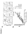
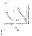


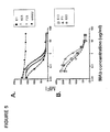
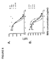
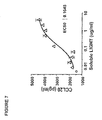

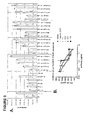


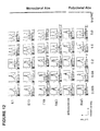

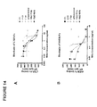
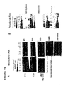
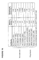

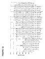
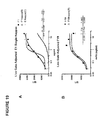

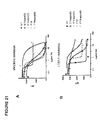


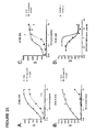


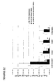
Classifications
-
- A—HUMAN NECESSITIES
- A61—MEDICAL OR VETERINARY SCIENCE; HYGIENE
- A61P—SPECIFIC THERAPEUTIC ACTIVITY OF CHEMICAL COMPOUNDS OR MEDICINAL PREPARATIONS
- A61P1/00—Drugs for disorders of the alimentary tract or the digestive system
-
- C—CHEMISTRY; METALLURGY
- C07—ORGANIC CHEMISTRY
- C07K—PEPTIDES
- C07K16/00—Immunoglobulins [IGs], e.g. monoclonal or polyclonal antibodies
- C07K16/18—Immunoglobulins [IGs], e.g. monoclonal or polyclonal antibodies against material from animals or humans
- C07K16/28—Immunoglobulins [IGs], e.g. monoclonal or polyclonal antibodies against material from animals or humans against receptors, cell surface antigens or cell surface determinants
-
- C—CHEMISTRY; METALLURGY
- C07—ORGANIC CHEMISTRY
- C07K—PEPTIDES
- C07K16/00—Immunoglobulins [IGs], e.g. monoclonal or polyclonal antibodies
- C07K16/18—Immunoglobulins [IGs], e.g. monoclonal or polyclonal antibodies against material from animals or humans
- C07K16/24—Immunoglobulins [IGs], e.g. monoclonal or polyclonal antibodies against material from animals or humans against cytokines, lymphokines or interferons
-
- A—HUMAN NECESSITIES
- A61—MEDICAL OR VETERINARY SCIENCE; HYGIENE
- A61K—PREPARATIONS FOR MEDICAL, DENTAL OR TOILETRY PURPOSES
- A61K39/00—Medicinal preparations containing antigens or antibodies
- A61K39/395—Antibodies; Immunoglobulins; Immune serum, e.g. antilymphocytic serum
-
- A—HUMAN NECESSITIES
- A61—MEDICAL OR VETERINARY SCIENCE; HYGIENE
- A61P—SPECIFIC THERAPEUTIC ACTIVITY OF CHEMICAL COMPOUNDS OR MEDICINAL PREPARATIONS
- A61P1/00—Drugs for disorders of the alimentary tract or the digestive system
- A61P1/04—Drugs for disorders of the alimentary tract or the digestive system for ulcers, gastritis or reflux esophagitis, e.g. antacids, inhibitors of acid secretion, mucosal protectants
-
- A—HUMAN NECESSITIES
- A61—MEDICAL OR VETERINARY SCIENCE; HYGIENE
- A61P—SPECIFIC THERAPEUTIC ACTIVITY OF CHEMICAL COMPOUNDS OR MEDICINAL PREPARATIONS
- A61P29/00—Non-central analgesic, antipyretic or antiinflammatory agents, e.g. antirheumatic agents; Non-steroidal antiinflammatory drugs [NSAID]
-
- A—HUMAN NECESSITIES
- A61—MEDICAL OR VETERINARY SCIENCE; HYGIENE
- A61P—SPECIFIC THERAPEUTIC ACTIVITY OF CHEMICAL COMPOUNDS OR MEDICINAL PREPARATIONS
- A61P31/00—Antiinfectives, i.e. antibiotics, antiseptics, chemotherapeutics
- A61P31/04—Antibacterial agents
-
- A—HUMAN NECESSITIES
- A61—MEDICAL OR VETERINARY SCIENCE; HYGIENE
- A61P—SPECIFIC THERAPEUTIC ACTIVITY OF CHEMICAL COMPOUNDS OR MEDICINAL PREPARATIONS
- A61P31/00—Antiinfectives, i.e. antibiotics, antiseptics, chemotherapeutics
- A61P31/12—Antivirals
-
- A—HUMAN NECESSITIES
- A61—MEDICAL OR VETERINARY SCIENCE; HYGIENE
- A61P—SPECIFIC THERAPEUTIC ACTIVITY OF CHEMICAL COMPOUNDS OR MEDICINAL PREPARATIONS
- A61P37/00—Drugs for immunological or allergic disorders
- A61P37/02—Immunomodulators
- A61P37/06—Immunosuppressants, e.g. drugs for graft rejection
-
- C—CHEMISTRY; METALLURGY
- C07—ORGANIC CHEMISTRY
- C07K—PEPTIDES
- C07K16/00—Immunoglobulins [IGs], e.g. monoclonal or polyclonal antibodies
- C07K16/18—Immunoglobulins [IGs], e.g. monoclonal or polyclonal antibodies against material from animals or humans
- C07K16/28—Immunoglobulins [IGs], e.g. monoclonal or polyclonal antibodies against material from animals or humans against receptors, cell surface antigens or cell surface determinants
- C07K16/2875—Immunoglobulins [IGs], e.g. monoclonal or polyclonal antibodies against material from animals or humans against receptors, cell surface antigens or cell surface determinants against the NGF/TNF superfamily, e.g. CD70, CD95L, CD153, CD154
-
- C—CHEMISTRY; METALLURGY
- C12—BIOCHEMISTRY; BEER; SPIRITS; WINE; VINEGAR; MICROBIOLOGY; ENZYMOLOGY; MUTATION OR GENETIC ENGINEERING
- C12N—MICROORGANISMS OR ENZYMES; COMPOSITIONS THEREOF; PROPAGATING, PRESERVING, OR MAINTAINING MICROORGANISMS; MUTATION OR GENETIC ENGINEERING; CULTURE MEDIA
- C12N15/00—Mutation or genetic engineering; DNA or RNA concerning genetic engineering, vectors, e.g. plasmids, or their isolation, preparation or purification; Use of hosts therefor
- C12N15/09—Recombinant DNA-technology
- C12N15/11—DNA or RNA fragments; Modified forms thereof; Non-coding nucleic acids having a biological activity
-
- G—PHYSICS
- G01—MEASURING; TESTING
- G01N—INVESTIGATING OR ANALYSING MATERIALS BY DETERMINING THEIR CHEMICAL OR PHYSICAL PROPERTIES
- G01N33/00—Investigating or analysing materials by specific methods not covered by groups G01N1/00 - G01N31/00
- G01N33/48—Biological material, e.g. blood, urine; Haemocytometers
- G01N33/50—Chemical analysis of biological material, e.g. blood, urine; Testing involving biospecific ligand binding methods; Immunological testing
- G01N33/53—Immunoassay; Biospecific binding assay; Materials therefor
-
- A—HUMAN NECESSITIES
- A61—MEDICAL OR VETERINARY SCIENCE; HYGIENE
- A61K—PREPARATIONS FOR MEDICAL, DENTAL OR TOILETRY PURPOSES
- A61K39/00—Medicinal preparations containing antigens or antibodies
- A61K2039/505—Medicinal preparations containing antigens or antibodies comprising antibodies
-
- C—CHEMISTRY; METALLURGY
- C07—ORGANIC CHEMISTRY
- C07K—PEPTIDES
- C07K2317/00—Immunoglobulins specific features
- C07K2317/20—Immunoglobulins specific features characterized by taxonomic origin
- C07K2317/21—Immunoglobulins specific features characterized by taxonomic origin from primates, e.g. man
-
- C—CHEMISTRY; METALLURGY
- C07—ORGANIC CHEMISTRY
- C07K—PEPTIDES
- C07K2317/00—Immunoglobulins specific features
- C07K2317/30—Immunoglobulins specific features characterized by aspects of specificity or valency
- C07K2317/34—Identification of a linear epitope shorter than 20 amino acid residues or of a conformational epitope defined by amino acid residues
-
- C—CHEMISTRY; METALLURGY
- C07—ORGANIC CHEMISTRY
- C07K—PEPTIDES
- C07K2317/00—Immunoglobulins specific features
- C07K2317/50—Immunoglobulins specific features characterized by immunoglobulin fragments
- C07K2317/56—Immunoglobulins specific features characterized by immunoglobulin fragments variable (Fv) region, i.e. VH and/or VL
- C07K2317/565—Complementarity determining region [CDR]
-
- C—CHEMISTRY; METALLURGY
- C07—ORGANIC CHEMISTRY
- C07K—PEPTIDES
- C07K2317/00—Immunoglobulins specific features
- C07K2317/70—Immunoglobulins specific features characterized by effect upon binding to a cell or to an antigen
- C07K2317/76—Antagonist effect on antigen, e.g. neutralization or inhibition of binding
Definitions
- antibodies that immunospecifically bind to a human LIGHT (hLIGHT) (lymphotoxin-like, exhibits inducible expression and competes with HSV glycoprotein D for H VEM, a receptor expressed by T lymphocytes) polypeptide, a hLIGHT polypeptide fragment or other hLIGHT epitope.
- the antibodies are fully human antibodies, preferably fully human monoclonal antibodies, that immunospecifically bind to a hLIGHT polypeptide, hLIGHT polypeptide fragment or hLIGHT epitope.
- isolated nucleic acids encoding antibodies that immunospecifically bind to a hLIGHT polypeptide, hLIGHT polypeptide fragment, or hLIGHT epitope are also provided herein.
- the invention further provides vectors and host cells comprising nucleic acids encoding antibodies that immunospecifically bind to a hLIGHT polypeptide, hLIGHT polypeptide fragment, or hLIGHT epitope, as well as methods of making antibodies that immunospecifically bind to a hLIGHT polypeptide, hLIGHT polypeptide fragment, or hLIGHT epitope. Also provided are methods of using the anti-hLIGHT antibodies provided herein to inhibit hLIGHT biological activity in vivo and/or to treat or otherwise manage a hLIGHT-mediated disease in a patient.
- LIGHT (lymphotoxin-like, exhibits inducible expression and competes with HSV glycoprotein D for H VEM, a receptor expressed by T lymphocytes) is one potential cytokine target that has been implicated in the processes of chronic inflammatory autoimmune diseases ( Mauri et al. 1998 Immunity 8 21-30 ).
- TNFSF TNF superfamily
- LIGHT is also known as TNFSF14 or CD258. LIGHT is expressed on the surface ofT cells upon activation in a tightly regulated manner appearing within 4 hours, peaking by 12-24 hours and disappearing by 48 hours ( Castellano et al. 2002 J Biol Chem 277 42841-51 ).
- LIGHT is also present at detectable levels constitutively on the surface of immature dendritic cells ( Tamada et al. 2000 J Immunol 164 4105-10 ) and on T cells and natural killer (NK) cells of the gut ( Cohavy et al. 2005 J Immunol 174 646-53 ). LIGHT mediates its biologic effects by binding three TNF superfamily receptors including the lymphotoxin ⁇ receptor (LT ⁇ R) ( Crowe et al. 1994 Science 264 707-10 , Browning et al. 1997 J Immunol 159 3288-98 ), the herpes virus entry mediator (HVEM) ( Montgomery et al. 1996 Cell 87(3) 427-36 ), and decoy receptor 3 (DcR3) ( Yu et al. 1999 J Biol Chem 274 13733-6 ).
- L ⁇ R lymphotoxin ⁇ receptor
- HVEM herpes virus entry mediator
- DcR3 decoy receptor 3
- mice treated with an inhibitory LT ⁇ R-Fc fusion protein reduced the inflammatory symptoms in the CD4+CD45RB high T cell transfer model of colitis, a CD4+ T cell-mediated pathology ( Mackay et al. 1998 Gastroenterology 115 1464-75 ), Constitutive transgenic T cell specific expression of LIGHT also has been shown to lead to severe intestinal inflammation with autoimmune-like pathology resembling human inflammatory bowel disease (IBD) ( Wang et al. 2005 J Immunol 174 8173-82 , Sheikh et al. 2001 J Immunol 167 6330-7 , Wang et al. 2001 J Immunol 167 5099-105 , Wang et al.
- IBD human inflammatory bowel disease
- LIGHT-expressing lymphocytes can induce IBD-like symptoms (e.g ., cytokine profiles of human Crohn's disease, fissuring ulcers, ileitis, and increases in colonic IFN- ⁇ and TNF) when mesenteric lymph node cells from LIGHT transgenic animals are transferred to RAG-/- ( Wang et al. 2005 J Immunol 174 8173-82 ).
- IBD-like symptoms e.g cytokine profiles of human Crohn's disease, fissuring ulcers, ileitis, and increases in colonic IFN- ⁇ and TNF
- CCL20-CCR6 signaling has been shown to be involved in IBD, and LIGHT induces CCL20 secretion from the human colonic epithelial cell line HT29.14s.
- LIGHT induces CCL20 secretion from the human colonic epithelial cell line HT29.14s.
- epithelial cells of the colon have been found to be a major source of CCL20 in IBD patients and CCL20 expression is increased in human IBD patients ( Kwon et al. 2002 Gut 51 818-26 ; ( Kaser et al. 2004 J Clin Immunol 24 74-85 ).
- hLIGHT has also been implicated in graft-versus-host disease (GVHD).
- LIGHT has been shown to provide potent costimulatory activity for T cells, enhancing proliferation an the production of Th1 cytokines independent of the B7-CD28 pathway (see, e.g., Tamada et al. 2000 J. Immunol. 164 4105-4110 ).
- Blocking of LIGHT-HVEM costimulation by either anti-HVEM monoclonal antibodies, HVEM-Ig, or LT ⁇ R fusion protein inhibits allogeneic T cell responses (see, e.g ., Tamada et al. 2000 J. Immunol. 164 4105-4110 ., Harrop et al.
- LT ⁇ R-Ig or murine anti-LIGHT antibodies inhibits anti-host cytotoxic T lymphocyte (CTL) responses in a murine acute GVHD model ( Tamada et al. 2000 Nat. Med. 6 283-289 ).
- CTL cytotoxic T lymphocyte
- antibodies such as fully human antibodies, that immunospecifically bind to a hLIGHT polypeptide, a hLIGHT polypeptide fragment, or a hLIGHT epitope.
- isolated nucleic acids encoding antibodies such as fully human antibodies, that immunospecifically bind to a hLIGHT polypeptide, a hLIGHT polypeptide fragment, or a hLIGHT epitope.
- vectors and host cells comprising nucleic acids encoding antibodies, such as fully human antibodies, that immunospecifically bind to a hLIGHT polypeptide, a hLIGHT polypeptide fragment, or a hLIGHT epitope.
- antibodies such as fully human antibodies, that immunospecifically bind to a hLIGHT polypeptide, a hLIGHT polypeptide fragment, or a hLIGHT epitope.
- a method of treating a hLIGHT-mediated disease comprising administering an antibody, such as a fully human antibody, that immunospecifically binds to a hLIGHT polypeptide, a hLIGHT polypeptide fragment, or a hLIGHT epitope.
- anti-hLIGHT antibodies provided herein are antagonist antibodies that ameliorate, neutralize or otherwise inhibit LIGHT biological activity in vivo ( e .
- an antibody of the invention can be a full-length antibody or an antigen-binding antibody fragment.
- the antibodies of the invention are also useful for detecting hLIGHT, as well as for ameliorating, neutralizing or otherwise inhibiting hLIGHT activity, e . g ., in a human subject suffering from a disorder in which hLIGHT activity is detrimental.
- an isolated antibody that immunospecifically binds to a hLIGHT polypeptide (e.g ., a cell surface-expressed or soluble hLIGHT), a hLIGHT polypeptide fragment, or a hLIGHT epitope.
- a hLIGHT polypeptide e.g ., a cell surface-expressed or soluble hLIGHT
- a hLIGHT polypeptide fragment e.g., a cell surface-expressed or soluble hLIGHT
- a hLIGHT epitope e.g a cell surface-expressed or soluble hLIGHT
- the antibody immunospecifically binds to (a) a trimeric (or native) hLIGHT epitope, (b) a monomeric (or denatured) hLIGHT epitope, (c) both a trimeric hLIGHT epitope and a monomeric hLIGHT epitope, (d) a trimeric hLIGHT epitope but not a monomeric hLIGHT epitope, or (e) a monomeric hLIGHT epitope but not a trimeric hLIGHT epitope.
- the antibody immunospecifically binds a trimeric hLIGHT epitope but not a human monomeric hLIGHT epitope.
- the antibody is an E1 antibody, E13 antibody, E63 antibody, F19 antibody, or F23 antibody.
- Hybridomas that produce each of the E1, E13, E63, F19 and F23 antibodies were deposited under provisions of the Budapest Treaty with the American Type Culture Collection (ATCC, 10801 University Boulevard., Manassas, VA 20110-2209) on July 12, 2006 (ATCC Accession Nos. PTA-7729 (hybridoma 124 E1) and PTA-7728 (hybridoma 124 F23), respectively), August 17, 2006 (ATCC Accession Nos. PTA-7818 (hybridoma 124 E63) and PTA-7819 (hybridoma 124 F19) and August 23, 2006 (ATCC Accession No. PTA-7842 (hybridoma 124 E13), and are herein incorporated by reference.
- Antibodies produced by each of hybridomas 124 E1, 124 E13, 124 E63, 124 F19 and 124 F23 may also be referred to herein as E1, E13, E63, F19, F23, respectively, and/or by the ATCC Accession Numbers. PTA-7729, PTA-7842, PTA-7818, PTA-7819, and PTA-7728, respectively.
- an antibody of the invention is one that is competitively blocked ( e.g ., in a dose-dependent manner) by an E1 antibody, an E13 antibody, and/or an E63 antibody for binding to a hLIGHT polypeptide ( e.g ., a cell surface-expressed or soluble hLIGHT), a hLIGHT fragment, or a hLIGHT epitope.
- a hLIGHT polypeptide e.g ., a cell surface-expressed or soluble hLIGHT
- a hLIGHT fragment e.g., a cell surface-expressed or soluble hLIGHT fragment
- a hLIGHT epitope e.g a cell surface-expressed or soluble hLIGHT
- the antibody is competitively blocked ( e.g ., in a dose-dependent manner) by an F19 antibody and/or an F23 antibody for binding to a hLIGHT polypeptide ( e .
- the antibody is competitively blocked ( e.g ., in a dose-dependent manner) by an E1 antibody, an E13 antibody and/or an E63 antibody, but is not competitively blocked ( e . g ., in a dose-dependent manner) by an F19 antibody and/or an F23 antibody, for binding to a hLIGHT polypeptide ( e . g ., a cell surface-expressed or soluble hLIGHT), a hLIGHT fragment, or a hLIGHT epitope.
- a hLIGHT polypeptide e . g ., a cell surface-expressed or soluble hLIGHT
- a hLIGHT fragment e.g ., a cell surface-expressed or soluble hLIGHT fragment
- a hLIGHT epitope e.g ., a cell surface-expressed or soluble hLIGHT
- the antibody is competitively blocked ( e . g ., in a dose-dependent manner) by an F19 antibody and/or an F23 antibody, but is not competitively blocked ( e . g ., in a dose-dependent manner) by an E1 antibody, an E13 antibody and/or an E63 antibody, for binding to a hLIGHT polypeptide ( e . g ., a cell surface-expressed or soluble hLIGHT), a hLIGHT fragment, or a hLIGHT epitope.
- a hLIGHT polypeptide e . g ., a cell surface-expressed or soluble hLIGHT
- hLIGHT fragment e hLIGHT fragment
- a hLIGHT epitope e. hLIGHT epitope.
- antibodies, or antigen binding antibody fragments thereof that immunospecifically bind to a hLIGHT polypeptide (e . g ., a cell surface-expressed or soluble hLIGHT), a hLIGHT polypeptide fragment, or a hLIGHT epitope.
- the antibody or antigen-binding fragment comprises a VH chain, VL chain, VH domain, VL domain, VH CDR1, VH CDR2, VH CDR3, VL CDR1, VL CDR2, and/or VL CDR3 of E1, E13, E63, F19 or F23.
- the antibody or antigen binding fragment thereof comprises less than six CDRs. In some embodiments, the antibody or antigen binding fragment thereof comprises or consists of one, two, three, four, or five CDRs selected from the group consisting of VH CDR1, VH CDR2, VH CDR3, VL CDR1, VL CDR2, and/or VL CDR3. In specific embodiments, the antibody or antigen binding fragment thereof comprises or consists of one, two, three, four, or five CDRs selected from the group consisting of VH CDR1, VH CDR2, VH CDR3, VL CDR1, VL CDR2, and/or VL CDR3 of E1, E13, E63, F19 or F23.
- compositions comprising an anti-hLIGHT antibody of the invention, such as E1, E13, E63, F19 or F23.
- an antibody of the invention is a fully human antibody, a monoclonal antibody, a recombinant antibody, an antagonist antibody, or any combination thereof.
- the antibody is a fully human antibody, such as a fully human monoclonal antibody, or antigen binding fragment thereof, that immunospecifically binds to hLIGHT.
- the antibody is an antagonist antibody.
- the antibody competes ( e . g ., in a dose dependent manner) with HVEM, LT ⁇ R, DcR3, or fusion proteins thereof (e.g ., Fc:HVEM, Fc: LT ⁇ R or Fc:DcR3), for binding to a hLIGHT polypeptide, such as a cell surface-expressed hLIGHT polypeptide or soluble hLIGHT polypeptide, a hLIGHT polypeptide fragment, or a hLIGHT epitope.
- a hLIGHT polypeptide such as a cell surface-expressed hLIGHT polypeptide or soluble hLIGHT polypeptide, a hLIGHT polypeptide fragment, or a hLIGHT epitope.
- nucleic acids encoding antibodies that immunospecifically bind to a hLIGHT polypeptide (e . g ., a cell surface-expressed or soluble hLIGHT), a hLIGHT polypeptide fragment, or a hLIGHT epitope.
- a hLIGHT polypeptide e . g ., a cell surface-expressed or soluble hLIGHT
- the nucleic acid encodes a VH chain, VL chain, VH domain, VL domain, VH CDR1, VH CDR2, VH CDR3, VL CDR1, VL CDR2, and/or VL CDR3 of E1, E13, E63, F19 or F23.
- vectors and host cells comprising nucleic acids encoding antibodies that immunospecifically bind to a hLIGHT polypeptide, a hLIGHT polypeptide fragment, or a hLIGHT epitope.
- the antibody immunospecifically binds to a single nucleotide polymorphism (SNP) variant of a hLIGHT polypeptide, such as 214E-32S, 214K-32S, 214E-32L and/or 214K-32L.
- SNP single nucleotide polymorphism
- hybridomas that produce antibodies that immunospecifically bind to a hLIGHT polypeptide, or SNP variant thereof.
- the hybridoma is the hybridoma that produces E1, E13, E63, F19 or F23.
- a hLIGHT-mediated disease in a subject (e.g ., a human subject), comprising administering to the subject an effective amount of an antibody that immunospecifically binds to a hLIGHT polypeptide (e.g ., a cell surface-expressed or soluble hLIGHT), wherein hLIGHT activity is inhibited by the antibody.
- a hLIGHT polypeptide e.g ., a cell surface-expressed or soluble hLIGHT
- the hLIGHT-mediated disease is an IBD, such as Crohn's disease or ulcerative colitis.
- the hLIGHT mediated disease is GVHD.
- hLIGHT hLIGHT to HVEM, LT ⁇ R and/or DcR3 in a subject ( e . g ., a human subject), comprising administering to the subject an effective amount of an antibody that immunospecifically binds to a hLIGHT polypeptide (e.g ., a cell surface-expressed or soluble hLIGHT).
- a hLIGHT polypeptide e.g ., a cell surface-expressed or soluble hLIGHT
- a hLIGHT biological activity such as secretion of CCL20, IL8 and/or RANTES
- a subject e.g ., a human subject
- administering comprising administering to the subject an effective amount of an antibody that immunospecifically binds to a hLIGHT polypeptide (e . g ., a cell surface-expressed hLIGHT), wherein hLIGHT biological activity is decreased or inhibited by the antibody.
- a hLIGHT biological activity such as secretion of CCL20, IL8 and/or RANTES
- hLIGHT polypeptide e . g ., a cell surface-expressed or soluble hLIGHT
- a hLIGHT polypeptide such as a hLIGHT polypeptide, a hLIGHT polypeptide fragment, or a hLIGHT epitope.
- a hLIGHT biological activity such as secretion of CCL20, IL8 and/or RANTES, in a cell having a cell surface-expressed hLIGHT receptor (such as, HVEM, LT ⁇ R and/or Dc3R)
- a cell surface-expressed hLIGHT receptor such as, HVEM, LT ⁇ R and/or Dc3R
- an antibody that immunospecifically binds to a hLIGHT polypeptide e.g ., a cell surface-expressed or soluble hLIGHT, or SNP variant thereof
- hLIGHT biological activity is decreased or inhibited by the antibody.
- an antibody that immunospecifically binds to a hLIGHT polypeptide, a hLIGHT polypeptide fragment, or a hLIGHT epitope, wherein said antibody further comprises a detectable tag.
- anti-hLIGHT antibodies that comprise a detectable tag are used in a method for the detection of hLIGHT in a sample, said method comprising contacting the sample with the anti-hLIGHT antibody.
- the sample comprises a cell expressing hLIGHT on the surface of the cell.
- kits comprising an antibody that immunospecifically binds to a hLIGHT polypeptide, a hLIGHT polypeptide fragment, or a hLIGHT epitope.
- administer refers to the act of injecting or otherwise physically delivering a substance as it exists outside the body (e . g ., an anti-hLIGHT antibody provided herein) into a patient, such as by mucosal, intradermal, intravenous, intramuscular delivery and/or any other method of physical delivery described herein or known in the art.
- a disease, or a symptom thereof is being treated, administration of the substance typically occurs after the onset of the disease or symptoms thereof.
- administration of the substance typically occurs before the onset of the disease or symptoms thereof.
- analog refers to a polypeptide that possesses a similar or identical function as a hLIGHT polypeptide, a fragment of a hLIGHT polypeptide, or an anti-hLIGHT antibody but does not necessarily comprise a similar or identical amino acid sequence of a hLIGHT polypeptide, a fragment of a hLIGHT polypeptide, or an anti-hLIGHT antibody, or possess a similar or identical structure of a hLIGHT polypeptide, a fragment of a hLIGHT polypeptide, or an anti-hLIGHT antibody.
- a polypeptide that has a similar amino acid sequence refers to a polypeptide that satisfies at least one of the following: (a) a polypeptide having an amino acid sequence that is at least 30%, at least 35%, at least 40%, at least 45%, at least 50%, at least 55%, at least 60%, at least 65%, at least 70%, at least 75%, at least 80%, at least 85%, and preferably at least 90%, more preferably at least 95%, or most preferably at least 99% identical to the amino acid sequence of a hLIGHT polypeptide ( e .
- a polypeptide with similar structure to a hLIGHT polypeptide, a fragment of a hLIGHT polypeptide, or an anti-hLIGHT antibody described herein refers to a polypeptide that has a similar secondary, tertiary or quaternary structure of a hLTGHT polypeptide, a fragment of a hLIGHT, or a hLIGHT antibody described herein.
- the structure of a polypeptide can determined by methods known to those skilled in the art, including but not limited to, X-ray crystallography, nuclear magnetic resonance, and crystallographic electron microscopy.
- the sequences are aligned for optimal comparison purposes (e.g ., gaps can be introduced in the sequence of a first amino acid or nucleic acid sequence for optimal alignment with a second amino acid or nucleic acid sequence).
- the amino acid residues or nucleotides at corresponding amino acid positions or nucleotide positions are then compared. When a position in the first sequence is occupied by the same amino acid residue or nucleotide as the corresponding position in the second sequence, then the molecules are identical at that position.
- the determination of percent identity between two sequences can also be accomplished using a mathematical algorithm.
- a preferred, non-limiting example of a mathematical algorithm utilized for the comparison of two sequences is the algorithm of Karlin and Altschul, 1990, Proc. Natl. Acad. Sci. U.S.A. 87:2264 2268 , modified as in Karlin and Altschul, 1993, Proc. Natl. Acad. Sci. U.S.A. 90:5873 5877 .
- Such an algorithm is incorporated into the NBLAST and XBLAST programs of Altschul et al., 1990, J. Mol. Biol. 215:403 .
- Gapped BLAST can be utilized as described in Altschul et al., 1997, Nucleic Acids Res. 25:3389 3402 .
- PSI BLAST can be used to perform an iterated search which detects distant relationships between molecules ( Id .).
- the default parameters of the respective programs e.g ., of XBLAST and NBLAST
- NCBI National Center for Biotechnology Information
- Another preferred, non limiting example of a mathematical algorithm utilized for the comparison of sequences is the algorithm of Myers and Miller, 1988, CABIOS 4:11 17 .
- Such an algorithm is incorporated in the ALIGN program (version 2.0) which is part of the GCG sequence alignment software package.
- ALIGN program version 2.0
- the percent identity between two sequences can be determined using techniques similar to those described above, with or without allowing gaps. In calculating percent identity, typically only exact matches are counted.
- an "antagonist” or “inhibitor” of hLIGHT refers to a molecule that is capable of inhibiting or otherwise decreasing one or more of the biological activities of hLIGHT, such as in a cell expressing hLIGHT or in a cell expressing a hLIGHT ligand, such as a hLIGHT receptor.
- antibodies of the invention are antagonist antibodies that inhibit or otherwise decrease secretion of CCL20, IL-8 and/or RANTES from a cell having a cell surface-expressed hLIGHT receptor (e.g ., HVEM, LT ⁇ R and/or DcR3) when said antibody is contacted with said cell.
- an antagonist of hLIGHT may, for example, act by inhibiting or otherwise decreasing the activation and/or cell signaling pathways of the cell expressing a hLIGHT receptor, thereby inhibiting a hLIGHT-mediated biological activity of the cell the relative to the hLIGHT-mediated biological activity in the absence of antagonist.
- the antibodies provided herein are fully human, antagonistic anti-hLIGHT antibodies, preferably fully human, monoclonal, antagonistic anti-hLIGHT antibodies.
- antibody and "immunoglobulin” or “Ig” may be used interchangeably herein.
- the terms “antibodies that immunospecifically bind to a hLIGHT antigen,” “antibodies that immunospecifically bind to a hLIGHT epitope,” “anti-hLIGHT antibodies” and analogous terms are also used interchangeably herein and refer to antibodies and fragments thereof, that specifically bind to a hLIGHT polypeptide, such as a hLIGHT antigen or epitope.
- An antibody or a fragment thereof that immunospecifically binds to a hLIGHT antigen may be cross-reactive with related antigens.
- an antibody or a fragment thereof that immunospecifically binds to a hLIGHT antigen does not cross-react with other antigens.
- An antibody or a fragment thereof that immunospecifically binds to a hLIGHT antigen can be identified, for example, by immunoassays, BIAcore, or other techniques known to those of skill in the art.
- An antibody or a fragment thereof binds specifically to a hLIGHT antigen when it binds to a hLIGHT antigen with higher affinity than to any cross-reactive antigen as determined using experimental techniques, such as radioimmunoassays (RIA) and enzyme-linked immunosorbent assays (ELISAs).
- RIA radioimmunoassays
- ELISAs enzyme-linked immunosorbent assays
- a specific or selective reaction will be at least twice background signal or noise and more typically more than 10 times background. See, e.g ., Paul, ed., 1989, Fundamental Immunology Second Edition, Raven Press, New York at pages 332-336 for a discussion regarding antibody specificity.
- Antibodies of the invention include, but are not limited to, synthetic antibodies, monoclonal antibodies, recombinantly produced antibodies, multispecific antibodies (including bi-specific antibodies), human antibodies, humanized antibodies, chimeric antibodies, intrabodies, single-chain Fvs (scFv) ( e . g ., including monospecific, bispecific, etc .), camelized antibodies, Fab fragments, F(ab') fragments, disulfide-linked Fvs (sdFv), anti-idiotypic (anti-Id) antibodies, and epitope-binding fragments of any of the above.
- antibodies of the present invention include immunoglobulin molecules and immunologically active portions of immunoglobulin molecules, i .
- the antibodies of the invention can be of any type (e . g ., IgG, IgE, IgM, IgD, IgA and IgY), any class ( e.g ., IgG1, IgG2, IgG3, IgG4, IgA1 and IgA2), or any subclass ( e.g ., IgG2a and IgG2b) of immunoglobulin molecule.
- the hLIGHT antibodies are fully human, such as fully human monoclonal hLIGHT antibodies.
- antibodies of the invention are IgG antibodies, or a class ( e . g ., human IgG1 or IgG4) or subclass thereof.
- antigen binding domain refers to that portion of an antibody which comprises the amino acid residues that interact with an antigen and confer on the binding agent its specificity and affinity for the antigen (e . g ., the complementarity determining regions (CDR)).
- the antigen binding region can be derived from any animal species, such as rodents ( e.g ., rabbit, rat or hamster) and humans. Preferably, the antigen binding region will be of human origin.
- composition is intended to encompass a product containing the specified ingredients (e.g ., an antibody of the invention) in, optionally, the specified amounts, as well as any product which results, directly or indirectly, from combination of the specified ingredients in, optionally, the specified amounts.
- constant region or “constant domain” refers to a carboxy terminal portion of the light and heavy chain which is not directly involved in binding of the antibody to antigen but exhibits various effector function, such as interaction with the Fc receptor.
- the terms refer to the portion of an immunoglobulin molecule having a more conserved amino acid sequence relative to the other portion of the immunoglobulin, the variable domain, which contains the antigen binding site.
- the constant domain contains the CH1, CH2 and CH3 domains of the heavy chain and the CHL domain of the light chain.
- derivative refers to a polypeptide that comprises an amino acid sequence of a hLIGHT polypeptide, a fragment of a hLIGHT polypeptide, or an antibody that immunospecifically binds to a hLIGHT polypeptide which has been altered by the introduction of amino acid residue substitutions, deletions or additions.
- derivative also refers to a hLIGHT polypeptide, a fragment of a hLIGHT polypeptide, or an antibody that immunospecifically binds to a hLIGHT polypeptide which has been chemically modified, e.g ., by the covalent attachment of any type of molecule to the polypeptide.
- a hLIGHT polypeptide, a fragment of a hLIGHT polypeptide, or a hLIGHT antibody may be chemically modified, e.g ., by glycosylation, acetylation, pegylation, phosphorylation, amidation, derivatization by known protecting/blocking groups, proteolytic cleavage, linkage to a cellular ligand or other protein, etc.
- the derivatives are modified in a manner that is different from naturally occurring or starting peptide or polypeptides, either in the type or location of the molecules attached. Derivatives further include deletion of one or more chemical groups which are naturally present on the peptide or polypeptide.
- a derivative of a hLIGHT polypeptide, a fragment of a hLIGHT polypeptide, or a hLIGHT antibody may be chemically modified by chemical modifications using techniques known to those of skill in the art, including, but not limited to specific chemical cleavage, acetylation, formulation, metabolic synthesis of tunicamycin, etc . Further, a derivative of a hLIGHT polypeptide, a fragment of a hLIGHT polypeptide, or a hLIGHT antibody may contain one or more non-classical amino acids.
- a polypeptide derivative possesses a similar or identical function as a hLIGHT polypeptide, a fragment of a hLIGHT polypeptide, or a hLIGHT antibody described herein.
- an effective amount refers to the amount of a therapy (e.g ., an antibody or pharmaceutical composition provided herein) which is sufficient to reduce and/or ameliorate the severity and/or duration of a given disease and/or a symptom related thereto. This term also encompasses an amount necessary for the reduction or amelioration of the advancement or progression of a given disease, reduction or amelioration of the recurrence, development or onset of a given disease, and/or to improve or enhance the prophylactic or therapeutic effect(s) of another therapy (e.g ., a therapy other than anti-hLIGHT antibody provided herein).
- a therapy e.g ., an antibody or pharmaceutical composition provided herein
- the effective amount of an antibody of the invention is from about 0.1 mg/kg (mg of antibody per kg weight of the subject) to about 100 mg/kg.
- an effective amount of an antibody provided therein is about 0.1 mg/kg, about 0.5 mg/kg, about 1 mg/kg, 3 mg/kg, 5 mg/kg, about 10 mg/kg, about 15 mg/kg, about 20 mg/kg, about 25 mg/kg, about 30 mg/kg, about 35 mg/kg, about 40 mg/kg, about 45 mg/kg, about 50 mg/kg, about 60 mg/kg, about 70 mg/kg, about 80 mg/kg about 90 mg/kg or about 100 mg/kg (or a range therein).
- "effective amount” as used herein also refers to the amount of an antibody of the invention to achieve a specified result (e.g ., inhibition of a hLIGHT biological activity of a cell, such as inhibition of secretion of CCL20, IL-8 or RANTES from the cell).
- epitope refers to a localized region on the surface of an antigen, such as hLIGHT polypeptide or hLIGHT polypeptide fragment, that is capable of being bound to one or more antigen binding regions of an antibody, and that has antigenic or immunogenic activity in an animal, preferably a mammal, and most preferably in a human, that is capable of eliciting an immune response.
- An epitope having immunogenic activity is a portion of a polypeptide that elicits a antibody response in an animal.
- An epitope having antigenic activity is a portion of a polypeptide to which an antibody immunospecifically binds as determined by any method well known in the art, for example, by the immunoassays described herein.
- Antigenic epitopes need not necessarily be immunogenic. Epitopes usually consist of chemically active surface groupings of molecules such as amino acids or sugar side chains and have specific three dimensional structural characteristics as well as specific charge characteristics. A region of a polypeptide contributing to an epitope may be contiguous amino acids of the polypeptide or the epitope may come together from two or more non-contiguous regions of the polypeptide The epitope may or may not be a three-dimensional surface feature of the antigen. In certain embodiments, a hLIGHT epitope is a three-dimensional surface feature of a hLIGHT polypeptide ( e.g ., in a trimeric form of a hLIGHT polypeptide).
- a hLIGHT epitope is linear feature of a hLIGHT polypeptide ( e.g ., in a trimeric form or monomeric form of the hLIGHT polypeptide).
- Antibodies provided herein may immunospecifically bind to an epitope of the monomeric (denatured) form of hLIGHT, an epitope of the trimeric (native) form of hLIGHT, or both the monomeric (denatured) form and the trimeric (native) form of hLIGHT.
- the antibodies provided herein immunospecifically bind to an epitope of the trimeric form of hLIGHT but do not immunospecifically bind the monomeric form of hLIGHT.
- excipients refers to inert substances which are commonly used as a diluent, vehicle, preservatives, binders, or stabilizing agent for drugs and includes, but not limited to, proteins (e.g ., serum albumin, etc .), amino acids (e . g ., aspartic acid, glutamic acid, lysine, arginine, glycine, histidine, etc .), fatty acids and phospholipids ( e . g ., alkyl sulfonates, caprylate, etc .), surfactants ( e .
- proteins e.g ., serum albumin, etc .
- amino acids e. g ., aspartic acid, glutamic acid, lysine, arginine, glycine, histidine, etc .
- fatty acids and phospholipids e . g ., alkyl sulfonates, caprylate, etc .
- fragment refers to a peptide or polypeptide that comprises less than the full length amino acid sequence. Such a fragment may arise, for example, from a truncation at the amino terminus, a truncation at the carboxy terminus, and/or an internal deletion of a residue(s) from the amino acid sequence. Fragments may, for example, result from alternative RNA splicing or from in vivo protease activity.
- hLIGHT fragments include polypeptides comprising an amino acid sequence of at least 5 contiguous amino acid residues, at least 10 contiguous amino acid residues, at least 15 contiguous amino acid residues, at least 20 contiguous amino acid residues, at least 25 contiguous amino acid residues, at least 40 contiguous amino acid residues, at least 50 contiguous amino acid residues, at least 60 contiguous amino residues, at least 70 contiguous amino acid residues, at least 80 contiguous amino acid residues, at least 90 contiguous amino acid residues, at least contiguous 100 amino acid residues, at least 125 contiguous amino acid residues, at least 150 contiguous amino acid residues, at least 175 contiguous amino acid residues, at least 200 contiguous amino acid residues, or at least 250 contiguous amino acid residues of the amino acid sequence of a hLIGHT polypeptide or an antibody that immunospecifically binds to a hLIGHT polypeptide.
- a specific embodiment a
- Fully human antibody or “human antibody” are used interchangeably herein and refer to an antibody that comprises a human variable region and, most preferably a human constant region. In specific embodiments, the terms refer to an antibody that comprises a variable region and constant region of human origin.
- Fully human anti-hLIGHT antibodies in certain embodiments, can also encompass antibodies which bind hLIGHT polypeptides and are encoded by nucleic acid sequences which are naturally occurring somatic variants of human germline immunoglobulin nucleic acid sequence. In a specific embodiment, the anti-hLIGHT antibodies provided herein are fully human antibodies.
- the term "fully human antibody” includes antibodies having variable and constant regions corresponding to human germline immunoglobulin sequences as described by Kabat et al.
- recombinant human antibody includes human antibodies that are prepared, expressed, created or isolated by recombinant means, such as antibodies expressed using a recombinant expression vector transfected into a host cell, antibodies isolated from a recombinant, combinatorial human antibody library, antibodies isolated from an animal ( e.g ., a mouse or cow) that is transgenic and/or transchromosomal for human immunoglobulin genes (see e.g., Taylor, L. D. et al. (1992) Nucl. Acids Res. 20:6287-6295 ) or antibodies prepared, expressed, created or isolated by any other means that involves splicing of human immunoglobulin gene sequences to other DNA sequences.
- recombinant means such as antibodies expressed using a recombinant expression vector transfected into a host cell, antibodies isolated from a recombinant, combinatorial human antibody library, antibodies isolated from an animal ( e.g ., a mouse or cow) that is transgenic and/or
- Such recombinant human antibodies can have variable and constant regions derived from human germline immunoglobulin sequences (See Kabat, E. A. et al. (1991) Sequences of Proteins of Immunological Interest, Fifth Edition, U.S. Department of Health and Human Services, NIH Publication No. 91-3242 ).
- such recombinant human antibodies are subjected to in vitro mutagenesis (or, when an animal transgenic for human Ig sequences is used, in vivo somatic mutagenesis) and thus the amino acid sequences of the VH and VL regions of the recombinant antibodies are sequences that, while derived from and related to human germline VH and VL sequences, may not naturally exist within the human antibody germline repertoire in vivo.
- fusion protein refers to a polypeptide that comprises an amino acid sequence of an antibody and an amino acid sequence of a heterologous polypeptide or protein (i . e ., a polypeptide or protein not normally a part of the antibody (e.g ., a non-anti-hLIGHT antigen antibody)).
- fusion when used in relation to hLIGHT or to a anti-hLIGHT antibody refers to the joining of a peptide or polypeptide, or fragment, variant and/or derivative thereof, with a heterologous peptide or polypeptide.
- the fusion protein retains the biological activity of the hLIGHT or anti-hLIGHT antibody.
- the fusion protein comprises a hLIGHT antibody VH domain, VL domain, VH CDR (one, two or three VH CDRs), and/or VL CDR (one, two or three VL CDRs), wherein the fusion protein immunospecifically binds to a hLIGHT epitope.
- heavy chain when used in reference to an antibody refers to five distinct types, called alpha ( ⁇ ), delta ( ⁇ ), epsilon ( ⁇ ), gamma ( ⁇ ) and mu ( ⁇ ), based on the amino acid sequence of the heavy chain constant domain.
- These distinct types of heavy chains are well known and give rise to five classes of antibodies, IgA, IgD, IgE, IgG and IgM, respectively, including four subclasses of IgG, namely IgG1, IgG1, IgG3 and IgG4,
- the heavy chain is a human heavy chain.
- host refers to an animal, preferably a mammal, and most preferably a human.
- host cell refers to the particular subject cell transfected with a nucleic acid molecule and the progeny or potential progeny of such a cell. Progeny of such a cell may not be identical to the parent cell transfected with the nucleic acid molecule due to mutations or environmental influences that may occur in succeeding generations or integration of the nucleic acid molecule into the host cell genome.
- immunomodulatory agent and variations thereof including, but not limited to, immunomodulatory agents, as used herein refer to an agent that modulates a host's immune system.
- an immunomodulatory agent is an immunosuppressant agent.
- an immunomodulatory agent is an immunostimulatory agent.
- an immunomodulatory agent used in the combination therapies of the invention does not include an anti-hLIGHT antibody or antigen-binding fragment.
- Immunomodulatory agents include, but are not limited to, small molecules, peptides, polypeptides, proteins, fusion proteins, antibodies, inorganic molecules, mimetic agents, and organic molecules.
- the term "in combination" in the context of the administration of other therapies refers to the use of more than one therapy.
- the use of the term “in combination” does not restrict the order in which therapies are administered to a subject with an infection.
- a first therapy can be administered before ( e . g ., 1 minute, 45 minutes, 30 minutes, 45 minutes, 1 hour, 2 hours, 4 hours, 6 hours, 12 hours, 24 hours, 48 hours, 72 hours, 96 hours, 1 week, 2 weeks, 3 weeks, 4 weeks, 5 weeks, 6 weeks, 8 weeks, or 12 weeks), concurrently, or after ( e .
- the antibodies of the invention can be administered in combination with one or more therapies ( e . g ., therapies that are not the antibodies of the invention that are currently administered to prevent, treat, manage, and/or ameliorate a hLIGHT-mediated disease.
- Non-limiting examples of therapies that can be administered in combination with an antibody of the invention include analgesic agents, anesthetic agents, antibiotics, or immunomodulatory agents or any other agent listed in the U.S. Pharmacopoeia and/or Physician's Desk Reference.
- inorganic salt refers to any compounds containing no carbon that result from replacement of part or all of the acid hydrogen or an acid by a metal or a group acting like a metal and are often used as a tonicity adjusting compound in pharmaceutical compositions and preparations of biological materials.
- the most common inorganic salts are NaCl, KCl, NaH 2 PO 4 , etc .
- an “isolated” or “purified” antibody is substantially free of cellular material or other contaminating proteins from the cell or tissue source from which the antibody is derived, or substantially free of chemical precursors or other chemicals when chemically synthesized.
- the language “substantially free of cellular material” includes preparations of an antibody in which the antibody is separated from cellular components of the cells from which it is isolated or recombinantly produced.
- an antibody that is substantially free of cellular material includes preparations of antibody having less than about 30%, 20%, 10%, or 5% (by dry weight) of heterologous protein (also referred to herein as a "contaminating protein").
- heterologous protein also referred to herein as a "contaminating protein”
- culture medium represents less than about 20%, 10%, or 5% of the volume of the protein preparation.
- the antibody is produced by chemical synthesis, it is preferably substantially free of chemical precursors or other chemicals, i . e ., it is separated from chemical precursors or other chemicals which are involved in the synthesis of the protein. Accordingly such preparations of the antibody have less than about 30%, 20%, 10%, 5% (by dry weight) of chemical precursors or compounds other than the antibody of interest.
- antibodies of the invention are isolated or purified.
- nucleic acid molecule is one which is separated from other nucleic acid molecules which are present in the natural source of the nucleic acid molecule.
- an "isolated" nucleic acid molecule such as a cDNA molecule, can be substantially free of other cellular material, or culture medium when produced by recombinant techniques, or substantially free of chemical precursors or other chemicals when chemically synthesized.
- a nucleic acid molecule(s) encoding an antibody of the invention is isolated or purified.
- human LIGHT refers to the polypeptides ("polypeptides,” “peptides” and “proteins” are used interchangeably herein) comprising the amino acid sequence of SEQ ID NO:52 and related polypeptides, including SNP variants thereof.
- Related polypeptides include allelic variants ( e . g ., SNP variants); splice variants; fragments; derivatives; substitution, deletion, and insertion variants; fusion polypeptides; and interspecies homologs, preferably, which retain hLIGHT activity and/or are sufficient to generate an anti-hLIGHT immune response.
- Exemplary non-synonymous SNP variants include, but are not limited to, hLIGHT polypeptides comprising 214E-32S (a glutamic acid at position 214 and serine at position 32 of a hLIGHT polypeptide ( e.g ., the hLIGHT polypeptide depicted in SEQ ID NO:52)), 214K-32S, 214E-32L and 214E-32L. Also encompassed are soluble forms of hLIGHT which are sufficient to generate an anti-hLIGHT immunological response ( see, e . g ., SEQ ID NO:53 and SEQ ID NO:54).
- an anti-hLIGHT antibody of the invention can bind to a hLIGHT polypeptide, polypeptide fragment, antigen, and/or epitope, as an epitope is part of the larger antigen, which is part of the larger polypeptide fragment, which, in turn, is part of the larger polypeptide.
- hLIGHT can exist in a trimeric (native) or monomeric (denatured) form.
- Kabat numbering and like terms are recognized in the art and refer to a system of numbering amino acid residues which are more variable ( i . e . hypervariable) than other amino acid residues in the heavy and light chain variable regions of an antibody, or an antigen binding portion thereof ( Kabat et al. (1971) Ann. NY Acad. Sci. 190:382-391 and, Kabat et al. (1991) Sequences of Proteins of Immunological Interest, Fifth Edition, U.S. Department of Health and Human Services, NIH Publication No.
- the hypervariable region typically ranges from amino acid positions 31 to 35 for CDR1, amino acid positions 50 to 65 for CDR2, and amino acid positions 95 to 102 for CDR3.
- the hypervariable region typically ranges from amino acid positions 24 to 34 for CDR1, amino acid positions 50 to 56 for CDR2, and amino acid positions 89 to 97 for CDR3.
- light chain when used in reference to an antibody refers to two distinct types, called kappa ( ⁇ ) of lambda ( ⁇ ) based on the amino acid sequence of the constant domains. Light chain amino acid sequences are well known in the art. In preferred embodiments, the light chain is a human light chain.
- a subject is administered one or more therapies (e . g ., prophylactic or therapeutic agents, such as an antibody of the invention) to "manage” a hLIGHT-mediated disease (e . g ., IBD or GVHD), one or more symptoms thereof, so as to prevent the progression or worsening of the disease.
- therapies e.g ., prophylactic or therapeutic agents, such as an antibody of the invention
- a “monoclonal antibody” refers to an antibody obtained from a population of homogenous or substantially homogeneous antibodies, and each monoclonal antibody will typically recognize a single epitope on the antigen.
- a “monoclonal antibody,” as used herein is an antibody produced by a single hybridoma or other cell, wherein the antibody immunospecifically binds to only a hLIGHT epitope as determined, e . g ., by ELISA or other antigen-binding or competitive binding assay known in the art or in the Examples provided herein.
- the term “monoclonal” is not limited to any particular method for making the antibody.
- monoclonal antibodies of the invention may be made by the hybridoma method as described in Kohler et al.; Nature, 256:495 (1975 ) or may be isolated from phage libraries using the techniques as described herein, for example.
- Other methods for the preparation of clonal cell lines and of monoclonal antibodies expressed thereby are well known in the art (see, for example, Chapter 11 in: Short Protocols in Molecular Biology, (2002) 5th Ed., Ausubel et al., eds., John Wiley and Sons, New York ).
- Other exemplary methods of producing other monoclonal antibodies are provided in the Examples herein.
- Naturally occurring or “native” when used in connection with biological materials such as nucleic acid molecules, polypeptides, host cells, and the like, refers to those which are found in nature and not manipulated by a human being.
- pharmaceutically acceptable means being approved by a regulatory agency of the Federal or a state government, or listed in the U.S. Pharmacopeia, European Pharmacopeia or other generally recognized Pharmacopoeia for use in animals, and more particularly in humans.
- Polyclonal antibodies refers to an antibody population generated in an immunogenic response to a protein having many epitopes and thus includes a variety of different antibodies directed to the same and to different epitopes within the protein. Methods for producing polyclonal antibodies are known in the art (See, e.g ., see, for example, Chapter 11 in: Short Protocols in Molecular Biology, (2002) 5th Ed., Ausubel et al., eds., John Wiley and Sons, New York ).
- nucleic acid nucleic acid molecule
- polynucleotide As used herein, the term “polynucleotide,” “nucleotide,” nucleic acid” “nucleic acid molecule” and other similar terms are used interchangeable and include DNA, RNA, mRNA and the like.
- the terms “prevent,” “preventing,” and “prevention” refer to the total or partial inhibition of the development, recurrence, onset or spread of a hLIGHT-mediated disease and/or symptom related thereto, resulting from the administration of a therapy or combination of therapies provided herein (e.g ., a combination of prophylactic or therapeutic agents, such as an antibody of the invention).
- prophylactic agent refers to any agent that can totally or partially inhibit the development, recurrence, onset or spread of a hLIGHT-mediated disease and/or symptom related thereto in a subject.
- prolactic agent refers to an antibody of the invention.
- prolactic agent refers to an agent other than an antibody of the invention.
- a prophylactic agent is an agent which is known to be useful to or has been or is currently being used to prevent a hLIGHT-mediated disease and/or a symptom related thereto or impede the onset, development, progression and/or severity of a hLIGHT-mediated disease and/or a symptom related thereto.
- the prophylactic agent is a fully human anti-hLIGHT antibody, such as a fully human anti-hLIGHT monoclonal antibody.
- a "prophylactically effective serum titer” is the serum titer in a subject, preferably a human, that totally or partially inhibits the development, recurrence, onset or spread of a hLIGHT-mediated disease and/or symptom related thereto in said subject.
- hLIGHT antigen refers to that portion of a hLIGHT polypeptide to which an antibody immunospecifically binds.
- a hLIGHT antigen also refers to an analog or derivative of a hLIGHT polypeptide or fragment thereof to which an antibody immunospecifically binds.
- a hLIGHT antigen is a monomeric hLIGHT antigen or a trimeric hLIGHT antigen.
- a region of a hLIGHT polypeptide contributing to an epitope may be contiguous amino acids of the polypeptide or the epitope may come together from two or more non-contiguous regions of the polypeptide
- the epitope may or may not be a three-dimensional surface feature of the antigen.
- a localized region on the surface of a hLIGHT antigen that is capable of eliciting an immune response is a hLIGHT epitope.
- the epitope may or may not be a three-dimensional surface feature of the antigen.
- hLIGHT-mediated disease and "hLIGHT-mediated disorder” are used interchangeably and refer to any disease that is completely or partially caused by or is the result of hLIGHT.
- hLIGHT is aberrantly ( e . g ., highly) expressed on the surface of a cell.
- hLIGHT may be aberrantly upregulated on a particular cell type.
- normal, aberrant or excessive cell signaling is caused by binding of hLIGHT to a hLIGHT ligand.
- the hLIGHT ligand is a hLIGHT receptor ( e .
- the hLIGHT-mediated disease is an inflammatory bowel disease (IBD), such as Crohn's disease (CD) or ulcerative colitis (UC).
- IBD inflammatory bowel disease
- CD Crohn's disease
- UC ulcerative colitis
- the hLIGHT-mediated disease is graft-versus-host disease (GVHD).
- hLIGHT ligand refers to a molecule which binds or otherwise interacts with hLIGHT.
- the hLIGHT ligand is a hLIGHT receptor.
- hLIGHT receptor or "hLIGHT binding receptor” are used interchangeably herein and refer to a receptor polypeptide that binds to hLIGHT.
- the hLIGHT receptor is HVEM, Fc ⁇ R or DcR3.
- the hLIGHT receptor is expressed on the surface of a cell, such as a colonic epithelial cell.
- saccharides refers to a class of molecules that are derivatives of polyhydric alcohols. Saccharides are commonly referred to as carbohydrates and may contain different amounts of sugar (saccharide) units, e . g ., monosaccharides, disaccharides and polysaccharides.
- serum titer refers to an average serum titer in a population of least 10, preferably at least 20, and most preferably at least 40 subjects up to about 140, 1000 or more.
- side effects encompasses unwanted and adverse effects of a therapy (e.g ., a prophylactic or therapeutic agent). Unwanted effects are not necessarily adverse. An adverse effect from a therapy (e . g ., a prophylactic or therapeutic agent) might be harmful or uncomfortable or risky.
- side effects include, diarrhea, cough, gastroenteritis, wheezing, nausea, vomiting, anorexia, abdominal cramping, fever, pain, loss of body weight, dehydration, alopecia, dyspenea, insomnia, dizziness, mucositis, nerve and muscle effects, fatigue, dry mouth, and loss of appetite, rashes or swellings at the site of administration, flu-like symptoms such as fever, chills and fatigue, digestive tract problems and allergic reactions. Additional undesired effects experienced by patients are numerous and known in the art. Many are described in the Physician's Desk Reference (60th ed., 2006 ).
- small molecule and analogous terms include, but are not limited to, peptides, peptidomimetics, amino acids, amino acid analogues, polynucleotides, polynucleotide analogues, nucleotides, nucleotide analogues, organic or inorganic compounds ( i .
- heterorganic and/or ganometallic compounds having a molecular weight less than about 10,000 grams per mole, organic or inorganic compounds having a molecular weight less than about 5,000 grams per mole, organic or inorganic compounds having a molecular weight less than about 1,000 grams per mole, organic or inorganic compounds having a molecular weight less than about 500 grams per mole, and salts, esters, and other pharmaceutically acceptable forms of such compounds.
- stable as used herein in the context of a liquid formulation comprising an antibody that immunospecifically binds to a hLIGHT antigen refer to the resistance of the antibody in the formulation to thermal and chemical unfolding, aggregation, degradation or fragmentation under given manufacture, preparation, transportation and storage conditions.
- the “stable” formulations of the invention retain biological activity equal to or more than 80%, 85%, 90%, 95%, 98%, 99%, or 99.5% under given manufacture, preparation, transportation and storage conditions.
- the stability of the antibody can be assessed by degrees of aggregation, degradation or fragmentation by methods known to those skilled in the art, including but not limited to reduced Capillary Gel Electrophoresis (rCGE), Sodium Dodecyl Sulfate Polyacrylamide Gel Electrophoresis (SDS-PAGE) and HPSEC, compared to a reference antibody.
- the overall stability of a formulation comprising an antibody that immunospeeifically binds to a hLIGHT antigen can be assessed by various immunological assays including, for example, ELISA and radioimmunoassay using the specific epitope of hLIGHT.
- a subject is preferably a mammal such as a non-primate (e.g ., cows, pigs, horses, cats, dogs, rats, etc .) or a primate ( e.g ., monkey and human), most preferably a human.
- the subject is a mammal, preferably a human, having a hLIGHT-mediated disease.
- the subject is a mammal, preferably a human, at risk of developing a hLIGHT-mediated disease.
- substantially all refers to refers to at least about 60%, at least about 70%, at least about 75%, at least about 80%, at least about 85%, at least about 90%, at least about 95%, at least about 98%, at least about 99%, or about 100%.
- substantially free of surfactant refers to a formulation of an antibody that immunospecifically binds to a hLIGHT antigen, said formulation containing less than 0.0005%, less than 0.0003%, or less than 0.0001% of surfactants and/or less than 0.0005%, less than 0.0003%, or less than 0.0001% of surfactants.
- substantially free of salt refers to a formulation of an antibody that immunospecifically binds to a hLIGHT antigen, said formulation containing less than 0.0005%, less than 0.0003%, or less than 0.0001% of inorganic salts.
- surfactant refers to organic substances having amphipathic structures; namely, they are composed of groups of opposing solubility tendencies, typically an oil-soluble hydrocarbon chain and a water-soluble ionic group. Surfactants can be classified, depending on the charge of the surface-active moiety, into anionic, cationic, and nonionic surfactants. Surfactants are often used as wetting, emulsifying, solubilizing, and dispersing agents for various pharmaceutical compositions and preparations of biological materials.
- the term "tag” refers to any type of moiety that is attached to, e.g., a polypeptide and/or a polynucleotide that encodes a hLIGHT or hLIGHT antibody or antigen binding fragment thereof.
- a polynucleotide that encodes a hLIGHT, hLIGHT antibody or antigen binding fragment thereof can contain one or more additional tag-encoding nucleotide sequences that encode a, e.g ., a detectable moiety or a moiety that aids in affinity purification.
- the tag and the antibody can be in the form of a fusion protein.
- detectable or “detection” with reference to a tag refers to any tag that is capable of being visualized or wherein the presence of the tag is otherwise able to be determined and/or measured ( e.g ., by quantitation).
- a non-limiting example of a detectable tag is a fluorescent tag.
- the term “therapeutic agent” refers to any agent that can be used in the treatment, management or amelioration of a hLIGHT-mediated disease and/or a symptom related thereto.
- the term “therapeutic agent” refers to an antibody of the invention.
- the term “therapeutic agent” refers to an agent other than an antibody of the invention.
- a therapeutic agent is an agent which is known to be useful for, or has been or is currently being used for the treatment, management or amelioration of a hLIGHT-mediated disease or one or more symptoms related thereto.
- the therapeutic agent is a fully human anti-hLIGHT antibody, such as a fully human anti-hLIGHT monoclonal antibody.
- the combination of therapies (e.g ., use of prophylactic or therapeutic agents) which is more effective than the additive effects of any two or more single therapy.
- a synergistic effect of a combination of prophylactic and/or therapeutic agents permits the use of lower dosages of one or more of the agents and/or less frequent administration of said agents to a subject with a hLIGHT-mediated disease.
- the ability to utilize lower dosages of prophylactic or therapeutic therapies and/or to administer said therapies less frequently reduces the toxicity associated with the administration of said therapies to a subject without reducing the efficacy of said therapies in the prevention, management, treatment or amelioration of a hLIGHT-mediated disease.
- synergistic effect can result in improved efficacy of therapies in the prevention, or in the management, treatment or amelioration of a hLIGHT-mediated disease.
- synergistic effect of a combination of therapies e . g ., prophylactic or therapeutic agents
- a "therapeutically effective serum titer” is the serum titer in a subject, preferably a human, that reduces the severity, the duration and/or the symptoms associated with a hLIGHT-mediated disease in said subject.
- the term “therapy” refers to any protocol, method and/or agent that can be used in the prevention, management, treatment and/or amelioration of a hLIGHT-mediated disease (e.g ., IBD or GVHD).
- the terms “therapies” and “therapy” refer to a biological therapy, supportive therapy, and/or other therapies useful in the prevention, management, treatment and/or amelioration of a hLIGHT-mediated disease known to one of skill in the art such as medical personnel.
- the terms “treat,” “treatment” and “treating” refer to the reduction or amelioration of the progression, severity, and/or duration of a hLIGHT-mediated disease (e.g ., IBD or GVHD) resulting from the administration of one or more therapies (including, but not limited to, the administration of one or more prophylactic or therapeutic agents, such as an antibody of the invention).
- a hLIGHT-mediated disease e.g ., IBD or GVHD
- therapies including, but not limited to, the administration of one or more prophylactic or therapeutic agents, such as an antibody of the invention.
- a prophylactic agent is a fully human anti-hLIGHT antibody, such as a fully human anti-hLIGHT monoclonal antibody.
- variable region refers to a portion of the light and heavy chains, typically about the amino-terminal 120 to 130 amino acids in the heavy chain and about 100 to 110 amino acids in the light chain, which differ extensively in sequence among antibodies and are used in the binding and specificity of each particular antibody for its particular antigen.
- the variability in sequence is concentrated in those regions called complimentarily determining regions (CDRs) while the more highly conserved regions in the variable domain are called framework regions (FR).
- CDRs of the light and heavy chains are primarily responsible for the interaction of the antibody with antigen. Numbering of amino acid positions used herein is according to the EU Index, as in Kabat et al. (1991) Sequences of proteins of immunological interest. (U.S. Department of Health and Human Services, Washington, D.C.) 5th ed . (“Kabat et al .”).
- the variable region is a human variable region.
- variant when used in relation to hLIGHT or to a hLIGHT antibody refers to a peptide or polypeptide comprising one or more (such as, for example, about 1 to about 25, about 1 to about 20, and preferably about 1 to about 15, more preferably about 1 to about 10, and most preferably about 1 to about 5) amino acid sequence substitutions, deletions, and/or additions as compared to a native or unmodified sequence.
- a hLIGHT variant may result from one or more (such as, for example, about 1 to about 25, about 1 to about 20, and preferably about 1 to about 15, more preferably about 1 to about 10, and most preferably about 1 to about 5) changes to an amino acid sequence of native hLIGHT.
- a variant of an anti-hLIGHT antibody may result from one or more (such as, for example, about 1 to about 25, about 1 to about 20, and preferably about 1 to about 15, more preferably about 1 to about 10, and most preferably about 1 to about 5) changes to an amino acid sequence of a native or previously unmodified anti-hLIGHT antibody.
- Variants may be naturally occurring, such as allelic or splice variants, or may be artificially constructed.
- Polypeptide variants may be prepared from the corresponding nucleic acid molecules encoding said variants.
- the hLIGHT variant or hLIGHT antibody variant retains hLIGHT or hLIGHT antibody functional activity, respectively.
- a hLIGHT antibody variant immunospecifically binds hLIGHT and/or is antagonistic to hLIGHT activity.
- the variant is encoded by a single nucleotide polymorphism (SNP) variant of hLIGHT.
- SNP single nucleotide polymorphism
- An exemplary SNP variant of hLIGHT encodes either a glutamic acid (E) or an lysine (K) at amino acid position 214.
- Another exemplary SNP variant of hLIGHT encodes either a serine (S) or a leucine (L) at amino acid position 32.
- FIG. 1A-1B depicts a cytometric analysis of endogenously expressed hLIGHT with human anti-hLIGHT antibodies.
- the human T cell line II23.D7 was activated with PMA and inonomycin overnight and stained for the activation marker CD69 in combination with various anti-hLIGHT antibodies.
- CD69 positive cells were gated and analyzed for hLIGHT staining (bold line) compared to control human anti-influenza IgG1 (dotted line) or staining of non-activated II23.D7 cells with anti-hLIGHT antibodies (grey line). Binding was detected with goat anti-human IgG-APC secondary antibody.
- FIG. 2A-2B depicts the staining of recombinant hLIGHT on EL4-hLIGHT cell line with human anti-hLIGHT antibodies.
- graded amounts of anti-hLIGHT antibodies were used to stain EL4-hLIGHT cells, detected with anti-human IgG-APC and analyzed by flow cytometry. The geometric mean fluorescence intensity (MFI) was determined and non-linear regression analysis applied. In all experiments human anti-influenza protein M2 was used as a negative control. Data obtained from this analysis is represented in FIG. 3 .
- FIG. 3 depicts characteristics of human anti-hLIGHT monoclonal antibodies.
- FIG. 5A-5B depicts the blockade of human HVEM:Fc binding to native hLIGHT on the cell surface by human anti-hLIGHT monoclonal antibodies.
- graded amounts of antibodies were incubated with EL4-hLIGHT cells, biotinylated human HVEM:Fc was added at a sub-saturating concentration and detected with SA-APC.
- SA-APC As shown in ( A ), the human anti-influenza M2 antibody was used as a control.
- FIG. 6A-6B depicts the blockade of human LT ⁇ R:Fc binding to native hLIGHT on the cell surface by human anti-hLIGHT monoclonal antibodies.
- graded amounts of antibodies were incubated with EL4-hLIGHT cells, polyHis tagged human LT ⁇ R:Fc was added at a sub-saturating concentration and detected with anti-His-APC.
- the human anti-influenza M2 antibody was used as a control.
- FIG. 7 depicts hLIGHT-mediated CCL20 secretion from human colonic epithelial cells. Recombinant soluble hLIGHT was added to the growth medium of HT29.14s cells at increasing concentrations. Growth media was harvested at day 3 post treatment and levels of CCL20 were determined by ELISA. Error bars indicate two independent treatments.
- FIG. 8 depicts hLIGHT-mediated IL-8 and RANTES secretion from human colonic epithelial cells.
- Recombinant soluble hLIGHT, TNF, LT ⁇ 1 ⁇ 2 and FLAG-BAP (as a negative control) were added to the growth medium of HT29.14s cells. Growth media was harvested from different wells at days 1, 2 and 3 post-treatment. Levels of IL-8 and RANTES were determined by ELISA.
- FLAG tagged bacterial alkaline phosphatase (FLAG-BAP) was used as a tagged irrelevant protein negative control.
- FIG. 9A-9B shows human anti-hLIGHT antibodies inhibit hLIGHT-mediated CCL20 secretion from human colonic epithelial cells.
- A Recombinant soluble hLIGHT (1 ⁇ g/ml) was pre-incubated with anti-hLIGHT antibodies and added to the growth medium of HT29.14s cells. Growth media was harvested from two wells from each treatment at day 3. Levels of CCL20 were determined by ELISA. Media alone, soluble hLIGHT alone, soluble hLIGHT incubated with anti-influenza M2 antibody and each anti-hLIGHT antibody alone were included as controls.
- B Nonlinear regression analysis of data represented in panel A.
- FIG. 10A-10B shows human anti-hLIGHT antibodies inhibit cell surface expressed hLIGHT-mediated RANTES secretion from human colonic epithelial cells.
- A fixed EL4-hLIGHT cells were pre-incubated with anti-hLIGHT antibodies and added to the growth medium of HT29.14s cells. Growth media was harvested from two wells for each treatment at day 3. Levels of RANTES were determined by ELISA. Media alone, EL4-hLIGHT cells alone, soluble hLIGHT alone, and each anti-hLIGHT antibody alone were included as controls.
- B Plot of data represented in ( A ).
- FIG. 11 depicts the results of a competitive blocking experiment for binding to hLIGHT.
- FIG. 12 depicts the blocking activity of antibodies for HVEM:Fc binding to 293 hLIGHT cells.
- E1 E13 F19 human anti-hLIGHT monoclonal antibodies, the R&D mAb, and commercially available goat anti-hLIGHT polyclonal antibodies (R&D Systems), and rabbit anti-hLIGHT polyclonal antibodies (eBioscience) were tested for their ability to block binding of HVEM:Fc to 293 cells expressing hLIGHT.
- FIG. 13 depicts the blocking activity of antibodies for LT ⁇ R:Fc binding to 293 hLIGHT cells.
- E1 and E13 human anti-hLIGHT monoclonal antibodies, the R&D mAb, and commercially available goat anti-hLIGHT polyclonal antibodies (R&D Systems), and rabbit anti-hLIGHT polyclonal antibodies (eBioscience) were tested for their ability to block binding of LT ⁇ R:Fc to 293 cells expressing hLIGHT.
- FIG. 14 depicts the blocking activity of antibodies for ( A ) LT ⁇ R:Fc and ( B ) HVEM:Fc binding to 293 hLIGHT cells and is a graphical representation of the data shown in FIGS. 12 and 13 .
- FIG. 15A-15B depicts the binding of various anti-hLIGHT antibodies to native or denatured soluble hLIGHT.
- Five micrograms of soluble human LIGHT was either boiled in 2 x SDS sample buffer (denatured) or untreated (native), and then both were serially diluted in 6x increments.
- 5 ⁇ l of each hLIGHT dilution was spotted simultaneously onto hydrated 0.2 ⁇ m PVDF membranes (Invitrogen, Carlsbad, CA) using an 8 multi-channel pipette. The blots were allowed to air dry then re-hydrated, blocked (1 x TBST (Tris-buffered saline Tween-20) +2.5% skim milk + 0.02% sodium azide).
- x TBST Tris-buffered saline Tween-20
- Each blot was probed with 5 ⁇ g/ml of each primary antibody.
- the blots were washed 3x in 1x TBST followed by biotinylated secondary Abs (Biotin-Goat ⁇ Human (Vector Labs, Burlingame, CA), Biotin-Goat ⁇ mouse (Jackson labs, Bar Harbor, ME), Biotin-Mouse ⁇ goat (Sigma-Aldrich corp., St. Louis, MO) at 5 ⁇ g/ml.
- the blots were washed 3x in 1x TBST followed by super SA-HRP (Amersham Biosciences, Piscataway, NJ).
- FIG. 16 depicts the binding of various anti-hLIGHT antibodies to native or denatured soluble hLIGHT and summarizes the data presented in FIG. 15 in tabular form.
- FIG. 17 shows that human anti-LIGHT antibodies of the invention inhibit LIGHT-mediated CCL20 secretion from human colonic epithelial cells, whereas commercially available mouse anti-hLIGHT antibodies do not.
- Recombinant soluble human LIGHT (1 ⁇ g/ml) was pre-incubated with anti-LIGHT antibodies and added to the growth medium of HT29.14s cells. Growth media was harvested from two wells from each treatment at day 3. Levels of CCL20 were determined by ELISA. Media alone, soluble LIGHT alone, soluble LIGHT incubated with anti-influenza M2 antibody, non-biocking anti-LIGHT Ab B12 and each anti-LIGHT antibody alone were included as controls. E1 and F 19 are representatives of each cross-blocking epitope group.
- FIG. 18 shows that human anti-LIGHT antibodies of the invention inhibit LIGHT-mediated RANTES secretion from human colonic epithelial cells, whereas commercially available mouse anti-hLIGHT antibodies do not.
- Recombinant soluble human LIGHT (1 ⁇ g/ml) was pre-incubated with anti-LIGHT antibodies and added to the growth medium of HT29.14s cells. Growth media was harvested from two wells from each treatment at day 3. Levels of RANTES were determined by EL1SA. Media alone, soluble LIGHT alone, soluble LIGHT incubated with anti-influenza M2 antibody, non-blocking anti-LIGHT Ab B12 and each anti-LIGHT antibody alone were included as controls. E1 and F19 are representatives of each cross-blocking epitope group
- FIGS. 19A-19B depict a cytometric analysis of cell surface-expressed hLIGHT binding by human (A) E1 or (B) F19 anti-hLIGHT antibodies in comparison to their recombinant single kappa chain antibody counterparts.
- Stable hLIGHT expressing 293 cells were incubated with increasing amounts of anti-LIGHT antibodies indicated in the legend. Binding was detected with goat anti-human IgG-APC secondary antibody.
- Antibodies were purified from either hybridoma cultures or 293F cells transiently transfected with mammalian expression vectors encoding the different kappa chain cDNAs paired with the heavy chain gene. Plots of the geometric mean fluorescence intensity data are shown along with non-linear regression.
- FIGS. 21A-21B depicts the blockade of (A) human HVEM:Fc or (B) human LT ⁇ R:Fc binding to native hLIGHT on the cell surface by human anti-hLIGHT monoclonal antibodies and their single kappa chain recombinant counterparts.
- Graded amounts of antibodies were incubated with EL4-hLIGHT cells, biotinylated human HVEM:Fc or polyHis labeled human LT ⁇ R:Fc added at a sub-saturating concentration, and then detected with SA-APC or anti-His-APC.
- Antibodies were purified from either hybridoma cultures or 293F cells transiently transfected with mammalian expression vectors encoding the different kappa chain cDNAs paired with the heavy chain gene.
- FIG. 22 depicts the inhibition of hLIGHT-mediated CCL20 secretion from human colonic epithelial cells by recombinant single kappa chain human anti-LIGHT antibodies compared to the parental hybridoma produced antibodies.
- Recombinant soluble human LIGHT (1 ⁇ g/ml) was pre-incubated with anti-LIGHT antibodies and added to the growth medium of HT29.14s cells. Growth media was harvested from two wells from each treatment at day 3. Levels of CCL20 were determined by ELISA. Media alone, soluble LIGHT alone (SHL), soluble LIGHT incubated with anti-influenza M2 antibody, or each antibody in the absence of soluble LIGHT were included as controls.
- Antibodies referred to as "E1k2" comprise E1kappa(B) and "F19k2" comprise F19kappa(B).
- FIG. 23 shows that human anti-LIGHT antibodies of the invention inhibit LIGHT -mediated CCL20 secretion from human colonic epithelial cells , whereas commercially available mouse anti-hLIGHT antibodies either do not inhibit (Abnova) or inhibit only at exceedingly high concentrations (100 ⁇ g/ml) (R&D).
- Recombinant soluble human LIGHT (1 ⁇ g/ml) was pre-incubated with anti-LIGHT antibodies and added to the growth medium of HT29.14s cells. Growth media was harvested from two wells from each treatment at day 3. Levels of CCL20 were determined by ELISA.
- E1 and F19 are representatives of each cross-blocking epitope group.
- FIGS. 24A-24B depicts allelic frequency of certain non-synonymous single nucleotide polymorphism (SNP) hLIGHT variants (A) encoding a glutamic acid (E) or lysine (K) at amino acid position 214; or (B) encoding a leucine (L) or serine (S) at amino acid position 32) across various ethnic populations.
- SNP single nucleotide polymorphism
- FIG. 25A-25D depict a dose titration of the staining of cell lines expressing non-synonymous SNP variants of human LIGHT with the human anti-hLIGHT antibodies 124F23 and 124E1kappa((B).
- Graded amounts of anti-hLIGHT antibodies were used to stain EL4-hLIGHT cells expressing either the SNP variant (A) 214E-32S (B) 2I4K-32S, or (C) 214E-32L, detected with anti-human IgG-APC and analyzed by flow cytometry, ( D ) depicts a dose titration of human anti-LIGHT antibody-mediated blockade of human HVEM:Fc (squares) or LTBR:Fc (triangles) binding to cell surface expressed 214K-32S LIGHT SNP variant performed as in FIG. 5 .
- MFI geometric mean fluorescence intensity
- FIGS. 26A-26B depicts a flow cytometric analysis of cell lines expressing non-synonymous SNP variants with human anti-hLIGHT antibodies.
- A The EL4-LIGHT SNP 214E cell line and
- B the EL4 SNP 214K cell line were stained with one concentration (10 ⁇ g/ml) of each anti-hLIGHT antibody. Binding was detected with goat anti-human IgG-APC secondary antibody. Isotype control human IgG was used as a negative control.
- FIG. 27 depicts human anti-hLIGHT antibody inhibition of cell surface-expressed hLIGHT SNP variant-mediated RANTES secretion from human colonic epithelial cells.
- Recombinant soluble hLIGHT (SHL) (1 ⁇ g/ml) or 5x10 5 EL4-hLIGHT 214K or 214E SNP variant cells were preincubated with anti-hLIGHT antibodies and added to the growth medium of HT29.14s cells. Growth media was harvested from two wells from each treatment at day 3. Levels of RANTES were determined by ELISA. Media alone, EL4-hLIGHT cells alone, soluble hLIGHT alone and each anti-hLIGHT, antibody alone were included as controls.
- FIG. 28 depicts a schematic representation of the acute xenogeneic GVHD model.
- SCID mice are injected with an IL2R ⁇ antibody (TM- ⁇ 1) on day -2 to deplete NK cells.
- TM- ⁇ 1 an IL2R ⁇ antibody
- the mice receive 2.5 Gy of sublethal irradiation.
- the mice receive 10 million human PBMC by intraperitoneal injection followed immediately by intravenous injection of a human anti-human LIGHT or negative control antibody.
- Mice are weighed at 3-4 day intervals, and at day 12, the mice are sacrificed and assessed for gross pathology. Spleens are removed for flow cytometric analysis, cecums are removed for histology, and serum is collected for cytokine and antibody analyses.
- FIG. 29 depicts the gross pathology scores observed in the murine acute xenogeneic GVHD disease model.
- Pathology scores for no monoclonal antibody injection (circles), 124F23 anti-hLIGHT monoclonal antibody injection (triangles) and control human IgG1 monoclonal antibody (squares) are represented. Scores of 0, 1, 2, or 3 (none, mild, moderate, or severe, respectively) are assigned for each of three categories: diarrhea, peritoneal inflammation/ascites, and intestinal inflammation (maximum total score of 9).
- FIG. 30 depicts the histopathology scores observed in the murine acute xenogeneic GVHD disease model.
- Pathology scores for no MAb injection (circles), 124F23 anti-hLIGHT MAb injection (triangles) and control hIgG1 MAb (squares) are represented. Scores of 0, 1, 2, or 3 (none, mild, moderate, or severe, respectively) are assigned for each of four categories: inflammation severity, inflammation extent, villus damage/atrophy, and percent involvement (maximum total score of 12).
- FIGS. 31A-31B depict representative histological hematoxylin and eosin (H&E) stained sections of the mouse cecum in the GVHD study.
- H&E histological hematoxylin and eosin
- FIG. 32 depicts total T cell numbers in the spleen of mice in the xenogeneic GVHD study. Asterisks indicate student t-test values of less than 0.05 for the comparisons between anti-LIGHT MAb treated animals and controls.
- antibodies that immunospecifically bind to a hLIGHT polypeptide, a hLIGHT polypeptide fragment, or a hLIGHT epitope are also provided. Also provided are isolated nucleic acids encoding antibodies that immunospecifically bind to a hLIGHT polypeptide, a hLIGHT polypeptide fragment, or a hLIGHT epitope. Further provided are vectors and host cells comprising nucleic acids encoding antibodies that immunospecifically bind to a hLIGHT polypeptide, a hLIGHT polypeptide fragment, or a hLIGHT epitope.
- Antibodies of the invention include, but are not limited to, synthetic antibodies, monoclonal antibodies, recombinantly produced antibodies, multispecific antibodies (including bi-specific antibodies), human antibodies, humanized antibodies, chimeric antibodies, intrabodies, single-chain Fvs (scFv) ( e.g ., including monospecific, bispecific, etc .), camelized antibodies, Fab fragments, F(ab') fragments, disulfide-linked Fvs (sdFv), anti-idiotypic (anti-Id) antibodies, and epitope-binding fragments of any of the above.
- synthetic antibodies monoclonal antibodies, recombinantly produced antibodies, multispecific antibodies (including bi-specific antibodies), human antibodies, humanized antibodies, chimeric antibodies, intrabodies, single-chain Fvs (scFv) ( e.g ., including monospecific, bispecific, etc .), camelized antibodies, Fab fragments, F(ab') fragments, disulfide-linked Fv
- antibodies provided herein include immunoglobulin molecules and immunologically active portions of immunoglobulin molecules, i.e ., molecules that contain an antigen binding site that immunospecifically binds to a hLIGHT antigen.
- the immunoglobulin molecules provided herein can be of any type (e.g ., IgG, IgE, IgM, IgD, IgA and IgY), class (e.g ., IgG1, IgG2, IgG3, IgG4, IgA1 and IgA2) or subclass of immunoglobulin molecule.
- an antibody provided herein is an IgG antibody, preferably an IgG1 or IgG4.
- Variants and derivatives of antibodies include antibody fragments that retain the ability to specifically bind to an epitope.
- Preferred fragments include Fab fragments (an antibody fragment that contains the antigen-binding domain and comprises a light chain and part of a heavy chain bridged by a disulfide bond); Fab' (an antibody fragment containing a single anti-binding domain comprising an Fab and an additional portion of the heavy chain through the hinge region); F(ab') 2 (two Fab' molecules joined by interchain disulfide bonds in the hinge regions of the heavy chains; the Fab' molecules may be directed toward the same or different epitopes); a bispecific Fab (a Fab molecule having two antigen binding domains, each of which may be directed to a different epitope); a single chain Fab chain comprising a variable region, also known as, a sFv (the variable, antigen-binding determinative region of a single light and heavy chain of an antibody linked together by a chain of 10-25 amino acids); a disul
- Derivatives of antibodies also include one or more CDR sequences of an antibody combining site.
- the CDR sequences may be linked together on a scaffold when two or more CDR sequences are present.
- the antibody to be used with the invention comprises a single-chain Fv ("scFv").
- scFvs are antibody fragments comprising the VH and VL domains of an antibody, wherein these domains are present in a single polypeptide chain.
- the scFv polypeptide further comprises a polypeptide linker between the VH and VL domains which enables the scFv to form the desired structure for antigen binding.
- the antibodies of the invention may be from any animal origin including birds and mammals (e . g ., human, murine, donkey, sheep, rabbit, goat, guinea pig, camel, horse, or chicken).
- the antibodies of the invention are human or humanized monoclonal antibodies.
- "human” antibodies include antibodies having the amino acid sequence of a human immunoglobulin and include antibodies isolated from human immunoglobulin libraries or from mice that express antibodies from human genes.
- the antibodies of the invention are fully human antibodies, such as fully human antibodies that immunospecifically bind a hLIGHT polypeptide, a hLIGHT polypeptide fragment, or a hLIGHT epitope.
- Such fully human antibodies would be advantageous over fully mouse (or other full or partial non-human species antibodies), humanized antibodies, or chimeric antibodies to minimize the development of unwanted or unneeded side effects, such as immune responses directed toward non-fully human antibodies (e.g ., anti-hLIGHT antibodies derived from other species) when administered to the subject.
- the antibodies of the present invention may be monospecific, bispecific, trispecific or of greater multispecificity. Multispecific antibodies may be specific for different epitopes of a hLIGHT polypeptide or may be specific for both a hLIGHT polypeptide as well as for a heterologous epitope, such as a heterologous polypeptide or solid support material. In preferred embodiments, the antibodies provided herein are monospecific for a given epitope of a hLIGHT polypeptide and do not immunospecifically bind to other epitopes.
- antibodies of the compositions comprising the antibodies and methods of using the antibodies of the present invention include an E1, E13, E63, F19 or F23 antibody (ATCC Accession Nos. PTA-7729, PTA-7842, PTA-7818, PTA-7819, or PTA-7728, respectively). Also provided herein are hybridomas that produce E1, E13, E63, F19 or F23 antibody (ATCC Accession Nos. PTA-7729, PTA-7842, PTA-7818, PTA-7819, or PTA-7728, respectively) and/or other anti-hLIGHT monoclonal antibodies described herein.
- an isolated antibody immunospecifically binds to a hLIGHT epitope wherein the binding to the hLIGHT epitope by the antibody is competitively blocked ( e.g ., in a dose-dependent manner) by: (a) an E1 antibody, E13 antibody, or E63 antibody, or (b) an F19 antibody or F23 antibody; with the proviso that the binding to the hLIGHT epitope is not blocked by both of: (a) the E1 antibody and the F19 antibody, (b) the E1 antibody and the F23 antibody, (c) the E13 antibody and the F19 antibody, (d) the E13 antibody and the F23 antibody, (e) the E63 antibody and the F19 antibody, or (f) the E63 antibody and the F23 antibody.
- the antibody may or may not be a fully human antibody.
- the antibody is a fully human monoclonal anti-hLIGHT antibody, and even more preferably a fully human, monoclonal, antagonist anti-hLIGHT antibody.
- Exemplary competitive blocking tests that can be used are provided in the Examples herein.
- an isolated antibody preferably a fully human antibody
- immunospecifically binds to a hLIGHT epitope wherein the binding to the hLIGHT epitope by the antibody is competitively blocked (e.g ., in a dose-dependent manner) by: (a) an E1 antibody, E13 antibody, or E63 antibody, or (b) an F19 antibody or F23 antibody.
- the antibody may or may not be a fully human antibody.
- the antibody is a fully human monoclonal anti-hLIGHT antibody, and even more preferably a fully human, monoclonal, antagonist anti-hLIGHT antibody. Exemplary competitive blocking tests that can be used are provided in the Examples herein.
- the antibodies provided herein compete ( e . g ., in a dose-dependent manner) with HVEM, LT ⁇ R and/or DcR3 (or fusion protein(s) thereof) for binding to cell surface-expressed hLIGHT. In other embodiments, the antibodies provided herein compete ( e . g ., in a dose-dependent manner) with HVEM, LT ⁇ R and/or DcR3 (or fusion protein(s) thereof) for binding to soluble hLIGHT. Exemplary competitive binding assays that can be used are provided in the Examples herein.
- the antibody partially or completely inhibits binding of HVEM, LT ⁇ R and/or DcR3 to cell surface-expressed hLIGHT, such as hLIGHT. In another embodiment, the antibody partially or completely inhibits binding of HVEM, LT ⁇ R and/or DcR3 to soluble hLIGHT. In some embodiments, the antibodies provided herein partially or completely inhibits the secretion of CCL20, IL-8, and/or RANTES from a cell having cell surface-expressed hLIGHT ligand, such as a hLIGHT receptor ( e . g ., HVEM, LT ⁇ R and/or DcR3). In certain embodiments, the cell expressing the hLIGHT receptor is a colonic epithelial cell.
- the antibodies of the present invention include those antibodies and antigen-binding fragments of the following antibodies: an E1 antibody (ATCC Accession No. PTA-7729), E13 antibody (ATCC Accession No. PTA-7842), or E63 antibody (ATCC Accession No. PTA-7818), an F19 antibody (ATCC Accession No. PTA-7819) or F23 antibody (ATCC Accession No. PTA-7728) antibody, the Examples Section, and elsewhere in the application..
- an antibody of the present invention is E1, E13, E63, F19, or F23 antibody.
- an antibody of the invention comprises an antigen-binding fragment (e . g ., a Fab fragment) of E1, E13, E63, F19, or F23.
- the antibodies of the invention are fully human, monoclonal antibodies, such as fully human, monoclonal antagonist antibodies, that immunospecifically bind to hLIGHT.
- the antibodies provided herein bind to a hLIGHT epitope that is a three-dimensional surface feature of a hLIGHT polypeptide ( e . g ., in a trimeric form of a hLIGHT polypeptide).
- a region of a hLIGHT polypeptide contributing to an epitope may be contiguous amino acids of the polypeptide or the epitope may come together from two or more non-contiguous regions of the polypeptide
- a hLIGHT epitope may be present in (a) the trimeric form ("a trimeric hLIGHT epitope") of hLIGHT, (b) the monomeric form ("a monomeric hLIGHT epitope") of hLIGHT, (c) both the trimeric and monomeric form of hLIGHT, (d) the trimeric form, but not the monomeric form of hLIGHT, or (e) the monomeric form, but not the trimeric form of hLIGHT.
- the epitope is only present or available for binding in the trimeric (native) form, but is not present or available for binding in the monomeric (denatured) form by an anti-hLIGHT antibody.
- the hLIGHT epitope is linear feature of the hLIGHT polypeptide ( e . g ., in a trimeric form or monomeric form of the hLIGHT polypeptide).
- Antibodies provided herein may immunospecifically bind to (a) an epitope of the monomeric form of hLIGHT, (b) an epitope of the trimeric form of hLIGHT, (c) an epitope of the monomeric but not the trimeric form of hLIGHT, (d) an epitope of the trimeric but not the monomeric form of hLIGHT, or (e) both the monomeric form and the trimeric form of hLIGHT.
- the antibodies provided herein immunospecifically bind to an epitope of the trimeric form of hLIGHT but do not immunospecifically bind to an epitope the monomeric form of hLIGHT.
- the present invention provides for one or more antibodies that immunospecifically bind to a hLIGHT epitope, said antibodies comprising a VH chain and/or VL chain having the amino acid sequence of a VH chain and/or VL chain of an E1, E13, E63, F19, and/or F23 antibody; or of an antibody produced by a hybridoma having ATCC Accession Nos. PTA-7729, PTA-7842, PTA-7818, PTA-7819, or PTA-7728.
- the present invention provides for one or more antibodies that immunospecifically bind to a hLIGHT epitope, said antibodies comprising a VH domain and/or VL domain having the amino acid sequence of a VH domain and/or VL domain of E1, E13, E63, F19, and/or F23 antibody; or of an antibody produced by a hybridoma having ATCC Accession Nos. PTA-7729, PTA-7842, PTA-7818, PTA-7819, or PTA-7728.
- the present invention provides for antibodies that immunospecifically bind to a hLIGHT epitope, said antibodies comprising one, two, three, or more CDRs having the amino acid sequence of one, two, three, or more CDRs of E1, E13, E63, F19, and/or F23 antibody; or of an antibody produced by a hybridoma having ATCC Accession Nos. PTA-7729, PTA-7842, PTA-7818, PTA-7819, or PTA-7728.
- the present invention provides for one or more antibodies that immunospecifically bind to a hLIGHT epitope, said antibodies comprising a combination of VH CDRs and/or VL CDRs having the amino acid sequence oF VH CDRs and/or VL CDRs of E1, E 13, E63, F 19, and/or F23; or of an antibody produced by a hybridoma having ATCC Accession Nos. PTA-7729, PTA-7842, PTA-7818, PTA-7819, or PTA-7728.
- the present invention provides antibodies that immunospecifically bind to a hLIGHT epitope, said antibodies comprising a VH chain and/or VL chain having an amino acid sequence of a VH chain and/or VL chain, respectively, of an antibody that immunospecifically binds to a hLIGHT epitope wherein the binding to the hLIGHT epitope by the antibody is competitively blocked in a dose-dependent manner by: (a) an E1, E13, or E63 antibody, or (b) an F19 or F23 antibody; with the proviso that the binding to the hLIGHT epitope is not blocked by both of: (a) the E1 and the F19 antibody, (b) the E1 and the F23 antibody, (c) the E13 and the F19 antibody, (d) the E13 and the F23 antibody, (e) the E63 and the F19 antibody, or (f) the E63 and the F23 antibody.
- the present invention also provides fully human antibodies that immunospecifically bind to a hLIGHT epitope, said antibodies comprising a VH chain and/or VL chain having an amino acid sequence of a VH chain and/or VL chain, respectively, of an antibody that immunospecifically binds to a hLIGHT epitope, wherein the binding to the hLIGHT epitope by the antibody is competitively blocked in a dose-dependent manner by: (a) an E1 antibody, E13 antibody, or E63 antibody, or (b) an F19 antibody or F23 antibody.
- the fully human antibody is a fully human monoclonal antibody and/or a hLIGHT antagonist antibody.
- the present invention provides antibodies that immunospecifically bind to a hLIGHT epitope, said antibodies comprising a VH domain and/or VL domain having an amino acid sequence of a VH domain and/or VL domain, respectively, of an antibody that immunospecifically binds to a hLIGHT epitope wherein the binding to the hLIGHT epitope by the antibody is competitively blocked in a dose-dependent manner by: (a) an E1, E13, or E63 antibody, or (b) an F19 or F23 antibody; with the proviso that the binding to the hLIGHT epitope is not blocked by both of: (a) the E1 and the F19 antibody, (b) the E1 and the F23 antibody, (c) the E13 and the F19 antibody, (d) the E13 and the F23 antibody, (e) the E63 and the F19 antibody, or (f) the E63 and the F23 antibody.
- the present invention also provides fully human antibodies that immunospecifically bind to a hLIGHT epitope, said antibodies comprising a VH domain and/or VL domain having an amino acid sequence of a VH domain and/or VL domain, respectively, of an antibody that immunospecifically binds to a hLIGHT epitope, wherein the binding to the hLIGHT epitope by the antibody is competitively blocked in a dose-dependent manner by: (a) an E1 antibody, E13 antibody, or E63 antibody, or (b) an F19 antibody or F23 antibody.
- the fully human antibody is a fully human monoclonal antibody and/or a hLIGHT antagonist antibody.
- the present invention provides antibodies that immunospecifically bind to a hLIGHT epitope, said antibodies comprising one, two or three VH CDRs ( i . e ., VH CDR1, VH CDR2, and/or VH CDR3) having an amino acid sequence one, two or three VH CDRs, respectively, of an antibody that immunospecifically binds to a hLIGHT epitope wherein the binding to the hLIGHT epitope by the antibody is competitively blocked in a dose-dependent manner by: (a) an E1, E13, or E63 antibody, or (b) an F19 or F23 antibody; with the proviso that the binding to the hLIGHT epitope is not blocked by both of: (a) the E1 and the F19 antibody, (b) the E1 and the F23 antibody, (c) the E13 and the F19 antibody, (d) the E13 and the F23 antibody, (e) the E63 and the F19 antibody, or (f) the E63 and the F23 antibody.
- the present invention also provides fully human antibodies that immunospecifically bind to a hLIGHT epitope, said comprising one, two or three VH CDRs ( i . e ., VH CDR1, VH CDR2, and/or VH CDR3) having an amino acid sequence one, two or three VH CDRs, respectively, of an antibody that immunospecifically binds to a hLIGHT epitope, wherein the binding to the hLIGHT epitope by the antibody is competitively blocked in a dose-dependent manner by: (a) an E1 antibody, E13 antibody, or E63 antibody, or (b) an F19 antibody or F23 antibody.
- the fully human antibody is a fully human monoclonal antibody and/or a hLIGHT antagonist antibody.
- the present invention provides antibodies that immunospecifically bind to a hLIGHT epitope, said antibodies comprising one, two or three VL CDRs ( i . e ., VL CDR1, VL CDR2, and/or VL CDR3) having an amino acid sequence one, two or three VL CDRs, respectively, of an antibody that immunospecifically binds to a hLIGHT epitope wherein the binding to the hLIGHT epitope by the antibody is competitively blocked in a dose-dependent manner by: (a) an E1, E13, or E63 antibody, or (b) an F19 or F23 antibody; with the proviso that the binding to the hLIGHT epitope is not blocked by both of: (a) the E1 and the F19 antibody, (b) the E1 and the F23 antibody, (c) the E13 3 and the F19 antibody, (d) the E13 and the F23 antibody, (e) the E63 and the F19 antibody, or (f) the E63 and the F23 antibody
- the present invention also provides fully human antibodies that immunospecifically bind to a hLIGHT epitope, said comprising one, two or three VL CDRs ( i . e ., VL CDR1, VL CDR2, and/or VL CDR3) having an amino acid sequence one, two or three VL CDRs, respectively, of an antibody that immunospecifically binds to a hLIGHT epitope, wherein the binding to the hLIGHT epitope by the antibody is competitively blocked in a dose-dependent manner by: (a) an E1 antibody, E13 antibody, or E63 antibody, or (b) an F19 antibody or F23 antibody.
- the fully human antibody is a fully human monoclonal antibody and/or a hLIGHT antagonist antibody.
- the present invention also provides antibodies that immunospecifically bind to a hLIGHT epitope, said antibodies comprising one or more VH CDRs (i. e., VH CDR1, VH CDR2, and/or VH CDR3) having an amino acid sequence of any one of the VH CDRs ( i.e., VH CDR1, VH CDR2, and/or VH CDR3) of E1, E13, E63, F19, and/or F23; or of an antibody produced by a hybridoma having ATCC Accession Nos. PTA-7729, PTA-7842, PTA-7818, PTA-7819, or PTA-7728; or any combination thereof.
- VH CDRs i.e., VH CDR1, VH CDR2, and/or VH CDR3
- VH CDR1, VH CDR2, and/or VH CDR3 having an amino acid sequence of any one of the VH CDRs (i.e., VH CDR1, VH C
- antibodies that immunospecifically bind to a hLIGHT epitope comprise a VH domain having the amino acid sequence of the VH domain depicted in any one of SEQ ID NOS:1, 2, 3, 4 or 5 and/or a VL domain having the amino acid sequence of the VL domain depicted in any one of SEQ ID NOS:82, 6, 83, 7, 8, 90, 9, 91, or 10.
- an antibody that immunospecifically binds to a hLIGHT epitope comprises (a) a VH domain having the amino acid sequence depicted in SEQ ID NO:1 and a VL domain having the amino acid sequence depicted in any one of SEQ ID NOS:82, 6 or 83; (b) a VH domain having the amino acid sequence depicted in SEQ ID NO:2 and a VL domain having the amino acid sequence depicted in SEQ ID NO:7; (c) a VH domain having the amino acid sequence depicted in SEQ ID NO:3 and a VL domain having the amino acid sequence depicted in SEQ ID NO:8; (d) a VH domain having the amino acid sequence depicted in SEQ ID NO:4 and a VL domain having the amino acid sequence depicted in any one of SEQ ID NOS:90, 9, 91 or 92; or (e) a VH domain having the amino acid sequence depicted in SEQ ID NO:5 and a VL domain
- antibodies that immunospecifically bind to a hLIGHT epitope comprise a VH domain having the amino acid sequence of the VH domain of an antibody having ATCC Accession Nos. PTA-7729, PTA-7842, PTA-7818, PTA-7819, or PTA-7728 (E1, E13, E63, F19 or F23, respectively) and/or a VL domain having the amino acid sequence of the VL domain of an antibody having ATCC Accession No. ATCC Accession Nos. PTA-7729, PTA-7842, PTA-7818, PTA-7819, or PTA-7728 (E1, E13, E63, F19 or F23, respectively).
- an antibody that immunospecifically binds to a hLIGHT epitope comprises (a) a VH domain having the amino acid sequence of an antibody having ATCC Accession No. PTA-7729 (E1) and a VL domain having the amino acid sequence of an antibody having ATCC Accession No. PTA-7729 (E1); (b) a VH domain having the amino acid sequence of an antibody having ATCC Accession No. PTA-7842 (E13) and a VL domain having the amino acid sequence of an antibody having ATCC Accession No. PTA-7842 (E13); (c) a VH domain having the amino acid sequence of an antibody having ATCC Accession No.
- the antibody is a fully human antibody, such as a fully human monoclonal antibody, and/or a hLIGHT antagonist antibody.
- antibodies of the invention comprise a VH CDR1 having the amino acid sequence of the VH CDR1 of any one of the VH regions depicted in SEQ ID NOS:1, 2, 3, 4 or 5.
- antibodies of the invention comprise a VH CDR2 having the amino acid sequence of the VH CDR2 of any one of the VH regions depicted in SEQ ID NOS:1, 2, 3, 4 or 5.
- antibodies of the invention comprise a VH CDR3 having the amino acid sequence of the VH CDR3 of any one of the VH regions depicted in SEQ ID NOS:1, 2, 3, 4 or 5.
- antibodies of the invention comprise a VH CDR1 and/or a VH CDR2 and/or a VH CDR3 independently selected from a VH CDR1, VH CDR2, VH CDR3 as depicted in any one of the VH regions depicted in SEQ ID NOS:1, 2, 3, 4 or 5.
- the present invention also provides antibodies that immunospecifically bind to a hLIGHT epitope, said antibodies comprising one or more VL CDRs ( i . e ., VL CDR1, VL CDR2, and/or VL CDR3) having an amino acid sequence of any one of the VL CDRs ( i.e., VL CDR1, VL CDR2 and/or VL: CDR3) of E1, E13, E63, F19, and/or F23; or of an antibody produced by a hybridoma having ATCC Accession Nos. PTA-7729, PTA-7842, PTA-7818, PTA-7819, or PTA-7728 (E1, E13, E63, F19 or F23, respectively); or any combination thereof.
- VL CDRs i . e ., VL CDR1, VL CDR2, and/or VL CDR3
- VL CDR1, VL CDR2 and/or VL: CDR3 having an amino acid sequence
- an antibody that immunospecifically binds to an hLIGHT epitope comprises (1) a VH domain having (a) a VH CDR1, VH CDR2, and/or VH CDR3 having the amino acid sequence depicted in SEQ ID NQS:11, 12 and/or 13, respectively, (b) a VH CDR1, VH CDR2, and/or VH CDR3 having the amino acid sequence depicted in SEQ ID NOS:14, 15 and/or 16, respectively, (c) a VH CDR1, VH CDR2, and/or VH CDR3 having the amino acid sequence depicted in SEQ ID NQS:17, 18, and/or 19, respectively, (d) a VH CDR1, VH CDR2, and/or VH CDR3 having the amino acid sequence depicted in SEQ ID NOS:20, 21 and/or 22, respectively, or (e) a VH CDR1, VH CDR2, and/or VH CDR3 having the amino acid sequence depicted in SEQ ID NOS
- an antibody that immunospecifically binds to an hLIGHT epitope comprises (1) a VH domain having (a) a VH CDR1, VH CDR2, and/or VH CDR3 having the amino acid sequence of a VH CDR1, VH CDR2, and/or VH CDR3 of an antibody having ATCC Accession No. PTA-7729 (E1), (b) a VH CDR1, VH CDR2, and/or VH CDR3 having the amino acid sequence of a VH CDR1, VH CDR2, and/or VH CDR3 of an antibody having ATCC Accession No.
- PTA-7842 E13
- PTA-7819 F19
- a VH CDR1, VH CDR2, and/or VH CDR3 having the amino acid sequence of a VH CDR1, VH CDR2, and/or VH CDR3 of an antibody having ATCC Accession No. PTA-7728 (F23)
- a VL domain having (a) a VL CDR1, VL CDR2, and/or VL CDR3 having the amino acid sequence of a VL, CDR1, VL CDR2, and/or VL CDR3 of an antibody having ATCC Accession No.
- PTA-7729 (E1), (b) a VL CDR1, VL CDR2, and/or VL CDR3 having the amino acid sequence of a VL CDR1, VL CDR2, and/or VL CDR3 of an antibody having ATCC Accession No. PTA-7842 (E13), (c) a VL CDR1, VL CDR2, and/or VL CDR3 having the amino acid sequence of a VL CDR1, VL CDR2, and/or VL CDR3 of an antibody having ATCC Accession No.
- the antibody is a fully human antibody, such as a fully human monoclonal antibody, and/or a hLIGHT antagonist antibody.
- an antibody that immunospecifically binds to an hLIGHT epitope comprises (1) (a) a VH domain having a VH CDR1, VH CDR2, and/or VH CDR3 having the amino acid sequence depicted in SEQ ID NOS:11, 12 and/or 13, respectively, and (b) a VL domain having a VL CDR1 having the amino acid sequence depicted in any one of SEQ ID NOS:84, 26 or 85; a VL CDR2 having the amino acid sequence depicted in any one of SEQ ID NOS:86, 27, or 87; and/or a VL CDR3 having the amino acid sequence depicted in any one of SEQ ID NOS:88, 28, or 89; (2) (a) a VH domain having a VH CDR1, VH CDR2, and/or VH CDR3 having the amino acid sequence depicted in SEQ ID NOS:14, 15 and/or 16, respectively, and (b) a VL CDR1, VL CDR
- an antibody that immunospecifically binds to an hLIGHT epitope comprises (1) (a) a VH domain having a VH CDR1, VH CDR2, and/or VH CDR3 having the amino acid sequence of a VH CDR1, VH CDR2, and/or VH CDR3 of an antibody having ATCC Accession No. PTA-7729 (E1), and (b) a VL domain having a VL CDR1, VL CDR2, and/or VL CDR3 having the amino acid sequence of a VL CDR1, VL CDR2, and/or VL CDR3 of an antibody having ATCC Accession No.
- PTA-7729 E1; (2) (a) a VH domain having a VH CDR1, VH CDR2, and/or VH CDR3 having the amino acid sequence of a VH CDR1, VH CDR2, and/or VH CDR3 of an antibody having ATCC Accession No. PTA-7842 (E13) and (b) a VL domain having a VL CDR1, VL CDR2, and/or VL CDR3 having the amino acid sequence of a VL CDR1, VL CDR2, and/or VL CDR3 of an antibody having ATCC Accession No.
- PTA-7842 E13; (3) (a) a VH domain having a VH CDR1, VH CDR2, and/or VH CDR3 having the amino acid sequence of a VH CDR1, VH CDR2, and/or VH CDR3 of an antibody having ATCC Accession No. PTA-7818 (E63) and (b) a VL domain having a VL CDR1, VL CDR2, and/or VL CDR3 having the amino acid sequence of a VL CDR1, VL CDR2, and/or VL CDR3 of an antibody having ATCC Accession No.
- PTA-7818 E63; (4) (a) a VH domain having a VH CDR1, VH CDR2, and/or VH CDR3 having the amino acid sequence of a VH CDR1, VH CDR2, and/or VH CDR3 of an antibody having ATCC Accession No. PTA-7819 (F19) and a VL CDR1, VL CDR2, and/or VL CDR3 having the amino acid sequence of a VL CDR1, VL CDR2, and/or VL CDR3 of an antibody having ATCC Accession No.
- the antibody is a fully human antibody, such as a fully human monoclonal antibody, and/or a hLIGHT antagonist antibody.
- antibodies of the invention comprise a VL CDR1 having the amino acid sequence of the VL CDR1 of any one of the VL regions depicted in SEQ ID NOS:82, 6, 83, 7, 8, 90, 9, 91, 92, or 10; or of an antibody produced by a hybridoma having ATCC Accession No. PTA-7729, PTA-7842, PTA-7818, PTA-7819, or PTA-7728 (E1, E13, E63, F19 or F23, respectively).
- antibodies of the invention comprise a VL CDR2 having the amino acid sequence of the VL CDR2 of any one of the VL regions depicted in SEQ ID NOS:82, 6, 83, 7, 8, 90, 9, 91, 92, or 10; or of an antibody produced by a hybridoma having ATCC Accession No. PTA-7729, PTA-7842, PTA-7818, PTA-7819, or PTA-7728 (E1, E13, E63, F19 or F23, respectively).
- antibodies of the invention comprise a VL CDR3 having the amino acid sequence of the VL CDR3 of any one of the VL regions depicted in SEQ ID NOS:82, 6, 83, 7, 8, 90, 9, 91, 92, or 10; or of an antibody produced by a hybridoma having ATCC Accession No. PTA-7729, PTA-7842, PTA-7818, PTA-7819, or PTA-7728 (E1, E13, E63, F19 or F23, respectively).
- antibodies of the invention comprise a VL CDR1 and/or a VL CDR2 and/or a VL CDR3 independently selected from the VL CDR1, VL CDR2, VL CDR3 as depicted in any one of the VL regions depicted in SEQ ID NOS:82, 6, 83, 7, 8, 90, 9, 91, 92, or 10; or of an antibody produced by a hybridoma having ATCC Accession No. PTA-7729, PTA-7842, PTA-7818, PTA-7819, or PTA-7728 (E1, E13, E63, F19 or F23, respectively).
- antibodies of the invention comprises a (1) VH domain or chain having one or more of (a) a VH CDR1 having the amino acid sequence of a VH CDR1 of any one of the VH regions depicted in SEQ ID NOS:1, 2, 3, 4 or 5; or of an antibody produced by a hybridoma having ATCC Accession No.
- VH CDR2 having the amino acid sequence of a VH CDR2 of any one of the VH regions depicted in SEQ ID NOS:1, 2, 3, 4 or 5; or of an antibody produced by a hybridoma having ATCC Accession No.
- PTA-7729, PTA-7842, PTA-7818, PTA-7819, or PTA-7728 (E1, E13, E63, F19 or F23, respectively), or (c) a VH CDR3 having the amino acid sequence a VH CDR3 of any one of the VH regions depicted in SEQ ID NOS:1, 2, 3, 4 or 5; or of an antibody produced by a hybridoma having ATCC Accession No.
- PTA-7729, PTA-7842, PTA-7818, PTA-7819, or PTA-7728 (E1, E13, E63, F19 or F23, respectively); and/or (2) a VL domain or chain having one of more of (a) a VL CDR1 having the amino acid sequence of the VL CDR1 of any one of the VL regions depicted in SEQ ID NOS:82, 6, 83, 7, 8, 90, 9, 91, 92, or 10; or of an antibody produced by a hybridoma having ATCC Accession No.
- VL CDR2 having the amino acid sequence of a VL CDR2 of any one of the VL regions depicted in SEQ ID NOS:82, 6, 83, 7, 8, 90, 9,91, 92, or 10; or of an antibody produced by a hybridoma having ATCC Accession No.
- PTA-7729, PTA-7842, PTA-7818, PTA-7819, or PTA-7728 (E1, E13, E63, F19 or F23, respectively), and/or (c) a VL CDR3 having the amino acid sequence of a VL CDR3 of any one of the VL regions depicted in SEQ ID NOS:82, 6, 83, 7, 8, 90, 9, 91, 92, or 10; or of an antibody produced by a hybridoma having ATCC Accession No. PTA-7729, PTA-7842, PTA-7818, PTA-7819, or PTA-7728 (E1, E13, E63, F19 or F23, respectively).
- the present invention also provides antibodies comprising one or more VH CDRs and one or more VL CDRs listed in Table I.
- the invention provides for an antibody comprising a VH CDR1 (SEQ ID NOS:11, 14, 17, 20, or 23) and a VL CDR1 (SEQ ID NOS:84, 26, 85, 29, 32, 35 or 38); a VH CDR1 (SEQ ID NOS:11, 14, 17, 20, or 23) and a VL CDR2 (SEQ ID NOS:86, 27, 87, 30, 33, 96, 36, 97, 98, or 39); a VH CDR1 (SEQ ID NOS:11, 14, 17, 20, or 23) and a VL CDR3 (SEQ ID NOS:88, 28, 89, 31, 34, 99, 37, 100, 101 or 40); a VH CDR2 (SEQ ID NOS:12, 15, 18, 21 or 24) and a VL CDR1 (SEQ ID NQS:84, 26, 85, 29, 32, 35 or 38
- the antibody is a fully human antibody, such as a fully human monoclonal antibody, and/or a hLIGHT antagonist antibody.
- the present invention also provides antibodies that immunospecifically bind to a hLIGHT epitope, the antibodies comprising derivatives of the VH domains, VH CDRs, VL domains, and VL CDRs described herein that immunospecifically bind to a hLIGHT antigen.
- the present invention also provides antibodies comprising derivatives of E1, E13, E63, F19, and/or F23, wherein said antibodies immunospecifically bind to a hLIGHT epitope.
- Standard techniques known to those of skill in the art can be used to introduce mutations in the nucleotide sequence encoding a molecule of the invention, including, for example, site-directed mutagenesis and PCR-mediated mutagenesis which results in amino acid substitutions.
- the derivatives include less than 25 amino acid substitutions, less than 20 amino acid substitutions, less than 15 amino acid substitutions, less than 10 amino acid substitutions, less than 5 amino acid substitutions, less than 4 amino acid substitutions, less than 3 amino acid substitutions, or less than 2 amino acid substitutions relative to the original molecule.
- the derivatives have conservative amino acid substitutions are made at one or more predicted non-essential amino acid residues.
- a "conservative amino acid substitution” is one in which the amino acid residue is replaced with an amino acid residue having a side chain with a similar charge. Families of amino acid residues having side chains with similar charges have been defined in the art.
- amino acids with basic side chains e.g ., lysine, arginine, histidine
- acidic side chains e.g ., aspartic acid, glutamic acid
- uncharged polar side chains e.g ., glycine, asparagine, glutamine, serine, threonine, tyrosine, cysteine
- nonpolar side chains e.g .
- mutations can be introduced randomly along all or part of the coding sequence, such as by saturation mutagenesis, and the resultant mutants can be screened for biological activity to identify mutants that retain activity. Following mutagenesis, the encoded protein can be expressed and the activity of the protein can be determined.
- an antibody that immunospecifically binds to a hLIGHT epitope comprises an amino acid sequence that is at least 35%, at least 40%, at least 45%, at least 50%, at least 55%, at least 60%, at least 65%, at least 70%, at least 75%, at least 80%, at least 85%, at least 90%, at least 95%, or at least 99% identical to the amino acid sequence of E1, E13, E63, F19, and/or F23, or an antigen-binding fragment thereof, such as a VH domain, VL domain, VH chain, or VL chain.
- an antibody that immunospecifically binds to a hLIGHT epitope comprises an amino acid sequence that is at least 35%, at least 40%, at least 45%, at least 50%, at least 55%, at least 60%, at least 65%, at least 70%, at least 75%, at least 80%, at least 85%, at least 90%, at least 95%, or at least 99% identical to an amino acid sequence depicted in SEQ ID NOS:1, 2, 3, 4 or 5.
- an antibody that immunospecifically binds to a hLIGHT epitope comprises an amino acid sequence that is at least 35%, at least 40%, at least 45%, at least 50%, at least 55%, at least 60%, at least 65%, at least 70%, at least 75%, at least 80%, at least 85%, at least 90%, at least 95%, or at least 99% identical to an amino acid sequence depicted in SEQ ID NOS:82, 6, 83, 7, 8, 90, 9, 91, 92, or 10.
- an antibody that immunospecifically binds to a hLIGHT epitope comprises a VH CDR and/or a VL CDR amino acid sequence that is at least 35%, at least 40%, at least 45%, at least 50%, at least 55%, at least 60%, at least 65%, at least 70%, at least 75%, at least 80%, at least 85%, at least 90%, at least 95%, or at least 99% identical to a VH CDR amino acid sequence depicted in SEQ ID NOS:11, 12, 13, 14, 15, 16, 17, 18, 19, 20, 21, 22, 23, 24, or 25 (VH CDRs) and/or a VL CDR amino acid sequence depicted in SEQ ID NOS:84, 26, 85, 86, 27, 87, 88, 28, 89, 2, 30, 31, 32, 33, 34, 93, 35, 94, 95, 96, 36, 97, 98, 99, 37, 100, 101, 38, 39, or 40.
- the antibody is a fully human anti-human antibody, such as a fully human monoclonal antibody.
- Fully human antibodies may be produced by any method known in the art. Exemplary methods include immunization with a hLIGHT antigen (any hLIGHT polypeptide capable of eliciting an immune response, and optionally conjugated to a carrier) of transgenic animals (e.g ., mice) that are capable of producing a repertoire of human antibodies in the absence of endogenous immunoglobulin production; see, e . g ., Jakobovits et al., (1993) Proc. Natl.
- Fully human antibodies may be generated through the in vitro screening of phage display antibody libraries; see e . g ., Hoogenboom et al., J. Mol. Biol., 227:381 (1991 ); Marks et al., J. Mol. Biol., 222:581 (1991 ), incorporated herein by reference.
- Various antibody-containing phage display libraries have been described and may be readily prepared by one skilled in the art. Libraries may contain a diversity of human antibody sequences, such as human Fab, Fv, and scFv fragments, that may be screened against an appropriate target.
- the antibodies used in accordance with the methods of the invention have a high affinity for a hLIGHT polypeptide, or polypeptide fragment or epitope thereof.
- the antibodies used in accordance with the methods of the invention have a higher affinity for a hLIGHT antibody than known antibodies ( e . g ., commercially available monoclonal antibodies discussed elsewhere herein).
- the antibodies used in accordance with the methods of the invention have a 2- to 10-fold (or more) higher affinity for a hLIGHT antigen than a known anti-hLIGHT antibody as assessed by techniques described herein or known to one of skill in the art (e.g ., a BIAcore assay).
- the affinity of the antibodies are, in one embodiment, assessed by a BIAcore assay.
- an antibody that immunospecifically binds a hLIGHT antigen comprises an amino acid sequence of a VH domain and/or an amino acid sequence a VL domain encoded by a nucleotide sequence that hybridizes to (1) the complement of a nucleotide sequence encoding any one of the VH and/or VL domains depicted in SEQ ID NOS:41, 42, 43, 44 or 45 (VH) and/or SEQ ID NOS:102, 46, 103, 47, 48, 104, 49, 105, 106, or 50 (VL) or (2) the complement of a nucleotide sequence encoding any one of the VH or VL domains of an antibody produced by a hybridoma having ATCC Accession No.
- PTA-7729, PTA-7842, PTA-7818, PTA-7819, or PTA-7728 under stringent conditions (e.g ., hybridization to filter-bound DNA in 6x sodium chloride/sodium citrate (SSC) at about 45° C followed by one or more washes in 0.2xSSC/0.1% SDS at about 50-65° C) under highly stringent conditions ( e .
- stringent conditions e.g ., hybridization to filter-bound DNA in 6x sodium chloride/sodium citrate (SSC) at about 45° C followed by one or more washes in 0.2xSSC/0.1% SDS at about 50-65° C
- highly stringent conditions e.
- an antibody that immunospecifically binds a hLIGHT antigen comprises an amino acid sequence of a VH CDR or an amino acid sequence of a VL CDRs encoded by a nucleotide sequence that hybridizes to the complement of a nucleotide sequence encoding any one of the VH CDRs and/or VL CDRs depicted in SEQ ID NOS:11, 12, 13, 14, 15, 16, 17, 18, 19, 20, 21, 22, 23, 24, or 25 (VH CDRs) and/or SEQ ID NOS:84, 26, 85, 86, 27, 87, 88, 28, 89, 2, 30, 31, 32, 33, 34, 93, 35, 94, 95, 96, 36, 97, 98, 99, 37, 100, 101, 38, 39, or 40 (VL CDRs) or (b) the complement of a nucleic acid sequence encoding any one of the VH CDRs and/or VL CDRs of an antibody produced by a hybridoma having ATCC Acces
- PTA-7729, PTA-7842, PTA-7818, PTA-7819, or PTA-7728 E1, E13, E63, F19 or F23
- stringent conditions e.g ., hybridization to filter-bound DNA in 6X SSC at about 45° C followed by one or more washes in 0.2X SSC/0.1% SDS at about 50-65° C
- highly stringent conditions e.
- the antibodies of the invention include antibodies that are chemically modified, i . e ., by the covalent attachment of any type of molecule to the antibody.
- the antibody derivatives include antibodies that have been chemically modified, e . g ., by glycosylation, acetylation, pegylation, phosphorylation, amidation, derivatization by known protecting/blocking groups, proteolytic cleavage, linkage to a cellular ligand or other protein, etc . Any of numerous chemical modifications may be carried out by known techniques, including, but not limited to specific chemical cleavage, acetylation, formulation, metabolic synthesis of tunicamycin, etc . Additionally, the antibody may contain one or more non-classical amino acids.
- the present invention also provides antibodies that immunospecifically bind to a hLIGHT antigen which comprise a framework region known to those of skill in the art (e.g ., a human or non-human fragment).
- the framework region may, for example, be naturally occurring or consensus framework regions.
- the framework region of an antibody of the invention is human (see, e . g ., Chothia et al., 1998, J. Mol. Biol. 278:457-479 for a listing of human framework regions, which is incorporated by reference herein in its entirety). See also Kabat et al. (1991) Sequences of Proteins of Immunological Interest (U.S. Department of Health and Human Services, Washington, D.C.) 5th ed .
- the present invention provides for antibodies that immunospecifically bind to a hLIGHT antigen, said antibodies comprising the amino acid sequence of one or more of the CDRs of E1, E13, E63, F19, and/or F23 ( i .
- SEQ ID NOS:11, 12, 13, 14, 15, 16, 17, 18, 19, 20, 21, 22, 23, 24, or 25 (VH CDRs) or SEQ ID NOS:84, 26, 85, 86, 27, 87, 88, 28, 89, 2, 30, 31, 32, 33, 34, 93, 35, 94, 95, 96, 36, 97, 98, 99, 37, 100, 101, 38, 39, or 40 (VL CDRs), or of an antibody produced by a hybridoma having ATCC Accession No.
- antibodies that immunospecifically bind to a hLIGHT antigen comprising the human framework regions with one or more amino acid substitutions at one, two, three or more of the above-identified residues are antagonistic hLIGHT antibodies.
- the present invention encompasses antibodies that immunospecifically bind to a hLIGHT antigen, said antibodies comprising the amino acid sequence of the VH domain and/or VL domain or an antigen-binding fragment thereof of an antibody produced by the hybridoma having ATCC Accession No. PTA-7729, PTA-7842, PTA-7818, PTA-7819, or PTA-7728), or of an E1, E13, E63, F19, and/or F23 antibody, having mutations ( e . g ., one or more amino acid substitutions) in the framework regions.
- antibodies that immunospecifically bind to a hLIGHT antigen comprise the amino acid sequence of the VH domain and/or VL domain or an antigen-binding fragment thereof of E1, E13, E63, F19, and/or F23 with one or more amino acid residue substitutions in the framework regions of the VH and/or VL domains.
- the present invention also encompasses antibodies that immunospecifically bind to a hLIGHT antigen, said antibodies comprising the amino acid sequence of the VH domain and/or VL domain of an antibody produced by the hybridoma having ATCC Accession No. PTA-7729, PTA-7842, PTA-7818, PTA-7819, or PTA-7728, or of an E1, E13, E63, F19, and/or F23 antibody, having mutations ( e . g ., one or more amino acid residue substitutions) in the hypervariable and framework regions.
- the amino acid substitutions in the hypervariable and framework regions improve binding of the antibody to a hLIGHT antigen.
- antibodies provided herein decrease or inhibit binding of hLIGHT to HVEM, LT ⁇ R and/or DcR3, and/or decrease or inhibit a hLIGHT biological activity, such as secretion of CCL20, IL8 and/or RANTES, in subject ( e . g ., a human subject).
- antibodies provided herein, such as a human monoclonal anti-hLIGHT antibody decreases or inhibits binding of a soluble or cell-surface expressed hLIGHT to HVEM or LT ⁇ R, and/or decreases or inhibits secretion of CCL20 and/or RANTES after contact with a soluble or cell-surface expressed hLIGHT, in a subject.
- the hLIGHT is a SNP variant of hLIGHT, such as 214E-325, 214K-32S, 214E-32L or 214K-32L.
- Blocking activity of an antibody provided herein of hLIGHT binding to HVEM, LT ⁇ R and/or DCR3 can be detected using an assay as described in any one of Examples 1-4, Inhibition of biological activity of cells expressing a hLIGHT receptor by a hLIGHT antibody provided herein can be detected using an assay as described in any one of Examples 1-4.
- antibodies provided herein decrease or inhibit binding of hLIGHT to HVEM, LT ⁇ R and/or DcR3 and/or decrease or inhibit a hLIGHT biological activity, such as secretion of CCL20, IL8 and/or RANTES, in a cell having a cell surface-expressed hLIGHT receptor (such as, HVEM, LT ⁇ R and/or Dc3R).
- hLIGHT biological activity such as secretion of CCL20, IL8 and/or RANTES
- antibodies provided herein decreases or inhibits binding of a soluble or cell-surface expressed hLIGHT to HVEM or LT ⁇ R, and/or decreases or inhibits secretion of CCL20 and/or RANTES, in a cell having a cell surface-expressed hLIGHT receptor after contact with a soluble or cell-surface expressed hLIGHT.
- the hLIGHT is a SNP variant of hLIGHT, such as 214E-32S, 214K-32S, 214E-32L or 214K-32L.
- Blocking activity of an antibody provided herein of hLIGHT binding to HVEM, LT ⁇ R and/or DCR3 can be detected using an assay as described in any one of Examples 1-4
- Inhibition of biological activity of cells expressing a hLIGHT receptor by a hLIGHT antibody provided herein can be detected using an assay as described in any one of Examples 1-4.
- the present invention also provides for fusion proteins comprising an antibody provided herein that immunospecifically binds to a hLIGHT antigen and a heterologous polypeptide.
- the heterologous polypeptide to which the antibody is fused is useful for targeting the antibody to cells having cell surface-expressed hLIGHT.
- the present invention also provides for panels of antibodies that immunospecifically bind to a hLIGHT antigen.
- the invention provides for panels of antibodies having different association rate constants different dissociation rate constants, different affinities for hLIGHT antigen, and/or different specificities for a hLIGHT antigen.
- the invention provides panels of about 10, preferably, about 25, about 50, about 75, about 100, about 125, about 150, about 175, about 200, about 250, about 300, about 350, about 400, about 450, about 500, about 550, about 600, about 650,about 700, about 750, about 800, about 850, about 900, about 950, or about 1000 antibodies or more.
- Panels of antibodies can be used, for example, in 96 well or 384 well plates, such as for assays such as ELISAs.
- antibodies of the invention are conjugated or recombinantly fused to a diagnostic, detectable or therapeutic agent or any other molecule.
- the conjugated or recombinantly fused antibodies can be useful, e . g ., for monitoring or prognosing the onset, development, progression and/or severity of a hLIGHT-mediated disease as part of a clinical testing procedure, such as determining the efficacy of a particular therapy.
- Such diagnosis and detection can accomplished, for example, by coupling the antibody to detectable substances including, but not limited to, various enzymes, such as, but not limited to, horseradish peroxidase, alkaline phosphatase, beta-galactosidase, or acetylcholinesterase; prosthetic groups, such as, but not limited to, streptavidin/biotin and avidin/biotin; fluorescent materials, such as, but not limited to, umbelliferone, fluorescein, fluorescein isothiocynate, rhodamine, dichlorotriazinylamine fluorescein, dansyl chloride or phycoerythrin; luminescent materials, such as, but not limited to, luminol; bioluminescent materials, such as but not limited to, luciferase, luciferin, and aequorin; radioactive materials, such as, but not limited to, iodine ( 131 I, 125 I, 123
- the present invention further encompasses uses of the antibodies of the invention conjugated or recombinantly fused to a therapeutic moiety (or one or more therapeutic moieties).
- the antibody may be conjugated or recombinantly fused to a therapeutic moiety, such as a cytotoxin, e . g ., a cytostatic or cytocidal agent, a therapeutic agent or a radioactive metal ion, e . g ., alpha-emitters.
- a cytotoxin or cytotoxic agent includes any agent that is detrimental to cells.
- Therapeutic moieties include, but are not limited to, antimetabolites (e.g ., methotrexate, 6-mercaptopurine, 6-thioguanine, cytarabine, 5-fluorouracil decarbazine); alkylating agents (e.g ., mechlorethamine, thioepa chlorambucil, melphalan, carmustine (BCNU) and lomustine (CCNU), cyclothosphamide, busulfan, dibromomannitol, streptozotocin, mitomycin C, and cisdichlorodiamine platinum (II) (DDP), and cisplatin); anthracyclines ( e .
- antimetabolites e.g ., methotrexate, 6-mercaptopurine, 6-thioguanine, cytarabine, 5-fluorouracil decarbazine
- alkylating agents e.g ., mechlorethamine,
- daunorubicin (formerly daunomycin) and doxorubicin); antibiotics (e.g ., d actinomycin (formerly actinomycin), bleomycin, mithramycin, and anthramycin (AMC)); Auristatin molecules (e.g ., auristatin PHE, bryostatin 1, and solastatin 10; see Woyke et al., Antimicrob. Agents Chemother. 46:3802-8 (2002 ), Woyke et al., Antimicrob. Agents Chemother.
- cytotoxic agents e . g ., paclitaxel, cytochalasin B, gramicidin D, ethidium bromide, emetine, mitomycin, etoposide, tenoposide, vincristine, vinblastine, colchicin, doxorubicin, daunorubicin, dihydroxy anthracin dione, mitoxantrone, mithramycin, actinomycin D, 1-dehydrotestasterone, glucorticoids, procaine, tetracaine, lidocaine, propranolol, and puromycin and analogs or homologs thereof and those compounds disclosed in U.S.
- cytotoxic agents e . g ., paclitaxel, cytochalasin B, gramicidin D, ethidium bromide, emetine, mitomycin, etoposide, tenoposide, vincristine, vinblastine, colchicin,
- farnesyl transferase inhibitors e . g ., R115777, BMS-214662, and those disclosed by, for example, U.S.
- Patent Nos. 6,277,832 , 5,998,596 , 5,885,834 , 5,734,033 , and 5,618,709 adenosine deaminase inhibitors (e.g ., Fludarabine phosphate and 2-Chlorodeoxyadenosine); ibritumomab tiuxetan (Zevalin®); tositumomab (Bexxar®)) and pharmaceutically acceptable salts, solvates, clathrates, and prodrugs thereof.
- adenosine deaminase inhibitors e.g ., Fludarabine phosphate and 2-Chlorodeoxyadenosine
- ibritumomab tiuxetan Zevalin®
- tositumomab Bexxar®
- an antibody of the invention may be conjugated or recombinantly fused to a therapeutic moiety or drug moiety that modifies a given biological response.
- Therapeutic moieties or drug moieties are not to be construed as limited to classical chemical therapeutic agents.
- the drug moiety may be a protein, peptide, or polypeptide possessing a desired biological activity.
- Such proteins may include, for example, a toxin such as abrin, ricin A, pseudomonas exotoxin, cholera toxin, or diphtheria toxin; a protein such as tumor necrosis factor, ⁇ -interferon, ⁇ -interferon, nerve growth factor, platelet derived growth factor, tissue plasminogen activator, an apoptotic agent, e . g ., TNF- ⁇ , TNF- ⁇ , AIM I (see, International Publication No. WO 97/33899 ), AIM II (see, International Publication No. WO 97/34911 ), Fas Ligand ( Takahashi et al., 1994, J.
- a toxin such as abrin, ricin A, pseudomonas exotoxin, cholera toxin, or diphtheria toxin
- a protein such as tumor necrosis factor, ⁇ -interferon, ⁇ -interferon, nerve growth factor,
- VEGF vascular endothelial growth factor
- an anti-angiogenic agent e . g ., angiostatin, endostatin or a component of the coagulation pathway (e . g ., tissue factor); or, a biological response modifier such as, for example, a lymphokine ( e .
- interferon gamma interleukin-1
- interleukin-2 interleukin-2
- interleukin-5 IL-5"
- interleukin-6 interleukin-6
- IL-7 interleukin-7
- interleukin 9 interleukin-9
- interleukin-10 interleukin-10
- IL-12 interleukin-12
- IL-15 interleukin-15
- IL-23 interleukin-23
- GM-CSF granulocyte macrophage colony stimulating factor
- G-CSF granulocyte colony stimulating factor
- a growth factor e.g ., growth hormone (“GH)
- a coagulation agent e.g ., calcium, vitamin K, tissue factors, such as but not limited to, Hageman factor (factor XII), high-molecular-weight kininogen (HMWK), prekallikrein (PK), coagulation proteins-factors II (prothrombin), factor V, XI
- the present invention encompasses antibodies of the invention recombinantly fused or chemically conjugated (covalent or non-covalent conjugations) to a heterologous protein or polypeptide (or fragment thereof, preferably to a polypeptide of about 10, about 20, about 30, about 40, about 50, about 60, about 70, about 80, about 90 or about 100 amino acids) to generate fusion proteins.
- the invention provides fusion proteins comprising an antigen-binding fragment of an antibody of the invention (e.g ., a Fab fragment, Fd fragment, Fv fragment, F(ab) 2 fragment, a VH domain, a VH CDR, a VL domain or a VL CDR) and a heterologous protein, polypeptide, or peptide.
- the heterologous protein, polypeptide, or peptide that the antibody is fused to is useful for targeting the antibody to a particular cell type, such as a cell that expresses hLIGHT or an hLIGHT receptor.
- a particular cell type such as a cell that expresses hLIGHT or an hLIGHT receptor.
- an antibody that immunospecifically binds to a cell surface receptor expressed by a particular cell type e.g ., an immune cell
- a conjugated or fusion protein of the invention comprises any antibody of the invention described herein and a heterologous polypeptide.
- a conjugated or fusion protein of the invention comprises E1, E13, E63, F19, or F23 antibody, or an antibody produced by a hybridoma having ATCC Accession No. PTA-7729, PTA-7842, PTA-7818, PTA-7819, or PTA-7728 (E1, E13, E63, F19, or F23), and a heterologous polypeptide.
- a conjugated or fusion protein of the invention comprises an antigen-binding fragment of E1, E13, E63, F19, or F23, or of an antibody produced by a hybridoma having ATCC Accession No. PTA-7729, PTA-7842, PTA-7818, PTA-7819, or PTA-7728 (E1, E13, E63, F19, or F23), and a heterologous polypeptide.
- a conjugated or fusion protein of the invention comprises a VH domain having the amino acid sequence of any one of the VH domains depicted in SEQ ID NOS:1, 2, 3, 4 or 5, or of an antibody produced by a hybridoma having ATCC Accession No.
- PTA-7729, PTA-7842, PTA-7818, PTA-7819, or PTA-7728 (E1, E13, E63, F19, or F23), and/or a VL domain having the amino acid sequence of any one of the VL domains depicted in SEQ ID NOS:82, 6, 83, 7, 8, 90, 9, 91, 92, or 10, or of an antibody produced by a hybridoma having ATCC Accession No. PTA-7729, PTA-7842, PTA-7818, PTA-7819, or PTA-7728 (E1, E13, E63, F19, or F23), and a heterologous polypeptide.
- a conjugated or fusion protein of the present invention comprises one or more VH CDRs having the amino acid sequence of any one of the VH CDRs depicted in SEQ ID NOS:11, 12, 13, 14, 15, 16, 17, 18, 19, 20, 21, 22, 23, 24, or 25, or of an antibody produced by a hybridoma having ATCC Accession No. PTA-7729, PTA-7842, PTA-7818, PTA-7819, or PTA-7728 (E1, E13, E63, F19, or F23), and a heterologous polypeptide.
- a conjugated or fusion protein comprises one or more VL CDRs having the amino acid sequence of any one of the VL CDRs depicted in SEQ ID NOS:84, 26, 85, 86, 27, 87, 88, 28, 89, 2, 30, 31, 32, 33, 34, 93, 35, 94, 95, 96, 36, 97, 98, 99, 37, 100, 101, 38, 39, or 40, or of an antibody produced by a hybridoma having ATCC Accession No. PTA-7729, PTA-7842, PTA-7818, PTA-7819, or PTA-7728 (E1, E13, E63, F19, or F23), and a heterologous polypeptide.
- a conjugated or fusion protein of the invention comprises at least one VH domain and at least one VL domain depicted in SEQ ID NOS:1, 2, 3, 4 or 5 and SEQ ID NOS:82, 6, 83, 7, 8, 90, 9, 91, 92, or 10, respectively, or of an antibody produced by a hybridoma having ATCC Accession No. PTA-7729, PTA-7842, PTA-7818, PTA-7819, or PTA-7728 (E1, E13, E63, F19, or F23), and a heterologous polypeptide.
- a conjugated or fusion protein of the invention comprises at least one VH CDR and at least one VL CDR depicted in SEQ ID NOS:11, 12, 13, 14, 15, 16, 17, 18, 19, 20, 21, 22, 23, 24, or 25 and SEQ ID NOS:84, 26, 85, 86, 27, 87, 88, 28, 89, 2, 30, 31, 32, 33, 34, 93, 35, 94, 95, 96, 36, 97, 98, 99, 37, 100, 101, 38, 39, or 40, respectively, or of an antibody produced by a hybridoma having ATCC Accession No. PTA-7729, PTA-7842, PTA-7818, PTA-7819, or PTA-7728 (E1, E13, E63, F19, or F23), and a heterologous polypeptide.
- an antibody of the invention can be conjugated to therapeutic moieties such as a radioactive metal ion, such as alpha-emitters such as 213 Bi or macrocyclic chelators useful for conjugating radiometal ions, including but not limited to, 131 In, 131 LU, 131 Y, 131 Ho, 131 Sm, to polypeptides.
- the macrocyclic chelator is 1,4,7,10-tetraazacyclododecane-N,N',N",N"'-tetraacetic acid (DOTA) which can be attached to the antibody via a linker molecule.
- linker molecules are commonly known in the art and described in Denardo et al., 1998, Clin Cancer Res.
- antibodies of the invention can be fused to marker sequences, such as a peptide to facilitate purification.
- the marker amino acid sequence is a hexa-histidine peptide, such as the tag provided in a pQE vector (QIAGEN, Inc.), among others, many of which are commercially available.
- hexa-histidine provides for convenient purification of the fusion protein.
- peptide tags useful for purification include, but are not limited to, the hemagglutinin ("HA") tag, which corresponds to an epitope derived from the influenza hemagglutinin protein ( Wilson et al., 1984, Cell 37:767 ), and the "FLAG" tag.
- HA hemagglutinin
- FLAG FLAG
- Fusion proteins may be generated, for example, through the techniques of gene-shuffling, motif-shuffling, exon-shuffling, and/or codon-shuffling (collectively referred to as "DNA shuffling").
- DNA shuffling may be employed to alter the activities of antibodies of the invention (e.g ., antibodies with higher affinities and lower dissociation rates). See, generally, U.S. Patent Nos. 5,605,793 , 5,811,238 , 5,830,721 , 5,834,252 , and 5,837,458 ; Patten et al., 1997, Curr. Opinion Biotechnol. 8:724-33 ; Harayama, 1998, Trends Biotechnol.
- Antibodies, or the encoded antibodies may be altered by being subjected to random mutagenesis by error-prone PCR, random nucleotide insertion or other methods prior to recombination.
- a polynucleotide encoding an antibody of the invention may be recombined with one or more components, motifs, sections, parts, domains, fragments, etc. of one or more heterologous molecules.
- An antibody of the invention can also be conjugated to a second antibody to form an antibody heteroconjugate as described in U.S. Patent No. 4,676,980 , which is incorporated herein by reference in its entirety.
- the therapeutic moiety or drug conjugated or recombinantly fused to an antibody of the invention that immunospecifically binds to a hLIGHT antigen should be chosen to achieve the desired prophylactic or therapeutic effect(s).
- the antibody is a modified antibody.
- a clinician or other medical personnel should consider the following when deciding on which therapeutic moiety or drug to conjugate or recombinantly fuse to an antibody of the invention: the nature of the disease, the severity of the disease, and the condition of the subject.
- Antibodies of the invention may also be attached to solid supports, which are particularly useful for immunoassays or purification of the target antigen.
- solid supports include, but are not limited to, glass, cellulose, polyacrylamide, nylon, polystyrene, polyvinyl chloride or polypropylene.
- Therapeutic formulations containing one or more antibodies of the invention provided herein can be prepared for storage by mixing the antibody having the desired degree of purity with optional physiologically acceptable carriers, excipients or stabilizers ( Remington's Pharmaceutical Sciences (1990) Mack Publishing Co., Easton, PA ), in the form of lyophilized formulations or aqueous solutions.
- Acceptable carriers, excipients, or stabilizers are nontoxic to recipients at the dosages and concentrations employed, and include buffers such as phosphate, citrate, and other organic acids; antioxidants including ascorbic acid and methionine; preservatives (such as octadecyldimethylbenzyl ammonium chloride; hexamethonium chloride; benzalkonium chloride, benzethonium chloride; phenol, butyl or benzyl alcohol; alkyl parabens such as methyl or propyl paraben; catechol; resorcinol; cyclohexanol; 3-pentanol; and m-cresol); low molecular weight (less than about 10 residues) polypeptides; proteins, such as serum albumin, gelatin, or immunoglobulins; hydrophilic polymers such as polyvinylpyrrolidone; amino acids such as glycine, glutamine, asparagine, histidine,
- the antibodies of the invention provided herein can also, for example, be formulated in liposomes.
- Liposomes containing the molecule of interest are prepared by methods known in the art, such as described in Epstein et al. (1985) Proc, Natl. Acad. Sci. USA 82:3688 ; Hwang et al. (1980) Proc. Natl. Cad. Sci. USA 77:4030 : and U.S. Patent Nos. 4,485,045 and 4,544,545 .
- Liposomes with enhanced circulation time are disclosed in U.S. Patent No. 5,013,556 .
- Particularly useful immunoliposomes can be generated by the reverse phase evaporation method with a lipid composition containing phosphatidylcholine, cholesterol and PEG-derivatized phosphatidylethanolamine (PEG-PE). Liposomes are extruded through filters of defined pore size to yield liposomes with the desired diameter.
- Fab' fragments of an antibody provided herein can be conjugated to the liposomes as described in Martin et al. (1982) J. Biol. Chem. 257:286-288 via a disulfide interchange reaction.
- a chemotherapeutic agent such as Doxorubicin is optionally contained within the liposome; See Gabizon et al., (1989) J. National Cancer Inst. 81(19):1484 .
- Formulations such as those described herein, can also contain more than one active compound as necessary for the particular indication being treated.
- formulations comprise an antibody of the invention and one or more active compounds with complementary activities that do not adversely affect each other.
- Such molecules are suitably present in combination in amounts that are effective for the purpose intended.
- an antibody of the invention can be combined with one or more other therapeutic agents. Such combined therapy can be administered to the patient serially or simultaneously or in sequence.
- An antibody of the invention can also be entrapped in microcapsule prepared, for example, by coacervation techniques or by interfacial polymerization, for example, hydroxymethylcellulose or gelatin-microcapsule and poly-(methylmethacylate) microcapsule, respectively, in colloidal drug delivery systems (for example, liposomes, albumin microspheres, microemulsions, nano-particles and nanocapsules) or in macroemulsions.
- colloidal drug delivery systems for example, liposomes, albumin microspheres, microemulsions, nano-particles and nanocapsules
- the formulations to be used for in vivo administration can be sterile. This is readily accomplished by filtration through, e . g ., sterile filtration membranes.
- Sustained-release preparations can also be prepared. Suitable examples of sustained-release preparations include semipermeable matrices of solid hydrophobic polymers containing the antagonist, which matrices are in the form of shaped articles, e . g ., films, or microcapsule. Examples of sustained-release matrices include polyesters, hydrogels (for example, poly(2-hydroxyethyl-methacrylate), or poly(vinylalcohol)), polylactides ( U.S. Patent No.
- copolymers of L-glutamic acid and ethyl-L-glutamate non-degradable ethylene-vinyl acetate, degradable lactic acid-glycolic acid copolymers such as the LUPRON DEPOTTM (injectable microspheres composed of lactic acid-glycolic acid copolymer and leuprolide acetate), and poly-D-(-)-3-hydroxybutyric acid.
- LUPRON DEPOTTM injectable microspheres composed of lactic acid-glycolic acid copolymer and leuprolide acetate
- poly-D-(-)-3-hydroxybutyric acid While polymers such as ethylene-vinyl acetate and lactic acid-glycolic acid enable release of molecules for over 100 days, certain hydrogels release proteins for shorter time periods.
- encapsulated antibodies When encapsulated antibodies remain in the body for a long time, they may denature or aggregate as a result of exposure to moisture at 37°C, resulting in a loss of biological activity and possible changes in immunogenicity. Rational strategies can be devised for stabilization depending on the mechanism involved. For example, if the aggregation mechanism is discovered to be intermolecular S--S bond formation through thio-disulfide interchange, stabilization may be achieved by modifying sulfhydryl residues, lyophilizing from acidic solutions, controlling moisture content, using appropriate additives, and developing specific polymer matrix compositions.
- compositions provided herein contain therapeutically effective amounts of one or more of the antibodies of the invention provided herein, and optionally one or more additional prophylactic of therapeutic agents, in a pharmaceutically acceptable carrier.
- Such pharmaceutical compositions are useful in the prevention, treatment, management or amelioration of a hLIGHT-mediated disease, such as an inflammatory bowl disease, or one or more of the symptoms thereof.
- Pharmaceutical carriers suitable for administration of the compounds provided herein include any such carriers known to those skilled in the art to be suitable for the particular mode of administration.
- antibodies of the invention may be formulated as the sole pharmaceutically active ingredient in the composition or may be combined with other active ingredients (such as one or more other prophylactic or therapeutic agents).
- compositions can contain one or more antibodies of the invention.
- the antibodies are formulated into suitable pharmaceutical preparations, such as solutions, suspensions, tablets, dispersible tablets, pills, capsules, powders, sustained release formulations or elixirs, for oral administration or in sterile solutions or suspensions for parenteral administration, as well as transdermal patch preparation and dry powder inhalers.
- the antibodies described above are formulated into pharmaceutical compositions using techniques and procedures well known in the art (see, e . g ., Ansel (1985) Introduction to Pharmaceutical Dosage Forms. 4th Ed., p. 126 ).
- compositions effective concentrations of one or more antibodies or derivatives thereof is (are) mixed with a suitable pharmaceutical carrier.
- concentrations of the compounds in the compositions are effective for delivery of an amount, upon administration, that treats, prevents, or ameliorates a hLIGHT-mediated disease or symptom thereof.
- compositions are formulated for single dosage administration.
- the weight fraction of compound is dissolved, suspended, dispersed or otherwise mixed in a selected carrier at an effective concentration such that the treated condition is relieved, prevented, or one or more symptoms are ameliorated.
- An antibody of the invention is included in the pharmaceutically acceptable carrier in an effective amount sufficient to exert a therapeutically useful effect in the absence of undesirable side effects on the patient treated.
- the therapeutically effective concentration can be determined empirically by testing the compounds in in vitro and in vivo systems using routine methods and then extrapolated therefrom for dosages for humans.
- the concentration of antibody in the pharmaceutical composition will depend on, e . g ., the physicochemical characteristics of the antibody, the dosage schedule, and amount administered as well as other factors known to those of skill in the art.
- a therapeutically effective dosage produces a serum concentration of antibody of from about 0.1 ng/ml to about 50-100 ⁇ g/ml.
- the pharmaceutical compositions in another embodiment, provide a dosage of from about 0.001 mg to about 2000 mg of antibody per kilogram of body weight per day.
- Pharmaceutical dosage unit forms can be prepared to provide from about 0.01 mg, 0.1 mg or 1 mg to about 500 mg, 1000 mg or 2000 mg, and in one embodiment from about 10 mg to about 500 mg of the antibody and/or a combination of other optional essential ingredients per dosage unit form.
- the antibody can be administered at once, or may be divided into a number of smaller doses to be administered at intervals of time. It is understood that the precise dosage and duration of treatment is a function of the disease being treated and can be determined empirically using known testing protocols or by extrapolation from in vivo or in vitro test data. It is to be noted that concentrations and dosage values can also vary with the severity of the condition to be alleviated. It is to be further understood that for any particular subject, specific dosage regimens can be adjusted over time according to the individual need and the professional judgment of the person administering or supervising the administration of the compositions, and that the concentration ranges set forth herein are exemplary only and are not intended to limit the scope or practice of the claimed compositions.
- the resulting mixture can be a solution, suspension, emulsion or the like.
- the form of the resulting mixture depends upon a number of factors, including the intended mode of administration and the solubility of the compound in the selected carrier or vehicle.
- the effective concentration is sufficient for ameliorating the symptoms of the disease, disorder or condition treated and may be empirically determined.
- the pharmaceutical compositions are provided for administration to humans and animals in unit dosage forms, such as tablets, capsules, pills, powders, granules, sterile parenteral solutions or suspensions, and oral solutions or suspensions, and oil-water emulsions containing suitable quantities of the compounds or pharmaceutically acceptable derivatives thereof.
- the antibody is, in one embodiment, formulated and administered in unit-dosage forms or multiple-dosage forms.
- Unit-dose forms as used herein refers to physically discrete units suitable for human and animal subjects and packaged individually as is known in the art. Each unit-dose contains a predetermined quantity of the antibody sufficient to produce the desired therapeutic effect, in association with the required pharmaceutical carrier, vehicle or diluent.
- unit-dose forms include ampoules and syringes and individually packaged tablets or capsules.
- Unit-dose forms can be administered in fractions or multiples thereof.
- a multiple-dose form is a plurality of identical unit-dosage forms packaged in a single container to be administered in segregated unit-dose form.
- Examples of multiple-dose forms include vials, bottles of tablets or capsules or bottles of pints or gallons.
- multiple dose form is a multiple of unit-doses which are not segregated in packaging.
- one or more anti-hLIGHT antibodies of the invention are in a liquid pharmaceutical formulation.
- Liquid pharmaceutically administrable compositions can, for example, be prepared by dissolving, dispersing, or otherwise mixing an active compound as defined above and optional pharmaceutical adjuvants in a carrier, such as, for example, water, saline, aqueous dextrose, glycerol, glycols, ethanol, and the like, to thereby form a solution or suspension.
- the pharmaceutical composition to be administered can also contain minor amounts of nontoxic auxiliary substances such as wetting agents, emulsifying agents, solubilizing agents, pH buffering agents and the like, for example, acetate, sodium citrate, cyclodextrine derivatives, sorbitan monolaurate, triethanolamine sodium acetate, triethanolamine oleate, and other such agents.
- nontoxic auxiliary substances such as wetting agents, emulsifying agents, solubilizing agents, pH buffering agents and the like, for example, acetate, sodium citrate, cyclodextrine derivatives, sorbitan monolaurate, triethanolamine sodium acetate, triethanolamine oleate, and other such agents.
- Dosage forms or compositions containing antibody in the range of 0.005% to 100% with the balance made up from non-toxic carrier can be prepared. Methods for preparation of these compositions are known to those skilled in the art.
- Oral pharmaceutical dosage forms are either solid, gel or liquid.
- the solid dosage forms are tablets, capsules, granules, and bulk powders.
- Types of oral tablets include compressed, chewable lozenges and tablets which may be enteric-coated, sugar-coated or film-coated.
- Capsules can be hard or soft gelatin capsules, while granules and powders can be provided in non-effervescent or effervescent form with the combination of other ingredients known to those skilled in the art.
- the formulations are solid dosage forms. In certain embodiments, the formulations are capsules or tablets.
- the tablets, pills, capsules, troches and the like can contain one or more of the following ingredients, or compounds of a similar nature: a binder; a lubricant; a diluent; a glidant; a disintegrating agent; a coloring agent; a sweetening agent; a flavoring agent; a wetting agent; an emetic coating; and a film coating.
- binders include microcrystalline cellulose, gum tragacanth, glucose solution, acacia mucilage, gelatin solution, molasses, polvinylpyrrolidine, povidone, crospovidones, sucrose and starch paste.
- Lubricants include talc, starch, magnesium or calcium stearate, lycopodium and stearic acid.
- Diluents include, for example, lactose, sucrose, starch, kaolin, salt, mannitol and dicalcium phosphate.
- Glidants include, but are not limited to, colloidal silicon dioxide.
- Disintegrating agents include crosscarmellose sodium, sodium starch glycolate, alginic acid, corn starch, potato starch, bentonite, methylcellulose, agar and carboxymethylcellulose.
- Coloring agents include, for example, any of the approved certified water soluble FD and C dyes, mixtures thereof; and water insoluble FD and C dyes suspended on alumina hydrate.
- Sweetening agents include sucrose, lactose, mannitol and artificial sweetening agents such as saccharin, and any number of spray dried flavors.
- Flavoring agents include natural flavors extracted from plants such as fruits and synthetic blends of compounds which produce a pleasant sensation, such as, but not limited to peppermint and methyl salicylate.
- Wetting agents include propylene glycol monostearate, sorbitan monooleate, diethylene glycol monolaurate and polyoxyethylene laural ether.
- Emetic-coatings include fatty acids, fats, waxes, shellac, ammoniated shellac and cellulose acetate phthalates.
- Film coatings include hydroxyethylcellulose, sodium carboxymethylcellulose, polyethylene glycol 4000 and cellulose acetate phthalate.
- the antibodies of the invention can be provided in a composition that protects it from the acidic environment of the stomach.
- the composition can be formulated in an enteric coating that maintains its integrity in the stomach and releases the active compound in the intestine.
- the composition can also be formulated in combination with an antacid or other such ingredient.
- the dosage unit form When the dosage unit form is a capsule, it can contain, in addition to material of the above type, a liquid carrier such as a fatty oil.
- dosage unit forms can contain various other materials which modify the physical form of the dosage unit, for example, coatings of sugar and other enteric agents.
- the compounds can also be administered as a component of an elixir, suspension, syrup, wafer, sprinkle, chewing gum or the like.
- a syrup may contain, in addition to the active compounds, sucrose as a sweetening agent and certain preservatives, dyes and colorings and flavors.
- the antibody can also be mixed with other active materials which do not impair the desired action, or with materials that supplement the desired action, such as antacids, H2 blockers, and diuretics.
- the active ingredient is an antibody or pharmaceutically acceptable derivative thereof as described herein. Higher concentrations, up to about 98% by weight of the active ingredient may be included.
- tablets and capsules formulations can be coated as known by those of skill in the art in order to modify or sustain dissolution of the active ingredient.
- they may be coated with a conventional enterically digestible coating, such as phenylsalicylate, waxes and cellulose acetate phthalate.
- the formulations are liquid dosage forms.
- Liquid oral dosage forms include aqueous solutions, emulsions, suspensions, solutions and/or suspensions reconstituted from non-effervescent granules and effervescent preparations reconstituted from effervescent granules.
- Aqueous solutions include, for example, elixirs and syrups.
- Emulsions are either oil-in-water or water-in-oil.
- Elixirs are clear, sweetened, hydroalcoholic preparations.
- Pharmaceutically acceptable carriers used in elixirs include solvents. Syrups are concentrated aqueous solutions of a sugar, for example, sucrose, and may contain a preservative.
- An emulsion is a two-phase system in which one liquid is dispersed in the form of small globules throughout another liquid.
- Pharmaceutically acceptable carriers used in emulsions are non-aqueous liquids, emulsifying agents and preservatives. Suspensions use pharmaceutically acceptable suspending agents and preservatives.
- Pharmaceutically acceptable substances used in non-effervescent granules, to be reconstituted into a liquid oral dosage form include diluents, sweeteners and wetting agents.
- Pharmaceutically acceptable substances used in effervescent granules, to be reconstituted into a liquid oral dosage form include organic acids and a source of carbon dioxide. Coloring and flavoring agents are used in all of the above dosage forms.
- Solvents include glycerin, sorbitol, ethyl alcohol and syrup.
- preservatives include glycerin, methyl and propylparaben, benzoic acid, sodium benzoate and alcohol.
- non-aqueous liquids utilized in emulsions include mineral oil and cottonseed oil.
- emulsifying agents include gelatin, acacia, tragacanth, bentonite, and surfactants such as polyoxyethylene sorbitan monooleate.
- Suspending agents include sodium carboxymethylcellulose, pectin, tragacanth, Veegum and acacia.
- Sweetening agents include sucrose, syrups, glycerin and artificial sweetening agents such as saccharin.
- Wetting agents include propylene glycol monostearate, sorbitan monooleate, diethylene glycol monolaurate and polyoxyethylene lauryl ether.
- Organic acids include citric and tartaric acid.
- Sources of carbon dioxide include sodium bicarbonate and sodium carbonate.
- Coloring agents include any of the approved certified water soluble FD and C dyes, and mixtures thereof.
- Flavoring agents include natural flavors extracted from plants such fruits, and synthetic blends of compounds which produce a pleasant taste sensation.
- the solution or suspension in for example propylene carbonate, vegetable oils or triglycerides, is, in one embodiment, encapsulated in a gelatin capsule.
- a gelatin capsule Such solutions, and the preparation and encapsulation thereof, are disclosed in U.S. Patent Nos. 4,328,245 ; 4,409,239 ; and 4,410,545 .
- the solution e . g ., for example, in a polyethylene glycol
- a pharmaceutically acceptable liquid carrier e . g ., water
- liquid or semi-solid oral formulations can be prepared by dissolving or dispersing the active compound or salt in vegetable oils, glycols, triglycerides, propylene glycol esters ( e . g ., propylene carbonate) and other such carriers, and encapsulating these solutions or suspensions in hard or soft gelatin capsule shells.
- Other useful formulations include those set forth in U.S. Patent Nos. RE28,819 and 4,358,603 .
- such formulations include, but are not limited to, those containing a compound provided herein, a dialkylated mono- or poly-alkylene glycol, including, but not limited to, 1,2-dimethoxymethane, diglyme, triglyme, tetraglyme, polyethylene glycol-350-dimethyl ether, polyethylene glycol-550-dimethyl ether, polyethylene glycol-750-dimethyl ether wherein 350, 550 and 750 refer to the approximate average molecular weight of the polyethylene glycol, and one or more antioxidants, such as butylated hydroxytoluene (BHT), butylated hydroxyanisole (BHA), propyl gallate, vitamin E, hydroquinone, hydroxycoumarins, ethanolamine, lecithin, cephalin, ascorbic acid, malic acid, sorbitol, phosphoric acid, thiodipropionic acid and its esters, and dithiocarbamates.
- BHT butylated
- formulations include, but are not limited to, aqueous alcoholic solutions including a pharmaceutically acceptable acetal.
- Alcohols used in these formulations are any pharmaceutically acceptable water-miscible solvents having one or more hydroxyl groups, including, but not limited to, propylene glycol and ethanol.
- Acetals include, but are not limited to, di(lower alkyl) acetals of lower alkyl aldehydes such as acetaldehyde diethyl acetal.
- Parenteral administration in one embodiment, is characterized by injection, either subcutaneously, intramuscularly or intravenously is also contemplated herein.
- injectables can be prepared in conventional forms, either as liquid solutions or suspensions, solid forms suitable for solution or suspension in liquid prior to injection, or as emulsions.
- the injectables, solutions and emulsions also contain one or more excipients. Suitable excipients are, for example, water, saline, dextrose, glycerol or ethanol.
- compositions to be administered can also contain minor amounts of non-toxic auxiliary substances such as wetting or emulsifying agents, pH buffering agents, stabilizers, solubility enhancers, and other such agents, such as for example, sodium acetate, sorbitan monolaurate, triethanolamine oleate and cyclodextrins.
- auxiliary substances such as wetting or emulsifying agents, pH buffering agents, stabilizers, solubility enhancers, and other such agents, such as for example, sodium acetate, sorbitan monolaurate, triethanolamine oleate and cyclodextrins.
- Implantation of a slow-release or sustained-release system is also contemplated herein.
- a compound provided herein is dispersed in a solid inner matrix, e .
- the antibody diffuses through the outer polymeric membrane in a release rate controlling step.
- the amount of antibody contained in such parenteral compositions is highly dependent on the specific nature thereof, as well as the activity of the compound and the needs of the subject.
- Preparations for parenteral administration include sterile solutions ready for injection, sterile dry soluble products, such as lyophilized powders, ready to be combined with a solvent just prior to use, including hypodermic tablets, sterile suspensions ready for injection, sterile dry insoluble products ready to be combined with a vehicle just prior to use and sterile emulsions.
- the solutions may be either aqueous or nonaqueous.
- suitable carriers include physiological saline or phosphate buffered saline (PBS), and solutions containing thickening and solubilizing agents, such as glucose, polyethylene glycol, and polypropylene glycol and mixtures thereof.
- PBS physiological saline or phosphate buffered saline
- thickening and solubilizing agents such as glucose, polyethylene glycol, and polypropylene glycol and mixtures thereof.
- Pharmaceutical acceptable carriers used in parenteral preparations include aqueous vehicles, nonaqueous vehicles, antimicrobial agents, isotonic agents, buffers, antioxidants, local anesthetics, suspending and dispersing agents, emulsifying agents, sequestering or chelating agents and other pharmaceutically acceptable substances.
- aqueous vehicles examples include Sodium Chloride Injection, Ringers Injection, Isotonic Dextrose Injection, Sterile Water Injection, Dextrose and Lactated Ringers Injection.
- Nonaqueous parenteral vehicles include fixed oils of vegetable origin, cottonseed oil, corn oil, sesame oil and peanut oil.
- Antimicrobial agents in bacteriostatic or fungistatic concentrations can be added to parenteral preparations packaged in multiple-dose containers which include phenols or cresols, mercurials, benzyl alcohol, chlorobutanol, methyl and propyl p-hydroxybenzoic acid esters, thimerosal, benzalkonium chloride and benzethonium chloride.
- Isotonic agents include sodium chloride and dextrose. Buffers include phosphate and citrate. Antioxidants include sodium bisulfate. Local anesthetics include procaine hydrochloride. Suspending and dispersing agents include sodium carboxymethylcelluose, hydroxypropyl methylcellulose and polyvinylpyrrolidone. Emulsifying agents include Polysorbate 80 (TWEEN ® 80). A sequestering or chelating agent of metal ions includes EDTA. Pharmaceutical carriers also include ethyl alcohol, polyethylene glycol and propylene glycol for water miscible vehicles; and sodium hydroxide, hydrochloric acid, citric acid or lactic acid for pH adjustment.
- the concentration of the pharmaceutically active compound is adjusted so that an injection provides an effective amount to produce the desired pharmacological effect.
- the exact dose depends on the age, weight and condition of the patient or animal as is known in the art.
- the unit-dose parenteral preparations can be packaged in an ampoule, a vial or a syringe with a needle. All preparations for parenteral administration can be sterile, as is known and practiced in the art.
- intravenous or intraarterial infusion of a sterile aqueous solution containing an active compound is an effective mode of administration.
- Another embodiment is a sterile aqueous or oily solution or suspension containing an active material injected as necessary to produce the desired pharmacological effect.
- Injectables are designed for local and systemic administration.
- a therapeutically effective dosage is formulated to contain a concentration of at least about 0.1% w/w up to about 90% w/w or more, in certain embodiments more than 1% w/w of the active compound to the treated tissue(s).
- the antibody can be suspended in micronized or other suitable form.
- the form of the resulting mixture depends upon a number of factors, including the intended mode of administration and the solubility of the compound in the selected carrier or vehicle.
- the effective concentration is sufficient for ameliorating the symptoms of the condition and may be empirically determined.
- the pharmaceutical formulations are lyophilized powders, which can be reconstituted for administration as solutions, emulsions and other mixtures. They may also be reconstituted and formulated as solids or gels.
- the lyophilized powder is prepared by dissolving a antibody provided herein, or a pharmaceutically acceptable derivative thereof, in a suitable solvent.
- the lyophilized powder is sterile.
- the solvent may contain an excipient which improves the stability or other pharmacological component of the powder or reconstituted solution, prepared from the powder. Excipients that may be used include, but are not limited to, dextrose, sorbital, fructose, corn syrup, xylitol, glycerin, glucose, sucrose or other suitable agent.
- the solvent may also contain a buffer, such as citrate, sodium or potassium phosphate or other such buffer known to those of skill in the art at, in one embodiment, about neutral pH.
- the resulting solution will be apportioned into vials for lyophilization.
- Each vial will contain a single dosage or multiple dosages of the compound.
- the lyophilized powder can be stored under appropriate conditions, such as at about 4 °C to room temperature.
- Reconstitution of this lyophilized powder with water for injection provides a formulation for use in parenteral administration.
- the lyophilized powder is added to sterile water or other suitable carrier. The precise amount depends upon the selected compound. Such amount can be empirically determined.
- Topical mixtures are prepared as described for the local and systemic administration.
- the resulting mixture can be a solution, suspension, emulsions or the like and can be formulated as creams, gels, ointments, emulsions, solutions, elixirs, lotions, suspensions, tinctures, pastes, foams, aerosols, irrigations, sprays, suppositories, bandages, dermal patches or any other formulations suitable for topical administration.
- the antibodies of the invention can be formulated as aerosols for topical application, such as by inhalation (see, e . g ., U.S. Patent Nos. 4,044,126 , 4,414,209 , and 4,364,923 , which describe aerosols for delivery of a steroid useful for treatment of inflammatory diseases, particularly asthma).
- These formulations for administration to the respiratory tract can be in the form of an aerosol or solution for a nebulizer, or as a microfine powder for insufflations, alone or in combination with an inert carrier such as lactose.
- the particles of the formulation will, in one embodiment, have diameters of less than 50 microns, in one embodiment less than 10 microns.
- the compounds can be formulated for local or topical application, such as for topical application to the skin and mucous membranes, such as in the eye, in the form of gels, creams, and lotions and for application to the eye or for intracisternal or intraspinal application.
- Topical administration is contemplated for transdermal delivery and also for administration to the eyes or mucosa, or for inhalation therapies.
- Nasal solutions of the active compound alone or in combination with other pharmaceutically acceptable excipients can also be administered.
- solutions particularly those intended for ophthalmic use, may be formulated as 0.01% - 10% isotonic solutions, pH about 5-7, with appropriate salts.
- transdermal patches including iontophoretic and electrophoretic devices, and rectal administration, are also contemplated herein.
- Transdermal patches including iotophoretic and electrophoretic devices, are well known to those of skill in the art.
- such patches are disclosed in U.S. Patent Nos. 6,267,983 , 6,261,595 , 6,256,533 , 6,167,301 , 6,024,975 , 6,010715 , 5,985,317 , 5,983,134 , 5,948,433 , and 5,860,957 ,
- rectal suppositories are used herein mean solid bodies for insertion into the rectum which melt or soften at body temperature releasing one or more pharmacologically or therapeutically active ingredients.
- Pharmaceutically acceptable substances utilized in rectal suppositories are bases or vehicles and agents to raise the melting point. Examples of bases include cocoa butter (theobroma oil), glycerin-gelatin, carbowax (polyoxyethylene glycol) and appropriate mixtures of mono-, di- and triglycerides of fatty acids. Combinations of the various bases may be used.
- spermaceti and wax agents to raise the melting point of suppositories include spermaceti and wax.
- Rectal suppositories may be prepared either by the compressed method or by molding.
- the weight of a rectal suppository in one embodiment, is about 2 to 3 gm.
- Tablets and capsules for rectal administration can be manufactured using the same pharmaceutically acceptable substance and by the same methods as for formulations for oral administration.
- compositions provided herein may also be formulated to be targeted to a particular tissue, receptor, or other area of the body of the subject to be treated.
- Many such targeting methods are well known to those of skill in the art. All such targeting methods are contemplated herein for use in the instant compositions.
- All such targeting methods are contemplated herein for use in the instant compositions.
- For non-limiting examples of targeting methods see, e . g ., U.S. Patent Nos.
- the anti-hLIGHT antibodies of the invention are targeted (or otherwise administered) to the colon, such as in a patient having or at risk of having an IBD.
- liposomal suspensions including tissue-targeted liposomes, such as tumor-targeted liposomes, may also be suitable as pharmaceutically acceptable carriers.
- tissue-targeted liposomes such as tumor-targeted liposomes
- liposome formulations can be prepared according to methods known to those skilled in the art.
- liposome formulations can be prepared as described in U.S. Patent No. 4,522,811 . Briefly, liposomes such as multilamellar vesicles (MLV's) may be formed by drying down egg phosphatidyl choline and brain phosphatidyl serine (7:3 molar ratio) on the inside of a flask.
- MLV's multilamellar vesicles
- a solution of a compound provided herein in phosphate buffered saline lacking divalent cations (PBS) is added and the flask shaken until the lipid film is dispersed.
- PBS phosphate buffered saline lacking divalent cations
- the present invention further provides for compositions comprising one or more antibodies of the invention for use in the prevention, management, treatment and/or amelioration of a hLIGHT-mediated disease (or symptom thereof).
- compositions comprising one or more antibodies of the invention for use in the prevention, management, treatment and/or amelioration of a hLIGHT-mediated disease, such as IBD (e . g ., ulcerative colitis or Crohn's disease), or a symptom thereof.
- IBD symptoms may range from mild to severe and generally depend upon the part of the intestinal tract involved. Exemplary symptoms of IBD include abdominal cramps and pain, bloody diarrhea, severe urgency to have a bowel movement, fever, loss of appetite, weight loss, anemia, fatigue, and/or sores on lower legs, ankles, calves, thighs, and arms.
- Exemplary intestinal complications of IBD include profuse bleeding from the ulcers, perforation or rupture of the bowel, strictures and obstruction, fistulae (abnormal passage) and perianal disease, toxic megacolon (e . g ., acute nonobstructive dilation of the colon), and/or malignancy (e . g ., cancer of the colon or small intestine).
- Exemplary extraintestinal complications of IBD include arthritis, skin conditions, inflammation of the eye, liver and kidney disorders, and/or bone loss. Any combination of these symptoms may be prevented, managed, treated, and/or ameliorated using the compositions and methods provided herein.
- compositions comprising one or more antibodies of the invention for use in the prevention, management, treatment and/or amelioration of an hLIGHT-mediated disease, such as GVHD, or a symptom thereof.
- GVHD generally occurs following allogeneic or matched unrelated bone marrow transplants (BMT).
- the GVHD is acute GVHD.
- the symptoms of acute GVHD can happen quickly and can be mild or severe. In certain instances, acute GVHD develops within about three months after transplant, such as when blood counts recover after transplant. It certain instances, the acute GVHD affects the skin, gastrointestinal (GI) tract and/or liver.
- GI gastrointestinal
- acute skin GVHD begins with a rash, for example, on the palms of the patient's hands, soles of the feet, or shoulders. However, the rash can become widespread, and may be itchy and painful and/or might blister and peel.
- Acute liver GVHD may affect normal functions of the liver, such as liver enzymes, and may in turn, cause jaundice.
- Acute liver GVHD may also cause the patient's abdomen to become swollen and painful if the liver becomes enlarged.
- symptoms of acute gut GVHD can include diarrhea, mucus or blood in the stool, cramping or abdominal pain, indigestion, nausea and/or loss of appetite.
- Other general symptoms of acute GVHD can include anemia. low grade fever, and/or being more prone to infections. Any combination of these symptoms of acute GVHD may be prevented, managed, treated, and/or ameliorated using the compositions and methods provided herein.
- the GVHD is chronic GVHD.
- Chronic GVHD can occur from about three months to about a year or longer after transplant.
- Chronic GVHD can be mild or severe, and generally includes symptoms similar to those of acute GVHD.
- Chronic GVHD can affect the skin and digestive system, including the liver but can also involve other organs and the immune system ( e . g ., making the patient more prone to infections) and/or connective tissues.
- Symptoms of chronic skin GVHD include a rash, dry skin, tight skin, itchy skin, darkening of the color of the skin, thickening of the skin, and/or may affect hair ( e . g ., hair loss, turning gray) or nails ( e .
- Chronic gut GVHD can affect the digestive system, mouth, esophagus, lining of the stomach, and/or lining of the bowel, and symptoms can include diarrhea, dry or sore mouth, painful swallowing, low nutrient absorption by the stomach, bloating, stomach cramps,.
- Chronic liver GVHD can cause damage and scarring of the liver (cirrhosis).
- Chronic GVHD of the eyes can affect the glands that make tears, causing eyes to become dry, burning and painful or difficult to tolerate bright light.
- Chronic lung GVHD can cause shortness of breath, wheezing, persistent cough, and/or being more prone to chest infections.
- Chronic GVHD affects tendons ( e . g ., inflammation) that connect muscle to bone causing difficulty straightening or bending your arms and legs. Any combination of these symptoms of chronic GVHD may be prevented, managed, treated, and/or ameliorated using the compositions and methods provided herein.
- a composition for use in the prevention, management, treatment and/or amelioration of a hLIGHT-mediated disease comprises an E1, E13, E63, F19, and/or F23 antibody.
- a composition for use in the prevention, management, treatment and/or amelioration of a hLIGHT-mediated disease comprises an antibody produced by a hybridoma having ATCC Accession No. PTA-7729, PTA-7842, PTA-7818, PTA-7819, or PTA-7728 (E1, E13, E63, F19 or F23).
- composition for use in the prevention, management, treatment and/or amelioration of a hLIGHT-mediated disease comprises an antigen-binding fragment of an E1, E13, E63, F19, and/or F23 antibody.
- a composition for use in the prevention, management, treatment and/or amelioration of a hLIGHT-mediated disease comprises an antigen-binding fragment of an antibody produced by a hybridoma having ATCC Accession No. PTA-7729, PTA-7842, PTA-7818, PTA-7819, or PTA-7728 (E1, E13, E63, F19 or F23).
- a composition for use in the prevention, management, treatment and/or amelioration of a hLIGHT-mediated disease comprises one or more antibodies comprising one or more VH domains having an amino acid sequence of any one of the VH domains depicted in SEQ ID NOS:1, 2, 3, 4 or 5, or of an antibody produced by a hybridoma having ATCC Accession No. PTA-7729, PTA-7842, PTA-7818, PTA-7819, or PTA-7728 (E1, E13, E63, F19 or F23).
- a composition for use in the prevention, management, treatment and/or amelioration of a hLIGHT-mediated disease comprises one or more antibodies comprising one or more VH CDR1s having an amino acid sequence of any one of the VH CDR1s depicted in SEQ ID NOS:11, 14, 17, 20, or 23 ( i . e ., VH CDR1s of the VH depicted in SEQ ID NOS:1, 2, 3, 4 or 5, respectively), or of an antibody produced by a hybridoma having ATCC Accession No. PTA-7729, PTA-7842, PTA-7818, PTA-7819, or PTA-7728 (E1, E13, E63, F19 or F23).
- a composition for use in the prevention, management, treatment and/or amelioration of a hLIGHT-mediated disease comprises one or more antibodies comprising one or more VH CDR2s having an amino acid sequence of any one of the VH CDR2s depicted in SEQ ID NOS:12, 15, 18, 21, or 24 ( i . e ., VH CDR2s of the VH depicted in SEQ ID NOS:1, 2, 3, 4 or 5, respectively), or of an antibody produced by a hybridoma having ATCC Accession No. PTA-7729, PTA-7842, PTA-7818, PTA-7819, or PTA-7728 (E1, E13, E63, F19 or F23).
- a composition for use in the prevention, management, treatment and/or amelioration of a hLIGHT-mediated disease comprises one or more antibodies comprising one or more VH CDR3s having an amino acid sequence of any one of the VH CDR3s depicted in SEQ ID NOS:13, 16, 19, 22, or 25 (i.e., VH CDR3s of the VH depicted in SEQ ID NOS:1, 2, 3, 4 or 5, respectively), or of an antibody produced by a hybridoma having ATCC Accession No. PTA-7729, PTA-7842, PTA-7818, PTA-7819, or PTA-7728 (E1, E13, E63, F19 or F23).
- a composition for use in the prevention, management, treatment and/or amelioration of a hLIGHT-mediated disease comprises one or more antibodies comprising one or more VL domains having an amino acid sequence of any one of the VL domains depicted in SEQ ID NOS:82, 6, 83, 7, 8, 90, 9, 91, 92, or 10, or of an antibody produced by a hybridoma having ATCC Accession No. PTA-7729, PTA-7842, PTA-7818, PTA-7819, or PTA-7728 (E1, E13, E63, F19 or F23).
- a composition for use in the prevention, management, treatment and/or amelioration of a hLIGHT-mediated disease comprises one or more antibodies comprising one or more VL CDR1s having an amino acid sequence of any one of the VL CDR1s depicted in SEQ ID NOS:84, 26, 85, 29, 32, 93, 35, 94, 95, or 38 ( i . e ., VL CDR1s of the VL depicted in SEQ ID NOS:82, 6, 83, 7, 8, 90, 9, 91, 92, or 10, respectively), or of an antibody produced by a hybridoma having ATCC Accession No.
- a composition for use in the prevention, management, treatment and/or amelioration of a hLIGHT-mediated disease comprises one or more antibodies comprising one or more VL CDR2s having an amino acid sequence of any one of the VL CDR2s depicted in SEQ ID NOS:86, 27, 87, 30, 33, 96, 36, 97, 98, or 39 ( i.e., VL CDR2s of the VL depicted in SEQ ID NOS:82, 6, 83, 7, 8, 90, 9, 91, 92, or 10, respectively); or of an antibody produced by a hybridoma having ATCC Accession No.
- a composition for use in the prevention, management, treatment and/or amelioration of a hLIGHT-mediated disease comprises one or more antibodies comprising one or more VL CDR3s having an amino acid sequence of any one of the VL CDR3s depicted in SEQ ID NQS:88, 28, 89, 31, 34, 99, 37, 100, 101, or 40 ( i .
- composition for use in the prevention, management, treatment and/or amelioration of a hLIGHT-mediated disease comprises one or more antibodies comprising one or more VH domains having an amino acid sequence of any one of the VH domains depicted in SEQ ID NOS:1, 2, 3, 4 or 5, or of an antibody produced by a hybridoma having ATCC Accession No.
- PTA-7729, PTA-7842, PTA-7818, PTA-7819, or PTA-7728 (E1, E13, E63, F19 or F23), and one or more VL domains having an amino acid sequence of any one of the VL domains depicted in SEQ ID NOS:82, 6, 83, 7, 8, 90, 9, 91, 92, or 10, or of an antibody produced by a hybridoma having ATCC Accession No. PTA-7729, PTA-7842, PTA-7818, PTA-7819, or PTA-7728 (E1, E13, E63, F19 or F23).
- a composition for use in the prevention, management, treatment and/or amelioration of a hLIGHT-mediated disease comprises one or more antibodies comprising one or more VH CDR1s having an amino acid sequence of any one of the VH CDR1s depicted in SEQ ID NOS:11, 14, 17, 20, or 23 ( i . e ., VH CDR1s of the VH depicted in SEQ ID NOS:1, 2, 3, 4 or 5, respectively), or of an antibody produced by a hybridoma having ATCC Accession No.
- VL CDR1s having an amino acid sequence of any one of the VL CDR1s depicted in SEQ ID NOS:84, 26, 85, 29, 32, 93, 35, 94, 95, or 38 ( i . e ., VL CDR1s of the VL depicted in SEQ ID NOS:82, 6, 83, 7, 8, 90, 9, 91, 92, or 10, respectively), or of an antibody produced by a hybridoma having ATCC Accession No.
- a composition for use in the prevention, management, treatment and/or amelioration of a hLIGHT-mediated disease comprises one or more antibodies comprising one or more VH CDR1s having an amino acid sequence of any one of the VH CDR1s depicted in SEQ ID NOS:11, 14, 17, 20, or 23 ( i.e ., VH CDR1s of the VH depicted in SEQ ID NOS:1, 2, 3, 4 or 5, respectively), or of an antibody produced by a hybridoma having ATCC Accession No.
- VL CDR2s having an amino acid sequence of any one of the VL CDR2s depicted in SEQ ID NOS:86, 27, 87, 30, 33, 96, 36, 97, 98, or 39 ( i . e ., VL CDR2s of the VL depicted in SEQ ID NOS:82, 6, 83, 7, 8, 90, 9, 91, 92, or 10, respectively).
- a composition for use in the prevention, management, treatment and/or amelioration of a hLIGHT-mediated disease comprises one or more antibodies comprising one or more VH CDR1s having an amino acid sequence of any one of the VH CDR1s depicted in SEQ ID NOS:11, 14, 17, 20, or 23 ( i . e ., VH CDR1s of the VH depicted in SEQ ID NOS:1, 2, 3, 4 or 5, respectively), or of an antibody produced by a hybridoma having ATCC Accession No.
- VL CDR3s having an amino acid sequence of any one of the VL CDR3s having an amino acid sequence of any one of the VL CDR3s depicted in SEQ ID NOS:88, 28, 89, 31, 34, 99, 37, 100, 101, or 40 ( i . e ., VL CDR3s of the VL depicted in SEQ ID NQS:82, 6, 83, 7, 8, 90, 9, 91, 92, or 10, respectively), or of an antibody produced by a hybridoma having ATCC Accession No. PTA-7729, PTA-7842, PTA-7818, PTA-7819, or PTA-7728 (E1, E13, E63, F19 or F23).
- a composition for use in the prevention, management, treatment and/or amelioration of a hLIGHT-mediated disease comprises one or more antibodies comprising one or more VH CDR2s having an amino acid sequence of any one of the VH CDR2s depicted in SEQ ID NOS:12, 15, 18, 21, or 24 ( i . e ., VH CDR2s of the VH depicted in SEQ ID NOS:1, 2, 3, 4 or 5, respectively), or of an antibody produced by a hybridoma having ATCC Accession No.
- VL CDR1s having an amino acid sequence of any one of the VL CDR1s depicted in SEQ ID NOS:84, 26, 85, 29, 32, 93, 35, 94, 95, or 38 ( i . e ., VL CDR1s of the VL depicted in SEQ ID NOS:82, 6, 83, 7, 8, 90, 9, 91, 92, or 10, respectively), or of an antibody produced by a hybridoma having ATCC Accession No.
- a composition for use in the prevention, management, treatment and/or amelioration of a hLIGHT-mediated disease comprises one or more antibodies comprising one or more VH CDR2s having an amino acid sequence of any one of the VH CDR2s depicted in SEQ ID NOS:12, 15, 18, 21, or 24 ( i . e ., VH CDR2s of the VH depicted in SEQ ID NOS:1, 2, 3, 4 or 5, respectively), or of an antibody produced by a hybridoma having ATCC Accession No.
- VL CDR2s having an amino acid sequence of any one of the VL CDR2s depicted in SEQ ID NOS:86, 27, 87, 30, 33, 96, 36, 97, 98, or 39 ( i . e ., VL CDR2s of the VL depicted in SEQ ID NOS:82, 6, 83, 7, 8, 90, 9, 91, 92, or 10, respectively), or of an antibody produced by a hybridoma having ATCC Accession No.
- a composition for use in the prevention, management, treatment and/or amelioration of a hLIGHT-mediated disease comprises one or more antibodies comprising one or more VH CDR2s having an amino acid sequence of any one of the VH CDR2s depicted in SEQ ID NOS:12, 15, 18, 21, or 24 (i. e., VH CDR2s of the VH depicted in SEQ ID NOS:1, 2, 3, 4 or 5, respectively), or of an antibody produced by a hybridoma having ATCC Accession No.
- VL CDR3s having an amino acid sequence of any one of the VL CDR3s having an amino acid sequence of any one of the VL CDR3s depicted in SEQ ID NOS:88, 28, 89, 31, 34, 99, 37, 100, 101, or 40 ( i . e ., VL CDR3s of the VL depicted in SEQ ID NOS:82, 6, 83, 7, 8, 90, 9, 91, 92, or 10, respectively), or of an antibody produced by a hybridoma having ATCC Accession No. PTA-7729, PTA-7842, PTA-7818, PTA-7819, or PTA-7728 (E1, E13, E63, F19 or F23).
- a composition for use in the prevention, management, treatment and/or amelioration of a hLIGHT-mediated disease comprises one or more antibodies comprising one or more VH CDR3s having an amino acid sequence of any one of the VH CDR3s depicted in SEQ ID NOS:13, 16, 19, 22, or 25 ( i . e ., VH CDR3s of the VH depicted in SEQ ID NOS:1, 2, 3, 4 or 5, respectively), or of an antibody produced by a hybridoma having ATCC Accession No.
- VL CDR1s having an amino acid sequence of any one of the VL CDR1s depicted in SEQ ID NOS:84, 26, 85, 29, 32, 93, 35, 94, 95, or 38 ( i . e ., VL CDR1s of the VL depicted in SEQ ID NOS:82, 6, 83, 7, 8, 90, 9, 91, 92, or 10, respectively), or of an antibody produced by a hybridoma having ATCC Accession No.
- a composition for use in the prevention, management, treatment and/or amelioration of a hLIGHT-mediated disease comprises one or more antibodies comprising one or more VH CDR3s having an amino acid sequence of any one of the VH CDR3s depicted in SEQ ID NOS:13, 16, 19, 22, or 25 ( i . e ., VH CDR3s of the VH depicted in SEQ ID NOS:1, 2, 3, 4 or 5, respectively), or of an antibody produced by a hybridoma having ATCC Accession No.
- VL CDR2s having an amino acid sequence of any one of the VL CDR2s depicted in SEQ ID NOS:86, 27, 87, 30, 33, 96, 35, 97, 98, or 39 ( i . e ., VL CDR2s of the VL depicted in SEQ ID NOS:82, 6, 83, 7, 8, 90, 9, 91, 92, or 10, respectively), or of an antibody produced by a hybridoma having ATCC Accession No.
- a composition for use in the prevention, management, treatment and/or amelioration of a hLIGHT-mediated disease comprises one or more antibodies comprising one or more VH CDR3s having an amino acid sequence of any one of the VH CDR3s depicted in SEQ ID NOS:13, 16, 19, 22, or 25 ( i . e ., VH CDR3s of the VH depicted in SEQ ID NOS:1, 2, 3, 4 or 5, respectively), or of an antibody produced by a hybridoma having ATCC Accession No.
- VL CDR3s having an amino acid sequence of any one of the VL CDR3s having an amino acid sequence of any one of the VL CDR3s depicted in SEQ ID NOS:88, 28, 89, 31, 34, 99, 37, 100, 101, or 40 ( i . e ., VL CDR3s of the VL depicted in SEQ ID NOS:82, 6, 83, 7, 8, 90, 9, 91, 92, or 10, respectively), or of an antibody produced by a hybridoma having ATCC Accession No. PTA-7729, PTA-7842, PTA-7818, PTA-7819, or PTA-7728 (E1, E13, E63, F19 or F23).
- a composition for use in the prevention, management, treatment and/or amelioration of a hLIGHT-mediated disease comprises a VH domain having the amino acid sequence of the VH domain depicted in any one of SEQ ID NOS:1, 2, 3, 4 or 5 and/or a VL domain having the amino acid sequence of the VL domain depicted in any one of SEQ ID NOS:82, 6, 83, 7, 8, 90, 9, 91, or 10.
- a composition for use in the prevention, management, treatment and/or amelioration of a hLIGHT-mediated disease comprises an antibody that immunospecifically binds to a hLIGHT epitope, wherein said antibody comprises (a) a VH domain having the amino acid sequence depicted in SEQ ID NO:1 and a VL domain having the amino acid sequence depicted in any one of SEQ ID NOS:82, 6 or 83; (b) a VH domain having the amino acid sequence depicted in SEQ ID NO:2 and a VL domain having the amino acid sequence depicted in SEQ ID NO:7; (c) a VH domain having the amino acid sequence depicted in SEQ ID NO:3 and a VL domain having the amino acid sequence depicted in SEQ ID NO:8; (d) a VH domain having the amino acid sequence depicted in SEQ ID NO:4 and a VL domain having the amino acid sequence depicted in any one of SEQ ID NOS:90, 9, 91 or
- a composition for use in the prevention, management, treatment and/or amelioration of a hLIGHT-mediated disease comprises an antibody that immunospecifically binds to a hLIGHT epitope, wherein said antibody comprises a VH domain having the amino acid sequence of the VH domain of an antibody having ATCC Accession No. PTA-7729, PTA-7842, PTA-7818, PTA-7819, or PTA-7728 (E1, E13, E63, F19 or F23, respectively) and/or a VL domain having the amino acid sequence of the VL domain of an antibody having ATCC Accession No. PTA-7729, PTA-7842, PTA-7818, PTA-7819, or PTA-7728 (E1, E13, E63, F19 or F23, respectively).
- a composition for use in the prevention, management, treatment and/or amelioration of a hLIGHT-mediated disease comprises an antibody that immunospecifically binds to a HLIGHT epitope, wherein said antibody comprises (a) a VH domain having the amino acid sequence of an antibody having ATCC Accession No. PTA-7729 (E1) and a VL domain having the amino acid sequence of an antibody having ATCC Accession No. PTA-7729 (E1); (b) a VH domain having the amino acid sequence of an antibody having ATCC Accession No. PTA-7842 (E13) and a VL domain having the amino acid sequence of an antibody having ATCC Accession No.
- the antibody is a fully human antibody, such as a fully human monoclonal antibody, and/or a hLIGHT antagonist antibody.
- a composition for use in the prevention, management, treatment and/or amelioration of a hLIGHT-mediated disease comprises an antibody that immunospecifically binds to a hLIGHT epitope, wherein said antibody comprises a VH CDR1 having the amino acid sequence of the VH CDR1 of any one of the VH regions depicted in SEQ ID NOS:1, 2, 3, 4 or 5.
- a composition for use in the prevention, management, treatment and/or amelioration of a hLIGHT-mediated disease comprises an antibody that immunospecifically binds to a hLIGHT epitope, wherein said antibody comprises a VH CDR2 having the amino acid sequence of the VH CDR2 of any one of the VH regions depicted in SEQ ID NOS:1, 2, 3, 4 or 5.
- composition for use in the prevention, management, treatment and/or amelioration of a hLIGHT-mediated disease comprises an antibody that immunospecifically binds to a hLIGHT epitope, wherein said antibody comprises a VH CDR3 having the amino acid sequence of the VH CDR3 of any one of the VH regions depicted in SEQ ID NOS:1, 2, 3, 4 or 5.
- a composition for use in the prevention, management, treatment and/or amelioration of a hLIGHT-mediated disease comprises an antibody that immunospecifically binds to a hLIGI-1T epitope, wherein said antibody comprises a VH CDR1 and/or a VH CDR2 and/or a VH CDR3 independently selected from a VH CDR1, VH CDR2, VH CDR3 as depicted in any one of the VH regions depicted in SEQ ID NOS:1, 2, 3, 4 or 5.
- VL CDRs i . e ., VL CDR1, VL CDR2,
- a composition for use in the prevention, management, treatment and/or amelioration of a hLIGHT-mediated disease comprises an antibody that immunospecifically binds to a hLIGHT epitope, wherein said antibody comprises (1) a VH domain having (a) a VH CDR1, VH CDR2, and/or VH CDR3 having the amino acid sequence depicted in SEQ ID NOS:11, 12 and/or 13, respectively, (b) a VH CDR1, VH CDR2, and/or VH CDR3 having the amino acid sequence depicted in SEQ ID NOS:14, 15 and/or 16, respectively, (c) a VH CDR1, VH CDR2, and/or VH CDR3 having the amino acid sequence depicted in SEQ ID NOS;17, 18, and/or 19, respectively, (d) a VH CDR1, VH CDR2, and/or VH CDR3 having the amino acid sequence depicted in SEQ ID NOS:20, 21 and/or 22, respectively, or (
- a composition for use in the prevention, management, treatment and/or amelioration of a hLIGHT-mediated disease comprises an antibody that immunospecifically binds to a hLIGHT epitope, wherein said antibody comprises (1) a VH domain having (a) a VH CDR1, VH CDR2, and/or VH CDR3 having the amino acid sequence of a VH CDR1, VH CDR2, and/or VH CDR3 of an antibody having ATCC Accession No. PTA-7729 (E1), (b) a VH CDR1, VH CDR2, and/or VH CDR3 having the amino acid sequence of a VH CDR1, VH CDR2, and/or VH CDR3 of an antibody having ATCC Accession No.
- PTA-7842 E13
- PTA-7819 F19
- a VH CDR1, VH CDR2, and/or VH CDR3 having the amino acid sequence of a VH CDR1, VH CDR2, and/or VH CDR3 of an antibody having ATCC Accession No. PTA-7728 (F23)
- a VL domain having (a) a VL CDR1, VL CDR2, and/or VL CDR3 having the amino acid sequence of a VL CDR1, VL CDR2, and/or VL CDR3 of an antibody having ATCC Accession No.
- PTA-7729 (E1), (b) a VL CDR1, VL CDR2, and/or VL CDR3 having the amino acid sequence of a VL CDR1, VL CDR2, and/or VL CDR3 of an antibody having ATCC Accession No. PTA-7842 (E13), (c) a VL CDR1, VL CDR2, and/or VL CDR3 having the amino acid sequence of a VL CDR1, VL CDR2, and/or VL CDR3 of an antibody having ATCC Accession No.
- the antibody is a fully human antibody, such as a fully human monoclonal antibody, and/or a hLIGHT antagonist antibody.
- a composition for use in the prevention, management, treatment and/or amelioration of a hLIGHT-mediated disease comprises an antibody that immunospecifically binds to a hLIGHT epitope, wherein said antibody comprises (1) (a) a VH domain having a VH CDR1, VH CDR2, and/or VH CDR3 having the amino acid sequence depicted in SEQ ID NOS:11, 12 and/or 13, respectively, and (b) a VL domain having a VL CDR1 having the amino acid sequence depicted in any one of SEQ ID NOS:84, 26 or 85; a VL CDR2 having the amino acid sequence depicted in any one of SEQ ID NOS:86, 27, or 87; and/or a VL CDR3 having the amino acid sequence depicted in any one of SEQ ID NOS: 88, 28, or 89; (2) (a) a VH domain having a VH CDR1, VH CDR2, and/or VH CDR3 having the amino acid
- a composition for use in the prevention, management, treatment and/or amelioration of a hLIGHT-mediated disease comprises an antibody that immunospecifically binds to a hLIGHT epitope, wherein said antibody comprises (1) (a) a VH domain having a VH CDR1, VH CDR2, and/or VH CDR3 having the amino acid sequence of a VH CDR1, VH CDR2, and/or VH CDR3 of an antibody having ATCC Accession No.
- PTA-7729 (E1) and (b) a VL domain having a VL CDR1, VL CDR2, and/or VL CDR3 having the amino acid sequence of a VL CDR1, VL CDR2, and/or VL CDR3 of an antibody having ATCC Accession No. PTA-7729 (E1); (2) (a) a VH domain having a VH CDR1, VH CDR2, and/or VH CDR3 having the amino acid sequence of a VH CDR1, VH CDR2, and/or VH CDR3 of an antibody having ATCC Accession No.
- the antibody is a fully human antibody, such as a fully human monoclonal antibody, and/or a hLIGHT antagonist antibody.
- a composition for use in the prevention, management, treatment and/or amelioration of a hLIGHT-mediated disease comprises an antibody that immunospecifically binds to a hLIGHT epitope, wherein said antibody comprises a VL CDR1 having the amino acid sequence of the VL CDR1 of any one of the VL regions depicted in SEQ ID NOS:82, 6, 83, 7, 8, 90, 9, 91, 92, or 10; or of an antibody produced by a hybridoma having ATCC Accession No. PTA-7729, PTA-7842, PTA-7818, PTA-7819, or PTA-7728 (E1, E13, E63, F19 or F23, respectively).
- a composition for use in the prevention, management, treatment and/or amelioration of a hLIGHT-mediated disease comprises an antibody that immunospecifically binds to a hLIGHT epitope, wherein said antibody comprises a VL CDR2 having the amino acid sequence of the VL CDR2 of any one of the VL regions depicted in SEQ ID NOS:82, 6, 83, 7, 8, 90, 9, 91, 92, or 10; or of an antibody produced by a hybridoma having ATCC Accession No. PTA-7729, PTA-7842, PTA-7818, PTA-7819, or PTA-7728 (E1, E13, E63, F19 or F23, respectively).
- a composition for use in the prevention, management, treatment and/or amelioration of a hLIGHT-mediated disease comprises an antibody that immunospecifically binds to a hLIGHT epitope, wherein said antibody comprises a VL CDR3 having the amino acid sequence of the VL CDR3 of any one of the VL regions depicted in SEQ ID NOS:82, 6, 83, 7, 8, 90, 9, 91, 92, or 10; or of an antibody produced by a hybridoma having ATCC Accession No. PTA-7729, PTA-7842, PTA-7818, PTA-7819, or PTA-7728 (E1, E13, E63, F19 or F23, respectively).
- a composition for use in the prevention, management, treatment and/or amelioration of a hLIGHT-mediated disease comprises an antibody that immunospecifically binds to a hLIGHT epitope, wherein said antibody comprises a VL CDR1 and/or a VL CDR2 and/or a VL CDR3 independently selected from the VL CDR1, VL CDR2, VL CDR3 as depicted in any one of the VL regions depicted in SEQ ID NOS:82, 6, 83, 7, 8, 90, 9, 91, 92, or 10; or of an antibody produced by a hybridoma having ATCC Accession No. PTA-7729, PTA-7842, PTA-7818, PTA-7819, or PTA-7728 (E1, E13, E63, F19 or F23, respectively).
- a composition for use in the prevention, management, treatment and/or amelioration of a hLIGHT-mediated disease comprises an antibody that immunospecifically binds to a hLIGHT epitope, wherein said antibody comprises a (1) VH domain or chain having one or more of (a) a VH CDR1 having the amino acid sequence of a VH CDR1 of any one of the VH regions depicted in SEQ ID NOS:1, 2, 3, 4 or 5; or of an antibody produced by a hybridoma having ATCC Accession No.
- VH CDR2 having the amino acid sequence of a VH CDR2 of any one of the VH regions depicted in SEQ ID NOS:1, 2, 3, 4 or 5; or of an antibody produced by a hybridoma having ATCC Accession No.
- PTA-7729, PTA-7842, PTA-7818, PTA-7819, or PTA-7728 (E1, E13, E63, F19 or F23, respectively), or (c) a VH CDR3 having the amino acid sequence a VH CDR3 of any one of the VH regions depicted in SEQ ID NOS:1, 2, 3, 4 or 5; or of an antibody produced by a hybridoma having ATCC Accession No.
- PTA-7729, PTA-7842, PTA-7818, PTA-7819, or PTA-7728 (E1, E13, E63, F19 or F23, respectively); and/or (2) a VL domain or chain having one of more of (a) a VL CDR1 having the amino acid sequence of the VL CDR1 of any one of the VL regions depicted in SEQ ID NOS:82, 6, 83, 7, 8, 90, 9, 91, 92, or 10; or of an antibody produced by a hybridoma having ATCC Accession No.
- VL CDR2 having the amino acid sequence of a VL CDR2 of any one of the VL regions depicted in SEQ ID NOS:82, 6, 83, 7, 8, 90, 9, 91, 92, or 10; or of an antibody produced by a hybridoma having ATCC Accession No.
- PTA-7729, PTA-7842, PTA-7818, PTA-7819, or PTA-7728 (E1, E13, E63, F19 or F23, respectively), and/or (c) a VL CDR3 having the amino acid sequence of a VL CDR3 of any one of the VL regions depicted in SEQ ID NOS:82, 6, 83, 7, 8, 90, 9, 91, 92, or 10; or of an antibody produced by a hybridoma having ATCC Accession No. PTA-7729, PTA-7842, PTA-7818, PTA-7819, or PTA-7728 (E1, E13, E63, F19 or F23, respectively).
- the antibody is a fully human antibody, such as a fully human monoclonal antibody, and/or a hLIGHT antagonist antibody.
- a composition of the invention may be used either alone or in combination with other compounds or compositions.
- the antibodies may further be recombinantly fused to a heterologous polypeptide at the N- or C-terminus or chemically conjugated (including covalently and non-covalently conjugations) to polypeptides or other compositions.
- antibodies of the present invention may be recombinantly fused or conjugated to molecules useful as labels in detection assays and effector molecules such as heterologous polypeptides, drugs, radionucleotides, or toxins. See, e . g ., PCT publications WO 92/08495 ; WO 91/14438 ; WO 89/12624 ; U.S. Patent No. 5,314,995 ; and EP 396,387 .
- hLIGHT is a SNP variant of hLIGHT, such as 214E-32S, 214K-32S, 214E-32L or 214K-32L.
- a hLIGHT biological activity such as secretion of CCL20, IL8 and/or RANTES, is also decreased or inhibited in the subject.
- hLIGHT biological activity such as secretion of CCL20, IL8 and/or RANTES
- a subject e . g ., a human subject
- administering comprising administering to the subject an effective amount of an antibody that immunospecifically binds to a hLIGHT polypeptide ( e . g ., a cell surface-expressed hLIGHT), wherein hLIGHT biological activity is decreased or inhibited by the antibody.
- the hLIGHT is a SNP variant of hLIGHT, such as 214E-32S, 214K-32S, 214E-32L or 214K-32L.
- hLIGHT polypeptide e.g ., a cell surface-expressed or soluble hLIGHT
- a hLIGHT polypeptide such as a hLIGHT polypeptide, a hLIGHT polypeptide fragment, or a hLIGHT epitope.
- the hLIGHT is a SNP variant of hLIGHT, such as 214E-32S, 214K-32S, 214E-32L or 214K-32L.
- a hLIGHT biological activity such as secretion of CCL20, IL8 and/or RANTES, is also decreased or inhibited in the cell.
- hLIGHT biological activity such as secretion of CCL20, IL8 and/or RANTES
- a cell having a cell surface-expressed hLIGHT receptor such as, HVEM, LT ⁇ R and/or Dc3R
- the hLIGHT is a SNP variant of hLIGHT, such as 214E-32S, 214K-32S, 214E-32L or 214K-32L.
- Antibodies of the present invention may be used, for example, to purify, detect, and target hLIGHT antigens, in both in vitro and in vivo diagnostic and therapeutic methods.
- the modified antibodies have use in immunoassays for qualitatively and quantitatively measuring levels of hLIGHT in biological samples. See, e . g ., Harlow et al., Antibodies: A Laboratory Manual, (Cold Spring Harbor Laboratory Press, 2nd ed. 1988 ) (incorporated by reference herein in its entirety).
- the invention also provides methods of preventing, managing, treating and/or ameliorating a hLIGHT-mediated disease by administrating to a subject of an effective amount of an antibody, or pharmaceutical composition comprising an antibody of the invention.
- an antibody is substantially purified (i . e ., substantially free from substances that limit its effect or produce undesired side-effects).
- the antibody is a fully human monoclonal antibody, such as a fully human monoclonal antagonist antibody.
- the subject administered a therapy is preferably a mammal such as non-primate (e.g ., cows, pigs, horses, cats, dogs, rats etc .) or a primate (e.g ., a monkey, such as a cynornolgous monkey, or a human).
- a mammal such as non-primate (e.g ., cows, pigs, horses, cats, dogs, rats etc .) or a primate (e.g ., a monkey, such as a cynornolgous monkey, or a human).
- the subject is a human.
- the subject is a human infant or a human infant born prematurely.
- the subject is a human with a hLIGHT-mediated disease.
- a prophylactic or therapeutic agent e.g ., an antibody of the invention
- a prophylactic or therapeutic agent e.g ., an antibody of the invention
- encapsulation in liposomes, microparticles, microcapsules, recombinant cells capable of expressing the antibody, receptor-mediated endocytosis see, e . g ., Wu and Wu, J. Biol. Chem. 262:4429-4432 (1987 )
- Methods of administering a prophylactic or therapeutic agent e.g ., an antibody of the invention
- pharmaceutical composition include, but are not limited to, parenteral administration ( e .
- a prophylactic or therapeutic agent e.g ., an antibody of the present invention
- a pharmaceutical composition is administered intranasally, intramuscularly, intravenously, or subcutaneously.
- the prophylactic or therapeutic agents, or compositions may be administered by any convenient route, for example by infusion or bolus injection, by absorption through epithelial or mucocutaneous linings (e.g ., oral mucosa, intranasal mucosa, rectal and intestinal mucosa, etc .) and may be administered together with other biologically active agents. Administration can be systemic or local.
- pulmonary administration can also be employed, e . g ., by use of an inhaler or nebulizer, and formulation with an aerosolizing agent. See, e.g ., U.S. Patent Nos.
- a prophylactic or therapeutic agent or a pharmaceutical composition of the invention locally to the area in need of treatment.
- This may be achieved by, for example, and not by way of limitation, local infusion, by topical administration (e . g ., by intranasal spray), by injection, or by means of an implant, said implant being of a porous, non-porous, or gelatinous material, including membranes, such as sialastic membranes, or fibers.
- care must be taken to use materials to which the antibody does not absorb.
- a prophylactic or therapeutic agent, or a composition of the invention can be delivered in a vesicle, in particular a liposome (see Langer, 1990, Science 249:1527-1533 ; Treat et al., in Liposomes in the Therapy of Infectious Disease and Cancer, Lopez-Berestein and Fidler (eds.), Liss, New York, pp. 353- 365 (1989 ); Lopez-Berestein, ibid., pp. 317-327 ; see generally ibid.).
- a prophylactic or therapeutic agent, or a composition of the invention can be delivered in a controlled release or sustained release system.
- a pump may be used to achieve controlled or sustained release (see Langer, supra ; Sefton, 1987, CRC Crit. Ref. Biomed. Eng. 14:20 ; Buchwald et al., 1980, Surgery 88:507 ; Saudek et al., 1989, N. Engl. J. Med. 321:574 ).
- polymeric materials can be used to achieve controlled or sustained release of a prophylactic or therapeutic agent ( e .
- an antibodies of the invention or a composition of the invention
- a composition of the invention see e.g ., Medical Applications of Controlled Release, Langer and Wise (eds.), CRC Pres., Boca Raton, Florida (1974 ); Controlled Drug Bioavailability, Drug Product Design and Performance, Smolen and Ball (eds.), Wiley, New York (1984 ); Ranger and Peppas, 1983, J., Macromol. Sci. Rev. Macromol. Chem. 23:61 ; see also Levy et al., 1985, Science 228:190 ; During et al., 1989, Ann. Neurol. 25:351 ; Howard et al., 1989, J. Neurosurg.
- polymers used in sustained release formulations include, but are not limited to, poly(2-hydroxy ethyl methacrylate), poly(methyl methacrylate), poly(acrylic acid), poly(ethylene-co-vinyl acetate), poly(methacrylic acid), polyglycolides (PLG), polyanhydrides, poly(N-vinyl pyrrolidone), poly(vinyl alcohol), polyacrylamide, poly(ethylene glycol), polylactides (PLA), poly(lactide-co-glycolides) (PLGA), and polyorthoesters.
- the polymer used in a sustained release formulation is inert, free of leachable impurities, stable on storage, sterile, and biodegradable.
- a controlled or sustained release system can be placed in proximity of the therapeutic target, i.e ., the nasal passages or lungs, thus requiring only a fraction of the systemic dose (see, e.g ., Goodson, in Medical Applications of Controlled Release, supra, vol. 2, pp. 115-138 (1984 )). Controlled release systems are discussed in the review by Langer (1990, Science 249:1527-1533 ). Any technique known to one of skill in the art can be used to produce sustained release formulations comprising one or more antibodies of the invention. See, e.g ., U.S. Patent No.
- the composition of the invention is a nucleic acid encoding a prophylactic or therapeutic agent (e.g ., an antibody of the invention)
- the nucleic acid can be administered in vivo to promote expression of its encoded prophylactic or therapeutic agent, by constructing it as part of an appropriate nucleic acid expression vector and administering it so that it becomes intracellular, e.g ., by use of a retroviral vector (see U.S. Patent No.
- a nucleic acid can be introduced intracellularly and incorporated within host cell DNA for expression by homologous recombination.
- a composition of the invention comprises one, two or more antibodies of the invention.
- a composition of the invention comprises one, two or more antibodies of the invention and a prophylactic or therapeutic agent other than an antibody of the invention.
- the agents are known to be useful for or have been or are currently used for the prevention, management, treatment and/or amelioration of a hLIGHT-mediated disease.
- the compositions of the invention may also comprise a carrier.
- compositions of the invention include bulk drug compositions useful in the manufacture of pharmaceutical compositions (e . g ., compositions that are suitable for administration to a subject or patient) that can be used in the preparation of unit dosage forms.
- a composition of the invention is a pharmaceutical composition.
- Such compositions comprise a prophylactically or therapeutically effective amount of one or more prophylactic or therapeutic agents (e.g ., an antibody of the invention or other prophylactic or therapeutic agent), and a pharmaceutically acceptable carrier.
- the pharmaceutical compositions are formulated to be suitable for the route of administration to a subject.
- the term “carrier” refers to a diluent, adjuvant (e.g ., Freund's adjuvant (complete and incomplete)), excipient, or vehicle with which the therapeutic is administered.
- Such pharmaceutical carriers can be sterile liquids, such as water and oils, including those of petroleum, animal, vegetable or synthetic origin, such as peanut oil, soybean oil, mineral oil, sesame oil and the like. Water is a preferred carrier when the pharmaceutical composition is administered intravenously. Saline solutions and aqueous dextrose and glycerol solutions can also be employed as liquid carriers, particularly for injectable solutions.
- Suitable pharmaceutical excipients include starch, glucose, lactose, sucrose, gelatin, malt, rice, flour, chalk, silica gel, sodium stearate, glycerol monostearate, talc, sodium chloride, dried skim milk, glycerol, propylene, glycol, water, ethanol and the like.
- the composition if desired, can also contain minor amounts of wetting or emulsifying agents, or pH buffering agents. These compositions can take the form of solutions, suspensions, emulsion, tablets, pills, capsules, powders, sustained-release formulations and the like.
- Oral formulation can include standard carriers such as pharmaceutical grades of mannitol, lactose, starch, magnesium stearate, sodium saccharine, cellulose, magnesium carbonate, etc .
- suitable pharmaceutical carriers are described in Remington's Pharmaceutical Sciences (1990) Mack Publishing Co., Easton, PA .
- Such compositions will contain a prophylactically or therapeutically effective amount of the antibody, preferably in purified form, together with a suitable amount of carrier so as to provide the form for proper administration to the patient.
- the formulation should suit the mode of administration.
- the composition is formulated in accordance with routine procedures as a pharmaceutical composition adapted for intravenous administration to human beings.
- compositions for intravenous administration are solutions in sterile isotonic aqueous buffer.
- the composition may also include a solubilizing agent and a local anesthetic such as lignocamne to ease pain at the site of the injection.
- Such compositions may be administered by a route other than intravenous.
- compositions of the invention are supplied either separately or mixed together in unit dosage form, for example, as a dry lyophilized powder or water free concentrate in a hermetically sealed container such as an ampoule or sachette indicating the quantity of active agent.
- a hermetically sealed container such as an ampoule or sachette indicating the quantity of active agent.
- the composition is to be administered by infusion, it can be dispensed with an infusion bottle containing sterile pharmaceutical grade water or saline.
- an ampoule of sterile water for injection or saline can be provided so that the ingredients may be mixed prior to administration.
- the invention also provides that an antibody of the invention is packaged in a hermetically sealed container such as an ampoule or sachette indicating the quantity of antibody.
- the antibody is supplied as a dry sterilized lyophilized powder or water free concentrate in a hermetically sealed container and can be reconstituted, e . g ., with water or saline to the appropriate concentration for administration to a subject.
- the antibody is supplied as a dry sterile lyophilized powder in a hermetically sealed container at a unit dosage of at least 0.1 mg, at least 0.5 mg, at least 1 mg, at least 2 mg, or at least 3 mg, and more preferably at least 5 mg, at least 10 mg, at least 15 mg, at least 25 mg, at least 30 mg, at least 35 mg, at least 45 mg, at least 50 mg, at least 60 mg, at least 75 mg, at least 80 mg, at least 85 mg, at least 90 mg, at least 95 mg, or at least 100 mg.
- the lyophilized antibody can be stored at between 2 and 8° C in its original container and the antibody can be administered within 12 hours, preferably within 6 hours, within 5 hours, within 3 hours, or within 1 hour after being reconstituted.
- an antibody is supplied in liquid form in a hermetically sealed container indicating the quantity and concentration of the antibody.
- the liquid form of the antibody is supplied in a hermetically sealed container at least 0.1 mg/ml, at least 0.5 mg/ml, or at least 1 mg/ml, and more preferably at least 5 mg/ml, at least 10 mg/ml, at least 15 mg/ml, at least 25 mg/ml, at least 30 mg/ml, at least 40 mg/ml, at least 50 mg/ml, at least 60 mg/ml, at least 70 mg/ml, at least 80 mg/ml, at least 90 mg/ml, or at least 100 mg/ml.
- compositions of the invention can be formulated as neutral or salt forms.
- Pharmaceutically acceptable salts include those formed with anions such as those derived from hydrochloric, phosphoric, acetic, oxalic, tartaric acids, etc ., and those formed with cations such as those derived from sodium, potassium, ammonium, calcium, ferric hydroxides, isopropylamine, triethylamine, 2-ethylamino ethanol, histidine, procaine, etc.
- a prophylactic or therapeutic agent e.g ., an antibody of the invention
- a composition of the invention that will be effective in the prevention, management, treatment and/or amelioration of a hLIGHT-mediated disease can be determined by standard clinical techniques.
- a dosage of an antibody or a composition that results in a serum titer of from about 0.1 ⁇ g/ml to about 450 ⁇ g/ml, and in some embodiments at least 0.1 ⁇ g/ml, at least 0.2 ⁇ g/ml, at least 0.4 ⁇ g/ml, at least 0.5 ⁇ g/ml, at least 0.6 ⁇ g/ml, at least 0.8 ⁇ g/ml, at least 1 ⁇ g/ml, at least 1.5 ⁇ g/ml, and preferably at least 2 ⁇ g/ml, at least 5 ⁇ g/ml, at least 10 ⁇ g/ml, at least 15 ⁇ g/ml, at least 20 ⁇ g/ml, at least 25 ⁇ g/ml, at least 30 ⁇ g/ml, at least 35 ⁇ g/ml, at least 40 ⁇ g/ml, at least 50 ⁇ g/ml, at least 75 ⁇ g/ml, at least 100 ⁇ g,
- in vitro assays may optionally be employed to help identify optimal dosage ranges.
- the precise dose to be employed in the formulation will also depend on the route of administration, and the seriousness of a hLIGHT-mediated disease, and should be decided according to the judgment of the practitioner and each patient's circumstances.
- Effective doses may be extrapolated from dose-response curves derived from in vitro or animal model test systems.
- the dosage administered to a patient is typically 0.1 mg/kg to 100 mg/kg of the patient's body weight. In some embodiments, the dosage administered to the patient is about 1 mg/kg to about 75 mg/kg of the patient's body weight. Preferably, the dosage administered to a patient is between 1 mg/kg and 20 mg/kg of the patient's body weight, more preferably 1 mg/kg to 5 mg/kg of the patient's body weight.
- human antibodies have a longer half-life within the human body than antibodies from other species due to the immune response to the foreign polypeptides. Thus, lower dosages of human antibodies and less frequent administration is often possible. Further, the dosage and frequency of administration of the antibodies of the invention may be reduced by enhancing uptake and tissue penetration of the antibodies by modifications such as, for example, lipidation.
- approximately 100 mg/kg or less, approximately 75 mg/kg or less, approximately 50 mg/kg or less, approximately 25 mg/kg or less, approximately 10 mg/kg or less, approximately 5 mg/kg or less, approximately 1 mg/kg or less, approximately 0.5 mg/kg or less, or approximately 0.1 mg/kg or less of an antibody the invention is administered 5 times, 4 times, 3 times, 2 times or, preferably, 1 time to manage a hLIGHT-mediated disease.
- an antibody of the invention is administered about 1-12 times, wherein the doses may be administered as necessary, e.g ., weekly, biweekly, monthly, bimonthly, trimonthly, etc ., as determined by a physician.
- a lower dose e.g ., 1-15 mg/kg
- a higher dose e.g., 25-100 mg/kg
- other dosing amounts and schedules are easily determinable and within the scope of the invention.
- approximately 100 mg/kg, approximately 75 mg/kg or less, approximately 50 mg/kg or less, approximately 25 mg/kg or less, approximately 10 mg/kg or less, approximately 5 mg/kg or less, approximately 1 mg/kg or less, approximately 0.5 mg/kg or less, approximately 0.1 mg/kg or less of an antibody the invention in a sustained release formulation is administered to a subject, preferably a human, to prevent, manage, treat and/or ameliorate a hLIGHT-mediated disease.
- an approximately 100 mg/kg, approximately 75 mg/kg or less, approximately 50 mg/kg or less, approximately 25 mg/kg or less, approximately 10 mg/kg or less, approximately 5 mg/kg or less, approximately 1 mg/kg or less, approximately 0.5 mg/kg or less, or approximately 0.1 mg/kg or less bolus of an antibody the invention not in a sustained release formulation is administered to a subject, preferably a human, to prevent, manage, treat and/or ameliorate a hLIGHT-mediated disease, and after a certain period of time, approximately 100 mg/kg, approximately 75 mg/kg or less, approximately 50 mg/kg or less, approximately 25 mg/kg or less, approximately 10 mg/kg or less, approximately 5 mg/kg or less, approximately 1 mg/kg or less, approximately 0.5 mg/kg or less, or approximately 5 mg/kg or less of an antibody of the invention in a sustained release is administered to said subject ( e . g ., intranasally or intramuscularly) two, three or four times (preferably one time).
- a single dose of an antibody of the invention is administered to a patient to prevent, manage, treat and/or ameliorate a hLIGHT-mediated disease two, three, four, five, six, seven, eight, nine, ten, eleven, twelve times, thirteen, fourteen, fifteen, sixteen, seventeen, eighteen, nineteen, twenty, twenty-one, twenty-two, twenty-three, twenty-four, twenty five, or twenty six at bi-weekly ( e .
- each dose monthly dose may or may not be identical).
- a single dose of an antibody of the invention is administered to patient to prevent, manage, treat and/or ameliorate a hLIGHT-mediated disease two, three, four, five, six, seven, eight, nine, ten, eleven, or twelve times at about monthly ( e .
- each dose monthly dose may or may not be identical).
- a single dose of an antibody of the invention is administered to a patient to prevent, manage, treat and/or ameliorate a hLIGHT-mediated disease two, three, four, five, or six times at about bi-monthly (e.g ., about 60 day) intervals over the course of a year, wherein the dose is selected from the group consisting of about 0.1 mg/kg, about 0.5 mg/kg, about 1 mg/kg, about 5 mg/kg, about 10 mg/kg, about 15 mg/kg, about 20 mg/kg, about 25 mg/kg, about 30 mg/kg, about 35 mg/kg, about 40 mg/kg, about 45 mg/kg, about 50 mg/kg, about 55 mg/kg, about 60 mg/kg, about 65 mg/kg, about 70 mg/kg, about 75 mg/kg, about 80 mg/kg, about 85 mg/kg, about 90 mg/kg, about 95 mg/kg, about 100 mg/kg, or a combination thereof ( i . e ., each bi-monthly dose may or may
- a single dose of an antibody of the invention is administered to a patient to prevent, manage, treat and/or ameliorate a hLIGHT-mediated disease two, three, or four times at about tri-monthly (e.g ., about 120 day) intervals over the course of a year, wherein the dose is selected from the group consisting of about 0.1 mg/kg, about 0.5 mg/kg, about 1 mg/kg, about 5 mg/kg, about 10 mg/kg, about 15 mg/kg, about 20 mg/kg, about 25 mg/kg, about 30 mg/kg, about 35 mg/kg, about 40 mg/kg, about 45 mg/kg, about 50 mg/kg, about 55 mg/kg, about 60 mg/kg, about 65 mg/kg, about 70 mg/kg, about 75 mg/kg, about 80 mg/kg, about 85 mg/kg, about 90 mg/kg, about 95 mg/kg, about 100 mg/kg, or a combination thereof ( i . e ., each tri-monthly dose may or may not be identical
- the route of administration for a dose of an antibody of the invention to a patient is intranasal, intramuscular, intravenous, or a combination thereof, but other routes described herein are also acceptable.
- Each dose may or may not be administered by an identical route of administration.
- an antibody of the invention may be administered via multiple routes of administration simultaneously or subsequently to other doses of the same or a different antibody of the invention.
- antibodies of the invention are administered prophylactically or therapeutically to a subject.
- Antibodies of the invention can be prophylactically or therapeutically administered to a subject so as to prevent, lessen or ameliorate a hLIGHT-mediated disease or symptom thereof.
- nucleic acids comprising sequences encoding antibodies of the invention or functional derivatives thereof, are administered to prevent, manage, treat and/or ameliorate a hLIGHT-mediated disease by way of gene therapy.
- Gene therapy refers to therapy performed by the administration to a subject of an expressed or expressible nucleic acid.
- the nucleic acids produce their encoded antibody, and the antibody mediates a prophylactic or therapeutic effect.
- a composition of the invention comprises nucleic acids encoding an antibody of the invention, said nucleic acids being part of an expression vector that expresses the antibody or chimeric proteins or heavy or light chains thereof in a suitable host.
- nucleic acids have promoters, preferably heterologous promoters, operably linked to the antibody coding region, said promoter being inducible or constitutive, and, optionally, tissue-specific.
- nucleic acid molecules are used in which the antibody coding sequences and any other desired sequences are flanked by regions that promote homologous recombination at a desired site in the genome, thus providing for intrachromosomal expression of the antibody encoding nucleic acids ( Koller and Smithies, 1989, Proc. Natl. Acad. Sci. USA 86:8932-8935 ; Zijlstra et al., 1989, Nature 342:435-438 ).
- the expressed antibody molecule is a single chain antibody; alternatively, the nucleic acid sequences include sequences encoding both the heavy and light chains, or fragments thereof, of the antibody.
- Delivery of the nucleic acids into a subject may be either direct, in which case the subject is directly exposed to the nucleic acid or nucleic acid-carrying vectors, or indirect, in which case, cells are first transformed with the nucleic acids in vitro , then transplanted into the subject. These two approaches are known, respectively, as in vivo or ex vivo gene therapy.
- the nucleic acid sequences are directly administered in vivo, where the sequences are expressed to produce the encoded product.
- This can be accomplished by any of numerous methods known in the art, e , g ., by constructing them as part of an appropriate nucleic acid expression vector and administering the vector so that the sequences become intracellular, e . g ., by infection using defective or attenuated retrovirals or other viral vectors (see U.S. Patent No. 4,980,286 ), or by direct injection of naked DNA, or by use of microparticle bombardment ( e .
- a gene gun Biolistic, Dupont
- nucleic acid-ligand complexes can be formed in which the ligand comprises a fusogenic viral peptide to disrupt endosomes, allowing the nucleic acid to avoid lysosomal degradation.
- the nucleic acid can be targeted in vivo for cell specific uptake and expression, by targeting a specific receptor (see, e . g ., PCT Publications WO 92/06180 ; WO 92/22635 ; WO 92/20316 ; WO93/14188 , WO 93/20221 ).
- the nucleic acid can be introduced intracellularly and incorporated within host cell DNA for expression, by homologous recombination ( Koller and Smithies, 1989, Proc. Natl. Acad. Sci. USA 86:8932-8935 ; and Zijlstra et al., 1989, Nature 342:435-438 ).
- viral vectors that contains nucleic acid sequences encoding an antibody of the invention are used.
- a retroviral vector can be used (see Miller et al., 1993, Meth. Enzymol. 217:581-599 ). These retroviral vectors contain the components necessary for the correct packaging of the viral genome and integration into the host cell DNA.
- the nucleic acid sequences encoding the antibody to be used in gene therapy can be cloned into one or more vectors, which facilitates delivery of the gene into a subject.
- retroviral vectors More detail about retroviral vectors can be found in Boesen et al., 1994, Biotherapy 6:291-302 , which describes the use of a retroviral vector to deliver the mdr 1 gene to hematopoietic stem cells in order to make the stem cells more resistant to chemotherapy.
- Other references illustrating the use of retroviral vectors in gene therapy are: Clowes et al., 1994, J. Clin. Invest. 93:644-651 ; Klein et al., 1994, Blood 83:1467-1473 ; Salmons and Gunzberg, 1993, Human Gene Therapy 4:129-141 ; and Grossman and Wilson, 1993, Curr. Opin. in Genetics and Devel. 3:110-114 .
- Adenoviruses are other viral vectors that can be used in gene therapy. Adenoviruses are especially attractive vehicles for delivering genes to respiratory epithelia. Adenoviruses naturally infect respiratory epithelia where they cause a mild disease. Other targets for adenovirus-based delivery systems are liver, the central nervous system, endothelial cells, and muscle. Adenoviruses have the advantage of being capable of infecting non-dividing cells. Kozarsky and Wilson, 1993, Current Opinion in Genetics and Development 3:499-503 present a review of adenovirus-based gene therapy.
- adenovirus vectors are used.
- Adeno-associated virus has also been proposed for use in gene therapy ( Walsh et al., 1993, Proc. Soc. Exp. Biol. Med. 204:289-300 ; and U.S. Patent No. 5,436,146 ).
- Another approach to gene therapy involves transferring a gene to cells in tissue culture by such methods as electroporation, lipofection, calcium phosphate mediated transfection, or viral infection.
- the method of transfer includes the transfer of a selectable marker to the cells. The cells are then placed under selection to isolate those cells that have taken up and are expressing the transferred gene. Those cells are then delivered to a subject.
- the nucleic acid is introduced into a cell prior to administration in vivo of the resulting recombinant cell.
- introduction can be carried out by any method known in the art, including but not limited to transfection, electroporation, microinjection, infection with a viral or bacteriophage vector containing the nucleic acid sequences, cell fusion, chromosome-mediated gene transfer, microcellmediated gene transfer, spheroplast fusion, etc .
- Numerous techniques are known in the art for the introduction of foreign genes into cells (see, e . g ., Loeffler and Behr, 1993, Meth. Enzymol. 217:599-618 ; Cohen et al., 1993, Meth. Enzymol.
- the technique should provide for the stable transfer of the nucleic acid to the cell, so that the nucleic acid is expressible by the cell and preferably heritable and expressible by its cell progeny.
- Recombinant blood cells e . g ., hematopoietic stem or progenitor cells
- Recombinant blood cells are preferably administered intravenously.
- the amount of cells envisioned for use depends on the desired effect, patient state, etc ., and can be determined by one skilled in the art.
- Cells into which a nucleic acid can be introduced for purposes of gene therapy encompass any desired, available cell type, and include but are not limited to epithelial cells, endothelial cells, keratinocytes, fibroblasts, muscle cells, hepatocytes; blood cells such as T lymphocytes, B lymphocytes, monocytes, macrophages, neutrophils, cosinophils, megakaryocytes, granulocytes; various stem or progenitor cells, in particular hematopoietic stem or progenitor cells, e . g ., as obtained from bone marrow, umbilical cord blood, peripheral blood, fetal liver, etc.
- the cell used for gene therapy is autologous to the subject.
- nucleic acid sequences encoding an antibody of the invention are introduced into the cells such that they are expressible by the cells or their progeny, and the recombinant cells are then administered in vivo for therapeutic effect.
- stem or progenitor cells are used. Any stem and/or progenitor cells which can be isolated and maintained in vitro can potentially be used in accordance with this embodiment of the present invention (see e . g ., PCT Publication WO 94/08598 ; Stemple and Anderson, 1992, Cell 7 1:973-985 ; Rheinwald, 1980, Meth. Cell Bio. 21A:229 ; and Pittelkow and Scott, 1986, Mayo Clinic Proc. 61:771 ).
- the nucleic acid to be introduced for purposes of gene therapy comprises an inducible promoter operably linked to the coding region, such that expression of the nucleic acid is controllable by controlling the presence or absence of the appropriate inducer of transcription.
- Labeled antibodies of the invention and derivatives and analogs thereof, which immunospecifically bind to a hLIGHT antigen can be used for diagnostic purposes to detect, diagnose, or monitor a hLIGHT-mediated disease.
- the invention provides methods for the detection of a hLIGHT-mediated disease comprising: (a) assaying the expression of a hLIGHT antigen in cells or a tissue sample of a subject using one or more antibodies of the invention that immunospecifically bind to the hLIGHT antigen; and (b) comparing the level of the hLIGHT antigen with a control level, e . g ., levels in normal tissue samples ( e . g ., from a patient not having a hLIGHT-. mediated disease, or from the same patient before disease onset), whereby an increase in the assayed level of hLIGHT antigen compared to the control level of the hLIGHT antigen is indicative of a hLIGHT-mediated disease.
- a control level e
- the invention provides a diagnostic assay for diagnosing a hLIGHT-mediated disease comprising: (a) assaying for the level of a hLIGHT antigen in cells or a tissue sample of an individual using one or more antibodies of the invention that immunospecifically bind to a hLIGHT antigen; and (b) comparing the level of the hLIGHT antigen with a control level, e . g ., levels in normal tissue samples, whereby an increase in the assayed hLIGHT antigen level compared to the control level of the hLIGHT antigen is indicative of a hLIGHT-mediated disease.
- a more definitive diagnosis of a hLIGHT-mediated disease may allow health professionals to employ preventative measures or aggressive treatment earlier thereby preventing the development or further progression of the hLIGHT-mediated disease.
- Antibodies of the invention can be used to assay hLIGHT antigen levels in a biological sample using classical immunohistological methods as described herein or as known to those of skill in the art (e . g ., see Jalkanen et al., 1985, J. Cell. Biol. 101:976-985 ; and Jalkanen et al., 1987, J. Cell. Biol. 105:3087-3096 ).
- Other antibody-based methods useful for detecting protein gene expression include immunoassays, such as the enzyme linked immunosorbent assay (ELISA) and the radioimmunoassay (RIA).
- ELISA enzyme linked immunosorbent assay
- RIA radioimmunoassay
- Suitable antibody assay labels include enzyme labels, such as, glucose oxidase; radioisotopes, such as iodine ( 125 I, 125 I), carbon ( 14 C), sulfur ( 35 S), tritium ( 3 H), indium ( 121 In), and technetium ( 99 Tc); luminescent labels, such as luminol; and fluorescent labels, such as fluorescein and rhodamine, and biotin.
- enzyme labels such as, glucose oxidase
- radioisotopes such as iodine ( 125 I, 125 I), carbon ( 14 C), sulfur ( 35 S), tritium ( 3 H), indium ( 121 In), and technetium ( 99 Tc)
- luminescent labels such as luminol
- fluorescent labels such as fluorescein and rhodamine, and biotin.
- diagnosis comprises: a) administering (for example, parenterally, subcutaneously, or intraperitoneally) to a subject an effective amount of a labeled antibody that immunospecifically binds to a hLIGHT antigen; b) waiting for a time interval following the administering for permitting the labeled antibody to preferentially concentrate at sites in the subject where the hLIGHT antigen is expressed (and for unbound labeled molecule to be cleared to background level); c) determining background level; and d) detecting the labeled antibody in the subject, such that detection of labeled antibody above the background level indicates that the subject has a hLIGHT-mediated disease.
- Background level can be determined by various methods including, comparing the amount of labeled molecule detected to a standard value previously determined for a particular system.
- the size of the subject and the imaging system used will determine the quantity of imaging moiety needed to produce diagnostic images.
- the quantity of radioactivity injected will normally range from about 5 to 20 millicuries of 99 Tc.
- the labeled antibody will then preferentially accumulate at the location of cells which contain the specific protein.
- In vivo tumor imaging is described in S.W. Burchiel et al., "Immunopharmacokinetics of Radiolabeled Antibodies and Their Fragments.” (Chapter 13 in Tumor Imaging: The Radiochemical Detection of Cancer, S.W. Burchiel and B.A. Rhodes, eds., Masson Publishing Inc. (1982 ).
- the time interval following the administration for permitting the labeled antibody to preferentially concentrate at sites in the subject and for unbound labeled antibody to be cleared to background level is 6 to 48 hours or 6 to 24 hours or 6 to 12 hours. In another embodiment the time interval following administration is 5 to 20 days or 5 to 10 days.
- monitoring of a hLIGHT-mediated disease is carried out by repeating the method for diagnosing the a hLIGHT-mediated disease, for example, one month after initial diagnosis, six months after initial diagnosis, one year after initial diagnosis, etc .
- Presence of the labeled molecule can be detected in the subject using methods known in the art for in vivo scanning. These methods depend upon the type of label used. Skilled artisans will be able to determine the appropriate method for detecting a particular label. Methods and devices that may be used in the diagnostic methods of the invention include, but are not limited to, computed tomography (CT), whole body scan such as position emission tomography (PET), magnetic resonance imaging (MRI), and sonography.
- CT computed tomography
- PET position emission tomography
- MRI magnetic resonance imaging
- sonography sonography
- the molecule is labeled with a radioisotope and is detected in the patient using a radiation responsive surgical instrument ( Thurston et al., U.S. Patent No. 5,441,050 ).
- the molecule is labeled with a fluorescent compound and is detected in the patient using a fluorescence responsive scanning instrument.
- the molecule is labeled with a positron emitting metal and is detected in the patient using positron emission-tomography.
- the molecule is labeled with a paramagnetic label and is detected in a patient using magnetic resonance imaging (MRI).
- MRI magnetic resonance imaging
- Antibodies of the invention that immunospecifically bind to an antigen can be produced by any method known in the art for the synthesis of antibodies, in particular, by chemical synthesis or preferably, by recombinant expression techniques.
- the practice of the invention employs, unless otherwise indicated, conventional techniques in molecular biology, microbiology, genetic analysis, recombinant DNA, organic chemistry, biochemistry, PCR, oligonucleotide synthesis and modification, nucleic acid hybridization, and related fields within the skill of the art. These techniques are described in the references cited herein and are fully explained in the literature. See, e . g .,, Maniatis et al.
- Polyclonal antibodies that immunospecifically bind to an antigen can be produced by various procedures well-known in the art.
- a human antigen can be administered to various host animals including, but not limited to, rabbits, mice, rats, etc. to induce the production of sera containing polyclonal antibodies specific for the human antigen.
- adjuvants may be used to increase the immunological response, depending on the host species, and include but are not limited to, Freund's (complete and incomplete), mineral gels such as aluminum hydroxide, surface active substances such as lysolecithin, pluronic polyols, polyanions, peptides, oil emulsions, keyhole limpet hemocyanins, dinitrophenol, and potentially useful human adjuvants such as BCG (bacille Calmette-Guerin) and corynebacterium parvum. Such adjuvants are also well known in the art.
- Monoclonal antibodies can be prepared using a wide variety of techniques known in the art including the use of hybridoma, recombinant, and phage display technologies, or a combination thereof.
- monoclonal antibodies can be produced using hybridoma techniques including those known in the art and taught, for example, in Harlow et al., Antibodies: A Laboratory Manual, (Cold Spring Harbor Laboratory Press, 2nd ed. 1988 ); Hammerling et al., in: Monoclonal Antibodies and T-Cell Hybridomas 563 681 (Elsevier, N.Y., 1981 ) (said references incorporated by reference in their entireties).
- monoclonal antibody as used herein is not limited to antibodies produced through hybridoma technology. Other exemplary methods of producing monoclonal antibodies are discussed elsewhere herein, such as e . g ., use of the KM mouse TM . Additional exemplary methods of producing monoclonal antibodies are provided in the Examples herein.
- mice can be immunized with a hLIGHT antigen and once an immune response is detected, e . g ., antibodies specific for hLIGHT antigen are detected in the mouse serum, the mouse spleen is harvested and splenocytes isolated. The splenocytes are then fused by well known techniques to any suitable myeloma cells, for example cells from cell line SP20 available from the ATCC. Hybridomas are selected and cloned by limited dilution.
- a RIMMS (repetitive immunization multiple sites) technique can be used to immunize an animal ( Kilptrack et al., 1997 Hybridoma 16:381-9 , incorporated by reference in its entirety).
- the hybridoma clones are then assayed by methods known in the art for cells that secrete antibodies capable of binding a polypeptide of the invention.
- Ascites fluid which generally contains high levels of antibodies, can be generated by immunizing mice with positive hybridoma clones.
- the present invention provides methods of generating antibodies by culturing a hybridoma cell secreting a modified antibody of the invention wherein, preferably, the hybridoma is generated by fusing splenocytes isolated from a mouse immunized with a hLIGHT antigen with myeloma cells and then screening the hybridomas resulting from the fusion for hybridoma clones that secrete an antibody able to bind to a hLIGHT antigen.
- Antibody fragments which recognize specific hLIGHT antigens may be generated by any technique known to those of skill in the art.
- Fab and F(ab') 2 fragments of the invention may be produced by proteolytic cleavage of immunoglobulin molecules, using enzymes such as papain (to produce Fab fragments) or pepsin (to produce F(ab') 2 fragments).
- F(ab') 2 fragments contain the variable region, the light chain constant region and the CH1 domain of the heavy chain.
- the antibodies of the present invention can also be generated using various phage display methods known in the art.
- antibodies can also be generated using various phage display methods.
- phage display methods functional antibody domains are displayed on the surface of phage particles which carry the polynucleotide sequences encoding them.
- DNA sequences encoding VH and VL domains are amplified from animal cDNA libraries ( e . g ., human or murine cDNA libraries of affected tissues).
- the DNA encoding the VH and VL domains are recombined together with an scFv linker by PCR and cloned into a phagemid vector.
- the vector is electroporated in E . coli and the E. coli is infected with helper phage.
- Phage used in these methods are typically filamentous phage including fd and M13 and the VH and VL domains are usually recombinantly fused to either the phage gene III or gene VIII.
- Phage expressing an antigen binding domain that binds to a particular antigen can be selected or identified with antigen, e . g ., using labeled antigen or antigen bound or captured to a solid surface or bead.
- Examples of phage display methods that can be used to make the antibodies of the present invention include those disclosed in Brinkman et al., 1995, J. Immunol. Methods 182:41-50 ; Ames et al., 1995, J.
- the antibody coding regions from the phage can be isolated and used to generate whole antibodies, including human antibodies, or any other desired antigen binding fragment, and expressed in any desired host, including mammalian cells, insect cells, plant cells, yeast, and bacteria, e . g ., as described below.
- Techniques to recombinantly produce Fab, Fab' and F(ab') 2 fragments can also be employed using methods known in the art such as those disclosed in PCT publication No.
- PCR primers including VH or VL nucleotide sequences, a restriction site, and a flanking sequence to protect the restriction site can be used to amplify the VH or VL sequences in scFv clones.
- the PCR amplified VH domains can be cloned into vectors expressing a VH constant region, e . g ., the human gamma 4 constant region, and the PCR amplified VL domains can be cloned into vectors expressing a VL constant region, e . g ., human kappa or lambda constant regions.
- VH and VL domains may also cloned into one vector expressing the necessary constant regions.
- the heavy chain conversion vectors and light chain conversion vectors are then co-transfected into cell lines to generate stable or transient cell lines that express full-length antibodies, e . g ., IgG, using techniques known to those of skill in the art.
- human or chimeric antibodies For some uses, including in vivo use of antibodies in humans and in vitro detection assays, it may be preferable to use human or chimeric antibodies. Completely human antibodies are particularly desirable for therapeutic treatment of human subjects.
- Human antibodies can be made by a variety of methods known in the art including phage display methods described above using antibody libraries derived from human immunoglobulin sequences. See also U.S. Patent Nos. 4,444,887 and 4,716,111 ; and International Publication Nos.
- human antibodies are produced.
- Human antibodies and/or fully human antibodies can be produced using any method known in the art, including the Examples provided herein.
- transgenic mice which are incapable of expressing functional endogenous immunoglobulins, but which can express human immunoglobulin genes.
- the human heavy and light chain immunoglobulin gene complexes may be introduced randomly or by homologous recombination into mouse embryonic stem cells.
- the human variable region, constant region, and diversity region may be introduced into mouse embryonic stem cells in addition to the human heavy and light chain genes.
- the mouse heavy and light chain immunoglobulin genes may be rendered non functional separately or simultaneously with the introduction of human immunoglobulin loci by homologous recombination.
- homozygous deletion of the J H region prevents endogenous antibody production.
- the modified embryonic stem cells are expanded and microinjected into blastocysts to produce chimeric mice.
- the chimeric mice are then bred to produce homozygous offspring which express human antibodies.
- the transgenic mice are immunized in the normal fashion with a selected antigen, e . g ., all or a portion of a polypeptide of the invention.
- Monoclonal antibodies directed against the antigen can be obtained from the immunized, transgenic mice using conventional hybridoma technology.
- the human immunoglobulin transgenes harbored by the transgenic mice rearrange during B cell differentiation, and subsequently undergo class switching and somatic mutation.
- a chimeric antibody is a molecule in which different portions of the antibody are derived from different immunoglobulin molecules.
- Methods for producing chimeric antibodies are known in the art. See, e . g ., Morrison, 1985, Science 229:1202 ; Oi et al., 1986, BioTechniques 4:214 ; Gillies et al., 1989, J. Immunol. Methods 125:191-202 ; and U.S. Patent Nos. 5,807,715 , 4,816,567 , 4,816,397 , and 6,331,415 , which are incorporated herein by reference in their entirety.
- a humanized antibody is an antibody or its variant or fragment thereof which is capable of binding to a predetermined antigen and which comprises a framework region having substantially the amino acid sequence of a human immunoglobulin and a CDR having substantially the amino acid sequence of a non-human immunoglobulin.
- a humanized antibody comprises substantially all of at least one, and typically two, variable domains (Fab, Fab', F(ab') 2 , Fabc, Fv) in which all or substantially all of the CDR regions correspond to those of a non human immunoglobulin ( i . e ., donor antibody) and all or substantially all of the framework regions are those of a human immunoglobulin consensus sequence.
- a humanized antibody also comprises at least a portion of an immunoglobulin constant region (Fc), typically that of a human immunoglobulin.
- the antibody will contain both the light chain as well as at least the variable domain of a heavy chain.
- the antibody also may include the CH1, hinge, CH2, CH3, and CH4 regions of the heavy chain.
- the humanized antibody can be selected from any class of immunoglobulins, including IgM, IgG, IgD, IgA and IgE, and any isotype, including IgG1, IgG2, IgG3 and IgG4.
- the constant domain is a complement fixing constant domain where it is desired that the humanized antibody exhibit cytotoxic activity, and the class is typically IgG1.
- the constant domain may be of the IgG2 class.
- VL and VH constant domains that can be used in certain embodiments of the invention include, but are not limited to, C-kappa and C-gamma-1 (nG1m) described in Johnson et al. (1997) J. Infect. Dis. 176, 1215-1224 and those described in U.S. Patent No. 5,824,307 .
- the humanized antibody may comprise sequences from more than one class or isotype, and selecting particular constant domains to optimize desired effector functions is within the ordinary skill in the art.
- the framework and CDR regions of a humanized antibody need not correspond precisely to the parental sequences, e .
- the donor CDR or the consensus framework may be mutagenized by substitution, insertion or deletion of at least one residue so that the CDR or framework residue at that site does not correspond to either the consensus or the import antibody. Such mutations, however, will not be extensive. Usually, at least 75% of the humanized antibody residues will correspond to those of the parental FR and CDR sequences, more often 90%, and most preferably greater than 95%, Humanized antibodies can be produced using variety of techniques known in the art, including but not limited to, CDR-grafting (European Patent No. EP 239,400 ; International publication No. WO 91/09967 ; and U.S. Patent Nos.
- Single domain antibodies for example, antibodies lacking the light chains, can be produced by methods well-known in the art. See Riechmann et al., 1999, J. Immunol. 231:25-38 ; Nuttall et al., 2000, Curr. Pharm. Biotechnol. 1(3):253-263 ; Muylderman, 2001, J. Biotechnol. 74(4):277302 ; U.S. Patent No. 6,005,079 ; and International Publication Nos. WO 94/04678 , WO 94/25591 , and WO 01/44301 , each of which is incorporated herein by reference in its entirety.
- the antibodies that immunospecifically bind to a hLIGHT antigen can, in turn, be utilized to generate anti-idiotype antibodies that "mimic" an antigen using techniques well known to those skilled in the art. (See, e . g ., Greenspan & Bona, 1989, FASEB J. 7(5):437-444 ; and Nissinoff, 1991, J. Immunol. 147(8):2429-2438 ).
- the invention provides polynucleotides comprising a nucleotide sequence encoding an antibody of the invention that immunospecifically binds to a hLIGHT epitope.
- the invention also encompasses polynucleotides that hybridize under high stringency, intermediate or lower stringency hybridization conditions, e . g ., as defined supra , to polynucleotides that encode a modified antibody of the invention.
- the polynucleotides may be obtained, and the nucleotide sequence of the polynucleotides determined, by any method known in the art. Since the amino acid sequences of E1, E13, E63, F19 and F23 are known (see, e . g ., SEQ ID NOS:41, 42, 43, 44, 45, 102, 46, 103, 47, 48, 104, 49, 105, 106, and 50; and ATCC Accession Nos.
- nucleotide sequences encoding these antibodies and modified versions of these antibodies can be determined using methods well known in the art, i . e ., nucleotide codons known to encode particular amino acids are assembled in such a way to generate a nucleic acid that encodes the antibody.
- nucleotide encoding the antibody may be assembled from chemically synthesized oligonucleotides ( e .
- a polynucleotide encoding an antibody of the invention may be generated from nucleic acid from a suitable source (e . g ., a hybridoma having an ATCC Accession No. PTA-7729, PTA-7842, PTA-7818, PTA-7819, or PTA-7728 (E1, E13, E63, F19 or F23). If a clone containing a nucleic acid encoding a particular antibody is not available, but the sequence of the antibody molecule is known, a nucleic acid encoding the immunoglobulin may be chemically synthesized or obtained from a suitable source ( e .
- nucleic acid molecules of the invention comprise or consist of a nucleic acid sequence as depicted in any one of SEQ ID NOS:41, 42, 43, 44, 45 (encoding a VH) and/or SEQ ID NOS:102, 46, 103, 47, 48, 104, 49, 105, 106, or 50 (encoding a VL), or any combination thereof (e . g ., as a nucleotide sequence encoding an antibody of the invention, such as a full-length antibody, heavy and/or light chain of an antibody, or a single chain antibody of the invention).
- an antibody of the invention e . g ., a full-length antibody, heavy and/or light chain of an antibody, or a single chain antibody of the invention
- an expression vector containing a polynucleotide that encodes the antibody Once a polynucleotide encoding an antibody molecule, heavy or light chain of an antibody, or fragment thereof (preferably, but not necessarily, containing the heavy and/or light chain variable domain) of the invention has been obtained, the vector for the production of the antibody molecule may be produced by recombinant DNA technology using techniques well-known in the art.
- a protein by expressing a polynucleotide containing an antibody encoding nucleotide sequence are described herein. Methods which are well known to those skilled in the art can be used to construct expression vectors containing antibody coding sequences and appropriate transcriptional and translational control signals. These methods include, for example, in vitro recombinant DNA techniques, synthetic techniques, and in vivo genetic recombination.
- the invention thus, provides replicable vectors comprising a nucleotide sequence encoding an antibody molecule of the invention, a heavy or light chain of an antibody, a heavy or light chain variable domain of an antibody or a fragment thereof, or a heavy or light chain CDR, operably linked to a promote.
- Such vectors may include the nucleotide sequence encoding the constant region of the antibody molecule (see, e . g ., International Publication Nos. WO 86/05807 and WO 89/01036 ; and U.S. Patent No. 5,122,464 ) and the variable domain of the antibody may be cloned into such a vector for expression of the entire heavy, the entire light chain, or both the entire heavy and light chains.
- the expression vector is transferred to a host cell by conventional techniques and the transfected cells are then cultured by conventional techniques to produce an antibody of the invention.
- the invention includes host cells containing a polynucleotide encoding an antibody of the invention or fragments thereof, or a heavy or light chain thereof, or fragment thereof, or a single chain antibody of the invention, operably linked to a heterologous promoter.
- vectors encoding both the heavy and light chains may be co-expressed in the host cell for expression of the entire immunoglobulin molecule, as detailed below.
- host-expression vector systems may be utilized to express the antibody molecules of the invention (see, e . g ., U.S. Patent No. 5,807,715 ).
- host-expression systems represent vehicles by which the coding sequences of interest may be produced and subsequently purified, but also represent cells which may, when transformed or transfected with the appropriate nucleotide coding sequences, express an antibody molecule of the invention in situ.
- microorganisms such as bacteria ( e . g., E. coli and B.
- subtilis transformed with recombinant bacteriophage DNA, plasmid DNA or cosmid DNA expression vectors containing antibody coding sequences; yeast ( e.g ., Saccharomyces Pichia ) transformed with recombinant yeast expression vectors containing antibody coding sequences; insect cell systems infected with recombinant virus expression vectors ( e . g ., baculovirus) containing antibody coding sequences; plant cell systems infected with recombinant virus expression vectors ( e . g ., cauliflower mosaic virus, CaMV; tobacco mosaic virus, TMV) or transformed with recombinant plasmid expression vectors ( e .
- yeast e.g ., Saccharomyces Pichia
- insect cell systems infected with recombinant virus expression vectors e . g ., baculovirus
- plant cell systems infected with recombinant virus expression vectors e . g ., cauliflower mosaic
- mammalian cell systems e . g ., COS, CHO, BHK, 293, NS0, and 3T3 cells harboring recombinant expression constructs containing promoters derived from the genome of mammalian cells (e . g ., metallothionein promoter) or from mammalian viruses (e . g ., the adenovirus late promoter; the vaccinia virus 7.5K promoter).
- bacterial cells such as Escherichia coli, and more preferably, eukaryotic cells, especially for the expression of whole recombinant antibody molecule, are used for the expression of a recombinant antibody molecule.
- mammalian cells such as Chinese hamster ovary cells (CHO), in conjunction with a vector such as the major intermediate early gene promoter element from human cytomegalovirus is an effective expression system for antibodies ( Foecking et al., 1986, Gene 45:101 ; and Cockett et al., 1990, Bio/Technology 8:2 ).
- antibodies of the invention are produced in CHO cells.
- the expression of nucleotide sequences encoding antibodies of the invention which immunospecifically bind to a hLIGHT antigen is regulated by a constitutive promoter, inducible promoter or tissue specific promoter.
- a number of expression vectors may be advantageously selected depending upon the use intended for the antibody molecule being expressed.
- vectors which direct the expression of high levels of fusion protein products that are readily purified may be desirable.
- Such vectors include, but are not limited to, the E. coli expression vector pUR278 ( Ruther et al., 1983, EMBO 12:1791 ), in which the antibody coding sequence may be ligated individually into the vector in frame with the lac Z coding region so that a fusion protein is produced; pIN vectors ( Inouye & Inouye, 1985, Nucleic Acids Res.
- pGEX vectors may also be used to express foreign polypeptides as fusion proteins with glutathione 5-transferase (GST).
- GST glutathione 5-transferase
- fusion proteins are soluble and can easily be purified from lysed cells by adsorption and binding to matrix glutathione agarose beads followed by elution in the presence of free glutathione.
- the pGEX vectors are designed to include thrombin or factor Xa protease cleavage sites so that the cloned target gene product can be released from the GST moiety.
- Autographa californica nuclear polyhedrosis virus (AcNPV) is used as a vector to express foreign genes.
- the virus grows in Spodoptera frugiperda cells.
- the antibody coding sequence may be cloned individually into non-essential regions (for example the polyhedrin gene) of the virus and placed under control of an AcNPV promoter (for example the polyhedrin promote).
- a number of viral-based expression systems may be utilized.
- the antibody coding sequence of interest may be ligated to an adenovirus transcription/translation control complex, e . g ., the late promoter and tripartite leader sequence.
- This chimeric gene may then be inserted in the adenovirus genome by in vitro or in vivo recombination. Insertion in a non-essential region of the viral genome ( e . g ., region E1 or E3) will result in a recombinant virus that is viable and capable of expressing the antibody molecule in infected hosts ( e .
- Specific initiation signals may also be required for efficient translation of inserted antibody coding sequences. These signals include the ATG initiation codon and adjacent sequences. Furthermore, the initiation codon must be in phase with the reading frame of the desired coding sequence to ensure translation of the entire insert. These exogenous translational control signals and initiation codons can be of a variety of origins, both natural and synthetic. The efficiency of expression may be enhanced by the inclusion of appropriate transcription enhancer elements, transcription terminators, etc. (see, e . g ., Bittner et al., 1987, Methods in Enzymol. 153:51-544 ).
- a host cell strain may be chosen which modulates the expression of the inserted sequences, or modifies and processes the gene product in the specific fashion desired. Such modifications (e . g ., glycosylation) and processing (e . g ., cleavage) of protein products may be important for the function of the protein.
- Different host cells have characteristic and specific mechanisms for the post-translational processing and modification of proteins and gene products. Appropriate cell lines or host systems can be chosen to ensure the correct modification and processing of the foreign protein expressed.
- eukaryotic host cells which possess the cellular machinery for proper processing of the primary transcript, glycosylation, and phosphorylation of the gene product may be used.
- Such mammalian host cells include but are not limited to CHO, VERY, BHK, Hela, COS, MDCK, 293, 3T3, W138, BT483, Hs578T, HTB2, BT2O and T47D, NS0 (a murine myeloma cell line that does not endogenously produce any immunoglobulin chains), CRL7O3O and HsS78Bst cells.
- NS0 a murine myeloma cell line that does not endogenously produce any immunoglobulin chains
- CRL7O3O and HsS78Bst cells are produced in mammalian cells, such as CHO cells.
- cell lines which stably express the antibody molecule may be engineered.
- host cells can be transformed with DNA controlled by appropriate expression control elements (e . g ., promoter, enhancer, sequences, transcription terminators, polyadenylation sites, etc .), and a selectable marker.
- appropriate expression control elements e . g ., promoter, enhancer, sequences, transcription terminators, polyadenylation sites, etc .
- engineered cells may be allowed to grow for 1-2 days in an enriched media, and then are switched to a selective media.
- the selectable marker in the recombinant plasmid confers resistance to the selection and allows cells to stably integrate the plasmid into their chromosomes and grow to form foci which in turn can be cloned and expanded into cell lines.
- This method may advantageously be used to engineer cell lines which express the antibody molecule.
- Such engineered cell lines may be particularly useful in screening and evaluation of compositions that interact directly or indirectly with the antibody molecule.
- a number of selection systems may be used, including but not limited to, the herpes simplex virus thymidine kinase ( Wigler et al., 1977, Cell 11:223 ), hypoxanthineguanine phosphoribosyltransferase ( Szybalska & Szybalski, 1992, Proc. Natl. Acad. Sci. USA 48:202 ), and adenine phosphoribosyltransferase ( Lowy et al., 1980, Cell 22:8-17 ) genes can be employed in tk-, hgprt- or aprt-cells, respectively.
- antimetabolite resistance can be used as the basis of selection for the following genes: dhfr , which confers resistance to methotrexate ( Wigler et al., 1980, Natl. Acad. Sci. USA 77:357 ; O'Hare et al., 1981, Proc. Natl. Acad. Sci. USA 78:1527 ); gpt , which confers resistance to mycophenolic acid ( Mulligan & Berg, 1981, Proc. Natl. Acad. Sci. USA 78:2072 ); neo, which confers resistance to the aminoglycoside C-418 ( Wu and Wu, 1991, Biotherapy 3:87-95 ; Tolstoshev, 1993, Ann. Rev.
- the expression levels of an antibody molecule can be increased by vector amplification (for a review, see Bebbington and Hentschel, The use of vectors based on gene amplification for the expression of cloned genes in mammalian cells in DNA cloning, Vol. 3 (Academic Press, New York, 1987 )).
- vector amplification for a review, see Bebbington and Hentschel, The use of vectors based on gene amplification for the expression of cloned genes in mammalian cells in DNA cloning, Vol. 3 (Academic Press, New York, 1987 )).
- a marker in the vector system expressing antibody is amplifiable
- increase in the level of inhibitor present in culture of host cell will increase the number of copies of the marker gene. Since the amplified region is associated with the antibody gene, production of the antibody will also increase ( Crouse et al., 1983, Mol. Cell. Biol. 3:257 ).
- the host cell may be co-transfected with two expression vectors of the invention, the first vector encoding a heavy chain derived polypeptide and the second vector encoding a light chain derived polypeptide.
- the two vectors may contain identical selectable markers which enable equal expression of heavy and light chain polypeptides.
- a single vector may be used which encodes, and is capable of expressing, both heavy and light chain polypeptides. In such situations, the light chain should be placed before the heavy chain to avoid an excess of toxic free heavy chain ( Proudfoot, 1986, Nature 322:52 ; and Kohler, 1980, Proc. Natl. Acad. Sci. USA 77:2197-2199 ).
- the coding sequences for the heavy and light chains may comprise cDNA or genomic DNA.
- an antibody molecule of the invention may be purified by any method known in the art for purification of an immunoglobulin molecule, for example, by chromatography (e . g ., ion exchange, affinity, particularly by affinity for the specific antigen after Protein A, and sizing column chromatography), centrifugation, differential solubility, or by any other standard technique for the purification of proteins.
- chromatography e . g ., ion exchange, affinity, particularly by affinity for the specific antigen after Protein A, and sizing column chromatography
- centrifugation e.g ion exchange, affinity, particularly by affinity for the specific antigen after Protein A, and sizing column chromatography
- differential solubility e.g chromatography
- the antibodies of the present invention may be fused to heterologous polypeptide sequences described herein or otherwise known in the art to facilitate purification.
- the invention also provides a pharmaceutical pack or kit comprising one or more containers filled with one or more of the ingredients of the pharmaceutical compositions of the invention, such as one or more antibodies provided herein.
- a pharmaceutical pack or kit comprising one or more containers filled with one or more of the ingredients of the pharmaceutical compositions of the invention, such as one or more antibodies provided herein.
- Optionally associated with such container(s) can be a notice in the form prescribed by a governmental agency regulating the manufacture, use or sale of pharmaceutical or biological products, which notice reflects approval by the agency of manufacture, use or sale for human administration.
- kits that can be used in the above methods.
- a kit comprises an antibody of the invention, preferably a purified antibody, in one or more containers.
- the kits of the present invention contain a substantially isolated hLIGHT antigen as a control.
- the kits of the present invention further comprise a control antibody which does not react with the hLIGHT antigen.
- the kits of the present invention contain a means for detecting the binding of a modified antibody to a hLIGHT antigen ( e .
- the antibody may be conjugated to a detectable substrate such as a fluorescent compound, an enzymatic substrate, a radioactive compound or a luminescent compound, or a second antibody which recognizes the first antibody may be conjugated to a detectable substrate).
- the kit may include a recombinantly produced or chemically synthesized hLIGHT antigen.
- the hLIGHT antigen provided in the kit may also be attached to a solid support.
- the detecting means of the above described kit includes a solid support to which hLIGHT antigen is attached.
- Such a kit may also include a non-attached reporter-labeled anti-human antibody. In this embodiment, binding of the antibody to the hLIGHT antigen can be detected by binding of the said reporter-labeled antibody.
- hLIGHT endogenously expressed hLIGHT on the surface of the human T cell hybridoma (II-23.D7) ( Ware et al. 1986 Lymphokine Res 5 313-24 ) upon activation.
- II-23.D7 endogenously expressed hLIGHT on the surface of the human T cell hybridoma (II-23.D7) ( Ware et al. 1986 Lymphokine Res 5 313-24 ) upon activation.
- the isolated antibodies recognize one of two epitopes on hLIGHT as determined by cross-blocking experiments, as described below.
- the antibodies were able to block cell surface-expressed hLIGHT binding to soluble receptor-Fc fusion forms of both human HVEM and the LT ⁇ R.
- Soluble hLIGHT induces the secretion of the chemokines CCL20 and RANTES from the human colonic epithelial cell line HT2914s (ATCC HTB-38) in a dose dependent manor. Incubation of soluble hLIGHT with anti-hLIGHT antibodies blocks hLIGHT-mediated secretion of both CCL20 and RANTES from HT29.14s cells. In addition, pre-incubation of cell surface expressed hLIGHT (EL4-hLIGHT) with these anti-hLIGHT antibodies blocks membrane-bound hLIGHT-induced chemokine secretion from HT29 cells. Together, these results illustrate functional and structural characteristics of the fully human anti-hLIGHT monoclonal antibodies and provide evidence of their usefulness in the treatment of hLIGHT-mediated diseases.
- EL4-hLIGHT cell surface expressed hLIGHT
- Antigen preparation The antigen used for immunizations in the generation of fully human anti- hLIGHT antibodies was a soluble version of hLIGHT that is truncated to include only the extracellular region, starting from the glycine at amino acid position 66 to Valine 240, and contains a FLAG epitope tag at the amino-terminus of the protein (SEQ ID NO:54). The production of this molecule has been reported previously ( Rooney et al. 2000 J Biol Chem 275 14307-15 ).
- Nucleic acid (SEQ ID NO:51) encoding the full-length hLIGHT amino acid sequence (SEQ ID NO:52) has been cloned from activated II23.D7 T cell hybridoma cells by reverse transcriptase-PCR ( Mauri et al. 1998 Immunity 8 21-30 ).
- the II-23 cell line (a D7 subclone) is a human CD4+ T cell hybridoma ( Ware et al. 1986 Lymphokine Res 5 313-24 ).
- the hLIGHT PCR product was subcloned into the mammalian expression vector pcDNA3.1 (+) to create pcDNA3.1-hLIGHT.
- the extracellular domain (encoding Gly66 to Va1240) was amplified from pcDNA3-hLIGHT by PCR using the following primers with incorporated restriction sites:
- the amplified hLIGHT PCR product was digested with EcoRI and ligated into the SnaB1 and EcoRI sites of pCDNA3.1-VCAM-FLAG, which encodes the VCAM1 leader sequence followed by the FLAG epitope for the production of secreted, N-terminally FLAG-tagged protein (SEQ ID NO:52).
- HEK293 cells were transfected using the calcium phosphate method, and stable clones were selected with G418 (Invitrogen, Corp.) and screened for hLIGHT production by ELISA.
- Soluble hLIGHT was purified from culture supernatants of cells grown in DMEM containing 1.0% defined fetal bovine serum (Hyclone Laboratories, Logan, UT). Soluble hLIGHT was purified by affinity chromatography with anti-FLAG (M2) antibody coupled to agarose beads.
- Soluble hLIGHT was eluted from the column using 20 mM glycine, 150 mM NaC1, pH 3.0, and pH-neutralized immediately by collection into 50 mM Tris, pH 7.4. Protein concentration was determined by absorbency at 280 nm.
- Nucleotide sequence of a soluble FLAG-tagged hLIGHT (VCAM leader sequences shown, followed by FLAG encoding sequence in bold) (SEQ ID NO:53)
- EL4 (ATCC TIB-39) cells were stably transduced with a retrovirus containing the cDNA encoding full-length hLIGHT for the generation of the EL4-hLIGHT cell line.
- Fc fusion protein preparation Cloning, expression, and purification of the soluble receptor fusion proteins containing the Fc region of human IgG1 and the ligand-binding domains of human LT ⁇ R and human HVEM have been described previously ( Rooney et al. 2000 Methods Enzymol 322 345-63 ). Briefly, the extracellular regions of HVEM and the LT ⁇ R were isolated by polymerase chain reactions using primers with incorporated restriction endonuclease sites and ligated in-frame into the baculovirus vector pVL1392 (Pharmingen) containing the human Fc IgG1.
- Trichoplusia ni High-Five BTI-TN-5b1-4 (Tn5) insect cells (Invitrogen Corp.) were infected with the L TpR:Fc or HVEM:Fc recombinant baculoviruses for protein production (see antibody and protein purification).
- mice Human trans-chromosomic KM mice TM ( WO 02/43478 , WO 02/092812 , Ishida and Lonberg, IBC's 11th Antibody Engineering Meeting. Abstract (2000 ); and Kataoka, S. IBC's 13th Antibody Engineering Meeting. Abstract (2002 )) harboring human chromosome fragments encoding the human immunoglobulin region were obtained from Kirin Brewery Co., Ltd., Japan, and were housed in the animal facility at Gemini Science (La Jolla, CA). An overview of the technology for producing human antibodies is described in Lonberg and Huszar 1995 Int Rev Immunol 13 65-93 .
- Transgenic animals with one or more human immunoglobulin genes that do not express endogenous immunoglobulins are described, for example in, U.S. Patent No. 5,939,598 .
- Additional methods for producing human antibodies and human monoclonal antibodies are described (see, e.g. , WO 98/24893 ; WO 92/01047 ; WO 96/34096 ; WO 96/33735 ; U.S. Patent Nos.
- FLAG-tagged soluble hLIGHT recombinant protein was mixed with an equal volume of complete Freund's adjuvant (CFA, Sigma) and an emulsion was prepared.
- Mice were immunized with 10 to 50 ⁇ g of protein subcutaneously (s.c.) and were boosted s.c. with 10 to 20 ⁇ g of protein emulsified in incomplete Freund's adjuvant (IFA, Sigma) at 2-3 week intervals for 2 to 3 boosts.
- IFA incomplete Freund's adjuvant
- a final intravenous (i.v.) injection of 10 ⁇ g of FLAG-tagged soluble hLIGHT without adjuvant was given 3 days prior to fusion.
- Hybridoma production The mice displaying the highest anti-hLIGHT IgG specific antibody titer in their serum, as determined by the hLIGHT ELISA and FACS using hLIGHT transduced EL4 cells versus EL4 parental cells, were selected for the production of monoclonal antibodies.
- the spleens were harvested and single cell suspensions were fused to the SP2/O-Agl4 myeloma cell line (ATCC, Manassas, VA) at a ratio of 5:1 with 50% polyethylene glycol (Boehringer Mannheim, Indianapolis, IN).
- the fusions were plated into 96 well flat bottom plates at an optimal density (here 1x10 6 /ml) in complete DMEM-10 medium (Dulbecco's Modified Engle's Medium with 10% fetal bovine serum (FBS, Invitrogen, Corp.), 1% non-essential amino acids, 2 mM L-giutamine, 100 U/ml penicillin, 100 ⁇ g/ml streptomycin sulfate (all from BioWhittaker, Walkersville, MD), HAT supplement (Sigma), and 10% Hybridoma Cloning Factor (HCF, Biovaris, San Diego, CA)) and cultured at 37°C in a 10% CO 2 incubator, Approximately 2800 wells from 2 fusions were screened by ELISA for human kappa containing hLIGHT specific antibodies.
- FBS fetal bovine serum
- HAT supplement HAT supplement
- HCF Hybridoma Cloning Factor
- Human anti-hLIGHT IgG antibodies were confirmed by flow cytometric analysis using hLIGHT-EL4 cells versus EL4 parental cells. Positive wells were also tested for receptor blocking activity by pre-incubation of crude hybridoma extinction culture growth media with EL4-hLIGHT cells and staining with half-saturating HVEM:Fc or LT ⁇ R:Fc. Positive wells were expanded and subjected to 3 - 5 rounds of limiting dilution cloning to obtain monoclonal antibodies.
- Antibody and protein purification hybridomas were cultured in 2 liter roller bottles at 350 milliliter to 1 liter/bottie or in a 1 liter Integra system (INTEGRA Bioscience, Inc., Ijamsville, MD) with hybridoma-SFM medium (Invitrogen, Corp.) supplemented with ultra low IgG fetal bovine serum (Invitrogen, Corp.).
- the conditioned medium was filtered with a 0.22 ⁇ m vacuum filter unit (Millipore, Bedford, MA) and loaded onto a Protein A-Sepharose Fast Flow column (Amersham Biosciences) of appropriate size for the amount of human antibody in the medium.
- the column was washed thoroughly with 20 column volumes of PBS and the antibody was eluted with 0.1 M Gly-HCl, pH 3.6, 0.15 M NaCl and neutralized with 1 M Tris-HCl, pH 8.0.
- the fractions were analyzed by SDS-PAGE and the positive fractions were pooled and concentrated with a centrifugal concentrator (Vivaspin, 50,000 MWCO: Sartorius, Gettingen, Germany).
- Human IgG quantitation ELISA To determine the amount of human antibody present in supernatants and purified stocks the following protocol was used. Goat anti-human Fc ⁇ specific antibody (Jackson Immunoresearch Laboratories, West Grove, PA) was coated to the 96 well plates (Nunc, Denmark) in carbonate buffer at 0.5 ⁇ g/well for 1 hour at 37°C. The plates were then blocked with Super block (Pierce, Rockford, IL) for 30 minutes followed by addition of the samples to the plates. Standard curves were generated using total human IgG (Sigma) or purified human IgG1 or IgG4 (Kirin Brewery Co., Ltd).
- the plates were incubated for 1 hour at 37°C, washed in PBS/1%BSA/0.1%Tween20 (Sigma), and the bound antibody was detected with goat anti-human Fc ⁇ specific antibody conjugated to horseradish peroxidase (HRP, Jackson Immunoresearch Laboratorie, West Grove, PA) for 1 hour at 37°C.
- HRP horseradish peroxidase
- the TMB substrate (Sigma) was added for 10 minutes and the reaction was stopped with H 2 SO 4 (LabChem, Pittsburgh, PA). The OD was measured at 450 nm on a microplate reader.
- Mammalian cell culture The human II-23 cell line (D7 subclone), a CD4+ T cell hybridoma line ( Ware et al. 1986 Lymphokine Res 5 313-24 ), was maintained in RPMI 1640 containing 10% FBS (HyClone Laboratories, Logan, UT) and 100 U/ml penicillin/100 ⁇ g/ml streptomycin (Life Technologies, Grand Island, NY).
- the human HT29.14s cell line, the EL4-hLIGHT cell line and the 293- hLIGHT cell line were all maintained in DMEM containing 10% FBS (HyClone Laboratories, Logan, UT). All mammalian cells were cultured in a 5% CO 2 humidified incubator at 37°C.
- Anti-hLIGHT antibody detection ELISA Antibody titers, specificity, and production by hybridomas were determined by ELISA. In brief, 96 well flat bottom plates were coated with 50 ⁇ l of FLAG tagged soluble hLIGHT at 5 ⁇ g/ml in carbonate buffer (pH 9.4) overnight at 4°C or at 37°C for 1 hour. After washing twice with PS/0.1% Tween 20, plates were blocked with PBS/1%BSA/0.1%Tween20 at 37°C for 1 hour. The serum, supernatant, or purified antibody was diluted in blocking buffer, added to the wells, and the plates were incubated for 1 hour at 37°C.
- the plates were washed 4 times with PBS/0.1% Tween 20 and the peroxidase conjugated sheep anti-human kappa detection antibody (The Binding Site, Birmingham, UK) was added at a dilution of 1:2000. Following a 1 hour incubation at 37°C, the plates were washed and the TMB (Sigma) substrate was added and incubated at room temperature for 10 to 30 minutes. The reaction was stopped with H 2 SO 4 (LabChem) and a microplate reader measured the optical density at 450 nm.
- Antibody titers, specificity, and relative binding affinities were determined by flow cytometric analysis using hLIGHT stable EL4 transduced cell lines or a 6-15 hr PMA (40 ng/ml) + inonomycin (500 ng/ml) activated 1123.D7 T cell line.
- the cells were washed once in staining buffer: PBS + 2% FBS + 0.01% NaN 3 + 10 mM EDTA, then resuspended in serum, supernatant, or purified antibodies in a volume of 50 ⁇ l.
- the cells were incubated with the antibodies on ice for 20 minutes, washed twice in staining buffer then resuspended in an goat anti-human IgG-APC labeled secondary antibody (Donkey anti-human-APC, Jackson Immunoresearch Laboratories, West Grove, PA). Following a 20 minute incubation on ice, the cells were washed once and fixed 10 minutes with 1% paraformaldehyde or subjected to a final wash, then the cells were resuspended in staining buffer and the samples acquired using FACScan or FACS Calibur flow cytometers (Becton Dickinson Biosciences, Palo Alto, CA). The data were analyzed using CELLQUEST (Becton Dickinson Biosciences) or FLOW JO (TreeStar, Inc., San Carlos, CA) software.
- CELLQUEST Becton Dickinson Biosciences
- FLOW JO TeStar, Inc., San Carlos, CA
- Anti-hLIGHT antibody cross-blocking assays An ELISA protocol was used to determine if the antibodies bind the same hLIGHT epitope.
- NUNC 96 well flat bottom ELISA plates were coated with the human anti-hLIGHT antibodies in carbonate buffer at 2 ⁇ g/ml for 1 hour at 37°C. The plates were washed and then blocked with PBS/1%BSA/Tween 20. The human anti-hLIGHT antibodies were then pre-incubated with recombinant human FLAG-tagged soluble hLIGHT for 30 minutes at 4°C. The combinations of antibody-hLIGHT protein were added to the plate and incubated for 1 hour at 4°C.
- Human cytokine analysis A panel of 8 human cytokines in the growth media of treated HT29.14s cells was measured using multiplex technology and following the manufacturer's instructions (Bio-Rad Laboratories, Hercules, CA). The detection of CCL20 in the culture media of HT29.14s cells was performed by ELISA (R&D systems, Minneapolis, MN) using manufacturers instructions.
- HVEM:Fc-biotin or LT ⁇ R:Fc-His were detected by a 30 minute incubation with either SA-APC at 2.5 ⁇ g/ml or anti-His-HRP respectively.
- Cells were then analyzed on a FACScatiber (Becton Dickinson) flow cytometers. Dead cells were gated out from the forward scatter vs. side scatter plot and geometric means of each histogram were calculated using FLOWJO (TreeStar, San Carlos, CA, USA).
- the SMART RACE cDNA Amplification Kit (Clontech Co., Ltd., Palo Alto, CA) was used for cloning of cDNA that encodes the variable region of the immunoglobulin genes from total hybridoma cell RNA. Briefly, first strand cDNA was prepared by reverse transcriptase from 2 microgram of RNA. This cDNA was used as a template for polymerase chain reaction (PCR) to amplify the variable region and a part of the constant region of heavy and light chains (VH and VL, respectively).
- PCR polymerase chain reaction
- the 3' primers used for amplification of the heavy and light chain genes in the 5' RACE reactions were HH-2 (SEQ ID NO:64) (H chain constant region) and HK-2 (SEQ ID NO:65) (light chain constant region), respectively.
- the amplified sequences also contained the leader sequences.
- the reaction was as follows: 2.5 units PFU ULTRA DNA polymerase (Stratagene, La Jolla, CA); 0.2 ⁇ M 3' Primer (for Heavy chain: IgG1p, for light chain: hk5, Table 2); 1X Universal Primer Mix A for the 5' end (UMP primer mix A included in the SMART RACE Kit); 200 ⁇ M dNTP mix; 1 mM MgCl 2 ; Puff Ultra Buffer (final concentration is 1x); and cDNA template.
- thermocycling program was 5 cycles of: 94°C x 30 sec, 72°C x 3 min. 5 cycles of: 94°C x 30 sec, 70°C x 30 sec, 72°C x 3 min. 25 cycles of: 94°C x 30 sec, 68°C x 30 sec, 72°C x 3 min followed by an extension at 72°C x 7 min. Amplified DNA fragments were collected by agarose gel electrophoresis, and purified by QIAQUICK Gel Extraction Kit (Qiagen Co., Ltd., Germany).
- VH and LV Purified DNA fragments of VH and LV were integrated into PCR 4 Blunt-TOPO vector using the Zero Blunt TOPO PCR Cloning Kit (Invitrogen, Carlsbad, CA), and each construct plasmid was transformed into E. coli , and then cloned. Nucleotide sequences of each insert (HV and LV) in the construct plasmids were analyzed using specific universal vector primers M13F (SEQ ID NO:58) and M13R (SEQ ID NO:59). Based on the sequence obtained from VH and VL, oligonucleotide primers were designed to amplify the respective VH and VL (see Table 2).
- E13 was an IgG1 subtype with a single kappa chain (see below) hybridoma, there was no need to subclone the E13 cDNA into an IgG1 vector for the further analyses.
- oligonucleotide primers containing 5'-Sall and 3'-NheI restriction enzyme recognition sites were designed to amplify the variable region of the heavy chain (VH) by PCR.
- VH variable region of the heavy chain
- PCR was performed using pTopoE63VH mini-prep DNA as a template, E63HF85 (SEQ ID NO:60) and E63HR38 (SEQ ID NO:61) as primers ( see Table 2) with PFU ULTRA DNA polymerase.
- the PCR product was sub-cloned into the IgG1 expression vector (IDEC Pharmaceuticals, San Diego, CA, N5KG1-Val Lark (a modified vector of N5KG1 ( U.S. Patent No. 6,001,358 )) that was pre-digested with NheI and SalI (8.9 kilobases DNA fragment).
- IgG1 expression vector IgG1 expression vector
- oligonucleotide primers containing 5' BglII and 3' BsiWI restriction enzyme recognition sites were designed to amplify the variable region of the light chain (VL) by PCR.
- VL variable region of the light chain
- the E63VL was inserted into the N5KG1-Val Lark-VH vector by digesting the DNA vector with BglII and BsiWI. The 9.1 kb DNA fragment was then isolated.
- a primer set for PCR of VL was designed to contain the recognition sites for 5'BglI and 3'BsiWI.
- E63LF84 SEQ ID NO:62
- E63LR43 SEQ ID NO:63
- the PCR product was digested with BglII and BsiWI and isolated by agarose gel electrophoresis and gel purification. This fragment, containing E63VL, was ligated to the prepared 9.1 kb vector with T4 DNA ligase and used to transform Top10 cells (Invitrogen). Positive E.coli transformants were selected.
- This expression vector, pG1K112E63 was purified, and the presence of both E63VL and E63VH regions were confirmed by restriction analysis.
- F23VH was generated using F23HF86 (SEQ ID NO:66) and F23HR55 (SEQ ID NO:67).
- the F23VL amplification primers were F23LF36 (SEQ ID NO:68) and F23LR43 (SEQ ID NO:69).
- PCR amplification of the E1VH was performed using E1HFSal1 (SEQ ID NO:70) and E1HRNheI (SEQ ID NO:71).
- PCR amplification of E1VL kappa(A), E1VL kappa(B) and E1VL kappa(C) was performed using E1KF2+3BglII (SEQ ID NO:74) paired with either E1KR2BsiWI (SEQ ID NO:75) or E1KR3BsiWI (SEQ ID NO:76).
- PCR amplification of F19VH was performed using F19HFSalI (SEQ ID NO:72) and F19HRNheI (SEQ ID NO:73).
- PCR amplification of F19L kappa(A) and F19L kappa(B) was performed using F19KR1+2BsiWI (SEQ ID NO:77) and F19KF1+2+3BglII (SEQ ID NO:79).
- PCR amplification of F19L kappa(C) was performed using F19KR3BsiWI (SEQ ID NO:78) and F19KF1+2+3BglII (SEQ ID NO:79).
- the resulting vectors, pKLG1/F23, pKLG1/E1 and pKLG1/F19 are also confirmed by restriction enzyme digest and sequencing.
- F19L kappa(D) was not PCR amplified due to a reading frameshift, which was detected by sequence analysis, that produced a C-terminal segment of the antibody.
- VL E63 kappa light chain variable region (from initiation codon (ATG) to the end of variable region) (SEQ ID NO:48):
- Nucleotide sequence of cDNA of F23 heavy chain variable region (from initiation codon (ATG) to the end of variable region) (SEQ ID NO:45):
- Nucleotide sequence of cDNA of F23 kappa light chain variable region (from initiation codon (ATG) to the end of variable region) (SEQ ID NO:50):
- Nucleotide sequence of cDNA of E1 heavy chain variable region (from initiation codon (ATG) to the end of variable region) (SEQ ID NO:41):
- Nucleotide sequence of cDMA of E1 kappa light chain variable region #1 (from initiation codon (ATG) to the end of variable region) (SEQ ID NO: 102):
- Nucleotide sequence of cDNA of E1 kappa light chain variable region #2 (from initiation codon (ATG) to the end of variable region) (SEQ ID NO:46):
- Nucleotide sequence of cDNA of E1 kappa light chain variable region #3 (from initiation codon (ATG) to the end of variable region) (SEQ ID NO:103):
- Nucleotide sequence of cDNA of E13 heavy chain variable region (from initiation codon (ATG) to the end of variable region) (SEQ ID NO:42):
- Nucleotide sequence of cDNA of E13 kappa light chain variable region (from initiation codon (ATG) to the end of variable region) (SEQ ID NO:47):
- Nucleotide sequence of cDNA of F19 heavy chain variable region (from initiation codon (ATG) to the end of variable region) (SEQ ID NO:44):
- Nucleotide sequence of cDNA of F19 kappa light chain variable region #1 (from initiation codon (ATG) to the end of variable region) (SEQ ID NO:104):
- Nucleotide sequence of cDNA of F19 kappa light chain variable region #2 (from initiation codon (ATG) to the end of variable region) (SEQ ID NO:49)
- Nucleotide sequence ofcDNA of F19 kappa chain variable region #3 (from initiation codon (ATG) to the end of variable region) (SEQ ID NO:105):
- Nucleotide sequence of cDNA of F19 kappa chain variable region #4 (from initiation codon (ATG) to the end of variable region) (SEQ ID NO:106):
- Amino acid sequence of cDNA of E1 kappa light chain variable region #2 (E1kappa(B)) (leader sequence (bold) and variable region) (SEQ ID NO:6):
- Amino acid sequence of cDNA of E1 kappa light chain variable region #3 (E1kappa(C)) (leader sequence (bold) and variable region) (SEQ ID NO:83):
- Amino acid sequence of cDNA of F19 kappa light chain variable region #1 (F19kappa(A)) (leader sequence (bold) and variable region) (SEQ ID NO:90):
- the KM mouse TM is described in, e . g ., Fishwild et al. 1996, Nat. Biotechnol. 14:845-51 ; Lonberg et al. 2005 Nat. Biotechnol. 9:1117-1125 ; Tomizuka et al. 2000 Proc. Natl. Acad. Sci. USA 97:722-7 ; Tomizuka 1997 Nat Genet. 16:133-43 , each of which is incorporated herein by reference in its entirety. Due to the nature of the KM rnause TM ( e .
- Example 3 an exemplary method of characterizing antibodies (e.g. , E1 and F19) having with multiple kappa chains.
- mice TM were immunized with soluble recombinant FLAG-tagged hLIGHT in CFA/IFA.
- Several of the mice raised anti-hLIGHT specific antibodies, with a range in human IgG hLIGHT specific titers measured by ELISA and FACS analysis staining of hLIGHT-EL4 cells.
- Splenocytes from the highest responders were fused with myeloma cells to generate human anti-hLIGHT producing hybridomas.
- the production of anti-hLIGHT antibodies by the individual hybridomas was determined in the primary screen by anti-hLIGHT ELISA.
- the anti-FLAG antibody was coated on the plate to capture FLAG-tagged recombinant hLIGHT in a successful effort to mask the FLAG epitope and prevent isolation of anti-FLAG antibody producing hybridomas.
- Media from ELISA positive clones was used in a secondary screen by flow cytometry staining the hLIGHT-EL4 cell line to confirm the identification of antibodies that immunospecifically bind to the native form of hLIGHT.
- the positive hybridomas were tested for antagonistic activity by ranking the capacity of the antibodies produced by the hybridoma to block HVEM:Fc and LT ⁇ R:Fc binding to hLIGHT-EL4 cells. This blocking activity was normalized to antibody concentration determined by human IgG ELISA. The top 15 candidates were cloned by limiting dilution to yield monoclonal hybridomas, while the rest were frozen.
- E1, E13, E63, F23 and F19 anti-hLIGHT monoclonal antibodies each bound specifically to the activated human T cell line (II23.D7) and the stable hLIGHT expressing cell line hLIGHT-EL4, but not to parental EL4 or resting II23.D7 cells ( FIG. 1A ).
- the binding of these human anti-hLIGHT antibodies reached saturation ( FIG. 1B ).
- the functional steady-state binding affinity of each antibody was determined by titrating the amount of antibody needed to label hLIGHT-EL4 cells ( FIGS. 2A and 2B ), Non-linear regression analysis was performed to determine the functional binding affinity measurement or EC50 for each candidate ( FIG. 3 ). A range of functional affinities was observed.
- a low EC50 as well as a high level of staining (mean fluorescence intensity (MFI)) at saturation were both considered ideal during the ranking and selection process.
- MFI mean fluorescence intensity
- the antibodies were tested by ELISA to determine if they compete with one another for binding to soluble hLIGHT ( FIG. 4 ). Two hLIGHT epitope groups were identified in this analysis. The “E antibodies” (E1, E13, and E63) crossblock each another, and the “F antibodies” (F19 and F23) cross-block each other. However, the “E antibodies” were not able to cross-block the “F antibodies” and vice versa. As expected, all antibodies blocked themselves in this assay.
- FIGS. 5A and 5B The ability of E1, E13, E63, F23 and F19 to block the human HVEM:Fc and LT ⁇ R:Fc fusion proteins to cell surface expressed hLIGHT using a flow cytometric based assays is shown in FIGS. 5A and 5B , and FIGS. 6A and 6B respectively.
- graded amounts of each antibody were added to the EL4-hLIGHT cell line followed by the addition of a sub-saturating amount of the receptor fusion protein.
- the receptor fusion proteins were detected by either anti-His antibodies for His-tagged LT ⁇ R:Fc or streptavidin-PE for biotinylated HVEM:Fc.
- each of the antibodies blocked either receptor fusion protein from binding hLIGHT, in contrast to the fully human anti-influenza M2 antibody control which had no effect on either Fc-receptor binding.
- all antibodies blocked receptor binding in a dose dependent manner enabling analysis by non-linear regression to determine the IC 50 dose ( FIG. 3 ). These values were taken into consideration when ranking the potential candidates.
- FIG. 7 represents a dose titration of soluble hLIGHT harvested at day 3.
- TNF Recombinant TNF was used as a positive control for chemokine induction through the TNF receptors, while lymphotoxin (LT ⁇ 1 ⁇ 2 ) was used as a positive control for signaling through the LT ⁇ R.
- FLAG tagged bacterial alkaline phosphatase (FLAG-BAP) was used as a tagged irrelevant protein negative control.
- levels of chemokines produced by contacting the cells with hLIGHT were equivalent to those induced by LT ⁇ , whereas TNF was more effective at inducing CCL20 and IL-8, but induced similar levels of RANTES.
- This cellular response assay is used to measure hLIGHT signaling and assess the capacity of antibodies of the invention to block hLIGHT-mediated signaling events in vitro.
- hLIGHT-mediated HT29.14s CCL20 induction assay graded amounts of the anti-hLIGHT antibodies were pre-incubated with a constant amount of recombinant soluble hLIGHT, then added to HT29.14s cells ( FIG. 9 ). Chemokine levels were assayed on day 3 or 4 post treatment and compared to levels induced by soluble hLIGHT alone or soluble hLIGHT pre-incubated with an irrelevant fully human anti-influenza M2 protein as an isotype control. In these assays, the antibodies of the invention tested herein blocked soluble hLIGHT-mediated CCL20 induction in a dose dependent manner. In some cases, non-linear regression analysis was able to produce IC50 values.
- Antibody cross-blocking was conducted as described in Example 1 using mouse anti-hLIGHT monoclonal antibodies available from R&D Systems ("R&D mouse mAb”) and Abnova (“Abnova mouse mAb”), as well as the human anti-hLIGHT monoclonal antibodies identified in Example 1, to assess which hLIGHT epitope that the antibodies bind. Results are presented in FIG. 11 .
- Human E antibodies cross-blocked other human E antibodies, and human F antibodies cross-blocked other human F antibodies; whereas the human E antibodies and human F antibodies all were able to cross-block the R&D mouse mAb. Similarly, the R&D mouse mAb cross-blocked the human E antibodies as well as the human F antibodies.
- the Abnova mouse mAb does not bind either epitope that is bound by the human E antibodies and the human F antibodies. That is, the Abnova mouse mAb was not cross-blocked by any of human E1, E13, E63, F19 or F23 antibodies, nor was the Abnova mouse mAb able to cross-block any of the human E1, E13, E63, F19 or F23 antibodies.
- Blocking activity of antibodies for HVEM:Fc binding to 293 hLIGHT cells E1, E13 and F19 human anti-hLIGHT Monoclonal antibodies, the R&D mouse mAb, commercially available goat anti-hLIGHT polyclonal antibodies (R&D Systems), and rabbit anti-hLIGHT polyclonal antibodies (eBioscience) were tested for their ability to block binding of HVEM:Fc to 293 cells expressing hLIGHT as described in Example 1. The results are shown in FIG. 12 and FIG. 14B . All four monoclonal antibodies were able to inhibit binding in a dose-dependent manner, as determined by FACS analysis. The R&D goat polyclonal antibodies were also able to inhibit binding of the HVEM:Fc, whereas the eBioscience rabbit polyclonal antibody was not.
- Blocking activity of antibodies for LT ⁇ R:Fc binding to 293 hLIGHT cells E1 and E13 human anti-hLIGHT monoclonal antibodies, the R&D mouse mAb, commercially available goat anti-hLIGHT polyclonal antibodies (R&D Systems), and rabbit anti-hLIGHT polyclonal antibodies (eBioscience) were tested for their ability to block binding of LT ⁇ R:Fc to 293 cells expressing hLIGHT as described in Example 1. The results are shown in FIG. 13 and FIG. 14B . All four monoclonal antibodies were able to inhibit binding in a dose dependent manner, as determined by FACS analysis. The R&D goat polyclonal antibodies were also able to inhibit binding of the LT ⁇ R:Fc, whereas the rabbit eBioscience polyclonal antibody was not.
- Binding to native and denatured hLIGHT Five micrograms of soluble human LIGHT was either boiled in 2 x SDS sample buffer (denatured) or untreated (native), and then both were serially diluted in 6x increments. 5 ⁇ l of each hLIGHT dilution was spotted simultaneously onto hydrated 0.2 ⁇ m PVDF membranes (Invitrogen, Carlsbad, CA) using an 8 multi-channel pipette. The blots were allowed to air dry then re-hydrated, blocked (1 x TBST (Tris-buffered saline Tween-20) +2.5% skim milk + 0.02% sodium azide). Each blot was probed with 5 ⁇ g/ml of each primary antibody (see below).
- the blots were washed 3x in 1x TBST followed by biotinylated secondary Abs (Biotin-Goat ⁇ Human (Vector Labs, Burlingame, CA), Biotin-Goat ⁇ mouse (Jackson labs, Bar Harbor, ME), Biotin-Mouse ⁇ goat (Sigma-Aldrich corp., St. Louis, MO)) at 5 ⁇ g/ml.
- the blots were washed 3x in 1x TBST followed by super SA-HRP (Amersham Biosciences, Piscataway, NJ). Chemiluminescence was used for detection using the ECL detection kit (Amersham Biosciences, Piscataway, NJ) and signal was visualized by exposure to X-OMAT AR imaging film (Kodak, Rochester, New York)
- the primary antibodies tested were the E1, E13, E63, F19, F23 human anti-hLIGHT mAb (see Example 1), murine anti-hLIGHT monoclonal antibodies commercially available from R&D Systems ("R&D mouse mAb”) and Abnova ("Abnova mouse mAb”), a goat anti-hLIGHT polyclonal antibody preparation (R&D Systems "R&D goat pAb”) and two rabbit anti-hLIGHT polyclonal antibody preparations (eBioscience (“eBioscience rabbit pAb”) and Peprotech (“Peprotech rabbit pAb”)).
- FIG. 15 and FIG. 16 The results are shown in FIG. 15 and FIG. 16 .
- the human anti-hLIGHT monoclonal "E antibodies" of the invention (E1, E13 and E63) tested in this assay immunospecifically bind to both native and denatured forms of soluble hLIGHT ( FIG. 15A and FIG. 16 ).
- Antibody E63 immunospecifically binds to lower concentrations of native hLIGHT (lowest limit of detection is 3.9 ng native hLIGHT) than denatured hLIGHT (lowest limit of detection is 139 ng denatured hLIGHT).
- Antibody B1 also immunospecifically binds to lower concentrations of native hLIGHT (lowest limit of detection is 23 ng native hLIGHT) as compared to denatured hLIGHT (lowest limit of detection is 139 ng denatured hLIGHT).
- Antibody E13 immunospecifically binds to both native and denatured forms of hLIGHT with the lowest limit of detection of 0.64 ng for both forms of hLIGHT.
- the human anti-hLIGHT monoclonal "F antibodies” of the invention (F19 and F23) tested in this assay immunospecifically bind to the native form of soluble hLIGHT (lowest limit of detection is 23 ng native hLIGHT), but not the denatured form, even at highest (>5000 ng) concentrations of denatured hLIGHT ( FIG. 15A and FIG. 16 ).
- Each of the commercially available mouse anti-hLIGHT monoclonal antibodies (R&D mouse mAb and Abnova mouse mAb) tested in this assay immunospecifically bind to both the native and denatured forms of soluble LIGHT.
- the R&D mouse mAb immunospecifically binds to lower concentrations of native hLIGHT (lowest limit of detection is 23 ng native hLIGHT) as compared to denatured hLIGHT (lowest limit of detection is 139 ng denatured hLIGHT).
- the Abnova mouse mAb immunospecifically binds to about equal concentrations of both the native and denatured forms of soluble hLIGHT (lowest limit of detection is 0.64 ng native or denatured hLIGHT, respectively).
- the three commercially available anti-hLIGHT polyclonal antibody preparations (R&D goat pAb, eBioscience rabbit pAb, and Peprotech rabbit pAb) each bound to both native and denatured forms of soluble hLIGHT.
- the R&D goat pAb immunospecifically binds to slightly lower concentrations of native hLIGHT (lowest limit of detection is 0.04 ng native hLIGHT) as compared to denatured LIGHT (lowest limit of detection is 0.13 ng denatured hLIGHT).
- the eBioscience rabbit pAb also immunospecifically binds to slightly lower concentrations of native hLIGHT (lowest limit of detection is 0.4 ng native hLIGHT) as compared to denatured hLIGHT (lowest limit of detection is 1.2 ng denatured hLIGHT).
- the Peprotech rabbit pAb also immunospecifically binds to slightly lower concentrations of native hLIGHT (lowest limit of detection is 0.04 ng native hLIGHT) as compared to denatured hLIGHT (lowest limit of detection is 0.13 ng denatured hLIGHT).
- Example 1 The procedures discussed in Example 1 were used to find a preferred kappa chain-heavy chain pair of the antibodies produced by the E1 and F19 hybridomas. Based on the results of these experiments, it was shown that E1kappa(B) (SEQ NO:6) is the preferred kappa light chain of the hLIGHT antibodies produced by the E1 hybridoma, and F19kappa(B) (SEQ NO: 9) is the preferred kappa light chain of the hLIGHT antibodies produced by the F19 hybridoma.
- Recombinant single kappa chain antibodies were generated by transient transfection of mammalian expression vectors containing the heavy chain genes paired with each of the individual kappa chain genes that existed in the parental hybridoma cells. This material was then tested in parallel with the purified antibodies generated from the respective parental hybridomas.
- Antibody binding assays were performed as described in Example 1.
- the kappa chain-heavy chain pair comprising the E1 kappa(B) or F19kappa(B) specifically stained hLIGHT stably transfected cell lines, (HEK 293-hLIGHT) to an equivalent degree compared to the respective parental hybridoma produced antibodies ( FIG. 19 ).
- the single kappa chain recombinant antibodies were further tested as described in Example 1 for their ability to block cell surface-expressed hLIGHT binding to soluble receptor-Fc fusion forms of both human HVEM and the LT ⁇ R ( FIG. 21 ), Levels of blockade were not identical to the parental Abs.
- the single kappa chain recombinants also maintain the specificity of the parental hybridoma produced antibodies in the dot blot assessment of native vs. denatured LIGHT (data not shown).
- E1kappa(B) is the preferred kappa light chain for use in combination with the E1 heavy chain (SEQ ID NO:1)
- F19kappa(B) is the preferred kappa light chain for use in combination with the F19 heavy chain (SEQ ID NO:4).
- One SNP variant encodes either a glutamic acid (E) or an lysine (K) at amino acid position 214, and the other SNP variant encodes either a serine (S) or a leucine (L) at amino acid position 32.
- E glutamic acid
- K lysine
- L leucine
- the allelic frequency of each SNP variant across a variety of ethnic populations varies.
- hLIGHT antibodies that bind a given SNP variant may be more efficacious in the treatment or prevention of an hLIGHT-mediated disease, or symptom, thereof in those ethnic populations having a higher incidence of the given SNP variant.
- hLIGHT antibodies provided herein were shown to bind non-synonymous hLIGHT SNP variants that are present in hLIGHT extracellular and cytoplasmic domains.
- the binding of these antibodies to SNP variants also correlated with the ability of the antibody to effectively block HVEM:Fc and LT ⁇ R:Fc to the hLIGHT SNP variant, and also effectively block biological activity of cells expressing a hLIGHT receptor.
- the F23 (an IgG1), F19, E63 and E1kappa(B) antibodies were also tested to determine if there was a difference between the ability of the "F antibodies” and the "E antibodies” to recognize either form of SNP variant. As shown in FIG. 26A and 26B , the F23 and F19 antibodies bind both 214E and 214K SNP forms of hLIGHT. However, the E63 and E1kappa(B) antibodies bind only the predominant form of LIGHT 214E, and not 214K ( FIGS. 26A and 26B ).
- Blocking activity of antibodies for HVEM:Fc and LT ⁇ R:Fc binding to LIGHT SNP variant 214K-32S Since the F23 antibody bound both the predominant form (214E) and less predominant form (214K) of LIGHT variants, it was next determined whether the F23 antibody could block binding of HVEM:Fc or LT ⁇ R:Fc.
- Antibody-mediated blockade of the receptor fusion proteins was performed as in Example 1. In brief, the cell line EL4-214K-32S was incubated with increasing amounts of anti-LIGHT antibodies followed by the addition of either HVEM:Fc or LT ⁇ R:Fc.
- the antibodies provided herein blocked soluble hLIGHT and both cell surface expressed hLIGHT SNP variant (214E or 214K)-mediated RANTES induction in a dose dependent manner.
- the commercially available mouse anti-hLIGHT monoclonal antibody available from R&D systems (the R&D mouse mAb, as in Example 1) was not able to block either soluble or cell surface expressed hLIGHT-mediated chemokine secretion, regardless of SNP variant.
- the hLIGHT genomic locus resides in a chromosomal region, ch19p13.3, that has contains a susceptibility locus for inflammatory bowel disease ( Rioux et al. (2000) Am J Hum Genet. 66:1863-70 ), and thus suggests that a SNP may be a correlate of IBD disease frequency. Therefore, it was of interest to determine if the antagonistic anti-hLIGHT antibodies provided herein were able to recognize non-synonymous SNP variants of hLIGHT.
- cell surface hLIGHT SNP-mediated chemokine induction was also inhibited by anti-hLIGHT antibodies of this invention.
- EL4 cell lines expressing either 214E or 214K hLIGHT SNP variants were fixed with formalin and used to treat the HT29 colonic epithelial cell line. As shown in FIG. 27 , these cell lines alone induced similar levels of RANTES compared to 1 ⁇ g soluble LIGHT.
- Anti-hLIGHT "F antibodies” were tested by pre-incubating the hLIGHT-expressing cell lines with graded amounts of antibody and compared to isotype controls or the commercially available R&D mouse mAb. Both F23 and F19kappa(B) inhibited RANTES secretion mediated by either SNP variant expressing cell line. However, the R&D mouse mAb and the human isotype negative control did not inhibit RANTES secretion by either LIGHT SNP variant. This was despite the fact that the R&D mouse mAb was able to bind both SNP variants. These results not only demonstrate that the F23 and F19kappa(B) antibodies of this invention block either signaling by LIGHT SNP variant, but also display superiority to the commercial R&D mouse mAb.
- GVHD murine acute xenogeneic graft-versus-host disease model
- PBMC Human PBMC from whole blood: Whole blood was collected from healthy donors between the ages of 18 and 50 by the normal blood donor program at Scripps Green Hospital (La Jolla, CA), and heparin was added to prevent clotting. No race, ethnicity, or gender was specified. The blood was diluted in PBS and then underlayed with FICOLL-PLAQUE Plus (Amersham Biosciences). The mononuclear cells were separated from the serum and platelets by centrifugation at 1800 RPM without the brake. The interface containing the PBMC was then collected and washed two times with PBS.
- Acute Graft Versus Host Disease In Vivo Model An acute xenogeneic graft versus host disease model was used to test the therapeutic potential of the F23 (124F23G1) human anti-human LIGHT antibody in vivo ( Watanabe et al. 2006 1006. Clin Immunol. 120 247-59 ), essentially as outlined in FIG. 28 . Briefly, severe combined immunodeficient (SCID) male mice aged 5-10 weeks were injected on day -2 with 20 ⁇ g rat anti-mouse IL2 receptor-beta (IL2R ⁇ ) chain antibody ( TM ⁇ 1, Tanaka et al. 1993 J Exp Med. 178 1103 ) to deplete endogenous murine natural killer cells.
- SCID severe combined immunodeficient
- mice received 2.5 Gy of sub-lethal irradiation using a cesium source to allow migration of the human cells to the intestinal tract.
- day 0 the mice received 10 million total human peripheral blood mononuclear cells in PBS by intraperitoneal injection followed immediately by intravenous injection of human anti-human LIGHT (124F23G1) or negative control hIgG1 (anti-dinitrophenol (anti-DNP), Kirin Brewery Co. Ltd.) antibodies at a dose of 100 ⁇ g in 100 ⁇ l PBS.
- the human T cells expand and induce a graft versus host like disease and symptoms thereof, resulting in, for example, weight loss, hematuria, hydroperitoneum, inflammatory cell infiltrates in the liver and intestinal tract, and eventually death.
- the disease is primarily mediated by human T cells as transfer ofT cells alone induces similar symptoms.
- Body weight was determined every 3-4 days and mice received the anti-IL2R ⁇ antibody weekly. At day 12 the mice were sacrificed and analyzed for gross pathology and symptoms of disease, the spleens were collected for flow cytometric analysis and cecums for histology, and serum was collected for human cytokine and antibody analysis ( Watanabe et al. 1006. Clin Immunol. 120 247-59 ).
- mice that received the control antibody or PBMCs alone (no antibody injection) all displayed symptoms of GVHD, with higher pathology scores than mice that received 124F23G1 anti-LIGHT antibody.
- FIG. 31A which shows a uniform villus structure, sub-mucosa and muscle layer, as well as a lack of ascites or blood.
- histology the cecum from a control antibody treated animal has prominent hallmarks of disease, including sub-mucosa filled with ascites, signs of intestinal bleeding indicated by clusters of erythrocytes and prominent lymphocyte infiltrates ( FIG. 31B ).
- T cell depleting (IgG1) and/or non-depleting (IgG4PE) versions of the anti-hLIGHT antibodies can be used to assess the mechanism of disease amelioration, such as whether T cells are being blocked by the antibody or instead undergo apoptosis.
- GVHD Acute Graft-versus-host disease
- GVHD is generally defined as the broad attack against host tissues by donor T cells. Following transplantation, systemic immunosuppression is the current method for preventing GVHD, however this can lead to opportunistic pathogen infections and relapse of leukemia. Therefore, blockade of T cell co-stimulatory signals is one of the more promising alternatives to immunosuppressants.
- LIGHT-HVEM costimulation of T cells plays a critical pathogenic role in GVHD (Xu et al. (2007) 109:4097-4104).
- antagonistic anti-LIGHT antibodies may have therapeutic efficacy for GVHD.
- the in vivo efficacy demonstrated in the acute xenogeneic GVHD model demonstrates this potential.
- FIG. 31A shows representative H&E stained section of the cecum from an anti-LIGHT treated animal.
- the cecum of a control antibody treated animal displays hallmarks of severe inflammation in the intestine, including red patches of cells indicative of hemorrhaging, the grossly involuted fluid filled sub-mucosa, and lymphocyte infiltrate ( FIG, 31B ).
- the transferred human T cells are primarily responsible for disease induction and splenic T cell numbers tend to correlate with disease severity.
- Anti-LIGHT antibody treatment significantly reduced total human T cell numbers in the spleen ( FIG. 32 ).
- the F23G1 antibody (or Fab fragment thereof) is used to assess the nature of preferential recognition of native trimeric hLIGHT. Structural analysis enables the identification of specific contact amino acid residues between the anti-hLIGHT antibody and the hLIGHT molecule to further define the conformational epitope recognized by the antibody. Crystallization of LIGHT-anti-LIGHT Fab complexes is performed by standard methods of sitting drop vapor diffusion (see, e.g ., McRee 1993 In: Practical Protein Crystallography (Academic Press, San Diego, CA) at pp. 1-23 ; Rhodes 1993 In: Crystallography Made Crystal Clear (Academic Press, San Diego, CA) at pp. 8-10, 29-38 .
- the crystals are analyzed using a SYNCHROTRON, and the data analyzed using the CCP4 software suite (Science & Technology Facilities Council, Computational Science and Engineering Department), which is a collection of disparate programs covering most of the computations required for macromolecular crystallography.
- CCP4 software suite Science & Technology Facilities Council, Computational Science and Engineering Department
- other hLIGHT antibodies provided herein can be similarly used to determine hLIGHT epitope binding and amino acid contact residues.
- a human-anti-hLIGHT antibody (e.g. , F23) is administered at a dose of 100 ⁇ g (or, e.g. , ranging from 2 ⁇ g - 500 ⁇ g) per animal by intravenous injection simultaneously with human donor T cell administration.
- the anti-hLIGHT antibody is administered at various time intervals before and/or after human donor T cell administration. Because the anti-LIGHT antibodies bind LIGHT expressed on the surface of activated T cells, symptoms of disease are prevented and/or treated.
- both T cell depleting (IgG1) and/or non-depleting (IgG4PE) versions of the anti-hLIGHT antibodies can be used to assess the mechanism of disease amelioration. For example, IgG4 anti-LIGHT antibodies are able to block T cell co-stimulation and survival.
- LIGHT has previously been implicated in IBD disease pathology (see, e . g ., Wang et al. 2005 J. Immunol. 174:8173.82 ; Wang et al. 2004 J. Clin. Invest. 113:826-35 ; Cohavy et al. 2005 J. Immunol. 174:646-53 ).
- a hLIGHT knock-in mouse model of IBD is created, and human anti-hLIGHT monoclonal antibodies of the invention are administered to the animal to assess the in vivo efficacy of these antibodies in the treatment of IBD. Because these antibodies have previously been shown to block hLIGHT receptor binding, blocking hLIGHT biological activity (see, e.g. , Examples 1-4), and treating GVHD (Example 5), it is expected that hLIGHT antibodies of the invention are also effective in treating IBD.
- LIGHT knock-in generation Mice disrupted for the mouse LIGHT gene that also have a targeted insertion of the human LIGHT gene are generated using standard methods of gene targeting by homologous recombination.
- gene-targeted mouse ES cells are produced by electroporation of a gene-targeting construct into wild-type ES cells. Homologous recombination between the genome of the ES cell and two regions of homology in the targeting vector that flank the human LIGHT gene result in the replacement of the mouse LIGHT gene with the human LIGHT gene. A blastocyst is then implanted into pseudo-pregnant females leading to chimeric mouse generation. Breeding produces the homologous LIGHT knock-in animals.
- IBD models of human disease The hLIGHT knock-in animals are used in established models of IBD.
- One established model of IBD includes the administration of dextran sodium sulfate (DSS) in drinking water (see, e.g., Mähler et al. 1998 Am J Physiol. 274G544-51 ). Briefly, experimental colitis is induced by giving 3.5% (w/v) DDS (mol. wt. 36,100-45,000; TbD Consultancy, Uppsala, Sweden) in acidified drinking water ad libitum for 5 days. DDS administration is then stopped, and mice receive acidified drinking water alone for 16 days until necropsy on day 21.
- DDS dextran sodium sulfate
- This dose induces moderate to sever colitis while minimizing the mortality, though other doses may be used.
- the large intestine is then collected, and the cecum is separated from the colon. Standard tissue fixation and H&E staining is then performed to determine severity of inflammation and lesions. The mice are assessed for pathology, histopathology, wasting syndrome and/or death.
- a second established model of IBD includes rectal administration of trinitrobenzene sulfonic acid (TNBS) (see, e.g. , Neurath, et al. 1995 J Exp Med. 182 1281-90 ).
- mice are briefly anesthetized with metofane, and a 3.5F catheter is then carefully inserted into the solon, such that the time is about 4 cm. proximal to the anus.
- 0.5 mg of hapten reagent TNBS Sigma, St. Louis, MO
- 50% ethanol to break the instestinal barrier
- mice receive 50% ethanol alone.
- the total injection volume is 100 ⁇ l in both groups allowing TNBS or ethanol to reach the entire colon, including the cecum and appendix. Animals are then kept in a vertical position for 30 seconds and returned to their cages. , or the CD4+/CD45Rbhi transfer model of mouse colitis (see, e.g. , Morrissey, et al. 1993 J Exp Med. 178 237-44 ).
- Anti-LIGHT antibodies can then be used for treatment and prevention of established disease, such as in doses of 2-500 ⁇ g per animal, essentially as described above.
- the large instestine is then collected, and the cecum is separated from the colon. At various time points thereafter, the intestine is removed, and standard tissue fixation and H&E staining is then performed to determine severity of inflammation and lesions.
- the mice are assessed for pathology, histopathology, wasting syndrome and/or death.
- a third established model of IBD is the CD4+/CD45RBhi transfer model of mouse colitis (see, e . g ., Morrissey, et al. 1993 J Exp Med. 178 237-44 ). Briefly, purified CD4+ lymph node T cells are sorted according to their expression of CD45RB and injected into the hLIGHT knock-in mice. Standard tissue fixation and H&E staining is then performed to determine severity of inflammation and lesions. The mice are assessed for pathology, histopathology, wasting syndrome and/or death.
- Anti-LIGHT antibodies e.g. , doses of 2-500 ⁇ g per animal
- any IBD model such as those described above, to assess the efficacy in the treatment and prevention of IBD, essentially as described above. Because these antibodies have previously been shown to block hLIGHT receptor binding, blocking hLIGHT biological activity (see, e.g. , Examples 1-4), and treating GVHD (Example 5), it is expected that hLIGHT antibodies of the invention are also effective in treating IBD.
Priority Applications (5)
| Application Number | Priority Date | Filing Date | Title |
|---|---|---|---|
| DK12162958.8T DK2484696T3 (en) | 2006-08-28 | 2007-08-24 | Antagonistic hLIGHT-specific human monoclonal antibodies |
| PL10172151T PL2292663T3 (pl) | 2006-08-28 | 2007-08-24 | Antagonistyczne ludzkie przeciwciała monoklonalne swoiste wobec ludzkiego LIGHT |
| EP12162958.8A EP2484696B1 (fr) | 2006-08-28 | 2007-08-24 | Anticorps monoclonaux humains anti-HLIGHT humain |
| SI200731379T SI2292663T1 (sl) | 2006-08-28 | 2007-08-24 | Antagonistična humana monoklonska protitelesa, specifična za humani LIGHT |
| CY20131101126T CY1114719T1 (el) | 2006-08-28 | 2013-12-13 | Ανταγωνιστικα ανθρωπινου light-ειδικα ανθρωπινα μονοκλωνικα αντισωματα |
Applications Claiming Priority (3)
| Application Number | Priority Date | Filing Date | Title |
|---|---|---|---|
| US84077406P | 2006-08-28 | 2006-08-28 | |
| US89787507P | 2007-01-25 | 2007-01-25 | |
| EP07837385A EP2064243A2 (fr) | 2006-08-28 | 2007-08-24 | Anticorps monoclonaux humains anti-hlight humain |
Related Parent Applications (1)
| Application Number | Title | Priority Date | Filing Date |
|---|---|---|---|
| EP07837385.9 Division | 2007-08-24 |
Related Child Applications (2)
| Application Number | Title | Priority Date | Filing Date |
|---|---|---|---|
| EP12162958.8A Division EP2484696B1 (fr) | 2006-08-28 | 2007-08-24 | Anticorps monoclonaux humains anti-HLIGHT humain |
| EP12162958.8 Division-Into | 2012-04-03 |
Publications (3)
| Publication Number | Publication Date |
|---|---|
| EP2292663A2 true EP2292663A2 (fr) | 2011-03-09 |
| EP2292663A3 EP2292663A3 (fr) | 2011-11-23 |
| EP2292663B1 EP2292663B1 (fr) | 2013-10-02 |
Family
ID=39126775
Family Applications (3)
| Application Number | Title | Priority Date | Filing Date |
|---|---|---|---|
| EP07837385A Withdrawn EP2064243A2 (fr) | 2006-08-28 | 2007-08-24 | Anticorps monoclonaux humains anti-hlight humain |
| EP10172151.2A Active EP2292663B1 (fr) | 2006-08-28 | 2007-08-24 | Anticorps antagonistes monoclonaux humains specifiques á LIGHT humaine |
| EP12162958.8A Revoked EP2484696B1 (fr) | 2006-08-28 | 2007-08-24 | Anticorps monoclonaux humains anti-HLIGHT humain |
Family Applications Before (1)
| Application Number | Title | Priority Date | Filing Date |
|---|---|---|---|
| EP07837385A Withdrawn EP2064243A2 (fr) | 2006-08-28 | 2007-08-24 | Anticorps monoclonaux humains anti-hlight humain |
Family Applications After (1)
| Application Number | Title | Priority Date | Filing Date |
|---|---|---|---|
| EP12162958.8A Revoked EP2484696B1 (fr) | 2006-08-28 | 2007-08-24 | Anticorps monoclonaux humains anti-HLIGHT humain |
Country Status (30)
| Country | Link |
|---|---|
| US (4) | US8058402B2 (fr) |
| EP (3) | EP2064243A2 (fr) |
| JP (2) | JP5209625B2 (fr) |
| KR (2) | KR101374983B1 (fr) |
| AU (1) | AU2007290570C1 (fr) |
| BR (1) | BRPI0716088B8 (fr) |
| CA (1) | CA2661782C (fr) |
| CO (1) | CO6170363A2 (fr) |
| CR (1) | CR10652A (fr) |
| CY (1) | CY1114719T1 (fr) |
| DK (2) | DK2484696T3 (fr) |
| EA (1) | EA021255B1 (fr) |
| ES (2) | ES2439994T3 (fr) |
| GT (1) | GT200900043A (fr) |
| HK (1) | HK1153209A1 (fr) |
| HR (1) | HRP20131215T1 (fr) |
| IL (2) | IL197158A (fr) |
| MA (1) | MA31347B1 (fr) |
| MX (1) | MX2009002151A (fr) |
| MY (1) | MY162024A (fr) |
| NI (1) | NI200900027A (fr) |
| NO (1) | NO20091235L (fr) |
| NZ (2) | NZ596091A (fr) |
| PL (1) | PL2292663T3 (fr) |
| PT (1) | PT2292663E (fr) |
| RS (1) | RS53120B (fr) |
| SG (1) | SG170032A1 (fr) |
| TN (1) | TN2009000051A1 (fr) |
| TW (2) | TW201343676A (fr) |
| WO (1) | WO2008027338A2 (fr) |
Families Citing this family (30)
| Publication number | Priority date | Publication date | Assignee | Title |
|---|---|---|---|---|
| EP3020414B1 (fr) | 2002-09-06 | 2018-10-24 | Amgen Inc. | Anticorps monoclonal anti-il-1r1 thérapeutique |
| WO2010006071A1 (fr) * | 2008-07-08 | 2010-01-14 | La Jolla Institute For Allergy And Immunology | Complexes hvem/btla, hvem/cd160 et hvem/gd cis et leurs procédés d'utilisation |
| MY162024A (en) * | 2006-08-28 | 2017-05-31 | La Jolla Inst Allergy & Immunology | Antagonistic human light-specific human monoclonal antibodies |
| JP2010539243A (ja) * | 2007-09-18 | 2010-12-16 | ラ ホヤ インスティテュート フォア アラージー アンド イムノロジー | 喘息、肺及び気道の炎症、呼吸器、間質性、肺性および線維性疾患の処置のためのlight阻害剤 |
| KR20190064664A (ko) | 2008-10-02 | 2019-06-10 | 압테보 리서치 앤드 디벨롭먼트 엘엘씨 | Cd86 길항제 다중-표적 결합 단백질 |
| RU2542394C2 (ru) * | 2009-03-24 | 2015-02-20 | ТЕВА БИОФАРМАСЬЮТИКАЛЗ ЮЭсЭй, ИНК. | Гуманизированные антитела против light и их применение |
| US9982023B2 (en) * | 2011-11-15 | 2018-05-29 | Sanford-Burnham Medical Research Institute | Compositions and methods for treating autoimmune and inflammatory disorders |
| US20130315913A1 (en) | 2012-03-26 | 2013-11-28 | Sanofi | Anti-light antibody therapy for inflammatory bowel disease |
| JP6265970B2 (ja) * | 2012-03-26 | 2018-01-24 | サノフイ | 安定なIgG4に基づく結合剤の製剤 |
| US9592289B2 (en) * | 2012-03-26 | 2017-03-14 | Sanofi | Stable IgG4 based binding agent formulations |
| PT2830658T (pt) * | 2012-03-26 | 2018-12-28 | Sanofi Sa | Formulações de agente de ligação igg4 estável |
| JP6153019B2 (ja) | 2013-01-31 | 2017-06-28 | 国立大学法人 東京大学 | リゾホスファチジルセリン受容体機能調節活性を有する化合物 |
| US10259880B2 (en) | 2014-01-14 | 2019-04-16 | Kymab Limited | Anti-LIGHT antibodies |
| GB201403775D0 (en) | 2014-03-04 | 2014-04-16 | Kymab Ltd | Antibodies, uses & methods |
| TWI555513B (zh) * | 2014-05-27 | 2016-11-01 | 奇美醫療財團法人奇美醫院 | 管體窄化監測裝置及其監測方法 |
| JP6369721B2 (ja) | 2014-07-04 | 2018-08-08 | 国立大学法人 東京大学 | リゾホスファチジルセリン誘導体 |
| MA41414A (fr) * | 2015-01-28 | 2017-12-05 | Centre Nat Rech Scient | Protéines de liaison agonistes d' icos |
| EP3730188A1 (fr) * | 2015-08-21 | 2020-10-28 | The Children's Hospital of Philadelphia | Compositions et procédés pour une utilisation en combinaison pour le traitement et le diagnostic de maladies auto-immunes |
| US11779604B2 (en) | 2016-11-03 | 2023-10-10 | Kymab Limited | Antibodies, combinations comprising antibodies, biomarkers, uses and methods |
| EP3551047A1 (fr) | 2016-12-07 | 2019-10-16 | Progenity, Inc. | Procédés, dispositifs et systèmes de détection du tractus gastro-intestinal |
| CA3045307A1 (fr) | 2016-12-14 | 2018-06-21 | Progenity, Inc. | Traitement d'une maladie du tractus gastro-intestinal avec un inhibiteur du tnf |
| GB201701194D0 (en) * | 2017-01-24 | 2017-03-08 | Capella Bioscience Ltd | Antigen binding molecules that bind light |
| US11312781B2 (en) | 2018-01-24 | 2022-04-26 | Capella Bioscience Ltd. | Antigen binding molecules that bind LIGHT |
| US20230312700A1 (en) | 2018-06-20 | 2023-10-05 | Progenity, Inc. | Treatment of a disease of the gastrointestinal tract with a tnf inhibitor |
| CN116726361A (zh) | 2018-11-19 | 2023-09-12 | 比奥拉治疗股份有限公司 | 用生物治疗剂治疗疾病的方法和装置 |
| TW202124425A (zh) | 2019-09-13 | 2021-07-01 | 日商協和麒麟股份有限公司 | DcR3改型體 |
| US20210155697A1 (en) * | 2019-11-21 | 2021-05-27 | Unity Biotechnology, Inc. | Antibodies directed to tie-2 and methods of use |
| EP3870261B1 (fr) | 2019-12-13 | 2024-01-31 | Biora Therapeutics, Inc. | Dispositif ingestible pour la livraison d'un agent thérapeutique au système gastro-intestinal |
| JP2023520216A (ja) * | 2020-04-01 | 2023-05-16 | アヴァロ セラピューティクス, インコーポレイテッド | 過剰な遊離lightに関与する方法及び治療 |
| WO2023133389A2 (fr) * | 2022-01-05 | 2023-07-13 | Avalo Therapeutics, Inc. | Méthodes de traitement de l'asthme avec des anticorps anti-light |
Citations (221)
| Publication number | Priority date | Publication date | Assignee | Title |
|---|---|---|---|---|
| US452281A (en) | 1891-05-12 | Railway signaling device | ||
| US3710795A (en) | 1970-09-29 | 1973-01-16 | Alza Corp | Drug-delivery device with stretched, rate-controlling membrane |
| US3773919A (en) | 1969-10-23 | 1973-11-20 | Du Pont | Polylactide-drug mixtures |
| USRE28819E (en) | 1972-12-08 | 1976-05-18 | Syntex (U.S.A.) Inc. | Dialkylated glycol compositions and medicament preparations containing same |
| US4044126A (en) | 1972-04-20 | 1977-08-23 | Allen & Hanburys Limited | Steroidal aerosol compositions and process for the preparation thereof |
| US4328245A (en) | 1981-02-13 | 1982-05-04 | Syntex (U.S.A.) Inc. | Carbonate diester solutions of PGE-type compounds |
| US4358603A (en) | 1981-04-16 | 1982-11-09 | Syntex (U.S.A.) Inc. | Acetal stabilized prostaglandin compositions |
| US4364923A (en) | 1972-04-20 | 1982-12-21 | Allen & Hanburs Limited | Chemical compounds |
| US4409239A (en) | 1982-01-21 | 1983-10-11 | Syntex (U.S.A.) Inc. | Propylene glycol diester solutions of PGE-type compounds |
| US4410545A (en) | 1981-02-13 | 1983-10-18 | Syntex (U.S.A.) Inc. | Carbonate diester solutions of PGE-type compounds |
| US4444887A (en) | 1979-12-10 | 1984-04-24 | Sloan-Kettering Institute | Process for making human antibody producing B-lymphocytes |
| US4485045A (en) | 1981-07-06 | 1984-11-27 | Research Corporation | Synthetic phosphatidyl cholines useful in forming liposomes |
| US4526938A (en) | 1982-04-22 | 1985-07-02 | Imperial Chemical Industries Plc | Continuous release formulations |
| US4544545A (en) | 1983-06-20 | 1985-10-01 | Trustees University Of Massachusetts | Liposomes containing modified cholesterol for organ targeting |
| WO1986005807A1 (fr) | 1985-04-01 | 1986-10-09 | Celltech Limited | Lignee cellulaire de myelomes transformee et procede d'expression d'un gene codant un polypeptide eucaryotque employant cette lignee |
| US4676980A (en) | 1985-09-23 | 1987-06-30 | The United States Of America As Represented By The Secretary Of The Department Of Health And Human Services | Target specific cross-linked heteroantibodies |
| EP0239400A2 (fr) | 1986-03-27 | 1987-09-30 | Medical Research Council | Anticorps recombinants et leurs procédés de production |
| US4716111A (en) | 1982-08-11 | 1987-12-29 | Trustees Of Boston University | Process for producing human antibodies |
| WO1989001036A1 (fr) | 1987-07-23 | 1989-02-09 | Celltech Limited | Vecteurs d'expression a base d'adn recombinant |
| EP0307434A1 (fr) | 1987-03-18 | 1989-03-22 | Medical Res Council | Anticorps alteres. |
| US4816397A (en) | 1983-03-25 | 1989-03-28 | Celltech, Limited | Multichain polypeptides or proteins and processes for their production |
| US4880078A (en) | 1987-06-29 | 1989-11-14 | Honda Giken Kogyo Kabushiki Kaisha | Exhaust muffler |
| WO1989012624A2 (fr) | 1988-06-14 | 1989-12-28 | Cetus Corporation | Agents de couplage et conjugues lies a des disulfures a empechement sterique prepares a partir de tels agents |
| WO1990002809A1 (fr) | 1988-09-02 | 1990-03-22 | Protein Engineering Corporation | Production et selection de proteines de liaison diversifiees de recombinaison |
| EP0367166A1 (fr) | 1988-10-31 | 1990-05-09 | Takeda Chemical Industries, Ltd. | IL-2 modifiée et sa production |
| EP0394827A1 (fr) | 1989-04-26 | 1990-10-31 | F. Hoffmann-La Roche Ag | Polypeptides chimériques de CD4-immunoglobuline |
| EP0396387A2 (fr) | 1989-05-05 | 1990-11-07 | Research Development Foundation | Nouveau système de délivrance comportant un anticorps pour les modificateurs de la réponse biologique |
| US4980286A (en) | 1985-07-05 | 1990-12-25 | Whitehead Institute For Biomedical Research | In vivo introduction and expression of foreign genetic material in epithelial cells |
| WO1991005548A1 (fr) | 1989-10-10 | 1991-05-02 | Pitman-Moore, Inc. | Composition a liberation entretenue pour proteines macromoleculaires |
| US5013556A (en) | 1989-10-20 | 1991-05-07 | Liposome Technology, Inc. | Liposomes with enhanced circulation time |
| WO1991006570A1 (fr) | 1989-10-25 | 1991-05-16 | The University Of Melbourne | MOLECULES RECEPTRICES Fc HYBRIDES |
| WO1991009967A1 (fr) | 1989-12-21 | 1991-07-11 | Celltech Limited | Anticorps humanises |
| WO1991010741A1 (fr) | 1990-01-12 | 1991-07-25 | Cell Genesys, Inc. | Generation d'anticorps xenogeniques |
| WO1991010737A1 (fr) | 1990-01-11 | 1991-07-25 | Molecular Affinities Corporation | Production d'anticorps utilisant des librairies de genes |
| WO1991014438A1 (fr) | 1990-03-20 | 1991-10-03 | The Trustees Of Columbia University In The City Of New York | Anticorps chimeriques utilisant des ligands de liaison de recepteurs a la place de leur region constante |
| WO1992001047A1 (fr) | 1990-07-10 | 1992-01-23 | Cambridge Antibody Technology Limited | Procede de production de chainon de paires a liaison specifique |
| WO1992006180A1 (fr) | 1990-10-01 | 1992-04-16 | University Of Connecticut | Ciblage de virus et de cellules pour leur inclusion selective dans des cellules |
| US5112946A (en) | 1989-07-06 | 1992-05-12 | Repligen Corporation | Modified pf4 compositions and methods of use |
| WO1992008495A1 (fr) | 1990-11-09 | 1992-05-29 | Abbott Biotech, Inc. | Immunoconjugues de cytokine |
| US5122464A (en) | 1986-01-23 | 1992-06-16 | Celltech Limited, A British Company | Method for dominant selection in eucaryotic cells |
| US5128326A (en) | 1984-12-06 | 1992-07-07 | Biomatrix, Inc. | Drug delivery systems based on hyaluronans derivatives thereof and their salts and methods of producing same |
| WO1992018619A1 (fr) | 1991-04-10 | 1992-10-29 | The Scripps Research Institute | Banques de recepteurs heterodimeres utilisant des phagemides |
| WO1992019244A2 (fr) | 1991-05-01 | 1992-11-12 | Henry M. Jackson Foundation For The Advancement Of Military Medicine | Procede de traitement des maladies respiratoires infectieuses |
| WO1992020316A2 (fr) | 1991-05-14 | 1992-11-26 | University Of Connecticut | Apport cible de genes codant des proteines immunogenes |
| WO1992022324A1 (fr) | 1991-06-14 | 1992-12-23 | Xoma Corporation | Fragments d'anticorps produits par des microbes et leurs conjugues |
| EP0519596A1 (fr) | 1991-05-17 | 1992-12-23 | Merck & Co. Inc. | Procédé pour réduire l'immunogénécité des domaines variables d'anticorps |
| WO1992022635A1 (fr) | 1991-06-05 | 1992-12-23 | University Of Connecticut | Apport cible de genes codant des proteines secretoires |
| WO1993011236A1 (fr) | 1991-12-02 | 1993-06-10 | Medical Research Council | Production d'anticorps anti-auto-antigenes a partir de repertoires de segments d'anticorps affiches sur phage |
| US5223409A (en) | 1988-09-02 | 1993-06-29 | Protein Engineering Corp. | Directed evolution of novel binding proteins |
| US5225539A (en) | 1986-03-27 | 1993-07-06 | Medical Research Council | Recombinant altered antibodies and methods of making altered antibodies |
| WO1993014188A1 (fr) | 1992-01-17 | 1993-07-22 | The Regents Of The University Of Michigan | Virus cible |
| WO1993017105A1 (fr) | 1992-02-19 | 1993-09-02 | Scotgen Limited | Anticorps modifies, produits et procedes s'y rapportant |
| WO1993020221A1 (fr) | 1992-04-03 | 1993-10-14 | Young Alexander T | Therapie genique utilisant des vecteurs viraux cibles |
| WO1994004678A1 (fr) | 1992-08-21 | 1994-03-03 | Casterman Cecile | Immunoglobulines exemptes de chaines legeres |
| EP0592106A1 (fr) | 1992-09-09 | 1994-04-13 | Immunogen Inc | Remodelage d'anticorps des rongeurs |
| WO1994008598A1 (fr) | 1992-10-09 | 1994-04-28 | Advanced Tissue Sciences, Inc. | Cellules hepatiques de reserve |
| US5314995A (en) | 1990-01-22 | 1994-05-24 | Oncogen | Therapeutic interleukin-2-antibody based fusion proteins |
| WO1994012649A2 (fr) | 1992-12-03 | 1994-06-09 | Genzyme Corporation | Therapie genique de la fibrose kystique |
| US5336603A (en) | 1987-10-02 | 1994-08-09 | Genentech, Inc. | CD4 adheson variants |
| US5349053A (en) | 1990-06-01 | 1994-09-20 | Protein Design Labs, Inc. | Chimeric ligand/immunoglobulin molecules and their uses |
| US5359046A (en) | 1990-12-14 | 1994-10-25 | Cell Genesys, Inc. | Chimeric chains for receptor-associated signal transduction pathways |
| WO1994025591A1 (fr) | 1993-04-29 | 1994-11-10 | Unilever N.V. | PRODUCTION D'ANTICORPS OU DE FRAGMENTS FONCTIONNALISES D'ANTICORPS, DERIVES DES IMMUNOGLOBULINES A CHAINE LOURDE DE $i(CAMELIDAE) |
| US5413923A (en) | 1989-07-25 | 1995-05-09 | Cell Genesys, Inc. | Homologous recombination for universal donor cells and chimeric mammalian hosts |
| WO1995015982A2 (fr) | 1993-12-08 | 1995-06-15 | Genzyme Corporation | Procede de generation d'anticorps specifiques |
| US5427908A (en) | 1990-05-01 | 1995-06-27 | Affymax Technologies N.V. | Recombinant library screening methods |
| US5436146A (en) | 1989-09-07 | 1995-07-25 | The Trustees Of Princeton University | Helper-free stocks of recombinant adeno-associated virus vectors |
| WO1995020401A1 (fr) | 1994-01-31 | 1995-08-03 | Trustees Of Boston University | Banques d'anticorps polyclonaux |
| US5441050A (en) | 1992-12-18 | 1995-08-15 | Neoprobe Corporation | Radiation responsive surgical instrument |
| US5447851A (en) | 1992-04-02 | 1995-09-05 | Board Of Regents, The University Of Texas System | DNA encoding a chimeric polypeptide comprising the extracellular domain of TNF receptor fused to IgG, vectors, and host cells |
| WO1996004388A1 (fr) | 1994-07-29 | 1996-02-15 | Smithkline Beecham Plc | Nouveaux composes |
| US5516637A (en) | 1994-06-10 | 1996-05-14 | Dade International Inc. | Method involving display of protein binding pairs on the surface of bacterial pili and bacteriophage |
| US5530101A (en) | 1988-12-28 | 1996-06-25 | Protein Design Labs, Inc. | Humanized immunoglobulins |
| WO1996020698A2 (fr) | 1995-01-05 | 1996-07-11 | The Board Of Regents Acting For And On Behalf Of The University Of Michigan | Nanoparticules a modification de surface et leurs procedes de fabrication et d'utilisation |
| WO1996022024A1 (fr) | 1995-01-17 | 1996-07-25 | Brigham And Women's Hospital, Inc. | Transport transepithelial specifique de recepteurs d'immunogenes |
| US5545806A (en) | 1990-08-29 | 1996-08-13 | Genpharm International, Inc. | Ransgenic non-human animals for producing heterologous antibodies |
| US5565332A (en) | 1991-09-23 | 1996-10-15 | Medical Research Council | Production of chimeric antibodies - a combinatorial approach |
| US5569825A (en) | 1990-08-29 | 1996-10-29 | Genpharm International | Transgenic non-human animals capable of producing heterologous antibodies of various isotypes |
| WO1996033735A1 (fr) | 1995-04-27 | 1996-10-31 | Abgenix, Inc. | Anticorps humains derives d'une xenosouris immunisee |
| WO1996034096A1 (fr) | 1995-04-28 | 1996-10-31 | Abgenix, Inc. | Anticorps humains derives de xeno-souris immunisees |
| US5587459A (en) | 1994-08-19 | 1996-12-24 | Regents Of The University Of Minnesota | Immunoconjugates comprising tyrosine kinase inhibitors |
| US5605793A (en) | 1994-02-17 | 1997-02-25 | Affymax Technologies N.V. | Methods for in vitro recombination |
| US5618709A (en) | 1994-01-14 | 1997-04-08 | University Of Pennsylvania | Antisense oligonucleotides specific for STK-1 and method for inhibiting expression of the STK-1 protein |
| WO1997013844A1 (fr) | 1995-10-06 | 1997-04-17 | Cambridge Antibody Technology Limited | Elements de fixation specifiques destines au facteur beta humain de croissance transformant, materiaux et procedes associes |
| US5622929A (en) | 1992-01-23 | 1997-04-22 | Bristol-Myers Squibb Company | Thioether conjugates |
| US5625126A (en) | 1990-08-29 | 1997-04-29 | Genpharm International, Inc. | Transgenic non-human animals for producing heterologous antibodies |
| US5633425A (en) | 1990-08-29 | 1997-05-27 | Genpharm International, Inc. | Transgenic non-human animals capable of producing heterologous antibodies |
| US5648239A (en) | 1996-06-21 | 1997-07-15 | Incyte Pharmaceuticals, Inc. | Human camp-dependent protein kinase inhibitor homolog |
| US5661016A (en) | 1990-08-29 | 1997-08-26 | Genpharm International Inc. | Transgenic non-human animals capable of producing heterologous antibodies of various isotypes |
| WO1997032572A2 (fr) | 1996-03-04 | 1997-09-12 | The Penn State Research Foundation | Materiaux et procedes permettant d'accroitre la penetration intracellulaire |
| WO1997033899A1 (fr) | 1996-03-14 | 1997-09-18 | Human Genome Sciences, Inc. | Molecule i induisant l'apoptose |
| WO1997034631A1 (fr) | 1996-03-18 | 1997-09-25 | Board Of Regents, The University Of Texas System | Domaines analogues a l'immunoglobuline a demi-vies prolongees |
| WO1997034911A1 (fr) | 1996-03-22 | 1997-09-25 | Human Genome Sciences, Inc. | Molecule ii inductrice d'apoptose |
| US5679377A (en) | 1989-11-06 | 1997-10-21 | Alkermes Controlled Therapeutics, Inc. | Protein microspheres and methods of using them |
| WO1997044013A1 (fr) | 1996-05-24 | 1997-11-27 | Massachusetts Institute Of Technology | Particules legeres aerodynamiques pour la diffusion de medicaments dans l'appareil respiratoire |
| US5698426A (en) | 1990-09-28 | 1997-12-16 | Ixsys, Incorporated | Surface expression libraries of heteromeric receptors |
| US5709874A (en) | 1993-04-14 | 1998-01-20 | Emory University | Device for local drug delivery and methods for using the same |
| US5723125A (en) | 1995-12-28 | 1998-03-03 | Tanox Biosystems, Inc. | Hybrid with interferon-alpha and an immunoglobulin Fc linked through a non-immunogenic peptide |
| US5728868A (en) | 1993-07-15 | 1998-03-17 | Cancer Research Campaign Technology Limited | Prodrugs of protein tyrosine kinase inhibitors |
| US5734033A (en) | 1988-12-22 | 1998-03-31 | The Trustees Of The University Of Pennsylvania | Antisense oligonucleotides inhibiting human bcl-2 gene expression |
| US5733743A (en) | 1992-03-24 | 1998-03-31 | Cambridge Antibody Technology Limited | Methods for producing members of specific binding pairs |
| WO1998016654A1 (fr) | 1996-10-11 | 1998-04-23 | Japan Tobacco, Inc. | Production de proteine multimere par procede de fusion cellulaire |
| US5750753A (en) | 1996-01-24 | 1998-05-12 | Chisso Corporation | Method for manufacturing acryloxypropysilane |
| US5759542A (en) | 1994-08-05 | 1998-06-02 | New England Deaconess Hospital Corporation | Compositions and methods for the delivery of drugs by platelets for the treatment of cardiovascular and other diseases |
| WO1998024893A2 (fr) | 1996-12-03 | 1998-06-11 | Abgenix, Inc. | MAMMIFERES TRANSGENIQUES POSSEDANT DES LOCI DE GENES D'IMMUNOGLOBULINE D'ORIGINE HUMAINE, DOTES DE REGIONS VH ET Vλ, ET ANTICORPS PRODUITS A PARTIR DE TELS MAMMIFERES |
| US5766886A (en) | 1991-12-13 | 1998-06-16 | Xoma Corporation | Modified antibody variable domains |
| US5780225A (en) | 1990-01-12 | 1998-07-14 | Stratagene | Method for generating libaries of antibody genes comprising amplification of diverse antibody DNAs and methods for using these libraries for the production of diverse antigen combining molecules |
| US5783181A (en) | 1994-07-29 | 1998-07-21 | Smithkline Beecham Corporation | Therapeutic uses of fusion proteins between mutant IL 4/IL13 antagonists and immunoglobulins |
| WO1998031346A1 (fr) | 1997-01-16 | 1998-07-23 | Massachusetts Institute Of Technology | Preparation de particules pour inhalation |
| US5807715A (en) | 1984-08-27 | 1998-09-15 | The Board Of Trustees Of The Leland Stanford Junior University | Methods and transformed mammalian lymphocyte cells for producing functional antigen-binding protein including chimeric immunoglobulin |
| US5814318A (en) | 1990-08-29 | 1998-09-29 | Genpharm International Inc. | Transgenic non-human animals for producing heterologous antibodies |
| US5821047A (en) | 1990-12-03 | 1998-10-13 | Genentech, Inc. | Monovalent phage display |
| US5824307A (en) | 1991-12-23 | 1998-10-20 | Medimmune, Inc. | Human-murine chimeric antibodies against respiratory syncytial virus |
| WO1998046645A2 (fr) | 1997-04-14 | 1998-10-22 | Micromet Gesellschaft Für Biomedizinische Forschung Mbh | Nouveau procede de production de recepteurs d'anti-antigenes humains et leur utilisation |
| US5834252A (en) | 1995-04-18 | 1998-11-10 | Glaxo Group Limited | End-complementary polymerase reaction |
| WO1998050433A2 (fr) | 1997-05-05 | 1998-11-12 | Abgenix, Inc. | Anticorps monoclonaux humains contre le recepteur du facteur de croissance epidermique |
| US5837458A (en) | 1994-02-17 | 1998-11-17 | Maxygen, Inc. | Methods and compositions for cellular and metabolic engineering |
| US5840745A (en) | 1995-01-26 | 1998-11-24 | Pharmacia S. P. A. | Hydrosoluble 3-arylidene-2-oxindole derivatives as tyrosine kinase inhibitors |
| US5840674A (en) | 1990-11-01 | 1998-11-24 | Oregon Health Sciences University | Covalent microparticle-drug conjugates for biological targeting |
| US5844095A (en) | 1991-06-27 | 1998-12-01 | Bristol-Myers Squibb Company | CTLA4 Ig fusion proteins |
| US5855913A (en) | 1997-01-16 | 1999-01-05 | Massachusetts Instite Of Technology | Particles incorporating surfactants for pulmonary drug delivery |
| US5860957A (en) | 1997-02-07 | 1999-01-19 | Sarcos, Inc. | Multipathway electronically-controlled drug delivery system |
| US5863904A (en) | 1995-09-26 | 1999-01-26 | The University Of Michigan | Methods for treating cancers and restenosis with P21 |
| WO1999004813A1 (fr) | 1997-07-24 | 1999-02-04 | Brigham & Women's Hospital, Inc. | Transport trans-epithelial d'agents therapeutiques specifique de recepteur |
| US5885834A (en) | 1996-09-30 | 1999-03-23 | Epstein; Paul M. | Antisense oligodeoxynucleotide against phosphodiesterase |
| WO1999015154A1 (fr) | 1997-09-24 | 1999-04-01 | Alkermes Controlled Therapeutics, Inc. | Procedes de fabrication de preparations de liberation controlee a base de polymere |
| WO1999020253A1 (fr) | 1997-10-23 | 1999-04-29 | Bioglan Therapeutics Ab | Procede d'encapsulage |
| US5900252A (en) | 1990-04-17 | 1999-05-04 | Eurand International S.P.A. | Method for targeted and controlled release of drugs in the intestinal tract and more particularly in the colon |
| WO1999023105A1 (fr) | 1997-11-03 | 1999-05-14 | Human Genome Sciences, Inc. | Vegi, un inhibiteur de l'angiogenese et de la croissance tumorale |
| US5911995A (en) | 1994-08-19 | 1999-06-15 | Regents Of The University Of Minnesota | EGF-genistein conjugates for the treatment of cancer |
| US5912015A (en) | 1992-03-12 | 1999-06-15 | Alkermes Controlled Therapeutics, Inc. | Modulated release from biocompatible polymers |
| US5916597A (en) | 1995-08-31 | 1999-06-29 | Alkermes Controlled Therapeutics, Inc. | Composition and method using solid-phase particles for sustained in vivo release of a biologically active agent |
| US5925376A (en) | 1994-01-10 | 1999-07-20 | Heng; Madalene C. Y. | Method for treating psoriasis using selected phosphorylase kinase inhibitor and additional compounds |
| US5934272A (en) | 1993-01-29 | 1999-08-10 | Aradigm Corporation | Device and method of creating aerosolized mist of respiratory drug |
| US5948433A (en) | 1997-08-21 | 1999-09-07 | Bertek, Inc. | Transdermal patch |
| US5958769A (en) | 1996-01-18 | 1999-09-28 | Fred Hutchinson Cancer Research Center | Compositions and methods for mediating cell cycle progression |
| US5972366A (en) | 1994-11-28 | 1999-10-26 | The Unites States Of America As Represented By The Secretary Of The Army | Drug releasing surgical implant or dressing material |
| US5983134A (en) | 1995-04-23 | 1999-11-09 | Electromagnetic Bracing Systems Inc. | Electrophoretic cuff apparatus drug delivery system |
| US5985309A (en) | 1996-05-24 | 1999-11-16 | Massachusetts Institute Of Technology | Preparation of particles for inhalation |
| US5985317A (en) | 1996-09-06 | 1999-11-16 | Theratech, Inc. | Pressure sensitive adhesive matrix patches for transdermal delivery of salts of pharmaceutical agents |
| US5985307A (en) | 1993-04-14 | 1999-11-16 | Emory University | Device and method for non-occlusive localized drug delivery |
| US5985877A (en) | 1997-08-15 | 1999-11-16 | Cephalon, Inc. | Combination of tyrosine kinase inhibitor and chemical castration to treat prostate cancer |
| US5998596A (en) | 1995-04-04 | 1999-12-07 | The United States Of America As Represented By The Department Of Health And Human Services | Inhibition of protein kinase activity by aptameric action of oligonucleotides |
| US6001358A (en) | 1995-11-07 | 1999-12-14 | Idec Pharmaceuticals Corporation | Humanized antibodies to human gp39, compositions containing thereof |
| US6005079A (en) | 1992-08-21 | 1999-12-21 | Vrije Universiteit Brussels | Immunoglobulins devoid of light chains |
| US6004534A (en) | 1993-07-23 | 1999-12-21 | Massachusetts Institute Of Technology | Targeted polymerized liposomes for improved drug delivery |
| WO1999066903A2 (fr) | 1998-06-24 | 1999-12-29 | Advanced Inhalation Research, Inc. | Grandes particules poreuses emises par un inhalateur |
| US6010715A (en) | 1992-04-01 | 2000-01-04 | Bertek, Inc. | Transdermal patch incorporating a polymer film incorporated with an active agent |
| US6019968A (en) | 1995-04-14 | 2000-02-01 | Inhale Therapeutic Systems, Inc. | Dispersible antibody compositions and methods for their preparation and use |
| US6024975A (en) | 1992-04-08 | 2000-02-15 | Americare International Diagnostics, Inc. | Method of transdermally administering high molecular weight drugs with a polymer skin enhancer |
| US6034053A (en) | 1998-07-13 | 2000-03-07 | Wayne Hughes Institute | EGF-isoflavone conjugates for the prevention of restenosis |
| US6039975A (en) | 1995-10-17 | 2000-03-21 | Hoffman-La Roche Inc. | Colon targeted delivery system |
| US6040305A (en) | 1996-09-13 | 2000-03-21 | Schering Corporation | Compounds useful for inhibition of farnesyl protein transferase |
| US6048736A (en) | 1998-04-29 | 2000-04-11 | Kosak; Kenneth M. | Cyclodextrin polymers for carrying and releasing drugs |
| US6051574A (en) | 1996-04-03 | 2000-04-18 | Merck & Co., Inc. | Inhibitors of farnesyl-protein transferase |
| US6051582A (en) | 1997-06-17 | 2000-04-18 | Schering Corporation | Compounds useful for inhibition of farnesyl protein transferase |
| US6054466A (en) | 1997-12-04 | 2000-04-25 | Merck & Co., Inc. | Inhibitors of farnesyl-protein transferase |
| US6060082A (en) | 1997-04-18 | 2000-05-09 | Massachusetts Institute Of Technology | Polymerized liposomes targeted to M cells and useful for oral or mucosal drug delivery |
| US6063930A (en) | 1996-04-03 | 2000-05-16 | Merck & Co., Inc. | Substituted imidazole compounds useful as farnesyl-protein transferase inhibitors |
| US6066738A (en) | 1996-01-30 | 2000-05-23 | Merck & Co., Inc. | Inhibitors of farnesyl-protein transferase |
| US6071935A (en) | 1996-06-27 | 2000-06-06 | Pfizer Inc. | Derivatives of 2-(2-oxo-ethylidene)-imidazolidin-4-one and their use as farnesyl protein transferase inhibitors |
| US6071495A (en) | 1989-12-22 | 2000-06-06 | Imarx Pharmaceutical Corp. | Targeted gas and gaseous precursor-filled liposomes |
| US6077853A (en) | 1996-12-30 | 2000-06-20 | Merck & Co., Inc. | Inhibitors of farnesyl-protein transferase |
| US6080870A (en) | 1996-04-03 | 2000-06-27 | Merck & Co., Inc. | Biaryl substituted imidazole compounds useful as farnesyl-protein transferase inhibitors |
| US6090948A (en) | 1996-01-30 | 2000-07-18 | Merck & Co., Inc. | Inhibitors of farnesyl-protein transferase |
| US6093737A (en) | 1996-12-30 | 2000-07-25 | Merck & Co., Inc. | Inhibitors of farnesyl-protein transferase |
| US6103723A (en) | 1997-10-17 | 2000-08-15 | Merck & Co., Inc. | Inhibitors of farnesyl-protein transferase |
| US6120751A (en) | 1997-03-21 | 2000-09-19 | Imarx Pharmaceutical Corp. | Charged lipids and uses for the same |
| US6124295A (en) | 1996-09-13 | 2000-09-26 | Schering Corporation | Compounds useful for inhibition of farnesyl protein transferase |
| US6124465A (en) | 1997-11-25 | 2000-09-26 | Rhone-Poulenc S.A. | Farnesyl transferase inhibitors, their preparation, the pharmaceutical compositions which contain them and their use in the preparation of medicaments |
| US6127366A (en) | 1995-11-22 | 2000-10-03 | Merck & Co., Inc. | Inhibitors of farnesyl-protein transferase |
| US6133303A (en) | 1997-02-11 | 2000-10-17 | Warner-Lambert Company | Bicyclic inhibitors of protein farnesyl transferase |
| US6131570A (en) | 1998-06-30 | 2000-10-17 | Aradigm Corporation | Temperature controlling device for aerosol drug delivery |
| US6139865A (en) | 1996-10-01 | 2000-10-31 | Eurand America, Inc. | Taste-masked microcapsule compositions and methods of manufacture |
| US6143766A (en) | 1999-04-16 | 2000-11-07 | Warner-Lambert Company | Benzopyranone and quinolone inhibitors of ras farnesyl transferase |
| US6159984A (en) | 1997-06-17 | 2000-12-12 | Schering Corporation | Farnesyl protein transferase inhibitors |
| US6167301A (en) | 1995-08-29 | 2000-12-26 | Flower; Ronald J. | Iontophoretic drug delivery device having high-efficiency DC-to-DC energy conversion circuit |
| US6169096B1 (en) | 1995-12-08 | 2001-01-02 | Janssen Pharmacaeutic N.V. | Farnesyl protein transferase inhibiting (imidazol-5-yl)methyl-2-quinolinone derivatives |
| US6187786B1 (en) | 1997-03-10 | 2001-02-13 | Janssen Pharmaceutica N.V. | Farnesyl transferase inhibiting 1,8-annelated quinolinone derivatives substituted with N- or C-linked imidazoles |
| US6211193B1 (en) | 1997-06-17 | 2001-04-03 | Schering Corporation | Compounds useful for inhibition of farnesyl protein transferase |
| US6218410B1 (en) | 1996-08-12 | 2001-04-17 | Yoshitomi Pharmaceutical Industries, Ltd. | Medicines comprising Rho kinase inhibitor |
| US6218406B1 (en) | 1996-12-30 | 2001-04-17 | Aventis Pharma S.A. | Farnesyl transferase inhibitors, their preparation, the pharmaceutical compositions which contain them and their use in the preparation of medicaments |
| US6225322B1 (en) | 1997-06-17 | 2001-05-01 | Schering Corporation | Compounds useful for inhibition of farnesyl protein transferase |
| US6228856B1 (en) | 1996-09-13 | 2001-05-08 | Schering Corporation | Compounds useful for inhibition of farnesyl protein transferase |
| US6228865B1 (en) | 1997-06-17 | 2001-05-08 | Schering Corporation | Compounds useful for inhibition of farnesyl protein transferase |
| US6232338B1 (en) | 1995-08-04 | 2001-05-15 | Zeneca Limited | 4-Mercaptopyrrolidine derivatives as farnesyl transferase inhibitors |
| US6239140B1 (en) | 1997-06-17 | 2001-05-29 | Schering Corporation | Compounds useful for inhibition of farnesyl protein transferase |
| US6242196B1 (en) | 1997-12-11 | 2001-06-05 | Dana-Farber Cancer Institute | Methods and pharmaceutical compositions for inhibiting tumor cell growth |
| US6245759B1 (en) | 1999-03-11 | 2001-06-12 | Merck & Co., Inc. | Tyrosine kinase inhibitors |
| US6248756B1 (en) | 1996-04-03 | 2001-06-19 | Merck & Co., Inc. | Inhibitors of farnesyl-protein transferase |
| WO2001044301A1 (fr) | 1999-11-29 | 2001-06-21 | Unilever Plc | Immobilisation de molecules de liaison d'antigene a domaine unique |
| US6253872B1 (en) | 1996-05-29 | 2001-07-03 | Gmundner Fertigteile Gesellschaft M.B.H & Co., Kg | Track soundproofing arrangement |
| US6256533B1 (en) | 1999-06-09 | 2001-07-03 | The Procter & Gamble Company | Apparatus and method for using an intracutaneous microneedle array |
| US6261595B1 (en) | 2000-02-29 | 2001-07-17 | Zars, Inc. | Transdermal drug patch with attached pocket for controlled heating device |
| US6268363B1 (en) | 1997-11-28 | 2001-07-31 | Lg Chemical Ltd. | Imidazole derivatives having an inhibitory activity for farnesyl transferase and process for preparation thereof |
| US6267983B1 (en) | 1997-10-28 | 2001-07-31 | Bando Chemical Industries, Ltd. | Dermatological patch and process for producing thereof |
| US6271242B1 (en) | 1992-02-10 | 2001-08-07 | Bristol-Myers Squibb Co. | Method for treating cancer using a tyrosine protein kinase inhibitor |
| US6271359B1 (en) | 1999-04-14 | 2001-08-07 | Musc Foundation For Research Development | Tissue-specific and pathogen-specific toxic agents and ribozymes |
| US6274552B1 (en) | 1993-03-18 | 2001-08-14 | Cytimmune Sciences, Inc. | Composition and method for delivery of biologically-active factors |
| US6277832B1 (en) | 1995-06-01 | 2001-08-21 | Tadamitsu Kishimoto | Growth inhibitor for leukemia cells comprising antisense oligonucleotide derivative to wilms tumor gene (wt1) |
| US6300501B1 (en) | 1996-05-22 | 2001-10-09 | Warner-Lambert Company | Histidine-(N-benzyl glycinamide) inhibitors of protein farnesyl transferase |
| US6316652B1 (en) | 1995-06-06 | 2001-11-13 | Kosta Steliou | Drug mitochondrial targeting agents |
| US6331415B1 (en) | 1983-04-08 | 2001-12-18 | Genentech, Inc. | Methods of producing immunoglobulins, vectors and transformed host cells for use therein |
| US6335156B1 (en) | 1997-12-18 | 2002-01-01 | The Johns Hopkins University School Of Medicine | 14-3-3σ arrests the cell cycle |
| US6342765B1 (en) | 1997-10-22 | 2002-01-29 | Astrazeneca Uk Limited | Imidazole derivatives and their use as farnesyl protein transferase inhibitors |
| US6342487B1 (en) | 1998-12-21 | 2002-01-29 | Aventis Pharma Rorer S.A. | Compositions containing at least one farnesyl transferase inhibitor and at least one topoisomerase inhibitor and compositions containing at least one farnesyl transferase inhibitor and at least one taxoid |
| US6362188B1 (en) | 1998-12-18 | 2002-03-26 | Schering Corporation | Farnesyl protein transferase inhibitors |
| US6369034B1 (en) | 1998-04-27 | 2002-04-09 | Warner-Lambert Company | Functionalized alkyl and alenyl side chain derivatives of glycinamides as farnesyl transferase inhibitors |
| US6372747B1 (en) | 1998-12-18 | 2002-04-16 | Schering Corporation | Farnesyl protein transferase inhibitors |
| US6383790B1 (en) | 1999-01-11 | 2002-05-07 | Princeton University | High affinity protein kinase inhibitors |
| US6399633B1 (en) | 1999-02-01 | 2002-06-04 | Aventis Pharmaceuticals Inc. | Use of 4-H-1-benzopryan-4-one derivatives as inhibitors of smooth muscle cell proliferation |
| WO2002043478A2 (fr) | 2000-11-30 | 2002-06-06 | Medarex, Inc. | Rongeurs transgeniques et transchromosomiques pour la fabrication d'anticorps humains |
| US6403581B1 (en) | 2000-01-19 | 2002-06-11 | American Cyanamid Company | Method of inhibition of farnesyl-protein transferase using substituted benz (cd) indol-2-imine and-amine derivatives |
| US6407213B1 (en) | 1991-06-14 | 2002-06-18 | Genentech, Inc. | Method for making humanized antibodies |
| US6410539B1 (en) | 1997-10-22 | 2002-06-25 | Astrazenca Uk Limited | Imidazole derivatives and their use as farnesyl protein transferase inhibitors |
| US6414145B1 (en) | 1997-01-29 | 2002-07-02 | Zeneca Limited | Imidazolyl compounds as inhibitors of farnesyl-protein tranferase |
| US6432959B1 (en) | 1998-12-23 | 2002-08-13 | Schering Corporation | Inhibitors of farnesyl-protein transferase |
| US6436960B1 (en) | 1998-02-02 | 2002-08-20 | Lg Chemical Ltd. | Farnesyl transferase inhibitors having a piperidine structure and process for preparation thereof |
| US6451812B1 (en) | 1998-07-06 | 2002-09-17 | Janssen Pharmaceutica N.V. | Farnesyl protein transferase inhibitors for treating arthropathies |
| US6458935B1 (en) | 1999-06-23 | 2002-10-01 | Merck & Co., Inc. | Radiolabeled farnesyl-protein transferase inhibitors |
| WO2002092812A1 (fr) | 2001-05-11 | 2002-11-21 | Kirin Beer Kabushiki Kaisha | CHROMOSOME HUMAIN ARTIFICIEL CONTENANT LE GENE A CHAINE LEGERE DE L'ANTICORPS HUMAIN $g(l) |
| US20050042664A1 (en) | 2003-08-22 | 2005-02-24 | Medimmune, Inc. | Humanization of antibodies |
Family Cites Families (25)
| Publication number | Priority date | Publication date | Assignee | Title |
|---|---|---|---|---|
| US627366A (en) | 1899-06-20 | John treleaven | ||
| US4522811A (en) | 1982-07-08 | 1985-06-11 | Syntex (U.S.A.) Inc. | Serial injection of muramyldipeptides and liposomes enhances the anti-infective activity of muramyldipeptides |
| US6635743B1 (en) * | 1996-03-22 | 2003-10-21 | Human Genome Sciences, Inc. | Apoptosis inducing molecule II and methods of use |
| US7964190B2 (en) * | 1996-03-22 | 2011-06-21 | Human Genome Sciences, Inc. | Methods and compositions for decreasing T-cell activity |
| US6495520B2 (en) * | 1996-03-22 | 2002-12-17 | Human Genome Sciences, Inc. | Apoptosis Inducing Molecule II and methods of use |
| US6479254B2 (en) | 1996-03-22 | 2002-11-12 | Human Genome Sciences, Inc. | Apoptosis inducing molecule II |
| DE19617461A1 (de) | 1996-05-02 | 1997-11-06 | Bayer Ag | Acylmercapto-triazolyl-Derivate |
| EP0922099B1 (fr) * | 1996-07-19 | 2004-09-08 | Takeda Chemical Industries, Ltd. | Proteine similaire au ligand fas, son procede de production et d'utilisation |
| US7118742B2 (en) * | 1997-07-07 | 2006-10-10 | La Jolla Institute For Allergy And Immunology | Ligand for herpes simplex virus entry mediator and methods of use |
| US6998108B1 (en) * | 1997-07-07 | 2006-02-14 | La Jolla Institute For Allergy And Immunology | Antibodies to p30 polypeptides and methods making and using same |
| US6140467A (en) | 1997-07-07 | 2000-10-31 | La Jolla Institute For Allergy And Immunology | Ligand for herpes simplex virus entry mediator and methods of use |
| US6346388B1 (en) * | 1997-08-13 | 2002-02-12 | Smithkline Beecham Corporation | Method of identifying agonist and antagonists for tumor necrosis related receptors TR1 and TR2 |
| EP1054968A1 (fr) | 1998-02-20 | 2000-11-29 | Human Genome Sciences, Inc. | Molecule ii induisant l'apoptose et procedes d'utilisation |
| EP1179347A4 (fr) | 1999-05-21 | 2002-06-26 | Takeda Chemical Industries Ltd | Regulateurs des fonctions hepatiques |
| WO2001079496A2 (fr) | 2000-03-13 | 2001-10-25 | La Jolla Institute For Allergy And Immunology | Ligand pour mediateur d'entree du virus herpes simplex et procedes de mise en oeuvre |
| JP2004508036A (ja) | 2000-09-08 | 2004-03-18 | マイクロメット アーゲー | 抗体および/またはケモカイン構築物および免疫障害におけるそれらの使用 |
| EP1364653A4 (fr) * | 2001-02-23 | 2005-01-26 | Takeda Pharmaceutical | Inhibiteurs de caspase 3 |
| ATE429233T1 (de) * | 2001-06-29 | 2009-05-15 | Glykos Finland Oy | Verwendung mindestens einer glycoinhibitor- substanz gegen infektionskrankheiten |
| US20040038349A1 (en) * | 2001-07-27 | 2004-02-26 | Hilbert David M. | Heteromultimeric TNF ligand family members |
| AR035119A1 (es) * | 2001-08-16 | 2004-04-14 | Lilly Co Eli | Anticuerpos humanos antagonistas anti-htnfsf13b |
| EP1336619A3 (fr) * | 2002-02-19 | 2003-12-10 | Millenium Pharmaceuticals, Inc. | Combinaison d'un inhibiteur HVEM-LIGHT et d'un agent immunosuppressif pour le traitement ou la prévention des maladies immunes |
| US20060008861A1 (en) | 2002-04-15 | 2006-01-12 | Allnutt F C T | Incorporation of anaerobic bacteria in feed formulation |
| AU2003234706A1 (en) * | 2002-04-15 | 2003-11-03 | Human Genome Sciences, Inc. | Antibodies that specifically bind to tl5 |
| US20090311280A1 (en) | 2004-12-09 | 2009-12-17 | La Jolla Institute For Allergy And Immunology | Novel tnf receptor regulatory domain |
| MY162024A (en) * | 2006-08-28 | 2017-05-31 | La Jolla Inst Allergy & Immunology | Antagonistic human light-specific human monoclonal antibodies |
-
2007
- 2007-08-24 MY MYPI20090724A patent/MY162024A/en unknown
- 2007-08-24 EP EP07837385A patent/EP2064243A2/fr not_active Withdrawn
- 2007-08-24 AU AU2007290570A patent/AU2007290570C1/en active Active
- 2007-08-24 BR BRPI0716088A patent/BRPI0716088B8/pt active IP Right Grant
- 2007-08-24 CA CA2661782A patent/CA2661782C/fr active Active
- 2007-08-24 EP EP10172151.2A patent/EP2292663B1/fr active Active
- 2007-08-24 NZ NZ596091A patent/NZ596091A/en unknown
- 2007-08-24 TW TW102125965A patent/TW201343676A/zh unknown
- 2007-08-24 WO PCT/US2007/018832 patent/WO2008027338A2/fr active Application Filing
- 2007-08-24 PT PT101721512T patent/PT2292663E/pt unknown
- 2007-08-24 RS RS20130536A patent/RS53120B/en unknown
- 2007-08-24 NZ NZ575163A patent/NZ575163A/en unknown
- 2007-08-24 ES ES10172151.2T patent/ES2439994T3/es active Active
- 2007-08-24 ES ES12162958.8T patent/ES2643687T3/es active Active
- 2007-08-24 DK DK12162958.8T patent/DK2484696T3/en active
- 2007-08-24 EP EP12162958.8A patent/EP2484696B1/fr not_active Revoked
- 2007-08-24 SG SG201101342-2A patent/SG170032A1/en unknown
- 2007-08-24 EA EA200970234A patent/EA021255B1/ru not_active IP Right Cessation
- 2007-08-24 PL PL10172151T patent/PL2292663T3/pl unknown
- 2007-08-24 JP JP2009526670A patent/JP5209625B2/ja active Active
- 2007-08-24 US US12/439,518 patent/US8058402B2/en active Active
- 2007-08-24 DK DK10172151.2T patent/DK2292663T3/da active
- 2007-08-24 KR KR1020137011247A patent/KR101374983B1/ko active IP Right Grant
- 2007-08-24 TW TW096131608A patent/TWI405771B/zh active
- 2007-08-24 MX MX2009002151A patent/MX2009002151A/es active IP Right Grant
- 2007-08-24 KR KR1020097006317A patent/KR101314362B1/ko active IP Right Grant
-
2009
- 2009-02-17 TN TN2009000051A patent/TN2009000051A1/fr unknown
- 2009-02-19 IL IL197158A patent/IL197158A/en not_active IP Right Cessation
- 2009-02-26 GT GT200900043A patent/GT200900043A/es unknown
- 2009-02-26 NI NI200900027A patent/NI200900027A/es unknown
- 2009-03-05 CR CR10652A patent/CR10652A/es unknown
- 2009-03-17 CO CO09027214A patent/CO6170363A2/es active IP Right Grant
- 2009-03-18 MA MA31722A patent/MA31347B1/fr unknown
- 2009-03-24 NO NO20091235A patent/NO20091235L/no not_active Application Discontinuation
-
2011
- 2011-07-15 HK HK11107374.8A patent/HK1153209A1/xx unknown
- 2011-09-22 US US13/240,356 patent/US8461307B2/en active Active
- 2011-11-15 IL IL216381A patent/IL216381A0/en unknown
-
2013
- 2013-02-21 JP JP2013032235A patent/JP5751680B2/ja active Active
- 2013-05-08 US US13/889,608 patent/US8974787B2/en active Active
- 2013-12-13 CY CY20131101126T patent/CY1114719T1/el unknown
- 2013-12-20 HR HRP20131215TT patent/HRP20131215T1/hr unknown
-
2015
- 2015-02-02 US US14/611,965 patent/US9771419B2/en active Active
Patent Citations (250)
| Publication number | Priority date | Publication date | Assignee | Title |
|---|---|---|---|---|
| US452281A (en) | 1891-05-12 | Railway signaling device | ||
| US3773919A (en) | 1969-10-23 | 1973-11-20 | Du Pont | Polylactide-drug mixtures |
| US3710795A (en) | 1970-09-29 | 1973-01-16 | Alza Corp | Drug-delivery device with stretched, rate-controlling membrane |
| US4044126A (en) | 1972-04-20 | 1977-08-23 | Allen & Hanburys Limited | Steroidal aerosol compositions and process for the preparation thereof |
| US4364923A (en) | 1972-04-20 | 1982-12-21 | Allen & Hanburs Limited | Chemical compounds |
| US4414209A (en) | 1972-04-20 | 1983-11-08 | Allen & Hanburys Limited | Micronized aerosol steroids |
| USRE28819E (en) | 1972-12-08 | 1976-05-18 | Syntex (U.S.A.) Inc. | Dialkylated glycol compositions and medicament preparations containing same |
| US4444887A (en) | 1979-12-10 | 1984-04-24 | Sloan-Kettering Institute | Process for making human antibody producing B-lymphocytes |
| US4328245A (en) | 1981-02-13 | 1982-05-04 | Syntex (U.S.A.) Inc. | Carbonate diester solutions of PGE-type compounds |
| US4410545A (en) | 1981-02-13 | 1983-10-18 | Syntex (U.S.A.) Inc. | Carbonate diester solutions of PGE-type compounds |
| US4358603A (en) | 1981-04-16 | 1982-11-09 | Syntex (U.S.A.) Inc. | Acetal stabilized prostaglandin compositions |
| US4485045A (en) | 1981-07-06 | 1984-11-27 | Research Corporation | Synthetic phosphatidyl cholines useful in forming liposomes |
| US4409239A (en) | 1982-01-21 | 1983-10-11 | Syntex (U.S.A.) Inc. | Propylene glycol diester solutions of PGE-type compounds |
| US4526938A (en) | 1982-04-22 | 1985-07-02 | Imperial Chemical Industries Plc | Continuous release formulations |
| US4716111A (en) | 1982-08-11 | 1987-12-29 | Trustees Of Boston University | Process for producing human antibodies |
| US4816397A (en) | 1983-03-25 | 1989-03-28 | Celltech, Limited | Multichain polypeptides or proteins and processes for their production |
| US6331415B1 (en) | 1983-04-08 | 2001-12-18 | Genentech, Inc. | Methods of producing immunoglobulins, vectors and transformed host cells for use therein |
| US4544545A (en) | 1983-06-20 | 1985-10-01 | Trustees University Of Massachusetts | Liposomes containing modified cholesterol for organ targeting |
| US5807715A (en) | 1984-08-27 | 1998-09-15 | The Board Of Trustees Of The Leland Stanford Junior University | Methods and transformed mammalian lymphocyte cells for producing functional antigen-binding protein including chimeric immunoglobulin |
| US5128326A (en) | 1984-12-06 | 1992-07-07 | Biomatrix, Inc. | Drug delivery systems based on hyaluronans derivatives thereof and their salts and methods of producing same |
| WO1986005807A1 (fr) | 1985-04-01 | 1986-10-09 | Celltech Limited | Lignee cellulaire de myelomes transformee et procede d'expression d'un gene codant un polypeptide eucaryotque employant cette lignee |
| US4980286A (en) | 1985-07-05 | 1990-12-25 | Whitehead Institute For Biomedical Research | In vivo introduction and expression of foreign genetic material in epithelial cells |
| US4676980A (en) | 1985-09-23 | 1987-06-30 | The United States Of America As Represented By The Secretary Of The Department Of Health And Human Services | Target specific cross-linked heteroantibodies |
| US5122464A (en) | 1986-01-23 | 1992-06-16 | Celltech Limited, A British Company | Method for dominant selection in eucaryotic cells |
| EP0239400A2 (fr) | 1986-03-27 | 1987-09-30 | Medical Research Council | Anticorps recombinants et leurs procédés de production |
| US5225539A (en) | 1986-03-27 | 1993-07-06 | Medical Research Council | Recombinant altered antibodies and methods of making altered antibodies |
| EP0307434A1 (fr) | 1987-03-18 | 1989-03-22 | Medical Res Council | Anticorps alteres. |
| US4880078A (en) | 1987-06-29 | 1989-11-14 | Honda Giken Kogyo Kabushiki Kaisha | Exhaust muffler |
| WO1989001036A1 (fr) | 1987-07-23 | 1989-02-09 | Celltech Limited | Vecteurs d'expression a base d'adn recombinant |
| US5336603A (en) | 1987-10-02 | 1994-08-09 | Genentech, Inc. | CD4 adheson variants |
| WO1989012624A2 (fr) | 1988-06-14 | 1989-12-28 | Cetus Corporation | Agents de couplage et conjugues lies a des disulfures a empechement sterique prepares a partir de tels agents |
| US5403484A (en) | 1988-09-02 | 1995-04-04 | Protein Engineering Corporation | Viruses expressing chimeric binding proteins |
| WO1990002809A1 (fr) | 1988-09-02 | 1990-03-22 | Protein Engineering Corporation | Production et selection de proteines de liaison diversifiees de recombinaison |
| US5223409A (en) | 1988-09-02 | 1993-06-29 | Protein Engineering Corp. | Directed evolution of novel binding proteins |
| US5571698A (en) | 1988-09-02 | 1996-11-05 | Protein Engineering Corporation | Directed evolution of novel binding proteins |
| EP0367166A1 (fr) | 1988-10-31 | 1990-05-09 | Takeda Chemical Industries, Ltd. | IL-2 modifiée et sa production |
| US5734033A (en) | 1988-12-22 | 1998-03-31 | The Trustees Of The University Of Pennsylvania | Antisense oligonucleotides inhibiting human bcl-2 gene expression |
| US5585089A (en) | 1988-12-28 | 1996-12-17 | Protein Design Labs, Inc. | Humanized immunoglobulins |
| US5530101A (en) | 1988-12-28 | 1996-06-25 | Protein Design Labs, Inc. | Humanized immunoglobulins |
| EP0394827A1 (fr) | 1989-04-26 | 1990-10-31 | F. Hoffmann-La Roche Ag | Polypeptides chimériques de CD4-immunoglobuline |
| EP0396387A2 (fr) | 1989-05-05 | 1990-11-07 | Research Development Foundation | Nouveau système de délivrance comportant un anticorps pour les modificateurs de la réponse biologique |
| US5112946A (en) | 1989-07-06 | 1992-05-12 | Repligen Corporation | Modified pf4 compositions and methods of use |
| US5413923A (en) | 1989-07-25 | 1995-05-09 | Cell Genesys, Inc. | Homologous recombination for universal donor cells and chimeric mammalian hosts |
| US5436146A (en) | 1989-09-07 | 1995-07-25 | The Trustees Of Princeton University | Helper-free stocks of recombinant adeno-associated virus vectors |
| WO1991005548A1 (fr) | 1989-10-10 | 1991-05-02 | Pitman-Moore, Inc. | Composition a liberation entretenue pour proteines macromoleculaires |
| US5013556A (en) | 1989-10-20 | 1991-05-07 | Liposome Technology, Inc. | Liposomes with enhanced circulation time |
| WO1991006570A1 (fr) | 1989-10-25 | 1991-05-16 | The University Of Melbourne | MOLECULES RECEPTRICES Fc HYBRIDES |
| US5679377A (en) | 1989-11-06 | 1997-10-21 | Alkermes Controlled Therapeutics, Inc. | Protein microspheres and methods of using them |
| WO1991009967A1 (fr) | 1989-12-21 | 1991-07-11 | Celltech Limited | Anticorps humanises |
| US6071495A (en) | 1989-12-22 | 2000-06-06 | Imarx Pharmaceutical Corp. | Targeted gas and gaseous precursor-filled liposomes |
| WO1991010737A1 (fr) | 1990-01-11 | 1991-07-25 | Molecular Affinities Corporation | Production d'anticorps utilisant des librairies de genes |
| WO1991010741A1 (fr) | 1990-01-12 | 1991-07-25 | Cell Genesys, Inc. | Generation d'anticorps xenogeniques |
| US5780225A (en) | 1990-01-12 | 1998-07-14 | Stratagene | Method for generating libaries of antibody genes comprising amplification of diverse antibody DNAs and methods for using these libraries for the production of diverse antigen combining molecules |
| US5939598A (en) | 1990-01-12 | 1999-08-17 | Abgenix, Inc. | Method of making transgenic mice lacking endogenous heavy chains |
| US5314995A (en) | 1990-01-22 | 1994-05-24 | Oncogen | Therapeutic interleukin-2-antibody based fusion proteins |
| WO1991014438A1 (fr) | 1990-03-20 | 1991-10-03 | The Trustees Of Columbia University In The City Of New York | Anticorps chimeriques utilisant des ligands de liaison de recepteurs a la place de leur region constante |
| US5900252A (en) | 1990-04-17 | 1999-05-04 | Eurand International S.P.A. | Method for targeted and controlled release of drugs in the intestinal tract and more particularly in the colon |
| US5427908A (en) | 1990-05-01 | 1995-06-27 | Affymax Technologies N.V. | Recombinant library screening methods |
| US5580717A (en) | 1990-05-01 | 1996-12-03 | Affymax Technologies N.V. | Recombinant library screening methods |
| US5349053A (en) | 1990-06-01 | 1994-09-20 | Protein Design Labs, Inc. | Chimeric ligand/immunoglobulin molecules and their uses |
| US5969108A (en) | 1990-07-10 | 1999-10-19 | Medical Research Council | Methods for producing members of specific binding pairs |
| WO1992001047A1 (fr) | 1990-07-10 | 1992-01-23 | Cambridge Antibody Technology Limited | Procede de production de chainon de paires a liaison specifique |
| US5569825A (en) | 1990-08-29 | 1996-10-29 | Genpharm International | Transgenic non-human animals capable of producing heterologous antibodies of various isotypes |
| US5633425A (en) | 1990-08-29 | 1997-05-27 | Genpharm International, Inc. | Transgenic non-human animals capable of producing heterologous antibodies |
| US5661016A (en) | 1990-08-29 | 1997-08-26 | Genpharm International Inc. | Transgenic non-human animals capable of producing heterologous antibodies of various isotypes |
| US5814318A (en) | 1990-08-29 | 1998-09-29 | Genpharm International Inc. | Transgenic non-human animals for producing heterologous antibodies |
| US5625126A (en) | 1990-08-29 | 1997-04-29 | Genpharm International, Inc. | Transgenic non-human animals for producing heterologous antibodies |
| US5545806A (en) | 1990-08-29 | 1996-08-13 | Genpharm International, Inc. | Ransgenic non-human animals for producing heterologous antibodies |
| US5698426A (en) | 1990-09-28 | 1997-12-16 | Ixsys, Incorporated | Surface expression libraries of heteromeric receptors |
| WO1992006180A1 (fr) | 1990-10-01 | 1992-04-16 | University Of Connecticut | Ciblage de virus et de cellules pour leur inclusion selective dans des cellules |
| US5840674A (en) | 1990-11-01 | 1998-11-24 | Oregon Health Sciences University | Covalent microparticle-drug conjugates for biological targeting |
| WO1992008495A1 (fr) | 1990-11-09 | 1992-05-29 | Abbott Biotech, Inc. | Immunoconjugues de cytokine |
| US5821047A (en) | 1990-12-03 | 1998-10-13 | Genentech, Inc. | Monovalent phage display |
| US5359046A (en) | 1990-12-14 | 1994-10-25 | Cell Genesys, Inc. | Chimeric chains for receptor-associated signal transduction pathways |
| WO1992018619A1 (fr) | 1991-04-10 | 1992-10-29 | The Scripps Research Institute | Banques de recepteurs heterodimeres utilisant des phagemides |
| US5658727A (en) | 1991-04-10 | 1997-08-19 | The Scripps Research Institute | Heterodimeric receptor libraries using phagemids |
| WO1992019244A2 (fr) | 1991-05-01 | 1992-11-12 | Henry M. Jackson Foundation For The Advancement Of Military Medicine | Procede de traitement des maladies respiratoires infectieuses |
| US5290540A (en) | 1991-05-01 | 1994-03-01 | Henry M. Jackson Foundation For The Advancement Of Military Medicine | Method for treating infectious respiratory diseases |
| WO1992020316A2 (fr) | 1991-05-14 | 1992-11-26 | University Of Connecticut | Apport cible de genes codant des proteines immunogenes |
| EP0519596A1 (fr) | 1991-05-17 | 1992-12-23 | Merck & Co. Inc. | Procédé pour réduire l'immunogénécité des domaines variables d'anticorps |
| WO1992022635A1 (fr) | 1991-06-05 | 1992-12-23 | University Of Connecticut | Apport cible de genes codant des proteines secretoires |
| WO1992022324A1 (fr) | 1991-06-14 | 1992-12-23 | Xoma Corporation | Fragments d'anticorps produits par des microbes et leurs conjugues |
| US6407213B1 (en) | 1991-06-14 | 2002-06-18 | Genentech, Inc. | Method for making humanized antibodies |
| US5844095A (en) | 1991-06-27 | 1998-12-01 | Bristol-Myers Squibb Company | CTLA4 Ig fusion proteins |
| US5565332A (en) | 1991-09-23 | 1996-10-15 | Medical Research Council | Production of chimeric antibodies - a combinatorial approach |
| WO1993011236A1 (fr) | 1991-12-02 | 1993-06-10 | Medical Research Council | Production d'anticorps anti-auto-antigenes a partir de repertoires de segments d'anticorps affiches sur phage |
| US5885793A (en) | 1991-12-02 | 1999-03-23 | Medical Research Council | Production of anti-self antibodies from antibody segment repertoires and displayed on phage |
| US5766886A (en) | 1991-12-13 | 1998-06-16 | Xoma Corporation | Modified antibody variable domains |
| US5824307A (en) | 1991-12-23 | 1998-10-20 | Medimmune, Inc. | Human-murine chimeric antibodies against respiratory syncytial virus |
| WO1993014188A1 (fr) | 1992-01-17 | 1993-07-22 | The Regents Of The University Of Michigan | Virus cible |
| US5622929A (en) | 1992-01-23 | 1997-04-22 | Bristol-Myers Squibb Company | Thioether conjugates |
| US6271242B1 (en) | 1992-02-10 | 2001-08-07 | Bristol-Myers Squibb Co. | Method for treating cancer using a tyrosine protein kinase inhibitor |
| WO1993017105A1 (fr) | 1992-02-19 | 1993-09-02 | Scotgen Limited | Anticorps modifies, produits et procedes s'y rapportant |
| US5912015A (en) | 1992-03-12 | 1999-06-15 | Alkermes Controlled Therapeutics, Inc. | Modulated release from biocompatible polymers |
| US5733743A (en) | 1992-03-24 | 1998-03-31 | Cambridge Antibody Technology Limited | Methods for producing members of specific binding pairs |
| US6010715A (en) | 1992-04-01 | 2000-01-04 | Bertek, Inc. | Transdermal patch incorporating a polymer film incorporated with an active agent |
| US5447851A (en) | 1992-04-02 | 1995-09-05 | Board Of Regents, The University Of Texas System | DNA encoding a chimeric polypeptide comprising the extracellular domain of TNF receptor fused to IgG, vectors, and host cells |
| US5447851B1 (en) | 1992-04-02 | 1999-07-06 | Univ Texas System Board Of | Dna encoding a chimeric polypeptide comprising the extracellular domain of tnf receptor fused to igg vectors and host cells |
| WO1993020221A1 (fr) | 1992-04-03 | 1993-10-14 | Young Alexander T | Therapie genique utilisant des vecteurs viraux cibles |
| US6024975A (en) | 1992-04-08 | 2000-02-15 | Americare International Diagnostics, Inc. | Method of transdermally administering high molecular weight drugs with a polymer skin enhancer |
| WO1994004678A1 (fr) | 1992-08-21 | 1994-03-03 | Casterman Cecile | Immunoglobulines exemptes de chaines legeres |
| US6005079A (en) | 1992-08-21 | 1999-12-21 | Vrije Universiteit Brussels | Immunoglobulins devoid of light chains |
| EP0592106A1 (fr) | 1992-09-09 | 1994-04-13 | Immunogen Inc | Remodelage d'anticorps des rongeurs |
| WO1994008598A1 (fr) | 1992-10-09 | 1994-04-28 | Advanced Tissue Sciences, Inc. | Cellules hepatiques de reserve |
| WO1994012649A2 (fr) | 1992-12-03 | 1994-06-09 | Genzyme Corporation | Therapie genique de la fibrose kystique |
| US5441050A (en) | 1992-12-18 | 1995-08-15 | Neoprobe Corporation | Radiation responsive surgical instrument |
| US5934272A (en) | 1993-01-29 | 1999-08-10 | Aradigm Corporation | Device and method of creating aerosolized mist of respiratory drug |
| US6274552B1 (en) | 1993-03-18 | 2001-08-14 | Cytimmune Sciences, Inc. | Composition and method for delivery of biologically-active factors |
| US5709874A (en) | 1993-04-14 | 1998-01-20 | Emory University | Device for local drug delivery and methods for using the same |
| US5985307A (en) | 1993-04-14 | 1999-11-16 | Emory University | Device and method for non-occlusive localized drug delivery |
| WO1994025591A1 (fr) | 1993-04-29 | 1994-11-10 | Unilever N.V. | PRODUCTION D'ANTICORPS OU DE FRAGMENTS FONCTIONNALISES D'ANTICORPS, DERIVES DES IMMUNOGLOBULINES A CHAINE LOURDE DE $i(CAMELIDAE) |
| US5728868A (en) | 1993-07-15 | 1998-03-17 | Cancer Research Campaign Technology Limited | Prodrugs of protein tyrosine kinase inhibitors |
| US6004534A (en) | 1993-07-23 | 1999-12-21 | Massachusetts Institute Of Technology | Targeted polymerized liposomes for improved drug delivery |
| WO1995015982A2 (fr) | 1993-12-08 | 1995-06-15 | Genzyme Corporation | Procede de generation d'anticorps specifiques |
| US5925376C1 (en) | 1994-01-10 | 2001-03-20 | Madalene C Y Heng | Method for treating psoriasis using selected phosphorylase kinase inhibitor and additional compounds |
| US5925376A (en) | 1994-01-10 | 1999-07-20 | Heng; Madalene C. Y. | Method for treating psoriasis using selected phosphorylase kinase inhibitor and additional compounds |
| US5618709A (en) | 1994-01-14 | 1997-04-08 | University Of Pennsylvania | Antisense oligonucleotides specific for STK-1 and method for inhibiting expression of the STK-1 protein |
| WO1995020401A1 (fr) | 1994-01-31 | 1995-08-03 | Trustees Of Boston University | Banques d'anticorps polyclonaux |
| US5811238A (en) | 1994-02-17 | 1998-09-22 | Affymax Technologies N.V. | Methods for generating polynucleotides having desired characteristics by iterative selection and recombination |
| US5605793A (en) | 1994-02-17 | 1997-02-25 | Affymax Technologies N.V. | Methods for in vitro recombination |
| US5837458A (en) | 1994-02-17 | 1998-11-17 | Maxygen, Inc. | Methods and compositions for cellular and metabolic engineering |
| US5830721A (en) | 1994-02-17 | 1998-11-03 | Affymax Technologies N.V. | DNA mutagenesis by random fragmentation and reassembly |
| US5516637A (en) | 1994-06-10 | 1996-05-14 | Dade International Inc. | Method involving display of protein binding pairs on the surface of bacterial pili and bacteriophage |
| WO1996004388A1 (fr) | 1994-07-29 | 1996-02-15 | Smithkline Beecham Plc | Nouveaux composes |
| US5783181A (en) | 1994-07-29 | 1998-07-21 | Smithkline Beecham Corporation | Therapeutic uses of fusion proteins between mutant IL 4/IL13 antagonists and immunoglobulins |
| US5759542A (en) | 1994-08-05 | 1998-06-02 | New England Deaconess Hospital Corporation | Compositions and methods for the delivery of drugs by platelets for the treatment of cardiovascular and other diseases |
| US5872223A (en) | 1994-08-19 | 1999-02-16 | Regents Of The University Of Minnesota | Immunoconjugates comprising tyrosine kinase inhibitors |
| US5911995A (en) | 1994-08-19 | 1999-06-15 | Regents Of The University Of Minnesota | EGF-genistein conjugates for the treatment of cancer |
| US5587459A (en) | 1994-08-19 | 1996-12-24 | Regents Of The University Of Minnesota | Immunoconjugates comprising tyrosine kinase inhibitors |
| US5972366A (en) | 1994-11-28 | 1999-10-26 | The Unites States Of America As Represented By The Secretary Of The Army | Drug releasing surgical implant or dressing material |
| WO1996020698A2 (fr) | 1995-01-05 | 1996-07-11 | The Board Of Regents Acting For And On Behalf Of The University Of Michigan | Nanoparticules a modification de surface et leurs procedes de fabrication et d'utilisation |
| WO1996022024A1 (fr) | 1995-01-17 | 1996-07-25 | Brigham And Women's Hospital, Inc. | Transport transepithelial specifique de recepteurs d'immunogenes |
| US5840745A (en) | 1995-01-26 | 1998-11-24 | Pharmacia S. P. A. | Hydrosoluble 3-arylidene-2-oxindole derivatives as tyrosine kinase inhibitors |
| US5998596A (en) | 1995-04-04 | 1999-12-07 | The United States Of America As Represented By The Department Of Health And Human Services | Inhibition of protein kinase activity by aptameric action of oligonucleotides |
| US6019968A (en) | 1995-04-14 | 2000-02-01 | Inhale Therapeutic Systems, Inc. | Dispersible antibody compositions and methods for their preparation and use |
| US5834252A (en) | 1995-04-18 | 1998-11-10 | Glaxo Group Limited | End-complementary polymerase reaction |
| US5983134A (en) | 1995-04-23 | 1999-11-09 | Electromagnetic Bracing Systems Inc. | Electrophoretic cuff apparatus drug delivery system |
| WO1996033735A1 (fr) | 1995-04-27 | 1996-10-31 | Abgenix, Inc. | Anticorps humains derives d'une xenosouris immunisee |
| WO1996034096A1 (fr) | 1995-04-28 | 1996-10-31 | Abgenix, Inc. | Anticorps humains derives de xeno-souris immunisees |
| US6277832B1 (en) | 1995-06-01 | 2001-08-21 | Tadamitsu Kishimoto | Growth inhibitor for leukemia cells comprising antisense oligonucleotide derivative to wilms tumor gene (wt1) |
| US6316652B1 (en) | 1995-06-06 | 2001-11-13 | Kosta Steliou | Drug mitochondrial targeting agents |
| US6232338B1 (en) | 1995-08-04 | 2001-05-15 | Zeneca Limited | 4-Mercaptopyrrolidine derivatives as farnesyl transferase inhibitors |
| US6167301A (en) | 1995-08-29 | 2000-12-26 | Flower; Ronald J. | Iontophoretic drug delivery device having high-efficiency DC-to-DC energy conversion circuit |
| US5916597A (en) | 1995-08-31 | 1999-06-29 | Alkermes Controlled Therapeutics, Inc. | Composition and method using solid-phase particles for sustained in vivo release of a biologically active agent |
| US6057300A (en) | 1995-09-26 | 2000-05-02 | University Of Michigan | Methods for treating cancers and restenosis with p21 |
| US5863904A (en) | 1995-09-26 | 1999-01-26 | The University Of Michigan | Methods for treating cancers and restenosis with P21 |
| US6218372B1 (en) | 1995-09-26 | 2001-04-17 | The Trustees Of The University Of Michigan | Methods for treating restenosis with p21 |
| WO1997013844A1 (fr) | 1995-10-06 | 1997-04-17 | Cambridge Antibody Technology Limited | Elements de fixation specifiques destines au facteur beta humain de croissance transformant, materiaux et procedes associes |
| US6039975A (en) | 1995-10-17 | 2000-03-21 | Hoffman-La Roche Inc. | Colon targeted delivery system |
| US6001358A (en) | 1995-11-07 | 1999-12-14 | Idec Pharmaceuticals Corporation | Humanized antibodies to human gp39, compositions containing thereof |
| US6127366A (en) | 1995-11-22 | 2000-10-03 | Merck & Co., Inc. | Inhibitors of farnesyl-protein transferase |
| US6420387B1 (en) | 1995-12-08 | 2002-07-16 | Janssen Pharmaceutica N.V. | Farnesyl protein transferase inhibiting (imidazol-5-yl) methyl-2-quinolinone derivatives |
| US6169096B1 (en) | 1995-12-08 | 2001-01-02 | Janssen Pharmacaeutic N.V. | Farnesyl protein transferase inhibiting (imidazol-5-yl)methyl-2-quinolinone derivatives |
| US5723125A (en) | 1995-12-28 | 1998-03-03 | Tanox Biosystems, Inc. | Hybrid with interferon-alpha and an immunoglobulin Fc linked through a non-immunogenic peptide |
| US5908626A (en) | 1995-12-28 | 1999-06-01 | Tanox, Inc. | Hybrid with interferon-β and an immunoglobulin Fc joined by a peptide linker |
| US5958769A (en) | 1996-01-18 | 1999-09-28 | Fred Hutchinson Cancer Research Center | Compositions and methods for mediating cell cycle progression |
| US5750753A (en) | 1996-01-24 | 1998-05-12 | Chisso Corporation | Method for manufacturing acryloxypropysilane |
| US6090948A (en) | 1996-01-30 | 2000-07-18 | Merck & Co., Inc. | Inhibitors of farnesyl-protein transferase |
| US6066738A (en) | 1996-01-30 | 2000-05-23 | Merck & Co., Inc. | Inhibitors of farnesyl-protein transferase |
| US5985320A (en) | 1996-03-04 | 1999-11-16 | The Penn State Research Foundation | Materials and methods for enhancing cellular internalization |
| WO1997032572A2 (fr) | 1996-03-04 | 1997-09-12 | The Penn State Research Foundation | Materiaux et procedes permettant d'accroitre la penetration intracellulaire |
| WO1997033899A1 (fr) | 1996-03-14 | 1997-09-18 | Human Genome Sciences, Inc. | Molecule i induisant l'apoptose |
| WO1997034631A1 (fr) | 1996-03-18 | 1997-09-25 | Board Of Regents, The University Of Texas System | Domaines analogues a l'immunoglobuline a demi-vies prolongees |
| WO1997034911A1 (fr) | 1996-03-22 | 1997-09-25 | Human Genome Sciences, Inc. | Molecule ii inductrice d'apoptose |
| US6051574A (en) | 1996-04-03 | 2000-04-18 | Merck & Co., Inc. | Inhibitors of farnesyl-protein transferase |
| US6248756B1 (en) | 1996-04-03 | 2001-06-19 | Merck & Co., Inc. | Inhibitors of farnesyl-protein transferase |
| US6080870A (en) | 1996-04-03 | 2000-06-27 | Merck & Co., Inc. | Biaryl substituted imidazole compounds useful as farnesyl-protein transferase inhibitors |
| US6063930A (en) | 1996-04-03 | 2000-05-16 | Merck & Co., Inc. | Substituted imidazole compounds useful as farnesyl-protein transferase inhibitors |
| US6300501B1 (en) | 1996-05-22 | 2001-10-09 | Warner-Lambert Company | Histidine-(N-benzyl glycinamide) inhibitors of protein farnesyl transferase |
| WO1997044013A1 (fr) | 1996-05-24 | 1997-11-27 | Massachusetts Institute Of Technology | Particules legeres aerodynamiques pour la diffusion de medicaments dans l'appareil respiratoire |
| US5874064A (en) | 1996-05-24 | 1999-02-23 | Massachusetts Institute Of Technology | Aerodynamically light particles for pulmonary drug delivery |
| US5985309A (en) | 1996-05-24 | 1999-11-16 | Massachusetts Institute Of Technology | Preparation of particles for inhalation |
| US6253872B1 (en) | 1996-05-29 | 2001-07-03 | Gmundner Fertigteile Gesellschaft M.B.H & Co., Kg | Track soundproofing arrangement |
| US5648239A (en) | 1996-06-21 | 1997-07-15 | Incyte Pharmaceuticals, Inc. | Human camp-dependent protein kinase inhibitor homolog |
| US5922844A (en) | 1996-06-21 | 1999-07-13 | Incyte Pharmaceuticals, Inc. | Human cAMP-dependent protein kinase inhibitor homolog |
| US6071935A (en) | 1996-06-27 | 2000-06-06 | Pfizer Inc. | Derivatives of 2-(2-oxo-ethylidene)-imidazolidin-4-one and their use as farnesyl protein transferase inhibitors |
| US6218410B1 (en) | 1996-08-12 | 2001-04-17 | Yoshitomi Pharmaceutical Industries, Ltd. | Medicines comprising Rho kinase inhibitor |
| US5985317A (en) | 1996-09-06 | 1999-11-16 | Theratech, Inc. | Pressure sensitive adhesive matrix patches for transdermal delivery of salts of pharmaceutical agents |
| US6228856B1 (en) | 1996-09-13 | 2001-05-08 | Schering Corporation | Compounds useful for inhibition of farnesyl protein transferase |
| US6040305A (en) | 1996-09-13 | 2000-03-21 | Schering Corporation | Compounds useful for inhibition of farnesyl protein transferase |
| US6124295A (en) | 1996-09-13 | 2000-09-26 | Schering Corporation | Compounds useful for inhibition of farnesyl protein transferase |
| US6387905B2 (en) | 1996-09-13 | 2002-05-14 | Schering Corporation | Compounds useful for inhibition of farnesyl protein transferase |
| US5885834A (en) | 1996-09-30 | 1999-03-23 | Epstein; Paul M. | Antisense oligodeoxynucleotide against phosphodiesterase |
| US6139865A (en) | 1996-10-01 | 2000-10-31 | Eurand America, Inc. | Taste-masked microcapsule compositions and methods of manufacture |
| US5916771A (en) | 1996-10-11 | 1999-06-29 | Abgenix, Inc. | Production of a multimeric protein by cell fusion method |
| WO1998016654A1 (fr) | 1996-10-11 | 1998-04-23 | Japan Tobacco, Inc. | Production de proteine multimere par procede de fusion cellulaire |
| WO1998024893A2 (fr) | 1996-12-03 | 1998-06-11 | Abgenix, Inc. | MAMMIFERES TRANSGENIQUES POSSEDANT DES LOCI DE GENES D'IMMUNOGLOBULINE D'ORIGINE HUMAINE, DOTES DE REGIONS VH ET Vλ, ET ANTICORPS PRODUITS A PARTIR DE TELS MAMMIFERES |
| US6218406B1 (en) | 1996-12-30 | 2001-04-17 | Aventis Pharma S.A. | Farnesyl transferase inhibitors, their preparation, the pharmaceutical compositions which contain them and their use in the preparation of medicaments |
| US6093737A (en) | 1996-12-30 | 2000-07-25 | Merck & Co., Inc. | Inhibitors of farnesyl-protein transferase |
| US6077853A (en) | 1996-12-30 | 2000-06-20 | Merck & Co., Inc. | Inhibitors of farnesyl-protein transferase |
| WO1998031346A1 (fr) | 1997-01-16 | 1998-07-23 | Massachusetts Institute Of Technology | Preparation de particules pour inhalation |
| US5855913A (en) | 1997-01-16 | 1999-01-05 | Massachusetts Instite Of Technology | Particles incorporating surfactants for pulmonary drug delivery |
| US6414145B1 (en) | 1997-01-29 | 2002-07-02 | Zeneca Limited | Imidazolyl compounds as inhibitors of farnesyl-protein tranferase |
| US5860957A (en) | 1997-02-07 | 1999-01-19 | Sarcos, Inc. | Multipathway electronically-controlled drug delivery system |
| US6265422B1 (en) | 1997-02-11 | 2001-07-24 | Warner-Lambert Company | Bicyclic inhibitors of protein farnesyl transferase |
| US6133303A (en) | 1997-02-11 | 2000-10-17 | Warner-Lambert Company | Bicyclic inhibitors of protein farnesyl transferase |
| US6187786B1 (en) | 1997-03-10 | 2001-02-13 | Janssen Pharmaceutica N.V. | Farnesyl transferase inhibiting 1,8-annelated quinolinone derivatives substituted with N- or C-linked imidazoles |
| US6120751A (en) | 1997-03-21 | 2000-09-19 | Imarx Pharmaceutical Corp. | Charged lipids and uses for the same |
| WO1998046645A2 (fr) | 1997-04-14 | 1998-10-22 | Micromet Gesellschaft Für Biomedizinische Forschung Mbh | Nouveau procede de production de recepteurs d'anti-antigenes humains et leur utilisation |
| US6060082A (en) | 1997-04-18 | 2000-05-09 | Massachusetts Institute Of Technology | Polymerized liposomes targeted to M cells and useful for oral or mucosal drug delivery |
| WO1998050433A2 (fr) | 1997-05-05 | 1998-11-12 | Abgenix, Inc. | Anticorps monoclonaux humains contre le recepteur du facteur de croissance epidermique |
| US6228865B1 (en) | 1997-06-17 | 2001-05-08 | Schering Corporation | Compounds useful for inhibition of farnesyl protein transferase |
| US6239140B1 (en) | 1997-06-17 | 2001-05-29 | Schering Corporation | Compounds useful for inhibition of farnesyl protein transferase |
| US6410541B2 (en) | 1997-06-17 | 2002-06-25 | Schering Corporation | Compounds useful for inhibition of farnesyl protein transferase |
| US6225322B1 (en) | 1997-06-17 | 2001-05-01 | Schering Corporation | Compounds useful for inhibition of farnesyl protein transferase |
| US6399615B1 (en) | 1997-06-17 | 2002-06-04 | Schering Corporation | Farnesyl protein transferase inhibitors |
| US6159984A (en) | 1997-06-17 | 2000-12-12 | Schering Corporation | Farnesyl protein transferase inhibitors |
| US6211193B1 (en) | 1997-06-17 | 2001-04-03 | Schering Corporation | Compounds useful for inhibition of farnesyl protein transferase |
| US6051582A (en) | 1997-06-17 | 2000-04-18 | Schering Corporation | Compounds useful for inhibition of farnesyl protein transferase |
| US6440974B2 (en) | 1997-06-17 | 2002-08-27 | Schering Corporation | Compounds useful for inhibition of farnesyl protein transferase |
| WO1999004813A1 (fr) | 1997-07-24 | 1999-02-04 | Brigham & Women's Hospital, Inc. | Transport trans-epithelial d'agents therapeutiques specifique de recepteur |
| US5985877A (en) | 1997-08-15 | 1999-11-16 | Cephalon, Inc. | Combination of tyrosine kinase inhibitor and chemical castration to treat prostate cancer |
| US5948433A (en) | 1997-08-21 | 1999-09-07 | Bertek, Inc. | Transdermal patch |
| US5989463A (en) | 1997-09-24 | 1999-11-23 | Alkermes Controlled Therapeutics, Inc. | Methods for fabricating polymer-based controlled release devices |
| WO1999015154A1 (fr) | 1997-09-24 | 1999-04-01 | Alkermes Controlled Therapeutics, Inc. | Procedes de fabrication de preparations de liberation controlee a base de polymere |
| US6103723A (en) | 1997-10-17 | 2000-08-15 | Merck & Co., Inc. | Inhibitors of farnesyl-protein transferase |
| US6410539B1 (en) | 1997-10-22 | 2002-06-25 | Astrazenca Uk Limited | Imidazole derivatives and their use as farnesyl protein transferase inhibitors |
| US6342765B1 (en) | 1997-10-22 | 2002-01-29 | Astrazeneca Uk Limited | Imidazole derivatives and their use as farnesyl protein transferase inhibitors |
| WO1999020253A1 (fr) | 1997-10-23 | 1999-04-29 | Bioglan Therapeutics Ab | Procede d'encapsulage |
| US6267983B1 (en) | 1997-10-28 | 2001-07-31 | Bando Chemical Industries, Ltd. | Dermatological patch and process for producing thereof |
| WO1999023105A1 (fr) | 1997-11-03 | 1999-05-14 | Human Genome Sciences, Inc. | Vegi, un inhibiteur de l'angiogenese et de la croissance tumorale |
| US6124465A (en) | 1997-11-25 | 2000-09-26 | Rhone-Poulenc S.A. | Farnesyl transferase inhibitors, their preparation, the pharmaceutical compositions which contain them and their use in the preparation of medicaments |
| US6268363B1 (en) | 1997-11-28 | 2001-07-31 | Lg Chemical Ltd. | Imidazole derivatives having an inhibitory activity for farnesyl transferase and process for preparation thereof |
| US6054466A (en) | 1997-12-04 | 2000-04-25 | Merck & Co., Inc. | Inhibitors of farnesyl-protein transferase |
| US6242196B1 (en) | 1997-12-11 | 2001-06-05 | Dana-Farber Cancer Institute | Methods and pharmaceutical compositions for inhibiting tumor cell growth |
| US6335156B1 (en) | 1997-12-18 | 2002-01-01 | The Johns Hopkins University School Of Medicine | 14-3-3σ arrests the cell cycle |
| US6436960B1 (en) | 1998-02-02 | 2002-08-20 | Lg Chemical Ltd. | Farnesyl transferase inhibitors having a piperidine structure and process for preparation thereof |
| US6369034B1 (en) | 1998-04-27 | 2002-04-09 | Warner-Lambert Company | Functionalized alkyl and alenyl side chain derivatives of glycinamides as farnesyl transferase inhibitors |
| US6048736A (en) | 1998-04-29 | 2000-04-11 | Kosak; Kenneth M. | Cyclodextrin polymers for carrying and releasing drugs |
| WO1999066903A2 (fr) | 1998-06-24 | 1999-12-29 | Advanced Inhalation Research, Inc. | Grandes particules poreuses emises par un inhalateur |
| US6131570A (en) | 1998-06-30 | 2000-10-17 | Aradigm Corporation | Temperature controlling device for aerosol drug delivery |
| US6451812B1 (en) | 1998-07-06 | 2002-09-17 | Janssen Pharmaceutica N.V. | Farnesyl protein transferase inhibitors for treating arthropathies |
| US6034053A (en) | 1998-07-13 | 2000-03-07 | Wayne Hughes Institute | EGF-isoflavone conjugates for the prevention of restenosis |
| US6362188B1 (en) | 1998-12-18 | 2002-03-26 | Schering Corporation | Farnesyl protein transferase inhibitors |
| US6372747B1 (en) | 1998-12-18 | 2002-04-16 | Schering Corporation | Farnesyl protein transferase inhibitors |
| US6342487B1 (en) | 1998-12-21 | 2002-01-29 | Aventis Pharma Rorer S.A. | Compositions containing at least one farnesyl transferase inhibitor and at least one topoisomerase inhibitor and compositions containing at least one farnesyl transferase inhibitor and at least one taxoid |
| US6432959B1 (en) | 1998-12-23 | 2002-08-13 | Schering Corporation | Inhibitors of farnesyl-protein transferase |
| US6383790B1 (en) | 1999-01-11 | 2002-05-07 | Princeton University | High affinity protein kinase inhibitors |
| US6399633B1 (en) | 1999-02-01 | 2002-06-04 | Aventis Pharmaceuticals Inc. | Use of 4-H-1-benzopryan-4-one derivatives as inhibitors of smooth muscle cell proliferation |
| US6245759B1 (en) | 1999-03-11 | 2001-06-12 | Merck & Co., Inc. | Tyrosine kinase inhibitors |
| US6271359B1 (en) | 1999-04-14 | 2001-08-07 | Musc Foundation For Research Development | Tissue-specific and pathogen-specific toxic agents and ribozymes |
| US6143766A (en) | 1999-04-16 | 2000-11-07 | Warner-Lambert Company | Benzopyranone and quinolone inhibitors of ras farnesyl transferase |
| US6256533B1 (en) | 1999-06-09 | 2001-07-03 | The Procter & Gamble Company | Apparatus and method for using an intracutaneous microneedle array |
| US6458935B1 (en) | 1999-06-23 | 2002-10-01 | Merck & Co., Inc. | Radiolabeled farnesyl-protein transferase inhibitors |
| WO2001044301A1 (fr) | 1999-11-29 | 2001-06-21 | Unilever Plc | Immobilisation de molecules de liaison d'antigene a domaine unique |
| US6403581B1 (en) | 2000-01-19 | 2002-06-11 | American Cyanamid Company | Method of inhibition of farnesyl-protein transferase using substituted benz (cd) indol-2-imine and-amine derivatives |
| US6261595B1 (en) | 2000-02-29 | 2001-07-17 | Zars, Inc. | Transdermal drug patch with attached pocket for controlled heating device |
| WO2002043478A2 (fr) | 2000-11-30 | 2002-06-06 | Medarex, Inc. | Rongeurs transgeniques et transchromosomiques pour la fabrication d'anticorps humains |
| WO2002092812A1 (fr) | 2001-05-11 | 2002-11-21 | Kirin Beer Kabushiki Kaisha | CHROMOSOME HUMAIN ARTIFICIEL CONTENANT LE GENE A CHAINE LEGERE DE L'ANTICORPS HUMAIN $g(l) |
| US20050042664A1 (en) | 2003-08-22 | 2005-02-24 | Medimmune, Inc. | Humanization of antibodies |
Non-Patent Citations (219)
| Title |
|---|
| "Controlled Drug Delivery. 2nd Ed.", 1987, MARCEL DEKKER, INC, article HELLSTROM ET AL: "Antibodies For Drug Delivery", pages: 623 - 653 |
| "DNA cloning", vol. 3, 1987, (ACADEMIC PRESS, NEW YORK |
| "Monoclonal Antibodies 84: Biological And Clinical Applications", 1985, article THORPE: "Antibody Carriers Of Cytotoxic Agents In Cancer Therapy: A Review", pages: 475 - 506 |
| "Physician's Desk Reference. 60th Ed.", 2002, PDR |
| "Pluckthun in The Pharmacology of Monoclonal Antibodies", vol. 113, SPRINGER-VERLAG, pages: 269 - 315 |
| "Remington's Pharmaceutical Sciences", 1990, MACK PUBLISHING CO |
| ALTSCHUL ET AL., J. MOL. BIOL., vol. 215, 1990, pages 403 |
| ALTSCHUL ET AL., NUCLEIC ACIDS RES., vol. 25, 1997, pages 3389 - 3402 |
| AMES ET AL., J. IMMUNOL, METHODS, vol. 184, 1995, pages 177 - 186 |
| ANSEL: "Introduction to Pharmaceutical Dosage Forms. 4th Ed.", 1985, pages: 126 |
| ASHKENAZI ET AL., PROC. NATL. ACAD. SCI. USA, vol. 88, 1991, pages 10535 - 10539 |
| AUSUBEL ET AL: "Current Protocols in Molecular Biology", 1993, JOHN WILEY & SONS, NY |
| AUSUBEL ET AL: "hort Protocols in Molecular Biology. 5th Ed.", 2002, article CHAPTER 11 |
| AUSUBEL, F.M. E? AL: "Current Protocols in Molecular Biology.", vol. I, 1989, GREEN PUBLISHING ASSOCIATES, INC. AND JOHN WILEY & SONS, pages: 6.3.1 - 6.3.6,2. |
| BACA ET AL., J. BIOL. CHEM., vol. 272, no. 16, 1997, pages 10678 - 84 |
| BALDWIN ET AL.: "Monoclonal Antibodies For Cancer Detection And Therapy", 1985, article "Analysis, Results, And Future Prospective Of The Therapeutic Use Of Radiolabeled Antibody In Cancer Therapy", pages: 303 - 316 |
| BETTER ET AL., SCIENCE, vol. 240, 1988, pages 1041 - 1043 |
| BIRREN ET AL: "Genome Analysis: A Laboratory Manual", 1999, COLD SPRING HARBOR LABORATORY PRESS |
| BITTNER ET AL., METHODS IN ENZYMOL, vol. 153, 1987, pages 51 - 544 |
| BOESEN ET AL., BIOTHERAPY, vol. 6, 1994, pages 291 - 302 |
| BONEN; CHO, GASTROENTEROLOGY, vol. 124, 2003, pages 521 - 36 |
| BOUT, HUMAN GENE THERAPY, vol. 5, 1994, pages 3 - 10 |
| BRINKMAN ET AL., J. IMMUNOL. METHODS, vol. 182, 1995, pages 41 - 50 |
| BROWNING ET AL., J IMMUNOL, vol. 159, 1997, pages 3288 - 98 |
| BRUGGERMANN ET AL., YEAR IN IMMUNOL., vol. 7, 1993, pages 33 |
| BURTON ET AL., ADVANCES IN IMMUNOLOGY, vol. 57, 1994, pages 191 - 280 |
| CALDAS ET AL., PROTEIN ENG., vol. 13, no. 5, 2000, pages 353 - 60 |
| CASTELLANO ET AL., J BIOL CHEM, vol. 277, 2002, pages 42841 - 51 |
| CHOTHIA, J MOL. BIOL., vol. 278, 1998, pages 457 - 479 |
| CLEEK: "Biodegradable Polymeric Carriers for a bFGF Antibody for Cardiovascular Application", PRO. INT'L. SYMP. CONTROL. REL. BIOACT. MATER., vol. 24, 1997, pages 853 - 854 |
| CLIN. PHARMA. THER., vol. 29, 1985, pages 69 - 92 |
| CLOWES ET AL., J. CLIN, INVEST., vol. 93, 1994, pages 644 - 65 1 |
| COCKETT ET AL., BIO/TECHNOLOGY, vol. 8, 1990, pages 2 |
| COHAVY ET AL., J IMMUNOL, vol. 173, 2004, pages 251 - 8 |
| COHAVY ET AL., J IMMUNOL, vol. 174, 2005, pages 646 - 53 |
| COHAVY ET AL., J. IMMUNOL., vol. 174, 2005, pages 646 - 53 |
| COHEN ET AL., METH. ENZYMOL., vol. 217, 1993, pages 618 - 644 |
| COLBERRE-GARAPIN ET AL, J. MOL. BIOL., vol. 150, no. 1, 1981 |
| COUTO ET AL., CANCER RES., vol. 55, no. 23, 1995, pages 5973S - 5977S |
| COUTO, CANCER RES., vol. 55, no. 8, 1995, pages 1717 - 22 |
| CROUSE ET AL., MOL. CELL. BIOL., vol. 3, 1983, pages 257 |
| CROWE ET AL., SCIENCE, vol. 264, 1994, pages 707 - 10 |
| DENARDO ET AL., CLIN CANCER RES., vol. 4, no. 10, 1998, pages 2483 - 90 |
| DRACOPOLI ET AL: "Current Protocols in Human Genetics", 1994, JOHN WILEY & SONS, NY, article CHAPTERS 12 AND 13 |
| DURING ET AL., ANN. NEUROL., vol. 25, 1989, pages 351 |
| ECKSTEIN: "Oligonucleotides and Analogues: A Practical Approach", 1991, IRL PRESS |
| EPSTEIN ET AL., PROC. NATL. ACAD. SCI. USA, vol. 82, 1985, pages 3688 |
| FISHWILD ET AL., NAT. BIOTECHNOL., vol. 14, 1996, pages 845 - 51 |
| FOECKING, GENE, vol. 45, 1986, pages 101 |
| GABIZON ET AL., J. NATIONAL CANCER INST., vol. 81, no. 19, 1989, pages 1484 |
| GAIT: "Oligonucleotide Synthesis: A Practical Approach", 1984, IRL PRESS |
| GENTZ, PROC. NATL. ACAD. SCI. USA, vol. 86, 1989, pages 821 - 824 |
| GILLIES, J, IMMUNOL. METHODS, vol. 125, 1989, pages 191 - 202 |
| GOLDSPIEL ET AL., CLINICAL PHARMACY, vol. 12, 1993, pages 488 - 505 |
| GRANGER ET AL., J IMMUNOL, vol. 167, 2001, pages 5122 - 8 |
| GREENSPAN; BONA, FASEB J., vol. 7, no. 5, 1989, pages 437 - 444 |
| GROSSMAN; WILSON, CURR. OPIN. IN GENETICS AND DEVEL., vol. 3, 1993, pages 110 - 114 |
| HAMMERLING ET AL: "Monoclonal Antibodies and T-Cell Hybridomas", 1981, ELSEVIER, N.Y, pages: 563 - 681 |
| HANSSON ET AL., J. MOL. BIOL., vol. 287, 1999, pages 265 - 76 |
| HARAYAMA, TRENDS BIOTECHNOL., vol. 16, no. 2, 1998, pages 76 - 92 |
| HARLOW ET AL.: "Antibodies: A Laboratory Manual. 2nd Ed.", 1988, COLD SPRING HARBOR LABORATORY PRESS |
| HARLOW ET AL: "Antibodies: A Laboratorv Manual. 2nd d.", 1988, COLD SPRING HARBOR LABORATORY PRESS |
| HARROP ET AL., J. IMMUNOL., vol. 161, 1998, pages 1786 |
| HOOGENBOOM ET AL., J. MOL. BIOL., vol. 227, 1991, pages 381 |
| HOWARD ET AL., J. NEUROSURG., vol. 7 1, 1989, pages 105 |
| HWANG ET AL., PROC. NATL. ACAD. SCI. USA, vol. 77, 1980, pages 4030 |
| INOUYE; INOUYE, NUCLEIC ACIDS RES, vol. 13, 1985, pages 3101 - 3109 |
| ISHIDA, 11TH ANTIBODY ENGINEERING MEETING, 2000 |
| ISHIDA; LONBERG, IBC'S 11TH ANTIBODY ENGINEERING MEETING, 2000 |
| JAKOBOVITS ET AL., NATURE, vol. 362, 1993, pages 255 258 |
| JAKOBOVITS ET AL., PROC. NATL. ACAD. SCI., vol. 90, 1993, pages 2551 |
| JALKANEN ET AL., J. CELL . BIOL., vol. 105, 1987, pages 3087 - 3096 |
| JALKANEN ET AL., J. CELL. BIOL., vol. 101, 1985, pages 976 - 985 |
| JOHNSON ET AL., J. INFECT. DIS., vol. 176, 1997, pages 1215 - 1224 |
| JOLIOT ET AL., PROC. NATL. ACAD. SCI. USA, vol. 88, 1991, pages 1864 - 1868 |
| KABAT ET AL., ANN. NYACAD. SCI., vol. 190, 1971, pages 382 - 391 |
| KABAT ET AL.: "Sequences of Proteins of Immunological Interest. 5th Ed.", 1991, U.S. DEPARTMENT OF HEALTH AND HUMAN SERVICES, NIH PUBLICATION |
| KANTARJIAN ET AL., CLIN CANCER RES., vol. 8, no. 7, 2002, pages 2167 - 76 |
| KARLIN; ALTSCHUL, PROC. NATL. ACAD. SCI. U.S.A., vol. 87, 1990, pages 2264 - 2268 |
| KARLIN; ALTSCHUL, PROC. NATL. ACAD. SCI. U.S.A., vol. 90, 1993, pages 5873 - 5877 |
| KASER ET AL., J CLIN IMMUNOL, vol. 24, 2004, pages 74 - 85 |
| KATAOKA, S., IBC'S 13LH ANTIBODY ENGINEERING MEETING, 2002 |
| KATAOKA, S., IBC'S 13TH ANTIBODY ENGINEERING MEETING, 2002 |
| KETTLEBOROUGH ET AL., EUR. J. IMMUNOL., vol. 24, 1994, pages 952 - 958 |
| KILPTRACK, HYBRIDOMA, vol. 16, 1997, pages 381 - 9 |
| KLEIN ET AL., BLOOD, vol. 83, 1994, pages 1467 - 1473 |
| KOHLER ET AL., NATURE, vol. 256, 1975, pages 495 |
| KOHLER, PROC. NATL. ACAD. SCI. USA, vol. 77, 1980, pages 2197 - 2199 |
| KOLLER; SMITHIES, PROC. NATL. ACAD. SCI. USA, vol. 86, 1989, pages 8932 - 8935 |
| KOZARSKY; WILSON, CURRENT OPINION IN GENETICS AND DEVELOPMENT, vol. 3, 1993, pages 499 - 503 |
| KRIEGLER: "Gene Transfer and Expression, A Laboratory Manual", 1990, STOCKTON PRESS, NY |
| KUROIWA ET AL., NAT BIOTECHNOL, vol. 20, 2002, pages 889 - 94 |
| KUROIWA ET AL., NAT GENET, vol. 36, 2004, pages 775 - 80 |
| KUTMEIER ET AL., BIOTECHNIQUES, vol. 17, 1994, pages 242 |
| KWON ET AL., GUT, vol. 51, 2002, pages 818 - 26 |
| LAM ET AL.: "Microencapsulation of Recombinant Humanized Monoclonal Antibody for Local Delivery", PROC. INT'L. SYMP. CONTROL REL. BIOACT. MATER., vol. 24, 1997, pages 759 - 760 |
| LANGER AND WISE: "Medical Applications of Controlled Release", 1974, CRC PRES |
| LANGER, SCIENCE, vol. 249, 1990, pages 1 527 - 1533 |
| LANGER, SCIENCE, vol. 249, 1990, pages 1527 - 1533 |
| LEVY ET AL., SCIENCE, vol. 228, 1985, pages 190 |
| LOEFFLER; BEHR, METH. ENZYMOL., vol. 217, 1993, pages 599 - 618 |
| LOGAN; SHENK, PROC. NATL. ACAD. SCI. USA, vol. 8, no. 1, 1984, pages 355 - 359 |
| LONBERG ET AL., NAT. BIOTECHNOL., vol. 9, 2005, pages 1117 - 1125 |
| LONBERG; HUSZAR, INT REV IMMUNOL, vol. 13, 1995, pages 65 - 93 |
| LONBERG; HUSZAR, INT. REV. IMMUNOL., vol. 13, 1995, pages 65 - 93 |
| LORENZO; BLASCO, BIOTECHNIQUES, vol. 24, no. 2, 1998, pages 308 - 313 |
| LOW ET AL., INFLAMM BOWEL DIS, vol. 10, 2004, pages 173 - 81 |
| LOWY ET AL., CELL, vol. 22, 1980, pages 8 - 17 |
| MACKAY ET AL., GASTROENTEROLOGY, vol. 115, no. 5, 1998, pages 1464 - 75 |
| MAHLER ET AL., AM J PHYSIOL., vol. 274G, 1998, pages 544 - 51 |
| MANIATIS ET AT: "Molecular Cloning: A Laboratory Manual", 1982, COLD SPRING HARBOR PRESS |
| MARKS, J. MOL. BIOL., vol. 222, 1991, pages 581 |
| MARTIN ET AL., J. BIOL. CHEM., vol. 257, 1982, pages 286 - 288 |
| MASTRANGEII, J. CLIN, INVEST., vol. 91, 1993, pages 225 - 234 |
| MAURI ET AL., IMMUNITY, vol. 8, 1998, pages 21 - 30 |
| MAY, TIB TECH, vol. 11, no. 5, 1993, pages 155 - 2 15 |
| MAY, TIBTECH, vol. 11, no. 5, 1993, pages 155 - 215 |
| MCREE: "Practical Protein Crystallography", 1993, ACADEMIC PRESS, pages: 1 - 23 |
| MILLER ET AL., METH. ENZYMOL., vol. 217, 1993, pages 581 - 599 |
| MOHAMMAD ET AL., ANTICANCER DRUGS, vol. 12, 2001, pages 735 - 40 |
| MOHAMMAD ET AL., INT. J. ONCOL., vol. 15, 1999, pages 367 - 72 |
| MONTGOMERY ET AL., CELL, vol. 87, no. 3, 1996, pages 427 - 36 |
| MOREA ET AL., METHODS, vol. 20, no. 3, 2000, pages 267 79 |
| MORGAN; ANDERSON, ANN. REV. BIOCHEM., vol. 62, 1993, pages 191 - 217 |
| MORRISON, SCIENCE, vol. 229, 1985, pages 1202 |
| MORRISSEY ET AL., J EXP MED., vol. 178, 1993, pages 237 |
| MORRISSEY ET AL., J EXP MED., vol. 178, 1993, pages 237 - 44 |
| MULIIGAN, SCIENCE, vol. 260, 1993, pages 926 - 932 |
| MULLIGAN, SCIENCE, vol. 260, 1993, pages 926 - 932 |
| MULLIGAN; BERG, PROC. NATL. ACAD. SCI. USA, vol. 78, 1981, pages 2072 |
| MULLINAX ET AL., BIOTECHNIQUES, vol. 12, no. 6, 1992, pages 864 - 869 |
| MUYLDERMAN, J. BIOTECHNOL., vol. 74, no. 4, 2001, pages 277302 |
| MYERS; MILLER, CABIOS, vol. 4, 1988, pages 11 17 |
| NEURATH ET AL., J EXP MED., vol. 182, 1995, pages 1281 - 90 |
| NING ET AL.: "Intratumoral Radioimmunotherapy of a Human Colon Cancer Xenograft Using a Sustained-Release Gel", RADIOTHERAPY & ONCOLOGY, vol. 39, 1996, pages 179 - 189 |
| NISSINOFF, J. IMMUNOL., vol. 147, no. 8, 1991, pages 2429 - 2438 |
| NUTTALL ET AL., CURR. PHARM. BIOTECHNOL., vol. 1, no. 3, 2000, pages 253 - 263 |
| O'HARE ET AL., PROC. NATL. ACAD. SCI. USA, vol. 78, 1981, pages 1527 |
| OI ET AL., BIOTECHNIQUES, vol. 4, 1986, pages 214 |
| PADLAN, MOLECULAR IMMUNOLOGY, vol. 28, no. 4/5, 1991, pages 489 - 498 |
| PATTEN ET AL., CURR. OPINION BIOTECHNOL., vol. 8, 1997, pages 724 - 33 |
| PAUL: "Fundamental Immunology Second Edition", 1989, RAVEN PRESS, pages: 332 - 336 |
| PEDERSEN ET AL., J. MOL. BIOL., vol. 235, no. 3, 1994, pages 959 - 73 |
| PERSIC, GENE, vol. 187, 1997, pages 9 - 18 |
| PETERSON, BIOCONJUG. CHEM., vol. 10, no. 4, 1999, pages 553 - 7 |
| PITTELKOW; SCOTT, MAYO CLINIC PROC., vol. 61, 1986, pages 771 |
| PROUDFOOT, NATURE, vol. 322, 1986, pages 52 |
| RANGER; PEPPAS, J., MACROMOL. SCI. REV. MACROMOL. CHEM., vol. 23, 1983, pages 61 |
| REICHMANN ET AL., NATURE, vol. 332, 1988, pages 323 |
| REISFELD ET AL.: "Monoclonal Antibodies And Cancer Therapy", 1985, article ARNON ET AL: "Monoclonal Antibodies For Immunotargeting Of Drugs In Cancer Therapy", pages: 243 - 256 |
| RHEINWALD, METH. CELL BIO., vol. 21A, 1980, pages 229 |
| RHODES: "Crvstallographv Made Crystal Clear", 1993, ACADEMIC PRESS, pages: 8-10,29 - 38 |
| RIECHMANN ET AL., J. IMMUNOL., vol. 231, 1999, pages 25 - 38 |
| RIOUX ET AL., AM J HUM GENET, vol. 66, 2000, pages 1863 - 70 |
| RIOUX ET AL., AM J HUM GENET., vol. 66, 2000, pages 1863 - 70 |
| ROGUSKA ET AL., PNAS, vol. 91, 1994, pages 969 - 973 |
| ROGUSKA ET AL., PROTEIN ENG., vol. 9, no. 10, 1996, pages 895 904 |
| ROONEY ET AL., J BIOL CHEM, vol. 275, 2000, pages 14307 - 15 |
| ROONEY ET AL., METH. ENZYMOL., vol. 322, 2000, pages 345 - 63 |
| ROONEY ET AL., METHODS ENZYMOL, vol. 322, 2000, pages 345 - 63 |
| ROSENFELD ET AL., CELL, vol. 68, 1992, pages 143 - 155 |
| ROSENFELD ET AL., SCIENCE, vol. 252, 1991, pages 431 - 434 |
| RUMBO, GASTROENTEROLOGY, vol. 127, 2004, pages 213 - 23 |
| RUTHER ET AL., EMBO, vol. 12, 1983, pages 1791 |
| S.W. BURCHIEL ET AL: "Immunopharmacokinetics of Radiolabeled Antibodies and Their Fragments", 1982, article CHAPTER 13: "Tumor Imaging: The Radiochemical Detection of Cancer" |
| SALMONS; GUNZBERG, HUMAN GENE THERAPY, vol. 4, 1993, pages 129 - 141 |
| SAMBROOK ET AL.: "Molecular Cloning: A Laboratory Manual", 2001, COLD SPRING HARBOR LABORATORY PRESS |
| SAMBROOK ET AL: "Molecular Cloning: A Laboratory Manual, Second Edition", 1989, COLD SPRING HARBOR LABORATORY PRESS |
| SANDHU JS, GENE, vol. 150, no. 2, 1994, pages 409 - 10 |
| SANTERRE ET AL., GENE, vol. 30, 1984, pages 147 |
| SAUDEK ET AL., N. ENGL. J. MED., vol. 321, 1989, pages 574 |
| SAWAI ET AL., AJRI, vol. 34, 1995, pages 26 - 34 |
| SEFTON, CRC CRIT. REF. BIOMED. ENG., vol. 14, 1987, pages 20 |
| SHAIKH ET AL., J IMMUNOL, vol. 167, 2001, pages 6330 - 7 |
| SMOLEN AND BALL: "Controlled Drug Bioavailability, Drug Product Design and Performance", 1984, WILEY |
| SONG ET AL.: "Antibody Mediated Lung Targeting of Long-Circulating Emulsions", PDA JOURNAL OF PHARMACEUTICAL SCIENCE & TECHNOLOGY, vol. 50, 1995, pages 372 - 397 |
| STEMPLE; ANDERSON, CELL, vol. 7, no. 1, 1992, pages 973 - 985 |
| STUDNICKA ET AL., PROTEIN ENGINEERING, vol. 7, no. 6, 1994, pages 805 - 814 |
| SZYBALSKA; SZYBALSKI, PROC. NATL. ACAD. SCI. USA, vol. 48, 1992, pages 202 |
| TAKAHASHI ET AL., J. IMMUNOL, vol. 6, 1994, pages 1567 - 1574 |
| TAMADA ET AL., J IMMUNOL, vol. 164, 2000, pages 4105 - 10 |
| TAMADA ET AL., J. IMMUNOL., vol. 164, 2000, pages 4105 - 4110 |
| TAMADA ET AL., J.IMMUNOL., vol. 164, 2000, pages 4105 - 4110 |
| TAMADA ET AL., NAT. MED., vol. 6, 2000, pages 283 - 289 |
| TAN ET AL., J. IMMUNOL., vol. 169, 2002, pages 1119 25 |
| TANAKA ET AL., J EXP MED., vol. 178, 1993, pages 1103 |
| TAYLOR, L. D. ET AL., NUCL. ACIDS RES., vol. 20, 1992, pages 6287 - 6295 |
| THORPE ET AL., IMMUNOL. REV., vol. 62, 1982, pages 119 - 58 |
| TOLSTOSHEV, ANN. REV. PHARMACOL. TOXICOL., vol. 32, 1993, pages 573 - 596 |
| TOMIZUKA ET AL., PROC. NATL. ACAD. SCI. USA, vol. 97, 2000, pages 722 - 7 |
| TOMIZUKA, NAT GENET., vol. 16, 1997, pages 133 - 43 |
| TRAUNECKER ET AL., NATURE, vol. 331, 1988, pages 84 - 86 |
| TREAT ET AL.: "Liposomes in the Therapy of Infectious Disease and Cancer", 1989, LISS, NEW YORK, pages: 353 - 365 |
| VAN HEEKE; SCHUSTER, J. BIOL. CHEM, vol. 24, 1989, pages 5503 - 5509 |
| VIL, PROC. NATL. ACAD. SCI. USA, vol. 89, 1992, pages 11337 - 11341 |
| WALL ET AL., BIOCHEM. BIOPHYS. RES. COMMUN., vol. 266, 1999, pages 76 - 80 |
| WALSH ET AL., PROC. SOC. EXP. BIOL. MED., vol. 204, 1993, pages 289 - 300 |
| WANG ET AL., GENE THERAPY, vol. 2, 1995, pages 775 - 783 |
| WANG ET AL., J CLIN INVEST, vol. 113, 2004, pages 826 - 35 |
| WANG ET AL., J IMMUNOL, vol. 167, 2001, pages 5099 - 105 |
| WANG ET AL., J IMMUNOL, vol. 174, 2005, pages 8173 - 82 |
| WANG ET AL., J. DIN. INVEST., vol. 113, 2004, pages 826 - 35 |
| WANG ET AL., J. IMMUNOL., vol. I74, 2005, pages 8173 - 82 |
| WARE ET AL., LYMPHOKINE RES, vol. 5, 1986, pages 313 - 24 |
| WATANABE ET AL., CLIN IMMUNOL, vol. 120, 2006, pages 247 - 59 |
| WATANABE ET AL., CLIN IMMUNOL., vol. 120, pages 247 - 59 |
| WIGLER ET AL., CELL, vol. 11, 1977, pages 223 |
| WIGLER ET AL., NATL. ACAD. SCI. USA, vol. 77, 1980, pages 357 |
| WILSON ET AL., CELL, vol. 37, 1984, pages 767 |
| WOYKE ET AL., ANTIMICROB. AGENTS CHEMOTHER., vol. 45, 2001, pages 3580 - 4 |
| WOYKE ET AL., ANTIMICROB. AGENTS CHEMOTHER., vol. 46, 2002, pages 3802 - 8 |
| WU; WU, BIOTHERAPY, vol. 3, 1991, pages 87 - 95 |
| WU; WU, J. BIOL. CHEM., vol. 262, 1987, pages 4429 - 4432 |
| WU; WU, J. BIOL. CHERN., vol. 262, 1987, pages 4429 - 4432 |
| YU ET AL., J BIOL CHEM, vol. 274, 1999, pages 13733 - 6 |
| ZHENG ET AL., J. IMMUNOL., vol. 154, 1995, pages 5590 - 5600 |
| ZIJISTRA ET AL., NATURE, vol. 342, 1989, pages 435 - 438 |
| ZIJLSTRA ET AL., NATURE, vol. 342, 1989, pages 435 - 438 |
| ZIMMERMAN, NUCL. MED. BIOL., vol. 26, no. 8, 1999, pages 943 - 50 |
Also Published As
Similar Documents
| Publication | Publication Date | Title |
|---|---|---|
| EP2292663B1 (fr) | Anticorps antagonistes monoclonaux humains specifiques á LIGHT humaine | |
| AU2013273774A1 (en) | Methods of reducing eosinophil levels | |
| EP3094651B1 (fr) | Anticorps anti-light | |
| US20130011406A1 (en) | Anti-kit antibodies and uses thereof | |
| CN101663321B (zh) | 拮抗型人light特异性人源性单克隆抗体 | |
| AU2013203359B2 (en) | Antagonistic human LIGHT-specific human monoclonal antibodies |
Legal Events
| Date | Code | Title | Description |
|---|---|---|---|
| PUAI | Public reference made under article 153(3) epc to a published international application that has entered the european phase |
Free format text: ORIGINAL CODE: 0009012 |
|
| 17P | Request for examination filed |
Effective date: 20100806 |
|
| AC | Divisional application: reference to earlier application |
Ref document number: 2064243 Country of ref document: EP Kind code of ref document: P |
|
| AK | Designated contracting states |
Kind code of ref document: A2 Designated state(s): AT BE BG CH CY CZ DE DK EE ES FI FR GB GR HU IE IS IT LI LT LU LV MC MT NL PL PT RO SE SI SK TR |
|
| AX | Request for extension of the european patent |
Extension state: AL BA HR MK RS |
|
| RIN1 | Information on inventor provided before grant (corrected) |
Inventor name: WARE, CARL F. Inventor name: GRANGER, STEVEN W. Inventor name: KATO, SHINICHIRO |
|
| PUAL | Search report despatched |
Free format text: ORIGINAL CODE: 0009013 |
|
| RIC1 | Information provided on ipc code assigned before grant |
Ipc: A61P 37/06 20060101ALI20111006BHEP Ipc: A61P 31/12 20060101ALI20111006BHEP Ipc: A61P 31/04 20060101ALI20111006BHEP Ipc: A61K 39/395 20060101ALI20111006BHEP Ipc: C07K 16/28 20060101AFI20111006BHEP Ipc: C12N 15/13 20060101ALI20111006BHEP Ipc: G01N 33/53 20060101ALI20111006BHEP |
|
| AK | Designated contracting states |
Kind code of ref document: A3 Designated state(s): AT BE BG CH CY CZ DE DK EE ES FI FR GB GR HU IE IS IT LI LT LU LV MC MT NL PL PT RO SE SI SK TR |
|
| AX | Request for extension of the european patent |
Extension state: AL BA HR MK RS |
|
| RIC1 | Information provided on ipc code assigned before grant |
Ipc: A61P 31/12 20060101ALI20111017BHEP Ipc: A61P 31/04 20060101ALI20111017BHEP Ipc: A61K 39/395 20060101ALI20111017BHEP Ipc: A61P 37/06 20060101ALI20111017BHEP Ipc: C07K 16/28 20060101AFI20111017BHEP Ipc: G01N 33/53 20060101ALI20111017BHEP Ipc: C12N 15/13 20060101ALI20111017BHEP |
|
| REG | Reference to a national code |
Ref country code: HK Ref legal event code: DE Ref document number: 1153209 Country of ref document: HK |
|
| 17Q | First examination report despatched |
Effective date: 20120503 |
|
| GRAP | Despatch of communication of intention to grant a patent |
Free format text: ORIGINAL CODE: EPIDOSNIGR1 |
|
| INTG | Intention to grant announced |
Effective date: 20130410 |
|
| GRAS | Grant fee paid |
Free format text: ORIGINAL CODE: EPIDOSNIGR3 |
|
| GRAA | (expected) grant |
Free format text: ORIGINAL CODE: 0009210 |
|
| AC | Divisional application: reference to earlier application |
Ref document number: 2064243 Country of ref document: EP Kind code of ref document: P |
|
| AK | Designated contracting states |
Kind code of ref document: B1 Designated state(s): AT BE BG CH CY CZ DE DK EE ES FI FR GB GR HU IE IS IT LI LT LU LV MC MT NL PL PT RO SE SI SK TR |
|
| AX | Request for extension of the european patent |
Extension state: AL BA HR MK RS |
|
| REG | Reference to a national code |
Ref country code: GB Ref legal event code: FG4D |
|
| REG | Reference to a national code |
Ref country code: CH Ref legal event code: EP Ref country code: AT Ref legal event code: REF Ref document number: 634581 Country of ref document: AT Kind code of ref document: T Effective date: 20131015 |
|
| REG | Reference to a national code |
Ref country code: DK Ref legal event code: T3 Effective date: 20131031 Ref country code: DK Ref legal event code: T3 Ref country code: PT Ref legal event code: SC4A Free format text: AVAILABILITY OF NATIONAL TRANSLATION Effective date: 20131024 |
|
| REG | Reference to a national code |
Ref country code: IE Ref legal event code: FG4D |
|
| REG | Reference to a national code |
Ref country code: NL Ref legal event code: T3 |
|
| REG | Reference to a national code |
Ref country code: DE Ref legal event code: R096 Ref document number: 602007033191 Country of ref document: DE Effective date: 20131128 |
|
| REG | Reference to a national code |
Ref country code: RO Ref legal event code: EPE |
|
| REG | Reference to a national code |
Ref country code: HR Ref legal event code: TUEP Ref document number: P20131215 Country of ref document: HR |
|
| REG | Reference to a national code |
Ref country code: CH Ref legal event code: NV Representative=s name: BOVARD AG, CH |
|
| REG | Reference to a national code |
Ref country code: ES Ref legal event code: FG2A Ref document number: 2439994 Country of ref document: ES Kind code of ref document: T3 Effective date: 20140127 |
|
| REG | Reference to a national code |
Ref country code: SE Ref legal event code: TRGR |
|
| REG | Reference to a national code |
Ref country code: HR Ref legal event code: T1PR Ref document number: P20131215 Country of ref document: HR |
|
| REG | Reference to a national code |
Ref country code: GR Ref legal event code: EP Ref document number: 20130402677 Country of ref document: GR Effective date: 20140124 |
|
| REG | Reference to a national code |
Ref country code: HK Ref legal event code: GR Ref document number: 1153209 Country of ref document: HK |
|
| REG | Reference to a national code |
Ref country code: EE Ref legal event code: FG4A Ref document number: E008958 Country of ref document: EE Effective date: 20131223 |
|
| REG | Reference to a national code |
Ref country code: SK Ref legal event code: T3 Ref document number: E 15818 Country of ref document: SK |
|
| REG | Reference to a national code |
Ref country code: DE Ref legal event code: R097 Ref document number: 602007033191 Country of ref document: DE |
|
| REG | Reference to a national code |
Ref country code: HR Ref legal event code: ODRP Ref document number: P20131215 Country of ref document: HR Payment date: 20140704 Year of fee payment: 8 |
|
| PLBE | No opposition filed within time limit |
Free format text: ORIGINAL CODE: 0009261 |
|
| STAA | Information on the status of an ep patent application or granted ep patent |
Free format text: STATUS: NO OPPOSITION FILED WITHIN TIME LIMIT |
|
| 26N | No opposition filed |
Effective date: 20140703 |
|
| PGFP | Annual fee paid to national office [announced via postgrant information from national office to epo] |
Ref country code: LU Payment date: 20140829 Year of fee payment: 8 |
|
| REG | Reference to a national code |
Ref country code: DE Ref legal event code: R097 Ref document number: 602007033191 Country of ref document: DE Effective date: 20140703 |
|
| REG | Reference to a national code |
Ref country code: HU Ref legal event code: AG4A Ref document number: E020738 Country of ref document: HU |
|
| PGFP | Annual fee paid to national office [announced via postgrant information from national office to epo] |
Ref country code: EE Payment date: 20140721 Year of fee payment: 8 Ref country code: NL Payment date: 20140710 Year of fee payment: 8 Ref country code: MC Payment date: 20140710 Year of fee payment: 8 Ref country code: BG Payment date: 20140715 Year of fee payment: 8 Ref country code: IS Payment date: 20140707 Year of fee payment: 8 Ref country code: IE Payment date: 20140812 Year of fee payment: 8 Ref country code: RO Payment date: 20140711 Year of fee payment: 8 Ref country code: LT Payment date: 20140716 Year of fee payment: 8 Ref country code: FI Payment date: 20140812 Year of fee payment: 8 Ref country code: GR Payment date: 20140724 Year of fee payment: 8 Ref country code: CZ Payment date: 20140804 Year of fee payment: 8 |
|
| PGFP | Annual fee paid to national office [announced via postgrant information from national office to epo] |
Ref country code: LV Payment date: 20140728 Year of fee payment: 8 Ref country code: HU Payment date: 20140811 Year of fee payment: 8 Ref country code: TR Payment date: 20140723 Year of fee payment: 8 Ref country code: SI Payment date: 20140710 Year of fee payment: 8 Ref country code: SK Payment date: 20140804 Year of fee payment: 8 Ref country code: PL Payment date: 20140716 Year of fee payment: 8 |
|
| PGFP | Annual fee paid to national office [announced via postgrant information from national office to epo] |
Ref country code: PT Payment date: 20131024 Year of fee payment: 8 |
|
| PGFP | Annual fee paid to national office [announced via postgrant information from national office to epo] |
Ref country code: BE Payment date: 20140812 Year of fee payment: 8 |
|
| PGFP | Annual fee paid to national office [announced via postgrant information from national office to epo] |
Ref country code: CY Payment date: 20140711 Year of fee payment: 8 |
|
| REG | Reference to a national code |
Ref country code: HR Ref legal event code: PBON Ref document number: P20131215 Country of ref document: HR Effective date: 20150824 |
|
| REG | Reference to a national code |
Ref country code: PT Ref legal event code: MM4A Free format text: LAPSE DUE TO NON-PAYMENT OF FEES Effective date: 20160224 |
|
| REG | Reference to a national code |
Ref country code: LT Ref legal event code: MM4D Effective date: 20150824 |
|
| PG25 | Lapsed in a contracting state [announced via postgrant information from national office to epo] |
Ref country code: LU Free format text: LAPSE BECAUSE OF NON-PAYMENT OF DUE FEES Effective date: 20150824 Ref country code: MC Free format text: LAPSE BECAUSE OF NON-PAYMENT OF DUE FEES Effective date: 20150831 |
|
| REG | Reference to a national code |
Ref country code: EE Ref legal event code: MM4A Ref document number: E008958 Country of ref document: EE Effective date: 20150831 |
|
| PG25 | Lapsed in a contracting state [announced via postgrant information from national office to epo] |
Ref country code: CY Free format text: LAPSE BECAUSE OF NON-PAYMENT OF DUE FEES Effective date: 20150824 Ref country code: IS Free format text: LAPSE BECAUSE OF FAILURE TO SUBMIT A TRANSLATION OF THE DESCRIPTION OR TO PAY THE FEE WITHIN THE PRESCRIBED TIME-LIMIT Effective date: 20160301 Ref country code: BG Free format text: LAPSE BECAUSE OF NON-PAYMENT OF DUE FEES Effective date: 20160331 Ref country code: LT Free format text: LAPSE BECAUSE OF NON-PAYMENT OF DUE FEES Effective date: 20150824 Ref country code: CZ Free format text: LAPSE BECAUSE OF NON-PAYMENT OF DUE FEES Effective date: 20150824 Ref country code: EE Free format text: LAPSE BECAUSE OF NON-PAYMENT OF DUE FEES Effective date: 20150831 Ref country code: SK Free format text: LAPSE BECAUSE OF NON-PAYMENT OF DUE FEES Effective date: 20150824 |
|
| REG | Reference to a national code |
Ref country code: SK Ref legal event code: MM4A Ref document number: E 15818 Country of ref document: SK Effective date: 20150824 |
|
| REG | Reference to a national code |
Ref country code: NL Ref legal event code: MM Effective date: 20150901 |
|
| PG25 | Lapsed in a contracting state [announced via postgrant information from national office to epo] |
Ref country code: SI Free format text: LAPSE BECAUSE OF NON-PAYMENT OF DUE FEES Effective date: 20150825 Ref country code: RO Free format text: LAPSE BECAUSE OF NON-PAYMENT OF DUE FEES Effective date: 20150824 Ref country code: LV Free format text: LAPSE BECAUSE OF NON-PAYMENT OF DUE FEES Effective date: 20150824 Ref country code: GR Free format text: LAPSE BECAUSE OF NON-PAYMENT OF DUE FEES Effective date: 20160303 Ref country code: FI Free format text: LAPSE BECAUSE OF NON-PAYMENT OF DUE FEES Effective date: 20150824 Ref country code: PT Free format text: LAPSE BECAUSE OF NON-PAYMENT OF DUE FEES Effective date: 20160224 Ref country code: HU Free format text: LAPSE BECAUSE OF NON-PAYMENT OF DUE FEES Effective date: 20150825 |
|
| REG | Reference to a national code |
Ref country code: SI Ref legal event code: KO00 Effective date: 20160405 |
|
| REG | Reference to a national code |
Ref country code: IE Ref legal event code: MM4A |
|
| REG | Reference to a national code |
Ref country code: GR Ref legal event code: ML Ref document number: 20130402677 Country of ref document: GR Effective date: 20160303 |
|
| PG25 | Lapsed in a contracting state [announced via postgrant information from national office to epo] |
Ref country code: NL Free format text: LAPSE BECAUSE OF NON-PAYMENT OF DUE FEES Effective date: 20150901 |
|
| PGFP | Annual fee paid to national office [announced via postgrant information from national office to epo] |
Ref country code: MT Payment date: 20141008 Year of fee payment: 8 |
|
| REG | Reference to a national code |
Ref country code: FR Ref legal event code: PLFP Year of fee payment: 10 |
|
| PG25 | Lapsed in a contracting state [announced via postgrant information from national office to epo] |
Ref country code: IE Free format text: LAPSE BECAUSE OF NON-PAYMENT OF DUE FEES Effective date: 20150824 |
|
| PG25 | Lapsed in a contracting state [announced via postgrant information from national office to epo] |
Ref country code: PL Free format text: LAPSE BECAUSE OF NON-PAYMENT OF DUE FEES Effective date: 20150824 |
|
| PG25 | Lapsed in a contracting state [announced via postgrant information from national office to epo] |
Ref country code: MT Free format text: LAPSE BECAUSE OF NON-PAYMENT OF DUE FEES Effective date: 20150831 |
|
| REG | Reference to a national code |
Ref country code: FR Ref legal event code: PLFP Year of fee payment: 11 |
|
| PG25 | Lapsed in a contracting state [announced via postgrant information from national office to epo] |
Ref country code: BE Free format text: LAPSE BECAUSE OF NON-PAYMENT OF DUE FEES Effective date: 20150831 |
|
| PG25 | Lapsed in a contracting state [announced via postgrant information from national office to epo] |
Ref country code: TR Free format text: LAPSE BECAUSE OF NON-PAYMENT OF DUE FEES Effective date: 20150824 |
|
| REG | Reference to a national code |
Ref country code: FR Ref legal event code: PLFP Year of fee payment: 12 |
|
| PG25 | Lapsed in a contracting state [announced via postgrant information from national office to epo] |
Ref country code: MT Free format text: LAPSE BECAUSE OF NON-PAYMENT OF DUE FEES Effective date: 20150824 |
|
| REG | Reference to a national code |
Ref country code: DE Ref legal event code: R082 Ref document number: 602007033191 Country of ref document: DE Representative=s name: HOFFMANN - EITLE PATENT- UND RECHTSANWAELTE PA, DE Ref country code: DE Ref legal event code: R081 Ref document number: 602007033191 Country of ref document: DE Owner name: LA JOLLA INSTITUTE FOR ALLERGY AND IMMUNOLOGY,, US Free format text: FORMER OWNERS: KYOWA HAKKO KIRIN CO., LTD., TOKYO, JP; LA JOLLA INSTITUTE FOR ALLERGY AND IMMUNOLOGY, LA JOLLA, CALIF., US Ref country code: DE Ref legal event code: R081 Ref document number: 602007033191 Country of ref document: DE Owner name: KYOWA KIRIN CO., LTD., JP Free format text: FORMER OWNERS: KYOWA HAKKO KIRIN CO., LTD., TOKYO, JP; LA JOLLA INSTITUTE FOR ALLERGY AND IMMUNOLOGY, LA JOLLA, CALIF., US |
|
| REG | Reference to a national code |
Ref country code: CH Ref legal event code: PFA Owner name: LA JOLLA INSTITUTE FOR ALLERGY AND IMMUNOLOGY, JP Free format text: FORMER OWNER: KYOWA HAKKO KIRIN CO., LTD., US |
|
| P01 | Opt-out of the competence of the unified patent court (upc) registered |
Effective date: 20230522 |
|
| REG | Reference to a national code |
Ref country code: AT Ref legal event code: HC Ref document number: 634581 Country of ref document: AT Kind code of ref document: T Owner name: LA JOLLA INSTITUTE FOR ALLERGY AND IMMUNOLOGY, US Effective date: 20230523 Ref country code: AT Ref legal event code: HC Ref document number: 634581 Country of ref document: AT Kind code of ref document: T Owner name: KYOWA KIRIN CO., LTD., JP Effective date: 20230523 |
|
| PGFP | Annual fee paid to national office [announced via postgrant information from national office to epo] |
Ref country code: IT Payment date: 20230711 Year of fee payment: 17 Ref country code: GB Payment date: 20230706 Year of fee payment: 17 Ref country code: ES Payment date: 20230901 Year of fee payment: 17 Ref country code: CH Payment date: 20230902 Year of fee payment: 17 Ref country code: AT Payment date: 20230725 Year of fee payment: 17 |
|
| PGFP | Annual fee paid to national office [announced via postgrant information from national office to epo] |
Ref country code: SE Payment date: 20230630 Year of fee payment: 17 Ref country code: FR Payment date: 20230703 Year of fee payment: 17 Ref country code: DK Payment date: 20230814 Year of fee payment: 17 Ref country code: DE Payment date: 20230627 Year of fee payment: 17 |
|
| REG | Reference to a national code |
Ref country code: ES Ref legal event code: PC2A Owner name: KYOWA KIRIN CO., LTD. Effective date: 20240319 |