EP2291512B1 - Biomimetic cell scaffolds - Google Patents
Biomimetic cell scaffolds Download PDFInfo
- Publication number
- EP2291512B1 EP2291512B1 EP09742369.3A EP09742369A EP2291512B1 EP 2291512 B1 EP2291512 B1 EP 2291512B1 EP 09742369 A EP09742369 A EP 09742369A EP 2291512 B1 EP2291512 B1 EP 2291512B1
- Authority
- EP
- European Patent Office
- Prior art keywords
- cells
- gel
- cell
- stem cells
- Prior art date
- Legal status (The legal status is an assumption and is not a legal conclusion. Google has not performed a legal analysis and makes no representation as to the accuracy of the status listed.)
- Active
Links
- 230000003592 biomimetic effect Effects 0.000 title description 6
- 210000004027 cell Anatomy 0.000 claims description 162
- 210000004962 mammalian cell Anatomy 0.000 claims description 35
- 210000000130 stem cell Anatomy 0.000 claims description 25
- 238000000034 method Methods 0.000 claims description 17
- 235000015097 nutrients Nutrition 0.000 claims description 12
- 239000000512 collagen gel Substances 0.000 claims description 9
- 230000035755 proliferation Effects 0.000 claims description 9
- 239000002207 metabolite Substances 0.000 claims description 8
- 230000004069 differentiation Effects 0.000 claims description 6
- 238000012258 culturing Methods 0.000 claims description 5
- 238000010899 nucleation Methods 0.000 claims description 5
- 210000001185 bone marrow Anatomy 0.000 claims description 4
- 238000004113 cell culture Methods 0.000 claims description 4
- 230000004060 metabolic process Effects 0.000 claims description 4
- 239000002699 waste material Substances 0.000 claims description 4
- 230000007423 decrease Effects 0.000 claims description 3
- 210000002514 epidermal stem cell Anatomy 0.000 claims description 3
- 210000004349 growth plate Anatomy 0.000 claims description 3
- 239000001963 growth medium Substances 0.000 claims description 2
- 230000007425 progressive decline Effects 0.000 claims 1
- 239000000499 gel Substances 0.000 description 71
- 108010073929 Vascular Endothelial Growth Factor A Proteins 0.000 description 45
- 102000005789 Vascular Endothelial Growth Factors Human genes 0.000 description 45
- 108010019530 Vascular Endothelial Growth Factors Proteins 0.000 description 45
- QVGXLLKOCUKJST-UHFFFAOYSA-N atomic oxygen Chemical compound [O] QVGXLLKOCUKJST-UHFFFAOYSA-N 0.000 description 45
- 239000001301 oxygen Substances 0.000 description 45
- 229910052760 oxygen Inorganic materials 0.000 description 45
- 229920001436 collagen Polymers 0.000 description 38
- 108010035532 Collagen Proteins 0.000 description 37
- 102000008186 Collagen Human genes 0.000 description 37
- 239000004033 plastic Substances 0.000 description 23
- 229920003023 plastic Polymers 0.000 description 23
- 239000000523 sample Substances 0.000 description 23
- 230000014509 gene expression Effects 0.000 description 19
- 239000011159 matrix material Substances 0.000 description 19
- 239000007943 implant Substances 0.000 description 17
- 238000000338 in vitro Methods 0.000 description 14
- 238000005056 compaction Methods 0.000 description 13
- 238000009792 diffusion process Methods 0.000 description 13
- 239000007788 liquid Substances 0.000 description 13
- 238000007906 compression Methods 0.000 description 12
- 230000001965 increasing effect Effects 0.000 description 12
- 230000004044 response Effects 0.000 description 12
- 210000001519 tissue Anatomy 0.000 description 12
- 230000006835 compression Effects 0.000 description 11
- 210000002950 fibroblast Anatomy 0.000 description 11
- GEHJYWRUCIMESM-UHFFFAOYSA-L sodium sulfite Chemical compound [Na+].[Na+].[O-]S([O-])=O GEHJYWRUCIMESM-UHFFFAOYSA-L 0.000 description 10
- 241000283973 Oryctolagus cuniculus Species 0.000 description 9
- 230000001143 conditioned effect Effects 0.000 description 9
- 230000035899 viability Effects 0.000 description 9
- IJGRMHOSHXDMSA-UHFFFAOYSA-N Atomic nitrogen Chemical compound N#N IJGRMHOSHXDMSA-UHFFFAOYSA-N 0.000 description 8
- 206010021143 Hypoxia Diseases 0.000 description 8
- 230000003833 cell viability Effects 0.000 description 8
- 230000002500 effect on skin Effects 0.000 description 8
- 230000002491 angiogenic effect Effects 0.000 description 7
- 230000030833 cell death Effects 0.000 description 7
- 230000004663 cell proliferation Effects 0.000 description 7
- 230000001413 cellular effect Effects 0.000 description 7
- 230000008859 change Effects 0.000 description 7
- 230000000694 effects Effects 0.000 description 7
- 238000002474 experimental method Methods 0.000 description 7
- 238000001727 in vivo Methods 0.000 description 7
- 102000012422 Collagen Type I Human genes 0.000 description 6
- 108010022452 Collagen Type I Proteins 0.000 description 6
- 230000001419 dependent effect Effects 0.000 description 6
- 239000000463 material Substances 0.000 description 6
- 238000005259 measurement Methods 0.000 description 6
- 239000002609 medium Substances 0.000 description 6
- 238000006213 oxygenation reaction Methods 0.000 description 6
- 230000035699 permeability Effects 0.000 description 6
- 230000002829 reductive effect Effects 0.000 description 6
- 108010067306 Fibronectins Proteins 0.000 description 5
- 102000016359 Fibronectins Human genes 0.000 description 5
- 102100031181 Glyceraldehyde-3-phosphate dehydrogenase Human genes 0.000 description 5
- 230000033115 angiogenesis Effects 0.000 description 5
- 230000010261 cell growth Effects 0.000 description 5
- 229940096422 collagen type i Drugs 0.000 description 5
- 108020004445 glyceraldehyde-3-phosphate dehydrogenase Proteins 0.000 description 5
- 230000007954 hypoxia Effects 0.000 description 5
- 239000010410 layer Substances 0.000 description 5
- 238000003753 real-time PCR Methods 0.000 description 5
- 235000010265 sodium sulphite Nutrition 0.000 description 5
- 239000000243 solution Substances 0.000 description 5
- 230000004083 survival effect Effects 0.000 description 5
- 108091032973 (ribonucleotides)n+m Proteins 0.000 description 4
- 238000002965 ELISA Methods 0.000 description 4
- 108010073385 Fibrin Proteins 0.000 description 4
- 102000009123 Fibrin Human genes 0.000 description 4
- BWGVNKXGVNDBDI-UHFFFAOYSA-N Fibrin monomer Chemical compound CNC(=O)CNC(=O)CN BWGVNKXGVNDBDI-UHFFFAOYSA-N 0.000 description 4
- WQZGKKKJIJFFOK-GASJEMHNSA-N Glucose Natural products OC[C@H]1OC(O)[C@H](O)[C@@H](O)[C@@H]1O WQZGKKKJIJFFOK-GASJEMHNSA-N 0.000 description 4
- 239000004677 Nylon Substances 0.000 description 4
- 238000006243 chemical reaction Methods 0.000 description 4
- 229950003499 fibrin Drugs 0.000 description 4
- 239000012530 fluid Substances 0.000 description 4
- 239000008103 glucose Substances 0.000 description 4
- 229910052757 nitrogen Inorganic materials 0.000 description 4
- 229920001778 nylon Polymers 0.000 description 4
- 229920000642 polymer Polymers 0.000 description 4
- 230000008569 process Effects 0.000 description 4
- 108090000623 proteins and genes Proteins 0.000 description 4
- 230000003827 upregulation Effects 0.000 description 4
- QTBSBXVTEAMEQO-UHFFFAOYSA-N Acetic acid Chemical compound CC(O)=O QTBSBXVTEAMEQO-UHFFFAOYSA-N 0.000 description 3
- HEMHJVSKTPXQMS-UHFFFAOYSA-M Sodium hydroxide Chemical compound [OH-].[Na+] HEMHJVSKTPXQMS-UHFFFAOYSA-M 0.000 description 3
- 238000004458 analytical method Methods 0.000 description 3
- 238000003556 assay Methods 0.000 description 3
- WQZGKKKJIJFFOK-VFUOTHLCSA-N beta-D-glucose Chemical compound OC[C@H]1O[C@@H](O)[C@H](O)[C@@H](O)[C@@H]1O WQZGKKKJIJFFOK-VFUOTHLCSA-N 0.000 description 3
- 239000012620 biological material Substances 0.000 description 3
- 230000033228 biological regulation Effects 0.000 description 3
- 230000036755 cellular response Effects 0.000 description 3
- 230000001146 hypoxic effect Effects 0.000 description 3
- 238000011534 incubation Methods 0.000 description 3
- 230000000670 limiting effect Effects 0.000 description 3
- 238000004020 luminiscence type Methods 0.000 description 3
- 238000004519 manufacturing process Methods 0.000 description 3
- 108020004999 messenger RNA Proteins 0.000 description 3
- 229910052751 metal Inorganic materials 0.000 description 3
- 239000002184 metal Substances 0.000 description 3
- 230000003278 mimic effect Effects 0.000 description 3
- 238000012544 monitoring process Methods 0.000 description 3
- 239000000178 monomer Substances 0.000 description 3
- 230000001575 pathological effect Effects 0.000 description 3
- 230000010412 perfusion Effects 0.000 description 3
- 239000012071 phase Substances 0.000 description 3
- 102000004169 proteins and genes Human genes 0.000 description 3
- 238000011002 quantification Methods 0.000 description 3
- 230000009467 reduction Effects 0.000 description 3
- XLYOFNOQVPJJNP-UHFFFAOYSA-N water Substances O XLYOFNOQVPJJNP-UHFFFAOYSA-N 0.000 description 3
- QGZKDVFQNNGYKY-UHFFFAOYSA-N Ammonia Chemical compound N QGZKDVFQNNGYKY-UHFFFAOYSA-N 0.000 description 2
- 102000008076 Angiogenic Proteins Human genes 0.000 description 2
- 108010074415 Angiogenic Proteins Proteins 0.000 description 2
- CURLTUGMZLYLDI-UHFFFAOYSA-N Carbon dioxide Chemical compound O=C=O CURLTUGMZLYLDI-UHFFFAOYSA-N 0.000 description 2
- 239000006144 Dulbecco’s modified Eagle's medium Substances 0.000 description 2
- 102000010834 Extracellular Matrix Proteins Human genes 0.000 description 2
- 108010037362 Extracellular Matrix Proteins Proteins 0.000 description 2
- 102100034343 Integrase Human genes 0.000 description 2
- TWRXJAOTZQYOKJ-UHFFFAOYSA-L Magnesium chloride Chemical compound [Mg+2].[Cl-].[Cl-] TWRXJAOTZQYOKJ-UHFFFAOYSA-L 0.000 description 2
- 206010028980 Neoplasm Diseases 0.000 description 2
- 108010038512 Platelet-Derived Growth Factor Proteins 0.000 description 2
- 102000010780 Platelet-Derived Growth Factor Human genes 0.000 description 2
- 108010092799 RNA-directed DNA polymerase Proteins 0.000 description 2
- 102000040945 Transcription factor Human genes 0.000 description 2
- 108091023040 Transcription factor Proteins 0.000 description 2
- 230000024245 cell differentiation Effects 0.000 description 2
- 239000002131 composite material Substances 0.000 description 2
- 238000012790 confirmation Methods 0.000 description 2
- 210000002808 connective tissue Anatomy 0.000 description 2
- 230000001351 cycling effect Effects 0.000 description 2
- 230000003013 cytotoxicity Effects 0.000 description 2
- 231100000135 cytotoxicity Toxicity 0.000 description 2
- GTSMOYLSFUBTMV-UHFFFAOYSA-N ethidium homodimer Chemical compound [H+].[H+].[Cl-].[Cl-].[Cl-].[Cl-].C12=CC(N)=CC=C2C2=CC=C(N)C=C2C(C)=[N+]1CCCNCCNCCC[N+](C1=CC(N)=CC=C1C1=CC=C(N)C=C11)=C1C1=CC=CC=C1 GTSMOYLSFUBTMV-UHFFFAOYSA-N 0.000 description 2
- 230000008014 freezing Effects 0.000 description 2
- 238000007710 freezing Methods 0.000 description 2
- 230000006545 glycolytic metabolism Effects 0.000 description 2
- 230000012010 growth Effects 0.000 description 2
- 239000003102 growth factor Substances 0.000 description 2
- 238000002513 implantation Methods 0.000 description 2
- 230000010354 integration Effects 0.000 description 2
- 230000003993 interaction Effects 0.000 description 2
- 239000007791 liquid phase Substances 0.000 description 2
- 238000011068 loading method Methods 0.000 description 2
- 230000035800 maturation Effects 0.000 description 2
- 230000002503 metabolic effect Effects 0.000 description 2
- 239000003068 molecular probe Substances 0.000 description 2
- 230000004962 physiological condition Effects 0.000 description 2
- 238000002360 preparation method Methods 0.000 description 2
- 238000011084 recovery Methods 0.000 description 2
- 238000005070 sampling Methods 0.000 description 2
- 239000012047 saturated solution Substances 0.000 description 2
- -1 silk Proteins 0.000 description 2
- 239000007787 solid Substances 0.000 description 2
- UCSJYZPVAKXKNQ-HZYVHMACSA-N streptomycin Chemical compound CN[C@H]1[C@H](O)[C@@H](O)[C@H](CO)O[C@H]1O[C@@H]1[C@](C=O)(O)[C@H](C)O[C@H]1O[C@@H]1[C@@H](NC(N)=N)[C@H](O)[C@@H](NC(N)=N)[C@H](O)[C@H]1O UCSJYZPVAKXKNQ-HZYVHMACSA-N 0.000 description 2
- 210000002536 stromal cell Anatomy 0.000 description 2
- 238000012360 testing method Methods 0.000 description 2
- 230000001960 triggered effect Effects 0.000 description 2
- 230000006711 vascular endothelial growth factor production Effects 0.000 description 2
- 230000004862 vasculogenesis Effects 0.000 description 2
- DGVVWUTYPXICAM-UHFFFAOYSA-N β‐Mercaptoethanol Chemical compound OCCS DGVVWUTYPXICAM-UHFFFAOYSA-N 0.000 description 2
- 238000013335 3D tissue model Methods 0.000 description 1
- OKTJSMMVPCPJKN-UHFFFAOYSA-N Carbon Chemical compound [C] OKTJSMMVPCPJKN-UHFFFAOYSA-N 0.000 description 1
- 229920002101 Chitin Polymers 0.000 description 1
- 229920001651 Cyanoacrylate Polymers 0.000 description 1
- 108090000695 Cytokines Proteins 0.000 description 1
- 102000004127 Cytokines Human genes 0.000 description 1
- MYMOFIZGZYHOMD-UHFFFAOYSA-N Dioxygen Chemical compound O=O MYMOFIZGZYHOMD-UHFFFAOYSA-N 0.000 description 1
- KCXVZYZYPLLWCC-UHFFFAOYSA-N EDTA Chemical compound OC(=O)CN(CC(O)=O)CCN(CC(O)=O)CC(O)=O KCXVZYZYPLLWCC-UHFFFAOYSA-N 0.000 description 1
- 102000016942 Elastin Human genes 0.000 description 1
- 108010014258 Elastin Proteins 0.000 description 1
- 102000004190 Enzymes Human genes 0.000 description 1
- 108090000790 Enzymes Proteins 0.000 description 1
- QTANTQQOYSUMLC-UHFFFAOYSA-O Ethidium cation Chemical compound C12=CC(N)=CC=C2C2=CC=C(N)C=C2[N+](CC)=C1C1=CC=CC=C1 QTANTQQOYSUMLC-UHFFFAOYSA-O 0.000 description 1
- 108090000288 Glycoproteins Proteins 0.000 description 1
- 102000003886 Glycoproteins Human genes 0.000 description 1
- 108010043121 Green Fluorescent Proteins Proteins 0.000 description 1
- ZDXPYRJPNDTMRX-VKHMYHEASA-N L-glutamine Chemical compound OC(=O)[C@@H](N)CCC(N)=O ZDXPYRJPNDTMRX-VKHMYHEASA-N 0.000 description 1
- JVTAAEKCZFNVCJ-UHFFFAOYSA-M Lactate Chemical compound CC(O)C([O-])=O JVTAAEKCZFNVCJ-UHFFFAOYSA-M 0.000 description 1
- 241001465754 Metazoa Species 0.000 description 1
- MWCLLHOVUTZFKS-UHFFFAOYSA-N Methyl cyanoacrylate Chemical compound COC(=O)C(=C)C#N MWCLLHOVUTZFKS-UHFFFAOYSA-N 0.000 description 1
- 229930182555 Penicillin Natural products 0.000 description 1
- JGSARLDLIJGVTE-MBNYWOFBSA-N Penicillin G Chemical compound N([C@H]1[C@H]2SC([C@@H](N2C1=O)C(O)=O)(C)C)C(=O)CC1=CC=CC=C1 JGSARLDLIJGVTE-MBNYWOFBSA-N 0.000 description 1
- 238000002123 RNA extraction Methods 0.000 description 1
- 238000011529 RT qPCR Methods 0.000 description 1
- XUIMIQQOPSSXEZ-UHFFFAOYSA-N Silicon Chemical compound [Si] XUIMIQQOPSSXEZ-UHFFFAOYSA-N 0.000 description 1
- 240000003186 Stachytarpheta cayennensis Species 0.000 description 1
- 235000009233 Stachytarpheta cayennensis Nutrition 0.000 description 1
- 229910000831 Steel Inorganic materials 0.000 description 1
- 102000004142 Trypsin Human genes 0.000 description 1
- 108090000631 Trypsin Proteins 0.000 description 1
- 108010031318 Vitronectin Proteins 0.000 description 1
- 102100035140 Vitronectin Human genes 0.000 description 1
- 239000002250 absorbent Substances 0.000 description 1
- 230000002745 absorbent Effects 0.000 description 1
- 238000009825 accumulation Methods 0.000 description 1
- 239000002253 acid Substances 0.000 description 1
- 230000002411 adverse Effects 0.000 description 1
- 229910021529 ammonia Inorganic materials 0.000 description 1
- 230000003321 amplification Effects 0.000 description 1
- 239000002870 angiogenesis inducing agent Substances 0.000 description 1
- 230000006427 angiogenic response Effects 0.000 description 1
- 230000004888 barrier function Effects 0.000 description 1
- 230000009286 beneficial effect Effects 0.000 description 1
- 230000015572 biosynthetic process Effects 0.000 description 1
- 238000010804 cDNA synthesis Methods 0.000 description 1
- BQRGNLJZBFXNCZ-UHFFFAOYSA-N calcein am Chemical compound O1C(=O)C2=CC=CC=C2C21C1=CC(CN(CC(=O)OCOC(C)=O)CC(=O)OCOC(C)=O)=C(OC(C)=O)C=C1OC1=C2C=C(CN(CC(=O)OCOC(C)=O)CC(=O)OCOC(=O)C)C(OC(C)=O)=C1 BQRGNLJZBFXNCZ-UHFFFAOYSA-N 0.000 description 1
- 239000002041 carbon nanotube Substances 0.000 description 1
- 229910021393 carbon nanotube Inorganic materials 0.000 description 1
- 238000005266 casting Methods 0.000 description 1
- 230000015556 catabolic process Effects 0.000 description 1
- 239000006143 cell culture medium Substances 0.000 description 1
- 230000022131 cell cycle Effects 0.000 description 1
- 230000032823 cell division Effects 0.000 description 1
- 230000012292 cell migration Effects 0.000 description 1
- 239000006285 cell suspension Substances 0.000 description 1
- 108091092328 cellular RNA Proteins 0.000 description 1
- 230000019522 cellular metabolic process Effects 0.000 description 1
- 229920002678 cellulose Polymers 0.000 description 1
- 239000001913 cellulose Substances 0.000 description 1
- 239000000919 ceramic Substances 0.000 description 1
- 239000003153 chemical reaction reagent Substances 0.000 description 1
- 210000001612 chondrocyte Anatomy 0.000 description 1
- 238000004581 coalescence Methods 0.000 description 1
- 239000000501 collagen implant Substances 0.000 description 1
- 230000000295 complement effect Effects 0.000 description 1
- 239000002299 complementary DNA Substances 0.000 description 1
- 230000002153 concerted effect Effects 0.000 description 1
- 230000003750 conditioning effect Effects 0.000 description 1
- 238000004624 confocal microscopy Methods 0.000 description 1
- 230000001276 controlling effect Effects 0.000 description 1
- 210000004748 cultured cell Anatomy 0.000 description 1
- 238000005520 cutting process Methods 0.000 description 1
- 238000006731 degradation reaction Methods 0.000 description 1
- 238000006392 deoxygenation reaction Methods 0.000 description 1
- 230000000779 depleting effect Effects 0.000 description 1
- 238000001514 detection method Methods 0.000 description 1
- 229910001882 dioxygen Inorganic materials 0.000 description 1
- 238000011038 discontinuous diafiltration by volume reduction Methods 0.000 description 1
- 238000009826 distribution Methods 0.000 description 1
- 230000002900 effect on cell Effects 0.000 description 1
- 229920002549 elastin Polymers 0.000 description 1
- 238000009760 electrical discharge machining Methods 0.000 description 1
- 238000004049 embossing Methods 0.000 description 1
- 210000002889 endothelial cell Anatomy 0.000 description 1
- 235000020774 essential nutrients Nutrition 0.000 description 1
- 238000005530 etching Methods 0.000 description 1
- 210000003722 extracellular fluid Anatomy 0.000 description 1
- 210000002744 extracellular matrix Anatomy 0.000 description 1
- 238000013213 extrapolation Methods 0.000 description 1
- 239000000835 fiber Substances 0.000 description 1
- 108060002894 fibrillar collagen Proteins 0.000 description 1
- 102000013373 fibrillar collagen Human genes 0.000 description 1
- 210000003953 foreskin Anatomy 0.000 description 1
- 230000004927 fusion Effects 0.000 description 1
- 239000011521 glass Substances 0.000 description 1
- 239000003292 glue Substances 0.000 description 1
- ZDXPYRJPNDTMRX-UHFFFAOYSA-N glutamine Natural products OC(=O)C(N)CCC(N)=O ZDXPYRJPNDTMRX-UHFFFAOYSA-N 0.000 description 1
- 150000004676 glycans Chemical class 0.000 description 1
- 238000010438 heat treatment Methods 0.000 description 1
- 230000036571 hydration Effects 0.000 description 1
- 238000006703 hydration reaction Methods 0.000 description 1
- 230000001976 improved effect Effects 0.000 description 1
- 230000006698 induction Effects 0.000 description 1
- 230000001939 inductive effect Effects 0.000 description 1
- 239000002054 inoculum Substances 0.000 description 1
- 229920000592 inorganic polymer Polymers 0.000 description 1
- 230000000968 intestinal effect Effects 0.000 description 1
- 230000007774 longterm Effects 0.000 description 1
- 239000012139 lysis buffer Substances 0.000 description 1
- 229910001629 magnesium chloride Inorganic materials 0.000 description 1
- 238000012423 maintenance Methods 0.000 description 1
- 238000001000 micrograph Methods 0.000 description 1
- 238000002156 mixing Methods 0.000 description 1
- 239000000203 mixture Substances 0.000 description 1
- 238000004264 monolayer culture Methods 0.000 description 1
- 210000001665 muscle stem cell Anatomy 0.000 description 1
- 210000003098 myoblast Anatomy 0.000 description 1
- 230000017074 necrotic cell death Effects 0.000 description 1
- 230000007935 neutral effect Effects 0.000 description 1
- 230000007959 normoxia Effects 0.000 description 1
- 238000003199 nucleic acid amplification method Methods 0.000 description 1
- 108020004707 nucleic acids Proteins 0.000 description 1
- 102000039446 nucleic acids Human genes 0.000 description 1
- 150000007523 nucleic acids Chemical class 0.000 description 1
- 210000000056 organ Anatomy 0.000 description 1
- 229920000620 organic polymer Polymers 0.000 description 1
- 230000036284 oxygen consumption Effects 0.000 description 1
- 230000036961 partial effect Effects 0.000 description 1
- 238000000059 patterning Methods 0.000 description 1
- 229940049954 penicillin Drugs 0.000 description 1
- 239000005365 phosphate glass Substances 0.000 description 1
- 229920000747 poly(lactic acid) Polymers 0.000 description 1
- 239000002861 polymer material Substances 0.000 description 1
- 229920001184 polypeptide Polymers 0.000 description 1
- 229920001282 polysaccharide Polymers 0.000 description 1
- 239000005017 polysaccharide Substances 0.000 description 1
- 230000001023 pro-angiogenic effect Effects 0.000 description 1
- 102000004196 processed proteins & peptides Human genes 0.000 description 1
- 108090000765 processed proteins & peptides Proteins 0.000 description 1
- 238000012545 processing Methods 0.000 description 1
- 230000002035 prolonged effect Effects 0.000 description 1
- 238000004451 qualitative analysis Methods 0.000 description 1
- 230000036647 reaction Effects 0.000 description 1
- 238000007430 reference method Methods 0.000 description 1
- 230000001105 regulatory effect Effects 0.000 description 1
- 230000003252 repetitive effect Effects 0.000 description 1
- 238000011160 research Methods 0.000 description 1
- 239000003161 ribonuclease inhibitor Substances 0.000 description 1
- 229920006395 saturated elastomer Polymers 0.000 description 1
- 238000007493 shaping process Methods 0.000 description 1
- 230000011664 signaling Effects 0.000 description 1
- 239000010703 silicon Substances 0.000 description 1
- 229910052710 silicon Inorganic materials 0.000 description 1
- 210000003491 skin Anatomy 0.000 description 1
- 150000003384 small molecules Chemical class 0.000 description 1
- 210000000329 smooth muscle myocyte Anatomy 0.000 description 1
- 238000002798 spectrophotometry method Methods 0.000 description 1
- 229910001220 stainless steel Inorganic materials 0.000 description 1
- 239000010935 stainless steel Substances 0.000 description 1
- 239000010959 steel Substances 0.000 description 1
- 210000000603 stem cell niche Anatomy 0.000 description 1
- 230000000638 stimulation Effects 0.000 description 1
- 229960005322 streptomycin Drugs 0.000 description 1
- 238000007920 subcutaneous administration Methods 0.000 description 1
- 239000000126 substance Substances 0.000 description 1
- 230000008093 supporting effect Effects 0.000 description 1
- 238000001356 surgical procedure Methods 0.000 description 1
- 230000002459 sustained effect Effects 0.000 description 1
- 229920001059 synthetic polymer Polymers 0.000 description 1
- 230000002123 temporal effect Effects 0.000 description 1
- 210000002435 tendon Anatomy 0.000 description 1
- 230000001225 therapeutic effect Effects 0.000 description 1
- 238000005382 thermal cycling Methods 0.000 description 1
- 230000036962 time dependent Effects 0.000 description 1
- 230000017423 tissue regeneration Effects 0.000 description 1
- 239000002407 tissue scaffold Substances 0.000 description 1
- 230000007704 transition Effects 0.000 description 1
- 210000002993 trophoblast Anatomy 0.000 description 1
- 239000012588 trypsin Substances 0.000 description 1
- 238000010200 validation analysis Methods 0.000 description 1
- 210000005166 vasculature Anatomy 0.000 description 1
Images




Classifications
-
- A—HUMAN NECESSITIES
- A61—MEDICAL OR VETERINARY SCIENCE; HYGIENE
- A61L—METHODS OR APPARATUS FOR STERILISING MATERIALS OR OBJECTS IN GENERAL; DISINFECTION, STERILISATION OR DEODORISATION OF AIR; CHEMICAL ASPECTS OF BANDAGES, DRESSINGS, ABSORBENT PADS OR SURGICAL ARTICLES; MATERIALS FOR BANDAGES, DRESSINGS, ABSORBENT PADS OR SURGICAL ARTICLES
- A61L27/00—Materials for grafts or prostheses or for coating grafts or prostheses
- A61L27/36—Materials for grafts or prostheses or for coating grafts or prostheses containing ingredients of undetermined constitution or reaction products thereof, e.g. transplant tissue, natural bone, extracellular matrix
- A61L27/38—Materials for grafts or prostheses or for coating grafts or prostheses containing ingredients of undetermined constitution or reaction products thereof, e.g. transplant tissue, natural bone, extracellular matrix containing added animal cells
- A61L27/3804—Materials for grafts or prostheses or for coating grafts or prostheses containing ingredients of undetermined constitution or reaction products thereof, e.g. transplant tissue, natural bone, extracellular matrix containing added animal cells characterised by specific cells or progenitors thereof, e.g. fibroblasts, connective tissue cells, kidney cells
- A61L27/3834—Cells able to produce different cell types, e.g. hematopoietic stem cells, mesenchymal stem cells, marrow stromal cells, embryonic stem cells
-
- A—HUMAN NECESSITIES
- A61—MEDICAL OR VETERINARY SCIENCE; HYGIENE
- A61P—SPECIFIC THERAPEUTIC ACTIVITY OF CHEMICAL COMPOUNDS OR MEDICINAL PREPARATIONS
- A61P1/00—Drugs for disorders of the alimentary tract or the digestive system
- A61P1/04—Drugs for disorders of the alimentary tract or the digestive system for ulcers, gastritis or reflux esophagitis, e.g. antacids, inhibitors of acid secretion, mucosal protectants
-
- A—HUMAN NECESSITIES
- A61—MEDICAL OR VETERINARY SCIENCE; HYGIENE
- A61P—SPECIFIC THERAPEUTIC ACTIVITY OF CHEMICAL COMPOUNDS OR MEDICINAL PREPARATIONS
- A61P17/00—Drugs for dermatological disorders
- A61P17/02—Drugs for dermatological disorders for treating wounds, ulcers, burns, scars, keloids, or the like
-
- A—HUMAN NECESSITIES
- A61—MEDICAL OR VETERINARY SCIENCE; HYGIENE
- A61P—SPECIFIC THERAPEUTIC ACTIVITY OF CHEMICAL COMPOUNDS OR MEDICINAL PREPARATIONS
- A61P7/00—Drugs for disorders of the blood or the extracellular fluid
- A61P7/02—Antithrombotic agents; Anticoagulants; Platelet aggregation inhibitors
-
- A—HUMAN NECESSITIES
- A61—MEDICAL OR VETERINARY SCIENCE; HYGIENE
- A61P—SPECIFIC THERAPEUTIC ACTIVITY OF CHEMICAL COMPOUNDS OR MEDICINAL PREPARATIONS
- A61P9/00—Drugs for disorders of the cardiovascular system
-
- A—HUMAN NECESSITIES
- A61—MEDICAL OR VETERINARY SCIENCE; HYGIENE
- A61P—SPECIFIC THERAPEUTIC ACTIVITY OF CHEMICAL COMPOUNDS OR MEDICINAL PREPARATIONS
- A61P9/00—Drugs for disorders of the cardiovascular system
- A61P9/10—Drugs for disorders of the cardiovascular system for treating ischaemic or atherosclerotic diseases, e.g. antianginal drugs, coronary vasodilators, drugs for myocardial infarction, retinopathy, cerebrovascula insufficiency, renal arteriosclerosis
-
- C—CHEMISTRY; METALLURGY
- C12—BIOCHEMISTRY; BEER; SPIRITS; WINE; VINEGAR; MICROBIOLOGY; ENZYMOLOGY; MUTATION OR GENETIC ENGINEERING
- C12N—MICROORGANISMS OR ENZYMES; COMPOSITIONS THEREOF; PROPAGATING, PRESERVING, OR MAINTAINING MICROORGANISMS; MUTATION OR GENETIC ENGINEERING; CULTURE MEDIA
- C12N5/00—Undifferentiated human, animal or plant cells, e.g. cell lines; Tissues; Cultivation or maintenance thereof; Culture media therefor
- C12N5/0068—General culture methods using substrates
-
- C—CHEMISTRY; METALLURGY
- C12—BIOCHEMISTRY; BEER; SPIRITS; WINE; VINEGAR; MICROBIOLOGY; ENZYMOLOGY; MUTATION OR GENETIC ENGINEERING
- C12N—MICROORGANISMS OR ENZYMES; COMPOSITIONS THEREOF; PROPAGATING, PRESERVING, OR MAINTAINING MICROORGANISMS; MUTATION OR GENETIC ENGINEERING; CULTURE MEDIA
- C12N2533/00—Supports or coatings for cell culture, characterised by material
- C12N2533/50—Proteins
- C12N2533/54—Collagen; Gelatin
-
- C—CHEMISTRY; METALLURGY
- C12—BIOCHEMISTRY; BEER; SPIRITS; WINE; VINEGAR; MICROBIOLOGY; ENZYMOLOGY; MUTATION OR GENETIC ENGINEERING
- C12N—MICROORGANISMS OR ENZYMES; COMPOSITIONS THEREOF; PROPAGATING, PRESERVING, OR MAINTAINING MICROORGANISMS; MUTATION OR GENETIC ENGINEERING; CULTURE MEDIA
- C12N2535/00—Supports or coatings for cell culture characterised by topography
- C12N2535/10—Patterned coating
Definitions
- This invention relates to three dimensional implants and tissue scaffolds which promote cellular activities, such as angiogenesis, vasculogenesis, differentiation and proliferation.
- Tissue hypoxia results in rapid angiogenesis in vivo , triggered by angiogenic proteins, including vascular endothelial growth factor (VEGF).
- VEGF vascular endothelial growth factor
- Current views of tissue viability are founded on whether deeper-lying cells receive sufficient nutrients and oxygen for normal activity and ultimately survival. For intact tissues, levels of such essential nutrients are governed by micro-vascular perfusion.
- 3D models which enable testing of the interplay or interdependence of matrix and cell density, and path diffusion on oxygen consumption in vitro. As a result, concepts on cell vulnerability to low oxygen levels, together with the nature of cellular responses are ill defined.
- the present inventors have recognised that cells in the core regions of 3-dimensional biomimetic implants do not undergo rapid cell death and may remain viable for extended periods, even under reduced oxygen conditions.
- Nutrient and/or metabolite gradients are generated within 3-dimensional biomimetic implants which may be useful in controlling mammalian cell growth and proliferation. These gradients may also induce the cells to produce a physiological pro-angiogenic responses. Implants producing these responses may be useful in therapeutic applications in which increased angiogenesis and/or vasculogenesis is required.
- aspects of the invention relate to the use of nutrient and/metabolite gradients within biomimetic spatial structures to control mammalian cell growth and proliferation.
- An aspect of the invention provides a scaffold for cell culture as set out in claim 1
- Suitable mammalian cells include endothelial cells, fibroblasts, such as human dermal or tendon fibroblasts, stromal cells, such as bone marrow derived stromal cells and smooth muscle cells, and stem cells.
- Suitable stem cells include corneal (limbal) stem cells; skin epidermal stem cells; gut (intestinal) stem cells; orogenital stem cells; bronchial and other epithelial stem cells; bone marrow stromal stem cells; and growth plate stem cells.
- a pocket is a recess or crypt within the surface of the gel.
- the pocket comprises an opening on the surface of the gel which allows cell entry and egress and walls which define the boundaries of the pocket in the gel.
- the walls of the pocket are sufficiently rigid to prevent the collapse of the pocket.
- the pocket is of a suitable size to accommodate a population of mammalian cells.
- the dimensions of the pocket determine the number of cells within the pocket and the extent of the hypoxic gradient and may vary according to the specific application of the scaffold.
- a suitable pocket may be 50 ⁇ m to 1500 ⁇ m deep
- a suitable pocket may be at least 50 ⁇ m, at least 100 ⁇ m or at least 150 ⁇ m in diameter.
- the pocket may be 50 ⁇ m to 2000 ⁇ m diameter, more preferably 100 ⁇ m to 500 ⁇ m in diameter and may be 100 ⁇ m to 5000 ⁇ m, preferably 200 ⁇ m to 5000 ⁇ m deep.
- the walls of the scaffold may comprise additional attachment proteins, for example extracellular matrix proteins, such as fibronectin, vitronectin and fibrin.
- mammalian cells such as stem cells
- the mammalian cells which are adjacent to the walls of the pocket have cell-collagen contacts on one face and cell-cell contacts on the opposite face.
- Mammalian cells which are not adjacent the walls of the pocket i.e. in the core of the pocket
- Mammalian cells which are not adjacent the walls of the pocket are surrounded by other cells and have cell-cell contacts all round.
- Mammalian cells which are adjacent to the opening of the pocket have cell-cell contacts and cell-fluid contacts on opposing faces.
- Metabolic activity of the mammalian cells located in the pocket which may be accompanied by cell division and increasing cell density, consumes diffusible factors, nutrients, oxygen and glucose and produces waste metabolites. Since diffusible factors enter the pocket by diffusing through the gel, mammalian cells, such as stem cells, adjacent the gel are exposed to high levels of these factors. In addition, waste metabolites exit the pocket by diffusing through the gel and so these cells are exposed to low levels of cell metabolites.
- mammalian cells Since they are surrounded by other mammalian cells consuming diffusible factors, such as oxygen, mammalian cells which are not adjacent to the gel are exposed to low levels of these factors. In addition, these cells will be exposed on all surfaces to high levels of cell metabolites.
- the scaffold thus allows the generation and maintenance of concentration gradients of diffusible factors, such as nutrients, oxygen and glucose and waste metabolites, such as CO 2 , lactate and ammonia, within the pocket which mimic natural mammalian cell niches.
- diffusible factors such as nutrients, oxygen and glucose
- waste metabolites such as CO 2 , lactate and ammonia
- the gradients of diffusible factors in the pocket stimulate the differentiation and proliferation of mammalian cells, such as stem cells, in the pocket.
- the differentiation and proliferation of the mammalian cells in the pocket may be controlled by modulating the concentration gradients and the polarities of the mammalian cells. This may be achieved by altering the pocket geometry, cell density, gel properties or external concentrations of factors
- the minimum levels of diffusible factors such as nutrients, oxygen and glucose which may be achieved within the pocket are determined by the amount of cells in the pocket.
- the pocket is filled with cells only. The greater the number of cells which are seeded in the pocket, the faster this will be achieved.
- the pocket may be seeded with greater than 5 million cells per ml, greater than 15 million cells per ml or greater than 50 million cells per ml.
- cell migration and egress through the opening of the pocket increases.
- cells which emerge from the pocket such as stem cells, may already be stimulated to differentiate.
- the rate of cell egress from the pocket is determined by the rate of proliferation and the dimensions of the pocket and the pocket opening.
- a method of culturing mammalian cells may comprise providing a gel having a pocket on the surface thereof, wherein the pocket is 50 ⁇ m to 1500 ⁇ m deep, seeding the pocket with mammalian cells, and incubating the gel in a culture medium; wherein the metabolism of the cells in the pocket causes the amount of nutrients to progressively decrease from the sides of the pocket inwards.
- a suitable scaffold for control of mammalian cell growth and proliferation may be produced, for example, by plastic compression fabrication to emboss pockets onto the gel surface.
- Plastic compression fabrication is described in more detail in WO2006/003442
- a micro-structured 'die' or template may be fabricated which corresponds to the required patterning of pockets in the gel. Contact between the die and the gel then embosses the pattern of pockets into the gel. In other words, one or more projections are present on the contact surface of the die and contact between the die and the gel causes these projections to emboss pockets in the surface of the gel.
- the pockets in the gel correspond in dimensions and arrangement to the dimensions and arrangement of the projections on the die and are suitable for the growth of mammalian cells.
- a collagen gel may be plastically compressed by a die to produce crypt-like pockets 50-2000 ⁇ m diameter by 200 ⁇ m to 5000 ⁇ m deep as described above.
- An example of a scaffold comprising a collagen gel embossed with pockets is shown in Figure 16 .
- projections or other micro-structure may be applied to glass or silicon dies by standard etching techniques or to metal dies by spark erosion techniques.
- porous template or die may be employed. This allows fluid to leave the gel as it is plastically compressed, as described herein. Porous dies may be produced, for example, using sintered materials (metal, plastic or ceramic) which have been shaped to contain projections or other microstructure for embossing pockets into the gel of the scaffold.
- Scaffolds for mammalian cell culture as described herein may be useful in in vivo cell growth and in biomimetic implants.
- Suitable gels for use in the implants and scaffolds described herein may comprise a matrix of scaffold fibres and an interstitial fluid. Gels are formed by the coalescence and elongation of scaffold fibrils, as the fibrils form a continuous network around the aqueous interstitial liquid which originally held the monomers.
- triple helical collagen monomers may be initially dissolved in dilute acid and then induced to polymerise (aggregate) to fibrils (e.g. at 37° and neutral pH). As the fibrils polymerise, there is a phase change and the solid network of fibrils 'supports' the remaining interstitial liquid in approximately the same volume and shape - i.e. it gels. Phase transition from soluble monomer to solid polymer is characteristic of a gel.
- any hydrated polymer material may be suitable for use in the gels described herein, including naturally occurring polymers, for example proteins, such as silk, fibrin, fibronectin, elastin or collagen (e.g. collagen type I), glycoproteins such as fibronectin, or polysaccharides such as chitin, or cellulose.
- the matrix fibres are made collagen. Native fibril forming collagen types are preferred including collagen types are I, II, III, V, VI, IX and XI and combinations of these (e.g. I, III V or II, IX, XI).
- collagen type I may be used as the gel or scaffold material.
- the gel may comprise 5 to 25% collagen type I (dry/wet weight ratio), more preferably about 10%.
- the gel may comprise 15 to 20% collagen type I (dry/wet weight ratio), more preferably about 10%.
- Suitable fibrous scaffold materials include synthetic polymers i.e. polymers that are not naturally present in the human or animal body.
- Suitable polymers include organic polymers such as polylactone, polyglycone and polycapryolactone, inorganic polymers such as phosphate glass and synthetic, gelling polypeptide gels.
- the fibrous scaffold material may be a composite material comprising two or more different types of fibre.
- the scaffold may comprise fibronectin and collagen, collagen and polylactide, fibrin and collagen, collagen fibres and carbon-nanotubes, or fibrin, collagen and fibronectin.
- the interstitial liquid is typically an aqueous liquid which supports the growth and proliferation of the cells contained in the gel.
- suitable liquids include mammalian cell culture media such as Eagles MEM solution.
- Techniques for formulating and casting gels for use as biomaterials are well-known in the art (see, for example, WO2006/003442 ; WO2007/060459 ; Marenzana et al 2006 Exp Cell Res 312 423-433 ; Tomasek et al (2002) Nat Rev Mol Cell Biol 3 349-363 ; Harris et al Nature 290 (1981) 249-251 ; Elsdale et al 1972 J Cell Biol. 54 626-637 ; Kolodney et al J Cell Biol. (1992) 117 73-82 ; Eastwood et al Biochem Biophys Acta 1201 (1994) 186-192 ).
- high density gels are preferred to facilitate the generation of gradients of diffusible factors, such as oxygen.
- a suitable gel for use in the implants described herein may have a liquid phase of >70% (wet/dry weight ratio), >75%, >80%, >85%, or >90%.
- a suitable gel may have a liquid phase of 75% (wet/dry weight ratio) to 95%, typically about 88%.
- a suitable gel described herein may have a may have an oxygen diffusion coefficient of 1 to 10 x 10 -6 cm 2 /s - , more preferably 4 to 5 x 10 -6 cm 2 /s -1 .
- Gel permeability may also be measured by determining the O 2 re-equilibrium rate after depletion, for example using sodium sulfite or N 2 saturation.
- the gel may have an O 2 re-equilibrium rate over a 1 mm shortest diffusion path of 2 to 4 mmHg/min, preferably about 3 mmHg/min in an air saturated solution, following O 2 depletion.
- the permeability of a gel suitable for use in an implant may be equivalent to a gel containing 5% to 25% collagen, preferably about 10% collagen (dry/wet weight ratio).
- a dense gel may be used which has a permeability equivalent to a gel containing 15% to 20% collagen.
- Cells may be seeded within the matrix by mixing them with the liquid scaffold matrix and then allowing the liquid matrix to solidify into a gel. Seeding of the matrix is preferably performed under suitable conditions of temperature, pH, ionic strength and sheer to maintain viability, prior to gel formation.
- the initial cell density in the gel may be from about 1 x 10 4 to 1 x 10 7 cells per ml, more preferably from about 5 x 10 5 to 1 x 10 6 cells per ml.
- the mammalian cell scaffolds as described herein may be produced by a method comprising plastically compacting a gel which is seeded with cells. This increases the density of cells within the gel.
- Plastic compaction involves deforming an object such as a gel to reduce its volume, such that the object substantially retains its new volume, even after the cause of compaction is removed.
- Plastic compaction is a rapid, cell-independent process which results from subjecting the gel to a physical treatment, such as an external force or pressure, which expels interstitial liquid from the gel, such that it does not return on removal of the load: i.e. the gel undergoes a plastic compaction.
- plastic compaction may form a sheet comprising cells, which may be rolled or folded to produce a multilayer implant.
- Plastic compaction of gels, including gels seeded with cells, is described in more detail in WO2006/003442 .
- Plastic compaction may improve the mechanical properties of the gel. Unconfined compaction of a gel expels interstitial liquid, which does not return on removal of the load: i.e. the gel undergoes a plastic compaction. In an untreated gel, the scaffold matrix is generally in a gross, hydrated form. This scaffold structure collapses during plastic compaction without loss of structural detail, dehydrating the scaffold in the gel, and leading to increased density and strength.
- the plastic compaction process may be optimised to achieve the desired final ratio of fibres and cells from a standard starting gel.
- a standard gel for example, may comprise 1 to 4% collagen and 0.2 to 10 x10 6 cells per ml.
- the gel environment is preferably maintained at physiological conditions (e.g. temperature, pH, hydration and ionic strength) for the cells to survive. It is preferred that plastic compaction does not alter the ionic properties of the gel fluid significantly from physiological conditions.
- the gel may be subjected to repeated cycles of uniaxial tensile loading to improve its mechanical properties. Suitable cycling is described in WO2007/060459 .
- repetitive cycles of loading increase the fusion of collagen fibrils to produce a biomaterial which has improved material strength (i.e. increased break stress, break strain and/or elastic modulus).
- the gel or biomaterial comprising the cells may be shaped, cut or moulded into any convenient implant form, for example, a patch, block, tube, tape, strip, ring, toroid, capillary, roll, sheet or thread.
- implant form for example, a patch, block, tube, tape, strip, ring, toroid, capillary, roll, sheet or thread.
- the final shape of the tissue equivalent implant will depend on the particular context in which it is to be used.
- the tissue equivalent implant may have a pliable form which is suitable for further shaping.
- Human dermal neonatal fibroblasts were explanted from neonatal foreskins (obtained freshly from the operating theatre, with full ethical approval, following surgery for circumcision), as previously described [13].
- Cells were maintained in Dulbecco's modified Eagles medium(DMEM, Gibco, Paisley, UK), supplemented with 10% FCS (First Link, West Midlands, UK), 2 mM glutamine and penicillin/streptomycin (1000 U/ml; 100 mg/ml, Gibco Chemicals).
- DMEM Dulbecco's modified Eagles medium
- FCS First Link, West Midlands, UK
- penicillin/streptomycin 1000 U/ml; 100 mg/ml, Gibco Chemicals
- flasks containing cells were washed with 0.1 M PBS, and incubated with trypsin (0.5% in 5 mM'EDTA) for 5 min at 37 8C.
- the percentage of collagen was 0.2%, which increased to 11% following compression (measured by dry/wet weight ratio), which corresponds to a 58-fold increase.
- the cell density would be expected to change by the same degree from a total of 100 000 cells/ml (or 0.5 million cells/ construct) to a final density of 5.8 million cells/ml; 200 000 cells/ ml (or 1 million cells/construct) to 11.6 million cells/ml; 400 000 cells/ml (or 2 million cells/ construct) to 23.2 million cells/ml [15].
- Fibre-optic oxygen probes (Oxford Optronix, Oxford, UK) were inserted into the centre of the 3D spiral constructs and positioned halfway along its axis ( Fig. 1 ). The constructs were then sealed at the end using cyanoacrylate glue. Hence, a diffusion length of >1 mm was studied and refers mainly to lateral diffusion, as the ends of the construct were sealed.
- the tip of the sensor probe (280 mm diameter) incorporates an oxygen-sensitive luminescent probe within an oxygen-permeable matrix. The luminescence is quenched in the presence of molecular oxygen so that the luminescence emission lifetime becomes longer at lower oxygen concentrations in the surrounding medium.
- the calibration of the probe which is accurate to 0.7 mmHg, essentially relies on the correlation of the luminescence lifetime (rather than intensity) versus the oxygen concentration [16].
- This method results in an exceptionally stable calibrated response so that each probe may be used up to 6 days at the slowest sampling rate. Therefore, for monitoring of constructs for longer than 6 days, the expiring probes were removed and new probes inserted on day 5 (see below). After each experiment, the probe reading was checked in the external medium to confirm that there was no drift in the response.
- the fibre-optic probes were used in conjunction with an OxyLabpO2 ETM system coupled to an A/D converter (12 bit) and the results were recorded on an IBM PC computer using Labview.
- Results are presented as partial pressure values, i.e., pO2 in mmHg (e.g. 7.6 mmHg corresponds to 1% oxygen).
- the sampling rate was varied according to the experiment performed, yielding an overall response time of ⁇ 10 s (for de-oxygenation measurements, Fig. 2 , 3 ) and up to ⁇ 30 s for the long-term studies as shown in Figures 4 and 5 .
- For studies of the rate of oxygen diffusion through the construct we transferred the construct containing the probe into an anoxic solution of 3% sodium sulphite at 37 8C. Rates of de-oxygenation and re-oxygenation were estimated using the quasilinear portions of the traces, as shown in Figures 1 to 3 . Samples were kept and monitored in 7.5% CO2-enriched incubators. At the end of each experiment the construct was removed and the ambient 02 tension of the media tested, which remained at approximately 140 mmHg.
- Cell viability was assessed using Live/Dead Viability/Cytotoxicity Kit (Molecular Probes, L-3224) based on the simultaneous determination of live and dead cells with calcein AM and ethidium homodimer (EthD-1), respectively, for qualitative analysis. Quantitative analyses were carried out with Live/Dead Reduced Biohazard Viability/Cytotoxicity Kit (Molecular Probes, L-7013) according to the manufacturer's protocol. SYTO_ 10, a green fluorescent nucleic acid stain and Dead Red (ethidium homodimer-2) were used, and, after capturing images, live/dead nuclei were counted to ascertain percentage viability. Viability of cells in each construct was performed independently from the oxygen measurements.
- RNA extraction from experimental constructs was performed independently from the oxygen measurements. RNA was extracted either from the whole construct, or from specific regions of the spiral construct (core, mid and surface). These regions were isolated after culture by unrolling the spirals and cutting into the three different regions corresponding to core, mid and surface. Total cellular RNA was isolated from the cellular 3D plastic compressed constructs using the Qiagen RNeasymethod (Qiagen, UK).
- Relative quantitative PCR was performed using Applied Biosystems 7300 Real-time PCR system (CA,USA), with the Taqman universal PCR Master Mix (Applied Biosystems).
- cDNA, 9 ml/reaction was mixed with 1 mL of the required gene probe (VEGF, Applied Biosystems assay ID: Hs00900057_ml or GAPDH, Applied Biosystems assay ID: Hs99999905_ml) and 10 ml Mastermix (Applied Biosystems) in a 96-well plate for cycling and analysis in the Applied Biosystems 7300 Real-time PCR machine (total 20-ml reaction volume).
- VEGF Applied Biosystems assay ID: Hs00900057_ml or GAPDH
- Applied Biosystems assay ID: Hs99999905_ml 10 ml Mastermix
- the primer sequences are not disclosed and kept confidential by Applied Biosystems (Roche).
- Each primer set is calibrated to work optimally in the 7300 Real-time PCR machine.
- the combined thermal cycling and amplification-specific software enabled detection of the PCR products as cycle-by-cycle accumulation in a single-tube reaction. Values for each sample were normalised to the corresponding GAPDH result, which did not change significantly in any sample tested.
- Relative quantification was performed by expressing each sample/ GAPDH ratio relative to a calibrator. This calibrator was set as 2 million cells per construct at 1 day. This calibrator was run along each run tested and compared. By doing this, any small changes from run to run could be accounted for, as we were relying on relative quantification.
- This calibrator was measured in each PCR run (with no significant variation) and was always set to unity, and the sample change expressed as fold increase or decrease relative to this. In the case of cell density, culture period and region experiments, this was set as 2 million cells/construct at 24 h. Therefore, all relative quantification of gene expression changes were set relative to this.
- FIG. 1 The 3D monitoring set-up is shown schematically in Figure 1 .
- Validation and calibration studies on constructs without cells ( Fig. 2 ) showed a steady baseline level of 02 tension in the core of cell-free constructs, with negligible consumption of oxygen over 24 h (not significantly different from the external medium), consistent withminimal02 consumption of the probe.
- Cell-independent depletion and recovery of core 02 levels was demonstrated ( Fig. 3 ) by addition and then removal and washout of a 3% solution of sodium sulphite to sequester 02 from the system. The oxygen levels were observed to fall progressively over several minutes, reaching zero after approximately 30 min. Note that the time response of the probes was set to ⁇ 10 s.
- Cell density was the key determining factor in the degree of oxygen depletion response, with the lower core pO2 correlating with the higher cell density, which ranged from 0, 5.8, 11.6 to 23.2 million cells/ml. In this case, the plateau 02 tensions declined significantly (p ⁇ 0.001) with each increase in cell density.
- the oxygen level in the centre of constructs seeded with 0.5 million cells (final density of 5.8 million cells/ ml) was around 80 mmHg after 24 h, compared to a construct seeded with 2 million cells (final cell density of 23.2 million cells/ml), where the level was ⁇ 25 mmHg.
- Figure 6 shows the core 02 profile, over a 10-day period, of a construct seeded at an initial cell density of 2 million cells (23.2 million cells/ml).
- core oxygen tension increased to a second elevated steady state of around 60 mmHg. This is likely to be a result of changes in cell consumption rather than changes in material properties ( Fig. 6 ).
- Over the 10-day period there were no major changes in cell number through proliferation.
- core cell viability was still 80%, and close to 100% at the surface.
- the stabilised core 02 tension rose by 35 mmHg (>twofold increase), to approximately 60 mmHg. This occurred over a 6-h period, consistent with a change in fibroblast metabolism and 02 utilisation. This level was approximately maintained over the following 24 h and then fell gradually back to ⁇ 20 mmHg over the following 72 h.
- VEGF mRNA expression was measured in constructs at the three cell densities studied and in the 23.2 million cells/ml construct over 10 days of culture ( Figs. 7 and 8 ). At the 24-h stage, the three cell densities gave relative VEGF gene expression levels with no significant difference, shown here relative to the highest cell density ( Fig. 7 ). Over the longer 10-day period, VEGF expression levels changed dramatically in the 23.2 million cells/ml constructs ( Fig. 8 ). Between days 1 and 3 (23.2 million cells/ml) there was a 5-fold increase in VEGF expression (p ⁇ 0.05), followed by small incremental increases between days 3 and 7, reaching an 11-fold increase over day 1 levels (p ⁇ 0.05).
- VEGF were found in the core region, where cells were exposed to the lowest levels of 02.
- Figure 9 shows the gradient of VEGF expression at 8 day (23.2 million cells/ml) under these conditions (i.e., the underlying zonal heterogeneity at day 8).
- Importantly cells in the surface region still expressed increased levels of message (4.7-fold increase over basal 1-day VEGF levels). In the mid and core regions this increased to 7.1- and 10.1-fold greater expression than 1-day levels, consistent with a direct relationship between 02 tension and VEGF gene expression.
- the cell and matrix densities are both determined by the initial inoculum and initial collagen content (respectively) multiplied by the fold compression (x58).
- the collagen matrix has a nano-fibrillar lattice of around 88% water, making it highly permeable to 02.
- VEGF expression is induced by the transcription factor HIF-1a, which possesses an oxygen-sensitive degradation domain [24, 25].
- Large increases in VEGF mRNA levels were measured by day 8 in cell-dense constructs.
- One trigger for this up-regulation may be low levels of 02 or a resultant sustained period of glycolytic metabolism [26].
- Confirmation that cell death was not significant here (>80% cell viability at day 5) is important, since it indicates that the VEGF response was not dependent on dying cells. However, by day 10, a reduction in cell viability was measured and this has been the predominant reason VEGF levels dropped on day 10.
- 3D collagen constructs were produced as described above and seeded with 2x10 6 human dermal fibroblasts (HDFs).
- the amounts of angiogenesis factors HIF-1a and VEGF in the constructs after 5 and 10 days in in vitro culture were measured by ELISA.
- High levels of both HIF-1a and VEGF were observed in the constructs after in vitro culture ( Figures 11 and 12 ). Neither HIF-1a nor VEGF was observed by ELISA in control gels which had not been cultured in vitro.
- 3D collagen constructs were produced as described above and seeded with 2x10 6 human dermal fibroblasts (HDFs) (equivalent to 23 x10 6 cells/ml). The constructs were either pre-conditioned for 5 days in vitro or not preconditioned. The constructs were then embedded into acellular collagen wraps and subcutaneously implanted into rabbits (subcutaneously). After 1 week, the constructs were recovered (see figure 13 ). Ingrown vessels were observed from the host into both the pre-conditioned and non-pre-conditioned constructs, indicating that both types of constructs simulate angiogenic responses in the rabbit host. Vascularisation was observed to be increased in the pre-conditioned constructs, relative to the non-pre-conditioned constructs.
- HDFs human dermal fibroblasts
- 3D collagen constructs were produced as described above and seeded with 2x10 6 human dermal fibroblasts (HDFs) (equivalent to 23 x10 6 cells/ml).
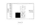
- the constructs were either not preconditioned (controls) or pre-conditioned by culturing for 10 days in vitro , and then all constructs were frozen for 5 mins in liquid nitrogen, embedded into acellular collagen wraps and subcutaneously implanted into rabbits (subcutaneously). After 1 week, the constructs were recovered. No visible vascularisation was observed in the control constructs which had not been pre-conditioned ( figure 14 ). By contrast, visible ingrowth of the host vasculature into collagen implants which had been preconditioned before freezing was observed ( figure 15 ).
- Angiogenesis was therefore shown to be promoted by both cellular and acellular angiogenic implants as described herein.
- VEGF vascular endothelial growth factor
Landscapes
- Health & Medical Sciences (AREA)
- Life Sciences & Earth Sciences (AREA)
- Engineering & Computer Science (AREA)
- Chemical & Material Sciences (AREA)
- Bioinformatics & Cheminformatics (AREA)
- Biomedical Technology (AREA)
- Organic Chemistry (AREA)
- General Health & Medical Sciences (AREA)
- Medicinal Chemistry (AREA)
- Animal Behavior & Ethology (AREA)
- Chemical Kinetics & Catalysis (AREA)
- Public Health (AREA)
- Veterinary Medicine (AREA)
- Cell Biology (AREA)
- Zoology (AREA)
- Nuclear Medicine, Radiotherapy & Molecular Imaging (AREA)
- Pharmacology & Pharmacy (AREA)
- General Chemical & Material Sciences (AREA)
- Biotechnology (AREA)
- Wood Science & Technology (AREA)
- Genetics & Genomics (AREA)
- Hematology (AREA)
- Dermatology (AREA)
- Urology & Nephrology (AREA)
- Epidemiology (AREA)
- Biochemistry (AREA)
- Developmental Biology & Embryology (AREA)
- Oral & Maxillofacial Surgery (AREA)
- Transplantation (AREA)
- Cardiology (AREA)
- General Engineering & Computer Science (AREA)
- Botany (AREA)
- Microbiology (AREA)
- Heart & Thoracic Surgery (AREA)
- Vascular Medicine (AREA)
- Diabetes (AREA)
- Micro-Organisms Or Cultivation Processes Thereof (AREA)
- Materials For Medical Uses (AREA)
- Medicines Containing Material From Animals Or Micro-Organisms (AREA)
- Medicinal Preparation (AREA)
Description
- This invention relates to three dimensional implants and tissue scaffolds which promote cellular activities, such as angiogenesis, vasculogenesis, differentiation and proliferation.
- Tissue hypoxia results in rapid angiogenesis in vivo, triggered by angiogenic proteins, including vascular endothelial growth factor (VEGF). Current views of tissue viability are founded on whether deeper-lying cells receive sufficient nutrients and oxygen for normal activity and ultimately survival. For intact tissues, levels of such essential nutrients are governed by micro-vascular perfusion. However, there have been few effective quantitatively defined 3D models, which enable testing of the interplay or interdependence of matrix and cell density, and path diffusion on oxygen consumption in vitro. As a result, concepts on cell vulnerability to low oxygen levels, together with the nature of cellular responses are ill defined.
- The present inventors have recognised that cells in the core regions of 3-dimensional biomimetic implants do not undergo rapid cell death and may remain viable for extended periods, even under reduced oxygen conditions. Nutrient and/or metabolite gradients are generated within 3-dimensional biomimetic implants which may be useful in controlling mammalian cell growth and proliferation. These gradients may also induce the cells to produce a physiological pro-angiogenic responses. Implants producing these responses may be useful in therapeutic applications in which increased angiogenesis and/or vasculogenesis is required.
- Aspects of the invention relate to the use of nutrient and/metabolite gradients within biomimetic spatial structures to control mammalian cell growth and proliferation.
- An aspect of the invention provides a scaffold for cell culture as set out in
claim 1 - Suitable mammalian cells include endothelial cells, fibroblasts, such as human dermal or tendon fibroblasts, stromal cells, such as bone marrow derived stromal cells and smooth muscle cells, and stem cells.
- Suitable stem cells include corneal (limbal) stem cells; skin epidermal stem cells; gut (intestinal) stem cells; orogenital stem cells; bronchial and other epithelial stem cells; bone marrow stromal stem cells; and growth plate stem cells.
- A pocket is a recess or crypt within the surface of the gel. The pocket comprises an opening on the surface of the gel which allows cell entry and egress and walls which define the boundaries of the pocket in the gel. Preferably, the walls of the pocket are sufficiently rigid to prevent the collapse of the pocket.
- The pocket is of a suitable size to accommodate a population of mammalian cells. The dimensions of the pocket determine the number of cells within the pocket and the extent of the hypoxic gradient and may vary according to the specific application of the scaffold.
- A suitable pocket may be 50 µm to 1500 µm deep
- A suitable pocket may be at least 50 µm, at least 100 µm or at least 150 µm in diameter.
- In some embodiments, the pocket may be 50µm to 2000µm diameter, more preferably 100µm to 500µm in diameter and may be 100µm to 5000µm, preferably 200µm to 5000µm deep.
- The walls of the scaffold may comprise additional attachment proteins, for example extracellular matrix proteins, such as fibronectin, vitronectin and fibrin.
- Populations of mammalian cells, such as stem cells, within the pocket form three distinct polarities of cell-surface and cell-cell attachments which mimic natural mammalian cell niches. The mammalian cells which are adjacent to the walls of the pocket have cell-collagen contacts on one face and cell-cell contacts on the opposite face. Mammalian cells which are not adjacent the walls of the pocket (i.e. in the core of the pocket) are surrounded by other cells and have cell-cell contacts all round. Mammalian cells which are adjacent to the opening of the pocket have cell-cell contacts and cell-fluid contacts on opposing faces.
- Metabolic activity of the mammalian cells located in the pocket, which may be accompanied by cell division and increasing cell density, consumes diffusible factors, nutrients, oxygen and glucose and produces waste metabolites. Since diffusible factors enter the pocket by diffusing through the gel, mammalian cells, such as stem cells, adjacent the gel are exposed to high levels of these factors. In addition, waste metabolites exit the pocket by diffusing through the gel and so these cells are exposed to low levels of cell metabolites.
- Since they are surrounded by other mammalian cells consuming diffusible factors, such as oxygen, mammalian cells which are not adjacent to the gel are exposed to low levels of these factors. In addition, these cells will be exposed on all surfaces to high levels of cell metabolites.
- The scaffold thus allows the generation and maintenance of concentration gradients of diffusible factors, such as nutrients, oxygen and glucose and waste metabolites, such as CO2, lactate and ammonia, within the pocket which mimic natural mammalian cell niches.
- The gradients of diffusible factors in the pocket stimulate the differentiation and proliferation of mammalian cells, such as stem cells, in the pocket. The differentiation and proliferation of the mammalian cells in the pocket may be controlled by modulating the concentration gradients and the polarities of the mammalian cells. This may be achieved by altering the pocket geometry, cell density, gel properties or external concentrations of factors
- The minimum levels of diffusible factors such as nutrients, oxygen and glucose which may be achieved within the pocket are determined by the amount of cells in the pocket. Preferably, the pocket is filled with cells only. The greater the number of cells which are seeded in the pocket, the faster this will be achieved. For example, the pocket may be seeded with greater than 5 million cells per ml, greater than 15 million cells per ml or greater than 50 million cells per ml.
- As mammalian cells proliferate and the number of cells inside the pocket increases, cell migration and egress through the opening of the pocket increases. Furthermore, cells which emerge from the pocket, such as stem cells, may already be stimulated to differentiate. The rate of cell egress from the pocket is determined by the rate of proliferation and the dimensions of the pocket and the pocket opening.
- A method of culturing mammalian cells may comprise
providing a gel having a pocket on the surface thereof, wherein the pocket is 50 µm to 1500 µm deep,
seeding the pocket with mammalian cells, and
incubating the gel in a culture medium;
wherein the metabolism of the cells in the pocket causes the amount of nutrients to progressively decrease from the sides of the pocket inwards. - A suitable scaffold for control of mammalian cell growth and proliferation may be produced, for example, by plastic compression fabrication to emboss pockets onto the gel surface. Plastic compression fabrication is described in more detail in
WO2006/003442 A micro-structured 'die' or template may be fabricated which corresponds to the required patterning of pockets in the gel. Contact between the die and the gel then embosses the pattern of pockets into the gel. In other words, one or more projections are present on the contact surface of the die and contact between the die and the gel causes these projections to emboss pockets in the surface of the gel. The pockets in the gel correspond in dimensions and arrangement to the dimensions and arrangement of the projections on the die and are suitable for the growth of mammalian cells. For example, a collagen gel may be plastically compressed by a die to produce crypt-like pockets 50-2000 µm diameter by 200µm to 5000µm deep as described above. An example of a scaffold comprising a collagen gel embossed with pockets is shown inFigure 16 . - Suitable techniques for creating projections and other micro-structure on a die or template surface are well-known in the art. For example, projections or other microstructure may be applied to glass or silicon dies by standard etching techniques or to metal dies by spark erosion techniques.
- In some embodiments, a porous template or die may be employed. This allows fluid to leave the gel as it is plastically compressed, as described herein. Porous dies may be produced, for example, using sintered materials (metal, plastic or ceramic) which have been shaped to contain projections or other microstructure for embossing pockets into the gel of the scaffold.
- Scaffolds for mammalian cell culture as described herein may be useful in in vivo cell growth and in biomimetic implants.
- Suitable gels for use in the implants and scaffolds described herein may comprise a matrix of scaffold fibres and an interstitial fluid. Gels are formed by the coalescence and elongation of scaffold fibrils, as the fibrils form a continuous network around the aqueous interstitial liquid which originally held the monomers. For example, triple helical collagen monomers may be initially dissolved in dilute acid and then induced to polymerise (aggregate) to fibrils (e.g. at 37° and neutral pH). As the fibrils polymerise, there is a phase change and the solid network of fibrils 'supports' the remaining interstitial liquid in approximately the same volume and shape - i.e. it gels. Phase transition from soluble monomer to solid polymer is characteristic of a gel.
- Any hydrated polymer material may be suitable for use in the gels described herein, including naturally occurring polymers, for example proteins, such as silk, fibrin, fibronectin, elastin or collagen (e.g. collagen type I), glycoproteins such as fibronectin, or polysaccharides such as chitin, or cellulose. In some preferred embodiments, the matrix fibres are made collagen. Native fibril forming collagen types are preferred including collagen types are I, II, III, V, VI, IX and XI and combinations of these (e.g. I, III V or II, IX, XI). For example, collagen type I may be used as the gel or scaffold material. In some preferred embodiments, the gel may comprise 5 to 25% collagen type I (dry/wet weight ratio), more preferably about 10%. In some preferred embodiments, the gel may comprise 15 to 20% collagen type I (dry/wet weight ratio), more preferably about 10%.
- Other suitable fibrous scaffold materials include synthetic polymers i.e. polymers that are not naturally present in the human or animal body. Suitable polymers include organic polymers such as polylactone, polyglycone and polycapryolactone, inorganic polymers such as phosphate glass and synthetic, gelling polypeptide gels.
- In some embodiments, the fibrous scaffold material may be a composite material comprising two or more different types of fibre. For example, the scaffold may comprise fibronectin and collagen, collagen and polylactide, fibrin and collagen, collagen fibres and carbon-nanotubes, or fibrin, collagen and fibronectin.
- The interstitial liquid is typically an aqueous liquid which supports the growth and proliferation of the cells contained in the gel. Suitable liquids include mammalian cell culture media such as Eagles MEM solution. Techniques for formulating and casting gels for use as biomaterials are well-known in the art (see, for example,
WO2006/003442 ;WO2007/060459 ; Marenzana et al 2006 Exp Cell Res 312 423-433; Tomasek et al (2002) Nat Rev ; Harris et al Nature 290 (1981) 249-251; Elsdale et al 1972 J Cell Biol. 54 626-637; Kolodney et al J Cell Biol. (1992) 117 73-82; Eastwood et al Biochem Biophys Acta 1201 (1994) 186-192). - In general, high density gels are preferred to facilitate the generation of gradients of diffusible factors, such as oxygen.
- A suitable gel for use in the implants described herein may have a liquid phase of >70% (wet/dry weight ratio), >75%, >80%, >85%, or >90%. For example, a suitable gel may have a liquid phase of 75% (wet/dry weight ratio) to 95%, typically about 88%.
- A suitable gel described herein may have a may have an oxygen diffusion coefficient of 1 to 10 x 10-6 cm2/s-, more preferably 4 to 5 x 10⁻⁶ cm2/s-1.
- Gel permeability may also be measured by determining the O2 re-equilibrium rate after depletion, for example using sodium sulfite or N2 saturation. In some embodiments, the gel may have an O2 re-equilibrium rate over a 1 mm shortest diffusion path of 2 to 4 mmHg/min, preferably about 3 mmHg/min in an air saturated solution, following O2 depletion.
- The permeability of a gel suitable for use in an implant may be equivalent to a gel containing 5% to 25% collagen, preferably about 10% collagen (dry/wet weight ratio). In some embodiments, a dense gel may be used which has a permeability equivalent to a gel containing 15% to 20% collagen.
- Cells may be seeded within the matrix by mixing them with the liquid scaffold matrix and then allowing the liquid matrix to solidify into a gel. Seeding of the matrix is preferably performed under suitable conditions of temperature, pH, ionic strength and sheer to maintain viability, prior to gel formation. The initial cell density in the gel may be from about 1 x 10⁴ to 1 x 10⁷ cells per ml, more preferably from about 5 x 10⁵ to 1 x 10⁶ cells per ml.
- In some embodiments, the mammalian cell scaffolds as described herein may be produced by a method comprising plastically compacting a gel which is seeded with cells. This increases the density of cells within the gel. Plastic compaction involves deforming an object such as a gel to reduce its volume, such that the object substantially retains its new volume, even after the cause of compaction is removed. Plastic compaction is a rapid, cell-independent process which results from subjecting the gel to a physical treatment, such as an external force or pressure, which expels interstitial liquid from the gel, such that it does not return on removal of the load: i.e. the gel undergoes a plastic compaction.
- For example, plastic compaction may form a sheet comprising cells, which may be rolled or folded to produce a multilayer implant. Plastic compaction of gels, including gels seeded with cells, is described in more detail in
WO2006/003442 . - Plastic compaction may improve the mechanical properties of the gel. Unconfined compaction of a gel expels interstitial liquid, which does not return on removal of the load: i.e. the gel undergoes a plastic compaction. In an untreated gel, the scaffold matrix is generally in a gross, hydrated form. This scaffold structure collapses during plastic compaction without loss of structural detail, dehydrating the scaffold in the gel, and leading to increased density and strength.
- The plastic compaction process may be optimised to achieve the desired final ratio of fibres and cells from a standard starting gel. A standard gel, for example, may comprise 1 to 4% collagen and 0.2 to 10 x106 cells per ml.
- The gel environment is preferably maintained at physiological conditions (e.g. temperature, pH, hydration and ionic strength) for the cells to survive. It is preferred that plastic compaction does not alter the ionic properties of the gel fluid significantly from physiological conditions.
- Following compaction, the gel may be subjected to repeated cycles of uniaxial tensile loading to improve its mechanical properties. Suitable cycling is described in
WO2007/060459 . In a compacted collagen gel, repetitive cycles of loading increase the fusion of collagen fibrils to produce a biomaterial which has improved material strength (i.e. increased break stress, break strain and/or elastic modulus). - The gel or biomaterial comprising the cells may be shaped, cut or moulded into any convenient implant form, for example, a patch, block, tube, tape, strip, ring, toroid, capillary, roll, sheet or thread. The final shape of the tissue equivalent implant will depend on the particular context in which it is to be used. In some embodiments, the tissue equivalent implant may have a pliable form which is suitable for further shaping.
- Various further aspects and embodiments of the present invention will be apparent to those skilled in the art in view of the present disclosure.
- "and/or" where used herein is to be taken as specific disclosure of each of the two specified features or components with or without the other. For example "A and/or B" is to be taken as specific disclosure of each of (i) A, (ii) B and (iii) A and B, just as if each is set out individually herein.
- Unless context dictates otherwise, the descriptions and definitions of the features set out above are not limited to any particular aspect or embodiment of the invention and apply equally to all aspects and embodiments which are described.
- Certain aspects and embodiments of the invention will now be illustrated by way of example and with reference to the figures and tables described below.
-
Figure 1 shows a schematic of the experimental set up with oxygen probe in the centre of a spiralled plastic compression construct. Constructs were cultured in 50 ml media. -
Figure 2 shows oxygen levels in the centre of acellular plastic compression constructs. -
Figure 3 shows showing the deoxygenation of an acellular plastic compression construct with sodium sulphite, followed by oxygenation in DMEM media (values of 4.5 +/- 0.5 and 3.2 +/- 0.5 mmHg/min). The gradients (corresponding to de-oxygenation and re-oxygenation rates) were estimated using the approximately linear portions of the traces. -
Figure 4 shows oxygen levels in the centre of cell-seeded plastic compression constructs. Oxygen levels at different cell densities were measured, 5.8 x 10⁶ - 23.2x 10⁶ cells/ml, magnification bar 50 mm (0.5 x 10⁶ - 2x 10⁶ cells/construct), * p < 0.0001. Average of n = 3 for each data set is presented here. Time 'zero' is taken as the time point when the probe was positioned in the construct. -
Figure 5 shows confocal images of the central and outer region of the plastic compression constructs seeded with 5.8 x 10⁶ (top), 11.6 x 10⁶ (middle) and 23.2 x 10⁶cells/ml (bottom). -
Figure 6 shows oxygen levels in the centre of a cell-seeded construct, 2 x 10⁶ cells in a construct (23.2 x 10⁶cells/ml), over a 10-day period, with supporting cell viability onday 5 from the centre and surface of the construct. Graph represents a mean of n = 3.Magnification bar 100 mm for micrographs. -
Figure 7 shows VEGF levels in constructs of varying cell density at 24 h. No significant difference was found in VEGF levels between different cell densities. -
Figure 8 shows the level of VEGF in constructs containing 2 million cells/construct over a 10-day period (normalised to GAPDH, withday 1 as the set calibrator) shows a statistically significant increase measured onday 8, which then dropped by day 10 (** p < 0.001, * p < 0.05). -
Figure 9 shows spiral constructs seeded with 2 x 10⁶ cells, cultured for 8 days (peak in VEGF expression) and unrolled to study VEGF in three regions of the construct (because of spiralling these regions corresponded to three unequal thicknesses of the original construct core,mid and surface, approximately 7, 5 and 3 layers respectively). VEGF expression was normalised to GAPDH, withday 1 as the set calibrator, which was 23.2 x 10⁶ cells/ml at 24 h. There were significantly higher levels of VEGF found in the centre of the gels (* p < 0.001). -
Figure 10 shows an example of a biomimetic stem cell niche as described herein. -
Figure 11 shows levels of HIF-1a determined by ELISA in a collagen construct seeded with 2 x 10⁶ HDFs and incubated for 5 and 10 days in vitro. -
Figure 12 shows levels of VEGF determined by ELISA in a collagen construct seeded with 2 x 10⁶ HDFs and incubated for 5 and 10 days in vitro. -
Figure 13 shows collagen constructs seeded with 2 x 10⁶ HDFs, preconditioned by incubation for 5 days in vitro (top) or without preconditioning (bottom) and implanted subcutaneously into a rabbit for 1 week. -
Figure 14 shows (top panel) a control collagen construct which has been seeded with 2 x 10⁶ HDFs, frozen in liquid nitrogen without preconditioning and implanted subcutaneously into a rabbit for 1 week and (bottom panel) a control acellular collagen construct after subcutaneous implantation in a rabbit for 1 week, without seeding with cells or freezing. -
Figure 15 shows collagen constructs seeded with 2 x 10⁶ HDFs and preconditioned by incubation for 10 days in vitro, and then frozen in liquid nitrogen and then implanted subcutaneously into a rabbit for 1 week. -
Figure 16 shows a collagen scaffold with embossed pockets for accommodating mammalian cells. - Human dermal neonatal fibroblasts were explanted from neonatal foreskins (obtained freshly from the operating theatre, with full ethical approval, following surgery for circumcision), as previously described [13]. Cells were maintained in Dulbecco's modified Eagles medium(DMEM, Gibco, Paisley, UK), supplemented with 10% FCS (First Link, West Midlands, UK), 2 mM glutamine and penicillin/streptomycin (1000 U/ml; 100 mg/ml, Gibco Chemicals). For removal of cells from monolayer culture, flasks containing cells were washed with 0.1 M PBS, and incubated with trypsin (0.5% in 5 mM'EDTA) for 5 min at 37 8C.
- Once detached, cells were counted and embedded in 3D collagen type I gels. Collagen gels were set in a mould (2.2 x 3.3 x 1 cm 3). For collagen gel preparation, 0.5 ml 10_ Eagles MEM solution (Gibco) was added to 4 ml rat-tail type I collagen (First Link) in 0.1 M acetic acid, protein concentration 2.035 mg/ml, neutralized with 5 M NaOH, using the indicator colour changes from yellow to cirrus pink [14]. This gel preparation was added to the cell suspension. Following setting and incubation, gels were routinely compacted by a combination of compression and blotting using layers of mesh and paper sheets [12]. Briefly, 165-mm-thick stainless steel mesh and a layer of nylon mesh were placed on a double layer of absorbent paper. The collagen gel was placed on the nylon mesh, covered with a second nylon mesh, and loaded with a 120-g flat metal block (steel) for 5 min at room temperature, giving a flat collagen sheet (50 -60 mm thick) protected between two nylon meshes. These dense sheets of collagen were then rolled to produce a tight spirally wound rod, 2.3 mm in diameter and 21 mm long. Cell densities were increased by the plastic compression in direct proportion to the volume reduction of gel and final cell density was calculated as: initial cell density x fold volume change. Hence for a typical initial gel volume of 5 ml, the percentage of collagen was 0.2%, which increased to 11% following compression (measured by dry/wet weight ratio), which corresponds to a 58-fold increase. The cell density would be expected to change by the same degree from a total of 100 000 cells/ml (or 0.5 million cells/ construct) to a final density of 5.8 million cells/ml; 200 000 cells/ ml (or 1 million cells/construct) to 11.6 million cells/ml; 400 000 cells/ml (or 2 million cells/ construct) to 23.2 million cells/ml [15].
- Fibre-optic oxygen probes (Oxford Optronix, Oxford, UK) were inserted into the centre of the 3D spiral constructs and positioned halfway along its axis (
Fig. 1 ). The constructs were then sealed at the end using cyanoacrylate glue. Hence, a diffusion length of >1 mm was studied and refers mainly to lateral diffusion, as the ends of the construct were sealed. The tip of the sensor probe (280 mm diameter) incorporates an oxygen-sensitive luminescent probe within an oxygen-permeable matrix. The luminescence is quenched in the presence of molecular oxygen so that the luminescence emission lifetime becomes longer at lower oxygen concentrations in the surrounding medium. The calibration of the probe, which is accurate to 0.7 mmHg, essentially relies on the correlation of the luminescence lifetime (rather than intensity) versus the oxygen concentration [16]. This method results in an exceptionally stable calibrated response so that each probe may be used up to 6 days at the slowest sampling rate. Therefore, for monitoring of constructs for longer than 6 days, the expiring probes were removed and new probes inserted on day 5 (see below). After each experiment, the probe reading was checked in the external medium to confirm that there was no drift in the response. The fibre-optic probes were used in conjunction with an OxyLabpO2 ETM system coupled to an A/D converter (12 bit) and the results were recorded on an IBM PC computer using Labview. Results are presented as partial pressure values, i.e., pO2 in mmHg (e.g. 7.6 mmHg corresponds to 1% oxygen). The sampling rate was varied according to the experiment performed, yielding an overall response time of <10 s (for de-oxygenation measurements,Fig. 2 ,3 ) and up to ~30 s for the long-term studies as shown inFigures 4 and5 . For studies of the rate of oxygen diffusion through the construct, we transferred the construct containing the probe into an anoxic solution of 3% sodium sulphite at 37 8C. Rates of de-oxygenation and re-oxygenation were estimated using the quasilinear portions of the traces, as shown inFigures 1 to 3 . Samples were kept and monitored in 7.5% CO2-enriched incubators. At the end of each experiment the construct was removed and the ambient 02 tension of the media tested, which remained at approximately 140 mmHg. - Cell viability was assessed using Live/Dead Viability/Cytotoxicity Kit (Molecular Probes, L-3224) based on the simultaneous determination of live and dead cells with calcein AM and ethidium homodimer (EthD-1), respectively, for qualitative analysis. Quantitative analyses were carried out with Live/Dead Reduced Biohazard Viability/Cytotoxicity Kit (Molecular Probes, L-7013) according to the manufacturer's protocol.
SYTO_ 10, a green fluorescent nucleic acid stain and Dead Red (ethidium homodimer-2) were used, and, after capturing images, live/dead nuclei were counted to ascertain percentage viability. Viability of cells in each construct was performed independently from the oxygen measurements. Representative areas for 02 determination were chosen in two regions (core and surface) of plastic compressed constructs and were visualized Cell. Mol. LifeSci. Research Article 3 with confocal microscopy (Bio-Rad Radiance 2100, Carl Zeiss Ltd, Hertfordshire, UK). Quantitative PCR analysis of VEGF mRNA. RNA extraction from experimental constructs was performed independently from the oxygen measurements. RNA was extracted either from the whole construct, or from specific regions of the spiral construct (core, mid and surface). These regions were isolated after culture by unrolling the spirals and cutting into the three different regions corresponding to core, mid and surface. Total cellular RNA was isolated from the cellular 3D plastic compressed constructs using the Qiagen RNeasymethod (Qiagen, UK). Constructs were first flash-frozen in liquid nitrogen, and 500 ml lysis buffer containing 2-mercaptoethanol was added to each sample and left to dissolve at room temperature for 40 min (Qiagen, UK). The resultant solution was then aspirated using a 21-G needle and from then on the commercial assay protocol was followed (Qiagen,UK).RNA was eluted in RNase-free water, and the concentration determined by spectrophotometry at 260 and 280 nm (Genequant, Pharmacia Biotech, NJ, USA). First strand cDNA synthesis was performed using Amplitaq reverse transcriptase (Applied Biosystems, Roche). Total RNA (0.5 mg RNA) was diluted in 38.85 ml water. A further 9.15 ml Mastermix was added to each tube (dNTP, RNase inhibitor, MgCl2, Oligo DT Random primers; Applied Biosystems, Roche), and heated at 70 8C for 10 min; 2 ml reverse transcriptase was then added to each tube and tubes were incubated at 40 8C for 1.5 h, followed by heating at 90 8C for 2 min to denature any remaining enzyme. - Relative quantitative PCR was performed using Applied Biosystems 7300 Real-time PCR system (CA,USA), with the Taqman universal PCR Master Mix (Applied Biosystems). cDNA, 9 ml/reaction, was mixed with 1 mL of the required gene probe (VEGF, Applied Biosystems assay ID: Hs00900057_ml or GAPDH, Applied Biosystems assay ID: Hs99999905_ml) and 10 ml Mastermix (Applied Biosystems) in a 96-well plate for cycling and analysis in the Applied Biosystems 7300 Real-time PCR machine (total 20-ml reaction volume). The primer sequences are not disclosed and kept confidential by Applied Biosystems (Roche). Each primer set is calibrated to work optimally in the 7300 Real-time PCR machine. The combined thermal cycling and amplification-specific software enabled detection of the PCR products as cycle-by-cycle accumulation in a single-tube reaction. Values for each sample were normalised to the corresponding GAPDH result, which did not change significantly in any sample tested. Relative quantification was performed by expressing each sample/ GAPDH ratio relative to a calibrator. This calibrator was set as 2 million cells per construct at 1 day. This calibrator was run along each run tested and compared. By doing this, any small changes from run to run could be accounted for, as we were relying on relative quantification. This calibrator was measured in each PCR run (with no significant variation) and was always set to unity, and the sample change expressed as fold increase or decrease relative to this. In the case of cell density, culture period and region experiments, this was set as 2 million cells/construct at 24 h. Therefore, all relative quantification of gene expression changes were set relative to this.
- The 3D monitoring set-up is shown schematically in
Figure 1 . Validation and calibration studies on constructs without cells (Fig. 2 ) showed a steady baseline level of 02 tension in the core of cell-free constructs, with negligible consumption of oxygen over 24 h (not significantly different from the external medium), consistent withminimal02 consumption of the probe. Cell-independent depletion and recovery of core 02 levels was demonstrated (Fig. 3 ) by addition and then removal and washout of a 3% solution of sodium sulphite to sequester 02 from the system. The oxygen levels were observed to fall progressively over several minutes, reaching zero after approximately 30 min. Note that the time response of the probes was set to <10 s. Recovery back to air-saturated pO2 levels was measured by transferring the construct still containing the probe back into an air-saturated solution, which occurred at an approximate rate of 3.2 x 0.5 mmHg/min. Comparable rapid rates of reequilibration were seen usingN2 saturation of medium instead of the sodium sulphite reagent. Cellular constructs exhibited time-dependent oxygen depletion in their core, where the probe was positioned, over a period of 24 h (Fig. 4 ). Oxygen levels fell rapidly towards approximately steady-state or plateau values, which varied according to the cell density. Cell consumption, therefore, appeared to be the only factor influencing oxygen levels in the construct. Cell density was the key determining factor in the degree of oxygen depletion response, with the lower core pO2 correlating with the higher cell density, which ranged from 0, 5.8, 11.6 to 23.2 million cells/ml. In this case, the plateau 02 tensions declined significantly (p < 0.001) with each increase in cell density. The oxygen level in the centre of constructs seeded with 0.5 million cells (final density of 5.8 million cells/ ml) was around 80 mmHg after 24 h, compared to a construct seeded with 2 million cells (final cell density of 23.2 million cells/ml), where the level was ~25 mmHg. - Exposure of cells to low levels of oxygen in the core had no effect on cell viability over this 24-h period. Cells in the construct core had over 95% viability after exposure to both 80 mmHg and 25 mmHg (
Fig. 5 ). Hence exposure of cells to oxygen tensions as low as 25 mmHg did not increase cell death up to 5 days. Atday 10, up to 55% cell death was observed in the core, compared to 40% at the surface. In such a 3D model, the diffusion of critical higher molecular weight nutrients may also be a limiting factor for cell survival. Glucose diffusion coefficients have previously been established and this is not found to be limiting for cells in the same 3D model [17]. -
Figure 6 shows the core 02 profile, over a 10-day period, of a construct seeded at an initial cell density of 2 million cells (23.2 million cells/ml). Importantly, after the initial 24-h fall to a first equilibrium level of ~25 mmHg, core oxygen tension increased to a second elevated steady state of around 60 mmHg. This is likely to be a result of changes in cell consumption rather than changes in material properties (Fig. 6 ). Over the 10-day period, there were no major changes in cell number through proliferation. After 5 days, core cell viability was still 80%, and close to 100% at the surface. Hence, between the 30- and 36-h culture periods, the stabilised core 02 tension rose by 35 mmHg (>twofold increase), to approximately 60 mmHg. This occurred over a 6-h period, consistent with a change in fibroblast metabolism and 02 utilisation. This level was approximately maintained over the following 24 h and then fell gradually back to ~20 mmHg over the following 72 h. - Levels of VEGF mRNA expression were measured in constructs at the three cell densities studied and in the 23.2 million cells/ml construct over 10 days of culture (
Figs. 7 and8 ). At the 24-h stage, the three cell densities gave relative VEGF gene expression levels with no significant difference, shown here relative to the highest cell density (Fig. 7 ). Over the longer 10-day period, VEGF expression levels changed dramatically in the 23.2 million cells/ml constructs (Fig. 8 ). Betweendays 1 and 3 (23.2 million cells/ml) there was a 5-fold increase in VEGF expression (p < 0.05), followed by small incremental increases betweendays day 1 levels (p < 0.05). The greatest changes, however, were seen betweendays day 1. This was followed almost immediately by a complete collapse in expression (149-fold), such that byday 10 expression had returned back today 1 levels. This pattern of an apparent temporal spike of growth factor expression is characteristic of control systems based on a number of different molecular elements operating in sequence, as might be expected for angiogenic stimulation. Such a spike in VEGF expression would be expected where only one growth factor of a sequential cocktail was monitored, as in this system. This pattern of VEGF regulation over 10 days was not observed with the lower cell densities studied. - Zonal changes in VEGF expression, through the thickness of the spiral constructs (23.2 million cells/ml) and so along the gradient of 02 tension, were determined at the 8-day culture stage (core, mid and surface). Zonal changes were measured by unfurling the spiral construct after culturing (
Fig. 9 ). It was assumed that cells in the core section of the gel, which was the zone sampled by the probe, had been exposed to 02 levels between 20 and 60 mmHg, in the mid section to between 60 and 100 mmHg, and in the outer section, which was in close proximity to the aerated medium, to between 100 and 140 mmHg over most of the culture period. Significantly higher levels of - VEGF were found in the core region, where cells were exposed to the lowest levels of 02.
Figure 9 shows the gradient of VEGF expression at 8 day (23.2 million cells/ml) under these conditions (i.e., the underlying zonal heterogeneity at day 8). Importantly cells in the surface region still expressed increased levels of message (4.7-fold increase over basal 1-day VEGF levels). In the mid and core regions this increased to 7.1- and 10.1-fold greater expression than 1-day levels, consistent with a direct relationship between 02 tension and VEGF gene expression. However, given high 02 tensions found in the surface zone, this provides indication that cells were either extremely sensitive to very modest reductions in available 02 or, more plausibly, surface zone cells had responded to earlier stage cytokine or metabolic signals (not measured here) produced and diffusing out from the core cells. This provides indication that systems exist for amplifying the zone of increased VEGF expression. - Current views of tissue viability and 3D cellular constructs are dominated by the ability of 'deeper lying' cells to receive sufficient nutrient and oxygen for normal activity and ultimately for survival. In intact tissues such factors are clearly controlled by the presence and rates of micro-vascular perfusion. In the absence of this process, in 3D culture, cells rely entirely on simple diffusion from the construct margins. However, there have been few, if any, effective quantitatively defined 3D models that enable testing of the interplay of matrix density and cell density on 02 depletion. As a result, concepts on cell vulnerability to low 02, together with the rate, extent and nature of their responses (short of cell death) are simplistic. Importantly, many are based on tumour or other cell masses, with little or no extracellular matrix content. The present study has developed the first 3D-monitored model of connective tissue to define precisely which factors dominate and how they affect resident cell behaviour and survival. Using this model, the time-dependence of oxygen versus VEGF was studied over several days together with spatial measurements.
- There are several key determining factors in this system, as there are in vivo, for the level and rate of attainment of minimum 02 tension at the core (furthest from perfusion point) of 3D constructs and tissues: (1) cell density, which is assumed to be a homogenous distribution, but can change zonally and over time, (2) matrix density/permeability to the limiting diffusing component (02 or other nutrient), and (3) cell type or cell activity (i.e., O2 demand of the cells, which is dependent on aerobic/anaerobic and active/quiescent status; e.g., chondrocyte, dermal fibroblast, myoblast). The plastic compression process for the 3D collagen constructs provides control of all these factors. The cell and matrix densities are both determined by the initial inoculum and initial collagen content (respectively) multiplied by the fold compression (x58). The collagen matrix has a nano-fibrillar lattice of around 88% water, making it highly permeable to 02.
- The initial rate of consumption of 02 was non-linear over the first 10 -30 min of the experiment, but thereafter was nearly constant. In this system the rate of fall of 02 and the equilibrium consumption of 02 was entirely predictive, being dependent on cell density (
Fig. 4 ). Despite the relatively high matrix density and resultant 02 depletion at the centre of the constructs, cell viability was unaffected at 24 h and only slightly reduced (>80% of cells viable) after 5 days at the highest cell density (Fig. 6 ). To relate this to other work on cell responses to low levels of 02, pathological hypoxia is conventionally set at <1% or 8 mmHg [6, 18]. Consequently, pO2 levels in the core of high cell density constructs here never fell below 18 mmHg, and so were not conventionally hypoxic (Fig. 6 ). This challenges the conventional impression that diffusion gradients >1 mm are frequently damaging to cells, but supports the idea that dense fibrillar collagen represents only a modest diffusion barrier (i.e., highly permeable to small molecules). This high permeability to 02 is reasonable given the nano-fibrillar mesh structure of the matrix, with approximately 88%fluid phase. This was established here by the 02 re-equilibrium rate (1-mm shortest diffusion path) of 4.5 x 0.5 mmHg/min. - Importantly, the greater reductions in core 02 tension were apparently cell number dependent, providing indication that it is not necessarily matrix diffusion path length that dominates cell response, but the number of overlying cell layers, each depleting 02. This provides an insight into understanding why the core of cell-rich, matrix-poor structures (tumours, organs) are so much more vulnerable to core cell necrosis compared to connective tissues. The overall levels of VEGF measured in the different regions were significantly lower compared to the
total day 8 reading. This may be due to slow processing of the constructs since they had to be unrolled carefully, during which time (up to 1 h) the values of VEGF may have dropped as cells in the entire construct became exposed to normoxia, including the core region during the final stage of unrolling. Cells exposed to low oxygen tensions respond by significant increases in TGF-b, platelet-derived growth factor (PDGF) and VEGF expression. It seems likely, therefore, that some exposure to low levels of oxygen is in fact complementary to tissue construct maturation, and will in fact be beneficial to cell survival. The influence of varying 02 tension on cellular proliferation and differentiation is crucial in understanding how physiological microenvironments can influence cellular behaviour, and there has been work to show that low levels of oxygen enhance proliferation of many cell types, including fibroblasts [22, 23]. Cell growth at reduced 02 tension does not necessarily result in cell death. When cultured at 2% oxygen (~ 15.2 mmHg), trophoblast cell proliferation is stimulated, whereas at 20% (152 mmHg), cells actually exit the cell cycle, and undergo increased differentiation [22]. Reduced 02 tensions measured in the current study (down to as low as 15 mmHg), were close to those used in the study by Ma et al. [22], and while direct extrapolation is dangerous here, due to differences in cell type and cell density, some parallels can be drawn. For instance, it does highlight the paradox of suggestions that 3D cell/matrix culture composites may undergo cell death in the centre due to oxygen depletion, in contrast to data indicating that reduced levels of 02 tension in tissue culture can enhance cell proliferation and indeed mimic many native cell environments. Precise and 3D-localised measurement of 02 tension is, therefore, critical in determining our understanding of any given 3D tissue model. It may, for example, be necessary to manipulate both high and low levels of oxygen locally to stimulate at different stages cell proliferation and cell differentiation for maturation of the model. - The first of three cell reactions to prolonged low 02 tension (>24 h, high density) was an apparent shift to anaerobic metabolism, indicated by the relative increase in 02 tension after 23 h (
Fig. 4 ). Although confirmation of this was beyond the scope of this study, such a shift to predominantly glycolytic metabolism seems the most reasonable explanation, given that changes in cell number or matrix permeability capable of explaining this rise in core 02 can be ruled out in this instance. Such a concerted shift in cell metabolism would not be surprising. It does, however, provide indication that such cells have low 02 responses, even above conventional pathological hypoxia and these this may trigger further downstream responses. Most tissue repair and tissue engineering integration processes depend on rapid angiogenesis, triggered by a number of angiogenic proteins, including VEGF. - VEGF expression is induced by the transcription factor HIF-1a, which possesses an oxygen-sensitive degradation domain [24, 25]. Large increases in VEGF mRNA levels were measured by
day 8 in cell-dense constructs. One trigger for this up-regulation may be low levels of 02 or a resultant sustained period of glycolytic metabolism [26]. Confirmation that cell death was not significant here (>80% cell viability at day 5) is important, since it indicates that the VEGF response was not dependent on dying cells. However, byday 10, a reduction in cell viability was measured and this has been the predominant reason VEGF levels dropped onday 10. Furthermore, it was possible to demonstrate that much (although not all) of the VEGF response was due to cells at the construct core, where levels were assumed to range between 20 and 60 mmHg compared to 100 and 140 mmHg at the construct surface. This was achievable because as the 3D spiral model could be unfurled, following the oxygen measurements, which then allowed us to quantitatively map out where VEGF production was up-regulated, with regards to the spatial position. The exposure of cells to 02 was dependent on the specific region they were located within the gel construct, which correspondingly influenced VEGF levels. Critically, the up-regulation of VEGF was not observed up to 24 h, when constructs were seeded with different cell densities (and therefore exposed to different 02 gradients within the constructs). The gradients varied from 82 to 140 mmHg (0.5 million), 66 to 140 mmHg (1 million), and 23 to 140 mmHg (2 million). This controlled in vitro system allowed the 02-dependent VEGF regulation to be monitored carefully. As levels of 02 did not fall below 15 mmHg, and certainly not as low as 7.6 mmHg (1% oxygen), it cannot be said that the trigger for VEGF up-regulation is classical "pathological hypoxia". The signalling of this low level of 02 (within physiological hypoxia range) to the cells to stimulate VEGF is therefore likely to be through initial up-regulation of its transcription factor, HIF-Ia. - To assess the effect of in vitro pre-conditioning on angiogenic implants, 3D collagen constructs were produced as described above and seeded with 2x10⁶ human dermal fibroblasts (HDFs). The amounts of angiogenesis factors HIF-1a and VEGF in the constructs after 5 and 10 days in in vitro culture were measured by ELISA. High levels of both HIF-1a and VEGF were observed in the constructs after in vitro culture (
Figures 11 and12 ). Neither HIF-1a nor VEGF was observed by ELISA in control gels which had not been cultured in vitro. - To assess the effect of angiogenic implants containing live cells in vivo, 3D collagen constructs were produced as described above and seeded with 2x10⁶ human dermal fibroblasts (HDFs) (equivalent to 23 x106 cells/ml). The constructs were either pre-conditioned for 5 days in vitro or not preconditioned. The constructs were then embedded into acellular collagen wraps and subcutaneously implanted into rabbits (subcutaneously). After 1 week, the constructs were recovered (see
figure 13 ). Ingrown vessels were observed from the host into both the pre-conditioned and non-pre-conditioned constructs, indicating that both types of constructs simulate angiogenic responses in the rabbit host. Vascularisation was observed to be increased in the pre-conditioned constructs, relative to the non-pre-conditioned constructs. - The above experiments were then repeated with 3D collagen constructs seeded with 5x10⁵ human dermal fibroblasts (HDFs) (equivalent to 5 x10⁶ cells/ml). The constructs were either pre-conditioned for 5 days in vitro or not preconditioned. The constructs were then embedded into acellular collagen wraps and subcutaneously implanted into rabbits (subcutaneously). After 1 week, the constructs were recovered and assessed for vascularisation. Vascularisation of the constructs which had been preconditioned in vitro was observed. No vascularisation occurs in constructs containing 5x10⁵ cells which have not been pre-conditioned.
- In a control experiment, a 3D collagen construct which did not contain cells was subcutaneously implanted into rabbits. After 1 week, the acellular construct was recovered and assessed for vascularisation. No vascularisation was observed.
- To assess the effect of angiogenic implants without live cells in vivo, 3D collagen constructs were produced as described above and seeded with 2x10⁶ human dermal fibroblasts (HDFs) (equivalent to 23 x106 cells/ml). The constructs were either not preconditioned (controls) or pre-conditioned by culturing for 10 days in vitro, and then all constructs were frozen for 5 mins in liquid nitrogen, embedded into acellular collagen wraps and subcutaneously implanted into rabbits (subcutaneously). After 1 week, the constructs were recovered. No visible vascularisation was observed in the control constructs which had not been pre-conditioned (
figure 14 ). By contrast, visible ingrowth of the host vasculature into collagen implants which had been preconditioned before freezing was observed (figure 15 ). - Angiogenesis was therefore shown to be promoted by both cellular and acellular angiogenic implants as described herein.
- In summary, we have established a basic model for the study of 02 consumption by human dermal fibroblasts in 3D collagen matrices. It was clearly evident that no adverse affect on cell viability was observed due to lower levels of oxygen exposure. In turn, the pattern of VEGF regulation has been determined under low pO2 but non-hypoxic conditions. This study also shows that naturally occurring angiogenic signals may be engineered for induction of vascularisation in such 3D tissue engineered constructs post implantation. The production of VEGF, along with other cell-manufactured angiogenic signals, in a 3D construct core, with lower levels produced by cells the closer they got to the surface, a gradient of these signals would induce vascularisation from the surface of a 3D construct towards the core, potentially inducing vascularisation from outside the construct in an in vivo scenario. For instance, by understanding when and where cells at defined densities and within a defined matrix will up-regulate VEGF production could be used to promote implant integration in vivo.
-
- 1 Weaver, V. et al. (1997) J. Cell Biol. 137, 231-245.
- 2 Wang, F. et al (1998) Proc. Natl. Acad. Sci. USA 95, 14821-14826.
- 3 Weiss, P. (1959) Cellular dynamics. Rev. Mod. Phys. 31, 11-20.
- 4 Tomasek, J. J et al. (2002) Nat. Rev. Mol. Cell. Biol. 3,349-363.
- 5 Grinnell, F. (2003). Trends Cell Biol. 13, 264-269.
- 6 Okazaki, K. M. and Maltepe, E. (2006) Regen. Med. 1, 71-83.
- 7 Werrlein, R. J. and Glinos,A. D. (1974) Nature 251, 317-319.
- 8 Tokuda,Y. et al (2000) J. Cell Physiol. 182, 414-420.
- 9 Gnaiger, E. et al (2000) Proc. Natl. Acad. Sci.USA 97, 11080-11085.
- 10 Levenberg, S. et al (2005) Nat. Biotechnol. 23, 879-884.
- 11 Lussi, J.W. et al (2006). Biomaterials 27, 2534-2541.
- 12 Brown, R. A. et al. (2005) Adv. Funct. Mater. 15, 1762-1770.
- 13 Burt, A. and McGrouther, D. A. (1992) Production and use of skin cell cultures in therapeutic situation. In: Animal Cell Biotechnology, vol. 5, pp. 150-168, Spier, R. E., Griffiths, J. B. (eds.), Academic Press, New York.
- 14 Cheema, U. et al (2003) Cell Motil. Cytoskeleton 54, 226-236.
- 15 Cheema,U. et al. (2007. Biotechnol. Bioprocess. Eng. 12, 9-14.
- 16 Seddon, B. M. et al (2001) Radiat. Res. 155, 837-846.
- 17 Rong, Z., et al (2006). Analyst 131, 816-821.
- 18 Cooper, A. and Beasley, D. (1999) Am. J. Physiol. 277, H1326-H1337.
- 19 Falanga,V. et al (1991). J. Invest. Dermatol. 97, 634-637.
- 20 Kourembanas, S., et al (1990) J. Clin. Invest. 86, 670-674.
- 21 Steinbrech, D. S et al (1999) J. Surg. Res. 84, 127-133.
- 22 Ma, T. et al (2001) Tissue Eng. 7, 495-506.
- 23 Falanga, V. and Kirsner, R. S. (1993) J. Cell Physiol. 154, 506-510.
- 24 Trentin, D. et al. (2006) Proc. Natl. .
- 25 Semenza, G. L. (1998) Curr. Opin. Genet. Dev. 8, 588-594.
- 26 Kumar, V. B. et al.(2007) J. Cell Physiol. 211, 477-485.
- 27 Mudera V. et al J Tissue Eng Regen Med 2007; 1: 192-198.
Claims (11)
- A scaffold for cell culture comprising:a gel having a pocket on the surface thereof,wherein the pocket is 50 µm to 1500 µm deep and contains mammalian cells,said pocket being produced by contacting said surface with a die having a projection on the surface thereof,such that said projection embosses said pockets in said surface.
- A scaffold according to claim 1 wherein the gel is a collagen gel.
- A scaffold according to any one of the preceding claims wherein the mammalian cells are stem cells, for example corneal stem cells; skin epidermal stem cells; gut stem cells; orogenital stem cells; epithelial stem cells; bone marrow stromal stem cells; or growth plate stem cells.
- A scaffold according to any one of the preceding claims wherein said mammalian cells produce the concentration gradients of diffusible factors and waste metabolites within the pocket.
- A scaffold according to claim 4 wherein said concentration gradients stimulate the differentiation and proliferation of cells in the pocket.
- A method of culturing mammalian cells comprising:providing a gel having a pocket on the surface thereof,wherein the pocket is 50 µm to 1500 µm deep,seeding the pocket with mammalian cells, andincubating the gel in a culture medium;wherein the metabolism of the cells in the pocket causes the amount of nutrients to progressively decrease from the sides of the pocket inwards.
- A method according to claim 6 wherein the gel is a collagen gel.
- A method according to claim 6 or 7 wherein the pocket contains greater than 5 million cells per ml.
- A method according to any one of claims 6 to 8 wherein the mammalian cells are stem cells, for example corneal stem cells; skin epidermal stem cells; gut stem cells; orogenital stem cells; epithelial stem cells; bone marrow stromal stem cells; or growth plate stem cells.
- A method according to any one of claims 6 to 8 wherein the progressive decrease in concentration of said nutrients stimulates the differentiation and proliferation of mammalian cells in the pocket.
- A method of producing a scaffold for culturing mammalian cells comprising:providing a gel,contacting the surface of the gel with a die having one or more projections on the surface thereof, such that said projections emboss pockets in the surface of the gel, said pockets being 50 µm to 1500 µm deep and;seeding the pockets with mammalian cells.
Applications Claiming Priority (2)
| Application Number | Priority Date | Filing Date | Title |
|---|---|---|---|
| US5112408P | 2008-05-07 | 2008-05-07 | |
| PCT/GB2009/001158 WO2009136173A2 (en) | 2008-05-07 | 2009-05-07 | Biomimetic cell scaffolds |
Publications (2)
| Publication Number | Publication Date |
|---|---|
| EP2291512A2 EP2291512A2 (en) | 2011-03-09 |
| EP2291512B1 true EP2291512B1 (en) | 2015-10-07 |
Family
ID=40847066
Family Applications (1)
| Application Number | Title | Priority Date | Filing Date |
|---|---|---|---|
| EP09742369.3A Active EP2291512B1 (en) | 2008-05-07 | 2009-05-07 | Biomimetic cell scaffolds |
Country Status (6)
| Country | Link |
|---|---|
| US (1) | US8580564B2 (en) |
| EP (1) | EP2291512B1 (en) |
| JP (1) | JP2011523355A (en) |
| CN (1) | CN102089425A (en) |
| CA (1) | CA2723760A1 (en) |
| WO (1) | WO2009136173A2 (en) |
Families Citing this family (19)
| Publication number | Priority date | Publication date | Assignee | Title |
|---|---|---|---|---|
| AUPR298901A0 (en) | 2001-02-07 | 2001-03-08 | McComb Foundation, Inc., The | Cell suspension preparation technique and device |
| GB0912399D0 (en) * | 2009-07-16 | 2009-08-26 | Ucl Business Plc | Polymeric collagen biomaterials |
| AU2013214187B2 (en) | 2012-02-03 | 2017-02-09 | Technische Universitat Munchen - Klinikum Rechts Der Isar | Device-based methods for localised delivery of cell-free carriers with stress-induced cellular factors |
| EP2828378B1 (en) * | 2012-03-22 | 2021-02-17 | Avita Medical Ltd | Cell suspension and use thereof |
| AU2013205148B2 (en) | 2013-03-14 | 2014-10-30 | AVITA Medical Americas, LLC | Systems and methods for tissue processing and preparation of cell suspension therefrom |
| US9878071B2 (en) | 2013-10-16 | 2018-01-30 | Purdue Research Foundation | Collagen compositions and methods of use |
| EP3185922B1 (en) | 2014-08-27 | 2025-04-02 | Purdue Research Foundation | Collagen-based therapeutic delivery systems |
| EP3250678A4 (en) * | 2015-01-30 | 2018-08-01 | The University of North Carolina at Chapel Hill | Methods to generate gastrointestinal epithelial tissue constructs |
| WO2016172365A1 (en) | 2015-04-21 | 2016-10-27 | Purdue Research Foundation Office Of Technology Commercialization | Cell-collagen-silica composites and methods of making and using the same |
| EP3491122A4 (en) | 2016-07-27 | 2020-04-08 | The University of North Carolina at Chapel Hill | METHODS FOR GENERATING POLYMERIC SCAFFOLDS HAVING A GRADIENT OF CROSSLINKING DENSITY |
| US12280176B2 (en) | 2017-01-31 | 2025-04-22 | Geniphys, Inc. | Methods and compositions for matrix preparation |
| WO2018200750A1 (en) | 2017-04-25 | 2018-11-01 | Purdue Research Foundation | 3-dimensional (3d) tissue-engineered muscle for tissue restoration |
| US12104149B2 (en) | 2018-05-15 | 2024-10-01 | The University Of North Carolina At Chapel Hill | Devices, systems and apparatuses for generating self-sustaining hypoxic conditions and gaseous and non-gaseous chemical gradients for in vitro cell culture |
| JP7451427B2 (en) | 2018-05-25 | 2024-03-18 | ザ ユニバーシティ オブ ノース カロライナ アット チャペル ヒル | Formation of an array of planar intestinal crypts with stem cell/proliferating cell compartments and differentiated cell zones |
| EP3880785A4 (en) | 2018-11-16 | 2022-08-24 | The University of North Carolina at Chapel Hill | IN VITRO CELL CULTURE MUCUS SYSTEMS |
| JP7881181B2 (en) | 2020-01-27 | 2026-06-29 | ジェニフィス,インコーポレイテッド | Biological fillers for tissue restoration and regeneration |
| US12570949B2 (en) | 2022-12-27 | 2026-03-10 | AVITA Medical Americas, LLC | Systems and devices for wound therapy and related methods of use |
| DE202023002794U1 (en) | 2022-12-27 | 2024-07-15 | AVITA Medical Americas, LLC | Cassette for the preparation of a regenerative epidermal suspension |
| USD1112812S1 (en) | 2023-12-22 | 2026-02-10 | AVITA Medical Americas, LLC | Tissue processing cartridge |
Family Cites Families (6)
| Publication number | Priority date | Publication date | Assignee | Title |
|---|---|---|---|---|
| US6939378B2 (en) * | 2001-06-01 | 2005-09-06 | The Board Of Trustees Of The Leland Stanford Junior University | Microfabricated tissue as a substrate for pigment epithelium transplantation |
| EP1727893B1 (en) * | 2004-03-09 | 2012-12-12 | Jan-Eric W. Ahlfors | Autogenic living scaffolds and living tissue matrices: methods and uses thereof |
| GB0415080D0 (en) | 2004-07-05 | 2004-08-04 | Ucl Biomedica Plc | Methods for preparing tissue equivalent implants and products thereof |
| KR100700324B1 (en) * | 2005-11-07 | 2007-03-29 | 주식회사 웰스킨 | Method for culturing collagen support with high porosity and tensile strength using stimulation system |
| WO2007070660A2 (en) * | 2005-12-13 | 2007-06-21 | President And Fellows Of Harvard College | Scaffolds for cell transplantation |
| WO2007074500A1 (en) * | 2005-12-26 | 2007-07-05 | Osaka University | Method of culturing three-dimensional tissue including living organism configuration |
-
2009
- 2009-05-07 WO PCT/GB2009/001158 patent/WO2009136173A2/en not_active Ceased
- 2009-05-07 US US12/991,396 patent/US8580564B2/en not_active Expired - Fee Related
- 2009-05-07 CA CA2723760A patent/CA2723760A1/en not_active Abandoned
- 2009-05-07 JP JP2011507990A patent/JP2011523355A/en active Pending
- 2009-05-07 CN CN2009801264466A patent/CN102089425A/en active Pending
- 2009-05-07 EP EP09742369.3A patent/EP2291512B1/en active Active
Also Published As
| Publication number | Publication date |
|---|---|
| US8580564B2 (en) | 2013-11-12 |
| CA2723760A1 (en) | 2009-11-12 |
| WO2009136173A2 (en) | 2009-11-12 |
| JP2011523355A (en) | 2011-08-11 |
| CN102089425A (en) | 2011-06-08 |
| EP2291512A2 (en) | 2011-03-09 |
| US20110070646A1 (en) | 2011-03-24 |
| WO2009136173A3 (en) | 2009-12-30 |
Similar Documents
| Publication | Publication Date | Title |
|---|---|---|
| US8580564B2 (en) | Biomimetic cell scaffolds | |
| Cheema et al. | Spatially defined oxygen gradients and vascular endothelial growth factor expression in an engineered 3D cell model | |
| Hann et al. | Dual 3D printing for vascularized bone tissue regeneration | |
| Unger et al. | Growth of human cells on a non-woven silk fibroin net: a potential for use in tissue engineering | |
| Griffanti et al. | Rapid biofabrication of printable dense collagen bioinks of tunable properties | |
| Ma et al. | Tissue engineering human placenta trophoblast cells in 3‐D fibrous matrix: spatial effects on cell proliferation and function | |
| JP4599022B2 (en) | Method for providing a substrate with a uniformly dispersed extracellular matrix ready for use | |
| CN107849530A (en) | Vascularized tissue, skin or mucosal equivalent | |
| Wray et al. | Slowly degradable porous silk microfabricated scaffolds for vascularized tissue formation | |
| Wray et al. | Microfabricated porous silk scaffolds for vascularizing engineered tissues | |
| Bahcecioglu et al. | Effects of microarchitecture and mechanical properties of 3D microporous PLLA-PLGA scaffolds on fibrochondrocyte and L929 fibroblast behavior | |
| Peret et al. | Controllable soluble protein concentration gradients in hydrogel networks | |
| Ajalloueian et al. | Compressed collagen constructs with optimized mechanical properties and cell interactions for tissue engineering applications | |
| CN105079783B (en) | Pharmaceutical composition and its preparation method and application | |
| AU2011200128A1 (en) | Skin equivalent culture | |
| Fang et al. | 3D printing of vascularized hepatic tissues with a high cell density and heterogeneous microenvironment | |
| Bettahalli et al. | Corrugated round fibers to improve cell adhesion and proliferation in tissue engineering scaffolds | |
| Ng et al. | Assimilating cell sheets and hybrid scaffolds for dermal tissue engineering | |
| Nagarajan et al. | Biomimetic human skin model patterned with rete ridges | |
| Hong et al. | The role of ERK signaling in protein hydrogel remodeling by vascular smooth muscle cells | |
| Shahin-Shamsabadi et al. | π-SACS: pH induced self-assembled cell sheets without the need for modified surfaces | |
| US10940237B2 (en) | Tissue structure scaffolds | |
| Sima et al. | Dermal cells distribution on laser‐structured ormosils | |
| Zhang et al. | The fabrication of 3D surface scaffold of collagen/poly (L‐lactide‐co‐caprolactone) with dynamic liquid system and its application in urinary incontinence treatment as a tissue engineered sub‐urethral sling: In vitro and in vivo study | |
| Lafrance et al. | Study of the tensile properties of living skin equivalents |
Legal Events
| Date | Code | Title | Description |
|---|---|---|---|
| PUAI | Public reference made under article 153(3) epc to a published international application that has entered the european phase |
Free format text: ORIGINAL CODE: 0009012 |
|
| 17P | Request for examination filed |
Effective date: 20101203 |
|
| AK | Designated contracting states |
Kind code of ref document: A2 Designated state(s): AT BE BG CH CY CZ DE DK EE ES FI FR GB GR HR HU IE IS IT LI LT LU LV MC MK MT NL NO PL PT RO SE SI SK TR |
|
| AX | Request for extension of the european patent |
Extension state: AL BA RS |
|
| 17Q | First examination report despatched |
Effective date: 20110620 |
|
| DAX | Request for extension of the european patent (deleted) | ||
| REG | Reference to a national code |
Ref country code: DE Ref legal event code: R079 Ref document number: 602009034037 Country of ref document: DE Free format text: PREVIOUS MAIN CLASS: C12N0005060000 Ipc: C12N0005071000 |
|
| RIC1 | Information provided on ipc code assigned before grant |
Ipc: C12N 5/071 20100101AFI20150129BHEP Ipc: A61L 27/38 20060101ALI20150129BHEP |
|
| GRAP | Despatch of communication of intention to grant a patent |
Free format text: ORIGINAL CODE: EPIDOSNIGR1 |
|
| INTG | Intention to grant announced |
Effective date: 20150424 |
|
| GRAS | Grant fee paid |
Free format text: ORIGINAL CODE: EPIDOSNIGR3 |
|
| GRAA | (expected) grant |
Free format text: ORIGINAL CODE: 0009210 |
|
| AK | Designated contracting states |
Kind code of ref document: B1 Designated state(s): AT BE BG CH CY CZ DE DK EE ES FI FR GB GR HR HU IE IS IT LI LT LU LV MC MK MT NL NO PL PT RO SE SI SK TR |
|
| REG | Reference to a national code |
Ref country code: GB Ref legal event code: FG4D |
|
| REG | Reference to a national code |
Ref country code: AT Ref legal event code: REF Ref document number: 753782 Country of ref document: AT Kind code of ref document: T Effective date: 20151015 Ref country code: CH Ref legal event code: EP |
|
| REG | Reference to a national code |
Ref country code: IE Ref legal event code: FG4D |
|
| REG | Reference to a national code |
Ref country code: DE Ref legal event code: R096 Ref document number: 602009034037 Country of ref document: DE |
|
| REG | Reference to a national code |
Ref country code: NL Ref legal event code: MP Effective date: 20151007 |
|
| REG | Reference to a national code |
Ref country code: AT Ref legal event code: MK05 Ref document number: 753782 Country of ref document: AT Kind code of ref document: T Effective date: 20151007 |
|
| REG | Reference to a national code |
Ref country code: LT Ref legal event code: MG4D |
|
| REG | Reference to a national code |
Ref country code: FR Ref legal event code: PLFP Year of fee payment: 8 |
|
| PG25 | Lapsed in a contracting state [announced via postgrant information from national office to epo] |
Ref country code: IS Free format text: LAPSE BECAUSE OF FAILURE TO SUBMIT A TRANSLATION OF THE DESCRIPTION OR TO PAY THE FEE WITHIN THE PRESCRIBED TIME-LIMIT Effective date: 20160207 Ref country code: LT Free format text: LAPSE BECAUSE OF FAILURE TO SUBMIT A TRANSLATION OF THE DESCRIPTION OR TO PAY THE FEE WITHIN THE PRESCRIBED TIME-LIMIT Effective date: 20151007 Ref country code: IT Free format text: LAPSE BECAUSE OF FAILURE TO SUBMIT A TRANSLATION OF THE DESCRIPTION OR TO PAY THE FEE WITHIN THE PRESCRIBED TIME-LIMIT Effective date: 20151007 Ref country code: HR Free format text: LAPSE BECAUSE OF FAILURE TO SUBMIT A TRANSLATION OF THE DESCRIPTION OR TO PAY THE FEE WITHIN THE PRESCRIBED TIME-LIMIT Effective date: 20151007 Ref country code: ES Free format text: LAPSE BECAUSE OF FAILURE TO SUBMIT A TRANSLATION OF THE DESCRIPTION OR TO PAY THE FEE WITHIN THE PRESCRIBED TIME-LIMIT Effective date: 20151007 Ref country code: NL Free format text: LAPSE BECAUSE OF FAILURE TO SUBMIT A TRANSLATION OF THE DESCRIPTION OR TO PAY THE FEE WITHIN THE PRESCRIBED TIME-LIMIT Effective date: 20151007 Ref country code: NO Free format text: LAPSE BECAUSE OF FAILURE TO SUBMIT A TRANSLATION OF THE DESCRIPTION OR TO PAY THE FEE WITHIN THE PRESCRIBED TIME-LIMIT Effective date: 20160107 |
|
| PG25 | Lapsed in a contracting state [announced via postgrant information from national office to epo] |
Ref country code: PT Free format text: LAPSE BECAUSE OF FAILURE TO SUBMIT A TRANSLATION OF THE DESCRIPTION OR TO PAY THE FEE WITHIN THE PRESCRIBED TIME-LIMIT Effective date: 20160208 Ref country code: SE Free format text: LAPSE BECAUSE OF FAILURE TO SUBMIT A TRANSLATION OF THE DESCRIPTION OR TO PAY THE FEE WITHIN THE PRESCRIBED TIME-LIMIT Effective date: 20151007 Ref country code: GR Free format text: LAPSE BECAUSE OF FAILURE TO SUBMIT A TRANSLATION OF THE DESCRIPTION OR TO PAY THE FEE WITHIN THE PRESCRIBED TIME-LIMIT Effective date: 20160108 Ref country code: FI Free format text: LAPSE BECAUSE OF FAILURE TO SUBMIT A TRANSLATION OF THE DESCRIPTION OR TO PAY THE FEE WITHIN THE PRESCRIBED TIME-LIMIT Effective date: 20151007 Ref country code: AT Free format text: LAPSE BECAUSE OF FAILURE TO SUBMIT A TRANSLATION OF THE DESCRIPTION OR TO PAY THE FEE WITHIN THE PRESCRIBED TIME-LIMIT Effective date: 20151007 Ref country code: PL Free format text: LAPSE BECAUSE OF FAILURE TO SUBMIT A TRANSLATION OF THE DESCRIPTION OR TO PAY THE FEE WITHIN THE PRESCRIBED TIME-LIMIT Effective date: 20151007 Ref country code: LV Free format text: LAPSE BECAUSE OF FAILURE TO SUBMIT A TRANSLATION OF THE DESCRIPTION OR TO PAY THE FEE WITHIN THE PRESCRIBED TIME-LIMIT Effective date: 20151007 |
|
| REG | Reference to a national code |
Ref country code: DE Ref legal event code: R097 Ref document number: 602009034037 Country of ref document: DE |
|
| PG25 | Lapsed in a contracting state [announced via postgrant information from national office to epo] |
Ref country code: CZ Free format text: LAPSE BECAUSE OF FAILURE TO SUBMIT A TRANSLATION OF THE DESCRIPTION OR TO PAY THE FEE WITHIN THE PRESCRIBED TIME-LIMIT Effective date: 20151007 |
|
| PLBE | No opposition filed within time limit |
Free format text: ORIGINAL CODE: 0009261 |
|
| STAA | Information on the status of an ep patent application or granted ep patent |
Free format text: STATUS: NO OPPOSITION FILED WITHIN TIME LIMIT |
|
| PG25 | Lapsed in a contracting state [announced via postgrant information from national office to epo] |
Ref country code: EE Free format text: LAPSE BECAUSE OF FAILURE TO SUBMIT A TRANSLATION OF THE DESCRIPTION OR TO PAY THE FEE WITHIN THE PRESCRIBED TIME-LIMIT Effective date: 20151007 Ref country code: SK Free format text: LAPSE BECAUSE OF FAILURE TO SUBMIT A TRANSLATION OF THE DESCRIPTION OR TO PAY THE FEE WITHIN THE PRESCRIBED TIME-LIMIT Effective date: 20151007 Ref country code: BE Free format text: LAPSE BECAUSE OF NON-PAYMENT OF DUE FEES Effective date: 20160531 Ref country code: RO Free format text: LAPSE BECAUSE OF FAILURE TO SUBMIT A TRANSLATION OF THE DESCRIPTION OR TO PAY THE FEE WITHIN THE PRESCRIBED TIME-LIMIT Effective date: 20151007 Ref country code: DK Free format text: LAPSE BECAUSE OF FAILURE TO SUBMIT A TRANSLATION OF THE DESCRIPTION OR TO PAY THE FEE WITHIN THE PRESCRIBED TIME-LIMIT Effective date: 20151007 |
|
| 26N | No opposition filed |
Effective date: 20160708 |
|
| PG25 | Lapsed in a contracting state [announced via postgrant information from national office to epo] |
Ref country code: SI Free format text: LAPSE BECAUSE OF FAILURE TO SUBMIT A TRANSLATION OF THE DESCRIPTION OR TO PAY THE FEE WITHIN THE PRESCRIBED TIME-LIMIT Effective date: 20151007 |
|
| PG25 | Lapsed in a contracting state [announced via postgrant information from national office to epo] |
Ref country code: BE Free format text: LAPSE BECAUSE OF FAILURE TO SUBMIT A TRANSLATION OF THE DESCRIPTION OR TO PAY THE FEE WITHIN THE PRESCRIBED TIME-LIMIT Effective date: 20151007 Ref country code: LU Free format text: LAPSE BECAUSE OF FAILURE TO SUBMIT A TRANSLATION OF THE DESCRIPTION OR TO PAY THE FEE WITHIN THE PRESCRIBED TIME-LIMIT Effective date: 20160507 |
|
| REG | Reference to a national code |
Ref country code: IE Ref legal event code: MM4A |
|
| REG | Reference to a national code |
Ref country code: FR Ref legal event code: PLFP Year of fee payment: 9 |
|
| PG25 | Lapsed in a contracting state [announced via postgrant information from national office to epo] |
Ref country code: IE Free format text: LAPSE BECAUSE OF NON-PAYMENT OF DUE FEES Effective date: 20160507 |
|
| REG | Reference to a national code |
Ref country code: FR Ref legal event code: PLFP Year of fee payment: 10 |
|
| PG25 | Lapsed in a contracting state [announced via postgrant information from national office to epo] |
Ref country code: CY Free format text: LAPSE BECAUSE OF FAILURE TO SUBMIT A TRANSLATION OF THE DESCRIPTION OR TO PAY THE FEE WITHIN THE PRESCRIBED TIME-LIMIT Effective date: 20151007 Ref country code: HU Free format text: LAPSE BECAUSE OF FAILURE TO SUBMIT A TRANSLATION OF THE DESCRIPTION OR TO PAY THE FEE WITHIN THE PRESCRIBED TIME-LIMIT; INVALID AB INITIO Effective date: 20090507 |
|
| PG25 | Lapsed in a contracting state [announced via postgrant information from national office to epo] |
Ref country code: MC Free format text: LAPSE BECAUSE OF FAILURE TO SUBMIT A TRANSLATION OF THE DESCRIPTION OR TO PAY THE FEE WITHIN THE PRESCRIBED TIME-LIMIT Effective date: 20151007 Ref country code: TR Free format text: LAPSE BECAUSE OF FAILURE TO SUBMIT A TRANSLATION OF THE DESCRIPTION OR TO PAY THE FEE WITHIN THE PRESCRIBED TIME-LIMIT Effective date: 20151007 Ref country code: MT Free format text: LAPSE BECAUSE OF NON-PAYMENT OF DUE FEES Effective date: 20160531 Ref country code: MK Free format text: LAPSE BECAUSE OF FAILURE TO SUBMIT A TRANSLATION OF THE DESCRIPTION OR TO PAY THE FEE WITHIN THE PRESCRIBED TIME-LIMIT Effective date: 20151007 |
|
| PG25 | Lapsed in a contracting state [announced via postgrant information from national office to epo] |
Ref country code: BG Free format text: LAPSE BECAUSE OF FAILURE TO SUBMIT A TRANSLATION OF THE DESCRIPTION OR TO PAY THE FEE WITHIN THE PRESCRIBED TIME-LIMIT Effective date: 20151007 |
|
| P01 | Opt-out of the competence of the unified patent court (upc) registered |
Effective date: 20230522 |
|
| P02 | Opt-out of the competence of the unified patent court (upc) changed |
Effective date: 20230526 |
|
| PGFP | Annual fee paid to national office [announced via postgrant information from national office to epo] |
Ref country code: FR Payment date: 20230523 Year of fee payment: 15 Ref country code: DE Payment date: 20230530 Year of fee payment: 15 Ref country code: CH Payment date: 20230602 Year of fee payment: 15 |
|
| PGFP | Annual fee paid to national office [announced via postgrant information from national office to epo] |
Ref country code: GB Payment date: 20230523 Year of fee payment: 15 |
|
| REG | Reference to a national code |
Ref country code: DE Ref legal event code: R119 Ref document number: 602009034037 Country of ref document: DE |
|
| REG | Reference to a national code |
Ref country code: CH Ref legal event code: PL |
|
| GBPC | Gb: european patent ceased through non-payment of renewal fee |
Effective date: 20240507 |
|
| PG25 | Lapsed in a contracting state [announced via postgrant information from national office to epo] |
Ref country code: CH Free format text: LAPSE BECAUSE OF NON-PAYMENT OF DUE FEES Effective date: 20240531 |
|
| PG25 | Lapsed in a contracting state [announced via postgrant information from national office to epo] |
Ref country code: DE Free format text: LAPSE BECAUSE OF NON-PAYMENT OF DUE FEES Effective date: 20241203 |
|
| PG25 | Lapsed in a contracting state [announced via postgrant information from national office to epo] |
Ref country code: FR Free format text: LAPSE BECAUSE OF NON-PAYMENT OF DUE FEES Effective date: 20240531 |
|
| PG25 | Lapsed in a contracting state [announced via postgrant information from national office to epo] |
Ref country code: GB Free format text: LAPSE BECAUSE OF NON-PAYMENT OF DUE FEES Effective date: 20240507 |