EP2285421B1 - Methods for production and use of substance-loaded erythrocytes for observation and treatment of microvascular hemodynamics - Google Patents
Methods for production and use of substance-loaded erythrocytes for observation and treatment of microvascular hemodynamics Download PDFInfo
- Publication number
- EP2285421B1 EP2285421B1 EP09739980.2A EP09739980A EP2285421B1 EP 2285421 B1 EP2285421 B1 EP 2285421B1 EP 09739980 A EP09739980 A EP 09739980A EP 2285421 B1 EP2285421 B1 EP 2285421B1
- Authority
- EP
- European Patent Office
- Prior art keywords
- erythrocytes
- dye
- solution
- substance
- cells
- Prior art date
- Legal status (The legal status is an assumption and is not a legal conclusion. Google has not performed a legal analysis and makes no representation as to the accuracy of the status listed.)
- Active
Links
- 210000003743 erythrocyte Anatomy 0.000 title claims description 190
- 239000000126 substance Substances 0.000 title claims description 94
- 238000000034 method Methods 0.000 title claims description 75
- 238000004519 manufacturing process Methods 0.000 title description 6
- 230000000004 hemodynamic effect Effects 0.000 title description 3
- 239000000975 dye Substances 0.000 claims description 115
- MOFVSTNWEDAEEK-UHFFFAOYSA-M indocyanine green Chemical compound [Na+].[O-]S(=O)(=O)CCCCN1C2=CC=C3C=CC=CC3=C2C(C)(C)C1=CC=CC=CC=CC1=[N+](CCCCS([O-])(=O)=O)C2=CC=C(C=CC=C3)C3=C2C1(C)C MOFVSTNWEDAEEK-UHFFFAOYSA-M 0.000 claims description 86
- 229960004657 indocyanine green Drugs 0.000 claims description 86
- 239000000243 solution Substances 0.000 claims description 78
- 210000004369 blood Anatomy 0.000 claims description 40
- 239000008280 blood Substances 0.000 claims description 40
- 238000000502 dialysis Methods 0.000 claims description 33
- 239000007850 fluorescent dye Substances 0.000 claims description 26
- 239000000084 colloidal system Substances 0.000 claims description 23
- 239000011148 porous material Substances 0.000 claims description 20
- 238000005406 washing Methods 0.000 claims description 15
- FAPWRFPIFSIZLT-UHFFFAOYSA-M Sodium chloride Chemical compound [Na+].[Cl-] FAPWRFPIFSIZLT-UHFFFAOYSA-M 0.000 claims description 14
- HDTRYLNUVZCQOY-UHFFFAOYSA-N α-D-glucopyranosyl-α-D-glucopyranoside Natural products OC1C(O)C(O)C(CO)OC1OC1C(O)C(O)C(O)C(CO)O1 HDTRYLNUVZCQOY-UHFFFAOYSA-N 0.000 claims description 13
- HDTRYLNUVZCQOY-WSWWMNSNSA-N Trehalose Natural products O[C@@H]1[C@@H](O)[C@@H](O)[C@@H](CO)O[C@@H]1O[C@@H]1[C@H](O)[C@@H](O)[C@@H](O)[C@@H](CO)O1 HDTRYLNUVZCQOY-WSWWMNSNSA-N 0.000 claims description 13
- HDTRYLNUVZCQOY-LIZSDCNHSA-N alpha,alpha-trehalose Chemical compound O[C@@H]1[C@@H](O)[C@H](O)[C@@H](CO)O[C@@H]1O[C@@H]1[C@H](O)[C@@H](O)[C@H](O)[C@@H](CO)O1 HDTRYLNUVZCQOY-LIZSDCNHSA-N 0.000 claims description 13
- 239000003146 anticoagulant agent Substances 0.000 claims description 12
- 229940127219 anticoagulant drug Drugs 0.000 claims description 12
- WQZGKKKJIJFFOK-GASJEMHNSA-N Glucose Natural products OC[C@H]1OC(O)[C@H](O)[C@@H](O)[C@@H]1O WQZGKKKJIJFFOK-GASJEMHNSA-N 0.000 claims description 10
- 239000003814 drug Substances 0.000 claims description 9
- 239000000872 buffer Substances 0.000 claims description 8
- 239000008103 glucose Substances 0.000 claims description 8
- 239000000546 pharmaceutical excipient Substances 0.000 claims description 8
- 229910052709 silver Inorganic materials 0.000 claims description 8
- 239000004332 silver Substances 0.000 claims description 8
- 239000003795 chemical substances by application Substances 0.000 claims description 7
- 238000005286 illumination Methods 0.000 claims description 7
- 239000013078 crystal Substances 0.000 claims description 6
- 238000004108 freeze drying Methods 0.000 claims description 6
- 230000001965 increasing effect Effects 0.000 claims description 6
- 238000010521 absorption reaction Methods 0.000 claims description 5
- 230000015572 biosynthetic process Effects 0.000 claims description 5
- 229940124597 therapeutic agent Drugs 0.000 claims description 5
- 239000001046 green dye Substances 0.000 claims description 4
- 150000002772 monosaccharides Chemical class 0.000 claims description 4
- WQZGKKKJIJFFOK-VFUOTHLCSA-N beta-D-glucose Chemical compound OC[C@H]1O[C@@H](O)[C@H](O)[C@@H](O)[C@@H]1O WQZGKKKJIJFFOK-VFUOTHLCSA-N 0.000 claims description 3
- 102000006995 beta-Glucosidase Human genes 0.000 claims description 3
- 108010047754 beta-Glucosidase Proteins 0.000 claims description 3
- 230000001066 destructive effect Effects 0.000 claims description 3
- 239000000644 isotonic solution Substances 0.000 claims description 2
- 210000004027 cell Anatomy 0.000 description 106
- 238000002347 injection Methods 0.000 description 20
- 239000007924 injection Substances 0.000 description 20
- 239000011534 wash buffer Substances 0.000 description 19
- 210000002381 plasma Anatomy 0.000 description 17
- 239000000203 mixture Substances 0.000 description 16
- 210000005166 vasculature Anatomy 0.000 description 16
- 238000002583 angiography Methods 0.000 description 15
- 239000012528 membrane Substances 0.000 description 14
- 230000002792 vascular Effects 0.000 description 14
- 238000011068 loading method Methods 0.000 description 13
- 230000033001 locomotion Effects 0.000 description 12
- XLYOFNOQVPJJNP-UHFFFAOYSA-N water Chemical compound O XLYOFNOQVPJJNP-UHFFFAOYSA-N 0.000 description 12
- 230000017531 blood circulation Effects 0.000 description 11
- 238000002360 preparation method Methods 0.000 description 11
- 239000000725 suspension Substances 0.000 description 11
- 230000001225 therapeutic effect Effects 0.000 description 11
- 239000012530 fluid Substances 0.000 description 10
- 210000000170 cell membrane Anatomy 0.000 description 9
- 102000001554 Hemoglobins Human genes 0.000 description 8
- 108010054147 Hemoglobins Proteins 0.000 description 8
- 238000002073 fluorescence micrograph Methods 0.000 description 8
- 239000000815 hypotonic solution Substances 0.000 description 8
- 238000012800 visualization Methods 0.000 description 8
- BQCADISMDOOEFD-UHFFFAOYSA-N Silver Chemical compound [Ag] BQCADISMDOOEFD-UHFFFAOYSA-N 0.000 description 7
- 230000027455 binding Effects 0.000 description 7
- 230000004087 circulation Effects 0.000 description 7
- 238000005538 encapsulation Methods 0.000 description 7
- 239000002245 particle Substances 0.000 description 7
- 230000008569 process Effects 0.000 description 7
- 238000013459 approach Methods 0.000 description 6
- 238000005119 centrifugation Methods 0.000 description 6
- 230000000694 effects Effects 0.000 description 6
- 241000282693 Cercopithecidae Species 0.000 description 5
- 210000000265 leukocyte Anatomy 0.000 description 5
- 230000002829 reductive effect Effects 0.000 description 5
- IJRKANNOPXMZSG-SSPAHAAFSA-N 2-hydroxypropane-1,2,3-tricarboxylic acid;(2r,3s,4r,5r)-2,3,4,5,6-pentahydroxyhexanal Chemical compound OC[C@@H](O)[C@@H](O)[C@H](O)[C@@H](O)C=O.OC(=O)CC(O)(C(O)=O)CC(O)=O IJRKANNOPXMZSG-SSPAHAAFSA-N 0.000 description 4
- TWRXJAOTZQYOKJ-UHFFFAOYSA-L Magnesium chloride Chemical compound [Mg+2].[Cl-].[Cl-] TWRXJAOTZQYOKJ-UHFFFAOYSA-L 0.000 description 4
- 230000008901 benefit Effects 0.000 description 4
- 210000001772 blood platelet Anatomy 0.000 description 4
- 230000021615 conjugation Effects 0.000 description 4
- 238000001514 detection method Methods 0.000 description 4
- NJDNXYGOVLYJHP-UHFFFAOYSA-L disodium;2-(3-oxido-6-oxoxanthen-9-yl)benzoate Chemical compound [Na+].[Na+].[O-]C(=O)C1=CC=CC=C1C1=C2C=CC(=O)C=C2OC2=CC([O-])=CC=C21 NJDNXYGOVLYJHP-UHFFFAOYSA-L 0.000 description 4
- 229940079593 drug Drugs 0.000 description 4
- 230000005284 excitation Effects 0.000 description 4
- 238000003384 imaging method Methods 0.000 description 4
- 230000002503 metabolic effect Effects 0.000 description 4
- 201000011591 microinvasive gastric cancer Diseases 0.000 description 4
- 230000002207 retinal effect Effects 0.000 description 4
- 210000002966 serum Anatomy 0.000 description 4
- 239000011780 sodium chloride Substances 0.000 description 4
- DAEPDZWVDSPTHF-UHFFFAOYSA-M sodium pyruvate Chemical compound [Na+].CC(=O)C([O-])=O DAEPDZWVDSPTHF-UHFFFAOYSA-M 0.000 description 4
- 238000010186 staining Methods 0.000 description 4
- 230000000283 vasomotion Effects 0.000 description 4
- 102000004506 Blood Proteins Human genes 0.000 description 3
- 108010017384 Blood Proteins Proteins 0.000 description 3
- 241000283973 Oryctolagus cuniculus Species 0.000 description 3
- 238000000137 annealing Methods 0.000 description 3
- 238000004061 bleaching Methods 0.000 description 3
- 210000004204 blood vessel Anatomy 0.000 description 3
- 239000000969 carrier Substances 0.000 description 3
- 239000006285 cell suspension Substances 0.000 description 3
- 238000001816 cooling Methods 0.000 description 3
- 238000007865 diluting Methods 0.000 description 3
- 238000009826 distribution Methods 0.000 description 3
- 238000010438 heat treatment Methods 0.000 description 3
- 238000005534 hematocrit Methods 0.000 description 3
- 238000003780 insertion Methods 0.000 description 3
- 230000037431 insertion Effects 0.000 description 3
- 239000007788 liquid Substances 0.000 description 3
- 208000002780 macular degeneration Diseases 0.000 description 3
- 230000003287 optical effect Effects 0.000 description 3
- 238000010791 quenching Methods 0.000 description 3
- 230000000171 quenching effect Effects 0.000 description 3
- 238000009877 rendering Methods 0.000 description 3
- AJPJDKMHJJGVTQ-UHFFFAOYSA-M sodium dihydrogen phosphate Chemical compound [Na+].OP(O)([O-])=O AJPJDKMHJJGVTQ-UHFFFAOYSA-M 0.000 description 3
- 229910000162 sodium phosphate Inorganic materials 0.000 description 3
- 238000002560 therapeutic procedure Methods 0.000 description 3
- 210000001519 tissue Anatomy 0.000 description 3
- 230000001960 triggered effect Effects 0.000 description 3
- JKMHFZQWWAIEOD-UHFFFAOYSA-N 2-[4-(2-hydroxyethyl)piperazin-1-yl]ethanesulfonic acid Chemical compound OCC[NH+]1CCN(CCS([O-])(=O)=O)CC1 JKMHFZQWWAIEOD-UHFFFAOYSA-N 0.000 description 2
- KCXVZYZYPLLWCC-UHFFFAOYSA-N EDTA Chemical compound OC(=O)CN(CC(O)=O)CCN(CC(O)=O)CC(O)=O KCXVZYZYPLLWCC-UHFFFAOYSA-N 0.000 description 2
- 239000007995 HEPES buffer Substances 0.000 description 2
- 229930010555 Inosine Natural products 0.000 description 2
- UGQMRVRMYYASKQ-KQYNXXCUSA-N Inosine Chemical compound O[C@@H]1[C@H](O)[C@@H](CO)O[C@H]1N1C2=NC=NC(O)=C2N=C1 UGQMRVRMYYASKQ-KQYNXXCUSA-N 0.000 description 2
- 208000035719 Maculopathy Diseases 0.000 description 2
- 206010030113 Oedema Diseases 0.000 description 2
- UIIMBOGNXHQVGW-UHFFFAOYSA-M Sodium bicarbonate Chemical compound [Na+].OC([O-])=O UIIMBOGNXHQVGW-UHFFFAOYSA-M 0.000 description 2
- 238000013019 agitation Methods 0.000 description 2
- 125000003277 amino group Chemical group 0.000 description 2
- 230000003321 amplification Effects 0.000 description 2
- QVGXLLKOCUKJST-UHFFFAOYSA-N atomic oxygen Chemical compound [O] QVGXLLKOCUKJST-UHFFFAOYSA-N 0.000 description 2
- 210000000601 blood cell Anatomy 0.000 description 2
- 238000006243 chemical reaction Methods 0.000 description 2
- 206010012601 diabetes mellitus Diseases 0.000 description 2
- 238000002059 diagnostic imaging Methods 0.000 description 2
- 238000002405 diagnostic procedure Methods 0.000 description 2
- 238000010586 diagram Methods 0.000 description 2
- LOKCTEFSRHRXRJ-UHFFFAOYSA-I dipotassium trisodium dihydrogen phosphate hydrogen phosphate dichloride Chemical compound P(=O)(O)(O)[O-].[K+].P(=O)(O)([O-])[O-].[Na+].[Na+].[Cl-].[K+].[Cl-].[Na+] LOKCTEFSRHRXRJ-UHFFFAOYSA-I 0.000 description 2
- 238000012377 drug delivery Methods 0.000 description 2
- 238000005516 engineering process Methods 0.000 description 2
- 210000003617 erythrocyte membrane Anatomy 0.000 description 2
- 238000002474 experimental method Methods 0.000 description 2
- 239000011521 glass Substances 0.000 description 2
- 230000001900 immune effect Effects 0.000 description 2
- 238000001727 in vivo Methods 0.000 description 2
- 229960003786 inosine Drugs 0.000 description 2
- 238000010253 intravenous injection Methods 0.000 description 2
- 230000031700 light absorption Effects 0.000 description 2
- 239000002502 liposome Substances 0.000 description 2
- 230000002934 lysing effect Effects 0.000 description 2
- 229910001629 magnesium chloride Inorganic materials 0.000 description 2
- 238000003199 nucleic acid amplification method Methods 0.000 description 2
- 210000000056 organ Anatomy 0.000 description 2
- 229910052760 oxygen Inorganic materials 0.000 description 2
- 239000001301 oxygen Substances 0.000 description 2
- 239000002953 phosphate buffered saline Substances 0.000 description 2
- 230000035479 physiological effects, processes and functions Effects 0.000 description 2
- 102000004169 proteins and genes Human genes 0.000 description 2
- 108090000623 proteins and genes Proteins 0.000 description 2
- 230000036647 reaction Effects 0.000 description 2
- 230000009467 reduction Effects 0.000 description 2
- 229940054269 sodium pyruvate Drugs 0.000 description 2
- 238000001228 spectrum Methods 0.000 description 2
- 150000003431 steroids Chemical class 0.000 description 2
- 238000003860 storage Methods 0.000 description 2
- 238000012360 testing method Methods 0.000 description 2
- 231100000331 toxic Toxicity 0.000 description 2
- 230000002588 toxic effect Effects 0.000 description 2
- MHIITNFQDPFSES-UHFFFAOYSA-N 25,26,27,28-tetrazahexacyclo[16.6.1.13,6.18,11.113,16.019,24]octacosa-1(25),2,4,6,8(27),9,11,13,15,17,19,21,23-tridecaene Chemical class N1C(C=C2C3=CC=CC=C3C(C=C3NC(=C4)C=C3)=N2)=CC=C1C=C1C=CC4=N1 MHIITNFQDPFSES-UHFFFAOYSA-N 0.000 description 1
- WLCZTRVUXYALDD-IBGZPJMESA-N 7-[[(2s)-2,6-bis(2-methoxyethoxycarbonylamino)hexanoyl]amino]heptoxy-methylphosphinic acid Chemical compound COCCOC(=O)NCCCC[C@H](NC(=O)OCCOC)C(=O)NCCCCCCCOP(C)(O)=O WLCZTRVUXYALDD-IBGZPJMESA-N 0.000 description 1
- 102000009027 Albumins Human genes 0.000 description 1
- 108010088751 Albumins Proteins 0.000 description 1
- 208000005590 Choroidal Neovascularization Diseases 0.000 description 1
- 206010060823 Choroidal neovascularisation Diseases 0.000 description 1
- 108010024636 Glutathione Proteins 0.000 description 1
- 102000003839 Human Proteins Human genes 0.000 description 1
- 108090000144 Human Proteins Proteins 0.000 description 1
- 208000001344 Macular Edema Diseases 0.000 description 1
- 206010025415 Macular oedema Diseases 0.000 description 1
- 102000018697 Membrane Proteins Human genes 0.000 description 1
- 108010052285 Membrane Proteins Proteins 0.000 description 1
- 241001465754 Metazoa Species 0.000 description 1
- 201000010183 Papilledema Diseases 0.000 description 1
- 240000007711 Peperomia pellucida Species 0.000 description 1
- 206010038886 Retinal oedema Diseases 0.000 description 1
- 230000005856 abnormality Effects 0.000 description 1
- 239000006096 absorbing agent Substances 0.000 description 1
- 230000001154 acute effect Effects 0.000 description 1
- 206010064930 age-related macular degeneration Diseases 0.000 description 1
- 239000003708 ampul Substances 0.000 description 1
- 230000001772 anti-angiogenic effect Effects 0.000 description 1
- 230000003466 anti-cipated effect Effects 0.000 description 1
- 230000000798 anti-retroviral effect Effects 0.000 description 1
- 239000000427 antigen Substances 0.000 description 1
- 102000036639 antigens Human genes 0.000 description 1
- 108091007433 antigens Proteins 0.000 description 1
- 210000002565 arteriole Anatomy 0.000 description 1
- 238000003556 assay Methods 0.000 description 1
- 230000004888 barrier function Effects 0.000 description 1
- 210000001736 capillary Anatomy 0.000 description 1
- 190000008236 carboplatin Chemical compound 0.000 description 1
- 229960004562 carboplatin Drugs 0.000 description 1
- 230000030833 cell death Effects 0.000 description 1
- 230000009087 cell motility Effects 0.000 description 1
- 230000015861 cell surface binding Effects 0.000 description 1
- 230000000973 chemotherapeutic effect Effects 0.000 description 1
- 230000000052 comparative effect Effects 0.000 description 1
- 238000011109 contamination Methods 0.000 description 1
- 238000013270 controlled release Methods 0.000 description 1
- 229940125368 controlled substance Drugs 0.000 description 1
- 239000000599 controlled substance Substances 0.000 description 1
- 238000002425 crystallisation Methods 0.000 description 1
- 230000008025 crystallization Effects 0.000 description 1
- 230000009089 cytolysis Effects 0.000 description 1
- 210000004292 cytoskeleton Anatomy 0.000 description 1
- 230000006378 damage Effects 0.000 description 1
- 230000003247 decreasing effect Effects 0.000 description 1
- 230000007812 deficiency Effects 0.000 description 1
- 230000002950 deficient Effects 0.000 description 1
- 229960003957 dexamethasone Drugs 0.000 description 1
- UREBDLICKHMUKA-CXSFZGCWSA-N dexamethasone Chemical compound C1CC2=CC(=O)C=C[C@]2(C)[C@]2(F)[C@@H]1[C@@H]1C[C@@H](C)[C@@](C(=O)CO)(O)[C@@]1(C)C[C@@H]2O UREBDLICKHMUKA-CXSFZGCWSA-N 0.000 description 1
- 239000000385 dialysis solution Substances 0.000 description 1
- 230000003467 diminishing effect Effects 0.000 description 1
- 239000012153 distilled water Substances 0.000 description 1
- 230000003511 endothelial effect Effects 0.000 description 1
- 238000011049 filling Methods 0.000 description 1
- 229940043075 fluocinolone Drugs 0.000 description 1
- FEBLZLNTKCEFIT-VSXGLTOVSA-N fluocinolone acetonide Chemical compound C1([C@@H](F)C2)=CC(=O)C=C[C@]1(C)[C@]1(F)[C@@H]2[C@@H]2C[C@H]3OC(C)(C)O[C@@]3(C(=O)CO)[C@@]2(C)C[C@@H]1O FEBLZLNTKCEFIT-VSXGLTOVSA-N 0.000 description 1
- 238000007710 freezing Methods 0.000 description 1
- 230000008014 freezing Effects 0.000 description 1
- IRSCQMHQWWYFCW-UHFFFAOYSA-N ganciclovir Chemical compound O=C1NC(N)=NC2=C1N=CN2COC(CO)CO IRSCQMHQWWYFCW-UHFFFAOYSA-N 0.000 description 1
- 229960002963 ganciclovir Drugs 0.000 description 1
- RWSXRVCMGQZWBV-WDSKDSINSA-N glutathione Chemical compound OC(=O)[C@@H](N)CCC(=O)N[C@@H](CS)C(=O)NCC(O)=O RWSXRVCMGQZWBV-WDSKDSINSA-N 0.000 description 1
- 150000004676 glycans Chemical class 0.000 description 1
- 239000008240 homogeneous mixture Substances 0.000 description 1
- 210000000987 immune system Anatomy 0.000 description 1
- 238000010348 incorporation Methods 0.000 description 1
- 230000001939 inductive effect Effects 0.000 description 1
- 230000002401 inhibitory effect Effects 0.000 description 1
- 230000003993 interaction Effects 0.000 description 1
- 150000002605 large molecules Chemical class 0.000 description 1
- 230000003902 lesion Effects 0.000 description 1
- 230000007774 longterm Effects 0.000 description 1
- 229920002521 macromolecule Polymers 0.000 description 1
- 201000010230 macular retinal edema Diseases 0.000 description 1
- 238000012423 maintenance Methods 0.000 description 1
- 239000003550 marker Substances 0.000 description 1
- 238000012544 monitoring process Methods 0.000 description 1
- 230000007310 pathophysiology Effects 0.000 description 1
- 229940005014 pegaptanib sodium Drugs 0.000 description 1
- 239000008188 pellet Substances 0.000 description 1
- 230000000737 periodic effect Effects 0.000 description 1
- 230000000144 pharmacologic effect Effects 0.000 description 1
- 230000000649 photocoagulation Effects 0.000 description 1
- 238000002428 photodynamic therapy Methods 0.000 description 1
- 230000000704 physical effect Effects 0.000 description 1
- 230000004962 physiological condition Effects 0.000 description 1
- 229920001282 polysaccharide Polymers 0.000 description 1
- 239000005017 polysaccharide Substances 0.000 description 1
- 238000003825 pressing Methods 0.000 description 1
- 238000012545 processing Methods 0.000 description 1
- 230000005855 radiation Effects 0.000 description 1
- 238000011084 recovery Methods 0.000 description 1
- 210000001525 retina Anatomy 0.000 description 1
- 201000011195 retinal edema Diseases 0.000 description 1
- 230000002441 reversible effect Effects 0.000 description 1
- 238000007789 sealing Methods 0.000 description 1
- 230000001953 sensory effect Effects 0.000 description 1
- 238000000926 separation method Methods 0.000 description 1
- 229910000030 sodium bicarbonate Inorganic materials 0.000 description 1
- 241000894007 species Species 0.000 description 1
- 230000000087 stabilizing effect Effects 0.000 description 1
- 239000008223 sterile water Substances 0.000 description 1
- 230000004936 stimulating effect Effects 0.000 description 1
- 231100000057 systemic toxicity Toxicity 0.000 description 1
- UCFGDBYHRUNTLO-QHCPKHFHSA-N topotecan Chemical compound C1=C(O)C(CN(C)C)=C2C=C(CN3C4=CC5=C(C3=O)COC(=O)[C@]5(O)CC)C4=NC2=C1 UCFGDBYHRUNTLO-QHCPKHFHSA-N 0.000 description 1
- 229960000303 topotecan Drugs 0.000 description 1
- 238000012546 transfer Methods 0.000 description 1
- 229960002117 triamcinolone acetonide Drugs 0.000 description 1
- YNDXUCZADRHECN-JNQJZLCISA-N triamcinolone acetonide Chemical compound C1CC2=CC(=O)C=C[C@]2(C)[C@]2(F)[C@@H]1[C@@H]1C[C@H]3OC(C)(C)O[C@@]3(C(=O)CO)[C@@]1(C)C[C@@H]2O YNDXUCZADRHECN-JNQJZLCISA-N 0.000 description 1
- 210000003462 vein Anatomy 0.000 description 1
- 210000000264 venule Anatomy 0.000 description 1
- 229960003895 verteporfin Drugs 0.000 description 1
- ZQFGRJWRSLZCSQ-ZSFNYQMMSA-N verteporfin Chemical compound C=1C([C@@]2([C@H](C(=O)OC)C(=CC=C22)C(=O)OC)C)=NC2=CC(C(=C2C=C)C)=NC2=CC(C(=C2CCC(O)=O)C)=NC2=CC2=NC=1C(C)=C2CCC(=O)OC ZQFGRJWRSLZCSQ-ZSFNYQMMSA-N 0.000 description 1
Images


Classifications
-
- C—CHEMISTRY; METALLURGY
- C12—BIOCHEMISTRY; BEER; SPIRITS; WINE; VINEGAR; MICROBIOLOGY; ENZYMOLOGY; MUTATION OR GENETIC ENGINEERING
- C12N—MICROORGANISMS OR ENZYMES; COMPOSITIONS THEREOF; PROPAGATING, PRESERVING, OR MAINTAINING MICROORGANISMS; MUTATION OR GENETIC ENGINEERING; CULTURE MEDIA
- C12N5/00—Undifferentiated human, animal or plant cells, e.g. cell lines; Tissues; Cultivation or maintenance thereof; Culture media therefor
- C12N5/06—Animal cells or tissues; Human cells or tissues
- C12N5/0602—Vertebrate cells
- C12N5/0634—Cells from the blood or the immune system
- C12N5/0641—Erythrocytes
-
- A—HUMAN NECESSITIES
- A61—MEDICAL OR VETERINARY SCIENCE; HYGIENE
- A61K—PREPARATIONS FOR MEDICAL, DENTAL OR TOILETRY PURPOSES
- A61K49/00—Preparations for testing in vivo
- A61K49/001—Preparation for luminescence or biological staining
- A61K49/0013—Luminescence
- A61K49/0017—Fluorescence in vivo
- A61K49/0019—Fluorescence in vivo characterised by the fluorescent group, e.g. oligomeric, polymeric or dendritic molecules
- A61K49/0021—Fluorescence in vivo characterised by the fluorescent group, e.g. oligomeric, polymeric or dendritic molecules the fluorescent group being a small organic molecule
- A61K49/0032—Methine dyes, e.g. cyanine dyes
- A61K49/0034—Indocyanine green, i.e. ICG, cardiogreen
-
- A—HUMAN NECESSITIES
- A61—MEDICAL OR VETERINARY SCIENCE; HYGIENE
- A61K—PREPARATIONS FOR MEDICAL, DENTAL OR TOILETRY PURPOSES
- A61K49/00—Preparations for testing in vivo
- A61K49/001—Preparation for luminescence or biological staining
- A61K49/0063—Preparation for luminescence or biological staining characterised by a special physical or galenical form, e.g. emulsions, microspheres
- A61K49/0069—Preparation for luminescence or biological staining characterised by a special physical or galenical form, e.g. emulsions, microspheres the agent being in a particular physical galenical form
- A61K49/0097—Cells, viruses, ghosts, red blood cells, viral vectors, used for imaging or diagnosis in vivo
-
- A—HUMAN NECESSITIES
- A61—MEDICAL OR VETERINARY SCIENCE; HYGIENE
- A61K—PREPARATIONS FOR MEDICAL, DENTAL OR TOILETRY PURPOSES
- A61K9/00—Medicinal preparations characterised by special physical form
- A61K9/48—Preparations in capsules, e.g. of gelatin, of chocolate
- A61K9/50—Microcapsules having a gas, liquid or semi-solid filling; Solid microparticles or pellets surrounded by a distinct coating layer, e.g. coated microspheres, coated drug crystals
- A61K9/5005—Wall or coating material
- A61K9/5063—Compounds of unknown constitution, e.g. material from plants or animals
Definitions
- kits, compositions, and methods for the clinical use of erythrocytes in the fields of medical angiography and therapy have been pre-loaded with substances for observation of blood flow under physiological conditions to detect circulation abnormalities.
- the erythrocytes may also be used for delivery of therapeutic substances to localized vascular areas. This technology can be applied to any vasculature that can be directly visualized by an optical means, such as ocular vasculatures.
- Medical angiographic imaging typically involves administration of a detectable substance to a subject (see U.S. Patent No. 6,915,154 ).
- the detectable substance may also be a therapeutic agent (see U.S. Patent Publication No. 2004/0206364 ).
- the detectable substance is administered directly to a subject by intravascular injection, in which case the detectable substance mixes with and is carried through the vasculature by plasma, along with the blood cells.
- the detectable substance is a liquid dye
- blood flow physiology is treated too simplistically, especially at the microvascular level (i.e., the arterioles, capillaries, and venules).
- Blood is a shear-thinning, non-Newtonian fluid.
- blood often is treated as if it were water-like (i.e., a Newtonian fluid), and not an homogeneous mixture of two distinctly different non-Newtonian fluids: (1) liquid (plasma) and (2) particles (blood cells, especially erythrocytes).
- plasma liquid
- particles blood cells, especially erythrocytes.
- Limitations inherent in conventional angiography contribute to ignoring that the dynamics of plasma movement do not necessarily reflect the dynamics of erythrocyte movement, especially at the microvascular level where their movement is far more important to the circulation's metabolic efficiency than that of plasma.
- a detectable substance e.g., sodium fluorescein dye
- a particle carrier heat sensitive liposomes
- such artificial particles are rigid and are small to assure that they can pass unobstructed through the smallest capillary vessels. They may not serve as faithful models of erythrocyte dynamics.
- kits, compositions, and methods that take advantage of the ability of erythrocytes to be preloaded with various substances, such as fluorescent dyes that facilitate medical imaging.
- Human erythrocytes despite their large diameters and volumes, readily conform to the small capillary diameters, and they have been demonstrated to possess properties that make them useful as carriers of molecules other than haemoglobin.
- Erythrocytes are capable of reversible deformation, such as occurs when they are in hypotonic solution; their volumes increase, causing 200-500 A pores to open transiently in the cells' membranes ( Seeman, 1967, J Cell Biology 32:55 ), allowing two-way trans-membrane exchange between their normal content (haemoglobin) and high-molecular-weight substances placed in their externally vicinity.
- S-IEs substance-loaded erythrocytes
- S-IEs have been used in the field of medical imaging (see, e.g., Thelwall et al., 2002, Magnetic Resonance in Medicine 48:649 ; Kleszcynska et al., J. Flouresc. 15(2):137 ).
- One embodiment relates to the discovery that the amount of ICG dye inserted into each erythrocyte can produce detectable fluorescence without exceeding safe levels of retinal illumination. Another embodiment relates to the discovery that the amount needed to optimally induce fluorescence and the much larger amount needed to absorb sufficient energy to enhance photocoagulation are mutually exclusive. Yet another embodiment relates to the re-sealing of the cells of the substance-loaded erythrocytes.
- erythrocytes as a drug delivery system has been investigated (see, e.g., Rossi et al. 2001, Biotechnol Appl Biochem 33:85 ; Magnani et al., 1998, Biotechnol Appl Biochem 28:1 ).
- erythrocytes that had been loaded with various therapeutic substances were autologously re-injected into a subject and were subsequently distributed throughout the body; there they continuously released the encapsulated substance as the erythrocyte population gradually underwent normal cell death over a span of about 120 days.
- this method does not readily facilitate targeted release of therapeutic substances in high concentration.
- Another embodiment relates to the discovery that in situations where the targeted vascular area is optically accessible (e.g., the ocular vasculatures or the vasculatures of hollow organs such as the bladder), erythrocytes loaded with appropriate substances can be lysed by means of optical delivery of appropriate radiation, thereby delivering their entrapped contents to precisely localized areas.
- optically accessible e.g., the ocular vasculatures or the vasculatures of hollow organs such as the bladder
- kits and methods for the relatively facile preparation of substance-loaded erythrocytes for use in clinical application for autologous re-injection.
- pre-loaded erythrocytes suitable for homologous re-injection in an off-the-shelf form can be prepared.
- methodologies for producing and stabilizing substance-loaded cells, for both diagnostic and therapeutic applications in human subjects which can obviate need for end-user access to extensive laboratory facilities in order to obtain and process cells under blood-banking sterile conditions.
- Another embodiment relates to the discovery that S-IEs having increased fluorescence (beyond that which can be achieved by optimal loading of dye alone into each erythrocyte) for cell detection can be achieved by incorporation of metallic silver colloids.
- Another embodiment provides novel approaches to detection and controlled release of therapeutic substances encapsulated in S-IEs that result in delivery of high substance concentrations with respect to targeted vascular areas, but which at the same time amount to micro-dose concentrations with respect to the circulation as a whole; this makes possible use of substances that would otherwise be rejected due to the significant systemic toxicity they demonstrate when delivered by conventional intravenous injection.
- Various substances and combinations thereof can be pre-loaded into erythrocytes in ways that take into account a spectrum of desired biological, chemical, and physical properties of those elements in ways without which those combinations would not produce useful results.
- One embodiment relates to the use of pre-loading substances into erythrocytes and use of S-IEs in ophthalmic diagnostic angiography and drug delivery.
- this technology can be applied to other vasculatures that can be directly visualized, as well as to other substances that facilitate similar ends in those other vasculatures.
- One embodiment relates to encapsulation of fluorescent dyes in erythrocytes for diagnostic observation.
- the erythrocytes can be used for therapeutic substance delivery.
- re-injection of S-IEs can improve performance of angiography by imaging movement of fluorescent erythrocytes, rather than dye-tagged plasma.
- the impetus for substance encapsulation in the cells is that many desirable substances do not bind well to the outer cell membrane (as is true for ICG dye).
- encapsulation of those substances in erythrocytes and localized release by laser-induced lysis facilitates delivery of high substance concentrations at the targeted areas, which amount only to micro-dose concentrations with respect to the circulation as a whole.
- encapsulation of various substances in human erythrocytes is accomplished by a procedure of hypotonic dialysis, isotonic resealing and re-annealing. Placing washed erythrocytes in a hypotonic solution causes pores that can have a size ranging from 200-500 A to open transiently in the cell membranes, thereby allowing substances (such as ICG dye) in the solution to cross the membranes. Making the hypotonic solution normotonic causes the pores close, and the cells (now with reduced hemoglobin content) return to an osmotically competent state, trapping the substances inside. Remaining un-entrapped substances can then be washed away, leaving only substance-loaded erythrocytes (S-IEs).
- S-IEs substance-loaded erythrocytes
- the dye entrapping is as follows. Fresh blood in acid-citrate-dextrose anticoagulant is obtained under sterile conditions and centrifuged to obtain erythrocytes. These are then washed in a buffer to remove surface proteins, leukocytes and platelets and then centrifuging. The erythrocytes are then suspended at about 70% hematocrit (Ht) in the washing buffer solution inside a dialysis tube, where they are dialyzed against a dialysis buffer. ICG dye is then added to the dialyzed erythrocyte solution, and the mixture is incubated under gentle agitation.
- Ht hematocrit
- the erythrocytes (now with reduced hemoglobin content) are then resealed by returning the dialyzed erythrocyte solution to a normotonic state and incubating the cells.
- the resealed cells are washed several times and centrifuged each time. About 9 ml of whole blood yields 4 ml of packed (about 80% Ht) ICG-loaded erythrocytes.
- One embodiment provides a method for an end-user who wants to use S-IEs for a specific subject, using the subject's own erythrocytes, or for introducing substances in a desired combination or concentration.
- the invention provides a method comprising:
- the providing in step (a) comprises obtaining a blood sample from a subject.
- the dialyzed solution in (b) has an osmolality ranging from 80-90 mOsm/kg, e.g., an osmolality ranging from 85-87 mOsm/kg.
- the method further comprises washing the erythrocytes with an isotonic solution prior to the dialyzing in (b).
- the at least one fluorescent dye in (c) is indocyanine green dye having a concentration ranging from 0.25 to 3.0 ⁇ moles/mL of dialyzed solution.
- a kit comprising the following may be used:
- the dialysis chamber has suspended from its top a dialysis tube with a molecular weight cut-off ranging from 12,000 to 14,000 Daltons.
- the dialysis tube allows introduction of a blood-containing fluid into the tube, and a dialysis fluid can be introduced outside the suspended tube.
- kits (one for each individual subject) each consisting of a series of disposable sterile containers, pre-loaded with appropriate amounts of chemicals. A freshly obtained volume of blood can be transferred, in sequence from one to another, until the substance-loading steps are completed (see FIGs..9 , 10 , and 11 ). S-IEs prepared in this manner can be stored for use for several days at 4°C, until used.
- a small refrigerator containing a small fixed-speed, fixed- time centrifuge can be used.
- the refrigerator space (accessible from its top, to keep the cold air reasonably contained) can be large enough to hold and pre-cool a kit and to provide a cool sterile workspace, as well as a place to temporarily store prepared S-IEs until used.
- kits comprising:
- the kit disclosed herein provides a plurality of hypotonic solutions to successively diluting a blood sample and achieve a final osmolality ranging from 80-90 mOsm/kg, e.g., ranging from 85-87 mOsm/kg.
- the plurality of solutions may comprise at least three or at least four hypotonic solutions, each having a different osmolality.
- the plurality of solutions can have the same or different osmolalities, so long as the maximum osmolality any of the solutions is 300 mOsm/kg and the minimum osmolality of any of the solutions is 50 mOsm/kg.
- the kit may further comprise an isotonic saline washing solution comprising glucose or trehalose, e.g., a 5 mM glucose solution or at least a 50 mM trehalose solution (e.g., a 50 mM trehalose solution).
- an isotonic saline washing solution comprising glucose or trehalose, e.g., a 5 mM glucose solution or at least a 50 mM trehalose solution (e.g., a 50 mM trehalose solution).
- Another method disclosed herein comprises the following steps:
- kits or methods disclosed herein include an isotonic resealing solution having an osmolality of at least 2000 mOsm/kg.
- step (d) after performing any method disclosed herein, after step (d), the method further comprises:
- the washing is performed with a saline solution containing 50 mM trehalose.
- kits or methods disclosed herein provide at least one fluorescent dye, in particular indocyanine green (ICG) dye.
- the dye concentration of a composition comprising the entrapped dye ranges from 0.25 to 1.5 mM. In another embodiment, the dye concentration ranges from 0.3 to 0.4 mM. In one embodiment, this concentration optimizes fluorescence intensity to facilitate visualization of the cells. In yet another embodiment, the dye concentration is greater than 0.4 mM. This concentration can facilitate absorption by the cells of of infra-red wavelengths, thereby rendering the cells capable of lysing upon heating.
- the source of erythrocytes is O-type blood.
- the method can further comprise adding ⁇ - ⁇ glucosidase to split off the erythrocyte surface A- and B-agglutinogens.
- the composition comprising the dye entrapped in the erythrocytes further comprises at least one biocompatible excipient.
- the at least one excipient is polysucrose.
- the at least one excipient concentration ranges from 0.5% to 5%.
- One embodiment provides a method for producing S-IEs for end-users who want access to S-IEs without having to acquire blood from their subjects. This method can be performed on a large scale and can supply multiple end-users.
- the pool of subjects that forms the source of erythrocytes for bulk preparation of universally injectable substance-loaded erythrocytes can be expanded beyond those having only type O-negative by using ⁇ - ⁇ glucosidase to split off the cell surface A- and B-agglutinogens at the beginning of the cell-loading process.
- ⁇ - ⁇ glucosidase to split off the cell surface A- and B-agglutinogens at the beginning of the cell-loading process.
- Freeze-drying S-IEs prepared for universal injection into which the desired substances have been inserted at desired concentrations results in an off-the-shelf preparation that is easily stored at room temperature for prolonged periods (in excess of a year). Reconstitution requires only addition of sterile water, making such a product the most convenient form of injectable S-IEs. This would appear to be the most convenient method for rendering the substance-loaded cells stable and easily reconstituted.
- erythrocytes have been lyophilized by previous methods, for example in either an aqueous or phosphate-buffered saline (PBS) solution, the reconstituted cells are damaged to the extent that they are not capable of metabolizing (that is, the cell hemoglobin cannot carry oxygen), and/or the yield of intact, non-deformed cells is unacceptably low.
- PBS phosphate-buffered saline
- compositions comprising the entrapped dye is frozen or freeze-dried. Accordingly, disclosed herein are frozen or freeze dried (lyophilized) compositions comprising a freeze dried (lyophilized) composition comprising at least one fluorescent dye entrapped within erythrocytes.
- the erythrocytes in (d) further entraps a substance (e.g., while in a hypotonic state) that inhibits the destructive formation of ice crystals during cooling.
- the substance is a monosaccharide, e.g., trehalose.
- erythrocytes can further entrap trehalose or at least one therapeutically effective agent..
- One embodiment relates to the recognition that obviating the problems encountered by these various previous attempts lies in recognition of two advantages associated with the methods of use and production of substance-loaded erythrocytes with respect to use and production methods aimed at long-term storage of metabolically functional blood.
- the first is that production of metabolically normal O 2 -carrying cells upon reconstitution is not important for the diagnostic and treatment applications of S-IEs; in fact, replacement of a portion of the cells' hemoglobin content is part of the loading process.
- the methods of S-IE production all involve opening of pores in the cells' membranes to allow equilibrium between the cells' internal contents and the solution in which the cells are suspended.
- Medical angiographic imaging typically involves administration of a detectable substance to a subject, as described, e.g., in U.S. Patent Nos. 6,915,154 and 6,351,663 .
- the detectable substance may also be a therapeutic agent, as described in, e.g., U.S. Patent Publication No. 2004/0206364 .
- erythrocytes are well suited for use as substance carriers. They are the largest naturally occurring blood-borne particles, and from a hemodynamic point of view, they are essentially as passive as serum, adding only resistance to blood flow because of their size and mass. (By comparison, leukocytes and thrombocytes are biologically active, so their movements in flowing blood are influenced by other than serum fluid dynamics.
- leukocytes which are part of the immune system, exhibit a drag-and-roll behavior as they pass the endothelial surface of vessel walls.
- S-IEs retain the capability of the original erythrocytes to deform, allowing them to pass through small diameter capillaries; this feature overcomes the size limitation inherent in use of artificial carriers (such as liposomes) whose geometry always is rigid.
- angiograms showing individual fluorescent erythrocytes can provide continuous information about blood flow speed and direction in a multitude of vessels simultaneously.
- dye-loaded erythrocytes e.g., ICG-loaded erythrocytes
- Results have also been obtained from the first human subjects in a preliminary clinical study to demonstrate feasibility of autologous re-injection of ICG-loaded erythrocytes for angiography (see FIG. 5 ).
- the human study protocol required injecting very small volumes of ICG-loaded cells in the first subjects and gradually increasing the volume in subsequent ones, until reaching a level equivalent to that used in the monkeys. Consequently, there are significant differences between the appearance of the published monkey angiograms, where the methodology had been optimized, and these from these first human subjects. The main reason is that the fraction of circulating fluorescent erythrocytes in these first human subjects was about 4-times less than in the monkeys.
- FIGs. 5A and 58 are from the normal right-eye of an age-related macular edema subject; FIG. 58 shows a single ICG-loaded erythrocyte angiogram image of the 10°-area field of view.
- the distribution of stalled erythrocytes is relatively even, consistent with whatwas reported for the normal monkey eye 9 .
- the image from the angiogram is centered over the choroidal neovascular lesion (CNV).
- CNV choroidal neovascular lesion
- the distribution of stalled erythrocytes surrounding the CNV is similar to that of the normal fellow eye, but in the CNV no erythrocytes are apparent.
- the first human angiograms demonstrate that ICG-loaded erythrocytes can be acquired and that apparently the presence of vasomotion-or lack of it-can be determined thereby.
- visualization of erythrocytes can be used as a predictor for retinal edema onset and for monitoring early efficacy of treatment. Also, clearly abiltiy to track individual erythrocytes makes possible routine quantifiable assessment of blood flow in any vessel.
- the methods disclosed herein further comprise the step of adding metallic colloids prior to step (d).
- the metallic colloids are silver.
- the array of samples was uniformly illuminated with 805-nm wavelength light, and the ICG fluorescence image was captured.
- the ICG-only on the left is significantly brighter
- the dye/colloid suspension is significantly brighter.
- colloids in suspension scatter light as shown in FIG. 8 , wherein the same array of samples in FIG. 7 is uniformly illuminated with 810 nm wavelength light and an image made from the reflected light. Note that both the thick and thin dye/colloid samples reflected-rather than absorbed or transmitted-the incident light; that is, the observed light interactions with the dye/suspensions arise as a surface phenomenon.
- the erythrocytes entrap at least one therapeutically effective agent.
- S-IEs essentially are closed-volume circulating containers in which the concentration of inserted molecules can be precisely controlled, S-IEs can be used to transport their contents, sequestered from the blood, to a given vascular location, where their presence can be detected and their contents released.
- vascular location is visually accessible, that the S-IEs contain a detectable marker (such as a fluorescent dye) in addition to the transported substance, and that a means to release the S-IEs' contents exists.
- Controlled substance release in targeted vascular areas by this method makes possible use of substances that would be prohibited if delivery were by conventional means. This is because when encapsulated in S-IEs and injected in a bolus of packed cells, the aggregate substance volume introduced is very small. So long as that aggregate volume is on the order of 1/100th of the threshold volume for producing a pharmacological effect (therapeutic or toxic), it generally is considered micro-dosing. (In Canada, Europe, and the U.S., now human experimental IND studies often can proceed on the basis of just small animal data. This puts such studies in a range more likely to be affordable by smaller companies.) Therefore, the spectrum of dyes that might be considered potentially usable is greatly expanded when administered as micro-doses.
- S-IEs maintain the shape, behavior, and longevity of the erythrocytes from which they are derived, so they are able to deform in order to pass through small diameter capillaries, making them ideal vesicles for carrying and delivering substances intravascularly.
- Re-injected substance-loaded erythrocytes can circulate for up to about 120 days (the average lifetime of erythrocytes), during which time their contents will be released by one means or another. Because any population of S-IEs behaves the same as any population of normal erythrocytes with respect to eryptosis, unless externally triggered to do so, only a small fraction of injected cell contents will be released at any particular time, complete release being spread-out over approximately 120 days.
- Direct visualization facilitates control over the amount of substance released, by taking into account that, for any given area of vasculature anywhere in the body, the density of circulating S-IEs will be greatest during initial transit and will rapidly diminish with passage of time until only a few cells will be present in each capillary of the visualized area. For example, if the light energy were applied to a certain vascular area during the initial passage of the injected cells, released substance concentration would be high; if applied a few minutes later, the concentration would be much less, but always in proportion to the ratio of S-IEs to normal circulating erythrocytes. Even though the aggregate volume of injected substance encapsulated in S-IEs may be at the micro-dose level, when released only within the blood volume of a small area of vasculature, substance concentration could be very high.
- step (d) results in a first population of dye-entrapped erythrocytes
- step (d) results in a first population of dye-entrapped erythrocytes
- the method further comprising adding at least one therapeutically effective agent at step (e) or (f), wherein the second population further comprises agent-entrapped erythrocytes.
- therapeutically effective agents can be envisioned for pre-loading.
- Exemplary drugs include ganciclovir (antiretroviral), triamcinolone acetonide (steroid); fluocinolone and dexamethasone (ocular-specific steroids); pegaptanib sodium and rhuFab V2 (Anti-VEGF, anti-angiogenic); verteporfin (benzoporphyrin derivative) (photodynamic therapy); and carboplatin and topotecan (chemotherapeutic).
- the at least one dye in the first population has a concentration ranging from 0.3 to 0.4 mM, and the at least one dye in the second population has a concentration greater than 0.4 mM, wherein upon illumination with 805 nm laser energy, the erythrocytes of the first population will fluoresce, and wherein increasing the laser energy heats the erythrocytes of the second population due to their enhanced absorption, causing them to lyse and release the entrapped therapeutic agent.
- Ability to release an encapsulated substance from S-IEs requires ability to detect or observe presence of the cells in the target area, so that release-stimulating light energy can be applied precisely at the time the desired concentration of cells is present. This is accomplished by including a fluorescent dye (such as ICG) in the cells, along with the therapeutic substance.
- a software algorithm applied to the video images of the vasculature of interest can be used to track in real time the number--and hence, density--of S-IEs present in the vascular area of interest, and when the desired level is reached, it can automatically trigger application of the release-stimulating light energy. Knowing the density of cells present prior to triggering release may be necessary to assure that not so many cells simultaneously rupture that significant reduction or occlusion of blood flow occurs.
- one embodiment provides a software algorithm applied to the video images of a vascular area of interest that can track in real time the number--and hence, density--of S-IEs present in the vascular area of interest.
- a software algorithm applied to the video images of a vascular area of interest that can track in real time the number--and hence, density--of S-IEs present in the vascular area of interest.
- application of light energy capable of causing the substance-loaded erythrocytes to release their contents will be triggered.
- a software algorithm takes into account the respective volumes of two different substance-loaded erythrocyte populations injected as a mixed bolus, the total volume of circulating blood, and the time elapsed following injection, will, based on the detection of S-IEs of one type, calculate the density of the other type present.
- the type of S-IE detected is loaded with ICG dye
- the second type of S-IE present is loaded with a therapeutic agent as well as the highest possible concentration of ICG, the latter substance being entrapped to facilitate absorption of infra-red wavelengths, thereby heating and lysing the cells.
- the applied light can interact with the S-IEs in a number of ways to cause release of the cells' contents.
- the light can be selectively absorbed by the cells, thereby raising their temperatures to the point they rupture, or it might react with a specific molecule associated with the cell membrane that will compromise the membrane 's structural integrity, causing rupture.
- Selective absorption of light energy can be accomplished by applying a wavelength that is efficiently absorbed by some substance associated with the substance-loaded cells. For instance, assuming those cells also contain a fluorescent dye to facilitate visualizing them, then a higher power level of the same fluorescence-stimulating light can be applied to just the target vascular area rather than the entire field of view. Of course, if this approach is used, then, it becomes desirable to have as much of the dye as possible present in the S-IEs for efficient light absorption.
- the dye in question undergoes fluorescence concentration quenching, then there is an optimum concentration of its molecules, in any given volume and for any given illumination level at which optimum fluorescence occurs. Above or below that optimum concentration, reduced fluorescence intensity occurs, and exceeding the maximum permissible exposure (MPE) cannot compensate for that.
- MPE maximum permissible exposure
- the specific methodology employed to insert ICG into S-IEs is such that only the optimum concentration of dye molecules is present in the loaded cells, about 0.03 mg/ml; above that level fluorescence quenching occurs.
- the optimum intercellular dye concentration for stimulating cell fluorescence is not necessarily high enough to act as an absorber of laser energy for purposes of heating the cells to the level that they lyse and release their contents; optimum absorption calls for the highest possible dye concentration.
- the solution to this dilemma is to inject a mixture of two species of S-IEs: one containing just the fluorescent dye at the optimum concentration for maximum fluorescence, and the second one containing both the therapeutic substance and the dye at the maximum concentration possible. The mixture would have fewer of the former type cells than the latter, since the purpose of the first type is only to indicate presence of the loaded S-IEs and to facilitate determining their concentration within the vascular area of interest.
- ICG is encapsulated in human erythrocytes by a procedure of hypotonic dialysis, isotonic resealing and re-annealing. These steps are carried out using the various sterile containers (pre-loaded with appropriate fluids) included in a kit, as depicted in FIG. 9 . Some of the containers are centrifuge tubes, and all containers and vials are disposable.
- FIG. 9 is a diagram of the sterile containers used in substance loading of erythrocytes. A: one 10-ml vacutainer containing an anticoagulant for acquisition of a blood sample.
- B represents two 15-ml and two 50-ml centrifuge vacutainer tubes, each containing a pre-measured amount of isotonic saline washing buffer.
- C a container having a rubber stopper top to which a length of dialysis tubing (sealed at its bottom) is affixed, such that an erythrocyte-containing solution can be introduced into the dialysis tube via an injection needle inserted through the rubber stopper. Outside the dialysis tube a small amount of washing solution keeps the dialysis tube membrane moist.
- D a sealed container containing the volume of hypotonic dialysis buffer into which the dialysis tube containing the washed erythrocyte solution is lowered.
- E a rubber stoppered vial containing a pre-measured quantity of lyophilized ICG dye and a sealed vial containing a pre-measured volume of distilled H 2 O for reconstituting the dye.
- F a rubber-stoppered vial containing a pre-measured amount of resealing solution.
- G a rubber-stoppered vacutainer for storing the finished cells.
- H one of a number of long (6") sterile needles for transferring cells from container to container.
- vacutainer refers to a variation of the sterilised rubber-stoppered evacuated test tube-like container commonly used for venipuncture. Each is constructed to withstand centrifugation, is sterilised, pre-filled with an appropriate fluid, and evacuated of air to form a vacuum.
- the erythrocytes are then washed twice in 10 mM HEPES (pH 7.4) containing 154 mM NaCl and 5 mM glucose (washing buffer) to remove leukocytes and platelets, centrifuging at 2000 g for 5 minutes each time. This is done by injecting the erythrocytes in the syringe, from the above step, into the first of two 15-ml vacutainer pre-loaded with 10 ml of washing solution and centrifuged, again at 2000 g for 4 min.. Once the eythrocytes are again concentrated in the tube bottom, they are removed using the long needle and syringe (see FIG. 10, B ) and transferred to the second 15-ml vacutainer pre-loaded with washing solution, centrifugation is repeated and the concentrated erythrocytes again collected in a syringe.
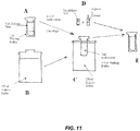
- the erythrocytes then are suspended at 70% hematocrit (Ht) in the washing buffer solution inside a dialysis tube having a cut-off of 12-14 kD; this is done by injecting the syringe contents from the above step through the top and into the bottle containing the dialysis tube (see FIG. 11, A ).
- the erythrocyte-containing dialysis tube is then removed from its bottle by lifting off the bottle top (to which the dialysis tube is attached) and transferring it to the large container ( FIG. 11,B ).
- the small lid of the large container is removed, and the dialysis bag is inserted through the opening and pressing it down to seal the container (see FIG. 11, C ).
- RBCs is dialysed for 90 min against 50 volumes of dialysis buffer (10 mM NaH 2 PO 4 , 10 mM NaHCO 3 and 20 mM glucose, pH 7.4) containing 3 mM reduced glutathione and 2 mM ATP.
- the osmolality of the buffer is about 60 mOsm, whereas that of the RBC solution reached about 87 mOsm at the end of the dialysis time. All these procedures are performed at 4°C.
- the dialysis tube is then removed from the large container and returned to its original bottle, at which time 1 ⁇ mol of ICG dye is then added to each millilitre of the dialysed RBC solution (see FIG. 11,D ), and the mixture is incubated for 30 min at 70°C under gentle agitation. (The correct amount of lyophilised ICG is provided in a small vial to which a per-measured volume is added.)
- the erythrocytes are resealed by adding 0.1 vol of, 100 mM inosine, 20 mM ATP, 4 mM MgCl 2 , glucose anhydrous 100 mM, sodium pyruvate 100 mM, 190 mM NaCl, 1666 mM KCI and 33 mM NaH 2 PO 4 (pH 7.4) per volume of dialysed erythrocyte solution (this solution is pre-measured and provided in a sterile ampoule).
- the dialysis bag in its bottle is placed in a small bath at 37°C, and the cells are incubated for 25 min.
- the resealed cells are washed 4 times in the washing buffer (50 ml each time) and centrifuged each time for 10 min at 500 g; there is a cell recovery of about 40%.
- ICG encapsulation in human erythrocytes by a procedure of hypotonic dialysis, isotonic resealing and re-annealing similar to the method described in Example 1 can be carried out without recourse to a dialysis step. This is done by decreasing the tonicity of the solution in which erythrocytes are suspended from 300 mOsm/kg to 87 mOsm/kg in four stages to open pores in the cells' membranes. These stages are all carried out in the same 50-ml centrifuge tube, at the completion of which ICG dye can be introduced to the cell suspension solution.
- a kit consisting of various sterile containers and pre-measured fluids is utilised. In this case, the a kit has different components than indicated in the one depicted in FIG. 9 ; this kit consists of:
- the erythrocytes are then washed twice in 10 mM HEPES (pH 7.4) containing 154 mM NaCl and 5 mM glucose (washing buffer) to remove leukocytes and platelets, centrifuging at 2000 g for 5 minutes each time. This is done by injecting the erythrocytes in the syringe, from the above step, into the first of two 15-ml vacutainer pre-loaded with 10 ml of washing solution and centrifuged, again at 2000 g for 4 min.. Once the eythrocytes are again concentrated in the tube bottom, they are removed using one of the long needles and a syringe (see FIG. 10, B ) and transferred to the second 15-ml vacutainer pre-loaded with washing solution, centrifugation is repeated and the concentrated erythrocytes again collected in a syringe.
- the pre-measured 10 ml volume of 99 mOsm/kg washing buffer is added to a 50-ml vacutainer and gently agitated for an additional 20 min.
- the pre-measured 20 ml volume of 49 mOsm/kg washing buffer is added to a 50-ml vacutainer and gently agitated for a final 20 min.; this will produce a mixture at about 87 mOsm/kg, causing pores in the erythrocytes to opened.
- the pre-measured 1.5 ml volume of distilled H 2 O is added to the vial containing 29.8 mg vial of lyophilised ICG to reconstituted it, and the liquid dye is then added to the contents of the 50ml vacutainer, resulting in a dye concentration of 1 ⁇ mol/ml.
- the 50-ml vacutainer is gently agitated in a 37°C bath for 20 min.
- the erythrocytes are resealed by adding the pre-measured 3.8 ml volume of, 100 mM inosine, 20 mM ATP, 4 mM MgCl 2 , glucose anhydrous 100 mM, sodium pyruvate 100 mM, 190 mM NaCl, 1666 mM KCI and 33 mM NaH 2 PO 4 (pH 7.4) to the solution in the 50-ml vacutainer to make the mixture normotonic; it is agitated and kept in the 37°C bath for 20 min.
- the 50-ml tube is centrifuged at 2000 g for 5 min, after which one of the supplied long needles and a syringe are used to remove the 3-ml volume of cells at the bottom of the tube and transfer them to a fresh 50-ml stoppered tube containing 40 ml of washing buffer. This tube is agitated and centrifuged at 2000 g for 4 min.
Description
- Disclosed herein are kits, compositions, and methods for the clinical use of erythrocytes in the fields of medical angiography and therapy. The erythrocytes have been pre-loaded with substances for observation of blood flow under physiological conditions to detect circulation abnormalities. The erythrocytes may also be used for delivery of therapeutic substances to localized vascular areas. This technology can be applied to any vasculature that can be directly visualized by an optical means, such as ocular vasculatures.
- Medical angiographic imaging typically involves administration of a detectable substance to a subject (see
U.S. Patent No. 6,915,154 ). In some instances the detectable substance may also be a therapeutic agent (seeU.S. Patent Publication No. 2004/0206364 ). Most often the detectable substance is administered directly to a subject by intravascular injection, in which case the detectable substance mixes with and is carried through the vasculature by plasma, along with the blood cells. When using conventional angiography methodology, wherein the detectable substance is a liquid dye, blood flow physiology is treated too simplistically, especially at the microvascular level (i.e., the arterioles, capillaries, and venules). - Blood is a shear-thinning, non-Newtonian fluid. However, in diagnostic applications, blood often is treated as if it were water-like (i.e., a Newtonian fluid), and not an homogeneous mixture of two distinctly different non-Newtonian fluids: (1) liquid (plasma) and (2) particles (blood cells, especially erythrocytes). Limitations inherent in conventional angiography contribute to ignoring that the dynamics of plasma movement do not necessarily reflect the dynamics of erythrocyte movement, especially at the microvascular level where their movement is far more important to the circulation's metabolic efficiency than that of plasma. For example, in conventional sodium fluorescein and indocyanine green angiography (SFA and ICGA) of the ocular vasculatures, observed fluorescence arises from dye molecules associated primarily with blood plasma, not erythrocytes. Even in capillaries, where they deform in order to pass through one-at-a-time in boxcar fashion, erythrocytes cannot be seen in conventional angiogram images, so metabolically significant phenomena such as vasomotion, which results in periodic suspension of erythrocyte movement through individual capillaries, cannot be directly visualized. Yet, it has been postulated that all the clinical findings concerning edema in diabetic maculopathy can be explained as a result of disturbances in retinal vasomotion (Bek 1999, Acta Ophthalmol Scand 77:376). Moreover, in conventional angiography, dye molecules leave the plasma and become associated with vessel walls, so those blood vessels rapidly exhibit steady-state fluorescence, thereby obscuring further visualization of blood movement. Consequently, conventional fluorescent dye angiography is limited as a diagnostic tool, since what hemodynamic information it conveys is misleading with respect to metabolic efficiency and capacity of microvascular blood flow. An example of this would be relying on observation of conventional angiograms to assay the metabolic capacity of blood flow through a choroidal neovascular (CNV) membrane. Due to the well-known phenomenon of plasma skimming (likely to occur where the CNV feeder vessel arises at an acute angle from the choroidal arterial vessel feeding it), only erythrocyte-deficient plasma would profuse the CNV, but this deficiency would not be reflected in the angiogram images since the fluorescence arises only from dye in the plasma, not the erythrocytes.
- Alternatively, a detectable substance (e.g., sodium fluorescein dye) has been administered in a particle carrier, heat sensitive liposomes (Kiryu et al. 1994, Invest Opthalmol Vis Sci 35:3724). However, such artificial particles are rigid and are small to assure that they can pass unobstructed through the smallest capillary vessels. They may not serve as faithful models of erythrocyte dynamics.
- Disclosed herein are kits, compositions, and methods that take advantage of the ability of erythrocytes to be preloaded with various substances, such as fluorescent dyes that facilitate medical imaging. Human erythrocytes, despite their large diameters and volumes, readily conform to the small capillary diameters, and they have been demonstrated to possess properties that make them useful as carriers of molecules other than haemoglobin. Erythrocytes are capable of reversible deformation, such as occurs when they are in hypotonic solution; their volumes increase, causing 200-500 A pores to open transiently in the cells' membranes (Seeman, 1967, J Cell Biology 32:55), allowing two-way trans-membrane exchange between their normal content (haemoglobin) and high-molecular-weight substances placed in their externally vicinity. Then, by returning the solution to normotonicity, the pores close, and the cells return to normal size, trapping the added substances inside; remaining non-entrapped substance can be washed away, leaving substance-loaded osmotically competent erythrocytes; these cells contain reduced amounts of hemoglobin, rendering them incompetent-or at least inefficient-with regard to oxygen delivery. Nevertheless, substance-loaded erythrocytes (S-IEs) appear to have a normal life span of up to 120 days, and they have been used for studying membrane morphology, physiology and biochemistry (see, e.g., Wu et al., 2005, Biochem Pharmacol 69(8): 1257; Rodnenkov et al., 2005, Pathophysiology 11 (4):209). S-IEs have been used in the field of medical imaging (see, e.g., Thelwall et al., 2002, Magnetic Resonance in Medicine 48:649; Kleszcynska et al., J. Flouresc. 15(2):137).
- One embodiment relates to the discovery that the amount of ICG dye inserted into each erythrocyte can produce detectable fluorescence without exceeding safe levels of retinal illumination. Another embodiment relates to the discovery that the amount needed to optimally induce fluorescence and the much larger amount needed to absorb sufficient energy to enhance photocoagulation are mutually exclusive. Yet another embodiment relates to the re-sealing of the cells of the substance-loaded erythrocytes.
- Use of erythrocytes as a drug delivery system has been investigated (see, e.g., Rossi et al. 2001, Biotechnol Appl Biochem 33:85; Magnani et al., 1998, Biotechnol Appl Biochem 28:1). In the method disclosed, erythrocytes that had been loaded with various therapeutic substances were autologously re-injected into a subject and were subsequently distributed throughout the body; there they continuously released the encapsulated substance as the erythrocyte population gradually underwent normal cell death over a span of about 120 days. Although useful for delivery of certain drugs where maintenance of some level of substance throughout the circulation is desirable, this method does not readily facilitate targeted release of therapeutic substances in high concentration. Therefore, its use for delivery of substances to targeted vascular areas requiring therapeutic concentrations too high to be tolerated throughout the body may be prohibited. Another embodiment relates to the discovery that in situations where the targeted vascular area is optically accessible (e.g., the ocular vasculatures or the vasculatures of hollow organs such as the bladder), erythrocytes loaded with appropriate substances can be lysed by means of optical delivery of appropriate radiation, thereby delivering their entrapped contents to precisely localized areas.
- Also disclosed herein are kits and methods for the relatively facile preparation of substance-loaded erythrocytes (S-IEs) for use in clinical application for autologous re-injection. Altenatively, pre-loaded erythrocytes suitable for homologous re-injection in an off-the-shelf form can be prepared. To that end, disclosed herein are methodologies for producing and stabilizing substance-loaded cells, for both diagnostic and therapeutic applications in human subjects, which can obviate need for end-user access to extensive laboratory facilities in order to obtain and process cells under blood-banking sterile conditions.
- Another embodiment relates to the discovery that S-IEs having increased fluorescence (beyond that which can be achieved by optimal loading of dye alone into each erythrocyte) for cell detection can be achieved by incorporation of metallic silver colloids.
- Another embodiment provides novel approaches to detection and controlled release of therapeutic substances encapsulated in S-IEs that result in delivery of high substance concentrations with respect to targeted vascular areas, but which at the same time amount to micro-dose concentrations with respect to the circulation as a whole; this makes possible use of substances that would otherwise be rejected due to the significant systemic toxicity they demonstrate when delivered by conventional intravenous injection. Various substances and combinations thereof can be pre-loaded into erythrocytes in ways that take into account a spectrum of desired biological, chemical, and physical properties of those elements in ways without which those combinations would not produce useful results.
- One embodiment relates to the use of pre-loading substances into erythrocytes and use of S-IEs in ophthalmic diagnostic angiography and drug delivery. Alternatively, this technology can be applied to other vasculatures that can be directly visualized, as well as to other substances that facilitate similar ends in those other vasculatures.
- Various embodiments of the invention will be understood from the following description, the appended claims and the accompanying drawings, in which:
-
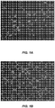
FIGs. 1A-1D are near-infrared fluorescence micrographs at 40x magnification of erythrocytes, comparing the brightness of cells "stained" with ICG (A - stained in 1 mM ICG solution for 30 min. and B - stained in 2mM ICG solution for 60 min. to cells into which ICG has been loaded through pores in their membranes; C - stained plasma resulting from exposure for 20 min. to the stained erythrocytes from A; D - dialysed in 1 mM ICG solution for 30 min.); -
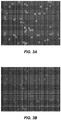
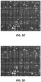
FIGs. 2A-C are near-infrared fluorescence micrographs at 40x magnification of erythrocytes into which ICG has been loaded, demonstrating the effect of introducing different dye concentrations into the dialyzed cell suspension; -
FIGs. 3A-3D are near-infrared fluorescence micrographs at 40x magnification comparing brightness of ICG-"stained" (A = initial brightness, B = brightness at 20 s) and ICG-loaded erythrocytes (C= initial brightness, D = brightness at 20 s) after continuous exposure to the same illumination, due to bleaching; -
FIG. 4A is a near-infrared fluorescence micrograph at 40x magnification of ICG-loaded human erythrocytes; -
FIG. 4B is a photograph of a vial containing freeze-dried ICG-loaded human erythrocytes; -
FIG. 4C is a near-infrared fluorescence micrograph at 40x magnification of freeze-dried ICG-loaded human erythrocytes reconstituted by addition of 1 mL of H2O; -
FIG. 5A is a sodium fluorescein angiogram image (left) and a conventional ICG angiogram image (right) for orientation of a patient's normal right eye; -
FIG. 5B is an angiogram image of ICG-loaded erythrocytes of the patient's normal right eye; individual erythrocytes appear as white dots; -
FIG. 5C is a sodium fluorescein angiogram image (left) and a conventional ICG angiogram image (right) for orientation of the patient's diseased left eye; -
FIG. 5D is an angiogram of ICG-loaded erythrocytes of the patient's diseased left eye; individual erythrocytes appear as white dots; -
FIGs. 6A and 6B are angiogram images from the same rabbit eye comparing ICG fluorescence in the choroidal vasculature following conventional intravenous injection of a bolus of aqueous ICG dye (6A) and following injection of an identical bolus containing silver colloid (6B); -
FIG. 7 is an infrared fluorescence image comparing the fluorescence enhancement effect of silver colloid in 0.05 mg/mL ICG solutions; samples in the lefthand side contain no colloid, while those in the right-hand side contain colloid; samples at the top are in 1-cm thick tubes, while those at the bottom are 100 microns thick; -
FIG. 8 is an infrared fluorescence image of the samples ofFIG. 7 showing reflection of the 805-nm wavelength light used to stimulate fluorescence; no barrier filter was placed in front of the CCD camera in this case; -
FIG. 9 is a schematic diagram of an embodiment of a kit containing sterile containers used in substance loading of erythrocytes (see Example 1); -
FIG. 10A is a schematic representation of the steps involved in removing a plasma- and leukocyte-free volume of erythrocytes from the bottom of the blood sample collection vacutainer after centrifugation, using a long needle; -
FIG. 10B is a schematic representation of steps involved in removing a volume of erythrocytes from the bottom of the two 15-ml washing solution tubes after centrifugation, using a long needle; and -
FIG. 11 is a schematic representation of the sequence ordered steps (A-E) detailing the use of those particular containers form the kit depicted inFIG. 9 to accomplish dialysis of the erythrocytes, including addition of ICG dye to the solution once dialysis is complete and pores in the cell membranes are open. - One embodiment relates to encapsulation of fluorescent dyes in erythrocytes for diagnostic observation. Optionally, the erythrocytes can be used for therapeutic substance delivery. In the former application, re-injection of S-IEs can improve performance of angiography by imaging movement of fluorescent erythrocytes, rather than dye-tagged plasma. In the latter application, the impetus for substance encapsulation in the cells is that many desirable substances do not bind well to the outer cell membrane (as is true for ICG dye). However, encapsulation of those substances in erythrocytes and localized release by laser-induced lysis facilitates delivery of high substance concentrations at the targeted areas, which amount only to micro-dose concentrations with respect to the circulation as a whole. This makes possible use of substances that otherwise may have had to be rejected. In the former application of diagnostic observation, the association of fluorescent dyes with erythrocytes may depend on one or more of three circumstances, each of which relates to why dye insertion into the cell rather than simply "staining" the outside of the cell is useful:
- (1) Although many fluorescent dyes, such as ICG dye, do bind to the erythrocyte outer cell membrane, binding is weak and non-covalent, and regardless of the dye concentration and conjugation time, the maximum fluorescence brightness that can be achieved is considerably less than optimum (see
FIG. 1 ). The erythrocytes in Frame B were "stained" by conjugation with ICG dye in a solution at twice the dye concentration and for twice as long as the cells in Frame A; yet the fluorescence brightness of both is the same since there only are a finite number of available cell-surface binding sites, regardless of the abundance of dye molecules in a particular solution. Frame C demonstrates that once "stained" cells come into contact with plasma proteins, ICG molecules are more attracted to plasma proteins than to the cell membrane, so "stained" cells become even less bright during circulation; this does not occur with dye-loaded cells. - (2) Exposure to fluorescence excitation energy for diagnostic purposes must not exceed the safe maximum permissible exposure level for the sensory retina. Since ICG dye is a fairly weak emitter of fluorescence, care must be taken to ensure that the amount of ICG present post-loading is optimal, resulting in maximum fluorescence intensity possible at a given safe excitation energy level. This condition can be met by insertion of dye into the erythrocytes.
FIG. 1 , Frame D, demonstrates that ICG-loading of erythrocytes by dialysis in a dye solution having the same lower concentration used to stain the cells in Frame A. are considerably brighter; there are considerably more binding sites within a cell volume (including those associated with residual haemoglobin) than on its external surface. Moreover, since ICG is subject to concentration fluorescence quenching-even when inside erythrocytes-the amount of loaded dye must be controlled: too much or too little will not produce the maximum fluorescence. Equal brightness of cells in A and B demonstrates "staining" by conjugation in even high-concentration dye solution does not achieve the optimum brightness possible by dye insertion (D). Image C demonstrates that the affinity of ICG dye molecules is greater for plasma proteins than for the erythrocyte outer membrane; cells in A, B, and D are suspended in isotonic saline. Achieving optimal dye concentration is accomplished by controlling the amount of dye in the solution during dialysis once the pores in the cell membranes open; experimentation has determined the optimum amount to be approximately 1.0 µ mol/ ml dialysed cell-solution (seeFIG. 2 ). After equilibrium is established between the solutions internal and external to the cells, the pores are closed by returning the hypotonic solution containing the cells to a normotonic level, fixing the entrapped dye concentration. This also has the advantage of preventing plasma dye-staining and the associated reduction of contrast between cell fluorescence and background brightness once the cells are re-injected.FIGs. 2A, 2B, and 2C have concentrations of 0.31 µmol/mL solution, 0.89 µmol/mL solution, and 6.14 µmol/mL solution giving rise to an average 8-bit-gray-scale brightness of 159, > 255, and 165, respectively. - (3) ICG dye is subject to bleaching, potentially making detection and imaging of ICG-loaded erythrocytes difficult, due to the resulting reduced levels of fluorescence for a given level of illumination. This can be a problem, especially when it is desirable to make observations for periods of many seconds, as would be required for observing the effects on erythrocyte movement of vasomotion, wherein individual cell motion may be temporarily halted for up to 30 sec. It has been experimentally determined that, whereas ICG dye-"stained" cells are susceptible to bleaching, ICG-loaded cells are not (see
FIG. 3 ). Again, it is the limited number of dye molecules on an erythrocyte's outer membrane, compared to the number within the cell volume that makes this phenomenon significantly visible in the "stained" cells, but not in the loaded cells. - In one embodiment, encapsulation of various substances in human erythrocytes is accomplished by a procedure of hypotonic dialysis, isotonic resealing and re-annealing. Placing washed erythrocytes in a hypotonic solution causes pores that can have a size ranging from 200-500 A to open transiently in the cell membranes, thereby allowing substances (such as ICG dye) in the solution to cross the membranes. Making the hypotonic solution normotonic causes the pores close, and the cells (now with reduced hemoglobin content) return to an osmotically competent state, trapping the substances inside. Remaining un-entrapped substances can then be washed away, leaving only substance-loaded erythrocytes (S-IEs).
- In one embodiment, the dye entrapping (or encapsulation) is as follows. Fresh blood in acid-citrate-dextrose anticoagulant is obtained under sterile conditions and centrifuged to obtain erythrocytes. These are then washed in a buffer to remove surface proteins, leukocytes and platelets and then centrifuging. The erythrocytes are then suspended at about 70% hematocrit (Ht) in the washing buffer solution inside a dialysis tube, where they are dialyzed against a dialysis buffer. ICG dye is then added to the dialyzed erythrocyte solution, and the mixture is incubated under gentle agitation. The erythrocytes (now with reduced hemoglobin content) are then resealed by returning the dialyzed erythrocyte solution to a normotonic state and incubating the cells. The resealed cells are washed several times and centrifuged each time. About 9 ml of whole blood yields 4 ml of packed (about 80% Ht) ICG-loaded erythrocytes. This same basic methodology can be elaborated to satisfy several approaches to providing S-IEs to be used in several different circumstances:
- One embodiment provides a method for an end-user who wants to use S-IEs for a specific subject, using the subject's own erythrocytes, or for introducing substances in a desired combination or concentration.
- The invention provides a method comprising:
- (a) providing erythrocytes in a blood anti-coagulant solution;
- (b) dialyzing the erythrocyte-containing solution in (a) against a dialysis buffer having an osmolality ranging from 50-70 mOsm/kg to cause pores in the erythrocyte to open;
- (c) combining the dialyzed solution in (b) with at least one fluorescent dye which is indocyanine green dye and a substance that inhibits destructive formation of ice crystals; and
- (d) combining the dye-containing solution in (c) with a resealing solution having an osmolality of at least 1000 mOsm/kg to reduce the osmolality, thereby causing the erythrocyte pores to close and entrap the at least one fluorescent dye and the substance within the erythrocytes; further comprising the step of freeze-drying the erythrocytes containing the entrapped dye and the substance.
- In one embodiment, the providing in step (a) comprises obtaining a blood sample from a subject.
- In one embodiment, the dialyzed solution in (b) has an osmolality ranging from 80-90 mOsm/kg, e.g., an osmolality ranging from 85-87 mOsm/kg.
- In one embodiment, the method further comprises washing the erythrocytes with an isotonic solution prior to the dialyzing in (b).
- In one embodiment, the at least one fluorescent dye in (c) is indocyanine green dye having a concentration ranging from 0.25 to 3.0 µmoles/mL of dialyzed solution.
- A kit comprising the following may be used:
- a blood anti-coagulant;
- a dialysis buffer having an osmolality ranging from 50-70 mOsm/kg;
- a dialysis chamber;
- at least one fluorescent dye; and
- a resealing solution having an osmolality of at least 1000 mOsm/kg.
- In one embodiment, the dialysis chamber has suspended from its top a dialysis tube with a molecular weight cut-off ranging from 12,000 to 14,000 Daltons. In one embodiment, the dialysis tube allows introduction of a blood-containing fluid into the tube, and a dialysis fluid can be introduced outside the suspended tube.
- Small quantities of erythrocytes, such as would be needed for autologous re-injection into a single individual can be prepared under sterile conditions, using his own blood, in about 1.75 hours time. However, since laboratory sterile conditions are seldom conveniently available to locations where frequent diagnostic and therapeutic procedures are carried out, a convenient way to prepare or acquire the erythrocytes is a requirement if such procedures are to be broadly applied. One embodiment provides kits (one for each individual subject) each consisting of a series of disposable sterile containers, pre-loaded with appropriate amounts of chemicals. A freshly obtained volume of blood can be transferred, in sequence from one to another, until the substance-loading steps are completed (see
FIGs..9 ,10 , and11 ). S-IEs prepared in this manner can be stored for use for several days at 4°C, until used. - A small refrigerator containing a small fixed-speed, fixed- time centrifuge can be used. The refrigerator space (accessible from its top, to keep the cold air reasonably contained) can be large enough to hold and pre-cool a kit and to provide a cool sterile workspace, as well as a place to temporarily store prepared S-IEs until used.
- The advantage of this basic method of preparation is ad hoc introduction of substances or mixtures of substances (i.e., simultaneous introduction of two or more discrete substances, each at a different concentration). Also, autologous re-injection of a subject's cells assures that on contamination of incompatibility of human protein will be encountered. Although no further processing is required, so long as the cells are stored at proper temperature and that they are used within several days, the cells may be preserved for much longer periods by some of the methods described below. (See Examples 1 and 2)
- Accordingly, disclosed herein is also a kit, comprising:
- a blood anti-coagulant;
- a plurality of hypotonic solutions;
- at least one fluorescent dye; and
- a resealing solution having an osmolality of at least 1000 mOsm/kg.
- The kit disclosed herein provides a plurality of hypotonic solutions to successively diluting a blood sample and achieve a final osmolality ranging from 80-90 mOsm/kg, e.g., ranging from 85-87 mOsm/kg. The plurality of solutions may comprise at least three or at least four hypotonic solutions, each having a different osmolality. In one embodiment, the plurality of solutions can have the same or different osmolalities, so long as the maximum osmolality any of the solutions is 300 mOsm/kg and the minimum osmolality of any of the solutions is 50 mOsm/kg.
- In any kit embodiments disclosed herein, the kit may further comprise an isotonic saline washing solution comprising glucose or trehalose, e.g., a 5 mM glucose solution or at least a 50 mM trehalose solution (e.g., a 50 mM trehalose solution).
- Another method disclosed herein comprises the following steps:
- (a) combining erythrocytes with a blood anti-coagulant to form a solution having an osmolality of at least 300 mOsm/kg;
- (b) successively diluting the solution in (a) with at least three hypotonic solutions to the reduce the osmolality to less than 100 mOsm/kg to cause the erythrocyte pores to open;
- (c) combining the diluted solution in (b) with at least one fluorescent dye; and
- (d) combining the dye-containing solution in (c) with a resealing solution having an osmolality of at least 1000 mOsm/kg to reduce the osmolality, thereby causing the erythrocyte pores to close and entrap the at least one fluorescent dye within the erythrocytes.
- In one embodiment, the kits or methods disclosed herein include an isotonic resealing solution having an osmolality of at least 2000 mOsm/kg.
- In one embodiment, after performing any method disclosed herein, after step (d), the method further comprises:
- (e) washing the erythrocytes in an isotonic saline washing solution containing glucose or trehalose to remove extracellular dye.
- In one embodiment, the washing is performed with a saline solution containing 50 mM trehalose.
- In one embodiment, the kits or methods disclosed herein provide at least one fluorescent dye, in particular indocyanine green (ICG) dye. In one embodiment, the dye concentration of a composition comprising the entrapped dye ranges from 0.25 to 1.5 mM. In another embodiment, the dye concentration ranges from 0.3 to 0.4 mM. In one embodiment, this concentration optimizes fluorescence intensity to facilitate visualization of the cells. In yet another embodiment, the dye concentration is greater than 0.4 mM. This concentration can facilitate absorption by the cells of of infra-red wavelengths, thereby rendering the cells capable of lysing upon heating.
- In one embodiment, the source of erythrocytes is O-type blood. In another embodiment, the erythrocyte source blood of A-type or B-type. In the latter embodiment, the method can further comprise adding α - β glucosidase to split off the erythrocyte surface A- and B-agglutinogens.
- In one embodiment, the composition comprising the dye entrapped in the erythrocytes further comprises at least one biocompatible excipient. In one embodiment, the at least one excipient is polysucrose. In one embodiment, the at least one excipient concentration ranges from 0.5% to 5%.
- One embodiment provides a method for producing S-IEs for end-users who want access to S-IEs without having to acquire blood from their subjects. This method can be performed on a large scale and can supply multiple end-users.
- In situations where application of the above basic methodology may be inconvenient due to constraints of facilities location or time, it is possible to instead make use of S-IEs obtained from a source other than the subject. Using the basic methodology, aliquots from large volumes of erythrocytes from human O-negative blood type donors pre-loaded with various substances or mixtures of substances, can be slowly frozen and maintained at cryogenic temperatures until needed for universal injection. Such an approach can be used to create substance-loaded cells that can be universally injected into human subjects, while obviating the necessity to first obtain the subject's own blood and the time required for perpetration. This approach is particularly suited to preparations of cells loaded with substances likely to be used frequently in numerous subjects, such as for performance of standardised diagnostic or therapeutic procedures, where ad hoc use of substances is not contemplated.
- Alternatively, the pool of subjects that forms the source of erythrocytes for bulk preparation of universally injectable substance-loaded erythrocytes can be expanded beyond those having only type O-negative by using α - β glucosidase to split off the cell surface A- and B-agglutinogens at the beginning of the cell-loading process. There appears to be nothing definitive in the literature to indicate the threshold level at which any cell membrane surface antigens not removed by washing or other treatment would induce an immunologic reaction. Since it is anticipated, especially with regard to diagnostic use, that the maximum volume of processed cells re-injected will be on the order of only 1 or 2 ml, there is minimal likelihood of inducing an immunologic reaction.
- Freeze-drying S-IEs prepared for universal injection into which the desired substances have been inserted at desired concentrations results in an off-the-shelf preparation that is easily stored at room temperature for prolonged periods (in excess of a year). Reconstitution requires only addition of sterile water, making such a product the most convenient form of injectable S-IEs. This would appear to be the most convenient method for rendering the substance-loaded cells stable and easily reconstituted.
- Past attempts to develop a method for freeze-drying erythrocytes such that reconstitution of the cells results in erythrocytes with an intact cytoskeleton and with biologically active hemoglobin (i.e., completely biologically viable cells) have proven illusive; the fact that such a preparation is not readily available underscores this. Generally, when erythrocytes have been lyophilized by previous methods, for example in either an aqueous or phosphate-buffered saline (PBS) solution, the reconstituted cells are damaged to the extent that they are not capable of metabolizing (that is, the cell hemoglobin cannot carry oxygen), and/or the yield of intact, non-deformed cells is unacceptably low. A factor in causing the damage may arise in the freezing process, where ice crystals form internal and external to the cells, thereby disrupting cell structure. Attempts to obviate this problem seem mainly to have included procedures that utilize cooling rates that are faster than ice crystal formation (see for example, United States Patent
5656498 ), or inclusion in the cell suspension fluid a monosaccharide which may or may not diffuse into the cells, the presence of which suppresses ice crystallization during cooling (see for example, United States Patent5340592 ). However, it appears that these approaches have failed to produce sufficiently high yields of functional cells upon reconstitution to be useful in the clinical arena. - In the methods disclosed herein, containing composition comprising the entrapped dye is frozen or freeze-dried. Accordingly, disclosed herein are frozen or freeze dried (lyophilized) compositions comprising a freeze dried (lyophilized) composition comprising at least one fluorescent dye entrapped within erythrocytes.
- In the method of the invention, the erythrocytes in (d) further entraps a substance (e.g., while in a hypotonic state) that inhibits the destructive formation of ice crystals during cooling. In one embodiment, the substance is a monosaccharide, e.g., trehalose. Accordingly, in one embodiment, erythrocytes can further entrap trehalose or at least one therapeutically effective agent..
- One embodiment relates to the recognition that obviating the problems encountered by these various previous attempts lies in recognition of two advantages associated with the methods of use and production of substance-loaded erythrocytes with respect to use and production methods aimed at long-term storage of metabolically functional blood. The first is that production of metabolically normal O2-carrying cells upon reconstitution is not important for the diagnostic and treatment applications of S-IEs; in fact, replacement of a portion of the cells' hemoglobin content is part of the loading process. Secondly, the methods of S-IE production all involve opening of pores in the cells' membranes to allow equilibrium between the cells' internal contents and the solution in which the cells are suspended. This latter situation readily facilitates controlled introduction of an adequate amount of monosaccharide (preferably, trehalose) to prevent ice crystal formation during the freeze-drying process; this is accomplished by substituting the 50 mM trehalose for the 5 mM glucose in the washing buffer solution composition described in the Example 1 Ad-hoc Preparation of ICG dye-loaded Human Erythrocytes for Autologous Re-injection. Once this is done, the finished substance-loaded cells are diluted in to a 50% "hematocrit" in the same trehalose washing buffer, polysaccharide (5%) is added as an excipient to reduce cell clumping upon reconstitution, when 1 ml of distilled water is added to the freeze-dried pellet in the stoppered vial. As demonstrated in
FIG. 4 , the reconstituted ICG-loaded human erythrocytes are equivalent in fluorescence intensity to that they possesses prior to freeze-drying. - Medical angiographic imaging typically involves administration of a detectable substance to a subject, as described, e.g., in
U.S. Patent Nos. 6,915,154 and6,351,663 . In some instances the detectable substance may also be a therapeutic agent, as described in,
e.g.,U.S. Patent Publication No. 2004/0206364 . - Aside from their role in supplying the metabolic requirements of the tissues they serve, among the naturally occurring particles in blood, as well as the artificial ones that have been introduced into blood circulation for various purposes, erythrocytes are well suited for use as substance carriers. They are the largest naturally occurring blood-borne particles, and from a hemodynamic point of view, they are essentially as passive as serum, adding only resistance to blood flow because of their size and mass. (By comparison, leukocytes and thrombocytes are biologically active, so their movements in flowing blood are influenced by other than serum fluid dynamics. For example, leukocytes, which are part of the immune system, exhibit a drag-and-roll behavior as they pass the endothelial surface of vessel walls.) Moreover, despite their large volumes, S-IEs retain the capability of the original erythrocytes to deform, allowing them to pass through small diameter capillaries; this feature overcomes the size limitation inherent in use of artificial carriers (such as liposomes) whose geometry always is rigid. These attributes of erythrocytes make them ideal for transporting the fluorescent dyes used in angiography. Unlike conventional angiograms showing dye-stained plasma movement, which often show a poorly defined wave front that transits only once, angiograms showing individual fluorescent erythrocytes can provide continuous information about blood flow speed and direction in a multitude of vessels simultaneously. There are several aspects of creating and using dye-loaded erythrocytes (e.g., ICG-loaded erythrocytes) for angiography:
- 1. Dye-loaded erythrocytes, rather than dye-stained ones, are desirable because dye binding to the outer erythrocyte membrane appears to be weak (i.e., not covalent binding). Energy imparted to those cells "stained" with dye as a result of turbulent blood flow, for example, is sufficient to break those weak bonds. Then dye molecules become available to bond with other nearby proteins, quickly resulting in plasma and blood vessel wall staining that diminishes contrast between the erythrocytes and their background, while at the same time diminishing the brightness of the erythrocytes themselves. Moreover, it is not possible to bind enough dye to the cell membranes to achieve the peak fluorescence intensity needed for adequate visualization of individual cells (see
FIGs. 1A and 1B ). On the other hand, the dye-loading process permits control of the amount of dye inserted into erythrocytes, such that the dye concentration inside the cells is very near--but does not exceed-the concentration at which fluorescence quenching occurs(cf. 1A and 1B to 1D). In the case of ICG, this is achieved by addition of 1µmol of ICG dye to each millilitre of the dialysed erythrocyte solution at that point in the substance-loading procedure when the volumes of the cells have increased to the extent that pores in the cells' membranes have opened (seeFIG. 2 ). With the pores open, trans-membrane exchange of large molecules occurs; haemoglobin inside the cells moves to the outside until a dynamic equilibrium between inward and outward haemoglobin movement is reached. ICG dye added to the solution in which the cells are suspended binds with hemoglobin molecules, and in time, those molecules move into the cells and reach concentration equilibrium with those outside the cells. When the pores in the cells are closed and dye/haemoglobin molecules outside the cells are washed away, the remaining dye-loaded cells fluoresce optimally when stimulated by appropriate excitation light energy. - 2. Once filled with dye-stained plasma, as in conventional fluorescent dye angiography, blood vessels exhibit only steady-state fluorescence, inhibiting visualization of any blood-borne particles, even if the particles themselves were fluorescent. However, it is possible to visualize individual erythrocytes in capillaries if they, rather than the plasma, are emitting fluorescence and if separation between adjacent erythrocytes exceeds the diffraction limited resolution of the optics of the eye (about 7 µm for the perifoveal capillaries and 11 µm for the choriocapillaris (see Flower RW. Optimizing treatment of choroidal neovascularization feeder vessels associated with age-related macular degeneration. Am J. Ophthalmol. 2002; 134:228-239. 8). These conditions were met by entrapping ICG dye in erythrocytes, as described above, and re-injecting a small bolus of them. During transit to the ocular vasculatures, they are diluted in circulating blood to the extent that individual ICG-loaded cells were separated by more than 11 µm, making visualization of them possible. It has been discovered that using erythrocytes loaded so as to produce optimal fluorescence produces the strongest fluorescence signal for any given level of excitation light irradiance. For reasons of safety, it is desirable to remain as far below the maximum permissible exposure (MPE) for a given tissue as possible.
- 3. Visualization of circulating dye-loaded erythrocytes requires use of a device that irradiates the tissue field-of-view of interest with light energy at an appropriate wavelength (optimally, 805 nm for ICG). This can be efficiently done when that energy is applied as high-frequency trains of high-peak-power, short duration pulses of light, each of which illuminate the entire field-of-view of interest. This configuration results in a high image signal-to-noise ratio by requiring the lowest signal amplification, thereby minimizing introduction of electronic noise associated with amplification. Additionally, acquiring images during very short time periods in synchrony with short duration pulses of illumination acts essentially to freeze motion within each image, thereby obviating blurring associated with movement of erythrocytes within the field of view (FOV).
- The efficacy of using dye-loaded erythrocytes has been successfully demonstrated in both rabbit and non-human primate eyes 9. (see Flower R, Peiretti E, Magnani M, Rossi L , Serafini S, Gryczynski Z, Gryczynski I., Observation of erythrocyte dynamics in the retinal capillaries and choriocapillaris using ICG-loaded erythrocyte ghost cells. Invest. Ophthalmol. Vis. Sci. 2008; 49: 5510-5516). Moreover, the results reported validated the prediction by Bek that all the clinical findings concerning edema in diabetic maculopathy
can be explained as a result of disturbances in retinal vasomotion 10. Results have also been obtained from the first human subjects in a preliminary clinical study to demonstrate feasibility of autologous re-injection of ICG-loaded erythrocytes for angiography (seeFIG. 5 ). The human study protocol required injecting very small volumes of ICG-loaded cells in the first subjects and gradually increasing the volume in subsequent ones, until reaching a level equivalent to that used in the monkeys. Consequently, there are significant differences between the appearance of the published monkey angiograms, where the methodology had been optimized, and these from these first human subjects. The main reason is that the fraction of circulating fluorescent erythrocytes in these first human subjects was about 4-times less than in the monkeys. - Nevertheless, these first human angiograms support some of the findings in the more extensive monkey studies.
FIGs. 5A and 58 are from the normal right-eye of an age-related macular edema subject; FIG. 58 shows a single ICG-loaded erythrocyte angiogram image of the 10°-area field of view. The distribution of stalled erythrocytes is relatively even, consistent with whatwas reported for the normal monkey eye 9. In the diseased left eye (FIGs. 5C and 50), the image from the angiogram is centered over the choroidal neovascular lesion (CNV). The distribution of stalled erythrocytes surrounding the CNV is similar to that of the normal fellow eye, but in the CNV no erythrocytes are apparent. When watching the actual angiogram movie, however, a few erythrocytes eventually do pass through the CNV area; but they do so at about half the speed as those erythrocytes which are not stalled in the area surrounding the CNV or those in the normal eye. (This may be the result of both plasma skimming and of higher CNV resistance to blood flow.) Minimally, the first human angiograms demonstrate that ICG-loaded erythrocytes can be acquired and that apparently the presence of vasomotion-or lack of it-can be determined thereby. Based on Bek's work 10, visualization of erythrocytes can be used as a predictor for retinal edema onset and for monitoring early efficacy of treatment. Also, clearly abiltiy to track individual erythrocytes makes possible routine quantifiable assessment of blood flow in any vessel. - In one embodiment, the methods disclosed herein further comprise the step of adding metallic colloids prior to step (d). In one embodiment, the metallic colloids are silver.
- Metallic silver colloids in close proximity to weakly fluorescing dyes can markedly increase fluorescence yields. It has been shown that if ICG molecules are brought into close proximity by first binding silver colloids to amino groups that have been plated on a glass surface and then binding ICG/albumin conjugates to the same surfaces, fluorescence yields from the ICG increased more than 30-fold 11. Subsequently, experiments were carried to determine if the same enhancement effect would occur in vivo in ICG angiograms made in rabbit eyes.
- Samples of ICG dye alone and of dye/Ag-colloid suspension, containing identical concentrations of dye (0.250 mg/ml) were injected into rabbit ear veins, and ICG fluorescence angiograms recorded. The fundus of the eye was uniformly illuminated by pulsed laser diode light at 805 nm wavelength, and as the dye transited through the ocular vasculatures, the 835 nm wavelength fluorescence light emitted by the dye was digitally recorded. Comparison of dye only and dye/colloid-suspension angiograms from the same eyes were made, using images from the same phase of vascular dye filling in each case. As indicated by the example angiogram images in
FIG. 6 , the fluorescence intensity of the dye/suspension ranged from only two to three times greater than that from dye alone. - The disparity between fluorescence enhancement reported in the experiments made using glass surfaces on which the dye/colloid conjugates were bound with amino groups (30-fold) and those observed in the in vivo angiograms (2- to 3-fold) apparently was due to the fact that the colloid fluorescence-enhancement effect is manifest in very thin layers of the dye/suspension and suppressed in thick ones. This is demonstrated by comparing fluorescence from thick (1 cm) and thin (100 µm) layers of both samples, as shown in
FIG. 7 . In this figure, the thick samples are at the top, and the thin ones at the bottom; 0.250 mg/ml ICG in HAS is on the left, and 0.250 mg/ml ICG in HAS with 4.684 mg/ml Ag colloid on the right. The array of samples was uniformly illuminated with 805-nm wavelength light, and the ICG fluorescence image was captured. In the case of the thick samples, the ICG-only on the left is significantly brighter, while in the case of the thin samples, the dye/colloid suspension is significantly brighter. In part, the explanation for these relationships is that colloids in suspension scatter light, as shown inFIG. 8 , wherein the same array of samples inFIG. 7 is uniformly illuminated with 810 nm wavelength light and an image made from the reflected light. Note that both the thick and thin dye/colloid samples reflected-rather than absorbed or transmitted-the incident light; that is, the observed light interactions with the dye/suspensions arise as a surface phenomenon. Consequently, more dye molecules in the ICG-only thick sample interacted with light than did those in the dye/suspension thick sample. In the case of the two thin samples, however, the fluorescence enhancement in the dye/suspension sample resulted in brighter fluorescence from the ICG dye molecules present than from the same density of molecules in the dye-only sample. This was because more of the incident light was transmitted by the dye-only sample than by the dye/colloid-suspension one. - Based on the above observations, achieving significant ICG fluorescence enhancement in angiography through association of the dye with metallic colloids requires not only that the dye molecules be held in close association with the colloids, but that the dye/colloid conjugation be contained in thin layers. These conditions can be met by containing the dye/colloid conjugates within S-IEs, wherein the small amount of hemoglobin remaining in the cells is sufficient to act as the ICG protein-binding site. Thus, significant and useful enhancement of the dye fluorescence from ICG-loaded EGCs can be achieved by encapsulation of silver metallic colloid during the S-IE loading process.
- In one embodiment, the erythrocytes entrap at least one therapeutically effective agent. Since S-IEs essentially are closed-volume circulating containers in which the concentration of inserted molecules can be precisely controlled, S-IEs can be used to transport their contents, sequestered from the blood, to a given vascular location, where their presence can be detected and their contents released. Such a scenario assumes that the vascular location is visually accessible, that the S-IEs contain a detectable marker (such as a fluorescent dye) in addition to the transported substance, and that a means to release the S-IEs' contents exists.
- Controlled substance release in targeted vascular areas by this method makes possible use of substances that would be prohibited if delivery were by conventional means. This is because when encapsulated in S-IEs and injected in a bolus of packed cells, the aggregate substance volume introduced is very small. So long as that aggregate volume is on the order of 1/100th of the threshold volume for producing a pharmacological effect (therapeutic or toxic), it generally is considered micro-dosing. (In Canada, Europe, and the U.S., now human experimental IND studies often can proceed on the basis of just small animal data. This puts such studies in a range more likely to be affordable by smaller companies.) Therefore, the spectrum of dyes that might be considered potentially usable is greatly expanded when administered as micro-doses.
- S-IEs maintain the shape, behavior, and longevity of the erythrocytes from which they are derived, so they are able to deform in order to pass through small diameter capillaries, making them ideal vesicles for carrying and delivering substances intravascularly. Re-injected substance-loaded erythrocytes can circulate for up to about 120 days (the average lifetime of erythrocytes), during which time their contents will be released by one means or another. Because any population of S-IEs behaves the same as any population of normal erythrocytes with respect to eryptosis, unless externally triggered to do so, only a small fraction of injected cell contents will be released at any particular time, complete release being spread-out over approximately 120 days. And since only very small volumes of cells are injected, levels of molecules released into the body at any point in time-or even in the aggregate-should always be orders of magnitude below effect or toxic thresholds. Moreover, since the distribution of S-IEs eventually would be uniform throughout the total circulating blood volume, further diluting the concentration of gradually released substance to possibly undetectable (or at least, trace) levels.
- During initial transit through the circulation, an injected packed S-IE bolus remains relatively intact. Within a few minutes following injection, the S-IEs become fairly distributed throughout the body, and unless somehow targeted in a specific area of the body, thereafter substance release is gradual over a 120-day period. In vasculatures that can be directly observed by optical means, release of the drug content of circulating S-IEs can be triggered by application of light energy, especially during initial and early transit. Such light energy could easily be focused to cover any desirable area, small enough to cover a single cell or large enough to cover the vasculature of an entire organ.
- Direct visualization facilitates control over the amount of substance released, by taking into account that, for any given area of vasculature anywhere in the body, the density of circulating S-IEs will be greatest during initial transit and will rapidly diminish with passage of time until only a few cells will be present in each capillary of the visualized area. For example, if the light energy were applied to a certain vascular area during the initial passage of the injected cells, released substance concentration would be high; if applied a few minutes later, the concentration would be much less, but always in proportion to the ratio of S-IEs to normal circulating erythrocytes. Even though the aggregate volume of injected substance encapsulated in S-IEs may be at the micro-dose level, when released only within the blood volume of a small area of vasculature, substance concentration could be very high.
- Accordingly, in combination with the methods disclosed herein wherein step (d) results in a first population of dye-entrapped erythrocytes, after step (d) the method further comprises:
- (e) repeating steps (a) and (b) with a second sample of erythrocytes;
- (f) repeating step (c) with at least one fluorescent dye at a concentration higher than that of step (c);
- (g) repeating step (d) with the product of (f) to form a second population of dye-entrapped erythrocytes; and
- (h) combining the first and second populations.
- In one embodiment, the method further comprising adding at least one therapeutically effective agent at step (e) or (f), wherein the second population further comprises agent-entrapped erythrocytes. Numerous therapeutically effective agents can be envisioned for pre-loading. Exemplary drugs include ganciclovir (antiretroviral), triamcinolone acetonide (steroid); fluocinolone and dexamethasone (ocular-specific steroids); pegaptanib sodium and rhuFab V2 (Anti-VEGF, anti-angiogenic); verteporfin (benzoporphyrin derivative) (photodynamic therapy); and carboplatin and topotecan (chemotherapeutic).
- In one embodiment, the at least one dye in the first population has a concentration ranging from 0.3 to 0.4 mM, and the at least one dye in the second population has a concentration greater than 0.4 mM,
wherein upon illumination with 805 nm laser energy, the erythrocytes of the first population will fluoresce, and
wherein increasing the laser energy heats the erythrocytes of the second population due to their enhanced absorption, causing them to lyse and release the entrapped therapeutic agent. - Ability to release an encapsulated substance from S-IEs requires ability to detect or observe presence of the cells in the target area, so that release-stimulating light energy can be applied precisely at the time the desired concentration of cells is present. This is accomplished by including a fluorescent dye (such as ICG) in the cells, along with the therapeutic substance. A software algorithm applied to the video images of the vasculature of interest can be used to track in real time the number--and hence, density--of S-IEs present in the vascular area of interest, and when the desired level is reached, it can automatically trigger application of the release-stimulating light energy. Knowing the density of cells present prior to triggering release may be necessary to assure that not so many cells simultaneously rupture that significant reduction or occlusion of blood flow occurs.
- Accordingly, one embodiment provides a software algorithm applied to the video images of a vascular area of interest that can track in real time the number--and hence, density--of S-IEs present in the vascular area of interest. In one embodiment, when a predetermined level of S-IEs density is reached, application of light energy capable of causing the substance-loaded erythrocytes to release their contents will be triggered.
- In another embodiment, a software algorithm takes into account the respective volumes of two different substance-loaded erythrocyte populations injected as a mixed bolus, the total volume of circulating blood, and the time elapsed following injection, will, based on the detection of S-IEs of one type, calculate the density of the other type present.
- In one embodiment, where the type of S-IE detected is loaded with ICG dye, and the second type of S-IE present is loaded with a therapeutic agent as well as the highest possible concentration of ICG, the latter substance being entrapped to facilitate absorption of infra-red wavelengths, thereby heating and lysing the cells.
- The applied light can interact with the S-IEs in a number of ways to cause release of the cells' contents. For example, the light can be selectively absorbed by the cells, thereby raising their temperatures to the point they rupture, or it might react with a specific molecule associated with the cell membrane that will compromise the membrane 's structural integrity, causing rupture. Selective absorption of light energy can be accomplished by applying a wavelength that is efficiently absorbed by some substance associated with the substance-loaded cells. For instance, assuming those cells also contain a fluorescent dye to facilitate visualizing them, then a higher power level of the same fluorescence-stimulating light can be applied to just the target vascular area rather than the entire field of view. Of course, if this approach is used, then, it becomes desirable to have as much of the dye as possible present in the S-IEs for efficient light absorption.
- If, however, the dye in question undergoes fluorescence concentration quenching, then there is an optimum concentration of its molecules, in any given volume and for any given illumination level at which optimum fluorescence occurs. Above or below that optimum concentration, reduced fluorescence intensity occurs, and exceeding the maximum permissible exposure (MPE) cannot compensate for that. (For example, the specific methodology employed to insert ICG into S-IEs is such that only the optimum concentration of dye molecules is present in the loaded cells, about 0.03 mg/ml; above that level fluorescence quenching occurs.) Thus, the optimum intercellular dye concentration for stimulating cell fluorescence is not necessarily high enough to act as an absorber of laser energy for purposes of heating the cells to the level that they lyse and release their contents; optimum absorption calls for the highest possible dye concentration. The solution to this dilemma is to inject a mixture of two species of S-IEs: one containing just the fluorescent dye at the optimum concentration for maximum fluorescence, and the second one containing both the therapeutic substance and the dye at the maximum concentration possible. The mixture would have fewer of the former type cells than the latter, since the purpose of the first type is only to indicate presence of the loaded S-IEs and to facilitate determining their concentration within the vascular area of interest.
- ICG is encapsulated in human erythrocytes by a procedure of hypotonic dialysis, isotonic resealing and re-annealing. These steps are carried out using the various sterile containers (pre-loaded with appropriate fluids) included in a kit, as depicted in
FIG. 9 . Some of the containers are centrifuge tubes, and all containers and vials are disposable.FIG. 9 is a diagram of the sterile containers used in substance loading of erythrocytes. A: one 10-ml vacutainer containing an anticoagulant for acquisition of a blood sample. B: represents two 15-ml and two 50-ml centrifuge vacutainer tubes, each containing a pre-measured amount of isotonic saline washing buffer. C: a container having a rubber stopper top to which a length of dialysis tubing (sealed at its bottom) is affixed, such that an erythrocyte-containing solution can be introduced into the dialysis tube via an injection needle inserted through the rubber stopper. Outside the dialysis tube a small amount of washing solution keeps the dialysis tube membrane moist. D: a sealed container containing the volume of hypotonic dialysis buffer into which the dialysis tube containing the washed erythrocyte solution is lowered. In the center of this container's cover is a covered port into the rubber stopper of C fits. E: a rubber stoppered vial containing a pre-measured quantity of lyophilized ICG dye and a sealed vial containing a pre-measured volume of distilled H2O for reconstituting the dye. F: a rubber-stoppered vial containing a pre-measured amount of resealing solution. G: a rubber-stoppered vacutainer for storing the finished cells. H; one of a number of long (6") sterile needles for transferring cells from container to container. - Nine (9) ml of fresh blood, to which an anti-coagulant (acid-citrate-dextrose or ethylenediaminetetraacetic acid) is added, are obtained under sterile conditions and centrifuged at 2000 g for 5 min to obtain at least 4.0 ml of erythrocytes (see
FIG. 10, A ). Blood collection is done using a 10-ml vacutainer* pre-loaded with anti-coagulant. This same tube is then inserted into the centrifuge, and the blood is separated, with the erythrocytes at the bottom. Using a long needle connected to a syringe, the 4-ml erythrocyte sample is extracted from the bottom, so as to exclude any white cells in the overlying buffy coat and serum. (* Note: "vacutainer", as used throughout, refers to a variation of the sterilised rubber-stoppered evacuated test tube-like container commonly used for venipuncture. Each is constructed to withstand centrifugation, is sterilised, pre-filled with an appropriate fluid, and evacuated of air to form a vacuum.) - The erythrocytes are then washed twice in 10 mM HEPES (pH 7.4) containing 154 mM NaCl and 5 mM glucose (washing buffer) to remove leukocytes and platelets, centrifuging at 2000 g for 5 minutes each time. This is done by injecting the erythrocytes in the syringe, from the above step, into the first of two 15-ml vacutainer pre-loaded with 10 ml of washing solution and centrifuged, again at 2000 g for 4 min.. Once the eythrocytes are again concentrated in the tube bottom, they are removed using the long needle and syringe (see
FIG. 10, B ) and transferred to the second 15-ml vacutainer pre-loaded with washing solution, centrifugation is repeated and the concentrated erythrocytes again collected in a syringe. - The erythrocytes then are suspended at 70% hematocrit (Ht) in the washing buffer solution inside a dialysis tube having a cut-off of 12-14 kD; this is done by injecting the syringe contents from the above step through the top and into the bottle containing the dialysis tube (see
FIG. 11, A ). The erythrocyte-containing dialysis tube is then removed from its bottle by lifting off the bottle top (to which the dialysis tube is attached) and transferring it to the large container (FIG. 11,B ). The small lid of the large container is removed, and the dialysis bag is inserted through the opening and pressing it down to seal the container (seeFIG. 11, C ). Inside the large container, one volume of RBCs is dialysed for 90 min against 50 volumes of dialysis buffer (10 mM NaH2PO4, 10 mM NaHCO3 and 20 mM glucose, pH 7.4) containing 3 mM reduced glutathione and 2 mM ATP. The osmolality of the buffer is about 60 mOsm, whereas that of the RBC solution reached about 87 mOsm at the end of the dialysis time. All these procedures are performed at 4°C. - The dialysis tube is then removed from the large container and returned to its original bottle, at which
time 1 µmol of ICG dye is then added to each millilitre of the dialysed RBC solution (seeFIG. 11,D ), and the mixture is incubated for 30 min at 70°C under gentle agitation. (The correct amount of lyophilised ICG is provided in a small vial to which a per-measured volume is added.) - The erythrocytes are resealed by adding 0.1 vol of, 100 mM inosine, 20 mM ATP, 4 mM MgCl2, glucose anhydrous 100 mM, sodium pyruvate 100 mM, 190 mM NaCl, 1666 mM KCI and 33 mM NaH2PO4 (pH 7.4) per volume of dialysed erythrocyte solution (this solution is pre-measured and provided in a sterile ampoule). The dialysis bag in its bottle is placed in a small bath at 37°C, and the cells are incubated for 25 min.
- The resealed cells are washed 4 times in the washing buffer (50 ml each time) and centrifuged each time for 10 min at 500 g; there is a cell recovery of about 40%. Typically about 32 ml of whole blood yields 16 ml of packed (70 Ht) ICG-loaded erythrocytes. This is done according to the same procedure shown in
FIG. 10 , B, and finally the processed ICG-loaded erythrocytes are injected into the provided sterile storage vial and stored at 4°C until used for angiography on the subject from whom the cells were obtained. - ICG encapsulation in human erythrocytes by a procedure of hypotonic dialysis, isotonic resealing and re-annealing similar to the method described in Example 1 can be carried out without recourse to a dialysis step. This is done by decreasing the tonicity of the solution in which erythrocytes are suspended from 300 mOsm/kg to 87 mOsm/kg in four stages to open pores in the cells' membranes. These stages are all carried out in the same 50-ml centrifuge tube, at the completion of which ICG dye can be introduced to the cell suspension solution. As in Example 1, a kit consisting of various sterile containers and pre-measured fluids is utilised. In this case, the a kit has different components than indicated in the one depicted in
FIG. 9 ; this kit consists of: - 1 10-ml vacutainer* containing an anti-coagulant (acid-citrate-dextrose)
- 2 15-ml vacutainers, each containing 10 ml sterilised washing buffer
- 1 50-ml vacutainer containing 4 ml of 198 mOsm/kg sterilised washing buffer
- 1 vial containing 10 ml of 198 mOsm/kg sterilised washing buffer
- 1 vial containing 20 ml of 99 mOsm/kg sterilised washing buffer
- 1 vial containing 29.8 mg of lyophilised ICG dye
- 1 syringe pre-loaded with 1.5 ml sterilised distilled H2O for reconstitution of the ICG
- 1 vial containing 3.8 ml of sterilised resealing solution
- 5 long (about 4 inch) sterile needles
- 3 50-ml vacutainers, each containing 40 ml normotonic sterilised washing buffer
- 1 5-ml rubber-stoppered, evacuated vial containing 1 ml of normotonic washing buffer
- Nine (9) ml of fresh blood, to which an anti-coagulant (acid-citrate-dextrose or ethylenediaminetetraacetic acid) is added, are obtained under sterile conditions and centrifuged at 2000 g for 5 min to obtain at least 4.0 ml of erythrocytes (see
FIG.10, A ). Blood collection is done using a 10-ml vacutainer* pre-loaded with anti-coagulant. This same tube is then inserted into the centrifuge, and the blood is separated, with the erythrocytes at the bottom. Using one of the two supplied long needle connected to a syringe (seeFIG. 9, H ), the 4-ml erythrocyte sample is extracted from the bottom, so as to exclude any white cells in the overlying buffy coat and serum. - The erythrocytes are then washed twice in 10 mM HEPES (pH 7.4) containing 154 mM NaCl and 5 mM glucose (washing buffer) to remove leukocytes and platelets, centrifuging at 2000 g for 5 minutes each time. This is done by injecting the erythrocytes in the syringe, from the above step, into the first of two 15-ml vacutainer pre-loaded with 10 ml of washing solution and centrifuged, again at 2000 g for 4 min.. Once the eythrocytes are again concentrated in the tube bottom, they are removed using one of the long needles and a syringe (see
FIG. 10, B ) and transferred to the second 15-ml vacutainer pre-loaded with washing solution, centrifugation is repeated and the concentrated erythrocytes again collected in a syringe. - Three (3) ml of the twice-washed erythrocytes are added to a 50-ml vacutainer containing 4 ml of 198 mOsm/kg washing buffer and gently agitated for 20 min.
- The pre-measured 10 ml volume of 99 mOsm/kg washing buffer is added to a 50-ml vacutainer and gently agitated for an additional 20 min.
- The pre-measured 20 ml volume of 49 mOsm/kg washing buffer is added to a 50-ml vacutainer and gently agitated for a final 20 min.; this will produce a mixture at about 87 mOsm/kg, causing pores in the erythrocytes to opened.
- The pre-measured 1.5 ml volume of distilled H2O is added to the vial containing 29.8 mg vial of lyophilised ICG to reconstituted it, and the liquid dye is then added to the contents of the 50ml vacutainer, resulting in a dye concentration of 1µmol/ml. The 50-ml vacutainer is gently agitated in a 37°C bath for 20 min.
- The erythrocytes are resealed by adding the pre-measured 3.8 ml volume of, 100 mM inosine, 20 mM ATP, 4 mM MgCl2, glucose anhydrous 100 mM, sodium pyruvate 100 mM, 190 mM NaCl, 1666 mM KCI and 33 mM NaH2PO4 (pH 7.4) to the solution in the 50-ml vacutainer to make the mixture normotonic; it is agitated and kept in the 37°C bath for 20 min.
- The 50-ml tube is centrifuged at 2000 g for 5 min, after which one of the supplied long needles and a syringe are used to remove the 3-ml volume of cells at the bottom of the tube and transfer them to a fresh 50-ml stoppered tube containing 40 ml of washing buffer. This tube is agitated and centrifuged at 2000 g for 4 min.
- The procedure of paragraph [052] is repeated twice more, after which 1-2 ml of the thrice-washed dye-loaded erythrocytes at the bottom of the tube are removed with the last of the long needles and a syringe and deposited in a sterile 5-ml rubber-stoppered, evacuated vial containing 1-2 ml of the normotonic washing buffer. These cells at about 50% Ht can be stored at 4°C for up to several days prior to re-injection for angiography.
-
- 1. DeLoach R. Encapsulation of exogenous agents in erythrocytes and the circulating survival of carrier erythrocytes. J Appl Biochem. 1983; 5:149-57.
- 2. De Loach JR Sprandel U: Red blood cells as carriers for drugs. Basel Karger 1985; De Flora A PNAS 1986; 83:7029.
- 3. Ropars C, Chassaigne M, Nicolau C, Red Blood Cells as carriers for drugs. Potential therapeutic applications. Oxford, Pergamon Press, 1987.
- 4. Rossi L, Brandi G, Schiavano GF, Scarfi S, Millo E, Damonte G, Benatti U, De Flora A, Magnani M. Heterodimer-loaded RBCs as bioreactors for slow delivery of the antiviral drug azidothymidine and the antimycobacterial drug ethambutol. AIDS Res Hum Retrovir 1999; 15:345-353.
- 5. Rossi L, Serafini S, Cenerini L, Picardi F, Bigi L, Panzani I, Magnani M. RBC-mediated delivery of dexamethasone in patients with chronic obstructive pulmonary disease. Biotechnol Appl Biochem. 2001; 33:85-89.
- 6. Rossi L, Castro M, D'Orio F, Damonte G, Serafini S, Bigi L, Panzani I, Novelli G, Dallapiccola B, Panunzi S, Di Carlo P, Bella S, Magnani M. Low doses of dexamethasone constantly delivered by autologous RBCs slow the progression of lung disease in cystic fibrosis patients. Blood Cells Mol Dis. 2004; 33:57-63.
- 7. Annese V, Latiano A, Rossi L, Lombardi G, Dallapiccola B, Serafini S, Damonte G, Andriulli A, Magnani M. RBCs-mediated delivery of dexamethasone in steroid-dependent IBD patients-a pilot uncontrolled study. Am J Gastroenterol. 2005; 100:1370-1375.
- 8. Flower RW. Optimizing treatment of choroidal neovascularization feeder vessels associated with age-related macular degeneration. Am J. Ophthalmol. 2002; 134:228-239.
- 9. Flower R, Peiretti E, Magnani M, Rossi L , Serafini S, Gryczynski Z, Gryczynski I., Observation of erythrocyte dynamics in the retinal capillaries and choriocapillaris using ICG-loaded erythrocyte ghost cells. Invest. Ophthalmol. Vis. Sci. 2008; 49: 5510-5516.
- 10. Bek T. Diabetic maculopathy caused by disturbances in retinal vasomotion: a new hypothesis. Acta Ophthalmol Scand. 1999; 77:376-380.
- 11. Geddes CD, Haishi C, Gryczynski I, Gryczynski Z, Jiyu F, Lakowicz JR. Metal-Enhanced Fluorescence (MEF) Due to Silver Colloids on a Planar Surface: Potential Applications of Indocyanine Green to in Vivo Imaging. J Phys. Chem. A. 2003: 107:3443-3449.
Claims (15)
- A method comprising:(a) providing erythrocytes in a blood anti-coagulant solution;(b) dialyzing the erythrocyte-containing solution in (a) against a hypotonic dialysis buffer having an osmolality ranging from 50-70 mOsm/kg to cause pores in the erythrocyte to open;(c) combining the dialyzed solution in (b) with at least one fluorescent dye which is indocyanine green dye and a substance that inhibits destructive formation of ice crystals; and(d) combining the dye and substance containing solution in (c) with a resealing solution having an osmolality of at least 1000 mOsm/kg to reduce the osmolality, thereby causing the erythrocyte pores to close and entrap the at least one fluorescent dye and the substance within the erythrocytes;further comprising the step of (d-1) freeze-drying the erythrocytes containing the entrapped fluorescent dye and the substance.
- The method of claim 1, wherein the substance is optionally a monosaccharide, optionally trehalose.
- The method of claim 1 or 2, wherein the dialyzed solution in (b) has an osmolality ranging from 80-90 mOsm/kg and/or the resealing solution has an osmolality of at least 2000 mOsm/kg.
- The method of any preceding claim, wherein the providing in step (a) comprises obtaining a blood sample from a subject, optionally wherein a source of the erythrocytes in (a) is(i) 0-type blood, or(ii) A-type or B-type blood and for (ii), after step (a), the method optionally further comprises adding α - β glucosidase to split off the erythrocyte surface A- and B- agglutinogens.
- The method of any preceding claim, wherein the erythrocytes are washed with an isotonic solution prior to the dialyzing in (b).
- The method of any preceding claim, wherein the indocyanine green dye in (d) has a concentration ranging from 0.25 to 3.0 µmoles/ml of dialyzed solution,
and/or wherein the concentration of the entrapped dye in (d) ranges from 0.25 to 1.5 mM, or ranges from 0.3 to 0.4 mM, or is greater than 0.4 mM. - The method of any preceding claim, further comprising the step of adding at least one therapeutically effective agent prior to step (d) to entrap the at least one agent within the erythrocytes.
- The method of any preceding claim, wherein after step (d), the method further comprises adding at least one biocompatible excipient, which is optionally polysucrose, optionally wherein the at least one excipient concentration ranges from 0.5% to 5%.
- The method of any preceding claim, wherein step (d) results in a first population of dye-entrapped erythrocytes, and wherein after step (d), the method further comprises:(e) repeating steps (a) to (b) with a second sample of erythrocytes;(f) repeating step (c) with the product of (e) and at least one fluorescent dye at a concentration higher than that of step (c);(g) repeating step (d) with the product of (f) to form a second population of dye-entrapped erythrocytes; and(h) combining the first and second populations.
- The method of claim 9, further comprising adding at least one therapeutically effective agent at step (e) or (f), wherein the second population further comprises agent-entrapped erythrocytes and optionally wherein the at least one dye in the first population has a concentration ranging from 0.3 to 0.4 mM, and the at least one dye in the second population has a concentration greater than 0.4 mM,
wherein upon illumination with 805 nm laser energy, the erythrocytes of the first population fluoresce, and wherein increasing the laser energy heats the erythrocytes of the second population due to their enhanced absorption, causing them to lyse and release the entrapped therapeutic agent. - The method of any one of claims 1, 2, 4, 5, 7, 9 or 10, wherein after step (d), the method further comprises:(e-1) washing the erythrocytes in an isotonic saline washing solution containing glucose or trehalose to remove extracellular dye.
- The method of claim 11, wherein after the step (e-1) the method further comprises adding at least one biocompatible excipient, which is optionally polysucrose, optionally wherein the at least one excipient concentration ranges from 0.5% to 5%.
- The method of any one of claims 1, 2, 4, 5 or 7 to 11, wherein the washing solution contains trehalose having a concentration of at least 50 mM.
- The method of any one of the preceding claims, further comprising the step of adding metallic colloids prior to step (d).
- The method of claim 14, wherein the metallic colloids are silver.
Priority Applications (2)
| Application Number | Priority Date | Filing Date | Title |
|---|---|---|---|
| EP13178642.8A EP2687235A3 (en) | 2008-05-02 | 2009-05-01 | Methods for production and use of substance-loaded erythrocytes (S-LES) for observation and treatment of microvascular hemodynamics |
| EP18166591.0A EP3372250B1 (en) | 2008-05-02 | 2009-05-01 | Methods for production and use of substance-loaded erythrocytes for observation and treatment of microvascular hemodynamics |
Applications Claiming Priority (2)
| Application Number | Priority Date | Filing Date | Title |
|---|---|---|---|
| US12634408P | 2008-05-02 | 2008-05-02 | |
| PCT/US2009/042606 WO2009135178A2 (en) | 2008-05-02 | 2009-05-01 | Methods for production and use of substance-loaded erythrocytes (s-les) for observation and treatment of microvascular hemodynamics |
Related Child Applications (3)
| Application Number | Title | Priority Date | Filing Date |
|---|---|---|---|
| EP13178642.8A Division-Into EP2687235A3 (en) | 2008-05-02 | 2009-05-01 | Methods for production and use of substance-loaded erythrocytes (S-LES) for observation and treatment of microvascular hemodynamics |
| EP13178642.8A Division EP2687235A3 (en) | 2008-05-02 | 2009-05-01 | Methods for production and use of substance-loaded erythrocytes (S-LES) for observation and treatment of microvascular hemodynamics |
| EP18166591.0A Division EP3372250B1 (en) | 2008-05-02 | 2009-05-01 | Methods for production and use of substance-loaded erythrocytes for observation and treatment of microvascular hemodynamics |
Publications (2)
| Publication Number | Publication Date |
|---|---|
| EP2285421A2 EP2285421A2 (en) | 2011-02-23 |
| EP2285421B1 true EP2285421B1 (en) | 2018-04-11 |
Family
ID=40791371
Family Applications (3)
| Application Number | Title | Priority Date | Filing Date |
|---|---|---|---|
| EP18166591.0A Active EP3372250B1 (en) | 2008-05-02 | 2009-05-01 | Methods for production and use of substance-loaded erythrocytes for observation and treatment of microvascular hemodynamics |
| EP13178642.8A Withdrawn EP2687235A3 (en) | 2008-05-02 | 2009-05-01 | Methods for production and use of substance-loaded erythrocytes (S-LES) for observation and treatment of microvascular hemodynamics |
| EP09739980.2A Active EP2285421B1 (en) | 2008-05-02 | 2009-05-01 | Methods for production and use of substance-loaded erythrocytes for observation and treatment of microvascular hemodynamics |
Family Applications Before (2)
| Application Number | Title | Priority Date | Filing Date |
|---|---|---|---|
| EP18166591.0A Active EP3372250B1 (en) | 2008-05-02 | 2009-05-01 | Methods for production and use of substance-loaded erythrocytes for observation and treatment of microvascular hemodynamics |
| EP13178642.8A Withdrawn EP2687235A3 (en) | 2008-05-02 | 2009-05-01 | Methods for production and use of substance-loaded erythrocytes (S-LES) for observation and treatment of microvascular hemodynamics |
Country Status (4)
| Country | Link |
|---|---|
| US (2) | US20110098685A1 (en) |
| EP (3) | EP3372250B1 (en) |
| ES (1) | ES2671710T3 (en) |
| WO (1) | WO2009135178A2 (en) |
Families Citing this family (14)
| Publication number | Priority date | Publication date | Assignee | Title |
|---|---|---|---|---|
| US20040115216A1 (en) * | 2002-07-12 | 2004-06-17 | The Johns Hopkins University | Reagents and methods for engaging unique clonotypic lymphocyte receptors |
| US20070122344A1 (en) | 2005-09-02 | 2007-05-31 | University Of Rochester Medical Center Office Of Technology Transfer | Intraoperative determination of nerve location |
| US20080161744A1 (en) | 2006-09-07 | 2008-07-03 | University Of Rochester Medical Center | Pre-And Intra-Operative Localization of Penile Sentinel Nodes |
| US8406860B2 (en) | 2008-01-25 | 2013-03-26 | Novadaq Technologies Inc. | Method for evaluating blush in myocardial tissue |
| US10219742B2 (en) | 2008-04-14 | 2019-03-05 | Novadaq Technologies ULC | Locating and analyzing perforator flaps for plastic and reconstructive surgery |
| EP3372250B1 (en) | 2008-05-02 | 2019-12-25 | Novadaq Technologies ULC | Methods for production and use of substance-loaded erythrocytes for observation and treatment of microvascular hemodynamics |
| US10492671B2 (en) | 2009-05-08 | 2019-12-03 | Novadaq Technologies ULC | Near infra red fluorescence imaging for visualization of blood vessels during endoscopic harvest |
| US10278585B2 (en) | 2012-06-21 | 2019-05-07 | Novadaq Technologies ULC | Quantification and analysis of angiography and perfusion |
| CN107209118B (en) | 2014-09-29 | 2021-05-28 | 史赛克欧洲运营有限公司 | Imaging of target fluorophores in biological materials in the presence of autofluorescence |
| CN107427247B (en) | 2014-10-09 | 2021-06-04 | 史赛克欧洲运营有限公司 | Quantification of absolute blood flow in tissue using fluorescence-mediated photoplethysmography |
| JP6470604B2 (en) * | 2015-03-25 | 2019-02-13 | キヤノン株式会社 | Image processing apparatus, image processing method, and program |
| US11140305B2 (en) | 2017-02-10 | 2021-10-05 | Stryker European Operations Limited | Open-field handheld fluorescence imaging systems and methods |
| WO2021108806A1 (en) * | 2019-11-29 | 2021-06-03 | Rutgers, The State University Of New Jersey | Compositions, kits and methods for storage of blood products and methods of use thereof |
| CN111658772B (en) * | 2020-07-22 | 2022-06-03 | 南通大学 | Natural light induced controlled release medicine and preparation method and application thereof |
Family Cites Families (322)
| Publication number | Priority date | Publication date | Assignee | Title |
|---|---|---|---|---|
| US4109647A (en) | 1977-03-16 | 1978-08-29 | The United States Of America As Represented By The Secretary Of The Department Of Health, Education And Welfare | Method of and apparatus for measurement of blood flow using coherent light |
| US4263916A (en) | 1978-03-27 | 1981-04-28 | University Of Southern California | Image averaging for angiography by registration and combination of serial images |
| US4162405A (en) | 1978-05-23 | 1979-07-24 | Britton Chance | Flying spot fluoro-meter for oxidized flavoprotein and reduced pyridine nucleotide |
| US4200801A (en) | 1979-03-28 | 1980-04-29 | The United States Of America As Represented By The United States Department Of Energy | Portable spotter for fluorescent contaminants on surfaces |
| US4394199A (en) | 1981-09-08 | 1983-07-19 | Agnus Chemical Company | Explosive emulsion composition |
| JPS58141135A (en) | 1981-10-20 | 1983-08-22 | 富士写真フイルム株式会社 | Image transmitting system of endoscope using solid image sensor |
| DE3380277D1 (en) | 1982-04-08 | 1989-08-31 | Olympus Optical Co | Endoscope focus state detectors |
| JPS58222331A (en) | 1982-06-21 | 1983-12-24 | Sony Corp | Reproducer of character information |
| JPS5940830A (en) | 1982-08-31 | 1984-03-06 | 浜松ホトニクス株式会社 | Apparatus for diagnosis of cancer using laser beam pulse |
| JPS5970903A (en) | 1982-10-15 | 1984-04-21 | Olympus Optical Co Ltd | Automatic measuring apparatus of endoscope |
| JPS5969721A (en) | 1982-10-15 | 1984-04-20 | Olympus Optical Co Ltd | Endoscope measuring device |
| US4541438A (en) | 1983-06-02 | 1985-09-17 | The Johns Hopkins University | Localization of cancerous tissue by monitoring infrared fluorescence emitted by intravenously injected porphyrin tumor-specific markers excited by long wavelength light |
| US4532918A (en) | 1983-10-07 | 1985-08-06 | Welch Allyn Inc. | Endoscope signal level control |
| JPS60256443A (en) | 1984-05-31 | 1985-12-18 | オムロン株式会社 | Image measuring apparatus |
| US4559557A (en) | 1984-06-01 | 1985-12-17 | General Electric Company | Region-of-interest digital subtraction angiography |
| SE455646B (en) | 1984-10-22 | 1988-07-25 | Radians Innova Ab | FLUORESCENT DEVICE |
| US5318024A (en) | 1985-03-22 | 1994-06-07 | Massachusetts Institute Of Technology | Laser endoscope for spectroscopic imaging |
| US5125404A (en) | 1985-03-22 | 1992-06-30 | Massachusetts Institute Of Technology | Apparatus and method for obtaining spectrally resolved spatial images of tissue |
| US4718417A (en) | 1985-03-22 | 1988-01-12 | Massachusetts Institute Of Technology | Visible fluorescence spectral diagnostic for laser angiosurgery |
| DE3511255A1 (en) | 1985-03-28 | 1986-10-02 | Grün Optik Wetzlar GmbH, 6330 Wetzlar | ARRANGEMENT FOR THE INDIVIDUAL CONTROL OF THE INTENSITY OF SEVERAL SPECTRAL LAMPS |
| CN85100424B (en) | 1985-04-01 | 1986-10-29 | 上海医疗器械研究所 | Inherent fluorescence diagnostic instrument for malignant tumor |
| US4619249A (en) | 1985-07-24 | 1986-10-28 | Kim Landry | Transcutaneous intravenous illuminator |
| AT387860B (en) | 1985-09-16 | 1989-03-28 | Optical Sensing Technology | METHOD AND DEVICE FOR TUMOR DIAGNOSIS BY MEANS OF SERA |
| GB2181323B (en) | 1985-10-02 | 1990-06-06 | Olympus Optical Co | Television apparatus |
| US5134662A (en) | 1985-11-04 | 1992-07-28 | Cell Analysis Systems, Inc. | Dual color camera microscope and methodology for cell staining and analysis |
| US5042494A (en) | 1985-11-13 | 1991-08-27 | Alfano Robert R | Method and apparatus for detecting cancerous tissue using luminescence excitation spectra |
| US4930516B1 (en) | 1985-11-13 | 1998-08-04 | Laser Diagnostic Instr Inc | Method for detecting cancerous tissue using visible native luminescence |
| US4774568A (en) | 1986-01-27 | 1988-09-27 | Kabushiki Kaisha Toshiba | Endoscopic apparatus |
| JPS62247232A (en) | 1986-04-21 | 1987-10-28 | Agency Of Ind Science & Technol | Fluorescence measuring apparatus |
| GB2203831B (en) | 1986-07-07 | 1991-02-06 | Academy Of Applied Sciences | Apparatus and method for the diagnosis of malignant tumours |
| JPH0763443B2 (en) | 1986-09-30 | 1995-07-12 | 株式会社東芝 | Electronic endoscope |
| JPS63122421A (en) | 1986-11-12 | 1988-05-26 | 株式会社東芝 | Endoscope apparatus |
| US5255087A (en) | 1986-11-29 | 1993-10-19 | Olympus Optical Co., Ltd. | Imaging apparatus and endoscope apparatus using the same |
| JP2572394B2 (en) | 1987-03-19 | 1997-01-16 | オリンパス光学工業株式会社 | Electronic endoscope |
| JPH0783B2 (en) | 1987-03-30 | 1995-01-11 | 株式会社東芝 | Electronic endoscopic device |
| US4986262A (en) | 1987-03-31 | 1991-01-22 | Kabushiki Kaisha Toshiba | Measuring endoscope |
| US4852579A (en) | 1987-04-20 | 1989-08-01 | Karl Storz Endoscopy Gmbh And Company | Photocharacterization and treatment of normal abnormal and ectopic endometrium |
| US4900934A (en) | 1987-07-15 | 1990-02-13 | University Of Utah | Apparatus for simultaneous visualization and measurement of fluorescence from fluorescent dye-treated cell preparations and solutions |
| JPH0824668B2 (en) | 1987-09-14 | 1996-03-13 | オリンパス光学工業株式会社 | Electronic endoscopic device |
| US4858001A (en) | 1987-10-08 | 1989-08-15 | High-Tech Medical Instrumentation, Inc. | Modular endoscopic apparatus with image rotation |
| JPH01160525A (en) | 1987-12-17 | 1989-06-23 | Olympus Optical Co Ltd | Endoscope |
| DE3906860A1 (en) | 1988-03-08 | 1989-09-28 | Fraunhofer Ges Forschung | Device for producing an angiography |
| JPH01236879A (en) | 1988-03-17 | 1989-09-21 | Canon Inc | Picture encoder |
| JPH06105190B2 (en) | 1988-03-31 | 1994-12-21 | 工業技術院長 | Signal analyzer |
| US4998972A (en) | 1988-04-28 | 1991-03-12 | Thomas J. Fogarty | Real time angioscopy imaging system |
| US5078150A (en) | 1988-05-02 | 1992-01-07 | Olympus Optical Co., Ltd. | Spectral diagnosing apparatus with endoscope |
| IL90188A0 (en) | 1988-05-18 | 1989-12-15 | Cryopharm Corp | Process and medium for the lyophilization of erythrocytes |
| US4938205A (en) | 1988-05-27 | 1990-07-03 | The University Of Connecticut | Endoscope with traced raster and elemental photodetectors |
| US5178616A (en) | 1988-06-06 | 1993-01-12 | Sumitomo Electric Industries, Ltd. | Method and apparatus for intravascular laser surgery |
| US4995396A (en) | 1988-12-08 | 1991-02-26 | Olympus Optical Co., Ltd. | Radioactive ray detecting endoscope |
| ATE133545T1 (en) | 1988-12-21 | 1996-02-15 | Massachusetts Inst Technology | METHOD FOR LASER-INDUCED FLUORESCENCE OF TISSUE |
| US5353799A (en) | 1991-01-22 | 1994-10-11 | Non Invasive Technology, Inc. | Examination of subjects using photon migration with high directionality techniques |
| JP2987816B2 (en) | 1989-01-30 | 1999-12-06 | オリンパス光学工業株式会社 | Fluorescence observation device |
| DE3903019A1 (en) | 1989-02-02 | 1990-08-09 | Hell Rudolf Dr Ing Gmbh | OPTICAL COLOR DIVIDER ARRANGEMENT |
| SE8900612D0 (en) | 1989-02-22 | 1989-02-22 | Jonas Johansson | TISSUE CHARACTERIZATION USING A BLOOD-FREE FLUORESCENCE CRITERIA |
| US5421337A (en) | 1989-04-14 | 1995-06-06 | Massachusetts Institute Of Technology | Spectral diagnosis of diseased tissue |
| WO1990012536A1 (en) | 1989-04-14 | 1990-11-01 | Massachusetts Institute Of Technology | Spectral diagnosis of diseased tissue |
| KR100190423B1 (en) | 1989-06-06 | 1999-06-01 | 기타지마 요시도시 | Apparatus for repairing defects in emulsion masks by passing laser light through a variable shaped aperture |
| US4993404A (en) | 1989-06-26 | 1991-02-19 | Lane Timothy G | Fluoroscopy switching device |
| CN1049781A (en) | 1989-09-02 | 1991-03-13 | 住友电气工业株式会社 | The laser surgery equipment that is used for vascular surgery |
| JPH0614921B2 (en) | 1989-09-29 | 1994-03-02 | 浜松ホトニクス株式会社 | Fluorescence observation device for biological tissue |
| EP0439018B1 (en) | 1990-01-08 | 1995-11-08 | Ernest Feiler, M.D. | Diagnostic method for checking the blood flow |
| US5091652A (en) | 1990-01-12 | 1992-02-25 | The Regents Of The University Of California | Laser excited confocal microscope fluorescence scanner and method |
| US5420628A (en) | 1990-01-16 | 1995-05-30 | Research Development Foundation | Video densitometer with determination of color composition |
| US5131398A (en) | 1990-01-22 | 1992-07-21 | Mediscience Technology Corp. | Method and apparatus for distinguishing cancerous tissue from benign tumor tissue, benign tissue or normal tissue using native fluorescence |
| US4995398A (en) | 1990-04-30 | 1991-02-26 | Turnidge Patrick A | Coronary angiography imaging system |
| US5071417A (en) | 1990-06-15 | 1991-12-10 | Rare Earth Medical Lasers, Inc. | Laser fusion of biological materials |
| US6671540B1 (en) | 1990-08-10 | 2003-12-30 | Daryl W. Hochman | Methods and systems for detecting abnormal tissue using spectroscopic techniques |
| US6196226B1 (en) | 1990-08-10 | 2001-03-06 | University Of Washington | Methods and apparatus for optically imaging neuronal tissue and activity |
| US5845639A (en) | 1990-08-10 | 1998-12-08 | Board Of Regents Of The University Of Washington | Optical imaging methods |
| US5699798A (en) | 1990-08-10 | 1997-12-23 | University Of Washington | Method for optically imaging solid tumor tissue |
| US5438989A (en) | 1990-08-10 | 1995-08-08 | Hochman; Darryl | Solid tumor, cortical function, and nerve tissue imaging methods and device |
| US5465718A (en) | 1990-08-10 | 1995-11-14 | Hochman; Daryl | Solid tumor, cortical function, and nerve tissue imaging methods and device |
| ZA917369B (en) | 1990-09-20 | 1992-06-24 | Merrell Dow Pharma | Calcium uptake inhibitors |
| US5997844A (en) | 1991-02-08 | 1999-12-07 | Diatide, Inc. | Technetium-99m labeled peptides for imaging |
| JPH04297236A (en) | 1991-03-26 | 1992-10-21 | Toshiba Corp | Digital fluorography system |
| JPH04307024A (en) | 1991-04-02 | 1992-10-29 | Olympus Optical Co Ltd | Electronic endoscope apparatus |
| US5318023A (en) | 1991-04-03 | 1994-06-07 | Cedars-Sinai Medical Center | Apparatus and method of use for a photosensitizer enhanced fluorescence based biopsy needle |
| US5377676A (en) | 1991-04-03 | 1995-01-03 | Cedars-Sinai Medical Center | Method for determining the biodistribution of substances using fluorescence spectroscopy |
| US6485413B1 (en) | 1991-04-29 | 2002-11-26 | The General Hospital Corporation | Methods and apparatus for forward-directed optical scanning instruments |
| US5117466A (en) | 1991-04-30 | 1992-05-26 | The United States Of America As Represented By The United States Department Of Energy | Integrated fluorescence analysis system |
| CA2042075C (en) | 1991-05-08 | 2001-01-23 | Branko Palcic | Endoscopic imaging system |
| US5225883A (en) | 1991-06-05 | 1993-07-06 | The Babcock & Wilcox Company | Video temperature monitor |
| SE468925B (en) | 1991-08-22 | 1993-04-19 | Gert Nilsson | A METHOD AND APPARATUS SHOULD REDUCE THE DISTANCE-BASED FACTOR IN Saturation of STRAIGHT MOVEMENTS WITH AN IMAGING LASER-DOUBLE TECHNIQUE, SPECIFICALLY IN SEATING BLOOD PERFUSION THROUGH |
| US5377686A (en) | 1991-10-11 | 1995-01-03 | The University Of Connecticut | Apparatus for detecting leakage from vascular tissue |
| JP3297725B2 (en) | 1991-12-09 | 2002-07-02 | 国立循環器病センター総長 | Contrast-enhanced blood vessel high-precision pipe diameter measuring device |
| US5214503A (en) | 1992-01-31 | 1993-05-25 | The United States Of America As Represented By The Secretary Of The Army | Color night vision camera system |
| US5235984A (en) | 1992-03-30 | 1993-08-17 | Hewlett-Packard Company | On-line acoustic densitometry tool for use with an ultrasonic imaging system |
| US6096289A (en) | 1992-05-06 | 2000-08-01 | Immunomedics, Inc. | Intraoperative, intravascular, and endoscopic tumor and lesion detection, biopsy and therapy |
| DE4220633C1 (en) | 1992-06-24 | 1994-02-03 | Wolf Gmbh Richard | Device for supplying light to endoscopes |
| US5733721A (en) | 1992-11-20 | 1998-03-31 | The Board Of Regents Of The University Of Oklahoma | Cell analysis method using quantitative fluorescence image analysis |
| US5279298A (en) | 1992-11-20 | 1994-01-18 | The Johns Hopkins University | Method and apparatus to identify and treat neovascular membranes in the eye |
| US5514127A (en) | 1993-02-18 | 1996-05-07 | Central Research Laboratories Limited | Apparatus for irradiating an area with a controllable pattern of light |
| US5437274A (en) | 1993-02-25 | 1995-08-01 | Gholam A. Peyman | Method of visualizing submicron-size vesicles and particles in blood circulation |
| JP3228627B2 (en) | 1993-03-19 | 2001-11-12 | オリンパス光学工業株式会社 | Endoscope image processing device |
| US5431161A (en) | 1993-04-15 | 1995-07-11 | Adac Laboratories | Method and apparatus for information acquistion, processing, and display within a medical camera system |
| US5421339A (en) | 1993-05-12 | 1995-06-06 | Board Of Regents, The University Of Texas System | Diagnosis of dysplasia using laser induced fluoroescence |
| US5394199A (en) | 1993-05-17 | 1995-02-28 | The Johns Hopkins University | Methods and apparatus for improved visualization of choroidal blood flow and aberrant vascular structures in the eye using fluorescent dye angiography |
| WO1996009792A1 (en) | 1993-05-17 | 1996-04-04 | The Johns Hopkins University | Improved visualization of choroidal blood flow and aberrant vascular structures in the eye |
| US5424841A (en) | 1993-05-28 | 1995-06-13 | Molecular Dynamics | Apparatus for measuring spatial distribution of fluorescence on a substrate |
| US5673701A (en) | 1994-10-07 | 1997-10-07 | Non Invasive Technology, Inc. | Optical techniques for examination of biological tissue |
| DK75593D0 (en) | 1993-06-25 | 1993-06-25 | Novo Nordisk As | |
| US5365057A (en) | 1993-07-02 | 1994-11-15 | Litton Systems, Inc. | Light-weight night vision device |
| US5371355A (en) | 1993-07-30 | 1994-12-06 | Litton Systems, Inc. | Night vision device with separable modular image intensifier assembly |
| JPH0765154A (en) | 1993-08-31 | 1995-03-10 | Toshiba Corp | Device and method for quantitatively analyzing blood vessel image |
| JPH0779955A (en) | 1993-09-14 | 1995-03-28 | Toshiba Corp | Radiographic apparatus |
| JPH07155290A (en) | 1993-12-03 | 1995-06-20 | Olympus Optical Co Ltd | Endoscope apparatus |
| JPH07222712A (en) | 1994-02-10 | 1995-08-22 | Olympus Optical Co Ltd | Fluorescent endoscope system |
| JP3487933B2 (en) | 1993-12-03 | 2004-01-19 | オリンパス株式会社 | Fluorescence observation device |
| JP3194660B2 (en) | 1993-12-03 | 2001-07-30 | オリンパス光学工業株式会社 | Fluorescence observation device |
| JPH07155291A (en) | 1993-12-03 | 1995-06-20 | Olympus Optical Co Ltd | Fluorescence observation apparatus |
| JP3285265B2 (en) | 1993-12-03 | 2002-05-27 | オリンパス光学工業株式会社 | Fluorescence observation device |
| JP3283128B2 (en) | 1993-12-03 | 2002-05-20 | オリンパス光学工業株式会社 | Fluorescence observation endoscope device |
| US5453448A (en) | 1993-12-09 | 1995-09-26 | Pdt Cardiovascular, Inc. | In vivo method for estimating the lipid contant of an atheromatous lesion |
| JP3275159B2 (en) | 1993-12-17 | 2002-04-15 | 日本光電工業株式会社 | Circulating blood volume measurement device |
| US5496369A (en) | 1994-02-09 | 1996-03-05 | University Of Iowa Research Foundation | Human cerebral cortex neural prosthetic |
| US5656498A (en) | 1994-02-22 | 1997-08-12 | Nippon Telegraph And Telephone Corporation | Freeze-dried blood cells, stem cells and platelets, and manufacturing method for the same |
| US5707986A (en) | 1994-03-14 | 1998-01-13 | Miller; Joan W. | Angiographic method using green porphyrins in primate eyes |
| JPH07250804A (en) | 1994-03-15 | 1995-10-03 | Olympus Optical Co Ltd | Fluorescence observer |
| US5590660A (en) | 1994-03-28 | 1997-01-07 | Xillix Technologies Corp. | Apparatus and method for imaging diseased tissue using integrated autofluorescence |
| WO1995029705A1 (en) | 1994-05-03 | 1995-11-09 | Molecular Biosystems, Inc. | Composition for ultrasonically quantitating myocardial perfusion |
| US5519534A (en) | 1994-05-25 | 1996-05-21 | The Government Of The United States Of America As Represented By The Secretary Of The Department Of Health And Human Services | Irradiance attachment for an optical fiber to provide a uniform level of illumination across a plane |
| JP3641495B2 (en) | 1994-07-19 | 2005-04-20 | 株式会社日立メディコ | Medical diagnostic imaging equipment |
| CA2141181A1 (en) | 1994-09-21 | 1996-03-22 | Kimberly-Clark Worldwide, Inc. | Wet-resilient webs |
| PT801534E (en) | 1994-09-26 | 2004-04-30 | Univ Johns Hopkins | IMPROVED VISUALIZATION OF THE COROIDEU BLOOD FLOW AND ABERRANT VASCULAR STRUCTURES IN THE EYE |
| US5627907A (en) | 1994-12-01 | 1997-05-06 | University Of Pittsburgh | Computerized detection of masses and microcalcifications in digital mammograms |
| US5935942A (en) | 1994-12-14 | 1999-08-10 | Zeimer; Ran | Selective and non-invasive visualization or treatment of vasculature |
| US5951980A (en) | 1995-01-06 | 1999-09-14 | Leuven Research & Development Vzw | Identification, production and use of new staphylokinase derivatives with reduced immunogenicity |
| GB9502065D0 (en) | 1995-02-02 | 1995-03-22 | Nycomed Imaging As | Contrast media |
| JPH08224240A (en) | 1995-02-22 | 1996-09-03 | Olympus Optical Co Ltd | Fluorescent diagnosing device |
| JPH08224208A (en) | 1995-02-22 | 1996-09-03 | Olympus Optical Co Ltd | Fluorescence observing endoscope device |
| JP3560671B2 (en) | 1995-02-23 | 2004-09-02 | オリンパス株式会社 | Fluorescence observation device |
| JP3411737B2 (en) | 1995-03-03 | 2003-06-03 | ペンタックス株式会社 | Biological fluorescence diagnostic equipment |
| US7236815B2 (en) | 1995-03-14 | 2007-06-26 | The Board Of Regents Of The University Of Texas System | Method for probabilistically classifying tissue in vitro and in vivo using fluorescence spectroscopy |
| US5576013A (en) | 1995-03-21 | 1996-11-19 | Eastern Virginia Medical School | Treating vascular and neoplastic tissues |
| JP3929491B2 (en) | 1995-04-04 | 2007-06-13 | ウーンド・ヒーリング・オブ・オクラホマ・インコーポレーテッド | Cancer treatment by photodynamic therapy combined with immunoadjuvant |
| US5689241A (en) | 1995-04-24 | 1997-11-18 | Clarke, Sr.; James Russell | Sleep detection and driver alert apparatus |
| US5743266A (en) | 1995-04-25 | 1998-04-28 | Molecular Biosystems, Inc. | Method for processing real-time contrast enhanced ultrasonic images |
| US5623930A (en) | 1995-05-02 | 1997-04-29 | Acuson Corporation | Ultrasound system for flow measurement |
| US6032070A (en) | 1995-06-07 | 2000-02-29 | University Of Arkansas | Method and apparatus for detecting electro-magnetic reflection from biological tissue |
| AU1130797A (en) | 1995-08-24 | 1997-03-19 | Purdue Research Foundation | Fluorescence lifetime-based imaging and spectroscopy in tissues and other random media |
| US5836311A (en) | 1995-09-20 | 1998-11-17 | Medtronic, Inc. | Method and apparatus for temporarily immobilizing a local area of tissue |
| US5647368A (en) | 1996-02-28 | 1997-07-15 | Xillix Technologies Corp. | Imaging system for detecting diseased tissue using native fluorsecence in the gastrointestinal and respiratory tract |
| US5756541A (en) | 1996-03-11 | 1998-05-26 | Qlt Phototherapeutics Inc | Vision through photodynamic therapy of the eye |
| DE19613342A1 (en) | 1996-04-03 | 1997-10-09 | Philips Patentverwaltung | Automatic image evaluation process |
| JPH09305845A (en) | 1996-05-13 | 1997-11-28 | Shibaura Eng Works Co Ltd | Hot vending machine |
| US5662644A (en) | 1996-05-14 | 1997-09-02 | Mdlt, Inc. | Dermatological laser apparatus and method |
| US5785965A (en) | 1996-05-15 | 1998-07-28 | The Board Of Trustees Of The Leland Stanford Junior Univ. | VEGF gene transfer into endothelial cells for vascular prosthesis |
| JP3896176B2 (en) | 1996-05-21 | 2007-03-22 | 浜松ホトニクス株式会社 | Near-infrared fluorescent tracer and fluorescent imaging method |
| GB9610700D0 (en) | 1996-05-22 | 1996-07-31 | Moor Instr Ltd | Apparatus for imaging microvascular blood flow |
| JPH09308609A (en) | 1996-05-24 | 1997-12-02 | Canon Inc | Ophthalmologic image processor |
| DE19635038A1 (en) | 1996-08-29 | 1998-03-12 | Pulsion Verwaltungs Gmbh & Co | Method for the non-invasive determination of cerebral blood flow by means of near infrared spectroscopy |
| US5851181A (en) | 1996-08-30 | 1998-12-22 | Esc Medical Systems Ltd. | Apparatus for simultaneously viewing and spectrally analyzing a portion of skin |
| JP2793989B2 (en) | 1996-09-30 | 1998-09-03 | オリンパス光学工業株式会社 | Rotating filter of light source device for endoscope |
| JP3177635B2 (en) | 1996-09-30 | 2001-06-18 | 株式会社応用光電研究室 | Frequency standard and selection standard frequency generation method |
| US6013265A (en) | 1996-10-22 | 2000-01-11 | University Of Maryland, Baltimore | Vaccine composition for herpes simplex virus and methods of using |
| JP3962122B2 (en) | 1996-11-20 | 2007-08-22 | オリンパス株式会社 | Endoscope device |
| US6293911B1 (en) | 1996-11-20 | 2001-09-25 | Olympus Optical Co., Ltd. | Fluorescent endoscope system enabling simultaneous normal light observation and fluorescence observation in infrared spectrum |
| JP3713347B2 (en) | 1996-11-25 | 2005-11-09 | オリンパス株式会社 | Fluorescence endoscope device |
| DE19648935B4 (en) | 1996-11-26 | 2008-05-15 | IMEDOS Intelligente Optische Systeme der Medizin- und Messtechnik GmbH | Device and method for the examination of vessels |
| CA2192036A1 (en) | 1996-12-04 | 1998-06-04 | Harvey Lui | Fluorescence scope system for dermatologic diagnosis |
| US6086539A (en) | 1996-12-04 | 2000-07-11 | Acuson Corporation | Methods and apparatus for ultrasound image quantification |
| JP4236014B2 (en) | 1997-01-08 | 2009-03-11 | バイオセンス・ウェブスター・インコーポレイテッド | Monitoring myocardial vascular regeneration |
| JP3771985B2 (en) | 1997-01-20 | 2006-05-10 | オリンパス株式会社 | Fluorescence observation endoscope device |
| JPH10210367A (en) | 1997-01-20 | 1998-08-07 | Olympus Optical Co Ltd | Electronic image-pickup device |
| US5965356A (en) | 1997-01-31 | 1999-10-12 | University Of Maryland, Baltimore | Herpes simplex virus type specific seroassay |
| US6122042A (en) | 1997-02-07 | 2000-09-19 | Wunderman; Irwin | Devices and methods for optically identifying characteristics of material objects |
| US6466687B1 (en) | 1997-02-12 | 2002-10-15 | The University Of Iowa Research Foundation | Method and apparatus for analyzing CT images to determine the presence of pulmonary tissue pathology |
| US6081612A (en) | 1997-02-28 | 2000-06-27 | Electro Optical Sciences Inc. | Systems and methods for the multispectral imaging and characterization of skin tissue |
| US6008889A (en) | 1997-04-16 | 1999-12-28 | Zeng; Haishan | Spectrometer system for diagnosis of skin disease |
| JP2001521422A (en) | 1997-04-17 | 2001-11-06 | アビモ グループ リミテッド | Eye microcirculation inspection and processing equipment |
| GB9710049D0 (en) | 1997-05-19 | 1997-07-09 | Nycomed Imaging As | Method |
| US6280386B1 (en) | 1997-06-16 | 2001-08-28 | The Research Foundation Of The City University Of New York | Apparatus for enhancing the visibility of a luminous object inside tissue and methods for same |
| WO1999000053A1 (en) | 1997-06-27 | 1999-01-07 | Toa Medical Electronics Co., Ltd. | Living body inspecting apparatus and noninvasive blood analyzer using the same |
| US6342611B1 (en) | 1997-10-10 | 2002-01-29 | Cytovia, Inc. | Fluorogenic or fluorescent reporter molecules and their applications for whole-cell fluorescence screening assays for capsases and other enzymes and the use thereof |
| DE19747172C2 (en) | 1997-10-24 | 2000-04-13 | Pulsion Verwaltungs Gmbh & Co | Device for determining a pericardial effusion |
| JP3370912B2 (en) | 1997-11-14 | 2003-01-27 | 松下電器産業株式会社 | Imaging device |
| US6306642B1 (en) * | 1997-11-24 | 2001-10-23 | Quidel Corporation | Enzyme substrate delivery and product registration in one step enzyme immunoassays |
| JPH11155812A (en) | 1997-12-02 | 1999-06-15 | Olympus Optical Co Ltd | Fluorescent observation device |
| US5919616A (en) | 1997-12-12 | 1999-07-06 | Aurx, Inc. | Serological assay for herpes |
| JPH11183358A (en) | 1997-12-25 | 1999-07-09 | Kowa Co | Fluorescent grain image pickup container |
| DE19800312A1 (en) | 1998-01-07 | 1999-07-08 | Wolf Gmbh Richard | Diagnostic device for imaging of fluorescent biological tissue areas |
| US6054131A (en) | 1998-01-16 | 2000-04-25 | University Of Maryland Baltimore | Vaccine composition for herpes simplex virus and method of using |
| CA2319454C (en) | 1998-02-11 | 2010-04-27 | Non-Invasive Technology, Inc. | Detection, imaging and characterization of breast tumors |
| US6113588A (en) | 1998-03-13 | 2000-09-05 | Corvascular, Inc. | Transillumination catheter and method |
| CA2324269C (en) | 1998-03-18 | 2007-06-12 | Magnetic Imaging Technologies, Inc. | Mr methods for imaging pulmonary and cardiac vasculature and evaluating blood flow using dissolved polarized 129xe |
| US6462770B1 (en) | 1998-04-20 | 2002-10-08 | Xillix Technologies Corp. | Imaging system with automatic gain control for reflectance and fluorescence endoscopy |
| US6399354B1 (en) | 1998-07-31 | 2002-06-04 | President And Fellows Of Harvard College | Replication-competent virus expressing a detectable fusion protein |
| US6178340B1 (en) | 1998-08-24 | 2001-01-23 | Eduardo Svetliza | Three-dimensional infrared imager for subcutaneous puncture and study of vascular network |
| CA2413033A1 (en) | 1998-09-18 | 2000-03-30 | Schering Aktiengesellschaft | Near infrared fluorescent contrast agent and fluorescence imaging |
| US6162242A (en) | 1999-01-21 | 2000-12-19 | Peyman; Gholam A. | Selective photodynamic treatment |
| CA2359637A1 (en) | 1999-01-26 | 2000-07-27 | Stephen F. Fulghum, Jr. | Autofluorescence imaging system for endoscopy |
| GB9903394D0 (en) | 1999-02-15 | 1999-04-07 | Avimo Group Limited | Treatment of neovascularization and other eye diseases |
| US6331703B1 (en) | 1999-03-12 | 2001-12-18 | Ethicon Endo-Surgery, Inc. | Guidance method for radiation detection |
| US6167297A (en) | 1999-05-05 | 2000-12-26 | Benaron; David A. | Detecting, localizing, and targeting internal sites in vivo using optical contrast agents |
| US6217848B1 (en) | 1999-05-20 | 2001-04-17 | Mallinckrodt Inc. | Cyanine and indocyanine dye bioconjugates for biomedical applications |
| US6186628B1 (en) | 1999-05-23 | 2001-02-13 | Jozek F. Van de Velde | Scanning laser ophthalmoscope for selective therapeutic laser |
| AU6754900A (en) | 1999-08-03 | 2001-02-19 | Biophysica, Llc | Spectroscopic systems and methods for detecting tissue properties |
| US6351663B1 (en) | 1999-09-10 | 2002-02-26 | Akorn, Inc. | Methods for diagnosing and treating conditions associated with abnormal vasculature using fluorescent dye angiography and dye-enhanced photocoagulation |
| US6944493B2 (en) | 1999-09-10 | 2005-09-13 | Akora, Inc. | Indocyanine green (ICG) compositions and related methods of use |
| WO2001017561A1 (en) | 1999-09-10 | 2001-03-15 | Akorn, Inc. | Fluorescent dye angiography and dye-enhanced photocoagulation |
| US20050182434A1 (en) | 2000-08-11 | 2005-08-18 | National Research Council Of Canada | Method and apparatus for performing intra-operative angiography |
| US6915154B1 (en) | 1999-09-24 | 2005-07-05 | National Research Council Of Canada | Method and apparatus for performing intra-operative angiography |
| EP1143852B1 (en) | 1999-09-24 | 2007-10-17 | National Research Council Of Canada | Apparatus for performing intra-operative angiography |
| US7581191B2 (en) | 1999-11-15 | 2009-08-25 | Xenogen Corporation | Graphical user interface for 3-D in-vivo imaging |
| JP2001147387A (en) | 1999-11-22 | 2001-05-29 | Asahi Optical Co Ltd | Scanning optical device |
| AU1547101A (en) | 1999-11-26 | 2001-06-04 | Applied Spectral Imaging Ltd. | System and method for functional brain mapping and an oxygen saturation difference map algorithm for effecting same |
| US6443976B1 (en) | 1999-11-30 | 2002-09-03 | Akorn, Inc. | Methods for treating conditions and illnesses associated with abnormal vasculature |
| AT409451B (en) | 1999-12-14 | 2002-08-26 | Hoffmann La Roche | DEVICE FOR DETERMINING THE LOCAL DISTRIBUTION OF A MEASURED VALUE |
| US6319273B1 (en) | 1999-12-16 | 2001-11-20 | Light Sciences Corporation | Illuminating device for treating eye disease |
| US6603552B1 (en) | 1999-12-22 | 2003-08-05 | Xillix Technologies Corp. | Portable system for detecting skin abnormalities based on characteristic autofluorescence |
| AUPQ514600A0 (en) | 2000-01-18 | 2000-02-10 | James Cook University | Brain injury treatment |
| US6447443B1 (en) | 2001-01-13 | 2002-09-10 | Medtronic, Inc. | Method for organ positioning and stabilization |
| JP2003527604A (en) | 2000-03-10 | 2003-09-16 | ワシントン・ユニバーシティ | How to label individual cells |
| JP2001299676A (en) | 2000-04-25 | 2001-10-30 | Fuji Photo Film Co Ltd | Method and system for detecting sentinel lymph node |
| GB0010123D0 (en) | 2000-04-27 | 2000-06-14 | Univ Nottingham | Planar light sheet anemometers |
| WO2001082786A2 (en) | 2000-05-03 | 2001-11-08 | Flock Stephen T | Optical imaging of subsurface anatomical structures and biomolecules |
| GB0011278D0 (en) | 2000-05-10 | 2000-06-28 | Univ London | Repair of nerve damage |
| DE10028233A1 (en) | 2000-06-07 | 2002-01-24 | Cobra Electronic Gmbh | Colour camera has slide with infrared or optical filters provides day and night modes |
| CA2418179A1 (en) | 2000-07-26 | 2002-01-31 | University Of Maryland, Baltimore | The protein kinase domain of the large subunit of herpes simplex type 2 ribonucleotide reductase (icp10pk) has anti-apoptopic activity |
| US6669926B1 (en) | 2000-10-16 | 2003-12-30 | Mallinckrodt, Inc. | Hydrophilic light absorbing indole compounds for determination of physiological function in critically ill patients |
| US6869437B1 (en) | 2000-11-13 | 2005-03-22 | Cardica, Inc. | Method and system for performing closed-chest bypass |
| DE10059070C1 (en) | 2000-11-28 | 2002-02-14 | Pulsion Medical Sys Ag | Device for determining tissue perfusion has source and expansion optics arranged in safety housing so only expanded beam of intensity within safety limits for persons near device emanates |
| US6958210B2 (en) | 2001-01-31 | 2005-10-25 | Mayo Foundation For Medical Education And Research | Detection of herpes simplex virus |
| US20020181752A1 (en) | 2001-03-14 | 2002-12-05 | Warren Wallo | Method for measuring changes in portions of a human body |
| AU2002305148A1 (en) | 2001-04-05 | 2002-10-21 | Johns Hopkins University | Imaging systems for in vivo protocols |
| DE10120980B4 (en) | 2001-05-01 | 2009-12-03 | Pulsion Medical Systems Ag | A method, apparatus and computer program for determining blood flow in a tissue or organ region |
| US6757554B2 (en) | 2001-05-22 | 2004-06-29 | Alfred E. Mann Institute For Biomedical Engineering At The University Of Southern California | Measurement of cardiac output and blood volume by non-invasive detection of indicator dilution |
| US6609794B2 (en) | 2001-06-05 | 2003-08-26 | Adaptive Optics Associates, Inc. | Method of treating the human eye with a wavefront sensor-based ophthalmic instrument |
| JP3766421B2 (en) | 2001-07-03 | 2006-04-12 | 株式会社日立製作所 | Biological sample optical measurement method and biological sample optical measurement device |
| WO2003006658A1 (en) | 2001-07-13 | 2003-01-23 | The General Hospital Corporation | Mutant herpes simplex virus that expresses yeast cytosine deaminase |
| US6544183B2 (en) | 2001-08-02 | 2003-04-08 | Unilever Home & Personal Care Usa, Division Of Conopco, Inc. | Method for imaging skin surface intercellular and intracellular structure using a compound to enhance contrast |
| DE10143995A1 (en) | 2001-09-07 | 2003-04-03 | Pulsion Medical Sys Ag | System and computer program for determining a patient's circulatory parameters |
| US7113817B1 (en) | 2001-10-04 | 2006-09-26 | Wintec, Llc | Optical imaging of blood circulation velocities |
| WO2003039333A2 (en) | 2001-10-19 | 2003-05-15 | Innovative Retinal Products Llc | Macula cover and method |
| US6942655B2 (en) | 2001-11-13 | 2005-09-13 | Minu, Llc | Method to treat age-related macular degeneration |
| US6936043B2 (en) | 2001-11-13 | 2005-08-30 | Minu, Llc | Method to treat age-related macular degeneration |
| JP3753650B2 (en) | 2001-11-14 | 2006-03-08 | 株式会社島津製作所 | Blood flow measuring device |
| SE0103827D0 (en) | 2001-11-16 | 2001-11-16 | Mikael Wiberg | Nerve repair unit and method of producing it |
| US6899675B2 (en) | 2002-01-15 | 2005-05-31 | Xillix Technologies Corp. | Fluorescence endoscopy video systems with no moving parts in the camera |
| EP1332718A1 (en) | 2002-02-01 | 2003-08-06 | Stichting Voor De Technische Wetenschappen | Laser doppler perfusion imaging using a CMOS image sensor |
| AU2003211952A1 (en) | 2002-02-14 | 2003-09-04 | Takeda Chemical Industries, Ltd. | Novel screening method |
| AU2003218116A1 (en) | 2002-03-12 | 2003-09-29 | Beth Israel Deaconess Medical Center | Medical imaging systems |
| US20030187349A1 (en) | 2002-03-29 | 2003-10-02 | Olympus Optical Co., Ltd. | Sentinel lymph node detecting method |
| US20030232016A1 (en) | 2002-04-17 | 2003-12-18 | Russell Heinrich | Nerve identification and sparing method |
| US7404640B2 (en) | 2002-06-14 | 2008-07-29 | Physical Sciences, Inc. | Monitoring blood flow in the retina using a line-scanning laser ophthalmoscope |
| JP4515721B2 (en) | 2002-06-21 | 2010-08-04 | コーニンクレッカ フィリップス エレクトロニクス エヌ ヴィ | Method, apparatus and software for analyzing perfusion images |
| WO2004006963A1 (en) | 2002-07-12 | 2004-01-22 | Beth Israel Deaconess Medical Center | Conjugated infrared fluorescent substances for detection of cell death |
| US7364574B2 (en) | 2002-07-17 | 2008-04-29 | Novadaq Technologies Inc. | Combined photocoagulation and photodynamic therapy |
| DE10339784B4 (en) | 2002-08-28 | 2021-09-16 | Carl Zeiss Meditec Ag | Microscopy system and microscopy method |
| US20040077952A1 (en) | 2002-10-21 | 2004-04-22 | Rafter Patrick G. | System and method for improved diagnostic image displays |
| US7005518B2 (en) | 2002-10-25 | 2006-02-28 | Li-Cor, Inc. | Phthalocyanine dyes |
| JP2006519032A (en) | 2002-12-02 | 2006-08-24 | エダ リサーチ アンド ディベロップメント カンパニー リミティド | Characterization of arteriosclerosis by optical imaging |
| DE10257743B4 (en) | 2002-12-10 | 2006-11-23 | Irmgard Zerrle | Device for determining perfusion in a tissue region and blood flow through individual blood vessels |
| US20040156782A1 (en) | 2003-02-12 | 2004-08-12 | Akorn, Inc. | Methods of using indocyanine green (ICG) dye |
| WO2005002425A2 (en) | 2003-07-02 | 2005-01-13 | U.S. Government As Represented By The Secretary Of The Army | Wearable tissue viability diagnostic unit |
| US20050019744A1 (en) | 2003-07-25 | 2005-01-27 | La Jolla Bioengineering Institute | Ultrasound-assisted ischemic reperfusion |
| US20070105220A1 (en) * | 2003-08-06 | 2007-05-10 | The Regents Of The University Of California | Erythrocytic cells and method for loading solutes |
| WO2005034747A1 (en) | 2003-09-15 | 2005-04-21 | Beth Israel Deaconess Medical Center | Medical imaging systems |
| US7512436B2 (en) | 2004-02-12 | 2009-03-31 | The Regents Of The University Of Michigan | Method of evaluating metabolism of the eye |
| US20060079750A1 (en) | 2004-07-06 | 2006-04-13 | Fauci Mark A | Systems and methods for localizing vascular architecture, and evaluation and monitoring of functional behavior of same |
| US7381400B2 (en) | 2004-07-13 | 2008-06-03 | Board Of Supervisors Of Louisiana State University And Agricultural And Mechanical College | Injection of a radioactive dye for sentinel lymph node identification |
| US7394053B2 (en) | 2004-09-09 | 2008-07-01 | Beth Israel Deaconess Medical Center, Inc. | Systems and methods for multi-modal imaging having a spatial relationship in three dimensions between first and second image data |
| US7899271B1 (en) | 2004-09-15 | 2011-03-01 | Raytheon Company | System and method of moving target based calibration of non-uniformity compensation for optical imagers |
| US7477931B2 (en) | 2004-12-06 | 2009-01-13 | Cambridge Research & Instruments, Inc. | Systems and methods for in-vivo and optical imaging and measurement |
| WO2006069379A2 (en) | 2004-12-22 | 2006-06-29 | Bio-Tree Systems, Inc. | Medical imaging methods and apparatus for diagnosis and monitoring of diseases and uses therefor |
| FR2882147B1 (en) | 2005-02-14 | 2007-10-05 | Commissariat Energie Atomique | FLUORESCENCE IMAGING DEVICE BY TWO WAVE LENGTH REFLECTION |
| DE202005003411U1 (en) | 2005-02-24 | 2006-07-06 | Karl Storz Gmbh & Co. Kg | Multifunctional fluorescence diagnostic system |
| CN100584277C (en) | 2005-04-14 | 2010-01-27 | 伯拉考开发股份有限公司 | Perfusion assessment based on animated perfusion imaging |
| WO2006111909A1 (en) | 2005-04-20 | 2006-10-26 | Cvl Cosmetics S.A. | Instrument and method for high-speed perfusion imaging |
| US20130296715A1 (en) | 2005-04-20 | 2013-11-07 | Ecole Polytechnique Federale De Lausanne (Epfl) | Instrument and method for high-speed perfusion imaging |
| US20060239921A1 (en) | 2005-04-26 | 2006-10-26 | Novadaq Technologies Inc. | Real time vascular imaging during solid organ transplant |
| US8185176B2 (en) | 2005-04-26 | 2012-05-22 | Novadaq Technologies, Inc. | Method and apparatus for vasculature visualization with applications in neurosurgery and neurology |
| RU2288633C1 (en) | 2005-04-29 | 2006-12-10 | ГОУ ВПО "Красноярская государственная медицинская академия Федерального агентства по здравоохранению и социальному развитию" | Method for detecting the borders of resection in case of close trauma of pancreas along with the rupture of the main pancreatic duct |
| US7918559B2 (en) | 2005-04-29 | 2011-04-05 | Novadaq Technologies Inc. | Choroid and retinal imaging and treatment system |
| CN101179998B (en) | 2005-05-20 | 2010-12-08 | 株式会社日立医药 | Image diagnosing device |
| US7400755B2 (en) | 2005-06-02 | 2008-07-15 | Accuray Incorporated | Inverse planning using optimization constraints derived from image intensity |
| JP2007021006A (en) | 2005-07-20 | 2007-02-01 | Hitachi Medical Corp | X-ray ct apparatus |
| CA2618443A1 (en) | 2005-08-10 | 2007-02-15 | Novadaq Technologies, Inc. | Intra-operative head & neck nerve mapping |
| US20070122344A1 (en) | 2005-09-02 | 2007-05-31 | University Of Rochester Medical Center Office Of Technology Transfer | Intraoperative determination of nerve location |
| US8340744B2 (en) | 2005-12-15 | 2012-12-25 | Koninklijke Philips Electronics N.V. | System, apparatus, and method for reproducible and comparable flow acquisitions |
| WO2007122602A1 (en) | 2006-04-26 | 2007-11-01 | Seng Enterprises Ltd. | A method and system for measuring membrane potential based on fluorescence polarization |
| DE102006025423A1 (en) | 2006-05-31 | 2007-12-06 | Siemens Ag | X-ray arrangement operating method, involves storing projection images that correspond with energy spectra, which are different from one another, where one spectra lies above and below energy barrier that amounts to specific volts |
| WO2007143141A2 (en) | 2006-06-01 | 2007-12-13 | The General Hospital Corporation | In-vivo optical imaging method including analysis of dynamic images |
| CA2692923A1 (en) | 2006-07-03 | 2008-06-26 | Beth Israel Deaconess Medical Center, Inc. | Intraoperative imaging methods |
| WO2008005991A1 (en) | 2006-07-03 | 2008-01-10 | Beth Israel Deaconess Medical Center, Inc. | Invisible light fluorescent platelets for intraoperative detection of vascular thrombosis |
| US7450243B2 (en) | 2006-07-10 | 2008-11-11 | The Board Of Trustees Of The University Of Illinois | Volumetric endoscopic coherence microscopy using a coherent fiber bundle |
| US8078265B2 (en) | 2006-07-11 | 2011-12-13 | The General Hospital Corporation | Systems and methods for generating fluorescent light images |
| US20080161744A1 (en) | 2006-09-07 | 2008-07-03 | University Of Rochester Medical Center | Pre-And Intra-Operative Localization of Penile Sentinel Nodes |
| WO2008070269A2 (en) | 2006-10-06 | 2008-06-12 | Novadaq Technologies, Inc. | Methods, software and systems for imaging |
| KR100867977B1 (en) | 2006-10-11 | 2008-11-10 | 한국과학기술원 | Machine to analyze tissue perfusion using concentration of indocyanine green in blood and a method for analysing tissue perfusion using the same |
| US8498695B2 (en) | 2006-12-22 | 2013-07-30 | Novadaq Technologies Inc. | Imaging system with a single color image sensor for simultaneous fluorescence and color video endoscopy |
| WO2008087869A1 (en) | 2007-01-16 | 2008-07-24 | Olympus Corporation | Fluorescent signal analyzing apparatus and fluorescent signal analyzing method |
| KR100818669B1 (en) | 2007-03-09 | 2008-04-02 | 한국과학기술원 | Apparatus for measuring the perfusion rate of legs |
| DE102007014133B4 (en) | 2007-03-23 | 2015-10-29 | Siemens Aktiengesellschaft | A method of visualizing a sequence of tomographic volume data sets of medical imaging |
| DE102007018641B8 (en) | 2007-04-19 | 2009-10-08 | Carl Zeiss Surgical Gmbh | Navigation system for brain surgery |
| EP2166934A4 (en) | 2007-07-06 | 2012-10-17 | Ind Res Ltd | Laser speckle imaging systems and methods |
| US8073224B2 (en) | 2007-07-09 | 2011-12-06 | Siemens Aktiengesellschaft | System and method for two-dimensional visualization of temporal phenomena and three dimensional vessel reconstruction |
| DE102007048362B9 (en) | 2007-10-09 | 2017-08-17 | Carl Zeiss Meditec Ag | System and method for examining an object |
| CA3011310A1 (en) | 2008-01-25 | 2009-07-30 | Novadaq Technologies ULC | Method for evaluating blush in myocardial tissue |
| US8406860B2 (en) | 2008-01-25 | 2013-03-26 | Novadaq Technologies Inc. | Method for evaluating blush in myocardial tissue |
| US20140316262A1 (en) | 2008-02-26 | 2014-10-23 | Novadaq Technologies Inc. | Preoperative identification of perforator vessels in flaps to be used in reconstructive surgery |
| KR101517264B1 (en) | 2008-03-18 | 2015-05-04 | 노바다크 테크놀러지즈 인코포레이티드 | Imaging system for combined full-color reflectance and near-infrared imaging |
| JP5074256B2 (en) | 2008-03-24 | 2012-11-14 | オリンパス株式会社 | Fluorescence observation equipment |
| US10219742B2 (en) | 2008-04-14 | 2019-03-05 | Novadaq Technologies ULC | Locating and analyzing perforator flaps for plastic and reconstructive surgery |
| BRPI0907272A2 (en) | 2008-04-14 | 2015-07-21 | Novadaq Technologies Inc | "Evaluation method and device for the assessment of perfusion vessels" |
| EP3372250B1 (en) | 2008-05-02 | 2019-12-25 | Novadaq Technologies ULC | Methods for production and use of substance-loaded erythrocytes for observation and treatment of microvascular hemodynamics |
| BRPI0908612A2 (en) | 2008-05-14 | 2020-08-18 | Novadaq Technologies Inc. | composition, vaccine, method for producing a composition, method for reducing the risk of iatrogenic injury and method for diagnosing a nerve condition |
| US8150127B2 (en) | 2008-05-28 | 2012-04-03 | Siemens Medical Solutions Usa, Inc. | Method for automatically synchronizing the review of two DSA scenes |
| US8144958B2 (en) | 2008-09-11 | 2012-03-27 | Carl Zeiss Meditec Ag | Medical systems and methods |
| US10492671B2 (en) | 2009-05-08 | 2019-12-03 | Novadaq Technologies ULC | Near infra red fluorescence imaging for visualization of blood vessels during endoscopic harvest |
| US20110071403A1 (en) | 2009-09-21 | 2011-03-24 | Board Of Regents Of The University Of Texas System | Functional near-infrared fluorescence lymphatic mapping for diagnosing, accessing, monitoring and directing therapy of lymphatic disorders |
| US9091903B2 (en) | 2010-07-29 | 2015-07-28 | Logitech Europe S.A. | Optimized movable IR filter in cameras |
| JP5682976B2 (en) | 2010-09-17 | 2015-03-11 | 国立大学法人東北大学 | Method for determining the effectiveness of pharmaceuticals containing antibodies as components |
| US9271658B2 (en) | 2011-01-10 | 2016-03-01 | East Carolina University | Methods, systems and computer program products for non-invasive determination of blood flow distribution using speckle imaging techniques and hemodynamic modeling |
| US9226673B2 (en) | 2011-01-10 | 2016-01-05 | East Carolina University | Methods, systems and computer program products for non-invasive determination of blood flow distribution using speckle imaging techniques and hemodynamic modeling |
| US10278585B2 (en) | 2012-06-21 | 2019-05-07 | Novadaq Technologies ULC | Quantification and analysis of angiography and perfusion |
| US9449377B2 (en) | 2012-10-09 | 2016-09-20 | The United States Of America As Represented By The Secretary Of The Department Of Health And Human Services | Imaging methods and computer-readable media |
| WO2015002082A1 (en) | 2013-07-03 | 2015-01-08 | コニカミノルタ株式会社 | Image processing device, pathological diagnosis support system, image processing program, and pathological diagnosis support method |
| WO2015186397A1 (en) | 2014-06-05 | 2015-12-10 | オリンパスメディカルシステムズ株式会社 | Processing device, endoscope system, endoscope device, image processing method, and image processing program |
| CN107209118B (en) | 2014-09-29 | 2021-05-28 | 史赛克欧洲运营有限公司 | Imaging of target fluorophores in biological materials in the presence of autofluorescence |
| CN107427247B (en) | 2014-10-09 | 2021-06-04 | 史赛克欧洲运营有限公司 | Quantification of absolute blood flow in tissue using fluorescence-mediated photoplethysmography |
-
2009
- 2009-05-01 EP EP18166591.0A patent/EP3372250B1/en active Active
- 2009-05-01 EP EP13178642.8A patent/EP2687235A3/en not_active Withdrawn
- 2009-05-01 WO PCT/US2009/042606 patent/WO2009135178A2/en active Application Filing
- 2009-05-01 EP EP09739980.2A patent/EP2285421B1/en active Active
- 2009-05-01 ES ES09739980.2T patent/ES2671710T3/en active Active
-
2010
- 2010-11-02 US US12/917,849 patent/US20110098685A1/en not_active Abandoned
-
2014
- 2014-02-10 US US14/177,050 patent/US10041042B2/en active Active
Also Published As
| Publication number | Publication date |
|---|---|
| WO2009135178A3 (en) | 2009-12-23 |
| US20140308656A1 (en) | 2014-10-16 |
| EP2687235A3 (en) | 2014-11-05 |
| US10041042B2 (en) | 2018-08-07 |
| WO2009135178A2 (en) | 2009-11-05 |
| EP2285421A2 (en) | 2011-02-23 |
| EP3372250B1 (en) | 2019-12-25 |
| EP3372250A1 (en) | 2018-09-12 |
| EP2687235A2 (en) | 2014-01-22 |
| ES2671710T3 (en) | 2018-06-08 |
| US20110098685A1 (en) | 2011-04-28 |
Similar Documents
| Publication | Publication Date | Title |
|---|---|---|
| EP2285421B1 (en) | Methods for production and use of substance-loaded erythrocytes for observation and treatment of microvascular hemodynamics | |
| ES2376979T3 (en) | Lysis / resealing procedure to incorporate an active ingredient in erythrocytes | |
| KR101431650B1 (en) | Erythrocytes containing arginine deiminase | |
| CA2797194C (en) | Apparatus and kit for encapsulating at least one compound for therapeutic and/or diagnostic use in erythrocytes | |
| Choe et al. | Drug-loaded sickle cells programmed ex vivo for delayed hemolysis target hypoxic tumor microvessels and augment tumor drug delivery | |
| Susa et al. | Comparative studies of different preservation methods and relative freeze-drying formulations for extracellular vesicle pharmaceutical applications | |
| US11123441B2 (en) | Methods and compositions for drug delivery | |
| CN111920949B (en) | Light-operated drug carrier based on red blood cells and preparation method and application thereof | |
| Walker | Component therapy | |
| Sigot et al. | A simple and effective method to improve intrasplenic rat hepatocyte transplantation | |
| JPH07500597A (en) | How to freeze cells for transplantation | |
| Dixon et al. | Photoacoustic imaging of stimuli-responsive red blood cell drug delivery agents | |
| Begum et al. | An Advanced Review on Resealed Erythrocytes | |
| ITBO20100256A1 (en) | METHOD FOR ENCAPSING AT LEAST A COMPOUND FOR THERAPEUTIC AND / OR DIAGNOSTIC USE WITHIN ERYTHROCYTES | |
| Jain et al. | Erythrocytes as drug delivery system: a boon to cure | |
| Pothula et al. | Cellular Drug Delivery Systems | |
| Harshitha et al. | A Review on Resealed Erythrocyte as a Newer Drug Carrier System | |
| Dandge | Review On Resealed Erythrocytes As A Novel Drug Delivery System | |
| Kumari et al. | Resealed Erythrocyte: An Approach to Targeted Drug Delivery (A Review) | |
| WO2020252297A1 (en) | Photoactive compositions for therapuetic agent delivery | |
| Carneiro Dias | Study of RBC shape transitions induced by nanoparticles | |
| CN117797274A (en) | Homologous bionic nanoparticle for three-in-one synergistic treatment of brain glioma as well as preparation method and application thereof |
Legal Events
| Date | Code | Title | Description |
|---|---|---|---|
| PUAI | Public reference made under article 153(3) epc to a published international application that has entered the european phase |
Free format text: ORIGINAL CODE: 0009012 |
|
| 17P | Request for examination filed |
Effective date: 20101130 |
|
| AK | Designated contracting states |
Kind code of ref document: A2 Designated state(s): AT BE BG CH CY CZ DE DK EE ES FI FR GB GR HR HU IE IS IT LI LT LU LV MC MK MT NL NO PL PT RO SE SI SK TR |
|
| AX | Request for extension of the european patent |
Extension state: AL BA RS |
|
| DAX | Request for extension of the european patent (deleted) | ||
| 17Q | First examination report despatched |
Effective date: 20110721 |
|
| RAP1 | Party data changed (applicant data changed or rights of an application transferred) |
Owner name: NOVADAQ TECHNOLOGIES INC. |
|
| RIN1 | Information on inventor provided before grant (corrected) |
Inventor name: FLOWER, ROBERT W. |
|
| RAP1 | Party data changed (applicant data changed or rights of an application transferred) |
Owner name: NOVADAQ TECHNOLOGIES INC. |
|
| GRAP | Despatch of communication of intention to grant a patent |
Free format text: ORIGINAL CODE: EPIDOSNIGR1 |
|
| INTG | Intention to grant announced |
Effective date: 20171201 |
|
| GRAS | Grant fee paid |
Free format text: ORIGINAL CODE: EPIDOSNIGR3 |
|
| GRAA | (expected) grant |
Free format text: ORIGINAL CODE: 0009210 |
|
| RAP1 | Party data changed (applicant data changed or rights of an application transferred) |
Owner name: NOVADAQ TECHNOLOGIES ULC |
|
| AK | Designated contracting states |
Kind code of ref document: B1 Designated state(s): AT BE BG CH CY CZ DE DK EE ES FI FR GB GR HR HU IE IS IT LI LT LU LV MC MK MT NL NO PL PT RO SE SI SK TR |
|
| REG | Reference to a national code |
Ref country code: GB Ref legal event code: FG4D |
|
| REG | Reference to a national code |
Ref country code: CH Ref legal event code: EP |
|
| REG | Reference to a national code |
Ref country code: AT Ref legal event code: REF Ref document number: 987298 Country of ref document: AT Kind code of ref document: T Effective date: 20180415 |
|
| REG | Reference to a national code |
Ref country code: IE Ref legal event code: FG4D |
|
| REG | Reference to a national code |
Ref country code: DE Ref legal event code: R096 Ref document number: 602009051715 Country of ref document: DE |
|
| REG | Reference to a national code |
Ref country code: ES Ref legal event code: FG2A Ref document number: 2671710 Country of ref document: ES Kind code of ref document: T3 Effective date: 20180608 |
|
| REG | Reference to a national code |
Ref country code: NL Ref legal event code: FP |
|
| REG | Reference to a national code |
Ref country code: LT Ref legal event code: MG4D |
|
| REG | Reference to a national code |
Ref country code: FR Ref legal event code: PLFP Year of fee payment: 10 |
|
| PG25 | Lapsed in a contracting state [announced via postgrant information from national office to epo] |
Ref country code: BG Free format text: LAPSE BECAUSE OF FAILURE TO SUBMIT A TRANSLATION OF THE DESCRIPTION OR TO PAY THE FEE WITHIN THE PRESCRIBED TIME-LIMIT Effective date: 20180711 Ref country code: PL Free format text: LAPSE BECAUSE OF FAILURE TO SUBMIT A TRANSLATION OF THE DESCRIPTION OR TO PAY THE FEE WITHIN THE PRESCRIBED TIME-LIMIT Effective date: 20180411 Ref country code: FI Free format text: LAPSE BECAUSE OF FAILURE TO SUBMIT A TRANSLATION OF THE DESCRIPTION OR TO PAY THE FEE WITHIN THE PRESCRIBED TIME-LIMIT Effective date: 20180411 Ref country code: LT Free format text: LAPSE BECAUSE OF FAILURE TO SUBMIT A TRANSLATION OF THE DESCRIPTION OR TO PAY THE FEE WITHIN THE PRESCRIBED TIME-LIMIT Effective date: 20180411 Ref country code: SE Free format text: LAPSE BECAUSE OF FAILURE TO SUBMIT A TRANSLATION OF THE DESCRIPTION OR TO PAY THE FEE WITHIN THE PRESCRIBED TIME-LIMIT Effective date: 20180411 Ref country code: NO Free format text: LAPSE BECAUSE OF FAILURE TO SUBMIT A TRANSLATION OF THE DESCRIPTION OR TO PAY THE FEE WITHIN THE PRESCRIBED TIME-LIMIT Effective date: 20180711 |
|
| PG25 | Lapsed in a contracting state [announced via postgrant information from national office to epo] |
Ref country code: GR Free format text: LAPSE BECAUSE OF FAILURE TO SUBMIT A TRANSLATION OF THE DESCRIPTION OR TO PAY THE FEE WITHIN THE PRESCRIBED TIME-LIMIT Effective date: 20180712 Ref country code: HR Free format text: LAPSE BECAUSE OF FAILURE TO SUBMIT A TRANSLATION OF THE DESCRIPTION OR TO PAY THE FEE WITHIN THE PRESCRIBED TIME-LIMIT Effective date: 20180411 Ref country code: LV Free format text: LAPSE BECAUSE OF FAILURE TO SUBMIT A TRANSLATION OF THE DESCRIPTION OR TO PAY THE FEE WITHIN THE PRESCRIBED TIME-LIMIT Effective date: 20180411 |
|
| REG | Reference to a national code |
Ref country code: AT Ref legal event code: MK05 Ref document number: 987298 Country of ref document: AT Kind code of ref document: T Effective date: 20180411 |
|
| PG25 | Lapsed in a contracting state [announced via postgrant information from national office to epo] |
Ref country code: PT Free format text: LAPSE BECAUSE OF FAILURE TO SUBMIT A TRANSLATION OF THE DESCRIPTION OR TO PAY THE FEE WITHIN THE PRESCRIBED TIME-LIMIT Effective date: 20180813 |
|
| REG | Reference to a national code |
Ref country code: DE Ref legal event code: R097 Ref document number: 602009051715 Country of ref document: DE |
|
| REG | Reference to a national code |
Ref country code: BE Ref legal event code: MM Effective date: 20180531 |
|
| PG25 | Lapsed in a contracting state [announced via postgrant information from national office to epo] |
Ref country code: RO Free format text: LAPSE BECAUSE OF FAILURE TO SUBMIT A TRANSLATION OF THE DESCRIPTION OR TO PAY THE FEE WITHIN THE PRESCRIBED TIME-LIMIT Effective date: 20180411 Ref country code: CZ Free format text: LAPSE BECAUSE OF FAILURE TO SUBMIT A TRANSLATION OF THE DESCRIPTION OR TO PAY THE FEE WITHIN THE PRESCRIBED TIME-LIMIT Effective date: 20180411 Ref country code: SK Free format text: LAPSE BECAUSE OF FAILURE TO SUBMIT A TRANSLATION OF THE DESCRIPTION OR TO PAY THE FEE WITHIN THE PRESCRIBED TIME-LIMIT Effective date: 20180411 Ref country code: AT Free format text: LAPSE BECAUSE OF FAILURE TO SUBMIT A TRANSLATION OF THE DESCRIPTION OR TO PAY THE FEE WITHIN THE PRESCRIBED TIME-LIMIT Effective date: 20180411 Ref country code: MC Free format text: LAPSE BECAUSE OF FAILURE TO SUBMIT A TRANSLATION OF THE DESCRIPTION OR TO PAY THE FEE WITHIN THE PRESCRIBED TIME-LIMIT Effective date: 20180411 Ref country code: EE Free format text: LAPSE BECAUSE OF FAILURE TO SUBMIT A TRANSLATION OF THE DESCRIPTION OR TO PAY THE FEE WITHIN THE PRESCRIBED TIME-LIMIT Effective date: 20180411 Ref country code: DK Free format text: LAPSE BECAUSE OF FAILURE TO SUBMIT A TRANSLATION OF THE DESCRIPTION OR TO PAY THE FEE WITHIN THE PRESCRIBED TIME-LIMIT Effective date: 20180411 |
|
| PLBE | No opposition filed within time limit |
Free format text: ORIGINAL CODE: 0009261 |
|
| STAA | Information on the status of an ep patent application or granted ep patent |
Free format text: STATUS: NO OPPOSITION FILED WITHIN TIME LIMIT |
|
| 26N | No opposition filed |
Effective date: 20190114 |
|
| PG25 | Lapsed in a contracting state [announced via postgrant information from national office to epo] |
Ref country code: LU Free format text: LAPSE BECAUSE OF NON-PAYMENT OF DUE FEES Effective date: 20180501 |
|
| PG25 | Lapsed in a contracting state [announced via postgrant information from national office to epo] |
Ref country code: SI Free format text: LAPSE BECAUSE OF FAILURE TO SUBMIT A TRANSLATION OF THE DESCRIPTION OR TO PAY THE FEE WITHIN THE PRESCRIBED TIME-LIMIT Effective date: 20180411 Ref country code: BE Free format text: LAPSE BECAUSE OF NON-PAYMENT OF DUE FEES Effective date: 20180531 |
|
| PG25 | Lapsed in a contracting state [announced via postgrant information from national office to epo] |
Ref country code: MT Free format text: LAPSE BECAUSE OF NON-PAYMENT OF DUE FEES Effective date: 20180501 |
|
| PG25 | Lapsed in a contracting state [announced via postgrant information from national office to epo] |
Ref country code: TR Free format text: LAPSE BECAUSE OF FAILURE TO SUBMIT A TRANSLATION OF THE DESCRIPTION OR TO PAY THE FEE WITHIN THE PRESCRIBED TIME-LIMIT Effective date: 20180411 |
|
| PG25 | Lapsed in a contracting state [announced via postgrant information from national office to epo] |
Ref country code: HU Free format text: LAPSE BECAUSE OF FAILURE TO SUBMIT A TRANSLATION OF THE DESCRIPTION OR TO PAY THE FEE WITHIN THE PRESCRIBED TIME-LIMIT; INVALID AB INITIO Effective date: 20090501 |
|
| PG25 | Lapsed in a contracting state [announced via postgrant information from national office to epo] |
Ref country code: MK Free format text: LAPSE BECAUSE OF NON-PAYMENT OF DUE FEES Effective date: 20180411 Ref country code: CY Free format text: LAPSE BECAUSE OF FAILURE TO SUBMIT A TRANSLATION OF THE DESCRIPTION OR TO PAY THE FEE WITHIN THE PRESCRIBED TIME-LIMIT Effective date: 20180411 |
|
| PG25 | Lapsed in a contracting state [announced via postgrant information from national office to epo] |
Ref country code: IS Free format text: LAPSE BECAUSE OF FAILURE TO SUBMIT A TRANSLATION OF THE DESCRIPTION OR TO PAY THE FEE WITHIN THE PRESCRIBED TIME-LIMIT Effective date: 20180811 |
|
| REG | Reference to a national code |
Ref country code: GB Ref legal event code: 732E Free format text: REGISTERED BETWEEN 20210916 AND 20210922 |
|
| REG | Reference to a national code |
Ref country code: DE Ref legal event code: R081 Ref document number: 602009051715 Country of ref document: DE Owner name: STRYKER EUROPEAN OPERATIONS LIMITED, CARRIGTWO, IE Free format text: FORMER OWNER: NOVADAQ TECHNOLOGIES ULC, BURNABY, CA |
|
| REG | Reference to a national code |
Ref country code: NL Ref legal event code: PD Owner name: STRYKER EUROPEAN OPERATIONS LIMITED; IE Free format text: DETAILS ASSIGNMENT: CHANGE OF OWNER(S), ASSIGNMENT; FORMER OWNER NAME: STRYKER EUROPEAN HOLDINGS LLC Effective date: 20211026 |
|
| REG | Reference to a national code |
Ref country code: ES Ref legal event code: PC2A Owner name: STRYKER EUROPEAN OPERATIONS LIMITED Effective date: 20220426 |
|
| PGFP | Annual fee paid to national office [announced via postgrant information from national office to epo] |
Ref country code: IE Payment date: 20230310 Year of fee payment: 15 Ref country code: FR Payment date: 20230309 Year of fee payment: 15 |
|
| PGFP | Annual fee paid to national office [announced via postgrant information from national office to epo] |
Ref country code: GB Payment date: 20230309 Year of fee payment: 15 |
|
| P01 | Opt-out of the competence of the unified patent court (upc) registered |
Effective date: 20230522 |
|
| PGFP | Annual fee paid to national office [announced via postgrant information from national office to epo] |
Ref country code: NL Payment date: 20230314 Year of fee payment: 15 |
|
| PGFP | Annual fee paid to national office [announced via postgrant information from national office to epo] |
Ref country code: IT Payment date: 20230412 Year of fee payment: 15 Ref country code: ES Payment date: 20230602 Year of fee payment: 15 Ref country code: DE Payment date: 20230307 Year of fee payment: 15 Ref country code: CH Payment date: 20230602 Year of fee payment: 15 |