EP2273264A1 - Platelet measurement reagent, platelet measurement reagent kit, and platelet measurement method - Google Patents
Platelet measurement reagent, platelet measurement reagent kit, and platelet measurement method Download PDFInfo
- Publication number
- EP2273264A1 EP2273264A1 EP09711933A EP09711933A EP2273264A1 EP 2273264 A1 EP2273264 A1 EP 2273264A1 EP 09711933 A EP09711933 A EP 09711933A EP 09711933 A EP09711933 A EP 09711933A EP 2273264 A1 EP2273264 A1 EP 2273264A1
- Authority
- EP
- European Patent Office
- Prior art keywords
- reagent
- platelets
- nile blue
- dye
- measuring
- Prior art date
- Legal status (The legal status is an assumption and is not a legal conclusion. Google has not performed a legal analysis and makes no representation as to the accuracy of the status listed.)
- Granted
Links
- 0 C*(C)N=C1C=C2Oc3cc(N)c(cccc4)c4c3N=C2C=C1 Chemical compound C*(C)N=C1C=C2Oc3cc(N)c(cccc4)c4c3N=C2C=C1 0.000 description 1
Images








Classifications
-
- G—PHYSICS
- G01—MEASURING; TESTING
- G01N—INVESTIGATING OR ANALYSING MATERIALS BY DETERMINING THEIR CHEMICAL OR PHYSICAL PROPERTIES
- G01N33/00—Investigating or analysing materials by specific methods not covered by groups G01N1/00 - G01N31/00
- G01N33/48—Biological material, e.g. blood, urine; Haemocytometers
- G01N33/50—Chemical analysis of biological material, e.g. blood, urine; Testing involving biospecific ligand binding methods; Immunological testing
- G01N33/80—Chemical analysis of biological material, e.g. blood, urine; Testing involving biospecific ligand binding methods; Immunological testing involving blood groups or blood types or red blood cells
-
- G—PHYSICS
- G01—MEASURING; TESTING
- G01N—INVESTIGATING OR ANALYSING MATERIALS BY DETERMINING THEIR CHEMICAL OR PHYSICAL PROPERTIES
- G01N33/00—Investigating or analysing materials by specific methods not covered by groups G01N1/00 - G01N31/00
- G01N33/48—Biological material, e.g. blood, urine; Haemocytometers
- G01N33/50—Chemical analysis of biological material, e.g. blood, urine; Testing involving biospecific ligand binding methods; Immunological testing
- G01N33/52—Use of compounds or compositions for colorimetric, spectrophotometric or fluorometric investigation, e.g. use of reagent paper and including single- and multilayer analytical elements
Definitions
- the present invention relates to a reagent for measuring platelets, a reagent kit for measuring platelets and a method for measuring platelets using the same. More specifically, the present invention relates to the reagent for measuring platelets comprising Nile Blue, particularly Nile Blue hydrogensulfate.
- U.S. Patent No. 6,114,173 discloses a method for counting and detecting reticulocytes, erythrocytes and platelets in whole blood samples as well as a reagent composition used for the method. Specifically, reticulocytes are stained with a reagent mixture containing a cationic dye, particularly Oxazine 750, and their scattered light signal and light absorbance signal are measured using flow cytometry.
- a reagent mixture containing a cationic dye, particularly Oxazine 750
- erythrocytes and reticulocytes in blood samples are differentiated by the difference in absorbance signals, and the group of erythrocytes and reticulocytes and platelets are differentiated by the difference in scattered light signals, so as to determine total numbers of each species.
- dyes which can stain platelets may be degraded in a solution with time. Measurements of platelets by flow cytometry are carried out by detecting scattered light and fluorescence obtained by applying light to the cells stained with the dye in samples.
- a dye solution is used in which the amount of the dye contained for platelet staining has been decreased, accurate scattered light or fluorescence can not be obtained, and accurate measurement results can not be obtained.
- the present invention has been achieved in view of the above circumstances, and is intended to provide a reagent and reagent kit for measuring platelets that makes it possible to measure platelets with higher accuracy by preventing the effect of contaminants which may be contained in samples and prevent the detection of platelets and that have high storage stability.
- the present invention is also intended to provide a method for measuring platelets that makes it possible to measure platelets with higher accuracy.
- the present invention provides a reagent for measuring platelets comprising, as a dye for staining platelets, Nile Blue having hydrogensulfate ion as a counter ion.
- a reagent kit for measuring platelets comprising:
- the present invention provides a method for measuring platelets comprising the steps of:
- the present invention provides a reagent for measuring platelets comprising, as a dye for staining platelets, Nile Blue having an inorganic acid ion as a counter ion and an acid.
- the present invention provides a reagent kit for measuring platelets comprising:
- the present invention provides a method for measuring platelets comprising the steps of:
- Nile Blue denotes dyes consisting of the compound which is represented by, for example, the following formula (II): wherein X - is an anion; and is in the form of a salt with the counter ion represented by X - .
- the dyes represented by the formula (II) in which the counter ion represented by X - is not specifically limited are called merely “Nile Blue”
- the dyes represented by the formula (II) in which the counter ion is specifically defined are called "Nile Blue XXX (XXX denotes a salt with a particular ion)".
- the dye having sulfuric ion (1/2 SO 4 2- ) as a counter ion is called “Nile Blue sulfate” and the dye having hydrogensulfate ion (HSO 4 - ) as a counter ion is called “Nile Blue hydrogensulfate”.
- the present inventors have found that platelets can be clearly distinguished from contaminants in blood such as blood cell components other than platelets or lipid particles by using Nile Blue as a dye for staining platelets in methods for measuring platelets by flow cytometry. Further, the present inventors have conducted extensive studies for the conditions for measuring platelets using Nile Blue and found that the reagents for measuring platelets have superior storage stability when (1) it contains Nile Blue hydrogensulfate; or (2) it contains Nile Blue and an acid, so as to complete the present invention.
- the anion (counter ion) of X - includes an inorganic acid ion such as sulfuric ion (1/2 SO 4 2- ), chloride ion (Cl - ), hydrogensulfate ion (HSO 4 -), perchloric ion (ClO 4 - ).
- the above Nile Blues are commercially available and are purchased from, for example, Sigma Aldrich, Tokyo Chemical Industry Co., Ltd., Nacalai Tesque, Inc., Wako Pure Chemical Industries, Ltd., Kishida Chemical Co., Ltd. and the like. Specifically, Nile Blue hydrogensulfate can be purchased from Wako Pure Chemical Industries, Ltd. and Kishida Chemical Co., Ltd.
- Nile Blue contained in the reagent for measuring platelets of the present invention is preferably contained in a dye reagent in a concentration range so as to obtain the concentration of 0.05 to 5.0 ppm, more preferably 0.1 to 0.6 ppm, further preferably 0.2 to 0.5 ppm when the reagent for measuring platelets is mixed with a sample.
- concentration of Nile Blue in the reagent for measuring platelets is preferably 2.5 to 250 ppm. Platelets are more clearly distinguished from blood cell components other than platelets or contaminants, when Nile Blue is present within the above concentration range.
- the acid may be either of an organic acid or inorganic acid, with the preference of an inorganic acid.
- the inorganic acid includes oxo-acids such as sulfuric acid, phosphoric acid, perchloric acid, and hydroacids such as hydrochloric acid, hydrofluoric acid, hydroiodic acid, hydrobromic acid, hydrocyanic acid, hydroisocyanic acid, hydrogen sulfide, hydrogen azide.
- the acid is contained in the reagent for measuring platelets at the concentration which is equal or lower than the molar concentration of Nile Blue in the reagent for measuring platelets.
- the ratio of molar concentrations of Nile Blue and the acid in the reagent for measuring platelets is preferably 10: to 1: 1.
- the acid is hydrochloric acid
- the ratio of molar concentrations of Nile Blue and hydrochloric acid is preferably 3:1 to 1:1.
- the acid is sulfuric acid
- the ratio of molar concentrations of Nile Blue and sulfuric acid is preferably 4:1 to 1:1.
- any combination of Nile Blue and the acid can be used.
- the more preferred combinations are:
- the reagent for measuring platelets of the present invention when Nile Blue is Nile Blue hydrogensulfate, the reagent for measuring platelets does not need to contain an acid.
- Nile Blue is Nile Blue hydrogensulfate
- reduction in the effective dye component concentration of Nile Blue in the reagent for measuring platelets with time can be effectively prevented without an acid, so that the storage stability of the reagent for measuring platelets can be improved.
- Nile Blue contains three nitrogen atoms in their diethylamino group, amino group and phenoxazine structure.
- nitrogen atoms other than the one which forms the salt with the counter anion represented by X- in the above formula form an acid addition salt, so that the stability of nitrogen atoms and the solubility of the dye are improved and the stability of the reagent for measuring platelets is improved.
- Nile Blue hydrogensulfate may generate a proton (H + ) and sulfuric ion (SO 4 2- ) due to the dissociation of hydrogensulfate ion (HSO 4 - ). This proton can bind to any of nitrogen atoms of Nile Blue to produce a protic nitrogen atom.
- the reagent for measuring platelets of the present invention may be the one comprising Nile Blue and optionally an acid so as to Nile Blue can form an intermolecular salt.
- the reagent for measuring platelets of the present invention preferably comprises a water-soluble organic solvent.
- the water-saluble organic solvent is not specifically limited so long as it can dissolve the above Nile Blue and includes protic solvents such as ethylene glycol, diethylene glycol, poly(ethylene glycol).
- the reagent for measuring platelets of the present invention can be prepared by dissolving the above Nile Blue and optionally the acid into the water-soluble organic solvent at the above desired concentration.
- the above reagent for measuring platelets can be combined with a reagent comprising a buffering agent for diluting the reagent for measuring platelets comprising the dye to form a reagent kit.
- a reagent kit for measuring platelets comprising:
- the details for the first reagent are the same as those described above for the reagent for measuring platelets.
- the buffering agent contained in the second reagent may not be specifically limited so long as it can keep pH of the second reagent within a desired range, and can be carboxylates, phosphates, Good Buffers, taurine, triethanolamine depending on the desired pH.
- the buffering agent is preferably contained in the dilution reagent so as to obtain the pH range of the dilution reagent of 6.0 to 11.0, more preferably 7.0 to 10.0, and further preferably 8.0 to 9.5.
- pH of the dilution reagent is 6.0 or above, erythrocytes become resistant to lysing and the contaminants fragmented red cells can be decreased upon the mixture of the dilution reagent and a sample.
- pH is 11.0 or below, non-specific staining of fragmented red cells can be reduced.
- the second reagent is preferably a buffer having pH of 6.0 to 11.0.
- the dilution reagent may comprise, in addition to the buffering agent, other components such as an osmoticum, a staining accelerating agent for accelerating the staining of platelets by Nile Blue.
- the osmoticum may be any compound so long as it can keep the osmotic pressure of the dilution reagent within an appropriate range, and includes alkaline metal salts of an organic acid such as propionic acid, saccharides such as glucose, mannose. Alkaline metal halides such as NaCl and alkaline earth metal halides may also be used. One or more osmoticums can be used.
- the osmoticum is preferably contained in the dilution reagent so as to adjust the osmotic pressure of the dilution reagent to 150 to 600 mOsm/kg, and more preferably 200 to 300 mOsm/kg.
- the osmotic pressure of the mixture of the reagents in the reagent kit and a sample may be closer to the physiological osmotic pressure, so that hemolysis due to hypotonicity can be prevented, increase of fragmented red cells can be suppressed and the measurement of platelets can be more accurate.
- the osmotic pressure of the dilution reagent can be adjusted within the above range by using the above buffering agent, the osmoticum may not be required.
- the staining accelerating agent may be any compound that can increase the permeability of Nile Blue into platelets, and includes surfactants, particularly preferably cationic surfactants.
- the preferable cationic surfactant is the one represented by the following formula (I): wherein R 11 is an alkyl group having 8 to 12 carbon atoms; R 12 , R 13 and R 14 are the same or different from each other and are an alkyl group having 1 to 6 carbon atoms; and Y- is an anion.
- the alkyl group having 8 to 12 carbon atoms of R 11 may be linear or branched and include, for example, octyl, decyl, lauryl, cetyl, myristyl and the like groups.
- the alkyl group having 1 to 6 carbon atoms of R 12 , R 13 and R 14 may be linear or branched and include, for example, methyl, ethyl, propyl, butyl, isobutyl, sec-butyl, tert-butyl, pentyl, hexyl and the like groups. Among these, methyl and ethyl groups are preferable.
- the anion of Y- is preferably a bromine or chlorine ion.
- the preferable cationic surfactants of the formula (I) include decyltrimethylammonium bromide (DTAB), octyltrimethylammonium bromide (OTAB), lauryltrimethylammonium chloride (LTAC), cetyltrimethylammonium chloride (CTAC), myristyltrimethylammonium bromide (MTAB) and the like.
- DTAB decyltrimethylammonium bromide
- OTAB octyltrimethylammonium bromide
- LTAC lauryltrimethylammonium chloride
- CTAC cetyltrimethylammonium chloride
- MTAB myristyltrimethylammonium bromide
- One or more cationic surfactants may be used.
- the cationic surfactant is preferably contained in the dilution reagent at the concentration of 50 to 20,000 ppm.
- the particularly preferable concentration may depend on the type of the cationic surfactants and is preferably 500 to 3,000 ppm when DTAB is used and 100 to 500 ppm when LTAC is used.
- the dilution reagent contains the cationic surfactant at such concentrations, hemolysis can be prevented and the staining of platelets can be accelerated upon mixing of the reagents in the reagent kit for measuring platelets and a sample.
- the dilution reagent may comprise, in addition to the above components, a preservative such as sodium 2-pyridylthio-1-oxide, ⁇ -phenethyl alcohol.
- the buffering agent which is contained in the dilution reagent and other components which may optionally be contained in the dilution reagent may be mixed with the dye reagent beforehand.
- the reagent kit separately comprising the dye reagent and dilution reagent is preferable in view of the storage stability.
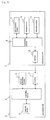
- the reagent kit for measuring platelets may be contained in a first container A for containing the dye reagent and a second container B for containing the dilution reagent as shown in Fig. 1 .
- the dye reagent and dilution reagent are contained in separate containers, the degradation of Nile Blue contained in the dye reagent can be prevented due to water or an alkaline component contained in the dilution reagent, so that the reagent kit for measuring platelets having a superior storage stability can be provided.
- the reagent or reagent kit for measuring platelets is mixed with a sample which may contain platelets, thereby staining the platelets.
- the present invention further provides a method for measuring platelets by using the reagent or reagent kit for measuring platelets.
- the method for measuring of the present invention comprises the steps of:
- a second reagent comprising the buffering agent may be further mixed or not mixed. It is preferable that the second reagent is further mixed.
- the details for the first and second reagents are the same as those described above for the reagent kit for measuring platelets.
- the sample includes blood (whole blood, platelet rich plasma (PRP) etc.) and bone marrow fluid obtained from organisms, particularly from mammals including human.
- the sample may be blood or bone marrow fluid diluted with an appropriate solution such as buffer.
- the first reagent and the sample are preferably mixed so as to obtain the concentration of Nile Blue in the measurement sample of 0.05 to 5.0 ppm, preferably 0.1 to 0.6 ppm, and further preferably 0.2 to 0.5 ppm.
- the first reagent may be diluted with the second reagent in order to achieve the above concentration of Nile Blue in the measurement sample after mixing with the sample.
- the volume ratio between the first and second reagents is preferably 1:10 to 1:100, and more preferably 1:20 to 1:75.
- the mixing order of the first reagent, second reagent and sample is not specifically limited.
- the step of mixing can be carried out at a temperature of approximately 25 to 50°C, more preferably approximately 35 to 45°C.
- the time for mixing is preferably 10 seconds to 5 minutes, more preferably 10 seconds to 2 minutes, and further preferably 10 seconds to 60 seconds.
- an apparatus for applying light is preferably a flow cytometer.
- the measurement sample is introduced into a flow cell of the flow cytometer and light is applied to the cells in the measurement sample passing through the flow cell.
- the light source of the flow cytometer is not specifically limited and can be any light source suitable for excitation of Nile Blue (e.g. around 600 to 680 nm).
- the light source includes, for example, red semiconductor laser and He-Ne laser.
- the semiconductor laser is suitable because it is cheaper than gas laser.
- Scattered light emitted from the cells applied with light in the flow cytometer may be either of forward scattered light (light-receiving angle of approximately 0 to 20 degrees) or side scattered light (light-receiving angle of approximately 90 degrees).
- the forward scattered light may be either of low angle forward scattered light (light-receiving angle of approximately 1 to 5 degrees) or high angle forward scattered light (light-receiving angle of approximately 6 to 20 degrees).
- the fluorescence emitted from the cells applied with light in the flow cytometer can be detected by selecting an appropriate light-receiving wave length for Nile Blue (approximately 630 to 680 nm).
- a scattergram is prepared, so as to detect platelets by distinguishing platelets from contaminants such as blood cell components other than platelets, lipid particles.
- the thus detected platelets can also be counted.
- the detection and/or counting of platelets is preferably carried out by using an appropriate analyzing software.
- Fig. 2 is a front view of the schematic constitution of the sample analyzer.
- the sample analyzer 1 is an apparatus used for hematological tests, for example, and is mainly composed of a measurement unit 2 and a data process unit 3.
- the measurement unit 2 certain components contained in a blood sample are measured, and the data process unit 3 receives the measurement data to carry out an analytical processing.
- Fig. 3 is a block diagram showing the schematic constitution of the sample analyzer 1.
- the measurement unit 2 comprises a controlling section 4 for controlling the action of respective sections; a detecting section 5 equipped with a flow cytometer for detecting particles to be analyzed in the sample; a sample preparing section 6 for preparing a measurement sample and providing the prepared measurement sample to the detecting section 5; and an interface 7 for transmitting data with the data process unit 3.
- the data process unit 3 comprises a controlling section 8 for controlling the action of respective sections; a data analyzing section 9 for analyzing the detected data received from the measurement unit 2; a display section 10 which displays the analysis results from the data analyzing section 9 as scattergrams; an operating section 11 which accepts operations by an operator; and an interface 12 for transmitting data with the measurement unit 2.
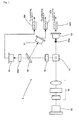
- Fig. 4 is a schematic plan view showing the constitution of the detecting section.
- the detecting section 5 is provided with a semiconductor laser light source 52 so as to emit a laser beam towards a flow cell 51. Between the semiconductor laser light source 52 and the flow cell 51, Light application lenses 53 consisting of a plurality of lenses are provided.
- a beam stopper 54a is provided so as to be opposed to the light application lenses 53 across the flow cell 51, so that direct light from the semiconductor laser light source 52 is interrupted by the beam stopper 54a.
- a photodiode 54 is provided on further downstream of the light axis from the beam stopper 54a.
- the forward signal light is emitted toward the photodiode 54.
- direct light from the semiconductor laser light source 52 is interrupted by the beam stopper 54a, and the photodiode 54 mainly receives scattered light which goes along the light source (hereinafter referred to as "forward scattered light”).
- the forward scattered light emitted from the flow cell 51 is photoelectrically transduced in the photodiode 54, and the thus generated electric signal (hereinafter referred to as "forward scattered light signal”) is amplified in an amplifier 54b.
- the forward scattered light signal is output to the controlling section 4, further output to the data process unit 3 via the interface 7, and processed in the data analyzing section 9 of the data process unit 3.
- Such forward scattered light signal reflects the size of particles to be detected.
- the size of particles to be detected is obtained.
- a side condensing lens 55 In the direction which crosses at right angles with the light axis linearly extended from the semiconductor laser light source 52 to the photodiode 54 is provided with a side condensing lens 55, so as to condense side light with the side condensing lens 55 which is generated by applying a semiconductor laser to particles to be detected passing through the flow cell 51 (light which is emitted to the direction crossing at right angles with the light axis).
- a dichroic mirror 56 On the downstream of the side condensing lens 55 in the direction of the side light is provided with a dichroic mirror 56, so as to divide signal light received from the side condensing lens 55 into a scattered light component and a fluorescence component.
- the dichroic mirror 56 On the side direction of the dichroic mirror 56 (the direction which crosses with the light axis connecting the side condensing lens 55 and the dichroic mirror 56) is provided with a photodiode 57 for receiving side scattered light, and on the downstream of the dicroic mirror 56 in the direction of the light axis are provided with an optical filter 58a and a photomultiplier 58.
- the side scattered light component divided in the dichroic mirror 56 is photoelectrically transduced in the photodiode 57, and the thus generated electric signal (hereinafter referred to as "side scattered light signal”) is amplified in an amplifier 57a.
- the side scattered light signal is output to the controlling section 4, further output to the data process unit 3 via the interface 7, and processed in the data analyzing section 9 of the data process unit 3.
- the side scattered light signal reflects internal information of particles to be detected (e.g. the nuclear size of leukocytes and the like). By processing the side scattered light signal in the data analyzing section 9, the nuclear size of leukocytes and the like are obtained.
- the side fluorescence component emitted from the dichroic mirror 56 is selected depending on the wavelength in the optical filter 58a and photoelectrically transduced in the photomultiplier 58.
- the thus generated electric signal (side fluorescence signal) is amplified in an amplifier 58b.
- the side fluorescence signal is output to the controlling section 4, further output to the data process unit 3 via the interface, and processed in the data analyzing section 9 of the data process unit 3.
- the side fluorescence signal reflects information on the degree of staining of particles to be detected. By processing the side fluorescence signal in the data analyzing section 9, the degree of staining of particles to be detected is obtained.
- the data analyzing section 9 of the data process unit 3 can analyze the thus obtained forward scattered light signal, side scattered light signal and fluorescence signal to detect the particles to be detected, and further can count the detected particles.
- the data analyzing section 9 further generates scattergrams based on the analysis results of the forward scattered light signal, side scattered light signal and fluorescence signal. The generated scattergram are displayed in the display section 10.
- a sample is mixed with a reagent for measuring platelets or with reagents contained in a reagent kit for measuring platelets to prepare a measurement sample.
- the reagent and reagent kit for measuring platelets to be used can be the reagent and reagent kit for measuring platelets described above.
- the reagent for measuring platelets or in the dye reagent of the reagent kit for measuring platelets contains Nile Blue, so that platelets contained in the sample are stained with Nile Blue.
- a measurement sample is preferably prepared by mixing a sample and the reagent for measuring platelets or the reagents contained in the reagent kit for measuring platelets so as to obtain the concentration of Nile Blue in the measurement sample of 0.05 to 5.0 ppm (0.00014 to 0.014 mmol/L), preferably 0.1 to 0.6 ppm, further preferably 0.2 to 0.5 ppm.
- the reaction temperature and time for preparing the measurement sample is preferably the ones described above for the reaction temperature and time for the reagent for measuring platelets.
- the measurement sample prepared in the sample preparing section 6 is then provided to the detecting section 5.
- the measurement sample is introduced into the flow cell 51 of the flow cytometer, and a laser beam is applied from the semiconductor laser light source 52 to the particles to be detected in the measurement sample.
- the forward scattered light signal, side scattered light signal and fluorescence signal obtained by applying the laser beam to the particles are processed in the data analyzing section 9 of the data process unit 3.
- platelets in the measurement sample are clearly distinguished from other components such as fractured red cells or lipid particles contained in the sample based on the forward scattered light signal and fluorescence signal.
- the thus detected platelets may also be counted.
- the signal processing in the data analyzing section 9 is preferably carried out by using an appropriate analyzing software.
- the constitution in which platelets are detected based on the forward scattered light signal and fluorescence signal is preferably carried out by using an appropriate analyzing software.
- the constitution in which platelets are detected based on the forward scattered light signal and fluorescence signal is employed.
- other constitutions such as the one in which platelets are detected based on the side scattered light signal and fluorescence signal may be employed.
- reagents for measuring platelets which contain Nile blue shown in Table 1 at the concentrations shown in Table 1 were prepared, and absorbance, a parameter reflecting the effective dye component concentration of Nile Blue in the dye reagents, was measured to evaluate the storage stability of Nile Blue in the dye reagents.
- the obtained dye reagents were diluted to 1/10 with ethanol, and absorbance of each dye reagent was measured at 630 nm with a spectrophotometer, at which the absorbance spectrum of the dye is maximum.
- the absorbance measurements were carried out with using ethanol as a reference.
- Absorbance of the diluted reagents was measured after 2-, 4- and 7-month storages at 45°C, and the rates of change in absorbance were calculated.
- the results of the absorbance measurements and the rates of change in absorbance are shown in Tables 2-1 and 2-2, respectively.
- the "rate of change in absorbance" denotes the rate of change in absorbance by percent calculated by using the maximum measured absorbance value as a standard value among all absorbance values obtained for the respective Experimental Example.
- Dye reagents Dye for staining platelets At the concentrations shown in Table 3 Ethylene glycol 1L (2) Dilution reagent Tricine (buffering agent) 1.8 g Lauryltrimethylammonium chloride (LTAC) 0.15 g Purified water 1L (adjusted to pH 9.0 and osmotic pressure 200 mOsm/kg.H 2 O)
- the dyes for measuring platelets used were those shown in Table 3 at the concentrations shown in Table 3.
- the final concentrations of the dyes after mixing the reagents with a sample are shown in parentheses.
- the chemical formulae of the dyes are shown in Table 3.
- the dilution reagent (1 mL aliquots) in tubes was heated in a water bath of 40°C.
- To the dilution reagent were added 20 ⁇ l of the dye reagent and 5 ⁇ l of whole blood of healthy human as a sample, and the reaction was carried out at 40°C for 25 seconds to prepare measurement samples.
- the measurement samples were introduced into a detecting section of a flow cytometer having a light source emitting light with the wavelength of 633 nm, excitation light was applied to the cells in the measurement samples, a scattered light signal and fluorescence signal emitted from the cells were detected, the obtained signals were analyzed and the platelets in the measurement samples were measured.
- Preparation of measurement samples and measurements of platelets by flow cytometer were carried out in the blood analyzer XE-2100 (Sysmex Corporation).
- the output of the light source of the blood analyzer was modified to 9W from the normal output of 3W.
- the scattergrams obtained from Experimental Examples 9 to 13 are shown in Figs. 5 to 9 , respectively.
- (a) is a scattergram having the y-axis of forward scattered light intensity and the x-axis of fluorescence intensity
- (b) is a scattergram in which the y-axis of the scattergram (a) is converted to the logarithms.
- (b) the area where platelets appear is shown with a solid line.
- Table 3 The measurement results from the Experimental Examples are shown in Table 3.
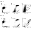
- FIG. 10 and 11 The scattergrams obtained from the measurements of blood containing fragmented red cells and blood containing lipid particles using the dye reagent of Experimental Example 9 are shown in Figs. 10 and 11 .
- the scattergrams obtained from the measurements of blood containing fragmented red cells and blood containing lipid particles using the dye reagent of Experimental Example 10 are shown in Figs. 12 and 13 .
- (a) is a scattergram having the y-axis of forward scattered light intensity and the x-axis of fluorescence intensity
- (b) is a scattergram in which the y-axis of the scattergram (a) is converted to the logarithms
- (c) is a magnified view of the area where platelets appear in (b).
- the area where platelets appear is shown with a solid line.
- Figs. 10 to 13 show that the dye reagents of Experimental Examples 9 and 10 can specifically stain platelets with clear discrimination from fragmented red cells or lipid particles in blood.
- Nile Blue hydrogensulfate is used for staining platelets, platelets can be measured with clear discrimination from other blood cell components, even when blood contains contaminants such as fragmented red cells or lipid particles.
- Experimental Examples 14 to 24 Platelet staining properties of reagents for measuring platelets containing Nile Blue hydrogensulfates at various concentrations
- the dye reagents were prepared containing Nile Blue hydrogensulfate (Merck Co.) at the concentrations indicated in Table 4 in ethylene glycol.
- the obtained dye reagents were combined with the dilution reagent described in Experimental Examples 9 to 13 to measure platelets in the following samples: whole blood of healthy human, blood containing fragmented red cells and blood containing lipid particles, as similar to Experimental Examples 9 to 13.
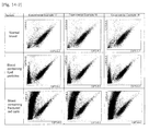
- the obtained scattergrams are shown in Fig. 14 .
- the results in Fig. 14 show that platelets were sufficiently measured with Nile Blue hydrogensulfate at all concentrations tested. Among these, when the sample, blood containing fragmented red cells, was tested with the reagents for measuring platelets of Experimental Examples 17 to 22 (final concentration of Nile Blue hydrogensulfate: 0.24 to 0.44 mg/L), platelets were more clearly discriminated from other blood cell components and contaminants.
- Experimental Example 25 Storage stability of reagents for measuring platelets containing Nile Blue hydrogensulfate
- the dye reagent was prepared containing 14.4 ppm of Nile Blue hydrogensulfate (Merck Co.) in ethylene glycol.
- the dye reagent was stored at the temperature condition of 45°C for 16 weeks and used for the measurement of samples.
- the samples used were blood taken from healthy human, low-platelet blood, blood containing artificial lipid and blood containing fragmented red cells. Platelets were measured as described in Experimental Examples 9 to 13.
- the obtained scattergrams are shown in Fig. 15 .
- the scattergrams in Fig. 15 show that platelets could be measured with clear discrimination from contaminants in all samples. These results show that, when Nile Blue hydrogensulfate is used as the dye for staining platelets, platelets can be accurately measured with little effect from contaminants, even when the dye reagent is stored under unfavorable condition for the dye.
- the dye concentration in the undiluted solution after 16-week storage was calculated as 13.1 ppm. Therefore, it is shown that a favorable dye concentration for platelet measurements can be maintained even after the storage under the above condition.
- Nile Blue having sulfuric ion as a counter ion was used. This Nile Blue was dissolved in ethylene glycol at the concentration of 0.02 mmol/L to prepare Nile Blue solution (Comparative Example 1). To 5 mL of the Nile Blue solution was added the following substance at the following concentration to obtain Experimental Examples 26 to 29.
- Each solution was diluted to 1/5 with ethanol, and absorbance of each solution was measured at 630 nm with a spectrophotometer, at which the absorbance spectrum of the dye is maximum.
- the diluted solutions were stored under the temperature condition of 60°C, and absorbance at 2 weeks and 4 weeks after dilution was measured to calculate the rate of change in absorbance. Ethanol was used as a control in the measurement of absorbance. The results of absorbance measurements and the rates of change in absorbance are shown in Tables 6-1 and 6-2, respectively.
- the initial absorbances of Experimental Examples 28 and 29 containing sodium hydroxide were, as shown in Table 6-1, 0.0065 and 0.4948, respectively, showing that the initial absorbance was lowered compared to Comparative Example 1.
- the rates of change in absorbance of Experimental Examples 28 and 29 were, as shown in Table 6-2, below -20% at 4 weeks after preparation, showing that the reduction in absorbance with time was significant. This shows that the storage stability of Nile Blue solutions was lowered.
- Experimental Examples 30 to 36 Effect of molar concentration ratio between Nile Blue and hydrochloric acid on storage stability of reagents for measuring platelets It is found that the storage stability of Nile Blue solutions is improved by adding a certain concentration of hydrochloric acid, according to Experimental Examples 26 to 29. It is considered that the reduction in the storage stability of Nile Blue solutions, namely, the reduction in the effective dye component concentration of Nile Blue is due to the degradation of the effective dye component, so that the concentration of hydrochloric acid to be added to the solutions should be examined in relation to the number of molecules of Nile Blue.
- Nile Blue A (Sigma) was dissolved in ethylene glycol at the concentration of 0.02 mmol/L to prepare Nile Blue solution (Comparative Example 2).
- Nile Blue solution To 5 mL of the Nile Blue solution was added hydrochloric acid at the following concentration to obtain Experimental Examples 30 to 36.
- the molar concentration ratios between Nile Blue and hydrochloric acid in the solutions are described in parentheses.
- the obtained dye solutions were diluted to 1/20 with ethanol and absorbance of each solution was measured at 630 nm with a spectrophotometer, at which the absorbance spectrum of the dye is maximum.
- the dye solutions were stored under the temperature condition of 45°C, and absorbance at 4 weeks, 9 weeks and 18 weeks after the preparation was measured to calculate the rate of change in absorbance. Ethanol was used as a control in the measurement of absorbance. The results of absorbance measurements and the rates of change in absorbance are shown in Tables 7-1 and 7-2, respectively.
- the initial absorbances of Experimental Examples 30 to 36 were not significantly different from that of Comparative Example 2, as shown in Table 7-1.
- the rates of change in absorbance of Experimental Example 31 in which the molar concentration ratio between Nile Blue and hydrochloric acid was 3:1 were -6.9%, -9.3% and -20.9% at 4, 9 and 18 weeks after the preparation, respectively.
- the rates of change in absorbance of Experimental Example 32 in which the molar concentration ratio between Nile Blue and hydrochloric acid was 1:1 were -5.6%, -5.2% and -16.8% at 4, 9 and 18 weeks after the preparation, respectively.
- Nile Blue A (Sigma) was dissolved in ethylene glycol at the concentration of 0.02 mmol/L to prepare Nile Blue solution (Comparative Example 3).
- Nile Blue solution To 5 mL of the Nile Blue solution was added sulfuric acid at the following concentration to obtain Experimental Examples 37 to 40.
- the molar concentration ratios between Nile Blue and sulfuric acid in the solutions are described in parentheses.
- Nile Blue solutions were prepared with Nile Blue A having sulfuric ion as a counter ion (Nacalai Tesque, Inc.) instead of Nile Blue A (Sigma) used in Experimental Examples 37 to 40 (Comparative Example 4).
- Nile Blue A having sulfuric ion as a counter ion (Nacalai Tesque, Inc.) instead of Nile Blue A (Sigma) used in Experimental Examples 37 to 40 (Comparative Example 4).
- sulfuric acid was added to 5 mL of the Nile Blue solution to 5 mL of the Nile Blue solution.
- the molar concentration ratios between Nile Blue and sulfuric acid in the solutions are described in parentheses.
- Nile Blue solutions are improved by adding an acid to the Nile Blue solutions at the concentration which is equal to or lower than the molar concentration of Nile Blue.
- the optimum concentrations of sulfuric acid in the Nile Blue solutions were different between Experimental Examples 37 to 40 and 41 to 44. It is considered to be due to the difference in purity of Nile Blue contained in the commercial Nile Blue reagents, and may be within the range of an error.
- the reagents for measuring platelets and dilution reagent having the following compositions were prepared.
- Tricine (buffering agent) 1.8 g Lauryltrimethylammonium chloride (LTAC) 0.15 g Purified water 1 L (adjusted to pH 9.0 and osmotic pressure 200 mOsm/kg.H 2 O)
- the dyes for measuring platelets used were the dyes shown in Table 10 at the concentrations shown in Table 10. The final concentrations of the dye in the mixture of the reagent, dilution solution and a sample are shown in parentheses. The chemical formulae of the dyes are shown in Table 10. The measurements of platelets were carried out as described in Experimental Examples 9 to 13.
- Figs. 20 and 21 show the scattergrams obtained by measuring blood containing fractured red cells and blood containing lipid particles with the reagent for measuring platelets of Experimental Example 45.
- (a) is a scattergram having the y-axis of forward scattered light intensity and the x-axis of fluorescence intensity
- (b) is a scattergram in which the y-axis of the scattergram (a) is converted to the logarithms
- (c) is a magnified view of the area where platelets appear in (b). In (c), the area where platelets appear is shown with a solid line.
- Figs. 20 and 21 show that the groups of platelets contained in the samples appear on the scattergrams distinctively from other components in the samples such as fractured red cells or lipid particles. According to these results, it is demonstrated that platelets can be distinctively detected and accurately measured with the reagents for measuring platelets of the present invention even when blood contains the contaminants such as fractured red cells or lipid particles.
Landscapes
- Health & Medical Sciences (AREA)
- Life Sciences & Earth Sciences (AREA)
- Hematology (AREA)
- Engineering & Computer Science (AREA)
- Immunology (AREA)
- Urology & Nephrology (AREA)
- Molecular Biology (AREA)
- Chemical & Material Sciences (AREA)
- Biomedical Technology (AREA)
- Physics & Mathematics (AREA)
- Microbiology (AREA)
- Cell Biology (AREA)
- Food Science & Technology (AREA)
- Medicinal Chemistry (AREA)
- Biotechnology (AREA)
- Analytical Chemistry (AREA)
- Biochemistry (AREA)
- General Health & Medical Sciences (AREA)
- General Physics & Mathematics (AREA)
- Pathology (AREA)
- Investigating Or Analysing Biological Materials (AREA)
- Investigating, Analyzing Materials By Fluorescence Or Luminescence (AREA)
Abstract
Description
- The present invention relates to a reagent for measuring platelets, a reagent kit for measuring platelets and a method for measuring platelets using the same. More specifically, the present invention relates to the reagent for measuring platelets comprising Nile Blue, particularly Nile Blue hydrogensulfate.
- Methods have been known in which blood cells, i.e. leukocytes, reticulocytes, erythrocytes and the like are rapidly measured by using a principle of flow cytometry.
As one of such measurement methods, for example,U.S. Patent No. 6,114,173 discloses a method for counting and detecting reticulocytes, erythrocytes and platelets in whole blood samples as well as a reagent composition used for the method. Specifically, reticulocytes are stained with a reagent mixture containing a cationic dye, particularly Oxazine 750, and their scattered light signal and light absorbance signal are measured using flow cytometry. Then, erythrocytes and reticulocytes in blood samples are differentiated by the difference in absorbance signals, and the group of erythrocytes and reticulocytes and platelets are differentiated by the difference in scattered light signals, so as to determine total numbers of each species. - It has been known that fragmented red cells, lipids and the like occurring in blood are similar in size as platelets, so that they affect measurements as contaminants. Especially, samples containing few platelets for which blood transfusion may be required are more significantly affected by these contaminants upon measurements. Such problem may occur in the method disclosed in
U.S. Patent No. 6,114,173 . However,U.S. Patent No. 6,114,173 does not describe that platelets are measured by preventing any effect by the contaminants. - In addition, dyes which can stain platelets may be degraded in a solution with time. Measurements of platelets by flow cytometry are carried out by detecting scattered light and fluorescence obtained by applying light to the cells stained with the dye in samples. However, when a dye solution is used in which the amount of the dye contained for platelet staining has been decreased, accurate scattered light or fluorescence can not be obtained, and accurate measurement results can not be obtained.
- The present invention has been achieved in view of the above circumstances, and is intended to provide a reagent and reagent kit for measuring platelets that makes it possible to measure platelets with higher accuracy by preventing the effect of contaminants which may be contained in samples and prevent the detection of platelets and that have high storage stability. The present invention is also intended to provide a method for measuring platelets that makes it possible to measure platelets with higher accuracy.
- Therefore, the present invention provides a reagent for measuring platelets comprising, as a dye for staining platelets, Nile Blue having hydrogensulfate ion as a counter ion. The present invention also provides a reagent kit for measuring platelets comprising:
- a first reagent comprising, as a dye for staining platelets, Nile Blue having hydrogensulfate ion as a counter ion, and
- a second reagent comprising a buffering agent.
- Further, the present invention provides a method for measuring platelets comprising the steps of:
- mixing a first reagent comprising, as a dye for staining platelets, Nile Blue having hydrogensulfate ion as a counter ion and a sample with or without adding a second reagent comprising a buffering agent to prepare a measurement sample;
- applying light to cells in the obtained measurement sample to detect scattered light and fluorescence emitted from the cells; and
- detecting platelets contained in the measurement sample based on the detected scattered light and fluorescence.
- In another aspect, the present invention provides a reagent for measuring platelets comprising, as a dye for staining platelets, Nile Blue having an inorganic acid ion as a counter ion and an acid.
- Further, the present invention provides a reagent kit for measuring platelets comprising:
- a first reagent comprising , as a dye for staining platelets, Nile Blue having an inorganic acid ion as a counter ion and an acid, and
- a second reagent comprising a buffering agent.
- Further, the present invention provides a method for measuring platelets comprising the steps of:
- mixing a reagent for measuring platelets comprising Nile Blue and an acid and a sample with or without adding a second reagent comprising a buffering agent to prepare a measurement sample;
- applying light to cells in the obtained measurement sample to detect scattered light and fluorescence emitted from the cells; and
- detecting platelets contained in the measurement sample based on the detected scattered light and fluorescence.
- As used herein, "Nile Blue" denotes dyes consisting of the compound which is represented by, for example, the following formula (II):

and is in the form of a salt with the counter ion represented by X-. In the present specification, the dyes represented by the formula (II) in which the counter ion represented by X- is not specifically limited are called merely "Nile Blue", and the dyes represented by the formula (II) in which the counter ion is specifically defined are called "Nile Blue XXX (XXX denotes a salt with a particular ion)". For example, the dye having sulfuric ion (1/2 SO4 2-) as a counter ion is called "Nile Blue sulfate" and the dye having hydrogensulfate ion (HSO4 -) as a counter ion is called "Nile Blue hydrogensulfate". -
-
Fig. 1 shows containers which can contain the reagent kit for measuring platelets of the present invention. -
Fig. 2 is a front view of the schematic constitution of a sample analyzer. -
Fig. 3 is a block diagram showing the schematic constitution of the sample analyzer. -
Fig. 4 is a schematic plan view showing the constitution of a detecting section. -
Fig. 5 shows scattergrams obtained by the platelet measurement using the reagent for measuring platelets of Experimental Example 9. -
Fig. 6 shows scattergrams obtained by the platelet measurement using the reagent for measuring platelets of Experimental Example 10. -
Fig. 7 shows scattergrams obtained by the platelet measurement using the reagent for measuring platelets of Experimental Example 11. -
Fig. 8 shows scattergrams obtained by the platelet measurement using the reagent for measuring platelets of Experimental Example 12. -
Fig. 9 shows scattergrams obtained by the platelet measurement using the dye reagent of Experimental Example 13. -
Fig. 10 shows scattergrams obtained by the measurement of blood containing fragmented red cells using the reagent for measuring platelets of Experimental Example 9. -
Fig. 11 shows scattergrams obtained by the measurement of blood containing lipid particles using the reagent for measuring platelets of Experimental Example 9. -
Fig. 12 shows scattergrams obtained by the measurement of blood containing fragmented red cells using the reagent for measuring platelets of Experimental Example 10. -
Fig. 13 shows scattergrams obtained by the measurement of blood containing lipid particles using the reagent for measuring platelets of Experimental Example 10. -
Fig. 14 shows scattergrams obtained by the measurements of samples using the reagents for measuring platelets of Experimental Examples 14 to 24. -
Fig. 15 shows scattergrams obtained by the measurement of a sample using the reagent for measuring platelets of Experimental Example 25. -
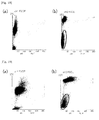
Fig. 16 shows scattergrams obtained by the platelet measurement using the reagent for measuring platelets of Experimental Example 45. -
Fig. 17 shows scattergrams obtained by the platelet measurement using the reagent for measuring platelets of Experimental Example 46. -
Fig. 18 shows scattergrams obtained by the platelet measurement using the reagent for measuring platelets of Experimental Example 47. -
Fig. 19 shows scattergrams obtained by the platelet measurement using the reagent for measuring platelets of Experimental Example 48. -
Fig. 20 shows scattergrams obtained by the measurement of blood containing fragmented red cells using the reagent for measuring platelets of Experimental Example 45. -
Fig. 21 shows scattergrams obtained by the measurement of blood containing lipid particles using the reagent for measuring platelets of Experimental Example 45. -
- A
- First container
- B
- Second container
- 1
- Sample analyzer
- 2
- Measurement unit
- 3
- Data process unit
- 4
- Controlling section
- 5
- Detecting section
- 6
- Sample preparing section
- 7
- Interface
- 8
- Controlling section
- 9
- Data analyzing section
- 10
- Display section
- 1
- Operating section
- 12
- Interface
- 51
- Flow cell
- 52
- Laser light source
- 53
- Light application lenses
- 54
- and 57 Photodiodes
- 54a
- Beam stopper
- 54b
- Amplifier
- 55
- Side condensing lens
- 56
- Dichroic mirror
- 57
- Photodiode
- 57a
- and 58b Amplifiers
- 58
- Photomultiplier
- 58a
- Optical filter
- The present inventors have found that platelets can be clearly distinguished from contaminants in blood such as blood cell components other than platelets or lipid particles by using Nile Blue as a dye for staining platelets in methods for measuring platelets by flow cytometry.
Further, the present inventors have conducted extensive studies for the conditions for measuring platelets using Nile Blue and found that the reagents for measuring platelets have superior storage stability when (1) it contains Nile Blue hydrogensulfate; or (2) it contains Nile Blue and an acid, so as to complete the present invention. - In the above formula (II), the anion (counter ion) of X- includes an inorganic acid ion such as sulfuric ion (1/2 SO4 2-), chloride ion (Cl-), hydrogensulfate ion (HSO4-), perchloric ion (ClO4 -).
The above Nile Blues are commercially available and are purchased from, for example, Sigma Aldrich, Tokyo Chemical Industry Co., Ltd., Nacalai Tesque, Inc., Wako Pure Chemical Industries, Ltd., Kishida Chemical Co., Ltd. and the like.
Specifically, Nile Blue hydrogensulfate can be purchased from Wako Pure Chemical Industries, Ltd. and Kishida Chemical Co., Ltd. - Nile Blue contained in the reagent for measuring platelets of the present invention (e.g. Nile Blue hydrogensulfate) is preferably contained in a dye reagent in a concentration range so as to obtain the concentration of 0.05 to 5.0 ppm, more preferably 0.1 to 0.6 ppm, further preferably 0.2 to 0.5 ppm when the reagent for measuring platelets is mixed with a sample. For example, when the reagent for measuring platelets is mixed with a sample to the dilution rate of 1/50, the concentration of Nile Blue in the reagent for measuring platelets is preferably 2.5 to 250 ppm.
Platelets are more clearly distinguished from blood cell components other than platelets or contaminants, when Nile Blue is present within the above concentration range. - In the aspect in which the reagent for measuring platelets of the present invention comprises Nile Blue and an acid, the acid may be either of an organic acid or inorganic acid, with the preference of an inorganic acid. The inorganic acid includes oxo-acids such as sulfuric acid, phosphoric acid, perchloric acid, and hydroacids such as hydrochloric acid, hydrofluoric acid, hydroiodic acid, hydrobromic acid, hydrocyanic acid, hydroisocyanic acid, hydrogen sulfide, hydrogen azide.
- In the above aspect of the reagent for measuring platelets of the present invention, the acid is contained in the reagent for measuring platelets at the concentration which is equal or lower than the molar concentration of Nile Blue in the reagent for measuring platelets. Specifically, the ratio of molar concentrations of Nile Blue and the acid in the reagent for measuring platelets is preferably 10: to 1: 1. When the acid is hydrochloric acid, the ratio of molar concentrations of Nile Blue and hydrochloric acid is preferably 3:1 to 1:1. When the acid is sulfuric acid, the ratio of molar concentrations of Nile Blue and sulfuric acid is preferably 4:1 to 1:1. By using the acid at the above concentrations, the reduction of the effective dye component concentration of Nile Blue in the reagent for measuring platelets with time can be effectively prevented, so that the storage stability of the reagent for measuring platelets can be improved.
- In the above aspect of the reagent for measuring platelets of the present invention, any combination of Nile Blue and the acid can be used. The more preferred combinations are:
- (A) Nile Blue sulfate and an oxo-acid or hydroacid;
- (B) Nile Blue chloride and sulfuric acid; and
- (C) Nile Blue hydrogensulfate and an oxo-acid or hydroacid.
- In another aspect of the reagent for measuring platelets of the present invention, when Nile Blue is Nile Blue hydrogensulfate, the reagent for measuring platelets does not need to contain an acid. When Nile Blue is Nile Blue hydrogensulfate, reduction in the effective dye component concentration of Nile Blue in the reagent for measuring platelets with time can be effectively prevented without an acid, so that the storage stability of the reagent for measuring platelets can be improved.
- The following two hypotheses may be given as to why the reagents for measuring platelets of the present invention in the above two aspects have improved storage stability.
- (1) It is considered that Nile Blue has high stability when it is in the form of the hydrogensulfate salt.
When an acid is added to Nile Blue sulfate, a proton (H+) which is generated by the dissociation of the acid binds to SO4 2- of Nile Blue sulfate to form HSO4 -, so that HSO4 - and Nile Blue are in balance of 1:1 and stabilized.
When Nile Blue is a salt other than the above, sulfuric acid may be added, so that HSO4 - generated from sulfuric acid can stabilize Nile Blue.
Thus, the reagent for measuring platelets of the present invention may be the one comprising Nile Blue and optionally an acid so as to hydrogensulfate ion exists in the reagent. - (2) Nile Blue contains three nitrogen atoms in their diethylamino group, amino group and phenoxazine structure. Among these nitrogen atoms, nitrogen atoms other than the one which forms the salt with the counter anion represented by X- in the above formula form an acid addition salt, so that the stability of nitrogen atoms and the solubility of the dye are improved and the stability of the reagent for measuring platelets is improved.
Namely, Nile Blue hydrogensulfate may generate a proton (H+) and sulfuric ion (SO4 2-) due to the dissociation of hydrogensulfate ion (HSO4 -). This proton can bind to any of nitrogen atoms of Nile Blue to produce a protic nitrogen atom. It is considered that this protic nitrogen atom forms an intermolecular salt via sulfuric acid, and hence the solubility and stability of the dye can be improved.
When an acid is added to Nile Blue other than the hydrogensulfate salt, it is considered that a proton and an anion generated by the dissociation of the acid contribute to the formation of intermolecular salts of Nile Blue, so that the solubility and stability of the dye can be improved.
Thus, the reagent for measuring platelets of the present invention may be the one comprising Nile Blue and optionally an acid so as to Nile Blue can form an intermolecular salt. - The reagent for measuring platelets of the present invention preferably comprises a water-soluble organic solvent. The water-saluble organic solvent is not specifically limited so long as it can dissolve the above Nile Blue and includes protic solvents such as ethylene glycol, diethylene glycol, poly(ethylene glycol).
- The reagent for measuring platelets of the present invention can be prepared by dissolving the above Nile Blue and optionally the acid into the water-soluble organic solvent at the above desired concentration.
- The above reagent for measuring platelets can be combined with a reagent comprising a buffering agent for diluting the reagent for measuring platelets comprising the dye to form a reagent kit.
Thus, the present invention also provides a reagent kit for measuring platelets comprising: - a first reagent comprising Nile Blue hydrogensulfate, or Nile Blue and an acid (hereinafter also referred to as "the dye reagent"); and
- a second reagent comprising a buffering agent (hereinafter also referred to as "the dilution reagent").
- The details for the first reagent are the same as those described above for the reagent for measuring platelets.
- The buffering agent contained in the second reagent may not be specifically limited so long as it can keep pH of the second reagent within a desired range, and can be carboxylates, phosphates, Good Buffers, taurine, triethanolamine depending on the desired pH. The buffering agent is preferably contained in the dilution reagent so as to obtain the pH range of the dilution reagent of 6.0 to 11.0, more preferably 7.0 to 10.0, and further preferably 8.0 to 9.5. When pH of the dilution reagent is 6.0 or above, erythrocytes become resistant to lysing and the contaminants fragmented red cells can be decreased upon the mixture of the dilution reagent and a sample. When pH is 11.0 or below, non-specific staining of fragmented red cells can be reduced.
Thus, the second reagent is preferably a buffer having pH of 6.0 to 11.0. - The dilution reagent may comprise, in addition to the buffering agent, other components such as an osmoticum, a staining accelerating agent for accelerating the staining of platelets by Nile Blue.
- The osmoticum may be any compound so long as it can keep the osmotic pressure of the dilution reagent within an appropriate range, and includes alkaline metal salts of an organic acid such as propionic acid, saccharides such as glucose, mannose. Alkaline metal halides such as NaCl and alkaline earth metal halides may also be used. One or more osmoticums can be used.
The osmoticum is preferably contained in the dilution reagent so as to adjust the osmotic pressure of the dilution reagent to 150 to 600 mOsm/kg, and more preferably 200 to 300 mOsm/kg. By adjusting the osmotic pressure within the above range, the osmotic pressure of the mixture of the reagents in the reagent kit and a sample may be closer to the physiological osmotic pressure, so that hemolysis due to hypotonicity can be prevented, increase of fragmented red cells can be suppressed and the measurement of platelets can be more accurate. When the osmotic pressure of the dilution reagent can be adjusted within the above range by using the above buffering agent, the osmoticum may not be required. - The staining accelerating agent may be any compound that can increase the permeability of Nile Blue into platelets, and includes surfactants, particularly preferably cationic surfactants.
The preferable cationic surfactant is the one represented by the following formula (I):
- In the above formula (I), the alkyl group having 8 to 12 carbon atoms of R11 may be linear or branched and include, for example, octyl, decyl, lauryl, cetyl, myristyl and the like groups.
The alkyl group having 1 to 6 carbon atoms of R12, R13 and R14 may be linear or branched and include, for example, methyl, ethyl, propyl, butyl, isobutyl, sec-butyl, tert-butyl, pentyl, hexyl and the like groups. Among these, methyl and ethyl groups are preferable.
The anion of Y- is preferably a bromine or chlorine ion. - The preferable cationic surfactants of the formula (I) include decyltrimethylammonium bromide (DTAB), octyltrimethylammonium bromide (OTAB), lauryltrimethylammonium chloride (LTAC), cetyltrimethylammonium chloride (CTAC), myristyltrimethylammonium bromide (MTAB) and the like. One or more cationic surfactants may be used.
- The cationic surfactant is preferably contained in the dilution reagent at the concentration of 50 to 20,000 ppm. The particularly preferable concentration may depend on the type of the cationic surfactants and is preferably 500 to 3,000 ppm when DTAB is used and 100 to 500 ppm when LTAC is used. When the dilution reagent contains the cationic surfactant at such concentrations, hemolysis can be prevented and the staining of platelets can be accelerated upon mixing of the reagents in the reagent kit for measuring platelets and a sample.
- The dilution reagent may comprise, in addition to the above components, a preservative such as sodium 2-pyridylthio-1-oxide, β-phenethyl alcohol.
- The buffering agent which is contained in the dilution reagent and other components which may optionally be contained in the dilution reagent may be mixed with the dye reagent beforehand. However, because Nile Blue contained in the dye reagent may be degraded with water or an alkaline component, the reagent kit separately comprising the dye reagent and dilution reagent is preferable in view of the storage stability.
- The reagent kit for measuring platelets according to the present embodiment may be contained in a first container A for containing the dye reagent and a second container B for containing the dilution reagent as shown in
Fig. 1 . When the dye reagent and dilution reagent are contained in separate containers, the degradation of Nile Blue contained in the dye reagent can be prevented due to water or an alkaline component contained in the dilution reagent, so that the reagent kit for measuring platelets having a superior storage stability can be provided. - The reagent or reagent kit for measuring platelets is mixed with a sample which may contain platelets, thereby staining the platelets.
Thus, the present invention further provides a method for measuring platelets by using the reagent or reagent kit for measuring platelets. - The method for measuring of the present invention comprises the steps of:
- mixing a first reagent comprising Nile Blue hydrogensulfate, or Nile Blue and the acid with a sample to prepare a measurement sample;
- applying light to cells in the obtained measurement sample to detect scattered light and fluorescence emitted from the cells; and
- detecting platelets contained in the measurement sample based on the detected scattered light and fluorescence.
- In the step of mixing the first reagent and a sample in the above method, a second reagent comprising the buffering agent may be further mixed or not mixed. It is preferable that the second reagent is further mixed.
The details for the first and second reagents are the same as those described above for the reagent kit for measuring platelets. - The sample includes blood (whole blood, platelet rich plasma (PRP) etc.) and bone marrow fluid obtained from organisms, particularly from mammals including human. The sample may be blood or bone marrow fluid diluted with an appropriate solution such as buffer.
- The first reagent and the sample are preferably mixed so as to obtain the concentration of Nile Blue in the measurement sample of 0.05 to 5.0 ppm, preferably 0.1 to 0.6 ppm, and further preferably 0.2 to 0.5 ppm.
- The first reagent may be diluted with the second reagent in order to achieve the above concentration of Nile Blue in the measurement sample after mixing with the sample.
When the second reagent is used, the first and second reagents and the sample are preferably mixed in the volume ratio of (first reagent + second reagent) : sample = 100:1 to 1000:1.
When the second reagent is used, the volume ratio between the first and second reagents is preferably 1:10 to 1:100, and more preferably 1:20 to 1:75.
When the second reagent is added in the step of mixing, the mixing order of the first reagent, second reagent and sample is not specifically limited. - The step of mixing can be carried out at a temperature of approximately 25 to 50°C, more preferably approximately 35 to 45°C. The time for mixing is preferably 10 seconds to 5 minutes, more preferably 10 seconds to 2 minutes, and further preferably 10 seconds to 60 seconds.
- In the method for measuring of the present invention, light is applied to the cells in the measurement sample thus obtained and scattered light and fluorescence emitted from the cells are detected.
An apparatus for applying light is preferably a flow cytometer. In the flow cytometer, the measurement sample is introduced into a flow cell of the flow cytometer and light is applied to the cells in the measurement sample passing through the flow cell.
The light source of the flow cytometer is not specifically limited and can be any light source suitable for excitation of Nile Blue (e.g. around 600 to 680 nm). The light source includes, for example, red semiconductor laser and He-Ne laser. The semiconductor laser is suitable because it is cheaper than gas laser. - Scattered light emitted from the cells applied with light in the flow cytometer may be either of forward scattered light (light-receiving angle of approximately 0 to 20 degrees) or side scattered light (light-receiving angle of approximately 90 degrees). The forward scattered light may be either of low angle forward scattered light (light-receiving angle of approximately 1 to 5 degrees) or high angle forward scattered light (light-receiving angle of approximately 6 to 20 degrees).
- The fluorescence emitted from the cells applied with light in the flow cytometer can be detected by selecting an appropriate light-receiving wave length for Nile Blue (approximately 630 to 680 nm).
- Based on the scattered light and fluorescence obtained as above, a scattergram is prepared, so as to detect platelets by distinguishing platelets from contaminants such as blood cell components other than platelets, lipid particles. The thus detected platelets can also be counted. The detection and/or counting of platelets is preferably carried out by using an appropriate analyzing software.
- Measurements of platelets using a sample analyzer equipped with a flow cytometer are now described with referring to the attached Figures.
Fig. 2 is a front view of the schematic constitution of the sample analyzer. Thesample analyzer 1 is an apparatus used for hematological tests, for example, and is mainly composed of ameasurement unit 2 and adata process unit 3. In themeasurement unit 2, certain components contained in a blood sample are measured, and thedata process unit 3 receives the measurement data to carry out an analytical processing. -
Fig. 3 is a block diagram showing the schematic constitution of thesample analyzer 1. As shown inFig. 3 , themeasurement unit 2 comprises a controllingsection 4 for controlling the action of respective sections; a detectingsection 5 equipped with a flow cytometer for detecting particles to be analyzed in the sample; asample preparing section 6 for preparing a measurement sample and providing the prepared measurement sample to the detectingsection 5; and aninterface 7 for transmitting data with thedata process unit 3. Thedata process unit 3 comprises a controllingsection 8 for controlling the action of respective sections; a data analyzing section 9 for analyzing the detected data received from themeasurement unit 2; adisplay section 10 which displays the analysis results from the data analyzing section 9 as scattergrams; anoperating section 11 which accepts operations by an operator; and aninterface 12 for transmitting data with themeasurement unit 2. - The constitution of the detecting
section 5 equipped with a flow cytometer is now described with referring toFig. 4. Fig. 4 is a schematic plan view showing the constitution of the detecting section. The detectingsection 5 is provided with a semiconductorlaser light source 52 so as to emit a laser beam towards aflow cell 51. Between the semiconductorlaser light source 52 and theflow cell 51,Light application lenses 53 consisting of a plurality of lenses are provided. On a light axis extended linearly from the semiconductorlaser light source 52, abeam stopper 54a is provided so as to be opposed to thelight application lenses 53 across theflow cell 51, so that direct light from the semiconductorlaser light source 52 is interrupted by thebeam stopper 54a. On further downstream of the light axis from thebeam stopper 54a, aphotodiode 54 is provided. - When a measurement sample passes through the
flow cell 51, scattered light and fluorescence signals are generated due to a laser beam. Among the signals, the forward signal light is emitted toward thephotodiode 54. Among light beams which goes along the light axis linearly extended from the semiconductorlaser light source 52, direct light from the semiconductorlaser light source 52 is interrupted by thebeam stopper 54a, and thephotodiode 54 mainly receives scattered light which goes along the light source (hereinafter referred to as "forward scattered light"). The forward scattered light emitted from theflow cell 51 is photoelectrically transduced in thephotodiode 54, and the thus generated electric signal (hereinafter referred to as "forward scattered light signal") is amplified in anamplifier 54b. The forward scattered light signal is output to the controllingsection 4, further output to thedata process unit 3 via theinterface 7, and processed in the data analyzing section 9 of thedata process unit 3.
Such forward scattered light signal reflects the size of particles to be detected. By processing the forward scattered light signal in the data analyzing section 9, the size of particles to be detected is obtained. - In the direction which crosses at right angles with the light axis linearly extended from the semiconductor
laser light source 52 to thephotodiode 54 is provided with aside condensing lens 55, so as to condense side light with theside condensing lens 55 which is generated by applying a semiconductor laser to particles to be detected passing through the flow cell 51 (light which is emitted to the direction crossing at right angles with the light axis). On the downstream of theside condensing lens 55 in the direction of the side light is provided with adichroic mirror 56, so as to divide signal light received from theside condensing lens 55 into a scattered light component and a fluorescence component. On the side direction of the dichroic mirror 56 (the direction which crosses with the light axis connecting theside condensing lens 55 and the dichroic mirror 56) is provided with aphotodiode 57 for receiving side scattered light, and on the downstream of thedicroic mirror 56 in the direction of the light axis are provided with anoptical filter 58a and aphotomultiplier 58. The side scattered light component divided in thedichroic mirror 56 is photoelectrically transduced in thephotodiode 57, and the thus generated electric signal (hereinafter referred to as "side scattered light signal") is amplified in anamplifier 57a. The side scattered light signal is output to the controllingsection 4, further output to thedata process unit 3 via theinterface 7, and processed in the data analyzing section 9 of thedata process unit 3.
The side scattered light signal reflects internal information of particles to be detected (e.g. the nuclear size of leukocytes and the like). By processing the side scattered light signal in the data analyzing section 9, the nuclear size of leukocytes and the like are obtained. - The side fluorescence component emitted from the
dichroic mirror 56 is selected depending on the wavelength in theoptical filter 58a and photoelectrically transduced in thephotomultiplier 58. The thus generated electric signal (side fluorescence signal) is amplified in anamplifier 58b. The side fluorescence signal is output to the controllingsection 4, further output to thedata process unit 3 via the interface, and processed in the data analyzing section 9 of thedata process unit 3.
The side fluorescence signal reflects information on the degree of staining of particles to be detected. By processing the side fluorescence signal in the data analyzing section 9, the degree of staining of particles to be detected is obtained. - The data analyzing section 9 of the
data process unit 3 can analyze the thus obtained forward scattered light signal, side scattered light signal and fluorescence signal to detect the particles to be detected, and further can count the detected particles. The data analyzing section 9 further generates scattergrams based on the analysis results of the forward scattered light signal, side scattered light signal and fluorescence signal. The generated scattergram are displayed in thedisplay section 10. - By employing such sample analyzer, platelets can be measured.
First, in thesample preparing section 6, a sample is mixed with a reagent for measuring platelets or with reagents contained in a reagent kit for measuring platelets to prepare a measurement sample. The reagent and reagent kit for measuring platelets to be used can be the reagent and reagent kit for measuring platelets described above. In the reagent for measuring platelets or in the dye reagent of the reagent kit for measuring platelets contains Nile Blue, so that platelets contained in the sample are stained with Nile Blue.
In thesample preparing section 6, a measurement sample is preferably prepared by mixing a sample and the reagent for measuring platelets or the reagents contained in the reagent kit for measuring platelets so as to obtain the concentration of Nile Blue in the measurement sample of 0.05 to 5.0 ppm (0.00014 to 0.014 mmol/L), preferably 0.1 to 0.6 ppm, further preferably 0.2 to 0.5 ppm. The reaction temperature and time for preparing the measurement sample is preferably the ones described above for the reaction temperature and time for the reagent for measuring platelets. - The measurement sample prepared in the
sample preparing section 6 is then provided to the detectingsection 5. In the detectingsection 5, the measurement sample is introduced into theflow cell 51 of the flow cytometer, and a laser beam is applied from the semiconductorlaser light source 52 to the particles to be detected in the measurement sample. The forward scattered light signal, side scattered light signal and fluorescence signal obtained by applying the laser beam to the particles are processed in the data analyzing section 9 of thedata process unit 3. - In the data analyzing section 9, platelets in the measurement sample are clearly distinguished from other components such as fractured red cells or lipid particles contained in the sample based on the forward scattered light signal and fluorescence signal. In data analyzing section 9, the thus detected platelets may also be counted.
In order to detect and count platelets based on the forward scattered light signal and fluorescence signal, the signal processing in the data analyzing section 9 is preferably carried out by using an appropriate analyzing software.
In the above, the constitution in which platelets are detected based on the forward scattered light signal and fluorescence signal. However, other constitutions such as the one in which platelets are detected based on the side scattered light signal and fluorescence signal may be employed. - The present invention is now described in further details. However, these Examples are not intended to limit the scope of the present invention.
- In order to demonstrate the storage stability of the dye reagent, reagents for measuring platelets (dye reagents) which contain Nile blue shown in Table 1 at the concentrations shown in Table 1 were prepared, and absorbance, a parameter reflecting the effective dye component concentration of Nile Blue in the dye reagents, was measured to evaluate the storage stability of Nile Blue in the dye reagents.
- Reagents for measuring platelets:
Dye At the concentrations shown in Table 1 Ethylene glycol 5 mL -
[Table 1] Experimental Examples Dye Concentration 1 Nile Blue hydrogensulfate (Nile Blue hydrogensulfate, Wako Pure Chemical Industries, Ltd.) 18.4 ppm 2 Nile Blue hydrogensulfate (Nile Blue hydrogensulfate, Kishida Chemical Co., Ltd.) 18.4 ppm 3 Nile Blue sulfate (Nile Blue sulfate salt, Sigma) 16.0 ppm 4 Nile Blue sulfate (Nile Blue, Wako Pure Chemical Industries, Ltd.) 16.0 ppm 5 Nile Blue sulfate (Nile Blue sulfate, Nacalai Tesque, Inc.) 16.0 ppm 6 Nile Blue sulfate (Nile Blue A, Nacalai Tesque, Inc.) 16.0 ppm 7 Nile Blue chloride (Nile Blue chloride, Kishida Chemical Co., Ltd.) 16.0 ppm 8 Nile Blue sulfate (Nile Blue A, Kishida Chemical Co., Ltd.) 16.0 ppm - The obtained dye reagents were diluted to 1/10 with ethanol, and absorbance of each dye reagent was measured at 630 nm with a spectrophotometer, at which the absorbance spectrum of the dye is maximum. The absorbance measurements were carried out with using ethanol as a reference. Absorbance of the diluted reagents was measured after 2-, 4- and 7-month storages at 45°C, and the rates of change in absorbance were calculated. The results of the absorbance measurements and the rates of change in absorbance are shown in Tables 2-1 and 2-2, respectively.
The "rate of change in absorbance" denotes the rate of change in absorbance by percent calculated by using the maximum measured absorbance value as a standard value among all absorbance values obtained for the respective Experimental Example. -
[Table 2-1] Experimental Example Immediately after dilution 2 months later 4 months later 7 months later 1 0.3223 0.3141 0.3063 0.3001 2 0.3369 0.3250 0.3138 0.2799 3 0.3316 0.2919 0.2583 0.2285 4 0.3297 0.2803 0.2484 0.2178 5 0.3217 0.2729 0.2441 0.2117 6 0.3248 0.2790 0.2443 0.2119 7 0,3184 0.2696 0.2335 0.2023 8 0.3319 0.2825 0.2426 0.2234 -
[Table 2-2] Experimental Example Immediately after dilution 2 months later 4 months later 7 months later 1 0.0% -2.5% -5.0% -6.9% 2 0.0% -3.5% -6.9% -16.9% 3 0.0% -12.0% -22.1 % -31.1% 4 0.0% -15.0% -24.7% -33.9% 5 0.0% -15.2% -24.1% -34.2% 6 0.0% -14.1% -24.8% -35.0% 7 0.0% -15.3% -26.7% -36.5% 8 0.0% -14.9% -26.9% -32.7% - As shown in Table 2-2, reduction in absorbance of the dye reagent of Experimental Example 1 which contained Nile Blue hydrogensulfate as the dye was 2.5%, 5.0% and 6.9% after 2 months, 4 months and 7 months, respectively. Reduction in absorbance of the dye reagent of Experimental Example 2 which also contained Nile Blue hydrogensulfate as the dye was 3.5%, 6.9% and 16.9% after 2 months, 4 months and 7 months, respectively.
On the other hand, absorbance of the dye reagents of Experimental Examples 3 to 8 which contained Nile Blues having counter ions other than hydrogensulfate ion was decreased by more than 10%, more than 20% and more than 30% after 2 months, 4 months and 7 months, respectively.
From the comparisons with these Experimental Examples 3 to 8, it is found that the dye reagents which contain Nile Blues having hydrogensulfate ions as counter ions have less reduction in absorbance with time and superior storage stability compared to the dye reagents which contain Nile Blues having counter ions other than hydrogensulfate ion. - Experimental Examples 9 to 13: Platelet staining property of reagents for measuring platelets comprising Nile Blue hydrogensulfate
The following experiments were carried out in order to demonstrate the platelet staining property of the reagents for measuring platelets comprising Nile Blue hydrogensulfate.
Reagent kits consisting of dye reagents having the following compositions and the dilution reagent were prepared.
(1) Dye reagentsDye for staining platelets At the concentrations shown in Table 3 Ethylene glycol 1L
(2) Dilution reagentTricine (buffering agent) 1.8 g Lauryltrimethylammonium chloride (LTAC) 0.15 g Purified water 1L osmotic pressure 200 mOsm/kg.H2O) - The dyes for measuring platelets used were those shown in Table 3 at the concentrations shown in Table 3. The final concentrations of the dyes after mixing the reagents with a sample are shown in parentheses.
The chemical formulae of the dyes are shown in Table 3.
The dilution reagent (1 mL aliquots) in tubes was heated in a water bath of 40°C. To the dilution reagent were added 20µl of the dye reagent and 5 µl of whole blood of healthy human as a sample, and the reaction was carried out at 40°C for 25 seconds to prepare measurement samples. The measurement samples were introduced into a detecting section of a flow cytometer having a light source emitting light with the wavelength of 633 nm, excitation light was applied to the cells in the measurement samples, a scattered light signal and fluorescence signal emitted from the cells were detected, the obtained signals were analyzed and the platelets in the measurement samples were measured. Preparation of measurement samples and measurements of platelets by flow cytometer were carried out in the blood analyzer XE-2100 (Sysmex Corporation). The output of the light source of the blood analyzer was modified to 9W from the normal output of 3W. -
[Table 3] Dye Conc. Structure Results Ex. 9 Nile Blue A Hydrogensulfate (Wako Pure Chemicals) 18,4ppm (0.3ppm) 
Sufficient staining of platelets Ex. 10 Nile Blue A Hydrogensulfate (Kishida Chemical) 18.4ppm (0.3ppm) 
Sufficient staining of platelets Ex. 11 Methylene Blue NNX (Sigma) 102.5ppm (2ppm) 
Insufficient staining of platelets Ex. 12 Cresyl Violet Acetate (Sigma) 1025ppm (20ppm) 
No staining Ex. 13 Basic Green 5 (Tokyo Chemical Industry) 1025ppm (20ppm) 
Insufficient staining of platelets - The scattergrams obtained from Experimental Examples 9 to 13 are shown in
Figs. 5 to 9 , respectively. In each Figure, (a) is a scattergram having the y-axis of forward scattered light intensity and the x-axis of fluorescence intensity, and (b) is a scattergram in which the y-axis of the scattergram (a) is converted to the logarithms. In (b), the area where platelets appear is shown with a solid line. The measurement results from the Experimental Examples are shown in Table 3. - As shown in
Figs. 5 to 9 , when Experimental Examples 9 and 10 were used which contained Nile Blue hydrogensulfate as the dye for staining platelets, platelets were stained sufficiently compared to Experimental Examples 11 to 13 which contained the dyes other than Nile Blue, so that platelets could be detected in the region with higher fluorescence intensity.
Thus, when Nile Blue hydrogensulfate is used as the dye for staining platelets, platelets can be measured with better discrimination from other blood cell components. - Next, it was examined whether platelets can be specifically stained by using the dye reagents of Experimental Examples 9 and 10 containing Nile Blue hydrogensulfate even when blood contains the contaminants such as lipid particles.
The platelet measurements of blood containing fragmented red cells and blood containing lipid particles were carried out with the dye reagents of Experimental Examples 9 and 10. The experimental procedures were the same as above except that the blood containing fragmented red cells and the blood containing lipid particles were used instead of whole blood of healthy human. - The scattergrams obtained from the measurements of blood containing fragmented red cells and blood containing lipid particles using the dye reagent of Experimental Example 9 are shown in
Figs. 10 and 11 . The scattergrams obtained from the measurements of blood containing fragmented red cells and blood containing lipid particles using the dye reagent of Experimental Example 10 are shown inFigs. 12 and 13 . In each Figure, (a) is a scattergram having the y-axis of forward scattered light intensity and the x-axis of fluorescence intensity, (b) is a scattergram in which the y-axis of the scattergram (a) is converted to the logarithms, and (c) is a magnified view of the area where platelets appear in (b). In (c), the area where platelets appear is shown with a solid line. - The results of
Figs. 10 to 13 show that the dye reagents of Experimental Examples 9 and 10 can specifically stain platelets with clear discrimination from fragmented red cells or lipid particles in blood. Thus, when Nile Blue hydrogensulfate is used for staining platelets, platelets can be measured with clear discrimination from other blood cell components, even when blood contains contaminants such as fragmented red cells or lipid particles. - According to Experimental Examples 1 and 2, it is shown that the dye reagents containing Nile Blue hydrogensulfate have less reduction in absorbance with time and superior storage stability. Further, Experimental Examples 9 and 10 show that Nile Blue hydrogensulfate as the dye for staining platelets can specifically stain platelets in blood, and that it makes it possible to carry out accurate platelet measurements even when blood contains contaminants such as fragmented red cells or lipid particles.
- Experimental Examples 14 to 24: Platelet staining properties of reagents for measuring platelets containing Nile Blue hydrogensulfates at various concentrations
The dye reagents were prepared containing Nile Blue hydrogensulfate (Merck Co.) at the concentrations indicated in Table 4 in ethylene glycol.
The obtained dye reagents were combined with the dilution reagent described in Experimental Examples 9 to 13 to measure platelets in the following samples: whole blood of healthy human, blood containing fragmented red cells and blood containing lipid particles, as similar to Experimental Examples 9 to 13. -
[Table 4] Experimental Example Dye concentration in dye reagent (mg/L) Final dye concentration in the mixture of reagents and sample (mg/L) 14 6.0 0.12 15 8.0 0.16 16 9.1 0.18 17 12.5 0.24 18 15.0 0.29 19 16.6 0.32 20 18.3 0.36 21 20.3 0.40 22 22.5 0.44 23 25.5 0.50 24 29.3 0.57 - The obtained scattergrams are shown in
Fig. 14 .
The results inFig. 14 show that platelets were sufficiently measured with Nile Blue hydrogensulfate at all concentrations tested.
Among these, when the sample, blood containing fragmented red cells, was tested with the reagents for measuring platelets of Experimental Examples 17 to 22 (final concentration of Nile Blue hydrogensulfate: 0.24 to 0.44 mg/L), platelets were more clearly discriminated from other blood cell components and contaminants. - Experimental Example 25: Storage stability of reagents for measuring platelets containing Nile Blue hydrogensulfate
The dye reagent was prepared containing 14.4 ppm of Nile Blue hydrogensulfate (Merck Co.) in ethylene glycol. The dye reagent was stored at the temperature condition of 45°C for 16 weeks and used for the measurement of samples. - The samples used were blood taken from healthy human, low-platelet blood, blood containing artificial lipid and blood containing fragmented red cells. Platelets were measured as described in Experimental Examples 9 to 13.
- The obtained scattergrams are shown in
Fig. 15 .
The scattergrams inFig. 15 show that platelets could be measured with clear discrimination from contaminants in all samples.
These results show that, when Nile Blue hydrogensulfate is used as the dye for staining platelets, platelets can be accurately measured with little effect from contaminants, even when the dye reagent is stored under unfavorable condition for the dye. - The change in concentration of the dye during the storage under the above condition was examined by the following experiments.
Absorbance was measured as described in Experimental Examples 1 to 8 except that the dye reagent immediately after preparation and the dye reagent after 16-week storage were diluted to 1/10 with ethanol and absorbance at 628 nm was measured. Based on the reduction rate in the measured absorbance, the dye concentration in the dye reagent after 16-week storage was calculated. The results are shown in the following Table 5. -
[Table 5] Immediately after preparation After 16-week storage Absorbance 0.288 0.262 Reduction rate (%) 0.00% -9.03% Dye concentration (ppm) 14.40 13.10 - As shown in Table 5, the dye concentration in the undiluted solution after 16-week storage was calculated as 13.1 ppm. Therefore, it is shown that a favorable dye concentration for platelet measurements can be maintained even after the storage under the above condition.
- Experimental Examples 26 to 29: Storage stability of reagents for measuring platelets containing Nile Blue and acid
The experiments were carried out in order to study the storage stability of Nile Blue solutions containing hydrochloric acid or sodium hydroxide. The storage stability of the Nile Blue solutions was evaluated by measuring absorbance which is a parameter reflecting the effective dye component concentration of Nile Blue in the solution.
In the following descriptions, the "rate of change in absorbance" denotes the rate of change in absorbance by percent calculated by using the maximum measured absorbance value as a standard value among all absorbance values obtained from the respective Experimental Example. - In the present Experimental Examples, Nile Blue having sulfuric ion as a counter ion (Nile Blue A, Sigma) was used. This Nile Blue was dissolved in ethylene glycol at the concentration of 0.02 mmol/L to prepare Nile Blue solution (Comparative Example 1).
To 5 mL of the Nile Blue solution was added the following substance at the following concentration to obtain Experimental Examples 26 to 29. -
26 Hydrochloric acid 2 mmol/L 27 Hydrochloric acid 0.02 mmol/L 28 Sodium hydroxide 2 mmol/L 29 Sodium hydroxide 0.02 mmol/L - Each solution was diluted to 1/5 with ethanol, and absorbance of each solution was measured at 630 nm with a spectrophotometer, at which the absorbance spectrum of the dye is maximum. The diluted solutions were stored under the temperature condition of 60°C, and absorbance at 2 weeks and 4 weeks after dilution was measured to calculate the rate of change in absorbance. Ethanol was used as a control in the measurement of absorbance. The results of absorbance measurements and the rates of change in absorbance are shown in Tables 6-1 and 6-2, respectively.
-
[Table 6-1] Immediately after preparation After 2 weeks After 4 weeks Comparative Example 1 0.5158 0.5033 0.4306 Experimental Example 26 (HCl 2mM) 0.5388 0.0800 0.0055 27 (HCl 0.02 mM) 0.5319 0.5286 0.4741 28 ( NaOH 2 mM)0.0065 0.0017 0.0047 29 (NaOH 0.02 mM) 0.4948 0.4721 0.3825 -
[Table 6-2] Immediately after preparation After 2 weeks After 4 weeks Comparative Example 1 0.0% -2.4% -16.5% Experimental Example 26 (HCl 2mM) 0.0% -85.2% -99.0% 27 (HCl 0.02 mM) 0.0% -0.6% -10.9% 28 ( NaOH 2 mM)0.0% -73.8% -27.7% 29 (NaOH 0.02 mM) 0.0% -4.6% -22.7% - As shown in Table 6-1, initial absorbance of Comparative Example 1 was 0.5158 to which hydrochloric acid or sodium hydroxide was not added. The rates of change in absorbance of Comparative Example 1 were, as shown in Table 6-2, -2.4% and -16.5% at 2 and 4 weeks after the preparation, respectively.
- The initial absorbances of Experimental Examples 26 and 27 containing hydrochloric acid were, as shown in Table 6-1, 0.5388 and 0.5319, respectively, and these values were not significantly different from the initial absorbance of Comparative Example 1.
The rates of change in absorbance of Experimental Example 26 containing 2 mmol/ L concentration of hydrochloric acid were -85.2% and -99.0% at 2 and 4 weeks, presenting significant reduction in absorbance with time compared to Comparative Example 1. This shows that the storage stability of Nile Blue solutions was lowered.
On the other hand, the rates of change in absorbance of Experimental Example 27 containing 0.02 mmol/L concentration of hydrochloric acid were -0.6% and -10.9% at 2 and 4 weeks, showing that the reduction in absorbance with time was moderate compared to Comparative Example 1. This shows that the storage stability of Nile Blue solutions was improved. - The initial absorbances of Experimental Examples 28 and 29 containing sodium hydroxide were, as shown in Table 6-1, 0.0065 and 0.4948, respectively, showing that the initial absorbance was lowered compared to Comparative Example 1.
The rates of change in absorbance of Experimental Examples 28 and 29 were, as shown in Table 6-2, below -20% at 4 weeks after preparation, showing that the reduction in absorbance with time was significant. This shows that the storage stability of Nile Blue solutions was lowered. - These experimental results suggested that the addition of hydrochloric acid at certain concentrations to Nile Blue solutions may improve the storage stability of the Nile Blue solutions.
- Experimental Examples 30 to 36: Effect of molar concentration ratio between Nile Blue and hydrochloric acid on storage stability of reagents for measuring platelets
It is found that the storage stability of Nile Blue solutions is improved by adding a certain concentration of hydrochloric acid, according to Experimental Examples 26 to 29.
It is considered that the reduction in the storage stability of Nile Blue solutions, namely, the reduction in the effective dye component concentration of Nile Blue is due to the degradation of the effective dye component, so that the concentration of hydrochloric acid to be added to the solutions should be examined in relation to the number of molecules of Nile Blue.
Thus, in the present Experimental Examples, the relations between the ratio of Nile Blue and hydrochloric acid (molar concentration ratio) and the storage stability of Nile Blue solutions were examined by varying the ratio of hydrochloric acid and Nile Blue (molar concentration ratio) in Nile Blue solutions.
The storage stability of Nile Blue solutions is examined by evaluating absorbance of Nile Blue solutions as similar to Experimental Examples 26 to 29. - As similar to Experimental Examples 26 to 29, Nile Blue A (Sigma) was dissolved in ethylene glycol at the concentration of 0.02 mmol/L to prepare Nile Blue solution (Comparative Example 2).
- To 5 mL of the Nile Blue solution was added hydrochloric acid at the following concentration to obtain Experimental Examples 30 to 36. The molar concentration ratios between Nile Blue and hydrochloric acid in the solutions are described in parentheses.
-
30 Hydrochloric acid 0.002 mmol/L (10:1) 31 Hydrochloric acid 0.006 mmol/L (3:1) 32 Hydrochloric acid 0. 02 mmol/L (1:1) 33 Hydrochloric acid 0.06 mmol/L (1:3) 34 Hydrochloric acid 0.2 mmol/L (1:10) 35 Hydrochloric acid 0.6 mmol/L (1:30) 36 Hydrochloric acid 2 mmol/L (1:100) - The obtained dye solutions were diluted to 1/20 with ethanol and absorbance of each solution was measured at 630 nm with a spectrophotometer, at which the absorbance spectrum of the dye is maximum. The dye solutions were stored under the temperature condition of 45°C, and absorbance at 4 weeks, 9 weeks and 18 weeks after the preparation was measured to calculate the rate of change in absorbance. Ethanol was used as a control in the measurement of absorbance.
The results of absorbance measurements and the rates of change in absorbance are shown in Tables 7-1 and 7-2, respectively. -
[Table 7-1] Immediately after preparation After 4 weeks After 9 weeks After 18 weeks Comparative Example 2 0.1342 0.1208 0.1174 0.0986 Experimental Example 30(HCl 0.002mM) 0.1369 0.1246 0.1225 0.1051 31 (HCl 0.006 mM) 0.1371 0.1277 0.1244 0.1084 32 (HCl 0.02 mM) 0.1380 0.1303 0.1308 0.1148 33 (HCl 0.06 mM) 0.1381 0.1211 0.0838 0.0245 34 (HCl 0.2 mM) 0.1394 0.0255 0.0001 0.0000 35 (HCl 0.6 mM) 0.1399 0.0282 0.0006 0.0000 36 (HCl 2mM) 0.1399 0.0466 0.0014 0.0000 -
[Table 7-2] Immediately after preparation After 4 weeks After 9 weeks After 18 weeks Comparative Example 2 0.0% -10.0% -12.5% -26.5% Experimetal Example 30 (HCl 0.002mM) 0.0% -9.0% -10.5% -23.2% 31 (HCl 0.006 mM) 0.0% -6.9% -9.3% -20.9% 32 (HCl 0.02mM) 0.0% -5.6% -5.2% -16.8% 33 (HCl 0.06 mM) 0.0% -12.3% -39.3% -82.3% 34 (HCl 0.2 mM) 0.0% -81.7% -99.9% -100.0% 35 (HCl 0.6 mM) 0.0% -79.8% -99.6% -100.0% 36 (HCl 2mM) 0.0% -66.7% -99.0% -100.0% - As shown in Table 7-1, initial absorbance of Comparative Example 2 was 0.1342 to which no hydrochloric acid was added. The rates of change in absorbance of Comparative Example 2 were, as shown in Table 7-2, -10.0%, -12.5% and -26.5% at 4, 9 and 18 weeks after the preparation, respectively.
- The initial absorbances of Experimental Examples 30 to 36 were not significantly different from that of Comparative Example 2, as shown in Table 7-1.
The rates of change in absorbance of Experimental Example 30 in which the molar concentration ratio between Nile Blue and hydrochloric acid was 10:1 where -9.0%, -10.5% and -23.2% at 4, 9 and 18 weeks after the preparation, respectively.
The rates of change in absorbance of Experimental Example 31 in which the molar concentration ratio between Nile Blue and hydrochloric acid was 3:1 were -6.9%, -9.3% and -20.9% at 4, 9 and 18 weeks after the preparation, respectively.
The rates of change in absorbance of Experimental Example 32 in which the molar concentration ratio between Nile Blue and hydrochloric acid was 1:1 were -5.6%, -5.2% and -16.8% at 4, 9 and 18 weeks after the preparation, respectively. - As apparent from the comparison to Comparative Example 2, the reduction in absorbance of Experimental Examples 30 to 32 in which the molar concentration of hydrochloric acid is lower than that of Nile Blue is moderate at any point of time than Comparative Example 2 which does not contain hydrochloric acid. Thus, the storage stability of the Nile Blue solutions is improved.
In particular, Experimental Example 32 in which the molar concentration ratio between Nile Blue and hydrochloric acid was 1:1 had better storage stability than other Experimental Examples. - Experimental Examples 37 to 44: Storage stability of reagents for measuring platelets containing Nile Blue and sulfuric acid
Next, the storage stability of the Nile Blue solutions was studied which contained sulfuric acid instead of hydrochloric acid. - As similar to Experimental Examples 26 to 36, Nile Blue A (Sigma) was dissolved in ethylene glycol at the concentration of 0.02 mmol/L to prepare Nile Blue solution (Comparative Example 3).
- To 5 mL of the Nile Blue solution was added sulfuric acid at the following concentration to obtain Experimental Examples 37 to 40. The molar concentration ratios between Nile Blue and sulfuric acid in the solutions are described in parentheses.
-
37 Sulfuric acid 0.005 mmol/L (4:1) 38 Sulfuric acid 0.01 mmol/L (2:1) 39 Sulfuric acid 0.02 mmol/L (1:1) 40 Sulfuric acid 0.05 mmol/L (1:2.5) - Nile Blue solutions were prepared with Nile Blue A having sulfuric ion as a counter ion (Nacalai Tesque, Inc.) instead of Nile Blue A (Sigma) used in Experimental Examples 37 to 40 (Comparative Example 4).
To 5 mL of the Nile Blue solution was added sulfuric acid at the following concentration to obtain Experimental Examples 41 to 44. The molar concentration ratios between Nile Blue and sulfuric acid in the solutions are described in parentheses. -
41 Sulfuric acid 0.005 mmol/L (4:1) 42 Sulfuric acid 0.01 mmol/L (2:1) 43 Sulfuric acid 0.02 mmol/L (1:1) 44 Sulfuric acid 0.05 mmol/L (1:2.5) - The absorbances immediately after preparation of the solutions of Experimental Examples 37 to 44 were measured. Each solution was stored under the temperature condition of 45°C, and the absorbance at 1 month, 2 months, 3 months and 4 months after the preparation was measured to calculate the rate of change in absorbance.
The results of absorbance measurements and the rates of change in absorbance of Experimental Examples 37 to 40 are shown in Tables 8-1 and 8-2, respectively.
The results of absorbance measurements and the rates of change in absorbance of Experimental Examples 41 to 44 are shown in Tables 9-1 and 9-2, respectively. -
[Table 8-1] Immediately after preparation After 1 month After 2 months After 3 months After 4 months Comparative Example 3 0.2852 0.2849 0.2421 0.2225 0.2076 Experimental Example 37 (H2SO4 0.005mM) 0.2887 0.2907 0.2547 0.2367 0.2224 38 (H2SO4 0.01 mM) 0.2923 0.3045 0.3041 0.2672 0.2545 39 (H2SO4 0.02mM) 0.2999 0.3109 0.3009 0.2980 0.3000 40 (H2SO4 0.05mM) 0.3076 0.2921 0.2934 0.2123 0.1546 -
[Table 8-2] Immediately after preparation After 1 month After 2 months After 3 months After 4 months Comparative Example 3 0.0% -0.1% -15.1% -22.0% -27.2% Experimental Example 37 (H2SO4 0.005mM) 0.0% 0.7% -11.8% -18.0% -23.0% 38 (H2SO4 0.01mM) 0.0% 4.2% 4.0% -8.6% -12.9% 39 (H2SO4 0.02mM) 0.0% 3.7% 0.3% -0.6% 0.0% 40 (H2SO4 0.05mM) 0.0% -5.0% -4.6% -31.0% -49.7% -
[Table 9-1] Immediately after preparation After 1 month After 2 months After 3 months After 4 months Comparative Example 4 0.2941 0.2901 0.2552 0.2449 0.2136 Experimental Example 41 (H2SO4 0.005mM) 0.2981 0.2990 0.2752 0.2633 0.2346 42 (H2SO4 0.01mM) 0.3013 0.3120 0.3041 0.3024 0.2885 43 (H2SO4 0.02mM) 0.3068 0.3155 0.3009 0.2832 0.2562 44 (H2SO4 0.05mM) 0.3140 0.3159 0.2934 0.2660 0.2092 -
[Table 9-2] Immediately after preparation After 1 month After 2 months After 3 months After 4 months Comparative Example 4 0.0% -1.4% -13.2% -16.7% -27.4% Experimental Example 41 (H2SO4 0.005mM) 0.0% 0.3% -7.7% -11.7% -21.3% 42 (H2SO4 0.01mM) 0.0% 3.6% 0.9% 0.4% -4.2% 43 (H2SO4 0.02mM) 0.0% 2.8% -1.9% -7.7% -16.5% 44 (H2SO4 0.05mM) 0.0% 0.6% -6.6% -15.3% -33.4% - As apparent from Table 8-2, the reduction in absorbance of Experimental Examples 37 to 39 in which the molar concentration of sulfuric acid contained is lower than that of Nile Blue is moderate at any point of time compared to Comparative Example 3 which does not contain sulfuric acid. Thus, the storage stability of Nile Blue solutions is improved. Further as apparent from Table 9-2, the reduction in absorbance of Experimental Examples 41 to 43 in which the molar concentration of sulfuric acid contained is lower than that of Nile Blue is moderate at any point of time compared to Comparative Example 4 which does not contain sulfuric acid. Thus, the storage stability of Nile Blue solutions is improved. In particular, the rates of change in absorbance after 4 months of Experimental Examples 39 and 42 in which the molar concentration ratio between Nile Blue and sulfuric acid was 2:1 to 1:1 where 0.0% and -4.2%, respectively, showing particularly favorable storage stability thereof.
- Thus, it is demonstrated that the storage stability of Nile Blue solutions is improved by adding an acid to the Nile Blue solutions at the concentration which is equal to or lower than the molar concentration of Nile Blue.
Incidentally, the optimum concentrations of sulfuric acid in the Nile Blue solutions were different between Experimental Examples 37 to 40 and 41 to 44. It is considered to be due to the difference in purity of Nile Blue contained in the commercial Nile Blue reagents, and may be within the range of an error. - Experimental Examples 45 to 48: Platelet staining property of reagents for measuring platelets containing Nile Blue and acid
The following experiments were carried out in order to study the staining property of platelets of the reagents for measuring platelets containing Nile Blue. - The reagents for measuring platelets and dilution reagent having the following compositions were prepared.
-
Dye for staining platelets At the concentrations shown in Table 10 Ethylene glycol 1 L -
Tricine (buffering agent) 1.8 g Lauryltrimethylammonium chloride (LTAC) 0.15 g Purified water 1 L osmotic pressure 200 mOsm/kg.H2O) - The dyes for measuring platelets used were the dyes shown in Table 10 at the concentrations shown in Table 10. The final concentrations of the dye in the mixture of the reagent, dilution solution and a sample are shown in parentheses. The chemical formulae of the dyes are shown in Table 10.
The measurements of platelets were carried out as described in Experimental Examples 9 to 13. -
[Table 10] Dye Conc. Structure Results Ex. 45 Nile Blue A (Wako Pure Chemicals) 18.4ppm (0.3ppm) 
Sufficient staining of platelets Ex. 46 Methylene Blue NNX (Sigma) 102.5ppm (2ppm) 
Insufficient staining of platelets Ex. 47 Cresyl Violet Acetate (Sigma) 1025ppm (20ppm) 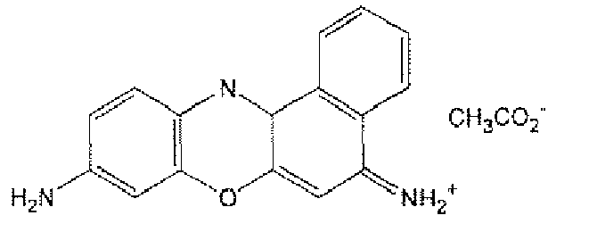
No staining Ex. 48 Basic Green 5 (Tokyo Chemical Industry) 1025ppm (20ppm) 
Insufficient staining of platelets - The scattergrams obtained with Experimental Examples 45 to 48 are shown in
Figs. 16 to 19 , respectively. In each Figure, (a) is a scattergram having the y-axis of forward scattered light intensity and the x-axis of fluorescence intensity, and (b) is a scattergram in which the y-axis of the scattergram (a) is converted to the logarithms. In (b), the area where platelets appear is shown with a solid line.
The measurement results with the Experimental Examples are shown in Table 10. - As shown in
Fig. 16 , when Experimental Example 45 was used in which Nile Blue was used as the dye, the group of platelets appears at the are having high fluorescence on the scattergram, showing that platelets were stained sufficiently.
On the other hand, when Experimental Examples 46 to 48 were used, in which the dyes other than Nile Blue were used, the group of platelets was concentrated at the area having low fluorescence on the scattergram, showing that the staining properties of platelets were not sufficient.
Thus, it is suggested that, when Nile Blue is used as the dye for measuring platelets, platelets can be sufficiently stained compared to the other dyes. Further, it is suggested that, when platelets were stained with Nile Blue, platelets can accurately be detected with forward scattered light intensity and fluorescence intensity. - Next, it was studied whether platelets could be specifically stained by using the reagent for measuring platelets of Experimental Example 45 containing Nile Blue and an acid even when blood contained the contaminants such as lipid particles, according to the similar experiments as described in Examples 9 to 13.
-
Figs. 20 and 21 show the scattergrams obtained by measuring blood containing fractured red cells and blood containing lipid particles with the reagent for measuring platelets of Experimental Example 45. In each Figure, (a) is a scattergram having the y-axis of forward scattered light intensity and the x-axis of fluorescence intensity, (b) is a scattergram in which the y-axis of the scattergram (a) is converted to the logarithms, and (c) is a magnified view of the area where platelets appear in (b). In (c), the area where platelets appear is shown with a solid line. - The results in
Figs. 20 and 21 show that the groups of platelets contained in the samples appear on the scattergrams distinctively from other components in the samples such as fractured red cells or lipid particles. According to these results, it is demonstrated that platelets can be distinctively detected and accurately measured with the reagents for measuring platelets of the present invention even when blood contains the contaminants such as fractured red cells or lipid particles. - The present application relates to Japanese Patent Application Nos.
2008-36013 2008-79785 2008-247484
Claims (15)
- A reagent for measuring platelets comprising, as a dye for staining platelets, a compound of the following formula (III):

- A reagent kit for measuring platelets comprising:a first reagent comprising , as a dye for staining platelets, a compound of the following formula (III):
 a second reagent comprising a buffering agent.
 - The reagent kit for measuring platelets according to Claim 2, wherein the second reagent is a buffer having pH of 6.0 to 11.0.
- The reagent kit for measuring platelets according to Claim 2, wherein the second reagent has an osmotic pressure of 150 to 600 mOsm/kg.
- The reagent kit for measuring platelets according to Claim 2, wherein the second reagent further comprises a staining accelerating agent for accelerating the staining of platelets with the dye.
- The reagent kit for measuring platelets according to Claim 5, wherein the staining accelerating agent is a cationic surfactant.
- A method for measuring platelets comprising the steps of:mixing a first reagent comprising , as a dye for staining platelets, a compound of the following formula (III):
 applying light to cells in the obtained measurement sample to detect scattered light and fluorescence emitted from the cells; anddetecting platelets contained in the measurement sample based on the detected scattered light and fluorescence.
 - The method for measuring platelets according to Claim 7, which further comprises a step of counting the detected platelets.
- A reagent for measuring platelets comprising , as a dye for staining platelets, a compound of the following formula (II) :

an acid. - The reagent for measuring platelets according to Claim 9, wherein the acid is an inorganic acid.
- The reagent for measuring platelets according to Claim 9, wherein the molar concentration ratio between the dye and the acid is 10:1 1 to 1:1.
- A reagent kit for measuring platelets comprising:a first reagent comprising , as a dye for staining platelets, a compound of the following formula (II):
 a second reagent comprising a buffering agent.
 - The reagent kit for measuring platelets according to Claim 12, wherein the molar concentration ratio between the dye and the acid in the first reagent is 10:1 to 1:1.
- A method for measuring platelets comprising the steps of:mixing a first reagent comprising a compound of the following formula (II):
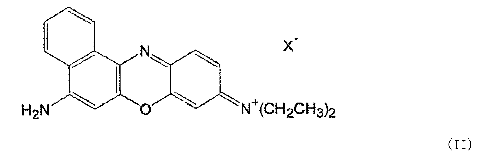 applying light to cells in the obtained measurement sample to detect scattered light and fluorescence emitted from the cells; anddetecting platelets contained in the measurement sample based on the detected scattered light and fluorescence.
 - The method for measuring platelets according to Claim 14, which further comprises a step of counting the detected platelets.
Priority Applications (1)
| Application Number | Priority Date | Filing Date | Title |
|---|---|---|---|
| PL09711933T PL2273264T3 (en) | 2008-02-18 | 2009-02-17 | Platelet measurement reagent, platelet measurement reagent kit, and platelet measurement method |
Applications Claiming Priority (4)
| Application Number | Priority Date | Filing Date | Title |
|---|---|---|---|
| JP2008036013 | 2008-02-18 | ||
| JP2008079785 | 2008-03-26 | ||
| JP2008247484 | 2008-09-26 | ||
| PCT/JP2009/052685 WO2009104598A1 (en) | 2008-02-18 | 2009-02-17 | Platelet measurement reagent, platelet measurement reagent kit, and platelet measurement method |
Publications (3)
| Publication Number | Publication Date |
|---|---|
| EP2273264A1 true EP2273264A1 (en) | 2011-01-12 |
| EP2273264A4 EP2273264A4 (en) | 2011-03-02 |
| EP2273264B1 EP2273264B1 (en) | 2015-04-01 |
Family
ID=40985478
Family Applications (1)
| Application Number | Title | Priority Date | Filing Date |
|---|---|---|---|
| EP09711933.3A Active EP2273264B1 (en) | 2008-02-18 | 2009-02-17 | Platelet measurement reagent, platelet measurement reagent kit, and platelet measurement method |
Country Status (12)
| Country | Link |
|---|---|
| US (2) | US9081021B2 (en) |
| EP (1) | EP2273264B1 (en) |
| JP (1) | JP4659122B2 (en) |
| KR (1) | KR101151592B1 (en) |
| CN (1) | CN101910839B (en) |
| BR (1) | BRPI0908250B1 (en) |
| CO (1) | CO6300813A2 (en) |
| ES (1) | ES2535012T3 (en) |
| HU (1) | HUE025441T2 (en) |
| MX (1) | MX2010008966A (en) |
| PL (1) | PL2273264T3 (en) |
| WO (1) | WO2009104598A1 (en) |
Cited By (1)
| Publication number | Priority date | Publication date | Assignee | Title |
|---|---|---|---|---|
| EP3258274A1 (en) | 2016-06-17 | 2017-12-20 | Sysmex Corporation | Sample analyzer using a patient-specific detection history |
Families Citing this family (5)
| Publication number | Priority date | Publication date | Assignee | Title |
|---|---|---|---|---|
| CN105759019B (en) | 2010-02-26 | 2019-05-31 | 希森美康株式会社 | Reagent container and reagent container assembly |
| CN107011284A (en) * | 2017-05-09 | 2017-08-04 | 济南大学 | A kind of hypersensitive high selectivity hypochlorite colorimetric fluorescence probe |
| EP3789757B1 (en) * | 2018-04-28 | 2025-11-05 | Shenzhen Mindray Bio-Medical Electronics Co., Ltd. | Blood analyzer and analysis method |
| CN113884690B (en) * | 2020-07-04 | 2025-10-17 | 深圳迈瑞生物医疗电子股份有限公司 | Sample analyzer and method for judging insufficient fluorescent reagent allowance thereof |
| KR102229764B1 (en) * | 2020-10-21 | 2021-03-19 | (주)휴마스 | Method and apparatus for rapid monitoring of unederwater microplastics based on fluorescence tagging and multifunctional photometry |
Family Cites Families (12)
| Publication number | Priority date | Publication date | Assignee | Title |
|---|---|---|---|---|
| US4336029A (en) | 1980-08-15 | 1982-06-22 | Ortho Diagnostic Systems Inc. | Method and reagents for quantitative determination of reticulocytes and platelets in whole blood |
| US5075556A (en) * | 1989-12-01 | 1991-12-24 | Technicon Instruments Corporation | Acridine orange derivatives and their use in the quantitation of reticulocytes in whole blood |
| JP3425830B2 (en) * | 1995-10-06 | 2003-07-14 | シスメックス株式会社 | New compounds and their uses |
| TW379284B (en) * | 1996-04-12 | 2000-01-11 | Toa Medical Electronics | Agent for detecting reticulocyte |
| US6114173A (en) | 1997-04-03 | 2000-09-05 | Bayer Corporation | Fully automated method and reagent composition therefor for rapid identification and characterization of reticulocytes erythrocytes and platelets in whole blood |
| JP3886271B2 (en) * | 1998-11-27 | 2007-02-28 | シスメックス株式会社 | Reagent for classification counting of erythroblast and classification counting method |
| JP3837006B2 (en) * | 2000-03-22 | 2006-10-25 | シスメックス株式会社 | Bacterial staining method and detection method |
| DE10027236A1 (en) * | 2000-05-31 | 2001-12-06 | Wack O K Chemie Gmbh | Method and test liquid for the detection of acidic flux residues on electronic assemblies |
| JP4212827B2 (en) | 2002-05-16 | 2009-01-21 | シスメックス株式会社 | Bone marrow nucleated cell automatic analysis method |
| JP4806330B2 (en) * | 2006-10-30 | 2011-11-02 | シスメックス株式会社 | Reticulocyte and platelet measuring reagent, reticulocyte and platelet measuring reagent kit, and reticulocyte and platelet measuring method |
| US20080102526A1 (en) | 2006-10-30 | 2008-05-01 | Sysmex Corporation | Reagent, reagent kit and analyzing method |
| JP4794414B2 (en) * | 2006-10-30 | 2011-10-19 | シスメックス株式会社 | Platelet measuring reagent, platelet measuring reagent kit and platelet measuring method |
-
2009
- 2009-02-17 WO PCT/JP2009/052685 patent/WO2009104598A1/en not_active Ceased
- 2009-02-17 KR KR1020107014194A patent/KR101151592B1/en not_active Expired - Fee Related
- 2009-02-17 CN CN2009801016521A patent/CN101910839B/en not_active Expired - Fee Related
- 2009-02-17 US US12/918,240 patent/US9081021B2/en active Active
- 2009-02-17 HU HUE09711933A patent/HUE025441T2/en unknown
- 2009-02-17 BR BRPI0908250-6A patent/BRPI0908250B1/en not_active IP Right Cessation
- 2009-02-17 MX MX2010008966A patent/MX2010008966A/en active IP Right Grant
- 2009-02-17 EP EP09711933.3A patent/EP2273264B1/en active Active
- 2009-02-17 JP JP2009554323A patent/JP4659122B2/en not_active Expired - Fee Related
- 2009-02-17 ES ES09711933.3T patent/ES2535012T3/en active Active
- 2009-02-17 PL PL09711933T patent/PL2273264T3/en unknown
-
2010
- 2010-08-24 CO CO10104194A patent/CO6300813A2/en not_active Application Discontinuation
-
2015
- 2015-01-06 US US14/590,551 patent/US9310385B2/en active Active
Cited By (1)
| Publication number | Priority date | Publication date | Assignee | Title |
|---|---|---|---|---|
| EP3258274A1 (en) | 2016-06-17 | 2017-12-20 | Sysmex Corporation | Sample analyzer using a patient-specific detection history |
Also Published As
| Publication number | Publication date |
|---|---|
| EP2273264B1 (en) | 2015-04-01 |
| KR101151592B1 (en) | 2012-05-31 |
| US20150185210A1 (en) | 2015-07-02 |
| WO2009104598A1 (en) | 2009-08-27 |
| ES2535012T3 (en) | 2015-05-04 |
| BRPI0908250A2 (en) | 2015-07-21 |
| JPWO2009104598A1 (en) | 2011-06-23 |
| MX2010008966A (en) | 2010-12-06 |
| US20100323396A1 (en) | 2010-12-23 |
| KR20100100908A (en) | 2010-09-15 |
| CN101910839B (en) | 2012-10-17 |
| HUE025441T2 (en) | 2016-04-28 |
| CO6300813A2 (en) | 2011-07-21 |
| PL2273264T3 (en) | 2015-08-31 |
| US9310385B2 (en) | 2016-04-12 |
| CN101910839A (en) | 2010-12-08 |
| JP4659122B2 (en) | 2011-03-30 |
| US9081021B2 (en) | 2015-07-14 |
| EP2273264A4 (en) | 2011-03-02 |
| BRPI0908250B1 (en) | 2021-11-09 |
Similar Documents
| Publication | Publication Date | Title |
|---|---|---|
| US9310385B2 (en) | Platelet measurement reagent, platelet measurement reagent kit, and platelet measurement method | |
| EP1089078B1 (en) | Reagent and method for analyzing solid components in urine | |
| US8367358B2 (en) | Reagent, kit and method for differentiating and counting leukocytes | |
| CN101726579B (en) | Blood test reagent and method | |
| US8334109B2 (en) | Reagent for blood analysis and method of using the same | |
| EP1865318B1 (en) | Reagent for sample analysis, kit for sample analysis and method for sample analysis | |
| EP2798087B1 (en) | Cellular analysis of body fluids | |
| EP0806664B1 (en) | A reagent for measuring reticulocytes and a method of measuring them | |
| EP2202516B1 (en) | Reagent kit for sample analysis and sample analysis method | |
| JP4981907B2 (en) | Reagent for analysis of immature leukocytes and reagent kit for analysis | |
| SG194452A1 (en) | Method for classifying/counting leukocytes, reagent kit for classifying leukocytes, and reagent for classifying leukocytes | |
| CN106062557B (en) | Urine sample analysis method and urine sample analysis kit | |
| EP1918709B1 (en) | Analyzing method for platelets | |
| JPH08170960A (en) | Reagent for analyzing corpuscle component in urine and method for analyzing corpuscle component using reagent | |
| JP3485436B2 (en) | Reticulocyte measuring reagent and measuring method | |
| RU2445628C1 (en) | Reagent for thrombocyte measurement, reagents kit for thrombocyte measurement and method for thrombocyte measurement |
Legal Events
| Date | Code | Title | Description |
|---|---|---|---|
| PUAI | Public reference made under article 153(3) epc to a published international application that has entered the european phase |
Free format text: ORIGINAL CODE: 0009012 |
|
| 17P | Request for examination filed |
Effective date: 20100817 |
|
| AK | Designated contracting states |
Kind code of ref document: A1 Designated state(s): AT BE BG CH CY CZ DE DK EE ES FI FR GB GR HR HU IE IS IT LI LT LU LV MC MK MT NL NO PL PT RO SE SI SK TR |
|
| AX | Request for extension of the european patent |
Extension state: AL BA RS |
|
| A4 | Supplementary search report drawn up and despatched |
Effective date: 20110202 |
|
| RIC1 | Information provided on ipc code assigned before grant |
Ipc: G01N 33/49 20060101AFI20090915BHEP Ipc: G01N 33/483 20060101ALI20110124BHEP Ipc: G01N 21/64 20060101ALI20110124BHEP Ipc: G01N 33/48 20060101ALI20110124BHEP |
|
| DAX | Request for extension of the european patent (deleted) | ||
| 17Q | First examination report despatched |
Effective date: 20121026 |
|
| REG | Reference to a national code |
Ref country code: DE Ref legal event code: R079 Ref document number: 602009030324 Country of ref document: DE Free format text: PREVIOUS MAIN CLASS: G01N0033490000 Ipc: G01N0033520000 |
|
| RIC1 | Information provided on ipc code assigned before grant |
Ipc: G01N 33/483 20060101ALI20140627BHEP Ipc: G01N 33/49 20060101ALI20140627BHEP Ipc: G01N 21/64 20060101ALI20140627BHEP Ipc: G01N 33/48 20060101ALI20140627BHEP Ipc: G01N 33/52 20060101AFI20140627BHEP |
|
| GRAP | Despatch of communication of intention to grant a patent |
Free format text: ORIGINAL CODE: EPIDOSNIGR1 |
|
| INTG | Intention to grant announced |
Effective date: 20141113 |
|
| GRAS | Grant fee paid |
Free format text: ORIGINAL CODE: EPIDOSNIGR3 |
|
| GRAA | (expected) grant |
Free format text: ORIGINAL CODE: 0009210 |
|
| AK | Designated contracting states |
Kind code of ref document: B1 Designated state(s): AT BE BG CH CY CZ DE DK EE ES FI FR GB GR HR HU IE IS IT LI LT LU LV MC MK MT NL NO PL PT RO SE SI SK TR |
|
| REG | Reference to a national code |
Ref country code: GB Ref legal event code: FG4D |
|
| REG | Reference to a national code |
Ref country code: CH Ref legal event code: EP |
|
| REG | Reference to a national code |
Ref country code: IE Ref legal event code: FG4D |
|
| REG | Reference to a national code |
Ref country code: ES Ref legal event code: FG2A Ref document number: 2535012 Country of ref document: ES Kind code of ref document: T3 Effective date: 20150504 |
|
| REG | Reference to a national code |
Ref country code: DE Ref legal event code: R096 Ref document number: 602009030324 Country of ref document: DE Effective date: 20150513 |
|
| REG | Reference to a national code |
Ref country code: AT Ref legal event code: REF Ref document number: 719375 Country of ref document: AT Kind code of ref document: T Effective date: 20150515 |
|
| REG | Reference to a national code |
Ref country code: NL Ref legal event code: VDEP Effective date: 20150401 |
|
| REG | Reference to a national code |
Ref country code: PL Ref legal event code: T3 |
|
| REG | Reference to a national code |
Ref country code: LT Ref legal event code: MG4D |
|
| PG25 | Lapsed in a contracting state [announced via postgrant information from national office to epo] |
Ref country code: NL Free format text: LAPSE BECAUSE OF FAILURE TO SUBMIT A TRANSLATION OF THE DESCRIPTION OR TO PAY THE FEE WITHIN THE PRESCRIBED TIME-LIMIT Effective date: 20150401 |
|
| PG25 | Lapsed in a contracting state [announced via postgrant information from national office to epo] |
Ref country code: NO Free format text: LAPSE BECAUSE OF FAILURE TO SUBMIT A TRANSLATION OF THE DESCRIPTION OR TO PAY THE FEE WITHIN THE PRESCRIBED TIME-LIMIT Effective date: 20150701 Ref country code: CZ Free format text: LAPSE BECAUSE OF FAILURE TO SUBMIT A TRANSLATION OF THE DESCRIPTION OR TO PAY THE FEE WITHIN THE PRESCRIBED TIME-LIMIT Effective date: 20150401 Ref country code: HR Free format text: LAPSE BECAUSE OF FAILURE TO SUBMIT A TRANSLATION OF THE DESCRIPTION OR TO PAY THE FEE WITHIN THE PRESCRIBED TIME-LIMIT Effective date: 20150401 Ref country code: FI Free format text: LAPSE BECAUSE OF FAILURE TO SUBMIT A TRANSLATION OF THE DESCRIPTION OR TO PAY THE FEE WITHIN THE PRESCRIBED TIME-LIMIT Effective date: 20150401 Ref country code: LT Free format text: LAPSE BECAUSE OF FAILURE TO SUBMIT A TRANSLATION OF THE DESCRIPTION OR TO PAY THE FEE WITHIN THE PRESCRIBED TIME-LIMIT Effective date: 20150401 Ref country code: PT Free format text: LAPSE BECAUSE OF FAILURE TO SUBMIT A TRANSLATION OF THE DESCRIPTION OR TO PAY THE FEE WITHIN THE PRESCRIBED TIME-LIMIT Effective date: 20150803 |
|
| PG25 | Lapsed in a contracting state [announced via postgrant information from national office to epo] |
Ref country code: IS Free format text: LAPSE BECAUSE OF FAILURE TO SUBMIT A TRANSLATION OF THE DESCRIPTION OR TO PAY THE FEE WITHIN THE PRESCRIBED TIME-LIMIT Effective date: 20150801 Ref country code: GR Free format text: LAPSE BECAUSE OF FAILURE TO SUBMIT A TRANSLATION OF THE DESCRIPTION OR TO PAY THE FEE WITHIN THE PRESCRIBED TIME-LIMIT Effective date: 20150702 Ref country code: LV Free format text: LAPSE BECAUSE OF FAILURE TO SUBMIT A TRANSLATION OF THE DESCRIPTION OR TO PAY THE FEE WITHIN THE PRESCRIBED TIME-LIMIT Effective date: 20150401 |
|
| REG | Reference to a national code |
Ref country code: RO Ref legal event code: EPE |
|
| REG | Reference to a national code |
Ref country code: DE Ref legal event code: R097 Ref document number: 602009030324 Country of ref document: DE |
|
| REG | Reference to a national code |
Ref country code: FR Ref legal event code: PLFP Year of fee payment: 8 |
|
| PG25 | Lapsed in a contracting state [announced via postgrant information from national office to epo] |
Ref country code: DK Free format text: LAPSE BECAUSE OF FAILURE TO SUBMIT A TRANSLATION OF THE DESCRIPTION OR TO PAY THE FEE WITHIN THE PRESCRIBED TIME-LIMIT Effective date: 20150401 Ref country code: EE Free format text: LAPSE BECAUSE OF FAILURE TO SUBMIT A TRANSLATION OF THE DESCRIPTION OR TO PAY THE FEE WITHIN THE PRESCRIBED TIME-LIMIT Effective date: 20150401 |
|
| PLBE | No opposition filed within time limit |
Free format text: ORIGINAL CODE: 0009261 |
|
| STAA | Information on the status of an ep patent application or granted ep patent |
Free format text: STATUS: NO OPPOSITION FILED WITHIN TIME LIMIT |
|
| PG25 | Lapsed in a contracting state [announced via postgrant information from national office to epo] |
Ref country code: SK Free format text: LAPSE BECAUSE OF FAILURE TO SUBMIT A TRANSLATION OF THE DESCRIPTION OR TO PAY THE FEE WITHIN THE PRESCRIBED TIME-LIMIT Effective date: 20150401 |
|
| 26N | No opposition filed |
Effective date: 20160105 |
|
| REG | Reference to a national code |
Ref country code: HU Ref legal event code: AG4A Ref document number: E025441 Country of ref document: HU |
|
| PG25 | Lapsed in a contracting state [announced via postgrant information from national office to epo] |
Ref country code: SI Free format text: LAPSE BECAUSE OF FAILURE TO SUBMIT A TRANSLATION OF THE DESCRIPTION OR TO PAY THE FEE WITHIN THE PRESCRIBED TIME-LIMIT Effective date: 20150401 Ref country code: BE Free format text: LAPSE BECAUSE OF NON-PAYMENT OF DUE FEES Effective date: 20160229 |
|
| PG25 | Lapsed in a contracting state [announced via postgrant information from national office to epo] |
Ref country code: BE Free format text: LAPSE BECAUSE OF FAILURE TO SUBMIT A TRANSLATION OF THE DESCRIPTION OR TO PAY THE FEE WITHIN THE PRESCRIBED TIME-LIMIT Effective date: 20150401 |
|
| PG25 | Lapsed in a contracting state [announced via postgrant information from national office to epo] |
Ref country code: MC Free format text: LAPSE BECAUSE OF FAILURE TO SUBMIT A TRANSLATION OF THE DESCRIPTION OR TO PAY THE FEE WITHIN THE PRESCRIBED TIME-LIMIT Effective date: 20150401 Ref country code: LU Free format text: LAPSE BECAUSE OF FAILURE TO SUBMIT A TRANSLATION OF THE DESCRIPTION OR TO PAY THE FEE WITHIN THE PRESCRIBED TIME-LIMIT Effective date: 20160217 |
|
| REG | Reference to a national code |
Ref country code: CH Ref legal event code: PL |
|
| PG25 | Lapsed in a contracting state [announced via postgrant information from national office to epo] |
Ref country code: CH Free format text: LAPSE BECAUSE OF NON-PAYMENT OF DUE FEES Effective date: 20160229 Ref country code: LI Free format text: LAPSE BECAUSE OF NON-PAYMENT OF DUE FEES Effective date: 20160229 |
|
| REG | Reference to a national code |
Ref country code: IE Ref legal event code: MM4A |
|
| REG | Reference to a national code |
Ref country code: FR Ref legal event code: PLFP Year of fee payment: 9 |
|
| PG25 | Lapsed in a contracting state [announced via postgrant information from national office to epo] |
Ref country code: IE Free format text: LAPSE BECAUSE OF NON-PAYMENT OF DUE FEES Effective date: 20160217 |
|
| REG | Reference to a national code |
Ref country code: AT Ref legal event code: UEP Ref document number: 719375 Country of ref document: AT Kind code of ref document: T Effective date: 20150401 |
|
| PG25 | Lapsed in a contracting state [announced via postgrant information from national office to epo] |
Ref country code: SE Free format text: LAPSE BECAUSE OF FAILURE TO SUBMIT A TRANSLATION OF THE DESCRIPTION OR TO PAY THE FEE WITHIN THE PRESCRIBED TIME-LIMIT Effective date: 20150401 |
|
| PG25 | Lapsed in a contracting state [announced via postgrant information from national office to epo] |
Ref country code: MT Free format text: LAPSE BECAUSE OF FAILURE TO SUBMIT A TRANSLATION OF THE DESCRIPTION OR TO PAY THE FEE WITHIN THE PRESCRIBED TIME-LIMIT Effective date: 20150401 |
|
| REG | Reference to a national code |
Ref country code: FR Ref legal event code: PLFP Year of fee payment: 10 |
|
| PG25 | Lapsed in a contracting state [announced via postgrant information from national office to epo] |
Ref country code: CY Free format text: LAPSE BECAUSE OF FAILURE TO SUBMIT A TRANSLATION OF THE DESCRIPTION OR TO PAY THE FEE WITHIN THE PRESCRIBED TIME-LIMIT Effective date: 20150401 |
|
| PG25 | Lapsed in a contracting state [announced via postgrant information from national office to epo] |
Ref country code: MK Free format text: LAPSE BECAUSE OF FAILURE TO SUBMIT A TRANSLATION OF THE DESCRIPTION OR TO PAY THE FEE WITHIN THE PRESCRIBED TIME-LIMIT Effective date: 20150401 Ref country code: MT Free format text: LAPSE BECAUSE OF FAILURE TO SUBMIT A TRANSLATION OF THE DESCRIPTION OR TO PAY THE FEE WITHIN THE PRESCRIBED TIME-LIMIT Effective date: 20160229 |
|
| PG25 | Lapsed in a contracting state [announced via postgrant information from national office to epo] |
Ref country code: BG Free format text: LAPSE BECAUSE OF FAILURE TO SUBMIT A TRANSLATION OF THE DESCRIPTION OR TO PAY THE FEE WITHIN THE PRESCRIBED TIME-LIMIT Effective date: 20150401 |
|
| PGFP | Annual fee paid to national office [announced via postgrant information from national office to epo] |
Ref country code: PL Payment date: 20231228 Year of fee payment: 16 |
|
| PGFP | Annual fee paid to national office [announced via postgrant information from national office to epo] |
Ref country code: ES Payment date: 20240304 Year of fee payment: 16 |
|
| PGFP | Annual fee paid to national office [announced via postgrant information from national office to epo] |
Ref country code: AT Payment date: 20240125 Year of fee payment: 16 |
|
| PGFP | Annual fee paid to national office [announced via postgrant information from national office to epo] |
Ref country code: RO Payment date: 20240108 Year of fee payment: 16 Ref country code: HU Payment date: 20240111 Year of fee payment: 16 Ref country code: DE Payment date: 20231228 Year of fee payment: 16 Ref country code: GB Payment date: 20240108 Year of fee payment: 16 |
|
| PGFP | Annual fee paid to national office [announced via postgrant information from national office to epo] |
Ref country code: TR Payment date: 20240209 Year of fee payment: 16 Ref country code: IT Payment date: 20240111 Year of fee payment: 16 Ref country code: FR Payment date: 20240103 Year of fee payment: 16 |
|
| REG | Reference to a national code |
Ref country code: DE Ref legal event code: R119 Ref document number: 602009030324 Country of ref document: DE |
|
| REG | Reference to a national code |
Ref country code: AT Ref legal event code: MM01 Ref document number: 719375 Country of ref document: AT Kind code of ref document: T Effective date: 20250217 |
|
| PG25 | Lapsed in a contracting state [announced via postgrant information from national office to epo] |
Ref country code: HU Free format text: LAPSE BECAUSE OF NON-PAYMENT OF DUE FEES Effective date: 20250218 |
|
| PG25 | Lapsed in a contracting state [announced via postgrant information from national office to epo] |
Ref country code: AT Free format text: LAPSE BECAUSE OF NON-PAYMENT OF DUE FEES Effective date: 20250217 |
|
| PG25 | Lapsed in a contracting state [announced via postgrant information from national office to epo] |
Ref country code: RO Free format text: LAPSE BECAUSE OF NON-PAYMENT OF DUE FEES Effective date: 20250217 |
|
| GBPC | Gb: european patent ceased through non-payment of renewal fee |
Effective date: 20250217 |
|
| PG25 | Lapsed in a contracting state [announced via postgrant information from national office to epo] |
Ref country code: DE Free format text: LAPSE BECAUSE OF NON-PAYMENT OF DUE FEES Effective date: 20250902 |
|
| PG25 | Lapsed in a contracting state [announced via postgrant information from national office to epo] |
Ref country code: GB Free format text: LAPSE BECAUSE OF NON-PAYMENT OF DUE FEES Effective date: 20250217 |
|
| PG25 | Lapsed in a contracting state [announced via postgrant information from national office to epo] |
Ref country code: IT Free format text: LAPSE BECAUSE OF NON-PAYMENT OF DUE FEES Effective date: 20250217 Ref country code: FR Free format text: LAPSE BECAUSE OF NON-PAYMENT OF DUE FEES Effective date: 20250228 |
|
| REG | Reference to a national code |
Ref country code: ES Ref legal event code: FD2A Effective date: 20260406 |
|
| PG25 | Lapsed in a contracting state [announced via postgrant information from national office to epo] |
Ref country code: ES Free format text: LAPSE BECAUSE OF NON-PAYMENT OF DUE FEES Effective date: 20250218 |