EP2264181B1 - Assay for detecting mycoplasma by measuring acetate kinase or carbamate kinase activity - Google Patents
Assay for detecting mycoplasma by measuring acetate kinase or carbamate kinase activity Download PDFInfo
- Publication number
- EP2264181B1 EP2264181B1 EP10075423A EP10075423A EP2264181B1 EP 2264181 B1 EP2264181 B1 EP 2264181B1 EP 10075423 A EP10075423 A EP 10075423A EP 10075423 A EP10075423 A EP 10075423A EP 2264181 B1 EP2264181 B1 EP 2264181B1
- Authority
- EP
- European Patent Office
- Prior art keywords
- mycoplasma
- sample
- test sample
- cells
- culture
- Prior art date
- Legal status (The legal status is an assumption and is not a legal conclusion. Google has not performed a legal analysis and makes no representation as to the accuracy of the status listed.)
- Expired - Lifetime
Links
Images




Classifications
-
- C—CHEMISTRY; METALLURGY
- C12—BIOCHEMISTRY; BEER; SPIRITS; WINE; VINEGAR; MICROBIOLOGY; ENZYMOLOGY; MUTATION OR GENETIC ENGINEERING
- C12Q—MEASURING OR TESTING PROCESSES INVOLVING ENZYMES, NUCLEIC ACIDS OR MICROORGANISMS; COMPOSITIONS OR TEST PAPERS THEREFOR; PROCESSES OF PREPARING SUCH COMPOSITIONS; CONDITION-RESPONSIVE CONTROL IN MICROBIOLOGICAL OR ENZYMOLOGICAL PROCESSES
- C12Q1/00—Measuring or testing processes involving enzymes, nucleic acids or microorganisms; Compositions therefor; Processes of preparing such compositions
- C12Q1/008—Measuring or testing processes involving enzymes, nucleic acids or microorganisms; Compositions therefor; Processes of preparing such compositions for determining co-enzymes or co-factors, e.g. NAD, ATP
-
- C—CHEMISTRY; METALLURGY
- C12—BIOCHEMISTRY; BEER; SPIRITS; WINE; VINEGAR; MICROBIOLOGY; ENZYMOLOGY; MUTATION OR GENETIC ENGINEERING
- C12Q—MEASURING OR TESTING PROCESSES INVOLVING ENZYMES, NUCLEIC ACIDS OR MICROORGANISMS; COMPOSITIONS OR TEST PAPERS THEREFOR; PROCESSES OF PREPARING SUCH COMPOSITIONS; CONDITION-RESPONSIVE CONTROL IN MICROBIOLOGICAL OR ENZYMOLOGICAL PROCESSES
- C12Q1/00—Measuring or testing processes involving enzymes, nucleic acids or microorganisms; Compositions therefor; Processes of preparing such compositions
- C12Q1/02—Measuring or testing processes involving enzymes, nucleic acids or microorganisms; Compositions therefor; Processes of preparing such compositions involving viable microorganisms
- C12Q1/04—Determining presence or kind of microorganism; Use of selective media for testing antibiotics or bacteriocides; Compositions containing a chemical indicator therefor
-
- C—CHEMISTRY; METALLURGY
- C12—BIOCHEMISTRY; BEER; SPIRITS; WINE; VINEGAR; MICROBIOLOGY; ENZYMOLOGY; MUTATION OR GENETIC ENGINEERING
- C12Q—MEASURING OR TESTING PROCESSES INVOLVING ENZYMES, NUCLEIC ACIDS OR MICROORGANISMS; COMPOSITIONS OR TEST PAPERS THEREFOR; PROCESSES OF PREPARING SUCH COMPOSITIONS; CONDITION-RESPONSIVE CONTROL IN MICROBIOLOGICAL OR ENZYMOLOGICAL PROCESSES
- C12Q1/00—Measuring or testing processes involving enzymes, nucleic acids or microorganisms; Compositions therefor; Processes of preparing such compositions
-
- C—CHEMISTRY; METALLURGY
- C12—BIOCHEMISTRY; BEER; SPIRITS; WINE; VINEGAR; MICROBIOLOGY; ENZYMOLOGY; MUTATION OR GENETIC ENGINEERING
- C12Q—MEASURING OR TESTING PROCESSES INVOLVING ENZYMES, NUCLEIC ACIDS OR MICROORGANISMS; COMPOSITIONS OR TEST PAPERS THEREFOR; PROCESSES OF PREPARING SUCH COMPOSITIONS; CONDITION-RESPONSIVE CONTROL IN MICROBIOLOGICAL OR ENZYMOLOGICAL PROCESSES
- C12Q1/00—Measuring or testing processes involving enzymes, nucleic acids or microorganisms; Compositions therefor; Processes of preparing such compositions
- C12Q1/48—Measuring or testing processes involving enzymes, nucleic acids or microorganisms; Compositions therefor; Processes of preparing such compositions involving transferase
-
- C—CHEMISTRY; METALLURGY
- C12—BIOCHEMISTRY; BEER; SPIRITS; WINE; VINEGAR; MICROBIOLOGY; ENZYMOLOGY; MUTATION OR GENETIC ENGINEERING
- C12Q—MEASURING OR TESTING PROCESSES INVOLVING ENZYMES, NUCLEIC ACIDS OR MICROORGANISMS; COMPOSITIONS OR TEST PAPERS THEREFOR; PROCESSES OF PREPARING SUCH COMPOSITIONS; CONDITION-RESPONSIVE CONTROL IN MICROBIOLOGICAL OR ENZYMOLOGICAL PROCESSES
- C12Q1/00—Measuring or testing processes involving enzymes, nucleic acids or microorganisms; Compositions therefor; Processes of preparing such compositions
- C12Q1/48—Measuring or testing processes involving enzymes, nucleic acids or microorganisms; Compositions therefor; Processes of preparing such compositions involving transferase
- C12Q1/485—Measuring or testing processes involving enzymes, nucleic acids or microorganisms; Compositions therefor; Processes of preparing such compositions involving transferase involving kinase
-
- G—PHYSICS
- G01—MEASURING; TESTING
- G01N—INVESTIGATING OR ANALYSING MATERIALS BY DETERMINING THEIR CHEMICAL OR PHYSICAL PROPERTIES
- G01N2333/00—Assays involving biological materials from specific organisms or of a specific nature
- G01N2333/195—Assays involving biological materials from specific organisms or of a specific nature from bacteria
- G01N2333/30—Assays involving biological materials from specific organisms or of a specific nature from bacteria from Mycoplasmatales, e.g. Pleuropneumonia-like organisms [PPLO]
Definitions
- the present invention relates to assay methods and materials for detecting members of the Mollicutes family, that contaminate a test sample, such as a sample from a cell culture.
- Taxonomically the lack of cell walls has been used to separate Mollicutes from other bacteria in a class named Mollicutes (Razin et al 1998).
- the members of this class are summarised in the following table 1.
- Table 1 Major Characteristics and Taxonomy of the Class Mollicutes. Classification No.
- mycoplasma is intended to embrace all members of the class Mollicutes, not just Mycoplasmatales. In fact, “mycoplasma” is the common term in the art for all of the Mollicutes.
- Mycoplasmas are widespread in nature as parasites of humans, mammals, reptiles, fish, arthropods and plants. They are the smallest and simplest prokaryotes. They lack a rigid cell wall and are incapable of peptidoglycan synthesis; they are therefore not sensitive to antibiotics, such as penicillin and its analogues.
- Mycoplasma have developed by degenerate evolution from gram-positive bacteria with a low molecular percentage guanine and cytosine content of DNA ie. the Lactobacillus, Bacillus, Streptococcus and two Clostridium species. The Mollicutes have lost, during the process of evolution, a substantial part of their genetic information. It is this limited coding capacity that has dictated the need for a parasitic way of life. Most species are facultative anaerobes, but some are obligate, and hence the similarities in their metabolism to anaerobic bacteria.
- More than 180 Mollicute species have been identified of which 20 distinct Mycoplasma and Acholeplasma species from human, bovine and swine have been isolated from cell culture. There are six species that account for 95% of all mycoplasma infections; these are M. orale, M arginii, M fermentans, M salivarum, M hyorhinis and A. laidlawii. The major cause of infection is cross contamination from other cell lines introduced into laboratories. Also an unwanted source of exogenous mycoplasma can be found in tissue culture reagents, such as serum products. Mycoplasma, unlike bacterial, contamination rarely produces turbid growth or obvious cell damage.
- Viable mycoplasma can be recovered from work surfaces seven days after inoculation, and mycoplasma can also pass through bacteria-retaining filters. At their maximum population phase there can be as many as 10 8 mycoplasma/ml of supernatant, at a ratio of 5:1 with the host cells. If present, mycoplasma 'grow' to detectable concentrations in the culture medium, they are then also adsorbed onto the cell surface. It is a moot point as to whether mycoplasma enter and survive within mammalian cells in culture.
- Mycoplasma are capable of altering almost every property of an in vitro culture. They will deplete culture nutrients, in particular arginine. Infected eukaryotic cells exhibit aberrant growth, changes in metabolism and morphology. Certain biological properties have been implicated as virulence determinants; these include secretion or introduction of mycoplasmal enzymes such as phospholipases, ATPases, hemolysins, proteases and nucleases into the host cell milieu.
- Mycoplasma can be cultured on agar and in broth culture, with most mycoplasma producing microscopic colonies with a characteristic 'fried egg' appearance, growing embedded in the agar, although some colonies may not grow completely embedded.
- mycoplasma species that adsorb to host cell surfaces (Rottem and Barile 1993).
- these tests are most commonly done by mycoplasma testing service laboratories.
- DAPI 4',6-diamine-2-phenylindole dihydrochloride
- Hoecsht staining is considered to be the method of choice.
- Cell culture samples are taken, fixed and stained with Hoechst 33258 (bisbenzamide) and examined under UV epifluorescence (Battaglia et al 1994, Raab 1999). If there are mycoplasma associated with the cells, then the cell nuclei will appear surrounded by fluorescing structures in the cytoplasm. Negative cells are represented by just the nuclear staining of the cellular DNA. Accurate interpretation of results from DNA staining requires an experienced eye, it also needs specialist equipment i.e. a fluorescence microscope.
- Mycoplasma detection by PCR is a commonly used test by external service laboratories, and is also performed in those laboratories that have the appropriate equipment.
- the primers used in mycoplasma PCR kits anneal to conserved regions of the mycoplasma genome, allowing the detection of several species (Raab 1999).
- Most commercially available PCR kits require that the amplified products be analysed by agarose gel electrophoresis, with the resulting banding patterns determining the contaminating species present. However visualisation of banding patterns is subjective.
- the Mycoplasma PCR ELISA from Roche relies on a different system, and cannot distinguish between species.
- This kit includes digoxigenin-dUTP, and the PCR product is captured onto the surface of wells in a microtitre plate coated with anti-digoxigenin-peroxidase conjugate.
- the coloured product with tetramethylbenzidine (TMB) is visualised using a standard ELISA plate reader.
- UK patent No. 2 357 336 B describes an assay which can be used to detect mycoplasmas in cell cultures.
- the assay is based on the observation that mycoplasmas over-produce the enzyme ATPase in large amounts.
- the ATPase activity of mycoplasmas converts sufficient cellular or externally added ATP to ADP, to make the ADP detectable.
- the assay is based on detection of ADP and this is carried out by adding to the sample an enzyme containing reagent (containing a combination of pyruvate kinase and phosphoenol pyruvate; adenylate kinase; glycerol kinase, myokinase; or a combination of creative kinase and creative phosphate), which converts the ADP to ATP and detecting ATP using a bioluminescent reaction.
- an enzyme containing reagent containing a combination of pyruvate kinase and phosphoenol pyruvate; adenylate kinase; glycerol kinase, myokinase; or a combination of creative kinase and creative phosphate
- the present invention seeks to provide further means for detecting mycoplasmas in samples, such as samples from cell cultures.
- the invention provides a method of detecting the presence of contaminating mycoplasma in a test sample comprising:
- the method further comprises the following steps performed after step (i) but before step (ii):
- the invention provides a method wherein detecting and/or measuring the activity (B) of acetate kinase and/or carbamate kinase in the test sample in step (i) and/or obtaining acetate kinase and/or carbamate kinase activity information (A) in a corresponding control sample in step (ia) comprises detecting and/or measuring the appearance and/or disappearance of one or more of the substrates and/or one or more of the products of the following reactions:
- the detecting and/or measuring step comprises detecting and/or measuring ATP.
- the ATP is detected and/or measured by a light-emitting reaction, especially a bioluminescent reaction.
- Light-emitting systems have been known and isolated from many luminescent organisms, including certain bacteria, protozoa, coelenterates, molluscs, fish, millipedes, flies, fungi, worms, crustaceans, and beetles, particularly the fireflies of the genera Photinus, Photuris, and Luciola and click beetles of genus pyrophorus.
- enzymatically catalyzed oxidoreductions take place in which the free energy change is utilised to excite a molecule to a high energy state. Then, when the excited molecule spontaneously returns to the ground state, visible light is emitted. This emitted light is called "bioluminescence”.
- P.pyralis luciferase is an enzyme which appears to have no prosthetic groups or tightly bound metal ions and has 550 amino acids and a molecular weight of about 60,000 daltons; the enzyme has been available to the art in crystalline form for many years.
- firefly luciferin a polyheterocyclic organic acid, D-(-)-2-(6'-hydroxy-2'-benzothiazolyl)- 2 -thiazoline-4-carboxylic acid (herein-after referred to as "luciferin", unless otherwise indicated).
- ATP can be detected using the following bioluminescent reaction.
- the emitted light intensity is linearly related to the ATP concentration and is measured using a luminometer.
- Luciferase has been used as a means of assaying minute concentrations of ATP; as little as 10 -16 molar ATP can be detected with high quality preparations of the enzyme.
- the luciferase-luciferin reaction is highly specific for ATP. For example, deoxy-ATP produces less than 2% of the light generated by ATP, and other nucleoside triphosphates produce less than 0.1%.
- Crystalline luciferases can be isolated directly from the light organs of beetles.
- cDNAS encoding luciferases of several beetle species including, among others, the luciferase of P.pyralis (firefly), the four luciferase isozymes of P.plagiophthalamus (click beetle), the luciferase of L.cruciata (firefly) and the luciferase of L. lateralis) (de Wet et al., 1987, Masuda et al., 1989, Wood et al., 1989, European Patent Application Publication No. 0 353 464 ) are available.
- cDNAs encoding luciferases of any other beetle species are readily obtainable by the skilled using known techniques (de Wet et al., 1986, Wood et al., 1989). With the cDNA encoding a beetle luciferase in hand, it is entirely straightforward to prepare large amounts of the luciferase in highly pure form by isolation from bacteria (e.g. E.coli ) , yeast, mammalian cells in culture, or the like, which have been transformed to express the cDNA.
- bacteria e.g. E.coli
- Such a mutant luciferase will have an amino acid sequence that differs from the sequence of a naturally occurring beetle luciferase at one or more positions (White et al., 1996, WO 01/31028 and WO 00/24878 ).
- the term "luciferase” comprehends not only the luciferases that occur naturally in beetles but also the mutants, which retain activity in providing bioluminescence by catalyzing the luciferase-luciferin reaction, of such naturally occurring luciferases.
- the sample is treated so as to lyse any mycoplasma and thereby release their cellular contents into the sample.
- lysis can be effected by a variety of methods including application of chemicals, such as detergents and mechanical methods such as sonication etc.
- the lysis is effected by treating the sample with a detergent, or other lysis method, which allows for the lysis of the Mycoplasma cell membrane but which does not affect the cell wall of any bacteria which may be present.
- a detergent or other lysis method, which allows for the lysis of the Mycoplasma cell membrane but which does not affect the cell wall of any bacteria which may be present.
- Exemplary detergent treatment includes the use of low concentrations (e.g. 0.25% v/v) of a detergent, such as Triton X100.
- the preferred lysis method is one that is sufficient to lyse the mycoplasmal membrane, but would be ineffective against bacterial cells.
- non-ionic detergents mainly polyethoxyethers
- bacteria For efficient lysis and total protein release, bacteria often require exposure to enzymes such as lysozyme to breach the cell wall (Pellegrini et al 1992).
- the most preferred detergent mycoplasma lysis conditions are shown hereinafter.
- mycoplasma will pass through a 0.45 ⁇ M filter used for filter sterilisation (Baseman and Tully 1997), and it is possible to distinguish between a bacterial and mycoplasmal contamination through the addition of a filtration step.
- test sample is passed through a bacterial filter before step (i).
- a bacteria filter for example by passing it through a bacteria-retaining filter, it is not important to lyse mycoplasma selectively, i.e. without lysing bacteria.
- ADP is added to the test sample prior to the detecting and/or measuring step (i).
- the assay can also utilise endogenous ADP.
- a mycoplasma substrate reagent is added to the test sample prior to the detecting and/or measuring step (i), the mycoplasma substrate reagent comprising: acetyl phosphate or acetyl-Co-A and/or carbamoyl phosphate or citrulline and/or ammonia.
- a precursor thereof we include one or more compounds from which acetyl phosphate and/or carbamoyl phosphate can be generated. Exemplary reactions are outlined below:
- acetyl phosphate instead of adding acetyl phosphate to the mycoplasma substrate reagent, one could include a precursor, such as acetyl-CoA.
- carbamoyl phosphate instead of carbamoyl phosphate one could add a precursor, such as citrulline and ammonia to the mycoplasma substrate reagent.
- both acetyl phosphate and carbamoyl phosphate and/or acetyl-Co-A and/or citrulline and/or ammonia are added to the sample prior to step (i), in the methods of the invention.
- This enables a generic assay for mycoplasma contamination to be carried out because mycoplasmas utilise either or both substrates by means of their acetate kinase and/or carbamate kinase enzymes.
- a more specific assay can be produced by only using one of the above substrates or precursors thereof.
- Such an assay will be specific for mycoplasma which only use one of the enzymes acetate kinase or carbamate kinase.
- the following table 2 cites some examples of the members of the Mollicutes family (parasitise mammalian hosts) that utilise acetate kinase preferentially, carbamate kinase preferentially, or both.
- the "corresponding control sample” is the test sample prior to a mycoplasma lysis treatment and/or addition of a mycoplasma substrate and/or a time interval (e.g. more than approximately 30 minutes).
- both of the activity measurements are carried out on the same sample, the test sample.
- a first activity measurement (A) is taken either before or concurrent with a mycoplasma lysis step then, after addition of a mycoplasma substrate and/or a time interval (e.g. more than approximately 30 minutes), a second activity measurement (B) is taken. If the value of B A is greater than one the test sample is identified as contaminated with mycoplasma.
- control sample could also be a predetermined negative control sample, but this is less preferred.
- control sample has been shown to be free from mycoplasma contamination. Suitable methods for doing this include PCR testing, DNA fluorescent staining or culture methods as described herein.
- corresponding control sample we mean a sample which contains substantially the same material as that contained in the test sample, but which, unlike the test sample, has been shown to be free from mycoplasma contamination. Skilled persons will appreciate that a mycoplasma uncontaminated condition can be shown by a variety of known methods. A number of suitable methods are reviewed by Rottem and Barile 1993, while an outline of testing kits and services is given in Raab et al 1999.
- the test sample and/or control sample can be a cell sample, such as a cell culture sample, especially a culture of mammalian cells. Some examples are listed in the following table 3. Table 3: Commonly cultured cell lines that have been tested using the assay method.
- the assays of the invention can be utilised to detect mycoplasma contamination in cultures of both adherent cells (e.g. HepG2, A549, CHO and COS cells) and cells which culture in suspension (e.g. Jurkats, U937, K562 and HL-60 Cells.)
- adherent cells e.g. HepG2, A549, CHO and COS cells
- cells which culture in suspension e.g. Jurkats, U937, K562 and HL-60 Cells.
- the sample to be tested is from the cell culture supernatant which has previously been centrifuged to remove cellular material.
- Cell-free samples can also be tested using the methods of the invention.
- the methods of the invention are particularly useful for testing samples of cell-free reagents, such as tissue culture media, and typically those containing animal-derived materials, such as serum (e.g. foetal calf serum), trypsin, and other culture supplements, etc. Examples of some commonly used media and supplements that may be tested in this manner are shown in table 4. Table 4: Tissue culture media and supplements that may be tested using the assay system.
- a method of detecting the presence of mycoplasma in a test sample comprising the following steps:-
- Mycoplasma lysis treatment and/or addition of mycoplasma substrate and/or a time interval occurs before step (i).
- the "corresponding control sample” is the test sample except that it has not been subjected to Mycoplasma lysis treatment and/or addition of mycoplasma substrate and/or left for a time interval (e.g. more than approximately 30 minutes).
- both measurements are taken from the test sample.
- the control ATP and/or light output measurement is taken following addition to the sample of the mycoplasma detection reagent containing the detergent and luciferase/luciferin plus AMP, and the test ATP and/or light output measurement is taken following addition of substrates for kinase activity (or precursors thereof).
- a further aspect of the invention provides an in vitro process for treating a cell culture to remove mycoplasma contamination comprising:- treating a mycoplasma contaminated cell culture with an agent to remove or destroy mycoplasma; and subsequently testing a sample from the culture for mycoplasma contamination using a method of the invention; if necessary, repeating the process one or more times until mycoplasma contamination is not detected in the sample.
- the antibiotics tetracycline and ciprofloxacin are reported to have success rates of less than 80-85% ( www.unc.edu/depts/tcf/mycoplasma.htm ). It is therefore extremely difficult to completely irradicate mycoplasma from cultures, once a contamination has taken hold.
- mycoplasma exposed to antibiotics in eukaryotic cell culture have different profiles from the same species isolated from a human or animal source (Taylor-Robinson and Bebear 1997). While the reported success of anti-mycoplasma treatments appears highly variable, a recent study by Uphoff et al 2002, reports that 96% of leukaemia-lymphoma cell lines were rendered free of mycoplasma with at least one of the treatments tested.
- a further aspect of the invention is the kit according to claims 26-32.
- the principle of the preferred assay method is to supply the appropriate substrates for mycoplasmal enzymes. If mycoplasma contamination is present, there is a conversion of ADP to ATP which can then be measured, preferably by the luciferase-luciferin reaction.
- Mycoplasma Detection Reagent is added and, after approximately 5 minutes, an initial light output reading (A) is taken, the Mycoplasma Substrate (MS) is added and any enzymatic activity is allowed to progress for approximately 10 minutes, at which point a second light reading (B) is taken.
- A initial light output reading
- MS Mycoplasma Substrate
- the second reading (B) will be higher when compared to the first reading (A), giving a ratio B A of greater than 1. If the culture is negative (uncontaminated by mycoplasma), then the ratio B A will be 1 or most often less than one due to the luminescent light signal decay usually seen over time.
- Figure 1 demonstrates the kinetics of the reaction. Typically, the ratio B A seen with mycoplasma contamination is much greater than 1, for example Figure 1 shows a ratio of 114.
- a preferred assay kit of the invention comprises a Mycoplasma Detection Reagent (MDR); Mycoplasma Assay Buffer (MAB) for reconstitution of MDR and the Mycoplasma Substrate (MS).
- MDR and MS are preferably provided as lyophilised preparations.
- Mycoplasma Substrate of the invention contains substrates for one or both of these enzymatic reactions.
- ADP is a requirement for both enzymes, and is preferably supplied in excess in the Mycoplasma Detection Reagent of the invention to drive the generation of ATP formation.
- the MDR is added to a sample of culture supernatant that has previously been centrifuged to remove cellular material, although it is possible to perform the assay in the presence of cells.
- the test sample can be passed through a bacterial filter.
- the MDR contains substrates for luciferase, luciferin and other co-factors plus AMP and ADP.
- the Mycoplasma Substrate (MS) contains carbamoyl phosphate and/or acetyl phosphate or acetyl-Co-A and/or citrulline and/or ammonia required for detection of the carbamate and/or acetate kinase activities.
- a preferred sample volume is 100 ⁇ l to which 100 ⁇ l of reconstituted MDR is added. After approximately 5 minutes the first luminometric reading (A) is taken, this gives the base reading upon which the further ratio calculations B A are determined.
- the assay methods of the invention have been used to investigate contamination by Acholeplasma laidlawii, M. hyorhinis, M. fermentans, M orale, and M genitalium and to detect a number of unknown mycoplasma contaminations.
- the inventors have compared their data to detection of mycoplasma by PCR, and have shown that there is a correlation between ratios greater than one and detection of mycoplasmal DNA. This is shown in Figure 2 where the positive PCR bands on the gel correlate with ratios of more than one.
- Example 2 use of the methods of the invention in a process for removing mycoplasma contamination from a cell culture.
- the inventors have also shown that we can detect a reduction in ratios B A as cells are treated with an exemplary Mycoplasma Removal Agent (ICN-Flow), a derivative of the quinolone family of antibiotics.
- ICN-Flow Mycoplasma Removal Agent
- Figure 3 also shows that the treatment with MRA for 10 days with COS-7 and CHO cell cultures was sufficient to remove the contaminating mycoplasma.
- Mycoplasma can form colonies as large as 600 ⁇ m in diameter, but can also exist in their life cycle as single cells as small as 0.15 ⁇ m. Due to their small size mycoplasma can pass through the 0.45 ⁇ m and 0.22 ⁇ m filters commonly used to "sterilise" tissue culture reagents.
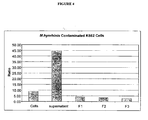
- Figure 4 also confirms that the assay can be performed in the presence of cells, but that there is a reduced sensitivity of detection. Hence, it is preferred that the assay methods of the invention are performed on samples which are substantially cell free. This can easily be achieved by centrifugation of cell cultures and sampling of the supernatant and, optionally, filtration through a bacterial filter.
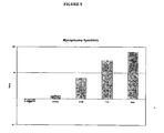
- dilution of the supernatants shows the sensitivity of the assays, in that a 1:1000 dilution of contaminated culture supernatant can still give ratios greater than 1.
- the dilution range will also vary according to the number of colony forming units in the test sample.
- the assay methods of the invention will work without the external exogenous addition of carbamate and acetate kinase substrates in the form of ADP, carbamoyl phosphate and acetyl phosphate or precursors thereof.
- ADP can be generated by other externally added or cellular enzymes i.e. adenylate kinase utilising ATP and AMP.
- the two substrates could also be generated from "precursors" by utilising earlier parts of the glucose fermentation and argenine lysis pathways for example by the addition of acetyl-CoA and citrulline that could be used by mycoplasmal enzymes to synthesise acetyl phosphate and carbamoyl phosphate respectively.
- M.fermentans utilises both pathways
- M.orale utilises only the carbamate kinase pathway
- positive ratios are only observed in the single carbamate reagent or the combined reagent.
- Figure 8 shows the detection limits are as low as 14 CFU/well with M.orale.
- the inventors have investigated a mycoplasma that preferentially utilises the acetate kinase pathway, namely M.hyorhinis.
- the data are shown in Figure 9 .
- the inventors have tested over 15 different cell lines (see table 3) and shown that none of the cells have sufficient background enzymatic activity to impact upon the ratios and give false positives.
- the inventors think the reason for this is that the pathways are anaerobic, and all mammalian cell cultures will generate ATP through oxidative phosphorylation.
- carbamoyl phosphate or a precursor thereof or only acetyl phosphate or a precursor thereof one can produce an assay method of the invention which will allow one to determine whether the mycoplasmal contaminants in question are from a group which uses the acetate kinase pathway, the carbamate kinase pathway, or both. This may have useful diagnostic applications.
- the methods of the invention can be varied to include an initial screening step for bacterial contamination, if necessary. This can be achieved by a variety of methods, but is preferably carried out by passing the sample through a standard bacterial filter (Baseman and Tully, 1997).
- Example 6 Preferred Reagent Components for use in the mycoplasma assay methods and kits of the invention
- MDR Mycoplasma Detection Reagent
- Ms Mycoplasma Substrate
- Preferred concentration ranges acetyl-CoA 0.1mM to 100mM Inorganic phosphate (e.g. potassium phosphate) 0.1mM to 100mM ii.
- Preferred concentration ranges citrulline 1mM to 100mM ammonium bicarbonate 1mM to 200mM iii.
- Preferred concentration ranges acetate (e.g. sodium or potassium) 1mM to 500mM ATP 0.1 mM to 100mM iv.
- Disruption of the viable mycoplasma cell membrane to allow for the release of the enzymes into the sample is a preferred embodiment of the assay method of the invention. This allows for binding of the substrates and generation of ATP. However, positive ratios indicating mycoplasma contamination can be obtained in the absence of any lysis treatment. The implication is that these enzymes can be released by viable mycoplasma The other possibility is that some non-viable organisms have released their contents through natural lysis.
- the aim of the following experiments was to determine the concentrations of Triton-X100 in the Hepes-EDTA buffer on the ratios seen with mycoplasma contaminated K562 cell cultures. Two organisms were investigated, M.hyorhinis and M.orale.
- Figure 10 shows that it is possible to detect mycoplasmal enzymes in the absence of a detergent lysis step. It also shows a drop in the light output with concentrations greater than 4-5%, this is due to adverse effects of the detergent on the luciferase enzyme/reaction. However, it is still possible to detect positive activity with concentrations as high as 20% (v/v).
- the inventors have also shown that the concentrations of Triton-X100 used in the above experiments did not result in any detectable carbamate or acetate kinase activity from the E.coli strain JM109 cells.
- Non-ionic detergents are non-denaturing and permit the solubilization of membranes without interfering with biological activity. They have principally been used for the study of protein conformations and for the separation of hydrophilic proteins from membrane spanning hydrophobic proteins.
- Anionic and cationic detergents result in greater modification of protein structure and are more effective at disrupting protein aggregation.
- Zwitterionic detergents are also low-denaturing, but are effective at disruption of protein aggregates.
- the required lysis agent is one that causes disruption of the mycoplasmal membrane and allows release of the metabolic enzymes that are required to react with the substrates.
- the chosen system does not interfere with the activity of the carbamate and/or acetate kinase, or the luciferase/luciferin/ATP reaction. It is also preferable to use a system that selectively causes the lysis of mycoplasma, with little or no effect on bacteria that may be potential contaminates of the cell cultures/samples.
- bacteria and mycoplasma The key difference between bacteria and mycoplasma is the lack of cell wall, and it is the bacterial cell wall that makes bacteria more difficult to lyse. There are a number of fairly brutal methods that can bring about total lysis, these include pressure (French Press) and sonication. Other enzyme digest methods include lysozyme followed by the addition of detergents. However, mycoplasma can be lysed with concentrations of Triton X-100 at around 1-2%.
- Contaminating mycoplasma can be detected in the absence of a lysis step to disrupt the mycoplasmal membrane.
- a gentle lysis step 0.25% Triton X-100 in Hepes-EDTA buffer
- the lysis step would preferably cause selective lysis of mycoplasma, while having little or no effect on bacterial cells. Low concentrations of most non-ionic detergents should do this. However, a filtration step would physically remove any contaminating bacteria, and allow for the use of any detergent but preferably those that do not inhibit either the luciferase reaction or the activity of carbamate kinase and acetate kinase.
- Example 8 preferred kit contents
- the preferred embodiment of the invention provides a selective biochemical test that exploits the activity of certain mycoplasmal enzymes.
- the presence of these enzymes provides a rapid screening procedure, allowing sensitive detection of contaminating mycoplasma in a test sample.
- the viable mycoplasma are lysed and the enzymes react with the Mycoplasma Substrate catalysing the conversation of ADP to ATP.
- a ratio B A can be obtained which is indicative of the presence or absence of mycoplasma. If these enzymes are not present, the second reading shows no increase over the first (A), while reaction of mycoplasmal enzymes with their specific substrates in the Mycoplasma Substrate Reagent, leads to elevated ATP levels. This increase in ATP can be detected using the following bioluminescent reaction.
- the emitted light intensity is linearly related to the ATP concentration and is measured using a luminometer.
- the assay is preferably conducted at ambient room temperature (18-22°C), the optimal temperature for luciferase activity.
- the culture supernatant be centrifuged to remove cells and, optionally, passed through a bacterial filter prior to performing the assay.
- the kit contains all the required reagents to perform the assay.
- MDR Mycoplasma Detection Reagent
- MS Mycoplasma Substrate
- MAB Mycoplasma Assay Buffer
- the ratio of Reading B to Reading A is used to determine whether a cell culture is contaminated by mycoplasma.
- kits according to the invention means that it provides a unique method for screening cultures for the presence of mycoplasma. As such it is ideally suited to routine testing of cells in culture. Frequent use of the test methods of the invention will indicate when a cell line becomes infected allowing prompt remedial action to be taken.
- the test methods of the invention can also be extended to incoming cell lines and the commonly used constituents of complete media.
- the optimal working temperature for all reagents is 22°C. If reagents have been refrigerated always allow time for them to reach room temperature (18-22°C) before use.
- the sensitivity of the assay does allow for detection of covert contamination, and if the ratio is marginally above 1 (for example up to 1.3) it is recommended that the sample be retested. Any cultures maintained in quarantine can be tested after a further 24-48 hours in culture to see if the ratios have increased.
- the assays of the invention can be performed in the presence or absence of cells. Unlike known mycoplasma detection systems, they allow for samples to be screened rapidly using cheap hand-held luminometer systems, and can give results within 15 minutes to allow for the appropriate handling of the contaminated samples.
- PCR and DAPI/Hoechst staining will bind to all DNA, be it from viable or non viable mycoplasma. Hence, if looking to treat and remove mycoplasma, you could still end up with false positives when using PCR/DNA staining even though mycoplasma have been irradicated.
- the assays can detect viable mycoplasma whereas known methods such as PCR cannot distinguish between viable and non-viable mycoplasma.
Landscapes
- Chemical & Material Sciences (AREA)
- Life Sciences & Earth Sciences (AREA)
- Organic Chemistry (AREA)
- Health & Medical Sciences (AREA)
- Engineering & Computer Science (AREA)
- Zoology (AREA)
- Wood Science & Technology (AREA)
- Proteomics, Peptides & Aminoacids (AREA)
- Analytical Chemistry (AREA)
- General Engineering & Computer Science (AREA)
- Immunology (AREA)
- Microbiology (AREA)
- Molecular Biology (AREA)
- Biophysics (AREA)
- Physics & Mathematics (AREA)
- Genetics & Genomics (AREA)
- Biochemistry (AREA)
- Bioinformatics & Cheminformatics (AREA)
- Biotechnology (AREA)
- General Health & Medical Sciences (AREA)
- Toxicology (AREA)
- Measuring Or Testing Involving Enzymes Or Micro-Organisms (AREA)
Description
- The present invention relates to assay methods and materials for detecting members of the Mollicutes family, that contaminate a test sample, such as a sample from a cell culture.
- Taxonomically, the lack of cell walls has been used to separate Mollicutes from other bacteria in a class named Mollicutes (Razin et al 1998). The members of this class are summarised in the following table 1.
Table 1: Major Characteristics and Taxonomy of the Class Mollicutes. Classification No. Species Genome Size (kb) Mol % G+C of genome Habitat Order I: Mycoplasmatales Family I: Mycoplasmataceae Genus I: Mycoplasma 102 580-1,350 23-40 Humans, animals Genus II: Ureaplasma 6 760-1,170 27-30 Humans, animals Order II: Entoplasmatales Family I: Entoplasmataceae Genus I: Entomoplasma 5 790-1,140 27-29 Insects, plants Genus II: Mesoplasma 12 870-1,100 27-30 Insects, plants Family II:Spiroplasmataceae Genus I: Spiroplasma 33 780-2,220 24-31 Insects, plants Order III: Acholeplasmatales Family I: Acholeplasmataceae Genus: Acholeplasma 13 1,500-1,650 26-36 Animals, some plants, insects Order IV: Anaeroplasmatales Family: Anaeroplasmataceae Genus I: Anaeroplasma 4 1,500- 1,650 29-34 Bovine/ovine rumen Genus II: Asteroplasma 1 1,500 40 Bovine/ovine rumen Undefined (1999) Phytoplasma 640-1,185 23-29 Insects, plants - In the context of the present application, the term "mycoplasma" is intended to embrace all members of the class Mollicutes, not just Mycoplasmatales. In fact, "mycoplasma" is the common term in the art for all of the Mollicutes.
- Mycoplasmas are widespread in nature as parasites of humans, mammals, reptiles, fish, arthropods and plants. They are the smallest and simplest prokaryotes. They lack a rigid cell wall and are incapable of peptidoglycan synthesis; they are therefore not sensitive to antibiotics, such as penicillin and its analogues. Mycoplasma have developed by degenerate evolution from gram-positive bacteria with a low molecular percentage guanine and cytosine content of DNA ie. the Lactobacillus, Bacillus, Streptococcus and two Clostridium species. The Mollicutes have lost, during the process of evolution, a substantial part of their genetic information. It is this limited coding capacity that has dictated the need for a parasitic way of life. Most species are facultative anaerobes, but some are obligate, and hence the similarities in their metabolism to anaerobic bacteria.
- More than 180 Mollicute species have been identified of which 20 distinct Mycoplasma and Acholeplasma species from human, bovine and swine have been isolated from cell culture. There are six species that account for 95% of all mycoplasma infections; these are M. orale, M arginii, M fermentans, M salivarum, M hyorhinis and A. laidlawii. The major cause of infection is cross contamination from other cell lines introduced into laboratories. Also an unwanted source of exogenous mycoplasma can be found in tissue culture reagents, such as serum products. Mycoplasma, unlike bacterial, contamination rarely produces turbid growth or obvious cell damage. Viable mycoplasma can be recovered from work surfaces seven days after inoculation, and mycoplasma can also pass through bacteria-retaining filters. At their maximum population phase there can be as many as 108 mycoplasma/ml of supernatant, at a ratio of 5:1 with the host cells. If present, mycoplasma 'grow' to detectable concentrations in the culture medium, they are then also adsorbed onto the cell surface. It is a moot point as to whether mycoplasma enter and survive within mammalian cells in culture.
- Mycoplasma are capable of altering almost every property of an in vitro culture. They will deplete culture nutrients, in particular arginine. Infected eukaryotic cells exhibit aberrant growth, changes in metabolism and morphology. Certain biological properties have been implicated as virulence determinants; these include secretion or introduction of mycoplasmal enzymes such as phospholipases, ATPases, hemolysins, proteases and nucleases into the host cell milieu.
- A major problem with mycoplasma is that their contamination is often covert, and unlike bacterial detection, cannot be easily visualised. Their resistance to antibiotics and ability to pass through normal bacterial sterilisation filters means that they can evade typical precautions of cell culture technique. As a result of the negative impact of having these contaminations going undetected, it has become evident that continuous screening is essential for any cell culture laboratory.
- There are a number of studies that have shown that at least 10%-15% of cells in culture may be contaminated with mycoplasma. (Rottem and Barile 1993, McGarrity and Kotani 1985). Most cell biologists recognise the need to perform routine testing for mycoplasma, however due to the cost and inaccuracies of the currently available tests, this has so far remained an unrealised ideal.
- The only accurate method available for the detection of viable mycoplasma is culture of the micro-organisms. However, the difficulty associated with their in vitro culture has proved problematic due to the complex media required for their cultivation (Razin et al 1998). Culture has also been considered to be the most sensitive method, as it is said to be able to detect a single viable organism. However, the results take two to three weeks by highly skilled staff with very specific culture requirements. The time taken is a result of the need to culture the cells to a sufficient number whereby they form colonies, which can then be distinguished using a Dienes stain. Mycoplasma can be cultured on agar and in broth culture, with most mycoplasma producing microscopic colonies with a characteristic 'fried egg' appearance, growing embedded in the agar, although some colonies may not grow completely embedded. There are some strains that cannot be readily grown using standard agar or broth culture media. These strains require cell-assisted culture for their isolation and identification. The latter approach aids in the identification and detection of mycoplasma species that adsorb to host cell surfaces (Rottem and Barile 1993). However, due to the complicated nature of the culturing procedures, these tests are most commonly done by mycoplasma testing service laboratories.
- One of the simpler means of detecting mycoplasma in samples is the assay of DNA using a fluorochrome. One of the most commonly used is 4',6-diamine-2-phenylindole dihydrochloride (DAPI), but Hoecsht staining is considered to be the method of choice. Cell culture samples are taken, fixed and stained with Hoechst 33258 (bisbenzamide) and examined under UV epifluorescence (Battaglia et al 1994, Raab 1999). If there are mycoplasma associated with the cells, then the cell nuclei will appear surrounded by fluorescing structures in the cytoplasm. Negative cells are represented by just the nuclear staining of the cellular DNA. Accurate interpretation of results from DNA staining requires an experienced eye, it also needs specialist equipment i.e. a fluorescence microscope.
- Mycoplasma detection by PCR is a commonly used test by external service laboratories, and is also performed in those laboratories that have the appropriate equipment. The primers used in mycoplasma PCR kits anneal to conserved regions of the mycoplasma genome, allowing the detection of several species (Raab 1999). Most commercially available PCR kits require that the amplified products be analysed by agarose gel electrophoresis, with the resulting banding patterns determining the contaminating species present. However visualisation of banding patterns is subjective.
- The Mycoplasma PCR ELISA from Roche (Raab 1999) relies on a different system, and cannot distinguish between species. This kit includes digoxigenin-dUTP, and the PCR product is captured onto the surface of wells in a microtitre plate coated with anti-digoxigenin-peroxidase conjugate. The coloured product with tetramethylbenzidine (TMB) is visualised using a standard ELISA plate reader.
- Life Technologies has developed the MYCOTECT™ Kit, based on the activity of adenosine phosphorylase, which is found only in small amounts (if at all) in mammalian cells (Verhoef et al 1983). This enzyme converts 6-methylpurine deoxiriboside (6-MPDR) into two toxic products (6-methylpurine and 6-methylpurine riboside). The assay requires addition of the contaminated cell line to an indicator cell line grown in a 24 well tissue culture plate. The 6-MPDR substrate is added and after 3-4 days of additional growth, a crystal violet stain is added to test for viability of the indicator cells, in that mycoplasma positivity results in production of these toxic agents. Although it has been reported to detect 1 mycoplasma cell per 200,000 target cell, if the medium conditions are adjusted to favour the growth of mycoplasma (Whitaker et al 1987), the main disadvantage of this system is that it is labour intensive and time consuming.
- It is possible to detect mycoplasma antigens using immunoassays, employing antibodies raised against mycoplasma antigens. For example, the detection of M.pneumoniae in clinical samples (Daxboeck et al 2003) Use of different antibodies allows for species identification. There are a number of commercially available kits, for example IDEXX laboratories (US), supply enzyme linked immunosorbent assays (ELISA) for the detection of a number of mycoplasma that have implications in animal health.
- Most of the known assays take a minimum of 24 hours to complete, need expensive equipment and a significant amount of expertise. Also, they are strain-specific assays. None are generic, that is, have the ability to detect mycoplasma species in general.
-
UK patent No. 2 357 336 B - The present invention seeks to provide further means for detecting mycoplasmas in samples, such as samples from cell cultures.
- According to a first aspect the invention provides a method of detecting the presence of contaminating mycoplasma in a test sample comprising:
- (i) detecting and/or measuring the activity (B) of acetate kinase and/or carbamate kinase in a test sample, and said activity being indicative of the presence of contaminating mycoplasma; and
- (ii) identifying the test sample as contaminated with mycoplasma on the basis of detection and/or measurement of said activity in step (i).
- Preferably, the method further comprises the following steps performed after step (i) but before step (ii):
- (ia) obtaining acetate kinase and/or carbamate kinase activity information (A) detected and/or measured in a corresponding control sample; and
- (ib) comparing the activity detected and/or measured in the test sample (B) with that in the control sample (A);
- wherein the test sample is identified as contaminated with mycoplasma in step (ii) if the activity (B) detected and/or measured in the test sample in step (i) is greater than that of the control sample (A) in step (ia), that is, the ratio

- In a second aspect the invention provides a method wherein detecting and/or measuring the activity (B) of acetate kinase and/or carbamate kinase in the test sample in step (i) and/or obtaining acetate kinase and/or carbamate kinase activity information (A) in a corresponding control sample in step (ia) comprises detecting and/or measuring the appearance and/or disappearance of one or more of the substrates and/or one or more of the products of the following reactions:


- Preferably, the detecting and/or measuring step comprises detecting and/or measuring ATP. Still more preferably, the ATP is detected and/or measured by a light-emitting reaction, especially a bioluminescent reaction.
- Light-emitting systems have been known and isolated from many luminescent organisms, including certain bacteria, protozoa, coelenterates, molluscs, fish, millipedes, flies, fungi, worms, crustaceans, and beetles, particularly the fireflies of the genera Photinus, Photuris, and Luciola and click beetles of genus pyrophorus. In many of these organisms, enzymatically catalyzed oxidoreductions take place in which the free energy change is utilised to excite a molecule to a high energy state. Then, when the excited molecule spontaneously returns to the ground state, visible light is emitted. This emitted light is called "bioluminescence".
- Beetle luciferases, particularly that from the firefly species, Photinus pyralis, have served as paradigms for understanding of bioluminescence since the earliest studies. The P.pyralis luciferase is an enzyme which appears to have no prosthetic groups or tightly bound metal ions and has 550 amino acids and a molecular weight of about 60,000 daltons; the enzyme has been available to the art in crystalline form for many years. Studies of the molecular components in the mechanism of firefly luciferases in producing bioluminescence have shown that the substrate of the enzymes is firefly luciferin, a polyheterocyclic organic acid, D-(-)-2-(6'-hydroxy-2'-benzothiazolyl)-2-thiazoline-4-carboxylic acid (herein-after referred to as "luciferin", unless otherwise indicated).
- ATP can be detected using the following bioluminescent reaction.

- The emitted light intensity is linearly related to the ATP concentration and is measured using a luminometer.
- Luciferase has been used as a means of assaying minute concentrations of ATP; as little as 10-16 molar ATP can be detected with high quality preparations of the enzyme. The luciferase-luciferin reaction is highly specific for ATP. For example, deoxy-ATP produces less than 2% of the light generated by ATP, and other nucleoside triphosphates produce less than 0.1%.
- Crystalline luciferases can be isolated directly from the light organs of beetles. cDNAS encoding luciferases of several beetle species (including, among others, the luciferase of P.pyralis (firefly), the four luciferase isozymes of P.plagiophthalamus (click beetle), the luciferase of L.cruciata (firefly) and the luciferase of L. lateralis) (de Wet et al., 1987, Masuda et al., 1989, Wood et al., 1989, European Patent Application Publication No.
0 353 464 ) are available. Further, the cDNAs encoding luciferases of any other beetle species, which make luciferases, are readily obtainable by the skilled using known techniques (de Wet et al., 1986, Wood et al., 1989). With the cDNA encoding a beetle luciferase in hand, it is entirely straightforward to prepare large amounts of the luciferase in highly pure form by isolation from bacteria (e.g. E.coli), yeast, mammalian cells in culture, or the like, which have been transformed to express the cDNA. - Further, the availability of cDNAs encoding beetle luciferases and the ability to rapidly screen for cDNAs that encode enzymes which catalyze the luciferase-luciferin reaction (see de Wet et al., 1986, supra, and Wood et al., supra) also allow the skilled person to prepare, and obtain in large amounts in pure form, mutant luciferases that retain activity in catalyzing production of bioluminescence through the luciferase-luciferin reaction. Such a mutant luciferase will have an amino acid sequence that differs from the sequence of a naturally occurring beetle luciferase at one or more positions (White et al., 1996,
WO 01/31028 WO 00/24878 - It is most preferred that in the method of the invention, before step (i), the sample is treated so as to lyse any mycoplasma and thereby release their cellular contents into the sample. Skilled persons will understand that lysis can be effected by a variety of methods including application of chemicals, such as detergents and mechanical methods such as sonication etc.
- Advantageously, the lysis is effected by treating the sample with a detergent, or other lysis method, which allows for the lysis of the Mycoplasma cell membrane but which does not affect the cell wall of any bacteria which may be present. Exemplary detergent treatment includes the use of low concentrations (e.g. 0.25% v/v) of a detergent, such as Triton X100.
- The preferred lysis method is one that is sufficient to lyse the mycoplasmal membrane, but would be ineffective against bacterial cells. In studies comparing eukarytic cell lysis and bacterial lysis, it has been observed that non-ionic detergents (mainly polyethoxyethers) could be used to lyse somatic cells without affecting microbial cells (Schramm and Weyens-van Witzenberg 1989, Stanley 1989). It is the presence of the rigid cell wall that makes bacteria less sensitive to detergent lysis, and more rigorous lysis procedures are required to lyse bacterial cells. For efficient lysis and total protein release, bacteria often require exposure to enzymes such as lysozyme to breach the cell wall (Pellegrini et al 1992). The most preferred detergent mycoplasma lysis conditions are shown hereinafter.
- However, a contamination with bacteria will produce turbid growth, and bacteria are also visible when viewing a cell culture under phase contrast microscopy. These bacterial cultures can be detected quite easily and discarded straight away.
- Unlike bacteria, mycoplasma will pass through a 0.45 µM filter used for filter sterilisation (Baseman and Tully 1997), and it is possible to distinguish between a bacterial and mycoplasmal contamination through the addition of a filtration step.
- Hence, in preferred embodiments of the invention the test sample is passed through a bacterial filter before step (i). Of course, skilled persons will appreciate that if the test sample is treated to remove bacteria, for example by passing it through a bacteria-retaining filter, it is not important to lyse mycoplasma selectively, i.e. without lysing bacteria.
- In a preferred embodiment, ADP is added to the test sample prior to the detecting and/or measuring step (i). However, the assay can also utilise endogenous ADP.
- In a preferred embodiment, a mycoplasma substrate reagent is added to the test sample prior to the detecting and/or measuring step (i), the mycoplasma substrate reagent comprising: acetyl phosphate or acetyl-Co-A and/or carbamoyl phosphate or citrulline and/or ammonia.
- By "a precursor thereof' we include one or more compounds from which acetyl phosphate and/or carbamoyl phosphate can be generated. Exemplary reactions are outlined below:
- (i)

- (ii)

- Hence, instead of adding acetyl phosphate to the mycoplasma substrate reagent, one could include a precursor, such as acetyl-CoA.
- Similarly, instead of carbamoyl phosphate one could add a precursor, such as citrulline and ammonia to the mycoplasma substrate reagent.
- It is most preferred that both acetyl phosphate and carbamoyl phosphate and/or acetyl-Co-A and/or citrulline and/or ammonia are added to the sample prior to step (i), in the methods of the invention. This enables a generic assay for mycoplasma contamination to be carried out because mycoplasmas utilise either or both substrates by means of their acetate kinase and/or carbamate kinase enzymes.
- Alternatively, a more specific assay can be produced by only using one of the above substrates or precursors thereof. Such an assay will be specific for mycoplasma which only use one of the enzymes acetate kinase or carbamate kinase. The following table 2 cites some examples of the members of the Mollicutes family (parasitise mammalian hosts) that utilise acetate kinase preferentially, carbamate kinase preferentially, or both. In addition to those listed below there are a number of reptile, insect and plant infecting mycoplasmas where biochemical investigations have identified the use of these same pathways (Kirchoff et al 1997, Forsyth et al 1996, Taylor et al 1996 and Tully et al 1994).
Table 2: ATP generation by mycoplasma through glucose or arginine utilisation. Species Preferential ATP Generation Pathway Enzymes Utilised M. hyorhinis Glucose fermentation and Arginine lysis Acetate kinase/Carbamate kinase M.orale Arginine lysis Carbamate kinase M.fermentans Arginine lysis and glucose fermentation Carbamate kinase /Acetate kinase M.salivarum Arginine lysis Carbamate kinase M.arginii Arginine lysis Carbamate A.laidlawii Glucose fermenting Acetate kinase U.urealyticum Glucose fermenting Acetate kinase M.pneumoniae Arginine lysis and glucose fermentation Carbamate kinase /Acetate kinase M.mycoides Glucose fermenting Acetate kinase M.arthritidis Arginine lysis Carbamate kinase Anaeroplasma sp Arginine lysis Carbamate kinase M.hominis Arginine lysis Carbamate kinase A.vituli Glucose fermenting Acetate kinase M.lagogenitalium Glucose fermenting Acetate kinase M.mycoides Glucose fermenting Acetate kinase M.penetrans Arginine lysis and glucose fermentation Carbamate kinase/Acetate kinase M.pirum Arginine lysis and glucose fermentation Carbamate kinase/Acetate kinase M.incognitis Arginine lysis and glucose fermentation Carbamate kinase/Acetate kinase - Most preferably, in all of the methods of all aspects of the invention, the "corresponding control sample" is the test sample prior to a mycoplasma lysis treatment and/or addition of a mycoplasma substrate and/or a time interval (e.g. more than approximately 30 minutes). In this preferred embodiment both of the activity measurements are carried out on the same sample, the test sample. A first activity measurement (A) is taken either before or concurrent with a mycoplasma lysis step then, after addition of a mycoplasma substrate and/or a time interval (e.g. more than approximately 30 minutes), a second activity measurement (B) is taken. If the value of

- Skilled persons will appreciate that the "corresponding control sample" could also be a predetermined negative control sample, but this is less preferred.
- In an embodiment, the control sample has been shown to be free from mycoplasma contamination. Suitable methods for doing this include PCR testing, DNA fluorescent staining or culture methods as described herein. Thus, in one embodiment, by "corresponding control sample" we mean a sample which contains substantially the same material as that contained in the test sample, but which, unlike the test sample, has been shown to be free from mycoplasma contamination. Skilled persons will appreciate that a mycoplasma uncontaminated condition can be shown by a variety of known methods. A number of suitable methods are reviewed by Rottem and Barile 1993, while an outline of testing kits and services is given in Raab et al 1999.
- The test sample and/or control sample can be a cell sample, such as a cell culture sample, especially a culture of mammalian cells. Some examples are listed in the following table 3.
Table 3: Commonly cultured cell lines that have been tested using the assay method. Cell Name Cell type Supplier/Deposit Number K562 Human Chronic Myelogenous Leukaemia ECACC 89121407 U937 Human Histiocytic Lymphoma ECACC 87010802 HL-60 Human Promyelocytic ECACC 88112501 Cem-7 Human Acute T-Lymphoblastic Leukaemia ATCC CCL-119 Jurkat Human T-Cell Leukaemia ECACC 88042803 CHO Chinese Hamster Ovary ECACC 85050302 COS-7 Simian Kidney Cells, SV40 transformed ECACC 87021302 Vero African Green Monkey Kidney Cells ECACC 84113001 MRC5 Human Foetal Lung ECACC 84101801 HUVEC Human Umbilical Vein Endothelial Cells ECACC 89110702 BSMC Human Broncial Smooth Muscle Cells Cambrex CC-2576 NHEK Normal Human Epidermal Keratinocytes Cambrex CC-2503 MCF-7 Human Breast Adenocarcinoma ECACC 86012803 AoSMC Aortic Smooth Muscle Cells Cambrex CC-2571 A549 Human Lung Carcinoma Cells ECACC 86012804 HepG2 Human Hepatocyte Carcinoma ECACC 85011430 FM3A Mouse Mammary Carcinoma ECACC 87100804 PC12 Rat Adrenal Pheochromocytoma ECACC 88022401 ARPE-19 Human Retinal Pigment Epithelial Cells ATCC CRL-2302 RT112 Human Bladder Carcinoma ECACC 85061106 Where ECACC represents the European Collection of Animal Cell Culture, ATCC represents the American Tissue Culture Collection, and Cambrex represents Cambrex Bio Science Wokingham, UK. - Also, it would be possible to test mammalian primary cell types, plus all those cells held by tissue banks, for example the ATCC and ECACC.
- It is notable that the assays of the invention can be utilised to detect mycoplasma contamination in cultures of both adherent cells (e.g. HepG2, A549, CHO and COS cells) and cells which culture in suspension (e.g. Jurkats, U937, K562 and HL-60 Cells.)
- Preferably the sample to be tested is from the cell culture supernatant which has previously been centrifuged to remove cellular material. However, it is also possible to perform the assay in the presence of cells or cellular debris.
- Cell-free samples can also be tested using the methods of the invention. For example, the methods of the invention are particularly useful for testing samples of cell-free reagents, such as tissue culture media, and typically those containing animal-derived materials, such as serum (e.g. foetal calf serum), trypsin, and other culture supplements, etc. Examples of some commonly used media and supplements that may be tested in this manner are shown in table 4.
Table 4: Tissue culture media and supplements that may be tested using the assay system. Culture Media Sera Growth Factors Other Tissue Culture Reagents RPMI Foetal Calf Epidermal growth factor Trypsin DMEM Newborn Calf Transforming growth factor Insulin Eagle's MEM Horse Granulocyte-colony stimulating factor Transferrin Glasgow MEM Human Granulocyte-macrophage CSF Collagen Ham's F12 Porcine Nerve growth factor Fibronectin IMDM Chicken Vitronectin Medium 199 Rabbit Amino acid supplemnts McCoy's 5A Sheep Gelatin Hybridoma Albumins CHO media Pancreatin Embryo Culture Media Bovine pituitary extract Williams Medium E - Disclosed is a method of detecting the presence of mycoplasma in a test sample, comprising the following steps:-
- (i) providing a test sample;
- (ii) without adding an exogenous reagent (e.g. substrates for kinase activity) to convert ADP to ATP, detecting or measuring ATP in the test sample using a bioluminescent reaction to obtain an ATP and/or light output measurement (A);
- (iii) obtaining an ATP and/or light output measurement (B) from a corresponding control sample;
- (iv) comparing the ATP and/or light output measurement ratio

- (v) identifying the test sample as contaminated with mycoplasma in the event that the ratio

- As mentioned in connection with the earlier aspects of the invention, it is most preferred that Mycoplasma lysis treatment and/or addition of mycoplasma substrate and/or a time interval occurs before step (i).
- As mentioned in connection with the earlier aspects of the invention, it is most preferred that the "corresponding control sample" is the test sample except that it has not been subjected to Mycoplasma lysis treatment and/or addition of mycoplasma substrate and/or left for a time interval (e.g. more than approximately 30 minutes).
- In other words, both measurements are taken from the test sample. Thus, in a preferred embodiment, the control ATP and/or light output measurement is taken following addition to the sample of the mycoplasma detection reagent containing the detergent and luciferase/luciferin plus AMP, and the test ATP and/or light output measurement is taken following addition of substrates for kinase activity (or precursors thereof).
- A further aspect of the invention provides an in vitro process for treating a cell culture to remove mycoplasma contamination comprising:- treating a mycoplasma contaminated cell culture with an agent to remove or destroy mycoplasma; and subsequently testing a sample from the culture for mycoplasma contamination using a method of the invention; if necessary, repeating the process one or more times until mycoplasma contamination is not detected in the sample.
- Most routine antibiotics used in cell culture are ineffective against mycoplasma. There are some agents that show inhibitory activity, these include gentamicin sulfate, kanamycin sulfate and tylosin tartrate (www.unc.edu/depts/tcf/mycoplasma.htm). There are a number of commercial treatment products, including Mycoplasma Removal Agent (ICN-Flow), a derivative of the quinolone family of antibiotics, also a non-antibiotic treatment from Minerva Biolabs (Berlin, Germany), Mynox®. The US company Invivogen supply Plasmocin™, which has two bactericidal components, one that acts on protein synthesis and the other that inhibits DNA replication. The antibiotics tetracycline and ciprofloxacin are reported to have success rates of less than 80-85% (www.unc.edu/depts/tcf/mycoplasma.htm). It is therefore extremely difficult to completely irradicate mycoplasma from cultures, once a contamination has taken hold.
- Most of the effective antibiotics are quinolone derivatives, and the effectiveness of different antibiotics varies according to the mycoplasma species being tested. Duffy et al 2000, investigated M.pneumoniae, M.hominis, M.fermentans, M.genitalium and U.urealyticum viability against the quinolone gemifloxacin, and compared with a number of antibiotics including tetracycline, clindamycin and other quinolones. The results showed variable responses between species, however gemifloxacin performed better than tetracycline. There are some species that show resistance to tetracyclines, due to acquisition of the tetM gene. This is a frequent occurrence, and is complicated by variations in the responses of species dependent upon the source of mycoplasma. For example, mycoplasma exposed to antibiotics in eukaryotic cell culture have different profiles from the same species isolated from a human or animal source (Taylor-Robinson and Bebear 1997). While the reported success of anti-mycoplasma treatments appears highly variable, a recent study by Uphoff et al 2002, reports that 96% of leukaemia-lymphoma cell lines were rendered free of mycoplasma with at least one of the treatments tested.
- A further aspect of the invention is the kit according to claims 26-32.
- Examples which embody various aspects of the invention will now be described with reference to the accompanying figures in which:-
-
Figure 1 : The kinetics of ATP generation in the presence of M.hyorhinis contamination. -
Figure 2 : A Comparison between the PCR kit from Stratagene and a preferred embodiment of the invention ratios. -
Figure 3 : Treatment of contaminated cell lines with Mycoplasma Removal Agent according to a preferred embodiment of the invention. -
Figure 4 : Ratio data with cells, supernatants and supernatants filtered through a 0.45µm (F1), 0.22µm (F2) and 0.1µm (F3) filters. -
Figure 5 : Effect of supernatant dilution. -
Figure 6 : M. fermentans at 7900 CFUs/well, tested against the different substrates. -
Figure 7 : M. orale stock at 1450 CFUs/well, tested against the different substrates. -
Figure 8 : Dilution of the M.orale stock to show sensitivity of the assay. -
Figure 9 : M. hyorhinis, comparison of different substrate reagents. -
Figure 10 : The effect of Triton-X100 concentrations on the detection of mycoplasma enzyme activities in K562 cells infected with M.hyorhinis (MH) and M.orale (MO). -
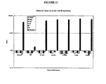
Figure 11 : Shows the effect of increasing Triton-X100 concentrations on K562 cells contaminated with M.hyorhinis (MH) compared to increasing numbers (1-10,000 cells/100µl sample) of bacterial cells (E.coli). - The principle of the preferred assay method is to supply the appropriate substrates for mycoplasmal enzymes. If mycoplasma contamination is present, there is a conversion of ADP to ATP which can then be measured, preferably by the luciferase-luciferin reaction.
- Mycoplasma Detection Reagent is added and, after approximately 5 minutes, an initial light output reading (A) is taken, the Mycoplasma Substrate (MS) is added and any enzymatic activity is allowed to progress for approximately 10 minutes, at which point a second light reading (B) is taken.
- If there is mycoplasmal contamination then the second reading (B) will be higher when compared to the first reading (A), giving a ratio


Figure 1 demonstrates the kinetics of the reaction. Typically, the ratio
Figure 1 shows a ratio of 114. - A preferred assay kit of the invention comprises a Mycoplasma Detection Reagent (MDR); Mycoplasma Assay Buffer (MAB) for reconstitution of MDR and the Mycoplasma Substrate (MS). MDR and MS are preferably provided as lyophilised preparations.
- All mycoplasma generate ATP through either the acetate kinase pathway or the carbamate kinase pathway. The Mycoplasma Substrate of the invention contains substrates for one or both of these enzymatic reactions. ADP is a requirement for both enzymes, and is preferably supplied in excess in the Mycoplasma Detection Reagent of the invention to drive the generation of ATP formation.
- The MDR is added to a sample of culture supernatant that has previously been centrifuged to remove cellular material, although it is possible to perform the assay in the presence of cells. Alternatively or additionally, the test sample can be passed through a bacterial filter.
- The MDR contains substrates for luciferase, luciferin and other co-factors plus AMP and ADP. The Mycoplasma Substrate (MS) contains carbamoyl phosphate and/or acetyl phosphate or acetyl-Co-A and/or citrulline and/or ammonia required for detection of the carbamate and/or acetate kinase activities.
- A preferred sample volume is 100µl to which 100µl of reconstituted MDR is added. After approximately 5 minutes the first luminometric reading (A) is taken, this gives the base reading upon which the further ratio calculations

- The assay methods of the invention have been used to investigate contamination by Acholeplasma laidlawii, M. hyorhinis, M. fermentans, M orale, and M genitalium and to detect a number of unknown mycoplasma contaminations.
- The inventors have compared their data to detection of mycoplasma by PCR, and have shown that there is a correlation between ratios greater than one and detection of mycoplasmal DNA. This is shown in
Figure 2 where the positive PCR bands on the gel correlate with ratios of more than one. - The inventors have also shown that we can detect a reduction in ratios

- The manufacturers (ICN-Flow) recommend treatment for 7 days of cells in quarantine to ensure complete removal of contaminating mycoplasma. However, the ratio data obtained using the assay methods of the invention showed that 7 days was not sufficient. This was evident from the fact that the ratios remained greater than one. Also after removal of treatment, and continued culture the ratios increased, and the cultures were again positive after PCR testing (Stratagene kit). These data are shown in
Figure 3 , where three different cell lines were found to be contaminated with M.hyorhinis. While the K562 and U937 cells are suspension cell lines, the A549 cells are an adherent cell type; these data therefore confirms that the assay can be used on both adherent and suspension cell types. This is also shown inFigure 2 where the CHO and COS-7 cells are adherent cell types commonly used in cell culture laboratories. -
Figure 3 also shows that the treatment with MRA for 10 days with COS-7 and CHO cell cultures was sufficient to remove the contaminating mycoplasma. - The inventors have also shown that culture supernatants put through a number of bacterial retarding filters continued to show positive ratios which is indicative of the presence of viable mycoplasma. This is shown in
Figure 4 . - Mycoplasma can form colonies as large as 600µm in diameter, but can also exist in their life cycle as single cells as small as 0.15µm. Due to their small size mycoplasma can pass through the 0.45µm and 0.22µm filters commonly used to "sterilise" tissue culture reagents.
Figure 4 also confirms that the assay can be performed in the presence of cells, but that there is a reduced sensitivity of detection. Hence, it is preferred that the assay methods of the invention are performed on samples which are substantially cell free. This can easily be achieved by centrifugation of cell cultures and sampling of the supernatant and, optionally, filtration through a bacterial filter. - As shown in
Figures 5 and8 , dilution of the supernatants shows the sensitivity of the assays, in that a 1:1000 dilution of contaminated culture supernatant can still give ratios greater than 1. Dependent on the specific activity of the acetate kinase and carbamate enzymes in different mycoplasma (Mollicutes) species, it is possible to dilute samples out further. The dilution range will also vary according to the number of colony forming units in the test sample. - The assay methods of the invention will work without the external exogenous addition of carbamate and acetate kinase substrates in the form of ADP, carbamoyl phosphate and acetyl phosphate or precursors thereof. In a contaminated culture sample the acetyl and carbamoyl phosphates or precursors thereof will be present endogenously together with sufficient cellular ADP, derived from the cell culture, to prime the reaction towards the formation of ATP. Alternatively, ADP can be generated by other externally added or cellular enzymes i.e. adenylate kinase utilising ATP and AMP.
- It is possible to avoid direct addition of these substrates and have the system generate them itself. The use of acetate and ammonia along with ATP will cause the acetate kinase and carbamate kinase enzymes to generate acetyl phosphate and carbamoyl phosphate that can then be used by the same enzymes to generate ATP from ADP:


- The two substrates could also be generated from "precursors" by utilising earlier parts of the glucose fermentation and argenine lysis pathways for example by the addition of acetyl-CoA and citrulline that could be used by mycoplasmal enzymes to synthesise acetyl phosphate and carbamoyl phosphate respectively.


- The following figures show the differences between the biochemical activities of M.fermentans, which generates ATP preferentially through the carbamate kinase pathway, but will also utilise the acetate kinase pathway.
Figure 6 shows the effect of adding the substrates for the enzyme pathways individually, and then in a combined reagent. - While M.fermentans utilises both pathways, M.orale utilises only the carbamate kinase pathway, and as shown in
Figure 7 , positive ratios are only observed in the single carbamate reagent or the combined reagent.Figure 8 shows the detection limits are as low as 14 CFU/well with M.orale. - The inventors have investigated a mycoplasma that preferentially utilises the acetate kinase pathway, namely M.hyorhinis. The data are shown in
Figure 9 . - The inventors have tested over 15 different cell lines (see table 3) and shown that none of the cells have sufficient background enzymatic activity to impact upon the ratios and give false positives. The inventors, without wishing to be bound by theory, think the reason for this is that the pathways are anaerobic, and all mammalian cell cultures will generate ATP through oxidative phosphorylation. Hence, by using only carbamoyl phosphate or a precursor thereof or only acetyl phosphate or a precursor thereof, one can produce an assay method of the invention which will allow one to determine whether the mycoplasmal contaminants in question are from a group which uses the acetate kinase pathway, the carbamate kinase pathway, or both. This may have useful diagnostic applications.
- The only bacteria that have acetate kinase activity are not those that are commonly found as contaminants of cell culture, with the possible exception of certain E.coli species that are handled in laboratories, principally for molecular biology purposes. However, activity with this organism, is only seen at very high inoculum concentrations where there is turbid growth and the resulting turbidity of the sample is readily observed by eye. Hence, the methods of the invention can be varied to include an initial screening step for bacterial contamination, if necessary. This can be achieved by a variety of methods, but is preferably carried out by passing the sample through a standard bacterial filter (Baseman and Tully, 1997).
-
- Magnesium acetate 214.5mg (10mM)
- Inorganic pyrophosphate 178.4µg (4µM)
- Bovine serum albumin 160mg (0.16%)
- D-Luciferin 10mg (360µM)
- L-Luciferin 250µg (8.9µM)
- Luciferase (RY) 85µg
- ADP 250.5µg (5µM)
- AMP 69.44mg (2mM)
-
- Acetyl phosphate 55.23mg (3mM)
- Carbamoyl phosphate 45.87mg (3mM)
- Examples of reactions generating acetyl or carbamoyl phosphate:
i.
acetyl-CoA 0.1mM to 100mM Inorganic phosphate
(e.g. potassium phosphate)0.1mM to 100mM 
citrulline 1mM to 100mM ammonium bicarbonate 1mM to 200mM 
acetate (e.g. sodium or potassium) 1mM to 500mM ATP 0.1 mM to 100mM 
ammonium bicarbonate 1 mM to 200mM ATP 0.1 mM to 100mM -
- Sigma-Aldrich Company Ltd.
- Fancy Road
- Poole
- Dorset
-
BH 12 4QH - United Kingdom
-
- EPES 1.1915g (50mM)
- EDTA 74.44mg (2mM)
- Triton X-100 250µl (0.25%)
- pH 7.50
- Preferred concentration ranges include
- ADP 1µM to 100mM, preferably 1 to 100µM, more preferably 5µM.
-
AMP 1 µM to 100mM, preferably 0.1mM to 10mM, more preferably 2mM. - Acetyl phosphate 1µM to 100mM, preferably 0.1mM to 20mM, more preferably 3mM. Concentrations above 10 reduce the light output, but the assay still performs.
- Carbamoyl phosphate 1µM to 100mM, preferably 0.1mM to 20mM, more preferably 3mM.
- Disruption of the viable mycoplasma cell membrane to allow for the release of the enzymes into the sample is a preferred embodiment of the assay method of the invention. This allows for binding of the substrates and generation of ATP. However, positive ratios indicating mycoplasma contamination can be obtained in the absence of any lysis treatment. The implication is that these enzymes can be released by viable mycoplasma The other possibility is that some non-viable organisms have released their contents through natural lysis.
- The addition of even low concentrations of the non-ionic detergent Triton-X100, greatly increases the sensitivity of the assay by ensuring maximal release of the carbamate and acetate kinases into the sample.
- The aim of the following experiments was to determine the concentrations of Triton-X100 in the Hepes-EDTA buffer on the ratios seen with mycoplasma contaminated K562 cell cultures. Two organisms were investigated, M.hyorhinis and M.orale.
-
Figure 10 shows that it is possible to detect mycoplasmal enzymes in the absence of a detergent lysis step. It also shows a drop in the light output with concentrations greater than 4-5%, this is due to adverse effects of the detergent on the luciferase enzyme/reaction. However, it is still possible to detect positive activity with concentrations as high as 20% (v/v). - The inventors have also shown that the concentrations of Triton-X100 used in the above experiments did not result in any detectable carbamate or acetate kinase activity from the E.coli strain JM109 cells.
- The above results confirm that there are no positive ratios with the bacterial cell number used. Increasing the Triton-X100 concentration to levels that have been reported to lyse bacterial cells (1-2%), still did not result in positive ratios above 1.
- Generally biological detergents are commonly used to disrupt the bipolar membranes of lipids in order to release and then solubilise membrane bound proteins. Non-ionic detergents are non-denaturing and permit the solubilization of membranes without interfering with biological activity. They have principally been used for the study of protein conformations and for the separation of hydrophilic proteins from membrane spanning hydrophobic proteins. Anionic and cationic detergents result in greater modification of protein structure and are more effective at disrupting protein aggregation. Zwitterionic detergents are also low-denaturing, but are effective at disruption of protein aggregates.
- These different groups of detergents have been studied with a number of different cells types to efficiently lyse, and release and preserve the protein content, of both eukaryotic and prokaryotic organisms.
- For the preferred Assays of the invention the required lysis agent is one that causes disruption of the mycoplasmal membrane and allows release of the metabolic enzymes that are required to react with the substrates. As there is no detergent removal or neutralisation step, it is therefore important that the chosen system does not interfere with the activity of the carbamate and/or acetate kinase, or the luciferase/luciferin/ATP reaction. It is also preferable to use a system that selectively causes the lysis of mycoplasma, with little or no effect on bacteria that may be potential contaminates of the cell cultures/samples.
- The presence of a filtration step through 0.45µm filters, however, should remove any contaminating larger micro-organisms.
- The key difference between bacteria and mycoplasma is the lack of cell wall, and it is the bacterial cell wall that makes bacteria more difficult to lyse. There are a number of fairly brutal methods that can bring about total lysis, these include pressure (French Press) and sonication. Other enzyme digest methods include lysozyme followed by the addition of detergents. However, mycoplasma can be lysed with concentrations of Triton X-100 at around 1-2%.
- Low concentrations of other non-ionic detergents, such as Brij®35 (0.4%) (Sigma-Aldrich Company Ltd.) and B-PER (1%) (Perbio Science UK Ltd.), do not have adverse effects on the luciferase enzyme, and are capable of disrupting the mycoplasmal membrane, without adversely affecting the luciferase reaction. The concentrations of these detergents can be taken up to 10% without loss of sensitivity of mycoplasmal detection.
- Contaminating mycoplasma can be detected in the absence of a lysis step to disrupt the mycoplasmal membrane. However, addition of a gentle lysis step (0.25% Triton X-100 in Hepes-EDTA buffer) increases the sensitivity of the assay by releasing the mycoplasmal enzymes of interest into the reaction mixture.
- The lysis step would preferably cause selective lysis of mycoplasma, while having little or no effect on bacterial cells. Low concentrations of most non-ionic detergents should do this. However, a filtration step would physically remove any contaminating bacteria, and allow for the use of any detergent but preferably those that do not inhibit either the luciferase reaction or the activity of carbamate kinase and acetate kinase.
-
- 1. LT27-217 Mycoplasma Detection Reagent, Lyophilised. 2 x 600 µl vials.
- 2. LT27-218 Mycoplasma Assay Buffer. 1 x 10 ml bottle.
- 3. LT27-221 Mycoplasma Substrate. Lyophilised. 2 x 600 µl vials.
-
- 1. LT27-217 Mycoplasma Detection Reagent. Lyophilised. 5 x 600 µl vials.
- 2. LT27-218 Mycoplasma Assay Buffer. 1 x 10 ml bottle.
- 3. LT27-221 Mycoplasma Substrate. Lyophilised. 5 x 600 µl vials.
-
- 1. LT27-216 Mycoplasma Detection Reagent. Lyophilised. 1 x 10 ml vial.
- 2. LT27-220 Mycoplasma Assay Buffer. 1 x 20 ml bottle.
- 3. LT27-224 Mycoplasma Substrate. Lyophilised. 1 x 10 ml vial.
-
- 1. Mycoplasma Detection Reagent (MDR) per 100ml
- Magnesium acetate1 214.5mg(10mM)
- Inorganic pyrophosphate1 178.4µg (4µM)
- Bovine serum albumin1 160mg (0.16%)
- D-Luciferin2 10mg (360µM)
- L-Luciferin2 250µg (8.9µM)
- Luciferase (RY)3 85µg
- ADP1 250.5µg (5µM)
- AMP1 69.44mg (2mM)
- 2. Mycoplasma Substrate (MS) per 100ml
- Acetyl phosphate1 55.23mg (3mM)
- Carbamoyl phosphate1 45.87mg (3mM)
- 3. Mycoplasma Assay Buffer (MAB) per 100ml
- HEPES1 1.1915g(50mM)
- EDTA1 74.44mg (2mM)
- Triton X-1001 250µl (0.25%)
- pH 7.50
- Preferred concentration ranges:
- ADP 1µM to 100mM
- AMP 1µM to 100mM
- Acetyl phosphate 1µM to 100mM, preferably, mM to 10mM
-
Carbamoyl phosphate 1 µM to 100mM -
- 1.
- Sigma-Aldrich Company Ltd.
- Fancy Road
- Poole
- Dorset
-
BH 12 4QH - United Kingdom
- 2.
- Resem BV
- Goudenregenstraat 84
- NL-4131 BE Vanen
- Netherlands
- 3.
- Lucigen Ltd
- Porton Down Science Park
- Porton, Salisbury
- Wiltshire SP4 0JQ
- U.K.
- The preferred embodiment of the invention provides a selective biochemical test that exploits the activity of certain mycoplasmal enzymes. The presence of these enzymes provides a rapid screening procedure, allowing sensitive detection of contaminating mycoplasma in a test sample. The viable mycoplasma are lysed and the enzymes react with the Mycoplasma Substrate catalysing the conversation of ADP to ATP.
- By measuring the level of ATP in a sample both before (A) and after (B) the addition of the Mycoplasma Substrate, a ratio


- The emitted light intensity is linearly related to the ATP concentration and is measured using a luminometer. The assay is preferably conducted at ambient room temperature (18-22°C), the optimal temperature for luciferase activity.
- If


If
- It is preferred that the culture supernatant be centrifuged to remove cells and, optionally, passed through a bacterial filter prior to performing the assay.
- The kit contains all the required reagents to perform the assay.
- 100µl of culture supernatant is taken as the sample.
- Add Mycoplasma detection reagent (MDR)
- Wait 5 minutes
- Read luminescence (A)
- Add Mycoplasma Substrate (MS)
- Wait 10 minutes.
- Read luminescence (B).
- Ensure that you follow the correct reagent reconstitution applicable to the relevant kit (10, 25 or 100 assay points).
This procedure usually requires at least 15 minutes equilibration time.
The Mycoplasma Detection Reagent (MDR) and Mycoplasma Substrate (MS) are preferably supplied as lyophilised pellets. These are reconstituted in Mycoplasma Assay Buffer (MAB) to produce the working solutions for use in the assay. - For 10 tests (KIT LT07-118):
- 1. Preparation of Mycoplasma Detection Reagent
Add 600µl of Mycoplasma Assay Buffer into a vial containing the lyophilised Mycoplasma Detection Reagent.
Replace the cap and mix gently.
Allow the reagent to equilibrate for 15 minutes at room temperature. - 2. Preparation of Mycoplasma Substrate
Add 600µl of Mycoplasma Assay Buffer into a vial containing the lyophilised Mycoplasma Substrate.
Replace the cap and mix gently.
Allow the reagent to equilibrate for 15 minutes at room temperature. - 3. Mycoplasma Assay Buffer
This is preferably provided ready for use. Store at 2-8°C when not in use. - For 25 tests (KIT LT07-218)
- 1. Preparation of Mycoplasma Detection Reagent
Add 600µl of Mycoplasma Assay Buffer into a vial containing the lyophilised Mycoplasma Detection Reagent.
Replace the cap and mix gently.
Allow the reagent to equilibrate for 15 minutes at room temperature. - 2. Preparation of Mycoplasma Substrate
Add 600µl of Mycoplasma Assay Buffer into a vial containing the lyophilised Mycoplasma Substrate.
Replace the cap and mix gently.
Allow the reagent to equilibrate for 15 minutes at room temperature. - 3. Mycoplasma Assay Buffer
This is preferably provided ready for use. Store at 2-8 when not in use. - For 100 tests (KIT LT07-318)
- 1. Preparation of Mycoplasma Detection Reagent
Add 10ml of Mycoplasma Assay Buffer into a vial containing the lyophilised Mycoplasma Detection Reagent.
Replace the cap and mix gently.
Allow the reagent to equilibrate for 15 minutes at room temperature. - 2. Preparation of Mycoplasma Substrate
Add 600µl of Mycoplasma Assay Buffer into a vial containing the lyophilised Mycoplasma Substrate.
Replace the cap and mix gently.
Allow the reagent to equilibrate for 15 minutes at room temperature. - 3. Mycoplasma Assay Buffer
This is preferably provided ready for use. Store at 2-8 when not in use. -
- 1. Instrumentation
The kit requires the use of a luminometer. The parameters of the luminometer should be assessed and the conditions below used to produce the correct programming of the machine.
The preferred assay of the invention has been designed for use with cuvette/tube luminometers. For use with plate luminometers please see below.
Cuvette/tube luminometers:
Readtime 1 second (integrated). - 2. Additional equipment and consumables
- a. 10 ml sterile pipettes
- b. Luminometer cuvettes
- c. Micropipettes - 50-200µl; 200-1000 µl
- d. Bench centrifuge.
- Please note samples of the culture medium should be taken before any further processing steps, e.g. trypsinisation.
- 1. Bring all reagents up to room temperature before use.
- 2. Reconstitute the Mycoplasma Detection Reagent and Mycoplasma Substrate in Mycoplasma Assay Buffer. Leave for 15 minutes at room temperature to ensure complete rehydration.
- 3.
Transfer 2 ml of cell culture or culture supernatant into a centrifuge tube and pellet any cells at 1500 rpm (200 x g) for 5 minutes. - 4. Transfer 100µl of cleared supernatant into a luminometer cuvette/tube.
- 5. Program the luminometer to take a 1 second integrated reading (this is usually the default setting on most cuvette luminometers).
- 6. Add 100µl of Mycoplasma Detection Reagent to each sample and wait 5 minutes.
- 7. Place cuvette in luminometer and initiate the programme (Reading A).
- 8. Add 100µl of Mycoplasma Substrate to each sample and wait 10 minutes.
- 9. Place cuvette in luminometer and initiate the programme (Reading B).
- 10. Calculate ration = Reading B/Reading A.
- The ratio of Reading B to Reading A is used to determine whether a cell culture is contaminated by mycoplasma.
- The speed and convenience offered by the kits according to the invention means that it provides a unique method for screening cultures for the presence of mycoplasma. As such it is ideally suited to routine testing of cells in culture. Frequent use of the test methods of the invention will indicate when a cell line becomes infected allowing prompt remedial action to be taken. The test methods of the invention can also be extended to incoming cell lines and the commonly used constituents of complete media.
- The interpretation of the different ratios obtained, within each experimental situation, may vary according to the cell types and conditions used. However, the test gives ratios

Table a. Interpretation of assay results: illustrating examples of healthy and infected cell lines. Cell Line Mycoplasma ratio Conclusions Infected cells K562 123.26 Positive A549 4.10 Positive U937 8.26 Positive HepG2 1.27* Borderline, quarantine and retest in 24 hours Healthy cells HL60 0.72 Negative COS-7 0.46 Negative -
- 1. Bring all reagents up to room temperature before use.
- 2. Reconstitute the Mycoplasma Detection Reagent and Mycoplasma Substrate in Mycoplasma Assay Buffer. Leave for 15 minutes at room temperature to ensure complete rehydration.
- 3.
Transfer 2 ml of cell culture of cell culture supernatant into a centrifuge tube and pellet any cells at 1500rpm (200 x g) for 5 minutes. - 4. Transfer 100µl of cleared supernatant into a luminescence compatible plate.
- 5. Program the luminometer to take a 1 second integrated reading.
- 6. Add 100µl of Mycoplasma Detection Reagent to each sample and wait 5 minutes.
- 7. Place plate in luminometer and initiate the programme (ReadingA).
- 8. Add 100µl of Mycoplasma Substrate to each sample and wait 10 minutes.
- 9. Place plate in luminometer and initiate the programme (Reading B).
- 10. Calculate ratio = Reading B/Reading A.
- Great care should be taken when handling any of the reagents. Skin has high levels of ATP on its surface that can contaminate the reagents leading to falsely high readings. Latex gloves avoid this problem.
- The optimal working temperature for all reagents is 22°C. If reagents have been refrigerated always allow time for them to reach room temperature (18-22°C) before use.
- The sensitivity of the assay does allow for detection of covert contamination, and if the ratio is marginally above 1 (for example up to 1.3) it is recommended that the sample be retested. Any cultures maintained in quarantine can be tested after a further 24-48 hours in culture to see if the ratios have increased.
- The assays of the invention can be performed in the presence or absence of cells. Unlike known mycoplasma detection systems, they allow for samples to be screened rapidly using cheap hand-held luminometer systems, and can give results within 15 minutes to allow for the appropriate handling of the contaminated samples.
- PCR and DAPI/Hoechst staining, will bind to all DNA, be it from viable or non viable mycoplasma. Hence, if looking to treat and remove mycoplasma, you could still end up with false positives when using PCR/DNA staining even though mycoplasma have been irradicated.
- The assays can detect viable mycoplasma whereas known methods such as PCR cannot distinguish between viable and non-viable mycoplasma.
-
- 1) Razin S, Yogev D and Naot Y. 1998. Molecular biology and pathogenicity of mycoplasmas. Microbiol. and Mol. Biol. Rev. 62(4): 1094-1156.
- 2) Rottem S and Barile MF. 1993. Beware of mycopaslasmas. TIBTECH. 11: 143-151.
- 3) Rottem S. 2002. Sterols and acylated proteins in mycoplasmas. Biochem. Biophys. Res. Commun. 292: 1289-1292.
- 4) McGarrity GJ and Kotani H. 1985. in The mycoplasmas Vol IV. (Razin S and Barile MF eds) p. 353-390. Academic Press.
- 5) Battaglia M, Pozzi D, Grimaldi S and Parasassi T. 1994. Hoecsht 33258 staining for detecting mycoplasma contamination in cell cultures: a method for reducing fluorescence photobleaching. Biotechnic and Histochem. 69: 152-156.
- 6) Raab LS. 1999. Cultural revolution: mycoplasma testing kits and services. The Scientist. 13 (20): 21-25.
- 7) Verhoef V, Germain G and Fridland A. 1983. Adenosine phosphorylase activity in mycoplasma-free growth media for mammalian cells. Exp. Cell Res. 149(1): 37-44.
- 8) Whitaker AM, Windsor GD, Burnett CM and Taylor CH. 1987. A rapid and sensitive method for the detection of mycoplasmas in infected cell cultures using 6-methyl purine deoxyriboside. Dev. Biol. Stand. 66:503-509.
- 9) Daxboeck F, Krause R and Wenisch C. 2003. Laboratory diagnosis of Mycoplasma pneumoniae infection. Clin. Microbiol. Infect. 9(4): 263-73.
- 10) de Wet JR, Wood KV, DeLuca M, Helinski DR and Subramani S. 1987. Firefly luciferase gene: structure and expression in mammalian cells. Mol. Cell Biol. 7(2): 725-37.
- 11) Masuda T, Tatsumi H and Nakano E. 1989. Cloning and sequence analysis of cDNA for luciferase of a Japanese firefly, Luciola cruciata.Gene. 77(2): 265-70.
- 12) Wood KV, Lam YA, Seliger HH and McElroy WD. 1989. Complementary DNA coding click beetle luciferases can elicit bioluminenscence of different colours. Science. 244 (4905): 700-2.
- 13) de Wet JR, Wood KV, Helinski DR and DeLuca M. 1986. Cloning firefly luciferase. Methods Enzymol. 133: 3-14.
- 14) White PJ, Squirrell DJ, Arnaud P, Lowe CR and Murray JA. 1996. Improved thermostability of the North American firefly luciferase: saturation mutagenesis at position 354. Biochem J. 319(2): 343-50.
- 15) Baseman JB and Tully JG. 1997. Mycoplasmas: sophisticated, reemerging, and burdened by their notoriety. Emerg. Infect. Dis. 3(1): 21-32.
- 16) Kirchhoff H, Mohan K, Schmidt R, Runge M, Brown DR, Brown MB, Foggin CM, Muvavarirwa P, Lehmann H and Flossdorf J. 1997. Mycoplasma crocodyli sp. nov., a new species from crocodiles. Int. J. Syst. Bacteriol. 47: 742-6.
- 17) Forsyth MH, Tully JG, Gorton TS, Hinkley L, Frasca S, van Kruiningen HJ and Geary SJ. Int. J. Syst. Bacteriol. 1996. Mycoplasma sturni sp. nov., from the conjunctiva of a European starling (Sturnus vulgaris). Int. J. Syst. Bacteriol. 46: 716-9.
- 18) Taylor RR, Mohan K and Miles RJ. 1996. Diversity of energy-yielding substrates and metabolism in avian mycoplasmas. Vet. Microbiol. 51: 291-304.
- 19) Tully JG, Whitcomb RF, Rose DL, Bove JM, Carle P, Somerson NL, Williamson DL and Eden-Green S. 1994. Acholeplasma brassica sp. nov. and Acholeplasma palmae sp. nov., two non-sterol-requiring mollicutes from plant surfaces. Int. J. Syst. Bacteriol. 44:690-4.
- 20) Web reference: www.unc.edu/depts/tcf/mycoplasma.htm
- 21) Duffy LB, Crabb D, Searcey K and Kempf MC. 2000. Comparative potency of gemifloxacin, new quinolones, macrolides, tetracycline and clindamycin against Mycoplasma spp. J. Antimicrobial Chemotherapy. 45: 29.
- 22) Taylor-Robinson D and Bebear C. 1997. Antibiotic susceptibilities of mycoplasmas and treatment of mycoplasmal infections. 40: 622-630.
- 23) Uphoff CC, Meyer C and Drexler HG. 2002. Elimination of mycoplasma from leukaemia-lymphoma cell lines using antibiotics. 16(2): 284-288.
- 24) Schram E and Weyens-van Witzenburg A. 1989. Improved ATP methodology for biomass assays. J. Biolumin. Chemilumin. 4: 390-398.
- 25) Stanley PE. 1989. A review of bioluminescent ATP techniques in rapid microbiology. J. Biolumin. Chemilumin. 4:375-380.
- 26) Pellegrini A, Thomas U, von Fellenberg R and Wild P. 1992. Bactericidal activities of lysozyme and aprotinin against gram-negative and gram positive bacteria related to their basic character. J. Appl. Bacteriol. 72: 180-187.
- The present disclosure is also described with reference to the following numbered paragraphs:
- 1. A method of detecting the presence of contaminating mycoplasma in a test sample comprising:
- (i) providing a test sample;
- (ii) detecting and/or measuring the activity (B) of acetate kinase and/or carbamate kinase in the test sample, and said activity being indicative of the presence of contaminating mycoplasma; and
- (iii) identifying the test sample as contaminated with mycoplasma on the basis of detection and/or measurement of said activity in step (ii).
- 2. A method according to
paragraph 1 further comprising the following steps performed after step (ii) but before step (iii):- (iia) obtaining acetate kinase and/or carbamate kinase activity information (A) detected and/or measured in a corresponding control sample; and
- (iib) comparing the activity detected and/or measured in the test sample (B) with that in the control sample (A);
wherein the test sample is identified as contaminated with mycoplasma in step (iii) if the activity (B) detected and/or measured in the test sample in step (ii) is greater than that of the control sample (A) in step (iia), that is, the ratio
- 3. A method according to
paragraph 

- 4. A process according to any one of
paragraphs 1 to 3 further comprising the step of releasing mycoplasma cellular contents into the sample by treatment of the test sample with a mycoplasma lysis agent that is performed after step (i) but before step (ii). - 5. A method according to
paragraph 4 wherein the lysis agent is a detergent. - 6. A method according to paragraph 5 wherein the detergent lysis treatment is not capable of lysing bacterial cells.
- 7. A method according to any of
paragraphs 4 to 6 wherein the corresponding control sample is the same as the test sample prior to mycoplasma lysis treatment. - 8. A method according to any of
paragraphs 2 to 4 wherein the corresponding control sample is the same as the test sample but the obtention of detection/measurement for the test sample activity information is carried out after a time interval following the obtention of detection/measurement information for the control sample. - 9. A method according to
paragraph 8 wherein the time interval is at least approximately 30 minutes. - 10. A method according to any of
paragraphs 1 to 9 wherein the detecting and/or measuring step comprises detecting and/or measuring ATP. - 11. A method according to
paragraph 10 wherein the ATP is detected and/or measured by a light-emitting reaction. - 12. A method according to paragraph 11 where the light emitting reaction is a bioluminescent reaction.
- 13. A method according to any of the preceding paragraphs wherein ADP is added to the test sample prior to the detecting and/or measuring step (ii).
- 14. A method according to any of the preceding paragraphs wherein a mycoplasma substrate (MS) reagent is added to the test sample prior to the detecting and/or measuring step (ii), the MS reagent comprising: acetyl phosphate or a precursor thereof and/or carbamoyl phosphate or a precursor thereof.
- 15. A method according to
paragraph 14 wherein the precursor of acetyl phosphate is acetyl-CoA. - 16. A method according to
paragraph 14 wherein the precursor of carbamoyl phosphate is citrulline and/or ammonia. - 17. A method according to any of paragraphs 13 to 16 wherein the control sample is all or an aliquot of the test sample to which a mycoplasma reagent has not been added.
- 18. A method according to any of
paragraphs 2 to 7, or 9 to 17 wherein the control sample has been shown to be free from mycoplasma by a separate method. - 19. A method according to
paragraph 18 wherein the control sample has been shown to be free from mycoplasma by one or more of PCR testing, DNA fluorescence staining, or mycoplasma culture method. - 20. A method according to any preceding paragraph wherein the test sample and/or control sample is a cell-culture sample.
- 21. A method according to
paragraph 20 wherein the cells in the cell-culture sample are mammalian cells, preferably adherent cells, such as Vero, MRC5, HUVEC, BSMC, NHEK, MCF-7, AoSMC, A549, HepG2, FM3A, PC12, ARPE-19, CHO and COS cells, and/or adherent primary cells isolated from animal source. - 22. A method according to
paragraph 20 wherein the mammalian cells in the cell-culture sample grow in suspension, such as K562, U937, HL-60, Cem-7, and Jurkats plus primary cell types such as leukaemic blast cells. - 23. A method according to
paragraph 20 where the cell culture is a culture of plant cells. - 24. A method according to any of
paragraphs 20 to 23 where the cell culture sample is a sample which is derived from a cell culture but is itself substantially free of cellular material. - 25. A method according to any of
paragraphs 1 to 19 wherein the test sample and/or control sample consists of a cell-free reagent. - 26. A method according to paragraph 25 where the cell-free reagent is trypsin.
- 27. A process for treating a cell culture to remove mycoplasma contamination comprising:- treating a mycoplasma contaminated cell culture with an agent to remove and/or destroy mycoplasma; and subsequently testing a sample from the culture for mycoplasma contamination using a method according toany preceding paragraph; if necessary, repeating the process one or more times until mycoplasma contamination is not detected in the sample.
- 28. A method of detecting the presence of mycoplasma in a test sample, comprising the following steps:-
- (i) providing a test sample;
- (ii) without adding an exogenous reagent (e.g. substrates for kinase activity) to convert ADP to ATP, detecting or measuring ATP in the test sample using a bioluminescent reaction to obtain an ATP and/or light output measurement (A);
- (iii) obtaining an ATP and/or light output measurement (B) from a corresponding control sample;
- (iv) comparing the ATP and/or light output measurement ratio

- (v) identifying the test sample as contaminated with mycoplasma in the event that the ratio

- 29. A method according to any preceding paragraph wherein the method includes a step of passing the test sample through a filter which retains bacterial cells.
- 30. Use of acetyl phosphate or a precursor thereof; and/or carbamoyl phosphate or a precursor thereof in a method of detecting contamination of a sample by mycoplasma as defined in
paragraphs 14 to 29. - 31. A kit for use in the detection of mycoplasma contamination which comprises the following:
- (i) acetyl phosphate or a precursor thereof and/or carbamoyl phosphate or a precursor thereof;
- (ii) ADP at an amount in excess to drive the enzymatic reactions in the direction of ATP formation;
- (iii) one or more agents for lysing mycoplasma.
- 32. A kit according to paragraph 31, wherein the agents for lysing mycoplasma comprises a detergent.
- 33. A kit according to any one of paragraphs 31 to 32 further comprising means for detecting and/or measuring ATP by a light-emitting reaction.
- 34. A kit according to paragraph 33 wherein said means comprise a mycoplasma detection reagent (MDR) which includes magnesium acetate, inorganic pyrophosphate, bovine serum albumin, luciferin luciferase, ADP and AMP.
- 35. A kit according to any one of paragraphs 31 to 34 wherein the reagents are provided in a lyophilised condition.
- 36. A kit according to any one of paragraphs 31 to 35 which further comprises a mycoplasma assay buffer (MAB) in which lyophilised reagents can be reconstituted.
- 37. A kit according to paragraph 36 wherein the buffer maintains a pH of approximately 7.5.
- 38. A kit according to paragraph 33 or 34 further comprising a luminometer, which is preferably a hand-held luminometer.
- 39. A kit according to any one of paragraphs 31 to 38 further comprising a bacterial filter.
- 40. A kit for detecting mycoplasma contamination of a test sample substantially as described herein, preferably with reference to one or more of the accompanying examples and/or figures.
- 41. A method of detecting mycoplasma contamination of a test sample substantially as described herein, preferably with reference to one or more of the accompanying examples and/or figures.
- 42. A method of treating a cell culture to remove mycoplasma contamination substantially as described herein, preferably with reference to one or more of the accompanying examples and/or figures.
Claims (32)
- A method of detecting the presence of contaminating mycoplasma in a test sample comprising:(i) detecting and/or measuring the activity (B) of acetate kinase and/or carbamate kinase in a test sample, and said activity being indicative of the presence of contaminating mycoplasma; and(ii) identifying the test sample as contaminated with mycoplasma on the basis of detection and/or measurement of said activity in step (i).
- A method according to Claim 1 further comprising the following steps performed after step (i) but before step (ii):(ia) obtaining acetate kinase and/or carbamate kinase activity information (A) detected and/or measured in a corresponding control sample; and(ib) comparing the activity detected and/or measured in the test sample (B) with that in the control sample (A);wherein the test sample is identified as contaminated with mycoplasma in step (ii) if the activity (B) detected and/or measured in the test sample in step (i) is greater than that of the control sample (A) in step (ia), that is, the ratio

- A method as claimed in Claims 1 or 2 wherein detecting and/or measuring the activity (B) of acetate kinase and/or carbamate kinase in the test sample in step (i) and/or obtaining acetate kinase and/or carbamate kinase activity information (A) in a corresponding control sample in step (ia) comprises detecting and/or measuring the appearance and/or disappearance of one or more of the substrates and/or one or more of the products of the following reactions:


- A method according to any one of Claims 1 to 3 further comprising the step of releasing mycoplasma cellular contents into the sample by treatment of the test sample with a mycoplasma lysis agent that is performed before step (i).
- A method as claimed in Claim 4 wherein the lysis agent is a detergent.
- A method as claimed in Claim 5 wherein the detergent lysis treatment is not capable of lysing bacterial cells.
- A method as claimed in any of Claims 4 to 6 wherein the corresponding control sample is the same as the test sample prior to mycoplasma lysis treatment.
- A method as claimed in any of Claims 2 to 4 wherein the corresponding control sample is the same as the test sample but the obtention of detection/measurement for the test sample activity information is carried out after a time interval following the obtention of detection/measurement information for the control sample, for example wherein the time interval is at least approximately 30 minutes.
- A method according to any of Claims 1 to 8 wherein the detecting and/or measuring step comprises detecting and/or measuring ATP.
- A method according to Claim 9 wherein the ATP is detected and/or measured by a light-emitting reaction, for example a bioluminescent reaction.
- A method according to any of the preceding claims wherein ADP is added to the test sample prior to the detecting and/or measuring step (i).
- A method according to any of the preceding claims wherein a mycoplasma substrate (MS) reagent is added to the test sample prior to the detecting and/or measuring step (i), the MS reagent comprising: acetyl phosphate or acetyl-CoA and/or carbamoyl phosphate or citrulline and/or ammonia.
- A method according to Claim 12 wherein the mycoplasma substrate (MS) reagent is acetyl-CoA, and/or citrulline and/or ammonia.
- A method according to any of Claims 11 to 13 wherein the control sample is all or an aliquot of the test sample to which a mycoplasma reagent has not been added.
- A method according to any of Claims 2 to 7, or 9 to 14 wherein the control sample has been shown to be free from mycoplasma by a separate method, for example, by one or more of PCR testing, DNA fluorescence staining, or mycoplasma culture method.
- A method as claimed in any preceding claim wherein the test sample and/or control sample is a cell-culture sample.
- A method according to Claim 16 wherein the cells in the cell-culture sample are mammalian cells, preferably adherent cells, such as Vero, MRC5, HUVEC, BSMC, NHEK, MCF-7, AoSMC, A549, HepG2, FM3A, PC12, ARPE-19, CHO and COS cells, and/or adherent primary cells isolated from animal source.
- A method according to Claim 16 wherein the mammalian cells in the cell-culture sample grow in suspension, such as K562, U937, HL-60, Cem-7, and Jurkats plus primary cell types such as leukaemic blast cells.
- A method as claimed in Claim 16 where the cell culture is a culture of plant cells.
- A method according to any of Claims 16 to 19 where the cell culture sample is a sample which is derived from a cell culture but is itself substantially free of cellular material.
- A method according to any of Claims 1 to 15 wherein the test sample and/or control sample consists of a cell-free reagent, such as trypsin.
- A process for treating a cell culture to remove mycoplasma contamination comprising:- treating a mycoplasma contaminated cell culture with an agent to remove and/or destroy mycoplasma; and subsequently testing a sample from the culture for mycoplasma contamination using a method as claimed in any preceding claim; if necessary, repeating the process one or more times until mycoplasma contamination is not detected in the sample.
- A method of detecting the presence of mycoplasma in a test sample, comprising the following steps:-(i) without adding an exogenous reagent to convert ADP to ATP, detecting or measuring the activity of acetate kinase and/or carbamate kinase in a test sample using a bioluminescent reaction to obtain an ATP and/or light output measurement (A);(ii) obtaining an ATP and/or light output measurement (B) from a corresponding control sample;(iii) comparing the ATP and/or light output measurement ratio
 (iv) identifying the test sample as contaminated with mycoplasma in the event that the ratio
- A method as claimed in any preceding claims wherein the method includes a step of passing the test sample through a filter which retains bacterial cells.
- Use of acetyl phosphate or acetyl-CoA; and/or carbamoyl phosphate or citrulline and/or ammonia in a method of detecting contamination of a sample by mycoplasma as defined in Claims 12 to 24.
- A kit for use in the detection of mycoplasma contamination which comprises the following:(i) acetyl phosphate or acetyl-CoA and/or carbamoyl phosphate or citrulline and/or ammonia;(ii) ADP at an amount in excess to drive the enzymatic reactions in the direction of ATP formation;(iii) one or more agents for lysing mycoplasma.
- A kit as claimed in Claim 26, wherein the agents for lysing mycoplasma comprises a detergent.
- A kit as claimed in any one of Claims 26 to 27 further comprising means for detecting and/or measuring ATP by a light-emitting reaction, such as means comprising a mycoplasma detection reagent (MDR) which includes magnesium acetate, inorganic pyrophosphate, bovine serum albumin, luciferin luciferase, ADP and AMP.
- A kit as claimed in any one of Claims 26 to 28 wherein the reagents are provided in a lyophilised condition.
- A kit as claimed in any one of Claims 26 to 29 which further comprises a mycoplasma assay buffer (MAB) in which lyophilised reagents can be reconstituted, and preferably, wherein the buffer maintains a pH of approximately 7.5.
- A kit as claimed in Claim 28 further comprising a luminometer, which is preferably a hand-held luminometer.
- A kit as claimed in any one of Claims 26 to 31 further comprising a bacterial filter.
Applications Claiming Priority (3)
| Application Number | Priority Date | Filing Date | Title |
|---|---|---|---|
| US43632303P | 2003-04-17 | 2003-04-17 | |
| GB0308829A GB2400659B8 (en) | 2003-04-17 | 2003-04-17 | Assay methods and materials |
| EP04727944A EP1613763A1 (en) | 2003-04-17 | 2004-04-16 | Assay for detecting mycoplasma by measuring acetate kinase or carbamate kinase activity |
Related Parent Applications (1)
| Application Number | Title | Priority Date | Filing Date |
|---|---|---|---|
| EP04727944.3 Division | 2004-04-16 |
Publications (2)
| Publication Number | Publication Date |
|---|---|
| EP2264181A1 EP2264181A1 (en) | 2010-12-22 |
| EP2264181B1 true EP2264181B1 (en) | 2012-07-11 |
Family
ID=9956913
Family Applications (1)
| Application Number | Title | Priority Date | Filing Date |
|---|---|---|---|
| EP10075423A Expired - Lifetime EP2264181B1 (en) | 2003-04-17 | 2004-04-16 | Assay for detecting mycoplasma by measuring acetate kinase or carbamate kinase activity |
Country Status (5)
| Country | Link |
|---|---|
| US (1) | US7585644B2 (en) |
| EP (1) | EP2264181B1 (en) |
| KR (1) | KR20120044351A (en) |
| CN (1) | CN1756848B (en) |
| GB (1) | GB2400659B8 (en) |
Families Citing this family (7)
| Publication number | Priority date | Publication date | Assignee | Title |
|---|---|---|---|---|
| WO2003063693A2 (en) * | 2002-01-31 | 2003-08-07 | Expressive Constructs, Inc. | Method for detecting microorganisms |
| EP2227567A4 (en) * | 2007-10-28 | 2011-03-30 | Eci Biotech Inc | Sensors for measuring contaminants |
| CN102888441A (en) * | 2011-07-20 | 2013-01-23 | 农高惠 | Kit for fast culture, identification and drug sensitivity test of myeoplasmapneumoniae (Mp) and detection method therefor |
| CN105277537B (en) * | 2015-10-12 | 2019-04-16 | 江苏三联生物工程有限公司 | A method of detection enzymatic activity and chemiluminescence reaction substrate performance |
| CN109517813B (en) * | 2018-11-21 | 2022-04-12 | 东软威特曼生物科技(南京)有限公司 | Creatine kinase and isozyme activator, determination reagent and kit thereof |
| CN113906286B (en) * | 2019-07-02 | 2024-09-20 | 株式会社堀场先进技术 | Biological sample analysis device and biological sample analysis method |
| CN110283882A (en) * | 2019-07-22 | 2019-09-27 | 赛莱克斯(深圳)科技有限公司 | There are the methods of mycoplasma pneumoniae in a kind of quick detection clinical sample |
Family Cites Families (21)
| Publication number | Priority date | Publication date | Assignee | Title |
|---|---|---|---|---|
| US5583024A (en) * | 1985-12-02 | 1996-12-10 | The Regents Of The University Of California | Recombinant expression of Coleoptera luciferase |
| US4968613A (en) | 1987-07-29 | 1990-11-06 | Kikkoman Corporation | Luciferase gene and novel recombinant DNA as well as a method of producing luciferase |
| DE68910036T2 (en) | 1988-07-01 | 1994-03-31 | Kikkoman Corp | Luciferase gene and new recombinant DNA as well as processes for the production of luciferase. |
| US5219737A (en) * | 1990-03-27 | 1993-06-15 | Kikkoman Corporation | Mutant luciferase of a firefly, mutant luciferase genes, recombinant dnas containing the genes and a method of producing mutant luciferase |
| US5374534A (en) * | 1990-07-19 | 1994-12-20 | Charm Sciences, Inc. | Method of preparing D-luciferin derivatives |
| GB9301118D0 (en) | 1993-01-21 | 1993-03-10 | Secr Defence | Enzyme linked assays |
| ES2301160T3 (en) | 1994-01-03 | 2008-06-16 | Promega Corporation | MUTANT LUCIFERASES. |
| AU707243B2 (en) | 1995-01-20 | 1999-07-08 | Promega Corporation | Mutant luciferases |
| US5876995A (en) * | 1996-02-06 | 1999-03-02 | Bryan; Bruce | Bioluminescent novelty items |
| GB9626932D0 (en) | 1996-12-24 | 1997-02-12 | Lumitech Limited | Assay methods and kits therefor |
| GB9707486D0 (en) | 1997-04-11 | 1997-05-28 | Secr Defence | Enzyme assays |
| US6074859A (en) * | 1997-07-08 | 2000-06-13 | Kikkoman Corporation | Mutant-type bioluminescent protein, and process for producing the mutant-type bioluminescent protein |
| EP1020524B1 (en) | 1997-07-08 | 2007-04-18 | Kikkoman Corporation | Mutant bioluminescent protein and process for producing the mutant bioluminescent protein |
| FR2769092B1 (en) | 1997-09-29 | 1999-11-12 | Lyonnaise Eaux Eclairage | METHOD FOR REGULATING DISINFECTION OF LIQUIDS |
| JP2002500892A (en) | 1998-01-21 | 2002-01-15 | イギリス国 | Antibiotic susceptibility test |
| GB9803156D0 (en) | 1998-02-13 | 1998-04-08 | Celsis Int Plc | Assay |
| AU765703B2 (en) * | 1998-03-27 | 2003-09-25 | Bruce J. Bryan | Luciferases, fluorescent proteins, nucleic acids encoding the luciferases and fluorescent proteins and the use thereof in diagnostics, high throughput screening and novelty items |
| GB9823468D0 (en) | 1998-10-28 | 1998-12-23 | Secr Defence | Novel enzyme |
| GB9911095D0 (en) | 1999-05-13 | 1999-07-14 | Secr Defence | Microbiological test method and reagents |
| ATE374820T1 (en) | 1999-10-26 | 2007-10-15 | Secr Defence | MUTANT LUCIFERASE |
| JP2003513651A (en) * | 1999-11-08 | 2003-04-15 | ルミテック・(ユーケー)・リミテッド | Assay of microorganisms in cell culture |
-
2003
- 2003-04-17 GB GB0308829A patent/GB2400659B8/en not_active Expired - Lifetime
-
2004
- 2004-04-16 EP EP10075423A patent/EP2264181B1/en not_active Expired - Lifetime
- 2004-04-16 KR KR1020127002641A patent/KR20120044351A/en not_active Ceased
- 2004-04-16 CN CN2004800056923A patent/CN1756848B/en not_active Expired - Lifetime
- 2004-04-16 US US10/825,607 patent/US7585644B2/en not_active Expired - Lifetime
Also Published As
| Publication number | Publication date |
|---|---|
| GB0308829D0 (en) | 2003-05-21 |
| EP2264181A1 (en) | 2010-12-22 |
| GB2400659A (en) | 2004-10-20 |
| CN1756848A (en) | 2006-04-05 |
| GB2400659B8 (en) | 2007-07-09 |
| US7585644B2 (en) | 2009-09-08 |
| KR20120044351A (en) | 2012-05-07 |
| US20040265942A1 (en) | 2004-12-30 |
| CN1756848B (en) | 2010-04-14 |
| GB2400659B (en) | 2004-12-29 |
Similar Documents
| Publication | Publication Date | Title |
|---|---|---|
| Ford | Medical microbiology | |
| Ahmad et al. | Identification of Acinetobacter baumannii and Determination of MDR and XDR Strains | |
| Villa et al. | Reliability of a multiplex PCR system for diagnosis of early and late prosthetic joint infections before and after broth enrichment | |
| US7585644B2 (en) | Mycopolasma detecting methods and materials | |
| EP1228239B1 (en) | Assay of micro-organisms in cell cultures | |
| AU2004233290B2 (en) | Assay for detecting mycoplasma by measuring acetate kinase or carbama kinase activity | |
| BRPI0920391B1 (en) | REACTIVE MEDIA FOR METICYLIN RESISTANT STAPHYLOCOCCUS AUREUS (MRSA) BACTERIA, ITS USE AND PROCESS FOR DETECTION AND / OR IDENTIFICATION | |
| JP7314230B2 (en) | H. influenzae screening method and screening medium | |
| US20100297692A1 (en) | Reaction medium for detecting and/or identtifying staphyloccous aureus | |
| KR101162738B1 (en) | Assay for detecting mycoplasma by measuring acetate kinase or carbama kinase activity | |
| US9404141B2 (en) | Method for detecting the presence or absence of a target microbe in a test sample | |
| Magalhães et al. | Traditional methods for isolation of Listeria monocytogenes | |
| HK1148790B (en) | Assay for detecting mycoplasma by measuring acetate kinase or carbamate kinase activity | |
| Magalhães et al. | Traditional methods of analysis for Listeria monocytogenes | |
| CA1205029A (en) | Selective agent for group a streptococci | |
| KR101789787B1 (en) | Mycoplasma Detection Method Using Dye | |
| Cheong et al. | Validation of nested PCR and a selective biochemical method as alternatives for mycoplasma detection | |
| EP1098001A1 (en) | Assay of micro-organisms in cell cultures | |
| Lin et al. | A rapid chromogenic microtitre assay of arginine aminopeptidase activity in Mycoplasma strains | |
| WO2018225821A1 (en) | Culture medium for detection of bacterium belonging to genus listeria |
Legal Events
| Date | Code | Title | Description |
|---|---|---|---|
| PUAI | Public reference made under article 153(3) epc to a published international application that has entered the european phase |
Free format text: ORIGINAL CODE: 0009012 |
|
| 17P | Request for examination filed |
Effective date: 20100929 |
|
| AC | Divisional application: reference to earlier application |
Ref document number: 1613763 Country of ref document: EP Kind code of ref document: P |
|
| AK | Designated contracting states |
Kind code of ref document: A1 Designated state(s): AT BE BG CH CY CZ DE DK EE ES FI FR GB GR HU IE IT LI LU MC NL PL PT RO SE SI SK TR |
|
| REG | Reference to a national code |
Ref country code: HK Ref legal event code: DE Ref document number: 1148790 Country of ref document: HK |
|
| GRAP | Despatch of communication of intention to grant a patent |
Free format text: ORIGINAL CODE: EPIDOSNIGR1 |
|
| RIC1 | Information provided on ipc code assigned before grant |
Ipc: C12Q 1/04 20060101AFI20111219BHEP Ipc: C12Q 1/48 20060101ALI20111219BHEP Ipc: C12Q 1/00 20060101ALI20111219BHEP |
|
| RTI1 | Title (correction) |
Free format text: ASSAY FOR DETECTING MYCOPLASMA BY MEASURING ACETATE KINASE OR CARBAMATE KINASE ACTIVITY |
|
| GRAS | Grant fee paid |
Free format text: ORIGINAL CODE: EPIDOSNIGR3 |
|
| GRAA | (expected) grant |
Free format text: ORIGINAL CODE: 0009210 |
|
| AC | Divisional application: reference to earlier application |
Ref document number: 1613763 Country of ref document: EP Kind code of ref document: P |
|
| AK | Designated contracting states |
Kind code of ref document: B1 Designated state(s): AT BE BG CH CY CZ DE DK EE ES FI FR GB GR HU IE IT LI LU MC NL PL PT RO SE SI SK TR |
|
| REG | Reference to a national code |
Ref country code: GB Ref legal event code: FG4D |
|
| REG | Reference to a national code |
Ref country code: CH Ref legal event code: EP |
|
| REG | Reference to a national code |
Ref country code: AT Ref legal event code: REF Ref document number: 566178 Country of ref document: AT Kind code of ref document: T Effective date: 20120715 |
|
| REG | Reference to a national code |
Ref country code: IE Ref legal event code: FG4D |
|
| REG | Reference to a national code |
Ref country code: DE Ref legal event code: R096 Ref document number: 602004038567 Country of ref document: DE Effective date: 20120830 |
|
| REG | Reference to a national code |
Ref country code: NL Ref legal event code: VDEP Effective date: 20120711 |
|
| REG | Reference to a national code |
Ref country code: AT Ref legal event code: MK05 Ref document number: 566178 Country of ref document: AT Kind code of ref document: T Effective date: 20120711 |
|
| REG | Reference to a national code |
Ref country code: HK Ref legal event code: GR Ref document number: 1148790 Country of ref document: HK |
|
| PG25 | Lapsed in a contracting state [announced via postgrant information from national office to epo] |
Ref country code: AT Free format text: LAPSE BECAUSE OF FAILURE TO SUBMIT A TRANSLATION OF THE DESCRIPTION OR TO PAY THE FEE WITHIN THE PRESCRIBED TIME-LIMIT Effective date: 20120711 Ref country code: CY Free format text: LAPSE BECAUSE OF FAILURE TO SUBMIT A TRANSLATION OF THE DESCRIPTION OR TO PAY THE FEE WITHIN THE PRESCRIBED TIME-LIMIT Effective date: 20120711 Ref country code: FI Free format text: LAPSE BECAUSE OF FAILURE TO SUBMIT A TRANSLATION OF THE DESCRIPTION OR TO PAY THE FEE WITHIN THE PRESCRIBED TIME-LIMIT Effective date: 20120711 Ref country code: BE Free format text: LAPSE BECAUSE OF FAILURE TO SUBMIT A TRANSLATION OF THE DESCRIPTION OR TO PAY THE FEE WITHIN THE PRESCRIBED TIME-LIMIT Effective date: 20120711 |
|
| PG25 | Lapsed in a contracting state [announced via postgrant information from national office to epo] |
Ref country code: PT Free format text: LAPSE BECAUSE OF FAILURE TO SUBMIT A TRANSLATION OF THE DESCRIPTION OR TO PAY THE FEE WITHIN THE PRESCRIBED TIME-LIMIT Effective date: 20121112 Ref country code: GR Free format text: LAPSE BECAUSE OF FAILURE TO SUBMIT A TRANSLATION OF THE DESCRIPTION OR TO PAY THE FEE WITHIN THE PRESCRIBED TIME-LIMIT Effective date: 20121012 Ref country code: SE Free format text: LAPSE BECAUSE OF FAILURE TO SUBMIT A TRANSLATION OF THE DESCRIPTION OR TO PAY THE FEE WITHIN THE PRESCRIBED TIME-LIMIT Effective date: 20120711 Ref country code: SI Free format text: LAPSE BECAUSE OF FAILURE TO SUBMIT A TRANSLATION OF THE DESCRIPTION OR TO PAY THE FEE WITHIN THE PRESCRIBED TIME-LIMIT Effective date: 20120711 Ref country code: PL Free format text: LAPSE BECAUSE OF FAILURE TO SUBMIT A TRANSLATION OF THE DESCRIPTION OR TO PAY THE FEE WITHIN THE PRESCRIBED TIME-LIMIT Effective date: 20120711 |
|
| PG25 | Lapsed in a contracting state [announced via postgrant information from national office to epo] |
Ref country code: NL Free format text: LAPSE BECAUSE OF FAILURE TO SUBMIT A TRANSLATION OF THE DESCRIPTION OR TO PAY THE FEE WITHIN THE PRESCRIBED TIME-LIMIT Effective date: 20120711 |
|
| PG25 | Lapsed in a contracting state [announced via postgrant information from national office to epo] |
Ref country code: CZ Free format text: LAPSE BECAUSE OF FAILURE TO SUBMIT A TRANSLATION OF THE DESCRIPTION OR TO PAY THE FEE WITHIN THE PRESCRIBED TIME-LIMIT Effective date: 20120711 Ref country code: EE Free format text: LAPSE BECAUSE OF FAILURE TO SUBMIT A TRANSLATION OF THE DESCRIPTION OR TO PAY THE FEE WITHIN THE PRESCRIBED TIME-LIMIT Effective date: 20120711 Ref country code: RO Free format text: LAPSE BECAUSE OF FAILURE TO SUBMIT A TRANSLATION OF THE DESCRIPTION OR TO PAY THE FEE WITHIN THE PRESCRIBED TIME-LIMIT Effective date: 20120711 Ref country code: DK Free format text: LAPSE BECAUSE OF FAILURE TO SUBMIT A TRANSLATION OF THE DESCRIPTION OR TO PAY THE FEE WITHIN THE PRESCRIBED TIME-LIMIT Effective date: 20120711 Ref country code: ES Free format text: LAPSE BECAUSE OF FAILURE TO SUBMIT A TRANSLATION OF THE DESCRIPTION OR TO PAY THE FEE WITHIN THE PRESCRIBED TIME-LIMIT Effective date: 20121022 |
|
| PLBE | No opposition filed within time limit |
Free format text: ORIGINAL CODE: 0009261 |
|
| STAA | Information on the status of an ep patent application or granted ep patent |
Free format text: STATUS: NO OPPOSITION FILED WITHIN TIME LIMIT |
|
| PG25 | Lapsed in a contracting state [announced via postgrant information from national office to epo] |
Ref country code: SK Free format text: LAPSE BECAUSE OF FAILURE TO SUBMIT A TRANSLATION OF THE DESCRIPTION OR TO PAY THE FEE WITHIN THE PRESCRIBED TIME-LIMIT Effective date: 20120711 Ref country code: IT Free format text: LAPSE BECAUSE OF FAILURE TO SUBMIT A TRANSLATION OF THE DESCRIPTION OR TO PAY THE FEE WITHIN THE PRESCRIBED TIME-LIMIT Effective date: 20120711 |
|
| 26N | No opposition filed |
Effective date: 20130412 |
|
| PG25 | Lapsed in a contracting state [announced via postgrant information from national office to epo] |
Ref country code: BG Free format text: LAPSE BECAUSE OF FAILURE TO SUBMIT A TRANSLATION OF THE DESCRIPTION OR TO PAY THE FEE WITHIN THE PRESCRIBED TIME-LIMIT Effective date: 20121011 |
|
| REG | Reference to a national code |
Ref country code: DE Ref legal event code: R097 Ref document number: 602004038567 Country of ref document: DE Effective date: 20130412 |
|
| PG25 | Lapsed in a contracting state [announced via postgrant information from national office to epo] |
Ref country code: MC Free format text: LAPSE BECAUSE OF FAILURE TO SUBMIT A TRANSLATION OF THE DESCRIPTION OR TO PAY THE FEE WITHIN THE PRESCRIBED TIME-LIMIT Effective date: 20120711 |
|
| REG | Reference to a national code |
Ref country code: CH Ref legal event code: PL |
|
| GBPC | Gb: european patent ceased through non-payment of renewal fee |
Effective date: 20130416 |
|
| REG | Reference to a national code |
Ref country code: IE Ref legal event code: MM4A |
|
| PG25 | Lapsed in a contracting state [announced via postgrant information from national office to epo] |
Ref country code: LI Free format text: LAPSE BECAUSE OF NON-PAYMENT OF DUE FEES Effective date: 20130430 Ref country code: GB Free format text: LAPSE BECAUSE OF NON-PAYMENT OF DUE FEES Effective date: 20130416 Ref country code: CH Free format text: LAPSE BECAUSE OF NON-PAYMENT OF DUE FEES Effective date: 20130430 |
|
| REG | Reference to a national code |
Ref country code: FR Ref legal event code: ST Effective date: 20131231 |
|
| PG25 | Lapsed in a contracting state [announced via postgrant information from national office to epo] |
Ref country code: FR Free format text: LAPSE BECAUSE OF NON-PAYMENT OF DUE FEES Effective date: 20130430 |
|
| PG25 | Lapsed in a contracting state [announced via postgrant information from national office to epo] |
Ref country code: IE Free format text: LAPSE BECAUSE OF NON-PAYMENT OF DUE FEES Effective date: 20130416 |
|
| PG25 | Lapsed in a contracting state [announced via postgrant information from national office to epo] |
Ref country code: TR Free format text: LAPSE BECAUSE OF FAILURE TO SUBMIT A TRANSLATION OF THE DESCRIPTION OR TO PAY THE FEE WITHIN THE PRESCRIBED TIME-LIMIT Effective date: 20120711 |
|
| PG25 | Lapsed in a contracting state [announced via postgrant information from national office to epo] |
Ref country code: HU Free format text: LAPSE BECAUSE OF FAILURE TO SUBMIT A TRANSLATION OF THE DESCRIPTION OR TO PAY THE FEE WITHIN THE PRESCRIBED TIME-LIMIT; INVALID AB INITIO Effective date: 20040416 Ref country code: LU Free format text: LAPSE BECAUSE OF NON-PAYMENT OF DUE FEES Effective date: 20130416 |
|
| REG | Reference to a national code |
Ref country code: DE Ref legal event code: R082 Ref document number: 602004038567 Country of ref document: DE Representative=s name: FLACH BAUER & PARTNER PATENTANWAELTE MBB, DE Ref country code: DE Ref legal event code: R082 Ref document number: 602004038567 Country of ref document: DE Representative=s name: FLACH BAUER STAHL PATENTANWAELTE PARTNERSCHAFT, DE |
|
| PGFP | Annual fee paid to national office [announced via postgrant information from national office to epo] |
Ref country code: DE Payment date: 20230427 Year of fee payment: 20 |
|
| REG | Reference to a national code |
Ref country code: DE Ref legal event code: R071 Ref document number: 602004038567 Country of ref document: DE |