EP2261380A2 - Method of preventing virus:cell fusion by inhibiting the function of the fusion initiation region in rna viruses having class I membrane fusogenic envelope proteins - Google Patents
Method of preventing virus:cell fusion by inhibiting the function of the fusion initiation region in rna viruses having class I membrane fusogenic envelope proteins Download PDFInfo
- Publication number
- EP2261380A2 EP2261380A2 EP10182060A EP10182060A EP2261380A2 EP 2261380 A2 EP2261380 A2 EP 2261380A2 EP 10182060 A EP10182060 A EP 10182060A EP 10182060 A EP10182060 A EP 10182060A EP 2261380 A2 EP2261380 A2 EP 2261380A2
- Authority
- EP
- European Patent Office
- Prior art keywords
- virus
- peptide
- fir
- fusion
- group
- Prior art date
- Legal status (The legal status is an assumption and is not a legal conclusion. Google has not performed a legal analysis and makes no representation as to the accuracy of the status listed.)
- Granted
Links
Images


Classifications
-
- C—CHEMISTRY; METALLURGY
- C07—ORGANIC CHEMISTRY
- C07K—PEPTIDES
- C07K14/00—Peptides having more than 20 amino acids; Gastrins; Somatostatins; Melanotropins; Derivatives thereof
- C07K14/005—Peptides having more than 20 amino acids; Gastrins; Somatostatins; Melanotropins; Derivatives thereof from viruses
-
- A—HUMAN NECESSITIES
- A61—MEDICAL OR VETERINARY SCIENCE; HYGIENE
- A61K—PREPARATIONS FOR MEDICAL, DENTAL OR TOILETRY PURPOSES
- A61K38/00—Medicinal preparations containing peptides
- A61K38/04—Peptides having up to 20 amino acids in a fully defined sequence; Derivatives thereof
-
- A—HUMAN NECESSITIES
- A61—MEDICAL OR VETERINARY SCIENCE; HYGIENE
- A61K—PREPARATIONS FOR MEDICAL, DENTAL OR TOILETRY PURPOSES
- A61K38/00—Medicinal preparations containing peptides
- A61K38/16—Peptides having more than 20 amino acids; Gastrins; Somatostatins; Melanotropins; Derivatives thereof
- A61K38/162—Peptides having more than 20 amino acids; Gastrins; Somatostatins; Melanotropins; Derivatives thereof from virus
-
- A—HUMAN NECESSITIES
- A61—MEDICAL OR VETERINARY SCIENCE; HYGIENE
- A61P—SPECIFIC THERAPEUTIC ACTIVITY OF CHEMICAL COMPOUNDS OR MEDICINAL PREPARATIONS
- A61P31/00—Antiinfectives, i.e. antibiotics, antiseptics, chemotherapeutics
- A61P31/12—Antivirals
-
- A—HUMAN NECESSITIES
- A61—MEDICAL OR VETERINARY SCIENCE; HYGIENE
- A61P—SPECIFIC THERAPEUTIC ACTIVITY OF CHEMICAL COMPOUNDS OR MEDICINAL PREPARATIONS
- A61P31/00—Antiinfectives, i.e. antibiotics, antiseptics, chemotherapeutics
- A61P31/12—Antivirals
- A61P31/14—Antivirals for RNA viruses
- A61P31/16—Antivirals for RNA viruses for influenza or rhinoviruses
-
- C—CHEMISTRY; METALLURGY
- C07—ORGANIC CHEMISTRY
- C07K—PEPTIDES
- C07K7/00—Peptides having 5 to 20 amino acids in a fully defined sequence; Derivatives thereof
-
- C—CHEMISTRY; METALLURGY
- C07—ORGANIC CHEMISTRY
- C07K—PEPTIDES
- C07K7/00—Peptides having 5 to 20 amino acids in a fully defined sequence; Derivatives thereof
- C07K7/04—Linear peptides containing only normal peptide links
- C07K7/08—Linear peptides containing only normal peptide links having 12 to 20 amino acids
-
- C—CHEMISTRY; METALLURGY
- C12—BIOCHEMISTRY; BEER; SPIRITS; WINE; VINEGAR; MICROBIOLOGY; ENZYMOLOGY; MUTATION OR GENETIC ENGINEERING
- C12N—MICROORGANISMS OR ENZYMES; COMPOSITIONS THEREOF; PROPAGATING, PRESERVING, OR MAINTAINING MICROORGANISMS; MUTATION OR GENETIC ENGINEERING; CULTURE MEDIA
- C12N7/00—Viruses; Bacteriophages; Compositions thereof; Preparation or purification thereof
-
- C—CHEMISTRY; METALLURGY
- C12—BIOCHEMISTRY; BEER; SPIRITS; WINE; VINEGAR; MICROBIOLOGY; ENZYMOLOGY; MUTATION OR GENETIC ENGINEERING
- C12Q—MEASURING OR TESTING PROCESSES INVOLVING ENZYMES, NUCLEIC ACIDS OR MICROORGANISMS; COMPOSITIONS OR TEST PAPERS THEREFOR; PROCESSES OF PREPARING SUCH COMPOSITIONS; CONDITION-RESPONSIVE CONTROL IN MICROBIOLOGICAL OR ENZYMOLOGICAL PROCESSES
- C12Q1/00—Measuring or testing processes involving enzymes, nucleic acids or microorganisms; Compositions therefor; Processes of preparing such compositions
- C12Q1/02—Measuring or testing processes involving enzymes, nucleic acids or microorganisms; Compositions therefor; Processes of preparing such compositions involving viable microorganisms
- C12Q1/18—Testing for antimicrobial activity of a material
-
- G—PHYSICS
- G01—MEASURING; TESTING
- G01N—INVESTIGATING OR ANALYSING MATERIALS BY DETERMINING THEIR CHEMICAL OR PHYSICAL PROPERTIES
- G01N33/00—Investigating or analysing materials by specific methods not covered by groups G01N1/00 - G01N31/00
- G01N33/48—Biological material, e.g. blood, urine; Haemocytometers
- G01N33/50—Chemical analysis of biological material, e.g. blood, urine; Testing involving biospecific ligand binding methods; Immunological testing
- G01N33/53—Immunoassay; Biospecific binding assay; Materials therefor
- G01N33/569—Immunoassay; Biospecific binding assay; Materials therefor for microorganisms, e.g. protozoa, bacteria, viruses
- G01N33/56983—Viruses
- G01N33/56988—HIV or HTLV
-
- A—HUMAN NECESSITIES
- A61—MEDICAL OR VETERINARY SCIENCE; HYGIENE
- A61K—PREPARATIONS FOR MEDICAL, DENTAL OR TOILETRY PURPOSES
- A61K38/00—Medicinal preparations containing peptides
-
- C—CHEMISTRY; METALLURGY
- C07—ORGANIC CHEMISTRY
- C07K—PEPTIDES
- C07K7/00—Peptides having 5 to 20 amino acids in a fully defined sequence; Derivatives thereof
- C07K7/04—Linear peptides containing only normal peptide links
- C07K7/06—Linear peptides containing only normal peptide links having 5 to 11 amino acids
-
- C—CHEMISTRY; METALLURGY
- C12—BIOCHEMISTRY; BEER; SPIRITS; WINE; VINEGAR; MICROBIOLOGY; ENZYMOLOGY; MUTATION OR GENETIC ENGINEERING
- C12N—MICROORGANISMS OR ENZYMES; COMPOSITIONS THEREOF; PROPAGATING, PRESERVING, OR MAINTAINING MICROORGANISMS; MUTATION OR GENETIC ENGINEERING; CULTURE MEDIA
- C12N2760/00—MICROORGANISMS OR ENZYMES; COMPOSITIONS THEREOF; PROPAGATING, PRESERVING, OR MAINTAINING MICROORGANISMS; MUTATION OR GENETIC ENGINEERING; CULTURE MEDIA ssRNA viruses negative-sense
- C12N2760/00011—Details
- C12N2760/10011—Arenaviridae
- C12N2760/10022—New viral proteins or individual genes, new structural or functional aspects of known viral proteins or genes
-
- C—CHEMISTRY; METALLURGY
- C12—BIOCHEMISTRY; BEER; SPIRITS; WINE; VINEGAR; MICROBIOLOGY; ENZYMOLOGY; MUTATION OR GENETIC ENGINEERING
- C12N—MICROORGANISMS OR ENZYMES; COMPOSITIONS THEREOF; PROPAGATING, PRESERVING, OR MAINTAINING MICROORGANISMS; MUTATION OR GENETIC ENGINEERING; CULTURE MEDIA
- C12N2760/00—MICROORGANISMS OR ENZYMES; COMPOSITIONS THEREOF; PROPAGATING, PRESERVING, OR MAINTAINING MICROORGANISMS; MUTATION OR GENETIC ENGINEERING; CULTURE MEDIA ssRNA viruses negative-sense
- C12N2760/00011—Details
- C12N2760/14011—Filoviridae
- C12N2760/14111—Ebolavirus, e.g. Zaire ebolavirus
- C12N2760/14122—New viral proteins or individual genes, new structural or functional aspects of known viral proteins or genes
-
- C—CHEMISTRY; METALLURGY
- C12—BIOCHEMISTRY; BEER; SPIRITS; WINE; VINEGAR; MICROBIOLOGY; ENZYMOLOGY; MUTATION OR GENETIC ENGINEERING
- C12N—MICROORGANISMS OR ENZYMES; COMPOSITIONS THEREOF; PROPAGATING, PRESERVING, OR MAINTAINING MICROORGANISMS; MUTATION OR GENETIC ENGINEERING; CULTURE MEDIA
- C12N2760/00—MICROORGANISMS OR ENZYMES; COMPOSITIONS THEREOF; PROPAGATING, PRESERVING, OR MAINTAINING MICROORGANISMS; MUTATION OR GENETIC ENGINEERING; CULTURE MEDIA ssRNA viruses negative-sense
- C12N2760/00011—Details
- C12N2760/18011—Paramyxoviridae
- C12N2760/18411—Morbillivirus, e.g. Measles virus, canine distemper
- C12N2760/18422—New viral proteins or individual genes, new structural or functional aspects of known viral proteins or genes
-
- C—CHEMISTRY; METALLURGY
- C12—BIOCHEMISTRY; BEER; SPIRITS; WINE; VINEGAR; MICROBIOLOGY; ENZYMOLOGY; MUTATION OR GENETIC ENGINEERING
- C12N—MICROORGANISMS OR ENZYMES; COMPOSITIONS THEREOF; PROPAGATING, PRESERVING, OR MAINTAINING MICROORGANISMS; MUTATION OR GENETIC ENGINEERING; CULTURE MEDIA
- C12N2760/00—MICROORGANISMS OR ENZYMES; COMPOSITIONS THEREOF; PROPAGATING, PRESERVING, OR MAINTAINING MICROORGANISMS; MUTATION OR GENETIC ENGINEERING; CULTURE MEDIA ssRNA viruses negative-sense
- C12N2760/00011—Details
- C12N2760/18011—Paramyxoviridae
- C12N2760/18411—Morbillivirus, e.g. Measles virus, canine distemper
- C12N2760/18433—Use of viral protein as therapeutic agent other than vaccine, e.g. apoptosis inducing or anti-inflammatory
-
- C—CHEMISTRY; METALLURGY
- C12—BIOCHEMISTRY; BEER; SPIRITS; WINE; VINEGAR; MICROBIOLOGY; ENZYMOLOGY; MUTATION OR GENETIC ENGINEERING
- C12N—MICROORGANISMS OR ENZYMES; COMPOSITIONS THEREOF; PROPAGATING, PRESERVING, OR MAINTAINING MICROORGANISMS; MUTATION OR GENETIC ENGINEERING; CULTURE MEDIA
- C12N2770/00—MICROORGANISMS OR ENZYMES; COMPOSITIONS THEREOF; PROPAGATING, PRESERVING, OR MAINTAINING MICROORGANISMS; MUTATION OR GENETIC ENGINEERING; CULTURE MEDIA ssRNA viruses positive-sense
- C12N2770/00011—Details
- C12N2770/20011—Coronaviridae
- C12N2770/20022—New viral proteins or individual genes, new structural or functional aspects of known viral proteins or genes
Definitions
- the present invention relates to a method of preventing or inhibiting viral infection of a cell and/or fusion between the envelope of a virus and the membranes of a cell targeted by the virus (thereby preventing delivery of the viral genome into the cell cytoplasm, a step required for viral infection).
- the present invention provides methods for identifying a fusion initiation region, or FIR, of the viruses.
- the present invention provides for a method of identifying the FIR in these viruses.
- the present invention further provides for methods of preventing infection by a Type I virus by interfering with its FIR.
- RNA viruses having Class I membrane fusion proteins Type I viruses
- the process involves (a) binding of the virion to the target cell, (b) fusion of the envelope of the virus with the plasma membrane or an internal cellular membrane, (c) destabilisation of the viral envelope and cellular membrane at the fused area to create a fusion pore, (d) transfer of the viral RNA through the pore, and (e) modification of cellular function by the viral RNA.
- a virus is unable to spread and propagate within its host if this fusion process is disrupted. Intentional disruption of this fusion process can be achieved by directing peptides and peptide mimics homologous to fusion protein sequences, antibodies that recognize the fusion protein, and other factors that act against the fusion protein.
- Hemagglutinin 2 (HA2) of influenza virus is the prototypic RNA virus Class I fusion protein and contains an amino terminal hydrophobic domain, referred to as the fusion peptide, that is exposed during cleavage of the hemagglutinin precursor protein.
- the fusion protein of HIV-1 the transmembrane glycoprotein and other retroviral transmembrane proteins, like those of orthomyxoviruses and paramyxoviruses, possess a hydrophobic fusion peptide domain exposed during cleavage of a precursor (gp160) (Gallaher, 1987; Gonzalez-Scarano et al., 1987).
- retroviral transmembrane proteins contain several structural features in addition to the fusion peptide in common with the known structure of HA2, including an extended amino terminal helix (N-helix, usually a "heptad repeat” or “leucine zipper"), a carboxyl terminal helix (C-helix), and an aromatic motif proximal to the transmembrane domain.
- N-helix usually a "heptad repeat” or "leucine zipper”
- C-helix carboxyl terminal helix
- aromatic motif proximal to the transmembrane domain The presence of at least four out of these five domains defines a viral envelope protein as a Class I fusion protein.
- This retroviral transmembrane protein model was subsequently confirmed by structural determinations and mutational analyses (Chan et al., 1997; Kowalski et al., 1991; Weissenhorn et al., 1997).
- FIG. 1 shows the five, previously-described, domains of the fusion proteins of the six families of Type I viruses.
- the fusion proteins originate in a hydrophobic fusion peptide, terminate in an anchor peptide, and incorporate an extended amino terminal alpha-helix (N-helix, usually a "heptad repeat” or “leucine zipper"), a carboxyl terminal alpha-helix (C-helix) (Carr and Kim, 1993; Suarez et al., 2000; Wilson, Skehel, and Wiley, 1981), and sometimes an aromatic motif proximal to the virion envelope.
- N-helix usually a "heptad repeat” or "leucine zipper”
- C-helix carboxyl terminal alpha-helix
- FIR fusion initiation region
- N-helix and C-helix have also been found to be effective in inhibiting viral infection both in vitro and in vivo.
- a 17-amino-acid peptide corresponding to the carboxy-terminal portion of the N-helix of the HIV-1 fusion protein, defined as the CS3 region blocked HIV infection (Qureshi et al., 1990).
- other N-helix and C-helix inhibitory peptides were developed based on the fusion protein structural model (Wild, Greenwell, and Matthews, 1993; Wild et al., 1992), including the C-helix anti-HIV-1 peptidic drug DP178 (T-20 or FUZEON®).
- DP178 overlaps the C-helix and the aromatic anchor-proximal domain and inhibits HIV-1 virion:cell fusion at very low concentrations (50% inhibition at 1.7 nM) achievable in vivo following injection.
- 100 mg/day of DP178 caused an approximately 100-fold reduction in plasma HIV-1 load of infected individuals (Kilby et al., 1998).
- This result has greatly motivated the search for other HIV-1 inhibitory peptides based on transmembrane protein structure (Pozniak, 2001; Sodroski, 1999).
- Peptidic inhibitors of paramyxoviruses have also been shown to inhibit viral replication (Lambert et al., 1996; Young et al., 1999).
- the present inventors previously demonstrated inhibition at the CS3 region of HTV-1 TM, labeled CS3 in Figure 7 , but identified no method of inhibition, suggesting only that CS3:CS3-receptor interaction is inhibited.
- the unexpected discovery of the FIR by the present inventors (as currently described herein) and the fact that the CS3 sequences lie within the FIR indicates that the CS3:CS3-receptor binding described in USPN 5,567,805 is in fact binding that occurs between the CS3 portion of the FIR and portions of the cell membrane for which the CS3 portion of the FIR has an affinity.
- Various embodiments of the instant invention provide for methods of identifying "factors” (compounds) capable of inhibiting membrane fusion between viruses and their host cells and, thereby, preventing or inhibiting infection of the host cell by the virus. Aspects of this embodiment of the invention provide for methods of identifying these inhibitory "factors” where the method comprises the steps of (a) identifying a virus having an envelope fusion protein having two, or more, extended alpha helices, a fusion peptide, and a fusion initiation region (FIR); (b) preparing a "target” wherein the target comprises the amino acid sequence of the FIR, (c) exposing the "target” to one or more test compounds, and (d) identifying those test compounds that physically interact with the "target”.
- Target and test compounds having dissociation coefficients (K d ) in the micromolar range or lower are considered to be positively interacting.
- compositions comprising an isolated peptide having the amino acid sequence of a viral fusion initiation region (FIR) or a functional segment of the FIR or having an amino acid sequence which is analogous to the sequence of a FIR or a functional segment of a FIR.
- FIR viral fusion initiation region
- an analogous amino acid or peptide sequence is a sequence containing a majority of identical or chemically similar amino acids in the same order as a primary sequence. Such chemical similarities are well known to those skilled in the art.
- isolated, typically substantially purified, peptides or peptide analogs that are capable of preventing or inhibiting viral infection of a host cell and/or inhibiting membrane fusion of a virus with a host cell, where the virus comprises a membrane fusion protein having two (extended) alpha helices, a fusion peptide and a FIR.
- Additional embodiments of the instant invention provide for methods of treating or preventing viral infection by administering to a patient one or more of the compounds identified by the methods described herein as capable of inhibiting viral infection.
- the compounds administered are peptides or peptide analogs comprising all or a functional segment of a viral FIR sequence.
- the administered compound is antigenic and is administered in an amount sufficient to eliciting an immune response.
- a "molecular factor” such as a plasmid, recombinant virus, or other substance which enables or stimulates a cell or organism to produce a peptide or peptide analog that is capable of preventing or inhibiting a viral infection of that cell or organism.
- the "molecular factor” is capable of preventing or inhibiting a viral infection when administered to a patient.
- Another embodiments of the instant invention provide for antibodies capable of inhibiting the virus:cell membrane fusion of a virus having a fusion protein comprising two, extended alpha-helices, a fusion peptide and a FIR.
- the antibodies are capable of binding specifically to amino acid sequences comprising the FIR sequence, or fragments thereof of sufficient size to allow antibody recognition.
- Various aspects of this embodiment of the invention provide for methods of producing the antibodies.
- the method for producing antibodies comprises: (a) providing as the antigen a peptide comprising a viral initiation region (FIR) or an antigenic fragment of the FIR; (b) introducing the antigen in to an animal so as to elicit an immune response; (c) collecting antibodies from the animal; and optionally, (d) purifying the collected antibodies to identify that fraction of the collected antibodies having a high specificity for the antigen.
- FIR viral initiation region
- inventions of the current invention provide methods of treating patients, which methods comprise administering to the patient antibodies that specifically recognize and bind to peptides comprising a FIR region from a virus or comprising a functional fragment of such a FIR region where the functional fragment is of sufficient size to allow its specific recognition by an antibody (that is, it is an antigenic fragment).
- the arenaviruses, coxonavimses, filoviruses, orthomyxoviruses, paramyxoviruses, and retroviruses are the six families of RNA viruses currently identified that have Class I membrane fusion envelope proteins.
- the fusion proteins of these Type I viruses have previously been shown by the present inventors (Garry) and others to incorporate five conserved motifs, or domains (Carr and Kim, 1993; Gallaher et al., 1989; Suarez et al., 2000; Wilson, Skehel, and Wiley, 1981).
- These domains comprise a fusion peptide, an N-helix, a C-helix, and an aromatic motif, all of which are ectodomains, and an anchor peptide, which is an endodomain.
- this sixth domain is described herein.
- the viruses possessing this domain include, but are not necessarily limited to the six classes of RNA viruses listed above.
- the domain is referred to herein as the fusion initiation region (FIR) of the viruses.
- Various embodiments of the instant invention provide methods of identifying the FIR in arenavirus, coronavirus, filovirus, orthomyxovirus, paramyxovirus, and retrovirus families of viruses. Also provided are methods of determining whether the FIR is present in other known virus families or in any newly discovered virus families.
- extended alpha helix refers to an alpha helix having more than four "alpha helix turns" (specifically, more than 14 amino acids).
- factors includes, but is not limited to isolated peptides or functional peptide segments (or peptide analogs thereof) of the newly described fusion initiation region (FIR) domains, peptide mimics ("peptide mimic” refers to any compound or substance that could serve as a substitute for a peptide interacting with the FIR, that is any compound that mimics the properties of a functional segment of the FIR), antibodies specific for functional FIR domains (e.g. idiotypic or anti-idiotypic antibodies) and other molecular compounds that interfere with virus:cell binding and/or fusion.
- FIR fusion initiation region
- fusion initiation region refers to a fragment capable of inhibiting virus:cell fusion, inhibiting viral infectivity, capable of eliciting an antibody capable of recognizing and specifically binding to the FIR and/or interfering with FIR-mediated cell infection.
- a "peptide analog” or “modified peptide” is preferably defined as a FIR peptide modified to contain an amino group, an acetyl group, a hydrophobic group (for example carbobenzoxyl, dansyl, or t-butyloxycarbonyl) or a macromolecular carrier group (for example lipid conjugate, polyethylene glycol, a carbohydrate or a protein) at the amino terminus.
- An additional class of FIR peptide analogs contains a carboxyl group, an amido group, a hydrophobic group or a macromolecular carrier group at the carboxyl terminus.
- peptide analogs are defined as FIR peptides wherein at least one bond linking adjacent amino acids residues is a non-peptide bond (for example an imido, ester, hydrazine, semicarbazoide or azo bond), a peptide wherein at least one amino acid residue is in a D-isomer configurations or a peptide in which the order of the amino acids is inverted.
- Additional peptide analogs are FIR peptides compromising at least one amino acid substitution wherein a first amino acid residue is substituted for a second, different amino acid residue (the amino acid substitution can be a conserved substitution or a non-conserved substitution).
- such peptide analogs may comprise analogous amino acid sequences in which the analogous sequences contain a majority of identical or chemically similar amino acids in the same order as the primary sequences.
- fusion initiation region generally refers to a region of a viral fusion protein involved in the initial step or steps of viral infection and/or fusion with a host cell.
- peptide mimic includes, but is not limited to organic compounds or other chemicals that mimic the structure or function of the FIR peptide.
- Examples of peptide mimics include, but are not limited to organic compounds comprising the functional side-groups of an amino acid or peptide, but lacking the carbon/nitrogen backbone or peptide bonds.
- Peptide mimic also refers to compounds that mimic the action of these functional side-groups with other moieties.
- idiotype or anti-idiotype antibodies or proteins selected via phage display methods that bind to the peptides, peptide analogs or peptide mimics described in the present application may also function as inhibitors of viral infection and/or virus:cell fusion.
- plasmids, or recombinant viruses, or other molecules or compounds that enable or stimulate the patient to produce an analog of the inhibitory compounds are also contemplated by the instant invention.
- a recombinant protein produced in an engineered bacterial, fungal, or mammalian cell, can be used to produce an immunogenic analog of the FIR of a viral fusion protein.
- an anti-idiotypic response could be induced in the individual by using an engineered protein comprising a sequence corresponding to the binding site of a FIR-specific antibody.
- fusion peptide preferably refers to a hydrophobic sequence at or near the amino terminus of a class I viral fusion protein ( see, Gallaher et al., 1987; 1992).
- substantially purified peptide or peptide analog preferably refers to a peptide or peptide analog that is greater than about 80% pure. More preferably, “substantially purified” refers to a peptide or peptide analog that is greater than about 90% or greater than about 95% pure. Most preferably it refers to a peptide or peptide analog that is greater than 96%, 97%, 98%, or 99% pure. Functionally, “substantially purified” means that it is free from contaminants to a degree that that makes it suitable for the purposes provided herein. Methods for assessing purity are well known to those of skill in the art. Suitable methods include, but are not limited to gas chromatography (GC) linked mass spectrophotometry, high performance liquid chromatography (HPLC) analysis, and functional assays in cell culture systems that, inter alia, assess cytotoxicity.
- GC gas chromatography
- HPLC high performance liquid chromatography
- stable analog refers to a peptide that has a pharmacologically active half-life in biological systems. Biological half-lives of greater than 60 minutes are contemplated.
- peptide derivative refers to a peptide that has substituted amino acids different from those in the FIR sequence of a viral fusion protein. Wherein the substitutions do not render the peptide useless for the instant invention.
- the peptides, peptide analogs, peptide mimics, and other factors may be produced by any means known in the art, including, but not limited to, chemical synthesis, recombinant DNA methods and combinations thereof.
- the present invention provides methods for identifying the FIR of Type I, and other, viruses and for treating or preventing infection by these viruses.
- One possible mechanism by which the current invention may to prevent and/or inhibit infection is by interfering with the FIR mediated virus:cell fusion.
- Figure 1 shows the domains of the fusion proteins of one member of each of these six viral families (namely, arenaviruses, coronaviruses, filoviruses, orthomyxoviruses, paramyxoviruses, and retroviruses).
- the circles in Figure 1 show the approximate location of the FIR in each virus illustrated.
- Figures 2 through 7 show the amino acid sequences of these fusion proteins (corresponding to SEQ ID NOs 16-21, respectively) and a schematic representation of their ectopic structure. Specifically shown are the five previously-described domains are the fusion peptide, i.e ., the N-helix, the C-helix, the aromatic motif (if present), and the anchor peptide. The newly-discovered sixth domain, the fusion initiation region, or FIR is also identified. Each FIR is indicated by a polygon in Figures 2 through 7 .
- the circled area behind the fusion proteins in each of Figures 2-7 represents the primary virus:cell binding protein (VCBP) of the virus.
- the VCBP usually interacts with the portion of the fusion protein which is most distal from the viral membrane and is thus shown to be so positioned in the Figures.
- the VCBP of each virus family is more divergent. It is usually the VCBP that dictates the host range of the virus and determines which of the host's cell types are targeted for infection.
- the VCBP acts in this capacity by recognizing and binding with specific cell surface proteins. The binding of the VCBP to the targeted cell proteins occurs prior to and is typically a prerequisite for virus:cell fusion.
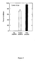
- Figure 8 Inhibition of coronavirus infectivity by fusion initiation region peptides.
- a virus strain A59 or SARS coronavirus strain Urbani were pre-incubated with or without the indicated peptides ( ⁇ 100 ⁇ M) in serum-free DMEM for 1 h.
- Cells were then exposed to peptide-treated inoculum or a vehicle control (no peptide). After 1 h adsorption, the inoculum was removed, cells were washed twice with 1X phosphate buffered saline, and the cells were overlaid with DMEM containing 10% FBS and 0.5% agarose. Forty-eight hours after infection, infected monolayers were fixed and stained with crystal violet to determine plaque numbers.
- Figure 9 Inhibition of Lassa virus infectivity by fusion initiation region peptides. Between 50 and 100 PFU Lassa virus was pre-incubated with or without the indicated peptides ( ⁇ 100 ⁇ M) in serum-free BME for 1 h. Cells were then exposed to the peptide-treated inoculum or vehicle control (no peptide). After 1 h adsorption, the inoculum was removed, cells were washed twice with 1X phosphate buffered saline, and the cells were overlaid with BME containing 5% FBS, 10 mM HEPES and 0.5% agarose. Four days after infection a second overlay containing 5% neutral red was applied, and plaques were counted 24 h later.
- RNA viruses now known to have Class I membrane fusion proteins (Type I viruses) and representative members of each family are as follows: Representative RNA Viruses Having Class I Membrane Fusion Proteins (Type I Viruses) Family Representative Virus Shown in Figures Arenaviruses Lassa Virus Yes Lymphocytic Choriomeningitis Virus (LCMV) No Junin Virus No Machupo Virus No Guanarito Virus No Sabia Virus No Coronaviruses Severe Acute Respiratory Syndrome (SARS) Virus Yes Murine Hepatitis Virus (MHV) No Bovine Coronavirus No Canine Coronavirus No Feline Infectious Peritonitis Virus No Filoviruses Ebola Virus Yes Marburg Virus No Orthomyxoviruses Influenza A Virus Yes Influenza B Virus No Influenza C Virus No Paramyxoviruses Measles Virus Yes Mumps Virus No Canine Distemper Virus No Newcastle Disease Virus No Retroviruses Human Immunode
- Certain embodiments of the invention comprise a method of identifying within the fusion proteins of viruses a conserved motif.
- the conserved motif of the FIR regions from different viruses will have similar structure and function. Additionally, the FIR regions of related viruses may, but will not necessarily, have highly similar primary amino acid sequences.
- the current invention provides means for identifying these regions, either with or without relying on their identitylsimilarity to known sequences.
- inventions of the present invention provide for methods useful for preventing or inhibiting viral infection and/or virus:cell fusion using peptides, peptide mimics, antibodies or other factors that are targeted to the specific virus' FIR and interfere with the function of that FIR.
- the FIR is typically between 50 and 100 amino acids in length, although it may be longer in some viruses.
- Various aspects of the current embodiments provide methods for identifying the FIR of a viral fusion protein wherein the methods comprises the following steps:
- the carboxy terminus of the FIR is the carboxy-terminus of the second peptide sequence with positive interfacial-hydrophobicity that is found beyond the N-helix.
- the sequence between the N-helix and C-helix in the F1 protein of paramyxoviruses is longer than the interhelical sequences of other viruses with Class I viral fusion proteins.
- the F2 protein of paramyxoviruses, which serves a receptor-binding function, is correspondingly shorter.
- the F1 protein contains a sequence insert between the N-helix and C-helix. Consequently, the FIR of paramyxoviruses contains two cysteine loops and two high-interfacial-hydrophobicity sequences and is discontinuous because additional amino acids which are characteristic only of the paramyxoviruses and appear between the N-helix and the first high-interfacial-hydrophobicity sequence are excluded from the FIR.
- inventions of the present invention provide methods of inhibiting virus:cell fusion by interfering with the function of the FIR.
- Various aspects of these embodiments include targeting the FIR with peptides, peptide mimics and other factors which may or may not be analogs of the FIR, in order to interfere with virus:cell fusion.
- the peptides, peptide mimics, and peptide analogs are between about 6 and 150 amino acid residues long. More preferably, they are from about 8 to 50 residues long, even more preferably they are from about 8 to 40 amino acids in length or of such length as is necessary to provide effective inhibition of viral infection.
- the term "of such length as necessary to provide effective inhibition of the virus preferably refers to a length sufficient to provide a 5-fold or greater reduction in viral infectivity, when used according to the instant invention.
- Methods for quantifying reduction in viral infectivity are well known to those of skill in the art. For example, reductions in viral activity may be determined by plaque reduction, binding inhibition, titer reduction assays, or by animal challenge studies.
- FIR peptides, peptides of analogous sequences, or fragments or derivatives thereof, contemplated as being part of the instant invention include, but are not limited to, those comprising, as primary amino acid sequences, all or an efficacious part of one or more of the following:
- FIR region is from a virus belonging to one of the viral families selected from the group consisting of arenaviruses, coronaviruses, filoviruses, orthomyxoviruses, paramyxoviruses, and retroviruses.
- the FIR is from a virus selected from the group consisting of Lassa Virus, Lymphocytic Choriomeningitis Virus (LCMV), Junin Virus, Machupo Virus, Guanarito Virus, Sabia Virus, Severe Acute Respiratory Syndrome (SARS) Virus, Murine Hepatitis Virus (MHV), Bovine Coronavirus, Canine Coronavirus, Feline Infectious Peritonitis Virus, Ebola Virus, Marburg Virus, Influenza A Virus, Influenza B Virus, Influenza C Virus, Measles Virus, Mumps Virus, Canine Distemper Virus, Newcastle Disease Virus, Human Immunodeficiency Virus 1 (HIV-1), Human Immunodeficiency Virus 2 (HIV-2), Human T-cell Lymphotrophic Virus 1 (HTLV-1), Human T-cell Lymphotrophic Virus 2 (HTLV-2), Human Intracisternal A-type Particle 1 (HIAP),
- sequences comprising a functional fragment of a FIR sequence or sequences analogous thereto, particularly from a virus belonging to one of the viral families selected from the group consisting of arenaviruses, coronaviruses, filoviruses, orthomyxoviruses, paramyxoviruses, and retroviruses (with the exception of the HIV-1 TM CS3 peptide previously described by the present inventors (Garry) and depicted in Figure 7 ).
- the peptide comprises a functional fragment (except the HIV-1 TM CS3 fragment) or a sequence analogous to a functional fragment from a virus selected from the group consisting of Lassa Virus, Lymphocytic Choriomeningitis Virus (LCMV), Junin Virus, Machupo Virus, Guanarito Virus, Sabia Virus, Severe Acute Respiratory Syndrome (SARS) Virus, Murine Hepatitis Virus (MHV), Bovine Coronavirus, Canine Coronavirus, Feline Infectious Peritonitis Virus, Ebola Virus, Marburg Virus, Influenza A Virus, Influenza B Virus, Influenza C Virus, Measles Virus, Mumps Virus, Canine Distemper Virus, Newcastle Disease Virus, Human Immunodeficiency Virus 1 (HIV-1), Human Immunodeficiency Virus 2 (HIV-2), Human T-cell Lymphotrophic Virus 1 (HTLV-1), Human T-cell Lymphotrophic Virus 1
- the instant invention also contemplates derivatives of the FIR peptides described above and analogous sequences thereto.
- These derivative peptides may comprise altered sequences in which functionally equivalent amino acid residues are substituted for residues within the sequence resulting in a silent change.
- one or more amino acid residues within the sequence can be substituted for by another amino acid of a similar polarity that acts as a functional equivalent, resulting in a silent alteration ( e.g . substitution of leucine for isoleucine).
- Substitutes for an amino acid within the sequence may be selected from other members of the class to which the amino acid belongs.
- the nonpolar (hydrophobic) amino acids include alanine, leucine, isoleucine, valine, proline, phenylalanine, tryptophan and methionine.
- the polar neutral amino acids include glycine, serine, threonine, cysteine, tyrosine, asparagine, and glutamine.
- the positively charged (basic) amino acids include arginine, lysine and histidine.
- the negatively charged (acidic) amino acids include aspartic acid and glutamic acid.
- such peptides may also comprise D-amino acids, and/or the may comprise an inefficient carrier protein, or no carrier protein at all.
- FIR peptides may comprise peptides in which "X" comprises an amino group, an acetyl group, a hydrophobic group or a macromolecular carrier group; and/or "Z” comprises a carboxyl group, an amido group a hydrophobic group or a macromolecular carrier group.
- Various aspects of the instant invention are drawn to peptides wherein the "X" moiety may also be selected from the group comprising: a hydrophobic moiety, a carbobenzoxyl moiety, dansyl moiety, or a t-butyloxycarbonyl moiety.
- the "Z" moiety may be selected from the group comprising: a hydrophobic moiety, a t-butyloxycarbonyl moiety.
- the "X" moiety may comprise a macromolecular carrier group.
- Such macromolecular carrier group may be selected from the group comprising, but not limited to: a lipid conjugate, a polyethylene glycol moiety, or a carbohydrate moiety.
- the "Z” may also comprise a macromolecular carrier group; wherein said macromolecular carrier is selected from the group comprising, but not limited to: a lipid conjugate, polyethylene glycol moiety, or a carbohydrate moiety.
- peptides wherein one or more of the molecular bonds linking adjacent amino acid residues is a non-peptide bond.
- non-peptide bonds include, but are not limited to: imido, ester, hydrazine, semicarbazoide and azo bonds.
- peptides wherein the peptide comprises one or more amino acid residues that is/are in a D-isomer amino acid.
- aspects of the instant invention provide for peptides comprising one or more amino acid substitution wherein a first amino acid residue is substituted for a second, different amino acid residue, in the sequences provided above (or a functional segment thereof).
- the amino acid substitution is a conservative substitution.
- the amino acid substitution is a non-conservative substitution.
- Yet other aspects of this embodiment of the invention provide for peptides as described above except that one or more amino acid residues have been deleted.
- the FIR peptides comprise at least three contiguous residues of a FIR. More preferably the FIR peptide comprises at least 8 contiguous residues of a FIR.
- FIR inhibitory peptide(s) preferably refers to a peptide or peptides having the sequence of a FIR (or functional segment thereof) and to such FIR peptides or functional segments in which one or more amino acids is/are substituted for by functionally equivalent or chemically similar amino acids (see infra).
- peptides include but not limited to, benzylated derivatives, glycosylated derivatives, and peptides that include enantiomers of naturally occurring amino acids.
- the peptide is selected from those having the sequence of any of SEQ ID NOs 1-7, 8-15, 22-25, and 30.
- the peptide has a sequence selected from the group consisting of SEQ ID NOs 22-25 and 30.
- the FIR peptides may be linked to a carrier molecule such as a protein, including but not limited to, human serum albumin (HSA).
- a carrier molecule such as a protein, including but not limited to, human serum albumin (HSA).
- the instant invention contemplates molecules comprising any combination of the X and Z moieties and/or other peptide modifications described above.
- Peptides according to the instant invention may be produced from naturally occurring or recombinant viral proteins. They may also be produced using standard recombinant DNA techniques (e.g . the expression of peptide by a microorganism that contains recombinant nucleic acid molecule encoding the desired peptide, expressed under the control of a suitable transcriptional promoter, and the harvesting of desired peptide from said microorganism).
- any of the peptides of the invention may be prepared using any chemical synthesis methodology known in the art including, but not limited to, Merrifield solid phase synthesis ( Clark-Lewis et al., 1986, Science 231:134-139 ).
- Embodiments of the instant invention also provide for other compounds useful for treating or preventing infection of a cell by a virus.
- These include antibodies (or active segments thereof, meaning portions of antibodies capable of specifically recognizing a FIR region or a functional segment thereof) and other molecules.
- Certain aspects of this embodiment of the invention provide for antibodies that specifically recognize a FIR, or antigenic fragment thereof and/or are capable of interfering with virus:cell interaction sufficiently to prevent or reduce infection of the cell by the virus.
- Antibodies according to these embodiments of the invention may be monoclonal or polyclonal.
- Methods for producing antibodies comprise the steps of (i) providing an antigen comprising a FIR or an antigenic fragment thereof (such antigen may be an unmodified peptide, a peptide mimic, a peptide analog, or a peptide derivative); (ii) exposing the immune system of an animal to the antigen so as to induce an immune response; (iii) collecting antibodies from the animal and identifying those antibodies that either specifically recognize a FIR (or functional segment thereof) and/or are capable of inhibiting or reducing virus:cell infection in a dose responsive manner in assays that measure viral infectivity.
- identifying compounds capable of preventing or inhibiting infection by a virus comprising a FIR or that are useful as drug leads for the development of drugs for preventing or inhibiting viral infection comprise the steps of: (i) identifying a virus having at least one membrane fusion protein comprising a fusion initiation region that is requisite for virus:cell fusion; (ii) preparing a target, where the target comprises the amino acid sequence of a FIR, or a functional segment of a FIR; (iii) screening a plurality of compounds to identify at least one compound that binds to the target, thereby identifying a target-binding compound; (iv) screening at least one target-binding compound to identify a target-binding compound that is capable of specifically preventing or reducing viral infection by the virus from which the target was obtained or that us useful as a drug lead for the development of a drug for specifically preventing or reducing infection by such a virus.
- the phrase "specifically preventing or reducing viral infection” means that the compound specifically prevents infection by the target virus, without any substantial effect on an unrelated virus. For example, if a compound that specifically prevented infection by the SARS virus would not prevent infection by the HIV-1 virus.
- the compounds (e.g . drugs or drug leads) identified by the methods described above may be of any type, by way of non-exclusive list they may be any peptide (or derivative, analog, or mimic thereof) this includes short peptides as are typically employed in phage display libraries, any antibody or active fragment thereof ( i.e . any fragment, such as an F ab that is capable of specifically recognizing the target) or any other organic or inorganic molecule.
- the FIR may be from any virus having a membrane fusion protein comprising at least extended two alpha-helices, a fusion peptide, and a fusion initiation region.
- the virus is selected from a virus family, wherein the virus family is selected from the group consisting of: arenaviruses, coronaviruses, filoviruses, orthomyxoviruses, paramyxoviruses, and retroviruses. More preferably, the virus is selected from the group consisting of: Lassa virus, SARS (severe acute respiratory syndrome) virus, Ebola virus, influenza virus, measles virus, and HIV-1 (human immunodeficiency virus type 1).
- the peptides and/or factors of the instant invention useful for treating or preventing viral infection of a cell can target the amino acids surrounding and within the FIR cysteine loop, the distal portion of the FIR N-helix, any of the interfacial hydrophobicity regions of the FIR, other areas of the FIR, or any combination of thereof.
- These factors, antibodies, peptides or peptide analogs may be used individually; alternatively they may be used in combinations of two or more to prevent or inhibit infection of the cell by the virus.
- the methods of preventing or inhibiting viral infection of the cell by interfering with the function of the FIR also include the use of neutralizing antibodies, produced exogenously or endogenously, against all or portions of the FIR.
- the purpose of such use is to interfere with the function of the FIR, thereby inhibiting viral infection of the cell and/or virus:cell membrane fusion.
- compositions including pharmaceutical compositions, comprising any and all of the compounds, peptides (including analogs, derivatives, and mimics thereof), antibodies, or any other molecule of the instant invention or identified by the methods of instant invention.
- compositions containing any molecule that comprises, consists essentially of, or consists of a FIR, or a functional segment of a FIR. It further includes, but is not limited to compositions comprising any compound that specifically recognizes, binds to, or interferes with the function of a viral FIR.
- the phrase "interfering with the function of the FIR” means that a compound interacts with the FIR or with the cellular protein that serves as the receptor that recognizes the FIR so as to prevent or reduce infection of the cell by the virus. Additionally, it is contemplated that the compositions may comprise either one of the molecules described or mixtures of two or more of the molecules.
- Still other aspects of this embodiment of the invention provide for methods that comprise administering to a patient an effective amount of a composition comprising at least one recombinant DNA or RNA molecule; where the RNA or DNA encodes a FIR (or functional segment thereof) or a molecule capable of specifically binding to a FIR or a cellular receptor that recognizes a FIR so as to prevent or reduce infection by the virus.
- the recombinant RNA or DNA molecule and or pharmaceutical composition further comprises the elements necessary to allow the protein encoded by the RNA or DNA molecule to be expressed in a human cell.
- the recombinant RNA or DNA molecule is part of a recombinant plasmid or a recombinant virus.
- the method to identify the FIR of Class I viral fusion proteins can be illustrated by two examples.
- the first example is identification of the FIR in the minimal class I fusion protein glycoprotein 2 (GP2) of Ebola virus, a filovirus.
- the boundaries of the N-helix and the C-helix of Ebola virus GP2 have been determined by x-ray crystallographic methods (Malashkevich et al., 1999).
- the terminal amino acids of the N-helix contain the sequence ILNRKAIDF (SEQ ID NO:8) that fits the consensus of a core comprising three or four hydrophobic amino acids, a positively-charged amino acid, a negatively-charged amino acid, and an aromatic amino acid.
- the second example is a complex class I fusion protein, the F1 protein of measles virus, a paramyxovirus.
- the N- and C- helices of measles virus F1 can be identified by examining the primary sequence for amino acids with the propensity to form helices. Alignment of the primary sequence of measles virus F1 with the primary amino acid sequence of the F1 protein of another paramyxovirus, Newcastle disease virus F1, can also aid in the identification of the helix boundaries.
- the structure of the Newcastle disease virus F1 protein has been determined by x-ray crystallographic methods (Chen et al., 2001). The boundaries of the N- and C- helices can thus be predicted to be amino acids 131 - 217 and 455-491 respectively.
- the primary sequence between the N- and C- helices in the measles virus is longer than 100 amino acids.
- the FIR region of measles virus F1 contains an insertion which, upon inspection of computer models, is obvious to those skilled in the art, and thus the FIR structure is formed by a secondary arrangement that brings together two parts of the primary sequence.
- the inserted sequence forms a loop external to the FIR.
- the terminal amino acids of the N-helix contain the sequence LKLLRYYTE (SEQ ID NO:11) which fits the consensus of a core comprising three or four hydrophobic amino acids, a positively-charged amino acid, a negatively-charged amino acid, and an aromatic amino acid.
- cysteine residues in measles virus F1 there are eight cysteine residues in measles virus F1 between the N- and C- helices.
- CTFMPEGTVC SEQ ID NO:12
- WYTTVPKYVATQGYLISNF SEQ ID NO:13
- the second pair of cysteines in the sequence, CLRGSTKSC (SEQ ID NO:14), is also part of the FIR because it is adjacent to a sequence TLVSGSFGNRFILSQGNLIANCASILCKCYTTGTII (SEQ ID NO:15) with a Wimley-White interfacial hydrophobicity score of 2.54, as determined by the MPEX program.
- the FIR of measles virus F1 extends from amino acids 205 to 407, with amino acids 221 to 314 representing an insertion that does not participate in FIR function.
- Example 3 Identification Of Coronavirus Fusion Inhibitory Peutides .
- Severe acute respiratory syndrome is a newly recognized illness that spread from southern China in late 2002/early 2003 to several countries in Asia, Europe and North America (Guan et al., 2004). SARS usually begins with a fever greater than 38°C. Initial symptoms can also include headache, malaise and mild respiratory symptoms. Within two days to a week, SARS patients may develop a dry cough and have trouble breathing. Patients in more advanced stages of SARS develop either pneumonia or respiratory distress syndrome. In the initial outbreak there were 8098 cases worldwide, with an overall mortality of 9.6%.
- Coronaviruses are large positive-stranded RNA viruses typically with a broad host range. Like other enveloped viruses, CoV enter target cells by fusion between the viral and cellular membranes, a process mediated by the viral spike (S) protein. CoV S proteins, characterized to date, appear to consist of two non-covalently associated subunits, S1 and S2.
- L2 cells or Vero E6 cells were maintained as monolayers in complete Dulbecco's modified Eagle's medium (DMEM) containing 0.15% HCO 3 - supplemented with 10% fetal bovine serum (FBS), penicillin G (100 U/ml), streptomycin (100 mg/ml), and 2mM L-glutamine at 37°C in a 5% CO 2 incubator.
- DMEM Dulbecco's modified Eagle's medium
- FBS fetal bovine serum
- penicillin G 100 U/ml
- streptomycin 100 mg/ml
- 2mM L-glutamine at 37°C in a 5% CO 2 incubator.
- Mouse hepatitis virus (MHV) strain A59 or SARS CoV strain Urbani or HK was propagated on L2 cells.
- L2 cells or Vero E6 cells were seeded at a density of 1x10 6 cells in each well of a 6-well plate.
- Fifty to 100-plaque forming units (p.f.u.) of MHV or SARS CoV were pre-incubated with or without approximately 100 ⁇ g/ml of peptide in serum-free DMEM for 1 h. Cells were then infected with peptide-treated inoculum or vehicle control inoculum. After 1 h adsorption, the inoculum was removed, cells were washed twice with 1X phosphate buffered saline, and the cells were overlaid with 10% FBS/DMEM containing 0.5% SEAPLAQUE® agarose (Cambrex Bio Science Rockland, Inc., Rockland, ME). Monolayers were fixed with 3.7% formalin and stained with 1X crystal violet 2 days post-infection, and plaque numbers were determined by light microscopy.
- Synthetic peptides corresponding to the FIR domains of the MHV or SARS CoV S protein were tested for their ability to inhibit infection by these coronaviruses.
- the ability to inhibit formation of plaques in cell monolayers is the most stringent in vitro test of a potential infection inhibitor drug.
- Two peptides (GNHILSLVQNAPYGLYFIHFSW, SEQ IDS NO:22 and GYFVQDDGEWKFTGSSYYY, SEQ ID NO:23) from the MHV FIR can inhibit plaque formation by MHV, though the first MHV FIR peptide is more efficient ( see Fig. 8A ).
- Example 4 Identification Of Arenavirus Fusion Inhibitory Peptides .
- Lassa fever is an often-fatal hemorrhagic illness named for the town in the Yedseram River valley of Nigeria in which the first described cases occurred in 1969 (Buckley and Casals, 1970). Parts of Guinea, Sierra Leone, Nigeria, and Liberia are endemic for the etiologic agent, Lassa virus (LasV). The public health impact of LasV in endemic areas is immense. The Centers for Diseases Control, and Prevention (CDC) have estimated that there are 100,000-300,000 cases of Lassa per year in West Africa and 5,000 deaths. In some parts of Sierra Leone, 10-15% of all patients admitted to hospitals have Lassa fever.
- Case fatality rates for Lassa fever are typically 15% to 20%, although in epidemics overall mortality can be as high as 45%.
- the mortality rate for women in the last month of pregnancy is always high, ⁇ 90%, and LasV infection causes high rates of fetal death at all stages of gestation.
- Mortality rates for Lassa appear to be higher in non-Africans, which is of concern because Lassa is the most commonly exported hemorrhagic fever. Because of the high case fatality rate and the ability to spread easily by human-human contact, LasV is classified as a Biosafety Level 4 and NIAID Biodefense category A agent.
- LasV is a member of the Arenaviridae family.
- the genome of arenaviruses consists of two segments of single-stranded, ambisense RNA.
- the enveloped spherical virions show grainy particles that are ribosomes acquired from the host cells (Murphy and Whitfield, 1975).
- Arenaviruses are zoonotic; each virus is associated with a specific species of rodent (Bowen, Peters, and Nichol, 1997).
- the reservoir of LasV is the "multimammate rat" of the genus Mastomys (Monath et al., 1974). The wide distribution of Mastomys in Africa makes eradication of this rodent reservoir impractical and ecologically undesirable.
- Lassa fever Signs and symptoms of Lassa fever, which occur 1-3 weeks after virus exposure, are highly variable, but can include fever, retrosternal, back or abdominal pain, sore throat, cough, vomiting, diarrhea, conjunctival injection, and facial swelling. LasV infects endothelial cells, resulting in increased capillary permeability, diminished effective circulating volume, shock, and multi-organ system failure. Frank bleeding, usual mucosal (gums, etc.), occurs in less than a third of cases, but confers a poor prognosis. Neurological problems have also been described, including hearing loss, tremors, and encephalitis. Patients who survive begin to defervesce 2-3 weeks after onset of the disease. The most common complication of Lassa fever is deafness.
- Temporary or permanent unilateral or bilateral deafness occurs in ⁇ 30% of Lassa fever patients during convalescence, and is not associated with the severity of the acute disease.
- the antiviral drug ribavirin is effective in the treatment of Lassa fever, but only if administered early (up to six days) in the course of illness (Johnson et al., 1987; McCormick et al., 1986). It is unknown whether ribavirin is effective against other arenaviruses, such as Junin, Machupo, Guanarito or Sabiá viruses. No LasV vaccine is currently available.
- Vero cells were maintained as monolayers in Basal Medium Eagle (BME) containing 10 mM HEPES and 5% FBS. Lassa virus (Josiah strain) was propagated on Vero cells. For plaque assays, Vero cells were seeded at a density of 1x10 6 cells in each well of a 6-well plate. Fifty to 100 p.f.u. of LasV were pre-incubated with or without peptide in serum-free BME for 1 h. Cells were then infected with peptide-treated inoculum or vehicle control inoculum.
- BME Basal Medium Eagle
- Synthetic peptides corresponding to the FIR domains of LasV glycoprotein 2 (GP2) were tested for their ability to inhibit infection by this arenavirus.
- a peptide (NYSKYWYLNHTTTGR, SEQ ID NO:30) analogous to the sequence NYSRYWYLNHTSTGK from SEQ ID NO:1 (LASSA FIR) can inhibit plaque formation by LasV ( Fig. 9 ).
- a peptide analogous to another GP2 region, the fusion peptide, (GTFTWTLSDSEGKDTPGGY, SEQ ID NO:31) also inhibited infection by LasV, but to a lesser extent ( Fig. 9 ). No arenavirus inhibitory peptides have been reported.
- a method of identifying a compound capable of preventing or inhibiting viral infection of a host cell comprising:
- the method of paragraph 1 further comprising identifying a virus having a membrane fusion protein comprising: two or more alpha helices, a fusion peptide, and a fusion initiation region (FIR).
- a virus having a membrane fusion protein comprising: two or more alpha helices, a fusion peptide, and a fusion initiation region (FIR).
- influenza virus is from a family of viruses selected from the group consisting of arenaviruses, coronaviruses, filoviruses, orthomyxoviruses, paramyxoviruses, and retroviruses.
- the virus is selected from the group consisting of Lassa Virus, Lymphocytic Choriomeningitis Virus (LCMV), Junin Virus, Machupo Virus, Guanarito Virus, Sabia Virus, Severe Acute Respiratory Syndrome (SARS) Virus, Murine Hepatitis Virus (MHV), Bovine Coronavirus, Canine Coronavirus, Feline Infectious Peritonitis Virus, Ebola Virus, Marburg Virus, Influenza A Virus, Influenza B Virus, Influenza C Virus, Measles Virus, Mumps Virus, Canine Distemper Virus, Newcastle Disease Virus, Human Immunodeficiency Virus 1 (HIV-1), Human Immunodeficiency Virus 2 (HIV-2), Human T-cell Lymphotrophic Virus 1 (HTLV-1), Human T-cell Lymphotrophic Virus 2 (HTLV-2), Human Intracisternal A-type Particle 1 (HIAP-1), and Human Intracisternal A-type Particle 1
- peptide of paragraph 7 comprising an amino acid sequence or analogous sequence thereto of a viral fusion initiation region from a virus in a virus family selected from the group of virus families consisting of arenaviruses, coronaviruses, filoviruses, orthomyxoviruses, paramyxoviruses, and retroviruses.
- the FIR is from a virus selected from the group consisting of Lassa Virus, Lymphocytic Choriomeningitis Virus (LCMV), Junin Virus, Machupo Virus, Guanarito Virus, Sabia Virus, Severe Acute Respiratory Syndrome (SARS) Virus, Murine Hepatitis Virus (MHV), Bovine Coronavirus, Canine Coronavirus, Feline Infectious Peritonitis Virus, Ebola Virus, Marburg Virus, Influenza A Virus, Influenza B Virus, Influenza C Virus, Measles Virus, Mumps Virus, Canine Distemper Virus, Newcastle Disease Virus, Human Immunodeficiency Virus 1 (HIV-1), Human Immunodeficiency Virus 2 (HIV-2), Human T-cell Lymphotrophic Virus 1 (HTLV-1), Human T-cell Lymphotrophic Virus 2 (HTLV-2), Human Intracisternal A-type Particle 1 (HIAP),
- the peptide of paragraph 7 having a sequence selected from the group consisting of SEQ ID NOs 1-7 or a functional segment of any one of SEQ ID NOs 1-7.
- a method of treating or preventing a viral infection comprising administering to a patient a compound identified by any of the methods of paragraphs 1 to 6.
- a method of treating or preventing a viral infection comprising administering to a patient a peptide of any of paragraphs 7 to 12.
- a method of treating or preventing a viral infection comprising administering to a patient an effective amount of a composition comprising of a recombinant DNA molecule that enables or stimulates the patient to produce the peptide of any of paragraphs 7 to 12.
- a method of treating or preventing a viral infection comprising administering to a patient an effective amount of antibody that binds specifically to a fusion initiation region.
- An antibody according to paragraph 17 capable of inhibiting membrane fusion of a virus having membrane fusion proteins comprising at least two extended alpha-helices and a fusion peptide, wherein these proteins comprise a fusion initiation region (FIR) requisite for cell fusion, wherein the antibody binds to amino acid sequences within the FIR.
- FIR fusion initiation region
- An isolated nucleic acid sequence capable of encoding a polypeptide having the sequence of a viral FIR or a sequence analogous thereto from a virus belonging to family of viruses selected from the group consisting of arenaviruses, coronaviruses, filoviruses, orthomyxoviruses, paramyxoviruses, and retroviruses.
- a method of producing an antibody comprising:
- the antigen consists of an amino acid sequence or analogous sequence thereto of a viral fusion initiation region (FIR) from a virus in a virus family selected from the group of virus families consisting of arenaviruses, coronaviruses, filoviruses, orthomyxoviruses, paramyxoviruses, and retroviruses.
- FIR viral fusion initiation region
- the antigen comprises a peptide analog; a peptide derivative; or a peptide mimic of a fusion initiation region (or antigenic fragment thereof).
- a method of identifying a viral fusion initiation region (FIR) in a viral fusion protein sequence comprising;
Abstract
Description
- This application claims the benefit of United States Provisional
Application Serial Number 60/117,181, filed November 4, 003 - The present invention relates to a method of preventing or inhibiting viral infection of a cell and/or fusion between the envelope of a virus and the membranes of a cell targeted by the virus (thereby preventing delivery of the viral genome into the cell cytoplasm, a step required for viral infection). The present invention provides methods for identifying a fusion initiation region, or FIR, of the viruses. The present invention provides for a method of identifying the FIR in these viruses. The present invention further provides for methods of preventing infection by a Type I virus by interfering with its FIR.
- All viruses must bind to and invade their target cells to replicate. For enveloped animal viruses, including RNA viruses having Class I membrane fusion proteins (Type I viruses), the process involves (a) binding of the virion to the target cell, (b) fusion of the envelope of the virus with the plasma membrane or an internal cellular membrane, (c) destabilisation of the viral envelope and cellular membrane at the fused area to create a fusion pore, (d) transfer of the viral RNA through the pore, and (e) modification of cellular function by the viral RNA.
- Fusion of the viral membrane and the cell envelope, steps (b) and (c) above, is mediated by the interaction of a viral transmembrane glycoprotein (fusion protein) with surface proteins and membranes of the target cell. These interactions cause conformational changes in the fusion protein that result in the insertion of a viral fusion peptide into the taget cell membrane. This insertion is followed by further conformational changes within the fusion protein that bring the viral envelope and cell membranes into close proximity and results in the fusion of the two membrane bilayers.
- A virus is unable to spread and propagate within its host if this fusion process is disrupted. Intentional disruption of this fusion process can be achieved by directing peptides and peptide mimics homologous to fusion protein sequences, antibodies that recognize the fusion protein, and other factors that act against the fusion protein.
- Hemagglutinin 2 (HA2) of influenza virus, an orthomyxovirus, is the prototypic RNA virus Class I fusion protein and contains an amino terminal hydrophobic domain, referred to as the fusion peptide, that is exposed during cleavage of the hemagglutinin precursor protein. The membrane fusion proteins of RNA viruses from several diverse families, including arenaviruses, coronaviruses, filoviruses, orthomyxoviruses, paramyxoviruses, and retroviruses, share several common structural features with HA2 and have been referred to as Class I viral fusion proteins. It has been observed that the fusion protein of HIV-1, the transmembrane glycoprotein and other retroviral transmembrane proteins, like those of orthomyxoviruses and paramyxoviruses, possess a hydrophobic fusion peptide domain exposed during cleavage of a precursor (gp160) (Gallaher, 1987; Gonzalez-Scarano et al., 1987). Based on these similarities and computer algorithms that predict protein configurations, it has been suggested (Gallaher et al., 1989) that the external portion (ectodomain, amino terminus) of HIV-1 transmembrane protein and the transmembrane proteins of other retroviruses, all could fit the scaffold of HA2 structure as determined by x-ray crystallography (Wilson, Skehel, and Wiley, 1981).
- Based on these observations, it was predicted that retroviral transmembrane proteins contain several structural features in addition to the fusion peptide in common with the known structure of HA2, including an extended amino terminal helix (N-helix, usually a "heptad repeat" or "leucine zipper"), a carboxyl terminal helix (C-helix), and an aromatic motif proximal to the transmembrane domain. The presence of at least four out of these five domains defines a viral envelope protein as a Class I fusion protein. This retroviral transmembrane protein model was subsequently confirmed by structural determinations and mutational analyses (Chan et al., 1997; Kowalski et al., 1991; Weissenhorn et al., 1997). Common structural motifs are present not only in orthomyxovirus and retrovirus fusion proteins, but also in those of paramyxoviruses, filoviruses (such as Ebola virus, EboV) (Gallaher, 1996) and arenaviruses (Gallaher, DiSimone, and Buchmeier, 2001). The Gallaher structural model of the EboV fusion protein (GP2) has also been confirmed by x-ray crystallographic methods (Malashkevich et al., 1999; Weissenhorn et al., 1998).
-
Figure 1 shows the five, previously-described, domains of the fusion proteins of the six families of Type I viruses. The fusion proteins originate in a hydrophobic fusion peptide, terminate in an anchor peptide, and incorporate an extended amino terminal alpha-helix (N-helix, usually a "heptad repeat" or "leucine zipper"), a carboxyl terminal alpha-helix (C-helix) (Carr and Kim, 1993; Suarez et al., 2000; Wilson, Skehel, and Wiley, 1981), and sometimes an aromatic motif proximal to the virion envelope. Also shown is the sixth domain, the fusion initiation region (FIR), discovered by the present inventors. - Previous attempts by the present inventors (Garry) and others to design peptides and peptide mimics, antibodies, and other factors that inhibit fusion in Type I viruses have focused on the fusion peptide, the N-helix, and the C-helix of the fusion proteins. In the case of fusion peptides, analogs of the orthomyxoviruses and paramyxoviruses (Richardson, Scheid, and Choppin, 1980) and HIV-1 fusion peptide domains (Gallaher et al., 1992; Owens et al., 1990; Silburn et al., 1998) have been found to block viral infection, presumably by forming inactive heteroaggregates. Peptides corresponding to portions of the N-helix and C-helix have also been found to be effective in inhibiting viral infection both in vitro and in vivo. For example, a 17-amino-acid peptide corresponding to the carboxy-terminal portion of the N-helix of the HIV-1 fusion protein, defined as the CS3 region, blocked HIV infection (Qureshi et al., 1990). In addition, other N-helix and C-helix inhibitory peptides were developed based on the fusion protein structural model (Wild, Greenwell, and Matthews, 1993; Wild et al., 1992), including the C-helix anti-HIV-1 peptidic drug DP178 (T-20 or FUZEON®). DP178 overlaps the C-helix and the aromatic anchor-proximal domain and inhibits HIV-1 virion:cell fusion at very low concentrations (50% inhibition at 1.7 nM) achievable in vivo following injection. In a clinical trial, 100 mg/day of DP178 caused an approximately 100-fold reduction in plasma HIV-1 load of infected individuals (Kilby et al., 1998). This result has greatly motivated the search for other HIV-1 inhibitory peptides based on transmembrane protein structure (Pozniak, 2001; Sodroski, 1999). Peptidic inhibitors of paramyxoviruses have also been shown to inhibit viral replication (Lambert et al., 1996; Young et al., 1999). Studies by Watanabe and coworkers suggest that a similar approach of targeting the N-helix and the C-helix of EboV GP2 may also lead to useful inhibitors (Watanabe et al., 2000). Neutralizing antibodies directed against portions of the fusion protein domains have also been shown to inhibit virion:cell fusion.
- A great deal of study has been devoted to fusion inhibition in human immunodeficiency virus HIV-1, one of the Type I RNA viruses.
Bolognesi et al. (5,464,933 ) and the present inventors (Garry, USPN 5,567,805 ) teach that HIV-mediated cell killing can be inhibited by introducing peptides that bind to portions of the transmembrane fusion protein of the HIV-1 virion. The Bolognesi DP178 binding region, labeled FUZEON® inFigure 7 , lies primarily on the C-helix and is outside what is described in the present application the fusion initiation region (FIR). Bolognesi demonstrates inhibition but teaches no method of inhibition. The present inventors (Garry) previously demonstrated inhibition at the CS3 region of HTV-1 TM, labeled CS3 inFigure 7 , but identified no method of inhibition, suggesting only that CS3:CS3-receptor interaction is inhibited. The unexpected discovery of the FIR by the present inventors (as currently described herein) and the fact that the CS3 sequences lie within the FIR indicates that the CS3:CS3-receptor binding described inUSPN 5,567,805 is in fact binding that occurs between the CS3 portion of the FIR and portions of the cell membrane for which the CS3 portion of the FIR has an affinity. In addition, although Melikyan, Watanabe, Bewley, and others have described fusion inhibition with introduced peptides, they have not explained the mechanisms through which the inhibition occurs. Correspondingly, the location of the FUZEON® peptide is distant from the FIR, which strongly suggests that other elements of the fusion process operate in the FUZEON® region. - In view of the foregoing, it is clear that there exists a need in the art for a more effective means for identifying those regions of viruses that are involved in the infection process and for compositions effective for preventing or inhibiting viral infection. The invention described and disclosed herein provides an effective solution to these needs.
- Various embodiments of the instant invention provide for methods of identifying "factors" (compounds) capable of inhibiting membrane fusion between viruses and their host cells and, thereby, preventing or inhibiting infection of the host cell by the virus. Aspects of this embodiment of the invention provide for methods of identifying these inhibitory "factors" where the method comprises the steps of (a) identifying a virus having an envelope fusion protein having two, or more, extended alpha helices, a fusion peptide, and a fusion initiation region (FIR); (b) preparing a "target" wherein the target comprises the amino acid sequence of the FIR, (c) exposing the "target" to one or more test compounds, and (d) identifying those test compounds that physically interact with the "target". For example, physical interaction can be detected using a "target" bound to a solid substrate and a fluorescently or radioactively labeled test compound in a standard binding assay. Target and test compounds having dissociation coefficients (Kd) in the micromolar range or lower (i.e. ≤ about 9 X 10⁻⁶) are considered to be positively interacting.
- Other aspects of the instant invention provide for compositions comprising an isolated peptide having the amino acid sequence of a viral fusion initiation region (FIR) or a functional segment of the FIR or having an amino acid sequence which is analogous to the sequence of a FIR or a functional segment of a FIR. As used herein, an analogous amino acid or peptide sequence is a sequence containing a majority of identical or chemically similar amino acids in the same order as a primary sequence. Such chemical similarities are well known to those skilled in the art.
- Other aspects of this embodiment of the invention provide for isolated, typically substantially purified, peptides or peptide analogs that are capable of preventing or inhibiting viral infection of a host cell and/or inhibiting membrane fusion of a virus with a host cell, where the virus comprises a membrane fusion protein having two (extended) alpha helices, a fusion peptide and a FIR.
- Additional embodiments of the instant invention provide for methods of treating or preventing viral infection by administering to a patient one or more of the compounds identified by the methods described herein as capable of inhibiting viral infection. In various aspects of this embodiment of the invention the compounds administered are peptides or peptide analogs comprising all or a functional segment of a viral FIR sequence. In any aspect of this embodiment of the invention the administered compound is antigenic and is administered in an amount sufficient to eliciting an immune response.
- Other embodiments of the instant invention provide for a "molecular factor", such as a plasmid, recombinant virus, or other substance which enables or stimulates a cell or organism to produce a peptide or peptide analog that is capable of preventing or inhibiting a viral infection of that cell or organism. In any aspect of this embodiment the "molecular factor" is capable of preventing or inhibiting a viral infection when administered to a patient.
- Another embodiments of the instant invention provide for antibodies capable of inhibiting the virus:cell membrane fusion of a virus having a fusion protein comprising two, extended alpha-helices, a fusion peptide and a FIR. In any aspect of this embodiment of the invention the antibodies are capable of binding specifically to amino acid sequences comprising the FIR sequence, or fragments thereof of sufficient size to allow antibody recognition. Various aspects of this embodiment of the invention provide for methods of producing the antibodies. In certain aspects of this embodiment, the method for producing antibodies comprises: (a) providing as the antigen a peptide comprising a viral initiation region (FIR) or an antigenic fragment of the FIR; (b) introducing the antigen in to an animal so as to elicit an immune response; (c) collecting antibodies from the animal; and optionally, (d) purifying the collected antibodies to identify that fraction of the collected antibodies having a high specificity for the antigen.
- Other embodiments of the current invention provide methods of treating patients, which methods comprise administering to the patient antibodies that specifically recognize and bind to peptides comprising a FIR region from a virus or comprising a functional fragment of such a FIR region where the functional fragment is of sufficient size to allow its specific recognition by an antibody (that is, it is an antigenic fragment).
- Other embodiments of the instant invention provide for methods of producing antibodies specific for FIR or functional fragments thereof.
- The arenaviruses, coxonavimses, filoviruses, orthomyxoviruses, paramyxoviruses, and retroviruses are the six families of RNA viruses currently identified that have Class I membrane fusion envelope proteins. The fusion proteins of these Type I viruses have previously been shown by the present inventors (Garry) and others to incorporate five conserved motifs, or domains (Carr and Kim, 1993; Gallaher et al., 1989; Suarez et al., 2000; Wilson, Skehel, and Wiley, 1981). These domains comprise a fusion peptide, an N-helix, a C-helix, and an aromatic motif, all of which are ectodomains, and an anchor peptide, which is an endodomain.
- Using computational analyses, secondary structure models, interfacial hydrophobicity calculations and other techniques, the present inventors have made the surprising discovery of a highly conserved sixth domain that is present in the fusion proteins of a wide variety of viruses (this sixth domain is described herein). The viruses possessing this domain include, but are not necessarily limited to the six classes of RNA viruses listed above. To emphasize the critical function of this newly identified domain, which is an ectodomain, the domain is referred to herein as the fusion initiation region (FIR) of the viruses.
- Various embodiments of the instant invention provide methods of identifying the FIR in arenavirus, coronavirus, filovirus, orthomyxovirus, paramyxovirus, and retrovirus families of viruses. Also provided are methods of determining whether the FIR is present in other known virus families or in any newly discovered virus families.
- As used herein the term "extended" alpha helix refers to an alpha helix having more than four "alpha helix turns" (specifically, more than 14 amino acids).
- Other embodiments provide for "factors" that the inventors have unexpectedly found are effective for preventing or inhibiting viral infection and/or virus:cell fusion.
- As used herein the term "factors" includes, but is not limited to isolated peptides or functional peptide segments (or peptide analogs thereof) of the newly described fusion initiation region (FIR) domains, peptide mimics ("peptide mimic" refers to any compound or substance that could serve as a substitute for a peptide interacting with the FIR, that is any compound that mimics the properties of a functional segment of the FIR), antibodies specific for functional FIR domains (e.g. idiotypic or anti-idiotypic antibodies) and other molecular compounds that interfere with virus:cell binding and/or fusion.
- As used herein the term "functional segment" or "functional fragment" of a fusion initiation region (FIR) refers to a fragment capable of inhibiting virus:cell fusion, inhibiting viral infectivity, capable of eliciting an antibody capable of recognizing and specifically binding to the FIR and/or interfering with FIR-mediated cell infection.
- As used herein, a "peptide analog" or "modified peptide" is preferably defined as a FIR peptide modified to contain an amino group, an acetyl group, a hydrophobic group (for example carbobenzoxyl, dansyl, or t-butyloxycarbonyl) or a macromolecular carrier group (for example lipid conjugate, polyethylene glycol, a carbohydrate or a protein) at the amino terminus. An additional class of FIR peptide analogs contains a carboxyl group, an amido group, a hydrophobic group or a macromolecular carrier group at the carboxyl terminus. Other peptide analogs are defined as FIR peptides wherein at least one bond linking adjacent amino acids residues is a non-peptide bond (for example an imido, ester, hydrazine, semicarbazoide or azo bond), a peptide wherein at least one amino acid residue is in a D-isomer configurations or a peptide in which the order of the amino acids is inverted. Additional peptide analogs are FIR peptides compromising at least one amino acid substitution wherein a first amino acid residue is substituted for a second, different amino acid residue (the amino acid substitution can be a conserved substitution or a non-conserved substitution). As used herein, such peptide analogs may comprise analogous amino acid sequences in which the analogous sequences contain a majority of identical or chemically similar amino acids in the same order as the primary sequences.
- As used herein, the term "fusion initiation region" (FIR) generally refers to a region of a viral fusion protein involved in the initial step or steps of viral infection and/or fusion with a host cell.
- As used herein the term "peptide mimic" includes, but is not limited to organic compounds or other chemicals that mimic the structure or function of the FIR peptide. Examples of peptide mimics include, but are not limited to organic compounds comprising the functional side-groups of an amino acid or peptide, but lacking the carbon/nitrogen backbone or peptide bonds. Peptide mimic also refers to compounds that mimic the action of these functional side-groups with other moieties.
- Other molecules, such as idiotype or anti-idiotype antibodies or proteins selected via phage display methods, that bind to the peptides, peptide analogs or peptide mimics described in the present application may also function as inhibitors of viral infection and/or virus:cell fusion. Also contemplated by the instant invention are plasmids, or recombinant viruses, or other molecules or compounds that enable or stimulate the patient to produce an analog of the inhibitory compounds. For example, a recombinant protein, produced in an engineered bacterial, fungal, or mammalian cell, can be used to produce an immunogenic analog of the FIR of a viral fusion protein. Similarly, an anti-idiotypic response could be induced in the individual by using an engineered protein comprising a sequence corresponding to the binding site of a FIR-specific antibody.
- As used herein the term "fusion peptide" preferably refers to a hydrophobic sequence at or near the amino terminus of a class I viral fusion protein (see, Gallaher et al., 1987; 1992).
- As used herein the term "substantially purified" peptide or peptide analog preferably refers to a peptide or peptide analog that is greater than about 80% pure. More preferably, "substantially purified" refers to a peptide or peptide analog that is greater than about 90% or greater than about 95% pure. Most preferably it refers to a peptide or peptide analog that is greater than 96%, 97%, 98%, or 99% pure. Functionally, "substantially purified" means that it is free from contaminants to a degree that that makes it suitable for the purposes provided herein. Methods for assessing purity are well known to those of skill in the art. Suitable methods include, but are not limited to gas chromatography (GC) linked mass spectrophotometry, high performance liquid chromatography (HPLC) analysis, and functional assays in cell culture systems that, inter alia, assess cytotoxicity.
- As used herein the term "stable analog" refers to a peptide that has a pharmacologically active half-life in biological systems. Biological half-lives of greater than 60 minutes are contemplated.
- As used herein the term "peptide derivative" refers to a peptide that has substituted amino acids different from those in the FIR sequence of a viral fusion protein. Wherein the substitutions do not render the peptide useless for the instant invention.
- According to various aspects of the present embodiment of the invention the peptides, peptide analogs, peptide mimics, and other factors may be produced by any means known in the art, including, but not limited to, chemical synthesis, recombinant DNA methods and combinations thereof.
- The present invention provides methods for identifying the FIR of Type I, and other, viruses and for treating or preventing infection by these viruses. One possible mechanism by which the current invention may to prevent and/or inhibit infection is by interfering with the FIR mediated virus:cell fusion.
-
Figure 1 shows the domains of the fusion proteins of one member of each of these six viral families (namely, arenaviruses, coronaviruses, filoviruses, orthomyxoviruses, paramyxoviruses, and retroviruses). The circles inFigure 1 show the approximate location of the FIR in each virus illustrated. -
Figures 2 through 7 show the amino acid sequences of these fusion proteins (corresponding to SEQ ID NOs 16-21, respectively) and a schematic representation of their ectopic structure. Specifically shown are the five previously-described domains are the fusion peptide, i.e., the N-helix, the C-helix, the aromatic motif (if present), and the anchor peptide. The newly-discovered sixth domain, the fusion initiation region, or FIR is also identified. Each FIR is indicated by a polygon inFigures 2 through 7 . - The circled area behind the fusion proteins in each of
Figures 2-7 represents the primary virus:cell binding protein (VCBP) of the virus. The VCBP usually interacts with the portion of the fusion protein which is most distal from the viral membrane and is thus shown to be so positioned in the Figures. Unlike the highly-conserved fusion protein, the VCBP of each virus family is more divergent. It is usually the VCBP that dictates the host range of the virus and determines which of the host's cell types are targeted for infection. The VCBP acts in this capacity by recognizing and binding with specific cell surface proteins. The binding of the VCBP to the targeted cell proteins occurs prior to and is typically a prerequisite for virus:cell fusion. -
Figure 8 : Inhibition of coronavirus infectivity by fusion initiation region peptides. Between 50 and 100 PFU of mouse hepatitis virus strain A59 or SARS coronavirus strain Urbani were pre-incubated with or without the indicated peptides (∼100µM) in serum-free DMEM for 1 h. Cells were then exposed to peptide-treated inoculum or a vehicle control (no peptide). After 1 h adsorption, the inoculum was removed, cells were washed twice with 1X phosphate buffered saline, and the cells were overlaid with DMEM containing 10% FBS and 0.5% agarose. Forty-eight hours after infection, infected monolayers were fixed and stained with crystal violet to determine plaque numbers. -
Figure 9 : Inhibition of Lassa virus infectivity by fusion initiation region peptides. Between 50 and 100 PFU Lassa virus was pre-incubated with or without the indicated peptides (∼100µM) in serum-free BME for 1 h. Cells were then exposed to the peptide-treated inoculum or vehicle control (no peptide). After 1 h adsorption, the inoculum was removed, cells were washed twice with 1X phosphate buffered saline, and the cells were overlaid with BME containing 5% FBS, 10 mM HEPES and 0.5% agarose. Four days after infection a second overlay containing 5% neutral red was applied, and plaques were counted 24 h later. - The six families of RNA viruses now known to have Class I membrane fusion proteins (Type I viruses) and representative members of each family are as follows:
Representative RNA Viruses Having Class I Membrane Fusion Proteins (Type I Viruses) Family Representative Virus Shown in Figures Arenaviruses Lassa Virus Yes Lymphocytic Choriomeningitis Virus (LCMV) No Junin Virus No Machupo Virus No Guanarito Virus No Sabia Virus No Coronaviruses Severe Acute Respiratory Syndrome (SARS) Virus Yes Murine Hepatitis Virus (MHV) No Bovine Coronavirus No Canine Coronavirus No Feline Infectious Peritonitis Virus No Filoviruses Ebola Virus Yes Marburg Virus No Orthomyxoviruses Influenza A Virus Yes Influenza B Virus No Influenza C Virus No Paramyxoviruses Measles Virus Yes Mumps Virus No Canine Distemper Virus No Newcastle Disease Virus No Retroviruses Human Immunodeficiency Virus 1 (HIV-1) Yes Human Immunodeficiency Virus 2 (HIV-2) No Human T-cell Lymphotrophic Virus 1 (HTLV-1) No Human T-cell Lymphotrophic Virus 2 (HTLV-2) No Human Intracisternal A-type Particle 1 (HIAP-1) No Human Intracisternal A-type Particle 2 (HIAP-2) No Illustrated RNA Viruses Having Class I Membrane Fusion Proteins (Type I Viruses) Figure Family Virus Shown Protein Shown Figure 2 Arenaviruses Lassa Virus GP2 Figure 3 Coronaviruses SARS Virus S Figure 4 Filoviruses Ebola Virus GP2 Figure 5 Orthomyxoviruses Influenza A Virus HA2 Figure 6 Paramyxoviruses Measles Virus F1 Figure 7 Retroviruses HIV-1 TM -


- Certain embodiments of the invention comprise a method of identifying within the fusion proteins of viruses a conserved motif. The conserved motif of the FIR regions from different viruses will have similar structure and function. Additionally, the FIR regions of related viruses may, but will not necessarily, have highly similar primary amino acid sequences. The current invention provides means for identifying these regions, either with or without relying on their identitylsimilarity to known sequences.
- Other embodiments of the present invention provide for methods useful for preventing or inhibiting viral infection and/or virus:cell fusion using peptides, peptide mimics, antibodies or other factors that are targeted to the specific virus' FIR and interfere with the function of that FIR.
- The FIR is typically between 50 and 100 amino acids in length, although it may be longer in some viruses. Various aspects of the current embodiments provide methods for identifying the FIR of a viral fusion protein wherein the methods comprises the following steps:
- (1) The sequence of the fusion protein is first fitted to the HIV transmembrane fusion protein scaffold, which comprises the N-helix, the C-helix, and other previously-described domains, in order to identify the N-helix and the C-helix in the subject fusion protein. This fitting process is facilitated by searching the primary amino acid sequence of the protein for two or more cysteines that have a propensity to form at least one covalently bonded loop, which will be present in most but not all of these sequences. The N-helix can then be identified in the region preceding this cysteine loop by examining the region for charged amino acids and other amino acids that have the propensity to form an alpha helix (e.g., glutamine (Q), alanine (A), tryptophane (W), lysine (K) and leucine (L)).
- (2) The amino terminus of the FIR is then identified on the N-helix. This terminus will usually lie within the final 10 to 20 amino acids of the N-helix and will have a core typically comprising three or four hydrophobic amino acids (such as leucine (L) or alanine (A)), a positively-charged amino acid (such as lysine (K) or arginine (R)), a negatively-charged amino acid (such as glutamate (E)), and an aromatic amino acid (such as tyrosine (Y)).
- (3) The carboxy terminus of the FIR is then identified. In the case of all of the families except the coronaviruses and paramyxoviruses, this terminus is the carboxy-terminus of the first peptide sequence with positive interfacial-hydrophobicity that is found beyond the N-helix. This terminus is usually located beyond the cysteine loop, if the loop is present, and sometimes overlaps the C-helix or is positioned on the C-helix. The positive interfacial-hydrophobicity sequences have a high percentage of aromatic amino acids (such as tryptophane (W), phenylalanine (F), and tyrosine (Y)) and small hydrophobic amino acids (such as glycine (G)). The degree of interfacial hydrophobicity of these sequences can be determined by using the Wimley-White interfacial hydrophobicity scale, preferably with a computer program such as the MPEX program that incorporates this scale. ("Interfacial hydrophobicity" is a measure of a peptide's ability to transfer from an aqueous solution to the membrane bilayer interface and is based on the experimentally determined Wimley-White whole-residue hydrophobicity scale (Jaysinghe, Hristova, and White, 2000). Computer programs using this scale can identify a peptide sequence of a peptide chain having positive interfacial hydrophobicity scores and are therefore the most likely to associate with the surface of membranes.) See Example 1, as an example of the application of this method to the identification of the FIR in the Ebola virus.
- In the case of the coronaviruses, which have longer alpha helices and a generally larger scale, and the paramyxoviruses, in which the FIR is discontinuous because of a non-FIR sequence insert, the carboxy terminus of the FIR is the carboxy-terminus of the second peptide sequence with positive interfacial-hydrophobicity that is found beyond the N-helix. The sequence between the N-helix and C-helix in the F1 protein of paramyxoviruses is longer than the interhelical sequences of other viruses with Class I viral fusion proteins. The F2 protein of paramyxoviruses, which serves a receptor-binding function, is correspondingly shorter. Upon inspection of computer models, it is obvious to those skilled in the art that the F1 protein contains a sequence insert between the N-helix and C-helix. Consequently, the FIR of paramyxoviruses contains two cysteine loops and two high-interfacial-hydrophobicity sequences and is discontinuous because additional amino acids which are characteristic only of the paramyxoviruses and appear between the N-helix and the first high-interfacial-hydrophobicity sequence are excluded from the FIR.
- The sequence of the fusion protein and FIR for each of the six representative viruses shown in
Figure 2 through Figure 7 is given in the respective Figure and in the Sequence Listing provided below (SEQ ID NO:16 to SEQ ID NO:21 provide the respective fusion proteins; and SEQ ID NO:1 to SEQ NO:7 provide the respective FIR). Although there is some minor sequence variation among the sister viruses within each of these six families, the FIR in any Type I virus can readily be identified using the representative sequence given in the appropriate figure. - Other embodiments of the present invention provide methods of inhibiting virus:cell fusion by interfering with the function of the FIR. Various aspects of these embodiments include targeting the FIR with peptides, peptide mimics and other factors which may or may not be analogs of the FIR, in order to interfere with virus:cell fusion. In the various aspects of this embodiment of the present invention the peptides, peptide mimics, and peptide analogs are between about 6 and 150 amino acid residues long. More preferably, they are from about 8 to 50 residues long, even more preferably they are from about 8 to 40 amino acids in length or of such length as is necessary to provide effective inhibition of viral infection. As used herein the term "of such length as necessary to provide effective inhibition of the virus, preferably refers to a length sufficient to provide a 5-fold or greater reduction in viral infectivity, when used according to the instant invention. Methods for quantifying reduction in viral infectivity are well known to those of skill in the art. For example, reductions in viral activity may be determined by plaque reduction, binding inhibition, titer reduction assays, or by animal challenge studies.
- FIR peptides, peptides of analogous sequences, or fragments or derivatives thereof, contemplated as being part of the instant invention include, but are not limited to, those comprising, as primary amino acid sequences, all or an efficacious part of one or more of the following:
- LASSA
X-LIMKNHLRDIMGIPYCNYSRYWYLNHTSTGKTLPRCWLI-Z (SEQ ID NO:1). - SARS

- EBOLA
X-LRTFSILNRKAIDFLLQRWGGTCHILGPDCCI-Z (SEQ ID NO:3) - INFLUENZA
X-IQDLEKYVEDTKIDLWSYNAELLVALENQHTIDLTDSENKLF-Z (SEQ ID NO:4) - MEASLES
X-LGLKLLRYYTEILSLFG-Z (SEQ ID NO:5) - (The "----" indicates that the Measles FIR is discontinuous).

- HIV

- Other peptides provided by the instant invention include those having the sequence of a FIR region. In a preferred aspect of this embodiment the FIR region is from a virus belonging to one of the viral families selected from the group consisting of arenaviruses, coronaviruses, filoviruses, orthomyxoviruses, paramyxoviruses, and retroviruses. In a more preferred aspect of this embodiment, the FIR is from a virus selected from the group consisting of Lassa Virus, Lymphocytic Choriomeningitis Virus (LCMV), Junin Virus, Machupo Virus, Guanarito Virus, Sabia Virus, Severe Acute Respiratory Syndrome (SARS) Virus, Murine Hepatitis Virus (MHV), Bovine Coronavirus, Canine Coronavirus, Feline Infectious Peritonitis Virus, Ebola Virus, Marburg Virus, Influenza A Virus, Influenza B Virus, Influenza C Virus, Measles Virus, Mumps Virus, Canine Distemper Virus, Newcastle Disease Virus, Human Immunodeficiency Virus 1 (HIV-1), Human Immunodeficiency Virus 2 (HIV-2), Human T-cell Lymphotrophic Virus 1 (HTLV-1), Human T-cell Lymphotrophic Virus 2 (HTLV-2), Human Intracisternal A-type Particle 1 (HIAP-1), and Human Intracisternal A-type Particle 2 (HIAP-2).
- Other aspects of this embodiment of the invention provide for sequences comprising a functional fragment of a FIR sequence or sequences analogous thereto, particularly from a virus belonging to one of the viral families selected from the group consisting of arenaviruses, coronaviruses, filoviruses, orthomyxoviruses, paramyxoviruses, and retroviruses (with the exception of the HIV-1 TM CS3 peptide previously described by the present inventors (Garry) and depicted in
Figure 7 ). In another preferred aspect of this embodiment, the peptide comprises a functional fragment (except the HIV-1 TM CS3 fragment) or a sequence analogous to a functional fragment from a virus selected from the group consisting of Lassa Virus, Lymphocytic Choriomeningitis Virus (LCMV), Junin Virus, Machupo Virus, Guanarito Virus, Sabia Virus, Severe Acute Respiratory Syndrome (SARS) Virus, Murine Hepatitis Virus (MHV), Bovine Coronavirus, Canine Coronavirus, Feline Infectious Peritonitis Virus, Ebola Virus, Marburg Virus, Influenza A Virus, Influenza B Virus, Influenza C Virus, Measles Virus, Mumps Virus, Canine Distemper Virus, Newcastle Disease Virus, Human Immunodeficiency Virus 1 (HIV-1), Human Immunodeficiency Virus 2 (HIV-2), Human T-cell Lymphotrophic Virus 1 (HTLV-1), Human T-cell Lymphotrophic Virus 2 (HTLV-2), Human Intracisternal A-type Particle 1 (HIAP-1), and Human Intracisternal A-type Particle 2 (HIAP-2). - As noted above the instant invention also contemplates derivatives of the FIR peptides described above and analogous sequences thereto. These derivative peptides may comprise altered sequences in which functionally equivalent amino acid residues are substituted for residues within the sequence resulting in a silent change. For example, one or more amino acid residues within the sequence can be substituted for by another amino acid of a similar polarity that acts as a functional equivalent, resulting in a silent alteration (e.g. substitution of leucine for isoleucine). Substitutes for an amino acid within the sequence may be selected from other members of the class to which the amino acid belongs. For example, the nonpolar (hydrophobic) amino acids include alanine, leucine, isoleucine, valine, proline, phenylalanine, tryptophan and methionine. The polar neutral amino acids include glycine, serine, threonine, cysteine, tyrosine, asparagine, and glutamine. The positively charged (basic) amino acids include arginine, lysine and histidine. The negatively charged (acidic) amino acids include aspartic acid and glutamic acid. By way of further example; and not by way of limitation, such peptides may also comprise D-amino acids, and/or the may comprise an inefficient carrier protein, or no carrier protein at all.
- FIR peptides may comprise peptides in which "X" comprises an amino group, an acetyl group, a hydrophobic group or a macromolecular carrier group; and/or "Z" comprises a carboxyl group, an amido group a hydrophobic group or a macromolecular carrier group. Various aspects of the instant invention are drawn to peptides wherein the "X" moiety may also be selected from the group comprising: a hydrophobic moiety, a carbobenzoxyl moiety, dansyl moiety, or a t-butyloxycarbonyl moiety. In any of the peptides of the instant invention the "Z" moiety may be selected from the group comprising: a hydrophobic moiety, a t-butyloxycarbonyl moiety.
- In other aspects of this embodiment of the invention the "X" moiety may comprise a macromolecular carrier group. Such macromolecular carrier group may be selected from the group comprising, but not limited to: a lipid conjugate, a polyethylene glycol moiety, or a carbohydrate moiety. Similarly the "Z" may also comprise a macromolecular carrier group; wherein said macromolecular carrier is selected from the group comprising, but not limited to: a lipid conjugate, polyethylene glycol moiety, or a carbohydrate moiety.
- Various embodiments of this aspect of the invention also contemplate peptides wherein one or more of the molecular bonds linking adjacent amino acid residues is a non-peptide bond. Such non-peptide bonds include, but are not limited to: imido, ester, hydrazine, semicarbazoide and azo bonds.
- Yet other aspects of the instant invention provide for peptides wherein the peptide comprises one or more amino acid residues that is/are in a D-isomer amino acid.
- Other aspects of the instant invention provide for peptides comprising one or more amino acid substitution wherein a first amino acid residue is substituted for a second, different amino acid residue, in the sequences provided above (or a functional segment thereof). In various aspects of this embodiment, the amino acid substitution is a conservative substitution. In other aspects of this embodiment the amino acid substitution is a non-conservative substitution. Yet other aspects of this embodiment of the invention provide for peptides as described above except that one or more amino acid residues have been deleted.
- In various preferred aspects of the instant embodiments the FIR peptides comprise at least three contiguous residues of a FIR. More preferably the FIR peptide comprises at least 8 contiguous residues of a FIR. As used herein the term "FIR inhibitory peptide(s)" preferably refers to a peptide or peptides having the sequence of a FIR (or functional segment thereof) and to such FIR peptides or functional segments in which one or more amino acids is/are substituted for by functionally equivalent or chemically similar amino acids (see infra). It also refers to derivatives of these peptides, including but not limited to, benzylated derivatives, glycosylated derivatives, and peptides that include enantiomers of naturally occurring amino acids. In a preferred aspect of this embodiment the peptide is selected from those having the sequence of any of SEQ ID NOs 1-7, 8-15, 22-25, and 30. In particularly preferred aspects of this embodiment the peptide has a sequence selected from the group consisting of SEQ ID NOs 22-25 and 30.
- In yet other aspects of this embodiment of the invention, the FIR peptides may be linked to a carrier molecule such as a protein, including but not limited to, human serum albumin (HSA).
- Furthermore, the instant invention contemplates molecules comprising any combination of the X and Z moieties and/or other peptide modifications described above.
- Peptides according to the instant invention may be produced from naturally occurring or recombinant viral proteins. They may also be produced using standard recombinant DNA techniques (e.g. the expression of peptide by a microorganism that contains recombinant nucleic acid molecule encoding the desired peptide, expressed under the control of a suitable transcriptional promoter, and the harvesting of desired peptide from said microorganism). In a preferred aspect of the invention, any of the peptides of the invention may be prepared using any chemical synthesis methodology known in the art including, but not limited to, Merrifield solid phase synthesis (Clark-Lewis et al., 1986, Science 231:134-139).
- Embodiments of the instant invention also provide for other compounds useful for treating or preventing infection of a cell by a virus. These include antibodies (or active segments thereof, meaning portions of antibodies capable of specifically recognizing a FIR region or a functional segment thereof) and other molecules. Certain aspects of this embodiment of the invention provide for antibodies that specifically recognize a FIR, or antigenic fragment thereof and/or are capable of interfering with virus:cell interaction sufficiently to prevent or reduce infection of the cell by the virus. Antibodies according to these embodiments of the invention may be monoclonal or polyclonal.
- Various embodiments of the invention provide for methods of producing antibodies capable of specifically recognizing a FIR and/or preventing or reducing infection of the cell by the virus. General methods for producing antibodies are well known to those of skill in the art. Methods for producing antibodies according to the instant invention comprise the steps of (i) providing an antigen comprising a FIR or an antigenic fragment thereof (such antigen may be an unmodified peptide, a peptide mimic, a peptide analog, or a peptide derivative); (ii) exposing the immune system of an animal to the antigen so as to induce an immune response; (iii) collecting antibodies from the animal and identifying those antibodies that either specifically recognize a FIR (or functional segment thereof) and/or are capable of inhibiting or reducing virus:cell infection in a dose responsive manner in assays that measure viral infectivity.
- Other embodiment of the instant invention provide for methods of identifying compounds capable of preventing or inhibiting infection by a virus comprising a FIR or that are useful as drug leads for the development of drugs for preventing or inhibiting viral infection. Such methods comprise the steps of: (i) identifying a virus having at least one membrane fusion protein comprising a fusion initiation region that is requisite for virus:cell fusion; (ii) preparing a target, where the target comprises the amino acid sequence of a FIR, or a functional segment of a FIR; (iii) screening a plurality of compounds to identify at least one compound that binds to the target, thereby identifying a target-binding compound; (iv) screening at least one target-binding compound to identify a target-binding compound that is capable of specifically preventing or reducing viral infection by the virus from which the target was obtained or that us useful as a drug lead for the development of a drug for specifically preventing or reducing infection by such a virus. As used herein the phrase "specifically preventing or reducing viral infection" means that the compound specifically prevents infection by the target virus, without any substantial effect on an unrelated virus. For example, if a compound that specifically prevented infection by the SARS virus would not prevent infection by the HIV-1 virus.
- As used herein the compounds (e.g. drugs or drug leads) identified by the methods described above may be of any type, by way of non-exclusive list they may be any peptide (or derivative, analog, or mimic thereof) this includes short peptides as are typically employed in phage display libraries, any antibody or active fragment thereof (i.e. any fragment, such as an Fab that is capable of specifically recognizing the target) or any other organic or inorganic molecule.
- In any embodiment of the instant invention the FIR may be from any virus having a membrane fusion protein comprising at least extended two alpha-helices, a fusion peptide, and a fusion initiation region. Preferably, the virus is selected from a virus family, wherein the virus family is selected from the group consisting of: arenaviruses, coronaviruses, filoviruses, orthomyxoviruses, paramyxoviruses, and retroviruses. More preferably, the virus is selected from the group consisting of: Lassa virus, SARS (severe acute respiratory syndrome) virus, Ebola virus, influenza virus, measles virus, and HIV-1 (human immunodeficiency virus type 1).
- According to various aspects of the instant invention, the peptides and/or factors of the instant invention useful for treating or preventing viral infection of a cell can target the amino acids surrounding and within the FIR cysteine loop, the distal portion of the FIR N-helix, any of the interfacial hydrophobicity regions of the FIR, other areas of the FIR, or any combination of thereof. These factors, antibodies, peptides or peptide analogs (collectively compounds) may be used individually; alternatively they may be used in combinations of two or more to prevent or inhibit infection of the cell by the virus. The methods of preventing or inhibiting viral infection of the cell by interfering with the function of the FIR provided by the instant invention also include the use of neutralizing antibodies, produced exogenously or endogenously, against all or portions of the FIR. The purpose of such use is to interfere with the function of the FIR, thereby inhibiting viral infection of the cell and/or virus:cell membrane fusion.
- Other embodiments of the instant invention provide for compositions, including pharmaceutical compositions, comprising any and all of the compounds, peptides (including analogs, derivatives, and mimics thereof), antibodies, or any other molecule of the instant invention or identified by the methods of instant invention. This includes, but is not limited to, compositions containing any molecule that comprises, consists essentially of, or consists of a FIR, or a functional segment of a FIR. It further includes, but is not limited to compositions comprising any compound that specifically recognizes, binds to, or interferes with the function of a viral FIR. As used herein, the phrase "interfering with the function of the FIR" means that a compound interacts with the FIR or with the cellular protein that serves as the receptor that recognizes the FIR so as to prevent or reduce infection of the cell by the virus. Additionally, it is contemplated that the compositions may comprise either one of the molecules described or mixtures of two or more of the molecules.
- Further embodiments of the instant invention provide for methods of treating or preventing infection of a cell by a virus (where the virus comprises a FIR) using any of the compounds of the instant invention and/or any compound identified by any of the methods of the instant invention. Various aspects of this embodiment of the invention provide for administering an effective amount of any of the pharmaceutical compositions described herein to a patient suspected of being exposed to a virus (or having potential for being exposed to a virus) wherein the virus comprises a FIR. In various aspects of the invention the pharmaceutical composition comprises an antibody that specifically recognizes and binds to a FIR (or functional segment of a FIR) or a fragment of such antibody that specifically recognizes and binds to a FIR, or functional segment of a FIR.
- Still other aspects of this embodiment of the invention provide for methods that comprise administering to a patient an effective amount of a composition comprising at least one recombinant DNA or RNA molecule; where the RNA or DNA encodes a FIR (or functional segment thereof) or a molecule capable of specifically binding to a FIR or a cellular receptor that recognizes a FIR so as to prevent or reduce infection by the virus. In a preferred aspect of this embodiment the recombinant RNA or DNA molecule and or pharmaceutical composition further comprises the elements necessary to allow the protein encoded by the RNA or DNA molecule to be expressed in a human cell. By way of non-exclusive example, in certain aspects of this embodiment of the invention the recombinant RNA or DNA molecule is part of a recombinant plasmid or a recombinant virus.
- The method to identify the FIR of Class I viral fusion proteins can be illustrated by two examples. The first example is identification of the FIR in the minimal class I fusion protein glycoprotein 2 (GP2) of Ebola virus, a filovirus. The boundaries of the N-helix and the C-helix of Ebola virus GP2 have been determined by x-ray crystallographic methods (Malashkevich et al., 1999). The terminal amino acids of the N-helix contain the sequence ILNRKAIDF (SEQ ID NO:8) that fits the consensus of a core comprising three or four hydrophobic amino acids, a positively-charged amino acid, a negatively-charged amino acid, and an aromatic amino acid. Between these two helices are two cysteines in the sequence CHILGPDC (SEQ ID NO:9). Defining the ends of the Ebola virus GP2 FIR is the sequence FLLQRWGGTCHILGPDCCI (SEQ ID NO:10), which has a Wimley-White interfacial hydrophobicity score of 2.59 as determined by the MPEX program (Jaysinghe et al, 2002). Thus, the FIR of Ebola virus GP2 extends from amino acids 579 to 610.
- The second example is a complex class I fusion protein, the F1 protein of measles virus, a paramyxovirus. The N- and C- helices of measles virus F1 can be identified by examining the primary sequence for amino acids with the propensity to form helices. Alignment of the primary sequence of measles virus F1 with the primary amino acid sequence of the F1 protein of another paramyxovirus, Newcastle disease virus F1, can also aid in the identification of the helix boundaries. The structure of the Newcastle disease virus F1 protein has been determined by x-ray crystallographic methods (Chen et al., 2001). The boundaries of the N- and C- helices can thus be predicted to be amino acids 131 - 217 and 455-491 respectively. In contrast to Ebola virus GP2 and most other viral class I fusion proteins, the primary sequence between the N- and C- helices in the measles virus is longer than 100 amino acids. The FIR region of measles virus F1 contains an insertion which, upon inspection of computer models, is obvious to those skilled in the art, and thus the FIR structure is formed by a secondary arrangement that brings together two parts of the primary sequence. The inserted sequence forms a loop external to the FIR. The terminal amino acids of the N-helix contain the sequence LKLLRYYTE (SEQ ID NO:11) which fits the consensus of a core comprising three or four hydrophobic amino acids, a positively-charged amino acid, a negatively-charged amino acid, and an aromatic amino acid. There are eight cysteine residues in measles virus F1 between the N- and C- helices. On the basis of the alignment with Newcastle disease virus F1 it can be determined that the first two cysteines and the second two cysteines form disulfide-linked loops: The first pair of cysteines in the sequence, CTFMPEGTVC (SEQ ID NO:12), is part of the FIR because it is bounded by a sequence WYTTVPKYVATQGYLISNF (SEQ ID NO:13) with a Wimley-White interfacial hydrophobicity score of 3.36, as determined by the MPEX program. The second pair of cysteines in the sequence, CLRGSTKSC (SEQ ID NO:14), is also part of the FIR because it is adjacent to a sequence TLVSGSFGNRFILSQGNLIANCASILCKCYTTGTII (SEQ ID NO:15) with a Wimley-White interfacial hydrophobicity score of 2.54, as determined by the MPEX program. Thus, the FIR of measles virus F1 extends from amino acids 205 to 407, with amino acids 221 to 314 representing an insertion that does not participate in FIR function.
- Severe acute respiratory syndrome (SARS) is a newly recognized illness that spread from southern China in late 2002/early 2003 to several countries in Asia, Europe and North America (Guan et al., 2004). SARS usually begins with a fever greater than 38°C. Initial symptoms can also include headache, malaise and mild respiratory symptoms. Within two days to a week, SARS patients may develop a dry cough and have trouble breathing. Patients in more advanced stages of SARS develop either pneumonia or respiratory distress syndrome. In the initial outbreak there were 8098 cases worldwide, with an overall mortality of 9.6%. A previously unrecognized coronavirus (CoV) has been demonstrated to be the cause of the new disease (Poutanen et al., 2003; Peiris et al., 2003; Drosten et al., 2003; Rota et al., 2003; Mara et al., 2003). Public health interventions, such as surveillance, travel restrictions and quarantines, contained the original spread of SARS CoV in 2003 and again appear to have stopped the spread of SARS after the appearance of a few new cases in 2004. It is unknown, however, whether these draconian containment measures can be sustained with each appearance of the SARS CoV in humans. Furthermore, the potential of this new and sometimes lethal CoV as a bio-terrorism threat is obvious.
- Coronaviruses are large positive-stranded RNA viruses typically with a broad host range. Like other enveloped viruses, CoV enter target cells by fusion between the viral and cellular membranes, a process mediated by the viral spike (S) protein. CoV S proteins, characterized to date, appear to consist of two non-covalently associated subunits, S1 and S2. Using computational analysis, Garry and Gallaher (2003) first proposed that the portion of the SARS-CoV S protein corresponding to the S2 subunit fit the prototypical model of a class I viral fusion protein based on the presence of two predicted alpha helical regions at the N- and C-terminal regions of S2 (N-helix, C-helix) and an aromatic amino acid-rich region just prior to the transmembrane anchor domain.
- L2 cells or Vero E6 cells were maintained as monolayers in complete Dulbecco's modified Eagle's medium (DMEM) containing 0.15% HCO3 - supplemented with 10% fetal bovine serum (FBS), penicillin G (100 U/ml), streptomycin (100 mg/ml), and 2mM L-glutamine at 37°C in a 5% CO2 incubator. Mouse hepatitis virus (MHV) strain A59 or SARS CoV strain Urbani or HK was propagated on L2 cells. For plaque assays, L2 cells or Vero E6 cells were seeded at a density of 1x10⁶ cells in each well of a 6-well plate. Fifty to 100-plaque forming units (p.f.u.) of MHV or SARS CoV were pre-incubated with or without approximately 100µg/ml of peptide in serum-free DMEM for 1 h. Cells were then infected with peptide-treated inoculum or vehicle control inoculum. After 1 h adsorption, the inoculum was removed, cells were washed twice with 1X phosphate buffered saline, and the cells were overlaid with 10% FBS/DMEM containing 0.5% SEAPLAQUE® agarose (Cambrex Bio Science Rockland, Inc., Rockland, ME). Monolayers were fixed with 3.7% formalin and stained with
1X crystal violet 2 days post-infection, and plaque numbers were determined by light microscopy. - Synthetic peptides corresponding to the FIR domains of the MHV or SARS CoV S protein were tested for their ability to inhibit infection by these coronaviruses. The ability to inhibit formation of plaques in cell monolayers is the most stringent in vitro test of a potential infection inhibitor drug. Two peptides (GNHILSLVQNAPYGLYFIHFSW, SEQ IDS NO:22 and GYFVQDDGEWKFTGSSYYY, SEQ ID NO:23) from the MHV FIR can inhibit plaque formation by MHV, though the first MHV FIR peptide is more efficient (see
Fig. 8A ). Two peptides from the FIR of SARS, CoV (GYHLMSFPQAAPHGVVFLHVTY, SEQ ID NO:24 and GVFVFNGTSWFITQRNFFS, SEQ ID NO:25) inhibited plaque formation by this coronavirus (seeFig. 8B ). There was also a significant reduction (∼50%) in the average diameter of the residual plaques. These results suggest that this peptide inhibits both entry and spread of MHV. Similar results with these inhibitory peptides were obtained in independent experiments, with 50% plaque inhibition observed at concentrations of <5 µM. These results are unlikely to be explained by non-specific cytotoxic effects of the peptides. Except for the plaques, cells in the monolayers were intact and viable. The low number of plaques grew were similar in size to control plaques. Peptides from other regions also inhibited infection by these viruses, but to a lesser extent than the most active FIR peptides (Fig. 8 ). For example, peptides from the fusion peptide region and the carboxyl terminal helix (C-helix) of the MHV S and SARS CoV S provided some inhibition (MHV S fusion peptide = MFPPWSAAAGVPFSLSVQY, SEQ ID NO:26; MHV S C-helix = QDAIKKLNESYINLKEVGTYEMYVKW, SEQ ID NO:27; SARS CoV S fusion peptide = MYKTPTLKYFGGFNFSQIL, SEQ ID NO:28; SARS CoV S C-helix = AACEVAKNLNESLIDLQELGKYEQYIKW, SEQ ID NO:29. Inhibitory activities in the µM range were recently reported with coronavirus C-helix peptides by Bosch et al., (2003) and others (Bosch et al., 2004; Lui et al., 2004; Yuan et al., 2004; Zhu et al., 2004). However, no FIR coronavirus inhibitory peptides have been reported. Nevertheless, in view of the current invention, the cited references collectively, provide support for the tremendous advantages of the currently disclosed and claimed inventions. That is, these references are consistent with the inventors' assertion that the methods of the present invention can be advantageously used to identify synthetic peptides that inhibit fusion/infectivity by members of the Coronaviridae family. - Lassa fever is an often-fatal hemorrhagic illness named for the town in the Yedseram River valley of Nigeria in which the first described cases occurred in 1969 (Buckley and Casals, 1970). Parts of Guinea, Sierra Leone, Nigeria, and Liberia are endemic for the etiologic agent, Lassa virus (LasV). The public health impact of LasV in endemic areas is immense. The Centers for Diseases Control, and Prevention (CDC) have estimated that there are 100,000-300,000 cases of Lassa per year in West Africa and 5,000 deaths. In some parts of Sierra Leone, 10-15% of all patients admitted to hospitals have Lassa fever. Case fatality rates for Lassa fever are typically 15% to 20%, although in epidemics overall mortality can be as high as 45%. The mortality rate for women in the last month of pregnancy is always high, ∼90%, and LasV infection causes high rates of fetal death at all stages of gestation. Mortality rates for Lassa appear to be higher in non-Africans, which is of concern because Lassa is the most commonly exported hemorrhagic fever. Because of the high case fatality rate and the ability to spread easily by human-human contact, LasV is classified as a Biosafety Level 4 and NIAID Biodefense category A agent.
- LasV is a member of the Arenaviridae family. The genome of arenaviruses consists of two segments of single-stranded, ambisense RNA. When viewed by transmission electron microscopy, the enveloped spherical virions (diameter: 110-130 nm) show grainy particles that are ribosomes acquired from the host cells (Murphy and Whitfield, 1975). Hence, the use for the family name of the Latin "arena," which means "sandy." In addition to LasV, other arenaviruses that cause illness in humans include Junin virus (Argentine hemorrhagic fever), Machupo virus (Bolivian hemorrhagic fever), Guanarito virus (Venezuelan hemorrhagic fever) and Sabiá virus (Brazilian hemorrhagic fever). Arenaviruses are zoonotic; each virus is associated with a specific species of rodent (Bowen, Peters, and Nichol, 1997). The reservoir of LasV is the "multimammate rat" of the genus Mastomys (Monath et al., 1974). The wide distribution of Mastomys in Africa makes eradication of this rodent reservoir impractical and ecologically undesirable.
- Signs and symptoms of Lassa fever, which occur 1-3 weeks after virus exposure, are highly variable, but can include fever, retrosternal, back or abdominal pain, sore throat, cough, vomiting, diarrhea, conjunctival injection, and facial swelling. LasV infects endothelial cells, resulting in increased capillary permeability, diminished effective circulating volume, shock, and multi-organ system failure. Frank bleeding, usual mucosal (gums, etc.), occurs in less than a third of cases, but confers a poor prognosis. Neurological problems have also been described, including hearing loss, tremors, and encephalitis. Patients who survive begin to defervesce 2-3 weeks after onset of the disease. The most common complication of Lassa fever is deafness. Temporary or permanent unilateral or bilateral deafness occurs in ∼30% of Lassa fever patients during convalescence, and is not associated with the severity of the acute disease. The antiviral drug ribavirin is effective in the treatment of Lassa fever, but only if administered early (up to six days) in the course of illness (Johnson et al., 1987; McCormick et al., 1986). It is unknown whether ribavirin is effective against other arenaviruses, such as Junin, Machupo, Guanarito or Sabiá viruses. No LasV vaccine is currently available.
- Vero cells were maintained as monolayers in Basal Medium Eagle (BME) containing 10 mM HEPES and 5% FBS. Lassa virus (Josiah strain) was propagated on Vero cells. For plaque assays, Vero cells were seeded at a density of 1x10⁶ cells in each well of a 6-well plate. Fifty to 100 p.f.u. of LasV were pre-incubated with or without peptide in serum-free BME for 1 h. Cells were then infected with peptide-treated inoculum or vehicle control inoculum. After 1 h adsorption, the inoculum was removed, cells were washed twice with 1X phosphate buffered saline, and the cells were overlaid with 2 ml of 0.5% agarose in BME containing 10 mM HEPES and 5% FBS, and incubated for 4 days. A second overlay containing 5% neutral red was applied, and plaques were counted 24 h later.
- Synthetic peptides corresponding to the FIR domains of LasV glycoprotein 2 (GP2) were tested for their ability to inhibit infection by this arenavirus. A peptide (NYSKYWYLNHTTTGR, SEQ ID NO:30) analogous to the sequence NYSRYWYLNHTSTGK from SEQ ID NO:1 (LASSA FIR) can inhibit plaque formation by LasV (
Fig. 9 ). A peptide analogous to another GP2 region, the fusion peptide, (GTFTWTLSDSEGKDTPGGY, SEQ ID NO:31) also inhibited infection by LasV, but to a lesser extent (Fig. 9 ). No arenavirus inhibitory peptides have been reported. Collectively, these results suggest that our approaches can identify synthetic peptides that inhibit fusion/infectivity by members of the Arenaviridae. These results, in combination with our results with coronavirus FIR inhibitory peptides, establish proof of the principle that FIR regions peptides can function as viral inhibitors - In certain embodiments the invention may be:
- 1. A method of identifying a compound capable of preventing or inhibiting viral infection of a host cell, the method comprising:
- (a) preparing a target comprising the amino acid sequence of a viral fusion initiation region (FIR), wherein the FIR is from a virus having a membrane fusion protein comprising: (i) at least two extended alpha helices, (ii) a fusion peptide, and (iii) a fusion initiation region;
- (b) screening a plurality of compounds to identify at least one compound that binds to the target, a target-binding compound;
- (c) screening at least one target-binding compound to identify a target-binding compound capable of preventing or inhibiting viral infection of a host cell by a virus with a fusion protein comprising the FIR.
- 2. The method of
paragraph 1 further comprising identifying a virus having a membrane fusion protein comprising: two or more alpha helices, a fusion peptide, and a fusion initiation region (FIR). - 3. The method of
paragraph 1 wherein the target is a peptide analog, a peptide derivative, or a peptide mimic. - 4. The method of
paragraph 1 wherein the compound is an antibody or functional fragment thereof. - 5. The method of
paragraph 1 wherein the virus is from a family of viruses selected from the group consisting of arenaviruses, coronaviruses, filoviruses, orthomyxoviruses, paramyxoviruses, and retroviruses. - 6. The method of
paragraph 1 wherein the virus is selected from the group consisting of Lassa Virus, Lymphocytic Choriomeningitis Virus (LCMV), Junin Virus, Machupo Virus, Guanarito Virus, Sabia Virus, Severe Acute Respiratory Syndrome (SARS) Virus, Murine Hepatitis Virus (MHV), Bovine Coronavirus, Canine Coronavirus, Feline Infectious Peritonitis Virus, Ebola Virus, Marburg Virus, Influenza A Virus, Influenza B Virus, Influenza C Virus, Measles Virus, Mumps Virus, Canine Distemper Virus, Newcastle Disease Virus, Human Immunodeficiency Virus 1 (HIV-1), Human Immunodeficiency Virus 2 (HIV-2), Human T-cell Lymphotrophic Virus 1 (HTLV-1), Human T-cell Lymphotrophic Virus 2 (HTLV-2), Human Intracisternal A-type Particle 1 (HIAP-1), and Human Intracisternal A-type Particle 2 (HIAP-2). - 7. An isolated peptide comprising:
- (a) an amino acid sequence or analogous sequence thereto of a viral fusion initiation region (FIR); or
- (b) a functional segment of a FIR or analogous sequence thereto from a virus belonging to one of the group of virus families consisting of arenaviruses coronaviruses, filoviruses, orthomyxoviruses, paramyxoviruses, and retroviruses, wherein the functional segment does not include HIV-1 TM CS3.
- 8. The peptide of paragraph 7 comprising an amino acid sequence or analogous sequence thereto of a viral fusion initiation region from a virus in a virus family selected from the group of virus families consisting of arenaviruses, coronaviruses, filoviruses, orthomyxoviruses, paramyxoviruses, and retroviruses.
- 9. The peptide of paragraph 7 wherein the FIR is from a virus selected from the group consisting of Lassa Virus, Lymphocytic Choriomeningitis Virus (LCMV), Junin Virus, Machupo Virus, Guanarito Virus, Sabia Virus, Severe Acute Respiratory Syndrome (SARS) Virus, Murine Hepatitis Virus (MHV), Bovine Coronavirus, Canine Coronavirus, Feline Infectious Peritonitis Virus, Ebola Virus, Marburg Virus, Influenza A Virus, Influenza B Virus, Influenza C Virus, Measles Virus, Mumps Virus, Canine Distemper Virus, Newcastle Disease Virus, Human Immunodeficiency Virus 1 (HIV-1), Human Immunodeficiency Virus 2 (HIV-2), Human T-cell Lymphotrophic Virus 1 (HTLV-1), Human T-cell Lymphotrophic Virus 2 (HTLV-2), Human Intracisternal A-type Particle 1 (HIAP-1), and Human Intracisternal A-type Particle 2 (HIAP-2) or the functional FIR fragment is from the a virus selected from the group consisting of Lassa Virus, Lymphocytic Choriomeningitis Virus (LCMV), Junin Virus, Machupo Virus, Guanarito Virus, Sabia Virus, Severe Acute Respiratory Syndrome (SARS) Virus, Murine Hepatitis Virus (MHV), Bovine Coronavirus, Canine Coronavirus, Feline Infectious Peritonitis Virus, Ebola Virus, Marburg Virus, Influenza A Virus, Influenza B Virus, Influenza C Virus, Measles Virus, Mumps Virus, Canine Distemper Virus, Newcastle Disease Virus, Human Immunodeficiency Virus 1 (HIV-1), Human Immunodeficiency Virus 2 (HIV-2), Human T-cell Lymphotrophic Virus 1 (HTLV-1), Human T-cell Lymphotrophic Virus 2 (HTLV-2), Human Intracisternal A-type Particle 1 (HIAP-1), and Human Intracisternal A-type Particle 2 (HIAP-2).
- 10. The peptide of paragraph 7 having a sequence selected from the group consisting of SEQ ID NOs 1-7,8-15, 22-25 and 30.
- 11. The peptide of paragraph 7 having a sequence selected from the group consisting of SEQ ID NO: 22-25 and 30.
- 12. The peptide of paragraph 7 having a sequence selected from the group consisting of SEQ ID NOs 1-7 or a functional segment of any one of SEQ ID NOs 1-7.
- 13. A method of treating or preventing a viral infection comprising administering to a patient a compound identified by any of the methods of
paragraphs 1 to 6. - 14. A method of treating or preventing a viral infection comprising administering to a patient a peptide of any of paragraphs 7 to 12.
- 15. A method of treating or preventing a viral infection comprising administering to a patient an effective amount of a composition comprising of a recombinant DNA molecule that enables or stimulates the patient to produce the peptide of any of paragraphs 7 to 12.
- 16. A method of treating or preventing a viral infection comprising administering to a patient an effective amount of antibody that binds specifically to a fusion initiation region.
- 17. An isolated antibody identified by the method of
paragraph 1. - 18. An antibody according to paragraph 17 capable of inhibiting membrane fusion of a virus having membrane fusion proteins comprising at least two extended alpha-helices and a fusion peptide, wherein these proteins comprise a fusion initiation region (FIR) requisite for cell fusion, wherein the antibody binds to amino acid sequences within the FIR.
- 19. An isolated nucleic acid sequence capable of encoding a polypeptide having the sequence of a viral FIR or a sequence analogous thereto from a virus belonging to family of viruses selected from the group consisting of arenaviruses, coronaviruses, filoviruses, orthomyxoviruses, paramyxoviruses, and retroviruses.
- 20. A method of producing an antibody comprising:
- (a) providing an antigen capable of eliciting an antibody that specifically recognizes
- (i) a viral fusion initiation region (FIR), or
- (ii) an antigenic fragment of a FIR from a virus belonging to one of the group of virus families consisting of arenaviruses, coronaviruses, filoviruses, orthomyxoviruses, paramyxoviruses, and retroviruses, wherein the fragment does not include HIV-1 TM CS3;
- (b) introducing said antigen into an animal so as to elicit an immune response thereto;
- (c) collecting antibodies from said animal; and
- (d) identifying those antibodies that specifically recognize a FIR or antigenic fragment thereof.
- 21. The method of
paragraph 20 wherein the antigen consists of an amino acid sequence or analogous sequence thereto of a viral fusion initiation region (FIR) from a virus in a virus family selected from the group of virus families consisting of arenaviruses, coronaviruses, filoviruses, orthomyxoviruses, paramyxoviruses, and retroviruses. - 22. The method of
paragraph 20 wherein the antigen comprises a peptide analog; a peptide derivative; or a peptide mimic of a fusion initiation region (or antigenic fragment thereof). - 23. The method of
paragraph 20 wherein the antigen comprises an isolated virus or an envelope fusion protein from an isolated virus. - 24. A method of identifying a viral fusion initiation region (FIR) in a viral fusion protein sequence, the method comprising;
- (a) fitting the viral fusion protein sequence to the HIV transmembrane fusion protein scaffold
- (b) identifying the FIR's amino terminus; and
- (c) Identifying the FIR's carboxy terminus
- 25. The method of paragraph 24 wherein the viral FIR from a virus belonging to family of viruses selected from the group consisting of arenaviruses, coronaviruses, filoviruses, orthomyxoviruses, paramyxoviruses, and retroviruses.
-
-
Bolognesi et al. US Pat. No. 5,464, 933 - Bosch, et al., (2004). Proc. Natl. Acad. Sci. U.S.A 101: 8455-8460.
- Bosch, et al, (2003) J Virol 77: 8801-8811.
- Bowen, et al, (1997). Mol Phylogenet Evol 8 (3), 301-16.
- Buckley, S. M. , and Casals, J. (1970). Am J Trop Med Hyg 19(4), 680-91.
- Carr, C. M. , and Kim, P. S. (1993). Cell 73(4), 823-32.
- Chan et al, (1997). Cell 89(2), 263-73.
- Chen et al, (2001). Structure 9(3), 255-266,
- Clark-Lewis et al. (1986) Science. 231: 134-9.
- Drosten, et al (2003). New England J Med 348, 1967-76.
- Gallaher et al (1992). Adv. Membrane Fluidity 6,113-142.
- Gallaher, W. R. (1987). Cell 50(3), 327-8.
- Gallaher, W. R. (1996). Cell 85, 1-2.
- Gallaher, et al. (1989). AIDS Res Human Retroviruses 5(4), 431-40.
- Gallaher, et al. (2001). BMC Microbiol 1 (1), 1.
- Gallaher, W. R. and Garry, R. F. (2003). < www. virology. net/Articles/sars/s2model. html > May 1, 2003.
- Gelder, et al (1995). J. Virol 69, 7497-7506
- Gonzalez-Scarano et al. (1987). AIDS Res Hum Retroviruses. 3(3), 245-52.
- Guan et al (2004). Lancet 363,99-104.
- Guan et al, (2003). Science 302,276-278.
-
Henderson, Coy and Garry, U. S. Pat. No. 5,567,805 . - Jaysinghe et al (2000). Membrane Protein Explorer. www.blanco.biomol.cui.edu.mplex.
- Johnson et al, (1987). J Infect Dis 155 (3), 456-64.
- Kilby, et al, (1998). Nat Med 4 (11), 1302-7.
- Kowalski, et al, (1991). J. Virol. 65,281-291.
- Ksiazek, et al. (2003). N Engl J Med 348, 1953-66.
- Lambert, et al. (1996). Proc Natl Acad Sci USA 93(5), 2186-91.
- Liu et al, (2004). Lancet 363: 938-947.
- Malashkevich, et al, (1999). Proc Natl Acad Sci USA 96(6), 2662-7.
- Marra, et al, (2003). .
- McCormick, et al (1986), N Engl J Med 314 (1), 20-6.
- Monath et al, (1974). Science 185 (147), 263-5.
- Murphy, F. A. , and Whitfield, S. G. (1975). Bull World Health Organ 52(4-6), 409-19.
- Owens et al, (1990). AIDS Res Hum Retroviruses 6(11), 1289-96.
- Peiris, et al, (2003). Lancet 361,1319-25.
- Pozniak, A. (2001). J HIV Ther 6(4), 91-4.
- Poutanen et al, (2003). New England J Med 348, 1995-2005.
- Qureshi, et al, (1990). AIDS 4, 553-558.
- Richardson, et al, (1980). Virology 105(1), 205-22.
- Rota, et al, (2003). Science, 300,1394-1399.
- Silbum, et al, (1998). AIDS Res Hum Retroviruses 14(5), 385-92.
- Sodroski, et al, (1999). Cell 99(3), 243-6.
- Suarez, et al (2000). J Virol 74(17), 8038-47.
- Watanabe, et al, (2000). J Virol 74(21), 10194-201.
- Weissenhorn, W. (1998). Mol Cell 2(5), 605-16.
- Weissenhorn, et al, (1997). Nature 387 (6631), 426-30.
- Wild et al, (1993). AIDS Research & Human Retroviruses 9 (11), 1051-3.
- Wild, et al, (1992). Proc Natl Acad Sci USA 89(21), 10537-41.
- Wilson et al, (1981). Nature 289(5796), 366-73.
- Young et al,(1999). J Virol 73 (7), 5945-56.
- Yuan et al, (2004). Biochem. Biophys. Res. Commun. 319: 746-752.
- Zhu et al, (2004). Biochem. Biophys. Res. Commun. 319: 283-288.


Claims (15)
- An isolated peptide selected from the group consisting of:(a) a peptide having an amino acid sequence consisting of SEQ ID NO: 3,(b) a peptide having an amino acid sequence consisting of 8 or more contiguous amino acid residues of SEQ ID NO: 3,(c) a peptide analog having an amino acid sequence consisting of SEQ ID NO: 3 that includes one or more conservative amino acid substitutions and in which the majority of residues of the analog are identical to the sequence of SEQ ID NO: 3; and(d) a peptide analog having an amino acid sequence consisting of 8 or more contiguous amino acid residues of SEQ ID NO: 3 that includes one or more conservative amino acid substitutions and in which the majority of residues of the analog are identical to the sequence of the 8 or more contiguous amino acid residues of SEQ ID NO: 3.
- A peptide as defined in claim 1 including an acetyl group, a carbobenzoxyl group, a dansyl group, a t-butyloxycarbonyl group, a hydrophobic group, or a macromolecular group at the amino terminus of the peptide.
- A peptide as defined in any one of claims 1 and 2 including an amido group, a hydrophobic group, or a macromolecular group at the carboxyl terminus of the peptide.
- A peptide as defined in any one of claims 1 through 3 for use in treating a filovirus infection.
- The peptide of claim 5 wherein the filovirus infection is an Ebola virus infection.
- Use of a peptide as defined in any one of claims 1 through 3 for the preparation of a medicament for treating a filovirus infection in a patient.
- The use of claim 6 wherein the filovirus infection is an Ebola virus infection.
- A recombinant DNA molecule that encodes a peptide as defined in claim 1.
- A recombinant DNA molecule that enables or stimulates a patient to produce the peptide as defined in claim 1 for treating a filovirus infection in a patient.
- Use of a recombinant DNA molecule that enables or stimulates a patient to produce the peptide as defined in claim 1 for the preparation of a medicament for treating a filovirus infection in a patient.
- The use of claim 10 wherein the filovirus infection is an Ebola virus infection.
- A viral fusion inhibiting agent comprising a peptide having an amino acid sequence consisting of 8 to 50 amino acid residues for use in treating a filovirus infection, wherein the peptide comprises an amino acid sequence of a peptide of claim 1.
- The viral fusion inhibiting agent as defined claim 12 including an acetyl group, a carbobenzoxyl group, a dansyl group, a t-butyloxycarbonyl group, a hydrophobic group, or a macromolecular group at the amino terminus thereof.
- The viral fusion inhibiting agent as defined claim 12 including an amido group, a hydrophobic group, or a macromolecular group at the carboxyl terminus thereof.
- Use of a viral fusion inhibiting agent of any one of claims 12 through 14 for the preparation of a medicament for treating a filovirus infection in a patient.
Applications Claiming Priority (2)
| Application Number | Priority Date | Filing Date | Title |
|---|---|---|---|
| US51718103P | 2003-11-04 | 2003-11-04 | |
| EP04810256A EP1692265B1 (en) | 2003-11-04 | 2004-11-03 | Method of preventing virus: cell fusion by inhibiting the function of the fusion initiation region in rna viruses having class i membrane fusogenic envelope proteins |
Related Parent Applications (1)
| Application Number | Title | Priority Date | Filing Date |
|---|---|---|---|
| EP04810256.0 Division | 2004-11-03 |
Publications (3)
| Publication Number | Publication Date |
|---|---|
| EP2261380A2 true EP2261380A2 (en) | 2010-12-15 |
| EP2261380A3 EP2261380A3 (en) | 2011-02-23 |
| EP2261380B1 EP2261380B1 (en) | 2013-07-17 |
Family
ID=34572923
Family Applications (7)
| Application Number | Title | Priority Date | Filing Date |
|---|---|---|---|
| EP10182049.6A Not-in-force EP2261377B1 (en) | 2003-11-04 | 2004-11-03 | Method of preventing virus:cell fusion by inhibiting the function of the fusion initiation region in RNA viruses having class I membrane fusogenic envelope proteins |
| EP10182018A Not-in-force EP2261375B1 (en) | 2003-11-04 | 2004-11-03 | Method of preventing virus:cell fusion by inhibiting the function of the fusion initiation region in RNA viruses having class I membrane fusogenic envelope proteins |
| EP10181974A Not-in-force EP2261374B1 (en) | 2003-11-04 | 2004-11-03 | Method of preventing virus:cell fusion by inhibiting the function of the fusion initiation region in RNA viruses having Class I membrane fusogenic envelope proteins |
| EP10182040.5A Not-in-force EP2261376B1 (en) | 2003-11-04 | 2004-11-03 | Method of preventing virus:cell fusion by inhibiting the function of the fusion initiation region in RNA viruses having class I membrane fusogenic envelope proteins |
| EP10182060.3A Not-in-force EP2261380B1 (en) | 2003-11-04 | 2004-11-03 | Method of preventing virus:cell fusion by inhibiting the function of the fusion initiation region in rna viruses having class I membrane fusogenic envelope proteins |
| EP04810256A Not-in-force EP1692265B1 (en) | 2003-11-04 | 2004-11-03 | Method of preventing virus: cell fusion by inhibiting the function of the fusion initiation region in rna viruses having class i membrane fusogenic envelope proteins |
| EP10182056.1A Not-in-force EP2261378B1 (en) | 2003-11-04 | 2004-11-03 | Method of preventing virus:cell fusion by inhibiting the function of the fusion initiation region in RNA viruses having class i membrane fusogenic envelope proteins |
Family Applications Before (4)
| Application Number | Title | Priority Date | Filing Date |
|---|---|---|---|
| EP10182049.6A Not-in-force EP2261377B1 (en) | 2003-11-04 | 2004-11-03 | Method of preventing virus:cell fusion by inhibiting the function of the fusion initiation region in RNA viruses having class I membrane fusogenic envelope proteins |
| EP10182018A Not-in-force EP2261375B1 (en) | 2003-11-04 | 2004-11-03 | Method of preventing virus:cell fusion by inhibiting the function of the fusion initiation region in RNA viruses having class I membrane fusogenic envelope proteins |
| EP10181974A Not-in-force EP2261374B1 (en) | 2003-11-04 | 2004-11-03 | Method of preventing virus:cell fusion by inhibiting the function of the fusion initiation region in RNA viruses having Class I membrane fusogenic envelope proteins |
| EP10182040.5A Not-in-force EP2261376B1 (en) | 2003-11-04 | 2004-11-03 | Method of preventing virus:cell fusion by inhibiting the function of the fusion initiation region in RNA viruses having class I membrane fusogenic envelope proteins |
Family Applications After (2)
| Application Number | Title | Priority Date | Filing Date |
|---|---|---|---|
| EP04810256A Not-in-force EP1692265B1 (en) | 2003-11-04 | 2004-11-03 | Method of preventing virus: cell fusion by inhibiting the function of the fusion initiation region in rna viruses having class i membrane fusogenic envelope proteins |
| EP10182056.1A Not-in-force EP2261378B1 (en) | 2003-11-04 | 2004-11-03 | Method of preventing virus:cell fusion by inhibiting the function of the fusion initiation region in RNA viruses having class i membrane fusogenic envelope proteins |
Country Status (13)
| Country | Link |
|---|---|
| US (5) | US7491793B2 (en) |
| EP (7) | EP2261377B1 (en) |
| JP (1) | JP5008398B2 (en) |
| AT (1) | ATE518967T1 (en) |
| AU (2) | AU2004288218B2 (en) |
| BR (1) | BRPI0416187A (en) |
| CA (1) | CA2544848C (en) |
| DK (7) | DK1692265T3 (en) |
| ES (7) | ES2372633T3 (en) |
| HK (6) | HK1152085A1 (en) |
| PL (4) | PL2261377T3 (en) |
| PT (4) | PT2261378E (en) |
| WO (1) | WO2005044992A2 (en) |
Families Citing this family (31)
| Publication number | Priority date | Publication date | Assignee | Title |
|---|---|---|---|---|
| EP2261377B1 (en) | 2003-11-04 | 2013-10-02 | The Administrators Of The Tulane Educational Fund | Method of preventing virus:cell fusion by inhibiting the function of the fusion initiation region in RNA viruses having class I membrane fusogenic envelope proteins |
| US7682619B2 (en) | 2006-04-06 | 2010-03-23 | Cornell Research Foundation, Inc. | Canine influenza virus |
| US8222204B2 (en) * | 2006-05-03 | 2012-07-17 | The Administrators of the Tulane Educational Fund and Autoimmune Technologies, LLC | Influenza inhibiting compositions and methods |
| BRPI0813922A2 (en) * | 2007-06-25 | 2017-05-09 | Autoimmune Tech Llc | influenza inhibiting compositions and methods. |
| WO2009005783A1 (en) * | 2007-06-28 | 2009-01-08 | Blanchette Rockefeller Neurosciences Institute | Peptides, compositions and methods for reducing beta-amyloid-mediated apoptosis |
| CA2735278A1 (en) * | 2008-08-28 | 2010-03-04 | Aarhus Universitet | Hiv-1 envelope polypeptides for hiv vaccine |
| JP2012521786A (en) | 2009-03-30 | 2012-09-20 | モウント シナイ スクール オフ メディシネ | Influenza virus vaccine and use thereof |
| US10138276B2 (en) * | 2009-09-30 | 2018-11-27 | Signablok, Inc. | Inhibition of TCR signaling with peptide variants |
| US20130108653A1 (en) * | 2009-12-22 | 2013-05-02 | Shervin Bahrami | Bivalent molecules for hiv entry inhibition |
| EP2528623A2 (en) | 2010-01-26 | 2012-12-05 | The Regents of the University of Colorado, a body corporate | Respiratory virus epitopes templated into double stranded coiled -coils and use thereof in immunization |
| JP2013520172A (en) * | 2010-02-18 | 2013-06-06 | モウント シナイ スクール オフ メディシネ | Vaccines for use in the prevention and treatment of influenza virus diseases |
| EP3900740A1 (en) | 2010-03-30 | 2021-10-27 | Icahn School of Medicine at Mount Sinai | Influenza virus vaccines and uses thereof |
| WO2012045001A2 (en) * | 2010-09-30 | 2012-04-05 | Vanderbilt University | Influenza virus antibodies and immunogens and uses therefor |
| US9060972B2 (en) * | 2010-10-30 | 2015-06-23 | George Dacai Liu | Recombinant hemagglutinin protein of influenza virus and vaccine containing the same |
| CN104185476A (en) | 2011-09-20 | 2014-12-03 | 西奈山医学院 | Influenza virus vaccines and uses thereof |
| US9969794B2 (en) | 2012-05-10 | 2018-05-15 | Visterra, Inc. | HA binding agents |
| SG11201504728RA (en) | 2012-12-18 | 2015-07-30 | Icahn School Med Mount Sinai | Influenza virus vaccines and uses thereof |
| US8999925B2 (en) * | 2013-02-26 | 2015-04-07 | The Administrators Of The Tulane Educational Fund | Arenavirus inhibiting peptides and uses therefor |
| US9649375B2 (en) | 2013-03-14 | 2017-05-16 | The Administrators Of The Tulane Educational Fund | Immunogenic peptide conjugate and method for inducing an anti-influenza therapeutic antibody response therewith |
| US9908930B2 (en) | 2013-03-14 | 2018-03-06 | Icahn School Of Medicine At Mount Sinai | Antibodies against influenza virus hemagglutinin and uses thereof |
| WO2016049380A1 (en) * | 2014-09-24 | 2016-03-31 | University Of Utah Research Foundation | Ebolavirus pre-hairpin intermediate mimics and methods of use |
| EP3247389A4 (en) | 2015-01-23 | 2019-10-30 | Icahn School of Medicine at Mount Sinai | Influenza virus vaccination regimens |
| EP3374390A1 (en) | 2015-11-13 | 2018-09-19 | Visterra, Inc. | Compositions and methods for treating and preventing influenza |
| CN109641041A (en) | 2016-06-15 | 2019-04-16 | 西奈山伊坎医学院 | Influenza virus haemagglutinin albumen and application thereof |
| EP3606555A4 (en) | 2017-04-07 | 2021-08-04 | Icahn School of Medicine at Mount Sinai | Anti-influenza b virus neuraminidase antibodies and uses thereof |
| US11690907B2 (en) | 2018-06-12 | 2023-07-04 | Kbio Holdings Limited | Vaccines formed by virus and antigen conjugation |
| US11696948B2 (en) | 2018-06-12 | 2023-07-11 | Kbio Holdings Limited | Vaccines formed by virus and antigen conjugation |
| AU2020248404A1 (en) | 2019-03-25 | 2021-09-30 | Visterra, Inc. | Compositions and methods for treating and preventing influenza |
| US20230014151A1 (en) * | 2019-09-04 | 2023-01-19 | The Trustees Of Columbia University In The City Of New York | Combination antiviral therapy for measles |
| US11479582B2 (en) | 2020-10-16 | 2022-10-25 | King Faisal University | Anti-SARS-CoV-2 fusion peptides |
| JP2022116482A (en) * | 2021-01-29 | 2022-08-10 | 東亞合成株式会社 | Amino acid sequence derived from sars-cov-2 and use thereof |
Citations (2)
| Publication number | Priority date | Publication date | Assignee | Title |
|---|---|---|---|---|
| US5464933A (en) | 1993-06-07 | 1995-11-07 | Duke University | Synthetic peptide inhibitors of HIV transmission |
| US5567805A (en) | 1990-03-09 | 1996-10-22 | Administrators Of The Tulane Educational Fund | The cellular receptor for the CS3 peptide of human immunodeficiency virus |
Family Cites Families (27)
| Publication number | Priority date | Publication date | Assignee | Title |
|---|---|---|---|---|
| AU1711888A (en) | 1987-04-24 | 1988-12-02 | Biogen, Inc. | Immunotherapeutic methods and compositions |
| US5747239A (en) | 1990-02-16 | 1998-05-05 | United Biomedical, Inc. | Synthetic peptides specific for the detection of antibodies to HCV, diagnosis of HCV infection and preventions thereof as vaccines |
| WO1994017826A1 (en) * | 1993-02-01 | 1994-08-18 | Smithkline Beecham Corporation | Vaccinal polypeptides |
| US6479055B1 (en) * | 1993-06-07 | 2002-11-12 | Trimeris, Inc. | Methods for inhibition of membrane fusion-associated events, including respiratory syncytial virus transmission |
| US6037348A (en) | 1996-02-09 | 2000-03-14 | Eli Lilly And Company | Inhibition of viral replication |
| JPH11209281A (en) * | 1998-01-21 | 1999-08-03 | Daikin Ind Ltd | Inhibitor against hemagglutinin bonding activity and sialidase activity, and method for treating and/or preventing viral and microorganism infection using the same |
| IL127331A0 (en) | 1998-11-30 | 1999-09-22 | Yeda Res & Dev | Peptide-based vaccine for influenza |
| JP4528989B2 (en) * | 1999-03-31 | 2010-08-25 | 学校法人慶應義塾 | Influenza virus / hemagglutinin-binding peptide |
| US6750008B1 (en) | 1999-07-09 | 2004-06-15 | Trimeris, Inc. | Methods and compositions for inhibition of membrane fusion-associated events, including HIV transmission |
| AU7089600A (en) * | 1999-08-30 | 2001-03-26 | U.S. Army Medical Research Institute Of Infectious Diseases | Monoclonal antibodies and vaccines against epitopes on the ebola virus glycoprotein |
| AU2001233340B2 (en) * | 2000-02-10 | 2006-05-04 | Panacos Pharmaceuticals, Inc. | Assay for detection of viral fusion inhibitors |
| GB0022969D0 (en) | 2000-09-19 | 2000-11-01 | Chiron Spa | Influenza a virus subtype H16 |
| AU2001292944B2 (en) * | 2000-09-22 | 2005-07-21 | Whitehead Institute For Biomedical Research | Peptide inhibitors of HIV entry |
| WO2002079239A2 (en) * | 2001-01-31 | 2002-10-10 | U.S. Army Medical Research Institute Of Infectious Diseases | Chimeric filovirus glycoprotein |
| US7442761B2 (en) * | 2003-06-06 | 2008-10-28 | Samuel Bogoch | Replikin peptides and uses thereof |
| US7189800B2 (en) * | 2001-03-27 | 2007-03-13 | Samuel Bogoch | Replikin peptides in rapid replication of glioma cells and in influenza epidemics |
| US7420028B2 (en) * | 2001-03-27 | 2008-09-02 | Samuel Bogoch | Replikins and methods of identifying replikin-containing sequences |
| US7774144B2 (en) * | 2001-10-26 | 2010-08-10 | Samuel Bogoch | System and method for identifying complex patterns of amino acids |
| US7452963B2 (en) * | 2001-03-27 | 2008-11-18 | Samuel Bogoch | Replikin peptides and antibodies therefore |
| US7894999B2 (en) * | 2001-03-27 | 2011-02-22 | Samuel Bogoch | Systems and methods for identifying Replikin Scaffolds and uses of said Replikin Scaffolds |
| NZ592340A (en) * | 2001-03-27 | 2012-12-21 | Elenore S Bogoch | A method for making a virus vaccine compising Replikin peptides |
| JP4651213B2 (en) * | 2001-03-27 | 2011-03-16 | 株式会社グライコメディクス | Influenza virus hemagglutinin-binding peptide |
| AU2003245729A1 (en) * | 2002-06-27 | 2004-01-19 | Dana-Farber Cancer Institute, Inc. | Compositions and methods for modulating a cytotoxic t lymphocyte immune response |
| WO2005016238A2 (en) * | 2003-05-08 | 2005-02-24 | Duke University | Severe acute respiratory syndrome |
| WO2004111081A2 (en) * | 2003-06-13 | 2004-12-23 | Crucell Holland B.V. | Antigenic peptides of sars coronavirus and uses thereof |
| EP1648927A1 (en) | 2003-07-21 | 2006-04-26 | THE GOVERNMENT OF THE UNITED STATES OF AMERICA, as represented by THE SECRETARY, DEPARTMENT OF HEALTH AND HUMAN SERVICES | Soluble fragments of the sars-cov spike glycoprotein |
| EP2261377B1 (en) | 2003-11-04 | 2013-10-02 | The Administrators Of The Tulane Educational Fund | Method of preventing virus:cell fusion by inhibiting the function of the fusion initiation region in RNA viruses having class I membrane fusogenic envelope proteins |
-
2004
- 2004-11-03 EP EP10182049.6A patent/EP2261377B1/en not_active Not-in-force
- 2004-11-03 US US10/578,013 patent/US7491793B2/en active Active
- 2004-11-03 EP EP10182018A patent/EP2261375B1/en not_active Not-in-force
- 2004-11-03 DK DK04810256.0T patent/DK1692265T3/en active
- 2004-11-03 DK DK10182056.1T patent/DK2261378T3/en active
- 2004-11-03 WO PCT/US2004/036578 patent/WO2005044992A2/en active Application Filing
- 2004-11-03 PL PL10182049T patent/PL2261377T3/en unknown
- 2004-11-03 PT PT101820561T patent/PT2261378E/en unknown
- 2004-11-03 JP JP2006538450A patent/JP5008398B2/en not_active Expired - Fee Related
- 2004-11-03 DK DK10181974.6T patent/DK2261374T3/en active
- 2004-11-03 CA CA2544848A patent/CA2544848C/en not_active Expired - Fee Related
- 2004-11-03 EP EP10181974A patent/EP2261374B1/en not_active Not-in-force
- 2004-11-03 BR BRPI0416187-4A patent/BRPI0416187A/en not_active Application Discontinuation
- 2004-11-03 ES ES04810256T patent/ES2372633T3/en active Active
- 2004-11-03 PL PL04810256T patent/PL1692265T3/en unknown
- 2004-11-03 ES ES10182060T patent/ES2427847T3/en active Active
- 2004-11-03 EP EP10182040.5A patent/EP2261376B1/en not_active Not-in-force
- 2004-11-03 DK DK10182018.1T patent/DK2261375T3/en active
- 2004-11-03 PL PL10182056T patent/PL2261378T3/en unknown
- 2004-11-03 EP EP10182060.3A patent/EP2261380B1/en not_active Not-in-force
- 2004-11-03 ES ES10182056.1T patent/ES2437858T3/en active Active
- 2004-11-03 EP EP04810256A patent/EP1692265B1/en not_active Not-in-force
- 2004-11-03 PT PT101820405T patent/PT2261376E/en unknown
- 2004-11-03 ES ES10182018T patent/ES2400456T3/en active Active
- 2004-11-03 AT AT04810256T patent/ATE518967T1/en active
- 2004-11-03 ES ES10182049T patent/ES2435941T3/en active Active
- 2004-11-03 AU AU2004288218A patent/AU2004288218B2/en not_active Ceased
- 2004-11-03 ES ES10182040T patent/ES2425600T3/en active Active
- 2004-11-03 ES ES10181974T patent/ES2392891T3/en active Active
- 2004-11-03 PL PL10182040T patent/PL2261376T3/en unknown
- 2004-11-03 EP EP10182056.1A patent/EP2261378B1/en not_active Not-in-force
- 2004-11-03 PT PT04810256T patent/PT1692265E/en unknown
- 2004-11-03 DK DK10182060.3T patent/DK2261380T3/en active
- 2004-11-03 DK DK10182049.6T patent/DK2261377T3/en active
- 2004-11-03 DK DK10182040.5T patent/DK2261376T3/en active
- 2004-11-03 PT PT101820496T patent/PT2261377E/en unknown
-
2009
- 2009-02-17 US US12/378,558 patent/US20090264362A1/en not_active Abandoned
-
2010
- 2010-03-18 AU AU2010201046A patent/AU2010201046B2/en not_active Ceased
-
2011
- 2011-06-15 HK HK11106198.4A patent/HK1152085A1/en not_active IP Right Cessation
- 2011-06-15 HK HK11106194.8A patent/HK1152083A1/en not_active IP Right Cessation
- 2011-06-15 HK HK11106192.0A patent/HK1152081A1/en not_active IP Right Cessation
- 2011-06-15 HK HK11106200.0A patent/HK1152086A1/en not_active IP Right Cessation
- 2011-06-15 HK HK11106195.7A patent/HK1152084A1/en not_active IP Right Cessation
- 2011-06-15 HK HK11106193.9A patent/HK1152082A1/en not_active IP Right Cessation
-
2012
- 2012-05-29 US US13/482,388 patent/US8598116B2/en active Active
-
2013
- 2013-08-08 US US13/962,524 patent/US9056900B2/en active Active
-
2015
- 2015-05-13 US US14/711,364 patent/US9725487B2/en active Active
Patent Citations (2)
| Publication number | Priority date | Publication date | Assignee | Title |
|---|---|---|---|---|
| US5567805A (en) | 1990-03-09 | 1996-10-22 | Administrators Of The Tulane Educational Fund | The cellular receptor for the CS3 peptide of human immunodeficiency virus |
| US5464933A (en) | 1993-06-07 | 1995-11-07 | Duke University | Synthetic peptide inhibitors of HIV transmission |
Non-Patent Citations (51)
| Title |
|---|
| BOSCH ET AL., J VIROL, vol. 77, 2003, pages 8801 - 8811 |
| BOSCH ET AL., PROC. NATL. ACAD. SCI. U.S.A, vol. 101, 2004, pages 8455 - 8460 |
| BOWEN ET AL., MOL PHYLOGENET EVOL, vol. 8, no. 3, 1997, pages 301 - 16 |
| BUCKLEY, S. M.; CASALS, J., AM J TROP MED HYG, vol. 19, no. 4, 1970, pages 680 - 91 |
| CARR, C. M.; KIM, P. S., CELL, vol. 73, no. 4, 1993, pages 823 - 32 |
| CHAN ET AL., CELL, vol. 89, no. 2, 1997, pages 263 - 73 |
| CHEN ET AL., STRUCTURE, vol. 9, no. 3, 2001, pages 255 - 266 |
| CLARK-LEWIS ET AL., SCIENCE, vol. 231, 1986, pages 134 - 139 |
| CLARK-LEWIS ET AL., SCIENCE, vol. 231, 1986, pages 134 - 9 |
| DROSTEN ET AL., NEW ENGLAND J MED, vol. 348, 2003, pages 1967 - 76 |
| GALLAHER ET AL., ADV. MEMBRANE FLUIDITY, vol. 6, 1992, pages 113 - 142 |
| GALLAHER ET AL., AIDS RES HUMAN RETROVIRUSES, vol. 5, no. 4, 1989, pages 431 - 40 |
| GALLAHER ET AL., BMC MICROBIOL, vol. 1, no. 1, 2001, pages 1 |
| GALLAHER, W. R., CELL, vol. 50, no. 3, 1987, pages 327 - 8 |
| GALLAHER, W. R., CELL, vol. 85, 1996, pages 1 - 2 |
| GALLAHER, W. R.; GARRY, R. F., VIROLOGY, 1 May 2003 (2003-05-01) |
| GELDER ET AL., J. VIROL, vol. 69, 1995, pages 7497 - 7506 |
| GONZALEZ-SCARANO ET AL., AIDS RES HUM RETROVIRUSES, vol. 3, no. 3, 1987, pages 245 - 52 |
| GUAN ET AL., LANCET, vol. 363, 2004, pages 99 - 104 |
| GUAN ET AL., SCIENCE, vol. 302, 2003, pages 276 - 278 |
| JAYSINGHE ET AL., MEMBRANE PROTEIN EXPLORER, 2000 |
| JOHNSON ET AL., J INFECT DIS, vol. 155, no. 3, 1987, pages 456 - 64 |
| KILBY ET AL., NAT MED, vol. 4, no. 11, 1998, pages 1302 - 7 |
| KOWALSKI ET AL., J. VIROL., vol. 65, 1991, pages 281 - 291 |
| KSIAZEK ET AL., N ENGL J MED, vol. 348, 2003, pages 1953 - 66 |
| LAMBERT ET AL., PROC NATL ACAD SCI USA, vol. 93, no. 5, 1996, pages 2186 - 91 |
| LIU ET AL., LANCET, vol. 363, 2004, pages 938 - 947 |
| MALASHKEVICH ET AL., PROC NATL ACAD SCI USA, vol. 96, no. 6, 1999, pages 2662 - 7 |
| MARRA ET AL., SCIENCE, vol. 300, 2003, pages 1399 - 1404 |
| MCCORMICK ET AL., N ENGL J MED, vol. 314, no. 1, 1986, pages 20 - 6 |
| MONATH ET AL., SCIENCE, vol. 185, no. 147, 1974, pages 263 - 5 |
| MURPHY, F. A.; WHITFIELD, S. G., BULL WORLD HEALTH ORGAN, vol. 52, no. 4-6, 1975, pages 409 - 19 |
| OWENS ET AL., AIDS RES HUM RETROVIRUSES, vol. 6, no. 11, 1990, pages 1289 - 96 |
| PEIRIS ET AL., LANCET, vol. 361, 2003, pages 1319 - 25 |
| POUTANEN ET AL., NEW ENGLAND J MED, vol. 348, 2003, pages 1995 - 2005 |
| POZNIAK, A., J HIV THER, vol. 6, no. 4, 2001, pages 91 - 4 |
| QURESHI ET AL., AIDS, vol. 4, 1990, pages 553 - 558 |
| RICHARDSON ET AL., VIROLOGY, vol. 105, no. 1, 1980, pages 205 - 22 |
| ROTA ET AL., SCIENCE, vol. 300, 2003, pages 1394 - 1399 |
| SILBUM ET AL., AIDS RES HUM RETROVIRUSES, vol. 14, no. 5, 1998, pages 385 - 92 |
| SODROSKI ET AL., CELL, vol. 99, no. 3, 1999, pages 243 - 6 |
| SUAREZ ET AL., J VIROL, vol. 74, no. 17, 2000, pages 8038 - 47 |
| WATANABE ET AL., J VIROL, vol. 74, no. 21, 2000, pages 10194 - 201 |
| WEISSENHORN ET AL., NATURE, vol. 387, no. 6631, 1997, pages 426 - 30 |
| WEISSENHORN, W., MOL CELL, vol. 2, no. 5, 1998, pages 605 - 16 |
| WILD ET AL., AIDS RESEARCH & HUMAN RETROVIRUSES, vol. 9, no. 11, 1993, pages 1051 - 3 |
| WILD ET AL., PROC NATL ACAD SCI USA, vol. 89, no. 21, 1992, pages 10537 - 41 |
| WILSON; 1981 ET AL., NATURE, vol. 289, no. 5796, pages 366 - 73 |
| YOUNG ET AL., J VIROL, vol. 73, no. 7, 1999, pages 5945 - 56 |
| YUAN ET AL., BIOCHEM. BIOPHYS. RES. COMMUN., vol. 319, 2004, pages 746 - 752 |
| ZHU ET AL., BIOCHEM. BIOPHYS. RES. COMMUN., vol. 319, 2004, pages 283 - 288 |
Also Published As
Similar Documents
| Publication | Publication Date | Title |
|---|---|---|
| US9725487B2 (en) | Compositions and methods for measles virus inhibition | |
| Welsch et al. | More than one door–budding of enveloped viruses through cellular membranes | |
| Jasenosky et al. | Filovirus budding | |
| JP2007514408A5 (en) | ||
| Sugrue | Interactions between respiratory syncytial virus and the host cell: opportunities for antivirus strategies? | |
| Jangra et al. | Filovirus entry into susceptible cells | |
| Ziegler | Key Virus-Host Interactions Required For Arenavirus Particle Assembly And Release | |
| Choppin et al. | Oligopeptides as specific antiviral agents | |
| TORDO¹ et al. | STRUCTURE OF RABIES VIRUS | |
| Perez et al. | Myristoylation of the RING Finger Z Protein |
Legal Events
| Date | Code | Title | Description |
|---|---|---|---|
| PUAI | Public reference made under article 153(3) epc to a published international application that has entered the european phase |
Free format text: ORIGINAL CODE: 0009012 |
|
| AC | Divisional application: reference to earlier application |
Ref document number: 1692265 Country of ref document: EP Kind code of ref document: P |
|
| AK | Designated contracting states |
Kind code of ref document: A2 Designated state(s): AT BE BG CH CY CZ DE DK EE ES FI FR GB GR HU IE IS IT LI LU MC NL PL PT RO SE SI SK TR |
|
| PUAL | Search report despatched |
Free format text: ORIGINAL CODE: 0009013 |
|
| AK | Designated contracting states |
Kind code of ref document: A3 Designated state(s): AT BE BG CH CY CZ DE DK EE ES FI FR GB GR HU IE IS IT LI LU MC NL PL PT RO SE SI SK TR |
|
| RAP1 | Party data changed (applicant data changed or rights of an application transferred) |
Owner name: THE ADMINISTRATORS OF THE TULANE EDUCATIONAL FUND Owner name: AUTOIMMUNE TECHNOLOGIES, LLC |
|
| 17P | Request for examination filed |
Effective date: 20110811 |
|
| 17Q | First examination report despatched |
Effective date: 20111116 |
|
| REG | Reference to a national code |
Ref country code: HK Ref legal event code: DE Ref document number: 1152084 Country of ref document: HK |
|
| GRAP | Despatch of communication of intention to grant a patent |
Free format text: ORIGINAL CODE: EPIDOSNIGR1 |
|
| GRAS | Grant fee paid |
Free format text: ORIGINAL CODE: EPIDOSNIGR3 |
|
| GRAA | (expected) grant |
Free format text: ORIGINAL CODE: 0009210 |
|
| AC | Divisional application: reference to earlier application |
Ref document number: 1692265 Country of ref document: EP Kind code of ref document: P |
|
| AK | Designated contracting states |
Kind code of ref document: B1 Designated state(s): AT BE BG CH CY CZ DE DK EE ES FI FR GB GR HU IE IS IT LI LU MC NL PL PT RO SE SI SK TR |
|
| REG | Reference to a national code |
Ref country code: GB Ref legal event code: FG4D |
|
| REG | Reference to a national code |
Ref country code: CH Ref legal event code: EP |
|
| REG | Reference to a national code |
Ref country code: IE Ref legal event code: FG4D |
|
| REG | Reference to a national code |
Ref country code: AT Ref legal event code: REF Ref document number: 622288 Country of ref document: AT Kind code of ref document: T Effective date: 20130815 |
|
| REG | Reference to a national code |
Ref country code: DE Ref legal event code: R096 Ref document number: 602004042793 Country of ref document: DE Effective date: 20130912 |
|
| REG | Reference to a national code |
Ref country code: DK Ref legal event code: T3 Effective date: 20131002 Ref country code: DK Ref legal event code: T3 |
|
| REG | Reference to a national code |
Ref country code: NL Ref legal event code: T3 |
|
| REG | Reference to a national code |
Ref country code: CH Ref legal event code: NV Representative=s name: FIAMMENGHI-FIAMMENGHI, CH |
|
| REG | Reference to a national code |
Ref country code: ES Ref legal event code: FG2A Ref document number: 2427847 Country of ref document: ES Kind code of ref document: T3 Effective date: 20131104 |
|
| REG | Reference to a national code |
Ref country code: HK Ref legal event code: GR Ref document number: 1152084 Country of ref document: HK |
|
| PG25 | Lapsed in a contracting state [announced via postgrant information from national office to epo] |
Ref country code: BE Free format text: LAPSE BECAUSE OF FAILURE TO SUBMIT A TRANSLATION OF THE DESCRIPTION OR TO PAY THE FEE WITHIN THE PRESCRIBED TIME-LIMIT Effective date: 20130717 Ref country code: PT Free format text: LAPSE BECAUSE OF FAILURE TO SUBMIT A TRANSLATION OF THE DESCRIPTION OR TO PAY THE FEE WITHIN THE PRESCRIBED TIME-LIMIT Effective date: 20131118 Ref country code: IS Free format text: LAPSE BECAUSE OF FAILURE TO SUBMIT A TRANSLATION OF THE DESCRIPTION OR TO PAY THE FEE WITHIN THE PRESCRIBED TIME-LIMIT Effective date: 20131117 Ref country code: CY Free format text: LAPSE BECAUSE OF FAILURE TO SUBMIT A TRANSLATION OF THE DESCRIPTION OR TO PAY THE FEE WITHIN THE PRESCRIBED TIME-LIMIT Effective date: 20130911 Ref country code: SE Free format text: LAPSE BECAUSE OF FAILURE TO SUBMIT A TRANSLATION OF THE DESCRIPTION OR TO PAY THE FEE WITHIN THE PRESCRIBED TIME-LIMIT Effective date: 20130717 |
|
| PG25 | Lapsed in a contracting state [announced via postgrant information from national office to epo] |
Ref country code: SI Free format text: LAPSE BECAUSE OF FAILURE TO SUBMIT A TRANSLATION OF THE DESCRIPTION OR TO PAY THE FEE WITHIN THE PRESCRIBED TIME-LIMIT Effective date: 20130717 Ref country code: GR Free format text: LAPSE BECAUSE OF FAILURE TO SUBMIT A TRANSLATION OF THE DESCRIPTION OR TO PAY THE FEE WITHIN THE PRESCRIBED TIME-LIMIT Effective date: 20131018 Ref country code: FI Free format text: LAPSE BECAUSE OF FAILURE TO SUBMIT A TRANSLATION OF THE DESCRIPTION OR TO PAY THE FEE WITHIN THE PRESCRIBED TIME-LIMIT Effective date: 20130717 Ref country code: PL Free format text: LAPSE BECAUSE OF FAILURE TO SUBMIT A TRANSLATION OF THE DESCRIPTION OR TO PAY THE FEE WITHIN THE PRESCRIBED TIME-LIMIT Effective date: 20130717 |
|
| PG25 | Lapsed in a contracting state [announced via postgrant information from national office to epo] |
Ref country code: CY Free format text: LAPSE BECAUSE OF FAILURE TO SUBMIT A TRANSLATION OF THE DESCRIPTION OR TO PAY THE FEE WITHIN THE PRESCRIBED TIME-LIMIT Effective date: 20130717 |
|
| PG25 | Lapsed in a contracting state [announced via postgrant information from national office to epo] |
Ref country code: SK Free format text: LAPSE BECAUSE OF FAILURE TO SUBMIT A TRANSLATION OF THE DESCRIPTION OR TO PAY THE FEE WITHIN THE PRESCRIBED TIME-LIMIT Effective date: 20130717 Ref country code: RO Free format text: LAPSE BECAUSE OF FAILURE TO SUBMIT A TRANSLATION OF THE DESCRIPTION OR TO PAY THE FEE WITHIN THE PRESCRIBED TIME-LIMIT Effective date: 20130717 Ref country code: EE Free format text: LAPSE BECAUSE OF FAILURE TO SUBMIT A TRANSLATION OF THE DESCRIPTION OR TO PAY THE FEE WITHIN THE PRESCRIBED TIME-LIMIT Effective date: 20130717 Ref country code: CZ Free format text: LAPSE BECAUSE OF FAILURE TO SUBMIT A TRANSLATION OF THE DESCRIPTION OR TO PAY THE FEE WITHIN THE PRESCRIBED TIME-LIMIT Effective date: 20130717 |
|
| PLBE | No opposition filed within time limit |
Free format text: ORIGINAL CODE: 0009261 |
|
| STAA | Information on the status of an ep patent application or granted ep patent |
Free format text: STATUS: NO OPPOSITION FILED WITHIN TIME LIMIT |
|
| PG25 | Lapsed in a contracting state [announced via postgrant information from national office to epo] |
Ref country code: IT Free format text: LAPSE BECAUSE OF FAILURE TO SUBMIT A TRANSLATION OF THE DESCRIPTION OR TO PAY THE FEE WITHIN THE PRESCRIBED TIME-LIMIT Effective date: 20130717 |
|
| 26N | No opposition filed |
Effective date: 20140422 |
|
| PG25 | Lapsed in a contracting state [announced via postgrant information from national office to epo] |
Ref country code: MC Free format text: LAPSE BECAUSE OF FAILURE TO SUBMIT A TRANSLATION OF THE DESCRIPTION OR TO PAY THE FEE WITHIN THE PRESCRIBED TIME-LIMIT Effective date: 20130717 |
|
| REG | Reference to a national code |
Ref country code: DE Ref legal event code: R097 Ref document number: 602004042793 Country of ref document: DE Effective date: 20140422 |
|
| REG | Reference to a national code |
Ref country code: IE Ref legal event code: MM4A |
|
| PG25 | Lapsed in a contracting state [announced via postgrant information from national office to epo] |
Ref country code: IE Free format text: LAPSE BECAUSE OF NON-PAYMENT OF DUE FEES Effective date: 20131103 |
|
| PG25 | Lapsed in a contracting state [announced via postgrant information from national office to epo] |
Ref country code: BG Free format text: LAPSE BECAUSE OF FAILURE TO SUBMIT A TRANSLATION OF THE DESCRIPTION OR TO PAY THE FEE WITHIN THE PRESCRIBED TIME-LIMIT Effective date: 20130717 Ref country code: LU Free format text: LAPSE BECAUSE OF NON-PAYMENT OF DUE FEES Effective date: 20131103 Ref country code: HU Free format text: LAPSE BECAUSE OF FAILURE TO SUBMIT A TRANSLATION OF THE DESCRIPTION OR TO PAY THE FEE WITHIN THE PRESCRIBED TIME-LIMIT; INVALID AB INITIO Effective date: 20041103 |
|
| REG | Reference to a national code |
Ref country code: FR Ref legal event code: PLFP Year of fee payment: 12 |
|
| REG | Reference to a national code |
Ref country code: FR Ref legal event code: PLFP Year of fee payment: 13 |
|
| REG | Reference to a national code |
Ref country code: FR Ref legal event code: PLFP Year of fee payment: 14 |
|
| REG | Reference to a national code |
Ref country code: FR Ref legal event code: PLFP Year of fee payment: 15 |
|
| PGFP | Annual fee paid to national office [announced via postgrant information from national office to epo] |
Ref country code: NL Payment date: 20191014 Year of fee payment: 16 Ref country code: DE Payment date: 20191022 Year of fee payment: 16 |
|
| PGFP | Annual fee paid to national office [announced via postgrant information from national office to epo] |
Ref country code: ES Payment date: 20191202 Year of fee payment: 16 Ref country code: DK Payment date: 20191112 Year of fee payment: 16 Ref country code: FR Payment date: 20191014 Year of fee payment: 16 |
|
| PGFP | Annual fee paid to national office [announced via postgrant information from national office to epo] |
Ref country code: AT Payment date: 20191025 Year of fee payment: 16 Ref country code: CH Payment date: 20191116 Year of fee payment: 16 Ref country code: TR Payment date: 20191030 Year of fee payment: 16 |
|
| PGFP | Annual fee paid to national office [announced via postgrant information from national office to epo] |
Ref country code: GB Payment date: 20191031 Year of fee payment: 16 |
|
| REG | Reference to a national code |
Ref country code: DE Ref legal event code: R119 Ref document number: 602004042793 Country of ref document: DE |
|
| REG | Reference to a national code |
Ref country code: DK Ref legal event code: EBP Effective date: 20201130 |
|
| REG | Reference to a national code |
Ref country code: CH Ref legal event code: PL |
|
| REG | Reference to a national code |
Ref country code: NL Ref legal event code: MM Effective date: 20201201 |
|
| REG | Reference to a national code |
Ref country code: AT Ref legal event code: MM01 Ref document number: 622288 Country of ref document: AT Kind code of ref document: T Effective date: 20201103 |
|
| GBPC | Gb: european patent ceased through non-payment of renewal fee |
Effective date: 20201103 |
|
| PG25 | Lapsed in a contracting state [announced via postgrant information from national office to epo] |
Ref country code: LI Free format text: LAPSE BECAUSE OF NON-PAYMENT OF DUE FEES Effective date: 20201130 Ref country code: NL Free format text: LAPSE BECAUSE OF NON-PAYMENT OF DUE FEES Effective date: 20201201 Ref country code: CH Free format text: LAPSE BECAUSE OF NON-PAYMENT OF DUE FEES Effective date: 20201130 Ref country code: AT Free format text: LAPSE BECAUSE OF NON-PAYMENT OF DUE FEES Effective date: 20201103 |
|
| PG25 | Lapsed in a contracting state [announced via postgrant information from national office to epo] |
Ref country code: FR Free format text: LAPSE BECAUSE OF NON-PAYMENT OF DUE FEES Effective date: 20201130 |
|
| PG25 | Lapsed in a contracting state [announced via postgrant information from national office to epo] |
Ref country code: GB Free format text: LAPSE BECAUSE OF NON-PAYMENT OF DUE FEES Effective date: 20201103 Ref country code: DE Free format text: LAPSE BECAUSE OF NON-PAYMENT OF DUE FEES Effective date: 20210601 Ref country code: DK Free format text: LAPSE BECAUSE OF NON-PAYMENT OF DUE FEES Effective date: 20201130 |
|
| REG | Reference to a national code |
Ref country code: ES Ref legal event code: FD2A Effective date: 20220131 |
|
| PG25 | Lapsed in a contracting state [announced via postgrant information from national office to epo] |
Ref country code: ES Free format text: LAPSE BECAUSE OF NON-PAYMENT OF DUE FEES Effective date: 20201104 |
|
| PG25 | Lapsed in a contracting state [announced via postgrant information from national office to epo] |
Ref country code: TR Free format text: LAPSE BECAUSE OF NON-PAYMENT OF DUE FEES Effective date: 20201103 |