EP2258863A1 - miRNA biomarkers for the diagnosis of duchenne muscular dystrophy progression, for monitoring therapeutic interventions, and as therapeutics - Google Patents
miRNA biomarkers for the diagnosis of duchenne muscular dystrophy progression, for monitoring therapeutic interventions, and as therapeutics Download PDFInfo
- Publication number
- EP2258863A1 EP2258863A1 EP20090161038 EP09161038A EP2258863A1 EP 2258863 A1 EP2258863 A1 EP 2258863A1 EP 20090161038 EP20090161038 EP 20090161038 EP 09161038 A EP09161038 A EP 09161038A EP 2258863 A1 EP2258863 A1 EP 2258863A1
- Authority
- EP
- European Patent Office
- Prior art keywords
- mir
- muscle
- dystromir
- molecules
- mdx
- Prior art date
- Legal status (The legal status is an assumption and is not a legal conclusion. Google has not performed a legal analysis and makes no representation as to the accuracy of the status listed.)
- Withdrawn
Links
- 230000001225 therapeutic effect Effects 0.000 title claims abstract description 17
- 238000003745 diagnosis Methods 0.000 title claims abstract description 6
- 238000012544 monitoring process Methods 0.000 title claims abstract description 4
- 206010013801 Duchenne Muscular Dystrophy Diseases 0.000 title claims description 20
- 239000003814 drug Substances 0.000 title claims description 3
- 239000002679 microRNA Substances 0.000 title abstract description 67
- 108091070501 miRNA Proteins 0.000 title abstract description 66
- 239000000090 biomarker Substances 0.000 title description 2
- 210000003205 muscle Anatomy 0.000 claims abstract description 43
- 108091063796 miR-206 stem-loop Proteins 0.000 claims abstract description 30
- 108091007431 miR-29 Proteins 0.000 claims abstract description 30
- 238000000034 method Methods 0.000 claims abstract description 17
- 230000004069 differentiation Effects 0.000 claims abstract description 12
- 238000011282 treatment Methods 0.000 claims abstract description 12
- 108091060568 Mir-133 microRNA precursor family Proteins 0.000 claims abstract description 10
- 108091062140 Mir-223 Proteins 0.000 claims abstract description 10
- 108091023685 miR-133 stem-loop Proteins 0.000 claims abstract description 10
- 210000001087 myotubule Anatomy 0.000 claims abstract description 9
- 210000001519 tissue Anatomy 0.000 claims abstract description 9
- 206010016654 Fibrosis Diseases 0.000 claims abstract description 8
- 230000004761 fibrosis Effects 0.000 claims abstract description 8
- 208000029549 Muscle injury Diseases 0.000 claims abstract description 7
- 208000021642 Muscular disease Diseases 0.000 claims abstract description 7
- 208000037265 diseases, disorders, signs and symptoms Diseases 0.000 claims abstract description 7
- 230000002757 inflammatory effect Effects 0.000 claims abstract description 7
- 230000004054 inflammatory process Effects 0.000 claims abstract description 7
- 108091062762 miR-21 stem-loop Proteins 0.000 claims abstract description 7
- 108091041631 miR-21-1 stem-loop Proteins 0.000 claims abstract description 7
- 108091044442 miR-21-2 stem-loop Proteins 0.000 claims abstract description 7
- 230000009756 muscle regeneration Effects 0.000 claims abstract description 7
- 208000029578 Muscle disease Diseases 0.000 claims abstract description 6
- 239000012472 biological sample Substances 0.000 claims abstract description 6
- 230000007850 degeneration Effects 0.000 claims abstract description 6
- 208000035475 disorder Diseases 0.000 claims abstract description 6
- 108091074487 miR-34 stem-loop Proteins 0.000 claims abstract description 6
- 108091092493 miR-34-1 stem-loop Proteins 0.000 claims abstract description 6
- 108091059780 miR-34-2 stem-loop Proteins 0.000 claims abstract description 6
- 206010061218 Inflammation Diseases 0.000 claims abstract description 5
- 230000006378 damage Effects 0.000 claims abstract description 5
- 108091028606 miR-1 stem-loop Proteins 0.000 claims description 22
- 210000002966 serum Anatomy 0.000 claims description 12
- 108091060585 Mir-31 Proteins 0.000 claims description 7
- 210000004369 blood Anatomy 0.000 claims description 7
- 239000008280 blood Substances 0.000 claims description 7
- 239000000523 sample Substances 0.000 claims description 7
- 238000003199 nucleic acid amplification method Methods 0.000 claims description 5
- 230000007170 pathology Effects 0.000 claims description 4
- 238000007910 systemic administration Methods 0.000 claims description 4
- 239000003795 chemical substances by application Substances 0.000 claims description 3
- 230000003321 amplification Effects 0.000 claims description 2
- 230000035772 mutation Effects 0.000 claims description 2
- 239000008194 pharmaceutical composition Substances 0.000 claims description 2
- 238000011897 real-time detection Methods 0.000 claims description 2
- 239000013598 vector Substances 0.000 claims description 2
- LZLVZIFMYXDKCN-QJWFYWCHSA-N 1,2-di-O-arachidonoyl-sn-glycero-3-phosphocholine Chemical compound CCCCC\C=C/C\C=C/C\C=C/C\C=C/CCCC(=O)OC[C@H](COP([O-])(=O)OCC[N+](C)(C)C)OC(=O)CCC\C=C/C\C=C/C\C=C/C\C=C/CCCCC LZLVZIFMYXDKCN-QJWFYWCHSA-N 0.000 claims 1
- 230000003412 degenerative effect Effects 0.000 abstract description 3
- 238000002560 therapeutic procedure Methods 0.000 abstract description 3
- 101100184147 Caenorhabditis elegans mix-1 gene Proteins 0.000 abstract 1
- 201000006938 muscular dystrophy Diseases 0.000 abstract 1
- 241001465754 Metazoa Species 0.000 description 35
- 108010069091 Dystrophin Proteins 0.000 description 34
- 102000001039 Dystrophin Human genes 0.000 description 33
- 230000014509 gene expression Effects 0.000 description 32
- 108090000623 proteins and genes Proteins 0.000 description 24
- 241000699670 Mus sp. Species 0.000 description 22
- 238000004458 analytical method Methods 0.000 description 19
- 238000002487 chromatin immunoprecipitation Methods 0.000 description 13
- 108020004999 messenger RNA Proteins 0.000 description 12
- 108020004414 DNA Proteins 0.000 description 11
- 238000011529 RT qPCR Methods 0.000 description 11
- 108091032973 (ribonucleotides)n+m Proteins 0.000 description 10
- 102100039999 Histone deacetylase 2 Human genes 0.000 description 10
- 101001035011 Homo sapiens Histone deacetylase 2 Proteins 0.000 description 10
- 230000008901 benefit Effects 0.000 description 10
- 210000004027 cell Anatomy 0.000 description 10
- 230000000694 effects Effects 0.000 description 10
- 102000008186 Collagen Human genes 0.000 description 9
- 108010035532 Collagen Proteins 0.000 description 9
- 229920001436 collagen Polymers 0.000 description 9
- 210000003098 myoblast Anatomy 0.000 description 9
- 102000004169 proteins and genes Human genes 0.000 description 9
- 102000016942 Elastin Human genes 0.000 description 8
- 108010014258 Elastin Proteins 0.000 description 8
- 102000006538 Nitric Oxide Synthase Type I Human genes 0.000 description 8
- 108010008858 Nitric Oxide Synthase Type I Proteins 0.000 description 8
- 229920002549 elastin Polymers 0.000 description 8
- 108010077544 Chromatin Proteins 0.000 description 7
- SNIOPGDIGTZGOP-UHFFFAOYSA-N Nitroglycerin Chemical compound [O-][N+](=O)OCC(O[N+]([O-])=O)CO[N+]([O-])=O SNIOPGDIGTZGOP-UHFFFAOYSA-N 0.000 description 7
- 239000000006 Nitroglycerin Substances 0.000 description 7
- 210000003483 chromatin Anatomy 0.000 description 7
- 229960003711 glyceryl trinitrate Drugs 0.000 description 7
- 108010069440 Dystrophin-Associated Protein Complex Proteins 0.000 description 6
- WSFSSNUMVMOOMR-UHFFFAOYSA-N Formaldehyde Chemical compound O=C WSFSSNUMVMOOMR-UHFFFAOYSA-N 0.000 description 6
- 102100039996 Histone deacetylase 1 Human genes 0.000 description 6
- 101001035024 Homo sapiens Histone deacetylase 1 Proteins 0.000 description 6
- FAPWRFPIFSIZLT-UHFFFAOYSA-M Sodium chloride Chemical compound [Na+].[Cl-] FAPWRFPIFSIZLT-UHFFFAOYSA-M 0.000 description 6
- 230000015572 biosynthetic process Effects 0.000 description 6
- 108091088477 miR-29a stem-loop Proteins 0.000 description 6
- 108091029716 miR-29a-1 stem-loop Proteins 0.000 description 6
- 108091092089 miR-29a-2 stem-loop Proteins 0.000 description 6
- 108091066559 miR-29a-3 stem-loop Proteins 0.000 description 6
- 230000000877 morphologic effect Effects 0.000 description 6
- 238000003786 synthesis reaction Methods 0.000 description 6
- QKNYBSVHEMOAJP-UHFFFAOYSA-N 2-amino-2-(hydroxymethyl)propane-1,3-diol;hydron;chloride Chemical compound Cl.OCC(N)(CO)CO QKNYBSVHEMOAJP-UHFFFAOYSA-N 0.000 description 5
- KCXVZYZYPLLWCC-UHFFFAOYSA-N EDTA Chemical compound OC(=O)CN(CC(O)=O)CCN(CC(O)=O)CC(O)=O KCXVZYZYPLLWCC-UHFFFAOYSA-N 0.000 description 5
- 102000003964 Histone deacetylase Human genes 0.000 description 5
- 108090000353 Histone deacetylase Proteins 0.000 description 5
- 101001053946 Homo sapiens Dystrophin Proteins 0.000 description 5
- 230000007423 decrease Effects 0.000 description 5
- 239000000835 fiber Substances 0.000 description 5
- 102000057878 human DMD Human genes 0.000 description 5
- 230000001105 regulatory effect Effects 0.000 description 5
- 238000001262 western blot Methods 0.000 description 5
- 108700039691 Genetic Promoter Regions Proteins 0.000 description 4
- 102100021454 Histone deacetylase 4 Human genes 0.000 description 4
- 101000899259 Homo sapiens Histone deacetylase 4 Proteins 0.000 description 4
- 101150003028 Hprt1 gene Proteins 0.000 description 4
- 101100351033 Mus musculus Pax7 gene Proteins 0.000 description 4
- MWUXSHHQAYIFBG-UHFFFAOYSA-N Nitric oxide Chemical compound O=[N] MWUXSHHQAYIFBG-UHFFFAOYSA-N 0.000 description 4
- 108091034117 Oligonucleotide Proteins 0.000 description 4
- JLCPHMBAVCMARE-UHFFFAOYSA-N [3-[[3-[[3-[[3-[[3-[[3-[[3-[[3-[[3-[[3-[[3-[[5-(2-amino-6-oxo-1H-purin-9-yl)-3-[[3-[[3-[[3-[[3-[[3-[[5-(2-amino-6-oxo-1H-purin-9-yl)-3-[[5-(2-amino-6-oxo-1H-purin-9-yl)-3-hydroxyoxolan-2-yl]methoxy-hydroxyphosphoryl]oxyoxolan-2-yl]methoxy-hydroxyphosphoryl]oxy-5-(5-methyl-2,4-dioxopyrimidin-1-yl)oxolan-2-yl]methoxy-hydroxyphosphoryl]oxy-5-(6-aminopurin-9-yl)oxolan-2-yl]methoxy-hydroxyphosphoryl]oxy-5-(6-aminopurin-9-yl)oxolan-2-yl]methoxy-hydroxyphosphoryl]oxy-5-(6-aminopurin-9-yl)oxolan-2-yl]methoxy-hydroxyphosphoryl]oxy-5-(6-aminopurin-9-yl)oxolan-2-yl]methoxy-hydroxyphosphoryl]oxyoxolan-2-yl]methoxy-hydroxyphosphoryl]oxy-5-(5-methyl-2,4-dioxopyrimidin-1-yl)oxolan-2-yl]methoxy-hydroxyphosphoryl]oxy-5-(4-amino-2-oxopyrimidin-1-yl)oxolan-2-yl]methoxy-hydroxyphosphoryl]oxy-5-(5-methyl-2,4-dioxopyrimidin-1-yl)oxolan-2-yl]methoxy-hydroxyphosphoryl]oxy-5-(5-methyl-2,4-dioxopyrimidin-1-yl)oxolan-2-yl]methoxy-hydroxyphosphoryl]oxy-5-(6-aminopurin-9-yl)oxolan-2-yl]methoxy-hydroxyphosphoryl]oxy-5-(6-aminopurin-9-yl)oxolan-2-yl]methoxy-hydroxyphosphoryl]oxy-5-(4-amino-2-oxopyrimidin-1-yl)oxolan-2-yl]methoxy-hydroxyphosphoryl]oxy-5-(4-amino-2-oxopyrimidin-1-yl)oxolan-2-yl]methoxy-hydroxyphosphoryl]oxy-5-(4-amino-2-oxopyrimidin-1-yl)oxolan-2-yl]methoxy-hydroxyphosphoryl]oxy-5-(6-aminopurin-9-yl)oxolan-2-yl]methoxy-hydroxyphosphoryl]oxy-5-(4-amino-2-oxopyrimidin-1-yl)oxolan-2-yl]methyl [5-(6-aminopurin-9-yl)-2-(hydroxymethyl)oxolan-3-yl] hydrogen phosphate Polymers Cc1cn(C2CC(OP(O)(=O)OCC3OC(CC3OP(O)(=O)OCC3OC(CC3O)n3cnc4c3nc(N)[nH]c4=O)n3cnc4c3nc(N)[nH]c4=O)C(COP(O)(=O)OC3CC(OC3COP(O)(=O)OC3CC(OC3COP(O)(=O)OC3CC(OC3COP(O)(=O)OC3CC(OC3COP(O)(=O)OC3CC(OC3COP(O)(=O)OC3CC(OC3COP(O)(=O)OC3CC(OC3COP(O)(=O)OC3CC(OC3COP(O)(=O)OC3CC(OC3COP(O)(=O)OC3CC(OC3COP(O)(=O)OC3CC(OC3COP(O)(=O)OC3CC(OC3COP(O)(=O)OC3CC(OC3COP(O)(=O)OC3CC(OC3COP(O)(=O)OC3CC(OC3COP(O)(=O)OC3CC(OC3COP(O)(=O)OC3CC(OC3CO)n3cnc4c(N)ncnc34)n3ccc(N)nc3=O)n3cnc4c(N)ncnc34)n3ccc(N)nc3=O)n3ccc(N)nc3=O)n3ccc(N)nc3=O)n3cnc4c(N)ncnc34)n3cnc4c(N)ncnc34)n3cc(C)c(=O)[nH]c3=O)n3cc(C)c(=O)[nH]c3=O)n3ccc(N)nc3=O)n3cc(C)c(=O)[nH]c3=O)n3cnc4c3nc(N)[nH]c4=O)n3cnc4c(N)ncnc34)n3cnc4c(N)ncnc34)n3cnc4c(N)ncnc34)n3cnc4c(N)ncnc34)O2)c(=O)[nH]c1=O JLCPHMBAVCMARE-UHFFFAOYSA-N 0.000 description 4
- 230000004913 activation Effects 0.000 description 4
- 230000000692 anti-sense effect Effects 0.000 description 4
- 238000013459 approach Methods 0.000 description 4
- 230000001419 dependent effect Effects 0.000 description 4
- 102000010834 Extracellular Matrix Proteins Human genes 0.000 description 3
- 108010037362 Extracellular Matrix Proteins Proteins 0.000 description 3
- 238000000636 Northern blotting Methods 0.000 description 3
- 241000700605 Viruses Species 0.000 description 3
- 239000011324 bead Substances 0.000 description 3
- 239000000872 buffer Substances 0.000 description 3
- 230000008859 change Effects 0.000 description 3
- 238000006243 chemical reaction Methods 0.000 description 3
- 230000002596 correlated effect Effects 0.000 description 3
- 238000002474 experimental method Methods 0.000 description 3
- 239000001963 growth medium Substances 0.000 description 3
- 238000002347 injection Methods 0.000 description 3
- 239000007924 injection Substances 0.000 description 3
- 239000012528 membrane Substances 0.000 description 3
- 108091045542 miR-1-2 stem-loop Proteins 0.000 description 3
- 108091047757 miR-133a-1 stem-loop Proteins 0.000 description 3
- 108091057475 miR-29b-1 stem-loop Proteins 0.000 description 3
- 108091047189 miR-29c stem-loop Proteins 0.000 description 3
- 108091054490 miR-29c-2 stem-loop Proteins 0.000 description 3
- 238000003753 real-time PCR Methods 0.000 description 3
- 210000002027 skeletal muscle Anatomy 0.000 description 3
- 239000011780 sodium chloride Substances 0.000 description 3
- 238000010186 staining Methods 0.000 description 3
- 238000013518 transcription Methods 0.000 description 3
- 230000035897 transcription Effects 0.000 description 3
- 238000012384 transportation and delivery Methods 0.000 description 3
- 239000013607 AAV vector Substances 0.000 description 2
- 108010085238 Actins Proteins 0.000 description 2
- 229920000936 Agarose Polymers 0.000 description 2
- IJGRMHOSHXDMSA-UHFFFAOYSA-N Atomic nitrogen Chemical compound N#N IJGRMHOSHXDMSA-UHFFFAOYSA-N 0.000 description 2
- 108091026890 Coding region Proteins 0.000 description 2
- LFQSCWFLJHTTHZ-UHFFFAOYSA-N Ethanol Chemical compound CCO LFQSCWFLJHTTHZ-UHFFFAOYSA-N 0.000 description 2
- DHMQDGOQFOQNFH-UHFFFAOYSA-N Glycine Chemical compound NCC(O)=O DHMQDGOQFOQNFH-UHFFFAOYSA-N 0.000 description 2
- 101000896557 Homo sapiens Eukaryotic translation initiation factor 3 subunit B Proteins 0.000 description 2
- 101000988834 Homo sapiens Hypoxanthine-guanine phosphoribosyltransferase Proteins 0.000 description 2
- 102100029098 Hypoxanthine-guanine phosphoribosyltransferase Human genes 0.000 description 2
- 206010028289 Muscle atrophy Diseases 0.000 description 2
- 238000012408 PCR amplification Methods 0.000 description 2
- UIIMBOGNXHQVGW-UHFFFAOYSA-M Sodium bicarbonate Chemical compound [Na+].OC([O-])=O UIIMBOGNXHQVGW-UHFFFAOYSA-M 0.000 description 2
- 230000004075 alteration Effects 0.000 description 2
- 238000003491 array Methods 0.000 description 2
- 230000009286 beneficial effect Effects 0.000 description 2
- 239000002299 complementary DNA Substances 0.000 description 2
- 230000000875 corresponding effect Effects 0.000 description 2
- 230000002950 deficient Effects 0.000 description 2
- 238000012217 deletion Methods 0.000 description 2
- 230000037430 deletion Effects 0.000 description 2
- 230000008021 deposition Effects 0.000 description 2
- 238000001514 detection method Methods 0.000 description 2
- 238000010586 diagram Methods 0.000 description 2
- 230000008995 epigenetic change Effects 0.000 description 2
- 230000001973 epigenetic effect Effects 0.000 description 2
- 210000002744 extracellular matrix Anatomy 0.000 description 2
- 230000003176 fibrotic effect Effects 0.000 description 2
- 230000006870 function Effects 0.000 description 2
- 238000010166 immunofluorescence Methods 0.000 description 2
- 238000001727 in vivo Methods 0.000 description 2
- 230000003993 interaction Effects 0.000 description 2
- 230000003834 intracellular effect Effects 0.000 description 2
- 239000007788 liquid Substances 0.000 description 2
- KWGKDLIKAYFUFQ-UHFFFAOYSA-M lithium chloride Chemical compound [Li+].[Cl-] KWGKDLIKAYFUFQ-UHFFFAOYSA-M 0.000 description 2
- 230000004807 localization Effects 0.000 description 2
- 239000002609 medium Substances 0.000 description 2
- 108091079016 miR-133b Proteins 0.000 description 2
- 108091043162 miR-133b stem-loop Proteins 0.000 description 2
- 108091035591 miR-23a stem-loop Proteins 0.000 description 2
- 108091025088 miR-29b-2 stem-loop Proteins 0.000 description 2
- 239000013642 negative control Substances 0.000 description 2
- 238000002360 preparation method Methods 0.000 description 2
- 239000000047 product Substances 0.000 description 2
- 238000011002 quantification Methods 0.000 description 2
- 230000009257 reactivity Effects 0.000 description 2
- 238000011084 recovery Methods 0.000 description 2
- 230000008929 regeneration Effects 0.000 description 2
- 238000011069 regeneration method Methods 0.000 description 2
- 238000010839 reverse transcription Methods 0.000 description 2
- 230000011664 signaling Effects 0.000 description 2
- 241000894007 species Species 0.000 description 2
- 239000006228 supernatant Substances 0.000 description 2
- 230000009885 systemic effect Effects 0.000 description 2
- GPRLSGONYQIRFK-MNYXATJNSA-N triton Chemical compound [3H+] GPRLSGONYQIRFK-MNYXATJNSA-N 0.000 description 2
- 210000003462 vein Anatomy 0.000 description 2
- 230000003612 virological effect Effects 0.000 description 2
- HRPVXLWXLXDGHG-UHFFFAOYSA-N Acrylamide Chemical compound NC(=O)C=C HRPVXLWXLXDGHG-UHFFFAOYSA-N 0.000 description 1
- 102000007469 Actins Human genes 0.000 description 1
- 102000000132 Alpha tubulin Human genes 0.000 description 1
- 241000972773 Aulopiformes Species 0.000 description 1
- 108091032955 Bacterial small RNA Proteins 0.000 description 1
- 230000004568 DNA-binding Effects 0.000 description 1
- 206010061818 Disease progression Diseases 0.000 description 1
- 108700024394 Exon Proteins 0.000 description 1
- 102000016970 Follistatin Human genes 0.000 description 1
- 108010014612 Follistatin Proteins 0.000 description 1
- 102100031181 Glyceraldehyde-3-phosphate dehydrogenase Human genes 0.000 description 1
- 239000004471 Glycine Substances 0.000 description 1
- 102100039939 Growth/differentiation factor 8 Human genes 0.000 description 1
- 108010033040 Histones Proteins 0.000 description 1
- 101100456626 Homo sapiens MEF2A gene Proteins 0.000 description 1
- 108091007775 MIR1-1 Proteins 0.000 description 1
- 108700011259 MicroRNAs Proteins 0.000 description 1
- 241001529936 Murinae Species 0.000 description 1
- 241000699666 Mus <mouse, genus> Species 0.000 description 1
- 101100079042 Mus musculus Myef2 gene Proteins 0.000 description 1
- 102100021148 Myocyte-specific enhancer factor 2A Human genes 0.000 description 1
- 108010056852 Myostatin Proteins 0.000 description 1
- 108091028043 Nucleic acid sequence Proteins 0.000 description 1
- 238000010222 PCR analysis Methods 0.000 description 1
- 102000009572 RNA Polymerase II Human genes 0.000 description 1
- 108010009460 RNA Polymerase II Proteins 0.000 description 1
- 230000006295 S-nitrosylation Effects 0.000 description 1
- 238000000692 Student's t-test Methods 0.000 description 1
- 229920004890 Triton X-100 Polymers 0.000 description 1
- 239000013504 Triton X-100 Substances 0.000 description 1
- 108090000704 Tubulin Proteins 0.000 description 1
- 108091026838 U1 spliceosomal RNA Proteins 0.000 description 1
- 108091026828 U2 spliceosomal RNA Proteins 0.000 description 1
- 108091026822 U6 spliceosomal RNA Proteins 0.000 description 1
- 238000009825 accumulation Methods 0.000 description 1
- 230000021736 acetylation Effects 0.000 description 1
- 238000006640 acetylation reaction Methods 0.000 description 1
- 230000001668 ameliorated effect Effects 0.000 description 1
- 239000012491 analyte Substances 0.000 description 1
- 239000005557 antagonist Substances 0.000 description 1
- 238000003556 assay Methods 0.000 description 1
- 238000001574 biopsy Methods 0.000 description 1
- 230000001413 cellular effect Effects 0.000 description 1
- 238000005119 centrifugation Methods 0.000 description 1
- 239000003153 chemical reaction reagent Substances 0.000 description 1
- 230000012085 chronic inflammatory response Effects 0.000 description 1
- 230000001276 controlling effect Effects 0.000 description 1
- SUYVUBYJARFZHO-RRKCRQDMSA-N dATP Chemical compound C1=NC=2C(N)=NC=NC=2N1[C@H]1C[C@H](O)[C@@H](COP(O)(=O)OP(O)(=O)OP(O)(O)=O)O1 SUYVUBYJARFZHO-RRKCRQDMSA-N 0.000 description 1
- SUYVUBYJARFZHO-UHFFFAOYSA-N dATP Natural products C1=NC=2C(N)=NC=NC=2N1C1CC(O)C(COP(O)(=O)OP(O)(=O)OP(O)(O)=O)O1 SUYVUBYJARFZHO-UHFFFAOYSA-N 0.000 description 1
- 238000007405 data analysis Methods 0.000 description 1
- 229940009976 deoxycholate Drugs 0.000 description 1
- KXGVEGMKQFWNSR-LLQZFEROSA-N deoxycholic acid Chemical compound C([C@H]1CC2)[C@H](O)CC[C@]1(C)[C@@H]1[C@@H]2[C@@H]2CC[C@H]([C@@H](CCC(O)=O)C)[C@@]2(C)[C@@H](O)C1 KXGVEGMKQFWNSR-LLQZFEROSA-N 0.000 description 1
- 239000013024 dilution buffer Substances 0.000 description 1
- 201000010099 disease Diseases 0.000 description 1
- 230000005750 disease progression Effects 0.000 description 1
- 238000004520 electroporation Methods 0.000 description 1
- 239000012149 elution buffer Substances 0.000 description 1
- 239000003623 enhancer Substances 0.000 description 1
- YQGOJNYOYNNSMM-UHFFFAOYSA-N eosin Chemical compound [Na+].OC(=O)C1=CC=CC=C1C1=C2C=C(Br)C(=O)C(Br)=C2OC2=C(Br)C(O)=C(Br)C=C21 YQGOJNYOYNNSMM-UHFFFAOYSA-N 0.000 description 1
- 230000009795 fibrotic process Effects 0.000 description 1
- 239000012634 fragment Substances 0.000 description 1
- 230000002068 genetic effect Effects 0.000 description 1
- 108020004445 glyceraldehyde-3-phosphate dehydrogenase Proteins 0.000 description 1
- 244000144993 groups of animals Species 0.000 description 1
- 230000012010 growth Effects 0.000 description 1
- 239000012133 immunoprecipitate Substances 0.000 description 1
- 230000001771 impaired effect Effects 0.000 description 1
- 238000000338 in vitro Methods 0.000 description 1
- 238000011065 in-situ storage Methods 0.000 description 1
- 210000004969 inflammatory cell Anatomy 0.000 description 1
- 239000003112 inhibitor Substances 0.000 description 1
- 239000012139 lysis buffer Substances 0.000 description 1
- 238000004519 manufacturing process Methods 0.000 description 1
- 239000000463 material Substances 0.000 description 1
- 230000035800 maturation Effects 0.000 description 1
- 230000001404 mediated effect Effects 0.000 description 1
- 101150014102 mef-2 gene Proteins 0.000 description 1
- 108091044592 miR-1-1 stem-loop Proteins 0.000 description 1
- 108091079012 miR-133a Proteins 0.000 description 1
- 108091024038 miR-133a stem-loop Proteins 0.000 description 1
- 108091057540 miR-133a-2 stem-loop Proteins 0.000 description 1
- 108091047641 miR-186 stem-loop Proteins 0.000 description 1
- 108091053008 miR-23 stem-loop Proteins 0.000 description 1
- 108091055059 miR-30c stem-loop Proteins 0.000 description 1
- 108091072917 miR-30c-1 stem-loop Proteins 0.000 description 1
- 108091029119 miR-34a stem-loop Proteins 0.000 description 1
- 238000003253 miRNA assay Methods 0.000 description 1
- 210000003632 microfilament Anatomy 0.000 description 1
- 230000004048 modification Effects 0.000 description 1
- 238000012986 modification Methods 0.000 description 1
- 238000000329 molecular dynamics simulation Methods 0.000 description 1
- 238000001964 muscle biopsy Methods 0.000 description 1
- 230000029712 muscle cell homeostasis Effects 0.000 description 1
- 230000003387 muscular Effects 0.000 description 1
- 230000007472 neurodevelopment Effects 0.000 description 1
- 210000002569 neuron Anatomy 0.000 description 1
- 239000002840 nitric oxide donor Substances 0.000 description 1
- 229910052757 nitrogen Inorganic materials 0.000 description 1
- 230000009635 nitrosylation Effects 0.000 description 1
- 230000005937 nuclear translocation Effects 0.000 description 1
- 230000002018 overexpression Effects 0.000 description 1
- 230000035778 pathophysiological process Effects 0.000 description 1
- 230000037361 pathway Effects 0.000 description 1
- 239000008188 pellet Substances 0.000 description 1
- 239000013612 plasmid Substances 0.000 description 1
- 230000029279 positive regulation of transcription, DNA-dependent Effects 0.000 description 1
- 230000008569 process Effects 0.000 description 1
- 230000001172 regenerating effect Effects 0.000 description 1
- 230000008672 reprogramming Effects 0.000 description 1
- 230000004043 responsiveness Effects 0.000 description 1
- 230000000717 retained effect Effects 0.000 description 1
- 235000019515 salmon Nutrition 0.000 description 1
- 210000001057 smooth muscle myoblast Anatomy 0.000 description 1
- 229910000030 sodium bicarbonate Inorganic materials 0.000 description 1
- 235000017557 sodium bicarbonate Nutrition 0.000 description 1
- 239000000243 solution Substances 0.000 description 1
- 238000012385 systemic delivery Methods 0.000 description 1
- 238000012353 t test Methods 0.000 description 1
- 238000012360 testing method Methods 0.000 description 1
- 230000002103 transcriptional effect Effects 0.000 description 1
- 238000010361 transduction Methods 0.000 description 1
- 230000026683 transduction Effects 0.000 description 1
- 238000012546 transfer Methods 0.000 description 1
- 238000013519 translation Methods 0.000 description 1
- 230000003827 upregulation Effects 0.000 description 1
- 238000005406 washing Methods 0.000 description 1
Images







Classifications
-
- C—CHEMISTRY; METALLURGY
- C12—BIOCHEMISTRY; BEER; SPIRITS; WINE; VINEGAR; MICROBIOLOGY; ENZYMOLOGY; MUTATION OR GENETIC ENGINEERING
- C12Q—MEASURING OR TESTING PROCESSES INVOLVING ENZYMES, NUCLEIC ACIDS OR MICROORGANISMS; COMPOSITIONS OR TEST PAPERS THEREFOR; PROCESSES OF PREPARING SUCH COMPOSITIONS; CONDITION-RESPONSIVE CONTROL IN MICROBIOLOGICAL OR ENZYMOLOGICAL PROCESSES
- C12Q1/00—Measuring or testing processes involving enzymes, nucleic acids or microorganisms; Compositions therefor; Processes of preparing such compositions
- C12Q1/68—Measuring or testing processes involving enzymes, nucleic acids or microorganisms; Compositions therefor; Processes of preparing such compositions involving nucleic acids
- C12Q1/6876—Nucleic acid products used in the analysis of nucleic acids, e.g. primers or probes
- C12Q1/6883—Nucleic acid products used in the analysis of nucleic acids, e.g. primers or probes for diseases caused by alterations of genetic material
-
- C—CHEMISTRY; METALLURGY
- C12—BIOCHEMISTRY; BEER; SPIRITS; WINE; VINEGAR; MICROBIOLOGY; ENZYMOLOGY; MUTATION OR GENETIC ENGINEERING
- C12Q—MEASURING OR TESTING PROCESSES INVOLVING ENZYMES, NUCLEIC ACIDS OR MICROORGANISMS; COMPOSITIONS OR TEST PAPERS THEREFOR; PROCESSES OF PREPARING SUCH COMPOSITIONS; CONDITION-RESPONSIVE CONTROL IN MICROBIOLOGICAL OR ENZYMOLOGICAL PROCESSES
- C12Q2600/00—Oligonucleotides characterized by their use
- C12Q2600/158—Expression markers
-
- C—CHEMISTRY; METALLURGY
- C12—BIOCHEMISTRY; BEER; SPIRITS; WINE; VINEGAR; MICROBIOLOGY; ENZYMOLOGY; MUTATION OR GENETIC ENGINEERING
- C12Q—MEASURING OR TESTING PROCESSES INVOLVING ENZYMES, NUCLEIC ACIDS OR MICROORGANISMS; COMPOSITIONS OR TEST PAPERS THEREFOR; PROCESSES OF PREPARING SUCH COMPOSITIONS; CONDITION-RESPONSIVE CONTROL IN MICROBIOLOGICAL OR ENZYMOLOGICAL PROCESSES
- C12Q2600/00—Oligonucleotides characterized by their use
- C12Q2600/178—Oligonucleotides characterized by their use miRNA, siRNA or ncRNA
Definitions
- the invention refers to diagnosis and therapy of muscle degenerative disorders, as Duchenne Muscular Dystrophy (DMD by means of a class of specific miRNAs.
- DMD Duchenne Muscular Dystrophy
- DAPC Dystrophin-Associated Protein Complex
- nNOS sarcolemmal neuronal Nitric Oxide Synthase
- HDACs Histone deacetylases
- miRNAs are molecules known to play crucial functions in the differentiation commitment of several cell types and to be involved in many patho-physiological processes.
- authors identified a specific signature of miRNAs that correlated with the DMD pathology. They found a different miRNA expression profile between wild type and Duchenne cells, both in human DMD and in mdx mice.
- miRNA profile analysis in cells in which dystrophin has been rescued through the exon skipping approach indicated the existence of a specific class of miRNAs that are directly controlled by dystrophin: some of them being important for muscle differentiation and regeneration.
- a different group of miRNAs was found to change as a consequence of the benefit of the therapeutic treatment after dystrophin rescue; this class includes miRNAs diagnostic of the inflammatory and fibrotic processes.
- dystromiR miRNAs
- these miRNAs can be used as diagnostic tools of other types of muscular disorders in which fiber degeneration occurs with subsequent side effects such as inflammation and fibrosis.
- authors were able to measure alterations of the dystromiR profile directly in serum samples and muscle biopsies in a quantitative and rapid way.
- miRNAs can be utilized as new therapeutic tools for the treatment of DMD: their administration to Duchenne muscles was shown to confer strong morphological benefit.
- a method for the diagnosis of a muscle disorder and damage leading to muscle fiber degeneration, inflammation and fibrosis or for monitoring the progress of therapeutic treatments on subjects affected by said disorder consisting in detecting in a biological sample at least two of dystromiR molecules, each of the two dystromiR molecules belonging to one of the following groups: a) muscle regeneration (miR-206, miR-34), b) muscle differentiation (miR-1, miR-133), c) inflammatory (miR-223, miR-21) and d) tissue integrity (miR-29, miR-30).
- the muscle disorder is the Duchenne Muscular Dystrophy.
- each of the two dystromiR molecules belongs preferably to one of the following groups: a) muscle regeneration (miR-206, miR-34), b) muscle differentiation (miR-1, miR-133), c) inflammatory (miR-223, miR-21) and d) tissue integrity (miR-29, miR-30); Duchenne specific (miR-31), more preferably the at least two of dystromiR molecules are selected in the following group: miR-31, miR-206, miR-1, miR-223 and miR-29. The detection of such molecules shall be compared with appropriate controls to monitor the increase/decrease of such molecules in the analyte sample.
- the method is performed on a muscle bioptic sample, alternatively on a serum or a blood sample.
- any way of detection of the at least two of dystromiR molecules in a biological sample is within the instant invention.
- Preferred methods involve reverse specific amplification and real time detection of amplified products.
- compositions for local and/or systemic administration comprising the dystromiR molecule or a vector encoding the dystromiR molecule according to claim 9.
- miRNAs as below show perfect sequence conservation between human and mouse. The mature sequence of the miRNA not listed can be found on the public database of miRNAs ( www.mirbase.org ).
- miR-1 uggaauguaaagaaguauguau (SEQ ID No. 1) (the same mature miRNA sequence derives from the two miRNA genes (miR-1-1 and miR-1-2).
- miR-206 uggaauguaaggaagugugugg (SEQ ID No. 2)
- miR-223 ugucaguuugucaaauacccca (SEQ ID No. 3)
- miR-21 ugucaguuugucaaauacccca (SEQ ID No.
- miR-29a uagcaccaucugaaaucgguua (SEQ ID No. 5)
- miR-29b1/2 uagcaccauuugaaaucaguguu (SEQ ID No. 6)
- miR-29c uagcaccauuugaaaucgguua (SEQ ID No. 7)
- the miR-29 family includes miR-29a miR-29b1/2 and miR-29c (each produced from a specific locus). When miR-29 expression levels are indicated, the reported values are the mean result of all the three mature miRNAs. Also in this case the same mature miRNA sequence can derive from different genomic locations (miR-29b-1 and miR-29b-2 genes).
- miR-31 aggcaagaugcuggcauagcu (SEQ ID No. 8)
- miR-34a uggcagugucuuagcugguugu (SEQ ID No. 9)
- miR-133a1/2 uuugguccccuucaaccagcug (SEQ ID No. 10)
- miR-133b uuugguccccuucaaccagcua (SEQ ID No. 11)
- the miR-133 family includes miR-133a1/2 and miR-133b (each produced from a specific locus). When miR-133 expression levels are indicated, the reported values are the mean result of the two mature miRNAs. Also in this case the same mature miRNA sequence can derive from different genomic locations (miR-133a-1 and miR-133a-2 genes). miR-30c1/2: uguaaacauccuacacucucagc (SEQ ID No. 12)
- 6 weeks-old mdx mice were tail vein injected with 0.5-1x10 12 genome copies of the AAV-U1#23 virus as described in Denti et al.(2006) and sacrificed after 4 weeks. Electroporation was performed on 6 week-old mdx as already described (Donà et al., 2003) with an expression cassette carrying the 29a/b coding region under the control of the U1 snRNA promoter (U1-29a/b). Nitroglycerin treatment was performed on 18 week-old mdx mice locally injected in gastrocnemius with 0,3 mg/kg of nitroglycerin. mdx control mice were injected with physiological solution. Animals were sacrificed after 5 hours.
- RNA Preparation and Analysis Total RNA was prepared from liquid nitrogen powdered tissue homogenized in TRIzol reagent (Invitrogen). RNA from blood and serum samples was extracted using TRIreagent BD (Sigma). miRNA profiling was performed using Applied Biosystems TaqMan Megaplex Low Density Array. (See supplementary methods online), while analysis of individual miRNAs was performed using specific TaqMan microRNA assay (Applied Biosystems). Relative quantification was performed using sno55 as endogenous control for murine samples and U6 for human samples. Data were expressed as means ⁇ SEM, unless otherwise stated. Statistical significance of the differences between means was assessed by t-test (nonparametric). A probability of ⁇ 5% was considered significant.
- HDAC2 Santa Cruz Biotechnology
- HDAC1 HDAC4
- H3-Lys9 Upstate Biotechnology
- RNA reverse transcription kit 700 ng of total RNA were reverse transcribed using the miRNA reverse transcription kit in combination with the stem-loop Megaplex RT primers pool A (Applied Biosystem), allowing the simultaneous reverse transcription of 335 miRNAs, 5 species-specific endogenous controls and one negative control.
- 335 small RNAs were profiled using an Applied Biosystems TaqMan Low Density Array. Since instrument and liquid handling variations were shown to be minimal, no PCR replicates were measured according to manufacturer specifications. PCR amplification reactions were carried out in a total volume of 1,5 microL. Raw Ct values were calculated using the SDS software V.2.3 using automatic baseline settings and a threshold of 0,2. Data were subsequently analyzed through StatMiner platform according to the manufacturer pipeline. StatMiner Genorm algorithm was applied to determine the best invariant endogenous controls on a list of 5.
- Chromatin immunoprecipitation ChIP
- qPCR quantitative PCR
- Chromatin immunoprecipitation (ChIP) assay Gastrocnemius (0,1-0,2 g) were dissected and incubated in 1,8% formaldehyde for 20 minutes at 22°C (after 5 minutes they were cut in pieces of 1-3 mm3). C27 myoblasts (1x10⁶ cells/ml) were instead incubated in 1% formaldehyde for 10 minutes at 25°C. The reactions were stopped by glycine (0,125 M) for 5 minutes at RT.
- Immunoprecipitates were washed twice with buffer A (SDS 0,1 %, TRITON X-100 1%, EDTA 2 mM, Tris-HCl pH 8 20 mM, NaCl 150 mM), twice with buffer B (SDS 0,1 %, TRITON X-100 1%, EDTA 2 mM, Tris-HCl pH 8 20 mM, NaCl 500 mM), twice with buffer C (LiCl 0,25M,NP40 1%, deoxycholate 1 %, EDTA 1 mM, Tris-HCl pH 8 10 mM) and once with TE 1X.
- buffer A SDS 0,1 %, TRITON X-100 1%, EDTA 2 mM, Tris-HCl pH 8 20 mM, NaCl 150 mM
- buffer B SDS 0,1 %, TRITON X-100 1%, EDTA 2 mM, Tris-HCl pH 8 20 mM, Na
- Immunocomplexes were eluted with 500 ⁇ l of elution buffer (SDS 1%, NaHCO3 0,1 M) at RT in rotation for 15'. The latter step was repeated twice. After crosslink reversion, proteins were extracted and DNA ethanol precipitated. The pellets were resuspended in 30 ⁇ l of H2O and amplified by PCR in the presence of [alfa-32P]dATP (1 microCi/12.5 microL). 1 ⁇ l of immunoprecipitated DNA (IP) and negative control without Ab (B-beads only) in 12,5 ⁇ l of total PCR reaction. Total input sample was diluted 1:10 before PCR.
- elution buffer SDS 1%, NaHCO3 0,1 M
- the promoter region of the following genes was analyzed: P-miR-206, P-miR-1, P-miR-29, P-HPRT1. Oligonucleotides for 5,8s rDNA were used as internal control by co-amplification with each of the specific primers. We analyzed the PCR products on a 8% acrylamide gel and visualized them using a Typhoon Imager (GE Healthcare).
- Quantifications were performed with an ImageQuant software (Molecular Dynamics) and values normalized for internal control co-amplification and subtraction of background retained on the agarose beads (B). Each value in the histogram is the average of the signals on 3 different preparations of immunoprecipitated DNA.
- RNA from the gastrocnemius of wild type, mdx, and AAV-U1#23-treated mdx animals were performed on RNA from the gastrocnemius of wild type, mdx, and AAV-U1#23-treated mdx animals. miRNA expression levels, normalized for 5 endogenous controls ( Fig. 8 ), revealed clear differences between WT and mdx. In the diagram of Fig. 2A , the miRNAs displaying the most significant variations (fold-change > 1,5) between WT and mdx are reported. Aware of the cellular heterogeneity of the dystrophic muscle, a parallel profile analysis on human DMD myoblasts ( Fig. 9 ) was performed.
- the animal and the in vitro cultured differentiated myoblasts were due to miRNAs belonging to cell types other than muscle ones (box B3 and B1) and, in particular as expected for a damaged dystrophic fiber, to miRNAs related to the inflammatory process (box B1, Fazi et al., 2005; Baltimore et al., 2008).
- the B4 group contains miRNAs in common between cultured myoblasts and dissected muscles, while B2 include more ubiquitous miRNAs.
- the underlined miRNAs identify those species that in AAV-U1#23-treated mdx are recovered to levels similar to WT.
- miR-206 levels increase in mdx and in treated mice, while miR-31, displaying the strongest variation between WT and mdx did not show any variation in exon-skipped animals (not shown).
- Extracellular matrix proteins are known to increase in mdx animals where they contribute to the extensive fibrotic degeneration (Haslett et al., 2002).
- Crucial factors involved in this process are collagens and elastin whose mRNAs have been recently shown to be targets of the miR-29 family (van Rooij et al., 2008).
- qRT-PCR reveals that the mRNAs for these factors are up-regulated in mdx, where miR-29 is low, while significantly reduced in treated animals at levels similar to WT, where miR-29 is high ( Fig. 3A ). Therefore, the typical inverse correlation between miRNAs and corresponding targets is observed.
- Trichromic staining analysis indicated a decrease in collagen deposition when muscles were electroporated with miR-29 in comparison to mock-treated controls ( Fig. 3E ). This suggest a direct role of miR-29 in the morphological benefit obtained in dystrophin-rescued animals. Therefore, both miR-29 or miR-30 can be utilized as therapeutic tool in order to ameliorate the fibrotic deposition consequent to muscle fiber degeneration. They can be administered either as synthetic molecules or as gene constructs with muscle specific promoters in the backbone of viral constructs. The delivery route can be either by local or systemic administration.
- Chromatin immunoprecipitation (ChIP) analysis with antibodies against RNA polymerase II and H3K9 acetylation on the miR-1/133, miR-206 and miR-29 promoters (Fig. 4A ) (Rao et al., 2006; Liu et al., 2007; Shkumatava et al., 2009; Wang et al., 2008, Han et al., 2004 and D.C. and M.C. personal data) correlated very well with the differential expression levels of the corresponding miRNAs in WT, mdx and G3 animals ( Fig. 4B ).
- nitrosylation and HDAC2 DNA binding Due to the described correlation between nitrosylation and HDAC2 DNA binding (Nott et al., 2008), we tested whether the treatment of mdx animals with the NO donor, nitroglycerin (NTG), would provide the same epigenetic changes observed with the exon skipping treatment.
- Fig.6 shows that this treatment: i) up-regulates miR-1 and miR-29; ii) releases HDAC2 from the promoters of these miRNAs and iii) has no effect on miR-206 levels. Finally, it was tested that, as a consequence of muscle damage, specific muscle miRNAs are released into the blood. qRT-PCR on blood and serum of wild type versus dystrophic animals was performed.
- Fig.7 indicates that high levels of the muscle-specific miR-1 and miR-206 (15 and 30-fold increase respectively) are found in the serum of mdx animals.
Landscapes
- Chemical & Material Sciences (AREA)
- Life Sciences & Earth Sciences (AREA)
- Health & Medical Sciences (AREA)
- Proteomics, Peptides & Aminoacids (AREA)
- Organic Chemistry (AREA)
- Genetics & Genomics (AREA)
- Zoology (AREA)
- Analytical Chemistry (AREA)
- Wood Science & Technology (AREA)
- Engineering & Computer Science (AREA)
- Microbiology (AREA)
- Biochemistry (AREA)
- Biotechnology (AREA)
- Molecular Biology (AREA)
- Biophysics (AREA)
- Physics & Mathematics (AREA)
- Pathology (AREA)
- Immunology (AREA)
- Bioinformatics & Cheminformatics (AREA)
- General Engineering & Computer Science (AREA)
- General Health & Medical Sciences (AREA)
- Pharmaceuticals Containing Other Organic And Inorganic Compounds (AREA)
- Medicines That Contain Protein Lipid Enzymes And Other Medicines (AREA)
- Measuring Or Testing Involving Enzymes Or Micro-Organisms (AREA)
Abstract
The invention refers to diagnosis and therapy of muscle degenerative disorders, as Duchcnnc Muscular Dystrophy (DMD by means of a class of specific miRNAs. In particular the invention refers to a method for the diagnosis of a muscle disorder and damage leading to muscle fiber degeneration, inflammation and fibrosis or for monitoring the progress of therapeutic treatments on subjects affected by said disorder consisting in detecting in a biological sample at least two of dystromiR molecules, each of the two dystromiR molecules belonging to one of the following groups: a) muscle regeneration (miR-206, miR-34), b) muscle differentiation (mix-1, miR-133), c) inflammatory (miR-223, miR-21) and d) tissue integrity (miR-29, miR-30).
Description
- The invention refers to diagnosis and therapy of muscle degenerative disorders, as Duchenne Muscular Dystrophy (DMD by means of a class of specific miRNAs.
- In Duchenne Muscular Dystrophy, the absence of dystrophin leads to a dramatic decrease of the Dystrophin-Associated Protein Complex (DAPC) required to connect intracellular actin microfilaments to the extracellular matrix (Matsumura et al., 1994; Ervasti et al., 2008). As a consequence, the muscle fibers become more sensitive to mechanical damage leading to muscle degeneration, chronic inflammatory response and increase in fibrosis, all of which exacerbate the dystrophic phenotype.
- Making use of the exon skipping strategy (Denti et al., 2006) for the rescue of dystrophin synthesis in Duchenne mice (mdx), the authors discovered that this protein, besides its structural function, is also able to alter the expression pattern of a specific subset of miRNA genes relevant for muscle differentiation (Chen et al., 2006) and proper tissue morphology (Van Rooij et al., 2008).
- The authors show that this effect is mediated by dystrophin-dependent nNOS re-localization to the membrane, activation of the NO signalling and HDAC2 release from the chromatin (Wehling et al., 2001) of these genes. While in the absence of dystrophin, such as in human DMD myoblasts and mdx mice, these miRNAs are repressed, in conditions of dystrophin synthesis they are transcriptionally active.
- These results also indicate that the morpho-functional benefit observed in dystrophin-rescued mdx animals (Denti et al., 2006) is not simply due to restored membrane integrity but rather to the activation of specific genetic programmes required for proper muscle structure.
- The link between miRNA expression, NO pathway and dystrophin is supported by the observation that beside this structural role, the Dystrophin-Associated Protein Complex is required for the localization and activation of the sarcolemmal neuronal Nitric Oxide Synthase (nNOS): impaired NO production in dystrophic muscles was demonstrated to be due to the disruption of the association between the sarcolemmal neuronal Nitric Oxide Synthase (nNOS) and DAPC (Brenman et al., 1995).
- Nitric Oxide has recently emerged as a crucial key player mediating epigenetic changes through the direct control of Histone deacetylases (HDACs). In neurons and skeletal muscles, a NO-dependent S-nitrosylation of HDAC2 resulted in its release from the target DNA sequences with consequent changes of histone modification and activation of genes for neuronal development (Nott et al., 2008). On the contrary, in dystrophin-deficient mdx mice, defective for the NO-pathway, the activity of HDAC2 was specifically increased (Colussi C. et al., 2008). At variance with class I, class II histone deacetylase activity was instead enhanced by NO, by increased HDAC4 and 5 nuclear translocation (Illi et al., 2008).
- The relevance of NO and HDAC activities in the Duchenne pathology, is supported on one side from the evidence that Nitric Oxide release in animals ameliorated the dystrophic phenotype (Brunelli et al., 2007) and on the other that deacetylase inhibitors conferred strong morpho-functional benefit to dystrofic muscle fibers. In the latter case changes in gene expression patterns, such as the transcriptional activation of follistatin, the myostatin antagonist, were described (Minetti et al., 2006). Therefore, a direct link between dystrophin and gene reprogramming through alteration of the epigenetic signature has been suggested.
- miRNAs are molecules known to play crucial functions in the differentiation commitment of several cell types and to be involved in many patho-physiological processes. In this work, authors identified a specific signature of miRNAs that correlated with the DMD pathology. They found a different miRNA expression profile between wild type and Duchenne cells, both in human DMD and in mdx mice. Furthermore, miRNA profile analysis in cells in which dystrophin has been rescued through the exon skipping approach indicated the existence of a specific class of miRNAs that are directly controlled by dystrophin: some of them being important for muscle differentiation and regeneration. A different group of miRNAs was found to change as a consequence of the benefit of the therapeutic treatment after dystrophin rescue; this class includes miRNAs diagnostic of the inflammatory and fibrotic processes.
- The authors propose that the observed differences in the expression levels of both classes of miRNAs (dystromiR) can be utilized as biomarkers in order to evaluate the severity/progression of the disease in human patients as well as for measuring the outcomes of therapeutic interventions. In consideration of their link with muscle degeneration these miRNAs can be used as diagnostic tools of other types of muscular disorders in which fiber degeneration occurs with subsequent side effects such as inflammation and fibrosis. Moreover, authors were able to measure alterations of the dystromiR profile directly in serum samples and muscle biopsies in a quantitative and rapid way.
- Finally, they found that specific miRNAs (dystromiR) can be utilized as new therapeutic tools for the treatment of DMD: their administration to Duchenne muscles was shown to confer strong morphological benefit.
- Relevant dystromiR are:
- miR-31. It increases by 50-70 fold in dystrophic muscles; however, its level is not modified by rescue of dystrophin synthesis. It is very useful for studying disease progression. Since it targets dystrophin mRNA, its depletion can increase dystrophin translation with potential therapeutic benefits.
- miR-223, miR-21. They are expressed in inflammatory cells and inflammation is known to be very strong in Duchenne muscles. Upon dystrophin rescue the values are corrected to almost wild type levels due to the beneficial effect of dystrophin rescue on tissue integrity.
- miR-29, miR-30 They are down-regulated in mdx muscles. Members of the miR-29 and miR-30 families have been recently shown to target mRNAs coding for crucial factors involved in muscle damage (Haslett et al., 2002). Among them, collagens and elastin, integral components of the extracellular matrix, known to increase in mdx animals where they contribute to the massive fibrosis typical of Duchenne muscles. Upon dystrophin rescue their levels are restored to almost wild type levels. Delivery of miR-29 to mdx muscles recovers wild type levels of collagen and elastin mRNAs and reduces fibrosis. Therefore, miR-29 and/or similarly miR-30 represents new potential therapeutic tools for improving several features of dystrophic muscles such as fibrosis. They can be utilized in conjunction with other therapies (i.e. exon skipping) to improve the effectiveness of the therapeutic treatment. They can be administered either as synthetic molecules or as gene constructs with muscle specific promoters in the backbone of viral constructs. The delivery route can be either by local or systemic administration.
- miR-206 and miR-34. They correlate with the amount of regenerating fibers. They increase in dystrophic muscles paralleling muscle damage. They further increase after exon skipping when muscle regeneration is strongly enhanced. miR-206 levels increase in the serum of dystrophic mice .
- miR-1 and miR-133 They are markers of muscle differentiation. Their accumulation decreases in dystrophic muscles while it is restored upon dystrophin rescue. miR-1 levels increase in the serum of dystrophic mice.
- Therefore it is an object of the instant invention a method for the diagnosis of a muscle disorder and damage leading to muscle fiber degeneration, inflammation and fibrosis or for monitoring the progress of therapeutic treatments on subjects affected by said disorder consisting in detecting in a biological sample at least two of dystromiR molecules, each of the two dystromiR molecules belonging to one of the following groups: a) muscle regeneration (miR-206, miR-34), b) muscle differentiation (miR-1, miR-133), c) inflammatory (miR-223, miR-21) and d) tissue integrity (miR-29, miR-30). In a preferred aspect the muscle disorder is the Duchenne Muscular Dystrophy. In this case each of the two dystromiR molecules belongs preferably to one of the following groups: a) muscle regeneration (miR-206, miR-34), b) muscle differentiation (miR-1, miR-133), c) inflammatory (miR-223, miR-21) and d) tissue integrity (miR-29, miR-30); Duchenne specific (miR-31), more preferably the at least two of dystromiR molecules are selected in the following group: miR-31, miR-206, miR-1, miR-223 and miR-29. The detection of such molecules shall be compared with appropriate controls to monitor the increase/decrease of such molecules in the analyte sample.
- In an aspect of the invention the method is performed on a muscle bioptic sample, alternatively on a serum or a blood sample.
- Any way of detection of the at least two of dystromiR molecules in a biological sample is within the instant invention. Preferred methods involve reverse specific amplification and real time detection of amplified products.
- It is another object of the invention a kit to perform the method as above described.
- It is another object of the invention a dystromiR molecule belonging to the group of miR-29 and/or miR-30 for use as therapeutic medicament of Duchenne Muscular Dystrophy or of pathologies associated to mutations of DAPC components, such as Girdle Limb dystrophy, or as coadiuvant of other therapeutic strategies, such as exon skipping.
- It is another object of the invention a pharmaceutical composition for local and/or systemic administration comprising the dystromiR molecule or a vector encoding the dystromiR molecule according to claim 9.
- It is another object of the invention an agent able to functionally inactivate the dystromiR-31 molecule, preferably as coadiuvant of other therapeutic strategies, such as exon skipping.
-
-
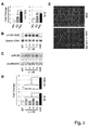
Figure 1 . Analysis of dystrophin and nNOS rescue upon exon skipping treatment. (A) Schematic representation of the exon skipping strategy. (B)Western blot with anti-dystrophin (αDys) and α-tubulin (α-tub) antibodies on 50 µg of proteins from gastrocnemius of wild type (WT) animals and 50 µg of mdx, G1, G2 andG3 animals 4 weeks after systemic injection of AAV-U1# 23. (C)Western blot with anti-dystrophin (αDys) antibody on 50 µg of proteins from triceps, diaphragm and vastus from mdx, G1, G2, G3 and wild type (WT)animals 4 weeks after systemic injection of AAV-U1# 23. (C) Muscles from wild type (WT), control (mdx) and AAV-U1# 23 injected mdx mice were sliced and analyzed by immunofluorescence with anti-dystrophin and anti-nNOS antibodies. The morphological state was analyzed by Hematoxilin/Eosin (H&E) staining. Original magnification, X20. Scale bar is 100 µm. -
Figure 2 - Analysis of miRNA expression. (A) A list of differentially expressed miRNAs (at least 1,5 fold variation), in WT and mdx gastrocnemius, was derived from the values of real-time based low density arrays (Table 1). Aware of tissue heterogenity, the diagram shows miRNA classification into three groups: Box1 (B1), Box2 (B2), Box3 (B3). For each miRNA the fold change value between WT and mdx is shown. Dots indicate the mean expression of miRNAs belonging to the same family; miRNAs, rescued towards WT levels in exon skipping-treated mice, are underlined. (B) Histograms show miRNA relative expression in gastrocnemius from WT (white bars), G1-G3 (grey bars) and mdx (black bars) mice, measured by qRT-PCR. The invariant miR-23a and miR-27a were used as controls. Fold changes are normalized to snoRNA55 and shown with respect to WT set to a value of 1. (C) AAV administrations do not affect miRNA expression. mdx animals were tail-vein injected with a AAV-vector encoding only for GFP (AAV-GFP) and sacrificed after 4 weeks in parallel with wild type (WT) and untreated mdx animals. miR-1 and miR-29 levels were measured by qRT-PCR on RNA from gastrocnemius. Fold changes are shown with respect to WT, set to a value of 1 and normalized to snoRNA55. (D) The human DMD deletion of exons 48-50 of the dystrophin gene produces an out of frame mRNA. Skipping ofexon 51 would produce an in frame mRNA and the synthesis of a shorter but functional dystrophin protein. Human cultured DMD myoblasts carrying the exon 48-50 deletion were infected with a lentiviral construct carrying antisense sequences against the splice junctions of exon 51 (De Angelis et al. 2002). Cells were maintained in DM medium for 7 days. Western blot with anti-dystrophin (α-dys) antibodies on 50 µg of proteins from mock infected (mock) and antisense-treated (U1#51) DMD cells indicates the efficient recovery of dystrophin synthesis. Anti-tubulin antibodies (α-tub) were used as control. (E) qRT-PCR on the indicated miRNAs in control (mock) and antisense-treated (U1#51) DMD cells. U6 snRNA was used as endogenous control. Fold changes are shown with respect to mock-infected DMD cells, set to a value of 1. One asterisk (p<0.01), two asterisks (p<0.05). -
Figure 3 - Analysis of miR-29 targets. (A) qRT-PCR for the collagen (COL1A1) and elastin (ELN) mRNAs on total RNA from the gastrocnemius of WT (white bars), G3 (grey bars) and mdx (black bars) mice. Fold changes are normalized to HPRT1 mRNA and shown with respect to WT set to a value of 1. In panels B-E the gastrocnemius of mdx animals were electroporated with a miR-29a/b expression construct (U1-29) and sacrificed after 25 days. (B) PCR analysis on DNA from WT, mdx and U1-29 electroporated (1-4) mdx. Primers specific for the U1-29 construct and for the β-actin gene were utilized. (C) Northern blot for miR-29a and control snoRNA55. Normalized miR-29a relative expression level is indicated below each lane. (D) qRT-PCR on COL1A1 and ELN mRNAs. One asterisk (p<0.01), two asterisks (p<0.05). (E) The morphological state of muscles from control (mdx) and U1-29 electroporated mdx mice was analyzed by Trichromic staining. Original magnification, X4. -
Figure 4 - Chromatin immunoprecipitation analysis on microRNA promoters. (A) Schematic representations of the coding and promoter regions of miR-1-2/133a-1, miR-29b-2/29c and miR-206/133b genes. The transcriptional start site (TSS) and the primers utilized for PCR amplifications are indicated. (B) ChIP analyses were performed on chromatin from gastrocnemius of WT (white bars), G3 (grey bars) and mdx (black bars) mice with antibodies against AcH3K9, HDAC1, HDAC2 and HDAC4. The immunoprecipitated DNA samples were amplified with specific primers spanning miR-1, miR-29 and miR-206 regulatory regions. The Hprt1 promoter region was analyzed as control. Oligonucleotides for 5,8s rDNA were used as internal control by coamplification with each of the specific primers. Data are mean ± s.d. from three independent experiments. Results are expressed as fold enrichment of DNA immunoprecipitated samples relative to INPUT chromatin. -
Figure 5 - Analysis of miR-206 and Pax7 expression. (A) miR-206 and miR-1 expression was analyzed by Northern blot in C27 cells maintained in growth medium (GM) or in differentiation medium (DM) for the indicated times. U2 snRNA was utilized as loading control. (B) ChIP analysis with antibodies against AcH3K9, HDAC1 and HDAC2 was performed on miR-206 and miR-1-2 promoters in GM and DM (7 days). (C) Western analysis of Pax7 (upper panel) and Northern for miR-206 (lower panel) in the gastrocnemius of WT, G3 and mdx animals. Signals were normalized for GAPDH and snoRNA55, respectively. -
Figure 6 - Responsiveness of miRNA expression to the NO signalling. (A) qRT-PCR on miR-1, miR-29, miR-206 in WT (white bars), G3 (grey bars) and mdx (black bars) mice treated for 4 hours with nitroglycerin (NTG). miR-23, is utilized as an internal invariant control miRNA. (B) α-HDAC2 ChIP analyses were performed on chromatin from gastrocnemius of WT, mdx and NTG-treated mdx mice. The immunoprecipitated DNA samples were amplified with specific primers spanning miR-1, miR-29 and miR-206 regulatory regions (seeFig. 4 ). The Hprt1 promoter region was utilized as control. Oligonucleotides for 5,8s rDNA were used as internal control by co-amplification with each one of the specific primers. Data are mean ± s.d. from three independent experiments. Results are expressed as fold enrichment of DNA immunoprecipitated samples relative to INPUT chromatin. No antibody was used as control. -
Figure 7 - miRNA expression in blood and serum. Histograms show miR-206 and miR-1 relative expression in total blood and serum from WT (white bars) and mdx (black bars) mice, measured by qRT-PCR. Fold changes are normalized to snoRNA55 and shown with respect to WT serum levels set to a value of 1. -
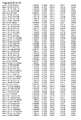
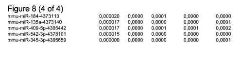
Figure 8 - A real time PCR-based approach (Applied Biosystems Taqman® Megaplex Array) was applied to total RNA derived from the gastrocnemius of a pool of three different individuals of WT (column DCt.WT), mdx (column DCt.MDX) and three independent AAV-U1# 23 treated mice (columns DCt.G1, DCt.G2, DCt.G3). miRNA Raw ΔCt values are indicated and ranked from the highest to the lowest expression level in mdx. -
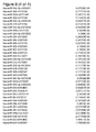
Figure 9 - A real time PCR-based approach (Applied Biosystems Taqman® Megaplex Array) was applied to total RNA derived from DMD myoblasts induced to differentiate. Raw ΔCt values are indicated and ranked from the highest to the lowest (column DCt.DMD). - Mature miRNAs as below show perfect sequence conservation between human and mouse. The mature sequence of the miRNA not listed can be found on the public database of miRNAs (www.mirbase.org).
miR-1: uggaauguaaagaaguauguau (SEQ ID No. 1) (the same mature miRNA sequence derives from the two miRNA genes (miR-1-1 and miR-1-2).
miR-206: uggaauguaaggaagugugugg (SEQ ID No. 2)
miR-223: ugucaguuugucaaauacccca (SEQ ID No. 3)
miR-21: ugucaguuugucaaauacccca (SEQ ID No. 4)
miR-29a: uagcaccaucugaaaucgguua (SEQ ID No. 5)
miR-29b1/2: uagcaccauuugaaaucaguguu (SEQ ID No. 6)
miR-29c: uagcaccauuugaaaucgguua (SEQ ID No. 7) - The miR-29 family includes miR-29a miR-29b1/2 and miR-29c (each produced from a specific locus). When miR-29 expression levels are indicated, the reported values are the mean result of all the three mature miRNAs. Also in this case the same mature miRNA sequence can derive from different genomic locations (miR-29b-1 and miR-29b-2 genes).
miR-31: aggcaagaugcuggcauagcu (SEQ ID No. 8)
miR-34a: uggcagugucuuagcugguugu (SEQ ID No. 9)
miR-133a1/2: uuugguccccuucaaccagcug (SEQ ID No. 10)
miR-133b: uuugguccccuucaaccagcua (SEQ ID No. 11) - The miR-133 family includes miR-133a1/2 and miR-133b (each produced from a specific locus). When miR-133 expression levels are indicated, the reported values are the mean result of the two mature miRNAs. Also in this case the same mature miRNA sequence can derive from different genomic locations (miR-133a-1 and miR-133a-2 genes).
miR-30c1/2: uguaaacauccuacacucucagc (SEQ ID No. 12) - 6 weeks-old mdx mice were tail vein injected with 0.5-1x10¹² genome copies of the AAV-
U1# 23 virus as described in Denti et al.(2006) and sacrificed after 4 weeks. Electroporation was performed on 6 week-old mdx as already described (Donà et al., 2003) with an expression cassette carrying the 29a/b coding region under the control of the U1 snRNA promoter (U1-29a/b). Nitroglycerin treatment was performed on 18 week-old mdx mice locally injected in gastrocnemius with 0,3 mg/kg of nitroglycerin. mdx control mice were injected with physiological solution. Animals were sacrificed after 5 hours. - RNA Preparation and Analysis. Total RNA was prepared from liquid nitrogen powdered tissue homogenized in TRIzol reagent (Invitrogen). RNA from blood and serum samples was extracted using TRIreagent BD (Sigma). miRNA profiling was performed using Applied Biosystems TaqMan Megaplex Low Density Array. (See supplementary methods online), while analysis of individual miRNAs was performed using specific TaqMan microRNA assay (Applied Biosystems). Relative quantification was performed using sno55 as endogenous control for murine samples and U6 for human samples. Data were expressed as means ± SEM, unless otherwise stated. Statistical significance of the differences between means was assessed by t-test (nonparametric). A probability of <5% was considered significant.
- Western blot on total extract and in situ analyses on 7 µm-thick gastrocnemius cryosections were performed as described in Denti et al (2006). The NCL-DYS2 (Novocastra) 1:12.5; nNOS (Santa Cruz Biotechnology) 1:10. monoclonal antibodies were used.
- ChIP analysis was performed as described in suppl. methods online with the following antibodies: HDAC2 (Santa Cruz Biotechnology), HDAC1, HDAC4, acetylated H3-Lys9 (Upstate Biotechnology).
- To synthesize single-stranded cDNA, 700 ng of total RNA were reverse transcribed using the miRNA reverse transcription kit in combination with the stem-loop Megaplex RT primers pool A (Applied Biosystem), allowing the simultaneous reverse transcription of 335 miRNAs, 5 species-specific endogenous controls and one negative control. From the cDNA samples, 335 small RNAs were profiled using an Applied Biosystems TaqMan Low Density Array. Since instrument and liquid handling variations were shown to be minimal, no PCR replicates were measured according to manufacturer specifications. PCR amplification reactions were carried out in a total volume of 1,5 microL. Raw Ct values were calculated using the SDS software V.2.3 using automatic baseline settings and a threshold of 0,2. Data were subsequently analyzed through StatMiner platform according to the manufacturer pipeline. StatMiner Genorm algorithm was applied to determine the best invariant endogenous controls on a list of 5.
-
- 206 fw:cagtgtccattcatctcctacagc (SEQ ID No. 13)
- 206 rw:gcttggaatatgctgacaggctgc (SEQ ID No. 14)
- 1-2 fw:caaagcagtgttgggaccacagga (SEQ ID No. 15)
- 1-2 rw:actcccaggcaaacagactctct (SEQ ID No. 16)
- 29 fw:ctgtggagacaggtagtgaagctc (SEQ ID No. 17)
- 29 rw:tgaactggaagggagccaacatgg (SEQ ID No. 18)
- rma fw:cgtcgatgaagaacgcagctagct (SEQ ID No. 19)
- rma rw:gtgtcgatgatcaatgtgtcctgc (SEQ ID No. 20)
- hprt fw:gtgattatctgggaatcctctggg (SEQ ID No. 21)
- hprt rw:ccggaactcttatctgactaggtg (SEQ ID No. 22)
- Chromatin immunoprecipitation (ChIP) assay. Gastrocnemius (0,1-0,2 g) were dissected and incubated in 1,8% formaldehyde for 20 minutes at 22°C (after 5 minutes they were cut in pieces of 1-3 mm3). C27 myoblasts (1x10⁶ cells/ml) were instead incubated in 1% formaldehyde for 10 minutes at 25°C. The reactions were stopped by glycine (0,125 M) for 5 minutes at RT. After washing with cold PBS 1X, samples were collected by centrifugation at 4 C° at 3000 rpm, resuspended in 5 volumes of lysis buffer (
SDS 1%,EDTA 10 mM TRIS-HCl 50 mM pH 8) and sonicated. The average DNA fragment length was 400-1000 bp. Samples were then centrifuged at 10,000 rpm for 10minutes at 4°C. Supernatant was diluted with ChIP dilution buffer (SDS 0,01%, Triton X-100 1,1%,EDTA mM pH 8, NaCl 167 mM) and pre-cleared with 50 µl of Protein A Agarose/Salmon Sperm DNA beads (Upstate). 30 µl of supernatant were collected as input (I) sample. Chromatins were incubated with 3-5 µg of Ab (IP) or without Ab (B) overnight at 4°C. Immunoprecipitates were washed twice with buffer A (SDS EDTA 2 mM, Tris-HCl pH 8 20 mM, NaCl 150 mM), twice with buffer B (SDS EDTA 2 mM, Tris-HCl pH 8 20 mM, NaCl 500 mM), twice with buffer C (LiCl 0,25M,NP40 1%,deoxycholate 1 %,EDTA 1 mM, Tris-HCl pH 8 10 mM) and once with TE 1X. Immunocomplexes were eluted with 500 µl of elution buffer (SDS 1%,NaHCO3 0,1 M) at RT in rotation for 15'. The latter step was repeated twice. After crosslink reversion, proteins were extracted and DNA ethanol precipitated. The pellets were resuspended in 30 µl of H2O and amplified by PCR in the presence of [alfa-32P]dATP (1 microCi/12.5 microL). 1 µl of immunoprecipitated DNA (IP) and negative control without Ab (B-beads only) in 12,5 µl of total PCR reaction. Total input sample was diluted 1:10 before PCR. The promoter region of the following genes was analyzed: P-miR-206, P-miR-1, P-miR-29, P-HPRT1. Oligonucleotides for 5,8s rDNA were used as internal control by co-amplification with each of the specific primers. We analyzed the PCR products on a 8% acrylamide gel and visualized them using a Typhoon Imager (GE Healthcare). - Quantifications were performed with an ImageQuant software (Molecular Dynamics) and values normalized for internal control co-amplification and subtraction of background retained on the agarose beads (B). Each value in the histogram is the average of the signals on 3 different preparations of immunoprecipitated DNA.
- 6-week old mdx animals were tail vein injected with AAV recombinant viruses (AAV-U1#23) carrying a U1-chimeric antisense construct (
Fig.1A ) previously reported to induce the skipping of the mutatedexon 23 and to restore dystrophin synthesis (Denti et al., 2006 and 2008). After 1 month, mdx and treated-mdx seeblings were sacrificed in parallel with wild type (WT) isogenic/aged matched animals. Different muscular districts were dissected and subdivided for RNA, protein and chromatin analysis. Body wide rescue (Denti et al., 2006) of dystrophin was obtained (Fig.1B,C ). We took advantage of the intrinsic variability of in vivo transduction, following systemic delivery (Denti et al., 2006), in order to classify the animals on the basis of dystrophin rescue levels. Three different groups, each one including at least three different individuals, were obtained (G1, G2 and G3); compared to wild type mice, they display ≤1%, 1-5% and 5-10% of dystrophin rescue, respectively (Fig.1B ). Even if these levels seem very low to confer any beneficial effect, it was demonstrated that amounts of dystrophin as low as a few percent are able to confer long life benefit to mdx muscles (Denti et al., 2008; Ghahramani Seno et al., 2008). Dystrophin rescue paralleled both the recovery of nNOS localization at the periphery of the fibers (Fig.1D ) and morphological amelioration of the transduced muscle (Fig 1D ). The effect was very significant in G3 animals but still detectable in G2 (not shown). - Real-time based low density arrays were performed on RNA from the gastrocnemius of wild type, mdx, and AAV-U1#23-treated mdx animals. miRNA expression levels, normalized for 5 endogenous controls (
Fig. 8 ), revealed clear differences between WT and mdx. In the diagram ofFig. 2A , the miRNAs displaying the most significant variations (fold-change > 1,5) between WT and mdx are reported. Aware of the cellular heterogeneity of the dystrophic muscle, a parallel profile analysis on human DMD myoblasts (Fig. 9 ) was performed. Relevant differences between the animal and the in vitro cultured differentiated myoblasts were due to miRNAs belonging to cell types other than muscle ones (box B3 and B1) and, in particular as expected for a damaged dystrophic fiber, to miRNAs related to the inflammatory process (box B1, Fazi et al., 2005; Baltimore et al., 2008). Among the others, the B4 group contains miRNAs in common between cultured myoblasts and dissected muscles, while B2 include more ubiquitous miRNAs. The underlined miRNAs identify those species that in AAV-U1#23-treated mdx are recovered to levels similar to WT. Interestingly, this effect was proportional to the levels of dystrophin rescue, with G1 showing a very limited effect and G3 providing levels very close to the wild type ones (Fig. 8 ). Individual qRT-PCR were performed on B4 miRNAs and on a selection of control miRNAs. miR-1, miR-133a, miR-186, miR-29c and miR-30c, showing reduced levels in mdx, recover towards wild type levels in the three groups of treated animals proportionally to the level of dystrohin rescue (Fig. 2B ). At difference with miR-1 and miR-133 myomiRs, miR-206 levels increase in mdx and in treated mice, while miR-31, displaying the strongest variation between WT and mdx did not show any variation in exon-skipped animals (not shown). - The same up-regulation of the above miRNAs was also observed in human DMD myoblasts treated with the exon skipping approach (
Fig.2D and E ). - Interestingly, the inflammatory-specific miR-223 (Fazi et al., 2005), very abundant in mdx, is proportionally reduced in the three different groups of animals, indicating the amelioration of the inflammatory state of the muscle due to dystrophin rescue (see
fig.1c and Denti et al., 2008). The ubiquitous miR-23a and 27a, unchanged in WT and mdx, behaved similarly also in treated animals. Control injections of mdx mice with a AAV vector coding for the GFP protein indicated that the virus per se did not produce any change in miRNA levels (Fig. 2C ). The relative expression of the selected miRNAs in WT, mdx and G3 animals was the same also in other muscle districts, demonstrating that the miRNA expression profile correlated with dystrophin rescue in a body-wide manner (Fig. 1C ). - These data indicate that a specific subset of miRNAs undergoes altered expression in Duchenne Muscular Dystrophy as a direct consequence of dystrophin absence, while a different subset varies as a consequence of fiber damage.
- Extracellular matrix proteins are known to increase in mdx animals where they contribute to the extensive fibrotic degeneration (Haslett et al., 2002). Crucial factors involved in this process are collagens and elastin whose mRNAs have been recently shown to be targets of the miR-29 family (van Rooij et al., 2008). qRT-PCR reveals that the mRNAs for these factors are up-regulated in mdx, where miR-29 is low, while significantly reduced in treated animals at levels similar to WT, where miR-29 is high (
Fig. 3A ). Therefore, the typical inverse correlation between miRNAs and corresponding targets is observed. - In order to test whether miR-29 alone is indeed responsible for this effect on collagen and elastin mRNAs, we electroporated in the gastrocnemius of mdx animals a plasmid DNA containing an expression cassette driving the transcription of miR-29a/b. Northern blot indicated that a 2-fold overexpression of miR-29 was specifically obtained in treated muscles which paralleled a decrease of collagen and elastin mRNAs (
Fig. 3 ). - Trichromic staining analysis indicated a decrease in collagen deposition when muscles were electroporated with miR-29 in comparison to mock-treated controls (
Fig. 3E ). This suggest a direct role of miR-29 in the morphological benefit obtained in dystrophin-rescued animals. Therefore, both miR-29 or miR-30 can be utilized as therapeutic tool in order to ameliorate the fibrotic deposition consequent to muscle fiber degeneration. They can be administered either as synthetic molecules or as gene constructs with muscle specific promoters in the backbone of viral constructs. The delivery route can be either by local or systemic administration. - Chromatin immunoprecipitation (ChIP) analysis with antibodies against RNA polymerase II and H3K9 acetylation on the miR-1/133, miR-206 and miR-29 promoters (
Fig. 4A ) (Rao et al., 2006; Liu et al., 2007; Shkumatava et al., 2009; Wang et al., 2008, Han et al., 2004 and D.C. and M.C. personal data) correlated very well with the differential expression levels of the corresponding miRNAs in WT, mdx and G3 animals (Fig. 4B ). Notably, in conditions where these miRNAs are repressed (mdx animals) a specific interaction was detected with HDAC2, while no reactivity was observed with either HDAC1 or HDAC4 (Fig. 4B ). On the contrary, miR-206 and HPRT1 promoters, directing active transcription, do not show reactivity with any HDAC. - Interestingly, in growing C27 myoblasts, where miR-206 is poorly expressed (
Fig. 5A ,), a specific interaction was found between its promoter and HDAC1; upon differentiation, when transcription is activated, clearance of HDAC1 from the promoter was observed (Fig. 5B ). In growth conditions, the miR-1/133 promoter was found again to be repressed by HDAC2 (Fig. 5A ). The increased expression of miR-206 in mdx animals paralleled that of the satellite-specifying factor Pax 7 (Relaix, et al., 2006) (Fig.5C ). These data, together with previous findings, reporting markedly increased expression of miR-206 in newly formed muscle fibers/myotubes (Yuasa et al., 2008) and a postulated role in regulating genes involved in satellite cells specification (McCarty, 2008), suggest a role of miR-206 in active regeneration and efficient maturation of skeletal muscle fibers. Notably, Pax7 and miR-206 levels are slightly higher in AAV-U1# 23 treated animals (Fig.5C ), indicating that dystrophin rescue parallels intensive muscle regeneration, in agreement with the morphological (Fig.1D ) and functional benefit observed in treated animals (Denti et al., 2006). These results allow to conclude that miR-206 follows an epigenetic control independent from the one responsible for the expression of the terminally differentiating miR-1 and miR-133. - Due to the described correlation between nitrosylation and HDAC2 DNA binding (Nott et al., 2008), we tested whether the treatment of mdx animals with the NO donor, nitroglycerin (NTG), would provide the same epigenetic changes observed with the exon skipping treatment.
Fig.6 shows that this treatment: i) up-regulates miR-1 and miR-29; ii) releases HDAC2 from the promoters of these miRNAs and iii) has no effect on miR-206 levels. Finally, it was tested that, as a consequence of muscle damage, specific muscle miRNAs are released into the blood. qRT-PCR on blood and serum of wild type versus dystrophic animals was performed.Fig.7 indicates that high levels of the muscle-specific miR-1 and miR-206 (15 and 30-fold increase respectively) are found in the serum of mdx animals. These results indicate that muscle damage in Duchenne individuals (as well as in other muscle degenerative disorders) can be easily and quantitatively assessed by measuring the serum levels of muscle specific miRNAs. - Altogether, these experiments clarify a new link between dystrophin and important genes encoding for miRNAs acting as regulators of muscle tissue differentiation and morphology. Moreover, the altered pattern of NO-dependent miRNAs in DMD can be extended to other muscle disorders in which the DAPC complex is absent or altered (such as in Girdle Limb dystrophy). These data also support the hypothesis that the morpho-functional benefit observed in exon-skipping mdx treated animals (Denti et al., 2008) may be due not only to the structural amelioration of the muscle membrane but also to an intracellular cascade of events controlling the expression of genes relevant for proper muscle homeostasis.
-
- 1. Matsumura, K., et al. Expression of dystrophin-associated proteins in dystrophin-positive muscle fibers (revertants) in Duchenne muscular dystrophy. Neuromuscul. Disord. 4:115-120 (1994).
- 2. Ervasti, J.M., Sonnemann, K.J. Biology of the striated muscle dystrophin-glycoprotein complex. Int Rev Cytol. 265:191-225 (2008).
- 3. Denti, M.A., et al. Body-wide gene therapy of Duchenne Muscular Dystrophy in the mdx mouse model. Proc. Natl. Acad. Sci. USA 103:3758-3763 (2006).
- 4. Naguibneva I. et al. (2006) The microRNA miR-181 targets the homeobox protein Hox-A11 during mammalian myoblast differentiation. Nat Cell Biol.8 :278-84.
- 5. Chen J.F. et al. (2006) The role of microRNA-1 and microRNA-133 in skeletal muscle proliferation and differentiation. Nat Genet. 38:228-33.
- 6. Van Rooij, E., et al. Dysregulation of microRNAs after myocardial infarction reveals a role of miR-29 in cardiac fibrosis. Proc. Natl. Acad. Sci. USA 105:13027-13032 (2008).
- 7. Brenman, J.E., et al. Nitric oxide synthase complexed with dystrophin and absent from skeletal muscle sarcolemma in Duchenne muscular dystrophy. Cell 82:743-752 (1995).
- 8. Wehling, M., Spencer, M.J., Tidball, J.G. A nitric oxide synthase transgene ameliorates muscular dystrophy in mdx mice. J. Cell Biol. 155:123-131 (2001).
- 9. Riccio, A., et al. A nitric oxide signaling pathway controls CREB-mediated gene expression in neurons. Mol. Cell 21:283-294 (2006).
- 10. Nott, A., et al. S-nitrosylation of ).
- 11. Minetti, G.C., et al. Functional and morphological recovery of dystrophic muscles in mice treated with deacetylase inhibitors. Nat. Med. 12:1147-1150 (2006).
- 12. Brunelli, S., et al. Nitric oxide release combined with nonsteroidal antiinflammatory activity prevents muscular dystrophy pathology and enhances stem cell therapy. Proc. Natl. Acad. Sci. USA 104:264 -269 (2007).
- 13. Illi, B., et al. Nitric oxide modulates chromatin folding in human endothelial cells via protein phosphatase 2A activation and class II histone deacetylases nuclear shuttling. Circ Res. 102:51(2008).
- 14. Colussi, C., et al. Nitric oxide deficiency determines global chromatin changes in Duchenne muscular dystrophy. FASEB J. in press (2009).
- 15. Denti M. A., et al. Life-long benefit of AAV/antisense-mediated exon skipping in dystrophic mice. Hum. Gene Ther. 19:601-608 (2008).
- 16. Ghahramani Seno, M.M., et al. RNAi-mediated knockdown of dystrophin expression in adult mice does not lead to overt muscular dystrophy pathology. Hum. Mol. Genet. 17:2622-2632 (2008).
- 17. Eisenberg, I., et al. Distinctive patterns of microRNA expression in primary muscular disorders. Proc. Natl. Acad. Sci. USA 104:17016-17021 (2007).
- 18. Fazi, F., et al. A mini-circuitry comprising microRNA-223 and transcription factors NFI-A and C/EBPα regulates human granulopoiesis. Cell 123:819-831 (2005).
- 19. Baltimore, D., et al. MicroRNAs: new regulators of immune cell development and function. Nat. Immunol. 9:839-45 (2008).
- 20. Haslett, J.N., et al. Gene expression comparison of biopsies from Duchenne muscular dystrophy (DMD) and normal skeletal muscle. Proc. Natl. Acad. Sci. USA 99:15000-15005 (2002).
- 21. Liu, N., et al. An intragenic MEF2-dependent enhancer directs muscle-specific expression ofmicroRNAs 1 and 133. Proc. Natl. Acad. Sci USA 104:20844-20849 (2007).
- 22. Rao, P.K., et al. Myogenic factors that regulate expression of muscle-specific microRNAs. Proc. Natl. Acad. Sci. USA 103:8721-8726 (2006).
- 23. Relaix, F., et al. Pax3 and Pax7 have distinct and overlapping functions in adult muscle progenitor cells. J. Cell Biol. 172:91-102 (2006).
- 24. Shkumatava, A., Stark, A., Sive, H., Bartel, D.P. Coherent but overlapping expression of microRNAs and their targets during vertebrate development. Genes Dev. 23:466-81 (2009).
- 25. Wang, H., et al. NF-kappaB-YY1-miR-29 regulatory circuitry in skeletal myogenesis and rhabdomyosarcoma. Cancer Cell 14:369-381 (2008).
- 26. Lee, Y., et al. MicroRNA genes are transcribed by RNA polymerase II. EMBO J. 23: 4051-4060 (2004)
- 27. Yuasa, K., et al. MicroRNA-206 is highly expressed in newly formed muscle fibers: implications regarding potential for muscle regeneration and maturation in muscular dystrophy. Cell Struct. Funct. 33:163-169 (2008).
- 28. McCarthy, J.J., MicroRNA-206: the skeletal muscle-specific miomiR. Biochim. Biophys. Acta. 1779:682-691(2008).
- 29. Donà, M., et al. Functional in vivo gene transfer into the myofibers of adult skeletal muscle. Biochem. Biophys. Res. Commun. 312:1132-1138(2003).


Claims (12)
- A method for the diagnosis of a muscle disorder and damage leading to muscle fiber degeneration, inflammation and fibrosis or for monitoring the progress of therapeutic treatments on subjects affected by said disorder consisting in detecting in a biological sample at least two of dystromiR molecules, each of the two dystromiR molecules belonging to one of the following groups: a) muscle regeneration (miR-206, miR-34), b) muscle differentiation (miR-1, miR-133), c) inflammatory (miR-223, miR-21) and d) tissue integrity (miR-29, miR-30).
- The method according to claim 1 wherein said muscle disorder is the Duchenne Muscular Dystrophy.
- The method according to claim 2 wherein the each of the two dystromiR molecules belongs to one of the following groups: a) muscle regeneration (miR-206, miR-34), b) muscle differentiation (miR-1, miR-133), c) inflammatory (miR-223, miR-21) and d) tissue integrity (miR-29, miR-30); Duchenne specific (miR-31).
- The method according to claim 3 wherein the at least two of dystromiR molecules are selected in the following group: miR-31, miR-206, miR-1, miR-223 and miR-29.
- The method according to any of previous claims wherein the biological sample is a muscle bioptic sample.
- The method according to claims 1 to 4 wherein the biological sample is a serum or a blood sample.
- The method according to any of previous claims wherein the detecting at least two of dystromiR molecules is performed by reverse amplification of said dystromiR molecules and real time detection of amplified products.
- A kit to perform the method according to claim 4.
- A dystromiR molecule belonging to the group of miR-29 and/or miR-30 for use as therapeutic medicament of Duchenne Muscular Dystrophy or of pathologies associated to mutations of DAPC components, such as Girdle Limb dystrophy, or as coadiuvant of other therapeutic strategies, such as exon skipping.
- A pharmaceutical composition for local and/or systemic administration comprising the dystromiR molecule or a vector encoding the dystromiR molecule according to claim 9.
- An agent able to functionally inactivate the dystromiR-31 molecule
- The agent according to claim 11 as it can implement other as coadiuvant of other therapeutic strategies, such as exon skipping.
Priority Applications (12)
| Application Number | Priority Date | Filing Date | Title |
|---|---|---|---|
| EP20090161038 EP2258863A1 (en) | 2009-05-25 | 2009-05-25 | miRNA biomarkers for the diagnosis of duchenne muscular dystrophy progression, for monitoring therapeutic interventions, and as therapeutics |
| US13/322,023 US9034838B2 (en) | 2009-05-25 | 2010-05-24 | miR-31 in duchenne muscular dystrophy therapy |
| EP20100723982 EP2435583B1 (en) | 2009-05-25 | 2010-05-24 | miR-31 IN DUCHENNE MUSCULAR DYSTROPHY THERAPY |
| PCT/EP2010/057088 WO2010136415A1 (en) | 2009-05-25 | 2010-05-24 | miRNA BIOMARKERS FOR THE DIAGNOSIS OF DUCHENNE MUSCULAR DYSTROPHY, ITS PROGRESSION AND FOR MONITORING THERAPEUTIC INTERVENTIONS |
| PCT/EP2010/057093 WO2010136417A1 (en) | 2009-05-25 | 2010-05-24 | miR-31 IN DUCHENNE MUSCULAR DYSTROPHY THERAPY |
| DK10723982T DK2435583T3 (en) | 2009-05-25 | 2010-05-24 | miR-31 IN TREATMENT OF THE MUSCLE DROPS OF THE DUCHENNES |
| CA2763432A CA2763432A1 (en) | 2009-05-25 | 2010-05-24 | Mirna biomarkers for the diagnosis of duchenne muscular dystrophy, its progression and for monitoring therapeutic interventions |
| ES10723980T ES2443146T3 (en) | 2009-05-25 | 2010-05-24 | Biomarkers of miRNA for the diagnosis of muscular dystrophy and its progression |
| US13/322,026 US20120115150A1 (en) | 2009-05-25 | 2010-05-24 | miRNA BIOMARKERS FOR THE DIAGNOSIS OF DUCHENNE MUSCULAR DYSTROPHY, ITS PROGRESSION AND FOR MONITORING THERAPEUTIC INTERVENTIONS |
| JP2012512325A JP2012527873A (en) | 2009-05-25 | 2010-05-24 | MiRNA biomarkers for the diagnosis of Duchenne muscular dystrophy, its progression, and for monitoring therapeutic intervention |
| AU2010252075A AU2010252075A1 (en) | 2009-05-25 | 2010-05-24 | miRNA biomarkers for the diagnosis of Duchenne Muscular Dystrophy, its progression and for monitoring therapeutic interventions |
| EP20100723980 EP2435582B1 (en) | 2009-05-25 | 2010-05-24 | miRNA BIOMARKERS FOR THE DIAGNOSIS OF MUSCULAR DYSTROPHY AND ITS PROGRESSION |
Applications Claiming Priority (2)
| Application Number | Priority Date | Filing Date | Title |
|---|---|---|---|
| EP20090161038 EP2258863A1 (en) | 2009-05-25 | 2009-05-25 | miRNA biomarkers for the diagnosis of duchenne muscular dystrophy progression, for monitoring therapeutic interventions, and as therapeutics |
| PCT/EP2010/057088 WO2010136415A1 (en) | 2009-05-25 | 2010-05-24 | miRNA BIOMARKERS FOR THE DIAGNOSIS OF DUCHENNE MUSCULAR DYSTROPHY, ITS PROGRESSION AND FOR MONITORING THERAPEUTIC INTERVENTIONS |
Publications (1)
| Publication Number | Publication Date |
|---|---|
| EP2258863A1 true EP2258863A1 (en) | 2010-12-08 |
Family
ID=43855977
Family Applications (2)
| Application Number | Title | Priority Date | Filing Date |
|---|---|---|---|
| EP20090161038 Withdrawn EP2258863A1 (en) | 2009-05-25 | 2009-05-25 | miRNA biomarkers for the diagnosis of duchenne muscular dystrophy progression, for monitoring therapeutic interventions, and as therapeutics |
| EP20100723980 Not-in-force EP2435582B1 (en) | 2009-05-25 | 2010-05-24 | miRNA BIOMARKERS FOR THE DIAGNOSIS OF MUSCULAR DYSTROPHY AND ITS PROGRESSION |
Family Applications After (1)
| Application Number | Title | Priority Date | Filing Date |
|---|---|---|---|
| EP20100723980 Not-in-force EP2435582B1 (en) | 2009-05-25 | 2010-05-24 | miRNA BIOMARKERS FOR THE DIAGNOSIS OF MUSCULAR DYSTROPHY AND ITS PROGRESSION |
Country Status (7)
| Country | Link |
|---|---|
| US (1) | US20120115150A1 (en) |
| EP (2) | EP2258863A1 (en) |
| JP (1) | JP2012527873A (en) |
| AU (1) | AU2010252075A1 (en) |
| CA (1) | CA2763432A1 (en) |
| ES (1) | ES2443146T3 (en) |
| WO (1) | WO2010136415A1 (en) |
Cited By (8)
| Publication number | Priority date | Publication date | Assignee | Title |
|---|---|---|---|---|
| WO2011102908A3 (en) * | 2010-02-22 | 2012-01-19 | Duke University | Biomarkers of musculoskeletal disease |
| EP2535412A1 (en) | 2011-06-17 | 2012-12-19 | Universitat Pompeu-Fabra | New treatment for muscular dystrophies |
| US8871918B2 (en) | 2008-10-24 | 2014-10-28 | Sarepta Therapeutics, Inc. | Multiple exon skipping compositions for DMD |
| US9018368B2 (en) | 2004-06-28 | 2015-04-28 | The University Of Western Australia | Antisense oligonucleotides for inducing exon skipping and methods of use thereof |
| US9217148B2 (en) | 2013-03-14 | 2015-12-22 | Sarepta Therapeutics, Inc. | Exon skipping compositions for treating muscular dystrophy |
| US9228187B2 (en) | 2009-11-12 | 2016-01-05 | The University Of Western Australia | Antisense molecules and methods for treating pathologies |
| US9506058B2 (en) | 2013-03-15 | 2016-11-29 | Sarepta Therapeutics, Inc. | Compositions for treating muscular dystrophy |
| US11560594B2 (en) | 2015-02-05 | 2023-01-24 | Duke University | Methods of detecting osteoarthritis and predicting progression thereof |
Families Citing this family (4)
| Publication number | Priority date | Publication date | Assignee | Title |
|---|---|---|---|---|
| US20130034850A1 (en) * | 2010-02-26 | 2013-02-07 | Natinal Center of Neurology and Psychiatry | Marker for detecting myogenic disease and detection method using the same |
| FR2984358A1 (en) * | 2011-12-16 | 2013-06-21 | Genethon | METHODS FOR THE DIAGNOSIS OF MUSCLE DYSTROPHIES |
| EP3246398B1 (en) * | 2015-01-14 | 2021-06-16 | Kyoto University | Method for selecting skeletal muscle progenitor cell |
| CN111729088B (en) * | 2020-08-21 | 2020-12-15 | 江苏申基生物科技有限公司 | A kind of microRNA miR-34a coupled probe and preparation method thereof |
Citations (1)
| Publication number | Priority date | Publication date | Assignee | Title |
|---|---|---|---|---|
| WO2003095647A2 (en) * | 2002-05-08 | 2003-11-20 | Universitá Degli Studi Di Roma | CHIMERIC snRNA MOLECULES CARRYING ANTISENSE SEQUENCES AGAINST THE SPLICE JUNCTIONS OF THE DYSTROPHIN GENE AND THEIR THERAPEUTIC APPLICATIONS |
Family Cites Families (3)
| Publication number | Priority date | Publication date | Assignee | Title |
|---|---|---|---|---|
| JP5399011B2 (en) * | 2007-06-28 | 2014-01-29 | 三菱化学メディエンス株式会社 | Method for detecting tissue injury or cell proliferative disorder |
| EP2271931A4 (en) * | 2008-04-25 | 2011-10-12 | Merck Sharp & Dohme | BIOMARKERS OF MICROARN-BASED TISSUE LESIONS |
| CN101386848A (en) * | 2008-08-12 | 2009-03-18 | 南京大学 | Microribonucleic acid contained in cell microparticles and its preparation research method and application |
-
2009
- 2009-05-25 EP EP20090161038 patent/EP2258863A1/en not_active Withdrawn
-
2010
- 2010-05-24 CA CA2763432A patent/CA2763432A1/en active Pending
- 2010-05-24 ES ES10723980T patent/ES2443146T3/en active Active
- 2010-05-24 WO PCT/EP2010/057088 patent/WO2010136415A1/en not_active Ceased
- 2010-05-24 US US13/322,026 patent/US20120115150A1/en not_active Abandoned
- 2010-05-24 EP EP20100723980 patent/EP2435582B1/en not_active Not-in-force
- 2010-05-24 AU AU2010252075A patent/AU2010252075A1/en not_active Abandoned
- 2010-05-24 JP JP2012512325A patent/JP2012527873A/en active Pending
Patent Citations (1)
| Publication number | Priority date | Publication date | Assignee | Title |
|---|---|---|---|---|
| WO2003095647A2 (en) * | 2002-05-08 | 2003-11-20 | Universitá Degli Studi Di Roma | CHIMERIC snRNA MOLECULES CARRYING ANTISENSE SEQUENCES AGAINST THE SPLICE JUNCTIONS OF THE DYSTROPHIN GENE AND THEIR THERAPEUTIC APPLICATIONS |
Non-Patent Citations (35)
Cited By (41)
| Publication number | Priority date | Publication date | Assignee | Title |
|---|---|---|---|---|
| US9249416B2 (en) | 2004-06-28 | 2016-02-02 | The University Of Western Australia | Antisense oligonucleotides for inducing exon skipping and methods of use thereof |
| US9605262B2 (en) | 2004-06-28 | 2017-03-28 | The University Of Western Australia | Antisense oligonucleotides for inducing exon skipping and methods of use thereof |
| US10968450B2 (en) | 2004-06-28 | 2021-04-06 | The University Of Western Australia | Antisense oligonucleotides for inducing exon skipping and methods of use thereof |
| US10227590B2 (en) | 2004-06-28 | 2019-03-12 | The University Of Western Australia | Antisense oligonucleotides for inducing exon skipping and methods of use thereof |
| US9018368B2 (en) | 2004-06-28 | 2015-04-28 | The University Of Western Australia | Antisense oligonucleotides for inducing exon skipping and methods of use thereof |
| US9024007B2 (en) | 2004-06-28 | 2015-05-05 | The University Of Western Australia | Antisense oligonucleotides for inducing exon skipping and methods of use thereof |
| US9035040B2 (en) | 2004-06-28 | 2015-05-19 | The University Of Western Australia | Antisense oligonucleotides for inducing exon skipping and methods of use thereof |
| US9175286B2 (en) | 2004-06-28 | 2015-11-03 | The University Of Western Australia | Antisense oligonucleotides for inducing exon skipping and methods of use thereof |
| US10781451B2 (en) | 2004-06-28 | 2020-09-22 | The University Of Western Australia | Antisense oligonucleotides for inducing exon skipping and methods of use thereof |
| USRE47769E1 (en) | 2004-06-28 | 2019-12-17 | The University Of Western Australia | Antisense oligonucleotides for inducing exon skipping and methods of use thereof |
| USRE47751E1 (en) | 2004-06-28 | 2019-12-03 | The University Of Western Australia | Antisense oligonucleotides for inducing exon skipping and methods of use thereof |
| US9994851B2 (en) | 2004-06-28 | 2018-06-12 | The University Of Western Australia | Antisense oligonucleotides for inducing exon skipping and methods of use thereof |
| US10995337B2 (en) | 2004-06-28 | 2021-05-04 | The University Of Western Australia | Antisense oligonucleotides for inducing exon skipping and methods of use thereof |
| US10421966B2 (en) | 2004-06-28 | 2019-09-24 | The University Of Western Australia | Antisense oligonucleotides for inducing exon skipping and methods of use thereof |
| US10266827B2 (en) | 2004-06-28 | 2019-04-23 | The University Of Western Australia | Antisense oligonucleotides for inducing exon skipping and methods of use thereof |
| US9441229B2 (en) | 2004-06-28 | 2016-09-13 | The University Of Western Australia | Antisense oligonucleotides for inducing exon skipping and methods of use thereof |
| US9422555B2 (en) | 2004-06-28 | 2016-08-23 | The University Of Western Australia | Antisense oligonucleotides for inducing exon skipping and methods of use thereof |
| US9447415B2 (en) | 2004-06-28 | 2016-09-20 | The University Of Western Australia | Antisense oligonucleotides for inducing exon skipping and methods of use thereof |
| USRE47691E1 (en) | 2004-06-28 | 2019-11-05 | The University Of Western Australia | Antisense oligonucleotides for inducing exon skipping and methods of use thereof |
| US9453225B2 (en) | 2008-10-24 | 2016-09-27 | Sarepta Therapeutics, Inc. | Multiple exon skipping compositions for DMD |
| US9447417B2 (en) | 2008-10-24 | 2016-09-20 | Sarepta Therapeutics, Inc. | Multiple exon skipping compositions for DMD |
| US9447416B2 (en) | 2008-10-24 | 2016-09-20 | Sarepta Therapeutics, Inc. | Multiple exon skipping compositions for DMD |
| US9434948B2 (en) | 2008-10-24 | 2016-09-06 | Sarepta Therapeutics, Inc. | Multiple exon skipping compositions for DMD |
| US9234198B1 (en) | 2008-10-24 | 2016-01-12 | Sarepta Therapeutics, Inc. | Multiple exon skipping compositions for DMD |
| US8871918B2 (en) | 2008-10-24 | 2014-10-28 | Sarepta Therapeutics, Inc. | Multiple exon skipping compositions for DMD |
| US9228187B2 (en) | 2009-11-12 | 2016-01-05 | The University Of Western Australia | Antisense molecules and methods for treating pathologies |
| US10781450B2 (en) | 2009-11-12 | 2020-09-22 | Sarepta Therapeutics, Inc. | Antisense molecules and methods for treating pathologies |
| US11447776B2 (en) | 2009-11-12 | 2022-09-20 | The University Of Western Australia | Antisense molecules and methods for treating pathologies |
| US9758783B2 (en) | 2009-11-12 | 2017-09-12 | The University Of Western Australia | Antisense molecules and methods for treating pathologies |
| US10287586B2 (en) | 2009-11-12 | 2019-05-14 | The University Of Western Australia | Antisense molecules and methods for treating pathologies |
| WO2011102908A3 (en) * | 2010-02-22 | 2012-01-19 | Duke University | Biomarkers of musculoskeletal disease |
| US9207247B2 (en) | 2010-02-22 | 2015-12-08 | Duke University | Biomarkers of musculoskeletal disease |
| EP2535412A1 (en) | 2011-06-17 | 2012-12-19 | Universitat Pompeu-Fabra | New treatment for muscular dystrophies |
| WO2012172012A1 (en) | 2011-06-17 | 2012-12-20 | Universitat Pompeu Fabra | New treatment for muscular dystrophies |
| US9217148B2 (en) | 2013-03-14 | 2015-12-22 | Sarepta Therapeutics, Inc. | Exon skipping compositions for treating muscular dystrophy |
| US10907154B2 (en) | 2013-03-14 | 2021-02-02 | Sarepta Therapeutics, Inc. | Exon skipping compositions for treating muscular dystrophy |
| US11932851B2 (en) | 2013-03-14 | 2024-03-19 | Sarepta Therapeutics, Inc. | Exon skipping compositions for treating muscular dystrophy |
| US9506058B2 (en) | 2013-03-15 | 2016-11-29 | Sarepta Therapeutics, Inc. | Compositions for treating muscular dystrophy |
| US10364431B2 (en) | 2013-03-15 | 2019-07-30 | Sarepta Therapeutics, Inc. | Compositions for treating muscular dystrophy |
| US10337003B2 (en) | 2013-03-15 | 2019-07-02 | Sarepta Therapeutics, Inc. | Compositions for treating muscular dystrophy |
| US11560594B2 (en) | 2015-02-05 | 2023-01-24 | Duke University | Methods of detecting osteoarthritis and predicting progression thereof |
Also Published As
| Publication number | Publication date |
|---|---|
| EP2435582A1 (en) | 2012-04-04 |
| CA2763432A1 (en) | 2010-12-02 |
| JP2012527873A (en) | 2012-11-12 |
| AU2010252075A1 (en) | 2011-12-15 |
| EP2435582B1 (en) | 2013-10-23 |
| US20120115150A1 (en) | 2012-05-10 |
| WO2010136415A1 (en) | 2010-12-02 |
| ES2443146T3 (en) | 2014-02-18 |
Similar Documents
| Publication | Publication Date | Title |
|---|---|---|
| Cacchiarelli et al. | MicroRNAs involved in molecular circuitries relevant for the Duchenne muscular dystrophy pathogenesis are controlled by the dystrophin/nNOS pathway | |
| EP2435582B1 (en) | miRNA BIOMARKERS FOR THE DIAGNOSIS OF MUSCULAR DYSTROPHY AND ITS PROGRESSION | |
| Wang et al. | MALAT1 lncRNA induces autophagy and protects brain microvascular endothelial cells against oxygen–glucose deprivation by binding to miR-200c-3p and upregulating SIRT1 expression | |
| Lu et al. | Morphine regulates expression of μ-opioid receptor MOR-1A, an intron-retention carboxyl terminal splice variant of the μ-opioid receptor (OPRM1) gene via miR-103/miR-107 | |
| Xu et al. | Long non-coding RNAs: new players in ocular neovascularization | |
| JP6157852B2 (en) | Use of microRNA to treat diseases associated with ciliary dysfunction in multicilia epithelial cells | |
| US20110160285A1 (en) | Identification of mirna profiles that are diagnostic of hypertrophic cardiomyopathy | |
| US10280422B2 (en) | MiR-92 inhibitors and uses thereof | |
| EP2622105A2 (en) | Compositions and methods useful for the treatment and diagnosis of inflammatory bowel disease | |
| WO2015006705A2 (en) | Micrornas that silence tau expression | |
| EP2539357A2 (en) | Methods and compositions for the detection and treatment of cancer involving mirnas and mirna inhibitors and targets | |
| EP2488662A2 (en) | Methods for diagnosing and treating a pathology associated with a synonymous mutation occuring within a gene of interest | |
| US20160145615A1 (en) | Agonists of ddah1 for treating endothelial dysfunction | |
| US20200199680A1 (en) | MIRNA in Gulf War Illness | |
| US20160304959A1 (en) | Compositions and methods for treating diabetic nephropathy | |
| CN107227362B (en) | Gene related to liver cancer and application thereof | |
| Malecova et al. | Development of a DUX4-targeting antibody oligonucleotide conjugate as a therapy for FSHD | |
| TWI818006B (en) | Mir-17~92 as therapeutic or diagnostic target of motor neuron (mn) degeneration diseases | |
| JP7158759B2 (en) | A pharmaceutical composition for the prevention or treatment of muscle disease or cachexia, comprising as an active ingredient a miRNA located in the Dlk1-Dio3 cluster or a variant thereof | |
| CN114058692A (en) | Application of circ _ RELB as molecular marker in preparation of new products for diagnosing diabetic retinopathy | |
| WO2020247130A1 (en) | Compounds and methods for inhibiting cancers via rest inhibition | |
| CN111263811A (en) | Compositions and methods for detecting and treating insulin resistance | |
| Galloway et al. | miR-146a is a Pleiotropic Regulator of Motor Neuron Degeneration | |
| Pinnarò | Role of noncoding RNAs in muscle differentiation | |
| Dubinsky | Expression profiling and functional studies of non-coding RNAs in the central nervous system |
Legal Events
| Date | Code | Title | Description |
|---|---|---|---|
| PUAI | Public reference made under article 153(3) epc to a published international application that has entered the european phase |
Free format text: ORIGINAL CODE: 0009012 |
|
| AK | Designated contracting states |
Kind code of ref document: A1 Designated state(s): AT BE BG CH CY CZ DE DK EE ES FI FR GB GR HR HU IE IS IT LI LT LU LV MC MK MT NL NO PL PT RO SE SI SK TR |
|
| AX | Request for extension of the european patent |
Extension state: AL BA RS |
|
| STAA | Information on the status of an ep patent application or granted ep patent |
Free format text: STATUS: THE APPLICATION IS DEEMED TO BE WITHDRAWN |
|
| 18D | Application deemed to be withdrawn |
Effective date: 20110609 |