EP2255717A1 - Capsule medical device, method for operating same, and capsule medical device system - Google Patents
Capsule medical device, method for operating same, and capsule medical device system Download PDFInfo
- Publication number
- EP2255717A1 EP2255717A1 EP09724627A EP09724627A EP2255717A1 EP 2255717 A1 EP2255717 A1 EP 2255717A1 EP 09724627 A EP09724627 A EP 09724627A EP 09724627 A EP09724627 A EP 09724627A EP 2255717 A1 EP2255717 A1 EP 2255717A1
- Authority
- EP
- European Patent Office
- Prior art keywords
- information
- section
- examination
- light
- medical device
- Prior art date
- Legal status (The legal status is an assumption and is not a legal conclusion. Google has not performed a legal analysis and makes no representation as to the accuracy of the status listed.)
- Withdrawn
Links
- 239000002775 capsule Substances 0.000 title claims abstract description 83
- 238000000034 method Methods 0.000 title claims description 21
- 239000003795 chemical substances by application Substances 0.000 claims description 25
- 230000010365 information processing Effects 0.000 claims description 4
- 230000005284 excitation Effects 0.000 description 20
- 230000004048 modification Effects 0.000 description 8
- 238000012986 modification Methods 0.000 description 8
- 230000005540 biological transmission Effects 0.000 description 6
- 238000010586 diagram Methods 0.000 description 5
- 210000000056 organ Anatomy 0.000 description 5
- 206010028980 Neoplasm Diseases 0.000 description 4
- 201000011510 cancer Diseases 0.000 description 4
- 210000002784 stomach Anatomy 0.000 description 4
- 238000005286 illumination Methods 0.000 description 3
- 239000000126 substance Substances 0.000 description 3
- 102000008186 Collagen Human genes 0.000 description 2
- 108010035532 Collagen Proteins 0.000 description 2
- 230000000903 blocking effect Effects 0.000 description 2
- 229920001436 collagen Polymers 0.000 description 2
- 238000001514 detection method Methods 0.000 description 2
- 210000002429 large intestine Anatomy 0.000 description 2
- 229920006254 polymer film Polymers 0.000 description 2
- 230000002123 temporal effect Effects 0.000 description 2
- 206010009944 Colon cancer Diseases 0.000 description 1
- 241000305071 Enterobacterales Species 0.000 description 1
- 102000004190 Enzymes Human genes 0.000 description 1
- 108090000790 Enzymes Proteins 0.000 description 1
- 208000000461 Esophageal Neoplasms Diseases 0.000 description 1
- 206010030155 Oesophageal carcinoma Diseases 0.000 description 1
- 208000009956 adenocarcinoma Diseases 0.000 description 1
- 230000006399 behavior Effects 0.000 description 1
- 239000008280 blood Substances 0.000 description 1
- 210000004369 blood Anatomy 0.000 description 1
- 208000029742 colonic neoplasm Diseases 0.000 description 1
- 238000003745 diagnosis Methods 0.000 description 1
- 230000005674 electromagnetic induction Effects 0.000 description 1
- 201000004101 esophageal cancer Diseases 0.000 description 1
- 210000003238 esophagus Anatomy 0.000 description 1
- GNBHRKFJIUUOQI-UHFFFAOYSA-N fluorescein Chemical compound O1C(=O)C2=CC=CC=C2C21C1=CC=C(O)C=C1OC1=CC(O)=CC=C21 GNBHRKFJIUUOQI-UHFFFAOYSA-N 0.000 description 1
- 238000010253 intravenous injection Methods 0.000 description 1
- 230000003287 optical effect Effects 0.000 description 1
- 230000035945 sensitivity Effects 0.000 description 1
- 210000000813 small intestine Anatomy 0.000 description 1
- 239000000243 solution Substances 0.000 description 1
- 238000005507 spraying Methods 0.000 description 1
Images




Classifications
-
- A—HUMAN NECESSITIES
- A61—MEDICAL OR VETERINARY SCIENCE; HYGIENE
- A61B—DIAGNOSIS; SURGERY; IDENTIFICATION
- A61B1/00—Instruments for performing medical examinations of the interior of cavities or tubes of the body by visual or photographical inspection, e.g. endoscopes; Illuminating arrangements therefor
- A61B1/04—Instruments for performing medical examinations of the interior of cavities or tubes of the body by visual or photographical inspection, e.g. endoscopes; Illuminating arrangements therefor combined with photographic or television appliances
- A61B1/045—Control thereof
-
- A—HUMAN NECESSITIES
- A61—MEDICAL OR VETERINARY SCIENCE; HYGIENE
- A61B—DIAGNOSIS; SURGERY; IDENTIFICATION
- A61B1/00—Instruments for performing medical examinations of the interior of cavities or tubes of the body by visual or photographical inspection, e.g. endoscopes; Illuminating arrangements therefor
- A61B1/00002—Operational features of endoscopes
- A61B1/00025—Operational features of endoscopes characterised by power management
- A61B1/00036—Means for power saving, e.g. sleeping mode
-
- A—HUMAN NECESSITIES
- A61—MEDICAL OR VETERINARY SCIENCE; HYGIENE
- A61B—DIAGNOSIS; SURGERY; IDENTIFICATION
- A61B1/00—Instruments for performing medical examinations of the interior of cavities or tubes of the body by visual or photographical inspection, e.g. endoscopes; Illuminating arrangements therefor
- A61B1/04—Instruments for performing medical examinations of the interior of cavities or tubes of the body by visual or photographical inspection, e.g. endoscopes; Illuminating arrangements therefor combined with photographic or television appliances
- A61B1/041—Capsule endoscopes for imaging
-
- A—HUMAN NECESSITIES
- A61—MEDICAL OR VETERINARY SCIENCE; HYGIENE
- A61B—DIAGNOSIS; SURGERY; IDENTIFICATION
- A61B1/00—Instruments for performing medical examinations of the interior of cavities or tubes of the body by visual or photographical inspection, e.g. endoscopes; Illuminating arrangements therefor
- A61B1/00002—Operational features of endoscopes
- A61B1/00011—Operational features of endoscopes characterised by signal transmission
- A61B1/00016—Operational features of endoscopes characterised by signal transmission using wireless means
-
- A—HUMAN NECESSITIES
- A61—MEDICAL OR VETERINARY SCIENCE; HYGIENE
- A61B—DIAGNOSIS; SURGERY; IDENTIFICATION
- A61B1/00—Instruments for performing medical examinations of the interior of cavities or tubes of the body by visual or photographical inspection, e.g. endoscopes; Illuminating arrangements therefor
- A61B1/00002—Operational features of endoscopes
- A61B1/00025—Operational features of endoscopes characterised by power management
- A61B1/00027—Operational features of endoscopes characterised by power management characterised by power supply
- A61B1/00032—Operational features of endoscopes characterised by power management characterised by power supply internally powered
-
- A—HUMAN NECESSITIES
- A61—MEDICAL OR VETERINARY SCIENCE; HYGIENE
- A61B—DIAGNOSIS; SURGERY; IDENTIFICATION
- A61B1/00—Instruments for performing medical examinations of the interior of cavities or tubes of the body by visual or photographical inspection, e.g. endoscopes; Illuminating arrangements therefor
- A61B1/04—Instruments for performing medical examinations of the interior of cavities or tubes of the body by visual or photographical inspection, e.g. endoscopes; Illuminating arrangements therefor combined with photographic or television appliances
- A61B1/043—Instruments for performing medical examinations of the interior of cavities or tubes of the body by visual or photographical inspection, e.g. endoscopes; Illuminating arrangements therefor combined with photographic or television appliances for fluorescence imaging
-
- A—HUMAN NECESSITIES
- A61—MEDICAL OR VETERINARY SCIENCE; HYGIENE
- A61B—DIAGNOSIS; SURGERY; IDENTIFICATION
- A61B2560/00—Constructional details of operational features of apparatus; Accessories for medical measuring apparatus
- A61B2560/02—Operational features
- A61B2560/0204—Operational features of power management
- A61B2560/0209—Operational features of power management adapted for power saving
Definitions
- the present invention relates to capsule medical devices, methods for operating the devices, and capsule medical device systems.
- capsule medical device that has a casing having a capsule shape, that is introduced into a patient's body, and that takes an image of the interior of a body cavity to acquire image information (see, for example, Patent Citation 1).
- This capsule medical device includes wireless transmission means to transmit the acquired image information toward an external device disposed outside the patient's body.
- Patent Citation 1 Japanese Unexamined Patent Application, Publication No. 2003-38424
- the capsule medical device of Patent Citation 1 acquires image information in the body cavity at predetermined time intervals, it needs to acquire a large amount of image information at an increased frame rate to carry out examination over the entire length of the inner wall of the body cavity in the longitudinal direction without missing an affected area. If high-resolution image information is to be acquired, the amount of data thereof is enormous, and accordingly the amount of power required to transmit the data to a memory for storing the data or to the outside is increased, thus resulting in the disadvantage of increasing the size of the battery and therefore the size of the casing of the capsule medical device.
- An object of the present invention which has been made in light of the circumstances described above, is to provide a capsule medical device, a method for operating the device, and a capsule medical device system capable of acquiring, storing, or transmitting the required minimum amount of examination site information without missing an affected area, thus reducing power consumption, reducing the size of the battery disposed in the capsule, and reducing the size of the casing.
- a first aspect of the present invention is a capsule medical device including a casing having a capsule shape; a light-detecting section, disposed in the casing, for detecting information about the intensity of light from outside the casing; an information-acquiring section for acquiring examination site information from an examination site positioned outside the casing; and a determination section for determining whether or not to acquire, store, or transmit, to the outside, the examination site information acquired by the information-acquiring section on the basis of the information about the intensity of the light detected by the light-detecting section.
- the light-detecting section disposed in the casing detects information about the intensity of light from outside the casing.
- the determination section determines whether or not to acquire, store, or transmit, to the outside, examination site information on the basis of the information about the intensity of the detected light. Because the acquisition of the information about the intensity of the light, which is a smaller amount of information, does not require a large amount of power, the need for acquiring, storing, or transmitting, to the outside, the examination site information can be accurately determined with a small amount of power. This avoids the disadvantage of wastefully acquiring, storing, or transmitting, to the outside, the examination site information, which is a larger amount of information, thus preventing battery exhaustion, reducing the size of the battery, and reducing the size of the casing.
- the examination site information may be image information from the examination site positioned outside the casing. Because the acquisition of image information requires a large amount of power, battery exhaustion can be effectively suppressed by avoiding acquisition of unnecessary image information.
- the examination site information may be brightness information from the examination site positioned outside the casing.
- the determination section may be configured to determine that the examination site information is to be acquired, stored, or transmitted to the outside if the information about the intensity of the light detected by the light-detecting section exceeds a predetermined threshold. By doing so, if the information about the intensity of the light is lower than the predetermined threshold, it is possible to avoid wasteful processing such as acquiring examination site information that is dark, thus having a small amount of information, or where no diseased area is present, or storing or transmitting, to the outside, such examination site information. This consequently prevents battery exhaustion.
- an examination-site identifying section for identifying the examination site and a threshold-changing section for changing the threshold depending on the type of examination site identified by the examination-site identifying section may be provided.
- the determination section can determine whether or not to acquire, store, or transmit, to the outside, the examination site information with respect to the threshold depending on the type of examination site. This avoids acquisition etc. of unnecessary information and therefore prevents battery exhaustion more effectively than uniform determination with respect to a single threshold because the examination site information can be acquired etc. depending on the type of examination site.
- the examination-site identifying section may include an examination-time measuring section for measuring examination time and an examination-site storing section storing types of examination sites in association with the examination time, and a type of examination site stored in the examination-site storing section may be specified on the basis of the examination time measured by the examination-time measuring section.
- the type of examination site stored in the examination-site storing section can be specified on the basis of the examination time measured by the examination-time measuring section to easily select the threshold depending on the type of examination site and accurately determine whether or not to acquire, store, or transmit, to the outside, the examination site information
- the light-detecting section may split the light from outside the casing to detect information about the intensities of the individual split components, and the determination section may execute the determination on the basis of the ratio of the information about the intensities of the individual split components detected by the light-detecting section.
- the light-detecting section and the information-acquiring section may be constituted by a common light-intensity detecting device.
- the light-intensity detecting device may be an image-acquisition device having a plurality of pixels
- the light-detecting section may detect the information about the intensity of the light from at least some of the pixels of the image-acquisition device
- the information-acquiring section may acquire an image of the examination site from at least some of the pixels of the image-acquisition device.
- the determination section may execute the determination on the basis of an average value of information about the intensities of pixels having light intensity information exceeding a predetermined threshold and the number of the pixels. By doing so, it can be accurately determined whether or not an important site such as a diseased area is present at the examination site under examination using the average value of the information about the intensities of the pixels and the number of the pixels as criteria.
- an agent-elapsed-time measuring section for measuring elapsed time from administration of an agent to a patient and a threshold-changing section for changing the threshold on the basis of the elapsed time measured by the agent-elapsed-time measuring section may be provided.
- a second aspect of the present invention is a method for operating a capsule medical device that has a casing having a capsule shape and that is introduced into a patient's body, and the method includes a light-intensity detecting step of detecting information about the intensity of light from outside the casing; a determination step of determining whether or not to acquire examination site information from an examination site positioned outside the casing on the basis of the information about the intensity of the light detected in the light-intensity detecting step; and an information-acquiring step of acquiring the examination site information on the basis of a result of the determination in the determination step.
- the determination step because it is determined in the determination step whether or not to acquire examination site information from the examination site on the basis of the information about the intensity of the light detected in the light-intensity detecting step, it is possible to avoid the disadvantage of acquiring unnecessary examination site information in the information-acquiring step, thus preventing battery exhaustion and permitting examination over an extended period of time.
- a third aspect of the present invention is a method for operating a capsule medical device that has a casing having a capsule shape and that is introduced into a patient's body, and the method includes a light-intensity detecting step of detecting information about the intensity of light from outside the casing; an information-acquiring step of acquiring examination site information from an examination site positioned outside the casing; a determination step of determining whether or not to store or transmit, to the outside, the examination site information acquired in the information-acquiring step on the basis of the information about the intensity of the light detected in the light-intensity detecting step; and an information-processing step of storing or transmitting the examination site information on the basis of a result of the determination in the determination step.
- the examination site information acquired in the information-acquiring step on the basis of the information about the intensity of the light detected in the light-intensity detecting step, it is possible to avoid the disadvantage of storing or transmitting, to the outside, unnecessary examination site information in the information-processing step, thus preventing battery exhaustion and permitting examination over an extended period of time.
- a fourth aspect of the present invention is a capsule medical device system including the above capsule medical device and an external device disposed outside the capsule medical device;
- the external device includes an examination-site identifying section for identifying, from outside the patient's body, the examination site where the capsule medical device is positioned and a transmitting section for transmitting threshold information corresponding to the examination site identified by the examination-site identifying section;
- the capsule medical device includes a receiving section for receiving the threshold information transmitted from the transmitting section; and the determination section determines that the examination site information is to be acquired, stored, or transmitted to the outside if the information about the intensity of the light detected by the light-detecting section exceeds a threshold corresponding to the threshold information received by the receiving section.
- the examination-site identifying section disposed in the external device identifies, from outside the patient's body, the examination site where the capsule medical device is positioned, and the transmitting section transmits the threshold information corresponding to the identified examination site to the capsule medical device. This allows the capsule medical device to determine whether or not to acquire, store, or transmit, to the outside, the examination site information on the basis of the threshold transmitted from the external device without the need for storing thresholds in association with types of examination sites.
- the present invention provides the advantage of acquiring, storing, or transmitting the required minimum amount of examination site information without missing an affected area, thus reducing power consumption, reducing the size of the battery disposed in the capsule, and reducing the size of the casing.
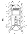
- the capsule medical device 1 includes a casing 2 having a capsule shape in which the two ends of a cylindrical casing body 2a are respectively sealed by a hemispherical transparent window 2b and end plate 2c, an illumination section 3 accommodated in the casing 2 and emitting excitation light through the transparent window 2b, a light-detecting section 4 for detecting fluorescence entering the casing 2 through the transparent window 2b from an examination site outside the casing 2, an image-acquiring section (information-acquiring section) 5 for taking an image of the examination site, an image-generating section 6 for generating image information (examination site information) on the basis of a two-dimensional luminance distribution acquired by the image-acquiring section 5, an antenna (transmitting section) 7 for wirelessly transmitting the generated image information outside the casing 2, a control section (determination section) 8 for controlling these
- the illumination section 3 includes, for example, an LED 3a for emitting wideband light and an excitation light filter 3b for transmitting, of the light emitted from the LED 3a, only light at the excitation wavelength, so that it exits through the transparent window 2b.
- the light-detecting section 4 includes an excitation light cut filter 4a for blocking excitation light and a photodetector 4b for detecting the intensity of fluorescence from which the excitation light is removed.
- the image-acquiring section 5 includes an image-acquisition optical system 5a for collecting fluorescence entering the casing 2 through the transparent window 2b, an excitation light cut filter 5b for blocking excitation light in the collected fluorescence, and an image-acquisition device 5c, such as a high-sensitivity CCD, for acquiring the two-dimensional intensity distribution of the fluorescence from which the excitation light is removed.
- an image-acquisition optical system 5a for collecting fluorescence entering the casing 2 through the transparent window 2b
- an excitation light cut filter 5b for blocking excitation light in the collected fluorescence
- an image-acquisition device 5c such as a high-sensitivity CCD
- the control section 8 is connected to the photodetector 4b, compares fluorescence intensity information transmitted from the photodetector 4b with a predetermined threshold stored in the control section 8 in advance, and, if the fluorescence intensity is higher than the threshold, determines that the image-acquisition device 5c is to be driven to acquire a fluorescence intensity distribution by outputting a drive command signal to the image-acquisition device 5c.
- the control section 8 causes the image-generating section 6 to generate image information from the fluorescence intensity distribution acquired by the image-acquisition device 5c and wirelessly transmits the image information via the antenna 7 outside the casing 2.
- the illumination section 3 is operated to irradiate an examination site with excitation light through the transparent window 2b to excite a fluorescent substance present at the examination site so that it emits fluorescence, which then enters the casing 2 through the transparent window 2b.
- the fluorescence emitted from the fluorescent substance is, for example, fluorescence emitted from a dye that is introduced into the patient by, for example, spraying, intravenous injection, or oral administration in advance before the administration of the capsule medical device 1 to the patient and that is accumulated at the examination site, or autofluorescence emitted from the examination site itself.
- the intensity of the fluorescence entering the casing 2 is detected by the light-detecting section 4 (light-intensity detecting step S1).
- the control section 8 determines whether or not to drive the image-acquisition device 5c on the basis of information about the intensity of the detected fluorescence (determination step S2). The determination is made by the control section 8 on the basis of whether or not the luminance of the fluorescence detected by the light-detecting section 4 exceeds a predetermined threshold.
- the fluorescence intensity exceeds the threshold, it can be determined that a large amount of fluorescent substance is present at the examination site.
- the fluorescence intensity detected is often high where a diseased area is present because a certain fluorescent agent accumulates specifically at a diseased area such as cancer. Accordingly, if the fluorescence intensity is higher than the predetermined threshold, the image-acquisition device 5c can be driven (information-acquiring step S3) to acquire image information at an examination site where a diseased area is probably present. Subsequently, image information is generated by the image-generating section 6 (image-information generating step S4) and is wirelessly transmitted (image-information transmitting step S5).
- the capsule medical device 1 acquires and wirelessly transmits an image only when the fluorescence intensity exceeds a predetermined threshold. This provides the advantage of avoiding acquisition and wireless transmission of unnecessary images with small amounts of information, such as blank images, thus preventing exhaustion of the battery 9. This allows a reduction in the sizes of the battery 9 and the casing 2.
- the amount of image information required to be transmitted outside can be minimized to reduce the amount of image information to be checked later. This provides the advantage of facilitating diagnosis.
- an examination site with high fluorescence intensity can be more reliably selected because the photodetector 4b generally has higher sensitivity than a CCD.
- control section 8 determines whether or not to drive the image-acquisition device 5c on the basis of the fluorescence intensity detected by the light-detecting section 4 in this embodiment, it may instead determine whether or not to wirelessly transmit the generated image information. Because wireless transmission requires a larger amount of power than other processing, exhaustion of the battery 9 can be effectively prevented merely by avoiding wireless transmission of unnecessary image information.
- a timer (examination-time measuring section) 10 for measuring examination time (for example, the elapsed time from introduction into the body cavity) may be provided in the casing 2 and, if the fluorescence intensity detected by the light-detecting section 4 is higher than a predetermined threshold, the control section 8 may wirelessly transmit information about the examination time measured by the timer 10 outside the casing 2 in association with the image information generated by the image-generating section 6.
- the examination site such as an organ, shown in the image information can be easily located.
- fluorescence intensity information may instead be associated with examination time by replacing the image-acquisition device 5c with a photodetector. Because the examination site can be located using examination time, the presence of, for example, the presence a diseased area can be checked merely on the basis of fluorescence intensity information serving as examination site information.
- the configuration can be simplified by the shared use of the photodetector 4b for detecting the fluorescence intensity for determining whether or not to acquire examination site information as the photodetector for detecting the fluorescence intensity serving as examination site information.
- This is also advantageous in that the amount of data is small because the fluorescence intensity information acquired by the photodetector 4b contains no two-dimensional position information, thus demanding a smaller amount of power for acquisition and wireless transmission and a shorter processing time.
- the fluorescence intensity used for determining whether or not to take an image with the image-acquisition device 5c may be the maximum or average value of the fluorescence intensities of the individual pixels acquired by the CCD.
- the area of a region where the fluorescence intensity is at or above a predetermined threshold (that is, the number of pixels having such fluorescence intensities) may be used to determine whether or not to take an image with the image-acquisition device 5c.
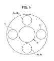
- a fluorescent image is acquired on the basis of the fluorescence intensity detected by the photodetector 4b in this embodiment, as shown in Fig. 6 , it is instead possible to provide white light LEDs 11 having no excitation filter 3b and to acquire a two-dimensional white light image by detecting light reflected from the examination site by the image-acquisition device 5c.
- the excitation light cut filter 5b does not have to be provided in front of the image-acquisition device 5c if the LED 3a having the excitation filter 3b emits excitation light for detection of fluorescence intensity by the photodetector 4b and the light LEDs 11 having no excitation filter 3b are illuminated, whereas the LED 3a having the excitation filter 3b is switched off, for acquisition of a white light image.
- a memory 12 for storing image information or fluorescence intensity information may instead be provided.
- image information or fluorescence intensity information can be stored in the memory 12 in association with examination time to locate the examination site corresponding to the image information or fluorescence intensity information later.
- the determination is carried out by comparing the information about the detected fluorescence luminance with a predetermined threshold in this embodiment, the threshold may instead be changed.
- the threshold may instead be changed.
- the fluorescence intensity of agent fluorescence varies with the elapsed time from administration of the fluorescent agent.
- the fluorescence intensity of fluorescence emitted from the same examination site varies with different elapsed times from administration.
- the timer 10 that starts counting upon administration of the agent and a threshold table 13 storing thresholds in association with elapsed times may be prepared so that the elapsed time can be measured by the timer 10 to switch the threshold depending on the elapsed time.
- an elapsed-time measuring step S11 and a threshold-reading step S12 based on the threshold table 13 may be carried out in addition to the processing in Fig. 3 to set an appropriate threshold for determining whether or not to take an image with the image-acquisition device 5c to more accurately take an image of a region that is suspected of being diseased so that images of unnecessary regions can be prevented from being taken.
- the temporal change in the fluorescence intensity of agent fluorescence varies with the type of fluorescent agent.
- a fluorescent-agent selecting device 15 for selecting the type of fluorescent agent outside the patient's body and wirelessly transmitting information about the selected type of fluorescent agent via an antenna 14 and to provide a receiving section 17 for receiving the information, transmitted wirelessly, about the type of fluorescent agent via an antenna 16 in the capsule medical device 1.
- the threshold table 13 may store thresholds in association with elapsed times for each type of fluorescent agent.
- the information about the type of fluorescent agent is selected and wirelessly transmitted via the antenna 14 by operating the fluorescent-agent selecting device 15 disposed outside the patient's body, and the transmitted information about the type of fluorescent agent is received by the receiving section 17 via the antenna 16 in the capsule medical device 1.
- the control section 8 then reads a threshold stored in the threshold table 13 on the basis of the information, received by the receiving section 17, about the type of fluorescent agent and the elapsed time. This allows appropriate determination as to whether or not to take an image with the image-acquisition device 5c irrespective of the type of fluorescent agent and the elapsed time from administration.
- agent fluorescence intensity of agent fluorescence varies with the type of examination site.
- the behavior of the agent may differ between esophageal cancer (squamous cancer) and colon cancer (adenocarcinoma), and agent fluorescence may also become darker in a low-pH environment such as the stomach.
- it can be more accurately determined whether or not to take an image with the image-acquisition device 5c by identifying the examination site (organ) where the capsule medical device 1 is positioned by some method and setting the threshold depending on the particular examination site.
- An example of the method for identifying the examination site is a method of identification on the basis of the elapsed time from introduction of the capsule medical device 1 into the body cavity X. If the examination site is identified on the basis of the elapsed time, individual patients have differences in the relationship between the elapsed time and the examination site; therefore, if the same patient repeatedly undergoes examination, the relationship between the elapsed time and the examination site may be calibrated on the basis of past examination results for each patient. The relationship between the elapsed time and the examination site may also be calibrated on the basis of data such as the patient's weight.
- a pressure sensor may be provided on a sidewall of the casing body 2a to identify the examination site on the basis of the pressure value detected.
- the examination site can be determined to be a narrow organ such as the esophagus, the small intestine, or the large intestine if the pressure received from the wall surface of the body cavity X is large, and can be determined to be a large organ such as the stomach if the pressure is small.
- an enterobacterium sensor 18 may be provided on the sidewall of the casing body 2a.
- the enterobacterium sensor 18 includes, for example, a pressure chamber 19 set to a predetermined pressure higher than the atmospheric pressure, an azo polymer film 18a that is disposed so as to seal off an opening 19a thereof and that is ruptured by an enzyme secreted by enterobacteria, and a pressure sensor 20 that detects the internal pressure of the pressure chamber 19.
- a drop in the internal pressure of the pressure chamber 19 due to rupture of the azo polymer film 18a can be detected by the pressure sensor 20 etc. to determine that the examination site has shifted to the large intestine.
- a pH sensor may be provided on the casing body to identify the examination site on the basis of the pH value detected.
- the threshold table 13 may store thresholds in association with elapsed times for each examination site. For example, it can be determined on the basis of the output of the pH sensor that the examination site has shifted to the stomach because the stomach is lower in pH value than other organs. The determination threshold can be correspondingly set to accurately determine whether or not to take an image with the image-acquisition device 5c.
- the timer 10, the pressure sensor 20, the enterobacterium sensor 18, and the pH sensor may be used in any combination.
- a dichroic mirror 21 may instead be used to split fluorescence into wavelengths ⁇ 1 and ⁇ 2 for detection of the fluorescence intensities at the wavelengths ⁇ 1 and ⁇ 2.
- excitation with excitation light having a wavelength of 410 nm it is possible to separately detect the intensity at the wavelength of autofluorescence emitted from blood (about 630 nm) and the intensity at the wavelength of autofluorescence emitted from collagen (about 480 nm). It can therefore be more accurately determined that the presence of cancer cells is strongly suspected in the region under examination if the ratio between these autofluorescence intensities exceeds a predetermined threshold.
- a fluorescent agent such as fluorescein, which emits fluorescence having a wavelength of 520 nm, and the intensity at the wavelength of autofluorescence emitted from collagen (about 480 nm), and it can be more accurately determined that the presence of cancer cells is strongly suspected in the region under examination if the ratio between these autofluorescence intensities exceeds a predetermined threshold.
- the capsule medical device 1 includes a device for identifying the examination site where the capsule medical device 1 is positioned, for example, the timer 10, the pressure sensor 20, or the pH sensor
- a capsule medical device system including an external device for locating the capsule medical device 1 outside the patient's body may instead be used.
- the external device can be exemplified by an X-ray device or electromagnetic induction device that identifies the examination site and that transmits information about it, or the corresponding threshold, to the capsule medical device 1.
Landscapes
- Health & Medical Sciences (AREA)
- Life Sciences & Earth Sciences (AREA)
- Surgery (AREA)
- Biomedical Technology (AREA)
- Medical Informatics (AREA)
- Optics & Photonics (AREA)
- Pathology (AREA)
- Radiology & Medical Imaging (AREA)
- Biophysics (AREA)
- Engineering & Computer Science (AREA)
- Physics & Mathematics (AREA)
- Heart & Thoracic Surgery (AREA)
- Nuclear Medicine, Radiotherapy & Molecular Imaging (AREA)
- Molecular Biology (AREA)
- Animal Behavior & Ethology (AREA)
- General Health & Medical Sciences (AREA)
- Public Health (AREA)
- Veterinary Medicine (AREA)
- Endoscopes (AREA)
- Measurement Of The Respiration, Hearing Ability, Form, And Blood Characteristics Of Living Organisms (AREA)
Abstract
Description
- The present invention relates to capsule medical devices, methods for operating the devices, and capsule medical device systems.
- In the related art, there is a known capsule medical device that has a casing having a capsule shape, that is introduced into a patient's body, and that takes an image of the interior of a body cavity to acquire image information (see, for example, Patent Citation 1).
This capsule medical device includes wireless transmission means to transmit the acquired image information toward an external device disposed outside the patient's body. - Patent Citation 1:
Japanese Unexamined Patent Application, Publication No.2003-38424 - However, because the capsule medical device of Patent Citation 1 acquires image information in the body cavity at predetermined time intervals, it needs to acquire a large amount of image information at an increased frame rate to carry out examination over the entire length of the inner wall of the body cavity in the longitudinal direction without missing an affected area. If high-resolution image information is to be acquired, the amount of data thereof is enormous, and accordingly the amount of power required to transmit the data to a memory for storing the data or to the outside is increased, thus resulting in the disadvantage of increasing the size of the battery and therefore the size of the casing of the capsule medical device.
- An object of the present invention, which has been made in light of the circumstances described above, is to provide a capsule medical device, a method for operating the device, and a capsule medical device system capable of acquiring, storing, or transmitting the required minimum amount of examination site information without missing an affected area, thus reducing power consumption, reducing the size of the battery disposed in the capsule, and reducing the size of the casing.
- To achieve the above object, the present invention provides the following solutions.
A first aspect of the present invention is a capsule medical device including a casing having a capsule shape; a light-detecting section, disposed in the casing, for detecting information about the intensity of light from outside the casing; an information-acquiring section for acquiring examination site information from an examination site positioned outside the casing; and a determination section for determining whether or not to acquire, store, or transmit, to the outside, the examination site information acquired by the information-acquiring section on the basis of the information about the intensity of the light detected by the light-detecting section. - According to the above first aspect, after the casing having a capsule shape is introduced into a body cavity, the light-detecting section disposed in the casing detects information about the intensity of light from outside the casing. The determination section then determines whether or not to acquire, store, or transmit, to the outside, examination site information on the basis of the information about the intensity of the detected light. Because the acquisition of the information about the intensity of the light, which is a smaller amount of information, does not require a large amount of power, the need for acquiring, storing, or transmitting, to the outside, the examination site information can be accurately determined with a small amount of power. This avoids the disadvantage of wastefully acquiring, storing, or transmitting, to the outside, the examination site information, which is a larger amount of information, thus preventing battery exhaustion, reducing the size of the battery, and reducing the size of the casing.
- In the above first aspect, the examination site information may be image information from the examination site positioned outside the casing.
Because the acquisition of image information requires a large amount of power, battery exhaustion can be effectively suppressed by avoiding acquisition of unnecessary image information. - In the above first aspect, additionally, the examination site information may be brightness information from the examination site positioned outside the casing.
By doing so, the amount of examination site information can be reduced to reduce the amount of power required for acquisition, storage, and transmission, thus effectively avoiding battery exhaustion. - In the above first aspect, additionally, the determination section may be configured to determine that the examination site information is to be acquired, stored, or transmitted to the outside if the information about the intensity of the light detected by the light-detecting section exceeds a predetermined threshold.
By doing so, if the information about the intensity of the light is lower than the predetermined threshold, it is possible to avoid wasteful processing such as acquiring examination site information that is dark, thus having a small amount of information, or where no diseased area is present, or storing or transmitting, to the outside, such examination site information. This consequently prevents battery exhaustion. - In the above configuration, additionally, an examination-site identifying section for identifying the examination site and a threshold-changing section for changing the threshold depending on the type of examination site identified by the examination-site identifying section may be provided.
By doing so, the determination section can determine whether or not to acquire, store, or transmit, to the outside, the examination site information with respect to the threshold depending on the type of examination site. This avoids acquisition etc. of unnecessary information and therefore prevents battery exhaustion more effectively than uniform determination with respect to a single threshold because the examination site information can be acquired etc. depending on the type of examination site. - In the above configuration, additionally, the examination-site identifying section may include an examination-time measuring section for measuring examination time and an examination-site storing section storing types of examination sites in association with the examination time, and a type of examination site stored in the examination-site storing section may be specified on the basis of the examination time measured by the examination-time measuring section.
- By doing so, the type of examination site stored in the examination-site storing section can be specified on the basis of the examination time measured by the examination-time measuring section to easily select the threshold depending on the type of examination site and accurately determine whether or not to acquire, store, or transmit, to the outside, the examination site information
- In the above first aspect, additionally, the light-detecting section may split the light from outside the casing to detect information about the intensities of the individual split components, and the determination section may execute the determination on the basis of the ratio of the information about the intensities of the individual split components detected by the light-detecting section.
By doing so, it can be accurately determined whether or not an important site such as a diseased area is present at the examination site under examination on the basis of the information about the intensities of the individual split components of the light emitted from the examination site. - In the above first aspect, additionally, the light-detecting section and the information-acquiring section may be constituted by a common light-intensity detecting device.
By doing so, the configuration can be simplified. - In the above configuration, additionally, the light-intensity detecting device may be an image-acquisition device having a plurality of pixels, the light-detecting section may detect the information about the intensity of the light from at least some of the pixels of the image-acquisition device, and the information-acquiring section may acquire an image of the examination site from at least some of the pixels of the image-acquisition device.
By doing so, the information about the intensity of the light can be easily detected using information from some of the pixels of the image-acquisition device capable of image acquisition, thus simplifying the configuration. - In the above configuration, additionally, the determination section may execute the determination on the basis of an average value of information about the intensities of pixels having light intensity information exceeding a predetermined threshold and the number of the pixels.
By doing so, it can be accurately determined whether or not an important site such as a diseased area is present at the examination site under examination using the average value of the information about the intensities of the pixels and the number of the pixels as criteria. - In the above first aspect, additionally, an agent-elapsed-time measuring section for measuring elapsed time from administration of an agent to a patient and a threshold-changing section for changing the threshold on the basis of the elapsed time measured by the agent-elapsed-time measuring section may be provided.
By doing so, it can be more accurately determined whether or not to acquire, store, or transmit, to the outside, the examination site information by taking into account the temporal change of the light emitted from the agent. - In addition, a second aspect of the present invention is a method for operating a capsule medical device that has a casing having a capsule shape and that is introduced into a patient's body, and the method includes a light-intensity detecting step of detecting information about the intensity of light from outside the casing; a determination step of determining whether or not to acquire examination site information from an examination site positioned outside the casing on the basis of the information about the intensity of the light detected in the light-intensity detecting step; and an information-acquiring step of acquiring the examination site information on the basis of a result of the determination in the determination step.
- According to the above second aspect, because it is determined in the determination step whether or not to acquire examination site information from the examination site on the basis of the information about the intensity of the light detected in the light-intensity detecting step, it is possible to avoid the disadvantage of acquiring unnecessary examination site information in the information-acquiring step, thus preventing battery exhaustion and permitting examination over an extended period of time.
- In addition, a third aspect of the present invention is a method for operating a capsule medical device that has a casing having a capsule shape and that is introduced into a patient's body, and the method includes a light-intensity detecting step of detecting information about the intensity of light from outside the casing; an information-acquiring step of acquiring examination site information from an examination site positioned outside the casing; a determination step of determining whether or not to store or transmit, to the outside, the examination site information acquired in the information-acquiring step on the basis of the information about the intensity of the light detected in the light-intensity detecting step; and an information-processing step of storing or transmitting the examination site information on the basis of a result of the determination in the determination step.
- According to the above third aspect, because it is determined in the determination step whether or not to store or transmit, to the outside, the examination site information acquired in the information-acquiring step on the basis of the information about the intensity of the light detected in the light-intensity detecting step, it is possible to avoid the disadvantage of storing or transmitting, to the outside, unnecessary examination site information in the information-processing step, thus preventing battery exhaustion and permitting examination over an extended period of time.
- In addition, a fourth aspect of the present invention is a capsule medical device system including the above capsule medical device and an external device disposed outside the capsule medical device; the external device includes an examination-site identifying section for identifying, from outside the patient's body, the examination site where the capsule medical device is positioned and a transmitting section for transmitting threshold information corresponding to the examination site identified by the examination-site identifying section; the capsule medical device includes a receiving section for receiving the threshold information transmitted from the transmitting section; and the determination section determines that the examination site information is to be acquired, stored, or transmitted to the outside if the information about the intensity of the light detected by the light-detecting section exceeds a threshold corresponding to the threshold information received by the receiving section.
- According to the above fourth aspect, the examination-site identifying section disposed in the external device identifies, from outside the patient's body, the examination site where the capsule medical device is positioned, and the transmitting section transmits the threshold information corresponding to the identified examination site to the capsule medical device. This allows the capsule medical device to determine whether or not to acquire, store, or transmit, to the outside, the examination site information on the basis of the threshold transmitted from the external device without the need for storing thresholds in association with types of examination sites.
- The present invention provides the advantage of acquiring, storing, or transmitting the required minimum amount of examination site information without missing an affected area, thus reducing power consumption, reducing the size of the battery disposed in the capsule, and reducing the size of the casing.
-
- [
FIG. 1] Fig. 1 is a longitudinal sectional view showing a capsule medical device according to an embodiment of the present invention. - [
FIG. 2] Fig. 2 is a block diagram showing the internal structure of the capsule medical device inFig. 1 . - [
FIG. 3] Fig. 3 is a flowchart showing a method for operating the capsule medical device inFig. 1 . - [
FIG. 4] Fig. 4 is a block diagram showing a first modification of the capsule medical device inFig. 1 . - [
FIG. 5] Fig. 5 is a graph showing an example of a determination method for a second modification of the capsule medical device inFig. 1 . - [
FIG. 6] Fig. 6 is a front view showing an arrangement of LEDs and a photodiode in a third modification of the capsule medical device inFig. 1 . - [
FIG. 7] Fig. 7 is a longitudinal sectional view showing a fourth modification of the capsule medical device inFig. 1 . - [
FIG. 8] Fig. 8 is a graph showing the relationship between the elapsed time from the administration of a fluorescent agent and the intensity of agent fluorescence. - [
FIG. 9] Fig. 9 is a block diagram showing a fifth modification of the capsule medical device inFig. 1 . - [
FIG. 10] Fig. 10 is a flowchart showing a method for operating the capsule medical device inFig. 9 . - [
FIG. 11] Fig. 11 is a block diagram showing a sixth modification of the capsule medical device inFig. 1 . - [
FIG. 12] Fig. 12 is a longitudinal sectional view showing a seventh modification of the capsule medical device inFig. 1 . - [
FIG. 13] Fig. 13 is a block diagram showing an eighth modification of the capsule medical device inFig. 1 . - [
FIG. 14] Fig. 14 is a graph showing an example of a determination method for the capsule medical device inFig. 13 . - [
FIG. 15] Fig. 15 is a graph showing another determination method for the capsule medical device inFig. 13 . -
- S1: light-intensity detecting step
- S2: determination step
- S3: information-acquiring step
- S4: image-information generating step (information-processing step)
- 1: capsule medical device
- 2: casing
- 4b: photodetector (light-detecting section: light-intensity detecting device)
- 5c: image-acquisition device (information-acquiring section: light-intensity detecting device)
- 8: control section (determination section: threshold-changing section)
- 10: timer (examination-site identifying section: examination-time measuring section: agent-elapsed-time measuring section) 13: threshold table (examination-site storing section)
- 18: enterobacterium sensor (examination-site identifying section)
- 20: pressure sensor (examination-site identifying section)
- A capsule
medical device 1 and a method for operating thedevice 1 according to an embodiment of the present invention will be described below with reference toFigs. 1 to 3 .
As shown inFig. 1 , the capsulemedical device 1 according to this embodiment includes acasing 2 having a capsule shape in which the two ends of acylindrical casing body 2a are respectively sealed by a hemisphericaltransparent window 2b andend plate 2c, anillumination section 3 accommodated in thecasing 2 and emitting excitation light through thetransparent window 2b, a light-detectingsection 4 for detecting fluorescence entering thecasing 2 through thetransparent window 2b from an examination site outside thecasing 2, an image-acquiring section (information-acquiring section) 5 for taking an image of the examination site, an image-generatingsection 6 for generating image information (examination site information) on the basis of a two-dimensional luminance distribution acquired by the image-acquiringsection 5, an antenna (transmitting section) 7 for wirelessly transmitting the generated image information outside thecasing 2, a control section (determination section) 8 for controlling these, and abattery 9 for supplying power to the individual sections. Wiring from thebattery 9 to the individual sections is not shown. - The
illumination section 3 includes, for example, anLED 3a for emitting wideband light and anexcitation light filter 3b for transmitting, of the light emitted from theLED 3a, only light at the excitation wavelength, so that it exits through thetransparent window 2b.
The light-detectingsection 4 includes an excitation light cutfilter 4a for blocking excitation light and aphotodetector 4b for detecting the intensity of fluorescence from which the excitation light is removed. - The image-acquiring
section 5 includes an image-acquisitionoptical system 5a for collecting fluorescence entering thecasing 2 through thetransparent window 2b, an excitation light cutfilter 5b for blocking excitation light in the collected fluorescence, and an image-acquisition device 5c, such as a high-sensitivity CCD, for acquiring the two-dimensional intensity distribution of the fluorescence from which the excitation light is removed. - As shown in
Fig. 2 , thecontrol section 8 is connected to thephotodetector 4b, compares fluorescence intensity information transmitted from thephotodetector 4b with a predetermined threshold stored in thecontrol section 8 in advance, and, if the fluorescence intensity is higher than the threshold, determines that the image-acquisition device 5c is to be driven to acquire a fluorescence intensity distribution by outputting a drive command signal to the image-acquisition device 5c.
In addition, thecontrol section 8 causes the image-generatingsection 6 to generate image information from the fluorescence intensity distribution acquired by the image-acquisition device 5c and wirelessly transmits the image information via theantenna 7 outside thecasing 2. - The method for operating the thus-configured capsule
medical device 1 according to this embodiment will now be described.
With the capsulemedical device 1 according to this embodiment introduced into a patient's body cavity X, theillumination section 3 is operated to irradiate an examination site with excitation light through thetransparent window 2b to excite a fluorescent substance present at the examination site so that it emits fluorescence, which then enters thecasing 2 through thetransparent window 2b.
The fluorescence emitted from the fluorescent substance is, for example, fluorescence emitted from a dye that is introduced into the patient by, for example, spraying, intravenous injection, or oral administration in advance before the administration of the capsulemedical device 1 to the patient and that is accumulated at the examination site, or autofluorescence emitted from the examination site itself. - The intensity of the fluorescence entering the
casing 2 is detected by the light-detecting section 4 (light-intensity detecting step S1). Thecontrol section 8 then determines whether or not to drive the image-acquisition device 5c on the basis of information about the intensity of the detected fluorescence (determination step S2). The determination is made by thecontrol section 8 on the basis of whether or not the luminance of the fluorescence detected by the light-detectingsection 4 exceeds a predetermined threshold. - If the fluorescence intensity exceeds the threshold, it can be determined that a large amount of fluorescent substance is present at the examination site. The fluorescence intensity detected is often high where a diseased area is present because a certain fluorescent agent accumulates specifically at a diseased area such as cancer. Accordingly, if the fluorescence intensity is higher than the predetermined threshold, the image-
acquisition device 5c can be driven (information-acquiring step S3) to acquire image information at an examination site where a diseased area is probably present. Subsequently, image information is generated by the image-generating section 6 (image-information generating step S4) and is wirelessly transmitted (image-information transmitting step S5). - Thus, the capsule
medical device 1 according to this embodiment acquires and wirelessly transmits an image only when the fluorescence intensity exceeds a predetermined threshold. This provides the advantage of avoiding acquisition and wireless transmission of unnecessary images with small amounts of information, such as blank images, thus preventing exhaustion of thebattery 9. This allows a reduction in the sizes of thebattery 9 and thecasing 2. - In addition, the amount of image information required to be transmitted outside can be minimized to reduce the amount of image information to be checked later. This provides the advantage of facilitating diagnosis.
In addition, an examination site with high fluorescence intensity can be more reliably selected because thephotodetector 4b generally has higher sensitivity than a CCD. - Although the
control section 8 determines whether or not to drive the image-acquisition device 5c on the basis of the fluorescence intensity detected by the light-detectingsection 4 in this embodiment, it may instead determine whether or not to wirelessly transmit the generated image information. Because wireless transmission requires a larger amount of power than other processing, exhaustion of thebattery 9 can be effectively prevented merely by avoiding wireless transmission of unnecessary image information. - In this embodiment, additionally, as shown in
Fig. 4 , a timer (examination-time measuring section) 10 for measuring examination time (for example, the elapsed time from introduction into the body cavity) may be provided in thecasing 2 and, if the fluorescence intensity detected by the light-detectingsection 4 is higher than a predetermined threshold, thecontrol section 8 may wirelessly transmit information about the examination time measured by thetimer 10 outside thecasing 2 in association with the image information generated by the image-generatingsection 6. By measuring the examination time, the examination site, such as an organ, shown in the image information can be easily located. - In this case, although the image information acquired by the image-
acquisition device 5c is associated with examination time in this embodiment, fluorescence intensity information may instead be associated with examination time by replacing the image-acquisition device 5c with a photodetector. Because the examination site can be located using examination time, the presence of, for example, the presence a diseased area can be checked merely on the basis of fluorescence intensity information serving as examination site information. - In this case, additionally, the configuration can be simplified by the shared use of the
photodetector 4b for detecting the fluorescence intensity for determining whether or not to acquire examination site information as the photodetector for detecting the fluorescence intensity serving as examination site information. This is also advantageous in that the amount of data is small because the fluorescence intensity information acquired by thephotodetector 4b contains no two-dimensional position information, thus demanding a smaller amount of power for acquisition and wireless transmission and a shorter processing time. - In addition, a CCD may be shared instead of sharing the
photodetector 4b. In this case, the fluorescence intensity used for determining whether or not to take an image with the image-acquisition device 5c may be the maximum or average value of the fluorescence intensities of the individual pixels acquired by the CCD.
In this case, in addition to the maximum or average value of the fluorescence intensities of the individual pixels, the area of a region where the fluorescence intensity is at or above a predetermined threshold (that is, the number of pixels having such fluorescence intensities) may be used to determine whether or not to take an image with the image-acquisition device 5c. By doing so, as shown inFig. 5 , it is possible to prevent acquisition, due to noise, of an image of an examination site having a relatively narrow area and a high fluorescence intensity (reference B inFig. 5 ), thus more reliably acquiring an image of an examination site that is suspected of being diseased (reference A inFig. 5 ). - In addition, although a fluorescent image is acquired on the basis of the fluorescence intensity detected by the
photodetector 4b in this embodiment, as shown inFig. 6 , it is instead possible to providewhite light LEDs 11 having noexcitation filter 3b and to acquire a two-dimensional white light image by detecting light reflected from the examination site by the image-acquisition device 5c. In this case, the excitation light cutfilter 5b does not have to be provided in front of the image-acquisition device 5c if theLED 3a having theexcitation filter 3b emits excitation light for detection of fluorescence intensity by thephotodetector 4b and thelight LEDs 11 having noexcitation filter 3b are illuminated, whereas theLED 3a having theexcitation filter 3b is switched off, for acquisition of a white light image. - In addition, although the case where the
antenna 7 for wirelessly transmitting the acquired image information outside thecasing 2 is provided has been described in this embodiment, as shown inFig. 7 , amemory 12 for storing image information or fluorescence intensity information may instead be provided. Again, in this case, image information or fluorescence intensity information can be stored in thememory 12 in association with examination time to locate the examination site corresponding to the image information or fluorescence intensity information later. - In addition, although the determination is carried out by comparing the information about the detected fluorescence luminance with a predetermined threshold in this embodiment, the threshold may instead be changed.
For example, as shown inFig. 8 , the fluorescence intensity of agent fluorescence varies with the elapsed time from administration of the fluorescent agent. Hence, the fluorescence intensity of fluorescence emitted from the same examination site varies with different elapsed times from administration. - Accordingly, as shown in
Fig. 9 , thetimer 10 that starts counting upon administration of the agent and a threshold table 13 storing thresholds in association with elapsed times may be prepared so that the elapsed time can be measured by thetimer 10 to switch the threshold depending on the elapsed time. In this case, as shown inFig. 10 , an elapsed-time measuring step S11 and a threshold-reading step S12 based on the threshold table 13 may be carried out in addition to the processing inFig. 3 to set an appropriate threshold for determining whether or not to take an image with the image-acquisition device 5c to more accurately take an image of a region that is suspected of being diseased so that images of unnecessary regions can be prevented from being taken. - As shown in
Fig. 8 , additionally, the temporal change in the fluorescence intensity of agent fluorescence varies with the type of fluorescent agent. Accordingly, as shown inFig. 11 , it is possible to use a fluorescent-agent selecting device 15 for selecting the type of fluorescent agent outside the patient's body and wirelessly transmitting information about the selected type of fluorescent agent via an antenna 14 and to provide a receivingsection 17 for receiving the information, transmitted wirelessly, about the type of fluorescent agent via anantenna 16 in the capsulemedical device 1. In addition, the threshold table 13 may store thresholds in association with elapsed times for each type of fluorescent agent. - By doing so, the information about the type of fluorescent agent is selected and wirelessly transmitted via the antenna 14 by operating the fluorescent-
agent selecting device 15 disposed outside the patient's body, and the transmitted information about the type of fluorescent agent is received by the receivingsection 17 via theantenna 16 in the capsulemedical device 1. Thecontrol section 8 then reads a threshold stored in the threshold table 13 on the basis of the information, received by the receivingsection 17, about the type of fluorescent agent and the elapsed time. This allows appropriate determination as to whether or not to take an image with the image-acquisition device 5c irrespective of the type of fluorescent agent and the elapsed time from administration. - In addition, the fluorescence intensity of agent fluorescence varies with the type of examination site. For example, the behavior of the agent may differ between esophageal cancer (squamous cancer) and colon cancer (adenocarcinoma), and agent fluorescence may also become darker in a low-pH environment such as the stomach. Hence, it can be more accurately determined whether or not to take an image with the image-
acquisition device 5c by identifying the examination site (organ) where the capsulemedical device 1 is positioned by some method and setting the threshold depending on the particular examination site. - An example of the method for identifying the examination site is a method of identification on the basis of the elapsed time from introduction of the capsule
medical device 1 into the body cavity X.
If the examination site is identified on the basis of the elapsed time, individual patients have differences in the relationship between the elapsed time and the examination site; therefore, if the same patient repeatedly undergoes examination, the relationship between the elapsed time and the examination site may be calibrated on the basis of past examination results for each patient. The relationship between the elapsed time and the examination site may also be calibrated on the basis of data such as the patient's weight. - In addition, a pressure sensor may be provided on a sidewall of the
casing body 2a to identify the examination site on the basis of the pressure value detected. For example, the examination site can be determined to be a narrow organ such as the esophagus, the small intestine, or the large intestine if the pressure received from the wall surface of the body cavity X is large, and can be determined to be a large organ such as the stomach if the pressure is small. - In addition, as shown in
Fig. 12 , anenterobacterium sensor 18 may be provided on the sidewall of thecasing body 2a. Theenterobacterium sensor 18 includes, for example, apressure chamber 19 set to a predetermined pressure higher than the atmospheric pressure, anazo polymer film 18a that is disposed so as to seal off anopening 19a thereof and that is ruptured by an enzyme secreted by enterobacteria, and apressure sensor 20 that detects the internal pressure of thepressure chamber 19. A drop in the internal pressure of thepressure chamber 19 due to rupture of theazo polymer film 18a can be detected by thepressure sensor 20 etc. to determine that the examination site has shifted to the large intestine. - In addition, a pH sensor may be provided on the casing body to identify the examination site on the basis of the pH value detected. In addition, the threshold table 13 may store thresholds in association with elapsed times for each examination site.
For example, it can be determined on the basis of the output of the pH sensor that the examination site has shifted to the stomach because the stomach is lower in pH value than other organs. The determination threshold can be correspondingly set to accurately determine whether or not to take an image with the image-acquisition device 5c.
In addition, thetimer 10, thepressure sensor 20, theenterobacterium sensor 18, and the pH sensor may be used in any combination. - In addition, although the
photodetector 4b is used to detect fluorescence intensity in this embodiment, as shown inFig. 13 , adichroic mirror 21 may instead be used to split fluorescence into wavelengths λ1 and λ2 for detection of the fluorescence intensities at the wavelengths λ1 and λ2. By doing so, as shown inFig. 14 , for excitation with excitation light having a wavelength of 410 nm, it is possible to separately detect the intensity at the wavelength of autofluorescence emitted from blood (about 630 nm) and the intensity at the wavelength of autofluorescence emitted from collagen (about 480 nm). It can therefore be more accurately determined that the presence of cancer cells is strongly suspected in the region under examination if the ratio between these autofluorescence intensities exceeds a predetermined threshold. - In addition, as shown in
Fig. 15 , for excitation with excitation light having a wavelength of 410 nm, it is possible to separately detect the fluorescence intensity of a fluorescent agent such as fluorescein, which emits fluorescence having a wavelength of 520 nm, and the intensity at the wavelength of autofluorescence emitted from collagen (about 480 nm), and it can be more accurately determined that the presence of cancer cells is strongly suspected in the region under examination if the ratio between these autofluorescence intensities exceeds a predetermined threshold. - In addition, although the capsule
medical device 1 according to this embodiment includes a device for identifying the examination site where the capsulemedical device 1 is positioned, for example, thetimer 10, thepressure sensor 20, or the pH sensor, a capsule medical device system including an external device (not shown) for locating the capsulemedical device 1 outside the patient's body may instead be used. The external device can be exemplified by an X-ray device or electromagnetic induction device that identifies the examination site and that transmits information about it, or the corresponding threshold, to the capsulemedical device 1.
Claims (14)
- A capsule medical device comprising:a casing having a capsule shape;a light-detecting section, disposed in the casing, for detecting information about the intensity of light from outside the casing;an information-acquiring section for acquiring examination site information from an examination site positioned outside the casing; anda determination section for determining whether or not to acquire, store, or transmit, to the outside, the examination site information acquired by the information-acquiring section on the basis of the information about the intensity of the light detected by the light-detecting section.
- The capsule medical device according to Claim 1, wherein the examination site information is image information from the examination site positioned outside the casing.
- The capsule medical device according to Claim 1, wherein the examination site information is brightness information from the examination site positioned outside the casing.
- The capsule medical device according to Claim 1, wherein the determination section determines that the examination site information is to be acquired, stored, or transmitted to the outside if the information about the intensity of the light detected by the light-detecting section exceeds a predetermined threshold.
- The capsule medical device according to Claim 4, further comprising:an examination-site identifying section for identifying the examination site; anda threshold-changing section for changing the threshold depending on the type of examination site identified by the examination-site identifying section.
- The capsule medical device according to Claim 5, wherein
the examination-site identifying section includes an examination-time measuring section for measuring examination time and an examination-site storing section storing types of examination sites in association with the examination time; and
a type of examination site stored in the examination-site storing section is specified on the basis of the examination time measured by the examination-time measuring section. - The capsule medical device according to Claim 1, wherein
the light-detecting section splits the light from outside the casing to detect information about the intensities of the individual split components; and
the determination section executes the determination on the basis of the ratio of the information about the intensities of the individual split components detected by the light-detecting section. - The capsule medical device according to Claim 1, wherein the light-detecting section and the information-acquiring section comprise a common light-intensity detecting device.
- The capsule medical device according to Claim 8, wherein
the light-intensity detecting device is an image-acquisition device having a plurality of pixels;
the light-detecting section detects the information about the intensity of the light from at least some of the pixels of the image-acquisition device; and
the information-acquiring section acquires an image of the examination site from at least some of the pixels of the image-acquisition device. - The capsule medical device according to Claim 9, wherein the determination section executes the determination on the basis of an average value of information about the intensities of pixels having light intensity information exceeding a predetermined threshold and the number of the pixels.
- The capsule medical device according to one of Claims 4 to 6, further comprising:an agent-elapsed-time measuring section for measuring elapsed time from administration of an agent to a patient; anda threshold-changing section for changing the threshold on the basis of the elapsed time measured by the agent-elapsed-time measuring section.
- A method for operating a capsule medical device that has a casing having a capsule shape and that is introduced into a patient's body, the method comprising:a light-intensity detecting step of detecting information about the intensity of light from outside the casing;a determination step of determining whether or not to acquire examination site information from an examination site positioned outside the casing on the basis of the information about the intensity of the light detected in the light-intensity detecting step; andan information-acquiring step of acquiring the examination site information on the basis of a result of the determination in the determination step.
- A method for operating a capsule medical device that has a casing having a capsule shape and that is introduced into a patient's body, the method comprising:a light-intensity detecting step of detecting information about the intensity of light from outside the casing;an information-acquiring step of acquiring examination site information from an examination site positioned outside the casing;a determination step of determining whether or not to store or transmit, to the outside, the examination site information acquired in the information-acquiring step on the basis of the information about the intensity of the light detected in the light-intensity detecting step; andan information-processing step of storing or transmitting the examination site information on the basis of a result of the determination in the determination step.
- A capsule medical device system comprising the capsule medical device according to Claim 1 and an external device disposed outside the capsule medical device, wherein
the external device includes an examination-site identifying section for identifying, from outside the patient's body, the examination site where the capsule medical device is positioned and a transmitting section for transmitting threshold information corresponding to the examination site identified by the examination-site identifying section;
the capsule medical device includes a receiving section for receiving the threshold information transmitted from the transmitting section; and
the determination section determines that the examination site information is to be acquired, stored, or transmitted to the outside if the information about the intensity of the light detected by the light-detecting section exceeds a threshold corresponding to the threshold information received by the receiving section.
Applications Claiming Priority (2)
| Application Number | Priority Date | Filing Date | Title |
|---|---|---|---|
| JP2008076337A JP5336749B2 (en) | 2008-03-24 | 2008-03-24 | Capsule type medical device and operation method thereof |
| PCT/JP2009/055045 WO2009119366A1 (en) | 2008-03-24 | 2009-03-16 | Capsule medical device, method for operating same, and capsule medical device system |
Publications (2)
| Publication Number | Publication Date |
|---|---|
| EP2255717A1 true EP2255717A1 (en) | 2010-12-01 |
| EP2255717A4 EP2255717A4 (en) | 2014-07-09 |
Family
ID=41113563
Family Applications (1)
| Application Number | Title | Priority Date | Filing Date |
|---|---|---|---|
| EP09724627.6A Withdrawn EP2255717A4 (en) | 2008-03-24 | 2009-03-16 | Capsule medical device, method for operating same, and capsule medical device system |
Country Status (5)
| Country | Link |
|---|---|
| US (1) | US8328713B2 (en) |
| EP (1) | EP2255717A4 (en) |
| JP (1) | JP5336749B2 (en) |
| CN (1) | CN101977542B (en) |
| WO (1) | WO2009119366A1 (en) |
Cited By (2)
| Publication number | Priority date | Publication date | Assignee | Title |
|---|---|---|---|---|
| US9839359B2 (en) | 2013-08-23 | 2017-12-12 | Olympus Corporation | Fluorescence observation apparatus |
| US11343428B2 (en) | 2019-02-07 | 2022-05-24 | Olympus Corporation | Imaging apparatus and recording medium operating in a first mode or a second mode based on determined brightness of an image |
Families Citing this family (5)
| Publication number | Priority date | Publication date | Assignee | Title |
|---|---|---|---|---|
| JP5669416B2 (en) * | 2010-03-23 | 2015-02-12 | オリンパス株式会社 | Fluorescence observation equipment |
| JP6005918B2 (en) * | 2011-09-08 | 2016-10-12 | オリンパス株式会社 | Fluorescence observation equipment |
| CN108697314B (en) | 2016-01-28 | 2021-04-06 | 基文影像公司 | Method and system for controlling image capture and transmission rates in an in vivo device |
| CN113164007B (en) * | 2018-12-10 | 2024-07-05 | 奥林巴斯株式会社 | Image recording device, image recording method, and endoscope system |
| CN116712017A (en) * | 2023-07-18 | 2023-09-08 | 欧阳宏 | A universal method for monitoring and recording working air pressure of digestive endoscopes |
Family Cites Families (22)
| Publication number | Priority date | Publication date | Assignee | Title |
|---|---|---|---|---|
| ATE509328T1 (en) * | 2001-03-14 | 2011-05-15 | Given Imaging Ltd | METHOD AND SYSTEM FOR DETECTING COLORIMETRIC ANOMALIES |
| JP2002345739A (en) * | 2001-05-25 | 2002-12-03 | Fuji Photo Film Co Ltd | Image display device |
| US20030117491A1 (en) * | 2001-07-26 | 2003-06-26 | Dov Avni | Apparatus and method for controlling illumination in an in-vivo imaging device |
| US20060184039A1 (en) * | 2001-07-26 | 2006-08-17 | Dov Avni | Apparatus and method for light control in an in-vivo imaging device |
| JP4744026B2 (en) | 2001-07-30 | 2011-08-10 | オリンパス株式会社 | Capsule endoscope and capsule endoscope system |
| JP4643089B2 (en) * | 2001-09-27 | 2011-03-02 | オリンパス株式会社 | Capsule medical device |
| US20030216622A1 (en) * | 2002-04-25 | 2003-11-20 | Gavriel Meron | Device and method for orienting a device in vivo |
| JP2006512109A (en) * | 2002-08-01 | 2006-04-13 | ザ ジョンズ ホプキンズ ユニバーシティ | Identification technology of molecular structure using fluorescent light emission, and treatment technology of cell types lined inside the body lumen |
| US7118529B2 (en) * | 2002-11-29 | 2006-10-10 | Given Imaging, Ltd. | Method and apparatus for transmitting non-image information via an image sensor in an in vivo imaging system |
| US20060155174A1 (en) * | 2002-12-16 | 2006-07-13 | Arkady Glukhovsky | Device, system and method for selective activation of in vivo sensors |
| JP4383134B2 (en) * | 2003-09-04 | 2009-12-16 | オリンパス株式会社 | Wireless in-vivo information acquisition device |
| JP3993546B2 (en) * | 2003-09-08 | 2007-10-17 | オリンパス株式会社 | In-subject introduction apparatus and wireless in-subject information acquisition system |
| US8021356B2 (en) * | 2003-09-29 | 2011-09-20 | Olympus Corporation | Capsule medication administration system, medication administration method using capsule medication administration system, control method for capsule medication administration system |
| JP4868720B2 (en) * | 2004-05-27 | 2012-02-01 | オリンパス株式会社 | Capsule dosing system |
| EP1681009A4 (en) * | 2003-10-27 | 2010-08-11 | Olympus Corp | Image processing device, image processing method, and image processing program |
| JP4445799B2 (en) * | 2004-05-24 | 2010-04-07 | オリンパス株式会社 | Intra-subject introduction device and medical device |
| JP5057651B2 (en) * | 2005-02-15 | 2012-10-24 | オリンパス株式会社 | Lumen image processing apparatus, lumen image processing method, and program therefor |
| EP1849402B1 (en) * | 2005-02-15 | 2018-05-16 | Olympus Corporation | Medical image processing device, lumen image processing device, lumen image processing method, and programs for them |
| US8449458B2 (en) * | 2005-02-25 | 2013-05-28 | Olympus Corporation | Body-insertable apparatus and radio in-vivo information acquiring system |
| JP2007082664A (en) * | 2005-09-21 | 2007-04-05 | Fujifilm Corp | Capsule endoscope |
| JP2009112580A (en) * | 2007-11-07 | 2009-05-28 | Hoya Corp | Operation switching mechanism |
| US9017248B2 (en) * | 2007-11-08 | 2015-04-28 | Olympus Medical Systems Corp. | Capsule blood detection system and method |
-
2008
- 2008-03-24 JP JP2008076337A patent/JP5336749B2/en not_active Expired - Fee Related
-
2009
- 2009-03-16 EP EP09724627.6A patent/EP2255717A4/en not_active Withdrawn
- 2009-03-16 WO PCT/JP2009/055045 patent/WO2009119366A1/en not_active Ceased
- 2009-03-16 CN CN2009801103996A patent/CN101977542B/en not_active Expired - Fee Related
-
2010
- 2010-08-25 US US12/868,239 patent/US8328713B2/en active Active
Cited By (2)
| Publication number | Priority date | Publication date | Assignee | Title |
|---|---|---|---|---|
| US9839359B2 (en) | 2013-08-23 | 2017-12-12 | Olympus Corporation | Fluorescence observation apparatus |
| US11343428B2 (en) | 2019-02-07 | 2022-05-24 | Olympus Corporation | Imaging apparatus and recording medium operating in a first mode or a second mode based on determined brightness of an image |
Also Published As
| Publication number | Publication date |
|---|---|
| WO2009119366A1 (en) | 2009-10-01 |
| US8328713B2 (en) | 2012-12-11 |
| CN101977542B (en) | 2013-06-19 |
| EP2255717A4 (en) | 2014-07-09 |
| CN101977542A (en) | 2011-02-16 |
| JP2009226065A (en) | 2009-10-08 |
| JP5336749B2 (en) | 2013-11-06 |
| US20100324371A1 (en) | 2010-12-23 |
Similar Documents
| Publication | Publication Date | Title |
|---|---|---|
| US8328713B2 (en) | Capsule medical device, method for operating the same, and capsule medical device system | |
| CN101621956B (en) | Intrasubject introduction system | |
| US9906739B2 (en) | Image pickup device and image pickup method | |
| US9265409B2 (en) | Capsule medical device and capsule medical system | |
| US8082024B2 (en) | Micro-scale compact device for in vivo medical diagnosis combining optical imaging and point fluorescence spectroscopy | |
| EP2356935B1 (en) | Image-processing system, imaging device, receiving device, and image display device | |
| EP2201888B1 (en) | Endoscopic apparatus and setting method thereof | |
| US9155456B2 (en) | In vivo examination apparatus and capsule endoscope | |
| US20060052710A1 (en) | Endoscope apparatus and fluorescence detection method using endoscope apparatus | |
| US20140085686A1 (en) | Fluorescence observation device | |
| EP2412292A1 (en) | Capsule medical device and capsule medical system | |
| US20020103417A1 (en) | Endoscopic smart probe and method | |
| US20050027166A1 (en) | Endoscope system for fluorescent observation | |
| EP3028622A1 (en) | Imaging device | |
| US20030153811A1 (en) | Fluorescence endoscope with inserted/retracted short-pass filter | |
| JP5331394B2 (en) | Endoscope device | |
| JP4383134B2 (en) | Wireless in-vivo information acquisition device | |
| JP2002078670A (en) | Fluorescent imaging system | |
| EP2255718A1 (en) | Capsule medical device | |
| DK202170262A1 (en) | Medical imaging system and method for fluorescence enhanced medical imaging |
Legal Events
| Date | Code | Title | Description |
|---|---|---|---|
| PUAI | Public reference made under article 153(3) epc to a published international application that has entered the european phase |
Free format text: ORIGINAL CODE: 0009012 |
|
| 17P | Request for examination filed |
Effective date: 20100830 |
|
| AK | Designated contracting states |
Kind code of ref document: A1 Designated state(s): AT BE BG CH CY CZ DE DK EE ES FI FR GB GR HR HU IE IS IT LI LT LU LV MC MK MT NL NO PL PT RO SE SI SK TR |
|
| AX | Request for extension of the european patent |
Extension state: AL BA RS |
|
| DAX | Request for extension of the european patent (deleted) | ||
| A4 | Supplementary search report drawn up and despatched |
Effective date: 20140611 |
|
| RIC1 | Information provided on ipc code assigned before grant |
Ipc: A61B 1/00 20060101AFI20140605BHEP Ipc: A61B 5/07 20060101ALI20140605BHEP Ipc: A61B 1/04 20060101ALN20140605BHEP |
|
| RAP1 | Party data changed (applicant data changed or rights of an application transferred) |
Owner name: OLYMPUS CORPORATION |
|
| RIN1 | Information on inventor provided before grant (corrected) |
Inventor name: ISHIHARA, YASUSHIGE |
|
| STAA | Information on the status of an ep patent application or granted ep patent |
Free format text: STATUS: THE APPLICATION HAS BEEN WITHDRAWN |
|
| 18W | Application withdrawn |
Effective date: 20170223 |