EP2250507B1 - Diagnostic methods for metastasis - Google Patents
Diagnostic methods for metastasis Download PDFInfo
- Publication number
- EP2250507B1 EP2250507B1 EP09701520A EP09701520A EP2250507B1 EP 2250507 B1 EP2250507 B1 EP 2250507B1 EP 09701520 A EP09701520 A EP 09701520A EP 09701520 A EP09701520 A EP 09701520A EP 2250507 B1 EP2250507 B1 EP 2250507B1
- Authority
- EP
- European Patent Office
- Prior art keywords
- seq
- proteins
- protein
- metastasis
- patient
- Prior art date
- Legal status (The legal status is an assumption and is not a legal conclusion. Google has not performed a legal analysis and makes no representation as to the accuracy of the status listed.)
- Not-in-force
Links
Images

Classifications
-
- G—PHYSICS
- G01—MEASURING; TESTING
- G01N—INVESTIGATING OR ANALYSING MATERIALS BY DETERMINING THEIR CHEMICAL OR PHYSICAL PROPERTIES
- G01N33/00—Investigating or analysing materials by specific methods not covered by groups G01N1/00 - G01N31/00
- G01N33/48—Biological material, e.g. blood, urine; Haemocytometers
- G01N33/50—Chemical analysis of biological material, e.g. blood, urine; Testing involving biospecific ligand binding methods; Immunological testing
- G01N33/53—Immunoassay; Biospecific binding assay; Materials therefor
- G01N33/575—Immunoassay; Biospecific binding assay; Materials therefor for cancer
- G01N33/57515—Immunoassay; Biospecific binding assay; Materials therefor for cancer of the breast
-
- A—HUMAN NECESSITIES
- A61—MEDICAL OR VETERINARY SCIENCE; HYGIENE
- A61P—SPECIFIC THERAPEUTIC ACTIVITY OF CHEMICAL COMPOUNDS OR MEDICINAL PREPARATIONS
- A61P35/00—Antineoplastic agents
- A61P35/04—Antineoplastic agents specific for metastasis
-
- G—PHYSICS
- G01—MEASURING; TESTING
- G01N—INVESTIGATING OR ANALYSING MATERIALS BY DETERMINING THEIR CHEMICAL OR PHYSICAL PROPERTIES
- G01N2800/00—Detection or diagnosis of diseases
- G01N2800/56—Staging of a disease; Further complications associated with the disease
-
- Y—GENERAL TAGGING OF NEW TECHNOLOGICAL DEVELOPMENTS; GENERAL TAGGING OF CROSS-SECTIONAL TECHNOLOGIES SPANNING OVER SEVERAL SECTIONS OF THE IPC; TECHNICAL SUBJECTS COVERED BY FORMER USPC CROSS-REFERENCE ART COLLECTIONS [XRACs] AND DIGESTS
- Y10—TECHNICAL SUBJECTS COVERED BY FORMER USPC
- Y10T—TECHNICAL SUBJECTS COVERED BY FORMER US CLASSIFICATION
- Y10T436/00—Chemistry: analytical and immunological testing
- Y10T436/14—Heterocyclic carbon compound [i.e., O, S, N, Se, Te, as only ring hetero atom]
- Y10T436/142222—Hetero-O [e.g., ascorbic acid, etc.]
- Y10T436/143333—Saccharide [e.g., DNA, etc.]
Definitions
- the present invention relates to the prediction, prognosis or diagnosis of lymph node metastasis in breast cancer patients.
- lymph node (LN) metastasis is a major determinant for the staging and the prognosis of most human malignancies and often guides therapeutic decisions.
- axillary lymph node involvement for breast cancer, in current clinical practice, axillary lymph node involvement, age of the patient, tumour size, pathologic grade and hormone receptor status are considered as the main prognostic and/or predictive factors ( Dikicioglu E. et al. 2005, Int.J.Clin.Pract. 59:1039-1044 ). Involvement of lymph nodes and the number of lymph nodes harbouring metastases have an inverse relationship with the disease prognosis, meaning that patients with lymph nodes free of metastases have a better outcome ( Weigelt B. et al. 2004 Br.J.Cancer 90:1531-1537 ).
- SLNB sentinel lymph node biopsy
- lymph node involvement remains a critical benchmark in cancers such as breast cancer and is often the earliest sign of tumour progression, insights into the underlying molecular mechanisms are essential. Lymph node metastasis is a complex series of events involving the generation of new blood vessels, growth, invasion with breakdown of the host matrix, transport to other sites with adhesion and subsequent invasion ( Shinozaki M. 2005 Clin. Cancer Res. 11:2156-2162 ). If the lymph node status can be predicted from primary cancer tissue, axillary surgery can be avoided in lymph node negative patients and for patients with isolated lymphatic drainage to non-axillary lymph nodes it will be the only way for accurate staging (and thus treatment).
- US 2007/099209 discloses various breast cancer biomarker gene signatures, including hemoglobin chain alpha signature, in tumour samples and their use for monitoring metastasis and lymph node status.
- the prior art also describes some methods for the prediction of the involvement of the lymph nodes without lymph node dissection or biopsy, such as the use of clinicopathologic characteristics (Nothingham Prognostic Index (NPI), includes the breast tumour diameter) the detection of altered glycosilation in the primary tumour ( Brooks, S.A. et al. Lancet 1991, 338 (8759), 71-74 ) or of mRNA markers.
- NPI Nothingham Prognostic Index
- these methods do not yield results with enough clinical significance and have other problems limiting their use in clinic.
- the present invention is an in vitro method for the prediction, prognosis or diagnosis of lymph node metastasis in a breast cancer patient comprising the measuring of the levels of Hemoglobin Chain alpha, Eosinophil peroxidase, measuring of the levels of Hemoglobin Chain alpha, Eosinophil peroxidase, Histone H4, and 60S ribosomal protein L32 or nucleotide sequences encoding said proteins in a tissue sample of the tumour of said patient. Further embodiments are specified in the dependent claims.
- the method involves measuring, directly or indirectly, the levels of the amino acid sequences depicted in Table 2 with numbers 9, 11, 12, and 28 (i.e. EETRGVLKVFLENVIRDAVT [SEQ ID NO:1]; NIGYGSNKKTKHMLPSGFRKF [SEQ ID. NO:2]; WLPAEYEDGLSLPFGWTPSRR [SEQ ID. NO:3]; VLSPADKTNVKAAWGKVGAHAGEYGAEALERMFLSFPTTKTYFPHFDLSHGSAQV KGHGKKVADALTNAVAHVDDMPNALSALSDLHAHKLRVDPVNFKLLSHCLLVTLAA HLPAEFTPAVHASLDKFLASVSTVLTSKYR [SEQ ID. NO:4].
- the level of the protein in Table 2 with number 28 is additionally determined.
- Figure 1 An example of two small proteins that show significantly higher expression in node positive breast tumours, compared to node negative breast tumours.
- the present disclosure is based on the observation that the protein expression pattern of the tissue of a cancer patient, more particularly of a tumour tissue of the patient, is indicative of whether or not the patient has or will develop lymph node metastasis.
- the present disclosure relates to the prediction of lymph node involvement in patients with cancer based on protein expression profiles.
- a particular aspect of the disclosure relates to axillary lymph node involvement prediction in patients with breast cancer from protein expression profiles, more in particular of primary breast tumour tissue.
- cancer generally refers to any type of cancer which is known to potentially involve lymph node metastasis, such as, but not limited to breast cancer, lung cancer, prostate cancer etc.
- the invention is limited to breast cancer.
- metastasis refers to the spread of cancer cells from one organ or part to another non-adjacent organ or part.
- the invention is limited to lymph node metastasis.
- biological sample refers to a tissue sample including a tumour sample or a body fluid sample.
- body fluid refers to all fluids that are present in the body including but not limited to blood, plasma, serum, synovial fluid, lymph, urine, saliva or cerebrospinal fluid.
- the biological sample may also be obtained by subjecting it to a pretreatment if necessary, for example, by homogenizing or extracting. Such a pretreatment may be selected appropriately by those skilled in the art depending on the biological sample to be subjected.
- the invention is limited to a tumor tissue sample.
- expression profile refers to a representation (either graphically or in the form of data) of the expression of marker proteins or amino acid sequences in one or more tissues.
- the invention is limited to the quadruple marker combination of claim 1. This includes but is not limited to a differential expression pattern, whereby expression of the proteins in two different tissues is provided.
- a differential expression profile refers to the representation of the expression of said protein in a tissue of a patient with lymph node metastasis and in the same tissue of a patient without lymph node metastasis, as determined by the same or a comparable method.
- a reference expression profile refers to the representation of the expression of one or more marker proteins (or amino acid sequences) in one or more reference tissues.
- the reference tissues are one or more samples of cancer tissue of a patient diagnosed with metastasis and/or one or more samples of cancer tissue of a patient diagnosed without metastasis.
- reagent specifically identifying refers to any compound which specifically binds a proteins and/or amino acid sequence or nucleotide sequence described herein and can be used for the specific identification thereof.
- reagents typically include binding agents such as antibodies (including derivatives thereof), and, depending on the nature of the protein to be detected, reagents specifically binding to a protein may include substrates, specifically binding enzymes, receptors, ligands, etc.
- 'antibody' as used herein generally refers to a polypeptide that displays one or more binding properties for an antigen and includes immunoglobulins, functional fragments and derivatives of immunolgobulins, such as but not limited to Fc, Fab, Fab', F(ab')2, scFv, Fv, dimers, minibodies, diabodies, domains of heavy and/or light chains of the variable riogen (such as dAb, Fd, Vk, Vh, VHH etc..) or other fragments which can be synthesized by recombinant techniques or can be chemically synthesized.
- immunoglobulins such as but not limited to Fc, Fab, Fab', F(ab')2, scFv, Fv, dimers, minibodies, diabodies, domains of heavy and/or light chains of the variable riogen (such as dAb, Fd, Vk, Vh, VHH etc..) or other fragments which can be synth
- a reagent specifically identifying the target is typically a probe, i.e. a nucleotide sequence which is at least partially complementary to the sequence of interest and capable of specifically hybridizing to the sequence of interest.
- the present disclosure relates to the identification of proteins which are believed to play a role in metastasis of cancer. It is accordingly envisaged that these proteins can be used as marker proteins for the prediction, prognosis or diagnosis of lymph node metastasis and that strategies involving reducing the expression of these proteins can be used in the treatment of metastasis.
- the present disclosure relates to marker proteins identified herein either by their mass (and relative abundance) and/or their amino acid or nucleic acid sequences or parts thereof, and their use in the diagnosis or prediction of metastasis in patients, more particularly in patients with cancer, most particularly for the diagnosis and/or prediction of lymph node metastasis in breast cancer patients.
- the present disclosure also relates to the use of differential protein expression profiles, such as that provided in Table 2 for the prediction of lymph node metastasis in breast cancer patients.
- the marker proteins are proteins (or protein fractions) which are characterized by their mass (and relative abundance) such as provided in Table 2 herein. These are of particular interest where the material and/or time constraints do not allow sequence identification of proteins in a patient sample.
- Most particularly marker proteins according to this aspect include the proteins with numbers 9, 11, 12, 15, 19, 20, 28, and 29 of Table 2, even more particularly, the proteins identified with numbers 9, 11, and 12.
- the metastasis marker protein may be a protein having a mass of about 2,290 (9), 2,453 (11), 2,478 (12), 7,567 (15), 11,299 (19), 11,477 (20), 15,110 (28), and 15,249 (29) (m/z), with an error margin of 3 kDa.
- metastasis marker proteins of the present disclosure can be used as an indicator of metastasis either alone or in combination. Although these marker proteins make it possible to detect metastasis when used alone, the accuracy of diagnosis and/or prognosis is increased when two or more of these markers are used Furthermore, the metastasis marker proteins according to the present disclosure may also be used in combination with a known cancer and/or metastasis marker, such as an oncogene.
- the present disclosure provides expression profiles such as those provided in Table 2 (or other representations of the data provided therein), more particularly expression profiles of one or more of the marker proteins with numbers 9, 11, 12, 15, 19, 20, 28, and 29 of Table 2, most particularly expression profiles of the proteins with numbers 9, 11, and 12 of Table 2.
- differential expression patterns of these proteins are provided, whereby the expression level of the protein is provided in a tissue of a patient with metastasis and in a tissue of a patient without metastasis.
- the expression profile represents the expression of one or more of these proteins in a tumour tissue of a patient with lymph node metastasis and in a tissue of a patient without lymph node metastasis.
- the expression level of the one or more proteins typically represents average expression of the protein in two or more, more particularly four or more patients.
- Expression of a protein as measured in a protein extract is typically represented as the concentration of the protein, whereby the value will be determined by the measurement method.
- the expression level is represented as abundance of the protein in a sample as determined by mass spectrometry.
- Suitable devices for performing mass spectrometry include, but are not limited to, a quadrupole mass spectrometer, a time-of-flight mass spectrometer or a protein chip systems (such as reverse-phase protein chips, metal-ion-fixed protein chips, and cation-exchange protein chips).
- a protein chip systems such as reverse-phase protein chips, metal-ion-fixed protein chips, and cation-exchange protein chips.
- the present disclosure furthermore relates to methods for the prediction of lymph node metastasis in cancer patients by measuring the amount of marker proteins identified in Table 2 herein.
- metastasis marker proteins are provided which are identified by additional parameters, such as but not limited to, their amino acid sequence.
- additional parameters such as but not limited to, their amino acid sequence.
- proteins listed in Table 2 or other protein fractions which, based on their differential expression can be used as metastasis markers, standard methods and protocols known in the art can be used, one of these is more in detailed described in the examples of this invention.
- amino acid sequencing it is known by a person skilled in the art to use these proteins or amino acid sequences to derive specific reagents such as antibodies against these proteins or amino acid sequences.
- Measuring, of the amount of the identified proteins from Table 2 may be performed on protein extracts from breast tumour tissue or may be performed on blood samples. In particular, measuring may be performed by using mass spectrometry or by other methods known in the art such as antibody detection of proteins (ELISA), making use of said specifically derived antibodies.
- ELISA antibody detection of proteins
- the present disclosure provides methods for the prediction of the presence of metastasis in cancer, more in particular metastasis in lymph nodes in patients with cancer, most particularly metastasis in axillary lymph nodes in patients with breast cancer, which involve obtaining an expression profile of proteins as identified by their mass such as in Table 2.
- Said methods may for instance comprise the steps of extracting proteins from a tumour tissue, more particularly breast tumour tissue isolated from the patient, performing ProteinChip retention chromatography coupled to mass spectrometry, analysing the data obtained and comparing the data of proteins so obtained with the differential protein expression profile given in Table 2 hereof.
- the lymph node negative profile for example in the case of 90% meaning that 90% of the abundance of the proteins measured falls within the abundances and their standard deviation given in Table 2 for the lymph node negative profile
- 75%, more in particular 80% or 85%, yet more in particular 90% or 95% of the profile obtained with the breast tumour tissue fits within the lymph node positive profile it is predicted that the lymph nodes of the patient under investigation are metastasised.
- methods according to this aspect of the disclosure are performed using a group of marker proteins selected from the proteins with numbers 9, 11, 12, 15, 19, 20, 28, and 29 from Table 2, more particularly selected from the proteins with numbers 9, 11, 12, 15, 28 and 29 from Table 2.
- the methods involve determining whether the expression profile of the proteins corresponding to numbers 9, 11, 12, 15, 28 and 29 as in Table 2 in the (tumour) tissue of the patient represents an at least twofold, preferably three-fold increase compared to the level of the corresponding protein in Table 2 in lymph node negative samples.
- said method further comprises (a selection of) each of the steps as described in the examples under materials and methods, such as but not limited to the use of cation exchange, anion exchange and copper-loaded metal affinity protein chip arrays.
- a further aspect of the present disclosure provides metastasis marker proteins which have been identified to correspond to Hemoglobin Chain alpha, Eosinophil peroxidase, Histone H4, 60S ribosomal protein L32, Hemopexin, Protein DJ-1, Transgelin, Apolipoprotein A-I, Cellular retinoic acid-binding protein 1, mitochondrial 60 kDa heat shock protein, Heat shock 70 kDa protein, mitochondrial Stress-70 protein, Azurocidin, SH3 domain-binding glutamic acid-rich-like protein, Annexin A5, Interleukin-25, Tubulin folding cofactor B, mitochondrial Superoxide dismutase [Mn], Hemoglobin chain beta, Albumin, XP_001717485 [gi:169170953], LOC286076 [gi:119602615] and MSTP132 [gi:33338088].
- the invention relates to the marker proteins Hemoglobin Chain alpha, Eosinophil peroxidase, Histone H4, and 60S ribosomal protein L32.
- the disclosure further relates to the use of these marker proteins in methods for the prediction and/or prognosis of metastasis in cancer patients. More particularly these methods are based on determining the abundance of expression of these marker proteins in a biological sample or tissue of a patient.
- the present inventors have found that, as these marker proteins show differential abundance in biological samples of patients with metastasis compared to samples of patients without metastasis, these proteins can be used as metastatic marker proteins.
- Particular methods are based on determining, in a sample of a patient, the abundance of Hemoglobin Chain alpha, Eosinophil peroxidase, Histone H4, 60S ribosomal protein L32, Hemopexin, Protein DJ-1, Transgelin, Apolipoprotein A-I, Cellular retinoic acid-binding protein 1, mitochondrial 60 kDa heat shock protein, Heat shock 70 kDa protein, mitochondrial Stress-70 protein, Azurocidin, SH3 domain-binding glutamic acid-rich-like protein, Annexin A5, Interleukin-25, Tubulin folding cofactor B, mitochondrial Superoxide dismutase [Mn], Hemoglobin chain beta, and Albumin. Further methods described herein involve the determination of the abundance of all 23 of these proteins. Further methods involve the use of the marker proteins Hemoglobin Chain alpha, Eosinophil peroxidase, Histone H4, and 60S ribosomal protein L32.
- the methods further comprise, in addition to the use of the markers described above, the use of Vascular Endothelial Growth Factor A isoform 111 as a metastatic marker. , more particularly involve determining the expression of Vascular Endothelial Growth Factor A isoform 111 in a biological sample of a patient.
- Methods for determining abundance of a marker protein suitable for use in the context of in vitrolex vivo methods of the present invention are known in the art. Most particularly, these include detection at the protein level. In particular the methods involve detection at the protein level, including, but not limited to
- the detection of proteins in a biological sample may, in some embodiments require the pretreatment of the biological sample.
- Suitable pre-treatment steps include but are not limited to filtration, (partial) purification, concentration and more particularly protein extraction,
- abundance of a marker protein proteins and/or amino acid sequence of the present invention in a sample can in some situations be determined based on expression at the RNA level.
- Expression levels of mRNA can be quantified by a number of methods. Traditional methods include but are not limited to Northern blot analysis, real-time PCR or kinetic RT-PCR.
- mRNA detection methods may require the pre-treatment of a biological sample using methods such as, but not limited to filtration, (partial) purification, concentration and more particularly, RNA extraction.
- abundance of mRNA is representative of the abundance of the protein it encodes. Accordingly, while when referring to the methods of the present invention reference will typically be made to the detection at the protein level, it will be understood to the skilled person that, in particular embodiments, similar results can be obtained using detection at the RNA level.
- the methods involve the detection of marker proteins, amino acid sequences and/or nucleic acid sequences using reagents capable of specifically identifying these marker proteins, amino acid sequence and/or amino acid sequences.
- the present disclosure envisages the use of reagents capable of specifically identifying the proteins and/or amino acid sequences or the nucleic acid sequences encoding these proteins and/or amino acid sequences in methods for prediction and/or prognosis of metastasis (more particularly lymph node metastasis) in cancer patients.
- the disclosure provides the use of one or more reagents specifically identifying Hemoglobin Chain alpha; Eosinophil peroxidase; Histone H4; and 60S ribosomal protein L32.
- the invention provides the use of one or more reagents specifically identifying Hemoglobin Chain alpha, Eosinophil peroxidase, Histone H4, 60S ribosomal protein L32, Hemopexin, Protein DJ-1, Transgelin, Apolipoprotein A-I, Cellular retinoic acid-binding protein 1, mitochondrial 60 kDa heat shock protein, Heat shock 70 kDa protein, mitochondrial Stress-70 protein, Azurocidin, SH3 domain-binding glutamic acid-rich-like protein, Annexin A5, Interleukin-25, Tubulin folding cofactor B, mitochondrial Superoxide dismutase [Mn], Hemoglobin chain beta, Albumin, XP_001717485 [gi:
- the reagents capable of specifically identifying the proteins, amino acids sequences and/or nucleotide sequences of the invention are typically labeled, e.g. with chromophoric or magnetic or radioactive labels, so as to allow detection, or are used in combination with other reagents which are labeled or which allow detection of the specific binding of the reagent with the marker protein, amino acid sequence or nucleotide sequence of interest.
- the methods which allow the quantitative detection of a protein, amino acid sequence or nucleic acid sequence in a sample such as those provided as nonlimiting examples above allow the determination of the relative abundance of the protein of interest in a sample and/or allow absolute quantification of the protein in the sample.
- the methods for the prediction and/or prognosis of metastasis in cancer patients comprise, in addition to a step involving the determination of the abundance of a marker protein (or of an amino acid sequence thereof or of a nucleotide sequence encoding such protein or amino acid sequence) in a sample of a patient, the step of comparing the abundance of that marker protein in the sample so determined to a reference.
- a reference may be obtained by determining the abundance of the protein of interest in one or more reference samples (simultaneously or at a different time).
- Such a reference may be an expression profile of the proteins of interest which has been previously determined.
- the marker proteins of the present disclosure are proteins which have been determined by the present inventors to be up-regulated in biological samples from cancer patients with metastasis compared to biological samples from cancer patients not showing metastasis. Determining whether or not the abundance of the protein of interest in a sample corresponds to an "up-regulated” or a "non-up-regulated” profile of the protein, is typically performed by comparing the abundance of the protein determined in the sample with the abundance as determined (preferably using the same method) in a biological sample (or an average of several of such samples) from a cancer patient with metastasis and/or in a biological sample (or an average of such samples) from a cancer patient not showing metastasis.
- the step of comparing the abundance of the (one or more) marker protein(s) to a reference comprises determining whether or not the abundance of the protein(s) is (at least) similar to (or higher than) that determined in a biological sample of a cancer patient diagnosed with metastasis (reference sample), whereby a positive outcome is indicative of metastasis in the patient.
- the marker proteins of the present disclosure have been found to be present at least twice as abundantly in biological samples from cancer patients with metastasis compared to their abundance in biological samples from cancer patients without metastasis. Accordingly, the methods disclosed comprise the step of determining whether or not the abundance of the marker protein(s) as determined in the sample is at least twice as high, more particularly at least a threefold of the abundance of the corresponding marker protein(s) in a sample from a cancer patient diagnosed to be without metastasis (reference sample), whereby a positive outcome is indicative of metastasis in the patient.
- the reference can represent an average of multiple reference samples or can represent a value of pooled reference samples.
- methods of prediction and prognosis of metastasis in a (cancer) patient comprise a step of determining the abundance of marker proteins in a biological sample of the patient and the step of comparing the abundance so determined with two references, i.e. the abundance of the marker proteins in a sample from a cancer patient with metastasis and the abundance of the marker proteins in a sample from a (cancer) patient without metastasis.
- absolute values can be determined corresponding to the level of the marker protein typically measured in a sample of a (cancer) patient with or without metastasis, such that, once these values are known, "comparison" as such is no longer necessary but comes down to determining whether or not the abundance determined for the marker protein in the sample corresponds to one or the other predetermined value.
- references of use in the methods of the present disclosure may take the form of actual samples for which the abundance of the protein are determined together with the test sample.
- the reference takes the form of predetermined expression pattern or profile of marker proteins.
- the present disclosure further relates to reference expression profiles of the marker proteins of the invention and the use thereof in the diagnosis and prognosis of metastasis.
- a reference expression profile can correspond to a representation of the abundance of a set of proteins identified only by their mass.
- the abundance of proteins e.g. within a molecular mass range is provided for a positive (with metastasis) and negative (without metastasis) reference.
- the molecular weight range is between 500 and 100.000 Dalton, more particularly between 1000 and 50.000 Dalton, even more particularly between 1100 and 45.000 Dalton, such as between 1200 and 42.000 Dalton.
- Further particular embodiments comprise ranges of proteins between 1200 and 5000 Dalton.
- Such expression profiles comprise most particularly about 5 marker proteins of which the abundance of expression is indicative for metastasis.
- the reference expression profiles envisaged herein can refer to specifically identified sequences or fragments thereof. More particularly, the present disclosure relates to reference expression profiles of one or more marker proteins, wherein the marker proteins are Hemoglobin Chain alpha; Eosinophil peroxidase; Histone H4; and 60S ribosomal protein L32.
- the disclosure relates to reference expression profiles of the marker proteins Hemoglobin Chain alpha, Eosinophil peroxidase, Histone H4, 60S ribosomal protein L32, Hemopexin, Protein DJ-1, Transgelin, Apolipoprotein A-I, Cellular retinoic acid-binding protein 1, mitochondrial 60 kDa heat shock protein, Heat shock 70 kDa protein, mitochondrial Stress-70 protein, Azurocidin, SH3 domain-binding glutamic acid-rich-like protein, Annexin A5, Interleukin-25, Tubulin folding cofactor B, mitochondrial Superoxide dismutase [Mn], Hemoglobin chain beta, Albumin, XP_001717485 [gi:169170953], LOC286076 [gi:119602615] and MSTP132 [gi:33338088] and the use thereof in the prediction or diagnosis of metastasis, more particularly lymph node metastasis in a cancer patient.
- Such reference expression profiles may include the expression of all 23 of these marker proteins in a biological sample from a (cancer) patient with metastasis and/or in a biological sample from a (cancer) patient without metastasis.
- reference expression profiles are provided which indicate (either graphically or in numerical values) the abundance of these marker proteins in a tumour tissue sample from a cancer patient with metastasis and/or in a tumour tissue sample from a cancer patient without metastasis.
- Particular aspects of the disclosure relate to reference expression profiles of these proteins in breast tumour tissue samples of breast cancer patients with and without metastasis.
- the method of detection used (which is typically the same for the reference and for the sample of interest) will determine the nature of the value representing the abundance of the protein. As indicated above, these values may be presented in different ways, including but not limited to numerical values or graphs, may be on paper or in electronic form or may be integrated in a computer program of a detection device.
- the disclosure envisages reference expression profiles comprising data on the abundance of the marker proteins disclosed herein.
- more markers will be used and the reliability of the methods of prediction and diagnosis increases with the number of markers used. It is envisaged that where more markers are used, the higher the level of correspondence between the values for the different marker proteins in the sample compared to that of a reference sample or reference expression profile, the more reliable the methods of prediction will be.
- the method involves determining lymph node metastasis in breast cancer patients. If 75%, more in particular 80% or 85%, yet more in particular 90% or 95% of the profile obtained with the (breast) tumour tissue of the patient fits within the lymph node negative profile (for example in the case of 90% meaning that 90% of the abundance of the proteins measured falls within the abundances and their standard deviation), it is predicted that the lymph nodes of the patient under investigation are not metastasised. If 75%, more in particular 80% or 85%, yet more in particular 90% or 95% of the profile obtained with the breast tumour tissue of the patient fits within the lymph node positive profile, it is predicted that the lymph nodes of the patient under investigation are metastasised. '
- the methods and tools provided are of use in determining whether a patient has or is likely to develop metastasis, most particularly lymph node metastasis, i.e. involvement of the lymph nodes in the spreading of cancer.
- the methods (and the tools provided for performing these methods) are particularly suited for the diagnosis and/or prognosis of breast cancer
- the markers are equally of use in the diagnosis and/or prognosis of other forms of (potentially) metastasizing cancers such as prostate cancer, lung cancer, liver cancer, pancreas cancer etc..
- the methods disclosed make it possible to determine whether or not a patient suffers or is likely to suffer from metastasis, based on the determination of the abundance of expression of one or more marker proteins in a biological sample of the patient.
- the biological sample is a sample of tumour tissue, isolated from the patient.
- other biological samples are also suitable, such as e.g. blood, lymph fluid or other tissues.
- the step of detecting the abundance of the marker proteins in the methods of the invention is carried out in vitro or ex vivo , i.e. on a sample isolated from the body of the patient.
- the methods disclosed allow the diagnosis and/or prognosis of metastasis without the need for invasive surgery.
- sentinel lymph node biopsy or a lymph node dissection is performed in order to determine whether or not there is metastasis of cancer in a patient.
- the methods disclosed make it possible to determine metastasis without these procedures
- the methods disclosed are particularly suitable for patients in which sentinel lymph node biopsy or lymph node dissection is difficult or contra-indicated.
- the present disclosure provide methods for diagnosis of metastasis in a patient in which in a first step one or more marker proteins are determined in a biological tissue of the patient and, depending on the outcome of this determination, the step of lymph node biopsy or dissection is performed.
- proteins are provided which, based on their excessive abundance in samples of patients with metastasis, are believed to be involved in the process of metastasis.
- the disclosure further provides such agents for use in the treatment and prevention of metastasis, more particularly lymph node metastasis, most particularly for use in the treatment of breast cancer metastasis.
- Suitable dosages correspond to those which ensure that the level or marker protein in a biological sample of the patient is reduced to a level characteristic for the absence of metastasis and can be determined by the person skilled in the art.
- EXAMPLE 1 DETERMINATION OF DIFFERENTIALLY EXPRESSED PROTEIN PROFILES IN LYMPH NODE POSITIVE VS LYMPH NODE NEGATIVE BREAST CANCERS
- lymph node involvement is often the earliest sign of tumour progression, insights into the underlying molecular mechanisms are essential in order to be able to predict lymph node involvement from protein expression profiles. , more in particular of primary breast tumour tissue.
- a pilot proteomic study in 8 patients with breast cancer was performed, investigating the differences in protein expression profiles between lymph node negative and lymph node positive breast cancers using SELDI-TOF MS. Representative biopsies were selected taking various clinicopathological and biological parameters into account.
- SELDI-TOF MS Surface Enhanced Laser Desorption/Ionization Time-of-flight Mass Spectrometry
- Protein extraction Protein extracts were prepared by crushing 10 mg of breast tumour tissue in liquid nitrogen with a mortar and pestle. The pulverised tissue was dissovolved in 1 ml of protein extraction buffer (9M Urea, 1 % CHAPS and 1 % Protease Inhibitor Cocktail) by brief homogenisation with a tissue homogenisator. The extracts were centrifuged for 5 minutes at 10,000g (4°C) and the supernatant was aliquoted and frozen at -80°C. Total protein concentrations were measured with a Bradford protein assay (Bio-Rad, Hercules, CA).
- Protein Chip retention chromatography All experiments were performed in duplicate. The protein extracts were normalized to a final concentration of 0.5 ⁇ g/ ⁇ l in extraction buffer. These extracts were diluted 20 fold in the different ProteinChip binding buffers (0.1 M Ammonium Acetate pH 4 for Cation Exchange arrays, 50mM Tris-HCl pH 9 for Anion Exchange arrays, and 0.1M Sodium Phosphate/0.5M Sodium Chloride for copper-loaded metal affinity arrays).
- Cation exchange, anion exchange and copper-loaded metal affinity ProteinChip arrays were mounted in a bioprocessor and every spot was rinsed two times with 100 ⁇ l of the respective binding buffer for 5 minutes a room temperature.
- the arrays were than loaded with 100 ⁇ l of the diluted protein extract per spot, and incubated for 45 minutes at room temperature with vigorous shaking on a Micromix 5 (DPC, Los Angeles, CA). After incubation, the arrays were washed three times with 100 ⁇ l of binding buffer per spot for 5 minutes at room temperature.
- the arrays were desalted prior to mass spectrometry (MS) by a bulk wash with ultra pure water.
- MS mass spectrometry
- Mass spectrometry The energy absorbing matrices sinnapinic and cinnaminic acid (Ciphergen Biosystems, Fremont, CA; 2.5% solutions in 50% Acetonitrile/0.5% Trifluoro Acetic Acid) were added twice (0.8 ⁇ l/spot), and the arrays were dried in a dark room. The proteins bound to the retention chromatographic ProteinChip arrays were then analysed by SELDI TOF MS on a PBS II system (Ciphergen Biosystems, Fremont, CA). The instrument was externally calibrated with peptide and protein standards (Ciphergen Biosystems, Fremont, CA), according to the manufacturer's instructions, and was operated in an air conditioned room with a constant temperature of 22°C.
- a total of 42 proteins displayed significant differential expression (p ⁇ 0.05) between breast cancers with lymph node involvement versus breast cancer without lymph node involvement.
- the molecular weights of these proteins ranged from 1300 to 42000 Dalton. Twenty proteins were upregulated in the lymph node positive group, and 22 proteins were upregulated in the lymph node negative group. Most differences were seen on anion exchange arrays (29), followed by copper loaded metal affinity arrays (7) and cation exchange arrays (6). An example of two small proteins/ peptides that were found to be significantly higher expressed in the lymph node positive group is presented in figure 1 .
- 250 ⁇ l of tissue extract was diluted with 250 ⁇ l 50mM Tris-HCl pH 9 and loaded on an anion exchange mini spin column (Sartorius, Aubange, France) that was equilibrated with 500 ⁇ l 50mM Tris-HCl pH 9. After binding of the peptides to the membrane, the column was washed three times with 500 ⁇ l 50mM Tris-HCl pH 9 and the peptides were finally eluted with 500 ⁇ l 50% Acetonitrile/ 1% Formic Acid. The eluate was subsequently evaporated in a vacuum centrifuge and re-dissolved in 10 ⁇ l 500mM Tris-HCl pH 9.
- the ProteinChip was then mounted in a stainless steel ProteinChip-MTP adapter (Bruker Daltonics, Bremen, Germany) and was analyzed with Matrix Assisted Laser Desorption/ Ionization Time-of-Flight Mass Spectrometry (MALDI TOF MS, Ultraflex II, Bruker Daltonics, Bremen, Germany).
- the instrument was operated in an air conditioned room with a constant temperature of 22°C and was externally calibrated from a normal phase ProteinChip array with a standard peptide mixture (Bruker Daltonics, Bremen, Germany) to a mass accuracy of less than 20 ppm.
- Peptides of interest were selected for further analysis by the use of the precursor selection window (PCIS) and post-source decay (PSD) fragments of the respective parent ions were accelerated in a LIFT electrical field, enabling MS 2 analysis.
- PCIS precursor selection window
- PSD post-source decay
- proteins in particular the proteins listed in Table 2, standard methods and protocols known by a person skilled in the art can be used. Three proteins from Table 2, are identified by the methods described in this example 2:
- each tissue protein extract was labeled with 200 pmol of Cy3 or Cy5 (two "forward” and two “reverse” labeled samples were prepared for the two sample groups), whereas 50 ⁇ g of pooled internal standard was labeled with 200 pmol of Cy2.
- the internal standard consisted of a pool of all lymph node positive and negative breast carcinoma tissue extracts. The labeling reaction was carried out for 30 min on ice and quenched with 10 mM lysine (15 min on ice).
- Labeled protein extracts were pooled, and sample loading buffer was added (7 M urea, 2 M thiourea, 4% w/v CHAPS, 0.5% v/v Isoelectric Phocussing (IPG) buffer, and 1.2% v/v Destreak).
- IPG strips (24 cm, pH 3-10) were rehydrated overnight in 450 ⁇ l of rehydration buffer (7 M urea, 2 M thiourea, 4% w/v CHAPS, 0.5% v/v IPG buffer, and 1.2% w/v Destreak.
- the pooled samples containing sample loading buffer were loaded onto the rehydrated strips using anodic cup loading and separated according to their isoelectric point on an Ettan IPGphor II manifold. The complete process was tracked using the Ettan IPGphor control software (version 1.01.03). The first dimension was ended when the current reached a stable phase (at - 60 kV-h).
- the strips Prior to the second dimension, the strips were equilibrated during two intervals of 15 min each in an equilibration buffer (6 M urea, 30% v/v glycerol, 2% w/v SDS, and 50 mM Tris-HCl, pH 8.8) containing 1% w/v DTT in the first step and 4% w/v iodoacetamide and 0.02% bromphenol blue in the second step. Equilibrated strips were placed on top of 12.5% SDS-polyacrylamide gel and separated on an Ettan DaitSix system,
- a preparative gel was run (6 times 100 ⁇ g of protein extract pooled from each lymph node positive tumour sample). The first and second dimension run was performed as described above except that CyDye labeling was omitted. Glass plates were pretreated with BindSilane, and two reference markers were applied to enable automatic picking. The gels were poststained using Deep Purple. Matching with the analytical gels was performed using the biological variation analysis module of the DeCyder Version 6.5 software. A pick list was generated and exported into the Spot Picker Version 1.20 software that controls the Ettan Spot Picker.
- Tryptic peptides were analyzed by nano Liquid Chromatography/ tandem Mass Spectrometry (nanoLC/MS/MS) using an Ultimate3000 nanoLC system (Dionex, Amsterdam, The Netherlands) coupled to an Electrospray Ionization Quadrupole Time-Of-Flight (ESI QTOF) mass spectrometer (MicrOTOF-Q, Bruker Daltonics, Bremen, Germany). Chromatography was performed using a guard column ( ⁇ -guard column MGU-30 C18, Dionex, Amsterdam, The Netherlands) acting as a reverse phase support to trap the peptides.
- ESI QTOF Electrospray Ionization Quadrupole Time-Of-Flight
- the dried tryptic peptides were redissolved into 5 ⁇ l of 5% v/v ACN in ultra pure water with 0,5% v/v formic acid and they were subsequently loaded on the pre-column with an isocratic flow of ultra pure water with 0,5% v/v formic acid at a flow rate of 30 ⁇ l/ min.
- the column-switching valve was switched, placing the pre-column online with the analytical capillary column, a C18 of 75 ⁇ m x 150 mm nano column (Pepmap, Dionex, Amsterdam, The Netherlands).
- Separation was conducted using a linear gradient from 95% v/v solvent A and 5% v/v solvent B to 20% v/v A and 80% v/v B in 90 min, followed by a linear gradient from 20% v/v A and 80% v/v B to 50% v/v A and 50% v/v B in 60 min (solvent A: 99,5% v/v ultrapure water/ 0,5% v/v formic acid; solvent B: 99,5% v/v ACN/ 0,5 %v/v formic acid).
- the flow rate was set at 200 nl/ main.
- the nanoLC system was connected in series to the electrospray interface of the ESI QTOF device.
- the column eluent was directed through a metal-coated fused silica tip (Picotip type FS360-75-10 D, New Objective, Woburn, Massachusetts).
- the mass spectrometer was externally calibrated with a tuning mixture (TuneMix, Agilent, Santa Clare, CA) to a mass accuracy of less than 2,5 ppm.
- the system was operated in the positive ion node and all double, triple and quadruple charged ions (typical for peptides, background ions have generally only one charge) of sufficient parent ion intensity (threshold was set at 15 counts per second) were automatically recognized by the charge state recognition software (MicrOTOF Control version 2.4, Bruker Daltonics, Bremen, Germany), and selected for fragmentation as they eluted from the column.
- Argon was used as a collision gas, and the collision energy was set to 25 - 40 electron Volts, depending on the mass and charge state of the selected ion.
Landscapes
- Health & Medical Sciences (AREA)
- Life Sciences & Earth Sciences (AREA)
- Engineering & Computer Science (AREA)
- Immunology (AREA)
- Chemical & Material Sciences (AREA)
- Urology & Nephrology (AREA)
- Molecular Biology (AREA)
- Hematology (AREA)
- Biomedical Technology (AREA)
- General Health & Medical Sciences (AREA)
- Medicinal Chemistry (AREA)
- Physics & Mathematics (AREA)
- General Physics & Mathematics (AREA)
- Pathology (AREA)
- Biochemistry (AREA)
- Analytical Chemistry (AREA)
- Food Science & Technology (AREA)
- Biotechnology (AREA)
- Cell Biology (AREA)
- Microbiology (AREA)
- Nuclear Medicine, Radiotherapy & Molecular Imaging (AREA)
- General Chemical & Material Sciences (AREA)
- Chemical Kinetics & Catalysis (AREA)
- Organic Chemistry (AREA)
- Oncology (AREA)
- Pharmacology & Pharmacy (AREA)
- Animal Behavior & Ethology (AREA)
- Public Health (AREA)
- Veterinary Medicine (AREA)
- Measuring Or Testing Involving Enzymes Or Micro-Organisms (AREA)
- Investigating Or Analysing Biological Materials (AREA)
- Peptides Or Proteins (AREA)
Description
- The present invention relates to the prediction, prognosis or diagnosis of lymph node metastasis in breast cancer patients.
- The extent of lymph node (LN) metastasis is a major determinant for the staging and the prognosis of most human malignancies and often guides therapeutic decisions.
- For instance, for breast cancer, in current clinical practice, axillary lymph node involvement, age of the patient, tumour size, pathologic grade and hormone receptor status are considered as the main prognostic and/or predictive factors (Dikicioglu E. et al. 2005, Int.J.Clin.Pract. 59:1039-1044). Involvement of lymph nodes and the number of lymph nodes harbouring metastases have an inverse relationship with the disease prognosis, meaning that patients with lymph nodes free of metastases have a better outcome (Weigelt B. et al. 2004 Br.J.Cancer 90:1531-1537).
- Till now, surgery is the only accurate method to identify axillary lymph node metastases. Until recently, the standard treatment for patients with operable breast cancer included the complete axillary lymph node dissection. More than half of these patients were found to have metastases-free lymph nodes and thus had been subjected to unnecessary morbidity (Harris J.R. et al. 2000 Second edition. Lippincott Williams and Wilkins, Philadelphia, p 413). The performance of selective sentinel lymph node dissection can overcome the sequelae of axillary lymph node dissection (Rietman J.S 2004 Ann.Surg.Oncol. 11:1018-1024), but is only indicated in a selected group of patients (Kuehn T. et al. 2005 Cancer 103:451-461). A less invasive method for the assessment of lymph node status is the sentinel lymph node biopsy (SLNB). Thereby, the lymphatic route of tumour cells to the lymph node(s) that primarily drains the tumour and most likely harbours metastatic disease are mapped (Giuliano A.E. et al. Ann Surg 1994, 220, 391-401).
- Because lymph node involvement remains a critical benchmark in cancers such as breast cancer and is often the earliest sign of tumour progression, insights into the underlying molecular mechanisms are essential. Lymph node metastasis is a complex series of events involving the generation of new blood vessels, growth, invasion with breakdown of the host matrix, transport to other sites with adhesion and subsequent invasion (Shinozaki M. 2005 Clin. Cancer Res. 11:2156-2162). If the lymph node status can be predicted from primary cancer tissue, axillary surgery can be avoided in lymph node negative patients and for patients with isolated lymphatic drainage to non-axillary lymph nodes it will be the only way for accurate staging (and thus treatment).
- Published US patent application
US 2007/243540 discloses protein biomarkers of breast cancer, such as for example hemoglobin chain alpha, that can be used for diagnosing metastasis to the lymph nodes. - Furthermore,
US 2007/099209 discloses various breast cancer biomarker gene signatures, including hemoglobin chain alpha signature, in tumour samples and their use for monitoring metastasis and lymph node status. - The prior art also describes some methods for the prediction of the involvement of the lymph nodes without lymph node dissection or biopsy, such as the use of clinicopathologic characteristics (Nothingham Prognostic Index (NPI), includes the breast tumour diameter) the detection of altered glycosilation in the primary tumour (Brooks, S.A. et al. Lancet 1991, 338 (8759), 71-74) or of mRNA markers. However, these methods do not yield results with enough clinical significance and have other problems limiting their use in clinic.
- Therefore, there is a huge need for the early, correct and easy diagnosis of lymph node involvement when tumours are identified.
- The present invention is an in vitro method for the prediction, prognosis or diagnosis of lymph node metastasis in a breast cancer patient comprising the measuring of the levels of Hemoglobin Chain alpha, Eosinophil peroxidase, measuring of the levels of Hemoglobin Chain alpha, Eosinophil peroxidase, Histone H4, and 60S ribosomal protein L32 or nucleotide sequences encoding said proteins in a tissue sample of the tumour of said patient. Further embodiments are specified in the dependent claims.
- More particularly, the method involves measuring, directly or indirectly, the levels of the amino acid sequences depicted in Table 2 with numbers 9, 11, 12, and 28 (i.e. EETRGVLKVFLENVIRDAVT [SEQ ID NO:1]; NIGYGSNKKTKHMLPSGFRKF [SEQ ID. NO:2]; WLPAEYEDGLSLPFGWTPSRR [SEQ ID. NO:3]; VLSPADKTNVKAAWGKVGAHAGEYGAEALERMFLSFPTTKTYFPHFDLSHGSAQV KGHGKKVADALTNAVAHVDDMPNALSALSDLHAHKLRVDPVNFKLLSHCLLVTLAA HLPAEFTPAVHASLDKFLASVSTVLTSKYR [SEQ ID. NO:4].
- In further particular embodiments of the methods of the present invention, the level of the protein in Table 2 with number 28 (i.e. APMAEGGGQNHHEWKFMDVYQRSYCHPIETLVDIFQEYPDEIEYIFKPSCVPLMR CGGCCNDEGLECVPTEESNITMQIMRIKPHQGQHIGEMSFLQHNKCECRCDKPRR [SEQ ID. NO:5]) is additionally determined.
-
Figure 1 : An example of two small proteins that show significantly higher expression in node positive breast tumours, compared to node negative breast tumours. - The present disclosure is based on the observation that the protein expression pattern of the tissue of a cancer patient, more particularly of a tumour tissue of the patient, is indicative of whether or not the patient has or will develop lymph node metastasis.
- The present disclosure relates to the prediction of lymph node involvement in patients with cancer based on protein expression profiles. A particular aspect of the disclosure relates to axillary lymph node involvement prediction in patients with breast cancer from protein expression profiles, more in particular of primary breast tumour tissue.
- The term "cancer' as used herein generally refers to any type of cancer which is known to potentially involve lymph node metastasis, such as, but not limited to breast cancer, lung cancer, prostate cancer etc. The invention is limited to breast cancer.
- The term "metastasis" as used herein refers to the spread of cancer cells from one organ or part to another non-adjacent organ or part. The invention is limited to lymph node metastasis.
- The term "biological sample" as used herein refers to a tissue sample including a tumour sample or a body fluid sample. The term "body fluid" refers to all fluids that are present in the body including but not limited to blood, plasma, serum, synovial fluid, lymph, urine, saliva or cerebrospinal fluid. The biological sample may also be obtained by subjecting it to a pretreatment if necessary, for example, by homogenizing or extracting. Such a pretreatment may be selected appropriately by those skilled in the art depending on the biological sample to be subjected. The invention is limited to a tumor tissue sample.
- The term "expression profile" as used herein refers to a representation (either graphically or in the form of data) of the expression of marker proteins or amino acid sequences in one or more tissues. The invention is limited to the quadruple marker combination of claim 1. This includes but is not limited to a differential expression pattern, whereby expression of the proteins in two different tissues is provided. In the context of the present invention, a differential expression profile refers to the representation of the expression of said protein in a tissue of a patient with lymph node metastasis and in the same tissue of a patient without lymph node metastasis, as determined by the same or a comparable method. A reference expression profile as used herein refers to the representation of the expression of one or more marker proteins (or amino acid sequences) in one or more reference tissues. Typically, in the present invention the reference tissues are one or more samples of cancer tissue of a patient diagnosed with metastasis and/or one or more samples of cancer tissue of a patient diagnosed without metastasis.
- The term "reagent specifically identifying" referring a protein, amino acid sequence or nucleotide sequence as used herein refers to any compound which specifically binds a proteins and/or amino acid sequence or nucleotide sequence described herein and can be used for the specific identification thereof. For the identification of proteins and/or amino acid sequences, such reagents typically include binding agents such as antibodies (including derivatives thereof), and, depending on the nature of the protein to be detected, reagents specifically binding to a protein may include substrates, specifically binding enzymes, receptors, ligands, etc. The term 'antibody' as used herein generally refers to a polypeptide that displays one or more binding properties for an antigen and includes immunoglobulins, functional fragments and derivatives of immunolgobulins, such as but not limited to Fc, Fab, Fab', F(ab')2, scFv, Fv, dimers, minibodies, diabodies, domains of heavy and/or light chains of the variable riogen (such as dAb, Fd, Vk, Vh, VHH etc..) or other fragments which can be synthesized by recombinant techniques or can be chemically synthesized.
- In the context of specific nucleotide sequence detection such a reagent specifically identifying the target is typically a probe, i.e. a nucleotide sequence which is at least partially complementary to the sequence of interest and capable of specifically hybridizing to the sequence of interest.
- The present disclosure relates to the identification of proteins which are believed to play a role in metastasis of cancer. It is accordingly envisaged that these proteins can be used as marker proteins for the prediction, prognosis or diagnosis of lymph node metastasis and that strategies involving reducing the expression of these proteins can be used in the treatment of metastasis.
- The present disclosure relates to marker proteins identified herein either by their mass (and relative abundance) and/or their amino acid or nucleic acid sequences or parts thereof, and their use in the diagnosis or prediction of metastasis in patients, more particularly in patients with cancer, most particularly for the diagnosis and/or prediction of lymph node metastasis in breast cancer patients. The present disclosure also relates to the use of differential protein expression profiles, such as that provided in Table 2 for the prediction of lymph node metastasis in breast cancer patients.
- In particular, the marker proteins are proteins (or protein fractions) which are characterized by their mass (and relative abundance) such as provided in Table 2 herein. These are of particular interest where the material and/or time constraints do not allow sequence identification of proteins in a patient sample. Most particularly marker proteins according to this aspect include the proteins with
numbers - The metastasis marker proteins of the present disclosure can be used as an indicator of metastasis either alone or in combination. Although these marker proteins make it possible to detect metastasis when used alone, the accuracy of diagnosis and/or prognosis is increased when two or more of these markers are used Furthermore, the metastasis marker proteins according to the present disclosure may also be used in combination with a known cancer and/or metastasis marker, such as an oncogene.
- Accordingly, the present disclosure provides expression profiles such as those provided in Table 2 (or other representations of the data provided therein), more particularly expression profiles of one or more of the marker proteins with
numbers - In further aspects of the disclosure, metastasis marker proteins are provided which are identified by additional parameters, such as but not limited to, their amino acid sequence. For the identification of the proteins listed in Table 2 or other protein fractions which, based on their differential expression can be used as metastasis markers, standard methods and protocols known in the art can be used, one of these is more in detailed described in the examples of this invention. Upon Identification of the proteins and the amino acid sequence, by methods known in the art, including but not limited to amino acid sequencing, it is known by a person skilled in the art to use these proteins or amino acid sequences to derive specific reagents such as antibodies against these proteins or amino acid sequences. Measuring, of the amount of the identified proteins from Table 2, may be performed on protein extracts from breast tumour tissue or may be performed on blood samples. In particular, measuring may be performed by using mass spectrometry or by other methods known in the art such as antibody detection of proteins (ELISA), making use of said specifically derived antibodies.
- In particular, however, the present disclosure provides methods for the prediction of the presence of metastasis in cancer, more in particular metastasis in lymph nodes in patients with cancer, most particularly metastasis in axillary lymph nodes in patients with breast cancer, which involve obtaining an expression profile of proteins as identified by their mass such as in Table 2. Said methods may for instance comprise the steps of extracting proteins from a tumour tissue, more particularly breast tumour tissue isolated from the patient, performing ProteinChip retention chromatography coupled to mass spectrometry, analysing the data obtained and comparing the data of proteins so obtained with the differential protein expression profile given in Table 2 hereof. If 75%, more in particular 80% or 85%, yet more in particular 90% or 95% of the profile obtained with the (breast) tumour tissue fits within the lymph node negative profile (for example in the case of 90% meaning that 90% of the abundance of the proteins measured falls within the abundances and their standard deviation given in Table 2 for the lymph node negative profile), it is predicted that the lymph nodes of the patient under investigation are not metastasised. If 75%, more in particular 80% or 85%, yet more in particular 90% or 95% of the profile obtained with the breast tumour tissue fits within the lymph node positive profile, it is predicted that the lymph nodes of the patient under investigation are metastasised.
- In particular, methods according to this aspect of the disclosure are performed using a group of marker proteins selected from the proteins with
numbers numbers 9, 11, 12, 15, 28 and 29 from Table 2. In further particular aspects the methods involve determining whether the expression profile of the proteins corresponding tonumbers 9, 11, 12, 15, 28 and 29 as in Table 2 in the (tumour) tissue of the patient represents an at least twofold, preferably three-fold increase compared to the level of the corresponding protein in Table 2 in lymph node negative samples. - Most particularly, the methods involve using the proteins with numbers 9, 11 and 12. In a particular aspect, said method further comprises (a selection of) each of the steps as described in the examples under materials and methods, such as but not limited to the use of cation exchange, anion exchange and copper-loaded metal affinity protein chip arrays.
- A further aspect of the present disclosure provides metastasis marker proteins which have been identified to correspond to Hemoglobin Chain alpha, Eosinophil peroxidase, Histone H4, 60S ribosomal protein L32, Hemopexin, Protein DJ-1, Transgelin, Apolipoprotein A-I, Cellular retinoic acid-binding protein 1, mitochondrial 60 kDa heat shock protein, Heat shock 70 kDa protein, mitochondrial Stress-70 protein, Azurocidin, SH3 domain-binding glutamic acid-rich-like protein, Annexin A5, Interleukin-25, Tubulin folding cofactor B, mitochondrial Superoxide dismutase [Mn], Hemoglobin chain beta, Albumin, XP_001717485 [gi:169170953], LOC286076 [gi:119602615] and MSTP132 [gi:33338088]. The invention relates to the marker proteins Hemoglobin Chain alpha, Eosinophil peroxidase, Histone H4, and 60S ribosomal protein L32. The disclosure further relates to the use of these marker proteins in methods for the prediction and/or prognosis of metastasis in cancer patients. More particularly these methods are based on determining the abundance of expression of these marker proteins in a biological sample or tissue of a patient. The present inventors have found that, as these marker proteins show differential abundance in biological samples of patients with metastasis compared to samples of patients without metastasis, these proteins can be used as metastatic marker proteins. Particular methods are based on determining, in a sample of a patient, the abundance of Hemoglobin Chain alpha, Eosinophil peroxidase, Histone H4, 60S ribosomal protein L32, Hemopexin, Protein DJ-1, Transgelin, Apolipoprotein A-I, Cellular retinoic acid-binding protein 1, mitochondrial 60 kDa heat shock protein, Heat shock 70 kDa protein, mitochondrial Stress-70 protein, Azurocidin, SH3 domain-binding glutamic acid-rich-like protein, Annexin A5, Interleukin-25, Tubulin folding cofactor B, mitochondrial Superoxide dismutase [Mn], Hemoglobin chain beta, and Albumin. Further methods described herein involve the determination of the abundance of all 23 of these proteins. Further methods involve the use of the marker proteins Hemoglobin Chain alpha, Eosinophil peroxidase, Histone H4, and 60S ribosomal protein L32.
- In particular, the methods further comprise, in addition to the use of the markers described above, the use of Vascular Endothelial Growth Factor A isoform 111 as a metastatic marker. , more particularly involve determining the expression of Vascular Endothelial Growth Factor A isoform 111 in a biological sample of a patient.
- Methods for determining abundance of a marker protein suitable for use in the context of in vitrolex vivo methods of the present invention are known in the art. Most particularly, these include detection at the protein level. In particular the methods involve detection at the protein level, including, but not limited to
- chromatographic methods such as but not limited to (HP)LC, FPLC, TLC, etc.
- electrophoretic methods such as 1 D or 2D protein electrophoresis
- immunological assays such as but not limited to enzyme-linked immunosorbent assays (ELISA), immunoblotting, immunospotting (such as line immunoassays or LIA), radioimmunoassays, fluid or gel precipitation reactions, immunodiffusion (single or double), agglutination assays, immunoelectrophoresis, time-resolved immunofluorometric assay (TRIFMA), Western blots, liposome immunoassays, complement-fixation assays, immunoradiometric assays, fluorescent immunoassays, protein A immunoassays or immunoPCR.
- The detection of proteins in a biological sample may, in some embodiments require the pretreatment of the biological sample. Suitable pre-treatment steps include but are not limited to filtration, (partial) purification, concentration and more particularly protein extraction,
- Additionally or alternatively abundance of a marker protein proteins and/or amino acid sequence of the present invention in a sample can in some situations be determined based on expression at the RNA level. Expression levels of mRNA can be quantified by a number of methods. Traditional methods include but are not limited to Northern blot analysis, real-time PCR or kinetic RT-PCR. Similarly to the protein detection methods described above, mRNA detection methods may require the pre-treatment of a biological sample using methods such as, but not limited to filtration, (partial) purification, concentration and more particularly, RNA extraction. Depending on the sample, abundance of mRNA is representative of the abundance of the protein it encodes. Accordingly, while when referring to the methods of the present invention reference will typically be made to the detection at the protein level, it will be understood to the skilled person that, in particular embodiments, similar results can be obtained using detection at the RNA level.
- In particular, the methods involve the detection of marker proteins, amino acid sequences and/or nucleic acid sequences using reagents capable of specifically identifying these marker proteins, amino acid sequence and/or amino acid sequences. Accordingly, the present disclosure envisages the use of reagents capable of specifically identifying the proteins and/or amino acid sequences or the nucleic acid sequences encoding these proteins and/or amino acid sequences in methods for prediction and/or prognosis of metastasis (more particularly lymph node metastasis) in cancer patients.
- In particular, the disclosure provides the use of one or more reagents specifically identifying Hemoglobin Chain alpha; Eosinophil peroxidase; Histone H4; and 60S ribosomal protein L32. In yet further particular embodiments, the invention provides the use of one or more reagents specifically identifying Hemoglobin Chain alpha, Eosinophil peroxidase, Histone H4, 60S ribosomal protein L32, Hemopexin, Protein DJ-1, Transgelin, Apolipoprotein A-I, Cellular retinoic acid-binding protein 1, mitochondrial 60 kDa heat shock protein, Heat shock 70 kDa protein, mitochondrial Stress-70 protein, Azurocidin, SH3 domain-binding glutamic acid-rich-like protein, Annexin A5, Interleukin-25, Tubulin folding cofactor B, mitochondrial Superoxide dismutase [Mn], Hemoglobin chain beta, Albumin, XP_001717485 [gi:169170953], LOC286076 [gi:119602615] and MSTP132 [gi:33338088].
- Depending on the technology used, the reagents capable of specifically identifying the proteins, amino acids sequences and/or nucleotide sequences of the invention are typically labeled, e.g. with chromophoric or magnetic or radioactive labels, so as to allow detection, or are used in combination with other reagents which are labeled or which allow detection of the specific binding of the reagent with the marker protein, amino acid sequence or nucleotide sequence of interest.
- The methods which allow the quantitative detection of a protein, amino acid sequence or nucleic acid sequence in a sample such as those provided as nonlimiting examples above allow the determination of the relative abundance of the protein of interest in a sample and/or allow absolute quantification of the protein in the sample.
- In particular, the methods for the prediction and/or prognosis of metastasis in cancer patients, comprise, in addition to a step involving the determination of the abundance of a marker protein (or of an amino acid sequence thereof or of a nucleotide sequence encoding such protein or amino acid sequence) in a sample of a patient, the step of comparing the abundance of that marker protein in the sample so determined to a reference. Such a reference may be obtained by determining the abundance of the protein of interest in one or more reference samples (simultaneously or at a different time). Such a reference may be an expression profile of the proteins of interest which has been previously determined. The marker proteins of the present disclosure are proteins which have been determined by the present inventors to be up-regulated in biological samples from cancer patients with metastasis compared to biological samples from cancer patients not showing metastasis. Determining whether or not the abundance of the protein of interest in a sample corresponds to an "up-regulated" or a "non-up-regulated" profile of the protein, is typically performed by comparing the abundance of the protein determined in the sample with the abundance as determined (preferably using the same method) in a biological sample (or an average of several of such samples) from a cancer patient with metastasis and/or in a biological sample (or an average of such samples) from a cancer patient not showing metastasis. Accordingly, in particular aspects the step of comparing the abundance of the (one or more) marker protein(s) to a reference comprises determining whether or not the abundance of the protein(s) is (at least) similar to (or higher than) that determined in a biological sample of a cancer patient diagnosed with metastasis (reference sample), whereby a positive outcome is indicative of metastasis in the patient.
- The marker proteins of the present disclosure have been found to be present at least twice as abundantly in biological samples from cancer patients with metastasis compared to their abundance in biological samples from cancer patients without metastasis. Accordingly, the methods disclosed comprise the step of determining whether or not the abundance of the marker protein(s) as determined in the sample is at least twice as high, more particularly at least a threefold of the abundance of the corresponding marker protein(s) in a sample from a cancer patient diagnosed to be without metastasis (reference sample), whereby a positive outcome is indicative of metastasis in the patient. It will be understood to the skilled person that the reference can represent an average of multiple reference samples or can represent a value of pooled reference samples.
- In further particular aspects of the disclosure methods of prediction and prognosis of metastasis in a (cancer) patient are provided which comprise a step of determining the abundance of marker proteins in a biological sample of the patient and the step of comparing the abundance so determined with two references, i.e. the abundance of the marker proteins in a sample from a cancer patient with metastasis and the abundance of the marker proteins in a sample from a (cancer) patient without metastasis.
- Of course it will be understood by the skilled person that, for a given detection method, absolute values (or ranges of values) can be determined corresponding to the level of the marker protein typically measured in a sample of a (cancer) patient with or without metastasis, such that, once these values are known, "comparison" as such is no longer necessary but comes down to determining whether or not the abundance determined for the marker protein in the sample corresponds to one or the other predetermined value.
- As indicated above, the references of use in the methods of the present disclosure may take the form of actual samples for which the abundance of the protein are determined together with the test sample. Alternatively, the reference takes the form of predetermined expression pattern or profile of marker proteins.
- Accordingly, the present disclosure further relates to reference expression profiles of the marker proteins of the invention and the use thereof in the diagnosis and prognosis of metastasis. As detailed above, such a reference expression profile can correspond to a representation of the abundance of a set of proteins identified only by their mass. For instance, the abundance of proteins, e.g. within a molecular mass range is provided for a positive (with metastasis) and negative (without metastasis) reference. Typically, the molecular weight range is between 500 and 100.000 Dalton, more particularly between 1000 and 50.000 Dalton, even more particularly between 1100 and 45.000 Dalton, such as between 1200 and 42.000 Dalton. Further particular embodiments comprise ranges of proteins between 1200 and 5000 Dalton. Such expression profiles comprise most particularly about 5 marker proteins of which the abundance of expression is indicative for metastasis.
- Additionally or alternatively, the reference expression profiles envisaged herein can refer to specifically identified sequences or fragments thereof. More particularly, the present disclosure relates to reference expression profiles of one or more marker proteins, wherein the marker proteins are Hemoglobin Chain alpha; Eosinophil peroxidase; Histone H4; and 60S ribosomal protein L32. Most particularly, the disclosure relates to reference expression profiles of the marker proteins Hemoglobin Chain alpha, Eosinophil peroxidase, Histone H4, 60S ribosomal protein L32, Hemopexin, Protein DJ-1, Transgelin, Apolipoprotein A-I, Cellular retinoic acid-binding protein 1, mitochondrial 60 kDa heat shock protein, Heat shock 70 kDa protein, mitochondrial Stress-70 protein, Azurocidin, SH3 domain-binding glutamic acid-rich-like protein, Annexin A5, Interleukin-25, Tubulin folding cofactor B, mitochondrial Superoxide dismutase [Mn], Hemoglobin chain beta, Albumin, XP_001717485 [gi:169170953], LOC286076 [gi:119602615] and MSTP132 [gi:33338088] and the use thereof in the prediction or diagnosis of metastasis, more particularly lymph node metastasis in a cancer patient. Such reference expression profiles may include the expression of all 23 of these marker proteins in a biological sample from a (cancer) patient with metastasis and/or in a biological sample from a (cancer) patient without metastasis. In particular, reference expression profiles are provided which indicate (either graphically or in numerical values) the abundance of these marker proteins in a tumour tissue sample from a cancer patient with metastasis and/or in a tumour tissue sample from a cancer patient without metastasis. Particular aspects of the disclosure relate to reference expression profiles of these proteins in breast tumour tissue samples of breast cancer patients with and without metastasis.
- The method of detection used (which is typically the same for the reference and for the sample of interest) will determine the nature of the value representing the abundance of the protein. As indicated above, these values may be presented in different ways, including but not limited to numerical values or graphs, may be on paper or in electronic form or may be integrated in a computer program of a detection device.
- As detailed above, the disclosure envisages reference expression profiles comprising data on the abundance of the marker proteins disclosed herein. Typically, more markers will be used and the reliability of the methods of prediction and diagnosis increases with the number of markers used. It is envisaged that where more markers are used, the higher the level of correspondence between the values for the different marker proteins in the sample compared to that of a reference sample or reference expression profile, the more reliable the methods of prediction will be. More particularly, if 75%, more in particular 80% or 85%, yet more in particular 90% or 95% of the profile obtained with the tissue from the patient fits within the metastasis negative reference sample values or reference expression profile (for example in the case of 90% meaning that 90% of the abundance of the marker proteins measured falls within the abundances of the reference and their standard deviation), it is predicted that the patient under investigation does not have metastasis. If 75%, more in particular 80% or 85%, yet more in particular 90% or 95% of the profile obtained for the tissue sample of the patient fits within the metastasis positive reference sample values or reference profile, it is predicted that the patient under investigation does have metastasis. More particularly, where the method involves determining lymph node metastasis in breast cancer patients, If 75%, more in particular 80% or 85%, yet more in particular 90% or 95% of the profile obtained with the (breast) tumour tissue of the patient fits within the lymph node negative profile (for example in the case of 90% meaning that 90% of the abundance of the proteins measured falls within the abundances and their standard deviation), it is predicted that the lymph nodes of the patient under investigation are not metastasised. If 75%, more in particular 80% or 85%, yet more in particular 90% or 95% of the profile obtained with the breast tumour tissue of the patient fits within the lymph node positive profile, it is predicted that the lymph nodes of the patient under investigation are metastasised. '
- Though particular embodiments of the present invention relate to methods carried out on humans, it is envisaged that these methods may also be of use in other mammals.
- The methods and tools provided are of use in determining whether a patient has or is likely to develop metastasis, most particularly lymph node metastasis, i.e. involvement of the lymph nodes in the spreading of cancer. The methods (and the tools provided for performing these methods) are particularly suited for the diagnosis and/or prognosis of breast cancer However, it is envisaged that the markers are equally of use in the diagnosis and/or prognosis of other forms of (potentially) metastasizing cancers such as prostate cancer, lung cancer, liver cancer, pancreas cancer etc..
- The methods disclosed make it possible to determine whether or not a patient suffers or is likely to suffer from metastasis, based on the determination of the abundance of expression of one or more marker proteins in a biological sample of the patient. Typically, the biological sample is a sample of tumour tissue, isolated from the patient. However, it is envisaged that other biological samples are also suitable, such as e.g. blood, lymph fluid or other tissues.
- The step of detecting the abundance of the marker proteins in the methods of the invention is carried out in vitro or ex vivo, i.e. on a sample isolated from the body of the patient.
- The methods disclosed allow the diagnosis and/or prognosis of metastasis without the need for invasive surgery. Typically, in order to determine whether or not there is metastasis of cancer in a patient, sentinel lymph node biopsy or a lymph node dissection is performed. The methods disclosed make it possible to determine metastasis without these procedures The methods disclosed are particularly suitable for patients in which sentinel lymph node biopsy or lymph node dissection is difficult or contra-indicated. Additionally, the present disclosure provide methods for diagnosis of metastasis in a patient in which in a first step one or more marker proteins are determined in a biological tissue of the patient and, depending on the outcome of this determination, the step of lymph node biopsy or dissection is performed.
- It is known in the art that it is of interest to determine whether or not a patient has or is likely to develop metastasis in order to select the optimal method of treatment. The methods of diagnosis and/or prognosis of the present invention allow the selection of a more appropriate treatment.
- According to yet a further aspect of the present disclosure proteins are provided which, based on their excessive abundance in samples of patients with metastasis, are believed to be involved in the process of metastasis. The disclosure further provides such agents for use in the treatment and prevention of metastasis, more particularly lymph node metastasis, most particularly for use in the treatment of breast cancer metastasis. Suitable dosages correspond to those which ensure that the level or marker protein in a biological sample of the patient is reduced to a level characteristic for the absence of metastasis and can be determined by the person skilled in the art.
- Because lymph node involvement is often the earliest sign of tumour progression, insights into the underlying molecular mechanisms are essential in order to be able to predict lymph node involvement from protein expression profiles. , more in particular of primary breast tumour tissue. A pilot proteomic study in 8 patients with breast cancer was performed, investigating the differences in protein expression profiles between lymph node negative and lymph node positive breast cancers using SELDI-TOF MS. Representative biopsies were selected taking various clinicopathological and biological parameters into account.
- Surface Enhanced Laser Desorption/Ionization Time-of-flight Mass Spectrometry (SELDI-TOF MS) is a valuable method for the analysis of differential protein expression in tissue extracts and body fluids (Bischoff R. et al. 2004 J.Chromatogr.B Analyt. Technol. Biomed. Life Sci. 803:27-40). It combines two powerful techniques: chromatography and mass spectrometry. The end result of a SELDI-TOF MS analysis is a list of the molecular weights of proteins whose relative abundance differs significantly between two groups of samples (Issaq H.J. et al. 2002 Biochem. Biophys. Res. Commun. 292:587-592). This technology allows sensitive and high-throughput protein profiling of complex biological specimens (Landuyt B. et al. 2004 Amino.Acids 27:335-337; Semmes O.J. et al. 2005 Clin.Chem. 51:102-112; Shiwa M. et al. 2003 Biochem.Biophys.Res.Commun. 309:18-25).
- Using SELDI-TOF MS, we found a total of 42 proteins displaying significant differential expression (p<0.05) in breast cancers with and without lymph node involvement.
- Material: Eight primary breast cancers were selected from the tumour bank (-80°C) of the University Hospital Gasthuisberg. Patients were selected very carefully from a large tissue bank in order to get 2 homogeneous groups, differing only in lymph node involvement. Tissue samples were stained with hematoxylin and eosin and evaluated by a pathologist. All tissues were histopathologically classified as poorly differentiated invasive ductal carcinoma and contained more than 75% tumour cells. All patients underwent an axillary lymph node dissection: 4 patients were lymph node negative, 4 patients were lymph node positive.
- All patients except 1 were postmenopausal and mean patient age was 64 years (range, 43-71 years) with a mean tumour size of 2.5cm (range 1.6-4 cm). The estrogen receptor (ER) was evaluated on the primary tumour specimen by standard immunohistochemistry and was positive in all patients. Additional clinicopathologic characteristics are listed in Table 1.
Table 1: Clinicopathologic characteristics of selected samples. Node negative Node positive Mean age (years) 67,25 60,75 Mean NPI 4,475 6,29 Mean Tumour diameter (cm) 2,375 2,7 Mean metastatic lymph nodes 0 3,75 NPI: Nothingham Prognostic Index (Elston C.W. Aust N Z J Surg. 1984 54(1):11-15; Todd J.H. Br J Cancer. 1987 Oct;56(4):489-92.) - Protein extraction: Protein extracts were prepared by crushing 10 mg of breast tumour tissue in liquid nitrogen with a mortar and pestle. The pulverised tissue was dissovolved in 1 ml of protein extraction buffer (9M Urea, 1 % CHAPS and 1 % Protease Inhibitor Cocktail) by brief homogenisation with a tissue homogenisator. The extracts were centrifuged for 5 minutes at 10,000g (4°C) and the supernatant was aliquoted and frozen at -80°C. Total protein concentrations were measured with a Bradford protein assay (Bio-Rad, Hercules, CA).
- Protein Chip retention chromatography: All experiments were performed in duplicate. The protein extracts were normalized to a final concentration of 0.5µg/µl in extraction buffer. These extracts were diluted 20 fold in the different ProteinChip binding buffers (0.1 M Ammonium Acetate pH 4 for Cation Exchange arrays, 50mM Tris-HCl pH 9 for Anion Exchange arrays, and 0.1M Sodium Phosphate/0.5M Sodium Chloride for copper-loaded metal affinity arrays).
- Cation exchange, anion exchange and copper-loaded metal affinity ProteinChip arrays were mounted in a bioprocessor and every spot was rinsed two times with 100µl of the respective binding buffer for 5 minutes a room temperature. The arrays were than loaded with 100µl of the diluted protein extract per spot, and incubated for 45 minutes at room temperature with vigorous shaking on a Micromix 5 (DPC, Los Angeles, CA). After incubation, the arrays were washed three times with 100µl of binding buffer per spot for 5 minutes at room temperature. The arrays were desalted prior to mass spectrometry (MS) by a bulk wash with ultra pure water.
- Mass spectrometry: The energy absorbing matrices sinnapinic and cinnaminic acid (Ciphergen Biosystems, Fremont, CA; 2.5% solutions in 50% Acetonitrile/0.5% Trifluoro Acetic Acid) were added twice (0.8µl/spot), and the arrays were dried in a dark room. The proteins bound to the retention chromatographic ProteinChip arrays were then analysed by SELDI TOF MS on a PBS II system (Ciphergen Biosystems, Fremont, CA). The instrument was externally calibrated with peptide and protein standards (Ciphergen Biosystems, Fremont, CA), according to the manufacturer's instructions, and was operated in an air conditioned room with a constant temperature of 22°C.
- Data analysis: All spectra were normalised against the total ion current, and background was substracted at 8 times the expected value of the peak width. Biomarker wizard software (Ciphergen Biosystems, Fremont, CA) was used to calculate statistically significant differences in protein expression between the two defined sample groups (breast cancer with and without lymph node involvement). Only peaks with a minimal signal to noise ratio of 5 were retained for the first pass cluster determination, and a minimal signal to noise ratio of 3 was applied for the second pass cluster determination.
- A total of 42 proteins displayed significant differential expression (p<0.05) between breast cancers with lymph node involvement versus breast cancer without lymph node involvement.
- The molecular weights of these proteins ranged from 1300 to 42000 Dalton. Twenty proteins were upregulated in the lymph node positive group, and 22 proteins were upregulated in the lymph node negative group. Most differences were seen on anion exchange arrays (29), followed by copper loaded metal affinity arrays (7) and cation exchange arrays (6). An example of two small proteins/ peptides that were found to be significantly higher expressed in the lymph node positive group is presented in
figure 1 . - These data show that breast cancers with and without lymph node involvement express different proteins. The identification of the proteins with a differential expression profile is shown in Table 2.
- For the identification of the proteins listed in Table 2, standard methods and protocols known in the art can be used. The different proteins can be collected individually and than sequenced with an automated amino acid sequencer.
- 250 µl of tissue extract was diluted with 250 µl 50mM Tris-HCl pH 9 and loaded on an anion exchange mini spin column (Sartorius, Aubange, France) that was equilibrated with 500 µl 50mM Tris-HCl pH 9. After binding of the peptides to the membrane, the column was washed three times with 500 µl 50mM Tris-HCl pH 9 and the peptides were finally eluted with 500 µl 50% Acetonitrile/ 1% Formic Acid. The eluate was subsequently evaporated in a vacuum centrifuge and re-dissolved in 10 µl 500mM Tris-HCl pH 9.
- 5 µl of this purified and concentrated fraction of anionic peptides was then loaded on an anion exchange ProteinChip array and incubated in a humid chamber for 1 hour at room temperature. Finally, the ProteinChip surface was desalted by a bulk wash with ultra pure water.
- 1,5 µl of a saturated solution of cinnaminic acid (Sigma-Aldrich, St-Louis, MO) in 50% Acetonitrile/ 0.5% Trifluoro Acetic Acid was applied on the ProteinChip surface and was allowed to dry to the air in a dark room before further analysis.
- The ProteinChip was then mounted in a stainless steel ProteinChip-MTP adapter (Bruker Daltonics, Bremen, Germany) and was analyzed with Matrix Assisted Laser Desorption/ Ionization Time-of-Flight Mass Spectrometry (MALDI TOF MS, Ultraflex II, Bruker Daltonics, Bremen, Germany). The instrument was operated in an air conditioned room with a constant temperature of 22°C and was externally calibrated from a normal phase ProteinChip array with a standard peptide mixture (Bruker Daltonics, Bremen, Germany) to a mass accuracy of less than 20 ppm.
- Peptides of interest were selected for further analysis by the use of the precursor selection window (PCIS) and post-source decay (PSD) fragments of the respective parent ions were accelerated in a LIFT electrical field, enabling MS2 analysis.
- The resulting MS2 data were queried against the Swiss-Prot database of human proteins that was installed on an in-house Mascot server (Matrix Science, London, UK).
- For the identification of proteins, in particular the proteins listed in Table 2, standard methods and protocols known by a person skilled in the art can be used. Three proteins from Table 2, are identified by the methods described in this example 2:
- (9) 2290 Da: EETRGVLKVFLENVIRDAVT [SEQ ID NO:1] is from Histone H4
- (11) 2452 Da: NIGYGSNKKTKHMLPSGFRKF [SEQ ID. NO:2] is from 60S ribosomal protein L32, K stands for AceK meaning that this lysine residue is acetylated
- (12) 2477 Da: WLPAEYEDGLSLPFGWTPSRR [SEQ ID. NO:3] is from Eosinophil peroxidase
- (28) 15110 Da: VLSPADKTNVKAAWGKVGAHAGEYGAEALERMFLSFPTTKTYFPHFDLSHG SAQVKGHGKKVADALTNAVAHVDDMPNALSALSDLHAHKLRVDPVNFKLLS HCLLVTLAAHLPAEFTPAVHASLDKFLASVSTVLTSKYR [SEQ ID. NO:4] is from Hemoglobin Chain alpha
- APMAEGGGQNHHEWKFMDVYQRSYCHPIETLVDIFQEYPDEIEYIFKPSCV PLMRCGGCCNDEGLECVPTEESNITMQIMRIKPHQGQHIGEMSFLQHNKCE CRCDKPRR [SEQ ID. NO:5] is from Vascular Endothelial Growth Factor A isoform 111; N stands for carboN meaning that the aparagine residue is glycosylated.
- All materials, reagents and software for 2 Dimensional Differential Gel Electrophoresis (2D DIGE) were purchased from GE Healthcare (London, UK).
- In this study, 12 breast carcinoma tissue extracts were used, 6 lymph node metastasis positive and 6 lymph node metastasis negative control samples. 50 µg of each tissue protein extract was labeled with 200 pmol of Cy3 or Cy5 (two "forward" and two "reverse" labeled samples were prepared for the two sample groups), whereas 50 µg of pooled internal standard was labeled with 200 pmol of Cy2. The internal standard consisted of a pool of all lymph node positive and negative breast carcinoma tissue extracts. The labeling reaction was carried out for 30 min on ice and quenched with 10 mM lysine (15 min on ice). Labeled protein extracts were pooled, and sample loading buffer was added (7 M urea, 2 M thiourea, 4% w/v CHAPS, 0.5% v/v Isoelectric Phocussing (IPG) buffer, and 1.2% v/v Destreak).
- IPG strips (24 cm, pH 3-10) were rehydrated overnight in 450 µl of rehydration buffer (7 M urea, 2 M thiourea, 4% w/v CHAPS, 0.5% v/v IPG buffer, and 1.2% w/v Destreak. The pooled samples containing sample loading buffer were loaded onto the rehydrated strips using anodic cup loading and separated according to their isoelectric point on an Ettan IPGphor II manifold. The complete process was tracked using the Ettan IPGphor control software (version 1.01.03). The first dimension was ended when the current reached a stable phase (at - 60 kV-h).
- Prior to the second dimension, the strips were equilibrated during two intervals of 15 min each in an equilibration buffer (6 M urea, 30% v/v glycerol, 2% w/v SDS, and 50 mM Tris-HCl, pH 8.8) containing 1% w/v DTT in the first step and 4% w/v iodoacetamide and 0.02% bromphenol blue in the second step. Equilibrated strips were placed on top of 12.5% SDS-polyacrylamide gel and separated on an Ettan DaitSix system,
- Scanning of the gels was performed using a Typhoon 9400 at 100-µm pixel size. Prior to analysis with the DeCyderTM Version 6.5 software, gel images were cropped using ImageQuant TL. Spot detection and matching was performed automatically using the "Batch Processor" module of the DeCyder Version 6.5 software followed by careful manual rematching of wrongly matched spots or unmatched spots.
- For spot picking, a preparative gel was run (6 times 100 µg of protein extract pooled from each lymph node positive tumour sample). The first and second dimension run was performed as described above except that CyDye labeling was omitted. Glass plates were pretreated with BindSilane, and two reference markers were applied to enable automatic picking. The gels were poststained using Deep Purple. Matching with the analytical gels was performed using the biological variation analysis module of the DeCyder Version 6.5 software. A pick list was generated and exported into the Spot Picker Version 1.20 software that controls the Ettan Spot Picker.
- Spots were picked in ultrapure water, transferred to 100 µl of fixation solution (50% v/v methanol, 5% v/v acetic acid, and 45% v/v ultrapure water) and rinsed three times with ultrapure water and three times with ACN. The gels were hydrated in a 100 mM ammonium bicarbonate solution for 10 min followed by a dehydration step in 100% ACN for 10 min with vigorous vortexing. This step was repeated twice prior to dehydrating the gel pieces in a SpeedVac.
- Gel pieces were rehydrated in digestion buffer (50 mM ammonium bicarbonate and 5 mM CaCl2) containing 5 ng/µl modified trypsin (Promega, Madison, WI) and incubated overnight at 37°C. The resulting peptides were extracted out of the gel plugs in four steps: once with 50 mM ammonium bicarbonate, twice with 50% v/v ACN and 5% v/v formic acid, and once with 95% ACN and 5% formic acid (30 min each). Supernatants of respective proteins were pooled and dried in a SpeedVac.
- Tryptic peptides were analyzed by nano Liquid Chromatography/ tandem Mass Spectrometry (nanoLC/MS/MS) using an Ultimate3000 nanoLC system (Dionex, Amsterdam, The Netherlands) coupled to an Electrospray Ionization Quadrupole Time-Of-Flight (ESI QTOF) mass spectrometer (MicrOTOF-Q, Bruker Daltonics, Bremen, Germany). Chromatography was performed using a guard column (µ-guard column MGU-30 C18, Dionex, Amsterdam, The Netherlands) acting as a reverse phase support to trap the peptides. Prior to analysis, the dried tryptic peptides were redissolved into 5 µl of 5% v/v ACN in ultra pure water with 0,5% v/v formic acid and they were subsequently loaded on the pre-column with an isocratic flow of ultra pure water with 0,5% v/v formic acid at a flow rate of 30 µl/ min. After 2 min, the column-switching valve was switched, placing the pre-column online with the analytical capillary column, a C18 of 75 µm x 150 mm nano column (Pepmap, Dionex, Amsterdam, The Netherlands). Separation was conducted using a linear gradient from 95% v/v solvent A and 5% v/v solvent B to 20% v/v A and 80% v/v B in 90 min, followed by a linear gradient from 20% v/v A and 80% v/v B to 50% v/v A and 50% v/v B in 60 min (solvent A: 99,5% v/v ultrapure water/ 0,5% v/v formic acid; solvent B: 99,5% v/v ACN/ 0,5 %v/v formic acid). The flow rate was set at 200 nl/ main. The nanoLC system was connected in series to the electrospray interface of the ESI QTOF device. The column eluent was directed through a metal-coated fused silica tip (Picotip type FS360-75-10 D, New Objective, Woburn, Massachusetts). The mass spectrometer was externally calibrated with a tuning mixture (TuneMix, Agilent, Santa Clare, CA) to a mass accuracy of less than 2,5 ppm. The system was operated in the positive ion node and all double, triple and quadruple charged ions (typical for peptides, background ions have generally only one charge) of sufficient parent ion intensity (threshold was set at 15 counts per second) were automatically recognized by the charge state recognition software (MicrOTOF Control version 2.4, Bruker Daltonics, Bremen, Germany), and selected for fragmentation as they eluted from the column. Argon was used as a collision gas, and the collision energy was set to 25 - 40 electron Volts, depending on the mass and charge state of the selected ion.
- .Peptides were identified by subjecting their fragmentation spectra to a batch search against the NCBI human proteome database, which was installed on an in-house Mascot server (Matrix Science, London, UK).
- The following proteins were found to be statistically significantly upregulated (p < 0.05) in breast tumours with lymph node metastasis, compared with breast tumours without lymph node metastasis:
-

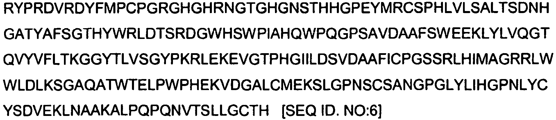
-

-

-

-

-

-

-


-
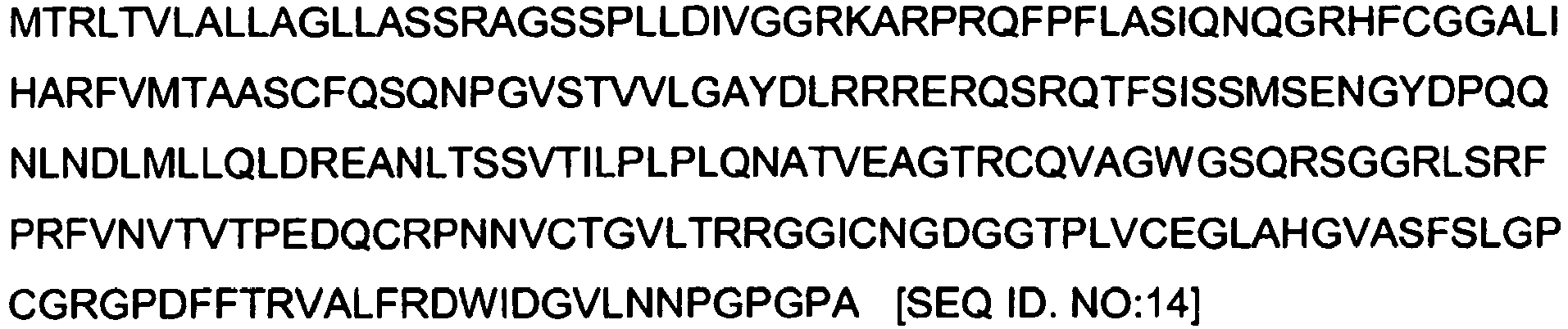
-

-

-

-

-

-

-

-


-
-
XP 001717485 [gi:169170953]

-
LOC286076 [gi:119602615]

-
MSTP132 [gi:33338088]

-
- <110> Katholieke Universiteit Leuven, K.U.Leuven R&D
- <120> MARKERS AND DIAGNOSTIC METHODS FOR METASTASIS
- <130> LRD-001-PCT
- <140> NOT YET KNOWN
<141> 2009-01-19 - <150>
GB0800944.1
<151> 2008-01-18 - <150>
GB0812298.8
<151> 2008-07-04 - <160> 24
- <170> PatentIn version 3.3
- <210> 1
<211> 20
<212> PRT
<213> Homo sapiens - <400> 1

- <210> 2
<211> 21
<212> PRT
<213> Homo sapiens - <400> 2

- <210> 3
<211> 21
<212> PRT
<213> Homo sapiens - <400> 3

- <210> 4
<211> 141
<212> PRT
<213> Homo sapiens - <400> 4

- <210> 5
<211> 111
<212> PRT
<213> Homo sapiens - <400> 5


- <210> 6
<211> 462
<212> PRT
<213> Homo sapiens - <400> 6


- <210> 7
<211> 189
<212> PRT
<213> Homo sapiens - <400> 7


- <210> 8
<211> 201
<212> PRT
<213> Homo sapiens - <400> 8


- <210> 9
<211> 267
<212> PRT
<213> Homo sapiens - <400> 9
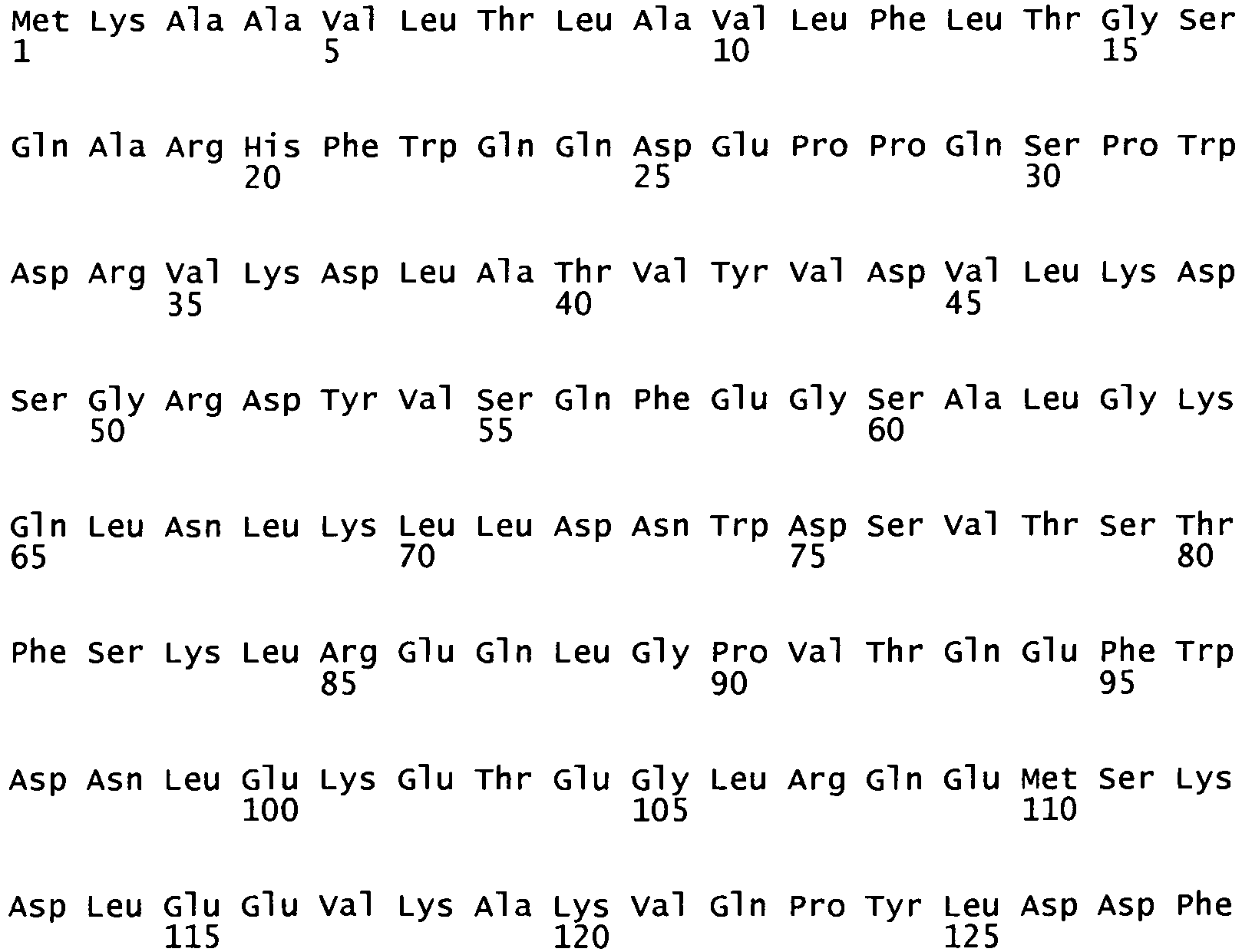

- <210> 10
<211> 137
<212> PRT
<213> Homo sapiens - <400> 10


- <210> 11
<211> 573
<212> PRT
<213> Homo sapiens - <400> 11
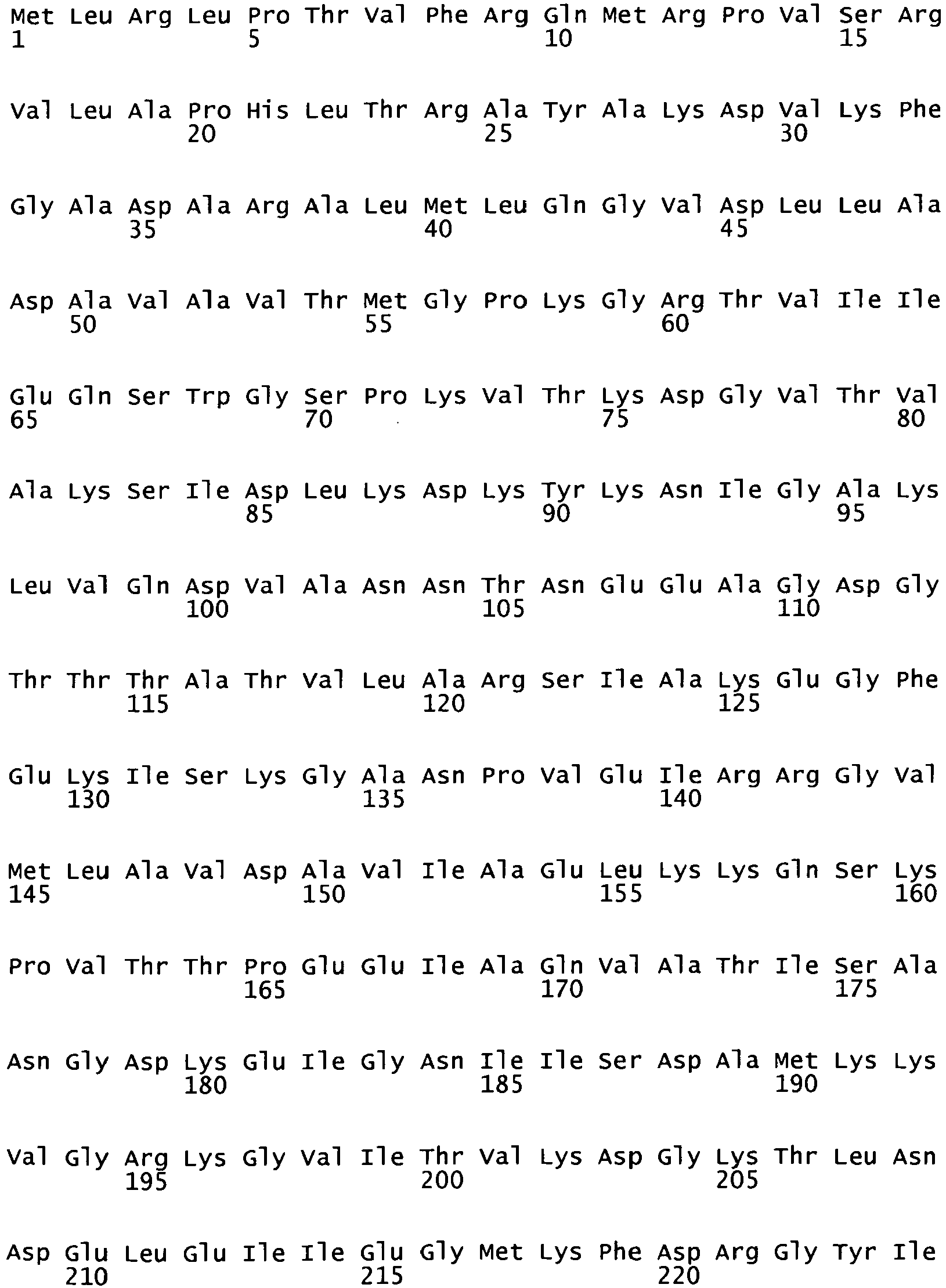


- <210> 12
<211> 641
<212> PRT
<213> Homo sapiens - <400> 12


- <210> 13
<211> 679
<212> PRT
<213> Homo sapiens - <400> 13
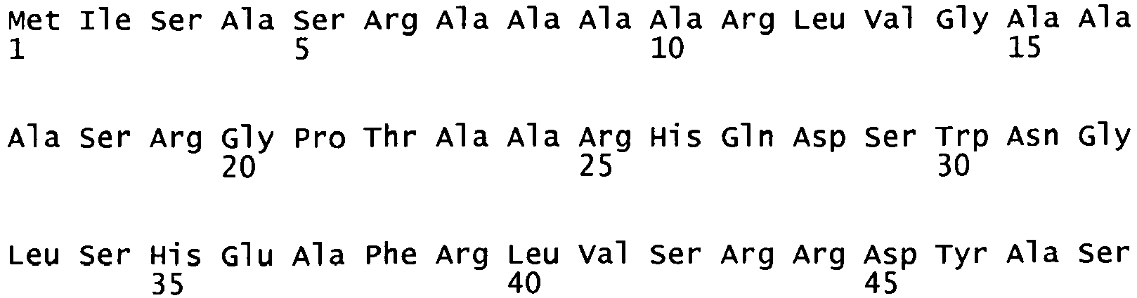


- <210> 14
<211> 251
<212> PRT
<213> Homo sapiens - <400> 14

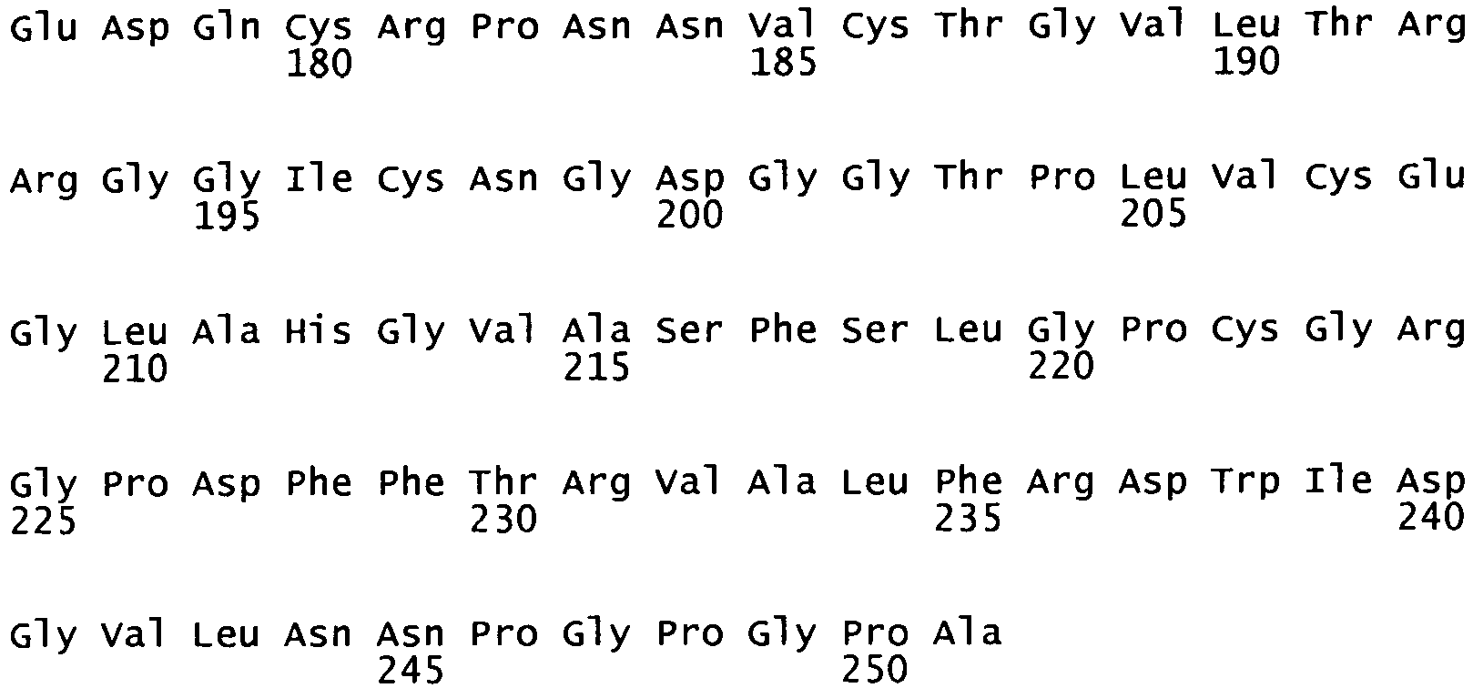
- <210> 15
<211> 114
<212> PRT
<213> Homo sapiens - <400> 15

- <210> 16
<211> 320
<212> PRT
<213> Homo sapiens - <400> 16


- <210> 17
<211> 177
<212> PRT
<213> Homo sapiens - <400> 17

- <210> 18
<211> 244
<212> PRT
<213> Homo sapiens - <400> 18


- <210> 19
<211> 222
<212> PRT
<213> Homo sapiens - <400> 19

- <210> 20
<211> 147
<212> PRT
<213> Homo sapiens - <400> 20


- <210> 21
<211> 609
<212> PRT - <213> Homo sapiens
- <400> 21


- <210> 22
<211> 801
<212> PRT
<213> Homo sapiens - <400> 22


- <210> 23
<211> 149
<212> PRT
<213> Homo sapiens - <400> 23


- <210> 24
<211> 61
<212> PRT
<213> Homo sapiens - <400> 24

| M/Z | p | Mean Node Neg | SD Node Neg | Mean Node Pos | SD Node Pos |
| Cation exchange array (CM10) at pH 4 | |||||
| (1-) 1375.29 | 0.0274231560 | 1.6870778082 | 1.2081885850 | 0.5747679441 | 0.4389184727 |
| (2+) 3354.63 | 0.0274231560 | 2.2347285918 | 1.6887134531 | 6.9437801790 | 4.4907141129 |
| (3+) 3417.37 | 0.0208625840 | 0.9663390866 | 1.6011746950 | 3.3320495713 | 1.7872618804 |
| (4-) 11506.39 | 0.0023220952 | 8.8690793963 | 4.8298523932 | 2.1522273533 | 2.3582952114 |
| (5-) 13929.03 | 0.0117186872 | 10.5366148929 | 2.3993891730 | 6.2380287997 | 4.1792723906 |
| (6-) 14132.43 | 0.0356919017 | 9.1879893115 | 3.6571443298 | 5.3188255848 | 2.8375150991 |
| Anion exchange array (Q10) at pH 9 | |||||
| (7-) 2022.23 | 0.0086515430 | 17.4659791576 | 5.8218885339 | 9.2959804930 | 3.5399548605 |
| (8-) 2276.73 | 0.0117186872 | 14.3831923660 | 3.1072477675 | 9.6161770954 | 3.5788505139 |
| (9+) 2290.00 | 0.0032758984 | 2.2166854670 | 3.6424423991 | 10.7050979707 | 5.2028762498 |
| (10+) 2306.00 | 0.0459993685 | 11.1330189198 | 9.9884291821 | 20.0282462422 | 8.3634075261 |
| (11+) 2452.63 | 0.0045744406 | 1.4383931734 | 2.3578200516 | 7.9656949689 | 3.5970398215 |
| (12+) 2477.81 | 0.0356919017 | 14.1500466740 | 17.6713561306 | 36.8321826002 | 7.5966537307 |
| (13+) 2662.54 | 0.0356919017 | 2.8656087379 | 4.5177850422 | 7.5039178253 | 3.3615362423 |
| (14+) 4962.61 | 0.0459993685 | 4.8498450505 | 4.1563158900 | 9.0363313941 | 3.3040218277 |
| (15+) 7567.26 | 0.0063229490 | 6.5571119856 | 5.3679943679 | 16.8135687049 | 4.5206197829 |
| (16-) 10280.33 | 0.0274231560 | 11.1977858348 | 2.2602955769 | 8.5119496408 | 1.9923464213 |
| (17-) 10392.05 | 0.0157143515 | 9.5816337993 | 4.3998366831 | 4.5719291359 | 1.2908014463 |
| (18+) 11062.03 | 0.0208625840 | 4.7681903768 | 3.2600949614 | 8.7840518433 | 2.3786735179 |
| (19-) 11298.92 | 0.0007775308 | 11.8128854619 | 3.0102875317 | 3.8806684148 | 2.5876764310 |
| (20-) 11477.52 | 0.0023220952 | 11.8898141013 | 4.3697538534 | 4.8647728730 | 2.5127084368 |
| (21+) 12632.05 | 0.0157143515 | 6.9212582463 | 1.9425018406 | 12.2737141087 | 5.7795017397 |
| (22+) 12787.06 | 0.0032758984 | 3.6624423020 | 1.3195209478 | 6.5246207838 | 1.8522777909 |
| (23-) 14144.07 | 0.0208625840 | 4.2270909245 | 1.5507169974 | 2.8691794061 | 0.9884474696 |
| (24-) 14603.70 | 0.0063229490 | 7.3546682643 | 2.4592351470 | 4.6153323662 | 0.5503777087 |
| (25-) 14754.71 | 0.0063229490 | 5.5556544556 | 2.0759324297 | 3.2513011167 | 0.8662145658 |
| (26-) 14956.22 | 0.0459993685 | 8.8314709597 | 6.9171022855 | 1.7485547241 | 1.4265824092 |
| (27-) 15094.45 | 0.0274231560 | 7.4459107443 | 5.1010413462 | 2.1747819723 | 1.0934166759 |
| (28+) 15110.53 | 0.0007775308 | 14.1776414059 | 9.6032549550 | 34.7866778849 | 5.0110162014 |
| (29+) 15249.32 | 0.0016291868 | 12.4649281795 | 5.2096509604 | 31.0375434707 | 8.7260256885 |
| (30+) 15305.01 | 0.0157143515 | 6.1528096689 | 3.3161518043 | 10.7849203112 | 2.2495106869 |
| (31-) 16888.72 | 0.0086515430 | 8.8036737906 | 2.2257267091 | 5.9527904853 | 1.0006268348 |
| (32-) 17871.94 | 0.0086515430 | 2.5037287174 | 0.6786425078 | 1.4153589592 | 0.5810624669 |
| (33-) 18003.62 | 0.0045744406 | 4.5593958506 | 1.1515963754 | 2.6796990848 | 1.1338140961 |
| (34-) 33356.20 | 0.0459993685 | 2.9463086499 | 0.8967372530 | 2.1736540870 | 0.4838099592 |
| (35-) 41665.94 | 0.0208625840 | 1.4622985955 | 0.4003093585 | 0.9312387135 | 0.2875171683 |
| Copper loaded Immobilized Metal Affinity array (IMAC-Cu) at 0.1 M Sodium Phosphate/ 0.5 M Sodium Chloride, pH 7 | |||||
| (36+) 2594.39 | 0.0032758984 | 1.6217419082 | 1.3245833913 | 3.4197868940 | 0.4991381175 |
| (37+) 3931.77 | 0.0032758984 | 1.0621999969 | 0.7602188469 | 2.6565595811 | 1.3290851933 |
| (38+) 4115.14 | 0.0032758984 | 1.1469489288 | 0.5049786548 | 4.6782890879 | 4.6910319099 |
| (39+) 5186.79 | 0.0157143515 | 2.2625664937 | 1.7964215156 | 4.9045423262 | 1.0741129522 |
| (40+) 9846.03 | 0.0086515430 | 3.1861852181 | 1.8416940282 | 7.6295131435 | 3.2963117374 |
| (41-) 11304.9 | 0.0157143515 | 5.8771415649 | 3.8176199984 | 2.3282098421 | 1.8146208213 |
| (42-) 11460.7 | 0.0117186872 | 17.5715851738 | 7.0610563265 | 7.1779059675 | 5.6281725626 |
Claims (10)
- An in vitro method for the prediction, prognosis or diagnosis of lymph node metastasis in a breast cancer patient comprising the measuring of the levels of Hemoglobin Chain alpha, Eosinophil peroxidase, Histone H4, and 60S ribosomal protein L32 or nucleotide sequences encoding said proteins in a tissue sample of the tumour of said patient.
- The method of claim 1, comprising the measuring the levels of the proteins with SEQ ID NO:1, SEQ ID. NO:2, SEQ ID. NO:3, and SEQ ID NO:4.
- The method of claim 1 or 2, further comprising measuring the level of the proteins Hemopexin, Protein DJ-1, Transgelin, Apolipoprotein A-1, Cellular retinoic acid-binding protein 1, mitochondrial 60 kDa heat shock protein, Heat shock 70 kDa protein, mitochondrial Stress-70 protein, Azurocidin, SH3 domain-binding glutamic acid-rich-like protein, Annexin A5, Interleukin-25, Tubulin folding cofactor B, mitochondrial Superoxide dismutase [Mn], Hemoglobin chain beta, Albumin, SEQ ID NO:22, SEQ ID NO:23, and SEQ ID NO:24 or nucleotide sequences encoding said proteins, in a tissue sample of the tumour of said patient.
- The method of any of claims 1 to 3, which in addition comprises measuring the level of Vascular Endothelial Growth Factor A isoform 111.
- The method of any one of claims 1 to 4, wherein said method further makes use of the proteins with SEQ ID NO: 6, SEQ ID NO: 7, SEQ ID NO: 8, SEQ ID NO: 9, SEQ ID NO: 10, SEQ ID NO: 11, SEQ ID NO: 12, SEQ ID NO: 13, SEQ ID NO:14, SEQ ID NO: 15 SEQ ID NO: 16, SEQ ID NO: 17, SEQ ID NO: 18, SEQ ID NO: 19, SEQ ID NO: 20, SEQ ID NO: 21, SEQ ID NO: 22, SEQ ID NO: 23 and SEQ ID NO: 24.
- The method according to any one of claims 1 to 5, comprising the steps of:(i) extracting said proteins from a tissue sample of the tumour of said patient; and(ii) determining the relative abundance of said proteins in said extract by comparing said abundance to the abundance of said proteins in a reference sample or a reference expression profile; wherein said relative abundance of said proteins is indicative of metastasis.
- The method according to claim 6, wherein in step (ii) said reference sample or reference expression profile is of a patient not having metastasis.
- The method of claim 6 or 7, wherein said relative abundance indicative of metastasis is at least a two-fold increase.
- The method according to any one of claims 1 to 8, which makes use of reagents specifically identifying said proteins, wherein said reagents are antibodies.
- The method according to any one of claims 1 to 9, which further comprises determining the expression of an oncogene in a biological sample isolated from said patient.
Applications Claiming Priority (3)
| Application Number | Priority Date | Filing Date | Title |
|---|---|---|---|
| GB0800944A GB0800944D0 (en) | 2008-01-18 | 2008-01-18 | Novel diagnostic method of lymph node metastasis |
| GB0812298A GB0812298D0 (en) | 2008-07-04 | 2008-07-04 | Identification of proteins |
| PCT/EP2009/050572 WO2009090269A2 (en) | 2008-01-18 | 2009-01-19 | Markers and diagnostic methods for metastasis |
Publications (2)
| Publication Number | Publication Date |
|---|---|
| EP2250507A2 EP2250507A2 (en) | 2010-11-17 |
| EP2250507B1 true EP2250507B1 (en) | 2012-10-17 |
Family
ID=40565285
Family Applications (1)
| Application Number | Title | Priority Date | Filing Date |
|---|---|---|---|
| EP09701520A Not-in-force EP2250507B1 (en) | 2008-01-18 | 2009-01-19 | Diagnostic methods for metastasis |
Country Status (3)
| Country | Link |
|---|---|
| US (1) | US8735329B2 (en) |
| EP (1) | EP2250507B1 (en) |
| WO (1) | WO2009090269A2 (en) |
Families Citing this family (5)
| Publication number | Priority date | Publication date | Assignee | Title |
|---|---|---|---|---|
| OA13012A (en) * | 2002-11-20 | 2006-11-10 | Sanaria Inc | Method for the prevention of malaria. |
| WO2011073896A1 (en) * | 2009-12-14 | 2011-06-23 | Koninklijke Philips Electronics N.V. | Novel tumor markers |
| WO2011073903A1 (en) * | 2009-12-14 | 2011-06-23 | Koninklijke Philips Electronics N.V. | Novel tumor markers |
| CN103109189A (en) * | 2010-07-19 | 2013-05-15 | 霍夫曼-拉罗奇有限公司 | Method of identifying patients with increased likelihood of responding to anticancer therapy |
| GB201906199D0 (en) * | 2019-05-02 | 2019-06-19 | Belgian Volition Sprl | Method for the detection of cancer |
Family Cites Families (4)
| Publication number | Priority date | Publication date | Assignee | Title |
|---|---|---|---|---|
| US5248591A (en) * | 1991-05-02 | 1993-09-28 | Puente Fernando D | Diagnosis of cancer and other proliferative disorders by analysis of prothymosin alpha expression |
| US6790604B1 (en) * | 1998-05-12 | 2004-09-14 | Stefan A. Cohen | Methods for identifying or diagnosing carcinoma cells with metastatic potential based on the measurement of lymphoid genes or their products in carcinoma cells |
| US20070243540A1 (en) | 2004-05-21 | 2007-10-18 | Tsai Jo C | Kits and Methods for Identification, Assessment, Prevention and Therapy of Breast Cancer |
| US20070099209A1 (en) | 2005-06-13 | 2007-05-03 | The Regents Of The University Of Michigan | Compositions and methods for treating and diagnosing cancer |
-
2009
- 2009-01-19 US US12/735,433 patent/US8735329B2/en not_active Expired - Fee Related
- 2009-01-19 EP EP09701520A patent/EP2250507B1/en not_active Not-in-force
- 2009-01-19 WO PCT/EP2009/050572 patent/WO2009090269A2/en not_active Ceased
Also Published As
| Publication number | Publication date |
|---|---|
| WO2009090269A8 (en) | 2012-07-19 |
| EP2250507A2 (en) | 2010-11-17 |
| US20100284935A1 (en) | 2010-11-11 |
| WO2009090269A3 (en) | 2009-09-17 |
| US8735329B2 (en) | 2014-05-27 |
| WO2009090269A2 (en) | 2009-07-23 |
Similar Documents
| Publication | Publication Date | Title |
|---|---|---|
| KR102116178B1 (en) | Biomarker for monitoring or detecting early onset of liver cancer from patient having high risk of liver cancer and its use | |
| Malik et al. | Serum levels of an isoform of apolipoprotein A-II as a potential marker for prostate cancer | |
| CN110799841B (en) | Biomarkers for detecting colorectal cancer | |
| EP2805167B1 (en) | Biomarkers for gastric cancer and uses thereof | |
| US20170285032A1 (en) | Compositions and methods for diagnosis and prognosis of colorectal cancer | |
| AU2001249275B2 (en) | Prostate cancer markers | |
| WO2013154422A1 (en) | Biomarkers for triple negative breast cancer | |
| WO2002100242A2 (en) | Detection of ovarian cancer based upon alpha-haptoglobin levels | |
| US8735329B2 (en) | Markers and diagnostic methods for metastasis | |
| EP3092495B1 (en) | Srm assay for pd-l1 | |
| US20140100130A1 (en) | Methods and Compositions for the Diagnosis of Ovarian Cancer | |
| EP1977250A1 (en) | Method and markers for the diagnosis of renal diseases | |
| Ishihara et al. | Development of quantitative plasma N-glycoproteomics using label-free 2-D LC-MALDI MS and its applicability for biomarker discovery in hepatocellular carcinoma | |
| US20030228639A1 (en) | Prostate cancer markers | |
| Wu et al. | Identification of potential serum markers for nasopharyngeal carcinoma from a xenografted mouse model using Cy‐dye labeling combined with three‐dimensional fractionation | |
| WO2009151693A2 (en) | Imaging mass spectrometry for improved prostate cancer diagnostics | |
| WO2018007555A1 (en) | Method for diagnosing cancer | |
| Fania et al. | A PSA-guided approach for a better diagnosis of prostatic adenocarcinoma based on MALDI profiling and peptide identification | |
| WO2009034562A2 (en) | A method of assessing lung squamous cell carcinoma status in an individual | |
| Yin et al. | Analysis of the peptides detected in atopic dermatitis and various inflammatory diseases patients-derived sera | |
| WO2005040814A1 (en) | Methods and means of cancer detection by histone modification | |
| JP7029745B2 (en) | Glioblastoma marker and its use | |
| JP7211604B2 (en) | Glioblastoma markers and uses thereof | |
| Li et al. | Targeted detection of prostate cancer proteins in serum using heavy‐isotope‐labeled‐peptide standards and MALDI‐TOF/TOF | |
| EP2844770A2 (en) | Mrm-ms signature assay |
Legal Events
| Date | Code | Title | Description |
|---|---|---|---|
| PUAI | Public reference made under article 153(3) epc to a published international application that has entered the european phase |
Free format text: ORIGINAL CODE: 0009012 |
|
| 17P | Request for examination filed |
Effective date: 20100816 |
|
| AK | Designated contracting states |
Kind code of ref document: A2 Designated state(s): AT BE BG CH CY CZ DE DK EE ES FI FR GB GR HR HU IE IS IT LI LT LU LV MC MK MT NL NO PL PT RO SE SI SK TR |
|
| AX | Request for extension of the european patent |
Extension state: AL BA RS |
|
| 17Q | First examination report despatched |
Effective date: 20110117 |
|
| DAX | Request for extension of the european patent (deleted) | ||
| RTI1 | Title (correction) |
Free format text: DIAGNOSTIC METHODS FOR METASTASIS |
|
| GRAP | Despatch of communication of intention to grant a patent |
Free format text: ORIGINAL CODE: EPIDOSNIGR1 |
|
| GRAS | Grant fee paid |
Free format text: ORIGINAL CODE: EPIDOSNIGR3 |
|
| GRAJ | Information related to disapproval of communication of intention to grant by the applicant or resumption of examination proceedings by the epo deleted |
Free format text: ORIGINAL CODE: EPIDOSDIGR1 |
|
| GRAS | Grant fee paid |
Free format text: ORIGINAL CODE: EPIDOSNIGR3 |
|
| GRAP | Despatch of communication of intention to grant a patent |
Free format text: ORIGINAL CODE: EPIDOSNIGR1 |
|
| GRAA | (expected) grant |
Free format text: ORIGINAL CODE: 0009210 |
|
| AK | Designated contracting states |
Kind code of ref document: B1 Designated state(s): AT BE BG CH CY CZ DE DK EE ES FI FR GB GR HR HU IE IS IT LI LT LU LV MC MK MT NL NO PL PT RO SE SI SK TR |
|
| REG | Reference to a national code |
Ref country code: GB Ref legal event code: FG4D |
|
| REG | Reference to a national code |
Ref country code: CH Ref legal event code: EP |
|
| REG | Reference to a national code |
Ref country code: IE Ref legal event code: FG4D |
|
| REG | Reference to a national code |
Ref country code: AT Ref legal event code: REF Ref document number: 580125 Country of ref document: AT Kind code of ref document: T Effective date: 20121115 |
|
| REG | Reference to a national code |
Ref country code: DE Ref legal event code: R096 Ref document number: 602009010471 Country of ref document: DE Effective date: 20121213 |
|
| REG | Reference to a national code |
Ref country code: NL Ref legal event code: T3 |
|
| REG | Reference to a national code |
Ref country code: AT Ref legal event code: MK05 Ref document number: 580125 Country of ref document: AT Kind code of ref document: T Effective date: 20121017 |
|
| REG | Reference to a national code |
Ref country code: LT Ref legal event code: MG4D |
|
| PG25 | Lapsed in a contracting state [announced via postgrant information from national office to epo] |
Ref country code: SE Free format text: LAPSE BECAUSE OF FAILURE TO SUBMIT A TRANSLATION OF THE DESCRIPTION OR TO PAY THE FEE WITHIN THE PRESCRIBED TIME-LIMIT Effective date: 20121017 Ref country code: IS Free format text: LAPSE BECAUSE OF FAILURE TO SUBMIT A TRANSLATION OF THE DESCRIPTION OR TO PAY THE FEE WITHIN THE PRESCRIBED TIME-LIMIT Effective date: 20130217 Ref country code: ES Free format text: LAPSE BECAUSE OF FAILURE TO SUBMIT A TRANSLATION OF THE DESCRIPTION OR TO PAY THE FEE WITHIN THE PRESCRIBED TIME-LIMIT Effective date: 20130128 Ref country code: NO Free format text: LAPSE BECAUSE OF FAILURE TO SUBMIT A TRANSLATION OF THE DESCRIPTION OR TO PAY THE FEE WITHIN THE PRESCRIBED TIME-LIMIT Effective date: 20130117 Ref country code: LT Free format text: LAPSE BECAUSE OF FAILURE TO SUBMIT A TRANSLATION OF THE DESCRIPTION OR TO PAY THE FEE WITHIN THE PRESCRIBED TIME-LIMIT Effective date: 20121017 Ref country code: HR Free format text: LAPSE BECAUSE OF FAILURE TO SUBMIT A TRANSLATION OF THE DESCRIPTION OR TO PAY THE FEE WITHIN THE PRESCRIBED TIME-LIMIT Effective date: 20121017 Ref country code: FI Free format text: LAPSE BECAUSE OF FAILURE TO SUBMIT A TRANSLATION OF THE DESCRIPTION OR TO PAY THE FEE WITHIN THE PRESCRIBED TIME-LIMIT Effective date: 20121017 |
|
| PG25 | Lapsed in a contracting state [announced via postgrant information from national office to epo] |
Ref country code: SI Free format text: LAPSE BECAUSE OF FAILURE TO SUBMIT A TRANSLATION OF THE DESCRIPTION OR TO PAY THE FEE WITHIN THE PRESCRIBED TIME-LIMIT Effective date: 20121017 Ref country code: PT Free format text: LAPSE BECAUSE OF FAILURE TO SUBMIT A TRANSLATION OF THE DESCRIPTION OR TO PAY THE FEE WITHIN THE PRESCRIBED TIME-LIMIT Effective date: 20130218 Ref country code: LV Free format text: LAPSE BECAUSE OF FAILURE TO SUBMIT A TRANSLATION OF THE DESCRIPTION OR TO PAY THE FEE WITHIN THE PRESCRIBED TIME-LIMIT Effective date: 20121017 Ref country code: GR Free format text: LAPSE BECAUSE OF FAILURE TO SUBMIT A TRANSLATION OF THE DESCRIPTION OR TO PAY THE FEE WITHIN THE PRESCRIBED TIME-LIMIT Effective date: 20130118 Ref country code: BE Free format text: LAPSE BECAUSE OF FAILURE TO SUBMIT A TRANSLATION OF THE DESCRIPTION OR TO PAY THE FEE WITHIN THE PRESCRIBED TIME-LIMIT Effective date: 20121017 Ref country code: PL Free format text: LAPSE BECAUSE OF FAILURE TO SUBMIT A TRANSLATION OF THE DESCRIPTION OR TO PAY THE FEE WITHIN THE PRESCRIBED TIME-LIMIT Effective date: 20121017 |
|
| PG25 | Lapsed in a contracting state [announced via postgrant information from national office to epo] |
Ref country code: AT Free format text: LAPSE BECAUSE OF FAILURE TO SUBMIT A TRANSLATION OF THE DESCRIPTION OR TO PAY THE FEE WITHIN THE PRESCRIBED TIME-LIMIT Effective date: 20121017 |
|
| PG25 | Lapsed in a contracting state [announced via postgrant information from national office to epo] |
Ref country code: BG Free format text: LAPSE BECAUSE OF FAILURE TO SUBMIT A TRANSLATION OF THE DESCRIPTION OR TO PAY THE FEE WITHIN THE PRESCRIBED TIME-LIMIT Effective date: 20130117 Ref country code: CZ Free format text: LAPSE BECAUSE OF FAILURE TO SUBMIT A TRANSLATION OF THE DESCRIPTION OR TO PAY THE FEE WITHIN THE PRESCRIBED TIME-LIMIT Effective date: 20121017 Ref country code: EE Free format text: LAPSE BECAUSE OF FAILURE TO SUBMIT A TRANSLATION OF THE DESCRIPTION OR TO PAY THE FEE WITHIN THE PRESCRIBED TIME-LIMIT Effective date: 20121017 Ref country code: DK Free format text: LAPSE BECAUSE OF FAILURE TO SUBMIT A TRANSLATION OF THE DESCRIPTION OR TO PAY THE FEE WITHIN THE PRESCRIBED TIME-LIMIT Effective date: 20121017 Ref country code: SK Free format text: LAPSE BECAUSE OF FAILURE TO SUBMIT A TRANSLATION OF THE DESCRIPTION OR TO PAY THE FEE WITHIN THE PRESCRIBED TIME-LIMIT Effective date: 20121017 |
|
| PLBE | No opposition filed within time limit |
Free format text: ORIGINAL CODE: 0009261 |
|
| STAA | Information on the status of an ep patent application or granted ep patent |
Free format text: STATUS: NO OPPOSITION FILED WITHIN TIME LIMIT |
|
| PG25 | Lapsed in a contracting state [announced via postgrant information from national office to epo] |
Ref country code: MC Free format text: LAPSE BECAUSE OF NON-PAYMENT OF DUE FEES Effective date: 20130131 Ref country code: IT Free format text: LAPSE BECAUSE OF FAILURE TO SUBMIT A TRANSLATION OF THE DESCRIPTION OR TO PAY THE FEE WITHIN THE PRESCRIBED TIME-LIMIT Effective date: 20121017 Ref country code: RO Free format text: LAPSE BECAUSE OF FAILURE TO SUBMIT A TRANSLATION OF THE DESCRIPTION OR TO PAY THE FEE WITHIN THE PRESCRIBED TIME-LIMIT Effective date: 20121017 |
|
| 26N | No opposition filed |
Effective date: 20130718 |
|
| REG | Reference to a national code |
Ref country code: IE Ref legal event code: MM4A |
|
| REG | Reference to a national code |
Ref country code: DE Ref legal event code: R097 Ref document number: 602009010471 Country of ref document: DE Effective date: 20130718 |
|
| PG25 | Lapsed in a contracting state [announced via postgrant information from national office to epo] |
Ref country code: CY Free format text: LAPSE BECAUSE OF FAILURE TO SUBMIT A TRANSLATION OF THE DESCRIPTION OR TO PAY THE FEE WITHIN THE PRESCRIBED TIME-LIMIT Effective date: 20121017 |
|
| PG25 | Lapsed in a contracting state [announced via postgrant information from national office to epo] |
Ref country code: IE Free format text: LAPSE BECAUSE OF NON-PAYMENT OF DUE FEES Effective date: 20130119 |
|
| PG25 | Lapsed in a contracting state [announced via postgrant information from national office to epo] |
Ref country code: MT Free format text: LAPSE BECAUSE OF FAILURE TO SUBMIT A TRANSLATION OF THE DESCRIPTION OR TO PAY THE FEE WITHIN THE PRESCRIBED TIME-LIMIT Effective date: 20121017 |
|
| PG25 | Lapsed in a contracting state [announced via postgrant information from national office to epo] |
Ref country code: TR Free format text: LAPSE BECAUSE OF FAILURE TO SUBMIT A TRANSLATION OF THE DESCRIPTION OR TO PAY THE FEE WITHIN THE PRESCRIBED TIME-LIMIT Effective date: 20121017 |
|
| PG25 | Lapsed in a contracting state [announced via postgrant information from national office to epo] |
Ref country code: HU Free format text: LAPSE BECAUSE OF FAILURE TO SUBMIT A TRANSLATION OF THE DESCRIPTION OR TO PAY THE FEE WITHIN THE PRESCRIBED TIME-LIMIT; INVALID AB INITIO Effective date: 20090119 Ref country code: MK Free format text: LAPSE BECAUSE OF FAILURE TO SUBMIT A TRANSLATION OF THE DESCRIPTION OR TO PAY THE FEE WITHIN THE PRESCRIBED TIME-LIMIT Effective date: 20121017 |
|
| REG | Reference to a national code |
Ref country code: FR Ref legal event code: PLFP Year of fee payment: 8 |
|
| REG | Reference to a national code |
Ref country code: FR Ref legal event code: PLFP Year of fee payment: 9 |
|
| REG | Reference to a national code |
Ref country code: FR Ref legal event code: PLFP Year of fee payment: 10 |
|
| PGFP | Annual fee paid to national office [announced via postgrant information from national office to epo] |
Ref country code: LU Payment date: 20180119 Year of fee payment: 10 |
|
| PGFP | Annual fee paid to national office [announced via postgrant information from national office to epo] |
Ref country code: NL Payment date: 20180119 Year of fee payment: 10 |
|
| PGFP | Annual fee paid to national office [announced via postgrant information from national office to epo] |
Ref country code: CH Payment date: 20180119 Year of fee payment: 10 Ref country code: DE Payment date: 20180122 Year of fee payment: 10 Ref country code: GB Payment date: 20180119 Year of fee payment: 10 |
|
| PGFP | Annual fee paid to national office [announced via postgrant information from national office to epo] |
Ref country code: FR Payment date: 20180119 Year of fee payment: 10 |
|
| REG | Reference to a national code |
Ref country code: DE Ref legal event code: R119 Ref document number: 602009010471 Country of ref document: DE |
|
| REG | Reference to a national code |
Ref country code: CH Ref legal event code: PL |
|
| REG | Reference to a national code |
Ref country code: NL Ref legal event code: MM Effective date: 20190201 |
|
| GBPC | Gb: european patent ceased through non-payment of renewal fee |
Effective date: 20190119 |
|
| PG25 | Lapsed in a contracting state [announced via postgrant information from national office to epo] |
Ref country code: LU Free format text: LAPSE BECAUSE OF NON-PAYMENT OF DUE FEES Effective date: 20190119 |
|
| PG25 | Lapsed in a contracting state [announced via postgrant information from national office to epo] |
Ref country code: NL Free format text: LAPSE BECAUSE OF NON-PAYMENT OF DUE FEES Effective date: 20190201 Ref country code: DE Free format text: LAPSE BECAUSE OF NON-PAYMENT OF DUE FEES Effective date: 20190801 Ref country code: FR Free format text: LAPSE BECAUSE OF NON-PAYMENT OF DUE FEES Effective date: 20190131 |
|
| PG25 | Lapsed in a contracting state [announced via postgrant information from national office to epo] |
Ref country code: LI Free format text: LAPSE BECAUSE OF NON-PAYMENT OF DUE FEES Effective date: 20190131 Ref country code: GB Free format text: LAPSE BECAUSE OF NON-PAYMENT OF DUE FEES Effective date: 20190119 Ref country code: CH Free format text: LAPSE BECAUSE OF NON-PAYMENT OF DUE FEES Effective date: 20190131 |