EP2247298B1 - Control of viral-host membrane fusion with hydrogen bond surrogate-based artificial helices - Google Patents
Control of viral-host membrane fusion with hydrogen bond surrogate-based artificial helices Download PDFInfo
- Publication number
- EP2247298B1 EP2247298B1 EP08873198.9A EP08873198A EP2247298B1 EP 2247298 B1 EP2247298 B1 EP 2247298B1 EP 08873198 A EP08873198 A EP 08873198A EP 2247298 B1 EP2247298 B1 EP 2247298B1
- Authority
- EP
- European Patent Office
- Prior art keywords
- peptide
- helix
- carbon
- amino acid
- bond
- Prior art date
- Legal status (The legal status is an assumption and is not a legal conclusion. Google has not performed a legal analysis and makes no representation as to the accuracy of the status listed.)
- Not-in-force
Links
- 239000001257 hydrogen Substances 0.000 title claims description 14
- 229910052739 hydrogen Inorganic materials 0.000 title claims description 14
- 230000034217 membrane fusion Effects 0.000 title description 6
- 108090000765 processed proteins & peptides Proteins 0.000 claims description 141
- VOUAQYXWVJDEQY-QENPJCQMSA-N 33017-11-7 Chemical compound OC(=O)CC[C@H](N)C(=O)N[C@@H](C)C(=O)N[C@@H](CCC(O)=O)C(=O)N[C@@H](CC(O)=O)C(=O)N[C@@H](CC(C)C)C(=O)N[C@@H](CCC(N)=O)C(=O)N[C@@H](C(C)C)C(=O)NCC(=O)N[C@@H](CCC(N)=O)C(=O)N[C@@H](C(C)C)C(=O)N[C@@H](CCC(O)=O)C(=O)N[C@@H](CC(C)C)C(=O)NCC(=O)NCC(=O)NCC(=O)N1CCC[C@H]1C(=O)NCC(=O)N[C@@H](C)C(=O)NCC(=O)N[C@@H](CO)C(=O)N[C@@H](CC(C)C)C(=O)N[C@@H](CCC(N)=O)C(=O)N1[C@H](C(=O)N[C@@H](CC(C)C)C(=O)N[C@@H](C)C(=O)N[C@@H](CC(C)C)C(=O)N[C@@H](CCC(O)=O)C(=O)NCC(=O)N[C@@H](CO)C(=O)N[C@@H](CC(C)C)C(=O)N[C@@H](CCC(N)=O)C(O)=O)CCC1 VOUAQYXWVJDEQY-QENPJCQMSA-N 0.000 claims description 36
- 241000713772 Human immunodeficiency virus 1 Species 0.000 claims description 34
- 125000003275 alpha amino acid group Chemical group 0.000 claims description 34
- 108010075254 C-Peptide Proteins 0.000 claims description 30
- 239000003112 inhibitor Substances 0.000 claims description 22
- 125000003118 aryl group Chemical group 0.000 claims description 18
- 230000002401 inhibitory effect Effects 0.000 claims description 18
- 239000000203 mixture Substances 0.000 claims description 18
- 125000000217 alkyl group Chemical group 0.000 claims description 17
- 230000015572 biosynthetic process Effects 0.000 claims description 16
- 239000011203 carbon fibre reinforced carbon Substances 0.000 claims description 14
- 238000005649 metathesis reaction Methods 0.000 claims description 11
- 125000004435 hydrogen atom Chemical group [H]* 0.000 claims description 8
- 238000010485 C−C bond formation reaction Methods 0.000 claims description 7
- 238000006664 bond formation reaction Methods 0.000 claims description 5
- 239000008194 pharmaceutical composition Substances 0.000 claims description 3
- 239000002243 precursor Substances 0.000 claims description 3
- 230000001737 promoting effect Effects 0.000 claims description 3
- 101800001690 Transmembrane protein gp41 Proteins 0.000 description 60
- 241000700605 Viruses Species 0.000 description 59
- 102000004196 processed proteins & peptides Human genes 0.000 description 51
- 210000004027 cell Anatomy 0.000 description 39
- 239000003795 chemical substances by application Substances 0.000 description 31
- 241000725303 Human immunodeficiency virus Species 0.000 description 26
- 230000027455 binding Effects 0.000 description 25
- 239000002502 liposome Substances 0.000 description 23
- 108010027796 Membrane Fusion Proteins Proteins 0.000 description 17
- 102000018897 Membrane Fusion Proteins Human genes 0.000 description 17
- 230000004927 fusion Effects 0.000 description 17
- YIYBQIKDCADOSF-UHFFFAOYSA-N pentenoic acid group Chemical group C(C=CCC)(=O)O YIYBQIKDCADOSF-UHFFFAOYSA-N 0.000 description 16
- 108090000623 proteins and genes Proteins 0.000 description 15
- 102000004169 proteins and genes Human genes 0.000 description 14
- 230000003612 virological effect Effects 0.000 description 14
- 239000003814 drug Substances 0.000 description 13
- 238000000034 method Methods 0.000 description 13
- 230000007910 cell fusion Effects 0.000 description 12
- 238000010586 diagram Methods 0.000 description 12
- 235000018102 proteins Nutrition 0.000 description 12
- 210000001519 tissue Anatomy 0.000 description 12
- 235000001014 amino acid Nutrition 0.000 description 11
- 239000012528 membrane Substances 0.000 description 11
- 210000004379 membrane Anatomy 0.000 description 11
- 208000030507 AIDS Diseases 0.000 description 10
- 150000001875 compounds Chemical class 0.000 description 9
- 238000013461 design Methods 0.000 description 9
- 230000002209 hydrophobic effect Effects 0.000 description 9
- 239000000243 solution Substances 0.000 description 9
- 238000005865 alkene metathesis reaction Methods 0.000 description 8
- 238000003556 assay Methods 0.000 description 8
- 230000000694 effects Effects 0.000 description 8
- 102000037865 fusion proteins Human genes 0.000 description 8
- 108020001507 fusion proteins Proteins 0.000 description 8
- 239000003446 ligand Substances 0.000 description 8
- 230000001404 mediated effect Effects 0.000 description 8
- 230000003278 mimic effect Effects 0.000 description 8
- 239000011347 resin Substances 0.000 description 8
- 229920005989 resin Polymers 0.000 description 8
- 230000005540 biological transmission Effects 0.000 description 7
- 229940079593 drug Drugs 0.000 description 7
- 230000005764 inhibitory process Effects 0.000 description 7
- 230000003993 interaction Effects 0.000 description 7
- 230000029052 metamorphosis Effects 0.000 description 7
- 210000000056 organ Anatomy 0.000 description 7
- 239000002953 phosphate buffered saline Substances 0.000 description 7
- 102100026189 Beta-galactosidase Human genes 0.000 description 6
- YMWUJEATGCHHMB-UHFFFAOYSA-N Dichloromethane Chemical compound ClCCl YMWUJEATGCHHMB-UHFFFAOYSA-N 0.000 description 6
- PEDCQBHIVMGVHV-UHFFFAOYSA-N Glycerine Chemical compound OCC(O)CO PEDCQBHIVMGVHV-UHFFFAOYSA-N 0.000 description 6
- 241000701024 Human betaherpesvirus 5 Species 0.000 description 6
- 125000002707 L-tryptophyl group Chemical group [H]C1=C([H])C([H])=C2C(C([C@](N([H])[H])(C(=O)[*])[H])([H])[H])=C([H])N([H])C2=C1[H] 0.000 description 6
- DNIAPMSPPWPWGF-UHFFFAOYSA-N Propylene glycol Chemical compound CC(O)CO DNIAPMSPPWPWGF-UHFFFAOYSA-N 0.000 description 6
- 238000013459 approach Methods 0.000 description 6
- 108010005774 beta-Galactosidase Proteins 0.000 description 6
- 238000006243 chemical reaction Methods 0.000 description 6
- -1 for example Chemical class 0.000 description 6
- 208000015181 infectious disease Diseases 0.000 description 6
- 239000000463 material Substances 0.000 description 6
- 229920001184 polypeptide Polymers 0.000 description 6
- 230000000241 respiratory effect Effects 0.000 description 6
- 229940124597 therapeutic agent Drugs 0.000 description 6
- 102100027723 Endogenous retrovirus group K member 6 Rec protein Human genes 0.000 description 5
- FAPWRFPIFSIZLT-UHFFFAOYSA-M Sodium chloride Chemical compound [Na+].[Cl-] FAPWRFPIFSIZLT-UHFFFAOYSA-M 0.000 description 5
- RHQDFWAXVIIEBN-UHFFFAOYSA-N Trifluoroethanol Chemical compound OCC(F)(F)F RHQDFWAXVIIEBN-UHFFFAOYSA-N 0.000 description 5
- 150000001413 amino acids Chemical class 0.000 description 5
- 239000000872 buffer Substances 0.000 description 5
- 239000003054 catalyst Substances 0.000 description 5
- GNBHRKFJIUUOQI-UHFFFAOYSA-N fluorescein Chemical compound O1C(=O)C2=CC=CC=C2C21C1=CC=C(O)C=C1OC1=CC(O)=CC=C21 GNBHRKFJIUUOQI-UHFFFAOYSA-N 0.000 description 5
- 238000001727 in vivo Methods 0.000 description 5
- 239000007788 liquid Substances 0.000 description 5
- 238000002360 preparation method Methods 0.000 description 5
- 239000000523 sample Substances 0.000 description 5
- 238000003786 synthesis reaction Methods 0.000 description 5
- 239000003826 tablet Substances 0.000 description 5
- 241000711573 Coronaviridae Species 0.000 description 4
- 241001115402 Ebolavirus Species 0.000 description 4
- 101710121417 Envelope glycoprotein Proteins 0.000 description 4
- 241000711950 Filoviridae Species 0.000 description 4
- 241000700586 Herpesviridae Species 0.000 description 4
- 241000282414 Homo sapiens Species 0.000 description 4
- 241000713869 Moloney murine leukemia virus Species 0.000 description 4
- 241000711466 Murine hepatitis virus Species 0.000 description 4
- 241000712464 Orthomyxoviridae Species 0.000 description 4
- 241000711504 Paramyxoviridae Species 0.000 description 4
- 241000712907 Retroviridae Species 0.000 description 4
- 241000315672 SARS coronavirus Species 0.000 description 4
- VYPSYNLAJGMNEJ-UHFFFAOYSA-N Silicium dioxide Chemical compound O=[Si]=O VYPSYNLAJGMNEJ-UHFFFAOYSA-N 0.000 description 4
- 241000713311 Simian immunodeficiency virus Species 0.000 description 4
- 208000000389 T-cell leukemia Diseases 0.000 description 4
- 208000028530 T-cell lymphoblastic leukemia/lymphoma Diseases 0.000 description 4
- 239000002775 capsule Substances 0.000 description 4
- 238000001142 circular dichroism spectrum Methods 0.000 description 4
- 238000011161 development Methods 0.000 description 4
- 230000018109 developmental process Effects 0.000 description 4
- 238000002875 fluorescence polarization Methods 0.000 description 4
- 238000004895 liquid chromatography mass spectrometry Methods 0.000 description 4
- 239000000546 pharmaceutical excipient Substances 0.000 description 4
- 239000007787 solid Substances 0.000 description 4
- 230000006641 stabilisation Effects 0.000 description 4
- 238000011105 stabilization Methods 0.000 description 4
- 239000000758 substrate Substances 0.000 description 4
- 239000000725 suspension Substances 0.000 description 4
- 241000712461 unidentified influenza virus Species 0.000 description 4
- 239000003981 vehicle Substances 0.000 description 4
- WEVYAHXRMPXWCK-UHFFFAOYSA-N Acetonitrile Chemical compound CC#N WEVYAHXRMPXWCK-UHFFFAOYSA-N 0.000 description 3
- 102000004190 Enzymes Human genes 0.000 description 3
- 108090000790 Enzymes Proteins 0.000 description 3
- LFQSCWFLJHTTHZ-UHFFFAOYSA-N Ethanol Chemical compound CCO LFQSCWFLJHTTHZ-UHFFFAOYSA-N 0.000 description 3
- 241001559185 Mammalian rubulavirus 5 Species 0.000 description 3
- OKKJLVBELUTLKV-UHFFFAOYSA-N Methanol Chemical compound OC OKKJLVBELUTLKV-UHFFFAOYSA-N 0.000 description 3
- YXFVVABEGXRONW-UHFFFAOYSA-N Toluene Chemical compound CC1=CC=CC=C1 YXFVVABEGXRONW-UHFFFAOYSA-N 0.000 description 3
- 239000002671 adjuvant Substances 0.000 description 3
- 239000000443 aerosol Substances 0.000 description 3
- 239000005557 antagonist Substances 0.000 description 3
- 239000007864 aqueous solution Substances 0.000 description 3
- 230000000712 assembly Effects 0.000 description 3
- 238000000429 assembly Methods 0.000 description 3
- 239000000969 carrier Substances 0.000 description 3
- 210000000170 cell membrane Anatomy 0.000 description 3
- 238000000978 circular dichroism spectroscopy Methods 0.000 description 3
- 239000006185 dispersion Substances 0.000 description 3
- 238000011156 evaluation Methods 0.000 description 3
- 239000007850 fluorescent dye Substances 0.000 description 3
- 238000004128 high performance liquid chromatography Methods 0.000 description 3
- 229920001223 polyethylene glycol Polymers 0.000 description 3
- 230000003389 potentiating effect Effects 0.000 description 3
- 102000005962 receptors Human genes 0.000 description 3
- 108020003175 receptors Proteins 0.000 description 3
- 150000003384 small molecules Chemical class 0.000 description 3
- 239000011780 sodium chloride Substances 0.000 description 3
- 238000011282 treatment Methods 0.000 description 3
- XLYOFNOQVPJJNP-UHFFFAOYSA-N water Substances O XLYOFNOQVPJJNP-UHFFFAOYSA-N 0.000 description 3
- 125000003088 (fluoren-9-ylmethoxy)carbonyl group Chemical group 0.000 description 2
- SCYULBFZEHDVBN-UHFFFAOYSA-N 1,1-Dichloroethane Chemical compound CC(Cl)Cl SCYULBFZEHDVBN-UHFFFAOYSA-N 0.000 description 2
- 102100026596 Bcl-2-like protein 1 Human genes 0.000 description 2
- 229920002261 Corn starch Polymers 0.000 description 2
- 241001465754 Metazoa Species 0.000 description 2
- JGFZNNIVVJXRND-UHFFFAOYSA-N N,N-Diisopropylethylamine (DIPEA) Chemical compound CCN(C(C)C)C(C)C JGFZNNIVVJXRND-UHFFFAOYSA-N 0.000 description 2
- ZMXDDKWLCZADIW-UHFFFAOYSA-N N,N-Dimethylformamide Chemical compound CN(C)C=O ZMXDDKWLCZADIW-UHFFFAOYSA-N 0.000 description 2
- 229910019142 PO4 Inorganic materials 0.000 description 2
- 102000035195 Peptidases Human genes 0.000 description 2
- 108091005804 Peptidases Proteins 0.000 description 2
- NQRYJNQNLNOLGT-UHFFFAOYSA-N Piperidine Chemical compound C1CCNCC1 NQRYJNQNLNOLGT-UHFFFAOYSA-N 0.000 description 2
- 239000002202 Polyethylene glycol Substances 0.000 description 2
- ATUOYWHBWRKTHZ-UHFFFAOYSA-N Propane Chemical compound CCC ATUOYWHBWRKTHZ-UHFFFAOYSA-N 0.000 description 2
- 239000004365 Protease Substances 0.000 description 2
- CZMRCDWAGMRECN-UGDNZRGBSA-N Sucrose Chemical compound O[C@H]1[C@H](O)[C@@H](CO)O[C@@]1(CO)O[C@@H]1[C@H](O)[C@@H](O)[C@H](O)[C@@H](CO)O1 CZMRCDWAGMRECN-UGDNZRGBSA-N 0.000 description 2
- 229930006000 Sucrose Natural products 0.000 description 2
- 230000002378 acidificating effect Effects 0.000 description 2
- 230000009471 action Effects 0.000 description 2
- 230000000840 anti-viral effect Effects 0.000 description 2
- 210000004899 c-terminal region Anatomy 0.000 description 2
- 238000000576 coating method Methods 0.000 description 2
- 238000012875 competitive assay Methods 0.000 description 2
- 230000002860 competitive effect Effects 0.000 description 2
- 239000008120 corn starch Substances 0.000 description 2
- 230000008878 coupling Effects 0.000 description 2
- 238000010168 coupling process Methods 0.000 description 2
- 238000005859 coupling reaction Methods 0.000 description 2
- LOKCTEFSRHRXRJ-UHFFFAOYSA-I dipotassium trisodium dihydrogen phosphate hydrogen phosphate dichloride Chemical compound P(=O)(O)(O)[O-].[K+].P(=O)(O)([O-])[O-].[Na+].[Na+].[Cl-].[K+].[Cl-].[Na+] LOKCTEFSRHRXRJ-UHFFFAOYSA-I 0.000 description 2
- 238000012377 drug delivery Methods 0.000 description 2
- 235000003599 food sweetener Nutrition 0.000 description 2
- 238000009472 formulation Methods 0.000 description 2
- 229940125777 fusion inhibitor Drugs 0.000 description 2
- 210000004392 genitalia Anatomy 0.000 description 2
- 239000011984 grubbs catalyst Substances 0.000 description 2
- 238000010438 heat treatment Methods 0.000 description 2
- 238000000338 in vitro Methods 0.000 description 2
- 238000010348 incorporation Methods 0.000 description 2
- 238000011534 incubation Methods 0.000 description 2
- 238000002347 injection Methods 0.000 description 2
- 239000007924 injection Substances 0.000 description 2
- NNPPMTNAJDCUHE-UHFFFAOYSA-N isobutane Chemical compound CC(C)C NNPPMTNAJDCUHE-UHFFFAOYSA-N 0.000 description 2
- 125000003588 lysine group Chemical group [H]N([H])C([H])([H])C([H])([H])C([H])([H])C([H])([H])C([H])(N([H])[H])C(*)=O 0.000 description 2
- HQKMJHAJHXVSDF-UHFFFAOYSA-L magnesium stearate Chemical compound [Mg+2].CCCCCCCCCCCCCCCCCC([O-])=O.CCCCCCCCCCCCCCCCCC([O-])=O HQKMJHAJHXVSDF-UHFFFAOYSA-L 0.000 description 2
- 230000002503 metabolic effect Effects 0.000 description 2
- 244000005700 microbiome Species 0.000 description 2
- 238000012544 monitoring process Methods 0.000 description 2
- 210000004400 mucous membrane Anatomy 0.000 description 2
- 239000003921 oil Substances 0.000 description 2
- 235000019198 oils Nutrition 0.000 description 2
- 239000012071 phase Substances 0.000 description 2
- NBIIXXVUZAFLBC-UHFFFAOYSA-K phosphate Chemical compound [O-]P([O-])([O-])=O NBIIXXVUZAFLBC-UHFFFAOYSA-K 0.000 description 2
- 239000010452 phosphate Substances 0.000 description 2
- 229920000642 polymer Polymers 0.000 description 2
- 239000000843 powder Substances 0.000 description 2
- 239000003755 preservative agent Substances 0.000 description 2
- 239000003380 propellant Substances 0.000 description 2
- 230000004850 protein–protein interaction Effects 0.000 description 2
- 230000002797 proteolythic effect Effects 0.000 description 2
- 238000007634 remodeling Methods 0.000 description 2
- 238000006798 ring closing metathesis reaction Methods 0.000 description 2
- 239000000377 silicon dioxide Substances 0.000 description 2
- 238000003860 storage Methods 0.000 description 2
- 239000005720 sucrose Substances 0.000 description 2
- 238000013268 sustained release Methods 0.000 description 2
- 239000012730 sustained-release form Substances 0.000 description 2
- 239000003765 sweetening agent Substances 0.000 description 2
- 230000017960 syncytium formation Effects 0.000 description 2
- 239000006188 syrup Substances 0.000 description 2
- 235000020357 syrup Nutrition 0.000 description 2
- 230000008685 targeting Effects 0.000 description 2
- 239000013638 trimer Substances 0.000 description 2
- 230000009385 viral infection Effects 0.000 description 2
- UOCLXMDMGBRAIB-UHFFFAOYSA-N 1,1,1-trichloroethane Chemical compound CC(Cl)(Cl)Cl UOCLXMDMGBRAIB-UHFFFAOYSA-N 0.000 description 1
- ZRPFJAPZDXQHSM-UHFFFAOYSA-L 1,3-bis(2,4,6-trimethylphenyl)-4,5-dihydroimidazole;dichloro-[(2-propan-2-yloxyphenyl)methylidene]ruthenium Chemical compound CC(C)OC1=CC=CC=C1C=[Ru](Cl)(Cl)=C1N(C=2C(=CC(C)=CC=2C)C)CCN1C1=C(C)C=C(C)C=C1C ZRPFJAPZDXQHSM-UHFFFAOYSA-L 0.000 description 1
- 125000003903 2-propenyl group Chemical group [H]C([*])([H])C([H])=C([H])[H] 0.000 description 1
- FWMNVWWHGCHHJJ-SKKKGAJSSA-N 4-amino-1-[(2r)-6-amino-2-[[(2r)-2-[[(2r)-2-[[(2r)-2-amino-3-phenylpropanoyl]amino]-3-phenylpropanoyl]amino]-4-methylpentanoyl]amino]hexanoyl]piperidine-4-carboxylic acid Chemical compound C([C@H](C(=O)N[C@H](CC(C)C)C(=O)N[C@H](CCCCN)C(=O)N1CCC(N)(CC1)C(O)=O)NC(=O)[C@H](N)CC=1C=CC=CC=1)C1=CC=CC=C1 FWMNVWWHGCHHJJ-SKKKGAJSSA-N 0.000 description 1
- GUBGYTABKSRVRQ-XLOQQCSPSA-N Alpha-Lactose Chemical compound O[C@@H]1[C@@H](O)[C@@H](O)[C@@H](CO)O[C@H]1O[C@@H]1[C@@H](CO)O[C@H](O)[C@H](O)[C@H]1O GUBGYTABKSRVRQ-XLOQQCSPSA-N 0.000 description 1
- 241000416162 Astragalus gummifer Species 0.000 description 1
- 241000894006 Bacteria Species 0.000 description 1
- 241000167854 Bourreria succulenta Species 0.000 description 1
- 0 C*(CC(*)C(*)CCN(*)C(C(*)NC(C(*)N1)=O)=O)C(*)C1=O Chemical compound C*(CC(*)C(*)CCN(*)C(C(*)NC(C(*)N1)=O)=O)C(*)C1=O 0.000 description 1
- 102100035875 C-C chemokine receptor type 5 Human genes 0.000 description 1
- 101710149870 C-C chemokine receptor type 5 Proteins 0.000 description 1
- 102100031650 C-X-C chemokine receptor type 4 Human genes 0.000 description 1
- OBMZMSLWNNWEJA-XNCRXQDQSA-N C1=CC=2C(C[C@@H]3NC(=O)[C@@H](NC(=O)[C@H](NC(=O)N(CC#CCN(CCCC[C@H](NC(=O)[C@@H](CC4=CC=CC=C4)NC3=O)C(=O)N)CC=C)NC(=O)[C@@H](N)C)CC3=CNC4=C3C=CC=C4)C)=CNC=2C=C1 Chemical compound C1=CC=2C(C[C@@H]3NC(=O)[C@@H](NC(=O)[C@H](NC(=O)N(CC#CCN(CCCC[C@H](NC(=O)[C@@H](CC4=CC=CC=C4)NC3=O)C(=O)N)CC=C)NC(=O)[C@@H](N)C)CC3=CNC4=C3C=CC=C4)C)=CNC=2C=C1 OBMZMSLWNNWEJA-XNCRXQDQSA-N 0.000 description 1
- OKTJSMMVPCPJKN-UHFFFAOYSA-N Carbon Chemical compound [C] OKTJSMMVPCPJKN-UHFFFAOYSA-N 0.000 description 1
- 239000004215 Carbon black (E152) Substances 0.000 description 1
- 102000000844 Cell Surface Receptors Human genes 0.000 description 1
- 108010001857 Cell Surface Receptors Proteins 0.000 description 1
- GSNUFIFRDBKVIE-UHFFFAOYSA-N DMF Natural products CC1=CC=C(C)O1 GSNUFIFRDBKVIE-UHFFFAOYSA-N 0.000 description 1
- 235000019739 Dicalciumphosphate Nutrition 0.000 description 1
- 101710091045 Envelope protein Proteins 0.000 description 1
- VGGSQFUCUMXWEO-UHFFFAOYSA-N Ethene Chemical compound C=C VGGSQFUCUMXWEO-UHFFFAOYSA-N 0.000 description 1
- 239000005977 Ethylene Substances 0.000 description 1
- 241000233866 Fungi Species 0.000 description 1
- 108010010803 Gelatin Proteins 0.000 description 1
- WQZGKKKJIJFFOK-GASJEMHNSA-N Glucose Natural products OC[C@H]1OC(O)[C@H](O)[C@@H](O)[C@@H]1O WQZGKKKJIJFFOK-GASJEMHNSA-N 0.000 description 1
- WHUUTDBJXJRKMK-UHFFFAOYSA-N Glutamic acid Natural products OC(=O)C(N)CCC(O)=O WHUUTDBJXJRKMK-UHFFFAOYSA-N 0.000 description 1
- 108090000288 Glycoproteins Proteins 0.000 description 1
- 102000003886 Glycoproteins Human genes 0.000 description 1
- 208000031886 HIV Infections Diseases 0.000 description 1
- 101000922348 Homo sapiens C-X-C chemokine receptor type 4 Proteins 0.000 description 1
- 101900330621 Human immunodeficiency virus type 1 group M subtype B Transmembrane protein gp41 Proteins 0.000 description 1
- 229920002153 Hydroxypropyl cellulose Polymers 0.000 description 1
- WHUUTDBJXJRKMK-VKHMYHEASA-N L-glutamic acid Chemical compound OC(=O)[C@@H](N)CCC(O)=O WHUUTDBJXJRKMK-VKHMYHEASA-N 0.000 description 1
- GUBGYTABKSRVRQ-QKKXKWKRSA-N Lactose Natural products OC[C@H]1O[C@@H](O[C@H]2[C@H](O)[C@@H](O)C(O)O[C@@H]2CO)[C@H](O)[C@@H](O)[C@H]1O GUBGYTABKSRVRQ-QKKXKWKRSA-N 0.000 description 1
- 235000010643 Leucaena leucocephala Nutrition 0.000 description 1
- 240000007472 Leucaena leucocephala Species 0.000 description 1
- 239000000232 Lipid Bilayer Substances 0.000 description 1
- 231100000002 MTT assay Toxicity 0.000 description 1
- 238000000134 MTT assay Methods 0.000 description 1
- 241000124008 Mammalia Species 0.000 description 1
- 208000001388 Opportunistic Infections Diseases 0.000 description 1
- 235000019483 Peanut oil Nutrition 0.000 description 1
- 101710176384 Peptide 1 Proteins 0.000 description 1
- 101800001442 Peptide pr Proteins 0.000 description 1
- RVGRUAULSDPKGF-UHFFFAOYSA-N Poloxamer Chemical compound C1CO1.CC1CO1 RVGRUAULSDPKGF-UHFFFAOYSA-N 0.000 description 1
- 101710188315 Protein X Proteins 0.000 description 1
- 229920001800 Shellac Polymers 0.000 description 1
- 229920001615 Tragacanth Polymers 0.000 description 1
- DTQVDTLACAAQTR-UHFFFAOYSA-N Trifluoroacetic acid Chemical compound OC(=O)C(F)(F)F DTQVDTLACAAQTR-UHFFFAOYSA-N 0.000 description 1
- 108010003533 Viral Envelope Proteins Proteins 0.000 description 1
- 208000036142 Viral infection Diseases 0.000 description 1
- 238000010521 absorption reaction Methods 0.000 description 1
- 239000004480 active ingredient Substances 0.000 description 1
- 238000012387 aerosolization Methods 0.000 description 1
- 230000002776 aggregation Effects 0.000 description 1
- 238000004220 aggregation Methods 0.000 description 1
- 125000003295 alanine group Chemical group N[C@@H](C)C(=O)* 0.000 description 1
- 239000000783 alginic acid Substances 0.000 description 1
- 235000010443 alginic acid Nutrition 0.000 description 1
- 229920000615 alginic acid Polymers 0.000 description 1
- 229960001126 alginic acid Drugs 0.000 description 1
- 150000004781 alginic acids Chemical class 0.000 description 1
- 150000001336 alkenes Chemical class 0.000 description 1
- 150000001408 amides Chemical class 0.000 description 1
- 150000001412 amines Chemical class 0.000 description 1
- 125000000539 amino acid group Chemical group 0.000 description 1
- 238000004458 analytical method Methods 0.000 description 1
- 229940121363 anti-inflammatory agent Drugs 0.000 description 1
- 239000002260 anti-inflammatory agent Substances 0.000 description 1
- 239000000427 antigen Substances 0.000 description 1
- 102000036639 antigens Human genes 0.000 description 1
- 108091007433 antigens Proteins 0.000 description 1
- 239000003443 antiviral agent Substances 0.000 description 1
- 210000000436 anus Anatomy 0.000 description 1
- 239000008346 aqueous phase Substances 0.000 description 1
- 238000003149 assay kit Methods 0.000 description 1
- 230000008901 benefit Effects 0.000 description 1
- 239000011230 binding agent Substances 0.000 description 1
- 229920001222 biopolymer Polymers 0.000 description 1
- 210000004369 blood Anatomy 0.000 description 1
- 239000008280 blood Substances 0.000 description 1
- 239000001273 butane Substances 0.000 description 1
- 239000001506 calcium phosphate Substances 0.000 description 1
- 229910052799 carbon Inorganic materials 0.000 description 1
- 125000004432 carbon atom Chemical group C* 0.000 description 1
- CREMABGTGYGIQB-UHFFFAOYSA-N carbon carbon Chemical compound C.C CREMABGTGYGIQB-UHFFFAOYSA-N 0.000 description 1
- 125000003178 carboxy group Chemical group [H]OC(*)=O 0.000 description 1
- 238000004113 cell culture Methods 0.000 description 1
- 230000010261 cell growth Effects 0.000 description 1
- 238000001516 cell proliferation assay Methods 0.000 description 1
- 230000003833 cell viability Effects 0.000 description 1
- 230000001413 cellular effect Effects 0.000 description 1
- 230000006364 cellular survival Effects 0.000 description 1
- 239000000919 ceramic Substances 0.000 description 1
- 230000008859 change Effects 0.000 description 1
- 238000012512 characterization method Methods 0.000 description 1
- 235000019693 cherries Nutrition 0.000 description 1
- 239000007958 cherry flavor Substances 0.000 description 1
- 238000003776 cleavage reaction Methods 0.000 description 1
- 239000011248 coating agent Substances 0.000 description 1
- 238000007398 colorimetric assay Methods 0.000 description 1
- 230000009137 competitive binding Effects 0.000 description 1
- 239000002131 composite material Substances 0.000 description 1
- 230000021615 conjugation Effects 0.000 description 1
- 230000001276 controlling effect Effects 0.000 description 1
- 239000013078 crystal Substances 0.000 description 1
- 238000012258 culturing Methods 0.000 description 1
- 125000004122 cyclic group Chemical group 0.000 description 1
- 231100000433 cytotoxic Toxicity 0.000 description 1
- 230000001472 cytotoxic effect Effects 0.000 description 1
- 238000002784 cytotoxicity assay Methods 0.000 description 1
- 231100000263 cytotoxicity test Toxicity 0.000 description 1
- 230000006378 damage Effects 0.000 description 1
- 238000007405 data analysis Methods 0.000 description 1
- 230000034994 death Effects 0.000 description 1
- 231100000517 death Toxicity 0.000 description 1
- 230000000994 depressogenic effect Effects 0.000 description 1
- 239000008121 dextrose Substances 0.000 description 1
- NEFBYIFKOOEVPA-UHFFFAOYSA-K dicalcium phosphate Chemical compound [Ca+2].[Ca+2].[O-]P([O-])([O-])=O NEFBYIFKOOEVPA-UHFFFAOYSA-K 0.000 description 1
- 229940038472 dicalcium phosphate Drugs 0.000 description 1
- 229910000390 dicalcium phosphate Inorganic materials 0.000 description 1
- 235000005911 diet Nutrition 0.000 description 1
- 230000037213 diet Effects 0.000 description 1
- 238000009792 diffusion process Methods 0.000 description 1
- 201000010099 disease Diseases 0.000 description 1
- 208000037265 diseases, disorders, signs and symptoms Diseases 0.000 description 1
- 239000002612 dispersion medium Substances 0.000 description 1
- 238000006073 displacement reaction Methods 0.000 description 1
- 231100000673 dose–response relationship Toxicity 0.000 description 1
- 238000009510 drug design Methods 0.000 description 1
- 238000009509 drug development Methods 0.000 description 1
- 239000003596 drug target Substances 0.000 description 1
- 239000000975 dye Substances 0.000 description 1
- 239000000839 emulsion Substances 0.000 description 1
- 210000001163 endosome Anatomy 0.000 description 1
- 230000026502 entry into host cell Effects 0.000 description 1
- 230000007515 enzymatic degradation Effects 0.000 description 1
- 230000002255 enzymatic effect Effects 0.000 description 1
- 230000005284 excitation Effects 0.000 description 1
- 239000010685 fatty oil Substances 0.000 description 1
- 239000012530 fluid Substances 0.000 description 1
- 235000013305 food Nutrition 0.000 description 1
- 125000000524 functional group Chemical group 0.000 description 1
- 239000007789 gas Substances 0.000 description 1
- 239000000499 gel Substances 0.000 description 1
- 229920000159 gelatin Polymers 0.000 description 1
- 239000008273 gelatin Substances 0.000 description 1
- 235000019322 gelatine Nutrition 0.000 description 1
- 235000011852 gelatine desserts Nutrition 0.000 description 1
- 239000011521 glass Substances 0.000 description 1
- 235000013922 glutamic acid Nutrition 0.000 description 1
- 239000004220 glutamic acid Substances 0.000 description 1
- 125000000291 glutamic acid group Chemical group N[C@@H](CCC(O)=O)C(=O)* 0.000 description 1
- 150000002334 glycols Chemical class 0.000 description 1
- 230000013595 glycosylation Effects 0.000 description 1
- 238000006206 glycosylation reaction Methods 0.000 description 1
- 230000012010 growth Effects 0.000 description 1
- PJJJBBJSCAKJQF-UHFFFAOYSA-N guanidinium chloride Chemical compound [Cl-].NC(N)=[NH2+] PJJJBBJSCAKJQF-UHFFFAOYSA-N 0.000 description 1
- 239000007887 hard shell capsule Substances 0.000 description 1
- 230000036541 health Effects 0.000 description 1
- 239000005556 hormone Substances 0.000 description 1
- 229940088597 hormone Drugs 0.000 description 1
- 239000011987 hoveyda–grubbs catalyst Substances 0.000 description 1
- 229930195733 hydrocarbon Natural products 0.000 description 1
- 150000002430 hydrocarbons Chemical class 0.000 description 1
- 238000005984 hydrogenation reaction Methods 0.000 description 1
- 239000001863 hydroxypropyl cellulose Substances 0.000 description 1
- 235000010977 hydroxypropyl cellulose Nutrition 0.000 description 1
- 210000000987 immune system Anatomy 0.000 description 1
- 239000007943 implant Substances 0.000 description 1
- 238000002513 implantation Methods 0.000 description 1
- 230000006872 improvement Effects 0.000 description 1
- 238000000099 in vitro assay Methods 0.000 description 1
- 239000003701 inert diluent Substances 0.000 description 1
- 230000000977 initiatory effect Effects 0.000 description 1
- 238000011081 inoculation Methods 0.000 description 1
- 238000001361 intraarterial administration Methods 0.000 description 1
- 210000004020 intracellular membrane Anatomy 0.000 description 1
- 238000010255 intramuscular injection Methods 0.000 description 1
- 239000007927 intramuscular injection Substances 0.000 description 1
- 239000007928 intraperitoneal injection Substances 0.000 description 1
- 238000010253 intravenous injection Methods 0.000 description 1
- 150000002500 ions Chemical class 0.000 description 1
- 239000001282 iso-butane Substances 0.000 description 1
- 239000008101 lactose Substances 0.000 description 1
- XMGQYMWWDOXHJM-UHFFFAOYSA-N limonene Chemical compound CC(=C)C1CCC(C)=CC1 XMGQYMWWDOXHJM-UHFFFAOYSA-N 0.000 description 1
- 239000000314 lubricant Substances 0.000 description 1
- 210000002751 lymph Anatomy 0.000 description 1
- 150000002678 macrocyclic compounds Chemical class 0.000 description 1
- 235000019359 magnesium stearate Nutrition 0.000 description 1
- 238000004519 manufacturing process Methods 0.000 description 1
- 238000004949 mass spectrometry Methods 0.000 description 1
- 238000005259 measurement Methods 0.000 description 1
- 230000007246 mechanism Effects 0.000 description 1
- 229910052751 metal Inorganic materials 0.000 description 1
- 239000002184 metal Substances 0.000 description 1
- 150000002739 metals Chemical class 0.000 description 1
- MYWUZJCMWCOHBA-VIFPVBQESA-N methamphetamine Chemical compound CN[C@@H](C)CC1=CC=CC=C1 MYWUZJCMWCOHBA-VIFPVBQESA-N 0.000 description 1
- 125000002496 methyl group Chemical group [H]C([H])([H])* 0.000 description 1
- 235000010270 methyl p-hydroxybenzoate Nutrition 0.000 description 1
- 230000005012 migration Effects 0.000 description 1
- 238000013508 migration Methods 0.000 description 1
- 239000002480 mineral oil Substances 0.000 description 1
- 235000010446 mineral oil Nutrition 0.000 description 1
- 230000009456 molecular mechanism Effects 0.000 description 1
- 210000000214 mouth Anatomy 0.000 description 1
- IJDNQMDRQITEOD-UHFFFAOYSA-N n-butane Chemical compound CCCC IJDNQMDRQITEOD-UHFFFAOYSA-N 0.000 description 1
- OFBQJSOFQDEBGM-UHFFFAOYSA-N n-pentane Natural products CCCCC OFBQJSOFQDEBGM-UHFFFAOYSA-N 0.000 description 1
- 238000002663 nebulization Methods 0.000 description 1
- 239000006199 nebulizer Substances 0.000 description 1
- FEMOMIGRRWSMCU-UHFFFAOYSA-N ninhydrin Chemical compound C1=CC=C2C(=O)C(O)(O)C(=O)C2=C1 FEMOMIGRRWSMCU-UHFFFAOYSA-N 0.000 description 1
- 231100001083 no cytotoxicity Toxicity 0.000 description 1
- 239000007968 orange flavor Substances 0.000 description 1
- 239000003960 organic solvent Substances 0.000 description 1
- 125000002524 organometallic group Chemical group 0.000 description 1
- 239000002245 particle Substances 0.000 description 1
- 239000000312 peanut oil Substances 0.000 description 1
- HVAMZGADVCBITI-UHFFFAOYSA-N pent-4-enoic acid Chemical compound OC(=O)CCC=C HVAMZGADVCBITI-UHFFFAOYSA-N 0.000 description 1
- 239000000816 peptidomimetic Substances 0.000 description 1
- 230000035699 permeability Effects 0.000 description 1
- 239000003208 petroleum Substances 0.000 description 1
- 150000003904 phospholipids Chemical class 0.000 description 1
- 239000004033 plastic Substances 0.000 description 1
- 229920003023 plastic Polymers 0.000 description 1
- 229920001993 poloxamer 188 Polymers 0.000 description 1
- 229920005862 polyol Polymers 0.000 description 1
- 150000003077 polyols Chemical class 0.000 description 1
- 229920001282 polysaccharide Polymers 0.000 description 1
- 239000005017 polysaccharide Substances 0.000 description 1
- 150000004804 polysaccharides Chemical class 0.000 description 1
- 239000011148 porous material Substances 0.000 description 1
- 229920001592 potato starch Polymers 0.000 description 1
- 239000002244 precipitate Substances 0.000 description 1
- 230000002335 preservative effect Effects 0.000 description 1
- 125000002924 primary amino group Chemical group [H]N([H])* 0.000 description 1
- 238000012545 processing Methods 0.000 description 1
- 230000035755 proliferation Effects 0.000 description 1
- 239000001294 propane Substances 0.000 description 1
- 235000010232 propyl p-hydroxybenzoate Nutrition 0.000 description 1
- 238000000159 protein binding assay Methods 0.000 description 1
- 210000001147 pulmonary artery Anatomy 0.000 description 1
- 239000011541 reaction mixture Substances 0.000 description 1
- 210000000664 rectum Anatomy 0.000 description 1
- 230000009467 reduction Effects 0.000 description 1
- 230000002829 reductive effect Effects 0.000 description 1
- 230000001105 regulatory effect Effects 0.000 description 1
- 238000011160 research Methods 0.000 description 1
- 238000004007 reversed phase HPLC Methods 0.000 description 1
- 238000012552 review Methods 0.000 description 1
- CVHZOJJKTDOEJC-UHFFFAOYSA-N saccharin Chemical compound C1=CC=C2C(=O)NS(=O)(=O)C2=C1 CVHZOJJKTDOEJC-UHFFFAOYSA-N 0.000 description 1
- 229940081974 saccharin Drugs 0.000 description 1
- 235000019204 saccharin Nutrition 0.000 description 1
- 239000000901 saccharin and its Na,K and Ca salt Substances 0.000 description 1
- 230000007017 scission Effects 0.000 description 1
- 239000004208 shellac Substances 0.000 description 1
- ZLGIYFNHBLSMPS-ATJNOEHPSA-N shellac Chemical compound OCCCCCC(O)C(O)CCCCCCCC(O)=O.C1C23[C@H](C(O)=O)CCC2[C@](C)(CO)[C@@H]1C(C(O)=O)=C[C@@H]3O ZLGIYFNHBLSMPS-ATJNOEHPSA-N 0.000 description 1
- 229940113147 shellac Drugs 0.000 description 1
- 235000013874 shellac Nutrition 0.000 description 1
- 239000007886 soft shell capsule Substances 0.000 description 1
- 239000007790 solid phase Substances 0.000 description 1
- 238000010532 solid phase synthesis reaction Methods 0.000 description 1
- 239000002904 solvent Substances 0.000 description 1
- 235000012424 soybean oil Nutrition 0.000 description 1
- 239000003549 soybean oil Substances 0.000 description 1
- 238000001228 spectrum Methods 0.000 description 1
- 239000003381 stabilizer Substances 0.000 description 1
- 230000003335 steric effect Effects 0.000 description 1
- 238000006467 substitution reaction Methods 0.000 description 1
- 239000004094 surface-active agent Substances 0.000 description 1
- 208000011580 syndromic disease Diseases 0.000 description 1
- 230000002194 synthesizing effect Effects 0.000 description 1
- 238000007910 systemic administration Methods 0.000 description 1
- 238000012360 testing method Methods 0.000 description 1
- 230000001225 therapeutic effect Effects 0.000 description 1
- 238000002560 therapeutic procedure Methods 0.000 description 1
- 230000007704 transition Effects 0.000 description 1
- 238000011269 treatment regimen Methods 0.000 description 1
- ZGYICYBLPGRURT-UHFFFAOYSA-N tri(propan-2-yl)silicon Chemical compound CC(C)[Si](C(C)C)C(C)C ZGYICYBLPGRURT-UHFFFAOYSA-N 0.000 description 1
- 125000001493 tyrosinyl group Chemical group [H]OC1=C([H])C([H])=C(C([H])=C1[H])C([H])([H])C([H])(N([H])[H])C(*)=O 0.000 description 1
- 229960005486 vaccine Drugs 0.000 description 1
- 235000015112 vegetable and seed oil Nutrition 0.000 description 1
- 239000008158 vegetable oil Substances 0.000 description 1
- 235000013311 vegetables Nutrition 0.000 description 1
Images


Classifications
-
- C—CHEMISTRY; METALLURGY
- C07—ORGANIC CHEMISTRY
- C07K—PEPTIDES
- C07K14/00—Peptides having more than 20 amino acids; Gastrins; Somatostatins; Melanotropins; Derivatives thereof
- C07K14/005—Peptides having more than 20 amino acids; Gastrins; Somatostatins; Melanotropins; Derivatives thereof from viruses
-
- A—HUMAN NECESSITIES
- A61—MEDICAL OR VETERINARY SCIENCE; HYGIENE
- A61P—SPECIFIC THERAPEUTIC ACTIVITY OF CHEMICAL COMPOUNDS OR MEDICINAL PREPARATIONS
- A61P31/00—Antiinfectives, i.e. antibiotics, antiseptics, chemotherapeutics
- A61P31/12—Antivirals
-
- A—HUMAN NECESSITIES
- A61—MEDICAL OR VETERINARY SCIENCE; HYGIENE
- A61P—SPECIFIC THERAPEUTIC ACTIVITY OF CHEMICAL COMPOUNDS OR MEDICINAL PREPARATIONS
- A61P31/00—Antiinfectives, i.e. antibiotics, antiseptics, chemotherapeutics
- A61P31/12—Antivirals
- A61P31/14—Antivirals for RNA viruses
-
- A—HUMAN NECESSITIES
- A61—MEDICAL OR VETERINARY SCIENCE; HYGIENE
- A61P—SPECIFIC THERAPEUTIC ACTIVITY OF CHEMICAL COMPOUNDS OR MEDICINAL PREPARATIONS
- A61P31/00—Antiinfectives, i.e. antibiotics, antiseptics, chemotherapeutics
- A61P31/12—Antivirals
- A61P31/14—Antivirals for RNA viruses
- A61P31/16—Antivirals for RNA viruses for influenza or rhinoviruses
-
- A—HUMAN NECESSITIES
- A61—MEDICAL OR VETERINARY SCIENCE; HYGIENE
- A61P—SPECIFIC THERAPEUTIC ACTIVITY OF CHEMICAL COMPOUNDS OR MEDICINAL PREPARATIONS
- A61P31/00—Antiinfectives, i.e. antibiotics, antiseptics, chemotherapeutics
- A61P31/12—Antivirals
- A61P31/14—Antivirals for RNA viruses
- A61P31/18—Antivirals for RNA viruses for HIV
-
- A—HUMAN NECESSITIES
- A61—MEDICAL OR VETERINARY SCIENCE; HYGIENE
- A61P—SPECIFIC THERAPEUTIC ACTIVITY OF CHEMICAL COMPOUNDS OR MEDICINAL PREPARATIONS
- A61P31/00—Antiinfectives, i.e. antibiotics, antiseptics, chemotherapeutics
- A61P31/12—Antivirals
- A61P31/20—Antivirals for DNA viruses
-
- A—HUMAN NECESSITIES
- A61—MEDICAL OR VETERINARY SCIENCE; HYGIENE
- A61P—SPECIFIC THERAPEUTIC ACTIVITY OF CHEMICAL COMPOUNDS OR MEDICINAL PREPARATIONS
- A61P31/00—Antiinfectives, i.e. antibiotics, antiseptics, chemotherapeutics
- A61P31/12—Antivirals
- A61P31/20—Antivirals for DNA viruses
- A61P31/22—Antivirals for DNA viruses for herpes viruses
-
- A—HUMAN NECESSITIES
- A61—MEDICAL OR VETERINARY SCIENCE; HYGIENE
- A61K—PREPARATIONS FOR MEDICAL, DENTAL OR TOILETRY PURPOSES
- A61K38/00—Medicinal preparations containing peptides
-
- C—CHEMISTRY; METALLURGY
- C12—BIOCHEMISTRY; BEER; SPIRITS; WINE; VINEGAR; MICROBIOLOGY; ENZYMOLOGY; MUTATION OR GENETIC ENGINEERING
- C12N—MICROORGANISMS OR ENZYMES; COMPOSITIONS THEREOF; PROPAGATING, PRESERVING, OR MAINTAINING MICROORGANISMS; MUTATION OR GENETIC ENGINEERING; CULTURE MEDIA
- C12N2740/00—Reverse transcribing RNA viruses
- C12N2740/00011—Details
- C12N2740/10011—Retroviridae
- C12N2740/16011—Human Immunodeficiency Virus, HIV
- C12N2740/16111—Human Immunodeficiency Virus, HIV concerning HIV env
- C12N2740/16122—New viral proteins or individual genes, new structural or functional aspects of known viral proteins or genes
Definitions
- Enveloped viruses depend upon fusion between the viral membrane and a host cell membrane (the plasma membrane or an intracellular membrane, depending upon the specific virus) for delivery of viral genetic material to the host cell, thereby initiating infection of the host cell ( Kielian & Ray, "Virus Membrane-fusion Proteins: More Than One Way to Make a Hairpin,” Nat. Rev. 4:67-76 (2006 )).
- This membrane-fusion reaction relies on virus membrane-fusion proteins ( Kielian & Ray, "Virus Membrane-fusion Proteins: More Than One Way to Make a Hairpin,” Nat. Rev. 4:67-76 (2006 ), ).
- Class I fusion proteins contain two heptad repeat regions, termed the N-terminal heptad region and the C-terminal heptad region, between a hydrophobic "fusion peptide” region and a transmembrane domain ( Dutch et al., “Virus Membrane Fusion Proteins: Biological Machines That Undergo a Metamorphosis,” Biosci. Rep. 20(6):597-612 (2000 ),).
- these heptad repeat regions ultimately adopt a highly stable coiled-coil assembly in which the N-terminal heptad repeat region forms an internal, trimeric coiled-coil buttressed by helices from the C-terminal heptad repeat region ( Dutch et al., "Virus Membrane Fusion Proteins: Biological Machines That Undergo a Metamorphosis,” Biosci. Rep. 20(6):597-612 (2000 ),).
- Agents that interfere with the formation of this coiled-coil assembly can prevent viral-host cell membrane fusion, thereby inhibiting infection of the new host cell.
- Viruses that use class I coiled-coil assemblies for viral infectivity include: Orthomyxoviridae, e.g., influenza virus ( Dutch et al., "Virus Membrane Fusion Proteins: Biological Machines That Undergo a Metamorphosis," Biosci. Rep. 20(6):597-612 (2000 ); Kielian & Ray, "Virus Membrane-fusion Proteins: More Than One Way to Make a Hairpin,” Nat. Rev. 4:67-76 (2006 ), ) ; Paramyxoviridae, e.g., Simian virus ( Dutch et al., "Virus Membrane Fusion Proteins: Biological Machines That Undergo a Metamorphosis," Biosci. Rep.
- Orthomyxoviridae e.g., influenza virus ( Dutch et al., "Virus Membrane Fusion Proteins: Biological Machines That Undergo a Metamorphosis," Biosci. Rep.
- Filoviridae e.g., Ebola virus ( Dutch et al., "Virus Membrane Fusion Proteins: Biological Machines That Undergo a Metamorphosis,” Biosci. Rep. 20(6):597-612 (2000 ); Kielian & Ray, "Virus Membrane-fusion Proteins: More Than One Way to Make a Hairpin,” Nat. Rev.
- Retroviridae e.g., Moloney murine leukemia virus, simian immunodeficiency virus, Human immunodeficiency virus ("HIV-1"), and human T cell leukemia virus
- HAV-1 Human immunodeficiency virus
- HAV-1 Human immunodeficiency virus
- T cell leukemia virus Dutch et al., "Virus Membrane Fusion Proteins: Biological Machines That Undergo a Metamorphosis," Biosci. Rep. 20(6):597-612 (2000 ); Kielian & Ray, "Virus Membrane-fusion Proteins: More Than One Way to Make a Hairpin,” Nat. Rev.
- Coronaviridae e.g., Mouse hepatitis virus and SARS virus ( Kielian & Ray, "Virus Membrane-fusion Proteins: More Than One Way to Make a Hairpin,” Nat. Rev. 4:67-76 (2006 ), ; and Herpesviridae, e . g ., human cytomegalovirus ( English et al., “Rational Development of ⁇ -Peptide Inhibitors of Human Cytomegalovirus Entry," J. Biol. Chem. 281:2661-7 (2006 ),)
- HIV-1 is illustrative of viruses that use class I fusion proteins. HIV has been identified as the etiological agent responsible for acquired immune deficiency syndrome ("AIDS"), a fatal disease characterized by destruction of the immune system and the inability to fight off life threatening opportunistic infections. Recent statistics indicate that as many as 33 million people worldwide are infected with the virus (AIDS EPIDEMIC UPDATE at 1, United Nations Programme on HIV/AIDS (Dec. 2007)). In addition to the large number of individuals already infected, the virus continues to spread. Estimates from 2007 point to close to 2.5 million new infections in that year alone (AIDS EPIDEMIC UPDATE at 1, United Nations Programme on HIV/AIDS (Dec. 2007)). In the same year there were approximately 2.1 million deaths associated with HIV and AIDS (AIDS EPIDEMIC UPDATE at 1, United Nations Programme on HIV/AIDS (Dec. 2007)).
- Env viral envelope glycoprotein
- CD4 cell surface receptors
- CXCR4 cell surface receptor 4
- CCR5 cell surface receptor 5
- Eckert & Kim “Mechanisms of Viral Membrane Fusion and Its Inhibition,” Annu. Rev. Biochem. 70:777-810 (2001 )
- the mature Env complex is a trimer, with three gp120 glycoproteins associated non-covalently with three viral membrane-anchored gp41 subunits.
- Binding of gp120/gp41 to cellular receptors triggers a series of conformational changes in gp41 that ultimately leads to formation of a postfusion trimer-of-hairpins structure and membrane fusion
- Chan et al. "Core Structure of gp41 from the HIV Envelope Glycoprotein," Cell 89:263-73 (1997 ); Weissenhorn et al., “Atomic Structure of the Ectodomain from HIV-1 gp41," Nature 387:426-30 (1997 ); Tan et al., “Atomic Structure of a Thermostable Subdomain of HIV-1 gp41," Proc. Nat'l Acad. Sci. U.S.A. 94:12303-8 (1997 ).
- the core of the postfusion trimer-of-hairpins structure is a bundle of six ⁇ -helices: three N-peptide helices form an interior, parallel coiled-coil trimer, while three C-peptide helices pack in an antiparallel manner into hydrophobic grooves on the coiled-coil surface
- Chan et al. "Core Structure of gp41 from the HIV Envelope Glycoprotein," Cell 89:263-73 (1997 ); Weissenhorn et al., "Atomic Structure of the Ectodomain from HIV-1 gp41," Nature 387:426-30 (1997 ); Tan et al., "Atomic Structure of a Thermostable Subdomain of HIV-1 gp41," Proc.
- N-peptide region features a hydrophobic pocket targeted by C-peptide residues W628, W631, and I635, as shown in Figure 1B ( Chan et al., "Evidence That a Prominent Cavity in the Coiled Coil of HIV Type 1 gp41 Is an Attractive Drug Target," Proc. Nat'l Acad. Sci. U.S.A. 95:15613-7 (1998 ),).
- a first aspect of the present invention relates to a peptide as defined in the claims. Further disclosed herein is a peptide having one or more stable, internally-constrained hydrogen bond surrogate (“HBS") ⁇ -helices, where the peptide mimics at least a portion of a C-peptide helix or at least a portion of an N-peptide helix of a viral coiled-coil assembly.
- HBS stable, internally-constrained hydrogen bond surrogate
- a peptide having a stable, internally-constrained alpha-helix wherein said alpha helix is constrained by a crosslink formed by a carbon-carbon bond-forming reaction, and further wherein the peptide mimics at least a portion of a class I C-peptide helix or at least a portion of a class I N-peptide helix, and wherein the peptide is an inhibitor of viral infectivity.
- the carbon-carbon bond-forming reaction may be, for example, metathesis.
- the class I C-peptide helix or the class I N-peptide helix is derived from a virus selected from the group of Orthomyxoviridae, Paramyxoviridae, Filoviridae, Retroviridae, Coronaviridae, Herpesviridae, influenza virus, Simian virus 5, respiratory syncitial virus, Ebola virus, Moloney murine leukemia virus, simian immunodeficiency virus, human immunodeficiency virus, human T cell leukemia virus, Mouse hepatitis virus, SARS virus, and human cytomegalovirus.
- the virus is human immunodeficiency virus.
- Peptides disclosed herein may mimic at least a portion of a gp41 C-peptide helix or at least a portion of a gp41 N-peptide helix.
- a peptide mimics at least a portion of a gp41 C-peptide helix, such as the WWI region of a gp41 C-peptide helix.
- a peptide disclosed herein comprises the formula: wherein
- a peptide may comprise the amino acid sequence of SEQ ID NO: 9, and may have an internally-constrained alpha-helix spanning residues 1 through 4 of SEQ ID NO: 9.
- the gp41 C-peptide helix has an amino acid sequence of SEQ ID NO: 11.
- a peptide mimics at least a portion of a gp41 N-peptide helix, such as the hydrophobic pocket of a gp41 N-peptide helix.
- the gp41 N-peptide helix may have, for example, an amino acid sequence of SEQ ID NO: 12.
- the invention also provides pharmaceutical compositions comprising a peptide of the invention and a pharmaceutically acceptable vehicle.
- a method of inhibiting infectivity of a virus in a subject comprising administering to the subject an effective amount of a composition comprising a peptide having a stable, internally-constrained alpha-helix, wherein said alpha helix is constrained by a crosslink formed by a carbon-carbon bond-forming reaction, and further wherein the peptide mimics at least a portion of a class I C-peptide helix or at least a portion of a class I N-peptide helix.
- the carbon-carbon bond-forming reaction may be, for example, metathesis.
- the virus is selected from the group of Orthomyxoviridae, Paramyxoviridae, Filoviridae, Retroviridae, Coronaviridae, Herpesviridae, influenza virus, Simian virus 5, respiratory syncitial virus, Ebola virus, Moloney murine leukemia virus, simian immunodeficiency virus, human immunodeficiency virus, human T cell leukemia virus, Mouse hepatitis virus, SARS virus, and human cytomegalovirus.
- a method of synthesizing a peptide which is an inhibitor of viral infectivity comprising: selecting a precursor peptide comprising at least a portion of a class I C-peptide helix or at least a portion of a class I N-peptide helix; and promoting formation of a carbon-carbon bond, wherein said bond formation results in a stable, internally-constrained alpha-helix.
- Such bond formation may, for example, introduce a non-native carbon-carbon bond.
- the bond formation is effected by metathesis.
- the virus is human immunodeficiency virus and the peptide mimics at least a portion of a gp41 C-peptide helix or at least a portion of a gp41 N-peptide helix.
- the peptide may comprise the formula WXXWXXXIXXYXXXI-R 4 , where X is any amino acid and R 4 is an amino acid, third peptide, -OR, NH 2 , an alkyl group, or an aryl group.
- the peptide may comprise the amino acid sequence of SEQ ID NO: 9, and has an internally-constrained alpha-helix spanning residues 1 through 4 of SEQ ID NO: 9.
- the peptide comprises a gp41 C-peptide helix which has an amino acid sequence of SEQ ID NO: 11.
- the present invention relates to hydrogen bond surrogate ("HBS”)-derived ⁇ -helices that inhibit the formation of viral class I coiled-coil assemblies.
- HBS hydrogen bond surrogate
- These HBS helices can potentially function as in vivo inhibitors of class I fusion protein-protein interactions involved in viral infectivity.
- a peptide having one or more stable, internally-constrained HBS ⁇ -helices where the peptide mimics at least a portion of a class I C-peptide helix or at least a portion of a class I N-peptide helix of a viral coiled-coil assembly.
- a peptide may mimic at least a portion of a class I C-peptide helix or a class I N-peptide helix of a virus selected from the group of Orthomyxoviridae, Paramyxoviridae, Filoviridae, Retroviridae, Coronaviridae, Herpesviridae, influenza virus, Simian virus 5, respiratory syncitial virus, Ebola virus, Moloney murine leukemia virus, simian immunodeficiency virus, HIV-1, human T cell leukemia virus, Mouse hepatitis virus, SARS virus, and human cytomegalovirus.
- a virus selected from the group of Orthomyxoviridae, Paramyxoviridae, Filoviridae, Retroviridae, Coronaviridae, Herpesviridae, influenza virus, Simian virus 5, respiratory syncitial virus, Ebola virus, Moloney murine leukemia virus, simian immunodeficiency virus, HIV-1
- the peptide may mimic at least a portion of an HIV-1 gp41 C-peptide helix or at least a portion of an HIV-1 gp41 N-peptide helix. These peptides are expected to interfere with the formation of the gp41 coiled-coil assembly, which is known to mediate HIV-1 entry into host cells.
- Suitable peptides that mimic a portion of a gp41 C-peptide helix include peptides that mimic the WWI region of a gp41 C-peptide helix. This region interacts with a hydrophobic pocket on a corresponding N-peptide helix during formation of the coiled-coil hexamer. Thus, artificial ⁇ -helical mimics of this region are expected to competitively interfere with the native gp41 C-peptide helices for binding with this hydrophobic region.
- suitable peptides that mimic a portion of a gp41 N-peptide helix include those that mimic a hydrophobic pocket of a gp41 N-peptide helix. These artificial ⁇ -helices are expected to competitively interfere with gp41 C-peptide helices for binding with the native N-peptide helices.
- the artificial ⁇ -helices disclosed herein can mimic at least a portion of the gp41 N-peptide and C-peptide helices shown in Table 1.
- Table 1. Exemplary gp41 Helices. Domain Name Sequence gp41 C-peptide HTTWMEWDREINNYTSLIHSLIEESQNQQEKNEQELLE (SEQ ID NO: 11) gp41 N-peptide QLLSGIVQQQNNLLRAIEAQQHLLQLTVWGIKQLQARILA (SEQ ID NO: 12)
- suitable peptides described herein include those that include the formula: where
- R 3 is a peptide comprising the formula -WXXWXXIXXYXXXI (SEQ ID NO: 13)-R 4 , where X is any amino acid.
- a peptide described herein includes the amino acid sequence of SEQ ID NO: 9, and has an internally-constrained ⁇ -helical region spanning residues 1 through 4 of SEQ ID NO: 9.
- a 0 compound prepared according to the methods disclosed herein may have the formula
- the peptides produced according to the methods disclosed herein may, for example, be less than 15 amino acids, including, for example, less than 10 amino acid residues.
- the present disclosure also relates to peptides having one or more stable, internally-constrained ⁇ -helices.
- the one or more stable, internally-constrained secondary structures includes the following motifs: where ------ is a single or double bond, is a single bond and is cis or trans when ------ is a double bond; n is 1 or 2; and m is any number. Examples of such motifs include:
- HBS ⁇ -helices described herein may be obtained by replacing an N- terminal main-chain i and i+4 hydrogen bond with a carbon-carbon bond through a ring-closing metathesis reaction, as shown in Figure 2 ( U.S. Patent No. 7,202,332 to Arora et al. ; Chapman & Arora, "Optimized Synthesis of Hydrogen-bond Surrogate Helices: Surprising Effects of Microwave Heating on the Activity of Grubbs Catalysts," Org. Lett. 8:5825-8 (2006 ); Chapman et al., "A Highly Stable Short ⁇ -Helix Constrained by a Main-chain Hydrogen-bond Surrogate," J. Am. Chem. Soc. 126:12252-3 (2004 ); Dimartino et al., "Solid-phase Synthesis of Hydrogen-bond Surrogate-derived ⁇ -Helices," Org. Lett. 7:2389-92 (2005 ),).
- the hydrogen bond surrogate pre-organizes an ⁇ -turn and stabilizes the peptide sequence in an ⁇ -helical conformation.
- HBS ⁇ -helices have been shown to adopt stable ⁇ -helical conformations from a variety of short peptide sequences ( Wang et al., "Evaluation of Biologically Relevant Short ⁇ -Helices Stabilized by a Main-chain Hydrogen-bond Surrogate," J. Am. Chem. Soc. 128:9248-56 (2006 ),) .
- preparing a involves providing a peptide precursor compound and promoting carbon-carbon bond formation to result in a stable, internally-constrained alpha-helix.
- the precursor has the formula:
- the compound of the formula above may be reacted under conditions effective to promote formation of a carbon-carbon bond.
- Such a reaction may be, for example, metathesis.
- the exceptional functional group tolerance displayed by the olefin metathesis catalysts for the facile introduction of non-native carbon-carbon constraints in the preparation of peptidomimetics suggests that X and Y could be two carbon atoms connected through an olefin metathesis reaction, as shown in Scheme 2 ( Hoveyda et al., "Ru Complexes Bearing Bidentate Carbenes: From Innocent Curiosity to Uniquely Effective Catalysts for Olefin Metathesis," Org. Biomolec. Chem.
- This aspect may, for example, involve a ring-closing olefin metathesis reaction.
- An olefin metathesis reaction couples two double bonds (olefins) to afford two new double bonds (one of which is typically ethylene gas).
- a ring-closing olefin metathesis utilizes an olefin metathesis reaction to form a macrocycle. In this reaction, two double bonds within a chain are connected.
- the reaction may be performed with a metathesis catalyst, for example of the formula
- metathesis catalyst is of the formula
- the metathesis reaction may be performed, for example, at a temperature between about 25°C and 110°C, and more preferably, at a temperature of about 50°C.
- the metathesis reaction may be performed with an organic solvent, such as dichloromethane, dichloroethane, trichloroethane, or toluene.
- organic solvent such as dichloromethane, dichloroethane, trichloroethane, or toluene.
- the reactions disclosed herein may, for example, be carried out on a solid support.
- Suitable solid supports include particles, strands, precipitates, gels, sheets, tubing, spheres, containers, capillaries, pads, slices, films, plates, slides, discs, membranes, etc. These solid supports can be made from a wide variety of materials, including polymers, plastics, ceramics, polysaccharides, silica or silica-based materials, carbon, metals, inorganic glasses, membranes, or composites thereof.
- the substrate is preferably flat but may take on a variety of alternative surface configurations. For example, the substrate may contain raised or depressed regions on which the synthesis takes place.
- the substrate and its surface preferably form a rigid support on which to carry out the reactions described herein. Other substrate materials will be readily apparent to those of ordinary skill in the art upon review of this disclosure.
- the metathesis reaction performed may initially yield a compound in which the newly formed carbon-carbon bond is a double bond. This double bond can be subsequently converted to a single bond by hydrogenation methods known in the art.
- a method of inhibiting viral infectivity of a subject involves administering to the subject an effective amount of a composition that includes a peptide as disclosed herein that mimics at least a portion of a class IN- or C-peptide helix.
- the method Inhibits HIV infectivity of a subject.
- the composition includes a peptide as disclosed herein that mimics a gp41 N-peptide helix or gp41 C-peptide helix.
- Inhibiting infectivity refers to any decrease in the rate and/or degree of transmission of the virus. This includes, without limitation, inhibiting transmission of the virus from one individual to another, as well as inhibiting further transmission of the virus within an infected individual.
- composition may be administered to individuals already infected with the virus (to inhibit transmission of the virus to others, and/or to inhibit further transmission of the virus within the infected individual), as well as to individuals not already infected with the virus (to inhibit transmission of the virus to the individual).
- administering may be carried out using generally known methods.
- Administration can be accomplished either via systemic administration to the subject or via targeted administration to affected cells.
- routes of administration include, without limitation, by intratracheal inoculation, aspiration, airway instillation, aerosolization, nebulization, intranasal instillation, oral or nasogastric instillation, intraperitoneal injection, intravascular injection, topically, transdermally, parenterally, subcutaneously, intravenous injection, intra-arterial injection (such as via the pulmonary artery), intramuscular injection, intrapleural instillation, intraventricularly, intralesionally, by application to mucous membranes (such as that of the nose, throat, bronchial tubes, genitals, and/or anus), or implantation of a sustained release vehicle.
- the peptide disclosed herein will be administered to a mammal as a pharmaceutical formulation that includes the therapeutic agent and any pharmaceutically acceptable adjuvants, carriers, excipients, and/or stabilizers, and can be in solid or liquid form, such as tablets, capsules, powders, solutions, suspensions, or emulsions.
- the compositions preferably contain from about 0.01 to about 99 weight percent, more preferably from about 2 to about 60 weight percent, of therapeutic agent together with the adjuvants, carriers and/or excipients.
- the amount of active compound in such therapeutically useful compositions is such that a suitable dosage unit will be obtained.
- the agents may be orally administered, for example, with an inert diluent, or with an assimilable edible carrier, or they may be enclosed in hard or soft shell capsules, or they may be compressed into tablets, or they may be incorporated directly with the food of the diet.
- these active compounds may be incorporated with excipients and used in the form of tablets, capsules, elixirs, suspensions, syrups, and the like.
- Such compositions and preparations should contain at least 0.1 % of the agent.
- the percentage of the agent in these compositions may, of course, be varied and may conveniently be between about 2% to about 60% of the weight of the unit.
- the amount of the agent in such therapeutically useful compositions is such that a suitable dosage will be obtained.
- the tablets, capsules, and the like may also contain a binder such as gum tragacanth, acacia, corn starch, or gelatin; excipients such as dicalcium phosphate; a disintegrating agent such as corn starch, potato starch, or alginic acid; a lubricant such as magnesium stearate; and a sweetening agent such as sucrose, lactose, or saccharin.
- a binder such as gum tragacanth, acacia, corn starch, or gelatin
- excipients such as dicalcium phosphate
- a disintegrating agent such as corn starch, potato starch, or alginic acid
- a lubricant such as magnesium stearate
- a sweetening agent such as sucrose, lactose, or saccharin.
- a liquid carrier such as a fatty oil.
- tablets may be coated with shellac, sugar, or both.
- a syrup may contain, in addition to active ingredient(s), sucrose as a sweetening agent, methyl and propylparabens as preservatives, a dye, and flavoring such as cherry or orange flavor.
- the agents may also be administered parenterally.
- Solutions or suspensions of the agent can be prepared in water suitably mixed with a surfactant, such as hydroxypropylcellulose.
- Dispersions can also be prepared in glycerol, liquid polyethylene glycols, and mixtures thereof in oils.
- Illustrative oils are those of petroleum, animal, vegetable, or synthetic origin, for example, peanut oil, soybean oil, or mineral oil.
- water, saline, aqueous dextrose and related sugar solutions, and glycols such as propylene glycol or polyethylene glycol are preferred liquid carriers, particularly for injectable solutions. Under ordinary conditions of storage and use, these preparations contain a preservative to prevent the growth of microorganisms.
- the pharmaceutical forms suitable for injectable use include sterile aqueous solutions or dispersions and sterile powders for the extemporaneous preparation of sterile injectable solutions or dispersions.
- the form must be sterile and must be fluid to the extent that easy syringability exists. It must be stable under the conditions of manufacture and storage and must be preserved against the contaminating action of microorganisms, such as bacteria and fungi.
- the carrier can be a solvent or dispersion medium containing, for example, water, ethanol, polyol ( e.g., glycerol, propylene glycol, and liquid polyethylene glycol), suitable mixtures thereof, and vegetable oils.
- the disclosed herein may also be administered directly to the airways in the form of an aerosol.
- the compounds disclosed herein in solution or suspension may be packaged in a pressurized aerosol container together with suitable propellants, for example, hydrocarbon propellants like propane, butane, or isobutane with conventional adjuvants.
- suitable propellants for example, hydrocarbon propellants like propane, butane, or isobutane with conventional adjuvants.
- suitable propellants for example, hydrocarbon propellants like propane, butane, or isobutane with conventional adjuvants.
- the materials disclosed herein also may be administered in a non-pressurized form such as in a nebulizer or atomizer.
- the agents disclosed herein may be administered directly to a targeted tissue, e . g ., tissue that is susceptible to infection by the virus. Additionally and/or alternatively, the agent may be administered to a non-targeted area along with one or more agents that facilitate migration of the agent to (and/or uptake by) a targeted tissue, organ, or cell. While the targeted tissue can be any tissue subject to infection by the virus, preferred target tissues in the case of inhibiting HIV-1 infection include mucous membranes of the mouth, genitals, and rectum. As will be apparent to one of ordinary skill in the art, the therapeutic agent itself be modified to facilitate its transport to (and uptake by) the desired tissue, organ, or cell.
- Exemplary delivery devices include, without limitation, nebulizers, atomizers, liposomes, transdermal patches, implants, implantable or injectable protein depot compositions, and syringes.
- Other delivery systems which are known to those of skill in the art can also be employed to achieve the desired delivery of the therapeutic agent to the desired organ, tissue, or cells in vivo
- the agent will be administered to a patient in a vehicle that delivers the agent(s) to the target cell, tissue, or organ.
- liposomes One approach for delivering agents into cells involves the use of liposomes. Basically, this involves providing a liposome which includes agent(s) to be delivered, and then contacting the target cell, tissue, or organ with the liposomes under conditions effective for delivery of the agent into the cell, tissue, or organ.
- Liposomes are vesicles comprised of one or more concentrically ordered lipid bilayers which encapsulate an aqueous phase. They are normally not leaky, but can become leaky if a hole or pore occurs in the membrane, if the membrane is dissolved or degrades, or if the membrane temperature is increased to the phase transition temperature.
- Current methods of drug delivery via liposomes require that the liposome carrier ultimately become permeable and release the encapsulated drug at the target site. This can be accomplished, for example, in a passive manner where the liposome bilayer degrades over time through the action of various agents in the body. Every liposome composition will have a characteristic half-life in the circulation or at other sites in the body and, thus, by controlling the half-life of the liposome composition, the rate at which the bilayer degrades can be somewhat regulated.
- liposome membranes can be constructed so that they become destabilized when the environment becomes acidic near the liposome membrane (see, e.g., Wang & Huang, "pH-Sensitive Immunoliposomes Mediate Target-cell-specific Delivery and Controlled Expression of a Foreign Gene in Mouse," Proc. Nat'l Acad. Sci. USA 84:7851-5 (1987 ),).
- liposomes When liposomes are endocytosed by a target cell, for example, they can be routed to acidic endosomes which will destabilize the liposome and result in drug release.
- the liposome membrane can be chemically modified such that an enzyme is placed as a coating on the membrane, which enzyme slowly destabilizes the liposome. Since control of drug release depends on the concentration of enzyme initially placed in the membrane, there is no real effective way to modulate or alter drug release to achieve "on demand” drug delivery. The same problem exists for pH-sensitive liposomes in that as soon as the liposome vesicle comes into contact with a target cell, it will be engulfed and a drop in pH will lead to drug release.
- This liposome delivery system can also be made to accumulate at a target organ, tissue, or cell via active targeting (e.g. , by incorporating an antibody or hormone on the surface of the liposomal vehicle). This can be achieved according to known methods.
- liposomes can be produced such that they contain, in addition to the therapeutic agents of the present invention, other therapeutic agents, such as anti-inflammatory agents, which would then be released at the target site (e.g., Wolff et al., "The Use of Monoclonal Anti-Thyl IgG1 for the Targeting of Liposomes to AKR-A Cells in Vitro and in Vivo," Biochim. Biophys. Acta 802:259-73 (1984 ),).
- therapeutic agents such as anti-inflammatory agents
- the chimeric protein can include a ligand domain and the polypeptide agent (e.g. , the artificial ⁇ -helix described herein).
- the ligand domain is specific for receptors located on a target cell.
- Administration can be carried out as frequently as required and for a duration that is suitable to provide effective treatment against viral infection.
- administration can be carried out with a single sustained-release dosage formulation or with multiple daily doses.
- Administration can be carried out before, concurrently with, and/or after exposure of the subject to the virus.
- a therapeutically effective amount can be an amount which is capable of at least partially preventing transmission of the virus to the subject, or spread of the virus within the subject.
- the dose required to obtain an effective amount may vary depending on the agent, formulation, virus, and individual to whom the agent is administered.
- Determination of effective amounts may also involve in vitro assays in which varying doses of agent are administered to cells in culture and the concentration of agent effective for inhibiting Infectivity is determined in order to calculate the concentration required in vivo .
- Effective amounts may also be based on in vivo animal studies. A therapeutically effective amount can be determined empirically by those of skill in the art.
- Peptides 1-11 were synthesized as shown in Scheme 1.
- Peptides 1 i.e., AcMTWMEWDREINNYT-NH 2 (SEQ ID NO: 14)
- 10 i.e. , AcMTWEEWDKKIEEYTKKI-NH 2 (SEQ ID NO: 15)
- 11 i.e.
- peptide IZN17, AcIKKEIEAIKKEQEAIKKKIEAIEKLLQLTVWGIKQLQARIL-NH 2 (SEQ ID NO: 16)
- resin-bound bis-olefins (17) were synthesized by conventional Fmoc solid phase chemistry on Rink amide HMBA resin (NovaBiochem), 0.05-0.15 mmol scale, with appropriate substitutions of N (allyl)-dipeptides 13-16 and 4-pentenoic acid.
- the Fmoc group was removed by treatment with 20% piperidine in NMP (2 ⁇ 20 min).
- the next Fmoc amino acid (4 equiv.) in the sequence was activated with HBTU (3.6 equiv.) in a 5% DIPEA/NMP solution for 15 minutes, added to the resin bearing the free amine. The resulting mixture was shaken for 60 minutes. The coupling efficiency for each step was monitored by ninhydrin test. After the peptide was assembled on the resin, the resin was thoroughly washed with DMF, methanol, and dichloromethane, respectively, and dried under vacuum overnight.
- Microwave-assisted ring-closing metathesis reactions on resin-bound bis-olefins (17) were performed with the Hoveyda-Grubbs catalyst (0.15 equiv.) in dichloroethane as described in Chapman & Arora, "Optimized Synthesis of Hydrogen-bond Surrogate Helices: Surprising Effects of Microwave Heating on the Activity of Grubbs Catalysts," Org. Lett. 8:5825-8 (2006 ), The reaction mixture was irradiated with these settings: 250 W maximum power, 120 °C, 5 minute ramp time, and 10 minute hold time.
- Resin bound peptides were cleaved from the resin by treatment with a cleavage cocktail (CF 3 CO 2 H:H 2 O:triisopropylsilane, 95:2.5:2.5) for 1.5 hours, and purified by reversed-phase HPLC to afford HBS ⁇ -helices 2-9, which are shown in Figures 3-10 .
- HPLC plots for peptides 1-12 are shown in Figures 11A-L .
- CD spectra were recorded on an AVIV 202SF CD spectrometer equipped with a temperature controller using 1 mm length cells and a scan speed of 5 nm/min. The spectra were averaged over 10 scans with the baseline subtracted from analogous conditions as that for the samples. The samples were prepared in 0.1x phosphate buffered saline (13.7 mM NaCl, 1 mM phosphate, 0.27 mM KCl, pH 7.4), containing 10% trifluoroethanol, with the final peptide concentration of 50-100 ⁇ M.
- the concentrations of unfolded peptides were determined by the UV absorption of the tyrosine residue at 276 nm in 6.0 M guanidinium hydrochloride aqueous solution.
- binding affinity ( K D ) values reported in Table 3 (see Example 6) for each peptide are the averages of 3 ⁇ 5 individual measurements, and were determined by fitting the experimental data to a sigmoidal dose-response nonlinear regression model on GraphPad Prism 4.0.
- Figure 15 shows a graph of the fraction of bound probe versus ligand concentration as a function of ligand binding affinity. The narrowing of the space between curves below K d of 5 ⁇ M illustrates the limits of this assay.
- Cell-cell fusion i.e., syncytium formation was assayed by coculturing CHO[HIVe] (clone 7d2) cells expressing HXB2 envelope and tat with U373-MAGI cells (M. Emerman and A. Geballe, National Institutes of Health AIDS Research and Reference Reagent Program) in the presence of different concentrations of peptides 1-10.
- Cell fusion allows the expression of nuclear ⁇ -galactosidase from the U373-MAGI indicator cell line and can be quantitated by monitoring ⁇ -galactosidase activity.
- ⁇ -galactosidase enzymatic activity was measured with the Mammalian ⁇ -galactosidase Chemiluminescent Assay Kit (Gal-Screen from Applied Biosystems).
- the peptide inhibitor concentrations at which activities were reduced by 50% (IC 50 ) relative to control samples lacking peptide inhibitor were calculated by fitting data to the variable-slope-sigmoid equation using the Prism program.
- cytotoxic effect of peptides 1-10 on U373-MAGI cells was measured in the presence of a series of diluted inhibitors for 6 days, and cell viability was quantitated with an MTT assay ( Mosmann, "Rapid Colorimetric Assay for Cellular Growth and Survival: Application to Proliferation and Cytotoxicity Assays," J. Immunol. Meth. 65:55-63 (1983 ),). . No cytotoxicity was observed for peptides up to 200 ⁇ M concentrations.
- a fluorescein-labeled C-peptide derivative i.e., suc-MTWMEWDREINNYTC Flu (SEQ ID NO: 10); peptide 12
- suc-MTWMEWDREINNYTC Flu SEQ ID NO: 10
- peptide 12 containing residues 628-641 of gp41 was used as a probe.
- Peptide 12 bound IZN 17 with a K d value of 24 ⁇ M, within range of the previously reported values ( Sia et al., "Short Constrained Peptides That Inhibit HIV-1 Entry,” Proc. Nat'l Acad. Sci. U.S.A. 99:14664-9 (2002 ); Stephens et al., "Inhibiting HIV Fusion with a ⁇ -Peptide Foldamer,” J. Am. Chem. Soc. 127:13126-7 (2005 ),).
- the competition assay provided a binding affinity of 37 ⁇ M for peptide 1, which is also in range of reported values, although aggregation of the peptide made it difficult to obtain accurate K d values.
- Circular dichroism spectroscopy suggested that 1 is unstructured or very weakly helical in 10% trifluoroethanol ("TFE") in PBS buffer ( Sia et al., "Short Constrained Peptides That Inhibit HIV-1 Entry,” Proc. Nat'l Acad. Sci. U.S.A. 99:14664-9 (2002 ), ). It was conjectured that stabilization of this peptide in helical conformation with the HBS approach may increase its helicity and affinity for IZN17. HBS ⁇ -helix 2 is roughly four times more helical than 1, as shown in Figure 16A , but did not bind the target protein with higher affinity, as shown in Table 3.
- TFE trifluoroethanol
- HBS ⁇ -helix 3 is the shortest peptide designed to contain the Trp/Trp/Ile motif necessary for binding. Two lysine residues were added within this sequence to improve solubility of the peptide.
- HBS ⁇ -helix 4 was designed such that the Trp/Trp/Ile motif would lie outside of the constraint in order to probe potential steric effects of the HBS constraints.
- a pair of glutamic acid and lysine residues were placed at i and i +4 positions to further stabilize the ⁇ -helical conformation through potential salt-bridging interactions ( Marqusee & Baldwin, "Helix Stabilization by Glu- ⁇ Lys+ Salt Bridges in Short Peptides of de Novo Design," Proc. Nat'l Acad. Sci. U.S.A.
- HBS ⁇ -helix 5 contains tyrosine-638 in addition to the Trp/Trp/Ile residues. Tyr-638 occupies the d position in the heptad and is expected to directly contact the N-terminal three strand coiled-coil, as shown in Figure 1C .
- HBS ⁇ -helix 5 is similar to that of the parent compounds 1 and 2 but is missing N-terminal methionine-626 and threonine-627 residues. This compound also showed negligible affinity for IZN17, which was surprising because it was expected that 5 would bind IZN17 with similar affinity to that observed for 2. CD spectroscopy suggests that 2 and 5 are equally helical, as shown in Figure 16A . The low binding affinity of 5 relative to 2 highlights the potential role of threonine-627, which occupies the g position in the heptad, as shown in Figure 1C , and was not projected to contribute significantly to binding.
- HBS ⁇ -helix 6 which bound IZN17 with a higher affinity than the parent HBS ⁇ -helix 2.
- HBS ⁇ -helix 7 The role of Thr-627 in the binding of HBS helices is also highlighted by HBS ⁇ -helix 7, in which this residue is replaced with an alanine residue.
- HBS ⁇ -helix 7 bound IZN17 by twenty-fold lower affinity than did 6.
- HBS ⁇ -helices 8 and 9 were prepared to explore the effect of additional contact points (residues that occupy a or d positions in the heptad) on the amino and carboxy ends. Both HBS ⁇ -helices 8 and 9 consist of five residues that occupy a or d positions in the heptad and bind IZN17 with K d values ⁇ 5 ⁇ M.
- Cell-cell fusion i.e., syncytium formation was assayed by co-culturing CHO[HIVe] (clone 7d2) cells expressing HXB2 envelope and tat ( Kozarsky et al., "Glycosylation and Processing of the Human Immunodeficiency Virus Type 1 Envelope Protein," J. Acquir. Immune Defic. Syndr. 2:163-9 (1989 ),) with U373-MAGI cells in the presence of different concentrations of the peptide inhibitors.
- Cell fusion allows the expression of nuclear ⁇ -galactosidase from the U373-MAGI indicator cell line and can be quantitated by monitoring ⁇ -galactosidase activity.
Landscapes
- Health & Medical Sciences (AREA)
- Life Sciences & Earth Sciences (AREA)
- Virology (AREA)
- Chemical & Material Sciences (AREA)
- Organic Chemistry (AREA)
- Medicinal Chemistry (AREA)
- General Health & Medical Sciences (AREA)
- Molecular Biology (AREA)
- Communicable Diseases (AREA)
- General Chemical & Material Sciences (AREA)
- Veterinary Medicine (AREA)
- Public Health (AREA)
- Animal Behavior & Ethology (AREA)
- Pharmacology & Pharmacy (AREA)
- Nuclear Medicine, Radiotherapy & Molecular Imaging (AREA)
- Oncology (AREA)
- Chemical Kinetics & Catalysis (AREA)
- Gastroenterology & Hepatology (AREA)
- Genetics & Genomics (AREA)
- Biochemistry (AREA)
- Proteomics, Peptides & Aminoacids (AREA)
- Biophysics (AREA)
- Engineering & Computer Science (AREA)
- Biotechnology (AREA)
- Pulmonology (AREA)
- AIDS & HIV (AREA)
- Tropical Medicine & Parasitology (AREA)
- Peptides Or Proteins (AREA)
- Medicines That Contain Protein Lipid Enzymes And Other Medicines (AREA)
Description
- Enveloped viruses depend upon fusion between the viral membrane and a host cell membrane (the plasma membrane or an intracellular membrane, depending upon the specific virus) for delivery of viral genetic material to the host cell, thereby initiating infection of the host cell (Kielian & Ray, "Virus Membrane-fusion Proteins: More Than One Way to Make a Hairpin," Nat. Rev. 4:67-76 (2006)). This membrane-fusion reaction relies on virus membrane-fusion proteins (Kielian & Ray, "Virus Membrane-fusion Proteins: More Than One Way to Make a Hairpin," Nat. Rev. 4:67-76 (2006), ). At least two classes of membrane-fusion proteins have been identified (Kielian & Ray, "Virus Membrane-fusion Proteins: More Than One Way to Make a Hairpin," Nat. Rev. 4:67-76 (2006),). . Class I fusion proteins contain two heptad repeat regions, termed the N-terminal heptad region and the C-terminal heptad region, between a hydrophobic "fusion peptide" region and a transmembrane domain (Dutch et al., "Virus Membrane Fusion Proteins: Biological Machines That Undergo a Metamorphosis," Biosci. Rep. 20(6):597-612 (2000),). During membrane fusion, these heptad repeat regions ultimately adopt a highly stable coiled-coil assembly in which the N-terminal heptad repeat region forms an internal, trimeric coiled-coil buttressed by helices from the C-terminal heptad repeat region (Dutch et al., "Virus Membrane Fusion Proteins: Biological Machines That Undergo a Metamorphosis," Biosci. Rep. 20(6):597-612 (2000),). . Agents that interfere with the formation of this coiled-coil assembly can prevent viral-host cell membrane fusion, thereby inhibiting infection of the new host cell.
- Viruses that use class I coiled-coil assemblies for viral infectivity include: Orthomyxoviridae, e.g., influenza virus (Dutch et al., "Virus Membrane Fusion Proteins: Biological Machines That Undergo a Metamorphosis," Biosci. Rep. 20(6):597-612 (2000); Kielian & Ray, "Virus Membrane-fusion Proteins: More Than One Way to Make a Hairpin," Nat. Rev. 4:67-76 (2006), ) ; Paramyxoviridae, e.g., Simian virus (Dutch et al., "Virus Membrane Fusion Proteins: Biological Machines That Undergo a Metamorphosis," Biosci. Rep. 20(6):597-612 (2000); Kielian & Ray, "Virus Membrane-fusion Proteins: More Than One Way to Make a Hairpin," Nat. Rev. 4:67-76 (2006), and respiratory syncitial virus (Dutch et al., "Virus Membrane Fusion Proteins: Biological Machines That Undergo a Metamorphosis," Biosci. Rep. 20(6):597-612 (2000); Shepherd et al., "Modular α-Helical Mimetics with Antiviral Activity Against Respiratory Syncitial Virus," J. Am. Chem. Soc. 128:13284-9 (2006), ; Filoviridae, e.g., Ebola virus (Dutch et al., "Virus Membrane Fusion Proteins: Biological Machines That Undergo a Metamorphosis," Biosci. Rep. 20(6):597-612 (2000); Kielian & Ray, "Virus Membrane-fusion Proteins: More Than One Way to Make a Hairpin," Nat. Rev. 4:67-76 (2006), ; Retroviridae, e.g., Moloney murine leukemia virus, simian immunodeficiency virus, Human immunodeficiency virus ("HIV-1"), and human T cell leukemia virus (Dutch et al., "Virus Membrane Fusion Proteins: Biological Machines That Undergo a Metamorphosis," Biosci. Rep. 20(6):597-612 (2000); Kielian & Ray, "Virus Membrane-fusion Proteins: More Than One Way to Make a Hairpin," Nat. Rev. 4:67-76 (2006), ; Coronaviridae, e.g., Mouse hepatitis virus and SARS virus (Kielian & Ray, "Virus Membrane-fusion Proteins: More Than One Way to Make a Hairpin," Nat. Rev. 4:67-76 (2006), ; and Herpesviridae, e.g., human cytomegalovirus (English et al., "Rational Development of β-Peptide Inhibitors of Human Cytomegalovirus Entry," J. Biol. Chem. 281:2661-7 (2006),)
- HIV-1 is illustrative of viruses that use class I fusion proteins. HIV has been identified as the etiological agent responsible for acquired immune deficiency syndrome ("AIDS"), a fatal disease characterized by destruction of the immune system and the inability to fight off life threatening opportunistic infections. Recent statistics indicate that as many as 33 million people worldwide are infected with the virus (AIDS EPIDEMIC UPDATE at 1, United Nations Programme on HIV/AIDS (Dec. 2007)). In addition to the large number of individuals already infected, the virus continues to spread. Estimates from 2007 point to close to 2.5 million new infections in that year alone (AIDS EPIDEMIC UPDATE at 1, United Nations Programme on HIV/AIDS (Dec. 2007)). In the same year there were approximately 2.1 million deaths associated with HIV and AIDS (AIDS EPIDEMIC UPDATE at 1, United Nations Programme on HIV/AIDS (Dec. 2007)).
- Entry of HIV-1 into its target cells to establish an infection is mediated by viral envelope glycoprotein ("Env") and cell surface receptors (CD4 and a coreceptor, such as CXCR4 or CCR5) (Eckert & Kim, "Mechanisms of Viral Membrane Fusion and Its Inhibition," Annu. Rev. Biochem. 70:777-810 (2001)). The mature Env complex is a trimer, with three gp120 glycoproteins associated non-covalently with three viral membrane-anchored gp41 subunits. Binding of gp120/gp41 to cellular receptors triggers a series of conformational changes in gp41 that ultimately leads to formation of a postfusion trimer-of-hairpins structure and membrane fusion (Chan et al., "Core Structure of gp41 from the HIV Envelope Glycoprotein," Cell 89:263-73 (1997); Weissenhorn et al., "Atomic Structure of the Ectodomain from HIV-1 gp41," Nature 387:426-30 (1997); Tan et al., "Atomic Structure of a Thermostable Subdomain of HIV-1 gp41," Proc. Nat'l Acad. Sci. U.S.A. 94:12303-8 (1997). As shown in
Figures 1A and 1C , the core of the postfusion trimer-of-hairpins structure is a bundle of six α-helices: three N-peptide helices form an interior, parallel coiled-coil trimer, while three C-peptide helices pack in an antiparallel manner into hydrophobic grooves on the coiled-coil surface (Chan et al., "Core Structure of gp41 from the HIV Envelope Glycoprotein," Cell 89:263-73 (1997); Weissenhorn et al., "Atomic Structure of the Ectodomain from HIV-1 gp41," Nature 387:426-30 (1997); Tan et al., "Atomic Structure of a Thermostable Subdomain of HIV-1 gp41," Proc. Nat'l Acad. Sci. U.S.A. 94:12303-8 (1997),) The N-peptide region features a hydrophobic pocket targeted by C-peptide residues W628, W631, and I635, as shown inFigure 1B (Chan et al., "Evidence That a Prominent Cavity in the Coiled Coil of ),). Agents that interfere with the formation of the gp41 coiled-coil hexamer are primary targets for vaccine and drug development (Deng et al., "Protein Design of a Bacterially Expressed HIV-1 gp41 Fusion Inhibitor," Biochem. 46:4360-9 (2007),). Peptides and synthetic molecules that bind to the N-terminal hydrophobic pocket and inhibit the formation of the six-helix bundle have been shown to effectively inhibit gp41-mediated HIV fusion (Wild et al., "Peptides Corresponding to a Predictive α-Helical Domain of Human ); Ferrer et al., "Selection of gp41-mediated HIV-1 Cell Entry Inhibitors from Biased Combinatorial Libraries of Non-natural Binding Elements," Nat. Struct. Biol. 6:953-60 (1999); Sia et al., "Short Constrained Peptides That Inhibit HIV-1 Entry," Proc. Nat'l Acad. Sci. U.S.A. 99:14664-9 (2002); Ernst et al., "Design of a Protein Surface Antagonist Based on α-Helix Mimicry: Inhibition of gp41 Assembly and Viral Fusion," Angew. Chem. Int'l Ed. Engl. 41:278-81 (2002), originally published at Angew. Chem. 114:282-91 (2002); Frey et al., "Small Molecules That Bind the Inner Core of gp41 and Inhibit HIV Envelope-mediated Fusion," Proc. Nat'l Acad. Sci. U.S.A. 103:13938-43 (2006); Stephens et al., "Inhibiting HIV Fusion with a β-Peptide Foldamer," J. Am. Chem. Soc. 127:13126-7 (2005); Deng et al., "Protein Design of a Bacterially Expressed HIV-1 gp41 Fusion Inhibitor," Biochem. 46:4360-9 (2007), - However, not all patients are responsive to existing therapies, and the virus develops resistance to most, if not all, known agents. Thus, there is a need for new antiviral agents against HIV and other viruses that use class I fusion proteins.
- A first aspect of the present invention relates to a peptide as defined in the claims. Further disclosed herein is a peptide having one or more stable, internally-constrained hydrogen bond surrogate ("HBS") α-helices, where the peptide mimics at least a portion of a C-peptide helix or at least a portion of an N-peptide helix of a viral coiled-coil assembly. Further disclosed herein is a peptide having a stable, internally-constrained alpha-helix, wherein said alpha helix is constrained by a crosslink formed by a carbon-carbon bond-forming reaction, and further wherein the peptide mimics at least a portion of a class I C-peptide helix or at least a portion of a class I N-peptide helix, and wherein the peptide is an inhibitor of viral infectivity. The carbon-carbon bond-forming reaction may be, for example, metathesis. In other examples the class I C-peptide helix or the class I N-peptide helix is derived from a virus selected from the group of Orthomyxoviridae, Paramyxoviridae, Filoviridae, Retroviridae, Coronaviridae, Herpesviridae, influenza virus, Simian
virus 5, respiratory syncitial virus, Ebola virus, Moloney murine leukemia virus, simian immunodeficiency virus, human immunodeficiency virus, human T cell leukemia virus, Mouse hepatitis virus, SARS virus, and human cytomegalovirus. For example, the virus is human immunodeficiency virus. Peptides disclosed herein may mimic at least a portion of a gp41 C-peptide helix or at least a portion of a gp41 N-peptide helix. In one example a peptide mimics at least a portion of a gp41 C-peptide helix, such as the WWI region of a gp41 C-peptide helix. - A peptide disclosed herein
comprises the formula:
-
------ is a single or double carbon-carbon bond; - is a single bond and is cis or trans when

------ is a double bond; - n is 1 or 2;
- m is zero or any positive integer;
- R is hydrogen, an amino acid side chain, an alkyl group, or an aryl group;
- R1 is an amino acid side chain, an alkyl group, or an aryl group;
- R2 is an amino acid, second peptide, -OR, -CH2NH2, an alkyl group, an aryl group, hydrogen, or a group having a formula

- A peptide may comprise the amino acid sequence of SEQ ID NO: 9, and may have an internally-constrained alpha-
helix spanning residues 1 through 4 of SEQ ID NO: 9. In other examples the gp41 C-peptide helix has an amino acid sequence of SEQ ID NO: 11. Alternatively, a peptide mimics at least a portion of a gp41 N-peptide helix, such as the hydrophobic pocket of a gp41 N-peptide helix. The gp41 N-peptide helix may have, for example, an amino acid sequence of SEQ ID NO: 12. - The invention also provides pharmaceutical compositions comprising a peptide of the invention and a pharmaceutically acceptable vehicle.
- Further disclosed herein is a method of inhibiting infectivity of a virus in a subject, the method comprising administering to the subject an effective amount of a composition comprising a peptide having a stable, internally-constrained alpha-helix, wherein said alpha helix is constrained by a crosslink formed by a carbon-carbon bond-forming reaction, and further wherein the peptide mimics at least a portion of a class I C-peptide helix or at least a portion of a class I N-peptide helix. The carbon-carbon bond-forming reaction may be, for example, metathesis. In some examples the virus is selected from the group of Orthomyxoviridae, Paramyxoviridae, Filoviridae, Retroviridae, Coronaviridae, Herpesviridae, influenza virus, Simian
virus 5, respiratory syncitial virus, Ebola virus, Moloney murine leukemia virus, simian immunodeficiency virus, human immunodeficiency virus, human T cell leukemia virus, Mouse hepatitis virus, SARS virus, and human cytomegalovirus. - Further disclosed herein is a method of synthesizing a peptide which is an inhibitor of viral infectivity comprising: selecting a precursor peptide comprising at least a portion of a class I C-peptide helix or at least a portion of a class I N-peptide helix; and promoting formation of a carbon-carbon bond, wherein said bond formation results in a stable, internally-constrained alpha-helix. Such bond formation may, for example, introduce a non-native carbon-carbon bond. In one example the bond formation is effected by metathesis. In some examples the virus is human immunodeficiency virus and the peptide mimics at least a portion of a gp41 C-peptide helix or at least a portion of a gp41 N-peptide helix. For example, the peptide may comprise the formula WXXWXXXIXXYXXXI-R4, where X is any amino acid and R4 is an amino acid, third peptide, -OR, NH2, an alkyl group, or an aryl group. For example, the peptide may comprise the amino acid sequence of SEQ ID NO: 9, and has an internally-constrained alpha-
helix spanning residues 1 through 4 of SEQ ID NO: 9. In another example the peptide comprises a gp41 C-peptide helix which has an amino acid sequence of SEQ ID NO: 11. - The novel features of the invention are set forth with particularity in the appended claims. A better understanding of the features and advantages of the present invention will be obtained by reference to the following detailed description that sets forth illustrative embodiments, in which the principles of the invention are utilized, and the accompanying drawings of which:
-
-
Figures 1A-C are schematic diagrams.Figure 1A illustrates the HIV-1 gp41 core six-helix bundle. "N" denotes the N-peptide region; "C" denotes the C-peptide region.Figure 1B shows the interaction of C-peptide residues W628, W631, and I635 with the N-peptide (PDB code: 1AIK) (Chan et al., "Core Structure of gp41 from the HIV Envelope Glycoprotein," Cell 89:263-73 (1997),).Figure 1C is a helical wheel diagram showing interactions between C- and N-peptide coiled-coil domains. The sequence spanning residues 624-642 (SEQ ID NO: 1) of the C-peptide region is also shown. -
Figure 2 is a schematic diagram illustrating the HBS approach. HBS α-helices feature a carbon-carbon bond in place of an i and i+4 hydrogen bond. R = amino acid side chain. -
Figure 3 is a schematic diagram of HBS α-helix 2, which has an amino acid sequence of XMTWMEWDREINNYT (SEQ ID NO: 2, where X is a pentenoic acid residue). -
Figure 4 is a schematic diagram of HBS α-helix 3, which has an amino acid sequence of XWAAWDKKI (SEQ ID NO: 3, where X is a pentenoic acid residue). -
Figure 5 is a schematic diagram of HBS α-helix 4, which has an amino acid sequence of XAAAWEEWDKKI (SEQ ID NO: 4, where X is a pentenoic acid residue). -
Figure 6 is a schematic diagram of HBS α-helix 5, which has an amino acid sequence of XWAAWDREINNYT (SEQ ID NO: 5, where X is a pentenoic acid residue). -
Figure 7 is a schematic diagram of HBS α-helix 6, which has an amino acid sequence of XMTWEEWDKKIEEYT (SEQ ID NO: 6, where X is a pentenoic acid residue). -
Figure 8 is a schematic diagram of HBS α-helix 7, which has an amino acid sequence of XEMAWEEWDKKIEEYT (SEQ ID NO: 7, where X is a pentenoic acid residue). -
Figure 9 is a schematic diagram of HBS α-helix 8, which has an amino acid sequence of XNEMTWEEWDKKIEEYT (SEQ ID NO: 8, where X is a pentenoic acid residue). -
Figure 10 is a schematic diagram of HBS α-helix 9, which has an amino acid sequence of XMTWEEWDKKIEEYTKKI (SEQ ID NO: 9, where X is a pentenoic acid residue). -
Figures 11A-L are analytical HPLC plots for peptides 1-12. HPLC conditions. C18 reversed-phase column; 5% B to 15% B in 3 minutes, 15% B to 35% B in 20 minutes, 35% B to 100% B in 7 minutes; A: 0.1 % aqueous TFA, B: acetonitrile; flow rate: 1.0 mL/min; monitored at 275 nm. - Figures 12A-J are CD spectra of peptides 1-10 in 10% TFE/PBS buffer.
-
Figure 13 is a schematic diagram of peptide 12 (i.e., Suc-MTWMEWDERINNYTCFlu-NH2 (SEQ ID NO: 10)). -
Figure 14 is a graph showing the saturation binding curve of fluorescein-labeledpeptide 12 with IZN17 in PBS buffer at 25 °C. -
Figure 15 is a graph of the fraction of bound probe versus ligand concentration as a function of ligand binding affinity (see Example 3, infra). -
Figures 16A-C are the circular dichroism spectra of 1, 2, 5, and 9 in 10% TFE in PBS buffer (Figure 16A ), a graph showing the determination of peptide binding to IZN17 by a fluorescence polarization assay (Figure 16B ), and a graph of the inhibition of gp41-mediated cell-cell fusion by HBS α-helix 9 and peptide 10 (Figure 16C ). - The present invention relates to hydrogen bond surrogate ("HBS")-derived α-helices that inhibit the formation of viral class I coiled-coil assemblies. These HBS helices can potentially function as in vivo inhibitors of class I fusion protein-protein interactions involved in viral infectivity.
- Disclosed herein is a peptide having one or more stable, internally-constrained HBS α-helices, where the peptide mimics at least a portion of a class I C-peptide helix or at least a portion of a class I N-peptide helix of a viral coiled-coil assembly.
- A peptide may mimic at least a portion of a class I C-peptide helix or a class I N-peptide helix of a virus selected from the group of Orthomyxoviridae, Paramyxoviridae, Filoviridae, Retroviridae, Coronaviridae, Herpesviridae, influenza virus,
Simian virus 5, respiratory syncitial virus, Ebola virus, Moloney murine leukemia virus, simian immunodeficiency virus, HIV-1, human T cell leukemia virus, Mouse hepatitis virus, SARS virus, and human cytomegalovirus. - The peptide may mimic at least a portion of an HIV-1 gp41 C-peptide helix or at least a portion of an HIV-1 gp41 N-peptide helix. These peptides are expected to interfere with the formation of the gp41 coiled-coil assembly, which is known to mediate HIV-1 entry into host cells.
- Suitable peptides that mimic a portion of a gp41 C-peptide helix include peptides that mimic the WWI region of a gp41 C-peptide helix. This region interacts with a hydrophobic pocket on a corresponding N-peptide helix during formation of the coiled-coil hexamer. Thus, artificial α-helical mimics of this region are expected to competitively interfere with the native gp41 C-peptide helices for binding with this hydrophobic region. Similarly, suitable peptides that mimic a portion of a gp41 N-peptide helix include those that mimic a hydrophobic pocket of a gp41 N-peptide helix. These artificial α-helices are expected to competitively interfere with gp41 C-peptide helices for binding with the native N-peptide helices.
- By way of example, the artificial α-helices disclosed herein can mimic at least a portion of the gp41 N-peptide and C-peptide helices shown in Table 1.
Table 1. Exemplary gp41 Helices. Domain Name Sequence gp41 C-peptide HTTWMEWDREINNYTSLIHSLIEESQNQQEKNEQELLE (SEQ ID NO: 11) gp41 N-peptide QLLSGIVQQQNNLLRAIEAQQHLLQLTVWGIKQLQARILA (SEQ ID NO: 12) - Generally, suitable peptides described herein include those that include the formula:

-
------ is a single or double carbon-carbon bond; - is a single bond and is cis or trans when
------ is a double bond; - n is 1 or 2;
- m is zero or any positive integer;
- R is hydrogen, an amino acid side chain, an alkyl group, or an aryl group;
- R1 is an amino acid side chain, an alkyl group, or an aryl group;
- R2 is an amino acid, peptide, -OR, -CH2NH2, an alkyl group, an aryl group, hydrogen, or a group having a formula

- In some examples R3 is a peptide comprising the formula -WXXWXXXIXXYXXXI (SEQ ID NO: 13)-R4, where X is any amino acid. In another example, a peptide described herein includes the amino acid sequence of SEQ ID NO: 9, and has an internally-constrained
α-helicalregion spanning residues 1 through 4 of SEQ ID NO: 9. - As will be apparent to one of ordinary skill in the art, the methods disclosed herein may be used to prepare peptides having highly stabilized, internally-constrained α-helices. The constraint may be placed anywhere within the peptide, not just at the N-terminus. For example, a 0 compound prepared according to the methods disclosed herein may have the formula

- The peptides produced according to the methods disclosed herein may, for example, be less than 15 amino acids, including, for example, less than 10 amino acid residues.
- The present disclosure also relates to peptides having one or more stable, internally-constrained α-helices. The one or more stable, internally-constrained secondary structures includes the following motifs:

------ is a single or double bond,is a single bond and is cis or trans when------ is a double bond; n is 1 or 2; and m is any number. Examples of such motifs include:
- HBS α-helices described herein may be obtained by replacing an N-terminal main-chain i and i+4 hydrogen bond with a carbon-carbon bond through a ring-closing metathesis reaction, as shown in
Figure 2 (U.S. Patent No. 7,202,332 to Arora et al. ; Chapman & Arora, "Optimized Synthesis of Hydrogen-bond Surrogate Helices: Surprising Effects of Microwave Heating on the Activity of Grubbs Catalysts," Org. Lett. 8:5825-8 (2006); Chapman et al., "A Highly Stable Short α-Helix Constrained by a Main-chain Hydrogen-bond Surrogate," J. Am. Chem. Soc. 126:12252-3 (2004); Dimartino et al., "Solid-phase Synthesis of Hydrogen-bond Surrogate-derived α-Helices," Org. Lett. 7:2389-92 (2005),). - The hydrogen bond surrogate pre-organizes an α-turn and stabilizes the peptide sequence in an α-helical conformation. HBS α-helices have been shown to adopt stable α-helical conformations from a variety of short peptide sequences (Wang et al., "Evaluation of Biologically Relevant Short α-Helices Stabilized by a Main-chain Hydrogen-bond Surrogate," J. Am. Chem. Soc. 128:9248-56 (2006),) . It has also been shown that these artificial α-helices can target their expected protein receptor with high affinity (Wang et al., "Enhanced Metabolic Stability and Protein-binding Properties of Artificial α Helices Derived from a Hydrogen-bond Surrogate: Application to Bcl-xL," Angew. Chem. Int'l Ed. Engl. 44:6525-9 (2005), originally published at Angew. Chem. 117:6683-7 (2005),)
- In another aspect, preparing a involves providing a peptide precursor compound and promoting carbon-carbon bond formation to result in a stable, internally-constrained alpha-helix.
- In one example the precursor has the formula:

- The compound of the formula above may be reacted under conditions effective to promote formation of a carbon-carbon bond. Such a reaction may be, for example, metathesis. The exceptional functional group tolerance displayed by the olefin metathesis catalysts for the facile introduction of non-native carbon-carbon constraints in the preparation of peptidomimetics suggests that X and Y could be two carbon atoms connected through an olefin metathesis reaction, as shown in Scheme 2 (Hoveyda et al., "Ru Complexes Bearing Bidentate Carbenes: From Innocent Curiosity to Uniquely Effective Catalysts for Olefin Metathesis," Org. Biomolec. Chem. 2:8-23 (2004); Trnka et al., "The Development of L2X2Tu = CHR Olefin Metathesis Catalysts: An Organometallic Success Story," Accounts Chem. Res. 34:18-29 (2001),)
- This aspect may, for example, involve a ring-closing olefin metathesis reaction. An olefin metathesis reaction couples two double bonds (olefins) to afford two new double bonds (one of which is typically ethylene gas). A ring-closing olefin metathesis utilizes an olefin metathesis reaction to form a macrocycle. In this reaction, two double bonds within a chain are connected. The reaction may be performed with a metathesis catalyst, for example of the formula
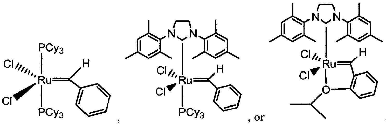
- In other examples the metathesis catalyst is of the formula

- The metathesis reaction may be performed, for example, at a temperature between about 25°C and 110°C, and more preferably, at a temperature of about 50°C.
- The metathesis reaction may be performed with an organic solvent, such as dichloromethane, dichloroethane, trichloroethane, or toluene.
- The reactions disclosed herein may, for example, be carried out on a solid support. Suitable solid supports include particles, strands, precipitates, gels, sheets, tubing, spheres, containers, capillaries, pads, slices, films, plates, slides, discs, membranes, etc. These solid supports can be made from a wide variety of materials, including polymers, plastics, ceramics, polysaccharides, silica or silica-based materials, carbon, metals, inorganic glasses, membranes, or composites thereof. The substrate is preferably flat but may take on a variety of alternative surface configurations. For example, the substrate may contain raised or depressed regions on which the synthesis takes place. The substrate and its surface preferably form a rigid support on which to carry out the reactions described herein. Other substrate materials will be readily apparent to those of ordinary skill in the art upon review of this disclosure.
- The metathesis reaction performed may initially yield a compound in which the newly formed carbon-carbon bond is a double bond. This double bond can be subsequently converted to a single bond by hydrogenation methods known in the art.
- Further disclosed herein is a method of inhibiting viral infectivity of a subject. This method involves administering to the subject an effective amount of a composition that includes a peptide as disclosed herein that mimics at least a portion of a class IN- or C-peptide helix. In a preferred example the method Inhibits HIV infectivity of a subject. In this example the composition includes a peptide as disclosed herein that mimics a gp41 N-peptide helix or gp41 C-peptide helix.
- Inhibiting infectivity refers to any decrease in the rate and/or degree of transmission of the virus. This includes, without limitation, inhibiting transmission of the virus from one individual to another, as well as inhibiting further transmission of the virus within an infected individual.
- The composition may be administered to individuals already infected with the virus (to inhibit transmission of the virus to others, and/or to inhibit further transmission of the virus within the infected individual), as well as to individuals not already infected with the virus (to inhibit transmission of the virus to the individual).
- As will be apparent to one of ordinary skill in the art, administering may be carried out using generally known methods.
- Administration can be accomplished either via systemic administration to the subject or via targeted administration to affected cells. Exemplary routes of administration include, without limitation, by intratracheal inoculation, aspiration, airway instillation, aerosolization, nebulization, intranasal instillation, oral or nasogastric instillation, intraperitoneal injection, intravascular injection, topically, transdermally, parenterally, subcutaneously, intravenous injection, intra-arterial injection (such as via the pulmonary artery), intramuscular injection, intrapleural instillation, intraventricularly, intralesionally, by application to mucous membranes (such as that of the nose, throat, bronchial tubes, genitals, and/or anus), or implantation of a sustained release vehicle.
- Typically, the peptide disclosed herein will be administered to a mammal as a pharmaceutical formulation that includes the therapeutic agent and any pharmaceutically acceptable adjuvants, carriers, excipients, and/or stabilizers, and can be in solid or liquid form, such as tablets, capsules, powders, solutions, suspensions, or emulsions. The compositions preferably contain from about 0.01 to about 99 weight percent, more preferably from about 2 to about 60 weight percent, of therapeutic agent together with the adjuvants, carriers and/or excipients. The amount of active compound in such therapeutically useful compositions is such that a suitable dosage unit will be obtained.
- The agents may be orally administered, for example, with an inert diluent, or with an assimilable edible carrier, or they may be enclosed in hard or soft shell capsules, or they may be compressed into tablets, or they may be incorporated directly with the food of the diet. For oral therapeutic administration, these active compounds may be incorporated with excipients and used in the form of tablets, capsules, elixirs, suspensions, syrups, and the like. Such compositions and preparations should contain at least 0.1 % of the agent. The percentage of the agent in these compositions may, of course, be varied and may conveniently be between about 2% to about 60% of the weight of the unit. The amount of the agent in such therapeutically useful compositions is such that a suitable dosage will be obtained.
- The tablets, capsules, and the like may also contain a binder such as gum tragacanth, acacia, corn starch, or gelatin; excipients such as dicalcium phosphate; a disintegrating agent such as corn starch, potato starch, or alginic acid; a lubricant such as magnesium stearate; and a sweetening agent such as sucrose, lactose, or saccharin. When the dosage unit form is a capsule, it may contain, in addition to materials of the above type, a liquid carrier, such as a fatty oil.
- Various other materials may be present as coatings or to modify the physical form of the dosage unit. For instance, tablets may be coated with shellac, sugar, or both. A syrup may contain, in addition to active ingredient(s), sucrose as a sweetening agent, methyl and propylparabens as preservatives, a dye, and flavoring such as cherry or orange flavor.
- The agents may also be administered parenterally. Solutions or suspensions of the agent can be prepared in water suitably mixed with a surfactant, such as hydroxypropylcellulose. Dispersions can also be prepared in glycerol, liquid polyethylene glycols, and mixtures thereof in oils. Illustrative oils are those of petroleum, animal, vegetable, or synthetic origin, for example, peanut oil, soybean oil, or mineral oil. In general, water, saline, aqueous dextrose and related sugar solutions, and glycols such as propylene glycol or polyethylene glycol, are preferred liquid carriers, particularly for injectable solutions. Under ordinary conditions of storage and use, these preparations contain a preservative to prevent the growth of microorganisms.
- The pharmaceutical forms suitable for injectable use include sterile aqueous solutions or dispersions and sterile powders for the extemporaneous preparation of sterile injectable solutions or dispersions. In all cases, the form must be sterile and must be fluid to the extent that easy syringability exists. It must be stable under the conditions of manufacture and storage and must be preserved against the contaminating action of microorganisms, such as bacteria and fungi. The carrier can be a solvent or dispersion medium containing, for example, water, ethanol, polyol (e.g., glycerol, propylene glycol, and liquid polyethylene glycol), suitable mixtures thereof, and vegetable oils.
- The disclosed herein may also be administered directly to the airways in the form of an aerosol. For use as aerosols, the compounds disclosed herein in solution or suspension may be packaged in a pressurized aerosol container together with suitable propellants, for example, hydrocarbon propellants like propane, butane, or isobutane with conventional adjuvants. The materials disclosed herein also may be administered in a non-pressurized form such as in a nebulizer or atomizer.
- The agents disclosed herein may be administered directly to a targeted tissue, e.g., tissue that is susceptible to infection by the virus. Additionally and/or alternatively, the agent may be administered to a non-targeted area along with one or more agents that facilitate migration of the agent to (and/or uptake by) a targeted tissue, organ, or cell. While the targeted tissue can be any tissue subject to infection by the virus, preferred target tissues in the case of inhibiting HIV-1 infection include mucous membranes of the mouth, genitals, and rectum. As will be apparent to one of ordinary skill in the art, the therapeutic agent itself be modified to facilitate its transport to (and uptake by) the desired tissue, organ, or cell.
- Exemplary delivery devices include, without limitation, nebulizers, atomizers, liposomes, transdermal patches, implants, implantable or injectable protein depot compositions, and syringes. Other delivery systems which are known to those of skill in the art can also be employed to achieve the desired delivery of the therapeutic agent to the desired organ, tissue, or cells in vivo
- Any suitable approach for delivery of the agents can be utilized Typically, the agent will be administered to a patient in a vehicle that delivers the agent(s) to the target cell, tissue, or organ.
- One approach for delivering agents into cells involves the use of liposomes. Basically, this involves providing a liposome which includes agent(s) to be delivered, and then contacting the target cell, tissue, or organ with the liposomes under conditions effective for delivery of the agent into the cell, tissue, or organ.
- Liposomes are vesicles comprised of one or more concentrically ordered lipid bilayers which encapsulate an aqueous phase. They are normally not leaky, but can become leaky if a hole or pore occurs in the membrane, if the membrane is dissolved or degrades, or if the membrane temperature is increased to the phase transition temperature. Current methods of drug delivery via liposomes require that the liposome carrier ultimately become permeable and release the encapsulated drug at the target site. This can be accomplished, for example, in a passive manner where the liposome bilayer degrades over time through the action of various agents in the body. Every liposome composition will have a characteristic half-life in the circulation or at other sites in the body and, thus, by controlling the half-life of the liposome composition, the rate at which the bilayer degrades can be somewhat regulated.
- In contrast to passive drug release, active drug release involves using an agent to induce a permeability change in the liposome vesicle. Liposome membranes can be constructed so that they become destabilized when the environment becomes acidic near the liposome membrane (see, e.g., Wang & Huang, "pH-Sensitive Immunoliposomes Mediate Target-cell-specific Delivery and Controlled Expression of a Foreign Gene in Mouse," Proc. Nat'l Acad. Sci. USA 84:7851-5 (1987),). When liposomes are endocytosed by a target cell, for example, they can be routed to acidic endosomes which will destabilize the liposome and result in drug release.
- Alternatively, the liposome membrane can be chemically modified such that an enzyme is placed as a coating on the membrane, which enzyme slowly destabilizes the liposome. Since control of drug release depends on the concentration of enzyme initially placed in the membrane, there is no real effective way to modulate or alter drug release to achieve "on demand" drug delivery. The same problem exists for pH-sensitive liposomes in that as soon as the liposome vesicle comes into contact with a target cell, it will be engulfed and a drop in pH will lead to drug release.
- This liposome delivery system can also be made to accumulate at a target organ, tissue, or cell via active targeting (e.g., by incorporating an antibody or hormone on the surface of the liposomal vehicle). This can be achieved according to known methods.
- Different types of liposomes can be prepared according to Bangham et al., "Diffusion of Univalent Ions Across the Lamellae of Swollen Phospholipids," J. Mol. Biol. 13:238-52 (1965);
U.S. Patent No. 5,653,996 to Hsu ;U.S. Patent No. 5,643,599 to Lee et al. ;U.S. Patent No. 5,885,613 to Holland et al. ;U.S. Patent No. 5,631,237 to Dzau & Kaneda ; andU.S. Patent No. 5,059,421 to Loughrey et al. , - These liposomes can be produced such that they contain, in addition to the therapeutic agents of the present invention, other therapeutic agents, such as anti-inflammatory agents, which would then be released at the target site (e.g., Wolff et al., "The Use of Monoclonal Anti-Thyl IgG1 for the Targeting of Liposomes to AKR-A Cells in Vitro and in Vivo," Biochim. Biophys. Acta 802:259-73 (1984),).
- An alternative approach for delivery of proteins or polypeptide agents (e.g., peptides of the present invention) involves the conjugation of the desired protein or polypeptide to a polymer that is stabilized to avoid enzymatic degradation of the conjugated protein or polypeptide. Conjugated proteins or polypeptides of this type are described in
U.S. Patent No. 5,681,811 to Ekwuribe , - Yet another approach for delivery of proteins or polypeptide agents involves preparation of chimeric proteins according to
U.S. Patent No. 5,817,789 to Heartlein et al. ,. The chimeric protein can include a ligand domain and the polypeptide agent (e.g., the artificial α-helix described herein). The ligand domain is specific for receptors located on a target cell. Thus, when the chimeric protein is delivered intravenously or otherwise introduced into blood or lymph, the chimeric protein will adsorb to the targeted cell, and the targeted cell will internalize the chimeric protein. - Administration can be carried out as frequently as required and for a duration that is suitable to provide effective treatment against viral infection. For example, administration can be carried out with a single sustained-release dosage formulation or with multiple daily doses. Administration can be carried out before, concurrently with, and/or after exposure of the subject to the virus.
- The amount to be administered will, of course, vary depending upon the treatment regimen. Generally, an agent is administered to achieve an amount effective for a reduction in infectivity of the virus (i.e., a therapeutically effective amount). Thus, a therapeutically effective amount can be an amount which is capable of at least partially preventing transmission of the virus to the subject, or spread of the virus within the subject. The dose required to obtain an effective amount may vary depending on the agent, formulation, virus, and individual to whom the agent is administered.
- Determination of effective amounts may also involve in vitro assays in which varying doses of agent are administered to cells in culture and the concentration of agent effective for inhibiting Infectivity is determined in order to calculate the concentration required in vivo. Effective amounts may also be based on in vivo animal studies. A therapeutically effective amount can be determined empirically by those of skill in the art.
- Peptides 1-11 were synthesized as shown in
Scheme 1. Peptides 1 (i.e., AcMTWMEWDREINNYT-NH2 (SEQ ID NO: 14)), 10 (i.e., AcMTWEEWDKKIEEYTKKI-NH2 (SEQ ID NO: 15)), and 11 (i.e., peptide IZN17, AcIKKEIEAIKKEQEAIKKKIEAIEKLLQLTVWGIKQLQARIL-NH2 (SEQ ID NO: 16)), and resin-bound bis-olefins (17) were synthesized by conventional Fmoc solid phase chemistry on Rink amide HMBA resin (NovaBiochem), 0.05-0.15 mmol scale, with appropriate substitutions of N(allyl)-dipeptides 13-16 and 4-pentenoic acid. In each coupling step, the Fmoc group was removed by treatment with 20% piperidine in NMP (2×20 min). The next Fmoc amino acid (4 equiv.) in the sequence was activated with HBTU (3.6 equiv.) in a 5% DIPEA/NMP solution for 15 minutes, added to the resin bearing the free amine. The resulting mixture was shaken for 60 minutes. The coupling efficiency for each step was monitored by ninhydrin test. After the peptide was assembled on the resin, the resin was thoroughly washed with DMF, methanol, and dichloromethane, respectively, and dried under vacuum overnight.
- Microwave-assisted ring-closing metathesis reactions on resin-bound bis-olefins (17) were performed with the Hoveyda-Grubbs catalyst (0.15 equiv.) in dichloroethane as described in Chapman & Arora, "Optimized Synthesis of Hydrogen-bond Surrogate Helices: Surprising Effects of Microwave Heating on the Activity of Grubbs Catalysts," Org. Lett. 8:5825-8 (2006), The reaction mixture was irradiated with these settings: 250 W maximum power, 120 °C, 5 minute ramp time, and 10 minute hold time. Resin bound peptides were cleaved from the resin by treatment with a cleavage cocktail (CF3CO2H:H2O:triisopropylsilane, 95:2.5:2.5) for 1.5 hours, and purified by reversed-phase HPLC to afford HBS α-helices 2-9, which are shown in
Figures 3-10 . HPLC plots for peptides 1-12 are shown inFigures 11A-L . - Peptides 1-12 were examined using liquid chromatography-mass spectrometry ("LCMS"). LCMS data were obtained on an Agilent 1100 series. The LCMS results are shown in Table 2.
Table 2. Mass spectrometry results for Peptides 1-12 (LC/MSD (XCT) electrospray trap). peptide Expected [M+H]+ Found [M+H]+ 1 1929.8 1929.6 2 2068.0 2068.6 3 1110.6 1111.1 4 1439.7 1440.5 5 1631.8 1632.2 6 1981.0 1981.8 7 2080.0 2080.0 8 2224.0 2225.0 9 2350.2 2351.2 10 2298.2 2299.0 11 2478.0 2478.6 12 4855.9 4854.7 - CD spectra were recorded on an AVIV 202SF CD spectrometer equipped with a temperature controller using 1 mm length cells and a scan speed of 5 nm/min. The spectra were averaged over 10 scans with the baseline subtracted from analogous conditions as that for the samples. The samples were prepared in 0.1x phosphate buffered saline (13.7 mM NaCl, 1 mM phosphate, 0.27 mM KCl, pH 7.4), containing 10% trifluoroethanol, with the final peptide concentration of 50-100 µM. The concentrations of unfolded peptides were determined by the UV absorption of the tyrosine residue at 276 nm in 6.0 M guanidinium hydrochloride aqueous solution. The helix content of each peptide was determined from the mean residue CD at 222 nm, [θ]222 (deg cm2 dmol-1) corrected for the number of amino acids. Percent helicity was calculated from the ratio [θ]222/[θ]max, where [θ]max = (-44000 + 250T)(1 - k/n), with k = 4.0 and n = number of residues. For details on θmax calculations for HBS helices, see Wang et al., "Evaluation of Biologically Relevant Short α-Helices Stabilized by a Main-chain Hydrogen-bond Surrogate," J. Am. Chem. Soc. 128:9248-56 (2006), . The CD spectra for peptides 1-10 are shown in Figures 12A-J.
- The relative affinity of peptides 1-10 for IZN17 (i.e., peptide 11) was determined using a fluorescence polarization-based competitive binding assay (Eckert & Kim, "Design of Potent Inhibitors of HIV-1 Entry from the gp41 N-Peptide Region," Proc. Nat'l Acad. Sci. U.S.A. 98:11187-92 (2001); Stephens et al., "Inhibiting HIV Fusion with a β-Peptide Foldamer," J. Am. Chem. Soc. 127:13126-7 (2005),) with fluorescein-labeled
peptide 12, shown inFigure 13. Figure 14 is a graph showing the saturation binding curve of fluorescein-labeledpeptide 12 with IZN17 in PBS buffer at 25 °C. - All samples were prepared in 96-well plates in 1x phosphate buffered saline (137 mM NaCl, 10 mM phosphate, 2.7 mM KCl, pH 7.4) with 0.1% pluronic F-68 (Sigma). A solution of 25 µM IZN17 and 15 nM fluorescein-labeled
peptide 12 was incubated at 25 °C. After 1 hour, appropriate concentrations (10 nM to 500 µM) of the antagonists (peptides 1-10) were added. The total volume of the incubation solution was 60 µL. After 1 hour, the amount of dissociatedfluorescent probe 12 was determined using a DTX 880 Multimode Detector (Beckman) at 25 °C, with excitation and emission wavelengths of 485 and 525 nm, respectively. - The binding affinity (KD ) values reported in Table 3 (see Example 6) for each peptide are the averages of 3~5 individual measurements, and were determined by fitting the experimental data to a sigmoidal dose-response nonlinear regression model on GraphPad Prism 4.0.
- It is noted that this competitive binding assay does not allow accurate estimates of Kd values much lower than the binding values of the fluorescent probe.
Figure 15 shows a graph of the fraction of bound probe versus ligand concentration as a function of ligand binding affinity. The narrowing of the space between curves below Kd of 5 µM illustrates the limits of this assay. - Cell-cell fusion (i.e., syncytium formation) was assayed by coculturing CHO[HIVe] (clone 7d2) cells expressing HXB2 envelope and tat with U373-MAGI cells (M. Emerman and A. Geballe, National Institutes of Health AIDS Research and Reference Reagent Program) in the presence of different concentrations of peptides 1-10. Cell fusion allows the expression of nuclear β-galactosidase from the U373-MAGI indicator cell line and can be quantitated by monitoring β-galactosidase activity. After an overnight incubation at 37°C after coculture, β-galactosidase enzymatic activity was measured with the Mammalian β-galactosidase Chemiluminescent Assay Kit (Gal-Screen from Applied Biosystems). The peptide inhibitor concentrations at which activities were reduced by 50% (IC50) relative to control samples lacking peptide inhibitor were calculated by fitting data to the variable-slope-sigmoid equation using the Prism program.
- The cytotoxic effect of peptides 1-10 on U373-MAGI cells was measured in the presence of a series of diluted inhibitors for 6 days, and cell viability was quantitated with an MTT assay (Mosmann, "Rapid Colorimetric Assay for Cellular Growth and Survival: Application to Proliferation and Cytotoxicity Assays," J. Immunol. Meth. 65:55-63 (1983),). . No cytotoxicity was observed for peptides up to 200 µM concentrations.
- The present gp41-targeting studies were begun by mimicking the 14-residue C-peptide (1) derived from gp41 that contains residues W628, W631, and I635, as shown in Table 3. This sequence would be expected to bind the gp41 hydrophobic pocket based on its crystal structure (shown in
Figure 1B ) (Chan et al., "Core Structure of gp41 from the HIV Envelope Glycoprotein," Cell 89:263-73 (1997),), but has been previously shown to be ineffective in cell-cell fusion assays (Sia et al., "Short Constrained Peptides That Inhibit HIV-1 Entry," Proc. Nat'l Acad. Sci. U.S.A. 99:14664-9 (2002),).
- A previously described fluorescence polarization assay was utilized to determine the in vitro binding affinity of unconstrained peptides and HBS helices for a stable model of the gp41 N-terminal three strand coiled-coil, IZN17, which contains the binding site for residues W628, W631, and I635 (see Example 3, supra) (Eckert & Kim, "Design of Potent Inhibitors of HIV-1 Entry from the gp41 N-Peptide Region," Proc. Nat'l Acad. Sci. U.S.A. 98:11187-92 (2001), ). . A fluorescein-labeled C-peptide derivative (i.e., suc-MTWMEWDREINNYTCFlu (SEQ ID NO: 10); peptide 12) containing residues 628-641 of gp41 was used as a probe. The competitive displacement of this probe by HBS helices afforded the Kd value for each peptide shown in Table 3 and
Figure 16B (Stephens et al., "Inhibiting HIV Fusion with a β-Peptide Foldamer," J. Am. Chem. Soc. 127:13126-7 (2005), ).Peptide 12 bound IZN 17 with a Kd value of 24 µM, within range of the previously reported values (Sia et al., "Short Constrained Peptides That Inhibit HIV-1 Entry," Proc. Nat'l Acad. Sci. U.S.A. 99:14664-9 (2002); Stephens et al., "Inhibiting HIV Fusion with a β-Peptide Foldamer," J. Am. Chem. Soc. 127:13126-7 (2005),). The competition assay provided a binding affinity of 37 µM forpeptide 1, which is also in range of reported values, although aggregation of the peptide made it difficult to obtain accurate Kd values. - Circular dichroism spectroscopy (see Example 2, supra) suggested that 1 is unstructured or very weakly helical in 10% trifluoroethanol ("TFE") in PBS buffer (Sia et al., "Short Constrained Peptides That Inhibit HIV-1 Entry," Proc. Nat'l Acad. Sci. U.S.A. 99:14664-9 (2002), ). It was conjectured that stabilization of this peptide in helical conformation with the HBS approach may increase its helicity and affinity for IZN17. HBS α-
helix 2 is roughly four times more helical than 1, as shown inFigure 16A , but did not bind the target protein with higher affinity, as shown in Table 3. This suggests that there may be a complex interplay between helicity of the peptide and its binding affinity for the target (Martin, "Preorganization in Biological Systems: Are Conformational Constraints Worth the Energy?," Pure Appl. Chem. 79:193-200 (2007); Benfield et al., "Ligand Preorganization May Be Accompanied by Entropic Penalties in Protein-ligand Interactions," Angew. Chem. Int'l Ed. Engl. 45:6830-5 (2006), originally published at Angew. Chem. 118:6984-9 (2006), ). Results with the short peptide and constrained helix mirror those observed by Kim and coworkers (Sia et al., "Short Constrained Peptides That Inhibit HIV-1 Entry," Proc. Nat'l Acad. Sci. U.S.A. 99:14664-9 (2002),) , and prompted the design of a small library of HBS α-helices to identify sequences that bind IZN17 with higher affinity. A representative selection (i.e., peptides 2-9) of these HBS helices is shown in Table 3. - The limited solubility of short C-peptides in aqueous solutions has been proposed as a key reason for their inactivity (Otaka et al., "Remodeling of gp41-C34 Peptide Leads to Highly Effective Inhibitors of the Fusion of HIV-1 with Target Cells," Angew. Chem. Int'l Ed. Engl. 41:2937-40 (2002), originally published at Angew. Chem. 114:3061-4 (2002),). To improve the solubility and helicity of constrained C-peptides, charged residues were incorporated at positions not expected to be involved in binding to IZN17 (Otaka et al., "Remodeling of gp41-C34 Peptide Leads to Highly Effective Inhibitors of the Fusion of HIV-1 with Target Cells," Angew. Chem. Int'l Ed. Engl. 41:2937-40 (2002), originally published at Angew. Chem. 114:3061-4 (2002),). HBS α-
helix 3 is the shortest peptide designed to contain the Trp/Trp/Ile motif necessary for binding. Two lysine residues were added within this sequence to improve solubility of the peptide. HBS α-helix 4 was designed such that the Trp/Trp/Ile motif would lie outside of the constraint in order to probe potential steric effects of the HBS constraints. A pair of glutamic acid and lysine residues were placed at i and i+4 positions to further stabilize the α-helical conformation through potential salt-bridging interactions (Marqusee & Baldwin, "Helix Stabilization by Glu- ···Lys+ Salt Bridges in Short Peptides of de Novo Design," Proc. Nat'l Acad. Sci. U.S.A. 84:8898-902 (1987); Shi et al., "Stabilization of α-Helix Structure by Polar Side-chain Interactions: Complex Salt Bridges, Cation-pi Interactions, and C-H Em Leader O H-bonds," Biopolymers 60:366-80 (2001),). Neither of these two HBS helices bound IZN17, suggesting that more contact points are needed to target the coiled-coil complex. HBS α-helix 5 contains tyrosine-638 in addition to the Trp/Trp/Ile residues. Tyr-638 occupies the d position in the heptad and is expected to directly contact the N-terminal three strand coiled-coil, as shown inFigure 1C . The sequence of HBS α-helix 5 is similar to that of theparent compounds Figure 16A . The low binding affinity of 5 relative to 2 highlights the potential role of threonine-627, which occupies the g position in the heptad, as shown inFigure 1C , and was not projected to contribute significantly to binding. Reintroduction of the Met-626 and Thr-627 residues and incorporation of charged residues at positions not expected to be involved in binding provided HBS α-helix 6, which bound IZN17 with a higher affinity than the parent HBS α-helix 2. The role of Thr-627 in the binding of HBS helices is also highlighted by HBS α-helix 7, in which this residue is replaced with an alanine residue. HBS α-helix 7 bound IZN17 by twenty-fold lower affinity than did 6. - Glutamic acid residues were placed at the N-terminus of HBS α-
helices - HBS α-
helices helices - Cell-cell fusion (i.e., syncytium formation) was assayed by co-culturing CHO[HIVe] (clone 7d2) cells expressing HXB2 envelope and tat (Kozarsky et al., "Glycosylation and Processing of the Human ),) with U373-MAGI cells in the presence of different concentrations of the peptide inhibitors. Cell fusion allows the expression of nuclear β-galactosidase from the U373-MAGI indicator cell line and can be quantitated by monitoring β-galactosidase activity.
- Inhibition of gp41-mediated cell fusion by short peptides is a challenging feat and has only been accomplished with a handful of synthetic peptides (Sia et al., "Short Constrained Peptides That Inhibit HIV-1 Entry," Proc. Nat'l Acad. Sci. U.S.A. 99:14664-9 (2002); Stephens et al., "Inhibiting HIV Fusion with a β-Peptide Foldamer," J. Am. Chem. Soc. 127:13126-7 (2005); Eckert et al., "Inhibiting HIV-1 Entry: Discovery of D-Peptide Inhibitors That Target the gp41 Coiled-coil Pocket," Cell 99:103-15 (1999),). . It was found that only HBS α-
helix 9 inhibited cell fusion with an EC50 value of 43 µM, as shown inFigure 16C . This value is comparable to those measured for side chain constrained α-helices (Sia et al., "Short Constrained Peptides That Inhibit HIV-1 Entry," Proc. Nat'l Acad. Sci. U.S.A. 99:14664-9 (2002),), cyclic D-peptides (Eckert et al., "Inhibiting HIV-1 Entry: Discovery of D-Peptide Inhibitors That Target the gp41 Coiled-coil Pocket," Cell 99:103-15 (1999),), aromatic foldamers (Ernst et al., "Design of a Protein Surface Antagonist Based on α-Helix Mimicry: Inhibition of gp41 Assembly and Viral Fusion," Angew. Chem. Int'l Ed. Engl. 41:278-81 (2002), originally published at Angew. Chem. 114:282-91 (2002),), and β-peptide foldamers (Stephens et al., "Inhibiting HIV Fusion with a β-Peptide Foldamer," J. Am. Chem. Soc. 127:13126-7 (2005),). Other HBS α-helices (2-8) did not provide any hints of cell fusion inhibition at concentrations up to 200 µM.Unconstrained peptide 10, which bound IZN17 with a similar affinity as 9, remained ineffective in the cell culture assay. This result potentially reflects the proteolytic instability of the unconstrained peptide, as stabilization of peptides in α-helical conformation is expected to enhance their resistance to proteases (Tyndall et al., "Proteases Universally Recognize β Strands in Their Active Sites," Chem. Rev. 105:973-99 (2005),). . Improvements in the proteolytic stability of HBS α-helices as compared to their unconstrained counterparts has previously been reported (Wang et al., "Enhanced Metabolic Stability and Protein-binding Properties of Artificial α Helices Derived from a Hydrogen-bond Surrogate: Application to Bcl-xL," Angew. Chem. Int'l Ed. Engl. 44:6525-9 (2005), originally published at Angew. Chem. 117:6683-7 (2005),) - In summary, through rational design and synthesis, an artificial α-helix (9) that inhibits gp41-mediated cell fusion has been developed. As formation of coiled-coil assemblies is a prerequisite for the fusion of several classes of viruses to their host cells (Dimitrov, "Virus Entry: Molecular Mechanisms and Biomedical Applications," Nat. Rev. Microbiol. 2:109-22 (2004)), this work suggests that HBS helices may be effective scaffolds for the generation of small molecule inhibitors or antigens against these viruses (English et al., "Rational Development of β-Peptide Inhibitors of Human Cytomegalovirus Entry," J. Biol. Chem. 281:2661-7 (2006); Shepherd et al., "Modular α-Helical Mimetics with Antiviral Activity Against Respiratory Syncitial Virus, J. Am. chem. Soc. 128:13284-9 (2006),).
Claims (7)
- A peptide having a stable, internally-constrained alpha-helix, wherein said alpha helix is constrained by a crosslink formed by a carbon-carbon bond-forming reaction, and further wherein the peptide mimics at least a portion of a class I C-peptide helix from human immunodeficiency virus type 1; wherein the peptide comprises the formula:

------ is a single or double carbon-carbon bond;is a single bond and is cis or trans when
------ is a double bond;n is 1 or 2;m is zero or any positive integer;R is hydrogen, an amino acid side chain, an alkyl group, or an aryl group;R1 is an amino acid side chain, an alkyl group, or an aryl group;R2 is an amino acid, second peptide, -OR, -CH2NH2, an alkyl group, an aryl group, hydrogen, or a group having a formula R3 is a fourth peptide and wherein R4 is an amino acid, third peptide, -OR, -NH2, an alkyl group, or an aryl group; andwherein the peptide comprises the amino acid sequence of SEQ ID NO: 9, and has an internally-constrained alpha-helix spanning residues 1-4 of SEQ ID NO: 9.
 - The peptide of claim 1, wherein the carbon-carbon bond-forming reaction is metathesis.
- A pharmaceutical composition comprising a peptide according to claim 1 and a pharmaceutically acceptable vehicle.
- A composition comprising a peptide according to claim 1 for use in inhibiting infectivity of a human immunodeficiency virus type 1 in a subject.
- A peptide which is an inhibitor of human immunodeficiency virus type 1 infectivity obtainable by: selecting a precursor peptide comprising at least a portion of a class I C-peptide helix from human immunodeficiency virus type 1; and promoting formation of a carbon-carbon bond, wherein said bond formation results in a stable, internally-constrained alpha-helix; and wherein the peptide comprises the amino acid sequence of SEQ ID NO: 9, and has an internally-constrained alpha-helix spanning residues 1-4 of SEQ ID NO:9
- The peptide according to claim 5, wherein the bond formation introduces a non-native carbon-carbon bond, or wherein said carbon-carbon bond is formed by metathesis.
- The peptide according to claim 5, wherein the peptide comprises the formula:

------ is a single or double carbon-carbon bond;is a single bond and is cis or trans when------ is a double bond;n is 1 or 2;m is zero or any positive integer;R is hydrogen, an amino acid side chain, an alkyl group, or an aryl group;R1 is an amino acid side chain, an alkyl group, or an aryl group;R2 is an amino acid, second peptide, -OR, -CH2NH2, an alkyl group, an aryl group, hydrogen, or a group having a formula wherein R4 is an amino acid, third peptide, -OR, -NH2, an alkyl group, or an aryl group; and R3 is a fourth peptide.
Applications Claiming Priority (2)
| Application Number | Priority Date | Filing Date | Title |
|---|---|---|---|
| US1811807P | 2007-12-31 | 2007-12-31 | |
| PCT/US2008/088667 WO2009110952A2 (en) | 2007-12-31 | 2008-12-31 | Control of viral-host membrane fusion with hydrogen bond surrogate-based artificial helices |
Publications (3)
| Publication Number | Publication Date |
|---|---|
| EP2247298A2 EP2247298A2 (en) | 2010-11-10 |
| EP2247298A4 EP2247298A4 (en) | 2012-06-06 |
| EP2247298B1 true EP2247298B1 (en) | 2016-08-31 |
Family
ID=41056505
Family Applications (1)
| Application Number | Title | Priority Date | Filing Date |
|---|---|---|---|
| EP08873198.9A Not-in-force EP2247298B1 (en) | 2007-12-31 | 2008-12-31 | Control of viral-host membrane fusion with hydrogen bond surrogate-based artificial helices |
Country Status (5)
| Country | Link |
|---|---|
| US (1) | US8871899B2 (en) |
| EP (1) | EP2247298B1 (en) |
| JP (1) | JP5653219B2 (en) |
| CA (1) | CA2710964A1 (en) |
| WO (1) | WO2009110952A2 (en) |
Families Citing this family (23)
| Publication number | Priority date | Publication date | Assignee | Title |
|---|---|---|---|---|
| JP2010510236A (en) * | 2006-11-15 | 2010-04-02 | ダナ−ファーバー キャンサー インスティテュート インク. | Stabilized MAML peptides and methods of use thereof |
| BRPI0720306A2 (en) | 2006-12-14 | 2014-02-04 | Aileron Therapeutics Inc | BIS-SUFIDRIL MACROCYCLING SYSTEMS |
| CA2678941C (en) | 2007-02-23 | 2018-11-27 | Aileron Therapeutics, Inc. | Triazole macrocycle systems |
| JP5631201B2 (en) | 2007-03-28 | 2014-11-26 | プレジデント アンド フェローズ オブ ハーバード カレッジ | Stitched polypeptide |
| US8613956B2 (en) * | 2008-06-23 | 2013-12-24 | International Flora Technologies, Ltd. | Cosmetic particles that transform from hard to soft particles comprising hydrogenated long-chain triglyceride oils |
| CN102482336B (en) | 2008-09-22 | 2015-05-06 | 爱勒让治疗公司 | Peptidomimetic macrocycles |
| CA2743177A1 (en) | 2008-11-24 | 2010-05-27 | Aileron Therapeutics, Inc. | Peptidomimetic macrocycles with improved properties |
| AU2010204648B2 (en) | 2009-01-14 | 2016-09-01 | Aileron Therapeutics, Inc. | Peptidomimetic macrocycles |
| JP2013505300A (en) | 2009-09-22 | 2013-02-14 | エルロン・セラピューティクス・インコーポレイテッド | Peptidomimetic macrocycle |
| KR102104762B1 (en) | 2010-08-13 | 2020-04-24 | 에일러론 테라퓨틱스 인코포레이티드 | Peptidomimetic macrocycles |
| EP2768518A4 (en) | 2011-10-18 | 2015-05-27 | Aileron Therapeutics Inc | PEPTIDOMIMETIC MACROCYCLES |
| WO2013059530A2 (en) * | 2011-10-18 | 2013-04-25 | Aileron Therapeutics, Inc. | Peptidomimetic macrocycles |
| CA2864120A1 (en) | 2012-02-15 | 2013-08-22 | Aileron Therapeutics, Inc. | Triazole-crosslinked and thioether-crosslinked peptidomimetic macrocycles |
| RU2642299C2 (en) | 2012-02-15 | 2018-01-24 | Эйлерон Терапьютикс, Инк. | p53 PEPTIDOMIMETIC MACROCYCLES |
| BR112015009470A2 (en) | 2012-11-01 | 2019-12-17 | Aileron Therapeutics Inc | disubstituted amino acids and their methods of preparation and use |
| US9605026B2 (en) | 2013-01-19 | 2017-03-28 | New York University | Hydrogen-bond surrogate peptides and peptidomimetics for p53 reactivation |
| WO2015034932A1 (en) * | 2013-09-03 | 2015-03-12 | New York University | INHIBITING INTERACTION BETWEEN HIF-1αAND P300/CBP WITH HYDROGEN BOND SURROGATE-BASED HELICES |
| CN107106642B (en) | 2014-09-24 | 2021-02-26 | 艾瑞朗医疗公司 | Peptidomimetic macrocycles and formulations thereof |
| EP3197478A4 (en) | 2014-09-24 | 2018-05-30 | Aileron Therapeutics, Inc. | Peptidomimetic macrocycles and uses thereof |
| JP2018516844A (en) | 2015-03-20 | 2018-06-28 | エルロン・セラピューティクス・インコーポレイテッドAileron Therapeutics,Inc. | Peptidomimetic macrocycles and uses thereof |
| US10059741B2 (en) | 2015-07-01 | 2018-08-28 | Aileron Therapeutics, Inc. | Peptidomimetic macrocycles |
| EP3347372A4 (en) | 2015-09-10 | 2019-09-04 | Aileron Therapeutics, Inc. | PEPTIDOMIMETIC MACROCYCLES AS MODULATORS OF MCL-1 |
| CN108822190B (en) * | 2018-05-23 | 2021-11-09 | 中国人民解放军军事科学院军事医学研究院 | Polypeptide and pharmaceutical composition and application thereof |
Citations (1)
| Publication number | Priority date | Publication date | Assignee | Title |
|---|---|---|---|---|
| WO2005118620A2 (en) * | 2004-05-27 | 2005-12-15 | New York University | Methods for preparing internally constraied peptides and peptidomimetics |
Family Cites Families (20)
| Publication number | Priority date | Publication date | Assignee | Title |
|---|---|---|---|---|
| US5446128A (en) * | 1993-06-18 | 1995-08-29 | The Board Of Trustees Of The University Of Illinois | Alpha-helix mimetics and methods relating thereto |
| US5840833A (en) * | 1995-10-27 | 1998-11-24 | Molecumetics, Ltd | Alpha-helix mimetics and methods relating thereto |
| US6150088A (en) * | 1997-04-17 | 2000-11-21 | Whitehead Institute For Biomedical Research | Core structure of gp41 from the HIV envelope glycoprotein |
| US6818740B1 (en) * | 1997-04-17 | 2004-11-16 | Whitehead Institute For Biomedical Research | Inhibitors of HIV membrane fusion |
| US6841657B2 (en) * | 1997-04-17 | 2005-01-11 | Whitehead Institute For Biomedical Research | Inhibitors of HIV membrane fusion |
| US6596497B1 (en) * | 1999-03-17 | 2003-07-22 | New York Blood Center, Inc. | Screening of antiviral compounds targeted to the HIV-1 gp41 core structure |
| US7192713B1 (en) * | 1999-05-18 | 2007-03-20 | President And Fellows Of Harvard College | Stabilized compounds having secondary structure motifs |
| US20030082525A1 (en) * | 1999-12-16 | 2003-05-01 | Whitehead Institute For Biomedical Research | Five-Helix protein |
| US7045552B2 (en) * | 2002-09-27 | 2006-05-16 | Trimeris, Inc. | Pharmaceutical composition for improved administration of HIV gp41-derived peptides, and its use in therapy |
| WO2005044839A2 (en) * | 2003-11-05 | 2005-05-19 | Dana-Farber Cancer Institute, Inc. | Stabilized alpha helical peptides and uses thereof |
| CA2564031A1 (en) * | 2004-04-23 | 2005-11-03 | Conjuchem Biotechnologies Inc. | Method for the purification of albumin conjugates |
| EP2354153A3 (en) * | 2004-06-01 | 2012-05-30 | Merck Sharp & Dohme Corp. | Stable peptide mimetic of HIV gp41 fusion intermediate |
| BRPI0720306A2 (en) * | 2006-12-14 | 2014-02-04 | Aileron Therapeutics Inc | BIS-SUFIDRIL MACROCYCLING SYSTEMS |
| US7981998B2 (en) * | 2006-12-14 | 2011-07-19 | Aileron Therapeutics, Inc. | Bis-sulfhydryl macrocyclization systems |
| CA2678941C (en) * | 2007-02-23 | 2018-11-27 | Aileron Therapeutics, Inc. | Triazole macrocycle systems |
| JP5631201B2 (en) * | 2007-03-28 | 2014-11-26 | プレジデント アンド フェローズ オブ ハーバード カレッジ | Stitched polypeptide |
| JP5788178B2 (en) | 2008-01-23 | 2015-09-30 | ダナ ファーバー キャンサー インスティテュート インコーポレイテッド | Compositions and methods for the treatment of viral infections |
| CN102482336B (en) * | 2008-09-22 | 2015-05-06 | 爱勒让治疗公司 | Peptidomimetic macrocycles |
| CA2743177A1 (en) * | 2008-11-24 | 2010-05-27 | Aileron Therapeutics, Inc. | Peptidomimetic macrocycles with improved properties |
| AU2010306718A1 (en) * | 2009-10-14 | 2012-05-24 | Aileron Therapeutics, Inc. | Improved peptidomimetic macrocycles |
-
2008
- 2008-12-31 CA CA2710964A patent/CA2710964A1/en not_active Abandoned
- 2008-12-31 WO PCT/US2008/088667 patent/WO2009110952A2/en not_active Ceased
- 2008-12-31 EP EP08873198.9A patent/EP2247298B1/en not_active Not-in-force
- 2008-12-31 JP JP2010540953A patent/JP5653219B2/en not_active Expired - Fee Related
- 2008-12-31 US US12/811,088 patent/US8871899B2/en active Active
Patent Citations (1)
| Publication number | Priority date | Publication date | Assignee | Title |
|---|---|---|---|---|
| WO2005118620A2 (en) * | 2004-05-27 | 2005-12-15 | New York University | Methods for preparing internally constraied peptides and peptidomimetics |
Also Published As
| Publication number | Publication date |
|---|---|
| WO2009110952A3 (en) | 2010-03-04 |
| EP2247298A2 (en) | 2010-11-10 |
| JP2011507972A (en) | 2011-03-10 |
| WO2009110952A2 (en) | 2009-09-11 |
| CA2710964A1 (en) | 2009-09-11 |
| US8871899B2 (en) | 2014-10-28 |
| JP5653219B2 (en) | 2015-01-14 |
| US20110046043A1 (en) | 2011-02-24 |
| EP2247298A4 (en) | 2012-06-06 |
Similar Documents
| Publication | Publication Date | Title |
|---|---|---|
| US8871899B2 (en) | Control of viral-host membrane fusion with hydrogen bond surrogate-based artificial helices | |
| Wang et al. | Discovery of hydrocarbon-stapled short α-helical peptides as promising Middle East respiratory syndrome coronavirus (MERS-CoV) fusion inhibitors | |
| He | Synthesized peptide inhibitors of HIV-1 gp41-dependent membrane fusion | |
| JP4682251B2 (en) | HIV fusion inhibitor peptides with improved biological properties | |
| US20080269462A1 (en) | Peptide inhibitors of HIV entry | |
| US20080241156A1 (en) | Inhibitors of hepatitis C virus | |
| US11680086B2 (en) | Lipopeptide for potently inhibiting HIV, derivative thereof, pharmaceutical composition thereof and use thereof | |
| EP4617282A1 (en) | Membrane fusion inhibitor for inhibiting aids virus and drug-resistant strain thereof, and pharmaceutical use thereof | |
| JP2008505059A (en) | Specific virus targeting compounds | |
| CA2423004C (en) | Peptide inhibitors of hiv entry | |
| US20030219451A1 (en) | Stable helical C peptides and uses therefor | |
| Huarte et al. | Membrane-transferring regions of gp41 as targets for HIV-1 fusion inhibition and viral neutralization | |
| Xue | Development of Antiviral Peptidomimetics | |
| CA3273216A1 (en) | Membrane fusion inhibitor for inhibiting aids virus and drug-resistant strain thereof, and pharmaceutical use thereof | |
| AU2012200012B2 (en) | Inhibitors of hepatitis C virus | |
| EP4685153A1 (en) | Broad-spectrum viral membrane fusion inhibitor, and preparation method therefor and use thereof | |
| HK40118032B (en) | Polypeptides capable of inhibiting mers-like coronavirus infections and their applications |
Legal Events
| Date | Code | Title | Description |
|---|---|---|---|
| PUAI | Public reference made under article 153(3) epc to a published international application that has entered the european phase |
Free format text: ORIGINAL CODE: 0009012 |
|
| 17P | Request for examination filed |
Effective date: 20100708 |
|
| AK | Designated contracting states |
Kind code of ref document: A2 Designated state(s): AT BE BG CH CY CZ DE DK EE ES FI FR GB GR HR HU IE IS IT LI LT LU LV MC MT NL NO PL PT RO SE SI SK TR |
|
| AX | Request for extension of the european patent |
Extension state: AL BA MK RS |
|
| DAX | Request for extension of the european patent (deleted) | ||
| A4 | Supplementary search report drawn up and despatched |
Effective date: 20120507 |
|
| RIC1 | Information provided on ipc code assigned before grant |
Ipc: A61K 38/12 20060101AFI20120427BHEP Ipc: C07K 5/00 20060101ALI20120427BHEP |
|
| 17Q | First examination report despatched |
Effective date: 20130703 |
|
| GRAP | Despatch of communication of intention to grant a patent |
Free format text: ORIGINAL CODE: EPIDOSNIGR1 |
|
| INTG | Intention to grant announced |
Effective date: 20160518 |
|
| GRAS | Grant fee paid |
Free format text: ORIGINAL CODE: EPIDOSNIGR3 |
|
| GRAA | (expected) grant |
Free format text: ORIGINAL CODE: 0009210 |
|
| AK | Designated contracting states |
Kind code of ref document: B1 Designated state(s): AT BE BG CH CY CZ DE DK EE ES FI FR GB GR HR HU IE IS IT LI LT LU LV MC MT NL NO PL PT RO SE SI SK TR |
|
| REG | Reference to a national code |
Ref country code: CH Ref legal event code: EP Ref country code: GB Ref legal event code: FG4D |
|
| REG | Reference to a national code |
Ref country code: IE Ref legal event code: FG4D |
|
| REG | Reference to a national code |
Ref country code: DE Ref legal event code: R096 Ref document number: 602008046067 Country of ref document: DE |
|
| REG | Reference to a national code |
Ref country code: AT Ref legal event code: REF Ref document number: 824381 Country of ref document: AT Kind code of ref document: T Effective date: 20161015 |
|
| REG | Reference to a national code |
Ref country code: LT Ref legal event code: MG4D |
|
| REG | Reference to a national code |
Ref country code: NL Ref legal event code: MP Effective date: 20160831 |
|
| REG | Reference to a national code |
Ref country code: AT Ref legal event code: MK05 Ref document number: 824381 Country of ref document: AT Kind code of ref document: T Effective date: 20160831 |
|
| PG25 | Lapsed in a contracting state [announced via postgrant information from national office to epo] |
Ref country code: LT Free format text: LAPSE BECAUSE OF FAILURE TO SUBMIT A TRANSLATION OF THE DESCRIPTION OR TO PAY THE FEE WITHIN THE PRESCRIBED TIME-LIMIT Effective date: 20160831 Ref country code: FI Free format text: LAPSE BECAUSE OF FAILURE TO SUBMIT A TRANSLATION OF THE DESCRIPTION OR TO PAY THE FEE WITHIN THE PRESCRIBED TIME-LIMIT Effective date: 20160831 Ref country code: NO Free format text: LAPSE BECAUSE OF FAILURE TO SUBMIT A TRANSLATION OF THE DESCRIPTION OR TO PAY THE FEE WITHIN THE PRESCRIBED TIME-LIMIT Effective date: 20161130 Ref country code: HR Free format text: LAPSE BECAUSE OF FAILURE TO SUBMIT A TRANSLATION OF THE DESCRIPTION OR TO PAY THE FEE WITHIN THE PRESCRIBED TIME-LIMIT Effective date: 20160831 |
|
| PG25 | Lapsed in a contracting state [announced via postgrant information from national office to epo] |
Ref country code: SE Free format text: LAPSE BECAUSE OF FAILURE TO SUBMIT A TRANSLATION OF THE DESCRIPTION OR TO PAY THE FEE WITHIN THE PRESCRIBED TIME-LIMIT Effective date: 20160831 Ref country code: ES Free format text: LAPSE BECAUSE OF FAILURE TO SUBMIT A TRANSLATION OF THE DESCRIPTION OR TO PAY THE FEE WITHIN THE PRESCRIBED TIME-LIMIT Effective date: 20160831 Ref country code: GR Free format text: LAPSE BECAUSE OF FAILURE TO SUBMIT A TRANSLATION OF THE DESCRIPTION OR TO PAY THE FEE WITHIN THE PRESCRIBED TIME-LIMIT Effective date: 20161201 Ref country code: LV Free format text: LAPSE BECAUSE OF FAILURE TO SUBMIT A TRANSLATION OF THE DESCRIPTION OR TO PAY THE FEE WITHIN THE PRESCRIBED TIME-LIMIT Effective date: 20160831 Ref country code: NL Free format text: LAPSE BECAUSE OF FAILURE TO SUBMIT A TRANSLATION OF THE DESCRIPTION OR TO PAY THE FEE WITHIN THE PRESCRIBED TIME-LIMIT Effective date: 20160831 Ref country code: AT Free format text: LAPSE BECAUSE OF FAILURE TO SUBMIT A TRANSLATION OF THE DESCRIPTION OR TO PAY THE FEE WITHIN THE PRESCRIBED TIME-LIMIT Effective date: 20160831 |
|
| PG25 | Lapsed in a contracting state [announced via postgrant information from national office to epo] |
Ref country code: EE Free format text: LAPSE BECAUSE OF FAILURE TO SUBMIT A TRANSLATION OF THE DESCRIPTION OR TO PAY THE FEE WITHIN THE PRESCRIBED TIME-LIMIT Effective date: 20160831 Ref country code: RO Free format text: LAPSE BECAUSE OF FAILURE TO SUBMIT A TRANSLATION OF THE DESCRIPTION OR TO PAY THE FEE WITHIN THE PRESCRIBED TIME-LIMIT Effective date: 20160831 |
|
| PG25 | Lapsed in a contracting state [announced via postgrant information from national office to epo] |
Ref country code: BE Free format text: LAPSE BECAUSE OF FAILURE TO SUBMIT A TRANSLATION OF THE DESCRIPTION OR TO PAY THE FEE WITHIN THE PRESCRIBED TIME-LIMIT Effective date: 20160831 Ref country code: CZ Free format text: LAPSE BECAUSE OF FAILURE TO SUBMIT A TRANSLATION OF THE DESCRIPTION OR TO PAY THE FEE WITHIN THE PRESCRIBED TIME-LIMIT Effective date: 20160831 Ref country code: SK Free format text: LAPSE BECAUSE OF FAILURE TO SUBMIT A TRANSLATION OF THE DESCRIPTION OR TO PAY THE FEE WITHIN THE PRESCRIBED TIME-LIMIT Effective date: 20160831 Ref country code: PL Free format text: LAPSE BECAUSE OF FAILURE TO SUBMIT A TRANSLATION OF THE DESCRIPTION OR TO PAY THE FEE WITHIN THE PRESCRIBED TIME-LIMIT Effective date: 20160831 Ref country code: PT Free format text: LAPSE BECAUSE OF FAILURE TO SUBMIT A TRANSLATION OF THE DESCRIPTION OR TO PAY THE FEE WITHIN THE PRESCRIBED TIME-LIMIT Effective date: 20170102 Ref country code: BG Free format text: LAPSE BECAUSE OF FAILURE TO SUBMIT A TRANSLATION OF THE DESCRIPTION OR TO PAY THE FEE WITHIN THE PRESCRIBED TIME-LIMIT Effective date: 20161130 Ref country code: DK Free format text: LAPSE BECAUSE OF FAILURE TO SUBMIT A TRANSLATION OF THE DESCRIPTION OR TO PAY THE FEE WITHIN THE PRESCRIBED TIME-LIMIT Effective date: 20160831 |
|
| REG | Reference to a national code |
Ref country code: DE Ref legal event code: R097 Ref document number: 602008046067 Country of ref document: DE |
|
| PG25 | Lapsed in a contracting state [announced via postgrant information from national office to epo] |
Ref country code: IT Free format text: LAPSE BECAUSE OF FAILURE TO SUBMIT A TRANSLATION OF THE DESCRIPTION OR TO PAY THE FEE WITHIN THE PRESCRIBED TIME-LIMIT Effective date: 20160831 |
|
| REG | Reference to a national code |
Ref country code: DE Ref legal event code: R119 Ref document number: 602008046067 Country of ref document: DE |
|
| PLBE | No opposition filed within time limit |
Free format text: ORIGINAL CODE: 0009261 |
|
| STAA | Information on the status of an ep patent application or granted ep patent |
Free format text: STATUS: NO OPPOSITION FILED WITHIN TIME LIMIT |
|
| REG | Reference to a national code |
Ref country code: CH Ref legal event code: PL |
|
| 26N | No opposition filed |
Effective date: 20170601 |
|
| GBPC | Gb: european patent ceased through non-payment of renewal fee |
Effective date: 20161231 |
|
| PG25 | Lapsed in a contracting state [announced via postgrant information from national office to epo] |
Ref country code: SI Free format text: LAPSE BECAUSE OF FAILURE TO SUBMIT A TRANSLATION OF THE DESCRIPTION OR TO PAY THE FEE WITHIN THE PRESCRIBED TIME-LIMIT Effective date: 20160831 |
|
| PG25 | Lapsed in a contracting state [announced via postgrant information from national office to epo] |
Ref country code: MC Free format text: LAPSE BECAUSE OF FAILURE TO SUBMIT A TRANSLATION OF THE DESCRIPTION OR TO PAY THE FEE WITHIN THE PRESCRIBED TIME-LIMIT Effective date: 20160831 |
|
| REG | Reference to a national code |
Ref country code: FR Ref legal event code: ST Effective date: 20170831 |
|
| REG | Reference to a national code |
Ref country code: IE Ref legal event code: MM4A |
|
| PG25 | Lapsed in a contracting state [announced via postgrant information from national office to epo] |
Ref country code: LI Free format text: LAPSE BECAUSE OF NON-PAYMENT OF DUE FEES Effective date: 20161231 Ref country code: FR Free format text: LAPSE BECAUSE OF NON-PAYMENT OF DUE FEES Effective date: 20170102 Ref country code: LU Free format text: LAPSE BECAUSE OF NON-PAYMENT OF DUE FEES Effective date: 20161231 Ref country code: CH Free format text: LAPSE BECAUSE OF NON-PAYMENT OF DUE FEES Effective date: 20161231 |
|
| PG25 | Lapsed in a contracting state [announced via postgrant information from national office to epo] |
Ref country code: IE Free format text: LAPSE BECAUSE OF NON-PAYMENT OF DUE FEES Effective date: 20161231 Ref country code: GB Free format text: LAPSE BECAUSE OF NON-PAYMENT OF DUE FEES Effective date: 20161231 Ref country code: DE Free format text: LAPSE BECAUSE OF NON-PAYMENT OF DUE FEES Effective date: 20170701 |
|
| PG25 | Lapsed in a contracting state [announced via postgrant information from national office to epo] |
Ref country code: CY Free format text: LAPSE BECAUSE OF FAILURE TO SUBMIT A TRANSLATION OF THE DESCRIPTION OR TO PAY THE FEE WITHIN THE PRESCRIBED TIME-LIMIT Effective date: 20160831 Ref country code: HU Free format text: LAPSE BECAUSE OF FAILURE TO SUBMIT A TRANSLATION OF THE DESCRIPTION OR TO PAY THE FEE WITHIN THE PRESCRIBED TIME-LIMIT; INVALID AB INITIO Effective date: 20081231 |
|
| PG25 | Lapsed in a contracting state [announced via postgrant information from national office to epo] |
Ref country code: IS Free format text: LAPSE BECAUSE OF FAILURE TO SUBMIT A TRANSLATION OF THE DESCRIPTION OR TO PAY THE FEE WITHIN THE PRESCRIBED TIME-LIMIT Effective date: 20160831 Ref country code: TR Free format text: LAPSE BECAUSE OF FAILURE TO SUBMIT A TRANSLATION OF THE DESCRIPTION OR TO PAY THE FEE WITHIN THE PRESCRIBED TIME-LIMIT Effective date: 20160831 |
|
| PG25 | Lapsed in a contracting state [announced via postgrant information from national office to epo] |
Ref country code: MT Free format text: LAPSE BECAUSE OF NON-PAYMENT OF DUE FEES Effective date: 20161231 |