EP2233589A1 - MSH2 and adjuvant cisplatin-based chemotherapy in non-small-cell lung cancer - Google Patents
MSH2 and adjuvant cisplatin-based chemotherapy in non-small-cell lung cancer Download PDFInfo
- Publication number
- EP2233589A1 EP2233589A1 EP09305248A EP09305248A EP2233589A1 EP 2233589 A1 EP2233589 A1 EP 2233589A1 EP 09305248 A EP09305248 A EP 09305248A EP 09305248 A EP09305248 A EP 09305248A EP 2233589 A1 EP2233589 A1 EP 2233589A1
- Authority
- EP
- European Patent Office
- Prior art keywords
- msh2
- ercc1
- expression level
- optionally
- expression
- Prior art date
- Legal status (The legal status is an assumption and is not a legal conclusion. Google has not performed a legal analysis and makes no representation as to the accuracy of the status listed.)
- Withdrawn
Links
Images
Classifications
-
- G—PHYSICS
- G01—MEASURING; TESTING
- G01N—INVESTIGATING OR ANALYSING MATERIALS BY DETERMINING THEIR CHEMICAL OR PHYSICAL PROPERTIES
- G01N33/00—Investigating or analysing materials by specific methods not covered by groups G01N1/00 - G01N31/00
- G01N33/48—Biological material, e.g. blood, urine; Haemocytometers
- G01N33/50—Chemical analysis of biological material, e.g. blood, urine; Testing involving biospecific ligand binding methods; Immunological testing
- G01N33/53—Immunoassay; Biospecific binding assay; Materials therefor
- G01N33/575—Immunoassay; Biospecific binding assay; Materials therefor for cancer
- G01N33/57575—Immunoassay; Biospecific binding assay; Materials therefor for cancer involving oncogenic proteins
-
- C—CHEMISTRY; METALLURGY
- C12—BIOCHEMISTRY; BEER; SPIRITS; WINE; VINEGAR; MICROBIOLOGY; ENZYMOLOGY; MUTATION OR GENETIC ENGINEERING
- C12Q—MEASURING OR TESTING PROCESSES INVOLVING ENZYMES, NUCLEIC ACIDS OR MICROORGANISMS; COMPOSITIONS OR TEST PAPERS THEREFOR; PROCESSES OF PREPARING SUCH COMPOSITIONS; CONDITION-RESPONSIVE CONTROL IN MICROBIOLOGICAL OR ENZYMOLOGICAL PROCESSES
- C12Q1/00—Measuring or testing processes involving enzymes, nucleic acids or microorganisms; Compositions therefor; Processes of preparing such compositions
- C12Q1/68—Measuring or testing processes involving enzymes, nucleic acids or microorganisms; Compositions therefor; Processes of preparing such compositions involving nucleic acids
- C12Q1/6876—Nucleic acid products used in the analysis of nucleic acids, e.g. primers or probes
- C12Q1/6883—Nucleic acid products used in the analysis of nucleic acids, e.g. primers or probes for diseases caused by alterations of genetic material
- C12Q1/6886—Nucleic acid products used in the analysis of nucleic acids, e.g. primers or probes for diseases caused by alterations of genetic material for cancer
-
- C—CHEMISTRY; METALLURGY
- C12—BIOCHEMISTRY; BEER; SPIRITS; WINE; VINEGAR; MICROBIOLOGY; ENZYMOLOGY; MUTATION OR GENETIC ENGINEERING
- C12Q—MEASURING OR TESTING PROCESSES INVOLVING ENZYMES, NUCLEIC ACIDS OR MICROORGANISMS; COMPOSITIONS OR TEST PAPERS THEREFOR; PROCESSES OF PREPARING SUCH COMPOSITIONS; CONDITION-RESPONSIVE CONTROL IN MICROBIOLOGICAL OR ENZYMOLOGICAL PROCESSES
- C12Q2600/00—Oligonucleotides characterized by their use
- C12Q2600/106—Pharmacogenomics, i.e. genetic variability in individual responses to drugs and drug metabolism
-
- C—CHEMISTRY; METALLURGY
- C12—BIOCHEMISTRY; BEER; SPIRITS; WINE; VINEGAR; MICROBIOLOGY; ENZYMOLOGY; MUTATION OR GENETIC ENGINEERING
- C12Q—MEASURING OR TESTING PROCESSES INVOLVING ENZYMES, NUCLEIC ACIDS OR MICROORGANISMS; COMPOSITIONS OR TEST PAPERS THEREFOR; PROCESSES OF PREPARING SUCH COMPOSITIONS; CONDITION-RESPONSIVE CONTROL IN MICROBIOLOGICAL OR ENZYMOLOGICAL PROCESSES
- C12Q2600/00—Oligonucleotides characterized by their use
- C12Q2600/112—Disease subtyping, staging or classification
-
- G—PHYSICS
- G01—MEASURING; TESTING
- G01N—INVESTIGATING OR ANALYSING MATERIALS BY DETERMINING THEIR CHEMICAL OR PHYSICAL PROPERTIES
- G01N2800/00—Detection or diagnosis of diseases
- G01N2800/52—Predicting or monitoring the response to treatment, e.g. for selection of therapy based on assay results in personalised medicine; Prognosis
Definitions
- the invention is generally directed to a diagnostic method for predicting the benefit of the response of a subject diagnosed with cancer to a platinum compound-based adjuvant chemotherapy, preferably to a cisplatin-based chemotherapy which implements the determination of the MSH2 expression level in the biological sample containing tumor cells, and, optionally, the ERCC 1 expression level.
- Platinum compound-based adjuvant chemotherapy such as cisplatin
- NSCLC non-small cell lung cancer
- Platinum compounds are a hallmark of chemotherapy against lung cancer due to their DNA binding capacity which results in DNA damage and cell death.
- DNA repair has a determinant role in both cancer susceptibility (high DNA repair capacity is a barrier against carcinogenesis) and drug resistance (high DNA repair capacity prevents platinum-induced DNA damage), it has been suggested to play a Janus-faced role in cancer physiology 1 .
- MutS homolog 2 (MSH2), which is frequently mutated in hereditary nonpolyposis colon cancer, encodes a component of the mismatch repair pathway 2 .
- MSH2 binds to DNA mismatches, thereby initiating DNA repair.
- MSH2 also binds to cisplatin-induced DNA cross-links, thereby initiating their excision and repair 3, 4 .
- the loss of MSH2 may therefore impair the repair of cisplatin-induced DNA cross-links, which are highly deleterious to tumour cells.
- MSH2 is required, together with critical components of other DNA damage repair pathways, such as the excision repair cross-complementing group 1 (ERCC1) protein, to repair cisplatin-induced DNA interstrand cross-links 5,6 .
- ERCC1 excision repair cross-complementing group 1
- MSH2 expression is reduced in 10 % to 58 % of NSCLC 7-16 .
- a recent study reported that loss of MSH2 expression in tumours from patients with advanced NSCLC led to higher rates of response to oxaliplatin-based chemotherapy, but not to cisplatin-based chemotherapy 14 .
- the MSH2 gIV12-6T>C variant was associated with low MSH2 expression and better response to cisplatin.
- the biology of the tumor is of a great interest when choosing the optimal therapy for patients with cancer, particularly for NSCLC.
- a number of potential biomarkers is under investigation in the hope that it will be possible to identify markers that assist in the selection of patients for specific therapies.
- the invention is directed to a method for in vitro predicting the benefit of the response of a subject diagnosed with cancer to a platinum-based chemotherapy from a biological sample from said subject comprising the following steps of:
- the MSH2 expression level obtained for the patient biological sample is less or equal than the expression level obtained for said reference population, or is decreased compared to the non tumor cells, then an overall survival benefit can be predicted.
- an overall survival benefit can be predicted.
- the invention is directed to a method for in vitro assessing whether a platinum-based chemotherapy is appropriate for a subject diagnosed with cancer from a biological sample from said subject, said method comprising the following steps of:
- the invention is directed to a method for in vitro selecting a subject diagnosed from cancer for a treatment with a platinum-based chemotherapy from a biological sample from said subject, said method comprising the steps of:
- said subject is a human patient.
- said chemotherapy is an adjuvant-platinum-based chemotherapy.
- said platinum-based chemotherapy or said adjuvant-platinum-based chemotherapy is selected from the group consisting of cisplatin-, carboplatin- or oxaliplatin-based chemotherapy, the most preferred being cisplatin-based chemotherapy.
- the present invention comprises the use of MSH2 as a prognostic marker associated with longer overall survival of a subject suffering from cancer.
- the diagnosed cancer or the subject suffers from a cancer selected from the group of cancer consisting of malignant mesothelioma, bladder cancer, testicular cancer, cancer of the upper aero-digestive tract or ovarian cancer.
- these patients suffering from this cancer are not patients presenting the Lynch syndrome (also called hereditary nonpolyposis colorectal cancer) which is one the most common hereditary colorectal cancer syndrome and often associated with mutations in the MSH2 gene or in the MLH1 gene (also related to the mismatch repair system).
- Lynch syndrome also called hereditary nonpolyposis colorectal cancer
- the diagnosed cancer is the NSCLC.
- said biological sample from the patient suffered from a cancer is a tissue sample comprising cancer cells, particularly a biopsy including tissue containing tumor cells.
- Said tissue sample can be fixed, paraffin-embedded, or fresh, or frozen.
- said MSH2 expression and/or said ERCC1 expression level(s) to be determined is (are) the level of the RNA transcript or their expression protein product of the MSH2 and/or ERCC1 gene.
- the MSH2 and/or ERCC1 expression level is determined by:
- the MSH2 and/or the ERCC1 expression products are the MSH2 and/or the ERCC1 protein, or a specific fragment thereof.
- the MSH2 and/or the ERCC1 expression level in the biological sample preferably in tissue sample from the tumor
- the MSH2 and/or the ERCC1 expression protein is determined by immunohistochemistry.
- the MSH2 nuclear and, optionally, the ERCC1 nuclear reactivity is determined.
- reactive lung or stroma cells can be served as internal positive control and the tumour cells can be graded using the expression level in fibroblasts or endothelial cells as reference.
- ERCC1-specific immunostaining is carried out as depicted in Olaussen et al. 2006 (18), see also Olaussen et al., 2007 (New England Journal of Medecine, 2007, ERCC1-Specific Immunostaining in Non-Small-Cell Lung Cancer.Volume 357, Number 15:1559-1561 ).
- the specific anti MSH2 and, optionally, the anti-ERCC1antibody used for the immunohistochemistry method, or comprised into the kit of the present invention are directed specifically against the MSH2 or the ERCC1 human protein selected from the group consisting of the proteins having the following reference sequences:
- the kit of the present invention or the step of immunostaining or immunohistochemistry analysis in the method of the present invention is carried out using anti-ERCC1 monoclonal or polyclonal antibody, monoclonal being the most preferred, obtained by using the full length ERCC1 protein, isoform 1 or 2, as antigen for immunization.
- kit or immunostaining or immunohistochemistry analysis is carried out using anti-ERCC1 antibody directed against a common epitope of the isoform 1 and 2 sequence (located in the consensus protein domain), the resulting antibody being able to recognize the ERCC1 isoform 1 and 2 protein.
- the kit of the present invention or the step of ERCC1-specific immunostaining of the method of the present invention is carried out by using a monoclonal antibody specifically directed against the full length recombinant human ERCC1 protein isoform 2.
- the MSH2 and/or the ERCC1 expression products are the MSH2 and/or the ERCC1 mRNAs, or a specific fragment thereof.
- the determination of the MSH2 and/or the ERCC1 mRNA can be carried out by a method which comprises the following steps:
- primers and probe set specific for the MSH2 and/or the ERCC1 mRNA to be quantified can be designed using the following reference sequences:
- the primer and probe sets can be designed in order to result to the quantification of the presence of an amplicon which is common to the transcript variant 1 and 2 sequence (located in the consensus domain, preferably, the primers having SEQ ID NOs/ 7 and 8, and, optionally, the probe having the sequence SEQ ID NO/ 9).
- the primer and probe sets can be designed in order to result to the quantification of the presence of a fragment amplicon comprised in the mRNA sequence encoding the full length recombinant human ERCC1 protein isoform 2.
- the present invention is directed to a kit or an array, preferably for in vitro predicting the benefit of the response of a subject diagnosed with cancer to a platinum-based chemotherapy, wherein said kit or array comprises a reagent for assaying MSH2 expression and a reagent for assaying ERCC1 expression in a biological sample from a patient, and, optionally, an instruction sheet.
- the reagent for assaying MSH2 and the reagent for assaying ERCC1 expression comprises a reagent selected from the group consisting of:
- Western blotting or immunohistochemistry method can be used for analysing or quantifying specific protein expression in biological sample. Such methods are well known from the skilled man.
- the blots can be detected using antibodies specifically directed against different specific regions or epitope of MSH2 or ERCC1 protein, particularly against human MSH2 or ERCC1 protein.
- antibody refers to immunoglobulin molecules and immunologically active portions of immunoglobulin molecules, i.e., molecules that contain an antigen binding site which specifically binds (immunoreacts with) the MSH2 or the ERCC1 protein.
- antibody comprises monoclonal or polyclonal antibodies but also chimeric or humanized antibodies.
- MSH2 or ERCC1 protein such as recombinant protein, or a specific fragment thereof can be used as an immunogen to generate antibodies that bind such protein using standard techniques for polyclonal and monoclonal antibody preparation. It may be also possible to use any fragment of these protein which contains at least one antigenic determinant may be used to generate these specific antibodies.
- a protein immunogen typically is used to prepare antibodies by immunizing a suitable subject, (e.g., rabbit, goat, mouse or other mammal) with the immunogen.
- a suitable subject e.g., rabbit, goat, mouse or other mammal
- An appropriate immunogenic preparation can contain said protein, or fragment thereof, and further can include an adjuvant, such as Freund's complete or incomplete adjuvant, or similar immunostimulatory agent.
- antibody for use in accordance with the invention include either polyclonal, monoclonal chimeric or humanized antibodies able to selectively bind, or which selectively bind to an epitope-containing a polypeptide comprising a contiguous span of at least 8 to 10 amino acids of an amino acid sequence of the MSH2 or the ERCC1 protein, preferably the human protein.
- RT-PCR analysis can be used to measure the mRNA expression levels of MSH2 and ERCC1 in a biological sample or tissue sample.
- Gene expression analysis by real-time quantitative PCR is well known from the skilled person.
- MSH2 and ERCC1 mRNA expression analysis by RT-QPCR can be assessed after standard tissue sample RNA extraction (for example the samples are lysed in a tris-chloride, EDTA, sodium dodecyl sulphate and proteinase K containing buffer and RNA is then extracted with phenol-chloroform-isoamyl alcohol followed by precipitation with isopropanol in the presence of glycogen and sodium acetate). RNA is then resuspended in diethyl pyrocarbonate water (Ambion Inc., Austin, TX) and treated with DNAse I (Ambion Inc., Austin, TX) to avoid DNA contamination.
- tissue sample RNA extraction for example the samples are lysed in a tris-chloride, EDTA, sodium dodecyl sulphate and proteinase K containing buffer and RNA is then extracted with phenol-chloroform-isoamyl alcohol followed by precipitation with isopropanol in the
- Primers and probe for the ERCC1 mRNA expression analysis by RT-QPCR can be:
- Reward primer 5' GCG GAG GCT GAG GAA CAG 3' 3' (SEQ ID NO : 8);
- MSH2 labeled probe-mix from Applied Biosystems can be used ( Helleman et al, BMC Cancer. 2006; 6: 201 ); - Applied Biosystems, TaqMan® Gene Expression Assays, Assay ID Hs00179887_ml (Reference Sequence: GenBank NM_000251.1, Translated protein: NP_000242.1 Exon Boundary: 14-15; location 2529; Amplicon Length: 87).
- a preferred agent for detecting and quantifying mRNA or cDNA encoding the MSH2 or the ERCC1 protein is a labeled nucleic acid probe or primers able to hybridize this mRNA or cDNA.
- the nucleic acid probe can be an oligonucleotide of at least 10, 15, 30, 50 or 100 nucleotides in length and sufficient to specifically hybridize under stringent conditions to the mRNA or cDNA.
- the nucleic acid primer can be an oligonucleotide of at least 10, 15 or 20 nucleotides in length and sufficient to specifically hybridize under stringent conditions to the mRNA or cDNA, or complementary sequence thereof (preferred are oligonucleotide primers or probe having at least 90 %, 95 %, 99 % and 100 % identity with the mRNA sequence fragment or the complementary sequence thereof) .
- a preferred agent for detecting and quantifying the MSH2 or the ERCC1 protein is an antibody able to bind specifically to this protein, preferably an antibody with a detectable label.
- Antibodies can be polyclonal, or more preferably, monoclonal. An intact antibody, or a fragment thereof (e.g., Fab or F(ab')2) can be used.
- the term "labeled", with regard to the probe or antibody is intended to encompass direct labeling of the probe or antibody by coupling (i.e., physically linking) a detectable substance to the probe or antibody, as well as indirect labeling of the probe or antibody by reactivity with another reagent that is directly labeled. Examples of indirect labeling include detection of a primary antibody using a fluorescently labeled secondary antibody and end-labeling of a DNA probe with biotin such that it can be detected with fluorescently labeled streptavidin.
- in vitro techniques for detection of candidate mRNA include Northern hybridizations and in situ hybridizations.
- in vitro techniques for detection of the candidate protein include enzyme linked immunosorbent assays (ELISAs), Western blots, immunoprecipitations and immunofluorescence.
- in vitro techniques for detection of candidate cDNA include Southern hybridizations.
- the kit can comprise a labeled compound or agent capable of quantifying these proteins. Said agents can be packaged in a suitable container.
- the kit can further comprise instructions for using the kit to quantify the level of the MSH2 and the ERCC1 protein, or of the MSH2 and the ERCC1 transcript.
- the determination of the MSH2 and the ERCC1 transcripts involves the use of a probe/primer in a polymerase chain reaction (PCR), such as anchor PCR or RACE PCR, or, alternatively, in a ligation chain reaction (LCR), or alternatively quantitative real time RT-PCR.
- PCR polymerase chain reaction
- LCR ligation chain reaction
- This method can include the steps of collecting a sample of tumor cells from a patient, isolating nucleic acid (e.g.
- mRNA from the tumor cells of the sample, optionally transforming mRNA into corresponding cDNA, contacting the nucleic acid sample with one or more primers which specifically hybridize to the MSH2 or the ERCC1 mRNA or their corresponding cDNA under conditions such that hybridization and amplification of the MSH2 or the ERCC1 mRNA or cDNA occurs, and quantifying the presence of the amplification products.
- primers which specifically hybridize to the MSH2 or the ERCC1 mRNA or their corresponding cDNA under conditions such that hybridization and amplification of the MSH2 or the ERCC1 mRNA or cDNA occurs, and quantifying the presence of the amplification products.
- PCR and/or LCR may be desirable to use as an amplification step in conjunction with any of the techniques used for quantifying nucleic acid detecting.
- the methods described herein may be performed, for example, by utilizing prepackaged diagnostic kits comprising at least one probe nucleic acid or set of primer or antibody reagent described herein, which may be conveniently used, e.g., in clinical settings to follow-up or diagnose patients.
- the IALT-Bio study was designed to examine whether markers predicted survival due to chemotherapy 18 .
- the 822 patients who were treated in centres that recruited fewer than 10 patients in the clinical IALT study were excluded from the biological study to facilitate specimen collection and to limit the "centre-effect" on the subsequent biomarker analysis. After tissue collection and a pathological review, 783 tissue blocks were included in the IALT-Bio study.
- tumour areas were selected for each case. Cores measuring 0.6 mm in diameter and 5mm in length (spots) were arrayed following a map. Among the 783 IALT-Bio tissue blocks that contained tumour material, triplicate spots were obtained in 768 (98 %) cases. Random spots were compared to the original blocks to verify agreement between their coordinates and the original samples.
- Immunostaining was performed following a standard procedure using the Vectastain Elite kit with NovaRED (Vector Laboratories, Burlingame, CA, USA) as the substrate and Mayer's haematoxylin as the counterstain.
- the primary antibody was the mouse monoclonal antibody FE11 (Calbiochem, San Diego, CA, USA) raised against the C-terminal fragment of human MSH2.
- FE11 mouse monoclonal antibody
- slides were heated at 98°C for one hour in 10mM citrate buffer, pH 7.3. Sections were incubated at room temperature for 90 minutes with the FE11 antibody at a dilution of 1:50.
- the freshly cut TMA sections were manually immunostained in a single experiment that included a tonsil section as an external control.
- TMA slides were scanned at high resolution (VM3 virtual scanner, Ziemens, Germany), enabling the study of an identical high-quality image at 20x magnification for each spot for detailed evaluation.
- Staining intensity was graded on a scale of 0-3, using the expression level in fibroblasts or endothelial cells as a reference (defined a priori as a score of 2).
- the percentage of reactive tumour cells was graded on a scale of 0 %-100 %.
- a proportion score was assigned to the percentage of reactive tumour nuclei: 0 was assigned if 0 % of reactive tumour nuclei were found, 0.1 if 1 %-9 %, 0.5 if 10 %-49 %, and 1 if ⁇ 50 % of reactive tumour nuclei were present. This proportion score was multiplied by the staining intensity to obtain a histology score (H-score) for each patient 18 .
- H-score histology score
- a logistic model stratified by centre was used to compare patients with MSH2-positive and MSH2-negative tumours.
- the prognostic values of the biomarker status and chemotherapy for overall survival were studied using the Cox model.
- the Cox model included every factor used in the stratified randomisation (centre, tumour stage, and type of surgery) plus clinical and histological prognostic factors (age, sex, WHO performance status, nodal status, lymphoid infiltration, and the revised histopathological type). All other factors that were statistically related to the biomarker status in the multivariate logistic model (P ⁇ 0.05) were added to the Cox model.
- the predictive value of the biomarker was studied by testing the interaction between the biomarker status and the allocated treatment (chemotherapy versus observation) in the same Cox model. Tests of homogeneity of the hazard ratios were performed within the Cox model. All reported P values were two-sided. In the IALT-Bio analysis plan, P values below 0.01 were considered as statistically significant in order to limit the risk of false positive results. Survival rates were estimated using the Kaplan-Meier method (all P values indicated, besides Kaplan-Meier curves, were adjusted P values corresponding to the Cox analysis).
- ERCC1-specific immunostaining is carried out as depicted in Olaussen et al. 2006 (18), paragraph " ERCC1 Immunostaining" page 985.
- the mouse monoclonal "8F1" specifically directed against the full length recombinant human ERCC1 protein being used.
- H-scores were: 0.2 in 1 case (0 %); 1 in 52 cases (8 %); 2 in 363 cases (54 %); and 3 in 257 cases (38 %).
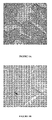
- Figures 1A-1B show examples of positive and negative cases.
- MSH2 expression The relationships between MSH2 expression and the clinical characteristics are provided in Table 1.
- the proportion of adenocarcinomas was lower (P 0.002) for MSH2-positive cases (20 %) than for MSH2-negative cases (37 %).
- Percentages may not total 100 because of rounding.
- ⁇ P values testing the difference between positive and negative tumours were calculated using logistic regression stratified on centre.
- Word Health Organization scores for performance status range from 0 to 2, with a score of 0 indicating no symptoms, 1 mild symptom, and 2 moderate symptoms.
- the 5-year overall survival rates were 47 % (95 %CI, 41 % to 52 %) in the chemotherapy arm, and 44 % (95 %CI, 39 % to 49 %) in the control arm.
- the 8-year overall survival rates were 36 % (95 %CI, 31 % to 42 %) in the chemotherapy arm, and 37 % (95 %CI, 32 % to 43 %) in the control arm.
- Median overall survival was 16 months longer in the MSH2-positive group (58 months) than in the MSH2-negative group (42 months).
- the ERCC1-positive group 304 patients
- the proportion of MSH2-positive cases was 29 % (104 of 354 cases) in the ERCC1-negative group and 49 % (150 of 304 cases) in the ERCC1-positive group (p ⁇ 0.001).
- the association of MSH2 with ERCC1 remained in a model adjusted on slide quality and histological type (p ⁇ 0.001).
- Example 8 Predictive value of combining MSH2 and ERCC1
- Patients in the MSH2-positive group did not benefit from chemotherapy, while patients in the MSH2-negative group did.
- MSH2-negative group In the MSH2-negative group, there was a reduced adjusted hazard ratio for death of 25 %, and a gain in median survival of 16 months in favour of chemotherapy versus observation.
- MSH2 positivity was associated with longer overall survival.
- the results are strengthened by the adjustments for multivariate overall survival predictors and by the use of two-sided statistical tests. They demonstrate that MSH2 is prognostic in patients under observation and is a predictor of overall survival benefit from cisplatin-based chemotherapy.
- MSH2 and ERCC1 are involved in the repair of cisplatin-induced DNA lesions 5, 6 .
- the inventors have demonstrated that they are co-ordinately expressed.
- the differential effect of chemotherapy on patients with MSH2-positive and MSH2-negative tumours was similar among patients with ERCC1-positive and ERCC1-negative tumours, suggesting that the predictive value of MSH2 is partly independent of that of ERCC1.
- the long-term effect of chemotherapy was associated with a reduced adjusted hazard ratio for death of 35 %, which highly suggests that the predictive value of MSH2 and that of ERCC1 act cumulatively. Whereas the data do not allow us to infer that chemotherapy was detrimental when both markers were positive, this possibility cannot be ruled out.
- Loss of MSH2 is associated with mismatch repair deficiency and the resulting microsatellite instability in colon and endometrial cancers, but not in lung cancer 21, 22 . This suggests that mismatch repair deficiency is not responsible for the effects of the loss of MSH2 on cisplatin resistance in NSCLC.
- results of the present study indicate that MSH2 is prognostic in patients under observation and predictive of long-term overall survival benefit from platinum-based chemotherapy, particularly for cisplatin-based chemotherapy. These results also demonstrate the potential use of MSH2 together with ERCC1 in the clinical setting.
Landscapes
- Health & Medical Sciences (AREA)
- Life Sciences & Earth Sciences (AREA)
- Chemical & Material Sciences (AREA)
- Immunology (AREA)
- Engineering & Computer Science (AREA)
- Organic Chemistry (AREA)
- Proteomics, Peptides & Aminoacids (AREA)
- Pathology (AREA)
- Analytical Chemistry (AREA)
- Molecular Biology (AREA)
- Physics & Mathematics (AREA)
- Wood Science & Technology (AREA)
- Zoology (AREA)
- Urology & Nephrology (AREA)
- Microbiology (AREA)
- Hematology (AREA)
- Biochemistry (AREA)
- General Health & Medical Sciences (AREA)
- Biomedical Technology (AREA)
- Biotechnology (AREA)
- Genetics & Genomics (AREA)
- Oncology (AREA)
- Hospice & Palliative Care (AREA)
- Cell Biology (AREA)
- Biophysics (AREA)
- General Physics & Mathematics (AREA)
- Medicinal Chemistry (AREA)
- Food Science & Technology (AREA)
- Bioinformatics & Cheminformatics (AREA)
- General Engineering & Computer Science (AREA)
- Measuring Or Testing Involving Enzymes Or Micro-Organisms (AREA)
Abstract
Description
- The invention is generally directed to a diagnostic method for predicting the benefit of the response of a subject diagnosed with cancer to a platinum compound-based adjuvant chemotherapy, preferably to a cisplatin-based chemotherapy which implements the determination of the MSH2 expression level in the biological sample containing tumor cells, and, optionally, the
ERCC 1 expression level. - Platinum compound-based adjuvant chemotherapy, such as cisplatin, has become a new standard of care for cancer patients, particulary non-small cell lung cancer (NSCLC) patients. Platinum compounds are a hallmark of chemotherapy against lung cancer due to their DNA binding capacity which results in DNA damage and cell death. As DNA repair has a determinant role in both cancer susceptibility (high DNA repair capacity is a barrier against carcinogenesis) and drug resistance (high DNA repair capacity prevents platinum-induced DNA damage), it has been suggested to play a Janus-faced role in cancer physiology1.
- MutS homolog 2 (MSH2), which is frequently mutated in hereditary nonpolyposis colon cancer, encodes a component of the mismatch repair pathway 2. MSH2 binds to DNA mismatches, thereby initiating DNA repair. MSH2 also binds to cisplatin-induced DNA cross-links, thereby initiating their excision and repair 3, 4. The loss of MSH2 may therefore impair the repair of cisplatin-induced DNA cross-links, which are highly deleterious to tumour cells. Indeed, MSH2 is required, together with critical components of other DNA damage repair pathways, such as the excision repair cross-complementing group 1 (ERCC1) protein, to repair cisplatin-induced DNA interstrand cross-links 5,6 .
- MSH2 expression is reduced in 10 % to 58 % of NSCLC 7-16. A recent study reported that loss of MSH2 expression in tumours from patients with advanced NSCLC led to higher rates of response to oxaliplatin-based chemotherapy, but not to cisplatin-based chemotherapy14. In a larger study of genetic polymorphisms among patients with advanced NSCLC, the MSH2 gIV12-6T>C variant was associated with low MSH2 expression and better response to cisplatin.
- The biology of the tumor is of a great interest when choosing the optimal therapy for patients with cancer, particularly for NSCLC. A number of potential biomarkers is under investigation in the hope that it will be possible to identify markers that assist in the selection of patients for specific therapies.
- Promising results so far suggest that customized therapy for individual patients with the help of predictive biomarkers is possible and it is likely that this strategy will improve treatment of NSCLC in the future.
- Thus, it remains desirable to provide new marker capable to predict the benefit of the response of a subject diagnosed with cancer, particularly with NSCLC, to a platinum-based chemotherapy such as cisplatin-based chemotherapy.
- This is the object of the present invention.
- In a first aspect, the invention is directed to a method for in vitro predicting the benefit of the response of a subject diagnosed with cancer to a platinum-based chemotherapy from a biological sample from said subject comprising the following steps of:
- a) determining the MSH2 expression level in the biological sample; and
- b) optionally, comparing said MSH2 expression level to the MSH2 expression level of a reference control or population, or compared to the non tumor cells content.
In a preferred embodiment, the method according to the present invention further comprises the following steps of: - c) determining the ERCC1 expression level from the same or from another biological sample; and
- d) optionally, comparing said ERCC1 expression level to the ERCC1 expression level of a reference control or population, or compared to the non tumor cells content.
- According to the present invention, if the MSH2 expression level obtained for the patient biological sample is less or equal than the expression level obtained for said reference population, or is decreased compared to the non tumor cells, then an overall survival benefit can be predicted.
- According to the present invention, if the MSH2 and the ERCC1 expression levels obtained for the patient biological sample are less or equal than the expression level obtained for said reference populations, or is decreased compared to the non tumor cells then an overall survival benefit can be predicted.
- In a second aspect, the invention is directed to a method for in vitro assessing whether a platinum-based chemotherapy is appropriate for a subject diagnosed with cancer from a biological sample from said subject, said method comprising the following steps of:
- a) determining the MSH2, and optionally the ERCC1, expression level in said biological sample; and
- b) optionally, comparing said MSH2, and optionally ERCC1, expression level to the MSH2, and optionally ERCC1, of a reference control or population, or compared to the non tumor cells content,
- a platinum-based chemotherapy will be determined as an appropriate chemotherapy if the MSH2, and optionally the ERCC1, expression level is less or equal than the MSH2, and optionally the ERCC1, expression level of a reference control or population, or is decreased compared to the non tumor cells, and
- a platinum-free chemotherapy will be determined as an appropriate chemotherapy if the MSH2, and optionally the ERCC1, expression level is greater than the MSH2, and optionally the ERCC1, expression level of said reference control or population, or is increased compared to the non tumor cells,.
- In a third aspect, the invention is directed to a method for in vitro selecting a subject diagnosed from cancer for a treatment with a platinum-based chemotherapy from a biological sample from said subject, said method comprising the steps of:
- a) determining the MSH2, and optionally the ERCC1, expression level in said biological sample; and
- b) optionally, comparing said MSH2, and optionally ERCC1, expression level to the MSH2, and optionally ERCC1, of a reference control or population, or compared to the non tumor cells content
- Preferably, said subject is a human patient.
- In a preferred embodiment, said chemotherapy is an adjuvant-platinum-based chemotherapy.
- In a more preferred embodiment, said platinum-based chemotherapy or said adjuvant-platinum-based chemotherapy is selected from the group consisting of cisplatin-, carboplatin- or oxaliplatin-based chemotherapy, the most preferred being cisplatin-based chemotherapy.
- In another aspect, the present invention comprises the use of MSH2 as a prognostic marker associated with longer overall survival of a subject suffering from cancer.
- In the method of the present invention or for the use of MSH2 as a prognostic marker of the invention, the diagnosed cancer or the subject suffers from a cancer selected from the group of cancer consisting of malignant mesothelioma, bladder cancer, testicular cancer, cancer of the upper aero-digestive tract or ovarian cancer.
- Preferably, these patients suffering from this cancer are not patients presenting the Lynch syndrome (also called hereditary nonpolyposis colorectal cancer) which is one the most common hereditary colorectal cancer syndrome and often associated with mutations in the MSH2 gene or in the MLH1 gene (also related to the mismatch repair system).
- In a preferred embodiment of the method of the present invention or for the use of MSH2 as a prognostic marker of the invention, the diagnosed cancer is the NSCLC.
- In a preferred embodiment of the method of the present invention said biological sample from the patient suffered from a cancer is a tissue sample comprising cancer cells, particularly a biopsy including tissue containing tumor cells.
- Said tissue sample can be fixed, paraffin-embedded, or fresh, or frozen.
- In a preferred embodiment of the method of the present invention in step a) and/or in step c), said MSH2 expression and/or said ERCC1 expression level(s) to be determined is (are) the level of the RNA transcript or their expression protein product of the MSH2 and/or ERCC1 gene.
- In a preferred embodiment the MSH2 and/or ERCC1 expression level is determined by:
- a method including a PCR or a RT-PCR method, or a Northern blot method when the determined MSH2 and/or ERCC1 expression product is the RNA transcript; or
- a Western blot method or an immunohistochemistry method when the determined MSH2 and/or ERCC1 expression product is the MSH2 and/or ERCC1 protein, or a specific fragment thereof.
- In a preferred embodiment, in the step of determining the MSH2 and/or the ERCC1 expression level in the biological sample, the MSH2 and/or the ERCC1 expression products are the MSH2 and/or the ERCC1 protein, or a specific fragment thereof.
- In a more preferred embodiment, in the step of determining the MSH2 and/or the ERCC1 expression level in the biological sample, preferably in tissue sample from the tumor, the MSH2 and/or the ERCC1 expression protein, is determined by immunohistochemistry.
- Preferably, when using an immunohistochemistry method on a tumor tissue sample from the subject, the MSH2 nuclear and, optionally, the ERCC1 nuclear reactivity is determined. Preferably reactive lung or stroma cells can be served as internal positive control and the tumour cells can be graded using the expression level in fibroblasts or endothelial cells as reference.
- In a preferred embodiment, ERCC1-specific immunostaining is carried out as depicted in Olaussen et al. 2006 (18), see also Olaussen et al., 2007 (New England Journal of Medecine, 2007, ERCC1-Specific Immunostaining in Non-Small-Cell Lung Cancer.Volume 357, Number 15:1559-1561).
- In a preferred embodiment, the specific anti MSH2 and, optionally, the anti-ERCC1antibody used for the immunohistochemistry method, or comprised into the kit of the present invention, are directed specifically against the MSH2 or the ERCC1 human protein selected from the group consisting of the proteins having the following reference sequences:
- DNA mismatch repair protein MSH2 (MutS protein homolog 2) (UniProtKB: P43246, SEQ ID N° 1);
-
Excision repair cross-complementing 1 isoform 1 (GenBank NP_973730, SEQ ID N° 2); and -
Excision repair cross-complementing 1 isoform 2 (UniProtKB: P07992, SEQ ID N° 3). - In a more preferred embodiment, for ERCC1, the kit of the present invention or the step of immunostaining or immunohistochemistry analysis in the method of the present invention is carried out using anti-ERCC1 monoclonal or polyclonal antibody, monoclonal being the most preferred, obtained by using the full length ERCC1 protein,
isoform - In an also more preferred embodiment, for ERCC1, said kit or immunostaining or immunohistochemistry analysis is carried out using anti-ERCC1 antibody directed against a common epitope of the
isoform ERCC1 isoform - In the most preferred embodiment, the kit of the present invention or the step of ERCC1-specific immunostaining of the method of the present invention is carried out by using a monoclonal antibody specifically directed against the full length recombinant human
ERCC1 protein isoform 2. - In an also preferred embodiment, in the step of determining the MSH2 and/or the ERCC1 expression level in the biological sample, the MSH2 and/or the ERCC1 expression products are the MSH2 and/or the ERCC1 mRNAs, or a specific fragment thereof.
- Thus, the determination of the MSH2 and/or the ERCC1 mRNA can be carried out by a method which comprises the following steps:
- A) extraction of the total RNAs of said biological sample, preferably from tumor cells, followed, where appropriate, by purification of the mRNAs;
- B) reverse transcription of the RNAs extracted in step A) via an oligo dT primer; and
- C) PCR amplification of the cDNAs obtained in step B) using at least a pair of primers specific for the MSH2 and/or the ERCC1 mRNA to be quantified.
- In a preferred embodiment, when the determination of the MSH2 and/or the ERCC1 mRNA is carried out by a method comprising a PCR or RT-PCR amplification or in the kit of the present invention, primers and probe set specific for the MSH2 and/or the ERCC1 mRNA to be quantified. can be designed using the following reference sequences:
- mRNA encoding DNA mismatch repair protein MSH2 (MutS protein homolog 2) (GenBank; NM_000251; SEQ ID N° 4);
-
transcript variant 1, mRNA (GenBank NM_202001; SEQ ID N° 5) encoding theexcision repair cross-complementing 1isoform 1 protein ERCC1; and -
transcript variant 2, mRNA (GenBank NM_001983, SEQ ID N° 6) encoding theexcision repair cross-complementing 1isoform 2 protein ERCC1. - In a more preferred embodiment, for ERCC1 mRNA expression analysis by RT-QPCR, or in the kit of the present invention, the primer and probe sets can be designed in order to result to the quantification of the presence of an amplicon which is common to the
transcript variant - In the most preferred embodiment, for ERCC1 mRNA expression analysis by RT-QPCR, or in the kit of the present invention, the primer and probe sets can be designed in order to result to the quantification of the presence of a fragment amplicon comprised in the mRNA sequence encoding the full length recombinant human
ERCC1 protein isoform 2. - In another aspect, the present invention is directed to a kit or an array, preferably for in vitro predicting the benefit of the response of a subject diagnosed with cancer to a platinum-based chemotherapy, wherein said kit or array comprises a reagent for assaying MSH2 expression and a reagent for assaying ERCC1 expression in a biological sample from a patient, and, optionally, an instruction sheet.
- In a preferred embodiment, in the kit or array according to the present invention, the reagent for assaying MSH2 and the reagent for assaying ERCC1 expression comprises a reagent selected from the group consisting of:
- a probe or a pair of primers that hybridizes specifically to the MSH2 or to the ERCC1 mRNA or cDNA; or
- an anti-MSH2 or an anti-ERCC1 antibody for performing a Western blot or immunohistochemistry assay.
- Western blotting or immunohistochemistry method can be used for analysing or quantifying specific protein expression in biological sample. Such methods are well known from the skilled man.
- The blots can be detected using antibodies specifically directed against different specific regions or epitope of MSH2 or ERCC1 protein, particularly against human MSH2 or ERCC1 protein.
- The term "antibody" as used herein refers to immunoglobulin molecules and immunologically active portions of immunoglobulin molecules, i.e., molecules that contain an antigen binding site which specifically binds (immunoreacts with) the MSH2 or the ERCC1 protein. The term "antibody" comprises monoclonal or polyclonal antibodies but also chimeric or humanized antibodies.
- An isolated MSH2 or ERCC1 protein, such as recombinant protein, or a specific fragment thereof can be used as an immunogen to generate antibodies that bind such protein using standard techniques for polyclonal and monoclonal antibody preparation. It may be also possible to use any fragment of these protein which contains at least one antigenic determinant may be used to generate these specific antibodies.
- A protein immunogen typically is used to prepare antibodies by immunizing a suitable subject, (e.g., rabbit, goat, mouse or other mammal) with the immunogen. An appropriate immunogenic preparation can contain said protein, or fragment thereof, and further can include an adjuvant, such as Freund's complete or incomplete adjuvant, or similar immunostimulatory agent.
- Thus, antibody for use in accordance with the invention include either polyclonal, monoclonal chimeric or humanized antibodies able to selectively bind, or which selectively bind to an epitope-containing a polypeptide comprising a contiguous span of at least 8 to 10 amino acids of an amino acid sequence of the MSH2 or the ERCC1 protein, preferably the human protein.
- In a preferred embodiment, quantitative RT-PCR analysis can be used to measure the mRNA expression levels of MSH2 and ERCC1 in a biological sample or tissue sample. Gene expression analysis by real-time quantitative PCR (RT-QPCR) is well known from the skilled person.
- For example MSH2 and ERCC1 mRNA expression analysis by RT-QPCR can be assessed after standard tissue sample RNA extraction (for example the samples are lysed in a tris-chloride, EDTA, sodium dodecyl sulphate and proteinase K containing buffer and RNA is then extracted with phenol-chloroform-isoamyl alcohol followed by precipitation with isopropanol in the presence of glycogen and sodium acetate). RNA is then resuspended in diethyl pyrocarbonate water (Ambion Inc., Austin, TX) and treated with DNAse I (Ambion Inc., Austin, TX) to avoid DNA contamination. Complementary DNA was synthesized using for example Maloney Murine Leukemia Virus retrotranscriptase enzyme. Template cDNA was added to Taqman Universal Master Mix (AB, Applied Biosystems, Foster City, CA) in a 12.5-µl reaction with specific primers and probe for each gene. The primer and probe sets can be designed using Primer Express 2.0 Software (AB) and the reference sequences (which can be obtained on the web site http://www.ncbi.nlm.nih.gov/entrez/query.fcgi?db=gene). Primers and probe for the ERCC1 mRNA expression analysis by RT-QPCR can be:
- ERCC1 (reference Genbank sequence NM_001983 3-4, Exon Boundary 3-4) (Bellmunt et al., Annals of Oncology 2007 18(3):522-528).
- Forward primer 5' GGG AAT TTG GCG ACG TAA TTC 3' (SEQ ID NO : 7);
- Reward primer 5' GCG GAG GCT GAG GAA CAG 3' 3' (SEQ ID NO : 8); and
- Labeled Probe 6FAM 5' CAC AGG TGC TCT GGC CCA GCA CAT A 3' TAMRA (SEQ ID NO : 9).
MSH2 : labeled probe-mix from Applied Biosystems can be used (Helleman et al, BMC Cancer. 2006; 6: 201);
- Applied Biosystems, TaqMan® Gene Expression Assays, Assay ID Hs00179887_ml (Reference Sequence: GenBank NM_000251.1, Translated protein: NP_000242.1 Exon Boundary: 14-15; location 2529; Amplicon Length: 87). - Quantification of gene expression was carried out using the ABI Prism 7900HT Sequence Detection System (AB).
- A preferred agent for detecting and quantifying mRNA or cDNA encoding the MSH2 or the ERCC1 protein, is a labeled nucleic acid probe or primers able to hybridize this mRNA or cDNA. The nucleic acid probe can be an oligonucleotide of at least 10, 15, 30, 50 or 100 nucleotides in length and sufficient to specifically hybridize under stringent conditions to the mRNA or cDNA. The nucleic acid primer can be an oligonucleotide of at least 10, 15 or 20 nucleotides in length and sufficient to specifically hybridize under stringent conditions to the mRNA or cDNA, or complementary sequence thereof (preferred are oligonucleotide primers or probe having at least 90 %, 95 %, 99 % and 100 % identity with the mRNA sequence fragment or the complementary sequence thereof) .
- A preferred agent for detecting and quantifying the MSH2 or the ERCC1 protein, is an antibody able to bind specifically to this protein, preferably an antibody with a detectable label. Antibodies can be polyclonal, or more preferably, monoclonal. An intact antibody, or a fragment thereof (e.g., Fab or F(ab')2) can be used. The term "labeled", with regard to the probe or antibody, is intended to encompass direct labeling of the probe or antibody by coupling (i.e., physically linking) a detectable substance to the probe or antibody, as well as indirect labeling of the probe or antibody by reactivity with another reagent that is directly labeled. Examples of indirect labeling include detection of a primary antibody using a fluorescently labeled secondary antibody and end-labeling of a DNA probe with biotin such that it can be detected with fluorescently labeled streptavidin.
- For example, in vitro techniques for detection of candidate mRNA include Northern hybridizations and in situ hybridizations. In vitro techniques for detection of the candidate protein include enzyme linked immunosorbent assays (ELISAs), Western blots, immunoprecipitations and immunofluorescence. In vitro techniques for detection of candidate cDNA include Southern hybridizations.
- When the invention encompasses kits for quantifying the level of the MSH2 and the ERCC1 protein, the kit can comprise a labeled compound or agent capable of quantifying these proteins. Said agents can be packaged in a suitable container. The kit can further comprise instructions for using the kit to quantify the level of the MSH2 and the ERCC1 protein, or of the MSH2 and the ERCC1 transcript.
- In certain embodiments of the method of the present invention, the determination of the MSH2 and the ERCC1 transcripts involves the use of a probe/primer in a polymerase chain reaction (PCR), such as anchor PCR or RACE PCR, or, alternatively, in a ligation chain reaction (LCR), or alternatively quantitative real time RT-PCR. This method can include the steps of collecting a sample of tumor cells from a patient, isolating nucleic acid (e.g. mRNA) from the tumor cells of the sample, optionally transforming mRNA into corresponding cDNA, contacting the nucleic acid sample with one or more primers which specifically hybridize to the MSH2 or the ERCC1 mRNA or their corresponding cDNA under conditions such that hybridization and amplification of the MSH2 or the ERCC1 mRNA or cDNA occurs, and quantifying the presence of the amplification products. It is anticipated that PCR and/or LCR may be desirable to use as an amplification step in conjunction with any of the techniques used for quantifying nucleic acid detecting.
- The methods described herein may be performed, for example, by utilizing prepackaged diagnostic kits comprising at least one probe nucleic acid or set of primer or antibody reagent described herein, which may be conveniently used, e.g., in clinical settings to follow-up or diagnose patients.
- The following examples and also the figures and the legends hereinafter have been chosen to provide those skilled in the art with a complete description in order to be able to implement and use the present invention. These examples are not intended to limit the scope of what the inventor considers to be its invention, nor are they intended to show that only the experiments hereinafter were carried out.
-
-
Figures 1A and 1B : Examples of MSH2 positive and MSH2 negative non-small cell lung cancer. -
Figure 1A : MSH2 positive case. The staining is intense and diffuse. -
Figure 1B : MSH2 negative case. The arrow shows positive stromal cells (internal control). -
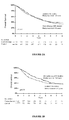
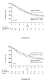
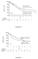
Figures 2A-2F : Kaplan-Meier estimates of the probability of overall survival -
Figure 2A : Patients with MSH2 negative tumours -
Figure 2B : Patients with MSH2 positive tumours -
Figure 2C : Patients with MSH2 negative/ERCC1 negative tumours -
Figure 2D : Patients with MSH2 positive/ERCC1 positive tumours -
Figure 2E : Patients with MSH2 negative/ERCC1 positive tumours -
Figure 2F : Patients with MSH2 positive/ERCC1 negative tumours - All patients had participated in the IALT study (1867 patients) 17. The IALT-Bio study was designed to examine whether markers predicted survival due to chemotherapy 18. The 822 patients who were treated in centres that recruited fewer than 10 patients in the clinical IALT study were excluded from the biological study to facilitate specimen collection and to limit the "centre-effect" on the subsequent biomarker analysis. After tissue collection and a pathological review, 783 tissue blocks were included in the IALT-Bio study.
- Three representative tumour areas were selected for each case. Cores measuring 0.6 mm in diameter and 5mm in length (spots) were arrayed following a map. Among the 783 IALT-Bio tissue blocks that contained tumour material, triplicate spots were obtained in 768 (98 %) cases. Random spots were compared to the original blocks to verify agreement between their coordinates and the original samples.
- Immunostaining was performed following a standard procedure using the Vectastain Elite kit with NovaRED (Vector Laboratories, Burlingame, CA, USA) as the substrate and Mayer's haematoxylin as the counterstain. The primary antibody was the mouse monoclonal antibody FE11 (Calbiochem, San Diego, CA, USA) raised against the C-terminal fragment of human MSH2. For epitope retrieval, slides were heated at 98°C for one hour in 10mM citrate buffer, pH 7.3. Sections were incubated at room temperature for 90 minutes with the FE11 antibody at a dilution of 1:50. The freshly cut TMA sections were manually immunostained in a single experiment that included a tonsil section as an external control.
- Two investigators who were blinded to clinical data, independently evaluated MSH2 nuclear reactivity. The TMA slides were scanned at high resolution (VM3 virtual scanner, Ziemens, Germany), enabling the study of an identical high-quality image at 20x magnification for each spot for detailed evaluation.
- The spots were carefully examined for reactive lung or stromal cells (endothelial cells and fibroblasts), which served as an internal positive control. Spots without a valid internal control were discarded. Cases for which no valid tumour spot could be evaluated were excluded.
- Staining intensity was graded on a scale of 0-3, using the expression level in fibroblasts or endothelial cells as a reference (defined a priori as a score of 2). The percentage of reactive tumour cells was graded on a scale of 0 %-100 %. A proportion score was assigned to the percentage of reactive tumour nuclei: 0 was assigned if 0 % of reactive tumour nuclei were found, 0.1 if 1 %-9 %, 0.5 if 10 %-49 %, and 1 if ≥50 % of reactive tumour nuclei were present. This proportion score was multiplied by the staining intensity to obtain a histology score (H-score) for each patient18.
- All discordant cases were reviewed in order to reach a consensus.
- Long-term IALT survival data were used with a median follow-up of 7.5
years 19 - A logistic model stratified by centre was used to compare patients with MSH2-positive and MSH2-negative tumours.
- The prognostic values of the biomarker status and chemotherapy for overall survival were studied using the Cox model. As in the main IALT analysis 16, the Cox model included every factor used in the stratified randomisation (centre, tumour stage, and type of surgery) plus clinical and histological prognostic factors (age, sex, WHO performance status, nodal status, lymphoid infiltration, and the revised histopathological type). All other factors that were statistically related to the biomarker status in the multivariate logistic model (P < 0.05) were added to the Cox model.
- The predictive value of the biomarker was studied by testing the interaction between the biomarker status and the allocated treatment (chemotherapy versus observation) in the same Cox model. Tests of homogeneity of the hazard ratios were performed within the Cox model. All reported P values were two-sided. In the IALT-Bio analysis plan, P values below 0.01 were considered as statistically significant in order to limit the risk of false positive results. Survival rates were estimated using the Kaplan-Meier method (all P values indicated, besides Kaplan-Meier curves, were adjusted P values corresponding to the Cox analysis).
- All analyses were performed using SAS software, version 9.1 (SAS Institute Inc. Cary NC, USA) and curves were drawn with the Tigre™ software.
- ERCC1-specific immunostaining is carried out as depicted in Olaussen et al. 2006 (18), paragraph " ERCC1 Immunostaining" page 985. The mouse monoclonal "8F1" specifically directed against the full length recombinant human ERCC1 protein being used.
- Among the 768 patients whose tumour was included in the TMA, no tumour material could be analysed after immunohistochemistry in 34 (4 %) cases. After excluding the cases without valid internal controls, the H-scores were evaluated in 673/768 (88 %) patients.
- The H-scores were: 0.2 in 1 case (0 %); 1 in 52 cases (8 %); 2 in 363 cases (54 %); and 3 in 257 cases (38 %). The median H-score was 2 which was chosen to separate positive cases (H-score=3) and negative cases (H-score<3). There were 257 (38 %) positive cases and 416 (62 %) negative cases.
Figures 1A-1B show examples of positive and negative cases. - The relationships between MSH2 expression and the clinical characteristics are provided in Table 1. The proportion of adenocarcinomas was lower (P=0.002) for MSH2-positive cases (20 %) than for MSH2-negative cases (37 %). The morphological slide quality after HE staining was associated with MSH2 expression (P=0.01).
- In a logistic model adjusted on sex and slide quality, histology differed according to MSH2 expression, with fewer adenocarcinomas among patients with MSH2-positive tumours (P=0.006).
- The 673 cases included in the MSH2 analysis differed from the 95 cases excluded in terms of histology (P<0.001), type of surgery (P=0.02), and slide quality (P=0.01) (there were fewer squamous-cell carcinomas, fewer pneumonectomies, and lower staining quality in excluded cases).
Table 1. Patient Characteristics* Characteristic Patients with MSH2-Positive Tumours (N=257) Patients with MSH2 Negative Tumours (N=416) All Patients (N=673) P Value† number percent number percent number percent Sex 0.05 Male 221 86 326 78 547 81 Female 36 14 90 22 126 19 Age 0.99 <55 years 70 27 127 31 197 29 55-64 years 116 45 181 44 297 44 >64 years 71 28 108 26 179 27 Pathological TNM stage 0.79 Stage I 3 32 150 36 233 35 Stage II 53 21 102 25 155 23 Stage III 121 47 164 39 285 42 Tumour 0.83 T1 36 14 63 15 99 15 T2 145 56 263 63 408 61 T3 74 29 82 20 156 23 T4 2 1 8 2 10 1 Nodes 0.99 N0 122 47 188 45 310 46 N1 69 27 125 30 194 29 N2 66 26 103 25 169 25 Histologic type <0.002 Adenocarcinoma 51 20 154 37 205 30 Squamous-cell carcinoma 174 68 217 52 391 58 Other NSCLC 32 12 45 11 77 11 Surgery 0.34 Pneumonectomy 118 46 161 39 279 41 Lobectomy or segmentectomy 139 54 255 61 394 59 Performance status score 
0.37 0 147 57 222 53 369 55 1 84 33 165 40 249 37 2 26 10 29 7 55 8 Lymphoid infiltration 0.20 Not intense 235 91 360 87 595 88 Intense 22 9 56 13 78 12 Pleural invasion 0.19 No 232 90 386 93 618 92 Yes 25 10 30 7 55 8 Vascular invasion 0.44 No 181 70 295 71 476 71 Yes 76 30 121 29 197 29 Lymphatic invasion 0.19 No 71 28 137 33 208 31 Yes 186 72 279 67 465 69 Quality after HE staining 0.01 Average 37 14 28 7 65 10 Good 220 86 388 93 608 90 * TNM denotes tumour-node-metastases. Percentages may not total 100 because of rounding.
† P values testing the difference between positive and negative tumours were calculated using logistic regression stratified on centre.
Word Health Organization scores for performance status range from 0 to 2, with a score of 0 indicating no symptoms, 1 mild symptom, and 2 moderate symptoms.
- For the group of patients included in the MSH2 analysis (673 patients), the adjusted hazard ratio for death associated with chemotherapy compared to observation was 0.88 (95 %CI, 0.72 to 1.07; P=0.21). The 5-year overall survival rates were 47 % (95 %CI, 41 % to 52 %) in the chemotherapy arm, and 44 % (95 %CI, 39 % to 49 %) in the control arm. The 8-year overall survival rates were 36 % (95 %CI, 31 % to 42 %) in the chemotherapy arm, and 37 % (95 %CI, 32 % to 43 %) in the control arm.
- Overall, the effects of adjuvant chemotherapy on the MSH2-negative and positive groups were borderline significantly different (test for interaction, P=0.06). In the MSH2-negative group, overall survival was longer in the chemotherapy arm than in the control arm, (adjusted hazard ratio for death, 0.76; 95 %CI, 0.59 to 0.97; P=0.03) (Table 2,
Figure 2A ). The 5-year overall survival rates among patients with MSH2-negative tumours were 49% (95%CI, 43% to 56%) in the chemotherapy arm and 41% (95%CI, 34% to 48%) in the control arm. The 8-year overall survival rates among patients with MSH2-negative tumours were 38% (95%CI, 32% to 45%) in the chemotherapy arm and 36% (95%CI, 30% to 43%) in the control arm. Among patients with MHS2-negative tumours, median overall survival was 16 months longer in the chemotherapy arm (58 months) than in the control arm (42 months). In the MSH2-positive group, there was no difference in overall survival between the chemotherapy arm and the control arm (adjusted hazard ratio for death, 1.12; 95%CI, 0.81 to 1.55; P=0.48) (Table 2,Figure 2B ). The 5-year overall survival rates among patients with MSH2-positive tumours were 42% (95%CI, 34% to 51%) in the chemotherapy arm and 49% (95%CI, 40% to 58%) in the control arm. The 8-year overall survival rates among patients with MSH2-positive tumours were 34% (95%CI, 25% to 43%) in the chemotherapy arm and 39% (95%CI, 31% to 49%) in the control arm.Table 2. Overall survival according to attributed treatment and MSH2 status Chemotherapy arm (No deaths / No patients) Control arm (No deaths / No patients) Hazard ratio for death (95% CI)* P value Patients with MSH2-negative tumours n = 416 131/215 130/201 0.75 (0.58-0.97) p=0.03 Patients with MSH2 positive tumours n = 257 83 / 131 75/126 1.12 (0.81-1.55) p=048 Hazard ratio for death (95% CI)** 0.99 (0.74-1.32) 0.66 (0.49-0.90) - - P Value p = 0.93 p = 0.01 - p=0.06† * Hazard ratios are for the comparison of chemotherapy group with the control group.
** Hazard ratios are for the comparison of patients with MSH2-positive tumours with those with MSH2-negative tumours.
† The P value is for the interaction between MSH2 expression and treatment. - In the control arm, MSH2 positivity compared to MSH2 negativity was associated with an adjusted hazard ratio for death of 0.66 (95 %CI, 0.49 to 0.90; P=0.01). Median overall survival was 16 months longer in the MSH2-positive group (58 months) than in the MSH2-negative group (42 months).
- In the chemotherapy arm, there was no difference in overall survival between MSH2-positive and MSH2-negative tumours (adjusted hazard ratio for death, 0.99; 95 %CI, 0.74 to 1.32; P=0.93).
- The expression levels of MSH2 and ERCC1 were both available for 658 patients (84% of the patients included in IALT-Bio). In a sub-analysis of these 658 patients, the difference between the long-term effects of chemotherapy according to MSH2 or ERCC1 expression were both borderline significant (test for interaction, P=0.05 and P=0.04, respectively).
- In the MSH2-negative group (404 patients), the adjusted hazard ratio for death associated with chemotherapy versus observation was 0.75 (95 %CI, 0.58 to 0.98; P=0.03)(Table 3). In the MSH2-positive group (254 patients), there was no difference in overall survival between the chemotherapy arm and the control arm (adjusted hazard ratio for death, 1.14; 95 %CI, 0.82 to 1.57; P=0.44).
- Among these 658 patients, the adjusted hazard ratio for death associated with chemotherapy versus observation was 0.73 (95 %CI, 0.55 to 0.96; P=0.03) in the ERCC1-negative group (354 patients) (Table 3). In the ERCC1-positive group (304 patients), there was no difference in overall survival between the chemotherapy arm and the control arm (adjusted hazard ratio for death, 1.11; 95 %CI, 0.83 to 1.50; P=0.49).
- The proportion of MSH2-positive cases was 29 % (104 of 354 cases) in the ERCC1-negative group and 49 % (150 of 304 cases) in the ERCC1-positive group (p<0.001). The association of MSH2 with ERCC1 remained in a model adjusted on slide quality and histological type (p<0.001).
- The effect of chemotherapy was examined in the following subgroups defined by combining MSH2 and ERCC1: MSH2-positive/ERCC1-positive, MSH2-positive/ERCC1-negative, MSH2-negative/ERCC1-positive, MSH2-negative/ERCC1-negative (
Figures 2C to 2F ). The differential effect of chemotherapy for patients with MSH2-positive tumours compared to those with MSH2-negative lesions was similar among patients with ERCC1-positive and ERCC1-negative tumours (P=0.94), i.e., the ratio of the hazard ratios associated with MSH2 among patients with ERCC1-negative (0.90/0.65) was equal to that among patients with ERCC1-positive tumours (1.32/0.92) (Table 3).Table 3. Overall survival according to allocated treatment and subgroup analysis Patients with MSH2-Negative tumours Patients with MSH2-Positive tumours Total Patients with ERCC1-negative tumours Number of death / number of patients 162 / 250 57 / 104 Hazard ratio for death (95% CI) P Value 0.65 (0.47-0.91)* p=0.01 0.90 (0.52-1.55)* p=0.70 0.73 (0.55-0.96)* Patients with ERCC1-positive tumours Number of death / number of patients 90 / 154 99/150 Hazard ratio for death (95% CI) P Value 0.92 (0.60-1.42)* P=0.71 1.32 (0.88-1.99)* P=0.19 1.11 (0.83-1.50)* Total Hazard ratio for death (95% CI) 0.75 (0.58-0.98)* 1.14 (0.82-1.57)* - P=0.03 P=0.44 * Hazard ratios are for the comparison of the chemotherapy group with the control group. - In the combined MSH2-negative/ERCC1-negative subgroup, overall survival was longer in the chemotherapy arm than in the control arm (adjusted hazard ratio for death, 0.65; 95 %CI, 0.47 to 0.91; P=0.01) (
Figure 2C ). In the MSH2-positive/ERCC1-positive subgroup, the hazard ratio for death associated with chemotherapy was 1.32 (95%CI, 0.88 to 1.99; P=0.19) (Figure 2D ). The adjusted hazard ratio for death associated with chemotherapy when 1 marker was positive (either MSH2 or ERCC1 but not both) was 0.91 (95 %CI, 0.65 to 1.28). When we defined a score by counting the number of positive MSH2 and ERCC1 markers in the above-defined subgroups (allocating a score of 0 for no positive marker, 1 for 1 positive marker and 2 for 2 positive markers), the test of a decreasing effect of chemotherapy with an increasing score was significant (P=0.01). - The results demonstrate that the long-term survival benefit derived from adjuvant chemotherapy may be different according to MSH2 expression in cancer patient, particularly in NSCLC. Patients in the MSH2-positive group did not benefit from chemotherapy, while patients in the MSH2-negative group did. In the MSH2-negative group, there was a reduced adjusted hazard ratio for death of 25 %, and a gain in median survival of 16 months in favour of chemotherapy versus observation. When the analysis focused exclusively on the control arm, MSH2 positivity was associated with longer overall survival. The results are strengthened by the adjustments for multivariate overall survival predictors and by the use of two-sided statistical tests. They demonstrate that MSH2 is prognostic in patients under observation and is a predictor of overall survival benefit from cisplatin-based chemotherapy.
- A previous study already demonstrated a survival benefit from adjuvant cisplatin-based chemotherapy in patients whose tumours were ERCC1 negative which was not the case for patients with ERCC1-positive lesions 18. Furthermore, the prognostic role of ERCC1 tumour expression suggested by our data was later strongly supported by Zheng et al 20. In the present study, we used recently updated survival data from the IALT study (7.5 years of median survival) which for the first time allowed us to evaluate DNA repair markers as predictors of the long-term benefit of cisplatin-based chemotherapy 19. The long-term predictive value of MSH2 was equal to that of ERCC1 in patients for whom both markers were available.
- Both MSH2 and ERCC1 are involved in the repair of cisplatin-induced DNA lesions 5, 6. Here, the inventors have demonstrated that they are co-ordinately expressed. The differential effect of chemotherapy on patients with MSH2-positive and MSH2-negative tumours was similar among patients with ERCC1-positive and ERCC1-negative tumours, suggesting that the predictive value of MSH2 is partly independent of that of ERCC1. Indeed, when both markers were negative, the long-term effect of chemotherapy was associated with a reduced adjusted hazard ratio for death of 35 %, which highly suggests that the predictive value of MSH2 and that of ERCC1 act cumulatively. Whereas the data do not allow us to infer that chemotherapy was detrimental when both markers were positive, this possibility cannot be ruled out.
- Loss of MSH2 is associated with mismatch repair deficiency and the resulting microsatellite instability in colon and endometrial cancers, but not in lung cancer 21, 22. This suggests that mismatch repair deficiency is not responsible for the effects of the loss of MSH2 on cisplatin resistance in NSCLC.
- In conclusion, the results of the present study indicate that MSH2 is prognostic in patients under observation and predictive of long-term overall survival benefit from platinum-based chemotherapy, particularly for cisplatin-based chemotherapy. These results also demonstrate the potential use of MSH2 together with ERCC1 in the clinical setting.
-
- 1. Gazdar AF. DNA repair and survival in lung cancer--the two faces of Janus. The New England journal of medicine 2007;356(8):771-3.
- 2. Fishel R, Lescoe MK, Rao MR, et al. The human mutator gene homolog MSH2 and its association with hereditary nonpolyposis colon cancer. Cell 1993;75(5):1027-38.
- 3. Duckett DR, Drummond JT, Murchie AI, et al. Human MutSalpha recognizes damaged DNA base pairs containing O6-methylguanine, O4-methylthymine, or the cisplatin-d(GpG) adduct. Proc Natl Acad Sci U S A 1996;93(13):6443-7.
- 4. Zdraveski ZZ, Mello JA, Farinelli CK, Essigmann JM, Marinus MG. MutS preferentially recognizes cisplatin- over oxaliplatin-modified DNA. J Biol Chem 2002;277(2):1255-60.
- 5. Lan L, Hayashi T, Rabeya RM, et al. Functional and physical interactions between ERCC1 and MSH2 complexes for resistance to cis-diamminedichloroplatinum(II) in mammalian cells. DNA Repair (Amst) 2004;3(2):135-43.
- 6. Zhang N, Liu X, Li L, Legerski R. Double-strand breaks induce homologous recombinational repair of interstrand cross-links via cooperation of MSH2, ERCC1-XPF, REV3, and the Fanconi anemia pathway. DNA Repair (Amst) 2007;6(11):1670-8.
- 7. Xinarianos G, Liloglou T, Prime W, et al. hMLH1 and hMSH2 expression correlates with allelic imbalance on chromosome 3p in non-small cell lung carcinomas. Cancer Res 2000;60(15):4216-21.
- 8. Brooks KR, To K, Joshi MB, et al. Measurement of chemoresistance markers in patients with stage III non-small cell lung cancer: a novel approach for patient selection. Ann Thorac Surg 2003;76(1):187-93;
discussion 93. - 9. Wang YC, Lu YP, Tseng RC, et al. Inactivation of hMLH1 and hMSH2 by promoter methylation in primary non-small cell lung tumors and matched sputum samples. J Clin Invest 2003;111(6):887-95.
- 10. Zhu LQ, Diao LM, Chen DJ, Li HG, Liu X, Zou ZY, Li BY, Wang M, Liu MQ. Relationship between hMSH2 and FHIT gene expression in non-small cell lung cancer Ai Zheng. 2003;22(6):571-4.
- 11. Hsu HS, Wen CK, Tang YA, et al. Promoter hypermethylation is the predominant mechanism in hMLH1 and hMSH2 deregulation and is a poor prognostic factor in nonsmoking lung cancer. Clin Cancer Res 2005;11(15):5410-6.
- 12. Skarda J, Fridman E, Plevova P, et al. Prognostic value of hMLH1 and hMSH2 immunohistochemical expression in non-small cell lung cancer. A tissue microarray study. Biomed Pap Med Fac Univ Palacky Olomouc Czech Repub 2006;150(2):255-9.
- 13. Kanellis G, Chatzistamou I, Koutselini H, et al. Expression of DNA mismatch repair gene MSH2 in cytological material from lung cancer patients. Diagn Cytopathol 2006;34(7):463-6.
- 14. Scartozzi M, Franciosi V, Campanini N, et al. Mismatch repair system (MMR) status correlates with response and survival in non-small cell lung cancer (NSCLC) patients. Lung Cancer 2006;53(1):103-9.
- 15. Hsu HS, Lee IH, Hsu WH, Kao WT, Wang YC. Polymorphism in the hMSH2 gene (gISV12-6T > C) is a prognostic factor in non-small cell lung cancer. Lung Cancer 2007;58(1):123-30.
- 16. Cooper WA, Kohonen-Corish MR, Chan C, et al. Prognostic significance of DNA repair proteins MLH1, MSH2 and MGMT expression in non-small-cell lung cancer and precursor lesions. Histopathology 2008;52(5):613-22.
- 17. Arriagada R, Bergman B, Dunant A, Le Chevalier T, Pignon JP, Vansteenkiste J. Cisplatin-based adjuvant chemotherapy in patients with completely resected non-small-cell lung cancer. The New England journal of medicine 2004;350(4):351-60.
- 18. Olaussen KA, Dunant A, Fouret P, et al. DNA repair by ERCC1 in non-small-cell lung cancer and cisplatin-based adjuvant chemotherapy. The New England journal of medicine 2006;355(10):983-91.
- 19. Le Chevalier T, Dunant A, Arriagada R, et al. Long-term results of the International Adjuvant Lung Cancer Trial (IALT) evaluating adjuvant cisplatin-based chemotherapy in non-small cell lung cancer (NSCLC). J Clin Oncol 2008;26(15 S Suppl): 398s.
- 20. Zheng Z, Chen T, Li X, Haura E, Sharma A, Bepler G. DNA synthesis and repair genes RRM1 and ERCC1 in lung cancer. The New England journal of medicine 2007;356(8):800-8.
- 21. Forgacs E, Wren JD, Kamibayashi C, et al. Searching for microsatellite mutations in coding regions in lung, breast, ovarian and colorectal cancers. Oncogene 2001;20(8):1005-9.
- 22. Weinberg RA. The biology of cancer. Garland Science Eds 2007:463-556. ly, the ERCC1 expression level.


Claims (17)
- A method for in vitro predicting the benefit of the response of a subject diagnosed with cancer to a platinum-based chemotherapy from a biological sample from said subject comprising the following steps of:a) determining the MSH2 expression level in the biological sample; andb) optionally, comparing said MSH2 expression level to the MSH2 expression level of a reference control or population, or compared to the non tumor cells content.
- The method claim 2, further comprisingc) determining the ERCC1 expression level from the same or from another biological sample; andd) optionally, comparing said ERCC1 expression level to the ERCC1 expression level of a reference control or population, or compared to the non tumor cells content.
- The method according to claim 1, wherein in step b), if the MSH2 expression level obtained for the patient biological sample is less or equal than the expression level obtained for said reference control or population, or is decreased compared to the non tumor cells content, then an overall survival benefit can be predicted.
- The method according to claim 2, wherein in step b) and in step d), if the MSH2 and the ERCC1 expression levels obtained for the patient biological sample are less or equal than the expression level obtained for said reference population, or is decreased compared to the non tumor cells content, then an overall survival benefit can be predicted.
- A method for in vitro assessing whether a platinum-based chemotherapy is appropriate for a subject diagnosed with cancer from a biological sample from said subject, said method comprising the following steps of:a) determining the MSH2, and optionally the ERCC1, expression level in said biological sample; andb) optionally, comparing said MSH2, and optionally ERCC1, expression level to the MSH2, and optionally ERCC1, of a reference control or population, or compared to the non tumor cells content- a platinum-based chemotherapy will be determined as an appropriate chemotherapy if the MSH2, and optionally the ERCC1, expression level is less or equal than the MSH2, and optionally the ERCC1, expression level of a reference control or population, or is decreased compared to the non tumor cells, and- a platinum-free chemotherapy will be determined as an appropriate chemotherapy if the MSH2, and optionally the ERCC1, expression level is greater than the MSH2, and optionally the ERCC1, expression level of said reference control or population, or is increased compared to the non tumor cells.
- An in vitro screening method for selecting a subject diagnosed from cancer for a treatment with a platinum-based chemotherapy from a biological sample from said subject, said method comprising the steps of:a) determining the MSH2, and optionally the ERCC1, expression level in said biological sample; andb) comparing said MSH2, and optionally ERCC1, expression level to the MSH2, and optionally ERCC1, of a reference control or population, or compared to the non tumor cells content,wherein said subject will be selected for a platinum-based chemotherapy if the MSH2, and optionally the ERCC1, expression level is less or equal than the MSH2, and optionally the ERCC1, expression level of a reference control or population, or is decreased compared to the non tumor cells content.
- The method of ant one of claims 1 to 6, wherein said subject is a human patient.
- The method of any one of claims 1 to 7, wherein said chemotherapy is an adjuvant-platinum-based chemotherapy.
- The method of any one of claims 1 to 8, wherein said platinum-based chemotherapy is cisplatin-based chemotherapy.
- MSH2 as a prognostic marker associated with longer overall survival of a subject suffering from cancer.
- The method of any one of claims 1-9 and the marker of claim 10, wherein the subject has a malignant mesothelioma, a bladder cancer, a testicular cancer, cancer of the upper aero-digestive tract or ovarian cancer.
- The method of any one of claims 1 to 9 and the marker of claim 10, wherein the subject has non- small-cell lung cancer.
- The method of any one of claims 1 to 12, wherein said biological sample is a tissue sample comprising cancer cells, particularly a biopsy containing tumor cells.
- The method of any one of claims 1 to 13, wherein in step a), said MSH2 expression and/or said ERCC1 expression level(s) to be determined is (are) the level of the RNA transcript or their expression protein product of the MSH2 and/or ERCC1 gene.
- The method of claim 14 wherein the MSH2 and/or ERCC1 expression level is determined by:- a method including a PCR or a RT-PCR method, or a Northern method when the determined MSH2 and/or ERCC1 expression product is the RNA transcript; or- a Western blot method or an immunohistochemistry method when the determined MSH2 and/or ERCC1 expression product is the MSH2 and/or ERCC1 protein, or a specific fragment thereof.
- Kit or array wherein said kit or array comprises a reagent for assaying MSH2 expression and a reagent for assaying ERCC1 expression in a biological sample from a patient.
- The kit or array of claim 16, wherein the reagent for assaying MSH2 and the reagent for assaying ERCC1 expression comprises a reagent selected from the group consisting of:- a probe or a pair of primers that specifically hybridizes to the MSH2 or to the ERCC1 mRNA or cDNA; or- an anti-MSH2 capable of specifically recognizing the MSH2 protein or an anti-ERCC1 antibody capable of specifically recognizing the ERCC1 protein.
Priority Applications (4)
| Application Number | Priority Date | Filing Date | Title |
|---|---|---|---|
| EP09305248A EP2233589A1 (en) | 2009-03-19 | 2009-03-19 | MSH2 and adjuvant cisplatin-based chemotherapy in non-small-cell lung cancer |
| EP10711197A EP2408932A1 (en) | 2009-03-19 | 2010-03-19 | Msh2 and adjuvant cisplatin-based chemotherapy in non-small-cell lung cancer |
| US13/256,814 US20120004136A1 (en) | 2009-03-19 | 2010-03-19 | Msh2 and adjuvant cisplatin-based chemotherapy in non-small-cell lung cancer |
| PCT/EP2010/053634 WO2010106176A1 (en) | 2009-03-19 | 2010-03-19 | Msh2 and adjuvant cisplatin-based chemotherapy in non-small-cell lung cancer |
Applications Claiming Priority (1)
| Application Number | Priority Date | Filing Date | Title |
|---|---|---|---|
| EP09305248A EP2233589A1 (en) | 2009-03-19 | 2009-03-19 | MSH2 and adjuvant cisplatin-based chemotherapy in non-small-cell lung cancer |
Publications (1)
| Publication Number | Publication Date |
|---|---|
| EP2233589A1 true EP2233589A1 (en) | 2010-09-29 |
Family
ID=40756409
Family Applications (2)
| Application Number | Title | Priority Date | Filing Date |
|---|---|---|---|
| EP09305248A Withdrawn EP2233589A1 (en) | 2009-03-19 | 2009-03-19 | MSH2 and adjuvant cisplatin-based chemotherapy in non-small-cell lung cancer |
| EP10711197A Withdrawn EP2408932A1 (en) | 2009-03-19 | 2010-03-19 | Msh2 and adjuvant cisplatin-based chemotherapy in non-small-cell lung cancer |
Family Applications After (1)
| Application Number | Title | Priority Date | Filing Date |
|---|---|---|---|
| EP10711197A Withdrawn EP2408932A1 (en) | 2009-03-19 | 2010-03-19 | Msh2 and adjuvant cisplatin-based chemotherapy in non-small-cell lung cancer |
Country Status (3)
| Country | Link |
|---|---|
| US (1) | US20120004136A1 (en) |
| EP (2) | EP2233589A1 (en) |
| WO (1) | WO2010106176A1 (en) |
Cited By (3)
| Publication number | Priority date | Publication date | Assignee | Title |
|---|---|---|---|---|
| CN103088123A (en) * | 2012-12-07 | 2013-05-08 | 周宏灏 | Kit and method for detecting expression level of ERCC1 (Excision Repair Cross Complementation 1) mRNA (messenger Ribonucleic Acid) |
| WO2016042009A1 (en) * | 2014-09-18 | 2016-03-24 | Adnagen Gmbh | Mrna and/or protein of ercc1 isoform 3 for use in diagnosing a resistance against a therapeutic agent and method for diagnosing a resistance against a therapeutic agent using said mrna and/or protein |
| EP3139172A3 (en) * | 2011-03-24 | 2017-06-14 | Keio University | Marker for determination of sensitivity to anticancer agent |
Families Citing this family (4)
| Publication number | Priority date | Publication date | Assignee | Title |
|---|---|---|---|---|
| US9702875B2 (en) | 2006-03-14 | 2017-07-11 | Institute Gustave-Roussy | Expression of isoform 202 of ERCC1 for predicting response to cancer chemotherapy |
| EP2602622A1 (en) * | 2011-12-07 | 2013-06-12 | Atlas Antibodies AB | Prediction of response to platinum-based therapy |
| JP2017528227A (en) * | 2014-09-11 | 2017-09-28 | ピーシビダ ユーエス,インコーポレイテッド | Injection equipment |
| CN110218251B (en) * | 2019-06-19 | 2022-10-25 | 福州迈新生物技术开发有限公司 | anti-MSH 2 protein monoclonal antibody, cell line, preparation method and application thereof |
-
2009
- 2009-03-19 EP EP09305248A patent/EP2233589A1/en not_active Withdrawn
-
2010
- 2010-03-19 US US13/256,814 patent/US20120004136A1/en not_active Abandoned
- 2010-03-19 EP EP10711197A patent/EP2408932A1/en not_active Withdrawn
- 2010-03-19 WO PCT/EP2010/053634 patent/WO2010106176A1/en not_active Ceased
Non-Patent Citations (33)
| Title |
|---|
| ARRIAGADA R ET AL.: "Cisplatin-based adjuvant chemotherapy in patients with completely resected non-small-cell lung cancer", THE NEW ENGLAND JOURNAL OF MEDICINE, vol. 350, no. 4, 2004, pages 351 - 60 |
| BELLMUNT ET AL., ANNALS OF ONCOLOGY, vol. 18, no. 3, 2007, pages 522 - 528 |
| BROOKS KR; TO K; JOSHI MB ET AL.: "Measurement of chemoresistance markers in patients with stage III non-small cell lung cancer: a novel approach for patient selection", ANN THORAC SURG, vol. 76, no. 1, 2003, pages 187 - 93 |
| COOPER WA; KOHONEN-CORISH MR; CHAN C ET AL.: "Prognostic significance of DNA repair proteins MLH1, MSH2 and MGMT expression in non-small-cell lung cancer and precursor lesions", HISTOPATHOLOGY, vol. 52, no. 5, 2008, pages 613 - 22 |
| DUCKETT DR; DRUMMOND JT; MURCHIE AI ET AL.: "Human MutSalpha recognizes damaged DNA base pairs containing 06-methylguanine, 04-methylthymine, or the cisplatin-d(GpG) adduct", PROC NATL ACAD SCI U S A, vol. 93, no. 13, 1996, pages 6443 - 7 |
| ERCCL-SPECIFIC IMMUNOSTAINING IN NON-SMALL-CELL LUNG CANCER, vol. 357, no. 15, pages 1559 - 1561 |
| FISHEL R; LESCOE MK; RAO MR ET AL.: "The human mutator gene homolog MSH2 and its association with hereditary nonpolyposis colon cancer", CELL, vol. 75, no. 5, 1993, pages 1027 - 38 |
| FORGACS E; WREN JD; KAMIBAYASHI C ET AL.: "Searching for microsatellite mutations in coding regions in lung, breast, ovarian and colorectal cancers", ONCOGENE, vol. 20, no. 8, 2001, pages 1005 - 9 |
| FRANCIOSI ET AL: "P-151 Correlations between clinical response to cisplatin or oxaliplatin and expression of the Mismatch Repair (MMR) and p53 in advanced non-small cell lung cancer (NSCLC) patients", LUNG CANCER, ELSEVIER, AMSTERDAM, NL, vol. 41, 1 August 2003 (2003-08-01), pages S128, XP005875372, ISSN: 0169-5002 * |
| GAZDAR AF.: "DNA repair and survival in lung cancer--the two faces of Janus", THE NEW ENGLAND JOURNAL OF MEDICINE, vol. 356, no. 8, 2007, pages 771 - 3 |
| HSU HS ET AL.: "Polymorphism in the hMSH2 gene (gISV12-6T > C) is a prognostic factor in non-small cell lung cancer", LUNG CANCER, vol. 58, no. 1, 2007, pages 123 - 30 |
| HSU HS; WEN CK; TANG YA ET AL.: "Promoter hypermethylation is the predominant mechanism in hMLH1 and hMSH2 deregulation and is a poor prognostic factor in nonsmoking lung cancer", CLIN CANCER RES, vol. 11, no. 15, 2005, pages 5410 - 6 |
| KANELLIS G; CHATZISTAMOU I; KOUTSELINI H ET AL.: "Expression of DNA mismatch repair gene MSH2 in cytological material from lung cancer patients", DIAGN CYTOPATHOL, vol. 34, no. 7, 2006, pages 463 - 6 |
| KORFEE S ET AL: "P-067 Impact of mismatch repair protein expression on cisplatinum-basedchemotherapeutic response in patients with stage III non-small-cell lung cancer (NSCLC)", LUNG CANCER, ELSEVIER, AMSTERDAM, NL, vol. 49, 1 July 2005 (2005-07-01), pages S131, XP025285717, ISSN: 0169-5002, [retrieved on 20050701] * |
| LAN L; HAYASHI T; RABEYA RM ET AL.: "Functional and physical interactions between ERCC1 1 and MSH2 complexes for resistance to cis- diamminedichloroplatinum(II) in mammalian cells", DNA REPAIR (AMST), vol. 3, no. 2, 2004, pages 135 - 43 |
| LAN LI ET AL: "Functional and physical interactions between ERCC1 and MSH2 complexes for resistance to cis-diamminedichloroplatinum(II) in mammalian cells.", DNA REPAIR, vol. 3, no. 2, 3 February 2004 (2004-02-03), pages 135 - 143, XP002321156, ISSN: 1568-7864 * |
| LE CHEVALIER T; DUNANT A; ARRIAGADA R ET AL.: "Long-term results of the International Adjuvant Lung Cancer Trial (IALT) evaluating adjuvant cisplatin-based chemotherapy in non-small cell lung cancer (NSCLC)", J CLIN ONCOL, vol. 26, no. 15, 2008, pages 398 |
| NADIN S B ET AL: "DNA damage and repair in peripheral blood lymphocytes from healthy individuals and cancer patients: A pilot study on the implications in the clinical response to chemotherapy", CANCER LETTERS, NEW YORK, NY, US, vol. 239, no. 1, 28 July 2006 (2006-07-28), pages 84 - 97, XP025021842, ISSN: 0304-3835, [retrieved on 20060728] * |
| OLAUSSEN KA; DUNANT A; FOURET P ET AL.: "DNA repair by ERCC1 in non-small-cell lung cancer and cisplatin-based adjuvant chemotherapy", THE NEW ENGLAND JOURNAL OF MEDICINE, vol. 355, no. 10, 2006, pages 983 - 91 |
| OLAUSSEN; OLAUSSEN ET AL., NEW ENGLAND JOURNAL OF MEDECINE, 2007 |
| OTA S ET AL: "Immunohistochemical expression of BCRP and ERCC1 in biopsy specimen predicts survival in advanced non-small-cell lung cancer treated with cisplatin-based chemotherapy", LUNG CANCER, ELSEVIER, AMSTERDAM, NL, vol. 64, no. 1, 26 September 2008 (2008-09-26), pages 98 - 104, XP025965405, ISSN: 0169-5002, [retrieved on 20080926] * |
| RELLECKE PHILIPP ET AL: "Mismatch repair protein hMSH2 in primary drug resistance in in vitro human malignant gliomas.", JOURNAL OF NEUROSURGERY OCT 2004, vol. 101, no. 4, October 2004 (2004-10-01), pages 653 - 658, XP002533209, ISSN: 0022-3085 * |
| SCARTOZZI M ET AL: "Mismatch repair system (MMR) status correlates with response and survival in non-small cell lung cancer (NSCLC) patients", LUNG CANCER, ELSEVIER, AMSTERDAM, NL, vol. 53, no. 1, 1 July 2006 (2006-07-01), pages 103 - 109, XP024893940, ISSN: 0169-5002, [retrieved on 20060701] * |
| SCARTOZZI M; FRANCIOSI V; CAMPANINI N ET AL.: "Mismatch repair system (MMR) status correlates with response and survival in non-small cell lung cancer (NSCLC) patients", LUNG CANCER, vol. 53, no. 1, 2006, pages 103 - 9 |
| SKARDA J; FRIDMAN E; PLEVOVA P ET AL.: "Prognostic value of hMLH1 and hMSH2 immunohistochemical expression in non-small cell lung cancer. A tissue microarray study", BIOMED PAP MED FAC UNIV PALACKY OLOMOUC CZECH REPUB, vol. 150, no. 2, 2006, pages 255 - 9 |
| SKARDA JOZEF ET AL: "Prognostic value of hMLH1 and hMSH2 immunohistochemical expression in non-small cell lung cancer. A tissue microarray study", BIOMEDICAL PAPERS (OLOMOUC), vol. 150, no. 2, November 2006 (2006-11-01), pages 255 - 259, XP002533210, ISSN: 1213-8118 * |
| WANG YC; LU YP; TSENG RC ET AL.: "Inactivation of hMLHI and hMSH2 by promoter methylation in primary non-small cell lung tumors and matched sputum samples", J CLIN INVEST, vol. 111, no. 6, 2003, pages 887 - 95 |
| WEINBERG RA: "The biology of cancer", 2007, GARLAND SCIENCE EDS, pages: 463 - 556 |
| XINARIANOS G; LILOGLOU T; PRIME W ET AL.: "hMLHI and hMSH2 expression correlates with allelic imbalance on chromosome 3p in non-small cell lung carcinomas", CANCER RES, vol. 60, no. 15, 2000, pages 4216 - 21 |
| ZDRAVESKI ZZ ET AL.: "MutS preferentially recognizes cisplatin- over oxaliplatin-modified DNA", J BIOL CHEM, vol. 277, no. 2, 2002, pages 1255 - 60 |
| ZHANG N ET AL.: "Double-strand breaks induce homologous recombinational repair of interstrand cross-links via cooperation of MSH2, ERCC1-XPF, REV3, and the Fanconi anemia pathway", DNA REPAIR (AMST), vol. 6, no. 11, 2007, pages 1670 - 8 |
| ZHENG Z ET AL.: "DNA synthesis and repair genes RRM1 and ERCC1 in lung cancer", THE NEW ENGLAND JOURNAL OF MEDICINE, vol. 356, no. 8, 2007, pages 800 - 8 |
| ZHU LQ ET AL., RELATIONSHIP BETWEEN HMSH2 AND FHIT GENE EXPRESSION IN NON-SMALL CELL LUNG CANCER AI ZHENG, vol. 22, no. 6, 2003, pages 571 - 4 |
Cited By (5)
| Publication number | Priority date | Publication date | Assignee | Title |
|---|---|---|---|---|
| EP3139172A3 (en) * | 2011-03-24 | 2017-06-14 | Keio University | Marker for determination of sensitivity to anticancer agent |
| US9797884B2 (en) | 2011-03-24 | 2017-10-24 | Keio University | Marker for determination of sensitivity to anticancer agent |
| US10309957B2 (en) | 2011-03-24 | 2019-06-04 | Keio University | Method for determining sensitivity to fluorouracil in a subject having colorectal cancer |
| CN103088123A (en) * | 2012-12-07 | 2013-05-08 | 周宏灏 | Kit and method for detecting expression level of ERCC1 (Excision Repair Cross Complementation 1) mRNA (messenger Ribonucleic Acid) |
| WO2016042009A1 (en) * | 2014-09-18 | 2016-03-24 | Adnagen Gmbh | Mrna and/or protein of ercc1 isoform 3 for use in diagnosing a resistance against a therapeutic agent and method for diagnosing a resistance against a therapeutic agent using said mrna and/or protein |
Also Published As
| Publication number | Publication date |
|---|---|
| US20120004136A1 (en) | 2012-01-05 |
| EP2408932A1 (en) | 2012-01-25 |
| WO2010106176A1 (en) | 2010-09-23 |
Similar Documents
| Publication | Publication Date | Title |
|---|---|---|
| Li et al. | EGFR mutations in lung adenocarcinomas: clinical testing experience and relationship to EGFR gene copy number and immunohistochemical expression | |
| AU2005249492B2 (en) | Methods for prediction of clinical outcome to epidermal growth factor receptor inhibitors by cancer patients | |
| US7736861B1 (en) | Tivozanib response prediction | |
| US20130065789A1 (en) | Compositions and methods for classifying lung cancer and prognosing lung cancer survival | |
| US20140336280A1 (en) | Compositions and methods for detecting and determining a prognosis for prostate cancer | |
| WO2008058018A2 (en) | Predicting cancer outcome | |
| US11821900B2 (en) | Method of selection for treatment of subjects at risk of invasive breast cancer | |
| US20120277110A1 (en) | Parp and adjuvant cisplatin-based chemotherapy in non-small-cell lung cancer | |
| CN106233143B (en) | cancer treatment | |
| US20120004136A1 (en) | Msh2 and adjuvant cisplatin-based chemotherapy in non-small-cell lung cancer | |
| JP5858405B2 (en) | Prognosis prediction method for lung adenocarcinoma and detection kit for lung adenocarcinoma | |
| WO2020054782A1 (en) | Method for estimating breast cancer cell abundance | |
| CN109642257B (en) | Methods for predicting the effect of drug therapy on cancer | |
| WO2014185466A1 (en) | Prognosis evaluation method for pancreatic cancer | |
| JP4908401B2 (en) | PKP3 oncogene as a prognostic indicator of lung cancer | |
| AU2011265464B2 (en) | Methods for prediction of clinical outcome to epidermal growth factor receptor inhibitors by cancer patients | |
| JPWO2017007031A1 (en) | Evaluation method for lymph node metastasis of endometrial cancer | |
| WO2003025217A1 (en) | Prognostic method | |
| AU2014213541A1 (en) | Methods for prediction of clinical outcome to epidermal growth factor receptor inhibitors by cancer patients |
Legal Events
| Date | Code | Title | Description |
|---|---|---|---|
| PUAI | Public reference made under article 153(3) epc to a published international application that has entered the european phase |
Free format text: ORIGINAL CODE: 0009012 |
|
| AK | Designated contracting states |
Kind code of ref document: A1 Designated state(s): AT BE BG CH CY CZ DE DK EE ES FI FR GB GR HR HU IE IS IT LI LT LU LV MC MK MT NL NO PL PT RO SE SI SK TR |
|
| AX | Request for extension of the european patent |
Extension state: AL BA RS |
|
| AKY | No designation fees paid | ||
| REG | Reference to a national code |
Ref country code: DE Ref legal event code: R108 Effective date: 20110503 |
|
| STAA | Information on the status of an ep patent application or granted ep patent |
Free format text: STATUS: THE APPLICATION IS DEEMED TO BE WITHDRAWN |
|
| 18D | Application deemed to be withdrawn |
Effective date: 20100330 |