EP2233073A1 - Method for measuring skin erythema - Google Patents
Method for measuring skin erythema Download PDFInfo
- Publication number
- EP2233073A1 EP2233073A1 EP10250571A EP10250571A EP2233073A1 EP 2233073 A1 EP2233073 A1 EP 2233073A1 EP 10250571 A EP10250571 A EP 10250571A EP 10250571 A EP10250571 A EP 10250571A EP 2233073 A1 EP2233073 A1 EP 2233073A1
- Authority
- EP
- European Patent Office
- Prior art keywords
- skin
- red
- erythema
- values
- green
- Prior art date
- Legal status (The legal status is an assumption and is not a legal conclusion. Google has not performed a legal analysis and makes no representation as to the accuracy of the status listed.)
- Granted
Links
Images




Classifications
-
- A—HUMAN NECESSITIES
- A61—MEDICAL OR VETERINARY SCIENCE; HYGIENE
- A61B—DIAGNOSIS; SURGERY; IDENTIFICATION
- A61B5/00—Measuring for diagnostic purposes; Identification of persons
- A61B5/0059—Measuring for diagnostic purposes; Identification of persons using light, e.g. diagnosis by transillumination, diascopy, fluorescence
-
- A—HUMAN NECESSITIES
- A61—MEDICAL OR VETERINARY SCIENCE; HYGIENE
- A61B—DIAGNOSIS; SURGERY; IDENTIFICATION
- A61B5/00—Measuring for diagnostic purposes; Identification of persons
- A61B5/41—Detecting, measuring or recording for evaluating the immune or lymphatic systems
- A61B5/411—Detecting or monitoring allergy or intolerance reactions to an allergenic agent or substance
-
- A—HUMAN NECESSITIES
- A61—MEDICAL OR VETERINARY SCIENCE; HYGIENE
- A61B—DIAGNOSIS; SURGERY; IDENTIFICATION
- A61B5/00—Measuring for diagnostic purposes; Identification of persons
- A61B5/44—Detecting, measuring or recording for evaluating the integumentary system, e.g. skin, hair or nails
- A61B5/441—Skin evaluation, e.g. for skin disorder diagnosis
- A61B5/445—Evaluating skin irritation or skin trauma, e.g. rash, eczema, wound, bed sore
-
- G—PHYSICS
- G06—COMPUTING OR CALCULATING; COUNTING
- G06T—IMAGE DATA PROCESSING OR GENERATION, IN GENERAL
- G06T7/00—Image analysis
- G06T7/0002—Inspection of images, e.g. flaw detection
- G06T7/0012—Biomedical image inspection
-
- G—PHYSICS
- G06—COMPUTING OR CALCULATING; COUNTING
- G06T—IMAGE DATA PROCESSING OR GENERATION, IN GENERAL
- G06T7/00—Image analysis
- G06T7/90—Determination of colour characteristics
-
- G—PHYSICS
- G06—COMPUTING OR CALCULATING; COUNTING
- G06T—IMAGE DATA PROCESSING OR GENERATION, IN GENERAL
- G06T2207/00—Indexing scheme for image analysis or image enhancement
- G06T2207/10—Image acquisition modality
- G06T2207/10024—Color image
-
- G—PHYSICS
- G06—COMPUTING OR CALCULATING; COUNTING
- G06T—IMAGE DATA PROCESSING OR GENERATION, IN GENERAL
- G06T2207/00—Indexing scheme for image analysis or image enhancement
- G06T2207/30—Subject of image; Context of image processing
- G06T2207/30004—Biomedical image processing
- G06T2207/30088—Skin; Dermal
Definitions
- the method of this invention relates to a method for measuring skin erythema.
- the method of this invention utilizes digital image analysis of conventional photos that can be acquired by inexpensive digital cameras. This method is useful for demonstrating the efficacy of skin care products intended for the prevention and/or reduction of erythema, even when such erythema is not apparent to the naked eye.
- Skin inflammation is often manifested as skin redness (erythema). Erythema is frequently associated with diaper rash, acne, dermatitis, eczema and other skin conditions. Many skin care products are designed either to prevent inflammation, as in the case of sunscreens, or to reduce imflammation, as in the case of diaper rash ointments). Significant skin inflammation may be present even after use of such skin care products but may not be apparent to the naked eye. While in many cases, individuals suffering from such inflammation can report that the inflammation and irritation is still present after use of such products, infants or others who are not able to express themselves, can continue to endure symptoms despite treatment. There is, therefore, a need for a method that is capable of demonstrating the efficacy of such skin care products more accurately.
- Skin erythema is primarily due to vasodilation and local increases in hemoglobin concentration.
- the hemoglobin concentration in skin can be evaluated using several methods.
- Chromameters have been utilized for analyzing hemoglobin. Chromameters give values of standardized parameters for color evaluation: L*,a*,b*, with a* being used as an indicator of the "red” content and therefore related to skin erythema.
- Spectrophotometers have been used in a method for analyzing hemoglobin based on diffuse reflectance spectroscopy, according to which the reflected light from skin is collected and analyzed into its spectral components.
- Spectral analysis algorithms have been used to calculate chromophore concentrations including oxy- and deoxy- hemoglobin (relating to erythema).
- Various light reflectance devices such as a Mexameter are also known for giving an erythema index.
- Spectral imaging is a method that combines the spectral information with two-dimensional spatial information to calculate intensity maps corresponding to chromophore concentrations including oxy- and deoxy-hemoglobin.
- the analysis of digital color images of skin has also been utilized for analyzing hemoglobin.
- the method of this invention provides a way in which to measure skin erythema including the following steps: a) acquiring a digital image of skin under orthogonal polarization conditions with a white calibration standard included in the field of view of the image; b) identifying median intensity values for a red, a green, and a blue channel for the white calibration standard; c) translating skin reflectance values given by the red, green, and blue intensity values of each pixel of the image to absorbance values by calculating the logarithm of the ratio of the white standard values, i.e., the pixel intensity of the white standard for each of the channels (red, green, blue) divided by the skin reflectance values, i.e., the pixel intensity values at the corresponding channels (red, green, blue) at each of the red green and blue channels separately, where the absorbance equals:
- the method of this invention also provides a way in which to demonstrate the efficacy of a skin care product including: acquiring a digital image of an area of skin and analyzing the skin according to the method described above; applying a skin care product to the same area of skin for an effective amount of time; and then at a later time, acquiring a subsequent digital image of the area of skin and analyzing the skin according to the method described above; and comparing the intensity maps of the images to demonstrate a reduction in erythema of the skin.
- the length of time between the first and second image acquisitions can vary depending upon the treatment or effect being evaluated.
- the efficacy of a moisturizer or anti-inflammatory may be evidenced within a short period of time, on the order of minutes or hours.
- the efficacy of a longacting active ingredient to ameliorate skin conditions such as an allergic reaction, contact dermatitis, acne or the like may be evidenced over a much longer period of time such as days, weeks or months.
- the method of this invention also provides a means by which to demonstrate the mildness of a skin care product including: acquiring a digital image of an area of skin and analyzing the skin according to the method described above; applying a skin care product to the same area of skin for an effective amount of time; acquiring a subsequent digital image of the area of skin and analyzing the skin according to the method described above; and comparing the intensity maps of the images to demonstrate no increase in erythema of the skin.
- a digital camera is used as it provides fast access to the images taken of the subject. It also allows the image to be displayed on a large monitor, enables the subject to easily enlarge areas of skin that are of particular interest (e.g., areas of the face), and allows the image to be printed in a report which can also include suggestions for products addressing any concerns the subject noticed upon examining the images.
- suitable digital cameras include, but are not limited to, those which take images of at least 1 million pixels, preferable at least 4 million pixels. Examples of such digital cameras include, but are not limited to, the Nikon D1X (Nikon, Tokyo, Japan) and the Fuji S1 (Fuji, Tokyo, Japan).
- the methods of this invention include the step of taking a polarized photograph of the subject.
- polarized photograph is a photograph of the subject taken (i) with a light source that emits light through a polarizing filter and/or (ii) through a polarized filter that filters light prior to or after entering the camera's lens.
- the camera and one or more flash units are on about the same plane as the subject's skin to be photographed, and the flash units are placed so that the angle formed by each flash unit(s), subject's skin, and camera is about 35 to 55 degrees, such as about 45 degrees.
- a polarizing filter is placed in front of each flash unit.
- polarizing filter is a filter that filters incoming light to emit substantially only polarized light.
- substantially is at least 75 percent, preferably 90 percent, and most preferably at least 95 percent.
- Examples of a polarizing filter include, but are not limited to, polarizing plates such as those available from Edmund Scientific (Barrington, N.J. USA), polarizing prisms such as Glan Thomson polarizing prisms, or a polarizing reflector that reflects light at about the Brewster angle.
- Polarizing filters may be linear or circular polarizing filters.
- a light diffuser is placed between the flash unit and the polarizing filter.
- a linear polarizing filter is used at the light source and the linear polarizing filter is arranged such that the electric field of the emitted light is about perpendicular (orthogonal) to the plane formed by the light source, the person's skin, and the camera.
- the flash unit(s) are positioned on a vertical plane above the camera and the subject's skin so that the angle formed by the flash unit, subject's skin and camera is about 35 to 55 degrees, preferably about 45 degrees.
- the flash unit(s) are filtered with a linear polarizing filter that is placed with the transmitted electric field in the vertical direction (e.g., parallel to the plane). In this arrangement the surface glare from the skin is minimized, thus enhancing the subsurface features of the skin, such as erythema (redness), blood vessels, and pigmentation.
- the photograph of the subject is taken both with a light source that emits lights through a polarizing filter and through a polarizing filter that filters the light prior to or after entering the camera's lens.
- a polarizing filter that filters the light prior to or after entering the camera's lens.
- the polarizing filters are aligned perpendicular to each other (e.g., one horizontal and one vertical), subsurface features of the skin such as erythema, pigmentation, blood vessels, and hair, are visually enhanced and surface specular reflections (glare) are eliminated.
- Image analysis requires: a) acquiring a digital image of skin under orthogonal polarization conditions with a white calibration standard included in the field of view of the image; b) identifying median intensity values for a red, a green, and a blue channel for the white standard; c) translating skin reflectance values given by the red, green, and blue intensity values of each pixel of the image to absorbance values by taking the logarithm of the ratio of the white standard values divided by the skin reflectance values at each of the red, green, and blue channels separately, where the absorbance equals the log (I white (red, green, blue)/I skin (red,green,blue)) for all pixels representing skin; d) subtracting the red channel absorbance values from the green channel absorbance values for all pixels representing skin; e) subtracting the red channel absorbance values from the blue channel absorbance values for all pixels representing skin; f) taking the ratio of the value calculated at step (d) divided by the value calculated at step (e), and g) building an intensity map of the ratio calculated at step
- the constructed erythema intensity map may be presented as a grayscale intensity image.
- colormaps which are tables of correspondence between gray-scale intensity values on one side and projected colors on the other, can be used in which the each intensity value is represented by a color. Colormaps can be used to enhance features corresponding to particular ranges of erythema intensity (see example 5). In this way, a gray-scale image can be projected as a "pseudo-color"; the sequence of colors in a colormap can be selected so as to enhance the appearance of features corresponding to a particular gray-scale range.
- skin care products include, but are not limited to, creams, lotions, ointments, solutions and the like that are intended to be used as cleansers, moisturizers, sunscreens, anti-wrinkle compositions, anti-itch compositions, anti-inflammatory compositions, skin pigmentation products, or anti-acne products.
- images may be taken prior to and after application of an anti-inflammatory cream. The images enable the consumer to see a reduction in the erythema associated with inflammation.
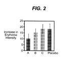
- Circular areas of interest were drawn around the marked skin sites and the mean, standard deviation, and median of the erythema intensity was calculated from the intensity histogram corresponding to each area ( Fig 1 ). The average increase in erythema intensity was calculated for all six subjects ( Fig. 2 ). The results demonstrate that the method of this invention enables one to identify the most efficacious of the tested substances for anti-inflammatory activity and protection from sunburn.
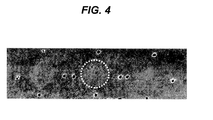
- erythema intensity map is shown in Fig. 4 .
- the mean, standard deviation, and median of the erythema intensity can be calculated for each site.
- the sites from left to right exhibit erythema indices of 130 (moisturizer A), 132 (moisturizer B), and 134 (untreated) respectively with the neighboring skin having a value of 119.
- This example demonstrates that using the method of this invention, one can distinguish between moisturizers in terms of reducing erythema and can identify the most efficacious one.
- the image was then analyzed by the method of this invention and the resulting erythema intensity map is shown in Fig. 6 .
- the mean, standard deviation, and median of the erythema intensity for each site were calculated.
- the sites from left to right exhibit erythema indices of 182, 180, and 181 respectively with the neighboring skin having a value of 178.
- This example demonstrates that using the method of this invention, one can distinguish between cleansers or concentrations of cleansers in terms of their mildness on the skin.
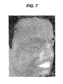
- erythema intensity map is shown in Fig. 7 . Shadows and artifacts due to natural curvature of the face are canceled out on the erythema map. All inflammatory lesions that appear on the original image can be identified easily in the erythema map due to the increased contrast.
- the high intensity values on the lips area are indicative of the high blood concentration on the lips. Areas of subclinical inflammation are also evident in the erythema map, such as the diffuse erythema on the nose, the cheek, and the chin area. Moreover, areas of reduced concentration of oxyhemoglobin such as those around the eye and the nasolabial fold can be identified.
- the erythema intensity map can be displayed using a color-mapping scheme that can be customized depending on the application.
- An example is shown below ( Fig. 8 ) of the erythema map of Fig. 7 using the color map indicated at the bottom of the image.
Landscapes
- Health & Medical Sciences (AREA)
- Life Sciences & Earth Sciences (AREA)
- Engineering & Computer Science (AREA)
- Physics & Mathematics (AREA)
- General Health & Medical Sciences (AREA)
- Medical Informatics (AREA)
- Public Health (AREA)
- Surgery (AREA)
- Veterinary Medicine (AREA)
- Biophysics (AREA)
- Pathology (AREA)
- Biomedical Technology (AREA)
- Heart & Thoracic Surgery (AREA)
- Animal Behavior & Ethology (AREA)
- Molecular Biology (AREA)
- Theoretical Computer Science (AREA)
- General Physics & Mathematics (AREA)
- Computer Vision & Pattern Recognition (AREA)
- Vascular Medicine (AREA)
- Immunology (AREA)
- Dermatology (AREA)
- Nuclear Medicine, Radiotherapy & Molecular Imaging (AREA)
- Radiology & Medical Imaging (AREA)
- Quality & Reliability (AREA)
- Measuring And Recording Apparatus For Diagnosis (AREA)
- Measurement Of The Respiration, Hearing Ability, Form, And Blood Characteristics Of Living Organisms (AREA)
Abstract
Description
- The method of this invention relates to a method for measuring skin erythema. The method of this invention utilizes digital image analysis of conventional photos that can be acquired by inexpensive digital cameras. This method is useful for demonstrating the efficacy of skin care products intended for the prevention and/or reduction of erythema, even when such erythema is not apparent to the naked eye.
- Skin inflammation is often manifested as skin redness (erythema). Erythema is frequently associated with diaper rash, acne, dermatitis, eczema and other skin conditions. Many skin care products are designed either to prevent inflammation, as in the case of sunscreens, or to reduce imflammation, as in the case of diaper rash ointments). Significant skin inflammation may be present even after use of such skin care products but may not be apparent to the naked eye. While in many cases, individuals suffering from such inflammation can report that the inflammation and irritation is still present after use of such products, infants or others who are not able to express themselves, can continue to endure symptoms despite treatment. There is, therefore, a need for a method that is capable of demonstrating the efficacy of such skin care products more accurately.
- Skin erythema is primarily due to vasodilation and local increases in hemoglobin concentration. The hemoglobin concentration in skin can be evaluated using several methods.
- Chromameters have been utilized for analyzing hemoglobin. Chromameters give values of standardized parameters for color evaluation: L*,a*,b*, with a* being used as an indicator of the "red" content and therefore related to skin erythema.
- Spectrophotometers have been used in a method for analyzing hemoglobin based on diffuse reflectance spectroscopy, according to which the reflected light from skin is collected and analyzed into its spectral components. Spectral analysis algorithms have been used to calculate chromophore concentrations including oxy- and deoxy- hemoglobin (relating to erythema). Various light reflectance devices such as a Mexameter are also known for giving an erythema index.
- Spectral imaging is a method that combines the spectral information with two-dimensional spatial information to calculate intensity maps corresponding to chromophore concentrations including oxy- and deoxy-hemoglobin. The analysis of digital color images of skin has also been utilized for analyzing hemoglobin.
- All of the methods described above have deficiencies in their ability to measure inflammation to the degree required in order to ensure treatment of a skin condition causing inflammation. Reflectance-based methods only give point measurements and are limited by the size of the probe used in conjunction with the method. Spectral imaging is the most accurate method for mapping hemoglobin concentration but requires specialized and expensive equipment. Methods using digital image analysis do not take into appropriate consideration all optical parameters of the skin that contribute to the perception of erythema. Therefore, there is a continuing need for a method for measuring skin erythema.
- The method of this invention provides a way in which to measure skin erythema including the following steps: a) acquiring a digital image of skin under orthogonal polarization conditions with a white calibration standard included in the field of view of the image; b) identifying median intensity values for a red, a green, and a blue channel for the white calibration standard; c) translating skin reflectance values given by the red, green, and blue intensity values of each pixel of the image to absorbance values by calculating the logarithm of the ratio of the white standard values, i.e., the pixel intensity of the white standard for each of the channels (red, green, blue) divided by the skin reflectance values, i.e., the pixel intensity values at the corresponding channels (red, green, blue) at each of the red green and blue channels separately, where the absorbance equals:
- log (Iwhite (red, green, blue) /Iskin (red, green, blue)
- for all pixels representing skin; d) subtracting the red channel absorbance values from the green channel absorbance values: (Abs(green)-Abs(red)) for all pixels representing skin; e) subtracting the red channel absorbance values from the blue channel absorbance values: (Abs(blue)-Abs(red)) for all pixels representing skin; f) calculating the ratio of the value calculated at step (d) divided by the value calculated at step (e)ratio = (Abs(green)-Abs(red)) / (Abs(blue)-Abs(red)), and g) building an intensity map of the ratio calculated at step (f).
- The method of this invention also provides a way in which to demonstrate the efficacy of a skin care product including: acquiring a digital image of an area of skin and analyzing the skin according to the method described above; applying a skin care product to the same area of skin for an effective amount of time; and then at a later time, acquiring a subsequent digital image of the area of skin and analyzing the skin according to the method described above; and comparing the intensity maps of the images to demonstrate a reduction in erythema of the skin. The length of time between the first and second image acquisitions can vary depending upon the treatment or effect being evaluated. For example, the efficacy of a moisturizer or anti-inflammatory may be evidenced within a short period of time, on the order of minutes or hours. However, the efficacy of a longacting active ingredient to ameliorate skin conditions such as an allergic reaction, contact dermatitis, acne or the like may be evidenced over a much longer period of time such as days, weeks or months.
- The method of this invention also provides a means by which to demonstrate the mildness of a skin care product including: acquiring a digital image of an area of skin and analyzing the skin according to the method described above; applying a skin care product to the same area of skin for an effective amount of time; acquiring a subsequent digital image of the area of skin and analyzing the skin according to the method described above; and comparing the intensity maps of the images to demonstrate no increase in erythema of the skin.
-
-
Figure 1 shows the transformation of a skin image to an intensity histogram corresponding to each area on the image. -
Figure 2 is a graph demonstrating the average increase in erythema intensity on the skin of several subjects. -
Figure 3 is a photograph of the skin of a subject. -
Figure 4 is an erythema intensity map of the skin photographed inFigure 3 . -
Figure 5 is a photograph of the skin of a subject. -
Figure 6 is an erythema intensity map of the skin photographed inFigure 5 . -
Figure 7 is an erythema intensity map of the skin of a subject's face. -
Figure 8 is a color map of the erythema intensity map ofFigure 7 . - Various types of cameras may be used in the methods of this invention. Examples of such cameras include, but are not limited to, standard 35 mm cameras, cameras using instant developing film (such as those available from Polaroid Corporation, Cambridge, Mass. USA), and digital cameras. Preferably, a digital camera is used as it provides fast access to the images taken of the subject. It also allows the image to be displayed on a large monitor, enables the subject to easily enlarge areas of skin that are of particular interest (e.g., areas of the face), and allows the image to be printed in a report which can also include suggestions for products addressing any concerns the subject noticed upon examining the images. Examples of suitable digital cameras include, but are not limited to, those which take images of at least 1 million pixels, preferable at least 4 million pixels. Examples of such digital cameras include, but are not limited to, the Nikon D1X (Nikon, Tokyo, Japan) and the Fuji S1 (Fuji, Tokyo, Japan).
- The following terms used herein have the meanings ascribed to them below:
- "ORTHOGONAL POLARIZATION CONDITIONS": imaging conditions that include a first polarizer filter in front of the light source and a second polarizer filter in front of the camera lens with the two filters having their polarization axes orthogonal to each other.
- "WHITE CALIBRATION STANDARD": a uniform white card corresponding to a certain % reflectance (e.g. 80%).
- "MEDIAN INTENSITY VALUES": the arithmetic median of a group of pixel intensity values corresponding to a region of interest in the image.
- "SKIN REFLECTANCE VALUES": pixel intensity values of an area of interest in the image corresponding to skin.
- "ABSORBANCE VALUES": the light absorbance being defined by the equation: absorbance = log {(reflectance of the white standard)/(reflectance of the object of interest)}.
- In one embodiment, the methods of this invention include the step of taking a polarized photograph of the subject. What is meant by "polarized photograph" is a photograph of the subject taken (i) with a light source that emits light through a polarizing filter and/or (ii) through a polarized filter that filters light prior to or after entering the camera's lens.
- In one embodiment, the camera and one or more flash units, preferably two, are on about the same plane as the subject's skin to be photographed, and the flash units are placed so that the angle formed by each flash unit(s), subject's skin, and camera is about 35 to 55 degrees, such as about 45 degrees. In one embodiment, a polarizing filter is placed in front of each flash unit. What is meant by a "polarizing filter" is a filter that filters incoming light to emit substantially only polarized light. What is meant by the term "substantially," as used herein, is at least 75 percent, preferably 90 percent, and most preferably at least 95 percent. Examples of a polarizing filter include, but are not limited to, polarizing plates such as those available from Edmund Scientific (Barrington, N.J. USA), polarizing prisms such as Glan Thomson polarizing prisms, or a polarizing reflector that reflects light at about the Brewster angle. Polarizing filters may be linear or circular polarizing filters.
- In another embodiment of the method of this invention, a light diffuser is placed between the flash unit and the polarizing filter.
- In yet another embodiment of the method of this invention, a linear polarizing filter is used at the light source and the linear polarizing filter is arranged such that the electric field of the emitted light is about perpendicular (orthogonal) to the plane formed by the light source, the person's skin, and the camera.
- In another embodiment of the method of this invention, the flash unit(s) are positioned on a vertical plane above the camera and the subject's skin so that the angle formed by the flash unit, subject's skin and camera is about 35 to 55 degrees, preferably about 45 degrees. The flash unit(s) are filtered with a linear polarizing filter that is placed with the transmitted electric field in the vertical direction (e.g., parallel to the plane). In this arrangement the surface glare from the skin is minimized, thus enhancing the subsurface features of the skin, such as erythema (redness), blood vessels, and pigmentation.
- In one embodiment of the method of this invention, the photograph of the subject is taken both with a light source that emits lights through a polarizing filter and through a polarizing filter that filters the light prior to or after entering the camera's lens. When the polarizing filters are aligned perpendicular to each other (e.g., one horizontal and one vertical), subsurface features of the skin such as erythema, pigmentation, blood vessels, and hair, are visually enhanced and surface specular reflections (glare) are eliminated.
- Image analysis requires: a) acquiring a digital image of skin under orthogonal polarization conditions with a white calibration standard included in the field of view of the image; b) identifying median intensity values for a red, a green, and a blue channel for the white standard; c) translating skin reflectance values given by the red, green, and blue intensity values of each pixel of the image to absorbance values by taking the logarithm of the ratio of the white standard values divided by the skin reflectance values at each of the red, green, and blue channels separately, where the absorbance equals the
log (Iwhite (red, green, blue)/Iskin (red,green,blue))
for all pixels representing skin; d) subtracting the red channel absorbance values from the green channel absorbance values for all pixels representing skin; e) subtracting the red channel absorbance values from the blue channel absorbance values for all pixels representing skin; f) taking the ratio of the value calculated at step (d) divided by the value calculated at step (e), and g) building an intensity map of the ratio calculated at step (f). - In one embodiment the constructed erythema intensity map may be presented as a grayscale intensity image. In another embodiment, colormaps, which are tables of correspondence between gray-scale intensity values on one side and projected colors on the other, can be used in which the each intensity value is represented by a color. Colormaps can be used to enhance features corresponding to particular ranges of erythema intensity (see example 5). In this way, a gray-scale image can be projected as a "pseudo-color"; the sequence of colors in a colormap can be selected so as to enhance the appearance of features corresponding to a particular gray-scale range.
- The methods are useful for measuring erythema, which enables one to demonstrate the efficacy of anti-inflammatory skin care products, or to demonstrate the mildness of skin care products. As used herein, skin care products include, but are not limited to, creams, lotions, ointments, solutions and the like that are intended to be used as cleansers, moisturizers, sunscreens, anti-wrinkle compositions, anti-itch compositions, anti-inflammatory compositions, skin pigmentation products, or anti-acne products. For example, images may be taken prior to and after application of an anti-inflammatory cream. The images enable the consumer to see a reduction in the erythema associated with inflammation.
- Likewise, if a product has not been effective in sufficiently reducing erythema, the methods of this invention will so demonstrate and assist in determining whether a treatment has been efficacious. The following examples serve to illustrate, but not to limit, the methods of this invention.
- The anti-inflammatory activity of three natural extracts in a 70% ethanol/30% propylene glycol base (A = 4% Jasmin extract/3% Sunflower extract, B = 2% Jasmin extract/2% Sunflower extract, and C = 1% Symatrix) was assessed versus the base alone. Four rectangular areas on the back of 6 volunteers were treated with either one of the tested formulations (A, B, C, placebo). Then three smaller round areas in each of the rectangles received UVB radiation at doses corresponding to 0.5, 1, and 1.5 MED. Images were acquired one day following the irradiation. The images were transformed using the method (and associated algorithm) of this invention to generate erythema intensity maps. Circular areas of interest were drawn around the marked skin sites and the mean, standard deviation, and median of the erythema intensity was calculated from the intensity histogram corresponding to each area (
Fig 1 ). The average increase in erythema intensity was calculated for all six subjects (Fig. 2 ). The results demonstrate that the method of this invention enables one to identify the most efficacious of the tested substances for anti-inflammatory activity and protection from sunburn. - A study was performed to assess the effect of two moisturizers (A and B) on the treatment of skin irritation following the act of compromising the skin barrier. Three sites of the volar forearm of the volunteers were subjected to 10 tape stripes followed by treatment with 0.16 ml of 5% SLS (sodium lauryl sulfate) solution under occlusion for two hrs. Following patch removal, the tested substances were applied on the designated skin sites (2 µl/cm2) and one site was kept untreated. The test substances were applied at the corresponding sites daily thereafter for four days. Images were taken daily during the duration of the study. The example in
Fig. 3 shows the photo of the arm of a volunteer on day three. Erythema is evident on the sites. However, it is impossible to qualify the sites in terms of severity or evaluate the erythema quantitatively just by observing this image.
The image was then analyzed by the method of this invention and the resulting erythema intensity map is shown inFig. 4 . The mean, standard deviation, and median of the erythema intensity can be calculated for each site. In this example, the sites from left to right exhibit erythema indices of 130 (moisturizer A), 132 (moisturizer B), and 134 (untreated) respectively with the neighboring skin having a value of 119. This example demonstrates that using the method of this invention, one can distinguish between moisturizers in terms of reducing erythema and can identify the most efficacious one. - A study was performed to assess the mildness of a cleanser (Johnson's® Head-to-Toe Baby Wash, commercially available from Johnson & Johnson Consumer Companies, Inc., Skillman, NJ) on the skin and compare it to water. Three sites of the volar forearm of the volunteers were treated with a) 25% dilution of the cleanser in distilled water, b) 10% dilution of the cleanser in distilled water and c) distilled water only, under occlusion for 3 hrs. Images were taken at baseline and at 1 and 2 days following treatment. The example in
Fig. 5 shows the photo of the arm of a volunteer on day 2. It is difficult to distinguish any signs of erythema in all three sites of treatment. The image was then analyzed by the method of this invention and the resulting erythema intensity map is shown inFig. 6 . The mean, standard deviation, and median of the erythema intensity for each site were calculated. In this example the sites from left to right exhibit erythema indices of 182, 180, and 181 respectively with the neighboring skin having a value of 178. This example demonstrates that using the method of this invention, one can distinguish between cleansers or concentrations of cleansers in terms of their mildness on the skin. - In the following example an orthogonal polarization image of a human face was analyzed by the method of this invention (with the associated algorithm). The erythema intensity map is shown in
Fig. 7 . Shadows and artifacts due to natural curvature of the face are canceled out on the erythema map. All inflammatory lesions that appear on the original image can be identified easily in the erythema map due to the increased contrast. The high intensity values on the lips area are indicative of the high blood concentration on the lips. Areas of subclinical inflammation are also evident in the erythema map, such as the diffuse erythema on the nose, the cheek, and the chin area. Moreover, areas of reduced concentration of oxyhemoglobin such as those around the eye and the nasolabial fold can be identified. - For better visualization of the underlying subclinical erythema, the erythema intensity map can be displayed using a color-mapping scheme that can be customized depending on the application. An example is shown below (
Fig. 8 ) of the erythema map ofFig. 7 using the color map indicated at the bottom of the image. - The Examples above demonstrate that using the methods of this invention, one can assess the mildness of cleansers, the activity of anti-inflammatories and other skin irritation treatments, and the efficacy of treatments against skin disorders such as but not limited to acne, rosacea, irritant contact dermatitis, allergic contact dermatitis, diaper dermatitis, atopic dermatitis, seborrheic dermatitis, etc. The methods can also be used for monitoring the progression of such disorders.
Claims (3)
- A method for measuring skin erythema comprising: a) acquiring a digital image of skin under orthogonal polarization conditions with a white calibration standard included in the field of view of the image;b) identifying median intensity values for a red, a green, and a blue channel for the white standard;c) calculating skin reflectance values given by the red, green, and blue intensity values of each pixel of the image to absorbance values by taking the logarithm of the ratio of the white standard values divided by the skin reflectance values at each of the red green and blue channels separately, where the absorbance equals:log (Iwhite (red, green, blue) /Iskin (red, green, blue))for all pixels representing skin;d) subtracting the red channel absorbance values from the green channel absorbance values for all pixels representing skin;e) subtracting the red channel absorbance values from the blue channel absorbance values for all pixels representing skin;f) calculating the ratio of the value calculated at step (d) divided by the value calculated at step (e); andg) building an intensity map of the ratio calculated at step (f).
- A method of demonstrating the efficacy of a skin care product comprising:taking a digital image of an area of skin and analyzing the skin according to the method of Claim 1;applying a skin care product to the same area of skin for an effective amount of time;taking a subsequent digital image of the area of skin and analyzing the skin according to the method of Claim 1; andcomparing the intensity maps of the images to demonstrate a reduction in erythema of the skin.
- A method of demonstrating the mildness of a skin care product comprising:taking a digital image of an area of skin and analyzing the skin according to the method of Claim 1;applying a skin care product to the same area of skin for an effective amount of time;taking a subsequent digital image of the area of skin and analyzing the skin according to the method of Claim 1; andcomparing the intensity maps of the images to demonstrate no increase in erythema of the skin.
Applications Claiming Priority (1)
| Application Number | Priority Date | Filing Date | Title |
|---|---|---|---|
| US12/411,642 US8150501B2 (en) | 2009-03-26 | 2009-03-26 | Method for measuring skin erythema |
Publications (2)
| Publication Number | Publication Date |
|---|---|
| EP2233073A1 true EP2233073A1 (en) | 2010-09-29 |
| EP2233073B1 EP2233073B1 (en) | 2017-01-04 |
Family
ID=42316010
Family Applications (1)
| Application Number | Title | Priority Date | Filing Date |
|---|---|---|---|
| EP10250571.6A Not-in-force EP2233073B1 (en) | 2009-03-26 | 2010-03-25 | Method for measuring skin erythema |
Country Status (9)
| Country | Link |
|---|---|
| US (1) | US8150501B2 (en) |
| EP (1) | EP2233073B1 (en) |
| JP (1) | JP5496729B2 (en) |
| KR (1) | KR101602149B1 (en) |
| CN (1) | CN101843475A (en) |
| AU (1) | AU2010201207B2 (en) |
| BR (1) | BRPI1015656A2 (en) |
| CA (1) | CA2697316C (en) |
| RU (1) | RU2010111626A (en) |
Cited By (3)
| Publication number | Priority date | Publication date | Assignee | Title |
|---|---|---|---|---|
| JP2010227571A (en) * | 2009-03-26 | 2010-10-14 | Johnson & Johnson Consumer Co Inc | Method for measuring skin erythema |
| GB2512876A (en) * | 2013-04-09 | 2014-10-15 | Image Analysis Ltd | Methods and apparatus for quantifying inflammation |
| EP2868260A4 (en) * | 2012-06-29 | 2016-03-02 | Toshiba Kk | Health management support device, method and program |
Families Citing this family (30)
| Publication number | Priority date | Publication date | Assignee | Title |
|---|---|---|---|---|
| AT511265B1 (en) * | 2011-03-24 | 2013-12-15 | Red Soft It Service Gmbh | DEVICE FOR DETERMINING A CHARACTERIZATION VALUE AND METHOD FOR EVALUATING THREE-DIMENSIONAL IMAGES |
| US9854970B2 (en) * | 2012-02-21 | 2018-01-02 | Massachusetts Eye & Ear Infirmary | Calculating conjunctival redness |
| CN103006238B (en) * | 2013-01-07 | 2014-11-26 | 天津大学 | Method for measuring degree of blood oxygen saturation |
| WO2014152096A1 (en) * | 2013-03-15 | 2014-09-25 | Suncare Research Laboratories, Llc | In vitro determination of sunscreen protection based on image analysis of sunscreens applied to skin |
| EP2781191A1 (en) | 2013-03-19 | 2014-09-24 | Schnidar, Harald | Methods for assessing erythema |
| JP6437466B2 (en) * | 2013-06-19 | 2018-12-12 | ロレアル | Method for assessing the cosmetic effect of cosmetics on the skin |
| CN105338887B (en) * | 2013-06-28 | 2019-10-25 | 松下电器(美国)知识产权公司 | Skin sensory evaluation device and skin evaluation method |
| US10004395B2 (en) | 2014-05-02 | 2018-06-26 | Massachusetts Eye And Ear Infirmary | Grading corneal fluorescein staining |
| RU2014122942A (en) * | 2014-06-06 | 2015-12-20 | Андрей Владимирович Сартори | METHOD AND DEVICE FOR DETERMINING THE ERYTHEMIC INDEX |
| CN104224125A (en) * | 2014-09-17 | 2014-12-24 | 中国人民解放军空军总医院 | Demoscopy image acquisition device and method |
| JP6138745B2 (en) * | 2014-11-19 | 2017-05-31 | 株式会社 資生堂 | Spot evaluation device and spot evaluation program |
| KR102348798B1 (en) * | 2015-03-31 | 2022-01-07 | (주)아모레퍼시픽 | Transparency and Erythema index Measuring Device of Skin |
| US10073945B1 (en) * | 2015-05-06 | 2018-09-11 | Ning Pan | Process for evaluating skin care product efficacy and skin care product efficacy evaluation system |
| US10460197B1 (en) * | 2016-05-06 | 2019-10-29 | Ning Pan | Process for evaluating skin care product efficacy and skin care product efficacy evaluation system |
| JP6265231B2 (en) * | 2016-06-23 | 2018-01-24 | カシオ計算機株式会社 | Diagnostic device, image processing method, and program |
| BR112018076965A8 (en) * | 2016-06-27 | 2023-03-07 | Koninklijke Philips Nv | SKIN SHINE DETECTION DEVICE, SKIN SHINE DETECTION METHOD, AND COMPUTER PROGRAM |
| CN106264463A (en) * | 2016-08-05 | 2017-01-04 | 深圳美立知科技有限公司 | A kind of skin sensitivity analyzes method and device |
| US12011256B2 (en) * | 2016-11-01 | 2024-06-18 | Universidade Federal De Minas Gerais—Ufmg | Device and method for determining gestational age |
| US10511777B2 (en) | 2016-11-08 | 2019-12-17 | Thomas Nichols | Personal care device with camera |
| US11122206B2 (en) | 2016-11-08 | 2021-09-14 | Preh Holding, Llc | Personal care device with camera |
| EP3384831A1 (en) | 2017-04-05 | 2018-10-10 | Koninklijke Philips N.V. | Skin gloss measurement using brewster's angle |
| KR102123598B1 (en) | 2017-12-26 | 2020-06-16 | (주)아모레퍼시픽 | Apparatus and system for skin diagnosis and method thereof |
| CN110018161B (en) * | 2019-02-28 | 2022-05-20 | 北京三立慧评化妆品科技有限公司 | Method for detecting irritation of cosmetic raw material and method for detecting irritation resistance of irritation-resistant product |
| US11229595B2 (en) * | 2019-05-29 | 2022-01-25 | Johnson & Johnson Consumer Inc. | Skin barrier preparation and method therefor |
| USD1000624S1 (en) | 2019-12-27 | 2023-10-03 | Thomas Nichols | Personal care device with camera |
| KR102378212B1 (en) * | 2020-08-07 | 2022-03-25 | 주식회사 에이아이포펫 | Skin disease measuring system using a portable treminal and skin disease management method |
| US11836922B2 (en) | 2021-05-19 | 2023-12-05 | Zerigo Health, Inc. | System and method for evaluating effectiveness of a skin treatment |
| EP4356398A4 (en) | 2021-06-14 | 2025-04-16 | Preh Holding, LLC | Connected body surface care module |
| CN113989138B (en) * | 2021-10-21 | 2024-07-16 | 武汉博视电子有限公司 | Method for extracting facial skin image inflammation and forming red area spectrum |
| EP4627526A1 (en) * | 2022-11-30 | 2025-10-08 | Lumenis Be Ltd. | System and method for determining human skin attributes and treatments |
Family Cites Families (10)
| Publication number | Priority date | Publication date | Assignee | Title |
|---|---|---|---|---|
| CA2305721C (en) * | 1997-10-08 | 2009-02-24 | The General Hospital Corporation | Phototherapy methods and systems |
| JP3727807B2 (en) * | 1999-06-14 | 2005-12-21 | 株式会社資生堂 | Method and apparatus for measuring components in skin and skin characteristics |
| US6907193B2 (en) * | 2001-11-08 | 2005-06-14 | Johnson & Johnson Consumer Companies, Inc. | Method of taking polarized images of the skin and the use thereof |
| US6961517B2 (en) * | 2001-11-08 | 2005-11-01 | Johnson & Johnson Consumer Companies, Inc. | Method of promoting skin care products |
| US7400754B2 (en) * | 2003-04-08 | 2008-07-15 | The Regents Of The University Of California | Method and apparatus for characterization of chromophore content and distribution in skin using cross-polarized diffuse reflectance imaging |
| US20050049467A1 (en) * | 2003-08-28 | 2005-03-03 | Georgios Stamatas | Method for assessing pigmented skin |
| SE0401004D0 (en) * | 2004-04-19 | 2004-04-19 | Gert Nilsson | Non-invasive method to monitor microcalculation |
| US7454046B2 (en) * | 2005-09-20 | 2008-11-18 | Brightex Bio-Photonics, Llc | Method and system for analyzing skin conditions using digital images |
| CA2643606C (en) * | 2006-03-02 | 2015-05-26 | Johnson & Johnson Consumer Companies, Inc. | Method for demonstrating pre-emergent pimples |
| US8150501B2 (en) * | 2009-03-26 | 2012-04-03 | Johnson & Johnson Consumer Companies, Inc. | Method for measuring skin erythema |
-
2009
- 2009-03-26 US US12/411,642 patent/US8150501B2/en not_active Expired - Fee Related
-
2010
- 2010-03-18 CA CA2697316A patent/CA2697316C/en not_active Expired - Fee Related
- 2010-03-25 EP EP10250571.6A patent/EP2233073B1/en not_active Not-in-force
- 2010-03-25 AU AU2010201207A patent/AU2010201207B2/en not_active Ceased
- 2010-03-25 JP JP2010069344A patent/JP5496729B2/en not_active Expired - Fee Related
- 2010-03-25 RU RU2010111626/15A patent/RU2010111626A/en not_active Application Discontinuation
- 2010-03-26 CN CN201010155652A patent/CN101843475A/en active Pending
- 2010-03-26 BR BRPI1015656-9A patent/BRPI1015656A2/en not_active IP Right Cessation
- 2010-03-26 KR KR1020100027111A patent/KR101602149B1/en not_active Expired - Fee Related
Non-Patent Citations (5)
| Title |
|---|
| BYUNGJO JUNG: "Polarization spectral imaging system for quantitative evaluation of port wine stain blanching following laser treatment", JOURNAL OF THE OPTICAL SOCIETY OF KOREA OPT. SOC. KOREA SOUTH KOREA, vol. 7, no. 4, December 2003 (2003-12-01), pages 234 - 239, XP002595718, ISSN: 1226-4776 * |
| DOLOTOV L E ET AL: "Digital imaging of human skin", PROCEEDINGS OF THE SPIE - THE INTERNATIONAL SOCIETY FOR OPTICAL ENGINEERING SPIE-INT. SOC. OPT. ENG USA, vol. 5067, no. 1, 2003, pages 139 - 147, XP002595717, ISSN: 0277-786X * |
| LEACHTENAUER J ET AL: "A Non-Contact Imaging-Based Approach to Detecting Stage I Pressure Ulcers", ENGINEERING IN MEDICINE AND BIOLOGY SOCIETY, 2006. EMBS '06. 28TH ANNUAL INTERNATIONAL CONFERENCE OF THE IEEE, IEEE, PISCATAWAY, NJ, USA, 30 August 2006 (2006-08-30), pages 6380 - 6383, XP031390821, ISBN: 978-1-4244-0032-4 * |
| STAMATAS G N ET AL: "In vivo measurement of skin erythema and pigmentation: new means of implementation of diffuse reflectance spectroscopy with a commercial instrument.", THE BRITISH JOURNAL OF DERMATOLOGY SEP 2008 LNKD- PUBMED:18510669, vol. 159, no. 3, September 2008 (2008-09-01), pages 683 - 690, XP002595720, ISSN: 1365-2133 * |
| STAMATAS G N ET AL: "Noninvasive quantitative documentation of cutaneous inflammation in vivo using spectral imaging", PROCEEDINGS OF THE SPIE - THE INTERNATIONAL SOCIETY FOR OPTICAL ENGINEERING SPIE-INT. SOC. OPT. ENG USA, vol. 6078, no. 1, 2006, pages 111 - 118, XP002595719, ISSN: 0277-786X * |
Cited By (3)
| Publication number | Priority date | Publication date | Assignee | Title |
|---|---|---|---|---|
| JP2010227571A (en) * | 2009-03-26 | 2010-10-14 | Johnson & Johnson Consumer Co Inc | Method for measuring skin erythema |
| EP2868260A4 (en) * | 2012-06-29 | 2016-03-02 | Toshiba Kk | Health management support device, method and program |
| GB2512876A (en) * | 2013-04-09 | 2014-10-15 | Image Analysis Ltd | Methods and apparatus for quantifying inflammation |
Also Published As
| Publication number | Publication date |
|---|---|
| KR101602149B1 (en) | 2016-03-11 |
| CA2697316A1 (en) | 2010-09-26 |
| AU2010201207A1 (en) | 2010-10-14 |
| JP2010227571A (en) | 2010-10-14 |
| CA2697316C (en) | 2017-10-24 |
| JP5496729B2 (en) | 2014-05-21 |
| BRPI1015656A2 (en) | 2015-08-18 |
| US20100249731A1 (en) | 2010-09-30 |
| RU2010111626A (en) | 2011-09-27 |
| KR20100108280A (en) | 2010-10-06 |
| EP2233073B1 (en) | 2017-01-04 |
| CN101843475A (en) | 2010-09-29 |
| US8150501B2 (en) | 2012-04-03 |
| AU2010201207B2 (en) | 2014-08-07 |
Similar Documents
| Publication | Publication Date | Title |
|---|---|---|
| CA2697316C (en) | Method for measuring skin erythema | |
| EP1993442B1 (en) | Method for demonstrating pre-emergent pimples | |
| JP6557210B2 (en) | Evaluation method of erythema | |
| JP5149527B2 (en) | How to display skin pigmentation | |
| RU2657377C2 (en) | Intellectual head for smartphone for determination of cleanliness, moisture and photo-age of skin | |
| EP2387942A1 (en) | Method for measuring skin hydration | |
| US20180242901A1 (en) | Systems and methods for hyperspectral medical imaging using real-time projection of spectral information | |
| Stamatas et al. | In vivo documentation of cutaneous inflammation using spectral imaging | |
| US20040215066A1 (en) | Method of measuring the efficacy of a skin treatment program | |
| Mattsson et al. | Assessment of erythema and skin perfusion by digital image analysis and scanner laser Doppler | |
| Leachtenauer et al. | A non-contact imaging-based approach to detecting stage i pressure ulcers | |
| Galeano et al. | Analysis of human skin hyper-spectral images by non-negative matrix factorization | |
| Randeberg et al. | Combined hyperspectral and 3D characterization of non-healing skin ulcers | |
| Abdlaty et al. | Noninvasive spectral monitoring for radiation therapy induced skin-inflammation | |
| McGrath | Development of a Multispectral VIS-SWIR Imaging Modality for Cutaneous Water Assessment |
Legal Events
| Date | Code | Title | Description |
|---|---|---|---|
| PUAI | Public reference made under article 153(3) epc to a published international application that has entered the european phase |
Free format text: ORIGINAL CODE: 0009012 |
|
| AK | Designated contracting states |
Kind code of ref document: A1 Designated state(s): AT BE BG CH CY CZ DE DK EE ES FI FR GB GR HR HU IE IS IT LI LT LU LV MC MK MT NL NO PL PT RO SE SI SK SM TR |
|
| AX | Request for extension of the european patent |
Extension state: AL BA ME RS |
|
| 17P | Request for examination filed |
Effective date: 20110321 |
|
| 17Q | First examination report despatched |
Effective date: 20120425 |
|
| GRAP | Despatch of communication of intention to grant a patent |
Free format text: ORIGINAL CODE: EPIDOSNIGR1 |
|
| INTG | Intention to grant announced |
Effective date: 20160803 |
|
| GRAS | Grant fee paid |
Free format text: ORIGINAL CODE: EPIDOSNIGR3 |
|
| GRAA | (expected) grant |
Free format text: ORIGINAL CODE: 0009210 |
|
| STAA | Information on the status of an ep patent application or granted ep patent |
Free format text: STATUS: THE PATENT HAS BEEN GRANTED |
|
| RAP1 | Party data changed (applicant data changed or rights of an application transferred) |
Owner name: JOHNSON & JOHNSON CONSUMER INC. |
|
| AK | Designated contracting states |
Kind code of ref document: B1 Designated state(s): AT BE BG CH CY CZ DE DK EE ES FI FR GB GR HR HU IE IS IT LI LT LU LV MC MK MT NL NO PL PT RO SE SI SK SM TR |
|
| REG | Reference to a national code |
Ref country code: GB Ref legal event code: FG4D |
|
| REG | Reference to a national code |
Ref country code: CH Ref legal event code: EP |
|
| REG | Reference to a national code |
Ref country code: AT Ref legal event code: REF Ref document number: 858417 Country of ref document: AT Kind code of ref document: T Effective date: 20170115 |
|
| REG | Reference to a national code |
Ref country code: IE Ref legal event code: FG4D |
|
| REG | Reference to a national code |
Ref country code: FR Ref legal event code: PLFP Year of fee payment: 8 |
|
| REG | Reference to a national code |
Ref country code: DE Ref legal event code: R096 Ref document number: 602010039290 Country of ref document: DE |
|
| REG | Reference to a national code |
Ref country code: LT Ref legal event code: MG4D Ref country code: NL Ref legal event code: MP Effective date: 20170104 |
|
| REG | Reference to a national code |
Ref country code: AT Ref legal event code: MK05 Ref document number: 858417 Country of ref document: AT Kind code of ref document: T Effective date: 20170104 |
|
| PG25 | Lapsed in a contracting state [announced via postgrant information from national office to epo] |
Ref country code: NL Free format text: LAPSE BECAUSE OF FAILURE TO SUBMIT A TRANSLATION OF THE DESCRIPTION OR TO PAY THE FEE WITHIN THE PRESCRIBED TIME-LIMIT Effective date: 20170104 |
|
| PG25 | Lapsed in a contracting state [announced via postgrant information from national office to epo] |
Ref country code: LT Free format text: LAPSE BECAUSE OF FAILURE TO SUBMIT A TRANSLATION OF THE DESCRIPTION OR TO PAY THE FEE WITHIN THE PRESCRIBED TIME-LIMIT Effective date: 20170104 Ref country code: GR Free format text: LAPSE BECAUSE OF FAILURE TO SUBMIT A TRANSLATION OF THE DESCRIPTION OR TO PAY THE FEE WITHIN THE PRESCRIBED TIME-LIMIT Effective date: 20170405 Ref country code: IS Free format text: LAPSE BECAUSE OF FAILURE TO SUBMIT A TRANSLATION OF THE DESCRIPTION OR TO PAY THE FEE WITHIN THE PRESCRIBED TIME-LIMIT Effective date: 20170504 Ref country code: NO Free format text: LAPSE BECAUSE OF FAILURE TO SUBMIT A TRANSLATION OF THE DESCRIPTION OR TO PAY THE FEE WITHIN THE PRESCRIBED TIME-LIMIT Effective date: 20170404 Ref country code: HR Free format text: LAPSE BECAUSE OF FAILURE TO SUBMIT A TRANSLATION OF THE DESCRIPTION OR TO PAY THE FEE WITHIN THE PRESCRIBED TIME-LIMIT Effective date: 20170104 Ref country code: FI Free format text: LAPSE BECAUSE OF FAILURE TO SUBMIT A TRANSLATION OF THE DESCRIPTION OR TO PAY THE FEE WITHIN THE PRESCRIBED TIME-LIMIT Effective date: 20170104 |
|
| PG25 | Lapsed in a contracting state [announced via postgrant information from national office to epo] |
Ref country code: LV Free format text: LAPSE BECAUSE OF FAILURE TO SUBMIT A TRANSLATION OF THE DESCRIPTION OR TO PAY THE FEE WITHIN THE PRESCRIBED TIME-LIMIT Effective date: 20170104 Ref country code: BG Free format text: LAPSE BECAUSE OF FAILURE TO SUBMIT A TRANSLATION OF THE DESCRIPTION OR TO PAY THE FEE WITHIN THE PRESCRIBED TIME-LIMIT Effective date: 20170404 Ref country code: PT Free format text: LAPSE BECAUSE OF FAILURE TO SUBMIT A TRANSLATION OF THE DESCRIPTION OR TO PAY THE FEE WITHIN THE PRESCRIBED TIME-LIMIT Effective date: 20170504 Ref country code: PL Free format text: LAPSE BECAUSE OF FAILURE TO SUBMIT A TRANSLATION OF THE DESCRIPTION OR TO PAY THE FEE WITHIN THE PRESCRIBED TIME-LIMIT Effective date: 20170104 Ref country code: ES Free format text: LAPSE BECAUSE OF FAILURE TO SUBMIT A TRANSLATION OF THE DESCRIPTION OR TO PAY THE FEE WITHIN THE PRESCRIBED TIME-LIMIT Effective date: 20170104 Ref country code: AT Free format text: LAPSE BECAUSE OF FAILURE TO SUBMIT A TRANSLATION OF THE DESCRIPTION OR TO PAY THE FEE WITHIN THE PRESCRIBED TIME-LIMIT Effective date: 20170104 Ref country code: SE Free format text: LAPSE BECAUSE OF FAILURE TO SUBMIT A TRANSLATION OF THE DESCRIPTION OR TO PAY THE FEE WITHIN THE PRESCRIBED TIME-LIMIT Effective date: 20170104 |
|
| REG | Reference to a national code |
Ref country code: DE Ref legal event code: R097 Ref document number: 602010039290 Country of ref document: DE |
|
| PG25 | Lapsed in a contracting state [announced via postgrant information from national office to epo] |
Ref country code: RO Free format text: LAPSE BECAUSE OF FAILURE TO SUBMIT A TRANSLATION OF THE DESCRIPTION OR TO PAY THE FEE WITHIN THE PRESCRIBED TIME-LIMIT Effective date: 20170104 Ref country code: IT Free format text: LAPSE BECAUSE OF FAILURE TO SUBMIT A TRANSLATION OF THE DESCRIPTION OR TO PAY THE FEE WITHIN THE PRESCRIBED TIME-LIMIT Effective date: 20170104 Ref country code: EE Free format text: LAPSE BECAUSE OF FAILURE TO SUBMIT A TRANSLATION OF THE DESCRIPTION OR TO PAY THE FEE WITHIN THE PRESCRIBED TIME-LIMIT Effective date: 20170104 Ref country code: SK Free format text: LAPSE BECAUSE OF FAILURE TO SUBMIT A TRANSLATION OF THE DESCRIPTION OR TO PAY THE FEE WITHIN THE PRESCRIBED TIME-LIMIT Effective date: 20170104 Ref country code: CZ Free format text: LAPSE BECAUSE OF FAILURE TO SUBMIT A TRANSLATION OF THE DESCRIPTION OR TO PAY THE FEE WITHIN THE PRESCRIBED TIME-LIMIT Effective date: 20170104 |
|
| REG | Reference to a national code |
Ref country code: CH Ref legal event code: PL |
|
| PLBE | No opposition filed within time limit |
Free format text: ORIGINAL CODE: 0009261 |
|
| STAA | Information on the status of an ep patent application or granted ep patent |
Free format text: STATUS: NO OPPOSITION FILED WITHIN TIME LIMIT |
|
| PG25 | Lapsed in a contracting state [announced via postgrant information from national office to epo] |
Ref country code: DK Free format text: LAPSE BECAUSE OF FAILURE TO SUBMIT A TRANSLATION OF THE DESCRIPTION OR TO PAY THE FEE WITHIN THE PRESCRIBED TIME-LIMIT Effective date: 20170104 Ref country code: SM Free format text: LAPSE BECAUSE OF FAILURE TO SUBMIT A TRANSLATION OF THE DESCRIPTION OR TO PAY THE FEE WITHIN THE PRESCRIBED TIME-LIMIT Effective date: 20170104 Ref country code: MC Free format text: LAPSE BECAUSE OF FAILURE TO SUBMIT A TRANSLATION OF THE DESCRIPTION OR TO PAY THE FEE WITHIN THE PRESCRIBED TIME-LIMIT Effective date: 20170104 |
|
| 26N | No opposition filed |
Effective date: 20171005 |
|
| REG | Reference to a national code |
Ref country code: IE Ref legal event code: MM4A |
|
| PG25 | Lapsed in a contracting state [announced via postgrant information from national office to epo] |
Ref country code: LU Free format text: LAPSE BECAUSE OF NON-PAYMENT OF DUE FEES Effective date: 20170325 |
|
| REG | Reference to a national code |
Ref country code: FR Ref legal event code: PLFP Year of fee payment: 9 |
|
| PG25 | Lapsed in a contracting state [announced via postgrant information from national office to epo] |
Ref country code: CH Free format text: LAPSE BECAUSE OF NON-PAYMENT OF DUE FEES Effective date: 20170331 Ref country code: IE Free format text: LAPSE BECAUSE OF NON-PAYMENT OF DUE FEES Effective date: 20170325 Ref country code: LI Free format text: LAPSE BECAUSE OF NON-PAYMENT OF DUE FEES Effective date: 20170331 Ref country code: SI Free format text: LAPSE BECAUSE OF FAILURE TO SUBMIT A TRANSLATION OF THE DESCRIPTION OR TO PAY THE FEE WITHIN THE PRESCRIBED TIME-LIMIT Effective date: 20170104 |
|
| REG | Reference to a national code |
Ref country code: BE Ref legal event code: MM Effective date: 20170331 |
|
| PGFP | Annual fee paid to national office [announced via postgrant information from national office to epo] |
Ref country code: GB Payment date: 20180321 Year of fee payment: 9 Ref country code: DE Payment date: 20180313 Year of fee payment: 9 |
|
| PG25 | Lapsed in a contracting state [announced via postgrant information from national office to epo] |
Ref country code: BE Free format text: LAPSE BECAUSE OF NON-PAYMENT OF DUE FEES Effective date: 20170331 |
|
| PGFP | Annual fee paid to national office [announced via postgrant information from national office to epo] |
Ref country code: FR Payment date: 20180222 Year of fee payment: 9 |
|
| PG25 | Lapsed in a contracting state [announced via postgrant information from national office to epo] |
Ref country code: MT Free format text: LAPSE BECAUSE OF NON-PAYMENT OF DUE FEES Effective date: 20170325 |
|
| PG25 | Lapsed in a contracting state [announced via postgrant information from national office to epo] |
Ref country code: HU Free format text: LAPSE BECAUSE OF FAILURE TO SUBMIT A TRANSLATION OF THE DESCRIPTION OR TO PAY THE FEE WITHIN THE PRESCRIBED TIME-LIMIT; INVALID AB INITIO Effective date: 20100325 |
|
| REG | Reference to a national code |
Ref country code: DE Ref legal event code: R119 Ref document number: 602010039290 Country of ref document: DE |
|
| PG25 | Lapsed in a contracting state [announced via postgrant information from national office to epo] |
Ref country code: CY Free format text: LAPSE BECAUSE OF NON-PAYMENT OF DUE FEES Effective date: 20170104 |
|
| GBPC | Gb: european patent ceased through non-payment of renewal fee |
Effective date: 20190325 |
|
| PG25 | Lapsed in a contracting state [announced via postgrant information from national office to epo] |
Ref country code: MK Free format text: LAPSE BECAUSE OF FAILURE TO SUBMIT A TRANSLATION OF THE DESCRIPTION OR TO PAY THE FEE WITHIN THE PRESCRIBED TIME-LIMIT Effective date: 20170104 |
|
| PG25 | Lapsed in a contracting state [announced via postgrant information from national office to epo] |
Ref country code: GB Free format text: LAPSE BECAUSE OF NON-PAYMENT OF DUE FEES Effective date: 20190325 Ref country code: DE Free format text: LAPSE BECAUSE OF NON-PAYMENT OF DUE FEES Effective date: 20191001 |
|
| PG25 | Lapsed in a contracting state [announced via postgrant information from national office to epo] |
Ref country code: FR Free format text: LAPSE BECAUSE OF NON-PAYMENT OF DUE FEES Effective date: 20190331 |
|
| PG25 | Lapsed in a contracting state [announced via postgrant information from national office to epo] |
Ref country code: TR Free format text: LAPSE BECAUSE OF FAILURE TO SUBMIT A TRANSLATION OF THE DESCRIPTION OR TO PAY THE FEE WITHIN THE PRESCRIBED TIME-LIMIT Effective date: 20170104 |