EP2231876B1 - A programmable oligonucleotide micro array - Google Patents
A programmable oligonucleotide micro array Download PDFInfo
- Publication number
- EP2231876B1 EP2231876B1 EP20070855762 EP07855762A EP2231876B1 EP 2231876 B1 EP2231876 B1 EP 2231876B1 EP 20070855762 EP20070855762 EP 20070855762 EP 07855762 A EP07855762 A EP 07855762A EP 2231876 B1 EP2231876 B1 EP 2231876B1
- Authority
- EP
- European Patent Office
- Prior art keywords
- target nucleic
- nucleic acid
- acid probes
- group
- probes
- Prior art date
- Legal status (The legal status is an assumption and is not a legal conclusion. Google has not performed a legal analysis and makes no representation as to the accuracy of the status listed.)
- Not-in-force
Links
- 238000002493 microarray Methods 0.000 title claims description 13
- 108091034117 Oligonucleotide Proteins 0.000 title description 8
- 238000009396 hybridization Methods 0.000 claims abstract description 111
- 238000001514 detection method Methods 0.000 claims abstract description 81
- 238000000034 method Methods 0.000 claims abstract description 71
- 239000000758 substrate Substances 0.000 claims abstract description 38
- 238000000018 DNA microarray Methods 0.000 claims abstract description 10
- 102000039446 nucleic acids Human genes 0.000 claims description 87
- 108020004707 nucleic acids Proteins 0.000 claims description 87
- 150000007523 nucleic acids Chemical class 0.000 claims description 86
- 108091093088 Amplicon Proteins 0.000 claims description 81
- 108020004711 Nucleic Acid Probes Proteins 0.000 claims description 75
- 239000002853 nucleic acid probe Substances 0.000 claims description 75
- 238000003752 polymerase chain reaction Methods 0.000 claims description 75
- 238000006243 chemical reaction Methods 0.000 claims description 40
- 125000003729 nucleotide group Chemical group 0.000 claims description 35
- 239000002773 nucleotide Substances 0.000 claims description 34
- 230000000295 complement effect Effects 0.000 claims description 21
- 230000003321 amplification Effects 0.000 claims description 19
- 238000003199 nucleic acid amplification method Methods 0.000 claims description 19
- 238000000137 annealing Methods 0.000 claims description 16
- 239000007853 buffer solution Substances 0.000 claims description 13
- 239000011521 glass Substances 0.000 claims description 13
- 238000010438 heat treatment Methods 0.000 claims description 13
- 238000001816 cooling Methods 0.000 claims description 11
- 238000004445 quantitative analysis Methods 0.000 claims description 11
- 239000000463 material Substances 0.000 claims description 8
- 238000003753 real-time PCR Methods 0.000 claims description 8
- 244000052769 pathogen Species 0.000 claims description 7
- VYPSYNLAJGMNEJ-UHFFFAOYSA-N Silicium dioxide Chemical compound O=[Si]=O VYPSYNLAJGMNEJ-UHFFFAOYSA-N 0.000 claims description 6
- 239000003153 chemical reaction reagent Substances 0.000 claims description 6
- 241000894006 Bacteria Species 0.000 claims description 5
- 238000003556 assay Methods 0.000 claims description 5
- 239000004033 plastic Substances 0.000 claims description 4
- 241000588724 Escherichia coli Species 0.000 claims description 3
- 241000606768 Haemophilus influenzae Species 0.000 claims description 3
- 241000204031 Mycoplasma Species 0.000 claims description 3
- 241000191940 Staphylococcus Species 0.000 claims description 3
- 229920000642 polymer Polymers 0.000 claims description 3
- 241000589969 Borreliella burgdorferi Species 0.000 claims description 2
- 241000606161 Chlamydia Species 0.000 claims description 2
- 241000193403 Clostridium Species 0.000 claims description 2
- 241000709687 Coxsackievirus Species 0.000 claims description 2
- 241000194033 Enterococcus Species 0.000 claims description 2
- 241000233866 Fungi Species 0.000 claims description 2
- 241000701074 Human alphaherpesvirus 2 Species 0.000 claims description 2
- 241000589902 Leptospira Species 0.000 claims description 2
- 241000186779 Listeria monocytogenes Species 0.000 claims description 2
- 201000009906 Meningitis Diseases 0.000 claims description 2
- LSDPWZHWYPCBBB-UHFFFAOYSA-N Methanethiol Chemical compound SC LSDPWZHWYPCBBB-UHFFFAOYSA-N 0.000 claims description 2
- 241000588769 Proteus <enterobacteria> Species 0.000 claims description 2
- 241000606701 Rickettsia Species 0.000 claims description 2
- 241000607142 Salmonella Species 0.000 claims description 2
- BLRPTPMANUNPDV-UHFFFAOYSA-N Silane Chemical compound [SiH4] BLRPTPMANUNPDV-UHFFFAOYSA-N 0.000 claims description 2
- 241000589970 Spirochaetales Species 0.000 claims description 2
- 108010090804 Streptavidin Proteins 0.000 claims description 2
- 241000194017 Streptococcus Species 0.000 claims description 2
- 241000700605 Viruses Species 0.000 claims description 2
- 230000001351 cycling effect Effects 0.000 claims description 2
- 150000004676 glycans Chemical class 0.000 claims description 2
- 239000002184 metal Substances 0.000 claims description 2
- 229920000729 poly(L-lysine) polymer Polymers 0.000 claims description 2
- 229920001282 polysaccharide Polymers 0.000 claims description 2
- 239000005017 polysaccharide Substances 0.000 claims description 2
- 229910000077 silane Inorganic materials 0.000 claims description 2
- 229910052710 silicon Inorganic materials 0.000 claims description 2
- 201000008827 tuberculosis Diseases 0.000 claims description 2
- XLYOFNOQVPJJNP-UHFFFAOYSA-N water Substances O XLYOFNOQVPJJNP-UHFFFAOYSA-N 0.000 claims description 2
- 230000003213 activating effect Effects 0.000 claims 2
- 241000203069 Archaea Species 0.000 claims 1
- 108090001008 Avidin Proteins 0.000 claims 1
- 241000709661 Enterovirus Species 0.000 claims 1
- 241000991587 Enterovirus C Species 0.000 claims 1
- 102000029797 Prion Human genes 0.000 claims 1
- 108091000054 Prion Proteins 0.000 claims 1
- 239000000919 ceramic Substances 0.000 claims 1
- 239000012528 membrane Substances 0.000 claims 1
- 230000003071 parasitic effect Effects 0.000 claims 1
- 239000010703 silicon Substances 0.000 claims 1
- 239000000523 sample Substances 0.000 abstract description 132
- 238000013461 design Methods 0.000 abstract description 3
- 239000003298 DNA probe Substances 0.000 abstract 1
- 238000004458 analytical method Methods 0.000 abstract 1
- 238000003384 imaging method Methods 0.000 abstract 1
- 108020004414 DNA Proteins 0.000 description 29
- 102000053602 DNA Human genes 0.000 description 29
- OPTASPLRGRRNAP-UHFFFAOYSA-N cytosine Chemical group NC=1C=CNC(=O)N=1 OPTASPLRGRRNAP-UHFFFAOYSA-N 0.000 description 15
- NYHBQMYGNKIUIF-UUOKFMHZSA-N Guanosine Chemical group C1=NC=2C(=O)NC(N)=NC=2N1[C@@H]1O[C@H](CO)[C@@H](O)[C@H]1O NYHBQMYGNKIUIF-UUOKFMHZSA-N 0.000 description 14
- IQFYYKKMVGJFEH-XLPZGREQSA-N Thymidine Chemical group O=C1NC(=O)C(C)=CN1[C@@H]1O[C@H](CO)[C@@H](O)C1 IQFYYKKMVGJFEH-XLPZGREQSA-N 0.000 description 12
- OIRDTQYFTABQOQ-KQYNXXCUSA-N adenosine Chemical group C1=NC=2C(N)=NC=NC=2N1[C@@H]1O[C@H](CO)[C@@H](O)[C@H]1O OIRDTQYFTABQOQ-KQYNXXCUSA-N 0.000 description 12
- 239000000203 mixture Substances 0.000 description 12
- 238000002844 melting Methods 0.000 description 11
- 230000008018 melting Effects 0.000 description 11
- 102000040430 polynucleotide Human genes 0.000 description 9
- 108091033319 polynucleotide Proteins 0.000 description 9
- 239000002157 polynucleotide Substances 0.000 description 9
- 102000016928 DNA-directed DNA polymerase Human genes 0.000 description 8
- 108010014303 DNA-directed DNA polymerase Proteins 0.000 description 8
- 238000004925 denaturation Methods 0.000 description 8
- 230000036425 denaturation Effects 0.000 description 8
- 230000003287 optical effect Effects 0.000 description 8
- MIKUYHXYGGJMLM-GIMIYPNGSA-N Crotonoside Natural products C1=NC2=C(N)NC(=O)N=C2N1[C@H]1O[C@@H](CO)[C@H](O)[C@@H]1O MIKUYHXYGGJMLM-GIMIYPNGSA-N 0.000 description 7
- NYHBQMYGNKIUIF-UHFFFAOYSA-N D-guanosine Natural products C1=2NC(N)=NC(=O)C=2N=CN1C1OC(CO)C(O)C1O NYHBQMYGNKIUIF-UHFFFAOYSA-N 0.000 description 7
- 229940104302 cytosine Drugs 0.000 description 7
- 229940029575 guanosine Drugs 0.000 description 7
- DWRXFEITVBNRMK-UHFFFAOYSA-N Beta-D-1-Arabinofuranosylthymine Chemical group O=C1NC(=O)C(C)=CN1C1C(O)C(O)C(CO)O1 DWRXFEITVBNRMK-UHFFFAOYSA-N 0.000 description 6
- 239000002126 C01EB10 - Adenosine Substances 0.000 description 6
- 229960005305 adenosine Drugs 0.000 description 6
- IQFYYKKMVGJFEH-UHFFFAOYSA-N beta-L-thymidine Chemical group O=C1NC(=O)C(C)=CN1C1OC(CO)C(O)C1 IQFYYKKMVGJFEH-UHFFFAOYSA-N 0.000 description 6
- 229940104230 thymidine Drugs 0.000 description 6
- 108091032973 (ribonucleotides)n+m Proteins 0.000 description 5
- 230000003247 decreasing effect Effects 0.000 description 5
- 238000012933 kinetic analysis Methods 0.000 description 5
- 239000000872 buffer Substances 0.000 description 4
- 150000001875 compounds Chemical class 0.000 description 4
- 238000009472 formulation Methods 0.000 description 4
- 230000009871 nonspecific binding Effects 0.000 description 4
- -1 polypropylene Polymers 0.000 description 4
- 239000000243 solution Substances 0.000 description 4
- 241000894007 species Species 0.000 description 4
- 239000000126 substance Substances 0.000 description 4
- 238000012360 testing method Methods 0.000 description 4
- 108091028043 Nucleic acid sequence Proteins 0.000 description 3
- 238000009739 binding Methods 0.000 description 3
- 239000007850 fluorescent dye Substances 0.000 description 3
- 239000012634 fragment Substances 0.000 description 3
- 238000005259 measurement Methods 0.000 description 3
- 238000010791 quenching Methods 0.000 description 3
- 230000000171 quenching effect Effects 0.000 description 3
- 108091060211 Expressed sequence tag Proteins 0.000 description 2
- ZHNUHDYFZUAESO-UHFFFAOYSA-N Formamide Chemical compound NC=O ZHNUHDYFZUAESO-UHFFFAOYSA-N 0.000 description 2
- 238000010222 PCR analysis Methods 0.000 description 2
- 108091093037 Peptide nucleic acid Proteins 0.000 description 2
- 230000027455 binding Effects 0.000 description 2
- 238000011088 calibration curve Methods 0.000 description 2
- 229910052799 carbon Inorganic materials 0.000 description 2
- 239000000975 dye Substances 0.000 description 2
- GNBHRKFJIUUOQI-UHFFFAOYSA-N fluorescein Chemical compound O1C(=O)C2=CC=CC=C2C21C1=CC=C(O)C=C1OC1=CC(O)=CC=C21 GNBHRKFJIUUOQI-UHFFFAOYSA-N 0.000 description 2
- 238000012986 modification Methods 0.000 description 2
- 230000004048 modification Effects 0.000 description 2
- 230000001717 pathogenic effect Effects 0.000 description 2
- 239000004065 semiconductor Substances 0.000 description 2
- 239000000377 silicon dioxide Substances 0.000 description 2
- BQCIDUSAKPWEOX-UHFFFAOYSA-N 1,1-Difluoroethene Chemical compound FC(F)=C BQCIDUSAKPWEOX-UHFFFAOYSA-N 0.000 description 1
- NCGICGYLBXGBGN-UHFFFAOYSA-N 3-morpholin-4-yl-1-oxa-3-azonia-2-azanidacyclopent-3-en-5-imine;hydrochloride Chemical group Cl.[N-]1OC(=N)C=[N+]1N1CCOCC1 NCGICGYLBXGBGN-UHFFFAOYSA-N 0.000 description 1
- 102100039339 Atrial natriuretic peptide receptor 1 Human genes 0.000 description 1
- 101710102163 Atrial natriuretic peptide receptor 1 Proteins 0.000 description 1
- OKTJSMMVPCPJKN-UHFFFAOYSA-N Carbon Chemical compound [C] OKTJSMMVPCPJKN-UHFFFAOYSA-N 0.000 description 1
- 241000146307 Coxsackievirus A13 Species 0.000 description 1
- 241000146310 Coxsackievirus A15 Species 0.000 description 1
- 241000146301 Coxsackievirus A18 Species 0.000 description 1
- 241000146316 Coxsackievirus A20 Species 0.000 description 1
- 241000709698 Coxsackievirus A21 Species 0.000 description 1
- 230000006820 DNA synthesis Effects 0.000 description 1
- 241000709714 Echovirus E11 Species 0.000 description 1
- 241001492221 Echovirus E12 Species 0.000 description 1
- 241001492220 Echovirus E2 Species 0.000 description 1
- 241000217449 Echovirus E26 Species 0.000 description 1
- 241001492209 Echovirus E4 Species 0.000 description 1
- 241000709638 Echovirus E6 Species 0.000 description 1
- 241000709643 Echovirus E9 Species 0.000 description 1
- 241000588697 Enterobacter cloacae Species 0.000 description 1
- 241001529459 Enterovirus A71 Species 0.000 description 1
- 229910005540 GaP Inorganic materials 0.000 description 1
- 229910001218 Gallium arsenide Inorganic materials 0.000 description 1
- 241000700588 Human alphaherpesvirus 1 Species 0.000 description 1
- 241000709716 Human enterovirus 70 Species 0.000 description 1
- 241000709701 Human poliovirus 1 Species 0.000 description 1
- 108020005196 Mitochondrial DNA Proteins 0.000 description 1
- 108020005187 Oligonucleotide Probes Proteins 0.000 description 1
- 239000004743 Polypropylene Substances 0.000 description 1
- 239000004793 Polystyrene Substances 0.000 description 1
- 241000588770 Proteus mirabilis Species 0.000 description 1
- 241000191963 Staphylococcus epidermidis Species 0.000 description 1
- 108020004566 Transfer RNA Proteins 0.000 description 1
- 108020000999 Viral RNA Proteins 0.000 description 1
- JLCPHMBAVCMARE-UHFFFAOYSA-N [3-[[3-[[3-[[3-[[3-[[3-[[3-[[3-[[3-[[3-[[3-[[5-(2-amino-6-oxo-1H-purin-9-yl)-3-[[3-[[3-[[3-[[3-[[3-[[5-(2-amino-6-oxo-1H-purin-9-yl)-3-[[5-(2-amino-6-oxo-1H-purin-9-yl)-3-hydroxyoxolan-2-yl]methoxy-hydroxyphosphoryl]oxyoxolan-2-yl]methoxy-hydroxyphosphoryl]oxy-5-(5-methyl-2,4-dioxopyrimidin-1-yl)oxolan-2-yl]methoxy-hydroxyphosphoryl]oxy-5-(6-aminopurin-9-yl)oxolan-2-yl]methoxy-hydroxyphosphoryl]oxy-5-(6-aminopurin-9-yl)oxolan-2-yl]methoxy-hydroxyphosphoryl]oxy-5-(6-aminopurin-9-yl)oxolan-2-yl]methoxy-hydroxyphosphoryl]oxy-5-(6-aminopurin-9-yl)oxolan-2-yl]methoxy-hydroxyphosphoryl]oxyoxolan-2-yl]methoxy-hydroxyphosphoryl]oxy-5-(5-methyl-2,4-dioxopyrimidin-1-yl)oxolan-2-yl]methoxy-hydroxyphosphoryl]oxy-5-(4-amino-2-oxopyrimidin-1-yl)oxolan-2-yl]methoxy-hydroxyphosphoryl]oxy-5-(5-methyl-2,4-dioxopyrimidin-1-yl)oxolan-2-yl]methoxy-hydroxyphosphoryl]oxy-5-(5-methyl-2,4-dioxopyrimidin-1-yl)oxolan-2-yl]methoxy-hydroxyphosphoryl]oxy-5-(6-aminopurin-9-yl)oxolan-2-yl]methoxy-hydroxyphosphoryl]oxy-5-(6-aminopurin-9-yl)oxolan-2-yl]methoxy-hydroxyphosphoryl]oxy-5-(4-amino-2-oxopyrimidin-1-yl)oxolan-2-yl]methoxy-hydroxyphosphoryl]oxy-5-(4-amino-2-oxopyrimidin-1-yl)oxolan-2-yl]methoxy-hydroxyphosphoryl]oxy-5-(4-amino-2-oxopyrimidin-1-yl)oxolan-2-yl]methoxy-hydroxyphosphoryl]oxy-5-(6-aminopurin-9-yl)oxolan-2-yl]methoxy-hydroxyphosphoryl]oxy-5-(4-amino-2-oxopyrimidin-1-yl)oxolan-2-yl]methyl [5-(6-aminopurin-9-yl)-2-(hydroxymethyl)oxolan-3-yl] hydrogen phosphate Polymers Cc1cn(C2CC(OP(O)(=O)OCC3OC(CC3OP(O)(=O)OCC3OC(CC3O)n3cnc4c3nc(N)[nH]c4=O)n3cnc4c3nc(N)[nH]c4=O)C(COP(O)(=O)OC3CC(OC3COP(O)(=O)OC3CC(OC3COP(O)(=O)OC3CC(OC3COP(O)(=O)OC3CC(OC3COP(O)(=O)OC3CC(OC3COP(O)(=O)OC3CC(OC3COP(O)(=O)OC3CC(OC3COP(O)(=O)OC3CC(OC3COP(O)(=O)OC3CC(OC3COP(O)(=O)OC3CC(OC3COP(O)(=O)OC3CC(OC3COP(O)(=O)OC3CC(OC3COP(O)(=O)OC3CC(OC3COP(O)(=O)OC3CC(OC3COP(O)(=O)OC3CC(OC3COP(O)(=O)OC3CC(OC3COP(O)(=O)OC3CC(OC3CO)n3cnc4c(N)ncnc34)n3ccc(N)nc3=O)n3cnc4c(N)ncnc34)n3ccc(N)nc3=O)n3ccc(N)nc3=O)n3ccc(N)nc3=O)n3cnc4c(N)ncnc34)n3cnc4c(N)ncnc34)n3cc(C)c(=O)[nH]c3=O)n3cc(C)c(=O)[nH]c3=O)n3ccc(N)nc3=O)n3cc(C)c(=O)[nH]c3=O)n3cnc4c3nc(N)[nH]c4=O)n3cnc4c(N)ncnc34)n3cnc4c(N)ncnc34)n3cnc4c(N)ncnc34)n3cnc4c(N)ncnc34)O2)c(=O)[nH]c1=O JLCPHMBAVCMARE-UHFFFAOYSA-N 0.000 description 1
- 230000001580 bacterial effect Effects 0.000 description 1
- 238000006664 bond formation reaction Methods 0.000 description 1
- 238000004364 calculation method Methods 0.000 description 1
- 150000001720 carbohydrates Chemical class 0.000 description 1
- 235000014633 carbohydrates Nutrition 0.000 description 1
- 239000011248 coating agent Substances 0.000 description 1
- 238000000576 coating method Methods 0.000 description 1
- 229910052681 coesite Inorganic materials 0.000 description 1
- 239000002131 composite material Substances 0.000 description 1
- 238000004590 computer program Methods 0.000 description 1
- 230000001268 conjugating effect Effects 0.000 description 1
- 238000007796 conventional method Methods 0.000 description 1
- 108700019707 coxsackievirus B 3A Proteins 0.000 description 1
- 229910052906 cristobalite Inorganic materials 0.000 description 1
- SUYVUBYJARFZHO-RRKCRQDMSA-N dATP Chemical compound C1=NC=2C(N)=NC=NC=2N1[C@H]1C[C@H](O)[C@@H](COP(O)(=O)OP(O)(=O)OP(O)(O)=O)O1 SUYVUBYJARFZHO-RRKCRQDMSA-N 0.000 description 1
- SUYVUBYJARFZHO-UHFFFAOYSA-N dATP Natural products C1=NC=2C(N)=NC=NC=2N1C1CC(O)C(COP(O)(=O)OP(O)(=O)OP(O)(O)=O)O1 SUYVUBYJARFZHO-UHFFFAOYSA-N 0.000 description 1
- RGWHQCVHVJXOKC-SHYZEUOFSA-J dCTP(4-) Chemical compound O=C1N=C(N)C=CN1[C@@H]1O[C@H](COP([O-])(=O)OP([O-])(=O)OP([O-])([O-])=O)[C@@H](O)C1 RGWHQCVHVJXOKC-SHYZEUOFSA-J 0.000 description 1
- HAAZLUGHYHWQIW-KVQBGUIXSA-N dGTP Chemical compound C1=NC=2C(=O)NC(N)=NC=2N1[C@H]1C[C@H](O)[C@@H](COP(O)(=O)OP(O)(=O)OP(O)(O)=O)O1 HAAZLUGHYHWQIW-KVQBGUIXSA-N 0.000 description 1
- NHVNXKFIZYSCEB-XLPZGREQSA-N dTTP Chemical compound O=C1NC(=O)C(C)=CN1[C@@H]1O[C@H](COP(O)(=O)OP(O)(=O)OP(O)(O)=O)[C@@H](O)C1 NHVNXKFIZYSCEB-XLPZGREQSA-N 0.000 description 1
- 230000001419 dependent effect Effects 0.000 description 1
- 230000008021 deposition Effects 0.000 description 1
- 238000005516 engineering process Methods 0.000 description 1
- 230000005284 excitation Effects 0.000 description 1
- 230000001747 exhibiting effect Effects 0.000 description 1
- 239000000499 gel Substances 0.000 description 1
- 229910052732 germanium Inorganic materials 0.000 description 1
- 239000001046 green dye Substances 0.000 description 1
- 229910052739 hydrogen Inorganic materials 0.000 description 1
- 239000001257 hydrogen Substances 0.000 description 1
- 239000004615 ingredient Substances 0.000 description 1
- 230000000977 initiatory effect Effects 0.000 description 1
- 238000009830 intercalation Methods 0.000 description 1
- 230000002452 interceptive effect Effects 0.000 description 1
- 238000004020 luminiscence type Methods 0.000 description 1
- 108020004999 messenger RNA Proteins 0.000 description 1
- 238000013048 microbiological method Methods 0.000 description 1
- 238000012544 monitoring process Methods 0.000 description 1
- 239000002105 nanoparticle Substances 0.000 description 1
- 238000007899 nucleic acid hybridization Methods 0.000 description 1
- 239000002751 oligonucleotide probe Substances 0.000 description 1
- 125000001181 organosilyl group Chemical group [SiH3]* 0.000 description 1
- 230000035515 penetration Effects 0.000 description 1
- 239000013612 plasmid Substances 0.000 description 1
- 239000004417 polycarbonate Substances 0.000 description 1
- 229920000515 polycarbonate Polymers 0.000 description 1
- 229920000098 polyolefin Polymers 0.000 description 1
- 229920001155 polypropylene Polymers 0.000 description 1
- 229920002223 polystyrene Polymers 0.000 description 1
- 102000004169 proteins and genes Human genes 0.000 description 1
- 108090000623 proteins and genes Proteins 0.000 description 1
- 238000011002 quantification Methods 0.000 description 1
- 238000012207 quantitative assay Methods 0.000 description 1
- 239000002096 quantum dot Substances 0.000 description 1
- 239000010453 quartz Substances 0.000 description 1
- 230000010076 replication Effects 0.000 description 1
- 238000011160 research Methods 0.000 description 1
- PYWVYCXTNDRMGF-UHFFFAOYSA-N rhodamine B Chemical compound [Cl-].C=12C=CC(=[N+](CC)CC)C=C2OC2=CC(N(CC)CC)=CC=C2C=1C1=CC=CC=C1C(O)=O PYWVYCXTNDRMGF-UHFFFAOYSA-N 0.000 description 1
- 150000003839 salts Chemical class 0.000 description 1
- 239000007787 solid Substances 0.000 description 1
- 230000003595 spectral effect Effects 0.000 description 1
- 238000001228 spectrum Methods 0.000 description 1
- 229910052682 stishovite Inorganic materials 0.000 description 1
- BFKJFAAPBSQJPD-UHFFFAOYSA-N tetrafluoroethene Chemical group FC(F)=C(F)F BFKJFAAPBSQJPD-UHFFFAOYSA-N 0.000 description 1
- 229910052905 tridymite Inorganic materials 0.000 description 1
- 238000010200 validation analysis Methods 0.000 description 1
Images



Classifications
-
- C—CHEMISTRY; METALLURGY
- C12—BIOCHEMISTRY; BEER; SPIRITS; WINE; VINEGAR; MICROBIOLOGY; ENZYMOLOGY; MUTATION OR GENETIC ENGINEERING
- C12Q—MEASURING OR TESTING PROCESSES INVOLVING ENZYMES, NUCLEIC ACIDS OR MICROORGANISMS; COMPOSITIONS OR TEST PAPERS THEREFOR; PROCESSES OF PREPARING SUCH COMPOSITIONS; CONDITION-RESPONSIVE CONTROL IN MICROBIOLOGICAL OR ENZYMOLOGICAL PROCESSES
- C12Q1/00—Measuring or testing processes involving enzymes, nucleic acids or microorganisms; Compositions therefor; Processes of preparing such compositions
- C12Q1/68—Measuring or testing processes involving enzymes, nucleic acids or microorganisms; Compositions therefor; Processes of preparing such compositions involving nucleic acids
- C12Q1/6813—Hybridisation assays
- C12Q1/6834—Enzymatic or biochemical coupling of nucleic acids to a solid phase
- C12Q1/6837—Enzymatic or biochemical coupling of nucleic acids to a solid phase using probe arrays or probe chips
-
- C—CHEMISTRY; METALLURGY
- C12—BIOCHEMISTRY; BEER; SPIRITS; WINE; VINEGAR; MICROBIOLOGY; ENZYMOLOGY; MUTATION OR GENETIC ENGINEERING
- C12Q—MEASURING OR TESTING PROCESSES INVOLVING ENZYMES, NUCLEIC ACIDS OR MICROORGANISMS; COMPOSITIONS OR TEST PAPERS THEREFOR; PROCESSES OF PREPARING SUCH COMPOSITIONS; CONDITION-RESPONSIVE CONTROL IN MICROBIOLOGICAL OR ENZYMOLOGICAL PROCESSES
- C12Q1/00—Measuring or testing processes involving enzymes, nucleic acids or microorganisms; Compositions therefor; Processes of preparing such compositions
- C12Q1/68—Measuring or testing processes involving enzymes, nucleic acids or microorganisms; Compositions therefor; Processes of preparing such compositions involving nucleic acids
- C12Q1/6844—Nucleic acid amplification reactions
- C12Q1/686—Polymerase chain reaction [PCR]
-
- B—PERFORMING OPERATIONS; TRANSPORTING
- B01—PHYSICAL OR CHEMICAL PROCESSES OR APPARATUS IN GENERAL
- B01L—CHEMICAL OR PHYSICAL LABORATORY APPARATUS FOR GENERAL USE
- B01L7/00—Heating or cooling apparatus; Heat insulating devices
- B01L7/52—Heating or cooling apparatus; Heat insulating devices with provision for submitting samples to a predetermined sequence of different temperatures, e.g. for treating nucleic acid samples
-
- Y—GENERAL TAGGING OF NEW TECHNOLOGICAL DEVELOPMENTS; GENERAL TAGGING OF CROSS-SECTIONAL TECHNOLOGIES SPANNING OVER SEVERAL SECTIONS OF THE IPC; TECHNICAL SUBJECTS COVERED BY FORMER USPC CROSS-REFERENCE ART COLLECTIONS [XRACs] AND DIGESTS
- Y02—TECHNOLOGIES OR APPLICATIONS FOR MITIGATION OR ADAPTATION AGAINST CLIMATE CHANGE
- Y02A—TECHNOLOGIES FOR ADAPTATION TO CLIMATE CHANGE
- Y02A50/00—TECHNOLOGIES FOR ADAPTATION TO CLIMATE CHANGE in human health protection, e.g. against extreme weather
- Y02A50/30—Against vector-borne diseases, e.g. mosquito-borne, fly-borne, tick-borne or waterborne diseases whose impact is exacerbated by climate change
Definitions
- the quantitative assay of nucleic acids is of considerable importance in basic biological research as well as in fields such as clinical microbiology.
- the real-time polymerase chain reaction (real-time PCR) process was developed in the mid 1990's to improve the original PCR process in a way that provides reliable, accurate quantitative measurements of the number of copies of any target DNA in the sample.
- fluorogenic probes that are only active when bound to target DNA are added to the PCR buffer solution. These fluorogenic probes are single strands of DNA, with a middle portion having a sequence of nucleotides that is complementary to the target DNA.
- the fluorogenic probe has a fluorescent molecule at one end, and a fluorescence quenching molecule at the other end.
- An unattached, folded probe has a fluorescing and a quenching molecule adjacent to each other, and consequently no fluorescent light is emitted when the unattached probe is illuminated.
- the fluorogenic probe is attached to its target DNA, however, it is unfolded, with the fluorescing and quenching molecules separated from each other.
- the fluorescent molecule emits fluorescent light.
- the number of amplicons at each stage of the reaction can be measured. This measurement can be used to very accurately determine the number of copies of the DNA in the initial sample because of a straight line relationship between the fractional number of cycles for the number of amplicons to reach a pre-determined threshold and the logarithm of the number of copies in the initial sample.
- evanescent waves i.e. light waves which decay exponentially in power upon entry into a reaction cell
- oligonucleoitide microarrays The use of evanescent waves, i.e. light waves which decay exponentially in power upon entry into a reaction cell, in oligonucleoitide microarrays is disclosed in W02006/135437 and Francois, P., et al. (2006). "Rapid bacterial identification using evanescent-waveguide olignonucletide microarray classification". Journal of Microbiological Methods, 65(3), 390-403 .
- a formulation may include a plurality of such formulations, so that a formulation of compound X may include formulations of compound X.
- the term "about” means a variation of 10 percent of the value specified, for example, about 50 percent carries a variation from 45 to 55 percent.
- the term about can include one or two integers greater than and less than a recited integer.
- amplicons refers to the products of polymerase chain reactions (PCR). Amplicons are pieces of DNA that have been synthesized using amplification techniques (e.g., a double-stranded DNA with two primers). The amplicon may contain, for example, a primer tagged with a fluorescent molecule at the 5' end.
- array and “microarray” refer to an arrangement of elements (i.e., entities) into a material or device. In another sense, the term “array” refers to the orderly arrangement (e.g., rows and columns) of two or more assay regions on a substrate.
- complementary or “complementarity” are used in reference to polynucleotides (i.e., a sequence of nucleotides) related by the base-pairing rules.
- critical angle is the angle of incidence above which the total internal reflection occurs.
- evanescent refers to a nearfield standing wave exhibiting exponential decay with distance. As used in optics, evanescent waves are formed when sinusoidal waves are internally reflected off an interface at an angle greater than the critical angle so that total internal reflection occurs.
- hybridization refers to the pairing of complementary nucleic acids.
- T h refers to "hybridization temperature.”
- the hybridization temperature is typically about 10 °C below the T m (melting temperature) of the nucleic acid.
- nucleic acid molecule refers to any nucleic acid containing molecule including, but not limited to, DNA or RNA.
- the term encompasses sequences that may include, for example, any of the known base analogs of DNA and RNA.
- polymerase chain reaction PCR refers to the method of K. B. Mullis, U.S. Patent Nos. 4,683,195 , 4,683,202 , and 4,965,188 .
- DNA polymerase refers to a thermostable DNA polymerase enzyme having an optimum temperature at which it functions, which is higher than 40 °C.
- primer refers to a single-stranded polynucleotide capable of acting as a point of initiation of template-directed DNA synthesis under appropriate conditions.
- probe refers to a nucleic acid capable of binding to a target nucleic acid of complementary sequence through one or more types of chemical bonds, usually through complementary base pairing, usually through hydrogen bond formation, thus forming a duplex structure.
- a probe binds or hybridizes to a "probe binding site.”
- a probe can be labeled with a detectable label to permit facile detection of the probe, particularly once the probe has hybridized to its complementary target.
- a label attached to the probe can include any of a variety of different labels known in the art that can be detected by, for example, chemical or physical means. Labels that can be attached to probes may include, for example, fluorescent and luminescence materials. Probes can vary significantly in size. Some probes are relatively short.
- probes are at least 7 to 15 nucleotides in length. Other probes are at least 20, 30 or 40 nucleotides long. Still other probes are somewhat longer, being at least 50, 60, 70, 80, 90 nucleotides long. Yet other probes are longer still, and are at least 100, 150, 200 or more nucleotides long. Probes can be of any specific length that falls within the foregoing ranges as well.
- the term "substrate” refers to material capable of supporting associated assay components (e.g., assay regions, cells, test compounds, etc.).
- the substrate may include a planar (i.e., 2 dimensional) glass, metal, composite, plastic, silica, or other biocompatible or biologically unreactive (or biologically reactive) composition.
- the substrate is generally flat but may take on a variety of alternative surface configurations.
- the substrate and its surface are also chosen to provide appropriate light-absorbing characteristics.
- the substrate may be a polymerized Langmuir Blodgett film, a functionalized glass, a glass, quartz, Si, Ge, GaAs, GaP, SiO 2 , SiN 4 , modified silicon, or any one of a wide variety of gels or polymers, for example, (poly)tetrafluoroethylene, (poly)vinylidenedifluoride, polystyrene, polycarbonate, polyolefins such as polypropylene, or combinations thereof.
- target nucleic acid refers to a polynucleotide inherent to a pathogen that is to be detected.
- the polynucleotide is genetic material including, for example, DNA/RNA, mitochondrial DNA, rRNA, tRNA, mRNA, viral RNA, and plasmid DNA.
- target nucleic acid probe refers to a polynucleotide that is complementary to the target nucleic acid.
- T m refers to the "melting temperature.”
- the melting temperature is the temperature at which a population of double-stranded nucleic acid molecules becomes half dissociated into single strands.
- the equation for calculating the T m of nucleic acids is well known in the art (see, e.g., Anderson and Young, Quantitative Filter Hybridization, in Nucleic Acid Hybridization (1985 )).
- T m (°C) 2 (number of adenosine and thymidine residues) + 4 (number of guanosine and cytosine residues).
- the present invention provides a method, an apparatus, and a system capable of real-time, simultaneous, quantitative measurement of a plurality of target nucleic acids from one or more pathogens in a sample.
- the target nucleic acids in the sample are amplified using the polymerase chain reaction (PCR).
- PCR is a well-known method of amplifying one or more stands of deoxyribonucleic acid (DNA).
- PCR is begun by placing the target nucleic acids in a buffer containing the nucleotides adenosine (ATP), thymidine (TTP), cytosine (CTP) and guanosine (GTP) (collectively referred to as dNTPs), a DNA polymerase, and primers.
- the primers are short strands of DNA, with sequences that complement the target nucleic acids to be amplified.
- the primers initiate replication of the target nucleic acids.
- the target nucleic acid primers may be fluorescently tagged with fluorescent molecules at the 5' end. In another embodiment, one of the target nucleic acid primers may be fluorescently tagged with fluorescent molecules at the 5' end. In yet another embodiment, the dNTPs are fluorescently tagged.
- the PCR process has three main steps: denaturation, annealing, and extension.
- the denaturation step the mixture is heated to about 94 °C (Centigrade), at which point the target DNA separates into single strands.
- the mixture is quickly cooled.
- the annealing step occurs, in which the primers, which may be fluorescently tagged, hybridize or bind to their complementary sequences on the target nucleic acids.
- the extension step may be performed at about 60 °C or may be raised to the 72-78 °C range.
- the DNA polymerase uses the dNTPs in solution to extend the annealed primers, which may be fluorescently tagged, and forms new strands of DNA known as an amplicons.
- the mixture is briefly reheated back to about 94 °C to separate the newly created double helix stands into single strands of nucleic acid, which begins another cycle of the PCR process. With each cycle of the PCR process, the number of copies of the original target nucleic acids roughly doubles.
- the PCR buffer additionally contains fluorescently tagged primers, that is, primers having a fluorescent dye molecule attached to them, so that upon completion of each PCR cycle, the amplicons produced are fluorescently tagged.
- the amplicons of the target nucleic acids are localized, using probe strands of DNA known as target nucleic acid probes.
- the target nucleic acid probes have the same complementary, nucleotide sequence as the target nucleic acids.
- the target nucleic acid probes are tethered to a substrate surface in a known, two-dimensional pattern, with the substrate surface forming part of the reaction cell containing the PCR ingredients.
- the target amplicons hybridize to their corresponding target nucleic acid probes.
- the hybridized, fluorescently tagged amplicons are illuminated with an evanescent wave of light of the appropriate wavelength to activate the fluorescent dye molecules of the fluorescently tagged primers or the fluorescently tagged dNTPs.
- This evanescent wave decays exponentially in power after entering the reaction cell via the substrate surface to which the target nucleic acid probes are tethered, with an effective penetration range of about 300 nm.
- the evanescent wave penetrates far enough into the reaction cell to activate the fluorescently tagged amplicons hybridized to those target nucleic acid probes, but that it does not activate the fluorescently tagged molecules (e.g., the fluorescently tagged primers or the fluorescently tagged dNTPs) in solution in the main body of the reaction cell.
- the fluorescently tagged molecules e.g., the fluorescently tagged primers or the fluorescently tagged dNTPs
- the current abundance of amplicons of the corresponding target nucleic acids can be determined. This may be done in real-time as the PCR progresses. The results are used to obtain a quantitative measure of the abundance of a specific target in the original sample, in a manner analogous to the real-time PCR calculation.
- the target nucleic acid probes are designed to be complementary to polynucleotides isolated from a pathogen selected from the group consisting of bacteria, viruses, fungi, and protozoa.
- the target nucleic acid probes are designed to be complementary to a specific region of polynucleotides isolated from Rickettsia, Chlamydia, Mycoplasma, Spirochete, Streptococcus, Salmonella, Staphylococcus, L. monocytogenes, N. meningitides, E. coli, H. influenzae, B. burgdorferi, Leptospira, Proteus, Anaerobacter, M.
- H. poliovirus 1 H. enterovirus 71, H. enterovirus 70, H. echovirus 2, H. echovirus 4, H. echovirus 6, H. echovirus 9, H. echovirus 11, H. echovirus 12, H. echovirus 26, H. coxsackievirus A13, H. coxsackievirus A15, H. coxsackievirus A18, H. coxsackievirus A20, H. coxsackievirus A21, H. coxsackievirus B3-A, H. coxsackievirus B3-C, HSV-1, and HSV-2.
- the PCR utilizes probes including an oligonucleotide selected from the group consisting of SEQ ID NO: 1-9 (Table 1).
- Theoretical T h T m -15 (°C)
- the present invention also provides a method to increase the detection limit and specificity in an oligonucleotide microarray or a real-time PCR microarray.
- the probes for different target nucleic acids may be designed and classified into several groups.
- the probes in each group have approximately equivalent optimal hybridization temperatures. That is, each group of target nucleic acid probes may include one or more target nucleic acid probes with optimal hybridization temperatures within ten percent of each other; or within five percent of each other, or within three percent of each other, or within two percent of each other.
- the probe sequences for each target molecule are designed and synthesized with conventional methods. Typically, one or more different probes are designed for each target molecule.
- the probes in each group are arrayed on the substrate such that probes with a uniform hybridization temperature (T h ) are arrayed in a location such as a line.
- the melting temperature (T m ) represents the temperature at which 50% of the nucleic acid molecules are in double stranded form (e.g., a probe-amplicon complex).
- the optimal hybridization temperature (T h ) is typically about 10-20 °C below the melting temperature (T m ) of the nucleic acid, which is influenced by strand length, base composition and chemical environment such as salt and formamide concentration.
- the optimal hybridization temperature (T h ) can be validated by hybridization kinetic analysis.
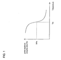
- the hybridization kinetic analysis for every probe may be performed, for example, in the following way. First, the corresponding target amplicons are hybridized with every probe at a series of different temperature for an appropriate length of time. With the decrease of hybridization temperature, more of the probe-amplicon complex will formed, as illustrated in FIG. 1 .
- the hybridization temperature (T h ) represents the temperature, which satisfies a high specific hybridization signal, for example, the highest temperature at which 80% of the maximal signal is achieved. Second, the non-specific binding signal with other kinds of amplicons may not occur at this temperature. This may be validated with a certain concentration of other target amplicons and interfering amplicons, which not included in the target groups.
- T m a probe with high melting temperature
- T h a high hybridization temperature
- probes with similar melting temperature can be chosen from the candidates to reduce the number.
- Those probes for high homologous target molecules can be designed in a low homologous region to increase their specificity.
- the DNA microarray is stationary and the optical detection system is mobile.
- the probes are arrayed by the group in the order of decreasing hybridization temperature (T h ) of each group and are immobilized on the substrate surface of the DNA microarray.
- T h hybridization temperature
- the optical detection system is moved to the first group of probes.
- the chamber and the buffer solution containing the reagents are heated to the denaturing temperature of about 90 °C and maintained at this temperature for several seconds.
- the chamber and the buffer solution containing the reagents is cooled to the hybridization temperature for the first group of probes and maintained at this temperature for several seconds.
- the optical detection system measures the response of the first group of probes.
- the optical detection system is moved to the second group of probes.
- the chamber and the buffer solution containing the reagents is cooled to the hybridization temperature for the second group of probes and maintained at this temperature for several seconds.
- T h1 > T h2 > T h3 is used because the probe groups with high hybridization temperature will be detected earlier at high temperature and the potential non-specific binding at T h2 or T h3 will not be detected and recorded.
- the optical detection system measures the response of the second group of probes. This cycle continues until all of the groups of probes have been detected.
- system parameters such as the number of amplification cycles before detection, the detection frequency (e.g., the number of amplification cycles between every detection cycle), temperature, time, rate of
- heating and cooling and movement of the optical detection system can be controlled by a computer program.
- the optical detection system is stationary and the DNA microarray is mobile. In another yet embodiment, both the optical detection system and the DNA microarray are mobile.
- T h1 > T h2 > T h3 is used in the embodiments described above, in other embodiments T h1 ⁇ T h2 ⁇ T h3 may be used.
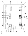
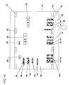
- FIG. 2 is a cross-sectional view of a reaction cartridge capable of evanescent wave detection of fluorescently tagged amplicons in a microarrayed PCR process, including a reaction cartridge 10, a substrate 11 having a substrate surface 12, a first group of three target nucleic acid probes with hybridization temperature (T h1 ) 13, a second group of three target nucleic acid probes with hybridization temperature (T h2 ) 14, a third group of three target nucleic acid probes with hybridization temperature (T h3 ) 15, wherein T h1 > T h2 > T h3 , a buffer solution 16, a first target nucleic acid strand 18, a second target nucleic acid strand 19, a third target nucleic acid strand 20, dNTPs 22, optional fluorescently tagged dNTPs 24, fluorescently tagged primers 26, a thermostable DNA polymerase 28, a heating element 30, and a cooling element 31.
- the heating element 30 and cooling element 31 can be combined into one element,
- FIG. 2 illustrates three groups of three target nucleic acid probes
- other combinations may be used such as two groups of two target nucleic acid probes, two groups of three target nucleic acid probes, three groups of two target nucleic acid probes, four groups of two target nucleic acid probes, four groups of three target nucleic acid probes, four groups of four target nucleic acid probes, and the like.
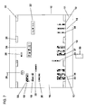
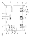
- FIG. 2-11 only one amplicon for one target nucleic acid probe each group is shown. However, it is to be understood that additional amplicons for other target nucleic acid probes within each group may be formed.
- the substrate 12 is comprised of a material that is optically denser than the buffer solution 16, so that evanescent wave detection can be used as described in detail below.
- the substrate 12 may for instance be glass, or a suitably coated plastic or polymer.
- these target nucleic acid probes are non-extendable. In other words, the nucleotides cannot be added to either end of the target nucleic acid probes.
- the target nucleic acid probes which make up each group, may be natural or synthetically fabricated polynucleotides, polynucleotides with artificial bases and/or artificial carbohydrates, peptide nucleic acids (PNAs), bicyclic nucleic acids (BNAs), or other nucleotide analogs, constructed using a commercially available oligonucleotide synthesizer, for example, the POLYPLEX synthesizer available from Genomic Solutions, Inc. of Ann Arbor, Mich.
- the probes may be, but are not limited to, a sequence chosen from a library of DNA sequences, such as a library of expressed sequence tags (EST) known to have some biological significance.
- EST expressed sequence tags
- the first, second, and third groups of target nucleic acid probes 13, 14, and 15, respectively, are arrayed on a substrate surface 12.
- the probes that make up each group of target nucleic acid probes 13, 14, and 15 are arrayed on the substrate as small spots by robotic printing using commercially available microarraying technology, for example, the OMNIGRID microarrayer available from Genomic Solutions, Inc. of Ann Arbor, Mich.
- the probes that make up each group of target nucleic acid probes 13, 14, and 15 may be immobilized on the substrate surface 12 by well-known techniques such as, but not limited to, covalently conjugating an active silyl moiety onto the target nucleic acid probes. Such silanized molecules are immobilized instantly onto glass surfaces after manual or automated deposition.
- the probes that make up each group of target nucleic acid probes 13, 14, and 15 may be immobilized by suitably electrically charging the surface, by using a suitable coating, for example, a silane, poly-L-lysine, streptavidin, a polysaccharide, mercaptan, or a combination thereof.
- the fluorescently tagged primers 26 or the optionally fluorescently tagged dNTPs 24 are nucleotides that may be tagged with a fluorescent dye, for example, fluorescein or
- Rhodamine Green dyes or similar, related compounds having similar fluorescing or luminescent characteristics, such as functionalized or intercalating dyes and luminescent, functionalized nanoparticles (quantum dots).
- the optionally fluorescently tagged dNTPs 24 may have one, two, three, or four of the four base nucleotides dGTP, dCTP, dATP, and dTTP that are fluorescently tagged.
- Heating element 30 may be any suitable resistive material, for example, carbon, that provides heat when an electric current flows though it. Heating element 30 need to be capable of heating the reaction cartridge 10 to a temperature of 94 °C within minutes.
- Cooling element 31 may be any suitable solid state cooling element, for example, a well known Peltier solid-state device functioning as a heat pump. The heating element 30 and cooling element 31 can also be located outside the reaction cartridge 10. In some embodiments, the heating element 30 and cooling element 31 can be combined into one element, such as a semiconductor cooler, which could be used for either heating or cooling.
- FIG. 3 is a cross-sectional view of a reaction cartridge 10 capable of evanescent wave detection of fluorescently tagged amplicons in a microarrayed PCR process using a hybridization temperature (T h1 ) for the first group of target nucleic acids probes 13 of the PCR process, further including the first fluorescently tagged amplicon 32.
- the fluorescent light detector 50 is located adjacent to the substrate 12 to be able to monitor the PCR process of the first group of target nucleic acid probes 13.
- First fluorescently tagged amplicon 32 is a DNA strand having a nucleotide sequence that is a complementary copy of one specific region of the first target nucleic acid strand 18, that is, for every adenosine (A) nucleotide in the one specific region of first target nucleic acid strand 18, there is a thymidine (T) nucleotide in the first fluorescently tagged amplicon 32, and vice versa.
- cytosine (C) nucleotide in the one specific region of first target nucleic acid strand 18 there is a guanosine (G) nucleotide in the one specific region of first fluorescently tagged amplicon 32.
- the amplicon 32 may be, for example, 200 base pairs (bp) and the fragment for hybridization may be, for example, 20 base pairs (bp).
- first fluorescently tagged amplicon 32 will hybridize with the corresponding probes that make up the first group of target nucleic acid probes 13. For instance, at surface site 36, a first fluorescently tagged amplicon 32 is hybridized to a first target nucleic acid probe of group 13. However, there will still be hybridization taking place for the second and third set of nucleic acid probes 14 and 15 (with their corresponding amplicons respectively).
- the target nucleic acid probes that make up, for example, the groups 13, 14, and 15 are designed not to be amplified in the PCR process by, for instance, being tethered by their 3' end to the substrate, or by having the 3' end modified by dideoxidation or by having a stable group added to the 3' end or by any other well known methods of making the target nucleic acid probes not participate in a PCR process in the presence of specific primers.
- FIG. 4 is a cross-sectional view of a reaction cartridge 10 capable of evanescent wave detection of fluorescently tagged amplicons in a microarrayed PCR process at the denaturation stage of the PCR process.
- the mixture within the reaction cartridge 10 has been heated to close to 100 °C, and optimally to about 94 °C.
- the DNA is denatured, that is, it separates into individual, single strands.
- T h1 hybridization temperature
- the first target nucleic acid strand 18 as well as each of the first fluorescently tagged amplicon 32 will anneal with fluorescently tagged primers 26.
- the annealed fluorescently tagged primers 26 will be extended as the thermostable DNA polymerase 28 adds appropriate nucleotides, until the first target nucleic acid strand 18 and the first fluorescently tagged amplicon 32 will be hybridized to a new amplicon, which is a copy or a complementary copy of the original first target nucleic acid strand 18.
- Amplification may also take place during the detection cycle.
- the detection cycle is conducted by, for example, programming the number of amplification cycles before detection and the detection frequency (e.g., the number of amplification cycles between every detection cycle).
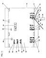
- FIG. 5 is cross-sectional view of a reaction cartridge 10 showing evanescent wave detection of fluorescently tagged amplicons in a microarrayed PCR process at the detection stage of the PCR process for the first group of nucleic acids probes 13, further including an incident beam of light 40, an angle of incidence 42, a reflected beam of light 44, an evanescent wave of light 46, a fluorescent beam of light 48 and a fluorescent light detector 50.
- the detection stage can be coincident with the annealing and/or extension stage.
- the incident beam of light 40 is chosen to be of a wavelength suitable for exciting the fluorophore used to label the fluorescently tagged primers 16 or the optionally fluorescently tagged dNTPs 24.
- the incident beam of light 40 is the 488 nm spectral line of an argon-ion laser, which closely matches the excitation maximum (494 nm) of fluorescein dye that is used to tag or label the fluorescently tagged primers 16 or the optionally fluorescently tagged dNTPs 24.
- the angle of incidence 42 of the incident beam of light 40 is chosen to be greater than the critical angle of the substrate to buffer interface.
- the substrate 12 is comprised of glass and has a refractive index of about 1.5
- the buffer solution 16 is comprised mainly of water having a refractive index of about 1.3, so that the critical angle of incidence is about 61 degrees.
- the intensity of the evanescent wave 46 drops by a factor of "e” (i.e., the mathematical constant) for each 130 nm increase in distance from the substrate surface 12. Therefore, only objects very near substrate surface 12 are illuminated by the evanescent wave 46.
- This property is used in one embodiment of the present invention to illuminate the first fluorescently tagged amplicon 32 that is hybridized to the first group of target nucleic acid probes 13.
- the fluorescent light 48 emitted by the fluorescently tagged amplicon 32 may be detected and analyzed by the fluorescent light detector 50.
- the fluorescent light detector 50 typically may include collection optics such as, a microscope objective lens, which focuses the light on to a detection system such as, a photomultiplier tube or a charge coupled device (CCD) camera or photodiodes.
- the origin and intensity of the collected fluorescent light can be used to estimate the number of fluorescently emitting molecules and therefore the number of fluorescently tagged amplicons currently hybridized to a particular type of oligoprobe by using, for example, the well known quantification techniques employed in Real-Time or Kinetic PCR analysis.
- the reactions are characterized by the point in time during cycling when amplification of a PCR product is first detected, rather than the amount of PCR product accumulated after a fixed number of cycles. The greater the number of copies of a target nucleic acid in the initial sample, the sooner a significant increase in fluorescence is observed.
- the fluorescent light detector 50 is moved into position to monitor the second group of target nucleic acid probes 14 as shown in FIG. 6 .
- FIG. 6 is a cross-sectional view of a reaction cartridge 10 capable of evanescent wave detection of fluorescently tagged amplicons in a microarrayed PCR process at the end of the annealing and/or extension stage using a hybridization temperature (T h2 ) for the second group of target nucleic acids probes 14 of the PCR process, further including the second fluorescently tagged amplicon 33.
- T h2 hybridization temperature
- Second fluorescently tagged amplicon 33 is a DNA strand having a nucleotide sequence that is a complementary copy of the second target nucleic acid strand 19, that is, for every adenosine (A) nucleotide in the second target nucleic acid strand 19, there is a thymidine (T) nucleotide in the second fluorescently tagged amplicon 33, and vice versa.
- adenosine (A) nucleotide in the second target nucleic acid strand 19 there is a thymidine (T) nucleotide in the second fluorescently tagged amplicon 33, and vice versa.
- T thymidine
- G guanosine nucleotide in the second fluorescently tagged amplicon 33.
- target strand 19 is 1000 base pairs (bp)
- the amplicon 33 may be, for example, 200 base pairs (bp) and the fragment for hybridization may be, for example, 20 base pairs (bp).
- second fluorescently tagged amplicon 33 produced by extension of annealed fluorescently tagged primers 26 remains hybridized to its corresponding probes that make up the second group of target nucleic acid probes 14.
- amplicons produced in previous cycles of the PCR process are hybridized to the tethered corresponding probes that make up the second group of target nucleic acid probes 14. For instance, at surface site 37, a second fluorescently tagged amplicon 33 is hybridized to a second target nucleic acid probe of group 14.
- FIG. 7 is a cross-sectional view of a reaction cartridge 10 capable of evanescent wave detection of fluorescently tagged amplicons in a microarrayed PCR process at the denaturation stage of the PCR process.
- the mixture within the reaction cartridge 10 has been heated to close to 100 °C, and optimally to about 94 °C.
- the DNA is denatured, that is, it separates into individual, single strands.
- T h2 hybridization temperature
- the second target nucleic acid strand 19 as well as each of the second fluorescently tagged amplicon 33 will anneal with fluorescently tagged primers 26.
- the annealed fluorescently tagged primers 26 will be extended as the thermostable DNA polymerase 28 adds appropriate nucleotides, until the second target nucleic acid strand 19 and the second fluorescently tagged amplicon 33 will be hybridized to a new amplicon, which is a copy or a complementary copy of the original second target nucleic acid strand 19.
- Amplification may also take place during the detection cycle.
- the detection cycle is conducted by, for example, programming the number of amplification cycles before detection and the detection frequency (e.g., the number of amplification cycles between every detection cycle).
- FIG. 8 is cross-sectional view of a reaction cartridge 10 showing evanescent wave detection of fluorescently tagged amplicons in a microarrayed PCR process at the detection stage of the PCR process for the second group of target nucleic acids probes 14, further including an incident beam of light 40, an angle of incidence 42, a reflected beam of light 44, an evanescent wave of light 46, a fluorescent beam of light 48 and a fluorescent light detector 50.
- the detection stage can be coincident with the annealing and/or extension stage.
- the fluorescent light detector 50 is moved into position to monitor the third group of target nucleic acid probes 15 as shown in FIG. 9 .
- FIG. 9 is a cross-sectional view of a reaction cartridge 10 capable of evanescent wave detection of fluorescently tagged amplicons in a microarrayed PCR process at the end of the annealing and/or extension stage using a hybridization temperature (T h3 ) for the third group of target nucleic acids probes 15 of the PCR process, further including the third fluorescently tagged amplicon 34.
- T h3 hybridization temperature
- Third fluorescently tagged amplicon 34 is a DNA strand having a nucleotide sequence that is a complementary copy of the third target nucleic acid strand 20, that is, for every adenosine (A) nucleotide in the third target nucleic acid strand 20, there is a thymidine (T) nucleotide in the third fluorescently tagged amplicon 34, and vice versa.
- adenosine (A) nucleotide in the third target nucleic acid strand 20 there is a thymidine (T) nucleotide in the third fluorescently tagged amplicon 34, and vice versa.
- T thymidine
- G guanosine nucleotide in the third fluorescently tagged amplicon 34.
- the amplicon 34 may be, for example, 200 base pairs (bp) and the fragment for hybridization may be, for example, 20 base pairs (bp).
- third fluorescently tagged amplicon 34 produced by extension of annealed fluorescently tagged primers 26 remains hybridized to its corresponding probes that make up the third group of target nucleic acid probes 15.
- amplicons produced in previous cycles of the PCR process are hybridized to the tethered corresponding probes that make up the third group of target nucleic acid probes 15. For instance, at surface site 38, a third fluorescently tagged amplicon 38 is hybridized to a third target nucleic acid probe of group 15.
- FIG. 10 is a cross-sectional view of a reaction cartridge 10 capable of evanescent wave detection of fluorescently tagged amplicons in a microarrayed PCR process at the denaturation stage of the PCR process.
- the mixture within the reaction cartridge 10 has been heated to close to 100 °C, and optimally to about 94 °C.
- the DNA is denatured, that is, it separates into individual, single strands.
- T h3 hybridization temperature
- the third target nucleic acid strand 20 as well as each of the third fluorescently tagged amplicon 34 will anneal with fluorescently tagged primers 26.
- the annealed fluorescently tagged primers 26 will be extended as the thermostable DNA polymerase 28 adds appropriate nucleotides, until the third target nucleic acid strand 20 and the third fluorescently tagged amplicon 34 will be hybridized to a new amplicon, which is a copy or a complementary copy of the original third target nucleic acid strand 20.
- Amplification may also take place during the detection cycle.
- the detection cycle is conducted by, for example, programming the number of amplification cycles before detection and the detection frequency (e.g., the number of amplification cycles between every detection cycle).
- FIG. 11 is cross-sectional view of a reaction cartridge 10 showing evanescent wave detection of fluorescently tagged amplicons in a microarrayed PCR process at the detection stage of the PCR process for the third group of target nucleic acids probes 15, further including an incident beam of light 40, an angle of incidence 42, a reflected beam of light 44, an evanescent wave of light 46, a fluorescent beam of light 48 and a fluorescent light detector 50.
- the detection stage can be coincident with the annealing and/or extension stage.
- the present invention also provides a kit for detecting two or more target nucleic acids from one or more pathogens.
- the kit may include at least one primer pair and an oligonucleotide microarray that may include two or more groups of target nucleic acid probes.
- Example is illustrative of the above invention.
- One skilled in the art will readily recognize that the techniques and reagents described in the Example suggest many other ways in which the present invention could be practiced. It should be understood that many variations and modifications may be made while remaining within the scope of the invention.
- This example illustrates the use of a programmable oligonucleotide microarray for the parallel detection of nine species of bacteria.
- a probe is designed for the specific sequence region of each species of bacteria listed in Table 1. These probes are designed with different length and guanosine and cytosine (GC) ratio.
- GC guanosine and cytosine
- T m melting temperature
- T h theoretical hybridization temperature
- the probes are divided into three groups, with the corresponding hybridization temperatures (T h ) of 59, 69, and 63 °C, respectively.
- the actual hybridization temperature (T h ) of each probe is verified by the hybridization kinetic analysis.
- the hybridization kinetic analysis is carried out in the following way.
- each probe is synthesized and immobilized on the surface of a glass plate.
- the glass plate makes up the hybridization and detection component of the microarray reaction chamber.
- the immobilization is be done using an aspirate-dispensing arrayer with the concentration of 20 micromolar ( ⁇ M) in an appropriate buffer to optimize the reaction between probes and functional molecules on the surface of glass.
- the glass is assembled with other chamber parts, such as a plastic cartridge, to form a complete microarray reaction chamber.
- the glass is treated to remove unbounded probes.
- the surfaces of the complete microarray reaction chamber undergo a passivating treatment to reduce non-specific binding.
- the corresponding target molecules are prepared in two certain concentrations.
- One concentration of the target molecules is in the middle of the detection range and is used for the specific signal detection.
- the other concentration of the target molecules is at the high end of the detection range and is used for the non-specific signal detection. All these target molecules are fluorescently tagged amplicons prepared by conventional PCR.
- the corresponding target molecules with a concentration in the middle of the detection range are added to test the specific hybridization signals at a series of hybridization temperatures (T h ).
- T h hybridization temperature
- the specific hybridization signal is recorded at an appropriate time to afford a curve of specific hybridization signal versus hybridization temperature (T h ).
- the highest hybridization temperature (T h ) is usually achieved at very low temperature, such as T h ⁇ T m -30.
- the non-specific hybridization signal For each probe, eight different target molecules with large concentrations are added to test the non-specific hybridization signal at a series of hybridization temperatures (T h ). For each hybridization temperature (T h ), the non-specific hybridization signal is recorded at an appropriate time. By this way, eight curves of non-specific hybridization signals versus hybridization temperature are achieved. With decreasing temperature, the non-specific hybridization will increase. The non-specific binding reaction with other non-related target molecules, which are not included in the detection spectrum, can also be done in this way.
- the proper hybridization temperature (T h ) for each probe is determined by the following guidelines. First, the specific hybridization signal should be high, such as greater than about 70% of the highest hybridization signal. Second, the non-specific hybridization signal should not be detectable. The optimal hybridization temperature (T h ) of each probe should be is verified and adjusted, if needed. If the actual hybridization temperature (T h ) is similar to the theoretical hybridization temperature (T h ), the probe can be divided into three groups, as discussed above. Consequently, all these probes together with one or more optional internal control probes can be immobilized on a functional surface of a glass to form the reaction chamber, which can be used directly for detection. The probes are arrayed in groups, for example, the probes of the same group are spotted in the same line.
- the reaction chamber can be used for the detection of the presence and its concentration of an arbitrary species of bacteria.
- a known concentration of one or more optional internal control target molecules may be added to the sample for testing.
- a programmable detection file used in the detection system may be used to increase the detection efficiency. By using this program, the detection may be carried out at the end of an appropriate number of amplification cycles.
- the temperature of the chamber and the reagent inside will be elevated to about 90 °C and be kept for several seconds. At this temperature, the nucleic acid molecules will denature into single strands.
- the temperature is cooled down to hybridization temperature (T h1 ), which is the average hybridization temperature (T h ) of the probe group 1 and has the highest hybridization temperature (T h ). At this temperature, target probes in group 1 will reach their optimal hybridization temperature (T h1 ).
- the laser which is used to activate the fluorescent molecules near the hybridization site, is moved to the corresponding position. At this position, the incidence of the laser beam can activate the fluorescent molecules of probe group 1, while can not activate the fluorescent molecules in the other groups of probes. As a result, only the hybridization signal of probe group 1 is detected and recorded.
- hybridization temperature (T h2 ) is the average hybridization temperature of probe group 2 and has a hybridization temperature (T h ) in the middle of the temperature range. At this temperature, target probes in group 2 will reach their optimal hybridization temperature (T h2 ).
- the laser which is used to activate the fluorescent molecules near the hybridization site, is moved to the corresponding position. At this position, the incidence of the laser beam can activate the fluorescent molecules of probe group 2, while can not activate the fluorescent molecules in the other groups of probes. As a result, only the hybridization signal of probe group 2 is detected and recorded.
- hybridization temperature (T h3 ) is the average hybridization temperature of probe group 3 and has a hybridization temperature (T h ) at the low end of the temperature range.
- T h3 hybridization temperature
- the laser which is used to activate the fluorescent molecules near the hybridization site, is moved to the corresponding position. At this position, the incidence of the laser beam can activate the fluorescent molecules of probe group 3, while can not activate the fluorescent molecules in the other groups of probes. As a result, only the hybridization signal of probe group 3 is detected and recorded.
- the temperature is decreased to the hybridization temperature (T hc ) of the one of the optional internal control probes.
- T hc the hybridization temperature of the one of the optional internal control probes.
- T hc the hybridization temperature of the one of the optional internal control probes.
- T hc the hybridization temperature of the one of the optional internal control probes.
- T hc the hybridization temperature of the one of the optional internal control probes.
- T hc T h3 ⁇ T h2 ⁇ T h1 .
- T h3 ⁇ T h2 ⁇ T hc the internal control probes will reach their optimal hybridization temperature.
- the laser which is used to activate the fluorescent molecules near the hybridization site, is moved to the corresponding position. At this position, the incidence of the laser beam can activate the fluorescent molecules of one of the optional internal control probe groups, while can not activate the fluorescent molecules in the other groups of probes. As a result, only the hybridization signal of one of the optional internal control probe groups is detected and recorded.
- one of the optional internal control probes is used.
- two or more optional internal control probes may be used and their corresponding hybridization temperatures may range from below T h3 to above T h1 .
- This detection process may be done in a circular mode. For example, the detection may be done every three amplification cycles until the hybridization signal of the suspected target molecule reaches a threshold.
- the CT number is the number of amplification cycles for the suspected target molecule to reach its threshold.
- the CT number of a suspected target molecule may indicate that the concentration of the suspected target molecule is about 10000 copies/ml.
- the calibration curve may reveal that the concentration of the optional internal control target is estimated to be low, for example, by about ten fold. Therefore, the actual concentration of the suspected target molecule would be about 100,000 copies/ml.
Landscapes
- Chemical & Material Sciences (AREA)
- Life Sciences & Earth Sciences (AREA)
- Organic Chemistry (AREA)
- Engineering & Computer Science (AREA)
- Zoology (AREA)
- Wood Science & Technology (AREA)
- Proteomics, Peptides & Aminoacids (AREA)
- Health & Medical Sciences (AREA)
- Biophysics (AREA)
- General Engineering & Computer Science (AREA)
- Immunology (AREA)
- Microbiology (AREA)
- Molecular Biology (AREA)
- Analytical Chemistry (AREA)
- Physics & Mathematics (AREA)
- Genetics & Genomics (AREA)
- Biochemistry (AREA)
- Bioinformatics & Cheminformatics (AREA)
- Biotechnology (AREA)
- General Health & Medical Sciences (AREA)
- Chemical Kinetics & Catalysis (AREA)
- Measuring Or Testing Involving Enzymes Or Micro-Organisms (AREA)
- Investigating, Analyzing Materials By Fluorescence Or Luminescence (AREA)
Abstract
Description
- The quantitative assay of nucleic acids is of considerable importance in basic biological research as well as in fields such as clinical microbiology. The real-time polymerase chain reaction (real-time PCR) process was developed in the mid 1990's to improve the original PCR process in a way that provides reliable, accurate quantitative measurements of the number of copies of any target DNA in the sample. In a real-time PCR, fluorogenic probes that are only active when bound to target DNA are added to the PCR buffer solution. These fluorogenic probes are single strands of DNA, with a middle portion having a sequence of nucleotides that is complementary to the target DNA. On either side of this middle portion, are extension nucleotide sequences that are complementary to each other, so that an unattached probe will fold onto itself in a hairpin configuration. The fluorogenic probe has a fluorescent molecule at one end, and a fluorescence quenching molecule at the other end. An unattached, folded probe has a fluorescing and a quenching molecule adjacent to each other, and consequently no fluorescent light is emitted when the unattached probe is illuminated. When the fluorogenic probe is attached to its target DNA, however, it is unfolded, with the fluorescing and quenching molecules separated from each other. When the attached probe is illuminated with the appropriate wavelength of light, the fluorescent molecule emits fluorescent light. By providing sufficient fluorogenic probes for a particular target DNA, and measuring the fluorescence from the bound probes at each stage of the PCR, the number of amplicons at each stage of the reaction can be measured. This measurement can be used to very accurately determine the number of copies of the DNA in the initial sample because of a straight line relationship between the fractional number of cycles for the number of amplicons to reach a pre-determined threshold and the logarithm of the number of copies in the initial sample.
- The use of evanescent waves, i.e. light waves which decay exponentially in power upon entry into a reaction cell, in oligonucleoitide microarrays is disclosed in
W02006/135437 and Francois, P., et al. (2006). "Rapid bacterial identification using evanescent-waveguide olignonucletide microarray classification". Journal of Microbiological Methods, 65(3), 390-403. - Embodiments of the invention may be best understood by referring to the following description and accompanying drawings, which illustrate such embodiments. In the drawings:
-
FIG. 1 illustrates a plot of melting temperature (Tm) versus probe-amplicon complex ratio. -
FIG. 2 illustrates a cross-sectional view of a cartridge capable of evanescent wave detection of fluorescently tagged amplicons in a microarrayed PCR process in an initial stage of the PCR process. -
FIG. 3 illustrates a cross-sectional view of a cartridge capable of evanescent wave detection of fluorescently tagged amplicons in a microarrayed PCR process at the end of the annealing and extension stage using a hybridization temperature (Th1) for the first group of target nucleic acids probes of the PCR process. -
FIG. 4 illustrates a cross-sectional view of a cartridge capable of evanescent wave detection of fluorescently tagged amplicons in a microarrayed PCR process at the denaturation stage of the PCR process for the first group of target nucleic acids probes. -
FIG. 5 illustrates a cross-sectional view of a cartridge showing evanescent wave detection of fluorescently tagged amplicons in a microarrayed PCR process at the detection stage of the PCR process for the first group of target nucleic acids probes. -
FIG. 6 illustrates a cross-sectional view of a reaction cartridge capable of evanescent wave detection of fluorescently tagged amplicons in a microarrayed PCR process at the end of the annealing and/or extension stage using hybridization temperature (Th2) for the second group of target nucleic acids probes of the PCR process. -
FIG. 7 illustrates a cross-sectional view of a reaction cartridge capable of evanescent wave detection of fluorescently tagged amplicons in a microarrayed PCR process at the denaturation stage of the PCR process for the second group of target nucleic acids probes. -
FIG. 8 illustrates cross-sectional view of a reaction cartridge showing evanescent wave detection of fluorescently tagged amplicons in a microarrayed PCR process at the detection stage of the PCR process for the second group of target nucleic acids probes. -
FIG. 9 illustrates a cross-sectional view of a reaction cartridge capable of evanescent wave detection of fluorescently tagged amplicons in a microarrayed PCR process at the end of the annealing and/or extension stage using hybridization temperature (Th3) for the third group of target nucleic acids probes of the PCR process. -
FIG. 10 illustrates a cross-sectional view of a reaction cartridge capable of evanescent wave detection of fluorescently tagged amplicons in a microarrayed PCR process at the denaturation stage of the PCR process for the third group of target nucleic acids probes. -
FIG. 11 illustrates a cross-sectional view of a reaction cartridge showing evanescent wave detection of fluorescently tagged amplicons in a microarrayed PCR process at the detection stage of the PCR process for the third group of target nucleic acids probes. - As used herein, certain terms have the following meanings. All other terms and phrases used in this specification have their ordinary meanings as one of skill in the art would understand. Such ordinary meanings may be obtained by reference to technical dictionaries, such as Hawley's Condensed Chemical Dictionary 11th Edition, by Sax and Lewis, Van Nostrand Reinhold, New York, N.Y., 1987, and The Merck Index, 11th Edition, Merck & Co., Rahway N.J. 1989.
- As used herein, the term "and/or" means any one of the items, any combination of the items, or all of the items with which this term is associated.
- As used herein, the singular forms "a," "an," and "the" may include plural reference unless the context clearly dictates otherwise. Therefore, for example, a reference to "a formulation" may include a plurality of such formulations, so that a formulation of compound X may include formulations of compound X.
- As used herein, the term "about" means a variation of 10 percent of the value specified, for example, about 50 percent carries a variation from 45 to 55 percent. For integer ranges, the term about can include one or two integers greater than and less than a recited integer.
- As used herein, the term "amplicons" refers to the products of polymerase chain reactions (PCR). Amplicons are pieces of DNA that have been synthesized using amplification techniques (e.g., a double-stranded DNA with two primers). The amplicon may contain, for example, a primer tagged with a fluorescent molecule at the 5' end.
- As used herein, the terms "array" and "microarray" refer to an arrangement of elements (i.e., entities) into a material or device. In another sense, the term "array" refers to the orderly arrangement (e.g., rows and columns) of two or more assay regions on a substrate.
- As used herein, the terms "complementary" or "complementarity" are used in reference to polynucleotides (i.e., a sequence of nucleotides) related by the base-pairing rules.
- As used herein, the term "critical angle" is the angle of incidence above which the total internal reflection occurs.
- As used herein, the term "evanescent" refers to a nearfield standing wave exhibiting exponential decay with distance. As used in optics, evanescent waves are formed when sinusoidal waves are internally reflected off an interface at an angle greater than the critical angle so that total internal reflection occurs.
- As used herein, the term "hybridization" refers to the pairing of complementary nucleic acids.
- As used herein, the term "Th" refers to "hybridization temperature." The hybridization temperature is typically about 10 °C below the Tm (melting temperature) of the nucleic acid.
- As used herein, the term "nucleic acid molecule" refers to any nucleic acid containing molecule including, but not limited to, DNA or RNA. The term encompasses sequences that may include, for example, any of the known base analogs of DNA and RNA. As used herein, the term "polymerase chain reaction" (PCR) refers to the method of
K. B. Mullis, U.S. Patent Nos. 4,683,195 ,4,683,202 , and4,965,188 . - As used herein, the term "DNA polymerase" refers to a thermostable DNA polymerase enzyme having an optimum temperature at which it functions, which is higher than 40 °C.
- As used herein, the term "primer" refers to a single-stranded polynucleotide capable of acting as a point of initiation of template-directed DNA synthesis under appropriate conditions.
- As used herein, the term "probe" refers to a nucleic acid capable of binding to a target nucleic acid of complementary sequence through one or more types of chemical bonds, usually through complementary base pairing, usually through hydrogen bond formation, thus forming a duplex structure. A probe binds or hybridizes to a "probe binding site." A probe can be labeled with a detectable label to permit facile detection of the probe, particularly once the probe has hybridized to its complementary target. A label attached to the probe can include any of a variety of different labels known in the art that can be detected by, for example, chemical or physical means. Labels that can be attached to probes may include, for example, fluorescent and luminescence materials. Probes can vary significantly in size. Some probes are relatively short. Generally, probes are at least 7 to 15 nucleotides in length. Other probes are at least 20, 30 or 40 nucleotides long. Still other probes are somewhat longer, being at least 50, 60, 70, 80, 90 nucleotides long. Yet other probes are longer still, and are at least 100, 150, 200 or more nucleotides long. Probes can be of any specific length that falls within the foregoing ranges as well.
- As used herein, the term "substrate" refers to material capable of supporting associated assay components (e.g., assay regions, cells, test compounds, etc.). For example, in some embodiments, the substrate may include a planar (i.e., 2 dimensional) glass, metal, composite, plastic, silica, or other biocompatible or biologically unreactive (or biologically reactive) composition. The substrate is generally flat but may take on a variety of alternative surface configurations. The substrate and its surface are also chosen to provide appropriate light-absorbing characteristics. For instance, the substrate may be a polymerized Langmuir Blodgett film, a functionalized glass, a glass, quartz, Si, Ge, GaAs, GaP, SiO2, SiN4, modified silicon, or any one of a wide variety of gels or polymers, for example, (poly)tetrafluoroethylene, (poly)vinylidenedifluoride, polystyrene, polycarbonate, polyolefins such as polypropylene, or combinations thereof.
- As used herein, the term "target nucleic acid" refers to a polynucleotide inherent to a pathogen that is to be detected. The polynucleotide is genetic material including, for example, DNA/RNA, mitochondrial DNA, rRNA, tRNA, mRNA, viral RNA, and plasmid DNA.
- As used herein, the term "target nucleic acid probe" refers to a polynucleotide that is complementary to the target nucleic acid.
- As used herein, the term "Tm" refers to the "melting temperature." The melting temperature is the temperature at which a population of double-stranded nucleic acid molecules becomes half dissociated into single strands. The equation for calculating the Tm of nucleic acids is well known in the art (see, e.g., Anderson and Young, Quantitative Filter Hybridization, in Nucleic Acid Hybridization (1985)). For immobilized oligonucleotide probes, typically Tm (°C) = 2 (number of adenosine and thymidine residues) + 4 (number of guanosine and cytosine residues).
- The present invention provides a method, an apparatus, and a system capable of real-time, simultaneous, quantitative measurement of a plurality of target nucleic acids from one or more pathogens in a sample.
- In one embodiment, the target nucleic acids in the sample are amplified using the polymerase chain reaction (PCR). The PCR is a well-known method of amplifying one or more stands of deoxyribonucleic acid (DNA). PCR is begun by placing the target nucleic acids in a buffer containing the nucleotides adenosine (ATP), thymidine (TTP), cytosine (CTP) and guanosine (GTP) (collectively referred to as dNTPs), a DNA polymerase, and primers. The primers are short strands of DNA, with sequences that complement the target nucleic acids to be amplified. The primers initiate replication of the target nucleic acids.
- In one embodiment, the target nucleic acid primers may be fluorescently tagged with fluorescent molecules at the 5' end. In another embodiment, one of the target nucleic acid primers may be fluorescently tagged with fluorescent molecules at the 5' end. In yet another embodiment, the dNTPs are fluorescently tagged.
- The PCR process has three main steps: denaturation, annealing, and extension. In the denaturation step, the mixture is heated to about 94 °C (Centigrade), at which point the target DNA separates into single strands. The mixture is quickly cooled. As the temperature falls to about 60 °C, the annealing step occurs, in which the primers, which may be fluorescently tagged, hybridize or bind to their complementary sequences on the target nucleic acids. The extension step may be performed at about 60 °C or may be raised to the 72-78 °C range. In this step, the DNA polymerase uses the dNTPs in solution to extend the annealed primers, which may be fluorescently tagged, and forms new strands of DNA known as an amplicons. The mixture is briefly reheated back to about 94 °C to separate the newly created double helix stands into single strands of nucleic acid, which begins another cycle of the PCR process. With each cycle of the PCR process, the number of copies of the original target nucleic acids roughly doubles.
- For real-time quantitative analysis of target nucleic acids, several methods utilizing evanescent wave detection techniques have been disclosed including, for example, the techniques described in Xu (
U.S. Patent Application Publication No. 2006/0088844 ) and in PCT Patent Application Serial No.PCT/CN2007/003124 - In one embodiment, the PCR buffer additionally contains fluorescently tagged primers, that is, primers having a fluorescent dye molecule attached to them, so that upon completion of each PCR cycle, the amplicons produced are fluorescently tagged. The amplicons of the target nucleic acids are localized, using probe strands of DNA known as target nucleic acid probes. The target nucleic acid probes have the same complementary, nucleotide sequence as the target nucleic acids. The target nucleic acid probes are tethered to a substrate surface in a known, two-dimensional pattern, with the substrate surface forming part of the reaction cell containing the PCR ingredients.
- During the annealing and extension phases of the PCR process, the target amplicons hybridize to their corresponding target nucleic acid probes. The hybridized, fluorescently tagged amplicons are illuminated with an evanescent wave of light of the appropriate wavelength to activate the fluorescent dye molecules of the fluorescently tagged primers or the fluorescently tagged dNTPs. This evanescent wave decays exponentially in power after entering the reaction cell via the substrate surface to which the target nucleic acid probes are tethered, with an effective penetration range of about 300 nm. This means that the evanescent wave penetrates far enough into the reaction cell to activate the fluorescently tagged amplicons hybridized to those target nucleic acid probes, but that it does not activate the fluorescently tagged molecules (e.g., the fluorescently tagged primers or the fluorescently tagged dNTPs) in solution in the main body of the reaction cell. By monitoring the strength of the fluorescence at various locations on the substrate surface, the current abundance of amplicons of the corresponding target nucleic acids can be determined. This may be done in real-time as the PCR progresses. The results are used to obtain a quantitative measure of the abundance of a specific target in the original sample, in a manner analogous to the real-time PCR calculation.
- In another embodiment, the target nucleic acid probes are designed to be complementary to polynucleotides isolated from a pathogen selected from the group consisting of bacteria, viruses, fungi, and protozoa. In yet another embodiment, the target nucleic acid probes are designed to be complementary to a specific region of polynucleotides isolated from Rickettsia, Chlamydia, Mycoplasma, Spirochete, Streptococcus, Salmonella, Staphylococcus, L. monocytogenes, N. meningitides, E. coli, H. influenzae, B. burgdorferi, Leptospira, Proteus, Anaerobacter, M. tuberculosis, Enterococcus, H. poliovirus 1, H. enterovirus 71, H. enterovirus 70, H. echovirus 2, H. echovirus 4, H. echovirus 6,
H. echovirus 9,H. echovirus 11,H. echovirus 12,H. echovirus 26, H. coxsackievirus A13, H. coxsackievirus A15, H. coxsackievirus A18, H. coxsackievirus A20, H. coxsackievirus A21, H. coxsackievirus B3-A, H. coxsackievirus B3-C, HSV-1, and HSV-2. - In one embodiment, the PCR utilizes probes including an oligonucleotide selected from the group consisting of SEQ ID NO: 1-9 (Table 1).
Table 1 Species Probe Sequence Length GC A T GC % Tm = (4×GC)+(2×AT) (°C) Theoretical Th = Tm-15 (°C) SEQ ID NO Staphyloco ccus 
25 12 13 0.48 74 59 1 Streptococc us 
24 13 11 0.542 74 59 2 E. cloacae 
26 11 15 0.423 74 59 3 S. aureus 
27 12 15 0.444 78 63 4 S. epidermidis 
27 12 15 0.444 78 63 5 P. mirabilis 
29 10 19 0.345 78 63 6 P . aeruginosa 
30 12 18 0.4 84 69 7 H. influenzae 
30 12 18 0.4 84 69 8 E. coli 
31 11 20 0.355 84 69 9 - The present invention also provides a method to increase the detection limit and specificity in an oligonucleotide microarray or a real-time PCR microarray.
- In one embodiment, the probes for different target nucleic acids may be designed and classified into several groups. The probes in each group have approximately equivalent optimal hybridization temperatures. That is, each group of target nucleic acid probes may include one or more target nucleic acid probes with optimal hybridization temperatures within ten percent of each other; or within five percent of each other, or within three percent of each other, or within two percent of each other.
- The probe sequences for each target molecule are designed and synthesized with conventional methods. Typically, one or more different probes are designed for each target molecule.
- When the number of oligonucleotides is less than twenty, the melting temperature (Tm) of each probe may be calculated from formula Tm (°C) = 2 (number of adenosine and thymidine residues) + 4 (number of guanosine and cytosine residues). The probes in each group are arrayed on the substrate such that probes with a uniform hybridization temperature (Th) are arrayed in a location such as a line. The melting temperature (Tm) represents the temperature at which 50% of the nucleic acid molecules are in double stranded form (e.g., a probe-amplicon complex).
- The optimal hybridization temperature (Th) is typically about 10-20 °C below the melting temperature (Tm) of the nucleic acid, which is influenced by strand length, base composition and chemical environment such as salt and formamide concentration. The optimal hybridization temperature (Th) can be validated by hybridization kinetic analysis. The hybridization kinetic analysis for every probe may be performed, for example, in the following way. First, the corresponding target amplicons are hybridized with every probe at a series of different temperature for an appropriate length of time. With the decrease of hybridization temperature, more of the probe-amplicon complex will formed, as illustrated in
FIG. 1 . The hybridization temperature (Th) represents the temperature, which satisfies a high specific hybridization signal, for example, the highest temperature at which 80% of the maximal signal is achieved. Second, the non-specific binding signal with other kinds of amplicons may not occur at this temperature. This may be validated with a certain concentration of other target amplicons and interfering amplicons, which not included in the target groups. - Usually a probe with high melting temperature (Tm) has a high hybridization temperature (Th). The hybridization temperature (Th) may be set as a function of the melting temperature (Tm), such as Th = Tm -15. This may be validated and adjusted by the hybridization kinetic analysis.
- To reduce the workload of hybridization temperature (Th) validation and probe design, probes with similar melting temperature (Tm) can be chosen from the candidates to reduce the number. Those probes for high homologous target molecules can be designed in a low homologous region to increase their specificity.
- In one embodiment, the DNA microarray is stationary and the optical detection system is mobile. The probes are arrayed by the group in the order of decreasing hybridization temperature (Th) of each group and are immobilized on the substrate surface of the DNA microarray. After an appointed number of amplification cycles, the optical detection system is moved to the first group of probes. The chamber and the buffer solution containing the reagents are heated to the denaturing temperature of about 90 °C and maintained at this temperature for several seconds. The chamber and the buffer solution containing the reagents is cooled to the hybridization temperature for the first group of probes and maintained at this temperature for several seconds. The optical detection system measures the response of the first group of probes. Next, the optical detection system is moved to the second group of probes. The chamber and the buffer solution containing the reagents is cooled to the hybridization temperature for the second group of probes and maintained at this temperature for several seconds. For example, Th1 > Th2 > Th3 is used because the probe groups with high hybridization temperature will be detected earlier at high temperature and the potential non-specific binding at Th2 or Th3 will not be detected and recorded. The optical detection system measures the response of the second group of probes. This cycle continues until all of the groups of probes have been detected. To optimize the reaction and detection efficiency, system parameters such as the number of amplification cycles before detection, the detection frequency (e.g., the number of amplification cycles between every detection cycle), temperature, time, rate of
- heating and cooling and movement of the optical detection system can be controlled by a computer program.
- In another embodiment, the optical detection system is stationary and the DNA microarray is mobile. In another yet embodiment, both the optical detection system and the DNA microarray are mobile. Although Th1 > Th2 > Th3 is used in the embodiments described above, in other embodiments Th1 < Th2 < Th3 may be used.
-
FIG. 2 is a cross-sectional view of a reaction cartridge capable of evanescent wave detection of fluorescently tagged amplicons in a microarrayed PCR process, including areaction cartridge 10, asubstrate 11 having asubstrate surface 12, a first group of three target nucleic acid probes with hybridization temperature (Th1) 13, a second group of three target nucleic acid probes with hybridization temperature (Th2) 14, a third group of three target nucleic acid probes with hybridization temperature (Th3) 15, wherein Th1 > Th2 > Th3, abuffer solution 16, a first targetnucleic acid strand 18, a second targetnucleic acid strand 19, a third targetnucleic acid strand 20,dNTPs 22, optional fluorescently taggeddNTPs 24, fluorescently taggedprimers 26, athermostable DNA polymerase 28, aheating element 30, and acooling element 31. In some embodiments, theheating element 30 andcooling element 31 can be combined into one element, such as a semiconductor cooler, which could be used for either heating or cooling. - Although
FIG. 2 illustrates three groups of three target nucleic acid probes, other combinations may be used such as two groups of two target nucleic acid probes, two groups of three target nucleic acid probes, three groups of two target nucleic acid probes, four groups of two target nucleic acid probes, four groups of three target nucleic acid probes, four groups of four target nucleic acid probes, and the like. In other words, there is not limit to the number of groups of target nucleic acid probes and the number of target nucleic acid probes within each group. - To simplify the drawings in
FIG. 2-11 , only one amplicon for one target nucleic acid probe each group is shown. However, it is to be understood that additional amplicons for other target nucleic acid probes within each group may be formed. - In one embodiment, the
substrate 12 is comprised of a material that is optically denser than thebuffer solution 16, so that evanescent wave detection can be used as described in detail below. Thesubstrate 12 may for instance be glass, or a suitably coated plastic or polymer. - The first, second, and third groups of target nucleic acid probes 13, 14, and 15, respectively, each contain several strands of DNA, each strand of DNA having a specific nucleotide sequence of one of the target strands of
DNA - The first, second, and third groups of target nucleic acid probes 13, 14, and 15, respectively, are arrayed on a
substrate surface 12. In one embodiment, the probes that make up each group of target nucleic acid probes 13, 14, and 15 are arrayed on the substrate as small spots by robotic printing using commercially available microarraying technology, for example, the OMNIGRID microarrayer available from Genomic Solutions, Inc. of Ann Arbor, Mich. - The probes that make up each group of target nucleic acid probes 13, 14, and 15 may be immobilized on the
substrate surface 12 by well-known techniques such as, but not limited to, covalently conjugating an active silyl moiety onto the target nucleic acid probes. Such silanized molecules are immobilized instantly onto glass surfaces after manual or automated deposition. Alternatively, the probes that make up each group of target nucleic acid probes 13, 14, and 15 may be immobilized by suitably electrically charging the surface, by using a suitable coating, for example, a silane, poly-L-lysine, streptavidin, a polysaccharide, mercaptan, or a combination thereof. - The fluorescently tagged
primers 26 or the optionally fluorescently taggeddNTPs 24 are nucleotides that may be tagged with a fluorescent dye, for example, fluorescein or - Rhodamine Green dyes, or similar, related compounds having similar fluorescing or luminescent characteristics, such as functionalized or intercalating dyes and luminescent, functionalized nanoparticles (quantum dots). The optionally fluorescently tagged
dNTPs 24 may have one, two, three, or four of the four base nucleotides dGTP, dCTP, dATP, and dTTP that are fluorescently tagged. -
Heating element 30 may be any suitable resistive material, for example, carbon, that provides heat when an electric current flows though it.Heating element 30 need to be capable of heating thereaction cartridge 10 to a temperature of 94 °C within minutes. Coolingelement 31 may be any suitable solid state cooling element, for example, a well known Peltier solid-state device functioning as a heat pump. Theheating element 30 andcooling element 31 can also be located outside thereaction cartridge 10. In some embodiments, theheating element 30 andcooling element 31 can be combined into one element, such as a semiconductor cooler, which could be used for either heating or cooling. -
FIG. 3 is a cross-sectional view of areaction cartridge 10 capable of evanescent wave detection of fluorescently tagged amplicons in a microarrayed PCR process using a hybridization temperature (Th1) for the first group of target nucleic acids probes 13 of the PCR process, further including the first fluorescently taggedamplicon 32. The fluorescentlight detector 50 is located adjacent to thesubstrate 12 to be able to monitor the PCR process of the first group of target nucleic acid probes 13. - First fluorescently tagged
amplicon 32 is a DNA strand having a nucleotide sequence that is a complementary copy of one specific region of the first targetnucleic acid strand 18, that is, for every adenosine (A) nucleotide in the one specific region of first targetnucleic acid strand 18, there is a thymidine (T) nucleotide in the first fluorescently taggedamplicon 32, and vice versa. Similarly for every cytosine (C) nucleotide in the one specific region of first targetnucleic acid strand 18, there is a guanosine (G) nucleotide in the one specific region of first fluorescently taggedamplicon 32. For example, iftarget strand 18 is 1000 base pairs (bp), theamplicon 32 may be, for example, 200 base pairs (bp) and the fragment for hybridization may be, for example, 20 base pairs (bp). - Using a hybridization temperature (Th1) for the first group of target nucleic acids probes 13, first fluorescently tagged
amplicon 32 will hybridize with the corresponding probes that make up the first group of target nucleic acid probes 13. For instance, atsurface site 36, a first fluorescently taggedamplicon 32 is hybridized to a first target nucleic acid probe ofgroup 13. However, there will still be hybridization taking place for the second and third set of nucleic acid probes 14 and 15 (with their corresponding amplicons respectively). For the second and third set of nucleic acid probes 14 and 15, their optimal hybridization temperature (Th2) and (Th3) is below (Th1), so at (Th1) their hybridization signal is less than their optimal hybridization signal. - The target nucleic acid probes that make up, for example, the
groups -
FIG. 4 is a cross-sectional view of areaction cartridge 10 capable of evanescent wave detection of fluorescently tagged amplicons in a microarrayed PCR process at the denaturation stage of the PCR process. In this stage, the mixture within thereaction cartridge 10 has been heated to close to 100 °C, and optimally to about 94 °C. At this temperature, the DNA is denatured, that is, it separates into individual, single strands. When cooled to the hybridization temperature (Th1) for the first group of target nucleic acids probes 13, the first targetnucleic acid strand 18 as well as each of the first fluorescently taggedamplicon 32 will anneal with fluorescently taggedprimers 26. The annealed fluorescently taggedprimers 26 will be extended as thethermostable DNA polymerase 28 adds appropriate nucleotides, until the first targetnucleic acid strand 18 and the first fluorescently taggedamplicon 32 will be hybridized to a new amplicon, which is a copy or a complementary copy of the original first targetnucleic acid strand 18. Amplification may also take place during the detection cycle. As such, the detection cycle is conducted by, for example, programming the number of amplification cycles before detection and the detection frequency (e.g., the number of amplification cycles between every detection cycle). -
FIG. 5 is cross-sectional view of areaction cartridge 10 showing evanescent wave detection of fluorescently tagged amplicons in a microarrayed PCR process at the detection stage of the PCR process for the first group of nucleic acids probes 13, further including an incident beam oflight 40, an angle ofincidence 42, a reflected beam oflight 44, an evanescent wave of light 46, a fluorescent beam oflight 48 and a fluorescentlight detector 50. The detection stage can be coincident with the annealing and/or extension stage. - The incident beam of
light 40 is chosen to be of a wavelength suitable for exciting the fluorophore used to label the fluorescently taggedprimers 16 or the optionally fluorescently taggeddNTPs 24. In one embodiment, the incident beam oflight 40 is the 488 nm spectral line of an argon-ion laser, which closely matches the excitation maximum (494 nm) of fluorescein dye that is used to tag or label the fluorescently taggedprimers 16 or the optionally fluorescently taggeddNTPs 24. - The angle of
incidence 42 of the incident beam oflight 40 is chosen to be greater than the critical angle of the substrate to buffer interface. The critical angle of incidence is the angle at which total internal reflection occurs and is dependent on the refractive indices of the materials forming the interface. From Snell's laws of refraction, the critical angle of incidence = sin-1(n1/n2) where n1 and n2 are the refractive indices of the materials on either side of the interface. In one embodiment of the present invention, thesubstrate 12 is comprised of glass and has a refractive index of about 1.5, while thebuffer solution 16 is comprised mainly of water having a refractive index of about 1.3, so that the critical angle of incidence is about 61 degrees. - When light is reflected off the
substrate surface 12 at an angle ofincidence 42 degrees greater than the critical angle so that total internal reflection occurs, anevanescent wave 46 is formed and propagates through thesubstrate surface 12. The intensity of theevanescent wave 46 drops by a factor of "e" (i.e., the mathematical constant) for each 130 nm increase in distance from thesubstrate surface 12. Therefore, only objects very nearsubstrate surface 12 are illuminated by theevanescent wave 46. This property is used in one embodiment of the present invention to illuminate the first fluorescently taggedamplicon 32 that is hybridized to the first group of target nucleic acid probes 13. Thefluorescent light 48 emitted by the fluorescently taggedamplicon 32, may be detected and analyzed by the fluorescentlight detector 50. The fluorescentlight detector 50 typically may include collection optics such as, a microscope objective lens, which focuses the light on to a detection system such as, a photomultiplier tube or a charge coupled device (CCD) camera or photodiodes. - The origin and intensity of the collected fluorescent light can be used to estimate the number of fluorescently emitting molecules and therefore the number of fluorescently tagged amplicons currently hybridized to a particular type of oligoprobe by using, for example, the well known quantification techniques employed in Real-Time or Kinetic PCR analysis. In the Real-Time or Kinetic PCR analysis, the reactions are characterized by the point in time during cycling when amplification of a PCR product is first detected, rather than the amount of PCR product accumulated after a fixed number of cycles. The greater the number of copies of a target nucleic acid in the initial sample, the sooner a significant increase in fluorescence is observed.
- After the hybridization cycle is completed for the first group of target nucleic acid probes 13, the fluorescent
light detector 50 is moved into position to monitor the second group of target nucleic acid probes 14 as shown inFIG. 6 . -
FIG. 6 is a cross-sectional view of areaction cartridge 10 capable of evanescent wave detection of fluorescently tagged amplicons in a microarrayed PCR process at the end of the annealing and/or extension stage using a hybridization temperature (Th2) for the second group of target nucleic acids probes 14 of the PCR process, further including the second fluorescently taggedamplicon 33. - Second fluorescently tagged
amplicon 33 is a DNA strand having a nucleotide sequence that is a complementary copy of the second targetnucleic acid strand 19, that is, for every adenosine (A) nucleotide in the second targetnucleic acid strand 19, there is a thymidine (T) nucleotide in the second fluorescently taggedamplicon 33, and vice versa. Similarly for every cytosine (C) nucleotide in the second targetnucleic acid strand 19, there is a guanosine (G) nucleotide in the second fluorescently taggedamplicon 33. For example, iftarget strand 19 is 1000 base pairs (bp), theamplicon 33 may be, for example, 200 base pairs (bp) and the fragment for hybridization may be, for example, 20 base pairs (bp). - At the end of the annealing and/or extension stage of the PCR process using a hybridization temperature (Th2) for the second group of target nucleic acids probes 14, second fluorescently tagged
amplicon 33 produced by extension of annealed fluorescently taggedprimers 26, remains hybridized to its corresponding probes that make up the second group of target nucleic acid probes 14. Additionally, amplicons produced in previous cycles of the PCR process are hybridized to the tethered corresponding probes that make up the second group of target nucleic acid probes 14. For instance, atsurface site 37, a second fluorescently taggedamplicon 33 is hybridized to a second target nucleic acid probe ofgroup 14. However, there is no hybridization taking place for the third set of nucleic acid probes 15 because the hybridization temperature (Th2) for the second group of target nucleic acids probes 14 for is below the hybridization temperature (Th3) for the third group of target nucleic acids probes 15. -
FIG. 7 is a cross-sectional view of areaction cartridge 10 capable of evanescent wave detection of fluorescently tagged amplicons in a microarrayed PCR process at the denaturation stage of the PCR process. In this stage, the mixture within thereaction cartridge 10 has been heated to close to 100 °C, and optimally to about 94 °C. At this temperature, the DNA is denatured, that is, it separates into individual, single strands. When cooled to the hybridization temperature (Th2) for the second group of target nucleic acids probes 14 in the next stage of the PCR process, the second targetnucleic acid strand 19 as well as each of the second fluorescently taggedamplicon 33 will anneal with fluorescently taggedprimers 26. The annealed fluorescently taggedprimers 26 will be extended as thethermostable DNA polymerase 28 adds appropriate nucleotides, until the second targetnucleic acid strand 19 and the second fluorescently taggedamplicon 33 will be hybridized to a new amplicon, which is a copy or a complementary copy of the original second targetnucleic acid strand 19. Amplification may also take place during the detection cycle. As such, the detection cycle is conducted by, for example, programming the number of amplification cycles before detection and the detection frequency (e.g., the number of amplification cycles between every detection cycle). -
FIG. 8 is cross-sectional view of areaction cartridge 10 showing evanescent wave detection of fluorescently tagged amplicons in a microarrayed PCR process at the detection stage of the PCR process for the second group of target nucleic acids probes 14, further including an incident beam oflight 40, an angle ofincidence 42, a reflected beam oflight 44, an evanescent wave of light 46, a fluorescent beam oflight 48 and a fluorescentlight detector 50. The detection stage can be coincident with the annealing and/or extension stage. - After the hybridization cycle is completed for the second group of target nucleic acid probes 14, the fluorescent
light detector 50 is moved into position to monitor the third group of target nucleic acid probes 15 as shown inFIG. 9 . -
FIG. 9 is a cross-sectional view of areaction cartridge 10 capable of evanescent wave detection of fluorescently tagged amplicons in a microarrayed PCR process at the end of the annealing and/or extension stage using a hybridization temperature (Th3) for the third group of target nucleic acids probes 15 of the PCR process, further including the third fluorescently taggedamplicon 34. - Third fluorescently tagged
amplicon 34 is a DNA strand having a nucleotide sequence that is a complementary copy of the third targetnucleic acid strand 20, that is, for every adenosine (A) nucleotide in the third targetnucleic acid strand 20, there is a thymidine (T) nucleotide in the third fluorescently taggedamplicon 34, and vice versa. Similarly for every cytosine (C) nucleotide in the third targetnucleic acid strand 20, there is a guanosine (G) nucleotide in the third fluorescently taggedamplicon 34. For example, iftarget strand 20 is 1000 base pairs (bp), theamplicon 34 may be, for example, 200 base pairs (bp) and the fragment for hybridization may be, for example, 20 base pairs (bp). - At the end of the annealing and/or extension stage of the PCR process using a hybridization temperature (Th3) for the third group of target nucleic acids probes 15, third fluorescently tagged
amplicon 34 produced by extension of annealed fluorescently taggedprimers 26, remains hybridized to its corresponding probes that make up the third group of target nucleic acid probes 15. Additionally, amplicons produced in previous cycles of the PCR process are hybridized to the tethered corresponding probes that make up the third group of target nucleic acid probes 15. For instance, atsurface site 38, a third fluorescently taggedamplicon 38 is hybridized to a third target nucleic acid probe ofgroup 15. -
FIG. 10 is a cross-sectional view of areaction cartridge 10 capable of evanescent wave detection of fluorescently tagged amplicons in a microarrayed PCR process at the denaturation stage of the PCR process. In this stage, the mixture within thereaction cartridge 10 has been heated to close to 100 °C, and optimally to about 94 °C. At this temperature, the DNA is denatured, that is, it separates into individual, single strands. When cooled to the hybridization temperature (Th3) for the third group of target nucleic acids probes 15 in the next stage of the PCR process, the third targetnucleic acid strand 20 as well as each of the third fluorescently taggedamplicon 34 will anneal with fluorescently taggedprimers 26. The annealed fluorescently taggedprimers 26 will be extended as thethermostable DNA polymerase 28 adds appropriate nucleotides, until the third targetnucleic acid strand 20 and the third fluorescently taggedamplicon 34 will be hybridized to a new amplicon, which is a copy or a complementary copy of the original third targetnucleic acid strand 20. Amplification may also take place during the detection cycle. As such, the detection cycle is conducted by, for example, programming the number of amplification cycles before detection and the detection frequency (e.g., the number of amplification cycles between every detection cycle). -
FIG. 11 is cross-sectional view of areaction cartridge 10 showing evanescent wave detection of fluorescently tagged amplicons in a microarrayed PCR process at the detection stage of the PCR process for the third group of target nucleic acids probes 15, further including an incident beam oflight 40, an angle ofincidence 42, a reflected beam oflight 44, an evanescent wave of light 46, a fluorescent beam oflight 48 and a fluorescentlight detector 50. The detection stage can be coincident with the annealing and/or extension stage. - The present invention also provides a kit for detecting two or more target nucleic acids from one or more pathogens. The kit may include at least one primer pair and an oligonucleotide microarray that may include two or more groups of target nucleic acid probes. Although the foregoing discussion has used DNA as a nucleic acid, it would be obvious to a person of reasonable skill in the art to apply the methods disclosed herein to other nucleic acids, including RNA sequences or combinations of RNA and DNA sequences.
- It is to be understood that certain descriptions of the present invention have been simplified to illustrate only those elements and limitations that are relevant to a clear understanding of the present invention, while eliminating, for purposes of clarity, other elements. Those of ordinary skill in the art, upon considering the present description of the invention, will recognize that other elements and/or limitations may be desirable in order to implement the present invention. However, because such other elements and/or limitations may be readily ascertained by one of ordinary skill upon considering the present description of the invention, and are not necessary for a complete understanding of the present invention, a discussion of such elements and limitations is not provided herein.
- The following Example is illustrative of the above invention. One skilled in the art will readily recognize that the techniques and reagents described in the Example suggest many other ways in which the present invention could be practiced. It should be understood that many variations and modifications may be made while remaining within the scope of the invention.
- This example illustrates the use of a programmable oligonucleotide microarray for the parallel detection of nine species of bacteria. A probe is designed for the specific sequence region of each species of bacteria listed in Table 1. These probes are designed with different length and guanosine and cytosine (GC) ratio. The melting temperature (Tm) and the theoretical hybridization temperature (Th) is calculated for each probe. The probes are divided into three groups, with the corresponding hybridization temperatures (Th) of 59, 69, and 63 °C, respectively. The actual hybridization temperature (Th) of each probe is verified by the hybridization kinetic analysis. The hybridization kinetic analysis is carried out in the following way.
- First, each probe is synthesized and immobilized on the surface of a glass plate. The glass plate makes up the hybridization and detection component of the microarray reaction chamber. The immobilization is be done using an aspirate-dispensing arrayer with the concentration of 20 micromolar (µM) in an appropriate buffer to optimize the reaction between probes and functional molecules on the surface of glass. The glass is assembled with other chamber parts, such as a plastic cartridge, to form a complete microarray reaction chamber. The glass is treated to remove unbounded probes. The surfaces of the complete microarray reaction chamber undergo a passivating treatment to reduce non-specific binding.
- Second, for each probe, the corresponding target molecules are prepared in two certain concentrations. One concentration of the target molecules is in the middle of the detection range and is used for the specific signal detection. The other concentration of the target molecules is at the high end of the detection range and is used for the non-specific signal detection. All these target molecules are fluorescently tagged amplicons prepared by conventional PCR.
- Third, for each probe, the corresponding target molecules with a concentration in the middle of the detection range are added to test the specific hybridization signals at a series of hybridization temperatures (Th). For each hybridization temperature (Th), the specific hybridization signal is recorded at an appropriate time to afford a curve of specific hybridization signal versus hybridization temperature (Th). The highest hybridization temperature (Th) is usually achieved at very low temperature, such as Th < Tm-30.
- For each probe, eight different target molecules with large concentrations are added to test the non-specific hybridization signal at a series of hybridization temperatures (Th). For each hybridization temperature (Th), the non-specific hybridization signal is recorded at an appropriate time. By this way, eight curves of non-specific hybridization signals versus hybridization temperature are achieved. With decreasing temperature, the non-specific hybridization will increase. The non-specific binding reaction with other non-related target molecules, which are not included in the detection spectrum, can also be done in this way.
- The proper hybridization temperature (Th) for each probe is determined by the following guidelines. First, the specific hybridization signal should be high, such as greater than about 70% of the highest hybridization signal. Second, the non-specific hybridization signal should not be detectable. The optimal hybridization temperature (Th) of each probe should be is verified and adjusted, if needed. If the actual hybridization temperature (Th) is similar to the theoretical hybridization temperature (Th), the probe can be divided into three groups, as discussed above. Consequently, all these probes together with one or more optional internal control probes can be immobilized on a functional surface of a glass to form the reaction chamber, which can be used directly for detection. The probes are arrayed in groups, for example, the probes of the same group are spotted in the same line.
- The reaction chamber can be used for the detection of the presence and its concentration of an arbitrary species of bacteria. For calibration purposes, a known concentration of one or more optional internal control target molecules may be added to the sample for testing.
- A programmable detection file used in the detection system may be used to increase the detection efficiency. By using this program, the detection may be carried out at the end of an appropriate number of amplification cycles. The temperature of the chamber and the reagent inside will be elevated to about 90 °C and be kept for several seconds. At this temperature, the nucleic acid molecules will denature into single strands. The temperature is cooled down to hybridization temperature (Th1), which is the average hybridization temperature (Th) of the probe group 1 and has the highest hybridization temperature (Th). At this temperature, target probes in group 1 will reach their optimal hybridization temperature (Th1).
- The laser, which is used to activate the fluorescent molecules near the hybridization site, is moved to the corresponding position. At this position, the incidence of the laser beam can activate the fluorescent molecules of probe group 1, while can not activate the fluorescent molecules in the other groups of probes. As a result, only the hybridization signal of probe group 1 is detected and recorded.
- Subsequently, the temperature is decreased to hybridization temperature (Th2), which is the average hybridization temperature of probe group 2 and has a hybridization temperature (Th) in the middle of the temperature range. At this temperature, target probes in group 2 will reach their optimal hybridization temperature (Th2).
- The laser, which is used to activate the fluorescent molecules near the hybridization site, is moved to the corresponding position. At this position, the incidence of the laser beam can activate the fluorescent molecules of probe group 2, while can not activate the fluorescent molecules in the other groups of probes. As a result, only the hybridization signal of probe group 2 is detected and recorded.
- Subsequently, the temperature is decreased to hybridization temperature (Th3), which is the average hybridization temperature of
probe group 3 and has a hybridization temperature (Th) at the low end of the temperature range. At this temperature, target probes ingroup 3 will reach their optimal hybridization temperature (Th3). - The laser, which is used to activate the fluorescent molecules near the hybridization site, is moved to the corresponding position. At this position, the incidence of the laser beam can activate the fluorescent molecules of
probe group 3, while can not activate the fluorescent molecules in the other groups of probes. As a result, only the hybridization signal ofprobe group 3 is detected and recorded. - Subsequently, the temperature is decreased to the hybridization temperature (Thc) of the one of the optional internal control probes. In this example, Thc < Th3 < Th2 < Th1. However, other various combinations are also possible including, for example, Th3 < Thc <Th2 < Th1, Th3 < Th2 < Thc <Th1, and Th3 < Th2 < Th1 < Thc. At hybridization temperature (Thc), the internal control probes will reach their optimal hybridization temperature.
- The laser, which is used to activate the fluorescent molecules near the hybridization site, is moved to the corresponding position. At this position, the incidence of the laser beam can activate the fluorescent molecules of one of the optional internal control probe groups, while can not activate the fluorescent molecules in the other groups of probes. As a result, only the hybridization signal of one of the optional internal control probe groups is detected and recorded.
- In this example, one of the optional internal control probes is used. However, two or more optional internal control probes may be used and their corresponding hybridization temperatures may range from below Th3 to above Th1.
- This detection process may be done in a circular mode. For example, the detection may be done every three amplification cycles until the hybridization signal of the suspected target molecule reaches a threshold. The CT number is the number of amplification cycles for the suspected target molecule to reach its threshold. By comparing the CT of the suspected target molecule and the CT of the internal control molecules with the standard curve of the suspected target molecule and the calibration curves (the standard curve of the internal control target molecules), the actual concentration of the suspected target molecules may be inferred.
- For example, the CT number of a suspected target molecule may indicate that the concentration of the suspected target molecule is about 10000 copies/ml. At the same time, the calibration curve may reveal that the concentration of the optional internal control target is estimated to be low, for example, by about ten fold. Therefore, the actual concentration of the suspected target molecule would be about 100,000 copies/ml.
- The invention has been described with reference to various specific embodiments and techniques. However, it should be understood that many variations and modifications may be made while remaining within the spirit and scope of the invention.
Claims (14)
- A quantitative method for analyzing a plurality of different target nucleic acids, comprising providing two or more groups of target nucleic acid probes immobilized in independent areas on an upper surface of a substrate, wherein each group of target nucleic acid probes correspond to, or are complimentary to, a different target nucleic acid; independently annealing one or more fluorescently tagged target amplicons to each group of target nucleic acid probes at the average hybridization temperature of each group;
independently activating a fluorescence response from the one or more fluorescently tagged target amplicons hybridized to each group of target nucleic acid probes at the average hybridization temperature of each group;
independently detecting each fluorescence response for a quantitative analysis of one or more target nucleic acids at the average hybridization temperature of each group of target nucleic acid probes;
wherein the independently activating of the each fluorescence response is by using an evanescent wave of a predetermined wavelength,
wherein each group of target nucleic acid probes comprises one or more target nucleic acid probes with optimal hybridization temperatures within ten percent of each other. - The quantitative method of claim 1, wherein the annealing occurs during a polymerase chain reaction and the detecting of each fluorescence response occurs during the annealing step or an extending step of the polymerase chain reaction.
- The quantitative method of claim 2, wherein polymerase chain reaction is a realtime polymerase chain reaction.
- The quantitative method of claim 1, wherein the substrate comprises a material selected from a silicon, a plastic, a glass, a quartz glass, a ceramic, a rubber, a metal, a polymer, a hybridization membrane, or a combination thereof.
- The quantitative method of claim 1, wherein the one or more target nucleic acid probes are printed and immobilized onto a substrate using a micro-array printer.
- The quantitative method of claim 5, wherein the substrate is chemically modified with a reagent selected from a silane, avidin, poly-L-lysine, streptavidin, a polysaccharide, a mercaptan, or a combination thereof.
- The quantitative method of claim 1, wherein the one or more target nucleic acids are derived from one or more pathogens, wherein the one or more pathogens is a virus, a bacterium, an archaea, a fungus, a protozoan, a mycoplasma, a prion, a parasitic organism, or combinations thereof
- The quantitative method of claim 7, wherein the one or more pathogens is Rickettsia, Chlamydia, Mycoplasma, Spirochete, Streptococcus, Staphylococcus, L. monocytogenes, N. meningitides, E. coli, H. influenzae, B. burgdorferi, Leptospira, Proteus, Anaerobacter, Salmonella, M. tuberculosis, Enterococcus, Poliovirus, Enterovirus, Coxsackievirus, HSV-I, HSV-2, or combinations thereof.
- An apparatus for quantitatively analyzing a plurality of different target nucleic acids, comprising:a substrate having an upper and a lower surface and a refractive index greater than a refractive index of water;a buffer solution substantially in contact with the upper surface of the substrate, the buffer solution being capable of sustaining a plurality of amplification reactions and a plurality of nucleotide hybridization reactions and containing one or more fluorescently tagged primers, one or more optionally fluorescently tagged dNTPs, and one or more target nucleic acids;two or more groups of target nucleic acid probes immobilized in independent areas on the upper surface of the substrate and within the buffer solution, wherein each group of target nucleic acid probes correspond to, or are complimentary to, a different target nucleic acid, wherein each group of the target nucleic acid probes comprises one or more target nucleic acid probes, and wherein each group of target nucleic acid probes comprises one or more target nucleic acid probes with optimal hybridization temperatures within ten percent of each other;wherein the one or more target nucleic acid probes within each group of the target nucleic acid probes having a nucleotide sequence corresponding to, or complementary to, a nucleotide sequence of a target nucleic acid;a ray of light, having a wavelength chosen to activate one or more fluorescently tagged target amplicons, incident on an interface between the substrate and the buffer solution at an angle chosen so that an evanescent wave propagates into the buffer solution;a detector capable of detecting fluorescent light emitted by the one or more fluorescently tagged target amplicons; anda heating element capable of cycling a temperature of the buffer solution, thereby enabling a plurality of amplification reactions.
- The apparatus of claim 9, wherein the plurality of nucleotide hybridization reactions are real-time polymerase chain reactions.
- The apparatus of claim 9, wherein the detector is mobile and capable of sequentially detecting fluorescent light emitted by the one or more fluorescently tagged target amplicons attached to the two or more groups of target nucleic acid probes.
- The apparatus of claim 9, wherein the substrate is mobile and capable of being sequentially addressed by the detector.
- A system for conducting a quantitative polymerase chain reaction assay, the system comprising:a DNA microarray comprising a plurality of groups of target nucleic acid probes immobilized in different areas on an upper surface of a substrate, wherein each group of target nucleic acid probes correspond, or are complementary to, a different target nucleic acid, wherein each group of target nucleic acid probes comprises one or more target nucleic acid probes with optimal hybridization temperatures within ten percent of each other;a thermal cycler in thermal communication with the DNA microarray;at least one detector providing an output signal; wherein the at least one photodetector is capable of detecting fluorescent light emitted by the one or more fiuorescently tagged target amplicons;at least one light source, wherein the light source is positioned such that light emitted therefrom passes through the DNA microarray to the at least one photodetector;a means for analyzing the output signal from the detector and determining quantitative fluorescence information indicative of the polymerase chain reaction assay; andwherein the DNA microarray, the thermal cycler, the at least one detector, the at least one light source, and the means for analyzing the output signal are programmed to cooperate with each other during a detection process.
- The system of claim 13, wherein the thermal cycler comprises a heating element and cooling element, or one element used for both heating and cooling.
Applications Claiming Priority (1)
| Application Number | Priority Date | Filing Date | Title |
|---|---|---|---|
| PCT/CN2007/003754 WO2009079856A1 (en) | 2007-12-24 | 2007-12-24 | A programmable oligonucleotme micro array |
Publications (3)
| Publication Number | Publication Date |
|---|---|
| EP2231876A1 EP2231876A1 (en) | 2010-09-29 |
| EP2231876A4 EP2231876A4 (en) | 2011-10-12 |
| EP2231876B1 true EP2231876B1 (en) | 2012-08-01 |
Family
ID=40800678
Family Applications (1)
| Application Number | Title | Priority Date | Filing Date |
|---|---|---|---|
| EP20070855762 Not-in-force EP2231876B1 (en) | 2007-12-24 | 2007-12-24 | A programmable oligonucleotide micro array |
Country Status (4)
| Country | Link |
|---|---|
| US (1) | US9005931B2 (en) |
| EP (1) | EP2231876B1 (en) |
| CN (1) | CN101946006B (en) |
| WO (1) | WO2009079856A1 (en) |
Families Citing this family (5)
| Publication number | Priority date | Publication date | Assignee | Title |
|---|---|---|---|---|
| US8058005B2 (en) * | 2008-10-23 | 2011-11-15 | Honeywell International Inc. | Method for single nucleotide polymorphism and mutation detection using real time polymerase chain reaction microarray |
| CN106824315B (en) * | 2017-02-24 | 2019-03-29 | 常州工学院 | The device and method of micro- liquid heating in a kind of detection zone of array chip |
| CN106908430B (en) * | 2017-03-28 | 2019-07-05 | 盐城工学院 | CO2Quantitative fluorescence sensing material, preparation method and application |
| GB2576304B (en) | 2018-07-26 | 2020-09-09 | Evonetix Ltd | Accessing data storage provided using double-stranded nucleic acid molecules |
| CN110317861B (en) * | 2019-07-18 | 2023-04-07 | 江苏宏微特斯医药科技有限公司 | Kit for detecting pathogen |
Family Cites Families (8)
| Publication number | Priority date | Publication date | Assignee | Title |
|---|---|---|---|---|
| US4683195A (en) * | 1986-01-30 | 1987-07-28 | Cetus Corporation | Process for amplifying, detecting, and/or-cloning nucleic acid sequences |
| US4965188A (en) * | 1986-08-22 | 1990-10-23 | Cetus Corporation | Process for amplifying, detecting, and/or cloning nucleic acid sequences using a thermostable enzyme |
| US4683202A (en) * | 1985-03-28 | 1987-07-28 | Cetus Corporation | Process for amplifying nucleic acid sequences |
| GB9119735D0 (en) * | 1991-09-16 | 1991-10-30 | Secr Defence | Gene probe biosensor method |
| DE10002566A1 (en) * | 2000-01-21 | 2001-08-02 | Fraunhofer Ges Forschung | Method and device for determining the melting point and / or the binding constant of substances such. B. DNA sequences in a sample |
| US20060088844A1 (en) * | 2004-10-22 | 2006-04-27 | Honeywell International Inc. | Real-time PCR microarray based on evanescent wave biosensor |
| US7429923B2 (en) * | 2004-10-22 | 2008-09-30 | Honeywell International Inc. | Neuronal sensor networks |
| WO2009059447A1 (en) | 2007-11-05 | 2009-05-14 | Honeywell International Inc. | A quantitative method for oligonucleotide microarray |
-
2007
- 2007-12-24 WO PCT/CN2007/003754 patent/WO2009079856A1/en not_active Ceased
- 2007-12-24 CN CN200780102357.9A patent/CN101946006B/en not_active Expired - Fee Related
- 2007-12-24 EP EP20070855762 patent/EP2231876B1/en not_active Not-in-force
- 2007-12-24 US US12/810,078 patent/US9005931B2/en active Active
Also Published As
| Publication number | Publication date |
|---|---|
| CN101946006A (en) | 2011-01-12 |
| US20110015086A1 (en) | 2011-01-20 |
| US9005931B2 (en) | 2015-04-14 |
| EP2231876A4 (en) | 2011-10-12 |
| EP2231876A1 (en) | 2010-09-29 |
| WO2009079856A1 (en) | 2009-07-02 |
| CN101946006B (en) | 2017-03-29 |
Similar Documents
| Publication | Publication Date | Title |
|---|---|---|
| US20060088844A1 (en) | Real-time PCR microarray based on evanescent wave biosensor | |
| US8058005B2 (en) | Method for single nucleotide polymorphism and mutation detection using real time polymerase chain reaction microarray | |
| Epstein et al. | Fluorescence-based nucleic acid detection and microarrays | |
| CN101501212B (en) | Real-time PCR microarray based on an evanescent wave biosensor | |
| EP1704258B1 (en) | Real-time pcr of targets on a micro-array | |
| CN101065498B (en) | Real-time PCR of targets on microarrays | |
| US20120040853A1 (en) | Real time multiplex pcr detection on solid surfaces using double stranded nucleic acid specific dyes | |
| Peter et al. | Optical DNA-sensor chip for real-time detection of hybridization events | |
| KR20120017033A (en) | Detection of Multiple Nucleic Acid Sequences in Reaction Cartridges | |
| US9745633B2 (en) | Detection of PNA clamping | |
| EP2180066B1 (en) | Single nucleotide polymorphism genotyping detection via the real-time invader assay microarray platform | |
| JP5258755B2 (en) | A reaction chamber for real-time PCR that includes a capture probe and allows detection of PCR products by hybridization without opening the PCR chamber | |
| EA007652B1 (en) | Methods for detection and analysis of polynucleotides using light harvesting multichromophores | |
| CN1446264A (en) | Method and device for four-dimensional parameter detection of nucleic acid hybridization pairs on a gene chip | |
| US9005931B2 (en) | Programmable oligonucleotide micro array | |
| US20240263229A1 (en) | Hybrid clustering | |
| EP2186909A1 (en) | Highly sensitive multiplex single nucleotide polymorphism and mutation detection using real time ligase chain reaction microarray | |
| WO2009059447A1 (en) | A quantitative method for oligonucleotide microarray | |
| CN101586156A (en) | Quantitative method for oligonucleotide microarray | |
| WO2010046807A1 (en) | Real-time high multiplex detection by primer extension on solid surfaces | |
| Vasantgadkar | A novel method for detecting DNA | |
| Bill et al. | Immobilization-Mediated Reduction in Melting | |
| CN101245373A (en) | Method for executing parallel test and real time quantitative investigation for nucleic acid molecule multi-fragment gene |
Legal Events
| Date | Code | Title | Description |
|---|---|---|---|
| PUAI | Public reference made under article 153(3) epc to a published international application that has entered the european phase |
Free format text: ORIGINAL CODE: 0009012 |
|
| 17P | Request for examination filed |
Effective date: 20100622 |
|
| AK | Designated contracting states |
Kind code of ref document: A1 Designated state(s): AT BE BG CH CY CZ DE DK EE ES FI FR GB GR HU IE IS IT LI LT LU LV MC MT NL PL PT RO SE SI SK TR |
|
| AX | Request for extension of the european patent |
Extension state: AL BA HR MK RS |
|
| DAX | Request for extension of the european patent (deleted) | ||
| RTI1 | Title (correction) |
Free format text: A PROGRAMMABLE OLIGONUCLEOTIDE MICRO ARRAY |
|
| A4 | Supplementary search report drawn up and despatched |
Effective date: 20110914 |
|
| RIC1 | Information provided on ipc code assigned before grant |
Ipc: C12Q 1/68 20060101AFI20110905BHEP Ipc: C12M 1/34 20060101ALI20110905BHEP |
|
| 17Q | First examination report despatched |
Effective date: 20111005 |
|
| GRAP | Despatch of communication of intention to grant a patent |
Free format text: ORIGINAL CODE: EPIDOSNIGR1 |
|
| GRAS | Grant fee paid |
Free format text: ORIGINAL CODE: EPIDOSNIGR3 |
|
| GRAA | (expected) grant |
Free format text: ORIGINAL CODE: 0009210 |
|
| AK | Designated contracting states |
Kind code of ref document: B1 Designated state(s): AT BE BG CH CY CZ DE DK EE ES FI FR GB GR HU IE IS IT LI LT LU LV MC MT NL PL PT RO SE SI SK TR |
|
| REG | Reference to a national code |
Ref country code: GB Ref legal event code: FG4D |
|
| REG | Reference to a national code |
Ref country code: AT Ref legal event code: REF Ref document number: 568756 Country of ref document: AT Kind code of ref document: T Effective date: 20120815 Ref country code: CH Ref legal event code: EP |
|
| REG | Reference to a national code |
Ref country code: IE Ref legal event code: FG4D |
|
| REG | Reference to a national code |
Ref country code: DE Ref legal event code: R096 Ref document number: 602007024441 Country of ref document: DE Effective date: 20120927 |
|
| REG | Reference to a national code |
Ref country code: NL Ref legal event code: VDEP Effective date: 20120801 |
|
| REG | Reference to a national code |
Ref country code: AT Ref legal event code: MK05 Ref document number: 568756 Country of ref document: AT Kind code of ref document: T Effective date: 20120801 |
|
| REG | Reference to a national code |
Ref country code: LT Ref legal event code: MG4D Effective date: 20120801 |
|
| PG25 | Lapsed in a contracting state [announced via postgrant information from national office to epo] |
Ref country code: IS Free format text: LAPSE BECAUSE OF FAILURE TO SUBMIT A TRANSLATION OF THE DESCRIPTION OR TO PAY THE FEE WITHIN THE PRESCRIBED TIME-LIMIT Effective date: 20121201 Ref country code: CY Free format text: LAPSE BECAUSE OF FAILURE TO SUBMIT A TRANSLATION OF THE DESCRIPTION OR TO PAY THE FEE WITHIN THE PRESCRIBED TIME-LIMIT Effective date: 20120801 Ref country code: LT Free format text: LAPSE BECAUSE OF FAILURE TO SUBMIT A TRANSLATION OF THE DESCRIPTION OR TO PAY THE FEE WITHIN THE PRESCRIBED TIME-LIMIT Effective date: 20120801 Ref country code: AT Free format text: LAPSE BECAUSE OF FAILURE TO SUBMIT A TRANSLATION OF THE DESCRIPTION OR TO PAY THE FEE WITHIN THE PRESCRIBED TIME-LIMIT Effective date: 20120801 Ref country code: FI Free format text: LAPSE BECAUSE OF FAILURE TO SUBMIT A TRANSLATION OF THE DESCRIPTION OR TO PAY THE FEE WITHIN THE PRESCRIBED TIME-LIMIT Effective date: 20120801 |
|
| PG25 | Lapsed in a contracting state [announced via postgrant information from national office to epo] |
Ref country code: SE Free format text: LAPSE BECAUSE OF FAILURE TO SUBMIT A TRANSLATION OF THE DESCRIPTION OR TO PAY THE FEE WITHIN THE PRESCRIBED TIME-LIMIT Effective date: 20120801 Ref country code: PT Free format text: LAPSE BECAUSE OF FAILURE TO SUBMIT A TRANSLATION OF THE DESCRIPTION OR TO PAY THE FEE WITHIN THE PRESCRIBED TIME-LIMIT Effective date: 20121203 Ref country code: GR Free format text: LAPSE BECAUSE OF FAILURE TO SUBMIT A TRANSLATION OF THE DESCRIPTION OR TO PAY THE FEE WITHIN THE PRESCRIBED TIME-LIMIT Effective date: 20121102 Ref country code: PL Free format text: LAPSE BECAUSE OF FAILURE TO SUBMIT A TRANSLATION OF THE DESCRIPTION OR TO PAY THE FEE WITHIN THE PRESCRIBED TIME-LIMIT Effective date: 20120801 Ref country code: SI Free format text: LAPSE BECAUSE OF FAILURE TO SUBMIT A TRANSLATION OF THE DESCRIPTION OR TO PAY THE FEE WITHIN THE PRESCRIBED TIME-LIMIT Effective date: 20120801 Ref country code: BE Free format text: LAPSE BECAUSE OF FAILURE TO SUBMIT A TRANSLATION OF THE DESCRIPTION OR TO PAY THE FEE WITHIN THE PRESCRIBED TIME-LIMIT Effective date: 20120801 Ref country code: LV Free format text: LAPSE BECAUSE OF FAILURE TO SUBMIT A TRANSLATION OF THE DESCRIPTION OR TO PAY THE FEE WITHIN THE PRESCRIBED TIME-LIMIT Effective date: 20120801 |
|
| PG25 | Lapsed in a contracting state [announced via postgrant information from national office to epo] |
Ref country code: NL Free format text: LAPSE BECAUSE OF FAILURE TO SUBMIT A TRANSLATION OF THE DESCRIPTION OR TO PAY THE FEE WITHIN THE PRESCRIBED TIME-LIMIT Effective date: 20120801 |
|
| PG25 | Lapsed in a contracting state [announced via postgrant information from national office to epo] |
Ref country code: ES Free format text: LAPSE BECAUSE OF FAILURE TO SUBMIT A TRANSLATION OF THE DESCRIPTION OR TO PAY THE FEE WITHIN THE PRESCRIBED TIME-LIMIT Effective date: 20121112 Ref country code: DK Free format text: LAPSE BECAUSE OF FAILURE TO SUBMIT A TRANSLATION OF THE DESCRIPTION OR TO PAY THE FEE WITHIN THE PRESCRIBED TIME-LIMIT Effective date: 20120801 Ref country code: CZ Free format text: LAPSE BECAUSE OF FAILURE TO SUBMIT A TRANSLATION OF THE DESCRIPTION OR TO PAY THE FEE WITHIN THE PRESCRIBED TIME-LIMIT Effective date: 20120801 Ref country code: RO Free format text: LAPSE BECAUSE OF FAILURE TO SUBMIT A TRANSLATION OF THE DESCRIPTION OR TO PAY THE FEE WITHIN THE PRESCRIBED TIME-LIMIT Effective date: 20120801 Ref country code: EE Free format text: LAPSE BECAUSE OF FAILURE TO SUBMIT A TRANSLATION OF THE DESCRIPTION OR TO PAY THE FEE WITHIN THE PRESCRIBED TIME-LIMIT Effective date: 20120801 |
|
| PG25 | Lapsed in a contracting state [announced via postgrant information from national office to epo] |
Ref country code: IT Free format text: LAPSE BECAUSE OF FAILURE TO SUBMIT A TRANSLATION OF THE DESCRIPTION OR TO PAY THE FEE WITHIN THE PRESCRIBED TIME-LIMIT Effective date: 20120801 Ref country code: SK Free format text: LAPSE BECAUSE OF FAILURE TO SUBMIT A TRANSLATION OF THE DESCRIPTION OR TO PAY THE FEE WITHIN THE PRESCRIBED TIME-LIMIT Effective date: 20120801 |
|
| PLBE | No opposition filed within time limit |
Free format text: ORIGINAL CODE: 0009261 |
|
| STAA | Information on the status of an ep patent application or granted ep patent |
Free format text: STATUS: NO OPPOSITION FILED WITHIN TIME LIMIT |
|
| 26N | No opposition filed |
Effective date: 20130503 |
|
| PG25 | Lapsed in a contracting state [announced via postgrant information from national office to epo] |
Ref country code: MC Free format text: LAPSE BECAUSE OF NON-PAYMENT OF DUE FEES Effective date: 20121231 Ref country code: BG Free format text: LAPSE BECAUSE OF FAILURE TO SUBMIT A TRANSLATION OF THE DESCRIPTION OR TO PAY THE FEE WITHIN THE PRESCRIBED TIME-LIMIT Effective date: 20121101 |
|
| REG | Reference to a national code |
Ref country code: CH Ref legal event code: PL |
|
| GBPC | Gb: european patent ceased through non-payment of renewal fee |
Effective date: 20121224 |
|
| REG | Reference to a national code |
Ref country code: DE Ref legal event code: R097 Ref document number: 602007024441 Country of ref document: DE Effective date: 20130503 |
|
| REG | Reference to a national code |
Ref country code: IE Ref legal event code: MM4A |
|
| REG | Reference to a national code |
Ref country code: FR Ref legal event code: ST Effective date: 20130830 |
|
| PG25 | Lapsed in a contracting state [announced via postgrant information from national office to epo] |
Ref country code: LI Free format text: LAPSE BECAUSE OF NON-PAYMENT OF DUE FEES Effective date: 20121231 Ref country code: IE Free format text: LAPSE BECAUSE OF NON-PAYMENT OF DUE FEES Effective date: 20121224 Ref country code: CH Free format text: LAPSE BECAUSE OF NON-PAYMENT OF DUE FEES Effective date: 20121231 |
|
| PG25 | Lapsed in a contracting state [announced via postgrant information from national office to epo] |
Ref country code: GB Free format text: LAPSE BECAUSE OF NON-PAYMENT OF DUE FEES Effective date: 20121224 Ref country code: MT Free format text: LAPSE BECAUSE OF FAILURE TO SUBMIT A TRANSLATION OF THE DESCRIPTION OR TO PAY THE FEE WITHIN THE PRESCRIBED TIME-LIMIT Effective date: 20120801 Ref country code: FR Free format text: LAPSE BECAUSE OF NON-PAYMENT OF DUE FEES Effective date: 20130102 |
|
| PG25 | Lapsed in a contracting state [announced via postgrant information from national office to epo] |
Ref country code: TR Free format text: LAPSE BECAUSE OF FAILURE TO SUBMIT A TRANSLATION OF THE DESCRIPTION OR TO PAY THE FEE WITHIN THE PRESCRIBED TIME-LIMIT Effective date: 20120801 |
|
| PG25 | Lapsed in a contracting state [announced via postgrant information from national office to epo] |
Ref country code: LU Free format text: LAPSE BECAUSE OF NON-PAYMENT OF DUE FEES Effective date: 20121224 |
|
| PG25 | Lapsed in a contracting state [announced via postgrant information from national office to epo] |
Ref country code: HU Free format text: LAPSE BECAUSE OF FAILURE TO SUBMIT A TRANSLATION OF THE DESCRIPTION OR TO PAY THE FEE WITHIN THE PRESCRIBED TIME-LIMIT Effective date: 20071224 |
|
| PGFP | Annual fee paid to national office [announced via postgrant information from national office to epo] |
Ref country code: DE Payment date: 20211228 Year of fee payment: 15 |
|
| REG | Reference to a national code |
Ref country code: DE Ref legal event code: R119 Ref document number: 602007024441 Country of ref document: DE |
|
| PG25 | Lapsed in a contracting state [announced via postgrant information from national office to epo] |
Ref country code: DE Free format text: LAPSE BECAUSE OF NON-PAYMENT OF DUE FEES Effective date: 20230701 |