EP2230248A1 - Method for increasing the pool of Ngn3+ endocrine progenitor cells and pancreatic endocrine cell mass - Google Patents
Method for increasing the pool of Ngn3+ endocrine progenitor cells and pancreatic endocrine cell mass Download PDFInfo
- Publication number
- EP2230248A1 EP2230248A1 EP09305197A EP09305197A EP2230248A1 EP 2230248 A1 EP2230248 A1 EP 2230248A1 EP 09305197 A EP09305197 A EP 09305197A EP 09305197 A EP09305197 A EP 09305197A EP 2230248 A1 EP2230248 A1 EP 2230248A1
- Authority
- EP
- European Patent Office
- Prior art keywords
- cells
- pancreatic
- stem cells
- endocrine
- ngn3
- Prior art date
- Legal status (The legal status is an assumption and is not a legal conclusion. Google has not performed a legal analysis and makes no representation as to the accuracy of the status listed.)
- Withdrawn
Links
- 210000000130 stem cell Anatomy 0.000 title claims abstract description 103
- 238000000034 method Methods 0.000 title claims abstract description 65
- 210000003890 endocrine cell Anatomy 0.000 title claims abstract description 60
- 230000009996 pancreatic endocrine effect Effects 0.000 title claims abstract description 59
- 230000002124 endocrine Effects 0.000 title claims abstract description 30
- 230000001965 increasing effect Effects 0.000 title claims abstract description 26
- 108010005516 Kir6.2 channel Proteins 0.000 claims abstract description 41
- 229940125400 channel inhibitor Drugs 0.000 claims abstract description 40
- 206010012601 diabetes mellitus Diseases 0.000 claims abstract description 17
- 101000760570 Homo sapiens ATP-binding cassette sub-family C member 8 Proteins 0.000 claims abstract description 8
- 102100024645 ATP-binding cassette sub-family C member 8 Human genes 0.000 claims abstract 7
- 210000004027 cell Anatomy 0.000 claims description 102
- 229960004580 glibenclamide Drugs 0.000 claims description 74
- ZNNLBTZKUZBEKO-UHFFFAOYSA-N glyburide Chemical group COC1=CC=C(Cl)C=C1C(=O)NCCC1=CC=C(S(=O)(=O)NC(=O)NC2CCCCC2)C=C1 ZNNLBTZKUZBEKO-UHFFFAOYSA-N 0.000 claims description 74
- 210000004153 islets of langerhan Anatomy 0.000 claims description 42
- 230000004069 differentiation Effects 0.000 claims description 22
- 238000000338 in vitro Methods 0.000 claims description 18
- 210000001671 embryonic stem cell Anatomy 0.000 claims description 17
- 210000001519 tissue Anatomy 0.000 claims description 15
- 238000011282 treatment Methods 0.000 claims description 15
- 239000002243 precursor Substances 0.000 claims description 13
- 239000003112 inhibitor Substances 0.000 claims description 11
- 210000002894 multi-fate stem cell Anatomy 0.000 claims description 8
- 239000008194 pharmaceutical composition Substances 0.000 claims description 8
- 210000002257 embryonic structure Anatomy 0.000 claims description 7
- 210000001778 pluripotent stem cell Anatomy 0.000 claims description 7
- 210000001185 bone marrow Anatomy 0.000 claims description 6
- 238000012258 culturing Methods 0.000 claims description 5
- 210000001082 somatic cell Anatomy 0.000 claims description 5
- 210000000577 adipose tissue Anatomy 0.000 claims description 4
- 210000004185 liver Anatomy 0.000 claims description 4
- 229940100389 Sulfonylurea Drugs 0.000 claims description 3
- 210000000952 spleen Anatomy 0.000 claims description 3
- 210000003169 central nervous system Anatomy 0.000 claims description 2
- 210000000277 pancreatic duct Anatomy 0.000 claims description 2
- 230000008672 reprogramming Effects 0.000 claims description 2
- SWLAMJPTOQZTAE-UHFFFAOYSA-N 4-[2-[(5-chloro-2-methoxybenzoyl)amino]ethyl]benzoic acid Chemical class COC1=CC=C(Cl)C=C1C(=O)NCCC1=CC=C(C(O)=O)C=C1 SWLAMJPTOQZTAE-UHFFFAOYSA-N 0.000 claims 1
- 229950004994 meglitinide Drugs 0.000 claims 1
- 239000000546 pharmaceutical excipient Substances 0.000 claims 1
- 210000000227 basophil cell of anterior lobe of hypophysis Anatomy 0.000 abstract description 43
- NOESYZHRGYRDHS-UHFFFAOYSA-N insulin Chemical compound N1C(=O)C(NC(=O)C(CCC(N)=O)NC(=O)C(CCC(O)=O)NC(=O)C(C(C)C)NC(=O)C(NC(=O)CN)C(C)CC)CSSCC(C(NC(CO)C(=O)NC(CC(C)C)C(=O)NC(CC=2C=CC(O)=CC=2)C(=O)NC(CCC(N)=O)C(=O)NC(CC(C)C)C(=O)NC(CCC(O)=O)C(=O)NC(CC(N)=O)C(=O)NC(CC=2C=CC(O)=CC=2)C(=O)NC(CSSCC(NC(=O)C(C(C)C)NC(=O)C(CC(C)C)NC(=O)C(CC=2C=CC(O)=CC=2)NC(=O)C(CC(C)C)NC(=O)C(C)NC(=O)C(CCC(O)=O)NC(=O)C(C(C)C)NC(=O)C(CC(C)C)NC(=O)C(CC=2NC=NC=2)NC(=O)C(CO)NC(=O)CNC2=O)C(=O)NCC(=O)NC(CCC(O)=O)C(=O)NC(CCCNC(N)=N)C(=O)NCC(=O)NC(CC=3C=CC=CC=3)C(=O)NC(CC=3C=CC=CC=3)C(=O)NC(CC=3C=CC(O)=CC=3)C(=O)NC(C(C)O)C(=O)N3C(CCC3)C(=O)NC(CCCCN)C(=O)NC(C)C(O)=O)C(=O)NC(CC(N)=O)C(O)=O)=O)NC(=O)C(C(C)CC)NC(=O)C(CO)NC(=O)C(C(C)O)NC(=O)C1CSSCC2NC(=O)C(CC(C)C)NC(=O)C(NC(=O)C(CCC(N)=O)NC(=O)C(CC(N)=O)NC(=O)C(NC(=O)C(N)CC=1C=CC=CC=1)C(C)C)CC1=CN=CN1 NOESYZHRGYRDHS-UHFFFAOYSA-N 0.000 description 73
- 210000000496 pancreas Anatomy 0.000 description 62
- 108010067035 Pancrelipase Proteins 0.000 description 42
- 108090001061 Insulin Proteins 0.000 description 38
- 108010091821 Sulfonylurea Receptors Proteins 0.000 description 38
- 102000018692 Sulfonylurea Receptors Human genes 0.000 description 38
- 229940125396 insulin Drugs 0.000 description 37
- 102000004877 Insulin Human genes 0.000 description 36
- 230000014509 gene expression Effects 0.000 description 31
- 238000002054 transplantation Methods 0.000 description 15
- 102100038553 Neurogenin-3 Human genes 0.000 description 14
- 101000603702 Homo sapiens Neurogenin-3 Proteins 0.000 description 13
- 238000011002 quantification Methods 0.000 description 13
- 229960001031 glucose Drugs 0.000 description 12
- 239000008103 glucose Substances 0.000 description 12
- WQZGKKKJIJFFOK-GASJEMHNSA-N Glucose Natural products OC[C@H]1OC(O)[C@H](O)[C@@H](O)[C@@H]1O WQZGKKKJIJFFOK-GASJEMHNSA-N 0.000 description 11
- 101710183548 Pyridoxal 5'-phosphate synthase subunit PdxS Proteins 0.000 description 11
- IAZDPXIOMUYVGZ-UHFFFAOYSA-N Dimethylsulphoxide Chemical compound CS(C)=O IAZDPXIOMUYVGZ-UHFFFAOYSA-N 0.000 description 10
- 108091023040 Transcription factor Proteins 0.000 description 10
- 102000040945 Transcription factor Human genes 0.000 description 10
- WQZGKKKJIJFFOK-VFUOTHLCSA-N beta-D-glucose Chemical compound OC[C@H]1O[C@@H](O)[C@H](O)[C@@H](O)[C@@H]1O WQZGKKKJIJFFOK-VFUOTHLCSA-N 0.000 description 10
- 238000002474 experimental method Methods 0.000 description 10
- 238000003753 real-time PCR Methods 0.000 description 10
- 230000035755 proliferation Effects 0.000 description 9
- 108020004999 messenger RNA Proteins 0.000 description 8
- 241000283973 Oryctolagus cuniculus Species 0.000 description 7
- 102100041030 Pancreas/duodenum homeobox protein 1 Human genes 0.000 description 7
- 206010067584 Type 1 diabetes mellitus Diseases 0.000 description 7
- 150000001875 compounds Chemical class 0.000 description 7
- 229960004666 glucagon Drugs 0.000 description 7
- 239000002609 medium Substances 0.000 description 7
- 108091006146 Channels Proteins 0.000 description 6
- 102000051325 Glucagon Human genes 0.000 description 6
- 108060003199 Glucagon Proteins 0.000 description 6
- 102100032132 Neuroendocrine convertase 1 Human genes 0.000 description 6
- 108090000702 Zinc Transporter 8 Proteins 0.000 description 6
- 102000004248 Zinc Transporter 8 Human genes 0.000 description 6
- 230000000694 effects Effects 0.000 description 6
- MASNOZXLGMXCHN-ZLPAWPGGSA-N glucagon Chemical compound C([C@@H](C(=O)N[C@H](C(=O)N[C@@H](CCC(N)=O)C(=O)N[C@@H](CC=1C2=CC=CC=C2NC=1)C(=O)N[C@@H](CC(C)C)C(=O)N[C@@H](CCSC)C(=O)N[C@@H](CC(N)=O)C(=O)N[C@@H]([C@@H](C)O)C(O)=O)C(C)C)NC(=O)[C@H](CC(O)=O)NC(=O)[C@H](CCC(N)=O)NC(=O)[C@H](C)NC(=O)[C@H](CCCNC(N)=N)NC(=O)[C@H](CCCNC(N)=N)NC(=O)[C@H](CO)NC(=O)[C@H](CC(O)=O)NC(=O)[C@H](CC(C)C)NC(=O)[C@H](CC=1C=CC(O)=CC=1)NC(=O)[C@H](CCCCN)NC(=O)[C@H](CO)NC(=O)[C@H](CC=1C=CC(O)=CC=1)NC(=O)[C@H](CC(O)=O)NC(=O)[C@H](CO)NC(=O)[C@@H](NC(=O)[C@H](CC=1C=CC=CC=1)NC(=O)[C@@H](NC(=O)CNC(=O)[C@H](CCC(N)=O)NC(=O)[C@H](CO)NC(=O)[C@@H](N)CC=1NC=NC=1)[C@@H](C)O)[C@@H](C)O)C1=CC=CC=C1 MASNOZXLGMXCHN-ZLPAWPGGSA-N 0.000 description 6
- 210000004263 induced pluripotent stem cell Anatomy 0.000 description 6
- 238000010186 staining Methods 0.000 description 6
- 208000001072 type 2 diabetes mellitus Diseases 0.000 description 6
- 101100129232 Danio rerio mafaa gene Proteins 0.000 description 5
- 238000010867 Hoechst staining Methods 0.000 description 5
- 101150051019 Klrg1 gene Proteins 0.000 description 5
- 101150084866 MAFA gene Proteins 0.000 description 5
- 241000700159 Rattus Species 0.000 description 5
- 201000010099 disease Diseases 0.000 description 5
- 208000037265 diseases, disorders, signs and symptoms Diseases 0.000 description 5
- 230000012010 growth Effects 0.000 description 5
- 239000001963 growth medium Substances 0.000 description 5
- 210000000056 organ Anatomy 0.000 description 5
- WOVKYSAHUYNSMH-RRKCRQDMSA-N 5-bromodeoxyuridine Chemical compound C1[C@H](O)[C@@H](CO)O[C@H]1N1C(=O)NC(=O)C(Br)=C1 WOVKYSAHUYNSMH-RRKCRQDMSA-N 0.000 description 4
- 239000012981 Hank's balanced salt solution Substances 0.000 description 4
- 101001128694 Homo sapiens Neuroendocrine convertase 1 Proteins 0.000 description 4
- 241000699666 Mus <mouse, genus> Species 0.000 description 4
- 230000004913 activation Effects 0.000 description 4
- 230000000735 allogeneic effect Effects 0.000 description 4
- 230000006907 apoptotic process Effects 0.000 description 4
- 238000001514 detection method Methods 0.000 description 4
- 238000001727 in vivo Methods 0.000 description 4
- 239000007924 injection Substances 0.000 description 4
- 238000002347 injection Methods 0.000 description 4
- 230000002829 reductive effect Effects 0.000 description 4
- 238000012360 testing method Methods 0.000 description 4
- 231100000419 toxicity Toxicity 0.000 description 4
- 230000001988 toxicity Effects 0.000 description 4
- 239000004382 Amylase Substances 0.000 description 3
- 101000614701 Homo sapiens ATP-sensitive inward rectifier potassium channel 11 Proteins 0.000 description 3
- 102100032063 Neurogenic differentiation factor 1 Human genes 0.000 description 3
- 108050000588 Neurogenic differentiation factor 1 Proteins 0.000 description 3
- 101100519293 Neurospora crassa (strain ATCC 24698 / 74-OR23-1A / CBS 708.71 / DSM 1257 / FGSC 987) pdx-1 gene Proteins 0.000 description 3
- 101710144033 Pancreas/duodenum homeobox protein 1 Proteins 0.000 description 3
- 210000004504 adult stem cell Anatomy 0.000 description 3
- 210000004369 blood Anatomy 0.000 description 3
- 239000008280 blood Substances 0.000 description 3
- 229910052799 carbon Inorganic materials 0.000 description 3
- 230000007423 decrease Effects 0.000 description 3
- 238000011161 development Methods 0.000 description 3
- 230000018109 developmental process Effects 0.000 description 3
- 210000000981 epithelium Anatomy 0.000 description 3
- 210000002907 exocrine cell Anatomy 0.000 description 3
- 210000001654 germ layer Anatomy 0.000 description 3
- 238000003364 immunohistochemistry Methods 0.000 description 3
- 230000003914 insulin secretion Effects 0.000 description 3
- 210000001161 mammalian embryo Anatomy 0.000 description 3
- 239000000203 mixture Substances 0.000 description 3
- 210000004923 pancreatic tissue Anatomy 0.000 description 3
- FCTRVTQZOUKUIV-MCDZGGTQSA-M potassium;[[[(2r,3s,4r,5r)-5-(6-aminopurin-9-yl)-3,4-dihydroxyoxolan-2-yl]methoxy-hydroxyphosphoryl]oxy-hydroxyphosphoryl] hydrogen phosphate Chemical compound [K+].C1=NC=2C(N)=NC=NC=2N1[C@@H]1O[C@H](COP(O)(=O)OP(O)(=O)OP(O)([O-])=O)[C@@H](O)[C@H]1O FCTRVTQZOUKUIV-MCDZGGTQSA-M 0.000 description 3
- 108090000623 proteins and genes Proteins 0.000 description 3
- 230000001105 regulatory effect Effects 0.000 description 3
- 210000002325 somatostatin-secreting cell Anatomy 0.000 description 3
- 208000024891 symptom Diseases 0.000 description 3
- 102100021177 ATP-sensitive inward rectifier potassium channel 11 Human genes 0.000 description 2
- 102000013142 Amylases Human genes 0.000 description 2
- 108010065511 Amylases Proteins 0.000 description 2
- IJGRMHOSHXDMSA-UHFFFAOYSA-N Atomic nitrogen Chemical compound N#N IJGRMHOSHXDMSA-UHFFFAOYSA-N 0.000 description 2
- WSFSSNUMVMOOMR-UHFFFAOYSA-N Formaldehyde Chemical compound O=C WSFSSNUMVMOOMR-UHFFFAOYSA-N 0.000 description 2
- 101000687905 Homo sapiens Transcription factor SOX-2 Proteins 0.000 description 2
- 206010062016 Immunosuppression Diseases 0.000 description 2
- 241001465754 Metazoa Species 0.000 description 2
- 241000699670 Mus sp. Species 0.000 description 2
- 101150111723 PDX1 gene Proteins 0.000 description 2
- 102100035423 POU domain, class 5, transcription factor 1 Human genes 0.000 description 2
- 101150075928 Pax4 gene Proteins 0.000 description 2
- 108090000544 Proprotein convertase 1 Proteins 0.000 description 2
- 238000002123 RNA extraction Methods 0.000 description 2
- 102000005157 Somatostatin Human genes 0.000 description 2
- 108010056088 Somatostatin Proteins 0.000 description 2
- 102100024270 Transcription factor SOX-2 Human genes 0.000 description 2
- 108091006550 Zinc transporters Proteins 0.000 description 2
- SHGAZHPCJJPHSC-YCNIQYBTSA-N all-trans-retinoic acid Chemical compound OC(=O)\C=C(/C)\C=C\C=C(/C)\C=C\C1=C(C)CCCC1(C)C SHGAZHPCJJPHSC-YCNIQYBTSA-N 0.000 description 2
- 235000019418 amylase Nutrition 0.000 description 2
- 210000002459 blastocyst Anatomy 0.000 description 2
- 230000037396 body weight Effects 0.000 description 2
- 239000007853 buffer solution Substances 0.000 description 2
- 239000006143 cell culture medium Substances 0.000 description 2
- 230000024245 cell differentiation Effects 0.000 description 2
- 210000000170 cell membrane Anatomy 0.000 description 2
- 230000001413 cellular effect Effects 0.000 description 2
- 230000003247 decreasing effect Effects 0.000 description 2
- LOKCTEFSRHRXRJ-UHFFFAOYSA-I dipotassium trisodium dihydrogen phosphate hydrogen phosphate dichloride Chemical compound P(=O)(O)(O)[O-].[K+].P(=O)(O)([O-])[O-].[Na+].[Na+].[Cl-].[K+].[Cl-].[Na+] LOKCTEFSRHRXRJ-UHFFFAOYSA-I 0.000 description 2
- 239000003814 drug Substances 0.000 description 2
- 238000005516 engineering process Methods 0.000 description 2
- 230000003203 everyday effect Effects 0.000 description 2
- GNBHRKFJIUUOQI-UHFFFAOYSA-N fluorescein Chemical compound O1C(=O)C2=CC=CC=C2C21C1=CC=C(O)C=C1OC1=CC(O)=CC=C21 GNBHRKFJIUUOQI-UHFFFAOYSA-N 0.000 description 2
- 230000004907 flux Effects 0.000 description 2
- 239000003102 growth factor Substances 0.000 description 2
- 229940088597 hormone Drugs 0.000 description 2
- 239000005556 hormone Substances 0.000 description 2
- 230000001506 immunosuppresive effect Effects 0.000 description 2
- 230000005764 inhibitory process Effects 0.000 description 2
- 210000003734 kidney Anatomy 0.000 description 2
- 230000007246 mechanism Effects 0.000 description 2
- 210000002901 mesenchymal stem cell Anatomy 0.000 description 2
- 230000000877 morphologic effect Effects 0.000 description 2
- 229960001597 nifedipine Drugs 0.000 description 2
- HYIMSNHJOBLJNT-UHFFFAOYSA-N nifedipine Chemical compound COC(=O)C1=C(C)NC(C)=C(C(=O)OC)C1C1=CC=CC=C1[N+]([O-])=O HYIMSNHJOBLJNT-UHFFFAOYSA-N 0.000 description 2
- 230000015031 pancreas development Effects 0.000 description 2
- 239000012188 paraffin wax Substances 0.000 description 2
- 238000007911 parenteral administration Methods 0.000 description 2
- 239000002953 phosphate buffered saline Substances 0.000 description 2
- 229910001414 potassium ion Inorganic materials 0.000 description 2
- 230000002265 prevention Effects 0.000 description 2
- 230000002035 prolonged effect Effects 0.000 description 2
- 238000011321 prophylaxis Methods 0.000 description 2
- 229930002330 retinoic acid Natural products 0.000 description 2
- 239000000523 sample Substances 0.000 description 2
- 230000003248 secreting effect Effects 0.000 description 2
- NHXLMOGPVYXJNR-ATOGVRKGSA-N somatostatin Chemical compound C([C@H]1C(=O)N[C@H](C(N[C@@H](CO)C(=O)N[C@@H](CSSC[C@@H](C(=O)N[C@@H](CCCCN)C(=O)N[C@@H](CC(N)=O)C(=O)N[C@@H](CC=2C=CC=CC=2)C(=O)N[C@@H](CC=2C=CC=CC=2)C(=O)N[C@@H](CC=2C3=CC=CC=C3NC=2)C(=O)N[C@@H](CCCCN)C(=O)N[C@H](C(=O)N1)[C@@H](C)O)NC(=O)CNC(=O)[C@H](C)N)C(O)=O)=O)[C@H](O)C)C1=CC=CC=C1 NHXLMOGPVYXJNR-ATOGVRKGSA-N 0.000 description 2
- 229960000553 somatostatin Drugs 0.000 description 2
- 241000894007 species Species 0.000 description 2
- UCSJYZPVAKXKNQ-HZYVHMACSA-N streptomycin Chemical compound CN[C@H]1[C@H](O)[C@@H](O)[C@H](CO)O[C@H]1O[C@@H]1[C@](C=O)(O)[C@H](C)O[C@H]1O[C@@H]1[C@@H](NC(N)=N)[C@H](O)[C@@H](NC(N)=N)[C@H](O)[C@H]1O UCSJYZPVAKXKNQ-HZYVHMACSA-N 0.000 description 2
- YROXIXLRRCOBKF-UHFFFAOYSA-N sulfonylurea Chemical class OC(=N)N=S(=O)=O YROXIXLRRCOBKF-UHFFFAOYSA-N 0.000 description 2
- 238000002560 therapeutic procedure Methods 0.000 description 2
- 231100000331 toxic Toxicity 0.000 description 2
- 230000002588 toxic effect Effects 0.000 description 2
- 229960001727 tretinoin Drugs 0.000 description 2
- 108091032973 (ribonucleotides)n+m Proteins 0.000 description 1
- BOVGTQGAOIONJV-BETUJISGSA-N 1-[(3ar,6as)-3,3a,4,5,6,6a-hexahydro-1h-cyclopenta[c]pyrrol-2-yl]-3-(4-methylphenyl)sulfonylurea Chemical compound C1=CC(C)=CC=C1S(=O)(=O)NC(=O)NN1C[C@H]2CCC[C@H]2C1 BOVGTQGAOIONJV-BETUJISGSA-N 0.000 description 1
- LLJFMFZYVVLQKT-UHFFFAOYSA-N 1-cyclohexyl-3-[4-[2-(7-methoxy-4,4-dimethyl-1,3-dioxo-2-isoquinolinyl)ethyl]phenyl]sulfonylurea Chemical compound C=1C(OC)=CC=C(C(C2=O)(C)C)C=1C(=O)N2CCC(C=C1)=CC=C1S(=O)(=O)NC(=O)NC1CCCCC1 LLJFMFZYVVLQKT-UHFFFAOYSA-N 0.000 description 1
- PRDFBSVERLRRMY-UHFFFAOYSA-N 2'-(4-ethoxyphenyl)-5-(4-methylpiperazin-1-yl)-2,5'-bibenzimidazole Chemical compound C1=CC(OCC)=CC=C1C1=NC2=CC=C(C=3NC4=CC(=CC=C4N=3)N3CCN(C)CC3)C=C2N1 PRDFBSVERLRRMY-UHFFFAOYSA-N 0.000 description 1
- JKMHFZQWWAIEOD-UHFFFAOYSA-N 2-[4-(2-hydroxyethyl)piperazin-1-yl]ethanesulfonic acid Chemical compound OCC[NH+]1CCN(CCS([O-])(=O)=O)CC1 JKMHFZQWWAIEOD-UHFFFAOYSA-N 0.000 description 1
- HFDKKNHCYWNNNQ-YOGANYHLSA-N 75976-10-2 Chemical compound C([C@@H](C(=O)N[C@@H]([C@@H](C)CC)C(=O)N[C@@H](CC(N)=O)C(=O)N[C@@H](CCSC)C(=O)N[C@@H](CC(C)C)C(=O)N[C@@H]([C@@H](C)O)C(=O)N[C@@H](CCCNC(N)=N)C(=O)N1[C@@H](CCC1)C(=O)N[C@@H](CCCNC(N)=N)C(=O)N[C@@H](CC=1C=CC(O)=CC=1)C(N)=O)NC(=O)[C@H](CCCNC(N)=N)NC(=O)[C@H](CCCNC(N)=N)NC(=O)[C@H](CC(C)C)NC(=O)[C@H](CC(O)=O)NC(=O)[C@H](C)NC(=O)[C@H](C)NC(=O)[C@H](CC=1C=CC(O)=CC=1)NC(=O)[C@H](CCC(N)=O)NC(=O)[C@H](C)NC(=O)[C@H](CCSC)NC(=O)[C@H](CCC(N)=O)NC(=O)[C@H](CCC(O)=O)NC(=O)[C@H]1N(CCC1)C(=O)[C@@H](NC(=O)[C@H](C)NC(=O)[C@H](CC(N)=O)NC(=O)[C@H](CC(O)=O)NC(=O)CNC(=O)[C@H]1N(CCC1)C(=O)[C@H](CC=1C=CC(O)=CC=1)NC(=O)[C@@H](NC(=O)[C@H]1N(CCC1)C(=O)[C@H](CCC(O)=O)NC(=O)[C@H](CC(C)C)NC(=O)[C@H]1N(CCC1)C(=O)[C@H](C)N)C(C)C)[C@@H](C)O)C1=CC=C(O)C=C1 HFDKKNHCYWNNNQ-YOGANYHLSA-N 0.000 description 1
- 229920000936 Agarose Polymers 0.000 description 1
- 239000012103 Alexa Fluor 488 Substances 0.000 description 1
- 206010003497 Asphyxia Diseases 0.000 description 1
- 210000002237 B-cell of pancreatic islet Anatomy 0.000 description 1
- 241000894006 Bacteria Species 0.000 description 1
- 108091003079 Bovine Serum Albumin Proteins 0.000 description 1
- 108090000312 Calcium Channels Proteins 0.000 description 1
- 102000003922 Calcium Channels Human genes 0.000 description 1
- 241000282472 Canis lupus familiaris Species 0.000 description 1
- 241000283707 Capra Species 0.000 description 1
- OKTJSMMVPCPJKN-UHFFFAOYSA-N Carbon Chemical compound [C] OKTJSMMVPCPJKN-UHFFFAOYSA-N 0.000 description 1
- 241000282693 Cercopithecidae Species 0.000 description 1
- RKWGIWYCVPQPMF-UHFFFAOYSA-N Chloropropamide Chemical compound CCCNC(=O)NS(=O)(=O)C1=CC=C(Cl)C=C1 RKWGIWYCVPQPMF-UHFFFAOYSA-N 0.000 description 1
- 108010072220 Cyclophilin A Proteins 0.000 description 1
- 108020004414 DNA Proteins 0.000 description 1
- 206010013554 Diverticulum Diseases 0.000 description 1
- 241000283086 Equidae Species 0.000 description 1
- 108010011459 Exenatide Proteins 0.000 description 1
- 241000282326 Felis catus Species 0.000 description 1
- 241000233866 Fungi Species 0.000 description 1
- 108090001126 Furin Proteins 0.000 description 1
- 101800001586 Ghrelin Proteins 0.000 description 1
- 102400000442 Ghrelin-28 Human genes 0.000 description 1
- FAEKWTJYAYMJKF-QHCPKHFHSA-N GlucoNorm Chemical compound C1=C(C(O)=O)C(OCC)=CC(CC(=O)N[C@@H](CC(C)C)C=2C(=CC=CC=2)N2CCCCC2)=C1 FAEKWTJYAYMJKF-QHCPKHFHSA-N 0.000 description 1
- HNSCCNJWTJUGNQ-UHFFFAOYSA-N Glyclopyramide Chemical compound C1=CC(Cl)=CC=C1S(=O)(=O)NC(=O)NN1CCCC1 HNSCCNJWTJUGNQ-UHFFFAOYSA-N 0.000 description 1
- 239000007995 HEPES buffer Substances 0.000 description 1
- 108700005087 Homeobox Genes Proteins 0.000 description 1
- 108700014808 Homeobox Protein Nkx-2.2 Proteins 0.000 description 1
- 102100031470 Homeobox protein ARX Human genes 0.000 description 1
- 101710081544 Homeobox protein ARX Proteins 0.000 description 1
- 241000282412 Homo Species 0.000 description 1
- 101001139134 Homo sapiens Krueppel-like factor 4 Proteins 0.000 description 1
- 101000984042 Homo sapiens Protein lin-28 homolog A Proteins 0.000 description 1
- 229940122199 Insulin secretagogue Drugs 0.000 description 1
- 108090000769 Isomerases Proteins 0.000 description 1
- 102000004195 Isomerases Human genes 0.000 description 1
- 102000017792 KCNJ11 Human genes 0.000 description 1
- 101710172072 Kexin Proteins 0.000 description 1
- 102100020677 Krueppel-like factor 4 Human genes 0.000 description 1
- ZDXPYRJPNDTMRX-VKHMYHEASA-N L-glutamine Chemical compound OC(=O)[C@@H](N)CCC(N)=O ZDXPYRJPNDTMRX-VKHMYHEASA-N 0.000 description 1
- 229930182816 L-glutamine Natural products 0.000 description 1
- 241000124008 Mammalia Species 0.000 description 1
- 101710096141 Neurogenin-3 Proteins 0.000 description 1
- 102100026450 POU domain, class 3, transcription factor 4 Human genes 0.000 description 1
- 101710126211 POU domain, class 5, transcription factor 1 Proteins 0.000 description 1
- 102000018886 Pancreatic Polypeptide Human genes 0.000 description 1
- 102000052651 Pancreatic hormone Human genes 0.000 description 1
- 101800001268 Pancreatic hormone Proteins 0.000 description 1
- 229930182555 Penicillin Natural products 0.000 description 1
- JGSARLDLIJGVTE-MBNYWOFBSA-N Penicillin G Chemical compound N([C@H]1[C@H]2SC([C@@H](N2C1=O)C(O)=O)(C)C)C(=O)CC1=CC=CC=C1 JGSARLDLIJGVTE-MBNYWOFBSA-N 0.000 description 1
- 102100034539 Peptidyl-prolyl cis-trans isomerase A Human genes 0.000 description 1
- 101150015197 Pou3f4 gene Proteins 0.000 description 1
- 102000006437 Proprotein Convertases Human genes 0.000 description 1
- 108010044159 Proprotein Convertases Proteins 0.000 description 1
- 102100025460 Protein lin-28 homolog A Human genes 0.000 description 1
- 239000012980 RPMI-1640 medium Substances 0.000 description 1
- 241000700157 Rattus norvegicus Species 0.000 description 1
- 238000010818 SYBR green PCR Master Mix Methods 0.000 description 1
- 238000000692 Student's t-test Methods 0.000 description 1
- 108090000787 Subtilisin Proteins 0.000 description 1
- 241000282887 Suidae Species 0.000 description 1
- 101000983124 Sus scrofa Pancreatic prohormone precursor Proteins 0.000 description 1
- JLRGJRBPOGGCBT-UHFFFAOYSA-N Tolbutamide Chemical compound CCCCNC(=O)NS(=O)(=O)C1=CC=C(C)C=C1 JLRGJRBPOGGCBT-UHFFFAOYSA-N 0.000 description 1
- 102000014104 Transcription factor MafA Human genes 0.000 description 1
- 108050003977 Transcription factor MafA Proteins 0.000 description 1
- 108700019146 Transgenes Proteins 0.000 description 1
- DFPAKSUCGFBDDF-ZQBYOMGUSA-N [14c]-nicotinamide Chemical compound N[14C](=O)C1=CC=CN=C1 DFPAKSUCGFBDDF-ZQBYOMGUSA-N 0.000 description 1
- 229960001466 acetohexamide Drugs 0.000 description 1
- VGZSUPCWNCWDAN-UHFFFAOYSA-N acetohexamide Chemical compound C1=CC(C(=O)C)=CC=C1S(=O)(=O)NC(=O)NC1CCCCC1 VGZSUPCWNCWDAN-UHFFFAOYSA-N 0.000 description 1
- 239000013543 active substance Substances 0.000 description 1
- 239000000654 additive Substances 0.000 description 1
- 230000000996 additive effect Effects 0.000 description 1
- 239000002671 adjuvant Substances 0.000 description 1
- 230000002411 adverse Effects 0.000 description 1
- 239000011543 agarose gel Substances 0.000 description 1
- 239000003242 anti bacterial agent Substances 0.000 description 1
- 230000003409 anti-rejection Effects 0.000 description 1
- 229940088710 antibiotic agent Drugs 0.000 description 1
- 230000001640 apoptogenic effect Effects 0.000 description 1
- 238000013459 approach Methods 0.000 description 1
- 239000012298 atmosphere Substances 0.000 description 1
- 230000008436 biogenesis Effects 0.000 description 1
- 210000004556 brain Anatomy 0.000 description 1
- 239000000872 buffer Substances 0.000 description 1
- 239000006172 buffering agent Substances 0.000 description 1
- 230000009460 calcium influx Effects 0.000 description 1
- 239000002775 capsule Substances 0.000 description 1
- 230000023852 carbohydrate metabolic process Effects 0.000 description 1
- 235000021256 carbohydrate metabolism Nutrition 0.000 description 1
- 229960003362 carbutamide Drugs 0.000 description 1
- VDTNNGKXZGSZIP-UHFFFAOYSA-N carbutamide Chemical compound CCCCNC(=O)NS(=O)(=O)C1=CC=C(N)C=C1 VDTNNGKXZGSZIP-UHFFFAOYSA-N 0.000 description 1
- 239000000969 carrier Substances 0.000 description 1
- 230000011712 cell development Effects 0.000 description 1
- 230000032823 cell division Effects 0.000 description 1
- 230000011748 cell maturation Effects 0.000 description 1
- 230000004663 cell proliferation Effects 0.000 description 1
- 230000008859 change Effects 0.000 description 1
- JUFFVKRROAPVBI-PVOYSMBESA-N chembl1210015 Chemical compound C([C@@H](C(=O)N[C@@H]([C@@H](C)CC)C(=O)N[C@@H](CCC(O)=O)C(=O)N[C@@H](CC=1C2=CC=CC=C2NC=1)C(=O)N[C@@H](CC(C)C)C(=O)N[C@@H](CCCCN)C(=O)N[C@@H](CC(=O)N[C@H]1[C@@H]([C@@H](O)[C@H](O[C@H]2[C@@H]([C@@H](O)[C@@H](O)[C@@H](CO[C@]3(O[C@@H](C[C@H](O)[C@H](O)CO)[C@H](NC(C)=O)[C@@H](O)C3)C(O)=O)O2)O)[C@@H](CO)O1)NC(C)=O)C(=O)NCC(=O)NCC(=O)N1[C@@H](CCC1)C(=O)N[C@@H](CO)C(=O)N[C@@H](CO)C(=O)NCC(=O)N[C@@H](C)C(=O)N1[C@@H](CCC1)C(=O)N1[C@@H](CCC1)C(=O)N1[C@@H](CCC1)C(=O)N[C@@H](CO)C(N)=O)NC(=O)[C@H](CC(C)C)NC(=O)[C@H](CCCNC(N)=N)NC(=O)[C@@H](NC(=O)[C@H](C)NC(=O)[C@H](CCC(O)=O)NC(=O)[C@H](CCC(O)=O)NC(=O)[C@H](CCC(O)=O)NC(=O)[C@H](CCSC)NC(=O)[C@H](CCC(N)=O)NC(=O)[C@H](CCCCN)NC(=O)[C@H](CO)NC(=O)[C@H](CC(C)C)NC(=O)[C@H](CC(O)=O)NC(=O)[C@H](CO)NC(=O)[C@@H](NC(=O)[C@H](CC=1C=CC=CC=1)NC(=O)[C@@H](NC(=O)CNC(=O)[C@H](CCC(O)=O)NC(=O)CNC(=O)[C@@H](N)CC=1NC=NC=1)[C@@H](C)O)[C@@H](C)O)C(C)C)C1=CC=CC=C1 JUFFVKRROAPVBI-PVOYSMBESA-N 0.000 description 1
- 239000003153 chemical reaction reagent Substances 0.000 description 1
- 229960001761 chlorpropamide Drugs 0.000 description 1
- 238000010367 cloning Methods 0.000 description 1
- 230000000052 comparative effect Effects 0.000 description 1
- 239000002299 complementary DNA Substances 0.000 description 1
- 238000011109 contamination Methods 0.000 description 1
- 239000013068 control sample Substances 0.000 description 1
- 230000002596 correlated effect Effects 0.000 description 1
- 230000002354 daily effect Effects 0.000 description 1
- 230000002939 deleterious effect Effects 0.000 description 1
- 238000009795 derivation Methods 0.000 description 1
- 235000001916 dieting Nutrition 0.000 description 1
- 230000037228 dieting effect Effects 0.000 description 1
- 108091007734 digestive enzymes Proteins 0.000 description 1
- 102000038379 digestive enzymes Human genes 0.000 description 1
- 238000002224 dissection Methods 0.000 description 1
- 229940079593 drug Drugs 0.000 description 1
- 239000003937 drug carrier Substances 0.000 description 1
- 230000002183 duodenal effect Effects 0.000 description 1
- 230000004064 dysfunction Effects 0.000 description 1
- 210000003981 ectoderm Anatomy 0.000 description 1
- 230000005014 ectopic expression Effects 0.000 description 1
- 210000002308 embryonic cell Anatomy 0.000 description 1
- 238000005538 encapsulation Methods 0.000 description 1
- 210000001900 endoderm Anatomy 0.000 description 1
- 230000008029 eradication Effects 0.000 description 1
- 239000003797 essential amino acid Substances 0.000 description 1
- 235000020776 essential amino acid Nutrition 0.000 description 1
- 230000007717 exclusion Effects 0.000 description 1
- 229960001519 exenatide Drugs 0.000 description 1
- 210000000646 extraembryonic cell Anatomy 0.000 description 1
- 210000001508 eye Anatomy 0.000 description 1
- 239000012894 fetal calf serum Substances 0.000 description 1
- 210000002950 fibroblast Anatomy 0.000 description 1
- 238000013467 fragmentation Methods 0.000 description 1
- 238000006062 fragmentation reaction Methods 0.000 description 1
- 210000001035 gastrointestinal tract Anatomy 0.000 description 1
- 210000001647 gastrula Anatomy 0.000 description 1
- 230000002068 genetic effect Effects 0.000 description 1
- GNKDKYIHGQKHHM-RJKLHVOGSA-N ghrelin Chemical compound C([C@H](NC(=O)[C@@H](NC(=O)[C@H](CO)NC(=O)CN)COC(=O)CCCCCCC)C(=O)N[C@@H](CC(C)C)C(=O)N[C@@H](CO)C(=O)N1[C@@H](CCC1)C(=O)N[C@@H](CCC(O)=O)C(=O)N[C@@H](CC=1N=CNC=1)C(=O)N[C@@H](CCC(N)=O)C(=O)N[C@@H](CCCNC(N)=N)C(=O)N[C@@H](C(C)C)C(=O)N[C@@H](CCC(N)=O)C(=O)N[C@@H](CCC(N)=O)C(=O)N[C@@H](CCCNC(N)=N)C(=O)N[C@@H](CCCCN)C(=O)N[C@@H](CCC(O)=O)C(=O)N[C@@H](CO)C(=O)N[C@@H](CCCCN)C(=O)N[C@@H](CCCCN)C(=O)N1[C@@H](CCC1)C(=O)N1[C@@H](CCC1)C(=O)N[C@@H](C)C(=O)N[C@@H](CCCCN)C(=O)N[C@@H](CC(C)C)C(=O)N[C@@H](CCC(N)=O)C(=O)N1[C@@H](CCC1)C(=O)N[C@@H](CCCNC(N)=N)C(O)=O)C1=CC=CC=C1 GNKDKYIHGQKHHM-RJKLHVOGSA-N 0.000 description 1
- 210000004907 gland Anatomy 0.000 description 1
- 230000002518 glial effect Effects 0.000 description 1
- 229960001764 glibornuride Drugs 0.000 description 1
- RMTYNAPTNBJHQI-LLDVTBCESA-N glibornuride Chemical compound C1=CC(C)=CC=C1S(=O)(=O)NC(=O)N[C@H]1[C@H](C2(C)C)CC[C@@]2(C)[C@H]1O RMTYNAPTNBJHQI-LLDVTBCESA-N 0.000 description 1
- 229960000346 gliclazide Drugs 0.000 description 1
- 229960004346 glimepiride Drugs 0.000 description 1
- WIGIZIANZCJQQY-RUCARUNLSA-N glimepiride Chemical compound O=C1C(CC)=C(C)CN1C(=O)NCCC1=CC=C(S(=O)(=O)NC(=O)N[C@@H]2CC[C@@H](C)CC2)C=C1 WIGIZIANZCJQQY-RUCARUNLSA-N 0.000 description 1
- 229960001381 glipizide Drugs 0.000 description 1
- ZJJXGWJIGJFDTL-UHFFFAOYSA-N glipizide Chemical compound C1=NC(C)=CN=C1C(=O)NCCC1=CC=C(S(=O)(=O)NC(=O)NC2CCCCC2)C=C1 ZJJXGWJIGJFDTL-UHFFFAOYSA-N 0.000 description 1
- 229960003468 gliquidone Drugs 0.000 description 1
- 229960003236 glisoxepide Drugs 0.000 description 1
- ZKUDBRCEOBOWLF-UHFFFAOYSA-N glisoxepide Chemical compound O1C(C)=CC(C(=O)NCCC=2C=CC(=CC=2)S(=O)(=O)NC(=O)NN2CCCCCC2)=N1 ZKUDBRCEOBOWLF-UHFFFAOYSA-N 0.000 description 1
- 230000004153 glucose metabolism Effects 0.000 description 1
- 230000004190 glucose uptake Effects 0.000 description 1
- 230000002641 glycemic effect Effects 0.000 description 1
- 229950002888 glyclopyramide Drugs 0.000 description 1
- 230000003394 haemopoietic effect Effects 0.000 description 1
- 230000003862 health status Effects 0.000 description 1
- 230000002440 hepatic effect Effects 0.000 description 1
- 210000002865 immune cell Anatomy 0.000 description 1
- 238000013388 immunohistochemistry analysis Methods 0.000 description 1
- 238000002513 implantation Methods 0.000 description 1
- 238000010874 in vitro model Methods 0.000 description 1
- 230000006698 induction Effects 0.000 description 1
- 230000001939 inductive effect Effects 0.000 description 1
- 230000002401 inhibitory effect Effects 0.000 description 1
- 210000000936 intestine Anatomy 0.000 description 1
- 239000007928 intraperitoneal injection Substances 0.000 description 1
- 239000007788 liquid Substances 0.000 description 1
- 239000003589 local anesthetic agent Substances 0.000 description 1
- 230000007774 longterm Effects 0.000 description 1
- 238000004519 manufacturing process Methods 0.000 description 1
- 239000000463 material Substances 0.000 description 1
- 239000011159 matrix material Substances 0.000 description 1
- 230000028161 membrane depolarization Effects 0.000 description 1
- 210000003716 mesoderm Anatomy 0.000 description 1
- XZWYZXLIPXDOLR-UHFFFAOYSA-N metformin Chemical compound CN(C)C(=N)NC(N)=N XZWYZXLIPXDOLR-UHFFFAOYSA-N 0.000 description 1
- 229960003365 mitiglinide Drugs 0.000 description 1
- WPGGHFDDFPHPOB-BBWFWOEESA-N mitiglinide Chemical compound C([C@@H](CC(=O)N1C[C@@H]2CCCC[C@@H]2C1)C(=O)O)C1=CC=CC=C1 WPGGHFDDFPHPOB-BBWFWOEESA-N 0.000 description 1
- 238000012544 monitoring process Methods 0.000 description 1
- 229960000698 nateglinide Drugs 0.000 description 1
- OELFLUMRDSZNSF-BRWVUGGUSA-N nateglinide Chemical compound C1C[C@@H](C(C)C)CC[C@@H]1C(=O)N[C@@H](C(O)=O)CC1=CC=CC=C1 OELFLUMRDSZNSF-BRWVUGGUSA-N 0.000 description 1
- 210000005036 nerve Anatomy 0.000 description 1
- 230000001537 neural effect Effects 0.000 description 1
- 210000005155 neural progenitor cell Anatomy 0.000 description 1
- 210000004498 neuroglial cell Anatomy 0.000 description 1
- 210000002569 neuron Anatomy 0.000 description 1
- 229910052757 nitrogen Inorganic materials 0.000 description 1
- 210000000287 oocyte Anatomy 0.000 description 1
- 230000002018 overexpression Effects 0.000 description 1
- 239000004025 pancreas hormone Substances 0.000 description 1
- 229940032957 pancreatic hormone Drugs 0.000 description 1
- 210000002705 pancreatic stellate cell Anatomy 0.000 description 1
- 229940049954 penicillin Drugs 0.000 description 1
- 210000002826 placenta Anatomy 0.000 description 1
- 239000004033 plastic Substances 0.000 description 1
- 239000011148 porous material Substances 0.000 description 1
- 210000003240 portal vein Anatomy 0.000 description 1
- 230000003389 potentiating effect Effects 0.000 description 1
- GCYXWQUSHADNBF-AAEALURTSA-N preproglucagon 78-108 Chemical compound C([C@@H](C(=O)N[C@@H]([C@@H](C)CC)C(=O)N[C@@H](C)C(=O)N[C@@H](CC=1C2=CC=CC=C2NC=1)C(=O)N[C@@H](CC(C)C)C(=O)N[C@@H](C(C)C)C(=O)N[C@@H](CCCCN)C(=O)NCC(=O)N[C@@H](CCCNC(N)=N)C(=O)NCC(O)=O)NC(=O)[C@H](CCC(O)=O)NC(=O)[C@H](CCCCN)NC(=O)[C@H](C)NC(=O)[C@H](C)NC(=O)[C@H](CCC(N)=O)NC(=O)CNC(=O)[C@H](CCC(O)=O)NC(=O)[C@H](CC(C)C)NC(=O)[C@H](CC=1C=CC(O)=CC=1)NC(=O)[C@H](CO)NC(=O)[C@H](CO)NC(=O)[C@@H](NC(=O)[C@H](CC(O)=O)NC(=O)[C@H](CO)NC(=O)[C@@H](NC(=O)[C@H](CC=1C=CC=CC=1)NC(=O)[C@@H](NC(=O)CNC(=O)[C@H](CCC(O)=O)NC(=O)[C@H](C)NC(=O)[C@@H](N)CC=1N=CNC=1)[C@@H](C)O)[C@@H](C)O)C(C)C)C1=CC=CC=C1 GCYXWQUSHADNBF-AAEALURTSA-N 0.000 description 1
- 239000003755 preservative agent Substances 0.000 description 1
- 230000002335 preservative effect Effects 0.000 description 1
- 230000001737 promoting effect Effects 0.000 description 1
- 230000000644 propagated effect Effects 0.000 description 1
- 238000004445 quantitative analysis Methods 0.000 description 1
- 230000009467 reduction Effects 0.000 description 1
- 230000001172 regenerating effect Effects 0.000 description 1
- 108091008025 regulatory factors Proteins 0.000 description 1
- 102000037983 regulatory factors Human genes 0.000 description 1
- 229960002354 repaglinide Drugs 0.000 description 1
- 230000010076 replication Effects 0.000 description 1
- 230000004044 response Effects 0.000 description 1
- 238000003757 reverse transcription PCR Methods 0.000 description 1
- 230000002441 reversible effect Effects 0.000 description 1
- 230000028327 secretion Effects 0.000 description 1
- 230000035945 sensitivity Effects 0.000 description 1
- 210000001626 skin fibroblast Anatomy 0.000 description 1
- 210000000813 small intestine Anatomy 0.000 description 1
- 239000000243 solution Substances 0.000 description 1
- 210000004988 splenocyte Anatomy 0.000 description 1
- 238000007619 statistical method Methods 0.000 description 1
- 239000008174 sterile solution Substances 0.000 description 1
- 210000002784 stomach Anatomy 0.000 description 1
- 238000003860 storage Methods 0.000 description 1
- 229960005322 streptomycin Drugs 0.000 description 1
- 239000007929 subcutaneous injection Substances 0.000 description 1
- 238000010254 subcutaneous injection Methods 0.000 description 1
- 238000001356 surgical procedure Methods 0.000 description 1
- 230000004083 survival effect Effects 0.000 description 1
- 239000000725 suspension Substances 0.000 description 1
- MPLHNVLQVRSVEE-UHFFFAOYSA-N texas red Chemical compound [O-]S(=O)(=O)C1=CC(S(Cl)(=O)=O)=CC=C1C(C1=CC=2CCCN3CCCC(C=23)=C1O1)=C2C1=C(CCC1)C3=[N+]1CCCC3=C2 MPLHNVLQVRSVEE-UHFFFAOYSA-N 0.000 description 1
- 229960002277 tolazamide Drugs 0.000 description 1
- OUDSBRTVNLOZBN-UHFFFAOYSA-N tolazamide Chemical compound C1=CC(C)=CC=C1S(=O)(=O)NC(=O)NN1CCCCCC1 OUDSBRTVNLOZBN-UHFFFAOYSA-N 0.000 description 1
- 229960005371 tolbutamide Drugs 0.000 description 1
- 210000003014 totipotent stem cell Anatomy 0.000 description 1
- 238000012546 transfer Methods 0.000 description 1
- 230000032258 transport Effects 0.000 description 1
- 229930185603 trichostatin Natural products 0.000 description 1
- RTKIYFITIVXBLE-QEQCGCAPSA-N trichostatin A Chemical compound ONC(=O)/C=C/C(/C)=C/[C@@H](C)C(=O)C1=CC=C(N(C)C)C=C1 RTKIYFITIVXBLE-QEQCGCAPSA-N 0.000 description 1
- 239000003981 vehicle Substances 0.000 description 1
- DGVVWUTYPXICAM-UHFFFAOYSA-N β‐Mercaptoethanol Chemical compound OCCS DGVVWUTYPXICAM-UHFFFAOYSA-N 0.000 description 1
Images



Classifications
-
- C—CHEMISTRY; METALLURGY
- C12—BIOCHEMISTRY; BEER; SPIRITS; WINE; VINEGAR; MICROBIOLOGY; ENZYMOLOGY; MUTATION OR GENETIC ENGINEERING
- C12N—MICROORGANISMS OR ENZYMES; COMPOSITIONS THEREOF; PROPAGATING, PRESERVING, OR MAINTAINING MICROORGANISMS; MUTATION OR GENETIC ENGINEERING; CULTURE MEDIA
- C12N5/00—Undifferentiated human, animal or plant cells, e.g. cell lines; Tissues; Cultivation or maintenance thereof; Culture media therefor
- C12N5/06—Animal cells or tissues; Human cells or tissues
- C12N5/0602—Vertebrate cells
- C12N5/0676—Pancreatic cells
-
- A—HUMAN NECESSITIES
- A61—MEDICAL OR VETERINARY SCIENCE; HYGIENE
- A61P—SPECIFIC THERAPEUTIC ACTIVITY OF CHEMICAL COMPOUNDS OR MEDICINAL PREPARATIONS
- A61P3/00—Drugs for disorders of the metabolism
- A61P3/08—Drugs for disorders of the metabolism for glucose homeostasis
- A61P3/10—Drugs for disorders of the metabolism for glucose homeostasis for hyperglycaemia, e.g. antidiabetics
-
- C—CHEMISTRY; METALLURGY
- C07—ORGANIC CHEMISTRY
- C07K—PEPTIDES
- C07K14/00—Peptides having more than 20 amino acids; Gastrins; Somatostatins; Melanotropins; Derivatives thereof
- C07K14/435—Peptides having more than 20 amino acids; Gastrins; Somatostatins; Melanotropins; Derivatives thereof from animals; from humans
- C07K14/705—Receptors; Cell surface antigens; Cell surface determinants
-
- C—CHEMISTRY; METALLURGY
- C12—BIOCHEMISTRY; BEER; SPIRITS; WINE; VINEGAR; MICROBIOLOGY; ENZYMOLOGY; MUTATION OR GENETIC ENGINEERING
- C12N—MICROORGANISMS OR ENZYMES; COMPOSITIONS THEREOF; PROPAGATING, PRESERVING, OR MAINTAINING MICROORGANISMS; MUTATION OR GENETIC ENGINEERING; CULTURE MEDIA
- C12N5/00—Undifferentiated human, animal or plant cells, e.g. cell lines; Tissues; Cultivation or maintenance thereof; Culture media therefor
- C12N5/06—Animal cells or tissues; Human cells or tissues
- C12N5/0602—Vertebrate cells
- C12N5/0676—Pancreatic cells
- C12N5/0678—Stem cells; Progenitor cells; Precursor cells
-
- C—CHEMISTRY; METALLURGY
- C12—BIOCHEMISTRY; BEER; SPIRITS; WINE; VINEGAR; MICROBIOLOGY; ENZYMOLOGY; MUTATION OR GENETIC ENGINEERING
- C12N—MICROORGANISMS OR ENZYMES; COMPOSITIONS THEREOF; PROPAGATING, PRESERVING, OR MAINTAINING MICROORGANISMS; MUTATION OR GENETIC ENGINEERING; CULTURE MEDIA
- C12N2501/00—Active agents used in cell culture processes, e.g. differentation
- C12N2501/999—Small molecules not provided for elsewhere
Definitions
- the present invention relates to the field of medicine, in particular to treatment or prevention of diabetes. It also relates to the field of cell biology.
- the pancreas is a gland organ composed of two subclasses of tissue: the exocrine cells (acinar tissue) and the endocrine cells (islets of Langerhans).
- the exocrine cells produce the digestive enzymes that pass to the small intestine.
- the islet cells produce hormones which are involved in carbohydrate metabolism. Islets are composed of five cell types: ⁇ , ⁇ , ⁇ , ⁇ and PP cells which produce glucagon, insulin, somatostatin, ghrelin and pancreatic polypeptide, respectively.
- ⁇ cells secrete insulin in response to an increase in extracellular glucose concentration.
- the first morphological signs of the primitive pancreas emerge as dorsal and ventral protrusions of the primitive gut epithelium at embryonic day (E) 9.5 in the mouse.
- all lineages defining the various pancreatic cell types comprising endocrine islet and exocrine acinar and duct cells, are formed from a multipotent progenitor cell pool expressing the transcription factor Pdx1.
- the transcription factor Ngn3 is transiently expressed in a subset of the pancreas progenitor cells from E 9.5 to E 18.5 and initiates the differentiation program of all islet cells.
- Ngn3 is required for the specification of a common precursor for the five pancreatic endocrine cell types ( ⁇ , ⁇ , ⁇ , ⁇ and PP) and mice lacking Ngn3 function fail to generate any pancreatic endocrine cells and die postnatally from diabetes (Gradmple et al., 2000).
- the specification of different islet cell types and the completion of the differentiation process require the activation of transcription factors that are downstream of Ngn3.
- NeuroD1, Pax4 and Nkx2.2 are direct targets of Ngn3.
- Type 1 and type 2 diabetes are characterized by loss and dysfunction of ⁇ cells.
- Type 2 diabetes which is the most common form, is associated with a gradual decline in sensitivity to insulin.
- Type 1 diabetes is a condition in which the body's immune cells attack ⁇ cells located in pancreatic islets, reducing or eliminating the body's ability to produce insulin.
- Treatment for type 1 diabetes is a lifelong commitment of monitoring blood glucose, exercising, dieting, and taking insulin. In some cases, individuals with type 2 diabetes similarly require insulin therapy. However, these approaches are sometimes insufficient to control blood glucose levels. Poorly controlled diabetes can lead to potentially fatal complications. Eyes, nerves and kidneys are particularly susceptible to the damage caused by poorly controlled type 1 or type 2 diabetes.
- An alternative treatment for patients with type 1 diabetes is whole organ pancreatic transplant. Such a procedure offers the possibility of excellent glycemic control but patients are subjected to the adverse effects of immunosuppression and the risks of major surgery.
- the present invention provides an in vitro method for increasing the pool of Ngn3 + endocrine progenitor cells obtained from stem cells, wherein said method comprises the step of contacting stem cells having the capacity to differentiate into pancreatic endocrine cells, except human embryonic stem cells, with a SUR1/Kir6.2 channel inhibitor.
- the present invention provides an in vitro method for increasing the number of pancreatic endocrine cells obtained from stem cells, wherein said method comprises the step of contacting stem cells having the capacity to differentiate into pancreatic endocrine cells, except human embryonic stem cells, with a SUR1/Kir6.2 channel inhibitor.
- the present invention also concerns an in vitro method for increasing the ⁇ cell mass obtained from stem cells, wherein said method comprises the step of contacting stem cells having the capacity to differentiate into pancreatic endocrine cells with a SUR1/Kir6.2 channel inhibitor.
- the present invention also concerns an in vitro method for obtaining pancreatic endocrine cells, wherein said method comprises the step of contacting stem cells having the capacity to differentiate into pancreatic endocrine cells with a SUR1/Kir6.2 channel inhibitor.
- the present invention provides the use of a SUR1/Kir6.2 channel inhibitor for the in vitro or ex vivo differentiation of stem cells into pancreatic endocrine cells.
- the present invention provides pancreatic cells obtained by the method of the invention.
- the present invention concerns pancreatic islets obtained by the method of the invention.
- the present invention concerns a pharmaceutical composition
- a pharmaceutical composition comprising pancreatic cells and/or pancreatic islets obtained by the method of the invention.
- the present invention concerns pancreatic cells and/or pancreatic islets obtained by the method of the invention for the treatment of diabetes in a subject in need thereof.
- the present invention concerns a method of treating diabetes in a subject in need thereof, said method comprising steps consisting of
- the pancreatic ⁇ cell K ATP channel plays a key role in glucose stimulated-insulin secretion by regulating the flux of potassium ions across cell membranes.
- K ATP channels are open at low glucose concentrations but close when glucose uptake and metabolism are stimulated by an increase in the plasma glucose concentration. This leads to membrane depolarization, activation of voltage-dependent calcium channels, calcium influx and, in turn, insulin secretion.
- This channel is composed of two types of subunit - the inward rectifier K + channel (Kir6.2) which forms the channel pore, and the sulfonylurea receptor (SUR1), which serves as a regulatory subunit. These subunits associate with a stoichiometry of Kir6.2 4 /SUR1 4 .
- Kir6.2 and SUR1 subunits are encoded by KCNJ11 and ABCC8 genes, respectively.
- the inventors have herein demonstrated that SUR1/Kir6.2 channel inhibitors are able to amplify the pool of endocrine progenitor Ngn3+ cells which subsequently differentiate into pancreatic endocrine cells. These inhibitors can thus be used to dramatically increase the ⁇ cell mass and insulin secretion capacity in a developing pancreas or islet.
- the present invention provides an in vitro method for increasing the pool of Ngn3+ endocrine progenitor cells, wherein said method comprises the step of contacting stem cells having the capacity to differentiate into pancreatic endocrine cells with a SUR1/Kir6.2 channel inhibitor.
- Ngn3+ endocrine progenitor cells refers to precursors of pancreatic endocrine cells expressing the transcription factor Neurogenin-3 (Ngn3). Progenitor cells are more differentiated than multipotent stem cells and can differentiate into only few cell types. In particular, Ngn3+ endocrine progenitor cells have the ability to differentiate into the five pancreatic endocrine cell types ( ⁇ , ⁇ , ⁇ , ⁇ and PP). The expression of Ngn3 may be assessed by any method known by the skilled person such as immunochemistry using an anti-Ngn3 antibody or quantitative RT-PCR.
- stem cells refers to cells which have the ability to go through numerous cycles of cell division while maintaining an undifferentiated state and have the capacity to differentiate into specialized cell types.
- mammalian stem cells There are two broad types of mammalian stem cells: embryonic stem cells isolated from the blastocysts and adult stem cells found in adult tissues. Stem cells may be classified according to their potency (their ability to differentiate into different cell types). Totipotent stem cells can differentiate into embryonic and extraembryonic cell types. Such cells contain all the genetic information needed to create a complete and viable organism. Pluripotent stem cells can differentiate into nearly all cell types but cannot develop into an embryo. These cells maintain the plasticity to generate all types of cells in an individual, except extraembryonic tissue such as placenta.
- Multipotent stem cells can differentiate into a number of cell types, but only those of a closely related family of cells.
- Adult stem cells which reside in small number in almost all adult tissues, are generally multipotent: their regenerative potential is tissue or germ-layer specific.
- stem cells encompasses embryonic stem cells, adult stem cells and reprogrammed somatic cells (induced pluripotent stem cells).
- embryonic stem cells are non-human embryonic stem cells due to some patent law and practices.
- stem cells having the capacity to differentiate into pancreatic endocrine cells are selected from the group consisting of pancreatic stem cells, pluripotent stem cells and multipotent stem cells.
- pancreatic stem cells are selected from the group consisting of stem cells derived from pancreatic islets, pancreatic ducts, pancreatic acinar cells and stem cells derived from the dorsal pancreatic bud from embryos.
- pancreatic stem cells refers to multipotent and organ specific stem cells expressing Pdx1 and which are able to differentiate into all types of pancreatic cells.
- the pancreas duodenal homeobox gene Pdx1 (UniGene Hs.32938) is one of the earliest genes expressed in the developing pancreas. Cells expressing Pdx1 give rise to all three types of pancreatic tissue, exocrine, endocrine and duct. After birth, Pdx1 expression is essentially restricted to ⁇ cells within the endocrine islets of the pancreas.
- pancreatic stem cells from pancreatic islet and ductal populations has been described in Seaberg et al. (Seaberg et al., 2004). This paper demonstrated that these stem cells coexpress neural and pancreatic precursor markers and differentiate to form distinct populations of neurons, glial and stellate cells, pancreatic endocrine beta-, alpha- and delta-cells, and pancreatic exocrine cells.
- pancreatic ductal and acinar cells are able under certain conditions to regress to a less differentiated phenotype and then can differentiate to form endocrine cells and, in particular, to form ⁇ cells (Bonner-Weir et al., 2008; Minami et al., 2008). Consequently, stem cells having the capacity to differentiate into pancreatic endocrine cells may be pancreatic stem cells derived from exocrine, endocrine or ductal tissue or differentiated pancreatic cells which move into a less differentiated stage to express Pdx1. Such pancreactic stem cells may be obtained from adult tissue by any method known in the prior art such as those described in the articles of Seaberg et al. Bonner-Weir et al., and Minami et al. (Seaberg et al., 2004 Bonner-Weir et al., 2008; Minami et al., 2008).
- Pancreatic stem cells may also be derived from the dorsal pancreatic bud from embryos.
- the dorsal pancreas is an embryonic bud from the endodermal lining of the gut on the dorsal wall cephalad to the level of the hepatic diverticulum, which forms most of the pancreas and its main duct.
- Pancreatic stem cells expressing Pdx1 may be obtained from fertilized ovocytes when pancreatic tissue has started to develop and before the terminal differentiation of most pancreatic cells.
- pancreatic stem cells are derived from human embryos.
- the age of these embryos is between 2 and 12 weeks of development, preferably between 2 and 8 weeks and more preferably between 2 and 6 weeks of development.
- pancreatic stem cells are derived from the dorsal pancreatic bud from non-human embryos due to some patent law and practices.
- stem cells having the capacity to differentiate into pancreatic endocrine cells are multipotent stem cells derived from other adult tissue than pancreatic tissue.
- multipotent stem cells are derived from adult tissue selected from the group consisting of bone marrow, liver, central nervous system, spleen and adipose tissue.
- Bone marrow-derived stem cells (hematopoietic or mesenchymal) have been described to be able to differentiate into pancreatic endocrine cells (Oh et al., 2004; Moriscot et al., 2005; Sun et al., 2007; Gabr et al., 2008).
- Bone marrow-derived stem cells may be isolated from the bone marrow based on their ability to adhere to plastic support. Then, they may be expanded and cultured. Pdx-1 gene expression may be induced in these cells using factors such as dimethyl sulfoxide, trichostatin or ⁇ -mercaptoethanol.
- Mesenchymal stem cells from bone marrow and adipose tissue represent a very similar cell population with comparable phenotype. Consequently, adipose tissue-derived mesenchymal stem cells have also the potential to differentiate in pancreatic endocrine cells (Timper et al., 2006).
- Liver stem cells also named oval stem cells
- oval stem cells have been described to be able to differentiate into pancreatic endocrine cells when cultured in a high-glucose environment (Yang et al., 2002).
- Another possibility may be to induce transdifferentiation of liver stem cells into pancreatic progenitor cells by expressing a Pdx-1 transgene (Sapir et al., 2005).
- Brain-derived neural progenitor cells (Hori et al., 2005) and splenocytes (Kodama et al., 2003) have been also described to be able to differentiate into pancreatic endocrine cells.
- multipotent stem cells derived from adult tissue and having the capacity to differentiate into pancreatic endocrine cells are not genetically modified. These cells exhibit the capacity to differentiate into pancreatic endocrine cells only by culturing them in presence of specific growth factors and/or compounds.
- stem cells are pluripotent stem cells obtained by reprogramming of somatic cells. Such cells are also named induced pluripotent stem cells. It has been found that induced pluripotent stem cells recapitulated the features of embryonic stem cells, such as human embryonic stem cells, and are thus an alternative to the controversial use of these cells (Romano et al., 2008). Induced pluripotent stem cells may be obtained from somatic cells, such as human skin fibroblasts, by a variety of methods essentially based on manipulation of a selected group of transcription factors (Maherali et al., 2008).
- induced pluripotent stem cells have been generated by ectopic expression of four transcription factors, OCT4, SOX2, KLF4 and c-MYC (Takahashi et al., 2007; Lowry et al., 2008) or OCT4, SOX2, NANOG and LIN28 (Yu et al., 2007). Furthermore, it has been demonstrated that induced pluripotent cells have the potential to differentiate into pancreatic endocrine cells (Tateishi et al., 2008).
- pluripotent stem cells are derived from embryonic stem cells.
- Embryonic stem (ES) cells are derived from totipotent cells of the early mammalian embryo and are capable of unlimited, undifferentiated proliferation in vitro. Essential characteristics of these cells include (i) derivation from the preimplantation or periimplantation embryo, (ii) prolonged undifferentiated proliferation, and (iii) stable developmental potential to form derivatives of all three embryonic germ layers (endoderm, mesoderm and ectoderm) even after prolonged culture.
- ES cells grow as homogenous and undifferentiated colonies when they are propagated on a feeder layer such as mouse embryonic fibroblasts. Removal from this feeder layer is associated with differentiation into derivatives of the three embryonic germ layers.
- Human embryonic stem cells have been described to be able to differentiate in vitro into pancreatic endocrine cells, and particularly into ⁇ cells (Assady et al., 2001).
- pluripotent stem cells are derived from non-human embryonic stem cells, due to some patent law and practices.
- Stem cells as described above which have the capacity to differentiate into pancreatic endocrine cells and thus into their precursors, namely Ngn3+ endocrine progenitor cells, may be used in the method of the invention for increasing the pool of these Ngn3+ cells.
- the step of contacting with a SUR1/Kir6.2 channel inhibitor has to be conducted after detection of pdx1 gene expression and before the complete differentiation of these cells into pancreatic endocrine cells, preferably after detection of pdx1 gene expression and before detection of Ngn3 expression.
- Stem cells as described above may be derived from any mammalian such as mice, rats, pigs, dogs, cats, horses, monkeys or humans.
- SUR1/Kir6.2 channel inhibitor refers to a compound which has the capacity to close the SUR1/Kir6.2 channel and thus to block the flux of potassium ions across cell membranes
- the SUR1/Kir6.2 channel inhibitor is selected from sulfonylureas and metiglinides (or glinides), and any combination thereof.
- the inhibitor is a sulfonylurea selected from the group consisting of acetohexamide, carbutamide, glibornuride, chlorpropamide, tolbutamide, tolazamide, glipizide, gliclazide, glibenclamide (glyburide), gliquidone, glyclopyramide, glisoxepide and glimepiride.
- the inhibitor is a metiglinide selected from the group consisting of repaglinide, nateglinide and mitiglinide.
- the SUR1/Kir6.2 channel inhibitor is glibenclamide.
- the SUR1/Kir6.2 channel inhibitor may be used alone, in combination with one or several SUR1/Kir6.2 channel inhibitors and in combination with other active substances.
- glibenclamide may be used in association with metformine.
- stem cells having the capacity to differentiate into pancreatic endocrine cells are contacted with a SUR1/Kir6.2 channel inhibitor.
- This step of contacting stem cells with a SUR1/Kir6.2 channel inhibitor may consist of culturing stem cells in a medium containing a SUR1/Kir6.2 channel inhibitor.
- the concentration of the SUR1/Kir6.2 channel inhibitor may be chosen by the skilled person using well known methods. For instance, preliminary tests may be achieved to evaluate the toxicity of the SUR1/Kir6.2 channel inhibitor on stem cells. In this case, stem cells are cultured with different concentrations of this inhibitor and toxicity markers are followed. These markers may be markers of apoptotic cell death such as apoptotic DNA fragmentation and DEVD-caspase activation.
- the concentration of the channel inhibitor has to be chosen in order to be safe of any toxic effects on growing stem cells. Preferably, the concentration is chosen in order to be the highest concentration without any toxic effect.
- stem cells are contacted with a SUR1/Kir6.2 channel inhibitor by culturing them in presence of 0.1 to 500 ⁇ M of said inhibitor, preferably in presence of 1 to 250 ⁇ M of said inhibitor and the most preferably in presence of 50 to 150 ⁇ M said inhibitor.
- the SUR1/Kir6.2 channel inhibitor is glibenclamide and stem cells are contacted with glibenclamide by culturing them in presence of 100 ⁇ M of glibenclamide.
- the culture medium which may be used during the step of contacting with a SUR1/Kir6.2 channel inhibitor is designed to support the growth and the differentiation of stem cells.
- This medium generally is changed every day and comprises a carbon source, a nitrogen source, antibiotics to prevent fungi and bacteria growth, a buffer to maintain pH and specific growth factors.
- This medium may be easily designed by the skilled person in the art. An example of such medium is presented in the experimental section below or in the experimental section of the article of Guillemain et al (Guillemain et al., 2007).
- stem cells are contacted with a SUR1/Kir6.2 channel inhibitor during 3 to 10 days, preferably from 5 to 7 days. During the step of contacting, stem cells are cultured in a medium supporting growth and differentiation and containing a SUR1/Kir6.2 channel inhibitor.
- the number of Ngn3-expressing cells may be assessed in order to verify the efficiency of the treatment, i.e. the increase of the pool of Ngn3+ endocrine progenitor cells.
- the number of Ngn3-expressing cells obtained in treated samples is compared to the number of Ngn3-expressing cells obtained in control sample, i.e. without step of contacting with a SUR1/Kir6.2 channel inhibitor.
- stem cells in treated and control samples have to be of the same cellular type and submitted to the same protocol except channel inhibitor treatment.
- the pool of Ngn3+ endocrine progenitor cells has increased by more than 25%, preferably by more than 50% and the most preferably by more than 100%. In a preferred embodiment, the pool of Ngn3+ endocrine progenitor cells has increased by more than 150%, preferably by more than 200% and the most preferably by more than 250%.
- the method further comprises a step consisting of the differentiation of obtained Ngn3+ endocrine progenitor cells into precursors of pancreatic endocrine cells and/or pancreatic endocrine cells.
- Ngn3+ endocrine progenitor cells differentiate into precursors of pancreatic endocrine cells and subsequently into ⁇ , ⁇ , ⁇ and/or PP cells.
- the present invention also concerns an in vitro method for increasing the number of pancreatic endocrine cells obtained from stem cells, wherein said method comprises the step of contacting stem cells having the capacity to differentiate into pancreatic endocrine cells with a SUR1/Kir6.2 channel inhibitor.
- this method comprises the step of contacting stem cells having the capacity to differentiate into pancreatic endocrine cells, except human embryonic stem cells, due to some patent law and practices.
- the present invention also concerns an in vitro method for increasing the ⁇ cell mass obtained from stem cells, wherein said method comprises the step of contacting stem cells having the capacity to differentiate into pancreatic endocrine cells with a SUR1/Kir6.2 channel inhibitor.
- this method comprises the step of contacting stem cells having the capacity to differentiate into pancreatic endocrine cells, except human embryonic stem cells, due to some patent law and practices.
- the present invention also concerns an in vitro method for obtaining pancreatic endocrine cells, wherein said method comprises the step of contacting stem cells having the capacity to differentiate into pancreatic endocrine cells with a SUR1/Kir6.2 channel inhibitor.
- the present invention concerns the use of a SUR1/Kir6.2 channel inhibitor for the in vitro or ex vivo differentiation of stem cells into pancreatic endocrine cells.
- Stem cells have to be able to differentiate into pancreatic endocrine cells and may be chosen as described above.
- the present invention also provides an in vivo method for increasing the number of pancreatic endocrine cells, in particular of ⁇ cells, in the pancreas of a foetus, wherein said method comprises administering a SUR1/Kir6.2 channel inhibitor to the pregnant female.
- the present invention also provides an in vivo method for increasing the number of pancreatic endocrine cells, in particular of ⁇ cells, in the pancreas of a subject, wherein said method comprises administering a SUR1/Kir6.2 channel inhibitor to said subject.
- the subject is a child.
- pancreatic cells obtained by the in vitro method of the invention.
- pancreatic cells are Ngn3 + endocrine progenitor cells.
- pancreatic cells are cells derived from Ngn3 + endocrine progenitor cells, i.e. pancreatic endocrine cell precursors and pancreatic endocrine cells.
- Precursors of pancreatic endocrine cells may express, for instance, Pax4 (paired box-encoding gene 4) or Arx (Aristaless-related homeobox).
- Pancreatic endocrine cells may be ⁇ , ⁇ , ⁇ , ⁇ and/or PP cells.
- pancreatic cells are ⁇ cells.
- ⁇ cells refers to pancreatic cells which are able to produce insulin. In vivo, these cells are found in the pancreatic islets of Langerhans. This cell population may be identified by the expression of specific markers such as ZnT-8, a specific zinc transporter (Chimienti et al. 2004) or MafA, a specific transcription factor (Zhang et al., 2005; Matsuoka et al., 2007), or by an ability to respond to glucose challenge in a specific way by secreting insulin.
- specific markers such as ZnT-8, a specific zinc transporter (Chimienti et al. 2004) or MafA, a specific transcription factor (Zhang et al., 2005; Matsuoka et al., 2007), or by an ability to respond to glucose challenge in a specific way by secreting insulin.
- pancreatic islets comprising pancreatic cells of the invention as described above.
- pancreatic islet refers to cell small discrete cell aggregates obtained in vitro or ex vivo and including pancreatic endocrine hormone producing cells, such as ⁇ cells, ⁇ cells, ⁇ cells, PP cells and ⁇ cells.
- pancreatic islets resemble the form of islets of Langerhans of the pancreas and are spheroid in form. In vivo, the islets of Langerhans are surrounded by the pancreatic exocrine tissue.
- pancreatic islets comprise ⁇ cells obtained by the method of the invention.
- pancreatic islets comprise ⁇ cells and ⁇ cells obtained by the method of the invention.
- pancreatic islets comprise ⁇ cells, ⁇ cells, ⁇ cells, PP cells and ⁇ cells obtained by the method of the invention.
- the present invention concerns a pharmaceutical composition
- a pharmaceutical composition comprising pancreatic cells and/or pancreatic islets of the invention, and a pharmaceutically acceptable carrier.
- Pharmacologically acceptable carriers have to be compatible with the cells and may be, for instance, cell culture medium (such as Eagle's minimal essential media), phosphate buffered saline, Krebs-Ringer buffer, and Hank's balanced salt solution +/- glucose (HBSS).
- composition is suitable for parenteral administration, e.g. subcutaneously, retroperitoneally and intravenously.
- Such composition may comprise any additive compatible with the cells.
- the pharmaceutical composition comprising pancreatic cells and/or pancreatic islets of the invention is formulated in accordance with standard pharmaceutical practice (see, e.g., Remington: The Science and Practice of Pharmacy (20th ed.), ed. A. R. Gennaro, Lippincott Williams & Wilkins, 2000 and Encyclopedia of Pharmaceutical Technology, eds. J. Swarbrick and J. C. Boylan, 1988-1999, Marcel Dekker, New York ) known by a person skilled in the art.
- the compositions for parenteral administration are generally physiologically compatible sterile solutions or suspensions.
- the pharmaceutical composition comprises pancreatic cells and/or pancreatic islets of the invention encapsulated in a biocompatible matrix known in the art.
- a biocompatible matrix known in the art.
- the pharmaceutical composition comprising pancreatic cells and/or pancreatic islets of the invention, may be used for islet transplantation and/or treatment of type 1 and type 2 diabetes.
- the pharmaceutical composition can also comprise one or several additional active compounds, e.g. compounds known to enhance cell survival, proliferation or to prevent any contamination.
- the present invention concerns pancreatic cells and/or pancreatic islets of the invention for the treatment of diabetes in a subject in need thereof.
- the present invention also concerns a method of treating diabetes in a subject in need thereof, said method comprising steps consisting of
- pancreatic islets begin to produce insulin, actively regulating the level of glucose in the blood.
- the main obstacle in islet transplantation is the fact that there is an inadequate supply of cadaveric islets to implement this procedure on a widespread clinical basis.
- the method of the invention solves this problem by obtaining an increase number of pancreatic islets which may be used for transplantation.
- diabetes is intended to encompass type 1 and type 2 diabetes mellitus.
- treatment refers to any act intended to ameliorate the health status of patients such as therapy, prevention, prophylaxis and retardation of the disease. In certain embodiments, such term refers to the amelioration or eradication of a disease or symptoms associated with a disease.
- treatment of diabetes does not necessarily mean a complete cure but means that the symptoms or complications of the underlying disease are reduced, and/or that one or more of the underlying cellular, physiological, or biochemical causes or mechanisms causing the symptoms or complications are reduced.
- treatment of diabetes also includes within its scope the prophylactic treatment of an asymptomatic subject thought to be at risk of developing diabetes.
- the subject to treat is any mammal, preferably a human being.
- terapéuticaally effective amount means an amount of islets administered to the subject, which will be effective to improve, prevent, delay the onset of, or treat diabetes or associated complications in the subject.
- a diabetic patient needs at least 10,000 pancreatic islets per kilogram body weight to achieve a measurable increase in insulin production.
- pancreatic islets per kilogram body weight are administered to the subject during transplantation.
- the number of pancreatic islets to be administered to a subject will vary depending on a number of parameters including the size of the subject, the severity of the disease and the site of implantation.
- pancreatic islets are suspended in a pharmacologically acceptable carrier, such as, for instance, cell culture medium (such as Eagle's minimal essential media), phosphate buffered saline, Krebs-Ringer buffer, and Hank's balanced salt solution +/glucose (HBSS).
- a pharmacologically acceptable carrier such as, for instance, cell culture medium (such as Eagle's minimal essential media), phosphate buffered saline, Krebs-Ringer buffer, and Hank's balanced salt solution +/glucose (HBSS).
- pancreatic islets can be administered by any method known to one of skill in the art.
- pancreatic islets are administered by injection.
- pancreatic islets may be administered by subcutaneous injection, intra-peritoneal injection, injection under the kidney capsule, injection through the portal vein and injection into the spleen.
- the islet transplantation may be autologous, isogeneic, allogeneic or xenogeneic.
- the "donor” is the donor of stem cells and the "recipient” is the subject who receives the islet transplantation.
- the islet transplantation is isogeneic, i.e. the donor and recipient are genetically identical.
- the islet transplantation is allogeneic, i.e. the donor and recipient are of the same species.
- the islet transplantation is xenogeneic, i.e. the donor and recipient are of different species. Allogeneic and xenogeneic transplantation require the administration of antirejection drugs.
- the donor may be alive or deceased.
- the islet transplantation is autologous, i.e. the donor and recipient are the same subject.
- stem cells may be (i) derived from adult tissue of the subject, (ii) derived from somatic cells of said subject which have been reprogrammed to provide induced pluripotent stem cells or (iii) from embryonic stem cells obtained by cloning.
- Pregnant Wistar rats were purchased from the Janvier Breeding centre (CERJ, LeGenet, France). The first day postcoitum was taken as embryonic day (E) 0.5. Pregnant rats were killed with CO2 asphyxiation according to guidelines issued by the French Animal Care Committee. Dorsal pancreatic buds from E13.5 rat embryos were dissected as described previously (Miralles et al., 1998). Briefly, the stomach, pancreas and a small portion of the intestine were dissected together, and then the pancreas primordium was isolated.
- Dorsal pancreatic rudiments were cultured on 0,45 ⁇ m filters (Millipore) at the airmedium interface in a 35mm sterile Petri dishes containing 2 ml RPMI-1640 medium (Invitrogen) supplemented with 100U/ml penicillin, 100 ⁇ g/ml streptomycin, 10mmol/L HEPES, 2 mmol/L L-glutamine, 1X non-essential aminoacids (Invitrogen) and 10% heat-inactivated fetal calf serum (HyClone, Logan, UT, USA).
- the primary antibodies were mouse anti insulin (1/2000; Sigma), rabbit anti-glucagon (1/1000; Diasorin), rabbit anti-amylase (1/300; Sigma), rabbit anti-carboxypeptidase A (1/600; Biogenesis, Kidlington, Oxford, UK), rabbit anti-PDX1 (1/1000) (Duvillie et al., 2003) mouse anti-BrdUrd5 (1/2; Amersham Biosciences, Buckingham, UK), rabbit anti-PCK1/3, rabbit anti-Ngn3 (1/100, Guillemain et al., 2007), rabbit anti-KIR6.2 and anti-SUR1 (1/100, Santa Cruz).
- the fluorescent secondary antibodies were fluorescein anti-rabbit antibody (1/200; Jackson Immunoresearch, Baltimore, MD, USA), fluorescein goat anti-rabbit Alexa Fluor 488 (1/400; Invitrogen) and Texas red anti-mouse antibody (1/200; Jackson). Nuclei were stained in blue with Hoechst 33342 (0,3 ⁇ g/ml; Invitrogen). Ngn3 detection was performed as previously described (Guillemain et al., 2007) using the Vectastain elite ABC kit (Vector laboratories). Photographs were taken using a fluorescence microscope (Leica, Leitz DMRB, Rueil-Malmaison, France) and digitized using a Hamamatsu (Middlesex, NJ) C5810 cooled 3CCD camera.
- Quantification To quantify the surface area of insulin, glucagon, PCSK1/3, CPA and amylase-expressing cells, all sections of each pancreatic rudiment were digitized. Alternate sections were examined to avoid counting the same cell twice. The surface of insulin, glucagon, PCSK1/3, CPA, amylase, and Hoechst stainings were quantified using Iplab (Scanalytics). The stained areas were summed to obtain the total surface area per rudiment in mm 2 . To measure proliferation of the early progenitors expressing PDX1, we counted the frequency of BrdU positive progenitors expressing PDX1 among 3000 early progenitors expressing PDX1 per rudiment.
- pancreatic rudiments were sectioned and all sections were stained with an anti-NGN3 antibody. Positive cells were counted on all sections of each pancreatic rudiment. A minimum of three rudiments was analyzed per condition.
- Real-time PCR was performed with the 7300 Fast real-time PCR system (Applied Biosystem) using either Taqman universal PCR universal PCR master mix or SYBR green PCR master mix (Applied Biosystem) with primers and labelled probes specific for each gene.
- Peptidylpropyl isomerase A/Cyclophilin A was used as endogenous control and E16.5 pancreas cDNA as calibrator sample.
- the data were analyzed by comparative cycle threshold method (Livak et al., 1997) and presented as the fold change in gene expression. At least three pools of explants were analysed by condition.
- glibenclamide To determine the effects of glibenclamide on endocrine development and in particular in cells expressing SUR1, the number of insulin-positive cells in pancreases grown for 7 days in the absence or the presence of glibenclamide was compared. The glibenclamide treated-explants exhibit a very few insulin positive cells and the surface area occupied by the insulin-cell population was decreased by 70% (data not shown). Because glibenclamide is a potent insulin secretagogue, the inventors asked if the observed low insulin content was only the consequence of increased insulin secretion in the culture medium or was due to the reduced insulin mRNA level. To test this latter hypothesis, they analysed by real-time PCR the expression of insulin gene before (DO) and after 1, 3, 5 and 7 days of culture.
- the insulin expression was strongly reduced in the glibenclamide-treated pancreases as early as D3.
- Two mechanisms can account for the observed decrease in insulin expression: (i) inhibition of the ⁇ -cell differentiation from the pro-endocrine progenitors leading to a reduction of the ⁇ -cell number and thus to a decrease in overall amount of insulin mRNA or (ii) inhibition of the insulin gene without affecting the ⁇ -cell population.
- Glibenclamide amplifies the pool of endocrine progenitors, increases the expression of the Ngn3 target but does not affect the proliferation of pancreatic precursors
- the pancreatic endocrine fate is determined by the expression of Ngn3, a transcription factor which specifically labels the endocrine precursors (Gradmple et al., 2000; Gu et al., 2002).
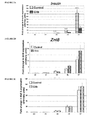
- the expression pattern of Ngn3 was investigated before and after 1, 3, 5 and 7 days of culture. As shown in Figure 3A , Ngn3 was weakly expressed at E13.5 (DO). It increases at day 1 and 3 but remain similar in absence or in presence of glibenclamide. In contrast, after 5 days of culture, Ngn3 expression reached a peak and was sevenfold increased by glibenclamide (***: p ⁇ 0,001).
- Ngn3 mRNA level decreased slightly but remained dramatically higher (***: p ⁇ 0,001) in the glibenclamide-treated pancreases.
- NGN3 expression was analysed by immunohistochemistry ( Figure 3B ) and the number of NGN3+ cells was compared in pancreases cultured for 5 days in absence or in presence of glibenclamide.
- Figure 3C shows that the number of NGN3+cells that develop in presence of glibenclamide was threefold higher than in absence of glibenclamide.
- Ngn3 the transcription factor NeuroD1/Beta2 is a downstream target of Ngn3 (Huang et al., 2000) and is necessary to the endocrine differentiation (Guillemain et al., 2007); the inventors examined the expression pattern of this target of Ngn3. mRNA levels were similar after 1 and 3 days of culture in absence or in presence of glibenclamide. In concordance with the expression pattern of Ngn3 in the glibenclamide-treated pancreases, NeuroD1 was significantly increased at D5 and reminded enhanced at D7 (data not shown). These results demonstrate that the overexpression of Ngn3 leads to the induction of a key factor important for islet differentiation.
- Pancreases were cultured up to 14 days in presence of glibenclamide only during the first 5 days of culture, i.e until Ngn3 reaches a peak (Glib-5D pancreases), in absence (Control pancreases) or in presence of glibenclamide (Glib pancreases) during the 14-day culture period. Then, the insulin-expressing cell masses were compared after 9, 11 and 14 days of culture. As shown in Figure 4A , a large number of insulin-expressing cells was observed in Glib-5D pancreases at D 9, D11 or D14. In contrast, less insulin-containing cells were detected in Glib pancreases (data not shown).
Landscapes
- Health & Medical Sciences (AREA)
- Life Sciences & Earth Sciences (AREA)
- Chemical & Material Sciences (AREA)
- Engineering & Computer Science (AREA)
- Organic Chemistry (AREA)
- Biomedical Technology (AREA)
- Genetics & Genomics (AREA)
- Zoology (AREA)
- Bioinformatics & Cheminformatics (AREA)
- Wood Science & Technology (AREA)
- Biotechnology (AREA)
- General Health & Medical Sciences (AREA)
- Cell Biology (AREA)
- Biochemistry (AREA)
- Medicinal Chemistry (AREA)
- Microbiology (AREA)
- General Engineering & Computer Science (AREA)
- Diabetes (AREA)
- Biophysics (AREA)
- Immunology (AREA)
- Toxicology (AREA)
- Gastroenterology & Hepatology (AREA)
- Molecular Biology (AREA)
- Proteomics, Peptides & Aminoacids (AREA)
- Chemical Kinetics & Catalysis (AREA)
- Veterinary Medicine (AREA)
- Public Health (AREA)
- Animal Behavior & Ethology (AREA)
- Pharmacology & Pharmacy (AREA)
- Nuclear Medicine, Radiotherapy & Molecular Imaging (AREA)
- General Chemical & Material Sciences (AREA)
- Obesity (AREA)
- Hematology (AREA)
- Endocrinology (AREA)
- Emergency Medicine (AREA)
- Developmental Biology & Embryology (AREA)
- Medicines Containing Material From Animals Or Micro-Organisms (AREA)
- Micro-Organisms Or Cultivation Processes Thereof (AREA)
Abstract
Description
- The present invention relates to the field of medicine, in particular to treatment or prevention of diabetes. It also relates to the field of cell biology.
- The pancreas is a gland organ composed of two subclasses of tissue: the exocrine cells (acinar tissue) and the endocrine cells (islets of Langerhans). The exocrine cells produce the digestive enzymes that pass to the small intestine. The islet cells produce hormones which are involved in carbohydrate metabolism. Islets are composed of five cell types: α, β, δ, ε and PP cells which produce glucagon, insulin, somatostatin, ghrelin and pancreatic polypeptide, respectively. β cells secrete insulin in response to an increase in extracellular glucose concentration.
- The first morphological signs of the primitive pancreas emerge as dorsal and ventral protrusions of the primitive gut epithelium at embryonic day (E) 9.5 in the mouse. Subsequently, all lineages defining the various pancreatic cell types, comprising endocrine islet and exocrine acinar and duct cells, are formed from a multipotent progenitor cell pool expressing the transcription factor Pdx1. The transcription factor Ngn3 is transiently expressed in a subset of the pancreas progenitor cells from E 9.5 to E 18.5 and initiates the differentiation program of all islet cells. It was demonstrated that Ngn3 is required for the specification of a common precursor for the five pancreatic endocrine cell types (α, β, δ, ε and PP) and mice lacking Ngn3 function fail to generate any pancreatic endocrine cells and die postnatally from diabetes (Gradwohl et al., 2000). The specification of different islet cell types and the completion of the differentiation process require the activation of transcription factors that are downstream of Ngn3. Among these regulatory factors NeuroD1, Pax4 and Nkx2.2 are direct targets of Ngn3.
-
Type 1 and type 2 diabetes are characterized by loss and dysfunction of β cells. Type 2 diabetes, which is the most common form, is associated with a gradual decline in sensitivity to insulin.Type 1 diabetes is a condition in which the body's immune cells attack β cells located in pancreatic islets, reducing or eliminating the body's ability to produce insulin. Treatment fortype 1 diabetes is a lifelong commitment of monitoring blood glucose, exercising, dieting, and taking insulin. In some cases, individuals with type 2 diabetes similarly require insulin therapy. However, these approaches are sometimes insufficient to control blood glucose levels. Poorly controlled diabetes can lead to potentially fatal complications. Eyes, nerves and kidneys are particularly susceptible to the damage caused by poorly controlledtype 1 or type 2 diabetes. - An alternative treatment for patients with
type 1 diabetes is whole organ pancreatic transplant. Such a procedure offers the possibility of excellent glycemic control but patients are subjected to the adverse effects of immunosuppression and the risks of major surgery. - Recently great strides have been made in developing human islet transplantation in the treatment of diabetes. However, a large number of islets is required to achieve long-term insulin independence and two or far more donor organs are needed to accumulate enough islet cells for a single complete transplant. Thus, the lack of cadaveric human islets is a major obstacle in the widespread use of islets transplantation. Furthermore, with this procedure, immunosuppression is still necessary and the islets are often severely injured from storage conditions or transport time causing apoptosis of the insulin secreting β cells.
- These limitations have given a high priority to efforts to stimulate the growth of new pancreatic islet tissue. As example, the patent application
WO 2006/046923 proposes to treat pancreatic stem cells with retinoic acid to obtain pancreatic hormone-producing endocrine cells. - Nevertheless, there is still a strong need to provide methods for providing large number of β cells which could be used to treat diabetes by pancreatic islet transplantation or for promoting β cell maturation.
- In a first aspect, the present invention provides an in vitro method for increasing the pool of Ngn3+ endocrine progenitor cells obtained from stem cells, wherein said method comprises the step of contacting stem cells having the capacity to differentiate into pancreatic endocrine cells, except human embryonic stem cells, with a SUR1/Kir6.2 channel inhibitor.
- In a second aspect, the present invention provides an in vitro method for increasing the number of pancreatic endocrine cells obtained from stem cells, wherein said method comprises the step of contacting stem cells having the capacity to differentiate into pancreatic endocrine cells, except human embryonic stem cells, with a SUR1/Kir6.2 channel inhibitor.
- In another aspect, the present invention also concerns an in vitro method for increasing the β cell mass obtained from stem cells, wherein said method comprises the step of contacting stem cells having the capacity to differentiate into pancreatic endocrine cells with a SUR1/Kir6.2 channel inhibitor.
- The present invention also concerns an in vitro method for obtaining pancreatic endocrine cells, wherein said method comprises the step of contacting stem cells having the capacity to differentiate into pancreatic endocrine cells with a SUR1/Kir6.2 channel inhibitor.
- In a further aspect, the present invention provides the use of a SUR1/Kir6.2 channel inhibitor for the in vitro or ex vivo differentiation of stem cells into pancreatic endocrine cells.
- In another aspect, the present invention provides pancreatic cells obtained by the method of the invention.
- In a further aspect, the present invention concerns pancreatic islets obtained by the method of the invention.
- In another aspect, the present invention concerns a pharmaceutical composition comprising pancreatic cells and/or pancreatic islets obtained by the method of the invention.
- In another aspect, the present invention concerns pancreatic cells and/or pancreatic islets obtained by the method of the invention for the treatment of diabetes in a subject in need thereof.
- In a last aspect, the present invention concerns a method of treating diabetes in a subject in need thereof, said method comprising steps consisting of
- obtaining stem cells having the capacity to differentiate into pancreatic endocrine cells;
- contacting said stem cells with a SUR1/Kir6.2 channel inhibitor during their differentiation into pancreatic endocrine cells;
- transplanting a therapeutically effective amount of pancreatic islets obtained by differentiation of said stem cells into said subject.
-
-
Figure 1 : Morphological effect of increased concentrations of glibenclamide on embryonic pancreas development in vitro.
(Fig. 1A ) E13.5 embryonic Rat pancreases were cultured at the air/liquid interface for 7 days without (control) or with increased concentrations of glibenclamide. Representative pictures of 1, 3, 5 and 7 days-cultured pancreases are shown. The epithelium, circled in white and surrounded by its mesenchyme, is more branched in the 100µM glibenclamide-treated pancreases (white asteriks). (Fig. 1B ) Hoechst staining of pancreases cultured 7 days without or with the indicated concentrations of glibenclamide. Note the lack of pycnotic nuclei in glibenclamide-treated pancreases. Scale bar: 50 µm. (Fig. 1C ) Quantification of the absolute surface areas occupied by Hoechst staining after 7 days of culture without or with 10 and 100µM glibenclamide. Quantification of Hoechst staining was used to estimate the size of pancreatic rudiments. Five to six pancreases were analysed for each condition. Values are means± sem. -
Figure 2 : Effects of glibenclamide on β and α cell differentiation.
Quantification by real-time PCR of insulin. (Fig. 2A ), ZnT-8 (Fig. 2B ) and MafA (Fig. 2C ). mRNA in E13.5 pancreases before (DO) and after 1, 3, 5 and 7 days of culture (D1, D3, D5 and D7 respectively) with or without 100µM glibenclamide. Each data point represents the mean± sem of at least six independent experiments **, p<0,01. (Fig. 2D ) Quantification of the absolute surface area occupied by proprotein convertase subtilisin/kexin 1/3 (PCSK1/3) staining in PDX-1-expressing cells that developed after 7 days of culture in presence or in absence of 100µM glibenclamide. Values are means± sem of at least three independent experiments.
α cell differentiation was evaluated by glucagon staining in E13.5 pancreases cultured 7 days in presence or in absence of 100µM glibenclamide. (Fig. 2E ) The absolute surface of glucagon staining in E13.5 pancreases cultured 7 days in presence or in absence of 100µM glibenclamide was quantified. Values are means± sem of at least three independent experiments **, p<0,01. (Fig. 2F ) Expression profile of Pou3F4 by real-time PCR in E13.5 pancreases before (DO) and after 1, 3, 5 and 7 days of culture (D1, D3, D5 and D7 respectively) in the presence or in absence of 100µM glibenclamide. Each data point represents the mean± sem of at least three independent experiments **, p<0,01. -
Figure 3 : Glibenclamide treatment amplifies the pool of pro-endocrine precursors without acting on pancreatic progenitor proliferation.
(Fig. 3A ) Quantification by real-time PCR of Ngn3 transcripts in E13.5 pancreases after 0, 1, 3, 5 and 7 days of culture (D0, D1, D3, D5 and D7 respectively) with or without 100µM glibenclamide. Each data point represents the mean± sem of at least seven independent experiments ***, p<0,001. (Fig. 3B ): Immunohistochemistry analysis of NGN3 expression in E13.5 pancreases cultured 5 days in presence (C, C') or in absence (B, B') of 100µM glibenclamide. Note the increased number of NGN3 positive nuclei in the glibenclamide-treated explants. Scale bar: 50 µm. B' and C' are respectively the enlargement of B and C. (Fig. 3C ) Quantification of the number of NGN3 positive-cells in E13.5 pancreases cultured for 5 days in presence or in absence of 100µM glibenclamide. Values are means± sem of at least three independent experiments **, p<0,01. (Fig. 3D ) E13.5 pancreases were grown for 24h with or whithout 100µM glibenclamide and pulsed with BrdU during the last hour of culture. Immunohistochemistry was performed using anti-PDX1 and anti-BrdU antibodies. Quantification of the proliferation of early PDX1 progenitors in pancreases cultured 24h in the absence or presence of 100µM glibenclamide -
Figure 4 : Glibenclamide-induced NGN3+ cells differentiate into β cells.
(Fig. 4A ) Quantification of the absolute surface area occupied by insulin staining in β cells that developed after 7, 9,11 and 14 days of culture in absence or in presence of 100µM glibenclamide only during the first 5 days of culture. Each data point represents the mean± sem of at least three independent experiments *, p<0,05; **, p<0,01. (Fig. 4B ) Quantification by real-time PCR of insulin transcripts in E13.5 pancreases after 7, 9, 11 and 14 days of cultured without or with 100µM glibenclamide only during the first 5 days of culture. Each data point represents the mean± sem of at least three independent experiments **, p<0,01. Quantification by real-time PCR of ZnT-8 (Fig. 4C ) and MafA (Fig. 4D ) transcripts in E13.5 pancreases after 7, 9, 11 and 14 days of cultured without or with 100µM glibenclamide only during the first 5 days of culture. Each data point represents the mean± sem of at least three independent experiments **, p<0,01. - The pancreatic β cell KATP channel plays a key role in glucose stimulated-insulin secretion by regulating the flux of potassium ions across cell membranes. KATP channels are open at low glucose concentrations but close when glucose uptake and metabolism are stimulated by an increase in the plasma glucose concentration. This leads to membrane depolarization, activation of voltage-dependent calcium channels, calcium influx and, in turn, insulin secretion. This channel is composed of two types of subunit - the inward rectifier K+ channel (Kir6.2) which forms the channel pore, and the sulfonylurea receptor (SUR1), which serves as a regulatory subunit. These subunits associate with a stoichiometry of Kir6.24/SUR14. Kir6.2 and SUR1 subunits are encoded by KCNJ11 and ABCC8 genes, respectively.
The inventors have herein demonstrated that SUR1/Kir6.2 channel inhibitors are able to amplify the pool of endocrine progenitor Ngn3+ cells which subsequently differentiate into pancreatic endocrine cells. These inhibitors can thus be used to dramatically increase the β cell mass and insulin secretion capacity in a developing pancreas or islet. - In a first aspect, the present invention provides an in vitro method for increasing the pool of Ngn3+ endocrine progenitor cells, wherein said method comprises the step of contacting stem cells having the capacity to differentiate into pancreatic endocrine cells with a SUR1/Kir6.2 channel inhibitor.
- As used herein, the term "Ngn3+ endocrine progenitor cells" refers to precursors of pancreatic endocrine cells expressing the transcription factor Neurogenin-3 (Ngn3). Progenitor cells are more differentiated than multipotent stem cells and can differentiate into only few cell types. In particular, Ngn3+ endocrine progenitor cells have the ability to differentiate into the five pancreatic endocrine cell types (α, β, δ, ε and PP). The expression of Ngn3 may be assessed by any method known by the skilled person such as immunochemistry using an anti-Ngn3 antibody or quantitative RT-PCR.
- The term "stem cells" refers to cells which have the ability to go through numerous cycles of cell division while maintaining an undifferentiated state and have the capacity to differentiate into specialized cell types. There are two broad types of mammalian stem cells: embryonic stem cells isolated from the blastocysts and adult stem cells found in adult tissues.
Stem cells may be classified according to their potency (their ability to differentiate into different cell types). Totipotent stem cells can differentiate into embryonic and extraembryonic cell types. Such cells contain all the genetic information needed to create a complete and viable organism. Pluripotent stem cells can differentiate into nearly all cell types but cannot develop into an embryo. These cells maintain the plasticity to generate all types of cells in an individual, except extraembryonic tissue such as placenta. Multipotent stem cells can differentiate into a number of cell types, but only those of a closely related family of cells. Adult stem cells, which reside in small number in almost all adult tissues, are generally multipotent: their regenerative potential is tissue or germ-layer specific.
As used herein, the term "stem cells" encompasses embryonic stem cells, adult stem cells and reprogrammed somatic cells (induced pluripotent stem cells).
In a particular embodiment, embryonic stem cells are non-human embryonic stem cells due to some patent law and practices. - In an embodiment, stem cells having the capacity to differentiate into pancreatic endocrine cells are selected from the group consisting of pancreatic stem cells, pluripotent stem cells and multipotent stem cells.
- In a particular embodiment, pancreatic stem cells are selected from the group consisting of stem cells derived from pancreatic islets, pancreatic ducts, pancreatic acinar cells and stem cells derived from the dorsal pancreatic bud from embryos.
- As used herein, the term "cells derived from" shall be taken to indicate that this particular group of cells has originated from the specified source, but has not necessarily been obtained directly from said source.
As used herein, the term "pancreatic stem cells" refers to multipotent and organ specific stem cells expressing Pdx1 and which are able to differentiate into all types of pancreatic cells. The pancreas duodenal homeobox gene Pdx1 (UniGene Hs.32938) is one of the earliest genes expressed in the developing pancreas. Cells expressing Pdx1 give rise to all three types of pancreatic tissue, exocrine, endocrine and duct. After birth, Pdx1 expression is essentially restricted to β cells within the endocrine islets of the pancreas. - The identification of pancreatic stem cells from pancreatic islet and ductal populations has been described in Seaberg et al. (Seaberg et al., 2004). This paper demonstrated that these stem cells coexpress neural and pancreatic precursor markers and differentiate to form distinct populations of neurons, glial and stellate cells, pancreatic endocrine beta-, alpha- and delta-cells, and pancreatic exocrine cells.
Furthermore, it was recently found that pancreatic ductal and acinar cells are able under certain conditions to regress to a less differentiated phenotype and then can differentiate to form endocrine cells and, in particular, to form β cells (Bonner-Weir et al., 2008; Minami et al., 2008).
Consequently, stem cells having the capacity to differentiate into pancreatic endocrine cells may be pancreatic stem cells derived from exocrine, endocrine or ductal tissue or differentiated pancreatic cells which move into a less differentiated stage to express Pdx1. Such pancreactic stem cells may be obtained from adult tissue by any method known in the prior art such as those described in the articles of Seaberg et al. Bonner-Weir et al., and Minami et al. (Seaberg et al., 2004 Bonner-Weir et al., 2008; Minami et al., 2008). - Pancreatic stem cells may also be derived from the dorsal pancreatic bud from embryos. The dorsal pancreas is an embryonic bud from the endodermal lining of the gut on the dorsal wall cephalad to the level of the hepatic diverticulum, which forms most of the pancreas and its main duct. Pancreatic stem cells expressing Pdx1 may be obtained from fertilized ovocytes when pancreatic tissue has started to develop and before the terminal differentiation of most pancreatic cells.
- In an embodiment, pancreatic stem cells are derived from human embryos. The age of these embryos is between 2 and 12 weeks of development, preferably between 2 and 8 weeks and more preferably between 2 and 6 weeks of development.
In a particular embodiment, pancreatic stem cells are derived from the dorsal pancreatic bud from non-human embryos due to some patent law and practices. - In another embodiment, stem cells having the capacity to differentiate into pancreatic endocrine cells are multipotent stem cells derived from other adult tissue than pancreatic tissue. Preferably, multipotent stem cells are derived from adult tissue selected from the group consisting of bone marrow, liver, central nervous system, spleen and adipose tissue.
Bone marrow-derived stem cells (hematopoietic or mesenchymal) have been described to be able to differentiate into pancreatic endocrine cells (Oh et al., 2004; Moriscot et al., 2005; Sun et al., 2007; Gabr et al., 2008). Bone marrow-derived stem cells may be isolated from the bone marrow based on their ability to adhere to plastic support. Then, they may be expanded and cultured. Pdx-1 gene expression may be induced in these cells using factors such as dimethyl sulfoxide, trichostatin or β-mercaptoethanol.
Mesenchymal stem cells from bone marrow and adipose tissue represent a very similar cell population with comparable phenotype. Consequently, adipose tissue-derived mesenchymal stem cells have also the potential to differentiate in pancreatic endocrine cells (Timper et al., 2006).
Liver stem cells, also named oval stem cells, have been described to be able to differentiate into pancreatic endocrine cells when cultured in a high-glucose environment (Yang et al., 2002). Another possibility may be to induce transdifferentiation of liver stem cells into pancreatic progenitor cells by expressing a Pdx-1 transgene (Sapir et al., 2005).
Brain-derived neural progenitor cells (Hori et al., 2005) and splenocytes (Kodama et al., 2003) have been also described to be able to differentiate into pancreatic endocrine cells.
In a preferred embodiment, multipotent stem cells derived from adult tissue and having the capacity to differentiate into pancreatic endocrine cells are not genetically modified. These cells exhibit the capacity to differentiate into pancreatic endocrine cells only by culturing them in presence of specific growth factors and/or compounds. - In a further particular embodiment, stem cells are pluripotent stem cells obtained by reprogramming of somatic cells. Such cells are also named induced pluripotent stem cells.
It has been found that induced pluripotent stem cells recapitulated the features of embryonic stem cells, such as human embryonic stem cells, and are thus an alternative to the controversial use of these cells (Romano et al., 2008). Induced pluripotent stem cells may be obtained from somatic cells, such as human skin fibroblasts, by a variety of methods essentially based on manipulation of a selected group of transcription factors (Maherali et al., 2008). For instance, induced pluripotent stem cells have been generated by ectopic expression of four transcription factors, OCT4, SOX2, KLF4 and c-MYC (Takahashi et al., 2007; Lowry et al., 2008) or OCT4, SOX2, NANOG and LIN28 (Yu et al., 2007). Furthermore, it has been demonstrated that induced pluripotent cells have the potential to differentiate into pancreatic endocrine cells (Tateishi et al., 2008). - In another embodiment, pluripotent stem cells are derived from embryonic stem cells.
Embryonic stem (ES) cells are derived from totipotent cells of the early mammalian embryo and are capable of unlimited, undifferentiated proliferation in vitro. Essential characteristics of these cells include (i) derivation from the preimplantation or periimplantation embryo, (ii) prolonged undifferentiated proliferation, and (iii) stable developmental potential to form derivatives of all three embryonic germ layers (endoderm, mesoderm and ectoderm) even after prolonged culture.
For human embryonic stem cells, it has been demonstrated that these cells may be obtained from frozen-thawed blastocysts that were destined to be discarded after 5 years in a routine human IVF-embryo transfer programme (Park et al., 2004).
ES cells grow as homogenous and undifferentiated colonies when they are propagated on a feeder layer such as mouse embryonic fibroblasts. Removal from this feeder layer is associated with differentiation into derivatives of the three embryonic germ layers. Human embryonic stem cells have been described to be able to differentiate in vitro into pancreatic endocrine cells, and particularly into β cells (Assady et al., 2001).
In a particular embodiment, pluripotent stem cells are derived from non-human embryonic stem cells, due to some patent law and practices. - Stem cells as described above, which have the capacity to differentiate into pancreatic endocrine cells and thus into their precursors, namely Ngn3+ endocrine progenitor cells, may be used in the method of the invention for increasing the pool of these Ngn3+ cells. The step of contacting with a SUR1/Kir6.2 channel inhibitor has to be conducted after detection of pdx1 gene expression and before the complete differentiation of these cells into pancreatic endocrine cells, preferably after detection of pdx1 gene expression and before detection of Ngn3 expression.
- Stem cells as described above may be derived from any mammalian such as mice, rats, pigs, dogs, cats, horses, monkeys or humans.
- The term "SUR1/Kir6.2 channel inhibitor" as used herein refers to a compound which has the capacity to close the SUR1/Kir6.2 channel and thus to block the flux of potassium ions across cell membranes
In an embodiment, the SUR1/Kir6.2 channel inhibitor is selected from sulfonylureas and metiglinides (or glinides), and any combination thereof.
In one embodiment, the inhibitor is a sulfonylurea selected from the group consisting of acetohexamide, carbutamide, glibornuride, chlorpropamide, tolbutamide, tolazamide, glipizide, gliclazide, glibenclamide (glyburide), gliquidone, glyclopyramide, glisoxepide and glimepiride.
In another embodiment, the inhibitor is a metiglinide selected from the group consisting of repaglinide, nateglinide and mitiglinide.
In a preferred embodiment, the SUR1/Kir6.2 channel inhibitor is glibenclamide.
The SUR1/Kir6.2 channel inhibitor may be used alone, in combination with one or several SUR1/Kir6.2 channel inhibitors and in combination with other active substances. For example, glibenclamide may be used in association with metformine. - In the present method, stem cells having the capacity to differentiate into pancreatic endocrine cells are contacted with a SUR1/Kir6.2 channel inhibitor. This step of contacting stem cells with a SUR1/Kir6.2 channel inhibitor may consist of culturing stem cells in a medium containing a SUR1/Kir6.2 channel inhibitor.
- The concentration of the SUR1/Kir6.2 channel inhibitor may be chosen by the skilled person using well known methods. For instance, preliminary tests may be achieved to evaluate the toxicity of the SUR1/Kir6.2 channel inhibitor on stem cells. In this case, stem cells are cultured with different concentrations of this inhibitor and toxicity markers are followed. These markers may be markers of apoptotic cell death such as apoptotic DNA fragmentation and DEVD-caspase activation. The concentration of the channel inhibitor has to be chosen in order to be safe of any toxic effects on growing stem cells. Preferably, the concentration is chosen in order to be the highest concentration without any toxic effect.
In an embodiment, stem cells are contacted with a SUR1/Kir6.2 channel inhibitor by culturing them in presence of 0.1 to 500 µM of said inhibitor, preferably in presence of 1 to 250 µM of said inhibitor and the most preferably in presence of 50 to 150 µM said inhibitor.
In a particular embodiment, the SUR1/Kir6.2 channel inhibitor is glibenclamide and stem cells are contacted with glibenclamide by culturing them in presence of 100 µM of glibenclamide. - The culture medium which may be used during the step of contacting with a SUR1/Kir6.2 channel inhibitor is designed to support the growth and the differentiation of stem cells. This medium generally is changed every day and comprises a carbon source, a nitrogen source, antibiotics to prevent fungi and bacteria growth, a buffer to maintain pH and specific growth factors. This medium may be easily designed by the skilled person in the art. An example of such medium is presented in the experimental section below or in the experimental section of the article of Guillemain et al (Guillemain et al., 2007).
- Other compounds may also be added in the medium such as compound known to stimulate β cell replication, to induce differentiation into β cells or to inhibit apoptosis of β cells. Such compounds may be chosen from the group consisting of nicotinamide, glucagon-like peptide-1 (GLP-1), glucose, exendin-4 and retinoic acid.
In an embodiment, stem cells are contacted with a SUR1/Kir6.2 channel inhibitor during 3 to 10 days, preferably from 5 to 7 days. During the step of contacting, stem cells are cultured in a medium supporting growth and differentiation and containing a SUR1/Kir6.2 channel inhibitor. - At the end of the step of contacting and/or several days later, the number of Ngn3-expressing cells may be assessed in order to verify the efficiency of the treatment, i.e. the increase of the pool of Ngn3+ endocrine progenitor cells. The number of Ngn3-expressing cells obtained in treated samples is compared to the number of Ngn3-expressing cells obtained in control sample, i.e. without step of contacting with a SUR1/Kir6.2 channel inhibitor. In order to be comparable, stem cells in treated and control samples have to be of the same cellular type and submitted to the same protocol except channel inhibitor treatment.
In an embodiment, the pool of Ngn3+ endocrine progenitor cells has increased by more than 25%, preferably by more than 50% and the most preferably by more than 100%.
In a preferred embodiment, the pool of Ngn3+ endocrine progenitor cells has increased by more than 150%, preferably by more than 200% and the most preferably by more than 250%. - In an embodiment, the method further comprises a step consisting of the differentiation of obtained Ngn3+ endocrine progenitor cells into precursors of pancreatic endocrine cells and/or pancreatic endocrine cells.
In appropriate culture medium, such as described above, Ngn3+ endocrine progenitor cells differentiate into precursors of pancreatic endocrine cells and subsequently into β, δ, ε and/or PP cells. - The present invention also concerns an in vitro method for increasing the number of pancreatic endocrine cells obtained from stem cells, wherein said method comprises the step of contacting stem cells having the capacity to differentiate into pancreatic endocrine cells with a SUR1/Kir6.2 channel inhibitor.
In a particular embodiment, this method comprises the step of contacting stem cells having the capacity to differentiate into pancreatic endocrine cells, except human embryonic stem cells, due to some patent law and practices. - The present invention also concerns an in vitro method for increasing the β cell mass obtained from stem cells, wherein said method comprises the step of contacting stem cells having the capacity to differentiate into pancreatic endocrine cells with a SUR1/Kir6.2 channel inhibitor.
- In a particular embodiment, this method comprises the step of contacting stem cells having the capacity to differentiate into pancreatic endocrine cells, except human embryonic stem cells, due to some patent law and practices.
- The present invention also concerns an in vitro method for obtaining pancreatic endocrine cells, wherein said method comprises the step of contacting stem cells having the capacity to differentiate into pancreatic endocrine cells with a SUR1/Kir6.2 channel inhibitor.
- In another aspect, the present invention concerns the use of a SUR1/Kir6.2 channel inhibitor for the in vitro or ex vivo differentiation of stem cells into pancreatic endocrine cells. Stem cells have to be able to differentiate into pancreatic endocrine cells and may be chosen as described above.
- In another aspect, the present invention also provides an in vivo method for increasing the number of pancreatic endocrine cells, in particular of β cells, in the pancreas of a foetus, wherein said method comprises administering a SUR1/Kir6.2 channel inhibitor to the pregnant female.
- In another aspect, the present invention also provides an in vivo method for increasing the number of pancreatic endocrine cells, in particular of β cells, in the pancreas of a subject, wherein said method comprises administering a SUR1/Kir6.2 channel inhibitor to said subject. Preferably, the subject is a child.
- In a further aspect, the present invention provides pancreatic cells obtained by the in vitro method of the invention.
In an embodiment, pancreatic cells are Ngn3+ endocrine progenitor cells.
In another embodiment, pancreatic cells are cells derived from Ngn3+ endocrine progenitor cells, i.e. pancreatic endocrine cell precursors and pancreatic endocrine cells.
Precursors of pancreatic endocrine cells may express, for instance, Pax4 (paired box-encoding gene 4) or Arx (Aristaless-related homeobox).
Pancreatic endocrine cells may be α, β, δ, ε and/or PP cells. - In a preferred embodiment, pancreatic cells are β cells. The term "β cells", as used herein, refers to pancreatic cells which are able to produce insulin. In vivo, these cells are found in the pancreatic islets of Langerhans. This cell population may be identified by the expression of specific markers such as ZnT-8, a specific zinc transporter (Chimienti et al. 2004) or MafA, a specific transcription factor (Zhang et al., 2005; Matsuoka et al., 2007), or by an ability to respond to glucose challenge in a specific way by secreting insulin.
- The present invention also concerns pancreatic islets comprising pancreatic cells of the invention as described above.
As used herein, the term "pancreatic islet" refers to cell small discrete cell aggregates obtained in vitro or ex vivo and including pancreatic endocrine hormone producing cells, such as α cells, β cells, δ cells, PP cells and ε cells. Pancreatic islets resemble the form of islets of Langerhans of the pancreas and are spheroid in form. In vivo, the islets of Langerhans are surrounded by the pancreatic exocrine tissue.
In an embodiment, pancreatic islets comprise β cells obtained by the method of the invention.
In another embodiment, pancreatic islets comprise β cells and α cells obtained by the method of the invention.
In a preferred embodiment, pancreatic islets comprise α cells, β cells, δ cells, PP cells and ε cells obtained by the method of the invention. - In a further aspect, the present invention concerns a pharmaceutical composition comprising pancreatic cells and/or pancreatic islets of the invention, and a pharmaceutically acceptable carrier.
Pharmacologically acceptable carriers have to be compatible with the cells and may be, for instance, cell culture medium (such as Eagle's minimal essential media), phosphate buffered saline, Krebs-Ringer buffer, and Hank's balanced salt solution +/- glucose (HBSS). - In a preferred embodiment, pharmaceutical composition is suitable for parenteral administration, e.g. subcutaneously, retroperitoneally and intravenously. Such composition may comprise any additive compatible with the cells.
The pharmaceutical composition comprising pancreatic cells and/or pancreatic islets of the invention is formulated in accordance with standard pharmaceutical practice (see, e.g., Remington: The Science and Practice of Pharmacy (20th ed.), ed. A. R. Gennaro, Lippincott Williams & Wilkins, 2000 and Encyclopedia of Pharmaceutical Technology, eds. J. Swarbrick and J. C. Boylan, 1988-1999, Marcel Dekker, New York) known by a person skilled in the art. The compositions for parenteral administration are generally physiologically compatible sterile solutions or suspensions. Adjuvants such as a local anesthetic, preservative and buffering agents can be dissolved in the vehicle.
In one embodiment, the pharmaceutical composition comprises pancreatic cells and/or pancreatic islets of the invention encapsulated in a biocompatible matrix known in the art. A variety of encapsulation technologies have been developed (e.g. Qi et al., 2008 andWO 91/10425
The pharmaceutical composition comprising pancreatic cells and/or pancreatic islets of the invention, may be used for islet transplantation and/or treatment oftype 1 and type 2 diabetes.
The pharmaceutical composition can also comprise one or several additional active compounds, e.g. compounds known to enhance cell survival, proliferation or to prevent any contamination. - In another aspect, the present invention concerns pancreatic cells and/or pancreatic islets of the invention for the treatment of diabetes in a subject in need thereof.
- The present invention also concerns a method of treating diabetes in a subject in need thereof, said method comprising steps consisting of
- obtaining stem cells having the capacity to differentiate into pancreatic endocrine cells;
- contacting said stem cells with a SUR1/Kir6.2 channel inhibitor during their differentiation into pancreatic endocrine cells;
- transplanting a therapeutically effective amount of pancreatic islets obtained by differentiation of said stem cells into said subject.
- Once transplanted, the pancreatic islets begin to produce insulin, actively regulating the level of glucose in the blood. The main obstacle in islet transplantation is the fact that there is an inadequate supply of cadaveric islets to implement this procedure on a widespread clinical basis. The method of the invention solves this problem by obtaining an increase number of pancreatic islets which may be used for transplantation.
- The term "diabetes" is intended to encompass
type 1 and type 2 diabetes mellitus.
As used herein, the term "treatment", "treat" or "treating" refers to any act intended to ameliorate the health status of patients such as therapy, prevention, prophylaxis and retardation of the disease. In certain embodiments, such term refers to the amelioration or eradication of a disease or symptoms associated with a disease.
In particular, the term "treatment of diabetes", as used herein, does not necessarily mean a complete cure but means that the symptoms or complications of the underlying disease are reduced, and/or that one or more of the underlying cellular, physiological, or biochemical causes or mechanisms causing the symptoms or complications are reduced. The term "treatment of diabetes" also includes within its scope the prophylactic treatment of an asymptomatic subject thought to be at risk of developing diabetes. - The subject to treat is any mammal, preferably a human being.
- As used herein, "therapeutically effective amount" means an amount of islets administered to the subject, which will be effective to improve, prevent, delay the onset of, or treat diabetes or associated complications in the subject. Typically, it was estimated that a diabetic patient needs at least 10,000 pancreatic islets per kilogram body weight to achieve a measurable increase in insulin production. Generally, between 10,000 and 30,000 pancreatic islets per kilogram body weight are administered to the subject during transplantation. The number of pancreatic islets to be administered to a subject will vary depending on a number of parameters including the size of the subject, the severity of the disease and the site of implantation.
- Generally pancreatic islets are suspended in a pharmacologically acceptable carrier, such as, for instance, cell culture medium (such as Eagle's minimal essential media), phosphate buffered saline, Krebs-Ringer buffer, and Hank's balanced salt solution +/glucose (HBSS).
- The pancreatic islets can be administered by any method known to one of skill in the art.
- In an embodiment, pancreatic islets are administered by injection. For example, pancreatic islets may be administered by subcutaneous injection, intra-peritoneal injection, injection under the kidney capsule, injection through the portal vein and injection into the spleen.
- According to the origin of stem cells, the islet transplantation may be autologous, isogeneic, allogeneic or xenogeneic.
As used below, the "donor" is the donor of stem cells and the "recipient" is the subject who receives the islet transplantation.
In an embodiment, the islet transplantation is isogeneic, i.e. the donor and recipient are genetically identical.
In another embodiment, the islet transplantation is allogeneic, i.e. the donor and recipient are of the same species.
In another embodiment, the islet transplantation is xenogeneic, i.e. the donor and recipient are of different species.
Allogeneic and xenogeneic transplantation require the administration of antirejection drugs.
For isogeneic, allogeneic and xenogeneic transplantation, the donor may be alive or deceased.
In a preferred embodiment, the islet transplantation is autologous, i.e. the donor and recipient are the same subject. In this case, stem cells may be (i) derived from adult tissue of the subject, (ii) derived from somatic cells of said subject which have been reprogrammed to provide induced pluripotent stem cells or (iii) from embryonic stem cells obtained by cloning. - All references cited in this specification are incorporated by reference.
- Throughout this specification and the claims which follow, unless the context requires otherwise, the word "comprise", and variations such as "comprises" and "comprising", will be understood to imply the inclusion of a stated integer or step or group of integers or steps but not the exclusion of any other integer or step or group of integers or steps."
- The reference in this specification to any prior publication (or information derived from it), or to any matter which is known, is not, and should not be taken as an acknowledgement or admission or any form of suggestion that that prior publication (or information derived from it) or known matter forms part of the common general knowledge in the field of endeavour to which this specification relates.
- The following examples are given for purposes of illustration and not by way of limitation.
- Pregnant Wistar rats were purchased from the Janvier Breeding centre (CERJ, LeGenet, France). The first day postcoitum was taken as embryonic day (E) 0.5. Pregnant rats were killed with CO2 asphyxiation according to guidelines issued by the French Animal Care Committee. Dorsal pancreatic buds from E13.5 rat embryos were dissected as described previously (Miralles et al., 1998). Briefly, the stomach, pancreas and a small portion of the intestine were dissected together, and then the pancreas primordium was isolated.
- Dorsal pancreatic rudiments were cultured on 0,45µm filters (Millipore) at the airmedium interface in a 35mm sterile Petri dishes containing 2 ml RPMI-1640 medium (Invitrogen) supplemented with 100U/ml penicillin, 100µg/ml streptomycin, 10mmol/L HEPES, 2 mmol/L L-glutamine, 1X non-essential aminoacids (Invitrogen) and 10% heat-inactivated fetal calf serum (HyClone, Logan, UT, USA). Glibenclamide (MP Biomedical) and nifedipine (Sigma), used at the indicated concentrations, were first dissolved as concentrated solutions in dimethylsulfoxide DMSO (Sigma), the final concentration of DMSO in the culture medium was less than 0,5% (vol/vol). The culture medium was changed every day. Glibenclamide and nifedipine were added to the media daily. The cultures were incubated at 37°C in a humidified atmosphere composed of 95% air and 5% CO2. At the end of the culture period, the pancreases were photographed, and fixed as described below or harvested for RNA extraction
- Immunochemistry - The pancreatic rudiments were fixed in 10% formalin, pre-embedded in agarose gel (4% of type VII low-gelling-temperature agarose (Sigma) in H2O) and embedded in paraffin. Immunohistochemistry was performed on 4-µm paraffin sections as previously described (Duvillie et al., 2006). The primary antibodies were mouse anti insulin (1/2000; Sigma), rabbit anti-glucagon (1/1000; Diasorin), rabbit anti-amylase (1/300; Sigma), rabbit anti-carboxypeptidase A (1/600; Biogenesis, Kidlington, Oxford, UK), rabbit anti-PDX1 (1/1000) (Duvillie et al., 2003) mouse anti-BrdUrd5 (1/2; Amersham Biosciences, Buckingham, UK), rabbit anti-PCK1/3, rabbit anti-Ngn3 (1/100, Guillemain et al., 2007), rabbit anti-KIR6.2 and anti-SUR1 (1/100, Santa Cruz). The fluorescent secondary antibodies were fluorescein anti-rabbit antibody (1/200; Jackson Immunoresearch, Baltimore, MD, USA), fluorescein goat anti-rabbit Alexa Fluor 488 (1/400; Invitrogen) and Texas red anti-mouse antibody (1/200; Jackson). Nuclei were stained in blue with Hoechst 33342 (0,3µg/ml; Invitrogen). Ngn3 detection was performed as previously described (Guillemain et al., 2007) using the Vectastain elite ABC kit (Vector laboratories).
Photographs were taken using a fluorescence microscope (Leica, Leitz DMRB, Rueil-Malmaison, France) and digitized using a Hamamatsu (Middlesex, NJ) C5810 cooled 3CCD camera. - Quantification - To quantify the surface area of insulin, glucagon, PCSK1/3, CPA and amylase-expressing cells, all sections of each pancreatic rudiment were digitized. Alternate sections were examined to avoid counting the same cell twice. The surface of insulin, glucagon, PCSK1/3, CPA, amylase, and Hoechst stainings were quantified using Iplab (Scanalytics). The stained areas were summed to obtain the total surface area per rudiment in mm2. To measure proliferation of the early progenitors expressing PDX1, we counted the frequency of BrdU positive progenitors expressing PDX1 among 3000 early progenitors expressing PDX1 per rudiment. To quantify the absolute number of NGN3-expressing cells, pancreatic rudiments were sectioned and all sections were stained with an anti-NGN3 antibody. Positive cells were counted on all sections of each pancreatic rudiment. A minimum of three rudiments was analyzed per condition.
- Total RNA was isolated from pools of at least three pancreases using the Qiagen RNeasy Microkit (Qiagen, Courtaboeuf, France) and reverse transcribed using Superscript reagents (Invitrogen). Real-time PCR was performed with the 7300 Fast real-time PCR system (Applied Biosystem) using either Taqman universal PCR universal PCR master mix or SYBR green PCR master mix (Applied Biosystem) with primers and labelled probes specific for each gene. Peptidylpropyl isomerase A/Cyclophilin A was used as endogenous control and E16.5 pancreas cDNA as calibrator sample. The data were analyzed by comparative cycle threshold method (Livak et al., 1997) and presented as the fold change in gene expression. At least three pools of explants were analysed by condition.
- All results are expressed as mean ± sem. Statistical significance was determined using Student's t test.
- Using the in vitro model as described above (E13.5 Rat embryonic pancreases cultured at the air/medium interface on a floating filter), we examined the effects of increased concentrations of the sulphonylurea glibenclamide (an inhibitor of KATP channels) on pancreas development. As shown in
Figure 1A , the pancreatic growth was similar in absence (control) or in presence of 10nM, 100nM, 1, 10 or 100µM glibenclamide during the 7 days of culture. Under both conditions, the epithelium grew rapidly, spread into the mesenchyme, and developed lobules. There is no difference in apoptosis in pancreases cultured 7 days without or with 10 and 100µM glibenclamide as shown by the Hoechst staining of the nuclei (Figure 1B ). Moreover, the lack of glibenclamide toxicity on the developing pancreas was confirmed by the quantitative analysis of the overall size of pancreases cultured 7 days in the presence or in the absence of 10 or 100µM glibenclamide (Figure 1C ).
Based on these first results, in particular the lack of glibenclamide toxicity on the pancreas morphology, and on the effects on the pro-endocrine progenitor cells (seefigure 4 ), the 100µM glibenclamide concentration was used in the next experiments. - To determine the effects of glibenclamide on endocrine development and in particular in cells expressing SUR1, the number of insulin-positive cells in pancreases grown for 7 days in the absence or the presence of glibenclamide was compared. The glibenclamide treated-explants exhibit a very few insulin positive cells and the surface area occupied by the insulin-cell population was decreased by 70% (data not shown). Because glibenclamide is a potent insulin secretagogue, the inventors asked if the observed low insulin content was only the consequence of increased insulin secretion in the culture medium or was due to the reduced insulin mRNA level. To test this latter hypothesis, they analysed by real-time PCR the expression of insulin gene before (DO) and after 1, 3, 5 and 7 days of culture. As shown in
Figure 2A , the insulin expression was strongly reduced in the glibenclamide-treated pancreases as early as D3. Two mechanisms can account for the observed decrease in insulin expression: (i) inhibition of the β-cell differentiation from the pro-endocrine progenitors leading to a reduction of the β-cell number and thus to a decrease in overall amount of insulin mRNA or (ii) inhibition of the insulin gene without affecting the β-cell population. Thus, the expression pattern of two β-cell markers: the zinc transporter ZnT-8 (Chimienti et al., 2004) and the β-cell specific transcription factor MafA (Zhang et al., 2005; Matsuoka et al., 2007) was examined (Figures 2B and 2C ). These results indicate that over the 7 days of culture, glibenclamide did not affect the expression of these two beta-cells markers. Moreover, after 7 days of culture, the surface area occupied by thepro-hormone convertase 1/3 (PCSK1/3) staining in PDX-1+ cells (Pdx-1 is specifically expressed in the adult β-cell (Ohlsson et al., 1993)) was similar in pancreases cultured without or with glibenclamide (Figure 2D ). These results demonstrate that high concentrations of glibenclamide do not prevent the β-cell differentiation. - It was further observed that glibenclamide increased by 3,5 fold the number of glucagon-expressing cells (
Figure 2E ). Moreover, this result was confirmed by the significant increase of Pou3F4/Brn4 mRNA level at D7 (Figure 2F ); Pou3F4, known as the only α-cell specific transcription factor which maintains the α cell fate (Jensen et al., 2000; Heller et al., 2004). In the other hand, we found also a two-fold increase in somatostatin expression after 7 days of culture in the glibenclamide-treated pancreas (data not shown). - The pancreatic endocrine fate is determined by the expression of Ngn3, a transcription factor which specifically labels the endocrine precursors (Gradwohl et al., 2000; Gu et al., 2002). The expression pattern of Ngn3 was investigated before and after 1, 3, 5 and 7 days of culture. As shown in
Figure 3A , Ngn3 was weakly expressed at E13.5 (DO). It increases atday
To test whether glibenclamide acts not only on Ngn3 gene expression but also on NGN3-expressing cell number, NGN3 expression was analysed by immunohistochemistry (Figure 3B ) and the number of NGN3+ cells was compared in pancreases cultured for 5 days in absence or in presence of glibenclamide.Figure 3C shows that the number of NGN3+cells that develop in presence of glibenclamide was threefold higher than in absence of glibenclamide. These results indicate that glibenclamide amplifies the pool of NGN3+ endocrine progenitors.
Because the transcription factor NeuroD1/Beta2 is a downstream target of Ngn3 (Huang et al., 2000) and is necessary to the endocrine differentiation (Guillemain et al., 2007); the inventors examined the expression pattern of this target of Ngn3. mRNA levels were similar after 1 and 3 days of culture in absence or in presence of glibenclamide. In concordance with the expression pattern of Ngn3 in the glibenclamide-treated pancreases, NeuroD1 was significantly increased at D5 and reminded enhanced at D7 (data not shown). These results demonstrate that the overexpression of Ngn3 leads to the induction of a key factor important for islet differentiation. - Furthermore, embryonic pancreases were cultured for 1 day and BrdU was added during the last hour of culture to test whether glibenclamide increased Ngn3+ cell number by acting on pancreatic progenitor cell proliferation. The percentage of PDX-1+ cells that incorporate BrdU was similar in presence (32,60% ± 4,2%) or in absence (31,24% ± 2,8%) of glibenclamide (
Figure 3D ). These results demonstrate that glibenclamide does not modify the pancreatic precursor proliferation.
In conclusion, glibenclamide amplifies the pool of pro-endocrine cells expressing Ngn3 without acting on the pancreatic progenitor proliferation. - Pancreases were cultured up to 14 days in presence of glibenclamide only during the first 5 days of culture, i.e until Ngn3 reaches a peak (Glib-5D pancreases), in absence (Control pancreases) or in presence of glibenclamide (Glib pancreases) during the 14-day culture period. Then, the insulin-expressing cell masses were compared after 9, 11 and 14 days of culture. As shown in
Figure 4A , a large number of insulin-expressing cells was observed in Glib-5D pancreases at D 9, D11 or D14. In contrast, less insulin-containing cells were detected in Glib pancreases (data not shown). Such an inhibitory effect of glibenclamide on insulin expression and content without affecting the beta cell number has been already mentioned above (seeFigure 2 ).
By real real-time PCR, the insulin expression was assessed after 7, 9, 11 and 14 days of culture. As shown inFigure 4B , while insulin mRNA levels in control and Glib-5D pancreases were identical at D7, the insulin mRNA level of Glib-5D pancreases increased by threefold at D9, twofold at D11 and threefold at D14 when compared with control pancreases. This result was further confirmed by the quantification of insulin-staining area (data not shown) which revealed a significant increase of insulin-positive cells inGlib 5D pancreases.
To verify that the strong activation of insulin expression, along with the increase of insulin-positive cells observed inGlib 5D pancreases, were correlated to an increase of β-cell differentiation, the expression patterns of the two beta-cell markers ZnT-8 and MafA were analyzed at D7, D9, D11 and D14. As shown inFigures 4C and 4D , whereas the ZnT-8 and MafA expression in Glib-5D and control pancreases were similar after 7 days of culture, a dramatic increase (p<0,01) of the expression of these two β-cell markers was observed after 9, 11 and 14 days of culture in Glib 5D pancreases suggesting that β-cell number is increased by glibenclamide treatment.
Thus, these results demonstrate that glibenclamide-induced NGN3+ cells have the ability to differentiate in β-cells. - The inventors demonstrated that a SUR1/Kir6.2 channel inhibitor, namely glibenclamide, was able to expand significantly the pool of the endocrine progenitor NGN3+ cells that further differentiate into pancreatic endocrine cells thus leading to a final increase of the beta cell mass and insulin expression, without inducing any deleterious effects in the developing pancreas.
-
- Assady S, Maor G, Amit M, Itskovitz-Eldor J, Skorecki KL, Tzukerman M. Insulin production by human embryonic stem cells. Diabetes. 50(8):1691-7, 2001
- Bonner-Weir S, Inada A, Yatoh S, Li WC, Aye T, Toschi E, Sharma A: Transdifferentiation of pancreatic ductal cells to endocrine beta-cells. Biochem Soc Trans. 36(3):353-6, 2008
- Chimienti F, Devergnas S, Favier A, Seve M: Identification and cloning of a beta-cell-specific zinc transporter, ZnT-8, localized into insulin secretory granules. Diabetes 53:2330-2337, 2004
- Duvillie B, Attali M, Aiello V, Quemeneur E, Scharfmann R: Label-retaining cells in the rat pancreas: location and differentiation potential in vitro. Diabetes 52:2035-2042, 2003
- Duvillie B, Attali M, Bounacer A, Ravassard P, Basmaciogullari A, Scharfmann R: The mesenchyme controls the timing of pancreatic beta-cell differentiation. Diabetes 55:582-589, 2006
- Gabr MM, Sobh MM, Zakaria MM, Refaie AF, Ghoneim MA: Transplantation of insulin-producing clusters derived from adult bone marrow stem cells to treat diabetes in rats. Exp Clin Transplant. 6(3):236-43, 2008
- Gradwohl G, Dierich A, LeMeur M, Guillemot F: neurogenin3 is required for the development of the four endocrine cell lineages of the pancreas. Proc Natl Acad Sci U S A 97:1607-1611, 2000
- Gu G, Dubauskaite J, Melton DA: Direct evidence for the pancreatic lineage: NGN3+ cells are islet progenitors and are distinct from duct progenitors. Development 129:2447-2457,2002
- Guillemain G, Filhoulaud G, Da Silva-Xavier G, Rutter GA, Scharfmann R: Glucose is necessary for embryonic pancreatic endocrine cell differentiation. J Biol Chem 282:15228-15237, 2007
- Heller RS, Stoffers DA, Liu A, Schedl A, Crenshaw EB, 3rd, Madsen OD, Serup P: The role of Brn4/Pou3f4 and Pax6 in forming the pancreatic glucagon cell identity. Dev Biol 268:123-134, 2004
- Hori Y, Gu X, Xie X, Kim SK: Differentiation of insulin-producing cells from human neural progenitor cells. PLoS Med. 2(4):e103, 2005
- Huang HP, Liu M, El-Hodiri HM, Chu K, Jamrich M, Tsai MJ: Regulation of the pancreatic islet-specific gene BETA2 (neuroD) by
- Hyde K, Reid CJ, Tebbutt SJ, Weide L, Hollingsworth MA, Harris A: The cystic fibrosis transmembrane conductance regulator as a marker of human pancreatic duct development. Gastroenterology 113:914-919, 1997
- Jensen J, Heller RS, Funder-Nielsen T, Pedersen EE, Lindsell C, Weinmaster G, Madsen OD, Serup P: Independent development of pancreatic alpha- and beta-cells from neurogenin3-expressing precursors: a role for the notch pathway in repression of premature differentiation. Diabetes 49:163-176, 2000
- Kilic G, Wang J, Sosa-Pineda B: Osteopontin is a novel marker of pancreatic ductal tissues and of undifferentiated pancreatic precursors in mice. Dev Dyn 235:1659-1667, 2006
- Kodama S, Kühtreiber W, Fujimura S, Dale EA, Faustman DL: Islet regeneration during the reversal of autoimmune diabetes in NOD mice. Science. 302(5648):1223-7, 2003
- Livak KJ, Schmittgen TD: Analysis of relative gene expression data using real-time quantitative PCR and the 2(-Delta Delta C(T)) Method. Methods 25:402-408, 2001
- Lowry WE, Richter L, Yachechko R, Pyle AD, Tchieu J, Sridharan R, Clark AT, Plath K. Generation of human induced pluripotent stem cells from dermal fibroblasts. Proc Natl Acad Sci U S A. 105(8):2883-8, 2008
- Maherali et al., Guidelines and techniques for the generation of induced pluripotent stem cells. Cell Stem Cell, 3(6):595-605, 2008
- Matsuoka TA, Kaneto H, Stein R, Miyatsuka T, Kawamori D, Henderson E, Kojima I, Matsuhisa M, Hori M, Yamasaki Y: MafA regulates expression of genes important to islet beta-cell function. Mol Endocrinol 21:2764-2774, 2007
- Miralles F, Czernichow P, Scharfmann R: Follistatin regulates the relative proportions of endocrine versus exocrine tissue during pancreatic development. Development 125:1017-1024, 1998
- Moriscot C, de Fraipont F, Richard MJ, Marchand M, Savatier P, Bosco D, Favrot M, Benhamou PY: Human bone marrow mesenchymal stem cells can express insulin and key transcription factors of the endocrine pancreas developmental pathway upon genetic and/or microenvironmental manipulation in vitro. Stem Cells. 23(4):594-603, 2005
- Oh SH, Muzzonigro TM, Bae SH, LaPlante JM, Hatch HM, Petersen BE: Adult bone marrow-derived cells trans-differentiating into insulin-producing cells for the treatment of type I diabetes. Lab Invest. 84(5):607-17, 2004
- Ohlsson H, Karlsson K, Edlund T: IPF1, a homeodomain-containing transactivator of the insulin gene. Embo J 12:4251-4259, 1993
- Park SP, Lee YJ, Lee KS, Ah Shin H, Cho HY, Chung KS, Kim EY, Lim JH. Establishment of human embryonic stem cell lines from frozen-thawed blastocysts using STO cell feeder layers. Hum Reprod.19(3):676-84, 2004
- Qi M, Strand BL, Mørch Y, Lacík I, Wang Y, Salehi P, Barbaro B, Gangemi A, Kuechle J, Romagnoli T, Hansen MA, Rodriguez LA, Benedetti E, Hunkeler D, Skjåk-Braek G, Oberholzer J. Encapsulation of human islets in novel inhomogeneous alginate-ca2+/ba2+ microbeads: in vitro and in vivo function. Artif Cells Blood Substit Immobil Biotechnol. 36(5):403-20, 2008
- Romano G: Artificial reprogramming of human somatic cells to generate pluripotent stem cells: A possible alternative to the controversial use of human embryonic stem cells. Drug News Perspect. 21(8): 440-5, 2008
- Sapir T, Shternhall K, Meivar-Levy I, Blumenfeld T, Cohen H, Skutelsky E, Eventov-Friedman S, Barshack I, Goldberg I, Pri-Chen S, Ben-Dor L, Polak-Charcon S, Karasik A, Shimon I, Mor E, Ferber S: Cell-replacement therapy for diabetes: Generating functional insulin-producing tissue from adult human liver cells. Proc Natl Acad Sci U S A. 102(22):7964-9, 2005
- Seaberg RM, Smukler SR, Kieffer TJ, Enikolopov G, Asghar Z, Wheeler MB, Korbutt G, van der Kooy D: Clonal identification of multipotent precursors from adult mouse pancreas that generate neural and pancreatic lineages. Nat Biotechnol. 22(9):1115-24,2004
- Sun Y, Chen L, Hou XG, Hou WK, Dong JJ, Sun L, Tang KX, Wang B, Song J, Li H, Wang KX: Differentiation of bone marrow-derived mesenchymal stem cells from diabetic patients into insulin-producing cells in vitro. Chin Med J (Engl). 120(9):771-6, 2007
- Takahashi K, Tanabe K, Ohnuki M, Narita M, Ichisaka T, Tomoda K, Yamanaka S. Induction of pluripotent stem cells from adult human fibroblasts by defined factors. Cell. 131(5):861-72,2007
- Tateishi K, He J, Taranova O, Liang G, D'Alessio AC, Zhang Y. Generation of insulin-secreting islet-like clusters from human skin fibroblasts. J Biol Chem. 283(46):31601-7, 2008
- Timper K, Seboek D, Eberhardt M, Linscheid P, Christ-Crain M, Keller U, Müller B, Zulewski H: Human adipose tissue-derived mesenchymal stem cells differentiate into insulin, somatostatin, and glucagon expressing cells. Biochem Biophys Res Commun. 341(4):1135-40, 2006
- Yang L, Li S, Hatch H, Ahrens K, Cornelius JG, Petersen BE, Peck AB: In vitro trans-differentiation of adult hepatic stem cells into pancreatic endocrine hormone-producing cells. Proc Natl Acad Sci U S A. 99(12):8078-83, 2002
- Yu J, Vodyanik MA, Smuga-Otto K, Antosiewicz-Bourget J, Frane JL, Tian S, Nie J, Jonsdottir GA, Ruotti V, Stewart R, Slukvin II, Thomson JA. Induced pluripotent stem cell lines derived from human somatic cells. Science. 318(5858):1917-20, 2007
- Zhang C, Moriguchi T, Kajihara M, Esaki R, Harada A, Shimohata H, Oishi H, Hamada M, Morito N, Hasegawa K, Kudo T, Engel JD, Yamamoto M, Takahashi S: MafA is a key regulator of glucose-stimulated insulin secretion. Mol Cell Biol 25:4969-4976, 2005
Claims (15)
- An in vitro method for increasing the pool of Ngn3+ endocrine progenitor cells obtained from stem cells, wherein said method comprises the step of contacting stem cells having the capacity to differentiate into pancreatic endocrine cells, except human embryonic stem cells, with a SUR1/Kir6.2 channel inhibitor.
- The method according to claim 1, further comprising a step consisting of the differentiation of said Ngn3+ endocrine progenitor cells into precursors of pancreatic endocrine cells and/or pancreatic endocrine cells.
- The method according anyone of claims 1 to 2, wherein the SUR1/Kir6.2 channel inhibitor is selected from the group consisting of sulfonylureas and meglitinides, and any combination thereof.
- The method according to claim 3, wherein the SUR1/Kir6.2 channel inhibitor is glibenclamide.
- The method according to anyone of claims 1 to 4, wherein stem cells are selected from the group consisting of pancreatic stem cells, pluripotent stem cells and multipotent stem cells.
- The method according to claim 5, wherein pancreatic stem cells are selected from the group consisting of stem cells derived from pancreatic islets, pancreatic ducts or pancreatic acinar cells and stem cells derived from the dorsal pancreatic bud from non-human embryos.
- The method according to claim 5, wherein multipotent stem cells are derived from adult tissue selected from the group consisting of bone marrow, liver, central nervous system, spleen and adipose tissue.
- The method according to claim 5, wherein pluripotent stem cells are derived from non-human embryonic stem cells or are obtained by reprogramming of somatic cells.
- The method according anyone of claims 1 to 8, wherein stem cells are contacted with a SUR1/Kir6.2 channel inhibitor during 3 to 10 days, preferably from 5 to 7 days.
- The method according to anyone of claims 1 to 9, wherein stem cells are contacted with a SUR1/Kir6.2 channel inhibitor by culturing them in presence of 0.1 to 500 µM of said inhibitor, preferably 50 to 150 µM.
- Use of a SUR1/Kir6.2 channel inhibitor for the in vitro or ex vivo differentiation of stem cells into pancreatic endocrine cells.
- Pancreatic cells obtained by the method according to anyone of claims 1 to 10.
- Pancreatic islets comprising pancreatic cells of claim 12.
- Pharmaceutical composition comprising cells of claim 12 and/or pancreatic islets of claim 13, and any pharmaceutically acceptable excipient.
- Pancreatic cells of claim 12 and/or pancreatic islets of claim 13 for the treatment of diabetes in a subject in need thereof.
Priority Applications (7)
| Application Number | Priority Date | Filing Date | Title |
|---|---|---|---|
| EP09305197A EP2230248A1 (en) | 2009-03-03 | 2009-03-03 | Method for increasing the pool of Ngn3+ endocrine progenitor cells and pancreatic endocrine cell mass |
| EP10705891.9A EP2403872B1 (en) | 2009-03-03 | 2010-03-02 | Method for increasing the pool of ngn3+ endocrine progenitor cells and pancreatic endocrine cell mass |
| CA2752267A CA2752267A1 (en) | 2009-03-03 | 2010-03-02 | Method for increasing the pool of ngn3+ endocrine progenitor cells and pancreatic endocrine cell mass |
| ES10705891.9T ES2584328T3 (en) | 2009-03-03 | 2010-03-02 | Method to increase the stock of endocrine progenitor cells NGN3 + and pancreatic endocrine cell mass |
| PCT/EP2010/052623 WO2010100150A1 (en) | 2009-03-03 | 2010-03-02 | Method for increasing the pool of ngn3+ endocrine progenitor cells and pancreatic endocrine cell mass |
| US13/203,019 US8679842B2 (en) | 2009-03-03 | 2010-03-02 | Method for increasing the pool of Ngn3+ endocrine progenitor cells and pancreatic endocrine cell mass |
| IL214601A IL214601A (en) | 2009-03-03 | 2011-08-11 | Method for increasing the pool of ngn3+endocrine progenitor cells and pancreatic endocrine cell mass |
Applications Claiming Priority (1)
| Application Number | Priority Date | Filing Date | Title |
|---|---|---|---|
| EP09305197A EP2230248A1 (en) | 2009-03-03 | 2009-03-03 | Method for increasing the pool of Ngn3+ endocrine progenitor cells and pancreatic endocrine cell mass |
Publications (1)
| Publication Number | Publication Date |
|---|---|
| EP2230248A1 true EP2230248A1 (en) | 2010-09-22 |
Family
ID=40626816
Family Applications (2)
| Application Number | Title | Priority Date | Filing Date |
|---|---|---|---|
| EP09305197A Withdrawn EP2230248A1 (en) | 2009-03-03 | 2009-03-03 | Method for increasing the pool of Ngn3+ endocrine progenitor cells and pancreatic endocrine cell mass |
| EP10705891.9A Not-in-force EP2403872B1 (en) | 2009-03-03 | 2010-03-02 | Method for increasing the pool of ngn3+ endocrine progenitor cells and pancreatic endocrine cell mass |
Family Applications After (1)
| Application Number | Title | Priority Date | Filing Date |
|---|---|---|---|
| EP10705891.9A Not-in-force EP2403872B1 (en) | 2009-03-03 | 2010-03-02 | Method for increasing the pool of ngn3+ endocrine progenitor cells and pancreatic endocrine cell mass |
Country Status (6)
| Country | Link |
|---|---|
| US (1) | US8679842B2 (en) |
| EP (2) | EP2230248A1 (en) |
| CA (1) | CA2752267A1 (en) |
| ES (1) | ES2584328T3 (en) |
| IL (1) | IL214601A (en) |
| WO (1) | WO2010100150A1 (en) |
Cited By (1)
| Publication number | Priority date | Publication date | Assignee | Title |
|---|---|---|---|---|
| EP3981415A1 (en) * | 2010-08-12 | 2022-04-13 | Janssen Biotech, Inc. | Treatment of diabetes with pancreatic endocrine precursor cells |
Citations (2)
| Publication number | Priority date | Publication date | Assignee | Title |
|---|---|---|---|---|
| WO1991010425A1 (en) | 1990-01-08 | 1991-07-25 | Brown University Research Foundation | Cell capsule extrusion systems |
| WO2006046923A1 (en) | 2004-10-28 | 2006-05-04 | Betagenon Ab | A method for differentiation of pancreatic stem cells into insulin producing cells |
-
2009
- 2009-03-03 EP EP09305197A patent/EP2230248A1/en not_active Withdrawn
-
2010
- 2010-03-02 WO PCT/EP2010/052623 patent/WO2010100150A1/en not_active Ceased
- 2010-03-02 EP EP10705891.9A patent/EP2403872B1/en not_active Not-in-force
- 2010-03-02 US US13/203,019 patent/US8679842B2/en not_active Expired - Fee Related
- 2010-03-02 CA CA2752267A patent/CA2752267A1/en not_active Abandoned
- 2010-03-02 ES ES10705891.9T patent/ES2584328T3/en active Active
-
2011
- 2011-08-11 IL IL214601A patent/IL214601A/en not_active IP Right Cessation
Patent Citations (2)
| Publication number | Priority date | Publication date | Assignee | Title |
|---|---|---|---|---|
| WO1991010425A1 (en) | 1990-01-08 | 1991-07-25 | Brown University Research Foundation | Cell capsule extrusion systems |
| WO2006046923A1 (en) | 2004-10-28 | 2006-05-04 | Betagenon Ab | A method for differentiation of pancreatic stem cells into insulin producing cells |
Non-Patent Citations (46)
Cited By (1)
| Publication number | Priority date | Publication date | Assignee | Title |
|---|---|---|---|---|
| EP3981415A1 (en) * | 2010-08-12 | 2022-04-13 | Janssen Biotech, Inc. | Treatment of diabetes with pancreatic endocrine precursor cells |
Also Published As
| Publication number | Publication date |
|---|---|
| IL214601A (en) | 2014-05-28 |
| WO2010100150A1 (en) | 2010-09-10 |
| ES2584328T3 (en) | 2016-09-27 |
| IL214601A0 (en) | 2011-09-27 |
| US20110319870A1 (en) | 2011-12-29 |
| US8679842B2 (en) | 2014-03-25 |
| EP2403872A1 (en) | 2012-01-11 |
| CA2752267A1 (en) | 2010-09-10 |
| EP2403872B1 (en) | 2016-04-27 |
Similar Documents
| Publication | Publication Date | Title |
|---|---|---|
| CN1602351B (en) | Islet cells derived from human embryonic stem cells | |
| US10221393B2 (en) | Methods of reprogramming cells | |
| Ackermann et al. | Molecular regulation of pancreatic β-cell mass development, maintenance, and expansion | |
| US9624472B2 (en) | Production of insulin producing cells | |
| KR102423106B1 (en) | SC-β CELLS AND COMPOSITIONS AND METHODS FOR GENERATING THE SAME | |
| van der Meulen et al. | Maturation of stem cell-derived beta-cells guided by the expression of urocortin 3 | |
| WO2010091241A2 (en) | Compositions and methods for promoting the generation of definitive endoderm | |
| Liew | Generation of insulin-producing cells from pluripotent stem cells: from the selection of cell sources to the optimization of protocols | |
| EP2403872B1 (en) | Method for increasing the pool of ngn3+ endocrine progenitor cells and pancreatic endocrine cell mass | |
| WO2005059095A2 (en) | Expansion and differentiation of islet progenitor cells | |
| KR20250163871A (en) | Therapeutic cells and methods related thereto | |
| WO2007009356A1 (en) | A method for detecting and culturing pancreatic cells and their application | |
| Alonso | Human Pluripotent Stem Cells and CRISPR-Cas9 Genome Editing to Model Diabetes | |
| McKiernan | An investigation of differentiation programmes governing pancreatic cell development in vitro | |
| Gammell | The molecular and cellular biology of pancreatic ß cell differentiation in vitro |
Legal Events
| Date | Code | Title | Description |
|---|---|---|---|
| PUAI | Public reference made under article 153(3) epc to a published international application that has entered the european phase |
Free format text: ORIGINAL CODE: 0009012 |
|
| AK | Designated contracting states |
Kind code of ref document: A1 Designated state(s): AT BE BG CH CY CZ DE DK EE ES FI FR GB GR HR HU IE IS IT LI LT LU LV MC MK MT NL NO PL PT RO SE SI SK TR |
|
| AX | Request for extension of the european patent |
Extension state: AL BA RS |
|
| AKY | No designation fees paid | ||
| REG | Reference to a national code |
Ref country code: DE Ref legal event code: R108 Effective date: 20110426 |
|
| STAA | Information on the status of an ep patent application or granted ep patent |
Free format text: STATUS: THE APPLICATION IS DEEMED TO BE WITHDRAWN |
|
| 18D | Application deemed to be withdrawn |
Effective date: 20110323 |