EP2227693B1 - Tle3 as a marker for chemotherapy - Google Patents
Tle3 as a marker for chemotherapy Download PDFInfo
- Publication number
- EP2227693B1 EP2227693B1 EP08855868.9A EP08855868A EP2227693B1 EP 2227693 B1 EP2227693 B1 EP 2227693B1 EP 08855868 A EP08855868 A EP 08855868A EP 2227693 B1 EP2227693 B1 EP 2227693B1
- Authority
- EP
- European Patent Office
- Prior art keywords
- prt
- homo sapiens
- cancer
- tle3
- sample
- Prior art date
- Legal status (The legal status is an assumption and is not a legal conclusion. Google has not performed a legal analysis and makes no representation as to the accuracy of the status listed.)
- Not-in-force
Links
Images


Classifications
-
- G—PHYSICS
- G01—MEASURING; TESTING
- G01N—INVESTIGATING OR ANALYSING MATERIALS BY DETERMINING THEIR CHEMICAL OR PHYSICAL PROPERTIES
- G01N33/00—Investigating or analysing materials by specific methods not covered by groups G01N1/00 - G01N31/00
- G01N33/48—Biological material, e.g. blood, urine; Haemocytometers
- G01N33/50—Chemical analysis of biological material, e.g. blood, urine; Testing involving biospecific ligand binding methods; Immunological testing
- G01N33/53—Immunoassay; Biospecific binding assay; Materials therefor
- G01N33/575—Immunoassay; Biospecific binding assay; Materials therefor for cancer
- G01N33/57515—Immunoassay; Biospecific binding assay; Materials therefor for cancer of the breast
-
- A—HUMAN NECESSITIES
- A61—MEDICAL OR VETERINARY SCIENCE; HYGIENE
- A61K—PREPARATIONS FOR MEDICAL, DENTAL OR TOILETRY PURPOSES
- A61K31/00—Medicinal preparations containing organic active ingredients
- A61K31/33—Heterocyclic compounds
- A61K31/395—Heterocyclic compounds having nitrogen as a ring hetero atom, e.g. guanethidine or rifamycins
- A61K31/495—Heterocyclic compounds having nitrogen as a ring hetero atom, e.g. guanethidine or rifamycins having six-membered rings with two or more nitrogen atoms as the only ring heteroatoms, e.g. piperazine or tetrazines
- A61K31/505—Pyrimidines; Hydrogenated pyrimidines, e.g. trimethoprim
- A61K31/513—Pyrimidines; Hydrogenated pyrimidines, e.g. trimethoprim having oxo groups directly attached to the heterocyclic ring, e.g. cytosine
-
- A—HUMAN NECESSITIES
- A61—MEDICAL OR VETERINARY SCIENCE; HYGIENE
- A61K—PREPARATIONS FOR MEDICAL, DENTAL OR TOILETRY PURPOSES
- A61K31/00—Medicinal preparations containing organic active ingredients
- A61K31/33—Heterocyclic compounds
- A61K31/395—Heterocyclic compounds having nitrogen as a ring hetero atom, e.g. guanethidine or rifamycins
- A61K31/495—Heterocyclic compounds having nitrogen as a ring hetero atom, e.g. guanethidine or rifamycins having six-membered rings with two or more nitrogen atoms as the only ring heteroatoms, e.g. piperazine or tetrazines
- A61K31/505—Pyrimidines; Hydrogenated pyrimidines, e.g. trimethoprim
- A61K31/519—Pyrimidines; Hydrogenated pyrimidines, e.g. trimethoprim ortho- or peri-condensed with heterocyclic rings
-
- A—HUMAN NECESSITIES
- A61—MEDICAL OR VETERINARY SCIENCE; HYGIENE
- A61K—PREPARATIONS FOR MEDICAL, DENTAL OR TOILETRY PURPOSES
- A61K31/00—Medicinal preparations containing organic active ingredients
- A61K31/66—Phosphorus compounds
-
- A—HUMAN NECESSITIES
- A61—MEDICAL OR VETERINARY SCIENCE; HYGIENE
- A61K—PREPARATIONS FOR MEDICAL, DENTAL OR TOILETRY PURPOSES
- A61K31/00—Medicinal preparations containing organic active ingredients
- A61K31/70—Carbohydrates; Sugars; Derivatives thereof
- A61K31/7028—Compounds having saccharide radicals attached to non-saccharide compounds by glycosidic linkages
- A61K31/7034—Compounds having saccharide radicals attached to non-saccharide compounds by glycosidic linkages attached to a carbocyclic compound, e.g. phloridzin
- A61K31/704—Compounds having saccharide radicals attached to non-saccharide compounds by glycosidic linkages attached to a carbocyclic compound, e.g. phloridzin attached to a condensed carbocyclic ring system, e.g. sennosides, thiocolchicosides, escin, daunorubicin
-
- A—HUMAN NECESSITIES
- A61—MEDICAL OR VETERINARY SCIENCE; HYGIENE
- A61P—SPECIFIC THERAPEUTIC ACTIVITY OF CHEMICAL COMPOUNDS OR MEDICINAL PREPARATIONS
- A61P35/00—Antineoplastic agents
-
- C—CHEMISTRY; METALLURGY
- C12—BIOCHEMISTRY; BEER; SPIRITS; WINE; VINEGAR; MICROBIOLOGY; ENZYMOLOGY; MUTATION OR GENETIC ENGINEERING
- C12Q—MEASURING OR TESTING PROCESSES INVOLVING ENZYMES, NUCLEIC ACIDS OR MICROORGANISMS; COMPOSITIONS OR TEST PAPERS THEREFOR; PROCESSES OF PREPARING SUCH COMPOSITIONS; CONDITION-RESPONSIVE CONTROL IN MICROBIOLOGICAL OR ENZYMOLOGICAL PROCESSES
- C12Q1/00—Measuring or testing processes involving enzymes, nucleic acids or microorganisms; Compositions therefor; Processes of preparing such compositions
- C12Q1/68—Measuring or testing processes involving enzymes, nucleic acids or microorganisms; Compositions therefor; Processes of preparing such compositions involving nucleic acids
- C12Q1/6876—Nucleic acid products used in the analysis of nucleic acids, e.g. primers or probes
- C12Q1/6883—Nucleic acid products used in the analysis of nucleic acids, e.g. primers or probes for diseases caused by alterations of genetic material
- C12Q1/6886—Nucleic acid products used in the analysis of nucleic acids, e.g. primers or probes for diseases caused by alterations of genetic material for cancer
-
- G—PHYSICS
- G01—MEASURING; TESTING
- G01N—INVESTIGATING OR ANALYSING MATERIALS BY DETERMINING THEIR CHEMICAL OR PHYSICAL PROPERTIES
- G01N33/00—Investigating or analysing materials by specific methods not covered by groups G01N1/00 - G01N31/00
- G01N33/48—Biological material, e.g. blood, urine; Haemocytometers
- G01N33/50—Chemical analysis of biological material, e.g. blood, urine; Testing involving biospecific ligand binding methods; Immunological testing
- G01N33/53—Immunoassay; Biospecific binding assay; Materials therefor
- G01N33/575—Immunoassay; Biospecific binding assay; Materials therefor for cancer
-
- G—PHYSICS
- G01—MEASURING; TESTING
- G01N—INVESTIGATING OR ANALYSING MATERIALS BY DETERMINING THEIR CHEMICAL OR PHYSICAL PROPERTIES
- G01N33/00—Investigating or analysing materials by specific methods not covered by groups G01N1/00 - G01N31/00
- G01N33/48—Biological material, e.g. blood, urine; Haemocytometers
- G01N33/50—Chemical analysis of biological material, e.g. blood, urine; Testing involving biospecific ligand binding methods; Immunological testing
- G01N33/53—Immunoassay; Biospecific binding assay; Materials therefor
- G01N33/575—Immunoassay; Biospecific binding assay; Materials therefor for cancer
- G01N33/57545—Immunoassay; Biospecific binding assay; Materials therefor for cancer of the ovaries
-
- G—PHYSICS
- G01—MEASURING; TESTING
- G01N—INVESTIGATING OR ANALYSING MATERIALS BY DETERMINING THEIR CHEMICAL OR PHYSICAL PROPERTIES
- G01N33/00—Investigating or analysing materials by specific methods not covered by groups G01N1/00 - G01N31/00
- G01N33/48—Biological material, e.g. blood, urine; Haemocytometers
- G01N33/50—Chemical analysis of biological material, e.g. blood, urine; Testing involving biospecific ligand binding methods; Immunological testing
- G01N33/53—Immunoassay; Biospecific binding assay; Materials therefor
- G01N33/575—Immunoassay; Biospecific binding assay; Materials therefor for cancer
- G01N33/5758—Immunoassay; Biospecific binding assay; Materials therefor for cancer involving compounds serving as markers for tumours, cancers or neoplasias, e.g. cellular determinants, receptors, heat shock/stress proteins, A-protein, oligosaccharides or metabolites
-
- G—PHYSICS
- G01—MEASURING; TESTING
- G01N—INVESTIGATING OR ANALYSING MATERIALS BY DETERMINING THEIR CHEMICAL OR PHYSICAL PROPERTIES
- G01N33/00—Investigating or analysing materials by specific methods not covered by groups G01N1/00 - G01N31/00
- G01N33/48—Biological material, e.g. blood, urine; Haemocytometers
- G01N33/50—Chemical analysis of biological material, e.g. blood, urine; Testing involving biospecific ligand binding methods; Immunological testing
- G01N33/68—Chemical analysis of biological material, e.g. blood, urine; Testing involving biospecific ligand binding methods; Immunological testing involving proteins, peptides or amino acids
- G01N33/6893—Chemical analysis of biological material, e.g. blood, urine; Testing involving biospecific ligand binding methods; Immunological testing involving proteins, peptides or amino acids related to diseases not provided for elsewhere
-
- C—CHEMISTRY; METALLURGY
- C12—BIOCHEMISTRY; BEER; SPIRITS; WINE; VINEGAR; MICROBIOLOGY; ENZYMOLOGY; MUTATION OR GENETIC ENGINEERING
- C12Q—MEASURING OR TESTING PROCESSES INVOLVING ENZYMES, NUCLEIC ACIDS OR MICROORGANISMS; COMPOSITIONS OR TEST PAPERS THEREFOR; PROCESSES OF PREPARING SUCH COMPOSITIONS; CONDITION-RESPONSIVE CONTROL IN MICROBIOLOGICAL OR ENZYMOLOGICAL PROCESSES
- C12Q2600/00—Oligonucleotides characterized by their use
- C12Q2600/106—Pharmacogenomics, i.e. genetic variability in individual responses to drugs and drug metabolism
-
- G—PHYSICS
- G01—MEASURING; TESTING
- G01N—INVESTIGATING OR ANALYSING MATERIALS BY DETERMINING THEIR CHEMICAL OR PHYSICAL PROPERTIES
- G01N2333/00—Assays involving biological materials from specific organisms or of a specific nature
- G01N2333/435—Assays involving biological materials from specific organisms or of a specific nature from animals; from humans
- G01N2333/46—Assays involving biological materials from specific organisms or of a specific nature from animals; from humans from vertebrates
- G01N2333/47—Assays involving proteins of known structure or function as defined in the subgroups
- G01N2333/4701—Details
- G01N2333/4703—Regulators; Modulating activity
Definitions
- a major challenge of cancer treatment is the selection of chemotherapies that maximize efficacy and minimize toxicity for a given patient.
- Assays for cell surface markers e.g., using immunohistochemistry (IHC) have provided means for dividing certain cancers into subclasses.
- IHC immunohistochemistry
- ER-positive breast cancers typically respond much more readily to hormonal therapies such as tamoxifen, which acts as an anti-estrogen in breast tissue, than ER-negative cancers.
- hormonal therapies such as tamoxifen, which acts as an anti-estrogen in breast tissue, than ER-negative cancers.
- these analyses only in part predict the clinical behavior of breast cancers.
- US 2007/065888A1 describes Reagents and methods for use in cancer diagnosis, classification and therapy.
- US 2003/092009A1 describes profiling tumor specific markers for the diagnosis and treatment of neoplastic disease.
- TLE3 transducin-like enhancer of split 3, Entrez Gene ID 7090
- This correlation has been demonstrated using TLE3 antibodies and samples from breast cancer cohorts which include both treated and untreated patients with known outcome.
- the inventors have also observed that binding of TLE3 antibodies in samples from treated ovarian cancer patients correlates with improved prognosis.
- the present invention provides a method for predicting the likelihood that a patient's cancer will respond to chemotherapy comprising treatment with a taxane or taxane derivative comprising steps of: providing a cancer sample that has been removed from a breast, ovarian or lung cancer patient; determining whether TLE3 is expressed in the cancer sample; and predicting the likelihood that the patient's cancer will respond to chemotherapy comprising treatment with a taxane or taxane derivative based upon the presence or absence of TLE3 expression in the cancer sample, wherein the presence of TLE3 expression is predictive that the cancer will respond and/or the absence of TLE3 expression is predictive that the cancer will not respond.
- the present invention provides a method for selecting a chemotherapy for a cancer patient comprising steps of: providing a cancer sample that has been removed from a breast, ovarian or lung cancer patient; determining whether TLE3 is expressed in the cancer sample; and selecting a taxane or taxane derivative for the cancer patient based upon the presence of TLE3 expression in the cancer sample or deciding not to administer treatment with a taxane or taxane derivative to the cancer patient based on the absence of TLE3 expression in the sample.
- the present invention provides a taxane for use in a method of treating breast, ovarian or lung cancer in a patient whose cancer has been determined to express TLE3.
- TLE3 can be detected using any known method.
- inventive methods have been exemplified by detecting TLE3 polypeptides using antibodies, in certain embodiments TLE3 polynucleotides may be detected using one or more primers as is well known in the art.
- TLE3 can be used in conjunction with other markers or clinical factors (e.g., stage, tumor size, node characteristics, age, etc.) to further improve the predictive power of the inventive methods.
- markers or clinical factors e.g., stage, tumor size, node characteristics, age, etc.
- Appendix A is a table that lists a variety of markers that could be used in a panel in conjunction with the TLE3 marker in an inventive method.
- the table includes the antibody ID, parent gene name, Entrez Gene ID, known aliases for the parent gene, peptides that may be used in preparing antibodies and exemplary antibody titers for staining.
- nucleotide (and corresponding amino acid) sequences for each and every one of the parent genes that are listed in Appendix A from a public database (e.g., GenBank, Swiss-Prot or any future derivative of these).
- GenBank GenBank
- Swiss-Prot Swiss-Prot or any future derivative of these.
- the nucleotide and corresponding amino acid sequences for each and every one of the parent genes that are listed in Appendix A are hereby incorporated by reference from these public databases.
- Antibodies with IDs that begin with S5 or S6 may be obtained from commercial sources as indicated.
- Cancer markers- "Cancer markers” or “markers” are molecular entities that are detectable in cancer samples. Generally, markers may be polypeptides (e.g., TLE3 protein) or polynucleotides (e.g., TLE3 mRNA) that are indicative of the expression of a gene (e.g., TLE3 gene) and present within the cancer sample, e.g., within the cytoplasm or membranes of cancerous cells and/or secreted from such cells.
- markers may be polypeptides (e.g., TLE3 protein) or polynucleotides (e.g., TLE3 mRNA) that are indicative of the expression of a gene (e.g., TLE3 gene) and present within the cancer sample, e.g., within the cytoplasm or membranes of cancerous cells and/or secreted from such cells.
- cancer sample is taken broadly to include cell or tissue samples removed from a cancer patient (e.g., from a tumor, from the bloodstream, etc.), cells derived from a tumor that may be located elsewhere in the body (e.g., cells in the bloodstream or at a site of metastasis), or any material derived from such a sample. Derived material may include, for example, nucleic acids or proteins extracted from the sample, cell progeny, etc.
- a cancer sample may be a tumor sample.
- Correlation refers to the degree to which one variable can be predicted from another variable, e.g., the degree to which a cancer's response to therapy can be predicted from the expression of a marker in a cancer sample.
- a variety of statistical methods may be used to measure correlation between two variables, e.g., without limitation the student t-test, the Fisher exact test, the Pearson correlation coefficient, the Spearman correlation coefficient, the Chi squared test, etc. Results are traditionally given as a measured correlation coefficient with a p-value that provides a measure of the likelihood that the correlation arose by chance.
- a correlation with a p-value that is less than 0.05 is generally considered to be statistically significant.
- Preferred correlations have p-values that are less than 0.01, especially less than 0.001.
- Hybridized - When a primer and a marker are physically "hybridized" with one another as described herein, they are non-covalently linked by base pair interactions.
- Interaction partner is an entity that binds a polypeptide marker.
- an interaction partner may be an antibody or a fragment thereof that binds a marker.
- an interaction partner is said to "bind specifically" with a marker if it binds at a detectable level with the marker and does not bind detectably with unrelated molecular entities (e.g., other markers) under similar conditions.
- Specific association between a marker and an interaction partner will typically be dependent upon the presence of a particular structural feature of the target marker such as an antigenic determinant or epitope recognized by the interaction partner. In general, it is to be understood that specificity need not be absolute.
- an interaction partner For example, it is well known in the art that antibodies frequently cross-react with other epitopes in addition to the target epitope. Such cross-reactivity may be acceptable depending upon the application for which the interaction partner is to be used. Thus the degree of specificity of an interaction partner will depend on the context in which it is being used. In general, an interaction partner exhibits specificity for a particular marker if it favors binding with that partner above binding with other potential partners, e.g., other markers. One of ordinary skill in the art will be able to select interaction partners having a sufficient degree of specificity to perform appropriately in any given application (e.g., for detection of a target marker, for therapeutic purposes, etc.).
- specificity may be evaluated in the context of additional factors such as the affinity of the interaction partner for the target marker versus the affinity of the interaction partner for other potential partners, e.g., other markers. If an interaction partner exhibits a high affinity for a target marker and low affinity for non-target molecules, the interaction partner will likely be an acceptable reagent for diagnostic purposes even if it lacks specificity.
- Primer - A "primer” is an oligonucleotide entity that physically hybridizes with a polynucleotide marker.
- a primer is said to "hybridize specifically" with a marker if it hybridizes at a detectable level with the marker and does not hybridize detectably with unrelated molecular entities (e.g., other markers) under similar conditions.
- Specific hybridization between a marker and a primer will typically be dependent upon the presence of a particular nucleotide sequence of the target marker which is complementary to the nucleotide sequence of the primer. In general, it is to be understood that specificity need not be absolute. The degree of specificity of a primer will depend on the context in which it is being used.
- a primer exhibits specificity for a particular marker if it favors hybridization with that partner above hybridization with other potential partners, e.g., other markers.
- specificity may be evaluated in the context of additional factors such as the affinity of the primer for the target marker versus the affinity of the primer for other potential partners, e.g., other markers. If a primer exhibits a high affinity for a target marker and low affinity for non-target molecules, the primer will likely be an acceptable reagent for diagnostic purposes even if it lacks specificity.
- the "response" of a cancer to therapy may represent any detectable change, for example at the molecular, cellular, erganellar, or organismal level. For instance, tumor size, patient life expectancy, recurrence, or the length of time the patient survives, etc., are all responses. Responses can be measured in any of a variety of ways, including for example non-invasive measuring of tumor size (e.g., CT scan, image-enhanced visualization, etc.), invasive measuring of tumor size (e.g., residual tumor resection, etc.), surrogate marker measurement (e.g., serum PSA, etc.), clinical course variance (e.g., measurement of patient quality of life, time to relapse, survival time, etc.).
- non-invasive measuring of tumor size e.g., CT scan, image-enhanced visualization, etc.
- invasive measuring of tumor size e.g., residual tumor resection, etc.
- surrogate marker measurement e.g., serum PSA
- Small molecule is a non-polymeric molecule.
- a small molecule can be synthesized in a laboratory (e.g., by combinatorial synthesis) or found in nature (e.g., a natural product).
- a small molecule is typically characterized in that it contains several carbon-carbon bonds and has a molecular weight of less than about 1500 Da, although this characterization is not intended to be limiting for the purposes of the present invention.
- TLE3 transducin-like enhancer of split 3, Entrez Gene ID 7090
- the present invention provides a method for predicting the likelihood that a patient's cancer will respond to chemotherapy comprising treatment with a taxane or taxane derivative comprising steps of: providing a cancer sample that has been removed from a breast, ovarian or lung cancer patient; determining whether TLE3 is expressed in the cancer sample; and predicting the likelihood that the patient's cancer will respond to chemotherapy comprising treatment with a taxane or taxane derivative based upon the presence or absence of TLE3 expression in the cancer sample, wherein the presence of TLE3 expression is predictive that the cancer will respond and/or the absence of TLE3 expression is predictive that the cancer will not respond.
- a negative control sample is provided and the step of determining comprises detecting a level of TLE3 expression in the cancer sample and the negative control sample and comparing the level of expression of TLE3 in the cancer sample and the negative control sample.
- the negative control sample can be any sample that does not reproducibly express TLE3.
- the negative control sample can be a sample that does not reproducibly bind TLE3 antibodies.
- the negative control sample can be a sample that does not reproducibly produce a detectable level of TLE3 mRNA.
- the negative control sample can be from a patient with a TLE3-negative cancer.
- the negative control sample can be from a patient without cancer.
- the negative control sample may originate from the same tissue type as the cancer in question (e.g., breast tissue when considering breast cancer). In other embodiments, the negative control sample may originate from a different tissue type or even a different organism, or a cell line.
- a positive control sample is provided and the step of determining comprises detecting a level of TLE3 expression in the cancer sample and the positive control sample and comparing the level of expression of TLE3 in the cancer sample and the positive control sample.
- the positive control sample can be any sample that reproducibly expresses TLE3.
- the negative control sample can be a sample that reproducibly bind TLE3 antibodies.
- the negative control sample can be a sample that reproducibly produces a detectable level of TLE3 mRNA.
- the positive control sample can be from a patient with a TLE3-positive cancer.
- the positive control sample may originate from the same tissue type as the cancer in question (e.g., breast tissue when considering breast cancer). In other embodiments, the positive control sample may originate from a different tissue type or even a different organism, or cell line.
- Expression of TLE3 can be determined using any known method.
- TLE3 polypeptides may be detected using an interaction partner that binds a TLE3 polypeptide (e.g., TLE3 protein or an antigenic fragment thereof).
- a TLE3 antibody as an interaction partner and detect TLE3 expression by contacting the cancer sample with the TLE3 antibody.
- the inventive methods may involve providing a cancer sample from a cancer patient, contacting the cancer sample with an antibody directed to TLE3, and predicting the likelihood that the patient's cancer will respond to chemotherapy based upon binding of the antibody to the cancer sample.
- the step of predicting may comprise predicting that the patient's cancer is likely to respond to chemotherapy based upon binding of the antibody to the cancer sample.
- the step of predicting may comprise predicting that the patient's cancer is unlikely to respond to chemotherapy based upon lack of binding of the antibody to the cancer sample.
- TLE3 polynucleotides may be detected using one or more primers that hybridize with a TLE3 polynucleotide (e.g., a TLE3 mRNA, cDNA or RNA).
- the inventive methods may involve providing a cancer sample from a cancer patient, contacting the cancer sample with one or more primers that hybridize with TLE3, and predicting the likelihood that the patient's cancer will respond to chemotherapy based upon hybridization of the one or more primers to the cancer sample.
- the step of predicting may comprise predicting that the patient's cancer is likely to respond to chemotherapy based upon hybridization of the one or more primers to the cancer sample.
- the step of predicting may comprise predicting that the patient's cancer is unlikely to respond to chemotherapy based upon lack of hybridization of the one or more primers to the cancer sample.
- the methods include a step of deciding whether to administer chemotherapy to the cancer patient based upon the likelihood that the patient's cancer will respond to chemotherapy.
- the step of deciding comprises deciding to administer chemotherapy to the cancer patient based upon the presence of TLE3 expression in the cancer sample.
- the step of deciding comprises deciding not to administer chemotherapy to the cancer patient based upon the absence of TLE3 expression in the cancer sample.
- the present invention provides methods for selecting a chemotherapy for a cancer patient. These methods comprise providing a cancer sample that has been removed from a breast, ovarian or lung cancer patient, determining whether TLE3 is expressed in the cancer sample, and selecting a taxane or taxane derivative for the cancer patient based upon the presence of TLE3 expression in the cancer sample or deciding not to administer treatment with a taxane or taxane derivative to the cancer patient based on the absence of TLE3 expression in the sample.
- TLE3 expression correlates with response to chemotherapy with methotrexate (see Figure 7 ) and taxanes (see Figures 10 , 12 and 13 ).
- Methotrexate and taxanes are thought to be cell cycle specific chemotherapeutics (e.g., see Goodman & Gilman's The Pharmacological Basis of Therapeutics, IX. Chemotherapy of Neoplastic Diseases Chapter 51. Antineoplastic Agents, 11th Edition, Laurence L. Brunton, editor-in-chief, John S. Lazo and Keith L. Parker, Associate Editors).
- Cell cycle specific chemotherapeutics exhibit their mechanism of action within a specific phase of the cell cycle in contrast to non-cell cycle specific chemotherapeutics that work equally with all phases including the resting phase (GO).
- Other plant alkaloids besides the taxanes have also been classified in the literature as cell cycle specific chemotherapeutics as have other antimetabolites besides methotrexate.
- many alkylating agents such as cisplatin and cyclophosamide have been classified as non-cell cycle specific chemotherapeutics.
- Our results suggest that the predictive power of TLE3 may extend to other cell cycle specific chemotherapeutics besides methotrexate and taxanes.
- the methods may be used to select, or decide whether to administer, an antimetabolite.
- the methods may be used to select, or decide whether to administer, a plant alkaloid.
- the methods may be used to select, or decide whether to administer, methotrexate.
- the methods of the invention may be used to select, or decide whether to administer, a taxane.
- the taxane is paclitaxel.
- the taxane is docetaxel.
- chemotherapeutics may be administered alone or in combination with other chemotherapeutics as is known in the art and discussed below.
- the present invention encompasses methods in which the selected chemotherapeutic is a or taxane derivative, i.e., a compound with a structure which is derived from or a taxane. Derivatives will typically share most of the structure of the parent compound but may include different substituents, heteroatoms, ring fusions, levels of saturation, isomerism, stereoisomerism, etc. at one or more positions within the parent compound.
- U.S. Patents describe the preparation of exemplary taxane derivatives that could be employed according to an inventive method: U.S.
- Methotrexate acts by inhibiting the metabolism of folic acid and has has been approved for the treatment of bladder cancer, breast cancer, gastric cancer, choriocarcinoma, head and neck cancer, leptomeningeal cancer, leukemia (acute meningeal, acute lymphoblastic, acute lymphocytic), lymphoma (Burkitt's, childhood, non-Hodgkin's), mycosis fungoides, primary unknown cancer and lymphatic sarcoma ( Methotrexate in BC Cancer Agency Cancer Drug Manual, 2007 ). Methotrexate has also been shown to be useful for treating esophageal cancer, lung cancer and testicular cancer ( Methotrexate in UpToDate, 2007 ).
- the inventive methods comprise a step of selecting, or deciding whether to administer, methotrexate in combination with one or more additional chemotherapeutics.
- methotrexate is commonly administered to cancer patients as a combination called CMF which also includes cyclophosphamide and 5-fluorouracil.
- Taxanes are diterpenes produced by the plants of the genus Taxus. Taxanes can be obtained from natural sources or produced synthetically. Taxanes include paclitaxel (TAXOLTM) and docetaxel (TAXOTERETM). Taxanes work by interfering with normal microtubule growth during cell division.
- the inventive methods comprise a step of selecting, or deciding whether to administer, a taxane (e.g., paclitaxel or docetaxel) in combination with one or more additional chemotherapeutics.
- a taxane e.g., paclitaxel or docetaxel
- taxanes are commonly administered to cancer patients in combination with cyclophosphamide and adriamycin (doxorubicin) and optionally 5-fluorouracil (i.e., with CA or CAF).
- Paclitaxel has been approved for the treatment of breast cancer, Kaposi's sarcoma, lung cancer and ovarian cancer ( Paclitaxel in BC Cancer Agency Cancer Drug Manual, 2007 and Mekhail and Markman, Expert Opin. Pharmacother. 3:755-66, 2002 ). Paclitaxel has also been shown to be useful in treating cervical cancer ( pp. 1124-34 in AHFS 2005 Drug Information.
- paclitaxel in BC Cancer Agency Cancer Drug Manual 2007
- bladder cancer Paclitaxel in UpToDate, 2007
- head and neck cancer Paclitaxel in UpToDate, 2007
- leukemia Paclitaxel in UpToDate, 2007
- malignant melanoma Paclitaxel in UpToDate, 2007.
- Side effects of paclitaxel include hypersensitivity reactions such as flushing of the face, skin rash, or shortness of breath. Patients often receive medication to prevent hypersensitivity reactions before they take paclitaxel. Paclitaxel can also cause temporary damage to the bone marrow.
- Bone marrow damage can cause a person to be more susceptible to infection, anemia, and bruise or bleed easily.
- Other side effects may include joint or muscle pain in the arms or legs; diarrhea; nausea and vomiting; numbness, burning, or tingling in the hands or feet; and loss of hair.
- Docetaxel has been approved for the treatment of breast cancer ( Aapro, Seminars in Oncology 25(5 Suppl 12):7-11, 1998 ; Nabholtz et al., Journal of Clinical Oncology 17(5):1413-24, 1999 ; Sjostrom et al., European Journal of Cancer 35(8):1194-201, 1999 ; and Burstein et al., Journal of Clinical Oncology 18(6):1212-9, 2000 ), non-small cell lung cancer ( Fossella et al., Journal of Clinical Oncology 18(12):2354-62, 2000 and Hainsworth et al., Cancer 89(2):328-33, 2000 ) and ovarian cancer ( Kaye et al., European Journal of Cancer 33(13):2167-70, 1997 ).
- Docetaxel has also been shown to be useful in treating esothelioma ( Vorobiof et al., Proc Am Soc Clin Oncol 19:578a, 2000 ), prostate cancer ( Picus et al., Seminars in Oncology 26(5 Suppl 17):14-8, 1999 and Petrylak et al., Journal of Clinical Oncology 17(3):958-67, 1999 ), urothelial transitional cell cancer ( Dimopoulos et al., Annals of Oncology 10(11):1385-8, 1999 and Pectasides et al., European Journal of Cancer 36(1):74-9,2000 ), head and neck cancer (Docetaxel in USP DI, 2000 and Couteau et al., British Journal of Cancer 81(3):457-62, 1999 ) and small cell lung cancer ( Smyth et al., European Journal of Cancer 30A(8):1058-60, 1994 ).
- inventive methods may be useful across different cancer types.
- TLE3 expression is associated with improved response to treatment with methotrexate and taxanes further suggest that the inventive methods may be applicable across cancers that respond to these chemotherapeutics. As discussed above, this includes without limitation breast cancer, ovarian cancer, lung cancer, bladder cancer, gastric cancer, head and neck cancer, and leukemia.
- the inventive methods may be used with a cancer patient that has breast cancer. In one embodiment, the inventive methods may be used with a cancer patient that has ovarian cancer. In one embodiment, the inventive methods may be used with a cancer patient that has lung cancer.
- the correlation between TLE3 expression and response to chemotherapy was observed with breast cancer patients that are triple negative for the ER (estrogen receptor, Entrez GeneID 2099), PR (progesterone receptor, Entrez GeneID 5241) and HER-2 markers (v-erb-b2 erythroblastic leukemia viral oncogene homolog 2, Entrez GeneID 2064).
- the inventive methods may therefore be used with breast cancer patients that belong to this class.
- the inventive methods may be used with patients receiving chemotherapy in a neoadjuvant setting.
- the chemotherapy may be administered in an adjuvant setting.
- the inventive methods may be used with patients with a stage II+ (i.e., stage II or greater) cancer. In certain embodiments, the inventive methods may be used with patients with a stage IIb+ or a stage III+ cancer.
- TLE3 expression can be determined using any known method.
- TLE3 expression may be determined by detecting TLE3 polypeptide markers using interaction partners (e.g., antibodies).
- TLE3 expression may be determined by detecting TLE3 polynucleotide markers using primers.
- TLE3 polypeptide markers may be detected using any interaction partner that binds a TLE3 polypeptide marker (which could be a TLE3 protein or an antigenic fragment thereof).
- a TLE3 polypeptide marker which could be a TLE3 protein or an antigenic fragment thereof.
- any entity that binds detectably to the TLE3 marker may be utilized as an interaction partner in accordance with the present invention, so long as it binds the marker with an appropriate combination of affinity and specificity.
- Particularly preferred interaction partners are antibodies, or fragments (e.g., F(ab) fragments, F(ab') 2 fragments, Fv fragments, or sFv fragments, etc.; see, for example, Inbar et al., Proc. Nat. Acad. Sci. USA 69:2659, 1972 ; Hochman et al., Biochem. 15:2706, 1976 ; and Ehrlich et al., Biochem. 19:4091, 1980 ; Huston et al., Proc. Nat. Acad. Sci. USA 85:5879, 1998 ; U.S. Pat. Nos. 5,091,513 and 5,132,405 to Huston et al. ; and U.S. Pat. No.
- interaction partners may be selected from libraries of mutant antibodies (or fragments thereof). For example, collections of antibodies that each include different point mutations may be screened for their association with a marker of interest. Yet further, chimeric antibodies may be used as interaction partners, e.g., "humanized” or “veneered” antibodies as described in greater detail below.
- antibodies When antibodies are used as interaction partners, these may be prepared by any of a variety of techniques known to those of ordinary skill in the art (e.g., see Harlow and Lane, Antibodies: A Laboratory Manual, Cold Spring Harbor Laboratory, 1988 , see also the Examples).
- antibodies can be produced by cell culture techniques, including the generation of monoclonal antibodies, or via transfection of antibody genes into suitable bacterial or mammalian cell hosts, in order to allow for the production of recombinant antibodies.
- an "immunogen" comprising an antigenic portion of a marker of interest (or the marker itself) is initially injected into any of a wide variety of mammals (e.g., mice, rats, rabbits, sheep or goats).
- a marker (or an antigenic portion thereof) may serve as the immunogen without modification.
- a superior immune response may be elicited if the marker is joined to a carrier protein, such as bovine serum albumin or keyhole limpet hemocyanin (KLH).
- KLH keyhole limpet hemocyanin
- the immunogen is injected into the animal host, preferably according to a predetermined schedule incorporating one or more booster immunizations and the animals are bled periodically.
- Polyclonal antibodies specific for the marker may then be purified from such antisera by, for example, affinity chromatography using the marker (or an antigenic portion thereof) coupled to a suitable solid support. An exemplary method is described in the Examples.
- monoclonal antibodies specific for TLE3 may be prepared, for example, using the technique of Kohler and Milstein, Eur. J. Immunol. 6:511, 1976 and improvements thereto. Briefly, these methods involve the preparation of immortal cell lines capable of producing antibodies having the desired specificity (i.e., reactivity with the marker of interest). Such cell lines may be produced, for example, from spleen cells obtained from an animal immunized as described above. The spleen cells are then immortalized by, for example, fusion with a myeloma cell fusion partner, preferably one that is syngeneic with the immunized animal. A variety of fusion techniques may be employed.
- the spleen cells and myeloma cells may be combined with a nonionic detergent for a few minutes and then plated at low density on a selective medium that supports the growth of hybrid cells, but not myeloma cells.
- a preferred selection technique uses HAT (hypoxanthine, aminopterin, thymidine) selection. After a sufficient time, usually about 1 to 2 weeks, colonies of hybrids are observed. Single colonies are selected and their culture supernatants tested for binding activity against the marker. Hybridomas having high reactivity and specificity are preferred.
- Monoclonal antibodies may be isolated from the supernatants of growing hybridoma colonies.
- various techniques may be employed to enhance the yield, such as injection of the hybridoma cell line into the peritoneal cavity of a suitable vertebrate host, such as a mouse.
- Monoclonal antibodies may then be harvested from the ascites fluid or the blood.
- Contaminants may be removed from the antibodies by conventional techniques, such as chromatography, gel filtration, precipitation and extraction.
- TLE3 may be used in the purification process in, for example, an affinity chromatography step.
- the present invention is not limited to using antibodies or antibody fragments as interaction partners.
- the present invention also encompasses the use of synthetic interaction partners that mimic the functions of antibodies.
- synthetic interaction partners that mimic the functions of antibodies.
- Several approaches to designing and/or identifying antibody mimics have been proposed and demonstrated (e.g., see the reviews by Hsieh-Wilson et al., Acc. Chem. Res. 29:164, 2000 and Peczuh and Hamilton, Chem. Rev. 100:2479,2000 ).
- small molecules that bind protein surfaces in a fashion similar to that of natural proteins have been identified by screening synthetic libraries of small molecules or natural product isolates (e.g., see Gallop et al., J. Med. Chem.
- Antibody mimics comprising a scaffold of a small molecule such as 3-aminomethylbenzoic acid and a substituent consisting of a single peptide loop have also been constructed.
- the peptide loop performs the binding function in these mimics (e.g., see Smythe et al., J. Am. Chem. Soc. 116:2725, 1994 ).
- a synthetic antibody mimic comprising multiple peptide loops built around a calixarene unit has also been described (e.g., see U.S. Pat. No. 5,770,380 to Hamilton et al. ).
- association can be detected by adding a detectable label to the interaction partner.
- association can be detected by using a labeled secondary interaction partner that binds specifically with the primary interaction partner, e.g., as is well known in the art of antigen/antibody detection.
- the detectable label may be directly detectable or indirectly detectable, e.g., through combined action with one or more additional members of a signal producing system. Examples of directly detectable labels include radioactive, paramagnetic, fluorescent, light scattering, absorptive and colorimetric labels. Examples of indirectly detectable include chemiluminescent labels, e.g., enzymes that are capable of converting a substrate to a chromogenic product such as alkaline phosphatase, horseradish peroxidase and the like.
- the complex may be visualized or detected in a variety of ways, with the particular manner of detection being chosen based on the particular detectable label, where representative detection means include, e.g., scintillation counting, autoradiography, measurement of paramagnetism, fluorescence measurement, light absorption measurement, measurement of light scattering and the like.
- detection means include, e.g., scintillation counting, autoradiography, measurement of paramagnetism, fluorescence measurement, light absorption measurement, measurement of light scattering and the like.
- association between an interaction partner and the TLE3 marker may be assayed by contacting the interaction partner with a cancer sample that includes the marker.
- appropriate methods include, but are not limited to, immunohistochemistry (IHC), radioimmunoassay, ELISA, immunoblotting and fluorescence activates cell sorting (FACS).
- IHC immunohistochemistry
- ELISA ELISA
- FACS fluorescence activates cell sorting
- the protein is to be detected in a tissue sample, e.g., a biopsy sample
- IHC is a particularly appropriate detection method. Techniques for obtaining tissue and cell samples and performing IHC and FACS are well known in the art.
- tissue arrays as described in the Examples may be used. Tissue arrays may be constructed according to a variety of techniques. According to one procedure, a commercially-available mechanical device (e.g., the manual tissue arrayer MTA1 from Beecher Instruments of Sun Prairie, WI) is used to remove an 0.6-micron-diameter, full thickness "core" from a paraffin block (the donor block) prepared from each patient, and to insert the core into a separate paraffin block (the recipient block) in a designated location on a grid.
- a commercially-available mechanical device e.g., the manual tissue arrayer MTA1 from Beecher Instruments of Sun Prairie, WI
- cores from as many as about 400 patients can be inserted into a single recipient block; preferably, core-to-core spacing is approximately 1 mm.
- the resulting tissue array may be processed into thin sections for staining with interaction partners according to standard methods applicable to paraffin embedded material.
- identification of a discriminating titer can simplify binding studies to assess the desirability of using an interaction partner.
- the interaction partner is contacted with a plurality of different samples that preferably have at least one common trait (e.g., tissue of origin), and often have multiple common traits (e.g., tissue of origin, stage, microscopic characteristics, etc.).
- tissue of origin e.g., tissue of origin
- multiple common traits e.g., tissue of origin, stage, microscopic characteristics, etc.
- it will be desirable to select a group of samples with at least one common trait and at least one different trait so that a titer is determined that distinguishes the different trait.
- it will be desirable to select a group of samples with no detectable different traits so that a titer is determined that distinguishes among previously indistinguishable samples.
- the invention also encompasses the recognition that markers that are secreted from the cells in which they are produced may be present in serum, enabling their detection through a blood test rather than requiring a biopsy specimen.
- An interaction partner that binds to such markers represents a particularly preferred embodiment of the invention.
- the results of such an assay can be presented in any of a variety of formats.
- the results can be presented in a qualitative fashion.
- the test report may indicate only whether or not the TLE3 marker was detected, perhaps also with an indication of the limits of detection.
- the test report may indicate the subcellular location of binding, e.g., nuclear versus cytoplasmic and/or the relative levels of binding in these different subcellular locations.
- the results may be presented in a semi-quantitative fashion. For example, various ranges may be defined and the ranges may be assigned a score (e.g., 0 to 5) that provides a certain degree of quantitative information.
- Such a score may reflect various factors, e.g., the number of cells in which the marker is detected, the intensity of the signal (which may indicate the level of expression of the marker), etc.
- the results may be presented in a quantitative fashion, e.g., as a percentage of cells in which the marker is detected, as a concentration, etc.
- the type of output provided by a test will vary depending upon the technical limitations of the test and the biological significance associated with detection of the marker. For example, in certain circumstances a purely qualitative output (e.g., whether or not the marker is detected at a certain detection level) provides significant information. In other cases a more quantitative output (e.g., a ratio of the level of expression of the marker in two samples) is necessary.
- inventive methods also encompass the use of primers for the detection of polynucleotide markers.
- a variety of methods for detecting the presence of a particular polynucleotide marker are known in the art and may be used in the inventive methods. In general, these methods rely on hybridization between one or more primers and the polynucleotide marker.
- hybridization can be detected by simply adding a detectable label to the primer.
- hybridization can be detected by using a labeled secondary primer that hybridizes specifically with the primary primer (e.g., a region of the primary primer that does not hybridize with the TLE3 marker).
- a detectable label may be directly detectable or indirectly detectable, e.g., through combined action with one or more additional members of a signal producing system.
- directly detectable labels include radioactive, paramagnetic, fluorescent, light scattering, absorptive and colorimetric labels.
- indirectly detectable include chemiluminescent labels, e.g., enzymes that are capable of converting a substrate to a chromogenic product such as alkaline phosphatase, horseradish peroxidase and the like.
- the complex may be visualized or detected in a variety of ways, with the particular manner of detection being chosen based on the particular detectable label, where representative detection means include, e.g., scintillation counting, autoradiography, measurement of paramagnetism, fluorescence measurement, light absorption measurement, measurement of light scattering and the like.
- detection means include, e.g., scintillation counting, autoradiography, measurement of paramagnetism, fluorescence measurement, light absorption measurement, measurement of light scattering and the like.
- hybridization between a primer and the TLE3 marker may be assayed by contacting the primer with a cancer sample that includes the marker.
- appropriate methods include, but are not limited to, microarray analysis, in situ hybridization, Northern blot, and various nucleic acid amplification techniques such as PCR, RT-PCR, quantitative PCR, the ligase chain reaction, etc.
- the therapeutic agent may be coupled with an interaction partner by direct or indirect covalent or non-covalent interactions.
- a direct interaction between a therapeutic agent and an interaction partner is possible when each possesses a substituent capable of reacting with the other.
- a nucleophilic group such as an amino or sulfhydryl group
- a carbonyl-containing group such as an anhydride or an acid halide
- an alkyl group containing a good leaving group e.g., a halide
- Indirect interactions might involve a linker group that is itself non-covalently bound to both the therapeutic agent and the interaction partner.
- a linker group can function as a spacer to distance an interaction partner from an agent in order to avoid interference with association capabilities.
- a linker group can also serve to increase the chemical reactivity of a substituent on an agent or an interaction partner and thus increase the coupling efficiency. An increase in chemical reactivity may also facilitate the use of agents, or functional groups on agents, which otherwise would not be possible.
- Any ligand/receptor pair with a sufficient stability and specificity to operate in the context of the invention may be employed to couple a therapeutic agent and an interaction partner.
- a therapeutic agent may be covalently linked with biotin and an interaction partner with avidin. The strong non-covalent binding of biotin to avidin would then allow for coupling of the therapeutic agent and the interaction partner.
- Typical ligand/receptor pairs include protein/co-factor and enzyme/substrate pairs.
- biotin/avidin pair examples include without limitation, biotin/streptavidin, digoxigenin/anti-digoxigenin, FK506/FK506-binding protein (FKBP), rapamycin/FKBP, cyclophilin/cyclosporin and glutathione/glutathione transferase pairs.
- FKBP FK506/FK506-binding protein
- rapamycin/FKBP FK506/FK506-binding protein
- cyclophilin/cyclosporin glutathione/glutathione transferase pairs.
- Other suitable ligand/receptor pairs would be recognized by those skilled in the art, e.g., monoclonal antibodies paired with a epitope tag such as, without limitation, glutathione-S-transferase (GST), c-myc, FLAG® and maltose binding protein (MBP) and further those described in Kessler pp.
- GST
- linker group which is cleavable during or upon internalization into a cell.
- a number of different cleavable linker groups have been described.
- the mechanisms for the intracellular release of an agent from these linker groups include cleavage by reduction of a disulfide bond (e.g., U.S. Pat. No. 4,489,710 to Spitler ), by irradiation of a photolabile bond (e.g., U.S. Pat. No. 4,625,014 to Senter et al. ), by hydrolysis of derivatized amino acid side chains (e.g., U.S. Pat. No.
- multiple molecules of an agent are coupled to one interaction partner molecule.
- more than one type of therapeutic agent may be coupled to one interaction partner molecule.
- preparations with more than one agent may be prepared in a variety of ways. For example, more than one agent may be coupled directly to an interaction partner molecule, or linkers that provide multiple sites for attachment can be used.
- a carrier can be used.
- a carrier may bear the agents in a variety of ways, including covalent bonding either directly or via a linker group. Suitable carriers include proteins such as albumins (e.g., U.S. Pat. No. 4,507,234 to Kato et al. ), peptides, and polysaccharides such as aminodextran (e.g., U.S. Pat. No. 4,699,784 to Shih et al. ).
- a carrier may also bear an agent by non-covalent bonding or by encapsulation, such as within a liposome vesicle (e.g., U.S. Pat. Nos. 4,429,008 to Martin et al.
- Carriers specific for radionuclide agents include radiohalogenated small molecules and chelating compounds.
- Carriers specific for radionuclide agents include radiohalogenated small molecules and chelating compounds.
- U.S. Pat. No. 4,735,792 to Srivastava discloses representative radiohalogenated small molecules and their synthesis.
- a radionuclide chelate may be formed from chelating compounds that include those containing nitrogen and sulfur atoms as the donor atoms for binding the metal, or metal oxide, radionuclide.
- U.S. Pat. No. 4,673,562 to Davison et al. discloses representative chelating compounds and their synthesis.
- interaction partners are themselves therapeutics, it will be understood that, in many cases, any interaction partner that binds the same marker may be so used.
- the therapeutic agents are antibodies, e.g., an antibody against the TLE3 marker.
- antibodies e.g., an antibody against the TLE3 marker.
- an antibody or fragment thereof when using an antibody or fragment thereof for therapeutic purposes it may prove advantageous to use a "humanized” or “veneered” version of an antibody of interest to reduce any potential immunogenic reaction.
- "humanized” or “veneered” antibody molecules and fragments thereof minimize unwanted immunological responses toward antihuman antibody molecules which can limit the duration and effectiveness of therapeutic applications of those moieties in human recipients.
- a number of "humanized" antibody molecules comprising an antigen binding portion derived from a non-human immunoglobulin have been described in the art, including chimeric antibodies having rodent variable regions and their associated complementarity-determining regions (CDRs) fused to human constant domains (e.g., see Winter et al., Nature 349:293, 1991 ; Lobuglio et al., Proc. Nat. Acad. Sci. USA 86:4220, 1989 ; Shaw et al., J. Immunol. 138:4534, 1987 ; and Brown et al., Cancer Res.
- CDRs complementarity-determining regions
- rodent CDRs grafted into a human supporting framework region (FR) prior to fusion with an appropriate human antibody constant domain e.g., see Riechmann et al., Nature 332:323, 1988 ; Verhoeyen et al., Science 239:1534, 1988 ; and Jones et al. Nature 321:522, 1986
- rodent CDRs supported by recombinantly veneered rodent FRs e.g., see European Patent Publication No. 519,596, published Dec. 23, 1992 .
- the invention also encompasses "fully human" antibodies produced using the XenoMouseTM technology (AbGenix Corp., Fremont, CA) according to the techniques described in U.S. Patent No. 6,075,181 .
- Veneered antibodies may be used that include “veneered FRs".
- the process of veneering involves selectively replacing FR residues from, e.g., a murine heavy or light chain variable region, with human FR residues in order to provide a xenogeneic molecule comprising an antigen binding portion which retains substantially all of the native FR protein folding structure.
- Veneering techniques are based on the understanding that the antigen binding characteristics of an antigen binding portion are determined primarily by the structure and relative disposition of the heavy and light chain CDR sets within the antigen-association surface (e.g., see Davies et al., Ann. Rev. Biochem. 59:439, 1990 ).
- antigen association specificity can be preserved in a humanized antibody only wherein the CDR structures, their interaction with each other and their interaction with the rest of the variable region domains are carefully maintained.
- exterior (e.g., solvent-accessible) FR residues which are readily encountered by the immune system are selectively replaced with human residues to provide a hybrid molecule that comprises either a weakly immunogenic, or substantially non-immunogenic veneered surface.
- interaction partners suitable for use as therapeutics exhibit high specificity for the target marker (e.g., TLE3) and low background binding to other markers.
- target marker e.g., TLE3
- monoclonal antibodies are preferred for therapeutic purposes.
- an interaction partner or primer may be a useful therapeutic agent.
- interaction partners defined or prepared according to the present invention bind to markers (e.g., TLE3) that serve as targets for therapeutic agents.
- inventive interaction partners may be used to deliver a therapeutic agent to a cancer cell.
- interaction partners provided in accordance with the present invention may be coupled to one or more therapeutic agents.
- a pharmaceutical composition will include a therapeutic agent in addition to one or more inactive agents such as a sterile, biocompatible carrier including, but not limited to, sterile water, saline, buffered saline, or dextrose solution.
- the pharmaceutical compositions may be administered either alone or in combination with other therapeutic agents including other chemotherapeutic agents, hormones, vaccines and/or radiation therapy.
- in combination with here and elsewhere in the specification, it is not intended to imply that the agents must be administered at the same time or formulated for delivery together, although these methods of delivery are within the scope of the invention.
- each agent will be administered at a dose and on a time schedule determined for
- This example describes a method that was employed to generate the TLE3 antibodies used in these Examples. Similar methods may be used to generate an antibody that binds to any marker of interest (e.g., to proteins that are or are derived from other markers listed in Appendix A). In some cases, antibodies may be obtained from commercial sources (e.g., Chemicon, Dako, Oncogene Research Products, NeoMarkers, etc.) or other publicly available sources (e.g., Imperial Cancer Research Technology, etc.).
- commercial sources e.g., Chemicon, Dako, Oncogene Research Products, NeoMarkers, etc.
- other publicly available sources e.g., Imperial Cancer Research Technology, etc.
- Peptide or peptides against which antibodies would be raised were selected from within the protein sequence of interest using a program that uses the Hopp/Woods method (described in Hopp and Woods, Mol. Immunol. 20:483, 1983 and Hopp and Woods, Proc. Nat. Acad. Sci. U.S.A. 78:3824, 1981 ).
- the program uses a scanning window that identifies peptide sequences of 15-20 amino acids containing several putative antigenic epitopes as predicted by low solvent accessibility. This is in contrast to most implementations of the Hopp/Woods method, which identify single short ( ⁇ 6 amino acids) presumptive antigenic epitopes.
- the sequence of the desired peptide was provided to the peptide synthesizer.
- the C-terminal residue was determined and the appropriate Wang Resin was attached to the reaction vessel.
- the peptide or peptides were synthesized C-terminus to N-terminus by adding one amino acid at a time using a synthesis cycle. Which amino acid is added was controlled by the peptide synthesizer, which looks to the sequence of the peptide that was entered into its database.
- the synthesis steps were performed as follows:
- Resins were deswelled in methanol (rinsed twice in 5 ml methanol, incubated 5 minutes in 5 ml methanol, rinsed in 5 ml methanol) and then vacuum dried.
- Peptide was removed from the resin by incubating 2 hours in reagent R and then precipitated into ether. Peptide was washed in ether and then vacuum dried. Peptide was resolubilized in diH 2 O, frozen and lyophilized overnight.
- Peptide (6 mg) was conjugated with Keyhole Limpet Hemocyanin (KLH). If the selected peptide includes at least one cysteine, three aliquots (2 mg) can be dissolved in PBS (2 ml) and coupled to KLH via glutaraldehyde, EDC or maleimide activated KLH (2 mg) in 2 ml of PBS for a total volume of 4 ml. When the peptide lacks cysteine (as in the TLE3 peptide), two aliquots (3 mg) can be coupled via glutaraldehyde and EDC methods.
- KLH Keyhole Limpet Hemocyanin
- Maleimide coupling can be accomplished by mixing 2 mg of peptide with 2 mg of maleimide-activated KLH dissolved in PBS (4 ml) and incubating 4 hr.
- EDC coupling can be accomplished by mixing 2 mg of peptide, 2 mg unmodified KLH, and 20 mg of EDC in 4 ml PBS (lowered to pH 5 by the addition of phosphoric acid), and incubating for 4 hours. The reaction is then stopped by the slow addition of 1.33 ml acetic acid (pH 4.2). When using EDC to couple 3 mg of peptide, the amounts listed above are increased by a factor of 1.5.
- Glutaraldehyde coupling occurs when 2 mg of peptide are mixed with 2 mg of KLH in 0.9 ml of PBS. 0.9 ml of 0.2% glutaraldehyde in PBS is added and mixed for one hour. 0.46 ml of 1 M glycine in PBS is added and mixed for one hour. When using glutaraldehyde to couple 3 mg of peptide, the above amounts are increased by a factor of 1.5.
- the conjugated aliquots were subsequently repooled, mixed for two hours, dialyzed in 1 liter PBS and lyophilized.
- Two New Zealand White Rabbits were injected with 250 ⁇ g (total) KLH conjugated peptide in an equal volume of complete Freund's adjuvant and saline in a total volume of 1 ml.
- 100 ⁇ g KLH conjugated peptide in an equal volume of incomplete Freund's Adjuvant and saline were then injected into three to four subcutaneous dorsal sites for a total volume of 1 ml two, six, eight and twelve weeks after the first immunization.
- the immunization schedule was as follows: Day 0 Pre-immune bleed, primary immunization Day 15 1 st boost Day 27 1st bleed Day 44 2nd boost Day 57 2nd bleed and 3rd boost Day 69 3rd bleed Day 84 4th boost Day 98 4th bleed
- the rabbits were bled (30 to 50 ml) from the auricular artery.
- the blood was allowed to clot at room temperature for 15 minutes and the serum was separated from the clot using an IEC DPR-6000 centrifuge at 5000g. Cell-free serum was decanted gently into a clean test tube and stored at -20°C for affinity purification.
- the plates were blocked by completely filling each well with BBS-TW containing 1% BSA and 0.1% gelatin (BBS-TW-BG) and incubating for 2 hours at room temperature.
- BBS-TW-BG BBS-TW containing 1% BSA and 0.1% gelatin
- the plates were emptied and sera of both pre- and post-immune serum were added to wells.
- the first well contained sera at 1:50 in BBS.
- the sera were then serially titrated eleven more times across the plate at a ratio of 1:1 for a final (twelfth) dilution of 1:204,800.
- the plates were incubated overnight at 4°C.
- the plates were emptied and washed three times as described.
- Biotinylated goat anti-rabbit IgG (100 ⁇ l) was added to each microtiter plate test well and incubated for four hours at room temperature. The plates were emptied and washed three times. Horseradish peroxidase-conjugated Streptavidin (100 ⁇ l diluted 1:10,000 in BBS-TW-BG) was added to each well and incubated for two hours at room temperature. The plates were emptied and washed three times.
- the ABTS was prepared fresh from stock by combining 10 ml of citrate buffer (0.1 M at pH 4.0), 0.2 ml of the stock solution (15 mg/ml in water) and 10 ⁇ l of 30% hydrogen peroxide. The ABTS solution (100 ⁇ l) was added to each well and incubated at room temperature. The plates were read at 414 nm, 20 minutes following the addition of substrate.
- the affinity column was prepared by conjugating 5 mg of peptide to 10 ml of cyanogen bromide-activated Sepharose 4B and 5 mg of peptide to hydrazine-Sepharose 4B. Briefly, 100 ⁇ l of DMF was added to peptide (5 mg) and the mixture was vortexed until the contents were completely wetted. Water was then added (900 ⁇ l) and the contents were vortexed until the peptide dissolved.
- the conjugated Sepharose was pooled and loaded onto fritted columns, washed with 10 ml of BBS, blocked with 10 ml of 1 M glycine and washed with 10 ml 0.1 M glycine adjusted to pH 2.5 with HCl and re-neutralized in BBS. The column was washed with enough volume for the optical density at 280 nm to reach baseline.
- the peptide affinity column was attached to a UV monitor and chart recorder. The titered rabbit antiserum was thawed and pooled. The serum was diluted with one volume of BBS and allowed to flow through the columns at 10 ml per minute. The non-peptide immunoglobulins and other proteins were washed from the column with excess BBS until the optical density at 280 nm reached baseline. The columns were disconnected and the affinity purified column was eluted using a stepwise pH gradient from pH 7.0 to 1.0. The elution was monitored at 280 nm and fractions containing antibody (pH 3.0 to 1.0) were collected directly into excess 0.5 M BBS. Excess buffer (0.5 M BBS) in the collection tubes served to neutralize the antibodies collected in the acidic fractions of the pH gradient.
- Excess buffer (0.5 M BBS) in the collection tubes served to neutralize the antibodies collected in the acidic fractions of the pH gradient.
- the entire procedure was repeated with "depleted" serum to ensure maximal recovery of antibodies.
- the eluted material was concentrated using a stirred cell apparatus and a membrane with a molecular weight cutoff of 30 kD.
- the concentration of the final preparation was determined using an optical density reading at 280 nm.
- additional steps may be used to purify antibodies of the invention.
- it may prove advantageous to repurify antibodies, e.g., against one of the peptides that was used in generating the antibodies.
- the present invention encompasses antibodies that have been prepared with such additional purification or repurification steps.
- the purification process may affect the binding between samples and the inventive antibodies.
- This example describes a method that was employed to prepare the tissue arrays that were used in the Examples. This example also describes how the antibody staining was performed.
- Tissue arrays were prepared by inserting full-thickness cores from a large number of paraffin blocks (donor blocks) that contain fragments of tissue derived from many different patients and/or different tissues or fragments of tissues from a single patient, into a virgin paraffin block (recipient block) in a grid pattern at designated locations in a grid.
- donor block paraffin block
- a standard slide of the paraffin embedded tissue (donor block) was then made which contained a thin section of the specimen amenable to H & E staining.
- a commercially available tissue arrayer from Beecher Instruments was then used to remove a core from the donor block which was then inserted into the recipient block at a designated location. The process was repeated until all donor blocks had been inserted into the recipient block. The recipient block was then thin-sectioned to yield 50-300 slides containing cores from all cases inserted into the block.
- Figure 1 shows exemplary IHC staining images of samples that are TLE3-negative (S0643-) and TLE3-positive (S0643+).
- Example 3 TLE3 Expression Correlates with Response to Chemotherapy in Cancer Patients
- Figure 8 shows Kaplan-Meier recurrence curves that were generated using patients in the HH breast cancer cohort of Figure 5 that received CA or CAF chemotherapy (with or without a taxane).
- FIG 11 shows Kaplan-Meier recurrence curves that were generated using patients in the RP breast cancer cohort of Figure 6 that received CA chemotherapy only (i.e., without a taxane).
- Recurrence data from TLE3-positive and TLE3-negative patients in this subset were used to generate the top and bottom curves, respectively.
- Figure 14 shows Kaplan-Meier recurrence curves that were generated using patients in the RP breast cancer cohort of Figure 6 that received neoadjuvant chemotherapy.
- Recurrence data from TLE3-positive and TLE3-negative patients in this subset were used to generate the top and bottom curves, respectively.
- two received CA both showed recurrence
- ten received CA with a taxane even showed recurrence, three did not).
- Figures 15-17 show Kaplan-Meier recurrence curves that were generated using patients in the RP breast cancer cohort of Figure 6 that received chemotherapy.
- Recurrence data from TLE3-positive and TLE3-negative patients with stage II+ ( Figure 15 ), stage IIb+ ( Figure 16 ) and stage III+ ( Figure 17 ) cancers were used to generate the top and bottom curves, respectively.
- antibody binding to the TLE3 marker correlated with improved prognosis across these subsets of treated patients.
- TLE3 Contains Tax 3 70 0.168 0.0061 2.749 0.0980 1 Nodes found with metastatic cancer. 2 Vascular lymphatic invasion. 3 Taxane containing regimens.
Landscapes
- Health & Medical Sciences (AREA)
- Life Sciences & Earth Sciences (AREA)
- Chemical & Material Sciences (AREA)
- Engineering & Computer Science (AREA)
- Immunology (AREA)
- Molecular Biology (AREA)
- General Health & Medical Sciences (AREA)
- Medicinal Chemistry (AREA)
- Urology & Nephrology (AREA)
- Biomedical Technology (AREA)
- Hematology (AREA)
- Pathology (AREA)
- Analytical Chemistry (AREA)
- Physics & Mathematics (AREA)
- Biochemistry (AREA)
- Biotechnology (AREA)
- Microbiology (AREA)
- Proteomics, Peptides & Aminoacids (AREA)
- Organic Chemistry (AREA)
- Cell Biology (AREA)
- Veterinary Medicine (AREA)
- Public Health (AREA)
- Animal Behavior & Ethology (AREA)
- Pharmacology & Pharmacy (AREA)
- General Physics & Mathematics (AREA)
- Food Science & Technology (AREA)
- Epidemiology (AREA)
- Genetics & Genomics (AREA)
- Zoology (AREA)
- Wood Science & Technology (AREA)
- Hospice & Palliative Care (AREA)
- Oncology (AREA)
- Biophysics (AREA)
- Bioinformatics & Cheminformatics (AREA)
- General Engineering & Computer Science (AREA)
- Nuclear Medicine, Radiotherapy & Molecular Imaging (AREA)
- Chemical Kinetics & Catalysis (AREA)
- General Chemical & Material Sciences (AREA)
- Measuring Or Testing Involving Enzymes Or Micro-Organisms (AREA)
- Pharmaceuticals Containing Other Organic And Inorganic Compounds (AREA)
Description
- A major challenge of cancer treatment is the selection of chemotherapies that maximize efficacy and minimize toxicity for a given patient. Assays for cell surface markers, e.g., using immunohistochemistry (IHC), have provided means for dividing certain cancers into subclasses. For example, one factor considered in prognosis and treatment decisions for breast cancer is the presence or absence of the estrogen receptor (ER). ER-positive breast cancers typically respond much more readily to hormonal therapies such as tamoxifen, which acts as an anti-estrogen in breast tissue, than ER-negative cancers. Though useful, these analyses only in part predict the clinical behavior of breast cancers. There is phenotypic diversity present in cancers that current diagnostic tools fail to detect. As a consequence, there is still much controversy over how to stratify patients amongst potential treatments in order to optimize outcome (e.g., for breast cancer see "NIH Consensus Development Conference Statement: Adjuvant Therapy for Breast Cancer, November 1-3, 2000", J. Nat. Cancer Inst. Monographs, 30:5-15, 2001 and Di Leo et al., Int. J. Clin. Oncol. 7:245-253, 2002). In particular, there is currently no tool for predicting a patient's likely response to treatment with paclitaxel, a chemotherapeutic with particularly adverse side-effects. There clearly exists a need for improved methods and reagents for classifying cancers and thereby selecting therapeutic regimens that maximize efficacy and minimize toxicity for a given patient.
Nelson ("Genes Responsible for Tamoxifen Resistance in Breast Cancer Identified", Medscape Medical News, www.medscape.com/viewaericle/572940) describes a number of genes purported to be associated with the duration of clinical benefit of tamoxifen therapy.US2006/121511A1 describes biomarkers and methods for determining sensitivity to microtubule-stabilising agents. Ton van Agthoven et al. ("Breast Cancer Research and Treatment", 2009 Kluwer Academic Publishers, BO, Vol.114, no. 1, pages 23-30) describes the functional identification of genes causing estrogen independence of human breast cancer cells. -
US 2007/065888A1 describes Reagents and methods for use in cancer diagnosis, classification and therapy.US 2003/092009A1 describes profiling tumor specific markers for the diagnosis and treatment of neoplastic disease. - We have identified a correlation between the expression of TLE3 (transducin-like enhancer of
split 3, Entrez Gene ID 7090) and a cancer's response to chemotherapy. This correlation has been demonstrated using TLE3 antibodies and samples from breast cancer cohorts which include both treated and untreated patients with known outcome. The inventors have also observed that binding of TLE3 antibodies in samples from treated ovarian cancer patients correlates with improved prognosis.
In one aspect, the present invention provides a method for predicting the likelihood that a patient's cancer will respond to chemotherapy comprising treatment with a taxane or taxane derivative comprising steps of: providing a cancer sample that has been removed from a breast, ovarian or lung cancer patient; determining whether TLE3 is expressed in the cancer sample; and predicting the likelihood that the patient's cancer will respond to chemotherapy comprising treatment with a taxane or taxane derivative based upon the presence or absence of TLE3 expression in the cancer sample, wherein the presence of TLE3 expression is predictive that the cancer will respond and/or the absence of TLE3 expression is predictive that the cancer will not respond.
In another aspect, the present invention provides a method for selecting a chemotherapy for a cancer patient comprising steps of: providing a cancer sample that has been removed from a breast, ovarian or lung cancer patient; determining whether TLE3 is expressed in the cancer sample; and selecting a taxane or taxane derivative for the cancer patient based upon the presence of TLE3 expression in the cancer sample or deciding not to administer treatment with a taxane or taxane derivative to the cancer patient based on the absence of TLE3 expression in the sample.
In another aspect, the present invention provides a taxane for use in a method of treating breast, ovarian or lung cancer in a patient whose cancer has been determined to express TLE3. - Expression of TLE3 can be detected using any known method. Thus, while the inventive methods have been exemplified by detecting TLE3 polypeptides using antibodies, in certain embodiments TLE3 polynucleotides may be detected using one or more primers as is well known in the art.
- In general, TLE3 can be used in conjunction with other markers or clinical factors (e.g., stage, tumor size, node characteristics, age, etc.) to further improve the predictive power of the inventive methods.
- This patent application refers to material comprising a table and data presented as Appendix A. Specifically, Appendix A is a table that lists a variety of markers that could be used in a panel in conjunction with the TLE3 marker in an inventive method. The table includes the antibody ID, parent gene name, Entrez Gene ID, known aliases for the parent gene, peptides that may be used in preparing antibodies and exemplary antibody titers for staining. Using the parent gene name, Entrez Gene ID and/or known aliases for the parent gene, a skilled person can readily obtain the nucleotide (and corresponding amino acid) sequences for each and every one of the parent genes that are listed in Appendix A from a public database (e.g., GenBank, Swiss-Prot or any future derivative of these). The nucleotide and corresponding amino acid sequences for each and every one of the parent genes that are listed in Appendix A are hereby incorporated by reference from these public databases. Antibodies with IDs that begin with S5 or S6 may be obtained from commercial sources as indicated.
-
-
Figure 1 compares IHC images of TLE3-negative (S0643-) and TLE3-positive (S0643+) samples from breast cancer patients. -
Figure 2 shows Kaplan-Meier recurrence curves that were generated using all patients in the Huntsville Hospital (HH) breast cancer cohort after classification based on staining with an antibody raised against the TLE3 marker. Recurrence data from TLE3-positive and TLE3-negative patients were used to generate the top and bottom curves, respectively. As shown in the Figure, antibody binding to the TLE3 marker correlates with improved prognosis across this breast cancer cohort (HR = 0.573, p < 0.004). -
Figure 3 shows Kaplan-Meier recurrence curves that were generated using all patients in the Roswell Park Cancer Institute (RP) breast cancer cohort after classification based on staining with an antibody raised against the TLE3 marker. The selected patients in the RP cohort were all triple negative for the ER (estrogen receptor, Entrez GeneID 2099), PR (progesterone receptor, Entrez GeneID 5241) and HER-2 markers (v-erb-b2 erythroblastic leukemia viraloncogene homolog 2, Entrez GeneID 2064). Recurrence data from TLE3-positive and TLE3-negative patients were used to generate the top and bottom curves, respectively. As shown in the Figure, antibody binding to the TLE3 marker correlates with improved prognosis across this breast cancer cohort (HR = 0.24, p < 0.011). -
Figure 4 shows Kaplan-Meier recurrence curves that were generated using patients in the HH breast cancer cohort ofFigure 1 that did not receive chemotherapy. Recurrence data from TLE3-positive and TLE3-negative patients in this subset were used to generate the top and bottom curves, respectively. As shown in the Figure, antibody binding to the TLE3 marker loses its correlation with prognosis in breast cancer patients that did not receive chemotherapy (HR = 0.788, p = 0.49). -
Figure 5 shows Kaplan-Meier recurrence curves that were generated using patients in the HH breast cancer cohort ofFigure 1 that did receive chemotherapy. Recurrence data from TLE3-positive and TLE3-negative patients in this subset were used to generate the top and bottom curves, respectively. As shown in the Figure, the correlation between antibody binding to the TLE3 marker and prognosis was restored in patients that did receive chemotherapy (HR = 0.539, p < 0.013). -
Figure 6 shows Kaplan-Meier recurrence curves that were generated using patients in the RP breast cancer cohort ofFigure 2 that did receive chemotherapy. Recurrence data from TLE3-positive and TLE3-negative patients in this subset were used to generate the top and bottom curves, respectively. As shown in the Figure, antibody binding to the TLE3 marker correlates with improved prognosis across this subset of breast cancer patients (HR = 0.194, p = 0.010). These results parallel those obtained inFigure 5 with the HH cohort. -
Figure 7 shows Kaplan-Meier recurrence curves that were generated using patients in the HH breast cancer cohort ofFigure 5 that received CMF (cyclophosphamide, methotrexate and 5-fluorouracil) chemotherapy. Recurrence data from TLE3-positive and TLE3-negative patients in this subset were used to generate the top and bottom curves, respectively. As shown in the Figure, antibody binding to the TLE3 marker correlates with improved prognosis across this subset of treated patients (HR = 0.398, p < 0.019). -
Figure 8 shows Kaplan-Meier recurrence curves that were generated using patients in the HH breast cancer cohort ofFigure 5 that received CA (cyclophosphamide and adriamycin) or CAF (cyclophosphamide, adriamycin and 5-fluorouracil) chemotherapy (with or without a taxane). Recurrence data from TLE3-positive and TLE3-negative patients in this subset were used to generate the top and bottom curves, respectively. As shown in the Figure, the correlation between antibody binding to the TLE3 marker and prognosis loses significance in this subset of treated patients (HR = 0.666, p = 0.22). -
Figure 9 shows Kaplan-Meier recurrence curves that were generated using patients in the HH breast cancer cohort ofFigure 8 that received CA or CAF chemotherapy only (i.e., without a taxane). Recurrence data from TLE3-positive and TLE3-negative patients in this subset were used to generate the top and bottom curves, respectively. As shown in the Figure, there is no correlation between antibody binding to the TLE3 marker and prognosis in this subset of treated patients (HR = 1.03, p = 0.95). -
Figure 10 shows Kaplan-Meier recurrence curves that were generated using patients in the HH breast cancer cohort ofFigure 8 that received CA or CAF in combination with a taxane. Recurrence data from TLE3-positive and TLE3-negative patients in this subset were used to generate the top and bottom curves, respectively. As shown in the Figure, the correlation between antibody binding to the TLE3 marker and prognosis was restored in this subset of treated patients (HR = 0.114, p = 0.038). -
Figure 11 shows Kaplan-Meier recurrence curves that were generated using patients in the RP breast cancer cohort ofFigure 6 that received CA chemotherapy only (i.e., without a taxane). Recurrence data from TLE3-positive and TLE3-negative patients in this subset were used to generate the top and bottom curves, respectively. As shown in the Figure, there is no correlation between antibody binding to the TLE3 marker and prognosis in this subset of treated patients (HR = 0.759, p = 0.81). -
Figure 12 shows Kaplan-Meier recurrence curves that were generated using patients in the RP breast cancer cohort ofFigure 6 that received CA in combination with a taxane. Recurrence data from TLE3-positive and TLE3-negative patients in this subset were used to generate the top and bottom curves, respectively. As shown in the Figure, antibody binding to the TLE3 marker correlates with improved prognosis across this subset of treated patients (HR = 0.142, p = 0.011). -
Figure 13 shows Kaplan-Meier recurrence curves that were generated using patients in the RP breast cancer cohort ofFigure 6 that received a taxane or CMF. Some of the patients receiving a taxane also received CA. Recurrence data from TLE3-positive and TLE3-negative patients in this subset were used to generate the top and bottom curves, respectively. As shown in the Figure, antibody binding to the TLE3 marker correlates with improved prognosis across this subset of treated patients (HR = 0.137, p = 0.011). -
Figure 14 shows Kaplan-Meier recurrence curves that were generated using patients in the RP breast cancer cohort ofFigure 6 that received neoadjuvant chemotherapy. Recurrence data from TLE3-positive and TLE3-negative patients in this subset were used to generate the top and bottom curves, respectively. The sample size was small (N = 12); however, as shown in the Figure, antibody binding to the TLE3 marker showed significant correlation with improved prognosis across this subset of treated patients when measured using the Fisher Exact Test (p = 0.005). -
Figures 15-17 show Kaplan-Meier recurrence curves that were generated using patients in the RP breast cancer cohort ofFigure 6 that received chemotherapy. Recurrence data from TLE3-positive and TLE3-negative patients with stage II+ (Figure 15 ), stage IIb+ (Figure 16 ) and stage III+ (Figure 17 ) cancers were used to generate the top and bottom curves, respectively. In each case, antibody binding to the TLE3 marker correlated with improved prognosis across these subsets of treated patients. The sample size was small in the subset ofFigure 17 (N = 19); however significance was obtained when measured using the Fisher Exact Test (p = 0.020). -
Figure 18 shows Kaplan-Meier recurrence curves that were generated using patients in the University of Alabama at Birmingham (UAB) ovarian cancer cohort. All patients received paclitaxel. Most patients also received platinum chemotherapy (carboplatin or cisplatin). Recurrence data from TLE3-positive and TLE3-negative patients in this subset were used to generate the top and bottom curves, respectively. As shown in the Figure, antibody binding to the TLE3 marker correlated with prognosis in these treated patients (HR = 0.64, p < 0.049). - Binds - When an interaction partner "binds" a marker they are linked by direct non-covalent interactions.
- Cancer markers- "Cancer markers" or "markers" are molecular entities that are detectable in cancer samples. Generally, markers may be polypeptides (e.g., TLE3 protein) or polynucleotides (e.g., TLE3 mRNA) that are indicative of the expression of a gene (e.g., TLE3 gene) and present within the cancer sample, e.g., within the cytoplasm or membranes of cancerous cells and/or secreted from such cells.
- Cancer sample - As used herein, the term "cancer sample" or "sample" is taken broadly to include cell or tissue samples removed from a cancer patient (e.g., from a tumor, from the bloodstream, etc.), cells derived from a tumor that may be located elsewhere in the body (e.g., cells in the bloodstream or at a site of metastasis), or any material derived from such a sample. Derived material may include, for example, nucleic acids or proteins extracted from the sample, cell progeny, etc. In one embodiment, a cancer sample may be a tumor sample.
- Correlation - "Correlation" refers to the degree to which one variable can be predicted from another variable, e.g., the degree to which a cancer's response to therapy can be predicted from the expression of a marker in a cancer sample. A variety of statistical methods may be used to measure correlation between two variables, e.g., without limitation the student t-test, the Fisher exact test, the Pearson correlation coefficient, the Spearman correlation coefficient, the Chi squared test, etc. Results are traditionally given as a measured correlation coefficient with a p-value that provides a measure of the likelihood that the correlation arose by chance. A correlation with a p-value that is less than 0.05 is generally considered to be statistically significant. Preferred correlations have p-values that are less than 0.01, especially less than 0.001.
- Hybridized - When a primer and a marker are physically "hybridized" with one another as described herein, they are non-covalently linked by base pair interactions.
- Interaction partner - An "interaction partner" is an entity that binds a polypeptide marker. For example and without limitation, an interaction partner may be an antibody or a fragment thereof that binds a marker. In general, an interaction partner is said to "bind specifically" with a marker if it binds at a detectable level with the marker and does not bind detectably with unrelated molecular entities (e.g., other markers) under similar conditions. Specific association between a marker and an interaction partner will typically be dependent upon the presence of a particular structural feature of the target marker such as an antigenic determinant or epitope recognized by the interaction partner. In general, it is to be understood that specificity need not be absolute. For example, it is well known in the art that antibodies frequently cross-react with other epitopes in addition to the target epitope. Such cross-reactivity may be acceptable depending upon the application for which the interaction partner is to be used. Thus the degree of specificity of an interaction partner will depend on the context in which it is being used. In general, an interaction partner exhibits specificity for a particular marker if it favors binding with that partner above binding with other potential partners, e.g., other markers. One of ordinary skill in the art will be able to select interaction partners having a sufficient degree of specificity to perform appropriately in any given application (e.g., for detection of a target marker, for therapeutic purposes, etc.). It is also to be understood that specificity may be evaluated in the context of additional factors such as the affinity of the interaction partner for the target marker versus the affinity of the interaction partner for other potential partners, e.g., other markers. If an interaction partner exhibits a high affinity for a target marker and low affinity for non-target molecules, the interaction partner will likely be an acceptable reagent for diagnostic purposes even if it lacks specificity.
- Primer - A "primer" is an oligonucleotide entity that physically hybridizes with a polynucleotide marker. In general, a primer is said to "hybridize specifically" with a marker if it hybridizes at a detectable level with the marker and does not hybridize detectably with unrelated molecular entities (e.g., other markers) under similar conditions. Specific hybridization between a marker and a primer will typically be dependent upon the presence of a particular nucleotide sequence of the target marker which is complementary to the nucleotide sequence of the primer. In general, it is to be understood that specificity need not be absolute. The degree of specificity of a primer will depend on the context in which it is being used. In general, a primer exhibits specificity for a particular marker if it favors hybridization with that partner above hybridization with other potential partners, e.g., other markers. One of ordinary skill in the art will be able to select primers having a sufficient degree of specificity to perform appropriately in any given application. It is also to be understood that specificity may be evaluated in the context of additional factors such as the affinity of the primer for the target marker versus the affinity of the primer for other potential partners, e.g., other markers. If a primer exhibits a high affinity for a target marker and low affinity for non-target molecules, the primer will likely be an acceptable reagent for diagnostic purposes even if it lacks specificity.
- Response - The "response" of a cancer to therapy may represent any detectable change, for example at the molecular, cellular, erganellar, or organismal level. For instance, tumor size, patient life expectancy, recurrence, or the length of time the patient survives, etc., are all responses. Responses can be measured in any of a variety of ways, including for example non-invasive measuring of tumor size (e.g., CT scan, image-enhanced visualization, etc.), invasive measuring of tumor size (e.g., residual tumor resection, etc.), surrogate marker measurement (e.g., serum PSA, etc.), clinical course variance (e.g., measurement of patient quality of life, time to relapse, survival time, etc.).
- Small molecule - A "small molecule" is a non-polymeric molecule. A small molecule can be synthesized in a laboratory (e.g., by combinatorial synthesis) or found in nature (e.g., a natural product). A small molecule is typically characterized in that it contains several carbon-carbon bonds and has a molecular weight of less than about 1500 Da, although this characterization is not intended to be limiting for the purposes of the present invention.
- As noted above, we have identified a correlation between the expression of TLE3 (transducin-like enhancer of
split 3, Entrez Gene ID 7090) in a cancer sample and a cancer's response to chemotherapy. As described in the Examples, this correlation has been demonstrated using TLE3 antibodies and samples from two breast cancer cohorts which include both treated and untreated patients with known outcome. We have also shown that this predictive model is consistent when applied to samples from a cohort of treated ovarian cancer patients. We have also demonstrated the utility of TLE3 for predicting response to specific types of chemotherapies including treatments which involve the administration of cell cycle specific chemotherapeutics, e.g., methotrexate and taxanes. Since these chemotherapeutics have known utility across different cancer types, these results suggest that the inventive methods will also be useful in predicting their efficacy across different cancer types. - In one aspect, as defined in the claims, the present invention provides a method for predicting the likelihood that a patient's cancer will respond to chemotherapy comprising treatment with a taxane or taxane derivative comprising steps of: providing a cancer sample that has been removed from a breast, ovarian or lung cancer patient; determining whether TLE3 is expressed in the cancer sample; and predicting the likelihood that the patient's cancer will respond to chemotherapy comprising treatment with a taxane or taxane derivative based upon the presence or absence of TLE3 expression in the cancer sample, wherein the presence of TLE3 expression is predictive that the cancer will respond and/or the absence of TLE3 expression is predictive that the cancer will not respond.
- In certain embodiments, a negative control sample is provided and the step of determining comprises detecting a level of TLE3 expression in the cancer sample and the negative control sample and comparing the level of expression of TLE3 in the cancer sample and the negative control sample. In general, the negative control sample can be any sample that does not reproducibly express TLE3. In one embodiment, the negative control sample can be a sample that does not reproducibly bind TLE3 antibodies. In one embodiment, the negative control sample can be a sample that does not reproducibly produce a detectable level of TLE3 mRNA. In one embodiment, the negative control sample can be from a patient with a TLE3-negative cancer. In one embodiment, the negative control sample can be from a patient without cancer. In certain embodiments the negative control sample may originate from the same tissue type as the cancer in question (e.g., breast tissue when considering breast cancer). In other embodiments, the negative control sample may originate from a different tissue type or even a different organism, or a cell line.
- Additionally or alternatively, in certain embodiments, a positive control sample is provided and the step of determining comprises detecting a level of TLE3 expression in the cancer sample and the positive control sample and comparing the level of expression of TLE3 in the cancer sample and the positive control sample. In general, the positive control sample can be any sample that reproducibly expresses TLE3. In one embodiment, the negative control sample can be a sample that reproducibly bind TLE3 antibodies. In one embodiment, the negative control sample can be a sample that reproducibly produces a detectable level of TLE3 mRNA. In one embodiment, the positive control sample can be from a patient with a TLE3-positive cancer. In certain embodiments the positive control sample may originate from the same tissue type as the cancer in question (e.g., breast tissue when considering breast cancer). In other embodiments, the positive control sample may originate from a different tissue type or even a different organism, or cell line.
- In one embodiment, TLE3 polypeptides may be detected using an interaction partner that binds a TLE3 polypeptide (e.g., TLE3 protein or an antigenic fragment thereof). For example, as described below one may use a TLE3 antibody as an interaction partner and detect TLE3 expression by contacting the cancer sample with the TLE3 antibody. In such embodiments, the inventive methods may involve providing a cancer sample from a cancer patient, contacting the cancer sample with an antibody directed to TLE3, and predicting the likelihood that the patient's cancer will respond to chemotherapy based upon binding of the antibody to the cancer sample. In one embodiment, the step of predicting may comprise predicting that the patient's cancer is likely to respond to chemotherapy based upon binding of the antibody to the cancer sample. In another embodiment, the step of predicting may comprise predicting that the patient's cancer is unlikely to respond to chemotherapy based upon lack of binding of the antibody to the cancer sample.
- In another embodiment, TLE3 polynucleotides may be detected using one or more primers that hybridize with a TLE3 polynucleotide (e.g., a TLE3 mRNA, cDNA or RNA). In such embodiments, the inventive methods may involve providing a cancer sample from a cancer patient, contacting the cancer sample with one or more primers that hybridize with TLE3, and predicting the likelihood that the patient's cancer will respond to chemotherapy based upon hybridization of the one or more primers to the cancer sample. In one embodiment, the step of predicting may comprise predicting that the patient's cancer is likely to respond to chemotherapy based upon hybridization of the one or more primers to the cancer sample. In another embodiment, the step of predicting may comprise predicting that the patient's cancer is unlikely to respond to chemotherapy based upon lack of hybridization of the one or more primers to the cancer sample.
- In certain embodiments, the methods include a step of deciding whether to administer chemotherapy to the cancer patient based upon the likelihood that the patient's cancer will respond to chemotherapy. In one embodiment, the step of deciding comprises deciding to administer chemotherapy to the cancer patient based upon the presence of TLE3 expression in the cancer sample. In one embodiment, the step of deciding comprises deciding not to administer chemotherapy to the cancer patient based upon the absence of TLE3 expression in the cancer sample.
- In yet another aspect, as defined in the claims, the present invention provides methods for selecting a chemotherapy for a cancer patient. These methods comprise providing a cancer sample that has been removed from a breast, ovarian or lung cancer patient, determining whether TLE3 is expressed in the cancer sample, and selecting a taxane or taxane derivative for the cancer patient based upon the presence of TLE3 expression in the cancer sample or deciding not to administer treatment with a taxane or taxane derivative to the cancer patient based on the absence of TLE3 expression in the sample.
- As described in the Examples, we have demonstrated that TLE3 expression correlates with response to chemotherapy with methotrexate (see
Figure 7 ) and taxanes (seeFigures 10 ,12 and13 ). Methotrexate and taxanes are thought to be cell cycle specific chemotherapeutics (e.g., see Goodman & Gilman's The Pharmacological Basis of Therapeutics, IX. Chemotherapy of Neoplastic Diseases Chapter 51. Antineoplastic Agents, 11th Edition, Laurence L. Brunton, editor-in-chief, John S. Lazo and Keith L. Parker, Associate Editors). Cell cycle specific chemotherapeutics exhibit their mechanism of action within a specific phase of the cell cycle in contrast to non-cell cycle specific chemotherapeutics that work equally with all phases including the resting phase (GO). Other plant alkaloids besides the taxanes have also been classified in the literature as cell cycle specific chemotherapeutics as have other antimetabolites besides methotrexate. In contrast, many alkylating agents such as cisplatin and cyclophosamide have been classified as non-cell cycle specific chemotherapeutics. Our results suggest that the predictive power of TLE3 may extend to other cell cycle specific chemotherapeutics besides methotrexate and taxanes. - Disclosed herein are methods be used to select, or decide whether to administer, a cell cycle specific chemotherapeutic. The methods may be used to select, or decide whether to administer, an antimetabolite. Alternatively, the methods may be used to select, or decide whether to administer, a plant alkaloid. The methods may be used to select, or decide whether to administer, methotrexate. The methods of the invention may be used to select, or decide whether to administer, a taxane. In one embodiment the taxane is paclitaxel. In one embodiment the taxane is docetaxel.
- In each case it will be appreciated that these chemotherapeutics may be administered alone or in combination with other chemotherapeutics as is known in the art and discussed below. It will also be appreciated that the present invention encompasses methods in which the selected chemotherapeutic is a or taxane derivative, i.e., a compound with a structure which is derived from or a taxane. Derivatives will typically share most of the structure of the parent compound but may include different substituents, heteroatoms, ring fusions, levels of saturation, isomerism, stereoisomerism, etc. at one or more positions within the parent compound. Without limitation, the following U.S. Patents describe the preparation of exemplary taxane derivatives that could be employed according to an inventive method:
U.S. Patent Nos. 7,074,945 ;7,063,977 ;6,906,101 ;6,649,778 ;6,596,880 ;6,552,205 ;6,531,611 ;6,482,963 ;6,482,850 ;6,462,208 ;6,455,575 ;6,441,026 ;6,433,180 ;6,392,063 ;6,369,244 ;6,339,164 ;6,291,690 ;6,268,381 ;6,239,167 ;6,218,553 ;6,214,863 ;6,201,140 ;6,191,290 ;6,187,916 ;6,162,920 ;6,147,234 ;6,136,808 ;6,114,550 ;6,107,332 ;6,051,600 ;6,025,385 ;6,011,056 ;5,955,489 ;5,939,567 ;5,912,263 ;5,908,835 ;5,869,680 ;5,861,515 ;5,821,263 ;5,763,477 ;5,750,561 ;5,728,687 ;5,726,346 ;5,726,318 ;5,721,268 ;5,719,177 ;5,714,513 ;5,714,512 ;5,703,117 ;5,698,582 ;5,686,623 ;5,677,462 ;5,646,176 ;5,637,723 ;5,621,121 ;5,616,739 ;5,606,083 ;5,580,899 ;5,476,954 ;5,403,858 ;5,380,916 ;5,254,703 ; and5,250,722 . - Methotrexate acts by inhibiting the metabolism of folic acid and has has been approved for the treatment of bladder cancer, breast cancer, gastric cancer, choriocarcinoma, head and neck cancer, leptomeningeal cancer, leukemia (acute meningeal, acute lymphoblastic, acute lymphocytic), lymphoma (Burkitt's, childhood, non-Hodgkin's), mycosis fungoides, primary unknown cancer and lymphatic sarcoma (Methotrexate in BC Cancer Agency Cancer Drug Manual, 2007). Methotrexate has also been shown to be useful for treating esophageal cancer, lung cancer and testicular cancer (Methotrexate in UpToDate, 2007). In certain embodiments, the inventive methods comprise a step of selecting, or deciding whether to administer, methotrexate in combination with one or more additional chemotherapeutics. For example, methotrexate is commonly administered to cancer patients as a combination called CMF which also includes cyclophosphamide and 5-fluorouracil.
- Taxanes are diterpenes produced by the plants of the genus Taxus. Taxanes can be obtained from natural sources or produced synthetically. Taxanes include paclitaxel (TAXOL™) and docetaxel (TAXOTERE™). Taxanes work by interfering with normal microtubule growth during cell division. In certain embodiments, the inventive methods comprise a step of selecting, or deciding whether to administer, a taxane (e.g., paclitaxel or docetaxel) in combination with one or more additional chemotherapeutics. For example, taxanes are commonly administered to cancer patients in combination with cyclophosphamide and adriamycin (doxorubicin) and optionally 5-fluorouracil (i.e., with CA or CAF).
- Paclitaxel has been approved for the treatment of breast cancer, Kaposi's sarcoma, lung cancer and ovarian cancer (Paclitaxel in BC Cancer Agency Cancer Drug Manual, 2007 and Mekhail and Markman, Expert Opin. Pharmacother. 3:755-66, 2002). Paclitaxel has also been shown to be useful in treating cervical cancer (pp. 1124-34 in AHFS 2005 Drug Information. Bethesda, Maryland: American Society of Health-System Pharmacists, 2005), endometrial cancer (Paclitaxel in BC Cancer Agency Cancer Drug Manual, 2007), bladder cancer (Paclitaxel in UpToDate, 2007), head and neck cancer (Paclitaxel in UpToDate, 2007), leukemia (Paclitaxel in UpToDate, 2007) and malignant melanoma (Paclitaxel in UpToDate, 2007). Side effects of paclitaxel include hypersensitivity reactions such as flushing of the face, skin rash, or shortness of breath. Patients often receive medication to prevent hypersensitivity reactions before they take paclitaxel. Paclitaxel can also cause temporary damage to the bone marrow. Bone marrow damage can cause a person to be more susceptible to infection, anemia, and bruise or bleed easily. Other side effects may include joint or muscle pain in the arms or legs; diarrhea; nausea and vomiting; numbness, burning, or tingling in the hands or feet; and loss of hair.
- Docetaxel has been approved for the treatment of breast cancer (Aapro, Seminars in Oncology 25(5 Suppl 12):7-11, 1998; Nabholtz et al., Journal of Clinical Oncology 17(5):1413-24, 1999; Sjostrom et al., European Journal of Cancer 35(8):1194-201, 1999; and Burstein et al., Journal of Clinical Oncology 18(6):1212-9, 2000), non-small cell lung cancer (Fossella et al., Journal of Clinical Oncology 18(12):2354-62, 2000 and Hainsworth et al., Cancer 89(2):328-33, 2000) and ovarian cancer (Kaye et al., European Journal of Cancer 33(13):2167-70, 1997). Docetaxel has also been shown to be useful in treating esothelioma (Vorobiof et al., Proc Am Soc Clin Oncol 19:578a, 2000), prostate cancer (Picus et al., Seminars in Oncology 26(5 Suppl 17):14-8, 1999 and Petrylak et al., Journal of Clinical Oncology 17(3):958-67, 1999), urothelial transitional cell cancer (Dimopoulos et al., Annals of Oncology 10(11):1385-8, 1999 and Pectasides et al., European Journal of Cancer 36(1):74-9,2000), head and neck cancer (Docetaxel in USP DI, 2000 and Couteau et al., British Journal of Cancer 81(3):457-62, 1999) and small cell lung cancer (Smyth et al., European Journal of Cancer 30A(8):1058-60, 1994).
- Our observation that improved response to chemotherapy is observed for both breast and ovarian cancer patients that are TLE3-positive suggests that the inventive methods may be useful across different cancer types. Our observation that TLE3 expression is associated with improved response to treatment with methotrexate and taxanes further suggest that the inventive methods may be applicable across cancers that respond to these chemotherapeutics. As discussed above, this includes without limitation breast cancer, ovarian cancer, lung cancer, bladder cancer, gastric cancer, head and neck cancer, and leukemia.
- In one embodiment, the inventive methods may be used with a cancer patient that has breast cancer. In one embodiment, the inventive methods may be used with a cancer patient that has ovarian cancer. In one embodiment, the inventive methods may be used with a cancer patient that has lung cancer.
- As demonstrated in the Examples, in one embodiment, the correlation between TLE3 expression and response to chemotherapy was observed with breast cancer patients that are triple negative for the ER (estrogen receptor, Entrez GeneID 2099), PR (progesterone receptor, Entrez GeneID 5241) and HER-2 markers (v-erb-b2 erythroblastic leukemia
viral oncogene homolog 2, Entrez GeneID 2064). In certain embodiments, the inventive methods may therefore be used with breast cancer patients that belong to this class. - As demonstrated in the Examples, the correlation between TLE3 expression and response to chemotherapy was found to also exist when treatment was administered in a neoadjuvant setting. Thus, in certain embodiments, the inventive methods may be used with patients receiving chemotherapy in a neoadjuvant setting. In other embodiments, the chemotherapy may be administered in an adjuvant setting.
- As demonstrated in the Examples, the correlation between TLE3 expression and response to chemotherapy was also found to be independent of stage. Thus, in certain embodiments, the inventive methods may be used with patients with a stage II+ (i.e., stage II or greater) cancer. In certain embodiments, the inventive methods may be used with patients with a stage IIb+ or a stage III+ cancer.
- As mentioned above, expression of TLE3 can be determined using any known method. In one embodiment, TLE3 expression may be determined by detecting TLE3 polypeptide markers using interaction partners (e.g., antibodies). In another embodiment, TLE3 expression may be determined by detecting TLE3 polynucleotide markers using primers.
- TLE3 polypeptide markers may be detected using any interaction partner that binds a TLE3 polypeptide marker (which could be a TLE3 protein or an antigenic fragment thereof). Thus, any entity that binds detectably to the TLE3 marker may be utilized as an interaction partner in accordance with the present invention, so long as it binds the marker with an appropriate combination of affinity and specificity.
- Particularly preferred interaction partners are antibodies, or fragments (e.g., F(ab) fragments, F(ab')2 fragments, Fv fragments, or sFv fragments, etc.; see, for example, Inbar et al., Proc. Nat. Acad. Sci. USA 69:2659, 1972; Hochman et al., Biochem. 15:2706, 1976; and Ehrlich et al., Biochem. 19:4091, 1980; Huston et al., Proc. Nat. Acad. Sci. USA 85:5879, 1998;
U.S. Pat. Nos. 5,091,513 and5,132,405 to Huston et al. ; andU.S. Pat. No. 4,946,778 to Ladner et al. , each of which is incorporated herein by reference). In certain embodiments, interaction partners may be selected from libraries of mutant antibodies (or fragments thereof). For example, collections of antibodies that each include different point mutations may be screened for their association with a marker of interest. Yet further, chimeric antibodies may be used as interaction partners, e.g., "humanized" or "veneered" antibodies as described in greater detail below. - When antibodies are used as interaction partners, these may be prepared by any of a variety of techniques known to those of ordinary skill in the art (e.g., see Harlow and Lane, Antibodies: A Laboratory Manual, Cold Spring Harbor Laboratory, 1988, see also the Examples). For example, antibodies can be produced by cell culture techniques, including the generation of monoclonal antibodies, or via transfection of antibody genes into suitable bacterial or mammalian cell hosts, in order to allow for the production of recombinant antibodies. In one technique, an "immunogen" comprising an antigenic portion of a marker of interest (or the marker itself) is initially injected into any of a wide variety of mammals (e.g., mice, rats, rabbits, sheep or goats). In this step, a marker (or an antigenic portion thereof) may serve as the immunogen without modification. Alternatively, particularly for relatively short markers, a superior immune response may be elicited if the marker is joined to a carrier protein, such as bovine serum albumin or keyhole limpet hemocyanin (KLH). The immunogen is injected into the animal host, preferably according to a predetermined schedule incorporating one or more booster immunizations and the animals are bled periodically. Polyclonal antibodies specific for the marker may then be purified from such antisera by, for example, affinity chromatography using the marker (or an antigenic portion thereof) coupled to a suitable solid support. An exemplary method is described in the Examples.
- If desired for diagnostic or therapeutic purposes, monoclonal antibodies specific for TLE3 may be prepared, for example, using the technique of Kohler and Milstein, Eur. J. Immunol. 6:511, 1976 and improvements thereto. Briefly, these methods involve the preparation of immortal cell lines capable of producing antibodies having the desired specificity (i.e., reactivity with the marker of interest). Such cell lines may be produced, for example, from spleen cells obtained from an animal immunized as described above. The spleen cells are then immortalized by, for example, fusion with a myeloma cell fusion partner, preferably one that is syngeneic with the immunized animal. A variety of fusion techniques may be employed. For example, the spleen cells and myeloma cells may be combined with a nonionic detergent for a few minutes and then plated at low density on a selective medium that supports the growth of hybrid cells, but not myeloma cells. A preferred selection technique uses HAT (hypoxanthine, aminopterin, thymidine) selection. After a sufficient time, usually about 1 to 2 weeks, colonies of hybrids are observed. Single colonies are selected and their culture supernatants tested for binding activity against the marker. Hybridomas having high reactivity and specificity are preferred.
- Monoclonal antibodies may be isolated from the supernatants of growing hybridoma colonies. In addition, various techniques may be employed to enhance the yield, such as injection of the hybridoma cell line into the peritoneal cavity of a suitable vertebrate host, such as a mouse. Monoclonal antibodies may then be harvested from the ascites fluid or the blood. Contaminants may be removed from the antibodies by conventional techniques, such as chromatography, gel filtration, precipitation and extraction. TLE3 may be used in the purification process in, for example, an affinity chromatography step.
- It is to be understood that the present invention is not limited to using antibodies or antibody fragments as interaction partners. In particular, the present invention also encompasses the use of synthetic interaction partners that mimic the functions of antibodies. Several approaches to designing and/or identifying antibody mimics have been proposed and demonstrated (e.g., see the reviews by Hsieh-Wilson et al., Acc. Chem. Res. 29:164, 2000 and Peczuh and Hamilton, Chem. Rev. 100:2479,2000). For example, small molecules that bind protein surfaces in a fashion similar to that of natural proteins have been identified by screening synthetic libraries of small molecules or natural product isolates (e.g., see Gallop et al., J. Med. Chem. 37:1233, 1994; Gordon et al., J. Med Chem. 37:1385, 1994; DeWitt et al., Proc. Natl. Acad. Sci. U.S.A. 90:6909, 1993; Bunin et al., Proc. Natl. Acad. Sci. U.S.A. 91:4708, 1994; Virgilio and Ellman, J. Am. Chem. Soc. 116:11580, 1994; Wang et al., J. Med. Chem. 38:2995, 1995; and Kick and Ellman, J. Med. Chem. 38:1427, 1995). Similarly, combinatorial approaches have been successfully applied to screen libraries of peptides and proteins for their ability to bind a range of proteins (e.g., see Cull et al., Proc. Natl. Acad. Sci. U.S.A. 89:1865, 1992; Mattheakis et al., Proc. Natl. Acad. Sci. U.S.A. 91:9022, 1994; Scott and Smith, Science 249:386, 1990; Devlin et al., Science 249:404, 1990; Corey et al., Gene 128:129, 1993; Bray et al., Tetrahedron Lett. 31:5811, 1990; Fodor et al., Science 251:767, 1991; Houghten et al., Nature 354:84, 1991; Lam et al., Nature 354:82, 1991; Blake and Litzi-Davis, Bioconjugate Chem. 3:510, 1992; Needels et al., Proc. Natl. Acad. Sci. U.S.A. 90:10700,1993; and Ohlmeyer et al., Proc. Natl. Acad. Sci. U.S.A. 90:10922, 1993). Similar approaches have also been used to study carbohydrate-protein interactions (e.g., see Oldenburg et al., Proc. Natl. Acad. Sci. U.S.A. 89:5393, 1992) and polynucleotide-protein interactions (e.g., see Ellington and Szostak, Nature 346:818, 1990 and Tuerk and Gold, Science 249:505, 1990). These approaches have also been extended to study interactions between proteins and unnatural biopolymers such as oligocarbamates, oligoureas, oligosulfones, etc. (e.g., see Zuckermann et al., J. Am. Chem. Soc. 114:10646, 1992; Simon et al., Proc. Natl. Acad. Sci. U.S.A. 89:9367,1992; Zuckermann et al., J. Med. Chem. 37:2678, 1994; Burgess et al., Angew. Chem., Int. Ed. Engl. 34:907, 1995; and Cho et al., Science 261:1303, 1993). Yet further, alternative protein scaffolds that are loosely based around the basic fold of antibody molecules have been suggested and may be used in the preparation of inventive interaction partners (e.g., see Ku and Schultz Proc. Natl. Acad. Sci. U.S.A. 92:6552, 1995). Antibody mimics comprising a scaffold of a small molecule such as 3-aminomethylbenzoic acid and a substituent consisting of a single peptide loop have also been constructed. The peptide loop performs the binding function in these mimics (e.g., see Smythe et al., J. Am. Chem. Soc. 116:2725, 1994). A synthetic antibody mimic comprising multiple peptide loops built around a calixarene unit has also been described (e.g., see
U.S. Pat. No. 5,770,380 to Hamilton et al. ). - Any available strategy or system may be utilized to detect association between an interaction partner and the TLE3 marker. In certain embodiments, association can be detected by adding a detectable label to the interaction partner. In other embodiments, association can be detected by using a labeled secondary interaction partner that binds specifically with the primary interaction partner, e.g., as is well known in the art of antigen/antibody detection. The detectable label may be directly detectable or indirectly detectable, e.g., through combined action with one or more additional members of a signal producing system. Examples of directly detectable labels include radioactive, paramagnetic, fluorescent, light scattering, absorptive and colorimetric labels. Examples of indirectly detectable include chemiluminescent labels, e.g., enzymes that are capable of converting a substrate to a chromogenic product such as alkaline phosphatase, horseradish peroxidase and the like.
- Once a labeled interaction partner has bound the TLE3 marker, the complex may be visualized or detected in a variety of ways, with the particular manner of detection being chosen based on the particular detectable label, where representative detection means include, e.g., scintillation counting, autoradiography, measurement of paramagnetism, fluorescence measurement, light absorption measurement, measurement of light scattering and the like.
- In general, association between an interaction partner and the TLE3 marker may be assayed by contacting the interaction partner with a cancer sample that includes the marker. Depending upon the nature of the sample, appropriate methods include, but are not limited to, immunohistochemistry (IHC), radioimmunoassay, ELISA, immunoblotting and fluorescence activates cell sorting (FACS). In the case where the protein is to be detected in a tissue sample, e.g., a biopsy sample, IHC is a particularly appropriate detection method. Techniques for obtaining tissue and cell samples and performing IHC and FACS are well known in the art.
- Where large numbers of samples are to be handled (e.g., when simultaneously analyzing several samples from the same patient or samples from different patients), it may be desirable to utilize arrayed and/or automated formats. In certain embodiments, tissue arrays as described in the Examples may be used. Tissue arrays may be constructed according to a variety of techniques. According to one procedure, a commercially-available mechanical device (e.g., the manual tissue arrayer MTA1 from Beecher Instruments of Sun Prairie, WI) is used to remove an 0.6-micron-diameter, full thickness "core" from a paraffin block (the donor block) prepared from each patient, and to insert the core into a separate paraffin block (the recipient block) in a designated location on a grid. In preferred embodiments, cores from as many as about 400 patients (or multiple cores from the same patient) can be inserted into a single recipient block; preferably, core-to-core spacing is approximately 1 mm. The resulting tissue array may be processed into thin sections for staining with interaction partners according to standard methods applicable to paraffin embedded material.
- Whatever the format, and whatever the detection strategy, identification of a discriminating titer can simplify binding studies to assess the desirability of using an interaction partner. In such studies, the interaction partner is contacted with a plurality of different samples that preferably have at least one common trait (e.g., tissue of origin), and often have multiple common traits (e.g., tissue of origin, stage, microscopic characteristics, etc.). In some cases, it will be desirable to select a group of samples with at least one common trait and at least one different trait, so that a titer is determined that distinguishes the different trait. In other cases, it will be desirable to select a group of samples with no detectable different traits, so that a titer is determined that distinguishes among previously indistinguishable samples. Those of ordinary skill in the art will understand, however, that the present invention often will allow both of these goals to be accomplished even in studies of sample collections with varying degrees of similarity and difference.
- As discussed above and in the Examples, the inventors have applied these techniques to samples from breast and ovarian cancer patients. The invention also encompasses the recognition that markers that are secreted from the cells in which they are produced may be present in serum, enabling their detection through a blood test rather than requiring a biopsy specimen. An interaction partner that binds to such markers represents a particularly preferred embodiment of the invention.
- In general, the results of such an assay can be presented in any of a variety of formats. The results can be presented in a qualitative fashion. For example, the test report may indicate only whether or not the TLE3 marker was detected, perhaps also with an indication of the limits of detection. Additionally the test report may indicate the subcellular location of binding, e.g., nuclear versus cytoplasmic and/or the relative levels of binding in these different subcellular locations. The results may be presented in a semi-quantitative fashion. For example, various ranges may be defined and the ranges may be assigned a score (e.g., 0 to 5) that provides a certain degree of quantitative information. Such a score may reflect various factors, e.g., the number of cells in which the marker is detected, the intensity of the signal (which may indicate the level of expression of the marker), etc. The results may be presented in a quantitative fashion, e.g., as a percentage of cells in which the marker is detected, as a concentration, etc. As will be appreciated by one of ordinary skill in the art, the type of output provided by a test will vary depending upon the technical limitations of the test and the biological significance associated with detection of the marker. For example, in certain circumstances a purely qualitative output (e.g., whether or not the marker is detected at a certain detection level) provides significant information. In other cases a more quantitative output (e.g., a ratio of the level of expression of the marker in two samples) is necessary.
- Although in many cases detection of polypeptide markers using interaction partners such as antibodies may represent the most convenient means of determining whether TLE3 is expressed in a particular sample, the inventive methods also encompass the use of primers for the detection of polynucleotide markers. A variety of methods for detecting the presence of a particular polynucleotide marker are known in the art and may be used in the inventive methods. In general, these methods rely on hybridization between one or more primers and the polynucleotide marker.
- Any available strategy or system may be utilized to detect hybridization between primers and the TLE3 polynucleotides (which could be a TLE3 mRNA, a cDNA produced by RT-PCR from mRNA, RNA produced from such cDNA, etc.). In certain embodiments, hybridization can be detected by simply adding a detectable label to the primer. In other embodiments, hybridization can be detected by using a labeled secondary primer that hybridizes specifically with the primary primer (e.g., a region of the primary primer that does not hybridize with the TLE3 marker). In yet other embodiments it may be advantageous to amplify the TLE3 marker within the cancer sample by PCR using a set of primers designed to amplify a region of the TLE3 gene. The resulting product can then be detected, e.g., using a labeled secondary primer that hybridizes with the amplified product. Those skilled in the art will appreciate variations on these embodiments.
- Considerations for primer design are well known in the art and are described, for example, in Newton, et al. (eds.) PCR: Essential data Series, John Wiley & Sons; PCR Primer: A Laboratory Manual, Cold Spring Harbor Laboratory Press, Cold Spring Harbor, NY, 1995; White, et al.. (eds.) PCR Protocols: Current methods and Applications, Methods in Molecular Biology, The Humana Press, Totowa, NJ, 1993. In addition, a variety of computer programs known in the art may be used to select appropriate primers.
- In general, a detectable label may be directly detectable or indirectly detectable, e.g., through combined action with one or more additional members of a signal producing system. Examples of directly detectable labels include radioactive, paramagnetic, fluorescent, light scattering, absorptive and colorimetric labels. Examples of indirectly detectable include chemiluminescent labels, e.g., enzymes that are capable of converting a substrate to a chromogenic product such as alkaline phosphatase, horseradish peroxidase and the like.
- Once a labeled primer has hybridized with the TLE3 marker, the complex may be visualized or detected in a variety of ways, with the particular manner of detection being chosen based on the particular detectable label, where representative detection means include, e.g., scintillation counting, autoradiography, measurement of paramagnetism, fluorescence measurement, light absorption measurement, measurement of light scattering and the like.
- In general, hybridization between a primer and the TLE3 marker may be assayed by contacting the primer with a cancer sample that includes the marker. Depending upon the nature of the cancer sample, appropriate methods include, but are not limited to, microarray analysis, in situ hybridization, Northern blot, and various nucleic acid amplification techniques such as PCR, RT-PCR, quantitative PCR, the ligase chain reaction, etc. docetaxel, thioguanine, 5-fluorouracil, actinomycin D, bleomycin, daunorubicin, doxorubicin, etoposide, vinblastine, vincristine, L-asparginase, adrenocorticosteroids, canciclovir triphosphate, adenine arabinonucleoside triphosphate, 5-aziridinyl-4-hydroxylamino-2-nitrobenzamide, acrolein, phosphoramide mustard, 6-methylpurine, etoposide, benzoic acid mustard, cyanide and nitrogen mustard.
- According to such embodiments, the therapeutic agent may be coupled with an interaction partner by direct or indirect covalent or non-covalent interactions. A direct interaction between a therapeutic agent and an interaction partner is possible when each possesses a substituent capable of reacting with the other. For example, a nucleophilic group, such as an amino or sulfhydryl group, on one may be capable of reacting with a carbonyl-containing group, such as an anhydride or an acid halide, or with an alkyl group containing a good leaving group (e.g., a halide) on the other. Indirect interactions might involve a linker group that is itself non-covalently bound to both the therapeutic agent and the interaction partner. A linker group can function as a spacer to distance an interaction partner from an agent in order to avoid interference with association capabilities. A linker group can also serve to increase the chemical reactivity of a substituent on an agent or an interaction partner and thus increase the coupling efficiency. An increase in chemical reactivity may also facilitate the use of agents, or functional groups on agents, which otherwise would not be possible.
- It will be evident to those skilled in the art that a variety of bifunctional or polyfunctional reagents, both homo- and hetero-functional (such as those described in the catalog of the Pierce Chemical Co., Rockford, Ill.), may be employed as the linker group. Coupling may be effected, for example, through amino groups, carboxyl groups, sulfydryl groups or oxidized carbohydrate residues. There are numerous references describing such methodology, e.g.,
U.S. Pat. No. 4,671,958, to Rodwell et al. It will further be appreciated that a therapeutic agent and an interaction partner may be coupled via non-covalent interactions, e.g., ligand/receptor type interactions. Any ligand/receptor pair with a sufficient stability and specificity to operate in the context of the invention may be employed to couple a therapeutic agent and an interaction partner. To give but an example, a therapeutic agent may be covalently linked with biotin and an interaction partner with avidin. The strong non-covalent binding of biotin to avidin would then allow for coupling of the therapeutic agent and the interaction partner. Typical ligand/receptor pairs include protein/co-factor and enzyme/substrate pairs. Besides the commonly used biotin/avidin pair, these include without limitation, biotin/streptavidin, digoxigenin/anti-digoxigenin, FK506/FK506-binding protein (FKBP), rapamycin/FKBP, cyclophilin/cyclosporin and glutathione/glutathione transferase pairs. Other suitable ligand/receptor pairs would be recognized by those skilled in the art, e.g., monoclonal antibodies paired with a epitope tag such as, without limitation, glutathione-S-transferase (GST), c-myc, FLAG® and maltose binding protein (MBP) and further those described in Kessler pp. 105-152 of Advances in Mutagenesis " Ed. by Kessler, Springer-Verlag, 1990; " Affinity Chromatography: Methods and Protocols (Methods in Molecular Biology)" Ed. by Pascal Baillon, Humana Press, 2000; and " Immobilized Affinity Ligand Techniques" by Hermanson et al., Academic Press, 1992. - Where a therapeutic agent is more potent when free from the interaction partner, it may be desirable to use a linker group which is cleavable during or upon internalization into a cell. A number of different cleavable linker groups have been described. The mechanisms for the intracellular release of an agent from these linker groups include cleavage by reduction of a disulfide bond (e.g.,
U.S. Pat. No. 4,489,710 to Spitler ), by irradiation of a photolabile bond (e.g.,U.S. Pat. No. 4,625,014 to Senter et al. ), by hydrolysis of derivatized amino acid side chains (e.g.,U.S. Pat. No. 4,638,045 to Kohn et al. ), by serum complement-mediated hydrolysis (e.g.,U.S. Pat. No. 4,671,958 to Rodwell et al. ) and by acid-catalyzed hydrolysis (e.g.,U.S. Pat. No. 4,569,789 to Blattler et al. ). - In certain embodiments, it may be desirable to couple more than one therapeutic agent to an interaction partner. In one embodiment, multiple molecules of an agent are coupled to one interaction partner molecule. In another embodiment, more than one type of therapeutic agent may be coupled to one interaction partner molecule. Regardless of the particular embodiment, preparations with more than one agent may be prepared in a variety of ways. For example, more than one agent may be coupled directly to an interaction partner molecule, or linkers that provide multiple sites for attachment can be used.
- Alternatively, a carrier can be used. A carrier may bear the agents in a variety of ways, including covalent bonding either directly or via a linker group. Suitable carriers include proteins such as albumins (e.g.,
U.S. Pat. No. 4,507,234 to Kato et al. ), peptides, and polysaccharides such as aminodextran (e.g.,U.S. Pat. No. 4,699,784 to Shih et al. ). A carrier may also bear an agent by non-covalent bonding or by encapsulation, such as within a liposome vesicle (e.g.,U.S. Pat. Nos. 4,429,008 to Martin et al. and4,873,088 to Mayhew et al. ). Carriers specific for radionuclide agents include radiohalogenated small molecules and chelating compounds. For example,U.S. Pat. No. 4,735,792 to Srivastava discloses representative radiohalogenated small molecules and their synthesis. A radionuclide chelate may be formed from chelating compounds that include those containing nitrogen and sulfur atoms as the donor atoms for binding the metal, or metal oxide, radionuclide. For example,U.S. Pat. No. 4,673,562 to Davison et al. discloses representative chelating compounds and their synthesis. - When interaction partners are themselves therapeutics, it will be understood that, in many cases, any interaction partner that binds the same marker may be so used.
- In one preferred embodiment of the invention, the therapeutic agents (whether interaction partners or otherwise) are antibodies, e.g., an antibody against the TLE3 marker. As is well known in the art, when using an antibody or fragment thereof for therapeutic purposes it may prove advantageous to use a "humanized" or "veneered" version of an antibody of interest to reduce any potential immunogenic reaction. In general, "humanized" or "veneered" antibody molecules and fragments thereof minimize unwanted immunological responses toward antihuman antibody molecules which can limit the duration and effectiveness of therapeutic applications of those moieties in human recipients.
- A number of "humanized" antibody molecules comprising an antigen binding portion derived from a non-human immunoglobulin have been described in the art, including chimeric antibodies having rodent variable regions and their associated complementarity-determining regions (CDRs) fused to human constant domains (e.g., see Winter et al., Nature 349:293, 1991; Lobuglio et al., Proc. Nat. Acad. Sci. USA 86:4220, 1989; Shaw et al., J. Immunol. 138:4534, 1987; and Brown et al., Cancer Res. 47:3577, 1987), rodent CDRs grafted into a human supporting framework region (FR) prior to fusion with an appropriate human antibody constant domain (e.g., see Riechmann et al., Nature 332:323, 1988; Verhoeyen et al., Science 239:1534, 1988; and Jones et al. Nature 321:522, 1986) and rodent CDRs supported by recombinantly veneered rodent FRs (e.g., see European Patent Publication No.
519,596, published Dec. 23, 1992 U.S. Patent No. 6,075,181 . - Yet further, so-called "veneered" antibodies may be used that include "veneered FRs". The process of veneering involves selectively replacing FR residues from, e.g., a murine heavy or light chain variable region, with human FR residues in order to provide a xenogeneic molecule comprising an antigen binding portion which retains substantially all of the native FR protein folding structure. Veneering techniques are based on the understanding that the antigen binding characteristics of an antigen binding portion are determined primarily by the structure and relative disposition of the heavy and light chain CDR sets within the antigen-association surface (e.g., see Davies et al., Ann. Rev. Biochem. 59:439, 1990). Thus, antigen association specificity can be preserved in a humanized antibody only wherein the CDR structures, their interaction with each other and their interaction with the rest of the variable region domains are carefully maintained. By using veneering techniques, exterior (e.g., solvent-accessible) FR residues which are readily encountered by the immune system are selectively replaced with human residues to provide a hybrid molecule that comprises either a weakly immunogenic, or substantially non-immunogenic veneered surface.
- Preferably, interaction partners suitable for use as therapeutics (or therapeutic agent carriers) exhibit high specificity for the target marker (e.g., TLE3) and low background binding to other markers. In certain embodiments, monoclonal antibodies are preferred for therapeutic purposes.
- As mentioned above, the present invention provides new therapies and methods for identifying these. In certain embodiments, an interaction partner or primer may be a useful therapeutic agent. Alternatively or additionally, interaction partners defined or prepared according to the present invention bind to markers (e.g., TLE3) that serve as targets for therapeutic agents. Also, inventive interaction partners may be used to deliver a therapeutic agent to a cancer cell. For example, interaction partners provided in accordance with the present invention may be coupled to one or more therapeutic agents.
- The invention includes pharmaceutical compositions comprising these inventive therapeutic agents. In general, a pharmaceutical composition will include a therapeutic agent in addition to one or more inactive agents such as a sterile, biocompatible carrier including, but not limited to, sterile water, saline, buffered saline, or dextrose solution. The pharmaceutical compositions may be administered either alone or in combination with other therapeutic agents including other chemotherapeutic agents, hormones, vaccines and/or radiation therapy. By "in combination with", here and elsewhere in the specification, it is not intended to imply that the agents must be administered at the same time or formulated for delivery together, although these methods of delivery are within the scope of the invention. In general, each agent will be administered at a dose and on a time schedule determined for
- This example describes a method that was employed to generate the TLE3 antibodies used in these Examples. Similar methods may be used to generate an antibody that binds to any marker of interest (e.g., to proteins that are or are derived from other markers listed in Appendix A). In some cases, antibodies may be obtained from commercial sources (e.g., Chemicon, Dako, Oncogene Research Products, NeoMarkers, etc.) or other publicly available sources (e.g., Imperial Cancer Research Technology, etc.).
-
- Anisole (Cat. No. A4405, Sigma, St. Louis, MO)
- 2,2'-azino-di-(3-ethyl-benzthiazoline-sulfonic acid) (ABTS) (Cat. No. A6499, Molecular Probes, Eugene, OR)
- Activated maleimide Keyhole Limpet Hemocyanin (Cat. No. 77106, Pierce, Rockford, IL)
- Keyhole Limpet Hemocyanin (Cat. No. 77600, Pierce, Rockford, IL)
- Phosphoric Acid (H3PO4) (Cat. No. P6560, Sigma)
- Glacial Acetic Acid (Cat No. BP1185-500, Fisher)
- EDC (EDAC) (Cat No. 341006, Calbiochem)
- 25% Glutaraldehyde (Cat No. G-5882, Sigma)
- Glycine (Cat No. G-8898, Sigma)
- Biotin (Cat. No. B2643, Sigma)
- Boric acid (Cat. No. B0252, Sigma)
- Sepharose 4B (Cat. No. 17-0120-01, LKB/Pharmacia, Uppsala, Sweden)
- Bovine Serum Albumin (LP) (Cat. No. 100 350, Boehringer Mannheim, Indianapolis, IN)
- Cyanogen bromide (Cat. No. C6388, Sigma)
- Dialysis tubing Spectra/Por Membrane MWCO: 6-8,000 (Cat. No. 132 665, Spectrum Industries, Laguna Hills, CA)
- Dimethyl formamide (DMF) (Cat. No. 22705-6, Aldrich, Milwaukee, WI)
- DIC (Cat. No. BP 592-500, Fisher)
- Ethanedithiol (Cat. No. 39,802-0, Aldrich)
- Ether (Cat. No. TX 1275-3, EM Sciences)
- Ethylenediaminetetraacetatic acid (EDTA) (Cat. No. BP 120-1, Fisher, Springfield, NJ)
- 1-ethyl-3-(3'dimethylaminopropyl)-carbodiimide, HCL (EDC) (Cat. no. 341-006, Calbiochem, San Diego, CA)
- Freund's Adjuvant, complete (Cat. No. M-0638-50B, Lee Laboratories, Grayson, GA)
- Freund's Adjuvant, incomplete (Cat. No. M-0639-50B, Lee Laboratories)
- Fritted chromatography columns (Column part No. 12131011; Frit Part No. 12131029, Varian Sample Preparation Products, Harbor City, CA)
- Gelatin from Bovine Skin (Cat. No. G9382, Sigma)
- Goat anti-rabbit IgG, biotinylated (Cat. No. A 0418, Sigma)
- HOBt (Cat. No. 01-62-0008, Calbiochem)
- Horseradish peroxidase (HRP) (Cat. No. 814 393, Boehringer Mannheim)
- HRP-Streptavidin (Cat. No. S 5512, Sigma)
- Hydrochloric Acid (Cat. No. 71445-500, Fisher)
- Hydrogen Peroxide 30% w/w (Cat. No. H1009, Sigma)
- Methanol (Cat. No. A412-20, Fisher)
- Microtiter plates, 96 well (Cat. No. 2595, Corning-Costar, Pleasanton, CA)
- N-α-Fmoc protected amino acids from Calbiochem. See '97-'98 Catalog pp. 1-45.
- N-α-Fmoc protected amino acids attached to Wang Resin from Calbiochem. See '97-'98 Catalog pp. 161-164.
- NMP (Cat. No. CAS 872-50-4, Burdick and Jackson, Muskegon, MI)
- Peptide (Synthesized by Research Genetics. Details given below)
- Piperidine (Cat. No. 80640, Fluka, available through Sigma)
- Sodium Bicarbonate (Cat. No. BP328-1, Fisher)
- Sodium Borate (Cat. No. B9876, Sigma)
- Sodium Carbonate (Cat. No. BP357-1, Fisher)
- Sodium Chloride (Cat. No. BP 358-10, Fisher)
- Sodium Hydroxide (Cat. No. SS 255-1, Fisher)
- Streptavidin (Cat. No. 1 520, Boehringer Mannheim)
- Thioanisole (Cat. No. T-2765, Sigma)
- Trifluoroacetic acid (Cat. No. TX 1275-3, EM Sciences)
- Tween-20 (Cat. No. BP 337-500, Fisher)
- Wetbox (Rectangular Servin' Saver™ Part No. 3862, Rubbermaid, Wooster, OH)
- BBS - Borate Buffered Saline with EDTA dissolved in distilled water (pH 8.2 to 8.4 with HCl or NaOH), 25 mM Sodium borate (Borax), 100 mM Boric Acid, 75 mM NaCl and 5 mM EDTA.
- 0.1 N HCl in saline as follows: concentrated HCl (8.3 ml/0.917 liter distilled water) and 0.154 MNaCl
- Glycine (pH 2.0 and pH 3.0) dissolved in distilled water and adjusted to the desired pH, 0.1 M glycine and 0.154 M NaCl.
- 5X Borate 1X Sodium Chloride dissolved in distilled water, 0.11 M NaCl, 60 mM Sodium Borate and 250 mM Boric Acid.
- Substrate Buffer in distilled water adjusted to pH 4.0 with sodium hydroxide, 50 to 100 mM Citric Acid.
- AA solution: HOBt is dissolved in NMP (8.8 grams HOBt to 1 liter NMP). Fmoc-N-a-amino at a concentration at 0.53 M.
- DIC solution: 1 part DIC to 3 parts NMP.
- Deprotecting solution: 1 part Piperidine to 3 parts DMF.
- Reagent R: 2 parts anisole, 3 parts ethanedithiol, 5 parts thioanisole and 90 parts trifluoroacetic acid.
-
- MRX Plate Reader (Dynatech, Chantilly, VA)
- Hamilton Eclipse (Hamilton Instruments, Reno, NV)
- Beckman TJ-6 Centrifuge (Model No. TJ-6, Beckman Instruments, Fullerton, CA)
- Chart Recorder (
Recorder 1 Part No. 18-1001-40, Pharmacia LKB Biotechnology) - UV Monitor (Uvicord SII Part No. 18-1004-50, Pharmacia LKB Biotechnology)
- Amicon Stirred Cell Concentrator (Model 8400, Amicon, Beverly, MA)
- 30 kD MW cut-off filter (Cat. No. YM-30 Membranes Cat. No. 13742, Amicon)
- Multi-channel Automated Pipettor (Cat. No. 4880, Corning Costar, Cambridge, MA)
- pH Meter Corning 240 (Corning Science Products, Corning Glassworks, Corning, NY)
- ACT396 peptide synthesizer (Advanced ChemTech, Louisville, KY)
- Vacuum dryer (Box from Labconco, Kansas City, MO and Pump from Alcatel, Laurel, MD).
- Lyophilizer (Unitop 600sl in tandem with
Freezemobile 12, both from Virtis, Gardiner, NY) - Peptide or peptides against which antibodies would be raised were selected from within the protein sequence of interest using a program that uses the Hopp/Woods method (described in Hopp and Woods, Mol. Immunol. 20:483, 1983 and Hopp and Woods, Proc. Nat. Acad. Sci. U.S.A. 78:3824, 1981). The program uses a scanning window that identifies peptide sequences of 15-20 amino acids containing several putative antigenic epitopes as predicted by low solvent accessibility. This is in contrast to most implementations of the Hopp/Woods method, which identify single short (∼ 6 amino acids) presumptive antigenic epitopes. Occasionally the predicted solvent accessibility was further assessed by PHD prediction of loop structures (described in Rost and Sander, Proteins 20:216, 1994). Preferred peptide sequences display minimal similarity with additional known human proteins. Similarity was determined by performing BLASTP alignments, using a wordsize of 2 (described in Altschul et al., J. Mol. Biol. 215:403, 1990). All alignments given an EXPECT value less than 1000 were examined and alignments with similarities of greater than 60% or more than four residues in an exact contiguous non-gapped alignment forced those peptides to be rejected. When it was desired to target regions of proteins exposed outside the cell membrane, extracellular regions of the protein of interest were determined from the literature or as defined by predicted transmembrane domains using a hidden Markov model (described in Krogh et al., J. Mol. Biol. 305:567, 2001). When the peptide sequence was in an extracellular domain, peptides were rejected if they contained N-linked glycosylation sites. As shown in Appendix A, for the preparation of TLE3 antibodies a single peptide was used having the amino acid sequence KNHHELDHRERESSAN (SEQ ID NO. 383). Appendix A provides one to three peptide sequences that can be used in preparing antibodies against other markers.
- The sequence of the desired peptide was provided to the peptide synthesizer. The C-terminal residue was determined and the appropriate Wang Resin was attached to the reaction vessel. The peptide or peptides were synthesized C-terminus to N-terminus by adding one amino acid at a time using a synthesis cycle. Which amino acid is added was controlled by the peptide synthesizer, which looks to the sequence of the peptide that was entered into its database. The synthesis steps were performed as follows:
- Step 1 - Resin Swelling: Added 2 ml DMF, incubated 30 minutes, drained DMF.
- Step 2 - Synthesis cycle (repeated over the length of the peptide)
- 2a-Deprotection: 1 ml deprotecting solution was added to the reaction vessel and incubated for 20 minutes.
- 2b - Wash Cycle
- 2c - Coupling: 750 ml of amino acid solution (changed as the sequence listed in the peptide synthesizer dictated) and 250 ml of DIC solution were added to the reaction vessel. The reaction vessel was incubated for thirty minutes and washed once. The coupling step was repeated once.
- 2d - Wash Cycle
- Step 3 - Final Deprotection: Steps 2a and 2b were performed one last time.
- Resins were deswelled in methanol (rinsed twice in 5 ml methanol, incubated 5 minutes in 5 ml methanol, rinsed in 5 ml methanol) and then vacuum dried.
- Peptide was removed from the resin by incubating 2 hours in reagent R and then precipitated into ether. Peptide was washed in ether and then vacuum dried. Peptide was resolubilized in diH2O, frozen and lyophilized overnight.
- Peptide (6 mg) was conjugated with Keyhole Limpet Hemocyanin (KLH). If the selected peptide includes at least one cysteine, three aliquots (2 mg) can be dissolved in PBS (2 ml) and coupled to KLH via glutaraldehyde, EDC or maleimide activated KLH (2 mg) in 2 ml of PBS for a total volume of 4 ml. When the peptide lacks cysteine (as in the TLE3 peptide), two aliquots (3 mg) can be coupled via glutaraldehyde and EDC methods.
- Maleimide coupling can be accomplished by mixing 2 mg of peptide with 2 mg of maleimide-activated KLH dissolved in PBS (4 ml) and incubating 4 hr.
- EDC coupling can be accomplished by mixing 2 mg of peptide, 2 mg unmodified KLH, and 20 mg of EDC in 4 ml PBS (lowered to pH 5 by the addition of phosphoric acid), and incubating for 4 hours. The reaction is then stopped by the slow addition of 1.33 ml acetic acid (pH 4.2). When using EDC to couple 3 mg of peptide, the amounts listed above are increased by a factor of 1.5.
- Glutaraldehyde coupling occurs when 2 mg of peptide are mixed with 2 mg of KLH in 0.9 ml of PBS. 0.9 ml of 0.2% glutaraldehyde in PBS is added and mixed for one hour. 0.46 ml of 1 M glycine in PBS is added and mixed for one hour. When using glutaraldehyde to couple 3 mg of peptide, the above amounts are increased by a factor of 1.5.
- The conjugated aliquots were subsequently repooled, mixed for two hours, dialyzed in 1 liter PBS and lyophilized.
- Two New Zealand White Rabbits were injected with 250 µg (total) KLH conjugated peptide in an equal volume of complete Freund's adjuvant and saline in a total volume of 1 ml. 100 µg KLH conjugated peptide in an equal volume of incomplete Freund's Adjuvant and saline were then injected into three to four subcutaneous dorsal sites for a total volume of 1 ml two, six, eight and twelve weeks after the first immunization. The immunization schedule was as follows:
Day 0Pre-immune bleed, primary immunization Day 15 1 st boost Day 27 1st bleed Day 44 2nd boost Day 57 2nd bleed and 3rd boost Day 69 3rd bleed Day 84 4th boost Day 98 4th bleed - The rabbits were bled (30 to 50 ml) from the auricular artery. The blood was allowed to clot at room temperature for 15 minutes and the serum was separated from the clot using an IEC DPR-6000 centrifuge at 5000g. Cell-free serum was decanted gently into a clean test tube and stored at -20°C for affinity purification.
- All solutions with the exception of wash solution were added by the Hamilton Eclipse, a liquid handling dispenser. The antibody titer was determined in the rabbits using an ELISA assay with peptide on the solid phase. Flexible high binding ELISA plates were passively coated with peptide diluted in BBS (100 µl, 1 µg/well) and the plate was incubated at 4°C in a wetbox overnight (air-tight container with moistened cotton balls). The plates were emptied and then washed three times with BBS containing 0.1% Tween-20 (BBS-TW) by repeated filling and emptying using a semi-automated plate washer. The plates were blocked by completely filling each well with BBS-TW containing 1% BSA and 0.1% gelatin (BBS-TW-BG) and incubating for 2 hours at room temperature. The plates were emptied and sera of both pre- and post-immune serum were added to wells. The first well contained sera at 1:50 in BBS. The sera were then serially titrated eleven more times across the plate at a ratio of 1:1 for a final (twelfth) dilution of 1:204,800. The plates were incubated overnight at 4°C. The plates were emptied and washed three times as described.
- Biotinylated goat anti-rabbit IgG (100 µl) was added to each microtiter plate test well and incubated for four hours at room temperature. The plates were emptied and washed three times. Horseradish peroxidase-conjugated Streptavidin (100 µl diluted 1:10,000 in BBS-TW-BG) was added to each well and incubated for two hours at room temperature. The plates were emptied and washed three times. The ABTS was prepared fresh from stock by combining 10 ml of citrate buffer (0.1 M at pH 4.0), 0.2 ml of the stock solution (15 mg/ml in water) and 10 µl of 30% hydrogen peroxide. The ABTS solution (100 µl) was added to each well and incubated at room temperature. The plates were read at 414 nm, 20 minutes following the addition of substrate.
- The affinity column was prepared by conjugating 5 mg of peptide to 10 ml of cyanogen bromide-activated Sepharose 4B and 5 mg of peptide to hydrazine-Sepharose 4B. Briefly, 100 µl of DMF was added to peptide (5 mg) and the mixture was vortexed until the contents were completely wetted. Water was then added (900 µl) and the contents were vortexed until the peptide dissolved. Half of the dissolved peptide (500 µl) was added to separate tubes containing 10 ml of cyanogen-bromide activated Sepharose 4B in 0.1 ml of borate buffered saline at pH 8.4 (BBS) and 10 ml of hydrazine-Sepharose 4B in 0.1 M carbonate buffer adjusted to pH 4.5 using excess EDC in citrate buffer pH 6.0. The conjugation reactions were allowed to proceed overnight at room temperature. The conjugated Sepharose was pooled and loaded onto fritted columns, washed with 10 ml of BBS, blocked with 10 ml of 1 M glycine and washed with 10 ml 0.1 M glycine adjusted to pH 2.5 with HCl and re-neutralized in BBS. The column was washed with enough volume for the optical density at 280 nm to reach baseline.
- The peptide affinity column was attached to a UV monitor and chart recorder. The titered rabbit antiserum was thawed and pooled. The serum was diluted with one volume of BBS and allowed to flow through the columns at 10 ml per minute. The non-peptide immunoglobulins and other proteins were washed from the column with excess BBS until the optical density at 280 nm reached baseline. The columns were disconnected and the affinity purified column was eluted using a stepwise pH gradient from pH 7.0 to 1.0. The elution was monitored at 280 nm and fractions containing antibody (pH 3.0 to 1.0) were collected directly into excess 0.5 M BBS. Excess buffer (0.5 M BBS) in the collection tubes served to neutralize the antibodies collected in the acidic fractions of the pH gradient.
- The entire procedure was repeated with "depleted" serum to ensure maximal recovery of antibodies. The eluted material was concentrated using a stirred cell apparatus and a membrane with a molecular weight cutoff of 30 kD. The concentration of the final preparation was determined using an optical density reading at 280 nm. The concentration was determined using the following formula: mg/ml = OD280/1.4.
- It will be appreciated that in certain embodiments, additional steps may be used to purify antibodies of the invention. In particular, it may prove advantageous to repurify antibodies, e.g., against one of the peptides that was used in generating the antibodies. It is to be understood that the present invention encompasses antibodies that have been prepared with such additional purification or repurification steps. It will also be appreciated that the purification process may affect the binding between samples and the inventive antibodies.
- This example describes a method that was employed to prepare the tissue arrays that were used in the Examples. This example also describes how the antibody staining was performed.
- Tissue arrays were prepared by inserting full-thickness cores from a large number of paraffin blocks (donor blocks) that contain fragments of tissue derived from many different patients and/or different tissues or fragments of tissues from a single patient, into a virgin paraffin block (recipient block) in a grid pattern at designated locations in a grid. A standard slide of the paraffin embedded tissue (donor block) was then made which contained a thin section of the specimen amenable to H & E staining. A trained pathologist, or the equivalent versed in evaluating tumor and normal tissue, designated the region of interest for sampling on the tissue array (e.g., a tumor area as opposed to stroma). A commercially available tissue arrayer from Beecher Instruments was then used to remove a core from the donor block which was then inserted into the recipient block at a designated location. The process was repeated until all donor blocks had been inserted into the recipient block. The recipient block was then thin-sectioned to yield 50-300 slides containing cores from all cases inserted into the block.
- The selected antibodies were then used to perform immunohistochemical staining using the DAKO Envision+, Peroxidase IHC kit (DAKO Corp., Carpenteria, CA) with DAB substrate according to the manufacturer's instructions.
Figure 1 shows exemplary IHC staining images of samples that are TLE3-negative (S0643-) and TLE3-positive (S0643+). - Tumor samples from two different breast cancer cohorts - Huntsville Hospital (HH) and Roswell Park Cancer Institute (RP) - were stained with the TLE3 antibody of Example 1. Treatment and recurrence data were available for all patients in both cohorts.
-
Figure 2 shows Kaplan-Meier recurrence curves that were generated using all patients in the HH cohort after classification based on staining with the TLE3 antibody. Recurrence data from TLE3-positive and TLE3-negative patients were used to generate the top and bottom curves, respectively. As shown in the Figure, antibody binding to the TLE3 marker correlates with improved prognosis across this breast cancer cohort (HR = 0.573, p = 0.004).Figure 3 shows Kaplan-Meier recurrence curves that were generated in a similar fashion using all patients in the RP cohort. As with the HH cohort, antibody binding to the TLE3 marker was found to correlate with improved prognosis (HR = 0.239, p = 0.011). - In order to determine whether TLE3 expression is correlated with response to chemotherapy, separate Kaplan-Meier recurrence curves were generated using HH cohort patients that did or did not receive chemotherapy (
Figures 4 and5 , respectively). As shown inFigure 4 , antibody binding to the TLE3 marker lost its correlation with prognosis in patients that did not receive chemotherapy (HR = 0.788, p = 0.490). However, as shown inFigure 5 , the correlation was restored in patients that did receive chemotherapy (HR = 0.539, p = 0.013). These results demonstrate that TLE3 expression is correlated with improved response to chemotherapy (i.e., TLE3-positive cancers are more likely to respond to chemotherapy than TLE-3 negative cancers). Kaplan-Meier recurrence curves that were generated using patients in the RP breast cancer cohort that received chemotherapy are consistent with this prediction model (seeFigure 6 , HR = 0.194, p = 0.010). Kaplan-Meier recurrence curves that were generated using patients in the UAB ovarian cancer cohort that received chemotherapy are also consistent with this prediction model (seeFigure 18 , HR = 0.64, p = 0.049). - Since different patients in the HH and RP cohorts received different types of chemotherapy we were also able to determine whether TLE3 expression correlates with response to specific types of chemotherapy.
-
Figure 7 shows Kaplan-Meier recurrence curves that were generated using patients in the HH breast cancer cohort ofFigure 5 that received CMF (cyclophosphamide, methotrexate and 5-fluorouracil) chemotherapy. Recurrence data from TLE3-positive and TLE3-negative patients in this subset were used to generate the top and bottom curves, respectively. As shown inFigure 7 , antibody binding to the TLE3 marker correlates with improved prognosis across this subset of treated patients (HR = 0.398, p = 0.019). Based on the results below which demonstrated a loss of correlation for patients in the HH cohort that were treated with CA (cyclophosphamide and adriamycin, HR = 1.000) or CAF (cyclophosphamide, adriamycin and 5-fluorouracil, HR = 1.000) we were able to establish that the predictive correlation inFigure 7 is between TLE3 binding and treatment with methotrexate (see alsoFigure 9 which combines the CA and CAF treated subsets, HR = 1.030). -
Figure 8 shows Kaplan-Meier recurrence curves that were generated using patients in the HH breast cancer cohort ofFigure 5 that received CA or CAF chemotherapy (with or without a taxane). As shown in the Figure, the correlation between antibody binding to the TLE3 marker and prognosis loses significance in this subset of treated patients (HR = 0.666, p = 0.22). When the curves were generated using patients that received CA or CAF chemotherapy only (i.e., without a taxane) the significance was further reduced (seeFigure 9 , HR = 1.030, p = 0.95). However, the correlation was restored in patients that received CA or CAF in combination with a taxane (seeFigure 10 , HR = 0.114, p = 0.038). These results demonstrate a correlation between TLE3 binding and treatment with a taxane. -
Figure 11 shows Kaplan-Meier recurrence curves that were generated using patients in the RP breast cancer cohort ofFigure 6 that received CA chemotherapy only (i.e., without a taxane). Recurrence data from TLE3-positive and TLE3-negative patients in this subset were used to generate the top and bottom curves, respectively. As shown in the Figure, there is no correlation between antibody binding to the TLE3 marker and prognosis in this subset of treated patients (HR = 0.759, p = 0.81). The correlation was restored when the curves were generated using patients that received CA chemotherapy in combination with a taxane (seeFigure 12 , HR = 0.153, p = 0.018). These results support the results ofFigures 8 and9 that were obtained using samples from the HH cohort. -
Figure 13 shows Kaplan-Meier recurrence curves that were generated using patients in the RP breast cancer cohort ofFigure 6 that received a taxane or CMF. Some of the patients receiving a taxane also received CA. Recurrence data from TLE3-positive and TLE3-negative patients in this subset were used to generate the top and bottom curves, respectively. As shown in the Figure, antibody binding to the TLE3 marker correlates with improved prognosis across this subset of treated patients (HR = 0.137, p = 0.011). -
Figure 14 shows Kaplan-Meier recurrence curves that were generated using patients in the RP breast cancer cohort ofFigure 6 that received neoadjuvant chemotherapy. Recurrence data from TLE3-positive and TLE3-negative patients in this subset were used to generate the top and bottom curves, respectively. The sample size was small (N = 12); however, as shown in the Figure, antibody binding to the TLE3 marker showed significant correlation with improved prognosis across this subset of treated patients when measured using the Fisher Exact Test (p = 0.005). In addition, of the 12 patients receiving neoadjuvant chemotherapy, two received CA (both showed recurrence) while ten received CA with a taxane (seven showed recurrence, three did not). Notably, the three patients that did not show any recurrence were the only patients with TLE3-positive samples. These results are significant since they show that the correlation between TLE3 binding and response to chemotherapy applies irrespective of whether treatment is administered in an adjuvant or neoadjuvant setting. -
Figures 15-17 show Kaplan-Meier recurrence curves that were generated using patients in the RP breast cancer cohort ofFigure 6 that received chemotherapy. Recurrence data from TLE3-positive and TLE3-negative patients with stage II+ (Figure 15 ), stage IIb+ (Figure 16 ) and stage III+ (Figure 17 ) cancers were used to generate the top and bottom curves, respectively. In each case, antibody binding to the TLE3 marker correlated with improved prognosis across these subsets of treated patients. The sample size was small in the subset ofFigure 17 (N =19); however significance was obtained when measured using the Fisher Exact Test (p = 0.020). These results are of clinical importance since they demonstrate that the predictive power of the TLE3 marker is independent of stage and remains significant even in patients with the worst prognosis (e.g., stage III+ patients). - In order to confirm that the predictive power of TLE3 is independent of other clinical factors (e.g., age, tumor size, nodes status, necrosis, etc.) we performed bivariate statistical analysis using results from the RP breast cohort. The results are summarized in Table 1 below. As shown in the Table, prediction using TLE3 remained significant in all bivariate analyses demonstrating its independence of other clinical factors.
Table 1 - Bivariate Analysis Factor 1 Factor 2 N HR for TLE3 p for TLE3 HR p TLE3 - 81 0.239 0.0110 - - TLE3 Age 81 0.223 0.0082 0.967 0.1200 TLE3 Tumor Size 78 0.219 0.0077 1.292 0.0002 TLE3 Nodes Met Ca1 79 0.252 0.0150 1.066 0.0086 TLE3 Necrosis 72 0.232 0.0100 1.903 0.2600 TLE3 Vasc. Lymph Inv.2 74 0.205 0.0071 0.412 0.0790 TLE3 Stage 80 0.284 0.0280 2.063 0.0130 TLE3 Contains Tax 370 0.168 0.0061 2.749 0.0980 1Nodes found with metastatic cancer.
2Vascular lymphatic invasion.
3Taxane containing regimens. - Other embodiments of the invention will be apparent to those skilled in the art from a consideration of the specification or practice of the invention disclosed herein. It is intended that the specification and examples be considered as exemplary only, with the true scope of the invention being indicated by the following claims.


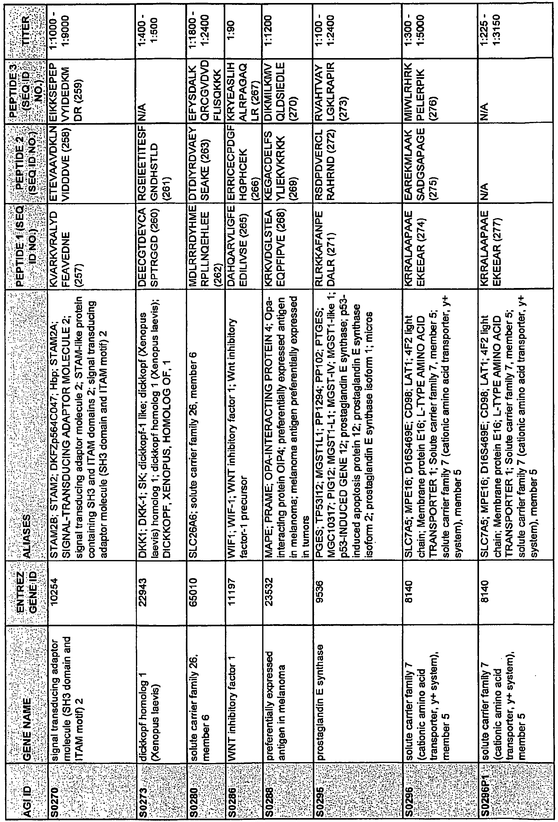



-
- <110> Ring, Brian
Ross, Douglas
Seitz, Robert
Beck, Rodney - <120> TLE3 as a Marker for Chemotherapy
- <130> 2003923-0027
- <150>
60/991,487
<151> 2007-11-30 - <160> 404
- <170> PatentIn version 3.5
- <210> 1
<211> 20
<212> PRT
<213> Homo sapiens - <400> 1

- <210> 2
<211> 15
<212> PRT
<213> Homo sapiens - <400> 2

- <210> 3
<211> 13
<212> PRT
<213> Homo sapiens - <400> 3

- <210> 4
<211> 18
<212> PRT
<213> Homo sapiens - <400> 4

- <210> 5
<211> 14
<212> PRT
<213> Homo sapiens - <400> 5

- <210> 6
<211> 15
<212> PRT
<213> Homo sapiens - <400> 6

- <210> 7
<211> 19
<212> PRT
<213> Homo sapiens - <400> 7

- <210> 8
<211> 15
<212> PRT
<213> Homo sapiens - <400> 8

- <210> 9
<211> 15
<212> PRT
<213> Homo sapiens - <400> 9

- <210> 10
<211> 19
<212> PRT
<213> Homo sapiens - <400> 10

- <210> 11
<211> 17
<212> PRT
<213> Homo sapiens - <400> 11

- <210> 12
<211> 15
<212> PRT
<213> Homo sapiens - <400> 12

- <210> 13
<211> 18
<212> PRT
<213> Homo sapiens - <400> 13

- <210> 14
<211> 17
<212> PRT
<213> Homo sapiens - <400> 14

- <210> 15
<211> 17
<212> PRT
<213> Homo sapiens - <400> 15

- <210> 16
<211> 20
<212> PRT
<213> Homo sapiens - <400> 16

- <210> 17
<211> 15
<212> PRT
<213> Homo sapiens - <400> 17

- <210> 18
<211> 17
<212> PRT
<213> Homo sapiens - <400> 18

- <210> 19
<211> 18
<212> PRT
<213> Homo sapiens - <400> 19

- <210> 20
<211> 16
<212> PRT
<213> Homo sapiens - <400> 20

- <210> 21
<211> 17
<212> PRT
<213> Homo sapiens - <400> 21

- <210> 22
<211> 19
<212> PRT
<213> Homo sapiens - <400> 22


- <210> 23
<211> 15
<212> PRT
<213> Homo sapiens - <400> 23

- <210> 24
<211> 15
<212> PRT
<213> Homo sapiens - <400> 24

- <210> 25
<211> 21
<212> PRT
<213> Homo sapiens - <400> 25

- <210> 26
<211> 19
<212> PRT
<213> Homo sapiens - <400> 26

- <210> 27
<211> 20
<212> PRT
<213> Homo sapiens - <400> 27

- <210> 28
<211> 19
<212> PRT
<213> Homo sapiens - <400> 28

- <210> 29
<211> 20
<212> PRT
<213> Homo sapiens - <400> 29

- <210> 30
<211> 18
<212> PRT
<213> Homo sapiens - <400> 30

- <210> 31
<211> 21
<212> PRT
<213> Homo sapiens - <400> 31

- <210> 32
<211> 15
<212> PRT
<213> Homo sapiens - <400> 32

- <210> 33
<211> 16
<212> PRT
<213> Homo sapiens - <400> 33

- <210> 34
<211> 15
<212> PRT
<213> Homo sapiens - <400> 34

- <210> 35
<211> 19
<212> PRT
<213> Homo sapiens - <400> 35

- <210> 36
<211> 15
<212> PRT
<213> Homo sapiens - <400> 36

- <210> 37
<211> 18
<212> PRT
<213> Homo sapiens - <400> 37

- <210> 38
<211> 17
<212> PRT
<213> Homo sapiens - <400> 38

- <210> 39
<211> 16
<212> PRT
<213> Homo sapiens - <400> 39

- <210> 40
<211> 13
<212> PRT
<213> Homo sapiens - <400> 40

- <210> 41
<211> 16
<212> PRT
<213> Homo sapiens - <400> 41

- <210> 42
<211> 16
<212> PRT
<213> Homo sapiens - <400> 42

- <210> 43
<211> 20
<212> PRT
<213> Homo sapiens - <400> 43

- <210> 44
<211> 15
<212> PRT
<213> Homo sapiens - <400> 44

- <210> 45
<211> 14
<212> PRT
<213> Homo sapiens - <400> 45

- <210> 46
<211> 16
<212> PRT
<213> Homo sapiens - <400> 46

- <210> 47
<211> 16
<212> PRT
<213> Homo sapiens - <400> 47

- <210> 48
<211> 18
<212> PRT
<213> Homo sapiens - <400> 48

- <210> 49
<211> 18
<212> PRT
<213> Homo sapiens - <400> 49

- <210> 50
<211> 15
<212> PRT
<213> Homo sapiens - <400> 50

- <210> 51
<211> 13
<212> PRT
<213> Homo sapiens - <400> 51

- <210> 52
<211> 19
<212> PRT
<213> Homo sapiens - <400> 52

- <210> 53
<211> 18
<212> PRT
<213> Homo sapiens - <400> 53

- <210> 54
<211> 15
<212> PRT
<213> Homo sapiens - <400> 54

- <210> 55
<211> 20
<212> PRT
<213> Homo sapiens - <400> 55


- <210> 56
<211> 17
<212> PRT
<213> Homo sapiens - <400> 56

- <210> 57
<211> 13
<212> PRT
<213> Homo sapiens - <400> 57

- <210> 58
<211> 18
<212> PRT
<213> Homo sapiens - <400> 58

- <210> 59
<211> 14
<212> PRT
<213> Homo sapiens - <400> 59

- <210> 60
<211> 15
<212> PRT
<213> Homo sapiens - <400> 60

- <210> 61
<211> 15
<212> PRT
<213> Homo sapiens - <400> 61

- <210> 62
<211> 14
<212> PRT
<213> Homo sapiens - <400> 62

- <210> 63
<211> 18
<212> PRT
<213> Homo sapiens - <400> 63

- <210> 64
<211> 13
<212> PRT
<213> Homo sapiens - <400> 64

- <210> 65
<211> 10
<212> PRT
<213> Homo sapiens - <400> 65

- <210> 66
<211> 17
<212> PRT
<213> Homo sapiens - <400> 66

- <210> 67
<211> 16
<212> PRT
<213> Homo sapiens - <400> 67

- <210> 68
<211> 18
<212> PRT
<213> Homo sapiens - <400> 68

- <210> 69
<211> 16
<212> PRT
<213> Homo sapiens - <400> 69

- <210> 70
<211> 12
<212> PRT
<213> Homo sapiens - <400> 70

- <210> 71
<211> 18
<212> PRT
<213> Homo sapiens - <400> 71

- <210> 72
<211> 19
<212> PRT
<213> Homo sapiens - <400> 72

- <210> 73
<211> 21
<212> PRT
<213> Homo sapiens - <400> 73

- <210> 74
<211> 16
<212> PRT
<213> Homo sapiens - <400> 74

- <210> 75
<211> 21
<212> PRT
<213> Homo sapiens - <400> 75

- <210> 76
<211> 20
<212> PRT
<213> Homo sapiens - <400> 76

- <210> 77
<211> 20
<212> PRT
<213> Homo sapiens - <400> 77

- <210> 78
<211> 20
<212> PRT
<213> Homo sapiens - <400> 78


- <210> 79
<211> 18
<212> PRT
<213> Homo sapiens - <400> 79

- <210> 80
<211> 15
<212> PRT
<213> Homo sapiens - <400> 80

- <210> 81
<211> 16
<212> PRT
<213> Homo sapiens - <400> 81

- <210> 82
<211> 20
<212> PRT
<213> Homo sapiens - <400> 82

- <210> 83
<211> 17
<212> PRT
<213> Homo sapiens - <400> 83

- <210> 84
<211> 18
<212> PRT
<213> Homo sapiens - <400> 84

- <210> 85
<211> 17
<212> PRT
<213> Homo sapiens - <400> 85

- <210> 86
<211> 17
<212> PRT
<213> Homo sapiens - <400> 86

- <210> 87
<211> 18
<212> PRT
<213> Homo sapiens - <400> 87

- <210> 88
<211> 20
<212> PRT
<213> Homo sapiens - <400> 88

- <210> 89
<211> 21
<212> PRT
<213> Homo sapiens - <400> 89

- <210> 90
<211> 20
<212> PRT
<213> Homo sapiens - <400> 90

- <210> 91
<211> 21
<212> PRT
<213> Homo sapiens - <400> 91


- <210> 92
<211> 19
<212> PRT
<213> Homo sapiens - <400> 92

- <210> 93
<211> 18
<212> PRT
<213> Homo sapiens - <400> 93

- <210> 94
<211> 22
<212> PRT
<213> Homo sapiens - <400> 94

- <210> 95
<211> 17
<212> PRT
<213> Homo sapiens - <400> 95


- <210> 96
<211> 17
<212> PRT
<213> Homo sapiens - <400> 96

- <210> 97
<211> 15
<212> PRT
<213> Homo sapiens - <400> 97

- <210> 98
<211> 18
<212> PRT
<213> Homo sapiens - <400> 98

- <210> 99
<211> 15
<212> PRT
<213> Homo sapiens - <400> 99

- <210> 100
<211> 20
<212> PRT
<213> Homo sapiens - <400> 100

- <210> 101
<211> 15
<212> PRT
<213> Homo sapiens - <400> 101

- <210> 102
<211> 15
<212> PRT
<213> Homo sapiens - <400> 102

- <210> 103
<211> 15
<212> PRT
<213> Homo sapiens - <400> 103

- <210> 104
<211> 16
<212> PRT
<213> Homo sapiens - <400> 104

- <210> 105
<211> 17
<212> PRT
<213> Homo sapiens - <400> 105

- <210> 106
<211> 15
<212> PRT
<213> Homo sapiens - <400> 106

- <210> 107
<211> 20
<212> PRT
<213> Homo sapiens - <400> 107

- <210> 108
<211> 21
<212> PRT
<213> Homo sapiens - <400> 108

- <210> 109
<211> 21
<212> PRT
<213> Homo sapiens - <400> 109


- <210> 110
<211> 20
<212> PRT
<213> Homo sapiens - <400> 110

- <210> 111
<211> 21
<212> PRT
<213> Homo sapiens - <400> 111

- <210> 112
<211> 16
<212> PRT
<213> Homo sapiens - <400> 112

- <210> 113
<211> 22
<212> PRT
<213> Homo sapiens - <400> 113

- <210> 114
<211> 22
<212> PRT
<213> Homo sapiens - <400> 114

- <210> 115
<211> 20
<212> PRT
<213> Homo sapiens - <400> 115

- <210> 116
<211> 21
<212> PRT
<213> Homo sapiens - <400> 116

- <210> 117
<211> 20
<212> PRT
<213> Homo sapiens - <400> 117

- <210> 118
<211> 20
<212> PRT
<213> Homo sapiens - <400> 118

- <210> 119
<211> 16
<212> PRT
<213> Homo sapiens - <400> 119

- <210> 120
<211> 18
<212> PRT
<213> Homo sapiens - <400> 120

- <210> 121
<211> 18
<212> PRT
<213> Homo sapiens - <400> 121

- <210> 122
<211> 20
<212> PRT
<213> Homo sapiens - <400> 122

- <210> 123
<211> 19
<212> PRT
<213> Homo sapiens - <400> 123

- <210> 124
<211> 16
<212> PRT
<213> Homo sapiens - <400> 124

- <210> 125
<211> 13
<212> PRT
<213> Homo sapiens - <400> 125

- <210> 126
<211> 15
<212> PRT
<213> Homo sapiens - <400> 126

- <210> 127
<211> 15
<212> PRT
<213> Homo sapiens - <400> 127

- <210> 128
<211> 15
<212> PRT
<213> Homo sapiens - <400> 128

- <210> 129
<211> 15
<212> PRT
<213> Homo sapiens - <400> 129

- <210> 130
<211> 19
<212> PRT
<213> Homo sapiens - <400> 130

- <210> 131
<211> 15
<212> PRT
<213> Homo sapiens - <400> 131

- <210> 132
<211> 19
<212> PRT
<213> Homo sapiens - <400> 132

- <210> 133
<211> 20
<212> PRT
<213> Homo sapiens - <400> 133

- <210> 134
<211> 17
<212> PRT
<213> Homo sapiens - <400> 134

- <210> 135
<211> 16
<212> PRT
<213> Homo sapiens - <400> 135

- <210> 136
<211> 14
<212> PRT
<213> Homo sapiens - <400> 136

- <210> 137
<211> 20
<212> PRT
<213> Homo sapiens - <400> 137

- <210> 138
<211> 18
<212> PRT
<213> Homo sapiens - <400> 138

- <210> 139
<211> 16
<212> PRT
<213> Homo sapiens - <400> 139

- <210> 140
<211> 20
<212> PRT
<213> Homo sapiens - <400> 140

- <210> 141
<211> 16
<212> PRT
<213> Homo sapiens - <400> 141

- <210> 142
<211> 13
<212> PRT
<213> Homo sapiens - <400> 142

- <210> 143
<211> 19
<212> PRT
<213> Homo sapiens - <400> 143

- <210> 144
<211> 18
<212> PRT
<213> Homo sapiens - <400> 144

- <210> 145
<211> 19
<212> PRT
<213> Homo sapiens - <400> 145

- <210> 146
<211> 20
<212> PRT
<213> Homo sapiens - <400> 146

- <210> 147
<211> 14
<212> PRT
<213> Homo sapiens - <400> 147

- <210> 148
<211> 11
<212> PRT
<213> Homo sapiens - <400> 148

- <210> 149
<211> 12
<212> PRT
<213> Homo sapiens - <400> 149

- <210> 150
<211> 18
<212> PRT
<213> Homo sapiens - <400> 150

- <210> 151
<211> 19
<212> PRT
<213> Homo sapiens - <400> 151

- <210> 152
<211> 22
<212> PRT
<213> Homo sapiens - <400> 152

- <210> 153
<211> 19
<212> PRT
<213> Homo sapiens - <400> 153

- <210> 154
<211> 17
<212> PRT
<213> Homo sapiens - <400> 154

- <210> 155
<211> 20
<212> PRT
<213> Homo sapiens - <400> 155

- <210> 156
<211> 18
<212> PRT
<213> Homo sapiens - <400> 156

- <210> 157
<211> 18
<212> PRT
<213> Homo sapiens - <400> 157

- <210> 158
<211> 21
<212> PRT
<213> Homo sapiens - <400> 158

- <210> 159
<211> 20
<212> PRT
<213> Homo sapiens - <400> 159

- <210> 160
<211> 21
<212> PRT
<213> Homo sapiens - <400> 160

- <210> 161
<211> 18
<212> PRT
<213> Homo sapiens - <400> 161

- <210> 162
<211> 16
<212> PRT
<213> Homo sapiens - <400> 162

- <210> 163
<211> 20
<212> PRT
<213> Homo sapiens - <400> 163

- <210> 164
<211> 16
<212> PRT
<213> Homo sapiens - <400> 164

- <210> 165
<211> 18
<212> PRT
<213> Homo sapiens - <400> 165

- <210> 166
<211> 16
<212> PRT
<213> Homo sapiens - <400> 166

- <210> 167
<211> 19
<212> PRT
<213> Homo sapiens - <400> 167

- <210> 168
<211> 18
<212> PRT
<213> Homo sapiens - <400> 168

- <210> 169
<211> 20
<212> PRT
<213> Homo sapiens - <400> 169

- <210> 170
<211> 16
<212> PRT
<213> Homo sapiens - <400> 170

- <210> 171
<211> 19
<212> PRT
<213> Homo sapiens - <400> 171

- <210> 172
<211> 18
<212> PRT
<213> Homo sapiens - <400> 172

- <210> 173
<211> 19
<212> PRT
<213> Homo sapiens - <400> 173

- <210> 174
<211> 18
<212> PRT
<213> Homo sapiens - <400> 174

- <210> 175
<211> 20
<212> PRT
<213> Homo sapiens - <400> 175

- <210> 176
<211> 17
<212> PRT
<213> Homo sapiens - <400> 176

- <210> 177
<211> 22
<212> PRT
<213> Homo sapiens - <400> 177

- <210> 178
<211> 19
<212> PRT
<213> Homo sapiens - <400> 178

- <210> 179
<211> 20
<212> PRT
<213> Homo sapiens - <400> 179

- <210> 180
<211> 19
<212> PRT
<213> Homo sapiens - <400> 180

- <210> 181
<211> 17
<212> PRT
<213> Homo sapiens - <400> 181

- <210> 182
<211> 19
<212> PRT
<213> Homo sapiens - <400> 182

- <210> 183
<211> 17
<212> PRT
<213> Homo sapiens - <400> 183

- <210> 184
<211> 18
<212> PRT
<213> Homo sapiens - <400> 184

- <210> 185
<211> 20
<212> PRT
<213> Homo sapiens - <400> 185

- <210> 186
<211> 20
<212> PRT
<213> Homo sapiens - <400> 186

- <210> 187
<211> 19
<212> PRT
<213> Homo sapiens - <400> 187

- <210> 188
<211> 18
<212> PRT
<213> Homo sapiens - <400> 188

- <210> 189
<211> 16
<212> PRT
<213> Homo sapiens - <400> 189

- <210> 190
<211> 20
<212> PRT
<213> Homo sapiens - <400> 190

- <210> 191
<211> 15
<212> PRT
<213> Homo sapiens - <400> 191

- <210> 192
<211> 20
<212> PRT
<213> Homo sapiens - <400> 192

- <210> 193
<211> 20
<212> PRT
<213> Homo sapiens - <400> 193

- <210> 194
<211> 19
<212> PRT
<213> Homo sapiens - <400> 194

- <210> 195
<211> 21
<212> PRT
<213> Homo sapiens - <400> 195

- <210> 196
<211> 16
<212> PRT
<213> Homo sapiens - <400> 196

- <210> 197
<211> 15
<212> PRT
<213> Homo sapiens - <400> 197

- <210> 198
<211> 14
<212> PRT
<213> Homo sapiens - <400> 198

- <210> 199
<211> 19
<212> PRT
<213> Homo sapiens - <400> 199

- <210> 200
<211> 17
<212> PRT
<213> Homo sapiens - <400> 200

- <210> 201
<211> 15
<212> PRT
<213> Homo sapiens - <400> 201

- <210> 202
<211> 14
<212> PRT
<213> Homo sapiens - <400> 202

- <210> 203
<211> 20
<212> PRT
<213> Homo sapiens - <400> 203

- <210> 204
<211> 14
<212> PRT
<213> Homo sapiens - <400> 204

- <210> 205
<211> 13
<212> PRT
<213> Homo sapiens - <400> 205

- <210> 206
<211> 15
<212> PRT
<213> Homo sapiens - <400> 206

- <210> 207
<211> 18
<212> PRT
<213> Homo sapiens - <400> 207

- <210> 208
<211> 15
<212> PRT
<213> Homo sapiens - <400> 208

- <210> 209
<211> 18
<212> PRT
<213> Homo sapiens - <400> 209

- <210> 210
<211> 17
<212> PRT
<213> Homo sapiens - <400> 210

- <210> 211
<211> 22
<212> PRT
<213> Homo sapiens - <400> 211


- <210> 212
<211> 17
<212> PRT
<213> Homo sapiens - <400> 212

- <210> 213
<211> 17
<212> PRT
<213> Homo sapiens - <400> 213

- <210> 214
<211> 16
<212> PRT
<213> Homo sapiens - <400> 214

- <210> 215
<211> 18
<212> PRT
<213> Homo sapiens - <400> 215

- <210> 216
<211> 16
<212> PRT
<213> Homo sapiens - <400> 216

- <210> 217
<211> 21
<212> PRT
<213> Homo sapiens - <400> 217

- <210> 218
<211> 16
<212> PRT
<213> Homo sapiens - <400> 218

- <210> 219
<211> 15
<212> PRT
<213> Homo sapiens - <400> 219

- <210> 220
<211> 16
<212> PRT
<213> Homo sapiens - <400> 220

- <210> 221
<211> 17
<212> PRT
<213> Homo sapiens - <400> 221

- <210> 222
<211> 15
<212> PRT
<213> Homo sapiens - <400> 222

- <210> 223
<211> 20
<212> PRT
<213> Homo sapiens - <400> 223

- <210> 224
<211> 17
<212> PRT
<213> Homo sapiens - <400> 224

- <210> 225
<211> 21
<212> PRT
<213> Homo sapiens - <400> 225


- <210> 226
<211> 18
<212> PRT
<213> Homo sapiens - <400> 226

- <210> 227
<211> 22
<212> PRT
<213> Homo sapiens - <400> 227

- <210> 228
<211> 19
<212> PRT
<213> Homo sapiens - <400> 228

- <210> 229
<211> 23
<212> PRT
<213> Homo sapiens - <400> 229


- <210> 230
<211> 21
<212> PRT
<213> Homo sapiens - <400> 230

- <210> 231
<211> 18
<212> PRT
<213> Homo sapiens - <400> 231

- <210> 232
<211> 18
<212> PRT
<213> Homo sapiens - <400> 232

- <210> 233
<211> 20
<212> PRT
<213> Homo sapiens - <400> 233


- <210> 234
<211> 21
<212> PRT
<213> Homo sapiens - <400> 234

- <210> 235
<211> 20
<212> PRT
<213> Homo sapiens - <400> 235

- <210> 236
<211> 20
<212> PRT
<213> Homo sapiens - <400> 236

- <210> 237
<211> 20
<212> PRT
<213> Homo sapiens - <400> 237


- <210> 238
<211> 19
<212> PRT
<213> Homo sapiens - <400> 238

- <210> 239
<211> 18
<212> PRT
<213> Homo sapiens - <400> 239

- <210> 240
<211> 19
<212> PRT
<213> Homo sapiens - <400> 240

- <210> 241
<211> 19
<212> PRT
<213> Homo sapiens - <400> 241

- <210> 242
<211> 21
<212> PRT
<213> Homo sapiens - <400> 242

- <210> 243
<211> 15
<212> PRT
<213> Homo sapiens - <400> 243

- <210> 244
<211> 20
<212> PRT
<213> Homo sapiens - <400> 244

- <210> 245
<211> 14
<212> PRT
<213> Homo sapiens - <400> 245

- <210> 246
<211> 19
<212> PRT
<213> Homo sapiens - <400> 246

- <210> 247
<211> 15
<212> PRT
<213> Homo sapiens - <400> 247

- <210> 248
<211> 19
<212> PRT
<213> Homo sapiens - <400> 248

- <210> 249
<211> 17
<212> PRT
<213> Homo sapiens - <400> 249

- <210> 250
<211> 20
<212> PRT
<213> Homo sapiens - <400> 250


- <210> 251
<211> 23
<212> PRT
<213> Homo sapiens - <400> 251

- <210> 252
<211> 12
<212> PRT
<213> Homo sapiens - <400> 252

- <210> 253
<211> 16
<212> PRT
<213> Homo sapiens - <400> 253

- <210> 254
<211> 21
<212> PRT
<213> Homo sapiens - <400> 254

- <210> 255
<211> 19
<212> PRT
<213> Homo sapiens - <400> 255
Arg Pro Lys

- <210> 256
<211> 18
<212> PRT
<213> Homo sapiens - <400> 256

- <210> 257
<211> 19
<212> PRT
<213> Homo sapiens - <400> 257

- <210> 258
<211> 18
<212> PRT
<213> Homo sapiens - <400> 258

- <210> 259
<211> 19
<212> PRT
<213> Homo sapiens - <400> 259

- <210> 260
<211> 18
<212> PRT
<213> Homo sapiens - <400> 260

- <210> 261
<211> 20
<212> PRT
<213> Homo sapiens - <400> 261

- <210> 262
<211> 22
<212> PRT
<213> Homo sapiens - <400> 262

- <210> 263
<211> 16
<212> PRT
<213> Homo sapiens - <400> 263

- <210> 264
<211> 24
<212> PRT
<213> Homo sapiens - <400> 264

- <210> 265
<211> 20
<212> PRT
<213> Homo sapiens - <400> 265

- <210> 266
<211> 18
<212> PRT
<213> Homo sapiens - <400> 266

- <210> 267
<211> 19
<212> PRT
<213> Homo sapiens - <400> 267

- <210> 268
<211> 19
<212> PRT
<213> Homo sapiens - <400> 268

- <210> 269
<211> 20
<212> PRT
<213> Homo sapiens - <400> 269

- <210> 270
<211> 18
<212> PRT
<213> Homo sapiens - <400> 270

- <210> 271
<211> 15
<212> PRT
<213> Homo sapiens - <400> 271

- <210> 272
<211> 16
<212> PRT
<213> Homo sapiens - <400> 272

- <210> 273
<211> 17
<212> PRT
<213> Homo sapiens - <400> 273

- <210> 274
<211> 13
<212> PRT
<213> Homo sapiens - <400> 274

- <210> 275
<211> 20
<212> PRT
<213> Homo sapiens - <400> 275

- <210> 276
<211> 16
<212> PRT
<213> Homo sapiens - <400> 276

- <210> 277
<211> 13
<212> PRT
<213> Homo sapiens - <400> 277

- <210> 278
<211> 14
<212> PRT
<213> Homo sapiens - <400> 278

- <210> 279
<211> 22
<212> PRT
<213> Homo sapiens - <400> 279

- <210> 280
<211> 12
<212> PRT
<213> Homo sapiens - <400> 280

- <210> 281
<211> 21
<212> PRT
<213> Homo sapiens - <400> 281

- <210> 282
<211> 18
<212> PRT
<213> Homo sapiens - <400> 282

- <210> 283
<211> 21
<212> PRT
<213> Homo sapiens - <400> 283

- <210> 284
<211> 17
<212> PRT
<213> Homo sapiens - <400> 284

- <210> 285
<211> 19
<212> PRT
<213> Homo sapiens - <400> 285

- <210> 286
<211> 21
<212> PRT
<213> Homo sapiens - <400> 286

- <210> 287
<211> 17
<212> PRT
<213> Homo sapiens - <400> 287

- <210> 288
<211> 20
<212> PRT
<213> Homo sapiens - <400> 288

- <210> 289
<211> 21
<212> PRT
<213> Homo sapiens - <400> 289

- <210> 290
<211> 23
<212> PRT
<213> Homo sapiens - <400> 290

- <210> 291
<211> 20
<212> PRT
<213> Homo sapiens - <400> 291

- <210> 292
<211> 17
<212> PRT
<213> Homo sapiens - <400> 292

- <210> 293
<211> 22
<212> PRT
<213> Homo sapiens - <400> 293

- <210> 294
<211> 16
<212> PRT
<213> Homo sapiens - <400> 294

- <210> 295
<211> 22
<212> PRT
<213> Homo sapiens - <400> 295

- <210> 296
<211> 20
<212> PRT
<213> Homo sapiens - <400> 296

- <210> 297
<211> 17
<212> PRT
<213> Homo sapiens - <400> 297

- <210> 298
<211> 20
<212> PRT
<213> Homo sapiens - <400> 298

- <210> 299
<211> 20
<212> PRT
<213> Homo sapiens - <400> 299

- <210> 300
<211> 21
<212> PRT
<213> Homo sapiens - <400> 300

- <210> 301
<211> 18
<212> PRT
<213> Homo sapiens - <400> 301

- <210> 302
<211> 18
<212> PRT
<213> Homo sapiens - <400> 302

- <210> 303
<211> 17
<212> PRT
<213> Homo sapiens - <400> 303

- <210> 304
<211> 19
<212> PRT
<213> Homo sapiens - <400> 304

- <210> 305
<211> 24
<212> PRT
<213> Homo sapiens - <400> 305

- <210> 306
<211> 23
<212> PRT
<213> Homo sapiens - <400> 306


- <210> 307
<211> 16
<212> PRT
<213> Homo sapiens - <400> 307

- <210> 308
<211> 12
<212> PRT
<213> Homo sapiens - <400> 308

- <210> 309
<211> 20
<212> PRT
<213> Homo sapiens - <400> 309

- <210> 310
<211> 20
<212> PRT
<213> Homo sapiens - <400> 310

- <210> 311
<211> 20
<212> PRT
<213> Homo sapiens - <400> 311

- <210> 312
<211> 20
<212> PRT
<213> Homo sapiens - <400> 312

- <210> 313
<211> 20
<212> PRT
<213> Homo sapiens - <400> 313

- <210> 314
<211> 20
<212> PRT
<213> Homo sapiens - <400> 314

- <210> 315
<211> 17
<212> PRT
<213> Homo sapiens - <400> 315

- <210> 316
<211> 17
<212> PRT
<213> Homo sapiens - <400> 316

- <210> 317
<211> 17
<212> PRT
<213> Homo sapiens - <400> 317

- <210> 318
<211> 16
<212> PRT
<213> Homo sapiens - <400> 318

- <210> 319
<211> 20
<212> PRT
<213> Homo sapiens - <400> 319


- <210> 320
<211> 20
<212> PRT
<213> Homo sapiens - <400> 320

- <210> 321
<211> 14
<212> PRT
<213> Homo sapiens - <400> 321

- <210> 322
<211> 20
<212> PRT
<213> Homo sapiens - <400> 322

- <210> 323
<211> 20
<212> PRT
<213> Homo sapiens - <400> 323

- <210> 324
<211> 20
<212> PRT
<213> Homo sapiens - <400> 324

- <210> 325
<211> 20
<212> PRT
<213> Homo sapiens - <400> 325

- <210> 326
<211> 20
<212> PRT
<213> Homo sapiens - <400> 326

- <210> 327
<211> 20
<212> PRT
<213> Homo sapiens - <400> 327

- <210> 328
<211> 20
<212> PRT
<213> Homo sapiens - <400> 328

- <210> 329
<211> 20
<212> PRT
<213> Homo sapiens - <400> 329

- <210> 330
<211> 18
<212> PRT
<213> Homo sapiens - <400> 330

- <210> 331
<211> 16
<212> PRT
<213> Homo sapiens - <400> 331

- <210> 332
<211> 20
<212> PRT
<213> Homo sapiens - <400> 332

- <210> 333
<211> 17
<212> PRT
<213> Homo sapiens - <400> 333

- <210> 334
<211> 20
<212> PRT
<213> Homo sapiens - <400> 334

- <210> 335
<211> 20
<212> PRT
<213> Homo sapiens - <400> 335

- <210> 336
<211> 20
<212> PRT
<213> Homo sapiens - <400> 336

- <210> 337
<211> 20
<212> PRT
<213> Homo sapiens - <400> 337

- <210> 338
<211> 19
<212> PRT
<213> Homo sapiens - <400> 338

- <210> 339
<211> 19
<212> PRT
<213> Homo sapiens - <400> 339

- <210> 340
<211> 17
<212> PRT
<213> Homo sapiens - <400> 340

- <210> 341
<211> 15
<212> PRT
<213> Homo sapiens - <400> 341

- <210> 342
<211> 19
<212> PRT
<213> Homo sapiens - <400> 342

- <210> 343
<211> 19
<212> PRT
<213> Homo sapiens - <400> 343

- <210> 344
<211> 19
<212> PRT
<213> Homo sapiens - <400> 344


- <210> 345
<211> 19
<212> PRT
<213> Homo sapiens - <400> 345

- <210> 346
<211> 15
<212> PRT
<213> Homo sapiens - <400> 346

- <210> 347
<211> 19
<212> PRT
<213> Homo sapiens - <400> 347

- <210> 348
<211> 18
<212> PRT
<213> Homo sapiens - <400> 348

- <210> 349
<211> 17
<212> PRT
<213> Homo sapiens - <400> 349

- <210> 350
<211> 19
<212> PRT
<213> Homo sapiens - <400> 350

- <210> 351
<211> 19
<212> PRT
<213> Homo sapiens - <400> 351

- <210> 352
<211> 18
<212> PRT
<213> Homo sapiens - <400> 352

- <210> 353
<211> 18
<212> PRT
<213> Homo sapiens - <400> 353

- <210> 354
<211> 16
<212> PRT
<213> Homo sapiens - <400> 354

- <210> 355
<211> 17
<212> PRT
<213> Homo sapiens - <400> 355

- <210> 356
<211> 19
<212> PRT
<213> Homo sapiens - <400> 356

- <210> 357
<211> 16
<212> PRT
<213> Homo sapiens - <400> 357

- <210> 358
<211> 15
<212> PRT
<213> Homo sapiens - <400> 358

- <210> 359
<211> 19
<212> PRT
<213> Homo sapiens - <400> 359

- <210> 360
<211> 17
<212> PRT
<213> Homo sapiens - <400> 360

- <210> 361
<211> 19
<212> PRT
<213> Homo sapiens - <400> 361

- <210> 362
<211> 19
<212> PRT
<213> Homo sapiens - <400> 362

- <210> 363
<211> 17
<212> PRT
<213> Homo sapiens - <400> 363

- <210> 364
<211> 19
<212> PRT
<213> Homo sapiens - <400> 364

- <210> 365
<211> 19
<212> PRT
<213> Homo sapiens - <400> 365

- <210> 366
<211> 19
<212> PRT
<213> Homo sapiens - <400> 366

- <210> 367
<211> 15
<212> PRT
<213> Homo sapiens - <400> 367

- <210> 368
<211> 14
<212> PRT
<213> Homo sapiens - <400> 368

- <210> 369
<211> 10
<212> PRT
<213> Homo sapiens - <400> 369

- <210> 370
<211> 19
<212> PRT
<213> Homo sapiens - <400> 370

- <210> 371
<211> 19
<212> PRT
<213> Homo sapiens - <400> 371

- <210> 372
<211> 19
<212> PRT
<213> Homo sapiens - <400> 372

- <210> 373
<211> 19
<212> PRT
<213> Homo sapiens - <400> 373

- <210> 374
<211> 14
<212> PRT
<213> Homo sapiens - <400> 374

- <210> 375
<211> 16
<212> PRT
<213> Homo sapiens - <400> 375

- <210> 376
<211> 19
<212> PRT
<213> Homo sapiens - <400> 376

- <210> 377
<211> 17
<212> PRT
<213> Homo sapiens - <400> 377

- <210> 378
<211> 18
<212> PRT
<213> Homo sapiens - <400> 378

- <210> 379
<211> 19
<212> PRT
<213> Homo sapiens - <400> 379


- <210> 380
<211> 17
<212> PRT
<213> Homo sapiens - <400> 380

- <210> 381
<211> 17
<212> PRT
<213> Homo sapiens - <400> 381

- <210> 382
<211> 18
<212> PRT
<213> Homo sapiens - <400> 382

- <210> 383
<211> 16
<212> PRT
<213> Homo sapiens - <400> 383

- <210> 384
<211> 16
<212> PRT
<213> Homo sapiens - <400> 384

- <210> 385
<211> 15
<212> PRT
<213> Homo sapiens - <400> 385

- <210> 386
<211> 15
<212> PRT
<213> Homo sapiens - <400> 386

- <210> 387
<211> 18
<212> PRT
<213> Homo sapiens - <400> 387

- <210> 388
<211> 17
<212> PRT
<213> Homo sapiens - <400> 388

- <210> 389
<211> 14
<212> PRT
<213> Homo sapiens - <400> 389

- <210> 390
<211> 14
<212> PRT
<213> Homo sapiens - <400> 390

- <210> 391
<211> 17
<212> PRT
<213> Homo sapiens - <400> 391

- <210> 392
<211> 17
<212> PRT
<213> Homo sapiens - <400> 392

- <210> 393
<211> 16
<212> PRT
<213> Homo sapiens - <400> 393

- <210> 394
<211> 17
<212> PRT
<213> Homo sapiens - <400> 394

- <210> 395
<211> 14
<212> PRT
<213> Homo sapiens - <400> 395

- <210> 396
<211> 16
<212> PRT
<213> Homo sapiens - <400> 396

- <210> 397
<211> 19
<212> PRT
<213> Homo sapiens - <400> 397

- <210> 398
<211> 17
<212> PRT
<213> Homo sapiens - <400> 398

- <210> 399
<211> 16
<212> PRT
<213> Homo sapiens - <400> 399

- <210> 400
<211> 16
<212> PRT
<213> Homo sapiens - <400> 400

- <210> 401
<211> 15
<212> PRT
<213> Homo sapiens - <400> 401

- <210> 402
<211> 20
<212> PRT
<213> Homo sapiens - <400> 402

- <210> 403
<211> 16
<212> PRT
<213> Homo sapiens - <400> 403

- <210> 404
<211> 17
<212> PRT
<213> Homo sapiens - <400> 404

Claims (14)
- A method for predicting the likelihood that a patient's cancer will respond to chemotherapy comprising treatment with a taxane or taxane derivative comprising steps of:providing a cancer sample that has been removed from a breast, ovarian or lung cancer patient;determining whether TLE3 is expressed in the cancer sample; andpredicting the likelihood that the patient's cancer will respond to chemotherapy comprising treatment with a taxane or taxane derivative based upon the presence or absence of TLE3 expression in the cancer sample, wherein the presence of TLE3 expression is predictive that the cancer will respond and/or the absence of TLE3 expression is predictive that the cancer will not respond.
- A method for selecting a chemotherapy for a cancer patient comprising steps of:providing a cancer sample that has been removed from a breast, ovarian or lung cancer patient;determining whether TLE3 is expressed in the cancer sample; andselecting a taxane or taxane derivative for the cancer patient based upon the presence of TLE3 expression in the cancer sample or deciding not to administer treatment with a taxane or taxane derivative to the cancer patient based on the absence of TLE3 expression in the sample.
- The method of claim 1 or claim 2, wherein the step of determining comprises steps of:providing a negative control sample;detecting a level of TLE3 expression in the negative control sample;detecting a level of TLE3 expression in the cancer sample; andcomparing the level of TLE3 expression in the cancer sample with the level of TLE3 expression in the negative control sample.
- The method of claim 1 or claim 2, wherein the step of determining comprises steps of:providing a positive control sample;detecting a level of TLE3 expression in the positive control sample;detecting a level of TLE3 expression in the cancer sample; andcomparing the level of TLE3 expression in the cancer sample with the level of TLE3 expression in the positive control sample.
- The method of claim 1 or claim 2, wherein the step of determining comprises contacting the cancer sample with an interaction partner that binds a TLE3 polypeptide.
- The method of claim 5 wherein the interaction partner is an antibody.
- The method of claim 1 or claim 2, wherein the step of determining comprises contacting the cancer sample with one or more primers that hybridize with a TLE3 polynucleotide.
- The method of claim 1 or claim 2, wherein the taxane is paclitaxel, or docetaxel.
- The method of claim 1 or claim 2, wherein the cancer patient has breast cancer.
- The method of claim 1 or claim 2, wherein the cancer patient has lung cancer.
- The method of claim 1 or claim 2, wherein the cancer patient has ovarian cancer.
- The method of claim 1 or claim 2, wherein the taxane or taxane derivative is administered in a neoadjuvant setting or in an adjuvant setting.
- The method of claim 1 or claim 2, wherein the cancer patient has a stage II+ cancer, or a stage IIb+ cancer, or a stage III+ cancer.
- A taxane for use in a method of treating breast, ovarian or lung cancer in a patient whose cancer has been determined to express TLE3.
Applications Claiming Priority (2)
| Application Number | Priority Date | Filing Date | Title |
|---|---|---|---|
| US99148707P | 2007-11-30 | 2007-11-30 | |
| PCT/US2008/084685 WO2009073478A2 (en) | 2007-11-30 | 2008-11-25 | Tle3 as a marker for chemotherapy |
Publications (3)
| Publication Number | Publication Date |
|---|---|
| EP2227693A2 EP2227693A2 (en) | 2010-09-15 |
| EP2227693A4 EP2227693A4 (en) | 2011-05-04 |
| EP2227693B1 true EP2227693B1 (en) | 2015-08-19 |
Family
ID=40718444
Family Applications (1)
| Application Number | Title | Priority Date | Filing Date |
|---|---|---|---|
| EP08855868.9A Not-in-force EP2227693B1 (en) | 2007-11-30 | 2008-11-25 | Tle3 as a marker for chemotherapy |
Country Status (7)
| Country | Link |
|---|---|
| US (5) | US7816084B2 (en) |
| EP (1) | EP2227693B1 (en) |
| JP (1) | JP5745273B2 (en) |
| CN (1) | CN101932938B (en) |
| CA (1) | CA2706881A1 (en) |
| GB (1) | GB2460769C (en) |
| WO (1) | WO2009073478A2 (en) |
Families Citing this family (22)
| Publication number | Priority date | Publication date | Assignee | Title |
|---|---|---|---|---|
| WO2007137187A2 (en) | 2006-05-18 | 2007-11-29 | Molecular Profiling Institute, Inc. | System and method for determining individualized medical intervention for a disease state |
| US8768629B2 (en) | 2009-02-11 | 2014-07-01 | Caris Mpi, Inc. | Molecular profiling of tumors |
| CN101932938B (en) * | 2007-11-30 | 2014-08-27 | 克雷特诊疗服务公司 | Tle3 as a marker for chemotherapy |
| CN106153918A (en) * | 2008-10-14 | 2016-11-23 | 卡里斯Mpi公司 | Describe tumor type biological marker pattern and the gene target of feature set and the protein targets of gene expression |
| CA2743211A1 (en) | 2008-11-12 | 2010-05-20 | Caris Life Sciences Luxembourg Holdings, S.A.R.L. | Methods and systems of using exosomes for determining phenotypes |
| US20110166030A1 (en) * | 2009-09-30 | 2011-07-07 | Yixin Wang | Prediction of response to docetaxel therapy based on the presence of TMPRSSG2:ERG fusion in circulating tumor cells |
| US9128101B2 (en) | 2010-03-01 | 2015-09-08 | Caris Life Sciences Switzerland Holdings Gmbh | Biomarkers for theranostics |
| WO2011127219A1 (en) | 2010-04-06 | 2011-10-13 | Caris Life Sciences Luxembourg Holdings | Circulating biomarkers for disease |
| EP2698634B1 (en) * | 2011-04-15 | 2017-11-08 | J-Pharma Co., Ltd. | Biomarker for breast cancer |
| EP2861252A4 (en) * | 2012-06-13 | 2016-04-13 | Univ Virginia Patent Found | COMPOSITIONS AND METHODS OF TREATING CANCER |
| CN102749448B (en) * | 2012-07-27 | 2014-07-02 | 复旦大学附属中山医院 | Kit for evaluating chemotherapy effect of lung adenocarcinoma |
| ES2833543T3 (en) | 2014-10-24 | 2021-06-15 | Koninklijke Philips Nv | Evaluation of the activity of the TGF cell signaling pathway using mathematical models of target gene expression |
| ES2857953T3 (en) | 2014-10-24 | 2021-09-29 | Koninklijke Philips Nv | Medical prognosis and prediction of response to treatment using the activities of multiple cell signaling pathways |
| JP7065610B6 (en) | 2014-10-24 | 2022-06-06 | コーニンクレッカ フィリップス エヌ ヴェ | Medical prognosis and prediction of therapeutic response using multiple cellular signaling pathway activities |
| AU2016209478B2 (en) | 2015-01-20 | 2019-03-07 | Nantomics, Llc | Systems and methods for response prediction to chemotherapy in high grade bladder cancer |
| US11450409B2 (en) | 2015-08-14 | 2022-09-20 | Innosign B.V. | Determination of NFkB pathway activity using unique combination of target genes |
| EP3387443A4 (en) * | 2015-12-10 | 2019-11-13 | Nantomics, LLC | INTEGRATED ANALYSIS TO DETERMINE PROGNOSIS FOLLOWING TREATMENT AGAINST BREAST CANCER |
| EP3461916A1 (en) | 2017-10-02 | 2019-04-03 | Koninklijke Philips N.V. | Assessment of jak-stat3 cellular signaling pathway activity using mathematical modelling of target gene expression |
| EP3461915A1 (en) | 2017-10-02 | 2019-04-03 | Koninklijke Philips N.V. | Assessment of jak-stat1/2 cellular signaling pathway activity using mathematical modelling of target gene expression |
| EP3502279A1 (en) | 2017-12-20 | 2019-06-26 | Koninklijke Philips N.V. | Assessment of mapk-ap 1 cellular signaling pathway activity using mathematical modelling of target gene expression |
| US11958046B1 (en) * | 2023-06-16 | 2024-04-16 | Kellogg Brown & Root Llc | Methods and systems for alkylate production involving a multi-zone alkylation reactor |
| CN118324864B (en) * | 2024-04-30 | 2025-02-14 | 南方科技大学 | Peptide inhibitors targeting human multidrug resistance-associated protein MRP5 |
Family Cites Families (33)
| Publication number | Priority date | Publication date | Assignee | Title |
|---|---|---|---|---|
| AU5168393A (en) * | 1992-09-30 | 1994-04-26 | Yale University | Therapeutic and diagnostic methods and compositions based on transducin-like enhancer of split proteins and nucleic acids |
| WO1994008037A1 (en) | 1992-09-30 | 1994-04-14 | Yale University | Human homologs of the transducin-like enhancer of split gene and methods based thereon |
| US7625697B2 (en) * | 1994-06-17 | 2009-12-01 | The Board Of Trustees Of The Leland Stanford Junior University | Methods for constructing subarrays and subarrays made thereby |
| CN1054382C (en) * | 1996-03-05 | 2000-07-12 | 核工业北京化工冶金研究院 | Method for separating and purifying taxol by reversed-phase preparative liquid chromatography |
| AU3316600A (en) * | 1999-02-22 | 2000-09-21 | Torben F. Orntoft | Gene expression in bladder tumors |
| AU2002226912A1 (en) * | 2000-11-16 | 2002-05-27 | Cedars-Sinai Medical Center | Profiling tumor specific markers for the diagnosis and treatment of neoplastic disease |
| US7820447B2 (en) * | 2000-12-22 | 2010-10-26 | Sagres Discovery Inc. | Compositions and methods for cancer |
| US20040072154A1 (en) | 2000-12-22 | 2004-04-15 | Morris David W. | Novel compositions and methods for cancer |
| US20040029114A1 (en) * | 2001-01-24 | 2004-02-12 | Eos Technology, Inc. | Methods of diagnosis of breast cancer, compositions and methods of screening for modulators of breast cancer |
| ITMI20011734A1 (en) * | 2001-08-07 | 2003-02-07 | Indena Spa | TAXAN DERIVATIVES AND PROCEDURES FOR THEIR PREPARATION |
| US20070237770A1 (en) | 2001-11-30 | 2007-10-11 | Albert Lai | Novel compositions and methods in cancer |
| US20040002067A1 (en) | 2001-12-21 | 2004-01-01 | Erlander Mark G. | Breast cancer progression signatures |
| US20030198972A1 (en) * | 2001-12-21 | 2003-10-23 | Erlander Mark G. | Grading of breast cancer |
| US20030216758A1 (en) | 2001-12-28 | 2003-11-20 | Angiotech Pharmaceuticals, Inc. | Coated surgical patches |
| US20040253606A1 (en) * | 2002-11-26 | 2004-12-16 | Protein Design Labs, Inc. | Methods of detecting soft tissue sarcoma, compositions and methods of screening for soft tissue sarcoma modulators |
| EP1581629B1 (en) | 2002-12-06 | 2015-04-01 | Millennium Pharmaceuticals, Inc. | Methods for the identification, assessment, and treatment of patients with proteasome inhibition therapy |
| US20050181375A1 (en) * | 2003-01-10 | 2005-08-18 | Natasha Aziz | Novel methods of diagnosis of metastatic cancer, compositions and methods of screening for modulators of metastatic cancer |
| US20040156884A1 (en) * | 2003-02-10 | 2004-08-12 | Brown Dale G. | Emulsion impregnated rawhide chews with antimicrobially active chlorhexidine |
| WO2004091383A2 (en) | 2003-04-01 | 2004-10-28 | Genzyme Corporation | Breast endothelial cell expression patterns |
| US20070057906A1 (en) * | 2003-09-22 | 2007-03-15 | Koninklijke Philips Electronics N.V. | Bi-stable display with reduced memory requirement |
| GB0323225D0 (en) | 2003-10-03 | 2003-11-05 | Ncc Technology Ventures Pte Lt | Materials and methods relating to breast cancer classification |
| EP1538218A1 (en) | 2003-12-04 | 2005-06-08 | Erasmus University Medical Center Rotterdam | Method to diagnose or screen for inflammatory diseases |
| US20050186261A1 (en) | 2004-01-30 | 2005-08-25 | Angiotech International Ag | Compositions and methods for treating contracture |
| US20090004173A1 (en) | 2004-05-28 | 2009-01-01 | St. Jude Children's Research Hospital | Diagnosis and Treatment of Drug Resistant Leukemia |
| US20060121511A1 (en) * | 2004-11-30 | 2006-06-08 | Hyerim Lee | Biomarkers and methods for determining sensitivity to microtubule-stabilizing agents |
| CN102747141A (en) * | 2004-12-08 | 2012-10-24 | 安万特药物公司 | Method for measuring resistance or sensitivity to docetaxel |
| RU2007130722A (en) | 2005-02-16 | 2009-03-27 | Вайет (Us) | METHODS AND DEVICES FOR DIAGNOSTIC, FORECASTING AND CHOICE OF LEUKOSIS TREATMENT |
| US7504225B2 (en) * | 2005-05-12 | 2009-03-17 | Applied Genomics, Inc. | Reagents and methods for use in cancer diagnosis, classification and therapy |
| JP2008545399A (en) | 2005-05-18 | 2008-12-18 | ワイス | Leukemia disease genes and uses thereof |
| WO2007038792A2 (en) * | 2005-09-28 | 2007-04-05 | H. Lee Moffitt Cancer Center | Individualized cancer treatments |
| JP2009532035A (en) * | 2006-03-31 | 2009-09-10 | ブリストル−マイヤーズ スクイブ カンパニー | Biomarkers and methods for determining sensitivity to microtubule stabilizers |
| US20090105167A1 (en) * | 2007-10-19 | 2009-04-23 | Duke University | Predicting responsiveness to cancer therapeutics |
| CN101932938B (en) * | 2007-11-30 | 2014-08-27 | 克雷特诊疗服务公司 | Tle3 as a marker for chemotherapy |
-
2008
- 2008-11-25 CN CN200880125991.9A patent/CN101932938B/en not_active Expired - Fee Related
- 2008-11-25 JP JP2010536127A patent/JP5745273B2/en not_active Expired - Fee Related
- 2008-11-25 CA CA 2706881 patent/CA2706881A1/en not_active Abandoned
- 2008-11-25 US US12/277,920 patent/US7816084B2/en not_active Expired - Fee Related
- 2008-11-25 EP EP08855868.9A patent/EP2227693B1/en not_active Not-in-force
- 2008-11-25 GB GB0910374A patent/GB2460769C/en not_active Expired - Fee Related
- 2008-11-25 WO PCT/US2008/084685 patent/WO2009073478A2/en not_active Ceased
-
2009
- 2009-10-13 US US12/578,255 patent/US7754431B2/en not_active Expired - Fee Related
-
2010
- 2010-09-24 US US12/889,630 patent/US9005900B2/en not_active Expired - Fee Related
-
2012
- 2012-11-27 US US13/685,924 patent/US8785156B2/en not_active Expired - Fee Related
-
2015
- 2015-03-27 US US14/671,015 patent/US20150198599A1/en not_active Abandoned
Also Published As
| Publication number | Publication date |
|---|---|
| US20110015259A1 (en) | 2011-01-20 |
| WO2009073478A3 (en) | 2009-08-27 |
| US7816084B2 (en) | 2010-10-19 |
| EP2227693A2 (en) | 2010-09-15 |
| US7754431B2 (en) | 2010-07-13 |
| CN101932938B (en) | 2014-08-27 |
| EP2227693A4 (en) | 2011-05-04 |
| GB2460769B (en) | 2010-06-02 |
| US20100062444A1 (en) | 2010-03-11 |
| US20130079392A1 (en) | 2013-03-28 |
| US20150198599A1 (en) | 2015-07-16 |
| US9005900B2 (en) | 2015-04-14 |
| WO2009073478A2 (en) | 2009-06-11 |
| WO2009073478A9 (en) | 2009-12-23 |
| GB0910374D0 (en) | 2009-07-29 |
| GB2460769C (en) | 2011-09-07 |
| US20090155798A1 (en) | 2009-06-18 |
| JP2011505562A (en) | 2011-02-24 |
| US8785156B2 (en) | 2014-07-22 |
| GB2460769A (en) | 2009-12-16 |
| HK1152563A1 (en) | 2012-03-02 |
| JP5745273B2 (en) | 2015-07-08 |
| CN101932938A (en) | 2010-12-29 |
| CA2706881A1 (en) | 2009-06-11 |
Similar Documents
| Publication | Publication Date | Title |
|---|---|---|
| CN101932938B (en) | Tle3 as a marker for chemotherapy | |
| US7811774B2 (en) | Reagents and methods for use in cancer diagnosis, classification and therapy | |
| WO2007086915A2 (en) | Reagents and methods for use in cancer diagnosis, classification and therapy | |
| EP1856535B1 (en) | Reagents and methods for use in cancer diagnosis, classification and therapy | |
| JP2007527699A (en) | Prostate specific genes and their use as targets for prostate cancer treatment and diagnosis | |
| US20090170866A1 (en) | Moesin, caveolin 1 and yes associated protein 1 as predictive markers of response to dasatinib in breast cancers | |
| US20130072397A1 (en) | Methods for the diagnosis and prognosis of a tumor using bcat1 protein | |
| WO2012156330A1 (en) | Rbm3 in bladder cancer | |
| US8697373B2 (en) | Reagents and methods for use in cancer diagnosis, classification and therapy | |
| WO2009117004A1 (en) | Reagents and methods for use in head and neck cancer diagnosis, classification and therapy | |
| US20080131916A1 (en) | Reagents and Methods For Use In Cancer Diagnosis, Classification and Therapy | |
| HK1152563B (en) | Tle3 as a marker for chemotherapy | |
| WO2014079954A1 (en) | Methods of diagnosing and therapeutic agents for use in the treatment of cancer |
Legal Events
| Date | Code | Title | Description |
|---|---|---|---|
| PUAI | Public reference made under article 153(3) epc to a published international application that has entered the european phase |
Free format text: ORIGINAL CODE: 0009012 |
|
| 17P | Request for examination filed |
Effective date: 20100624 |
|
| AK | Designated contracting states |
Kind code of ref document: A2 Designated state(s): AT BE BG CH CY CZ DE DK EE ES FI FR GB GR HR HU IE IS IT LI LT LU LV MC MT NL NO PL PT RO SE SI SK TR |
|
| AX | Request for extension of the european patent |
Extension state: AL BA MK RS |
|
| DAX | Request for extension of the european patent (deleted) | ||
| A4 | Supplementary search report drawn up and despatched |
Effective date: 20110406 |
|
| 17Q | First examination report despatched |
Effective date: 20111221 |
|
| RAP1 | Party data changed (applicant data changed or rights of an application transferred) |
Owner name: CLARIENT DIAGNOSTIC SERVICES, INC. |
|
| GRAP | Despatch of communication of intention to grant a patent |
Free format text: ORIGINAL CODE: EPIDOSNIGR1 |
|
| INTG | Intention to grant announced |
Effective date: 20150422 |
|
| GRAS | Grant fee paid |
Free format text: ORIGINAL CODE: EPIDOSNIGR3 |
|
| GRAA | (expected) grant |
Free format text: ORIGINAL CODE: 0009210 |
|
| AK | Designated contracting states |
Kind code of ref document: B1 Designated state(s): AT BE BG CH CY CZ DE DK EE ES FI FR GB GR HR HU IE IS IT LI LT LU LV MC MT NL NO PL PT RO SE SI SK TR |
|
| REG | Reference to a national code |
Ref country code: GB Ref legal event code: FG4D |
|
| REG | Reference to a national code |
Ref country code: CH Ref legal event code: EP |
|
| REG | Reference to a national code |
Ref country code: IE Ref legal event code: FG4D |
|
| REG | Reference to a national code |
Ref country code: AT Ref legal event code: REF Ref document number: 744175 Country of ref document: AT Kind code of ref document: T Effective date: 20150915 |
|
| REG | Reference to a national code |
Ref country code: DE Ref legal event code: R096 Ref document number: 602008039704 Country of ref document: DE |
|
| REG | Reference to a national code |
Ref country code: FR Ref legal event code: PLFP Year of fee payment: 8 |
|
| REG | Reference to a national code |
Ref country code: AT Ref legal event code: MK05 Ref document number: 744175 Country of ref document: AT Kind code of ref document: T Effective date: 20150819 |
|
| REG | Reference to a national code |
Ref country code: LT Ref legal event code: MG4D |
|
| REG | Reference to a national code |
Ref country code: NL Ref legal event code: MP Effective date: 20150819 |
|
| PG25 | Lapsed in a contracting state [announced via postgrant information from national office to epo] |
Ref country code: LT Free format text: LAPSE BECAUSE OF FAILURE TO SUBMIT A TRANSLATION OF THE DESCRIPTION OR TO PAY THE FEE WITHIN THE PRESCRIBED TIME-LIMIT Effective date: 20150819 Ref country code: LV Free format text: LAPSE BECAUSE OF FAILURE TO SUBMIT A TRANSLATION OF THE DESCRIPTION OR TO PAY THE FEE WITHIN THE PRESCRIBED TIME-LIMIT Effective date: 20150819 Ref country code: FI Free format text: LAPSE BECAUSE OF FAILURE TO SUBMIT A TRANSLATION OF THE DESCRIPTION OR TO PAY THE FEE WITHIN THE PRESCRIBED TIME-LIMIT Effective date: 20150819 Ref country code: GR Free format text: LAPSE BECAUSE OF FAILURE TO SUBMIT A TRANSLATION OF THE DESCRIPTION OR TO PAY THE FEE WITHIN THE PRESCRIBED TIME-LIMIT Effective date: 20151120 Ref country code: NO Free format text: LAPSE BECAUSE OF FAILURE TO SUBMIT A TRANSLATION OF THE DESCRIPTION OR TO PAY THE FEE WITHIN THE PRESCRIBED TIME-LIMIT Effective date: 20151119 |
|
| PGFP | Annual fee paid to national office [announced via postgrant information from national office to epo] |
Ref country code: DE Payment date: 20151127 Year of fee payment: 8 Ref country code: IT Payment date: 20151124 Year of fee payment: 8 |
|
| PG25 | Lapsed in a contracting state [announced via postgrant information from national office to epo] |
Ref country code: ES Free format text: LAPSE BECAUSE OF FAILURE TO SUBMIT A TRANSLATION OF THE DESCRIPTION OR TO PAY THE FEE WITHIN THE PRESCRIBED TIME-LIMIT Effective date: 20150819 Ref country code: SE Free format text: LAPSE BECAUSE OF FAILURE TO SUBMIT A TRANSLATION OF THE DESCRIPTION OR TO PAY THE FEE WITHIN THE PRESCRIBED TIME-LIMIT Effective date: 20150819 Ref country code: PL Free format text: LAPSE BECAUSE OF FAILURE TO SUBMIT A TRANSLATION OF THE DESCRIPTION OR TO PAY THE FEE WITHIN THE PRESCRIBED TIME-LIMIT Effective date: 20150819 Ref country code: PT Free format text: LAPSE BECAUSE OF FAILURE TO SUBMIT A TRANSLATION OF THE DESCRIPTION OR TO PAY THE FEE WITHIN THE PRESCRIBED TIME-LIMIT Effective date: 20151221 Ref country code: AT Free format text: LAPSE BECAUSE OF FAILURE TO SUBMIT A TRANSLATION OF THE DESCRIPTION OR TO PAY THE FEE WITHIN THE PRESCRIBED TIME-LIMIT Effective date: 20150819 Ref country code: IS Free format text: LAPSE BECAUSE OF FAILURE TO SUBMIT A TRANSLATION OF THE DESCRIPTION OR TO PAY THE FEE WITHIN THE PRESCRIBED TIME-LIMIT Effective date: 20151219 |
|
| PGFP | Annual fee paid to national office [announced via postgrant information from national office to epo] |
Ref country code: FR Payment date: 20151117 Year of fee payment: 8 |
|
| PG25 | Lapsed in a contracting state [announced via postgrant information from national office to epo] |
Ref country code: NL Free format text: LAPSE BECAUSE OF FAILURE TO SUBMIT A TRANSLATION OF THE DESCRIPTION OR TO PAY THE FEE WITHIN THE PRESCRIBED TIME-LIMIT Effective date: 20150819 |
|
| PG25 | Lapsed in a contracting state [announced via postgrant information from national office to epo] |
Ref country code: EE Free format text: LAPSE BECAUSE OF FAILURE TO SUBMIT A TRANSLATION OF THE DESCRIPTION OR TO PAY THE FEE WITHIN THE PRESCRIBED TIME-LIMIT Effective date: 20150819 Ref country code: SK Free format text: LAPSE BECAUSE OF FAILURE TO SUBMIT A TRANSLATION OF THE DESCRIPTION OR TO PAY THE FEE WITHIN THE PRESCRIBED TIME-LIMIT Effective date: 20150819 Ref country code: CZ Free format text: LAPSE BECAUSE OF FAILURE TO SUBMIT A TRANSLATION OF THE DESCRIPTION OR TO PAY THE FEE WITHIN THE PRESCRIBED TIME-LIMIT Effective date: 20150819 Ref country code: DK Free format text: LAPSE BECAUSE OF FAILURE TO SUBMIT A TRANSLATION OF THE DESCRIPTION OR TO PAY THE FEE WITHIN THE PRESCRIBED TIME-LIMIT Effective date: 20150819 |
|
| REG | Reference to a national code |
Ref country code: DE Ref legal event code: R097 Ref document number: 602008039704 Country of ref document: DE |
|
| PG25 | Lapsed in a contracting state [announced via postgrant information from national office to epo] |
Ref country code: RO Free format text: LAPSE BECAUSE OF FAILURE TO SUBMIT A TRANSLATION OF THE DESCRIPTION OR TO PAY THE FEE WITHIN THE PRESCRIBED TIME-LIMIT Effective date: 20150819 |
|
| PLBE | No opposition filed within time limit |
Free format text: ORIGINAL CODE: 0009261 |
|
| STAA | Information on the status of an ep patent application or granted ep patent |
Free format text: STATUS: NO OPPOSITION FILED WITHIN TIME LIMIT |
|
| PG25 | Lapsed in a contracting state [announced via postgrant information from national office to epo] |
Ref country code: MC Free format text: LAPSE BECAUSE OF FAILURE TO SUBMIT A TRANSLATION OF THE DESCRIPTION OR TO PAY THE FEE WITHIN THE PRESCRIBED TIME-LIMIT Effective date: 20150819 Ref country code: LU Free format text: LAPSE BECAUSE OF FAILURE TO SUBMIT A TRANSLATION OF THE DESCRIPTION OR TO PAY THE FEE WITHIN THE PRESCRIBED TIME-LIMIT Effective date: 20151125 |
|
| REG | Reference to a national code |
Ref country code: CH Ref legal event code: PL |
|
| 26N | No opposition filed |
Effective date: 20160520 |
|
| GBPC | Gb: european patent ceased through non-payment of renewal fee |
Effective date: 20151125 |
|
| PG25 | Lapsed in a contracting state [announced via postgrant information from national office to epo] |
Ref country code: CH Free format text: LAPSE BECAUSE OF NON-PAYMENT OF DUE FEES Effective date: 20151130 Ref country code: LI Free format text: LAPSE BECAUSE OF NON-PAYMENT OF DUE FEES Effective date: 20151130 |
|
| REG | Reference to a national code |
Ref country code: IE Ref legal event code: MM4A |
|
| PG25 | Lapsed in a contracting state [announced via postgrant information from national office to epo] |
Ref country code: SI Free format text: LAPSE BECAUSE OF FAILURE TO SUBMIT A TRANSLATION OF THE DESCRIPTION OR TO PAY THE FEE WITHIN THE PRESCRIBED TIME-LIMIT Effective date: 20150819 |
|
| PG25 | Lapsed in a contracting state [announced via postgrant information from national office to epo] |
Ref country code: IE Free format text: LAPSE BECAUSE OF NON-PAYMENT OF DUE FEES Effective date: 20151125 Ref country code: GB Free format text: LAPSE BECAUSE OF NON-PAYMENT OF DUE FEES Effective date: 20151125 |
|
| PG25 | Lapsed in a contracting state [announced via postgrant information from national office to epo] |
Ref country code: BE Free format text: LAPSE BECAUSE OF FAILURE TO SUBMIT A TRANSLATION OF THE DESCRIPTION OR TO PAY THE FEE WITHIN THE PRESCRIBED TIME-LIMIT Effective date: 20150819 |
|
| PG25 | Lapsed in a contracting state [announced via postgrant information from national office to epo] |
Ref country code: HU Free format text: LAPSE BECAUSE OF FAILURE TO SUBMIT A TRANSLATION OF THE DESCRIPTION OR TO PAY THE FEE WITHIN THE PRESCRIBED TIME-LIMIT; INVALID AB INITIO Effective date: 20081125 Ref country code: BG Free format text: LAPSE BECAUSE OF FAILURE TO SUBMIT A TRANSLATION OF THE DESCRIPTION OR TO PAY THE FEE WITHIN THE PRESCRIBED TIME-LIMIT Effective date: 20150819 |
|
| REG | Reference to a national code |
Ref country code: DE Ref legal event code: R119 Ref document number: 602008039704 Country of ref document: DE |
|
| PG25 | Lapsed in a contracting state [announced via postgrant information from national office to epo] |
Ref country code: CY Free format text: LAPSE BECAUSE OF FAILURE TO SUBMIT A TRANSLATION OF THE DESCRIPTION OR TO PAY THE FEE WITHIN THE PRESCRIBED TIME-LIMIT Effective date: 20150819 |
|
| PG25 | Lapsed in a contracting state [announced via postgrant information from national office to epo] |
Ref country code: HR Free format text: LAPSE BECAUSE OF FAILURE TO SUBMIT A TRANSLATION OF THE DESCRIPTION OR TO PAY THE FEE WITHIN THE PRESCRIBED TIME-LIMIT Effective date: 20150819 |
|
| REG | Reference to a national code |
Ref country code: FR Ref legal event code: ST Effective date: 20170731 |
|
| PG25 | Lapsed in a contracting state [announced via postgrant information from national office to epo] |
Ref country code: MT Free format text: LAPSE BECAUSE OF FAILURE TO SUBMIT A TRANSLATION OF THE DESCRIPTION OR TO PAY THE FEE WITHIN THE PRESCRIBED TIME-LIMIT Effective date: 20150819 Ref country code: TR Free format text: LAPSE BECAUSE OF FAILURE TO SUBMIT A TRANSLATION OF THE DESCRIPTION OR TO PAY THE FEE WITHIN THE PRESCRIBED TIME-LIMIT Effective date: 20150819 |
|
| PG25 | Lapsed in a contracting state [announced via postgrant information from national office to epo] |
Ref country code: FR Free format text: LAPSE BECAUSE OF NON-PAYMENT OF DUE FEES Effective date: 20161130 Ref country code: IT Free format text: LAPSE BECAUSE OF NON-PAYMENT OF DUE FEES Effective date: 20161125 |
|
| PG25 | Lapsed in a contracting state [announced via postgrant information from national office to epo] |
Ref country code: DE Free format text: LAPSE BECAUSE OF NON-PAYMENT OF DUE FEES Effective date: 20170601 |