EP2224243A1 - Identifying modulators of enzymatic activity - Google Patents
Identifying modulators of enzymatic activity Download PDFInfo
- Publication number
- EP2224243A1 EP2224243A1 EP10152972A EP10152972A EP2224243A1 EP 2224243 A1 EP2224243 A1 EP 2224243A1 EP 10152972 A EP10152972 A EP 10152972A EP 10152972 A EP10152972 A EP 10152972A EP 2224243 A1 EP2224243 A1 EP 2224243A1
- Authority
- EP
- European Patent Office
- Prior art keywords
- binding partner
- enzyme
- substrate
- binding
- metal complex
- Prior art date
- Legal status (The legal status is an assumption and is not a legal conclusion. Google has not performed a legal analysis and makes no representation as to the accuracy of the status listed.)
- Withdrawn
Links
- 230000002255 enzymatic effect Effects 0.000 title claims description 85
- 238000000034 method Methods 0.000 claims abstract description 151
- 108090000765 processed proteins & peptides Proteins 0.000 claims abstract description 74
- 102000004196 processed proteins & peptides Human genes 0.000 claims abstract description 60
- 238000002165 resonance energy transfer Methods 0.000 claims abstract description 58
- 229920001184 polypeptide Polymers 0.000 claims abstract description 55
- 239000000203 mixture Substances 0.000 claims abstract description 26
- 230000004001 molecular interaction Effects 0.000 claims abstract description 13
- 230000027455 binding Effects 0.000 claims description 368
- 238000012360 testing method Methods 0.000 claims description 199
- 239000000523 sample Substances 0.000 claims description 183
- 238000005259 measurement Methods 0.000 claims description 117
- 239000000700 radioactive tracer Substances 0.000 claims description 115
- 102000004190 Enzymes Human genes 0.000 claims description 104
- 108090000790 Enzymes Proteins 0.000 claims description 104
- 239000000758 substrate Substances 0.000 claims description 102
- 150000004696 coordination complex Chemical class 0.000 claims description 87
- 150000001875 compounds Chemical class 0.000 claims description 80
- 229910052751 metal Inorganic materials 0.000 claims description 58
- 239000002184 metal Substances 0.000 claims description 58
- 230000000694 effects Effects 0.000 claims description 56
- 229910052747 lanthanoid Inorganic materials 0.000 claims description 42
- 229910021645 metal ion Inorganic materials 0.000 claims description 34
- 239000013068 control sample Substances 0.000 claims description 33
- 150000002602 lanthanoids Chemical class 0.000 claims description 32
- 102000040430 polynucleotide Human genes 0.000 claims description 28
- 108091033319 polynucleotide Proteins 0.000 claims description 28
- 239000002157 polynucleotide Substances 0.000 claims description 28
- -1 lanthanide metal complex Chemical class 0.000 claims description 26
- 108091000080 Phosphotransferase Proteins 0.000 claims description 21
- 102000020233 phosphotransferase Human genes 0.000 claims description 21
- 108091006047 fluorescent proteins Proteins 0.000 claims description 15
- 102000034287 fluorescent proteins Human genes 0.000 claims description 15
- 150000007523 nucleic acids Chemical class 0.000 claims description 13
- 102000039446 nucleic acids Human genes 0.000 claims description 12
- 108020004707 nucleic acids Proteins 0.000 claims description 12
- 239000012634 fragment Substances 0.000 claims description 8
- 125000006850 spacer group Chemical group 0.000 claims description 8
- 150000002632 lipids Chemical class 0.000 claims description 7
- 238000012544 monitoring process Methods 0.000 claims description 7
- 102000008394 Immunoglobulin Fragments Human genes 0.000 claims description 6
- 108010021625 Immunoglobulin Fragments Proteins 0.000 claims description 6
- 150000004676 glycans Chemical class 0.000 claims description 6
- 239000005017 polysaccharide Substances 0.000 claims description 6
- 229920001282 polysaccharide Polymers 0.000 claims description 6
- 102100034343 Integrase Human genes 0.000 claims description 5
- 108010092799 RNA-directed DNA polymerase Proteins 0.000 claims description 5
- 230000021736 acetylation Effects 0.000 claims description 5
- 238000006640 acetylation reaction Methods 0.000 claims description 5
- 230000010933 acylation Effects 0.000 claims description 5
- 238000005917 acylation reaction Methods 0.000 claims description 5
- 230000013595 glycosylation Effects 0.000 claims description 5
- 238000006206 glycosylation reaction Methods 0.000 claims description 5
- 230000011987 methylation Effects 0.000 claims description 5
- 238000007069 methylation reaction Methods 0.000 claims description 5
- 230000013823 prenylation Effects 0.000 claims description 5
- DOSGOCSVHPUUIA-UHFFFAOYSA-N samarium(3+) Chemical compound [Sm+3] DOSGOCSVHPUUIA-UHFFFAOYSA-N 0.000 claims description 5
- 230000019635 sulfation Effects 0.000 claims description 5
- 238000005670 sulfation reaction Methods 0.000 claims description 5
- 230000034512 ubiquitination Effects 0.000 claims description 5
- 238000010798 ubiquitination Methods 0.000 claims description 5
- 102000053187 Glucuronidase Human genes 0.000 claims description 4
- 108010060309 Glucuronidase Proteins 0.000 claims description 4
- 102000017727 Immunoglobulin Variable Region Human genes 0.000 claims description 4
- 108010067060 Immunoglobulin Variable Region Proteins 0.000 claims description 4
- 101710163270 Nuclease Proteins 0.000 claims description 4
- 108090000119 Nucleotidyltransferases Proteins 0.000 claims description 4
- 102000003832 Nucleotidyltransferases Human genes 0.000 claims description 4
- 230000017858 demethylation Effects 0.000 claims description 4
- 238000010520 demethylation reaction Methods 0.000 claims description 4
- 230000017854 proteolysis Effects 0.000 claims description 4
- 238000013519 translation Methods 0.000 claims description 4
- 102000001706 Immunoglobulin Fab Fragments Human genes 0.000 claims description 3
- 108010054477 Immunoglobulin Fab Fragments Proteins 0.000 claims description 3
- 102000004160 Phosphoric Monoester Hydrolases Human genes 0.000 claims description 3
- 108090000608 Phosphoric Monoester Hydrolases Proteins 0.000 claims description 3
- 150000003904 phospholipids Chemical class 0.000 claims description 3
- 125000006615 aromatic heterocyclic group Chemical group 0.000 claims description 2
- 102220566479 GDNF family receptor alpha-1_S72A_mutation Human genes 0.000 claims 8
- 102220566687 GDNF family receptor alpha-1_F64L_mutation Human genes 0.000 claims 5
- 102220566468 GDNF family receptor alpha-1_S65G_mutation Human genes 0.000 claims 5
- 102220566469 GDNF family receptor alpha-1_S65T_mutation Human genes 0.000 claims 5
- 102220566451 GDNF family receptor alpha-1_Y66H_mutation Human genes 0.000 claims 5
- 102220566455 GDNF family receptor alpha-1_Y66W_mutation Human genes 0.000 claims 5
- 102220566496 GDNF family receptor alpha-1_M153T_mutation Human genes 0.000 claims 4
- 102220567282 GDNF family receptor alpha-1_T203Y_mutation Human genes 0.000 claims 4
- 102220094076 rs63750214 Human genes 0.000 claims 4
- 102220543132 Bis(5'-adenosyl)-triphosphatase_Y145F_mutation Human genes 0.000 claims 2
- 102220567280 GDNF family receptor alpha-1_T203F_mutation Human genes 0.000 claims 2
- 102220566483 GDNF family receptor alpha-1_V68L_mutation Human genes 0.000 claims 2
- 102220345324 c.437A>T Human genes 0.000 claims 2
- 108010048367 enhanced green fluorescent protein Proteins 0.000 claims 2
- 230000035772 mutation Effects 0.000 claims 2
- WKBPZYKAUNRMKP-UHFFFAOYSA-N 1-[2-(2,4-dichlorophenyl)pentyl]1,2,4-triazole Chemical compound C=1C=C(Cl)C=C(Cl)C=1C(CCC)CN1C=NC=N1 WKBPZYKAUNRMKP-UHFFFAOYSA-N 0.000 claims 1
- 241000579895 Chlorostilbon Species 0.000 claims 1
- 102220566480 GDNF family receptor alpha-1_K79R_mutation Human genes 0.000 claims 1
- 102220566548 GDNF family receptor alpha-1_N149K_mutation Human genes 0.000 claims 1
- 102220566467 GDNF family receptor alpha-1_S65A_mutation Human genes 0.000 claims 1
- 239000010976 emerald Substances 0.000 claims 1
- 229910052876 emerald Inorganic materials 0.000 claims 1
- 102200142969 rs398124653 Human genes 0.000 claims 1
- 102200149417 rs879253775 Human genes 0.000 claims 1
- 229910052594 sapphire Inorganic materials 0.000 claims 1
- 239000010980 sapphire Substances 0.000 claims 1
- 239000011031 topaz Substances 0.000 claims 1
- 229910052853 topaz Inorganic materials 0.000 claims 1
- 238000002875 fluorescence polarization Methods 0.000 abstract description 139
- 239000000370 acceptor Substances 0.000 description 104
- 229940088598 enzyme Drugs 0.000 description 81
- 238000003556 assay Methods 0.000 description 73
- 239000013522 chelant Substances 0.000 description 56
- 230000010287 polarization Effects 0.000 description 41
- GNBHRKFJIUUOQI-UHFFFAOYSA-N fluorescein Chemical compound O1C(=O)C2=CC=CC=C2C21C1=CC=C(O)C=C1OC1=CC(O)=CC=C21 GNBHRKFJIUUOQI-UHFFFAOYSA-N 0.000 description 32
- 238000006243 chemical reaction Methods 0.000 description 28
- 102000004022 Protein-Tyrosine Kinases Human genes 0.000 description 21
- 108090000412 Protein-Tyrosine Kinases Proteins 0.000 description 21
- 235000001014 amino acid Nutrition 0.000 description 21
- 238000001514 detection method Methods 0.000 description 21
- 150000001413 amino acids Chemical class 0.000 description 20
- 230000003993 interaction Effects 0.000 description 20
- 230000005284 excitation Effects 0.000 description 18
- 239000000126 substance Substances 0.000 description 18
- 238000002835 absorbance Methods 0.000 description 17
- 210000004027 cell Anatomy 0.000 description 17
- 235000018102 proteins Nutrition 0.000 description 15
- 102000004169 proteins and genes Human genes 0.000 description 15
- 108090000623 proteins and genes Proteins 0.000 description 15
- 108010001441 Phosphopeptides Proteins 0.000 description 12
- 230000008859 change Effects 0.000 description 12
- 238000004020 luminiscence type Methods 0.000 description 12
- DCWXELXMIBXGTH-UHFFFAOYSA-N phosphotyrosine Chemical compound OC(=O)C(N)CC1=CC=C(OP(O)(O)=O)C=C1 DCWXELXMIBXGTH-UHFFFAOYSA-N 0.000 description 12
- PYWVYCXTNDRMGF-UHFFFAOYSA-N rhodamine B Chemical compound [Cl-].C=12C=CC(=[N+](CC)CC)C=C2OC2=CC(N(CC)CC)=CC=C2C=1C1=CC=CC=C1C(O)=O PYWVYCXTNDRMGF-UHFFFAOYSA-N 0.000 description 11
- 238000012216 screening Methods 0.000 description 11
- 229910052771 Terbium Inorganic materials 0.000 description 10
- 229960000956 coumarin Drugs 0.000 description 10
- 238000000295 emission spectrum Methods 0.000 description 10
- 238000005516 engineering process Methods 0.000 description 10
- 239000003112 inhibitor Substances 0.000 description 10
- GZCRRIHWUXGPOV-UHFFFAOYSA-N terbium atom Chemical compound [Tb] GZCRRIHWUXGPOV-UHFFFAOYSA-N 0.000 description 10
- BZTDTCNHAFUJOG-UHFFFAOYSA-N 6-carboxyfluorescein Chemical compound C12=CC=C(O)C=C2OC2=CC(O)=CC=C2C11OC(=O)C2=CC=C(C(=O)O)C=C21 BZTDTCNHAFUJOG-UHFFFAOYSA-N 0.000 description 9
- 239000003446 ligand Substances 0.000 description 9
- 239000003068 molecular probe Substances 0.000 description 9
- 102000005962 receptors Human genes 0.000 description 9
- 108020003175 receptors Proteins 0.000 description 9
- 239000000243 solution Substances 0.000 description 9
- NLMDJJTUQPXZFG-UHFFFAOYSA-N 1,4,10,13-tetraoxa-7,16-diazacyclooctadecane Chemical group C1COCCOCCNCCOCCOCCN1 NLMDJJTUQPXZFG-UHFFFAOYSA-N 0.000 description 8
- 239000000427 antigen Substances 0.000 description 8
- 102000036639 antigens Human genes 0.000 description 8
- 108091007433 antigens Proteins 0.000 description 8
- 238000010790 dilution Methods 0.000 description 8
- 239000012895 dilution Substances 0.000 description 8
- 230000010354 integration Effects 0.000 description 8
- 238000010561 standard procedure Methods 0.000 description 8
- 102000001301 EGF receptor Human genes 0.000 description 7
- 108060006698 EGF receptor Proteins 0.000 description 7
- 241001465754 Metazoa Species 0.000 description 7
- 238000004458 analytical method Methods 0.000 description 7
- 239000000872 buffer Substances 0.000 description 7
- ZYGHJZDHTFUPRJ-UHFFFAOYSA-N coumarin Chemical compound C1=CC=C2OC(=O)C=CC2=C1 ZYGHJZDHTFUPRJ-UHFFFAOYSA-N 0.000 description 7
- 230000007423 decrease Effects 0.000 description 7
- 238000002372 labelling Methods 0.000 description 7
- 238000003752 polymerase chain reaction Methods 0.000 description 7
- 230000004481 post-translational protein modification Effects 0.000 description 7
- KCXVZYZYPLLWCC-UHFFFAOYSA-N EDTA Chemical compound OC(=O)CN(CC(O)=O)CCN(CC(O)=O)CC(O)=O KCXVZYZYPLLWCC-UHFFFAOYSA-N 0.000 description 6
- TWRXJAOTZQYOKJ-UHFFFAOYSA-L Magnesium chloride Chemical compound [Mg+2].[Cl-].[Cl-] TWRXJAOTZQYOKJ-UHFFFAOYSA-L 0.000 description 6
- 239000013604 expression vector Substances 0.000 description 6
- MHMNJMPURVTYEJ-UHFFFAOYSA-N fluorescein-5-isothiocyanate Chemical compound O1C(=O)C2=CC(N=C=S)=CC=C2C21C1=CC=C(O)C=C1OC1=CC(O)=CC=C21 MHMNJMPURVTYEJ-UHFFFAOYSA-N 0.000 description 6
- 238000011534 incubation Methods 0.000 description 6
- 125000005647 linker group Chemical group 0.000 description 6
- 238000004519 manufacturing process Methods 0.000 description 6
- PMZDQRJGMBOQBF-UHFFFAOYSA-N quinolin-4-ol Chemical compound C1=CC=C2C(O)=CC=NC2=C1 PMZDQRJGMBOQBF-UHFFFAOYSA-N 0.000 description 6
- 108020004414 DNA Proteins 0.000 description 5
- 108010041356 Estrogen Receptor beta Proteins 0.000 description 5
- 102100029951 Estrogen receptor beta Human genes 0.000 description 5
- 238000000149 argon plasma sintering Methods 0.000 description 5
- 239000013024 dilution buffer Substances 0.000 description 5
- LOKCTEFSRHRXRJ-UHFFFAOYSA-I dipotassium trisodium dihydrogen phosphate hydrogen phosphate dichloride Chemical compound P(=O)(O)(O)[O-].[K+].P(=O)(O)([O-])[O-].[Na+].[Na+].[Cl-].[K+].[Cl-].[Na+] LOKCTEFSRHRXRJ-UHFFFAOYSA-I 0.000 description 5
- 230000005281 excited state Effects 0.000 description 5
- 230000004048 modification Effects 0.000 description 5
- 238000012986 modification Methods 0.000 description 5
- 239000002773 nucleotide Substances 0.000 description 5
- 125000003729 nucleotide group Chemical group 0.000 description 5
- 239000002953 phosphate buffered saline Substances 0.000 description 5
- LISFMEBWQUVKPJ-UHFFFAOYSA-N quinolin-2-ol Chemical compound C1=CC=C2NC(=O)C=CC2=C1 LISFMEBWQUVKPJ-UHFFFAOYSA-N 0.000 description 5
- 238000013207 serial dilution Methods 0.000 description 5
- 238000003786 synthesis reaction Methods 0.000 description 5
- 238000012546 transfer Methods 0.000 description 5
- 102000005636 Cyclic AMP Response Element-Binding Protein Human genes 0.000 description 4
- 108010045171 Cyclic AMP Response Element-Binding Protein Proteins 0.000 description 4
- RTZKZFJDLAIYFH-UHFFFAOYSA-N Diethyl ether Chemical compound CCOCC RTZKZFJDLAIYFH-UHFFFAOYSA-N 0.000 description 4
- JUJWROOIHBZHMG-UHFFFAOYSA-N Pyridine Chemical compound C1=CC=NC=C1 JUJWROOIHBZHMG-UHFFFAOYSA-N 0.000 description 4
- CDBYLPFSWZWCQE-UHFFFAOYSA-L Sodium Carbonate Chemical compound [Na+].[Na+].[O-]C([O-])=O CDBYLPFSWZWCQE-UHFFFAOYSA-L 0.000 description 4
- UIIMBOGNXHQVGW-UHFFFAOYSA-M Sodium bicarbonate Chemical compound [Na+].OC([O-])=O UIIMBOGNXHQVGW-UHFFFAOYSA-M 0.000 description 4
- 241000700605 Viruses Species 0.000 description 4
- 125000003275 alpha amino acid group Chemical class 0.000 description 4
- 230000008901 benefit Effects 0.000 description 4
- YCIMNLLNPGFGHC-UHFFFAOYSA-N catechol Chemical compound OC1=CC=CC=C1O YCIMNLLNPGFGHC-UHFFFAOYSA-N 0.000 description 4
- 235000001671 coumarin Nutrition 0.000 description 4
- 238000002866 fluorescence resonance energy transfer Methods 0.000 description 4
- 210000004408 hybridoma Anatomy 0.000 description 4
- 239000000463 material Substances 0.000 description 4
- 230000003287 optical effect Effects 0.000 description 4
- 230000026731 phosphorylation Effects 0.000 description 4
- 238000006366 phosphorylation reaction Methods 0.000 description 4
- BZQFBWGGLXLEPQ-REOHCLBHSA-N phosphoserine Chemical class OC(=O)[C@@H](N)COP(O)(O)=O BZQFBWGGLXLEPQ-REOHCLBHSA-N 0.000 description 4
- 230000008569 process Effects 0.000 description 4
- 238000006862 quantum yield reaction Methods 0.000 description 4
- 230000003595 spectral effect Effects 0.000 description 4
- 238000004448 titration Methods 0.000 description 4
- GFNNBHLJANVSQV-UHFFFAOYSA-N tyrphostin AG 1478 Chemical compound C=12C=C(OC)C(OC)=CC2=NC=NC=1NC1=CC=CC(Cl)=C1 GFNNBHLJANVSQV-UHFFFAOYSA-N 0.000 description 4
- JWDFQMWEFLOOED-UHFFFAOYSA-N (2,5-dioxopyrrolidin-1-yl) 3-(pyridin-2-yldisulfanyl)propanoate Chemical compound O=C1CCC(=O)N1OC(=O)CCSSC1=CC=CC=N1 JWDFQMWEFLOOED-UHFFFAOYSA-N 0.000 description 3
- QEQDLKUMPUDNPG-UHFFFAOYSA-N 2-(7-amino-4-methyl-2-oxochromen-3-yl)acetic acid Chemical compound C1=C(N)C=CC2=C1OC(=O)C(CC(O)=O)=C2C QEQDLKUMPUDNPG-UHFFFAOYSA-N 0.000 description 3
- RAEOEMDZDMCHJA-UHFFFAOYSA-N 2-[2-[bis(carboxymethyl)amino]ethyl-[2-[2-[bis(carboxymethyl)amino]ethyl-(carboxymethyl)amino]ethyl]amino]acetic acid Chemical compound OC(=O)CN(CC(O)=O)CCN(CC(=O)O)CCN(CCN(CC(O)=O)CC(O)=O)CC(O)=O RAEOEMDZDMCHJA-UHFFFAOYSA-N 0.000 description 3
- OBYNJKLOYWCXEP-UHFFFAOYSA-N 2-[3-(dimethylamino)-6-dimethylazaniumylidenexanthen-9-yl]-4-isothiocyanatobenzoate Chemical compound C=12C=CC(=[N+](C)C)C=C2OC2=CC(N(C)C)=CC=C2C=1C1=CC(N=C=S)=CC=C1C([O-])=O OBYNJKLOYWCXEP-UHFFFAOYSA-N 0.000 description 3
- IJSMFQNTEUNRPY-UHFFFAOYSA-N 2-[3-(dimethylamino)-6-dimethylazaniumylidenexanthen-9-yl]-5-isothiocyanatobenzoate Chemical compound C=12C=CC(=[N+](C)C)C=C2OC2=CC(N(C)C)=CC=C2C=1C1=CC=C(N=C=S)C=C1C([O-])=O IJSMFQNTEUNRPY-UHFFFAOYSA-N 0.000 description 3
- HHLZCENAOIROSL-UHFFFAOYSA-N 2-[4,7-bis(carboxymethyl)-1,4,7,10-tetrazacyclododec-1-yl]acetic acid Chemical compound OC(=O)CN1CCNCCN(CC(O)=O)CCN(CC(O)=O)CC1 HHLZCENAOIROSL-UHFFFAOYSA-N 0.000 description 3
- YGDVXSDNEFDTGV-UHFFFAOYSA-N 2-[6-[bis(carboxymethyl)amino]hexyl-(carboxymethyl)amino]acetic acid Chemical compound OC(=O)CN(CC(O)=O)CCCCCCN(CC(O)=O)CC(O)=O YGDVXSDNEFDTGV-UHFFFAOYSA-N 0.000 description 3
- GMTIKJBITYJHPQ-UHFFFAOYSA-N 4-(trifluoromethyl)chromen-2-one Chemical compound C1=CC=CC2=C1OC(=O)C=C2C(F)(F)F GMTIKJBITYJHPQ-UHFFFAOYSA-N 0.000 description 3
- NJYVEMPWNAYQQN-UHFFFAOYSA-N 5-carboxyfluorescein Chemical compound C12=CC=C(O)C=C2OC2=CC(O)=CC=C2C21OC(=O)C1=CC(C(=O)O)=CC=C21 NJYVEMPWNAYQQN-UHFFFAOYSA-N 0.000 description 3
- FWTQMXTZJDNPJF-UHFFFAOYSA-N 6-chloro-7-hydroxy-2-oxochromene-3-carboxamide Chemical compound ClC1=C(O)C=C2OC(=O)C(C(=O)N)=CC2=C1 FWTQMXTZJDNPJF-UHFFFAOYSA-N 0.000 description 3
- MJXYFLJHTUSJGU-UHFFFAOYSA-N 7-amino-4-methyl-1h-quinolin-2-one Chemical compound NC1=CC=C2C(C)=CC(=O)NC2=C1 MJXYFLJHTUSJGU-UHFFFAOYSA-N 0.000 description 3
- NSZLQUPVYNFBHX-UHFFFAOYSA-N 7-hydroxy-2-oxochromene-3-carboxamide Chemical compound C1=C(O)C=C2OC(=O)C(C(=O)N)=CC2=C1 NSZLQUPVYNFBHX-UHFFFAOYSA-N 0.000 description 3
- 239000012099 Alexa Fluor family Substances 0.000 description 3
- 102000008130 Cyclic AMP-Dependent Protein Kinases Human genes 0.000 description 3
- IAZDPXIOMUYVGZ-UHFFFAOYSA-N Dimethylsulphoxide Chemical compound CS(C)=O IAZDPXIOMUYVGZ-UHFFFAOYSA-N 0.000 description 3
- 238000002965 ELISA Methods 0.000 description 3
- 108091005973 GFP derivatives Proteins 0.000 description 3
- 108010093488 His-His-His-His-His-His Proteins 0.000 description 3
- QPCDCPDFJACHGM-UHFFFAOYSA-N N,N-bis{2-[bis(carboxymethyl)amino]ethyl}glycine Chemical compound OC(=O)CN(CC(O)=O)CCN(CC(=O)O)CCN(CC(O)=O)CC(O)=O QPCDCPDFJACHGM-UHFFFAOYSA-N 0.000 description 3
- NSOXQYCFHDMMGV-UHFFFAOYSA-N Tetrakis(2-hydroxypropyl)ethylenediamine Chemical compound CC(O)CN(CC(C)O)CCN(CC(C)O)CC(C)O NSOXQYCFHDMMGV-UHFFFAOYSA-N 0.000 description 3
- WDLRUFUQRNWCPK-UHFFFAOYSA-N Tetraxetan Chemical compound OC(=O)CN1CCN(CC(O)=O)CCN(CC(O)=O)CCN(CC(O)=O)CC1 WDLRUFUQRNWCPK-UHFFFAOYSA-N 0.000 description 3
- UPZKDDJKJWYWHQ-UHFFFAOYSA-O [6-amino-9-(2-carboxyphenyl)xanthen-3-ylidene]azanium Chemical compound C=12C=CC(=[NH2+])C=C2OC2=CC(N)=CC=C2C=1C1=CC=CC=C1C(O)=O UPZKDDJKJWYWHQ-UHFFFAOYSA-O 0.000 description 3
- 150000001408 amides Chemical group 0.000 description 3
- 150000001412 amines Chemical group 0.000 description 3
- 239000012491 analyte Substances 0.000 description 3
- 230000008033 biological extinction Effects 0.000 description 3
- 230000009137 competitive binding Effects 0.000 description 3
- 230000009918 complex formation Effects 0.000 description 3
- GLNDAGDHSLMOKX-UHFFFAOYSA-N coumarin 120 Chemical compound C1=C(N)C=CC2=C1OC(=O)C=C2C GLNDAGDHSLMOKX-UHFFFAOYSA-N 0.000 description 3
- 238000000502 dialysis Methods 0.000 description 3
- 238000006073 displacement reaction Methods 0.000 description 3
- BJAJDJDODCWPNS-UHFFFAOYSA-N dotp Chemical compound O=C1N2CCOC2=NC2=C1SC=C2 BJAJDJDODCWPNS-UHFFFAOYSA-N 0.000 description 3
- 150000002148 esters Chemical class 0.000 description 3
- 238000000695 excitation spectrum Methods 0.000 description 3
- 230000006870 function Effects 0.000 description 3
- 229940088597 hormone Drugs 0.000 description 3
- 239000005556 hormone Substances 0.000 description 3
- 238000005286 illumination Methods 0.000 description 3
- 230000005764 inhibitory process Effects 0.000 description 3
- 150000002576 ketones Chemical class 0.000 description 3
- 230000033001 locomotion Effects 0.000 description 3
- 229910001629 magnesium chloride Inorganic materials 0.000 description 3
- 210000004962 mammalian cell Anatomy 0.000 description 3
- HPEUEJRPDGMIMY-IFQPEPLCSA-N molybdopterin Chemical compound O([C@H]1N2)[C@H](COP(O)(O)=O)C(S)=C(S)[C@@H]1NC1=C2N=C(N)NC1=O HPEUEJRPDGMIMY-IFQPEPLCSA-N 0.000 description 3
- 150000002894 organic compounds Chemical class 0.000 description 3
- 239000005022 packaging material Substances 0.000 description 3
- 239000013612 plasmid Substances 0.000 description 3
- LWIHDJKSTIGBAC-UHFFFAOYSA-K potassium phosphate Substances [K+].[K+].[K+].[O-]P([O-])([O-])=O LWIHDJKSTIGBAC-UHFFFAOYSA-K 0.000 description 3
- 238000000746 purification Methods 0.000 description 3
- 238000010791 quenching Methods 0.000 description 3
- 238000007423 screening assay Methods 0.000 description 3
- 230000035945 sensitivity Effects 0.000 description 3
- 235000004400 serine Nutrition 0.000 description 3
- 229940124530 sulfonamide Drugs 0.000 description 3
- 150000003456 sulfonamides Chemical class 0.000 description 3
- 238000003260 vortexing Methods 0.000 description 3
- 108091032973 (ribonucleotides)n+m Proteins 0.000 description 2
- VOXZDWNPVJITMN-ZBRFXRBCSA-N 17β-estradiol Chemical compound OC1=CC=C2[C@H]3CC[C@](C)([C@H](CC4)O)[C@@H]4[C@@H]3CCC2=C1 VOXZDWNPVJITMN-ZBRFXRBCSA-N 0.000 description 2
- JHALWMSZGCVVEM-UHFFFAOYSA-N 2-[4,7-bis(carboxymethyl)-1,4,7-triazonan-1-yl]acetic acid Chemical compound OC(=O)CN1CCN(CC(O)=O)CCN(CC(O)=O)CC1 JHALWMSZGCVVEM-UHFFFAOYSA-N 0.000 description 2
- WBIICVGYYRRURR-UHFFFAOYSA-N 3-(aminomethyl)-2,5,9-trimethylfuro[3,2-g]chromen-7-one Chemical compound O1C(=O)C=C(C)C2=C1C(C)=C1OC(C)=C(CN)C1=C2 WBIICVGYYRRURR-UHFFFAOYSA-N 0.000 description 2
- UFKSWYKDQZBJTH-UHFFFAOYSA-N 3-[(3,5-dibromo-4-hydroxyphenyl)methylidene]-5-iodo-1,2-dihydroindol-2-ol Chemical compound OC1NC2=CC=C(I)C=C2C1=CC1=CC(Br)=C(O)C(Br)=C1 UFKSWYKDQZBJTH-UHFFFAOYSA-N 0.000 description 2
- YMZMTOFQCVHHFB-UHFFFAOYSA-N 5-carboxytetramethylrhodamine Chemical compound C=12C=CC(N(C)C)=CC2=[O+]C2=CC(N(C)C)=CC=C2C=1C1=CC=C(C(O)=O)C=C1C([O-])=O YMZMTOFQCVHHFB-UHFFFAOYSA-N 0.000 description 2
- VDABVNMGKGUPEY-UHFFFAOYSA-N 6-carboxyfluorescein succinimidyl ester Chemical compound C=1C(O)=CC=C2C=1OC1=CC(O)=CC=C1C2(C1=C2)OC(=O)C1=CC=C2C(=O)ON1C(=O)CCC1=O VDABVNMGKGUPEY-UHFFFAOYSA-N 0.000 description 2
- LYBMHAINSFEHRL-UHFFFAOYSA-N 7-(diethylamino)-2-oxochromene-3-carbohydrazide Chemical compound C1=C(C(=O)NN)C(=O)OC2=CC(N(CC)CC)=CC=C21 LYBMHAINSFEHRL-UHFFFAOYSA-N 0.000 description 2
- 239000012112 Alexa Fluor 633 Substances 0.000 description 2
- 206010003445 Ascites Diseases 0.000 description 2
- 241000894006 Bacteria Species 0.000 description 2
- ROFVEXUMMXZLPA-UHFFFAOYSA-N Bipyridyl Chemical compound N1=CC=CC=C1C1=CC=CC=N1 ROFVEXUMMXZLPA-UHFFFAOYSA-N 0.000 description 2
- KXDHJXZQYSOELW-UHFFFAOYSA-N Carbamic acid Chemical group NC(O)=O KXDHJXZQYSOELW-UHFFFAOYSA-N 0.000 description 2
- BVKZGUZCCUSVTD-UHFFFAOYSA-L Carbonate Chemical compound [O-]C([O-])=O BVKZGUZCCUSVTD-UHFFFAOYSA-L 0.000 description 2
- 108020004635 Complementary DNA Proteins 0.000 description 2
- 241000196324 Embryophyta Species 0.000 description 2
- 238000001134 F-test Methods 0.000 description 2
- 238000001327 Förster resonance energy transfer Methods 0.000 description 2
- 206010056740 Genital discharge Diseases 0.000 description 2
- 102000007399 Nuclear hormone receptor Human genes 0.000 description 2
- 108020005497 Nuclear hormone receptor Proteins 0.000 description 2
- XBDQKXXYIPTUBI-UHFFFAOYSA-N Propionic acid Substances CCC(O)=O XBDQKXXYIPTUBI-UHFFFAOYSA-N 0.000 description 2
- 102000001253 Protein Kinase Human genes 0.000 description 2
- 102000009516 Protein Serine-Threonine Kinases Human genes 0.000 description 2
- 108010009341 Protein Serine-Threonine Kinases Proteins 0.000 description 2
- 240000004808 Saccharomyces cerevisiae Species 0.000 description 2
- 238000000692 Student's t-test Methods 0.000 description 2
- 239000007983 Tris buffer Substances 0.000 description 2
- 238000000862 absorption spectrum Methods 0.000 description 2
- 239000002671 adjuvant Substances 0.000 description 2
- 150000001336 alkenes Chemical class 0.000 description 2
- 150000001345 alkine derivatives Chemical class 0.000 description 2
- 230000003321 amplification Effects 0.000 description 2
- 239000005557 antagonist Substances 0.000 description 2
- 150000001491 aromatic compounds Chemical class 0.000 description 2
- UCMIRNVEIXFBKS-UHFFFAOYSA-N beta-alanine Chemical compound NCCC(O)=O UCMIRNVEIXFBKS-UHFFFAOYSA-N 0.000 description 2
- 230000031018 biological processes and functions Effects 0.000 description 2
- 230000015572 biosynthetic process Effects 0.000 description 2
- 238000010804 cDNA synthesis Methods 0.000 description 2
- WOCXQMCIOTUMJV-UHFFFAOYSA-N cb1954 Chemical compound C1=C([N+]([O-])=O)C(C(=O)N)=CC(N2CC2)=C1[N+]([O-])=O WOCXQMCIOTUMJV-UHFFFAOYSA-N 0.000 description 2
- 238000000546 chi-square test Methods 0.000 description 2
- 239000002299 complementary DNA Substances 0.000 description 2
- 150000001924 cycloalkanes Chemical class 0.000 description 2
- 230000030609 dephosphorylation Effects 0.000 description 2
- 238000006209 dephosphorylation reaction Methods 0.000 description 2
- 238000013461 design Methods 0.000 description 2
- 229940079593 drug Drugs 0.000 description 2
- 239000003814 drug Substances 0.000 description 2
- 238000006911 enzymatic reaction Methods 0.000 description 2
- OWEUDBYTKOYTAD-MKTPKCENSA-N ergocristine Chemical compound C([C@H]1C(=O)N2CCC[C@H]2[C@]2(O)O[C@](C(N21)=O)(NC(=O)[C@@H]1C=C2C3=CC=CC4=NC=C([C]34)C[C@H]2N(C)C1)C(C)C)C1=CC=CC=C1 OWEUDBYTKOYTAD-MKTPKCENSA-N 0.000 description 2
- HEFIYUQVAZFDEE-UHFFFAOYSA-N ergocristinine Natural products N12C(=O)C(C(C)C)(NC(=O)C3C=C4C=5C=CC=C6NC=C(C=56)CC4N(C)C3)OC2(O)C2CCCN2C(=O)C1CC1=CC=CC=C1 HEFIYUQVAZFDEE-UHFFFAOYSA-N 0.000 description 2
- 229960005309 estradiol Drugs 0.000 description 2
- 229930182833 estradiol Natural products 0.000 description 2
- 125000005519 fluorenylmethyloxycarbonyl group Chemical group 0.000 description 2
- 238000001506 fluorescence spectroscopy Methods 0.000 description 2
- 239000007850 fluorescent dye Substances 0.000 description 2
- 125000000524 functional group Chemical group 0.000 description 2
- 238000007429 general method Methods 0.000 description 2
- 238000004128 high performance liquid chromatography Methods 0.000 description 2
- 125000002887 hydroxy group Chemical group [H]O* 0.000 description 2
- 238000003384 imaging method Methods 0.000 description 2
- 229910021644 lanthanide ion Inorganic materials 0.000 description 2
- 230000000670 limiting effect Effects 0.000 description 2
- 239000012528 membrane Substances 0.000 description 2
- 125000000325 methylidene group Chemical group [H]C([H])=* 0.000 description 2
- 238000010369 molecular cloning Methods 0.000 description 2
- 238000007837 multiplex assay Methods 0.000 description 2
- 238000002703 mutagenesis Methods 0.000 description 2
- 231100000350 mutagenesis Toxicity 0.000 description 2
- 238000003199 nucleic acid amplification method Methods 0.000 description 2
- 230000036961 partial effect Effects 0.000 description 2
- 150000004713 phosphodiesters Chemical group 0.000 description 2
- 230000001323 posttranslational effect Effects 0.000 description 2
- 108060006633 protein kinase Proteins 0.000 description 2
- 230000004850 protein–protein interaction Effects 0.000 description 2
- UMJSCPRVCHMLSP-UHFFFAOYSA-N pyridine Natural products COC1=CC=CN=C1 UMJSCPRVCHMLSP-UHFFFAOYSA-N 0.000 description 2
- 230000000171 quenching effect Effects 0.000 description 2
- 238000003127 radioimmunoassay Methods 0.000 description 2
- 230000009467 reduction Effects 0.000 description 2
- 150000003355 serines Chemical class 0.000 description 2
- 229910000030 sodium bicarbonate Inorganic materials 0.000 description 2
- 235000017557 sodium bicarbonate Nutrition 0.000 description 2
- 229910000029 sodium carbonate Inorganic materials 0.000 description 2
- 238000010532 solid phase synthesis reaction Methods 0.000 description 2
- 241000894007 species Species 0.000 description 2
- 230000009870 specific binding Effects 0.000 description 2
- 238000001228 spectrum Methods 0.000 description 2
- 230000009897 systematic effect Effects 0.000 description 2
- 125000005931 tert-butyloxycarbonyl group Chemical group [H]C([H])([H])C(OC(*)=O)(C([H])([H])[H])C([H])([H])[H] 0.000 description 2
- 150000003573 thiols Chemical group 0.000 description 2
- LENZDBCJOHFCAS-UHFFFAOYSA-N tris Chemical compound OCC(N)(CO)CO LENZDBCJOHFCAS-UHFFFAOYSA-N 0.000 description 2
- 239000003656 tris buffered saline Substances 0.000 description 2
- 150000003668 tyrosines Chemical class 0.000 description 2
- DIGQNXIGRZPYDK-WKSCXVIASA-N (2R)-6-amino-2-[[2-[[(2S)-2-[[2-[[(2R)-2-[[(2S)-2-[[(2R,3S)-2-[[2-[[(2S)-2-[[2-[[(2S)-2-[[(2S)-2-[[(2R)-2-[[(2S,3S)-2-[[(2R)-2-[[(2S)-2-[[(2S)-2-[[(2S)-2-[[2-[[(2S)-2-[[(2R)-2-[[2-[[2-[[2-[(2-amino-1-hydroxyethylidene)amino]-3-carboxy-1-hydroxypropylidene]amino]-1-hydroxy-3-sulfanylpropylidene]amino]-1-hydroxyethylidene]amino]-1-hydroxy-3-sulfanylpropylidene]amino]-1,3-dihydroxypropylidene]amino]-1-hydroxyethylidene]amino]-1-hydroxypropylidene]amino]-1,3-dihydroxypropylidene]amino]-1,3-dihydroxypropylidene]amino]-1-hydroxy-3-sulfanylpropylidene]amino]-1,3-dihydroxybutylidene]amino]-1-hydroxy-3-sulfanylpropylidene]amino]-1-hydroxypropylidene]amino]-1,3-dihydroxypropylidene]amino]-1-hydroxyethylidene]amino]-1,5-dihydroxy-5-iminopentylidene]amino]-1-hydroxy-3-sulfanylpropylidene]amino]-1,3-dihydroxybutylidene]amino]-1-hydroxy-3-sulfanylpropylidene]amino]-1,3-dihydroxypropylidene]amino]-1-hydroxyethylidene]amino]-1-hydroxy-3-sulfanylpropylidene]amino]-1-hydroxyethylidene]amino]hexanoic acid Chemical compound C[C@@H]([C@@H](C(=N[C@@H](CS)C(=N[C@@H](C)C(=N[C@@H](CO)C(=NCC(=N[C@@H](CCC(=N)O)C(=NC(CS)C(=N[C@H]([C@H](C)O)C(=N[C@H](CS)C(=N[C@H](CO)C(=NCC(=N[C@H](CS)C(=NCC(=N[C@H](CCCCN)C(=O)O)O)O)O)O)O)O)O)O)O)O)O)O)O)N=C([C@H](CS)N=C([C@H](CO)N=C([C@H](CO)N=C([C@H](C)N=C(CN=C([C@H](CO)N=C([C@H](CS)N=C(CN=C(C(CS)N=C(C(CC(=O)O)N=C(CN)O)O)O)O)O)O)O)O)O)O)O)O DIGQNXIGRZPYDK-WKSCXVIASA-N 0.000 description 1
- MTCFGRXMJLQNBG-REOHCLBHSA-N (2S)-2-Amino-3-hydroxypropansäure Chemical compound OC[C@H](N)C(O)=O MTCFGRXMJLQNBG-REOHCLBHSA-N 0.000 description 1
- QUDAEJXIMBXKMG-UHFFFAOYSA-N 1-[2-[[2-[2-[[6-amino-2-[[2-[[2-amino-5-(diaminomethylideneamino)pentanoyl]amino]-5-(diaminomethylideneamino)pentanoyl]amino]hexanoyl]amino]propanoylamino]-3-hydroxypropanoyl]amino]acetyl]pyrrolidine-2-carboxylic acid Chemical compound NC(N)=NCCCC(N)C(=O)NC(CCCN=C(N)N)C(=O)NC(CCCCN)C(=O)NC(C)C(=O)NC(CO)C(=O)NCC(=O)N1CCCC1C(O)=O QUDAEJXIMBXKMG-UHFFFAOYSA-N 0.000 description 1
- UFBJCMHMOXMLKC-UHFFFAOYSA-N 2,4-dinitrophenol Chemical compound OC1=CC=C([N+]([O-])=O)C=C1[N+]([O-])=O UFBJCMHMOXMLKC-UHFFFAOYSA-N 0.000 description 1
- 150000003923 2,5-pyrrolediones Chemical class 0.000 description 1
- JKMHFZQWWAIEOD-UHFFFAOYSA-N 2-[4-(2-hydroxyethyl)piperazin-1-yl]ethanesulfonic acid Chemical compound OCC[NH+]1CCN(CCS([O-])(=O)=O)CC1 JKMHFZQWWAIEOD-UHFFFAOYSA-N 0.000 description 1
- SLRMQYXOBQWXCR-UHFFFAOYSA-N 2154-56-5 Chemical compound [CH2]C1=CC=CC=C1 SLRMQYXOBQWXCR-UHFFFAOYSA-N 0.000 description 1
- OALHHIHQOFIMEF-UHFFFAOYSA-N 3',6'-dihydroxy-2',4',5',7'-tetraiodo-3h-spiro[2-benzofuran-1,9'-xanthene]-3-one Chemical compound O1C(=O)C2=CC=CC=C2C21C1=CC(I)=C(O)C(I)=C1OC1=C(I)C(O)=C(I)C=C21 OALHHIHQOFIMEF-UHFFFAOYSA-N 0.000 description 1
- QTBSBXVTEAMEQO-UHFFFAOYSA-M Acetate Chemical compound CC([O-])=O QTBSBXVTEAMEQO-UHFFFAOYSA-M 0.000 description 1
- 241000242764 Aequorea victoria Species 0.000 description 1
- 108010000239 Aequorin Proteins 0.000 description 1
- 241000059559 Agriotes sordidus Species 0.000 description 1
- 235000014469 Bacillus subtilis Nutrition 0.000 description 1
- OYPRJOBELJOOCE-UHFFFAOYSA-N Calcium Chemical compound [Ca] OYPRJOBELJOOCE-UHFFFAOYSA-N 0.000 description 1
- 241000283707 Capra Species 0.000 description 1
- 241000700198 Cavia Species 0.000 description 1
- 241000701022 Cytomegalovirus Species 0.000 description 1
- 230000006820 DNA synthesis Effects 0.000 description 1
- 108010014303 DNA-directed DNA polymerase Proteins 0.000 description 1
- 102000016928 DNA-directed DNA polymerase Human genes 0.000 description 1
- 206010013710 Drug interaction Diseases 0.000 description 1
- 241000588724 Escherichia coli Species 0.000 description 1
- 229910016644 EuCl3 Inorganic materials 0.000 description 1
- 241000233866 Fungi Species 0.000 description 1
- 241000287828 Gallus gallus Species 0.000 description 1
- 108010043121 Green Fluorescent Proteins Proteins 0.000 description 1
- 102000004144 Green Fluorescent Proteins Human genes 0.000 description 1
- 239000007995 HEPES buffer Substances 0.000 description 1
- SQUHHTBVTRBESD-UHFFFAOYSA-N Hexa-Ac-myo-Inositol Natural products CC(=O)OC1C(OC(C)=O)C(OC(C)=O)C(OC(C)=O)C(OC(C)=O)C1OC(C)=O SQUHHTBVTRBESD-UHFFFAOYSA-N 0.000 description 1
- 241000238631 Hexapoda Species 0.000 description 1
- 108091006054 His-tagged proteins Proteins 0.000 description 1
- 101001059454 Homo sapiens Serine/threonine-protein kinase MARK2 Proteins 0.000 description 1
- 108060003951 Immunoglobulin Proteins 0.000 description 1
- 102000009786 Immunoglobulin Constant Regions Human genes 0.000 description 1
- 108010009817 Immunoglobulin Constant Regions Proteins 0.000 description 1
- QUOGESRFPZDMMT-UHFFFAOYSA-N L-Homoarginine Natural products OC(=O)C(N)CCCCNC(N)=N QUOGESRFPZDMMT-UHFFFAOYSA-N 0.000 description 1
- ZGUNAGUHMKGQNY-ZETCQYMHSA-N L-alpha-phenylglycine zwitterion Chemical compound OC(=O)[C@@H](N)C1=CC=CC=C1 ZGUNAGUHMKGQNY-ZETCQYMHSA-N 0.000 description 1
- QUOGESRFPZDMMT-YFKPBYRVSA-N L-homoarginine Chemical compound OC(=O)[C@@H](N)CCCCNC(N)=N QUOGESRFPZDMMT-YFKPBYRVSA-N 0.000 description 1
- AYFVYJQAPQTCCC-GBXIJSLDSA-N L-threonine Chemical compound C[C@@H](O)[C@H](N)C(O)=O AYFVYJQAPQTCCC-GBXIJSLDSA-N 0.000 description 1
- OUYCCCASQSFEME-QMMMGPOBSA-N L-tyrosine Chemical compound OC(=O)[C@@H](N)CC1=CC=C(O)C=C1 OUYCCCASQSFEME-QMMMGPOBSA-N 0.000 description 1
- 241000254158 Lampyridae Species 0.000 description 1
- 108060001084 Luciferase Proteins 0.000 description 1
- 108010047357 Luminescent Proteins Proteins 0.000 description 1
- 102000006830 Luminescent Proteins Human genes 0.000 description 1
- PEEHTFAAVSWFBL-UHFFFAOYSA-N Maleimide Chemical compound O=C1NC(=O)C=C1 PEEHTFAAVSWFBL-UHFFFAOYSA-N 0.000 description 1
- 229910021380 Manganese Chloride Inorganic materials 0.000 description 1
- GLFNIEUTAYBVOC-UHFFFAOYSA-L Manganese chloride Chemical compound Cl[Mn]Cl GLFNIEUTAYBVOC-UHFFFAOYSA-L 0.000 description 1
- 102000003792 Metallothionein Human genes 0.000 description 1
- 108090000157 Metallothionein Proteins 0.000 description 1
- 241000699670 Mus sp. Species 0.000 description 1
- 229910020700 Na3VO4 Inorganic materials 0.000 description 1
- 108091028043 Nucleic acid sequence Proteins 0.000 description 1
- BZQFBWGGLXLEPQ-UHFFFAOYSA-N O-phosphoryl-L-serine Chemical class OC(=O)C(N)COP(O)(O)=O BZQFBWGGLXLEPQ-UHFFFAOYSA-N 0.000 description 1
- 241000283973 Oryctolagus cuniculus Species 0.000 description 1
- 229910019142 PO4 Inorganic materials 0.000 description 1
- 102000057297 Pepsin A Human genes 0.000 description 1
- 108090000284 Pepsin A Proteins 0.000 description 1
- 102000045595 Phosphoprotein Phosphatases Human genes 0.000 description 1
- 108700019535 Phosphoprotein Phosphatases Proteins 0.000 description 1
- 241000700159 Rattus Species 0.000 description 1
- MTCFGRXMJLQNBG-UHFFFAOYSA-N Serine Natural products OCC(N)C(O)=O MTCFGRXMJLQNBG-UHFFFAOYSA-N 0.000 description 1
- 102100028904 Serine/threonine-protein kinase MARK2 Human genes 0.000 description 1
- AYFVYJQAPQTCCC-UHFFFAOYSA-N Threonine Natural products CC(O)C(N)C(O)=O AYFVYJQAPQTCCC-UHFFFAOYSA-N 0.000 description 1
- 239000004473 Threonine Substances 0.000 description 1
- 241000723873 Tobacco mosaic virus Species 0.000 description 1
- 101710120037 Toxin CcdB Proteins 0.000 description 1
- 238000010521 absorption reaction Methods 0.000 description 1
- 150000001242 acetic acid derivatives Chemical class 0.000 description 1
- 239000013543 active substance Substances 0.000 description 1
- 238000001042 affinity chromatography Methods 0.000 description 1
- 239000000556 agonist Substances 0.000 description 1
- 150000001298 alcohols Chemical class 0.000 description 1
- 125000001931 aliphatic group Chemical group 0.000 description 1
- 230000003281 allosteric effect Effects 0.000 description 1
- 230000004075 alteration Effects 0.000 description 1
- WNROFYMDJYEPJX-UHFFFAOYSA-K aluminium hydroxide Chemical compound [OH-].[OH-].[OH-].[Al+3] WNROFYMDJYEPJX-UHFFFAOYSA-K 0.000 description 1
- 125000000539 amino acid group Chemical group 0.000 description 1
- 230000000890 antigenic effect Effects 0.000 description 1
- 239000002246 antineoplastic agent Substances 0.000 description 1
- 229940034982 antineoplastic agent Drugs 0.000 description 1
- 239000007864 aqueous solution Substances 0.000 description 1
- 210000003719 b-lymphocyte Anatomy 0.000 description 1
- 230000001580 bacterial effect Effects 0.000 description 1
- 125000001797 benzyl group Chemical group [H]C1=C([H])C([H])=C(C([H])=C1[H])C([H])([H])* 0.000 description 1
- 229940000635 beta-alanine Drugs 0.000 description 1
- 239000011230 binding agent Substances 0.000 description 1
- 239000012620 biological material Substances 0.000 description 1
- 230000005540 biological transmission Effects 0.000 description 1
- 238000004061 bleaching Methods 0.000 description 1
- 210000001124 body fluid Anatomy 0.000 description 1
- 239000011575 calcium Substances 0.000 description 1
- 229910052791 calcium Inorganic materials 0.000 description 1
- 150000004657 carbamic acid derivatives Chemical class 0.000 description 1
- 235000013877 carbamide Nutrition 0.000 description 1
- 150000001720 carbohydrates Chemical class 0.000 description 1
- 235000014633 carbohydrates Nutrition 0.000 description 1
- 229910052799 carbon Inorganic materials 0.000 description 1
- 150000007942 carboxylates Chemical class 0.000 description 1
- 231100000504 carcinogenesis Toxicity 0.000 description 1
- 150000001768 cations Chemical class 0.000 description 1
- 230000004663 cell proliferation Effects 0.000 description 1
- 108091092328 cellular RNA Proteins 0.000 description 1
- 230000001413 cellular effect Effects 0.000 description 1
- 230000005754 cellular signaling Effects 0.000 description 1
- 239000003795 chemical substances by application Substances 0.000 description 1
- 235000013330 chicken meat Nutrition 0.000 description 1
- 210000004978 chinese hamster ovary cell Anatomy 0.000 description 1
- JPBGLQJDCUZXEF-UHFFFAOYSA-N chromenylium Chemical compound [O+]1=CC=CC2=CC=CC=C21 JPBGLQJDCUZXEF-UHFFFAOYSA-N 0.000 description 1
- 238000004440 column chromatography Methods 0.000 description 1
- 238000012875 competitive assay Methods 0.000 description 1
- 230000002860 competitive effect Effects 0.000 description 1
- 230000000536 complexating effect Effects 0.000 description 1
- 230000001010 compromised effect Effects 0.000 description 1
- 238000004624 confocal microscopy Methods 0.000 description 1
- 230000001268 conjugating effect Effects 0.000 description 1
- 150000004775 coumarins Chemical class 0.000 description 1
- 239000002739 cryptand Substances 0.000 description 1
- 125000004122 cyclic group Chemical group 0.000 description 1
- 235000018417 cysteine Nutrition 0.000 description 1
- XUJNEKJLAYXESH-UHFFFAOYSA-N cysteine Natural products SCC(N)C(O)=O XUJNEKJLAYXESH-UHFFFAOYSA-N 0.000 description 1
- 125000000151 cysteine group Chemical group N[C@@H](CS)C(=O)* 0.000 description 1
- 229940127089 cytotoxic agent Drugs 0.000 description 1
- 239000002254 cytotoxic agent Substances 0.000 description 1
- 231100000599 cytotoxic agent Toxicity 0.000 description 1
- 238000013524 data verification Methods 0.000 description 1
- 230000001419 dependent effect Effects 0.000 description 1
- UREBDLICKHMUKA-CXSFZGCWSA-N dexamethasone Chemical compound C1CC2=CC(=O)C=C[C@]2(C)[C@]2(F)[C@@H]1[C@@H]1C[C@@H](C)[C@@](C(=O)CO)(O)[C@@]1(C)C[C@@H]2O UREBDLICKHMUKA-CXSFZGCWSA-N 0.000 description 1
- 229950006137 dexfosfoserine Drugs 0.000 description 1
- 238000009792 diffusion process Methods 0.000 description 1
- 230000029087 digestion Effects 0.000 description 1
- 230000003292 diminished effect Effects 0.000 description 1
- 238000007876 drug discovery Methods 0.000 description 1
- 230000005274 electronic transitions Effects 0.000 description 1
- 230000008030 elimination Effects 0.000 description 1
- 238000003379 elimination reaction Methods 0.000 description 1
- 239000000839 emulsion Substances 0.000 description 1
- 239000003623 enhancer Substances 0.000 description 1
- 150000002170 ethers Chemical class 0.000 description 1
- NNMXSTWQJRPBJZ-UHFFFAOYSA-K europium(iii) chloride Chemical compound Cl[Eu](Cl)Cl NNMXSTWQJRPBJZ-UHFFFAOYSA-K 0.000 description 1
- 238000011156 evaluation Methods 0.000 description 1
- 238000002474 experimental method Methods 0.000 description 1
- 238000000605 extraction Methods 0.000 description 1
- 238000000799 fluorescence microscopy Methods 0.000 description 1
- 230000004927 fusion Effects 0.000 description 1
- 239000000499 gel Substances 0.000 description 1
- 238000010353 genetic engineering Methods 0.000 description 1
- 125000005842 heteroatom Chemical group 0.000 description 1
- 238000013537 high throughput screening Methods 0.000 description 1
- 238000012188 high-throughput screening assay Methods 0.000 description 1
- 238000009396 hybridization Methods 0.000 description 1
- 229910052739 hydrogen Inorganic materials 0.000 description 1
- 230000028993 immune response Effects 0.000 description 1
- 238000003018 immunoassay Methods 0.000 description 1
- 229940127121 immunoconjugate Drugs 0.000 description 1
- 230000002163 immunogen Effects 0.000 description 1
- 102000018358 immunoglobulin Human genes 0.000 description 1
- 238000001114 immunoprecipitation Methods 0.000 description 1
- 238000000338 in vitro Methods 0.000 description 1
- 238000001727 in vivo Methods 0.000 description 1
- 238000010348 incorporation Methods 0.000 description 1
- 238000002347 injection Methods 0.000 description 1
- 239000007924 injection Substances 0.000 description 1
- 229910052500 inorganic mineral Inorganic materials 0.000 description 1
- 229960000367 inositol Drugs 0.000 description 1
- 230000002452 interceptive effect Effects 0.000 description 1
- 229940125425 inverse agonist Drugs 0.000 description 1
- 238000004255 ion exchange chromatography Methods 0.000 description 1
- 150000002500 ions Chemical class 0.000 description 1
- 108010045069 keyhole-limpet hemocyanin Proteins 0.000 description 1
- 210000003734 kidney Anatomy 0.000 description 1
- 229940043355 kinase inhibitor Drugs 0.000 description 1
- 238000011031 large-scale manufacturing process Methods 0.000 description 1
- 238000007834 ligase chain reaction Methods 0.000 description 1
- 229920002521 macromolecule Polymers 0.000 description 1
- 239000011565 manganese chloride Substances 0.000 description 1
- 244000005700 microbiome Species 0.000 description 1
- 238000000386 microscopy Methods 0.000 description 1
- 239000011707 mineral Substances 0.000 description 1
- 235000010755 mineral Nutrition 0.000 description 1
- 230000009149 molecular binding Effects 0.000 description 1
- 229940126619 mouse monoclonal antibody Drugs 0.000 description 1
- 230000010309 neoplastic transformation Effects 0.000 description 1
- 229910052757 nitrogen Inorganic materials 0.000 description 1
- 108020004017 nuclear receptors Proteins 0.000 description 1
- 238000001668 nucleic acid synthesis Methods 0.000 description 1
- 238000011275 oncology therapy Methods 0.000 description 1
- 229910052760 oxygen Inorganic materials 0.000 description 1
- 239000004031 partial agonist Substances 0.000 description 1
- 229940111202 pepsin Drugs 0.000 description 1
- 125000001997 phenyl group Chemical group [H]C1=C([H])C([H])=C(*)C([H])=C1[H] 0.000 description 1
- CMPQUABWPXYYSH-UHFFFAOYSA-N phenyl phosphate Chemical compound OP(O)(=O)OC1=CC=CC=C1 CMPQUABWPXYYSH-UHFFFAOYSA-N 0.000 description 1
- 235000021317 phosphate Nutrition 0.000 description 1
- 150000008300 phosphoramidites Chemical class 0.000 description 1
- 229910052698 phosphorus Inorganic materials 0.000 description 1
- USRGIUJOYOXOQJ-GBXIJSLDSA-N phosphothreonine Chemical compound OP(=O)(O)O[C@H](C)[C@H](N)C(O)=O USRGIUJOYOXOQJ-GBXIJSLDSA-N 0.000 description 1
- 239000003757 phosphotransferase inhibitor Substances 0.000 description 1
- 239000004033 plastic Substances 0.000 description 1
- 229920003023 plastic Polymers 0.000 description 1
- 229920001983 poloxamer Polymers 0.000 description 1
- 238000002264 polyacrylamide gel electrophoresis Methods 0.000 description 1
- 229920000447 polyanionic polymer Polymers 0.000 description 1
- 229920000642 polymer Polymers 0.000 description 1
- 229920005862 polyol Polymers 0.000 description 1
- 150000003077 polyols Chemical class 0.000 description 1
- 102000035123 post-translationally modified proteins Human genes 0.000 description 1
- 108091005626 post-translationally modified proteins Proteins 0.000 description 1
- 230000003334 potential effect Effects 0.000 description 1
- 230000003389 potentiating effect Effects 0.000 description 1
- 125000006239 protecting group Chemical group 0.000 description 1
- 230000004952 protein activity Effects 0.000 description 1
- 238000000159 protein binding assay Methods 0.000 description 1
- 230000005610 quantum mechanics Effects 0.000 description 1
- 230000005855 radiation Effects 0.000 description 1
- 230000035484 reaction time Effects 0.000 description 1
- 238000003259 recombinant expression Methods 0.000 description 1
- 230000010076 replication Effects 0.000 description 1
- 238000011160 research Methods 0.000 description 1
- 238000012552 review Methods 0.000 description 1
- CDAISMWEOUEBRE-UHFFFAOYSA-N scyllo-inosotol Natural products OC1C(O)C(O)C(O)C(O)C1O CDAISMWEOUEBRE-UHFFFAOYSA-N 0.000 description 1
- 238000000926 separation method Methods 0.000 description 1
- 238000002741 site-directed mutagenesis Methods 0.000 description 1
- 238000001542 size-exclusion chromatography Methods 0.000 description 1
- 239000007974 sodium acetate buffer Substances 0.000 description 1
- 239000007790 solid phase Substances 0.000 description 1
- 238000004611 spectroscopical analysis Methods 0.000 description 1
- 229910052717 sulfur Inorganic materials 0.000 description 1
- 150000003585 thioureas Chemical class 0.000 description 1
- 235000008521 threonine Nutrition 0.000 description 1
- 210000001519 tissue Anatomy 0.000 description 1
- 230000005758 transcription activity Effects 0.000 description 1
- IHIXIJGXTJIKRB-UHFFFAOYSA-N trisodium vanadate Chemical compound [Na+].[Na+].[Na+].[O-][V]([O-])([O-])=O IHIXIJGXTJIKRB-UHFFFAOYSA-N 0.000 description 1
- 235000002374 tyrosine Nutrition 0.000 description 1
- 241000701161 unidentified adenovirus Species 0.000 description 1
- 241000701447 unidentified baculovirus Species 0.000 description 1
- 241001515965 unidentified phage Species 0.000 description 1
- 150000003672 ureas Chemical class 0.000 description 1
- 238000010200 validation analysis Methods 0.000 description 1
- 239000002023 wood Substances 0.000 description 1
- 150000003732 xanthenes Chemical class 0.000 description 1
Images









Classifications
-
- G—PHYSICS
- G01—MEASURING; TESTING
- G01N—INVESTIGATING OR ANALYSING MATERIALS BY DETERMINING THEIR CHEMICAL OR PHYSICAL PROPERTIES
- G01N33/00—Investigating or analysing materials by specific methods not covered by groups G01N1/00 - G01N31/00
- G01N33/48—Biological material, e.g. blood, urine; Haemocytometers
- G01N33/50—Chemical analysis of biological material, e.g. blood, urine; Testing involving biospecific ligand binding methods; Immunological testing
- G01N33/53—Immunoassay; Biospecific binding assay; Materials therefor
- G01N33/573—Immunoassay; Biospecific binding assay; Materials therefor for enzymes or isoenzymes
-
- G—PHYSICS
- G01—MEASURING; TESTING
- G01N—INVESTIGATING OR ANALYSING MATERIALS BY DETERMINING THEIR CHEMICAL OR PHYSICAL PROPERTIES
- G01N21/00—Investigating or analysing materials by the use of optical means, i.e. using sub-millimetre waves, infrared, visible or ultraviolet light
- G01N21/62—Systems in which the material investigated is excited whereby it emits light or causes a change in wavelength of the incident light
- G01N21/63—Systems in which the material investigated is excited whereby it emits light or causes a change in wavelength of the incident light optically excited
- G01N21/64—Fluorescence; Phosphorescence
- G01N21/6428—Measuring fluorescence of fluorescent products of reactions or of fluorochrome labelled reactive substances, e.g. measuring quenching effects, using measuring "optrodes"
-
- G—PHYSICS
- G01—MEASURING; TESTING
- G01N—INVESTIGATING OR ANALYSING MATERIALS BY DETERMINING THEIR CHEMICAL OR PHYSICAL PROPERTIES
- G01N21/00—Investigating or analysing materials by the use of optical means, i.e. using sub-millimetre waves, infrared, visible or ultraviolet light
- G01N21/62—Systems in which the material investigated is excited whereby it emits light or causes a change in wavelength of the incident light
- G01N21/63—Systems in which the material investigated is excited whereby it emits light or causes a change in wavelength of the incident light optically excited
- G01N21/64—Fluorescence; Phosphorescence
- G01N21/6445—Measuring fluorescence polarisation
-
- G—PHYSICS
- G01—MEASURING; TESTING
- G01N—INVESTIGATING OR ANALYSING MATERIALS BY DETERMINING THEIR CHEMICAL OR PHYSICAL PROPERTIES
- G01N33/00—Investigating or analysing materials by specific methods not covered by groups G01N1/00 - G01N31/00
- G01N33/48—Biological material, e.g. blood, urine; Haemocytometers
- G01N33/50—Chemical analysis of biological material, e.g. blood, urine; Testing involving biospecific ligand binding methods; Immunological testing
- G01N33/53—Immunoassay; Biospecific binding assay; Materials therefor
- G01N33/536—Immunoassay; Biospecific binding assay; Materials therefor with immune complex formed in liquid phase
- G01N33/542—Immunoassay; Biospecific binding assay; Materials therefor with immune complex formed in liquid phase with steric inhibition or signal modification, e.g. fluorescent quenching
-
- G—PHYSICS
- G01—MEASURING; TESTING
- G01N—INVESTIGATING OR ANALYSING MATERIALS BY DETERMINING THEIR CHEMICAL OR PHYSICAL PROPERTIES
- G01N33/00—Investigating or analysing materials by specific methods not covered by groups G01N1/00 - G01N31/00
- G01N33/48—Biological material, e.g. blood, urine; Haemocytometers
- G01N33/50—Chemical analysis of biological material, e.g. blood, urine; Testing involving biospecific ligand binding methods; Immunological testing
- G01N33/58—Chemical analysis of biological material, e.g. blood, urine; Testing involving biospecific ligand binding methods; Immunological testing involving labelled substances
- G01N33/582—Chemical analysis of biological material, e.g. blood, urine; Testing involving biospecific ligand binding methods; Immunological testing involving labelled substances with fluorescent label
-
- G—PHYSICS
- G01—MEASURING; TESTING
- G01N—INVESTIGATING OR ANALYSING MATERIALS BY DETERMINING THEIR CHEMICAL OR PHYSICAL PROPERTIES
- G01N2500/00—Screening for compounds of potential therapeutic value
- G01N2500/02—Screening involving studying the effect of compounds C on the interaction between interacting molecules A and B (e.g. A = enzyme and B = substrate for A, or A = receptor and B = ligand for the receptor)
Definitions
- This invention relates to assays employing fluorescence polarization (FP) and/or time-resolved resonance energy transfer (TR-RET) detection methods, and more particularly to methods for monitoring and measuring molecular interactions, such as competitive binding or enzymatic activity events, using the same.
- FP fluorescence polarization
- TR-RET time-resolved resonance energy transfer
- Drug discovery can involve the systematic and/or high-throughput screening of diverse chemical libraries containing thousands of members.
- Luminescence-based techniques including fluorescence polarization (FP), resonance energy transfer (RET), and luminescence resonance energy transfer methods (LRET) methods, are typically highly sensitive, homogenous methods for probing molecular interactions.
- Background luminescence e.g., fluorescence or luminescence from assay components
- non-specific interactions of assay components e.g., fluorescence or luminescence from assay components
- light scattering can limit the sensitivity of luminescence-based assays, particularly when luminophores having short lifetimes are used, resulting in the detection of false positives or false negatives in a drug screen.
- follow-up screening of individually-picked compounds or the use of multiple screens may be required to validate screen results. It would be useful to have screening methodologies that could increase the information content of fluorescent or luminescent assays and reduce the number of spurious results encountered in drug screens.
- the invention provides compositions, methods, apparatuses, and kits useful for monitoring molecular interactions, including competitive binding events and enzymatic activities. Accordingly, in one embodiment, the invention provides a method for measuring the effect of a test compound on binding between a first binding partner and a second binding partner. The method includes contacting a first binding partner, a second binding partner, and a test compound to form a test sample. In some embodiments, one of the first binding partner and the second binding partner includes a luminescent metal complex, while the other includes a fluorescent acceptor moiety. In other embodiments, one of the first binding partner and the test compound includes a luminescent metal complex, while the other includes a fluorescent acceptor moiety. A first binding partner and a second binding partner are capable of binding to one another to form a complex.
- a test sample is exposed to polarized light and the polarization of fluorescent emission from the test sample is measured.
- the test sample can also be exposed to light having a wavelength in the range from 250 nm to 750 nm and the fluorescence emission of the test sample can be measured.
- the exposure to polarized light and the measurement of polarization of fluorescent emission can be performed prior to, simultaneously with, or after the exposure to light having a wavelength in the range from 250 nm to 750 nm and the measurement of fluorescence emission.
- the test compound is identified as affecting binding between the first binding partner and the second binding partner when the fluorescence polarization measurement or the fluorescence emission measurement, or both, of the test sample is different from the fluorescence polarization measurement or the fluorescence emission measurement of a corresponding control sample lacking the test compound.
- a first binding partner and a second binding partner can be independently selected from the group consisting of a polypeptide, a polynucleotide, a lipid, a polysaccharide, a hormone, and a small organic compound.
- a polypeptide can be an antibody or antibody fragment.
- a fluorescent acceptor moiety can be selected from the group consisting of fluorescein, rhodamine, GFP, GFP derivatives, FITC, 5-FAM, 6-FAM, 7-hydroxycoumarin-3-carboxamide, 6-chloro-7-hydroxycoumarin-3-carboxamide, fluorescein-5-isothiocyanate, dichlorotriazinylaminofluorescein, tetramethylrhodamine-5-isothiocyanate, tetramethylrhodamine-6-isothiocyanate, succinimidyl ester of 5-carboxyfluorescein, succinimidyl ester of 6-carboxyfluorescein, 5-carboxytetramethylrhodamine, 6-carboxymethylrhodamine, and 7-amino-4-methylcoumarin-3-acetic acid.
- a luminescent metal complex can be a lanthanide metal complex.
- a lanthanide metal complex can include an organic antenna moiety, a metal liganding moiety and a lanthanide metal ion.
- a lanthanide metal ion can be selected from the group consisting of Sm(III), Ru(III), Eu (III), Gd(III), Tb(III), and Dy(III).
- An organic antenna moiety can be selected from the group consisting of: rhodamine 560, fluorescein 575, fluorescein 590, 2-quinolone, 4-quinolone, 4-trifluoromethylcoumarin (TFC), 7-diethyl-amino-coumarin-3-carbohydrazide, 7-amino-4-methyl-2-coumaim (carbostyril 124), 7-amino-4-methyl-2-coumarin (coumarin 120), 7-amino-4-trifluoromethyl-2-coumarin (coumarin 124), and aminomethyltrimethylpsoralen.
- a metal liganding moiety can be a metal chelating moiety selected from the group consisting of: EDTA, DTPA, TTHA, DOTA, NTA, HDTA, DTPP, EDTP, HDTP, NTP, DOTP, DO3A, DOTAGA, and NOTA.
- a lanthanide metal complex has a structure: -L n -A-S n -C M , or -L n -C M -S n -A, where A represents an organic antenna moiety; L represents a linker; S represents a spacer; n can be 0 or 1; C represents a metal chelating moiety; and M represents a lanthanide metal ion coordinated to C.
- the invention provides a method for identifying a modulator of an enzymatic activity
- the method includes contacting an enzyme with a substrate for the enzyme, where the contacting is carried out under conditions effective for an enzymatic activity of the enzyme to form a product from the substrate and where the contacting is carried out in the presence of a potential modulator of the enzymatic activity
- the enzyme, substrate, and potential modulator are then contacted with a first binding partner and a tracer to form a test sample.
- the first binding partner has binding specificity for either the product or the substrate of the enzymatic activity.
- the first binding partner is capable of binding the tracer.
- the tracer can be unlabeled or it can include a luminescent metal complex or a fluorescent acceptor moiety, e.g., a "luminescent tracer.”
- a luminescent metal complex e.g., a "luminescent tracer.”
- one of a first binding partner and a tracer includes a luminescent metal complex, while the other includes a fluorescent acceptor moiety.
- one of a first binding partner and a substrate includes a luminescent metal complex, while the other includes a fluorescent acceptor moiety.
- test sample is then exposed to polarized light and the polarization of fluorescent emission from the test sample is measured.
- the test sample can also be exposed to light having a wavelength in the range from 250 nm to 750 nm and the fluorescence emission from the test sample is measured.
- the exposure to polarized light and the measurement of polarization of fluorescent emission can be performed prior to, simultaneously with, or after the exposure to light having a wavelength in the range from 250 nm to 750 nm and the measurement of fluorescence emission.
- a potential modulator is identified as a modulator of the enzymatic activity when the fluorescence polarization measurement or the fluorescence emission measurement, or both, of the test sample is different from the fluorescence polarization measurement or fluorescence emission measurement, respectively, of a corresponding control sample lacking the potential modulator.
- a difference in the fluorescence polarization measurement of a test sample as compared to a control sample can be from about 30 mP to about 450 mP.
- a difference in fluorescence polarization measurement of a test sample as compared to a control sample can also be from about a 10% to about a 10,000% increase or decrease.
- the fluorescence emission of a test sample or a control sample can be measured at two or more wavelengths. A ratio of fluorescence emission measurements of a test sample or a control sample at two wavelengths can also be calculated.
- An enzymatic activity can be selected from the group consisting of kinase activity, phosphatase, activity, glucuronidase activity, prenylation, glycosylation, methylation, demethylation, acylation, acetylation, ubiquitination, sulfation, proteolysis, nuclease activity, nucleic acid polymerase activity, nucleic acid reverse transcriptase activity, nucleotidyl transferase activity, and polynucleotide translation activity.
- An article of manufacture such as a kit, can include packaging material; and a first binding partner and/or a second binding partner, where the second binding partner is capable of binding the first binding partner.
- a binding partner can comprise a luminescent metal complex or a Fluorescent acceptor moiety.
- An apparatus can include a sample chamber; means for generating plane polarized light to illuminate the sample chamber; means for detecting polarized light emitted from the sample chamber; means for illuminating the sample chamber with light having a wavelength from 250 nm to 750 nm; and means for detecting light emitted from the sample chamber.
- means for generating plane polarized light to illuminate the sample chamber and means for illuminating the sample chamber with light having a wavelength from 250 nm to 750 nm can be arranged so that both of the illumination means simultaneously illuminate the sample chamber with plane polarized light and light having a wavelength from 250 nm to 750 nm, respectively addition, means for detecting polarized light emitted from the sample chamber and means for detecting light emitted from the sample chamber can be arranged so that both detection means simultaneously detect the polarized light and the light emitted from said sample chamber.
- compositions can be a first binding partner, a second binding partner, or a mixture thereof.
- a binding partner can include a fluorescent acceptor moiety or a luminescent metal chelate.
- the invention is based on the discovery that the use of multiple detection modes (multiplex modes) can improve the sensitivity, reliability, and information content of assays that probe a wide variety of molecular interactions, including competitive binding events and enzymatic activities (e.g., post-translational modifications).
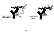
- Methods of the invention allow the use of both fluorescence polarization (FP) and time-resolved resonance energy transfer (TR-RET) detection modes.
- FP fluorescence polarization
- TR-RET time-resolved resonance energy transfer
- Use of the multiplex methods minimizes the number of false positive and false negative hits in screening assays, resulting in increased confidence in the integrity of screening results.
- use of the multiplex methods facilitates re-analysis of potentially spurious results due to background fluorescence interference and light scattering.
- t-Boc tert-butyloxycarbonyl
- Bzl benzyl
- PTK protein tyrosine kinase
- Fmoc fluorenylmethyloxycarbonyl
- ELISA enzyme-linked immuno absorbant assay
- FP fluorescence polarization
- FITC fluorescein isothiocyanate
- RET resonance energy transfer
- FRET fluorescence resonance energy transfer or Forster resonance energy transfer
- TR time resolved
- FAM carboxyfluorescein.
- antibody and “antibodies” include polyclonal antibodies, monoclonal antibodies, humanized or chimeric antibodies, single chain Fv antibody fragments, Fab fragments, and F(ab) 2 fragments.
- Polyclonal antibodies are heterogeneous populations of antibody molecules that are specific for a particular antigen, while monoclonal antibodies are homogeneous populations of antibodies to a particular epitope contained within an antigen.
- a chimeric antibody is a molecule in which different portions are derived from different animal species, such as those having a variable region derived from a mouse monoclonal antibody and a human immunoglobulin constant region.
- epipe refers to an antigenic determinant on an antigen to which an antibody binds.
- Epitopes usually consist of chemically active surface groupings of molecules such as amino acids, sugar side chains, or chemical moieties (e.g., from organic compounds) and typically have specific three-dimensional structural characteristics as well as specific charge characteristics.
- Epitopes can consist of a series of contiguous amino acids, e.g., 5 contiguous amino acids.
- an epitope can be a discontinuous epitope, e.g., the epitope is a particular arrangement of amino acids in space that results from the secondary, tertiary, and/or quaternary folding of a polypeptide.
- an epitope can consist of a modified amino acid side chain, e.g., a phosphorylated tyrosine, serine, or threonine.
- Monoclonal antibodies are particularly useful in the present invention.
- RET means resonance energy transfer, and refers to the radiationless transmission of an energy quantum from its site of absorption (the donor) to the site of its utilization (the acceptor) in a molecule, or system of molecules, by resonance interaction between donor and acceptor species, over distances considerably greater than interatomic, without substantial conversion to thermal energy, and without the donor and acceptor coming into kinetic collision.
- a donor is a moiety that initially absorbs energy (e.g., optical energy or electronic energy).
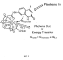
- a luminescent metal complex as described herein can comprise two donors: 1) an organic antenna moiety, which absorbs optical energy (e.g., from a photon); and 2) a lanthanide metal ion, which absorbs electronic energy (e.g., transferred from an organic antenna moiety).
- RET is sometimes referred to as fluorescent resonance energy transfer or Forster resonance energy transfer (both abbreviated FRET).
- acceptor refers to a chemical or biological moiety that accepts energy via resonance energy transfer.
- acceptors may re-emait energy transferred from a donor fluorescent or luminescent moiety as fluorescence (e.g., RET or TR-RET) and are "fluorescent acceptor moieties.”
- fluorescence e.g., RET or TR-RET
- fluorescent acceptor moieties such a donor fluorescent or luminescent moiety and an acceptor fluorescent moiety are referred to as a "RET pair.”
- acceptors include coumarins and related fluorophores; xanthenes such as fluoresceins and fluorescein derivatives; fluorescent proteins such as GFP and GFP derivatives; rhodols, rhodamines, and derivatives thereof; resorufins; cyanines; difluoroboradiazaindacenes; and phthalocyanines.
- Acceptors, including fluorescent acceptor moieties can also be useful as fluorescent probes in
- label refers to the inclusion of a luminescent metal complex or a fluorescent acceptor moiety on a first binding partner, second binding partner, tracer, test compound, potential modulator, substrate, or product, as described herein.
- modulates refers to partial or complete enhancement or inhibition of an activity or process (e.g., by attenuation of rate or efficiency).
- modulator refers to a chemical compound (naturally occurring or non-naturally occurring), such as a biological macromolecule (e.g., polynucleotide, polypeptide, hormone, polysaccharide, lipid), an organic molecule (e.g., a small organic molecule), or an extract made from biological materials such as bacteria, plants, fungi, or animal (particularly mammalian, including human) cells or tissues.
- a biological macromolecule e.g., polynucleotide, polypeptide, hormone, polysaccharide, lipid
- organic molecule e.g., a small organic molecule
- an extract made from biological materials such as bacteria, plants, fungi, or animal (particularly mammalian, including human) cells or tissues.
- Modulators may be evaluated for potential activity as inhibitors or enhancers (directly or indirectly) of a biological process or processes (e.g., agonist, partial antagonist, partial agonist, inverse agonist, antagonist, antineoplastic agents, cytotoxic agents, inhibitors of neoplastic transformation or cell proliferation, cell proliferation-promoting agents, and the like) by inclusion in screening assays described herein.
- a biological process or processes e.g., agonist, partial antagonist, partial agonist, inverse agonist, antagonist, antineoplastic agents, cytotoxic agents, inhibitors of neoplastic transformation or cell proliferation, cell proliferation-promoting agents, and the like.
- the activity of a modulator may be known, unknown, or partially known.
- non-naturally occurring refers to the fact that an object, compound, or chemical cannot be found in nature.
- a polypeptide or polynucleotide that is present in an organism (including viruses) that can be isolated from a source in nature and which has not been intentionally modified by man in the laboratory is naturally-occurring, while such a polypeptide or polynucleotide that has been intentionally modified by man is non-naturally occurring.
- organic molecule refers to compounds having a molecular skeleton containing a covalent arrangement of one or more of the elements C, N, H, O, S, and P, and typically having a molecular weight less than 10000 Daltons.
- Organic molecules having a molecular weight less than 5000 Daltons may be referred to as "small organic molecules.”
- polypeptide refers to a polymer of two or more amino acids joined together through amide bonds.
- a polypeptide can be an entire protein (e.g., isolated from a natural source or an expression system), a fragment of a protein, an enzymatically or chemically synthesized and/or modified version of a protein or protein fragment, or an amino acid sequence designed de novo (e.g., not based on a known protein sequence).
- Polypeptides can be 2-1000 amino acids in length (e.g., 2-900, 2-800, 2-700, 2-600, 2-500, 2-480, 2-450, 2-300, 2-200, 2-100, 2-50, 2-25, 5-900, 5-800, 5-700, 5-600, 5-500, 5-450, 5-300, 5-200, 5-100, 5-50, 5-25, 10-900, 10-800, 10-700, 10-600, 10-500, 10-450, 10-300, 10-200, 10-100, 10-50, 20-900, 20-800, 20-700, 20-600, 20-500, 20-450, 20-300, 20-200, 20-100, or 20-50 amino acids in length).
- 2-1000 amino acids in length e.g., 2-900, 2-800, 2-700, 2-600, 2-500, 2-480, 2-450, 2-300, 2-200, 2-100, 2-50, 2-25, 5-900, 5-800, 5-700, 5-600, 5-500, 5-450, 5-300, 5-200, 5-100, 5-50, 5-25, 10-900, 10-800, 10-700, 10-
- Amino acids may be natural or unnatural amino acids, including, for example, beta-alanine, phenylglycine, and homoarginine.
- beta-alanine phenylglycine
- homoarginine a group consisting of amino acids, amino acids, amino acids, amino acids and proteins, B. Weinstein, eds., Marcel Decker, New York, p. 267 (1983 ). All of the amino acids used in the present invention may be either the D- or L-isomer.
- Particularly useful chemically modified or substituted amino acids including phosphorylated (e.g., phospho-serine (phosphorylated at the hydroxyl of the side chain), phospho-tyrosine (phosphorylated at the OH of the side-chain phenyl ring), and phospho-threonine (phosphorylated at the hydroxyl of the size chain)), sulfated, methylated, or prenylated amino acids.
- phosphorylated e.g., phospho-serine (phosphorylated at the hydroxyl of the side chain), phospho-tyrosine (phosphorylated at the OH of the side-chain phenyl ring), and phospho-threonine (phosphorylated at the hydroxyl of the size chain)
- phosphorylated e.g., phospho-serine (phosphorylated at the hydroxyl of the side chain), phospho-tyrosine (phosphorylated at the OH of the side-chain phenyl ring), and phospho
- post-translational modification and “post-translational type modification” are used interchangeably and refer to enzymatic or neon-enzymatic modification of one or more amino acid residues in a polypeptide.
- Typical modifications include phosphorylation, dephosphorylation, glycosylation, methylation, sulfation, ubiquitination, acylation, acetylation, prenylation, and ADP-ribsoylation.
- Preferred post-translational type modifications include phosphorylation and dephosphorylation.
- post-translational modification includes non-covalent modifications that may affect polypeptide activity (e.g., protein activity), structure, or function, such as polypeptide-polypeptide interactions or the binding of ligands, allosteric modulators, other modulators, or second messengers such as calcium, cAMP, or inositol phosphates.
- polypeptide activity e.g., protein activity
- structure, or function such as polypeptide-polypeptide interactions or the binding of ligands, allosteric modulators, other modulators, or second messengers such as calcium, cAMP, or inositol phosphates.
- test compound refers to a compound to be tested by one or more screening method(s) of the invention, e.g., to determined if it is a putative modulator of an enzymatic activity such as a kinase activity.
- a test compound can be any chemical, such as an inorganic chemical, an organic molecule, a polypeptide, a carbohydrate, a polynucleotide, a polysaccharide, a lipid, a phospholipid, or a combination thereof.
- various predetermined concentrations e.g., various dilutions
- Experimental controls for a test compound can include measuring a signal for an assay performed in the absence of the test compound or comparing a signal obtained using a compound known to modulate a target activity with a signal obtained with the test compound.
- the invention is based on monitoring and/or measuring a molecular interaction (e.g., complex formation or disruption) between two binding partners.
- a "binding partner” is a compound (e.g., a first binding partner) that has affinity for another compound (e.g., a second binding partner) (or vice versa) such that the two binding partners are capable of forming a complex when bound.
- Two binding partners can be members of a specific binding pair.
- a first binding partner can be a monoclonal antibody and a second binding partner can be a composition having the epitope recognized by that monoclonal antibody.
- the invention provides compositions that include a binding partner.
- the binding partner can be labeled with a luminescent metal complex.
- the binding partner can be labeled with a fluorescent acceptor moiety.
- Specific examples of binding partners labeled with luminescent metal complexes or fluorescent acceptor moieties are set forth in the Examples, below.
- the present invention also provides mixtures of binding partners.
- a composition can include a first binding partner and a second binding partner.
- the first binding partner can comprise a luminescent metal complex while the second binding partner can comprise a fluorescent acceptor moiety.
- the first binding partner can comprise a fluorescent acceptor moiety.
- the second binding partner can comprise a luminescent metal complex.
- the affinity (apparent K d ) of a first binding partner for a second binding partner is about 1 mM or less, e.g., about 10 ⁇ M or less, or about 1 ⁇ M or less, or about 0.1 ⁇ M or less, or 10 nM or less, or 1 nM or less, or 0.1 nM or less.
- experimental parameters e.g., concentrations of assay components, reaction times, temperatures, and buffers, depending on the K d of the first binding partner for the second binding partner, to obtain a desired combination of conditions and cost-effectiveness.
- a second binding partner need not be an optimal binding partner for a first binding partner.
- a second binding partner is sometimes referred to herein as a "tracer,” and if it includes a luminescent metal complex or a fluorescent acceptor moiety, a "luminescent tracer.”
- a binding partner can be a polypeptide, a polynucleotide, a lipid, a phospholipid, a polysaccharide, or an organic molecule.
- specific polypeptide binding partners include an antibody, a protein, or an enzymatically or chemically-synthesized or modified polypeptide sequence (e.g., a polypeptide sequence derived from a protein, modified from a protein, or designed and synthesized de novo .)
- a polypeptide binding partner may be linear or cyclic.
- An organic molecule binding partner can be a small organic molecule.
- first and second binding partners that form complexes include an antibody and a composition having an epitope or epitope mimetic recognized by that antibody; a polypeptide and a ligand (e.g., receptor-ligand interactions); a polypeptide and another polypeptide (e.g., protein-protein interactions); a polypeptide and a polynucleotide (e.g., protein-DNA or protein-RNA interactions); a polynucleotide and another polynucleotide (e.g., DNA-DNA, DNA-RNA, or RNA-RNA interactions); a polypeptide and an organic molecule (e.g., protein-drug interactions); a polypeptide and a lipid (e.g., protein-phospholipid interactions); a polynucleotide and an organic molecule; and an organic molecule and another organic molecule.
- a ligand e.g., receptor-ligand interactions
- a polypeptide and another polypeptide e.g., protein
- a binding partner can comprise either a luminescent metal complex or a fluorescent acceptor moiety.
- one binding partner can comprise a luminescent metal complex and the other can comprise a fluorescent acceptor moiety, e.g., a first binding partner comprises a luminescent metal complex and a second binding partner comprises a fluorescent acceptor moiety.
- a luminescent metal complex and fluorescent acceptor moiety on a binding partner pair allows an interaction of first and second binding partners to be monitored by one or more fluorescent techniques (e.g., FP, TR-RET, or multiplex modes). For example, when a first binding partner and second binding partner are bound to one another, the complex will typically exhibit a characteristic FP or TR-RET signal (or bath).
- Disruption of the molecular interaction between the first binding partner and the second binding partner alters the FP or TR-RET signal (or both), allowing the monitoring of the molecular interaction in either FP or TR-RET modes (or both modes).
- an antibody can be labeled with a luminescent metal chelate and a polypeptide binding partner for the antibody can be labeled with a fluorescent acceptor moiety.
- the sample typically exhibits a high FP measurement and a fluorescence emission measurement characteristic ofRET between the luminescent metal chelate and the acceptor moiety.
- Addition of a competitor at a suitable concentration and with a suitable K d for the antibody results in displacement of the second binding partner, with a concomitant reduction in the FP measurement of the sample and a change in the fluorescence emission measurement as a result of a loss of RET between the luminescent metal chelate on the antibody and the fluorescent acceptor moiety on the polypeptide.
- Binding partners can be prepared and purified by a number of methods known to those of ordinary skill in the art.
- antibodies including monoclonal antibodies and antibody fragments, can be prepared by a number of methods known to those of skill in the art, or can be purchased from a variety of commercial vendors, including Serotec (Raleigh, NC), Abcam (Cambridge, MA), R&D Systems, Cambridge Antibody Technologies, and Covance Research Products (Denver, CO).
- an antigen for which an antibody is desired is prepared, e.g., recombinantly, by chemical synthesis, or by purification of a native protein, and then used to immunize animals.
- polypeptides containing a particular amino acid sequence and/or post-translational modification e.g., phosphorylation
- Various host animals including, for example, rabbits, chickens, mice, guinea pigs, goats, and rats, can be immunized by injection of the antigen of interest.
- adjuvants can be used to increase the immunological response and include Freund's adjuvant (complete and/or incomplete), mineral gels such as aluminum hydroxide, surface-active substances such as Iysolecithin, pluronic polyols, polyanions, peptides, oil emulsions, keyhole limpet hemocyanin, and dinitrophenol.
- Polyclonal antibodies are contained in the sera of the immunized animals.
- Monoclonal antibodies can be prepared using standard hybridoma technology. In particular, monoclonal antibodies can be obtained by any technique that provides for the production of antibody molecules by continuous cell lines in culture as described, for example, by Kohler et al.
- Such antibodies can be of any immunoglobulin class including IgM, IgG, IgE, IgA, IgD, and any subclass thereof
- the hybridoma producing the monoclonal antibodies of the invention can be cultivated in vitro or in vivo. Chimeric antibodies can be produced through standard techniques.
- Antibody fragments that have specific binding affinity for an antigen can be generated by known techniques. Such antibody fragments include, but are not limited to, F(ab') 2 fragments that can be produced by pepsin digestion of an antibody molecule, and Fab fragments that can be generated by reducing the disulfide bridges of F(ab') 2 fragments. Alternatively, Fab expression libraries can be constructed. See , for example, Huse et al. (1989) Science 246:1275-1281 . Single chain Fv antibody fragments are formed by linking the heavy and light chain fragments of the Fv region via an amino acid bridge (e.g., 15 to 18 amino acids), resulting in a single chain polypeptide. Single chain Fv antibody fragments can be produced through standard techniques, such as those disclosed in U.S. Patent No. 4,945,778 .
- antibodies or fragments thereof can be tested for recognition of (and affinity for) a second binding partner by standard immunoassay methods including, for example, enzyme-linked immunosorbent assay (ELISA) or radioimmuno assay (RIA).
- ELISA enzyme-linked immunosorbent assay
- RIA radioimmuno assay
- Suitable antibodies typically will have a K d for a second binding partner of about 1 mM or less, e.g., about 10 ⁇ M or less, or about 1 ⁇ M or less, or about 0.1 ⁇ M or less, or about 10 nM or less, or about 1 nM or less, or about 0.1 nM or less.
- the second binding partner can be a polypeptide containing the same post-translational modification.
- a second binding partner will have the same chemical structure as an antigen used to immunize.
- polypeptides in addition to antibodies are useful as first or second binding partners and can also be prepared and analyzed using standard methods.
- polypeptides can be obtained by extraction from a natural source (e.g., from isolated cells, tissues or bodily fluids), by expression of a recombinant nucleic acid encoding the polypeptide, or by chemical synthesis.
- Polypeptides can be produced by, for example, standard recombinant Technology, using expression vectors encoding the polypeptides. The resulting polypeptides then can be purified.
- Expression systems that can be used for small or large scale production of polypeptides include, without limitation, microorganisms such as bacteria (e.g., E. coli and B.
- subtilis transformed with recombinant bacteriophage DNA, plasmid DNA, or cosmid DNA expression vectors; yeast (e.g., S. cerevisiae ) transformed with recombinant yeast expression vectors; insect cell systems infected with recombinant virus expression vectors (e.g., baculovirus); plant cell systems infected with recombinant virus expression vectors (e.g., tobacco mosaic virus) or transformed with recombinant plasmid expression vectors (e.g., Ti plasmid); or mammalian cell systems (e.g., primary cells or immortalized cell lines such as COS cells, Chinese hamster ovary cells, HeLa cells, human embryonic kidney 293 cells, and 3T3 L1 cells) harboring recombinant expression constructs containing promoters derived from the genome of mammalian cells (e.g., the metallothionein promoter) or from mammalian viruses (e.g., the a
- Suitable methods for purifying the polypeptides of the invention can include, for example, affinity chromatography, immunoprecipitation, size exclusion chromatography, and ion exchange chromatography. See , for example, Flohe et al. (1970) Biochim. Biophys. Acta. 220:469-476 , or Tilgmann et al. (1990) FEBS 264:95-99 .
- the extent of purification can be measured by any appropriate method, including but not limited to: column chromatography, polyacrylamide gel electrophoresis, or high-performance liquid chromatography.
- Polypeptides as first or second binding partners can also be prepared using solid phase synthesis methods, see , e.g., WO 03/01115 and 6,410,255 .
- polypeptides synthesized chemically have between 3 to 50 amino acids (e.g., 3 to 30, 3 to 20, 3 to 15, 5 to 30, 5 to 20, 5 to 15, 8 to 20, 8 to 15, 10 to 10, 10 to 15 or 10 to 12 amino acids in length).
- Suitable amino acids include natural, non-natural, and modified.(e.g., phosphorylated) amino acids.
- Amino acids with many different protecting groups appropriate for immediate use in the solid phase synthesis of peptides are commercially available.
- Polynucleotides useful as binding partners can be produced by standard techniques, including, without limitation, common molecular cloning and chemical nucleic acid synthesis techniques. For example, polymerase chain reaction (PCR) techniques can be used. PCR refers to a procedure or technique in which target nucleic acids are enzymatically amplified. Sequence information from the ends of the region of interest or beyond typically is employed to design polynucleotide primers that are identical in sequence to opposite strands of the template to be amplified. PCR can be used to amplify specific sequences from DNA as well as RNA, including sequences from total genomic DNA or total cellular RNA.
- PCR polymerase chain reaction
- Primers are typically 14 to 40 nucleotides in length, but can range from 10 nucleotides to hundreds of nucleotides in length.
- General PCR techniques are described, for example in PCR Primer: A Laboratory Manual, ed. by Dieffenbach and Dveksler, Cold Spring Harbor Laboratory Press, 1995 .
- reverse transcriptase can be used to synthesize complementary DNA (cDNA) strands.
- Ligase chain reaction, strand displacement amplification, self-sustained sequence replication, or nucleic acid sequence-based amplification also can be used to obtain isolated nucleic acids. See , for example, Lewis Genetic Engineering News, 12(9):1 (1992 ); Guatelli et al., Proc. Natl. Acad. Sci. USA, 87:1874-1878 (1990 ); and Weiss, Science, 254:1292 (1991 ).
- Polynucleotides of the invention also can be chemically synthesized, either as a single nucleic acid molecule (e.g., using automated DNA synthesis in the 3' to 5' direction using phosphoramidite technology) or as a series of smaller polynucleotides.
- one or more pairs of long polynucleotides e.g., >100 nucleotides
- each pair containing a short segment of complementarity e.g., about 15 nucleotides
- DNA polymerase is used to extend the polynucleotides, resulting in a single, double-stranded polynucleotide.
- Polynucleotides of the invention also can be obtained by mutagenesis.
- polynucleotide can be mutated using standard techniques including polynucleotide-directed mutagenesis and site-directed mutagenesis through PCR. See Short Protocols in Molecular Biology, Chapter 8, Green Publishing Associates and John Wiley & Sons, edited by Ausubel et al., 1992 .
- a binding partner can comprise a luminescent metal complex.
- a luminescent metal complex can act as a donor fluorophore in a RET or TR-RET assay.
- a luminescent metal complex is useful in the present methods because its excited state lifetime is typically on the order of milliseconds or hundreds of microseconds rather than nanoseconds; a long excited state lifetime allows detection of a molecular interaction between binding partners to be monitored after the decay of background fluorescence and/or interference from light-scattering.
- a luminescent metal complex includes a metal liganding moiety, one or more lanthanide metal ions, and optionally linkers, spacers, and organic antenna moieties.
- a metal liganding moiety coordinates one or more lanthanide metal ions to form a metal complex.
- a metal liganding moiety includes one or more metal coordinating moieties X, where X is a heteroatom electron-donating group capable of coordinating a metal cation, such as O - , OH, NH 2 , OPO 3 2- , NHR, or OR where R is an aliphatic group.
- a metal liganding moiety can be a chelating moiety or a cryptand moiety. If a lanthanide metal ion is coordinated to a chelating moiety, the complex is referred to as a "metal chelate.” If a lanthanide metal ion is coordinated to a cryptand moiety, the complex is referred to as a "metal cryptand.”
- a metal chelate should be stable to exchange of the lanthanide ion.
- Metal chelates preferably have a formation constant (K f ) of greater than 10 10 M -1 .
- K f formation constant
- a variety of useful chelating moieties are known to those of skill in the art. Typical examples of cheating moieties include: EDTA, DTPA, TTHA, DOTA, NTA, HDTA, DTPP, EDTP, HDTP, NTP, DOTP, DO3A, DOTAGA, and NOTA.
- a luminescent metal chelate can have the following structures: -L n -A-S n -C M , or -L n -C M -S n -A,
- FIG. 3 also demonstrates luminescent metal chelates useful for conjugating to amine moieties (top structure) or thiol moieties (bottom structure) on binding partners.
- Cryptates are formed by the inclusion of a lanthanide cation into a tridimensional organic cavity, leading to highly stable complexes.
- a variety of useful cryptand moieties are known to those of skill in the art. Examples of cryptand moieties useful in the present methods include: trisbypyridine (TBP, e.g, TBP pentacarboxylate), and pyridine bipyridine (e.g., pyridine bipyridine tetracarboxylate).
- Chelating and cryptand moieties can be synthesized by a variety of methods known to those of skill in the art or may be purchased commercially. See U.S. Pat. Nos. 5,639,615 ; 5,656,433 ; 5,622,821 ; 5,571,897 ; 5,534,622 ; 5,220,012 ; 5,162,508 ; and 4,927,923 ; and WO 96/23526 and WO 03/011115 .
- Metal liganding moieties coordinate one or more lanthanide metal ions to form a metal complex.
- Lanthanide metal ions are useful because their special electronic configuration shields the optically active electrons, resulting in characteristic line type emissions. As the electronic transitions of the metal ions are forbidden by quantum mechanics rules, the emission lifetimes of these ions are typically long (from ⁇ s to msec).
- Useful lanthanide metal ions include Sm(III), Ru(III), Eu (III), Gd(III), Tb(III), and Dy(III).
- Methods for complexing a metal ion to a chelating or cryptand moiety are known to those of skill in the art, see , e.g., WO 96/23526 and WO 03/011115 .
- a luminescent metal complex can optionally include an organic antenna moiety.
- An organic antenna moiety typically has a conjugated electronic structure so that it can absorb light. The absorbed light is transferred by intramolecular non-radiative processes from the singlet to the triplet excited state of the antenna moiety, then from the triplet state to the emissive level of the lanthanide ion, which then emits characteristically long-lived luminescence. See FIGs. 2 and 4 .
- some metal liganding moieties can absorb light without the inclusion of an organic antenna moiety.
- certain cryptand moieties that contain conjugated organic moieties, such as tribipyridine pentacarboxylate do not require the inclusion of a discrete organic antenna moiety.
- an organic antenna moiety can be a polynuclear heterocyclic aromatic compound.
- the polynuclear heterocylic aromatic compound can have two or more fused ring structures.
- useful organic antenna moieties include rhodamine 560, fluorescein 575, fluorescein 590, 2-quinolone, 4-quinolone, 4-trifluoromethylcoumarin (TFC), 7-diethyl-amino-coumarin-3-carbohydrazide, 7-amino-4-methyl-2-coumann (carbostyril 124, CS 124), 7-amino-4-methyl-2-coumarin (coumarin 120), 7-amino-4-trifluoromethyl-2-coumarin (coumarin 124), and ammomethyltrimethylpsoralen. See FIGs. 2 and 3 .
- Linkers and Spacers can optionally be included in a luminescent metal complex.
- a Linker (L) functions to link a luminescent metal complex to a first or second binding partner.
- a L can link an acetate, amine, amide, carboxylate, or methylene functionality on a metal liganding moiety to a first or second binding partner.
- Ls can react with a number of functionalities on binding partners, including, without limitation, amines, acetates, thiols, alcohols, ethers, esters, ketones, and carboxylates.
- a L can cap the N-terminus, the C-terminus, or both N- and C- termini, as an amide moiety.
- Other exemplary L capping moieties include sulfonamides, ureas, thioureas and carbamates.
- Ls can also include linear, branched, or cyclic alkanes, alkenes, or alkynes, and phosphodiester moieties.
- the L may be substituted with one or more functional groups, including ketone, ester, amide, ether, carbonate, sulfonamide, or carbamate functionalities.
- Additional examples of Ls and synthetic methodologies for incorporating them into metal complexes, particularly metal complexes linked to polypeptides are set forth in WO 01/09188 , WO 01/08712 , and WO 03/011115 .
- a Spacer can connect an organic antenna moiety to a metal liganding moiety.
- a S can link an acetate, amine, or methylene functionality on a metal liganding moiety to an organic antenna moiety.
- One of skill in the art can design Ss to react with a number of functionalities on organic antenna moieties and on metal liganding moieties, including, without limitation, amines, acetates, thiols, alcohols, ethers, esters, ketones, and carboxylates.
- Ss can include linear, branched, or cyclic alkanes, alkenes, or alkynes, and phosphodiester moieties.
- the S may be substituted with one or more functional groups, including ketone, ester, amide, ether, carbonate, sulfonamide, or carbamate functionalities.
- a binding partner can include a fluorescent acceptor moiety.
- a fluorescent acceptor moiety can act as an acceptor in RET or TR-RET-based assays and/or can be a fluorophore for which the polarization of fluorescence emission is measured in an FP-based assay.
- a fluorescent acceptor moiety should exhibit a good quantum yield and a large extinction coefficient; should be resistant to collisional quenching and bleaching; and should be easily conjugated to a variety of first and second binding partners by methods known to those having ordinary skill in the art.
- Suitable fluorophores include, without limitation, fluorescein, rhodamine, FITCs (e.g., fluorescein-5-isothiocyanate), 5-FAM, 6-FAM, 5,6-FAM, 7-hydroxycoumarin-3-carboxamide, 6-chloro-7-hydroxycoumarin-3-carboxamide, dichlorotriazinylaminofluorescein, tetramethylrhodamine-5-isothiocyanate, tetramethylrhodamine-6-isothiocyanate, succinimidyl ester of 5-carboxyfluorcscein, succinimidyl ester of 6-carboxyfluorescein, 5-carboxytetramethylrhodamine, 6-carboxymethylrhodamine, and 7-amino-4-methylcoumarin-3-acetic acid.
- fluorescein fluorescein, rhodamine, FITCs (e.g., fluorescein-5-isothi
- fluorophores include the Cy family of fluorophores (Cy 3, Cy3B, Cy3.5, Cy5; available from Amersham Biosciences, Piscataway, NJ); the Alexa Fluor family (available from Molecular Probes, Eugene, OR); the BODIPY family (available from Molecular Probes, Eugene, OR); carbopyronins; squarines; cyanme/indocyanines; benzopyrylium heterocyles; and amide-bridged benzopyryliums.
- Fluorescent proteins and mutants can also be used as fluorescent acceptor moieties. Examples include firefly, bacterial, or click beetle luciferases, aequorins, and other photoproteins (for example as described in U.S. Patents 5,221,623, issued June 22, 1989 to Thompson et al. , 5,683,888 issued November 4, 1997 to Campbell ; 5,674,713 issued September 7, 1997 to DeLuca et al. ; 5,650,289 issued July 22, 1997 to Wood ; and 5,843,746 issued December 1, 1998 to Tatsumi et al. ). GFP and GFP mutants are particularly useful in applications using Tb(III)-containing metal complexes.
- a fluorescent acceptor moiety for use in multiplex assays should exhibit characteristics useful for both RET/TR-RET applications and FP applications.
- a fluorophore preferably exhibits a fluorescent excited state lifetime of at least 1 nanosecond, or at least 2 nanoseconds.
- TR-RET applications a region of the fluorophore's absorbance spectra should overlap with a region of a luminescent metal chelate's emission spectra, while a region of the fluorophore's emission spectra should not overlap substantially with a region of the luminescent metal chelate's emission spectra.
- FIG 5 demonstrates overlap of the emission spectra of an organic antenna-Tb(III)-chelate-containing metal complex with the absorption spectra of fluorescein and rhodamine.
- FIG 5 also demonstrates that regions of the emission spectra of the organic-antenna-Tb(II)-chelate-containing metal complex do not overlap with the emission spectra for fluorescein and rhodamine. See also FIGs. 6-7 .
- acceptor fluorophores in TR-RET assays using Tb(III)-containing luminescent metal complexes include: fluorescein (and its derivatives); rhodamine (and its derivatives); Alexa Fluors 488, 500,514,532, 546, 555, 568 (available from Molecular Probes); BODIPYs FL, R6G, and TMR (available from Molecular Probes); Cy3 and Cy3B (available from Amersham Biosciences), and IC3 (available from Dojindo Molecular Technologies, Gaithersburg, MD).
- acceptor fluorophore in TR-RET assays using Eu(III)-containg luminescent metal complexes include: Alexa Fluors 594, 610, 633, 647, and 660 (available from Molecular Probes); BODIPYs TR, 630/650, and 650/665 (available from Molecular Probes); Cy5 (available from Amersham Biosciences) and IC5 (available from Dojindo Molecular Technologies).
- Suitable fluorophores for use in the present invention are commercially available, e.g., from Molecular Probes (Eugene, OR), Attotec (Germany), Amersham, and Biosearch Technologies (Novato, CA). Methods for incorporating fluorophores into a variety of binding partners are known to those of skill in the art; see , e.g., 6,410,255.
- Methods of the present invention are based on the finding that luminescent or fluorescent assays based on FP or RET (including TR-RET) can be easily converted into multiplex assays, allowing detection in multiple luminescent modes.
- assays based on measuring a change in FP of one binding partner can be easily converted to TR-RET assays by the incorporation of an appropriate luminescent metal complex on one binding partner and a fluorescent acceptor moiety on the other binding partner.
- a binding partner that includes a fluorescent acceptor moiety useful for FP measurements need not be modified, provided that a suitable luminescent metal complex is chosen for inclusion on the other binding partner, as described above. Any of the methods described herein can be homogeneous or heterogeneous.
- Methods of the present invention take advantage of a change in fluorescence polarization upon first and second binding partner complex formation or disruption.
- Polarization measurements are based on the relative rotational movement of a fluorophore compared to the excited state life-time of that fluorophore.
- polarization p
- degree of rotational movement can be readily derived ( see Weber, Polarization of the fluorescence of solutions, in Fluorescence and Phosphorescence Analysis, Don Hercules (ed.), Interscience Publishers, New York, Chapter 8, pages 217-240 (1966 )).
- Rotational movement can be related to the rotational diffusion constant of the molecule, and hence to the molecular volume.
- a change in the fluorescence polarization of a sample can occur when a complex of a first and second binding partner (one of which comprises a fluorescent acceptor moiety) is either formed or disrupted. Complex formation or disruption can result in from about a 10% to about a 10,000% increase or decrease in the fluorescence polarization measurement of a sample. Preferably, a change in the fluorescence polarization measurement is from about 30 mP to about 450 mP.
- Polarization-based assays are relatively easy to set up and can be obtained over a wide concentration, temperature, and ionic strength range. See , e.g., U.S. 6,511,815 and 5,445,935 .
- Methods of the present invention also take advantage of resonance energy transfer between a luminescent metal chelate and a fluorescent acceptor moiety (RET).
- RET fluorescent acceptor moiety
- a donor luminescent metal chelate is excited by light of appropriate wavelength and intensity (e.g., within the donor antenna moiety's excitation spectrum) and under conditions in which direct excitation of the acceptor fluorophore is minimized.
- the donor luminescent chelate then transfers the absorbed energy by non-radiative means to the acceptor fluorescent moiety, which subsequently re-emits some of the absorbed energy as fluorescence emission at one or more characteristic wavelengths.
- the re-emitted radiation is not measured until after a suitable delay time, e.g., 25, 50,75,100,150,200, or 300 microseconds to allow decay of background fluorescence, light scattering, or other luminescence, such as that caused by the plastics used in microtiter plates.
- a suitable delay time e.g. 25, 50,75,100,150,200, or 300 microseconds to allow decay of background fluorescence, light scattering, or other luminescence, such as that caused by the plastics used in microtiter plates.
- a first binding partner can comprise either a luminescent metal complex or a fluorescent acceptor moiety, while the second binding partner comprises the other.
- an antibody first binding partner can be labeled with a Tb(III)-chelate-orgnic antenna moiety (luminescent metal chelate), while a polypeptide for which the antibody is specific can be labeled with a fluorescein (fluorescent acceptor moiety).
- disruption of the complex formed by the antibody and polypeptide results in an alteration in energy transfer between the luminescent metal chelate on the antibody and the fluorescent acceptor moiety on the polypeptide that may be used to monitor and measure the binding between the first and second binding partners.
- a compound that affects binding of a second binding partner (or tracer) to a first binding partner can be, for example, a test compound, an enzyme product (e.g., for which the first binding partner has specificity), or an enzyme substrate (e.g., for which the first binding partner has specificity).
- a compound that affects binding of a second binding partner (or tracer) to a first binding partner can comprise either a luminescent metal chelate or fluorescent acceptor moiety while the first binding partner comprises the other.
- disruption of the complex formed between the first binding partner and the second binding partner by the labeled compound that affects binding can result in an increase in RET.
- RET can be manifested as a reduction in the intensity of the luminescent signal from the donor luminescent metal complex and/or an increase in emission of fluorescence from the acceptor fluorescent moiety.
- a complex between an antibody having a donor luminescent metal complex and a polypeptide having an acceptor fluorescent moiety is disrupted, e.g., by a competitor for the polypeptide, such as an unlabeled polypeptide, the donor luminescent metal complex and the acceptor fluorescent moiety physically separate, and RET is diminished or eliminated. Under these circumstances, luminescence emission from the donor luminescent metal complex increases and fluorescence emission from the acceptor fluorescent moiety decreases.
- a ratio of emission amplitudes at wavelengths characteristic (e.g., the emission maximum) of the donor luminescent metal complex relative to the acceptor fluorescent moiety should increase as compared to the same ratio under RET conditions (e.g., when emission of the donor luminescent metal complex is quenched by the acceptor).
- the efficiency of RET is dependent on the separation distance and the orientation of the donor luminescent metal complex and acceptor fluorescent moiety, the luminescent quantum yield of the donor metal ion, the spectral overlap with the acceptor fluorescent moiety, and the extinction coefficient of the acceptor fluorophore at the wavelengths that overlap with the donor's emission spectra.
- E the efficiency of RET
- F and F° are the fluorescence intensities of the donor in the presence and absence of the acceptor, respectively
- R is the distance between the donor and the acceptor.
- Ro 9.79 x 10 3 ⁇ K 2 ⁇ QJn - 4 1 / 6
- K 2 is an orientation factor having an average value close to 0.67 for freely mobile donors and acceptors
- Q is the quantum yield of the unquenched fluorescent donor
- n is the refractive index of the intervening medium
- J is the overlap integral, which expresses in quantitative terms the degree of spectral overlap.
- the characteristic distance Ro at which RET is 50% efficient depends on the quantum yield of the donor, the extinction coefficient of the acceptor, the overlap between the donor's emission spectrum and the acceptor's excitation spectrum, and the orientation factor between the two fluorophores.
- Changes in the degree of RET can be determined as a function of a change in a ratio of the amount of luminescence from the donor and acceptor moieties, a process referred to as "ratioing.”
- ratioing By calculating a ratio, the assay is less sensitive to, for example, well-to-well fluctuations in substrate concentration, photobleaching and excitation intensity, thus making the assay more robust. This is of particular importance in automated screening applications where the quality of the data produced is important for its subsequent analysis and interpretation. See , e.g., U.S. 6, 410, 255 ; 4,822,733 ; 5,527,684 ; and 6,352,672 .
- a ratiometric analysis is performed, wherein a ratio of luminescence emission at two different wavelengths is compared between a test sample and a control sample.
- the two wavelengths can correspond to an emission maximum for a luminescent metal complex and a fluorescent acceptor moiety.
- an emissions ratio of the control sample will be about 1.5, 2, 3, 4, 5, 7, 10, 15, 20, 25, 30, 40, 50, or 100 times larger or smaller than the emissions ratio of a test sample.
- Methods of the present invention can be used to measure the effect of a test compound on binding between a first binding partner and a second binding partner.
- the present methods may be used to identify competitive binders to first or second binding partners, or to identify compounds that physically (e.g., allosterically) or chemically affect a first or second binding partner so as to consequently affect binding of its partner.
- assays to identify effects of test compounds on such binding partner interactions as protein-protein interactions, protein-ligand interactions, protein-DNA interactions, and polynucleotide hybridizations may be designed using the present methods.
- a first binding partner, a second binding partner, and a test compound are contacted to form a test sample.
- one of the binding partners comprises a luminescent metal complex, while the other comprises a fluorescent acceptor moiety. See FIG 1 .
- the first and second binding partner are capable of binding to one another to form a complex.
- the test sample is exposed to polarized light at an appropriate wavelength (e.g., at a wavelength in an absorbance band of the fluorescent acceptor moiety) and the polarization of fluorescent emission from the test sample is measured.
- the test sample is also exposed to light (e.g., at a wavelength in an absorbance band of the luminescent metal complex), typically in the wavelength range of 250 nm to 750 nm, and the fluorescence emission from the test sample is measured. Fluorescence emission may be measured after a suitable time delay, as indicated above, to result in a time-resolved fluorescence emission measurement.
- the exposure to polarized light and measurement of polarization may be performed before, after, or simultaneously with the exposure to light and measurement of fluorescence emission from the test sample.
- the exposure to polarized light and measurement of polarization may be performed up to 5 sec., 10 sec., 20 sec., 30 sec., 1 min., 2 min., 5 min., 10 min., 1 hr., 5 hrs., or 24 hrs. before or after the exposure, to light and measurement of fluorescence emission from the test sample.
- a test compound can comprise either a luminescent metal complex or a fluorescent acceptor moiety and a first binding partner can comprise the other.
- a first binding partner receptor can be labeled with a luminescent metal chelate while a test ligand for the first binding partner receptor can be labeled with a fluorescent acceptor moiety.
- Disruption of a complex formed between the first binding partner receptor and an unlabeled second binding partner (e.g., a ligand for the receptor) by the labeled test ligand can lead to an increase in RET and/or FP.
- a test compound is identified as affecting binding between first and second binding partners when the fluorescence polarization measurement or the fluorescence emission measurement of the test sample, or both, is different from the fluorescence polarization measurement or the fluorescence emission measurement of a control sample lacking the test compound.
- whether or not a difference is statistically significant will depend on the type of measurement and the experimental conditions. It is understood that when comparing measurements, a statistically significant difference indicates that the test compound may warrant further study.
- a difference is considered statistically significant at p ⁇ 0.05 with an appropriate parametric or non-parametric statistic, e.g., Chi-square test, Student's t-test, Mann-Whitney test, or F-test. In some embodiments, a difference is statistically significant at p ⁇ 0.01, p ⁇ 0.005, or p ⁇ 0.001.
- Methods of the invention can also be used to identify a modulator of enzymatic activity.
- a first binding partner is selected based on specificity for either a substrate or a product of an enzymatic activity.
- an antibody with specificity for a phosphorylated tyrosine as compared to an unmodified tyrosine can be a first binding partner with specificity for a product of tyrosine kinase activity.
- a tracer is then selected based partially on the specificity of the first binding partner for the substrate or product of the enzymatic activity.
- a tracer can include the purported epitope recognized by an antibody first binding partner, or a recognition site or chemical structure recognized by a polypeptide first binding partner.
- a tracer can have the same chemical structure as an antigen used to immunize an animal to generate a first binding partner antibody.
- the first binding partner will bind to a tracer with a similar K d as to the enzymatic product or substrate for which it has specificity, e.g., about 0.001 to 1000 times, or 0.01 to 100 times, or 0.1 to 10 times the K d of the first binding partner for the product or substrate.
- a tracer may be labeled (e.g., include a luminescent metal complex or a fluorescent acceptor moiety; referred to herein as a "luminescent tracer”) or the tracer may be unlabeled.
- the first binding partner is an antibody with specificity for a phosphorylated tyrosine, a product of tyrosine kinase activity
- a luminescent tracer can be selected that includes the epitope (or an epitope mimetic) recognized by the antibody (in this case, a phosphorylated tyrosine) so that the antibody binds the luminescent tracer.
- the inclusion of a fluorescent acceptor moiety or luminescent metal complex on the tracer should not substantially affect the K d of the first binding partner for the tracer.
- enzymatic activities may be probed, including, without limitation, kinase activity, phosphatase activity, glucuronidase activity, prenylation, glycosylation, methylation, demethylation, acylation, acetylation, ubiquitination, sulfation, proteolysis, nuclease activity, nucleic acid polymerase activity, nucleic acid reverse transcriptase activity, nucleotidyl transferase activity, polynucleotide transcription activity, and polynucleotide translation activity.
- an enzyme is contacted with a substrate for the enzyme under conditions effective for an enzymatic activity of the enzyme to form a product from the substrate.
- conditions effective for enzymatic activity will vary with the enzyme, enzymatic activity, and substrate chosen.
- ATP is generally included.
- Incubation conditions for a contacting step can vary, e.g., in enzyme concentration, substrate concentration, temperature, and length of time.
- Incubation temperature conditions typically can be from about 15 to about 40 °C; in some embodiments, the temperature may be about room temperature, e.g., about 20-25 °C.
- a contacting step is carried out in the presence of a potential modulator of the enzymatic activity.
- the enzyme, substrate, and potential modulator mixture is then contacted with a first binding partner and luminescent tracer, as described above, to form a test sample.
- a first binding partner or the luminescent tracer includes a luminescent metal complex, while the other includes a fluorescent acceptor moiety.
- the enzyme, substrate, and potential modulator mixture is contacted with a first binding partner and a tracer to form a test sample.
- either the first binding partner or the substrate includes a luminescent metal complex, while the other includes a fluorescent acceptor moiety.
- enzymatic activity can result in the conversion of the labeled substrate to a labeled product.
- the inclusion of a fluorescent acceptor moiety or luminescent metal complex on the substrate should not substantially affect the ability of the enzyme to form a product from the labeled substrate.
- the inclusion of a fluorescent acceptor moiety or luminescent metal complex on the substrate (or product) should not substantially affect the K d of the first binding partner for the substrate (or product) for which it has specificity.
- the test sample is exposed to polarized light at an appropriate wavelength (e.g., at a wavelength in an absorbance band of the fluorescent acceptor moiety) and the polarization of fluorescent emission from the test sample is measured.
- the test sample is also exposed to light (e.g., at a wavelength in an absorbance band of the luminescent metal complex), typically in the wavelength range of 250 nm to 750 nm, and the fluorescence emission from the test sample is measured.
- Fluorescence emission may be measured after a suitable time delay, as indicated above, to result in a time-resolved fluorescence emission measurement.
- the exposure to polarized light and measurement of polarization may be performed before, after, or simultaneously with the exposure to light and measurement of fluorescence emission from the test sample.
- a tracer maybe unlabeled, e.g., in embodiments where a first binding partner is labeled with a luminescent metal complex and a substrate is labeled with a fluorescent acceptor moiety.
- Disruption of a complex formed between an unlabeled tracer and a labeled first binding partner by an appropriately labeled compound (e.g., labeled substrate, labeled product, labeled test compound) that affects binding between the unlabeled tracer and first binding partner can lead to an increase or decrease in RET, FP, or both.
- a potential modulator is identified as a modulator of enzymatic activity when the fluorescence polarization measurement or the fluorescence emission measurement of the test sample, or both, is different from the fluorescence polarization measurement or the fluorescence emission measurement of a control sample lacking the potential modulator.
- a difference is considered statistically significant at p ⁇ 0.05 with an appropriate parametric or non-parametric statistic, e.g., Chi-square test, Student's t-test, Mann-Whitney test, or F-test.
- a difference is statistically significant at p ⁇ 0.01, p ⁇ 0.005, or p ⁇ 0.001.
- any of the methods of the present invention can be modified to be performed in a high-throughput or ultra-high-throughput manner.
- a method to identify a modulator of activity of an enzyme may be modified to contact a plurality of substrates, independently, with a particular enzyme and potential modulator, to form a plurality of enzyme mixtures.
- Each enzyme mixture is then contacted with an appropriate first binding partner and luminescent tracer to form a test sample, with the excitation (e.g., exposure to plane polarized light and exposure to light) and measurement (of emission of polarized and fluorescent light) steps as described previously.
- excitation e.g., exposure to plane polarized light and exposure to light
- measurement of emission of polarized and fluorescent light
- the Z'-Factor is a statistic designed to reflect both assay signal dynamic range and the variation associated with signal measurements.
- Signal-to-noise (S/N) or signal-to-background (S/B) ratios alone are unsatisfactory in this regard because they do not take into account the variability in sample and background measurements and signal dynamic range.
- the Z'-Factor takes into account these factors, and because it is dimensionless, it can be used to compare similar assays.
- assays of the present invention yield Z'-factors of greater than or equal to 0.5. Methods for determining Z'-factor are known to those of skill in the art.
- a Z'-factor may be determined by evaluating the dynamic range of a method.
- kits and apparatuses useful for performing the described inventions.
- a kit includes packaging material, such as a container, and one or more compositions useful as first and/or second binding partners.
- a kit can include one or more of the following: a multi-well plate, one or more enzymes, buffers, and directions for use of the kit.
- An apparatus will generally include a sample chamber, means for generating plane polarized light to illuminate the sample chamber; and means for illuminating the sample chamber with light having a wavelength from 250 nm to 750 nm.
- an apparatus will include means for detecting polarize light emitted from the sample chamber and means for detecting light (e.g., fluorescence) emitted from the sample chamber.
- both illumination means illuminate the sample chamber simultaneously.
- both detections means detect the polarized light and the light emitted (e.g., fluorescence) simultaneously.
- PY72 anti-phosphotyrosine IgG antibody
- an antibody that preferentially binds amino acid sequences containing phosphorylated tyrosines e.g., sequences phosphorylated by protein tyrosine kinases (PTKs)
- PY72 hybridoma cells were obtained from the Salk Institute; the immunogen was phosphotyrosine conjugated to KLH. Ascites were produced by Harlan Bioproducts for Science, Indianapolis IN. Ascites were purified with a protein G column (Pierce).
- Purified antibody is also available from Covance, Berkeley CA (Part # MMS414P).] The antibody was then removed from the dialysis membrane and concentrated to 48.8 uM (7.3 mg/mL) using a Centricon YM50 (Millipore) concentrator. 100 uL of this antibody solution was diluted to 5 mg/ml (33.4 uM) into the labeling reaction which consisted of 10 mM phenyl phosphate, and 660 ⁇ M carbostyril 124-diethylenetriaminepentaaceticacid-phenylalanine isothiocyanate *Tb(III) (CS124-DTPA-Phe-NCS*Tb, see FIG.
- a monoclonal antibody with specificity for phosphorylated serines was also prepared and labeled with a luminescent metal chelate, as described above.
- a direct binding curve (showing luminescent metal chelate -labeled PY72 antibody binding to fluorescent acceptor labeled tracer) was generated by incubating serial dilutions of the labeled antibody (10 nM to 9.8 pM in two fold dilutions) with 1 nM fluorescent acceptor-labeled tracer (PTK labeled tracer; sequence F-ADE(pY)LIPQQS, where F is fluorescein and pY is a phosphorylated tyrosine, SEQ ID NO:1; note that the tracer is a phosphorylated tyrosine derivative of a protein tyrosine kinase (PTK) substrate) in FP dilution buffer (PanVera, Madison WI part #P2839).
- PTK labeled tracer sequence F-ADE(pY)LIPQQS, where F is fluorescein and pY is a phosphorylated tyrosine, SEQ ID NO:1;
- a similar binding curve was performed with a luminescent metal chelated-labeled anti-pSer antibody and a fluorescent acceptor-labeled tracer (STK labeled tracer, sequence F-GRPRTS(pS)FAEG, where F is a fluorescein and pS is a phosphorylated serine, SEQ ID NO:2; note that the tracer is a phosphorylated serine derivative of a S/T kinase (STK) substrate).
- STK labeled tracer sequence F-GRPRTS(pS)FAEG, where F is a fluorescein and pS is a phosphorylated serine, SEQ ID NO:2; note that the tracer is a phosphorylated serine derivative of a S/T kinase (STK) substrate).
- a competition curve to show that the disruption of the antibody-tracer interaction could be monitored by both fluorescence polarization and time-resolved RET from the same sample was performed by incubating serial dilutions (10 ⁇ M to 19.5 nM in two-fold dilutions) of an unlabeled phosphotyrosine-containing peptide competitor (ADE(pY)LIPQQS, where pY is a phosphorylated tyrosine, SEQ ID NO:3) in the presence of 10 nM Tb-chelate labeled PY72 antibody and 1 nM labeled PTK labeled tracer, as described above. After a 30 minute incubation, the plate was read on a Tecan Ultra plate reader.
- Fluorescence polarization was measured using a 485 nm excitation filter (20 nm bandpass) and 535 nm emission filters (25 nm bandpass).
- Time-resolved RET was measured using a 340 nm excitation filter (35 nm bandpass) and 495 nm (10 nm bandpass) and 520 nm (25 nm bandpass) filters using a 200 ⁇ s integration window after a 100 ⁇ s post-flash delay with 10 flashes per well.
- the time-resolved RET value (ratio) was calculated by dividing the 520 nm signal by the 495 nm signal.
- Lyn B Kinase a member of the SRC family of protein tyrosine kinase (PTK) enzymes.
- the kinase reaction was performed in the presence of 10 ⁇ M of a Prestwick library compound (test compound; Prestwick Library available from Prestwick Chemical, Inc., Washington DC) in 20 mM HEPES pH 7.5, 5 mM MgCl 2 , 150 nM poly(Gly:Tyr, 4:1) protein tyrosine kinase substrate, and 10 ⁇ M ATP using 1 ng of Lyn B kinase per reaction.
- Prestwick library compound test compound; Prestwick Library available from Prestwick Chemical, Inc., Washington DC
- the kinase reaction was allowed to proceed for 1 hour at room temperature and then stopped by adding 100 mM EDTA to a final concentration of 5 mM in a total volume of 40 ⁇ l.
- 10 ⁇ l of a solution containing 20 nM Tb-chelate labeled PY72 antibody and 10 nM PTK labeled tracer was added to each well and incubated for an additional 30 min.
- the plate was then read on a Tecan Ultra plate reader in both fluorescence polarization and time-resolved RET measurement modes. Fluorescence polarization was measured using a 485 nm excitation filter (20 nm bandpass) and 535 nm emission filters (25 nm bandpass).
- Time-resolved RET was measured using a 340 nm excitation filter (35 nm bandpass) and 495 nm (10 nm bandpass) and 520 nm (25 nm bandpass) filters using a 200 ⁇ s integration window after a 100 ⁇ s post-flash delay with 10 flashes per well.
- the time-resolved RET value (ratio) was calculated by dividing the 520 nm signal by the 495 nm signal.
- Kinase inhibitors were identified by wells that showed high polarization or 520:495 TR-RET ratios. The results of the screen of approximately 750 compounds are shown in FIG. 11 .
- terbium-chelates are able to serve as donors to fluorophores such as fluorescein or rhodamine (and derivatives thereof) in TR-RET assays, and because fluorescein and rhodamine have excellent properties for use in FP assays, it is a simple matter to modify an FP assay such that it can be read in a dual-mode FP/TR-RET manner by labeling, for example, a binding partner such as a receptor protein or an antibody with a fluorescent terbium chelate.
- the use of multiplex modes e.g., both FP and TR-RET
- assays that are problematic in either the FP mode or TR-RET mode may be converted to robust assays using the other mode.
- CREB cyclic-AMP response element binding protein
- the assay required the identification of a fluorescein-labeled kinase product tracer containing a phosphorylated serine.
- the assay required an anti-CREB pSer133 antibody (available from Cell Signaling Technologies, Beverly, MA) capable of binding the tracer.
- Four candidate tracer peptides were prepared, as shown below, and tested for binding to the anti-pSer133 antibody.
- the tracers differed in their length and in the position of the fluorophore on the peptide.
- the anti-pSer133 antibody was labeled with CS124-DTPA-Phe-NCS*Tb (see Example 1 above) to yield an antibody with an average of 6.2 chelate molecules per antibody.
- SEQ ID NO:7 was seen to bind with sub-nM affinity and a 32-fold change in TR-RET value between free and bound forms. See FIG. 12B .
- PKA a serine kinase
- 1 ⁇ M peptide PKA substrate (LRRETLSRRPSYRK, SEQ ID NO:8) in 50 mM Tris (pH 7.5) containing 10 mM MgCl 2 , 50 ⁇ M NaVO 4 , and 5 ⁇ M ATP.
- the final reaction volume was 10 ⁇ L per well.
- the reactions were allowed to proceed for 90 minutes at room temperature, after which a 10 ⁇ L quench/detection solution (containing labeled tracer identified in Example 5 above), Tb-chelate-labeled anti-pSer133 antibody, and EDTA) was added.
- the plate was covered and incubated at room temperature for 2 hours. The plate was then read on a TECAN Ultra 384 fluorescence plate reader using a 340/35 nm excitation filter and 520/25 and 495/10 nm emission filters (Chroma Technology Corp.). Data was collected using 10 flashes per well with a 100 ⁇ s delay and 200 ⁇ s integration window. See FIG. 13A .
- a Z' value was determined from 48 20 ⁇ L wells containing Tb-chelate labeled anti-pSer133 antibody and labeled tracer (see above) in the presence (24 wells; "low signal” controls) or absence (24 wells, "high signal” controls) of 2.5 ⁇ M unlabeled tracer.
- the plate was covered and incubated for 2 hours at room temperature. The plate was then read on a TECAN Ultra 384 fluorescence plate reader using the parameters described above. The Z'-valuc was 0.92. See FIG. 13B .
- an Estrogen Receptor ⁇ (ER- ⁇ ) FP competition assay was converted by directly labeling the ER receptor with an amine-reactive terbium chelate; see Example 1 above.
- ER- ⁇ Estrogen Receptor ⁇
- displacement of a fluorescein-labeled tracer by a competitor causes a change in the observed polarization from high to low.
- TR-RET assay the amount of labeled tracer bound to receptor is measured by RET between the terbium chelate on the receptor and the fluorescein on the tracer.
- RET signal In the absence of a competitor the RET signal is high, and as the competitor displaces the tracer this signal decreases.
- 12.5 nM unlabeled ( see FIG. 14A ) or Tb-chelate labeled ER- ⁇ protein see FIG. 14B ) were incubated with 1 nM labeled tracer (Fluormone ES2 (PanVera, Madison WI part#P2613)) and titrated with serial dilutions of unlabeled estradiol, a known ER- ⁇ ligand. Both FP and TR-RET assays showed similar EC50 values for the competition curve.
- the TR-RET assay offers the advantage that it could be re-formatted, with similar results expected, using limiting concentrations of receptor and excess concentrations of tracer.
- Example 7 The general method identified in Example 7 was used to screen for inhibitors of Epidermal Growth Factor Receptor (EGFR) Kinase (a protein tyrosine kinase) using the LOPAC (Sigma #LO1280) compound library. Hits identified in both readout modes were all seen to be true hits, whereas hits that showed discrepancy between readout modes were seen to be false. These results indicate that by multiplexing readout modes within an assay, one can significantly improve the integrity of the determined results.
- EGFR Epidermal Growth Factor Receptor
- LOPAC Sigma #LO1280
- Anti p-Tyr antibody (anti-pY20 available from Zymed) was concentrated to 5 mg/mL in 100 mM sodium carbonate buffer, pH 9.5.
- CS124-DTPA-Phe-NCS*Tb (Tb-chelate) was added at a 5 to 40-fold molar excess relative to antibody, and the reaction incubated at room temperature for 4 hours with light vortexing every 30 minutes. After 4 hours, the antibody was dialyzed twice against PBS to remove unreacted and/or hydrolyzed chelate.
- EGFR Epidermal Growth Factor Receptor
- the kinase) reaction was performed in the presence of 10 ⁇ M library compound under the following reaction conditions: 20 mM BEPES pH 7.5, 5 mM MgCl 2 , 2mM MnCl 2 , 0.05mM Na 3 VO 4 , 1mM DTT, 150 nM poly(GlyTyr) 4:1 poly-GT tyrosine kinase substrate, and 10 ⁇ M ATP using 0.1. unit of kinase per reaction.
- the reaction was allowed to proceed for 90 minutes at 30°C, after which a 10 ⁇ l solution of a 20 mM EDTA, 8 nM Tb-labeled anti-ptyr (anti pY72 antibody; see Examples 1 and 2) and 4 nM PTK labeled-tracer ( see Example 2 above) in TR-RET dilution buffer (PanVera, Madison WI part#PV3152) were added.
- TR-RET dilution buffer PanVera, Madison WI part#PV3152
- Time Resolved RET was measured using a 340 nm excitation filter (35 nm bandpass) and two emission filters; a 495nm with a 10 nm band pass for a reference peak and 520 nm with a 25mn band pass for signal change measurement, using a 200 ⁇ s integration window following a 100 ⁇ s post-flash delay.
- TR-RET filters were from Chroma Technology Corp.
- TR-RET values (ratios) were determined by dividing the intensity of the sample at 520 nm by the intensity of the sample at 495 nm. See FIG. 17 .
- the two identified inhibitors (Tyrphostin AG1478 and GW2974, which are known inhibitors of EGFR kinase) were assayed in a series of 3-fold dilutions, and the four poorly-correlating compounds in a series of two-fold dilutions, against EGFR kinase under conditions as described in the library screen.
- GW2974 as the more potent inhibitor, with an EC50 of about 10-fold less than that seen for AG1478. See FIG. 19 .
- the assay was performed against a dilution series of the inhibitor Tyrphostin AG1478 in the presence of 10 nM fluorescein.
- the TR-RET data was seen to be impervious to the presence of the background fluorescence signal, whereas the FP data was severely compromised. See FIG. 20 .
- Binding partners labeled with Eu-chelates can also be used in the methods of the present invention.
- Buropium(III)-chelate labeled PY72 (anti-phosphotyrosine) antibody ( see Example 1) was prepared as follows. To 50 ⁇ L of a 28.4 ⁇ M solution of PY72 antibody in phosphate-buffered saline (PBS) was added 1 ⁇ L of 21.25 mM SPDP (N-Succinimidyl 3-(2-pyridyldithio) propionate, Pierce Chemical Company) in DMSO. After a one hour reaction at room-temperature, 50 ⁇ L of 50 mM dithiothreiotol (DTT) in 100 mM sodium acetate buffer, pH 4,5, was added and the reaction allowed to incubate an additional 30 minutes at room temperature.
- DTT dithiothreiotol
- the reaction was then dialyzed twice for two hours each against 1 L degassed PBS buffer. After dialysis, 8 ⁇ L of a solution containing 4.2 mM TTHA-AMCA-(2-amioethyl)maleimde and 10 mM EuCl 3 in 1 M Tris, pH 8.0, was added to the antibody solution and allowed to incubate for 2 hours at room temperature. The labeled antibody was then dialyzed twice (first for two hours, then overnight) to remove excess and unreacted chelate. Labeling of PY72 antibody using SPDP and Eu-TTHA-AMCA-(2-aminoethyl)maleimide.
- a competition curve to show that the disruption of an Eu-chelate labeled antibody-labeled tracer interaction by an unlabeled phosphopeptide (e.g., a product of a protein kinase enzymatic reaction) could be measured by fluorescence polarization and/or time-resolved RET from the same sample was performed by incubating serial dilutions of an unlabeled phosphotyrosine-containing peptide competitor (2 ⁇ M to 1 nM in two-fold dilutions; in the presence of 5 nM Eu-chelate labeled PY72 antibody and 1 nM luminescent tracer in FP dilution buffer (PanVera, Madison WI, Part #P2839).
- an unlabeled phosphopeptide e.g., a product of a protein kinase enzymatic reaction
- the luminescent labeled tracer was Alexa Fluor 633-CADE(pY)LBPQQS (SEQ ID NO:10), a peptide in which the C5 maleimide derivative of Alexa Fluor 633 (Molecular Probes, Eugene OR, Part #A20342) had been coupled to the terminal cysteine of the peptide using standard procedures (following the protocol included with the Alexa Fluor dye) and purified via HPLC using standard procedures.
- the peptide (CADE(pY)LIPQQS; SEQ ID NO:9) had been ordered by AnaSpee, San Jose CA.
- Alexa Fluor 633 has a maximum excitation wavelength of approximately 622 nm and a maximum emission wavelength of approximately 640 nm in aqueous solution.
- RET Fluorescence polarization was measured using a 590 nm excitation filter (20 nm bandpass) and 650 nm emission filters (40 nm bandpass).
- Time-resolved RET was measured using a 340 nm excitation filter (35 nm bandpass) and 615 nm (10 nm bandpass) and 665 nm (10 nm bandpass) emission filters using a 200 ⁇ s integration window after a 100 ⁇ s post-flash delay with 10 flashes per well.
- the time-resolved RET value was calculated by dividing the 665 nm signal by the 615 nm signal.
- the shape of the curves generated by TR-RET or FP were seen to nearly overlap, indicating that the presence of a competitor phosphopeptide (such as that generated by a kinase reaction) could be detected and quantitated using either FP or TR-RET modes. See FIG. 25 .
- a multiplex system for the detection of His-tagged proteins or peptides was developed.
- the basis of the assay was a competition between a Histidine-tagged analyte protein and a tracer consisting of fluorescein linked to a hexahistidine peptide for a terbium-chelate labeled anti-His-tag antibody.
- the fluorescein-labeled hexahistidine peptide associates with the anti-His-tag antibody, and this interaction can be detected by TR-RET or FP.
- Fluorescein-His6 peptide fluarescein-HHHHHH, the "luminescent tracer;" SEQ ID NO:11
- a commercial monoclonal antibody specific for the hexahistidine tag Part MCA1396, Serotec, Raleigh, NC was purchased and used as supplied with no additional purification. 0.25 mg antibody was concentrated in 100 mM sodium carbonate buffer, pH 9.5, to a final volume of 50 uL (5 mg / mL final concentration of antibody).
- CS124-DTPA-Phe-NCS-Tb (a 20-fold molar excess relative to antibody) was added and the reaction allowed to proceed at room temperature for 4 hours with light vortexing every 30 minutes. After 4 hours, the antibody was dialyzed twice versus PBS to remove unreacted and/or hydrolyzed chelate.
- a competitive binding assay was performed with 20 nM antibody and 2 nM tracer, with titration of increasing amounts of His-tagged peptide (sequence: Biotin-KGGHHHHHH, source: ResGen; SEQ ID NO:12) ranging from 3 uM to 1.5 nM in two-fold dilutions.
- the assay components were mixed in FP Dilution buffer (see above) and read after a 30 minute incubation on a Tecan Ultra plate reader using a 340 nm excitation filter (35 nm bandpass) and a 520 nm emission filter (25 nm bandpass). Data were collected using a 200 ⁇ s integration window after a 100 ⁇ s post-flash delay, with 10 flashes per well. The data are shown in FIG. 26A and B .
Landscapes
- Health & Medical Sciences (AREA)
- Life Sciences & Earth Sciences (AREA)
- Immunology (AREA)
- Chemical & Material Sciences (AREA)
- Engineering & Computer Science (AREA)
- Physics & Mathematics (AREA)
- Hematology (AREA)
- Molecular Biology (AREA)
- Urology & Nephrology (AREA)
- Biomedical Technology (AREA)
- Analytical Chemistry (AREA)
- Pathology (AREA)
- General Physics & Mathematics (AREA)
- General Health & Medical Sciences (AREA)
- Biochemistry (AREA)
- Biotechnology (AREA)
- Medicinal Chemistry (AREA)
- Food Science & Technology (AREA)
- Cell Biology (AREA)
- Microbiology (AREA)
- Nuclear Medicine, Radiotherapy & Molecular Imaging (AREA)
- Optics & Photonics (AREA)
- Chemical Kinetics & Catalysis (AREA)
- Investigating, Analyzing Materials By Fluorescence Or Luminescence (AREA)
- Measuring Or Testing Involving Enzymes Or Micro-Organisms (AREA)
- Investigating Or Analysing Materials By The Use Of Chemical Reactions (AREA)
- Peptides Or Proteins (AREA)
- Investigating Or Analysing Biological Materials (AREA)
- Organic Low-Molecular-Weight Compounds And Preparation Thereof (AREA)
Abstract
Description
- This application claims priority under 35 U.S.C. § 119(e) to
U.S. Provisional Application Serial No. 60/502,377, filed on September 12,2003 - This invention relates to assays employing fluorescence polarization (FP) and/or time-resolved resonance energy transfer (TR-RET) detection methods, and more particularly to methods for monitoring and measuring molecular interactions, such as competitive binding or enzymatic activity events, using the same.
- Drug discovery can involve the systematic and/or high-throughput screening of diverse chemical libraries containing thousands of members. The size and complexity of these libraries, when coupled with the expense and length of the FDA approval process, have resulted in the need for simple, efficient, and homogeneous assays for probing molecular interactions.
- Luminescence-based techniques, including fluorescence polarization (FP), resonance energy transfer (RET), and luminescence resonance energy transfer methods (LRET) methods, are typically highly sensitive, homogenous methods for probing molecular interactions. Background luminescence (e.g., fluorescence or luminescence from assay components), non-specific interactions of assay components, and light scattering, however, can limit the sensitivity of luminescence-based assays, particularly when luminophores having short lifetimes are used, resulting in the detection of false positives or false negatives in a drug screen. Follow-up screening of individually-picked compounds or the use of multiple screens may be required to validate screen results. It would be useful to have screening methodologies that could increase the information content of fluorescent or luminescent assays and reduce the number of spurious results encountered in drug screens.
- The invention provides compositions, methods, apparatuses, and kits useful for monitoring molecular interactions, including competitive binding events and enzymatic activities. Accordingly, in one embodiment, the invention provides a method for measuring the effect of a test compound on binding between a first binding partner and a second binding partner. The method includes contacting a first binding partner, a second binding partner, and a test compound to form a test sample. In some embodiments, one of the first binding partner and the second binding partner includes a luminescent metal complex, while the other includes a fluorescent acceptor moiety. In other embodiments, one of the first binding partner and the test compound includes a luminescent metal complex, while the other includes a fluorescent acceptor moiety. A first binding partner and a second binding partner are capable of binding to one another to form a complex.
- In the method, a test sample is exposed to polarized light and the polarization of fluorescent emission from the test sample is measured. The test sample can also be exposed to light having a wavelength in the range from 250 nm to 750 nm and the fluorescence emission of the test sample can be measured. The exposure to polarized light and the measurement of polarization of fluorescent emission can be performed prior to, simultaneously with, or after the exposure to light having a wavelength in the range from 250 nm to 750 nm and the measurement of fluorescence emission. The test compound is identified as affecting binding between the first binding partner and the second binding partner when the fluorescence polarization measurement or the fluorescence emission measurement, or both, of the test sample is different from the fluorescence polarization measurement or the fluorescence emission measurement of a corresponding control sample lacking the test compound.
- A first binding partner and a second binding partner can be independently selected from the group consisting of a polypeptide, a polynucleotide, a lipid, a polysaccharide, a hormone, and a small organic compound. In some embodiments, a polypeptide can be an antibody or antibody fragment. A fluorescent acceptor moiety can be selected from the group consisting of fluorescein, rhodamine, GFP, GFP derivatives, FITC, 5-FAM, 6-FAM, 7-hydroxycoumarin-3-carboxamide, 6-chloro-7-hydroxycoumarin-3-carboxamide, fluorescein-5-isothiocyanate, dichlorotriazinylaminofluorescein, tetramethylrhodamine-5-isothiocyanate, tetramethylrhodamine-6-isothiocyanate, succinimidyl ester of 5-carboxyfluorescein, succinimidyl ester of 6-carboxyfluorescein, 5-carboxytetramethylrhodamine, 6-carboxymethylrhodamine, and 7-amino-4-methylcoumarin-3-acetic acid.
- A luminescent metal complex can be a lanthanide metal complex. A lanthanide metal complex can include an organic antenna moiety, a metal liganding moiety and a lanthanide metal ion. A lanthanide metal ion can be selected from the group consisting of Sm(III), Ru(III), Eu (III), Gd(III), Tb(III), and Dy(III). An organic antenna moiety can be selected from the group consisting of: rhodamine 560, fluorescein 575, fluorescein 590, 2-quinolone, 4-quinolone, 4-trifluoromethylcoumarin (TFC), 7-diethyl-amino-coumarin-3-carbohydrazide, 7-amino-4-methyl-2-coumaim (carbostyril 124), 7-amino-4-methyl-2-coumarin (coumarin 120), 7-amino-4-trifluoromethyl-2-coumarin (coumarin 124), and aminomethyltrimethylpsoralen. A metal liganding moiety can be a metal chelating moiety selected from the group consisting of: EDTA, DTPA, TTHA, DOTA, NTA, HDTA, DTPP, EDTP, HDTP, NTP, DOTP, DO3A, DOTAGA, and NOTA.
- In some embodiments, a lanthanide metal complex has a structure:
-Ln-A-Sn-CM,
or
-Ln-CM-Sn-A, where A represents an organic antenna moiety; L represents a linker; S represents a spacer; n can be 0 or 1; C represents a metal chelating moiety; and M represents a lanthanide metal ion coordinated to C. - In another aspect, the invention provides a method for identifying a modulator of an enzymatic activity The method includes contacting an enzyme with a substrate for the enzyme, where the contacting is carried out under conditions effective for an enzymatic activity of the enzyme to form a product from the substrate and where the contacting is carried out in the presence of a potential modulator of the enzymatic activity The enzyme, substrate, and potential modulator are then contacted with a first binding partner and a tracer to form a test sample. The first binding partner has binding specificity for either the product or the substrate of the enzymatic activity. The first binding partner is capable of binding the tracer.
- The tracer can be unlabeled or it can include a luminescent metal complex or a fluorescent acceptor moiety, e.g., a "luminescent tracer." For example, in one embodiment of the method, one of a first binding partner and a tracer includes a luminescent metal complex, while the other includes a fluorescent acceptor moiety. In other embodiments, one of a first binding partner and a substrate includes a luminescent metal complex, while the other includes a fluorescent acceptor moiety.
- A test sample is then exposed to polarized light and the polarization of fluorescent emission from the test sample is measured. The test sample can also be exposed to light having a wavelength in the range from 250 nm to 750 nm and the fluorescence emission from the test sample is measured. The exposure to polarized light and the measurement of polarization of fluorescent emission can be performed prior to, simultaneously with, or after the exposure to light having a wavelength in the range from 250 nm to 750 nm and the measurement of fluorescence emission.
- A potential modulator is identified as a modulator of the enzymatic activity when the fluorescence polarization measurement or the fluorescence emission measurement, or both, of the test sample is different from the fluorescence polarization measurement or fluorescence emission measurement, respectively, of a corresponding control sample lacking the potential modulator. In any of the methods described herein, a difference in the fluorescence polarization measurement of a test sample as compared to a control sample can be from about 30 mP to about 450 mP. A difference in fluorescence polarization measurement of a test sample as compared to a control sample can also be from about a 10% to about a 10,000% increase or decrease. The fluorescence emission of a test sample or a control sample can be measured at two or more wavelengths. A ratio of fluorescence emission measurements of a test sample or a control sample at two wavelengths can also be calculated.
- An enzymatic activity can be selected from the group consisting of kinase activity, phosphatase, activity, glucuronidase activity, prenylation, glycosylation, methylation, demethylation, acylation, acetylation, ubiquitination, sulfation, proteolysis, nuclease activity, nucleic acid polymerase activity, nucleic acid reverse transcriptase activity, nucleotidyl transferase activity, and polynucleotide translation activity.
- The invention also provides articles of manufacture. An article of manufacture, such as a kit, can include packaging material; and a first binding partner and/or a second binding partner, where the second binding partner is capable of binding the first binding partner. A binding partner can comprise a luminescent metal complex or a Fluorescent acceptor moiety.
- In another aspect, the invention provides apparatuses. An apparatus can include a sample chamber; means for generating plane polarized light to illuminate the sample chamber; means for detecting polarized light emitted from the sample chamber; means for illuminating the sample chamber with light having a wavelength from 250 nm to 750 nm; and means for detecting light emitted from the sample chamber. In the apparatus, means for generating plane polarized light to illuminate the sample chamber and means for illuminating the sample chamber with light having a wavelength from 250 nm to 750 nm can be arranged so that both of the illumination means simultaneously illuminate the sample chamber with plane polarized light and light having a wavelength from 250 nm to 750 nm, respectively addition, means for detecting polarized light emitted from the sample chamber and means for detecting light emitted from the sample chamber can be arranged so that both detection means simultaneously detect the polarized light and the light emitted from said sample chamber.
- In another aspect, the invention provides compositions. A composition can be a first binding partner, a second binding partner, or a mixture thereof. A binding partner can include a fluorescent acceptor moiety or a luminescent metal chelate.
- Unless otherwise defined, all technical and scientific terms used herein have the same meaning as commonly understood by one of ordinary skill in the art to which this invention belongs. Although methods and materials similar or equivalent to those described herein can be used in the practice or testing of the present invention, suitable methods and materials are described below. In addition, the materials, methods, and examples are illustrative only and not intended to be limiting. All publications, patent applications, patents, and other references mentioned herein are incorporated by reference in their entirety. In case of conflict, the present specification, including definitions, will control.
- The details of one or more embodiments of the invention are set forth in the accompanying drawings and the description below. Other features, objects, and advantages of the invention will be apparent from the description and drawings, and from the claims.
-
-
FIG 1 is a schematic indicating one embodiment of a multiplex FP/TR-RET assay. -
FIG 2 demonstrates the structure of a lanthanide metal chelate comprising an organic antenna moiety and the transfer of energy from the organic antenna moiety to the lanthanide metal ion. -
FIG. 3 demonstrate the chemical structure of two luminescent metal chelates comprising organic antenna moieties. -
FIG. 4 demonstrates the normalized excitation/emission spectrum for a terbium chelate comprising an organic antenna moiety (CS124). -
FIG. 5 is a terbium chelate emission spectrum, demonstrating the overlap of terbium emission bands with fluorescein and rhodamine excitation bands and the location of fluorescein and rhodamine emission bands in regions having minimal terbium emission. -
FIG. 6 demonstrates an overlap of the terbium chelate and fluorescein spectra. -
FIG. 7 demonstrates an overlap of the terbium chelate and rhodamine spectra. -
FIG. 8 demonstrates the relationship, at various fluorophore lifetimes, between expected polarization values (mP) and the MW of a complex between a first and second binding partner. -
FIG. 9 demonstrates a direct binding assay using a Tb-chelate labeled antibody and a fluorescein-labeled phosphopeptide tracer. -
FIG 10 demonstrates a competition assay measured with FP and TR-RET between a complex of a Tb-chelate labeled antibody and a fluorescein-labeled phosphopeptide tracer titrated with an unlabeled phosphopeptide competitor. -
FIG. 11A and FIG. 11B demonstrate the results of screening a chemical library using both FP and TR-RET modes. -
FIG. 12A and FIG. 12B demonstrate the results of measuring the interaction between an anti-phosphorylated CREB antibody and four different phosphorylated peptide tracers using FP and TR-RET measurements, respectively. -
FIG. 13A and FIG. 13B demonstrate a titration of PKA enzyme measured by TR-RET and the Z'-factor for the assay, respectively. -
FIG. 14A and FIG. 14B demonstrate comparable EC50 values obtained using FP and TR-RET to measure the interaction between Estrogen Receptor β and Estradiol. -
FIG. 15 demonstrates the absorbance profile of a chelate and a chelate-antibody conjugate. -
FIG. 16 demonstrates the polarization measurement versus antibody concentration at varying chelate:Ab ratios. -
FIG. 17 demonstrates the normalized signal (FP or TR-RET) for a library screen. -
FIG. 18 is a plot of normalized FP data vs. normalized TR-RET data for a library screen. -
FIG. 19 is a plot of FP and TR-RET titration data obtained for two inhibitor compounds identified in a library screen. -
FIG. 20 demonstrates that the TR-RET detection mode is resistant to background fluorescence signals. -
FIG. 21 represents a re-analysis of spurious data obtained in a library screen, demonstrating the resistance of the TR-RET mode to light scattering. -
FIG. 22 represents a re-analysis of spurious data obtained in a library screen. -
FIG. 23A and 23B demonstrate the comparable EC50 values obtained using FP and TR-RET to measure the interaction between an antibody and a phosphopeptide. -
FIG. 24A and 24B demonstrate the Z'-factors obtained in TR-RET mode and FP mode for the interaction of an antibody and a phosphopeptide. -
FIG. 25 demonstrates FP and TR-RET data for a competition assay between a complex of an Eu-chelate labeled antibody and a labeled phosphopeptide tracer titrated with an unlabeled phosphopeptide competitor. -
FIG. 26A and 26B demonstrate FP and TR-RET data obtained for a competition assay between a complex of a Tb-chelate-labeled anti-histag antibody and a labeled histag tracer titrated with an unlabeled histag-labeled protein competitor. - Like reference symbols in the various drawings indicate like elements.
- The invention is based on the discovery that the use of multiple detection modes (multiplex modes) can improve the sensitivity, reliability, and information content of assays that probe a wide variety of molecular interactions, including competitive binding events and enzymatic activities (e.g., post-translational modifications). Methods of the invention allow the use of both fluorescence polarization (FP) and time-resolved resonance energy transfer (TR-RET) detection modes. Use of the multiplex methods minimizes the number of false positive and false negative hits in screening assays, resulting in increased confidence in the integrity of screening results. In addition, use of the multiplex methods facilitates re-analysis of potentially spurious results due to background fluorescence interference and light scattering. Screening assays based on FP can be easily converted to TR-RET or multiplex FP/TR-RET assays using the methods of the present invention, allowing the probing of molecular interactions in either or both fluorescent modes. Compositions suitable for use in the presently described methods are also described, including mixtures of compositions.
- Generally, the nomenclature used herein and many of the fluorescence, luminescence, computer, detection, chemistry, and laboratory procedures described herein are commonly employed in the art. Standard techniques are generally used for chemical synthesis, fluorescence or luminescence monitoring and detection, optics, molecular biology, and computer software and integration. Chemical reactions, cell assays, and enzymatic reactions are typically performed according to the manufacturer's specifications where appropriate. See, generally, Lakowicz, J.R. Topics in Fluorescence Spectroscopy, (3 volumes) New York: Plenum Press (1991), and Lakowicz, J. R. Emerging applications of florescence spectroscopy to cellular imaging: lifetime imaging, metal-ligand probes, multi photon excitation and light quenching, Scanning Microsc. Suppl. Vol. 10 (1996) pages 213-24, for fluorescence techniques; Sambrook et al., Molecular Cloning: A Laboratory Manual, 2ed. (1989) Cold Spring Harbor Laboratory Press, Cold Spring Harbor, N.Y., for molecular biology methods; Cells: A Laboratory Manual, 1st edition (1998) Cold Spring Harbor Laboratory Press, Cold Spring Harbor, N.Y., for cell biology methods; and
Optics Guide 5 Melles Griot® Irvine CA, and Optical Waveguide Theory, Snyder & Love (published by Chapman & Hall) for general optical methods, all of which are incorporated herein by reference. - General methods for performing a variety of fluorescent or luminescent assays on luminescent materials are known in the art and are described in, e.g., Lakowicz, J.R., Topics in Fluorescence Spectroscopy, ); Herman, B., Resonance Energy Transfer Microscopy, in Fluorescence Microscopy of Living Cells in Culture, Part B, Methods in Cell Biology, vol. 30, ed. Taylor, D.L. & Wang, Y.-L., San Diego: Academic Press (1989), pp. 219-243; Turro, N.J., Modem Molecular Photochemistry, Menlo Park: Benjamin/Cummings Publishing Col, Inc. (1978), pp. 296-361; and Bernard Valeur, "Molecular Fluorescence: Principles and Applications" Wiley VCH, 2002. Guidance in the selection and use of specific resonance acceptor moieties is available at, for example, Berlman, I.B., Energy transfer parameters of aromatic compounds, Academic Press, New York and London (1973), which contains tables of spectral overlap integrals for the selection of resonance energy transfer pairs. Additional information sources include the Molecular Probes Catalog (2003) and website; and Tsien et al., 1990 Handbook of Biological Confocal Microscopy, pp. 169-178. Instruments useful for performing FP and/or RET and TR-RET applications are available from Tecan Group Ltd. (Switzerland) (Ultra, Ultra 384, Ultra Evolution); Perkin-Elmer (Boston, MA) (Fusion, EnVision, Victor V, and ViewLux), Amersham Bioscience (Piscataway, NJ) (LeadSeeker); and Molecular Devices Corporation (Sunnyvale, CA) (Analyst AD, GT, and HT).
- Commonly used chemical abbreviations that are not explicitly defined in this disclosure may be found in The American Chemical Society Style Guide, Second Edition; American Chemical Society, Washington, DC (1997), "2001 Guidelines for Authors" J. Org. Chem. 66(1), 24A (2001), and "A Short Guide to Abbreviations and Their Use in Peptide Science" J. Peptide. Sci. 5, 465-471 (1999).
- Abbreviations: t-Boc, tert-butyloxycarbonyl; Bzl, benzyl; PTK, protein tyrosine kinase; Fmoc, fluorenylmethyloxycarbonyl; ELISA, enzyme-linked immuno absorbant assay; FP, fluorescence polarization; FITC, fluorescein isothiocyanate; RET, resonance energy transfer; FRET, fluorescence resonance energy transfer or Forster resonance energy transfer; TR, time resolved; FAM, carboxyfluorescein.
- As employed throughout the disclosure, the following terms, unless otherwise indicated, shall be understood to have the following meanings:
- The terms "antibody" and "antibodies" include polyclonal antibodies, monoclonal antibodies, humanized or chimeric antibodies, single chain Fv antibody fragments, Fab fragments, and F(ab)2 fragments. Polyclonal antibodies are heterogeneous populations of antibody molecules that are specific for a particular antigen, while monoclonal antibodies are homogeneous populations of antibodies to a particular epitope contained within an antigen. A chimeric antibody is a molecule in which different portions are derived from different animal species, such as those having a variable region derived from a mouse monoclonal antibody and a human immunoglobulin constant region. The term "epitope" refers to an antigenic determinant on an antigen to which an antibody binds. Epitopes usually consist of chemically active surface groupings of molecules such as amino acids, sugar side chains, or chemical moieties (e.g., from organic compounds) and typically have specific three-dimensional structural characteristics as well as specific charge characteristics. Epitopes can consist of a series of contiguous amino acids, e.g., 5 contiguous amino acids. In other embodiments, an epitope can be a discontinuous epitope, e.g., the epitope is a particular arrangement of amino acids in space that results from the secondary, tertiary, and/or quaternary folding of a polypeptide. In yet other embodiments, an epitope can consist of a modified amino acid side chain, e.g., a phosphorylated tyrosine, serine, or threonine. Monoclonal antibodies are particularly useful in the present invention.
- The term "RET" means resonance energy transfer, and refers to the radiationless transmission of an energy quantum from its site of absorption (the donor) to the site of its utilization (the acceptor) in a molecule, or system of molecules, by resonance interaction between donor and acceptor species, over distances considerably greater than interatomic, without substantial conversion to thermal energy, and without the donor and acceptor coming into kinetic collision. A donor is a moiety that initially absorbs energy (e.g., optical energy or electronic energy). A luminescent metal complex as described herein can comprise two donors: 1) an organic antenna moiety, which absorbs optical energy (e.g., from a photon); and 2) a lanthanide metal ion, which absorbs electronic energy (e.g., transferred from an organic antenna moiety). RET is sometimes referred to as fluorescent resonance energy transfer or Forster resonance energy transfer (both abbreviated FRET).
- The term "acceptor" refers to a chemical or biological moiety that accepts energy via resonance energy transfer. In RET applications, acceptors may re-emait energy transferred from a donor fluorescent or luminescent moiety as fluorescence (e.g., RET or TR-RET) and are "fluorescent acceptor moieties." As used herein, such a donor fluorescent or luminescent moiety and an acceptor fluorescent moiety are referred to as a "RET pair." Examples of acceptors include coumarins and related fluorophores; xanthenes such as fluoresceins and fluorescein derivatives; fluorescent proteins such as GFP and GFP derivatives; rhodols, rhodamines, and derivatives thereof; resorufins; cyanines; difluoroboradiazaindacenes; and phthalocyanines. Acceptors, including fluorescent acceptor moieties, can also be useful as fluorescent probes in FP assays.
- The terms "label" or "labeled" refer to the inclusion of a luminescent metal complex or a fluorescent acceptor moiety on a first binding partner, second binding partner, tracer, test compound, potential modulator, substrate, or product, as described herein.
- The term "modulates" refers to partial or complete enhancement or inhibition of an activity or process (e.g., by attenuation of rate or efficiency).
- The term "modulator" refers to a chemical compound (naturally occurring or non-naturally occurring), such as a biological macromolecule (e.g., polynucleotide, polypeptide, hormone, polysaccharide, lipid), an organic molecule (e.g., a small organic molecule), or an extract made from biological materials such as bacteria, plants, fungi, or animal (particularly mammalian, including human) cells or tissues. Modulators may be evaluated for potential activity as inhibitors or enhancers (directly or indirectly) of a biological process or processes (e.g., agonist, partial antagonist, partial agonist, inverse agonist, antagonist, antineoplastic agents, cytotoxic agents, inhibitors of neoplastic transformation or cell proliferation, cell proliferation-promoting agents, and the like) by inclusion in screening assays described herein. The activity of a modulator may be known, unknown, or partially known.
- The term "non-naturally occurring" refers to the fact that an object, compound, or chemical cannot be found in nature. For example, a polypeptide or polynucleotide that is present in an organism (including viruses) that can be isolated from a source in nature and which has not been intentionally modified by man in the laboratory is naturally-occurring, while such a polypeptide or polynucleotide that has been intentionally modified by man is non-naturally occurring.
- The term "organic molecule" refers to compounds having a molecular skeleton containing a covalent arrangement of one or more of the elements C, N, H, O, S, and P, and typically having a molecular weight less than 10000 Daltons. Organic molecules having a molecular weight less than 5000 Daltons may be referred to as "small organic molecules."
- The term "polypeptide" refers to a polymer of two or more amino acids joined together through amide bonds. A polypeptide can be an entire protein (e.g., isolated from a natural source or an expression system), a fragment of a protein, an enzymatically or chemically synthesized and/or modified version of a protein or protein fragment, or an amino acid sequence designed de novo (e.g., not based on a known protein sequence). Polypeptides can be 2-1000 amino acids in length (e.g., 2-900, 2-800, 2-700, 2-600, 2-500, 2-480, 2-450, 2-300, 2-200, 2-100, 2-50, 2-25, 5-900, 5-800, 5-700, 5-600, 5-500, 5-450, 5-300, 5-200, 5-100, 5-50, 5-25, 10-900, 10-800, 10-700, 10-600, 10-500, 10-450, 10-300, 10-200, 10-100, 10-50, 20-900, 20-800, 20-700, 20-600, 20-500, 20-450, 20-300, 20-200, 20-100, or 20-50 amino acids in length). Amino acids may be natural or unnatural amino acids, including, for example, beta-alanine, phenylglycine, and homoarginine. For a review, see Spatola, A.F., in Chemistry and Biochemistry of Amino Acids, Peptides and Proteins, B. Weinstein, eds., Marcel Decker, New York, p. 267 (1983). All of the amino acids used in the present invention may be either the D- or L-isomer. Particularly useful chemically modified or substituted amino acids including phosphorylated (e.g., phospho-serine (phosphorylated at the hydroxyl of the side chain), phospho-tyrosine (phosphorylated at the OH of the side-chain phenyl ring), and phospho-threonine (phosphorylated at the hydroxyl of the size chain)), sulfated, methylated, or prenylated amino acids.
- The terms "post-translational modification" and "post-translational type modification" are used interchangeably and refer to enzymatic or neon-enzymatic modification of one or more amino acid residues in a polypeptide. Typical modifications include phosphorylation, dephosphorylation, glycosylation, methylation, sulfation, ubiquitination, acylation, acetylation, prenylation, and ADP-ribsoylation. Preferred post-translational type modifications include phosphorylation and dephosphorylation. The term post-translational modification includes non-covalent modifications that may affect polypeptide activity (e.g., protein activity), structure, or function, such as polypeptide-polypeptide interactions or the binding of ligands, allosteric modulators, other modulators, or second messengers such as calcium, cAMP, or inositol phosphates.
- The term "test compound" refers to a compound to be tested by one or more screening method(s) of the invention, e.g., to determined if it is a putative modulator of an enzymatic activity such as a kinase activity. A test compound can be any chemical, such as an inorganic chemical, an organic molecule, a polypeptide, a carbohydrate, a polynucleotide, a polysaccharide, a lipid, a phospholipid, or a combination thereof. Typically, various predetermined concentrations (e.g., various dilutions) of test compounds are used for screening, such as 0.01 micromolar, 1 micromolar, or 10 micromolar. Experimental controls for a test compound can include measuring a signal for an assay performed in the absence of the test compound or comparing a signal obtained using a compound known to modulate a target activity with a signal obtained with the test compound.
- The invention is based on monitoring and/or measuring a molecular interaction (e.g., complex formation or disruption) between two binding partners. A"binding partner" is a compound (e.g., a first binding partner) that has affinity for another compound (e.g., a second binding partner) (or vice versa) such that the two binding partners are capable of forming a complex when bound. Two binding partners can be members of a specific binding pair. For example, a first binding partner can be a monoclonal antibody and a second binding partner can be a composition having the epitope recognized by that monoclonal antibody.
- Accordingly, in one aspect, the invention provides compositions that include a binding partner. The binding partner can be labeled with a luminescent metal complex. Alternatively, the binding partner can be labeled with a fluorescent acceptor moiety. Specific examples of binding partners labeled with luminescent metal complexes or fluorescent acceptor moieties are set forth in the Examples, below. The present invention also provides mixtures of binding partners. For example, a composition can include a first binding partner and a second binding partner. The first binding partner can comprise a luminescent metal complex while the second binding partner can comprise a fluorescent acceptor moiety. Alternatively, the first binding partner can comprise a fluorescent acceptor moiety., while the second binding partner can comprise a luminescent metal complex.
- Typically, the affinity (apparent Kd) of a first binding partner for a second binding partner is about 1 mM or less, e.g., about 10 µM or less, or about 1 µM or less, or about 0.1 µM or less, or 10 nM or less, or 1 nM or less, or 0.1 nM or less. As one of skill in the art will recognize, one can systematically adjust experimental parameters, e.g., concentrations of assay components, reaction times, temperatures, and buffers, depending on the Kd of the first binding partner for the second binding partner, to obtain a desired combination of conditions and cost-effectiveness.
A second binding partner need not be an optimal binding partner for a first binding partner. The term encompasses all binding partners whose binding interactions can be probed using the methods of the present invention. A second binding partner is sometimes referred to herein as a "tracer," and if it includes a luminescent metal complex or a fluorescent acceptor moiety, a "luminescent tracer." - A binding partner can be a polypeptide, a polynucleotide, a lipid, a phospholipid, a polysaccharide, or an organic molecule. Examples of specific polypeptide binding partners include an antibody, a protein, or an enzymatically or chemically-synthesized or modified polypeptide sequence (e.g., a polypeptide sequence derived from a protein, modified from a protein, or designed and synthesized de novo.) A polypeptide binding partner may be linear or cyclic. An organic molecule binding partner can be a small organic molecule.
- Typical examples of first and second binding partners that form complexes include an antibody and a composition having an epitope or epitope mimetic recognized by that antibody; a polypeptide and a ligand (e.g., receptor-ligand interactions); a polypeptide and another polypeptide (e.g., protein-protein interactions); a polypeptide and a polynucleotide (e.g., protein-DNA or protein-RNA interactions); a polynucleotide and another polynucleotide (e.g., DNA-DNA, DNA-RNA, or RNA-RNA interactions); a polypeptide and an organic molecule (e.g., protein-drug interactions); a polypeptide and a lipid (e.g., protein-phospholipid interactions); a polynucleotide and an organic molecule; and an organic molecule and another organic molecule.
- A binding partner can comprise either a luminescent metal complex or a fluorescent acceptor moiety. In some embodiments of the methods described herein, one binding partner can comprise a luminescent metal complex and the other can comprise a fluorescent acceptor moiety, e.g., a first binding partner comprises a luminescent metal complex and a second binding partner comprises a fluorescent acceptor moiety. Inclusion of a luminescent metal complex and fluorescent acceptor moiety on a binding partner pair allows an interaction of first and second binding partners to be monitored by one or more fluorescent techniques (e.g., FP, TR-RET, or multiplex modes). For example, when a first binding partner and second binding partner are bound to one another, the complex will typically exhibit a characteristic FP or TR-RET signal (or bath). Disruption of the molecular interaction between the first binding partner and the second binding partner (e.g., by the addition of a competitor of the second binding partner) alters the FP or TR-RET signal (or both), allowing the monitoring of the molecular interaction in either FP or TR-RET modes (or both modes).
- In one embodiment, an antibody can be labeled with a luminescent metal chelate and a polypeptide binding partner for the antibody can be labeled with a fluorescent acceptor moiety. When the antibody and polypeptide are bound to one another, the sample typically exhibits a high FP measurement and a fluorescence emission measurement characteristic ofRET between the luminescent metal chelate and the acceptor moiety. Addition of a competitor at a suitable concentration and with a suitable Kd for the antibody results in displacement of the second binding partner, with a concomitant reduction in the FP measurement of the sample and a change in the fluorescence emission measurement as a result of a loss of RET between the luminescent metal chelate on the antibody and the fluorescent acceptor moiety on the polypeptide.
- Binding partners can be prepared and purified by a number of methods known to those of ordinary skill in the art. For example, antibodies, including monoclonal antibodies and antibody fragments, can be prepared by a number of methods known to those of skill in the art, or can be purchased from a variety of commercial vendors, including Serotec (Raleigh, NC), Abcam (Cambridge, MA), R&D Systems, Cambridge Antibody Technologies, and Covance Research Products (Denver, CO).
- In general, an antigen for which an antibody is desired is prepared, e.g., recombinantly, by chemical synthesis, or by purification of a native protein, and then used to immunize animals. For example, polypeptides containing a particular amino acid sequence and/or post-translational modification (e.g., phosphorylation) can be prepared by solid-phase chemical synthesis in order to raise an antibody specific for the sequence and/or post-translational modification. Various host animals including, for example, rabbits, chickens, mice, guinea pigs, goats, and rats, can be immunized by injection of the antigen of interest. Depending on the host species, adjuvants can be used to increase the immunological response and include Freund's adjuvant (complete and/or incomplete), mineral gels such as aluminum hydroxide, surface-active substances such as Iysolecithin, pluronic polyols, polyanions, peptides, oil emulsions, keyhole limpet hemocyanin, and dinitrophenol. Polyclonal antibodies are contained in the sera of the immunized animals. Monoclonal antibodies can be prepared using standard hybridoma technology. In particular, monoclonal antibodies can be obtained by any technique that provides for the production of antibody molecules by continuous cell lines in culture as described, for example, by Kohler et al. (1975) Nature 256:495-497, the human B-cell hybridoma technique of Kosbor et al. (1983) Immunology Today 4:72, and Cote et al. (1983) Proc. Natl. Acad. Sci. USA 80:2026-2030, and the EBV-hybridoma technique of Cole et al., Monoclonal Antibodies and Cancer Therapy, Alan R. Liss, Inc. pp. 77-96 (1983). Such antibodies can be of any immunoglobulin class including IgM, IgG, IgE, IgA, IgD, and any subclass thereof The hybridoma producing the monoclonal antibodies of the invention can be cultivated in vitro or in vivo. Chimeric antibodies can be produced through standard techniques.
- Antibody fragments that have specific binding affinity for an antigen can be generated by known techniques. Such antibody fragments include, but are not limited to, F(ab')2 fragments that can be produced by pepsin digestion of an antibody molecule, and Fab fragments that can be generated by reducing the disulfide bridges of F(ab')2 fragments. Alternatively, Fab expression libraries can be constructed. See, for example, Huse et al. (1989) Science 246:1275-1281. Single chain Fv antibody fragments are formed by linking the heavy and light chain fragments of the Fv region via an amino acid bridge (e.g., 15 to 18 amino acids), resulting in a single chain polypeptide. Single chain Fv antibody fragments can be produced through standard techniques, such as those disclosed in
U.S. Patent No. 4,945,778 . - Once produced, antibodies or fragments thereof can be tested for recognition of (and affinity for) a second binding partner by standard immunoassay methods including, for example, enzyme-linked immunosorbent assay (ELISA) or radioimmuno assay (RIA). See, Short Protocols in Molecular Biology, eds. Ausubel et al., Green Publishing Associates and John Wiley & Sons (1992). Suitable antibodies typically will have a Kd for a second binding partner of about 1 mM or less, e.g., about 10 µM or less, or about 1 µM or less, or about 0.1 µM or less, or about 10 nM or less, or about 1 nM or less, or about 0.1 nM or less. For example, if a post-translationally modified protein is used to immunize an animal to produce an antibody specific for the particular post-translational modification, the second binding partner can be a polypeptide containing the same post-translational modification. In other embodiments, a second binding partner will have the same chemical structure as an antigen used to immunize.
- Other polypeptides in addition to antibodies are useful as first or second binding partners and can also be prepared and analyzed using standard methods. By way of example and not limitation, polypeptides can be obtained by extraction from a natural source (e.g., from isolated cells, tissues or bodily fluids), by expression of a recombinant nucleic acid encoding the polypeptide, or by chemical synthesis. Polypeptides can be produced by, for example, standard recombinant Technology, using expression vectors encoding the polypeptides. The resulting polypeptides then can be purified. Expression systems that can be used for small or large scale production of polypeptides include, without limitation, microorganisms such as bacteria (e.g., E. coli and B. subtilis) transformed with recombinant bacteriophage DNA, plasmid DNA, or cosmid DNA expression vectors; yeast (e.g., S. cerevisiae) transformed with recombinant yeast expression vectors; insect cell systems infected with recombinant virus expression vectors (e.g., baculovirus); plant cell systems infected with recombinant virus expression vectors (e.g., tobacco mosaic virus) or transformed with recombinant plasmid expression vectors (e.g., Ti plasmid); or mammalian cell systems (e.g., primary cells or immortalized cell lines such as COS cells, Chinese hamster ovary cells, HeLa cells, human embryonic kidney 293 cells, and 3T3 L1 cells) harboring recombinant expression constructs containing promoters derived from the genome of mammalian cells (e.g., the metallothionein promoter) or from mammalian viruses (e.g., the adenovirus late promoter and the cytomegalovirus promoter).
- Suitable methods for purifying the polypeptides of the invention can include, for example, affinity chromatography, immunoprecipitation, size exclusion chromatography, and ion exchange chromatography. See, for example, Flohe et al. (1970) Biochim. Biophys. Acta. 220:469-476, or Tilgmann et al. (1990) FEBS 264:95-99. The extent of purification can be measured by any appropriate method, including but not limited to: column chromatography, polyacrylamide gel electrophoresis, or high-performance liquid chromatography.
- Polypeptides as first or second binding partners can also be prepared using solid phase synthesis methods, see, e.g.,
WO 03/01115 6,410,255 - Polynucleotides useful as binding partners can be produced by standard techniques, including, without limitation, common molecular cloning and chemical nucleic acid synthesis techniques. For example, polymerase chain reaction (PCR) techniques can be used. PCR refers to a procedure or technique in which target nucleic acids are enzymatically amplified. Sequence information from the ends of the region of interest or beyond typically is employed to design polynucleotide primers that are identical in sequence to opposite strands of the template to be amplified. PCR can be used to amplify specific sequences from DNA as well as RNA, including sequences from total genomic DNA or total cellular RNA. Primers are typically 14 to 40 nucleotides in length, but can range from 10 nucleotides to hundreds of nucleotides in length. General PCR techniques are described, for example in PCR Primer: A Laboratory Manual, ed. by Dieffenbach and Dveksler, Cold Spring Harbor Laboratory Press, 1995. When using RNA as a source of template, reverse transcriptase can be used to synthesize complementary DNA (cDNA) strands. Ligase chain reaction, strand displacement amplification, self-sustained sequence replication, or nucleic acid sequence-based amplification also can be used to obtain isolated nucleic acids. See, for example, Lewis Genetic Engineering News, 12(9):1 (1992); Guatelli et al., Proc. Natl. Acad. Sci. USA, 87:1874-1878 (1990); and Weiss, Science, 254:1292 (1991).
- Polynucleotides of the invention also can be chemically synthesized, either as a single nucleic acid molecule (e.g., using automated DNA synthesis in the 3' to 5' direction using phosphoramidite technology) or as a series of smaller polynucleotides. For example, one or more pairs of long polynucleotides (e.g., >100 nucleotides) can be synthesized that contain the desired sequence, with each pair containing a short segment of complementarity (e.g., about 15 nucleotides) such that a duplex is formed when the polynucleotide pair is annealed. DNA polymerase is used to extend the polynucleotides, resulting in a single, double-stranded polynucleotide.
- Polynucleotides of the invention also can be obtained by mutagenesis. For example, polynucleotide can be mutated using standard techniques including polynucleotide-directed mutagenesis and site-directed mutagenesis through PCR. See Short Protocols in Molecular Biology, .
- A binding partner can comprise a luminescent metal complex. A luminescent metal complex can act as a donor fluorophore in a RET or TR-RET assay. A luminescent metal complex is useful in the present methods because its excited state lifetime is typically on the order of milliseconds or hundreds of microseconds rather than nanoseconds; a long excited state lifetime allows detection of a molecular interaction between binding partners to be monitored after the decay of background fluorescence and/or interference from light-scattering.
- Methods for covalently linking a luminescent metal complex to a variety of binding partners are known to those of skill in the art, see, e.g.,
WO 96/23526 WO 01/09188 WO 01/08712 WO 03/011115 U.S. Pat. Nos. 5,639,615 ;5,656,433 ;5,622,821 ;5,571,897 ;5,534,622 ;5,220,012 ;5,162,508 ; and4,927,923 . - A luminescent metal complex includes a metal liganding moiety, one or more lanthanide metal ions, and optionally linkers, spacers, and organic antenna moieties.
- A metal liganding moiety coordinates one or more lanthanide metal ions to form a metal complex. Typically, a metal liganding moiety includes one or more metal coordinating moieties X, where X is a heteroatom electron-donating group capable of coordinating a metal cation, such as O-, OH, NH2, OPO3 2-, NHR, or OR where R is an aliphatic group.
- A metal liganding moiety can be a chelating moiety or a cryptand moiety. If a lanthanide metal ion is coordinated to a chelating moiety, the complex is referred to as a "metal chelate." If a lanthanide metal ion is coordinated to a cryptand moiety, the complex is referred to as a "metal cryptand."
- A metal chelate should be stable to exchange of the lanthanide ion. Metal chelates preferably have a formation constant (Kf) of greater than 1010 M-1. A variety of useful chelating moieties are known to those of skill in the art. Typical examples of cheating moieties include: EDTA, DTPA, TTHA, DOTA, NTA, HDTA, DTPP, EDTP, HDTP, NTP, DOTP, DO3A, DOTAGA, and NOTA.
- In some embodiments, a luminescent metal chelate can have the following structures:
-Ln-A-Sn-CM,
or
-Ln-CM-Sn-A,
- wherein A represents an organic antenna moiety;
- L represents a linker;
- S represents a spacer;
- n can be 0 or 1;
- C represents a metal cheating moiety; and
- M represents a lanthanide metal ion coordinated to C.
- For illustrative examples of luminescent metal chelates, see
FIGs. 2 and3. FIG. 3 also demonstrates luminescent metal chelates useful for conjugating to amine moieties (top structure) or thiol moieties (bottom structure) on binding partners. - Cryptates are formed by the inclusion of a lanthanide cation into a tridimensional organic cavity, leading to highly stable complexes. A variety of useful cryptand moieties are known to those of skill in the art. Examples of cryptand moieties useful in the present methods include: trisbypyridine (TBP, e.g, TBP pentacarboxylate), and pyridine bipyridine (e.g., pyridine bipyridine tetracarboxylate).
-
- Metal liganding moieties coordinate one or more lanthanide metal ions to form a metal complex. Lanthanide metal ions are useful because their special electronic configuration shields the optically active electrons, resulting in characteristic line type emissions. As the electronic transitions of the metal ions are forbidden by quantum mechanics rules, the emission lifetimes of these ions are typically long (from µs to msec).
- Useful lanthanide metal ions include Sm(III), Ru(III), Eu (III), Gd(III), Tb(III), and Dy(III). Methods for complexing a metal ion to a chelating or cryptand moiety are known to those of skill in the art, see, e.g.,
WO 96/23526 WO 03/011115 - A luminescent metal complex can optionally include an organic antenna moiety. An organic antenna moiety typically has a conjugated electronic structure so that it can absorb light. The absorbed light is transferred by intramolecular non-radiative processes from the singlet to the triplet excited state of the antenna moiety, then from the triplet state to the emissive level of the lanthanide ion, which then emits characteristically long-lived luminescence. See
FIGs. 2 and4 . It should be noted that some metal liganding moieties can absorb light without the inclusion of an organic antenna moiety. For example, certain cryptand moieties that contain conjugated organic moieties, such as tribipyridine pentacarboxylate, do not require the inclusion of a discrete organic antenna moiety. - In some embodiments, an organic antenna moiety can be a polynuclear heterocyclic aromatic compound. The polynuclear heterocylic aromatic compound can have two or more fused ring structures. Examples of useful organic antenna moieties include rhodamine 560, fluorescein 575, fluorescein 590, 2-quinolone, 4-quinolone, 4-trifluoromethylcoumarin (TFC), 7-diethyl-amino-coumarin-3-carbohydrazide, 7-amino-4-methyl-2-coumann (carbostyril 124, CS 124), 7-amino-4-methyl-2-coumarin (coumarin 120), 7-amino-4-trifluoromethyl-2-coumarin (coumarin 124), and ammomethyltrimethylpsoralen. See
FIGs. 2 and3 . -
- Linkers and Spacers can optionally be included in a luminescent metal complex. A Linker (L) functions to link a luminescent metal complex to a first or second binding partner. In some embodiments, a L can link an acetate, amine, amide, carboxylate, or methylene functionality on a metal liganding moiety to a first or second binding partner.
- One of skill in the art can design Ls to react with a number of functionalities on binding partners, including, without limitation, amines, acetates, thiols, alcohols, ethers, esters, ketones, and carboxylates. In embodiments where the binding partner is a polypeptide, a L can cap the N-terminus, the C-terminus, or both N- and C- termini, as an amide moiety. Other exemplary L capping moieties include sulfonamides, ureas, thioureas and carbamates. Ls can also include linear, branched, or cyclic alkanes, alkenes, or alkynes, and phosphodiester moieties. The L may be substituted with one or more functional groups, including ketone, ester, amide, ether, carbonate, sulfonamide, or carbamate functionalities. Specific Ls contemplated also include NH--CO-NH-; -CO-(CH2)n-NH-, where n=1 to 10; -NH-Ph-; -NH-(CH2)n-, where n= 1 to 10; -CO-NH-; - (CH2)n-NH-, where n= 1 to 10; -CO-(CH2)n-NH-, where n=1 to 10; and -CS-NH-. Additional examples of Ls and synthetic methodologies for incorporating them into metal complexes, particularly metal complexes linked to polypeptides, are set forth in
WO 01/09188 WO 01/08712 WO 03/011115 - A Spacer (S) can connect an organic antenna moiety to a metal liganding moiety. In some embodiments, a S can link an acetate, amine, or methylene functionality on a metal liganding moiety to an organic antenna moiety. One of skill in the art can design Ss to react with a number of functionalities on organic antenna moieties and on metal liganding moieties, including, without limitation, amines, acetates, thiols, alcohols, ethers, esters, ketones, and carboxylates. Ss can include linear, branched, or cyclic alkanes, alkenes, or alkynes, and phosphodiester moieties. The S may be substituted with one or more functional groups, including ketone, ester, amide, ether, carbonate, sulfonamide, or carbamate functionalities. Specific Ss contemplated also include NH--CO-NH-; -CO-(CH2)n-NH-, where n= 1 to 10; -NH-Ph-; -NH-(CH2)n-, where n= 1 to 10; -CO-NH-;-(CH2)n-NH-, where n= 1 to 10; -CO-(CH2)n-NH-, where n=1 to 10; and -CS-NH-.
- A binding partner can include a fluorescent acceptor moiety. A fluorescent acceptor moiety can act as an acceptor in RET or TR-RET-based assays and/or can be a fluorophore for which the polarization of fluorescence emission is measured in an FP-based assay.
- In general, a fluorescent acceptor moiety should exhibit a good quantum yield and a large extinction coefficient; should be resistant to collisional quenching and bleaching; and should be easily conjugated to a variety of first and second binding partners by methods known to those having ordinary skill in the art. Suitable fluorophores include, without limitation, fluorescein, rhodamine, FITCs (e.g., fluorescein-5-isothiocyanate), 5-FAM, 6-FAM, 5,6-FAM, 7-hydroxycoumarin-3-carboxamide, 6-chloro-7-hydroxycoumarin-3-carboxamide, dichlorotriazinylaminofluorescein, tetramethylrhodamine-5-isothiocyanate, tetramethylrhodamine-6-isothiocyanate, succinimidyl ester of 5-carboxyfluorcscein, succinimidyl ester of 6-carboxyfluorescein, 5-carboxytetramethylrhodamine, 6-carboxymethylrhodamine, and 7-amino-4-methylcoumarin-3-acetic acid. Other suitable fluorophores include the Cy family of fluorophores (
Cy 3, Cy3B, Cy3.5, Cy5; available from Amersham Biosciences, Piscataway, NJ); the Alexa Fluor family (available from Molecular Probes, Eugene, OR); the BODIPY family (available from Molecular Probes, Eugene, OR); carbopyronins; squarines; cyanme/indocyanines; benzopyrylium heterocyles; and amide-bridged benzopyryliums. - Fluorescent proteins and mutants can also be used as fluorescent acceptor moieties. Examples include firefly, bacterial, or click beetle luciferases, aequorins, and other photoproteins (for example as described in
U.S. Patents 5,221,623, issued June 22, 1989 to Thompson et al. , 5,683,888 issued November 4, 1997 to Campbell ;5,674,713 issued September 7, 1997 to DeLuca et al. ;5,650,289 issued July 22, 1997 to Wood ; and5,843,746 issued December 1, 1998 to Tatsumi et al. ). GFP and GFP mutants are particularly useful in applications using Tb(III)-containing metal complexes. A variety of mutants of GFP from Aequorea victoria have been created that have distinct spectral properties, improved brightness, and enhanced expression and folding in mammalian cells compared to the native GFP (e.g., see Table 7 ofU.S. 6,410,255 and also Green Fluorescent Proteins, ;U.S. patent Nos: 5,625,048 to Tsien et al., issued April 29, 1997 ;5,777,079 to Tsien et al., issued July 7, 1998 ; andU.S. Patent No. 5,804,387 to Cormack et al., issued September 8, 1998 ). - A fluorescent acceptor moiety for use in multiplex assays should exhibit characteristics useful for both RET/TR-RET applications and FP applications. For example, for FP assays, a fluorophore preferably exhibits a fluorescent excited state lifetime of at least 1 nanosecond, or at least 2 nanoseconds. For TR-RET applications, a region of the fluorophore's absorbance spectra should overlap with a region of a luminescent metal chelate's emission spectra, while a region of the fluorophore's emission spectra should not overlap substantially with a region of the luminescent metal chelate's emission spectra. For example,
FIG 5 demonstrates overlap of the emission spectra of an organic antenna-Tb(III)-chelate-containing metal complex with the absorption spectra of fluorescein and rhodamine.FIG 5 also demonstrates that regions of the emission spectra of the organic-antenna-Tb(II)-chelate-containing metal complex do not overlap with the emission spectra for fluorescein and rhodamine. See alsoFIGs. 6-7 . - Examples of suitable acceptor fluorophores in TR-RET assays using Tb(III)-containing luminescent metal complexes include: fluorescein (and its derivatives); rhodamine (and its derivatives); Alexa Fluors 488, 500,514,532, 546, 555, 568 (available from Molecular Probes); BODIPYs FL, R6G, and TMR (available from Molecular Probes); Cy3 and Cy3B (available from Amersham Biosciences), and IC3 (available from Dojindo Molecular Technologies, Gaithersburg, MD). Examples of suitable acceptor fluorophore in TR-RET assays using Eu(III)-containg luminescent metal complexes include:
Alexa Fluors 594, 610, 633, 647, and 660 (available from Molecular Probes); BODIPYs TR, 630/650, and 650/665 (available from Molecular Probes); Cy5 (available from Amersham Biosciences) and IC5 (available from Dojindo Molecular Technologies). - Suitable fluorophores for use in the present invention are commercially available, e.g., from Molecular Probes (Eugene, OR), Attotec (Germany), Amersham, and Biosearch Technologies (Novato, CA). Methods for incorporating fluorophores into a variety of binding partners are known to those of skill in the art; see, e.g., 6,410,255.
- Methods of the present invention are based on the finding that luminescent or fluorescent assays based on FP or RET (including TR-RET) can be easily converted into multiplex assays, allowing detection in multiple luminescent modes. For example, assays based on measuring a change in FP of one binding partner can be easily converted to TR-RET assays by the incorporation of an appropriate luminescent metal complex on one binding partner and a fluorescent acceptor moiety on the other binding partner. In some embodiments, a binding partner that includes a fluorescent acceptor moiety useful for FP measurements need not be modified, provided that a suitable luminescent metal complex is chosen for inclusion on the other binding partner, as described above. Any of the methods described herein can be homogeneous or heterogeneous.
- Methods of the present invention take advantage of a change in fluorescence polarization upon first and second binding partner complex formation or disruption. Polarization measurements are based on the relative rotational movement of a fluorophore compared to the excited state life-time of that fluorophore. For globular molecules in dilute solution, the relationship between polarization (p) and the degree of rotational movement can be readily derived (see Weber, Polarization of the fluorescence of solutions, in Fluorescence and Phosphorescence Analysis, Don Hercules (ed.), Interscience Publishers, New York, )). Rotational movement can be related to the rotational diffusion constant of the molecule, and hence to the molecular volume. In practice there is often a close correlation between molecular size and relative polarization of emitted light from a fluorophore, although effects due to conformational freedom of a molecule, even when bound, are hard to predict. See
FIG. 8 for a graph of expected polarization values for different fluorophore lifetimes and MWs of complexes of a first and second binding partner. - A change in the fluorescence polarization of a sample can occur when a complex of a first and second binding partner (one of which comprises a fluorescent acceptor moiety) is either formed or disrupted. Complex formation or disruption can result in from about a 10% to about a 10,000% increase or decrease in the fluorescence polarization measurement of a sample. Preferably, a change in the fluorescence polarization measurement is from about 30 mP to about 450 mP.
- Polarization-based assays are relatively easy to set up and can be obtained over a wide concentration, temperature, and ionic strength range. See, e.g.,
U.S. 6,511,815 and5,445,935 . - Methods of the present invention also take advantage of resonance energy transfer between a luminescent metal chelate and a fluorescent acceptor moiety (RET). A donor luminescent metal chelate is excited by light of appropriate wavelength and intensity (e.g., within the donor antenna moiety's excitation spectrum) and under conditions in which direct excitation of the acceptor fluorophore is minimized. The donor luminescent chelate then transfers the absorbed energy by non-radiative means to the acceptor fluorescent moiety, which subsequently re-emits some of the absorbed energy as fluorescence emission at one or more characteristic wavelengths. In TR-RET applications, the re-emitted radiation is not measured until after a suitable delay time, e.g., 25, 50,75,100,150,200, or 300 microseconds to allow decay of background fluorescence, light scattering, or other luminescence, such as that caused by the plastics used in microtiter plates.
- In some RET applications, a first binding partner can comprise either a luminescent metal complex or a fluorescent acceptor moiety, while the second binding partner comprises the other. For example, an antibody first binding partner can be labeled with a Tb(III)-chelate-orgnic antenna moiety (luminescent metal chelate), while a polypeptide for which the antibody is specific can be labeled with a fluorescein (fluorescent acceptor moiety). In this case, disruption of the complex formed by the antibody and polypeptide (e.g., by a compound that affects binding between the two) results in an alteration in energy transfer between the luminescent metal chelate on the antibody and the fluorescent acceptor moiety on the polypeptide that may be used to monitor and measure the binding between the first and second binding partners. A compound that affects binding of a second binding partner (or tracer) to a first binding partner can be, for example, a test compound, an enzyme product (e.g., for which the first binding partner has specificity), or an enzyme substrate (e.g., for which the first binding partner has specificity).
- In other RET embodiments, a compound that affects binding of a second binding partner (or tracer) to a first binding partner can comprise either a luminescent metal chelate or fluorescent acceptor moiety while the first binding partner comprises the other. In these embodiments, disruption of the complex formed between the first binding partner and the second binding partner by the labeled compound that affects binding can result in an increase in RET.
- RET can be manifested as a reduction in the intensity of the luminescent signal from the donor luminescent metal complex and/or an increase in emission of fluorescence from the acceptor fluorescent moiety. For example, when a complex between an antibody having a donor luminescent metal complex and a polypeptide having an acceptor fluorescent moiety is disrupted, e.g., by a competitor for the polypeptide, such as an unlabeled polypeptide, the donor luminescent metal complex and the acceptor fluorescent moiety physically separate, and RET is diminished or eliminated. Under these circumstances, luminescence emission from the donor luminescent metal complex increases and fluorescence emission from the acceptor fluorescent moiety decreases. Accordingly, a ratio of emission amplitudes at wavelengths characteristic (e.g., the emission maximum) of the donor luminescent metal complex relative to the acceptor fluorescent moiety should increase as compared to the same ratio under RET conditions (e.g., when emission of the donor luminescent metal complex is quenched by the acceptor).
- The efficiency of RET is dependent on the separation distance and the orientation of the donor luminescent metal complex and acceptor fluorescent moiety, the luminescent quantum yield of the donor metal ion, the spectral overlap with the acceptor fluorescent moiety, and the extinction coefficient of the acceptor fluorophore at the wavelengths that overlap with the donor's emission spectra. Forster derived the relationship:


- Changes in the degree of RET can be determined as a function of a change in a ratio of the amount of luminescence from the donor and acceptor moieties, a process referred to as "ratioing." By calculating a ratio, the assay is less sensitive to, for example, well-to-well fluctuations in substrate concentration, photobleaching and excitation intensity, thus making the assay more robust. This is of particular importance in automated screening applications where the quality of the data produced is important for its subsequent analysis and interpretation. See, e.g.,
U.S. 6, 410, 255 ;4,822,733 ;5,527,684 ; and6,352,672 . - For example, in some embodiments of the method, a ratiometric analysis is performed, wherein a ratio of luminescence emission at two different wavelengths is compared between a test sample and a control sample. In a typical TR-RET-based assay, the two wavelengths can correspond to an emission maximum for a luminescent metal complex and a fluorescent acceptor moiety. In some embodiments, an emissions ratio of the control sample will be about 1.5, 2, 3, 4, 5, 7, 10, 15, 20, 25, 30, 40, 50, or 100 times larger or smaller than the emissions ratio of a test sample.
- Methods of the present invention can be used to measure the effect of a test compound on binding between a first binding partner and a second binding partner. For example, the present methods may be used to identify competitive binders to first or second binding partners, or to identify compounds that physically (e.g., allosterically) or chemically affect a first or second binding partner so as to consequently affect binding of its partner. Accordingly, assays to identify effects of test compounds on such binding partner interactions as protein-protein interactions, protein-ligand interactions, protein-DNA interactions, and polynucleotide hybridizations may be designed using the present methods.
- In one method, a first binding partner, a second binding partner, and a test compound are contacted to form a test sample. In some embodiments, one of the binding partners comprises a luminescent metal complex, while the other comprises a fluorescent acceptor moiety. See
FIG 1 . As described previously, the first and second binding partner are capable of binding to one another to form a complex. The test sample is exposed to polarized light at an appropriate wavelength (e.g., at a wavelength in an absorbance band of the fluorescent acceptor moiety) and the polarization of fluorescent emission from the test sample is measured. In some embodiments, the test sample is also exposed to light (e.g., at a wavelength in an absorbance band of the luminescent metal complex), typically in the wavelength range of 250 nm to 750 nm, and the fluorescence emission from the test sample is measured. Fluorescence emission may be measured after a suitable time delay, as indicated above, to result in a time-resolved fluorescence emission measurement. - In embodiments where a test sample is exposed to polarized light and also light (e.g., having a wavelength in the range from 250 nm to 750 nm), the exposure to polarized light and measurement of polarization may be performed before, after, or simultaneously with the exposure to light and measurement of fluorescence emission from the test sample. For example, the exposure to polarized light and measurement of polarization may be performed up to 5 sec., 10 sec., 20 sec., 30 sec., 1 min., 2 min., 5 min., 10 min., 1 hr., 5 hrs., or 24 hrs. before or after the exposure, to light and measurement of fluorescence emission from the test sample.
- In other embodiments, as explained above, a test compound can comprise either a luminescent metal complex or a fluorescent acceptor moiety and a first binding partner can comprise the other. For example, a first binding partner receptor can be labeled with a luminescent metal chelate while a test ligand for the first binding partner receptor can be labeled with a fluorescent acceptor moiety. Disruption of a complex formed between the first binding partner receptor and an unlabeled second binding partner (e.g., a ligand for the receptor) by the labeled test ligand can lead to an increase in RET and/or FP.
- A test compound is identified as affecting binding between first and second binding partners when the fluorescence polarization measurement or the fluorescence emission measurement of the test sample, or both, is different from the fluorescence polarization measurement or the fluorescence emission measurement of a control sample lacking the test compound. Generally, there should be a statistically significant difference in one or both measurements as compared to the control sample. As one of skill in the art will recognize, whether or not a difference is statistically significant will depend on the type of measurement and the experimental conditions. It is understood that when comparing measurements, a statistically significant difference indicates that the test compound may warrant further study. Typically, a difference is considered statistically significant at p <0.05 with an appropriate parametric or non-parametric statistic, e.g., Chi-square test, Student's t-test, Mann-Whitney test, or F-test. In some embodiments, a difference is statistically significant at p < 0.01, p < 0.005, or p<0.001.
- Methods of the invention can also be used to identify a modulator of enzymatic activity. In these embodiments, a first binding partner is selected based on specificity for either a substrate or a product of an enzymatic activity. For example, an antibody with specificity for a phosphorylated tyrosine as compared to an unmodified tyrosine can be a first binding partner with specificity for a product of tyrosine kinase activity. A tracer is then selected based partially on the specificity of the first binding partner for the substrate or product of the enzymatic activity. For example, a tracer can include the purported epitope recognized by an antibody first binding partner, or a recognition site or chemical structure recognized by a polypeptide first binding partner. In other embodiments, a tracer can have the same chemical structure as an antigen used to immunize an animal to generate a first binding partner antibody. Typically, the first binding partner will bind to a tracer with a similar Kd as to the enzymatic product or substrate for which it has specificity, e.g., about 0.001 to 1000 times, or 0.01 to 100 times, or 0.1 to 10 times the Kd of the first binding partner for the product or substrate.
- A tracer may be labeled (e.g., include a luminescent metal complex or a fluorescent acceptor moiety; referred to herein as a "luminescent tracer") or the tracer may be unlabeled. For example, if the first binding partner is an antibody with specificity for a phosphorylated tyrosine, a product of tyrosine kinase activity, a luminescent tracer can be selected that includes the epitope (or an epitope mimetic) recognized by the antibody (in this case, a phosphorylated tyrosine) so that the antibody binds the luminescent tracer. The inclusion of a fluorescent acceptor moiety or luminescent metal complex on the tracer should not substantially affect the Kd of the first binding partner for the tracer.
- Because the assay is based on the selection of a first binding partner having specificity for a product or substrate of an enzymatic activity, a wide variety of enzymatic activities may be probed, including, without limitation, kinase activity, phosphatase activity, glucuronidase activity, prenylation, glycosylation, methylation, demethylation, acylation, acetylation, ubiquitination, sulfation, proteolysis, nuclease activity, nucleic acid polymerase activity, nucleic acid reverse transcriptase activity, nucleotidyl transferase activity, polynucleotide transcription activity, and polynucleotide translation activity.
- In the method, an enzyme is contacted with a substrate for the enzyme under conditions effective for an enzymatic activity of the enzyme to form a product from the substrate. As one of skill in the art will recognize, conditions effective for enzymatic activity will vary with the enzyme, enzymatic activity, and substrate chosen. For kinase reactions, ATP is generally included. Incubation conditions for a contacting step can vary, e.g., in enzyme concentration, substrate concentration, temperature, and length of time. Incubation temperature conditions typically can be from about 15 to about 40 °C; in some embodiments, the temperature may be about room temperature, e.g., about 20-25 °C.
- A contacting step is carried out in the presence of a potential modulator of the enzymatic activity. In some embodiments, the enzyme, substrate, and potential modulator mixture is then contacted with a first binding partner and luminescent tracer, as described above, to form a test sample. As indicated previously, in these embodiments, either the first binding partner or the luminescent tracer includes a luminescent metal complex, while the other includes a fluorescent acceptor moiety.
- In other embodiments, the enzyme, substrate, and potential modulator mixture is contacted with a first binding partner and a tracer to form a test sample. In these embodiments, either the first binding partner or the substrate includes a luminescent metal complex, while the other includes a fluorescent acceptor moiety. In such cases, enzymatic activity can result in the conversion of the labeled substrate to a labeled product. The inclusion of a fluorescent acceptor moiety or luminescent metal complex on the substrate should not substantially affect the ability of the enzyme to form a product from the labeled substrate. In addition, the inclusion of a fluorescent acceptor moiety or luminescent metal complex on the substrate (or product) should not substantially affect the Kd of the first binding partner for the substrate (or product) for which it has specificity.
- The test sample is exposed to polarized light at an appropriate wavelength (e.g., at a wavelength in an absorbance band of the fluorescent acceptor moiety) and the polarization of fluorescent emission from the test sample is measured. In some embodiments, the test sample is also exposed to light (e.g., at a wavelength in an absorbance band of the luminescent metal complex), typically in the wavelength range of 250 nm to 750 nm, and the fluorescence emission from the test sample is measured. Fluorescence emission may be measured after a suitable time delay, as indicated above, to result in a time-resolved fluorescence emission measurement. As indicated previously, the exposure to polarized light and measurement of polarization may be performed before, after, or simultaneously with the exposure to light and measurement of fluorescence emission from the test sample.
- As described above, in some embodiments a tracer maybe unlabeled, e.g., in embodiments where a first binding partner is labeled with a luminescent metal complex and a substrate is labeled with a fluorescent acceptor moiety. Disruption of a complex formed between an unlabeled tracer and a labeled first binding partner by an appropriately labeled compound (e.g., labeled substrate, labeled product, labeled test compound) that affects binding between the unlabeled tracer and first binding partner can lead to an increase or decrease in RET, FP, or both.
- A potential modulator is identified as a modulator of enzymatic activity when the fluorescence polarization measurement or the fluorescence emission measurement of the test sample, or both, is different from the fluorescence polarization measurement or the fluorescence emission measurement of a control sample lacking the potential modulator. As indicated above, there should be a statistically significant difference as compared to the control sample. As one of skill in the art will recognize, whether or not a difference is statistically significant will depend on the type of measurement and the experimental conditions. It is understood that when comparing measurements, a statistically significant difference indicates that that potential modulator may warrant further study. Typically, a difference is considered statistically significant at p <0.05 with an appropriate parametric or non-parametric statistic, e.g., Chi-square test, Student's t-test, Mann-Whitney test, or F-test. In some embodiments, a difference is statistically significant at p < 0.01, p < 0.005, or p < 0.001.
- Any of the methods of the present invention can be modified to be performed in a high-throughput or ultra-high-throughput manner. For example, a method to identify a modulator of activity of an enzyme may be modified to contact a plurality of substrates, independently, with a particular enzyme and potential modulator, to form a plurality of enzyme mixtures. Each enzyme mixture is then contacted with an appropriate first binding partner and luminescent tracer to form a test sample, with the excitation (e.g., exposure to plane polarized light and exposure to light) and measurement (of emission of polarized and fluorescent light) steps as described previously. As one of skill in the art will appreciate, such high-throughput methods are particularly amenable to multi-well plate or 2-D array panel formats. Devices for incubating and monitoring multi-well plates are known in the art.
- The dynamic range, quality, and robustness of the methods of the present invention can be evaluated statistically. For example, the Z'-Factor is a statistic designed to reflect both assay signal dynamic range and the variation associated with signal measurements. Signal-to-noise (S/N) or signal-to-background (S/B) ratios alone are unsatisfactory in this regard because they do not take into account the variability in sample and background measurements and signal dynamic range. The Z'-Factor takes into account these factors, and because it is dimensionless, it can be used to compare similar assays. Typically, assays of the present invention yield Z'-factors of greater than or equal to 0.5. Methods for determining Z'-factor are known to those of skill in the art. A Z'-factor may be determined by evaluating the dynamic range of a method.
- The invention also provides articles of manufacture, such as kits, and apparatuses useful for performing the described inventions. Typically, a kit includes packaging material, such as a container, and one or more compositions useful as first and/or second binding partners. In some embodiments, a kit can include one or more of the following: a multi-well plate, one or more enzymes, buffers, and directions for use of the kit.
- An apparatus will generally include a sample chamber, means for generating plane polarized light to illuminate the sample chamber; and means for illuminating the sample chamber with light having a wavelength from 250 nm to 750 nm. In addition, an apparatus will include means for detecting polarize light emitted from the sample chamber and means for detecting light (e.g., fluorescence) emitted from the sample chamber. In some embodiments, both illumination means illuminate the sample chamber simultaneously. In other embodiments, both detections means detect the polarized light and the light emitted (e.g., fluorescence) simultaneously.
- 1 mg purified PY72 (anti-phosphotyrosine) IgG antibody, an antibody that preferentially binds amino acid sequences containing phosphorylated tyrosines (e.g., sequences phosphorylated by protein tyrosine kinases (PTKs)) and was dialyzed for 1.5 hours in a 100 mM sodium bicarbonate buffer, pH 9.5, using a 12-14,000 MWCO dialysis membrane. [PY72 hybridoma cells were obtained from the Salk Institute; the immunogen was phosphotyrosine conjugated to KLH. Ascites were produced by Harlan Bioproducts for Science, Indianapolis IN. Ascites were purified with a protein G column (Pierce). Purified antibody is also available from Covance, Berkeley CA (Part # MMS414P).] The antibody was then removed from the dialysis membrane and concentrated to 48.8 uM (7.3 mg/mL) using a Centricon YM50 (Millipore) concentrator. 100 uL of this antibody solution was diluted to 5 mg/ml (33.4 uM) into the labeling reaction which consisted of 10 mM phenyl phosphate, and 660 µM carbostyril 124-diethylenetriaminepentaaceticacid-phenylalanine isothiocyanate *Tb(III) (CS124-DTPA-Phe-NCS*Tb, see
FIG. 3 ) (final concentrations) in 100 mM sodium bicarbonate buffer, pH 9.5. The reaction was incubated at room temperature for 4 hours with light vortexing every 30 minutes, and then dialyzed twice for 1.5 hours each against tris-buffered saline (TBS) to remove unreacted and/or hydrolyzed chelate. The amount of chelate bound to the antibody was quantitated by the absorbance of the CS124 moiety at 343 nm (E340 = 11,440 M-1cm-1), and the amount of antibody quantitated by its absorbance at 280 nm (E280 = 210,000 M-1cm-1), correcting for the absorbance of the CS124 at 280 nM (1.1 times its absorbance at 343 nM). From these measurements it was determined that the reaction produced an antibody labeled with an average of 5.8 chelates per antibody. - A monoclonal antibody with specificity for phosphorylated serines (anti-pSer; phosphorylated serines are products of Serine/Threonine kinase activity) was also prepared and labeled with a luminescent metal chelate, as described above.
- A direct binding curve (showing luminescent metal chelate -labeled PY72 antibody binding to fluorescent acceptor labeled tracer) was generated by incubating serial dilutions of the labeled antibody (10 nM to 9.8 pM in two fold dilutions) with 1 nM fluorescent acceptor-labeled tracer (PTK labeled tracer; sequence F-ADE(pY)LIPQQS, where F is fluorescein and pY is a phosphorylated tyrosine, SEQ ID NO:1; note that the tracer is a phosphorylated tyrosine derivative of a protein tyrosine kinase (PTK) substrate) in FP dilution buffer (PanVera, Madison WI part #P2839). After a 30 minute incubation, the fluorescence polarization of each composition in the plate was read on a Tecan Ultra plate reader using a 485 nm excitation filter (20 nm bandpass) and 535 nm emission filters (25 nm bandpass). Data was collected using 10 flashes per well and a 40 µs integration time. The antibody was seen to bind to the tracer with an EC50 of slightly more than 1 nM. See
FIG. 9 . - A similar binding curve was performed with a luminescent metal chelated-labeled anti-pSer antibody and a fluorescent acceptor-labeled tracer (STK labeled tracer, sequence F-GRPRTS(pS)FAEG, where F is a fluorescein and pS is a phosphorylated serine, SEQ ID NO:2; note that the tracer is a phosphorylated serine derivative of a S/T kinase (STK) substrate).
- A competition curve to show that the disruption of the antibody-tracer interaction could be monitored by both fluorescence polarization and time-resolved RET from the same sample was performed by incubating serial dilutions (10 µM to 19.5 nM in two-fold dilutions) of an unlabeled phosphotyrosine-containing peptide competitor (ADE(pY)LIPQQS, where pY is a phosphorylated tyrosine, SEQ ID NO:3) in the presence of 10 nM Tb-chelate labeled PY72 antibody and 1 nM labeled PTK labeled tracer, as described above. After a 30 minute incubation, the plate was read on a Tecan Ultra plate reader. Fluorescence polarization was measured using a 485 nm excitation filter (20 nm bandpass) and 535 nm emission filters (25 nm bandpass). Time-resolved RET was measured using a 340 nm excitation filter (35 nm bandpass) and 495 nm (10 nm bandpass) and 520 nm (25 nm bandpass) filters using a 200 µs integration window after a 100 µs post-flash delay with 10 flashes per well. The time-resolved RET value (ratio) was calculated by dividing the 520 nm signal by the 495 nm signal. The shapes of the curves generated by TR-RET or FP were seen to nearly overlap, indicating that the presence of a phosphopeptide (such as that generated by a kinase reaction) could be detected and quantitated using FP or TR-RET, or both. See
FIG. 10 . - A chemical library screen to identify inhibitors of Lyn B Kinase, a member of the SRC family of protein tyrosine kinase (PTK) enzymes, was performed. The kinase reaction was performed in the presence of 10 µM of a Prestwick library compound (test compound; Prestwick Library available from Prestwick Chemical, Inc., Washington DC) in 20 mM HEPES pH 7.5, 5 mM MgCl2, 150 nM poly(Gly:Tyr, 4:1) protein tyrosine kinase substrate, and 10 µM ATP using 1 ng of Lyn B kinase per reaction. The kinase reaction was allowed to proceed for 1 hour at room temperature and then stopped by adding 100 mM EDTA to a final concentration of 5 mM in a total volume of 40 µl. To detect the presence of phosphopeptide product, 10 µl of a solution containing 20 nM Tb-chelate labeled PY72 antibody and 10 nM PTK labeled tracer was added to each well and incubated for an additional 30 min. The plate was then read on a Tecan Ultra plate reader in both fluorescence polarization and time-resolved RET measurement modes. Fluorescence polarization was measured using a 485 nm excitation filter (20 nm bandpass) and 535 nm emission filters (25 nm bandpass). Time-resolved RET was measured using a 340 nm excitation filter (35 nm bandpass) and 495 nm (10 nm bandpass) and 520 nm (25 nm bandpass) filters using a 200 µs integration window after a 100 µs post-flash delay with 10 flashes per well. The time-resolved RET value (ratio) was calculated by dividing the 520 nm signal by the 495 nm signal. Kinase inhibitors were identified by wells that showed high polarization or 520:495 TR-RET ratios. The results of the screen of approximately 750 compounds are shown in
FIG. 11 . - Because terbium-chelates are able to serve as donors to fluorophores such as fluorescein or rhodamine (and derivatives thereof) in TR-RET assays, and because fluorescein and rhodamine have excellent properties for use in FP assays, it is a simple matter to modify an FP assay such that it can be read in a dual-mode FP/TR-RET manner by labeling, for example, a binding partner such as a receptor protein or an antibody with a fluorescent terbium chelate. The use of multiplex modes (e.g., both FP and TR-RET) allows verification of data and elimination of false positive or false negative results. In addition, assays that are problematic in either the FP mode or TR-RET mode may be converted to robust assays using the other mode.
- An FP assay to detect phosphorylation of Ser133 on the cyclic-AMP response element binding protein (CREB) by CREB kinase (a serine kinase) was designed. The assay required the identification of a fluorescein-labeled kinase product tracer containing a phosphorylated serine. In addition, the assay required an anti-CREB pSer133 antibody (available from Cell Signaling Technologies, Beverly, MA) capable of binding the tracer. Four candidate tracer peptides were prepared, as shown below, and tested for binding to the anti-pSer133 antibody. The tracers differed in their length and in the position of the fluorophore on the peptide.
- Tracer 1: Fluorescein-LRREILSRRP(pS)YRK (SEQ ID NO:4);
- Tracer 2: Fluorescein-REILSRRP(pS)YRK (SEQ ID NO:5)
- Tracer 3: Fluorescein-ILSRRP(pS)YRK (SEQ ID NO:6); and
- Tracer 4: LRREILSRRP(pS)YRK-Fluorescein (SEQ ID NO:7).
- When tested in direct binding to the antibody in FP mode, two tracers were seen to bind with sub-nM Kd affinities, but neither showed a change in polarization greater than 100 mP between the free and bound state. See
FIG. 12A . The robustness of an FP assay is in part a function of the magnitude of this difference in polarization. As changes in polarization of greater than 30 mP, or greater than 50 mP, or greater than 100 mP, are generally preferred, an attempt was made to convert the assay to a TR-RET assay. - The anti-pSer133 antibody was labeled with CS124-DTPA-Phe-NCS*Tb (see Example 1 above) to yield an antibody with an average of 6.2 chelate molecules per antibody. When the four candidate tracer peptides were titrated separately against this labeled antibody, SEQ ID NO:7 was seen to bind with sub-nM affinity and a 32-fold change in TR-RET value between free and bound forms. See
FIG. 12B . - PKA (a serine kinase) was serially diluted across 24 wells of a 384 well plate and reacted with 1 µM peptide PKA substrate (LRRETLSRRPSYRK, SEQ ID NO:8) in 50 mM Tris (pH 7.5) containing 10 mM MgCl2, 50 µM NaVO4, and 5 µM ATP. The final reaction volume was 10 µL per well. The reactions were allowed to proceed for 90 minutes at room temperature, after which a 10 µL quench/detection solution (containing labeled tracer identified in Example 5 above), Tb-chelate-labeled anti-pSer133 antibody, and EDTA) was added. The plate was covered and incubated at room temperature for 2 hours. The plate was then read on a TECAN Ultra 384 fluorescence plate reader using a 340/35 nm excitation filter and 520/25 and 495/10 nm emission filters (Chroma Technology Corp.). Data was collected using 10 flashes per well with a 100 µs delay and 200 µs integration window. See
FIG. 13A . - To assess assay robustness, a Z' value was determined from 48 20 µL wells containing Tb-chelate labeled anti-pSer133 antibody and labeled tracer (see above) in the presence (24 wells; "low signal" controls) or absence (24 wells, "high signal" controls) of 2.5 µM unlabeled tracer. The plate was covered and incubated for 2 hours at room temperature. The plate was then read on a TECAN Ultra 384 fluorescence plate reader using the parameters described above. The Z'-valuc was 0.92. See
FIG. 13B . - To demonstrate the generality of the ability to convert FP assays to FP/TR-RET assays using terbium chelates, an Estrogen Receptor β (ER-β) FP competition assay was converted by directly labeling the ER receptor with an amine-reactive terbium chelate; see Example 1 above. In the FP assay, displacement of a fluorescein-labeled tracer by a competitor causes a change in the observed polarization from high to low. In the TR-RET assay, the amount of labeled tracer bound to receptor is measured by RET between the terbium chelate on the receptor and the fluorescein on the tracer. In the absence of a competitor the RET signal is high, and as the competitor displaces the tracer this signal decreases. 12.5 nM unlabeled (see
FIG. 14A ) or Tb-chelate labeled ER-β protein seeFIG. 14B ) were incubated with 1 nM labeled tracer (Fluormone ES2 (PanVera, Madison WI part#P2613)) and titrated with serial dilutions of unlabeled estradiol, a known ER-β ligand. Both FP and TR-RET assays showed similar EC50 values for the competition curve. In addition, the TR-RET assay offers the advantage that it could be re-formatted, with similar results expected, using limiting concentrations of receptor and excess concentrations of tracer. - The general method identified in Example 7 was used to screen for inhibitors of Epidermal Growth Factor Receptor (EGFR) Kinase (a protein tyrosine kinase) using the LOPAC (Sigma #LO1280) compound library. Hits identified in both readout modes were all seen to be true hits, whereas hits that showed discrepancy between readout modes were seen to be false. These results indicate that by multiplexing readout modes within an assay, one can significantly improve the integrity of the determined results.
- Anti p-Tyr antibody (anti-pY20 available from Zymed) was concentrated to 5 mg/mL in 100 mM sodium carbonate buffer, pH 9.5. CS124-DTPA-Phe-NCS*Tb (Tb-chelate) was added at a 5 to 40-fold molar excess relative to antibody, and the reaction incubated at room temperature for 4 hours with light vortexing every 30 minutes. After 4 hours, the antibody was dialyzed twice against PBS to remove unreacted and/or hydrolyzed chelate. The amount of chelate bound to the antibody was quantitated by the absorbance of the CS124 moiety at 343 nm (E340 = 11,440 M-1cm-1), and the amount of antibody quantitated by its absorbance at 280 nm (E280 = 210,000 M-1cm-1), correcting for the absorbance of the CS124 at 280 nM (1.1 x its absorbance at 343 nM). See
FIG. 15 . - To determine whether labeling of the antibody affected its affinity for a fluorescein-labeled phosphopeptide tracer (see Example 2), binding curves were performed as previously described. At a labeling ratio of less than 9 chelates per antibody, the affinity for the tracer was seen to vary by less than 2-fold. See
FIG. 16 . - Epidermal Growth Factor Receptor (EGFR) Tyrosine Kinase (available from PanVera, Madison WI, #P2628) was screened for activity against the LOPAC1280™ (Sigma #LO1280) library (containing 1280 compounds) in 10 µL reaction volume (20 µL detection volume) in Coming low-volume 384-well plates (part #3676). The kinase) reaction was performed in the presence of 10 µM library compound under the following reaction conditions: 20 mM BEPES pH 7.5, 5 mM MgCl2, 2mM MnCl2, 0.05mM Na3VO4, 1mM DTT, 150 nM poly(GlyTyr) 4:1 poly-GT tyrosine kinase substrate, and 10 µM ATP using 0.1. unit of kinase per reaction. The reaction was allowed to proceed for 90 minutes at 30°C, after which a 10 µl solution of a 20 mM EDTA, 8 nM Tb-labeled anti-ptyr (anti pY72 antibody; see Examples 1 and 2) and 4 nM PTK labeled-tracer (see Example 2 above) in TR-RET dilution buffer (PanVera, Madison WI part#PV3152) were added. The quenched reactions were then allowed to incubate for 1 hour at room temperature, after which they were read on a Tecan Ultra plate reader. Fluorescence Polarization was measured using a 485 nm excitation filter (20 nm bandpass) and 535 nm emission filters (25 nm bandpass). Time Resolved RET was measured using a 340 nm excitation filter (35 nm bandpass) and two emission filters; a 495nm with a 10 nm band pass for a reference peak and 520 nm with a 25mn band pass for signal change measurement, using a 200 µs integration window following a 100 µs post-flash delay. TR-RET filters were from Chroma Technology Corp. TR-RET values (ratios) were determined by dividing the intensity of the sample at 520 nm by the intensity of the sample at 495 nm. See
FIG. 17 . - Data from the FP and TR-RET reads were normalized and plotted on orthogonal axes. See
FIG. 18 . The difference in the percent inhibition as determined by FP and TR RET was determined. Four compounds that fell outside of three standard deviations from this average (CB1954, GW5074, Ergocristine, Pyrocatechol) were identified for further analysis. In addition, two compounds (Tyrophostin AG 1478, GW2974) showing strong correlation between detection modes and strong inhibition were also selected for follow-up profiling. - The two identified inhibitors (Tyrphostin AG1478 and GW2974, which are known inhibitors of EGFR kinase) were assayed in a series of 3-fold dilutions, and the four poorly-correlating compounds in a series of two-fold dilutions, against EGFR kinase under conditions as described in the library screen. Follow-up screening identified GW2974 as the more potent inhibitor, with an EC50 of about 10-fold less than that seen for AG1478. See
FIG. 19 . - To demonstrate the ability of the TR-RET detection mode to identify true hits even in the presence of interfering background fluorescence (a useful criteria when identifying either hits that are intrinsically fluorescent, or when screening libraries of pooled compounds in which the presence of a fluorescent compound could mask the presence of a hit), the assay was performed against a dilution series of the inhibitor Tyrphostin AG1478 in the presence of 10 nM fluorescein. The TR-RET data was seen to be impervious to the presence of the background fluorescence signal, whereas the FP data was severely compromised. See
FIG. 20 . - Two compounds that showed poor correlation between the FP and TR-RET detection modes, GW5074 and Ergocristine, were seen to precipitate, suggesting that the spurious signal in the FP detection mode was likely an artifact of light scatter. Because the signal due to scatter has a short lifetime, it does not affect the TR-RET reading mode. See
FIG. 21 . - Two other compounds that showed poor correlation, CB-1954 and Pyrocatechol, were re-assayed and neither was seen to be an inhibitor. An examination of the screen showed that these compounds were in adjacent wells of the assay plate, suggesting a systematic error that led to the spurious results. See
FIG. 22 . - To assess the concentration of phosphorylated kinase product required to give a detectable change in signal, a serial dilution of an unlabeled phosphorylated PTK tracer (as competitor product to tracer) was incubated with 4 nM Tb-chelate labeled anti-pTyr antibody and 2 nM fluorescein-labeled PTK tracer in TR-RET dilution buffer (see above). The plate was then read in both FP and TR-RET modes as described previously. The amount of competitor required for half-maximal signal change was seen to be nearly identical between assay modes, indicating that both assays had similar sensitivities. See
FIG 23 . - To assess assay robustness, 60 wells containing 4 nM Tb-chelate labeled anti-pTyr antibody and 2 nM fluorescein-labeled tracer (the "high value" controls), and 60 wells containing the same components in addition to 1 uM competitor peptide (the "low value" controls) were read in both FP and TR-RET detection modes as described previously. Z' values were calculated according to Zhang et al., "A Simple Statistical Parameter for Use in Evaluation and Validation of High Throughput Screening Assays," Journal of Biomolecular Screening 4(2):67-73 (1999). The Z'-factor was seen to be > 0.8 for each assay mode. See
FIG. 24 . - Binding partners labeled with Eu-chelates can also be used in the methods of the present invention.
- Buropium(III)-chelate labeled PY72 (anti-phosphotyrosine) antibody (see Example 1) was prepared as follows. To 50 µL of a 28.4 µM solution of PY72 antibody in phosphate-buffered saline (PBS) was added 1 µL of 21.25 mM SPDP (N-Succinimidyl 3-(2-pyridyldithio) propionate, Pierce Chemical Company) in DMSO. After a one hour reaction at room-temperature, 50 µL of 50 mM dithiothreiotol (DTT) in 100 mM sodium acetate buffer,
pH 
- A competition curve to show that the disruption of an Eu-chelate labeled antibody-labeled tracer interaction by an unlabeled phosphopeptide (e.g., a product of a protein kinase enzymatic reaction) could be measured by fluorescence polarization and/or time-resolved RET from the same sample was performed by incubating serial dilutions of an unlabeled phosphotyrosine-containing peptide competitor (2 µM to 1 nM in two-fold dilutions; in the presence of 5 nM Eu-chelate labeled PY72 antibody and 1 nM luminescent tracer in FP dilution buffer (PanVera, Madison WI, Part #P2839). The luminescent labeled tracer was Alexa Fluor 633-CADE(pY)LBPQQS (SEQ ID NO:10), a peptide in which the C5 maleimide derivative of Alexa Fluor 633 (Molecular Probes, Eugene OR, Part #A20342) had been coupled to the terminal cysteine of the peptide using standard procedures (following the protocol included with the Alexa Fluor dye) and purified via HPLC using standard procedures. The peptide (CADE(pY)LIPQQS; SEQ ID NO:9) had been ordered by AnaSpee, San Jose CA.
Alexa Fluor 633 has a maximum excitation wavelength of approximately 622 nm and a maximum emission wavelength of approximately 640 nm in aqueous solution. After a 30 minute incubation, the plate was read on a Tecan Ultra plate reader in both FP and TR-RET formats. Fluorescence polarization was measured using a 590 nm excitation filter (20 nm bandpass) and 650 nm emission filters (40 nm bandpass). Time-resolved RET was measured using a 340 nm excitation filter (35 nm bandpass) and 615 nm (10 nm bandpass) and 665 nm (10 nm bandpass) emission filters using a 200 µs integration window after a 100 µs post-flash delay with 10 flashes per well. The time-resolved RET value (ratio) was calculated by dividing the 665 nm signal by the 615 nm signal. The shape of the curves generated by TR-RET or FP were seen to nearly overlap, indicating that the presence of a competitor phosphopeptide (such as that generated by a kinase reaction) could be detected and quantitated using either FP or TR-RET modes. SeeFIG. 25 . - A multiplex system for the detection of His-tagged proteins or peptides was developed. The basis of the assay was a competition between a Histidine-tagged analyte protein and a tracer consisting of fluorescein linked to a hexahistidine peptide for a terbium-chelate labeled anti-His-tag antibody. In the absence of analyte protein or peptide, the fluorescein-labeled hexahistidine peptide associates with the anti-His-tag antibody, and this interaction can be detected by TR-RET or FP. In the presence of increasing amounts of analyte protein, this tracer-antibody interaction is disrupted and the TR-RET signal or fluorescence polarization of the tracer decreases. Fluorescein-His6 peptide (fluarescein-HHHHHH, the "luminescent tracer;" SEQ ID NO:11) was synthesized by a commercial supplier (ResGen, Huntsville AL) and used as supplied. A commercial monoclonal antibody specific for the hexahistidine tag (Part MCA1396, Serotec, Raleigh, NC) was purchased and used as supplied with no additional purification. 0.25 mg antibody was concentrated in 100 mM sodium carbonate buffer, pH 9.5, to a final volume of 50 uL (5 mg / mL final concentration of antibody). To label the antibody, 30 ug of CS124-DTPA-Phe-NCS-Tb (a 20-fold molar excess relative to antibody) was added and the reaction allowed to proceed at room temperature for 4 hours with light vortexing every 30 minutes. After 4 hours, the antibody was dialyzed twice versus PBS to remove unreacted and/or hydrolyzed chelate. The amount of chelate bound to the antibody was quantitated by the absorbance of the CS124 moiety at 343 nm (E340 = 11,440 M-1cm-1), and the amount of antibody quantitated by its absorbance at 280 nm (B280 = 210,000 M-1cm-1), correcting for the absorbance of the CS124 at 280 nM (1.1 x its absorbance at 343 nM). From these measurements an average of 7.7 chelates per antibody was determined. The labeled antibody was seen to be stable for at least 6 months with no noticeable loss in performance.
- A competitive binding assay was performed with 20 nM antibody and 2 nM tracer, with titration of increasing amounts of His-tagged peptide (sequence: Biotin-KGGHHHHHH, source: ResGen; SEQ ID NO:12) ranging from 3 uM to 1.5 nM in two-fold dilutions. The assay components were mixed in FP Dilution buffer (see above) and read after a 30 minute incubation on a Tecan Ultra plate reader using a 340 nm excitation filter (35 nm bandpass) and a 520 nm emission filter (25 nm bandpass). Data were collected using a 200 µs integration window after a 100 µs post-flash delay, with 10 flashes per well. The data are shown in
FIG. 26A and B . - Further embodiments are described in the following numbered paragraphs.
- 1. A method for measuring the effect of a test compound on binding between a first binding partner and a second binding partner, said method comprising:
- a) contacting a first binding partner, a second binding partner, and a test compound to form a test sample, wherein said first binding partner comprises a luminescent metal complex, wherein said first binding partner and said second binding partner are capable of binding to one another to form a complex, and wherein said second binding partner comprises a fluorescent acceptor moiety;
- b) exposing said test sample to polarized light and measuring the polarization of fluorescent emission from said test sample; and
- c) exposing said test sample to light having a wavelength in the range from 250 nm to 750 nm and measuring the fluorescence emission from said test sample;
- 2. A method for measuring the effect of a test compound on binding between a first binding partner and a second binding partner, said method comprising:
- a) contacting a first binding partner, a second binding partner, and a test compound to form a test sample, wherein said first binding partner comprises a fluorescent acceptor moiety, wherein said first binding partner and said second binding partner are capable of binding to one another to form a complex, and wherein said second binding partner comprises a luminescent metal complex;
- b) exposing said test sample to polarized light and measuring the polarization of fluorescent emission from said test sample; and
- c) exposing said test sample to light having a wavelength in the range from 250 nm to 750 nm and measuring the fluorescence emission from said test sample;
- 3. The method according to
paragraph - 4. The method according to
paragraph 3, wherein said polypeptide is an antibody or antibody fragment. - 5. The method according to
paragraph - 6. The method according to
paragraph - 7. The method according to
paragraph - 8. The method of
paragraph - 9. The method of
paragraph - 10. The method of
paragraph 9, wherein said lanthanide metal complex comprises an organic antenna moiety, a metal liganding moiety and a lanthanide metal ion. - 11. The method of
paragraph 10, wherein said lanthanide metal ion is selected from the group consisting of: Sm(III), Ru(III), Eu(III), Gd(III), Tb(III), and Dy(III). - 12. The method of
paragraph 10, wherein said organic antenna moiety is selected from the group consisting of: rhodamine 560, fluorescein 575, fluorescein 590, 2-quinolone, 4-quinolone, 4-trifluoromethylcoumarin (TFC), 7-diethyl-amino-coumearin -3-carbohydrazide, 7-amina-4-methyl-2-coumarin (carbostyril 124), 7-animo-4-methyl-2-coumarin (coumarin 120), 7-amino-4-trifluoromethyl-2-coumarin (coumarin 124), and aminomethyltrimethylpsoralen. - 13. The method of
paragraph 10, wherein said metal liganding moiety is a metal cheating moiety selected from the group consisting of: EDTA, DTPA, TTHA, DOTA, NTA, HDTA, DTPP, EDTP, HDTP, NTP, DOTP, DO3A, DOTAGA, and ROTA. - 14. The method of
paragraph 10, wherein said lanthanide metal complex has a structure:
-Ln-A-Sn-CM,
or
-Ln-CM-Sn-A,
- wherein A represents an organic antenna moiety;
- L represents a linker;
- S represents a spacer;
- n can be 0 or 1;
- C represents a metal chelating moiety; and
- M represents a lanthanide metal ion coordinated to C.
- 15. A method for identifying a modulator of an enzymatic activity, said method comprising:
- a) contacting an enzyme with a substrate for said enzyme, said contacting carried out under conditions effective for an enzymatic activity of said enzyme to form a product from said substrate, said contacting carried out in the presence of a potential modulator of said enzymatic activity;
- b) contacting said enzyme, said substrate, and said potential modulator with a first binding partner and a luminescent tracer to form a test sample, wherein said first binding partner has binding specificity for said product as compared to said substrate, wherein said first binding partner comprises a luminescent metal complex, wherein said first binding partner is capable of binding said luminescent tracer, and wherein said luminescent tracer comprises a fluorescent acceptor moiety;
- c) exposing said test sample to polarized light and measuring the polarization of fluorescent emission from said test sample; and
- d) exposing said test sample to light having a wavelength in the range from 250 nm to 750 nm and measuring the fluorescence emission from said test sample;
- 16. A method for identifying a modulator of an enzymatic activity, said method comprising:
- a) contacting an enzyme with a substrate for said enzyme, said contacting carried out under conditions effective for an enzymatic activity of said enzyme to form a product from said substrate, said contacting carried out in the presence of a potential modulator of said enzymatic activity;
- b) contacting said enzyme, said substrate, and said potential modulator with a first binding partner and a luminescent tracer to form a test sample, wherein said first binding partner has binding specificity for said product as compared to said substrate, wherein said first binding partner is capable of binding said luminescent tracer, wherein said first binding partner comprises a fluorescent acceptor moiety, and wherein said luminescent tracer comprises a luminescent metal complex;
- c) exposing said test sample to polarized light and measuring the polarization of fluorescent emission from said test sample; and
- d) exposing said test sample to light having a wavelength in the range from 250 nm to 750 nm and measuring the fluorescence emission from said test sample;
- 17. A method for identifying a modulator of an enzymatic activity, said method comprising:
- a) contacting an enzyme with a substrate for said enzyme, said contacting carried out under conditions effective for an enzymatic activity of said enzyme to form a product from said substrate, said contacting carried out in the presence of a potential modulator of said enzymatic activity;
- b) contacting said enzyme, said substrate, and said potential modulator with a first binding partner and a luminescent tracer to form a test sample, wherein said first binding partner has binding specificity for said substrate as compared to said product, wherein said first binding partner is capable of binding said luminescent tracer, wherein said first binding partner comprises a luminescent metal complex, and wherein said luminescent tracer comprises a fluorescent acceptor moiety;
- c) exposing said test sample to polarized light and measuring the polarization of fluorescent emission from said test sample; and
- d) exposing said test sample to light having a wavelength in the range from 250 nm to 750 nm and measuring the fluorescence emission from said test sample;
- 18. A method for identifying a modulator of an enzymatic activity, said method comprising:
- a) contacting an enzyme with a substrate for said enzyme, said contacting carried out under conditions effective for an enzymatic activity of said enzyme to form a product from said substrate, said contacting carried out in the presence of a potential modulator of said enzymatic activity;
- b) contacting said enzyme, said substrate, and said potential modulator with a first binding partner and a luminescent tracer to form a test sample, wherein said first binding partner has binding specificity for said substrate as compared to said product, wherein said first binding partner is capable of binding said luminescent tracer, wherein said first binding partner comprises a fluorescent acceptor moiety, and wherein said luminescent tracer comprises a luminescent metal complex;
- c) exposing said test sample to polarized light and measuring the polarization of fluorescent emission from said test sample; and
- d) exposing said test sample to light having a wavelength in the range from 250 nm to 750 nm and measuring the fluorescence emission from said test sample;
- 19. The method of
paragraph 15, 16, 17, or 18, wherein said enzymatic activity is selected from the group consisting of kinase activity, phosphatase activity, glucuronidase activity, prenylation, glycosylation, methylation, demethylation, acylation, acetylation, ubiquitination, sulfation, proteolysis, nuclease activity, nucleic acid polymerase activity, nucleic acid reverse transcriptase activity, nucleotidyl transferase activity and polynucleotide translation activity. - 20. The method of
paragraph - 21. The method of
paragraph - 22. The method of
paragraph - 23. The method of
paragraph - 24. The method of paragraph 23, wherein a ratio of said fluorescence emission of said test sample at said two wavelengths is calculated.
- 25. An article of manufacture comprising:
- a) packaging material;
- b) a first binding partner comprising a luminescent metal complex; and
- c) a second binding partner, wherein said second binding partner specifically binds said first binding partner and wherein said second binding partner comprises a fluorescent acceptor moiety.
- 26. An apparatus comprising:
- a) a sample chamber;
- b) means for generating plane polarized light to illuminate said sample chamber;
- c) means for detecting polarized light emitted from said sample chamber;
- d) means for illuminating said sample chamber with light having a wavelength from 250 nm to 750 nm; and
- e) means for detecting light emitted from said sample chamber.
- 27. The apparatus of paragraph 26, wherein said means for generating plane polarized light to illuminate said sample chamber and said means for illuminating said sample chamber with light having a wavelength from 250 nm to 750 nm are arranged so that both of said illumination means simultaneously illuminate said sample chamber with said plane polarized light and said light having a wavelength from 250 nm to 750 nm, respectively.
- 28. The apparatus of paragraph 26, wherein said means for detecting polarized light emitted from said sample chamber and said means for detecting light emitted from said sample chamber are arranged so that both of said detection means simultaneously detect said polarized light and said light emitted from said sample chamber.
- 29. A method for measuring the effect of a test compound on binding between a first binding partner and a second binding partner, said method comprising:
- a) contacting a first binding partner, a second binding partner, and a test compound to form a test sample, wherein said first binding partner comprises a luminescent metal complex, wherein said first binding partner and said second binding partner are capable of binding to one another to form a complex, and wherein said second binding partner comprises a fluorescent acceptor moiety; and
- b) exposing said test sample to polarized light and measuring the polarization of fluorescent emission from said test sample;
- 30. A method for measuring the effect of a test compound on binding between a first binding partner and a second binding partner, said method comprising:
- a) contacting a first binding partner, a second binding partner, and a test compound to form a test sample, wherein said first binding partner comprises a fluorescent acceptor moiety, wherein said first binding partner and said second binding partner are capable of binding to one another to form a complex, and wherein said second binding partner comprises a luminescent metal complex; and
- b) exposing said test sample to polarized light and measuring the polarization of fluorescent emission from said test sample;
- 31. A method for identifying a modulator of an enzymatic activity, said method comprising:
- a) contacting an enzyme with a substrate for said enzyme, said contacting carried out under conditions effective for an enzymatic activity of said enzyme to form a product from said substrate, said contacting carried out in the presence of a potential modulator of said enzymatic activity;
- b) contacting said enzyme, said substrate, and said potential modulator with a first binding partner and a luminescent tracer to form a test sample, wherein said first binding partner has binding specificity for said product as compared to said substrate, wherein said first binding partner comprises a luminescent metal complex, wherein said first binding partner is capable of binding said luminescent tracer, and wherein said luminescent tracer comprises a fluorescent acceptor moiety; and
- c) exposing said test sample to polarized light and measuring the polarization of fluorescent emission from said test sample;
- 32. A method for identifying a modulator of an enzymatic activity, said method comprising:
- a) contacting an enzyme with a substrate for said enzyme, said contacting carried out under conditions effective for an enzymatic activity of said enzyme to form a product from said substrate, said contacting carried out in the presence of a potential modulator of said enzymatic activity;
- b) contacting said enzyme, said substrate, and said potential modulator with a first binding partner and a luminescent tracer to form a test sample, wherein said first binding partner has binding specificity for said product as compared to said substrate, wherein said first binding partner is capable of binding said luminescent tracer, wherein said first binding partner comprises a fluorescent acceptor moiety, and wherein said luminescent tracer comprises a luminescent metal complex; and
- c) exposing said test sample to polarized light and measuring the polarization of fluorescent emission from said test sample;
- 33. A method for identifying a modulator of an enzymatic activity, said method comprising:
- a) contacting an enzyme with a substrate for said enzyme, said contacting carried out under conditions effective for an enzymatic activity of said enzyme to for a product from said substrate, said contacting carried out in the presence of a potential modulator of said enzymatic activity;
- b) contacting said enzyme, said substrate, and said potential modulator with a first binding partner and a luminescent tracer to form a test sample, wherein said first binding partner has binding specificity for said substrate as compared to said product, wherein said first binding partner is capable of binding said luminescent tracer, wherein said first binding partner comprises a luminescent metal complex, and wherein said luminescent tracer comprises a fluorescent acceptor moiety; and
- c) exposing said test sample to polarized light and measuring the polarization of fluorescent emission from said test sample;
- 34. A method for identifying a modulator of an enzymatic activity, said method comprising:
- a) contacting an enzyme with a substrate for said enzyme, said contacting carried out under conditions effective for an enzymatic activity of said enzyme to form a product from said substrate, said contacting carried out in the presence of a potential modulator of said enzymatic activity;
- b) contacting said enzyme, said substrate, and said potential modulator with a first binding partner and a luminescent tracer to form a test sample, wherein said first binding partner has binding specificity for said substrate as compared to said product, wherein said first binding partner is capable of binding said luminescent tracer, wherein said first binding partner comprises a fluorescent acceptor moiety, and wherein said luminescent tracer comprises a luminescent metal complex; and
- c) exposing said test sample to polarized light and measuring the polarization of fluorescent emission from said test sample;
- 35. A composition comprising a first binding partner and a second binding partner, wherein said first binding partner comprises a luminescent metal complex and said second binding partner comprises a fluorescent acceptor moiety.
- 36. A composition comprising a first binding partner and a second binding partner, wherein said first binding partner comprises a fluorescent acceptor moiety and said second binding partner comprises a luminescent metal complex.
- 37. A method for measuring the effect of a test compound on binding between a first binding partner and a second binding partner, said method comprising:
- a) contacting a first binding partner, a second binding partner, and a test compound to form a test sample, wherein said first binding partner comprises a luminescent metal complex, wherein said first binding partner and said second binding partner are capable of binding to one another to form a complex, and wherein said test compound comprises a fluorescent acceptor moiety;
- b) exposing said test sample to polarized light and measuring the polarization of fluorescent emission from said test sample; and
- c) exposing said test sample to light having a wavelength in the range from 250 nm to 750 nm and measuring the fluorescence emission from said test sample;
- 38. A method for measuring the effect of a test compound on binding between a first binding partner and a second binding partner, said method comprising:
- a) contacting a first binding partner, a second binding partner, and a test compound to form a test sample, wherein said first binding partner comprises a fluorescent acceptor moiety, wherein said first binding partner and said second binding partner are capable of binding to one another to form a complex, and wherein said test compound comprises a luminescent metal complex;
- b) exposing said test sample to polarized light and measuring the polarization of fluorescent emission from said test sample; and
- c) exposing said test sample to light having a wavelength in the range from 250 nm to 750 nm and measuring the fluorescence emission from said test sample;
- 39. A method for identifying a modulator of an enzymatic activity, said method comprising:
- a) contacting an enzyme with a substrate for said enzyme, said contacting carried out under conditions effective for an enzymatic activity of said enzyme to form a product from said substrate, said contacting carried out in the presence of a potential modulator of said enzymatic activity, and wherein said substrate comprises a fluorescent acceptor moiety;
- b) contacting said enzyme, said substrate, and said potential modulator with a first binding partner and a tracer to form a test sample, wherein said first binding partner has binding specificity for said product as compared to said substrate, wherein said first binding partner comprises a luminescent metal complex, and wherein said first binding partner is capable of binding said tracer;
- c) exposing said test sample to polarized light and measuring the polarization of fluorescent emission from said test sample; and
- d) exposing said test sample to light having a wavelength in the range from 250 nm to 750 nm and measuring the fluorescence emission from said test sample;
- 40. A method for identifying a modulator of an enzymatic activity, said method comprising:
- a) contacting an enzyme with a substrate for said enzyme, said contacting carried out under conditions effective for an enzymatic activity of said enzyme to form a product from said substrate, said contacting carried out in the presence of a potential modulator of said enzymatic activity, and wherein said substrate comprises a luminescent metal complex;
- b) contacting said enzyme, said substrate, and said potential modulator with a first binding partner and a tracer to form a test sample, wherein said first binding partner has binding specificity for said product as compared to said substrate, wherein said first binding partner is capable of binding said tracer, and wherein said first binding partner comprises a fluorescent acceptor moiety;
- c) exposing said test sample to polarized light and measuring the polarization of fluorescent emission from said test sample; and
- d) exposing said test sample to light having a wavelength in the range from 250 nm to 750 nm and measuring the fluorescence emission from said test sample;
- 41. A method for identifying a modulator of an enzymatic activity, said method comprising:
- a) contacting an enzyme with a substrate for said enzyme, said contacting carried out under conditions effective for an enzymatic activity of said enzyme to form a product from said substrate, said contacting carried out in the presence of a potential modulator of said enzymatic activity, and wherein said substrate comprises a fluorescent acceptor moiety;
- b) contacting said enzyme, said substrate, and said potential modulator with a first binding partner and a tracer to form a test sample, wherein said first binding partner has binding specificity for said substrate as compared to said product, wherein said first binding partner is capable of binding said tracer, and wherein said first binding partner comprises a luminescent metal complex;
- c) exposing said test sample to polarized light and measuring the polarization of fluorescent emission from said test sample; and
- d) exposing said test sample to light having a wavelength in the range from 250 nm to 750 nm and measuring the fluorescence emission from said test sample;
- 42. A method for identifying a modulator of an enzymatic activity, said method comprising:
- a) contacting an enzyme with a substrate for said enzyme, said contacting carried out under conditions effective for an enzymatic activity of said enzyme to form a product from said substrate, said contacting carried out in the presence of a potential modulator of said enzymatic activity, and wherein said substrate comprises a luminescent metal complex;
- b) contacting said enzyme, said substrate, and said potential modulator with a first binding partner and a tracer to form a test sample, wherein said first binding partner has binding specificity for said substrate as compared to said product, wherein said first binding partner is capable of binding said tracer, and wherein said first binding partner comprises a fluorescent acceptor moiety;
- c) exposing said test sample to polarized light and measuring the polarization of fluorescent emission from said test sample; and
- d) exposing said test sample to light having a wavelength in the range from 250 nm to 750 nm and measuring the fluorescence emission from said test sample;


Claims (15)
- A method for identifying a modulator of an enzymatic activity, the method comprising:exposing to a light having a wavelength in the range from 250 nm to 750 nm a test sample comprising:i) an enzyme, a substrate for the enzyme, a potential modulator, a first binding partner, and a luminescent tracer with the first binding partner is capable of binding, the first binding partner having specificity for either the substrate or a product of an enzymatic activity on the substrate, wherein one of the binding partner and the tracer includes a luminescent metal complex whilst the other includes a fluorescent protein, wherein the luminescent metal complex and fluorescent protein are capable of acting as resonance energy transfer pair, orii) an enzyme, a substrate, a potential modulator, and a first binding partner, the first binding partner having specificity for either the substrate or a product of an enzymatic activity on the substrate, wherein one of the binding partner and the substrate includes a luminescent metal complex whilst the other includes a fluorescent protein, wherein the luminescent metal complex and fluorescent protein are capable of acting as resonance energy transfer pair; andmeasuring the fluorescence emission from the test sample;wherein the potential modulator is identified as a modulator of the enzymatic activity of the enzyme when the fluorescence emission measurement of the test sample is different from the fluorescence emission measurement of a corresponding control sample lacking the potential modulator.
- The method of claim 1 wherein the test sample is obtainable by:contacting the enzyme with the substrate for the enzyme, where the contacting is carried out under conditions effective for an enzymatic activity of the enzyme to form a product from the substrate and where the contacting is carried out in the presence of a potential modulator of the enzymatic activity;then contacting the enzyme, substrate, and potential modulator with the first binding partner and the tracer to form the test sample.
- The method of claim 1 or claim 2 wherein a second substrate for the enzyme is contacted with the enzyme,optionally wherein the contacting further involvesa second binding partner having specificity for either the second substrate or a product of an enzymatic activity on the second substrate, wherein one of the second binding partner and the second substrate includes a luminescent metal complex whilst the other includes a fluorescent protein, wherein the luminescent metal complex and fluorescent protein are capable of acting as resonance energy transfer pair.
- The method of claim 1 in which a plurality of substrates are contacted, independently, with a particular enzyme and potential modulator, to form a plurality of enzyme mixtures, each enzyme mixture then being contacted with an appropriate first binding partner and luminescent tracer to form a test sample.
- The method of any of claims 1 to 4, wherein the first binding partner and, where present, the second binding partner are independently selected from the group consisting of a polypeptide, a polynucleotide, a lipid, a phospholipid, a polysaccharide, or an organic molecule,optionally wherein the polypeptide is an antibodyor antibody fragment e.g. the antibody is a polyclonal antibody, a monoclonal antibody, a humanised or chimeric antibody, a single chain Fv antibody fragment, a Fab fragment or a F(ab)2 fragment, optionally wherein the tracer includes the purported epitope recognized by the binding partner which is an antibody.
- The method of any preceding claim wherein the test sample comprises an unlabeled tracer.
- The method of any of claims 1 to 5 wherein the tracer includes a luminescent metal complex which optionally includes a metal liganding moiety and one or more lanthanide metal ions, optionally wherein the lanthanide metal ion is Sm(III), Ru(III), Eu(III), Gd(III), Tb(III) or Dy(III) and optionally wherein the a Tb(III) lanthanide metal ion or an Eu(III) lanthanide metal ion, e.g. wherein the fluorescent protein is a GFP or GFP mutant and the lanthanide metal complex is a Tb(III)-containing metal complex.
- The method of any preceding claim wherein the enzymatic activity is selected from the group consisting of kinase activity, phosphatase activity, glucuronidase activity, prenylation, glycosylation, methylation, demethylation, acylation, acetylation, ubiquitination, sulfation, proteolysis, nuclease activity, nucleic acid polymerase activity, nucleic acid reverse transcriptase activity, nucleotidyl transferase activity, and polynucleotide translation activity.
- The method of any preceding claim wherein the measuring of the fluorescence emission comprises measuring time resolved resonance energy transfer.
- The method of any preceding claim wherein the fluorescent protein is a GFP or GFP mutant, optionally wherein wherein the mutant GFP is a GFP of Table 7 of US-A-6410255, namely a mutation listed in the following Table:
Mutations Common Name S65 type S65T, S72A, N149K, M153T, I167T Emerald F64L, S65T, V163A F64L, S65T (EGFP) EGFP S65T Y66H type F64L, Y66H, Y145F, V163A P4-3E F64L, Y66H, Y145F Y66H, Y145F P4-3 Y66H BFP Y66W type S65A, Y66W, S72A, N146I, M153T, V163A W1C F64L, S65T, Y66W, N146I, M153T, V163A W1B Y66W, N146I, M153T, V163A hW7 Y66W T203Y type S65G, S72A, K79R, T203Y Topaz S65G, V68L, S72A, T203Y 10C S65G, V68L, Q69K, S72A, T203Y h10C+ S65G, S72A, T203H S65G, S72A, T203F T203I type T203I, S72A, Y145F Sapphire T203I, T202F H9 - The method of any preceding claim wherein the luminescent metal complex includes a metal liganding moiety and one or more lanthanide metal ions, optionally wherein the lanthanide metal ion is Sm(III), Ru(III), Eu(III), Gd(III), Tb(III) or Dy(III) and optionally wherein the a Tb(III) lanthanide metal ion or an Eu(III) lanthanide metal ion, e.g. wherein the fluorescent protein is a GFP or GFP mutant and the lanthanide metal complex is a Tb(III)-containing metal complex.
- The method of claim 7 or claim 11 wherein the lanthanide metal complex comprises an organic antenna moiety, a metal liganding moiety and a lanthanide metal ion, optionally wherein containing complex has one of the following structures:
-Ln-A-Sn-CM,
or
-Ln-CM-Sn-A,
whereinA represents an organic antenna moiety, e.g. a polynuclear heterocyclic aromatic compound;n is 0 or 1;L represents a linker, e.g. L is -NH-CO-NH-; -CO-(CH2)n-NH- where n=1 to 10; -NH-Ph-; -NH-(CH2)n-, wherein n=1 to 10; -CO-NH-; -(CH2)n-NH- where n=1 to 10; -CO-(CH2)n-NH-, where n=1 to 10; or -CS-NH-;S represents a spacer, e.g. S is -NH-CO-NH-; -CO-(CH2)n-NH- where n=1 to 10; -NH-Ph-; -NH-(CH2)n-, wherein n=1 to 10; -CO-NH-; -(CH2)n-NH- where n=1 to 10; -CO-(CH2)n-NH-, where n=1 to 10; or -CS-NH-;C represents a metal chelating moiety; andM represents a lanthanide metal ion coordinated to C. - A modulator identified by the method of any preceding claim.
- The use of resonance energy transfer, e.g. time resolved resonance energy transfer, to monitor and/or measure a disruption of the molecular interaction between between two binding partners, wherein a first binding partner comprises a luminescent metal complex, the complex optionally including a metal liganding moiety and one or more lanthanide metal ions, and a second binding partner comprises a flurorescent acceptor moiety which is optionally a fluorescent protein, the disruption being caused by a compound that affects binding between the binding partners and which is selected from an enzyme product and an enzyme substrate, said compound being comprised in a product obtained by contacting an enzyme with a substrate for the enzyme in the presence of a potential modulator under conditions effective for the enzymatic activity of the enzyme,
optionally wherein: the resonance energy transfer is measured by exposure to light having a wavelength in the range from 250 nm to 750 nm, the binding partners are as defined in claim 5, the enzyme has an activity defined in claim 8, the fluorescent protein is as defined in claim 10, the lanthanide metal ion is as defined in the optional part of claim 11, and/or the metal complex is as defined in claim 14. - A composition for use in performing a method for identifying a modulator of an enzymatic activity by monitoring and/or measuring a disruption of the molecular interaction between between two binding partners, the composition comprising
a composition obtainable by contacting the enzyme with a substrate for the enzyme in the presence of a potential modulator under conditions effective for the enzymatic activity of the enzyme;
two binding partners, wherein a first binding partner comprises a luminescent metal complex which optionally includes a metal liganding moiety and one or more lanthanide metal ions, and a second binding partner comprises a fluorescent acceptor moiety which is optionally a fluorescent protein
optionally wherein: the binding partners are as defined in claim 5, the enzyme has an activity defined in claim 8, the fluorescent protein is as defined in claim 10, the lanthanide metal ion is as defined in the optional part of claim 11, and/or the metal complex is as defined in claim 14.
Applications Claiming Priority (2)
| Application Number | Priority Date | Filing Date | Title |
|---|---|---|---|
| US50237703P | 2003-09-12 | 2003-09-12 | |
| EP04809691A EP1671128B1 (en) | 2003-09-12 | 2004-09-08 | Applications of the resonance energy transfer between terbium and GFP |
Related Parent Applications (1)
| Application Number | Title | Priority Date | Filing Date |
|---|---|---|---|
| EP04809691.1 Division | 2004-09-08 |
Publications (1)
| Publication Number | Publication Date |
|---|---|
| EP2224243A1 true EP2224243A1 (en) | 2010-09-01 |
Family
ID=34312388
Family Applications (2)
| Application Number | Title | Priority Date | Filing Date |
|---|---|---|---|
| EP04809691A Expired - Lifetime EP1671128B1 (en) | 2003-09-12 | 2004-09-08 | Applications of the resonance energy transfer between terbium and GFP |
| EP10152972A Withdrawn EP2224243A1 (en) | 2003-09-12 | 2004-09-08 | Identifying modulators of enzymatic activity |
Family Applications Before (1)
| Application Number | Title | Priority Date | Filing Date |
|---|---|---|---|
| EP04809691A Expired - Lifetime EP1671128B1 (en) | 2003-09-12 | 2004-09-08 | Applications of the resonance energy transfer between terbium and GFP |
Country Status (7)
| Country | Link |
|---|---|
| US (1) | US20050064485A1 (en) |
| EP (2) | EP1671128B1 (en) |
| JP (1) | JP2007505315A (en) |
| AT (1) | ATE458200T1 (en) |
| CA (1) | CA2538797A1 (en) |
| DE (1) | DE602004025592D1 (en) |
| WO (1) | WO2005026730A2 (en) |
Families Citing this family (21)
| Publication number | Priority date | Publication date | Assignee | Title |
|---|---|---|---|---|
| US7619059B2 (en) * | 2003-07-29 | 2009-11-17 | Life Technologies Corporation | Bimolecular optical probes |
| US7727752B2 (en) | 2003-07-29 | 2010-06-01 | Life Technologies Corporation | Kinase and phosphatase assays |
| CA2445420A1 (en) * | 2003-07-29 | 2005-01-29 | Invitrogen Corporation | Kinase and phosphatase assays |
| CA2541075C (en) * | 2003-10-01 | 2010-04-27 | Wallac Oy | Homogeneous time-resolved energy transfer assay |
| US8031338B2 (en) * | 2004-12-02 | 2011-10-04 | Vanderbilt University | Measuring Forster resonance energy transfer with polarized and depolarized light |
| JP2009513681A (en) * | 2005-10-28 | 2009-04-02 | インヴィトロジェン コーポレーション | Kinase and ubiquitination assays |
| JP5223162B2 (en) * | 2006-02-28 | 2013-06-26 | 公立大学法人名古屋市立大学 | Fluorescent dye for labeling peptide or protein, and labeling method using the fluorescent dye |
| US20080138836A1 (en) * | 2006-07-21 | 2008-06-12 | Invitrogen Corporation | Kinase and ubiquination assays |
| DE102008006610A1 (en) * | 2008-01-29 | 2009-07-30 | Universität Leipzig | Process for the sensitive detection of polyamino acids and other macromolecules |
| KR101834574B1 (en) | 2009-01-29 | 2018-03-05 | 커먼웰쓰 사이언티픽 앤드 인더스트리얼 리서치 오가니제이션 | Measuring g protein coupled receptor activation |
| WO2010127980A1 (en) * | 2009-05-05 | 2010-11-11 | F. Hoffmann-La Roche Ag | Fret-based binding assay |
| US9594081B2 (en) * | 2011-06-20 | 2017-03-14 | Proterixbio, Inc. | Methods and systems for the rapid characterization of functional biological molecules |
| WO2013134342A1 (en) * | 2012-03-09 | 2013-09-12 | New York University | Methods and kits for detecting non-luminescent or weakly luminescent metals |
| WO2013185010A1 (en) * | 2012-06-07 | 2013-12-12 | Duke University | HUMAN BISPECIFIC EGFRvIII ANTIBODY ENGAGING MOLECULES |
| CN103994989B (en) * | 2014-06-06 | 2017-01-04 | 湖南大学 | A kind of acetyl transferase and the method for detecting specificity of inhibitor thereof |
| MA48861A (en) | 2017-05-05 | 2020-04-01 | Fusion Pharmaceuticals Inc | PHARMACOKINETIC ENHANCEMENTS OF BIFUNCTIONAL CHELATES AND THEIR USES |
| US10093741B1 (en) | 2017-05-05 | 2018-10-09 | Fusion Pharmaceuticals Inc. | IGF-1R monoclonal antibodies and uses thereof |
| WO2018204872A2 (en) | 2017-05-05 | 2018-11-08 | Fusion Pharmaceuticals Inc. | Igf-1r monoclonal antibodies and uses thereof |
| US11709136B2 (en) * | 2018-06-18 | 2023-07-25 | Calico Life Sciences Llc | System and method for inferring protein binding |
| EP4196770A4 (en) * | 2020-08-17 | 2024-09-04 | Dana-Farber Cancer Institute, Inc. | Tr-fret based assay for detection of antibodies in serological samples |
| US12282016B2 (en) * | 2021-06-26 | 2025-04-22 | Zhejiang Gongshang University | Labeling method for improving signal intensity of time-resolved fluorescence |
Citations (1)
| Publication number | Priority date | Publication date | Assignee | Title |
|---|---|---|---|---|
| WO2000075167A2 (en) * | 1999-06-09 | 2000-12-14 | Ljl Biosystems, Inc. | Phosphorylation assays |
Family Cites Families (96)
| Publication number | Priority date | Publication date | Assignee | Title |
|---|---|---|---|---|
| US4000976A (en) * | 1972-12-07 | 1977-01-04 | G. D. Searle & Co. | Apparatus and method for preparing, diluting and replicating laboratory samples |
| IL60888A (en) * | 1978-06-16 | 1984-07-31 | Yeda Res & Dev | Substrates and process for the quantitative assay of enzymes |
| US4372745A (en) * | 1979-12-19 | 1983-02-08 | Electro-Nucleonics, Inc. | Chemical luminescence amplification substrate system for immunochemistry involving microencapsulated fluorescer |
| US4670572A (en) * | 1981-07-01 | 1987-06-02 | Eastman Kodak Company | Phenolic fluorescent labels |
| US4520110A (en) * | 1981-10-06 | 1985-05-28 | The Board Of Trustees Of The Leland Stanford Junior University | Fluorescent immunoassay employing a phycobiliprotein labeled ligand or receptor |
| US5196313A (en) * | 1983-01-03 | 1993-03-23 | American Cyanamid Company | Method for the evaluation of anticoccidial activity of compounds using 2,3,5-triphenyltetrazolium chloride and trypan blue indicators |
| US4603209A (en) * | 1984-09-07 | 1986-07-29 | The Regents Of The University Of California | Fluorescent indicator dyes for calcium ions |
| DK365785A (en) * | 1984-09-17 | 1986-03-18 | Hoffmann La Roche | metal complex |
| FR2570703B1 (en) * | 1984-09-26 | 1988-07-08 | Commissariat Energie Atomique | RARE EARTH MACROPOLYCYCLIC COMPLEXES AND APPLICATION AS FLUORESCENT MARKERS |
| US5220012A (en) * | 1984-09-26 | 1993-06-15 | Compagnie Oris Industrie Sa | Macropolycyclic rare earth complexes and application as fluorescent tracers |
| US4683195A (en) * | 1986-01-30 | 1987-07-28 | Cetus Corporation | Process for amplifying, detecting, and/or-cloning nucleic acid sequences |
| US5422096A (en) * | 1985-05-08 | 1995-06-06 | The General Hospital Corporation | Hydroxy-aryl metal chelates for diagnostic NMR imaging |
| US4822733A (en) * | 1985-05-28 | 1989-04-18 | Amoco Corporation | Lifetime-resolved assay procedures |
| US4923802A (en) * | 1985-06-13 | 1990-05-08 | Immunex Corporation | Peptide substrates for the detection, characterization and purification of protein kinase C |
| US4812409A (en) * | 1986-01-31 | 1989-03-14 | Eastman Kodak Company | Hydrolyzable fluorescent substrates and analytical determinations using same |
| US5206568A (en) * | 1986-03-26 | 1993-04-27 | Beckman Instruments, Inc. | Coordinated control of stepper motors |
| US5104621A (en) * | 1986-03-26 | 1992-04-14 | Beckman Instruments, Inc. | Automated multi-purpose analytical chemistry processing center and laboratory work station |
| US5125748A (en) * | 1986-03-26 | 1992-06-30 | Beckman Instruments, Inc. | Optical detection module for use in an automated laboratory work station |
| ATE79635T1 (en) * | 1986-04-01 | 1992-09-15 | Wako Pure Chem Ind Ltd | PEPTIDE DERIVATIVES AND METHOD OF MEASURING THE POTENCY OF PHYSIOLOGICALLY ACTIVE COMPOUNDS USING THE SAME AS SUBSTRATES. |
| US6048982A (en) * | 1986-04-18 | 2000-04-11 | Carnegie Mellon University | Cyanine dyes as labeling reagents for detection of biological and other materials by luminescence methods |
| US5268486A (en) * | 1986-04-18 | 1993-12-07 | Carnegie-Mellon Unversity | Method for labeling and detecting materials employing arylsulfonate cyanine dyes |
| US5627027A (en) * | 1986-04-18 | 1997-05-06 | Carnegie Mellon University | Cyanine dyes as labeling reagents for detection of biological and other materials by luminescence methods |
| US6225050B1 (en) * | 1986-04-18 | 2001-05-01 | Carnegie Mellon University | Cyanine dyes as labeling reagents for detection of biological and other materials by luminescence methods |
| US5221623A (en) * | 1986-07-22 | 1993-06-22 | Boyce Thompson Institute For Plant Research, Inc. | Use of bacterial luciferase structural genes for cloning and monitoring gene expression in microorganisms and for tagging and identification of genetically engineered organisms |
| US4810636A (en) * | 1986-12-09 | 1989-03-07 | Miles Inc. | Chromogenic acridinone enzyme substrates |
| GB8701635D0 (en) * | 1987-01-26 | 1987-03-04 | Unilever Plc | Stamping detergent bar |
| US4945171A (en) * | 1987-08-10 | 1990-07-31 | Molecular Probes, Inc. | Xanthene dyes having a fused (C) benzo ring |
| US4849362A (en) * | 1988-05-19 | 1989-07-18 | Smithkline Beckman Corporation | Fluorescent intracellular calcium indicators |
| US5527681A (en) * | 1989-06-07 | 1996-06-18 | Affymax Technologies N.V. | Immobilized molecular synthesis of systematically substituted compounds |
| US5132432A (en) * | 1989-09-22 | 1992-07-21 | Molecular Probes, Inc. | Chemically reactive pyrenyloxy sulfonic acid dyes |
| US6040194A (en) * | 1989-12-14 | 2000-03-21 | Sensor Technologies, Inc. | Methods and device for detecting and quantifying substances in body fluids |
| US5599906A (en) * | 1990-04-13 | 1997-02-04 | Schering Corporation | Protease assays |
| US5501980A (en) * | 1994-05-20 | 1996-03-26 | Molecular Probes, Inc. | Benzazolylcoumarin-based ion indicators |
| US5227487A (en) * | 1990-04-16 | 1993-07-13 | Molecular Probes, Inc. | Certain tricyclic and pentacyclic-hetero nitrogen rhodol dyes |
| US5316908A (en) * | 1990-07-13 | 1994-05-31 | Life Technologies, Inc. | Size markers for electrophoretic analysis of DNA |
| US5527670A (en) * | 1990-09-12 | 1996-06-18 | Scientific Generics Limited | Electrochemical denaturation of double-stranded nucleic acid |
| FR2672128B1 (en) * | 1991-01-28 | 1995-08-18 | Cis Bio Int | METHOD FOR MEASURING THE LUMINESCENCE EMITTED IN A LUMINESCENCE ASSAY. |
| US6352672B1 (en) * | 1991-01-28 | 2002-03-05 | Cis Bio International | Apparatus for measuring the luminescence emitted in a luminescent assay |
| DE4105354A1 (en) * | 1991-02-21 | 1992-08-27 | Basf Ag | AQUEOUS POLYMER DISPERSIONS, CONTAINING ORGANIC COMPOUNDS WITH CARBONATE AND CARBONYL GROUPS, AND PAINTING AND COATING SYSTEMS THEREFORE |
| US5187288A (en) * | 1991-05-22 | 1993-02-16 | Molecular Probes, Inc. | Ethenyl-substituted dipyrrometheneboron difluoride dyes and their synthesis |
| KR100221695B1 (en) * | 1991-08-12 | 1999-09-15 | 그린 마틴, 브라이언 쥐 테슬리 | Pharmaceutical Formulation Formulations |
| ZA929351B (en) * | 1991-12-11 | 1993-06-04 | Igen Inc | Electrochemiluminescent label for DNA assays. |
| EP0552108B1 (en) * | 1992-01-17 | 1999-11-10 | Lakowicz, Joseph R. | Energy transfer phase-modulation fluoro-immunoassay |
| US5288514A (en) * | 1992-09-14 | 1994-02-22 | The Regents Of The University Of California | Solid phase and combinatorial synthesis of benzodiazepine compounds on a solid support |
| US5445935A (en) | 1992-11-23 | 1995-08-29 | Royer; Catherine A. | Quantitative detection of macromolecules with fluorescent oligonucleotides |
| US5760188A (en) * | 1992-12-31 | 1998-06-02 | Martin R&F Inc. | Marker for neurotensin receptor |
| US5658751A (en) * | 1993-04-13 | 1997-08-19 | Molecular Probes, Inc. | Substituted unsymmetrical cyanine dyes with selected permeability |
| US5491084A (en) * | 1993-09-10 | 1996-02-13 | The Trustees Of Columbia University In The City Of New York | Uses of green-fluorescent protein |
| JP3198842B2 (en) * | 1994-03-24 | 2001-08-13 | トヨタ自動車株式会社 | Geranylgeranyl diphosphate synthase and DNA encoding the same |
| US5527688A (en) * | 1994-04-08 | 1996-06-18 | Pierce Chemical Company | Rapid assays for protein kinase activity |
| US5622821A (en) * | 1994-06-29 | 1997-04-22 | The Regents Of The University Of California | Luminescent lanthanide chelates and methods of use |
| US5601980A (en) * | 1994-09-23 | 1997-02-11 | Hewlett-Packard Company | Manufacturing method and apparatus for biological probe arrays using vision-assisted micropipetting |
| US5605809A (en) * | 1994-10-28 | 1997-02-25 | Oncoimmunin, Inc. | Compositions for the detection of proteases in biological samples and methods of use thereof |
| US5625048A (en) * | 1994-11-10 | 1997-04-29 | The Regents Of The University Of California | Modified green fluorescent proteins |
| US5602021A (en) * | 1994-12-29 | 1997-02-11 | Catalytic Antibodies, Inc. | Method for generating proteolytic enzymes specific against a selected peptide sequence |
| US5614191A (en) * | 1995-03-15 | 1997-03-25 | The United States Of America As Represented By The Department Of Health And Human Services | IL-13 receptor specific chimeric proteins and uses thereof |
| US5798276A (en) * | 1995-06-07 | 1998-08-25 | Molecular Probes, Inc. | Reactive derivatives of sulforhodamine 101 with enhanced hydrolytic stability |
| US6511811B1 (en) * | 1995-06-07 | 2003-01-28 | The Regents Of The University Of California | Protein kinase C antagonist related to insulin receptor |
| US6162931A (en) * | 1996-04-12 | 2000-12-19 | Molecular Probes, Inc. | Fluorinated xanthene derivatives |
| US6207397B1 (en) * | 1996-04-18 | 2001-03-27 | Ariad Pharmaceuticals, Inc. | In vitro fluorescence polarization assay |
| US5847162A (en) * | 1996-06-27 | 1998-12-08 | The Perkin Elmer Corporation | 4, 7-Dichlororhodamine dyes |
| US5863727A (en) * | 1996-05-03 | 1999-01-26 | The Perkin-Elmer Corporation | Energy transfer dyes with enhanced fluorescence |
| US6017712A (en) * | 1996-06-27 | 2000-01-25 | Lee; Linda | 4,7-dichlororhodamine dyes |
| US6080852A (en) * | 1996-06-27 | 2000-06-27 | The Perkin-Elmer Corporation | 4,7-dichlororhodamine dyes |
| WO1998005962A1 (en) | 1996-08-02 | 1998-02-12 | Panvera Corporation | A method for quantitating competitive binding of molecules to proteins utilizing fluorescence polarization |
| US20030036106A1 (en) * | 1996-10-28 | 2003-02-20 | Burke Thomas J. | Kinase and phosphatase activity measurement using fluorescence polarization |
| US6027709A (en) * | 1997-01-10 | 2000-02-22 | Li-Cor Inc. | Fluorescent cyanine dyes |
| US5917012A (en) * | 1997-04-03 | 1999-06-29 | Wako Pure Chemical Industries, Ltd. | Peptide derivatives |
| US6989259B2 (en) * | 1997-04-22 | 2006-01-24 | Invitrogen Corporation | Recombinant methods for making reverse transcriptases and mutants thereof |
| US5772926A (en) * | 1997-05-13 | 1998-06-30 | Lumigen, Inc. | Chemiluminescent reactions using dihydroxyaromatic compounds and heterocyclic enol phosphates |
| US5910287A (en) * | 1997-06-03 | 1999-06-08 | Aurora Biosciences Corporation | Low background multi-well plates with greater than 864 wells for fluorescence measurements of biological and biochemical samples |
| US6180632B1 (en) * | 1997-05-28 | 2001-01-30 | Aventis Pharmaceuticals Products Inc. | Quinoline and quinoxaline compounds which inhibit platelet-derived growth factor and/or p56lck tyrosine kinases |
| EP1037947B1 (en) * | 1997-07-28 | 2003-09-10 | Amersham plc | Cyanine dyes |
| US5882977A (en) * | 1997-10-03 | 1999-03-16 | International Business Machines Corporation | Method of forming a self-aligned, sub-minimum isolation ring |
| US6133429A (en) * | 1997-10-03 | 2000-10-17 | Becton Dickinson And Company | Chromophores useful for the preparation of novel tandem conjugates |
| US6054271A (en) * | 1997-10-21 | 2000-04-25 | The Regents Of The University Of California | Methods of using synthetic molecules and target sequences |
| US5936087A (en) * | 1997-11-25 | 1999-08-10 | The Perkin-Elmer Corporation | Dibenzorhodamine dyes |
| DK1036192T3 (en) * | 1997-12-05 | 2003-06-10 | Upjohn Co | Fluorescence-based screening assays with high-speed protein kinases and phosphatases |
| US6080868A (en) * | 1998-01-23 | 2000-06-27 | The Perkin-Elmer Corporation | Nitro-substituted non-fluorescent asymmetric cyanine dye compounds |
| DE19824535A1 (en) * | 1998-06-03 | 1999-12-09 | Roche Diagnostics Gmbh | New rhodamine derivatives and their use |
| US6114350A (en) * | 1999-04-19 | 2000-09-05 | Nen Life Science Products, Inc. | Cyanine dyes and synthesis methods thereof |
| WO2000064988A1 (en) * | 1999-04-23 | 2000-11-02 | Molecular Probes, Inc. | Xanthene dyes and their application as luminescence quenching compounds |
| US6410255B1 (en) * | 1999-05-05 | 2002-06-25 | Aurora Biosciences Corporation | Optical probes and assays |
| US6403807B1 (en) * | 1999-07-06 | 2002-06-11 | Surromed, Inc. | Bridged fluorescent dyes, their preparation and their use in assays |
| US6358684B1 (en) * | 1999-08-27 | 2002-03-19 | Pe Corporation | UV excitable fluorescent energy transfer dyes |
| US6509161B1 (en) * | 2000-02-29 | 2003-01-21 | Gentronix Limited | Green fluorescent protein |
| US6991916B2 (en) * | 2000-07-14 | 2006-01-31 | Pfizer Inc. | Compounds for the treatment of sexual dysfunction |
| EP1311487B1 (en) * | 2000-08-04 | 2008-11-26 | Molecular Probes, Inc. | Derivatives of 1,2-dihydro-7-hydroxyquinolines containing fused rings |
| CA2314398A1 (en) * | 2000-08-10 | 2002-02-10 | Edward Shipwash | Microarrays and microsystems for amino acid analysis and protein sequencing |
| US20030059811A1 (en) * | 2001-06-14 | 2003-03-27 | Hakim Djaballah | Methods of screening for ligands of target molecules |
| US6709397B2 (en) * | 2001-10-16 | 2004-03-23 | Envisioneering, L.L.C. | Scanning probe |
| US7727752B2 (en) * | 2003-07-29 | 2010-06-01 | Life Technologies Corporation | Kinase and phosphatase assays |
| CA2445420A1 (en) * | 2003-07-29 | 2005-01-29 | Invitrogen Corporation | Kinase and phosphatase assays |
| US7585643B2 (en) * | 2005-09-08 | 2009-09-08 | Marquette University | Method for detecting kinase activity with thiol reactive fluorescent reagents |
| JP2009513681A (en) * | 2005-10-28 | 2009-04-02 | インヴィトロジェン コーポレーション | Kinase and ubiquitination assays |
| US20080138836A1 (en) * | 2006-07-21 | 2008-06-12 | Invitrogen Corporation | Kinase and ubiquination assays |
-
2004
- 2004-09-08 US US10/936,343 patent/US20050064485A1/en not_active Abandoned
- 2004-09-08 CA CA002538797A patent/CA2538797A1/en not_active Abandoned
- 2004-09-08 EP EP04809691A patent/EP1671128B1/en not_active Expired - Lifetime
- 2004-09-08 JP JP2006526231A patent/JP2007505315A/en active Pending
- 2004-09-08 WO PCT/US2004/029099 patent/WO2005026730A2/en not_active Ceased
- 2004-09-08 EP EP10152972A patent/EP2224243A1/en not_active Withdrawn
- 2004-09-08 AT AT04809691T patent/ATE458200T1/en not_active IP Right Cessation
- 2004-09-08 DE DE602004025592T patent/DE602004025592D1/en not_active Expired - Lifetime
Patent Citations (1)
| Publication number | Priority date | Publication date | Assignee | Title |
|---|---|---|---|---|
| WO2000075167A2 (en) * | 1999-06-09 | 2000-12-14 | Ljl Biosystems, Inc. | Phosphorylation assays |
Non-Patent Citations (3)
| Title |
|---|
| GRAY A ET AL: "Nonradioactive methods for the assay of phosphoinositide 3-kinases and phosphoinositide phosphatases and selective detection of signaling lipids in cell and tissue extracts.", ANAL BIOCHEM, vol. 313, no. 2, 15 February 2003 (2003-02-15), pages 234 - 245, XP002592170 * |
| SELVIN P R: "PRINCIPLES AND BIOPHYSICAL APPLICATIONS OF LANTHANIDE-BASED PROBES", ANNU REV BIOPH BIOM, vol. 31, 1 January 2002 (2002-01-01), pages 275 - 302, XP008060956 * |
| UMEZAWA Y ET AL: "Methods of analysis for chemicals that promote/disrupt cellular signaling.", ANAL SCI, vol. 18, no. 5, May 2002 (2002-05-01), pages 503 - 516, XP002592169, ISSN: 0910-6340 * |
Also Published As
| Publication number | Publication date |
|---|---|
| WO2005026730A2 (en) | 2005-03-24 |
| WO2005026730A3 (en) | 2005-10-06 |
| DE602004025592D1 (en) | 2010-04-01 |
| EP1671128B1 (en) | 2010-02-17 |
| US20050064485A1 (en) | 2005-03-24 |
| JP2007505315A (en) | 2007-03-08 |
| ATE458200T1 (en) | 2010-03-15 |
| EP1671128A2 (en) | 2006-06-21 |
| CA2538797A1 (en) | 2005-03-24 |
Similar Documents
| Publication | Publication Date | Title |
|---|---|---|
| EP1671128B1 (en) | Applications of the resonance energy transfer between terbium and GFP | |
| Hewitt et al. | Application of lanthanide luminescence in probing enzyme activity | |
| US8642256B2 (en) | Kinase and ubiquination assays | |
| EP1036192B1 (en) | Fluorescence-based high throughput screening assays for protein kinases and phosphatases | |
| US8409814B2 (en) | Sensor proteins and assay methods | |
| US7619059B2 (en) | Bimolecular optical probes | |
| US20080138836A1 (en) | Kinase and ubiquination assays | |
| Song et al. | Study on the interaction between methyl blue and human serum albumin by fluorescence spectrometry | |
| US6632613B1 (en) | Compositions and kits for fluorescence polarization assay of large molecules | |
| CN116848410A (en) | TR-FRET-based assay for detection of antibodies in serology samples | |
| US20130288265A1 (en) | Ubiquitin ligase assays and related reagents | |
| CA2177976C (en) | Compositions and kits for fluorescence polarization assay of large molecules | |
| Chetverin et al. | Lack of Gross Protein Structure Changes in the Working Cycle of (Na+, K+)‐Dependent Adenosinetriphosphatase: Evidence from Infrared and Intrinsic Fluorescence Spectroscopy Data | |
| Eisold et al. | Bright or dark immune complexes of anti-TAMRA antibodies for adapted fluorescence-based bioanalysis | |
| WO2025209553A1 (en) | System and method for detecting the interaction between reactive probes and proteins | |
| Lowe | Quantum Dot Bioconjugate Platforms for Analysis of Enzyme Activity |
Legal Events
| Date | Code | Title | Description |
|---|---|---|---|
| PUAI | Public reference made under article 153(3) epc to a published international application that has entered the european phase |
Free format text: ORIGINAL CODE: 0009012 |
|
| AC | Divisional application: reference to earlier application |
Ref document number: 1671128 Country of ref document: EP Kind code of ref document: P |
|
| AK | Designated contracting states |
Kind code of ref document: A1 Designated state(s): AT BE BG CH CY CZ DE DK EE ES FI FR GB GR HU IE IT LI LU MC NL PL PT RO SE SI SK TR |
|
| AX | Request for extension of the european patent |
Extension state: AL HR LT LV MK |
|
| STAA | Information on the status of an ep patent application or granted ep patent |
Free format text: STATUS: THE APPLICATION IS DEEMED TO BE WITHDRAWN |
|
| 18D | Application deemed to be withdrawn |
Effective date: 20110302 |