EP2219682B1 - Methods for imaging dopamine transporter level - Google Patents
Methods for imaging dopamine transporter level Download PDFInfo
- Publication number
- EP2219682B1 EP2219682B1 EP08843521.9A EP08843521A EP2219682B1 EP 2219682 B1 EP2219682 B1 EP 2219682B1 EP 08843521 A EP08843521 A EP 08843521A EP 2219682 B1 EP2219682 B1 EP 2219682B1
- Authority
- EP
- European Patent Office
- Prior art keywords
- dopamine transporter
- spect
- altropane
- subject
- imaging
- Prior art date
- Legal status (The legal status is an assumption and is not a legal conclusion. Google has not performed a legal analysis and makes no representation as to the accuracy of the status listed.)
- Active
Links
Images

Classifications
-
- A—HUMAN NECESSITIES
- A61—MEDICAL OR VETERINARY SCIENCE; HYGIENE
- A61K—PREPARATIONS FOR MEDICAL, DENTAL OR TOILETRY PURPOSES
- A61K51/00—Preparations containing radioactive substances for use in therapy or testing in vivo
- A61K51/02—Preparations containing radioactive substances for use in therapy or testing in vivo characterised by the carrier, i.e. characterised by the agent or material covalently linked or complexing the radioactive nucleus
- A61K51/04—Organic compounds
- A61K51/041—Heterocyclic compounds
- A61K51/044—Heterocyclic compounds having nitrogen as a ring hetero atom, e.g. guanethidine, rifamycins
- A61K51/0446—Heterocyclic compounds having nitrogen as a ring hetero atom, e.g. guanethidine, rifamycins having five-membered rings with one nitrogen as the only ring hetero atom, e.g. sulpiride, succinimide, tolmetin, buflomedil
- A61K51/0448—Heterocyclic compounds having nitrogen as a ring hetero atom, e.g. guanethidine, rifamycins having five-membered rings with one nitrogen as the only ring hetero atom, e.g. sulpiride, succinimide, tolmetin, buflomedil tropane or nortropane groups, e.g. cocaine
Definitions
- the present invention relates to imaging the dopamine transporter sites and more particularly to diagnosing and monitoring neurological disorders.
- the dopamine transporter In normal brain tissue, the dopamine:serotonin transporter density ratio is approximately 10:1. In certain neurodegenerative disorders, such as Parkinson's disease, nerve cells that produce dopamine (and on which the dopamine transporter is located) undergo severe depletion, while serotonin nerve cells are less affected. The dopamine:serotonin transporter ratio can fall to 2:1 in Parkinson's disease.
- DLB Lewy Body Dementia
- DLB is the second most frequent cause of degenerative dementia in elderly adults.
- DLB is characterized by both loss of dopamine-producing neurons in the substantia nigra, similar to the loss seen in Parkinson's disease, and loss of acetylcholine-producing neurons in the basal nucleus of Meynert and elsewhere. This aspect is similar to that seen in Alzheimer's disease.
- the present invention provides methods for assessing dopamine transporter levels in the central nervous system of a subject.
- the method for assessing dopamine transporter levels in a selected area of a subject's central nervous system, wherein the selected area comprises the striatum by SPECT imaging comprising the steps of: a) Initiating at least one SPECT acquisition of said selected area for an imaging effective duration of about 30 minutes wherein said duration falls within the secular equilibrium commencing period beginning about 15 minutes after administration of a labeled dopamine transporter ligand, wherein the labeled dopamine transporter ligand is 123 I Altropane; and b) Assessing, at least in part based on said SPECT acquisition, the amount of said labeled dopamine transporter ligand that is bound to dopamine transporter in said selected area.
- a method of diagnosing or monitoring a neurological disorder in a subject comprising assessing dopamine transporter levels in the central nervous system of the subject.
- the neurological disorder is dementia with Lewy bodies.
- the neurological disorder is Parkinson's disease.
- a method for assessing dopamine transporter levels in a selected area of a subject's central nervous system by SPECT imaging comprising the steps of initiating at least one SPECT acquisition of said selected area for an imaging effective duration (in some instances about from about 10 to about 45 minutes, particularly about 20 to about 30 minutes, and notably about 30 minutes) wherein said duration falls within the secular equilibrium commencing period after administration of the labeled dopamine transporter ligand; and assessing, at least in part based on said SPECT acquisition, the amount of labeled dopamine transporter ligand that is bound to dopamine transporter in said selected area.
- an imaging effective duration in some instances about from about 10 to about 45 minutes, particularly about 20 to about 30 minutes, and notably about 30 minutes
- the labeled dopamine transporter ligand is [ 123 I] -2 ⁇ -carbomethoxy-3 ⁇ -(4-flurophenyl)-N-(3-iodo-E-allyl) nortropane also termed 123 I Altropane.
- the labeled dopamine transporter ligand is administered at a dose of about 8 mCi.
- the selected area comprises a portion of the brain and particularly the striatum.
- the secular equilibrium period begins about 15 minutes after administration of the labeled dopamine transporter ligand and ends after about 45 minutes
- the method for assessing dopamine transporter levels in the central nervous system such as the brain and particularly the striatum of a subject comprises administering to a subject an injection of a labeled dopamine transporter ligand (optionally I Altropane), preferably after the subject is positioned on an imaging table and initiating a single SPECT acquisition for an imaging effective duration, particularly duration a of about 30 minutes, commencing at about 15 minutes after administration of the labeled dopamine transporter ligand; and then assessing the amount of labeled dopamine transporter ligand that is bound to dopamine transporter in at least one region of the subject's central nervous system.
- a labeled dopamine transporter ligand optionally I Altropane
- SPECT images may be acquired using a SPECT tomograph comprising one or more heads.
- the SPECT acquisition is performed on a single headed SPECT tomograph.
- the SPECT acquisition is performed on a dual headed SPECT tomograph.
- the SPECT acquisition is performed on a triple headed SPECT tomograph. Imaging effective duration will be impacted by the number of heads and the sensitivity of each head, imaging dosage and emissions, as well as by the desired detail of the image or data collected. These parameters are well known to those skilled in the art.
- the labeled dopamine transporter ligand is administered at a dose ranging from about 1 mCi to about 10 mCi. In another embodiment, the labeled dopamine transporter ligand is administered at a dose ranging from about 5 mCi to about 8 mCi. In another embodiment, the labeled dopamine transporter ligand is administered at a dose of about 8 mCi.
- a method of diagnosing or monitoring a neurological disorder in a subject comprising assessing dopamine transporter levels in the central nervous system of the subject.
- the neurological disorder is dementia with Lewy bodies.
- the neurological disorder is Parkinson's disease.
- dopamine transporter ligand means a compound that binds to the dopamine transporter. In one embodiment, compounds bind selectively to the dopamine transporter in preference to the serotonin transporter.
- subject means a patient in need of a treatment wherein treatment is broadly understood to include therapeutic, diagnostic, and prophylactic steps and procedures.
- a subject is a mammal and particularly a human.
- selected area means a central nervous system such as the brain and particularly the striatum of the brain of a subject.
- the labeled dopamine transporter ligand is administered to the subject at approximately the time the subject is positioned for SPECT imaging.
- the term “approximately the time” refers to from about 60 minutes, and particularly about 30 minutes, more particularly about 15 minutes, to substantially immediately before or after a subject is positioned for SPECT imaging.
- the subject is first positioned on an imaging table and then subsequently administered the dopamine transporter ligand.
- the subject is first administered the dopamine transporter ligand and subsequently positioned on an imaging table.
- the signal-to-noise (S/N) ratio of the SPECT scans of the dopamine transporter ligand is optimal between the time of about 15 minutes and about 45 minutes post-injection of the dopamine transporter ligand.
- the SPECT scanning is one continuous scan or a series of shorter scans.
- the SPECT scanning comprises a series of one or more two minute scans.
- the SPECT scanning comprises a series of one or more ten minute scans.
- the SPECT images are generated using one continuous 30 minute scan.
- the SPECT images are generated using a series of two minute scans for a period of 60 minutes.
- the SPECT images are generated using a series of 4 minute scans for a period of 40 minutes.
- Assessment of dopamine transporter levels can be performed by measuring dopamine transporter availability. Measurements can be made using devices such as positron emission tomography (“PET”) or single photon emission computed tomography (“SPECT”).
- PET positron emission tomography
- SPECT single photon emission computed tomography
- SPECT single photon emission computed tomography
- SPECT single photon emission computed tomography
- SPECT single photon emission computed tomography
- SPECT single photon emission computed tomography
- SPECT single photon emission computed tomography
- quantification is determined by computing binding potential, where binding potential is defined as the maximum number of binding sites, B max , divided by a dissociation constant, K d .
- the binding potential is calculated from a continuous scan starting at about 15 min.
- the region of interest is identified and the counts in that region are determined.
- numerical values for binding potential can be calculated and these values can be compared between subjects who have undergone equivalent treatment and scanning protocols.
- the striatal binding potential of 123 I-Altropane (k3/k4) is calculated by the reference region approach as described by Farde, et al. ( Farde, et al., 1989, J. Cereb. Blood Flow Metab 9:696 708 ).
- Imaging agents that target the dopamine transporter include ( 11 C)Altropane, ( 11 C or 18 F) WIN 35,428 (( 11 C)CFT), 123 I Altropane, ( 99m Tc) 0-1505, 99m Tc-technepine, and similar compounds. Without being bound by any particular theory, it is believed that these agents bind the dopamine transporter with varying affinities, allowing multiple, dissimilar assessments to be performed.
- Suitable ligands include ( 11 C)CFT (( 11 C)WIN 35,428), ( 123 I)Altropane®, and ( 18 F)CFT.
- Ligands particularly suitable for use in PET include, but are not limited to, ( 11 C) Altropane.
- Ligands suitable for use in SPECT include, but are not limited to, technetium-labeled phenyltropane probes, such as ( 99m Tc) technepine, O-1505, and similar compounds. Other examples of compounds useful in the methods of the present invention are described in U.S. Pat. Nos.
- the portion of the subject's central nervous system for assessment is preferably a portion of the human brain, e.g ., the striatum.
- the invention employs 123 I compounds as disclosed in U.S. Pat. No. 5,493,026 , with particular reference to 2 ⁇ -carbomethoxy-3 ⁇ -(4'-flurophenyl)-N-(3-iodo-E-allyl) nortropane (ALTROPANE®, Alseres Pharmaceuticals, Inc. Hopkinton, MA) to differentiate neurological disorders.
- ALTROPANE® 2 ⁇ -carbomethoxy-3 ⁇ -(4'-flurophenyl)-N-(3-iodo-E-allyl) nortropane
- this invention also employs tropanes incorporating technetium (99mTc) as a radiolabel as disclosed in U.S. Pat. Nos. 6,171,576 and 6,548,041 .
- the compounds described in U.S. Pat. No. 6,548,041 have a tropane compound linked through the nitrogen atom at the 8-position to a chelating ligand capable of complexing a technetium or rhenium radionuclide to produce a neutral labeled complex that selectively binds to the dopamine transporter.
- the tropane compound is believed to bind to the dopamine transporter.
- the following example summarizes single photon emission computed tomographic (SPECT) image acquisition, analysis methods and results for Parkinson's disease research participants and healthy controls participating in an 123 I Altropane SPECT imaging study.
- the experiment is an open label study evaluating time-activity curves and striatal dopamine transporter occupancy over the first hour post injection of 123 I Altropane using temporally well-resolved dynamic SPECT in Parkinson's subjects and similarly aged healthy control subjects.
- a second injection and scan is performed on each subject on a separate test days to evaluate the test/retest reproducibility data for both visual and quantitative analyses.
- SPECT scans are acquired for 60 minutes following intravenous injection with 296 mBq (8.0 mCi) of 123 I Altropane.
- a series of dynamic SPECT scans are obtained as five scans at 6 minutes per acquisition followed by three scans at 10 minutes per acquisition. The subject remain in the camera for the duration of the acquisition.
- Each SPECT study is acquired on a Philips PRISM 3000XP triple-headed SPECT camera (Cleveland, Ohio, USA) fitted with fan-beam collimators. Each head rotates 360 degrees, sampling every 3 degrees for a total of 120 raw projection images per head.
- Projection data were collected in a 128 x 128 matrix within a symmetric energy window centered at 159 kEv ( ⁇ 10%).
- This acquisition protocol permits the post hoc analysis of imaging data at each time point using information from 1, 2, or all 3 heads, hence modeling the impact of different injected doses of 123 I Altropane at 2.7, 5.3, and 8.0 mCi, respectively.
- ROIs Regions of interest
- striatal uptake ratios defined as the density of counts (counts per voxel per minute) in the striatal region divided by the density of counts in the occipital background region.
- the mean striatal SBR scores were calculated as the mean of the left and right caudate and putamen SBR scores. Since sampling in striatal subregions used the identical size ROI, there was equal contribution from left and right caudate and putamen to the mean SBR.
- the time-activity data for mean striatal count densities and occipital background for each of the 8 images acquired in the 60 minutes post injection sampling period were plotted to permit visual interrogation of the uptake and washout characteristics of 123 I Altropane in PD subjects and control subjects.
- each SBR ratio obtained at the 8 time points after injection were plotted.
- the peak SBR was assessed for each subject for data acquired using 1, 2, and 3 heads of imaging data.
- a composite SBR ratio was determined for the scans (which are summed in Fig. 6 ) of each injection, corresponding to data collected over 13-40 minutes post injection (27 minutes total time). These time points were selected based on review of the peak and persistence of SBRs over the 60 minutes of image acquisition and a visual interrogation of the scan for optimal identification of striatal structures.
- This composite SBR was then compared within each subject's first and second 123 I Altropane injection days to review the reproducibility of the SBR using the following equation: composite SBR first injection ⁇ composite SBR second injection / composite SBR first injection .
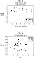
- Fig 3 is a plot of dynamic striatal binding ratios for a PD and control subject following bolus injection of 123 I Altropane. Note that there is relative stability of the binding ratios by about 15 minutes post injection. This suggests that commencing image acquisitions 15 minutes after 123 I Altropane injection will provide optimal signal:noise information.
- Fig 4 is a plot of peak striatal binding ratios (SBR) for PD and control subjects versus age.
- SBR peak striatal binding ratios
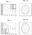
- Fig. 1 is a graphic presentation of dynamic time-activity data for a healthy control subject receiving a bolus injection of 123 I Altropane and imaged over 60 minutes. Peak uptake for striatum occurs by 10 minutes while background uptake peaks on the first acquisition. Both striatal and occipital background regions demonstrate rapid washout.
- Fig. 1a is an image showing characteristic striatal uptake and homogeneous cortical background.
- FIG. 2 is a graphic presentation of dynamic time-activity data for a PD subject following bolus injection of 123 I Altropane and imaged over 60 minutes. Peak uptake for striatum and background uptake occurs on the first acquisition. Fig. 2a demonstrates reduced striatal uptake most prominent in the putamen consistent with the known pattern of dopamine transporter loss in PD and visually distinct from the control subject in Figure 1 and 1a .
- Binding ratios decrease slightly when moving from 3 imaging heads of projection data to 2 imaging heads of projection data and further when using just one imaging head in the SPECT reconstruction. There is no effect on the shape of the SBR curves, with peak SBRs occurring from 15 minutes post injection for both the one and two head scenarios, suggesting an optimal scanning protocol from the perspective of signal to noise ratio at the points with the highest binding ratios.
- Table 3 indicates the peak SBRs for PD and control subjects (based on a single scan) demonstrating the small reductions in peak SBR when fewer imaging heads are incorporated into the reconstruction.
- Fig. 4 is a graphic presentation of Peak striatal binding ratios (SBR) for PD and control subjects versus age. The SBR is determined as the single scan post injection demonstrating highest uptake. The measure provides excellent separation between this group of well-characterized PD subjects and healthy controls.
- Fig. 5 is a graphic presentation of peak striatal binding ratios (SBR) for PD first and second (retest) study and control subjects.
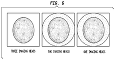
- Fig. 6 is a graphic presentation of transaxial SPECT images in a PD subject using three, two and one imaging head(s) for reconstruction. Fig.
- FIG. 6 depicts images in a PD subject for one, two, and three imaging heads of data processed as a summed image for scans. There is deterioration in the visual quality of these images for data acquired from a single head after 8 mCi 123 I Altropane injection.
- the test-retest reproducibility of the SPECT imaging was assessed in all 15 PD subjects using a mean total binding ratio taken as the mean of scan numbers 3 through 6 corresponding to roughly 30 minutes of imaging commencing about 15 min after bolus injection of 123 I Altropane. For this measure the percent test/retest reproducibility is defined as: (ratio test - ratio retest) / (ratio test) expressed as a percent.
- ratio test - ratio retest ratio / (ratio test) expressed as a percent.
- 123 I Altropane is administered at various low and high dose ranges or in single headed camera settings by utilizing various reconstruction and filtration strategies. For example, employing an iterativee reconstruction of these data would improve the signal to noise properties of the image.
- one or more filters are used on the 2D or 3D data sets.
Landscapes
- Health & Medical Sciences (AREA)
- Life Sciences & Earth Sciences (AREA)
- Epidemiology (AREA)
- Animal Behavior & Ethology (AREA)
- Chemical & Material Sciences (AREA)
- Medicinal Chemistry (AREA)
- Optics & Photonics (AREA)
- Pharmacology & Pharmacy (AREA)
- Proteomics, Peptides & Aminoacids (AREA)
- Physics & Mathematics (AREA)
- General Health & Medical Sciences (AREA)
- Public Health (AREA)
- Veterinary Medicine (AREA)
- Medicines Containing Antibodies Or Antigens For Use As Internal Diagnostic Agents (AREA)
- Nuclear Medicine (AREA)
- Peptides Or Proteins (AREA)
Description
- The present invention relates to imaging the dopamine transporter sites and more particularly to diagnosing and monitoring neurological disorders.
- There is a need for improved diagnostic methods for neurological disorders. There is a particular need for methods for diagnosing neurological disorders that selectively target a dopamine transporting protein (the dopamine transporter) and can distinguish it from another protein known as the serotonin transporter. In normal brain tissue, the dopamine:serotonin transporter density ratio is approximately 10:1. In certain neurodegenerative disorders, such as Parkinson's disease, nerve cells that produce dopamine (and on which the dopamine transporter is located) undergo severe depletion, while serotonin nerve cells are less affected. The dopamine:serotonin transporter ratio can fall to 2:1 in Parkinson's disease.
- Another neurodegenerative disorder is Lewy Body Dementia, also referred to as Dementia with Lewy Bodies (DLB). DLB, after Alzheimer's disease, is the second most frequent cause of degenerative dementia in elderly adults. DLB is characterized by both loss of dopamine-producing neurons in the substantia nigra, similar to the loss seen in Parkinson's disease, and loss of acetylcholine-producing neurons in the basal nucleus of Meynert and elsewhere. This aspect is similar to that seen in Alzheimer's disease.
- The present invention provides methods for assessing dopamine transporter levels in the central nervous system of a subject.
- In one aspect of the present invention, the method for assessing dopamine transporter levels in a selected area of a subject's central nervous system, wherein the selected area comprises the striatum, by SPECT imaging comprising the steps of: a) Initiating at least one SPECT acquisition of said selected area for an imaging effective duration of about 30 minutes wherein said duration falls within the secular equilibrium commencing period beginning about 15 minutes after administration of a labeled dopamine transporter ligand, wherein the labeled dopamine transporter ligand is 123I Altropane; and b) Assessing, at least in part based on said SPECT acquisition, the amount of said labeled dopamine transporter ligand that is bound to dopamine transporter in said selected area..
- Disclosed is a method of diagnosing or monitoring a neurological disorder in a subject comprising assessing dopamine transporter levels in the central nervous system of the subject. In one embodiment, the neurological disorder is dementia with Lewy bodies. In another embodiment, the neurological disorder is Parkinson's disease.
- Disclosed is a method for assessing dopamine transporter levels in a selected area of a subject's central nervous system by SPECT imaging comprising the steps of initiating at least one SPECT acquisition of said selected area for an imaging effective duration (in some instances about from about 10 to about 45 minutes, particularly about 20 to about 30 minutes, and notably about 30 minutes) wherein said duration falls within the secular equilibrium commencing period after administration of the labeled dopamine transporter ligand; and assessing, at least in part based on said SPECT acquisition, the amount of labeled dopamine transporter ligand that is bound to dopamine transporter in said selected area. Optionally in this method, the
labeled dopamine transporter ligand is [123I] -2β-carbomethoxy-3β-(4-flurophenyl)-N-(3-iodo-E-allyl) nortropane also termed 123I Altropane. In particular instances the labeled dopamine transporter ligand is administered at a dose of about 8 mCi. Notably, in this method the selected area comprises a portion of the brain and particularly the striatum. In an embodioment of this method the secular equilibrium period begins about 15 minutes after administration of the labeled dopamine transporter ligand and ends after about 45 minutes -
-
Figure 1 is a graphic presentation of dynamic time-activity data for a healthy control subject receiving a bolus injection of 123I Altropane and imaged over 60 min. -
Figure 1a is an image showing characteristic striatal uptake and homogeneous cortical background. -
Figure 2 is a graphic presentation of dynamic time-activity data for a PD subject following bolus injection of 123I Altropane and imaged over 60 min. -
Figure 2a is an image corresponding to the data ofFig. 2 . -
Figure 3 is a graphic presentation of dynamic striatal binding ratios for a PD and control subject following bolus injection of 123I Altropane. -
Figure 4 is a graphic presentation of peak striatal binding ratios (SBR) for PD and control subjects versus age. -
Figure 5 is a graphic presentation of peak striatal binding ratios (SBR) for PD first and second (retest) study and control subjects. -
Figure 6 is a graphic presentation of transaxial SPECT images in a PD subject using three, two and one imaging head(s) for reconstruction. - In one aspect of the present invention, the method for assessing dopamine transporter levels in the central nervous system such as the brain and particularly the striatum of a subject comprises administering to a subject an injection of a labeled dopamine transporter ligand (optionally I Altropane), preferably after the subject is positioned on an imaging table and initiating a single SPECT acquisition for an imaging effective duration, particularly duration a of about 30 minutes, commencing at about 15 minutes after administration of the labeled dopamine transporter ligand; and then assessing the amount of labeled dopamine transporter ligand that is bound to dopamine transporter in at least one region of the subject's central nervous system.
- SPECT images may be acquired using a SPECT tomograph comprising one or more heads. In one embodiment, the SPECT acquisition is performed on a single headed SPECT tomograph. In another embodiment, the SPECT acquisition is performed on a dual headed SPECT tomograph. In another embodiment, the SPECT acquisition is performed on a triple headed SPECT tomograph. Imaging effective duration will be impacted by the number of heads and the sensitivity of each head, imaging dosage and emissions, as well as by the desired detail of the image or data collected. These parameters are well known to those skilled in the art.
- In one embodiment, the labeled dopamine transporter ligand is administered at a dose ranging from about 1 mCi to about 10 mCi. In another embodiment, the labeled dopamine transporter ligand is administered at a dose ranging from about 5 mCi to about 8 mCi. In another embodiment, the labeled dopamine transporter ligand is administered at a dose of about 8 mCi.
- Disclosed is a method of diagnosing or monitoring a neurological disorder in a subject comprising assessing dopamine transporter levels in the central nervous system of the subject. In one embodiment, the neurological disorder is dementia with Lewy bodies. In another embodiment, the neurological disorder is Parkinson's disease.
- As used herein, the term "dopamine transporter ligand" means a compound that binds to the dopamine transporter. In one embodiment, compounds bind selectively to the dopamine transporter in preference to the serotonin transporter.
- The term "subject" means a patient in need of a treatment wherein treatment is broadly understood to include therapeutic, diagnostic, and prophylactic steps and procedures. In one embodiment, a subject is a mammal and particularly a human.
- The term "selected area" means a central nervous system such as the brain and particularly the striatum of the brain of a subject.
- In one aspect, the labeled dopamine transporter ligand is administered to the subject at approximately the time the subject is positioned for SPECT imaging. The term "approximately the time" refers to from about 60 minutes, and particularly about 30 minutes, more particularly about 15 minutes, to substantially immediately before or after a subject is positioned for SPECT imaging. In one embodiment, the subject is first positioned on an imaging table and then subsequently administered the dopamine transporter ligand. In another embodiment, the subject is first administered the dopamine transporter ligand and subsequently positioned on an imaging table.
- As to scan times, it has been discovered that the signal-to-noise (S/N) ratio of the SPECT scans of the dopamine transporter ligand is optimal between the time of about 15 minutes and about 45 minutes post-injection of the dopamine transporter ligand.
- Several alternatives are available for acquiring images. In one embodiment, the SPECT scanning is one continuous scan or a series of shorter scans. In another embodiment, the SPECT scanning comprises a series of one or more two minute scans. In another embodiment, the SPECT scanning comprises a series of one or more ten minute scans. In one particular embodiment, the SPECT images are generated using one continuous 30 minute scan. In another particular embodiment, the SPECT images are generated using a series of two minute scans for a period of 60 minutes. In another particular embodiment, the SPECT images are generated using a series of 4 minute scans for a period of 40 minutes.
- Assessment of dopamine transporter levels, as in the course of a diagnostic investigation, can be performed by measuring dopamine transporter availability. Measurements can be made using devices such as positron emission tomography ("PET") or single photon emission computed tomography ("SPECT"). To measure dopamine transporter availability, a labeled probe that targets the transporter is introduced into the brain (e.g., intravenously) and PET or SPECT is performed. From the PET or SPECT data and images, the density of the dopamine transporter is quantified. In one embodiment, quantification is determined by computing binding potential, where binding potential is defined as the maximum number of binding sites, Bmax, divided by a dissociation constant, Kd. The binding potential is calculated from a continuous scan starting at about 15 min. The region of interest is identified and the counts in that region are determined. Using appropriate modeling, numerical values for binding potential can be calculated and these values can be compared between subjects who have undergone equivalent treatment and scanning protocols. The striatal binding potential of 123I-Altropane (k3/k4) is calculated by the reference region approach as described by Farde, et al. (Farde, et al., 1989, J. Cereb. Blood Flow Metab 9:696 708).
- Imaging agents that target the dopamine transporter include (11C)Altropane, (11C or 18F) WIN 35,428 ((11C)CFT), 123I Altropane, (99mTc) 0-1505, 99mTc-technepine, and similar compounds. Without being bound by any particular theory, it is believed that these agents bind the dopamine transporter with varying affinities, allowing multiple, dissimilar assessments to be performed. Structures, synthesis, and/or sources of some of the above agents are described in Fischman et al., 1998, Synapse 29:125 41 ((123I) Altropane); Madras et al., 1996, Synapse 22:239 46; Meltzer et al., 1993, J. Med. Chem. 36:855 62; and Milius et al., 1990, J. Medicinal Chem. 34:1728 31. Another useful compound includes 123I-Ioflupane (DatSCAN™, (Nycomed-Amersham, Piscataway, NJ).
- Examples of suitable ligands include (11C)CFT ((11C)WIN 35,428), (123I)Altropane®, and (18F)CFT. Ligands particularly suitable for use in PET include, but are not limited to, (11C) Altropane. Ligands suitable for use in SPECT include, but are not limited to, technetium-labeled phenyltropane probes, such as (99mTc) technepine, O-1505, and similar compounds. Other examples of compounds useful in the methods of the present invention are described in
U.S. Pat. Nos. 5,493,026 ;5,506,359 ;5,770,180 ;5,853,696 ;5,948,933 ;6,171,576 ;6,548,041 and7,081,238 , the disclosures of which are hereby incorporated by reference. The portion of the subject's central nervous system for assessment is preferably a portion of the human brain, e.g., the striatum. - In one aspect, the invention employs 123I compounds as disclosed in
U.S. Pat. No. 5,493,026 , with particular reference to 2β-carbomethoxy-3β-(4'-flurophenyl)-N-(3-iodo-E-allyl) nortropane (ALTROPANE®, Alseres Pharmaceuticals, Inc. Hopkinton, MA) to differentiate neurological disorders. For these same functions, this invention also employs tropanes incorporating technetium (99mTc) as a radiolabel as disclosed inU.S. Pat. Nos. 6,171,576 and6,548,041 . - The compounds described in
U.S. Pat. No. 6,548,041 have a tropane compound linked through the nitrogen atom at the 8-position to a chelating ligand capable of complexing a technetium or rhenium radionuclide to produce a neutral labeled complex that selectively binds to the dopamine transporter. The tropane compound is believed to bind to the dopamine transporter. - The following examples serve to illustrate certain useful embodiments and aspects of the present invention. Alternative materials and methods can be utilized to obtain similar results.
- The following example summarizes single photon emission computed tomographic (SPECT) image acquisition, analysis methods and results for Parkinson's disease research participants and healthy controls participating in an 123I Altropane SPECT imaging study. The experiment is an open label study evaluating time-activity curves and striatal dopamine transporter occupancy over the first hour post injection of 123I Altropane using temporally well-resolved dynamic SPECT in Parkinson's subjects and similarly aged healthy control subjects. In addition, for the PD subjects, a second injection and scan is performed on each subject on a separate test days to evaluate the test/retest reproducibility data for both visual and quantitative analyses.
- Fifteen idiopathic Parkinson's disease subjects are recruited through local advertising and word-of-mouth for enrollment in the study. Imaging is performed at Molecular NeuroImaging, LLC in New Haven, Connecticut, USA. 123I Altropane is studied under the auspices of a physician-sponsored IND # 76,024, Dr. J. Seibyl physician sponsor using test article provided by MDS Nordion, Inc (Vancouver, BC, Canada). All subjects are provided informed consent prior to any study procedures. All fifteen subjects completed two separate injection and SPECT scan sessions with approximately 296 mBq (8 mCi) of 123I Altropane. Subjects undergo motor ratings using the Unified Parkinson's Disease Rating Scale (UPDRS) following an overnight period without anti-Parkinson medication administered by a movement disorder specialist.
- Twelve healthy control subjects are recruited for participation in a single scan session with 123I Altropane using the identical SPECT image acquisition protocol as the PD subjects. Healthy subjects are evaluated by a movement disorder specialist to confirm the absence of neurologic illness.
- SPECT scans are acquired for 60 minutes following intravenous injection with 296 mBq (8.0 mCi) of 123I Altropane. A series of dynamic SPECT scans are obtained as five scans at 6 minutes per acquisition followed by three scans at 10 minutes per acquisition. The subject remain in the camera for the duration of the acquisition. Each SPECT study is acquired on a Philips PRISM 3000XP triple-headed SPECT camera (Cleveland, Ohio, USA) fitted with fan-beam collimators. Each head rotates 360 degrees, sampling every 3 degrees for a total of 120 raw projection images per head. Projection data were collected in a 128 x 128 matrix within a symmetric energy window centered at 159 kEv (± 10%). This acquisition protocol permits the post hoc analysis of imaging data at each time point using information from 1, 2, or all 3 heads, hence modeling the impact of different injected doses of 123I Altropane at 2.7, 5.3, and 8.0 mCi, respectively.
- Data are re-constructed using filtered back projection and a simple ramp filter followed by a post hoc (3-D) standardized low pass filter. Attenuation correction was performed applying a
Chang 0 correction and a mu of 0.11 cm-1 using a standard or custom automated software package. Regions of interest (ROIs) are placed in the 3-head data with individual ROI sampling of the left and right caudate and putamen and an occipital background region. The ROIs were then applied to all images in the injection session (total of 24= 8 time points x 3 head conditions (1, 1&2, 1&2&3). To check for head movement within each scan session, five external skin fiducial markers containing 1 µCi of 123I were place along the canthomeatal line (2 right side, 3 left side) prior to each SPECT scan. Total counts within the ROI, total volume, and count density (counts/voxel) were extracted from each scan and logged in a data spreadsheet for determination of striatal uptake ratios (SBR) defined as the density of counts (counts per voxel per minute) in the striatal region divided by the density of counts in the occipital background region. The mean striatal SBR scores were calculated as the mean of the left and right caudate and putamen SBR scores. Since sampling in striatal subregions used the identical size ROI, there was equal contribution from left and right caudate and putamen to the mean SBR. - The time-activity data for mean striatal count densities and occipital background for each of the 8 images acquired in the 60 minutes post injection sampling period were plotted to permit visual interrogation of the uptake and washout characteristics of 123I Altropane in PD subjects and control subjects. In addition, each SBR ratio obtained at the 8 time points after injection were plotted. The peak SBR was assessed for each subject for data acquired using 1, 2, and 3 heads of imaging data.
- For each PD subject a composite SBR ratio was determined for the scans (which are summed in
Fig. 6 ) of each injection, corresponding to data collected over 13-40 minutes post injection (27 minutes total time). These time points were selected based on review of the peak and persistence of SBRs over the 60 minutes of image acquisition and a visual interrogation of the scan for optimal identification of striatal structures. This composite SBR was then compared within each subject's first and second 123I Altropane injection days to review the reproducibility of the SBR using the following equation:
-
Fig 3 . is a plot of dynamic striatal binding ratios for a PD and control subject following bolus injection of 123I Altropane. Note that there is relative stability of the binding ratios by about 15 minutes post injection. This suggests that commencing image acquisitions 15 minutes after 123I Altropane injection will provide optimal signal:noise information. -
Fig 4 . is a plot of peak striatal binding ratios (SBR) for PD and control subjects versus age. The SBR is determined as the single scan post injection demonstrating highest uptake. While this outcome measure is not an accurate quantitative measure for assessing dopamine transporter density, it nonetheless provides excellent separation between this group of well-characterized PD subjects and healthy controls. - All 15 PD subjects enrolled completed the trial with a baseline and retest 123I Altropane SPECT studies and all 12 healthy controls completed all imaging assessments for a single study with 123I Altropane. All imaging data was of good technical quality with adequate counting statistics in the raw projection data sets and included in the analysis. No scans needed to be eliminated for technical reasons (e.g. head movement, low injected dose, or other reason for sub-optimal quality). Subject demographic are summarized in Table 1.
Table 1 Parkinson subjects: Demographics and UPDRS motor ratings PD Subject No. Gender Age Duration DX Altropane Dose (mCi) HY Total UPDRS Motor UPDRS 1 Male 67.0 1.0 8.6 2 35 27 2 6.6 2 35 27 3 Male 66.0 10.0 6.6 2 48 28 4 8.2 2 46 26 5 Male 51.6 6.1 8.1 2 23 16 6 8.5 2 23 16 7 Male 64.0 10.0 6.8 1 22 9 8 8.5 2 30 17 9 Male 74.6 9.2 8.4 2 31 21 10 8.2 2 38 24 11 Female 60.6 7.8 8.2 3 46 27 12 8.3 3 40 26 13 Male 73.5 3.0 8.4 1 20 17 14 8.6 1 15 12 15 Male 52.7 2.0 8.2 2 45 31 16 8.4 2 46 31 17 Female 52.7 1.0 8.4 2 27 20 18 8.4 2 28 20 19 Male 44.9 10.6 8.4 2 46 34 20 8.4 2 35 23 21 Male 60.4 6.9 8.3 2 31 18 22 8.4 2 36 21 23 Female 85.4 2.2 8.4 1 13 6 24 8.1 1 13 6 25 Male 76.0 4.3 8.2 2 27 17 26 8.7 2 23 15 27 Female 53.2 8.7 6.7 2 42 25 28 8.4 2 44 26 29 Male 62.3 13.0 8.5 2 30 20 30 8.7 2 30 19 Note that each subject has paired scans consecutively numbered, e.g., Scans 1 and 2 are the same patient. Table 2 Healthy control subjects Control Subject No. Gender Age Altropane Dose (mCi) 1 Male 62.3 7.92 2 Female 65.8 8.64 3 Male 58.4 8.57 4 Male 77.2 8.3 5 Female 74.9 8.5 6 Male 61.7 8.47 7 Male 60.1 8.46 8 Male 55.3 8.08 9 Female 59.3 8.75 10 Female 54.7 8.62 11 Male 61.2 8.42 12 Male 55.9 8.37 - Following injection of 123I Altropane in controls and PD subjects peak striatal count densities are noted at 10 minutes post-injection. (
Figs. 1 and 2 ). It is noted that most subjects exhibit rapid elimination of Altropane from brain.Fig. 1 is a graphic presentation of dynamic time-activity data for a healthy control subject receiving a bolus injection of 123I Altropane and imaged over 60 minutes. Peak uptake for striatum occurs by 10 minutes while background uptake peaks on the first acquisition. Both striatal and occipital background regions demonstrate rapid washout.Fig. 1a is an image showing characteristic striatal uptake and homogeneous cortical background.Fig. 2 is a graphic presentation of dynamic time-activity data for a PD subject following bolus injection of 123I Altropane and imaged over 60 minutes. Peak uptake for striatum and background uptake occurs on the first acquisition.Fig. 2a demonstrates reduced striatal uptake most prominent in the putamen consistent with the known pattern of dopamine transporter loss in PD and visually distinct from the control subject inFigure 1 and 1a . - Localization of uptake in striatal structures was well demonstrated in all subjects in the study with the characteristic "comma" shaped appearance of striatal uptake in controls and more asymmetric uptake (left-right asymmetry and caudate > putamen asymmetry) in PD subjects consistent with other dopamine transporter studies using Altropane and other SPECT studies in PD. By 20-30 minutes post injection of 123I Altropane rates of washout from striatum and occipital background region were similar resulting in stability of the striatal binding ratio.
- Binding ratios decrease slightly when moving from 3 imaging heads of projection data to 2 imaging heads of projection data and further when using just one imaging head in the SPECT reconstruction. There is no effect on the shape of the SBR curves, with peak SBRs occurring from 15 minutes post injection for both the one and two head scenarios, suggesting an optimal scanning protocol from the perspective of signal to noise ratio at the points with the highest binding ratios. Table 3 indicates the peak SBRs for PD and control subjects (based on a single scan) demonstrating the small reductions in peak SBR when fewer imaging heads are incorporated into the reconstruction.
Table 3 Peak Striatal:Occipital Ratios PD 3 head 2 head 1 head Controls 3 head 2 head 1 head 1 1.39 1.37 1.33 1 1.68 1.55 1.50 2 1.28 1.27 1.41 2 1.86 1.84 1.66 3 1.43 1.39 1.33 3 1.77 1.61 1.51 4 1.30 1.28 1.29 4 1.81 1.66 1.49 5 1.30 1.27 1.24 5 1.79 1.67 1.57 6 1.25 1.25 1.22 6 2.06 1.95 1.79 7 1.44 1.41 1.35 7 1.69 1.62 1.47 8 1.44 1.46 1.41 8 1.81 1.68 1.56 9 1.74 1.66 1.52 9 1.90 1.79 1.63 10 1.60 1.53 1.43 10 1.93 1.77 1.64 11 1.33 1.26 1.26 11 2.30 2.10 1.86 12 1.34 1.28 1.24 12 1.71 1.59 1.52 13 1.45 1.36 1.29 14 1.35 1.32 1.26 15 1.45 1.43 1.32 mean mean SBR 1.41 1.37 1.33 SBR 1.86 1.74 1.60 %COV 9.1 8.5 6.5 %COV 9.4 9.4 7.6 - Examination of the semi-quantitative peak SBR demonstrates differences between the PD and healthy controls (
Figs. 4 ,5 ).Fig. 4 . is a graphic presentation of Peak striatal binding ratios (SBR) for PD and control subjects versus age. The SBR is determined as the single scan post injection demonstrating highest uptake. The measure provides excellent separation between this group of well-characterized PD subjects and healthy controls.Fig. 5 is a graphic presentation of peak striatal binding ratios (SBR) for PD first and second (retest) study and control subjects.Fig. 6 is a graphic presentation of transaxial SPECT images in a PD subject using three, two and one imaging head(s) for reconstruction.Fig. 6 depicts images in a PD subject for one, two, and three imaging heads of data processed as a summed image for scans. There is deterioration in the visual quality of these images for data acquired from a single head after 8 mCi 123I Altropane injection. - The test-retest reproducibility of the SPECT imaging was assessed in all 15 PD subjects using a mean total binding ratio taken as the mean of scan numbers 3 through 6 corresponding to roughly 30 minutes of imaging commencing about 15 min after bolus injection of 123I Altropane. For this measure the percent test/retest reproducibility is defined as: (ratio test - ratio retest) / (ratio test) expressed as a percent. The results for all PD subjects under conditions of 3, 2, and 1 imaging head data reconstructions are described in Table 4.
Table 4. Total striatal binding ratios 3 heads 2 heads 1 head Subject No. Ratio (3-6) Ratio (3-6) Ratio (3-6) 1 1.30 1.27 1.25 2 1.37 1.32 1.26 % test/retest 5.2 3.7 1.1 3 1.27 1.24 1.20 4 1.32 1.29 1.29 % test/retest 3.5 4.2 7.7 5 1.30 1.27 1.23 6 1.39 1.35 1.29 % test/retest 6.3 6.4 4.8 7 1.41 1.37 1.30 8 1.33 1.31 1.26 % test/retest 6.1 4.8 3.2 9 1.36 1.32 1.25 10 1.35 1.30 1.22 % test/retest 1.1 1.5 2.9 11 1.38 1.36 1.29 12 1.45 1.43 1.37 % test/retest 4.8 4.9 6.1 13 1.56 1.47 1.40 14 1.70 1.60 1.48 % test/retest 8.8 8.5 5.5 15 1.37 1.32 1.29 16 1.28 1.25 1.22 % test/retest 6.3 5.2 5.8 17 1.23 1.21 1.20 18 1.20 1.19 1.16 % test/retest 2.4 2.3 3.1 19 1.42 1.38 1.32 20 1.44 1.40 1.35 % test/retest 1.6 1.8 1.9 21 1.26 1.24 1.21 22 1.15 1.12 1.12 % test/retest 8.3 9.9 7.5 23 1.29 1.26 1.21 24 1.31 1.27 1.25 % test/retest 1.4 1.1 3.7 25 1.35 1.33 1.28 26 1.22 1.20 1.17 % test/retest 9.7 9.6 8.7 27 1.36 1.32 1.24 28 1.42 1.38 1.30 % test/retest 4.7 4.7 4.3 29 1.50 1.45 1.37 30 1.40 1.33 1.26 % test/retest 6.9 8.1 7.6 Mean & test/retest difference 5.14% 5.11% 4.94% - Total striatal binding obtained during the time points corresponding to roughly 30 minutes commencing 15 minutes after 123I Altropane injection demonstrates robust reproducibility in the 15 PD subjects studied. The reduction of counts involved in the reconstruction had no significant impact on the reproducibility measure used in this analysis.
-
- a) Single rapid or bolus injection of 8 mCi of 123I Altropane administered after the subject is positioned on the imaging table,
- b) Initiate a single 30 minutes SPECT acquisition commencing at 15 minutes after administration of 123I Altropane,
- c) Scans are performed on dual and triple-headed SPECT tomographs.
- In alternative embodiments, 123I Altropane is administered at various low and high dose ranges or in single headed camera settings by utilizing various reconstruction and filtration strategies. For example, employing an iterativee reconstruction of these data would improve the signal to noise properties of the image. In one embodiment, one or more filters are used on the 2D or 3D data sets.
- The findings from this dataset are generalizable. In one embodiment, reconstruction and filtration of the 123I Altropane imaging data uses commonly available and standard algorithms that might be encountered in a typical clinical nuclear medicine setting.
-
- Seibyl J, Marek K, Sheff K, et al. Iodine-123-beta-CIT and lodine-123-FPCIT SPECT Measurement of Dopamine Transporters in Healthy Subjects and Parkinson's Patients. J. Nucl. Med. 39:1500-07, 1998.
- Marek K, Seibyl J: Tech/Sight Imaging: A molecular map for neurodegeneration. Science 289(54781: 409-11, 2000.
- Seibyl J, Marek K, Quinlan D, Sheff K, Zoghbi S, Zea-Ponce Y, Baldwin R, Fussell B, Smith E, Chamey D, Hoffer P, Innis R. Decreased SPECT [123-I]β-CIT striatal uptake correlates with symptom severity in idiopathic Parkinson's disease. Annals of Neurology, 38:589-98, 1995.
- Seibyl J, Marek K, Sheff K, Baldwin R, Zoghbi S, Zea-Ponce Y, Chamey D, van Dyck C, Hoffer P, Innis R. Test/retest reproducibility of [1231]β-CIT SPECT brain measurement of dopamine transporters in Parkinson's patients. J. Nucl. Med. 38:1453-59, 1997.
- Lamelle M, Wallace E, Seibyl J, Baldwin R, Zea-Ponce Y, Zoghbi S, Neumeyer J, Chamey DS, Hoffer PB, Innis RB. Graphical, kinetic, and equilibrium analyses of in vivo [123I]β-CIT binding to dopamine transporters in healthy human subjects. J. Cereb. Blood Flow Metab. 14:982-94, 1994.
- Kugaya, A., Seneca, N. M. Snyder, P. J., Williams, S. A. ,Malison, R. T., Baldwin, R. M., Seibyl, J. P., Innis, R. B.., Changes in human in vivo serotonin and dopamine transporter availabilities during chronic antidepressant administration. Neuropsychopharmacology,. 28(2):413-20, 2003.
- Sahani, D., S. Saini, G.A. Fatuga, E.F. Halpern, M.E. Lanser, J.B. Zimmerman, and A.J. Fischman, Quantitative measurements of medical images for pharmaceutical clinical trials: comparison between on-site and off-site assessments. AJR Am J Roentgenol, 2000. 174(4): p. 1159-62.
- Fischman, A.J., A.A. Bonab, J.W. Babich, E.P. Palmer, N.M. Alpert, D.R. Elmaleh, R.J. Callahan, S.A. Barrow, W. Graham, P.C. Meltzer, R.N. Hanson, and B.K. Madras, Rapid detection of Parkinson's disease by SPECT with altropane: a selective ligand for dopamine transporters. Synapse, 1998. 29(2): p. 128-41.
- Madras, B.K., L.M. Gracz, M.A. Fahey, D. Elmaleh, P.C. Meltzer, A.Y. Liang, E.G. Stopa, J. Babich, and A.J. Fischman, Altropane, a SPECT or PET imaging probe for dopamine neurons: III. Human dopamine transporter in postmortem normal and Parkinson's diseased brain. Synapse, 1998. 29(2): p. 116-27.
- Madras, B.K., L.M. Gracz, P.C. Meltzer, A.Y. Liang, D.R. Elmaleh, M.J. Kaufman, and A.J. Fischman, Altropane, a SPECT or PET imaging probe for dopamine neurons: II. Distribution to dopamine-rich regions of primate brain. Synapse, 1998. 29(2): p. 105-15.
- Madras, B.K., P.C. Meltzer, A.Y. Liang, D.R. Elmaleh, J. Babich, and A.J. Fischman, Altropane, a SPECT or PET imaging probe for dopamine neurons: I. Dopamine transporter binding in primate brain. Synapse, 1998. 29(2): p. 93-104.
- Fischman, A.J., J.W. Babich, D.R. Elmaleh, S.A. Barrow, P. Meltzer, R.N. Hanson, and B.K. Madras, SPECT imaging of dopamine transporter sites in normal and MPTP-Treated rhesus monkeys. J Nucl Med, 1997. 38(1): p. 144-50.
Claims (5)
- A method for assessing dopamine transporter levels in a selected area of a subject's central nervous system, wherein the selected area comprises the striatum, by SPECT imaging comprising the steps of: a) Initiating at least one SPECT acquisition of said selected area for an imaging effective duration of about 30 minutes wherein said duration falls within the secular equilibrium commencing period beginning about 15 minutes after administration of a labeled dopamine transporter ligand, wherein the labeled dopamine transporter ligand is 123I Altropane; and b) Assessing, at least in part based on said SPECT acquisition, the amount of said labeled dopamine transporter ligand that is bound to dopamine transporter in said selected area.
- The method of Claim 1 wherein said secular equilibrium period ends about 45 minutes after administration of the labeled dopamine transporter ligand.
- The method of Claim 1 or 2, further comprising administering to the subject a labeled dopamine transporter ligand at approximately the time the subject is positioned for SPECT imaging.
- The method of any one of Claims 1 to 3, wherein the labeled dopamine transporter ligand is administered at a dose of about 8 mCi.
- Use of 123I Altropane in a method according to any one of claims 1 to 4.
Applications Claiming Priority (2)
| Application Number | Priority Date | Filing Date | Title |
|---|---|---|---|
| US11/931,979 US8084018B2 (en) | 2007-10-31 | 2007-10-31 | Methods for imaging dopamine transporter level |
| PCT/US2008/081419 WO2009058754A1 (en) | 2007-10-31 | 2008-10-28 | Methods for imaging dopamine transporter level |
Publications (3)
| Publication Number | Publication Date |
|---|---|
| EP2219682A1 EP2219682A1 (en) | 2010-08-25 |
| EP2219682A4 EP2219682A4 (en) | 2012-11-14 |
| EP2219682B1 true EP2219682B1 (en) | 2018-07-18 |
Family
ID=40583113
Family Applications (1)
| Application Number | Title | Priority Date | Filing Date |
|---|---|---|---|
| EP08843521.9A Active EP2219682B1 (en) | 2007-10-31 | 2008-10-28 | Methods for imaging dopamine transporter level |
Country Status (5)
| Country | Link |
|---|---|
| US (1) | US8084018B2 (en) |
| EP (1) | EP2219682B1 (en) |
| JP (2) | JP2011502130A (en) |
| CA (1) | CA2700468C (en) |
| WO (1) | WO2009058754A1 (en) |
Families Citing this family (9)
| Publication number | Priority date | Publication date | Assignee | Title |
|---|---|---|---|---|
| DE50208552D1 (en) | 2002-09-26 | 2006-12-07 | Ilford Imaging Ch Gmbh | Copper complex monoazo dyes, their preparation and use |
| CA2703563A1 (en) * | 2007-10-31 | 2009-05-07 | Alseres Pharmaceuticals, Inc. | Methods for diagnosing and monitoring treatment of lewy body dementia by assessing dopamine transporter level |
| GB201313291D0 (en) * | 2013-07-25 | 2013-09-11 | Ge Healthcare Ltd | Imaging neurological disease |
| JP5669118B1 (en) * | 2013-12-05 | 2015-02-12 | 独立行政法人国立循環器病研究センター | 3D brain state image analyzer |
| KR20170131829A (en) * | 2015-03-24 | 2017-11-30 | 니혼 메디피직스 가부시키가이샤 | Image processing apparatus, image processing method, and program |
| EP3579887A4 (en) * | 2017-02-10 | 2020-09-16 | Likeminds, Inc. | Methods for in vivo monitoring of dopaminergic disorders and efficacy of treatment agents therefor |
| EP3833403A4 (en) * | 2018-08-07 | 2022-05-04 | Likeminds, Inc. | Method for diagnosis of dopaminergic and movement disorders |
| WO2021102323A1 (en) * | 2019-11-21 | 2021-05-27 | Likeminds, Inc. | Pharmaceutical packaging units and methods for concomitant administration of radiotracers |
| WO2022240817A1 (en) * | 2021-05-13 | 2022-11-17 | Likeminds, Inc. | Methods of visualizing dopamine transporters |
Family Cites Families (10)
| Publication number | Priority date | Publication date | Assignee | Title |
|---|---|---|---|---|
| US5770180A (en) | 1992-08-24 | 1998-06-23 | Organix, Inc. | Bridge-substituted tropanes for methods of imaging and therapy |
| AU5088993A (en) | 1992-08-24 | 1994-03-15 | President And Fellows Of Harvard College | Cocaine analogues and their use as cocaine drug therapies and therapeutic and imaging agents for neurodegenerative disorders |
| US5493026A (en) | 1993-10-25 | 1996-02-20 | Organix, Inc. | Substituted 2-carboxyalkyl-3-(fluorophenyl)-8-(3-halopropen-2-yl) nortropanes and their use as imaging for agents for neurodegenerative disorders |
| US20020150535A1 (en) * | 1995-11-03 | 2002-10-17 | Madras Bertha K. | Methods for diagnosing and monitoring treatment ADHD by assessing the dopamine transporter level |
| US5948933A (en) | 1997-07-11 | 1999-09-07 | Organix, Inc. | Tropane analogs and methods for inhibition of monoamine transport |
| US7081238B2 (en) * | 1995-11-03 | 2006-07-25 | The President And Fellows Of Harvard College | Methods for diagnosing and monitoring treatment ADHD by assessing the dopamine transporter level |
| US6171576B1 (en) | 1995-11-03 | 2001-01-09 | Organix Inc. | Dopamine transporter imaging agent |
| US7553478B2 (en) * | 1999-05-12 | 2009-06-30 | President And Fellows Of Harvard College | Methods for diagnosing and monitoring treatment ADHD by assessing the dopamine transporter level |
| CA2721484A1 (en) | 1999-05-12 | 2000-11-12 | President And Fellows Of Harvard College | Dopamine transporter imaging agents |
| US20100143248A1 (en) * | 1999-06-28 | 2010-06-10 | President And Fellows Of Harvard College | Methods for diagnosing and monitoring treatment of adhd by assessing the dopamine transporter level |
-
2007
- 2007-10-31 US US11/931,979 patent/US8084018B2/en active Active
-
2008
- 2008-10-28 JP JP2010531324A patent/JP2011502130A/en active Pending
- 2008-10-28 EP EP08843521.9A patent/EP2219682B1/en active Active
- 2008-10-28 CA CA2700468A patent/CA2700468C/en active Active
- 2008-10-28 WO PCT/US2008/081419 patent/WO2009058754A1/en not_active Ceased
-
2014
- 2014-04-18 JP JP2014086233A patent/JP2014148529A/en active Pending
Also Published As
| Publication number | Publication date |
|---|---|
| WO2009058754A1 (en) | 2009-05-07 |
| CA2700468C (en) | 2016-11-29 |
| US8084018B2 (en) | 2011-12-27 |
| CA2700468A1 (en) | 2009-05-07 |
| US20090110635A1 (en) | 2009-04-30 |
| JP2011502130A (en) | 2011-01-20 |
| EP2219682A1 (en) | 2010-08-25 |
| EP2219682A4 (en) | 2012-11-14 |
| JP2014148529A (en) | 2014-08-21 |
Similar Documents
| Publication | Publication Date | Title |
|---|---|---|
| CA2700468C (en) | Methods for imaging dopamine transporter level | |
| Morbelli et al. | EANM practice guideline/SNMMI procedure standard for dopaminergic imaging in Parkinsonian syndromes 1.0 | |
| Fischman et al. | Rapid detection of Parkinson's disease by SPECT with altropane: a selective ligand for dopamine transporters | |
| Arlicot et al. | Initial evaluation in healthy humans of [18F] DPA-714, a potential PET biomarker for neuroinflammation | |
| Ouchi et al. | Alterations in binding site density of dopamine transporter in the striatum, orbitofrontal cortex, and amygdala in early Parkinson's disease: Compartment analysis for β‐CFT binding with positron emission tomography | |
| Hatazawa et al. | Evaluation of cerebral infarction with iodine 123-iomazenil SPECT | |
| JP5603855B2 (en) | Imaging neurodegenerative diseases with radiopharmaceuticals | |
| Holman et al. | Regional cerebral blood flow improves with treatment in chronic cocaine polydrug users | |
| US5377681A (en) | Method of diagnosing impaired blood flow | |
| US20100312105A1 (en) | Methods for diagnosing and monitoring treatment of lewy body dementia by assessing dopamine transporter level | |
| Ell et al. | Functional imaging of the brain | |
| Kuikka et al. | Pharmacokinetics and dosimetry of iodine-123 labelled PE2I in humans, a radioligand for dopamine transporter imaging | |
| Fischman et al. | SPECT imaging of dopamine transporter sites in normal and MPTP-treated rhesus monkeys | |
| Laurencin et al. | Modeling [11C] yohimbine PET human brain kinetics with test-retest reliability, competition sensitivity studies and search for a suitable reference region | |
| US20210162078A1 (en) | Method for diagnosis of dopaminergic and movement disorders | |
| Brownell et al. | Dopamine imaging markers and predictive mathematical models for progressive degeneration in Parkinson's disease | |
| Zhao et al. | First-in-human study of D6-[18F] FP-(+)-DTBZ, a novel VMAT2 tracer: whole-body biodistribution and brain PET comparison with [18F] FP-(+)-DTBZ (AV-133) | |
| Wienhard | Applications of 3D PET | |
| TWI520715B (en) | (99mtc) trodat-1 and (123i) adam brain spect in nonhuman primates | |
| Gerson et al. | Comparison of technetium-99m-Q3 and thallium-201 for detection of coronary artery disease in humans | |
| Chua et al. | Technetium-99m teboroxime regional myocardial washout in subjects with and without coronary artery disease | |
| Bailey et al. | APPLICATIONS OF 3D PET |
Legal Events
| Date | Code | Title | Description |
|---|---|---|---|
| PUAI | Public reference made under article 153(3) epc to a published international application that has entered the european phase |
Free format text: ORIGINAL CODE: 0009012 |
|
| 17P | Request for examination filed |
Effective date: 20100528 |
|
| AK | Designated contracting states |
Kind code of ref document: A1 Designated state(s): AT BE BG CH CY CZ DE DK EE ES FI FR GB GR HR HU IE IS IT LI LT LU LV MC MT NL NO PL PT RO SE SI SK TR |
|
| AX | Request for extension of the european patent |
Extension state: AL BA MK RS |
|
| DAX | Request for extension of the european patent (deleted) | ||
| RAP1 | Party data changed (applicant data changed or rights of an application transferred) |
Owner name: PRESIDENT AND FELLOWS OF HARVARD COLLEGE Owner name: ALSERES PHARMACEUTICALS, INC. |
|
| A4 | Supplementary search report drawn up and despatched |
Effective date: 20121017 |
|
| RIC1 | Information provided on ipc code assigned before grant |
Ipc: A61K 51/00 20060101AFI20121011BHEP |
|
| 17Q | First examination report despatched |
Effective date: 20160926 |
|
| STAA | Information on the status of an ep patent application or granted ep patent |
Free format text: STATUS: EXAMINATION IS IN PROGRESS |
|
| REG | Reference to a national code |
Ref country code: DE Ref legal event code: R079 Ref document number: 602008056080 Country of ref document: DE Free format text: PREVIOUS MAIN CLASS: A61K0051000000 Ipc: A61K0051040000 |
|
| GRAP | Despatch of communication of intention to grant a patent |
Free format text: ORIGINAL CODE: EPIDOSNIGR1 |
|
| STAA | Information on the status of an ep patent application or granted ep patent |
Free format text: STATUS: GRANT OF PATENT IS INTENDED |
|
| RIC1 | Information provided on ipc code assigned before grant |
Ipc: A61K 51/04 20060101AFI20180206BHEP Ipc: A61K 51/00 20060101ALI20180206BHEP Ipc: A61K 101/02 20060101ALI20180206BHEP |
|
| INTG | Intention to grant announced |
Effective date: 20180307 |
|
| GRAS | Grant fee paid |
Free format text: ORIGINAL CODE: EPIDOSNIGR3 |
|
| GRAA | (expected) grant |
Free format text: ORIGINAL CODE: 0009210 |
|
| STAA | Information on the status of an ep patent application or granted ep patent |
Free format text: STATUS: THE PATENT HAS BEEN GRANTED |
|
| AK | Designated contracting states |
Kind code of ref document: B1 Designated state(s): AT BE BG CH CY CZ DE DK EE ES FI FR GB GR HR HU IE IS IT LI LT LU LV MC MT NL NO PL PT RO SE SI SK TR |
|
| REG | Reference to a national code |
Ref country code: GB Ref legal event code: FG4D |
|
| REG | Reference to a national code |
Ref country code: CH Ref legal event code: EP |
|
| REG | Reference to a national code |
Ref country code: IE Ref legal event code: FG4D |
|
| REG | Reference to a national code |
Ref country code: AT Ref legal event code: REF Ref document number: 1018666 Country of ref document: AT Kind code of ref document: T Effective date: 20180815 |
|
| REG | Reference to a national code |
Ref country code: DE Ref legal event code: R096 Ref document number: 602008056080 Country of ref document: DE |
|
| REG | Reference to a national code |
Ref country code: FR Ref legal event code: PLFP Year of fee payment: 11 |
|
| REG | Reference to a national code |
Ref country code: NL Ref legal event code: MP Effective date: 20180718 |
|
| REG | Reference to a national code |
Ref country code: LT Ref legal event code: MG4D |
|
| REG | Reference to a national code |
Ref country code: AT Ref legal event code: MK05 Ref document number: 1018666 Country of ref document: AT Kind code of ref document: T Effective date: 20180718 |
|
| PG25 | Lapsed in a contracting state [announced via postgrant information from national office to epo] |
Ref country code: NL Free format text: LAPSE BECAUSE OF FAILURE TO SUBMIT A TRANSLATION OF THE DESCRIPTION OR TO PAY THE FEE WITHIN THE PRESCRIBED TIME-LIMIT Effective date: 20180718 |
|
| PG25 | Lapsed in a contracting state [announced via postgrant information from national office to epo] |
Ref country code: NO Free format text: LAPSE BECAUSE OF FAILURE TO SUBMIT A TRANSLATION OF THE DESCRIPTION OR TO PAY THE FEE WITHIN THE PRESCRIBED TIME-LIMIT Effective date: 20181018 Ref country code: GR Free format text: LAPSE BECAUSE OF FAILURE TO SUBMIT A TRANSLATION OF THE DESCRIPTION OR TO PAY THE FEE WITHIN THE PRESCRIBED TIME-LIMIT Effective date: 20181019 Ref country code: AT Free format text: LAPSE BECAUSE OF FAILURE TO SUBMIT A TRANSLATION OF THE DESCRIPTION OR TO PAY THE FEE WITHIN THE PRESCRIBED TIME-LIMIT Effective date: 20180718 Ref country code: IS Free format text: LAPSE BECAUSE OF FAILURE TO SUBMIT A TRANSLATION OF THE DESCRIPTION OR TO PAY THE FEE WITHIN THE PRESCRIBED TIME-LIMIT Effective date: 20181118 Ref country code: SE Free format text: LAPSE BECAUSE OF FAILURE TO SUBMIT A TRANSLATION OF THE DESCRIPTION OR TO PAY THE FEE WITHIN THE PRESCRIBED TIME-LIMIT Effective date: 20180718 Ref country code: FI Free format text: LAPSE BECAUSE OF FAILURE TO SUBMIT A TRANSLATION OF THE DESCRIPTION OR TO PAY THE FEE WITHIN THE PRESCRIBED TIME-LIMIT Effective date: 20180718 Ref country code: PL Free format text: LAPSE BECAUSE OF FAILURE TO SUBMIT A TRANSLATION OF THE DESCRIPTION OR TO PAY THE FEE WITHIN THE PRESCRIBED TIME-LIMIT Effective date: 20180718 Ref country code: BG Free format text: LAPSE BECAUSE OF FAILURE TO SUBMIT A TRANSLATION OF THE DESCRIPTION OR TO PAY THE FEE WITHIN THE PRESCRIBED TIME-LIMIT Effective date: 20181018 Ref country code: LT Free format text: LAPSE BECAUSE OF FAILURE TO SUBMIT A TRANSLATION OF THE DESCRIPTION OR TO PAY THE FEE WITHIN THE PRESCRIBED TIME-LIMIT Effective date: 20180718 |
|
| PG25 | Lapsed in a contracting state [announced via postgrant information from national office to epo] |
Ref country code: ES Free format text: LAPSE BECAUSE OF FAILURE TO SUBMIT A TRANSLATION OF THE DESCRIPTION OR TO PAY THE FEE WITHIN THE PRESCRIBED TIME-LIMIT Effective date: 20180718 Ref country code: HR Free format text: LAPSE BECAUSE OF FAILURE TO SUBMIT A TRANSLATION OF THE DESCRIPTION OR TO PAY THE FEE WITHIN THE PRESCRIBED TIME-LIMIT Effective date: 20180718 Ref country code: LV Free format text: LAPSE BECAUSE OF FAILURE TO SUBMIT A TRANSLATION OF THE DESCRIPTION OR TO PAY THE FEE WITHIN THE PRESCRIBED TIME-LIMIT Effective date: 20180718 |
|
| REG | Reference to a national code |
Ref country code: DE Ref legal event code: R097 Ref document number: 602008056080 Country of ref document: DE |
|
| PG25 | Lapsed in a contracting state [announced via postgrant information from national office to epo] |
Ref country code: EE Free format text: LAPSE BECAUSE OF FAILURE TO SUBMIT A TRANSLATION OF THE DESCRIPTION OR TO PAY THE FEE WITHIN THE PRESCRIBED TIME-LIMIT Effective date: 20180718 Ref country code: RO Free format text: LAPSE BECAUSE OF FAILURE TO SUBMIT A TRANSLATION OF THE DESCRIPTION OR TO PAY THE FEE WITHIN THE PRESCRIBED TIME-LIMIT Effective date: 20180718 Ref country code: CZ Free format text: LAPSE BECAUSE OF FAILURE TO SUBMIT A TRANSLATION OF THE DESCRIPTION OR TO PAY THE FEE WITHIN THE PRESCRIBED TIME-LIMIT Effective date: 20180718 Ref country code: IT Free format text: LAPSE BECAUSE OF FAILURE TO SUBMIT A TRANSLATION OF THE DESCRIPTION OR TO PAY THE FEE WITHIN THE PRESCRIBED TIME-LIMIT Effective date: 20180718 |
|
| PLBE | No opposition filed within time limit |
Free format text: ORIGINAL CODE: 0009261 |
|
| STAA | Information on the status of an ep patent application or granted ep patent |
Free format text: STATUS: NO OPPOSITION FILED WITHIN TIME LIMIT |
|
| PG25 | Lapsed in a contracting state [announced via postgrant information from national office to epo] |
Ref country code: SK Free format text: LAPSE BECAUSE OF FAILURE TO SUBMIT A TRANSLATION OF THE DESCRIPTION OR TO PAY THE FEE WITHIN THE PRESCRIBED TIME-LIMIT Effective date: 20180718 Ref country code: DK Free format text: LAPSE BECAUSE OF FAILURE TO SUBMIT A TRANSLATION OF THE DESCRIPTION OR TO PAY THE FEE WITHIN THE PRESCRIBED TIME-LIMIT Effective date: 20180718 |
|
| REG | Reference to a national code |
Ref country code: CH Ref legal event code: PL |
|
| 26N | No opposition filed |
Effective date: 20190423 |
|
| REG | Reference to a national code |
Ref country code: BE Ref legal event code: MM Effective date: 20181031 |
|
| PG25 | Lapsed in a contracting state [announced via postgrant information from national office to epo] |
Ref country code: LU Free format text: LAPSE BECAUSE OF NON-PAYMENT OF DUE FEES Effective date: 20181028 Ref country code: MC Free format text: LAPSE BECAUSE OF FAILURE TO SUBMIT A TRANSLATION OF THE DESCRIPTION OR TO PAY THE FEE WITHIN THE PRESCRIBED TIME-LIMIT Effective date: 20180718 |
|
| REG | Reference to a national code |
Ref country code: IE Ref legal event code: MM4A |
|
| PG25 | Lapsed in a contracting state [announced via postgrant information from national office to epo] |
Ref country code: CH Free format text: LAPSE BECAUSE OF NON-PAYMENT OF DUE FEES Effective date: 20181031 Ref country code: LI Free format text: LAPSE BECAUSE OF NON-PAYMENT OF DUE FEES Effective date: 20181031 Ref country code: SI Free format text: LAPSE BECAUSE OF FAILURE TO SUBMIT A TRANSLATION OF THE DESCRIPTION OR TO PAY THE FEE WITHIN THE PRESCRIBED TIME-LIMIT Effective date: 20180718 Ref country code: BE Free format text: LAPSE BECAUSE OF NON-PAYMENT OF DUE FEES Effective date: 20181031 |
|
| PG25 | Lapsed in a contracting state [announced via postgrant information from national office to epo] |
Ref country code: IE Free format text: LAPSE BECAUSE OF NON-PAYMENT OF DUE FEES Effective date: 20181028 |
|
| PG25 | Lapsed in a contracting state [announced via postgrant information from national office to epo] |
Ref country code: MT Free format text: LAPSE BECAUSE OF NON-PAYMENT OF DUE FEES Effective date: 20181028 |
|
| PG25 | Lapsed in a contracting state [announced via postgrant information from national office to epo] |
Ref country code: TR Free format text: LAPSE BECAUSE OF FAILURE TO SUBMIT A TRANSLATION OF THE DESCRIPTION OR TO PAY THE FEE WITHIN THE PRESCRIBED TIME-LIMIT Effective date: 20180718 |
|
| PG25 | Lapsed in a contracting state [announced via postgrant information from national office to epo] |
Ref country code: PT Free format text: LAPSE BECAUSE OF FAILURE TO SUBMIT A TRANSLATION OF THE DESCRIPTION OR TO PAY THE FEE WITHIN THE PRESCRIBED TIME-LIMIT Effective date: 20180718 |
|
| PG25 | Lapsed in a contracting state [announced via postgrant information from national office to epo] |
Ref country code: CY Free format text: LAPSE BECAUSE OF FAILURE TO SUBMIT A TRANSLATION OF THE DESCRIPTION OR TO PAY THE FEE WITHIN THE PRESCRIBED TIME-LIMIT Effective date: 20180718 Ref country code: HU Free format text: LAPSE BECAUSE OF FAILURE TO SUBMIT A TRANSLATION OF THE DESCRIPTION OR TO PAY THE FEE WITHIN THE PRESCRIBED TIME-LIMIT; INVALID AB INITIO Effective date: 20081028 |
|
| REG | Reference to a national code |
Ref country code: DE Ref legal event code: R082 Ref document number: 602008056080 Country of ref document: DE Representative=s name: BOEHMERT & BOEHMERT ANWALTSPARTNERSCHAFT MBB -, DE Ref country code: DE Ref legal event code: R081 Ref document number: 602008056080 Country of ref document: DE Owner name: LIKEMINDS, INC., BOSTON, US Free format text: FORMER OWNERS: ALSERES PHARMACEUTICALS, INC., HOPKINTON, MA, US; PRESIDENT AND FELLOWS OF HARVARD COLLEGE, CAMBRIDGE, MASS., US |
|
| REG | Reference to a national code |
Ref country code: GB Ref legal event code: 732E Free format text: REGISTERED BETWEEN 20210527 AND 20210602 |
|
| P01 | Opt-out of the competence of the unified patent court (upc) registered |
Effective date: 20230417 |
|
| PGFP | Annual fee paid to national office [announced via postgrant information from national office to epo] |
Ref country code: DE Payment date: 20251022 Year of fee payment: 18 |
|
| PGFP | Annual fee paid to national office [announced via postgrant information from national office to epo] |
Ref country code: GB Payment date: 20251023 Year of fee payment: 18 |
|
| PGFP | Annual fee paid to national office [announced via postgrant information from national office to epo] |
Ref country code: FR Payment date: 20251023 Year of fee payment: 18 |