EP2210562B1 - Geführte Sonografie - Google Patents
Geführte Sonografie Download PDFInfo
- Publication number
- EP2210562B1 EP2210562B1 EP20100000236 EP10000236A EP2210562B1 EP 2210562 B1 EP2210562 B1 EP 2210562B1 EP 20100000236 EP20100000236 EP 20100000236 EP 10000236 A EP10000236 A EP 10000236A EP 2210562 B1 EP2210562 B1 EP 2210562B1
- Authority
- EP
- European Patent Office
- Prior art keywords
- projector
- ultrasonic head
- carrier
- transducer
- mark
- Prior art date
- Legal status (The legal status is an assumption and is not a legal conclusion. Google has not performed a legal analysis and makes no representation as to the accuracy of the status listed.)
- Not-in-force
Links
Images

Classifications
-
- A—HUMAN NECESSITIES
- A61—MEDICAL OR VETERINARY SCIENCE; HYGIENE
- A61B—DIAGNOSIS; SURGERY; IDENTIFICATION
- A61B8/00—Diagnosis using ultrasonic, sonic or infrasonic waves
-
- A—HUMAN NECESSITIES
- A61—MEDICAL OR VETERINARY SCIENCE; HYGIENE
- A61B—DIAGNOSIS; SURGERY; IDENTIFICATION
- A61B8/00—Diagnosis using ultrasonic, sonic or infrasonic waves
- A61B8/42—Details of probe positioning or probe attachment to the patient
Definitions
- the invention relates to a system for guided ultrasound and a replaceable carrier for a corresponding system.
- Sonography is generally known; it is the application of ultrasound for imaging.
- ultrasonography In medicine and veterinary medicine, ultrasonography is regularly used to examine bones, joints and organs. It is also known to sonograph other materials to represent their internal structure.
- ultrasound is widely used to examine the hip.
- roofing deficits and various anatomical angles are measured.
- Ultrasound is particularly useful for children's bones, as they are not fully clogged and may not be well visible on the x-ray.
- the ultrasound head is applied to the surface of a son connectingrenden object, such as a body part for imaging the lying inside the body part bone.
- a son connectingrenden object such as a body part for imaging the lying inside the body part bone.
- the transducer is manually created by an operator to the surface of the object and possibly also guided along it.
- the resulting image strongly depends on the position, ie orientation and position, of the applied transducer. Error-free imaging results are achieved only if the transducer is not tilted or twisted relative to its optimal orientation; In addition, the transducer can also tilt when guided along the object. Tilting, twisting or tilting the transducer results in topographic transmission errors of the transducer position on the body surface. This involves the risk of considerable differences in measurement, especially among inexperienced users.
- the position of the growth plate is of interest.
- a transducer with a parallel radiation characteristic (so-called. Linear sound head) proceed as follows: If the growth joint in the figure in the middle, so the center of the transducer is transferred to the skin, which the position of the growth fugue to the outside should make visible. This is also called topographic transfer to the body surface. However, if the transducer is not aligned correctly, this position will be marked incorrectly.
- a faulty determination of the socket angle of the hip can be particularly serious; In this case, an erroneous determination of the angle can even lead to an unnecessary operation.
- the production and / or interpretation of sonographic images requires a lot of experience.
- the associated different skills are difficult to learn.
- the quality of the images depends on many circumstances, which must be considered at least partially simultaneously, for example: position and position of the object, such as a patient, device settings and especially the location and handling of the transducer. This requires good hand-eye coordination.
- the subjective impression must then be compared with the expected result and deviations assessed with regard to their significance; this requires a lot of experience. Accordingly, the results are only to a limited extent objective.
- the invention is based on the object to provide an advantageous system for sonography, which reduces the requirements for an operator.
- the object is achieved by a system for guided ultrasound with an ultrasound head, which can be applied to the surface of a son constiturenden object and changeable in its position, a projector for projecting a mark on the son connectingrende object, a coupling device that the projections the position of the ultrasound head coupled, wherein the mark has a predetermined position and / or training on the son connectingrenden to object when the ultrasound head is applied to this correctly.
- the invention is based on the idea of offering the operator feedback about the correct position of the transducer; In this sense, it is a "guided” sonography.
- the guide is expressed by the position and / or formation of the mark on the object to be sonographed. If the transducer is correctly applied, this can be recognized by the marking. If the transducer is not set up correctly, this can also be recognized by the marking; In this case it does not assume the prescribed position and / or training.
- the projector is used to project the marker onto the object to be sonographed.
- the projector may be, for example, a directional light source and in particular a laser.
- the projection of the projector depends on the position of the ultrasound head.
- the invention comprises a coupling device which ensures that the position of the transducer affects the projection.
- a change in position of the transducer causes a change in position of the projector and thus about a change in direction of the projection, resulting in a change of the mark on the object of interest;
- the coupling device may comprise an electrical or electronic device or be a mechanical device connecting the transducer and the projector, for example a support carrying both parts (see corresponding embodiments below).
- Projections or markings according to the invention can be designed differently. It is conceivable geometric projections, but also projections of other quality. So it may be useful to express the position of the transducer exclusively or in addition in color or via projected symbols. Any projection that makes it possible to make a statement about an aspect of the position of the ultrasound head makes sense.
- the system according to the invention can be set up such that, in the case of a correctly aligned transducer, the marking, ie the cross, is distorted symmetrically on the surface of the object and distorted in the case of a tilted transducer. In this way, an operator can easily see if she has placed the transducer correctly.
- the cross-shaped mark can be advantageously set in relation to reference points / or lines on the surface of the object, which also allows easy control of the position of the transducer.
- the references may be painted or projected, or natural prominent landmarks present on the surface of the object.
- the ultrasound head may, for example, be a linear transducer or else also a sector sound head (a sector sound head generates a divergent sound field).
- a sector acoustic head typically depicts internal organs.
- Sectoral sound heads are designed to image a wider range of angles than to allow accurate location.
- the invention also facilitates the localization or marking of relevant positions on the skin with a sector sound head, since the position of the transducer can be controlled via the projected marking.
- the marking may express one or more aspects of the position of the transducer. All in all allows the invention feedback on all aspects of the situation, so the orientation and position in space including position on the surface of the son connectingrenden object. This makes it easier even for inexperienced operators to produce high quality recordings.
- the invention can generate particularly advantageous relevant data for the construction of orthoses.
- the mechanical axes of rotation or the anatomical height of joints, such as the chopart joint in the foot, are of interest.
- the distal growth joint (femoral epiphysis) on the femur (femur) can be determined particularly advantageously over the medial and lateral side of the leg.
- the anatomical (compromise) pivot point of the knee joint At the height of the growth gap is the anatomical (compromise) pivot point of the knee joint. From these data it can be concluded in which anatomical height the mechanical joint-rail pivot point should be positioned on an orthosis covering the knee.
- the invention enables a more accurate transmission of anatomical joint lines, growth joints, joint pivots and so on. on the body surface or on its topography. This opens, for example, the orthopedic technician previously unknown options for anatomically accurate adaptation of orthopedic aids.
- the device can also be used advantageously for determining cuts to be made in the skin during operations or else for determining the local configuration of irradiations.
- the invention makes it possible to measure the deviation of the vertebrae from a vertical acting as a reference with particular accuracy.
- the marking changes depending on the position of the ultrasound head due to a change in the direction of projection.
- a change in position of the ultrasound head leads to a change in the direction of projection.
- Corresponding embodiments are conceptionally and structurally particularly simple, since the projection per se, ie, for example, the laser cross already mentioned above, can be invariable and only the direction of projection is changed. This can be achieved by changing the orientation of the projector.
- the projector is preferably a laser.
- a cross is projected.
- a cross can be easily led along a reference line.
- the first bar is guided parallel to the reference line, and the perpendicular thereto second bar allows a statement about the position of the transducer along the reference line. If interesting internal structures are found with the transducer, the second beam helps to correctly mark the position on the surface of the object.
- the first bar is aligned parallel to, but spaced from, the reference line, this may be indicative of tilting the transducer transversely of the reference line (see example).
- the system can be adjusted by different settings to different circumstances. This not only concerns systems with a support for the ultrasound head and the projector, but is also advantageous for systems without support.
- the coupling device comprises a measuring device for measuring the position of the transducer and a control device for controlling the projector based on the measurement of the position of the transducer.
- the measuring device may be, for example, position sensors.
- the resulting data can be transmitted via a communication device, for example via radio, WLAN or LAN, to a control device, for example comprising a conventional, appropriately programmed computer.
- the control device acts on the projector, possibly indirectly via actuators, such as electric motors.
- the position of the transducer for visual perception is additionally displayed, for example on a monitor.
- the data required for the visualization of the position of the transducer can be generated approximately with the position sensors just mentioned.
- the performance of a sonography with a system according to the invention by means of a virtual environment.
- the operator for example via a data glove, can virtually move the virtual counterpart of the transducer in the virtual world and thereby let himself be guided by the virtual marker on a virtual object.
- the systems according to the invention also advantageous remote control. It would only be necessary to provide for a suitable suspension and control of the real transducer.
- a corresponding mechanical coupling is structurally particularly simple and intuitive to use.
- corresponding embodiments of the invention can be made relatively small, so that the devices can be well transported or are mobile.
- the carrier is preferably a U-shaped bracket.
- the projector may be attached to one end of the stirrup and the ultrasound head to the other end of the stirrup. Due to the U-shape of the bracket can grip around a part of the son connectingrenden object. For example, it is possible to sonograph the knee from the side while aligning a projected onto the patella marker with a reference.
- the bracket can be adapted accordingly, sometimes larger or smaller or with a different radius of curvature. In particular, it is possible to realize designs which are handy and can be operated well manually or are also suitable for mobile use.
- brackets are understood as one-piece and multi-part constructions, the z. B. rounded, such as round curved bow, or polygonzugartig may be formed.
- brackets are also conceivable, which consist of several, for example three, straight rails. These can be approximately at right angles to each other in connection and thereby slid over slots against each other. Even more than three straight rails are conceivable in principle.
- the system is designed so that the carrier is replaceable.
- the carrier is replaceable.
- This facilitates the use of carriers with different geometry or properties. For example, in one case a rigid support may be particularly advantageous while in another case a flexible gooseneck is desired.
- the system can be adapted to the object to be sonographied even without changing the carrier.
- a reference mark with a second projector onto the object to be sonographed.
- this can be a reference line.
- a corresponding projection is more reliable than an imaginary reference and, unlike a painted reference, does not have to be washed off again.
- the invention also relates to a carrier, in particular an arm or strap, for a guided ultrasound system in order to adapt it to particular application circumstances.
- Corresponding carriers are designed so that they can accommodate a projector and an ultrasound head so that systems according to the invention can be formed with it.
- the carriers can have a wide variety of geometries and other properties.
- the invention also relates to the use of a carrier according to the invention as a replacement part in a system according to the invention.
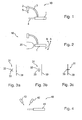
- FIG. 1 shows a first inventive system 10. This comprises a linear ultrasonic head 1, a laser 2 and a bracket 3. The ultrasonic head 1 and the laser 2 are each attached to one end of the bracket 3. Left of the ultrasound head 1, a sound field 4 is indicated. The arrow 5 below the laser 2 indicates the direction of projection of the laser 2.
- the ultrasound head 1 is designed to be able to be applied to the surface of an object to be sonographed (not shown here). He is changeable in his position.
- the system 10 can be grasped and guided by hand.
- the laser 2 projects a mark on the object to be sonographed (not shown).
- the bracket 3 couples the ultrasonic head 1 and the laser 2 to each other in such a way that a change in position of the ultrasonic head 1 causes a change in position of the laser 2. In this case, the direction of the projection 5 is changed, which has an effect on the marking on the object to be sonographied.
- the laser 2 and / or the ultrasound head 1 can also be displaceable and releasably fixable along the bracket 3.
- the geometry of the system 10 can be easily adapted to the proportions of the object to be sonographied.
- the bracket 3 can be replaced with another bracket with a different geometry. This further improves the adaptability of the system 10.
- FIG. 2 shows the system of the system 10 to a thigh 20.
- the ultrasound head 1 need not rest directly on the skin of the thigh 20, optionally may be given as a contact gel in a gap between the ultrasound head 1 and the thigh 20.
- the arrows A and B indicate the tilt degrees of freedom of the system 10.
- the system 10 can of course also be guided along the surface of the thigh 20, that is to say be moved translationally and / or rotationally.
- the Figures 3a, 3b and 3c each show a reference line 30 and a projected mark 31, here a cross.
- the reference line 30 is painted on the object to be sonographed. It is chosen so that the system according to the invention designed so that the system is correctly aligned when the first bar 32 of the cross 31 falls on the reference line 30 and the second bar 33 is perpendicular thereto.
- sub-figure a there are two deviations, once the cross 31 is rotated relative to the reference line 30 and also the intersection of the cross 31 is spaced from the reference line 30.
- the ultrasound head must now be moved so that both deviations are leveled.
- sub-figure b) the first beam 32 is aligned parallel to the reference line, but the intersection of the cross 31 is still spaced from this reference line 30.
- sub-figure c) finally, the cross 31 falls on the reference line 30, while the second bar 33 is perpendicular to the reference line 30th
- the system according to the invention can be designed so that the second bar 33 also indicates the center of the ultrasound image. If an interesting structure goes through the image, it can be easily marked on the skin.
- FIG. 4 shows a further inventive system 40.
- This also has an ultrasonic head 41 and a laser 42.
- this system 40 comprises a second laser 43.
- the first laser 42 serves to project the marking onto the object to be sonographed, which is coupled to the ultrasound head 41 via a coupling device (not shown).
- the second laser 43 serves to project a reference line onto the object to be sonographed.
- the projection of the second laser 43 is independent of the position of the ultrasound head 41.
- Ultrasonic head 41, first laser 42 and second laser 43 are movably and releasably fixable housed in a suspension (not shown).
- the suspension is provided with a controller which can move the ultrasonic head 41 and the two lasers 42 and 43 via electric motors.
- the ultrasonic head 41 has position sensors; these transmit the measured position data to the controller, which then couples the orientation of the laser 42 according to the invention to the position of the ultrasonic head 41. Since neither ultrasound head 41 nor one of the two lasers 42 and 43 has to be moved by hand and the position of the ultrasound head 41 is measured or the control knows anyway in which position the components of the system 40 are located, the system 40 can also remotely controlled from another location.
- the system 40 can be modeled in a virtual reality and the ultrasound head 41 can be manipulated via a data glove.
- FIG. 5 shows the system 10 FIG. 1 put on a knee.
Landscapes
- Life Sciences & Earth Sciences (AREA)
- Health & Medical Sciences (AREA)
- Biomedical Technology (AREA)
- Biophysics (AREA)
- Nuclear Medicine, Radiotherapy & Molecular Imaging (AREA)
- Pathology (AREA)
- Radiology & Medical Imaging (AREA)
- Engineering & Computer Science (AREA)
- Physics & Mathematics (AREA)
- Heart & Thoracic Surgery (AREA)
- Medical Informatics (AREA)
- Molecular Biology (AREA)
- Surgery (AREA)
- Animal Behavior & Ethology (AREA)
- General Health & Medical Sciences (AREA)
- Public Health (AREA)
- Veterinary Medicine (AREA)
- Ultra Sonic Daignosis Equipment (AREA)
Description
- Die Erfindung betrifft ein System zur geführten Sonografie sowie einen austauschbaren Träger für ein entsprechendes System.
- Sonografie ist an sich allgemein bekannt; es handelt sich um die Anwendung von Ultraschall zur Abbildung.
- In Medizin und Veterinärmedizin wird die Sonografie regelmäßig zur Untersuchung von Knochen, Gelenken und Organen eingesetzt. Es ist an sich auch bekannt, andere Materialien zur Darstellung ihrer inneren Struktur zu sonografieren.
- In der Kinderorthopädie wird die Sonografie flächendeckend zur Untersuchung der Hüfte eingesetzt. Es werden unter anderem Überdachungsdefizite und diverse anatomische Winkel ausgemessen. Für Kinderknochen bietet sich die Sonografie besonders an, da diese noch nicht vollständig durchgekalkt sind und ggf. nicht gut im Röntgenbild zu erkennen sind.
- Zur Abbildung wird der Ultraschallkopf an die Oberfläche eines zu sonografierenden Objekts angelegt, etwa an ein Körperteil zur Abbildung der im Inneren des Körperteils liegenden Knochen. Dabei wird der Schallkopf von einer Bedienperson manuell an die Oberfläche des Objekts angelegt und ggf. auch an ihr entlang geführt.
- Die resultierende Abbildung hängt stark von der Lage, also Ausrichtung und Position, des angelegten Schallkopfs ab. Fehlerfreie Abbildungsergebnisse werden nur erzielt, wenn der Schallkopf gegenüber seiner optimalen Ausrichtung nicht verkippt oder verdreht ist; außerdem kann der Schallkopf bei der Führung entlang des Objekts auch verkanten. Durch Verkippen, Verdrehen oder Verkanten des Schallkopfes ergeben sich topografische Übertragungsfehler der Schallkopf-Position auf die Körperoberfläche. Das birgt die Gefahr erheblicher Messdifferenzen, insbesondere bei ungeübten Anwendern.
- Gerade im medizinischen Bereich ist eine optimale Abbildungsqualität von hoher Relevanz.
- Beispielsweise ist bei der sonografischen Bestimmung der distalen Wachstumsfuge am Oberschenkel die Position der Wachstumsfuge von Interesse. Hier wird beispielsweise bei der Verwendung eines Schallkopfes mit paralleler Abstrahlcharakteristik (sog. Linearschallkopf) wie folgt vorgegangen: Befindet sich die Wachstumsfuge in der Abbildung in der Mitte, so wird auch die Mitte des Schallkopfes auf die Haut übertragen, was die Position der Wachstumsfuge nach außen sichtbar machen soll. Dieses wird auch als topografische Übertragung auf die Körperoberfläche bezeichnet. Ist der Schallkopf jedoch nicht korrekt ausgerichtet, wird diese Position falsch markiert.
- Besonders schwerwiegend kann etwa eine fehlerhafte Bestimmung des Pfannendachwinkels der Hüfte sein; hier kann eine fehlerhafte Bestimmung des Winkels sogar zu einer unnötigen Operation führen.
- Die Anfertigung und/oder Interpretation sonografischer Abbildungen erfordert viel Erfahrung. Die zugehörigen unterschiedlichen Fähigkeiten sind schwer zu erlernen. Die Qualität der Abbildungen hängt von vielen Umständen ab, die zumindest teilweise gleichzeitig berücksichtigt werden müssen, beispielsweise: Position und Lage des Objekts, etwa eines Patienten, Geräteeinstellungen und vor allem die Lage und Handhabung des Schallkopfes. Dies setzt eine gute Hand-Auge-Koordination voraus. Der subjektive Eindruck muss dann mit dem erwarteten Ergebnis verglichen und Abweichungen hinsichtlich ihrer Bedeutung beurteilt werden; dies setzt einiges an Erfahrung voraus. Entsprechend sind die Ergebnisse auch nur in begrenztem Umfang objektiv.
- Korrekte Abbildungen können verlässlich nur von erfahrenen und fähigen Bedienpersonen erstellt werden.
- Der Erfindung liegt die Aufgabe zu Grunde, ein vorteilhaftes System zur Sonografie anzugeben, welches die Anforderungen an eine Bedienperson verringert.
- Die Aufgabe wird gelöst durch ein System zur geführten Sonografie mit einem Ultraschallkopf, der an die Oberfläche eines zu sonografierenden Objekts anlegbar und dort in seiner Lage veränderbar ist, einem Projektor zur Projektion einer Markierung auf das zu sonografierende Objekt, einer Kopplungseinrichtung, die die Projektionen an die Lage des Ultraschallkopfes koppelt, wobei die Markierung eine vorgegebene Position und/oder Ausbildung auf dem zu sonografierenden Objekt aufweist, wenn der Ultraschallkopf an diesem korrekt anliegt.
- Bevorzugte Ausgestaltungen der Erfindung sind in abhängigen Ansprüchen angegeben und werden im Folgenden ebenfalls näher erläutert.
- Die Erfindung beruht auf der Idee, der Bedienperson eine Rückmeldung betreffend der korrekten Lage des Schallkopfes anzubieten; in diesem Sinne handelt es sich um eine "geführte" Sonografie.
- Die Führung drückt sich durch Position und/oder Ausbildung der Markierung auf dem zu sonografierenden Objekts aus. Ist der Schallkopf korrekt angelegt, so ist dies an der Markierung zu erkennen. Ist der Schallkopf nicht korrekt angelegt, so ist dies ebenfalls an der Markierung zu erkennen; sie nimmt in diesem Fall eben nicht die vorgegebene Position und/oder Ausbildung an.
- Der Projektor dient zur Projektion der Markierung auf das zu sonografierende Objekt. Bei dem Projektor kann es sich etwa um eine gerichtete Lichtquelle und insbesondere um einen Laser handeln.
- Die Projektion des Projektors ist von der Lage des Ultraschallkopfes abhängig. Dazu umfasst die Erfindung eine Kopplungseinrichtung, die gewährleistet, dass sich die Lage des Schallkopfes auf die Projektion auswirkt. Im einfachsten Fall bewirkt eine Lageänderung des Schallkopfes eine Lageänderung des Projektors und damit etwa eine Richtungsänderung der Projektion, was zu einer Änderung der Markierung auf dem interessierenden Objekt führt; dazu kann die Kopplungseinrichtung eine elektrische oder elektronische Einrichtung aufweisen oder eine mechanische, den Schallkopf und den Projektor verbindende Einrichtung sein, etwa ein beide Teile tragender Träger (vgl. entsprechende Ausgestaltungen unten).
- Erfindungsgemäße Projektionen bzw. Markierungen können unterschiedlich gestaltet sein. Es sind geometrische Projektionen, aber auch Projektionen anderer Qualität denkbar. So kann es sinnvoll sein, die Lage des Schallkopfes ausschließlich oder auch zusätzlich farblich oder auch über projizierte Symbole auszudrücken. Es ist jede Projektion sinnvoll, die es erlaubt, eine Aussage über einen Aspekt der Lage des Ultraschallkopfes zu machen.
- Zur Veranschaulichung sei auf eine Projektion verwiesen, bei der es sich um ein mit einem Laser projiziertes Kreuz mit zwei sich gegenseitig mittig kreuzenden, gleich langen und orthogonal zueinander liegenden Balken handelt. Das erfindungsgemäße System kann hier etwa so eingerichtet werden, dass bei einem korrekt ausgerichteten Schallkopf die Markierung, also das Kreuz, auf der Oberfläche des Objekts symmetrisch und bei einem verkippten Schallkopf verzerrt ist. Auf diese Weise kann eine Bedienperson leicht sehen, ob sie den Schallkopf korrekt aufgesetzt hat. Außerdem kann die kreuzförmige Markierung noch vorteilhaft in Bezug zu Referenzpunkten/ oder -linien an der Oberfläche des Objekts gesetzt werden, was zusätzlich auch eine einfache Kontrolle der Position des Schallkopfes erlaubt.
- Die Referenzen können bspw. aufgemalt oder ebenfalls projiziert oder auf der Oberfläche des Objekts vorhandene natürliche markante Orientierungshilfen sein.
- Der Ultraschallkopf kann beispielsweise, wie bereits erwähnt, ein linearer Schallkopf, oder aber etwa auch ein Sektorenschallkopf sein (ein Sektorenschallkopf erzeugt ein divergierendes Schallfeld). Mit einem Sektorenschallkopf werden typischerweise innere Organe abgebildet. Sektorenschallköpfe sind eher darauf ausgelegt, einen größeren Winkelbereich abzubilden, als eine genaue Lokalisation zu ermöglichen. Die Erfindung erleichtert die Lokalisation bzw. die Markierung relevanter Positionen auf der Haut auch mit einem Sektorenschallkopf, da die Lage des Schallkopfs über die projizierte Markierung kontrolliert werden kann.
- Eine Bedienperson kann sich bei einem System nach der Erfindung durch die Markierung führen lassen. Je nach Ausgestaltung der Erfindung kann die Markierung einen oder mehrere Aspekte der Lage des Schallkopfes ausdrücken. Insgesamt ermöglicht die Erfindung eine Rückmeldung bezüglich aller Aspekte der Lage, also die Ausrichtung und Position im Raum inklusive Position an der Oberfläche des zu sonografierenden Objekts. Damit wird es auch unerfahrenen Bedienpersonen erleichtert, qualitativ hochwertige Aufnahmen anzufertigen.
- In der technischen Orthopädie lassen sich mit der Erfindung beispielsweise besonders vorteilhaft relevante Daten für die Konstruktion von Orthesen erzeugen. Hier sind z.B. die mechanischen Drehachsen oder die anatomische Höhe von Gelenken, etwa des Chopart-Gelenks im Fuß, von Interesse.
- Beispielsweise kann mit dem System nach der Erfindung besonders vorteilhaft die distale Wachstumsfuge (femorale Epiphyse) am Oberschenkelknochen (Femur) über die mediale und laterale Seite des Beins bestimmt werden. In Höhe der Wachstumsfuge befindet sich der anatomische (Kompromiss-) Drehpunkt des Kniegelenks. Aus diesen Daten lässt sich schließen, in welcher anatomischen Höhe der mechanische Gelenkschienendrehpunkt an einer das Knie übergreifenden Orthese positioniert werden sollte.
- Die Erfindung ermöglicht eine exaktere Übertragung von anatomischen Gelenklinien, Wachstumsfugen, Gelenkdrehpunkte u.s.w. auf die Körperoberfläche bzw. auf deren Topografie. Dies eröffnet beispielsweise dem Orthopädietechniker bisher nicht bekannte Möglichkeiten zur anatomisch akkuraten Anpassung von orthopädischen Hilfsmitteln.
- Beispielsweise kann das Gerät aber auch vorteilhaft zur Festlegung von durchzuführenden Schnitten in die Haut bei Operationen oder aber auch zur Festlegung der örtlichen Ausgestaltung von Bestrahlungen eingesetzt werden.
- Besonders vorteilhaft ist auch die Möglichkeit, bei der menschlichen Wirbelsäule die Rotation der Wirbel ohne Röntgen, also nicht-invasiv, zu vermessen. Die Erfindung erlaubt es, die Abweichung der Wirbel von einer als Referenz fungierenden Senkrechten besonders genau zu vermessen.
- Bei einer bevorzugten Ausführungsform der Erfindung ändert sich die Markierung in Abhängigkeit von der Lage des Ultraschallkopfes aufgrund einer Änderung der Projektionsrichtung. In anderen Worten: Eine Lageänderung des Ultraschallkopfes führt zu einer Änderung der Projektionsrichtung. Entsprechende Ausführungsformen sind konzeptionell und konstruktiv besonders einfach, da die Projektion an sich, also etwa das oben bereits erwähnte Laserkreuz, unveränderlich sein kann und lediglich die Projektionsrichtung geändert wird. Dies kann durch eine Änderung der Ausrichtung des Projektors erzielt werden.
- Bei dem Projektor handelt es sich vorzugsweise um einen Laser.
- Vorzugsweise wird, wie oben bereits erwähnt, ein Kreuz projiziert. Ein Kreuz kann besonders leicht an einer Referenzlinie entlanggeführt werden. Dabei wird beispielsweise der erste Balken parallel zu der Referenzlinie geführt, und der dazu senkrechte zweite Balken erlaubt eine Aussage über die Position des Schallkopfs entlang der Referenzlinie. Werden interessante innere Strukturen mit dem Schallkopf gefunden, unterstützt der zweite Balken eine korrekte Markierung der Position auf der Oberfläche des Objekts. Ist der erste Balken zwar parallel zu der Referenzlinie ausgerichtet, von dieser jedoch beabstandet, kann dies für ein Verkippen des Schallkopfes quer zu der Referenzlinie sprechen (vgl. Beispiel).
- Vorstellbar ist beispielsweise auch eine kreisrunde Projektion; ist der Ultraschallkopf verkippt, so ergibt sich etwa eine ovale Markierung. Auch mit einer einfachen kurzen Linie als Markierung kann schon sinnvoll gearbeitet werden, man kann etwa auf die Ausrichtung relativ zu einer Referenzlinie bzw. einen Abstand von einer solchen abstellen.
- Insgesamt kann es wünschenswert sein, die relative Lage von dem Projektor zu dem Schallkopf veränderlich und lösbar feststellbar auszulegen. So kann das System durch unterschiedliche Einstellungen an unterschiedliche Umstände angepasst werden. Dies betrifft nicht nur Systeme mit einem Träger für den Ultraschallkopf und den Projektor, sondern ist auch für Systeme ohne Träger vorteilhaft.
- Bei einer bevorzugten Ausführungsform der Erfindung umfasst die Kopplungseinrichtung eine Messeinrichtung zur Messung der Lage des Schallkopfs und eine Steuereinrichtung zur Steuerung des Projektors basierend auf der Messung der Lage des Schallkopfs.
- Bei der Messeinrichtung kann es sich etwa um Lagesensoren handeln. Die resultierenden Daten können über eine Kommunikationseinrichtung, etwa über Funk, WLAN oder LAN an eine Steuereinrichtung, beispielsweise umfassend einen konventionellen, entsprechend programmierten Rechner, übermittelt werden. Die Steuereinrichtung wirkt schließlich, gegebenenfalls mittelbar über Aktuatoren, etwa Elektromotoren, auf den Projektor ein.
- Vorzugsweise wird die Lage des Schallkopfes zur visuellen Wahrnehmung zusätzlich dargestellt, beispielsweise auf einem Monitor. Die zur Visualisierung erforderlichen Daten über die Lage des Schallkopfes können etwa mit den eben bereits erwähnten Lagesensoren erzeugt werden.
- Besonders bevorzugt ist die Durchführung einer Sonografie mit einem System nach der Erfindung vermittels einer virtuellen Umgebung. Hier kann die Bedienperson, beispielsweise über einen Datenhandschuh, den virtuellen Gegenpart des Schallkopfes in der virtuellen Welt virtuell bewegen und sich dabei durch die virtuelle Markierung auf einem virtuellen Objekt führen lassen. Auf diese Weise ließen sich die Systeme nach der Erfindung auch vorteilhaft fernsteuern. Es müsste lediglich noch für eine geeignete Aufhängung und Ansteuerung des realen Schallkopfes gesorgt werden.
- Besonders bevorzugt ist es, den Projektor und den Ultraschallkopf an einem gemeinsamen Träger zu befestigen, so dass eine Lageänderung des Ultraschallkopfes über den Träger eine Lageänderung der Projektion bewirkt. Eine entsprechende mechanische Kopplung ist konstruktiv besonders einfach und auch intuitiv zu bedienen. Außerdem können entsprechende Ausgestaltungen der Erfindung vergleichsweise klein gebaut werden, so dass die Geräte gut transportiert werden können bzw. mobil einsetzbar sind.
- Bei dem Träger handelt es sich vorzugsweise um einen U-förmigen Bügel. Der Projektor kann an einem Ende des Bügels und der Ultraschallkopf an dem anderen Ende des Bügels befestigt sein. Aufgrund der U-Form kann der Bügel um einen Teil des zu sonografierenden Objektes herumgreifen. Beispielsweise ist es möglich, das Knie von der Seite her zu sonografieren und dabei eine auf die Kniescheibe projizierte Markierung mit einer Referenz abzugleichen. Je nach Größe und Form des zu sonografierenden Objekts/Körperteils kann der Bügel entsprechend angepasst, mal größer oder mal kleiner oder mit einem anderen Krümmungsradius, sein. Insbesondere lassen sich so gut Ausführungen realisieren, die handlich sind und gut manuell bedient werden können bzw. auch für den mobilen Einsatz taugen.
- Unter dem Begriff "U-förmiger Bügel" werden hier einstückige und mehrteilige Konstruktionen verstanden, die z. B. verrundet, etwa als verrundet gebogener Bügel, oder polygonzugartig ausgebildet sein können. So sind auch Bügel denkbar, die etwa aus mehreren, beispielsweise drei, geraden Schienen bestehen. Diese können etwa im rechten Winkel miteinander in Verbindung stehen und dabei über Langlöcher auch gegeneinander verschoben werden. Auch mehr als drei gerade Schienen sind grundsätzlich denkbar.
- Vorzugsweise ist das System so ausgelegt, dass der Träger austauschbar ist. Dies erleichtert die Verwendung von Trägem mit unterschiedlicher Geometrie oder Eigenschaften. Beispielsweise kann in einem Fall ein starrer Träger besonders vorteilhaft sein, während in einem anderen Fall ein biegsamer Schwanenhals gewünscht ist.
- Weiter ist es vorteilhaft, den Projektor verschiebbar und an verschiedenen Positionen lösbar fixierbar an dem Träger zu befestigen. So kann das System auch ohne Austausch des Trägers an das zu sonografierende Objekt angepasst werden.
- Weiter ist es bevorzugt, eine Referenzmarkierung mit einem zweiten Projektor auf das zu sonografierende Objekt zu projizieren. Im einfachsten Fall kann es sich hier um eine Referenzlinie handeln. Eine entsprechende Projektion ist zuverlässiger als eine gedachte Referenz und muss, anders als eine aufgemalte Referenz, auch nicht wieder abgewaschen werden.
- Die Erfindung betrifft auch einen Träger, insbesondere einen Arm oder Bügel, für ein System zur geführten Sonografie, um dieses an bestimmte Anwendungsumstände anzupassen. Entsprechende Träger sind so ausgelegt dass sie einen Projektor und einen Ultraschallkopf so aufnehmen können, dass Systeme nach der Erfindung damit gebildet werden können. Die Träger können die unterschiedlichsten Geometrien und andere Eigenschaften haben. Die Erfindung betrifft grundsätzlich auch die Verwendung eines erfindungsgemäßen Trägers als Austauschteil in einem erfindungsgemäßen System.
- Die vorangehende und die folgende Beschreibung bezieht sich sowohl auf das System als auch auf den Träger, ohne dass dies immer explizit ausformuliert ist; die dabei offenbarten Merkmale können auch einzeln und in anderen als den gezeigten Kombinationen erfindungsgemäß sein.
- Im Folgenden soll die Erfindung auch anhand von Ausführungsbeispielen näher erläutert werden, ohne dabei die Erfindung durch diese Beispiele einschränken zu wollen:
-
Figur 1 zeigt eine Prinzipskizze einer ersten Ausführungsform des erfindungsgemäßen Systems. -
Figur 2 zeigt eine Prinzipskizze, anhand der zwei Bewegungsfreiheitsgrade des Systems ausFigur 1 veranschaulicht werden. -
Figur 3 zeigt eine Markierung nach der Erfindung mit einer Referenzlinie. -
Figur 4 zeigt eine Prinzipskizze einer zweiten Ausführungsform des erfindungsgemäßen Systems. -
Figur 5 zeigt die Verwendung einer Ausführungsform entsprechend denFiguren 1 und 2 an einem Knie. -
Figur 1 zeigt ein erstes erfindungsgemäßes System 10. Dieses umfasst einen linearen Ultraschallkopf 1, einen Laser 2 und einen Bügel 3. Der Ultraschallkopf 1 und der Laser 2 sind jeweils an einem Ende des Bügels 3 befestigt. Links von dem Ultraschallkopf 1 ist ein Schallfeld 4 angedeutet. Der Pfeil 5 unterhalb des Lasers 2 deutet die Projektionsrichtung des Lasers 2 an. - Der Ultraschallkopf 1 ist dazu ausgelegt, an die Oberfläche eines zu sonografierenden Objektes (hier nicht gezeigt) anlegbar zu sein. Dabei ist er in seiner Lage veränderbar. Beispielsweise kann das System 10 mit der Hand gegriffen und geführt werden.
- Der Laser 2 projiziert 5 eine Markierung auf das zu sonografierende Objekt (nicht gezeigt).
- Der Bügel 3 koppelt den Ultraschallkopf 1 und den Laser 2 derart aneinander, dass eine Lageänderung des Ultraschallkopfes 1 eine Lageänderung des Lasers 2 verursacht. Dabei wird auch die Richtung der Projektion 5 geändert, was sich auf die Markierung auf dem zu sonografierenden Objekt auswirkt.
- Der Laser 2 und/oder der Ultraschallkopf 1 können auch entlang des Bügels 3 verschiebbar und lösbar fixierbar sein. So kann die Geometrie des Systems 10 leicht an die Proportionen des zu sonografierenden Objekts angepasst werden.
- Der Bügel 3 kann gegen einen anderen Bügel mit einer anderen Geometrie ausgetauscht werden. Dies verbessert weiter die Anpassbarkeit des Systems 10.
-
Figur 2 zeigt anhand einer Prinzipskizze die Anlage des Systems 10 an einen Oberschenkel 20. Der Ultraschallkopf 1 muss dabei nicht unmittelbar auf der Haut des Oberschenkels 20 aufliegen, gegebenenfalls kann etwa ein Kontaktgel in einem Spalt zwischen dem Ultraschallkopf 1 und dem Oberschenkel 20 gegeben sein. - Die Pfeile A und B deuten die Verkippungsfreiheitsgrade des Systems 10 an. Zusätzlich kann das System 10 natürlich auch an der Oberfläche des Oberschenkels 20 entlanggeführt werden, also translatorisch und/oder rotatorisch bewegt werden. Die
Figuren 3a, 3b und 3c zeigen jeweils eine Referenzlinie 30 und eine projizierte Markierung 31, hier ein Kreuz. Die Referenzinie 30 ist auf das zu sonografierende Objekt aufgemalt. Sie ist dabei so gewählt und das System nach der Erfindung so ausgelegt, dass das System korrekt ausgerichtet ist, wenn der erste Balken 32 des Kreuzes 31 auf die Referenzlinie 30 fällt und dabei der zweite Balken 33 senkrecht dazu steht. - In Teilfigur a) liegen zwei Abweichungen vor, einmal ist das Kreuz 31 gegenüber der Referenzlinie 30 verdreht und ausserdem ist der Schnittpunkt des Kreuzes 31 von der Referenzlinie 30 beabstandet. Der Ultraschallkopf muss nun so bewegt werden, dass beide Abweichungen nivelliert werden. In Teilfigur b) ist der erste Balken 32 parallel zu der Referenzlinie ausgerichtet, der Schnittpunkt des Kreuzes 31 ist allerdings immer noch von dieser Referenzlinie 30 beabstandet. In Teilfigur c) fällt schließlich das Kreuz 31 auf die Referenzlinie 30, dabei steht der zweite Balken 33 senkrecht auf der Referenzlinie 30.
- Das erfindungsgemäße System kann so ausgelegt sein, dass der zweite Balken 33 auch die Mitte des Ultraschallbildes angibt. Durchläuft eine interessante Struktur die Abbildung, so kann sie auf der Haut entsprechend leicht markiert werden.
-
Figur 4 zeigt ein weiteres erfindungsgemäßes System 40. Auch dieses weist einen Ultraschallkopf 41 und einen Laser 42 auf. Zusätzlich umfasst dieses System 40 einen zweiten Laser 43. Der erste Laser 42 dient zur Projektion der Markierung auf das zu sonografierende Objekt, welche über eine Kopplungseinrichtung (nicht gezeigt) an den Ultraschallkopf 41 gekoppelt ist. Der zweite Laser 43 dient zur Projektion einer Referenzlinie auf das zu sonografierende Objekt. Die Projektion des zweiten Lasers 43 ist unabhängig von der Lage des Ultraschallkopfes 41. - Ultraschallkopf 41, erster Laser 42 und zweiter Laser 43 sind beweglich und lösbar fixierbar in einer Aufhängung untergebracht (nicht gezeigt). Die Aufhängung ist mit einer Steuerung versehen, die über Elektromotoren den Ultraschallkopf 41 und die beiden Laser 42 und 43 bewegen kann. Der Ultraschallkopf 41 verfügt über Lagesensoren; diese übermitteln die gemessenen Lagedaten an die Steuerung, welche die Ausrichtung des Lasers 42 dann erfindungsgemäß an die Lage des Ultraschallkopfes 41 koppelt. Da hier weder Ultraschallkopf 41 noch einer der beiden Laser 42 und 43 per Hand bewegt werden müssen und die Lage des Ultraschallkopfes 41 gemessen wird bzw. die Steuerung ohnehin "weiß" in welcher Lage sich die Bestandteile des Systems 40 befinden, kann das System 40 auch von einem anderen Ort ferngesteuert werden.
- Dazu kann das System 40 in einer virtuellen Realität nachgebildet und der Ultraschallkopf 41 über einen Datenhandschuh manipuliert werden.
-
Figur 5 zeigt das System 10 ausFigur 1 angelegt an ein Knie.
Claims (12)
- System (10, 40) zur geführten Sonographie mit
einem Ultraschallkopf (1, 41), der zur Ausführung einer Sonografie an die Oberfläche eines zu sonografierenden Objekts anlegbar und dort in seiner Lage veränderbar ist,
einem Projektor (2, 42) zur Projektion (5) einer von einer Bedienperson erkennbaren Markierung (31) auf das zu sonografierende Objekt,
einer Kopplungseinrichtung (3), die die Projektion (5) an die Lage des Ultraschallkopfes (1, 41) koppelt, und
einer auf dem zu sonografierenden Objekt von einer Bedienperson erkennbaren Referenz, deren Position und Ausbildung unabhängig von der Markierung ist,
wobei die Markierung in Bezug auf diese Referenz eine vorgegebene Position und dort eine vorgegebene Ausbildung auf dem zu sonografierenden Objekt aufweist, wenn der Ultraschallkopf (1, 41) an diesem korrekt anliegt. - System (10, 40) nach Anspruch 1, bei dem die Projektionsrichtung veränderlich ist und an die Lage des Ultraschallkopfes (1, 41) gekoppelt ist.
- System (10, 40) nach Anspruch 1 oder 2, bei dem der Projektor (2, 42) ein Laser ist.
- System (10, 40) nach einem der vorangehenden Ansprüche, bei dem die Markierung (31) ein Kreuz (31) ist.
- System (10, 40) nach einem der vorangehenden Ansprüche, bei dem die relative Lage von dem Projektor (2, 42) zu dem Ultraschallkopf (1, 41) veränderlich und zur Einstellung lösbar festlegbar ist.
- System (10, 40) nach einem der vorangehenden Ansprüche, bei dem die Kopplungseinrichtung (3) eine Messeinrichtung zur Messung der Lage des Ultraschallkopfes und eine Steuereinrichtung zur Steuerung des Projektors (2, 42), basierend auf der Messung der Lage des Ultraschallkopfes (1, 41) aufweist.
- System (10, 40) nach einem der Ansprüche 1 bis 5, bei dem der Projektor (2, 42) und der Ultraschallkopf (1, 41) an einem gemeinsamen Träger (3) befestigt sind, so dass eine Lageänderung des Ultraschallkopfes (1, 41) über den Träger (3) eine Lageänderung des Projektors (2, 42) bewirkt.
- System (10, 40) nach Anspruch 7, bei dem der Träger (3) ein zumindest abschnittweise U-förmiger Bügel (3) ist.
- System (10, 40) nach Anspruch 7 oder 8, bei dem der Träger (3) austauschbar ist.
- System (10, 40) nach einem der Ansprüche 7 bis 9, bei dem der Projektor (2, 42) entlang des Trägers (3) verschiebbar und lösbar fixierbar ist.
- System (10, 40) nach einem der vorangehenden Ansprüche, mit einem zweiten Projektor (43) zur Projektion einer Referenzmarkierung (30) auf das zu sonographierende Objekt.
- Träger (3) mit einem System (10, 40) zur geführten Sonographie nach Anspruch 9, auch in Kombination mit einem der weiteren Systemansprüche, zur Anpassung des Systems (10, 40) an bestimmte Anwendungsumstände.
Applications Claiming Priority (1)
| Application Number | Priority Date | Filing Date | Title |
|---|---|---|---|
| DE200910005707 DE102009005707A1 (de) | 2009-01-22 | 2009-01-22 | Geführte Sonografie |
Publications (2)
| Publication Number | Publication Date |
|---|---|
| EP2210562A1 EP2210562A1 (de) | 2010-07-28 |
| EP2210562B1 true EP2210562B1 (de) | 2013-03-20 |
Family
ID=42124703
Family Applications (1)
| Application Number | Title | Priority Date | Filing Date |
|---|---|---|---|
| EP20100000236 Not-in-force EP2210562B1 (de) | 2009-01-22 | 2010-01-12 | Geführte Sonografie |
Country Status (2)
| Country | Link |
|---|---|
| EP (1) | EP2210562B1 (de) |
| DE (1) | DE102009005707A1 (de) |
Cited By (1)
| Publication number | Priority date | Publication date | Assignee | Title |
|---|---|---|---|---|
| WO2025108946A1 (de) | 2023-11-23 | 2025-05-30 | Ottobock Se & Co. Kgaa | Verfahren zum herstellen eines dreidimensionalen anatomischen modells eines körperabschnitts |
Family Cites Families (6)
| Publication number | Priority date | Publication date | Assignee | Title |
|---|---|---|---|---|
| WO1998038908A1 (en) * | 1997-03-03 | 1998-09-11 | Schneider Medical Technologies, Inc. | Imaging device and method |
| AU728749B3 (en) * | 2000-11-14 | 2001-01-18 | 1St Share Pty Ltd | Diagnostic imaging simulator |
| JP3825384B2 (ja) * | 2002-09-30 | 2006-09-27 | 三菱重工業株式会社 | 放射線治療装置およびその動作方法 |
| DE10319327A1 (de) * | 2003-04-29 | 2004-11-25 | Siemens Ag | Vorrichtung zur Strahlungsbildaufnahme |
| JP4329431B2 (ja) * | 2003-07-14 | 2009-09-09 | 株式会社日立製作所 | 位置計測装置 |
| DE102008013615A1 (de) * | 2008-03-11 | 2009-09-24 | Siemens Aktiengesellschaft | Verfahren und Markierungsvorrichtung zur Markierung einer Führungslinie eines Eindringungsinstruments, Steuerungseinrichtung und Aufnahmesystem |
-
2009
- 2009-01-22 DE DE200910005707 patent/DE102009005707A1/de not_active Withdrawn
-
2010
- 2010-01-12 EP EP20100000236 patent/EP2210562B1/de not_active Not-in-force
Cited By (1)
| Publication number | Priority date | Publication date | Assignee | Title |
|---|---|---|---|---|
| WO2025108946A1 (de) | 2023-11-23 | 2025-05-30 | Ottobock Se & Co. Kgaa | Verfahren zum herstellen eines dreidimensionalen anatomischen modells eines körperabschnitts |
Also Published As
| Publication number | Publication date |
|---|---|
| EP2210562A1 (de) | 2010-07-28 |
| DE102009005707A1 (de) | 2010-07-29 |
Similar Documents
| Publication | Publication Date | Title |
|---|---|---|
| DE69420228T2 (de) | Gegliederte Kanülenführung zur Anwendung bei einem Ultraschall-Abtastgerät | |
| DE60318996T2 (de) | Apparat zur Positionsbestimmung | |
| EP2092907B1 (de) | Lageberechnung von Körperteilen unter Berücksichtigung der anatomischen Symmetrie | |
| US8900247B2 (en) | Measuring and guiding device for reconstruction surgery | |
| EP2299909A1 (de) | Vorrichtung zum bestimmen der stabilität eines kniegelenks | |
| DE102005042751A1 (de) | System, Einrichtung und Verfahren zum Ad-hoc-Nachverfolgen eines Objektes | |
| DE29704393U1 (de) | Vorrichtung zur präoperativen Bestimmung der Positionsdaten von Endoprothesenteilen | |
| DE102006026913A1 (de) | Operationsnavigationsnachverfolgungseinrichtung, -system und -verfahren | |
| EP1827228B1 (de) | Vorrichtung und verfahren zum bestimmen wenigstens eines charakteristischen punktes eines orthopädisch zu vermessenden gelenks | |
| DE10145852B4 (de) | Medizinische Vorrichtung | |
| EP2123229A1 (de) | Verbindung von Schneidblöcken | |
| EP1330809A2 (de) | Simulatorvorrichtung mit zumindest zwei bewegungsfreiheitsgraden für ein instrument | |
| EP2555702A1 (de) | Nichtinvasives verfahren und vorrichtung zum bestimmen einer referenzebene des menschlichen beckens | |
| EP2062224A1 (de) | Verfahren zur herstellung von mindestens einem operationssplint, insbesondere für computerassistierte maxillofaziale operationen und umstellungsosteotomien | |
| DE102008013615A1 (de) | Verfahren und Markierungsvorrichtung zur Markierung einer Führungslinie eines Eindringungsinstruments, Steuerungseinrichtung und Aufnahmesystem | |
| DE202017007688U1 (de) | Chirurgisches System | |
| EP3242637B1 (de) | Lehre für die bestimmung einer für einen patienten passenden implantatgrösse des femurimplantats einer knie-endoprothese | |
| DE102015201067B4 (de) | Bestimmung eines Winkels zwischen zwei Teilen eines Knochens | |
| EP3045152B1 (de) | Verfahren zur herstellung einer orthese | |
| DE10012042C1 (de) | System zur Vorbereitung und Durchführung einer Ausfräsung eines Acetabulums im natürlichen Hüftbeckenknochen | |
| EP1887960B1 (de) | Nadelpositioniersystem | |
| WO2002074500A2 (de) | Vorrichtung zum anzeigen der räumlichen position eines chirurgischen instruments während einer operation | |
| EP2210562A1 (de) | Geführte Sonografie | |
| EP3085343B1 (de) | Orthese zur beübung eines sprunggelenks | |
| DE102006028195A1 (de) | Messvorrichtung für oral-implantologische Zwecke |
Legal Events
| Date | Code | Title | Description |
|---|---|---|---|
| PUAI | Public reference made under article 153(3) epc to a published international application that has entered the european phase |
Free format text: ORIGINAL CODE: 0009012 |
|
| AK | Designated contracting states |
Kind code of ref document: A1 Designated state(s): AT BE BG CH CY CZ DE DK EE ES FI FR GB GR HR HU IE IS IT LI LT LU LV MC MK MT NL NO PL PT RO SE SI SK SM TR |
|
| AX | Request for extension of the european patent |
Extension state: AL BA RS |
|
| 17P | Request for examination filed |
Effective date: 20110125 |
|
| GRAP | Despatch of communication of intention to grant a patent |
Free format text: ORIGINAL CODE: EPIDOSNIGR1 |
|
| GRAS | Grant fee paid |
Free format text: ORIGINAL CODE: EPIDOSNIGR3 |
|
| GRAA | (expected) grant |
Free format text: ORIGINAL CODE: 0009210 |
|
| AK | Designated contracting states |
Kind code of ref document: B1 Designated state(s): AT BE BG CH CY CZ DE DK EE ES FI FR GB GR HR HU IE IS IT LI LT LU LV MC MK MT NL NO PL PT RO SE SI SK SM TR |
|
| REG | Reference to a national code |
Ref country code: GB Ref legal event code: FG4D Free format text: NOT ENGLISH |
|
| REG | Reference to a national code |
Ref country code: CH Ref legal event code: EP |
|
| REG | Reference to a national code |
Ref country code: IE Ref legal event code: FG4D Free format text: LANGUAGE OF EP DOCUMENT: GERMAN |
|
| REG | Reference to a national code |
Ref country code: AT Ref legal event code: REF Ref document number: 601544 Country of ref document: AT Kind code of ref document: T Effective date: 20130415 |
|
| REG | Reference to a national code |
Ref country code: CH Ref legal event code: NV Representative=s name: SEIFERT AND PARTNER, CH |
|
| REG | Reference to a national code |
Ref country code: DE Ref legal event code: R096 Ref document number: 502010002567 Country of ref document: DE Effective date: 20130516 |
|
| PG25 | Lapsed in a contracting state [announced via postgrant information from national office to epo] |
Ref country code: BG Free format text: LAPSE BECAUSE OF FAILURE TO SUBMIT A TRANSLATION OF THE DESCRIPTION OR TO PAY THE FEE WITHIN THE PRESCRIBED TIME-LIMIT Effective date: 20130620 Ref country code: SE Free format text: LAPSE BECAUSE OF FAILURE TO SUBMIT A TRANSLATION OF THE DESCRIPTION OR TO PAY THE FEE WITHIN THE PRESCRIBED TIME-LIMIT Effective date: 20130320 Ref country code: ES Free format text: LAPSE BECAUSE OF FAILURE TO SUBMIT A TRANSLATION OF THE DESCRIPTION OR TO PAY THE FEE WITHIN THE PRESCRIBED TIME-LIMIT Effective date: 20130701 Ref country code: NO Free format text: LAPSE BECAUSE OF FAILURE TO SUBMIT A TRANSLATION OF THE DESCRIPTION OR TO PAY THE FEE WITHIN THE PRESCRIBED TIME-LIMIT Effective date: 20130620 Ref country code: LT Free format text: LAPSE BECAUSE OF FAILURE TO SUBMIT A TRANSLATION OF THE DESCRIPTION OR TO PAY THE FEE WITHIN THE PRESCRIBED TIME-LIMIT Effective date: 20130320 |
|
| REG | Reference to a national code |
Ref country code: LT Ref legal event code: MG4D |
|
| PG25 | Lapsed in a contracting state [announced via postgrant information from national office to epo] |
Ref country code: GR Free format text: LAPSE BECAUSE OF FAILURE TO SUBMIT A TRANSLATION OF THE DESCRIPTION OR TO PAY THE FEE WITHIN THE PRESCRIBED TIME-LIMIT Effective date: 20130621 Ref country code: FI Free format text: LAPSE BECAUSE OF FAILURE TO SUBMIT A TRANSLATION OF THE DESCRIPTION OR TO PAY THE FEE WITHIN THE PRESCRIBED TIME-LIMIT Effective date: 20130320 Ref country code: SI Free format text: LAPSE BECAUSE OF FAILURE TO SUBMIT A TRANSLATION OF THE DESCRIPTION OR TO PAY THE FEE WITHIN THE PRESCRIBED TIME-LIMIT Effective date: 20130320 Ref country code: LV Free format text: LAPSE BECAUSE OF FAILURE TO SUBMIT A TRANSLATION OF THE DESCRIPTION OR TO PAY THE FEE WITHIN THE PRESCRIBED TIME-LIMIT Effective date: 20130320 |
|
| REG | Reference to a national code |
Ref country code: NL Ref legal event code: VDEP Effective date: 20130320 |
|
| PG25 | Lapsed in a contracting state [announced via postgrant information from national office to epo] |
Ref country code: HR Free format text: LAPSE BECAUSE OF FAILURE TO SUBMIT A TRANSLATION OF THE DESCRIPTION OR TO PAY THE FEE WITHIN THE PRESCRIBED TIME-LIMIT Effective date: 20130320 |
|
| PG25 | Lapsed in a contracting state [announced via postgrant information from national office to epo] |
Ref country code: PT Free format text: LAPSE BECAUSE OF FAILURE TO SUBMIT A TRANSLATION OF THE DESCRIPTION OR TO PAY THE FEE WITHIN THE PRESCRIBED TIME-LIMIT Effective date: 20130722 Ref country code: RO Free format text: LAPSE BECAUSE OF FAILURE TO SUBMIT A TRANSLATION OF THE DESCRIPTION OR TO PAY THE FEE WITHIN THE PRESCRIBED TIME-LIMIT Effective date: 20130320 Ref country code: EE Free format text: LAPSE BECAUSE OF FAILURE TO SUBMIT A TRANSLATION OF THE DESCRIPTION OR TO PAY THE FEE WITHIN THE PRESCRIBED TIME-LIMIT Effective date: 20130320 Ref country code: SK Free format text: LAPSE BECAUSE OF FAILURE TO SUBMIT A TRANSLATION OF THE DESCRIPTION OR TO PAY THE FEE WITHIN THE PRESCRIBED TIME-LIMIT Effective date: 20130320 Ref country code: IS Free format text: LAPSE BECAUSE OF FAILURE TO SUBMIT A TRANSLATION OF THE DESCRIPTION OR TO PAY THE FEE WITHIN THE PRESCRIBED TIME-LIMIT Effective date: 20130720 Ref country code: NL Free format text: LAPSE BECAUSE OF FAILURE TO SUBMIT A TRANSLATION OF THE DESCRIPTION OR TO PAY THE FEE WITHIN THE PRESCRIBED TIME-LIMIT Effective date: 20130320 Ref country code: CZ Free format text: LAPSE BECAUSE OF FAILURE TO SUBMIT A TRANSLATION OF THE DESCRIPTION OR TO PAY THE FEE WITHIN THE PRESCRIBED TIME-LIMIT Effective date: 20130320 |
|
| PG25 | Lapsed in a contracting state [announced via postgrant information from national office to epo] |
Ref country code: CY Free format text: LAPSE BECAUSE OF FAILURE TO SUBMIT A TRANSLATION OF THE DESCRIPTION OR TO PAY THE FEE WITHIN THE PRESCRIBED TIME-LIMIT Effective date: 20130320 Ref country code: PL Free format text: LAPSE BECAUSE OF FAILURE TO SUBMIT A TRANSLATION OF THE DESCRIPTION OR TO PAY THE FEE WITHIN THE PRESCRIBED TIME-LIMIT Effective date: 20130320 |
|
| PLBE | No opposition filed within time limit |
Free format text: ORIGINAL CODE: 0009261 |
|
| STAA | Information on the status of an ep patent application or granted ep patent |
Free format text: STATUS: NO OPPOSITION FILED WITHIN TIME LIMIT |
|
| PG25 | Lapsed in a contracting state [announced via postgrant information from national office to epo] |
Ref country code: DK Free format text: LAPSE BECAUSE OF FAILURE TO SUBMIT A TRANSLATION OF THE DESCRIPTION OR TO PAY THE FEE WITHIN THE PRESCRIBED TIME-LIMIT Effective date: 20130320 |
|
| 26N | No opposition filed |
Effective date: 20140102 |
|
| REG | Reference to a national code |
Ref country code: DE Ref legal event code: R097 Ref document number: 502010002567 Country of ref document: DE Effective date: 20140102 |
|
| PG25 | Lapsed in a contracting state [announced via postgrant information from national office to epo] |
Ref country code: LU Free format text: LAPSE BECAUSE OF FAILURE TO SUBMIT A TRANSLATION OF THE DESCRIPTION OR TO PAY THE FEE WITHIN THE PRESCRIBED TIME-LIMIT Effective date: 20140112 Ref country code: MC Free format text: LAPSE BECAUSE OF FAILURE TO SUBMIT A TRANSLATION OF THE DESCRIPTION OR TO PAY THE FEE WITHIN THE PRESCRIBED TIME-LIMIT Effective date: 20130320 |
|
| GBPC | Gb: european patent ceased through non-payment of renewal fee |
Effective date: 20140112 |
|
| REG | Reference to a national code |
Ref country code: IE Ref legal event code: MM4A |
|
| PG25 | Lapsed in a contracting state [announced via postgrant information from national office to epo] |
Ref country code: GB Free format text: LAPSE BECAUSE OF NON-PAYMENT OF DUE FEES Effective date: 20140112 |
|
| PG25 | Lapsed in a contracting state [announced via postgrant information from national office to epo] |
Ref country code: IE Free format text: LAPSE BECAUSE OF NON-PAYMENT OF DUE FEES Effective date: 20140112 |
|
| REG | Reference to a national code |
Ref country code: FR Ref legal event code: PLFP Year of fee payment: 7 |
|
| PG25 | Lapsed in a contracting state [announced via postgrant information from national office to epo] |
Ref country code: MT Free format text: LAPSE BECAUSE OF FAILURE TO SUBMIT A TRANSLATION OF THE DESCRIPTION OR TO PAY THE FEE WITHIN THE PRESCRIBED TIME-LIMIT Effective date: 20130320 |
|
| PG25 | Lapsed in a contracting state [announced via postgrant information from national office to epo] |
Ref country code: SM Free format text: LAPSE BECAUSE OF FAILURE TO SUBMIT A TRANSLATION OF THE DESCRIPTION OR TO PAY THE FEE WITHIN THE PRESCRIBED TIME-LIMIT Effective date: 20130320 |
|
| PG25 | Lapsed in a contracting state [announced via postgrant information from national office to epo] |
Ref country code: HU Free format text: LAPSE BECAUSE OF FAILURE TO SUBMIT A TRANSLATION OF THE DESCRIPTION OR TO PAY THE FEE WITHIN THE PRESCRIBED TIME-LIMIT; INVALID AB INITIO Effective date: 20100112 Ref country code: TR Free format text: LAPSE BECAUSE OF FAILURE TO SUBMIT A TRANSLATION OF THE DESCRIPTION OR TO PAY THE FEE WITHIN THE PRESCRIBED TIME-LIMIT Effective date: 20130320 |
|
| REG | Reference to a national code |
Ref country code: FR Ref legal event code: PLFP Year of fee payment: 8 |
|
| PGFP | Annual fee paid to national office [announced via postgrant information from national office to epo] |
Ref country code: DE Payment date: 20170125 Year of fee payment: 8 Ref country code: CH Payment date: 20170125 Year of fee payment: 8 Ref country code: FR Payment date: 20170124 Year of fee payment: 8 |
|
| PGFP | Annual fee paid to national office [announced via postgrant information from national office to epo] |
Ref country code: AT Payment date: 20170125 Year of fee payment: 8 Ref country code: BE Payment date: 20170124 Year of fee payment: 8 |
|
| PGFP | Annual fee paid to national office [announced via postgrant information from national office to epo] |
Ref country code: IT Payment date: 20170130 Year of fee payment: 8 |
|
| PG25 | Lapsed in a contracting state [announced via postgrant information from national office to epo] |
Ref country code: MK Free format text: LAPSE BECAUSE OF FAILURE TO SUBMIT A TRANSLATION OF THE DESCRIPTION OR TO PAY THE FEE WITHIN THE PRESCRIBED TIME-LIMIT Effective date: 20130320 |
|
| REG | Reference to a national code |
Ref country code: DE Ref legal event code: R119 Ref document number: 502010002567 Country of ref document: DE |
|
| REG | Reference to a national code |
Ref country code: CH Ref legal event code: PL |
|
| REG | Reference to a national code |
Ref country code: AT Ref legal event code: MM01 Ref document number: 601544 Country of ref document: AT Kind code of ref document: T Effective date: 20180112 |
|
| PG25 | Lapsed in a contracting state [announced via postgrant information from national office to epo] |
Ref country code: DE Free format text: LAPSE BECAUSE OF NON-PAYMENT OF DUE FEES Effective date: 20180801 Ref country code: FR Free format text: LAPSE BECAUSE OF NON-PAYMENT OF DUE FEES Effective date: 20180131 |
|
| REG | Reference to a national code |
Ref country code: FR Ref legal event code: ST Effective date: 20180928 |
|
| REG | Reference to a national code |
Ref country code: BE Ref legal event code: MM Effective date: 20180131 |
|
| PG25 | Lapsed in a contracting state [announced via postgrant information from national office to epo] |
Ref country code: AT Free format text: LAPSE BECAUSE OF NON-PAYMENT OF DUE FEES Effective date: 20180112 Ref country code: BE Free format text: LAPSE BECAUSE OF NON-PAYMENT OF DUE FEES Effective date: 20180131 Ref country code: CH Free format text: LAPSE BECAUSE OF NON-PAYMENT OF DUE FEES Effective date: 20180131 Ref country code: LI Free format text: LAPSE BECAUSE OF NON-PAYMENT OF DUE FEES Effective date: 20180131 |
|
| PG25 | Lapsed in a contracting state [announced via postgrant information from national office to epo] |
Ref country code: IT Free format text: LAPSE BECAUSE OF NON-PAYMENT OF DUE FEES Effective date: 20180112 |