EP2203746B1 - T cell subpopulations capable of treating cancer - Google Patents
T cell subpopulations capable of treating cancer Download PDFInfo
- Publication number
- EP2203746B1 EP2203746B1 EP08789880A EP08789880A EP2203746B1 EP 2203746 B1 EP2203746 B1 EP 2203746B1 EP 08789880 A EP08789880 A EP 08789880A EP 08789880 A EP08789880 A EP 08789880A EP 2203746 B1 EP2203746 B1 EP 2203746B1
- Authority
- EP
- European Patent Office
- Prior art keywords
- tils
- cell
- receptor
- subpopulation
- signature
- Prior art date
- Legal status (The legal status is an assumption and is not a legal conclusion. Google has not performed a legal analysis and makes no representation as to the accuracy of the status listed.)
- Not-in-force
Links
Images








Classifications
-
- G—PHYSICS
- G01—MEASURING; TESTING
- G01N—INVESTIGATING OR ANALYSING MATERIALS BY DETERMINING THEIR CHEMICAL OR PHYSICAL PROPERTIES
- G01N33/00—Investigating or analysing materials by specific methods not covered by groups G01N1/00 - G01N31/00
- G01N33/48—Biological material, e.g. blood, urine; Haemocytometers
- G01N33/50—Chemical analysis of biological material, e.g. blood, urine; Testing involving biospecific ligand binding methods; Immunological testing
- G01N33/53—Immunoassay; Biospecific binding assay; Materials therefor
- G01N33/575—Immunoassay; Biospecific binding assay; Materials therefor for cancer
- G01N33/5758—Immunoassay; Biospecific binding assay; Materials therefor for cancer involving compounds serving as markers for tumours, cancers or neoplasias, e.g. cellular determinants, receptors, heat shock/stress proteins, A-protein, oligosaccharides or metabolites
-
- A—HUMAN NECESSITIES
- A61—MEDICAL OR VETERINARY SCIENCE; HYGIENE
- A61P—SPECIFIC THERAPEUTIC ACTIVITY OF CHEMICAL COMPOUNDS OR MEDICINAL PREPARATIONS
- A61P35/00—Antineoplastic agents
-
- G—PHYSICS
- G01—MEASURING; TESTING
- G01N—INVESTIGATING OR ANALYSING MATERIALS BY DETERMINING THEIR CHEMICAL OR PHYSICAL PROPERTIES
- G01N33/00—Investigating or analysing materials by specific methods not covered by groups G01N1/00 - G01N31/00
- G01N33/48—Biological material, e.g. blood, urine; Haemocytometers
- G01N33/50—Chemical analysis of biological material, e.g. blood, urine; Testing involving biospecific ligand binding methods; Immunological testing
- G01N33/5005—Chemical analysis of biological material, e.g. blood, urine; Testing involving biospecific ligand binding methods; Immunological testing involving human or animal cells
- G01N33/5008—Chemical analysis of biological material, e.g. blood, urine; Testing involving biospecific ligand binding methods; Immunological testing involving human or animal cells for testing or evaluating the effect of chemical or biological compounds, e.g. drugs, cosmetics
- G01N33/5044—Chemical analysis of biological material, e.g. blood, urine; Testing involving biospecific ligand binding methods; Immunological testing involving human or animal cells for testing or evaluating the effect of chemical or biological compounds, e.g. drugs, cosmetics involving specific cell types
- G01N33/5047—Cells of the immune system
- G01N33/505—Cells of the immune system involving T-cells
-
- G—PHYSICS
- G01—MEASURING; TESTING
- G01N—INVESTIGATING OR ANALYSING MATERIALS BY DETERMINING THEIR CHEMICAL OR PHYSICAL PROPERTIES
- G01N2800/00—Detection or diagnosis of diseases
- G01N2800/52—Predicting or monitoring the response to treatment, e.g. for selection of therapy based on assay results in personalised medicine; Prognosis
Definitions
- the present invention relates to a method of determining responsiveness to adoptive cell transfer therapy according to claim 1.
- the present disclosure in some embodiments thereof, relates to T cell populations capable of treating cancer.
- Current therapeutic strategies focus predominantly on achieving the removal or death of cancer cells within the patient, through a diverse array of surgical and non-surgical techniques; the most widely used are chemotherapy and gamma irradiation.
- Those methods have a number of prominent disadvantages, in particular the culling of healthy cells/tissues within the patient, and the toxic side-effects of the current generation of chemotherapeutic drugs utilized in cancer treatment. Furthermore, these treatments are not always successful.
- lymphocytes within melanoma deposits. These lymphocytes, when isolated, are capable of recognizing specific tumor antigens on autologous and allogeneic melanomas in an MHC restricted fashion.
- Tumor infiltrating lymphocytes from patients with metastatic melanoma recognize shared antigens including melanocyte-melanoma lineage specific tissue antigens in vitro ( Kawakami, Y., et al., (1993) J. Immunother. 14: 88-93 ; Anichini, A. et al., (1993) et al., J. Exp. Med. 177: 989-998 ).
- Anti-melanoma T cells appear to be enriched in TILs probably as a consequence of clonal expansion and accumulation at the tumor site in vivo ( Sensi, M., et al., (1993) J. Exp. Med. 178:1231-1246 ).
- adoptive immunotherapy describes the transfer of immunocompetent cells such as the TILs described herein above to the tumor-bearing host.
- adoptive immunotherapy describes the transfer of immunocompetent cells such as the TILs described herein above to the tumor-bearing host.
- Adoptive cell transfer (ACT) therapy for patients with cancer relies on the ex vivo generation of highly active tumor, specific lymphocytes, and their administration in large numbers to the autologous host.
- ACT therapy however effectively treats only a limited number of patients.
- Preclinical models have identified a variety of ways to manipulate the host immune environment that increase ACT therapeutic efficacy. These include immunosuppression prior to cell administration and concurrent interleukin 2 administration with the transferred T cells.
- a method of determining responsiveness to cancer treatment in a subject in need thereof comprising analyzing a frequency of tumor infiltrating lymphocytes (TILs) having a CD8 + CD28 - CD152 - signature in a sample of the subject, wherein a frequency of TILs having the CD8 + CD28 - CD152 - signature above a predetermined level is indicative of a positive responsiveness to cancer treatment.
- TILs tumor infiltrating lymphocytes
- the method further comprises analyzing a frequency of TILs having a CD8 + CD69 + CD33 - signature in the TIL sample, wherein a frequency of TILs having the CD8 + CD69 + CD33 - signature and the CD8 + CD28 - CD152 - signature above a predetermined level is indicative of a negative responsiveness to cancer treatment.
- a method of determining responsiveness to cancer treatment in a subject in need thereof comprising analyzing a frequency of TILs having a CD8 + CD28 - CD152 - signature in a sample of the subject, wherein a frequency of TILs having a CD8 + CD28 - CD152 - signature below a predetermined level is indicative of a negative responsiveness to cancer treatment.
- the method further comprises analyzing a frequency of TILs having a CD94 + signature in the sample, wherein a frequency of TILs not having the CD8 + CD28 - CD152 - signature whilst having a CD94+ signature above a predetermined level is further indicative of a negative responsiveness to cancer treatment.
- a method of predicting T cell responsiveness to a cancer in a subject comprising analyzing subpopulation marker signatures in a TIL sample of the subject, wherein a subpopulation marker signature corresponding to a reactive marker signatures as defined by Figure 3A is indicative of T cell responsiveness and a subpopulation marker signature corresponding to a non-reactive marker signature as defined by Figure 3A is indicative of a non T cell responsiveness.
- the cancer treatment comprises adoptive transfer therapy.
- a method of treating cancer in a subject in need thereof comprising depleting lymphocytes from a sample of TILs of the subject, wherein the lymphocytes express CD4, CD152 and CD28
- the method further comprises depleting additional lymphocytes of the subject wherein the additional lymphocytes express CD85 and/or CD94.
- a method of treating cancer in a subject in need thereof comprising enriching for a subpopulation of lymphocytes from a sample of TILs of the subject, the subpopulation expressing a CD8 + CD28 - CD152 - signature.
- the method further comprises depleting an additional subpopulation of lymphocytes from the sample of CD8 + CD28 - CD152 - emiched TILs, the additional subpopulation expressing a CD8 + CD69 + CD33 - signature.
- the subject has a cancer selected from the group consisting of prostate cancer, renal cell carcinoma, glioma and melanoma.
- a method of determining a reactivity of a subpopulation of TILs in a TIL sample comprising:
- the method further comprises removing the subpopulations following the analyzing the frequency, wherein a subpopulation comprising a frequency lower than 1 % is removed.
- T cell populations capable of treating cancer.
- Adoptive cell transfer (ACT) therapy for patients with cancer relies on the ex vivo generation of highly active tumor, specific lymphocytes, and their administration in large numbers to the autologous host.
- the present inventors have devised a novel method for studying the underlying composition and cellular interactions that determine the degree of TIL reactivity.
- This method is based on measuring frequencies of subpopulations fractions and constructing a "subpopulations signature" for each TIL.
- a method of determining a reactivity of a subpopulation of TILs in a TIL sample comprising:
- the term “reactivity” refers to an ability of the TILs to at least inhibit cancer progression and optimally promote regression of same (either partially, or completely).
- tumor-infiltrating lymphocytes refers to white blood cells of a subject afflicted with a cancer (such as melanoma), that have left the blood stream and have migrated into a tumor.
- a cancer such as melanoma
- tumor-infiltrating lymphocytes may have tumor specificity.
- lymphocytes can be isolated from an individual (e.g. during a tumor biopsy) to be treated by methods known in the art and cultured in vitro ( Kawakami, Y. et al. (1989) J. Immunol. 142; 2453-3461 ). Lymphocytes may be cultured in media such as RPMI or RPMI 1640 or AIM V for 1-10 weeks.
- An exemplary method for obtaining TILs includes plating viable cells (e.g. 1x10 6 ) of a single-cell suspension of enzymatically digested explant of metastatic melanoma. It will be appreciated that the TILs may be isolated from fresh tumors or from frozen tissue (at the cost of lower yield).
- the TIL samples may be obtained from any mammalian species, such as human.
- the method of this aspect is effected by assaying the activities of a statistically significant number of TIL samples. It will be appreciated that the number of statistically significant TILs is dependent on the number of markers that are analyzed per cell.
- the statistically significant number of samples is greater than about 10. According to another embodiment, the statistically significant number of samples is greater than about 50. According to another embodiment, the statistically significant number of samples is greater than about 75. According to another embodiment, the statistically significant number of samples is greater than about 100. According to another embodiment, the statistically significant number of samples is greater than about 150. According to another embodiment, the statistically significant number of samples is greater than about 200.
- Exemplary methods of assaying activities of TIL samples include 51 CR release cytotoxicity assays ( Cerundolo, V. et al. (1990) Nature 345:449-452 ) or lymphokine assays such as IFN- ⁇ or TNF secretion assays [ Schwartzentruber, D. et al., (1991) J. of Immunology 146:3674-3681 ].
- the method of this aspect is further effected by analyzing the TIL samples by flow cytometry analysis of at least three marker per cell in order to classify subpopulations of cells.
- flow cytometry refers to an assay in which the proportion of a material (e.g. lymphocyte comprising a particular maker) in a sample is determined by labeling the material (e.g., by binding a labeled antibody to the material), causing a fluid stream containing the material to pass through a beam of light, separating the light emitted from the sample into constituent wavelengths by a series of filters and mirrors, and detecting the light.
- a material e.g. lymphocyte comprising a particular maker
- the markers are cell surface antigens.
- At least one of the three markers is a cytokine or chemokines.
- cytokine and chemokine makers include, but are not limited to those set forth in Table 1.
- Table 1 Antigen Name Other Names Structure Function CD117 c-kit, SCFR IgSF, RTK family SCF receptor, hematopoietic progenitor development/differentiation CDw119 IFN ⁇ R IFN- ⁇ R ⁇ , w/IFN- ⁇ AF-1, host defense CD120a TNFR-I TNFRSF receptor for both TNF- ⁇ and TNF- ⁇ CD120b TNFR-II TNFRSF receptor for both TNF- ⁇ and TNF- ⁇ CD121a IL-1R type a I IgSF binds IL-1 ⁇ and IL-1 ⁇ , IL-1 signaling CDw121b IL-1R, type II IgSF binds IL-1 ⁇ and IL-1 ⁇ , negative signals CD122 IL-2R ⁇ CRSF IL-2R ⁇ and IL-15R ⁇ , signal transduction CDw1l23 IL-3R CRSF IL-3R ⁇ , w/
- CD331 TM tyr kinase binds FGF, high affinity receptor for fibroblast growth factors CD332 FGFR2, BEK, KGFR TM RTK family binds FGF, high affinity receptor for fibroblast growth factors CD333 FGFR3, ACH, CEK2 TM RTK family binds FGF, high affinity receptor for fibroblast growth factors CD334 FGFR4, JTK2, TKF TM RTK family binds FGF, high affinity receptor for fibroblast growth factors AITRL TNFSF18, TL6, GITRL CMKLR1 chemokine-like receptor 1 GPCR 7TM, chemokine receptor binds chemerin, pDC recruitment, bone development DcR3 TR6, TNFRSF6B Soluble Fas decoy receptor, tumor evasion HVEM TNFRSF14, TR2 TNFRSF receptor for LIGHT, LT- ⁇ , BTLA, Herpes Simplex Virus, lymphocyte activation IL-15R ⁇ binds to
- At least one of the three markers is an adhesion molecule, a co-inhibitory receptor, a co-stimulatory receptor or a relevant protein such as those set forth in Table 5.
- adhesion molecules contemplated by the present invention are set forth in Table 2.
- Table 2 Antigen Name Other Names Structure Function CD11a LFA-1, integrin aL Integrin family CD11a/CD18 receptor for ICAM-1, -2,-3, intercellular adhesion, T costimulation CD50 ICAM-3 IgSF adhesion, costimulation CD73 GPI-linked ecto-5'-nucleotidase, nucleoside uptake, T costimulation, lymph adhesion CD99 MIC2, E2 T cell activation, adhesion CD106 VCAM-1 IgSF VLA-4(CD49d/CD29) receptor, leukocyte adhesion, migration, costimulation CD2 T11, LFA-2, SRBC-R IgSF CD58 ligand, adhesion, T cell activation CD9 p24, MRP-1 TM4SF cellular adhesion and migration CD15 Lewis-x, Lex CHO adhesion CD15s Sialyl Lewis X CHO CD62L and CD62
- Table 3 Antigen Name Other Names Structure Function CD6 T12 Scavenger R SF CD166 receptor, T cell differentiation/ costimulation CD7 IgSF T costimulation CD26 DPP IV type II TM dipeptidyl peptidase, T costimulation, HIV entry CD27 T14 TNFRSF CD70 receptor, T costimulation CD28 Tp44, T44 IgSF CD80, CD86 receptor, T costimulation CD40 TNFRSF CD 154 receptor, B differentiation/ costimulation, isotype-switching, rescues B cells from apoptosis CD60a GD3 CHO costimulation CD70 Ki-24 TNFSF CD27 ligand, T and B cell costimulation CD80 B7, B7-1, BB1 IgSF binds to CD28, CD 152, T costimulation CD81 TAPA-1 TM4SF complex w/ CD 19 & CD21, signaling, T costimulation CD86 B
- Table 4 Antigen Name Other Names Structure Function CD158a p58.1 IgSF, KIR family inhibition of NK cell cytolytic activity, MHC class-I specific NK receptor CD158b p58.2 IgSF, KIR family inhibition of NK cell cytolytic activity, MHC class-I specific NK receptor CD85 IgSF, ILT/LIR family inhibition ofNK, T cell cytolytic function CD200 OX-2 inhibition of immune response CD272 BTLA IgSF HVEM receptor, inhibitory response CD294 CRTH2.
- GPR44 GPCR-7TM binds prostaglandin D2, stimulatory effects on Th2, allergic inflammation CD305 LAIR1 IgSF, ASV inhibitory receptor on NK and T cells CD77 Gb3, Pk blood group group apoptosis CD94 KP43 C-type lectin complex w/ NKG2, inhibits NK function CD118 LIFR, gp190 Type I CRSF & secreted forms membrane-bound involved in signal transduction, soluble form inhibits activity of LIF CD159c NKG2C Type II C-Type Lectin w/ MHC class I HLA-E molecules, forms heterodimer with CD94 CD253 TRAIL, Apo-2L, TL2, TNFSF10 TNFSF death CD279 PD1,SLEB2 B7-H1 & B7-DC receptor, autoimmune disease and peripheral tolerance CD300c CMRF35A, LIR IgSF unknown B7-H4 B7-S1, B7x B7 family may interact with BTLA (?), inhibition BAMBI T
- Table 5 Antigen Name Other Names Structure Function CD88 C5aR TM7SF C5a receptor, granulocyte activation CD89 FcaR IgSF IgA receptor, phagocytosis, degranulation, respiratory burst CD5 T1, Tp67 Scavenger R SF CD72 receptor, TCR or BCR signaling, T-B interaction CD159a NKG2A w/ CD94, NK cell receptor CD163 130kD Scavenger receptor SF CD173 Blood group H type 2 CHO CD174 Lewis Y CHO CD175 Tn CHO CD175s Sialyl-Tn CHO CD176 Thomson Friedrenreich CHO CD177 Ag NB1 CD178 FasL, CD95L TNFSF CD95 ligand, apoptosis, immune privilege, soluble form in serum CD2R T11-3 IgSF activation-dependent form of CD2 CD3 ⁇ , CD3 ⁇ T3 IgSF w/
- each TIL sample can be classified into subpopulations as described in the Examples section below.
- the significance of the subpopulation may be effected.
- subpopulations above a predetermined threshold in a reactive TIL sample may be classified as positive predictors.
- subpopulations above a predetermined threshold in a non-reactive TIL sample may be classified as negative predictors.
- the predetermined thresholds may be determined using mathematical algorithms as exemplified in the Figure 3B of the Examples section below.
- Such marker signatures may be used to predict T cell responsiveness to a cancer in a subject.
- a method of predicting T cell responsiveness to a cancer in a subject comprising analyzing subpopulation marker signatures in a TIL sample of the subject, wherein a subpopulation marker signature corresponding to a reactive marker signature as defined by Figure 3A is indicative of T cell responsiveness and a subpopulation marker signature corresponding to a non-reactive marker signature as defined by Figure 3A is indicative of a non T cell responsiveness.
- signature refers to an expression pattern of the indicated markers.
- the cancer to which T cell responsiveness is predicated includes melanoma, lung carcinoma, breast cancer, colon cancer, prostate cancer, ovarian carcinoma, renal cell carcinoma, glioma and the like.
- the cancer may be metastatic or non-metastatic.
- melanoma refers to metastatic melanomas, melanomas derived from either melanocytes or melanocytes related nevus cells, melanocarcinomas, melanoepitheliomas, melanosarcomas, melanoma in situ, superficial spreading melanoma, nodular melanoma, lentigo maligna melanoma, acral lentiginous melanoma, invasive melanoma or familial atypical mole and melanoma (FAM-M) syndrome.
- melanomas in mammals may be caused by, chromosomal abnormalities, degenerative growth and developmental disorders, mitogenic agents, ultraviolet radiation (UV), viral infections, inappropriate tissue expression of a gene, alterations in expression of a gene, or carcinogenic agents.
- subpopulations of TILs in TIL samples By determining reactivity of subpopulations of TILs in TIL samples, the present inventors identified three subpopulations as being important predictors of reactivity. These subpopulations include CD8 4 CD28 - CD152 - , CD94 + and CD8 + CD69 + CD33 - .
- a method of determining responsiveness to cancer treatment in a subject in need thereof comprising analyzing a frequency of tumor infiltrating lymphocytes (TILs) having a CD8 + CD28 - CD152 - signature in a sample of the subject, wherein a frequency of TILs having the CDB + CD28 - CD152 - signature above a predetermined level is indicative of a positive responsiveness to cancer treatment.
- TILs tumor infiltrating lymphocytes
- the cancer treatment is any treatment which involves the use of TILs, such as for example adoptive transfer therapy.
- the number of TILs in a TTL sample having a CD8 + CD28 - CD152 - signature is greater than 25 %, more preferably greater than 35 % and even more preferably greater than 45 %.
- TIL sample comprising a significant percentage of CD8 + CD69 + CD33 - bearing lymphocytes which already comprises a significant percentage of CD8 + CD28 - CD152 - bearing lymphocytes is indicative of a negative responsiveness to cancer.
- the predetermined level of CD8 + CD69 + CD33 - bearing lymphocytes is typically greater than about 40 %, more preferably greater than about 50 % and even more preferably greater than about 60 %.
- a method of determining responsiveness to cancer treatment in a subject in need thereof comprising analyzing a frequency of tumor infiltrating lymphocytes (TILs) having a CD8 + CD28 - CD152 - signature in a sample of the subject, wherein a frequency of TILs having the CD8 + CD28 - CD152 - signature below a predetermined level is indicative of a negative responsiveness to cancer treatment.
- TILs tumor infiltrating lymphocytes
- the number of TILs having a CD8 + CD28 - CD152 - signature is less than about 25 %, more preferably less than about 35 % and even more preferably less than about 45 %.
- TIL sample comprising a significant percentage of CD94 + bearing lymphocytes which already comprises a significantly low percentage of CD8 + CD28 - CD152 - bearing lymphocytes is further indicative of a negative responsiveness to cancer.
- the predetermined level of CD94 + bearing lymphocytes is typically greater than about 0.5 %, more preferably greater than about 0.6 % and even more preferably greater than about 0.7 %.
- T lymphocyte signatures which have been shown to be predictors of effective cancer treatment include CD56+, CD4+CD85-C1394-, CD8+CD33+CD69+ and CD4+CD33-CD69+.
- CD56+, CD4+CD85-C1394-, CD8+CD33+CD69+ and CD4+CD33-CD69+ are indicative of a TIL sample being effective for cancer treatment.
- CD4+CD85-CD94- signature this is indicative of a TIL sample being non-effective for cancer treatment.
- more than about 17 % of the TILs in a sample comprise a CD8+CD33+CD69+ signature
- this is indicative of a TIL sample being effective for cancer treatment.
- CD4+CD33-CD69+ signature this is indicative of a TIL sample being non-affective for cancer treatment.
- the present inventors By determining reactivity of subpopulations of TILs in a TIL sample, the present inventors uncovered several markers which predicted a negative responsiveness to cancer treatment. Thus, by depleting a TIL sample of those TILs which express the markers associated with negative responsiveness (i.e., lack of responsiveness), also referred to herein as "harmful markers" the present inventors postulated they should be able to increase the reactivity of the TIL sample. As shown in Figures 4A-C a TIL sample depleted of lymphocytes bearing a CD4, CD152, CD28, CD85 and/or CD94 marker comprised an increased reactivity towards autologous cancer cells.
- a method of treating cancer in a subject in need thereof comprising depleting lymphocytes from a sample of TILs of the subject, wherein the lymphocytes express CD4, CD152 and/or CD28.
- treating includes abrogating, substantially inhibiting, slowing or reversing the progression of a condition, substantially ameliorating clinical or aesthetical symptoms of a condition or substantially preventing the appearance of clinical or aesthetical symptoms of a condition.
- the phrase "subject in need thereof' refers to a subject which has the disease.
- the subject may be a mammal, e.g. a human.
- the disease being treated is melanoma
- the subject is typically one being diagnosed with melanoma, with or without metastasis, at any stage of the disease (e.g. IA, IB, IIA, IIB, IIC, IIIA, IIIB, IIIC or IV).
- the term "depleting” as used herein refers to a procedure that substantially removes the indicated T lymphocyte population from the TIL sample without also substantially removing the "effective" lymphocytes from the composition - i.e. those capable of destroying the tumor - e.g. the subpopulation having a CD8 + CD28 - CD152 - signature.
- substantially removes with respect to depletion of each of the cell types is intended to mean removal of at least 50 % or more of the particular cell type, such as at least about 75 %, about 80 %, about 90 %, about 95 %, or about 97 %, including at least 99 %, 99.5 %, 99.9 % or more of the particular cell type.
- lymphocytes express CD4, CD152 and/or CD28 from a TIL sample
- the remaining cells are substantially enriched for T lymphocytes comprising an "effective" lymphocyte population such as those comprising a CD8 + CD28 - CD152 - signature.
- depleting lymphocytes expressing the above mentioned markers may be effected by affinity labeling followed by label based separation.
- a fluorescently labeled anti-CD4, anti-CD152 or anti-CD28 antibody which specifically binds the "harmful" T-lymphocyte subpopulation (i.e. those T lymphocytes which deter the "effective” T lymphocytes from destroying a tumor) may be used to separate the "harmful" T lymphocytes from the "effective” T lymphocytes.
- depletion of T-lymphocytes expressing the above mentioned markers may be effected by affinity purification.
- a substrate including an antibody or a ligand capable of specifically binding CD4, CD152 and/or CD28 can be used to effectively deplete the "harmful" T-lymphocytes from the TIL sample.
- the affinity substrate can be a column matrix such as, for example agarose, cellulose and the like, or beads such as, for example, magnetic beads onto which the antibodies described above, are immobilized.
- depletion of T-lymphocytes expressing CD4, CD152 and/or CD28 can be effected via column chromatography or magnetic bead separation.
- TIL sample may be depleted of other subpopulations of T lymphocytes including for example those that express CD85 and/or CD94.
- T lymphocyte populations As mentioned above, depletion of "harmful" T lymphocyte populations from the TIL sample effectively enriches for a T lymphocyte population which is effective at destroying the tumor.
- a method of treating cancer in a subject in need thereof comprising enriching for a subpopulation of lymphocytes from a sample of TILs of the subject, the subpopulation expressing a CD8 + CD28 - CD152 - signature.
- the term “enriching” refers to a procedure which allows the TIL composition to comprise at least about 50 %, preferably at least about 70 %, more preferably at least about 80 %, about 95 %, about 97 %, about 99 % or more T lymphocytes comprising the CD8 + CD28 - CD152 - signature.
- the enriching may be effected using known cell sorting procedures such as by using a fluorescence-activated cell sorter (FACS).
- FACS fluorescence-activated cell sorter
- the enriching may also be effected by depleting of non-relevant subpopulations as further described herein above.
- the TIL population may also be enriched for other subpopulations (e.g. a subpopulation that expresses a CD4+CD33-CD69+ signature) in order to further enhance reactivity against tumors.
- subpopulations e.g. a subpopulation that expresses a CD4+CD33-CD69+ signature
- lymphocytes Following enrichment of a TIL sample for a particular subpopulation of lymphocytes (or depletion of a TIL sample of a particular subpopulation of lymphocytes e.g. CD8 + CD69 + CD33 - or CD69 + ), the lymphocytes are typically expanded ex-vivo and re-injected back into the patient following leuko-depletion.
- T cells may be expanded utilizing non-specific T-cell receptor stimulation in the presence of feeder lymphocytes and either IL-2 or IL-15.
- the non-specific T-cell receptor stimulus can consist of around 30 ng/ml of OKT3, a mouse monoclonal anti-CD3 antibody available from Ortho, Raritan, N.J.
- the autologous T-cells may be modified to express a T-cell growth factor that promotes the growth and activation thereof. Any suitable methods of modification may be used. See, e.g., Sambrook and Russell, Molecular Cloning, 3rd ed., SCHL Press (2001 ). Desirably, modified autologous T-cells express the T-cell growth factor at high levels.
- T-cell growth factor coding sequences such as that of IL-2, are readily available in the art, as are promoters, the operable linkage of which to a T-cell growth factor coding sequence promote high-level expression.
- the T-cells can be administered by any suitable route as known in the art.
- the T-cells may be administered as an intra-arterial or intravenous infusion, which preferably lasts approximately 30-60 minutes.
- routes of administration include intraperitoneal, intrathecal and intralymphatic.
- a suitable dose of T-cells to be administered is from about 2.3 x 10 10 T-cells to about 13.7 x 10 10 T-cells.
- the T cells are administered to the subject together with a T-cell growth factor.
- the T-cell growth factor can be any suitable growth factor that promotes the growth and activation of the autologous T-cells administered.
- suitable T-cell growth factors include IL-2, IL-7 and IL-15, which can be used alone or in various combinations, such as IL-2 and IL-7, IL-2 and IL-15, IL-7 and IL-15, or IL-2, IL-7 and IL-15.
- IL-2 is available from Chiron, Emerwlle, Calif.
- IL-7 is available from Cytheris, Vanves, Frances.
- IL-15 can be obtained from PeproTech, Inc., Rocky Hill, N.J.
- the T-cell growth factor can be administered by any suitable route. If more than one T-cell growth factor is administered, they can be administered simultaneously or sequentially, in any order, and by the same route or different routes. According to one embodiment, the T-cell growth factor, such as IL-2, is administered intravenously as a bolus injection. A typical dosage of IL-2 is about 720,000 IU/kg, administered three times daily until tolerance.
- the nonmyeloablative lymphodepleting chemotherapy can be any suitable such therapy, which can be administered by any suitable route.
- the nonmyeloablative lymphodepleting chemotherapy can comprise the administration of cyclophosphamide and fludarabine, particularly if the cancer is melanoma.
- a preferred route of administering cyclophosphamide and fludarabine is intravenously.
- any suitable dose of cyclophosphamide and fludarabine can be administered.
- melanom typically around 60 mg/kg of cyclophosphamide are administered for two days after which around 25 mg/m 2 fludarabine are administered for five days.
- compositions, method or structure may include additional ingredients, steps and/or parts, but only if the additional ingredients, steps and/or parts do not materially alter the basic and novel characteristics of the claimed composition, method or structure.
- a compound or “at least one compound” may include a plurality of compounds, including mixtures thereof.
- range format is merely for convenience and brevity and should not be construed as an inflexible limitation on the scope of the invention. Accordingly, the description of a range should be considered to have specifically disclosed all the possible subranges as well as individual numerical values within that range. For example, description of a range such as from 1 to 6 should be considered to have specifically disclosed subranges such as from 1 to 3, from 1 to 4, from 1 to 5, from 2 to 4, from 2 to 6, from 3 to 6 etc., as well as individual numbers within that range, for example, 1, 2, 3, 4, 5, and 6. This applies regardless of the breadth of the range.
- method refers to manners, means, techniques and procedures for accomplishing a given task including, but not limited to, those manners, means, techniques and procedures either known to, or readily developed from known manners, means, techniques and procedures by practitioners of the chemical, pharmacological, biological, biochemical and medical arts.
- IFN- ⁇ secretion was measured after co-incubation of 10 5 TIL cells with 10 5 viable autologous melanoma cells for an overnight period. The amount of IFN- ⁇ secretion in the culture supernatant was detected using standard sandwich ELISA protocol.
- the markers used for subpopulation mapping included combinations of triple staining from the pool of the following surface receptors: CD3, CD4, CD8, CD25, CD28, CD33, CD56, CD69, CD85, CD94, CD152 ( Figure 1B ) and the intracellular cytotoxic proteins perforin and granzyme B.
- the reactivity of 91 TILS from 26 melanoma patients was determined by measuring IFN- ⁇ secretion following co-incubation of the TILs with autologous melanoma. Using the clinical threshold of 200 pg/ml IFN- ⁇ , 39 TIL cultures were determined as reactive and 52 as nonreactive (Different TILs from the same patient produced different reactivity levels). The immune subpopulation compositions of these cultures were characterized by multicolor flow cytometry.
- Each triple staining of three different receptors X, Y and Z resulted in 6 single staining (X + , X - , Y + , Y - , Z + , Z - ), 12 double staining (e.g. X + Y) and 8 triple staining (e.g. X + Y + Z - ).
- the single, double and triple staining produces a hierarchy of subpopulation characterization ranging from general to more specific subpopulations.
- a quality control filtering procedure was employed yielding a final dataset containing 33 distinct subpopulations (see Figures 5-9 for filtering and dataset description).
- SVM classifications were performed with the gist-train-svm software www.bioinformatics.ubc.ca/gist/. All classifications were performed with a linear kernel and input data was normalized by rescaling the columns to values between -1 and 1. All tests were conducted by applying a 'leave three out' procedure. SVM performance was evaluated by the ROC (receive operating characteristics) analysis which calculates the true positive rate versus True negative rate for different cutoffs. The ROC value namely the area under A ROC curve was reported for each test. In addition, the total accuracy (TA), sensitivity (SN), specificity (SP) and the Matthews correlation coefficient (CC) were calculated.
- TA total accuracy
- SN sensitivity
- SP specificity
- CC Matthews correlation coefficient
- the present inventors compared individual subpopulation fractions and used them to predict reactive and nonreactive TILs ( Figure 2 ).
- the classification accuracy yielded a Matthews correlation coefficients (MCC) ranging from 0 to 0.58 and total accuracy ranging from 40 % to 78 %.
- MCC Matthews correlation coefficients
- the discriminative power of individual subpopulations characterized by triple staining was superior to that of a single and double staining, which may be attributed to the better characterization of identity or functional state of the first compared to the later.
- a CD8 + marker is an indicator of cytotoxic activity while CD8 + CD28 - CD152 - is, in addition to being cytotoxic, also fully activated and bearing no CD152 inhibitory receptors. This analysis emphasizes the limited predictive power of individual subpopulations.
- SVM support vector machine
- TIL reactivity using an SVM model Briefly, each TIL was mapped to a point in a multi-dimensional space according to its subpopulation constituents.
- the SVM classifier generates a hyper-surface that separates instances of the two classes. All classifications were done with a linear kernel and input data was normalized by rescaling the columns to values between -1 and 1. The classification was tested by applying a 'leave one out' procedure.
- MCC Matthews correlation coefficient
- the final SVM classifier contained the minimal feature subset that displayed the maximal MCC value.
- TIL samples in this study belong to a same patient.
- a Bootstrapping control was performed. A leave five out procedure, 10,000 itarations was performed. The results were similar to the SVM testing. SVM classifications were performed with the gist software www.bioinformatics.ubc.ca/gist/.
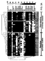
- the two clusters also separate between nonreactive and reactive TILs (Fischer exact P ⁇ 10 -3 ). This suggests that TIL reactivity against melanoma is largely dictated by its subpopulation composition. It was also observed that the nonreactive cluster is further divided into two sub-clusters, both of which are enriched with nonreactive TILs that have distinct profiles. The first is mostly CD4 + while the other is a mixture of CD8 + and CD4 + subpopulation derivatives, suggesting CD4 + dominance over CD8 + . To further simplify the subpopulation signature a decision tree algorithm was used that produced a simple set of rules for distinguishing between reactive and nonreactive TILs ( Figure 3B ).
- the third subpopulation (CD8 + CD69 + CD33 - ) is characterized by the CD69 + and CD33 - receptor staining. Little is known about the function of these two receptors. The present findings suggest that this subpopulation has a yet unknown role in determining T-cell functionality.
- T cell depletion was performed by incubating the TILs with anti-CD4 and/or anti-CD28 and/or anti-CD152 and/or anti-CD85 and/or anti-CD94 for 20 minutes. Subsequently, cells were mixed with anti mouse IgG coated magnetic beads (Dynal, Lake Success, NY) for an additional 10 minutes, followed by magnetic depletion for 5 minutes. The negative fraction was then washed 3 times with cold PBS 0.1 % BSA and was incubated for 36 hours at 37°C.
- the receptors used for depleting these subpopulations were CD4, CD28, CD85, CD94 and CD152.
- the experiments were performed on 12 nonreactive fresh TIL cultures that originated from four different melanoma patients (Table 6, herein below) and were not part of the 91 TIL samples used for the subpopulation signature elucidation. Reactivity levels, in terms of IFN- ⁇ secretion, were measured. TILs with IFN- ⁇ levels that exceeded 200 pg/ml were determined as reactive and otherwise as nonreactive (marked with a'+' and '-' respectively).
- Table 6 Patient TIL Reactive after separation 1 1 + 2 - 2 1 + 2 - 3 + 4 + 5 - 3 1 + 4 1 + 2 + 3 + 4 +
- TIL profiles were examined prior and after the manipulation (see Figure 4B ).
- 10 of the 12 TILs were used that had sufficient cell counts.
- the profile of 9 of the remaining 10 TILs prior to manipulation was similar to that of the nonreactive TILs as determined by the original 91 sample dataset (compare Figure 3A and Figure 4B ). It can be seen that the shift from nonreactive to reactive state is accompanied by a transformation of subpopulation signature as indicated by blue arrows in Figure 4B . This shift in profiles is further illustrated in Figure 4C .
- This approach may be applied in order to optimize the ACT clinical protocol by studying and manipulating TILs in the context of an objective clinical response.
- This general frame work demonstrates the practical implications of systems biology in the context of clinical research and can be further extended to predict, understand and control cell population functions in fields such as stem cells, tumor immunology and tissue engineering.
Landscapes
- Health & Medical Sciences (AREA)
- Life Sciences & Earth Sciences (AREA)
- Immunology (AREA)
- Engineering & Computer Science (AREA)
- Biomedical Technology (AREA)
- Chemical & Material Sciences (AREA)
- Urology & Nephrology (AREA)
- Cell Biology (AREA)
- Molecular Biology (AREA)
- Hematology (AREA)
- General Health & Medical Sciences (AREA)
- Medicinal Chemistry (AREA)
- Food Science & Technology (AREA)
- Biotechnology (AREA)
- Pathology (AREA)
- General Physics & Mathematics (AREA)
- Biochemistry (AREA)
- Analytical Chemistry (AREA)
- Physics & Mathematics (AREA)
- Microbiology (AREA)
- Toxicology (AREA)
- Tropical Medicine & Parasitology (AREA)
- Bioinformatics & Cheminformatics (AREA)
- Animal Behavior & Ethology (AREA)
- Organic Chemistry (AREA)
- Chemical Kinetics & Catalysis (AREA)
- Pharmacology & Pharmacy (AREA)
- Veterinary Medicine (AREA)
- General Chemical & Material Sciences (AREA)
- Public Health (AREA)
- Nuclear Medicine, Radiotherapy & Molecular Imaging (AREA)
- Measuring Or Testing Involving Enzymes Or Micro-Organisms (AREA)
- Investigating Or Analysing Biological Materials (AREA)
Description
- The present invention relates to a method of determining responsiveness to adoptive cell transfer therapy according to
claim 1. - The present disclosure, in some embodiments thereof, relates to T cell populations capable of treating cancer. Current therapeutic strategies focus predominantly on achieving the removal or death of cancer cells within the patient, through a diverse array of surgical and non-surgical techniques; the most widely used are chemotherapy and gamma irradiation. Those methods have a number of prominent disadvantages, in particular the culling of healthy cells/tissues within the patient, and the toxic side-effects of the current generation of chemotherapeutic drugs utilized in cancer treatment. Furthermore, these treatments are not always successful.
- The spontaneous regression of certain cancers, such as melanoma or renal cell cancer, supports the idea that the immune system is sometimes capable of delaying tumor progression and on rare occasions eliminating a tumor altogether. These observations have led to research interest in a variety of immunologic therapies designed to stimulate the immune system.
- Further evidence that an immune response to cancer exists in humans is provided by the existence of lymphocytes within melanoma deposits. These lymphocytes, when isolated, are capable of recognizing specific tumor antigens on autologous and allogeneic melanomas in an MHC restricted fashion. Tumor infiltrating lymphocytes (TILs) from patients with metastatic melanoma recognize shared antigens including melanocyte-melanoma lineage specific tissue antigens in vitro (Kawakami, Y., et al., (1993) J. Immunother. 14: 88-93; Anichini, A. et al., (1993) et al., J. Exp. Med. 177: 989-998). Anti-melanoma T cells appear to be enriched in TILs probably as a consequence of clonal expansion and accumulation at the tumor site in vivo (Sensi, M., et al., (1993) J. Exp. Med. 178:1231-1246).
- The term adoptive immunotherapy describes the transfer of immunocompetent cells such as the TILs described herein above to the tumor-bearing host. Adoptive cell transfer (ACT) therapy for patients with cancer relies on the ex vivo generation of highly active tumor, specific lymphocytes, and their administration in large numbers to the autologous host.
- Presently, ACT therapy however effectively treats only a limited number of patients. Preclinical models have identified a variety of ways to manipulate the host immune environment that increase ACT therapeutic efficacy. These include immunosuppression prior to cell administration and
concurrent interleukin 2 administration with the transferred T cells. - Preclinical models have also identified characteristics of lymphocyte cultures that are required for successful ACT therapy. Until presently, the most important characteristic was thought to be the presence of high affinity, tumor antigen specific CD8+ cells. It was also shown that CD4+ cells were also required for effective treatment of some tumors [Surman et al, J. Immunology 164, 562-565, 2000]. In addition, it has been demonstrated that the presence of CD4+CD25+ T cells suppress autoimmunity and may be potent inhibitors of antitumor effects in mice [Shevach E.M. Nat. Rev. Immunol. 2, 389-400 (2002)]. This has led to the conclusion that lymphodepleting subpopulations comprising this signature may be beneficial for ACT therapy.
- Filacl et al., [Clinical Immunology, Academic Press, US., Vol.123, 1 Jan 2007] teaches CD8+28- T suppressor cells infiltrating human cancer and that this population of cells is almost constantly present and functional in human tumors, being able to inhibit both T cell proliferation and cytotoxicity.
- Some functional requirements of the cells for effective ACT where elucidated in animal models. For example, the secretion of IFN-γ by injected TILs was shown to significantly correlate with in vivo regression of murine tumors suggesting activation of T-cells by the tumor antigens (Barth, R. J., et al., (1991) J. Exp. Med. 173:647-658). Accordingly, selection of tumor-reactive T cells for adoptive immunotherapy may be affected by analyzing IFN-γ secretion following exposure to tumor antigens. Despite its clinical importance, little is known about the underlying composition and cellular interactions that determine the degree of TIL reactivity as measured by IFN-γ secretion and consequentially on how to control this reactivity.
- The method of determining responsiveness to adoptive cell transfer therapy of the present invention is defined in
claim 1. - According to an aspect of some embodiments there is provided a method of determining responsiveness to cancer treatment in a subject in need thereof, the method comprising analyzing a frequency of tumor infiltrating lymphocytes (TILs) having a CD8+CD28-CD152- signature in a sample of the subject, wherein a frequency of TILs having the CD8+CD28-CD152- signature above a predetermined level is indicative of a positive responsiveness to cancer treatment.
- According to some embodiments, the method further comprises analyzing a frequency of TILs having a CD8+CD69+CD33- signature in the TIL sample, wherein a frequency of TILs having the CD8+CD69+CD33- signature and the CD8+CD28-CD152- signature above a predetermined level is indicative of a negative responsiveness to cancer treatment.
- According to an aspect of some embodiments there is provided a method of determining responsiveness to cancer treatment in a subject in need thereof, the method comprising analyzing a frequency of TILs having a CD8+CD28-CD152- signature in a sample of the subject, wherein a frequency of TILs having a CD8+CD28-CD152- signature below a predetermined level is indicative of a negative responsiveness to cancer treatment.
- According to some embodiments, the method further comprises analyzing a frequency of TILs having a CD94+ signature in the sample, wherein a frequency of TILs not having the CD8+CD28-CD152- signature whilst having a CD94+ signature above a predetermined level is further indicative of a negative responsiveness to cancer treatment.
- According to an aspect of some embodiments there is provided a method of predicting T cell responsiveness to a cancer in a subject, comprising analyzing subpopulation marker signatures in a TIL sample of the subject, wherein a subpopulation marker signature corresponding to a reactive marker signatures as defined by
Figure 3A is indicative of T cell responsiveness and a subpopulation marker signature corresponding to a non-reactive marker signature as defined byFigure 3A is indicative of a non T cell responsiveness. - According to some embodiments the cancer treatment comprises adoptive transfer therapy.
- According to an aspect of some embodiments there is provided a method of treating cancer in a subject in need thereof, the method comprising depleting lymphocytes from a sample of TILs of the subject, wherein the lymphocytes express CD4, CD152 and CD28
- According to some embodiments the method further comprises depleting additional lymphocytes of the subject wherein the additional lymphocytes express CD85 and/or CD94.
- According to an aspect of some embodiments there is provided a method of treating cancer in a subject in need thereof, the method comprising enriching for a subpopulation of lymphocytes from a sample of TILs of the subject, the subpopulation expressing a CD8+CD28-CD152- signature.
- According to some embodiments, the method further comprises depleting an additional subpopulation of lymphocytes from the sample of CD8+CD28-CD152- emiched TILs, the additional subpopulation expressing a CD8+CD69+CD33- signature.
- According to some embodiments, the subject has a cancer selected from the group consisting of prostate cancer, renal cell carcinoma, glioma and melanoma.
- According to an aspect of some embodiments there is provided a method of determining a reactivity of a subpopulation of TILs in a TIL sample, the method comprising:
- (a) assaying an activity of a statistically significant number of TIL samples;
- (b) analyzing the TIL samples by flow cytometry analysis of at least three markers per cell in order to classify subpopulations of cells, wherein at least one of the three markers is CD4 or CD8, at least a second of the three markers is a cytokine or chemokine and at least a third of the three markers is an adhesion molecule, a co-inhibitory receptor, a co-stimulatory receptor or a protein set forth in Table 5; and
- (c) analyzing a frequency of at least one subpopulation in the TIL sample, wherein a frequency above a predetermined threshold indicates that the at least one subpopulation of cells is associated with the activity.
- According to some embodiments of the invention, the method further comprises removing the subpopulations following the analyzing the frequency, wherein a subpopulation comprising a frequency lower than 1 % is removed.
- Unless otherwise defined, all technical and/or scientific terms used herein have the same meaning as commonly understood by one of ordinary skill in the art to which the invention pertains. Although methods and materials similar or equivalent to those described heroin can be used in the practice or testing of embodiments of the invention, exemplary methods and/or materials are described below. In case of conflict, the patent specification, including definitions, will control. In addition, the materials, methods, and examples are illustrative only and are not intended to be necessarily limiting.
- Some embodiments are herein described, by way of example only, with reference to the accompanying images. With specific reference now to the images in detail, it is stressed that the particulars shown are by way of example and for purposes of illustrative discussion of embodiments of the invention. In this regard, the description taken with the drawings makes apparent to those skilled in the art how embodiments of the invention may be practiced.
- In the drawings:
-
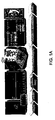
FIG. 1A is a schematic workflow of TIL characterization, analysis and reactivity control. As a first step TILs were extracted from surgically removed tumor mass originating from metastatic melanoma patients. Each TIL was characterized by functional evaluation of IFN-γ secretion levels followed by subpopulation fraction measurements using flow cytometry. This information was combined into a multi-parametric model for prediction and understanding of TIL reactivity. Following this analysis, the fractions of selected subpopulation were manipulated thus enabling controllability of TIL reactivity against melanoma. -
FIG. 1B is a diagram of the central cell surface receptors defining specific T-cell subpopulations with distinct functional states. -
FIG. 2 are optimal cutoffs segregating reactive from nonreactive TILs as represented in a graphical form based on individual subpopulation fractions. For each subpopulation the blue and red dots indicate its fraction in 39 reactive and 52 nonreactive TILs respectively. Subpopulation based linear classification (using a leave five out testing scheme) produced MCCs in the range of 0 to 0.58. In general, MCC values range between -1 to +1 indicating completely wrong and perfect classification respectively. An MCC = 0 indicates random. The black horizontal bars indicate the optimal border between reactive and nonreactive TILs as determined by MCC. -
FIGs. 3A-C are plots and diagrams illustrating that TIL reactivity is largely determined by its subpopulation fractions. (A) Reactive and nonreactive TILs exhibit distinct subpopulations signatures. Columns and rows correspond to TILs and subpopulations respectively. The distance between TILs was calculated using Spearman correlation followed by hierarchical clustering. The rows were clustered similarly. The red and blue arrows represent nonreactive and reactive TILs respectively. Two main clusters emerge characterized by CD4+ and CD8+ overabundant subpopulations. These clusters also separate nonreactive from reactive TILs (P < 10-3). (B) A decision tree algorithm was used in order to generate a simple set of rules for classifying TIL functionality. These rules classify the TILs with 89 % total accuracy. (C) Exact IFN-γ values of the reactive TILs can be described as a function of two subpopulation fractions with positive and negative weights:
The IFN-γ levels can be described as a balance between two opposing subpopulations with positive and negative effects and equal weights. -
FIGs. 4A-C are graphs and diagrams showing that rational subpopulation manipulation restores TIL anti-tumor reactivity accompanied by a shift in subpopulation signature. (A) IFN-γ increases after TIL subpopulation manipulation. IFN-γ levels of 12 TILs before and after inhibitory subpopulation depletion are compared. 9 of the original nonreactive TILs show significant increase in IFN-γ. Incubation of TILs in control experiments with culture media or unrelated melanoma indicates that the increase in IFN-γ secretion does not occur spontaneously and is specific. (B) Shift in reactivity can be explained in terms of a shift in subpopulation signature prior and after depletion. The subpopulation fractions of 10 TILs prior and after subpopulation depletion were determined by flow cytometry. 8 of the original nonreactive TILs became reactive, 7 of which also showed a shift from a nonreactive subpopulation signature to a reactive one, indicated by the blue arrows. The two TILs that remained nonreactive exhibited either a minor change or a negative change in subpopulation signature as indicated by the red arrows. (C) The transformation in reactivity of a TIL can be described as a path in a two dimensional space. A simple representation of the TIL reactivity signature was obtained by applying principal component analysis (PCA) [Ian T.Jolliffe, Principal component analysis (Springer, ed. second, 2002)], which is a method for dimensionality reduction at the expense of loosing part of the data variance. The data was reduced from 35 to two dimensions. The x and y axes are principle components capturing 49 % and 24 % of the total variance in the data (Figures 11A-B ). The figure shows a subspace that is overpopulated with reactive TILs. The change in reactivity can be visualized as a path from a nonreactive TIL to a TIL that resides in the reactive subspace (for example see dotted arrow). -
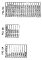
FIGs. 5A-C are tables of the final dataset of subpopulations after the filtration procedure. -
FIG. 6 is a flow chart of dataset filtering procedure. -
FIG. 7 are graphs illustrating the distributions of subpopulation percentages for single, double and triple staining of 91 TILs. The x-axis is subpopulation percentage and the y-axis is the number of TILs that had this percentage out of 91 TILs. -
FIG. 8 is a map showing the correlation between pairs of receptors. Different groups of receptors are correlated and anti-correlated. The percentage of receptor occurrence in 91 TILs was measured using flow cytometry. Several groups of receptors show strong correlations including A: (CD8+,CD69+,CD56+, Perforin, Grenzym); B: (CD85+,CD94+,CD152+,CD25+); and C: (CD4+,CD28+,CD33+). Furthermore, some groups are anti-correlated, for example group A and C. The correlated receptors also share common functionality. For example group B contains three co-inhibitory receptors. This suggests that receptors with common functionality also share a common regulation at the population level. -
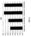
FIG. 9 is a bar graph comparing the SVM prediction accuracy between the datasets containing single, double and triple subpopulations as well as the filtered dataset. -
FIG. 10 is a Venn diagram illustrating that reactive TILs share a similar subpopulation signature. A simple representation of the TIL reactivity signature was obtained by applying principal component analysis (PCA). This technique enables a reduction in dimensionality at the expense of loosing some of the data variance.Figure 10 shows a mapping from the original 33 dimensional data into two dimensions. K-means unsupervised clustering generated two distinct clusters that were enriched for reactive and nonreactive TILs (Fischer exact P < 10-3). The x and y axes explain 60 % and 11 % of the variance in the data respectively. Another property of TIL reactivity emerges from the subspaces in which each functional state resides. While the reactive TILs occupy a defined subspace of subpopulation combinations, as indicated by the high density of blue dots, the nonreactive TILs, indicated by red dots, are dispersed -
FIGs. 11A-B are bar graphs illustrating the subpopulation coefficients for the first and second principle components shown inFigure 4C . - In some embodiments thereof, relates to T cell populations capable of treating cancer.
- Adoptive cell transfer (ACT) therapy for patients with cancer relies on the ex vivo generation of highly active tumor, specific lymphocytes, and their administration in large numbers to the autologous host.
- Preclinical models have identified characteristics of lymphocyte cultures that are required for successful ACT therapy. Until presently, the most important characteristic was thought to be the presence of high affinity, tumor antigen specific CD8+ cells. It was also shown that CD4+ cells were also required for effective treatment of some tumors [Surman et al, J. Immunology 164, 562-565, 2000]. In contrast, it has been demonstrated that the presence of CD4+CD25+ T cells suppress autoimmunity and may be potent inhibitors of antitumor effects in mice [Shevach E.M. Nat. Rev. Immunol. 2, 389-400 (2002)]. This has led to the conclusion that lymphodepleting subpopulations comprising this signature may be beneficial for ACT therapy.
- The present inventors have devised a novel method for studying the underlying composition and cellular interactions that determine the degree of TIL reactivity. This method, summarized in
Figure 1A , is based on measuring frequencies of subpopulations fractions and constructing a "subpopulations signature" for each TIL. - Using a decision tree algorithm, three subpopulations were identified as being important predictors of reactivity (
Figure 3B ). These subpopulations include CD8+CD28-CD152-, CD94+ and CD8+CD69+CD33-. - Knowledge of subpopulations that predict the reactivity of the TIL sample allowed the present inventors to control reactivity thereof. Accordingly, exploitation of this knowledge has lead to the generation of TIL populations of enhanced reactivity.
- Whilst further reducing the present invention to practice, the present inventors have shown that it is possible to deplete non-reactive TILs of lymphocytes of particular subpopulatlon signatures and restore TIL anti-tumor reactivity (
Figures 4A-C ). - Thus, according to one aspect, there is provided a method of determining a reactivity of a subpopulation of TILs in a TIL sample, the method comprising:
- (a) assaying an activity of a statistically significant number of TIL samples;
- (b) analyzing the TIL samples by flow cytometry analysis of at least three markers per cell in order to classify subpopulations of cells, wherein at least one of the three markers is CD4 or CD8, at least a second of the three markers is a cytokine or chemokine and at least a third of the three markers is an adhesion molecule, a co-inhibitory receptor, a co-stimulatory receptor or a protein set forth in Table 5; and
- (c) analyzing a frequency of at least one subpopulation in the TIL sample, wherein a frequency above a predetermined threshold indicates that the at least one subpopulation of cells is associated with the activity.
- As used herein, the term "reactivity" refers to an ability of the TILs to at least inhibit cancer progression and optimally promote regression of same (either partially, or completely).
- As used herein, the term "tumor-infiltrating lymphocytes" refers to white blood cells of a subject afflicted with a cancer (such as melanoma), that have left the blood stream and have migrated into a tumor. Thus, tumor-infiltrating lymphocytes may have tumor specificity.
- Such lymphocytes can be isolated from an individual (e.g. during a tumor biopsy) to be treated by methods known in the art and cultured in vitro (Kawakami, Y. et al. (1989) J. Immunol. 142; 2453-3461). Lymphocytes may be cultured in media such as RPMI or RPMI 1640 or AIM V for 1-10 weeks. An exemplary method for obtaining TILs includes plating viable cells (e.g. 1x10⁶) of a single-cell suspension of enzymatically digested explant of metastatic melanoma. It will be appreciated that the TILs may be isolated from fresh tumors or from frozen tissue (at the cost of lower yield).
- The TIL samples may be obtained from any mammalian species, such as human.
- As mentioned, the method of this aspect is effected by assaying the activities of a statistically significant number of TIL samples. It will be appreciated that the number of statistically significant TILs is dependent on the number of markers that are analyzed per cell.
- Thus, typically when three marker per cell are analyzed, the statistically significant number of samples is greater than about 10. According to another embodiment, the statistically significant number of samples is greater than about 50. According to another embodiment, the statistically significant number of samples is greater than about 75. According to another embodiment, the statistically significant number of samples is greater than about 100. According to another embodiment, the statistically significant number of samples is greater than about 150. According to another embodiment, the statistically significant number of samples is greater than about 200.
- Exemplary methods of assaying activities of TIL samples include 51CR release cytotoxicity assays (Cerundolo, V. et al. (1990) Nature 345:449-452) or lymphokine assays such as IFN-γ or TNF secretion assays [Schwartzentruber, D. et al., (1991) J. of Immunology 146:3674-3681].
- As mentioned herein above, the method of this aspect is further effected by analyzing the TIL samples by flow cytometry analysis of at least three marker per cell in order to classify subpopulations of cells.
- As used herein, the term "flow cytometry" refers to an assay in which the proportion of a material (e.g. lymphocyte comprising a particular maker) in a sample is determined by labeling the material (e.g., by binding a labeled antibody to the material), causing a fluid stream containing the material to pass through a beam of light, separating the light emitted from the sample into constituent wavelengths by a series of filters and mirrors, and detecting the light.
- A multitude of flow cytometers are commercially available including for e.g. Becton Dickinson FACScan and FACScaliber (BD Biosciences, Mountain View, CA). Antibodies that may be used for FACS analysis are taught in Schlossman S, Bournell L, et al, [Leucocyte Typing V. New York; Oxford University Press; 1995] and are widely commercially available.
- According to one embodiment, the markers are cell surface antigens.
- According to this aspect at least one of the three markers is a cytokine or chemokines.
- Exemplary cytokine and chemokine makers, include, but are not limited to those set forth in Table 1.
Table 1 Antigen Name Other Names Structure Function CD117 c-kit, SCFR IgSF, RTK family SCF receptor, hematopoietic progenitor development/differentiation CDw119 IFNγR IFN-γ Rα, w/IFN-γ AF-1, host defense CD120a TNFR-I TNFRSF receptor for both TNF-α and TNF-β CD120b TNFR-II TNFRSF receptor for both TNF-α and TNF-β CD121a IL-1R type a I IgSF binds IL-1α and IL-1β, IL-1 signaling CDw121b IL-1R, type II IgSF binds IL-1α and IL-1β, negative signals CD122 IL-2Rβ CRSF IL-2Rβ and IL-15Rβ, signal transduction CDw1l23 IL-3R CRSF IL-3Rα, w/ CDw131 CD124 IL-4R CRSF IL-4Rα, w/ CD132 or IL-13Rα, T cell growth/differentiation CDw125 IL-5R CRSF IL-5Rα, w/ CDw131 CD126 IL-6R IgSF, CRSF IL-6Rα, w/ CD130 CD127 IL-7R CRSF IL-7Rα, w/ CD132, B and T cell development CD130 IL-6Rβ, gp130 CRSF IL-6Rβ, IL-6, IL-11, LIF, CNF signals CDw131 IL-3R common β CRSF w/ α subunits of IL-3R, IL-5R, and GM-CSFR, signal transduction CD132 Common γ CRSF subunit of IL-2R, IL-4R, IL-7R, IL-9R, and IL-15R, signal transduction CD181 CXCR1, IL-8RA GPCR1 family binding of IL-8 induces chemotaxis of neutrophils CD182 CXCR2, IL-8RB GPCR1 family binding of IL-8 induces chemotaxis of neutrophils CD183 CXCR3 TM7SF IP-10, Mig and I-TAC receptor, T cell recruitment to inflammatory sites, enhancement of Th1 response CD184 CXCR4, fusin TM7SF SDF-1 receptor, X4 HIV-1 coreceptor CD185 CXCR5, BLR1 GPCR1 family w/ chemokine BLC, possible regulatory function in Burkitt Lymphomagenesis and/or B differentiation, activation of mature B CDw186 CXCR6, BONZO GPCR1 family receptor for CXCL16 and coreceptor for SIV, strains of HIV-2 and m-tropic HIV-1 CD191 CCR1, MIP-1αR, RANTES-R GPCR1 family binds C-C type chemokines and transduces signal by increasing intracellular calcium ion levels CD192 CCR2, MCP-1-R GPCR1 family binds MCP-1, MCP-3 & MCP-4, alternative coreceptor with CD4 for HIV-1 infection CD193 CCR3, CKR3 GPCR1 family binds eotaxin, eotaxin-3, MCP-3, MCP-4, RANTES & MIP-1δ, alternative coreceptor w/ CD4 for HIV-1 infectiongg CD195 CCR5 MIP-1α, MIP-1β and RANTES receptor, R5 HIV-1 coreceptor CD196 CCR6, LARC receptor, DRY6 GPCR1 family binds MIP-3α/LARC CD197 CCR7 6Ckine and MIP-2β receptor CDw198 CCR8, GPRCY6, TER1 GPCR1 family allergic inflammation, alternative coreceptor with CD4 for HIV-1 infection CDw199 CCR9, GPR-9-6 GPCR1 family binds SCYA25/TECK, alternative coreceptor with CD4 for HIV-1 infection CDw210 IL-10-R IL-10 receptor, signal transduction CD212 IL-12-R β1 binds IL-12 w/ high affinity, associates w/ IL-12 receptor β2 CD213a1 IL-13-R α1 binds IL-13 w/ low affinity, w/ CD124 CD213a2 IL-13-R a2 binds IL-13 w/ high affinity CDw217 IL-17-R IL-17 receptor CDw218a IL-18Rα, IL-1Rrp IL-1R family binds IL-18, activation of NF-κB CDw218b IL-18Rβ, IL18RAP IL-1R family heterodimeric receptor with IL-18Rα to enhance IL-18 binding CD234 Duffy, DARC Duffy antigen chemokine receptor CD25 Tac, p55 type I TM IL-2Rα, w/ IL-2Rβ and γ to form high affinity complex CD30 Ki-1 TNFRSF CD 153 receptor, lymph proliferation/ apoptosis CD46 MCP CCRSF membrane cofactor protein, binds C3b & C4b allowing degradation by Factor I, measles virus receptor CD105 Endoglin homodimer cellular response to TGF-β1 CD110 MPL, TPO-R CRSF thrombopoietin receptor, megakaryocyte progenitor cell growth/differentiation CD114 G-CSFR CRSF myeloid differentiation/proliferation CD115 M-CSFR, c-fms IgSF, RTK family CSF-1R, monocytic differentiation/ proliferation CD116 GM-CSFRα CRSF w/ common β, myeloid differentiation/proliferation CD135 Flt3/Flk2 RTK family tyrosine kinase, binds FLT3 ligand, early lymph development CDw136 MSP-R, RON RTK family migration, morphological change and proliferation of different target cells CD140a PDGFRα RTK family binds PDGF A and B CD140b PDGFRβ RTK family binds PDGF B CD254 TRANCE, RANKL, OPGL TNFSF binds OPG and RANK, osteoclast differentiation, enhances DC to stimulate naïve-T proliferation CD256 APRIL, TALL-2 TNFSF binds TACI & BCMA, B proliferation CD257 BLyS, BAFF, TALL-1 TNFSF B cell growth factor & costimulator of Ig production CD258 LIGHT, HVEM-L TNFSF binds LTBR, T proliferation, receptor for HVEM CD261 TRAIL-R1, DR4 TNFRSF contains death domain, apoptosis via FADD & caspase-8 CD262 TRAIL-R2, DR5 TNFRSF contains death domain, apoptosis via FADD and caspase-8 CD263 TRAIL-R3, DcR1, LIT TNFRSF receptor for TRAIL but lacks death domain CD264 TRAIL-R4, TRUNDD, DcR2 TNFRSF binds TRAIL but contains truncated death domain CD265 RANK, TRANCE-R, ODFR TNFRSF binds TRANCE, osteoclastogenesis, T-DC interactions CD266 TWEAK-R, FGF-inducible 14 TNFRSF TWEAK receptor, cell-matrix interactions and endoth growth and migration CD326 Ep-CAM, Ly74 FGFR1, Fms-like tyrosine kinase-2, KAL2, N-SAM growth factor receptor? CD331 TM tyr kinase binds FGF, high affinity receptor for fibroblast growth factors CD332 FGFR2, BEK, KGFR TM RTK family binds FGF, high affinity receptor for fibroblast growth factors CD333 FGFR3, ACH, CEK2 TM RTK family binds FGF, high affinity receptor for fibroblast growth factors CD334 FGFR4, JTK2, TKF TM RTK family binds FGF, high affinity receptor for fibroblast growth factors AITRL TNFSF18, TL6, GITRL CMKLR1 chemokine- like receptor 1GPCR 7TM, chemokine receptor binds chemerin, pDC recruitment, bone development DcR3 TR6, TNFRSF6B Soluble Fas decoy receptor, tumor evasion HVEM TNFRSF14, TR2 TNFRSF receptor for LIGHT, LT-α, BTLA, Herpes Simplex Virus, lymphocyte activation IL-15Rα binds to IL-15, w/ IL-2RB and common γ, IL-15 trans-presentation TLR5 TIL3 TLR family interacts w/ microbial lipoproteins, NF-κB, responds to Salmonella TLR6 TLR family interacts w/ microbial lipoproteins, protein sequence similar to hTLR1; regulates TLR2 response TLR7 TLR family TLR8 TLR family TLR10 TLR family most closely related to TLR1 and TLR6 TSLPR heterodimer with IL-7Rα/CD127 binds TSLP (Thymic Stromal Lymphopoietin) to activate DC - According to this aspect of the present invention at least one of the three markers is an adhesion molecule, a co-inhibitory receptor, a co-stimulatory receptor or a relevant protein such as those set forth in Table 5.
- Exemplary adhesion molecules contemplated by the present invention are set forth in Table 2.
Table 2 Antigen Name Other Names Structure Function CD11a LFA-1, integrin aL Integrin family CD11a/CD18 receptor for ICAM-1, -2,-3, intercellular adhesion, T costimulation CD50 ICAM-3 IgSF adhesion, costimulation CD73 GPI-linked ecto-5'-nucleotidase, nucleoside uptake, T costimulation, lymph adhesion CD99 MIC2, E2 T cell activation, adhesion CD106 VCAM-1 IgSF VLA-4(CD49d/CD29) receptor, leukocyte adhesion, migration, costimulation CD2 T11, LFA-2, SRBC-R IgSF CD58 ligand, adhesion, T cell activation CD9 p24, MRP-1 TM4SF cellular adhesion and migration CD15 Lewis-x, Lex CHO adhesion CD15s Sialyl Lewis X CHO CD62L and CD62P ligand, adhesion CD15u Sulfated Lewis X CHO adhesion CD18 Integrin β2 Integrin family w/ CD11a, b & c, adhesion CD22 BL-CAM, Siglec-2 IgSF, sialoadhesins adhesion, B-mono, B-T interactions CD31 PECAM-1 IgSF CD38 receptor, adhesion CD33 p67, Siglec-3 IgSF, sialoadhesins adhesion CD34 Sialomucin, type I TM stem cell marker, adhesion, CD62L receptor CD35 CR1 CCRSF complement receptor 1, binds C3b and C4b, adhesion, phagocytosis CD36 GPIV ECM receptor, adhesion, phagocytosis CD42a GPIX LRRF complex w/ CD42b, c and d, receptor for vWF and thrombin, platelet adhesion to subendothelial matrices CD42b GPIba LRRF complex w/ CD42a, c and d, binds to vWF and thrombin, platelet adhesion/ activation CD43 Leukosialin, sialophorin Sialomucin, type I TM inhibition of T cell interaction, CD54R, adhesion CD44 H-CAM, Pgp-1 hyaladherin family binds hyaluronic acid, adhesion CD44R CD44v adhesion, metastasis CD47 IAP IgSF leukocyte adhesion, migration, activation CD48 Blast-1 IgSF cell adhesion CD49a VLA-1 Integrin family integrin α1, adhesion, CD49a/CD29 binds collagen and laminin CD49b VLA-2 Integrin family integrin a2, adhesion, CD49b/CD29 binds collagen and laminin CD49c VLA-3 Integrin family integrin α3, adhesion, CD49c/CD29 binds laminin, fibronectin and collagen CD49d VLA-4 Integrin family integrin α4, adhesion, CD49d/CD29 binds fibronectin, VCAM-1 & MAdCAM-1 CD49e VLA-5 Integrin family integrin α5, adhesion, CD49e/CD29 binds fibronectin CD49f VLA-6 Integrin family integrin a6, adhesion, CD49f/CD29 binds laminin CD51 Vitronectin receptor Integrin family integrin av, adhesion, CD51/CD61 binds vitronectin, vWF, fibrinogen and thrombospondin CD56 NCAM IgSF adhesion CD58 LFA-3 IgSF CD2 receptor, adhesion CD61 GPIIIa Integrin family integrin β3, adhesion, CD41/CD61 or CD51/CD61 mediate adhesion to ECM CD62P P-selectin, PADGEM Selectin familv CD162, CD15s receptor, adhesion, neutrophil rolling, platelet-neutrophil and platelet-mono interactions CD66a BGP-1, NCA-160 IgSF, CEA family cell adhesion CD66b CD67, CGM6 IgSF, CEA family cell adhesion, neutrophil activation CD66c NCA IgSF, CEA family cell adhesion CD66e CEA IgSF, CEA family cell adhesion CD96 TACTILE IgSF adhesion of activated T and NK CD100 cell adhesion, cellular activation CD104 β4 integrin Integrin family w/ integrin a6 (CD49f), cell adhesion, differentiation, metastasis CD112 PRR2, Nectin-2 IgSF intercellular adhesion CDw113 PVRL3, Nectin3 IgSF adhesion molecule that interacts with afadin CD138 Syndecan-1 Syndecan family receptor for ECM, cell morphology CD 144 VE-Cadherin, Cadherin-5 Cadherin family adhesion, cell-cell interaction CD146 MUC18, S-endo IgSF adhesion CD147 Neurothelin, basoglin IgSF adhesion CD151 PETA-3 cell adhesion CD162 PSGL-1 Mucin family CD62P, CD62L ligand, adhesion, rolling CD166 ALCAM IgSF CD6 ligand, adhesion CD167a DDR1 RTK family tyrosine kinase, adhesion to collagen CD168 RHAMM adhesion, tumor migration, metastasis CD169 sialoadhesin, Siglec-1 IgSF, sialoadhesins adhesion, cell-cell and cell-matrix interactions, binds CD227 on breast cancer cells and CD43 on T cells CD170 Siglec-5, CD33-like2 IgSF, sialoadhesins adhesion CD172a SIRPγ adhesion, complex w/ CD47 CD222 IGF-II R, mannose-6 phosphate-R Type I TM adhesion, tumor growth, a receptor for TGFp-LAP, plasminogen, proliferin, truncated form (220kD) found in serum CD227 MUC1, EMA Mucin family, type I TM adhesion, signaling, binds CD 169, CD54, & selectins CD229 Ly-9 IgSF adhesion CD242 ICAM-4 IgSF adhesion, Landsteiner-Wiener blood group CD309 VEGFR2, KDR Type III TM tyr kinase binds VEGF, regulates adhesion and cell signaling CD312 EMR2 EGFR-7TM ASV cell adhesion and migration for phagocytosis CD318 CDCP1, SIMA135 Type I, ASV cell adhesion with ECM CD322 JAM2, VE-JAM IgSF cell adhesion, lymphocyte homing to secondary lymphoid organs CD324 E-Cadherin, Uvomorulin cadherin SF cell adhesion, homotypic interaction & binds αE/β7 CDw325 N-Cadherin, NCAD cadherin SF cell adhesion, neuronal recognition CDw327 SIGLEC6 IgSF adhesion, membrane- bound & secreted forms CDw328 SIGLEC7, AIRM-1 IgSF sialic-acid dependent adhesion, inhibit NK activation, hemopoiesis CDw329 SIGLEC9 IgSF sialic-acid dependent adhesion molecule CD11b Mac-1, integrin aM Integrin family binds CD54, ECM, iC3b CD11c p150,95,CR4, integrin αX Integrin family binds CD54, fibrinogen and iC3b CD24 BA-1 GPI-linked binds P-selectin CD29 Integrin β1 Integrin family w/ CD49a (VLA-1) receptor for VCAM-1, MAdCAM-1 and ECM CD41 gpIIb Integrin family w/ CD61 forms GPIIb, binds fibrinogen, fibronectin, vWF, thrombospondin, platelet activation and aggregation CD42c GPIbb LRRF complex w/ CD42a, b, d CD42d GPV LRRF complex w/ CD42a-c CD54 ICAM-1 IgSF receptor for CD11a/CD18 (LFA-1), CD11b/CD18 (Mac-1) and rhinovirus CD62E E-selectin, ELAM-1 Selectin family binds CD15s, cell rolling, metastasis CD62L L-selectin, LECAM-1 Selectin family CD34, GlyCAM, and MAdCAM-1 receptor, leukocyte homing, tethering, rolling CD66d CGM1 IgSF, CEA family CD66f PSG, Sp-1 IgSF, CEA family immune regulation, protects fetus from maternal immune system CD69 AIM C-type lectin signal transduction CD75 CHO Sialoglycan family lactosamines CD75s CHO Sialoglycan family α-2,6-sialylated lactosamines (previously CDw75 and CDw76) CD103 HML-1, a6, integrin aE Integrin family w/ integrin β7, binds E-cadherin, lymph homing/retention CD111 PRR1, Nectin-1 IgSF CD133 AC133, prominin-like 1 TM5SF CD141 Thrombomodulin C-type lectin initiation of protein C anticoagulant signal CD156a ADAM8 leukocyte extravasation CD280 ENDO180, UPARAP C-type lectin SF mannose receptor, collagen matrix remodeling and endocytic recycling CD303 BDCA2, HECL C-type lectin SF ASV inhibit IFN-α production CD321 JAM1, F11 receptor IgSF, Type I, ASV tight junctions Integrin β5 w/ av subunit, vitronectin receptor - Exemplary co-stimulatory receptors contemplated by the present invention are set forth in Table 3.
Table 3 Antigen Name Other Names Structure Function CD6 T12 Scavenger R SF CD166 receptor, T cell differentiation/ costimulation CD7 IgSF T costimulation CD26 DPP IV type II TM dipeptidyl peptidase, T costimulation, HIV entry CD27 T14 TNFRSF CD70 receptor, T costimulation CD28 Tp44, T44 IgSF CD80, CD86 receptor, T costimulation CD40 TNFRSF CD 154 receptor, B differentiation/ costimulation, isotype-switching, rescues B cells from apoptosis CD60a GD3 CHO costimulation CD70 Ki-24 TNFSF CD27 ligand, T and B cell costimulation CD80 B7, B7-1, BB1 IgSF binds to CD28, CD 152, T costimulation CD81 TAPA-1 TM4SF complex w/ CD 19 & CD21, signaling, T costimulation CD86 B70, B7-2 IgSF binds to CD28, CD152, T costimulation CD102 ICAM-2 IgSF binds CD11a/CD18, costimulation CDw137 4-1BB TNFRSF T costimulation CD150 SLAM IgSF costimulation, proliferation, Ig production, measles virus receptor CD152 CTLA-4 IgSF CD80 and CD86 receptor, negative regulation of T cell costimulation CD153 CD30L TNFSF CD30 ligand, T costimulation CD154 CD40L, gp39, TRAP TNFSF CD40 ligand, B and DC costimulation CD160 BY55 IgSF costimulation CD171 L1 IgSF kidney morphogenesis, lymph node architecture, T costimulation, neurohistogenesis, homotypic interaction, binds CD9, CD24, CD56, CD142, CD166, integrins CD252 OX-40Ligand, gp34 TNFSF T costimulation CD273 B7DC, PD-L2, PDCD1L2 IgSF PD-1 receptor, costimulation or suppression of T proliferation CD274 B7-H1, PD-L1 IgSF PD-1 receptor, costimulation of lymphocytes CD275 B7-H2, ICOSL, B7-RP1, GL50 B7 Family costimulation, cytokine production CD276 B7-H3 B7 Family, ASV costimulation, T activation CD278 ICOS, AILIM CD28 family binds ICOS-L, T costimulation CD314 NKG2D, KLR Type II lectin-like receptor binds MHC class I, MICA, MICB, Rael & ULBP4, activates cytolysis and cytokine production, costimulation CD38 T10 ecto-ADP-ribosyl cyclase, cell activation CD45 LCA, T200, B220 tyrosine phosphatase, enhanced TCR & BCR signals CD45RA exon A isoforms of CD45 CD45RB exon B isoforms of CD45 CD45RO isoform of CD45 lacking A, B, C exons CD63 LIMP, LAMP-3 TM4SF lysosomal membrane protein, moves to cell surface after activation CD83 HB15 IgSF CD101 V7, p126 IgSF T cell activation CD134 OX-40 TNFRSF T cell activation, differentiation, apoptosis CD148 HPTP-eta tyrosine phosphatase R Type III CD161 NKR-P1A C-type lectin NK cell-mediated cytotoxicity CD221 IGF-1 R binds IGF w/ high affinity, signaling, cell proliferation/ differentiation CD243 MDR-1, p170, P-gp ion pump CD244 2B4 type II TM NK activation, CD48 ligand CD247 TCRz RTK family TCR complex subunit, coupling of antigen recognition to signaling CD277 BT3.1, butyrophilin SF3 Al, BTF5 B7/BT family, ASV T activation CD319 CRACC, SLAMF7 Ig TM regulate T and NK cells CD335 NKp46, Ly-94 homolog IgSF activates NK cells upon non-MHC ligand binding CD336 NKp44, Ly-95 homolog IgSF activates NK cells upon non-MHC ligand binding CD337 NKp30, Ly117 IgSF activates NK cells upon non-MHC ligand binding 4-1BB Ligand CD137L TNFSF T costimulation AITR TNFRSF18, GITR costimulation SLP-76 T cell receptor mediated signaling T-bet transcription factor, T development/differentiation TCR αβ antigen recognition TCRγδ antigen recognition - Exemplary co-inhibitory receptor markers contemplated by the present invention are set forth in Table 4.
Table 4 Antigen Name Other Names Structure Function CD158a p58.1 IgSF, KIR family inhibition of NK cell cytolytic activity, MHC class-I specific NK receptor CD158b p58.2 IgSF, KIR family inhibition of NK cell cytolytic activity, MHC class-I specific NK receptor CD85 IgSF, ILT/LIR family inhibition ofNK, T cell cytolytic function CD200 OX-2 inhibition of immune response CD272 BTLA IgSF HVEM receptor, inhibitory response CD294 CRTH2. GPR44 GPCR-7TM binds prostaglandin D2, stimulatory effects on Th2, allergic inflammation CD305 LAIR1 IgSF, ASV inhibitory receptor on NK and T cells CD77 Gb3, Pk blood group group apoptosis CD94 KP43 C-type lectin complex w/ NKG2, inhibits NK function CD118 LIFR, gp190 Type I CRSF & secreted forms membrane-bound involved in signal transduction, soluble form inhibits activity of LIF CD159c NKG2C Type II C-Type Lectin w/ MHC class I HLA-E molecules, forms heterodimer with CD94 CD253 TRAIL, Apo-2L, TL2, TNFSF10 TNFSF death CD279 PD1,SLEB2 B7-H1 & B7-DC receptor, autoimmune disease and peripheral tolerance CD300c CMRF35A, LIR IgSF unknown B7-H4 B7-S1, B7x B7 family may interact with BTLA (?), inhibition BAMBI TGFBR TGFBR pseudoreceptor for TGF-β (short cytoplasmic domain), growth inhibition DR6 TR7 TNFRSF death, Th2 response Foxp3 SCURFIN Fox family forkhead transcription factor, unregulated in T regs TWEAK TNFSF12, APO3L TNFSF death - Other markers contemplated by the present invention include those set forth in Table 5 herein below.
Table 5 Antigen Name Other Names Structure Function CD88 C5aR TM7SF C5a receptor, granulocyte activation CD89 FcaR IgSF IgA receptor, phagocytosis, degranulation, respiratory burst CD5 T1, Tp67 Scavenger R SF CD72 receptor, TCR or BCR signaling, T-B interaction CD159a NKG2A w/ CD94, NK cell receptor CD163 130kD Scavenger receptor SF CD173 Blood group H type 2 CHO CD174 Lewis Y CHO CD175 Tn CHO CD175s Sialyl-Tn CHO CD176 Thomson Friedrenreich CHO CD177 Ag NB1 CD178 FasL, CD95L TNFSF CD95 ligand, apoptosis, immune privilege, soluble form in serum CD2R T11-3 IgSF activation-dependent form of CD2 CD3γ, CD3δ T3 IgSF w/ TCR, TCR surface expression/signal transduction CD3ε T3 IgSF w/ TCR, TCR surface expression/signal transduction CD4 T4 IgSF MHC class II coreceptor, HIV receptor, T cell differentiation/activation CD8a T8, Leu-2 IgSF MHC class I coreceptor, receptor for some mutated HIV-1, T cell differentiation/activation CD8b IgSF CD14 LPS-R GPI-linked receptor for LPS/LBP, LPS recognition CD16a FcγRIIIA IgSF component of low affinity Fc receptor, phagocytosis and ADCC CD16b FcγRIIIB IgSF component of low affinity Fc receptor, phagocytosis and ADCC CD23 FcεRII C-type lectin CD19-CD21-CD81 receptor, IgE low affinity receptor, signal transduction CD32 FcγRII IgSF low affinity Fc receptor for aggregated Ig and immune complexes CD39 NK, mac, Langerhans cells, DC, Bact CD55 DAF GPI-linked binds C3b, complement regulation CD57 HNK-1, Leu-7 CD64 FcγRI IgSF high affinity receptor for IgG, phagocytosis and ADCC CD71 T9 transferrin receptor, iron uptake CD74 Ii, invariant chain MHC class II traffic and function CD87 UPA-R GPI-linked urokinase plasminogen activator receptor, inflammatory cell invasion, metastasis CD91 LDLR family receptor for α-2-macroglobulin CD95 Apo-1, Fas TNFRSF FasL (CD178) receptor, apoptosis CD107a LAMP-1 a lysosomal membrane protein CD107b LAMP-2 a lysosomal membrane protein CD156b TACE/ADAM 17 cleaves membrane proteins (TNF, TGFα) to generate soluble forms CDw156c ADAM10 Peptidase M12B family proteolytic cleavage of cell-surface molecules including Notch, TNF-α, APP and ephrin-A2 CD165 AD2, gp37 lymph subset, mono, immature thymocytes, platelets CD281 TLR1 TLR family innate immunity, w/ TLR2 CD282 TLR2 TLR family binds dsRNA, response to bacterial lipoproteins, innate immunity CD283 TLR3 TLR family, ASV binds dsRNA, innate immunity CD284 TLR4 TLR family, ASV binds LPS, innate immunity CD289 TLR9 TLR family binds CpG-DNA, innate immunity CDw338 ABCG2, BCRP, Bcrpl, MXR GPCR 7TM multi-drug resistance transporter FcεRIα high-affinity IgE receptor tetramer complex triggers IgE-mediated allergic reactions GranzymeB Granzyme-2, CTLA-1 Peptidase S1 family target cell apoptotic lysis, cell-mediated immune responses HLA-ABC cell-mediated immune response & tumor surveillance HLA-DR presentation of peptides to CD4+ T lymphocytes MICA/MICB MHC Class I-related proteins unregulated on epith after shock, NKG2D receptors p38 SAP/MAP kinase role in cytolytic activity Perforin cytolytic protein Stro-1 surface marker for immature mesenchymal cells - Following flow cytometry analysis, each TIL sample can be classified into subpopulations as described in the Examples section below. By measuring the frequency of each subpopulation in an already defined reactive/non-reactive sample, the significance of the subpopulation may be effected. Thus subpopulations above a predetermined threshold in a reactive TIL sample may be classified as positive predictors. Conversely, subpopulations above a predetermined threshold in a non-reactive TIL sample may be classified as negative predictors.
- The predetermined thresholds may be determined using mathematical algorithms as exemplified in the
Figure 3B of the Examples section below. - According to an embodiment of this aspect, only subpopulations above a frequency of about 1 % are considered significant.
- As explained in the Examples section below, the present inventors utilized this method to screen a significantly relevant number of TILs (91) and incorporated all the information gleaned into a diagrammatic representation of reactive marker signatures (
Figure 3A ). Such marker signatures may be used to predict T cell responsiveness to a cancer in a subject. - Thus, according to another aspect, there is provided a method of predicting T cell responsiveness to a cancer in a subject, comprising analyzing subpopulation marker signatures in a TIL sample of the subject, wherein a subpopulation marker signature corresponding to a reactive marker signature as defined by
Figure 3A is indicative of T cell responsiveness and a subpopulation marker signature corresponding to a non-reactive marker signature as defined byFigure 3A is indicative of a non T cell responsiveness. - As used herein, the term "signature" refers to an expression pattern of the indicated markers.
- According to this aspect the cancer to which T cell responsiveness is predicated includes melanoma, lung carcinoma, breast cancer, colon cancer, prostate cancer, ovarian carcinoma, renal cell carcinoma, glioma and the like. The cancer may be metastatic or non-metastatic.
- As used herein, the term "melanoma" refers to metastatic melanomas, melanomas derived from either melanocytes or melanocytes related nevus cells, melanocarcinomas, melanoepitheliomas, melanosarcomas, melanoma in situ, superficial spreading melanoma, nodular melanoma, lentigo maligna melanoma, acral lentiginous melanoma, invasive melanoma or familial atypical mole and melanoma (FAM-M) syndrome. Such melanomas in mammals may be caused by, chromosomal abnormalities, degenerative growth and developmental disorders, mitogenic agents, ultraviolet radiation (UV), viral infections, inappropriate tissue expression of a gene, alterations in expression of a gene, or carcinogenic agents.
- By determining reactivity of subpopulations of TILs in TIL samples, the present inventors identified three subpopulations as being important predictors of reactivity. These subpopulations include CD84CD28-CD152-, CD94+ and CD8+CD69+CD33-.
- Thus according to yet another aspect, there is provided a method of determining responsiveness to cancer treatment in a subject in need thereof, the method comprising analyzing a frequency of tumor infiltrating lymphocytes (TILs) having a CD8+CD28-CD152- signature in a sample of the subject, wherein a frequency of TILs having the CDB+CD28-CD152- signature above a predetermined level is indicative of a positive responsiveness to cancer treatment.
- According to this aspect, the cancer treatment is any treatment which involves the use of TILs, such as for example adoptive transfer therapy.
- According to this aspect, the number of TILs in a TTL sample having a CD8+CD28-CD152- signature is greater than 25 %, more preferably greater than 35 % and even more preferably greater than 45 %.
- The present inventors have shown that a TIL sample comprising a significant percentage of CD8+CD69+CD33- bearing lymphocytes which already comprises a significant percentage of CD8+CD28-CD152- bearing lymphocytes is indicative of a negative responsiveness to cancer.
- According to this embodiment, the predetermined level of CD8+CD69+CD33- bearing lymphocytes is typically greater than about 40 %, more preferably greater than about 50 % and even more preferably greater than about 60 %.
- According to still another aspect, there is provided a method of determining responsiveness to cancer treatment in a subject in need thereof, the method comprising analyzing a frequency of tumor infiltrating lymphocytes (TILs) having a CD8+CD28-CD152- signature in a sample of the subject, wherein a frequency of TILs having the CD8+CD28-CD152- signature below a predetermined level is indicative of a negative responsiveness to cancer treatment.
- According to this aspect, the number of TILs having a CD8+CD28-CD152- signature is less than about 25 %, more preferably less than about 35 % and even more preferably less than about 45 %.
- The present inventors have shown that a TIL sample comprising a significant percentage of CD94+ bearing lymphocytes which already comprises a significantly low percentage of CD8+CD28-CD152- bearing lymphocytes is further indicative of a negative responsiveness to cancer.
- According to this embodiment, the predetermined level of CD94+ bearing lymphocytes is typically greater than about 0.5 %, more preferably greater than about 0.6 % and even more preferably greater than about 0.7 %.
- Other T lymphocyte signatures which have been shown to be predictors of effective cancer treatment include CD56+, CD4+CD85-C1394-, CD8+CD33+CD69+ and CD4+CD33-CD69+. Thus for example, when more than about 20 % of the TILs in a sample comprise a CD56+ signature, this is indicative of a TIL sample being effective for cancer treatment. When more than about 38 % of the TILs in a sample comprise a CD4+CD85-CD94- signature, this is indicative of a TIL sample being non-effective for cancer treatment. When more than about 17 % of the TILs in a sample comprise a CD8+CD33+CD69+ signature, this is indicative of a TIL sample being effective for cancer treatment. When more than about 10 % of the TILs in a sample comprise a CD4+CD33-CD69+ signature, this is indicative of a TIL sample being non-affective for cancer treatment.
- By determining reactivity of subpopulations of TILs in a TIL sample, the present inventors uncovered several markers which predicted a negative responsiveness to cancer treatment. Thus, by depleting a TIL sample of those TILs which express the markers associated with negative responsiveness (i.e., lack of responsiveness), also referred to herein as "harmful markers" the present inventors postulated they should be able to increase the reactivity of the TIL sample. As shown in
Figures 4A-C a TIL sample depleted of lymphocytes bearing a CD4, CD152, CD28, CD85 and/or CD94 marker comprised an increased reactivity towards autologous cancer cells. - Thus, according to still another aspect there is provided a method of treating cancer in a subject in need thereof, the method comprising depleting lymphocytes from a sample of TILs of the subject, wherein the lymphocytes express CD4, CD152 and/or CD28.
- As used herein, the term "treating" includes abrogating, substantially inhibiting, slowing or reversing the progression of a condition, substantially ameliorating clinical or aesthetical symptoms of a condition or substantially preventing the appearance of clinical or aesthetical symptoms of a condition.
- As used herein, the phrase "subject in need thereof' refers to a subject which has the disease. The subject may be a mammal, e.g. a human. For example if the disease being treated is melanoma, the subject is typically one being diagnosed with melanoma, with or without metastasis, at any stage of the disease (e.g. IA, IB, IIA, IIB, IIC, IIIA, IIIB, IIIC or IV).
- The term "depleting" as used herein refers to a procedure that substantially removes the indicated T lymphocyte population from the TIL sample without also substantially removing the "effective" lymphocytes from the composition - i.e. those capable of destroying the tumor - e.g. the subpopulation having a CD8+CD28-CD152- signature.
- The term "substantially removes" with respect to depletion of each of the cell types is intended to mean removal of at least 50 % or more of the particular cell type, such as at least about 75 %, about 80 %, about 90 %, about 95 %, or about 97 %, including at least 99 %, 99.5 %, 99.9 % or more of the particular cell type.
- Thus, by depleting lymphocytes express CD4, CD152 and/or CD28 from a TIL sample, the remaining cells are substantially enriched for T lymphocytes comprising an "effective" lymphocyte population such as those comprising a CD8+CD28-CD152- signature.
- According to one embodiment, depleting lymphocytes expressing the above mentioned markers may be effected by affinity labeling followed by label based separation. Thus, a fluorescently labeled anti-CD4, anti-CD152 or anti-CD28 antibody which specifically binds the "harmful" T-lymphocyte subpopulation (i.e. those T lymphocytes which deter the "effective" T lymphocytes from destroying a tumor) may be used to separate the "harmful" T lymphocytes from the "effective" T lymphocytes.
- According to still further features in the described preferred embodiments, depletion of T-lymphocytes expressing the above mentioned markers may be effected by affinity purification.
- For example, a substrate including an antibody or a ligand capable of specifically binding CD4, CD152 and/or CD28, can be used to effectively deplete the "harmful" T-lymphocytes from the TIL sample.
- The affinity substrate can be a column matrix such as, for example agarose, cellulose and the like, or beads such as, for example, magnetic beads onto which the antibodies described above, are immobilized.
- Thus, according to this aspect, depletion of T-lymphocytes expressing CD4, CD152 and/or CD28, can be effected via column chromatography or magnetic bead separation.
- It will be appreciated that the TIL sample may be depleted of other subpopulations of T lymphocytes including for example those that express CD85 and/or CD94.
- As mentioned above, depletion of "harmful" T lymphocyte populations from the TIL sample effectively enriches for a T lymphocyte population which is effective at destroying the tumor.
- Thus, according to another aspect, there is provided a method of treating cancer in a subject in need thereof, the method comprising enriching for a subpopulation of lymphocytes from a sample of TILs of the subject, the subpopulation expressing a CD8+CD28-CD152- signature.
- As used herein, the term "enriching" refers to a procedure which allows the TIL composition to comprise at least about 50 %, preferably at least about 70 %, more preferably at least about 80 %, about 95 %, about 97 %, about 99 % or more T lymphocytes comprising the CD8+CD28-CD152- signature.
- The enriching may be effected using known cell sorting procedures such as by using a fluorescence-activated cell sorter (FACS).
- It will be appreciated that the enriching may also be effected by depleting of non-relevant subpopulations as further described herein above.
- The TIL population may also be enriched for other subpopulations (e.g. a subpopulation that expresses a CD4+CD33-CD69+ signature) in order to further enhance reactivity against tumors.
- Following enrichment of a TIL sample for a particular subpopulation of lymphocytes (or depletion of a TIL sample of a particular subpopulation of lymphocytes e.g. CD8+CD69+CD33- or CD69+), the lymphocytes are typically expanded ex-vivo and re-injected back into the patient following leuko-depletion.
- Expansion of T-cell cultures can be accomplished by any of a number of methods as are known in the arts. For example, T cells may be expanded utilizing non-specific T-cell receptor stimulation in the presence of feeder lymphocytes and either IL-2 or IL-15. The non-specific T-cell receptor stimulus can consist of around 30 ng/ml of OKT3, a mouse monoclonal anti-CD3 antibody available from Ortho, Raritan, N.J.
- The autologous T-cells may be modified to express a T-cell growth factor that promotes the growth and activation thereof. Any suitable methods of modification may be used. See, e.g., Sambrook and Russell, Molecular Cloning, 3rd ed., SCHL Press (2001). Desirably, modified autologous T-cells express the T-cell growth factor at high levels. T-cell growth factor coding sequences, such as that of IL-2, are readily available in the art, as are promoters, the operable linkage of which to a T-cell growth factor coding sequence promote high-level expression.
- The T-cells can be administered by any suitable route as known in the art. For example, the T-cells may be administered as an intra-arterial or intravenous infusion, which preferably lasts approximately 30-60 minutes. Other examples of routes of administration include intraperitoneal, intrathecal and intralymphatic.
- A suitable dose of T-cells to be administered is from about 2.3 x 10¹⁰ T-cells to about 13.7 x 10¹⁰ T-cells.
- According to one embodiment, the T cells are administered to the subject together with a T-cell growth factor. The T-cell growth factor can be any suitable growth factor that promotes the growth and activation of the autologous T-cells administered. Examples of suitable T-cell growth factors include IL-2, IL-7 and IL-15, which can be used alone or in various combinations, such as IL-2 and IL-7, IL-2 and IL-15, IL-7 and IL-15, or IL-2, IL-7 and IL-15. IL-2 is available from Chiron, Emerwlle, Calif., whereas IL-7 is available from Cytheris, Vanves, Frances. IL-15 can be obtained from PeproTech, Inc., Rocky Hill, N.J.
- The T-cell growth factor can be administered by any suitable route. If more than one T-cell growth factor is administered, they can be administered simultaneously or sequentially, in any order, and by the same route or different routes. According to one embodiment, the T-cell growth factor, such as IL-2, is administered intravenously as a bolus injection. A typical dosage of IL-2 is about 720,000 IU/kg, administered three times daily until tolerance.
- The nonmyeloablative lymphodepleting chemotherapy can be any suitable such therapy, which can be administered by any suitable route. The nonmyeloablative lymphodepleting chemotherapy can comprise the administration of cyclophosphamide and fludarabine, particularly if the cancer is melanoma. A preferred route of administering cyclophosphamide and fludarabine is intravenously. Likewise, any suitable dose of cyclophosphamide and fludarabine can be administered. For melanom, typically around 60 mg/kg of cyclophosphamide are administered for two days after which around 25 mg/m2 fludarabine are administered for five days.
- As used herein the term "about" refers to ± 10 %.
- The terms "comprises", "comprising", "includes", "including", "having" and their conjugates mean "including but not limited to".
- The term "consisting of means "including and limited to".
- The term "consisting essentially of" means that the composition, method or structure may include additional ingredients, steps and/or parts, but only if the additional ingredients, steps and/or parts do not materially alter the basic and novel characteristics of the claimed composition, method or structure.
- As used herein, the singular form "a", "an" and "the" include plural references unless the context clearly dictates otherwise. For example, the term "a compound" or "at least one compound" may include a plurality of compounds, including mixtures thereof.
- Throughout this application, various embodiments of this invention may be presented in a range format. It should be understood that the description in range format is merely for convenience and brevity and should not be construed as an inflexible limitation on the scope of the invention. Accordingly, the description of a range should be considered to have specifically disclosed all the possible subranges as well as individual numerical values within that range. For example, description of a range such as from 1 to 6 should be considered to have specifically disclosed subranges such as from 1 to 3, from 1 to 4, from 1 to 5, from 2 to 4, from 2 to 6, from 3 to 6 etc., as well as individual numbers within that range, for example, 1, 2, 3, 4, 5, and 6. This applies regardless of the breadth of the range.
- Whenever a numerical range is indicated herein, it is meant to include any cited numeral (fractional or integral) within the indicated range. The phrases "ranging/ranges between" a first indicate number and a second indicate number and "ranging/ranges from" a first indicate number "to" a second indicate number are used herein interchangeably and are meant to include the first and second indicated numbers and all the fractional and integral numerals therebetween.
- As used herein the term "method" refers to manners, means, techniques and procedures for accomplishing a given task including, but not limited to, those manners, means, techniques and procedures either known to, or readily developed from known manners, means, techniques and procedures by practitioners of the chemical, pharmacological, biological, biochemical and medical arts.
- It is appreciated that certain features, which are, for clarity, described in the context of separate embodiments, may also be provided in combination in a single embodiment. Conversely, various features of the invention, which are, for brevity, described in the context of a single embodiment, may also be provided separately or in any suitable sub-combination or as suitable in any other described embodiment of the invention. Certain features described in the context of various embodiments are not to be considered essential features of those embodiments, unless the embodiment is inoperative without those elements.
- Various embodiments and aspects of the present invention as delineated hereinabove and as claimed in the claims section below find experimental support in the following examples.
- Reference is now made to the following examples, which together with the above descriptions, illustrate the invention in a non limiting fashion.
- Generally, the nomenclature used herein and the laboratory procedures utilized in the present invention include molecular, biochemical, microbiological and recombinant DNA techniques. Such techniques are thoroughly explained in the literature. See, for example, "Molecular Cloning: A laboratory Manual" Sambrook et al., (1989); "Current Protocols in Molecular Biology" Volumes I-III Ausubel, R. M., ed. (1994); Ausubel et al., "Current Protocols in Molecular Biology", John Wiley and Sons, Baltimore, Maryland (1989); Perbal, "A Practical Guide to Molecular Cloning", John Wiley & Sons, New York (1988); Watson et al., "Recombinant DNA", Scientific American Books, New York; Birren et al. (eds) "Genome Analysis: A Laboratory Manual Series". Vols. 1-4, Cold Spring Harbor Laboratory Press, New York (1998); methodologies as set forth in
U.S. Pat. Nos. 4,666,828 ;4,683,202 ;4,801,531 ;5,192,659 and5,272,057 , "Cell Biology: A Laboratory Handbook", Volumes I-III Cellis, J. B., ed. (1994); "Culture of Animal Cells - A Manual of Basic Technique" by Freshney, Wiley-Liss, N. Y. (1994), Third Edition; "Current Protocols in Immunology" Volumes I-III Coligan J. E., ed. (1994); Stites et al. (eds), "Basic and Clinical Immunology" (8th Edition), Appleton & Lange, Norwalk, CT (1994); Mishell and Shiigi (eds), "Selected Methods in Cellular Immunology", W. H. Freeman and Co., New York (1980); available immunoassays are extensively described in the patent and scientific literature, see, for example,U.S. Pat. Nos. 3,791,932 ;3,839,153 ;3,850,752 ;3,850,578 ;3,853,987 ;3,867,517 ;3,879,262 ;3,901,654 ;3,935,074 ;3,984,533 ;3,996,345 ;4,034,074 ;4,098,876 ;4,879,219 ;5,011,771 and5,281,521 ; "Oligonucleotide Synthesis" Gait, M. J., ed. (1984); "Nucleic Acid Hybridization" Hames, B. D., and Higgins S. J., eds. (1985); "Transcription and Translation" Hames, B. D., and Higgins S. J., eds. (1984); "Animal Cell Culture" Freshney, R. I., ed. (1986); "Immobilized Cells and Enzymes" IRL Press, (1986); "A Practical Guide to Molecular Cloning" Perbal, B., (1984) and "Methods in Enzymology" Vol. 1-317, Academic Press; "PCR Protocols: A Guide To Methods And Applications", Academic Press, San Diego, CA (1990); Marshak et al., "Strategies for Protein Purification and Characterization - A Laboratory Course Manual" CSHL Press (1996); Other general references are provided throughout this document. The procedures therein are believed to be well known in the art and are provided for the convenience of the reader. - Measurement of IFN-γ secretion: IFN-γ secretion was measured after co-incubation of 105 TIL cells with 105 viable autologous melanoma cells for an overnight period. The amount of IFN- γ secretion in the culture supernatant was detected using standard sandwich ELISA protocol.
- Flow Cytometry: The markers used for subpopulation mapping included combinations of triple staining from the pool of the following surface receptors: CD3, CD4, CD8, CD25, CD28, CD33, CD56, CD69, CD85, CD94, CD152 (
Figure 1B ) and the intracellular cytotoxic proteins perforin and granzyme B. - The following antibodies (Abs) were purchased from DakoCytomation: CD4, CD25, CD28, CD56, CD69, CD85, CD94, CD152. The following Abs were purchased from BD Pharmingen: CD3, CD8, CD33. Perforin and Granzyme B antibodies were purchased from eBioscience.
- For flow cytometric analysis of cell surface, 2.5 x 10⁵ cells were washed and resuspended in PBS containing 0.1% BSA. Cells were incubated on ice with the appropriate antibody for 20 min and then washed. Samples were analyzed on a FACScaliber (BD Biosciences, Mountain View, CA). Background staining was assessed by use of an isotype control antibody.
- As a first step, the reactivity of 91 TILS from 26 melanoma patients was determined by measuring IFN-γ secretion following co-incubation of the TILs with autologous melanoma. Using the clinical threshold of 200 pg/ml IFN-γ, 39 TIL cultures were determined as reactive and 52 as nonreactive (Different TILs from the same patient produced different reactivity levels). The immune subpopulation compositions of these cultures were characterized by multicolor flow cytometry. Each triple staining of three different receptors X, Y and Z resulted in 6 single staining (X+, X-, Y+, Y-, Z+, Z-), 12 double staining (e.g. X+Y) and 8 triple staining (e.g. X+Y+Z-). The single, double and triple staining produces a hierarchy of subpopulation characterization ranging from general to more specific subpopulations. A quality control filtering procedure was employed yielding a final dataset containing 33 distinct subpopulations (see
Figures 5-9 for filtering and dataset description). - SVM classification: SVM classifications were performed with the gist-train-svm software www.bioinformatics.ubc.ca/gist/. All classifications were performed with a linear kernel and input data was normalized by rescaling the columns to values between -1 and 1. All tests were conducted by applying a 'leave three out' procedure. SVM performance was evaluated by the ROC (receive operating characteristics) analysis which calculates the true positive rate versus True negative rate for different cutoffs. The ROC value namely the area under A ROC curve was reported for each test. In addition, the total accuracy (TA), sensitivity (SN), specificity (SP) and the Matthews correlation coefficient (CC) were calculated.
- The present inventors compared individual subpopulation fractions and used them to predict reactive and nonreactive TILs (
Figure 2 ). The classification accuracy yielded a Matthews correlation coefficients (MCC) ranging from 0 to 0.58 and total accuracy ranging from 40 % to 78 %. In general, the discriminative power of individual subpopulations characterized by triple staining was superior to that of a single and double staining, which may be attributed to the better characterization of identity or functional state of the first compared to the later. For example, a CD8+ marker is an indicator of cytotoxic activity while CD8+CD28-CD152- is, in addition to being cytotoxic, also fully activated and bearing no CD152 inhibitory receptors. This analysis emphasizes the limited predictive power of individual subpopulations. - To examine whether the combination of multiple subpopulations improves the prediction accuracy a support vector machine (SVM) model [W. S. Noble, Nat Biotechnol. 24, 1565 (2006)] was applied.
- Predicting TIL reactivity using an SVM model: Briefly, each TIL was mapped to a point in a multi-dimensional space according to its subpopulation constituents. The SVM classifier generates a hyper-surface that separates instances of the two classes. All classifications were done with a linear kernel and input data was normalized by rescaling the columns to values between -1 and 1. The classification was tested by applying a 'leave one out' procedure. SVM performance was evaluated using the Matthews correlation coefficient (MCC):

where TP, FP, TN, FN are true positives, false positives, true negatives and false negatives respectively. The total accuracy (TA), sensitivity (SN), specificity (SP) and the ROC (receiver operating characteristics) values were also used. To optimize SVM classification a recursive feature elimination procedure was used. In each iteration the 10 % of the least predictive features were removed, as determined by the error bound. Four different training sets were analyzed: single, double and triple staining features and the filtered training set (seeFigure 9 ). As a quality check d an additional run of the filtered training set was performed in which a group of 10 random features was included. The feature elimination rate of the subpopulation features was slower compared to the random features indicating that the former are informative of TIL reactivity. - The final SVM classifier contained the minimal feature subset that displayed the maximal MCC value. The optimal SVM with MCC = 0.74 had eight parameters: CD69+, CD4+CD69-CD33+, CD8+CD28-CD152-, CD8+CD85-CD94-, CD8+CD69-CD33+, CD8+CD69+CD33+, CD8+CD69-CD33-, CD8+CD69+CD33-.
- Some of the TIL samples in this study belong to a same patient. To exclude the possibility of interdependences between the samples that may cause a bias in the prediction a Bootstrapping control was performed. A leave five out procedure, 10,000 itarations was performed. The results were similar to the SVM testing. SVM classifications were performed with the gist software www.bioinformatics.ubc.ca/gist/.
- In summary, the prediction accuracy of the SVM model was MCC = 0.74 (87 % total accuracy) compared to an MCC = 0.58 (total accuracy 78 %) achieved by the best individual subpopulation.
- These results demonstrate the advantage of combining different subpopulation fractions for reactivity prediction and are in accordance with the "multi-player" nature of the immune system. The SVM had 13 % misclassifications that may be explained by flow cytometry sensitivity limitations, important subpopulations that were not measured and the inherent stochasticity of the system. The fact that a high accuracy of prediction can be achieved by the SVM indicates that there is an underlying pattern connecting between the subpopulation fractions and the ultimate TIL reactivity.
- Since the SVM model does not lend itself easily to biological interpretation, the present inventors decided to investigate the underlying biological rational that governs TIL reactivity. The usage of differential expression signatures has become a well established method for distinguishing between various cellular states and different pathological conditions. This concept was applied to cell populations, by using a similar notion of "subpopulations signature" that can be used to differentiate between reactive and nonreactive TILs (see
Figure 3A andFigure 11 ). Each column corresponds to a TIL culture and the rows represent subpopulations. Two significant clusters emerge, each representing a profile of CD4+ and CD8+ enriched subsets. These two markers represent regulatory and cytotoxic T-cell subpopulations respectively (Figure 1B ). Interestingly, the two clusters also separate between nonreactive and reactive TILs (Fischer exact P < 10-3). This suggests that TIL reactivity against melanoma is largely dictated by its subpopulation composition. It was also observed that the nonreactive cluster is further divided into two sub-clusters, both of which are enriched with nonreactive TILs that have distinct profiles. The first is mostly CD4+ while the other is a mixture of CD8+ and CD4+ subpopulation derivatives, suggesting CD4+ dominance over CD8+. To further simplify the subpopulation signature a decision tree algorithm was used that produced a simple set of rules for distinguishing between reactive and nonreactive TILs (Figure 3B ). The accuracy of these rule based predictions are 89 % with MCC = 0.79. These rules highlight three subpopulations, namely: CD8+CD28-CD152-, CD94+ and CD8+CD69+CD33. The first emphasizes the role of the CD28 and 152 receptors in determining the TIL reactivity in addition to CD8+. Specifically, the present observation that reactive CD8+ T-cells lack both co-stimulatory CD28 receptor and the co-inhibitory receptor CD152 on their surface is in agreement with current knowledge. CD28 tend to become down regulated and internalized following proper T-cell activation [S. C. Eck, D. Chang, A. D. Wells, L. A. Turka, Transplantation 64, 1497 (1997); P. S. Linsley, J. Bradshaw, M. Urnes, L. Grosmaire, J. A. Ledbetter, J. Immunol. 150, 3161 (1993)]. The absence of CD152 receptor on reactive TILs is in accordance with its potent co-inhibitory role [M. L. Alegre, K. A. Frauwirth, C. B. Thompson, Nat. Rev. Immunol. 1, 220 (2001)]. The second subpopulation is marked by CD94+, an inhibitory receptor expressed in low levels on T-cells [P. J. Leibson, Curr. Opin. Immunol. 16, 328 (2004)]. Its inhibitory function may explain why higher levels of it are correlated with nonreactive TILs. The third subpopulation (CD8+CD69+CD33-) is characterized by the CD69+ and CD33- receptor staining. Little is known about the function of these two receptors. The present findings suggest that this subpopulation has a yet unknown role in determining T-cell functionality. - To test whether subpopulation analysis can be used, not only to classify between reactive and nonreactive TILs, but also to predict the exact level of IFN-γ secretion, attention was focused exclusively on the reactive TILs. To this end a linear regression was performed on pairs of subpopulations and IFN-γ levels. By using an equation of the form IFN-γ = α+β1·X1+β2·X2 where X1 and X2 represent the fraction of two different subpopulations it was possible to accurately determine the exact levels of IFN-γ with P < 10-4 (see
Figure 3C ). The pair that yielded optimal results, in terms of IFN-γ secretion was CD8+CD28- and CD8+CD69+CD33-. Notably, these subpopulations are similar to those used for classification between reactive and nonreactive TILs in the decision tree (Figure 3B ). - Overall, these results indicate that TIL anti-tumor reactivity is too complex to be explained by an individual subpopulation or receptor. Yet, the combination of a few subpopulations based rules and simple formulas can explain the reactivity to a large extent.
- These observations raise the conjecture whether one could control reactivity of TILs by manipulating their subpopulation fractions. To test this hypothesis nonreactive associated subpopulations were selectively depleted.
- T cell depletion was performed by incubating the TILs with anti-CD4 and/or anti-CD28 and/or anti-CD152 and/or anti-CD85 and/or anti-CD94 for 20 minutes. Subsequently, cells were mixed with anti mouse IgG coated magnetic beads (Dynal, Lake Success, NY) for an additional 10 minutes, followed by magnetic depletion for 5 minutes. The negative fraction was then washed 3 times with cold PBS 0.1 % BSA and was incubated for 36 hours at 37°C.
- The receptors used for depleting these subpopulations were CD4, CD28, CD85, CD94 and CD152. The experiments were performed on 12 nonreactive fresh TIL cultures that originated from four different melanoma patients (Table 6, herein below) and were not part of the 91 TIL samples used for the subpopulation signature elucidation. Reactivity levels, in terms of IFN-γ secretion, were measured. TILs with IFN-γ levels that exceeded 200 pg/ml were determined as reactive and otherwise as nonreactive (marked with a'+' and '-' respectively).
Table 6 Patient TIL Reactive after separation 1 1 + 2 - 2 1 + 2 - 3 + 4 + 5 - 3 1 + 4 1 + 2 + 3 + 4 + - First the subpopulation frequencies of each TIL were determined. Then, the inhibitory related subpopulations were depleted using magnetic bead negative selection. After 36 hours of recovery both original and manipulated TILs were challenged with autologous melanoma for 12 hours followed by supernatant IFN-γ measurement. Remarkably, 9 of the 12 originally nonreactive TILs became reactive after manipulation (
Figure 4A ). The IFN-γ level of the 9 reactive TILs exceeded the 200 pg/ml clinical threshold with levels ranging between 300 - 4000 pg/ml (a 1.5 to 20 fold increase above the threshold). Two of the three TILs that retained a nonreactive state after manipulation also exhibited an increase in IFN- y levels. As a negative control specificity and spontaneous release of IFN-γ secretion was tested by incubating the TILs with unrelated melanoma or culture media. In all controls IFN-γ levels remained below threshold indicating specificity and low spontaneous release (seeFigure 4A ). - The fact that nonreactive TILs could be transformed into reactive ones suggests that nonreactivity is largely dictated by simple subpopulation interactions rather than lack of specificity to melanoma cancer epitopes.
- In order to link the change in reactivity with the change in the underlying subpopulation composition, TIL profiles were examined prior and after the manipulation (see
Figure 4B ). For thisanalysis 10 of the 12 TILs were used that had sufficient cell counts. The profile of 9 of the remaining 10 TILs prior to manipulation was similar to that of the nonreactive TILs as determined by the original 91 sample dataset (compareFigure 3A andFigure 4B ). It can be seen that the shift from nonreactive to reactive state is accompanied by a transformation of subpopulation signature as indicated by blue arrows inFigure 4B . This shift in profiles is further illustrated inFigure 4C . - Understanding and predicting the output of a heterogeneous cell population is a highly challenging task with many biological and clinical implications. In this study multi-parametric modeling was used that is based on subpopulation fractions in order to accurately predict the reactivity levels of TILs, an example of an immune heterogeneous cell population. The present results show that although the number of possible subpopulation combinations is infinite, in practice TILs fall into a few distinct profiles, which may be defined as "subpopulations signatures". These findings were further simplified into a set of rules that map between subpopulation proportions and TIL reactivity. Guided by these rules specific subpopulations were selected for enrichment and depletion and the present inventors were able to transform nonreactive TILs into reactive ones. This approach may be applied in order to optimize the ACT clinical protocol by studying and manipulating TILs in the context of an objective clinical response. This general frame work demonstrates the practical implications of systems biology in the context of clinical research and can be further extended to predict, understand and control cell population functions in fields such as stem cells, tumor immunology and tissue engineering.
Claims (3)
- A method of determining responsiveness to adoptive cell transfer therapy in a subject having melanoma, the method comprising analyzing a frequency of tumor infiltrating lymphocytes (TILs) having a CD8+CD28-CD152- signature in a TIL sample of the subject, wherein a frequency of TILs having said CD8+CD28-CD152- signature above 45 % is indicative of a positive responsiveness to adoptive cell transfer therapy, and wherein a frequency of TILs having a CD8+CD28-CD152- signature below 25 % is indicative of a negative responsiveness to adoptive transfer therapy, wherein said TILs comprise white blood cells that have migrated into a tumor of the subject.
- The method of claim 1, further comprising analyzing a frequency of TILs having a CD8+CD69+CD33- signature in the TIL sample, wherein a frequency of TILs having said CD8+CD59+CD33- signature and said CD8+CD28-CD152- signature above a predetermined level is indicative of a negative responsiveness to adoptive cell transfer therapy.
- The method of claim 1, further comprising analyzing a frequency of TILs having a CD94+ signature in said the sample, wherein a frequency of TILs not having said CD8+CD28-CD152- signature whilst having a CD94+ signature above 0.7 % is further indicative of a negative responsiveness to adoptive cell transfer therapy.
Applications Claiming Priority (3)
| Application Number | Priority Date | Filing Date | Title |
|---|---|---|---|
| US96029107P | 2007-09-24 | 2007-09-24 | |
| US6426808P | 2008-02-25 | 2008-02-25 | |
| PCT/IL2008/001224 WO2009040789A2 (en) | 2007-09-24 | 2008-09-15 | T cell subpopulations capable of treating cancer |
Publications (3)
| Publication Number | Publication Date |
|---|---|
| EP2203746A2 EP2203746A2 (en) | 2010-07-07 |
| EP2203746A4 EP2203746A4 (en) | 2010-09-22 |
| EP2203746B1 true EP2203746B1 (en) | 2013-03-06 |
Family
ID=40511990
Family Applications (1)
| Application Number | Title | Priority Date | Filing Date |
|---|---|---|---|
| EP08789880A Not-in-force EP2203746B1 (en) | 2007-09-24 | 2008-09-15 | T cell subpopulations capable of treating cancer |
Country Status (3)
| Country | Link |
|---|---|
| US (1) | US8541185B2 (en) |
| EP (1) | EP2203746B1 (en) |
| WO (1) | WO2009040789A2 (en) |
Cited By (2)
| Publication number | Priority date | Publication date | Assignee | Title |
|---|---|---|---|---|
| RU2729491C2 (en) * | 2015-03-27 | 2020-08-07 | Нейро-Био Лтд | Antibody, which recognizes peptide t14 ache |
| US20220257656A1 (en) * | 2019-07-10 | 2022-08-18 | National Cancer Center | Specific marker for identifying t cells specifically attacking cancer cells |
Families Citing this family (40)
| Publication number | Priority date | Publication date | Assignee | Title |
|---|---|---|---|---|
| ITRM20030376A1 (en) | 2003-07-31 | 2005-02-01 | Univ Roma | PROCEDURE FOR THE ISOLATION AND EXPANSION OF CARDIOC STAMIN CELLS FROM BIOPSIA. |
| US11660317B2 (en) | 2004-11-08 | 2023-05-30 | The Johns Hopkins University | Compositions comprising cardiosphere-derived cells for use in cell therapy |
| EP2203746B1 (en) | 2007-09-24 | 2013-03-06 | Technion Research & Development Foundation Ltd. | T cell subpopulations capable of treating cancer |
| US9845457B2 (en) | 2010-04-30 | 2017-12-19 | Cedars-Sinai Medical Center | Maintenance of genomic stability in cultured stem cells |
| US9249392B2 (en) | 2010-04-30 | 2016-02-02 | Cedars-Sinai Medical Center | Methods and compositions for maintaining genomic stability in cultured stem cells |
| US10208086B2 (en) | 2011-11-11 | 2019-02-19 | Fred Hutchinson Cancer Research Center | Cyclin A1-targeted T-cell immunotherapy for cancer |
| WO2013184527A1 (en) | 2012-06-05 | 2013-12-12 | Capricor, Inc. | Optimized methods for generation of cardiac stem cells from cardiac tissue and their use in cardiac therapy |
| AU2013302799B2 (en) | 2012-08-13 | 2018-03-01 | Cedars-Sinai Medical Center | Exosomes and micro-ribonucleic acids for tissue regeneration |
| AU2015222917A1 (en) | 2014-02-27 | 2016-09-15 | Lycera Corporation | Adoptive cellular therapy using an agonist of retinoic acid receptor-related orphan receptor gamma and related therapeutic methods |
| JP6728061B2 (en) | 2014-05-05 | 2020-07-22 | リセラ・コーポレイションLycera Corporation | Tetrahydroquinoline sulfonamide and related compounds for use as RORγ agonists and treatment of diseases |
| JP6523337B2 (en) | 2014-05-05 | 2019-05-29 | リセラ・コーポレイションLycera Corporation | Benzenesulfonamides and related compounds for use as agonists of ROR.gamma. And disease treatment |
| WO2016054591A1 (en) | 2014-10-03 | 2016-04-07 | Cedars-Sinai Medical Center | Cardiosphere-derived cells and exosomes secreted by such cells in the treatment of muscular dystrophy |
| AU2016257997A1 (en) | 2015-05-05 | 2017-11-09 | Lycera Corporation | Dihydro-2H-benzo[b][1,4]oxazine sulfonamide and related compounds for use as agonists of RORy and the treatment of disease |
| US10611740B2 (en) | 2015-06-11 | 2020-04-07 | Lycera Corporation | Aryl dihydro-2H-benzo[b][1,4]oxazine sulfonamide and related compounds for use as agonists of RORγ and the treatment of disease |
| KR101968452B1 (en) | 2015-12-04 | 2019-04-11 | 에이전시 포 사이언스, 테크놀로지 앤드 리서치 | Clinical correlates of immunotherapy efficacy |
| US11253551B2 (en) | 2016-01-11 | 2022-02-22 | Cedars-Sinai Medical Center | Cardiosphere-derived cells and exosomes secreted by such cells in the treatment of heart failure with preserved ejection fraction |
| WO2017210652A1 (en) | 2016-06-03 | 2017-12-07 | Cedars-Sinai Medical Center | Cdc-derived exosomes for treatment of ventricular tachyarrythmias |
| EP3515459A4 (en) | 2016-09-20 | 2020-08-05 | Cedars-Sinai Medical Center | CARDIOSPHERIC CELLS AND THEIR EXTRACELLULAR VESICLES TO DELAY OR REVERSE THE AGING PROCESS AND AGE-RELATED DISEASES |
| PL3532607T3 (en) | 2016-10-26 | 2024-07-01 | Iovance Biotherapeutics, Inc. | RESTIMULATION OF CRYO-PRESERVED LYMPHOCYTES INDUSTRATING THE CANCER TUMOR |
| US11254913B1 (en) | 2017-03-29 | 2022-02-22 | Iovance Biotherapeutics, Inc. | Processes for production of tumor infiltrating lymphocytes and uses of same in immunotherapy |
| JOP20190224A1 (en) | 2017-03-29 | 2019-09-26 | Iovance Biotherapeutics Inc | Operations for the production of tumor-infiltrating lymphocytes and their use in immunotherapy |
| CA3059910A1 (en) | 2017-04-19 | 2018-10-25 | Cedars-Sinai Medical Center | Methods and compositions for treating skeletal muscular dystrophy |
| AR112072A1 (en) | 2017-06-05 | 2019-09-18 | Iovance Biotherapeutics Inc | METHODS OF USE OF INFILTRATING TUMOR LYMPHOCYTES IN DOUBLE REFRACTORY MELANOMA |
| JP7565795B2 (en) * | 2017-12-15 | 2024-10-11 | アイオバンス バイオセラピューティクス,インコーポレイテッド | System and method for determining beneficial administration of tumor infiltrating lymphocytes and methods of use thereof, and beneficial administration of tumor infiltrating lymphocytes and methods of use thereof |
| US11660355B2 (en) | 2017-12-20 | 2023-05-30 | Cedars-Sinai Medical Center | Engineered extracellular vesicles for enhanced tissue delivery |
| KR102956273B1 (en) | 2018-01-08 | 2026-04-24 | 이오반스 바이오테라퓨틱스, 인크. | Method for generating TIL products enriched against tumor antigen-specific T-cells |
| US11713446B2 (en) | 2018-01-08 | 2023-08-01 | Iovance Biotherapeutics, Inc. | Processes for generating TIL products enriched for tumor antigen-specific T-cells |
| WO2019152549A1 (en) | 2018-02-05 | 2019-08-08 | Cedars-Sinai Medical Center | Methods for therapeutic use of exosomes and y-rnas |
| CA3090700A1 (en) | 2018-02-12 | 2019-08-15 | Fred Hutchinson Cancer Research Center | Cyclin a1 specific t cell receptors and uses thereof |
| EP3784254A1 (en) | 2018-04-27 | 2021-03-03 | Iovance Biotherapeutics, Inc. | Closed process for expansion and gene editing of tumor infiltrating lymphocytes and uses of same in immunotherapy |
| WO2019217753A1 (en) | 2018-05-10 | 2019-11-14 | Iovance Biotherapeutics, Inc. | Processes for production of tumor infiltrating lymphocytes and uses of same in immunotherapy |
| US12331320B2 (en) | 2018-10-10 | 2025-06-17 | The Research Foundation For The State University Of New York | Genome edited cancer cell vaccines |
| CR20210295A (en) | 2018-11-05 | 2021-07-09 | Iovance Biotherapeutics Inc | Processes for production of tumor infiltrating lymphocytes and uses of the same in immunotherapy |
| TW202039831A (en) | 2018-11-05 | 2020-11-01 | 美商艾歐凡斯生物治療公司 | Treatment of nsclc patients refractory for anti-pd-1 antibody |
| CN113853443A (en) * | 2019-03-08 | 2021-12-28 | 株式会社Neogentc | Markers for predicting tumor reactivity of lymphocytes and uses thereof |
| US20220251652A1 (en) * | 2019-03-08 | 2022-08-11 | Neogentc Corp. | Marker for predicting tumor reactivity of lymphocytes, and use thereof |
| EP4225330A1 (en) | 2020-10-06 | 2023-08-16 | Iovance Biotherapeutics, Inc. | Treatment of nsclc patients with tumor infiltrating lymphocyte therapies |
| WO2022204564A2 (en) | 2021-03-25 | 2022-09-29 | Iovance Biotherapeutics, Inc. | Methods and compositions for t-cell coculture potency assays and use with cell therapy products |
| WO2023201369A1 (en) | 2022-04-15 | 2023-10-19 | Iovance Biotherapeutics, Inc. | Til expansion processes using specific cytokine combinations and/or akti treatment |
| GB202403366D0 (en) * | 2024-03-08 | 2024-04-24 | Univ Oxford Innovation Ltd | Product |
Family Cites Families (5)
| Publication number | Priority date | Publication date | Assignee | Title |
|---|---|---|---|---|
| US7205157B2 (en) * | 2001-01-08 | 2007-04-17 | Becton, Dickinson And Company | Method of separating cells from a sample |
| US20030206916A1 (en) * | 2002-05-03 | 2003-11-06 | Rush-Presbyterian-St. Luke's Medical Center | Immunogenic peptides |
| US8034334B2 (en) * | 2002-09-06 | 2011-10-11 | The United States Of America As Represented By The Secretary, Department Of Health And Human Services | Immunotherapy with in vitro-selected antigen-specific lymphocytes after non-myeloablative lymphodepleting chemotherapy |
| AU2005302459A1 (en) * | 2004-10-29 | 2006-05-11 | University Of Southern California | Combination cancer immunotherapy with co-stimulatory molecules |
| EP2203746B1 (en) | 2007-09-24 | 2013-03-06 | Technion Research & Development Foundation Ltd. | T cell subpopulations capable of treating cancer |
-
2008
- 2008-09-15 EP EP08789880A patent/EP2203746B1/en not_active Not-in-force
- 2008-09-15 WO PCT/IL2008/001224 patent/WO2009040789A2/en not_active Ceased
- 2008-09-15 US US12/679,696 patent/US8541185B2/en not_active Expired - Fee Related
Non-Patent Citations (1)
| Title |
|---|
| DUDLEY M E ET AL: "Adoptive-cell-transfer therapy for the treatment of patients with cancer", NATURE REVIEWS. CANCER, NATUR PUBLISHING GROUP, LONDON, GB, vol. 3, no. 9, 1 September 2003 (2003-09-01), pages 666 - 675, XP002353828, ISSN: 1474-175X, DOI: 10.1038/NRC1167 * |
Cited By (2)
| Publication number | Priority date | Publication date | Assignee | Title |
|---|---|---|---|---|
| RU2729491C2 (en) * | 2015-03-27 | 2020-08-07 | Нейро-Био Лтд | Antibody, which recognizes peptide t14 ache |
| US20220257656A1 (en) * | 2019-07-10 | 2022-08-18 | National Cancer Center | Specific marker for identifying t cells specifically attacking cancer cells |
Also Published As
| Publication number | Publication date |
|---|---|
| WO2009040789A2 (en) | 2009-04-02 |
| EP2203746A4 (en) | 2010-09-22 |
| WO2009040789A3 (en) | 2010-03-04 |
| EP2203746A2 (en) | 2010-07-07 |
| US8541185B2 (en) | 2013-09-24 |
| US20100310534A1 (en) | 2010-12-09 |
Similar Documents
| Publication | Publication Date | Title |
|---|---|---|
| US8541185B2 (en) | Method of predicting responsiveness to autologous adoptive cell transfer therapy | |
| US20250197471A1 (en) | Methods for the treatment of b cell malignancies using adoptive cell therapy | |
| US20220339193A1 (en) | Modified Cell Expressing Therapeutic Agent and Uses thereof | |
| EP3585402B1 (en) | Compositions, articles of manufacture and methods related to dosing in cell therapy | |
| EP3445391B1 (en) | Ex vivo bite-activated t cells | |
| US20250059253A1 (en) | Phenotypic markers for cell therapy and related methods | |
| US20210161961A1 (en) | Modified Cell Expressing Therapeutic Agent and Uses thereof | |
| JP7727662B2 (en) | Methods for identifying features associated with clinical response and uses thereof | |
| EP4488366A2 (en) | Bite-activated car-t cells | |
| US20180319862A1 (en) | Chimeric receptors containing traf-inducing domains and related compositions and methods | |
| US20240158837A1 (en) | Epigenetic analysis of cell therapy and related methods | |
| WO2018085731A2 (en) | Combination therapy of a t cell therapy and a btk inhibitor | |
| US20170145381A1 (en) | Method of generating multilineage potential cells from lymphocytes | |
| JP7524465B2 (en) | Improved immune cell function | |
| JP2023519647A (en) | Enhanced receptors that improve immune cell function | |
| US20080108101A1 (en) | Novel Classification Method of Blood Cells and Tailor-Made Therapy and Prevention Based Thereupon | |
| US20190287013A1 (en) | Methods for determining dosing of a therapeutic agent and related treatments | |
| JP2025529604A (en) | Enhanced receptors, immune cells expressing enhanced receptors and uses thereof | |
| TW202430550A (en) | T-cell tumor treatment | |
| WO2025054164A1 (en) | T cell receptors targeting an npm1 neoantigen | |
| EP4526332A1 (en) | Endogenous signaling molecule activating chimeric antigen receptors and methods of generation thereof | |
| O’Gorman | 11 Human Leukocyte Differentiation Antigens, Cluster of Differentiation: Past, Present, and Future |
Legal Events
| Date | Code | Title | Description |
|---|---|---|---|
| PUAI | Public reference made under article 153(3) epc to a published international application that has entered the european phase |
Free format text: ORIGINAL CODE: 0009012 |
|
| 17P | Request for examination filed |
Effective date: 20100421 |
|
| AK | Designated contracting states |
Kind code of ref document: A2 Designated state(s): AT BE BG CH CY CZ DE DK EE ES FI FR GB GR HR HU IE IS IT LI LT LU LV MC MT NL NO PL PT RO SE SI SK TR |
|
| AX | Request for extension of the european patent |
Extension state: AL BA MK RS |
|
| A4 | Supplementary search report drawn up and despatched |
Effective date: 20100825 |
|
| DAX | Request for extension of the european patent (deleted) | ||
| 17Q | First examination report despatched |
Effective date: 20110427 |
|
| GRAP | Despatch of communication of intention to grant a patent |
Free format text: ORIGINAL CODE: EPIDOSNIGR1 |
|
| GRAS | Grant fee paid |
Free format text: ORIGINAL CODE: EPIDOSNIGR3 |
|
| GRAA | (expected) grant |
Free format text: ORIGINAL CODE: 0009210 |
|
| AK | Designated contracting states |
Kind code of ref document: B1 Designated state(s): AT BE BG CH CY CZ DE DK EE ES FI FR GB GR HR HU IE IS IT LI LT LU LV MC MT NL NO PL PT RO SE SI SK TR |
|
| REG | Reference to a national code |
Ref country code: GB Ref legal event code: FG4D |
|
| REG | Reference to a national code |
Ref country code: AT Ref legal event code: REF Ref document number: 599917 Country of ref document: AT Kind code of ref document: T Effective date: 20130315 Ref country code: CH Ref legal event code: EP |
|
| REG | Reference to a national code |
Ref country code: IE Ref legal event code: FG4D |
|
| REG | Reference to a national code |
Ref country code: DE Ref legal event code: R096 Ref document number: 602008022749 Country of ref document: DE Effective date: 20130502 |
|
| REG | Reference to a national code |
Ref country code: AT Ref legal event code: MK05 Ref document number: 599917 Country of ref document: AT Kind code of ref document: T Effective date: 20130306 |
|
| PG25 | Lapsed in a contracting state [announced via postgrant information from national office to epo] |
Ref country code: NO Free format text: LAPSE BECAUSE OF FAILURE TO SUBMIT A TRANSLATION OF THE DESCRIPTION OR TO PAY THE FEE WITHIN THE PRESCRIBED TIME-LIMIT Effective date: 20130606 Ref country code: AT Free format text: LAPSE BECAUSE OF FAILURE TO SUBMIT A TRANSLATION OF THE DESCRIPTION OR TO PAY THE FEE WITHIN THE PRESCRIBED TIME-LIMIT Effective date: 20130306 Ref country code: ES Free format text: LAPSE BECAUSE OF FAILURE TO SUBMIT A TRANSLATION OF THE DESCRIPTION OR TO PAY THE FEE WITHIN THE PRESCRIBED TIME-LIMIT Effective date: 20130617 Ref country code: LT Free format text: LAPSE BECAUSE OF FAILURE TO SUBMIT A TRANSLATION OF THE DESCRIPTION OR TO PAY THE FEE WITHIN THE PRESCRIBED TIME-LIMIT Effective date: 20130306 Ref country code: BG Free format text: LAPSE BECAUSE OF FAILURE TO SUBMIT A TRANSLATION OF THE DESCRIPTION OR TO PAY THE FEE WITHIN THE PRESCRIBED TIME-LIMIT Effective date: 20130606 Ref country code: SE Free format text: LAPSE BECAUSE OF FAILURE TO SUBMIT A TRANSLATION OF THE DESCRIPTION OR TO PAY THE FEE WITHIN THE PRESCRIBED TIME-LIMIT Effective date: 20130306 |
|
| REG | Reference to a national code |
Ref country code: NL Ref legal event code: VDEP Effective date: 20130306 |
|
| REG | Reference to a national code |
Ref country code: LT Ref legal event code: MG4D |
|
| PG25 | Lapsed in a contracting state [announced via postgrant information from national office to epo] |
Ref country code: FI Free format text: LAPSE BECAUSE OF FAILURE TO SUBMIT A TRANSLATION OF THE DESCRIPTION OR TO PAY THE FEE WITHIN THE PRESCRIBED TIME-LIMIT Effective date: 20130306 Ref country code: SI Free format text: LAPSE BECAUSE OF FAILURE TO SUBMIT A TRANSLATION OF THE DESCRIPTION OR TO PAY THE FEE WITHIN THE PRESCRIBED TIME-LIMIT Effective date: 20130306 Ref country code: GR Free format text: LAPSE BECAUSE OF FAILURE TO SUBMIT A TRANSLATION OF THE DESCRIPTION OR TO PAY THE FEE WITHIN THE PRESCRIBED TIME-LIMIT Effective date: 20130607 Ref country code: LV Free format text: LAPSE BECAUSE OF FAILURE TO SUBMIT A TRANSLATION OF THE DESCRIPTION OR TO PAY THE FEE WITHIN THE PRESCRIBED TIME-LIMIT Effective date: 20130306 |
|
| PG25 | Lapsed in a contracting state [announced via postgrant information from national office to epo] |
Ref country code: BE Free format text: LAPSE BECAUSE OF FAILURE TO SUBMIT A TRANSLATION OF THE DESCRIPTION OR TO PAY THE FEE WITHIN THE PRESCRIBED TIME-LIMIT Effective date: 20130306 Ref country code: HR Free format text: LAPSE BECAUSE OF FAILURE TO SUBMIT A TRANSLATION OF THE DESCRIPTION OR TO PAY THE FEE WITHIN THE PRESCRIBED TIME-LIMIT Effective date: 20130306 |
|
| PG25 | Lapsed in a contracting state [announced via postgrant information from national office to epo] |
Ref country code: EE Free format text: LAPSE BECAUSE OF FAILURE TO SUBMIT A TRANSLATION OF THE DESCRIPTION OR TO PAY THE FEE WITHIN THE PRESCRIBED TIME-LIMIT Effective date: 20130306 Ref country code: PT Free format text: LAPSE BECAUSE OF FAILURE TO SUBMIT A TRANSLATION OF THE DESCRIPTION OR TO PAY THE FEE WITHIN THE PRESCRIBED TIME-LIMIT Effective date: 20130708 Ref country code: NL Free format text: LAPSE BECAUSE OF FAILURE TO SUBMIT A TRANSLATION OF THE DESCRIPTION OR TO PAY THE FEE WITHIN THE PRESCRIBED TIME-LIMIT Effective date: 20130306 Ref country code: CZ Free format text: LAPSE BECAUSE OF FAILURE TO SUBMIT A TRANSLATION OF THE DESCRIPTION OR TO PAY THE FEE WITHIN THE PRESCRIBED TIME-LIMIT Effective date: 20130306 Ref country code: RO Free format text: LAPSE BECAUSE OF FAILURE TO SUBMIT A TRANSLATION OF THE DESCRIPTION OR TO PAY THE FEE WITHIN THE PRESCRIBED TIME-LIMIT Effective date: 20130306 Ref country code: IS Free format text: LAPSE BECAUSE OF FAILURE TO SUBMIT A TRANSLATION OF THE DESCRIPTION OR TO PAY THE FEE WITHIN THE PRESCRIBED TIME-LIMIT Effective date: 20130706 Ref country code: SK Free format text: LAPSE BECAUSE OF FAILURE TO SUBMIT A TRANSLATION OF THE DESCRIPTION OR TO PAY THE FEE WITHIN THE PRESCRIBED TIME-LIMIT Effective date: 20130306 |
|
| PG25 | Lapsed in a contracting state [announced via postgrant information from national office to epo] |
Ref country code: PL Free format text: LAPSE BECAUSE OF FAILURE TO SUBMIT A TRANSLATION OF THE DESCRIPTION OR TO PAY THE FEE WITHIN THE PRESCRIBED TIME-LIMIT Effective date: 20130306 Ref country code: CY Free format text: LAPSE BECAUSE OF FAILURE TO SUBMIT A TRANSLATION OF THE DESCRIPTION OR TO PAY THE FEE WITHIN THE PRESCRIBED TIME-LIMIT Effective date: 20130306 |
|
| PLBE | No opposition filed within time limit |
Free format text: ORIGINAL CODE: 0009261 |
|
| STAA | Information on the status of an ep patent application or granted ep patent |
Free format text: STATUS: NO OPPOSITION FILED WITHIN TIME LIMIT |
|
| PG25 | Lapsed in a contracting state [announced via postgrant information from national office to epo] |
Ref country code: DK Free format text: LAPSE BECAUSE OF FAILURE TO SUBMIT A TRANSLATION OF THE DESCRIPTION OR TO PAY THE FEE WITHIN THE PRESCRIBED TIME-LIMIT Effective date: 20130306 |
|
| 26N | No opposition filed |
Effective date: 20131209 |
|
| PG25 | Lapsed in a contracting state [announced via postgrant information from national office to epo] |
Ref country code: IT Free format text: LAPSE BECAUSE OF FAILURE TO SUBMIT A TRANSLATION OF THE DESCRIPTION OR TO PAY THE FEE WITHIN THE PRESCRIBED TIME-LIMIT Effective date: 20130306 |
|
| REG | Reference to a national code |
Ref country code: DE Ref legal event code: R097 Ref document number: 602008022749 Country of ref document: DE Effective date: 20131209 |
|
| PG25 | Lapsed in a contracting state [announced via postgrant information from national office to epo] |
Ref country code: MC Free format text: LAPSE BECAUSE OF FAILURE TO SUBMIT A TRANSLATION OF THE DESCRIPTION OR TO PAY THE FEE WITHIN THE PRESCRIBED TIME-LIMIT Effective date: 20130306 |
|
| REG | Reference to a national code |
Ref country code: CH Ref legal event code: PL |
|
| REG | Reference to a national code |
Ref country code: IE Ref legal event code: MM4A |
|
| PG25 | Lapsed in a contracting state [announced via postgrant information from national office to epo] |
Ref country code: LI Free format text: LAPSE BECAUSE OF NON-PAYMENT OF DUE FEES Effective date: 20130930 Ref country code: IE Free format text: LAPSE BECAUSE OF NON-PAYMENT OF DUE FEES Effective date: 20130915 Ref country code: CH Free format text: LAPSE BECAUSE OF NON-PAYMENT OF DUE FEES Effective date: 20130930 |
|
| PG25 | Lapsed in a contracting state [announced via postgrant information from national office to epo] |
Ref country code: MT Free format text: LAPSE BECAUSE OF FAILURE TO SUBMIT A TRANSLATION OF THE DESCRIPTION OR TO PAY THE FEE WITHIN THE PRESCRIBED TIME-LIMIT Effective date: 20130306 Ref country code: TR Free format text: LAPSE BECAUSE OF FAILURE TO SUBMIT A TRANSLATION OF THE DESCRIPTION OR TO PAY THE FEE WITHIN THE PRESCRIBED TIME-LIMIT Effective date: 20130306 |
|
| PG25 | Lapsed in a contracting state [announced via postgrant information from national office to epo] |
Ref country code: LU Free format text: LAPSE BECAUSE OF NON-PAYMENT OF DUE FEES Effective date: 20130915 Ref country code: HU Free format text: LAPSE BECAUSE OF FAILURE TO SUBMIT A TRANSLATION OF THE DESCRIPTION OR TO PAY THE FEE WITHIN THE PRESCRIBED TIME-LIMIT; INVALID AB INITIO Effective date: 20080915 |
|
| REG | Reference to a national code |
Ref country code: FR Ref legal event code: PLFP Year of fee payment: 9 |
|
| REG | Reference to a national code |
Ref country code: FR Ref legal event code: PLFP Year of fee payment: 10 |
|
| REG | Reference to a national code |
Ref country code: FR Ref legal event code: PLFP Year of fee payment: 11 |
|
| PGFP | Annual fee paid to national office [announced via postgrant information from national office to epo] |
Ref country code: DE Payment date: 20200121 Year of fee payment: 12 Ref country code: GB Payment date: 20200124 Year of fee payment: 12 |
|
| PGFP | Annual fee paid to national office [announced via postgrant information from national office to epo] |
Ref country code: FR Payment date: 20200121 Year of fee payment: 12 |
|
| REG | Reference to a national code |
Ref country code: DE Ref legal event code: R119 Ref document number: 602008022749 Country of ref document: DE |
|
| GBPC | Gb: european patent ceased through non-payment of renewal fee |
Effective date: 20200915 |
|
| PG25 | Lapsed in a contracting state [announced via postgrant information from national office to epo] |
Ref country code: DE Free format text: LAPSE BECAUSE OF NON-PAYMENT OF DUE FEES Effective date: 20210401 Ref country code: FR Free format text: LAPSE BECAUSE OF NON-PAYMENT OF DUE FEES Effective date: 20200930 |
|
| PG25 | Lapsed in a contracting state [announced via postgrant information from national office to epo] |
Ref country code: GB Free format text: LAPSE BECAUSE OF NON-PAYMENT OF DUE FEES Effective date: 20200915 |