EP2203183B1 - Medicaments and methods for treating mesothelioma - Google Patents
Medicaments and methods for treating mesothelioma Download PDFInfo
- Publication number
- EP2203183B1 EP2203183B1 EP08805222.0A EP08805222A EP2203183B1 EP 2203183 B1 EP2203183 B1 EP 2203183B1 EP 08805222 A EP08805222 A EP 08805222A EP 2203183 B1 EP2203183 B1 EP 2203183B1
- Authority
- EP
- European Patent Office
- Prior art keywords
- cells
- dendritic cells
- mesothelioma
- vaccinal
- individual
- Prior art date
- Legal status (The legal status is an assumption and is not a legal conclusion. Google has not performed a legal analysis and makes no representation as to the accuracy of the status listed.)
- Active
Links
Images




Classifications
-
- A—HUMAN NECESSITIES
- A61—MEDICAL OR VETERINARY SCIENCE; HYGIENE
- A61K—PREPARATIONS FOR MEDICAL, DENTAL OR TOILETRY PURPOSES
- A61K39/00—Medicinal preparations containing antigens or antibodies
- A61K39/12—Viral antigens
- A61K39/155—Paramyxoviridae, e.g. parainfluenza virus
- A61K39/165—Mumps or measles virus
-
- A—HUMAN NECESSITIES
- A61—MEDICAL OR VETERINARY SCIENCE; HYGIENE
- A61K—PREPARATIONS FOR MEDICAL, DENTAL OR TOILETRY PURPOSES
- A61K39/00—Medicinal preparations containing antigens or antibodies
- A61K39/12—Viral antigens
-
- A—HUMAN NECESSITIES
- A61—MEDICAL OR VETERINARY SCIENCE; HYGIENE
- A61K—PREPARATIONS FOR MEDICAL, DENTAL OR TOILETRY PURPOSES
- A61K40/00—Cellular immunotherapy
- A61K40/10—Cellular immunotherapy characterised by the cell type used
- A61K40/11—T-cells, e.g. tumour infiltrating lymphocytes [TIL] or regulatory T [Treg] cells; Lymphokine-activated killer [LAK] cells
-
- A—HUMAN NECESSITIES
- A61—MEDICAL OR VETERINARY SCIENCE; HYGIENE
- A61K—PREPARATIONS FOR MEDICAL, DENTAL OR TOILETRY PURPOSES
- A61K40/00—Cellular immunotherapy
- A61K40/10—Cellular immunotherapy characterised by the cell type used
- A61K40/19—Dendritic cells
-
- A—HUMAN NECESSITIES
- A61—MEDICAL OR VETERINARY SCIENCE; HYGIENE
- A61K—PREPARATIONS FOR MEDICAL, DENTAL OR TOILETRY PURPOSES
- A61K40/00—Cellular immunotherapy
- A61K40/20—Cellular immunotherapy characterised by the effect or the function of the cells
- A61K40/24—Antigen-presenting cells [APC]
-
- A—HUMAN NECESSITIES
- A61—MEDICAL OR VETERINARY SCIENCE; HYGIENE
- A61K—PREPARATIONS FOR MEDICAL, DENTAL OR TOILETRY PURPOSES
- A61K40/00—Cellular immunotherapy
- A61K40/40—Cellular immunotherapy characterised by antigens that are targeted or presented by cells of the immune system
- A61K40/41—Vertebrate antigens
- A61K40/42—Cancer antigens
- A61K40/4254—Adhesion molecules, e.g. NRCAM, EpCAM or cadherins
- A61K40/4255—Mesothelin [MSLN]
-
- A—HUMAN NECESSITIES
- A61—MEDICAL OR VETERINARY SCIENCE; HYGIENE
- A61K—PREPARATIONS FOR MEDICAL, DENTAL OR TOILETRY PURPOSES
- A61K40/00—Cellular immunotherapy
- A61K40/40—Cellular immunotherapy characterised by antigens that are targeted or presented by cells of the immune system
- A61K40/41—Vertebrate antigens
- A61K40/42—Cancer antigens
- A61K40/428—Undefined tumor antigens, e.g. tumor lysate or antigens targeted by cells isolated from tumor
-
- A—HUMAN NECESSITIES
- A61—MEDICAL OR VETERINARY SCIENCE; HYGIENE
- A61K—PREPARATIONS FOR MEDICAL, DENTAL OR TOILETRY PURPOSES
- A61K40/00—Cellular immunotherapy
- A61K40/40—Cellular immunotherapy characterised by antigens that are targeted or presented by cells of the immune system
- A61K40/46—Viral antigens
-
- A—HUMAN NECESSITIES
- A61—MEDICAL OR VETERINARY SCIENCE; HYGIENE
- A61P—SPECIFIC THERAPEUTIC ACTIVITY OF CHEMICAL COMPOUNDS OR MEDICINAL PREPARATIONS
- A61P35/00—Antineoplastic agents
-
- C—CHEMISTRY; METALLURGY
- C12—BIOCHEMISTRY; BEER; SPIRITS; WINE; VINEGAR; MICROBIOLOGY; ENZYMOLOGY; MUTATION OR GENETIC ENGINEERING
- C12N—MICROORGANISMS OR ENZYMES; COMPOSITIONS THEREOF; PROPAGATING, PRESERVING, OR MAINTAINING MICROORGANISMS; MUTATION OR GENETIC ENGINEERING; CULTURE MEDIA
- C12N5/00—Undifferentiated human, animal or plant cells, e.g. cell lines; Tissues; Cultivation or maintenance thereof; Culture media therefor
- C12N5/06—Animal cells or tissues; Human cells or tissues
- C12N5/0602—Vertebrate cells
- C12N5/0634—Cells from the blood or the immune system
- C12N5/0639—Dendritic cells, e.g. Langherhans cells in the epidermis
- C12N5/064—Immunosuppressive dendritic cells
-
- C—CHEMISTRY; METALLURGY
- C12—BIOCHEMISTRY; BEER; SPIRITS; WINE; VINEGAR; MICROBIOLOGY; ENZYMOLOGY; MUTATION OR GENETIC ENGINEERING
- C12N—MICROORGANISMS OR ENZYMES; COMPOSITIONS THEREOF; PROPAGATING, PRESERVING, OR MAINTAINING MICROORGANISMS; MUTATION OR GENETIC ENGINEERING; CULTURE MEDIA
- C12N7/00—Viruses; Bacteriophages; Compositions thereof; Preparation or purification thereof
-
- A—HUMAN NECESSITIES
- A61—MEDICAL OR VETERINARY SCIENCE; HYGIENE
- A61K—PREPARATIONS FOR MEDICAL, DENTAL OR TOILETRY PURPOSES
- A61K39/00—Medicinal preparations containing antigens or antibodies
- A61K2039/51—Medicinal preparations containing antigens or antibodies comprising whole cells, viruses or DNA/RNA
- A61K2039/525—Virus
- A61K2039/5254—Virus avirulent or attenuated
-
- A—HUMAN NECESSITIES
- A61—MEDICAL OR VETERINARY SCIENCE; HYGIENE
- A61K—PREPARATIONS FOR MEDICAL, DENTAL OR TOILETRY PURPOSES
- A61K39/00—Medicinal preparations containing antigens or antibodies
- A61K2039/58—Medicinal preparations containing antigens or antibodies raising an immune response against a target which is not the antigen used for immunisation
- A61K2039/585—Medicinal preparations containing antigens or antibodies raising an immune response against a target which is not the antigen used for immunisation wherein the target is cancer
-
- C—CHEMISTRY; METALLURGY
- C12—BIOCHEMISTRY; BEER; SPIRITS; WINE; VINEGAR; MICROBIOLOGY; ENZYMOLOGY; MUTATION OR GENETIC ENGINEERING
- C12N—MICROORGANISMS OR ENZYMES; COMPOSITIONS THEREOF; PROPAGATING, PRESERVING, OR MAINTAINING MICROORGANISMS; MUTATION OR GENETIC ENGINEERING; CULTURE MEDIA
- C12N2760/00—MICROORGANISMS OR ENZYMES; COMPOSITIONS THEREOF; PROPAGATING, PRESERVING, OR MAINTAINING MICROORGANISMS; MUTATION OR GENETIC ENGINEERING; CULTURE MEDIA ssRNA viruses negative-sense
- C12N2760/00011—Details
- C12N2760/18011—Paramyxoviridae
- C12N2760/18411—Morbillivirus, e.g. Measles virus, canine distemper
- C12N2760/18432—Use of virus as therapeutic agent, other than vaccine, e.g. as cytolytic agent
-
- C—CHEMISTRY; METALLURGY
- C12—BIOCHEMISTRY; BEER; SPIRITS; WINE; VINEGAR; MICROBIOLOGY; ENZYMOLOGY; MUTATION OR GENETIC ENGINEERING
- C12N—MICROORGANISMS OR ENZYMES; COMPOSITIONS THEREOF; PROPAGATING, PRESERVING, OR MAINTAINING MICROORGANISMS; MUTATION OR GENETIC ENGINEERING; CULTURE MEDIA
- C12N2760/00—MICROORGANISMS OR ENZYMES; COMPOSITIONS THEREOF; PROPAGATING, PRESERVING, OR MAINTAINING MICROORGANISMS; MUTATION OR GENETIC ENGINEERING; CULTURE MEDIA ssRNA viruses negative-sense
- C12N2760/00011—Details
- C12N2760/18011—Paramyxoviridae
- C12N2760/18411—Morbillivirus, e.g. Measles virus, canine distemper
- C12N2760/18434—Use of virus or viral component as vaccine, e.g. live-attenuated or inactivated virus, VLP, viral protein
Definitions
- the present invention relates to methods for preparing vaccinal dendritic cells intended for treating malignant mesothelioma.
- MM Malignant mesothelioma
- MPM Malignant Pleural Mesothelioma
- MV Middle Virus
- MV Middle Virus
- Various replication-competent live attenuated strains of MV have been developed for producing vaccines against measles.
- Schwartz, Moraten, or Zagreb which are derived from MV samples isolated from an Edmonston patient are safe and well-documented MV vaccine strains.
- Hegmans et al. discloses a method for preparing vaccinal dendritic cells for treating malignant mesothelioma in an individual comprising the steps of preparing bone marrow-derived dendritic subtypes, loading them with tumor antigens and inducing maturation.
- the present invention arises from the unexpected finding, by the present inventors, that attenuated measles virus could efficiently infect and kill mesothelioma cells. Furthermore, the present inventors have shown that dendritic cells contacted with lysate from attenuated measles virus-infected mesothelioma cells could activate anti-mesothelioma CD8 T cells.
- the present specification relates to an attenuated measles virus for use in the treatment of malignant mesothelioma in an individual.
- the present specification describes the use of at least one attenuated measles virus for the manufacture of a medicament intended for treating malignant mesothelioma in an individual.
- the specification also describes a method for treating malignant mesothelioma in an individual, wherein a therapeutically effective quantity of at least one attenuated measles virus is administered to said individual.
- the present invention further relates to a method for preparing vaccinal dendritic cells intended for treating malignant mesothelioma in an individual, comprising the following steps:
- the present invention also relates to vaccinal dendritic cells obtainable by the above-defined method of preparation and defined by the claims, to a pharmaceutical composition comprising said vaccinal dendritic cells as active ingredient, in association with a pharmaceutically acceptable carrier, to said vaccinal dendritic cells for use in the treatment of malignant mesothelioma in an individual.
- a pharmaceutical composition comprising said vaccinal dendritic cells as active ingredient, in association with a pharmaceutically acceptable carrier, to said vaccinal dendritic cells for use in the treatment of malignant mesothelioma in an individual.
- the specification also describes the use of said vaccinal dendritic cells, for the manufacture of a medicament intended for treating cancer in an individual.
- the specification further describes a method for treating cancer in an individual, wherein a therapeutically effective quantity of vaccinal dendritic cells liable to be obtained by the above-defined method of preparation are administered to said individual.
- the individual is preferably a mammal, more preferably a human.
- the individual has been exposed to asbestos.
- the expression "attenuated measles virus” designates any virus derived from a measles-causative virus and presenting a decreased virulence with respect to said measles-causative virus.
- the attenuated measles virus can be derived from measles-causative virus by any technique known to the man skilled in the art, such as serial passages on cultured cells and/or genetic engineering.
- the attenuated measles virus may be a recombinant virus, optionally expressing additional genes.
- the attenuated measles virus may be a measles virus wherein the expression of one or more proteins, preferably the accessory C protein, is abolished. It is preferred that the attenuated measles virus causes essentially no measles symptoms when administered to a human.
- the attenuated measles virus is preferably alive and replication-competent.
- the attenuated measles virus is an Edmonston strain.
- Edmonston strains of attenuated measles virus are well-known to one of skill in the art and are notably described in Griffin (2001) Field's Virology 4th Edition vol. 2 Knipe and Howley (ed.) Lippincott-Raven Publishers, Philadelphia, 1401-1441 ; Hilleman (2002) Vaccine 20:651-665 ).
- the attenuated measles virus is selected from the group constituted of a Schwartz strain and a Moraten strain. These strains, which genomes have been shown to be identical, are well-known to the man skilled in the art and are widely used for the production of vaccines against measles.
- the attenuated measles virus is produced from the pTM-MVSchw plasmid (SEQ ID NO: 1) described by Combredet et al. (2003) J. Virol. 77:11546-11554 .
- Malignant mesotheliomas to be treated within the frame of the present invention are preferably malignant pleural mesotheliomas. Such cancers are notably described in Kazan-Allen (2005) Lung cancer 49S1:S3-S8 and Robinson et al. (2005) Lancet 366:397-408 .
- the attenuated measles virus can be administered through the intrapleural cavity or by the intranasal, intramuscular, intravenous or subcutaneous routes. Where the attenuated measles virus is administered through the intrapleural cavity, it is preferably administered in close proximity or directly into the tumors to be treated. If necessary, the attenuated measles virus can be associated to any suitable pharmaceutically acceptable carriers.
- the therapeutically effective quantity of attenuated measles virus to be administered is preferably in the range of from 10 3 to 10 6 50% tissue culture infective doses (TCID50). TCID50 determination is well known to one of skill in the art and is notably described by Karber (1931) Arch. Exp. Path. Pharmak. 162:840-483 .
- the step of taking the malignant mesothelioma cells from the individual to be treated by the vaccinal dendritic cells is not included in the above-defined method of preparation of vaccinal dendritic cells.
- This step can proceed according to any technique known to one of skill in the art for taking cells, such as biopsies and effusions (e.g . pleural effusions).
- the malignant mesothelioma cells can be maintained in culture according to classical techniques, or frozen ( e.g. at -80°C) for conservation, for instance.
- the malignant mesothelioma cells do not originate from the individual to be treated by the vaccinal dendritic cells, they can notably derive from allogenic human mesothelioma cell lines.
- infection of the malignant mesothelioma cells by the attenuated measles virus can proceed by directly contacting cells and virus, for instance at a Mutliplicity Of Infection (MOI) of 1, with an incubation of 2 hours at 37°C. After infection, death of the infected cells proceeds spontaneously due to virus action. A syncitia is usually first formed followed by lysis of the cells. This phenomenon can be evidenced by direct microscopic observation of infected cells.
- “cell lysate” encompasses both whole (or total) cell lysate, or fractions of the cell lysate, such as membrane fractions ( e.g . cytoplasmic inclusion bodies or apobodies).
- the cell lysate obtained in the first step of the above-defined method of preparation corresponds to a virus infected malignant mesothelioma cell lysate.
- Dendritic cells can be obtained by numerous ways well known to the man skilled in the art.
- the dendritic cells preferably originate from the individual to be treated. It is presently preferred that the dendritic cells are monocyte-derived dendritic cells.
- the obtention of monocyte-derived cells is particularly well known to one of skill in the art.
- monocyte-derived cells can be obtained following the general methodology described in Example 4 or by Spisek et al. (2001) Cancer Immunology Immunotherapy 50:417-427 , or by Royer et al. (2006) Scand. J. Immunol. 63:401-409 .
- monocytes can be obtained from leukapheresis of said individual.
- contacting of the dendritic cells and of the cell lysate should be maintained for a time sufficient to enable an effective loading of the dendritic cells by antigens present in the cell lysate.
- vaccinal dendritic cells according to the invention are obtained. Loading can proceed by following the general methodology described in Example 4 .
- An exemplary contact period between dendritic cells and the cell lysate sufficient to enable efficient loading of the dendritic cells is of about 24 hours.
- the contact period can be maintained until the dendritic cells are in an activated state.
- the activated state is usually reached after the dendritic cells have been loaded.
- the activated state (or mature state) of dendritic cells can be evidenced by numerous markers well known to one of skill in the art, such as membrane or cytokine markers. Such markers of activated dendritic cells are notably listed in Example 5 .
- vaccinal dendritic cells obtainable according to the method of preparation of the invention are particularly advantageous since they are potent stimulators of anti-cancer CD8 T cells. Equally advantageous, the method of preparation according to the invention allows the preparation of vaccinal dendritic cells in an activated state.
- the mesothelioma cell lines (M11, M13, M47, M56, and M61) were established from pleural effusion collected by thoracocentesis of cancer patients. Diagnosis of epithelioid mesothelioma was established by biopsies immunohistochemical staining.
- the control mesothelial cell line (Met5A) was isolated from pleural fluids of cancer-free patients and immortalized by transfection with the pRSV plasmid encoding SV40 T-antigen (ATCC-LGC Promochem, Molsheim, France).
- Attenuated MV Schwarz vaccine strains were obtained from F. Tangy (Pasteur Institut, France). Schwarz MV was rescued from the pTM-MVSchw (SEQ ID NO: 1) cDNA by use of the helper-cell-based rescue system described by Radecke et al.(1995) EMBO J. 14:5773-5784 and modified by Parks et al. (1999) J. Virol. 73:3560-3566 . Briefly, 293-3-46 helper cells were transfected with 5 ⁇ g of pTM-MVSchw and 0.02 ⁇ g of pEMC-Lschw expressing the Schwarz MV-L gene ( Combredet et al. (2003) J. Virol.
- the titer of recombinant MV stock was determined by an endpoint limit-dilution assay on Vero cells.
- the TCID50 was calculated by use of Kärber method ( Karber (1931) Arch. Exp. Path. Pharmak. 162:480-483 ).
- Three days following MV infection typical morphological modifications of MV-infected cells were observed, that is development of an important cytopathic effect (CPE) on most tumoral MPM lines (4/5), by contrast with non cancerous Met5A cells ( Figure 1 ).
- CPE was evidenced through development of more or less important syncitia, which finally led to shedding in culture supernatant of cytoplasmic inclusion bodies of dead tumoral cells ( Figure 1 ).
- the development of these multinucleated giant syncitia is characteristic of measles infection and is produced by fusion of HA + infected cells with neighbour CD46 + culture cells.
- Met5A and M13 cell lines were infected with eGFP-recombinant MV stock ( Combredet et al. (2003) J. Virol. 77:11546-11554 ).
- the GFP-transgene expression was used as a marker of viral infection, thus allowing determination of infected cells percentage by flow cytometry.
- MV infection yield of both culture cells was dose-dependent (MOI ranging from 0.01 to 5.0), indicating the specificity of eGFP signal.
- MOI MOI ranging from 0.01 to 5.0
- mesothelioma tumors present a more important susceptibility both to MV-mediated infection and MV-related cytolytic activity than mesothelial tissue. Consequently, MPM appears as a relevant candidate for virotherapy approach based on measles virus administration.
- Sub-confluent monolayer M13 cells culture were either MV-infected (MOI 1.0), or UV-B-irradiated (312 nm - 5 kJ/m 2 ) using an UV Stratalinker2400 (Stratagene Europe, Amsterdam, Netherlands), as positive control for apoptosis.
- Cells were collected at different times post-treatment, and cellular death was quantified as described by Ebstein et al. (2004) Am. J. Respir. Crit. Care Med. 169:1322-1330 by concomitant phosphatidylserine and Annexin-V stainings.
- M13 cells As shown in Figure 6 , 24 hours exposition of M13 cells to UV-B irradiation and 72 hours infection of M13 cells with MV yielded an equivalent rate of tumoral cell death (comprised between 70% and 80% of Annexin-V positive cells), which indicates that MV induces apoptosis in infected tumor cells.
- the thus-defined M13 cell death-induced conditions were used in following experiments.
- virus-related cell killing was also confirmed by observation of an important cytopathic effect, leading to complete dislocation of M13 cellular layer 72-96 hours post-infection ( Figure 1 ).
- RT-PCR specific for viral dsRNA potential receptors (Mda-5, TLR-3, RIG-I and PKR) were performed. Specific primers for the ⁇ -actin gene were used as an internal experiment control.
- Whole cellular RNA was then extracted using RNeasy kits (Qiagen, Courtaboeuf, France) according to manufacturer's instructions, and reverse-transcribed using RTase (Invitrogen, Paisley, UK).
- Resulting cDNA was used as template for PCR amplification using primers specific for Mda-5, TLR-3, RIG-I, PKR, IFN ⁇ , and ⁇ -action.
- PCR primers sequences are listed in Table 1 .
- PCR products were visualized by agarose gel electrophoresis.
- Table 2 primer sequences Primer Sequence Fragment size (bp) SEQ ID NO: ⁇ -action Forward ATCTGGCACCACACCTTCTACAATGAGCTGCG 837 3 Reverse CGTCATACTCCTGCTTGCTGATCCACATCTGC 4 TLR-3 Forward ATTGGGTCTGGGAACATTTCTCTTC 319 5 Reverse GTGAGATTTAAACATTCCTCTTCGG 6 Mda-5 Forward GAGCAACTTCTTTCAACCAC 633 7 Reverse GAACACCAGCATCTTCTCCA 8 RIG-I Forward GAACGATTCCATCACTATCC 580 9 Reverse GGCATCATTATATTTCCGCA 10 PKR Forward CTTCTCAGCAGATACATCAG 689 11 Reverse GTTACAAGTCCAAAGTCTCC 12
- DCs dendritic cells
- Dendritic cells were derived from monocytes generated from leukapheresis harvests of HLA-A0201 healthy donors (EFS, France, France), after obtaining informed consent. Monocytes-enriched fraction (>85% purity) was first separated by Ficoll density gradient centrifugation (PAA Laboratories, Les Mureaux, France). Monocytes were then enriched by elutriation (counterflow centrifugation) using a Beckman Avanti J20 centrifuge equipped with a JE5.0 rotor and a 40-ml elutriation chamber. Routinely, purity of elutriated monocytes was over 80%, as assessed by flow cytometry based on the detection of the CD14 marker. Monocytes were cultured at 2x10 8 cells/ml with 500 IU/ml GM-CSF and 200 IU/ml IL-4 (Cell Genix Technology, Freiburg, Germany). Cells were then allowed to differentiate for 6 days.
- monocytes-derived DCs were collected from culture supernatant and seeded in culture for subsequent loading.
- Immature DCs were incubated with 2.10 8 cells/ml of apoptotic material, derived from UV-treated or MV-infected allogenic M13 tumoral cells, for additionally 24 hours co-culture (ratio 1:1).
- DC phagocytosis yield analysis was assessed both by flow cytometry and confocal laser microscopy, as previously described ( Massé et al. (2002) Cancer Research 32:1050-1056 ). Briefly, UV- or MV- treated M13 cells were labelled with PKH-26 membrane dye colorant, according to the manufacturer's protocol (Sigma, St Quentin Fallavier, France).
- DCs were stained with FITC-conjugated anti HLA-DR antibodies (Immunotech, Marseilles, France). After PBS washes, cells were harvested and analysed either on a FACSCalibur (BD Biosciences, Grenoble, France), or with a TCS NT microscope (Leica Instruments, Heidelberg, Germany). DCs that have ingested apoptotic cells were identified as HLA-DR/PKH-26 double positive cells ( Figure 7 ).
- Confocal laser-scanning microcopy experiments further confirmed an efficient internalization of apoptotic M13 cells by immature DCs within 24 hours co-culture, irrespective of the death-induced strategy used (MV-infected or UV-irradiated).
- Tumor cells infected with MV induce spontaneous DC maturation, by contrast with UV radiation-induced apoptotic M13 cells.
- the inventors next examined whether cell material derived from MV-infected M13 tumoral cells could efficiently stimulate DC maturation.
- DC maturation status was assessed within 24 hours following engulfment of tumoral cells killed either by radiation exposition or virus-mediated cytolytic activity.
- Phenotype of viable DCs was investigated by surface expression of Class I and II MHC molecules ( Figure 9 ) and of maturation markers CD80, CD86, CD83 and CD40 ( Figure 10 ), completed by cytokines secretion pattern analysis performed on co-culture supernatant ( Figure 11 ).
- DCs were left alone, or matured with a combination of TLR3 ligand and one pro-inflammatory cytokine (polyinosinic:polycytidylic acid/IFN ⁇ , as a mimick of viral infection), or directly primped by measles virus contact (MV).
- IL-10, IL-12, IL-6, IL-1 ⁇ and TNF ⁇ concentrations were measured using commercially available Cytometric Beads Array kits (BD Biosciences, Le Pont de Claix, France), according to the manufacturer's protocol. Quantification of IFN ⁇ was performed with an ELISA test (Biosource, Camarillo, USA).
- a spontaneous maturation program could be observed only for DCs loaded with apobodies derived from mesothelioma cells infected with MV, at a level essentially equivalent to the positive control maturation cocktail used in the experiment (Polyl:C/IFN ⁇ ).
- Spontaneous maturation was evidenced by significant up-regulation of co-stimulation molecules expression (for CD80, CD83, CD86, CD40 and HLA-ABC), and production of numerous pro-inflammatory cytokines (for IL-6, IL-1 ⁇ , TNF ⁇ , and IFN ⁇ ).
- tetramer immunostaining was performed on CD8 T-lymphocytes sensibilized for one-week with autologous DCs loaded with apoptotic material derived from UV- or MV- treated M13 cells.
- a similar experiment was conducted with the Jurkat lymphoma T-cell line, chosen on the basis of its susceptibility to MV and its MSLN-negative expression characteristics ( Figure 12 ).
- MelanA/Mart-1-specific tetramer staining (MelanA26-35L) was achieved in complement of those specific for the two selected MSLN-derived CTL epitopes.
- MSLN 531-539 and MSLN 541-550 were identified by scanning MSLN amino-acid sequence (GenPept NP 005814) for matches to consensus motifs for HLA-A0201 binding, using two computer algorithms BIMAS and SYFPEITHI ( Table 2 ): Table 2: tetramer characteristics HLA-A0201 binding score Tetramer name Localisation Sequence SYFPEITHI BIMAS HLA-A2 VLP9 531-539 VLPLTVAEV (SEQ ID NO: 13) 29/30 272/285 HLA-A2 KLL10 541-550 KLLGPHVEGL (SEQ ID NO: 14) 30/31 312/312
- CD8 T lymphocytes were prepared from HLA-A0201 healthy donors PBMCs by positive selection with the MACS column systems using CD8 multisort kit (Miltenyi Biotec, Paris, France). Purified na ⁇ ve CD8 T cells (>90% purity) were stimulated with autologous DCs loaded with each apoptotic preparation or unloaded DCs as a control.
- the co-culture was performed in round bottom 96-well plates (BD Falcon), by mixing 2.10 4 mature DCs with 2.10 5 responder T cells (ratio 1:10) in 200 ⁇ l of 8% human serum RPMI 1640 medium, supplemented with 10 ng/ml IL-12 for the first 3 days (AbCys SA, Paris, France) and with 10 U/ml IL-2 (Proleukin, Chiron Therapeutics, USA) for the next days. IL-2 was added every three days, allowing regular culture medium renewal. After 7-8 days culture, T cells were harvested and stained with MSLN-specifictetramers as follows.
- CD8 epitope peptides (synthesis performed by Eurogentec, vide, Belgium) were used for monomers production (Recombinant Proteins Production Platform, U601-IFR26, France) as previously described ( Labarriere et al. (2002) Int. J. Cancer 101:280-286 ).
- HLA-A2 VLP9 and HLA-A2 KLL10 monomers were oligomerized with PE-labeled streptavidin (BD Biosciences). Staining and washing were performed in 0.1% BSA-PBS.
- T cells were stained successively with 10 ⁇ g/ml of PE-labeled pMHC multimers at 4°C for 30 min, and with 1 ⁇ g/ml diluted PE-Cy5-conjugated anti-CD8 antibodies (clone RPA-T8, BD Biosciences) for additionally 30 min at 4°C. Cells were washed and immediately analysed on a FACSCalibur.
Landscapes
- Health & Medical Sciences (AREA)
- Life Sciences & Earth Sciences (AREA)
- General Health & Medical Sciences (AREA)
- Animal Behavior & Ethology (AREA)
- Public Health (AREA)
- Veterinary Medicine (AREA)
- Epidemiology (AREA)
- Chemical & Material Sciences (AREA)
- Immunology (AREA)
- Engineering & Computer Science (AREA)
- Biomedical Technology (AREA)
- Organic Chemistry (AREA)
- Virology (AREA)
- Microbiology (AREA)
- Wood Science & Technology (AREA)
- Bioinformatics & Cheminformatics (AREA)
- Zoology (AREA)
- Genetics & Genomics (AREA)
- Medicinal Chemistry (AREA)
- Biotechnology (AREA)
- Pharmacology & Pharmacy (AREA)
- General Engineering & Computer Science (AREA)
- Biochemistry (AREA)
- Mycology (AREA)
- Hematology (AREA)
- Cell Biology (AREA)
- Pulmonology (AREA)
- Communicable Diseases (AREA)
- Chemical Kinetics & Catalysis (AREA)
- General Chemical & Material Sciences (AREA)
- Nuclear Medicine, Radiotherapy & Molecular Imaging (AREA)
- Medicines Containing Material From Animals Or Micro-Organisms (AREA)
- Medicines Containing Antibodies Or Antigens For Use As Internal Diagnostic Agents (AREA)
- Micro-Organisms Or Cultivation Processes Thereof (AREA)
Description
- The present invention relates to methods for preparing vaccinal dendritic cells intended for treating malignant mesothelioma.
- Malignant mesothelioma (MM) are relatively rare and highly aggressive neoplasms, arising from the uncontrolled proliferation of mesothelial cells lining serosal cavities, most commonly the pleura (Malignant Pleural Mesothelioma or MPM) (Robinson et al. (2005) Lancet 366:397-408). Epidemiologic studies have established that exposure to asbestos is one of the most important MPM etiologic factor in industrialized countries (Gruber (2005), Lung Cancer 49S1:S21-S23; Bartrip (2004) Postgrad Med. J. 80:72-76). Although worldwide usage of asbestos has been considerably reduced, the incidence of mesothelioma is expected to rise in the next two decades, because of a long latency period (20 to 40 years) between asbestos exposure time and clinical symptoms apparition.
- Today, cancer diagnosis is usually established at an advanced stage because of the absence of overt symptoms in the early period of the disease, thus making poor the prognosis for mesothelioma patients. Consequently, MPM is actually considered as a cancer relatively refractory to all conventional treatment modalities. Accordingly, there is a pressing need for the development of new therapeutic approach.
- Past and recent development in immunotherapy of MPM have been reviewed by Grégoire and Ebstein (2007) Bull Cancer 94(1):23-31.
- One recent therapeutic approach describes the use of dendritic cells loaded with apoptotic heat shock protein 70 expressing cells for inducina cytotoxic T lymphocyte activity against mesothelioma cells (Ebstein et al. (2004) Am J Respir Crit Care Med 169:1322-1330).
- During the past decade, there has been an increasing interest in virotherapy, partly related to the growing knowledge in the production of recombinant viral vectors for human gene therapy. Numerous RNA replicating viruses are now considered as potential cancer therapeutics. As such, therapy of MPM using engineered replication-competent Herpex Simplex Viruses (HSV) has been proposed, based on in vitro studies and results obtained on a murine model of MPM (Adusumilli et al. (2006) J. Gene Med. 8:603-615). However, the long term safety of these engineered viral vectors in humans is not known and extensive clinical trials will be necessary to document this aspect of HSV usage.
- Accordingly, there is a need for viral vectors with recognized safety liable to be used in the frame of mesothelioma treatment.
- MV (Measles Virus) is an enveloped, negative single strand RNA virus belonging to the Paramyxoviridae family, genus Morbilli virus. Various replication-competent live attenuated strains of MV have been developed for producing vaccines against measles. By way of example, Schwartz, Moraten, or Zagreb (which are derived from MV samples isolated from an Edmonston patient) are safe and well-documented MV vaccine strains.
- It has been shown recently that in vivo administration of a replication-competent Edmonston MV strain resulted in growth slowing or sometimes regression of tumors established animal models of lymphoma and myeloma cancer (Grote et al. (2001) Blood 97:3746-3754; Peng et al. (2001) Blood 98:2002-2007). Besides, Anderson et al. (2004) Cancer Res. 64:4919-4926, have shown in in vitro experiments that high CD46 expression by tumor cells was necessary for the infection and the killing of these cells by a live attenuated Edmonston MV strain. However, it is known that CD46 is variably expressed by human carcinomas (Niehans et al. (1996) American J. Pathol. 149:129-142), thereby casting doubts on the general applicability of live attenuated MV strains for treating cancers.
- It has also been previously described the use an attenuated measle virus for treating cancers other than malignant mesothelioma, such as in Peng et al. (2002) Cancer Research 62:4656-4662, which describes that a recombinant attenuated measle virus aenetically engineered to express an inert soluble marker peptide, i.e. the extracellular domain of human carcinoembryonic antigen, is potent against human ovarian cancer cells, or in
WO02/23994 - Aldjahdhami et al. (Poster contribution TP112 at Thoracic Society of Australia and New Zealand Annual Spring Meeting 2007, Respirology 12, S1) also describes the infection of mesothelioma cells with measle virus vectors containing a transgene which is either the reporter gene luciferase or GFP, or the tumoral antigen CEA or NIS. This document also mentions that all the mesothelioma cells express the measle virus receptor CD46 and that measle virus vectors may have broader utility than adenovirus. It further specified that the cell lines showed the characteristics of syncitia formation after infection along with the appropriate transgene expression, and that they are susceptible to cell killing.
- Hegmans et al. (2005, Cancer Research, 171:1168-1177) discloses a method for preparing vaccinal dendritic cells for treating malignant mesothelioma in an individual comprising the steps of preparing bone marrow-derived dendritic subtypes, loading them with tumor antigens and inducing maturation.
- The invention is defined by the claims.
- The present invention arises from the unexpected finding, by the present inventors, that attenuated measles virus could efficiently infect and kill mesothelioma cells. Furthermore, the present inventors have shown that dendritic cells contacted with lysate from attenuated measles virus-infected mesothelioma cells could activate anti-mesothelioma CD8 T cells.
- Thus, the present specification relates to an attenuated measles virus for use in the treatment of malignant mesothelioma in an individual.
- The present specification describes the use of at least one attenuated measles virus for the manufacture of a medicament intended for treating malignant mesothelioma in an individual.
- The specification also describes a method for treating malignant mesothelioma in an individual, wherein a therapeutically effective quantity of at least one attenuated measles virus is administered to said individual.
- The present invention further relates to a method for preparing vaccinal dendritic cells intended for treating malignant mesothelioma in an individual, comprising the following steps:
- in vitro infection of malignant mesothelioma cells, taken from the individual by an attenuated measles strain to yield a cell lysate;
- contacting dendritic cells with the cell lysate to yield vaccinal dendritic cells.
- The present invention also relates to vaccinal dendritic cells obtainable by the above-defined method of preparation and defined by the claims, to a pharmaceutical composition comprising said vaccinal dendritic cells as active ingredient, in association with a pharmaceutically acceptable carrier, to said vaccinal dendritic cells for use in the treatment of malignant mesothelioma in an individual. The specification also describes the use of said vaccinal dendritic cells, for the manufacture of a medicament intended for treating cancer in an individual.
- The specification further describes a method for treating cancer in an individual, wherein a therapeutically effective quantity of vaccinal dendritic cells liable to be obtained by the above-defined method of preparation are administered to said individual.
- As intended herein, the individual is preferably a mammal, more preferably a human. Preferably also, the individual has been exposed to asbestos.
- As intended herein, the expression "attenuated measles virus" designates any virus derived from a measles-causative virus and presenting a decreased virulence with respect to said measles-causative virus. As intended herein the attenuated measles virus can be derived from measles-causative virus by any technique known to the man skilled in the art, such as serial passages on cultured cells and/or genetic engineering. In particular, the attenuated measles virus may be a recombinant virus, optionally expressing additional genes. More particularly, the attenuated measles virus may be a measles virus wherein the expression of one or more proteins, preferably the accessory C protein, is abolished. It is preferred that the attenuated measles virus causes essentially no measles symptoms when administered to a human. Besides, the attenuated measles virus is preferably alive and replication-competent.
- Preferably, the attenuated measles virus is an Edmonston strain. Edmonston strains of attenuated measles virus are well-known to one of skill in the art and are notably described in Griffin (2001) Field's Virology 4th Edition vol. 2 Knipe and Howley (ed.) Lippincott-Raven Publishers, Philadelphia, 1401-1441; Hilleman (2002) Vaccine 20:651-665). More preferably, the attenuated measles virus is selected from the group constituted of a Schwartz strain and a Moraten strain. These strains, which genomes have been shown to be identical, are well-known to the man skilled in the art and are widely used for the production of vaccines against measles. They are notably described in Schwarz (1962) Am. J. Dis. Child 103:216-219; Parks et al. (2001) J. Virol. 75:921-933 and Parks et al. (2001) J. Virol., 75:910-920. Most preferably, the attenuated measles virus is produced from the pTM-MVSchw plasmid (SEQ ID NO: 1) described by Combredet et al. (2003) J. Virol. 77:11546-11554.
- Malignant mesotheliomas to be treated within the frame of the present invention are preferably malignant pleural mesotheliomas. Such cancers are notably described in Kazan-Allen (2005) Lung cancer 49S1:S3-S8 and Robinson et al. (2005) Lancet 366:397-408.
- Where the attenuated measles virus is administered to an individual, it can be administered through the intrapleural cavity or by the intranasal, intramuscular, intravenous or subcutaneous routes. Where the attenuated measles virus is administered through the intrapleural cavity, it is preferably administered in close proximity or directly into the tumors to be treated. If necessary, the attenuated measles virus can be associated to any suitable pharmaceutically acceptable carriers. The therapeutically effective quantity of attenuated measles virus to be administered is preferably in the range of from 103 to 106 50% tissue culture infective doses (TCID50). TCID50 determination is well known to one of skill in the art and is notably described by Karber (1931) Arch. Exp. Path. Pharmak. 162:840-483.
- The step of taking the malignant mesothelioma cells from the individual to be treated by the vaccinal dendritic cells is not included in the above-defined method of preparation of vaccinal dendritic cells. This step can proceed according to any technique known to one of skill in the art for taking cells, such as biopsies and effusions (e.g. pleural effusions). After being taken, the malignant mesothelioma cells can be maintained in culture according to classical techniques, or frozen (e.g. at -80°C) for conservation, for instance. Where the malignant mesothelioma cells do not originate from the individual to be treated by the vaccinal dendritic cells, they can notably derive from allogenic human mesothelioma cell lines.
- In the above-defined method of preparation, infection of the malignant mesothelioma cells by the attenuated measles virus can proceed by directly contacting cells and virus, for instance at a Mutliplicity Of Infection (MOI) of 1, with an incubation of 2 hours at 37°C. After infection, death of the infected cells proceeds spontaneously due to virus action. A syncitia is usually first formed followed by lysis of the cells. This phenomenon can be evidenced by direct microscopic observation of infected cells. As intended herein "cell lysate" encompasses both whole (or total) cell lysate, or fractions of the cell lysate, such as membrane fractions (e.g. cytoplasmic inclusion bodies or apobodies). As will be well-understood by those skilled in the art, the cell lysate obtained in the first step of the above-defined method of preparation corresponds to a virus infected malignant mesothelioma cell lysate.
- Dendritic cells can be obtained by numerous ways well known to the man skilled in the art. The dendritic cells preferably originate from the individual to be treated. It is presently preferred that the dendritic cells are monocyte-derived dendritic cells. The obtention of monocyte-derived cells is particularly well known to one of skill in the art. Preferably, monocyte-derived cells can be obtained following the general methodology described in Example 4 or by Spisek et al. (2001) Cancer Immunology Immunotherapy 50:417-427, or by Royer et al. (2006) Scand. J. Immunol. 63:401-409. Where the monocyte-derived dendritic cells originate from the individual to be treated, monocytes can be obtained from leukapheresis of said individual.
- As will be apparent to one skilled in the art, contacting of the dendritic cells and of the cell lysate should be maintained for a time sufficient to enable an effective loading of the dendritic cells by antigens present in the cell lysate. Once loaded (or pulsed), vaccinal dendritic cells according to the invention are obtained. Loading can proceed by following the general methodology described in Example 4. An exemplary contact period between dendritic cells and the cell lysate sufficient to enable efficient loading of the dendritic cells is of about 24 hours. In particular, the contact period can be maintained until the dendritic cells are in an activated state. The activated state is usually reached after the dendritic cells have been loaded. The activated state (or mature state) of dendritic cells can be evidenced by numerous markers well known to one of skill in the art, such as membrane or cytokine markers. Such markers of activated dendritic cells are notably listed in Example 5.
- Thus, vaccinal dendritic cells obtainable according to the method of preparation of the invention are particularly advantageous since they are potent stimulators of anti-cancer CD8 T cells. Equally advantageous, the method of preparation according to the invention allows the preparation of vaccinal dendritic cells in an activated state.
-
-
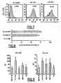
Figures 1, 2, 3, 4 and5 : Mesothelioma susceptibility to attenuated Measles Virus (MV).
Figure 1 - Selective Oncolytic activity of Schwarz MV vaccine strain. A panel of human epithelioid mesothelioma cell lines (M11, M13, M47, M56 & M61) and an immortalized normal mesothelial cell line (Met5A) were infected with non-recombinant MV (MOI 1.0) and microscope observations of infected cultures morphology were performed 72 hours later.
Figures 2-3 - Higher surface expression level of CD46 receptors for tumoral cells in comparaison with their normal counterparts. Cells were stained with FITC-conjugated CD46-specific antibodies (grey histogram) or related isotype Ig control (white histogram) (Figure 2 ). Numbers indicate the mean fluorescence index and histogram shows mean values of CD46 expression obtained for mesothelial (white bar) and mesothelioma (hatched bar) cell lines (Figure 3 ).
Figures 4-5 - Schwarz MV vaccine strain preferentially infects transformed tumoral cells. Equal numbers of M13 and Met5A cells were cultured separately (Figure 4 ) or co-cultured (Figure 5 ) overnight, allowing cellular adherence, and infection was done at MOI of 1.0 with eGFP-recombinant MV. In separate cultures, analysis of eGFP expression was performed at different times post-infection (24, 48, & 72 hours) by flow cytometry (Figure 4 ). In co-culture model, the same experiment was conducted along with HLA-A2 staining, as HLA alleles differential expression allowed distinction between two cell lines. Histogram shows % eGFP-positive cells for Met5A (white bar) and M13 (black bar) cells from co-culture (Figure 5 ). -
Figure 6 : Immunogenicity of MV-infected mesothelioma cell line.
Figure 6 - Cellular death induced by MV- and UV- treatments. Flow cytometry analysis of M13 tumoral cells apoptosis triggered by UV exposure (5 kJ/cm2) or MV infection (MOI=1.0) at the indicated time points (D1 = 24h, D2 = 48h, D3 = 72h, and D4 = 96h) (hatched bars) vs. untreated control cells (white bars). -
Figures 7 and 8 : Phagocytosis of apobodies by monocyte-derived DCs.
Figure 7 - UV- or MV-treated M13 tumor cells were labelled with PKH-26 and co-cultured with immature DCs for 24 hours. Harvested DCs were subsequently stained with FITC-conjugated anti HLA-DR antibodies and analysed by flow cytometry. One representative experiment of three with similar results is shown. The number of double-positive DCs, that is the percentage of PKH-26 positive DCs gated on basis of HLA-DR expression (FITC-conjugated antibodies, clone B8.12.2, Immunotech), indicates the phagocytosis efficiency of apoptotic cells. -
Figure 8 - The histogram represents mean values of phagocytosis yield obtained for each loading condition tested. -
Figures 9 ,10 and11 : DC maturation induced by co-culture with MV-infected mesothelioma cells.
Figures 9 and10 - Immature DCs and M13 tumoral cells were cultured in the indicated combinations (ratio 1/1) for 24 hours. As controls, DCs were incubated with TLR3 ligand, polyinosinic:polycytidylic acid (50 µg/ml; Sigma), or directly infected with MV (MOI=1.0). Subsequently DCs were harvested and stained with a PE-conjugated antibody panel specific for the indicated cell surface molecules (Figure 9 - HLA molecules;Figure 10 - Maturation Markers). DCs were gated according to their morphology characteristic, and dead cells were excluded on basis of TOPRO-3 staining (Molecular Probes). DCs surface phenotype was analysed by three-colors flow cytometry. Histogram shows means values obtained from four independent donors.
Figure 11 - DC cytokine secretion pattern was investigated on 24 hours supernatant co-culture by CBA (for IL-6, IL-1β, TNFα, IL-12 & IL-10) and ELISA (for IFNα) assays. -
Figure 12 : DCs loaded with MV-infected mesothelioma cells induce MSLN-specific CD8 T cell priming.
Figure 12 - Number of MSLN-specific CD8 T cells, derived from one week sensitization co-culture with unpulsed or UV-M13 or MV-M13 pulsed DCs, was analysed by flow cytometry. Histogram indicates the percentage of PE-tetramer positive cells among T cells gated on basis of human CD8 expression (PE-Cy5-conjugated antibodies, clone RPA-T8, BD Biosciences). One representative experiment is shown. - To compare MV-related cytopathic effect on tumoral and non-tumoral cells, a panel of five epithelioid mesothelioma cell lines (M11, M13, M47, M56, and M61) and mesothelial cells (Met5A) were infected with a Schwarz vaccine strain at a Multiplicity Of Infection (MOI) of 1.0.
- The mesothelioma cell lines (M11, M13, M47, M56, and M61) were established from pleural effusion collected by thoracocentesis of cancer patients. Diagnosis of epithelioid mesothelioma was established by biopsies immunohistochemical staining. The control mesothelial cell line (Met5A) was isolated from pleural fluids of cancer-free patients and immortalized by transfection with the pRSV plasmid encoding SV40 T-antigen (ATCC-LGC Promochem, Molsheim, France). Cell lines were maintained in RPMI-1640 medium supplemented with 10% heat-inactivated Foetal Calf Serum (FCS from Biowest, Nuaille, France), 1% L-glutamine and 1% penicillin/streptomycin antibiotics (all purchased from Sigma, St Quentin Fallavier, France). Cellular cultures were routinely checked for Mycoplasma contaminations using Hoechst 33258 staining (Sigma).
- Attenuated MV Schwarz vaccine strains were obtained from F. Tangy (Pasteur Institut, France). Schwarz MV was rescued from the pTM-MVSchw (SEQ ID NO: 1) cDNA by use of the helper-cell-based rescue system described by Radecke et al.(1995) EMBO J. 14:5773-5784 and modified by Parks et al. (1999) J. Virol. 73:3560-3566. Briefly, 293-3-46 helper cells were transfected with 5 µg of pTM-MVSchw and 0.02 µg of pEMC-Lschw expressing the Schwarz MV-L gene (Combredet et al. (2003) J. Virol. 77:11546-11554) (SEQ ID NO: 2). After overnight incubation at 37°C, a heat shock was applied for 2h at 43°C, and transfected cells were transferred onto a Vero cell monolayer. Syncytia that appeared in 15 days coculture were transferred to 35-mm wells and then expanded in 75-cm2 and 150-cm2 flasks of Vero cells culture in 5% FCS DMEM. When syncytia reached 80-90% confluence, the cells were scraped into a small volume of OptiMEM and frozen-thawed once. After low-speed centrifugation to pellet cellular debris, virus-containing supernatant was stored at -80°C. The titer of recombinant MV stock was determined by an endpoint limit-dilution assay on Vero cells. The TCID50 was calculated by use of Kärber method (Karber (1931) Arch. Exp. Path. Pharmak. 162:480-483).
- Viral infections of the mesothelioma cell lines were performed at a MOI=1.0 for 2 hours incubation at 37°C. Three days following MV infection, typical morphological modifications of MV-infected cells were observed, that is development of an important cytopathic effect (CPE) on most tumoral MPM lines (4/5), by contrast with non cancerous Met5A cells (
Figure 1 ). CPE was evidenced through development of more or less important syncitia, which finally led to shedding in culture supernatant of cytoplasmic inclusion bodies of dead tumoral cells (Figure 1 ). The development of these multinucleated giant syncitia is characteristic of measles infection and is produced by fusion of HA+ infected cells with neighbour CD46+ culture cells. - A significant upregulated expression of live-attenuated MV strains receptor CD46 by mesothelioma cells could be evidenced (
Figures 2-3 ). - In order to quantify susceptibility to MV infection, Met5A and M13 cell lines were infected with eGFP-recombinant MV stock (Combredet et al. (2003) J. Virol. 77:11546-11554). The GFP-transgene expression was used as a marker of viral infection, thus allowing determination of infected cells percentage by flow cytometry. MV infection yield of both culture cells was dose-dependent (MOI ranging from 0.01 to 5.0), indicating the specificity of eGFP signal. Whereas Met5A was infected by the MV strain (for MOI ranging from 0.5), M13 was also significantly infected by MV, but always at lowest MOI (for MOI ranging from 0.1). A significant increased infection yield of tumour cells in comparison to normal cells (for MOI 1.0), was also observed both in cellular separate culture (
Figure 4 ) and co-culture (Figure 5 ) systems (ratio 1:3) at 48 hours post-infection. Moreover, virus infection could also be evidenced by down-regulation of CD46 surface expression observed in infected cellular cultures. - Thus, according to these in vitro results, mesothelioma tumors present a more important susceptibility both to MV-mediated infection and MV-related cytolytic activity than mesothelial tissue. Consequently, MPM appears as a relevant candidate for virotherapy approach based on measles virus administration.
- After demonstrating that MV is able to infect mesothelioma cells, the inventors verified if virus infection could also lead to apoptosis-mediated cell death.
- Sub-confluent monolayer M13 cells culture were either MV-infected (MOI 1.0), or UV-B-irradiated (312 nm - 5 kJ/m2) using an UV Stratalinker2400 (Stratagene Europe, Amsterdam, Netherlands), as positive control for apoptosis. Cells were collected at different times post-treatment, and cellular death was quantified as described by Ebstein et al. (2004) Am. J. Respir. Crit. Care Med. 169:1322-1330 by concomitant phosphatidylserine and Annexin-V stainings.
- As shown in
Figure 6 , 24 hours exposition of M13 cells to UV-B irradiation and 72 hours infection of M13 cells with MV yielded an equivalent rate of tumoral cell death (comprised between 70% and 80% of Annexin-V positive cells), which indicates that MV induces apoptosis in infected tumor cells. The thus-defined M13 cell death-induced conditions were used in following experiments. - Moreover, virus-related cell killing was also confirmed by observation of an important cytopathic effect, leading to complete dislocation of M13 cellular layer 72-96 hours post-infection (
Figure 1 ). - In order to follow viral growth kinetic in infected M13 cells culture (MOI=1.0), RT-PCR specific for viral dsRNA potential receptors (Mda-5, TLR-3, RIG-I and PKR) were performed. Specific primers for the β-actin gene were used as an internal experiment control.
- Briefly, M13 cells were either incubated with polyinosinic:polycytidylic acid ligand (10 µg/ml) or MV (MOI=1.0) and cellular pellets were collected at different times. Whole cellular RNA was then extracted using RNeasy kits (Qiagen, Courtaboeuf, France) according to manufacturer's instructions, and reverse-transcribed using RTase (Invitrogen, Paisley, UK). Resulting cDNA was used as template for PCR amplification using primers specific for Mda-5, TLR-3, RIG-I, PKR, IFNβ, and β-action. PCR primers sequences are listed in Table 1 . PCR products were visualized by agarose gel electrophoresis.
Table 2: primer sequences Primer Sequence Fragment size (bp) SEQ ID NO: β-action Forward ATCTGGCACCACACCTTCTACAATGAGCTGCG 837 3 Reverse CGTCATACTCCTGCTTGCTGATCCACATCTGC 4 TLR-3 Forward ATTGGGTCTGGGAACATTTCTCTTC 319 5 Reverse GTGAGATTTAAACATTCCTCTTCGG 6 Mda-5 Forward GAGCAACTTCTTTCAACCAC 633 7 Reverse GAACACCAGCATCTTCTCCA 8 RIG-I Forward GAACGATTCCATCACTATCC 580 9 Reverse GGCATCATTATATTTCCGCA 10 PKR Forward CTTCTCAGCAGATACATCAG 689 11 Reverse GTTACAAGTCCAAAGTCTCC 12 - It could thus be shown that a viral replication peak occurred between 1 day to 4 days post-infection of mesothelioma M13 cells. Besides, PCR products corresponding to viral dsRNA potential receptors (Mda-5, TLR-3, RIG-I and PKR) could also be evidenced.
- The uptake by dendritic cells (DCs) of apobodies from MV-infected (72-hours) was then studied and compared to that of UV-irradiated (24-hours) M13 tumoral cells.
- Dendritic cells were derived from monocytes generated from leukapheresis harvests of HLA-A0201 healthy donors (EFS, Nantes, France), after obtaining informed consent. Monocytes-enriched fraction (>85% purity) was first separated by Ficoll density gradient centrifugation (PAA Laboratories, Les Mureaux, France). Monocytes were then enriched by elutriation (counterflow centrifugation) using a Beckman Avanti J20 centrifuge equipped with a JE5.0 rotor and a 40-ml elutriation chamber. Routinely, purity of elutriated monocytes was over 80%, as assessed by flow cytometry based on the detection of the CD14 marker. Monocytes were cultured at 2x10⁸ cells/ml with 500 IU/ml GM-CSF and 200 IU/ml IL-4 (Cell Genix Technology, Freiburg, Germany). Cells were then allowed to differentiate for 6 days.
- On
day 6, monocytes-derived DCs were collected from culture supernatant and seeded in culture for subsequent loading. Immature DCs were incubated with 2.108 cells/ml of apoptotic material, derived from UV-treated or MV-infected allogenic M13 tumoral cells, for additionally 24 hours co-culture (ratio 1:1). DC phagocytosis yield analysis was assessed both by flow cytometry and confocal laser microscopy, as previously described (Massé et al. (2002) Cancer Research 32:1050-1056). Briefly, UV- or MV- treated M13 cells were labelled with PKH-26 membrane dye colorant, according to the manufacturer's protocol (Sigma, St Quentin Fallavier, France). After 24 hours co-culture, DCs were stained with FITC-conjugated anti HLA-DR antibodies (Immunotech, Marseilles, France). After PBS washes, cells were harvested and analysed either on a FACSCalibur (BD Biosciences, Grenoble, France), or with a TCS NT microscope (Leica Instruments, Heidelberg, Germany). DCs that have ingested apoptotic cells were identified as HLA-DR/PKH-26 double positive cells (Figure 7 ). - As shown in
Figure 8 , it could be evidenced that DCs efficiently engulfed UV-and MV-treated mesothelioma cells at the same rate, as illustrated by a similar percentage of PKH26-positive DCs gated on basis of HLA-DR expression (65% and 74% for DCs loaded respectively with UV- or MV-treated M13 cells). - Confocal laser-scanning microcopy experiments further confirmed an efficient internalization of apoptotic M13 cells by immature DCs within 24 hours co-culture, irrespective of the death-induced strategy used (MV-infected or UV-irradiated).
- The inventors next examined whether cell material derived from MV-infected M13 tumoral cells could efficiently stimulate DC maturation.
- DC maturation status was assessed within 24 hours following engulfment of tumoral cells killed either by radiation exposition or virus-mediated cytolytic activity.
- Phenotype of viable DCs (gated on basis of TOPRO-3 positive staining exclusion) was investigated by surface expression of Class I and II MHC molecules (
Figure 9 ) and of maturation markers CD80, CD86, CD83 and CD40 (Figure 10 ), completed by cytokines secretion pattern analysis performed on co-culture supernatant (Figure 11 ). As controls, DCs were left alone, or matured with a combination of TLR3 ligand and one pro-inflammatory cytokine (polyinosinic:polycytidylic acid/IFNα, as a mimick of viral infection), or directly primped by measles virus contact (MV). - Briefly, immunostaining was performed with a panel of monoclonal antibodies (all purchased from Immunotech, Marseilles, France) specific for HLA-ABC (clone B9.12.1), HLA-DR (clone B8.12.2), CD80 (clone MAB104), CD83 (clone HB15a), CD86 (clone HA5.2B7), and CD40 (clone MAB89). DCs were incubated with each of the above antibodies (1 µg/ml) at 4°C for 30 min pior to flow cytometry. Cytokines pattern secretion was assayed in supernatants collected 24 hours after engulfment. IL-10, IL-12, IL-6, IL-1β and TNFα concentrations were measured using commercially available Cytometric Beads Array kits (BD Biosciences, Le Pont de Claix, France), according to the manufacturer's protocol. Quantification of IFNα was performed with an ELISA test (Biosource, Camarillo, USA).
- A spontaneous maturation program could be observed only for DCs loaded with apobodies derived from mesothelioma cells infected with MV, at a level essentially equivalent to the positive control maturation cocktail used in the experiment (Polyl:C/IFNα). Spontaneous maturation was evidenced by significant up-regulation of co-stimulation molecules expression (for CD80, CD83, CD86, CD40 and HLA-ABC), and production of numerous pro-inflammatory cytokines (for IL-6, IL-1β, TNFα, and IFNα).
- However, in line with previous reports, pulsing DCs with UV-irradiated apoptotic tumoral cells, as well as direct infection of DCs by measles virus (MV), did not lead to this effect.
- Overall these data strongly support an increased immunogenicity of MV-infected tumoral cells with respect to UV-irradiated tumoral cells.
- Finally, the inventors tested whether DCs loaded with apobodies derived from mesothelioma cells infected with MV could stimulate an effector CD8 response specific for an MPM-associated tumor antigen, such as Mesothelin (MSLN).
- In order to assess this question, tetramer immunostaining was performed on CD8 T-lymphocytes sensibilized for one-week with autologous DCs loaded with apoptotic material derived from UV- or MV- treated M13 cells. As controls, a similar experiment was conducted with the Jurkat lymphoma T-cell line, chosen on the basis of its susceptibility to MV and its MSLN-negative expression characteristics (
Figure 12 ). As internal experiment controls, MelanA/Mart-1-specific tetramer staining (MelanA26-35L) was achieved in complement of those specific for the two selected MSLN-derived CTL epitopes. These peptides (MSLN 531-539 and MSLN 541-550) were identified by scanning MSLN amino-acid sequence (GenPept NP 005814) for matches to consensus motifs for HLA-A0201 binding, using two computer algorithms BIMAS and SYFPEITHI ( Table 2):Table 2: tetramer characteristics HLA-A0201 binding score Tetramer name Localisation Sequence SYFPEITHI BIMAS HLA-A2 VLP9 531-539 VLPLTVAEV (SEQ ID NO: 13) 29/30 272/285 HLA-A2 KLL10 541-550 KLLGPHVEGL (SEQ ID NO: 14) 30/31 312/312 - Briefly, CD8 T lymphocytes were prepared from HLA-A0201 healthy donors PBMCs by positive selection with the MACS column systems using CD8 multisort kit (Miltenyi Biotec, Paris, France). Purified naïve CD8 T cells (>90% purity) were stimulated with autologous DCs loaded with each apoptotic preparation or unloaded DCs as a control. The co-culture was performed in round bottom 96-well plates (BD Falcon), by mixing 2.104 mature DCs with 2.105 responder T cells (ratio 1:10) in 200 µl of 8% human serum RPMI 1640 medium, supplemented with 10 ng/ml IL-12 for the first 3 days (AbCys SA, Paris, France) and with 10 U/ml IL-2 (Proleukin, Chiron Therapeutics, USA) for the next days. IL-2 was added every three days, allowing regular culture medium renewal. After 7-8 days culture, T cells were harvested and stained with MSLN-specifictetramers as follows.
- The selected CD8 epitope peptides (synthesis performed by Eurogentec, Liege, Belgium) were used for monomers production (Recombinant Proteins Production Platform, U601-IFR26, Nantes, France) as previously described (Labarriere et al. (2002) Int. J. Cancer 101:280-286). HLA-A2 VLP9 and HLA-A2 KLL10 monomers were oligomerized with PE-labeled streptavidin (BD Biosciences). Staining and washing were performed in 0.1% BSA-PBS. T cells were stained successively with 10 µg/ml of PE-labeled pMHC multimers at 4°C for 30 min, and with 1 µg/ml diluted PE-Cy5-conjugated anti-CD8 antibodies (clone RPA-T8, BD Biosciences) for additionally 30 min at 4°C. Cells were washed and immediately analysed on a FACSCalibur.
- Interestingly, a significant increase of MSLN-specific T-cells percentage among the CD8-positive gated population could be observed for co-cultures with DCs loaded with apoptotic material derived from MV-treated M13 cells with respect to co-cultures with DCs loaded with apoptotic material derived from UV-treated M13 cells.
-
- <110> INSTITUT NATIONAL DE LA SANTE ET DE LA RECHERCHE MEDICALE (INSERM)
INSTITUT PASTEUR - <120> MEDICAMENTS AND METHODS FOR TREATING MESOTHELIOMA
- <130> BET08P1093
- <150>
EP 07291232.2
<151> 2007-10-10 - <160> 14
- <170> PatentIn version 3.4
- <210> 1
<211> 18967
<212> DNA
<213> Artificial sequence - <220>
<223> Plasmid pTM-MVSchw - <400> 1




- <210> 2
<211> 12082
<212> DNA
<213> Artificial sequence - <220>
<223> Plasmid pEMC-LSchw - <400> 2


- <210> 3
<211> 32
<212> DNA
<213> Artificial sequence - <220>
<223> Primer - <400> 3
atctggcacc acaccttcta caatgagctg cg 32 - <210> 4
<211> 32
<212> DNA
<213> Artificial sequence - <220>
<223> Primer - <400> 4
cgtcatactc ctgcttgctg atccacatct gc 32 - <210> 5
<211> 25
<212> DNA
<213> Artificial sequence - <220>
<223> Primer - <400> 5
attgggtctg ggaacatttc tcttc 25 - <210> 6
<211> 25
<212> DNA
<213> Artificial sequence - <220>
<223> Primer - <400> 6
gtgagattta aacattcctc ttcgg 25 - <210> 7
<211> 20
<212> DNA
<213> Artificial sequence - <220>
<223> Primer - <400> 7
gagcaacttc tttcaaccac 20 - <210> 8
<211> 20
<212> DNA
<213> Artificial sequence - <220>
<223> Primer - <400> 8
gaacaccagc atcttctcca 20 - <210> 9
<211> 20
<212> DNA
<213> Artificial sequence - <220>
<223> Primer - <400> 9
gaacgattcc atcactatcc 20 - <210> 10
<211> 20
<212> DNA
<213> Artificial sequence - <220>
<223> Primer - <400> 10
ggcatcatta tatttccgca 20 - <210> 11
<211> 20
<212> DNA
<213> Artificial sequence - <220>
<223> Primer - <400> 11
cttctcagca gatacatcag 20 - <210> 12
<211> 20
<212> DNA
<213> Artificial sequence - <220>
<223> Primer - <400> 12
gttacaagtc caaagtctcc 20 - <210> 13
<211> 9
<212> PRT
<213> Artificial sequence - <220>
<223> MSLN 531-539 epitope - <400> 13

- <210> 14
<211> 10
<212> PRT
<213> Artificial sequence - <220>
<223> MSLN 541-550 epitope - <400> 14

Claims (10)
- A method for preparing vaccinal dendritic cells intended for treating malignant mesothelioma in an individual, comprising the following steps:- in vitro infection of malignant mesothelioma cells taken from the individual by an attenuated measles strain to yield a cell lysate;- contacting dendritic cells with the cell lysate to yield vaccinal dendritic cells.
- The method according to claim 1, wherein the dendritic cells originate from the individual.
- The method according to claim 1 or 2, wherein the dendritic cells are monocyte-derived dendritic cells.
- The method according to any of claims 1 to 3, wherein the malignant mesothelioma is malignant pleural mesothelioma.
- The method according to any of claims 1 to 4, wherein the attenuated measles virus is an Edmonston strain.
- The method according to any of claims 1 to 5, wherein the attenuated measles virus is selected from the group constituted of a Schwartz strain and a Moraten strain.
- Vaccinal dendritic cells obtainable by the method according to any of claims 1 to 6, wherein said vaccinal dendritic cells over-express CD80, CD83, CD86, CD40 and HLA-ABC and produce IL-6, IL-1β, TNFα and IFNα.
- A pharmaceutical composition comprising vaccinal dendritic cells according to claim 7 as active ingredient, in association with a pharmaceutically acceptable carrier.
- Vaccinal dendritic cells according to claim 7 for use in the treatment of malignant mesothelioma in an individual.
- Vaccinal dendritic cells according to claim 7 for use according to claim 9, wherein the malignant mesothelioma is malignant pleural mesothelioma.
Priority Applications (1)
| Application Number | Priority Date | Filing Date | Title |
|---|---|---|---|
| EP08805222.0A EP2203183B1 (en) | 2007-10-10 | 2008-10-10 | Medicaments and methods for treating mesothelioma |
Applications Claiming Priority (3)
| Application Number | Priority Date | Filing Date | Title |
|---|---|---|---|
| EP07291232A EP2058003A1 (en) | 2007-10-10 | 2007-10-10 | Medicaments and methods for treating mesothelioma |
| PCT/EP2008/063626 WO2009047331A1 (en) | 2007-10-10 | 2008-10-10 | Medicaments and methods for treating mesothelioma |
| EP08805222.0A EP2203183B1 (en) | 2007-10-10 | 2008-10-10 | Medicaments and methods for treating mesothelioma |
Publications (2)
| Publication Number | Publication Date |
|---|---|
| EP2203183A1 EP2203183A1 (en) | 2010-07-07 |
| EP2203183B1 true EP2203183B1 (en) | 2015-08-19 |
Family
ID=38935926
Family Applications (2)
| Application Number | Title | Priority Date | Filing Date |
|---|---|---|---|
| EP07291232A Withdrawn EP2058003A1 (en) | 2007-10-10 | 2007-10-10 | Medicaments and methods for treating mesothelioma |
| EP08805222.0A Active EP2203183B1 (en) | 2007-10-10 | 2008-10-10 | Medicaments and methods for treating mesothelioma |
Family Applications Before (1)
| Application Number | Title | Priority Date | Filing Date |
|---|---|---|---|
| EP07291232A Withdrawn EP2058003A1 (en) | 2007-10-10 | 2007-10-10 | Medicaments and methods for treating mesothelioma |
Country Status (7)
| Country | Link |
|---|---|
| US (2) | US9023643B2 (en) |
| EP (2) | EP2058003A1 (en) |
| CN (1) | CN101868249B (en) |
| CA (1) | CA2702186C (en) |
| DK (1) | DK2203183T3 (en) |
| ES (1) | ES2550938T3 (en) |
| WO (1) | WO2009047331A1 (en) |
Families Citing this family (4)
| Publication number | Priority date | Publication date | Assignee | Title |
|---|---|---|---|---|
| CA2432738A1 (en) * | 2003-02-26 | 2004-08-26 | Philippe Despres | New dengue and west nile viruses proteins and genes coding the foregoing, and their use in vaccinal, therapeutic and diagnostic applications |
| EP2058003A1 (en) | 2007-10-10 | 2009-05-13 | Institut National De La Sante Et De La Recherche Medicale (Inserm) | Medicaments and methods for treating mesothelioma |
| EP2759301A1 (en) * | 2013-01-24 | 2014-07-30 | Institut Pasteur | Use of a genetically modified infectious measles virus with enhanced pro-apoptotic properties (MV-DeltaC virus) |
| CN107085095A (en) * | 2017-03-02 | 2017-08-22 | 江苏华冠生物技术股份有限公司 | The preparation method of measles virus lysate as elisa kit envelope antigen |
Family Cites Families (2)
| Publication number | Priority date | Publication date | Assignee | Title |
|---|---|---|---|---|
| US7118740B1 (en) * | 2000-09-22 | 2006-10-10 | Mayo Foundation For Medical Education And Research | Method for limiting the growth of cancer cells using an attenuated measles virus |
| EP2058003A1 (en) | 2007-10-10 | 2009-05-13 | Institut National De La Sante Et De La Recherche Medicale (Inserm) | Medicaments and methods for treating mesothelioma |
-
2007
- 2007-10-10 EP EP07291232A patent/EP2058003A1/en not_active Withdrawn
-
2008
- 2008-10-10 CN CN200880116771XA patent/CN101868249B/en active Active
- 2008-10-10 DK DK08805222.0T patent/DK2203183T3/en active
- 2008-10-10 US US12/682,457 patent/US9023643B2/en active Active
- 2008-10-10 EP EP08805222.0A patent/EP2203183B1/en active Active
- 2008-10-10 ES ES08805222.0T patent/ES2550938T3/en active Active
- 2008-10-10 WO PCT/EP2008/063626 patent/WO2009047331A1/en not_active Ceased
- 2008-10-10 CA CA2702186A patent/CA2702186C/en active Active
-
2015
- 2015-04-08 US US14/681,362 patent/US9636395B2/en active Active
Non-Patent Citations (2)
| Title |
|---|
| I ALDJAHDHAMI ET AL: "Attenuated measles virus as a therapy for thoracic malignancies", RESPIROLOGY, 28 February 2007 (2007-02-28), ANNUAL SCIENTIFIC MEETING 2007, pages TP112, XP055043040, DOI: 10.1111/j.1400-1843.2007.01051.x * |
| J. P. J. J. HEGMANS: "Immunotherapy of Murine Malignant Mesothelioma Using Tumor Lysate-pulsed Dendritic Cells", AMERICAN JOURNAL OF RESPIRATORY AND CRITICAL CARE MEDICINE, vol. 171, no. 10, 1 January 2005 (2005-01-01), pages 1168 - 1177, XP055032971, ISSN: 1073-449X, DOI: 10.1164/rccm.200501-057OC * |
Also Published As
| Publication number | Publication date |
|---|---|
| CA2702186C (en) | 2019-05-28 |
| DK2203183T3 (en) | 2015-11-09 |
| EP2203183A1 (en) | 2010-07-07 |
| CN101868249A (en) | 2010-10-20 |
| WO2009047331A1 (en) | 2009-04-16 |
| CN101868249B (en) | 2013-05-29 |
| US20150209423A1 (en) | 2015-07-30 |
| US9023643B2 (en) | 2015-05-05 |
| US20100278872A1 (en) | 2010-11-04 |
| US9636395B2 (en) | 2017-05-02 |
| CA2702186A1 (en) | 2009-04-16 |
| ES2550938T3 (en) | 2015-11-13 |
| EP2058003A1 (en) | 2009-05-13 |
Similar Documents
| Publication | Publication Date | Title |
|---|---|---|
| Gauvrit et al. | Measles virus induces oncolysis of mesothelioma cells and allows dendritic cells to cross-prime tumor-specific CD8 response | |
| Janke et al. | Recombinant Newcastle disease virus (NDV) with inserted gene coding for GM-CSF as a new vector for cancer immunogene therapy | |
| Schirrmacher et al. | Newcastle disease virus: a promising vector for viral therapy, immune therapy, and gene therapy of cancer | |
| Moehler et al. | Parvovirus H-1-induced tumor cell death enhances human immune response in vitro via increased phagocytosis, maturation, and cross-presentation by dendritic cells | |
| KR101270839B1 (en) | Anticancer agent containing dendritic cell having RNA virus transferred thereinto | |
| Strobel et al. | Efficient expression of the tumor-associated antigen MAGE-3 in human dendritic cells, using an avian influenza virus vector | |
| CN104968782B (en) | Vaccines utilizing human parainfluenza type 2 virus vectors | |
| TW201814040A (en) | HPV-specific T cell production technology | |
| BR112020004221A2 (en) | pro-inflammatory dendritic cell, pharmaceutical composition, and method for providing pro-inflammatory dendritic cells | |
| US9636395B2 (en) | Medicaments and methods for treating mesothelioma | |
| US20110268764A1 (en) | Immune response inducing preparations | |
| US10980873B2 (en) | Use of a genetically modified infectious measles virus with enhanced pro-apoptotic properties (MV-deltaC virus) in cancer therapy | |
| AU2013262426A1 (en) | Cellular vaccine and method of inducing an immune response in a subject | |
| Han et al. | Development of oral CTL vaccine using a CTP-integrated Sabin 1 poliovirus-based vector system | |
| Matsunaga et al. | 628. Cancer Immunotherapy Against Murine Squamous Cell Carcinoma with Dendritic Cells Activated by Sendai Virus | |
| into Human | IMMUNO-GENE THERAPY STRATEGIES II | |
| Xue | Study of Different Approaches to Enhancing the Activity of Anti-tumor Cytotoxic T Lymphocytes |
Legal Events
| Date | Code | Title | Description |
|---|---|---|---|
| PUAI | Public reference made under article 153(3) epc to a published international application that has entered the european phase |
Free format text: ORIGINAL CODE: 0009012 |
|
| 17P | Request for examination filed |
Effective date: 20100416 |
|
| AK | Designated contracting states |
Kind code of ref document: A1 Designated state(s): AT BE BG CH CY CZ DE DK EE ES FI FR GB GR HR HU IE IS IT LI LT LU LV MC MT NL NO PL PT RO SE SI SK TR |
|
| AX | Request for extension of the european patent |
Extension state: AL BA MK RS |
|
| DAX | Request for extension of the european patent (deleted) | ||
| 17Q | First examination report despatched |
Effective date: 20110217 |
|
| REG | Reference to a national code |
Ref country code: DE Ref legal event code: R079 Ref document number: 602008039690 Country of ref document: DE Free format text: PREVIOUS MAIN CLASS: A61K0039165000 Ipc: A61K0035760000 |
|
| GRAP | Despatch of communication of intention to grant a patent |
Free format text: ORIGINAL CODE: EPIDOSNIGR1 |
|
| RIC1 | Information provided on ipc code assigned before grant |
Ipc: A61K 35/14 20060101ALI20140922BHEP Ipc: A61P 35/00 20060101ALI20140922BHEP Ipc: A61K 35/76 20060101AFI20140922BHEP Ipc: A61K 39/165 20060101ALI20140922BHEP |
|
| INTG | Intention to grant announced |
Effective date: 20141015 |
|
| GRAP | Despatch of communication of intention to grant a patent |
Free format text: ORIGINAL CODE: EPIDOSNIGR1 |
|
| INTG | Intention to grant announced |
Effective date: 20150311 |
|
| RAP1 | Party data changed (applicant data changed or rights of an application transferred) |
Owner name: CENTRE NATIONAL DE LA RECHERCHE SCIENTIFIQUE Owner name: INSTITUT PASTEUR |
|
| GRAS | Grant fee paid |
Free format text: ORIGINAL CODE: EPIDOSNIGR3 |
|
| GRAA | (expected) grant |
Free format text: ORIGINAL CODE: 0009210 |
|
| AK | Designated contracting states |
Kind code of ref document: B1 Designated state(s): AT BE BG CH CY CZ DE DK EE ES FI FR GB GR HR HU IE IS IT LI LT LU LV MC MT NL NO PL PT RO SE SI SK TR |
|
| REG | Reference to a national code |
Ref country code: GB Ref legal event code: FG4D |
|
| REG | Reference to a national code |
Ref country code: CH Ref legal event code: EP |
|
| REG | Reference to a national code |
Ref country code: IE Ref legal event code: FG4D |
|
| REG | Reference to a national code |
Ref country code: AT Ref legal event code: REF Ref document number: 743308 Country of ref document: AT Kind code of ref document: T Effective date: 20150915 |
|
| REG | Reference to a national code |
Ref country code: DE Ref legal event code: R096 Ref document number: 602008039690 Country of ref document: DE |
|
| REG | Reference to a national code |
Ref country code: CH Ref legal event code: NV Representative=s name: ARNOLD AND SIEDSMA AG, CH |
|
| REG | Reference to a national code |
Ref country code: FR Ref legal event code: PLFP Year of fee payment: 8 |
|
| REG | Reference to a national code |
Ref country code: DK Ref legal event code: T3 Effective date: 20151102 |
|
| REG | Reference to a national code |
Ref country code: ES Ref legal event code: FG2A Ref document number: 2550938 Country of ref document: ES Kind code of ref document: T3 Effective date: 20151113 |
|
| REG | Reference to a national code |
Ref country code: NL Ref legal event code: FP |
|
| REG | Reference to a national code |
Ref country code: LT Ref legal event code: MG4D |
|
| PG25 | Lapsed in a contracting state [announced via postgrant information from national office to epo] |
Ref country code: NO Free format text: LAPSE BECAUSE OF FAILURE TO SUBMIT A TRANSLATION OF THE DESCRIPTION OR TO PAY THE FEE WITHIN THE PRESCRIBED TIME-LIMIT Effective date: 20151119 Ref country code: LV Free format text: LAPSE BECAUSE OF FAILURE TO SUBMIT A TRANSLATION OF THE DESCRIPTION OR TO PAY THE FEE WITHIN THE PRESCRIBED TIME-LIMIT Effective date: 20150819 Ref country code: LT Free format text: LAPSE BECAUSE OF FAILURE TO SUBMIT A TRANSLATION OF THE DESCRIPTION OR TO PAY THE FEE WITHIN THE PRESCRIBED TIME-LIMIT Effective date: 20150819 Ref country code: FI Free format text: LAPSE BECAUSE OF FAILURE TO SUBMIT A TRANSLATION OF THE DESCRIPTION OR TO PAY THE FEE WITHIN THE PRESCRIBED TIME-LIMIT Effective date: 20150819 Ref country code: GR Free format text: LAPSE BECAUSE OF FAILURE TO SUBMIT A TRANSLATION OF THE DESCRIPTION OR TO PAY THE FEE WITHIN THE PRESCRIBED TIME-LIMIT Effective date: 20151120 |
|
| PG25 | Lapsed in a contracting state [announced via postgrant information from national office to epo] |
Ref country code: PT Free format text: LAPSE BECAUSE OF FAILURE TO SUBMIT A TRANSLATION OF THE DESCRIPTION OR TO PAY THE FEE WITHIN THE PRESCRIBED TIME-LIMIT Effective date: 20151221 Ref country code: IS Free format text: LAPSE BECAUSE OF FAILURE TO SUBMIT A TRANSLATION OF THE DESCRIPTION OR TO PAY THE FEE WITHIN THE PRESCRIBED TIME-LIMIT Effective date: 20151219 Ref country code: SE Free format text: LAPSE BECAUSE OF FAILURE TO SUBMIT A TRANSLATION OF THE DESCRIPTION OR TO PAY THE FEE WITHIN THE PRESCRIBED TIME-LIMIT Effective date: 20150819 Ref country code: PL Free format text: LAPSE BECAUSE OF FAILURE TO SUBMIT A TRANSLATION OF THE DESCRIPTION OR TO PAY THE FEE WITHIN THE PRESCRIBED TIME-LIMIT Effective date: 20150819 |
|
| PG25 | Lapsed in a contracting state [announced via postgrant information from national office to epo] |
Ref country code: CZ Free format text: LAPSE BECAUSE OF FAILURE TO SUBMIT A TRANSLATION OF THE DESCRIPTION OR TO PAY THE FEE WITHIN THE PRESCRIBED TIME-LIMIT Effective date: 20150819 Ref country code: EE Free format text: LAPSE BECAUSE OF FAILURE TO SUBMIT A TRANSLATION OF THE DESCRIPTION OR TO PAY THE FEE WITHIN THE PRESCRIBED TIME-LIMIT Effective date: 20150819 Ref country code: SK Free format text: LAPSE BECAUSE OF FAILURE TO SUBMIT A TRANSLATION OF THE DESCRIPTION OR TO PAY THE FEE WITHIN THE PRESCRIBED TIME-LIMIT Effective date: 20150819 |
|
| REG | Reference to a national code |
Ref country code: DE Ref legal event code: R097 Ref document number: 602008039690 Country of ref document: DE |
|
| PG25 | Lapsed in a contracting state [announced via postgrant information from national office to epo] |
Ref country code: RO Free format text: LAPSE BECAUSE OF FAILURE TO SUBMIT A TRANSLATION OF THE DESCRIPTION OR TO PAY THE FEE WITHIN THE PRESCRIBED TIME-LIMIT Effective date: 20150819 Ref country code: LU Free format text: LAPSE BECAUSE OF FAILURE TO SUBMIT A TRANSLATION OF THE DESCRIPTION OR TO PAY THE FEE WITHIN THE PRESCRIBED TIME-LIMIT Effective date: 20151010 |
|
| PLBE | No opposition filed within time limit |
Free format text: ORIGINAL CODE: 0009261 |
|
| STAA | Information on the status of an ep patent application or granted ep patent |
Free format text: STATUS: NO OPPOSITION FILED WITHIN TIME LIMIT |
|
| PG25 | Lapsed in a contracting state [announced via postgrant information from national office to epo] |
Ref country code: MC Free format text: LAPSE BECAUSE OF FAILURE TO SUBMIT A TRANSLATION OF THE DESCRIPTION OR TO PAY THE FEE WITHIN THE PRESCRIBED TIME-LIMIT Effective date: 20150819 |
|
| 26N | No opposition filed |
Effective date: 20160520 |
|
| REG | Reference to a national code |
Ref country code: IE Ref legal event code: MM4A |
|
| PG25 | Lapsed in a contracting state [announced via postgrant information from national office to epo] |
Ref country code: SI Free format text: LAPSE BECAUSE OF FAILURE TO SUBMIT A TRANSLATION OF THE DESCRIPTION OR TO PAY THE FEE WITHIN THE PRESCRIBED TIME-LIMIT Effective date: 20150819 |
|
| REG | Reference to a national code |
Ref country code: FR Ref legal event code: PLFP Year of fee payment: 9 |
|
| PG25 | Lapsed in a contracting state [announced via postgrant information from national office to epo] |
Ref country code: IE Free format text: LAPSE BECAUSE OF NON-PAYMENT OF DUE FEES Effective date: 20151010 |
|
| PG25 | Lapsed in a contracting state [announced via postgrant information from national office to epo] |
Ref country code: HU Free format text: LAPSE BECAUSE OF FAILURE TO SUBMIT A TRANSLATION OF THE DESCRIPTION OR TO PAY THE FEE WITHIN THE PRESCRIBED TIME-LIMIT; INVALID AB INITIO Effective date: 20081010 Ref country code: BG Free format text: LAPSE BECAUSE OF FAILURE TO SUBMIT A TRANSLATION OF THE DESCRIPTION OR TO PAY THE FEE WITHIN THE PRESCRIBED TIME-LIMIT Effective date: 20150819 |
|
| PG25 | Lapsed in a contracting state [announced via postgrant information from national office to epo] |
Ref country code: CY Free format text: LAPSE BECAUSE OF FAILURE TO SUBMIT A TRANSLATION OF THE DESCRIPTION OR TO PAY THE FEE WITHIN THE PRESCRIBED TIME-LIMIT Effective date: 20150819 |
|
| PG25 | Lapsed in a contracting state [announced via postgrant information from national office to epo] |
Ref country code: HR Free format text: LAPSE BECAUSE OF FAILURE TO SUBMIT A TRANSLATION OF THE DESCRIPTION OR TO PAY THE FEE WITHIN THE PRESCRIBED TIME-LIMIT Effective date: 20150819 |
|
| PG25 | Lapsed in a contracting state [announced via postgrant information from national office to epo] |
Ref country code: MT Free format text: LAPSE BECAUSE OF FAILURE TO SUBMIT A TRANSLATION OF THE DESCRIPTION OR TO PAY THE FEE WITHIN THE PRESCRIBED TIME-LIMIT Effective date: 20150819 |
|
| REG | Reference to a national code |
Ref country code: FR Ref legal event code: PLFP Year of fee payment: 10 |
|
| REG | Reference to a national code |
Ref country code: AT Ref legal event code: UEP Ref document number: 743308 Country of ref document: AT Kind code of ref document: T Effective date: 20150819 |
|
| REG | Reference to a national code |
Ref country code: FR Ref legal event code: PLFP Year of fee payment: 11 |
|
| P01 | Opt-out of the competence of the unified patent court (upc) registered |
Effective date: 20230523 |
|
| PGFP | Annual fee paid to national office [announced via postgrant information from national office to epo] |
Ref country code: DK Payment date: 20250930 Year of fee payment: 18 |
|
| PGFP | Annual fee paid to national office [announced via postgrant information from national office to epo] |
Ref country code: NL Payment date: 20250926 Year of fee payment: 18 |
|
| REG | Reference to a national code |
Ref country code: CH Ref legal event code: U11 Free format text: ST27 STATUS EVENT CODE: U-0-0-U10-U11 (AS PROVIDED BY THE NATIONAL OFFICE) Effective date: 20251101 |
|
| PGFP | Annual fee paid to national office [announced via postgrant information from national office to epo] |
Ref country code: DE Payment date: 20251015 Year of fee payment: 18 |
|
| PGFP | Annual fee paid to national office [announced via postgrant information from national office to epo] |
Ref country code: GB Payment date: 20251023 Year of fee payment: 18 |
|
| PGFP | Annual fee paid to national office [announced via postgrant information from national office to epo] |
Ref country code: AT Payment date: 20250922 Year of fee payment: 18 |
|
| PGFP | Annual fee paid to national office [announced via postgrant information from national office to epo] |
Ref country code: IT Payment date: 20251008 Year of fee payment: 18 |
|
| PGFP | Annual fee paid to national office [announced via postgrant information from national office to epo] |
Ref country code: FR Payment date: 20251027 Year of fee payment: 18 |
|
| PGFP | Annual fee paid to national office [announced via postgrant information from national office to epo] |
Ref country code: TR Payment date: 20251002 Year of fee payment: 18 Ref country code: BE Payment date: 20251020 Year of fee payment: 18 |
|
| PGFP | Annual fee paid to national office [announced via postgrant information from national office to epo] |
Ref country code: CH Payment date: 20251101 Year of fee payment: 18 |
|
| PGFP | Annual fee paid to national office [announced via postgrant information from national office to epo] |
Ref country code: ES Payment date: 20251110 Year of fee payment: 18 |