EP2200641B1 - Compositions for eliciting an immune response against mycobacterium avium subspecies paratuberculosis - Google Patents
Compositions for eliciting an immune response against mycobacterium avium subspecies paratuberculosis Download PDFInfo
- Publication number
- EP2200641B1 EP2200641B1 EP08837410A EP08837410A EP2200641B1 EP 2200641 B1 EP2200641 B1 EP 2200641B1 EP 08837410 A EP08837410 A EP 08837410A EP 08837410 A EP08837410 A EP 08837410A EP 2200641 B1 EP2200641 B1 EP 2200641B1
- Authority
- EP
- European Patent Office
- Prior art keywords
- map
- composition
- protein
- animals
- seq
- Prior art date
- Legal status (The legal status is an assumption and is not a legal conclusion. Google has not performed a legal analysis and makes no representation as to the accuracy of the status listed.)
- Active
Links
Images


Classifications
-
- C—CHEMISTRY; METALLURGY
- C07—ORGANIC CHEMISTRY
- C07K—PEPTIDES
- C07K14/00—Peptides having more than 20 amino acids; Gastrins; Somatostatins; Melanotropins; Derivatives thereof
- C07K14/195—Peptides having more than 20 amino acids; Gastrins; Somatostatins; Melanotropins; Derivatives thereof from bacteria
- C07K14/35—Peptides having more than 20 amino acids; Gastrins; Somatostatins; Melanotropins; Derivatives thereof from bacteria from Mycobacteriaceae (F)
-
- A—HUMAN NECESSITIES
- A61—MEDICAL OR VETERINARY SCIENCE; HYGIENE
- A61K—PREPARATIONS FOR MEDICAL, DENTAL OR TOILETRY PURPOSES
- A61K39/00—Medicinal preparations containing antigens or antibodies
- A61K39/02—Bacterial antigens
- A61K39/04—Mycobacterium, e.g. Mycobacterium tuberculosis
-
- A—HUMAN NECESSITIES
- A61—MEDICAL OR VETERINARY SCIENCE; HYGIENE
- A61P—SPECIFIC THERAPEUTIC ACTIVITY OF CHEMICAL COMPOUNDS OR MEDICINAL PREPARATIONS
- A61P1/00—Drugs for disorders of the alimentary tract or the digestive system
-
- A—HUMAN NECESSITIES
- A61—MEDICAL OR VETERINARY SCIENCE; HYGIENE
- A61P—SPECIFIC THERAPEUTIC ACTIVITY OF CHEMICAL COMPOUNDS OR MEDICINAL PREPARATIONS
- A61P1/00—Drugs for disorders of the alimentary tract or the digestive system
- A61P1/12—Antidiarrhoeals
-
- A—HUMAN NECESSITIES
- A61—MEDICAL OR VETERINARY SCIENCE; HYGIENE
- A61P—SPECIFIC THERAPEUTIC ACTIVITY OF CHEMICAL COMPOUNDS OR MEDICINAL PREPARATIONS
- A61P31/00—Antiinfectives, i.e. antibiotics, antiseptics, chemotherapeutics
-
- A—HUMAN NECESSITIES
- A61—MEDICAL OR VETERINARY SCIENCE; HYGIENE
- A61P—SPECIFIC THERAPEUTIC ACTIVITY OF CHEMICAL COMPOUNDS OR MEDICINAL PREPARATIONS
- A61P31/00—Antiinfectives, i.e. antibiotics, antiseptics, chemotherapeutics
- A61P31/04—Antibacterial agents
-
- A—HUMAN NECESSITIES
- A61—MEDICAL OR VETERINARY SCIENCE; HYGIENE
- A61P—SPECIFIC THERAPEUTIC ACTIVITY OF CHEMICAL COMPOUNDS OR MEDICINAL PREPARATIONS
- A61P31/00—Antiinfectives, i.e. antibiotics, antiseptics, chemotherapeutics
- A61P31/04—Antibacterial agents
- A61P31/06—Antibacterial agents for tuberculosis
-
- A—HUMAN NECESSITIES
- A61—MEDICAL OR VETERINARY SCIENCE; HYGIENE
- A61P—SPECIFIC THERAPEUTIC ACTIVITY OF CHEMICAL COMPOUNDS OR MEDICINAL PREPARATIONS
- A61P37/00—Drugs for immunological or allergic disorders
- A61P37/02—Immunomodulators
- A61P37/04—Immunostimulants
-
- C—CHEMISTRY; METALLURGY
- C07—ORGANIC CHEMISTRY
- C07K—PEPTIDES
- C07K16/00—Immunoglobulins [IG], e.g. monoclonal or polyclonal antibodies
- C07K16/12—Immunoglobulins [IG], e.g. monoclonal or polyclonal antibodies against material from bacteria
-
- C—CHEMISTRY; METALLURGY
- C07—ORGANIC CHEMISTRY
- C07K—PEPTIDES
- C07K16/00—Immunoglobulins [IG], e.g. monoclonal or polyclonal antibodies
- C07K16/12—Immunoglobulins [IG], e.g. monoclonal or polyclonal antibodies against material from bacteria
- C07K16/1267—Gram-positive bacteria
- C07K16/1289—Mycobacteriaceae (F)
-
- G—PHYSICS
- G01—MEASURING; TESTING
- G01N—INVESTIGATING OR ANALYSING MATERIALS BY DETERMINING THEIR CHEMICAL OR PHYSICAL PROPERTIES
- G01N33/00—Investigating or analysing materials by specific methods not covered by groups G01N1/00 - G01N31/00
- G01N33/48—Biological material, e.g. blood, urine; Haemocytometers
- G01N33/50—Chemical analysis of biological material, e.g. blood, urine; Testing involving biospecific ligand binding methods; Immunological testing
- G01N33/53—Immunoassay; Biospecific binding assay; Materials therefor
-
- A—HUMAN NECESSITIES
- A61—MEDICAL OR VETERINARY SCIENCE; HYGIENE
- A61K—PREPARATIONS FOR MEDICAL, DENTAL OR TOILETRY PURPOSES
- A61K38/00—Medicinal preparations containing peptides
Definitions
- the present invention relates generally to stimulation of immunological responses, and more specifically to compositions and methods for stimulating prophylactic or and therapeutic immunological responses against Mycobacterium avium subspecies paratuberculosis.
- Mycobacterium avium subspecies paratuberculosis is the causative agent of Johne's disease (JD), which causes chronic granulomatous enteritis in ruminants.
- JD Johne's disease
- Clinically affected animals develop chronic diarrhea and progressive weight loss that eventually results in death, while subclinically inflected animals mainly have decreased production of milk.
- JD is of tremendous economic importance to the worldwide dairy industry, causing major losses due to reduced production and early culling of animals with estimates of 20% of U.S. dairy herds affected and costs of S220 million per year to the dairy industry ( Wells, et al. 2000. J. Am. Vet. Med. Assoc.216:1450-1457 ).
- Cattle are most susceptible to infection with this organism within the first 6 months of life, but disease typically does not become evident until 3 to 5 years of age. Infection occurs by ingestion of contaminated manure, colostrum, or milk from infected cows ( Sweeney, 1996. Vet Clin. N. Am. Food Anim. Pract. 12:305 - 312 ). Fetal infection also occurs, particularly in pregnant cows with advanced disease ( Sweeney, et al. 1992. Am. J. Vet. Res. 53:477 - 480 ). Moreover, the significance of MAP has increased significantly because of its potential role as a causative agent of Crohn's disease in people ( Chamberlin, et al. Aliment Pharmacol Ther 2001; 15(3):337 - 46 ; Naser SA, et al.. Mol Cell Probes 2002; 16(1):41-8 ).
- the currently approved JD vaccine for field use is an oil suspension of a killed strain of MAP, which has significant limitations. Primarily, the efficacy of this vaccine this questionable with varying results in different vaccination trials. Another concern is the interference of whole cell bacterins with diagnostic testing, since vaccinated animals have false positive reactions for tuberculosis and paratuberculosis). Thus, the demand for improved vaccines is on the rise, but they need to be potent and at the same time should not interfere with the diagnosis of tuberculosis and JD. To achieve this goal, several approaches have been tried, which include recombinant vaccines, DNA vaccines and subunit vaccines (13; Shin SJ, et al. Infect Immun 2005; 73(8): 5074-85 ). However, there continues to be a need for the development for improved MAP vaccines.
- U.S. patent no. 7,074,559 82 discloses Mycobacterium paratuberculosis-specific nucleic acid molecules comprising a particular sequence, polypeptides encoded by these M. paratuberculosis-specific nucleic acid molecules, and antibodies having specific binding affinity for the polypeptides encoded by the M. paratuberculosis-specific nucleic acid molecules. It also discloses methods of detecting M. paratuberculosis in a sample using the disclosed nucleic acid molecules, polypeptides, and antibodies and discloses methods of preventing an M. paratuberculosis infection in an animal based on the disclosed nucleic acid molecules, polypeptides, and antibodies.
- U.S. patent application publication 2007/0134274 A1 discloses a number of genomic identifiers as potential targets for vaccines and other drugs against Johne's disease. Methods are disclosed that can be used to deliver an immunizing compound to a mammal and to provide an immune response against Johne's or Crohn's disease in the mammal.
- the present invention provides compositions and methods for stimulating an immunological response in mammals against MAP.
- the compositions comprise a novel 79 kDa recombinant polypeptide referred to herein as "Map74F".
- Map74F was generated by linking a ⁇ 17.6 kDa C-terminal fragment of Map3527 protein to a fragment of Map1519 protein, followed at the C terminus by a 14.6-kDa N-terminal portion of Map3527 protein.
- compositions of the invention may also comprise other MAP proteins, such as MAP proteins 85A, 85B, 85C. 35kDa, SOD, MptC, MptD and ESAT-6 like protein, and combinations thereof
- the method comprises administering the composition to a mammal in an amount effective to stimulate an immunological response against MAP bacteria.
- the method is expected to be of benefit to any mammal susceptible to MAP infection, but is particularly beneficial for ruminants.
- compositions can be formulated with standard pharmaceutical carriers and can be administered via any of a variety of conventional routes.
- the compositions can be administered at any time to an animal susceptible to contracting MAP infection or to an animal that is infected with MAP.
- it is preferable to administer the compositions prior to MAP infection such as by administration to pregnant animals that can transfer prophylactic immunologie components to their newborns via colostrum, or by administration during the period from one to five weeks after birth.
- the present invention provides compositions and methods for stimulating an immune response against MAP in an animal.
- the compositions comprise a recombinant polypeptide referred to herein as Map74F.
- Map74F The open reading frame (ORF) encoding Map74F is 2397 nucleotides in length and codes for a 799 amino acid polypeptide.
- the sequence of the Map74F protein is provided in SEQ ID NO:1.
- Map74F has a molecular mass calculated based upon its amino acid composition to be 79 kDa, despite its apparent molecular mass of about 74 kDa as estimated by SDS-PAGE analysis.
- the sequence provided in SEQ ID NO: 1 is shown without an optional purification tag, such as a histidine tag.
- Map74F was constructed by linking from N-terminus to C-terminus a ⁇ 17.6-kDa C-terminal fragment of Map3527 protein, a 46.8 kDa fragment of Map1519 protein, followed by a ⁇ 14.6-kDa N-terminal fragment of Map3527 protein.
- a schematic representation of Map74F is provided in Figure 1 .
- Map3527 and Map1519 are provided as SEQ ID NO:2 and SEQ ID NO:3, respectively.
- the 17.6-kDa C-terminal fragment of Map3527 protein present in Map74F is represented by amino acids 183 - 361 of SEQ ID NO:2.
- the 46.8 kDa fragment of the Map1519 protein present in Map74F is represented by amino acids 1-460 of SEQ ID NO:3.
- the 14.6-kDa N-terminal fragment of Map3527 present in Map74F is represented by amino acids 33 - 180 of SEQ ID NO:2. It is expected that longer fragments of Map1519 and of the N- and C-termini of Map3527 could be included in a recombinant protein that would be useful in the method of the invention.
- compositions of the invention may comprise other agents that can stimulate an immune response against MAP bacteria.
- the compositions may comprise one or more other MAP proteins, such as MAP proteins 85A, 85B, 85C, 35kDa, Superoxide dismutase (SOD), MptC, MptD and ESAT-6 like protein, and combinations thereof. These proteins are described in U.S. application no. 11/816,365 .
- the composition comprises MAP74F and one or more of the MAP proteins 85A, 85B or SOD.
- the DNA sequence encoding the MAP 85A gene and the amino acid sequence of the 85A gene are provided in GenBank accession no. AF280067 (October 10, 2003, entry).
- the DNA sequence encoding the MAP 85B gene and the amino acid sequence of the 85A gene is provided in GenBank accession no. AF219121 85B gene (January 21, 2002 entry).
- the DNA sequence encoding the MAP 85C gene and the amino acid sequence of the 85C gene is provided in GenBank accession no. AP280068 (November 21, 2002 entry).
- the DNA sequences encoding the MAP SOD gene and the amino acid sequence of the SOD gene is provided in GenBank accession no. AF180816 (November 30, 2001 entry).
- the method of the invention comprises administering a composition comprising Map74F to a mammal in an amount effective to stimulate an immune response against MAP.
- the stimulated immune response may comprise stimulation of any component of the immune system, including but not limited to generation of antibodies reactive to MAP antigens, stimulation of lymphocyte proliferation, production of Th1-associated cytokines, such as IFN- ⁇ , and combinations of the foregoing.
- Map74F may be administered to an animal in the form of a vector.
- nucleic acid sequences encoding Map74F may be cloned into the genome of a bacterium (e.g., Salmonella) or a virus (e.g., bovine herpesvirus-1 (BHV-1)), and the resulting recombinant bacterium or virus may be administered to animals.
- a bacterium e.g., Salmonella
- a virus e.g., bovine herpesvirus-1 (BHV-1)
- the present invention also includes bacterial and viral vectors expressing Map74F.
- DNA vaccine methodologies wherein nucleic acid molecules encoding Map74F, either alone or in combination with an adjuvant or transfection-facilitating agent, are administered directly to animals.
- the method can provide benefit to any animal susceptible to MAP inflection, the compositions and method are particularly well suited for prophylaxis or therapy for MAP infection of ruminants, including but not limited to cattle, sheep, goats, deer and elk, antelope, and buffalo. In one embodiment, the method can be used for prophylaxis or therapy of Johne's Disease.
- compositions can be administered to any MAP infected or non-infected animal. Administration of the compositions to infected animals according to the method of the invention is considered to stimulate a therapeutic immunological response. However, the invention also includes administering the compositions prior to MAP infection to stimulate a prophylactic response.
- the compositions can be administration to a pregnant animal that can transfer prophylactic immunologic components to their non-infected newborns via colostrum or milk during lactation.
- the compositions can be administered during the period from one to five weeks after birth to provide a prophylactic effect which can prevent MAP infection or reduce the severity of disease if infection occurs.
- the method of the invention is prophylactic for MAP infection, while in another embodiment, the method is therapeutic for MAP infection.
- compositions can be formulated with standard pharmaceutical carriers and can be administered via any of a variety of conventional routes.
- acceptable pharmaceutical carriers for use with proteins are described in Remington's Pharmaceutical Sciences (18th Edition, A. R. Gennaro et al. Eds., Mack Publishing Co., Easton, Pa., 1990 ).
- compositions used in the method of the invention may also comprise adjuvants. Any conventional adjuvant can be used.
- the adjuvant may be Monophosphoryl lipid A (MPL), which may be provided in combination with synthetic trehalose dicorynomycolate.
- MPL Monophosphoryl lipid A
- the adjuvant may be dimethydioctadecyl ammonium bromide (DDA).
- compositions of the invention can be administered by any acceptable route. Suitable routes of administration include oral, mucosal and parenteral (e.g., intravascular, intramuscular, and subcutaneous injection).
- routes of administration include oral, mucosal and parenteral (e.g., intravascular, intramuscular, and subcutaneous injection).
- the compositions can be administered at any time to an animal susceptible to contracting MAP infection or to an animal that is infected with MAP.
- compositions can be used in a single administration or in a series of administrations to boost the immunological response. In general, a total dosage of between 10-200 ⁇ g of protein can be administered.
- This Example provides a description of the cloning, expression and purification of Map74F.
- Map74F was generated by the sequential linkage in tandem of the ORFs of the ⁇ 17.6-kDa C-terminal fragment of Map3527 to an ORF encoding a Protein having a molecular mass of 46.8 kDa, followed at the C terminus with ⁇ 14.6-kDa N-terminal portion of Map3527 .
- Map3527c The 5' and 3' oligonucleotides to the C-terminal portion of Map3527 (Map3527c) were designed as follows: 5' (TA CATATG CAT CAT CAT CAT CAT CAT CTC AAC CAG AGC GTC TCG GC 3' (SEQ ID NO:4)) and (5' TA GAATTC GGC CGG CGG CCC CTC CGC C 3' (SEQ ID NO:5)).
- the 5' oligonucleotide contained an Nde I restriction site preceding an ATG initiation codon, followed by nucleotide sequences encoding six histidine residues (italics).
- the 3' oligonucleotide contained an Eco RI restriction site.
- Map3527c the carboxyl 540-nt portion (a 14.6 kDa, 180 aa residues) of Map3527 and the resulting PCR-amplified product was ligated to a PCR2.1 Topo vector.
- the plasmid DNA with the correct insert was digested with Nde I and Eco RI, and ligated into the pET17b expression vector cut with the same enzymes.
- the ligated products were transformed into Escherichia coli DH5 ⁇ cells and one transformant with the correct insert (Map3527c) was identified by restriction enzyme digestion and DNA sequencing.
- the 5' and 3' oligos of Map1519 contained the following sequences: 5'(5'-CTA ATC GAATTC ATG TTC TAT GGG GCC TTT C-3'SEQ ID NO:6)) and 3'(5'-TA ATC GATATC CAG GAC CTT GGA CTT GTC-3' SEQ ID NO:7)).
- the 5' oligonucleotide contained an Eco RI restriction site.
- the 3' oligonucleotide had an Eco RV restriction site, followed immediately by sequences comprising the six C-terminal amino acid residues and devoid of a stop codon.
- the 5' and 3' oligonucleotides of the N-terminal fragment of Map3527 were designed as described below: 5'(5'-AT CATATC GGG CTG GCG CCG GCG TCC-3' SEQ ID NO:8)) and 3'(5'-AT CTCGAG TCA CGC GAC CTT GCC GGC-3' SEQ ID NO:9)).
- the 5' oligonucleotide contained an Eco RV restriction site.
- the 3' oligonucleotide contained an Xho I restriction site. They were designed to amplify the N-terminal 447-bp (149-amino acid residues) portion of Map3527.
- the resulting PCR-amplified product was digested with Eco RV and Xho I, and ligated into the Map3527c-Map1519 fusion pET plasmid digested with Eco RV and Xho I.
- the ligation mixture was used to transform E. coli DH5 ⁇ cells and the positive clones were identified by restriction digestion and verified by DNA sequencing.
- the final construct encodes a 74-kDa polyprotein (Map74F), comprising a single ORF organized in the linear order, Map3527C-Map1519-Map3527N.
- Map74F Map74F recombinant protein
- the plasmid DNA construct was transformed into E. coli BL21 (DE3 pLysE) cells.
- the transformed E. coli cells were plated onto LB agar supplemented with ampicillin (100 ⁇ g/mL).
- a single colony was inoculated into 10 mL of LB broth with ampicillin (100 ⁇ g/mL), and cultured at 37°C overnight with shaking.
- the culture was diluted 1: 100 in the LB broth containing ampicillin (100 ⁇ g/mL) and chloramphenicol (34 ⁇ g/m) and grown at 37°C with shaking.
- the inclusion bodies were dissolved in Buffer B (100 mM sodium phosphate, 10mM Tris-HCl, 8M urea, 2mM PMSF (Sigma) and 20 ⁇ g/ml of leupeptin (Sigma), pH8.0) and purified with Ni-NTA agarose column (Invitrogen). Eluted fractions were checked by SDS-PAGE and the fractions containing the recombinant protein were pooled and dialyzed against 10mM Tris-HCl (pH7.8) overnight at 4oC for two times.
- Buffer B 100 mM sodium phosphate, 10mM Tris-HCl, 8M urea, 2mM PMSF (Sigma) and 20 ⁇ g/ml of leupeptin (Sigma), pH8.0
- Eluted fractions were checked by SDS-PAGE and the fractions containing the recombinant protein were pooled and dialyzed against 10mM Tris-HCl (pH7.8) overnight at 4oC for
- the protein was passed through the Detoxi-GelTM Endotoxin Removing Gel (Pierce, Rockford, IL) and the purified protein was checked for endotoxin levels by the Limulus amoebocyte assay.
- the purified protein had negligible levels (10pg/ml) of endotoxin.
- Map74F A diagrammatic representation of Map74F showing the organisation and restriction enzyme sites used to construct the polyprotein is shown in Figure 1 .
- the ORF of Map 74F is 2397 nt long, coding for a 799 amino acid polypeptide ( Fig.1B ) with a predicted molecular mass of ⁇ 79 kDa. Design and construction of the ORF resulted in the introduction of two hinge sequences ( Eco RI and Eco RV) of six nucleotides, coding for two amino acids at each of the junction sites connecting the three.
- the ORF was designed to have six histidine residues in the N-terminus for purification using Ni-NTA matrix. After expression in E. coli, the recombinant protein was purified from inclusion bodies and analyzed by SDS-PAGE ( Fig.1 B) with yields ranging around 1.5-2.0mg of purified protein from one liter of induced culture.
- This Example demonstrates the use of Map74F to simulate an immune response against MAP in a mouse model.
- mice have been found to be a suitable model for MAP infection studies. Bacterial load and pathology of specific organs are good indicators of the infectious status of mice following challenge with MAP. In mice, liver, spleen and mesenteric lymph nodes are the organs of choice for assessing the bacterial burden following MAP challenge. In this Example, the MAP burden in these organs was assessed to gauge the protective efficacy of Map74F lowing challenge. It is evident from the results presented below that Map74F immunized animals were able to either kill or inhibit the proliferation of MAP over a 16 weeks course of infection. In spleen, liver and MLN, the number of CFU decreased at 8 weeks after challenge, suggesting MAP elimination.
- the reduced number of granulomas and fewer acid-fast organisms observed in the liver and spleen of vaccinated animals indicated the protective efficacy of the polyprotein.
- the substantially reduced MAP load in the organs and the improved liver and spleen pathology indicate that immunization with 74F protected mice against MAP infection by rapidly decreasing or eliminating MAP load, Map74F also elicits high levels of IFN- ⁇ . Map74F induced a good Th1 response.
- MAP 66115-98 A MAP isolate from an infected cow, designated MAP 66115-98, was used to challenge the mice and for isolation of genomic DNA.
- MAP 66115-98 was grown in 7H9 medium supplemented with 10% oleic acid-albumin-dextrose-catalase (Becton, Dickinson and Co, Sparks, MD) and mycobactin J (Allied Monitor, Inc, Fayette, MO). After culturing for 6-8 weeks, organisms were harvested by centrifugation at 10,000 X g and washed twice with phosphate buffered saline (PBS; pH 7.2). The organisms were diluted in PBS to the required concentration and used for challenging the mice.
- PBS phosphate buffered saline
- mice were divided into two groups with 36 animals in each group.
- Group I animals were immunized twice, three weeks apart with 50 ⁇ g/animal, of the fusion protein formulated with MPL+TDM Emulsion (Ribi adjuvant systems, Corixa, Hamilyon, MT) in a total volume of 100 ⁇ l per dose via subcutaneous injection on the back.
- Group II animals were kept as unvaccinated controls and administered MPL+TDM Emulsion alone,
- 12 animals in each group, designated for immunogenicity studies were killed and spleen cells were obtained by conventional procedures.
- Spleen cells were cultured in RPMI 1640 medium (Gibco, Grand Island, NY) containing 10%FBS (Gibco) at 37°C at 5% CO 2 . Immunization experiments were repeated twice with the same dose and schedule.
- Portions of spleen, liver and MLN were fixed by immersion in 10% neutral buffered formation, embedded in paraffin wax, sectioned at 4 ⁇ m and stained with any eosin and Ziehl-Neelsen stain by conventional histological methods and examined by light microscopy.
- Antigen specific IgG response was measured by conventional ELISA.
- ELISA plates Nunc-immuno module, Nunc, Roskilde, Denmark
- PBST 0.05% Tween 20 in PBS
- 300 ⁇ l of blocking buffer 1%BSA in PBST
- the plates were washed 3 times with PBST and 100 ⁇ l of diluted serum samples were added to the wells and incubated at 37°C for 1 hour.
- tolal IgG response after washing, 50ng of alkaline phosphatase conjugated goat anti-mouse IgG (KPL, Gaithersburg, MD) was added to the wells and incubated at 25°C for 30 minutes. After washing, 50 ⁇ l of BluePhos substrate (KPL) was added and incubated for 10 minutes. Plates were read in an EL X 808 Ultra microplate reader (Bio-Tek Instruments, Inc, Winooski, VT) at 630nm after adding 50 ⁇ l of stop solution (KPL).
- KPL alkaline phosphatase conjugated goat anti-mouse IgG
- Spleen cells obtained by conventional procedures were plated in duplicate at 5 ⁇ 10 5 cells/well and cultured with and without the recombinant antigen for 48 h.
- Culture supernatants were harvested and analyzed for IFN- ⁇ using a solid phase sandwich ELISA kit (Biosource, Camarillo, CA) according to the manufacturer's protocol. Briefly, 50 ⁇ l of culture supernatants were added to the wells coated with monoclonal antibody specific for mice IFN- ⁇ . After 2 hr co-incubation at room temperature with biotinylated polyclonal antibody, the wells were washed and streptavidin-peroxidase was added. After 30 min incubation and washing, tetramethylbenzidine (TMB) solution was added to the wells and the results were read at 450nm in EL X 808 Ultra microplate reader (Bio-Tek Instruments).
- TMB tetramethylbenzidine
- ELIspot kit was used to determine the relative number of IFN- ⁇ expressing cells in the single-cell spleen suspensions according to the manufacturer's instructions. Briefly, 10 ⁇ g/ml of IFN- ⁇ capture Ab (BD Biosciences, San Jose, CA) was coated onto the MultiScreen 96-well filter plate (Millipore, Bedford, MA) for overnight at 4°C. After washing with 1x washing solution, plates were blocked by 1x BSA solution for 1 hour at 25°C and washed again. Spleen cells were plated in duplicate at 5 ⁇ 10 5 cells/well and cultured with and without the antigen for 48 hours at 37°C.
- the plates were washed with 1x washing solution and incubated 1 hr at 25°C with 5 ⁇ g/ml biotin conjugated rat anti-mouse IFN- ⁇ secondary Ab (BD Biosciences). The plates were washed and incubated for 30 min at 25°C with 0.2 ⁇ g/ml of HRP-streptavidin. The filters were developed by TureBlue substrate for 15 minutes, dried in the dark and the spots were counted.
- Spleen cells were plated in duplicate in 96-well tissue culture plates at 1 ⁇ 10 6 cells/well and cultured for 24 hr. FACS analysis was preformed after stimulating the spleen cells with 74F and concanavalin A with suitable unstimulated control cells. After washing thrice in FACS buffer (1% BSA and 0.05% sodium azide in PBS), the cells were suspended in 50 ⁇ l of FACS buffer and mixed with 0.5 ⁇ g of FITC or PE conjugated CD3, CD4 and CD8 antibodies (eBioscience, San Diego, CA) and incubated on ice for 30 minutes. Cells were washed with FACS buffer twice and suspended in 100 ⁇ l of 3% formaldehyde in PBS and transferred to FACS tubes containing 500 ⁇ l of PBS.
- FACS buffer 1% BSA and 0.05% sodium azide in PBS
- the probes were labeled with the fluorescent reporter dye, 6-Carboxyfluorescein (FAM) at the 5' end and the quencher dye, N', N',N',N',N'-6-Carboxytetramethylrhodamine (TAMRA) at the 3' end.
- FAM 6-Carboxyfluorescein
- TAMRA N', N',N',N',N'-6-Carboxytetramethylrhodamine
- the reaction was performed in duplicate in 25 ⁇ l volumes containing 2 ⁇ l of 10pM forward and reverse primers, 2 ⁇ l of 2pM probe, 12.5 ⁇ l of TaqMan PCR master Mix and 9.5 ⁇ l of diluted cDNA, using the following conditions: 10min at 94°C, followed by a total of 40 two-temperature cycles (15seconds at 95°C and 1min at 60°C), in an automated fluorometer (7700 Sequence detector, Applied Biosystems, Foster city, CA). Quantitation was done using the comparative cycle threshold (C 1 ) method and reported as relative transcription or the n-fold difference relative to a calibrator cDNA. Table 1. Gene Sequence (5' ⁇ 3') Length (bp) a Accession No.
- IL-2 FW CCTGAGCAGGATGGAGAATTACA (SEQ ID NO:10) 141 X017722 M16760 RV TCCAGAACATGCCGCAGAG (SEQ ID NO:11) AF195956 TP CCCAAGCAGGCCACAGAATTGAAAG (SEQ ID NO:12) IL-12p40 FW GGAAGCACGGCAGCAGAATA (SEQ ID NO:13) 180 M866712 RV AACTTGAGGGAGAAGTAGGAATGG (SEQ ID NO:14) S82420-6 TP CATCATCAAACCAGACCCGCCCAA (SEQ ID NO:15) TNF- ⁇ FW CATCTTCTCAAAATTCGAGTGACAA (SEQ ID NO:16) 175 M130492 Y00467 RV TGGGAGTAGACAAGGTACAACCC (SEQ ID NO:17) TP CACGTCGTAGCAAACCACCAAGTGGA (SEQ ID NO:18) INF- ⁇ FW TCAAGTGGCATAGATGTGGA
- mice from each group were killed, and anti-Map74F antibody response and T cell response were evaluated. Mice immunized with Map 74F had a significantly (P ⁇ 0.01) stronger IgG1 response against Map 74F but not IgG2a. In contrast, no Map 74F-specific antibodies were detected in the control group.
- IFN- ⁇ responses were assessed by IFN- ⁇ ELISA of the culture supernatant and the IFN- ⁇ ELISPOT assay. Antigen specific IFN- ⁇ response was significantly higher (P ⁇ 0.05) in mice immunized with Map 74F as compared to the control animals which received MPL alone ( Fig. 2A ). IFN- ⁇ ELlSPOT result was also comparable to the IFN- ⁇ ELISA result ( Fig.2B ), which further confirmed the increased IFN- ⁇ response in the vaccinated animals. The vaccinated group had a mean single-cell forming colonies (SFC) of 20, in contrast to 7 in the control group.
- SFC mean single-cell forming colonies
- CD3 + and CD4 + T cells were significantly higher (P ⁇ 0.01) in mice immunized with Map 74F ( Fig.2C ) than the control animals. In contrast, no significant difference was seen in CD8 + T cell populations between vaccinated and control animals. Cytokine gene expression levels were evaluated by real-time PCR. No significant differences (P > 0.05) were detected in the expression levels of the cytokine genes IL-2, IL-12 , TNF- ⁇ and IFN- ⁇ between the vaccinated and control animals ( Fig. 2D ).
- Map 74F protects C57 / BL6 mice against MAP infection
- mice Based on the immune responses obtained in vaccinated mice, we planned to assess the protective efficacy of Map 74F protein against MAP infection in mice.
- the sera collected at different time points were tested by ELISA for total IgG response ( Fig. 3A ) and IgG1/IgG2a ratio ( Fig. 3B ).
- Mice immunized with 74F showed a significant (P ⁇ 0.01) increase in 74F specific antibody response at 3 and 7 weeks (4weeks after challenge) after the booster vaccination compared to the control animals. Although the antibody levels increased in the control animals following challenge, the response was higher in the vaccinated animals at 8, 12 and 16 weeks after challenge.
- spleen, liver and MLN were cultured for MAP at different time points following challenge with MAP.
- MAP burden was significantly (P ⁇ 0.01) lower in the vaccinated animals compared to the control animals at 8, 12 and 16 weeks after challenge ( Fig. 4A ).
- vaccinated animals had a lower MAP burden than the control animals and the burden was significantly (P ⁇ 0.01) lower at 12 weeks after challenge ( Fig. 4B ).
- MAP burden in the MLN was significantly lower from 8 weeks on after challenge ( Fig. 4C ). Histopathological examination of liver ( Fig. 5 .
- Th1 cells are crucial for protection during the early phase of the infection.
- the most effective vaccination strategies against intracellular pathogens are considered to be those that stimulate both CD4 + and CD8 + T cell responses to produce Th1-associated cytokines.
- IFN- ⁇ is regarded as the major component in activation of macrophages and its production by Th1 CD4 + T cells is considered essential for containing MAP infection
- Our results indicate that vaccination with the fusion protein elicited a significant IFN- ⁇ response.
- MAP 66.115-98 Bacteria .
- MAP 66.115-98 a clinical isolate, was used to challenge the goats after immunization. This strain is IS 900 positive and mycobactin dependent.
- MAP 66115-98 was grown in 7H9 medium supplemented with 10% oleic acid-albumin-dextrose-catalase (Becton Dickinson Co, Sparks, MD) and mycobactin J (Allied Monitor, Inc. Fayette, MO). Alter culturing for 8 weeks, the organisms were harvested by centrifugation at 4000 x g for 10 min and washed twice with phosphate buffered saline (PBS; pH 7.2). The organisms were diluted in PBS to the required concentration and used for challenging the calves.
- PBS phosphate buffered saline
- MAP antigens and adjuvant Three recombinant MAP antigens, 85A, 85B, SOD, and the fusion polypeptide Map74F were cloned, expressed, and purified using standard techniques and as set forth in U.S. application no., 11/816,365 .
- the expressed proteins were purified using Ni-NTA agarose columns (Qiagen, Valencia, (CA). Endotoxin contamination was removed by use of Affinity Pak Detoxi Gel (Pierce, Rockford, IL) and the antigens had negligible levels (10pg/ml) of endotoxin in a Limulus amoebocyte assay.
- DDA (Sigma, USA) was mixed in sterile distilled water to a concentration of 2.5mg/ml, heated to 80°C with constant stirring for 20 min and cooled to room temperature DDA was mixed thoroughly with the recombinant antigens to a final concentration of 250 ⁇ g/dose.
- the goats were divided into three groups, with eight animals in group I and II and 9 animals in Group III (since we had an extra goat kid). All goat kids stayed with their dams until three months of age.
- Group 1 animals were immunized with 100 ⁇ g each of the four antigens (85A, 85B, SOD, and Map 74F) in DDA by subcutaneous injection.
- Group II animals were immunized with 100 ⁇ g of each antigen without DDA.
- Group III were kept as control animals and administered only DDA.
- the goats were boosted with the same regimen of antigens and adjuvant,
- PBMC peripheral blood mononuclear cells
- the lymphocytes were resuspended in RPMI-1640 medium containing 10% fetal bovine serum (Gibco, Grand Island, NY), 2mM L-glutamine, 100 mM HEPES, 100 IU/ml of penicillin, 100 ⁇ g/ml of streptomycin and 50 ⁇ g/ml of gentamycin (Gibco), to a final concentration of 2 x 10 6 viable cells/ml.
- the cells were seeded (200 ⁇ l/well) onto 96-well round or flat bottom plates, depending on the type of experiment.
- Lymphocyte proliferation assay Lymphocyte proliferation assays were performed using standard techniques. Briefly, PBMC, in 96-well flat bottom plates were incubated at 37°C in a humidified atmosphere in 5% CO 2 and stimulated with each of the four purified recombinant antigens (10 ⁇ g/ml), concanavalin A (ConA; 10 ⁇ g/ml, Sigma-Aldrich, St. Louis, MO) and purified protein derivative (PPD; 10 ⁇ g/ml, DBL, National Veterinary Services Laboratory, Ames, IA) for 72 hr.
- ConA concanavalin A
- PPD purified protein derivative
- DNA synthesis in stimulated and un-stimulated control cells was measured by the incorporation of bromodeoxyuridine (BrdU) by Cell proliferation ELISA, BrdU colorimetric kit (Roche Diagnostics, indianapolis, IN) as per the manufacturer's protocol. Briefly, the cells were labeled for 2 hr with 10 ⁇ l of BrdU labeling solution. The peroxidase conjugated anti-BrdU antibody was added and incubated for 90 min. This was followed by the addition of the enzyme substrate solution and incubation at room temperature for 15 min.
- bromodeoxyuridine BrdU
- BrdU colorimetric kit Roche Diagnostics, indianapolis, IN
- the enzymatic reaction was stopped by the addition of 1M H 2 SO 4 , and the optical density (OD) was read at 450 nm using an EL x 808 Ultra microplate reader (Bio-Tek Instruments, Inc, Winooski. VT). The tests were run in triplicate, and the results were expressed as the average stimulation index (SI), calculated as the ratio between the mean OD of cells cultured with the antigens and the mean OD of cells cultured without the antigens.
- SI average stimulation index
- IFN- ⁇ assay IFN- ⁇ levels were measured in the culture supernatants of PBMC using a monoclonal antibody-based sandwich enzyme immunoassay (BOVIGAM; Biocor Animal Health, Omaha, NE), as per the manufacturer's instructions. The plates were read at 450 nm using an ELx 808 Ultra microplate reader (Bio-Tek Instruments, Inc). The results were interpreted based on a comparison of negative and positive control optical density (OD). Results were determined as either positive (if the OD is more than that of positive control) or negative (if the OD is less than that of positive control), relative to the cutoff values as suggested by the manufacturer.
- BOVIGAM monoclonal antibody-based sandwich enzyme immunoassay
- Flow cytometric analysis of lymphocyte markers Single-color flow cytometric analysis was performed for lymphocyte surface differentiation antigens, using goat-specific monoclonal antibodies (CD2-MUC2A;CD4-17D-IgG 1 ; CD4-GC1A1-lgG2 a ;CD8-CACT80C- IgG 1 ; CD8-7C2B-IgG2 a ; CD25-CACT116A-IgG 1 ; CD45RO-ILA116A-IgG3; ⁇ TCR alpha chain specific IgG2b-GB21Al;ACT1-CACT7A-IgM; ACT16-GB110A-IgM) (VMRD, Inc, Pullman, WA) according to standard protocols.
- CD2-MUC2A CD4-17D-IgG 1 ; CD4-GC1A1-lgG2 a ;CD8-CACT80C- IgG 1 ; CD8-7C2B-IgG2 a ; CD
- the cells were washed three times in fluorescence activated cell sorter (FACS) buffer and incubated with the primary antibody previously titrated for optimum reactivity for 30 min at 4°C. Following this, the samples were washed three times and incubated with fluorescein isothiocyanate labeled horse anti-mouse immunoglobulin (Vector Laboratories, Burlingame. CA) for 30 min at 4°C. The cells were washed twice in FACS buffer and suspended in 100 ⁇ l of 3% neutral buffered formalin in PBS.
- FACS fluorescence activated cell sorter
- the cells were transferred to a FACS tube, and the volume was made up to 500 ⁇ l with PBS prior to measurement using a FACS caliber flow cytometer (Becton Dickinson, San Jose, CA). Data was collected on 5.000-10.000 events and were analyzed using Cellquest software.
- RNA isolation was isolated from lysed PBMC using an RNeasy mini Kit (Qianen, Valencia. CA). The isolated RNA samples were treated with 10 U/ ⁇ l of RNase-free DNase I (Qiagen) at 37°C for 10 min, followed by heat inactivation at 95°C for 5 min and then chilled on ice. Reverse transcription of the RNA samples was carried out in a 20 ⁇ l reaction volume (1.6 ⁇ l of total RNA, 200 U of Superscript II reverse transcriptase from Invitrogen, 50mM Tris-HCI.
- Probes and primers for real-time quantitative RT-PCR were designed with Primer Express software (Applied Biosystems, Foster city, CA) using bovine beta actin and cytokine gene sequences derived from GenBank. The details of probes and primers used in this study are presented in Table 2. Table 2. Cytokine Sequence (5'-3') Length Accession No.
- the probes were labeled with the fluorescent reporter dye, 6-carboxyfluorescein (FAM) at the 5' end, and the quencher dye, N', N',N',N',N'-6-carboxytetramethylrhodamine (TAMRA) at the 3' end.
- FAM 6-carboxyfluorescein
- TAMRA N', N',N',N',N'-6-carboxytetramethylrhodamine
- PCR was performed in a 25 ⁇ l reaction volume with 10 ⁇ l of diluted cDNA, 400 nM concentrations of primers, 80 nM of probe (Integrated DNA Technologies, Inc., Coralville, IO), and universal PCR master mix (Applied Biosystems), containing 10 mM Tris-HCl (pH 8.3), 50 mM KCl, 5 mM MgCl 2 , 2.5 mM concentrations of deoxynucleotide triphosphates, 0.625 U AmpliTaq Gold DNA polymerase and 0.25 U AmpErase uracil-N-glycosylase per reaction.
- Duplicate samples were kept in 96-well plates and amplified in an automated fluorometer (7700 Sequence Detector, Applied Biosystems). The PCR conditions were 2 min at 50°C and 10 min at 95°C, followed by 40 cycles at 95°C for 15 see and 60°C for 1 min. Quantitation was done using the comparative cycle threshold (C r ) method and reported as relative transcription or the n-fold difference relative to a calibralor cDNA.
- C r comparative cycle threshold
- Antibody responses to recombinant antigens were estimated by ELISA following a conventional protocol.
- An indirect ELISA was performed with 96-well flat bottom plates coated with 100 ⁇ l of each antigen, kept at 4°C overnight, and washed three times with PBS containing 0.05% Tween 20 (PBST). Unbound sites were blocked with 5% skim milk in PBST at 37°C for one hr and washed twice with PBST. 100 ⁇ l of 1:25,000 diluted anti-goat IgG-conjugated with horseradish peroxidase (Sigma) were added to the wells and incubated at 37°C for one hr.
- the plates were washed three times in PBST and 200 ⁇ l of 2,2'-azinobis-thiazoline-6-sulfonic acid (Sigma) was added to each well. Plates were incubated at 37°C for 30 min in the dark followed by the addition of stop solution (1 M HCl), and read three times at 405 nm at 2 min intervals using a microplate reader (BioTEK Instruments, Inc. Winooski, VT). Suitable positive and negative sera and antigen and antibody controls were included in each plate.
- MAP Fecal and organ culture of MAP .
- HEY Herald's egg yolk
- Lymphoproliferative responses to the antigens Although antigen specific lymphoproliferative responses were detected at 6 wk after primary vaccination (APV), the responses were significantly higher (P ⁇ 0.05) in both vaccinated groups (I and II) compared to the unvaccinated control group (III) at 10 wk APV ( Fig.6 ). Nevertheless, no significant differences were detected in the responses between the different antigens tested.
- Antigen specific IFN - ⁇ responses Significant differences (P ⁇ 0.01) were detected in the IFN- ⁇ responses between the vaccinated and control animals at 6 and 10 wk APV ( Fig.7 ). In the vaccinated animals the best response was detected for Map74F. IFN- ⁇ responses were significantly higher (P ⁇ 0.05) for antigens 85A and Map74F in group I animals than group II animals at 10 wk APV. However, no significant differences were detected among the other recombinant antigens tested. Though the recombinant antigen specific IFN- ⁇ levels declined from 18 wk through 38 wk APV, they were higher (P ⁇ 0.05) in the vaccinated animals than the controls.
- Lymphocyte subset distribution in response to recombinant antigens Antigen stimulated lymphocyte subsets were examined for differences in their percentages by flow cytometry. There were significant (P ⁇ 0.01) increases in CD4 + IgG1, CD4 + IgG2a, CD8 + IgG1, CD8 + IgG2a cell subtypes in the immunized groups over the control group ( Fig. 8A-D ). The increase began at 6 wk APV and persisted throughout the entire experiment period to 38 wk APV. Recombinant antigen specific CD4 + and CD8 + T cell populations were higher in the immunized animals, but the proportion of CD4 + cells was higher than that of CD8 + cells.
- Cytokine gene specific mRNA expression A significant increase in recombinant antigen specific IFN- ⁇ expression was detected in the immunized animals (P ⁇ 0.01) in contrast to the control animals beginning at 6 wk APV ( Fig.9A ). Though the expression levels peaked at 10 wk APV, levels remained significantly higher in the immunized animals compared to the controls until 38wk APV. In contrast, no significant differences were detected in IL-10 expression between the immunized and the control animals at any time point in our study, except for an overall increase in IL-10 expression levels at 26-38 wk APV in all three groups ( Fig.9B ).
- Recombinant antigen specific antibody responses All four recombinant antigens tested in this study induced robust antibody responses in both vaccinated groups. The responses were significantly higher (P ⁇ 0.01) in the vaccinated groups over the control group from six weeks APV through the entire experimental period ( Fig. 10 ). No significant differences were detected in the antibody responses among the various recombinant antigens used.
- Histopathology of tissues collected at necropsy Histopathological examination of tissues collected from each animal at necropsy indicated that 75% of the unvaccinated control animals (Group III) had granulomas in at least one tissue, whereas group I and group II animals had granulomas in only 25 and 50% of animals respectively. Granulomas were found in a mesenteric lymph node and jejunum from I animal and the ileocecal lymph node of another animal in Group I. In contrast, granulomas in Groups II and III were located primarily in the ileum, ileocecal junction, or cecum, while the ileocecal lymph node was affected in 5 animal from these two groups.
- MAP burden in tissues following challenge MAP burden in 23 tissues collected from each animal at necropsy was assessed by bacterial culture ( Figure 11 ). Among the vaccinated animals, only one animal in group I was culture positive with a very low CFU. In group II, although 5/8 animals were positive for MAP, the bacterial load was very low in four of the five animals ( ⁇ 5) from which MAP were isolated. In the unvaccinated group III, all nine animals were positive for MAP, with majority of the animals having at least 5 tissues positive for MAP with >300 CFU (too many to count).
- IFN- ⁇ is one of the major cytokine genes activated in response to MAP infection in cattle.
- major histocompatibility complex class restricted CD8 + T cells that produce cytokines such as IFN- ⁇ are required for resistance to other mycobacterial infections like M. tuberculosis.
- Map74F mediated IFN- ⁇ response was detected after the booster vaccination and challenge. Without intending to be bound by any particular theory, it is considered that the enhanced levels of IFN- ⁇ could have played an important role in the protective immunity of the goals following challenge with live MAP by IFN- ⁇ mediated signaling of macrophages.
- results presented in this Example also show a higher CD4 + and CD8 + T cell response in the immunized animals.
- Our results also support the contribution of CD4 + T cells to the peripheral IFN- ⁇ levels and proliferative responses following immunization with the recombinant antigens.
- CD25 is expressed by activated T-cells.
- Our results clearly indicate an expansion of activated T-cells in the vaccinated animals.
- the ⁇ TCR + cell population remained comparatively smaller than the CD4 + and CD8 + T cells, they were significantly higher (P ⁇ 0.05) in the immunized animals than the control animals.
- CD4 + T cell effector mechanisms are associated with the secretion of IFN- ⁇ , which activates bactericidal activity in macrophages, lymphotoxin, perform and granulysin.
- CD8 + and ⁇ + T also secrete granulysin. This is consistent with the results of our challenge experiments and support the protective efficacy of our recombinant antigens.
- the increased IFN- ⁇ mRNA expression levels clearly indicated that there was a definite antigen specific Th1 response in the immunized animals which was supported by the insignificant expression levels of the Th2 specific IL-10.
- MAP culture results presented in this Example clearly demonstrate the protective efficacy of the four recombinant antigens used. Protection was nearly complete when the antigens were given along with DDA. MAP was recovered from only one out of the eight animals of this group (I) and this animal had a significantly lower MAP load in the only one positive tissue, viz distal ileum.
Landscapes
- Health & Medical Sciences (AREA)
- Chemical & Material Sciences (AREA)
- Life Sciences & Earth Sciences (AREA)
- Organic Chemistry (AREA)
- General Health & Medical Sciences (AREA)
- Medicinal Chemistry (AREA)
- Immunology (AREA)
- Pharmacology & Pharmacy (AREA)
- Animal Behavior & Ethology (AREA)
- Public Health (AREA)
- Veterinary Medicine (AREA)
- General Chemical & Material Sciences (AREA)
- Nuclear Medicine, Radiotherapy & Molecular Imaging (AREA)
- Chemical Kinetics & Catalysis (AREA)
- Molecular Biology (AREA)
- Engineering & Computer Science (AREA)
- Biochemistry (AREA)
- Biophysics (AREA)
- Genetics & Genomics (AREA)
- Communicable Diseases (AREA)
- Proteomics, Peptides & Aminoacids (AREA)
- Bioinformatics & Cheminformatics (AREA)
- Oncology (AREA)
- Hematology (AREA)
- Microbiology (AREA)
- Gastroenterology & Hepatology (AREA)
- Urology & Nephrology (AREA)
- Biomedical Technology (AREA)
- Pulmonology (AREA)
- Biotechnology (AREA)
- Cell Biology (AREA)
- Epidemiology (AREA)
- Food Science & Technology (AREA)
- Physics & Mathematics (AREA)
- Analytical Chemistry (AREA)
- General Physics & Mathematics (AREA)
- Pathology (AREA)
- Mycology (AREA)
- Medicines Containing Antibodies Or Antigens For Use As Internal Diagnostic Agents (AREA)
- Peptides Or Proteins (AREA)
Abstract
Description
- The present invention relates generally to stimulation of immunological responses, and more specifically to compositions and methods for stimulating prophylactic or and therapeutic immunological responses against Mycobacterium avium subspecies paratuberculosis.
- Mycobacterium avium subspecies paratuberculosis (MAP) is the causative agent of Johne's disease (JD), which causes chronic granulomatous enteritis in ruminants. Clinically affected animals develop chronic diarrhea and progressive weight loss that eventually results in death, while subclinically inflected animals mainly have decreased production of milk. JD is of tremendous economic importance to the worldwide dairy industry, causing major losses due to reduced production and early culling of animals with estimates of 20% of U.S. dairy herds affected and costs of S220 million per year to the dairy industry (Wells, et al. 2000. J. Am. Vet. Med. Assoc.216:1450-1457). Cattle are most susceptible to infection with this organism within the first 6 months of life, but disease typically does not become evident until 3 to 5 years of age. Infection occurs by ingestion of contaminated manure, colostrum, or milk from infected cows (Sweeney, 1996. Vet Clin. N. Am. Food Anim. Pract. 12:305 - 312). Fetal infection also occurs, particularly in pregnant cows with advanced disease (Sweeney, et al. 1992. Am. J. Vet. Res. 53:477 - 480). Moreover, the significance of MAP has increased significantly because of its potential role as a causative agent of Crohn's disease in people (Chamberlin, et al. Aliment Pharmacol Ther 2001; 15(3):337 - 46; Naser SA, et al.. Mol Cell Probes 2002; 16(1):41-8).
- The currently approved JD vaccine for field use is an oil suspension of a killed strain of MAP, which has significant limitations. Primarily, the efficacy of this vaccine this questionable with varying results in different vaccination trials. Another concern is the interference of whole cell bacterins with diagnostic testing, since vaccinated animals have false positive reactions for tuberculosis and paratuberculosis). Thus, the demand for improved vaccines is on the rise, but they need to be potent and at the same time should not interfere with the diagnosis of tuberculosis and JD. To achieve this goal, several approaches have been tried, which include recombinant vaccines, DNA vaccines and subunit vaccines (13; Shin SJ, et al. Infect Immun 2005; 73(8): 5074-85). However, there continues to be a need for the development for improved MAP vaccines.
-
U.S. patent no. 7,074,559 82 discloses Mycobacterium paratuberculosis-specific nucleic acid molecules comprising a particular sequence, polypeptides encoded by these M. paratuberculosis-specific nucleic acid molecules, and antibodies having specific binding affinity for the polypeptides encoded by the M. paratuberculosis-specific nucleic acid molecules. It also discloses methods of detecting M. paratuberculosis in a sample using the disclosed nucleic acid molecules, polypeptides, and antibodies and discloses methods of preventing an M. paratuberculosis infection in an animal based on the disclosed nucleic acid molecules, polypeptides, and antibodies. - Li et al., "The complete genome sequence of Mycobacterium avium subspecies paratuberculosis," PNAS, vol. 102, no. 35, 30 August 2005, pages 12344 - 12349 is concerned with the provision of a diagnostic test for bovine Johne's Disease. It discloses the genome sequence of a common clone of MAP as well as MAP-specific sequences that are likely to serve as potential targets for sensitive and specific molecular and immunogenic diagnostic tests.
- Finally, the
U.S. patent application publication 2007/0134274 A1 discloses a number of genomic identifiers as potential targets for vaccines and other drugs against Johne's disease. Methods are disclosed that can be used to deliver an immunizing compound to a mammal and to provide an immune response against Johne's or Crohn's disease in the mammal. - The present invention provides compositions and methods for stimulating an immunological response in mammals against MAP. The compositions comprise a novel 79 kDa recombinant polypeptide referred to herein as "Map74F". Map74F was generated by linking a ~17.6 kDa C-terminal fragment of Map3527 protein to a fragment of Map1519 protein, followed at the C terminus by a 14.6-kDa N-terminal portion of Map3527 protein.
- In addition to Map74F, the compositions of the invention may also comprise other MAP proteins, such as
MAP proteins - The method comprises administering the composition to a mammal in an amount effective to stimulate an immunological response against MAP bacteria. The method is expected to be of benefit to any mammal susceptible to MAP infection, but is particularly beneficial for ruminants.
- The compositions can be formulated with standard pharmaceutical carriers and can be administered via any of a variety of conventional routes. The compositions can be administered at any time to an animal susceptible to contracting MAP infection or to an animal that is infected with MAP. However, it is preferable to administer the compositions prior to MAP infection, such as by administration to pregnant animals that can transfer prophylactic immunologie components to their newborns via colostrum, or by administration during the period from one to five weeks after birth.
-
-
Figure 1A provides a schematic representation of Map74F construct. Map74F was generated by sequential linkage from its N to C terminus of ORFs encoding a C-terminal fragment of Map3527 protein linked to an ORF encoding amino acids 1-460 of Map1519 protein, and terminating with an ORF encoding an N-terminal fragment of Map3527. The ORF encoding Map74F is 2397 nucleotides (nt) and it encodes a 799-aa polypeptide with a predicted molecular mass of ~79 kDa. -
Figure 1B provides a photographic representation of Coomassie Blue-stained 10% SDS-PAGE of E. coli lysates from E. coli (BL21/pLysE) transformed with an expression vector encoding Map74F. The cells were grown and induced with 1 mM IPTG. The lanes depict lysates before (lane UI) or 3 hours after (lane I) IPTG induction. Purified recombinant Map74F is shown in lane P along with molecular mass marker (lane M). -
Figures 2A - 2D provide graphical representations of data obtained from mice sacrificed 3 weeks after booster vaccination. Spleen cells from mice, administered with Map74F+ Monophosphoryl lipid A (MPL) and MPL alone were stimulated for 2 days with 10µg/ml of Map74F, ConA and medium. For the data presented inFig. 2A , culture supernatants were assayed for IFN-γ levels by ELISA. For the data presented inFig. 2B , ELIspot assay to determine the relative numbers of IFN-γ-expressing cells in single-cell spleen suspensions of immunized and control mice stimulated with and without antigens. InFig. 2C , data from FACS analysis of spleen cells for lymphocyte subset populations collected from the vaccinated (Map74F+MPL) and control animals (MPL, ConA-MPL) after stimulation with the recombinant and control antigens are presented. InFig. 2D , data are presented for cytokine mRNA expression in response to Map74F normalized to the housekeeping gene GAPDH. Data are representative of three independent experiments. -
Figures 3A and 3B present data obtained from sera collected from vaccinated and control animals at different time points (Pre-vaccination/primary vaccination (PV), booster vaccination (B), before challenge (BC), 4, 8, 12, and 16 weeks after challenge) and checked for antibody response. The data presented inFig. 3A are from sera tested for Map74F specific antibodies.Fig. 3B presents data testing for IgG1:IgG2 ratios by ELISA. Data are representative of three independent experiments. -
Figures 4A and 4B provide graphical depictions of data showing expression of protective immunity in spleen (Fig. 4A ), liver (Fig. 4B ) and mesenteric lymph node (MLN) (Fig. 4C) conferred by vaccination with Map74F+MPL. Map74F significantly reduced MAP burden in the spleen (at 8-16 weeks after challenge), liver (at 12-16 weeks after challenge) and MLN (at 8-16 weeks after challenge). Data are representative of three independent experiments. -
Figures 5A- 5D provide photographic representations of histopathologic examination of vaccinated and non-vaccinated mouse tissues.Fig. 5A is representative of liver from an un-vaccinated control mouse, Numerous large granulomas arc randomly dispersed throughout the liver. Hematoxylin and Eosin. Bar = 100nm. Insert: Higher magnification of a granuloma demonstrating numerous acid-fast bacilli. Ziehl-Neelsen staining.Fig. 5B is representative of liver from a mouse vaccinated with Map74f. Only sparse numbers of small lymphoid aggregates accompanied by a few macrophages are present.Fig. 5C is representative of spleen from an un-vaccinated control mouse showing occasional granulomas in the white pulp.Fig. 5D is representative of spleen from a mouse vaccinated with Map74F. The white and red pulp are devoid of granulomas. Hematoxylin and Eosin staining. Bar = 100nm. Insert: Higher magnification of a granuloma demonstrating the absence of acid-fast bacilli. Ziehl-Neilsen staining. -
Figures 6A- 6C provide graphical depictions of data showing lymphoproliferative responses of peripheral blood mononuclear cells (PBMC) from immunized (group 1 and 11) and control (group III) animals to the recombinant antigens (85A, 85B,Map 74F and SOD), Con A and PPD. The results are expressed as stimulation index (SI), and the error bars indicate standard deviation from the mean. -
Figures 7A- 7C provide graphical depictions of data showing antigen specific IFN-γ response of PBMC from immunized (group I and II) and control (group III) animals. Results are expressed as OD values and error bars indicate standard deviation from the mean. -
Figures 8A - 8F provide graphical depictions of data showing expression of lymphocyte subsets from PBMC collected from the immunized groups (I and II) and the control group (III) at specified times after stimulation with the recombinant and control antigens as analysed by flow-cytometry. Results are expressed as percentage of cells with positive staining relative to the un-induced sample (cultured with medium). Error bars indicate standard deviation from the mean. -
Figures 9A and9B provide graphical depictions of data showing cytokine gene mRNA expression in response to recombinant antigens in the immunized groups (I and II) and control group (III). Results are expressed as the mean fold increase over un-stimulated PBMC, which served as calibrators. Error bars indicate standard deviation from the mean. -
Fig. 10 . Antibody responses to individual recombinant antigens in the vaccinated group I and II animals and the control group III animals. Error bars represent standard deviation from the mean. -
Fig. 11 provides a tabular summary of MAP culture results (CPU) measured in various tissues collected at necropsy. - The present invention provides compositions and methods for stimulating an immune response against MAP in an animal. The compositions comprise a recombinant polypeptide referred to herein as Map74F. The open reading frame (ORF) encoding Map74F is 2397 nucleotides in length and codes for a 799 amino acid polypeptide. The sequence of the Map74F protein is provided in SEQ ID NO:1. Map74F has a molecular mass calculated based upon its amino acid composition to be 79 kDa, despite its apparent molecular mass of about 74 kDa as estimated by SDS-PAGE analysis. The sequence provided in SEQ ID NO: 1 is shown without an optional purification tag, such as a histidine tag.
- Map74F was constructed by linking from N-terminus to C-terminus a ~17.6-kDa C-terminal fragment of Map3527 protein, a 46.8 kDa fragment of Map1519 protein, followed by a ~14.6-kDa N-terminal fragment of Map3527 protein. A schematic representation of Map74F is provided in
Figure 1 . - The complete amino acid sequences of Map3527 and Map1519 are provided as SEQ ID NO:2 and SEQ ID NO:3, respectively. The 17.6-kDa C-terminal fragment of Map3527 protein present in Map74F is represented by amino acids 183 - 361 of SEQ ID NO:2. The 46.8 kDa fragment of the Map1519 protein present in Map74F is represented by amino acids 1-460 of SEQ ID NO:3. The 14.6-kDa N-terminal fragment of Map3527 present in Map74F is represented by amino acids 33 - 180 of SEQ ID NO:2. It is expected that longer fragments of Map1519 and of the N- and C-termini of Map3527 could be included in a recombinant protein that would be useful in the method of the invention.
- In addition to MAP74F, the compositions of the invention may comprise other agents that can stimulate an immune response against MAP bacteria. For instance, the compositions may comprise one or more other MAP proteins, such as
MAP proteins U.S. application no. 11/816,365 . - In one embodiment, the composition comprises MAP74F and one or more of the
MAP proteins MAP 85A gene and the amino acid sequence of the 85A gene are provided in GenBank accession no. AF280067 (October 10, 2003, entry). The DNA sequence encoding theMAP 85B gene and the amino acid sequence of the 85A gene is provided in GenBank accession no.AF219121 85B gene (November 21, 2002 entry). The DNA sequence encoding the MAP 85C gene and the amino acid sequence of the 85C gene is provided in GenBank accession no.AP280068 (November 21, 2002 - The method of the invention comprises administering a composition comprising Map74F to a mammal in an amount effective to stimulate an immune response against MAP. The stimulated immune response may comprise stimulation of any component of the immune system, including but not limited to generation of antibodies reactive to MAP antigens, stimulation of lymphocyte proliferation, production of Th1-associated cytokines, such as IFN-γ, and combinations of the foregoing.
- Map74F may be administered to an animal in the form of a vector. For example, nucleic acid sequences encoding Map74F may be cloned into the genome of a bacterium (e.g., Salmonella) or a virus (e.g., bovine herpesvirus-1 (BHV-1)), and the resulting recombinant bacterium or virus may be administered to animals. Thus, the present invention also includes bacterial and viral vectors expressing Map74F. Also contemplated within the scope of this invention are DNA vaccine methodologies wherein nucleic acid molecules encoding Map74F, either alone or in combination with an adjuvant or transfection-facilitating agent, are administered directly to animals.
- The method can provide benefit to any animal susceptible to MAP inflection, the compositions and method are particularly well suited for prophylaxis or therapy for MAP infection of ruminants, including but not limited to cattle, sheep, goats, deer and elk, antelope, and buffalo. In one embodiment, the method can be used for prophylaxis or therapy of Johne's Disease.
- The compositions can be administered to any MAP infected or non-infected animal. Administration of the compositions to infected animals according to the method of the invention is considered to stimulate a therapeutic immunological response. However, the invention also includes administering the compositions prior to MAP infection to stimulate a prophylactic response. For example, the compositions can be administration to a pregnant animal that can transfer prophylactic immunologic components to their non-infected newborns via colostrum or milk during lactation. Alternatively, the compositions can be administered during the period from one to five weeks after birth to provide a prophylactic effect which can prevent MAP infection or reduce the severity of disease if infection occurs. Thus, in one embodiment, the method of the invention is prophylactic for MAP infection, while in another embodiment, the method is therapeutic for MAP infection.
- The compositions can be formulated with standard pharmaceutical carriers and can be administered via any of a variety of conventional routes. Some examples of acceptable pharmaceutical carriers for use with proteins are described in Remington's Pharmaceutical Sciences (18th Edition, A. R. Gennaro et al. Eds., Mack Publishing Co., Easton, Pa., 1990).
- The compositions used in the method of the invention may also comprise adjuvants. Any conventional adjuvant can be used.
- In one embodiment, the adjuvant may be Monophosphoryl lipid A (MPL), which may be provided in combination with synthetic trehalose dicorynomycolate. In another embodiment, the adjuvant may be dimethydioctadecyl ammonium bromide (DDA).
- The compositions of the invention can be administered by any acceptable route. Suitable routes of administration include oral, mucosal and parenteral (e.g., intravascular, intramuscular, and subcutaneous injection). The compositions can be administered at any time to an animal susceptible to contracting MAP infection or to an animal that is infected with MAP.
- Those skilled in the art will recognize that the amount of MAP74F and any other antigenic agents in the composition administered to a particular animal will depend on a number of factors, such as the route of administration, and the size, physical condition and the MAP status of the animal, and can be adjusted by those skilled in the art to achieve a desired results. The compositions can be used in a single administration or in a series of administrations to boost the immunological response. In general, a total dosage of between 10-200 µg of protein can be administered.
- The following examples describe the various embodiments of this invention. These examples are illustrative and are not intended to be restrictive.
- This Example provides a description of the cloning, expression and purification of Map74F.
- Map74F was generated by the sequential linkage in tandem of the ORFs of the ∼17.6-kDa C-terminal fragment of Map3527 to an ORF encoding a Protein having a molecular mass of 46.8 kDa, followed at the C terminus with ∼14.6-kDa N-terminal portion of Map3527.
- The 5' and 3' oligonucleotides to the C-terminal portion of Map3527 (Map3527c) were designed as follows: 5' (TA CATATG CAT CAT CAT CAT CAT CAT CTC AAC CAG AGC GTC TCG GC 3' (SEQ ID NO:4)) and (5' TA GAATTC GGC CGG CGG CCC CTC CGC C 3' (SEQ ID NO:5)). The 5' oligonucleotide contained an NdeI restriction site preceding an ATG initiation codon, followed by nucleotide sequences encoding six histidine residues (italics). The 3' oligonucleotide contained an EcoRI restriction site. These oligos were used to amplify Map3527c, the carboxyl 540-nt portion (a 14.6 kDa, 180 aa residues) of Map3527 and the resulting PCR-amplified product was ligated to a PCR2.1 Topo vector. The plasmid DNA with the correct insert was digested with NdeI and EcoRI, and ligated into the pET17b expression vector cut with the same enzymes. The ligated products were transformed into Escherichia coli DH5α cells and one transformant with the correct insert (Map3527c) was identified by restriction enzyme digestion and DNA sequencing.
- The 5' and 3' oligos of Map1519 contained the following sequences: 5'(5'-CTA ATC GAATTC ATG TTC TAT GGG GCC TTT C-3'SEQ ID NO:6)) and 3'(5'-TA ATC GATATC CAG GAC CTT GGA CTT GTC-3' SEQ ID NO:7)). The 5' oligonucleotide contained an EcoRI restriction site. The 3' oligonucleotide had an EcoRV restriction site, followed immediately by sequences comprising the six C-terminal amino acid residues and devoid of a stop codon. The amplification of the coding sequence of Map1519 (1380 bp; a 460-aa stretch with a predicted size of ∼46.8 kDa), and resulting PCR-amplified product was ligated to PCR2.1 Topo vector cut with the same enzymes. The clone with the correct insert was digested with EcoRI and EcoRV and ligated to EcoRI/EcoRV predigested Map3527c-pET plasmid. The ligated products were then transformed into E. coli DH5α cells, and a transformant with the correct insert (Map3527c-Map1519) was identified by restriction enzyme digestion and verified by DNA sequencing.
- The 5' and 3' oligonucleotides of the N-terminal fragment of Map3527 were designed as described below: 5'(5'-AT CATATC GGG CTG GCG CCG GCG TCC-3' SEQ ID NO:8)) and 3'(5'-AT CTCGAG TCA CGC GAC CTT GCC GGC-3' SEQ ID NO:9)). The 5' oligonucleotide contained an EcoRV restriction site. The 3' oligonucleotide contained an XhoI restriction site. They were designed to amplify the N-terminal 447-bp (149-amino acid residues) portion of Map3527. The resulting PCR-amplified product was digested with EcoRV and XhoI, and ligated into the Map3527c-Map1519 fusion pET plasmid digested with EcoRV and XhoI. The ligation mixture was used to transform E. coli DH5α cells and the positive clones were identified by restriction digestion and verified by DNA sequencing. The final construct, encodes a 74-kDa polyprotein (Map74F), comprising a single ORF organized in the linear order, Map3527C-Map1519-Map3527N.
- The plasmid DNA construct was transformed into E. coli BL21 (DE3 pLysE) cells. The transformed E. coli cells were plated onto LB agar supplemented with ampicillin (100 µg/mL). A single colony was inoculated into 10 mL of LB broth with ampicillin (100µg/mL), and cultured at 37°C overnight with shaking. The culture was diluted 1: 100 in the LB broth containing ampicillin (100 µg/mL) and chloramphenicol (34 µg/m) and grown at 37°C with shaking. After 3 hr induction with 1 mM IPTG (Invitrogen Co, Carlsbad, CA), cells were harvested by centrifugation (5000Xg) and washed once in PBS. Cells were suspended in buffer A (50mM TrisHCl, 1mM EDTA, 50mM NaCl, pH8.0) and lysed in a French cell press or cell disruptor. After spinning down the inclusion bodies, the pellets were washed three times with inclusion body washing buffer (Buffer A + 1% Triton X-100) and twice with CHAPS (Sigma-Aldrich, St. Louis, MO) buffer (1% CHAPS in 10mM Tris-HCl, pH8.0) in order to remove lipopolysaccharide (LPS). The inclusion bodies were dissolved in Buffer B (100 mM sodium phosphate, 10mM Tris-HCl, 8M urea, 2mM PMSF (Sigma) and 20µg/ml of leupeptin (Sigma), pH8.0) and purified with Ni-NTA agarose column (Invitrogen). Eluted fractions were checked by SDS-PAGE and the fractions containing the recombinant protein were pooled and dialyzed against 10mM Tris-HCl (pH7.8) overnight at 4oC for two times. The protein was passed through the Detoxi-GelTM Endotoxin Removing Gel (Pierce, Rockford, IL) and the purified protein was checked for endotoxin levels by the Limulus amoebocyte assay. The purified protein had negligible levels (10pg/ml) of endotoxin.
- A diagrammatic representation of Map74F showing the organisation and restriction enzyme sites used to construct the polyprotein is shown in
Figure 1 . The ORF ofMap 74F is 2397 nt long, coding for a 799 amino acid polypeptide (Fig.1B ) with a predicted molecular mass of ∼79 kDa. Design and construction of the ORF resulted in the introduction of two hinge sequences (EcoRI and EcoRV) of six nucleotides, coding for two amino acids at each of the junction sites connecting the three. In addition, the ORF was designed to have six histidine residues in the N-terminus for purification using Ni-NTA matrix. After expression in E. coli, the recombinant protein was purified from inclusion bodies and analyzed by SDS-PAGE (Fig.1 B) with yields ranging around 1.5-2.0mg of purified protein from one liter of induced culture. - This Example demonstrates the use of Map74F to simulate an immune response against MAP in a mouse model.
- Mice have been found to be a suitable model for MAP infection studies. Bacterial load and pathology of specific organs are good indicators of the infectious status of mice following challenge with MAP. In mice, liver, spleen and mesenteric lymph nodes are the organs of choice for assessing the bacterial burden following MAP challenge. In this Example, the MAP burden in these organs was assessed to gauge the protective efficacy of Map74F lowing challenge. It is evident from the results presented below that Map74F immunized animals were able to either kill or inhibit the proliferation of MAP over a 16 weeks course of infection. In spleen, liver and MLN, the number of CFU decreased at 8 weeks after challenge, suggesting MAP elimination. Apart from the bacterial burden, the reduced number of granulomas and fewer acid-fast organisms observed in the liver and spleen of vaccinated animals indicated the protective efficacy of the polyprotein. The substantially reduced MAP load in the organs and the improved liver and spleen pathology indicate that immunization with 74F protected mice against MAP infection by rapidly decreasing or eliminating MAP load, Map74F also elicits high levels of IFN-γ. Map74F induced a good Th1 response.
- The following materials and methods were used in this Example.
- Experiments were performed using 6-8 week old female C57/BL6 mice (Harlan Sprague, Indianapolis, Indiana). Animals were maintained in a biosafety level II facility and had free access to feed and water. All the experimental work was conducted in compliance with the regulations, policies, and principles of the Animal Welfare Act, the Public Health Service Policy on Humane Care and Use of Laboratory Animals used in Testing, Research, and Training, the NIH Guide for the Care and Use of Laboratory Animals and the New York State Department of Public Health.
- A MAP isolate from an infected cow, designated MAP 66115-98, was used to challenge the mice and for isolation of genomic DNA. MAP 66115-98 was grown in 7H9 medium supplemented with 10% oleic acid-albumin-dextrose-catalase (Becton, Dickinson and Co, Sparks, MD) and mycobactin J (Allied Monitor, Inc, Fayette, MO). After culturing for 6-8 weeks, organisms were harvested by centrifugation at 10,000 X g and washed twice with phosphate buffered saline (PBS; pH 7.2). The organisms were diluted in PBS to the required concentration and used for challenging the mice.
- Mice were divided into two groups with 36 animals in each group. Group I animals were immunized twice, three weeks apart with 50 µg/animal, of the fusion protein formulated with MPL+TDM Emulsion (Ribi adjuvant systems, Corixa, Hamilyon, MT) in a total volume of 100 µl per dose via subcutaneous injection on the back. Group II animals were kept as unvaccinated controls and administered MPL+TDM Emulsion alone, Three weeks after the second immunization, 12 animals in each group, designated for immunogenicity studies, were killed and spleen cells were obtained by conventional procedures. Spleen cells were cultured in RPMI 1640 medium (Gibco, Grand Island, NY) containing 10%FBS (Gibco) at 37°C at 5% CO2. Immunization experiments were repeated twice with the same dose and schedule.
- Three weeks after the second immunization, 24 animals each in Group I and II were challenged by intraperitoneal injection of 109 CTU units of Mycobacterium avium subsp. paratuberculosis. Six animals each in both groups were euthanized at 4, 8, 12 and 16 weeks after the challenge and spleen, liver and mesenteric lymph nodes (MLN) were collected and divided into two parts. One set of tissues was homogenized in PBS (100mn/ml) and 100µl of individual tissue homogenates were inoculated on to Herald's egg yolk (HEY) slants (Becton, Dickinson and Co, Sparks, MD) containing mycobactin J. in order to estimate the bacterial load. The slants were checked for bacterial growth by colony count after 8-12 weeks of inoculation. The other set of tissues was used for histopathological examination.
- Portions of spleen, liver and MLN were fixed by immersion in 10% neutral buffered formation, embedded in paraffin wax, sectioned at 4 µm and stained with any eosin and Ziehl-Neelsen stain by conventional histological methods and examined by light microscopy.
- Antigen specific IgG response was measured by conventional ELISA. ELISA plates (Nunc-immuno module, Nunc, Roskilde, Denmark) were coated with 200 ng/well of recombinant protein and incubated at 4°C overnight. After washing once with PBST (0.05
% Tween 20 in PBS), 300µl of blocking buffer (1%BSA in PBST) was added and incubated at 25°C for 1 hour. The plates were washed 3 times with PBST and 100µl of diluted serum samples were added to the wells and incubated at 37°C for 1 hour. For tolal IgG response, after washing, 50ng of alkaline phosphatase conjugated goat anti-mouse IgG (KPL, Gaithersburg, MD) was added to the wells and incubated at 25°C for 30 minutes. After washing, 50µl of BluePhos substrate (KPL) was added and incubated for 10 minutes. Plates were read in an ELX 808 Ultra microplate reader (Bio-Tek Instruments, Inc, Winooski, VT) at 630nm after adding 50µl of stop solution (KPL). For isotype response, after washing, 25ng of biotin conjugated goal anti-mouse IgG1 or IgG2a (Southern Biotech, Birmingham, AL) was added to the wells and incubated at 25°C for 30 minutes. After washing, 0.2µg/ml of streptavidin labeled with horseradish peroxidase (KPL) was added and incubated at 25°C for 30 minutes. After washing, 50µl of 3,3', 5, 5' - Tetramethylbenzidine (TMB) was added to the wells and incubated for 15 minutes. Plates were read in an ELX 808 Ultra microplate reader (Bio-Tek Instruments) by endpoint method at 450nm after adding 50µl of 1N H2SO4 as stop solution. - Spleen cells obtained by conventional procedures were plated in duplicate at 5 × 10⁵ cells/well and cultured with and without the recombinant antigen for 48 h. Culture supernatants were harvested and analyzed for IFN-γ using a solid phase sandwich ELISA kit (Biosource, Camarillo, CA) according to the manufacturer's protocol. Briefly, 50µl of culture supernatants were added to the wells coated with monoclonal antibody specific for mice IFN-γ. After 2 hr co-incubation at room temperature with biotinylated polyclonal antibody, the wells were washed and streptavidin-peroxidase was added. After 30 min incubation and washing, tetramethylbenzidine (TMB) solution was added to the wells and the results were read at 450nm in ELX 808 Ultra microplate reader (Bio-Tek Instruments).
- An ELIspot kit (KPL) was used to determine the relative number of IFN-γ expressing cells in the single-cell spleen suspensions according to the manufacturer's instructions. Briefly, 10µg/ml of IFN-γ capture Ab (BD Biosciences, San Jose, CA) was coated onto the MultiScreen 96-well filter plate (Millipore, Bedford, MA) for overnight at 4°C. After washing with 1x washing solution, plates were blocked by 1x BSA solution for 1 hour at 25°C and washed again. Spleen cells were plated in duplicate at 5 × 10⁵ cells/well and cultured with and without the antigen for 48 hours at 37°C. The plates were washed with 1x washing solution and incubated 1 hr at 25°C with 5µg/ml biotin conjugated rat anti-mouse IFN-γ secondary Ab (BD Biosciences). The plates were washed and incubated for 30 min at 25°C with 0.2µg/ml of HRP-streptavidin. The filters were developed by TureBlue substrate for 15 minutes, dried in the dark and the spots were counted.
- Spleen cells were plated in duplicate in 96-well tissue culture plates at 1 × 10⁶ cells/well and cultured for 24 hr. FACS analysis was preformed after stimulating the spleen cells with 74F and concanavalin A with suitable unstimulated control cells. After washing thrice in FACS buffer (1% BSA and 0.05% sodium azide in PBS), the cells were suspended in 50µl of FACS buffer and mixed with 0.5µg of FITC or PE conjugated CD3, CD4 and CD8 antibodies (eBioscience, San Diego, CA) and incubated on ice for 30 minutes. Cells were washed with FACS buffer twice and suspended in 100µl of 3% formaldehyde in PBS and transferred to FACS tubes containing 500µl of PBS. Data were collected on 10,000 events using a FACS caliber flow cytometer (Becton-Dickinson, San Jose, CA) and analyzed using Cellquest software. The results were expressed as the increased average percentage of cells with positive staining relative to that of the uninduced sample stained with the same antibody.
- Total RNA was isolated from the splenic tissues of immunized mice using RNeasy mini kit (Qiagen, Valencia, CA). Messenger RNA was reverse-transcribed using SuperScript™ II (Invitrogen) and used as template cDNA. The details of primer and probe sequences used are presented in Table.1. The following annotations are used in Table 1: FW. forward primer: RV, reverse primer; TP. TaqMan probe, dual-labeled with 5' FAM and 3' TAMRA; a Amplicon length in base pairs. b Genbank accession number of cDNA and corresponding gene. c Antisense probe.
- The probes were labeled with the fluorescent reporter dye, 6-Carboxyfluorescein (FAM) at the 5' end and the quencher dye, N', N',N',N',N'-6-Carboxytetramethylrhodamine (TAMRA) at the 3' end. The reaction was performed in duplicate in 25µl volumes containing 2µl of 10pM forward and reverse primers, 2µl of 2pM probe, 12.5µl of TaqMan PCR master Mix and 9.5µl of diluted cDNA, using the following conditions: 10min at 94°C, followed by a total of 40 two-temperature cycles (15seconds at 95°C and 1min at 60°C), in an automated fluorometer (7700 Sequence detector, Applied Biosystems, Foster city, CA). Quantitation was done using the comparative cycle threshold (C1) method and reported as relative transcription or the n-fold difference relative to a calibrator cDNA.
Table 1. Gene Sequence (5'→3') Length (bp) a Accession No. IL-2 FW CCTGAGCAGGATGGAGAATTACA (SEQ ID NO:10) 141 X017722 M16760 RV TCCAGAACATGCCGCAGAG (SEQ ID NO:11) AF195956 TP CCCAAGCAGGCCACAGAATTGAAAG (SEQ ID NO:12) IL-12p40 FW GGAAGCACGGCAGCAGAATA (SEQ ID NO:13) 180 M866712 RV AACTTGAGGGAGAAGTAGGAATGG (SEQ ID NO:14) S82420-6 TP CATCATCAAACCAGACCCGCCCAA (SEQ ID NO:15) TNF-α FW CATCTTCTCAAAATTCGAGTGACAA (SEQ ID NO:16) 175 M130492 Y00467 RV TGGGAGTAGACAAGGTACAACCC (SEQ ID NO:17) TP CACGTCGTAGCAAACCACCAAGTGGA (SEQ ID NO:18) INF-γ FW TCAAGTGGCATAGATGTGGAAGAA (SEQ ID NO:19) 92 K00832 M74466 RV TGGCTCTGCAGGATTTTCATG (SEQ ID NO:20) M28381 TP c TCACCATCCTTTTGCCAGTTCCTCCAG (SEQ ID NO:21) GAPDH FW TCACCACCATGGAGAAGGC (SEQ ID NO:22) 168 M325991 RV GCTAAGCAGTTGGTGGTGCA (SEQ ID NO:23) TP ATGCCCCCATGTTTGTGATGGGTGT (SEQ ID NO:24) - The data were statistically analyzed with Excel software. Differences between groups and individual antigens were analyzed with one-way analysis of variance followed by Takey-Kramer multiple comparison or Student's t-test. Differences were considered significant when a probability value of <0.05 was obtained.
- The following results were obtained using the materials and methods set forth in this Example.
- Three weeks after booster vaccination, four mice from each group were killed, and anti-Map74F antibody response and T cell response were evaluated. Mice immunized with
Map 74F had a significantly (P < 0.01) stronger IgG1 response againstMap 74F but not IgG2a. In contrast, noMap 74F-specific antibodies were detected in the control group. - IFN-γ responses were assessed by IFN-γ ELISA of the culture supernatant and the IFN-γ ELISPOT assay. Antigen specific IFN-γ response was significantly higher (P < 0.05) in mice immunized with
Map 74F as compared to the control animals which received MPL alone (Fig. 2A ). IFN-γ ELlSPOT result was also comparable to the IFN-γ ELISA result (Fig.2B ), which further confirmed the increased IFN-γ response in the vaccinated animals. The vaccinated group had a mean single-cell forming colonies (SFC) of 20, in contrast to 7 in the control group. Distribution of antigen specific CD3+, CD4+, and CD8+ T cells in spleen cells were evaluated by FACS. CD3+ and CD4+ T cells were significantly higher (P < 0.01) in mice immunized withMap 74F (Fig.2C ) than the control animals. In contrast, no significant difference was seen in CD8+ T cell populations between vaccinated and control animals. Cytokine gene expression levels were evaluated by real-time PCR. No significant differences (P > 0.05) were detected in the expression levels of the cytokine genes IL-2, IL-12, TNF-α and IFN-γ between the vaccinated and control animals (Fig. 2D ). - Based on the immune responses obtained in vaccinated mice, we planned to assess the protective efficacy of
Map 74F protein against MAP infection in mice. The sera collected at different time points were tested by ELISA for total IgG response (Fig. 3A ) and IgG1/IgG2a ratio (Fig. 3B ). Mice immunized with 74F showed a significant (P < 0.01) increase in 74F specific antibody response at 3 and 7 weeks (4weeks after challenge) after the booster vaccination compared to the control animals. Although the antibody levels increased in the control animals following challenge, the response was higher in the vaccinated animals at 8, 12 and 16 weeks after challenge. Significant differences were detected in the IgG1/IgG2a ratios of vaccinated and control animals by 3wk after the booster vaccination. In the vaccinated animals the IgG1/IgG2a ratios increased after booster vaccination until 4 wk after challenge. Thereafter, a drop in the IgG1/IgG2a ratios was noticed in the vaccinated animals until the end of theobservation period 16 wk post-challenge. - To assess the protective efficacy of 74F, spleen, liver and MLN were cultured for MAP at different time points following challenge with MAP. In the spleen, MAP burden was significantly (P < 0.01) lower in the vaccinated animals compared to the control animals at 8, 12 and 16 weeks after challenge (
Fig. 4A ). In the liver, vaccinated animals had a lower MAP burden than the control animals and the burden was significantly (P < 0.01) lower at 12 weeks after challenge (Fig. 4B ). In vaccinated animals, MAP burden in the MLN was significantly lower from 8 weeks on after challenge (Fig. 4C ). Histopathological examination of liver (Fig. 5 . A &B) and spleen (Fig. 5 . C&D.) showed that intra-peritoneal challenge of MAP resulted in more granulomas in the control animals at 8, 12 and 16 weeks post-challenge. In the un-vaccinated control animals, the liver had numerous large randomly dispersed granulomas (Fig.5A ) while the liver of the vaccinated animals had only sparse numbers of small lymphoid aggregates (Fig.5B ). Similarly, spleen of un-vaccinated control animals had more granulomas (Fig.5. C ) than the vaccinated animals (Fig.5.D ). When the liver and spleen tissues from control animals were stained with Ziehl-Neilsen, numerous acid-fast bacilli were seen. In contrast, the number of granulomas and acid-fast bacilli were fewer in animals immunized with 74F. - Thus, in this Example, we used spleen cells from vaccinated and control animals to ascertain the type of T cell response induced by the fusion protein. In mycobacterial infections, Th1 cells are crucial for protection during the early phase of the infection. The most effective vaccination strategies against intracellular pathogens are considered to be those that stimulate both CD4+ and CD8+ T cell responses to produce Th1-associated cytokines. In general, IFN-γ is regarded as the major component in activation of macrophages and its production by Th1 CD4+ T cells is considered essential for containing MAP infection Our results indicate that vaccination with the fusion protein elicited a significant IFN-γ response. Similarly, we also found that immunization with Map74F elicited a strong CD3+ and CD4+ T cell response in the vaccinated animals in contrast to the CD8+ T cells, indicating that the increase in IFN-γ levels could be due to the increase in CD4+ T cell populations. The results support the conclusion that the expression of IFN-γ was predominantly by activated CD4+ T cells. The increased CD4+ T cell response and the protection levels obtained in our study following MAP challenge indicate the protective efficacy of Map74F.
- Our immunogenicity studies presented in this Example also indicate that 74F with MPL induced a good antibody response in the vaccinated animals. The IgG1/IgG2a ratio increased until 4 wk after MAP challenge, indicating a Th2 specific response. However, the IgG1/IgG2a ratio decreased gradually beginning at 8 wok indicating a possible shift to Th1 response. It is evident from the results presented in this Example that 74F induced both Th1 and Th2 responses with the former being more pronounced as exhibited by a significant IFN-γ response.
- This Example demonstrates the use of Map74F and other selected MPA proteins to simulate an immune response against MAP in ruminants.
- The following materials and methods were used to obtain the data presented in this Example.
- Animals. A total of 25 goat kids, between 5 and 10 days of age, from herds free from MAP were used in this study. Fecal samples taken from the goats before the immunization experiments were negative for MAP, both by culture and by PCR for the IS900 gene. All of the experimental work was conducted in compliance with the regulations, polices, and principles of the Anima Welfare Act, the Public Health Service Policy on Humane Care and Use of Laboratory Animals used in Testing, Research, and Training, the NIH Guide for the Care and Use of Laboratory Animals and the New York State Department of Public Health.
- Bacteria. MAP 66.115-98, a clinical isolate, was used to challenge the goats after immunization. This strain is IS900 positive and mycobactin dependent. MAP 66115-98 was grown in 7H9 medium supplemented with 10% oleic acid-albumin-dextrose-catalase (Becton Dickinson Co, Sparks, MD) and mycobactin J (Allied Monitor, Inc. Fayette, MO). Alter culturing for 8 weeks, the organisms were harvested by centrifugation at 4000 x g for 10 min and washed twice with phosphate buffered saline (PBS; pH 7.2). The organisms were diluted in PBS to the required concentration and used for challenging the calves.
- Antigens and adjuvant. Three recombinant MAP antigens, 85A, 85B, SOD, and the fusion polypeptide Map74F were cloned, expressed, and purified using standard techniques and as set forth in
U.S. application no., 11/816,365 . The expressed proteins were purified using Ni-NTA agarose columns (Qiagen, Valencia, (CA). Endotoxin contamination was removed by use of Affinity Pak Detoxi Gel (Pierce, Rockford, IL) and the antigens had negligible levels (10pg/ml) of endotoxin in a Limulus amoebocyte assay. DDA (Sigma, USA) was mixed in sterile distilled water to a concentration of 2.5mg/ml, heated to 80°C with constant stirring for 20 min and cooled to room temperature DDA was mixed thoroughly with the recombinant antigens to a final concentration of 250 µg/dose. - Immunization of animals. The goats were divided into three groups, with eight animals in group I and II and 9 animals in Group III (since we had an extra goat kid). All goat kids stayed with their dams until three months of age.
Group 1 animals were immunized with 100 µg each of the four antigens (85A, 85B, SOD, andMap 74F) in DDA by subcutaneous injection. Group II animals were immunized with 100 µg of each antigen without DDA. Group III were kept as control animals and administered only DDA. Three weeks after the primary immunization, the goats were boosted with the same regimen of antigens and adjuvant, - Challenge of animals with MAP. Three weeks after the booster, all 24 goats were challenged orally with 5 X 10⁸ MAP cells/animal in 10 ml milk replacer for 7 consecutive days. Fecal cultures were performed on each animal on
days - Isolation and culturing of peripheral blood mononuclear cells (PBMC). PBMC were isolated from the experimental goats using standard techniques. Briefly, 10-15 ml of peripheral blood was collected from the jugular vein into EDTA vacutainer tubes (Becton Dickinson and Co, Franklin Lakes, NJ). Lymphocytes were isolated by differential centrifugation using Histopaque 1.077 (Sigma-Aldrich, St. Louis, MO). The mononuclear cells were washed three times with phosphate-buffered saline (PHS, pH 7.2). Washed cell pellets were suspended in PBS and counted after staining with 0.4% trypan blue for viability. The lymphocytes were resuspended in RPMI-1640 medium containing 10% fetal bovine serum (Gibco, Grand Island, NY), 2mM L-glutamine, 100 mM HEPES, 100 IU/ml of penicillin, 100 µg/ml of streptomycin and 50 µg/ml of gentamycin (Gibco), to a final concentration of 2 x 10⁶ viable cells/ml. The cells were seeded (200 µl/well) onto 96-well round or flat bottom plates, depending on the type of experiment.
- Lymphocyte proliferation assay. Lymphocyte proliferation assays were performed using standard techniques. Briefly, PBMC, in 96-well flat bottom plates were incubated at 37°C in a humidified atmosphere in 5% CO2 and stimulated with each of the four purified recombinant antigens (10µg/ml), concanavalin A (ConA; 10 µg/ml, Sigma-Aldrich, St. Louis, MO) and purified protein derivative (PPD; 10µg/ml, DBL, National Veterinary Services Laboratory, Ames, IA) for 72 hr. DNA synthesis in stimulated and un-stimulated control cells was measured by the incorporation of bromodeoxyuridine (BrdU) by Cell proliferation ELISA, BrdU colorimetric kit (Roche Diagnostics, indianapolis, IN) as per the manufacturer's protocol. Briefly, the cells were labeled for 2 hr with 10µl of BrdU labeling solution. The peroxidase conjugated anti-BrdU antibody was added and incubated for 90 min. This was followed by the addition of the enzyme substrate solution and incubation at room temperature for 15 min. The enzymatic reaction was stopped by the addition of 1M H2SO4, and the optical density (OD) was read at 450 nm using an ELx 808 Ultra microplate reader (Bio-Tek Instruments, Inc, Winooski. VT). The tests were run in triplicate, and the results were expressed as the average stimulation index (SI), calculated as the ratio between the mean OD of cells cultured with the antigens and the mean OD of cells cultured without the antigens.
- IFN-γ assay. IFN-γ levels were measured in the culture supernatants of PBMC using a monoclonal antibody-based sandwich enzyme immunoassay (BOVIGAM; Biocor Animal Health, Omaha, NE), as per the manufacturer's instructions. The plates were read at 450 nm using an ELx 808 Ultra microplate reader (Bio-Tek Instruments, Inc). The results were interpreted based on a comparison of negative and positive control optical density (OD). Results were determined as either positive (if the OD is more than that of positive control) or negative (if the OD is less than that of positive control), relative to the cutoff values as suggested by the manufacturer.
- Flow cytometric analysis of lymphocyte markers. Single-color flow cytometric analysis was performed for lymphocyte surface differentiation antigens, using goat-specific monoclonal antibodies (CD2-MUC2A;CD4-17D-IgG1; CD4-GC1A1-lgG2a;CD8-CACT80C- IgG1; CD8-7C2B-IgG2a; CD25-CACT116A-IgG1; CD45RO-ILA116A-IgG3; γδTCR alpha chain specific IgG2b-GB21Al;ACT1-CACT7A-IgM; ACT16-GB110A-IgM) (VMRD, Inc, Pullman, WA) according to standard protocols. Briefly, the cells were washed three times in fluorescence activated cell sorter (FACS) buffer and incubated with the primary antibody previously titrated for optimum reactivity for 30 min at 4°C. Following this, the samples were washed three times and incubated with fluorescein isothiocyanate labeled horse anti-mouse immunoglobulin (Vector Laboratories, Burlingame. CA) for 30 min at 4°C. The cells were washed twice in FACS buffer and suspended in 100 µl of 3% neutral buffered formalin in PBS. Finally, the cells were transferred to a FACS tube, and the volume was made up to 500 µl with PBS prior to measurement using a FACS caliber flow cytometer (Becton Dickinson, San Jose, CA). Data was collected on 5.000-10.000 events and were analyzed using Cellquest software.
- Real-time quantitative PCR for cytokine gene expression. Total RNA isolation. cDNA synthesis, and real-time quantitative RT-PCR were performed according to conventional methods. Briefly, RNA was isolated from lysed PBMC using an RNeasy mini Kit (Qianen, Valencia. CA). The isolated RNA samples were treated with 10 U/µl of RNase-free DNase I (Qiagen) at 37°C for 10 min, followed by heat inactivation at 95°C for 5 min and then chilled on ice. Reverse transcription of the RNA samples was carried out in a 20 µl reaction volume (1.6 µl of total RNA, 200 U of Superscript II reverse transcriptase from Invitrogen, 50mM Tris-HCI. 75 mM KCl, 3 mM MgCl2, 0.01 M dithiothreitol and 0.5 mM dNTPs) at 42°C for 50 min, followed by inactivation at 70°C for 15 min. Probes and primers for real-time quantitative RT-PCR were designed with Primer Express software (Applied Biosystems, Foster city, CA) using bovine beta actin and cytokine gene sequences derived from GenBank. The details of probes and primers used in this study are presented in Table 2.
Table 2. Cytokine Sequence (5'-3') Length Accession No. IFN-γ FP-CAAATTCCGGTGGATGATCTG (SEQ ID) NO:25) 358-378 AY603405 433-412 RP-GCGACAGGTCATTCATCACCTT (SEQ ID NO:26) 382-409 Probe- atccagcgcaaagccataaatgaactca (SEQ ID NO:27) 1L-10 FP- CCAGGATGGTGACTCGACTAGAC (SEQ ID NO:28) 338-360 DQ837159 413-394 RP- TGGCTCTGCTCTCCCAGAAC (SEQ ID NO:29) 364-391 Probe- ccgcataaacctctgaaatccgaccca (SEQ ID NO:30) β-actin FP- GCCCCTCTGAACCCCAAA (SEQ ID NO:31) RP- GCAGGAGTGTTGAAAGTCTCGAA (SEQ ID NO:32) 67-84 AF481159 137-115 86-112 Probe- ccaaccgtgagaagatgacccagatca (SEQ ID NO:33) - The probes were labeled with the fluorescent reporter dye, 6-carboxyfluorescein (FAM) at the 5' end, and the quencher dye, N', N',N',N',N'-6-carboxytetramethylrhodamine (TAMRA) at the 3' end. PCR was performed in a 25 µl reaction volume with 10 µl of diluted cDNA, 400 nM concentrations of primers, 80 nM of probe (Integrated DNA Technologies, Inc., Coralville, IO), and universal PCR master mix (Applied Biosystems), containing 10 mM Tris-HCl (pH 8.3), 50 mM KCl, 5 mM MgCl2, 2.5 mM concentrations of deoxynucleotide triphosphates, 0.625 U AmpliTaq Gold DNA polymerase and 0.25 U AmpErase uracil-N-glycosylase per reaction. Duplicate samples were kept in 96-well plates and amplified in an automated fluorometer (7700 Sequence Detector, Applied Biosystems). The PCR conditions were 2 min at 50°C and 10 min at 95°C, followed by 40 cycles at 95°C for 15 see and 60°C for 1 min. Quantitation was done using the comparative cycle threshold (Cr) method and reported as relative transcription or the n-fold difference relative to a calibralor cDNA.
- Antibody responses to recombinant antigens. Antibody responses to the four recombinant antigens, con A and PPD were estimated by ELISA following a conventional protocol. An indirect ELISA was performed with 96-well flat bottom plates coated with 100 µl of each antigen, kept at 4°C overnight, and washed three times with PBS containing 0.05% Tween 20 (PBST). Unbound sites were blocked with 5% skim milk in PBST at 37°C for one hr and washed twice with PBST. 100 µl of 1:25,000 diluted anti-goat IgG-conjugated with horseradish peroxidase (Sigma) were added to the wells and incubated at 37°C for one hr. The plates were washed three times in PBST and 200 µl of 2,2'-azinobis-thiazoline-6-sulfonic acid (Sigma) was added to each well. Plates were incubated at 37°C for 30 min in the dark followed by the addition of stop solution (1 M HCl), and read three times at 405 nm at 2 min intervals using a microplate reader (BioTEK Instruments, Inc. Winooski, VT). Suitable positive and negative sera and antigen and antibody controls were included in each plate.
- Fecal and organ culture of MAP. Following challenge, attempts were made to isolate MAP organisms from feces using Herald's egg yolk (HEY) medium (Becton, Dickinson and Co. Sparks, MD) Showing standard protocols. Feed samples were collected from all animals at 2, 4, 6, 8 and 1 0 days after challenge, and every month thereafter for MAP isolation. Similarly, 23 tissue samples collected from each of the 24 animals at necropsy were also tested for MAP by culture. Cultures were performed by the Bacteriology section at the Cornell Animal Health Diagnostic center, and they were blinded to the treatment group.
- Gross pathology and histopathological examination. All the goats were euthanized 38 wk after primary vaccination and necropsied. A total of 23 tissue samples from each animal, which included spleen, tonsils, mesenteric lymph nodes (3), mandibular lymph node, ileo-cecal lymph node, hepatic lymph node, duodenum (ascending and descending), jejunum (3 sites of approximately equal intervals from proximal to distal end), ileum (2 sites at proximal end, 2 sites at mid ileum and 2 sites at distal end), ileo-cecal orifice, cecocolic orifice, cecum, and spiral colon were collected at the time of necropsy. Collected tissues were fixed by immersion in 10% neutral buffered formalin, embedded in paraffin wax, sectioned at 4 µm and stained with hematoxylin and eosin by conventional histological methods. Sections were examined by a board certified veterinary pathologist (SM), who was blinded to the treatment group.
- Statistical analysis. The data were statistically analyzed with the Excel software. Differences between groups and individual antigens were analyzed with one-way analysis of variance followed by Tukey-Kramer multiple comparison or Student's t-test. Differences were considered significant when a probability value of <0.05 was obtained.
- The following results were obtained using the material and methods set forth above in this Example.
- Lymphoproliferative responses to the antigens: Although antigen specific lymphoproliferative responses were detected at 6 wk after primary vaccination (APV), the responses were significantly higher (P < 0.05) in both vaccinated groups (I and II) compared to the unvaccinated control group (III) at 10 wk APV (
Fig.6 ). Nevertheless, no significant differences were detected in the responses between the different antigens tested. - Antigen specific IFN-γ responses: Significant differences (P<0.01) were detected in the IFN-γ responses between the vaccinated and control animals at 6 and 10 wk APV (
Fig.7 ). In the vaccinated animals the best response was detected for Map74F. IFN-γ responses were significantly higher (P<0.05) forantigens 85A and Map74F in group I animals than group II animals at 10 wk APV. However, no significant differences were detected among the other recombinant antigens tested. Though the recombinant antigen specific IFN-γ levels declined from 18 wk through 38 wk APV, they were higher (P<0.05) in the vaccinated animals than the controls. - Lymphocyte subset distribution in response to recombinant antigens: Antigen stimulated lymphocyte subsets were examined for differences in their percentages by flow cytometry. There were significant (P<0.01) increases in CD4+IgG1, CD4+IgG2a, CD8+IgG1, CD8+IgG2a cell subtypes in the immunized groups over the control group (
Fig. 8A-D ). The increase began at 6 wk APV and persisted throughout the entire experiment period to 38 wk APV. Recombinant antigen specific CD4+ and CD8+T cell populations were higher in the immunized animals, but the proportion of CD4+ cells was higher than that of CD8+ cells. However, there was an increase, though not significant, in the CD8+T cell population at 26 and 38 wk APV. While all the recombinant antigens tested in our study increased the γδTCR+ cell populations in both the vaccinated groups compared to the control animals, the increase was significantly (P< 0.05) higher for 85A and Map74F antigens. Again, as in the case of CD4+ and CD8+ T cell populations, a sustained increase in γδTCR+ cell populations was noticed until the end of the experimental period (Fig.8E ). In the immunized animals, the proportion of antigen specific CD25+ T were higher for the four recombinant antigens tested. Although there were minor differences in CD25+ T cell responses between the different antigens tested, they were not significant. - Cytokine gene specific mRNA expression: A significant increase in recombinant antigen specific IFN-γ expression was detected in the immunized animals (P< 0.01) in contrast to the control animals beginning at 6 wk APV (
Fig.9A ). Though the expression levels peaked at 10 wk APV, levels remained significantly higher in the immunized animals compared to the controls until 38wk APV. In contrast, no significant differences were detected in IL-10 expression between the immunized and the control animals at any time point in our study, except for an overall increase in IL-10 expression levels at 26-38 wk APV in all three groups (Fig.9B ). - Recombinant antigen specific antibody responses: All four recombinant antigens tested in this study induced robust antibody responses in both vaccinated groups. The responses were significantly higher (P<0.01) in the vaccinated groups over the control group from six weeks APV through the entire experimental period (
Fig. 10 ). No significant differences were detected in the antibody responses among the various recombinant antigens used. - Histopathology of tissues collected at necropsy: Histopathological examination of tissues collected from each animal at necropsy indicated that 75% of the unvaccinated control animals (Group III) had granulomas in at least one tissue, whereas group I and group II animals had granulomas in only 25 and 50% of animals respectively. Granulomas were found in a mesenteric lymph node and jejunum from I animal and the ileocecal lymph node of another animal in Group I. In contrast, granulomas in Groups II and III were located primarily in the ileum, ileocecal junction, or cecum, while the ileocecal lymph node was affected in 5 animal from these two groups. No granulomas were found in the duodenum of any animal and only 2 animals, one from Group I and 1 from Group III, had granulomas in the jejunum. Other affected tissues included the cecocolic junction and the hepatic lymph nodes in two of the unvaccinated control animal in Group III.
- MAP burden in tissues following challenge: MAP burden in 23 tissues collected from each animal at necropsy was assessed by bacterial culture (
Figure 11 ). Among the vaccinated animals, only one animal in group I was culture positive with a very low CFU. In group II, although 5/8 animals were positive for MAP, the bacterial load was very low in four of the five animals (<5) from which MAP were isolated. In the unvaccinated group III, all nine animals were positive for MAP, with majority of the animals having at least 5 tissues positive for MAP with >300 CFU (too many to count). - As evidenced by the foregoing, in this Example, we assessed the protective efficacy of recombinant 85A, 85B, SOD and Map74F in a goat model. Since MAP is an intracellular organism, a Th1 response mediated by sensitized T cells, and in particular IFN-γ secreting sensitized T cells, is believed to play a significant role in protection. Among the various tools used, measurement of lymphocyte proliferation response to the specific antigen tested is widely used to determine cellular immune responses. We demonstrate detection of recombinant antigen specific lymphocyte proliferation three weeks after the booster vaccination, which was significantly higher in the vaccinated groups compared to the controls following challenge, which indicates the induction of antigen specific cellular immunity.
- IFN-γ is one of the major cytokine genes activated in response to MAP infection in cattle. In addition, it is believed that major histocompatibility complex class restricted CD8+ T cells that produce cytokines such as IFN-γ are required for resistance to other mycobacterial infections like M. tuberculosis. In this Example, Map74F mediated IFN-γ response was detected after the booster vaccination and challenge. Without intending to be bound by any particular theory, it is considered that the enhanced levels of IFN-γ could have played an important role in the protective immunity of the goals following challenge with live MAP by IFN-γ mediated signaling of macrophages.
- The results presented in this Example also show a higher CD4+ and CD8+T cell response in the immunized animals. Our results also support the contribution of CD4+T cells to the peripheral IFN-γ levels and proliferative responses following immunization with the recombinant antigens. CD25 is expressed by activated T-cells. Our results clearly indicate an expansion of activated T-cells in the vaccinated animals. Although the γδTCR+ cell population remained comparatively smaller than the CD4+ and CD8+T cells, they were significantly higher (P<0.05) in the immunized animals than the control animals. CD4+T cell effector mechanisms are associated with the secretion of IFN-γ, which activates bactericidal activity in macrophages, lymphotoxin, perform and granulysin. CD8+ and γδ+T also secrete granulysin. This is consistent with the results of our challenge experiments and support the protective efficacy of our recombinant antigens. The increased IFN-γ mRNA expression levels clearly indicated that there was a definite antigen specific Th1 response in the immunized animals which was supported by the insignificant expression levels of the Th2 specific IL-10.
- With the lymphoproliferative response, IFN-γ response, CD4+T and CD8+T cell responses provide evidence for a significant Th1 response in the immunized groups. We analyzed the results of challenge experiments to assess the protective efficacy of the recombinant antigens in goats. In the absence of characteristic clinical signs, histopathology and bacterial burden of tissues collected at necropsy are considered to be the best standard in evaluating the protective efficacy of MAP vaccinations. We collected 23 tissues from each of the 15 animals and analyzed the tissues for histopathological lesions and MAP burden by culture. A significant reduction was observed in the number of animals and tissues with lesions in the group administered the recombinant antigens and DDA, indicating protective efficacy.
- Isolation of MAP in culture is a more sensitive means of detecting MAP in tissues, as compared to either acid-fast staining or microscopic examination, especially during the very early phase of infection. However, the distribution of granulomatous lesions generally rejected the MAP culture results. MAP culture results presented in this Example clearly demonstrate the protective efficacy of the four recombinant antigens used. Protection was nearly complete when the antigens were given along with DDA. MAP was recovered from only one out of the eight animals of this group (I) and this animal had a significantly lower MAP load in the only one positive tissue, viz distal ileum. Administration of the antigens without the adjuvant showed protection, although the protective efficacy was comparatively less when the antigens were administered without the adjuvant. This can be observed from the culture results of group II animals which received antigens without the adjuvant. Although 5/8 animals were positive for MAP. the bacterial load was significantly lower in these animals compared to the group III control animals indicating the protective nature of the antigens even without the adjuvant. Significantly higher numbers of MAP were recovered from all the animals in the unvaccinated control group, which again demonstrates the protective efficacy of the antigens. Immunization of goats with the recombinant antigens resulted in a sustained antibody response for a prolonged period. The results presented in this Example therefore indicate that the recombinant antigens stimulated both cell mediated and humoral immune systems. After booster vaccination, a significant increase in antibody response was detected for all the recombinant antigens in both vaccine groups compared to the unvaccinated control group. The response trended higher in group I animals which received the antigens with the adjuvant, though not significantly. Early onset of CMI reactivity followed by seroconversion is a constant feature of mycobacterial infections of ruminants. However, our multicomponent subunits used in this Example imparted significant protection in terms of reduction of bacilli burden in target organs as compared to sham immunized goats.
- The citation of any publication is for its disclosure prior to the filing date and should not be construed as an admission that the present invention is not entitled to antedate such publication by virtue of prior invention.
-
- <110> Chang, Yung-Fu
- <120> COMPOSITIONS FOR ELICITING AN IMMUNE RESPONSE AGAINST MYCOBACTERIUM AVIUM SUBSPECIES PARATUBERCULOSIS
- <130> 1258_4222PCT
- <150>
US 61/094,552
<151> 2008-09-05 - <150>
US 60/979,822
<151> 2007-10-13 - <160> 33
- <170> PatentIn version 3.4
- <210> 1
<211> 788
<212> PRT
<213> Artificial sequence - <220>
<223> Map74F protein - <400> 1

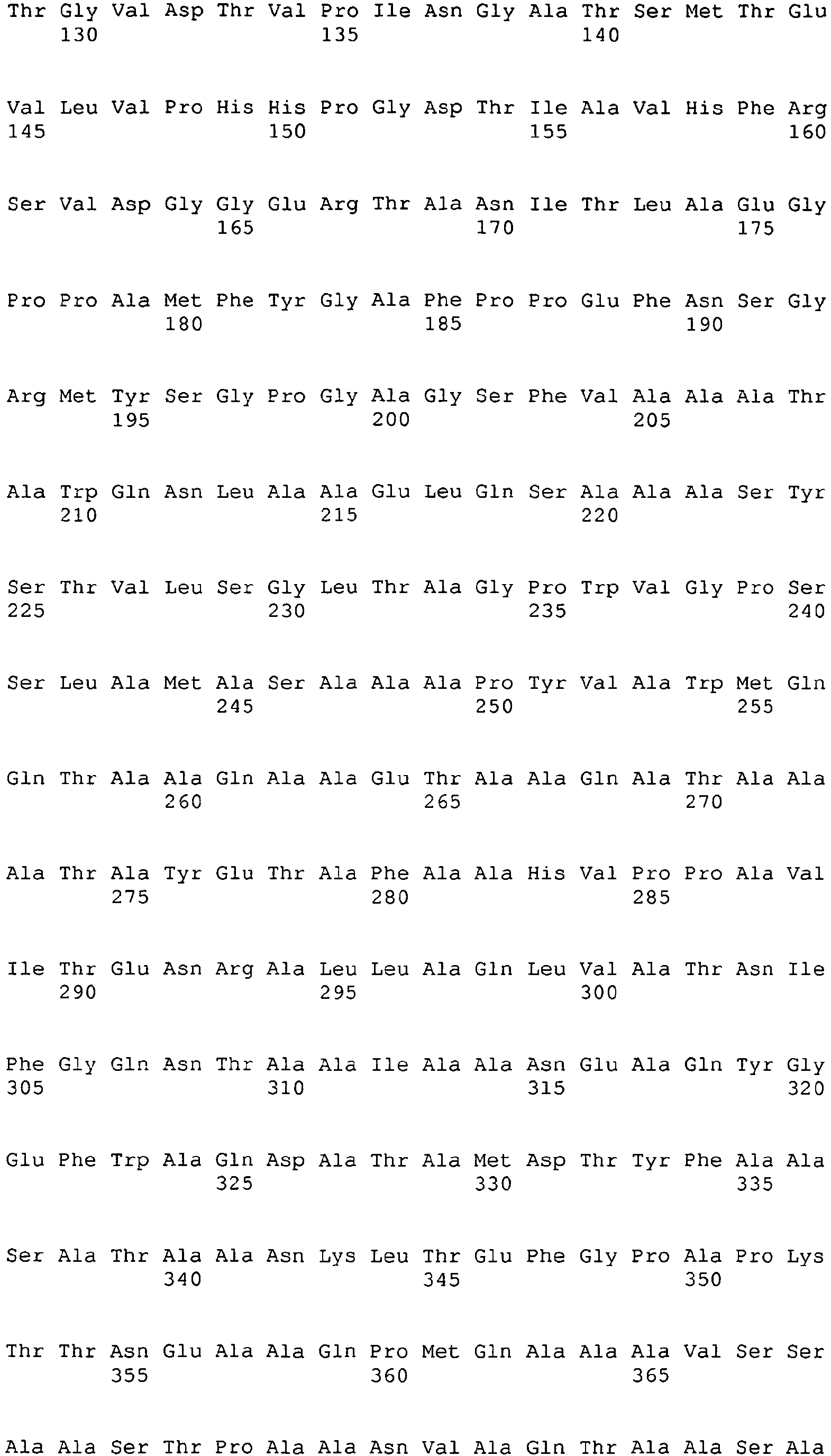
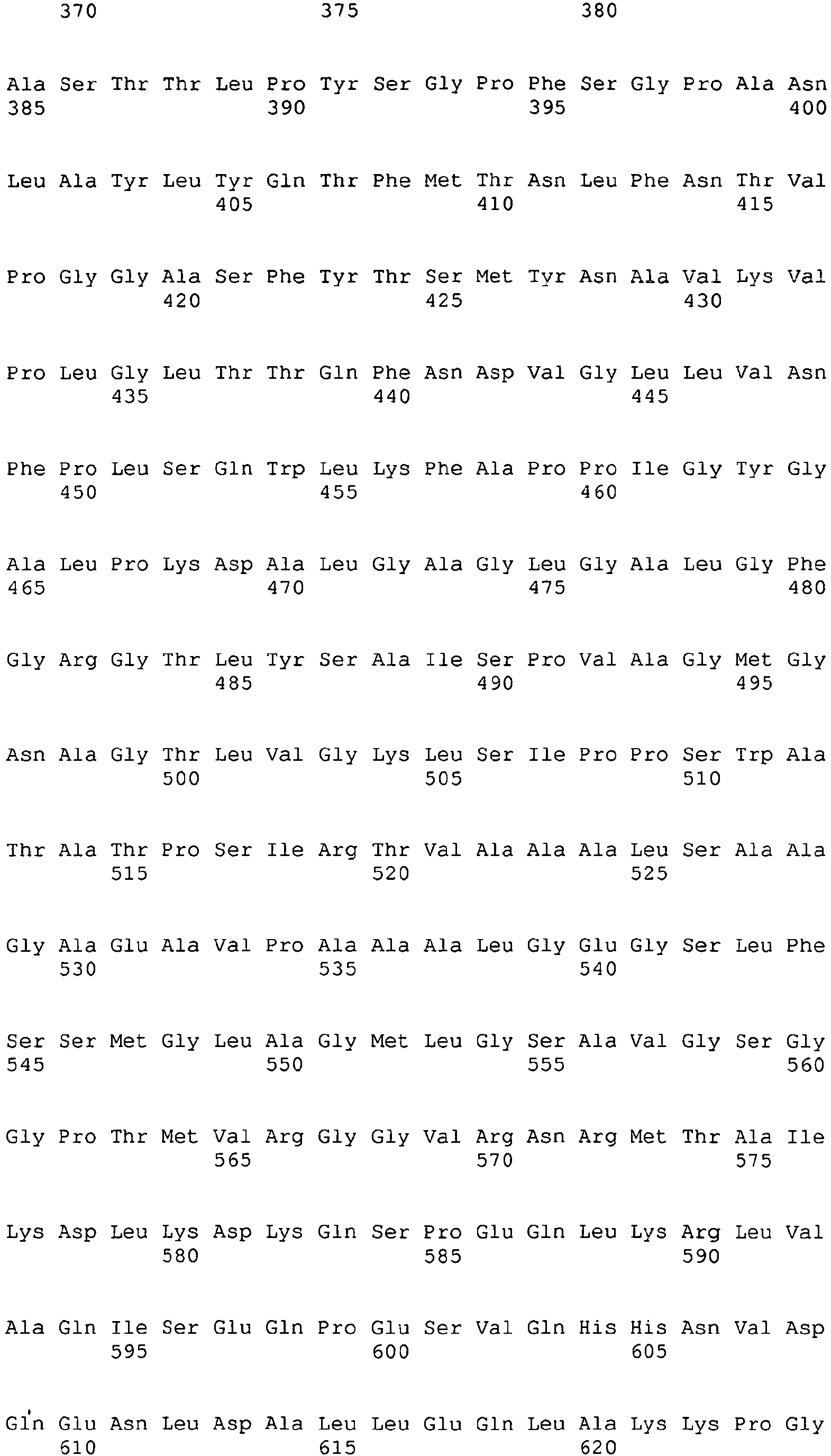

- <210> 2
<211> 361
<212> PRT
<213> MYCOBACTERIUM AVIUM SUBSPECIES PARATUBERCULOSIS - <400> 2


- <210> 3
<211> 467
<212> PRT
<213> MYCOBACTERIUM AVIUM SUBSPECIES PARATUBERCULOSIS - <400> 3


- <210> 4
<211> 44
<212> DNA
<213> Artificial sequence - <220>
<223> PCR amplification primer - <400> 4
tacatatgca tcatcatcat catcatctca accagagcgt ctcg 44 - <210> 5
<211> 27
<212> DNA
<213> Artificial sequence - <220>
<223> PCR primer - <400> 5
tagaattcgg ccggcggccc ctccgcc 27 - <210> 6
<211> 31
<212> DNA
<213> Artificial sequence - <220>
<223> PCR primer - <400> 6
ctaatcgaat tcatgttcta tggggcctttc 31 - <210> 7
<211> 29
<212> DNA
<213> Artificial sequence - <220>
<223> PCR primer - <400> 7
taatcgatat ccaggacctt ggacttgtc 29 - <210> 8
<211> 26
<212> DNA
<213> Artificial sequence - <220>
<223> PCR primer - <400> 8
atgatatcgg gctggcgccg gcgtcc 26 - <210> 9
<211> 26
<212> DNA
<213> Artificial sequence - <220>
<223> PCR primer - <400> 9
atctcgagtc acgcgacctt gccggc 26 - <210> 10
<211> 23
<212> DNA
<213> Mouse - <400> 10
cctgagcagg atggagaatt aca 23 - <210> 11
<211> 19
<212> DNA
<213> Mouse - <400> 11
tccagaacat gccgcagag 19 - <210> 12
<211> 25
<212> DNA
<213> mouse - <400> 12
cccaagcagg ccacagaatt gaaag 25 - <210> 13
<211> 20
<212> DNA
<213> mouse - <400> 13
ggaagcacgg cagcagaata 20 - <210> 14
<211> 24
<212> DNA
<213> mouse - <400> 14
aacttgaggg agaagtagga atgg 24 - <210> 15
<211> 24
<212> DNA
<213> mouse - <400> 15
catcatcaaa ccagacccgc ccaa 24 - <210> 16
<211> 25
<212> DNA
<213> mouse - <400> 16
catcttctca aaattcgagt gacaa 25 - <210> 17
<211> 23
<212> DNA
<213> mouse - <400> 17
tgggagtaga caaggtacaa ccc 23 - <210> 18
<211> 26
<212> DNA
<213> mouse - <400> 18
cacgtcgtag caaaccacca agtgga 26 - <210> 19
<211> 24
<212> DNA
<213> mouse - <400> 19
tcaagtggca tagatgtgga agaa 24 - <210> 20
<211> 21
<212> DNA
<213> mouse - <400> 20
tggctctgca ggattttcatg 21 - <210> 21
<211> 27
<212> DNA
<213> mouse - <400> 21
tcaccatcct tttgccagtt cctccag 27 - <210> 22
<211> 19
<212> DNA
<213> mouse - <400> 22
tcaccaccat ggagaaggc 19 - <210> 23
<211> 20
<212> DNA
<213> mouse - <400> 23
gctaagcagt tggtggtgca 20 - <210> 24
<211> 25
<212> DNA
<213> mouse - <400> 24
atgcccccat gtttgtgatg ggtgt 25 - <210> 25
<211> 21
<212> DNA
<213> Bovine - <400> 25
caaattccgg tggatgatctg 21 - <210> 26
<211> 22
<212> DNA
<213> Bovine - <400> 26
gcgacaggtc attcatcacctt 22 - <210> 27
<211> 28
<212> DNA
<213> bovine - <400> 27
atccagcgca aagccataaa tgaactca 28 - <210> 28
<211> 23
<212> DNA
<213> bovine - <400> 28
ccaggatggt gactcgacta gac 23 - <210> 29
<211> 20
<212> DNA
<213> bovine - <400> 29
tggctctgct ctcccagaac 20 - <210> 30
<211> 28
<212> DNA
<213> bovine - <400> 30
ccgacataaa cctctgaaat ccgaccca 28 - <210> 31
<211> 18
<212> DNA
<213> bovine - <400> 31
gcccctctga accccaaa 18 - <210> 32
<211> 23
<212> DNA
<213> bovine - <400> 32
gcaggagtgt tgaaagtctcgaa 23 - <210> 33
<211> 27
<212> DNA
<213> bovine - <400> 33
ccaaccgtga gaagatgacc cagatca 27
Claims (18)
- A composition comprising a recombinant protein, characterized in that the protein comprises from N-terminus to C-terminus:i) a C-terminal fragment of Map3527 protein comprising amino acids 183 - 361 of SEQ ID NO:2;ii) a Map1519 protein sequence comprising amino acids 1-460 of SEQ ID NO:3; andiii) an N-terminal fragment of Map3527 protein comprising amino acids 33 - 180 of SEQ ID NO:2.
- The composition of claim 1, characterized in that the Map1519 protein sequence comprises the amino acid sequence of SEQ ID NO:3.
- The composition of claim 1, characterized in that the polypeptide comprises the amino acid sequence of SEQ ID NO:1.
- The composition of claim 1, characterized in that the composition further comprises an adjuvant.
- The composition of claim 4, characterized in that the adjuvant is selected from the group consisting of monophosphoryl lipid A (MPL), dimethydioctadecyl ammonium bromide (DDA), and combinations thereof.
- The composition of claim 1, characterized in that the composition further comprises a Mycobacterium avium subspecies paratuberculosis (MAP) protein, wherein the protein is selected from the group consisting of MAP protein 85A, MAP protein 85B, MAP protein superoxide dismutase (SOD), and combinations thereof.
- The composition of claim 1 for use in a method for stimulating an immune response to Mycobacterium avium subspecies paratuberculosis (MAP) in a mammal by administering the composition to the mammal.
- The composition of claim 7, characterized in that the composition further comprises an adjuvant.
- The composition of claim 8, characterized in that the adjuvant is selected from the group consisting of monophosphoryl lipid A (MPL) and dimethydioctadecyl ammonium bromide (DDA).
- The composition of claim 7, characterized in that the administration is to a ruminant.
- The composition of claim 10, characterized in that the ruminant is a bovine, a sheep, a goat, a deer or an elk.
- The composition of claim 11, characterized in that the ruminant is a bovine.
- The composition of claim 10, characterized in that the ruminant is not infected with MAP.
- The composition of claim 10, characterized in that the ruminant is infected with MAP.
- The composition of claim 12, characterized in that the bovine has Johne's Disease.
- The composition of claim 7, characterized in that the animal is pregnant.
- The composition of claim 7, characterized in that the composition further comprises a Mycobacterium avium subspecies paratuberculosis (MAP) protein, wherein the protein is selected from the group consisting of MAP protein 85A, MAP protein 85B, MAP protein SOD, and combinations thereof.
- The composition of claim 7, characterized in that the polypeptide of the composition comprises the sequence of SEQ ID NO:1.
Priority Applications (1)
| Application Number | Priority Date | Filing Date | Title |
|---|---|---|---|
| PL08837410T PL2200641T3 (en) | 2007-10-13 | 2008-10-09 | Compositions for eliciting an immune response against mycobacterium avium subspecies paratuberculosis |
Applications Claiming Priority (3)
| Application Number | Priority Date | Filing Date | Title |
|---|---|---|---|
| US97982207P | 2007-10-13 | 2007-10-13 | |
| US9455208P | 2008-09-05 | 2008-09-05 | |
| PCT/US2008/079425 WO2009049097A1 (en) | 2007-10-13 | 2008-10-09 | Compositions for eliciting an immune response against mycobacterium avium subspecies paratuberculosis |
Publications (3)
| Publication Number | Publication Date |
|---|---|
| EP2200641A1 EP2200641A1 (en) | 2010-06-30 |
| EP2200641A4 EP2200641A4 (en) | 2010-09-29 |
| EP2200641B1 true EP2200641B1 (en) | 2012-07-04 |
Family
ID=40534814
Family Applications (1)
| Application Number | Title | Priority Date | Filing Date |
|---|---|---|---|
| EP08837410A Active EP2200641B1 (en) | 2007-10-13 | 2008-10-09 | Compositions for eliciting an immune response against mycobacterium avium subspecies paratuberculosis |
Country Status (18)
| Country | Link |
|---|---|
| US (1) | US7732580B2 (en) |
| EP (1) | EP2200641B1 (en) |
| JP (1) | JP2011500587A (en) |
| KR (1) | KR20100098602A (en) |
| CN (1) | CN101883582B (en) |
| AU (1) | AU2008310767A1 (en) |
| BR (1) | BRPI0817892A2 (en) |
| CA (1) | CA2704178A1 (en) |
| CO (1) | CO6270338A2 (en) |
| ES (1) | ES2390996T3 (en) |
| IN (1) | IN2010DN02553A (en) |
| MX (1) | MX2010003972A (en) |
| NZ (1) | NZ584659A (en) |
| PL (1) | PL2200641T3 (en) |
| PT (1) | PT2200641E (en) |
| RU (1) | RU2489165C2 (en) |
| WO (1) | WO2009049097A1 (en) |
| ZA (1) | ZA201002553B (en) |
Families Citing this family (2)
| Publication number | Priority date | Publication date | Assignee | Title |
|---|---|---|---|---|
| WO2013151706A2 (en) | 2012-04-06 | 2013-10-10 | Cornell University | Subunit vaccine delivery platform for robust humoral and cellular immune responses |
| US9175049B2 (en) | 2012-10-24 | 2015-11-03 | The United States Of America, As Represented By The Secretary Of Agriculture | Recombinant mycobacterium avium subsp. paratuberculosis proteins induce immunity and protect against infection |
Family Cites Families (6)
| Publication number | Priority date | Publication date | Assignee | Title |
|---|---|---|---|---|
| US5322858A (en) * | 1992-02-14 | 1994-06-21 | Jacobus Pharmaceutical Co. Inc. | N,N'-substituted imidodicarbonimidic diamides derived from hydroxylamines |
| KR100797876B1 (en) * | 1998-04-07 | 2008-01-24 | 코릭사 코포레이션 | Fusion protein of Mycobacterium tuberculosis antigen and its use |
| DE60139963D1 (en) * | 2000-06-20 | 2009-10-29 | Corixa Corp | FUSION PROTEINS FROM MYCOBACTERIUM TUBERCULOSIS |
| US7074559B2 (en) * | 2002-03-06 | 2006-07-11 | Refents of the University of Minnesota | Mycobacterial diagnostics |
| EP1855716B1 (en) | 2005-02-16 | 2010-12-15 | Cornell Research Foundation, Inc. | Compositions for eliciting an immune response against mycobacterium avium subspecies paratuberculosis |
| US8758773B2 (en) | 2005-12-09 | 2014-06-24 | Wisconsin Alumni Research Foundation | Vaccine candidates against Johne's disease |
-
2008
- 2008-10-09 EP EP08837410A patent/EP2200641B1/en active Active
- 2008-10-09 AU AU2008310767A patent/AU2008310767A1/en not_active Abandoned
- 2008-10-09 CN CN200880119048.7A patent/CN101883582B/en not_active Expired - Fee Related
- 2008-10-09 MX MX2010003972A patent/MX2010003972A/en not_active Application Discontinuation
- 2008-10-09 KR KR1020107010437A patent/KR20100098602A/en not_active Ceased
- 2008-10-09 JP JP2010529059A patent/JP2011500587A/en not_active Ceased
- 2008-10-09 US US12/248,764 patent/US7732580B2/en not_active Expired - Fee Related
- 2008-10-09 NZ NZ584659A patent/NZ584659A/en not_active IP Right Cessation
- 2008-10-09 RU RU2010119046/15A patent/RU2489165C2/en not_active IP Right Cessation
- 2008-10-09 PL PL08837410T patent/PL2200641T3/en unknown
- 2008-10-09 CA CA2704178A patent/CA2704178A1/en not_active Abandoned
- 2008-10-09 ES ES08837410T patent/ES2390996T3/en active Active
- 2008-10-09 WO PCT/US2008/079425 patent/WO2009049097A1/en not_active Ceased
- 2008-10-09 BR BRPI0817892 patent/BRPI0817892A2/en not_active IP Right Cessation
- 2008-10-09 PT PT08837410T patent/PT2200641E/en unknown
-
2010
- 2010-04-12 ZA ZA2010/02553A patent/ZA201002553B/en unknown
- 2010-04-13 IN IN2553DEN2010 patent/IN2010DN02553A/en unknown
- 2010-05-13 CO CO10057537A patent/CO6270338A2/en not_active Application Discontinuation
Also Published As
| Publication number | Publication date |
|---|---|
| US20090099083A1 (en) | 2009-04-16 |
| ZA201002553B (en) | 2010-12-29 |
| CA2704178A1 (en) | 2009-04-16 |
| AU2008310767A1 (en) | 2009-04-16 |
| IN2010DN02553A (en) | 2015-07-24 |
| BRPI0817892A2 (en) | 2015-05-05 |
| MX2010003972A (en) | 2010-08-09 |
| JP2011500587A (en) | 2011-01-06 |
| EP2200641A1 (en) | 2010-06-30 |
| CO6270338A2 (en) | 2011-04-20 |
| NZ584659A (en) | 2011-09-30 |
| PL2200641T3 (en) | 2012-12-31 |
| PT2200641E (en) | 2012-10-11 |
| RU2489165C2 (en) | 2013-08-10 |
| KR20100098602A (en) | 2010-09-08 |
| EP2200641A4 (en) | 2010-09-29 |
| RU2010119046A (en) | 2011-11-20 |
| CN101883582B (en) | 2014-03-19 |
| CN101883582A (en) | 2010-11-10 |
| US7732580B2 (en) | 2010-06-08 |
| WO2009049097A1 (en) | 2009-04-16 |
| ES2390996T3 (en) | 2012-11-20 |
Similar Documents
| Publication | Publication Date | Title |
|---|---|---|
| Corbel | Brucellosis: an overview | |
| Yan et al. | Immunogenicity and protective efficacy of recombinant Leptospira immunoglobulin-like protein B (rLigB) in a hamster challenge model | |
| US7037510B2 (en) | Hybrids of M. tuberculosis antigens | |
| Kathaperumal et al. | Evaluation of immune responses and protective efficacy in a goat model following immunization with a coctail of recombinant antigens and a polyprotein of Mycobacterium avium subsp. paratuberculosis | |
| EP1723170B1 (en) | A canine vaccine for protection against ehrlichiosis | |
| Chen et al. | Immune responses in mice to Mycobacterium avium subsp. paratuberculosis following vaccination with a novel 74F recombinant polyprotein | |
| US20160228528A1 (en) | A single or multistage mycobacterium avium subsp. paratuberculosis subunit vaccine | |
| US7732580B2 (en) | Compositions for eliciting an immune response against Mycobacterium avium subspecies paratuberculosis | |
| GB2551984A (en) | Fish virus | |
| CN101646447A (en) | Mycobacterium tuberculosis persistance genes | |
| CN101155596B (en) | Compositions for eliciting an immune response against mycobacterium avium subspecies paratuberculosis | |
| CN1843506B (en) | A kind of Mycobacterium tuberculosis vaccine kit medicine | |
| HK1145638A (en) | Compositions for eliciting an immune response against mycobacterium avium subspecies paratuberculosis | |
| US20230218734A1 (en) | Compositions and methods for preventing, controlling and diagnosing mycobacterial infections | |
| World Health Organization | The development of new/improved brucellosis vaccines: report of a WHO meeting, Geneva, Switzerland, 11-12 December 1997 | |
| CN1579550A (en) | Nucleic vaccine for tubercle bacillus | |
| World Health Organization | The Development of new | |
| Farahi et al. | Cloning and Expression of Brucella abortus Omp19 an Immunogenic Minor Outer Membrane Protein | |
| JP2010505792A (en) | A therapeutic vaccine comprising heat shock protein 70 of mycobacteria | |
| Lowry | Identification of novel gene products associated with virulence of Brucella abortus using gene discovery and targeted approaches | |
| Huntley | DNA vaccination and recombinant protein expression to identify immunogenic and protective genes from Mycobacterium avium subsp. paratuberculosis | |
| Obradovic et al. | Follow IVIS |
Legal Events
| Date | Code | Title | Description |
|---|---|---|---|
| PUAI | Public reference made under article 153(3) epc to a published international application that has entered the european phase |
Free format text: ORIGINAL CODE: 0009012 |
|
| 17P | Request for examination filed |
Effective date: 20100421 |
|
| AK | Designated contracting states |
Kind code of ref document: A1 Designated state(s): AT BE BG CH CY CZ DE DK EE ES FI FR GB GR HR HU IE IS IT LI LT LU LV MC MT NL NO PL PT RO SE SI SK TR |
|
| AX | Request for extension of the european patent |
Extension state: AL BA MK RS |
|
| A4 | Supplementary search report drawn up and despatched |
Effective date: 20100827 |
|
| 17Q | First examination report despatched |
Effective date: 20100917 |
|
| RAX | Requested extension states of the european patent have changed |
Extension state: RS Payment date: 20100421 Extension state: BA Payment date: 20100421 |
|
| RIC1 | Information provided on ipc code assigned before grant |
Ipc: A61K 39/04 20060101AFI20110617BHEP |
|
| GRAP | Despatch of communication of intention to grant a patent |
Free format text: ORIGINAL CODE: EPIDOSNIGR1 |
|
| GRAS | Grant fee paid |
Free format text: ORIGINAL CODE: EPIDOSNIGR3 |
|
| GRAA | (expected) grant |
Free format text: ORIGINAL CODE: 0009210 |
|
| AK | Designated contracting states |
Kind code of ref document: B1 Designated state(s): AT BE BG CH CY CZ DE DK EE ES FI FR GB GR HR HU IE IS IT LI LT LU LV MC MT NL NO PL PT RO SE SI SK TR |
|
| AX | Request for extension of the european patent |
Extension state: BA RS |
|
| REG | Reference to a national code |
Ref country code: GB Ref legal event code: FG4D |
|
| REG | Reference to a national code |
Ref country code: CH Ref legal event code: EP |
|
| REG | Reference to a national code |
Ref country code: AT Ref legal event code: REF Ref document number: 564871 Country of ref document: AT Kind code of ref document: T Effective date: 20120715 |
|
| REG | Reference to a national code |
Ref country code: IE Ref legal event code: FG4D |
|
| REG | Reference to a national code |
Ref country code: DE Ref legal event code: R096 Ref document number: 602008017002 Country of ref document: DE Effective date: 20120830 |
|
| REG | Reference to a national code |
Ref country code: NL Ref legal event code: T3 |
|
| REG | Reference to a national code |
Ref country code: PT Ref legal event code: SC4A Free format text: AVAILABILITY OF NATIONAL TRANSLATION Effective date: 20121004 |
|
| REG | Reference to a national code |
Ref country code: CH Ref legal event code: NV Representative=s name: MEYERLUSTENBERGER LACHENAL |
|
| REG | Reference to a national code |
Ref country code: ES Ref legal event code: FG2A Ref document number: 2390996 Country of ref document: ES Kind code of ref document: T3 Effective date: 20121120 |
|
| PG25 | Lapsed in a contracting state [announced via postgrant information from national office to epo] |
Ref country code: SI Free format text: LAPSE BECAUSE OF FAILURE TO SUBMIT A TRANSLATION OF THE DESCRIPTION OR TO PAY THE FEE WITHIN THE PRESCRIBED TIME-LIMIT Effective date: 20120704 |
|
| REG | Reference to a national code |
Ref country code: LT Ref legal event code: MG4D Effective date: 20120704 |
|
| REG | Reference to a national code |
Ref country code: PL Ref legal event code: T3 |
|
| PG25 | Lapsed in a contracting state [announced via postgrant information from national office to epo] |
Ref country code: NO Free format text: LAPSE BECAUSE OF FAILURE TO SUBMIT A TRANSLATION OF THE DESCRIPTION OR TO PAY THE FEE WITHIN THE PRESCRIBED TIME-LIMIT Effective date: 20121004 Ref country code: LT Free format text: LAPSE BECAUSE OF FAILURE TO SUBMIT A TRANSLATION OF THE DESCRIPTION OR TO PAY THE FEE WITHIN THE PRESCRIBED TIME-LIMIT Effective date: 20120704 Ref country code: FI Free format text: LAPSE BECAUSE OF FAILURE TO SUBMIT A TRANSLATION OF THE DESCRIPTION OR TO PAY THE FEE WITHIN THE PRESCRIBED TIME-LIMIT Effective date: 20120704 Ref country code: HR Free format text: LAPSE BECAUSE OF FAILURE TO SUBMIT A TRANSLATION OF THE DESCRIPTION OR TO PAY THE FEE WITHIN THE PRESCRIBED TIME-LIMIT Effective date: 20120704 Ref country code: CY Free format text: LAPSE BECAUSE OF FAILURE TO SUBMIT A TRANSLATION OF THE DESCRIPTION OR TO PAY THE FEE WITHIN THE PRESCRIBED TIME-LIMIT Effective date: 20120704 Ref country code: IS Free format text: LAPSE BECAUSE OF FAILURE TO SUBMIT A TRANSLATION OF THE DESCRIPTION OR TO PAY THE FEE WITHIN THE PRESCRIBED TIME-LIMIT Effective date: 20121104 |
|
| PG25 | Lapsed in a contracting state [announced via postgrant information from national office to epo] |
Ref country code: LV Free format text: LAPSE BECAUSE OF FAILURE TO SUBMIT A TRANSLATION OF THE DESCRIPTION OR TO PAY THE FEE WITHIN THE PRESCRIBED TIME-LIMIT Effective date: 20120704 Ref country code: SE Free format text: LAPSE BECAUSE OF FAILURE TO SUBMIT A TRANSLATION OF THE DESCRIPTION OR TO PAY THE FEE WITHIN THE PRESCRIBED TIME-LIMIT Effective date: 20120704 Ref country code: GR Free format text: LAPSE BECAUSE OF FAILURE TO SUBMIT A TRANSLATION OF THE DESCRIPTION OR TO PAY THE FEE WITHIN THE PRESCRIBED TIME-LIMIT Effective date: 20121005 |
|
| PG25 | Lapsed in a contracting state [announced via postgrant information from national office to epo] |
Ref country code: CZ Free format text: LAPSE BECAUSE OF FAILURE TO SUBMIT A TRANSLATION OF THE DESCRIPTION OR TO PAY THE FEE WITHIN THE PRESCRIBED TIME-LIMIT Effective date: 20120704 Ref country code: DK Free format text: LAPSE BECAUSE OF FAILURE TO SUBMIT A TRANSLATION OF THE DESCRIPTION OR TO PAY THE FEE WITHIN THE PRESCRIBED TIME-LIMIT Effective date: 20120704 Ref country code: RO Free format text: LAPSE BECAUSE OF FAILURE TO SUBMIT A TRANSLATION OF THE DESCRIPTION OR TO PAY THE FEE WITHIN THE PRESCRIBED TIME-LIMIT Effective date: 20120704 Ref country code: EE Free format text: LAPSE BECAUSE OF FAILURE TO SUBMIT A TRANSLATION OF THE DESCRIPTION OR TO PAY THE FEE WITHIN THE PRESCRIBED TIME-LIMIT Effective date: 20120704 |
|
| PLBE | No opposition filed within time limit |
Free format text: ORIGINAL CODE: 0009261 |
|
| STAA | Information on the status of an ep patent application or granted ep patent |
Free format text: STATUS: NO OPPOSITION FILED WITHIN TIME LIMIT |
|
| PG25 | Lapsed in a contracting state [announced via postgrant information from national office to epo] |
Ref country code: SK Free format text: LAPSE BECAUSE OF FAILURE TO SUBMIT A TRANSLATION OF THE DESCRIPTION OR TO PAY THE FEE WITHIN THE PRESCRIBED TIME-LIMIT Effective date: 20120704 |
|
| 26N | No opposition filed |
Effective date: 20130405 |
|
| PG25 | Lapsed in a contracting state [announced via postgrant information from national office to epo] |
Ref country code: BG Free format text: LAPSE BECAUSE OF FAILURE TO SUBMIT A TRANSLATION OF THE DESCRIPTION OR TO PAY THE FEE WITHIN THE PRESCRIBED TIME-LIMIT Effective date: 20121004 |
|
| REG | Reference to a national code |
Ref country code: DE Ref legal event code: R097 Ref document number: 602008017002 Country of ref document: DE Effective date: 20130405 |
|
| PGFP | Annual fee paid to national office [announced via postgrant information from national office to epo] |
Ref country code: PT Payment date: 20130409 Year of fee payment: 6 Ref country code: MC Payment date: 20130918 Year of fee payment: 6 |
|
| PGFP | Annual fee paid to national office [announced via postgrant information from national office to epo] |
Ref country code: CH Payment date: 20131029 Year of fee payment: 6 Ref country code: FR Payment date: 20131017 Year of fee payment: 6 Ref country code: LU Payment date: 20131031 Year of fee payment: 6 Ref country code: BE Payment date: 20131028 Year of fee payment: 6 Ref country code: IE Payment date: 20131025 Year of fee payment: 6 Ref country code: AT Payment date: 20130919 Year of fee payment: 6 Ref country code: DE Payment date: 20131029 Year of fee payment: 6 Ref country code: GB Payment date: 20131028 Year of fee payment: 6 |
|
| PGFP | Annual fee paid to national office [announced via postgrant information from national office to epo] |
Ref country code: PL Payment date: 20131002 Year of fee payment: 6 Ref country code: NL Payment date: 20131026 Year of fee payment: 6 Ref country code: ES Payment date: 20131028 Year of fee payment: 6 Ref country code: IT Payment date: 20131023 Year of fee payment: 6 |
|
| PGFP | Annual fee paid to national office [announced via postgrant information from national office to epo] |
Ref country code: TR Payment date: 20120924 Year of fee payment: 5 |
|
| PG25 | Lapsed in a contracting state [announced via postgrant information from national office to epo] |
Ref country code: HU Free format text: LAPSE BECAUSE OF FAILURE TO SUBMIT A TRANSLATION OF THE DESCRIPTION OR TO PAY THE FEE WITHIN THE PRESCRIBED TIME-LIMIT Effective date: 20081009 |
|
| REG | Reference to a national code |
Ref country code: PT Ref legal event code: MM4A Free format text: LAPSE DUE TO NON-PAYMENT OF FEES Effective date: 20150409 |
|
| REG | Reference to a national code |
Ref country code: DE Ref legal event code: R119 Ref document number: 602008017002 Country of ref document: DE |
|
| REG | Reference to a national code |
Ref country code: NL Ref legal event code: V1 Effective date: 20150501 |
|
| PG25 | Lapsed in a contracting state [announced via postgrant information from national office to epo] |
Ref country code: LU Free format text: LAPSE BECAUSE OF NON-PAYMENT OF DUE FEES Effective date: 20141009 Ref country code: MC Free format text: LAPSE BECAUSE OF NON-PAYMENT OF DUE FEES Effective date: 20141031 |
|
| REG | Reference to a national code |
Ref country code: CH Ref legal event code: PL |
|
| REG | Reference to a national code |
Ref country code: AT Ref legal event code: MM01 Ref document number: 564871 Country of ref document: AT Kind code of ref document: T Effective date: 20141009 |
|
| GBPC | Gb: european patent ceased through non-payment of renewal fee |
Effective date: 20141009 |
|
| PG25 | Lapsed in a contracting state [announced via postgrant information from national office to epo] |
Ref country code: BE Free format text: LAPSE BECAUSE OF NON-PAYMENT OF DUE FEES Effective date: 20141031 |
|
| REG | Reference to a national code |
Ref country code: IE Ref legal event code: MM4A |
|
| PG25 | Lapsed in a contracting state [announced via postgrant information from national office to epo] |
Ref country code: GB Free format text: LAPSE BECAUSE OF NON-PAYMENT OF DUE FEES Effective date: 20141009 Ref country code: PT Free format text: LAPSE BECAUSE OF NON-PAYMENT OF DUE FEES Effective date: 20150409 Ref country code: LI Free format text: LAPSE BECAUSE OF NON-PAYMENT OF DUE FEES Effective date: 20141031 Ref country code: DE Free format text: LAPSE BECAUSE OF NON-PAYMENT OF DUE FEES Effective date: 20150501 Ref country code: CH Free format text: LAPSE BECAUSE OF NON-PAYMENT OF DUE FEES Effective date: 20141031 |
|
| REG | Reference to a national code |
Ref country code: FR Ref legal event code: ST Effective date: 20150630 |
|
| PG25 | Lapsed in a contracting state [announced via postgrant information from national office to epo] |
Ref country code: IT Free format text: LAPSE BECAUSE OF NON-PAYMENT OF DUE FEES Effective date: 20141009 Ref country code: FR Free format text: LAPSE BECAUSE OF NON-PAYMENT OF DUE FEES Effective date: 20141031 Ref country code: NL Free format text: LAPSE BECAUSE OF NON-PAYMENT OF DUE FEES Effective date: 20150501 Ref country code: AT Free format text: LAPSE BECAUSE OF NON-PAYMENT OF DUE FEES Effective date: 20141009 |
|
| PGFP | Annual fee paid to national office [announced via postgrant information from national office to epo] |
Ref country code: MT Payment date: 20131001 Year of fee payment: 6 |
|
| PG25 | Lapsed in a contracting state [announced via postgrant information from national office to epo] |
Ref country code: IE Free format text: LAPSE BECAUSE OF NON-PAYMENT OF DUE FEES Effective date: 20141009 |
|
| REG | Reference to a national code |
Ref country code: ES Ref legal event code: FD2A Effective date: 20151126 |
|
| PG25 | Lapsed in a contracting state [announced via postgrant information from national office to epo] |
Ref country code: ES Free format text: LAPSE BECAUSE OF NON-PAYMENT OF DUE FEES Effective date: 20141010 Ref country code: PL Free format text: LAPSE BECAUSE OF NON-PAYMENT OF DUE FEES Effective date: 20141009 |
|
| PG25 | Lapsed in a contracting state [announced via postgrant information from national office to epo] |
Ref country code: MT Free format text: LAPSE BECAUSE OF NON-PAYMENT OF DUE FEES Effective date: 20141031 |
|
| PG25 | Lapsed in a contracting state [announced via postgrant information from national office to epo] |
Ref country code: TR Free format text: LAPSE BECAUSE OF NON-PAYMENT OF DUE FEES Effective date: 20141009 |
|
| PG25 | Lapsed in a contracting state [announced via postgrant information from national office to epo] |
Ref country code: MT Free format text: LAPSE BECAUSE OF NON-PAYMENT OF DUE FEES Effective date: 20141009 |