EP2200431B1 - Novel compositions and methods for cancer treatment - Google Patents
Novel compositions and methods for cancer treatment Download PDFInfo
- Publication number
- EP2200431B1 EP2200431B1 EP08830633.7A EP08830633A EP2200431B1 EP 2200431 B1 EP2200431 B1 EP 2200431B1 EP 08830633 A EP08830633 A EP 08830633A EP 2200431 B1 EP2200431 B1 EP 2200431B1
- Authority
- EP
- European Patent Office
- Prior art keywords
- cancer
- cells
- agent
- stat3
- cell
- Prior art date
- Legal status (The legal status is an assumption and is not a legal conclusion. Google has not performed a legal analysis and makes no representation as to the accuracy of the status listed.)
- Active
Links
- 206010028980 Neoplasm Diseases 0.000 title claims description 227
- 201000011510 cancer Diseases 0.000 title claims description 157
- 238000011282 treatment Methods 0.000 title claims description 37
- 239000000203 mixture Substances 0.000 title claims description 12
- 238000000034 method Methods 0.000 title description 48
- 101150099493 STAT3 gene Proteins 0.000 claims description 113
- 239000003795 chemical substances by application Substances 0.000 claims description 81
- 230000037361 pathway Effects 0.000 claims description 54
- 230000000694 effects Effects 0.000 claims description 48
- AOJJSUZBOXZQNB-TZSSRYMLSA-N Doxorubicin Chemical compound O([C@H]1C[C@@](O)(CC=2C(O)=C3C(=O)C=4C=CC=C(C=4C(=O)C3=C(O)C=21)OC)C(=O)CO)[C@H]1C[C@H](N)[C@H](O)[C@H](C)O1 AOJJSUZBOXZQNB-TZSSRYMLSA-N 0.000 claims description 29
- SDUQYLNIPVEERB-QPPQHZFASA-N gemcitabine Chemical compound O=C1N=C(N)C=CN1[C@H]1C(F)(F)[C@H](O)[C@@H](CO)O1 SDUQYLNIPVEERB-QPPQHZFASA-N 0.000 claims description 24
- 229960005277 gemcitabine Drugs 0.000 claims description 20
- 230000001594 aberrant effect Effects 0.000 claims description 19
- 239000002147 L01XE04 - Sunitinib Substances 0.000 claims description 17
- 239000005511 L01XE05 - Sorafenib Substances 0.000 claims description 16
- 150000003839 salts Chemical class 0.000 claims description 16
- 239000002136 L01XE07 - Lapatinib Substances 0.000 claims description 15
- 206010061902 Pancreatic neoplasm Diseases 0.000 claims description 15
- 208000015486 malignant pancreatic neoplasm Diseases 0.000 claims description 15
- 201000002528 pancreatic cancer Diseases 0.000 claims description 15
- 208000008443 pancreatic carcinoma Diseases 0.000 claims description 15
- 229960003787 sorafenib Drugs 0.000 claims description 15
- SAXKEWRSGLPYPB-UHFFFAOYSA-N 2-(1-hydroxyethyl)benzo[f][1]benzofuran-4,9-dione Chemical compound O=C1C2=CC=CC=C2C(=O)C2=C1OC(C(O)C)=C2 SAXKEWRSGLPYPB-UHFFFAOYSA-N 0.000 claims description 14
- 229960004679 doxorubicin Drugs 0.000 claims description 14
- 229960004891 lapatinib Drugs 0.000 claims description 14
- BCFGMOOMADDAQU-UHFFFAOYSA-N lapatinib Chemical compound O1C(CNCCS(=O)(=O)C)=CC=C1C1=CC=C(N=CN=C2NC=3C=C(Cl)C(OCC=4C=C(F)C=CC=4)=CC=3)C2=C1 BCFGMOOMADDAQU-UHFFFAOYSA-N 0.000 claims description 14
- DPHUWDIXHNQOSY-UHFFFAOYSA-N napabucasin Chemical compound O=C1C2=CC=CC=C2C(=O)C2=C1OC(C(=O)C)=C2 DPHUWDIXHNQOSY-UHFFFAOYSA-N 0.000 claims description 14
- MLDQJTXFUGDVEO-UHFFFAOYSA-N BAY-43-9006 Chemical compound C1=NC(C(=O)NC)=CC(OC=2C=CC(NC(=O)NC=3C=C(C(Cl)=CC=3)C(F)(F)F)=CC=2)=C1 MLDQJTXFUGDVEO-UHFFFAOYSA-N 0.000 claims description 13
- AAKJLRGGTJKAMG-UHFFFAOYSA-N erlotinib Chemical compound C=12C=C(OCCOC)C(OCCOC)=CC2=NC=NC=1NC1=CC=CC(C#C)=C1 AAKJLRGGTJKAMG-UHFFFAOYSA-N 0.000 claims description 13
- VSRXQHXAPYXROS-UHFFFAOYSA-N azanide;cyclobutane-1,1-dicarboxylic acid;platinum(2+) Chemical compound [NH2-].[NH2-].[Pt+2].OC(=O)C1(C(O)=O)CCC1 VSRXQHXAPYXROS-UHFFFAOYSA-N 0.000 claims description 12
- 229960004562 carboplatin Drugs 0.000 claims description 12
- VJJPUSNTGOMMGY-MRVIYFEKSA-N etoposide Chemical compound COC1=C(O)C(OC)=CC([C@@H]2C3=CC=4OCOC=4C=C3[C@@H](O[C@H]3[C@@H]([C@@H](O)[C@@H]4O[C@H](C)OC[C@H]4O3)O)[C@@H]3[C@@H]2C(OC3)=O)=C1 VJJPUSNTGOMMGY-MRVIYFEKSA-N 0.000 claims description 12
- 229960005420 etoposide Drugs 0.000 claims description 11
- 230000002401 inhibitory effect Effects 0.000 claims description 11
- 229960001796 sunitinib Drugs 0.000 claims description 11
- 239000005551 L01XE03 - Erlotinib Substances 0.000 claims description 10
- 206010058467 Lung neoplasm malignant Diseases 0.000 claims description 10
- ZDZOTLJHXYCWBA-VCVYQWHSSA-N N-debenzoyl-N-(tert-butoxycarbonyl)-10-deacetyltaxol Chemical compound O([C@H]1[C@H]2[C@@](C([C@H](O)C3=C(C)[C@@H](OC(=O)[C@H](O)[C@@H](NC(=O)OC(C)(C)C)C=4C=CC=CC=4)C[C@]1(O)C3(C)C)=O)(C)[C@@H](O)C[C@H]1OC[C@]12OC(=O)C)C(=O)C1=CC=CC=C1 ZDZOTLJHXYCWBA-VCVYQWHSSA-N 0.000 claims description 10
- 229960003668 docetaxel Drugs 0.000 claims description 10
- 201000005202 lung cancer Diseases 0.000 claims description 10
- 208000020816 lung neoplasm Diseases 0.000 claims description 10
- 206010009944 Colon cancer Diseases 0.000 claims description 9
- 229960001433 erlotinib Drugs 0.000 claims description 9
- 239000012453 solvate Substances 0.000 claims description 9
- WINHZLLDWRZWRT-ATVHPVEESA-N sunitinib Chemical compound CCN(CC)CCNC(=O)C1=C(C)NC(\C=C/2C3=CC(F)=CC=C3NC\2=O)=C1C WINHZLLDWRZWRT-ATVHPVEESA-N 0.000 claims description 9
- 201000010536 head and neck cancer Diseases 0.000 claims description 8
- 208000014829 head and neck neoplasm Diseases 0.000 claims description 8
- RVIUPSHRLLQFOH-UHFFFAOYSA-N 2-acetyl-7-chlorobenzo[f][1]benzofuran-4,9-dione Chemical compound O=C1C2=CC(Cl)=CC=C2C(=O)C2=C1OC(C(=O)C)=C2 RVIUPSHRLLQFOH-UHFFFAOYSA-N 0.000 claims description 7
- RPCXZIRGAHYOKZ-UHFFFAOYSA-N 2-acetyl-7-fluorobenzo[f][1]benzofuran-4,9-dione Chemical compound O=C1C2=CC(F)=CC=C2C(=O)C2=C1OC(C(=O)C)=C2 RPCXZIRGAHYOKZ-UHFFFAOYSA-N 0.000 claims description 7
- QORFLTDEUGKOSZ-UHFFFAOYSA-N 2-ethylbenzo[f][1]benzofuran-4,9-dione Chemical compound O=C1C2=CC=CC=C2C(=O)C2=C1OC(CC)=C2 QORFLTDEUGKOSZ-UHFFFAOYSA-N 0.000 claims description 7
- 208000001333 Colorectal Neoplasms Diseases 0.000 claims description 7
- 208000032839 leukemia Diseases 0.000 claims description 7
- 206010006187 Breast cancer Diseases 0.000 claims description 6
- 208000026310 Breast neoplasm Diseases 0.000 claims description 6
- 206010033128 Ovarian cancer Diseases 0.000 claims description 6
- 206010061535 Ovarian neoplasm Diseases 0.000 claims description 6
- 206010060862 Prostate cancer Diseases 0.000 claims description 6
- 208000000236 Prostatic Neoplasms Diseases 0.000 claims description 6
- 206010073071 hepatocellular carcinoma Diseases 0.000 claims description 6
- 208000003174 Brain Neoplasms Diseases 0.000 claims description 5
- 206010008342 Cervix carcinoma Diseases 0.000 claims description 5
- 206010025323 Lymphomas Diseases 0.000 claims description 5
- 208000034578 Multiple myelomas Diseases 0.000 claims description 5
- 206010035226 Plasma cell myeloma Diseases 0.000 claims description 5
- 206010039491 Sarcoma Diseases 0.000 claims description 5
- 208000005718 Stomach Neoplasms Diseases 0.000 claims description 5
- 208000006105 Uterine Cervical Neoplasms Diseases 0.000 claims description 5
- 201000010881 cervical cancer Diseases 0.000 claims description 5
- 201000010989 colorectal carcinoma Diseases 0.000 claims description 5
- 201000001441 melanoma Diseases 0.000 claims description 5
- 206010061289 metastatic neoplasm Diseases 0.000 claims description 5
- RCINICONZNJXQF-MZXODVADSA-N taxol Chemical compound O([C@@H]1[C@@]2(C[C@@H](C(C)=C(C2(C)C)[C@H](C([C@]2(C)[C@@H](O)C[C@H]3OC[C@]3([C@H]21)OC(C)=O)=O)OC(=O)C)OC(=O)[C@H](O)[C@@H](NC(=O)C=1C=CC=CC=1)C=1C=CC=CC=1)O)C(=O)C1=CC=CC=C1 RCINICONZNJXQF-MZXODVADSA-N 0.000 claims description 5
- 229930012538 Paclitaxel Natural products 0.000 claims description 4
- 208000006265 Renal cell carcinoma Diseases 0.000 claims description 4
- 231100000844 hepatocellular carcinoma Toxicity 0.000 claims description 4
- 230000001394 metastastic effect Effects 0.000 claims description 4
- 229960001592 paclitaxel Drugs 0.000 claims description 4
- 206010017758 gastric cancer Diseases 0.000 claims description 2
- 201000011549 stomach cancer Diseases 0.000 claims description 2
- 210000004027 cell Anatomy 0.000 description 169
- 210000000130 stem cell Anatomy 0.000 description 84
- BVRDQVRQVGRNHG-UHFFFAOYSA-N 2-morpholin-4-ylpyrimido[2,1-a]isoquinolin-4-one Chemical compound N1=C2C3=CC=CC=C3C=CN2C(=O)C=C1N1CCOCC1 BVRDQVRQVGRNHG-UHFFFAOYSA-N 0.000 description 56
- 150000001875 compounds Chemical class 0.000 description 50
- 239000002246 antineoplastic agent Substances 0.000 description 37
- 208000037265 diseases, disorders, signs and symptoms Diseases 0.000 description 29
- 239000003814 drug Substances 0.000 description 28
- 229940079593 drug Drugs 0.000 description 24
- 230000005764 inhibitory process Effects 0.000 description 22
- 239000003112 inhibitor Substances 0.000 description 21
- 208000035475 disorder Diseases 0.000 description 20
- 229940121358 tyrosine kinase inhibitor Drugs 0.000 description 20
- 239000005483 tyrosine kinase inhibitor Substances 0.000 description 19
- -1 c-Met Proteins 0.000 description 16
- 241001465754 Metazoa Species 0.000 description 15
- 206010027476 Metastases Diseases 0.000 description 14
- 229940127089 cytotoxic agent Drugs 0.000 description 13
- 230000004083 survival effect Effects 0.000 description 13
- 241000894006 Bacteria Species 0.000 description 12
- 230000009401 metastasis Effects 0.000 description 12
- 230000002195 synergetic effect Effects 0.000 description 12
- 230000004568 DNA-binding Effects 0.000 description 11
- 230000000381 tumorigenic effect Effects 0.000 description 11
- 239000012625 DNA intercalator Substances 0.000 description 10
- 238000011319 anticancer therapy Methods 0.000 description 10
- LOKCTEFSRHRXRJ-UHFFFAOYSA-I dipotassium trisodium dihydrogen phosphate hydrogen phosphate dichloride Chemical compound P(=O)(O)(O)[O-].[K+].P(=O)(O)([O-])[O-].[Na+].[Na+].[Cl-].[K+].[Cl-].[Na+] LOKCTEFSRHRXRJ-UHFFFAOYSA-I 0.000 description 10
- 239000002953 phosphate buffered saline Substances 0.000 description 10
- 108091003079 Bovine Serum Albumin Proteins 0.000 description 9
- 239000006144 Dulbecco’s modified Eagle's medium Substances 0.000 description 9
- 239000012091 fetal bovine serum Substances 0.000 description 9
- 239000013612 plasmid Substances 0.000 description 9
- 230000008685 targeting Effects 0.000 description 9
- 210000004881 tumor cell Anatomy 0.000 description 9
- 239000012623 DNA damaging agent Substances 0.000 description 8
- 108010017324 STAT3 Transcription Factor Proteins 0.000 description 8
- 239000002256 antimetabolite Substances 0.000 description 8
- 230000006907 apoptotic process Effects 0.000 description 8
- 201000010099 disease Diseases 0.000 description 8
- 239000000284 extract Substances 0.000 description 8
- 230000014509 gene expression Effects 0.000 description 8
- 238000003197 gene knockdown Methods 0.000 description 8
- 210000000952 spleen Anatomy 0.000 description 8
- 230000001225 therapeutic effect Effects 0.000 description 8
- 231100000588 tumorigenic Toxicity 0.000 description 8
- 150000004917 tyrosine kinase inhibitor derivatives Chemical class 0.000 description 8
- 241000699670 Mus sp. Species 0.000 description 7
- 102000004140 Oncostatin M Human genes 0.000 description 7
- 108090000630 Oncostatin M Proteins 0.000 description 7
- 229940124650 anti-cancer therapies Drugs 0.000 description 7
- 239000003080 antimitotic agent Substances 0.000 description 7
- 230000015572 biosynthetic process Effects 0.000 description 7
- 238000002512 chemotherapy Methods 0.000 description 7
- 238000001943 fluorescence-activated cell sorting Methods 0.000 description 7
- 230000026731 phosphorylation Effects 0.000 description 7
- 238000006366 phosphorylation reaction Methods 0.000 description 7
- 238000001959 radiotherapy Methods 0.000 description 7
- 238000002560 therapeutic procedure Methods 0.000 description 7
- 239000003981 vehicle Substances 0.000 description 7
- IAZDPXIOMUYVGZ-UHFFFAOYSA-N Dimethylsulphoxide Chemical compound CS(C)=O IAZDPXIOMUYVGZ-UHFFFAOYSA-N 0.000 description 6
- 102000001301 EGF receptor Human genes 0.000 description 6
- 108060006698 EGF receptor Proteins 0.000 description 6
- 102000004495 STAT3 Transcription Factor Human genes 0.000 description 6
- 230000004913 activation Effects 0.000 description 6
- 229940100198 alkylating agent Drugs 0.000 description 6
- 239000002168 alkylating agent Substances 0.000 description 6
- AVKUERGKIZMTKX-NJBDSQKTSA-N ampicillin Chemical compound C1([C@@H](N)C(=O)N[C@H]2[C@H]3SC([C@@H](N3C2=O)C(O)=O)(C)C)=CC=CC=C1 AVKUERGKIZMTKX-NJBDSQKTSA-N 0.000 description 6
- 229960000723 ampicillin Drugs 0.000 description 6
- 230000001640 apoptogenic effect Effects 0.000 description 6
- 239000003534 dna topoisomerase inhibitor Substances 0.000 description 6
- 238000002474 experimental method Methods 0.000 description 6
- 239000003102 growth factor Substances 0.000 description 6
- 238000001727 in vivo Methods 0.000 description 6
- NOESYZHRGYRDHS-UHFFFAOYSA-N insulin Chemical compound N1C(=O)C(NC(=O)C(CCC(N)=O)NC(=O)C(CCC(O)=O)NC(=O)C(C(C)C)NC(=O)C(NC(=O)CN)C(C)CC)CSSCC(C(NC(CO)C(=O)NC(CC(C)C)C(=O)NC(CC=2C=CC(O)=CC=2)C(=O)NC(CCC(N)=O)C(=O)NC(CC(C)C)C(=O)NC(CCC(O)=O)C(=O)NC(CC(N)=O)C(=O)NC(CC=2C=CC(O)=CC=2)C(=O)NC(CSSCC(NC(=O)C(C(C)C)NC(=O)C(CC(C)C)NC(=O)C(CC=2C=CC(O)=CC=2)NC(=O)C(CC(C)C)NC(=O)C(C)NC(=O)C(CCC(O)=O)NC(=O)C(C(C)C)NC(=O)C(CC(C)C)NC(=O)C(CC=2NC=NC=2)NC(=O)C(CO)NC(=O)CNC2=O)C(=O)NCC(=O)NC(CCC(O)=O)C(=O)NC(CCCNC(N)=N)C(=O)NCC(=O)NC(CC=3C=CC=CC=3)C(=O)NC(CC=3C=CC=CC=3)C(=O)NC(CC=3C=CC(O)=CC=3)C(=O)NC(C(C)O)C(=O)N3C(CCC3)C(=O)NC(CCCCN)C(=O)NC(C)C(O)=O)C(=O)NC(CC(N)=O)C(O)=O)=O)NC(=O)C(C(C)CC)NC(=O)C(CO)NC(=O)C(C(C)O)NC(=O)C1CSSCC2NC(=O)C(CC(C)C)NC(=O)C(NC(=O)C(CCC(N)=O)NC(=O)C(CC(N)=O)NC(=O)C(NC(=O)C(N)CC=1C=CC=CC=1)C(C)C)CC1=CN=CN1 NOESYZHRGYRDHS-UHFFFAOYSA-N 0.000 description 6
- 238000002955 isolation Methods 0.000 description 6
- 208000014018 liver neoplasm Diseases 0.000 description 6
- 239000002609 medium Substances 0.000 description 6
- LBWFXVZLPYTWQI-IPOVEDGCSA-N n-[2-(diethylamino)ethyl]-5-[(z)-(5-fluoro-2-oxo-1h-indol-3-ylidene)methyl]-2,4-dimethyl-1h-pyrrole-3-carboxamide;(2s)-2-hydroxybutanedioic acid Chemical compound OC(=O)[C@@H](O)CC(O)=O.CCN(CC)CCNC(=O)C1=C(C)NC(\C=C/2C3=CC(F)=CC=C3NC\2=O)=C1C LBWFXVZLPYTWQI-IPOVEDGCSA-N 0.000 description 6
- 208000016691 refractory malignant neoplasm Diseases 0.000 description 6
- 238000010186 staining Methods 0.000 description 6
- 229940034785 sutent Drugs 0.000 description 6
- 208000024891 symptom Diseases 0.000 description 6
- 210000001519 tissue Anatomy 0.000 description 6
- 229940044693 topoisomerase inhibitor Drugs 0.000 description 6
- 208000031261 Acute myeloid leukaemia Diseases 0.000 description 5
- 108090000672 Annexin A5 Proteins 0.000 description 5
- 102000004121 Annexin A5 Human genes 0.000 description 5
- 208000005623 Carcinogenesis Diseases 0.000 description 5
- 108020004414 DNA Proteins 0.000 description 5
- 206010059866 Drug resistance Diseases 0.000 description 5
- 102000004022 Protein-Tyrosine Kinases Human genes 0.000 description 5
- 108090000412 Protein-Tyrosine Kinases Proteins 0.000 description 5
- 238000004458 analytical method Methods 0.000 description 5
- 239000004037 angiogenesis inhibitor Substances 0.000 description 5
- 229940121369 angiogenesis inhibitor Drugs 0.000 description 5
- 230000001093 anti-cancer Effects 0.000 description 5
- 230000001363 autoimmune Effects 0.000 description 5
- 230000036952 cancer formation Effects 0.000 description 5
- 231100000504 carcinogenesis Toxicity 0.000 description 5
- 239000002254 cytotoxic agent Substances 0.000 description 5
- 238000005516 engineering process Methods 0.000 description 5
- 238000011534 incubation Methods 0.000 description 5
- 208000015181 infectious disease Diseases 0.000 description 5
- 208000027866 inflammatory disease Diseases 0.000 description 5
- 239000000463 material Substances 0.000 description 5
- 208000037819 metastatic cancer Diseases 0.000 description 5
- 208000011575 metastatic malignant neoplasm Diseases 0.000 description 5
- 238000011580 nude mouse model Methods 0.000 description 5
- 108090000623 proteins and genes Proteins 0.000 description 5
- 230000035897 transcription Effects 0.000 description 5
- 238000013518 transcription Methods 0.000 description 5
- VEEGZPWAAPPXRB-BJMVGYQFSA-N (3e)-3-(1h-imidazol-5-ylmethylidene)-1h-indol-2-one Chemical compound O=C1NC2=CC=CC=C2\C1=C/C1=CN=CN1 VEEGZPWAAPPXRB-BJMVGYQFSA-N 0.000 description 4
- SGTNSNPWRIOYBX-UHFFFAOYSA-N 2-(3,4-dimethoxyphenyl)-5-{[2-(3,4-dimethoxyphenyl)ethyl](methyl)amino}-2-(propan-2-yl)pentanenitrile Chemical compound C1=C(OC)C(OC)=CC=C1CCN(C)CCCC(C#N)(C(C)C)C1=CC=C(OC)C(OC)=C1 SGTNSNPWRIOYBX-UHFFFAOYSA-N 0.000 description 4
- 208000023275 Autoimmune disease Diseases 0.000 description 4
- ZBNZXTGUTAYRHI-UHFFFAOYSA-N Dasatinib Chemical compound C=1C(N2CCN(CCO)CC2)=NC(C)=NC=1NC(S1)=NC=C1C(=O)NC1=C(C)C=CC=C1Cl ZBNZXTGUTAYRHI-UHFFFAOYSA-N 0.000 description 4
- 239000002067 L01XE06 - Dasatinib Substances 0.000 description 4
- 241000124008 Mammalia Species 0.000 description 4
- 208000033776 Myeloid Acute Leukemia Diseases 0.000 description 4
- 206010070308 Refractory cancer Diseases 0.000 description 4
- RJURFGZVJUQBHK-UHFFFAOYSA-N actinomycin D Natural products CC1OC(=O)C(C(C)C)N(C)C(=O)CN(C)C(=O)C2CCCN2C(=O)C(C(C)C)NC(=O)C1NC(=O)C1=C(N)C(=O)C(C)=C2OC(C(C)=CC=C3C(=O)NC4C(=O)NC(C(N5CCCC5C(=O)N(C)CC(=O)N(C)C(C(C)C)C(=O)OC4C)=O)C(C)C)=C3N=C21 RJURFGZVJUQBHK-UHFFFAOYSA-N 0.000 description 4
- 206010003246 arthritis Diseases 0.000 description 4
- 238000003556 assay Methods 0.000 description 4
- 230000027455 binding Effects 0.000 description 4
- 230000003115 biocidal effect Effects 0.000 description 4
- 238000002648 combination therapy Methods 0.000 description 4
- 231100000599 cytotoxic agent Toxicity 0.000 description 4
- 238000001514 detection method Methods 0.000 description 4
- 239000012636 effector Substances 0.000 description 4
- 238000002337 electrophoretic mobility shift assay Methods 0.000 description 4
- 238000009472 formulation Methods 0.000 description 4
- 229940020967 gemzar Drugs 0.000 description 4
- 238000000338 in vitro Methods 0.000 description 4
- 230000001965 increasing effect Effects 0.000 description 4
- 238000001038 ionspray mass spectrometry Methods 0.000 description 4
- BPHPUYQFMNQIOC-NXRLNHOXSA-N isopropyl beta-D-thiogalactopyranoside Chemical compound CC(C)S[C@@H]1O[C@H](CO)[C@H](O)[C@H](O)[C@H]1O BPHPUYQFMNQIOC-NXRLNHOXSA-N 0.000 description 4
- 230000003902 lesion Effects 0.000 description 4
- 210000004185 liver Anatomy 0.000 description 4
- 201000007270 liver cancer Diseases 0.000 description 4
- 230000003211 malignant effect Effects 0.000 description 4
- 201000006417 multiple sclerosis Diseases 0.000 description 4
- 210000000056 organ Anatomy 0.000 description 4
- 239000000546 pharmaceutical excipient Substances 0.000 description 4
- 230000002829 reductive effect Effects 0.000 description 4
- 230000004044 response Effects 0.000 description 4
- UCSJYZPVAKXKNQ-HZYVHMACSA-N streptomycin Chemical compound CN[C@H]1[C@H](O)[C@@H](O)[C@H](CO)O[C@H]1O[C@@H]1[C@](C=O)(O)[C@H](C)O[C@H]1O[C@@H]1[C@@H](NC(N)=N)[C@H](O)[C@@H](NC(N)=N)[C@H](O)[C@H]1O UCSJYZPVAKXKNQ-HZYVHMACSA-N 0.000 description 4
- UHTHHESEBZOYNR-UHFFFAOYSA-N vandetanib Chemical compound COC1=CC(C(/N=CN2)=N/C=3C(=CC(Br)=CC=3)F)=C2C=C1OCC1CCN(C)CC1 UHTHHESEBZOYNR-UHFFFAOYSA-N 0.000 description 4
- 229960001722 verapamil Drugs 0.000 description 4
- XLYOFNOQVPJJNP-UHFFFAOYSA-N water Substances O XLYOFNOQVPJJNP-UHFFFAOYSA-N 0.000 description 4
- 201000009030 Carcinoma Diseases 0.000 description 3
- 102000004127 Cytokines Human genes 0.000 description 3
- 108090000695 Cytokines Proteins 0.000 description 3
- KCXVZYZYPLLWCC-UHFFFAOYSA-N EDTA Chemical compound OC(=O)CN(CC(O)=O)CCN(CC(O)=O)CC(O)=O KCXVZYZYPLLWCC-UHFFFAOYSA-N 0.000 description 3
- 101150029707 ERBB2 gene Proteins 0.000 description 3
- WSFSSNUMVMOOMR-UHFFFAOYSA-N Formaldehyde Chemical compound O=C WSFSSNUMVMOOMR-UHFFFAOYSA-N 0.000 description 3
- 229930182566 Gentamicin Natural products 0.000 description 3
- CEAZRRDELHUEMR-URQXQFDESA-N Gentamicin Chemical compound O1[C@H](C(C)NC)CC[C@@H](N)[C@H]1O[C@H]1[C@H](O)[C@@H](O[C@@H]2[C@@H]([C@@H](NC)[C@@](C)(O)CO2)O)[C@H](N)C[C@@H]1N CEAZRRDELHUEMR-URQXQFDESA-N 0.000 description 3
- 102000009465 Growth Factor Receptors Human genes 0.000 description 3
- 108010009202 Growth Factor Receptors Proteins 0.000 description 3
- 208000022559 Inflammatory bowel disease Diseases 0.000 description 3
- 102000004877 Insulin Human genes 0.000 description 3
- 108090001061 Insulin Proteins 0.000 description 3
- KFZMGEQAYNKOFK-UHFFFAOYSA-N Isopropanol Chemical compound CC(C)O KFZMGEQAYNKOFK-UHFFFAOYSA-N 0.000 description 3
- OUYCCCASQSFEME-QMMMGPOBSA-N L-tyrosine Chemical compound OC(=O)[C@@H](N)CC1=CC=C(O)C=C1 OUYCCCASQSFEME-QMMMGPOBSA-N 0.000 description 3
- OKKJLVBELUTLKV-UHFFFAOYSA-N Methanol Chemical compound OC OKKJLVBELUTLKV-UHFFFAOYSA-N 0.000 description 3
- 241000699660 Mus musculus Species 0.000 description 3
- 108091034117 Oligonucleotide Proteins 0.000 description 3
- 239000004698 Polyethylene Substances 0.000 description 3
- 102100033237 Pro-epidermal growth factor Human genes 0.000 description 3
- DNIAPMSPPWPWGF-UHFFFAOYSA-N Propylene glycol Chemical compound CC(O)CO DNIAPMSPPWPWGF-UHFFFAOYSA-N 0.000 description 3
- 208000003837 Second Primary Neoplasms Diseases 0.000 description 3
- 108020004459 Small interfering RNA Proteins 0.000 description 3
- 108091008605 VEGF receptors Proteins 0.000 description 3
- 102000009484 Vascular Endothelial Growth Factor Receptors Human genes 0.000 description 3
- 239000004480 active ingredient Substances 0.000 description 3
- 208000006673 asthma Diseases 0.000 description 3
- 230000009286 beneficial effect Effects 0.000 description 3
- 230000000903 blocking effect Effects 0.000 description 3
- 239000006143 cell culture medium Substances 0.000 description 3
- 230000000973 chemotherapeutic effect Effects 0.000 description 3
- 208000029742 colonic neoplasm Diseases 0.000 description 3
- 238000011284 combination treatment Methods 0.000 description 3
- 238000011254 conventional chemotherapy Methods 0.000 description 3
- 230000007423 decrease Effects 0.000 description 3
- 230000001419 dependent effect Effects 0.000 description 3
- 238000011161 development Methods 0.000 description 3
- 239000003085 diluting agent Substances 0.000 description 3
- 239000002552 dosage form Substances 0.000 description 3
- 239000003937 drug carrier Substances 0.000 description 3
- XGALLCVXEZPNRQ-UHFFFAOYSA-N gefitinib Chemical compound C=12C=C(OCCCN3CCOCC3)C(OC)=CC2=NC=NC=1NC1=CC=C(F)C(Cl)=C1 XGALLCVXEZPNRQ-UHFFFAOYSA-N 0.000 description 3
- RAXXELZNTBOGNW-UHFFFAOYSA-N imidazole Natural products C1=CNC=N1 RAXXELZNTBOGNW-UHFFFAOYSA-N 0.000 description 3
- 238000002347 injection Methods 0.000 description 3
- 239000007924 injection Substances 0.000 description 3
- 229940125396 insulin Drugs 0.000 description 3
- 210000004072 lung Anatomy 0.000 description 3
- 239000003550 marker Substances 0.000 description 3
- 230000001404 mediated effect Effects 0.000 description 3
- 238000011275 oncology therapy Methods 0.000 description 3
- 230000001575 pathological effect Effects 0.000 description 3
- 230000002062 proliferating effect Effects 0.000 description 3
- 230000035755 proliferation Effects 0.000 description 3
- 230000009467 reduction Effects 0.000 description 3
- 210000002966 serum Anatomy 0.000 description 3
- 210000001646 side-population cell Anatomy 0.000 description 3
- 230000019491 signal transduction Effects 0.000 description 3
- 239000013589 supplement Substances 0.000 description 3
- 201000000596 systemic lupus erythematosus Diseases 0.000 description 3
- NRUKOCRGYNPUPR-QBPJDGROSA-N teniposide Chemical compound COC1=C(O)C(OC)=CC([C@@H]2C3=CC=4OCOC=4C=C3[C@@H](O[C@H]3[C@@H]([C@@H](O)[C@@H]4O[C@@H](OC[C@H]4O3)C=3SC=CC=3)O)[C@@H]3[C@@H]2C(OC3)=O)=C1 NRUKOCRGYNPUPR-QBPJDGROSA-N 0.000 description 3
- 231100000331 toxic Toxicity 0.000 description 3
- 230000002588 toxic effect Effects 0.000 description 3
- 230000004614 tumor growth Effects 0.000 description 3
- OUYCCCASQSFEME-UHFFFAOYSA-N tyrosine Natural products OC(=O)C(N)CC1=CC=C(O)C=C1 OUYCCCASQSFEME-UHFFFAOYSA-N 0.000 description 3
- 229960000241 vandetanib Drugs 0.000 description 3
- 229940124676 vascular endothelial growth factor receptor Drugs 0.000 description 3
- YBJHBAHKTGYVGT-ZKWXMUAHSA-N (+)-Biotin Chemical compound N1C(=O)N[C@@H]2[C@H](CCCCC(=O)O)SC[C@@H]21 YBJHBAHKTGYVGT-ZKWXMUAHSA-N 0.000 description 2
- 108091032973 (ribonucleotides)n+m Proteins 0.000 description 2
- 102000040650 (ribonucleotides)n+m Human genes 0.000 description 2
- PRDFBSVERLRRMY-UHFFFAOYSA-N 2'-(4-ethoxyphenyl)-5-(4-methylpiperazin-1-yl)-2,5'-bibenzimidazole Chemical compound C1=CC(OCC)=CC=C1C1=NC2=CC=C(C=3NC4=CC(=CC=C4N=3)N3CCN(C)CC3)C=C2N1 PRDFBSVERLRRMY-UHFFFAOYSA-N 0.000 description 2
- JKMHFZQWWAIEOD-UHFFFAOYSA-N 2-[4-(2-hydroxyethyl)piperazin-1-yl]ethanesulfonic acid Chemical compound OCC[NH+]1CCN(CCS([O-])(=O)=O)CC1 JKMHFZQWWAIEOD-UHFFFAOYSA-N 0.000 description 2
- VOXBZHOHGGBLCQ-UHFFFAOYSA-N 2-amino-3,7-dihydropurine-6-thione;hydrate Chemical compound O.N1C(N)=NC(=S)C2=C1N=CN2.N1C(N)=NC(=S)C2=C1N=CN2 VOXBZHOHGGBLCQ-UHFFFAOYSA-N 0.000 description 2
- FTZIQBGFCYJWKA-UHFFFAOYSA-N 3-(4,5-dimethylthiazol-2-yl)-2,5-diphenyltetrazolium Chemical compound S1C(C)=C(C)N=C1[N+]1=NC(C=2C=CC=CC=2)=NN1C1=CC=CC=C1 FTZIQBGFCYJWKA-UHFFFAOYSA-N 0.000 description 2
- IDPUKCWIGUEADI-UHFFFAOYSA-N 5-[bis(2-chloroethyl)amino]uracil Chemical compound ClCCN(CCCl)C1=CNC(=O)NC1=O IDPUKCWIGUEADI-UHFFFAOYSA-N 0.000 description 2
- WYWHKKSPHMUBEB-UHFFFAOYSA-N 6-Mercaptoguanine Natural products N1C(N)=NC(=S)C2=C1N=CN2 WYWHKKSPHMUBEB-UHFFFAOYSA-N 0.000 description 2
- STQGQHZAVUOBTE-UHFFFAOYSA-N 7-Cyan-hept-2t-en-4,6-diinsaeure Natural products C1=2C(O)=C3C(=O)C=4C(OC)=CC=CC=4C(=O)C3=C(O)C=2CC(O)(C(C)=O)CC1OC1CC(N)C(O)C(C)O1 STQGQHZAVUOBTE-UHFFFAOYSA-N 0.000 description 2
- 108010006533 ATP-Binding Cassette Transporters Proteins 0.000 description 2
- 102000005416 ATP-Binding Cassette Transporters Human genes 0.000 description 2
- 208000024827 Alzheimer disease Diseases 0.000 description 2
- 102100021663 Baculoviral IAP repeat-containing protein 5 Human genes 0.000 description 2
- 102100032912 CD44 antigen Human genes 0.000 description 2
- 102000003952 Caspase 3 Human genes 0.000 description 2
- 108090000397 Caspase 3 Proteins 0.000 description 2
- PTOAARAWEBMLNO-KVQBGUIXSA-N Cladribine Chemical compound C1=NC=2C(N)=NC(Cl)=NC=2N1[C@H]1C[C@H](O)[C@@H](CO)O1 PTOAARAWEBMLNO-KVQBGUIXSA-N 0.000 description 2
- 102000006311 Cyclin D1 Human genes 0.000 description 2
- 108010058546 Cyclin D1 Proteins 0.000 description 2
- 239000012624 DNA alkylating agent Substances 0.000 description 2
- 229940126161 DNA alkylating agent Drugs 0.000 description 2
- 108010092160 Dactinomycin Proteins 0.000 description 2
- 208000016192 Demyelinating disease Diseases 0.000 description 2
- 206010012305 Demyelination Diseases 0.000 description 2
- 238000002965 ELISA Methods 0.000 description 2
- LFQSCWFLJHTTHZ-UHFFFAOYSA-N Ethanol Chemical compound CCO LFQSCWFLJHTTHZ-UHFFFAOYSA-N 0.000 description 2
- 108010040476 FITC-annexin A5 Proteins 0.000 description 2
- GHASVSINZRGABV-UHFFFAOYSA-N Fluorouracil Chemical compound FC1=CNC(=O)NC1=O GHASVSINZRGABV-UHFFFAOYSA-N 0.000 description 2
- 108700039691 Genetic Promoter Regions Proteins 0.000 description 2
- PEDCQBHIVMGVHV-UHFFFAOYSA-N Glycerine Chemical compound OCC(O)CO PEDCQBHIVMGVHV-UHFFFAOYSA-N 0.000 description 2
- 239000007995 HEPES buffer Substances 0.000 description 2
- 239000012981 Hank's balanced salt solution Substances 0.000 description 2
- 101000868273 Homo sapiens CD44 antigen Proteins 0.000 description 2
- 102000004889 Interleukin-6 Human genes 0.000 description 2
- 108090001005 Interleukin-6 Proteins 0.000 description 2
- 102000015617 Janus Kinases Human genes 0.000 description 2
- 108010024121 Janus Kinases Proteins 0.000 description 2
- 239000002145 L01XE14 - Bosutinib Substances 0.000 description 2
- UCEQXRCJXIVODC-PMACEKPBSA-N LSM-1131 Chemical compound C1CCC2=CC=CC3=C2N1C=C3[C@@H]1C(=O)NC(=O)[C@H]1C1=CNC2=CC=CC=C12 UCEQXRCJXIVODC-PMACEKPBSA-N 0.000 description 2
- UIARLYUEJFELEN-LROUJFHJSA-N LSM-1231 Chemical compound C12=C3N4C5=CC=CC=C5C3=C3C(=O)NCC3=C2C2=CC=CC=C2N1[C@]1(C)[C@](CO)(O)C[C@H]4O1 UIARLYUEJFELEN-LROUJFHJSA-N 0.000 description 2
- 239000005089 Luciferase Substances 0.000 description 2
- 231100000002 MTT assay Toxicity 0.000 description 2
- 238000000134 MTT assay Methods 0.000 description 2
- NWIBSHFKIJFRCO-WUDYKRTCSA-N Mytomycin Chemical compound C1N2C(C(C(C)=C(N)C3=O)=O)=C3[C@@H](COC(N)=O)[C@@]2(OC)[C@@H]2[C@H]1N2 NWIBSHFKIJFRCO-WUDYKRTCSA-N 0.000 description 2
- UFWIBTONFRDIAS-UHFFFAOYSA-N Naphthalene Chemical compound C1=CC=CC2=CC=CC=C21 UFWIBTONFRDIAS-UHFFFAOYSA-N 0.000 description 2
- 108700020796 Oncogene Proteins 0.000 description 2
- 229930182555 Penicillin Natural products 0.000 description 2
- JGSARLDLIJGVTE-MBNYWOFBSA-N Penicillin G Chemical compound N([C@H]1[C@H]2SC([C@@H](N2C1=O)C(O)=O)(C)C)C(=O)CC1=CC=CC=C1 JGSARLDLIJGVTE-MBNYWOFBSA-N 0.000 description 2
- JUJWROOIHBZHMG-UHFFFAOYSA-N Pyridine Chemical compound C1=CC=NC=C1 JUJWROOIHBZHMG-UHFFFAOYSA-N 0.000 description 2
- 108010052090 Renilla Luciferases Proteins 0.000 description 2
- 206010063837 Reperfusion injury Diseases 0.000 description 2
- 108091027981 Response element Proteins 0.000 description 2
- 102100024040 Signal transducer and activator of transcription 3 Human genes 0.000 description 2
- FAPWRFPIFSIZLT-UHFFFAOYSA-M Sodium chloride Chemical compound [Na+].[Cl-] FAPWRFPIFSIZLT-UHFFFAOYSA-M 0.000 description 2
- 208000006011 Stroke Diseases 0.000 description 2
- 108010002687 Survivin Proteins 0.000 description 2
- 102000002938 Thrombospondin Human genes 0.000 description 2
- 108060008245 Thrombospondin Proteins 0.000 description 2
- 102000040945 Transcription factor Human genes 0.000 description 2
- 108091023040 Transcription factor Proteins 0.000 description 2
- GLNADSQYFUSGOU-GPTZEZBUSA-J Trypan blue Chemical compound [Na+].[Na+].[Na+].[Na+].C1=C(S([O-])(=O)=O)C=C2C=C(S([O-])(=O)=O)C(/N=N/C3=CC=C(C=C3C)C=3C=C(C(=CC=3)\N=N\C=3C(=CC4=CC(=CC(N)=C4C=3O)S([O-])(=O)=O)S([O-])(=O)=O)C)=C(O)C2=C1N GLNADSQYFUSGOU-GPTZEZBUSA-J 0.000 description 2
- 102000004142 Trypsin Human genes 0.000 description 2
- 108090000631 Trypsin Proteins 0.000 description 2
- 238000002835 absorbance Methods 0.000 description 2
- 230000002378 acidificating effect Effects 0.000 description 2
- RJURFGZVJUQBHK-IIXSONLDSA-N actinomycin D Chemical compound C[C@H]1OC(=O)[C@H](C(C)C)N(C)C(=O)CN(C)C(=O)[C@@H]2CCCN2C(=O)[C@@H](C(C)C)NC(=O)[C@H]1NC(=O)C1=C(N)C(=O)C(C)=C2OC(C(C)=CC=C3C(=O)N[C@@H]4C(=O)N[C@@H](C(N5CCC[C@H]5C(=O)N(C)CC(=O)N(C)[C@@H](C(C)C)C(=O)O[C@@H]4C)=O)C(C)C)=C3N=C21 RJURFGZVJUQBHK-IIXSONLDSA-N 0.000 description 2
- 239000012190 activator Substances 0.000 description 2
- 238000010171 animal model Methods 0.000 description 2
- 230000000692 anti-sense effect Effects 0.000 description 2
- 229960003005 axitinib Drugs 0.000 description 2
- RITAVMQDGBJQJZ-FMIVXFBMSA-N axitinib Chemical compound CNC(=O)C1=CC=CC=C1SC1=CC=C(C(\C=C\C=2N=CC=CC=2)=NN2)C2=C1 RITAVMQDGBJQJZ-FMIVXFBMSA-N 0.000 description 2
- 239000007621 bhi medium Substances 0.000 description 2
- 230000031018 biological processes and functions Effects 0.000 description 2
- 238000001815 biotherapy Methods 0.000 description 2
- 229960002685 biotin Drugs 0.000 description 2
- 239000011616 biotin Substances 0.000 description 2
- JSKFWUPVIZYJMR-UDOAKELVSA-N bmy-27557 Chemical compound O=C1N(CCN(CC)CC)C(=O)C(C2=C3[CH]C=CC(Cl)=C3NC2=C23)=C1C2=C1C=CC=C(Cl)[C]1N3[C@@H]1O[C@H](CO)[C@@H](OC)[C@H](O)[C@H]1O JSKFWUPVIZYJMR-UDOAKELVSA-N 0.000 description 2
- 230000037396 body weight Effects 0.000 description 2
- 229960003736 bosutinib Drugs 0.000 description 2
- UBPYILGKFZZVDX-UHFFFAOYSA-N bosutinib Chemical compound C1=C(Cl)C(OC)=CC(NC=2C3=CC(OC)=C(OCCCN4CCN(C)CC4)C=C3N=CC=2C#N)=C1Cl UBPYILGKFZZVDX-UHFFFAOYSA-N 0.000 description 2
- 239000000872 buffer Substances 0.000 description 2
- 229950002826 canertinib Drugs 0.000 description 2
- OMZCMEYTWSXEPZ-UHFFFAOYSA-N canertinib Chemical compound C1=C(Cl)C(F)=CC=C1NC1=NC=NC2=CC(OCCCN3CCOCC3)=C(NC(=O)C=C)C=C12 OMZCMEYTWSXEPZ-UHFFFAOYSA-N 0.000 description 2
- 239000002775 capsule Substances 0.000 description 2
- 239000012876 carrier material Substances 0.000 description 2
- 230000030833 cell death Effects 0.000 description 2
- 230000010261 cell growth Effects 0.000 description 2
- 239000006285 cell suspension Substances 0.000 description 2
- 238000005119 centrifugation Methods 0.000 description 2
- 238000006243 chemical reaction Methods 0.000 description 2
- 229960004316 cisplatin Drugs 0.000 description 2
- DQLATGHUWYMOKM-UHFFFAOYSA-L cisplatin Chemical compound N[Pt](N)(Cl)Cl DQLATGHUWYMOKM-UHFFFAOYSA-L 0.000 description 2
- 230000005757 colony formation Effects 0.000 description 2
- 229960000640 dactinomycin Drugs 0.000 description 2
- 229960002448 dasatinib Drugs 0.000 description 2
- STQGQHZAVUOBTE-VGBVRHCVSA-N daunorubicin Chemical compound O([C@H]1C[C@@](O)(CC=2C(O)=C3C(=O)C=4C=CC=C(C=4C(=O)C3=C(O)C=21)OC)C(C)=O)[C@H]1C[C@H](N)[C@H](O)[C@H](C)O1 STQGQHZAVUOBTE-VGBVRHCVSA-N 0.000 description 2
- 229960000975 daunorubicin Drugs 0.000 description 2
- 230000003247 decreasing effect Effects 0.000 description 2
- 238000010790 dilution Methods 0.000 description 2
- 239000012895 dilution Substances 0.000 description 2
- 239000000539 dimer Substances 0.000 description 2
- 238000006471 dimerization reaction Methods 0.000 description 2
- 231100000673 dose–response relationship Toxicity 0.000 description 2
- 238000002651 drug therapy Methods 0.000 description 2
- 239000000975 dye Substances 0.000 description 2
- MMXKVMNBHPAILY-UHFFFAOYSA-N ethyl laurate Chemical compound CCCCCCCCCCCC(=O)OCC MMXKVMNBHPAILY-UHFFFAOYSA-N 0.000 description 2
- 238000000684 flow cytometry Methods 0.000 description 2
- ODKNJVUHOIMIIZ-RRKCRQDMSA-N floxuridine Chemical compound C1[C@H](O)[C@@H](CO)O[C@H]1N1C(=O)NC(=O)C(F)=C1 ODKNJVUHOIMIIZ-RRKCRQDMSA-N 0.000 description 2
- 229960002949 fluorouracil Drugs 0.000 description 2
- 235000013305 food Nutrition 0.000 description 2
- 239000012737 fresh medium Substances 0.000 description 2
- 230000006870 function Effects 0.000 description 2
- 208000005017 glioblastoma Diseases 0.000 description 2
- 230000012010 growth Effects 0.000 description 2
- 201000005787 hematologic cancer Diseases 0.000 description 2
- KTUFNOKKBVMGRW-UHFFFAOYSA-N imatinib Chemical compound C1CN(C)CCN1CC1=CC=C(C(=O)NC=2C=C(NC=3N=C(C=CN=3)C=3C=NC=CC=3)C(C)=CC=2)C=C1 KTUFNOKKBVMGRW-UHFFFAOYSA-N 0.000 description 2
- 230000000977 initiatory effect Effects 0.000 description 2
- 229940100601 interleukin-6 Drugs 0.000 description 2
- UWKQSNNFCGGAFS-XIFFEERXSA-N irinotecan Chemical compound C1=C2C(CC)=C3CN(C(C4=C([C@@](C(=O)OC4)(O)CC)C=4)=O)C=4C3=NC2=CC=C1OC(=O)N(CC1)CCC1N1CCCCC1 UWKQSNNFCGGAFS-XIFFEERXSA-N 0.000 description 2
- 208000012947 ischemia reperfusion injury Diseases 0.000 description 2
- 238000003468 luciferase reporter gene assay Methods 0.000 description 2
- HQKMJHAJHXVSDF-UHFFFAOYSA-L magnesium stearate Chemical compound [Mg+2].CCCCCCCCCCCCCCCCCC([O-])=O.CCCCCCCCCCCCCCCCCC([O-])=O HQKMJHAJHXVSDF-UHFFFAOYSA-L 0.000 description 2
- 229950008001 matuzumab Drugs 0.000 description 2
- 230000007246 mechanism Effects 0.000 description 2
- 229960004961 mechlorethamine Drugs 0.000 description 2
- HAWPXGHAZFHHAD-UHFFFAOYSA-N mechlorethamine Chemical compound ClCCN(C)CCCl HAWPXGHAZFHHAD-UHFFFAOYSA-N 0.000 description 2
- 238000000386 microscopy Methods 0.000 description 2
- KKZJGLLVHKMTCM-UHFFFAOYSA-N mitoxantrone Chemical compound O=C1C2=C(O)C=CC(O)=C2C(=O)C2=C1C(NCCNCCO)=CC=C2NCCNCCO KKZJGLLVHKMTCM-UHFFFAOYSA-N 0.000 description 2
- 230000004048 modification Effects 0.000 description 2
- 238000012986 modification Methods 0.000 description 2
- 201000005962 mycosis fungoides Diseases 0.000 description 2
- 210000000441 neoplastic stem cell Anatomy 0.000 description 2
- 229960004378 nintedanib Drugs 0.000 description 2
- XZXHXSATPCNXJR-ZIADKAODSA-N nintedanib Chemical compound O=C1NC2=CC(C(=O)OC)=CC=C2\C1=C(C=1C=CC=CC=1)\NC(C=C1)=CC=C1N(C)C(=O)CN1CCN(C)CC1 XZXHXSATPCNXJR-ZIADKAODSA-N 0.000 description 2
- 231100000252 nontoxic Toxicity 0.000 description 2
- 230000003000 nontoxic effect Effects 0.000 description 2
- 230000005937 nuclear translocation Effects 0.000 description 2
- 229960001972 panitumumab Drugs 0.000 description 2
- 229940049954 penicillin Drugs 0.000 description 2
- 210000003200 peritoneal cavity Anatomy 0.000 description 2
- 239000008194 pharmaceutical composition Substances 0.000 description 2
- BASFCYQUMIYNBI-UHFFFAOYSA-N platinum Chemical compound [Pt] BASFCYQUMIYNBI-UHFFFAOYSA-N 0.000 description 2
- 239000000047 product Substances 0.000 description 2
- 102000004169 proteins and genes Human genes 0.000 description 2
- 238000011160 research Methods 0.000 description 2
- 239000000523 sample Substances 0.000 description 2
- 238000000926 separation method Methods 0.000 description 2
- 230000011664 signaling Effects 0.000 description 2
- 239000000243 solution Substances 0.000 description 2
- 210000001988 somatic stem cell Anatomy 0.000 description 2
- 230000002269 spontaneous effect Effects 0.000 description 2
- 229940068117 sprycel Drugs 0.000 description 2
- 206010041823 squamous cell carcinoma Diseases 0.000 description 2
- 102000009076 src-Family Kinases Human genes 0.000 description 2
- 108010087686 src-Family Kinases Proteins 0.000 description 2
- 229960005322 streptomycin Drugs 0.000 description 2
- 239000000126 substance Substances 0.000 description 2
- 229960001278 teniposide Drugs 0.000 description 2
- 229940124597 therapeutic agent Drugs 0.000 description 2
- 229960003087 tioguanine Drugs 0.000 description 2
- 229950005976 tivantinib Drugs 0.000 description 2
- UCFGDBYHRUNTLO-QHCPKHFHSA-N topotecan Chemical compound C1=C(O)C(CN(C)C)=C2C=C(CN3C4=CC5=C(C3=O)COC(=O)[C@]5(O)CC)C4=NC2=C1 UCFGDBYHRUNTLO-QHCPKHFHSA-N 0.000 description 2
- 230000005758 transcription activity Effects 0.000 description 2
- 238000012546 transfer Methods 0.000 description 2
- 239000012588 trypsin Substances 0.000 description 2
- 125000001493 tyrosinyl group Chemical group [H]OC1=C([H])C([H])=C(C([H])=C1[H])C([H])([H])C([H])(N([H])[H])C(*)=O 0.000 description 2
- 229960001055 uracil mustard Drugs 0.000 description 2
- 238000010200 validation analysis Methods 0.000 description 2
- 229950000578 vatalanib Drugs 0.000 description 2
- YCOYDOIWSSHVCK-UHFFFAOYSA-N vatalanib Chemical compound C1=CC(Cl)=CC=C1NC(C1=CC=CC=C11)=NN=C1CC1=CC=NC=C1 YCOYDOIWSSHVCK-UHFFFAOYSA-N 0.000 description 2
- 238000005406 washing Methods 0.000 description 2
- CVCLJVVBHYOXDC-AMTOVTBASA-N (2e)-2-[(5e)-5-[(3,5-dimethyl-1h-pyrrol-2-yl)methylidene]-4-methoxypyrrol-2-ylidene]indole Chemical compound COC1=C\C(=C\2N=C3C=CC=CC3=C/2)N\C1=C\C=1NC(C)=CC=1C CVCLJVVBHYOXDC-AMTOVTBASA-N 0.000 description 1
- FQVLRGLGWNWPSS-BXBUPLCLSA-N (4r,7s,10s,13s,16r)-16-acetamido-13-(1h-imidazol-5-ylmethyl)-10-methyl-6,9,12,15-tetraoxo-7-propan-2-yl-1,2-dithia-5,8,11,14-tetrazacycloheptadecane-4-carboxamide Chemical compound N1C(=O)[C@@H](NC(C)=O)CSSC[C@@H](C(N)=O)NC(=O)[C@H](C(C)C)NC(=O)[C@H](C)NC(=O)[C@@H]1CC1=CN=CN1 FQVLRGLGWNWPSS-BXBUPLCLSA-N 0.000 description 1
- MWWSFMDVAYGXBV-MYPASOLCSA-N (7r,9s)-7-[(2r,4s,5s,6s)-4-amino-5-hydroxy-6-methyloxan-2-yl]oxy-6,9,11-trihydroxy-9-(2-hydroxyacetyl)-4-methoxy-8,10-dihydro-7h-tetracene-5,12-dione;hydrochloride Chemical compound Cl.O([C@@H]1C[C@@](O)(CC=2C(O)=C3C(=O)C=4C=CC=C(C=4C(=O)C3=C(O)C=21)OC)C(=O)CO)[C@H]1C[C@H](N)[C@H](O)[C@H](C)O1 MWWSFMDVAYGXBV-MYPASOLCSA-N 0.000 description 1
- FPVKHBSQESCIEP-UHFFFAOYSA-N (8S)-3-(2-deoxy-beta-D-erythro-pentofuranosyl)-3,6,7,8-tetrahydroimidazo[4,5-d][1,3]diazepin-8-ol Natural products C1C(O)C(CO)OC1N1C(NC=NCC2O)=C2N=C1 FPVKHBSQESCIEP-UHFFFAOYSA-N 0.000 description 1
- FDKXTQMXEQVLRF-ZHACJKMWSA-N (E)-dacarbazine Chemical compound CN(C)\N=N\c1[nH]cnc1C(N)=O FDKXTQMXEQVLRF-ZHACJKMWSA-N 0.000 description 1
- VSNHCAURESNICA-NJFSPNSNSA-N 1-oxidanylurea Chemical compound N[14C](=O)NO VSNHCAURESNICA-NJFSPNSNSA-N 0.000 description 1
- UEJJHQNACJXSKW-UHFFFAOYSA-N 2-(2,6-dioxopiperidin-3-yl)-1H-isoindole-1,3(2H)-dione Chemical compound O=C1C2=CC=CC=C2C(=O)N1C1CCC(=O)NC1=O UEJJHQNACJXSKW-UHFFFAOYSA-N 0.000 description 1
- CQOQDQWUFQDJMK-SSTWWWIQSA-N 2-methoxy-17beta-estradiol Chemical compound C([C@@H]12)C[C@]3(C)[C@@H](O)CC[C@H]3[C@@H]1CCC1=C2C=C(OC)C(O)=C1 CQOQDQWUFQDJMK-SSTWWWIQSA-N 0.000 description 1
- UAIUNKRWKOVEES-UHFFFAOYSA-N 3,3',5,5'-tetramethylbenzidine Chemical compound CC1=C(N)C(C)=CC(C=2C=C(C)C(N)=C(C)C=2)=C1 UAIUNKRWKOVEES-UHFFFAOYSA-N 0.000 description 1
- NDMPLJNOPCLANR-UHFFFAOYSA-N 3,4-dihydroxy-15-(4-hydroxy-18-methoxycarbonyl-5,18-seco-ibogamin-18-yl)-16-methoxy-1-methyl-6,7-didehydro-aspidospermidine-3-carboxylic acid methyl ester Natural products C1C(CC)(O)CC(CC2(C(=O)OC)C=3C(=CC4=C(C56C(C(C(O)C7(CC)C=CCN(C67)CC5)(O)C(=O)OC)N4C)C=3)OC)CN1CCC1=C2NC2=CC=CC=C12 NDMPLJNOPCLANR-UHFFFAOYSA-N 0.000 description 1
- AXRCEOKUDYDWLF-UHFFFAOYSA-N 3-(1-methyl-3-indolyl)-4-[1-[1-(2-pyridinylmethyl)-4-piperidinyl]-3-indolyl]pyrrole-2,5-dione Chemical compound C12=CC=CC=C2N(C)C=C1C(C(NC1=O)=O)=C1C(C1=CC=CC=C11)=CN1C(CC1)CCN1CC1=CC=CC=N1 AXRCEOKUDYDWLF-UHFFFAOYSA-N 0.000 description 1
- AOJJSUZBOXZQNB-VTZDEGQISA-N 4'-epidoxorubicin Chemical compound O([C@H]1C[C@@](O)(CC=2C(O)=C3C(=O)C=4C=CC=C(C=4C(=O)C3=C(O)C=21)OC)C(=O)CO)[C@H]1C[C@H](N)[C@@H](O)[C@H](C)O1 AOJJSUZBOXZQNB-VTZDEGQISA-N 0.000 description 1
- XXJWYDDUDKYVKI-UHFFFAOYSA-N 4-[(4-fluoro-2-methyl-1H-indol-5-yl)oxy]-6-methoxy-7-[3-(1-pyrrolidinyl)propoxy]quinazoline Chemical compound COC1=CC2=C(OC=3C(=C4C=C(C)NC4=CC=3)F)N=CN=C2C=C1OCCCN1CCCC1 XXJWYDDUDKYVKI-UHFFFAOYSA-N 0.000 description 1
- TVZGACDUOSZQKY-LBPRGKRZSA-N 4-aminofolic acid Chemical compound C1=NC2=NC(N)=NC(N)=C2N=C1CNC1=CC=C(C(=O)N[C@@H](CCC(O)=O)C(O)=O)C=C1 TVZGACDUOSZQKY-LBPRGKRZSA-N 0.000 description 1
- SGOOQMRIPALTEL-UHFFFAOYSA-N 4-hydroxy-N,1-dimethyl-2-oxo-N-phenyl-3-quinolinecarboxamide Chemical compound OC=1C2=CC=CC=C2N(C)C(=O)C=1C(=O)N(C)C1=CC=CC=C1 SGOOQMRIPALTEL-UHFFFAOYSA-N 0.000 description 1
- NMUSYJAQQFHJEW-KVTDHHQDSA-N 5-azacytidine Chemical compound O=C1N=C(N)N=CN1[C@H]1[C@H](O)[C@H](O)[C@@H](CO)O1 NMUSYJAQQFHJEW-KVTDHHQDSA-N 0.000 description 1
- MMRCWWRFYLZGAE-ZBZRSYSASA-N 533u947v6q Chemical compound O([C@]12[C@H](OC(C)=O)[C@]3(CC)C=CCN4CC[C@@]5([C@H]34)[C@H]1N(C)C1=C5C=C(C(=C1)OC)[C@]1(C(=O)OC)C3=C(C4=CC=CC=C4N3)CCN3C[C@H](C1)C[C@@](C3)(O)CC)C(=O)N(CCCl)C2=O MMRCWWRFYLZGAE-ZBZRSYSASA-N 0.000 description 1
- KDOPAZIWBAHVJB-UHFFFAOYSA-N 5h-pyrrolo[3,2-d]pyrimidine Chemical compound C1=NC=C2NC=CC2=N1 KDOPAZIWBAHVJB-UHFFFAOYSA-N 0.000 description 1
- VVIAGPKUTFNRDU-UHFFFAOYSA-N 6S-folinic acid Natural products C1NC=2NC(N)=NC(=O)C=2N(C=O)C1CNC1=CC=C(C(=O)NC(CCC(O)=O)C(O)=O)C=C1 VVIAGPKUTFNRDU-UHFFFAOYSA-N 0.000 description 1
- 102100026802 72 kDa type IV collagenase Human genes 0.000 description 1
- 101710151806 72 kDa type IV collagenase Proteins 0.000 description 1
- GSDSWSVVBLHKDQ-UHFFFAOYSA-N 9-fluoro-3-methyl-10-(4-methylpiperazin-1-yl)-7-oxo-2,3-dihydro-7H-[1,4]oxazino[2,3,4-ij]quinoline-6-carboxylic acid Chemical compound FC1=CC(C(C(C(O)=O)=C2)=O)=C3N2C(C)COC3=C1N1CCN(C)CC1 GSDSWSVVBLHKDQ-UHFFFAOYSA-N 0.000 description 1
- 229940125668 ADH-1 Drugs 0.000 description 1
- OONFNUWBHFSNBT-HXUWFJFHSA-N AEE788 Chemical compound C1CN(CC)CCN1CC1=CC=C(C=2NC3=NC=NC(N[C@H](C)C=4C=CC=CC=4)=C3C=2)C=C1 OONFNUWBHFSNBT-HXUWFJFHSA-N 0.000 description 1
- 206010000830 Acute leukaemia Diseases 0.000 description 1
- 229920001817 Agar Polymers 0.000 description 1
- 108010012934 Albumin-Bound Paclitaxel Proteins 0.000 description 1
- GUBGYTABKSRVRQ-XLOQQCSPSA-N Alpha-Lactose Chemical compound O[C@@H]1[C@@H](O)[C@@H](O)[C@@H](CO)O[C@H]1O[C@@H]1[C@@H](CO)O[C@H](O)[C@H](O)[C@H]1O GUBGYTABKSRVRQ-XLOQQCSPSA-N 0.000 description 1
- 206010073478 Anaplastic large-cell lymphoma Diseases 0.000 description 1
- 102400000068 Angiostatin Human genes 0.000 description 1
- 108010079709 Angiostatins Proteins 0.000 description 1
- 102000015790 Asparaginase Human genes 0.000 description 1
- 108010024976 Asparaginase Proteins 0.000 description 1
- 241000416162 Astragalus gummifer Species 0.000 description 1
- 201000001320 Atherosclerosis Diseases 0.000 description 1
- 208000032791 BCR-ABL1 positive chronic myelogenous leukemia Diseases 0.000 description 1
- 208000035143 Bacterial infection Diseases 0.000 description 1
- 206010005003 Bladder cancer Diseases 0.000 description 1
- 108010006654 Bleomycin Proteins 0.000 description 1
- 206010055113 Breast cancer metastatic Diseases 0.000 description 1
- 102100022595 Broad substrate specificity ATP-binding cassette transporter ABCG2 Human genes 0.000 description 1
- COVZYZSDYWQREU-UHFFFAOYSA-N Busulfan Chemical compound CS(=O)(=O)OCCCCOS(C)(=O)=O COVZYZSDYWQREU-UHFFFAOYSA-N 0.000 description 1
- FVLVBPDQNARYJU-XAHDHGMMSA-N C[C@H]1CCC(CC1)NC(=O)N(CCCl)N=O Chemical compound C[C@H]1CCC(CC1)NC(=O)N(CCCl)N=O FVLVBPDQNARYJU-XAHDHGMMSA-N 0.000 description 1
- 101100297345 Caenorhabditis elegans pgl-2 gene Proteins 0.000 description 1
- KLWPJMFMVPTNCC-UHFFFAOYSA-N Camptothecin Natural products CCC1(O)C(=O)OCC2=C1C=C3C4Nc5ccccc5C=C4CN3C2=O KLWPJMFMVPTNCC-UHFFFAOYSA-N 0.000 description 1
- GAGWJHPBXLXJQN-UORFTKCHSA-N Capecitabine Chemical compound C1=C(F)C(NC(=O)OCCCCC)=NC(=O)N1[C@H]1[C@H](O)[C@H](O)[C@@H](C)O1 GAGWJHPBXLXJQN-UORFTKCHSA-N 0.000 description 1
- SHHKQEUPHAENFK-UHFFFAOYSA-N Carboquone Chemical compound O=C1C(C)=C(N2CC2)C(=O)C(C(COC(N)=O)OC)=C1N1CC1 SHHKQEUPHAENFK-UHFFFAOYSA-N 0.000 description 1
- DLGOEMSEDOSKAD-UHFFFAOYSA-N Carmustine Chemical compound ClCCNC(=O)N(N=O)CCCl DLGOEMSEDOSKAD-UHFFFAOYSA-N 0.000 description 1
- 208000010833 Chronic myeloid leukaemia Diseases 0.000 description 1
- 102000029816 Collagenase Human genes 0.000 description 1
- 108060005980 Collagenase Proteins 0.000 description 1
- 206010055114 Colon cancer metastatic Diseases 0.000 description 1
- 229920002261 Corn starch Polymers 0.000 description 1
- 208000011231 Crohn disease Diseases 0.000 description 1
- CMSMOCZEIVJLDB-UHFFFAOYSA-N Cyclophosphamide Chemical compound ClCCN(CCCl)P1(=O)NCCCO1 CMSMOCZEIVJLDB-UHFFFAOYSA-N 0.000 description 1
- UHDGCWIWMRVCDJ-CCXZUQQUSA-N Cytarabine Chemical compound O=C1N=C(N)C=CN1[C@H]1[C@@H](O)[C@H](O)[C@@H](CO)O1 UHDGCWIWMRVCDJ-CCXZUQQUSA-N 0.000 description 1
- FBPFZTCFMRRESA-FSIIMWSLSA-N D-Glucitol Natural products OC[C@H](O)[C@H](O)[C@@H](O)[C@H](O)CO FBPFZTCFMRRESA-FSIIMWSLSA-N 0.000 description 1
- FBPFZTCFMRRESA-KVTDHHQDSA-N D-Mannitol Chemical compound OC[C@@H](O)[C@@H](O)[C@H](O)[C@H](O)CO FBPFZTCFMRRESA-KVTDHHQDSA-N 0.000 description 1
- FBPFZTCFMRRESA-JGWLITMVSA-N D-glucitol Chemical compound OC[C@H](O)[C@@H](O)[C@H](O)[C@H](O)CO FBPFZTCFMRRESA-JGWLITMVSA-N 0.000 description 1
- 238000012287 DNA Binding Assay Methods 0.000 description 1
- 230000008265 DNA repair mechanism Effects 0.000 description 1
- 229940124087 DNA topoisomerase II inhibitor Drugs 0.000 description 1
- 101100372758 Danio rerio vegfaa gene Proteins 0.000 description 1
- 206010012289 Dementia Diseases 0.000 description 1
- LVGKNOAMLMIIKO-UHFFFAOYSA-N Elaidinsaeure-aethylester Natural products CCCCCCCCC=CCCCCCCCC(=O)OCC LVGKNOAMLMIIKO-UHFFFAOYSA-N 0.000 description 1
- 241000196324 Embryophyta Species 0.000 description 1
- 206010014733 Endometrial cancer Diseases 0.000 description 1
- 206010014759 Endometrial neoplasm Diseases 0.000 description 1
- 102400001047 Endostatin Human genes 0.000 description 1
- 108010079505 Endostatins Proteins 0.000 description 1
- SAMRUMKYXPVKPA-VFKOLLTISA-N Enocitabine Chemical compound O=C1N=C(NC(=O)CCCCCCCCCCCCCCCCCCCCC)C=CN1[C@H]1[C@@H](O)[C@H](O)[C@@H](CO)O1 SAMRUMKYXPVKPA-VFKOLLTISA-N 0.000 description 1
- HTIJFSOGRVMCQR-UHFFFAOYSA-N Epirubicin Natural products COc1cccc2C(=O)c3c(O)c4CC(O)(CC(OC5CC(N)C(=O)C(C)O5)c4c(O)c3C(=O)c12)C(=O)CO HTIJFSOGRVMCQR-UHFFFAOYSA-N 0.000 description 1
- 241001198387 Escherichia coli BL21(DE3) Species 0.000 description 1
- 239000001856 Ethyl cellulose Substances 0.000 description 1
- ZZSNKZQZMQGXPY-UHFFFAOYSA-N Ethyl cellulose Chemical compound CCOCC1OC(OC)C(OCC)C(OCC)C1OC1C(O)C(O)C(OC)C(CO)O1 ZZSNKZQZMQGXPY-UHFFFAOYSA-N 0.000 description 1
- HKVAMNSJSFKALM-GKUWKFKPSA-N Everolimus Chemical compound C1C[C@@H](OCCO)[C@H](OC)C[C@@H]1C[C@@H](C)[C@H]1OC(=O)[C@@H]2CCCCN2C(=O)C(=O)[C@](O)(O2)[C@H](C)CC[C@H]2C[C@H](OC)/C(C)=C/C=C/C=C/[C@@H](C)C[C@@H](C)C(=O)[C@H](OC)[C@H](O)/C(C)=C/[C@@H](C)C(=O)C1 HKVAMNSJSFKALM-GKUWKFKPSA-N 0.000 description 1
- 208000006168 Ewing Sarcoma Diseases 0.000 description 1
- 108090000331 Firefly luciferases Proteins 0.000 description 1
- 101000796901 Gallus gallus Alcohol dehydrogenase 1 Proteins 0.000 description 1
- 206010017993 Gastrointestinal neoplasms Diseases 0.000 description 1
- 108010010803 Gelatin Proteins 0.000 description 1
- WQZGKKKJIJFFOK-GASJEMHNSA-N Glucose Natural products OC[C@H]1OC(O)[C@H](O)[C@@H](O)[C@@H]1O WQZGKKKJIJFFOK-GASJEMHNSA-N 0.000 description 1
- 102100031573 Hematopoietic progenitor cell antigen CD34 Human genes 0.000 description 1
- HTTJABKRGRZYRN-UHFFFAOYSA-N Heparin Chemical compound OC1C(NC(=O)C)C(O)OC(COS(O)(=O)=O)C1OC1C(OS(O)(=O)=O)C(O)C(OC2C(C(OS(O)(=O)=O)C(OC3C(C(O)C(O)C(O3)C(O)=O)OS(O)(=O)=O)C(CO)O2)NS(O)(=O)=O)C(C(O)=O)O1 HTTJABKRGRZYRN-UHFFFAOYSA-N 0.000 description 1
- 206010019695 Hepatic neoplasm Diseases 0.000 description 1
- 208000017604 Hodgkin disease Diseases 0.000 description 1
- 208000010747 Hodgkins lymphoma Diseases 0.000 description 1
- 241000282412 Homo Species 0.000 description 1
- 101000777663 Homo sapiens Hematopoietic progenitor cell antigen CD34 Proteins 0.000 description 1
- 101001012157 Homo sapiens Receptor tyrosine-protein kinase erbB-2 Proteins 0.000 description 1
- 241000714260 Human T-lymphotropic virus 1 Species 0.000 description 1
- 241000701044 Human gammaherpesvirus 4 Species 0.000 description 1
- 206010020751 Hypersensitivity Diseases 0.000 description 1
- 206010020772 Hypertension Diseases 0.000 description 1
- XDXDZDZNSLXDNA-TZNDIEGXSA-N Idarubicin Chemical compound C1[C@H](N)[C@H](O)[C@H](C)O[C@H]1O[C@@H]1C2=C(O)C(C(=O)C3=CC=CC=C3C3=O)=C3C(O)=C2C[C@@](O)(C(C)=O)C1 XDXDZDZNSLXDNA-TZNDIEGXSA-N 0.000 description 1
- XDXDZDZNSLXDNA-UHFFFAOYSA-N Idarubicin Natural products C1C(N)C(O)C(C)OC1OC1C2=C(O)C(C(=O)C3=CC=CC=C3C3=O)=C3C(O)=C2CC(O)(C(C)=O)C1 XDXDZDZNSLXDNA-UHFFFAOYSA-N 0.000 description 1
- 206010061218 Inflammation Diseases 0.000 description 1
- 102000006992 Interferon-alpha Human genes 0.000 description 1
- 108010047761 Interferon-alpha Proteins 0.000 description 1
- 102000013462 Interleukin-12 Human genes 0.000 description 1
- 108010065805 Interleukin-12 Proteins 0.000 description 1
- 102000042838 JAK family Human genes 0.000 description 1
- 108091082332 JAK family Proteins 0.000 description 1
- 230000004163 JAK-STAT signaling pathway Effects 0.000 description 1
- 208000007766 Kaposi sarcoma Diseases 0.000 description 1
- 208000002260 Keloid Diseases 0.000 description 1
- 208000008839 Kidney Neoplasms Diseases 0.000 description 1
- ZDXPYRJPNDTMRX-VKHMYHEASA-N L-glutamine Chemical compound OC(=O)[C@@H](N)CCC(N)=O ZDXPYRJPNDTMRX-VKHMYHEASA-N 0.000 description 1
- 229930182816 L-glutamine Natural products 0.000 description 1
- FBOZXECLQNJBKD-ZDUSSCGKSA-N L-methotrexate Chemical compound C=1N=C2N=C(N)N=C(N)C2=NC=1CN(C)C1=CC=C(C(=O)N[C@@H](CCC(O)=O)C(O)=O)C=C1 FBOZXECLQNJBKD-ZDUSSCGKSA-N 0.000 description 1
- 239000005517 L01XE01 - Imatinib Substances 0.000 description 1
- 239000005411 L01XE02 - Gefitinib Substances 0.000 description 1
- 239000005536 L01XE08 - Nilotinib Substances 0.000 description 1
- 239000003798 L01XE11 - Pazopanib Substances 0.000 description 1
- 239000002118 L01XE12 - Vandetanib Substances 0.000 description 1
- GUBGYTABKSRVRQ-QKKXKWKRSA-N Lactose Natural products OC[C@H]1O[C@@H](O[C@H]2[C@H](O)[C@@H](O)C(O)O[C@@H]2CO)[C@H](O)[C@@H](O)[C@H]1O GUBGYTABKSRVRQ-QKKXKWKRSA-N 0.000 description 1
- 241000254158 Lampyridae Species 0.000 description 1
- 208000006404 Large Granular Lymphocytic Leukemia Diseases 0.000 description 1
- 208000032004 Large-Cell Anaplastic Lymphoma Diseases 0.000 description 1
- 239000012097 Lipofectamine 2000 Substances 0.000 description 1
- GQYIWUVLTXOXAJ-UHFFFAOYSA-N Lomustine Chemical compound ClCCN(N=O)C(=O)NC1CCCCC1 GQYIWUVLTXOXAJ-UHFFFAOYSA-N 0.000 description 1
- 229930195725 Mannitol Natural products 0.000 description 1
- 208000000172 Medulloblastoma Diseases 0.000 description 1
- 108010090306 Member 2 Subfamily G ATP Binding Cassette Transporter Proteins 0.000 description 1
- 206010027457 Metastases to liver Diseases 0.000 description 1
- 206010048723 Multiple-drug resistance Diseases 0.000 description 1
- 101100268648 Mus musculus Abl1 gene Proteins 0.000 description 1
- 102100038895 Myc proto-oncogene protein Human genes 0.000 description 1
- 101710135898 Myc proto-oncogene protein Proteins 0.000 description 1
- 201000003793 Myelodysplastic syndrome Diseases 0.000 description 1
- 208000033761 Myelogenous Chronic BCR-ABL Positive Leukemia Diseases 0.000 description 1
- QJZRFPJCWMNVAV-HHHXNRCGSA-N N-(3-aminopropyl)-N-[(1R)-1-[7-chloro-4-oxo-3-(phenylmethyl)-2-quinazolinyl]-2-methylpropyl]-4-methylbenzamide Chemical compound NCCCN([C@H](C(C)C)C=1N(C(=O)C2=CC=C(Cl)C=C2N=1)CC=1C=CC=CC=1)C(=O)C1=CC=C(C)C=C1 QJZRFPJCWMNVAV-HHHXNRCGSA-N 0.000 description 1
- GXCLVBGFBYZDAG-UHFFFAOYSA-N N-[2-(1H-indol-3-yl)ethyl]-N-methylprop-2-en-1-amine Chemical compound CN(CCC1=CNC2=C1C=CC=C2)CC=C GXCLVBGFBYZDAG-UHFFFAOYSA-N 0.000 description 1
- CXQHYVUVSFXTMY-UHFFFAOYSA-N N1'-[3-fluoro-4-[[6-methoxy-7-[3-(4-morpholinyl)propoxy]-4-quinolinyl]oxy]phenyl]-N1-(4-fluorophenyl)cyclopropane-1,1-dicarboxamide Chemical compound C1=CN=C2C=C(OCCCN3CCOCC3)C(OC)=CC2=C1OC(C(=C1)F)=CC=C1NC(=O)C1(C(=O)NC=2C=CC(F)=CC=2)CC1 CXQHYVUVSFXTMY-UHFFFAOYSA-N 0.000 description 1
- 229930192627 Naphthoquinone Natural products 0.000 description 1
- 208000001132 Osteoporosis Diseases 0.000 description 1
- LSPANGZZENHZNJ-UHFFFAOYSA-N PD-153035 Chemical compound C=12C=C(OC)C(OC)=CC2=NC=NC=1NC1=CC=CC(Br)=C1 LSPANGZZENHZNJ-UHFFFAOYSA-N 0.000 description 1
- 206010034133 Pathogen resistance Diseases 0.000 description 1
- 235000019483 Peanut oil Nutrition 0.000 description 1
- 208000018262 Peripheral vascular disease Diseases 0.000 description 1
- 108091000080 Phosphotransferase Proteins 0.000 description 1
- 102000004211 Platelet factor 4 Human genes 0.000 description 1
- 108090000778 Platelet factor 4 Proteins 0.000 description 1
- 239000002202 Polyethylene glycol Substances 0.000 description 1
- HFVNWDWLWUCIHC-GUPDPFMOSA-N Prednimustine Chemical compound O=C([C@@]1(O)CC[C@H]2[C@H]3[C@@H]([C@]4(C=CC(=O)C=C4CC3)C)[C@@H](O)C[C@@]21C)COC(=O)CCCC1=CC=C(N(CCCl)CCCl)C=C1 HFVNWDWLWUCIHC-GUPDPFMOSA-N 0.000 description 1
- WDVSHHCDHLJJJR-UHFFFAOYSA-N Proflavine Chemical group C1=CC(N)=CC2=NC3=CC(N)=CC=C3C=C21 WDVSHHCDHLJJJR-UHFFFAOYSA-N 0.000 description 1
- 229940079156 Proteasome inhibitor Drugs 0.000 description 1
- 102000001253 Protein Kinase Human genes 0.000 description 1
- 201000004681 Psoriasis Diseases 0.000 description 1
- 108091030071 RNAI Proteins 0.000 description 1
- 239000012980 RPMI-1640 medium Substances 0.000 description 1
- AHHFEZNOXOZZQA-ZEBDFXRSSA-N Ranimustine Chemical compound CO[C@H]1O[C@H](CNC(=O)N(CCCl)N=O)[C@@H](O)[C@H](O)[C@H]1O AHHFEZNOXOZZQA-ZEBDFXRSSA-N 0.000 description 1
- 102100030086 Receptor tyrosine-protein kinase erbB-2 Human genes 0.000 description 1
- 206010038111 Recurrent cancer Diseases 0.000 description 1
- 206010038389 Renal cancer Diseases 0.000 description 1
- 241000283984 Rodentia Species 0.000 description 1
- 235000019485 Safflower oil Nutrition 0.000 description 1
- 241000282695 Saimiri Species 0.000 description 1
- 206010061934 Salivary gland cancer Diseases 0.000 description 1
- 101000832889 Scheffersomyces stipitis (strain ATCC 58785 / CBS 6054 / NBRC 10063 / NRRL Y-11545) Alcohol dehydrogenase 2 Proteins 0.000 description 1
- 206010041067 Small cell lung cancer Diseases 0.000 description 1
- DBMJMQXJHONAFJ-UHFFFAOYSA-M Sodium laurylsulphate Chemical compound [Na+].CCCCCCCCCCCCOS([O-])(=O)=O DBMJMQXJHONAFJ-UHFFFAOYSA-M 0.000 description 1
- 229920002472 Starch Polymers 0.000 description 1
- 108010090804 Streptavidin Proteins 0.000 description 1
- CZMRCDWAGMRECN-UGDNZRGBSA-N Sucrose Chemical compound O[C@H]1[C@H](O)[C@@H](CO)O[C@@]1(CO)O[C@@H]1[C@H](O)[C@@H](O)[C@H](O)[C@@H](CO)O1 CZMRCDWAGMRECN-UGDNZRGBSA-N 0.000 description 1
- 229930006000 Sucrose Natural products 0.000 description 1
- 230000005867 T cell response Effects 0.000 description 1
- 208000031673 T-Cell Cutaneous Lymphoma Diseases 0.000 description 1
- 210000001744 T-lymphocyte Anatomy 0.000 description 1
- 240000001949 Taraxacum officinale Species 0.000 description 1
- 235000005187 Taraxacum officinale ssp. officinale Nutrition 0.000 description 1
- BPEGJWRSRHCHSN-UHFFFAOYSA-N Temozolomide Chemical compound O=C1N(C)N=NC2=C(C(N)=O)N=CN21 BPEGJWRSRHCHSN-UHFFFAOYSA-N 0.000 description 1
- FOCVUCIESVLUNU-UHFFFAOYSA-N Thiotepa Chemical compound C1CN1P(N1CC1)(=S)N1CC1 FOCVUCIESVLUNU-UHFFFAOYSA-N 0.000 description 1
- 208000024770 Thyroid neoplasm Diseases 0.000 description 1
- IVTVGDXNLFLDRM-HNNXBMFYSA-N Tomudex Chemical compound C=1C=C2NC(C)=NC(=O)C2=CC=1CN(C)C1=CC=C(C(=O)N[C@@H](CCC(O)=O)C(O)=O)S1 IVTVGDXNLFLDRM-HNNXBMFYSA-N 0.000 description 1
- 239000000317 Topoisomerase II Inhibitor Substances 0.000 description 1
- 229920001615 Tragacanth Polymers 0.000 description 1
- 101710150448 Transcriptional regulator Myc Proteins 0.000 description 1
- YCPOZVAOBBQLRI-WDSKDSINSA-N Treosulfan Chemical compound CS(=O)(=O)OC[C@H](O)[C@@H](O)COS(C)(=O)=O YCPOZVAOBBQLRI-WDSKDSINSA-N 0.000 description 1
- 108010078814 Tumor Suppressor Protein p53 Proteins 0.000 description 1
- 102000015098 Tumor Suppressor Protein p53 Human genes 0.000 description 1
- 102000007537 Type II DNA Topoisomerases Human genes 0.000 description 1
- 108010046308 Type II DNA Topoisomerases Proteins 0.000 description 1
- 208000007097 Urinary Bladder Neoplasms Diseases 0.000 description 1
- 108010053099 Vascular Endothelial Growth Factor Receptor-2 Proteins 0.000 description 1
- 102100033177 Vascular endothelial growth factor receptor 2 Human genes 0.000 description 1
- 101150030763 Vegfa gene Proteins 0.000 description 1
- JXLYSJRDGCGARV-WWYNWVTFSA-N Vinblastine Natural products O=C(O[C@H]1[C@](O)(C(=O)OC)[C@@H]2N(C)c3c(cc(c(OC)c3)[C@]3(C(=O)OC)c4[nH]c5c(c4CCN4C[C@](O)(CC)C[C@H](C3)C4)cccc5)[C@@]32[C@H]2[C@@]1(CC)C=CCN2CC3)C JXLYSJRDGCGARV-WWYNWVTFSA-N 0.000 description 1
- 206010047741 Vulval cancer Diseases 0.000 description 1
- 208000000260 Warts Diseases 0.000 description 1
- 101100311214 Xenopus laevis stat3.1 gene Proteins 0.000 description 1
- CBPNZQVSJQDFBE-SREVRWKESA-N [(1S,2R,4S)-4-[(2R)-2-[(1R,9S,12S,15R,16E,18R,19R,21R,23S,24E,26E,28E,30S,32R,35R)-1,18-dihydroxy-19,30-dimethoxy-15,17,21,23,29,35-hexamethyl-2,3,10,14,20-pentaoxo-11,36-dioxa-4-azatricyclo[30.3.1.04,9]hexatriaconta-16,24,26,28-tetraen-12-yl]propyl]-2-methoxycyclohexyl] 3-hydroxy-2-(hydroxymethyl)-2-methylpropanoate Chemical compound C[C@@H]1CC[C@@H]2C[C@@H](/C(=C/C=C/C=C/[C@H](C[C@H](C(=O)[C@@H]([C@@H](/C(=C/[C@H](C(=O)C[C@H](OC(=O)[C@@H]3CCCCN3C(=O)C(=O)[C@@]1(O2)O)[C@H](C)C[C@@H]4CC[C@@H]([C@@H](C4)OC)OC(=O)C(C)(CO)CO)C)/C)O)OC)C)C)/C)OC CBPNZQVSJQDFBE-SREVRWKESA-N 0.000 description 1
- 229940028652 abraxane Drugs 0.000 description 1
- DPXJVFZANSGRMM-UHFFFAOYSA-N acetic acid;2,3,4,5,6-pentahydroxyhexanal;sodium Chemical compound [Na].CC(O)=O.OCC(O)C(O)C(O)C(O)C=O DPXJVFZANSGRMM-UHFFFAOYSA-N 0.000 description 1
- 239000002253 acid Substances 0.000 description 1
- 150000008043 acidic salts Chemical class 0.000 description 1
- 208000017733 acquired polycythemia vera Diseases 0.000 description 1
- 230000009471 action Effects 0.000 description 1
- 239000000853 adhesive Substances 0.000 description 1
- 230000001070 adhesive effect Effects 0.000 description 1
- 239000002671 adjuvant Substances 0.000 description 1
- 210000004504 adult stem cell Anatomy 0.000 description 1
- 239000008272 agar Substances 0.000 description 1
- 238000013019 agitation Methods 0.000 description 1
- 239000000783 alginic acid Substances 0.000 description 1
- 235000010443 alginic acid Nutrition 0.000 description 1
- 229920000615 alginic acid Polymers 0.000 description 1
- 229960001126 alginic acid Drugs 0.000 description 1
- 150000004781 alginic acids Chemical class 0.000 description 1
- 150000001447 alkali salts Chemical class 0.000 description 1
- 208000026935 allergic disease Diseases 0.000 description 1
- 230000007815 allergy Effects 0.000 description 1
- 229960000473 altretamine Drugs 0.000 description 1
- WNROFYMDJYEPJX-UHFFFAOYSA-K aluminium hydroxide Chemical compound [OH-].[OH-].[OH-].[Al+3] WNROFYMDJYEPJX-UHFFFAOYSA-K 0.000 description 1
- 229960003896 aminopterin Drugs 0.000 description 1
- APKFDSVGJQXUKY-INPOYWNPSA-N amphotericin B Chemical compound O[C@H]1[C@@H](N)[C@H](O)[C@@H](C)O[C@H]1O[C@H]1/C=C/C=C/C=C/C=C/C=C/C=C/C=C/[C@H](C)[C@@H](O)[C@@H](C)[C@H](C)OC(=O)C[C@H](O)C[C@H](O)CC[C@@H](O)[C@H](O)C[C@H](O)C[C@](O)(C[C@H](O)[C@H]2C(O)=O)O[C@H]2C1 APKFDSVGJQXUKY-INPOYWNPSA-N 0.000 description 1
- 230000033115 angiogenesis Effects 0.000 description 1
- 230000000964 angiostatic effect Effects 0.000 description 1
- 239000005557 antagonist Substances 0.000 description 1
- RGHILYZRVFRRNK-UHFFFAOYSA-N anthracene-1,2-dione Chemical compound C1=CC=C2C=C(C(C(=O)C=C3)=O)C3=CC2=C1 RGHILYZRVFRRNK-UHFFFAOYSA-N 0.000 description 1
- 239000003242 anti bacterial agent Substances 0.000 description 1
- 230000000340 anti-metabolite Effects 0.000 description 1
- 230000002001 anti-metastasis Effects 0.000 description 1
- 230000002927 anti-mitotic effect Effects 0.000 description 1
- 229940088710 antibiotic agent Drugs 0.000 description 1
- 238000011394 anticancer treatment Methods 0.000 description 1
- 229940100197 antimetabolite Drugs 0.000 description 1
- 229940041181 antineoplastic drug Drugs 0.000 description 1
- 229940045985 antineoplastic platinum compound Drugs 0.000 description 1
- 239000003963 antioxidant agent Substances 0.000 description 1
- 238000013459 approach Methods 0.000 description 1
- 239000012736 aqueous medium Substances 0.000 description 1
- 229940014583 arranon Drugs 0.000 description 1
- 229960003272 asparaginase Drugs 0.000 description 1
- DCXYFEDJOCDNAF-UHFFFAOYSA-M asparaginate Chemical compound [O-]C(=O)C(N)CC(N)=O DCXYFEDJOCDNAF-UHFFFAOYSA-M 0.000 description 1
- FZCSTZYAHCUGEM-UHFFFAOYSA-N aspergillomarasmine B Natural products OC(=O)CNC(C(O)=O)CNC(C(O)=O)CC(O)=O FZCSTZYAHCUGEM-UHFFFAOYSA-N 0.000 description 1
- 230000017047 asymmetric cell division Effects 0.000 description 1
- 238000011717 athymic nude mouse Methods 0.000 description 1
- 230000035578 autophosphorylation Effects 0.000 description 1
- 229940120638 avastin Drugs 0.000 description 1
- 229960002756 azacitidine Drugs 0.000 description 1
- KLNFSAOEKUDMFA-UHFFFAOYSA-N azanide;2-hydroxyacetic acid;platinum(2+) Chemical compound [NH2-].[NH2-].[Pt+2].OCC(O)=O KLNFSAOEKUDMFA-UHFFFAOYSA-N 0.000 description 1
- 230000001580 bacterial effect Effects 0.000 description 1
- 208000022362 bacterial infectious disease Diseases 0.000 description 1
- 229950001858 batimastat Drugs 0.000 description 1
- XFILPEOLDIKJHX-QYZOEREBSA-N batimastat Chemical compound C([C@@H](C(=O)NC)NC(=O)[C@H](CC(C)C)[C@H](CSC=1SC=CC=1)C(=O)NO)C1=CC=CC=C1 XFILPEOLDIKJHX-QYZOEREBSA-N 0.000 description 1
- 239000011324 bead Substances 0.000 description 1
- 229950008356 becatecarin Drugs 0.000 description 1
- LNHWXBUNXOXMRL-VWLOTQADSA-N belotecan Chemical compound C1=CC=C2C(CCNC(C)C)=C(CN3C4=CC5=C(C3=O)COC(=O)[C@]5(O)CC)C4=NC2=C1 LNHWXBUNXOXMRL-VWLOTQADSA-N 0.000 description 1
- 229950011276 belotecan Drugs 0.000 description 1
- 229960002707 bendamustine Drugs 0.000 description 1
- YTKUWDBFDASYHO-UHFFFAOYSA-N bendamustine Chemical compound ClCCN(CCCl)C1=CC=C2N(C)C(CCCC(O)=O)=NC2=C1 YTKUWDBFDASYHO-UHFFFAOYSA-N 0.000 description 1
- 230000008901 benefit Effects 0.000 description 1
- WQZGKKKJIJFFOK-VFUOTHLCSA-N beta-D-glucose Chemical compound OC[C@H]1O[C@@H](O)[C@H](O)[C@@H](O)[C@@H]1O WQZGKKKJIJFFOK-VFUOTHLCSA-N 0.000 description 1
- XQVVPGYIWAGRNI-JOCHJYFZSA-N bi-2536 Chemical compound N1([C@@H](C(N(C)C2=CN=C(NC=3C(=CC(=CC=3)C(=O)NC3CCN(C)CC3)OC)N=C21)=O)CC)C1CCCC1 XQVVPGYIWAGRNI-JOCHJYFZSA-N 0.000 description 1
- 238000004166 bioassay Methods 0.000 description 1
- 230000008238 biochemical pathway Effects 0.000 description 1
- 239000003124 biologic agent Substances 0.000 description 1
- 230000008827 biological function Effects 0.000 description 1
- 230000007321 biological mechanism Effects 0.000 description 1
- 235000020958 biotin Nutrition 0.000 description 1
- 201000000053 blastoma Diseases 0.000 description 1
- 229960001561 bleomycin Drugs 0.000 description 1
- OYVAGSVQBOHSSS-UAPAGMARSA-O bleomycin A2 Chemical compound N([C@H](C(=O)N[C@H](C)[C@@H](O)[C@H](C)C(=O)N[C@@H]([C@H](O)C)C(=O)NCCC=1SC=C(N=1)C=1SC=C(N=1)C(=O)NCCC[S+](C)C)[C@@H](O[C@H]1[C@H]([C@@H](O)[C@H](O)[C@H](CO)O1)O[C@@H]1[C@H]([C@@H](OC(N)=O)[C@H](O)[C@@H](CO)O1)O)C=1N=CNC=1)C(=O)C1=NC([C@H](CC(N)=O)NC[C@H](N)C(N)=O)=NC(N)=C1C OYVAGSVQBOHSSS-UAPAGMARSA-O 0.000 description 1
- 210000000988 bone and bone Anatomy 0.000 description 1
- 210000001185 bone marrow Anatomy 0.000 description 1
- GXJABQQUPOEUTA-RDJZCZTQSA-N bortezomib Chemical compound C([C@@H](C(=O)N[C@@H](CC(C)C)B(O)O)NC(=O)C=1N=CC=NC=1)C1=CC=CC=C1 GXJABQQUPOEUTA-RDJZCZTQSA-N 0.000 description 1
- 229960001467 bortezomib Drugs 0.000 description 1
- 210000004556 brain Anatomy 0.000 description 1
- 210000000481 breast Anatomy 0.000 description 1
- 239000006172 buffering agent Substances 0.000 description 1
- 229960002092 busulfan Drugs 0.000 description 1
- 229940088954 camptosar Drugs 0.000 description 1
- VSJKWCGYPAHWDS-FQEVSTJZSA-N camptothecin Chemical compound C1=CC=C2C=C(CN3C4=CC5=C(C3=O)COC(=O)[C@]5(O)CC)C4=NC2=C1 VSJKWCGYPAHWDS-FQEVSTJZSA-N 0.000 description 1
- 229940127093 camptothecin Drugs 0.000 description 1
- 229960002115 carboquone Drugs 0.000 description 1
- 239000001768 carboxy methyl cellulose Substances 0.000 description 1
- 150000001732 carboxylic acid derivatives Chemical class 0.000 description 1
- 229960005243 carmustine Drugs 0.000 description 1
- 229960002412 cediranib Drugs 0.000 description 1
- 238000004113 cell culture Methods 0.000 description 1
- 230000032823 cell division Effects 0.000 description 1
- 230000003915 cell function Effects 0.000 description 1
- 230000004663 cell proliferation Effects 0.000 description 1
- 230000003822 cell turnover Effects 0.000 description 1
- 230000003833 cell viability Effects 0.000 description 1
- 230000001413 cellular effect Effects 0.000 description 1
- 230000005754 cellular signaling Effects 0.000 description 1
- 239000001913 cellulose Substances 0.000 description 1
- 229920002678 cellulose Polymers 0.000 description 1
- 229920002301 cellulose acetate Polymers 0.000 description 1
- 210000003169 central nervous system Anatomy 0.000 description 1
- 229960005395 cetuximab Drugs 0.000 description 1
- 238000009614 chemical analysis method Methods 0.000 description 1
- 239000003153 chemical reaction reagent Substances 0.000 description 1
- 229960004630 chlorambucil Drugs 0.000 description 1
- JCKYGMPEJWAADB-UHFFFAOYSA-N chlorambucil Chemical compound OC(=O)CCCC1=CC=C(N(CCCl)CCCl)C=C1 JCKYGMPEJWAADB-UHFFFAOYSA-N 0.000 description 1
- 230000001684 chronic effect Effects 0.000 description 1
- 229960002436 cladribine Drugs 0.000 description 1
- 229960000928 clofarabine Drugs 0.000 description 1
- WDDPHFBMKLOVOX-AYQXTPAHSA-N clofarabine Chemical compound C1=NC=2C(N)=NC(Cl)=NC=2N1[C@@H]1O[C@H](CO)[C@@H](O)[C@@H]1F WDDPHFBMKLOVOX-AYQXTPAHSA-N 0.000 description 1
- 238000010367 cloning Methods 0.000 description 1
- 239000011248 coating agent Substances 0.000 description 1
- 229940110456 cocoa butter Drugs 0.000 description 1
- 235000019868 cocoa butter Nutrition 0.000 description 1
- 229960002424 collagenase Drugs 0.000 description 1
- 239000003086 colorant Substances 0.000 description 1
- 230000001276 controlling effect Effects 0.000 description 1
- 235000005687 corn oil Nutrition 0.000 description 1
- 239000002285 corn oil Substances 0.000 description 1
- 239000008120 corn starch Substances 0.000 description 1
- 208000029078 coronary artery disease Diseases 0.000 description 1
- 235000012343 cottonseed oil Nutrition 0.000 description 1
- 239000002385 cottonseed oil Substances 0.000 description 1
- 238000009109 curative therapy Methods 0.000 description 1
- 201000007241 cutaneous T cell lymphoma Diseases 0.000 description 1
- 229960004397 cyclophosphamide Drugs 0.000 description 1
- 229960000684 cytarabine Drugs 0.000 description 1
- 230000009089 cytolysis Effects 0.000 description 1
- 210000000805 cytoplasm Anatomy 0.000 description 1
- 231100000433 cytotoxic Toxicity 0.000 description 1
- 230000001472 cytotoxic effect Effects 0.000 description 1
- 229960003901 dacarbazine Drugs 0.000 description 1
- 230000002950 deficient Effects 0.000 description 1
- VSJKWCGYPAHWDS-UHFFFAOYSA-N dl-camptothecin Natural products C1=CC=C2C=C(CN3C4=CC5=C(C3=O)COC(=O)C5(O)CC)C4=NC2=C1 VSJKWCGYPAHWDS-UHFFFAOYSA-N 0.000 description 1
- 101150042537 dld1 gene Proteins 0.000 description 1
- 230000003828 downregulation Effects 0.000 description 1
- 229940115080 doxil Drugs 0.000 description 1
- 230000009977 dual effect Effects 0.000 description 1
- 201000008184 embryoma Diseases 0.000 description 1
- 210000001671 embryonic stem cell Anatomy 0.000 description 1
- 239000003995 emulsifying agent Substances 0.000 description 1
- 201000003914 endometrial carcinoma Diseases 0.000 description 1
- 230000002357 endometrial effect Effects 0.000 description 1
- 229950011487 enocitabine Drugs 0.000 description 1
- INVTYAOGFAGBOE-UHFFFAOYSA-N entinostat Chemical compound NC1=CC=CC=C1NC(=O)C(C=C1)=CC=C1CNC(=O)OCC1=CC=CN=C1 INVTYAOGFAGBOE-UHFFFAOYSA-N 0.000 description 1
- 229950002189 enzastaurin Drugs 0.000 description 1
- 229960001904 epirubicin Drugs 0.000 description 1
- 229940082789 erbitux Drugs 0.000 description 1
- 210000003743 erythrocyte Anatomy 0.000 description 1
- 150000002148 esters Chemical class 0.000 description 1
- 235000019441 ethanol Nutrition 0.000 description 1
- 235000019325 ethyl cellulose Nutrition 0.000 description 1
- 229920001249 ethyl cellulose Polymers 0.000 description 1
- LVGKNOAMLMIIKO-QXMHVHEDSA-N ethyl oleate Chemical compound CCCCCCCC\C=C/CCCCCCCC(=O)OCC LVGKNOAMLMIIKO-QXMHVHEDSA-N 0.000 description 1
- 229940093471 ethyl oleate Drugs 0.000 description 1
- 238000011156 evaluation Methods 0.000 description 1
- 229960005167 everolimus Drugs 0.000 description 1
- 239000000945 filler Substances 0.000 description 1
- 239000000796 flavoring agent Substances 0.000 description 1
- 229960000961 floxuridine Drugs 0.000 description 1
- 229960005304 fludarabine phosphate Drugs 0.000 description 1
- GIUYCYHIANZCFB-FJFJXFQQSA-N fludarabine phosphate Chemical compound C1=NC=2C(N)=NC(F)=NC=2N1[C@@H]1O[C@H](COP(O)(O)=O)[C@@H](O)[C@@H]1O GIUYCYHIANZCFB-FJFJXFQQSA-N 0.000 description 1
- MHMNJMPURVTYEJ-UHFFFAOYSA-N fluorescein-5-isothiocyanate Chemical compound O1C(=O)C2=CC(N=C=S)=CC=C2C21C1=CC=C(O)C=C1OC1=CC(O)=CC=C21 MHMNJMPURVTYEJ-UHFFFAOYSA-N 0.000 description 1
- 239000007850 fluorescent dye Substances 0.000 description 1
- VVIAGPKUTFNRDU-ABLWVSNPSA-N folinic acid Chemical compound C1NC=2NC(N)=NC(=O)C=2N(C=O)C1CNC1=CC=C(C(=O)N[C@@H](CCC(O)=O)C(O)=O)C=C1 VVIAGPKUTFNRDU-ABLWVSNPSA-N 0.000 description 1
- 235000008191 folinic acid Nutrition 0.000 description 1
- 239000011672 folinic acid Substances 0.000 description 1
- 229960004783 fotemustine Drugs 0.000 description 1
- YAKWPXVTIGTRJH-UHFFFAOYSA-N fotemustine Chemical compound CCOP(=O)(OCC)C(C)NC(=O)N(CCCl)N=O YAKWPXVTIGTRJH-UHFFFAOYSA-N 0.000 description 1
- 238000004108 freeze drying Methods 0.000 description 1
- 229960002584 gefitinib Drugs 0.000 description 1
- 239000000499 gel Substances 0.000 description 1
- 238000001502 gel electrophoresis Methods 0.000 description 1
- 239000008273 gelatin Substances 0.000 description 1
- 229920000159 gelatin Polymers 0.000 description 1
- 235000019322 gelatine Nutrition 0.000 description 1
- 235000011852 gelatine desserts Nutrition 0.000 description 1
- 230000030279 gene silencing Effects 0.000 description 1
- 230000009368 gene silencing by RNA Effects 0.000 description 1
- 238000012226 gene silencing method Methods 0.000 description 1
- 230000009395 genetic defect Effects 0.000 description 1
- 230000002068 genetic effect Effects 0.000 description 1
- 230000001738 genotoxic effect Effects 0.000 description 1
- 229940080856 gleevec Drugs 0.000 description 1
- 239000008103 glucose Substances 0.000 description 1
- 235000011187 glycerol Nutrition 0.000 description 1
- 150000002334 glycols Chemical class 0.000 description 1
- 230000009422 growth inhibiting effect Effects 0.000 description 1
- 239000001963 growth medium Substances 0.000 description 1
- 238000003306 harvesting Methods 0.000 description 1
- 201000000459 head and neck squamous cell carcinoma Diseases 0.000 description 1
- 230000003862 health status Effects 0.000 description 1
- 210000003958 hematopoietic stem cell Anatomy 0.000 description 1
- 229960002897 heparin Drugs 0.000 description 1
- 229920000669 heparin Polymers 0.000 description 1
- 230000002440 hepatic effect Effects 0.000 description 1
- UUVWYPNAQBNQJQ-UHFFFAOYSA-N hexamethylmelamine Chemical compound CN(C)C1=NC(N(C)C)=NC(N(C)C)=N1 UUVWYPNAQBNQJQ-UHFFFAOYSA-N 0.000 description 1
- 238000004128 high performance liquid chromatography Methods 0.000 description 1
- 229940121372 histone deacetylase inhibitor Drugs 0.000 description 1
- 239000003276 histone deacetylase inhibitor Substances 0.000 description 1
- 229940125697 hormonal agent Drugs 0.000 description 1
- 150000004677 hydrates Chemical class 0.000 description 1
- 229960000908 idarubicin Drugs 0.000 description 1
- 229960001101 ifosfamide Drugs 0.000 description 1
- HOMGKSMUEGBAAB-UHFFFAOYSA-N ifosfamide Chemical compound ClCCNP1(=O)OCCCN1CCCl HOMGKSMUEGBAAB-UHFFFAOYSA-N 0.000 description 1
- YLMAHDNUQAMNNX-UHFFFAOYSA-N imatinib methanesulfonate Chemical compound CS(O)(=O)=O.C1CN(C)CCN1CC1=CC=C(C(=O)NC=2C=C(NC=3N=C(C=CN=3)C=3C=NC=CC=3)C(C)=CC=2)C=C1 YLMAHDNUQAMNNX-UHFFFAOYSA-N 0.000 description 1
- 230000005931 immune cell recruitment Effects 0.000 description 1
- 230000028993 immune response Effects 0.000 description 1
- 230000037451 immune surveillance Effects 0.000 description 1
- 208000026278 immune system disease Diseases 0.000 description 1
- 238000010166 immunofluorescence Methods 0.000 description 1
- 238000010185 immunofluorescence analysis Methods 0.000 description 1
- 238000002513 implantation Methods 0.000 description 1
- 230000001976 improved effect Effects 0.000 description 1
- 230000006872 improvement Effects 0.000 description 1
- 230000006698 induction Effects 0.000 description 1
- 230000001939 inductive effect Effects 0.000 description 1
- 230000008595 infiltration Effects 0.000 description 1
- 238000001764 infiltration Methods 0.000 description 1
- 230000004968 inflammatory condition Effects 0.000 description 1
- 230000002757 inflammatory effect Effects 0.000 description 1
- 230000004054 inflammatory process Effects 0.000 description 1
- 238000003331 infrared imaging Methods 0.000 description 1
- 239000004615 ingredient Substances 0.000 description 1
- 238000011221 initial treatment Methods 0.000 description 1
- 230000000266 injurious effect Effects 0.000 description 1
- 230000003834 intracellular effect Effects 0.000 description 1
- 229960004768 irinotecan Drugs 0.000 description 1
- 229950007344 ispinesib Drugs 0.000 description 1
- 229960002014 ixabepilone Drugs 0.000 description 1
- FABUFPQFXZVHFB-CFWQTKTJSA-N ixabepilone Chemical compound C/C([C@@H]1C[C@@H]2O[C@]2(C)CCC[C@@H]([C@@H]([C@H](C)C(=O)C(C)(C)[C@H](O)CC(=O)N1)O)C)=C\C1=CSC(C)=N1 FABUFPQFXZVHFB-CFWQTKTJSA-N 0.000 description 1
- 210000001117 keloid Anatomy 0.000 description 1
- 201000010982 kidney cancer Diseases 0.000 description 1
- 230000002147 killing effect Effects 0.000 description 1
- 229940043355 kinase inhibitor Drugs 0.000 description 1
- 238000002372 labelling Methods 0.000 description 1
- 239000008101 lactose Substances 0.000 description 1
- 229960001320 lapatinib ditosylate Drugs 0.000 description 1
- 208000037393 large granular lymphocyte leukemia Diseases 0.000 description 1
- 229950005692 larotaxel Drugs 0.000 description 1
- SEFGUGYLLVNFIJ-QDRLFVHASA-N larotaxel dihydrate Chemical compound O.O.O([C@@H]1[C@@]2(C[C@@H](C(C)=C(C2(C)C)[C@H](C([C@@]23[C@H]1[C@@]1(CO[C@@H]1C[C@@H]2C3)OC(C)=O)=O)OC(=O)C)OC(=O)[C@H](O)[C@@H](NC(=O)OC(C)(C)C)C=1C=CC=CC=1)O)C(=O)C1=CC=CC=C1 SEFGUGYLLVNFIJ-QDRLFVHASA-N 0.000 description 1
- 230000002045 lasting effect Effects 0.000 description 1
- 229950001845 lestaurtinib Drugs 0.000 description 1
- 231100000225 lethality Toxicity 0.000 description 1
- 229960001691 leucovorin Drugs 0.000 description 1
- 239000007788 liquid Substances 0.000 description 1
- 229960002247 lomustine Drugs 0.000 description 1
- 239000000314 lubricant Substances 0.000 description 1
- 238000003670 luciferase enzyme activity assay Methods 0.000 description 1
- 201000005249 lung adenocarcinoma Diseases 0.000 description 1
- 210000002751 lymph Anatomy 0.000 description 1
- VTHJTEIRLNZDEV-UHFFFAOYSA-L magnesium dihydroxide Chemical compound [OH-].[OH-].[Mg+2] VTHJTEIRLNZDEV-UHFFFAOYSA-L 0.000 description 1
- 239000000347 magnesium hydroxide Substances 0.000 description 1
- 229910001862 magnesium hydroxide Inorganic materials 0.000 description 1
- 235000019359 magnesium stearate Nutrition 0.000 description 1
- 239000002122 magnetic nanoparticle Substances 0.000 description 1
- 238000012423 maintenance Methods 0.000 description 1
- 210000004962 mammalian cell Anatomy 0.000 description 1
- 239000000594 mannitol Substances 0.000 description 1
- 235000010355 mannitol Nutrition 0.000 description 1
- 229960000733 mannosulfan Drugs 0.000 description 1
- UUVIQYKKKBJYJT-ZYUZMQFOSA-N mannosulfan Chemical compound CS(=O)(=O)OC[C@@H](OS(C)(=O)=O)[C@@H](O)[C@H](O)[C@H](OS(C)(=O)=O)COS(C)(=O)=O UUVIQYKKKBJYJT-ZYUZMQFOSA-N 0.000 description 1
- 238000004519 manufacturing process Methods 0.000 description 1
- 229950008959 marimastat Drugs 0.000 description 1
- OCSMOTCMPXTDND-OUAUKWLOSA-N marimastat Chemical compound CNC(=O)[C@H](C(C)(C)C)NC(=O)[C@H](CC(C)C)[C@H](O)C(=O)NO OCSMOTCMPXTDND-OUAUKWLOSA-N 0.000 description 1
- 229940121386 matrix metalloproteinase inhibitor Drugs 0.000 description 1
- 239000003771 matrix metalloproteinase inhibitor Substances 0.000 description 1
- 238000005259 measurement Methods 0.000 description 1
- 229960001924 melphalan Drugs 0.000 description 1
- SGDBTWWWUNNDEQ-LBPRGKRZSA-N melphalan Chemical compound OC(=O)[C@@H](N)CC1=CC=C(N(CCCl)CCCl)C=C1 SGDBTWWWUNNDEQ-LBPRGKRZSA-N 0.000 description 1
- GLVAUDGFNGKCSF-UHFFFAOYSA-N mercaptopurine Chemical compound S=C1NC=NC2=C1NC=N2 GLVAUDGFNGKCSF-UHFFFAOYSA-N 0.000 description 1
- 208000010658 metastatic prostate carcinoma Diseases 0.000 description 1
- 229960000485 methotrexate Drugs 0.000 description 1
- BMGQWWVMWDBQGC-IIFHNQTCSA-N midostaurin Chemical compound CN([C@H]1[C@H]([C@]2(C)O[C@@H](N3C4=CC=CC=C4C4=C5C(=O)NCC5=C5C6=CC=CC=C6N2C5=C43)C1)OC)C(=O)C1=CC=CC=C1 BMGQWWVMWDBQGC-IIFHNQTCSA-N 0.000 description 1
- 229950010895 midostaurin Drugs 0.000 description 1
- 150000007522 mineralic acids Chemical class 0.000 description 1
- CFCUWKMKBJTWLW-BKHRDMLASA-N mithramycin Chemical compound O([C@@H]1C[C@@H](O[C@H](C)[C@H]1O)OC=1C=C2C=C3C[C@H]([C@@H](C(=O)C3=C(O)C2=C(O)C=1C)O[C@@H]1O[C@H](C)[C@@H](O)[C@H](O[C@@H]2O[C@H](C)[C@H](O)[C@H](O[C@@H]3O[C@H](C)[C@@H](O)[C@@](C)(O)C3)C2)C1)[C@H](OC)C(=O)[C@@H](O)[C@@H](C)O)[C@H]1C[C@@H](O)[C@H](O)[C@@H](C)O1 CFCUWKMKBJTWLW-BKHRDMLASA-N 0.000 description 1
- 229960004857 mitomycin Drugs 0.000 description 1
- 229960001156 mitoxantrone Drugs 0.000 description 1
- ZDZOTLJHXYCWBA-BSEPLHNVSA-N molport-006-823-826 Chemical compound O([C@H]1[C@H]2[C@@](C([C@H](O)C3=C(C)[C@@H](OC(=O)[C@H](O)[C@@H](NC(=O)OC(C)(C)C)C=4C=CC=CC=4)C[C@@]1(O)C3(C)C)=O)(C)[C@@H](O)C[C@H]1OC[C@]12OC(=O)C)C(=O)C1=CC=CC=C1 ZDZOTLJHXYCWBA-BSEPLHNVSA-N 0.000 description 1
- RAHBGWKEPAQNFF-UHFFFAOYSA-N motesanib Chemical compound C=1C=C2C(C)(C)CNC2=CC=1NC(=O)C1=CC=CN=C1NCC1=CC=NC=C1 RAHBGWKEPAQNFF-UHFFFAOYSA-N 0.000 description 1
- 210000004400 mucous membrane Anatomy 0.000 description 1
- 208000025113 myeloid leukemia Diseases 0.000 description 1
- VMGAPWLDMVPYIA-HIDZBRGKSA-N n'-amino-n-iminomethanimidamide Chemical compound N\N=C\N=N VMGAPWLDMVPYIA-HIDZBRGKSA-N 0.000 description 1
- AZBFJBJXUQUQLF-UHFFFAOYSA-N n-(1,5-dimethylpyrrolidin-3-yl)pyrrolidine-1-carboxamide Chemical compound C1N(C)C(C)CC1NC(=O)N1CCCC1 AZBFJBJXUQUQLF-UHFFFAOYSA-N 0.000 description 1
- OHDXDNUPVVYWOV-UHFFFAOYSA-N n-methyl-1-(2-naphthalen-1-ylsulfanylphenyl)methanamine Chemical compound CNCC1=CC=CC=C1SC1=CC=CC2=CC=CC=C12 OHDXDNUPVVYWOV-UHFFFAOYSA-N 0.000 description 1
- 150000002791 naphthoquinones Chemical class 0.000 description 1
- 229950007221 nedaplatin Drugs 0.000 description 1
- IXOXBSCIXZEQEQ-UHTZMRCNSA-N nelarabine Chemical compound C1=NC=2C(OC)=NC(N)=NC=2N1[C@@H]1O[C@H](CO)[C@@H](O)[C@@H]1O IXOXBSCIXZEQEQ-UHTZMRCNSA-N 0.000 description 1
- 229950008835 neratinib Drugs 0.000 description 1
- JWNPDZNEKVCWMY-VQHVLOKHSA-N neratinib Chemical compound C=12C=C(NC(=O)\C=C\CN(C)C)C(OCC)=CC2=NC=C(C#N)C=1NC(C=C1Cl)=CC=C1OCC1=CC=CC=N1 JWNPDZNEKVCWMY-VQHVLOKHSA-N 0.000 description 1
- 229940080607 nexavar Drugs 0.000 description 1
- 229960001346 nilotinib Drugs 0.000 description 1
- HHZIURLSWUIHRB-UHFFFAOYSA-N nilotinib Chemical compound C1=NC(C)=CN1C1=CC(NC(=O)C=2C=C(NC=3N=C(C=CN=3)C=3C=NC=CC=3)C(C)=CC=2)=CC(C(F)(F)F)=C1 HHZIURLSWUIHRB-UHFFFAOYSA-N 0.000 description 1
- 229960001420 nimustine Drugs 0.000 description 1
- VFEDRRNHLBGPNN-UHFFFAOYSA-N nimustine Chemical compound CC1=NC=C(CNC(=O)N(CCCl)N=O)C(N)=N1 VFEDRRNHLBGPNN-UHFFFAOYSA-N 0.000 description 1
- 208000002154 non-small cell lung carcinoma Diseases 0.000 description 1
- 231100001221 nontumorigenic Toxicity 0.000 description 1
- 229960005554 obatoclax mesylate Drugs 0.000 description 1
- 229960001699 ofloxacin Drugs 0.000 description 1
- 239000003921 oil Substances 0.000 description 1
- 235000019198 oils Nutrition 0.000 description 1
- 229940046166 oligodeoxynucleotide Drugs 0.000 description 1
- 239000004006 olive oil Substances 0.000 description 1
- 235000008390 olive oil Nutrition 0.000 description 1
- 230000003287 optical effect Effects 0.000 description 1
- 150000007524 organic acids Chemical class 0.000 description 1
- 235000005985 organic acids Nutrition 0.000 description 1
- BWKDAMBGCPRVPI-ZQRPHVBESA-N ortataxel Chemical compound O([C@@H]1[C@]23OC(=O)O[C@H]2[C@@H](C(=C([C@@H](OC(C)=O)C(=O)[C@]2(C)[C@@H](O)C[C@H]4OC[C@]4([C@H]21)OC(C)=O)C3(C)C)C)OC(=O)[C@H](O)[C@@H](NC(=O)OC(C)(C)C)CC(C)C)C(=O)C1=CC=CC=C1 BWKDAMBGCPRVPI-ZQRPHVBESA-N 0.000 description 1
- 229950001094 ortataxel Drugs 0.000 description 1
- 230000002018 overexpression Effects 0.000 description 1
- 229960001756 oxaliplatin Drugs 0.000 description 1
- DWAFYCQODLXJNR-BNTLRKBRSA-L oxaliplatin Chemical compound O1C(=O)C(=O)O[Pt]11N[C@@H]2CCCC[C@H]2N1 DWAFYCQODLXJNR-BNTLRKBRSA-L 0.000 description 1
- 108700027936 paclitaxel poliglumex Proteins 0.000 description 1
- 238000007911 parenteral administration Methods 0.000 description 1
- 230000036961 partial effect Effects 0.000 description 1
- 229960000639 pazopanib Drugs 0.000 description 1
- CUIHSIWYWATEQL-UHFFFAOYSA-N pazopanib Chemical compound C1=CC2=C(C)N(C)N=C2C=C1N(C)C(N=1)=CC=NC=1NC1=CC=C(C)C(S(N)(=O)=O)=C1 CUIHSIWYWATEQL-UHFFFAOYSA-N 0.000 description 1
- 239000000312 peanut oil Substances 0.000 description 1
- WVUNYSQLFKLYNI-AATRIKPKSA-N pelitinib Chemical compound C=12C=C(NC(=O)\C=C\CN(C)C)C(OCC)=CC2=NC=C(C#N)C=1NC1=CC=C(F)C(Cl)=C1 WVUNYSQLFKLYNI-AATRIKPKSA-N 0.000 description 1
- 239000008188 pellet Substances 0.000 description 1
- 229960005079 pemetrexed Drugs 0.000 description 1
- QOFFJEBXNKRSPX-ZDUSSCGKSA-N pemetrexed Chemical compound C1=N[C]2NC(N)=NC(=O)C2=C1CCC1=CC=C(C(=O)N[C@@H](CCC(O)=O)C(O)=O)C=C1 QOFFJEBXNKRSPX-ZDUSSCGKSA-N 0.000 description 1
- 229960002340 pentostatin Drugs 0.000 description 1
- FPVKHBSQESCIEP-JQCXWYLXSA-N pentostatin Chemical compound C1[C@H](O)[C@@H](CO)O[C@H]1N1C(N=CNC[C@H]2O)=C2N=C1 FPVKHBSQESCIEP-JQCXWYLXSA-N 0.000 description 1
- 239000000816 peptidomimetic Substances 0.000 description 1
- 239000002304 perfume Substances 0.000 description 1
- 230000002093 peripheral effect Effects 0.000 description 1
- 201000002628 peritoneum cancer Diseases 0.000 description 1
- 239000008177 pharmaceutical agent Substances 0.000 description 1
- 239000008363 phosphate buffer Substances 0.000 description 1
- 102000020233 phosphotransferase Human genes 0.000 description 1
- 239000003757 phosphotransferase inhibitor Substances 0.000 description 1
- 230000004962 physiological condition Effects 0.000 description 1
- 230000035479 physiological effects, processes and functions Effects 0.000 description 1
- 238000007747 plating Methods 0.000 description 1
- 229910052697 platinum Inorganic materials 0.000 description 1
- 150000003058 platinum compounds Chemical class 0.000 description 1
- 230000004983 pleiotropic effect Effects 0.000 description 1
- 229960003171 plicamycin Drugs 0.000 description 1
- 229920002401 polyacrylamide Polymers 0.000 description 1
- 238000002264 polyacrylamide gel electrophoresis Methods 0.000 description 1
- 208000037244 polycythemia vera Diseases 0.000 description 1
- 229920000573 polyethylene Polymers 0.000 description 1
- 229920001223 polyethylene glycol Polymers 0.000 description 1
- 229920005862 polyol Polymers 0.000 description 1
- 150000003077 polyols Chemical class 0.000 description 1
- 229920001451 polypropylene glycol Polymers 0.000 description 1
- 239000013641 positive control Substances 0.000 description 1
- 229920001592 potato starch Polymers 0.000 description 1
- 230000003389 potentiating effect Effects 0.000 description 1
- 239000002244 precipitate Substances 0.000 description 1
- 229960004694 prednimustine Drugs 0.000 description 1
- 238000002360 preparation method Methods 0.000 description 1
- 239000003755 preservative agent Substances 0.000 description 1
- 208000025638 primary cutaneous T-cell non-Hodgkin lymphoma Diseases 0.000 description 1
- CPTBDICYNRMXFX-UHFFFAOYSA-N procarbazine Chemical compound CNNCC1=CC=C(C(=O)NC(C)C)C=C1 CPTBDICYNRMXFX-UHFFFAOYSA-N 0.000 description 1
- 230000008569 process Effects 0.000 description 1
- 108090000765 processed proteins & peptides Proteins 0.000 description 1
- 229960000286 proflavine Drugs 0.000 description 1
- 230000000069 prophylactic effect Effects 0.000 description 1
- 210000002307 prostate Anatomy 0.000 description 1
- 239000003207 proteasome inhibitor Substances 0.000 description 1
- 238000000751 protein extraction Methods 0.000 description 1
- 108060006633 protein kinase Proteins 0.000 description 1
- 230000026447 protein localization Effects 0.000 description 1
- 238000000746 purification Methods 0.000 description 1
- UMJSCPRVCHMLSP-UHFFFAOYSA-N pyridine Natural products COC1=CC=CN=C1 UMJSCPRVCHMLSP-UHFFFAOYSA-N 0.000 description 1
- 230000005855 radiation Effects 0.000 description 1
- 229960004432 raltitrexed Drugs 0.000 description 1
- 229960002185 ranimustine Drugs 0.000 description 1
- 102000027426 receptor tyrosine kinases Human genes 0.000 description 1
- 108091008598 receptor tyrosine kinases Proteins 0.000 description 1
- 102000005962 receptors Human genes 0.000 description 1
- 108020003175 receptors Proteins 0.000 description 1
- 238000011084 recovery Methods 0.000 description 1
- 230000000306 recurrent effect Effects 0.000 description 1
- 230000001105 regulatory effect Effects 0.000 description 1
- BOLDJAUMGUJJKM-LSDHHAIUSA-N renifolin D Natural products CC(=C)[C@@H]1Cc2c(O)c(O)ccc2[C@H]1CC(=O)c3ccc(O)cc3O BOLDJAUMGUJJKM-LSDHHAIUSA-N 0.000 description 1
- 230000003362 replicative effect Effects 0.000 description 1
- 230000000717 retained effect Effects 0.000 description 1
- 150000004492 retinoid derivatives Chemical class 0.000 description 1
- 229960003522 roquinimex Drugs 0.000 description 1
- VHXNKPBCCMUMSW-FQEVSTJZSA-N rubitecan Chemical compound C1=CC([N+]([O-])=O)=C2C=C(CN3C4=CC5=C(C3=O)COC(=O)[C@]5(O)CC)C4=NC2=C1 VHXNKPBCCMUMSW-FQEVSTJZSA-N 0.000 description 1
- 229950009213 rubitecan Drugs 0.000 description 1
- 235000005713 safflower oil Nutrition 0.000 description 1
- 239000003813 safflower oil Substances 0.000 description 1
- 201000003804 salivary gland carcinoma Diseases 0.000 description 1
- LBGFKUUHOPIEMA-PEARBKPGSA-N sapacitabine Chemical compound O=C1N=C(NC(=O)CCCCCCCCCCCCCCC)C=CN1[C@H]1[C@@H](C#N)[C@H](O)[C@@H](CO)O1 LBGFKUUHOPIEMA-PEARBKPGSA-N 0.000 description 1
- 229950006896 sapacitabine Drugs 0.000 description 1
- 229950009919 saracatinib Drugs 0.000 description 1
- OUKYUETWWIPKQR-UHFFFAOYSA-N saracatinib Chemical compound C1CN(C)CCN1CCOC1=CC(OC2CCOCC2)=C(C(NC=2C(=CC=C3OCOC3=2)Cl)=NC=N2)C2=C1 OUKYUETWWIPKQR-UHFFFAOYSA-N 0.000 description 1
- 229960005399 satraplatin Drugs 0.000 description 1
- 190014017285 satraplatin Chemical compound 0.000 description 1
- 229950000055 seliciclib Drugs 0.000 description 1
- BTIHMVBBUGXLCJ-OAHLLOKOSA-N seliciclib Chemical compound C=12N=CN(C(C)C)C2=NC(N[C@@H](CO)CC)=NC=1NCC1=CC=CC=C1 BTIHMVBBUGXLCJ-OAHLLOKOSA-N 0.000 description 1
- CYOHGALHFOKKQC-UHFFFAOYSA-N selumetinib Chemical compound OCCONC(=O)C=1C=C2N(C)C=NC2=C(F)C=1NC1=CC=C(Br)C=C1Cl CYOHGALHFOKKQC-UHFFFAOYSA-N 0.000 description 1
- 229950003647 semaxanib Drugs 0.000 description 1
- 229960003440 semustine Drugs 0.000 description 1
- 239000012679 serum free medium Substances 0.000 description 1
- 239000004017 serum-free culture medium Substances 0.000 description 1
- 239000008159 sesame oil Substances 0.000 description 1
- 235000011803 sesame oil Nutrition 0.000 description 1
- 210000003491 skin Anatomy 0.000 description 1
- 208000000649 small cell carcinoma Diseases 0.000 description 1
- 208000000587 small cell lung carcinoma Diseases 0.000 description 1
- 150000003384 small molecules Chemical class 0.000 description 1
- 235000019812 sodium carboxymethyl cellulose Nutrition 0.000 description 1
- 229920001027 sodium carboxymethylcellulose Polymers 0.000 description 1
- 235000019333 sodium laurylsulphate Nutrition 0.000 description 1
- 210000004872 soft tissue Anatomy 0.000 description 1
- 239000007787 solid Substances 0.000 description 1
- 239000002904 solvent Substances 0.000 description 1
- 239000000600 sorbitol Substances 0.000 description 1
- 235000010356 sorbitol Nutrition 0.000 description 1
- 239000003549 soybean oil Substances 0.000 description 1
- 235000012424 soybean oil Nutrition 0.000 description 1
- 208000017572 squamous cell neoplasm Diseases 0.000 description 1
- 238000011255 standard chemotherapy Methods 0.000 description 1
- 235000019698 starch Nutrition 0.000 description 1
- 150000003431 steroids Chemical class 0.000 description 1
- 239000011550 stock solution Substances 0.000 description 1
- 229960001052 streptozocin Drugs 0.000 description 1
- ZSJLQEPLLKMAKR-GKHCUFPYSA-N streptozocin Chemical compound O=NN(C)C(=O)N[C@H]1[C@@H](O)O[C@H](CO)[C@@H](O)[C@@H]1O ZSJLQEPLLKMAKR-GKHCUFPYSA-N 0.000 description 1
- 239000005720 sucrose Substances 0.000 description 1
- 235000000346 sugar Nutrition 0.000 description 1
- 150000008163 sugars Chemical class 0.000 description 1
- 239000000829 suppository Substances 0.000 description 1
- FIAFUQMPZJWCLV-UHFFFAOYSA-N suramin Chemical compound OS(=O)(=O)C1=CC(S(O)(=O)=O)=C2C(NC(=O)C3=CC=C(C(=C3)NC(=O)C=3C=C(NC(=O)NC=4C=C(C=CC=4)C(=O)NC=4C(=CC=C(C=4)C(=O)NC=4C5=C(C=C(C=C5C(=CC=4)S(O)(=O)=O)S(O)(=O)=O)S(O)(=O)=O)C)C=CC=3)C)=CC=C(S(O)(=O)=O)C2=C1 FIAFUQMPZJWCLV-UHFFFAOYSA-N 0.000 description 1
- 229960005314 suramin Drugs 0.000 description 1
- 239000003765 sweetening agent Substances 0.000 description 1
- 239000000454 talc Substances 0.000 description 1
- 229910052623 talc Inorganic materials 0.000 description 1
- UXXQOJXBIDBUAC-UHFFFAOYSA-N tandutinib Chemical compound COC1=CC2=C(N3CCN(CC3)C(=O)NC=3C=CC(OC(C)C)=CC=3)N=CN=C2C=C1OCCCN1CCCCC1 UXXQOJXBIDBUAC-UHFFFAOYSA-N 0.000 description 1
- 229940120982 tarceva Drugs 0.000 description 1
- 229960004964 temozolomide Drugs 0.000 description 1
- MODVSQKJJIBWPZ-VLLPJHQWSA-N tesetaxel Chemical compound O([C@H]1[C@@H]2[C@]3(OC(C)=O)CO[C@@H]3CC[C@@]2(C)[C@H]2[C@@H](C3=C(C)[C@@H](OC(=O)[C@H](O)[C@@H](NC(=O)OC(C)(C)C)C=4C(=CC=CN=4)F)C[C@]1(O)C3(C)C)O[C@H](O2)CN(C)C)C(=O)C1=CC=CC=C1 MODVSQKJJIBWPZ-VLLPJHQWSA-N 0.000 description 1
- 229950009016 tesetaxel Drugs 0.000 description 1
- HVXKQKFEHMGHSL-QKDCVEJESA-N tesevatinib Chemical compound N1=CN=C2C=C(OC[C@@H]3C[C@@H]4CN(C)C[C@@H]4C3)C(OC)=CC2=C1NC1=CC=C(Cl)C(Cl)=C1F HVXKQKFEHMGHSL-QKDCVEJESA-N 0.000 description 1
- 238000012360 testing method Methods 0.000 description 1
- 229960003433 thalidomide Drugs 0.000 description 1
- 229960001196 thiotepa Drugs 0.000 description 1
- 201000002510 thyroid cancer Diseases 0.000 description 1
- 230000000699 topical effect Effects 0.000 description 1
- 229960000303 topotecan Drugs 0.000 description 1
- 231100000419 toxicity Toxicity 0.000 description 1
- 230000001988 toxicity Effects 0.000 description 1
- PKVRCIRHQMSYJX-AIFWHQITSA-N trabectedin Chemical compound C([C@@]1(C(OC2)=O)NCCC3=C1C=C(C(=C3)O)OC)S[C@@H]1C3=C(OC(C)=O)C(C)=C4OCOC4=C3[C@H]2N2[C@@H](O)[C@H](CC=3C4=C(O)C(OC)=C(C)C=3)N(C)[C@H]4[C@@H]21 PKVRCIRHQMSYJX-AIFWHQITSA-N 0.000 description 1
- 229960000977 trabectedin Drugs 0.000 description 1
- 239000000196 tragacanth Substances 0.000 description 1
- 235000010487 tragacanth Nutrition 0.000 description 1
- 229940116362 tragacanth Drugs 0.000 description 1
- 230000002103 transcriptional effect Effects 0.000 description 1
- 238000001890 transfection Methods 0.000 description 1
- 230000001052 transient effect Effects 0.000 description 1
- 229960000575 trastuzumab Drugs 0.000 description 1
- 238000011269 treatment regimen Methods 0.000 description 1
- 229960003181 treosulfan Drugs 0.000 description 1
- IUCJMVBFZDHPDX-UHFFFAOYSA-N tretamine Chemical compound C1CN1C1=NC(N2CC2)=NC(N2CC2)=N1 IUCJMVBFZDHPDX-UHFFFAOYSA-N 0.000 description 1
- 229950001353 tretamine Drugs 0.000 description 1
- 229960004560 triaziquone Drugs 0.000 description 1
- PXSOHRWMIRDKMP-UHFFFAOYSA-N triaziquone Chemical compound O=C1C(N2CC2)=C(N2CC2)C(=O)C=C1N1CC1 PXSOHRWMIRDKMP-UHFFFAOYSA-N 0.000 description 1
- 190014017283 triplatin tetranitrate Chemical compound 0.000 description 1
- 229950002860 triplatin tetranitrate Drugs 0.000 description 1
- 229960000875 trofosfamide Drugs 0.000 description 1
- UMKFEPPTGMDVMI-UHFFFAOYSA-N trofosfamide Chemical compound ClCCN(CCCl)P1(=O)OCCCN1CCCl UMKFEPPTGMDVMI-UHFFFAOYSA-N 0.000 description 1
- 210000005102 tumor initiating cell Anatomy 0.000 description 1
- 208000029729 tumor suppressor gene on chromosome 11 Diseases 0.000 description 1
- 230000007306 turnover Effects 0.000 description 1
- 229940094060 tykerb Drugs 0.000 description 1
- 208000001072 type 2 diabetes mellitus Diseases 0.000 description 1
- AHXICHPPXIGCBN-GPWPDEGDSA-N uqc681jjiv Chemical compound O([C@H]1[C@H]2[C@@](C([C@H](OC(C)=O)C3=C(C)[C@@H](OC(=O)[C@H](O)[C@@H](NC(=O)OC(C)(C)C)C(C)(C)C)C[C@]1(O)C3(C)C)=O)(C)[C@@H](O)C[C@H]1OC[C@]12OC(=O)OC)C(=O)C1=CC=CC=C1 AHXICHPPXIGCBN-GPWPDEGDSA-N 0.000 description 1
- 201000005112 urinary bladder cancer Diseases 0.000 description 1
- 206010046766 uterine cancer Diseases 0.000 description 1
- 208000012991 uterine carcinoma Diseases 0.000 description 1
- 229960003048 vinblastine Drugs 0.000 description 1
- JXLYSJRDGCGARV-XQKSVPLYSA-N vincaleukoblastine Chemical compound C([C@@H](C[C@]1(C(=O)OC)C=2C(=CC3=C([C@]45[C@H]([C@@]([C@H](OC(C)=O)[C@]6(CC)C=CCN([C@H]56)CC4)(O)C(=O)OC)N3C)C=2)OC)C[C@@](C2)(O)CC)N2CCC2=C1NC1=CC=CC=C21 JXLYSJRDGCGARV-XQKSVPLYSA-N 0.000 description 1
- 229960004528 vincristine Drugs 0.000 description 1
- OGWKCGZFUXNPDA-XQKSVPLYSA-N vincristine Chemical compound C([N@]1C[C@@H](C[C@]2(C(=O)OC)C=3C(=CC4=C([C@]56[C@H]([C@@]([C@H](OC(C)=O)[C@]7(CC)C=CCN([C@H]67)CC5)(O)C(=O)OC)N4C=O)C=3)OC)C[C@@](C1)(O)CC)CC1=C2NC2=CC=CC=C12 OGWKCGZFUXNPDA-XQKSVPLYSA-N 0.000 description 1
- OGWKCGZFUXNPDA-UHFFFAOYSA-N vincristine Natural products C1C(CC)(O)CC(CC2(C(=O)OC)C=3C(=CC4=C(C56C(C(C(OC(C)=O)C7(CC)C=CCN(C67)CC5)(O)C(=O)OC)N4C=O)C=3)OC)CN1CCC1=C2NC2=CC=CC=C12 OGWKCGZFUXNPDA-UHFFFAOYSA-N 0.000 description 1
- 229960004355 vindesine Drugs 0.000 description 1
- UGGWPQSBPIFKDZ-KOTLKJBCSA-N vindesine Chemical compound C([C@@H](C[C@]1(C(=O)OC)C=2C(=CC3=C([C@]45[C@H]([C@@]([C@H](O)[C@]6(CC)C=CCN([C@H]56)CC4)(O)C(N)=O)N3C)C=2)OC)C[C@@](C2)(O)CC)N2CCC2=C1N=C1[C]2C=CC=C1 UGGWPQSBPIFKDZ-KOTLKJBCSA-N 0.000 description 1
- 229950005839 vinzolidine Drugs 0.000 description 1
- 201000005102 vulva cancer Diseases 0.000 description 1
- 239000001993 wax Substances 0.000 description 1
- 238000001262 western blot Methods 0.000 description 1
- 239000000080 wetting agent Substances 0.000 description 1
- 239000002023 wood Substances 0.000 description 1
- 229940053867 xeloda Drugs 0.000 description 1
- ZPUHVPYXSITYDI-HEUWMMRCSA-N xyotax Chemical compound OC(=O)[C@@H](N)CCC(O)=O.O([C@@H]1[C@@]2(C[C@@H](C(C)=C(C2(C)C)[C@H](C([C@]2(C)[C@@H](O)C[C@H]3OC[C@]3([C@H]21)OC(C)=O)=O)OC(=O)C)OC(=O)[C@H](O)[C@@H](NC(=O)C=1C=CC=CC=1)C=1C=CC=CC=1)O)C(=O)C1=CC=CC=C1 ZPUHVPYXSITYDI-HEUWMMRCSA-N 0.000 description 1
Images

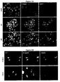
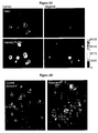

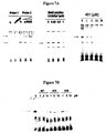

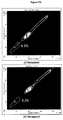

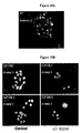
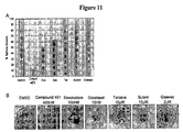
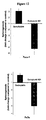
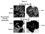
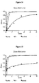


Classifications
-
- C—CHEMISTRY; METALLURGY
- C07—ORGANIC CHEMISTRY
- C07D—HETEROCYCLIC COMPOUNDS
- C07D307/00—Heterocyclic compounds containing five-membered rings having one oxygen atom as the only ring hetero atom
- C07D307/77—Heterocyclic compounds containing five-membered rings having one oxygen atom as the only ring hetero atom ortho- or peri-condensed with carbocyclic rings or ring systems
- C07D307/92—Naphthofurans; Hydrogenated naphthofurans
-
- A—HUMAN NECESSITIES
- A61—MEDICAL OR VETERINARY SCIENCE; HYGIENE
- A61K—PREPARATIONS FOR MEDICAL, DENTAL OR TOILETRY PURPOSES
- A61K31/00—Medicinal preparations containing organic active ingredients
- A61K31/33—Heterocyclic compounds
- A61K31/335—Heterocyclic compounds having oxygen as the only ring hetero atom, e.g. fungichromin
- A61K31/34—Heterocyclic compounds having oxygen as the only ring hetero atom, e.g. fungichromin having five-membered rings with one oxygen as the only ring hetero atom, e.g. isosorbide
- A61K31/343—Heterocyclic compounds having oxygen as the only ring hetero atom, e.g. fungichromin having five-membered rings with one oxygen as the only ring hetero atom, e.g. isosorbide condensed with a carbocyclic ring, e.g. coumaran, bufuralol, befunolol, clobenfurol, amiodarone
-
- A—HUMAN NECESSITIES
- A61—MEDICAL OR VETERINARY SCIENCE; HYGIENE
- A61K—PREPARATIONS FOR MEDICAL, DENTAL OR TOILETRY PURPOSES
- A61K31/00—Medicinal preparations containing organic active ingredients
- A61K31/33—Heterocyclic compounds
- A61K31/38—Heterocyclic compounds having sulfur as a ring hetero atom
-
- A—HUMAN NECESSITIES
- A61—MEDICAL OR VETERINARY SCIENCE; HYGIENE
- A61K—PREPARATIONS FOR MEDICAL, DENTAL OR TOILETRY PURPOSES
- A61K47/00—Medicinal preparations characterised by the non-active ingredients used, e.g. carriers or inert additives; Targeting or modifying agents chemically bound to the active ingredient
- A61K47/06—Organic compounds, e.g. natural or synthetic hydrocarbons, polyolefins, mineral oil, petrolatum or ozokerite
- A61K47/24—Organic compounds, e.g. natural or synthetic hydrocarbons, polyolefins, mineral oil, petrolatum or ozokerite containing atoms other than carbon, hydrogen, oxygen, halogen, nitrogen or sulfur, e.g. cyclomethicone or phospholipids
-
- A—HUMAN NECESSITIES
- A61—MEDICAL OR VETERINARY SCIENCE; HYGIENE
- A61K—PREPARATIONS FOR MEDICAL, DENTAL OR TOILETRY PURPOSES
- A61K47/00—Medicinal preparations characterised by the non-active ingredients used, e.g. carriers or inert additives; Targeting or modifying agents chemically bound to the active ingredient
- A61K47/30—Macromolecular organic or inorganic compounds, e.g. inorganic polyphosphates
- A61K47/42—Proteins; Polypeptides; Degradation products thereof; Derivatives thereof, e.g. albumin, gelatin or zein
-
- A—HUMAN NECESSITIES
- A61—MEDICAL OR VETERINARY SCIENCE; HYGIENE
- A61K—PREPARATIONS FOR MEDICAL, DENTAL OR TOILETRY PURPOSES
- A61K47/00—Medicinal preparations characterised by the non-active ingredients used, e.g. carriers or inert additives; Targeting or modifying agents chemically bound to the active ingredient
- A61K47/44—Oils, fats or waxes according to two or more groups of A61K47/02-A61K47/42; Natural or modified natural oils, fats or waxes, e.g. castor oil, polyethoxylated castor oil, montan wax, lignite, shellac, rosin, beeswax or lanolin
-
- A—HUMAN NECESSITIES
- A61—MEDICAL OR VETERINARY SCIENCE; HYGIENE
- A61P—SPECIFIC THERAPEUTIC ACTIVITY OF CHEMICAL COMPOUNDS OR MEDICINAL PREPARATIONS
- A61P1/00—Drugs for disorders of the alimentary tract or the digestive system
-
- A—HUMAN NECESSITIES
- A61—MEDICAL OR VETERINARY SCIENCE; HYGIENE
- A61P—SPECIFIC THERAPEUTIC ACTIVITY OF CHEMICAL COMPOUNDS OR MEDICINAL PREPARATIONS
- A61P1/00—Drugs for disorders of the alimentary tract or the digestive system
- A61P1/04—Drugs for disorders of the alimentary tract or the digestive system for ulcers, gastritis or reflux esophagitis, e.g. antacids, inhibitors of acid secretion, mucosal protectants
-
- A—HUMAN NECESSITIES
- A61—MEDICAL OR VETERINARY SCIENCE; HYGIENE
- A61P—SPECIFIC THERAPEUTIC ACTIVITY OF CHEMICAL COMPOUNDS OR MEDICINAL PREPARATIONS
- A61P11/00—Drugs for disorders of the respiratory system
- A61P11/06—Antiasthmatics
-
- A—HUMAN NECESSITIES
- A61—MEDICAL OR VETERINARY SCIENCE; HYGIENE
- A61P—SPECIFIC THERAPEUTIC ACTIVITY OF CHEMICAL COMPOUNDS OR MEDICINAL PREPARATIONS
- A61P17/00—Drugs for dermatological disorders
- A61P17/02—Drugs for dermatological disorders for treating wounds, ulcers, burns, scars, keloids, or the like
-
- A—HUMAN NECESSITIES
- A61—MEDICAL OR VETERINARY SCIENCE; HYGIENE
- A61P—SPECIFIC THERAPEUTIC ACTIVITY OF CHEMICAL COMPOUNDS OR MEDICINAL PREPARATIONS
- A61P17/00—Drugs for dermatological disorders
- A61P17/06—Antipsoriatics
-
- A—HUMAN NECESSITIES
- A61—MEDICAL OR VETERINARY SCIENCE; HYGIENE
- A61P—SPECIFIC THERAPEUTIC ACTIVITY OF CHEMICAL COMPOUNDS OR MEDICINAL PREPARATIONS
- A61P17/00—Drugs for dermatological disorders
- A61P17/12—Keratolytics, e.g. wart or anti-corn preparations
-
- A—HUMAN NECESSITIES
- A61—MEDICAL OR VETERINARY SCIENCE; HYGIENE
- A61P—SPECIFIC THERAPEUTIC ACTIVITY OF CHEMICAL COMPOUNDS OR MEDICINAL PREPARATIONS
- A61P19/00—Drugs for skeletal disorders
- A61P19/02—Drugs for skeletal disorders for joint disorders, e.g. arthritis, arthrosis
-
- A—HUMAN NECESSITIES
- A61—MEDICAL OR VETERINARY SCIENCE; HYGIENE
- A61P—SPECIFIC THERAPEUTIC ACTIVITY OF CHEMICAL COMPOUNDS OR MEDICINAL PREPARATIONS
- A61P25/00—Drugs for disorders of the nervous system
-
- A—HUMAN NECESSITIES
- A61—MEDICAL OR VETERINARY SCIENCE; HYGIENE
- A61P—SPECIFIC THERAPEUTIC ACTIVITY OF CHEMICAL COMPOUNDS OR MEDICINAL PREPARATIONS
- A61P25/00—Drugs for disorders of the nervous system
- A61P25/02—Drugs for disorders of the nervous system for peripheral neuropathies
-
- A—HUMAN NECESSITIES
- A61—MEDICAL OR VETERINARY SCIENCE; HYGIENE
- A61P—SPECIFIC THERAPEUTIC ACTIVITY OF CHEMICAL COMPOUNDS OR MEDICINAL PREPARATIONS
- A61P25/00—Drugs for disorders of the nervous system
- A61P25/28—Drugs for disorders of the nervous system for treating neurodegenerative disorders of the central nervous system, e.g. nootropic agents, cognition enhancers, drugs for treating Alzheimer's disease or other forms of dementia
-
- A—HUMAN NECESSITIES
- A61—MEDICAL OR VETERINARY SCIENCE; HYGIENE
- A61P—SPECIFIC THERAPEUTIC ACTIVITY OF CHEMICAL COMPOUNDS OR MEDICINAL PREPARATIONS
- A61P29/00—Non-central analgesic, antipyretic or antiinflammatory agents, e.g. antirheumatic agents; Non-steroidal antiinflammatory drugs [NSAID]
-
- A—HUMAN NECESSITIES
- A61—MEDICAL OR VETERINARY SCIENCE; HYGIENE
- A61P—SPECIFIC THERAPEUTIC ACTIVITY OF CHEMICAL COMPOUNDS OR MEDICINAL PREPARATIONS
- A61P31/00—Antiinfectives, i.e. antibiotics, antiseptics, chemotherapeutics
-
- A—HUMAN NECESSITIES
- A61—MEDICAL OR VETERINARY SCIENCE; HYGIENE
- A61P—SPECIFIC THERAPEUTIC ACTIVITY OF CHEMICAL COMPOUNDS OR MEDICINAL PREPARATIONS
- A61P35/00—Antineoplastic agents
-
- A—HUMAN NECESSITIES
- A61—MEDICAL OR VETERINARY SCIENCE; HYGIENE
- A61P—SPECIFIC THERAPEUTIC ACTIVITY OF CHEMICAL COMPOUNDS OR MEDICINAL PREPARATIONS
- A61P35/00—Antineoplastic agents
- A61P35/02—Antineoplastic agents specific for leukemia
-
- A—HUMAN NECESSITIES
- A61—MEDICAL OR VETERINARY SCIENCE; HYGIENE
- A61P—SPECIFIC THERAPEUTIC ACTIVITY OF CHEMICAL COMPOUNDS OR MEDICINAL PREPARATIONS
- A61P35/00—Antineoplastic agents
- A61P35/04—Antineoplastic agents specific for metastasis
-
- A—HUMAN NECESSITIES
- A61—MEDICAL OR VETERINARY SCIENCE; HYGIENE
- A61P—SPECIFIC THERAPEUTIC ACTIVITY OF CHEMICAL COMPOUNDS OR MEDICINAL PREPARATIONS
- A61P37/00—Drugs for immunological or allergic disorders
-
- A—HUMAN NECESSITIES
- A61—MEDICAL OR VETERINARY SCIENCE; HYGIENE
- A61P—SPECIFIC THERAPEUTIC ACTIVITY OF CHEMICAL COMPOUNDS OR MEDICINAL PREPARATIONS
- A61P37/00—Drugs for immunological or allergic disorders
- A61P37/02—Immunomodulators
-
- A—HUMAN NECESSITIES
- A61—MEDICAL OR VETERINARY SCIENCE; HYGIENE
- A61P—SPECIFIC THERAPEUTIC ACTIVITY OF CHEMICAL COMPOUNDS OR MEDICINAL PREPARATIONS
- A61P37/00—Drugs for immunological or allergic disorders
- A61P37/02—Immunomodulators
- A61P37/06—Immunosuppressants, e.g. drugs for graft rejection
-
- A—HUMAN NECESSITIES
- A61—MEDICAL OR VETERINARY SCIENCE; HYGIENE
- A61P—SPECIFIC THERAPEUTIC ACTIVITY OF CHEMICAL COMPOUNDS OR MEDICINAL PREPARATIONS
- A61P37/00—Drugs for immunological or allergic disorders
- A61P37/08—Antiallergic agents
-
- A—HUMAN NECESSITIES
- A61—MEDICAL OR VETERINARY SCIENCE; HYGIENE
- A61P—SPECIFIC THERAPEUTIC ACTIVITY OF CHEMICAL COMPOUNDS OR MEDICINAL PREPARATIONS
- A61P43/00—Drugs for specific purposes, not provided for in groups A61P1/00-A61P41/00
-
- A—HUMAN NECESSITIES
- A61—MEDICAL OR VETERINARY SCIENCE; HYGIENE
- A61P—SPECIFIC THERAPEUTIC ACTIVITY OF CHEMICAL COMPOUNDS OR MEDICINAL PREPARATIONS
- A61P7/00—Drugs for disorders of the blood or the extracellular fluid
-
- A—HUMAN NECESSITIES
- A61—MEDICAL OR VETERINARY SCIENCE; HYGIENE
- A61P—SPECIFIC THERAPEUTIC ACTIVITY OF CHEMICAL COMPOUNDS OR MEDICINAL PREPARATIONS
- A61P9/00—Drugs for disorders of the cardiovascular system
-
- A—HUMAN NECESSITIES
- A61—MEDICAL OR VETERINARY SCIENCE; HYGIENE
- A61P—SPECIFIC THERAPEUTIC ACTIVITY OF CHEMICAL COMPOUNDS OR MEDICINAL PREPARATIONS
- A61P9/00—Drugs for disorders of the cardiovascular system
- A61P9/10—Drugs for disorders of the cardiovascular system for treating ischaemic or atherosclerotic diseases, e.g. antianginal drugs, coronary vasodilators, drugs for myocardial infarction, retinopathy, cerebrovascula insufficiency, renal arteriosclerosis
-
- C—CHEMISTRY; METALLURGY
- C07—ORGANIC CHEMISTRY
- C07D—HETEROCYCLIC COMPOUNDS
- C07D333/00—Heterocyclic compounds containing five-membered rings having one sulfur atom as the only ring hetero atom
- C07D333/50—Heterocyclic compounds containing five-membered rings having one sulfur atom as the only ring hetero atom condensed with carbocyclic rings or ring systems
- C07D333/74—Naphthothiophenes
-
- Y—GENERAL TAGGING OF NEW TECHNOLOGICAL DEVELOPMENTS; GENERAL TAGGING OF CROSS-SECTIONAL TECHNOLOGIES SPANNING OVER SEVERAL SECTIONS OF THE IPC; TECHNICAL SUBJECTS COVERED BY FORMER USPC CROSS-REFERENCE ART COLLECTIONS [XRACs] AND DIGESTS
- Y02—TECHNOLOGIES OR APPLICATIONS FOR MITIGATION OR ADAPTATION AGAINST CLIMATE CHANGE
- Y02A—TECHNOLOGIES FOR ADAPTATION TO CLIMATE CHANGE
- Y02A50/00—TECHNOLOGIES FOR ADAPTATION TO CLIMATE CHANGE in human health protection, e.g. against extreme weather
- Y02A50/30—Against vector-borne diseases, e.g. mosquito-borne, fly-borne, tick-borne or waterborne diseases whose impact is exacerbated by climate change
Definitions
- the present invention relates to compositions for use of Stat3 pathway inhibitors and cancer stem cell inhibitors in combinatorial treatment of cancers and other disorders.
- CSCs Cancer Stem cells
- CSCs cancer stem cells
- the CSC model of tumorigenesis would explain why tens or hundreds of thousands of tumor cells need to be injected into an experimental animal in order to establish a tumor transplant.
- the frequency of these cells is less than 1 in 10,000 [2].
- cancer cell lines are selected from a sub-population of cancer cells that are specifically adapted to grow in tissue cultures, the biological and functional properties of cancer cell lines can undergo dramatic changes. Therefore, not all cancer cell lines contain CSCs.
- Cancer stem cells share many similar traits with normal stem cells.
- CSCs have self-renewal capacity, namely, the ability to give rise to additional tumorigenic cancer stem cells, typically at a slower rate than other dividing tumor cells, as opposed to a limited number of divisions.
- CSCs also have the ability to differentiate into multiple cell types, which would explain histological evidences that not only many tumors contain multiple cell types native to the host organ, but also that heterogeneity is commonly retained in tumor metastases.
- CSCs have been demonstrated to be fundamentally responsible for tumorigenesis, cancer metastasis, and cancer reoccurrence.
- CSCs are also called tumor initiating cells, cancer stem-like cells, stem-like cancer cells, highly tumorigenic cells, tumor stem cells, solid tumor stem cells, or super malignant cells.
- cancer stem cells have fundamental implications for future cancer treatments and therapies.
- the efficacy of current cancer treatments are, in the initial stages of testing, often measured by the size of the tumor shrinkage, i.e., the amount of tumor mass that is killed off.
- the measurement of tumor mass may not necessarily select for drugs that act specifically on the stem cells.
- cancer stem cells appear to be resistant to radiotherapy (XRT) and are also refractory to chemotherapeutic and targeted drugs [3-5].
- XRT radiotherapy
- chemotherapeutic and targeted drugs [3-5].
- Normal somatic stem cells are naturally resistant to chemotherapeutic agents--they have various pumps (such as MDR) that pump out drugs, and efficient DNA repair mechanisms.
- Cancer stem cells being the mutated counterparts of normal stem cells, may also have similar mechanisms that allow them to survive drug therapies and radiation treatment.
- conventional chemotherapies and radiotherapies kill differentiated or differentiating cells, which form the bulk of the tumor that are unable to regenerate tumors.
- the population of cancer stem cells that gave rise to the differentiated and differentiating cells could remain untouched and cause a relapse of the disease.
- a further danger for the conventional anti-cancer therapy is the possibility that the treatment of, for instance, chemotherapy, leaves only chemotherapy-resistant cancer stem cells, and the ensuing recurrent tumor will likely also be resistant to chemotherapy.
- anti-cancer therapies include strategies against CSCs (see Figure 1 ). This has been likened to the need for eliminating dandelion roots in order to prevent weed regrowth [6].
- CSCs weed regrowth
- By selectively targeting cancer stem cells it becomes possible to treat patients with aggressive, non-resectable tumors, and refractory or recurrent cancers, as well as preventing tumor metastasis and recurrence.
- Development of specific therapies targeting cancer stem cells therefore holds hope for survival and improved quality of life of cancer patients, especially for those with metastatic cancers.
- the key to unlocking this untapped potential is the identification and validation of pathways that are selectively important for cancer stem cell self-renewal and survival. Though multiple pathways underlying tumorigenesis in cancer and in embryonic stem cells or adult stem cells have been elucidated in the past, no pathways have been identified and validated for cancer stem cell self-renewal and survival.
- the side population (SP) technique originally used to enrich hematopoetic and leukemic stem cells, was also employed to identify and isolate CSCs [14].
- This technique first described by Goodell et al., takes advantage of differential ABC transporter-dependent efflux of fluorescent dyes such as Hoechst 33342 to define and isolate a cell population enriched in CSCs [10, 15].
- the SP is revealed by blocking drug efflux with verapamil, at which point the dyes can no longer be pumped out of the SP.
- CSC surface markers include CD44, CD133, and CD166 [16-24]. Sorting tumor cells based primarily upon the differential expression of these surface marker(s) have accounted for the majority of the highly tumorigenic CSCs described to date. Therefore, these surface markers are well validated for the identification and isolation of cancer stem cells from cancer cell lines and from bulk tumor tissues.
- the STAT (Signal Transducers and Activator of Transcription) protein family are latent transcription factors activated in response to cytokines/growth factors to promote proliferation, survival, and other biological processes.
- Stat3 is activated by phosphorylation of a critical tyrosine residue mediated by growth factor receptor tyrosine kinases, Janus kinases, or the Src family kinases, etc.
- kinases include but are not limited to EGFR, JAKs, Abl, KDR, c-Met, Src, and Her2 [25].
- Stat3 forms homo-dimers, translocates to the nucleus, binds to specific DNA-response elements in the promoter regions of the target genes, and induces gene expression [26] (see Figure 2 ).
- Stat3 activation is transient and tightly regulated, lasting from 30 minutes to several hours.
- Stat3 is found to be aberrantly active in a wide variety of human cancers, including all the major carcinomas as well as some hematologic tumors.
- Stat3 plays multiple roles in cancer progression.
- As a potent transcription regulator it targets genes involved in many important cellular functions, such as Bcl-x1, c-Myc, cyclin D1, Vegf, MMP-2, and survivin [27-32]. It is also a key negative regulator of tumor immune surveillance and immune cell recruitment [33-35].
- Stat3 signaling by antisense, siRNA, a dominant-negative form of Stat3, and/or blockade of tyrosine kinases inhibits certain cancer cell lines or tumors in vitro and/or in vivo [26, 28, 36, 37].
- Stat3 and cancer stem cell functionality have ever been empirically made.
- cancer stem cells CSCs
- CSCs cancer stem cells
- IL6 interleukin 6
- IL6-Stat3 pathway mediated inflammation has been found to be the common causative origin for atherosclerosis, peripheral vascular disease, coronary artery disease, hypertension, osteoporosis, type 2 diabetes, and dementia.
- the present invention is predicated, in part, on empirical evidence provided herein that Stat3 plays a key role in both the survival and self-renewal capacity of cancer stem cells (CSCs) across a broad spectrum of cancers.
- CSCs cancer stem cells
- the present disclosure also provides data that confirms certain compounds act as Stat3 pathway inhibitors and that they effectively inhibit CSCs both in vitro and in vivo.
- a first aspect of this disclosure provides a method of treating a subject with a disorder that is associated with aberrant Stat3 pathway activity, the method comprising the steps of: (a) administering to the subject a first amount of a first agent to inhibit at least some of the aberrant Stat3 pathway activity; and (b) administering to the subject a second amount of a second agent comprising a signal transduction inhibitor.
- the first agent may inhibit Stat3 pathway activity through at least one of the following actions: substantially inhibiting phosphorylation of the Stat3 protein, substantially inhibiting dimerization of the Stat3 protein, substantially inhibiting nuclear translocation of the Stat3 protein, substantially inhibiting DNA-binding activity of the Stat3 protein, and substantially inhibiting transcription activities of the Stat3 protein.
- the first agent is selected from the group consisting of 2-(1-hydroxyethyl)-naphtho[2,3-b]furan-4,9-dione, 2-Acetyl-7-Chloro-naphtho[2,3-b]furan-4,9-dione, 2-Acetyl-7-Fluoro-naphtho[2,3-b]furan-4,9-dione, 2-acetyinaphtho[2,3-b]furan-4,9-dione, 2-ethyl-naphtho[2,3-b]furan-4,9-dione, an enantiomer, diastereomer, tautomer, and a salt or solvate thereof (hereafter referred to as the "Compound of the Invention").
- Non-cancer disorders that can be treated by methods of the first aspect of this disclosure include but are not limited to: autoimmune diseases, inflammatory diseases, inflammatory bowel diseases, arthritis, asthma, and systemic lupus erythematosus, autoimmune demyelination disorder, Alzheimer's disease, stroke, ischemia reperfusion injury and multiple sclerosis.
- Cancers that can be treated by the methods of this invention include but are not limited to: breast cancer, head and neck cancer, lung cancer, ovarian cancer, pancreatic cancer, colorectal carcinoma, prostate cancer, renal cell carcinoma, melanoma, hepatocellular carcinomas, cervical cancer, sarcomas, brain tumors, gastric cancers, multiple myeloma, leukemia, and lymphomas. These non-cancer and cancerous disorders are known to be associated with aberrant Stat3 pathway activities.
- the second agent is a targeted agent, which can be a growth factor receptor-targeting agent, a kinase-targeting agent, or an angiogenesis inhibitor.
- the present invention provides a method of treating a subject of a cancer that is associated with aberrant Stat3 pathway activity, the method comprising the steps of: (a) administering to the subject a first amount of a first agent to inhibit at least some of the aberrant Stat3 pathway activity; and (b) administering to the subject a second amount of a second anti-cancer agent.
- the present invention provides a combination comprising a first agent for inhibiting aberrant Stat3 pathway Activity and a second agent for use in treating a subject with a cancer that is associated with aberrant Stat3 pathway activity, wherein the first agent is selected from the group consisting of 2-(1-hydroxyethyl)-naphtho[2,3 -b]furan-4,9-dione, 2-acetyl-7-chloro-naphtho[2,3 -b]furan-4,9-dione, 2-acetyl-7-fluoro-naphtho[2,3 -b]furan-4,9-dione, 2-acelylnaphtho[2,3 -b]furan-4,9-dione, 2-ethyl-naphtho[2,3-b]furan-4,9-dione, and a pharmaceutically acceptable salt or solvate thereof; and the second agent is selected from the group consisting of paclitaxel, erlot
- the second anti-cancer agent can be a cytotoxic agent, or a chemotherapeutic agent.
- the second agent is a standard first-line treatment for at least one cancer.
- the anti-cancer agent is a DNA-damaging agent, an antimitotic agent, and/or an antimetabolite agent.
- the DNA-damaging agent can be an alkylating agent, a topoisomerase inhibitor, or a DNA intercalator.
- the second agent is one of carboplatin, doxorubicin, gemcitabine, docetaxel, or etoposide.
- Cancers that can be treated by methods of the invention include those known to be associated with aberrant Stat3 pathway activities, which are listed above, and not repeated here.
- a method of treating cancer in a subject includes the steps of: (a) administering to the subject a first amount of a first anti-cancer agent to inhibit a cancer stem cell (CSC) population; and (b) administering to the subject a second amount of a second anti-cancer agent to inhibit a plurality of normal cancer cells.
- CSC cancer stem cell
- step (a) of this method inhibits at least one CSC from self-renewal, and/or kills at least one CSC.
- the first amount of the first anti-cancer agent also kills a plurality of normal cancer cells.
- step (a) inhibits at least some Stat3 pathway activity in cancer stem cells.
- the first anti-cancer agent shares the same features and characteristics as the first agent in methods according to the first aspect since this disclosure has provided evidence that Stat3 pathway inhibitors can effectively inhibit CSCs.
- the shared features include, for example, various steps of the Stat3 pathway that the first anti-cancer agent recited here can target.
- the first anti-cancer agent can be a small molecule Stat3 inhibitor, an RNAi agent against Stat3, an antisense agent against Stat3, a peptidomimetic Stat3 inhibitor, or a G-quartet oligodeoxynucleotides Stat3 inhibitor.
- Cancers that can be treated by this method preferably are those known or confirmed to contain CSCs, which include, but are not limited to: breast cancer, head and neck cancer, lung cancer, ovarian cancer, pancreatic cancer, multiple myeloma, colorectal carcinoma, prostate cancer, melanoma, kaposi sarcoma, ewing's sarcoma, liver cancer, gastric cancer, medulloblastoma, brain tumors, and leukemia.
- CSCs include, but are not limited to: breast cancer, head and neck cancer, lung cancer, ovarian cancer, pancreatic cancer, multiple myeloma, colorectal carcinoma, prostate cancer, melanoma, kaposi sarcoma, ewing's sarcoma, liver cancer, gastric cancer, medulloblastoma, brain tumors, and leukemia.
- the "second anti-cancer agent” in methods according to the third aspect can be the same “second anti-cancer agent” in methods according to the invention, and all the shared features are not repeated here.
- the second agent is a standard first-line treatment for at least one cancer.
- the second agent can be a cytotoxic agent.
- the anti-cancer agent is a DNA-damaging agent, an anti-mitotic agent, and/or an anti-metabolite agent.
- the DNA-damaging agent can be an alkylating agent, a topoisomerase inhibitor, or a DNA intercalator.
- the second agent is one of carboplatin, doxorubicin, gemcitabine, docetaxel, or etoposide.
- a method for treating cancer in a subject comprising the steps of: (a) administering to the subject a first amount of a first cancer stem cell inhibitor to inhibit Stat3 pathway activities; and (b) administering to the subject a second amount of a second cancer stem cell inhibitor to inhibit activities of a different pathway.
- the second cancer stem cell inhibitor is lapatinib.
- the second amount of the second anti-cancer agent is not therapeutically effective against the cancer stem cell population by itself.
- Cancers that can be treated by this method preferably are those known or confirmed to contain CSCs and some examples are listed above.
- the cancer is metastatic, refractory to a standard first-line cancer treatment, or relapsed.
- a method for treating cancer in a subject comprising the steps of: (a) administering to the subject a therapeutically effective amount of a first anti-cancer agent selected from the group consisting of 2-(1-hydroxyethyl)-naphtho[2,3-b]furan-4,9-dione, 2-acetyl-7-chloro-naphtho[2,3-b]furan-4,9-dione, 2-acetyl-7-fluoro-naphtho[2,3-b]furan-4,9-dione, 2-acetylnaphtho[2,3-b]furan-4,9-dione, 2-ethyl-naphtho[2,3-b]furan-4,9-dione, and a pharmaceutically acceptable salt or solvate thereof; and (b) administering a second anti-cancer agent that is not selected from the same group.
- a first anti-cancer agent selected from the group consisting of 2-
- the second anti-cancer agent can be any of the agents described in other aspects of the invention, including any of the cytotoxic or chemotherapeutic agents, and any of the targeted agents.
- the present disclosure provides a pharmaceutical composition
- a pharmaceutical composition comprising a therapeutically effective amount of a first anti-cancer agent selected from the group consisting of 2-(1-hydroxyethyl)-naphtho[2,3-b]furan-4,9-dione, 2-acetyl-7-chloro-naphtho[2,3-b]furan-4,9-dione, 2-acetyl-7-fluoro-naphtho[2,3-b]furan-4,9-dione, 2-acetylnaphtho[2,3-b]furan-4,9-dione, 2-ethyl-naphtho[2,3-b]furan-4,9-dione, and a pharmaceutically acceptable salt or solvate thereof; and a second anti-cancer therapy selected from the group consisting of a cytotoxic agent, a targeted agent, a radiotherapy agent, a biologic agent, a hormonal agent, a HDAC inhibitor
- the composition further includes a pharmaceutically-acceptable excipient, carrier, or diluent.
- a cell includes a plurality of cells including mixtures thereof.
- isolated or “purified” as used herein refer to a material that is substantially or essentially free from components that normally accompany it in its native state. Purity and homogeneity are typically determined using analytical chemistry techniques such as polyacrylamide gel electrophoresis or high performance liquid chromatography.
- cancer stem cell(s) and “CSC(s)” are interchangeable.
- CSCs are mammalian, and in preferred embodiments, these CSCs are of human origin, but they are not intended to be limited thereto.
- Cancer stem cells are defined and functionally characterized as a population of cells originating from a solid tumor that: (1) have extensive proliferative capacity; 2) are capable of asymmetric cell division to generate one or more kinds of differentiated progeny with reduced proliferative or developmental potential; and (3) are capable of symmetric cell divisions for self-renewal or self-maintenance.
- Other common approaches to characterize CSCs involve morphology and examination of cell surface markers, transcriptional profile, and drug response.
- CSCs are also called in the research literature tumor/cancer initiating cells, cancer stem-like cells, stem-like cancer cells, highly tumorigenic cells, tumor stem cells, solid tumor stem cells, drug survival cells (DSC), drug resistant cells (DRCs) or super malignant cells.
- DSC drug survival cells
- DRC drug resistant cells
- self-renewal refers to cancer stem cells' ability to give rise to new tumorigenic cancer stem cells to replenish or increase their number.
- cancer refers to or describe the physiological condition in mammals in which a population of cells are characterized by unregulated cell growth.
- Cancer cells and “tumor cells” as used herein refer to the total population of cells derived from a tumor including both non-tumorigenic cells, which comprise the bulk of the tumor cell population, and tumorigenic stem cells (cancer stem cells). Examples of cancer include, but are not limited to, carcinoma, lymphoma, blastoma, sarcoma, and leukemia.
- cancers include squamous cell cancer, small-cell lung cancer, non-small cell lung cancer, adenocarcinoma of the lung, squamous carcinoma of the lung, cancer of the peritoneum, hepatocellular cancer, gastrointestinal cancer, pancreatic cancer, glioblastoma, cervical cancer, ovarian cancer, liver cancer, bladder cancer, hepatoma, breast cancer, colon cancer, colorectal cancer, endometrial or uterine carcinoma, salivary gland carcinoma, kidney cancer, liver cancer, prostate cancer, vulval cancer, thyroid cancer, hepatic carcinoma and various types of head and neck cancer.
- Tumor refers to any mass of tissue that result from excessive cell growth or proliferation, either benign (noncancerous) or malignant (cancerous) including pre-cancerous lesions.
- Metalastasis refers to the process by which a cancer spreads or transfers from the site of origin to other regions of the body with the development of a similar cancerous lesion at the new location.
- a “metastatic” or “metastasizing” cell is one that loses adhesive contacts with neighboring cells and migrates via the bloodstream or lymph from the primary site of disease to invade neighboring body structures.
- the term “subject” refers to any animal (e.g., a mammal), including, but not limited to humans, non-human primates, rodents, and the like, which is to be the recipient of a particular treatment.
- the terms “subject” and “patient” are used interchangeably herein in reference to a human subject.
- Terms such as “treating” or “treatment” or “to treat” or “alleviating” or “to alleviate” as used herein refer to both 1) therapeutic measures that cure, slow down, lessen symptoms of, and/or halt progression of a diagnosed pathologic condition or disorder and 2) prophylactic or preventative measures that prevent or slow the development of a targeted pathologic condition or disorder.
- those in need of treatment include those already with the disorder; those prone to have the disorder; and those in whom the disorder is to be prevented.
- a subject is successfully "treated” according to the methods of the present invention if the patient shows one or more of the following: a reduction in the number of or complete absence of cancer cells; a reduction in the tumor size; inhibition of, or an absence of cancer cell infiltration into peripheral organs including the spread of cancer into soft tissue and bone; inhibition of, or an absence of tumor metastasis; inhibition or an absence of tumor growth; relief of one or more symptoms associated with the specific cancer; reduced morbidity and mortality; and improvement in quality of life.
- the term “inhibiting”, “to inhibit” and their grammatical equivalents, when used in the context of a bioactivity, refer to a down-regulation of the bioactivity, which may reduce or eliminate the targeted function, such as the production of a protein or the phosphorylation of a molecule. In particular embodiments, inhibition may refers to a reduction of about 20%, 30%, 40%, 50%, 60%, 70%, 80%, 90% or 95% of the targeted activity.
- the terms refer to success at preventing the onset of symptoms, alleviating symptoms, or eliminating the disease, condition or disorder.
- Normal cancer cells refers to cancer cells that are not cancer stem cells.
- Combination or “combinatorial” therapy or treatment means the administration of at least two different therapeutics to treat a disorder, condition or symptom, e.g., a cancer condition.
- Such combination therapy may involve the administration of one therapeutic before, during, and/or after the administration of the other therapeutic.
- the administrations of the therapeutics may be separated in time by up to several weeks, but more commonly within 48 hours, and most commonly within 24 hours.
- pharmaceutically-acceptable excipient, carrier, or diluent means a pharmaceutically-acceptable material, composition or vehicle, such as a liquid or solid filler, diluent, excipient, solvent or encapsulating material, involved in carrying or transporting the subject pharmaceutical agent from one organ, or portion of the body, to another organ, or portion of the body.
- Each carrier must be “acceptable” in the sense of being compatible with the other ingredients of the formulation and not injurious to the patient.
- materials which can serve as pharmaceutically-acceptable carriers include: sugars, such as lactose, glucose and sucrose; starches, such as corn starch and potato starch; cellulose, and its derivatives, such as sodium carboxymethyl cellulose, ethyl cellulose and cellulose acetate; powdered tragacanth; malt; gelatin; talc; excipients, such as cocoa butter and suppository waxes; oils, such as peanut oil, cottonseed oil, safflower oil, sesame oil, olive oil, corn oil and soybean oil; glycols, such as propylene glycol; polyols, such as glycerin, sorbitol, mannitol and polyethylene glycol; esters, such as ethyl oleate and ethyl laurate; agar; buffering agents, such as magnesium hydroxide and aluminum hydroxide; alginic acid; pyrogen-free water; isotonic saline; Ring
- wetting agents such as sodium lauryl sulfate, magnesium stearate, and polyethylene oxide-polypropylene oxide copolymer as well as coloring agents, release agents, coating agents, sweetening, flavoring and perfuming agents, preservatives and antioxidants can also be present in the compositions.
- the compounds of the present invention may form salts which are also within the scope of this invention.
- Reference to a compound of the present invention herein is understood to include reference to salts thereof, unless otherwise indicated.
- the term "salt(s)", as employed herein, denotes acidic and/or basic salts formed with inorganic and/or organic acids and bases.
- zwitterions inner salts
- inner salts may be formed and are included within the term "salt(s)" as used herein.
- Salts of the compounds of the present invention may be formed, for example, by reacting a compound I, II or III with an amount of acid or base, such as an equivalent amount, in a medium such as one in which the salt precipitates or in an aqueous medium followed by lyophilization.
- Solvates of the compounds of the invention are also contemplated herein.
- Solvates of the compounds of the present invention include, for example, hydrates.
- the present invention provides compounds that are effective inhibitors of Stat3 pathway activities (Example 2).
- Stat3 pathway is a latent transcription factor activated to promote proliferation, survival and many other biological processes, it is implicated in a wide variety of human cancers as well as non-cancer disorders including a number of autoimmune and inflammatory diseases (Table 1).
- the present invention provides, in a first aspect, combination treatment of disorders associated with aberrant, e.g., over-expressed, Stat3 pathway activity.
- the patient subject is administered a first amount of a first agent to inhibit at least some of the aberrant Stat3 pathway activity and also a second amount of a second agent which includes a signal transduction inhibitor.
- the first amount and the second amount can be a therapeutically effective amount of the respective agent before combination use, i.e., when used by itself against the disorder, or less than that amount because of outstanding synergistic effects from the combination.
- the first agent may target one or multiple steps in the Stat3 pathway.
- the first agent is the Compound of the Invention. Table 1. Activation of STAT3 PATHWAY in human diseases DISEASES REF.
- the second agent i.e., a signal transduction inhibitor
- a signal transduction inhibitor can be used to target a different pathway, a related pathway or a different step in the same Stat3 pathway from the one inhibited by the first agent.
- the two agents in the combination therapy target the same pathway, albeit at different steps, the expected amount of synergism is limited.
- data provided below in Example 5 show surprisingly high amounts of synergism between the Compound of the Invention and a second agent that presumably targets other steps in the same pathway, e.g., tyrosine kinases and GFR-targeting agents, suggesting unexpected mechanism of inhibition at work.
- Stat3 is activated by phosphorylation of a critical tyrosine residue mediated by growth factor receptor tyrosine kinases, Janus kinases, or the Src family kinases, etc; upon tyrosine phosphorylation, Stat3 forms homo-dimers, translocates to the nucleus, binds to specific DNA-response elements in the promoter regions of the target genes, and induces gene expression.
- Example 2 of the present invention shows that in the Stat3 pathway, the inhibitory effect by the Compound of the Invention is evident by the step of DNA-binding.
- the Compound of the Invention inhibits dimerization and/or nuclear translocation of the Stat3 protein. Therefore, when combined with tyrosine kinases (TKI) and GFR-targeting agents that also target the same Stat3 pathway, the amount of synergism observed was surprisingly high. For example, 100% inhibition of cells from a pancreatic cancer cell line was achieved when Compound 401 was combined with TKI Sorafenib, whereas both Compound 401 and Sorafenib could only respectively achieve 66% inhibition of the same cell line when administered individually-panereatic cancer is known to implicate over-expression of Stat3 [44]. In fact, all four TKIs tested in combination with Compound 401 showed marked synergism. In preferred embodiments of the invention, the combination treatment achieves over about 50%, or 70%, or 90% inhibition of the cancer cells.
- Methods according to this first aspect of the disclosure can be applied to treatment of cancers or non-cancer disorders, preferably those known to be associated with aberrant Stat3 pathway activities.
- non-cancer disorders associated with aberrant Stat3 pathway activities include but are not limited to: autoimmune diseases, inflammatory diseases, inflammatory bowel diseases, arthritis, asthma, and systemic lupus erythematosus, autoimmune demyelination disorder, Alzheimer's disease, stroke, ischemia reperfusion injury and multiple sclerosis.
- cancers associated with aberrant Stat3 pathway activities include but are not limited to: breast cancer, head and neck cancer, lung cancer, ovarian cancer, pancreatic cancer, colorectal carcinoma,
- prostate cancer renal cell carcinoma, melanoma, hepatocellular carcinomas, cervical cancer, sarcomas, brain tumors, gastric cancers, multiple myeloma, leukemia, and lymphomas.
- the second agent according to this first aspect of the disclosure can be a targeted agent, e.g., a growth factor receptor-targeting agent (see erlotinib (tarceva) data in Example 5), a kinase-targeting agent (see lapatinib, erlotinib, sunitinib and sorafenib data in Example 5) or an angiogenesis inhibitor (see sunitinib and sorafenib data in Example 5).
- a targeted agent e.g., a growth factor receptor-targeting agent (see erlotinib (tarceva) data in Example 5), a kinase-targeting agent (see lapatinib, erlotinib, sunitinib and sorafenib data in Example 5) or an angiogenesis inhibitor (see sunitinib and sorafenib data in Example 5).
- the second agent is a growth factor receptor-targeting agent, for example, an antibody targeting a growth factor receptor associated with a kinase, such as the Epidermal Growth Factor Receptor (EGFR) or the Vascular Endothelial Growth Factor Receptor (VEGFR).
- a growth factor receptor-targeting agent for example, an antibody targeting a growth factor receptor associated with a kinase, such as the Epidermal Growth Factor Receptor (EGFR) or the Vascular Endothelial Growth Factor Receptor (VEGFR).
- the target agent can be gefitinib (Iressa), erlotinib (tarceva), PD153035, cetuximab (erbitux), avastin, panitumumab, trastuzumab, and anti-c-Met antibody.
- the second agent is a kinase-targeting agent, which can be a kinase inhibitor such as a tyrosine kinase inhibitor (TKI).
- TKI can be erlotinib (Tarceva), sutent (sunitinib), lapatinib, sorafenib (nexavar), vandetanib, axitinib, bosutinib, cedivanib, dasatinib (sprycel), gefitinib (irressa), irriatinib (gleevac), lestaurtinib, and/or ARQ197.
- the kinase-targeting agent is one of the following: gefitinib (iressa), ZD6474 (AZD6474), EMD-72000 (matuzumab), panitumab (ABX-EGF), ICR-62, CI-1033 (PD183805), lapatinib (tykerb), AEE788 (pyrrolo-pyrimidine), EKB-569, EXEL 7647/EXEL 0999 , erlotinib (tarceva), imatinib (gleevec), sorafinib (nexavar), sunitinib (sutent), dasatinib (sprycel), vandetinib (ZACTIMA), temsirolimus (torisel), PTK787 (vatalanib), pazopanib, AZD2171, everolimus, seliciclib, AMG 706, axitinib, PD0325901, PKC-412, C
- the second agent is an angiogenesis inhibitor which can be one of the following: CM101, IFN- ⁇ , IL-12, platelet factor-4, suramin, SU5416, thrombospondin, VEGFR antagonists, angiostatic steroids plus heparin, Cartilage-Derived Angiogenesis Inhibitory Factor, matrix metalloproteinase inhibitors, batimastat, marimastat, angiostatin, endostatin, 2-methoxyestradiol, tecogalan, thrombospondin, ⁇ V ⁇ 3 inhibitors, linomide, and ADH-1.
- angiogenesis inhibitor which can be one of the following: CM101, IFN- ⁇ , IL-12, platelet factor-4, suramin, SU5416, thrombospondin, VEGFR antagonists, angiostatic steroids plus heparin, Cartilage-Derived Angiogenesis Inhibitory Factor, matrix metalloproteinase inhibitors, batimastat, marim
- the present disclosure provides a method of treating a subject of a cancer that is associated with aberrant Stat3 pathway activity, the method comprising the steps of: (a) administering to the subject a first amount of a first agent to inhibit at least some of the aberrant Stat3 pathway activity; and (b) administering to the subject a second amount of a second anti-cancer agent.
- Cancers that can be treated with this method include those known to be associated with, e.g., caused at least partly by, aberrant Stat3 pathway activities, a list of which is provided above with regard to the first aspect of the invention.
- the second anti-cancer agent can be a cytotoxic agent, or, a chemotherapeutic agent.
- the second agent is a standard first-line treatment for at least one cancer.
- the amount of the first agent and second agent used in the method can be a therapeutically effective amount of the respective agent before combination use or less.
- the anti-cancer agent is a DNA-damaging agent, an antimitotic agent, and/or an antimetabolite agent.
- the DNA-damaging agent can be an alkylating agent, a topoisomerase inhibitor, and/or a DNA intercalator.
- Compound 401 of the present invention was added to Paca2 pancreatic cancer cells along with each of the following: carboplatin (a DNA-alkylating agent), etoposide (inhibitor of topoisomerase II), doxorubicin (a DNA intercalator), docetaxel (an anti-mitotic agent), and Gemzar/gemcitabine (an anti-metabolite agent).
- carboplatin a DNA-alkylating agent
- etoposide inhibitor of topoisomerase II
- doxorubicin a DNA intercalator
- docetaxel an anti-mitotic agent
- Gemzar/gemcitabine an anti-metabolite agent
- the alkylating agent can be one of the following: chlorambucil, cyclophosphamide, ifosfamide, mechlorethamine, melphalan, uracil mustard, thiotepa, busulfan, carmustine, lomustine, streptozocin, carboplatin, cisplatin, satraplatin, oxaliplatin, altretamine, ET-743, XL119 (becatecarin), dacarbazine, chlormethine, bendamustine, trofosfamide, uramustine, fotemustine, nimustine, prednimustine, ranimustine, semustine, nedaplatin, triplatin tetranitrate, mannosulfan, treosulfan, temozolomide, carboquone, triaziquone, triethylenemelamine, and procarbazin.
- the topoisomerase inhibitor can be one of the following: doxorubicin (doxil), daunorubicin, epirubicin, idarubicin, anthracenedione (novantrone), mitoxantrone, mitomycin C, bleomycin, dactinomycin, plicatomycin, irinotecan (camptosar), camptothecin, rubitecan, belotecan, etoposide, teniposide, and topotecan (hycamptin).
- doxorubicin doxil
- daunorubicin epirubicin
- idarubicin anthracenedione
- novantrone novantrone
- mitoxantrone mitoxantrone
- mitomycin C bleomycin
- dactinomycin plicatomycin
- irinotecan camptothecin
- rubitecan belotecan
- belotecan etop
- the DNA intercalator can be proflavine, doxorubicin (adriamycin), daunorubicin, dactinomycin, and thalidomide.
- the antimitotic agent can be one of the following: paclitaxel (abraxane)/taxol, docetaxel (taxotere), BMS-275183, xyotax, tocosal, vinorlebine, vincristine, vinblastine, vindesine, vinzolidine, etoposide (VP-16), teniposide (VM-26), ixabepilone, larotaxel, ortataxel, tesetaxel, and ispinesib.
- the antimetabolite agent can be one of the following: fluorouracil (5-FU), floxuridine (5-FUdR), methotrexate, xeloda, arranon, leucovorin, hydroxyurea, thioguanine (6-TG), mercaptopurine (6-MP), cytarabine, pentostatin, fludarabine phosphate, cladribine (2-CDA), asparaginase, gemcitabine, pemetrexed, bortezomib, aminopterin, raltitrexed, clofarabine, enocitabine, sapacitabine, and azacitidine.
- the second anti-cancer agent is one of the following: carboplatin, doxorubicin, gemcitabine, docetaxel, and etoposide.
- this method inhibits Stat3 pathway, proven herein to be critical for both the self-renewal and survival of CSCs (see data in Example 1), and CSCs have been found to be fundamentally responsible for drug resistance, tumor relapse and metastasis, in preferred embodiments, this method is used to treat or prevent a refractory cancer, relapsed cancer and/or metastatic cancer.
- the present disclosure also provides both in vitro and in vivo data that the Compound of the Invention inhibits CSCs' self-renewal and is apoptotic to CSCs (Example 3). Moreover, the present invention empirically confirms the Compound of the Invention's efficacy against metastatic cancer (Example 4).
- cancer therapy is to prevent cancer cells from multiplying, invading, metastasizing, and ultimately killing their host organism, e.g. a human or other mammals.
- host organism e.g. a human or other mammals.
- cancer therapy is to prevent cancer cells from multiplying, invading, metastasizing, and ultimately killing their host organism, e.g. a human or other mammals.
- cell multiplication is a characteristic of many normal cells as well as cancer cells
- most of the existing anticancer therapies also have toxic effects on normal cells, particularly those with a rapid rate of turnover, such as bone marrow and mucous membrane cells. Therefore, an effective cancer therapy needs to have a marked growth inhibitory or controlling effect on the cancer cells while exerting a minimal toxic effect on the host's normal cells.
- cancer stem cells As described above, this tiny population of cells within the entire cancer mass can evade drugs and radiotherapies that are effective on the rest of the cancer cells because CSCs probably share the same kind of biological mechanisms with normal somatic stem cells, which are naturally resistant to most, if not all, of the chemotherapeutic agents.
- CSCs can re-espouse cancer regrowth or cause metastasis if left untreated. Since the initial treatment leaves only drug-resistant cancer stem cells, chances for the entire regrown or metastatic tumor to become resistant to the initially "effective" therapy have dramatically increased.
- anticancer therapies are used in combination for several reasons.
- treatment with two or more non-cross-resistant therapies may prevent the formation of resistant clones in the tumor.
- Resistance to one anticancer drug e.g. a platinum anticancer compound such as cisplatin, is often associated with cross-resistance to other drugs of the same class, e.g. other platinum compounds.
- the combination of two or more therapies that are active against cells in different phases of growth may kill cells that are dividing slowly as well as those that are dividing actively and/or recruit cells into a more actively dividing state, making them more sensitive to multiple anticancer therapies.
- the combination may create a biochemical enhancement effect by affecting different pathways or different steps in a single biochemical pathway.
- the present invention is able to devise a cancer treatment method that combines a CSC-targeting agent and another agent targeting normal cancer cells.
- the present invention provides a drug regimen that preempts the scenario, which is now supported by some preliminary data, where some normal cancer cells, left untreated or insufficiently treated, revert back or give rise to CSCs as the original CSCs get depleted by a single-drug therapy that targets CSCs only.
- the present disclosure provides a method of treating cancer in a subject, comprising the steps of: (a) administering to the subject a first amount of a first anti-cancer agent to inhibit a cancer stem cell population; and (b) administering to the subject a second amount of a second anti-cancer agent to inhibit a plurality of normal cancer cells.
- the CSCs are inhibited.
- One or both of the first amount and the second amount can be a therapeutically effective amount of the respective agent before combination use, i.e., when used by itself against the cancer, or less than that amount because of outstanding synergistic effects from the combination.
- the first agent is the Compound of the Invention, namely, 2-(1-hydroxyethyl)-naphtho[2,3-b]furan-4,9-dione, 2-Acetyl-7-Chloro-naphtho[2,3-b]furan-4,9-dione, 2-Acetyl-7-Fluoro-naphtho[2,3-b]furan-4,9-dione, 2-acetylnaphtho[2,3-b]furan-4,9-dione, 2-ethyl-naphtho[2,3-b]furan-4,9-dione, and a pharmaceutically acceptable salt or solvate thereof.
- the "second anti-cancer agent” can be the same "second anti-cancer agent” in methods described above, and all the shared features are not repeated here.
- the second anti-cancer agent is a DNA-damaging agent, an antimitotic agent, and/or an antimetabolite agent.
- the DNA-damaging agent can be an alkylating agent, a topoisomerase inhibitor, or a DNA intercalator. Lists of suitable alkylating agents, topoisomerase inhibitors, DNA intercalators, antimitotic agents, and antimetabolite agents are listed above and are not repeated here.
- the second agent is one of carboplatin, doxorubicin, gemcitabine, docetaxel, and etoposide.
- the second anti-cancer agent selected from the group defined in the claims is targeted agent, e.g., a growth factor receptor-targeting agent (see erlotinib (tarceva) data in Example 5), a kinase-targeting agent (see lapatinib, erlotinib, sunitinib and sorafenib data in Example 5) or an angiogenesis inhibitor (see sunitinib and sorafenib data in Example 5).
- a growth factor receptor-targeting agent see erlotinib (tarceva) data in Example 5
- a kinase-targeting agent see lapatinib, erlotinib, sunitinib and sorafenib data in Example 5
- angiogenesis inhibitor see sunitinib and sorafenib data in Example 5
- this method employs a therapeutic agent specifically targeting CSC cells in the tumor, which are fundamentally responsible for drug resistance, tumor relapse and metastasis, in preferred embodiments, this method is used to treat or prevent a refractory cancer, relapsed cancer and/or metastatic cancer.
- the present invention provides a method of treating cancer in a subject that includes the steps of: (a) administering to the subject a first amount of a first cancer stem cell inhibitor to inhibit Stat3 pathway activities; and (b) administering to the subject a second amount of a second cancer stem cell inhibitor to inhibit activities of a different pathway.
- the amount of the first and/or second anti-cancer agent in this method is not therapeutically effective against the cancer stem cell population by itself-but due to significant synergism achieved through the combination, lower amount is able to be used in this method in order to elicit response from the patient.
- the second anti-cancer stem cell agent is lapatinib (INN) or lapatinib ditosylate (USAN), which was approved by the FDA in 2007 for use in patients with advanced metastatic breast cancer.
- Lapatinib is an ATP-competitive epidermal growth factor receptor (EGFR) and HER2/neu (ErbB-2) dual tyrosine kinase inhibitor. It inhibits receptor autophosphorylation and activation by binding to the ATP-binding pocket of the EGFR/HER2 protein kinase domain.
- Example 5 Data presented in Example 5 below shows that marked synergism was achieved against Paca2 pancreatic cancer cells: pre-combination rate of inhibition for Compound 401 and lapatinib were respectively 32% and 27% while the rate of inhibition after combination shot up to 74%, higher than the sum of the two rates.
- this method pays additional attention to CSCs, which are fundamentally responsible for drug resistance, tumor relapse and metastasis, in preferred embodiments, this method is used to treat or prevent a refractory cancer, relapsed cancer and/or metastatic cancer.
- Formulations of the present invention include those suitable for oral, nasal, topical (including buccal and sublingual), rectal, vaginal and/or parenteral administration.
- the formulations may conveniently be presented in unit dosage form and may be prepared by any methods well known in the art of pharmacy.
- the amount of active ingredient which can be combined with a carrier material to produce a single dosage form will vary depending upon the mammal being treated and the particular mode of administration.
- the amount of active ingredient, which can be combined with a carrier material to produce a single dosage form will generally be that amount of the compound which produces a therapeutic effect. Generally, out of 100%, this amount will range, for example, from about 1% to about 99% of active ingredient, from about 5% to about 70%, from about 10% to about 30%.
- Table 2 shows the list of compounds described in the protocol.
- Table 2 Compound Name Compound Code 2-(1-hydroxyethyl)-naphtho[2,3-b]furan-4,9-dione 301 2-Acetyl-7-Chloro-naphtho[2,3-b]furan-4,9-dione 416 2-Acetyl-7-Fluoro-naphtho[2,3-b]furan-4,9-dione 418 2-acetylnaphtho[2,3-b]furan-4,9-dione 401 2-ethyl-naphtho[2,3-b]furan-4,9-dione 101
- DMEM Dulbecco's Modified Eagle Medium
- FBS fetal bovine serum
- penicillin/streptomycin/amphotercin B Invitrogen
- SW480 cells were removed from the culture dish with trypsin and EDTA, pelleted by centrifugation, washed with phosphate-buffered saline (PBS), and resuspended at 37°C in Dulbecco's modified Eagle's medium (DMEM) containing 2% FBS and 1 mM HEPES. The cells were then labeled with Hoechst 33342 (Invitrogen) at a concentration of 5 ⁇ g/mL. The labeled cells were incubated for 120 minutes at 37°C, either alone or with 50 ⁇ M verapamil (Sigma-Aldrich, St. Louis).
- PBS phosphate-buffered saline
- DMEM Dulbecco's modified Eagle's medium
- HBSS Hanks' balanced saline solution
- FBS Frequency-sensitive bovine serum
- 675LP 675 nm long-pass edge filter
- CSC isolation with surface markers Sorting tumor cells based primarily upon the differential expression of the surface marker(s), such as CD44 or CD133, have accounted for the majority of the highly tumorigenic CSCs described to date.
- CD133 isolation is based upon the method of Ricci-Vitiani et al. [21], with slight modification.
- CD133 + cells were isolated by either fluorescence activated cell sorting (FACS) or magnetic nanoparticle-based separation.
- CD44 high cells were isolated by FACS according to the methods described in Ponti et al, with slight modification [81]. Briefly, after trypsinization and recovery of cells for 30 minutes at 37°C in growth media, cells were pelleted at 400 x g and were resuspended in PBS with 2% FBS and 1mM EDTA at1 x 10 6 cells/mL. Cells were then incubated on ice with a 1:100 dilution of CD44-FITC (BD Biosicences, San Diego, CA) for 15 minutes. Alternatively, CD24-PE (BD Bioscences, San Diego, CA) (1:100) was utilized for negative selection. After washing three times, cells were resuspended at 2 x 10 6 /mL and passed through a 40 ⁇ M mesh before sorting
- Sphere assay A reliable method of measuring the self-renewal capacity of cell population if the ability to be cultured as spheres in the absence of serum or attachment.
- CD44 high FaDu or Hoechst side population cancer stem cells were cultured in ultra low attachment plates in cancer stem cell media (DMEM/F12, B27 Neurobasal supplement, 20 ng/ml EGF, 10 ng/ml FGF, 4 ⁇ g/ml insulin, and 0.4% BSA) to allow spheres formation.
- DMEM/F12, B27 Neurobasal supplement 20 ng/ml EGF, 10 ng/ml FGF, 4 ⁇ g/ml insulin, and 0.4% BSA
- sphere formation was evaluated by microscopy after 10-14 days in culture and spheres with >50 cells were scored.
- Luciferase Reporter Assay HeLa Cells were co-transfected with Stat3-luciferase (Stat3-Luc) reporter vector (Panomics, Fremont, CA) and Renilla luciferase (Promega, Madison, WI) using Lipofectamine 2000 as described by the manufacturer (Invitrogen). Following transfection, cells were maintained in medium containing 0.5% FBS for 24 hours. Cells were then treated with the indicated compound for 30 minutes prior to the addition of 25 ng/ml oncostatin M (OSM) (R&D Systems, Minneapolis, MN) to the medium. 6 hours following OSM addition, cells were harvested and levels of firefly and renilla luciferase were measured using the Dual-Glo Luciferase Assay System as described by the manufacturer (Promega).
- OSM oncostatin M
- Annexin-V staining Cells treated with or without compound were harvested at 5 hours post treatment for Annexin-V staining. Collected cells were washed with PBS and resuspended in Annexin-V-FITC containing buffer and stained according to manufactures directions (Roche). Annexin-V positive cells were determined by Flow cytometry.
- ESA Electrophoretic mobility shift assay
- ELISA enzyme-linked immunosorbent assay
- 5 ⁇ g of nuclear extract was preincubated with indicated concentration of indicated compound for 30 minutes prior to the addition ofbiotinylated oligo (5'-Biotin-GATCCTTCTGGGAATTCCTAGATC-3' SEQ ID NO.1).
- Stat3-DNA complexes were then captured on streptavidin coated 96 well plates (Pierce, Rockford, IL). Bound complexes were then incubated with Stat3 polyclonal antibody (Santa Cruz Biotechnology, Santa Cruz, CA) followed by anti-rabbit HRP conjugated secondary antibody (GE Healthcare, Pittsburgh, PA). Bound antibody was then visualized by addition of TMB substrate (Pierce) and absorbance measured at 450 nm.
- Immunofluorescence Cells treated with indicated compound for an indicated time were either fixed in 4% formaldehyde or cold methanol for the detection of Annexin V, cleaved caspase 3, or stat3, respectively. Coverslips were air dried and rehydrated in PBS at room temperature for 10 min. Samples were then incubated in blocking buffer (PBS, 5% FBS) for 10 min at room temperature in a humid chamber. Cells were incubated overnight at 4°C with primary antibodies. After washing, the cells were incubated for 1 hour at room temperature with a 1:500 dilution of FITC conjugated anti-rabbit antibody.
- PBS blocking buffer
- TPIV ® Therapeutic Pathway Identification and Validation technology (Boston Biomedical Inc., Norwood, MA, USA) provides plasmids that can be used to first transfect bacteria that are in turn taken up by a mammalian subject. After bacterial lysis, dsRNA encoded by the TPIV ® plasmids and processed by the bacteria get released into the mammalian cell cytoplasm and effect targeted gene knockdown.
- the TPIV ® technology is described in co-owned PCT patent application no. PCT/US08/68866 filed on June 30, 2008 .
- a TPIV ® plasmid that encodes effective siRNA sequences against Stat3 was constructed by PCR-cloning of a Stat3 plasmid purchased from Origene Technologies (Rockville, MD, USA) using the following primers:
- the control plasmid is constructed using a pGL2 plasmid purchased from Promega (Madison, WI, USA).
- Each culture was then treated with IPTG (1mM final concentration) for 2-4 hours to induce transcription of the long double strand RNAs which would be processed into a cocktail siRNAs by the bacteria.
- IPTG IPTG
- the number of bacteria for cell treatment was then calculated according to cell confluency and the needed multiplicity of infection (MOI; try ranges of 20:1 to 2000:1, bacteria to cells) in an appropriate reaction volume. As a rule of thumb, the reaction volume should be chosen to result in 3x10 8 /ml for a 1000:1 MOI.
- the required volume of bacteria culture was then centrifuged at 2500g for 10 mins at 4°C and the pellet was washed once with serum-free culture medium that was used for the cells being bactofectioned plus 100 ⁇ g/ml ampicillin and 1 mM of IPTG, and resuspended in the same medium at the required density for bacterial infection (bactofection).
- cancer cells or cancer stem cells were isolated. 30 minutes before bactofection, the cell culture medium was replaced with 2 ml of fresh serum-free medium containing 100 ⁇ g/ml of ampicillin and 1mM IPTG. Bacteria prepared above were then added to the cells at the desired MOI for 2 hours at 37°C.
- the cells were washed 3 times using serum-free cell culture medium. The cells were then incubated with 2ml of fresh complete cell culture medium containing 100 ⁇ g/ml of ampicillin and 150 ⁇ g/ml of gentamycin for 2 hours to kill any remaining extracellular bacteria. After treatment with ampicillin and gentamycin, the cells were incubated with 3ml of fresh complete RPMI 1640 medium containing 10 ⁇ g/ml of ofloxacin to kill any intracellular bacteria. The cells were then harvested or analysis at various time points in order to assess the extent of target gene silencing and the resulting phenotypes.
- Animal Husbandry Male or female athymic nude mice 4-5 weeks (Charles River Laboratories, Wilmington, MA.), were acclimated to the animal housing facility for at least 1 week before study initiation. All of the experimental procedures utilized were consistent with the guidelines outlined by the American Physiology Society and the Guide for the Care and Use of Laboratory Animals and were also approved by the Institutional Animal Care and Use Committee of Boston Biomedical Inc. The animals were housed in groups of four in wood chip bedded cages in a room having controlled temperature (68°F-72°F), light (12-h light-dark cycle), and humidity (45-55%). The animals were allowed free access to water and food during the experiment.
- Intrasplenic-nude mouse model system The female nude mice were anesthetized and under aseptic conditions, an incision was made in the left flank to expose the spleen.
- One million human colon cancer HT29 cells in 0.1 ml PBS were injected under the spleen capsule using a 27-gauge needle.
- the spleen was replaced in the peritoneal cavity and the incision was closed.
- Treatment started the next day after the implantation till the examination day.
- the regimen of the treatments is 5qd/wk via i.p.
- the mice were sacrificed when moribund or 30 days after the injection.
- the spleen and liver were removed and examined, and the number of tumor lesions was recorded.
- Stat3 knockdown in CSCs induces apoptosis.
- immunofluorence microscopy allows not only the analysis of rare cell populations, but also provides additional information on protein localization and the ability to correlate staining with phenotype (i.e. apoptosis).
- p-Stat3 and Stat3 were indeed present in SP cells and that it was modestly enriched in the nucleus ( Figure 3A ).
- FIG. 3A we also observed increased p-Stat3 staining in SP cells over NSP cells, suggesting that SP cells may rely more heavily on Stat3 for survival.
- Stat3 The status of Stat3 was also evaluated in CD133 + cells isolated from FaDu human head and neck cancer cells and LN18 human glioblastoma cells. As shown in Figure 3B , Stat3 are also constitutively active in these cells. Taken together, these data suggest Stat3 as a target that is particularly important for cancer stem cells.
- CD44 high /CD24 low FaDu or Hoeschst side population cancer stem cells were isolated by FACS, and cultured in ultra low attachment plates in cancer stem cell media (DMEM/F12, B27 Neurobasal supplement, 20 ng/mL EGF, 10 ng/mL FGF, 4 ⁇ g/mL insulin, and 0.4% BSA) to allow sphere formation.
- Primary spheres were collected, disaggregated with trypsin, and distributed to 96-well ultra low attachment plated prior to TPIV ® treatment.
- Bacteria were administered at an MOI of 1000 for two hours before addition of anti-biotic cocktail (penstrep, gentamycin, oflaxacin).
- Sphere formation was assessed after 10-14 days in culture. Representative sphere images were captured before ( Figure 5 , left upper panels) or after the addition of trypan blue to identify dead cells ( Figure 5 , left bottom panel). Relative spherogenesis was shown in the right panel of Figure 5 . The data clearly showed that Stat3 knockdown in cancer stem cells inhibits sphereogenesis, demonstrating that Stat3 is a key self-renewal factor of cancer stem cells.
- Stat3-luc Stat3-luciferase reporter construct
- Etoposide included as a control for genotoxic activity, showed little or no Stat3 inhibition.
- Compound 1001 which is naphthalene instead of naphthoquinone as the compounds in this invention, did not inhibit OSM-stimulated Stat3 reporter activity even at a much higher concentration ( Figure 6 , right panel).
- Table 3 Compound # IC 50 in Stat3-Luc assays 401 ⁇ 0.25 ⁇ M 416 ⁇ 0.75 ⁇ M 418 ⁇ 0.75 ⁇ M 301 ⁇ 2 ⁇ M
- the Hoechst side population cells were treated with compound 401 and the mode of cell death was accessed by Annexin V (an early marker for apoptosis) staining.
- Annexin V an early marker for apoptosis
- the results show that the dying cells are Annexin V positive ( Figure 10A ), demonstrating that compound 401 is apoptotic to cancer stem cells.
- CD133 one of the common cancer stem cell surface markers
- the CD133 + cells were then treated with compound 401 followed by staining with antibody against cleaved-Caspase 3 (a hallmark of apoptosis).
- cleaved-Caspase 3 a hallmark of apoptosis
- mice bearing Paca2 xenograft tumors were administered with either vehicle, gemcitabine (120 mg/kg, twice a week), or compound 401 (20 mg/kg) by ip for one week and animals bearing FaDu xenograft tumors were administered daily with either vehicle, carboplatin (30 mg/kg), or compound 401 (20 mg/kg) via ip for two weeks before sacrifice. Tumors were then collected for Paca2 and FaDu cells, respectively. Single cell suspensions were obtained following animal sacrifice, and sterile removal of tumors.
- tumors were minced with sterile scalpels into 0.1 mm 3 pieces before being digested in 1 mg/mL collagenase/HBSS for 15-30 minutes with constant agitation.
- RBCs, dead cells, and cell debris were removed by layering the cell suspension onto 1 mL of Histopaque and collecting the interface layer after centrifugation at 1440 x g for 30 minutes. Live cells were then counted and used to measure their ability to form spheres.
- cancer stem cell media (DMEM/F12, B27 Neurobasal supplement, 20 ng/mL EGF, 10 ng/mL FGF, 4 ⁇ g/mL insulin, and 0.4% BSA). Fresh media was added every three days, and sphere formation was determined after 10-14 days in culture. Spheres with >50 cells were scored. At conclusion of experiment, trypan blue was added to identify dead cells.
- standard chemotherapies gemcitabine (upper panel) and carboplatin (bottom panel) enriched cancer stem cells evidenced by the increased spherogenesis.
- compound 401 treatments decreased cancer stem cells as is evident by the decreased spherogenesis.
- Compound 401 was also tested for its capability to inhibit metastasis in ISMS model.
- the intrasplenic-nude mouse model system (ISMS model) is appropriate for studies of the malignant behavior of colorectal carcinomas, as this technique can produce experimental metastases in the liver.
- ISMS model The intrasplenic-nude mouse model system
- one million HT29 cells in 0.1 ml PBS were injected under the spleen capsule of the nude mice. The spleen was replaced in the peritoneal cavity and the incision was closed. Mice were sacrificed when moribund or 30 days after the injection. The spleen and liver were removed and examined, and the number of tumor lesions was recorded.
- Drug was administered via ip. 5 days/week starting from day 2 to day 30 after i.s. injection.
- the numbers of primary tumors and metastatic liver tumors were estimated microscopically. Representative pictures are shown in Figure 13 .
- In the vehicle control group there was heavy burden of primary tumors at spleen ( Figure 13 , upper left panel). Massive spontaneous liver metastases were also observed ( Figure 13 , upper right panel). Compound 401 treatments significantly reduced the number of primary tumor foci and the spontaneous liver metastasis ( Figure 13 , lower panels).
- Paca2 human pancreatic cancer cells, A549 human lung cancer cells, and HepG2 human liver cancer cells were cultured in Dulbecco's modified Eagle's medium containing 10% fetal bovine serum, 100 units/mL penicillin, 100 ⁇ g/mL streptomycin, and 2 mM L-glutamine.
- Compound 401 and Sutent were synthesized by Boston Biomedical, Inc.
- Carboplatin, doxorubicin, docetaxel, etoposide were obtained from Sigma (St. Louis, MO) and dissolved in water or DMSO at 10 mM. Erlotinib was from American Custom Chemicals (San Diego, CA).
- Gemcitabine was from Eli Lilly (Indianapolis, IN) in an aqueous 20 mM stock solution. Sorafenib was purchased from LKT (St. Paul, MN). Lapatinib was from LC Laboratories (Wobum, MA). Unless otherwise noted all compounds were solubilized in DMSO at 10 mM and aliquoted at -20°C. Exponentially growing Paca2 pancreatic cancer cells were seeded at 1,000 cells per well in 6-well plates and allowed to attach for 24 hours. Increasing concentrations of individual drugs and those in combination were then added to the media for another 24 hours. After 24 hours exposure, the drug was removed and fresh media was added for the next 10-14 days, allowing for colony formation.
- mice athymic female nude mice (Ncr) were inoculated subcutaneously with 8x10 6 MIA PaCa-2 human pancreatic cancer cells, and the tumors were allowed to grow to approximately 150mm 3 in size.
- the animals were randomized into four groups of six animals per group, and were treated with vehicle control, compound 401 at 100mg/kg in the clinical formulation (20% Gelucire) orally daily, gemcitabine (Gemzar ® ) at 120mg/kg (in PBS) intraperitoneally every three days, or both.
- the mice received a total of two-week treatments, and the mean volumes of the tumors were analyzed.
Description
- The present invention relates to compositions for use of Stat3 pathway inhibitors and cancer stem cell inhibitors in combinatorial treatment of cancers and other disorders.
- In recent years, a new model of tumorigenesis has gained wide acceptance, where it is hypothesized that only a small fraction of the entire tumor mass are responsible for the tumorigenic activities within the tumor, whereas the old or clonal genetic model posits that all the mutated tumor cells contribute equally to such tumorigenic activities. This small fraction of tumorigenic cells, according to the new model, are transformed cells with stem-cell-like qualities and are called "cancer stem cells" (CSCs). Bonnet and Dick first demonstrated, in vivo, the presence of CSCs in acute myeloid leukemia (AML) during the 1990s. Their data showed that only a small subpopulation of human AML cells had the ability to transfer AML when transplanted into immunodeficient mice while other AML cells were incapable of inducing leukemia. Later, these CSCs were shown to have the same cellular markers, CD34+/CD38-, as primitive hematopoietic stem cells [1]. Since then, researchers have found CSCs conclusively in various types of tumors including those of the brain, breast, skin, prostate, and so on.
- The CSC model of tumorigenesis would explain why tens or hundreds of thousands of tumor cells need to be injected into an experimental animal in order to establish a tumor transplant. In human AML, the frequency of these cells is less than 1 in 10,000 [2]. Even though rare within a given tumor cell population, there is mounting evidence that such cells exist in almost all tumor types. However, as cancer cell lines are selected from a sub-population of cancer cells that are specifically adapted to grow in tissue cultures, the biological and functional properties of cancer cell lines can undergo dramatic changes. Therefore, not all cancer cell lines contain CSCs.
- Cancer stem cells share many similar traits with normal stem cells. For example, CSCs have self-renewal capacity, namely, the ability to give rise to additional tumorigenic cancer stem cells, typically at a slower rate than other dividing tumor cells, as opposed to a limited number of divisions. CSCs also have the ability to differentiate into multiple cell types, which would explain histological evidences that not only many tumors contain multiple cell types native to the host organ, but also that heterogeneity is commonly retained in tumor metastases. CSCs have been demonstrated to be fundamentally responsible for tumorigenesis, cancer metastasis, and cancer reoccurrence. CSCs are also called tumor initiating cells, cancer stem-like cells, stem-like cancer cells, highly tumorigenic cells, tumor stem cells, solid tumor stem cells, or super malignant cells.
- The existence of cancer stem cells has fundamental implications for future cancer treatments and therapies. The efficacy of current cancer treatments are, in the initial stages of testing, often measured by the size of the tumor shrinkage, i.e., the amount of tumor mass that is killed off. As CSCs would form a very small proportion of the tumor and have markedly different biologic characteristics than their more differentiated progenies, the measurement of tumor mass may not necessarily select for drugs that act specifically on the stem cells. In fact, cancer stem cells appear to be resistant to radiotherapy (XRT) and are also refractory to chemotherapeutic and targeted drugs [3-5]. Normal somatic stem cells are naturally resistant to chemotherapeutic agents--they have various pumps (such as MDR) that pump out drugs, and efficient DNA repair mechanisms. Further, they also have a slow rate of cell turnover while chemotherapeutic agents target rapidly replicating cells. Cancer stem cells, being the mutated counterparts of normal stem cells, may also have similar mechanisms that allow them to survive drug therapies and radiation treatment. In other words, conventional chemotherapies and radiotherapies kill differentiated or differentiating cells, which form the bulk of the tumor that are unable to regenerate tumors. The population of cancer stem cells that gave rise to the differentiated and differentiating cells, on the other hand, could remain untouched and cause a relapse of the disease. A further danger for the conventional anti-cancer therapy is the possibility that the treatment of, for instance, chemotherapy, leaves only chemotherapy-resistant cancer stem cells, and the ensuing recurrent tumor will likely also be resistant to chemotherapy.
- Since the surviving cancer stem cells can repopulate the tumor and cause relapse, it is imperative that anti-cancer therapies include strategies against CSCs (see
Figure 1 ). This has been likened to the need for eliminating dandelion roots in order to prevent weed regrowth [6]. By selectively targeting cancer stem cells, it becomes possible to treat patients with aggressive, non-resectable tumors, and refractory or recurrent cancers, as well as preventing tumor metastasis and recurrence. Development of specific therapies targeting cancer stem cells therefore holds hope for survival and improved quality of life of cancer patients, especially for those with metastatic cancers. The key to unlocking this untapped potential is the identification and validation of pathways that are selectively important for cancer stem cell self-renewal and survival. Though multiple pathways underlying tumorigenesis in cancer and in embryonic stem cells or adult stem cells have been elucidated in the past, no pathways have been identified and validated for cancer stem cell self-renewal and survival. - There has also been a lot of research into the identification and isolation of cancer stem cells. Methods used mainly exploit the ability of CSCs to efflux drugs, or are based on the expression of surface markers associated with cancer stem cells.
- For example, since CSCs are resistant to many chemotherapeutic agents, it is not surprising that CSCs almost ubiquitously overexpress drug efflux pumps such as ABCG2 (BCRP-1) [7-11], and other ATP binding cassette (ABC) superfamily members [12, 13]. Accordingly, the side population (SP) technique, originally used to enrich hematopoetic and leukemic stem cells, was also employed to identify and isolate CSCs [14]. This technique, first described by Goodell et al., takes advantage of differential ABC transporter-dependent efflux of fluorescent dyes such as Hoechst 33342 to define and isolate a cell population enriched in CSCs [10, 15]. Specifically, the SP is revealed by blocking drug efflux with verapamil, at which point the dyes can no longer be pumped out of the SP.
- Researchers have also focused on finding specific markers that distinguish cancer stem cells from the bulk of the tumor. Most commonly expressed CSC surface markers include CD44, CD133, and CD166 [16-24]. Sorting tumor cells based primarily upon the differential expression of these surface marker(s) have accounted for the majority of the highly tumorigenic CSCs described to date. Therefore, these surface markers are well validated for the identification and isolation of cancer stem cells from cancer cell lines and from bulk tumor tissues.
- There are many different genetic defects in mammalian or human cancer cells, and many have been studied in the quest to cure cancer. For example, the p53 tumor suppressor has been found to be defective or altogether absent in more than half of the human cancers. The STAT (Signal Transducers and Activator of Transcription) protein family are latent transcription factors activated in response to cytokines/growth factors to promote proliferation, survival, and other biological processes. Among them, Stat3 is activated by phosphorylation of a critical tyrosine residue mediated by growth factor receptor tyrosine kinases, Janus kinases, or the Src family kinases, etc. These kinases include but are not limited to EGFR, JAKs, Abl, KDR, c-Met, Src, and Her2 [25]. Upon tyrosine phosphorylation, Stat3 forms homo-dimers, translocates to the nucleus, binds to specific DNA-response elements in the promoter regions of the target genes, and induces gene expression [26] (see
Figure 2 ). - In normal cells, Stat3 activation is transient and tightly regulated, lasting from 30 minutes to several hours. However, Stat3 is found to be aberrantly active in a wide variety of human cancers, including all the major carcinomas as well as some hematologic tumors. Stat3 plays multiple roles in cancer progression. As a potent transcription regulator, it targets genes involved in many important cellular functions, such as Bcl-x1, c-Myc, cyclin D1, Vegf, MMP-2, and survivin [27-32]. It is also a key negative regulator of tumor immune surveillance and immune cell recruitment [33-35].
- Ablating Stat3 signaling by antisense, siRNA, a dominant-negative form of Stat3, and/or blockade of tyrosine kinases inhibits certain cancer cell lines or tumors in vitro and/or in vivo [26, 28, 36, 37]. But no clear link between Stat3 and cancer stem cell functionality have ever been empirically made. Nor have researchers found an effective Stat3 pathway inhibitor to explore potential therapeutic uses with regard to cancers that have been found to contain cancer stem cells. As described earlier, cancer stem cells (CSCs) have been recently demonstrated to be fundamentally responsible for tumorigenesis, cancer metastasis and cancer reoccurrence, and should be taken into consideration in designing any curative therapy that targets a tumor known to have these cells no matter how small a fraction of the tumor mass they may constitute.
- In diseases other than cancer, over-activation of Stat3 by various cytokines, such as interleukin 6 (IL6), has been demonstrated in a number of autoimmune and inflammatory diseases [38]. Recently, it has been revealed that the Stat3 pathway also promotes pathologic immune responses through its essential role in generating TH17 T cell responses [39]. In addition, IL6-Stat3 pathway mediated inflammation has been found to be the common causative origin for atherosclerosis, peripheral vascular disease, coronary artery disease, hypertension, osteoporosis,
type 2 diabetes, and dementia. - The present invention is predicated, in part, on empirical evidence provided herein that Stat3 plays a key role in both the survival and self-renewal capacity of cancer stem cells (CSCs) across a broad spectrum of cancers. The present disclosure also provides data that confirms certain compounds act as Stat3 pathway inhibitors and that they effectively inhibit CSCs both in vitro and in vivo.
- Accordingly, a first aspect of this disclosure provides a method of treating a subject with a disorder that is associated with aberrant Stat3 pathway activity, the method comprising the steps of: (a) administering to the subject a first amount of a first agent to inhibit at least some of the aberrant Stat3 pathway activity; and (b) administering to the subject a second amount of a second agent comprising a signal transduction inhibitor.
- The first agent may inhibit Stat3 pathway activity through at least one of the following actions: substantially inhibiting phosphorylation of the Stat3 protein, substantially inhibiting dimerization of the Stat3 protein, substantially inhibiting nuclear translocation of the Stat3 protein, substantially inhibiting DNA-binding activity of the Stat3 protein, and substantially inhibiting transcription activities of the Stat3 protein.
- The first agent is selected from the group consisting of 2-(1-hydroxyethyl)-naphtho[2,3-b]furan-4,9-dione, 2-Acetyl-7-Chloro-naphtho[2,3-b]furan-4,9-dione, 2-Acetyl-7-Fluoro-naphtho[2,3-b]furan-4,9-dione, 2-acetyinaphtho[2,3-b]furan-4,9-dione, 2-ethyl-naphtho[2,3-b]furan-4,9-dione, an enantiomer, diastereomer, tautomer, and a salt or solvate thereof (hereafter referred to as the "Compound of the Invention").
- Non-cancer disorders that can be treated by methods of the first aspect of this disclosure include but are not limited to: autoimmune diseases, inflammatory diseases, inflammatory bowel diseases, arthritis, asthma, and systemic lupus erythematosus, autoimmune demyelination disorder, Alzheimer's disease, stroke, ischemia reperfusion injury and multiple sclerosis. Cancers that can be treated by the methods of this invention include but are not limited to: breast cancer, head and neck cancer, lung cancer, ovarian cancer, pancreatic cancer, colorectal carcinoma, prostate cancer, renal cell carcinoma, melanoma, hepatocellular carcinomas, cervical cancer, sarcomas, brain tumors, gastric cancers, multiple myeloma, leukemia, and lymphomas. These non-cancer and cancerous disorders are known to be associated with aberrant Stat3 pathway activities.
- In one feature, the second agent is a targeted agent, which can be a growth factor receptor-targeting agent, a kinase-targeting agent, or an angiogenesis inhibitor.
- The present invention provides a method of treating a subject of a cancer that is associated with aberrant Stat3 pathway activity, the method comprising the steps of: (a) administering to the subject a first amount of a first agent to inhibit at least some of the aberrant Stat3 pathway activity; and (b) administering to the subject a second amount of a second anti-cancer agent.
In particular the present invention provides a combination comprising a first agent for inhibiting aberrant Stat3 pathway Activity and a second agent for use in treating a subject with a cancer that is associated with aberrant Stat3 pathway activity, wherein the first agent is selected from the group consisting of 2-(1-hydroxyethyl)-naphtho[2,3 -b]furan-4,9-dione, 2-acetyl-7-chloro-naphtho[2,3 -b]furan-4,9-dione, 2-acetyl-7-fluoro-naphtho[2,3 -b]furan-4,9-dione, 2-acelylnaphtho[2,3 -b]furan-4,9-dione, 2-ethyl-naphtho[2,3-b]furan-4,9-dione, and a pharmaceutically acceptable salt or solvate thereof; and the second agent is selected from the group consisting of paclitaxel, erlotinib, sunitinib, lapatinib, sorafenib, carboplatin, doxorubicin, docetaxel, gemcitabine, and etoposide. - While features regarding the first agent can be similar to those described with regard to the first aspect of this disclosure, the second anti-cancer agent can be a cytotoxic
agent, or a chemotherapeutic agent. In one embodiment, the second agent is a standard first-line treatment for at least one cancer. - In one feature, the anti-cancer agent is a DNA-damaging agent, an antimitotic agent, and/or an antimetabolite agent. For example, the DNA-damaging agent can be an alkylating agent, a topoisomerase inhibitor, or a DNA intercalator. In one embodiment, the second agent is one of carboplatin, doxorubicin, gemcitabine, docetaxel, or etoposide.
- Cancers that can be treated by methods of the invention include those known to be associated with aberrant Stat3 pathway activities, which are listed above, and not repeated here.
- Since cancer stem cells are generally resistant to radiotherapy and conventional chemotherapies, a drug that targets cancer stem cells should have synergistic effect when used in combination with other anti-cancer therapies. Therefore, according to a third aspect in this disclosure, a method of treating cancer in a subject includes the steps of: (a) administering to the subject a first amount of a first anti-cancer agent to inhibit a cancer stem cell (CSC) population; and (b) administering to the subject a second amount of a second anti-cancer agent to inhibit a plurality of normal cancer cells.
- In various embodiments, step (a) of this method inhibits at least one CSC from self-renewal, and/or kills at least one CSC. In an embodiment, the first amount of the first anti-cancer agent also kills a plurality of normal cancer cells. In an embodiment, step (a) inhibits at least some Stat3 pathway activity in cancer stem cells. The first anti-cancer agent shares the same features and characteristics as the first agent in methods according to the first aspect since this disclosure has provided evidence that Stat3 pathway inhibitors can effectively inhibit CSCs. The shared features include, for example, various steps of the Stat3 pathway that the first anti-cancer agent recited here can target. In various embodiments, the first anti-cancer agent can be a small molecule Stat3 inhibitor, an RNAi agent against Stat3, an antisense agent against Stat3, a peptidomimetic Stat3 inhibitor, or a G-quartet oligodeoxynucleotides Stat3 inhibitor.
- Cancers that can be treated by this method preferably are those known or confirmed to contain CSCs, which include, but are not limited to: breast cancer, head and neck cancer, lung cancer, ovarian cancer, pancreatic cancer, multiple myeloma, colorectal carcinoma, prostate cancer, melanoma, kaposi sarcoma, ewing's sarcoma, liver cancer, gastric cancer, medulloblastoma, brain tumors, and leukemia.
- The "second anti-cancer agent" in methods according to the third aspect can be the same "second anti-cancer agent" in methods according to the invention, and all the shared features are not repeated here.
- In one embodiment, the second agent is a standard first-line treatment for at least one cancer. The second agent can be a cytotoxic agent. In one feature, the anti-cancer agent is a DNA-damaging agent, an anti-mitotic agent, and/or an anti-metabolite agent. For example, the DNA-damaging agent can be an alkylating agent, a topoisomerase inhibitor, or a DNA intercalator. In one embodiment, the second agent is one of carboplatin, doxorubicin, gemcitabine, docetaxel, or etoposide.
- According to a fourth aspect of this disclosure, a method is provided for treating cancer in a subject, comprising the steps of: (a) administering to the subject a first amount of a first cancer stem cell inhibitor to inhibit Stat3 pathway activities; and (b) administering to the subject a second amount of a second cancer stem cell inhibitor to inhibit activities of a different pathway.
- In an embodiment, the second cancer stem cell inhibitor is lapatinib. In some embodiments, the second amount of the second anti-cancer agent is not therapeutically effective against the cancer stem cell population by itself. Cancers that can be treated by this method preferably are those known or confirmed to contain CSCs and some examples are listed above. In various embodiments, the cancer is metastatic, refractory to a standard first-line cancer treatment, or relapsed.
- According to the invention, a method is provided for treating cancer in a subject, comprising the steps of: (a) administering to the subject a therapeutically effective amount of a first anti-cancer agent selected from the group consisting of 2-(1-hydroxyethyl)-naphtho[2,3-b]furan-4,9-dione, 2-acetyl-7-chloro-naphtho[2,3-b]furan-4,9-dione, 2-acetyl-7-fluoro-naphtho[2,3-b]furan-4,9-dione, 2-acetylnaphtho[2,3-b]furan-4,9-dione, 2-ethyl-naphtho[2,3-b]furan-4,9-dione, and a pharmaceutically acceptable salt or solvate thereof; and (b) administering a second anti-cancer agent that is not selected from the same group.
- The second anti-cancer agent can be any of the agents described in other aspects of the invention, including any of the cytotoxic or chemotherapeutic agents, and any of the targeted agents.
- In an aspect, the present disclosure provides a pharmaceutical composition comprising a therapeutically effective amount of a first anti-cancer agent selected from the group consisting of 2-(1-hydroxyethyl)-naphtho[2,3-b]furan-4,9-dione, 2-acetyl-7-chloro-naphtho[2,3-b]furan-4,9-dione, 2-acetyl-7-fluoro-naphtho[2,3-b]furan-4,9-dione, 2-acetylnaphtho[2,3-b]furan-4,9-dione, 2-ethyl-naphtho[2,3-b]furan-4,9-dione, and a pharmaceutically acceptable salt or solvate thereof; and a second anti-cancer therapy selected from the group consisting of a cytotoxic agent, a targeted agent, a radiotherapy agent, a biologic agent, a hormonal agent, a HDAC inhibitor, a retinoid agent, a checkpoint activator, a proteasome inhibitor, an adjuvant agent, or an adjunctive agent.
- In an embodiment, the composition further includes a pharmaceutically-acceptable excipient, carrier, or diluent.
- Other aspects including all compositions and kits related to methods described herein, and embodiments of the present invention are set forth or will be readily apparent from the following detailed description of the invention.
-
-
Figure 1 illustrates the differences between cancer-stem-cell-specific and conventional cancer therapies. -
Figure 2 shows the Stat3 pathway in cancer. -
Figure 3A shows that Stat3 is constitutively active in Hoechst Side Population cells. -
Figure 3B shows that Stat3 is constitutively active in CD133+ cells. -
Figure 4A and 4B show that Stat3 knockdown in cancer stem cells induces apoptosis. -
Figure 5 shows that Stat3 knockdown in cancer stem cells inhibits cancer stem cell spherogenesis. -
Figure 6 shows thatcompound 401 inhibits Stat3 transcription activation activity. -
Figure 7A shows thatcompound 401 inhibits Stat3 DNA-binding activity in nuclear extract. -
Figure 7B shows thatcompounds -
Figure 8A shows thatcompound 401 inhibits Stat3 DNA-binding activity in xenograft tumor tissues. -
Figure 8B shows thatcompound 401 inhibits the expression level of the Stat3 downstream effectors in xenograft tumor tissues. -
Figure 9A shows the sorting and analysis of the Hoechst Side Population. -
Figure 9B shows that Hoechst Side Population is as sensitive as non-side population to compound 401. -
Figure 10A shows thatcompound 401 is apoptotic to Hoechst Side Population cells. -
Figure 10B shows thatcompound 401 is apoptotic to CD133+ cells. -
Figure 11 shows thatcompound 401 blocks CD44high sphere formation. -
Figure 12 shows that invivo compound 401 treatment decreases the spherogenesis of the xenografted tumor cells. -
Figure 13 shows thatcompound 401 inhibits metastasis in ISMS model. -
Figure 14 shows thatcompound 401 has a synergistic effect with sorafenib in A549 human lung cancer cells. -
Figure 15 shows thatcompound 401 has a synergistic effect with erlotinib in A549 human lung cancer cells. -
Figure 16 shows thatcompound 401 has a synergistic effect with lapatinib in A549 human lung cancer cells. -
Figure 17 shows thatcompound 401 has a synergistic effect with sutent in A549 human lung cancer cells. -
Figure 18 shows thatcompound 401 has a synergistic effect with gemcitabine in Paca-2 human pancreatic xenograft model. - As used herein, the singular form "a", "an", and "the" include plural references unless the context clearly dictate otherwise. For example, the term "a cell" includes a plurality of cells including mixtures thereof.
- The terms "isolated" or "purified" as used herein refer to a material that is substantially or essentially free from components that normally accompany it in its native state. Purity and homogeneity are typically determined using analytical chemistry techniques such as polyacrylamide gel electrophoresis or high performance liquid chromatography.
- As used herein, the terms "cancer stem cell(s)" and "CSC(s)" are interchangeable. CSCs are mammalian, and in preferred embodiments, these CSCs are of human origin, but they are not intended to be limited thereto. Cancer stem cells are defined and functionally characterized as a population of cells originating from a solid tumor that: (1) have extensive proliferative capacity; 2) are capable of asymmetric cell division to generate one or more kinds of differentiated progeny with reduced proliferative or developmental potential; and (3) are capable of symmetric cell divisions for self-renewal or self-maintenance. Other common approaches to characterize CSCs involve morphology and examination of cell surface markers, transcriptional profile, and drug response. CSCs are also called in the research literature tumor/cancer initiating cells, cancer stem-like cells, stem-like cancer cells, highly tumorigenic cells, tumor stem cells, solid tumor stem cells, drug survival cells (DSC), drug resistant cells (DRCs) or super malignant cells.
- As used herein, the term "self-renewal" refers to cancer stem cells' ability to give rise to new tumorigenic cancer stem cells to replenish or increase their number.
- As used herein, the terms "cancer" and "cancerous" refer to or describe the physiological condition in mammals in which a population of cells are characterized by unregulated cell growth. "Cancer cells" and "tumor cells" as used herein refer to the total population of cells derived from a tumor including both non-tumorigenic cells, which comprise the bulk of the tumor cell population, and tumorigenic stem cells (cancer stem cells). Examples of cancer include, but are not limited to, carcinoma, lymphoma, blastoma, sarcoma, and leukemia. More particular examples of such cancers include squamous cell cancer, small-cell lung cancer, non-small cell lung cancer, adenocarcinoma of the lung, squamous carcinoma of the lung, cancer of the peritoneum, hepatocellular cancer, gastrointestinal cancer, pancreatic cancer, glioblastoma, cervical cancer, ovarian cancer, liver cancer, bladder cancer, hepatoma, breast cancer, colon cancer, colorectal cancer, endometrial or uterine carcinoma, salivary gland carcinoma, kidney cancer, liver cancer, prostate cancer, vulval cancer, thyroid cancer, hepatic carcinoma and various types of head and neck cancer.
- "Tumor" as used herein refers to any mass of tissue that result from excessive cell growth or proliferation, either benign (noncancerous) or malignant (cancerous) including pre-cancerous lesions.
- "Metastasis" as used herein refers to the process by which a cancer spreads or transfers from the site of origin to other regions of the body with the development of a similar cancerous lesion at the new location. A "metastatic" or "metastasizing" cell is one that loses adhesive contacts with neighboring cells and migrates via the bloodstream or lymph from the primary site of disease to invade neighboring body structures.
- As used herein, the term "subject" refers to any animal (e.g., a mammal), including, but not limited to humans, non-human primates, rodents, and the like, which is to be the recipient of a particular treatment. Typically, the terms "subject" and "patient" are used interchangeably herein in reference to a human subject.
- Terms such as "treating" or "treatment" or "to treat" or "alleviating" or "to alleviate" as used herein refer to both 1) therapeutic measures that cure, slow down, lessen symptoms of, and/or halt progression of a diagnosed pathologic condition or disorder and 2) prophylactic or preventative measures that prevent or slow the development of a targeted pathologic condition or disorder. Thus those in need of treatment include those already with the disorder; those prone to have the disorder; and those in whom the disorder is to be prevented. A subject is successfully "treated" according to the methods of the present invention if the patient shows one or more of the following: a reduction in the number of or complete absence of cancer cells; a reduction in the tumor size; inhibition of, or an absence of cancer cell infiltration into peripheral organs including the spread of cancer into soft tissue and bone; inhibition of, or an absence of tumor metastasis; inhibition or an absence of tumor growth; relief of one or more symptoms associated with the specific cancer; reduced morbidity and mortality; and improvement in quality of life.
- As used herein, the term "inhibiting", "to inhibit" and their grammatical equivalents, when used in the context of a bioactivity, refer to a down-regulation of the bioactivity, which may reduce or eliminate the targeted function, such as the production of a protein or the phosphorylation of a molecule. In particular embodiments, inhibition may refers to a reduction of about 20%, 30%, 40%, 50%, 60%, 70%, 80%, 90% or 95% of the targeted activity. When used in the context of a disorder or disease, the terms refer to success at preventing the onset of symptoms, alleviating symptoms, or eliminating the disease, condition or disorder.
- "Normal cancer cells," as used herein either in singular or plural form, refers to cancer cells that are not cancer stem cells.
- "Combination" or "combinatorial" therapy or treatment, as used herein means the administration of at least two different therapeutics to treat a disorder, condition or symptom, e.g., a cancer condition. Such combination therapy may involve the administration of one therapeutic before, during, and/or after the administration of the other therapeutic. The administrations of the therapeutics may be separated in time by up to several weeks, but more commonly within 48 hours, and most commonly within 24 hours.
- The term "pharmaceutically-acceptable excipient, carrier, or diluent" as used herein means a pharmaceutically-acceptable material, composition or vehicle, such as a liquid or solid filler, diluent, excipient, solvent or encapsulating material, involved in carrying or transporting the subject pharmaceutical agent from one organ, or portion of the body, to another organ, or portion of the body. Each carrier must be "acceptable" in the sense of being compatible with the other ingredients of the formulation and not injurious to the patient. Some examples of materials which can serve as pharmaceutically-acceptable carriers include: sugars, such as lactose, glucose and sucrose; starches, such as corn starch and potato starch; cellulose, and its derivatives, such as sodium carboxymethyl cellulose, ethyl cellulose and cellulose acetate; powdered tragacanth; malt; gelatin; talc; excipients, such as cocoa butter and suppository waxes; oils, such as peanut oil, cottonseed oil, safflower oil, sesame oil, olive oil, corn oil and soybean oil; glycols, such as propylene glycol; polyols, such as glycerin, sorbitol, mannitol and polyethylene glycol; esters, such as ethyl oleate and ethyl laurate; agar; buffering agents, such as magnesium hydroxide and aluminum hydroxide; alginic acid; pyrogen-free water; isotonic saline; Ringer's solution; ethyl alcohol; phosphate buffer solutions; and other non-toxic compatible substances employed in pharmaceutical formulations. Wetting agents, emulsifiers and lubricants, such as sodium lauryl sulfate, magnesium stearate, and polyethylene oxide-polypropylene oxide copolymer as well as coloring agents, release agents, coating agents, sweetening, flavoring and perfuming agents, preservatives and antioxidants can also be present in the compositions.
- The compounds of the present invention may form salts which are also within the scope of this invention. Reference to a compound of the present invention herein is understood to include reference to salts thereof, unless otherwise indicated. The term "salt(s)", as employed herein, denotes acidic and/or basic salts formed with inorganic and/or organic acids and bases. In addition, when a compound of the present invention contains both a basic moiety, such as but not limited to a pyridine or imidazole, and an acidic moiety such as but not limited to a carboxylic acid, zwitterions ("inner salts") may be formed and are included within the term "salt(s)" as used herein. Pharmaceutically acceptable (i.e., non-toxic, physiologically acceptable) salts are preferred, although other salts are also useful, e.g., in isolation or purification steps which may be employed during preparation. Salts of the compounds of the present invention may be formed, for example, by reacting a compound I, II or III with an amount of acid or base, such as an equivalent amount, in a medium such as one in which the salt precipitates or in an aqueous medium followed by lyophilization.
- Solvates of the compounds of the invention are also contemplated herein. Solvates of the compounds of the present invention include, for example, hydrates.
- The present invention provides compounds that are effective inhibitors of Stat3 pathway activities (Example 2). As Stat3 pathway is a latent transcription factor activated to promote proliferation, survival and many other biological processes, it is implicated in a wide variety of human cancers as well as non-cancer disorders including a number of autoimmune and inflammatory diseases (Table 1). Accordingly, the present invention provides, in a first aspect, combination treatment of disorders associated with aberrant, e.g., over-expressed, Stat3 pathway activity. Specifically, the patient subject is administered a first amount of a first agent to inhibit at least some of the aberrant Stat3 pathway activity and also a second amount of a second agent which includes a signal transduction inhibitor. In various embodiments, some (e.g., 20%, 30%, 40%), most (more than about 50%), or substantially all (e.g., 60%, 70%, 80%, 90%, 95% or 100%), of the aberrant Stat3 pathway activity is inhibited. One or both of the first amount and the second amount can be a therapeutically effective amount of the respective agent before combination use, i.e., when used by itself against the disorder, or less than that amount because of outstanding synergistic effects from the combination. The first agent may target one or multiple steps in the Stat3 pathway. In one embodiment, the first agent is the Compound of the Invention.
Table 1. Activation of STAT3 PATHWAY in human diseases DISEASES REF. Breast Cancer [40] Head and Neck Cancer (SCCHN) [41] Lung Cancer [42] Ovarian Cancer [43] Pancreatic Cancer [44] Solid Tumors Colorectal carcinoma [45] Prostate Cancer [46] Renal Cell carcinoma [47] Melanoma [48] Hepatocellular carcinomas [36] Cervical Cancer [49] ONCOLOGY DISEASES EndometriaCancer [49] Sarcomas [50,51] Brain Tumors [52] Gastric Cancers [29] Multiple Myeloma [53] HTLV-1-dependent Leukemia [54] Chronic Myelogenous Leukemia [47] Hematologic Tumors Leukemia Acute Myelogenous Leukemia [55] Large Granular Lymphocyte Leukemia [56] EBV-related/Burkitt's [57] Mycosis Fungoides [47] Lymphomas HSV Saimiri-dependent (T-cell) [47] Cutaneous T-cell Lymphoma [58] Hodgkin's Diseases [47] Anaplastic Large-cell Lymphoma [59] Inflammatory Bowel Diseases [60] Inflammatory Diseases Inflammatory Arthritis [61-63] Crohn's Diseases [64] IMMUNE DISEASES Chronic inflammatory conditions [65] Autoimmune Reumatoid Arthritis [61, 62, 66-68] Systemic lupus erythematosus [69] Asthma [70] Allergy [71] Infections [72] PROLIFERATIVE DISORDERS Psoriasis [73] Keloids [74] Warts [75] Myelodysplastic syndrome [76] Polycythemia vera [77] CNS DISEASES Alzheimer's [38,78,79] Multiple sclerosis (MS) [38, 78, 80] - The second agent, i.e., a signal transduction inhibitor, can be used to target a different pathway, a related pathway or a different step in the same Stat3 pathway from the one inhibited by the first agent. Normally, when the two agents in the combination therapy target the same pathway, albeit at different steps, the expected amount of synergism is limited. However, data provided below in Example 5 show surprisingly high amounts of synergism between the Compound of the Invention and a second agent that presumably targets other steps in the same pathway, e.g., tyrosine kinases and GFR-targeting agents, suggesting unexpected mechanism of inhibition at work.
- Specifically, Stat3 is activated by phosphorylation of a critical tyrosine residue mediated by growth factor receptor tyrosine kinases, Janus kinases, or the Src family kinases, etc; upon tyrosine phosphorylation, Stat3 forms homo-dimers, translocates to the nucleus, binds to specific DNA-response elements in the promoter regions of the target genes, and induces gene expression. Example 2 of the present invention shows that in the Stat3 pathway, the inhibitory effect by the Compound of the Invention is evident by the step of DNA-binding. Further, because such effect is found on constitutively activated Stat3, it is likely, that the Compound of the Invention inhibits dimerization and/or nuclear translocation of the Stat3 protein. Therefore, when combined with tyrosine kinases (TKI) and GFR-targeting agents that also target the same Stat3 pathway, the amount of synergism observed was surprisingly high. For example, 100% inhibition of cells from a pancreatic cancer cell line was achieved when
Compound 401 was combined with TKI Sorafenib, whereas bothCompound 401 and Sorafenib could only respectively achieve 66% inhibition of the same cell line when administered individually-panereatic cancer is known to implicate over-expression of Stat3 [44]. In fact, all four TKIs tested in combination withCompound 401 showed marked synergism. In preferred embodiments of the invention, the combination treatment achieves over about 50%, or 70%, or 90% inhibition of the cancer cells. - Methods according to this first aspect of the disclosure can be applied to treatment of cancers or non-cancer disorders, preferably those known to be associated with aberrant Stat3 pathway activities. Examples of non-cancer disorders associated with aberrant Stat3 pathway activities include but are not limited to: autoimmune diseases, inflammatory diseases, inflammatory bowel diseases, arthritis, asthma, and systemic lupus erythematosus, autoimmune demyelination disorder, Alzheimer's disease, stroke, ischemia reperfusion injury and multiple sclerosis. Examples of cancers associated with aberrant Stat3 pathway activities include but are not limited to: breast cancer, head and neck cancer, lung cancer, ovarian cancer, pancreatic cancer, colorectal carcinoma,
- prostate cancer, renal cell carcinoma, melanoma, hepatocellular carcinomas, cervical cancer, sarcomas, brain tumors, gastric cancers, multiple myeloma, leukemia, and lymphomas.
- The second agent according to this first aspect of the disclosure can be a targeted agent, e.g., a growth factor receptor-targeting agent (see erlotinib (tarceva) data in Example 5), a kinase-targeting agent (see lapatinib, erlotinib, sunitinib and sorafenib data in Example 5) or an angiogenesis inhibitor (see sunitinib and sorafenib data in Example 5).
- In one embodiment, the second agent is a growth factor receptor-targeting agent, for example, an antibody targeting a growth factor receptor associated with a kinase, such as the Epidermal Growth Factor Receptor (EGFR) or the Vascular Endothelial Growth Factor Receptor (VEGFR). For example, the target agent can be gefitinib (Iressa), erlotinib (tarceva), PD153035, cetuximab (erbitux), avastin, panitumumab, trastuzumab, and anti-c-Met antibody.
- In one embodiment, the second agent is a kinase-targeting agent, which can be a kinase inhibitor such as a tyrosine kinase inhibitor (TKI). For example, the TKI can be erlotinib (Tarceva), sutent (sunitinib), lapatinib, sorafenib (nexavar), vandetanib, axitinib, bosutinib, cedivanib, dasatinib (sprycel), gefitinib (irressa), irriatinib (gleevac), lestaurtinib, and/or ARQ197.
- In various embodiments, the kinase-targeting agent is one of the following: gefitinib (iressa), ZD6474 (AZD6474), EMD-72000 (matuzumab), panitumab (ABX-EGF), ICR-62, CI-1033 (PD183805), lapatinib (tykerb), AEE788 (pyrrolo-pyrimidine), EKB-569, EXEL 7647/EXEL 0999 , erlotinib (tarceva), imatinib (gleevec), sorafinib (nexavar), sunitinib (sutent), dasatinib (sprycel), vandetinib (ZACTIMA), temsirolimus (torisel), PTK787 (vatalanib), pazopanib, AZD2171, everolimus, seliciclib, AMG 706, axitinib, PD0325901, PKC-412, CEP701, XL880, bosutinib, BIBF1120, BIBF1120, nilotinib, AZD6244, HKI-272, MS-275, BI2536, GX15-070, AZD0530, enzastaurin, MLN-518, and ARQ197.
- In one embodiment, the second agent is an angiogenesis inhibitor which can be one of the following: CM101, IFN-α, IL-12, platelet factor-4, suramin, SU5416, thrombospondin, VEGFR antagonists, angiostatic steroids plus heparin, Cartilage-Derived Angiogenesis Inhibitory Factor, matrix metalloproteinase inhibitors, batimastat, marimastat, angiostatin, endostatin, 2-methoxyestradiol, tecogalan, thrombospondin, αVβ3 inhibitors, linomide, and ADH-1.
- In a related second aspect, the present disclosure provides a method of treating a subject of a cancer that is associated with aberrant Stat3 pathway activity, the method comprising the steps of: (a) administering to the subject a first amount of a first agent to inhibit at least some of the aberrant Stat3 pathway activity; and (b) administering to the subject a second amount of a second anti-cancer agent. Cancers that can be treated with this method include those known to be associated with, e.g., caused at least partly by, aberrant Stat3 pathway activities, a list of which is provided above with regard to the first aspect of the invention.
- While features regarding the first agent can be similar to those described with regard to the first aspect of the invention, the second anti-cancer agent can be a cytotoxic agent, or, a chemotherapeutic agent. In one embodiment, the second agent is a standard first-line treatment for at least one cancer. The amount of the first agent and second agent used in the method can be a therapeutically effective amount of the respective agent before combination use or less.
- In one feature, the anti-cancer agent is a DNA-damaging agent, an antimitotic agent, and/or an antimetabolite agent. The DNA-damaging agent can be an alkylating agent, a topoisomerase inhibitor, and/or a DNA intercalator. As shown in Example 5,
Compound 401 of the present invention was added to Paca2 pancreatic cancer cells along with each of the following: carboplatin (a DNA-alkylating agent), etoposide (inhibitor of topoisomerase II), doxorubicin (a DNA intercalator), docetaxel (an anti-mitotic agent), and Gemzar/gemcitabine (an anti-metabolite agent). Significant amount of synergism was found in each combination. For example, 96% inhibition of the pancreatic cancer cells was achieved whenCompound 401 was combined with Gemzar/gemcitabine whereasCompound 401 and Gemzar could only respectively achieve 66% and 36% inhibition of the same cell line when administered individually. - The alkylating agent can be one of the following: chlorambucil, cyclophosphamide, ifosfamide, mechlorethamine, melphalan, uracil mustard, thiotepa, busulfan, carmustine, lomustine, streptozocin, carboplatin, cisplatin, satraplatin, oxaliplatin, altretamine, ET-743, XL119 (becatecarin), dacarbazine, chlormethine, bendamustine, trofosfamide, uramustine, fotemustine, nimustine, prednimustine, ranimustine, semustine, nedaplatin, triplatin tetranitrate, mannosulfan, treosulfan, temozolomide, carboquone, triaziquone, triethylenemelamine, and procarbazin.
- The topoisomerase inhibitor can be one of the following: doxorubicin (doxil), daunorubicin, epirubicin, idarubicin, anthracenedione (novantrone), mitoxantrone, mitomycin C, bleomycin, dactinomycin, plicatomycin, irinotecan (camptosar), camptothecin, rubitecan, belotecan, etoposide, teniposide, and topotecan (hycamptin).
- The DNA intercalator can be proflavine, doxorubicin (adriamycin), daunorubicin, dactinomycin, and thalidomide.
- The antimitotic agent can be one of the following: paclitaxel (abraxane)/taxol, docetaxel (taxotere), BMS-275183, xyotax, tocosal, vinorlebine, vincristine, vinblastine, vindesine, vinzolidine, etoposide (VP-16), teniposide (VM-26), ixabepilone, larotaxel, ortataxel, tesetaxel, and ispinesib.
- The antimetabolite agent can be one of the following: fluorouracil (5-FU), floxuridine (5-FUdR), methotrexate, xeloda, arranon, leucovorin, hydroxyurea, thioguanine (6-TG), mercaptopurine (6-MP), cytarabine, pentostatin, fludarabine phosphate, cladribine (2-CDA), asparaginase, gemcitabine, pemetrexed, bortezomib, aminopterin, raltitrexed, clofarabine, enocitabine, sapacitabine, and azacitidine.
- In one embodiment, the second anti-cancer agent is one of the following: carboplatin, doxorubicin, gemcitabine, docetaxel, and etoposide. As this method inhibits Stat3 pathway, proven herein to be critical for both the self-renewal and survival of CSCs (see data in Example 1), and CSCs have been found to be fundamentally responsible for drug resistance, tumor relapse and metastasis, in preferred embodiments, this method is used to treat or prevent a refractory cancer, relapsed cancer and/or metastatic cancer.
- Further discussions of anticancer chemotherapy and biologic therapy, and examples of suitable therapeutic protocols, may be found in such books as Cancer Chemotherapy and Biotherapy: Principles and Practice, 3rd ed. (2001), Chabner and Longo, eds., and Handbook of Cancer Chemotherapy, 6th ed. (2003), Skeet, ed., both from Lippincott Williams & Wilkins, Philadelphia, Pa., U.S.A.; and regimens for anticancer therapies, especially chemotherapies, may be found on Web sites such as those maintained by the National Cancer Institute (www.cancer.gov), the American Society for Clinical Oncology (www.asco.org), and the National Comprehensive Cancer Network (www.nccn.org).
- The present disclosure also provides both in vitro and in vivo data that the Compound of the Invention inhibits CSCs' self-renewal and is apoptotic to CSCs (Example 3). Moreover, the present invention empirically confirms the Compound of the Invention's efficacy against metastatic cancer (Example 4).
- The purpose of cancer therapy (anticancer therapy) is to prevent cancer cells from multiplying, invading, metastasizing, and ultimately killing their host organism, e.g. a human or other mammals. Because cell multiplication is a characteristic of many normal cells as well as cancer cells, most of the existing anticancer therapies also have toxic effects on normal cells, particularly those with a rapid rate of turnover, such as bone marrow and mucous membrane cells. Therefore, an effective cancer therapy needs to have a marked growth inhibitory or controlling effect on the cancer cells while exerting a minimal toxic effect on the host's normal cells.
- Since the first effective anticancer compounds were brought into clinical trials in the 1940's, cancer relapse and drug resistance have remained some of the biggest problems in cancer treatment. Often, regressions of symptoms can be obtained, but responses are frequently partial and only of short duration, and relapsed cancers tend to be resistant to the original drug. This can now be explained by the existence of cancer stem cells (CSCs). As described above, this tiny population of cells within the entire cancer mass can evade drugs and radiotherapies that are effective on the rest of the cancer cells because CSCs probably share the same kind of biological mechanisms with normal somatic stem cells, which are naturally resistant to most, if not all, of the chemotherapeutic agents. Being the true root of tumorigenic activities in cancer masses, CSCs can re-espouse cancer regrowth or cause metastasis if left untreated. Since the initial treatment leaves only drug-resistant cancer stem cells, chances for the entire regrown or metastatic tumor to become resistant to the initially "effective" therapy have dramatically increased.
- Presently, anticancer therapies are used in combination for several reasons. First, treatment with two or more non-cross-resistant therapies may prevent the formation of resistant clones in the tumor. Resistance to one anticancer drug, e.g. a platinum anticancer compound such as cisplatin, is often associated with cross-resistance to other drugs of the same class, e.g. other platinum compounds. Further, there is also multiple drug resistance, also called pleiotropic drug resistance, where treatment with one drug confers resistance not only to that drug and others of its class but also to unrelated agents. Second, the combination of two or more therapies that are active against cells in different phases of growth may kill cells that are dividing slowly as well as those that are dividing actively and/or recruit cells into a more actively dividing state, making them more sensitive to multiple anticancer therapies. And third, the combination may create a biochemical enhancement effect by affecting different pathways or different steps in a single biochemical pathway.
- These rationales for combinational anti-cancer treatment have not taken into account recent advances in confirming and characterizing cancer stem cells. The failure to incorporate a CSC-specific therapy in the combination therapy could explain why current combination therapies cannot cure common cancers such as metastatic colon cancer and prostate cancer. With data provided herein that confirms the efficacy of the Compound of the Invention against CSCs (Example 3), the present invention is able to devise a cancer treatment method that combines a CSC-targeting agent and another agent targeting normal cancer cells. Further, while not wishing to be bound by a particular theory, the present invention provides a drug regimen that preempts the scenario, which is now supported by some preliminary data, where some normal cancer cells, left untreated or insufficiently treated, revert back or give rise to CSCs as the original CSCs get depleted by a single-drug therapy that targets CSCs only.
- Since cancer stem cells are generally resistant to radiotherapy and conventional chemotherapies, a drug that targets cancer stem cells should have synergistic effect when used in combination with other anti-cancer therapies. Therefore, the present disclosure provides a method of treating cancer in a subject, comprising the steps of: (a) administering to the subject a first amount of a first anti-cancer agent to inhibit a cancer stem cell population; and (b) administering to the subject a second amount of a second anti-cancer agent to inhibit a plurality of normal cancer cells.
- In various embodiments, some (e.g., 20%, 30%, 40%), most (more than about 50%), or substantially all (e.g., 60%, 70%, 80%, 90%, 95% or 100%), of the CSCs are inhibited. One or both of the first amount and the second amount can be a therapeutically effective amount of the respective agent before combination use, i.e., when used by itself against the cancer, or less than that amount because of outstanding synergistic effects from the combination. The first agent is the Compound of the Invention, namely, 2-(1-hydroxyethyl)-naphtho[2,3-b]furan-4,9-dione, 2-Acetyl-7-Chloro-naphtho[2,3-b]furan-4,9-dione, 2-Acetyl-7-Fluoro-naphtho[2,3-b]furan-4,9-dione, 2-acetylnaphtho[2,3-b]furan-4,9-dione, 2-ethyl-naphtho[2,3-b]furan-4,9-dione, and a pharmaceutically acceptable salt or solvate thereof.
- The "second anti-cancer agent" ,selected from the group defined in the claims, can be the same "second anti-cancer agent" in methods described above, and all the shared features are not repeated here. In one feature, the second anti-cancer agent is a DNA-damaging agent, an antimitotic agent, and/or an antimetabolite agent. For example, the DNA-damaging agent can be an alkylating agent, a topoisomerase inhibitor, or a DNA intercalator. Lists of suitable alkylating agents, topoisomerase inhibitors, DNA intercalators, antimitotic agents, and antimetabolite agents are listed above and are not repeated here. Significant synergism was observed in cancer-inhibition experiments where the Compound of the Invention is used in combination with each of the above classes of chemotherapeutic agents (see Example 5). In one embodiment, the second agent is one of carboplatin, doxorubicin, gemcitabine, docetaxel, and etoposide.
- In another feature, the second anti-cancer agent selected from the group defined in the claims is targeted agent, e.g., a growth factor receptor-targeting agent (see erlotinib (tarceva) data in Example 5), a kinase-targeting agent (see lapatinib, erlotinib, sunitinib and sorafenib data in Example 5) or an angiogenesis inhibitor (see sunitinib and sorafenib data in Example 5). Significant synergism was observed in cancer-inhibition experiments where the Compound of the Invention is used in combination with each of the above classes of targeted agents. Lists of suitable growth factor receptor-targeting agents, kinase-targeting agents (especially TKI), and angiogenesis inhibitors are listed above and not repeated here.
- As this method employs a therapeutic agent specifically targeting CSC cells in the tumor, which are fundamentally responsible for drug resistance, tumor relapse and metastasis, in preferred embodiments, this method is used to treat or prevent a refractory cancer, relapsed cancer and/or metastatic cancer.
- In targeting CSCs with a combination treatment, one strategy should aim at targeting more than one pathway that are implicated in CSC's critical biological functions such as self-renewal and survival. To that end, the present invention provides a method of treating cancer in a subject that includes the steps of: (a) administering to the subject a first amount of a first cancer stem cell inhibitor to inhibit Stat3 pathway activities; and (b) administering to the subject a second amount of a second cancer stem cell inhibitor to inhibit activities of a different pathway. In an embodiment, the amount of the first and/or second anti-cancer agent in this method is not therapeutically effective against the cancer stem cell population by itself-but due to significant synergism achieved through the combination, lower amount is able to be used in this method in order to elicit response from the patient.
- In one embodiment, the second anti-cancer stem cell agent is lapatinib (INN) or lapatinib ditosylate (USAN), which was approved by the FDA in 2007 for use in patients with advanced metastatic breast cancer. Lapatinib is an ATP-competitive epidermal growth factor receptor (EGFR) and HER2/neu (ErbB-2) dual tyrosine kinase inhibitor. It inhibits receptor autophosphorylation and activation by binding to the ATP-binding pocket of the EGFR/HER2 protein kinase domain. Data presented in Example 5 below shows that marked synergism was achieved against Paca2 pancreatic cancer cells: pre-combination rate of inhibition for
Compound 401 and lapatinib were respectively 32% and 27% while the rate of inhibition after combination shot up to 74%, higher than the sum of the two rates. As this method pays additional attention to CSCs, which are fundamentally responsible for drug resistance, tumor relapse and metastasis, in preferred embodiments, this method is used to treat or prevent a refractory cancer, relapsed cancer and/or metastatic cancer. - Formulations of the present invention include those suitable for oral, nasal, topical (including buccal and sublingual), rectal, vaginal and/or parenteral administration. The formulations may conveniently be presented in unit dosage form and may be prepared by any methods well known in the art of pharmacy. The amount of active ingredient which can be combined with a carrier material to produce a single dosage form will vary depending upon the mammal being treated and the particular mode of administration. The amount of active ingredient, which can be combined with a carrier material to produce a single dosage form, will generally be that amount of the compound which produces a therapeutic effect. Generally, out of 100%, this amount will range, for example, from about 1% to about 99% of active ingredient, from about 5% to about 70%, from about 10% to about 30%.
- Compounds of the present invention can be tested according to the protocol described above. Table 2 shows the list of compounds described in the protocol.
Table 2 Compound Name Compound Code 2-(1-hydroxyethyl)-naphtho[2,3-b]furan-4,9-dione 301 2-Acetyl-7-Chloro-naphtho[2,3-b]furan-4,9-dione 416 2-Acetyl-7-Fluoro-naphtho[2,3-b]furan-4,9- dione 418 2-acetylnaphtho[2,3-b]furan-4,9- dione 401 2-ethyl-naphtho[2,3-b]furan-4,9-dione 101 - Cell Culture: HeLa, DU145, H1299, DLD1, SW480, A549, MCF7, LN18, HCT116, HepG2, Paca2, Panel, LNcap, FaDu, HT29, and PC3 cells (ATCC, Manassas, VA) were maintained in Dulbecco's Modified Eagle Medium (DMEM) (Invitrogen, Carlsbad, CA) supplemented with 10% fetal bovine serum (FBS) (Gemini Bio-Products, West Sacramento, CA) and 5% penicillin/streptomycin/amphotercin B (Invitrogen).
- Hoechst Side Population: To identify and isolate side population (SP) and non-SP fractions, SW480 cells were removed from the culture dish with trypsin and EDTA, pelleted by centrifugation, washed with phosphate-buffered saline (PBS), and resuspended at 37°C in Dulbecco's modified Eagle's medium (DMEM) containing 2% FBS and 1 mM HEPES. The cells were then labeled with Hoechst 33342 (Invitrogen) at a concentration of 5 µg/mL. The labeled cells were incubated for 120 minutes at 37°C, either alone or with 50 µM verapamil (Sigma-Aldrich, St. Louis). After staining, the cells were suspended in Hanks' balanced saline solution (HBSS; Invitrogen) containing 2% FBS and 1 mM HEPES, passed a through 40 µm mesh filter, and maintained at 4°C until flow cytometry analysis. The Hoechst dye was excited at 350 nm, and its fluorescence was measured at two wavelengths using a 450 DF10 (450/20 nm band-pass filter) and a 675LP (675 nm long-pass edge filter) optical filter. The gating on forward and side scatter was not stringent, and only debris was excluded [15].
- CSC isolation with surface markers: Sorting tumor cells based primarily upon the differential expression of the surface marker(s), such as CD44 or CD133, have accounted for the majority of the highly tumorigenic CSCs described to date. CD133 isolation is based upon the method of Ricci-Vitiani et al. [21], with slight modification. CD133+ cells were isolated by either fluorescence activated cell sorting (FACS) or magnetic nanoparticle-based separation. Briefly, 107cells/mL were labeled with CD133/1 (AC133)-PE for FACS-based cell sorting; or with CD133/1 (AC133)-biotin (Miltenyi Biotec, Auburn, CA) for magnetic field-based separation using the EasySep® biotin selection kit (Miltenyi Biotec) according to the manufacturer's recommendations. Nonspecific labeling was blocked with the supplied FcR blocking reagent and antibody incubations (1:11) were carried out on ice for 15 minutes in PBS with 2% FBS and 1 mM EDTA. Five washes were done for EasySep® isolation, whereas cells were pelleted at 400 x g for 5 minutes and resuspended at 2 x10⁷/mL, before sorting by FACS.
- CD44high cells were isolated by FACS according to the methods described in Ponti et al, with slight modification [81]. Briefly, after trypsinization and recovery of cells for 30 minutes at 37°C in growth media, cells were pelleted at 400 x g and were resuspended in PBS with 2% FBS and 1mM EDTA at1 x 106 cells/mL. Cells were then incubated on ice with a 1:100 dilution of CD44-FITC (BD Biosicences, San Diego, CA) for 15 minutes. Alternatively, CD24-PE (BD Bioscences, San Diego, CA) (1:100) was utilized for negative selection. After washing three times, cells were resuspended at 2 x 10⁶/mL and passed through a 40 µM mesh before sorting
- Sphere assay: A reliable method of measuring the self-renewal capacity of cell population if the ability to be cultured as spheres in the absence of serum or attachment. CD44high FaDu or Hoechst side population cancer stem cells were cultured in ultra low attachment plates in cancer stem cell media (DMEM/F12, B27 Neurobasal supplement, 20 ng/ml EGF, 10 ng/ml FGF, 4 µg/ml insulin, and 0.4% BSA) to allow spheres formation. Typically, sphere formation was evaluated by microscopy after 10-14 days in culture and spheres with >50 cells were scored.
- Luciferase Reporter Assay: HeLa Cells were co-transfected with Stat3-luciferase (Stat3-Luc) reporter vector (Panomics, Fremont, CA) and Renilla luciferase (Promega, Madison, WI) using
Lipofectamine 2000 as described by the manufacturer (Invitrogen). Following transfection, cells were maintained in medium containing 0.5% FBS for 24 hours. Cells were then treated with the indicated compound for 30 minutes prior to the addition of 25 ng/ml oncostatin M (OSM) (R&D Systems, Minneapolis, MN) to the medium. 6 hours following OSM addition, cells were harvested and levels of firefly and renilla luciferase were measured using the Dual-Glo Luciferase Assay System as described by the manufacturer (Promega). - Analysis of Apoptosis: Cells treated with or without compound were harvested at 5 hours post treatment for Annexin-V staining. Collected cells were washed with PBS and resuspended in Annexin-V-FITC containing buffer and stained according to manufactures directions (Roche). Annexin-V positive cells were determined by Flow cytometry.
- STAT3 DNA Binding Assay: Electrophoretic mobility shift assay (EMSA) was performed as described by the manufacturer (Li-Cor Biosciences, Lincoln, NE). Briefly, nuclear extracts were made from HeLa cells using the NucBuster Protein Extraction Kit as described by the manufacturer (EMD Biosciences, San Diego, CA). 5 µg of nuclear extract was pre-incubated with the indicated dose of indicated compound for 30 minutes prior to a 15-minute incubation with the IR700-labeled consensus Stat3 oligonucleotide. Samples were then electrophoresed on a polyacrylamide gel and directly scanned using the Odyssey infrared imaging system (Li-Cor Biosciences). For the enzyme-linked immunosorbent assay (ELISA), 5 µg of nuclear extract was preincubated with indicated concentration of indicated compound for 30 minutes prior to the addition ofbiotinylated oligo (5'-Biotin-GATCCTTCTGGGAATTCCTAGATC-3' SEQ ID NO.1). Stat3-DNA complexes were then captured on streptavidin coated 96 well plates (Pierce, Rockford, IL). Bound complexes were then incubated with Stat3 polyclonal antibody (Santa Cruz Biotechnology, Santa Cruz, CA) followed by anti-rabbit HRP conjugated secondary antibody (GE Healthcare, Pittsburgh, PA). Bound antibody was then visualized by addition of TMB substrate (Pierce) and absorbance measured at 450 nm.
- Cell Viability Determination: For 3-(4,5 dimethylthiazol-2-yl)-2,5-diphenyltetrazolium (MTT) (Sigma-Aldrich, St. Louis, MO) analysis, cells were plated in 96 well plates at 10,000 cells per well. 24 hours after plating, compound was added to cells at indicated doses. 22 hours following compound addition, MTT was added to each well (0.5 mg/ml, final concentration) and plates were incubated for an additional 2 hours at 37°C. Medium was then aspirated and the formazan product was solubilized in 100 µl of isopropyl alcohol. The absorbance of each well was measured at 570 nm using a microplate reader.
- Immunofluorescence: Cells treated with indicated compound for an indicated time were either fixed in 4% formaldehyde or cold methanol for the detection of Annexin V, cleaved
caspase 3, or stat3, respectively. Coverslips were air dried and rehydrated in PBS at room temperature for 10 min. Samples were then incubated in blocking buffer (PBS, 5% FBS) for 10 min at room temperature in a humid chamber. Cells were incubated overnight at 4°C with primary antibodies. After washing, the cells were incubated for 1 hour at room temperature with a 1:500 dilution of FITC conjugated anti-rabbit antibody. Images were captured with a Nikon TE200 microscope equipped with epifluorescence and a SPOT mosaic CCD camerapolyclonalAnti-cleaved caspase 3 antibody (1:100) was obtained from Cell Signaling Technology, Danvers, MA. Annexin-V-FITC was obtained from Roche, Penzberg, Germany. Polyclonal anti-Stat3 antibody was obtained from Santa Cruz. - Gene knockdown by TPIV® technology: The TPIV® (Therapeutic Pathway Identification and Validation) technology (Boston Biomedical Inc., Norwood, MA, USA) provides plasmids that can be used to first transfect bacteria that are in turn taken up by a mammalian subject. After bacterial lysis, dsRNA encoded by the TPIV® plasmids and processed by the bacteria get released into the mammalian cell cytoplasm and effect targeted gene knockdown. The TPIV® technology is described in co-owned PCT patent application no.
PCT/US08/68866 filed on June 30, 2008 . Specifically, a TPIV® plasmid that encodes effective siRNA sequences against Stat3 was constructed by PCR-cloning of a Stat3 plasmid purchased from Origene Technologies (Rockville, MD, USA) using the following primers: -
- Stat3 TPIV For 5'-GGATCTAGAATCAGCTACAGCAGC (SEQ ID NO. 2)
- Stat3 TPIV Rev 5'-TCCTCTAGAGGGCAATCTCCATTG (SEQ ID NO.3)
- The control plasmid is constructed using a pGL2 plasmid purchased from Promega (Madison, WI, USA).
-
- GL2 TPIV For 5'- CCCTCTAGATGGTTCCTGGAAC (SEQ ID NO. 4)
- GL2 TPIV Rev 5'-GCTCTAGAAACCCCTTTTTGG (SEQ ID NO. 5)
- Chemically competent E. coli BL21 (DE3) pLYSe bacteria (50~100 µl) were transformed with control or 100ng of Stat3-targeting TPIV® plasmid according to the manufacturer instructions (Stratagene). A single colony was then inoculated into BHI medium containing 100 µg/ml ampicillin, and grown overnight at 37°C. The next day, 5ml of each overnight culture was diluted 1:40 into fresh BHI medium containing 100 µg/ml ampicillin and grown for a further 2-4 hours (until the OD600 = 0.5). Each culture was then treated with IPTG (1mM final concentration) for 2-4 hours to induce transcription of the long double strand RNAs which would be processed into a cocktail siRNAs by the bacteria. After IPTG induction, the total number of bacteria in each culture was calculated by measuring the OD600 value (8x10⁸ bactcria/ml culture has an OD600 = 1). The number of bacteria for cell treatment was then calculated according to cell confluency and the needed multiplicity of infection (MOI; try ranges of 20:1 to 2000:1, bacteria to cells) in an appropriate reaction volume. As a rule of thumb, the reaction volume should be chosen to result in 3x10⁸/ml for a 1000:1 MOI. The required volume of bacteria culture was then centrifuged at 2500g for 10 mins at 4°C and the pellet was washed once with serum-free culture medium that was used for the cells being bactofectioned plus 100 µg/ml ampicillin and 1 mM of IPTG, and resuspended in the same medium at the required density for bacterial infection (bactofection).
- At the same time, cancer cells or cancer stem cells were isolated. 30 minutes before bactofection, the cell culture medium was replaced with 2 ml of fresh serum-free medium containing 100 µg/ml of ampicillin and 1mM IPTG. Bacteria prepared above were then added to the cells at the desired MOI for 2 hours at 37°C.
- After the infection period, the cells were washed 3 times using serum-free cell culture medium. The cells were then incubated with 2ml of fresh complete cell culture medium containing 100 µg/ml of ampicillin and 150 µg/ml of gentamycin for 2 hours to kill any remaining extracellular bacteria. After treatment with ampicillin and gentamycin, the cells were incubated with 3ml of fresh complete RPMI 1640 medium containing 10 µg/ml of ofloxacin to kill any intracellular bacteria. The cells were then harvested or analysis at various time points in order to assess the extent of target gene silencing and the resulting phenotypes.
- In Life Evaluations: Daily examinations into the health status of each animal were also conducted. Body weights were checked every three days. Food and water was supplied daily according to the animal husbandry procedures of the facility. Treatment producing >20% lethality and or >20% net body weight loss were considered toxic. Results are expressed as mean tumor volume (mm3) ± SE. P Values < 0.05 are considered to be statistically relevant.
- Animal Husbandry: Male or female athymic nude mice 4-5 weeks (Charles River Laboratories, Wilmington, MA.), were acclimated to the animal housing facility for at least 1 week before study initiation. All of the experimental procedures utilized were consistent with the guidelines outlined by the American Physiology Society and the Guide for the Care and Use of Laboratory Animals and were also approved by the Institutional Animal Care and Use Committee of Boston Biomedical Inc. The animals were housed in groups of four in wood chip bedded cages in a room having controlled temperature (68°F-72°F), light (12-h light-dark cycle), and humidity (45-55%). The animals were allowed free access to water and food during the experiment.
- Intrasplenic-nude mouse model system (ISMS model): The female nude mice were anesthetized and under aseptic conditions, an incision was made in the left flank to expose the spleen. One million human colon cancer HT29 cells in 0.1 ml PBS were injected under the spleen capsule using a 27-gauge needle. The spleen was replaced in the peritoneal cavity and the incision was closed. Treatment started the next day after the implantation till the examination day. The regimen of the treatments is 5qd/wk via i.p. The mice were sacrificed when moribund or 30 days after the injection. The spleen and liver were removed and examined, and the number of tumor lesions was recorded.
- Stat3 knockdown in CSCs induces apoptosis. To determine whether cancer stem cells expressed Stat3 and whether Stat3 was constitutively active, we performed immunofluorence microscopy, which allows not only the analysis of rare cell populations, but also provides additional information on protein localization and the ability to correlate staining with phenotype (i.e. apoptosis). Following immunofluorescent detection of p-Stat3 and Stat3 in NSP and SP cells isolated by FACS from SW480 colon cancer cells, we determined that Stat3 was indeed present in SP cells and that it was modestly enriched in the nucleus (
Figure 3A ). In addition, we also observed increased p-Stat3 staining in SP cells over NSP cells, suggesting that SP cells may rely more heavily on Stat3 for survival. - The status of Stat3 was also evaluated in CD133+ cells isolated from FaDu human head and neck cancer cells and LN18 human glioblastoma cells. As shown in
Figure 3B , Stat3 are also constitutively active in these cells. Taken together, these data suggest Stat3 as a target that is particularly important for cancer stem cells. - We next tested the effect of Stat3 knockdown in CSCs using TPIV®. Immunofluorescence analysis revealed that significant depletion of Stat3 could be achieved within 24 hours of infection (
Figure 4A ) on freshly islolated CSCs (SP) and found that the majority of cells treated with Stat3-targeting TPIV® plasmids underwent apoptosis within 24 hours of infection, whereas control TPIV® plasmids did not induce apoptosis to levels above control, uninfected cells (Figure 4B ). These data demonstrate that cancer stem cells depend upon Stat3 for survival. - Knock down Stat3 in CSCs inhibits CSC spherogenesis. CD44high/CD24low FaDu or Hoeschst side population cancer stem cells were isolated by FACS, and cultured in ultra low attachment plates in cancer stem cell media (DMEM/F12, B27 Neurobasal supplement, 20 ng/mL EGF, 10 ng/mL FGF, 4 µg/mL insulin, and 0.4% BSA) to allow sphere formation. Primary spheres were collected, disaggregated with trypsin, and distributed to 96-well ultra low attachment plated prior to TPIV® treatment. Bacteria were administered at an MOI of 1000 for two hours before addition of anti-biotic cocktail (penstrep, gentamycin, oflaxacin). Sphere formation was assessed after 10-14 days in culture. Representative sphere images were captured before (
Figure 5 , left upper panels) or after the addition of trypan blue to identify dead cells (Figure 5 , left bottom panel). Relative spherogenesis was shown in the right panel ofFigure 5 . The data clearly showed that Stat3 knockdown in cancer stem cells inhibits sphereogenesis, demonstrating that Stat3 is a key self-renewal factor of cancer stem cells. - Inhibition of Stat3 transcription activity. Compounds were tested for their ability to inhibit Stat3 transcription activation activity in cells using a Stat3-luciferase (Stat3-luc) reporter construct. Cells transfected with Stat3-luc were cultured in reduced serum medium prior to addition of indicated compound for 30 minutes. Cells were then stimulated with 25 ng/ml oncostatin M (OSM) for 6 hours followed by detection of Stat3-luc reporter activity. Incubation of cells with
compound 401 inhibited OSM-stimulated Stat3 reporter activity (Figure 6 , left panel). AG490, a known inhibitor of the Jak-Stat pathway, was included as a positive control for Stat3 inhibition. Etoposide, included as a control for genotoxic activity, showed little or no Stat3 inhibition.Compound 1001, which is naphthalene instead of naphthoquinone as the compounds in this invention, did not inhibit OSM-stimulated Stat3 reporter activity even at a much higher concentration (Figure 6 , right panel). - Additional compounds were tested in the Stat3 luciferase reporter assays and the results are summarized in Table 3.
Table 3 Compound # IC50 in Stat3- Luc assays 401 ~0.25 µM 416 ~0.75 µM 418 ~0.75 µM 301 ~2 µM - Inhibition of Stat3 DNA-binding activity. Nuclear extracts from HeLa cells, which contain constitutively activated Stat3 as detected by phosphorylation at the tyrosine 705 residue, were used to perform Stat3 EMSAs to monitor Stat3 DNA binding activity. Nuclear extracts were incubated with indicated compound prior to incubation with IR700-labeled Stat3 consensus oligonucleotide. Binding of Stat3 to the oligonucleotide was monitored by gel electrophoresis and detection using a LiCor Odyssey infrared scanner. The Stat3 retarded band was identified and confirmed by supershift with the anti-Stat3 antibody (
Figure 7A , left panel) and dose-dependent inhibition with the Stat3 peptide (Figure 7A , middle panel). Dose dependent inhibition of Stat3 DNA binding was observed following incubation of the labeled probe with compound 401 (Figure 7A , right panel). - Additional compounds were tested in the EMSA assays. As shown in
Figure 7B , compounds 401, 416 and 418 can inhibit Stat3's DNA binding activity. - Inhibition of Stat3 downstream effectors in xenograft tumor tissues. Extracts were prepared from xenografted Paca2 tumors that were treated with
compound 401 orvehicle control 4 hours prior to harvest. The samples were analyzed by western blots and EMSA to evaluate the Stat3 downstream effector expression level and Stat3 DNA binding activity.Compound 401 treated sample (T) showed a decrease in Stat3 DNA binding activity over the control (V) (Figure 8A ). In addition,compound 401 treatment resulted in a decrease in the expression level of Stat3's downstream effectors cyclin D1 and survivin (Figure 8B ). - Identification of compounds that are apoptotic to cancer stem cells. Since cancer stem cells have been demonstrated to actively efflux Hoechst, SW480 cells were stained with Hoechst and the side population (shown in
Figure 9A , left panel gated area) was sorted out to enrich the cancer stem cells. To confirm that this side population is enriched with cancer stem cells, a control set of SW480 cells were first treated with Verapamil, an inhibitor of ABC transporters, before stained with Hoechst. As shown in the right panel ofFigure 9A , Verapamil treatment results in the loss of the side population. - The IC50 of
compound 401 against the Hoechst side population was accessed in MTT assays and was compared to the IC50 against the non-side population. The results show that the side population is as sensitive as the non-side population to compound 401 (Figure 9B , right panels). However, the side population is much more resistant than the non-side population to Doxorubicin (Figure 9B , left panels), which is consistent with previous publications [7, 82]. These data suggest thatcompound 401 kills cancer stem cells. - The Hoechst side population cells were treated with
compound 401 and the mode of cell death was accessed by Annexin V (an early marker for apoptosis) staining. The results show that the dying cells are Annexin V positive (Figure 10A ), demonstrating thatcompound 401 is apoptotic to cancer stem cells. - Alternatively, we performed CD133 (one of the common cancer stem cell surface markers) antibody magnetic beads pull down to enrich cancer stem cells. The CD133+ cells were then treated with
compound 401 followed by staining with antibody against cleaved-Caspase 3 (a hallmark of apoptosis). As shown inFigure 10B , many of the CD133+ cells become cleaved-Caspase 3 positive aftercompound 401 treatment, corroborating thatcompound 401 is apoptotic to cancer stem cells. - Identification of compounds that inhibit CSC spherogenesis in vitro. One of the hallmarks of cancer stem cells is their ability to self-renew. A reliable method of measuring the self-renewal capacity of cell populations is the ability to be cultured as spheres in the absence of serum or attachment. To compare the ability of
compound 401 to other targeted and chemotherapeutic agents, FACS-isolated CD44high CSCs were grown as spheres for 72 hours before being challenged with a panel of therapeutic agents. Of the agents tested, only compound 401 was effective at preventing sphere proliferation (Figure 11 ). Note that spheres were resistant to doxorubicin and docetaxel despite being applied at approximately ten times their IC50 concentrations for cell death in similar assays. Tarceva, Sutent, and Gleevec were added at approximately three times their reported therapeutic concentrations. This demonstrates that while cancer stem cells are resistant to conventional chemotherapeutic and targeted agents,compound 401 is highly effective at inhibiting their growth. - Identification of compounds that inhibit CSC spherogenesis in vivo. Six-vaeek-old female athymic nu/nu mice were obtained from Charles River Labs (Wilmington, MA). Mice were injected subcutaneously on the flank with 6x10⁶ FaDu or Paca2 cancer cells in 0.2 mL of serum-free DMEM. After xenografts reached ~200 mm3 in size, animals bearing Paca2 xenograft tumors were administered with either vehicle, gemcitabine (120 mg/kg, twice a week), or compound 401 (20 mg/kg) by ip for one week and animals bearing FaDu xenograft tumors were administered daily with either vehicle, carboplatin (30 mg/kg), or compound 401 (20 mg/kg) via ip for two weeks before sacrifice. Tumors were then collected for Paca2 and FaDu cells, respectively. Single cell suspensions were obtained following animal sacrifice, and sterile removal of tumors. Briefly, tumors were minced with sterile scalpels into 0.1 mm3 pieces before being digested in 1 mg/mL collagenase/HBSS for 15-30 minutes with constant agitation. Following passage through a 40 µm mesh filter, RBCs, dead cells, and cell debris were removed by layering the cell suspension onto 1 mL of Histopaque and collecting the interface layer after centrifugation at 1440 x g for 30 minutes. Live cells were then counted and used to measure their ability to form spheres. Cells were distributed to ultra low attachment 96 well plates at a density of 100 cells per well in cancer stem cell media (DMEM/F12, B27 Neurobasal supplement, 20 ng/mL EGF, 10 ng/mL FGF, 4 µg/mL insulin, and 0.4% BSA). Fresh media was added every three days, and sphere formation was determined after 10-14 days in culture. Spheres with >50 cells were scored. At conclusion of experiment, trypan blue was added to identify dead cells. As shown in
Figure 12 , standard chemotherapies gemcitabine (upper panel) and carboplatin (bottom panel) enriched cancer stem cells evidenced by the increased spherogenesis. In contrast,compound 401 treatments decreased cancer stem cells as is evident by the decreased spherogenesis. -
Compound 401 was also tested for its capability to inhibit metastasis in ISMS model. The intrasplenic-nude mouse model system (ISMS model) is appropriate for studies of the malignant behavior of colorectal carcinomas, as this technique can produce experimental metastases in the liver. In this model, one million HT29 cells in 0.1 ml PBS were injected under the spleen capsule of the nude mice. The spleen was replaced in the peritoneal cavity and the incision was closed. Mice were sacrificed when moribund or 30 days after the injection. The spleen and liver were removed and examined, and the number of tumor lesions was recorded. Mice were divided into 2 groups, a control group given vehicle (n=4) and the other group receiving 20 mg/kg compound 401 (n=4). Drug was administered via ip. 5 days/week starting fromday 2 today 30 after i.s. injection. The numbers of primary tumors and metastatic liver tumors were estimated microscopically. Representative pictures are shown inFigure 13 . In the vehicle control group, there was heavy burden of primary tumors at spleen (Figure 13 , upper left panel). Massive spontaneous liver metastases were also observed (Figure 13 , upper right panel).Compound 401 treatments significantly reduced the number of primary tumor foci and the spontaneous liver metastasis (Figure 13 , lower panels). - Paca2 human pancreatic cancer cells, A549 human lung cancer cells, and HepG2 human liver cancer cells (American Type Culture Collection) were cultured in Dulbecco's modified Eagle's medium containing 10% fetal bovine serum, 100 units/mL penicillin, 100 µg/mL streptomycin, and 2 mM L-glutamine.
Compound 401 and Sutent were synthesized by Boston Biomedical, Inc. Carboplatin, doxorubicin, docetaxel, etoposide were obtained from Sigma (St. Louis, MO) and dissolved in water or DMSO at 10 mM. Erlotinib was from American Custom Chemicals (San Diego, CA). Gemcitabine was from Eli Lilly (Indianapolis, IN) in an aqueous 20 mM stock solution. Sorafenib was purchased from LKT (St. Paul, MN). Lapatinib was from LC Laboratories (Wobum, MA). Unless otherwise noted all compounds were solubilized in DMSO at 10 mM and aliquoted at -20°C. Exponentially growing Paca2 pancreatic cancer cells were seeded at 1,000 cells per well in 6-well plates and allowed to attach for 24 hours. Increasing concentrations of individual drugs and those in combination were then added to the media for another 24 hours. After 24 hours exposure, the drug was removed and fresh media was added for the next 10-14 days, allowing for colony formation. Cells were fixed and stained with GIEMSA (Gibco BRL). Colonies of greater than 50 cells were scored as survivors and percentage of cell survival was normalized to untreated controls. Results are an average of duplicate experiments. Alternatively, MTT assays were performed 72 hours post-treatment in A549 and HepG2 cells. - Our data demonstrate that
compound 401 has beneficial effects when combined with all the drugs tested. Among them, combination with tyrosine kinase inhibitors (TKI) showed most remarkable results. For examples, as shown inFigure 14 ,compound 401 has a synergistic effect in combination with sorafenib in human lung A549 cells at 72 hours. Similarly,Figures 15 to 17 show that compound 401 also has synergistic effects in combination with erlotinib, lapatinib, and sunitinib (Sutent®), respectively, in human lung A549 cells at 72 hours. The rest of the data are summarized in Table 4 and demonstrate thatcompound 401 showed beneficial effects when combined with all the drugs tested.Table 4 Compound 401 (% Inhibition) X (%Inhibition) Compound 401+X (%Inhibition) Combo drug (X) Exemplary function 7 day colony formation PACA2 cells 32 [66nM] - - - 66 [133nM] - - - 32 43 [16µM] 80 Carboplatin DNA alkylating agent, DNA damaging 32 89 [16nM] 100 Doxorubicin DNA-intercalator, antibiotic, DNA damaging 32 48 [0.33nM] 62 Docetaxel Anti-mitotic 32 58 [660nM] 81 Etoposide topoisomerase II inhibitor, DNA damaging 32 27 [6.75µM] 74 Lapatinib TKI 32 26 [16µM] 40 Erlotinib TKI 32 52 [12µM] 73 Sunitinib TKI 66 36 [33nM] 96 Gemcitabine Anti-metabolite 66 66 [2µM] 100 Sorafenib TKI 72 hour MTT A549 cells 35 [500nM] 43 [2.5µM] 88 Erlotinib TKI 36 [250nM] 29 [12.5nM] 54 Doxorubicin DNA-intercalator, antibiotic, DNA damaging 35 [500nM] 75 [2.5µM] 81 Sunitinib TKI 35 [500nM] 40 [2.5µM] 74 Sorafenib TKI 35 [500nM] 66 [2.5µM] 85 Lapatinib TKI 72 hour MTT HepG2 cells 43 [250nM] 23 [12.5nM] 72 Doxorubicin DNA-intercalator, antibiotic, DNA damaging 43 51 [2.5µM] 68 Sorafenib TKI 7 [125nM] 34 [625nM] 42 Sorafenib TKI - Furthermore, we tested the combo effect of
compound 401 with gemcitabine in human pancreatic cancer xenograft model. Briefly, athymic female nude mice (Ncr) were inoculated subcutaneously with 8x10⁶ MIA PaCa-2 human pancreatic cancer cells, and the tumors were allowed to grow to approximately 150mm3 in size. The animals were randomized into four groups of six animals per group, and were treated with vehicle control,compound 401 at 100mg/kg in the clinical formulation (20% Gelucire) orally daily, gemcitabine (Gemzar®) at 120mg/kg (in PBS) intraperitoneally every three days, or both. The mice received a total of two-week treatments, and the mean volumes of the tumors were analyzed. - As shown in
figure 18 , treatment with either compound 401 (100mg/kg) or gemcitabine (120mg/kg) alone retarded tumor growth to a similar extent during the treatment. Animals treated with compound 401 (100mg/kg) in combination with gemcitabine (120mg/kg) showed a synergistic effect on tumor growth. No significant toxicity was noted for any of the treatment regimens. Our data suggest thatcompound 401 in combination with gemcitabine is clinically beneficial in treating pancreatic cancer -
- 1. Bonnet, D., Normal and leukaemic stem cells. Br J Haematol, 2005.130(4): p. 469-79.
- 2. Bonnet, D. and J.E. Dick, Human acute myeloid leukemia is organized as a hierarchy that originates from a primitive hematopoietic cell. Nat Med, 1997. 3(7): p. 730-7.
- 3. Baumann, M., M. Krause, and R. Hill, Exploring the role of cancer stem cells in radioreristance. Nat Rev Cancer, 2008. 8(7): p. 545-54.
- 4. Hambardzumyan, D., M. Squatrito, and E.C. Holland, Radiation resistance and stem-like cells in brain tumors. Cancer Cell, 2006. 10(6): p. 454-6.
- 5. Dean, M., T. Fojo, and S. Bates, Tumour stem cells and drug resistance. Nat Rev Cancer, 2005. 5(4): p. 275-84.
- 6. Jones, R.J., W.H. Matsui, and B.D. Smith, Cancer stem cells: are we missing the target? J Natl Cancer Inst, 2004. 96(8): p. 583-5.
- 7. Ho, M.M., et al., Side population in human lung cancer cell lines and tumors is enriched with stem-like cancer cells. Cancer Res, 2007. 67(10): p. 4827-33.
- 8. Wang, J., et al., Identification of cancer stem cell-like side population cells in human nasopharyngeal carcinoma cell line. Cancer Res, 2007. 67(8): p. 3716-24.
- 9. Haraguchi, N., et al., Characterization of a side population of cancer cells from human gastrointestinal system. Stem Cells, 2006. 24(3): p. 506-13.
- 10. Doyle, L.A. and D.D. Ross, Multidrug resistance mediated by the breast cancer resistance protein BCRP (ABCG2). Oncogene, 2003. 22(47): p. 7340-58.
- 11. Alvi, A.J., et al., Functional and molecular characterisation of mammary side population cells. Breast Cancer Res, 2003. 5(1): p. R1-8.
- 12. Frank, N.Y., et al., ABCB5-mediated doxorubicin transport and chemoresistance in human malignant melanoma. Cancer Res, 2005. 65(10): p. 4320-33.
- 13. Schatton, T., et al., Identification of cells initiating human melanomas. Nature, 2008. 451(7176): p. 345-9.
- 14. Kondo, T., T. Setoguchi, and T. Taga, Persistence of a small subpopulation of cancer stem-like cells in the C6 glioma cell line. Proc Natl Acad Sci U S A, 2004. 101(3): p. 781-6.
- 15. Goodell, M.A., et al., Isolation and functional properties of murine hematopoietic stem cells that are replicating in vivo. J Exp Med, 1996.183(4): p. 1797-806.
- 16. Al-Hajj, M., et al., Prospective identification of tumorigenic breast cancer cells. Proc Natl Acad Sci USA, 2003. 100(7): p. 3983-8.
- 17. Collins, A.T., et al., Prospective identification of tumorigenic prostate cancer stem cells. Cancer Res, 2005. 65(23): p. 10946-51.
- 18. Li, C., et al., Identification of pancreatic cancer stem cells. Cancer Res, 2007. 67(3): p. 1030-7.
- 19. Ma, S., et al., Identification and characterization of tumorigenic liver cancer stem/progenitor cells. Gastroenterology, 2007. 132(7): p. 2542-56.
- 20. Prince, M.E., et al., Identification of a subpopulation of cells with cancer stem cell properties in head and neck squamous cell carcinoma. Proc Natl Acad Sci U S A, 2007. 104(3): p. 973-8.
- 21. Ricci-Vitiani, L., et al., Identification and expansion of human colon-cancer-initiating cells. Nature, 2007. 445(7123): p. 111-5.
- 22. Singh, S.K., et al., Identification of a cancer stem cell in human brain tumors. Cancer Res, 2003. 63(18): p. 5821-8.
- 23. Dalerba, P., et al., Phenotypic characterization of human colorectal cancer stem cells. Proc Natl Acad Sci USA, 2007. 104(24): p. 10158-63.
- 24. Klein, W.M., et al., Increased expression of stem cell markers in malignant melanoma. Mod Pathol, 2007. 20(1): p. 102-7.
- 25. Yu, H. Stat3: Linking oncogenesis with tumor immune evasion. in AACR 2008 Annual Meeting. 2008. San Diego, CA.
- 26. Pedranzini, L., A. Leitch, and J. Bromberg, Stat3 is required for the development ofskin cancer. J Clin Invest, 2004.114(5): p. 619-22.
- 27. Catlett-Falcone, R., et al., Constitutive activation of Stat3 signaling confers resistance to apoptosis in human U266 myeloma cells. Immunity, 1999. 10(1): p. 105-15.
- 28. Bromberg, J.F., et al., Stat3 as an oncogene. Cell, 1999. 98(3): p. 295-303.
- 29. Kanda, N., et al., STAT3 is constitutively activated and supports cell survival in association with survivin expression in gastric cancer cells. Oncogene, 2004. 23(28): p. 4921-9.
- 30. Schlette, E.J., et al., Survivin expression predicts poorer prognosis in anaplastic large-cell lymphoma. J Clin Oncol, 2004. 22(9): p. 1682-8.
- 31. Niu, G., et al., Constitutive Stat3 activity up-regulates VEGF expression and tumor angiogenesis. Oncogene, 2002. 21(13): p. 2000-8.
- 32. Xie, T.X., et al., Stat3 activation regulates the expression of matrix metalloproteinase-2 and tumor invasion and metastasis. Oncogene, 2004. 23(20): p. 3550-60.
- 33. Kortylewski, M., et al., Inhibiting Stat3 signaling in the hematopoietic system elicits multicomponent antitumor immunity. Nat Med, 2005. 11(12): p. 1314-21.
- 34. Burdelya, L., et al., Stat3 activity in melanoma cells affects migration of immune effector cells and nitric oxide-mediated antitumor effects. J Immunol, 2005.174(7): p. 3925-31.
- 35. Wang, T., et al., Regulation of the innate and adaptive immune responses by Stat-3 signaling in tumour cells. Nat Med, 2004. 10(1): p. 48-54.
- 36. Darnell, J.E., Validating Stat3 in cancer therapy. Nat Med, 2005.11(6): p. 595-6.
- 37. Zhang, L., et al., Intratumoral delivery and suppression of prostate tumor growth by attenuated Salmonella enterica serovar typhimurium carrying plasmid-based small interfering RNAs. Cancer Res, 2007. 67(12): p. 5859-64.
- 38. Campbell, I.L., Cytokine-mediated inflammation, tumorigenesis, and disease-associated JAK/STAT/SOCS signaling circuits in the CNS. Brain Res Brain Res Rev, 2005. 48(2): p. 166-77.
- 39. Harris, T.J., et al., Cutting edge: An in vivo requirement for STAT3 signaling in TH17 development and TH17-dependent autoimmunity. J Immunol, 2007. 179(7): p. 4313-7.
- 40. Watson, C.J. and W.R. Miller, Elevated levels of members of the STAT family of transcription factors in breast carcinoma nuclear extracts. Br J Cancer, 1995. 71(4): p. 840-4.
- 41. Song, J.I. and J.R. Grandis, STAT signaling in head and neck cancer. Oncogene, 2000. 19(21): p. 2489-95.
- 42. Song, L., et al., Activation of Stat3 by receptor tyrosine kinases and cytokines regulates survival in human non-small cell carcinoma cells. Oncogene, 2003. 22(27): p. 4150-65.
- 43. Savarese, T.M., et al., Coexpression of oncostatin M and its receptors and evidence for STAT3 activation in human ovarian carcinomas. Cytokine, 2002. 17(6): p. 324-34.
- 44. Toyonaga, T., et al., Blockade of constitutively activated Janus kinase/signal transducer and activator of transcription-3 pathway inhibits growth of human pancreatic cancer. Cancer Lett, 2003. 201(1): p. 107-16.
- 45. Corvinus, F.M., et al., Persistent STAT3 activation in colon cancer is associated with enhanced cell proliferation and tumor growth. Neoplasia, 2005. 7(6): p. 545-55.
- 46. Gao, B., et al., Constitutive activation of JAK-STAT3 signaling by BRCA1 in human prostate cancer cells. FEBS Lett, 2001. 488(3): p. 179-84,
- 47. Buettner, R., L.B. Mora, and R. Jove, Activated STAT signaling in human tumors provides novel molecular targets for therapeutic intervention. Clin Cancer Res, 2002. 8(4): p. 945-54.
- 48. Carson, W.E., Interferon-alpha-induced activation of signal transducer and activator of transcription proteins in malignant melanoma. Clin Cancer Res, 1998. 4(9): p. 2219-28.
- 49. Chen, C.L., et al., Stat3 activation in human endometrial and cervical cancers. Br J Cancer, 2007. 96(4): p. 591-9.
- 50. Lai, R., et al., STAT3 is activated in a subset of the Ewing sarcoma family of tumours. J Pathol, 2006. 208(5): p. 624-32.
- 51. Punjabi, A.S., et al., Persistent activation of STAT3 by lament Kaposi's sarcoma-associated herpesvirus infection of endothelial cells. J Virol, 2007. 81(5): p. 2449-58.
- 52. Schaefer, L.K., et al., Constitutive activation of Stat3alpha in brain tumors: localization to tumor endothelial cells and activation by the endothelial tyrosine kinase receptor (VEGFR-2). Oncogene, 2002. 21(13): p. 2058-65.
- 53. Puthier, D., R. Bataille, and M. Amiot, IL-6 up-regulates mcl-1 in human myeloma cells through JAK/STAT rather than ras/MAP kinasepathway. Eur J Immunol, 1999. 29(12): p. 3945-50.
- 54. Migone, T.S., et al., Constitutively activated Jak-STAT pathway in T cells transformed with HTLV-I. Science, 1995. 269(5220): p. 79-81.
- 55. Spiekermann, K., et al., Constitutive activation of STAT transcription factors in acute myelogenous leukemia. Eur J Haematol, 2001. 67(2): p. 63-71.
- 56. Epling-Burnette, P.K., et al., Inhibition of STAT3 signaling leads to apoptosis. of leukemic large granular lymphocytes and decreased Mcl-1 expression. J Clin Invest, 2001. 107(3): p. 351-62.
- 57. Weber-Nordt, R.M., et al., Constitutive activation of STAT proteins in primary lymphoid and myeloid leukemia cells and in Epstein-Barr virus (EBV)-related lymphoma cell lines. Blood, 1996. 88(3): p. 809-16.
- 58. Sommer, V.H., et al., In vivo activation of STAT3 in cutaneous T cell Lymphoma. Evidence for an antiapoptotic function of STAT3. Leukemia, 2004. 18(7): p. 1288-95.
- 59. Lai, R., et al., Signal transducer and activator of transcription-3 activation contributes to high tissue inhibitor of metalloproteinase-1 expression in anaplastic lymphoma kinase-positive anaplastic large cell lymphoma. Am J Pathol, 2004. 164(6): p. 2251-8.
- 60. Fu, X.Y., STAT3 in immune responses and inflammatory bowel diseases. Cell Res, 2006. 16(2): p. 214-9.
- 61. Feldmann, M., F.M. Brennan, and R.N. Maini, Role of cytokines in rheumatoid arthritis. Annu Rev Immunol, 1996.14: p. 397-440.
- 62. Krause, A., et al., Rheumatoid arthritis synoviocyte survival is dependent on Stat3. J Immunol, 2002. 169(11): p. 6610-6.
- 63. Pfitzner, E., et al., The role of STATs in inflammation and inflammatory diseases. Curr Pharm Des, 2004. 10(23): p. 2839-50.
- 64. Lovato, P., et al., Constitutive STAT3 activation in intestinal T cells from patients with Crohn's disease. J Biol Chem, 2003. 278(19): p. 16777-81.
- 65. Ishihara, K. and T. Hirano, IL-6 in autoimmune disease and chronic inflammatory proliferative disease. Cytokine Growth Factor Rev, 2002. 13(4-5): p. 357-68.
- 66. Ivashkiv, L.B. and I. Tassiulas, Can SOCS make arthritis better? J Clin Invest, 2003. 111(6): p. 795-7.
- 67. Sengupta, T.K., et al., Activation of monocyte effector genes and STAT family transcription factors by inflammatory synovial fluid is independent of interferon gamma. J Exp Med, 1995. 181(3): p. 1015-25.
- 68. Shouda, T., et al., Induction of the cytokine signal regulator SOCS3/CIS3 as a therapeutic strategy for treating inflammatory arthritis. J Clin Invest, 2001.108(12): p. 1781-8.
- 69. Harada, T., et al., Increased expression of STAT3 in SLE T cells contributes to enhanced chemokine-mediated cell migration. Autoimnnunity, 2007. 40(1): p. 1-8.
- 70. Simeone-Penney, M.C., et al., Airway epithelial STAT3 is required for allergic inflammation in a murine model of asthma. J Immunol, 2007.178(10): p. 6191-9.
- 71. Hagler, M., Smith-Norowitz, T., Chice, S., Wallner, S., Viterbo, D., Mueller, C., Groos, R., Nowakowski, M., Schulze, R., Zenilman, M., Sophorolipids decrease IgE production in U266 cells by downregulation of BSAP (Pax5), TLR-2, STAT3 and IL-6. Journal of Allergy and Clinical Immunology, 2007.119(S1): p. S263-S263.
- 72. Benkhart, E.M., et al., Role of Stat3 in lipopolysaccharide-induced IL-10 gene expression. J Immunol, 2000. 165(3): p. 1612-7.
- 73. Sano, S., et al., Stat3 links activated keratinocytes and immunocytes required for development ofpsoriasis in a novel transgenic mouse model. Nat Med, 2005. 11(1): p. 43-9.
- 74. Lim, C.P., et al., Stat3 contributes to keloid pathogenesis via promoting collagen production, cell proliferation and migration. Oncogene, 2006. 25(39): p. 5416-25.
- 75. Arany, I., et al., Correlation between pretreatment levels of interferon response genes and clinical responses to an immune response modifier (Imiquimod) in genital warts. Antimicrob Agents Chemother, 2000. 44(7): p. 1869-73.
- 76. Tefferi, A., Classification, diagnosis and management of myeloproliferative disorders in the JAK2V617F era. Hematology Am Soc Hematol Educ Program, 2006: p. 240-5.
- 77. Roder, S., et al., STAT3 is constitutively active in some patients with Polycythemia rubra vera. Exp Hematol, 2001. 29(6): p. 694-702.
- 78. Kim, O.S., et al., JAK-STAT signaling mediates gangliosides-induced inflammatory responses in brain microglial cells. J Biol Chem, 2002. 277(43): p. 40594-601.
- 79. Wyss-Coray, T., Inflammation in Alzheimer disease: driving force, bystander or beneficial response? Nat Med, 2006. 12(9): p. 1005-15.
- 80. Stelmasiak, Z., et al., Interleukin-6 concentration in serum and cerebrospinal fluid in multiple sclerosis patients. Med Sci Monit, 2000. 6(6): p. 1104-8.
- 81. Ponti, D., et al., Isolation and in vitro propagation of tumorigenic breast cancer cells with stem/progenitor cell properties. Cancer Res, 2005. 65(13): p. 5506-11.
- 82. Szotek, P.P., et al., Ovarian cancer side population defines cells with stem cell-like characteristics and Mullerian Inhibiting Substance responsiveness. Proc Natl Acad Sci USA, 2006.103(30): p. 11154-9.
Claims (5)
- A combination comprising a first agent for inhibiting aberrant Stat3 pathway Activity and a second agent for use in treating a subject with a cancer that is associated with aberrant Stat3 pathway activity, wherein the first agent is selected from the group consisting of 2-(1-hydroxyethyl)-naphtho[2,3-b]furan-4,9-dione, 2-acetyl-7-chloro-naphtho[2,3 -b]furan-4,9-dione, 2-acetyl-7-fluoro-naphtho[2,3 -b]furan-4,9-dione, 2-acetylnaphtho[2,3 -b]furan-4,9-dione, 2-ethyl-naphtho[2,3-b]furan-4,9-dione, and a pharmaceutically acceptable salt or solvate thereof; and the second agent is selected from the group consisting of paclitaxel, erlotinib, sunitinib, lapatinib, sorafenib, carboplatin, doxorubicin, docetaxel, gemcitabine, and etoposide.
- The combination of claim 1 wherein the cancer is selected from the group consisting of breast cancer, head and neck cancer, lung cancer, ovarian cancer, pancreatic cancer, colorectal carcinoma, prostate cancer, renal cell carcinoma, melanoma, hepatocellular carcinoma, cervical cancer, sarcoma, brain tumor, gastric cancer, multiple myeloma, leukemia, and lymphoma.
- The combination of claim 1, wherein the first agent is 2-acetylnaphtho[2,3 -b]furan-4,9-dione or a pharmaceutically acceptable salt or solvate thereof.
- The combination of claim 1 or claim 3 wherein the cancer is metastatic, refractory to a standard first-line cancer treatment, or relapsed.
- The combination of claim 1, wherein the first agent is 2-acetylnaphtho[2,3 -b]furan-4,9-dione and the second agent is gemcitabine, wherein the first agent is formulated in a composition comprising Gelucire®.
Priority Applications (6)
| Application Number | Priority Date | Filing Date | Title |
|---|---|---|---|
| EP16156278.0A EP3067054B1 (en) | 2007-09-10 | 2008-09-10 | Novel compositions and methods for cancer treatment |
| DK16156278.0T DK3067054T3 (en) | 2007-09-10 | 2008-09-10 | NEW COMPOSITIONS AND METHODS FOR TREATING CANCER |
| SI200831655A SI2200431T1 (en) | 2007-09-10 | 2008-09-10 | Novel compositions and methods for cancer treatment |
| PL16156278T PL3067054T3 (en) | 2007-09-10 | 2008-09-10 | Novel compositions and methods for cancer treatment |
| HRP20160949TT HRP20160949T1 (en) | 2007-09-10 | 2016-07-26 | Novel compositions and methods for cancer treatment |
| CY20211100258T CY1123986T1 (en) | 2007-09-10 | 2021-03-24 | NEW COMPOSITIONS AND METHODS FOR THE TREATMENT OF CANCER |
Applications Claiming Priority (3)
| Application Number | Priority Date | Filing Date | Title |
|---|---|---|---|
| US97114407P | 2007-09-10 | 2007-09-10 | |
| US1337207P | 2007-12-13 | 2007-12-13 | |
| PCT/US2008/075906 WO2009036101A1 (en) | 2007-09-10 | 2008-09-10 | Novel compositions and methods for cancer treatment |
Related Child Applications (2)
| Application Number | Title | Priority Date | Filing Date |
|---|---|---|---|
| EP16156278.0A Division EP3067054B1 (en) | 2007-09-10 | 2008-09-10 | Novel compositions and methods for cancer treatment |
| EP16156278.0A Division-Into EP3067054B1 (en) | 2007-09-10 | 2008-09-10 | Novel compositions and methods for cancer treatment |
Publications (3)
| Publication Number | Publication Date |
|---|---|
| EP2200431A1 EP2200431A1 (en) | 2010-06-30 |
| EP2200431A4 EP2200431A4 (en) | 2011-12-21 |
| EP2200431B1 true EP2200431B1 (en) | 2016-07-20 |
Family
ID=40452461
Family Applications (6)
| Application Number | Title | Priority Date | Filing Date |
|---|---|---|---|
| EP16156278.0A Active EP3067054B1 (en) | 2007-09-10 | 2008-09-10 | Novel compositions and methods for cancer treatment |
| EP16156290.5A Active EP3058941B1 (en) | 2007-09-10 | 2008-09-10 | Novel stat3 pathway inhibitors and cancer stem cell inhibitors |
| EP08830633.7A Active EP2200431B1 (en) | 2007-09-10 | 2008-09-10 | Novel compositions and methods for cancer treatment |
| EP08830471.2A Active EP2194987B1 (en) | 2007-09-10 | 2008-09-10 | Novel stat3 pathway inhibitors and cancer stem cell inhibitors |
| EP08830619.6A Active EP2190429B1 (en) | 2007-09-10 | 2008-09-10 | A novel group of stat3 pathway inhibitors and cancer stem cell pathway inhibitors |
| EP16156335.8A Active EP3050566B1 (en) | 2007-09-10 | 2008-09-10 | A novel group of stat3 pathway inhibitors and cancer stem cell pathway inhibitors |
Family Applications Before (2)
| Application Number | Title | Priority Date | Filing Date |
|---|---|---|---|
| EP16156278.0A Active EP3067054B1 (en) | 2007-09-10 | 2008-09-10 | Novel compositions and methods for cancer treatment |
| EP16156290.5A Active EP3058941B1 (en) | 2007-09-10 | 2008-09-10 | Novel stat3 pathway inhibitors and cancer stem cell inhibitors |
Family Applications After (3)
| Application Number | Title | Priority Date | Filing Date |
|---|---|---|---|
| EP08830471.2A Active EP2194987B1 (en) | 2007-09-10 | 2008-09-10 | Novel stat3 pathway inhibitors and cancer stem cell inhibitors |
| EP08830619.6A Active EP2190429B1 (en) | 2007-09-10 | 2008-09-10 | A novel group of stat3 pathway inhibitors and cancer stem cell pathway inhibitors |
| EP16156335.8A Active EP3050566B1 (en) | 2007-09-10 | 2008-09-10 | A novel group of stat3 pathway inhibitors and cancer stem cell pathway inhibitors |
Country Status (15)
| Country | Link |
|---|---|
| US (12) | US9745278B2 (en) |
| EP (6) | EP3067054B1 (en) |
| JP (16) | JP5688840B2 (en) |
| CN (6) | CN105963289B (en) |
| CA (4) | CA2736564A1 (en) |
| CY (3) | CY1117604T1 (en) |
| DK (4) | DK2200431T3 (en) |
| ES (3) | ES2584904T3 (en) |
| HK (3) | HK1148906A1 (en) |
| HR (3) | HRP20160430T1 (en) |
| HU (3) | HUE027657T2 (en) |
| PL (4) | PL2194987T3 (en) |
| PT (2) | PT2194987T (en) |
| SI (3) | SI2194987T1 (en) |
| WO (3) | WO2009036059A2 (en) |
Families Citing this family (128)
| Publication number | Priority date | Publication date | Assignee | Title |
|---|---|---|---|---|
| NZ556673A (en) | 2005-02-03 | 2010-03-26 | Gen Hospital Corp | Method for treating gefitinib and/or erlotinib resistant cancer with an EGFR inhibitor |
| BRPI0618042A2 (en) | 2005-11-04 | 2011-08-16 | Wyeth Corp | uses of a rapamycin and herceptin, product, pharmaceutical package, and, pharmaceutical composition |
| ES2584904T3 (en) * | 2007-09-10 | 2016-09-30 | Boston Biomedical, Inc. | New compositions and methods for cancer treatment |
| US8022216B2 (en) | 2007-10-17 | 2011-09-20 | Wyeth Llc | Maleate salts of (E)-N-{4-[3-chloro-4-(2-pyridinylmethoxy)anilino]-3-cyano-7-ethoxy-6-quinolinyl}-4-(dimethylamino)-2-butenamide and crystalline forms thereof |
| WO2009062199A1 (en) * | 2007-11-09 | 2009-05-14 | Fox Chase Cancer Center | EGFR/NEDD9/TGF-β LNTERACTOME AND METHODS OF USE THEREOF FOR THE IDENTIFICATION OF AGENTS HAVING EFFICACY IN THE TREATMENT OF HYPERPROLIFERATIVE DISORDERS |
| US20130331294A1 (en) | 2007-11-09 | 2013-12-12 | Fox Chase Cancer Center | Egfr/nedd9/tgf-beta interactome and methods of use thereof for the identification of agents having efficacy in the treatment of hyperproliferative disorders |
| UY31800A (en) * | 2008-05-05 | 2009-11-10 | Smithkline Beckman Corp | CANCER TREATMENT METHOD USING A CMET AND AXL INHIBITOR AND AN ERBB INHIBITOR |
| ES2692769T3 (en) | 2008-06-17 | 2018-12-05 | Wyeth Llc | Antineoplastic combinations containing HKI-272 and vinorelbine |
| CA2729943A1 (en) | 2008-07-08 | 2010-01-14 | Board Of Regents, The University Of Texas System | Novel inhibitors of proliferation and activation of signal transducer and activator of transcription (stats) |
| SG10201702382RA (en) | 2008-08-04 | 2017-05-30 | Wyeth Llc | Antineoplastic combinations of 4-anilino-3-cyanoquinolines and capecitabine |
| CA2745265A1 (en) | 2008-12-01 | 2010-06-10 | James Turkson | Drug composition cytotoxic for pancreatic cancer cells |
| SG174382A1 (en) | 2009-04-06 | 2011-11-28 | Wyeth Llc | Treatment regimen utilizing neratinib for breast cancer |
| CN101897979B (en) * | 2009-05-27 | 2012-08-22 | 上海市计划生育科学研究所 | Targeted medicament for curing prostate diseases |
| US20130129675A1 (en) * | 2009-12-04 | 2013-05-23 | Board Of Regents, The University Of Texas System | Interferon therapies in combination with blockade of stat3 activation |
| KR101778320B1 (en) | 2009-12-28 | 2017-09-13 | 잇반샤단호우징 화루마바레프로제쿠토 시엥기코우 | 1,3,4-oxadiazole-2-carboxamide compound |
| US20110275577A1 (en) * | 2010-01-08 | 2011-11-10 | Moleculin, Llc | Methods of treating dermatologic, gynecologic, and genital disorders with caffeic acid analogs |
| CN102781316B (en) | 2010-03-01 | 2016-07-06 | 陶制药有限责任公司 | Cancer diagnosis and imaging |
| US9730909B2 (en) | 2010-03-19 | 2017-08-15 | Boston Biomedical, Inc | Methods for targeting cancer stem cells |
| SG10201502079VA (en) | 2010-03-19 | 2015-06-29 | Boston Biomedical Inc | Novel compounds and compositions for targeting cancer stem cells |
| US9084766B2 (en) * | 2010-03-19 | 2015-07-21 | Boston Biomedical, Inc. | Compounds and compositions for targeting cancer stem cells |
| AU2015218436B9 (en) * | 2010-03-19 | 2017-03-09 | Boston Biomedical, Inc. | Novel Methods For Targeting Cancer Stem Cells |
| WO2011130677A1 (en) * | 2010-04-16 | 2011-10-20 | University Of Pittsburgh - Of The Commonwealth System Of Higher Education | Inhibitors of cancer stem cells |
| US20110301194A1 (en) * | 2010-06-02 | 2011-12-08 | Arqule, Inc. | Method for Determining Treatment Efficacy |
| WO2012024818A1 (en) * | 2010-08-24 | 2012-03-01 | Zhoushan Haizhongzhou Xinsheng Pharmaceuticals Co., Ltd. | Novel anphthoquinones for disease therapies |
| US20130236473A1 (en) * | 2010-09-16 | 2013-09-12 | Osaka University | Therapeutic agents and prophylactic agents for symptoms accompanying autoimmune diseases, inflammatory diseases, allergy diseases and organ transplants |
| JP2013543011A (en) * | 2010-11-22 | 2013-11-28 | グラクソスミスクライン、インテレクチュアル、プロパティー、ナンバー2、リミテッド | Cancer treatment |
| CN103260647A (en) * | 2010-11-30 | 2013-08-21 | 伦敦健康科学中心研究公司 | EGFR antagonist for the treatment of heart disease |
| WO2012078982A2 (en) * | 2010-12-09 | 2012-06-14 | The Ohio State University | Xzh-5 inhibits constitutive and interleukin-6-induced stat3 phosphorylation in human hepatocellular carcinoma cells |
| WO2012119265A1 (en) * | 2011-03-04 | 2012-09-13 | Zhoushan Haizhongzhou Xinsheng Pharmaceuticals Co., Ltd. | NOVEL ESTERS OF 4,9-DIHYDROXY-NAPHTHO[2,3-b]FURANS FOR DISEASE THERAPIES |
| KR20120130658A (en) * | 2011-05-23 | 2012-12-03 | 주식회사 파멥신 | Bispecific antibody fused peptide and use thereof |
| MX2013014310A (en) * | 2011-06-06 | 2014-01-23 | Akebia Therapeutics Inc | Compounds and compositions for stabilizing hypoxia inducible factor-2 alpha as a method for treating cancer. |
| US20130064814A1 (en) * | 2011-09-12 | 2013-03-14 | Lloyd S. Gray | Antagonists of products of the hs.459642 unigene cluster for the inhibition of proliferation, development or differentiation of stem cells including cancer stem cells |
| CA2855368A1 (en) * | 2011-11-11 | 2013-05-16 | Millennium Pharmaceuticals, Inc. | Biomarkers of response to proteasome inhibitors |
| EP2814480A4 (en) * | 2012-02-17 | 2015-10-28 | Zhoushan Haizhongzhou Xinsheng Pharmaceuticals Co Ltd | METHOD FOR PREPARING AQUEOUS MANO PARTICLE SUSPENSIONS OF DERIVATIVES OF 4,9-DIHYDROXY-NAPHTHO[2,3-b]FURAN ALIPHATIC ACID ESTERS |
| US8940742B2 (en) | 2012-04-10 | 2015-01-27 | Infinity Pharmaceuticals, Inc. | Heterocyclic compounds and uses thereof |
| WO2013166618A1 (en) * | 2012-05-08 | 2013-11-14 | Zhoushan Haizhongzhou Xinsheng Pharmaceuticals Co., Ltd. | PRODRUGS OF 4,9-DIHYDROXY-NAPHTHO[2,3-b]FURANS FOR CIRCUMVENTING CANCER MULTIDRUG RESISTANCE |
| AU2013280644B2 (en) | 2012-06-26 | 2018-08-02 | Jeffrey A. BACHA | Methods for treating tyrosine-kinase-inhibitor-resistant malignancies in patients with genetic polymorphisms or AHI1 dysregulations or mutations employing dianhydrogalactitol, diacetyldianhydrogalactitol, dibromodulcitol, or analogs or derivatives thereof |
| CN106421795A (en) * | 2012-10-08 | 2017-02-22 | 贾力 | Medicinal composition for preventing re-metastasis of primary tumor after excision |
| DE102013003107A1 (en) | 2013-02-25 | 2014-09-11 | Thomas Rühl | Naphthofurandiones having a 1-bromoalkyl group or a 1-hydroxyalkyl group in the 2-position and an alkyl group in the 3-position to the furan ring oxygen and process for their preparation |
| AU2014251038A1 (en) | 2013-04-08 | 2015-11-26 | Dennis M. Brown | Therapeutic benefit of suboptimally administered chemical compounds |
| CA2908380A1 (en) | 2013-04-09 | 2014-10-16 | Boston Biomedical, Inc. | Methods for treating cancer |
| US10745489B2 (en) | 2013-04-29 | 2020-08-18 | Ogd2 Pharma | Targeting o-acetylated gd2 ganglioside as a new therapeutic and diagnostic strategy for Cancer Stem Cells cancer |
| ES2728305T3 (en) * | 2013-04-29 | 2019-10-23 | Ogd2 Pharma | Use as an O-acetylated GD2 ganglioside target as a new therapeutic and diagnostic strategy for cancer stem cell cancer |
| US9751888B2 (en) | 2013-10-04 | 2017-09-05 | Infinity Pharmaceuticals, Inc. | Heterocyclic compounds and uses thereof |
| TWI657085B (en) | 2013-10-04 | 2019-04-21 | 英菲尼提製藥股份有限公司 | Heterocyclic compounds and uses thereof |
| JP6472096B2 (en) * | 2014-01-27 | 2019-02-20 | ボストン バイオメディカル, インコーポレイテッド | Novel methods for treating cancer |
| CN106163284A (en) * | 2014-02-07 | 2016-11-23 | 北京强新生物科技有限公司 | 3 substituted carbonyl naphtho-[2,3 b] furan derivatives or its pharmaceutically acceptable salts |
| US11406707B2 (en) | 2014-02-10 | 2022-08-09 | H. Lee Moffitt Cancer Center And Research Institute, Inc. | STAT3 phosphorylation during graft-versus-host disease |
| JP6701088B2 (en) | 2014-03-19 | 2020-05-27 | インフィニティー ファーマシューティカルズ, インコーポレイテッド | Heterocyclic compounds for use in the treatment of PI3K-gamma mediated disorders |
| JP6894236B2 (en) * | 2014-03-26 | 2021-06-30 | デノボ バイオファーマ エルエルシー | Retroviral vector with immunostimulatory activity |
| WO2015164870A1 (en) | 2014-04-25 | 2015-10-29 | H. Lee Moffitt Cancer Center And Research Institute, Inc. | Gamma-aa-peptide stat3/dna inhibitors and methods of use |
| WO2015171985A1 (en) * | 2014-05-09 | 2015-11-12 | The Brigham And Women's Hospital, Inc. | Treatment of igg-immune complex-mediated organ damage |
| US10005752B2 (en) * | 2014-06-09 | 2018-06-26 | Kyoto Pharmaceutical Industries, Ltd. | Anticancer agent |
| US9708348B2 (en) | 2014-10-03 | 2017-07-18 | Infinity Pharmaceuticals, Inc. | Trisubstituted bicyclic heterocyclic compounds with kinase activities and uses thereof |
| KR20160066490A (en) * | 2014-12-02 | 2016-06-10 | 주식회사 씨앤드씨신약연구소 | Heterocyclic derivatives and use thereof |
| WO2016112304A1 (en) | 2015-01-08 | 2016-07-14 | The United States Of America | Furoquinolinediones as inhibitors of tdp2 |
| WO2016157052A1 (en) | 2015-03-27 | 2016-10-06 | Boston Biomedical, Inc. | Water-soluble prodrugs |
| CN104693279A (en) * | 2015-04-06 | 2015-06-10 | 苏州普罗达生物科技有限公司 | Signal transduction and transcription activity factor restraint polypeptide and application thereof |
| CN104693280A (en) * | 2015-04-06 | 2015-06-10 | 苏州普罗达生物科技有限公司 | Signal-transducer-and-activator-of-transcription suppressing polypeptide and application thereof |
| CN104725480A (en) * | 2015-04-06 | 2015-06-24 | 苏州普罗达生物科技有限公司 | Signal transduction and transcriptional activation factor inhibitory polypeptide and application thereof |
| US20180098959A1 (en) * | 2015-04-17 | 2018-04-12 | Boston Biomedical, Inc. | Methods for treating cancer |
| AU2016249157A1 (en) * | 2015-04-17 | 2017-11-02 | Boston Biomedical, Inc. | Methods for treating cancer |
| US20180085341A1 (en) * | 2015-04-17 | 2018-03-29 | Boston Biomedical, Inc. | Methods for treating cancer |
| SG11201708506QA (en) * | 2015-04-27 | 2017-11-29 | Boston Biomedical Inc | Methods for treating cancer with a stat3 pathway inhibitor and kinase inhibitor |
| JP6761852B2 (en) | 2015-05-14 | 2020-09-30 | ニューカナ パブリック リミテッド カンパニー | Cancer treatment |
| EP3294299A1 (en) * | 2015-05-15 | 2018-03-21 | Novartis AG | Methods for treating egfr mutant cancers |
| EP3302462A1 (en) * | 2015-06-03 | 2018-04-11 | Boston Biomedical, Inc. | Compositions comprising a cancer stemness inhibitor and an immunotherapeutic agent for use in treating cancer |
| US10329267B2 (en) | 2015-07-17 | 2019-06-25 | Sumitomo Dainippon Pharma Co., Ltd. | Method for producing 2-acetyl-4H,9H-naphtho[2,3-b]furan-4,9-dione |
| WO2017013270A1 (en) | 2015-07-23 | 2017-01-26 | Universite De Strasbourg | Use of leptin signaling inhibitor for protecting kidneys from patients having ciliopathy |
| WO2017023866A1 (en) * | 2015-07-31 | 2017-02-09 | Boston Biomedical, Inc. | Method of targeting stat3 and other non-druggable proteins |
| KR20180058741A (en) | 2015-09-14 | 2018-06-01 | 인피니티 파마슈티칼스, 인코포레이티드 | Solid form of isoquinolines, a process for their preparation, compositions comprising them and methods for using them |
| CN108135920A (en) | 2015-10-05 | 2018-06-08 | 努卡那有限公司 | Combination treatment |
| CA3010786A1 (en) * | 2016-01-07 | 2017-07-13 | Virginia Commonwealth University | A method of modulating survival and stemness of cancer stem cells by mda-9/syntenin (sdcbp) |
| WO2017132049A1 (en) | 2016-01-20 | 2017-08-03 | Boston Biomedical, Inc. | Methods for treating cancer |
| US11434229B2 (en) | 2016-03-01 | 2022-09-06 | Academia Sinica | 4,9-dioxo-4,9-dihydronaphtho[2,3-b]furan-3-carboxamide derivatives and uses thereof for treating proliferative diseases and infectious diseases |
| US10295527B2 (en) | 2016-03-14 | 2019-05-21 | Bruce Yacyshyn | Process and system for predicting responders and non-responders to mesalamine treatment of ulcerative colitis |
| US11946927B2 (en) | 2016-03-14 | 2024-04-02 | Musidora Biotechnology Llc | Process and system for identifying individuals having a high risk of inflammatory bowel disease and a method of treatment |
| WO2017161116A1 (en) | 2016-03-17 | 2017-09-21 | Infinity Pharmaceuticals, Inc. | Isotopologues of isoquinolinone and quinazolinone compounds and uses thereof as pi3k kinase inhibitors |
| JP2017171640A (en) * | 2016-03-25 | 2017-09-28 | 学校法人兵庫医科大学 | Cancer therapeutic system |
| WO2017164379A1 (en) | 2016-03-25 | 2017-09-28 | 大日本住友製薬株式会社 | METHOD FOR PRODUCING 2-ALKYLCARBONYLNAPHTHO[2,3-b]FURAN-4,9-DIONE-RELATED SUBSTANCE, AND SAID RELATED SUBSTANCE |
| CN105906595A (en) * | 2016-04-23 | 2016-08-31 | 陈斌 | Beclomethasone dipropionate pharmaceutical composition and application thereof in biological medicines |
| CN105837541A (en) * | 2016-04-23 | 2016-08-10 | 何淑琼 | Pharmaceutical composition of benproperine phosphate and application of pharmaceutical composition in biological medicines |
| GB201609600D0 (en) | 2016-06-01 | 2016-07-13 | Nucuna Biomed Ltd | Cancer treatments |
| WO2017214269A1 (en) | 2016-06-08 | 2017-12-14 | Infinity Pharmaceuticals, Inc. | Heterocyclic compounds and uses thereof |
| CA3029596A1 (en) | 2016-06-28 | 2018-01-04 | Boston Biomedical, Inc. | Methods for treating cancer |
| CN106380456B (en) * | 2016-08-29 | 2019-03-26 | 中南大学 | A method of synthesis benzofuran naphthoquinone derivatives |
| US20190375723A1 (en) | 2016-11-22 | 2019-12-12 | Boston Biomedical, Inc. | New Naphtho[2,3-B]Furan Derivatives |
| WO2018098352A2 (en) | 2016-11-22 | 2018-05-31 | Jun Oishi | Targeting kras induced immune checkpoint expression |
| US11299469B2 (en) | 2016-11-29 | 2022-04-12 | Sumitomo Dainippon Pharma Oncology, Inc. | Naphthofuran derivatives, preparation, and methods of use thereof |
| EP3548086A1 (en) * | 2016-11-30 | 2019-10-09 | Tyme, Inc. | Tyrosine derivatives and compositions comprising them |
| US20190358212A1 (en) * | 2017-01-22 | 2019-11-28 | Jiangsu Hengrui Medicine Co., Ltd. | Uses of egfr/her2 inhibitor combined with pyrimidine-type anti-metabolic drug |
| WO2018183908A1 (en) * | 2017-03-31 | 2018-10-04 | Dana-Farber Cancer Institute, Inc. | Compositions and methods for treating ovarian tumors |
| WO2018213424A1 (en) | 2017-05-17 | 2018-11-22 | Boston Biomedical, Inc. | Methods for treating cancer |
| JP2020525758A (en) * | 2017-06-04 | 2020-08-27 | ラパポート・ファミリー・インスティテュート・フォー・リサーチ・イン・ザ・メディカル・サイエンシーズRappaport Family Institute for Research in the Medical Sciences | Method for predicting individualized response to cancer treatment by immune checkpoint inhibitor and kit for the same |
| US11654292B2 (en) * | 2017-07-20 | 2023-05-23 | Board Of Supervisors Of Louisiana State University And Agricultural And Mechanical College | Targeted osmotic lysis of malignant cancer cells using pulsed magnetic field gradients |
| WO2019059344A1 (en) | 2017-09-22 | 2019-03-28 | 大日本住友製薬株式会社 | Chemically activated water-soluble prodrug |
| JP7236163B2 (en) * | 2017-11-16 | 2023-03-09 | ティルノーヴォ リミテッド | Combination of IRS/STAT3 dual modulators and anti-PD-1/PD-L1 antibodies for cancer therapy |
| EP3501511A1 (en) * | 2017-12-22 | 2019-06-26 | Fundación Centro Nacional De Investigaciones Oncológicas Carlos III | Means for preventing and treating brain metastasis |
| WO2019131940A1 (en) * | 2017-12-28 | 2019-07-04 | 株式会社カネカ | Pluripotent stem cell aggregation inhibitor |
| CN107973788B (en) * | 2018-01-09 | 2021-07-09 | 沈阳药科大学 | BBI608 derivative and preparation and application thereof |
| EP3770162B1 (en) | 2018-03-20 | 2023-03-15 | Sumitomo Pharma Co., Ltd. | Dihydrochromene derivatives |
| PT3773537T (en) * | 2018-04-19 | 2022-04-19 | Baylor College Medicine | Stat3 inhibitors |
| WO2019232214A1 (en) | 2018-05-31 | 2019-12-05 | Boston Biomedical, Inc. | Methods of using napabucasin |
| WO2020059705A1 (en) * | 2018-09-18 | 2020-03-26 | 株式会社ヤクルト本社 | Cancer combination therapy using quinoline carboxamide derivative |
| BR112021006898A2 (en) * | 2018-10-12 | 2021-07-20 | 1Globe Biomedical Co., Ltd. | new combination solution to treat chemotherapy-refractory cancer |
| CN109331004A (en) * | 2018-10-31 | 2019-02-15 | 南京先进生物材料与过程装备研究院有限公司 | A kind of taxol and dibenzo [b, d] thiophene-based STAT inhibitor mesylate A crystal form drug combination compositions |
| CN109200044A (en) * | 2018-10-31 | 2019-01-15 | 南京先进生物材料与过程装备研究院有限公司 | A kind of taxol and dibenzo [b, d] thiophene-based STAT inhibitor tartrate drug combination compositions |
| CN109966297A (en) * | 2018-10-31 | 2019-07-05 | 南京先进生物材料与过程装备研究院有限公司 | A kind of taxol and carbazoles STAT inhibitor maleate crystal form I drug combination compositions |
| CN109846888A (en) * | 2018-10-31 | 2019-06-07 | 南京先进生物材料与过程装备研究院有限公司 | A kind of taxol and indoles STAT3 inhibitor drug combination compositions |
| CN109223758A (en) * | 2018-10-31 | 2019-01-18 | 南京先进生物材料与过程装备研究院有限公司 | A kind of taxol and fluorenes class STAT3 inhibitor drug combination compositions |
| CN109200052A (en) * | 2018-10-31 | 2019-01-15 | 南京先进生物材料与过程装备研究院有限公司 | A kind of taxol and Fluorenone class STAT3 inhibitor drug combination compositions |
| CN109288845A (en) * | 2018-10-31 | 2019-02-01 | 南京先进生物材料与过程装备研究院有限公司 | A kind of carbazoles STAT inhibitor maleate crystal form II drug combination compositions and preparation method thereof |
| CN109288846A (en) * | 2018-10-31 | 2019-02-01 | 南京先进生物材料与过程装备研究院有限公司 | A kind of taxol and carbazoles STAT3 inhibitor crystal form A drug combination compositions |
| CN109172563A (en) * | 2018-10-31 | 2019-01-11 | 南京先进生物材料与过程装备研究院有限公司 | A kind of thioxanthene ketone class taxol and STAT3 inhibitor drug combination compositions |
| CN109288847A (en) * | 2018-10-31 | 2019-02-01 | 南京先进生物材料与过程装备研究院有限公司 | A kind of taxol and carbazoles STAT inhibitor Mesylate Form P drug combination compositions |
| CN109364252B (en) * | 2018-11-21 | 2021-09-28 | 南京大学 | Application of inhibiting IFN-I to ARG1 induction pathway in preparation of anti-tumor pharmaceutical composition |
| WO2020207344A1 (en) * | 2019-04-09 | 2020-10-15 | 健艾仕生物医药有限公司 | Pharmaceutical combination of pimozide and methotrexate and use thereof |
| US11564913B2 (en) * | 2019-05-03 | 2023-01-31 | Children's Hospital Medical Center | Compositions and methods for treating cancer |
| CN110218198B (en) * | 2019-05-22 | 2022-06-28 | 广州中医药大学(广州中医药研究院) | Naphthoquinone triazole core skeleton derivative compound and preparation method and application thereof |
| US20220315551A1 (en) * | 2019-06-14 | 2022-10-06 | Sumitomo Dainippon Pharma Co., Ltd. | 2-ALKYLCARBONYL[2,3-b]FURAN-4,9-DIONE PRODUCTION METHOD AND PRODUCTION INTERMEDIATE THEREFOR |
| JP7287929B2 (en) | 2019-09-25 | 2023-06-06 | 住友ファーマ株式会社 | Medicine containing dihydrochromene derivative |
| CN115066244A (en) * | 2019-11-20 | 2022-09-16 | 耶鲁大学 | Compounds, compositions and methods for treating ischemia-reperfusion injury and/or lung injury |
| AU2020398177A1 (en) * | 2019-12-05 | 2022-07-07 | Board Of Regents, The University Of Texas System | Exosomes-based therapy for liver fibrosis and other diseases associated with fibrosis |
| CN115135326A (en) * | 2020-03-16 | 2022-09-30 | 正大天晴药业集团股份有限公司 | Combined pharmaceutical composition of compounds as c-Met kinase inhibitor and application thereof |
| JP2023525767A (en) * | 2020-05-11 | 2023-06-19 | パーデュー・リサーチ・ファウンデーション | Scale-up synthesis of lomustine under continuous flow conditions |
| WO2022022524A1 (en) * | 2020-07-27 | 2022-02-03 | Huazhong University Of Science & Technology | Prevention and/or treatment of a disease associated with stat3 |
| WO2022216930A1 (en) * | 2021-04-08 | 2022-10-13 | Virginia Commonwealth University | Novel mda-9 antagonist with anti-metastatic potential |
| CN114591306B (en) * | 2022-03-03 | 2024-03-01 | 陕西中医药大学 | 1,4 naphthoquinone STAT3 inhibitor of 1,2,4-triazole ring and application thereof |
Family Cites Families (84)
| Publication number | Priority date | Publication date | Assignee | Title |
|---|---|---|---|---|
| US2472133A (en) * | 1947-03-20 | 1949-06-07 | Du Pont | Thiophanthraquinone derivatives |
| SU1049490A1 (en) | 1982-05-18 | 1983-10-23 | Ордена Трудового Красного Знамени Институт Химии Ан Мсср | (3ar,9as,9bs)-6,6,9a-trimethyltransperhydronaphth(2,2-b)-furan as scent component of perfumery composition |
| JPS616149A (en) | 1984-06-20 | 1986-01-11 | Mitsubishi Electric Corp | Production of inorganic insulating body |
| JPS63196576A (en) * | 1987-02-10 | 1988-08-15 | Tetsuo Ikegawa | Furalnaphthoquinone derivative and carcinostatic agent and production thereof |
| EP0309926A3 (en) | 1987-09-29 | 1990-12-05 | Siemens Aktiengesellschaft | Method for manufacturing circuits, in which the parameters may be fully specified, by the integrated circuitry technique |
| JPH01121284A (en) | 1987-11-06 | 1989-05-12 | Eisai Co Ltd | Bile acid salt of tocopherol, n,n-dialkylaminoalkylcarboxylic acid ester |
| EP0402192A1 (en) | 1989-05-31 | 1990-12-12 | BODET, Jean Augustin | Article made of carton or similar material and method for its manufacture |
| JPH04139177A (en) | 1989-12-28 | 1992-05-13 | Dainippon Ink & Chem Inc | Furalbenzoquinone derivative, its production and carcinostatic agent |
| JP2942015B2 (en) * | 1990-07-10 | 1999-08-30 | キヤノン株式会社 | Electrophotographic photoreceptor and electrophotographic apparatus using the same |
| EP0757293B1 (en) * | 1990-07-10 | 1999-12-22 | Canon Kabushiki Kaisha | Electrophotographic photosensitive member |
| TW252136B (en) * | 1992-10-08 | 1995-07-21 | Ciba Geigy | |
| JP3598168B2 (en) | 1996-03-18 | 2004-12-08 | 独立行政法人 科学技術振興機構 | Antiviral agent |
| JPH1121284A (en) | 1997-06-30 | 1999-01-26 | Kotobuki:Kk | Furanonaphthoquinone derivative and medicine containing the same |
| JPH1165141A (en) * | 1997-08-11 | 1999-03-05 | Canon Inc | Electrophotographic photoreceptor and process cartridge and electrophotographic apparatus each having this photoreceptor |
| US6174913B1 (en) * | 1998-06-05 | 2001-01-16 | The University Of North Carolina At Chapel Hill | Naphtho- and dihydrobenzo-thiophene derivatives as cytotoxic antitumor agents |
| WO2000044774A2 (en) | 1999-01-27 | 2000-08-03 | The University Of South Florida | Inhibition of stat3 signal transduction for human cancer therapy |
| IL145720A0 (en) | 1999-04-01 | 2002-07-25 | Inex Pharmaceuticals Corp | Pharmaceutical compositions containing liposome-encapsulated vinca alkaloids |
| US6482943B1 (en) * | 1999-04-30 | 2002-11-19 | Slil Biomedical Corporation | Quinones as disease therapies |
| ES2228587T3 (en) * | 1999-08-02 | 2005-04-16 | F. Hoffmann-La Roche Ag | RETINOIDS FOR THE DISEASE TREATMENT. |
| JP2001097860A (en) | 1999-09-29 | 2001-04-10 | Japan Science & Technology Corp | Anti-drug-resistant bacterium agent and anti-chlamydia agent |
| UA75055C2 (en) | 1999-11-30 | 2006-03-15 | Пфайзер Продактс Інк. | Benzoimidazole derivatives being used as antiproliferative agent, pharmaceutical composition based thereon |
| KR20010100194A (en) * | 2000-03-13 | 2001-11-14 | 박호군 | Composition and formulation for solubilization of various compounds and preparation method thereof |
| AU2002307217A1 (en) * | 2001-03-28 | 2002-10-15 | University Of South Florida | Materials and methods for treatment of cancer and identification of anti-cancer compounds |
| GB0117696D0 (en) | 2001-07-20 | 2001-09-12 | Bradford Particle Design Plc | Particle information |
| US20040092428A1 (en) | 2001-11-27 | 2004-05-13 | Hongming Chen | Oral pharmaceuticals formulation comprising paclitaxel, derivatives and methods of administration thereof |
| US20030139466A1 (en) | 2001-11-29 | 2003-07-24 | Peritt David L. | Methods for pretreating a subject with extracorporeal photopheresis |
| US7351729B2 (en) * | 2002-03-08 | 2008-04-01 | Signal Pharmaceuticals, Llc | JNK inhibitors for use in combination therapy for treating or managing proliferative disorders and cancers |
| DE60307701T2 (en) * | 2002-03-15 | 2007-10-11 | Natimmune A/S | Mannose binding protein-containing pharmaceutical compositions |
| WO2004024145A1 (en) | 2002-09-10 | 2004-03-25 | Dabur Research Foundation | Anti-cancer activity of carvedilol and its isomers |
| AU2003275056A1 (en) | 2002-09-17 | 2004-04-08 | Arqule, Inc. | Novel lapacho compounds and methods of use thereof |
| CL2003002353A1 (en) | 2002-11-15 | 2005-02-04 | Vertex Pharma | COMPOUNDS DERIVED FROM DIAMINOTRIAZOLS, INHIBITORS D ELA PROTEINA QUINASA; PHARMACEUTICAL COMPOSITION; PREPARATION PROCEDURE; AND ITS USE OF THE COMPOUND IN THE TREATMENT OF DISEASES OF ALLERGIC DISORDERS, PROLIFERATION, AUTOIMMUNES, CONDIC |
| TWI323662B (en) | 2002-11-15 | 2010-04-21 | Telik Inc | Combination cancer therapy with a gst-activated anticancer compound and another anticancer therapy |
| IS6633A (en) * | 2002-11-22 | 2004-05-23 | Omega Farma Ehf. | Compositions of finasteride tablets |
| JP2007516693A (en) | 2003-06-09 | 2007-06-28 | ザ・リージェンツ・オブ・ザ・ユニバーシティ・オブ・ミシガン | Compositions and methods for the treatment and diagnosis of cancer |
| EP1664070B1 (en) | 2003-08-13 | 2008-08-13 | University Of South Florida | Methods for inhibiting tumor cell proliferation involving platinum complexes |
| US20050049207A1 (en) | 2003-09-03 | 2005-03-03 | Kaufmann Doug A. | Method of treating and preventing cancer |
| WO2005033048A2 (en) * | 2003-09-29 | 2005-04-14 | The Johns Hopkins University | Wnt pathway antagonists |
| EP1706133B1 (en) | 2003-12-02 | 2010-10-06 | Cleveland Clinic Foundation | Methods of protecting against radiation using flagellin |
| EP2487156B1 (en) | 2003-12-11 | 2014-07-16 | Board Of Regents The University Of Texas System | Compounds for treatment of cell proliferative diseases |
| DE10359828A1 (en) | 2003-12-12 | 2005-07-28 | Zoser B. Dr.Rer.Nat. Salama | CHP gemcitabine combination agents and their use as antitumor agents, in particular anti-metastatic agents |
| EP1748772A2 (en) | 2004-04-09 | 2007-02-07 | University Of South Florida | Combination therapies for cancer and proliferative angiopathies |
| JP2004224802A (en) * | 2004-04-21 | 2004-08-12 | Japan Science & Technology Agency | Antibacterial agent |
| WO2006014359A2 (en) * | 2004-07-02 | 2006-02-09 | Icos Corporation | Compounds useful for inhibiting chk1 |
| AR050921A1 (en) * | 2004-08-18 | 2006-12-06 | Astrazeneca Ab | SELECTED FUSIONED HEREROCICLES AND USES OF THE SAME |
| AU2005304952B2 (en) * | 2004-11-08 | 2013-04-04 | Baxter Healthcare S.A. | Nanoparticulate compositions of tubulin inhibitors |
| CN101106983A (en) * | 2004-11-24 | 2008-01-16 | 诺瓦提斯公司 | Combinations comprising JAK inhibitors and at least one of Bcr-Abl, Flt-3, FAK or RAF kinase inhibitors |
| US7807662B2 (en) * | 2004-12-23 | 2010-10-05 | University Of South Florida | Platinum IV complex inhibitor |
| EP1853242A4 (en) * | 2005-02-25 | 2009-11-18 | Univ Michigan | Small molecule inhibitors of stat3 and the uses thereof |
| JP2006248978A (en) * | 2005-03-10 | 2006-09-21 | Mebiopharm Co Ltd | New liposome preparation |
| JP2006290871A (en) * | 2005-03-16 | 2006-10-26 | Taheebo Japan Kk | Compound exhibiting anti-cancer property, intermediate therefor and method for producing the same |
| US20060252073A1 (en) * | 2005-04-18 | 2006-11-09 | Regents Of The University Of Michigan | Compositions and methods for the treatment of cancer |
| JPWO2006126505A1 (en) | 2005-05-26 | 2008-12-25 | 国立大学法人 東京大学 | STAT function inhibitor and its application |
| US8133692B2 (en) * | 2005-07-20 | 2012-03-13 | University Of South Florida | Methods of predicting responsiveness to chemotherapeutic agents and selecting treatments |
| JP2009511562A (en) | 2005-10-13 | 2009-03-19 | オーキッド リサーチ ラボラトリーズ リミティド | Novel heterocyclic compounds as pSTAT3 / IL-6 inhibitors |
| UA96139C2 (en) * | 2005-11-08 | 2011-10-10 | Дженентек, Інк. | Anti-neuropilin-1 (nrp1) antibody |
| AR057579A1 (en) * | 2005-11-23 | 2007-12-05 | Merck & Co Inc | SPIROCICLICAL COMPOUNDS AS INHIBITORS OF ACETYLASE HISTONE (HDAC) |
| JP2007145680A (en) | 2005-11-30 | 2007-06-14 | Idemitsu Kosan Co Ltd | Hydrogen generating material and hydrogen generating method |
| US20090209494A1 (en) * | 2005-12-24 | 2009-08-20 | Christine Janet Martin | 21-deoxymacbecin analogues useful as antitumor agents |
| AU2007208494A1 (en) * | 2006-01-12 | 2007-08-02 | Merck Sharp & Dohme Corp. | Fluorinated arylamide derivatives |
| JP5528708B2 (en) * | 2006-02-09 | 2014-06-25 | 参天製薬株式会社 | Stable formulations and methods for preparing and using them |
| WO2007100640A2 (en) * | 2006-02-21 | 2007-09-07 | The Regents Of The University Of Michigan | Growth hormone receptor antagonist cancer treatment |
| EP1989187A4 (en) * | 2006-02-24 | 2009-07-08 | Merck Frosst Canada Ltd | 2-(phenyl or heterocyclic) - 1h-phenanthro (9,10-d) imidazoles |
| WO2007115269A2 (en) * | 2006-03-31 | 2007-10-11 | The Board Of Regents Of The University Of Texas System | Orally bioavailable caffeic acid related anticancer drugs |
| US20070238770A1 (en) | 2006-04-05 | 2007-10-11 | Bristol-Myers Squibb Company | Process for preparing novel crystalline forms of peliglitazar, novel stable forms produced therein and formulations |
| US8828451B2 (en) | 2006-10-04 | 2014-09-09 | University Of South Florida | Akt sensitization of cancer cells |
| WO2008077062A2 (en) | 2006-12-19 | 2008-06-26 | Board Of Regents, The University Of Texas System | Suppression of stat3 reactivation after src kinase inhibition to treat cancer |
| JP4077863B1 (en) * | 2007-05-31 | 2008-04-23 | タヒボジャパン株式会社 | Process for producing optically active 2- (1-hydroxyethyl) -5-hydroxynaphtho [2,3-b] furan-4,9-dione having anticancer activity |
| ES2584904T3 (en) | 2007-09-10 | 2016-09-30 | Boston Biomedical, Inc. | New compositions and methods for cancer treatment |
| WO2009060282A2 (en) | 2007-11-06 | 2009-05-14 | Orchid Research Laboratories Limited | Stilbene derivatives as pstat3/il-6 inhibitors |
| AU2015218436B9 (en) | 2010-03-19 | 2017-03-09 | Boston Biomedical, Inc. | Novel Methods For Targeting Cancer Stem Cells |
| US9730909B2 (en) | 2010-03-19 | 2017-08-15 | Boston Biomedical, Inc | Methods for targeting cancer stem cells |
| SG10201502079VA (en) | 2010-03-19 | 2015-06-29 | Boston Biomedical Inc | Novel compounds and compositions for targeting cancer stem cells |
| US9084766B2 (en) | 2010-03-19 | 2015-07-21 | Boston Biomedical, Inc. | Compounds and compositions for targeting cancer stem cells |
| WO2012119265A1 (en) | 2011-03-04 | 2012-09-13 | Zhoushan Haizhongzhou Xinsheng Pharmaceuticals Co., Ltd. | NOVEL ESTERS OF 4,9-DIHYDROXY-NAPHTHO[2,3-b]FURANS FOR DISEASE THERAPIES |
| US8977803B2 (en) | 2011-11-21 | 2015-03-10 | Western Digital Technologies, Inc. | Disk drive data caching using a multi-tiered memory |
| WO2013166618A1 (en) | 2012-05-08 | 2013-11-14 | Zhoushan Haizhongzhou Xinsheng Pharmaceuticals Co., Ltd. | PRODRUGS OF 4,9-DIHYDROXY-NAPHTHO[2,3-b]FURANS FOR CIRCUMVENTING CANCER MULTIDRUG RESISTANCE |
| CA2908380A1 (en) | 2013-04-09 | 2014-10-16 | Boston Biomedical, Inc. | Methods for treating cancer |
| US20180098959A1 (en) | 2015-04-17 | 2018-04-12 | Boston Biomedical, Inc. | Methods for treating cancer |
| AU2016249157A1 (en) | 2015-04-17 | 2017-11-02 | Boston Biomedical, Inc. | Methods for treating cancer |
| US20180085341A1 (en) | 2015-04-17 | 2018-03-29 | Boston Biomedical, Inc. | Methods for treating cancer |
| SG11201708506QA (en) | 2015-04-27 | 2017-11-29 | Boston Biomedical Inc | Methods for treating cancer with a stat3 pathway inhibitor and kinase inhibitor |
| EP3302462A1 (en) | 2015-06-03 | 2018-04-11 | Boston Biomedical, Inc. | Compositions comprising a cancer stemness inhibitor and an immunotherapeutic agent for use in treating cancer |
| WO2017132049A1 (en) | 2016-01-20 | 2017-08-03 | Boston Biomedical, Inc. | Methods for treating cancer |
| WO2018213424A1 (en) | 2017-05-17 | 2018-11-22 | Boston Biomedical, Inc. | Methods for treating cancer |
-
2008
- 2008-09-10 ES ES08830633.7T patent/ES2584904T3/en active Active
- 2008-09-10 EP EP16156278.0A patent/EP3067054B1/en active Active
- 2008-09-10 SI SI200831626A patent/SI2194987T1/en unknown
- 2008-09-10 HU HUE08830471A patent/HUE027657T2/en unknown
- 2008-09-10 HU HUE08830619A patent/HUE027443T2/en unknown
- 2008-09-10 SI SI200831606A patent/SI2190429T1/en unknown
- 2008-09-10 PL PL08830471T patent/PL2194987T3/en unknown
- 2008-09-10 SI SI200831655A patent/SI2200431T1/en unknown
- 2008-09-10 CA CA2736564A patent/CA2736564A1/en not_active Abandoned
- 2008-09-10 PT PT88304712T patent/PT2194987T/en unknown
- 2008-09-10 EP EP16156290.5A patent/EP3058941B1/en active Active
- 2008-09-10 CN CN201610134121.4A patent/CN105963289B/en active Active
- 2008-09-10 US US12/677,513 patent/US9745278B2/en active Active
- 2008-09-10 PL PL08830619.6T patent/PL2190429T3/en unknown
- 2008-09-10 CA CA2911990A patent/CA2911990C/en active Active
- 2008-09-10 JP JP2010524243A patent/JP5688840B2/en not_active Expired - Fee Related
- 2008-09-10 DK DK08830633.7T patent/DK2200431T3/en active
- 2008-09-10 DK DK16156335.8T patent/DK3050566T3/en active
- 2008-09-10 JP JP2010524251A patent/JP5872160B2/en not_active Expired - Fee Related
- 2008-09-10 HU HUE08830633A patent/HUE029111T2/en unknown
- 2008-09-10 CN CN201410583457.XA patent/CN104586832B/en active Active
- 2008-09-10 WO PCT/US2008/075848 patent/WO2009036059A2/en active Application Filing
- 2008-09-10 PT PT88306337T patent/PT2200431T/en unknown
- 2008-09-10 ES ES08830471.2T patent/ES2583010T3/en active Active
- 2008-09-10 WO PCT/US2008/075906 patent/WO2009036101A1/en active Application Filing
- 2008-09-10 PL PL16156278T patent/PL3067054T3/en unknown
- 2008-09-10 ES ES08830619.6T patent/ES2569215T3/en active Active
- 2008-09-10 PL PL08830633T patent/PL2200431T3/en unknown
- 2008-09-10 EP EP08830633.7A patent/EP2200431B1/en active Active
- 2008-09-10 US US12/677,511 patent/US8877803B2/en not_active Expired - Fee Related
- 2008-09-10 EP EP08830471.2A patent/EP2194987B1/en active Active
- 2008-09-10 DK DK08830471.2T patent/DK2194987T3/en active
- 2008-09-10 US US12/677,516 patent/US9732055B2/en active Active
- 2008-09-10 CA CA2736563A patent/CA2736563C/en active Active
- 2008-09-10 CN CN200880115249.XA patent/CN101854802B/en active Active
- 2008-09-10 EP EP08830619.6A patent/EP2190429B1/en active Active
- 2008-09-10 CN CN2008801152470A patent/CN101854937B/en active Active
- 2008-09-10 CN CN200880115250.2A patent/CN101854930B/en active Active
- 2008-09-10 DK DK08830619.6T patent/DK2190429T3/en active
- 2008-09-10 CN CN201310052882.1A patent/CN103288787B/en active Active
- 2008-09-10 CA CA2736532A patent/CA2736532C/en active Active
- 2008-09-10 JP JP2010524249A patent/JP5701603B2/en not_active Expired - Fee Related
- 2008-09-10 WO PCT/US2008/075903 patent/WO2009036099A1/en active Application Filing
- 2008-09-10 EP EP16156335.8A patent/EP3050566B1/en active Active
-
2011
- 2011-03-28 HK HK11103107.1A patent/HK1148906A1/en unknown
- 2011-03-28 HK HK11103106.2A patent/HK1148943A1/en unknown
- 2011-03-28 HK HK11103100.8A patent/HK1148942A1/en unknown
-
2014
- 2014-09-02 JP JP2014177915A patent/JP5938072B2/en not_active Expired - Fee Related
- 2014-09-02 JP JP2014177908A patent/JP5925849B2/en not_active Expired - Fee Related
- 2014-09-29 US US14/499,299 patent/US9834532B2/en active Active
- 2014-11-17 JP JP2014232396A patent/JP6106149B2/en not_active Expired - Fee Related
- 2014-11-17 JP JP2014232397A patent/JP2015034179A/en not_active Withdrawn
-
2015
- 2015-11-26 JP JP2015230291A patent/JP6358659B2/en not_active Expired - Fee Related
-
2016
- 2016-01-13 JP JP2016004076A patent/JP6347795B2/en not_active Expired - Fee Related
- 2016-02-01 JP JP2016016973A patent/JP6346628B2/en not_active Expired - Fee Related
- 2016-04-21 HR HRP20160430TT patent/HRP20160430T1/en unknown
- 2016-05-11 CY CY20161100393T patent/CY1117604T1/en unknown
- 2016-05-16 JP JP2016097607A patent/JP2016147906A/en not_active Withdrawn
- 2016-06-07 HR HRP20160625TT patent/HRP20160625T1/en unknown
- 2016-07-19 CY CY20161100701T patent/CY1118476T1/en unknown
- 2016-07-26 HR HRP20160949TT patent/HRP20160949T1/en unknown
- 2016-07-26 CY CY20161100733T patent/CY1117833T1/en unknown
-
2017
- 2017-02-10 US US15/429,939 patent/US20170197932A1/en not_active Abandoned
- 2017-07-11 US US15/647,054 patent/US20180030021A1/en not_active Abandoned
- 2017-07-20 US US15/655,366 patent/US20180030022A1/en not_active Abandoned
- 2017-12-19 JP JP2017242778A patent/JP2018076349A/en not_active Withdrawn
-
2018
- 2018-05-29 JP JP2018102234A patent/JP2018131464A/en not_active Withdrawn
- 2018-10-16 US US16/161,413 patent/US10851075B2/en active Active
- 2018-12-31 US US16/236,948 patent/US20200039948A1/en not_active Abandoned
-
2019
- 2019-01-14 US US16/246,829 patent/US10377731B2/en not_active Expired - Fee Related
- 2019-02-21 JP JP2019029436A patent/JP2019073560A/en active Pending
-
2020
- 2020-05-14 JP JP2020085065A patent/JP2020117547A/en active Pending
- 2020-05-15 US US16/875,479 patent/US20210024482A1/en not_active Abandoned
- 2020-05-26 JP JP2020091326A patent/JP2020143135A/en active Pending
- 2020-10-09 US US17/066,885 patent/US20210115006A1/en not_active Abandoned
Non-Patent Citations (2)
| Title |
|---|
| HIRAI KEI-ICHI ET AL: "Furanonaphthoquinone analogs possessing preferential antitumor activity compared to normal cells", CANCER DETECTION AND PREVENTION, ELSEVIER SCIENCE, vol. 223, no. 6, 1 January 1999 (1999-01-01), pages 539 - 550, XP009152976, ISSN: 0361-090X * |
| MARY RIEBER: "Mcl-1 cleavage and sustained phosphorylation of c-Jun-N-terminal kinase mediate melanoma apoptosis induced by 2-acetyl furanonaphthoquinone: roles of Bcl-2 and p53.", CANCER BIOLOGY & THERAPY, vol. 7, no. 8, 1 August 2008 (2008-08-01), pages 1206 - 1211, XP055078281, ISSN: 1538-4047 * |
Also Published As
Similar Documents
| Publication | Publication Date | Title |
|---|---|---|
| US10377731B2 (en) | Compositions and methods for cancer treatment | |
| ES2859699T3 (en) | Novel compositions and methods for cancer treatment |
Legal Events
| Date | Code | Title | Description |
|---|---|---|---|
| PUAI | Public reference made under article 153(3) epc to a published international application that has entered the european phase |
Free format text: ORIGINAL CODE: 0009012 |
|
| 17P | Request for examination filed |
Effective date: 20100331 |
|
| AK | Designated contracting states |
Kind code of ref document: A1 Designated state(s): AT BE BG CH CY CZ DE DK EE ES FI FR GB GR HR HU IE IS IT LI LT LU LV MC MT NL NO PL PT RO SE SI SK TR |
|
| AX | Request for extension of the european patent |
Extension state: AL BA MK RS |
|
| RIN1 | Information on inventor provided before grant (corrected) |
Inventor name: LI, YOUZHI Inventor name: MIKULE, KEITH Inventor name: LI, CHIANG, JIA |
|
| DAX | Request for extension of the european patent (deleted) | ||
| REG | Reference to a national code |
Ref country code: DE Ref legal event code: R079 Ref document number: 602008045226 Country of ref document: DE Free format text: PREVIOUS MAIN CLASS: A01N0031140000 Ipc: A61K0031505000 |
|
| RIC1 | Information provided on ipc code assigned before grant |
Ipc: A61P 35/00 20060101ALI20111024BHEP Ipc: A61K 38/17 20060101ALI20111024BHEP Ipc: A61K 31/55 20060101ALI20111024BHEP Ipc: A61K 31/505 20060101AFI20111024BHEP |
|
| A4 | Supplementary search report drawn up and despatched |
Effective date: 20111121 |
|
| RIC1 | Information provided on ipc code assigned before grant |
Ipc: A61P 35/00 20060101ALI20111115BHEP Ipc: A61K 38/17 20060101ALI20111115BHEP Ipc: A61K 31/55 20060101ALI20111115BHEP Ipc: A61K 31/505 20060101AFI20111115BHEP |
|
| 17Q | First examination report despatched |
Effective date: 20130916 |
|
| GRAJ | Information related to disapproval of communication of intention to grant by the applicant or resumption of examination proceedings by the epo deleted |
Free format text: ORIGINAL CODE: EPIDOSDIGR1 |
|
| GRAP | Despatch of communication of intention to grant a patent |
Free format text: ORIGINAL CODE: EPIDOSNIGR1 |
|
| GRAP | Despatch of communication of intention to grant a patent |
Free format text: ORIGINAL CODE: EPIDOSNIGR1 |
|
| INTG | Intention to grant announced |
Effective date: 20151005 |
|
| RAP1 | Party data changed (applicant data changed or rights of an application transferred) |
Owner name: BOSTON BIOMEDICAL, INC. |
|
| RIN1 | Information on inventor provided before grant (corrected) |
Inventor name: LI, YOUZHI Inventor name: LI, CHIANG, JIA Inventor name: MIKULE, KEITH |
|
| GRAP | Despatch of communication of intention to grant a patent |
Free format text: ORIGINAL CODE: EPIDOSNIGR1 |
|
| GRAS | Grant fee paid |
Free format text: ORIGINAL CODE: EPIDOSNIGR3 |
|
| INTG | Intention to grant announced |
Effective date: 20160523 |
|
| GRAA | (expected) grant |
Free format text: ORIGINAL CODE: 0009210 |
|
| AK | Designated contracting states |
Kind code of ref document: B1 Designated state(s): AT BE BG CH CY CZ DE DK EE ES FI FR GB GR HR HU IE IS IT LI LT LU LV MC MT NL NO PL PT RO SE SI SK TR |
|
| REG | Reference to a national code |
Ref country code: GB Ref legal event code: FG4D |
|
| REG | Reference to a national code |
Ref country code: HR Ref legal event code: TUEP Ref document number: P20160949 Country of ref document: HR Ref country code: RO Ref legal event code: EPE |
|
| REG | Reference to a national code |
Ref country code: CH Ref legal event code: NV Representative=s name: STOLMAR AND PARTNER INTELLECTUAL PROPERTY S.A., CH Ref country code: CH Ref legal event code: EP |
|
| REG | Reference to a national code |
Ref country code: PT Ref legal event code: SC4A Ref document number: 2200431 Country of ref document: PT Date of ref document: 20160803 Kind code of ref document: T Free format text: AVAILABILITY OF NATIONAL TRANSLATION Effective date: 20160726 |
|
| REG | Reference to a national code |
Ref country code: IE Ref legal event code: FG4D |
|
| REG | Reference to a national code |
Ref country code: DK Ref legal event code: T3 Effective date: 20160812 Ref country code: AT Ref legal event code: REF Ref document number: 813481 Country of ref document: AT Kind code of ref document: T Effective date: 20160815 |
|
| REG | Reference to a national code |
Ref country code: SE Ref legal event code: TRGR Ref country code: FR Ref legal event code: PLFP Year of fee payment: 9 |
|
| REG | Reference to a national code |
Ref country code: NL Ref legal event code: FP |
|
| REG | Reference to a national code |
Ref country code: DE Ref legal event code: R096 Ref document number: 602008045226 Country of ref document: DE |
|
| REG | Reference to a national code |
Ref country code: EE Ref legal event code: FG4A Ref document number: E012157 Country of ref document: EE Effective date: 20160720 |
|
| REG | Reference to a national code |
Ref country code: ES Ref legal event code: FG2A Ref document number: 2584904 Country of ref document: ES Kind code of ref document: T3 Effective date: 20160930 |
|
| REG | Reference to a national code |
Ref country code: HR Ref legal event code: T1PR Ref document number: P20160949 Country of ref document: HR |
|
| REG | Reference to a national code |
Ref country code: NO Ref legal event code: T2 Effective date: 20160720 |
|
| REG | Reference to a national code |
Ref country code: SK Ref legal event code: T3 Ref document number: E 22172 Country of ref document: SK Ref country code: GR Ref legal event code: EP Ref document number: 20160401718 Country of ref document: GR Effective date: 20161020 |
|
| REG | Reference to a national code |
Ref country code: HU Ref legal event code: AG4A Ref document number: E029111 Country of ref document: HU |
|
| REG | Reference to a national code |
Ref country code: DE Ref legal event code: R097 Ref document number: 602008045226 Country of ref document: DE |
|
| PLBE | No opposition filed within time limit |
Free format text: ORIGINAL CODE: 0009261 |
|
| STAA | Information on the status of an ep patent application or granted ep patent |
Free format text: STATUS: NO OPPOSITION FILED WITHIN TIME LIMIT |
|
| 26N | No opposition filed |
Effective date: 20170421 |
|
| REG | Reference to a national code |
Ref country code: FR Ref legal event code: PLFP Year of fee payment: 10 |
|
| REG | Reference to a national code |
Ref country code: CH Ref legal event code: PCAR Free format text: NEW ADDRESS: RUE DU CENDRIER 15 CP 1489, 1201 GENEVE (CH) |
|
| REG | Reference to a national code |
Ref country code: FR Ref legal event code: PLFP Year of fee payment: 11 |
|
| REG | Reference to a national code |
Ref country code: HR Ref legal event code: ODRP Ref document number: P20160949 Country of ref document: HR Payment date: 20190905 Year of fee payment: 12 |
|
| PGFP | Annual fee paid to national office [announced via postgrant information from national office to epo] |
Ref country code: CY Payment date: 20190827 Year of fee payment: 12 |
|
| REG | Reference to a national code |
Ref country code: AT Ref legal event code: UEP Ref document number: 813481 Country of ref document: AT Kind code of ref document: T Effective date: 20160720 |
|
| REG | Reference to a national code |
Ref country code: HR Ref legal event code: ODRP Ref document number: P20160949 Country of ref document: HR Payment date: 20200828 Year of fee payment: 13 |
|
| PG25 | Lapsed in a contracting state [announced via postgrant information from national office to epo] |
Ref country code: CY Free format text: LAPSE BECAUSE OF NON-PAYMENT OF DUE FEES Effective date: 20200910 |
|
| REG | Reference to a national code |
Ref country code: HR Ref legal event code: ODRP Ref document number: P20160949 Country of ref document: HR Payment date: 20210903 Year of fee payment: 14 |
|
| REG | Reference to a national code |
Ref country code: HR Ref legal event code: ODRP Ref document number: P20160949 Country of ref document: HR Payment date: 20220831 Year of fee payment: 15 |
|
| REG | Reference to a national code |
Ref country code: HR Ref legal event code: ODRP Ref document number: P20160949 Country of ref document: HR Payment date: 20230829 Year of fee payment: 16 |
|
| PGFP | Annual fee paid to national office [announced via postgrant information from national office to epo] |
Ref country code: TR Payment date: 20230901 Year of fee payment: 16 Ref country code: RO Payment date: 20230906 Year of fee payment: 16 Ref country code: NO Payment date: 20230926 Year of fee payment: 16 Ref country code: NL Payment date: 20230920 Year of fee payment: 16 Ref country code: MC Payment date: 20230918 Year of fee payment: 16 Ref country code: LU Payment date: 20230918 Year of fee payment: 16 Ref country code: IE Payment date: 20230828 Year of fee payment: 16 Ref country code: GB Payment date: 20230922 Year of fee payment: 16 Ref country code: FI Payment date: 20230918 Year of fee payment: 16 Ref country code: EE Payment date: 20230915 Year of fee payment: 16 Ref country code: CZ Payment date: 20230828 Year of fee payment: 16 Ref country code: BG Payment date: 20230915 Year of fee payment: 16 Ref country code: AT Payment date: 20230915 Year of fee payment: 16 |
|
| PGFP | Annual fee paid to national office [announced via postgrant information from national office to epo] |
Ref country code: SK Payment date: 20230905 Year of fee payment: 16 Ref country code: SI Payment date: 20230825 Year of fee payment: 16 Ref country code: SE Payment date: 20230922 Year of fee payment: 16 Ref country code: PT Payment date: 20230830 Year of fee payment: 16 Ref country code: PL Payment date: 20230911 Year of fee payment: 16 Ref country code: IS Payment date: 20230925 Year of fee payment: 16 Ref country code: HU Payment date: 20230901 Year of fee payment: 16 Ref country code: HR Payment date: 20230829 Year of fee payment: 16 Ref country code: GR Payment date: 20230915 Year of fee payment: 16 Ref country code: FR Payment date: 20230925 Year of fee payment: 16 Ref country code: DK Payment date: 20230919 Year of fee payment: 16 Ref country code: DE Payment date: 20230928 Year of fee payment: 16 Ref country code: BE Payment date: 20230918 Year of fee payment: 16 |
|
| PGFP | Annual fee paid to national office [announced via postgrant information from national office to epo] |
Ref country code: MT Payment date: 20230919 Year of fee payment: 16 Ref country code: LV Payment date: 20230921 Year of fee payment: 16 Ref country code: LT Payment date: 20230824 Year of fee payment: 16 |
|
| PGFP | Annual fee paid to national office [announced via postgrant information from national office to epo] |
Ref country code: ES Payment date: 20231019 Year of fee payment: 16 |
|
| PGFP | Annual fee paid to national office [announced via postgrant information from national office to epo] |
Ref country code: IT Payment date: 20230929 Year of fee payment: 16 Ref country code: CH Payment date: 20231001 Year of fee payment: 16 |