EP2199306A2 - VEGF-specific human antibody - Google Patents
VEGF-specific human antibody Download PDFInfo
- Publication number
- EP2199306A2 EP2199306A2 EP09173545A EP09173545A EP2199306A2 EP 2199306 A2 EP2199306 A2 EP 2199306A2 EP 09173545 A EP09173545 A EP 09173545A EP 09173545 A EP09173545 A EP 09173545A EP 2199306 A2 EP2199306 A2 EP 2199306A2
- Authority
- EP
- European Patent Office
- Prior art keywords
- seq
- vegf
- cancer
- human antibody
- group
- Prior art date
- Legal status (The legal status is an assumption and is not a legal conclusion. Google has not performed a legal analysis and makes no representation as to the accuracy of the status listed.)
- Granted
Links
Images





Classifications
-
- C—CHEMISTRY; METALLURGY
- C07—ORGANIC CHEMISTRY
- C07K—PEPTIDES
- C07K16/00—Immunoglobulins [IG], e.g. monoclonal or polyclonal antibodies
- C07K16/18—Immunoglobulins [IG], e.g. monoclonal or polyclonal antibodies against material from animals or humans
-
- C—CHEMISTRY; METALLURGY
- C07—ORGANIC CHEMISTRY
- C07K—PEPTIDES
- C07K16/00—Immunoglobulins [IG], e.g. monoclonal or polyclonal antibodies
- C07K16/18—Immunoglobulins [IG], e.g. monoclonal or polyclonal antibodies against material from animals or humans
- C07K16/22—Immunoglobulins [IG], e.g. monoclonal or polyclonal antibodies against material from animals or humans against growth factors ; against growth regulators
-
- A—HUMAN NECESSITIES
- A61—MEDICAL OR VETERINARY SCIENCE; HYGIENE
- A61P—SPECIFIC THERAPEUTIC ACTIVITY OF CHEMICAL COMPOUNDS OR MEDICINAL PREPARATIONS
- A61P17/00—Drugs for dermatological disorders
- A61P17/06—Antipsoriatics
-
- A—HUMAN NECESSITIES
- A61—MEDICAL OR VETERINARY SCIENCE; HYGIENE
- A61P—SPECIFIC THERAPEUTIC ACTIVITY OF CHEMICAL COMPOUNDS OR MEDICINAL PREPARATIONS
- A61P19/00—Drugs for skeletal disorders
- A61P19/02—Drugs for skeletal disorders for joint disorders, e.g. arthritis, arthrosis
-
- A—HUMAN NECESSITIES
- A61—MEDICAL OR VETERINARY SCIENCE; HYGIENE
- A61P—SPECIFIC THERAPEUTIC ACTIVITY OF CHEMICAL COMPOUNDS OR MEDICINAL PREPARATIONS
- A61P27/00—Drugs for disorders of the senses
- A61P27/02—Ophthalmic agents
-
- A—HUMAN NECESSITIES
- A61—MEDICAL OR VETERINARY SCIENCE; HYGIENE
- A61P—SPECIFIC THERAPEUTIC ACTIVITY OF CHEMICAL COMPOUNDS OR MEDICINAL PREPARATIONS
- A61P29/00—Non-central analgesic, antipyretic or antiinflammatory agents, e.g. antirheumatic agents; Non-steroidal antiinflammatory drugs [NSAID]
-
- A—HUMAN NECESSITIES
- A61—MEDICAL OR VETERINARY SCIENCE; HYGIENE
- A61P—SPECIFIC THERAPEUTIC ACTIVITY OF CHEMICAL COMPOUNDS OR MEDICINAL PREPARATIONS
- A61P35/00—Antineoplastic agents
-
- A—HUMAN NECESSITIES
- A61—MEDICAL OR VETERINARY SCIENCE; HYGIENE
- A61P—SPECIFIC THERAPEUTIC ACTIVITY OF CHEMICAL COMPOUNDS OR MEDICINAL PREPARATIONS
- A61P43/00—Drugs for specific purposes, not provided for in groups A61P1/00-A61P41/00
-
- A—HUMAN NECESSITIES
- A61—MEDICAL OR VETERINARY SCIENCE; HYGIENE
- A61P—SPECIFIC THERAPEUTIC ACTIVITY OF CHEMICAL COMPOUNDS OR MEDICINAL PREPARATIONS
- A61P9/00—Drugs for disorders of the cardiovascular system
-
- A—HUMAN NECESSITIES
- A61—MEDICAL OR VETERINARY SCIENCE; HYGIENE
- A61P—SPECIFIC THERAPEUTIC ACTIVITY OF CHEMICAL COMPOUNDS OR MEDICINAL PREPARATIONS
- A61P9/00—Drugs for disorders of the cardiovascular system
- A61P9/10—Drugs for disorders of the cardiovascular system for treating ischaemic or atherosclerotic diseases, e.g. antianginal drugs, coronary vasodilators, drugs for myocardial infarction, retinopathy, cerebrovascula insufficiency, renal arteriosclerosis
-
- C—CHEMISTRY; METALLURGY
- C07—ORGANIC CHEMISTRY
- C07K—PEPTIDES
- C07K16/00—Immunoglobulins [IG], e.g. monoclonal or polyclonal antibodies
-
- C—CHEMISTRY; METALLURGY
- C07—ORGANIC CHEMISTRY
- C07K—PEPTIDES
- C07K2317/00—Immunoglobulins specific features
- C07K2317/20—Immunoglobulins specific features characterized by taxonomic origin
- C07K2317/21—Immunoglobulins specific features characterized by taxonomic origin from primates, e.g. man
-
- C—CHEMISTRY; METALLURGY
- C07—ORGANIC CHEMISTRY
- C07K—PEPTIDES
- C07K2317/00—Immunoglobulins specific features
- C07K2317/60—Immunoglobulins specific features characterized by non-natural combinations of immunoglobulin fragments
- C07K2317/62—Immunoglobulins specific features characterized by non-natural combinations of immunoglobulin fragments comprising only variable region components
- C07K2317/622—Single chain antibody (scFv)
-
- C—CHEMISTRY; METALLURGY
- C07—ORGANIC CHEMISTRY
- C07K—PEPTIDES
- C07K2317/00—Immunoglobulins specific features
- C07K2317/70—Immunoglobulins specific features characterized by effect upon binding to a cell or to an antigen
- C07K2317/76—Antagonist effect on antigen, e.g. neutralization or inhibition of binding
Definitions
- the present disclosure relates to a vascular endothelial growth factor (VEGF)-specific human antibody.
- VEGF vascular endothelial growth factor
- VEGF is one of the most important factors in tumor angiogenesis, is expressed in most tumor tissues, such as renal cell cancer ( Tomisawa M et al., Eur J Cancer 35:133-137, 1999 ), lung cancer ( Volm M et al., Int J Cancer 74: 64-68, 1997 ), breast cancer ( Yoshiji H et al., Cancer Res. 56:2013-2016, 1996 ), ovarian cancer ( Sowter HM et al., Lab Invest. 77:607-614, 1997 ), etc., and is secreted not only in tumor cells, but also in tumor stromal cells.
- renal cell cancer Tomisawa M et al., Eur J Cancer 35:133-137, 1999
- lung cancer Volm M et al., Int J Cancer 74: 64-68, 1997
- breast cancer Yoshiji H et al., Cancer Res. 56:2013-2016, 1996
- ovarian cancer Sowter HM et al., Lab Invest
- mouse anti-human VEGF monoclonal antibodies did not have much effect on the ex vivo growth of tumor cells in an attempt to use VEGF antagonists to inhibit tumor growth, the antibodies showed significant inhibiting effects for tumor angiogenesis and tumor growth in vivo ( Kim KJ et al., Nature 362: 841-844, 1993 ; Borgstrom P et al., Cancer Res. 56:4032-4039 ).
- VEGF vascular endothelial growth factor
- RA rheumatoid arthritis
- PDR proliferative diabetic retinopathy
- diabetic macular edema In an effort to prevent visual loss associated with proliferative diabetic retinopathy (PDR) and diabetic macular edema and avoid side effects associated with destructive treatments such as laser treatment, humanized monoclonal anti-VEGF antibody fragments which are selectively bound to all the subtypes of VEGF are used.
- the compound known as rhuFab V2, produced by Genetech Co., Ltd. is now under clinical research and known to show some effectiveness in prevention of PDR or diabetic macular edema ( Heier JS, Program and abstracts of the American Academy of Opthalmology 2002 Annual Meeting; October 20-23, Orlando, Florida ).
- VEGF and VEGF receptors are the most representative targets, and include agents which inhibit activity, signal transduction, and production.
- VEGF inhibitors include antibodies, aqueous VEGF receptors (VEGF traps), etc. Because bevacizumab (AvastinTM, Genetech), which is a humanized anti-VEGF monoclonal antibody as an angiogenesis inhibitor for tumor treatment, showed life-prolonging effects on patients with metastatic colorectal cancer, the drug was approved by the FDA in February, 2004.
- these anti-VEGF human monoclonal antibodies has advantages in that the antibodies are a promising candidate for treatment of angiogenesis and various diseases associated with it and may be used in clinical and preclinical settings due to their minimal side effects, and thus the development of various therapeutic agents using these antibodies warrants due attention.
- the present inventors have selected 14 kinds of human antibodies specifically bound to VEGF, confirmed that the human antibodies have binding and neutralizing capacities similar to those of Avastin TM and exhibit cross reactivity with the mouse VEGF, proposed that the human antibodies of the present invention may be effectively used in treatment of VEGF-overexpressed diseases, and have made the present invention.
- One object of the present invention is to provide a VEGF-specific human antibody.
- Another object of the present invention is to provide a polynucleotide encoding a heavy chain of the human antibody or a fragment thereof, and an expression vector including the polynucleotide and a constant region of human heavy chain.
- Still another object of the present invention is to provide a polynucleotide encoding a light chain of the human antibody or a fragment thereof, and an expression vector including the polynucleotide and a constant region of human light chain.
- Even another object of the present invention is to provide a transformant prepared by introducing an expression vector including a polynucleotide encoding the heavy chain of the human antibody or an immunologically active fragment thereof into a host cell.
- Yet another object of the present invention is to provide a transformant prepared by introducing an expression vector including a polynucleotide encoding the light chain of the human antibody or an immunologically active fragment thereof into a host cell.
- Still further another object of the present invention is to provide a method for preparing a VEGF-specific human antibody by incubating the transformant.
- the present invention also provides a composition including the human antibody.
- the present invention also provides a pharmaceutical composition including the human antibody.
- Another object of the present invention is to provide a method for treating diseases caused by VEGF-overexpression, the method including administering a pharmaceutically effective amount of the human antibody to a subject.
- Still another object of the present invention is to provide a composition including the human antibody and a radioactive isotope.
- Even another object of the present invention is to provide an immunodetection method for detecting an ex vivo VEGF-overexpressed cancer, including contacting a composition including the radioactive isotope with a cancer cell.
- Yet another object of the present invention is to provide a method for imaging an in vivo VEGF-overexpressed cancer, including administering a composition including the radioactive isotope to a subject.
- Another object of the present invention is to provide a method for treating an in vivo VEGF-overexpressed cancer by using a composition including the radioactive isotope.
- Still further another object of the present invention is to provide a method for prognostic evaluation of a VEGF-overexpressed cancer treatment using a composition including the radioactive isotope.
- Even further another object of the present invention is to provide a method for measuring side effects of the human antibody, including administering the human antibody to an animal experiment model.
- the present invention provides a VEGF-specific human antibody including a heavy chain including a heavy chain variable region (V H ) including a heavy chain complementarity determining region (hereinafter, HCDR) 1 having an amino acid sequence selected from the group consisting of SEQ ID Nos. 5 to 17, HCDR 2 having an amino acid sequence selected from the group consisting of SEQ ID Nos. 18 to 30, and HCDR 3 having an amino acid sequence selected from the group consisting of SEQ ID Nos. 31 to 43, or a fragment thereof; and a light chain including a light chain variable region (V L ) including a light chain complementarity determining region (hereinafter, LCDR) 1 having an amino acid sequence selected from the group consisting of SEQ ID Nos.
- V H heavy chain variable region
- HCDR 3 having an amino acid sequence selected from the group consisting of SEQ ID Nos. 31 to 43, or a fragment thereof
- LCDR light chain complementarity determining region
- LCDR 2 having an amino acid sequence selected from the group consisting of SEQ ID Nos. 70 to 82 and SEQ ID Nos. 143 to 152
- LCDR 3 having an amino acid sequence selected from the group consisting of SEQ ID Nos. 83 to 93 and SEQ ID Nos. 153 to 164, or a fragment thereof.
- the present invention also provides a polynucleotide encoding a light chain of the human antibody or an immunologically active fragment thereof, and an expression vector including the polynucleotide.
- the present invention also provides a transformant prepared by introducing an expression vector including a polynucleotide encoding a heavy chain of the human antibody or an immunologically active fragment thereof into a host cell.
- the present invention also provides a transformant prepared by introducing an expression vector including a polynucleotide encoding a light chain of the human antibody or an immunologically active fragment thereof into a host cell.
- the present invention also provides a transformant prepared by introducing an expression vector including a polynucleotide encoding a heavy chain of the human antibody or an immunologically active fragment thereof and an expression vector including a polynucleotide encoding a light chain of the human antibody or an immunologically active fragment thereof simultaneously into a host cell.
- the present invention also provides a method for preparing a VEGF-specific human antibody by incubating the transformant.
- the present invention also provides a composition including the human antibody.
- the present invention also provides a pharmaceutical composition including the human antibody.
- the present invention also provides a method for treating a disease caused by VEGF-overexpression, including administering a pharmaceutically effective amount of the human antibody to a subject with the disease.
- the present invention also provides an immunodetection method for detecting an ex vivo VEGF-overexpressed cancer, including contacting a composition including the radioactive isotope with a cancer cell.
- the present invention also provides a method for imaging an in vivo VEGF-overexpressed cancer, the method including:
- the present invention also provides a method for treating an in vivo VEGF-overexpressed cancer, the method including:
- the present invention also provides a method for prognostic evaluation of a cancer patient, the method including:
- the present invention provides a method for measuring side effects of the human antibody, including administering the human antibody to an animal experiment model with a disease caused by VEGF-overexpression.
- Variable region means a region of an antibody molecule which specifically binds to an antigen and demonstrates modifications in sequence, which is exemplified by CDR1, CDR2, and CDR3. Between the CDRs, there is a framework region (FR) which supports the CDR loop.
- FR framework region
- Complementarity determining region is a loop-shaped site involved in antigen recognition, and specificity of an antibody against antigen depends on modification in that site.
- “Panning” refers to a process of selecting only a phage expressing a peptide which binds to a target molecule (antibody, enzyme, cell-surface receptor, etc.) on the coat of the phage from a phage library displaying the peptide on the coat.
- a target molecule antibody, enzyme, cell-surface receptor, etc.
- the present invention provides a VEGF-specific human antibody, including: a heavy chain including a heavy chain variable region (V H ) including a heavy chain complementarity determining region (hereinafter, HCDR) 1 having an amino acid sequence selected from the group consisting of SEQ ID Nos. 5 to 17, HCDR 2 having an amino acid sequence selected from the group consisting of SEQ ID Nos. 18 to 30, and HCDR 3 having an amino acid sequence selected from the group consisting of SEQ ID Nos. 31 to 43, or a fragment thereof; and a light chain including a light chain variable region (V L ) including a light chain complementarity determining region (hereinafter, LCDR) 1 having an amino acid sequence selected from the group consisting of SEQ ID Nos.
- V H heavy chain variable region
- HCDR heavy chain complementarity determining region
- LCDR light chain complementarity determining region
- the heavy chain variable region has an amino acid sequence selected from the group consisting of SEQ ID Nos. 44 to 56
- the light chain variable region has an amino acid sequence selected from the group consisting of SEQ ID Nos. 94 to 106 and SEQ ID Nos. 165 to 178.
- the antibody includes not only a whole antibody, but also a functional fragment of the antibody molecule.
- the whole antibody has a structure with two full-length light chains and two full-length heavy chains, and each light chain is linked to heavy chain by disulfide bond.
- the functional fragment of an antibody molecule indicates a fragment retaining a antigen-binding function, and examples of the antibody fragment include (i) Fab fragment consisting of light chain variable region (V L ) , heavy chain variable region (V H ) , light chain constant region (C L ) , and heavy chain 1 st constant region (C H1 ) ; (ii) Fd fragment consisting of V H and C H1 domains; (iii) Fv fragment consisting of V L and V H domains of a monoclonal antibody; (iv) dAb fragment consisting of V H domain ( Ward ES et al., Nature 341:544-546 (1989 )); (v) separated CDR region; (vi) F(ab')2 fragment
- a human antibody against VEGF was obtained as ScFV by using phage display technology and screened as a mono phage clone. As a result, 14 kinds of VEGF-specific monoclonal phages were obtained.
- the Ig BLAST program of NCBI (//www.ncbi.nlm.nih.gov/igblast/) was used for identification of similarity between the antibody and a germ line antibody group (see Table 6).
- 14 kinds of VEGF-specific phage antibodies were obtained.
- the selected monoclonal antibodies exhibited binding capacities in the order of G12> D12> E9> F6> H7> C5> B12> G9> F9> C11> F2> A4> C9> C12 (see FIGs. 9a and 9b ), and it was observed that they significantly inhibited a capillary-like tube formation of HUVEC cells, induced by VEGF (see FIG. 10 ).
- VEGF specific LC shuffling monoclonal phage antibodies were obtained and their VEGF binding capacities were confirmed (see FIG. 14 ).
- a confirmation result of human VEGF binding capacities of the LC shuffling monoclonal phage antibodies showed that the antibodies had high affinities similar to those of F6 and G12, except for 2C11, 2G03, 2C05, and 2F10.
- the present invention also provides a transformant prepared by introducing an expression vector including a polynucleotide encoding a heavy chain of the human antibody or a fragment thereof and an expression vector including a polynucleotide encoding a light chain of the human antibody or a fragment thereof simultaneously into a host cell.
- the present invention also provides a method for treating an in vivo VEGF-overexpressed cancer, the method including:
- the VEGF overexpressed cancer is preferably one selected from the group consisting of, but not limited to, colorectal cancer, renal cell cancer, lung cancer, breast cancer, and ovarian cancer, and includes all the VEGF-overexpressed cancers.
- the animal experiment model may be preferably an animal with a disease caused by VEGF-overexpression.
- the pcDNA3.1-VEGF was used as a template DNA, and a forward primer (SEQ ID No. 3: 5'-CAGGGGGCCGTGGGGGCCGCAGAAGGAGGAGGGCAG-3') and a reverse primer (SEQ ID No. 4: 5'-TAGCGGCCGACGCGGCCAACCGCCTCGGCTTGTCACA-3') were used to amplify the gene under the following conditions.
- the gene was treated with Sfi I, and a ligase was used to subclone the gene into a pYK602 vector ( FIG. 13a ).
- Protein A column (17-1279-03: GE Healthcare, USA) was used to purify the supernatant at the rate of 1.5 ml/min. Protein was obtained at a concentration of 0.25 mg/ml and dialysis of the protein was performed with PBS to confirm that a precipitate was produced.
- Transduction was performed on the subcloned pYK602-His-VEGF and pYK603-VEGF vectors by the same manner as in Example 1-2-1, followed by several subcultures to obtain a supernatant.
- the pYK602-VEGF and pYK602-His-VEGF vectors have similar expression levels with relatively low precipitate productions, while the PYK603-VEGF vector has a relatively lower expression level than those of the two.
- the pYK602-His-VEGF vector was ultimately used to express and purify VEGF.
- the elution was introduced into a membrane (10K, 132574: SPECTRAPOR, USA). Buffer exchange was performed at 4°C in 4 L of PBS solution for 4 hours or more, followed by dialysis in 4 L of pre-cooled PBS solution overnight to change the buffer solution. After the overnight dialysis, the solution was transferred to an e-tube. The concentration of the protein was measured by the Bradford method and identification was performed on a 10% SDS-PAGE gel ( FIG. 4 ).
- the pellet was dissolved in PBS, which proceeded to centrifugation again (12000 rpm, 10 min, 4°C). As a result, the supernatant containing library phage was obtained, which was transferred to a new tube and stored at 4°C .
- An immunosorb tube (Nunc 470319) was coated with 50 ⁇ g of VEGF-antigen obtained in Example 1 using 4 ml of a coating buffer [1.59 g of Na 2 CO 3 (Sigma, S7795), 2.93 g of NaHCO 3 (Sigma, S8875), 0.2 g of NaN 3 (Sigma, S2002)] at 4°C for 16 hours with rotator. Then, the antigen was dissolved in PBS at room temperature for 2 hours, followed by blocking in the immunotube using skim milk [(BD,232100)-4% in 1XPBS]. 2 ml of library phage constructed in Example 2 was added into the immunotube, followed by reaction at room temperature for 2 hours.
- the immunotube was washed five times with PBST (0.05%) and twice with PBS. After washing, antigen specific scFV-phage was eluted using 100 mM TEA (Sigma T-0886). E. coli (XL1-blue, stratagene, 200249) was transfected with the eluted phage, followed by amplification. The 2nd and 3rd pannings was performed on the phage amplified at the first panning by the same manner as described above except that washing times with PBST were increased (2nd: 13 times, 3rd: 23 times).
- Each well of the 96-well immuno-plate was washed with 0.2 ml of PBS-tween20 (0.05%).
- 100 ⁇ l of the 1st - 3rd panning poly ScFV-phage was added into each well, followed by reaction at room temperature for 2 hours. Again, each well was washed four times with 0.2 ml of PBS-tween20 (0.05%).
- the secondary antibody anti-M13-HRP (Amersham 27-9421-01) was diluted at 1:2000, followed by reaction at room temperature for 1 hour. The reactant was washed with 0.2 ml of PBS-tween20 (0.05%).
- the cultured cells were centrifuged (4500 rpm, 15 min, 4°C) and a supernatant was obtained, to which 4% PEG 6000 and 3% NaCl were added. Upon completion of dissolving, reaction was induced in ice for 1 hour. The reactant was centrifuged (8000 rpm, 20 min, 4°C) and pellet was dissolved in PBS.
- a 96-well immuno-plate was coated with VEGF antigen (100 ng/well) at 4°C for 16 hours, followed by blocking with skim milk dissolved in PBS (4%) .
- Each well of the 96-well immuno-plate was washed with 0.2 ml of PBS-tween20 (0.05%).
- 100 ⁇ l of the 3rd panning monoclonal scFV-phage was added to each well, followed by reaction at room temperature for 2 hours.
- Each well was washed four times with 0.2 ml of PBS-tween20 (0.05%).
- the secondary antibody anti-M13-HRP was diluted at 1:2000, followed by reaction at room temperature for 1 hour.
- the plate was washed with 0.2 ml of PBS-tween20 (0.05%), followed by color development.
- the optical density was measured at 490 nm .
- the colony PCR product was identified on a 1% agarose gel (Seakem LE, CAMERES 50004) .
- 0.2 ⁇ l of Bst NI (Roche11288075001, 10 U/ ⁇ l) was added to perform a reaction at 37 °C for 2-3 hours. Reaction conditions are as shown in Table 4.
- the fragmented product was identified on an 8% DNA polyacrylamide gel. [Table 4] 10X Buffer 3 ⁇ l colony PCR product 10 ⁇ l BstNI (10U/ ⁇ l) 0.2 ⁇ l Distilled water 16.8 ⁇ l
- the colony PCR product was identified on a 1% agarose gel (Seakem LE, CAMERES 50004). 0.2 ⁇ l of Bst NI (Roche11288075001, 10 U/ ⁇ l) was taken to perform a reaction at 37 °C for 2-3 hours. Reaction conditions are as shown in Table 5. The fragmented product was identified on an 8% DNA polyacrylamide gel.
- each binding capacity was measured by the manner as in Example 3-2.
- binding capacities of 14 kinds of the monoclonal phage antibodies were in the order of G12> D12> E9> F6> H7> C5> B12> G9> F9> C11> F2> A4> C9> C12.
- the heavy chain gene obtained through PCR was purified with DNA-gel extraction kit (Qiagen).
- 1 ⁇ l of pNATAB H vector ( FIG. 12a ) (10 ng), 15 ⁇ l of heavy chain (100-200 ng), 2 ⁇ l of 10 ⁇ buffer, 1 ⁇ l of Ligase (1 U/ ⁇ l), and distilled water were mixed with the gene and the mixture was left still at room temperature for 1-2 hours for linkage to the vector.
- the vector was left still in ice for 30 minutes along with a cell for transformation (XL1-blue), followed by heat shock at 42°C for 90 sec for transfection. It was again left still in ice for 5 minutes and 1 ml of LB medium was injected, followed by incubation at 37°C for 1 hour.
- pNATAB L vector ( FIG. 12b ) was used by the same manner to extract a DNA of the light chain.
- HUVEC cell C2517A: Lonza
- EBM-2 CC3156, Cambrex
- FBS 1% FBS
- a starvation state was made in a 37°C/5% CO 2 incubator for 4 hours.
- the medium was treated with trypsin to collect and suspend the cells in a starvation medium at 3 ⁇ 10 5 /ml.
- VEGF Ab is diluted with a starvation medium and treated at a concentration of 4 ⁇ g/ml
- a normal human IgG as a negative control group
- Avastin TM Genetech
- E9, F6, and G12 monoclonal antibodies were diluted gradually from 333 nM by 1/3, and each of 100 ⁇ l of the diluted monoclonal antibodies was added into each well of the two antigen coated plates with AvastinTM as a control group at room temperature for 2 hours. Each well was washed four times with 0.2 ml of PBS-tween20 (0.05%).
- the secondary antibody anti-human Fc-HRP was diluted at 1:3000, followed by reaction at room temperature for 30 minutes. The reactant was washed with 0.2 ml of PBS-tween20 (0.05%).
- An OPD tablet was added into a PC buffer to make a substrate solution, which was added into each well by 100 ⁇ l/well, followed by color development for 5 minutes. The optical density was measured at 490 nm by using a spectrophotometer.
- E9, F6, and G12 monoclonal antibodies showing neutralizing capacities similar to that of Avastin TM in FIG. 10 , all exhibited strong affinities for mouse VEGF similar to those for human VEGF, leading to a cross reaction (Table 8 and FIG. 11 ).
- the F6 antibody showing the strongest neutralizing capacity consistently exhibited an about 2-fold lower K D value than that of Avastin TM .
- the probabilities are certainly high that the VEGF neutralizing human antibody of the present invention is different from the conventional anti-cancer agent Avastin TM in terms of epitope.
- a ligated product was concentrated with ethanol, followed by electroporation of 20 ⁇ l of the ligated product in distilled water with XL1-BLUE cells at 1x10 9 cells/100 ⁇ l for transduction. Incubation of the product was performed at about 37°C overnight, followed by storage in 500 ⁇ l of 15% glycerol 2XYT at about -70°C.
- a titer measurement resulted in a diversity of 2.1 x 10 6 .
- a library phage was constructed for human naive VEGF F6 & G12 LC shuffling scFv library cells having a diversity of 2.1 x 10 6 prepared in Example 5-1 by the same manner as described above in Example 2.
- a phage panning was conducted using 50 ⁇ g of the purified VEGF antigen obtained in Example 1 and 2 ml of the library phage constructed in Example 5-2 by the same manner as in Example 3, a panning result was confirmed, a monoclonal antibody selected, and a monoclonal phage was classified and examined.
- the panning result confirmed by ELISA also showed that a VEGF antigen binding capacity was increased from LC shuffled polyclonal scFv-phage pools, a binding capacity to the 1st panning reached a saturated state, and a value for the tagged His was relatively low.
- a verification result by a fingerprinting process also confirmed that fragments of monoclonal phage antibodies digested by Bst NI had diversity as shown in FIG. 15 .
- GDGWLFDF SEQ ID No. 38 TGSSSNIGAG--YDVH: SEQ ID No. 67
- GNSNRPS SEQ ID No. 143 QSYDNSLS--- AY: SEQ ID No. 153 2B05 TSGVAVG: SEQ ID No. 12 LIYWDNDKRYSPS LKN: SEQ ID No. 25
- GDGWLFDF SEQ ID No. 38 TGGDSNIGAG--YDVN: SEQ ID No. 130
- GDTPFRPS SEQ ID No. 144 QSYDSSLS--- GY: SEQ ID No. 154 2C11 TSGVAVG: SEQ ID No. 12 LIYWDNDKRYSPS LKN: SEQ ID No.
- GDGWLFDF GDGWLFDF: SEQ ID No. 38 RSSQSLVRSDGTT YLS: SEQ ID No. 131 KISNRFS: SEQ ID No. 145 MQATFFPFT-- --: SEQ ID No. 155 2D10 TSGVAVG: SEQ ID No. 12 LIYWDNDKRYSPSLKN: SEQ ID No. 25 GDGWLFDF: SEQ ID No. 38 TGSSSNLGAP--NDVH: SEQ ID No. 132 GSTNRPS: SEQ ID No. 146 OSYDNSLS---AY: SEQ ID No.153 2E11 TSGVAVG: SEQ ID No. 12 LIYWDNDKRYSPS SEQ ID No.
- GDGWLFDF LKN: SEQ ID No. 38 TGSSSNIGAG--YDVH: SEQ ID No. 67 GNNNRPS: SEQ ID No. 80 QSNDPSLGGL--H: SEQ ID No. 156 2F01 TSGVAVG: SEQ ID No. 12 LIYWDNDKRYSPS LKN: SEQ ID No. 25 GDGWLFDF: SEQ ID No. 38 TGSSSNIGAP--NDVH: SEQ ID No. 133 GNTNRPS: SEQ ID No. 147 QSYDNGLSAS--Y: SEQ ID No. 157 2G03 TSGVAVG: SEQ ID No. 12 LIYWDNDKRYSPS LKN: SEQ ID No.
- GDGWLFDF SEQ ID No. 38 RSSQSLVRSDGTT YLS: SEQ ID No. 134 KISNRFS: SEQ ID No. 144 MQATFFPFT----: SEQ ID No. 155 2G04 TSGVAVG:: SEQ ID No. 12 LIYWDNDKRYSPS LKN: SEQ ID No. 25 GDGWLFDF: SEQ ID No. 38 TGSRSNFGFGAG--HDVH: SEQ ID No. 135 ID GNNNRPS: SEQ ID: No. 80 QSFDNTLNG---W: SEQ ID No. 158 2G12 TSGVAVG: SEQ ID No. 12 LIYWDNDKRYSPS LKN: SEQ ID No.
- GDGWLFDF SEQ ID No. 38 SESSSNIGAG--FDVH: SEQ ID No. 138 ID GNTDRPS: SEQ ID No. 148 QSYOSSLR---AY : SEQ ID No. 160 2H09 TSGVAVG: SEQ ID No. 12 LIYWDNDKRYSPS LKN: SEQ ID No. 25 GDGWLFDF: SEQ ID No. 38 RASQGIVS----WLA: SEQ ID No. 139 AASELQS: SEQ ID No. 149 QQLNNFLFA----: SEQ ID No. 161 G12(P NYAIS: SEQ ID No. 15 RIIPIYGTPTYAQ KFRD: SEQ ID No.
- a binding capacity was measured for an LC shuffling monoclonal antibody selected in Example 5-3 by the same manner as described in Example 4-1. As a result, binding capacities of the 15 LC shuffling monoclonal phage antibodies against the antigen were confirmed as indicated in Table 14.
- a colony PCR was performed by the same manner as described in Example 4-2 by using a primer pair in Table 15.
- the heavy chain was ligated to a pNATAB H vector ( FIG. 12a ) and the light chain was ligated to a pNATAB L vector ( FIG. 12b ).
- a nucleotide sequencing analysis was performed by extracting DNA from the vector. As a result, it was confirmed that sequences of the heavy and light chains of 15 clone phages for an LC shuffling monoclonal phage antibody converted into a whole IgG were identical to those of phage antibodies.
- 116 NATJL1-R SEQ ID No. 188 2E 11 2F01 2G03 NATV K3 NATJK-R7: SEQ ID No. 119 2G04 NATVL 10: SEQ ID No. 116 NATJL2-R: SEQ ID No. 124 2G12 NATJL1-R: SEQ ID No. 189 2H07 2H08 NATVL 13: SEQ ID No. 182 2H09 NATV K1-1: SEQ ID No.183 NATJK-R2: SEQ ID No. 190 G12(P) NA TV H1- 1: SE Q ID No. 112 NATJ H-ALL: SEQ ID No. 114 NATVL 10: SEQ ID No.
- NATJL2-R SEQ ID No. 124 2C05 NATVL 12: SEQ ID No. 184 NATJL5-R: SEQ ID No. 191 2F09 NATVL 13: SEQ ID No. 185 NATJL2-R: SEQ ID No. 124 1F10 NATV K1-1: SEQ ID No.115 NATJK-R7: SEQ ID No. 119
- ELISA was performed by the same manner as described in Example 4-5.
Landscapes
- Health & Medical Sciences (AREA)
- Chemical & Material Sciences (AREA)
- Organic Chemistry (AREA)
- General Health & Medical Sciences (AREA)
- Life Sciences & Earth Sciences (AREA)
- Medicinal Chemistry (AREA)
- Veterinary Medicine (AREA)
- Animal Behavior & Ethology (AREA)
- Chemical Kinetics & Catalysis (AREA)
- General Chemical & Material Sciences (AREA)
- Public Health (AREA)
- Nuclear Medicine, Radiotherapy & Molecular Imaging (AREA)
- Pharmacology & Pharmacy (AREA)
- Bioinformatics & Cheminformatics (AREA)
- Engineering & Computer Science (AREA)
- Immunology (AREA)
- Proteomics, Peptides & Aminoacids (AREA)
- Biochemistry (AREA)
- Molecular Biology (AREA)
- Genetics & Genomics (AREA)
- Biophysics (AREA)
- Heart & Thoracic Surgery (AREA)
- Cardiology (AREA)
- Rheumatology (AREA)
- Urology & Nephrology (AREA)
- Physical Education & Sports Medicine (AREA)
- Orthopedic Medicine & Surgery (AREA)
- Ophthalmology & Optometry (AREA)
- Pain & Pain Management (AREA)
- Vascular Medicine (AREA)
- Dermatology (AREA)
- Medicines Containing Antibodies Or Antigens For Use As Internal Diagnostic Agents (AREA)
- Peptides Or Proteins (AREA)
- Preparation Of Compounds By Using Micro-Organisms (AREA)
- Micro-Organisms Or Cultivation Processes Thereof (AREA)
- Medicines That Contain Protein Lipid Enzymes And Other Medicines (AREA)
- Magnetic Resonance Imaging Apparatus (AREA)
Abstract
Description
- The present disclosure relates to a vascular endothelial growth factor (VEGF)-specific human antibody.
- Vascular endothelial growth factor (VEGF) is known to play a critical role in vasculogenesis and angiogenesis of the developmental process (Soker S et al., J Cell Biochem. 85:357-368, 2002). As it has been reported that VEGFR1 is overexpressed not only in vascular endothelial cells, but also in colonic and pancreatic cancer cells, and is directly involved in tumor progression and metastasis, VEGFR1 is considered to play an important role in angiogenesis, tumor growth and metastasis, inflammation, etc (Wey JS et al., Cancer 104:427-438, 2005; Fan F et al., Oncogene 24:2647-2653, 2005). VEGF is one of the most important factors in tumor angiogenesis, is expressed in most tumor tissues, such as renal cell cancer (Tomisawa M et al., Eur J Cancer 35:133-137, 1999), lung cancer (Volm M et al., Int J Cancer 74: 64-68, 1997), breast cancer (Yoshiji H et al., Cancer Res. 56:2013-2016, 1996), ovarian cancer (Sowter HM et al., Lab Invest. 77:607-614, 1997), etc., and is secreted not only in tumor cells, but also in tumor stromal cells. Although mouse anti-human VEGF monoclonal antibodies did not have much effect on the ex vivo growth of tumor cells in an attempt to use VEGF antagonists to inhibit tumor growth, the antibodies showed significant inhibiting effects for tumor angiogenesis and tumor growth in vivo (Kim KJ et al., Nature 362: 841-844, 1993; Borgstrom P et al., Cancer Res. 56:4032-4039).
- It is widely known that VEGF is strongly associated with not only tumors, but also other diseases, and various efforts have been made to develop therapeutics for these diseases. Representative examples of these diseases include rheumatoid arthritis (RA) which is a disease associated with angiogenesis, diabetic retinopathy, ischemic retinopathy, psoriasis, etc., and it has been revealed that VEGF functions as an important factor to theses diseases. In the case of RA, it was confirmed that the amount of serum VEGF from RA patients increased compared to that from patients in a control group (Ikeda M et al,. J pathol. 191:426-33, 2000). The amount of serum VEGF from diabetic patients also increased and the increased blood sugar level caused toxic effects on the endothelium to induce a hyperglycemic pseudo-hypoxic state which induced VEGF production. This showed a correlation between endothelial damage in diabetes and dysfunction (Lim HS et al., Diabetes Care 27:2918-24; 2004). Excessive secretion of VEGF in the retina causes ocular neovascularization and hematoma, resulting in visual impairment/blindness. In an effort to prevent visual loss associated with proliferative diabetic retinopathy (PDR) and diabetic macular edema and avoid side effects associated with destructive treatments such as laser treatment, humanized monoclonal anti-VEGF antibody fragments which are selectively bound to all the subtypes of VEGF are used. The compound known as rhuFab V2, produced by Genetech Co., Ltd. is now under clinical research and known to show some effectiveness in prevention of PDR or diabetic macular edema (Heier JS, Program and abstracts of the American Academy of Opthalmology 2002 Annual Meeting; October 20-23, Orlando, Florida).
- Various types of 40 or more angiogenesis inhibitors are currently under clinical development for various kinds of tumors. VEGF and VEGF receptors are the most representative targets, and include agents which inhibit activity, signal transduction, and production. VEGF inhibitors include antibodies, aqueous VEGF receptors (VEGF traps), etc. Because bevacizumab (Avastin™, Genetech), which is a humanized anti-VEGF monoclonal antibody as an angiogenesis inhibitor for tumor treatment, showed life-prolonging effects on patients with metastatic colorectal cancer, the drug was approved by the FDA in February, 2004. Therefore, the development of these anti-VEGF human monoclonal antibodies has advantages in that the antibodies are a promising candidate for treatment of angiogenesis and various diseases associated with it and may be used in clinical and preclinical settings due to their minimal side effects, and thus the development of various therapeutic agents using these antibodies warrants due attention.
- Thus, the present inventors have selected 14 kinds of human antibodies specifically bound to VEGF, confirmed that the human antibodies have binding and neutralizing capacities similar to those of Avastin™ and exhibit cross reactivity with the mouse VEGF, proposed that the human antibodies of the present invention may be effectively used in treatment of VEGF-overexpressed diseases, and have made the present invention.
- One object of the present invention is to provide a VEGF-specific human antibody.
- Another object of the present invention is to provide a polynucleotide encoding a heavy chain of the human antibody or a fragment thereof, and an expression vector including the polynucleotide and a constant region of human heavy chain.
- Still another object of the present invention is to provide a polynucleotide encoding a light chain of the human antibody or a fragment thereof, and an expression vector including the polynucleotide and a constant region of human light chain.
- Even another object of the present invention is to provide a transformant prepared by introducing an expression vector including a polynucleotide encoding the heavy chain of the human antibody or an immunologically active fragment thereof into a host cell.
- Yet another object of the present invention is to provide a transformant prepared by introducing an expression vector including a polynucleotide encoding the light chain of the human antibody or an immunologically active fragment thereof into a host cell.
- Further another object of the present invention is to provide a transformant prepared by introducing an expression vector including a polynucleotide encoding the heavy chain of the human antibody or a fragment thereof and an expression vector including a polynucleotide encoding the light chain or a fragment thereof simultaneously into a host cell.
- Still further another object of the present invention is to provide a method for preparing a VEGF-specific human antibody by incubating the transformant.
- The present invention also provides a composition including the human antibody.
- The present invention also provides a pharmaceutical composition including the human antibody.
- Another object of the present invention is to provide a method for treating diseases caused by VEGF-overexpression, the method including administering a pharmaceutically effective amount of the human antibody to a subject.
- Still another object of the present invention is to provide a composition including the human antibody and a radioactive isotope.
- Even another object of the present invention is to provide an immunodetection method for detecting an ex vivo VEGF-overexpressed cancer, including contacting a composition including the radioactive isotope with a cancer cell.
- Yet another object of the present invention is to provide a method for imaging an in vivo VEGF-overexpressed cancer, including administering a composition including the radioactive isotope to a subject.
- Further another object of the present invention is to provide a method for treating an in vivo VEGF-overexpressed cancer by using a composition including the radioactive isotope.
- Still further another object of the present invention is to provide a method for prognostic evaluation of a VEGF-overexpressed cancer treatment using a composition including the radioactive isotope.
- Even further another object of the present invention is to provide a method for measuring side effects of the human antibody, including administering the human antibody to an animal experiment model.
- To achieve the objects, the present invention provides a VEGF-specific human antibody including a heavy chain including a heavy chain variable region (VH) including a heavy chain complementarity determining region (hereinafter, HCDR) 1 having an amino acid sequence selected from the group consisting of SEQ ID Nos. 5 to 17,
HCDR 2 having an amino acid sequence selected from the group consisting of SEQ ID Nos. 18 to 30, andHCDR 3 having an amino acid sequence selected from the group consisting of SEQ ID Nos. 31 to 43, or a fragment thereof; and
a light chain including a light chain variable region (VL) including a light chain complementarity determining region (hereinafter, LCDR) 1 having an amino acid sequence selected from the group consisting of SEQ ID Nos. 57 to 69 and SEQ ID Nos. 130 to 142,LCDR 2 having an amino acid sequence selected from the group consisting of SEQ ID Nos. 70 to 82 and SEQ ID Nos. 143 to 152, andLCDR 3 having an amino acid sequence selected from the group consisting of SEQ ID Nos. 83 to 93 and SEQ ID Nos. 153 to 164, or a fragment thereof. - The present invention also provides a polynucleotide encoding a heavy chain of the human antibody or an immunologically active fragment thereof, and an expression vector including the polynucleotide.
- The present invention also provides a polynucleotide encoding a light chain of the human antibody or an immunologically active fragment thereof, and an expression vector including the polynucleotide.
- The present invention also provides a transformant prepared by introducing an expression vector including a polynucleotide encoding a heavy chain of the human antibody or an immunologically active fragment thereof into a host cell.
- The present invention also provides a transformant prepared by introducing an expression vector including a polynucleotide encoding a light chain of the human antibody or an immunologically active fragment thereof into a host cell.
- The present invention also provides a transformant prepared by introducing an expression vector including a polynucleotide encoding a heavy chain of the human antibody or an immunologically active fragment thereof and an expression vector including a polynucleotide encoding a light chain of the human antibody or an immunologically active fragment thereof simultaneously into a host cell.
- The present invention also provides a method for preparing a VEGF-specific human antibody by incubating the transformant.
- The present invention also provides a composition including the human antibody.
- The present invention also provides a pharmaceutical composition including the human antibody.
- The present invention also provides a method for treating a disease caused by VEGF-overexpression, including administering a pharmaceutically effective amount of the human antibody to a subject with the disease.
- The present invention also provides a composition including the human antibody, a light or heavy chain of the human antibody or an immnunologically active fragment thereof, and a radioactive isotope.
- The present invention also provides an immunodetection method for detecting an ex vivo VEGF-overexpressed cancer, including contacting a composition including the radioactive isotope with a cancer cell.
- The present invention also provides a method for imaging an in vivo VEGF-overexpressed cancer, the method including:
- 1) administering a diagnostically effective amount of a composition including the radioactive isotope to a subject; and
- 2) obtaining a detection image for the subject.
- The present invention also provides a method for treating an in vivo VEGF-overexpressed cancer, the method including:
- 1) intravenously administering a composition including the radioactive isotope to a subject;
- 2) detecting the composition of Step 1) to identify tumor cells; and
- 3) eliminating the tumor cells identified in Step 2) by surgical operation.
- The present invention also provides a method for prognostic evaluation of a cancer patient, the method including:
- 1) intravenously administering a composition including the radioactive isotope to a patient whose tumor has been eliminated;
- 2) detecting the composition of Step 1) to identify tumor cells; and
- 3) judging that all tumor cells have been eliminated when tumor cells are not detected in step 2).
- Furthermore, the present invention provides a method for measuring side effects of the human antibody, including administering the human antibody to an animal experiment model with a disease caused by VEGF-overexpression.
- The VEGF-specific human antibody of the present invention may be used in diagnosis of diseases caused by the VEGF-overexpression, classification of the diseases, visualization, treatment, and prognostic evaluation.
- The above and other objects, features and other advantages of the present invention will be more clearly understood from the following detailed description taken in conjunction with the accompanying drawings, in which:
-
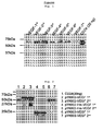
FIG. 1 is a photo illustrating results of VEGF protein expression in a pYK602-VEGF vector, confirmed by Western blot. -
FIG. 2 is a photo illustrating results of VEGF protein expression in pYK602-VEGF, pYK602-His-VEGF, and pYK603-VEGF vectors, compared by Western blot. -
FIG. 3 is a photo illustrating results of VEGF expression in a pYK603-VEGF vector, confirmed by Western blot. -
FIG. 4 is a photo illustrating results of a purified VEGF, confirmed by SDS-PAGE. -
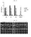
FIG. 5 is a graph illustrating results of phage screening in 1st to 3rd pannings. -
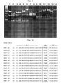
FIG. 6 is a photo illustrating results of diversity of human VEGF monoclonal phage antibodies to VEGF, confirmed by fingerprinting. -
FIG. 7 is a list of sequences illustrating analysis results of a polypeptide used in a heavy chain CDR of human VEGF monoclonal phage antibodies to VEGF. -
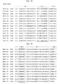
FIG. 8 is a list of sequences illustrating analysis results of a polypeptide used in a light chain CDR of human VEGF monoclonal phage antibodies to VEGF. -
FIG. 9 is a group of graphs illustrating results comparing binding specificities of human VEGF monoclonal antibodies:- a: C5, E9, F6, G12, A4, C11, and F2; and b: H7, G9, C9, B12, F9, D12, and C12.
-
FIG. 10 is a group of photos illustrating results, confirming that neutralizing capacities inhibited the tube formation of human VEGF monoclonal antibodies in HUVEC cells. -
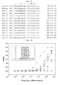
FIG. 11 is a group of graphs illustrating results of the measurement of cross-reactivity of human VEGF monoclonal antibodies with hVEGF and mVEGF. -
FIG. 12 is a group of drawings illustrating cleavage maps of pNATAB H and pNATAB L vectors:- a: pNATAB H vector, b: pNATAB L vector
-
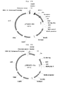
FIG. 13 is a group of drawings illustrating cleavage maps of pYK602, pYK602-His, and pYK603 vectors:- a: pYK602 vector; b: pYK602-His vector; and c: pYK603 vector.
-
FIG. 14 is a group of drawings illustrating results of phage antibody search from a LC shuffling library in 1st to 3rd pannings. -
FIG. 15 is a group of drawings illustrating results of a diversity of an LC shuffling monoclonal phage antibody confirmed through a fingerprinting process. -
FIG. 16 is a group of drawings illustrating analysis results of a polypeptide used in a light chain CDR of an LC shuffling monoclonal phage antibody. - Features and advantages of the present invention will be more clearly understood by the following detailed description of the present preferred embodiments by reference to the accompanying drawings. It is first noted that terms or words used herein should be construed as meanings or concepts corresponding with the technical sprit of the present invention, based on the principle that the inventor can appropriately define the concepts of the terms to best describe his own invention. Also, it should be understood that detailed descriptions of well-known functions and structures related to the present invention will be omitted so as not to unnecessarily obscure the important point of the present invention.
- Hereinafter, the terms of the present invention will be described.
- "Variable region" means a region of an antibody molecule which specifically binds to an antigen and demonstrates modifications in sequence, which is exemplified by CDR1, CDR2, and CDR3. Between the CDRs, there is a framework region (FR) which supports the CDR loop.
- "Complementarity determining region" is a loop-shaped site involved in antigen recognition, and specificity of an antibody against antigen depends on modification in that site.
- "Panning" refers to a process of selecting only a phage expressing a peptide which binds to a target molecule (antibody, enzyme, cell-surface receptor, etc.) on the coat of the phage from a phage library displaying the peptide on the coat.
- Hereinafter, the present invention will be described in detail.
- The present invention provides a VEGF-specific human antibody, including: a heavy chain including a heavy chain variable region (VH) including a heavy chain complementarity determining region (hereinafter, HCDR) 1 having an amino acid sequence selected from the group consisting of SEQ ID Nos. 5 to 17,
HCDR 2 having an amino acid sequence selected from the group consisting of SEQ ID Nos. 18 to 30, andHCDR 3 having an amino acid sequence selected from the group consisting of SEQ ID Nos. 31 to 43, or a fragment thereof; and
a light chain including a light chain variable region (VL) including a light chain complementarity determining region (hereinafter, LCDR) 1 having an amino acid sequence selected from the group consisting of SEQ ID Nos. 57 to 69 and SEQ ID Nos. 130 to 142,LCDR 2 having an amino acid sequence selected from the group consisting of SEQ ID Nos. 70 to 82 and SEQ ID Nos. 143 to 152, andLCDR 3 having an amino acid sequence selected from the group consisting of SEQ ID Nos. 83 to 93 and SEQ ID Nos. 153 to 164, or a fragment thereof. - Preferably, the heavy chain variable region has an amino acid sequence selected from the group consisting of SEQ ID Nos. 44 to 56, and the light chain variable region has an amino acid sequence selected from the group consisting of SEQ ID Nos. 94 to 106 and SEQ ID Nos. 165 to 178.
- The antibody includes not only a whole antibody, but also a functional fragment of the antibody molecule. The whole antibody has a structure with two full-length light chains and two full-length heavy chains, and each light chain is linked to heavy chain by disulfide bond. The functional fragment of an antibody molecule indicates a fragment retaining a antigen-binding function, and examples of the antibody fragment include (i) Fab fragment consisting of light chain variable region (VL) , heavy chain variable region (VH) , light chain constant region (CL) , and
heavy chain 1st constant region (CH1) ; (ii) Fd fragment consisting of VH and CH1 domains; (iii) Fv fragment consisting of VL and VH domains of a monoclonal antibody; (iv) dAb fragment consisting of VH domain (Ward ES et al., Nature 341:544-546 (1989)); (v) separated CDR region; (vi) F(ab')2 fragment including two linked Fab fragments, as a divalent fragment; (vii) single chain Fv molecule (ScFv) in which VH and VL domains are linked by a peptide linker to form an antigen binding site; (viii) bi-specific single chain Fv dimmer (PCT/US92/09965 WO94/13804 - In the present invention, a human antibody against VEGF was obtained as ScFV by using phage display technology and screened as a mono phage clone. As a result, 14 kinds of VEGF-specific monoclonal phages were obtained.
- In a specific example of the present invention, VEGF obtained through recombinant technology (see
FIG. 1 to 4 ) was used in preparation of monoclonal antibodies. The VEGF was reacted with a library phage constructed from human naive scFV library cells, followed by panning and screening of mono clones strongly binding to the VEGF antigen (see Tables 1 & 2 andFIG. 5 ). The selected mono clones were identified by fingerprinting (seeFIG. 6 ), followed by sequencing to identify CDR regions of VH and VL of the antibody (see Table 5 andFIGs. 7 and8 ). The Ig BLAST program of NCBI (//www.ncbi.nlm.nih.gov/igblast/) was used for identification of similarity between the antibody and a germ line antibody group (see Table 6). As a result, 14 kinds of VEGF-specific phage antibodies were obtained. The selected monoclonal antibodies exhibited binding capacities in the order of G12> D12> E9> F6> H7> C5> B12> G9> F9> C11> F2> A4> C9> C12 (seeFIGs. 9a and9b ), and it was observed that they significantly inhibited a capillary-like tube formation of HUVEC cells, induced by VEGF (seeFIG. 10 ). E9, F6, and G12 monoclonal antibodies showing binding capacities similar to that of Avastin™ all exhibited strong affinities for mouse VEGF similar to those for human VEGF, leading to a cross reaction (see Table 8 andFIG. 11 ). Because Avastin™ is not reacted with mouse VEGF at all, it has been difficult to perform animal experimental studies on side effects by Avastin™. However, the VEGF neutralizing human antibody of the present invention is cross reacted with the mouse, indicating that it is highly different from the conventional anti-cancer drug Avastin™ in terms of epitope. Because the VEGF neutralizing human antibody of the present invention exhibits a high cross reactivity with mouse, it may be used in animal experimental studies. - A library phage with a light chain shuffling was prepared for F6 and G12 monoclonal antibodies exhibiting neutralizing capacities similar to those of the Avastin™. The prepared library phage was reacted with VEGF for panning, followed by screening of monoclones strongly binding to VEGF antigen (see Tables 10 & 11 and
FIG. 14 ). The selected monoclones were confirmed by a fingerprinting process (seeFIG. 15 ), followed by sequencing to confirm CDR regions of VH and VL of the antibody. Because CDR regions of VH of the antibody selected by an LC shuffling were identical to each other, CDR regions of VL of the antibody were prepared (see Table 12 andFIG. 16 ). Homology between the antibody and germ line antibody group was investigated (see Table 13). As a result, 15 VEGF specific LC shuffling monoclonal phage antibodies were obtained and their VEGF binding capacities were confirmed (seeFIG. 14 ). A confirmation result of human VEGF binding capacities of the LC shuffling monoclonal phage antibodies showed that the antibodies had high affinities similar to those of F6 and G12, except for 2C11, 2G03, 2C05, and 2F10. - The present invention also provides a polynucleotide encoding a heavy chain of the human antibody or an immunologically active fragment thereof, and an expression vector including the polynucleotide.
- The present invention also provides a polynucleotide encoding a light chain of the human antibody or an immunologically active fragment thereof, and an expression vector including the polynucleotide.
- In a specific embodiment of the present invention, VEGF obtained by recombinant technology was used to screen mono clones strongly binding to VEGF antigens (see Tables 1 and 2 and
FIG. 5 ). The selected monoclones were identified by fingerprinting (seeFIG. 6 ), followed by sequencing to identify CDR regions of VH and VL of the antibody (see Table 5 andFIGs. 7 and8 ). The identification of similarity between the antibody and a germ line antibody group was performed (seeFIG. 6 ). As a result, 14 kinds of VEGF-specific phage antibodies were obtained. Binding capacities to VEGF of the selected monoclonal antibodies were measured (seeFIGs. 9a &9b ), and it was observed that the antibodies significantly inhibited a capillary-like tube formation of HUVEC cells, induced by VEGF (seeFIG. 10 ). E9, F6, and G12 monoclonal antibodies showing binding capacities similar to that of Avastin™ all exhibited strong affinities for mouse VEGF similar to those for human VEGF, leading to a cross reaction (see Table 8 andFIG. 11 ). Because Avastin™ is not reacted with mouse VEGF at all, it has been difficult to perform animal experimental studies on side effects by Avastin™. However, the VEGF neutralizing human antibody of the present invention is cross reacted with the mouse, indicating that it is highly different from the conventional anti-cancer drug Avastin™ in terms of epitope. Because the VEGF neutralizing human antibody of the present invention exhibits a high cross reactivity with mouse VEGF, it may be used in animal experimental studies. - In the polynucleotide encoding light and heavy chains of the human antibody of the present invention or a fragment thereof, due to degeneracy of the codon or in consideration of a preferred codon in an organism where light and heavy chains of the human antibody or a fragment thereof are to be expressed, various modifications may be made in a coding region within a scope that the amino acid sequences of light and heavy chains or a fragment thereof are not changed, and various changes or modifications may be made even in portions other than the coding region within a scope that the gene expression is not affected. It will be appreciated by those skilled in the art that these modified genes are also included within the scope of the present invention. That is, one or more nucleotides may be modified by substitution, deletion, insertion, or a combination thereof as long as the polynucleotide of the present invention encodes a protein with an equivalent activity thereof, and they are also included in the present invention. The sequence of the polynucleotide may be a single or double chain, and a DNA or RNA (mRNA) molecule.
- In preparation of the expression vector, an expression control sequence such as a promoter, a terminator, an enhancer, etc., a sequence for membrane targeting or secretion, etc. may be appropriately selected according to a kind of host cell in which light and heavy chains of the human antibody or a fragment thereof are to be produced, and may be variously combined according to its purpose.
- The expression vector of the present invention includes, but is not limited to, a plasmid vector, a cosmid vector, a bacteriophage, and a viral vector. A suitable expression vector may include expression regulatory elements such as a promoter, an operator, an initiation codon, a stop codon, a polyadenylation signal, and an enhancer and a signal sequence or leader sequence for membrane targeting or secretion, and may be variously prepared according to its purpose. A promoter of the expression vector may be constitutive or inductive. Examples of the signal sequence for use may include, but is not limited to, a PhoA signal sequence and an OmpA signal sequence for genus Escherichia hosts; an α-amylase signal sequence and a subtilicin signal sequence for genus Bacillus hosts; an MFα signal sequence and an SUC2 signal sequence for yeast hosts; and an insulin signal sequence, an α-interferon signal sequence, and an antibody molecule signal sequence for animal cell hosts. In addition, the expression vector may include a selection marker for selecting host cells containing the vector, and a replication origin when it is a replicable expression vector.
- The present invention also provides a transformant prepared by introducing an expression vector including a polynucleotide encoding a heavy chain of the human antibody or an immunologically active fragment thereof into a host cell.
- The present invention also provides a transformant prepared by introducing an expression vector including a polynucleotide encoding a light chain of the human antibody or an immunologically active fragment thereof into a host cell.
- The present invention also provides a transformant prepared by introducing an expression vector including a polynucleotide encoding a heavy chain of the human antibody or a fragment thereof and an expression vector including a polynucleotide encoding a light chain of the human antibody or a fragment thereof simultaneously into a host cell.
- In a specific example of the present invention, genes encoding light and heavy chains of a monoclonal phage were obtained and linked to a vector, respectively, and then a whole human IgG antibody expressed by introducing the expression vectors simultaneously into a host cell was identified. The human antibody was obtained to identify a binding capacity (see
FIGs. 9a &9b ), a neutralizing capacity to VEGF (seeFIG. 10 ), and a cross reactivity with mouse and human VEGF (see Table 8 andFIG. 11 ). - The expression vector according to the present invention may be transformed into a suitable host cell, for example, E. coli or yeast cell, and the transformed host cell may be incubated to produce light and heavy chains of the human antibody of the present invention or a fragment thereof in mass quantities. Incubation methods and media conditions suitable for a kind of host cell may be easily chosen from those known to those skilled in the art. The host cell may be a prokaryote such as E. coli or Bacillus subtilis. In addition, it may be a eukaryotic cell derived from a yeast such as Saccharomyces cerevisiae, an insect cell, a vegetable cell, and an animal cell. More preferably, the animal cell may be an autologous or allogeneic animal cell. A transformant prepared through introduction into an autologous or allogeneic animal cell may be administered to a subject for use in cellular therapy for cancer. As for a method for introducing an expression vector into the host cell, it is possible to use any method if it is known to those skilled in the art.
- The present invention also provides a method for preparing VEGF-specific human antibodies, the method including:
- 1) incubating the transformant; and
- 2) purifying the human antibody from the medium.
- As for the culture medium, it is desirable to select and use a culture medium suitable for the transformant among those known to those skilled in the art. As for the method for purifying human antibodies, it is possible to use any method known to those skilled in the art.
- In a specific example of the present invention, genes encoding light and heavy chains of a monoclonal phage were obtained and linked to a vector, respectively, and then a whole human IgG antibody expressed by introducing the expression vectors simultaneously into a host cell was identified. The human antibody was obtained to identify a binding capacity (see
FIGs. 9a &9b ), a neutralizing capacity to VEGF (seeFIG. 10 ), and a cross reactivity with mouse and human VEGF (see Table 8 andFIG. 11 ). - The present invention also provides a composition including the human antibody.
- The present invention also provides a pharmaceutical composition including the human antibody.
- The disease caused by VEGF-overexpression includes one associated with cancer or angiogenesis. The cancer is preferably one selected from the group consisting of, but not limited to, colorectal cancer, renal cell cancer, lung cancer, breast cancer, and ovarian cancer, and includes all the VEGF-overexpressed cancers. The diseases associated with angiogenesis also include rheumatoid arthritis (RA), diabetic retinopathy, ischemic retinopathy, psoriasis, proliferative diabetic retinopathy (PDR), diabetic macular edema, etc.
- In a specific example of the present invention, it was observed that VEGF monoclonal antibodies exhibiting binding capacities to VEGF (see
FIGs. 9a and9b ) significantly inhibited a capillary-like tube formation of HUVEC cells, induced by VEGF in a human umbilical vein endothelial cell line (seeFIG. 10 ). E9, F6, and G12 monoclonal antibodies showing binding capacities similar to that of Avastin™ all exhibited strong affinities for mouse VEGF similar to those for human VEGF, leading to a cross reaction (see Table 8 andFIG. 11 ). Because Avastin™ is not reacted with mouse VEGF at all, it has been difficult to perform animal experimental studies on side effects by Avastin™. However, the VEGF neutralizing human antibody of the present invention is cross reacted with the mouse, indicating that it is highly different from the conventional anti-cancer drug Avastin™ in terms of epitope. Because the VEGF neutralizing human antibody of the present invention exhibits a high cross reactivity with mouse VEGF, it may be used in animal experimental studies. Thus, the monoclonal antibody of the present invention may be useful as a composition for prevention and treatment of VEGF-overexpressed diseases. - The pharmaceutical composition of the present invention may selectively contain the VEGF-specific human antibody or the transformant, and may additionally contain one or more effective ingredients exhibiting functions identical or similar to those of the ingredient. For administration, the pharmaceutical composition of the present invention may be formulated by including one or more pharmaceutically acceptable carriers in addition to the effective ingredients described above. For example, the pharmaceutically acceptable carrier includes saline solution, sterilized water, Ringer's solution, buffered saline solution, dextrose solution, maltodextrin solution, glycerol, ethanol, liposome, and at least one combination thereof, and other general additives such as antioxidants, buffer solution, bacteriostatic agents, etc. may be added if necessary. In addition, it may be formulated in the form of an injectable formulation such as aqueous solution, suspension, emulsion, etc. by additionally adding diluent, dispersing agent, surfactant, binder and lubricant, and antibodies and other ligands specific to a target cell may be used in combination with the carrier to be specifically reacted with the target cell. Furthermore, the composition may be preferably formulated according to each disease or ingredient using a suitable method in the art or a method which is taught in Remington's Pharmaceutical Science, Mack Publishing Company, Easton PA.
- The pharmaceutical composition of the present invention may be parenterally administered, and the parenteral administration is effected by subcutaneous injection, intravenous injection, intramuscular injection, or intrapleural injection. For parenteral administration, the pharmaceutical composition of the present invention may be mixed with a stabilizer or buffer to prepare a solution or suspension, which may then be provided as ampoules or vials each containing a unit dosage form.
- The pharmaceutical composition of the present invention may be prepared in various forms according to the route of administration. For example, the pharmaceutical composition of the present invention may be formulated to a sterilized aqueous solution or dispersion for injection, or may be prepared in a freeze-dried form through a freeze-drying technique. The freeze-dried pharmaceutical composition may be stored typically at about 4°C and may be reconstituted with a stabilizer that may contain an adjuvant such as saline solution and/or HEPE.
- In a method of the present invention, factors affecting the amount of the pharmaceutical composition to be administered include, but are not limited to, administration mode, administration frequency, specific disease under treatment, severity of disease, history of disease, whether the subject is under treatment in combination with other therapeutics, the subject's age, height, weight, health, and physical conditions. As the patient's weight under treatment increases, the pharmaceutical composition of the present invention may preferably be administered in increasing amounts.
- In addition, the pharmaceutical composition of the present invention may be administered in combination with chemotherapy.
- The present invention also provides a method for treating a disease caused by VEGF-overexpression, the method including administering a pharmaceutically active amount of the human antibody to a subject with the disease caused by VEGF-overexpression.
- The disease caused by VEGF-overexpression includes one associated with cancer or angiogenesis. The cancer is preferably one selected from the group consisting of, but not limited to colorectal cancer, renal cell cancer, lung cancer, breast cancer, and ovarian cancer, and includes all the VEGF-overexpressed cancers. The diseases associated with angiogenesis also include rheumatoid arthritis, diabetic retinopathy, ischemic retinopathy, psoriasis, proliferative diabetic retinopathy (PDR), diabetic macular edema, etc.
- In a specific example of the present invention, it was observed that VEGF monoclonal antibodies showing binding capacities to VEGF (see
FIGs. 9a &9b ) significantly inhibited a capillary-like tube formation of HUVEC cells, induced by VEGF in a human umbilical vein endothelial cell line (seeFIG. 10 ). E9, F6, and G12 monoclonal antibodies showing binding capacities similar to that of Avastin™ all exhibited strong affinities for mouse VEGF similar to those for human VEGF, leading to a cross reaction (see Table 8 andFIG. 11 ). Because Avastin™ is not reacted with mouse VEGF at all, it has been difficult to perform animal experimental studies on side effects by Avastin™. However, the VEGF neutralizing human antibody of the present invention is cross reacted with the mouse, indicating that it is highly different from the conventional anti-cancer drug Avastin™ in terms of epitope. Because the VEGF neutralizing human antibody of the present invention exhibits a high cross reactivity with mouse VEGF, it may be used in animal experimental studies. Thus, the monoclonal antibody of the present invention may be useful as a composition for prevention and treatment of VEGF-overexpressed diseases. - The subject applicable in the present invention is a vertebrate, preferably a mammal, more preferably an experimental animal such as mouse, rabbit, guinea pig, hamster, dog, and cat, and most preferably a primate such as chimpanzee and gorilla.
- The method for administering the human antibody of the present invention may be conducted by parenteral administration (for example, intravenous, subcutaneous, intraperitoneal, or local administration) according to the purpose of use, and preferably by intravenous administration. In administration for solid cancer, local administration may be often preferable for rapid and facilitated access of the antibody. The dose may vary depending on weight, age, sex, and health condition of a patient, diet, administration time, administration method, excretion rate, and severity of disease. The single dose is in the range of 5 to 500 mg/m2, which may be administered daily or weekly. The effective amount may be controlled at the discretion of a doctor treating the patient.
- The human antibody of the present invention may be used alone or in combination with surgery, hormone therapy, chemical therapy, and a biological response controller for treatment of a patient.
- The present invention also provides a composition including the human antibody, light and heavy chains of the human antibody, or an immunologically active fragment thereof, and a radioactive isotope.
- The composition may be used for radioimmuno treatment and detection of a disease caused by VEGF-overexpression. The disease caused by VEGF-overexpression includes one associated with cancer or angiogenesis. The cancer is preferably one selected from the group consisting of, but not limited to, colorectal cancer, renal cell cancer, lung cancer, breast cancer, and ovarian cancer, and includes all the VEGF-overexpressed cancers. The diseases associated with angiogenesis also include rheumatoid arthritis, diabetic retinopathy, ischemic retinopathy, psoriasis, proliferative diabetic retinopathy (PDR), diabetic macular edema, etc.
- In a specific example of the present invention, it was confirmed that the monoclonal VEGF antibody showed strong binding capacity to VEGF (see
FIGs. 9a and9b ). Thus, the monoclonal antibody of the present invention may be useful as a composition for radioimmuno treatment of diseases caused by VEGF-overexpression and including a radioactive isotope. - Examples of preferred radioactive isotopes include 3H, 11C, 14C, 18F, 64Cu, 76Br, 86Y, 99mTc, 111In, 123I, 177Lu, and a mixture or combination thereof. The radioactive isotope is characterized by the fact that it is bound to a human antibody and included in a carrier to which the human antibody is bound.
- The present invention also provides an immunodetection method for detecting an ex vivo VEGF-overexpressed cancer, the method including: contacting a composition including the radioactive isotope with cancer cells.
- The VEGF-overexpressed cancer is preferably one selected from the group consisting of, but not limited to, colorectal cancer, renal cell cancer, lung cancer, breast cancer, and ovarian cancer, and includes all the VEGF-overexpressed cancers.
- In a specific example of the present invention, it was confirmed that the monoclonal VEGF antibody showed strong binding capacity to VEGF (see
FIGs. 9a and9b ). Thus, the monoclonal antibody of the present invention may be useful as a composition for radioimmuno treatment of VEGF-overexpressed diseases and including a radioactive isotope. - The composition including the radioactive isotope may be bound to a solid substrate in order to facilitate the subsequent steps such as washing or separation of complexes. The solid substrate includes, for example, synthetic resin, nitrocellulose, glass substrate, metal substrate, glass fiber, microsphere, microbead, etc. The synthetic resin includes polyester, polyvinyl chloride, polystyrene, polypropylene, PVDF, nylon, etc.
- In addition, cancer cell may be appropriately diluted before it is contacted with a composition including the radioactive isotope.
- The present invention also provides a method for imaging a VEGF-overexpressed cancer, the method including 1) administering a diagnostically effective amount of a compound including the radioactive isotope to a subject; and
2) obtaining a detection image for the subject. - The VEGF-overexpressed cancer is preferably one selected from the group consisting of, but not limited to, colorectal cancer, renal cell cancer, lung cancer, breast cancer, and ovarian cancer, and includes all the VEGF-overexpressed cancers.
- In a specific example of the present invention, it was confirmed that the monoclonal VEGF antibody showed strong binding capacity to VEGF (see
FIGs. 9a and9b ). Thus, the monoclonal antibody of the present invention may be useful in a method for imaging a VEGF-overexpressed cancer. - The detection image is characterized by the fact that it is obtained by near-infrared light imaging, PET, MRI, or ultrasonic imaging.
- The present invention also provides a method for treating an in vivo VEGF-overexpressed cancer, the method including:
- 1) intravenously administering a composition including the radioactive isotope to a subject;
- 2) detecting the composition of Step 1) to identify tumor cells; and
- 3) eliminating the tumor cells identified in Step 2) by surgical operation.
- The VEGF overexpressed cancer is preferably one selected from the group consisting of, but not limited to, colorectal cancer, renal cell cancer, lung cancer, breast cancer, and ovarian cancer, and includes all the VEGF-overexpressed cancers.
- In a specific example of the present invention, it was observed that VEGF monoclonal antibodies showing binding capacities to VEGF (see
FIGs. 9a and9b ) significantly inhibited a capillary-like tube formation of HUVEC cells, induced by VEGF in a human umbilical vein endothelial cell line (seeFIG. 10 ). E9, F6, and G12 monoclonal antibodies showing binding capacities similar to that of Avastin™ all exhibited strong affinities for mouse VEGF similar to those for human VEGF, leading to a cross reaction (see Table 8 andFIG. 11 ). Because Avastin™ is not reacted with mouse VEGF at all, it has been difficult to perform animal experimental studies on side effects by Avastin™. However, the VEGF neutralizing human antibody of the present invention is cross reacted with the mouse, indicating that it is highly different from the conventional anti-cancer drug Avastin™ in terms of epitope. Because the VEGF neutralizing human antibody of the present invention exhibits a high cross reactivity with mouse VEGF, it may be used in animal experimental studies. Thus, the monoclonal antibody of the present invention may be useful as a composition for prevention and treatment of VEGF-overexpressed diseases. Thus, the monoclonal antibodies of the present invention may be useful in prevention and treatment of VEGF-overexpressed cancers. - The present invention also provides a method for prognostic evaluation of a cancer patient, the method including:
- 1) intravenously administering a composition including the radioactive isotope to a patient whose tumor has been eliminated;
- 2) detecting the composition of Step 1) to identify tumor cells; and
- 3) judging that all tumor cells have been eliminated when tumor cells are not detected in step 2).
- In addition, the present invention provides a method for measuring side effects of the human antibody, the method including administering the human antibody to an animal experiment model.
- The animal experiment model may be preferably an animal with a disease caused by VEGF-overexpression.
- Hereinafter, the present invention will be described in more detail with reference to examples.
- However, the following examples are provided for illustrative purposes only, and the scope of the present invention should not be limited thereto in any manner.
- A plasmid (UG-0036-D12) containing human VEGF gene was provided from KUGI (Korean UniGene Information) of the Center for Functional Analysis of Human Genome in Korea Research Institute of Bioscience and Biotechnology. The plasmid was used as a template DNA. In order to express only the domain of the VEGF, a forward primer (SEQ ID No. 1: 5'-GCTCTAGAGTGATGAACTTTCTGCTGTCTT-3') and a reverse primer (SEQ ID No. 2: 5'-CGGAATTCCCGCCTCGGCTTGTCACA-3') were used to amplify the gene under the following conditions. The gene was treated with XbaI and EcoRI, followed by subcloning into a pcDNA3.1/myc-His (-) vector (V80020: Invitrogen, USA) using a ligase. PCR conditions are as follows: when a total reaction reagent was 50 µℓ, 500 ng of the template was introduced and a reaction at 94 °C for 5 minutes, 25 cycles of reactions at 95 °C for 30 seconds, at 55°C for 30 seconds, and at 72 °C for one and a half minutes, and at a reaction at 72°C for 10 minutes were performed to get a PCR product.
- The pcDNA3.1-VEGF was used as a template DNA, and a forward primer (SEQ ID No. 3: 5'-CAGGGGGCCGTGGGGGCCGCAGAAGGAGGAGGGCAG-3') and a reverse primer (SEQ ID No. 4: 5'-TAGCGGCCGACGCGGCCAACCGCCTCGGCTTGTCACA-3') were used to amplify the gene under the following conditions. The gene was treated with SfiI, and a ligase was used to subclone the gene into a pYK602 vector (
FIG. 13a ). PCR conditions are as follows: when a total reaction reagent was 50 µℓ, 500 ng of the template was introduced and a reaction at 95 °C for 5 minutes, 25 cycles of reactions at 95 °C for 30 seconds, at 58°C for 30 seconds, and at 72°C for 1 minute, and at a reaction at 72°C for 10 minutes were performed to get a PCR product. Furthermore, the base sequence of the subcloned pYK602-VEGF vector was identified. - pcDNA3.1-VEGF was used as a template by the same manner as in Example 1-1-1 to subclone the VEGF into a pYK602-His vector (
FIG. 13b ) and a pYK603 vector (FIG. 13c ). In addition, the base sequences of the subcloned pYK602-His-VEGF and pyK603-VEGF vectors were identified. - First, 5 x 10⁶ 293E cells were plated in ten 150 mm dishes. The next day, 10 µg of the subcloned pYK602-VEGF vector was treated with PEI (23966: Polysciences, Inc, USA) for transformation. The subsequent day, the dishes were changed with a growth medium (serum-free DMEM) and then a supernatant was obtained every other day. The obtained supernatant was electrophoresized in a 10% SDS-PAGE gel and identified by Western blot.
- Specifically, two sheets of 10% SDS-PAGE gel loaded with 20 µℓ of a supernatant VEGF of the 1st to 7th subcultures were electrophoresized at 100 V for 2 hours and then transferred at 85 V for 2 hours to NC membrane (HATF00010: Millipore, USA). Subsequently, the membrane was blocked overnight at 4°C in 4% skim milk in TBST. Subsequently, 0.8 mg/mℓ of commercially available anti-human Fc-HRP (product No. 31413: Thermo Sci, USA) was diluted at 1:4000 in 4% skim milk in TBST, followed by reaction at room temperature for 1 hour. The mixture was washed five times in a cycle of once every 10 minutes with TBST, followed by development (12145: Intron, USA) to compare the expression levels of protein.
- As a result, as shown in
FIG. 1 , VEGF was expressed until the 7th subculture, and the expression level of VEGF was not generally high while the expression levels of the 2nd and 3rd subcultures were high. - In addition, Protein A column (17-1279-03: GE Healthcare, USA) was used to purify the supernatant at the rate of 1.5 mℓ/min. Protein was obtained at a concentration of 0.25 mg/mℓ and dialysis of the protein was performed with PBS to confirm that a precipitate was produced.
- Thus, the protein was resubcloned into pYK602-His and pYK603 vectors which produced relatively low amounts of precipitation.
- Transduction was performed on the subcloned pYK602-His-VEGF and pYK603-VEGF vectors by the same manner as in Example 1-2-1, followed by several subcultures to obtain a supernatant.
- The supernatants of the 1st and 2nd subcultures of pYK602-VEGF, pYK602-His-VEGF, and pYK603-VEGF vectors were electrophoresized on a 10% SDS-PAGE gel and identified by Western blot. Precipitate productions were also confirmed.
- As a result, as shown in
FIG. 2 , the pYK602-VEGF and pYK602-His-VEGF vectors have similar expression levels with relatively low precipitate productions, while the PYK603-VEGF vector has a relatively lower expression level than those of the two. - Thus, the pYK602-His-VEGF vector was ultimately used to express and purify VEGF.
- 5 x 10⁶ 293E cells were plated in ten 150 mm dishes and transduction of a pYK602-His-VEGF vector was performed, followed by subculculture to obtain 300 mℓ of supernatants of the 1st to 7th subcultures, respectively. The supernatant was electrophoresized on a 10% SDS-PAGE gel, and then identified by Western blot.
- As a result, as shown in
FIG. 3 , expression levels were highest in supernatants of the 2nd and 3rd subcultures. - Each of supernatants of the 2nd and 3rd subcultures was concentrated into 50 mℓ, exchanged with 300 mℓ of Ni-NTA binding buffer (Qiagen, USA), and then concentrated again into 50 mℓ. Ni-NTA beads (1024473, Qiagen, USA) were added into the concentrate, followed by binding at 4°C for 2 hours to separate and elute the mixture.
- The elution was introduced into a membrane (10K, 132574: SPECTRAPOR, USA). Buffer exchange was performed at 4°C in 4 L of PBS solution for 4 hours or more, followed by dialysis in 4 L of pre-cooled PBS solution overnight to change the buffer solution. After the overnight dialysis, the solution was transferred to an e-tube. The concentration of the protein was measured by the Bradford method and identification was performed on a 10% SDS-PAGE gel (
FIG. 4 ). - 2.7 x 10¹⁰ human naive scFv library cells having diversity were incubated in a medium (3 L) containing 2 ×YTCM [17 g of Tryptone (CONDA, 1612.00), 10 g of yeast extract (CONDA, 1702.00), 5 g of NaCl (Sigma, S7653-5 kg), 34 µg/mℓ of chloramphenicol (Sigma, C0857)], 2% glucose (Sigma, G5400), and 5 mM MgCl2 (Sigma, M2393) at 37°C for 2-3 hours (OD600=0.5~0.7). Then, the cells were infected with helper phage, followed by incubation in a medium containing 2xYTCMK [2x YT CM, 70 µg/mℓ of Kanamycin (Sigma, K1876), 1 mM IPTG (ELPISBIO, IPTG025)] at 30°C for 16 hours. The incubated cells was centrifuged (4500 rpm, 15 min, 4°C) to obtain a supernatant. The supernatant was treated with PEG (Fluka, 81253) and NaCl (Sigma, S7653) until the two reagents became 4% and 3%, respectively. The reactant was centrifuged again (8000 rpm, 20 min, 4°C). The pellet was dissolved in PBS, which proceeded to centrifugation again (12000 rpm, 10 min, 4°C). As a result, the supernatant containing library phage was obtained, which was transferred to a new tube and stored at 4°C .
- An immunosorb tube (Nunc 470319) was coated with 50 µg of VEGF-antigen obtained in Example 1 using 4 mℓ of a coating buffer [1.59 g of Na2CO3 (Sigma, S7795), 2.93 g of NaHCO3 (Sigma, S8875), 0.2 g of NaN3 (Sigma, S2002)] at 4°C for 16 hours with rotator. Then, the antigen was dissolved in PBS at room temperature for 2 hours, followed by blocking in the immunotube using skim milk [(BD,232100)-4% in 1XPBS]. 2 mℓ of library phage constructed in Example 2 was added into the immunotube, followed by reaction at room temperature for 2 hours. The immunotube was washed five times with PBST (0.05%) and twice with PBS. After washing, antigen specific scFV-phage was eluted using 100 mM TEA (Sigma T-0886). E. coli (XL1-blue, stratagene, 200249) was transfected with the eluted phage, followed by amplification. The 2nd and 3rd pannings was performed on the phage amplified at the first panning by the same manner as described above except that washing times with PBST were increased (2nd: 13 times, 3rd: 23 times).
- As a result, as shown in Table 1, it was confirmed that colony titer against the antigen was increased at least 1000 times from the 2nd panning.
[Table 1] Target antigen Panning Initial phage number Binding phage number VEGF 1st 4.2 x 10¹³ 6.0 x 10⁵ 2nd 2.6 x 10¹³ 1.3 x 10⁸ 3rd 1.4 x 10¹³ 1.9 x 10⁸ - Cell stocks obtained from the 1st-3rd pannings and stored as frozen were dissolved in a medium containing 5 mℓ of 2×YTCM, 2% glucose, and 5 mM MgCl2 to make OD600 as 0.1. Then, the cells were incubated at 37°C for 2-3 hours (QD600=0.5~0.7), which were infected with M1 helper phage. Then, the cells were incubated in a medium containing 2 × YTCMK, 5 mM MgCl2 and 1 mM IPTG at 30°C for 16 hours. At this point, a single phage antibody (#39), which was specifically binding to WW45 antigen associated with a developmental process unrelated to VEGF and constructed by a method in Example of the present invention, was used as a control group. The incubated cells were centrifuged (4500 rpm, 15 min, 4°C), and the supernatant was transferred to a new tube (1st ~ 3rd panning poly scFv-phage). A 96-well immuno-plate (NUNC 439454) was coated with VEGF antigen (100 ng/well) using a coating buffer at 4°C for 16 hours, followed by blocking with skim milk dissolved in PBS (4%) . Each well of the 96-well immuno-plate was washed with 0.2 mℓ of PBS-tween20 (0.05%). 100 µℓ of the 1st - 3rd panning poly ScFV-phage was added into each well, followed by reaction at room temperature for 2 hours. Again, each well was washed four times with 0.2 mℓ of PBS-tween20 (0.05%). The secondary antibody anti-M13-HRP (Amersham 27-9421-01) was diluted at 1:2000, followed by reaction at room temperature for 1 hour. The reactant was washed with 0.2 mℓ of PBS-tween20 (0.05%). An OPD tablet (Sigma 8787-TAB) was added into a PC buffer [5.1 g of C6H8O7H2O (Sigma, C0706), 7.3 g of Na2HPO4 (Sigma, S7907)] to make a substrate solution, which was added into each well by 100 µℓ/well, followed by color development for 10 minutes. The optical density was measured at 490 nm by using a spectrophotometer (MolecularDevice, USA).
- As a result, as shown in
Figure 5 , it was confirmed that binding capacities to VEGF antigen were increased from the 2nd polyclonal ScFV-phage pool to reach a saturated state, and values for the tagged His were relatively low. - Colonies obtained from a polyclonal antibody group (the 3rd panning) having strong binding capacity were incubated in a 96-deep well plate (Bioneer, 90030) containing 1 mℓ of a medium supplemented with 2×YTCM, 2% glucose and 5 mM MgCl2 at 37 °C for 16 hours. 100-200 µℓ of the solution was incubated in 1 mℓ of a medium supplemented with 2×YTCM, 5 mM MgCl2, and 1 mM IPTG, which was loaded in a 96-deep well plate at 37 °C for 2-3 hours, followed by inoculation at an initial OD600 value of 0.1. The cells were infected with M1 helper phage (MOI=1:20) and the infected cells were cultured in a medium supplemented with 2×YTCMK, 5 mM MgCl2, and 1 mM IPTG at 30°C for 16 hours. The cultured cells were centrifuged (4500 rpm, 15 min, 4°C) and a supernatant was obtained, to which 4
% PEG 6000 and 3% NaCl were added. Upon completion of dissolving, reaction was induced in ice for 1 hour. The reactant was centrifuged (8000 rpm, 20 min, 4°C) and pellet was dissolved in PBS. Centrifugation (12000 rpm, 10 min, 4°C) was performed again and a supernatant was obtained, from which the 3rd panning monoclonal ScFv phage was obtained. The phage was transferred to a new tube and stored at 4 °C. - A 96-well immuno-plate was coated with VEGF antigen (100 ng/well) at 4°C for 16 hours, followed by blocking with skim milk dissolved in PBS (4%) . Each well of the 96-well immuno-plate was washed with 0.2 mℓ of PBS-tween20 (0.05%). 100 µℓ of the 3rd panning monoclonal scFV-phage was added to each well, followed by reaction at room temperature for 2 hours. Each well was washed four times with 0.2 mℓ of PBS-tween20 (0.05%). The secondary antibody anti-M13-HRP was diluted at 1:2000, followed by reaction at room temperature for 1 hour. The plate was washed with 0.2 mℓ of PBS-tween20 (0.05%), followed by color development. The optical density was measured at 490 nm .
- As a result, 50 monoclonal phages having antigen VEGF binding capacity values of 1.5 or more (highlighted in Table 2) were selected.


- 1 µℓ of the ten monoclonal cells firstly selected, 0.2 µℓ of Taq DNA polymerase (Gendocs, Korea) (5 U/ul), 50 p/µℓ of each forward primer (pelB5, SEQ. ID. No. 125: 5'-CTAGATAACGAGGGCAAATCATG-3') and reverse primer (cla3, SEQ. ID. No. 126: 5'-CGTCACCAATGAAACCATC-3'), 3 µℓ of 10× buffer, 0.6 µℓ of 10 mM dNTP mix, and 24.8 µℓ of distilled water were mixed to perform a colony PCR (iCycler iQ, BIO-RAD). PCR conditions are as shown in Table 3.
[Table 3] Temperature Time Cycle 95° C 5 min 95°C 30 sec 30 56°C 30 sec 72° C 1 min 72° C 10 min 4°C - The colony PCR product was identified on a 1% agarose gel (Seakem LE, CAMERES 50004) . 0.2 µℓ of BstNI (Roche11288075001, 10 U/µℓ) was added to perform a reaction at 37 °C for 2-3 hours. Reaction conditions are as shown in Table 4. The fragmented product was identified on an 8% DNA polyacrylamide gel.
[Table 4] 10X Buffer 3 µℓ colony PCR product 10 µℓ BstNI (10U/µℓ) 0.2 µℓ Distilled water 16.8 µℓ - The colony PCR product was identified on a 1% agarose gel (Seakem LE, CAMERES 50004). 0.2 µℓ of BstNI (Roche11288075001, 10 U/µℓ) was taken to perform a reaction at 37 °C for 2-3 hours. Reaction conditions are as shown in Table 5. The fragmented product was identified on an 8% DNA polyacrylamide gel.
- As a result, as shown in Table 6, fragments of monoclonal phage antibodies digested by BstNI were proved to have diversity.
- 50 kinds of the monoclonal phages were incubated in a medium (5 ml) supplemented with 2×YTCM, 2% glucose, and 5 mM MgCl2 at 37 °C for 16 hours. A DNA purification kit (Nuclogen 5112) was used for the incubated monoclones to obtain a DNA, and then sequencing was performed by using a pelB5 primer of SEQ ID No. 125 (Solgent, Korea).
- As a result, as shown in Table 5 and
FIGs. 7 &8 , CDR regions of VH and VL of the selected antibody were identified. - Similarity between the antibody and germ line antibody group was investigated by Ig BLAST program of NCBI (//www.ncbi.nlm.nih.gov/igblast/). As a result, 14 kinds of VEGF specific phage antibodies were obtained, and the result was summarized and presented in Table 6. Specifically, the heavy chain exhibited 89.9% to 96% homology with human germ cell family sequence, while the light chain exhibited 89.2% to 97% homology. In addition, polypeptides used in CDR3 of heavy and light chains of each human antibody were analyzed, and it was confirmed that their sequences were different.
[Table 5] Heavy Chain Light Chain Gro up Cl one na me CDR1 CDR2 CDR3 CDR1 CDR2 CDR3 1 A4 DYAMH SEQ No. 5 FINEDGGNIYYGDS VKG SEQ ID VKG No.18 EPSGSLTFDY SEQ ID No.31 RASQTISSYLN SEQ ID No.57 AASRLQS SEQ ID No.70 QQSYSTPYT SEQ ID No.83 2 B1 2 SYAIS SEQ ID No.6 GIIPIFGTANYAQK FQG SEQ ID No.19 DRSGYTAMDY SEQ ID No.32 RASQGISSYLA SEQ ID No.58 AASTLQS SEQ ID No.71 QQGHTTPYT SEQ ID No.84 3 C1 1 SDAIS SEQ ID No.7 GVIPIFATTTYAQG FQG SEQ ID No.20 GQMDRGGGLDP SEQ ID No.33 RASQGIGNYLN SEQ ID No.59 AASSLQR SEQ ID No.72 QQSYTTPYS SEQ ID No.85 4 C1 2 SYGMN SEQ ID No.8 SISSSSSSIHYADS VKG SEQ ID No.21 LGPYDAFDF SEQ ID No.34 PGGTSNIDSKY VH SEQ ID No.60 RNDQRPS SEQ ID No.73 QSYDTSLSA PYV SEQ ID No.86 5 C5 SYSMH SEQ ID No.9 GISYDGSSKQFGDS VKG SEQ ID No.22 DGVPGHSYGIG MDV SEQ ID No.35 RASQGISSWLA SEQ ID No.61 AASILQT SEQ ID No.74 QQANSFPYT SEQ ID No.87 6 C9 SYAMH SEQ No.10 VISYDGSNKYYADSVKG SEQ ID No.23 DVDSWSQGWFP SEQ ID No.36 RASQTISTFVN SEQ ID No.62 SASSLQS SEQ ID No.75 QQNYSTPLT SEQ ID No.88 7 D12 EYAMH SEQ 2 ID No. 11 LISGDDYNTFYADS VKG SEQ ID No.24 DAGPAGGGGLD SEQ ID No.37 RTSQTITNFLN SEQ ID No.63 GASSLQS SEQ ID No.76 QQSHGTPYT SEQ ID SEQ ID No.89 8 E9 TSGVAVG SEQ ID No.12 LIYWDNDKRYSPSL KN SEQ ID No.25 GDGWLFDF SEQ ID No.38 TGSNSNIGAGH SEQ ID No.64 GNTNRAS SEQ ID No.77 QSYDNSLSGYV SEQ ID No.90 9 F2 SYAMS SEQ ID No.13 YISSSGHDIYYADP VKG SEQ ID No.26 DKLATPGAFDI SEQ ID No.39 RASQSISNWLA SEQ ID No.65 EASSLES SEQ ID No.78 QQSHGTPYT SEQ ID No.89 10 F6 TSGVA VG SEQ ID No.12 LIYWDNDKRYSPSL KN SEQ ID No.25 GDGWLFDF SEQ ID No.38 TGSNSNIGAGHDVH SEQ ID No.64 GNTNRAS SEQ ID No.77 QSYDNSLSG YV ID SEQ ID No.90 11 F9 TTAIT SEQ ID No.14 WITPFNGNTFYAQK FQD SEQ FQD SEQ ID No.27 SQAAELGTGAF DI SEQ ID No.40 SGSYSNIGTNY VY SEQ ID No.66 KNTQRPS SEQ ID No.79 SAWDDSLSA VL SEQ ID No.91 12 G12 NYAIS SEQ 2 ID No.15 RIIPIYGTPTYAQK FRD SEQ ID No.28 ERSFWNWFAP SEQ ID SEQ ID No.41 TGSSSNIGAGYDVH SEQ ID No.67 GNNNRPS SEQ ID No.80 QSYDSRLGVV SEQ ID No.92 13 G9 TYALH SEQ ID No.16 VISHDGTTDYYRDS VKG SEQ ID No.29 DGSGYFFDY SEQ ID No.42 TGSSSDVGGYNYVS SEQ ID No.68 DVTKRPS SEQ ID No.81 SSYSSSTFYV SEQ ID No.93 14 H7 KYGMH SEQ ID No.17 FIWFDGSNKFYADS VKG SEQ ID No.30 DRDYYGSGPLDY SEQ ID No.43 RASQRIATYLH SEQID No.69 AASSLQS SEQ ID No.82 QQSYSTPYT SEQ ID No.83 [Table 6] clone name VH identities VL identities VH(CDR-a.a seq) Vk(CDR-a.a seq) VEGF anti-myc His Ratio Group A4 VH3-43 93.2(272/292) O12 97.2(276/284) EPSGSLTFDY QQSYSTPYT 1.669 0.904 0.055 1.880 1 B12 VH1-69 99.3(294/296) O12 93.3(264/284) DRSGYTAMDY QQGHTTPYT 1.686 1.288 0.073 1.309 2 C11 VH1-69 90.8(268/295) O12 93.3(265/284) GQMDRGGGLDP QQSYTTPYS 1.719 1.484 0.069 1159 3 C12 VH3-21 95.6(280/293) V1-17 87.4(250/286) LGPYDAFDF QSYDTSLSAPYV 1.688 1.390 0.084 1.214 4 C5 VH3-30 94.3(279/296) L5 97.5(277/284) DGYFGHSYGIGMOV QQANSFPYT 1.927 1.134 0.091 1.700 5 C9 VH3-30 96.6(286/296) 02 93.4(267/286) DVDSWSQGWFPH QQKYSTPLT 1.640 1171 8.874 1.400 6 012 VH3-43 91.8(269/293) O12 94.0(267/284) DAGPAGGGGLDH QQSHGTPYT 1.849 0.833 0.054 2.221 7 D9 VH2.5 94.8(280/297) V1-13 93.2 (272/292) GDGWLFDF QSYDNLSSGYV 2.091 0.771 0.065 2.712 8 E2 VH2-5 94.3(280/297) V1-13 93.2(272/292) GDGWLFDF QSYDNSLSGYV 1.825 0.566 0.068 3.226 8 E7 VH2-5 94.3(280/297) V1-13 93.2(272/292) GDGWLFDF QSYDNSLSGYV 2.000 0.750 0.066 2.668 8 E9 VH2-5 94.3(280/297) V1-13 93.2(272/292) GDGWLFDF QSYDNSLSGYV 2.162 0.705 0.066 3.069 8 F2 VH3-11 89.9(266/296) L12a 94.1(254/270) DKLATPGAFDI QQSHGTPYT 1.517 0.778 0.064 1.949 9 F6 VH2-5 94.3(280/297) V1-13 93.2(272/292) GDGWLFDF QSYDNSLSGYV 2.003 0.637 0.063 3.143 10 F9 VH1-45 90.5(268/296) V1-17 89.2(255/286) SQAAELGTGAFDI SAWDDSLSAVL 2.077 1.525 0.096 1.362 11 G12 VH1-69 94.3(279/296) V1-13 96.5(279/289) ERSFWNWFAP QSYSSRLGW 2.142 1.752 0.104 1.222 12 G9 VH3-30 91.6(271/296) V1-4 94.3(264/280) DGSGYFFDY SSYSSSTFW 1.852 1.091 0.089 1.698 13 H1 VH3-33 94.5(276/292) O12 97.5(278/285) DRDYYGSGPLDY QQSYSTPYT 1.730 1.420 0.185 1.253 14 - In order to measure binding capacities to VEGF of 14 kinds of monoclonal phage antibodies selected in Example 3, each binding capacity was measured by the manner as in Example 3-2.
- As a result, as shown in
FIGs. 9a &9b , binding capacities of 14 kinds of the monoclonal phage antibodies were in the order of G12> D12> E9> F6> H7> C5> B12> G9> F9> C11> F2> A4> C9> C12. - In order to convert monoclonal phage antibodies against VEGF from phage to whole IgG vector, 1 µℓ of heavy chain monoclonal DNA, 10 pmole/µℓ of each heavy chain forward primer and heavy chain reverse primer in Table 7, 5 µℓ of 10 × buffer, 1 µℓ of 10 mM dNTP mix, 0.5 µℓ of pfu DNA polymerase (Solgent, 2.5 U/µℓ), and distilled water (iCycler iQ, BIO-RAD) were mixed to perform a colony PCR (iCycler iQ, BIO-RAD). In addition, colony PCR was performed on light chain by the same manner by using light chain forward primer and reverse primer in Table 7.
[Table 7] Clon e nam e Heavy Chain Light Chain Forward primer(Sfi I) Reverse primer(NheI Forward primer(Sfi I) Reverse primer(Bgl II) A4 NATVH7- 1 SEQID No. 107 
NATJH-ALL SEQ ID No. 114 
NATVK 1-1 SEQ ID No.115 
NATJK-R7 SEQ No. 119 
C5 NATVH3- 1 SEQ ID No. 108 
NATJK-R1 SEQ 120 
C11 NATVH1-2 SEQ ID No.109 
NATJK-R2 SEQ ID No. 121 
C12 NATVH7- 1 SEQ ID No.107 
NATVL10 SEQ ID No. 116 
NATJL1-R SEQ ID No. 122 
E9 NATVH2-1 SEQ ID No.110 
NATVL2 SEQ ID No. 117 
F2 NATVH3-2 SEQ ID No.111 
NATVK 1-1 SEQ ID No. 115 
NATJK-R5 SEQ ID No. 123 
F6 NATVH2-1 SEQ ID No.110 
NATVL2 SEQ ID No. 117 
NATJL1-R SEQ ID No. 122 
G9 NATVH3-2 SEQ ID No.111 
NATVL9 SEQ ID No. 118 
G12 NATVH1-1 SEQ ID No.112 
NATVL2 SEQ ID No. 117 
NATJL2-R SEQ ID No. 124 
H7 NATVH7-2 SEQ ID No.113 
NATVK 1-1 SEQ ID No. 115 
NATJK-R1 ID No. 120 
D12 NATVH7-1 SEQ ID No.107 
NATJK-R5 SEQ ID No. 123 
C9 NATVH7-3 SEQ ID No.128 
NATJH-ALL SEQ ID No.114 
NATVK1-1 SEQ ID No. 115 
NATJK-R3 SEQ ID No. 129 
F9 NATVL10 SEQ ID No. 116 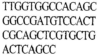
NATJL2-R SEQ ID No. 124 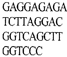
B12 NATVH1-1 SEQ ID No.112 
NATVK11 SEQ ID No. 115 
NATJK-R5 SEQ ID No. 123 
- The heavy chain gene obtained through PCR was purified with DNA-gel extraction kit (Qiagen). 1 µℓ of pNATAB H vector (
FIG. 12a ) (10 ng), 15 µℓ of heavy chain (100-200 ng), 2 µℓ of 10 × buffer, 1 µℓ of Ligase (1 U/µℓ), and distilled water were mixed with the gene and the mixture was left still at room temperature for 1-2 hours for linkage to the vector. The vector was left still in ice for 30 minutes along with a cell for transformation (XL1-blue), followed by heat shock at 42°C for 90 sec for transfection. It was again left still in ice for 5 minutes and 1 mℓ of LB medium was injected, followed by incubation at 37°C for 1 hour. The mixture was smeared in LB Amp liquid medium, followed by incubation at 37°C for 16 hours. Single colony was inoculated into 5 mℓ of LB Amp liquid medium, followed by incubation at 37°C for 16 hours. A DNA-prep kit (Nuclogen) was used for the medium to extract a DNA. - In addition, pNATAB L vector (
FIG. 12b ) was used by the same manner to extract a DNA of the light chain. - Sequencing of the obtained DNA was performed by using a CMV-proF primer (SEQ ID No. 127: AAA TGG GCG GTA GGC GTG) (Solgent).
- As a result, it was confirmed that the sequences of heavy and light chains of the 14 clone phages against VEGF converted into whole IgG were identical to those of the phage antibodies.
- 40 µg of PEI and 10 µg of each antibody heavy chain *DNA and light chain DNA in the whole form were added into 293E cells (Invitrogen) for co-transfection to obtain a supernatant, which was identified by Western blot. Normal human IgG (Jacson Lab) was used as a control group.
- As a result, it was confirmed that it was successfully converted into whole IgG form compared to a control group.
- A monolayer of HUVEC cell (C2517A: Lonza), a human umbilical vein endothelial cell of about 80% confluence, was exchanged with EBM-2 (CC3156, Cambrex) supplemented with 1% FBS and a starvation state was made in a 37°C/5% CO2 incubator for 4 hours. Subsequently, the medium was treated with trypsin to collect and suspend the cells in a starvation medium at 3 ×10⁵/mℓ. 100 µℓ of cell suspension, 150 µℓ of the purified human VEGF Ab solution of the present invention (VEGF Ab is diluted with a starvation medium and treated at a concentration of 4 µg/mℓ), and a normal human IgG as a negative control group were treated at a concentration of 4 µg/mℓ, or Avastin™ (Genetech) as a positive control group was treated at a concentration of 4 µg/mℓ, followed by pre-incubation at 37°C for 1 hour. During the pre-incubation, 200 µℓ of Matrigel (354230: BD Bio-Science, USA) cooled with ice was introduced into a 24-well plate, and then homogenization was performed in a 37°C/5% CO2 incubator for 30 minutes. Recombinant human VEGF was added into a mixture of the cells for which reaction had been completed and Ab at a concentration of 20 ng/mℓ and placed onto the Matrigel, followed by incubation in a 37°C/5% CO2 incubator. In order to observe a tube formation after incubation for 16 hours, 500 µℓ of 3.7% paraformaldehyde was introduced into the incubator, followed by immobilization at room temperature for 30 minutes. Next, the mixture was stained with 500 µℓ of 0.01% crystal violet/100 mM NaBorate and dried to observe a tube formation under a magnification of about 100X.
- As a result, as shown in
FIG. 10 , it was observed that the purified VEGF Ab exhibited an obvious inhibition of tube formation, compared to a positive control group. - It was observed that the three antibodies except for C5 among E9, F6, G12, and C5 all inhibited a capillary-like tube formation of HUVEC cells, induced by VEGF significantly, and thus neutralizing capacities of human antibodies against VEGF of the present invention were identified.
- In addition, ELISA was performed to see if each antibody could have cross-reactivity not only with human VEGF, but also with mouse VEGF. Two 96-well immuno-plates were coated with (50 ng/well) of each recombinant human VEGF (293-VE, R&D systems, USA) and recombinant mouse VEGF (493-MV, R&D systems, USA) using a coating buffer at 4°C for 16 hours, followed by blocking with skim milk dissolved in PBS (4%). Each well of the 96-well immuno-plate was washed with 0.2 mℓ of PBS-tween20 (0.05%). E9, F6, and G12 monoclonal antibodies were diluted gradually from 333 nM by 1/3, and each of 100 µℓ of the diluted monoclonal antibodies was added into each well of the two antigen coated plates with Avastin™ as a control group at room temperature for 2 hours. Each well was washed four times with 0.2 mℓ of PBS-tween20 (0.05%). The secondary antibody anti-human Fc-HRP was diluted at 1:3000, followed by reaction at room temperature for 30 minutes. The reactant was washed with 0.2 mℓ of PBS-tween20 (0.05%). An OPD tablet was added into a PC buffer to make a substrate solution, which was added into each well by 100 µℓ/well, followed by color development for 5 minutes. The optical density was measured at 490 nm by using a spectrophotometer.
- Graphpad prism ver.4 software (CA 92037: Graphpad Software Inc., USA) was used to analyze the ELISA results.
- As a result, E9, F6, and G12 monoclonal antibodies, showing neutralizing capacities similar to that of Avastin™ in
FIG. 10 , all exhibited strong affinities for mouse VEGF similar to those for human VEGF, leading to a cross reaction (Table 8 andFIG. 11 ). The F6 antibody showing the strongest neutralizing capacity consistently exhibited an about 2-fold lower KD value than that of Avastin™. Thus, the probabilities are certainly high that the VEGF neutralizing human antibody of the present invention is different from the conventional anti-cancer agent Avastin™ in terms of epitope.


- Heavy chain portions from scFv-phages of the monoclonal F6 and G12 obtained in Example 3 were treated with SfiI at about 50°C, to which SfiI was added every 2 hours, and this process was repeated three times for gel elution. pYG100, a library plasmid, was also treated with SfiI at about 50°C for about 4 hours, treated with SfiI once again, and restricted overnight for gel elution. Concentrations of heavy chain fragments of the monoclonal F6 and G12 digested and the concentration of vector were measured, followed by ligating with 10 units of ligase at a molar ratio of vector: heavy chain fragments = 1:5 prepared for a total solution at 1 µg/100 µℓ at room temperature for about 2 to about 4 hours. A ligated product was concentrated with ethanol, followed by electroporation of 20 µℓ of the ligated product in distilled water with XL1-BLUE cells at 1x10⁹ cells/100 µℓ for transduction. Incubation of the product was performed at about 37°C overnight, followed by storage in 500 µℓ of 15% glycerol 2XYT at about -70°C. A titer measurement resulted in a diversity of 2.1 x 10⁶.
- A library phage was constructed for human naive VEGF F6 & G12 LC shuffling scFv library cells having a diversity of 2.1 x 10⁶ prepared in Example 5-1 by the same manner as described above in Example 2.
- A phage panning was conducted using 50 µg of the purified VEGF antigen obtained in Example 1 and 2 mℓ of the library phage constructed in Example 5-2 by the same manner as in Example 3, a panning result was confirmed, a monoclonal antibody selected, and a monoclonal phage was classified and examined.
- As a result of the panning, it was confirmed that a high titer was shown from the 1st panning as indicated in Table 10 and colony titer of a phage against the antigen at the 2nd panning was increased at least 5 times.
[Table 10] Target antigen Panning Initial phage number Binding phage number VEGF 1st 2.0 x 10¹³ 2.2 x 10⁵ 2nd 1.2 x 10¹⁴ 4.6 x 10⁹ - As shown in
FIG. 14 , the panning result confirmed by ELISA also showed that a VEGF antigen binding capacity was increased from LC shuffled polyclonal scFv-phage pools, a binding capacity to the 1st panning reached a saturated state, and a value for the tagged His was relatively low. - 61 monoclonal phages with a VEGF antigen binding capacity of at least 2.0 (highlighted in Table 11) were also selected by a monoclonal antibody selection. Then, F6 and G12 in the LC shuffling were used as a positive control group.


- A verification result by a fingerprinting process also confirmed that fragments of monoclonal phage antibodies digested by BstNI had diversity as shown in
FIG. 15 . - Furthermore, a verification result by nucleotide sequencing analysis confirmed CDR regions of VH and VL of the selected antibody as described in Table 12. Homology between the selected antibody and germ line antibody group was investigated. AS a result, 12 VEGF specific phage antibodies with the same HC except for LC different from F6 were obtained, and 3 VEGF specific phage antibodies with the same HC except for LC different from G12 were obtained (Table 13). In particular, about 94.4% homology was shown in 90.1% of light chain. A polypeptide in CDR1, 2, and 3 of light chain of each human antibody was analyzed and it was confirmed that polypeptides with each different sequence were different each other. In
FIG. 16 , because CDR regions of VH of the antibody selected by LC shuffling were identical to each other, CDR regions of VL were compared.[Table 12] Heavy Chain Light Chain Clone CDR1 CDR2 CDR3 CDR1 CDR2 CDR3 F6(P) TSGVAVG: SEQ ID No. 12 LIYWDNDKRYSPS LKN: SEQ ID No. 25 GDGWLFDF: SEQ ID No. 38 TGSNSNIGAGHDV H: SEQ ID No. 64 GNTNRAS: SEQ ID No. 77 QSYDNSLSGYV : SEQ ID No. 90 2A09 TSGVAVG: SEQ ID No. 12 LIYWDNDKRYSPS LKN: SEQ ID No. 25 GDGWLFDF: SEQ ID No. 38 TGSSSNIGAG--YDVH: SEQ ID No. 67 GNSNRPS: SEQ ID No. 143 QSYDNSLS--- AY: SEQ ID No. 153 2B05 TSGVAVG: SEQ ID No. 12 LIYWDNDKRYSPS LKN: SEQ ID No. 25 GDGWLFDF: SEQ ID No. 38 TGGDSNIGAG--YDVN: SEQ ID No. 130 GDTPFRPS: SEQ ID No. 144 QSYDSSLS--- GY: SEQ ID No. 154 2C11 TSGVAVG: SEQ ID No. 12 LIYWDNDKRYSPS LKN: SEQ ID No. 25 GDGWLFDF: GDGWLFDF: SEQ ID No. 38 RSSQSLVRSDGTT YLS: SEQ ID No. 131 KISNRFS: SEQ ID No. 145 MQATFFPFT-- --: SEQ ID No. 155 2D10 TSGVAVG: SEQ ID No. 12 LIYWDNDKRYSPSLKN: SEQ ID No. 25 GDGWLFDF: SEQ ID No. 38 TGSSSNLGAP--NDVH: SEQ ID No. 132 GSTNRPS: SEQ ID No. 146 OSYDNSLS---AY: SEQ ID No.153 2E11 TSGVAVG: SEQ ID No. 12 LIYWDNDKRYSPS SEQ ID No. 25 GDGWLFDF: LKN: SEQ ID No. 38 TGSSSNIGAG--YDVH: SEQ ID No. 67 GNNNRPS: SEQ ID No. 80 QSNDPSLGGL--H: SEQ ID No. 156 2F01 TSGVAVG: SEQ ID No. 12 LIYWDNDKRYSPS LKN: SEQ ID No. 25 GDGWLFDF: SEQ ID No. 38 TGSSSNIGAP--NDVH: SEQ ID No. 133 GNTNRPS: SEQ ID No. 147 QSYDNGLSAS--Y: SEQ ID No. 157 2G03 TSGVAVG: SEQ ID No. 12 LIYWDNDKRYSPS LKN: SEQ ID No. 25 GDGWLFDF: SEQ ID No. 38 RSSQSLVRSDGTT YLS: SEQ ID No. 134 KISNRFS: SEQ ID No. 144 MQATFFPFT----: SEQ ID No. 155 2G04 TSGVAVG:: SEQ ID No. 12 LIYWDNDKRYSPS LKN: SEQ ID No. 25 GDGWLFDF: SEQ ID No. 38 TGSRSNFGFGAG--HDVH: SEQ ID No. 135 ID GNNNRPS: SEQ ID: No. 80 QSFDNTLNG---W: SEQ ID No. 158 2G12 TSGVAVG: SEQ ID No. 12 LIYWDNDKRYSPS LKN: SEQ ID No. 25 GDGWLFDF: SEQ ID No. 38 TGSSSNIGAG-- SEQ ID No. 136 GNNNRPS: SDVH: SEQ ID No. 80 QSYDSSLSG---Y: SEQ ID No. 154 2H07 TSGVAVG: SEQ ID No. 12 LIYWDNDKRYSPS LKN: SEQ ID No. 25 GDGWLFDF: SEQ ID No. 38 TGSSTNIGAG--YDVH: SEQ ID No. 137 GNSNRPS: SEO ID No. 143 QSYDSSLSG-SLY: SEO ID No. 159 2H08 TSGVAVG:: SEQ ID No. 12 LIYWDNDKPYSPS LKN: SEQ ID No. 25 GDGWLFDF: SEQ ID No. 38 SESSSNIGAG--FDVH: SEQ ID No. 138 ID GNTDRPS: SEQ ID No. 148 QSYOSSLR---AY : SEQ ID No. 160 2H09 TSGVAVG: SEQ ID No. 12 LIYWDNDKRYSPS LKN: SEQ ID No. 25 GDGWLFDF: SEQ ID No. 38 RASQGIVS----WLA: SEQ ID No. 139 AASELQS: SEQ ID No. 149 QQLNNFLFA----: SEQ ID No. 161 G12(P NYAIS: SEQ ID No. 15 RIIPIYGTPTYAQ KFRD: SEQ ID No. 28 ERSFWNWFA P: SEQ ID No. 41 TGSSSNIGAGYDV H: SEQ ID No. 67 GNNNRPS: SEQ ID No. 80 QSYDSRLGVV: SEQ ID No. 92 2C05 NYAIS: SEQ ID No. 15 RIIPIYGTPTYAQ KFRD: SEQ ID No. 28 ERSFWNWFA P: SEQ ID No. 41 GSSAGAVTSGHYP F: SEQ ID No. 140 DTSNRHF: SEQ ID No. 150 FVAYG--AIW: SEQ ID No. 162 2F09 NYAIS: No. 15 RIIPIYGTPTYAQ KFRD: SEQ ID No. 28 ERSFWNWFA P: SEQ ID No. 41 TGSSSNLGAGYDV H: SEQ ID No. 141 GDVNRPS: SEQ ID No. 151 QSYDTSLVGS: SEQ ID No. 163 1F10 NYAIS: SEQ ID No.15 RIIPIYGTPTYAQ KFRD: SEQ ID No. 28 ERSFWNWFA P: SEQ ID No. 41 RASQP---ISNWLA: SEQ ID No. 142 ATSILQS: SEQ ID No. 152 QQHRD--YPL: SEQ ID No. 164 
- A binding capacity was measured for an LC shuffling monoclonal antibody selected in Example 5-3 by the same manner as described in Example 4-1. As a result, binding capacities of the 15 LC shuffling monoclonal phage antibodies against the antigen were confirmed as indicated in Table 14.

- In order to convert LC shuffling monoclonal antibodies selected in Example 5-3 into a whole IgG vector in the phage, a colony PCR was performed by the same manner as described in Example 4-2 by using a primer pair in Table 15. As a result, the heavy chain was ligated to a pNATAB H vector (
FIG. 12a ) and the light chain was ligated to a pNATAB L vector (FIG. 12b ). A nucleotide sequencing analysis was performed by extracting DNA from the vector. As a result, it was confirmed that sequences of the heavy and light chains of 15 clone phages for an LC shuffling monoclonal phage antibody converted into a whole IgG were identical to those of phage antibodies.[Table 15] Clones Heavy chain Light chain Forwardprimer (Sfi I) Reverseprimer (Nhe I) Forwardptimer (Sfi I) Reverseprimer (Bgl II) F06(P) NA TV H2- 3: SE Q ID No. 179 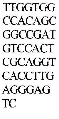
NATJ H-ALL: SEQ ID No. 114 
NATVL 10: SEQ ID No. 116 
NATJL1-R: SEQ ID No. 186 
2A09 NATVL 13: SEQ ID No. 180 
2B05 NATVL 10: SEQ ID No. 116 
2C11 NATV K3: SEQ ID No.181 
NATJK-R2: SEQ ID No. 187 
2D10 NATVL 10: SEQ ID No. 116 
NATJL1-R: SEQ ID No. 188 
2E 11 2F01 2G03 NATV K3 
NATJK-R7: SEQ ID No. 119 
2G04 NATVL 10: SEQ ID No. 116 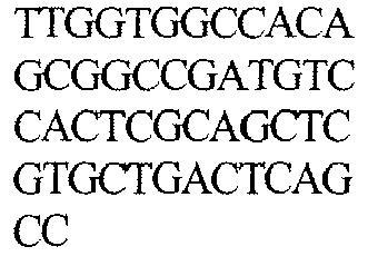
NATJL2-R: SEQ ID No. 124 
2G12 NATJL1-R: SEQ ID No. 189 
2H07 2H08 NATVL 13: SEQ ID No. 182 
2H09 NATV K1-1: SEQ ID No.183 
NATJK-R2: SEQ ID No. 190 
G12(P) NA TV H1- 1: SE Q ID No. 112 
NATJ H-ALL: SEQ ID No. 114 NATVL 10: SEQ ID No. 116 
NATJL2-R: SEQ ID No. 124 
2C05 NATVL 12: SEQ ID No. 184 
NATJL5-R: SEQ ID No. 191 
2F09 NATVL 13: SEQ ID No. 185 
NATJL2-R: SEQ ID No. 124 
1F10 NATV K1-1: SEQ ID No.115 
NATJK-R7: SEQ ID No. 119 
- In order to confirm a VEGF binding capacity by comparing LC shuffling monoclonal phage antibodies with F6 and G12, respectively, ELISA was performed by the same manner as described in Example 4-5.
- As a result, degrees of binding of only 2C11, 2G03, 2C05, and 2F10 were too low while others exhibited high affinities similar to that of a positive control group, as indicated in Table 16.

- Although the preferred embodiments of the present invention have been disclosed for illustrative purposes, those skilled in the art will appreciate that various modifications, additions and substitutions are possible, without departing from the scope and spirit of the invention as disclosed in the accompanying claims.

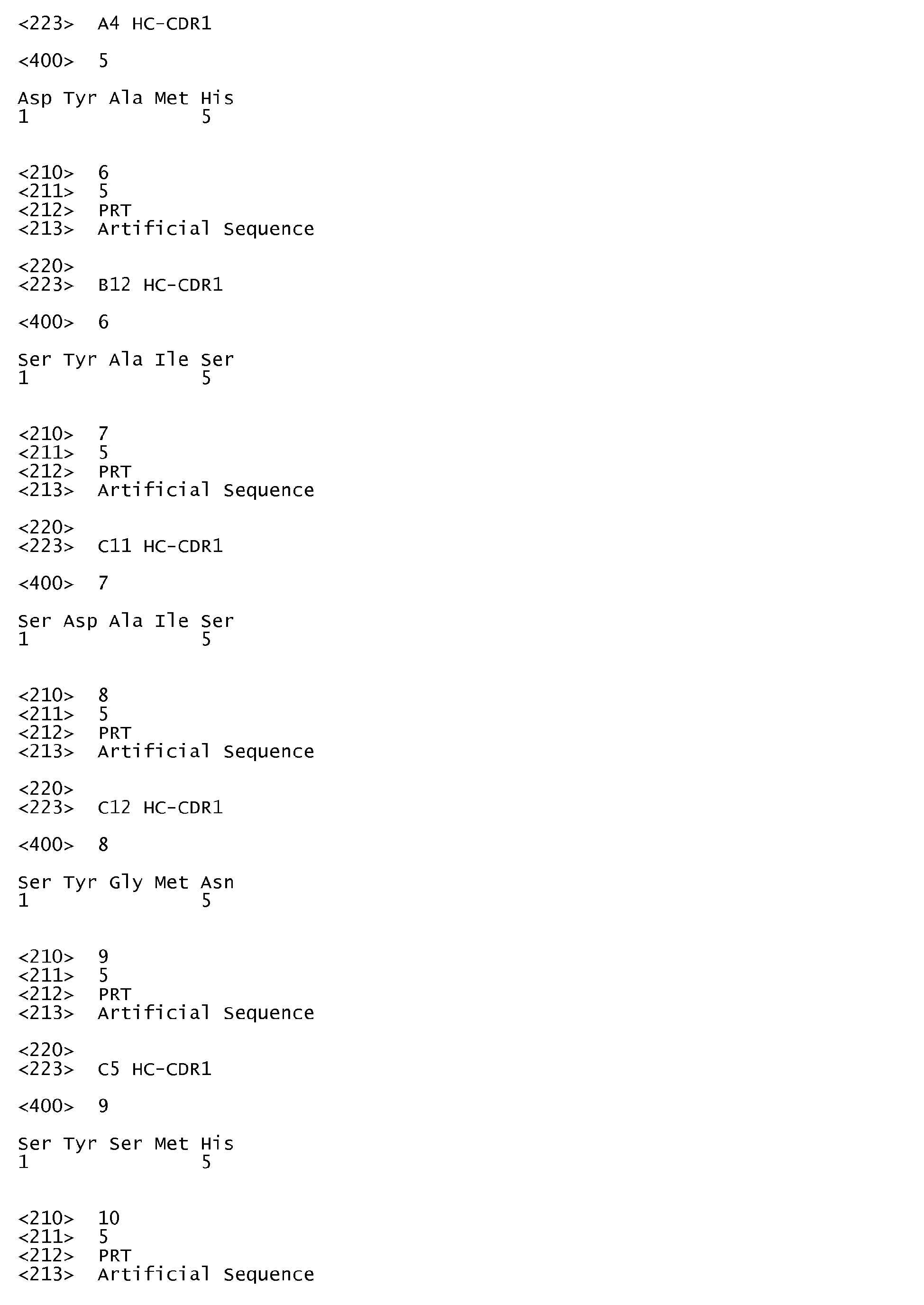


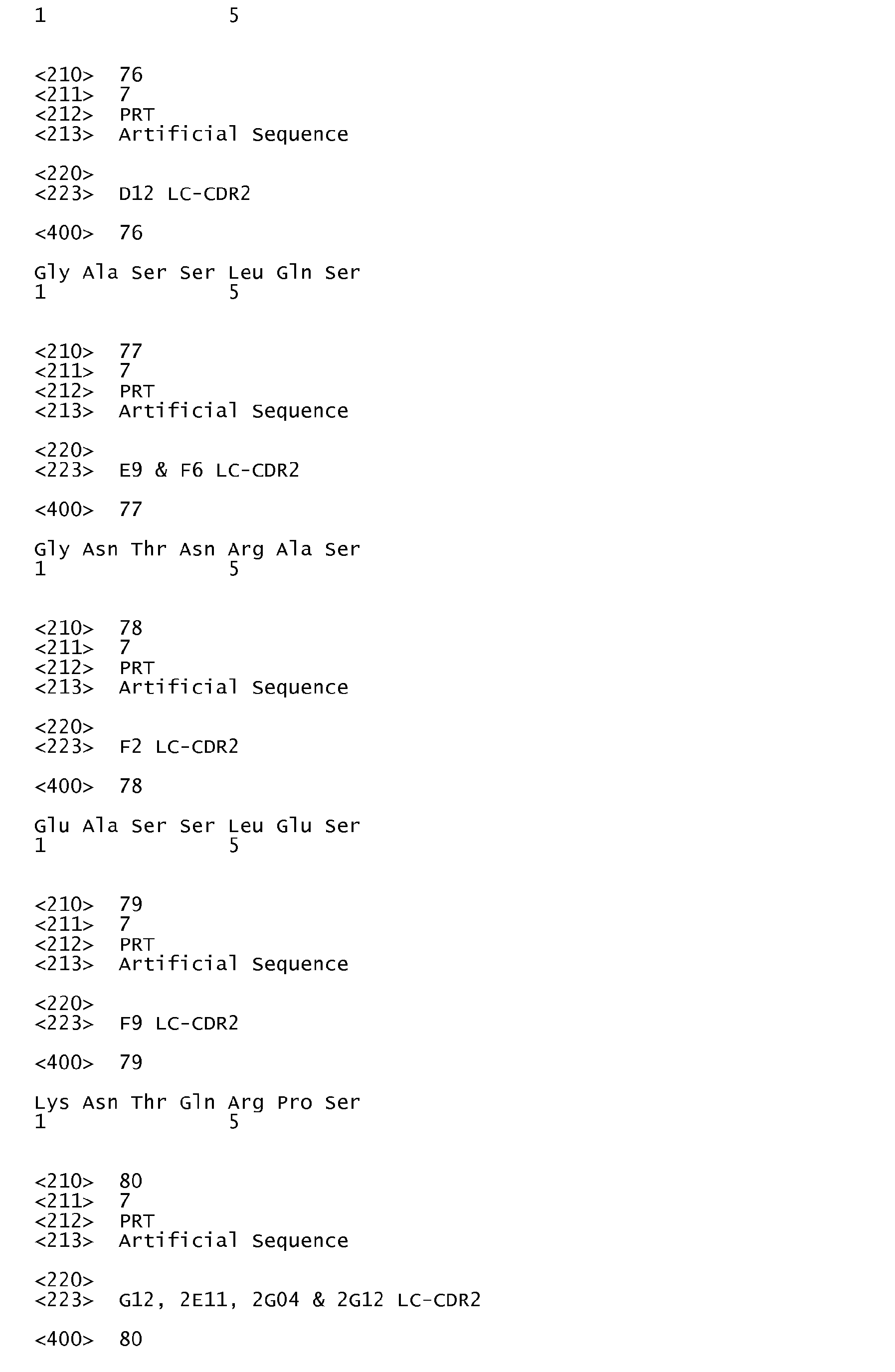


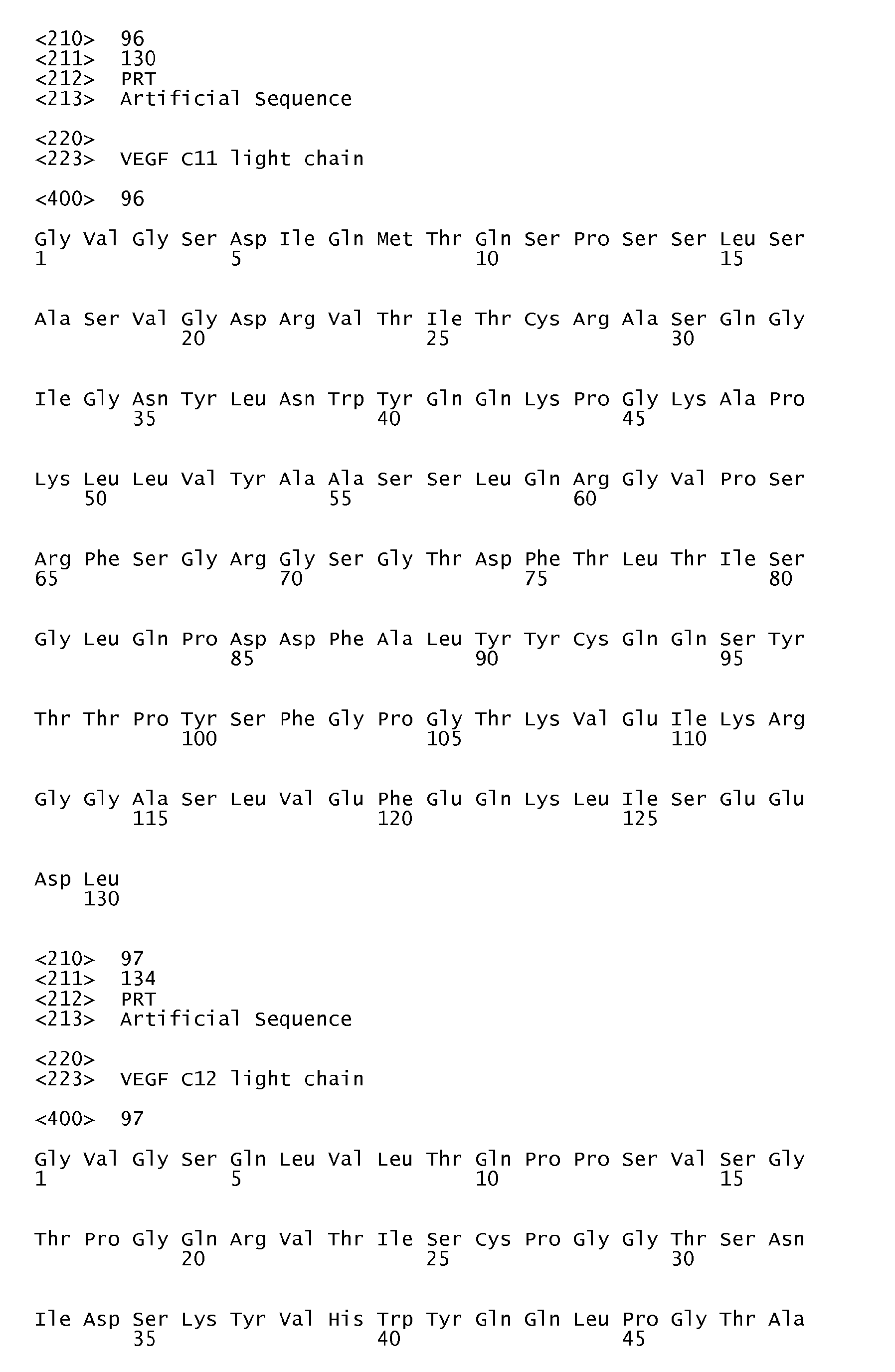
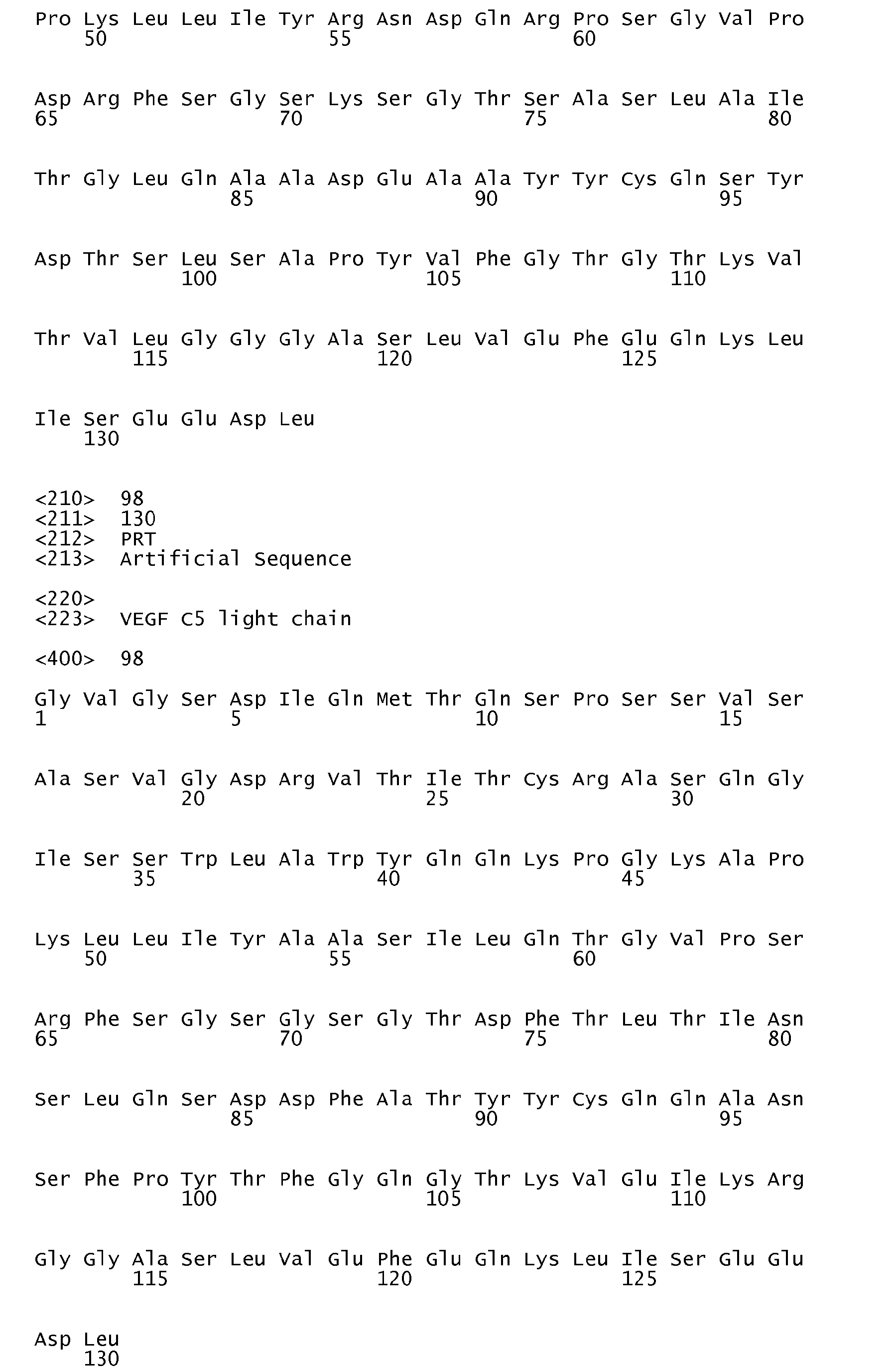


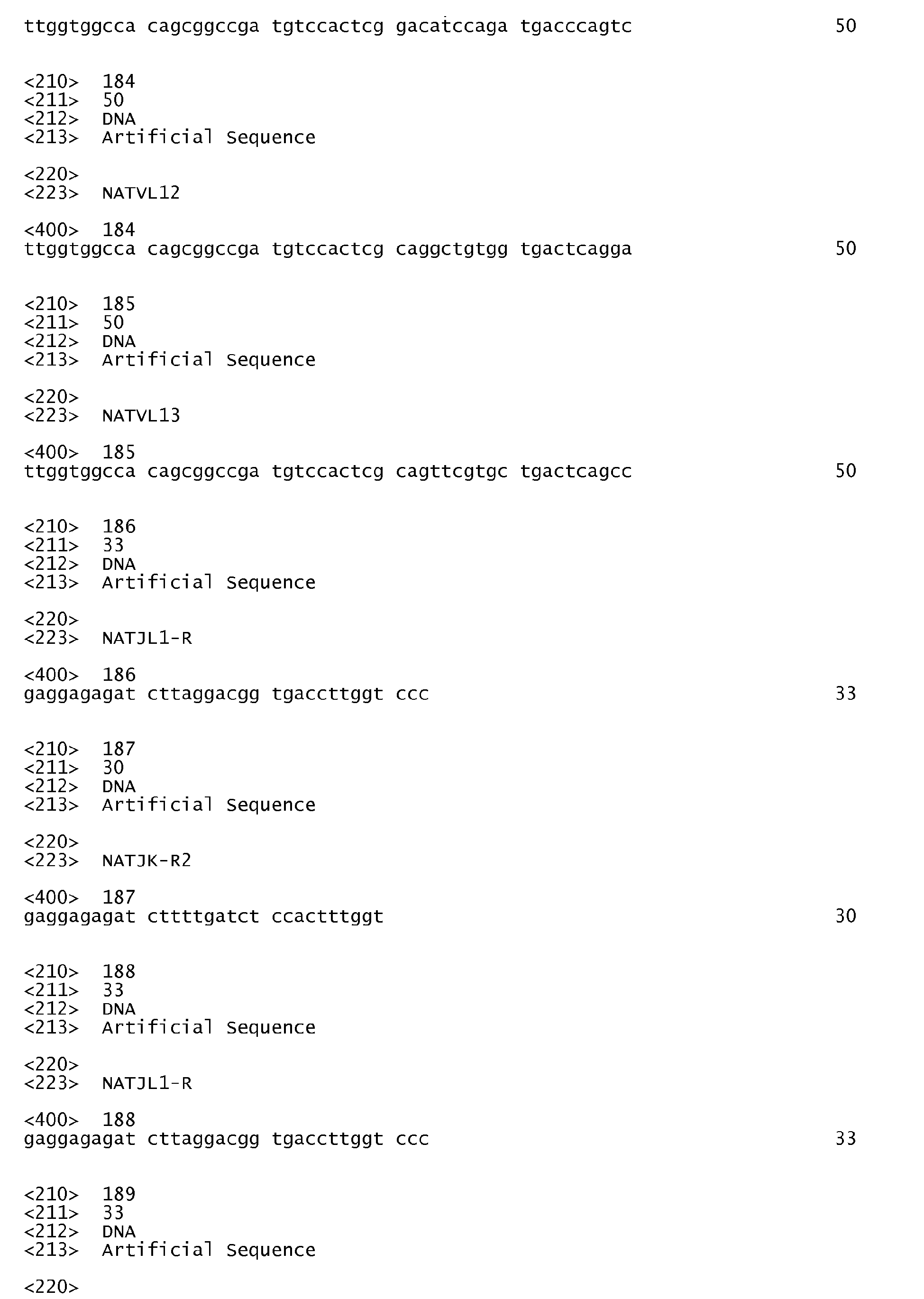

Claims (15)
- A VEGF-specific human antibody comprising a heavy chain comprising a heavy chain variable region (VH), preferably the heavy chain variable region has an amino acid sequence selected from the group consisting of SEQ ID Nos. 44 to 56 comprising a heavy chain complementarity determining region (HCDR) 1 having an amino acid sequence selected from the group consisting of SEQ ID Nos. 5 to 17, HCDR 2 having an amino acid sequence selected from the group consisting of SEQ ID Nos. 18 to 30, and HCDR 3 having an amino acid sequence selected from the group consisting of SEQ ID Nos. 31 to 43, or a fragment thereof; and
a light chain including a light chain variable region (VL), preferably the light chain variable region has an amino acid sequence selected from the group consisting of SEQ ID Nos. 94 to 106 and SEQ ID Nos. 165 to 178 including a light chain complementarity determining region (hereinafter, LCDR) 1 having an amino acid sequence selected from the group consisting of SEQ ID Nos. 57 to 69 and SEQ ID Nos. 130 to 142, LCDR 2 having an amino acid sequence selected from the group consisting of SEQ ID Nos. 70 to 82 and SEQ ID Nos. 143 to 152, and LCDR 3 having an amino acid sequence selected from the group consisting of SEQ ID Nos. 83 to 93 and SEQ ID Nos. 153 to 164, or a fragment thereof. - A polynucleotide encoding a heavy chain of the human antibody of claim 1 or an immunologically active fragment thereof.
- A polynucleotide encoding a light chain of the human antibody of claim 1 or an immunologically active fragment thereof.
- An expression vector comprising the polynucleotide of claim 2.
- An expression vector comprising the polynucleotide of claim 3.
- A transformant prepared by introducing the expression vector of claim 4 and/or the expression vector of claim 5-into a host cell.
- A method for preparing a VEGF-specific human antibody,, the method comprising:1) incubating the transformant of claim 6; and2) purifying the human antibody of claim 1 from the medium.
- A pharmaceutical composition comprising the human antibody of claim 1.
- The pharmaceutical composition as set forth in claim 8, wherein the pharmaceutical composition is used to prevent or treat a disease caused by VEGF-overexpression, preferably the disease caused by VEGF-overexpression is a disease associated with cancer, preferably the cancer is selected from the group consisting of colorectal cancer, renal cell cancer, lung cancer, breast cancer, and ovarian cancer or angiogenesis, preferably the angiogenesis is selected from the group consisting of rheumatoid arthritis, diabetic retinopathy, ischemic retinopathy, psoriasis, proliferative diabetic retinopathy, and diabetic macular edema.
- The pharmaceutical composition as set forth in claim 8, wherein the pharmaceutical composition is administered in combination with chemotherapy.
- A composition comprising the human antibody of claim 1, a light or heavy chain of the human antibody or an immnunologically active fragment thereof, and a radioactive isotope, preferably the radioactive isotope is selected from the group consisting of 3H, 11C, 14C, 18F, 64Cu, 76Br, 86Y, 99mTc, 111In, 123I, 177Lu, and a mixture or combination thereof.
- The composition as set forth in claim 11, wherein the composition is used for radioimmuno treatment or detection of a disease caused by VEGF-overexpression, preferably the disease caused by VEGF-overexpression is a disease associated with cancer, preferably the cancer is selected from the group consisting of colorectal cancer, renal cell cancer, lung cancer, breast cancer, and ovarian cancer or angiogenesis, preferably the angiogenesis is selected from the group consisting of rheumatoid arthritis, diabetic retinopathy, ischemic retinopathy, psoriasis, proliferative diabetic retinopathy, and diabetic macular edema.
- The composition as set forth in claim 11 or 12, wherein the radioactive isotope is bound to a human antibody or included in a carrier to which the human antibody is bound.
- A immunodetection method for detecting an ex vivo VEGF-overexpressed cancer, preferably the VEGF-overexpressed cancer is selected from the group consisting of colorectal cancer, pancreatic cancer, renal cell cancer, lung cancer, breast cancer, and ovarian cancer, the method comprising contacting the composition of claim 11 with a cancer cell.
- A method for imaging an in vivo VEGF-overexpressed cancer, preferably the VEGF-overexpressed cancer is selected from the group consisting of colorectal cancer, pancreatic cancer, renal cell cancer, lung cancer, breast cancer, and ovarian cancer, the method comprising:1) administering a diagnostically effective amount of the composition of claim 11 to a subject; and2) obtaining a detection image for the subject, preferably the detection image is obtained by near-infrared light imaging, PET, MRI, or ultrasonic imaging.
Applications Claiming Priority (1)
| Application Number | Priority Date | Filing Date | Title |
|---|---|---|---|
| KR1020080118124A KR101093717B1 (en) | 2008-11-26 | 2008-11-26 | VEUF-specific human antibodies |
Publications (3)
| Publication Number | Publication Date |
|---|---|
| EP2199306A2 true EP2199306A2 (en) | 2010-06-23 |
| EP2199306A3 EP2199306A3 (en) | 2010-09-08 |
| EP2199306B1 EP2199306B1 (en) | 2013-06-26 |
Family
ID=41651151
Family Applications (1)
| Application Number | Title | Priority Date | Filing Date |
|---|---|---|---|
| EP09173545.6A Not-in-force EP2199306B1 (en) | 2008-11-26 | 2009-10-20 | VEGF-specific human antibody |
Country Status (5)
| Country | Link |
|---|---|
| US (1) | US8236312B2 (en) |
| EP (1) | EP2199306B1 (en) |
| JP (1) | JP5289285B2 (en) |
| KR (1) | KR101093717B1 (en) |
| WO (1) | WO2010061991A1 (en) |
Cited By (4)
| Publication number | Priority date | Publication date | Assignee | Title |
|---|---|---|---|---|
| US11066465B2 (en) | 2015-12-30 | 2021-07-20 | Kodiak Sciences Inc. | Antibodies and conjugates thereof |
| US11155610B2 (en) | 2014-06-28 | 2021-10-26 | Kodiak Sciences Inc. | Dual PDGF/VEGF antagonists |
| US11912784B2 (en) | 2019-10-10 | 2024-02-27 | Kodiak Sciences Inc. | Methods of treating an eye disorder |
| US12071476B2 (en) | 2018-03-02 | 2024-08-27 | Kodiak Sciences Inc. | IL-6 antibodies and fusion constructs and conjugates thereof |
Families Citing this family (12)
| Publication number | Priority date | Publication date | Assignee | Title |
|---|---|---|---|---|
| CA2802376A1 (en) | 2010-06-14 | 2011-12-22 | Vaccinex, Inc. | Anti-vegf antibodies and uses thereof |
| JP2014505698A (en) * | 2011-02-02 | 2014-03-06 | グラクソ グループ リミテッド | Novel antigen binding protein |
| KR101453516B1 (en) * | 2011-09-20 | 2014-10-24 | 가톨릭대학교 산학협력단 | Composition for preventing and treating autoimmune diseases comprising anti-VEGF antibody |
| MY170528A (en) | 2013-02-18 | 2019-08-09 | Vegenics Pty Ltd | Vegfr-3 ligand binding molecules and uses thereof |
| KR101560880B1 (en) | 2013-10-15 | 2015-10-19 | 이화여자대학교 산학협력단 | A method to provide information for prognosis of breast cancer |
| KR102131370B1 (en) * | 2013-10-18 | 2020-07-08 | 삼성전자주식회사 | Polypeptide specifically binding to VEGF-C and use thereof |
| KR102039189B1 (en) * | 2016-03-23 | 2019-11-01 | 서울대학교산학협력단 | Antibodies that bind to outer membrane glycoproteins of severe febrile thrombocytopenia syndrome and uses thereof |
| KR101908330B1 (en) * | 2017-02-17 | 2018-10-16 | 인제대학교 산학협력단 | Novel anti-vascular endothelial growth factor antibody and composition for preventing or treating age-related macular degeneration comprising thereof |
| US11793889B2 (en) * | 2018-09-24 | 2023-10-24 | Kinase Pharma Inc. | Methods for selective kinase inhibition by endogenously produced antagonists of one or more kinases |
| CN110156891B (en) * | 2019-04-03 | 2020-04-28 | 浙江众意生物科技有限公司 | Antibody capable of efficiently and quickly binding VEGF and application thereof |
| CN110133278B (en) * | 2019-04-03 | 2022-05-17 | 浙江众意生物科技有限公司 | An in vitro kit for detecting the expression level of human VEGF protein |
| CN120040592A (en) * | 2020-07-31 | 2025-05-27 | 百奥泰生物制药股份有限公司 | CD47 antibodies and uses thereof |
Citations (1)
| Publication number | Priority date | Publication date | Assignee | Title |
|---|---|---|---|---|
| WO1994013804A1 (en) | 1992-12-04 | 1994-06-23 | Medical Research Council | Multivalent and multispecific binding proteins, their manufacture and use |
Family Cites Families (8)
| Publication number | Priority date | Publication date | Assignee | Title |
|---|---|---|---|---|
| AU2861692A (en) * | 1991-10-18 | 1993-05-21 | Beth Israel Hospital Association, The | Vascular permeability factor targeted compounds |
| US5730977A (en) * | 1995-08-21 | 1998-03-24 | Mitsui Toatsu Chemicals, Inc. | Anti-VEGF human monoclonal antibody |
| US20020032315A1 (en) | 1997-08-06 | 2002-03-14 | Manuel Baca | Anti-vegf antibodies |
| AU1728800A (en) | 1998-11-18 | 2000-06-05 | Genentech Inc. | Antibody variants with higher binding affinity compared to parent antibodies |
| EP1179541B1 (en) | 1999-04-28 | 2004-06-16 | Board Of Regents, The University Of Texas System | Compositions and methods for cancer treatment by selectively inhibiting VEGF |
| EP1472275B1 (en) * | 2002-02-05 | 2008-12-17 | Genentech, Inc. | Protein purification |
| CA2666974A1 (en) * | 2006-10-20 | 2008-11-06 | Schering Corporation | Fully human anti-vegf antibodies and methods of using |
| CU23636A1 (en) * | 2006-11-01 | 2011-03-21 | Ct Ingenieria Genetica Biotech | RECOMBINANT ANTIBODIES AGAINST THE GROWTH FACTOR OF THE VASCULAR ENDOTELIUM (VEGF) |
-
2008
- 2008-11-26 KR KR1020080118124A patent/KR101093717B1/en active Active
- 2008-11-28 WO PCT/KR2008/007045 patent/WO2010061991A1/en not_active Ceased
-
2009
- 2009-10-16 US US12/580,959 patent/US8236312B2/en not_active Expired - Fee Related
- 2009-10-20 EP EP09173545.6A patent/EP2199306B1/en not_active Not-in-force
- 2009-11-26 JP JP2009268214A patent/JP5289285B2/en not_active Expired - Fee Related
Patent Citations (1)
| Publication number | Priority date | Publication date | Assignee | Title |
|---|---|---|---|---|
| WO1994013804A1 (en) | 1992-12-04 | 1994-06-23 | Medical Research Council | Multivalent and multispecific binding proteins, their manufacture and use |
Non-Patent Citations (14)
| Title |
|---|
| "Remington's Pharmaceutical Science", MACK PUBLISHING COMPANY |
| BORGSTROM P ET AL., CANCER RES., vol. 56, pages 4032 - 4039 |
| FAN F ET AL., ONCOGENE, vol. 24, 2005, pages 2647 - 2653 |
| HEIER JS, PROGRAM AND ABSTRACTS OF THE AMERICAN ACADEMY OF OPTHALMOLOGY 2002 ANNUAL MEETING, 20 October 2002 (2002-10-20) |
| IKEDA M ET AL., J PATHOL., vol. 191, 2000, pages 426 - 33 |
| KIM KJ ET AL., NATURE, vol. 362, 1993, pages 841 - 844 |
| LIM HS ET AL., DIABETES CARE, vol. 27, 2004, pages 2918 - 24 |
| SOKER S ET AL., J CELL BIOCHEM., vol. 85, 2002, pages 357 - 368 |
| SOWTER HM ET AL., LAB INVEST., vol. 77, 1997, pages 607 - 614 |
| TOMISAWA M ET AL., EUR J CANCER, vol. 35, 1999, pages 133 - 137 |
| VOLM M ET AL., INT J CANCER, vol. 74, 1997, pages 64 - 68 |
| WARD ES ET AL., NATURE, vol. 341, 1989, pages 544 - 546 |
| WEY JS ET AL., CANCER, vol. 104, 2005, pages 427 - 438 |
| YOSHIJI H ET AL., CANCER RES., vol. 56, 1996, pages 2013 - 2016 |
Cited By (5)
| Publication number | Priority date | Publication date | Assignee | Title |
|---|---|---|---|---|
| US11155610B2 (en) | 2014-06-28 | 2021-10-26 | Kodiak Sciences Inc. | Dual PDGF/VEGF antagonists |
| US11066465B2 (en) | 2015-12-30 | 2021-07-20 | Kodiak Sciences Inc. | Antibodies and conjugates thereof |
| US12071476B2 (en) | 2018-03-02 | 2024-08-27 | Kodiak Sciences Inc. | IL-6 antibodies and fusion constructs and conjugates thereof |
| US11912784B2 (en) | 2019-10-10 | 2024-02-27 | Kodiak Sciences Inc. | Methods of treating an eye disorder |
| US12643958B2 (en) | 2019-10-10 | 2026-06-02 | Kodiak Sciences Inc. | Methods of treating an eye disorder |
Also Published As
| Publication number | Publication date |
|---|---|
| US8236312B2 (en) | 2012-08-07 |
| JP2010131009A (en) | 2010-06-17 |
| JP5289285B2 (en) | 2013-09-11 |
| WO2010061991A1 (en) | 2010-06-03 |
| EP2199306B1 (en) | 2013-06-26 |
| US20100129373A1 (en) | 2010-05-27 |
| EP2199306A3 (en) | 2010-09-08 |
| KR20100059374A (en) | 2010-06-04 |
| KR101093717B1 (en) | 2011-12-19 |
Similar Documents
| Publication | Publication Date | Title |
|---|---|---|
| EP2199306B1 (en) | VEGF-specific human antibody | |
| EP2207803B1 (en) | Cd9-specific human antibodies | |
| US20110076723A1 (en) | Antibody-peptide fused synergibody | |
| EP3000827B1 (en) | Anti-tnf-alpha/cxcl10 double-targeting antibody and use thereof | |
| CA2628552A1 (en) | Identification and characterization of function-blocking anti-ed-b-fibronectin antibodies | |
| AU2012222738B2 (en) | Tumor necrosis factor-a humanized antibody | |
| CN114685666B (en) | Anti-mesothelin nanobodies and their applications | |
| WO2022143550A1 (en) | Mesothelin binding molecule and application thereof | |
| EP2344533B1 (en) | Tmprss4-specific human antibody | |
| US20250326845A1 (en) | Single-domain antibody targeting pd-1, derivative thereof and use thereof | |
| CN114685667B (en) | Mesothelin binding molecules and uses thereof | |
| CN121152799A (en) | Nanobodies against interleukin-18 receptor and their applications | |
| KR101169336B1 (en) | Derivative of CD9-specific human antibody | |
| EP4729543A1 (en) | Anti-il-4 receptor antibodies for veterinary use | |
| CN121930341A (en) | TL1A binding molecules and uses thereof | |
| AU2006201095A1 (en) | Identification and characterisation of function blocking anti-ED-B fibronectin antibodies | |
| HK40064840B (en) | Anti-connective tissue growth factor antibody and application thereof | |
| NZ615477B2 (en) | Tumor necrosis factor-? humanized antibody | |
| HK1175486B (en) | HUMANIZED ANTI-TNFα ANTIBODY | |
| HK1175486A1 (en) | HUMANIZED ANTI-TNFα ANTIBODY |
Legal Events
| Date | Code | Title | Description |
|---|---|---|---|
| PUAI | Public reference made under article 153(3) epc to a published international application that has entered the european phase |
Free format text: ORIGINAL CODE: 0009012 |
|
| 17P | Request for examination filed |
Effective date: 20091020 |
|
| AK | Designated contracting states |
Kind code of ref document: A2 Designated state(s): AT BE BG CH CY CZ DE DK EE ES FI FR GB GR HR HU IE IS IT LI LT LU LV MC MK MT NL NO PL PT RO SE SI SK SM TR |
|
| PUAL | Search report despatched |
Free format text: ORIGINAL CODE: 0009013 |
|
| AK | Designated contracting states |
Kind code of ref document: A3 Designated state(s): AT BE BG CH CY CZ DE DK EE ES FI FR GB GR HR HU IE IS IT LI LT LU LV MC MK MT NL NO PL PT RO SE SI SK SM TR |
|
| 17Q | First examination report despatched |
Effective date: 20110622 |
|
| GRAP | Despatch of communication of intention to grant a patent |
Free format text: ORIGINAL CODE: EPIDOSNIGR1 |
|
| GRAS | Grant fee paid |
Free format text: ORIGINAL CODE: EPIDOSNIGR3 |
|
| GRAA | (expected) grant |
Free format text: ORIGINAL CODE: 0009210 |
|
| AK | Designated contracting states |
Kind code of ref document: B1 Designated state(s): AT BE BG CH CY CZ DE DK EE ES FI FR GB GR HR HU IE IS IT LI LT LU LV MC MK MT NL NO PL PT RO SE SI SK SM TR |
|
| REG | Reference to a national code |
Ref country code: GB Ref legal event code: FG4D |
|
| REG | Reference to a national code |
Ref country code: CH Ref legal event code: EP |
|
| REG | Reference to a national code |
Ref country code: AT Ref legal event code: REF Ref document number: 618669 Country of ref document: AT Kind code of ref document: T Effective date: 20130715 |
|
| REG | Reference to a national code |
Ref country code: IE Ref legal event code: FG4D |
|
| REG | Reference to a national code |
Ref country code: DE Ref legal event code: R096 Ref document number: 602009016653 Country of ref document: DE Effective date: 20130822 |
|
| PG25 | Lapsed in a contracting state [announced via postgrant information from national office to epo] |
Ref country code: SI Free format text: LAPSE BECAUSE OF FAILURE TO SUBMIT A TRANSLATION OF THE DESCRIPTION OR TO PAY THE FEE WITHIN THE PRESCRIBED TIME-LIMIT Effective date: 20130626 Ref country code: NO Free format text: LAPSE BECAUSE OF FAILURE TO SUBMIT A TRANSLATION OF THE DESCRIPTION OR TO PAY THE FEE WITHIN THE PRESCRIBED TIME-LIMIT Effective date: 20130926 Ref country code: FI Free format text: LAPSE BECAUSE OF FAILURE TO SUBMIT A TRANSLATION OF THE DESCRIPTION OR TO PAY THE FEE WITHIN THE PRESCRIBED TIME-LIMIT Effective date: 20130626 Ref country code: GR Free format text: LAPSE BECAUSE OF FAILURE TO SUBMIT A TRANSLATION OF THE DESCRIPTION OR TO PAY THE FEE WITHIN THE PRESCRIBED TIME-LIMIT Effective date: 20130927 Ref country code: SE Free format text: LAPSE BECAUSE OF FAILURE TO SUBMIT A TRANSLATION OF THE DESCRIPTION OR TO PAY THE FEE WITHIN THE PRESCRIBED TIME-LIMIT Effective date: 20130626 Ref country code: LT Free format text: LAPSE BECAUSE OF FAILURE TO SUBMIT A TRANSLATION OF THE DESCRIPTION OR TO PAY THE FEE WITHIN THE PRESCRIBED TIME-LIMIT Effective date: 20130626 |
|
| REG | Reference to a national code |
Ref country code: AT Ref legal event code: MK05 Ref document number: 618669 Country of ref document: AT Kind code of ref document: T Effective date: 20130626 |
|
| REG | Reference to a national code |
Ref country code: LT Ref legal event code: MG4D |
|
| PG25 | Lapsed in a contracting state [announced via postgrant information from national office to epo] |
Ref country code: BG Free format text: LAPSE BECAUSE OF FAILURE TO SUBMIT A TRANSLATION OF THE DESCRIPTION OR TO PAY THE FEE WITHIN THE PRESCRIBED TIME-LIMIT Effective date: 20130926 Ref country code: HR Free format text: LAPSE BECAUSE OF FAILURE TO SUBMIT A TRANSLATION OF THE DESCRIPTION OR TO PAY THE FEE WITHIN THE PRESCRIBED TIME-LIMIT Effective date: 20130626 |
|
| REG | Reference to a national code |
Ref country code: NL Ref legal event code: VDEP Effective date: 20130626 |
|
| PG25 | Lapsed in a contracting state [announced via postgrant information from national office to epo] |
Ref country code: LV Free format text: LAPSE BECAUSE OF FAILURE TO SUBMIT A TRANSLATION OF THE DESCRIPTION OR TO PAY THE FEE WITHIN THE PRESCRIBED TIME-LIMIT Effective date: 20130626 |
|
| PG25 | Lapsed in a contracting state [announced via postgrant information from national office to epo] |
Ref country code: SK Free format text: LAPSE BECAUSE OF FAILURE TO SUBMIT A TRANSLATION OF THE DESCRIPTION OR TO PAY THE FEE WITHIN THE PRESCRIBED TIME-LIMIT Effective date: 20130626 Ref country code: CY Free format text: LAPSE BECAUSE OF FAILURE TO SUBMIT A TRANSLATION OF THE DESCRIPTION OR TO PAY THE FEE WITHIN THE PRESCRIBED TIME-LIMIT Effective date: 20130828 Ref country code: AT Free format text: LAPSE BECAUSE OF FAILURE TO SUBMIT A TRANSLATION OF THE DESCRIPTION OR TO PAY THE FEE WITHIN THE PRESCRIBED TIME-LIMIT Effective date: 20130626 Ref country code: EE Free format text: LAPSE BECAUSE OF FAILURE TO SUBMIT A TRANSLATION OF THE DESCRIPTION OR TO PAY THE FEE WITHIN THE PRESCRIBED TIME-LIMIT Effective date: 20130626 Ref country code: PT Free format text: LAPSE BECAUSE OF FAILURE TO SUBMIT A TRANSLATION OF THE DESCRIPTION OR TO PAY THE FEE WITHIN THE PRESCRIBED TIME-LIMIT Effective date: 20131028 Ref country code: IS Free format text: LAPSE BECAUSE OF FAILURE TO SUBMIT A TRANSLATION OF THE DESCRIPTION OR TO PAY THE FEE WITHIN THE PRESCRIBED TIME-LIMIT Effective date: 20131026 Ref country code: CZ Free format text: LAPSE BECAUSE OF FAILURE TO SUBMIT A TRANSLATION OF THE DESCRIPTION OR TO PAY THE FEE WITHIN THE PRESCRIBED TIME-LIMIT Effective date: 20130626 Ref country code: BE Free format text: LAPSE BECAUSE OF FAILURE TO SUBMIT A TRANSLATION OF THE DESCRIPTION OR TO PAY THE FEE WITHIN THE PRESCRIBED TIME-LIMIT Effective date: 20130626 |
|
| PG25 | Lapsed in a contracting state [announced via postgrant information from national office to epo] |
Ref country code: RO Free format text: LAPSE BECAUSE OF FAILURE TO SUBMIT A TRANSLATION OF THE DESCRIPTION OR TO PAY THE FEE WITHIN THE PRESCRIBED TIME-LIMIT Effective date: 20130626 Ref country code: NL Free format text: LAPSE BECAUSE OF FAILURE TO SUBMIT A TRANSLATION OF THE DESCRIPTION OR TO PAY THE FEE WITHIN THE PRESCRIBED TIME-LIMIT Effective date: 20130626 Ref country code: ES Free format text: LAPSE BECAUSE OF FAILURE TO SUBMIT A TRANSLATION OF THE DESCRIPTION OR TO PAY THE FEE WITHIN THE PRESCRIBED TIME-LIMIT Effective date: 20131007 Ref country code: PL Free format text: LAPSE BECAUSE OF FAILURE TO SUBMIT A TRANSLATION OF THE DESCRIPTION OR TO PAY THE FEE WITHIN THE PRESCRIBED TIME-LIMIT Effective date: 20130626 |
|
| PG25 | Lapsed in a contracting state [announced via postgrant information from national office to epo] |
Ref country code: CY Free format text: LAPSE BECAUSE OF FAILURE TO SUBMIT A TRANSLATION OF THE DESCRIPTION OR TO PAY THE FEE WITHIN THE PRESCRIBED TIME-LIMIT Effective date: 20130626 |
|
| PG25 | Lapsed in a contracting state [announced via postgrant information from national office to epo] |
Ref country code: DK Free format text: LAPSE BECAUSE OF FAILURE TO SUBMIT A TRANSLATION OF THE DESCRIPTION OR TO PAY THE FEE WITHIN THE PRESCRIBED TIME-LIMIT Effective date: 20130626 |
|
| PLBE | No opposition filed within time limit |
Free format text: ORIGINAL CODE: 0009261 |
|
| STAA | Information on the status of an ep patent application or granted ep patent |
Free format text: STATUS: NO OPPOSITION FILED WITHIN TIME LIMIT |
|
| PG25 | Lapsed in a contracting state [announced via postgrant information from national office to epo] |
Ref country code: IT Free format text: LAPSE BECAUSE OF FAILURE TO SUBMIT A TRANSLATION OF THE DESCRIPTION OR TO PAY THE FEE WITHIN THE PRESCRIBED TIME-LIMIT Effective date: 20130626 Ref country code: MC Free format text: LAPSE BECAUSE OF FAILURE TO SUBMIT A TRANSLATION OF THE DESCRIPTION OR TO PAY THE FEE WITHIN THE PRESCRIBED TIME-LIMIT Effective date: 20130626 |
|
| REG | Reference to a national code |
Ref country code: CH Ref legal event code: PL |
|
| 26N | No opposition filed |
Effective date: 20140327 |
|
| REG | Reference to a national code |
Ref country code: DE Ref legal event code: R097 Ref document number: 602009016653 Country of ref document: DE Effective date: 20140327 |
|
| REG | Reference to a national code |
Ref country code: IE Ref legal event code: MM4A |
|
| PG25 | Lapsed in a contracting state [announced via postgrant information from national office to epo] |
Ref country code: LI Free format text: LAPSE BECAUSE OF NON-PAYMENT OF DUE FEES Effective date: 20131031 Ref country code: CH Free format text: LAPSE BECAUSE OF NON-PAYMENT OF DUE FEES Effective date: 20131031 |
|
| PG25 | Lapsed in a contracting state [announced via postgrant information from national office to epo] |
Ref country code: IE Free format text: LAPSE BECAUSE OF NON-PAYMENT OF DUE FEES Effective date: 20131020 |
|
| PG25 | Lapsed in a contracting state [announced via postgrant information from national office to epo] |
Ref country code: SM Free format text: LAPSE BECAUSE OF FAILURE TO SUBMIT A TRANSLATION OF THE DESCRIPTION OR TO PAY THE FEE WITHIN THE PRESCRIBED TIME-LIMIT Effective date: 20130626 |
|
| PG25 | Lapsed in a contracting state [announced via postgrant information from national office to epo] |
Ref country code: TR Free format text: LAPSE BECAUSE OF FAILURE TO SUBMIT A TRANSLATION OF THE DESCRIPTION OR TO PAY THE FEE WITHIN THE PRESCRIBED TIME-LIMIT Effective date: 20130626 |
|
| PG25 | Lapsed in a contracting state [announced via postgrant information from national office to epo] |
Ref country code: HU Free format text: LAPSE BECAUSE OF FAILURE TO SUBMIT A TRANSLATION OF THE DESCRIPTION OR TO PAY THE FEE WITHIN THE PRESCRIBED TIME-LIMIT; INVALID AB INITIO Effective date: 20091020 Ref country code: MK Free format text: LAPSE BECAUSE OF FAILURE TO SUBMIT A TRANSLATION OF THE DESCRIPTION OR TO PAY THE FEE WITHIN THE PRESCRIBED TIME-LIMIT Effective date: 20130626 Ref country code: LU Free format text: LAPSE BECAUSE OF NON-PAYMENT OF DUE FEES Effective date: 20131020 |
|
| PG25 | Lapsed in a contracting state [announced via postgrant information from national office to epo] |
Ref country code: MT Free format text: LAPSE BECAUSE OF FAILURE TO SUBMIT A TRANSLATION OF THE DESCRIPTION OR TO PAY THE FEE WITHIN THE PRESCRIBED TIME-LIMIT Effective date: 20130626 |
|
| REG | Reference to a national code |
Ref country code: FR Ref legal event code: PLFP Year of fee payment: 8 |
|
| REG | Reference to a national code |
Ref country code: FR Ref legal event code: PLFP Year of fee payment: 9 |
|
| REG | Reference to a national code |
Ref country code: FR Ref legal event code: PLFP Year of fee payment: 10 |
|
| PGFP | Annual fee paid to national office [announced via postgrant information from national office to epo] |
Ref country code: FR Payment date: 20190927 Year of fee payment: 11 |
|
| PGFP | Annual fee paid to national office [announced via postgrant information from national office to epo] |
Ref country code: DE Payment date: 20191008 Year of fee payment: 11 |
|
| PGFP | Annual fee paid to national office [announced via postgrant information from national office to epo] |
Ref country code: GB Payment date: 20191018 Year of fee payment: 11 |
|
| REG | Reference to a national code |
Ref country code: DE Ref legal event code: R119 Ref document number: 602009016653 Country of ref document: DE |
|
| GBPC | Gb: european patent ceased through non-payment of renewal fee |
Effective date: 20201020 |
|
| PG25 | Lapsed in a contracting state [announced via postgrant information from national office to epo] |
Ref country code: FR Free format text: LAPSE BECAUSE OF NON-PAYMENT OF DUE FEES Effective date: 20201031 Ref country code: DE Free format text: LAPSE BECAUSE OF NON-PAYMENT OF DUE FEES Effective date: 20210501 |
|
| PG25 | Lapsed in a contracting state [announced via postgrant information from national office to epo] |
Ref country code: GB Free format text: LAPSE BECAUSE OF NON-PAYMENT OF DUE FEES Effective date: 20201020 |