EP2197783B1 - Complex of cell translocational peptide and magnetic nanoparticles and use thereof - Google Patents
Complex of cell translocational peptide and magnetic nanoparticles and use thereof Download PDFInfo
- Publication number
- EP2197783B1 EP2197783B1 EP08835082.2A EP08835082A EP2197783B1 EP 2197783 B1 EP2197783 B1 EP 2197783B1 EP 08835082 A EP08835082 A EP 08835082A EP 2197783 B1 EP2197783 B1 EP 2197783B1
- Authority
- EP
- European Patent Office
- Prior art keywords
- cell
- magnetic nanoparticle
- fluorescence
- penetrating peptide
- complex
- Prior art date
- Legal status (The legal status is an assumption and is not a legal conclusion. Google has not performed a legal analysis and makes no representation as to the accuracy of the status listed.)
- Active
Links
Images




Classifications
-
- A—HUMAN NECESSITIES
- A61—MEDICAL OR VETERINARY SCIENCE; HYGIENE
- A61K—PREPARATIONS FOR MEDICAL, DENTAL OR TOILETRY PURPOSES
- A61K49/00—Preparations for testing in vivo
- A61K49/06—Nuclear magnetic resonance [NMR] contrast preparations; Magnetic resonance imaging [MRI] contrast preparations
- A61K49/18—Nuclear magnetic resonance [NMR] contrast preparations; Magnetic resonance imaging [MRI] contrast preparations characterised by a special physical form, e.g. emulsions, microcapsules, liposomes
- A61K49/1818—Nuclear magnetic resonance [NMR] contrast preparations; Magnetic resonance imaging [MRI] contrast preparations characterised by a special physical form, e.g. emulsions, microcapsules, liposomes particles, e.g. uncoated or non-functionalised microparticles or nanoparticles
- A61K49/1821—Nuclear magnetic resonance [NMR] contrast preparations; Magnetic resonance imaging [MRI] contrast preparations characterised by a special physical form, e.g. emulsions, microcapsules, liposomes particles, e.g. uncoated or non-functionalised microparticles or nanoparticles coated or functionalised microparticles or nanoparticles
- A61K49/1824—Nuclear magnetic resonance [NMR] contrast preparations; Magnetic resonance imaging [MRI] contrast preparations characterised by a special physical form, e.g. emulsions, microcapsules, liposomes particles, e.g. uncoated or non-functionalised microparticles or nanoparticles coated or functionalised microparticles or nanoparticles coated or functionalised nanoparticles
- A61K49/1827—Nuclear magnetic resonance [NMR] contrast preparations; Magnetic resonance imaging [MRI] contrast preparations characterised by a special physical form, e.g. emulsions, microcapsules, liposomes particles, e.g. uncoated or non-functionalised microparticles or nanoparticles coated or functionalised microparticles or nanoparticles coated or functionalised nanoparticles having a (super)(para)magnetic core, being a solid MRI-active material, e.g. magnetite, or composed of a plurality of MRI-active, organic agents, e.g. Gd-chelates, or nuclei, e.g. Eu3+, encapsulated or entrapped in the core of the coated or functionalised nanoparticle
- A61K49/1866—Nuclear magnetic resonance [NMR] contrast preparations; Magnetic resonance imaging [MRI] contrast preparations characterised by a special physical form, e.g. emulsions, microcapsules, liposomes particles, e.g. uncoated or non-functionalised microparticles or nanoparticles coated or functionalised microparticles or nanoparticles coated or functionalised nanoparticles having a (super)(para)magnetic core, being a solid MRI-active material, e.g. magnetite, or composed of a plurality of MRI-active, organic agents, e.g. Gd-chelates, or nuclei, e.g. Eu3+, encapsulated or entrapped in the core of the coated or functionalised nanoparticle the nanoparticle having a (super)(para)magnetic core coated or functionalised with a peptide, e.g. protein, polyamino acid
-
- B—PERFORMING OPERATIONS; TRANSPORTING
- B82—NANOTECHNOLOGY
- B82B—NANOSTRUCTURES FORMED BY MANIPULATION OF INDIVIDUAL ATOMS, MOLECULES, OR LIMITED COLLECTIONS OF ATOMS OR MOLECULES AS DISCRETE UNITS; MANUFACTURE OR TREATMENT THEREOF
- B82B1/00—Nanostructures formed by manipulation of individual atoms or molecules, or limited collections of atoms or molecules as discrete units
-
- A—HUMAN NECESSITIES
- A61—MEDICAL OR VETERINARY SCIENCE; HYGIENE
- A61K—PREPARATIONS FOR MEDICAL, DENTAL OR TOILETRY PURPOSES
- A61K49/00—Preparations for testing in vivo
- A61K49/001—Preparation for luminescence or biological staining
- A61K49/0013—Luminescence
- A61K49/0017—Fluorescence in vivo
- A61K49/0019—Fluorescence in vivo characterised by the fluorescent group, e.g. oligomeric, polymeric or dendritic molecules
- A61K49/0021—Fluorescence in vivo characterised by the fluorescent group, e.g. oligomeric, polymeric or dendritic molecules the fluorescent group being a small organic molecule
- A61K49/0041—Xanthene dyes, used in vivo, e.g. administered to a mice, e.g. rhodamines, rose Bengal
-
- A—HUMAN NECESSITIES
- A61—MEDICAL OR VETERINARY SCIENCE; HYGIENE
- A61K—PREPARATIONS FOR MEDICAL, DENTAL OR TOILETRY PURPOSES
- A61K49/00—Preparations for testing in vivo
- A61K49/001—Preparation for luminescence or biological staining
- A61K49/0013—Luminescence
- A61K49/0017—Fluorescence in vivo
- A61K49/0019—Fluorescence in vivo characterised by the fluorescent group, e.g. oligomeric, polymeric or dendritic molecules
- A61K49/0021—Fluorescence in vivo characterised by the fluorescent group, e.g. oligomeric, polymeric or dendritic molecules the fluorescent group being a small organic molecule
- A61K49/0041—Xanthene dyes, used in vivo, e.g. administered to a mice, e.g. rhodamines, rose Bengal
- A61K49/0043—Fluorescein, used in vivo
-
- A—HUMAN NECESSITIES
- A61—MEDICAL OR VETERINARY SCIENCE; HYGIENE
- A61K—PREPARATIONS FOR MEDICAL, DENTAL OR TOILETRY PURPOSES
- A61K49/00—Preparations for testing in vivo
- A61K49/001—Preparation for luminescence or biological staining
- A61K49/0063—Preparation for luminescence or biological staining characterised by a special physical or galenical form, e.g. emulsions, microspheres
- A61K49/0069—Preparation for luminescence or biological staining characterised by a special physical or galenical form, e.g. emulsions, microspheres the agent being in a particular physical galenical form
- A61K49/0089—Particulate, powder, adsorbate, bead, sphere
- A61K49/0091—Microparticle, microcapsule, microbubble, microsphere, microbead, i.e. having a size or diameter higher or equal to 1 micrometer
- A61K49/0093—Nanoparticle, nanocapsule, nanobubble, nanosphere, nanobead, i.e. having a size or diameter smaller than 1 micrometer, e.g. polymeric nanoparticle
-
- A—HUMAN NECESSITIES
- A61—MEDICAL OR VETERINARY SCIENCE; HYGIENE
- A61K—PREPARATIONS FOR MEDICAL, DENTAL OR TOILETRY PURPOSES
- A61K49/00—Preparations for testing in vivo
- A61K49/06—Nuclear magnetic resonance [NMR] contrast preparations; Magnetic resonance imaging [MRI] contrast preparations
- A61K49/08—Nuclear magnetic resonance [NMR] contrast preparations; Magnetic resonance imaging [MRI] contrast preparations characterised by the carrier
- A61K49/085—Nuclear magnetic resonance [NMR] contrast preparations; Magnetic resonance imaging [MRI] contrast preparations characterised by the carrier conjugated systems
-
- A—HUMAN NECESSITIES
- A61—MEDICAL OR VETERINARY SCIENCE; HYGIENE
- A61K—PREPARATIONS FOR MEDICAL, DENTAL OR TOILETRY PURPOSES
- A61K49/00—Preparations for testing in vivo
- A61K49/06—Nuclear magnetic resonance [NMR] contrast preparations; Magnetic resonance imaging [MRI] contrast preparations
- A61K49/08—Nuclear magnetic resonance [NMR] contrast preparations; Magnetic resonance imaging [MRI] contrast preparations characterised by the carrier
- A61K49/10—Organic compounds
- A61K49/14—Peptides, e.g. proteins
-
- B—PERFORMING OPERATIONS; TRANSPORTING
- B82—NANOTECHNOLOGY
- B82Y—SPECIFIC USES OR APPLICATIONS OF NANOSTRUCTURES; MEASUREMENT OR ANALYSIS OF NANOSTRUCTURES; MANUFACTURE OR TREATMENT OF NANOSTRUCTURES
- B82Y5/00—Nanobiotechnology or nanomedicine, e.g. protein engineering or drug delivery
Definitions
- the present invention relates to a cell-penetrating peptide/fluorescence-labeled magnetic nanoparticle complex and the use thereof, and more particularly to cell-penetrating peptide/fluorescence-labeled magnetic nanoparticle complex in which the cell-penetrating peptide is chemically linked to the fluorescence-labeled magnetic nanoparticle, such that fluorescence-labeled magnetic nanoparticle can be stably introduced directly into cells without endocytosis, and a composition for cell imaging containing the complex.
- Molecular imaging is a rapidly developing discipline that visualizes various changes occurring at the molecular level in cells, that is, genetic expressions, biochemical events and biological changes.
- An existing medical imaging technique comprises producing image signals using the difference in nonspecific physical and chemical properties and clinically analyzing the image signals, whereas the molecular imaging technique is characterized by more specific images, because it uses image signals produced by action at the molecular and genetic levels.
- the use of this molecular imaging technique enables genetic changes, protein changes, metabolic changes, intracellular biological changes and the like to be imaged.
- MRI magnetic resonance imaging
- MRI magnetic resonance imaging
- MRI is basically to measure signals from protons.
- MRI aimed at noninvasive diagnosis at first, but since an MRI contrast agent was first marketed in 1988, it has been found that the use of MRI contrast agents can improve diagnostic sensitivity and specificity.
- Recently, the application of MRI contrast agents has been gradually enlarged to MRI angiography, perfusion imaging and the like.
- Such MRI contrast agents are classified according to their effects on a magnetic field into paramagnetic and superparamagnetic materials which are called “positive contrast agents” and “negative contrast agents”, respectively.
- the former has a predominant T1 shortening effect, and thus is expressed as a light signal in T1-weighted images
- the latter has a predominant T2 shortening effect, and thus is expressed as a dark signal in T2-weighted images.
- the superparamagnetic materials iron oxides are used, and they may be classified according to particle size into two categories: superparamagnetic iron oxide (SPIO) having a particle size of more than 50 nm, and ultrasmall superparamagnetic iron oxide (USPIO) having a particle size of less than 50 nm.
- SPIO superparamagnetic iron oxide
- USPIO ultrasmall superparamagnetic iron oxide
- Iron oxide contrast agents which are currently being mainly studied are SPIO-based colloids which are either nanosized magnetite or magnetite coated with polymeric carbohydrate such as dextran or starch.
- Magnetic nanoparticles are nanoparticles in a colloidal iron oxide solution and are used as contrast agents because of the large relaxation signal of the nanoparticles.
- the size of nanoparticles in the iron oxide solution has various distributions depending on the size and coating thickness of the core thereof and the pattern of aggregation thereof ( Gro, C. et al., Cells Mater., 3:163, 2002 ).
- Gd contrast agents show an excellent contrast effect at a concentration of a few millimoles, whereas SPIO shows an excellent contrast effect even at a concentration of a few nanomoles, and thus can be studied and applied as MRI contrast agents ( Bulte, J.W. et al., Res. Med., 25:148, 1992 ; Knauth, M. et al., Am. J. Neuroradiol., 22:99, 2001 ; Lacava, L.M. et al., Biophys., 252:367, 2002 ).
- Pure magnetic particles are not used because of the following limitations: (1) they have a high aggregation propensity; (2) if they are not sufficiently stable, the inherent structure thereof can change, leading to a change in the magnetic properties thereof; (3) when they come into contact with a biological environment, they can be rapidly biodegraded; and (4) they are toxic by themselves.
- polymer-coated SPIO and USPIO have been developed ( Lind, K. et al., J. Drug Target., 10:221, 2002 ; Bellin, M.F. et al., J. Radiol., 34:257, 2000 ; Bonnemain, B. et al., Review. J. Drug Target., 6:167, 1998 ; Muller, R.N. et al., Invest Radiol., 25:34, 1990 ).
- Pseudomonas exotoxin is also used as a kind of transporter ( Prior, T.I. et al., Cell, 64:1017, 1991 ).
- transporter Prior, T.I. et al., Cell, 64:1017, 1991 .
- the effects of these methods on the intracellular migration of physiologically activated substances and their general applicability thereof have not yet been clearly verified. Accordingly, there has been a continuous demand for a method capable of delivering a biologically active substance into the cytoplasm or nucleus of living cells in a safer and more effective way.
- TAT protein which is a transcription factor of human immunodeficiency virus-1, HIV-1
- Tat protein was more effective in passing through the cell membrane, when it consisted of amino acids 47 to 57 (YGRKKRRQRRR), on which positively charged amino acids were concentrated, than when it was in the form of a full-length 86 amino acid protein ( Fawell, S. et al., Proc. Natl. Acad. Sci. USA, 91:664, 1994 ).
- CPPs cell penetrating peptides
- HSV-1 herpes simplex virus type 1
- a peptide having an amino acid sequence consisting of amino acids 84 to 92 of the UL-56 protein of HSV-2 (GenBank code:D1047 [gi:221784])
- a peptide having an amino acid sequence consisting of amino acids 339 to 355 of the antennapedia (ANTP) protein of Drosophila sp. Schwarze, S.R. et al., Trends. Pharmacol. Sci., 21:45, 2000
- artificial peptides consisting of positively charged amino acids were also found to be effective ( Laus R. et al., Nature Biotechnol. 18:1269-1272 (2000 )).
- LMWP low molecular weight protamine
- US 2006/251726 A1 discloses a nanoparticle-polypeptide complex comprising a bioactive polypeptide in association with a nanoparticle, wherein the bioactive polypeptide is modified by the addition of a chemical moiety that facilitates cellular uptake of the protein.
- MARCELO J KOGAN ET AL. "Peptides and metallic nanoparticles for biomedical applications", NANOMEDICINE, vol. 2, no. 3, 1 June 2007, pages 287-306, XP55104016, ISSN: 1743-5889, DOI: 10.2217/17435889.2.3.287 , describes the contribution of peptides to the biomedical applications of metallic nanoparticles.
- JOSEPHSON L ET AL. "High-efficiency intracellular magnetic labeling with novel superparamagnetic-Tat peptide conjugates", BIOCONJUGATE CHEMISTRY, ACS, WASHINGTON, DC, US, vol. 10, no. 2, 1 February 1999, pages 186-191, XP002172120, ISSN: 1043-1802, DOI: 10.1021/BC980125H , discloses the derivatization of a biocompatible, dextran coated superparamagnetic iron oxide particle with a peptide sequence from the HIV-tat protein to improve intracellular magnetic labeling of different target cells.
- the conjugate had a mean particle size of 41 nm and contained an average of 6.7 tat peptides.
- Derivatized particles were internalized into lymphocytes over 100-fold more efficiently than non-modified particles, resulting in up to 12.7 x 10 6 particles/cell.
- WO 2005/107818 A2 (UNIVERSITY FLORIDA), 17 November 2005, discloses fluorescent, radio-opaque and magnetic quantum nanoparticles, useful as multifunctional contrast agents or probes for in vivo bioimaging, and methods of their use.
- US 7 037 652 B2 discloses methods for detecting, characterizing, preventing and treating prostate cancer by providing KIAA markers wherein changes in the levels of expression of one or more of the KIAA markers is correlated with the presence of prostate cancer.
- US 7 019 113 B2 (MIRUS BIO CORPORATION), 28 MARCH 2006, discloses a process for the reversible modification of membrane interaction of a compound to facilitate delivery of molecules to cells in vivo and in vitro.
- WO 2004/081188 A2 (UNIVERSITY MICHIGAN), 23 September 2004, discloses compositions for transport across a biological membrane including a membrane-translocating LMWP peptide and a cargo molecule.
- the present inventors have made many efforts to solve the above-described problems occurring in the prior art and, as a result, have found that, when a cell-penetrating peptide is chemically linked to a fluorescence-labeled magnetic nanoparticle, the magnetic nanoparticle can be stably delivered into cells without a process of endocytosis, thereby completing the present invention.
- Another object of the present invention is to provide a composition for cell imaging containing said complex.
- the present invention provides a method for preparing a cell-penetrating peptide/fluorescence-labeled magnetic nanoparticle complex, the method comprising the steps of: (a) preparing an amino group-containing magnetic nanoparticle by modifying the surface of magnetic nanoparticle with an amino group; (b) preparing a fluorescence-labeled magnetic nanoparticle by adding a fluorescent substance to the amino group-containing magnetic nanoparticle; and (c) preparing cell-penetrating peptide/fluorescence-labeled magnetic nanoparticle complex by linking a cell-penetrating peptide to the fluorescence-labeled magnetic nanoparticle, wherein the diameter of said complex is 10-50 nm and wherein the cell penetrating peptide is low molecular weight protamine (LMWP, SEQ ID NO:1: VSRRRRRRGGRRRR).
- LMWP low molecular weight protamine
- the present invention also provides a complex of a cell-penetrating peptide and a fluorescence-labeled magnetic nanoparticle, which is prepared according to said method and in which the cell-penetrating peptide is linked to the fluorescence-labeled magnetic nanoparticle wherein the diameter of said complex is 10-50 nm; and a composition for cell imaging containing said complex, in which the cell-penetrating peptide is linked to the fluorescence-labeled magnetic nano-particle as defined above.
- the present invention relates to a method for preparing a complex of a cell-penetrating peptide and a fluorescence-labeled magnetic nanoparticle, the method comprising the steps of: (a) preparing an amino group-containing magnetic nanoparticle by modifying the surface of magnetic nanoparticle with an amino group; (b) preparing a fluorescence-labeled magnetic nanoparticle by adding a fluorescent substance to the amino group-containing magnetic nanoparticle; and (c) linking a cell-penetrating peptide to the fluorescence-labeled magnetic nanoparticle, wherein the diameter of said complex is 10-50 nm and wherein the cell-penetrating peptide is low molecular weight protamine (LMWP; SEQ ID NO: 1: VSRRRRRRGGRRRR).
- LMWP low molecular weight protamine
- Cell-penetrating peptide/fluorescence-labeled magnetic nanoparticle according to the present invention is prepared by the chemical interaction between a cell-penetrating peptide (CPP) serving as a transporter and fluorescence-labeled magnetic nanoparticles enabling the imaging diagnosis of target cells and tissues.
- CPP cell-penetrating peptide
- the cell-penetrating peptide/magnetic nanoparticle complex is introduced in vivo in the form of an aqueous solution, they penetrate directly into cells without an endocytosis process, show high stability, maximize imaging diagnosis through optimal targeting and minimize side effects, unlike existing viral peptide transporters.
- magnetic nanoparticle means superparamagnetic material that can temporarily interact with the proton of the surrounding water.
- magnetic iron oxide nanoparticle having a particle size of 10-150 nm.
- MRI contrast agents that enable the images of substantially all the cells and organs of the body to be obtained.
- the magnetic nanoparticle is preferably selected from the group consisting of ferric ammonium citrate (FAC), ferrum oxide, superparamagnetic iron oxide nanoparticle (SPIO), carboxy-dextran coated iron oxide nanoparticle, ultrasmall superparamagnetic iron oxide nanoparticle (USPIO), monocrystalline iron oxide nanoparticles (MION), polycrystalline iron oxide nanoparticles (PION) and oral magnetic particles (OMP).
- FAC ferric ammonium citrate
- SPIO superparamagnetic iron oxide nanoparticle
- USPIO ultrasmall superparamagnetic iron oxide nanoparticle
- MION monocrystalline iron oxide nanoparticles
- PION polycrystalline iron oxide nanoparticles
- oral magnetic particles OMP
- the fluorescent substance that is used in the present invention is preferably fluorescein isothiocyanate (FITC) or tetramethylrhodamine isothiocyanate (TRITC), but is not limited thereto and may include all chemically modified fluorescent substances, for example alexa fluor, rhodamine red-X, texas red, tetramethylrhodamine, cascade blue, DAPI(4',6-diamidino-2-phenylindole), coumarine, lucifer yellow, dansylaminde, etc.
- FITC fluorescein isothiocyanate
- TRITC tetramethylrhodamine isothiocyanate
- the cell-penetrating peptide is low molecular weight protamine (LMWP, SEQ ID NO: 1; VSRRRRRRGGRRRR).
- LMWP low molecular weight protamine
- the cell-penetrating peptide/magnetic nanoparticle complex according to the present invention may be prepared either by linking the peptide and the nanoparticles to each other through the electrostatic interaction between the positive charge of amino acid and the negative charge of dextran coated on the magnetic nanoparticles or by inducing the chemical linkage there between using a crosslinker.
- the complex may be prepared by modifying the surface of the magnetic nanoparticles to make a reactive group on the surface in order to increase stability in an in vivo environment, and then inducing the chemical linkage between the magnetic nanoparticles and the peptide using a crosslinker. Because the cell-penetrating peptide that is used in the present invention has free amino groups at the terminal end, it is easy to form the complex using the crosslinker.
- the crosslinker that is used in the present invention may be selected from the group consisting of, but not limited to, 1,4-bis-maleimidobutane (BMB), 1,11-bis-maleimidotetraethyleneglycol (BM[PEO]4), 1-ethyl-3-[3-dimethyl aminopropyl] carbodiimide hydrochloride (EDC), succinimidyl-4-[N-maleimidomethylcyclohexane-1-carboxy-[6-amidocaproate]] (SMCC) and sulfonate thereof (sulfo-SMCC), succimidyl 6-[3-(2-pyridyldithio)-ropionamido] hexanoate (SPDP) and sulfonate thereof (sulfo-SPDP), m-maleimidobenzoyl-N-hydroxysuccinimide ester (MBS) and sulfonate thereof (sulfo-MBS),
- the cell-penetrating peptide/magnetic nanoparticle complex according to the present invention is prepared by modifying the surface of negatively charged dextran-coated magnetic nanoparticles with ammonium hydroxide to prepare amino group-containing magnetic nanoparticles, allowing the amino group-containing magnetic nanoparticles to react with a fluorescent substance so as to prepare fluorescence-labeled magnetic nanoparticles, and then adding a crosslinker thereto to prepare fluorescence-labeled magnetic nanoparticles having a free sulfhydryl group.
- the cell-penetrating peptide may be coated with a crosslinker to prepare a cell-penetrating peptide having a free sulfhydryl group, which may be allowed to react with said fluorescence-labeled magnetic nanoparticles having a free sulfhydryl group, thus a cell-penetrating peptide/fluorescence-labeled magnetic nanoparticle complex.
- modifying the surface of the magnetic nanoparticles with an amino group may be carried out using one selected from the group consisting of ammonium hydroxide, ethanol, aminopropyl trimethylsilane (APTMS), N-(3-dimethylpropyl)-N-ethylcarbodiimide hydrochloride (EDC), N-hydroxysulfosuccinimide (sulfo-NHS) and N-hydroxysuccinimide (NHS).
- ATMS aminopropyl trimethylsilane
- EDC N-(3-dimethylpropyl)-N-ethylcarbodiimide hydrochloride
- NHS N-hydroxysulfosuccinimide
- the cell-penetrating peptide and the magnetic nanoparticles are linked by S-S bonds. Accordingly, the magnetic nanoparticles may be dissociated from the cell-penetrating peptide by reductase and etc. present in cells, and thus the magnetic nanoparticles may be introduced into cells in an easy and simple way, such that cell imaging and diagnosis can be easily carried out.
- the present invention relates to a complex of a cell-penetrating peptide and a fluorescence-labeled magnetic nanoparticle; and a composition for cell imaging containing said complex, wherein the diameter of said complex is 10-50 nm.
- composition for cell imaging according to the present invention is preferably administered parenterally, for example, via bolus injection, intravenous injection or intraarterial injection or spray (e.g., aerosol spray) for contrast of lungs.
- composition may be administered via the oral or rectal route and may also be administered according to any known method for administering contrast agents.
- the composition according to the present invention is administered parenterally, it must be administered in an aseptic state and must be free of a physiologically unacceptable agent and a paramagnetic, superparamagnetic, ferromagnetic or ferromagnetic contaminant.
- composition according to the present invention may contain a preservative, an antimicrobial agent, a buffer solution that is conventionally used in parenteral solutions, an antioxidant, excipient, and other additives that can be used in combination with MR contrast agents and do not interfere with the preparation, storage or use of products.
- the dosage of the composition for cell imaging according to the present invention will vary depend on the target tissue or organ used and the properties of a measurement system, it is preferably kept at a level as low as possible, as long as sensible contrast can be achieved.
- the dosage may generally be in the range corresponding to LD 50 10%, that is, 1-1000 mg/kg, preferably 2-500 mg/kg, and more preferably 3-300 mg/kg.
- the cell-penetrating peptide/magnetic nanoparticle complex prepared by linking the magnetic nanoparticles for cell imaging and tracing with the cell-penetrating peptide according to the present invention were analyzed for their cell penetration ability and their effect on the inherent characteristics of cells. As a result, it could be observed that the cell penetration of the cell-penetrating peptide/magnetic nanoparticle complex was increased in a concentration-dependent manner. When cells were cultured in various conditions and stained, the complex had no effect on the differentiation of human mesenchymal stem cells (hMSC) into osteoblasts and adipocytes. In addition, it could be observed that, even when model cells were treated with high concentrations of the cell-penetrating peptide/magnetic nanoparticle complex using a cell marker that is predominantly expressed in the cells, the inherent characteristics of the cells did not change.
- hMSC human mesenchymal stem cells
- the cell-penetrating peptide/magnetic nanoparticle complex was measured for their particle size and surface potential. As a result, it was shown that the magnetic nanoparticle complex had a uniform particle size distribution of 10-50 nm, such that they could penetrate even into compact tissue. In addition, it was shown that the surface potential of magnetic nanoparticles was increased through linkage to positively charged amino groups, suggesting that the process of linking the cell-penetrating peptide to the magnetic nanoparticles was performed in an accurate way.
- FITC fluorescein isothiocyanate
- TRITC tetramethylrhodamine isothiocyanate
- SMCC crosslinker (Pierce Biotechnology) was dissolved in 0.5 ml of DMSO (dimethyl sulfoxide), and then 6 ml of the fluorescence-labeled magnetic nanoparticle solution prepared in Example 1-1 was added thereto. The mixture solution was stirred at room temperature for 5 hours, and then ultrafiltered twice with alkaline phosphate buffer saline (PBS, pH 8).
- PBS alkaline phosphate buffer saline
- the linked low molecular weight protamine and fluorescence-labeled magnetic nanoparticles were ultrafiltered twice with triple deionized water, and the pH of the linked material was measured with pH-measuring paper.
- Magnetic nanoparticles (AMI, Cambridge, MA, USA) coated with negatively charged dextran were purchased and used.
- Amino group-containing magnetic nanoparticles were prepared according to the same composition and method as in Example 1-1, except that the step of adding the fluorescent substance was excluded.
- TEM transmission electron microscope
- the cell-penetrating peptide/fluorescence-labeled magnetic nanoparticle complexes (c) and (f) showed uniform particle distribution similar to the magnetic nanoparticles (a) and (d) and the amino group-containing magnetic nanoparticles (b) and (e). Also, the particle size determined based on the scale bar was 10-35 nm and similar between the three groups.
- a particle size analyzer PSSNICOMP, US was used.
- the cell-penetrating peptide/fluorescence-labeled magnetic nanoparticle complex prepared in Example 1, the magnetic nanoparticles prepared of Comparative Example 1 and the amino group-containing magnetic nanoparticles of Comparative Example 2 were dissolved in water at a concentration of 2 mg/ml and placed into a glass cuvette, and then the particle size thereof was measured at 23 °C and 632.8 nm.
- a zeta potential analyzer (Otsuka, Japan) was used.
- the cell-penetrating peptide/fluorescence-labeled magnetic nanoparticle complex prepared in Example 1, the magnetic nanoparticles prepared of Comparative Example 1 and the amino group-containing magnetic nanoparticles of Comparative Example 2 were dissolved in water at a concentration of 2 mg/ml and placed into the analyzer, and then the surface potential thereof was measured.
- the cell-permeable peptide/fluorescence-labeled magnetic nanoparticle complex had a particle size of 26.8 nm which was not greatly different from the particle size (20.4 nm) of the magnetic nanoparticles of Comparative Example 1 and the particle size (23.8 nm) of the amino group-containing magnetic nanoparticles of Comparative Example 2. Also, the magnetic nanoparticle complex had a particle size distribution of less than 50 nm, such that they could penetrate even into compact tissue.
- the surface potential of the particles was measured to be -51.54 for the magnetic nanoparticles of Comparative Example 1, -10.72 for the amino group-containing magnetic nanoparticles of Comparative Example 2, and 16.97 for the cell-penetrating peptide/fluorescence-labeled magnetic nanoparticle complex of Example 1.
- the surface potential was changed to positive charges.
- the process of preparing the magnetic nanoparticle/LMWP complex was performed in an accurate way.
- Table 1 Measurement of particle size and surface potential Particle size (nm) Surface potential (mV) Magnetic nanoparticle (ferumoxides) 20.4 -51.54 Amino group-containing magnetic nanoparticle (ferumoxides-NH 2 ) 23.8 -10.72 Cell-permeable peptide/fluorescence-labeled magnetic nanoparticle complex (ferumoxides-NH 2 -LMWP) 26.8 16.97
- hMSC human mesenchymal stem cells
- the cells were stained with a nuclear staining dye (Hoechst 33342, blue), and the cell penetration of the cell-penetrating peptide/magnetic nanoparticle complex was observed with a confocal scanning microscope (IX 70, Olympus Co., Tokyo, Japan).
- a nuclear staining dye Hoechst 33342, blue
- hMSC human mesenchymal stem cells
- the cells were separated from each well using 0.25% trypsin, and the cells were dispended onto a cytospin slide at a density of 2 x 10 5 cells.
- the dispensed cells were fixed with 10% NBF (neutral buffered formalin), and then washed twice with phosphate buffer saline (PBS).
- NBF neutral buffered formalin
- PBS phosphate buffer saline
- the iron oxide molecules which penetrated into the cells were stained blue with 2% Perl's reagent, and then the cells were stained red with nuclear fast red.
- the intracellular penetration of the fluorescence-labeled magnetic nanoparticle/cell-penetrating peptide complex was observed with an optical microscope (CKX41, Olympus Co., Tokyo, Japan).
- human mesenchymal stem cells hMSC
- FITC fetal bovine serum
- each well was washed twice with phosphate buffer saline (PBS), and the cells were separated from each well using 0.25% trypsin, washed with phosphate buffer saline (PBS), and then centrifuged to remove the supernatant. The washing and centrifugation procedure was repeated twice, thus removing the fluorescent dye outside the cells.
- the cells were suspended in 1 ml of phosphate buffer saline (PBS), and then the intracellular delivery of the fluorescence-labeled magnetic nanoparticle/cell-penetrating peptide complex of Example 1 were observed using FACSCalibur (BD, USA) at FL-1 (488 nm).
- human mesenchymal stem cells hMSC
- hMSC human mesenchymal stem cells
- TRITC, red cell-penetrating peptide/fluorescence-labeled magnetic nanoparticle complex prepared in Example 1 were added to each well in varying amounts of 0 ⁇ g , 36 ⁇ g and 108, and then incubated for 24 hours.
- the cells were cultured in a hard tissue-forming medium containing calcein (calcium green) for 14 days.
- the composition of the hard tissue-forming medium consisted of alpha-MEM medium containing 15% FBS (fetal bovine serum), 50 mg/mlL-ascorbic acid, 10 -7 M dexamethasone, 1% antibiotic-antimycotic solution and 10 mM beta-glycerol phosphate.
- FBS fetal bovine serum
- 50 mg/mlL-ascorbic acid 50 mg/mlL-ascorbic acid
- 10 -7 M dexamethasone 1% antibiotic-antimycotic solution
- 10 mM beta-glycerol phosphate 10 mM beta-glycerol phosphate
- the washed cells were fixed with 10% NBF (neutral buffered formalin) and stained with a nuclear staining dye (Hoechst 33342, blue), and whether the magnetic nanoparticles having the cell-penetrating peptide linked thereto are involved in differentiation into osteoblasts was observed with a confocal scanning microscope (IX 70, Olympus Co., Tokyo, Japan).
- NBF neutral buffered formalin
- Hoechst 33342 blue
- hMSC Human mesenchymal stem cells
- the cells were incubated for 3 days in an adipocyte differentiation-inducing medium containing insulin, a hormone involved in lipid metabolism, and indomethacin, a substance promoting the synthesis of arachidonic acid that is a kind of fatty acid, and then, the cells were incubated for 2 days in a maintenance medium containing medium supplements other than insulin. This cell incubation procedure was repeated three times, and then the cells were incubated in a maintenance medium for 7 days to induce differentiation into adipocytes.
- the differentiation-induced cells were fixed with 10% NBF (neutral buffered formalin), washed with 60% isopropanol and completely dried.
- the cells treated with each of 36 ⁇ g of the cell-penetrating peptide/fluorescence-labeled magnetic nanoparticle complex (b) and 108 ⁇ g of the cell-penetrating peptide/fluorescence-labeled magnetic nanoparticle complex (c) showed oil red O staining (red: index of adipocyte differentiation) in the same manner as the group (c) not treated with the cell-penetrating peptide/fluorescence-labeled magnetic nanoparticle complex.
- red index of adipocyte differentiation
- hMSC Human mesenchymal stem cells
- the washed cells were centrifuged to remove the supernatant. The centrifugation procedure was repeated twice to remove particles outside the cells. Then, the primary antibodies, Endoglin (CD 105) and STRO-1, diluted in bovine serum albumin (BSA) at a ratio of 1:25, were added to the cells, and then phosphate buffer saline (PBS) was added thereto. Then, the solution was incubated under shaking at 4 °C for 20 hours. The incubated solution was washed with 0.5% bovine serum albumin (BSA)-containing phosphate buffer saline (PBS), and then centrifuged to remove the supernatant.
- BSA bovine serum albumin
- the secondary antibody diluted in 0.5% bovine serum albumin (BSA)-containing phosphate buffer saline (BSA) at a ratio of 1:50 was added thereto, and then the solution was incubated under shaking at room temperature for 1 hour. Then, the cells were suspended in 300 ⁇ l of phosphate buffer saline (PBS), and the intracellular delivery of the cell-penetrating peptide/fluorescence-labeled magnetic nanoparticle complex was observed using FACSCalibur (BD, USA) at FL-1 (488 nm).
- BSA bovine serum albumin
- PBS phosphate buffer saline
- human mesenchymal stem cells hMSC
- hMSC human mesenchymal stem cells
- each well was washed with phosphate buffer saline (PBS), and the cells were fixed with 10% NBF (neutral buffered formalin) and stained with a nuclear staining dye (Hoechst 33342, blue). Then, the primary antibodies, Endoglin (CD 105) and STRO-1, diluted in 0.5% bovine serum albumin (BSA)-containing phosphate buffer saline (PBS) at a ratio of 1:25, were added thereto and incubated under shaking at 4°C for 20 hours (overnight).
- PBS bovine serum albumin
- the incubated solution was washed with 0.5% bovine serum albumin (BSA)-containing phosphate buffer saline (PBS), and the secondary antibody diluted in 0.5% bovine serum albumin-containing phosphate buffer saline (PBS) at a ratio of 1:50 was added thereto. Then, the solution was incubated under shaking at room temperature for 1 hour. The incubated solution was washed with phosphate buffer saline (PBS). Then, the chamber slide was mounted, and the cells were observed with a confocal scanning microscope.
- BSA bovine serum albumin
- PBS phosphate buffer saline
- the present invention can suggest an innovative therapeutic technology which shows high stability, maximizes the effect of imaging diagnosis through optimal targeting and minimizes side effects.
Landscapes
- Health & Medical Sciences (AREA)
- Engineering & Computer Science (AREA)
- Chemical & Material Sciences (AREA)
- Nanotechnology (AREA)
- Life Sciences & Earth Sciences (AREA)
- General Health & Medical Sciences (AREA)
- Epidemiology (AREA)
- Animal Behavior & Ethology (AREA)
- Public Health (AREA)
- Veterinary Medicine (AREA)
- Nuclear Medicine, Radiotherapy & Molecular Imaging (AREA)
- Biomedical Technology (AREA)
- Radiology & Medical Imaging (AREA)
- Medicinal Chemistry (AREA)
- Crystallography & Structural Chemistry (AREA)
- Proteomics, Peptides & Aminoacids (AREA)
- Bioinformatics & Cheminformatics (AREA)
- Biophysics (AREA)
- Biotechnology (AREA)
- General Engineering & Computer Science (AREA)
- Medical Informatics (AREA)
- Molecular Biology (AREA)
- Pharmacology & Pharmacy (AREA)
- Medicines Containing Antibodies Or Antigens For Use As Internal Diagnostic Agents (AREA)
- Peptides Or Proteins (AREA)
Description
- The present invention relates to a cell-penetrating peptide/fluorescence-labeled magnetic nanoparticle complex and the use thereof, and more particularly to cell-penetrating peptide/fluorescence-labeled magnetic nanoparticle complex in which the cell-penetrating peptide is chemically linked to the fluorescence-labeled magnetic nanoparticle, such that fluorescence-labeled magnetic nanoparticle can be stably introduced directly into cells without endocytosis, and a composition for cell imaging containing the complex.
- Molecular imaging is a rapidly developing discipline that visualizes various changes occurring at the molecular level in cells, that is, genetic expressions, biochemical events and biological changes. An existing medical imaging technique comprises producing image signals using the difference in nonspecific physical and chemical properties and clinically analyzing the image signals, whereas the molecular imaging technique is characterized by more specific images, because it uses image signals produced by action at the molecular and genetic levels. The use of this molecular imaging technique enables genetic changes, protein changes, metabolic changes, intracellular biological changes and the like to be imaged.
- In existing molecular biological research methods for analyzing the position, level and period of expression of various genes in cells or tissues, the sacrifice of test animals or damage to the tissue of patients are necessary, because target tissues or cells must be collected through tissue biopsy. For this reason, when the existing research methods are used, it is impossible to carry out repeated experiments in one individual, because the sacrifice of experimental animals is necessary. However, in the case of molecular imaging, because evaluation is carried out by images using imaging reporter genes, there is little or no damage to experimental animals, and because it is possible to carry out repeated experiments in one individual, it is possible to trace the change in genetic expression according to time using images. Namely, the molecular imaging method has problems in that, living body tissue is not damaged, repeated experiments in one individual can be carried out, and it is possible to carry out quantitative analysis through imaging.
- Among imaging diagnostic methods, the use of magnetic resonance imaging (MRI) that is the newest diagnostic method is being increased. MRI is basically to measure signals from protons. MRI aimed at noninvasive diagnosis at first, but since an MRI contrast agent was first marketed in 1988, it has been found that the use of MRI contrast agents can improve diagnostic sensitivity and specificity. Recently, the application of MRI contrast agents has been gradually enlarged to MRI angiography, perfusion imaging and the like.
- Such MRI contrast agents are classified according to their effects on a magnetic field into paramagnetic and superparamagnetic materials which are called "positive contrast agents" and "negative contrast agents", respectively. The former has a predominant T1 shortening effect, and thus is expressed as a light signal in T1-weighted images, and the latter has a predominant T2 shortening effect, and thus is expressed as a dark signal in T2-weighted images. As the superparamagnetic materials, iron oxides are used, and they may be classified according to particle size into two categories: superparamagnetic iron oxide (SPIO) having a particle size of more than 50 nm, and ultrasmall superparamagnetic iron oxide (USPIO) having a particle size of less than 50 nm. In the case in which the superparamagnetic materials are applied, negative contrast is required, and a signal in a portion to which the contrast agent has been delivered is reduced so as to make the image darker than that of the surrounding tissue (Susanne, M. et al., Invest. Radio., 37:167, 2002).
- Iron oxide contrast agents which are currently being mainly studied are SPIO-based colloids which are either nanosized magnetite or magnetite coated with polymeric carbohydrate such as dextran or starch. Magnetic nanoparticles are nanoparticles in a colloidal iron oxide solution and are used as contrast agents because of the large relaxation signal of the nanoparticles. The size of nanoparticles in the iron oxide solution has various distributions depending on the size and coating thickness of the core thereof and the pattern of aggregation thereof (Gro, C. et al., Cells Mater., 3:163, 2002). Gd contrast agents show an excellent contrast effect at a concentration of a few millimoles, whereas SPIO shows an excellent contrast effect even at a concentration of a few nanomoles, and thus can be studied and applied as MRI contrast agents (Bulte, J.W. et al., Res. Med., 25:148, 1992; Knauth, M. et al., Am. J. Neuroradiol., 22:99, 2001; Lacava, L.M. et al., Biophys., 252:367, 2002). For use as contrast agents, it is necessary to prepare a magnetic solution which has high saturation magnetization and, at the same time, small, uniform and stable. Pure magnetic particles are not used because of the following limitations: (1) they have a high aggregation propensity; (2) if they are not sufficiently stable, the inherent structure thereof can change, leading to a change in the magnetic properties thereof; (3) when they come into contact with a biological environment, they can be rapidly biodegraded; and (4) they are toxic by themselves. To reduce such problems, polymer-coated SPIO and USPIO have been developed (Lind, K. et al., J. Drug Target., 10:221, 2002; Bellin, M.F. et al., J. Radiol., 34:257, 2000; Bonnemain, B. et al., Review. J. Drug Target., 6:167, 1998; Muller, R.N. et al., Invest Radiol., 25:34, 1990).
- However, in order to use polymer-coated magnetic nanoparticles in the cell imaging and biomedical fields, a targeted drug delivery approach is required in which a drug is delivered only to target cells or lesions within a short time so as to treat or destroy the target cells or tissues. According to a previous report, in an experiment conducted before applying stem cell therapy to clinical trials, when stem cells and other mammalian cells were labeled with magnetic nanoparticles in order to examine the cell penetration of the nanoparticles in vitro, the magnetic nanoparticles were not efficient, because a long culture time was required for the magnetic particles to be translocated into the cells (Frank, J.A. et al., Academic Radiology, 484:487, 2002).
- Accordingly, there has been a need for methods which effectively delivery biologically active micromolecules both in vivo and in vitro without damaging cells. Examples of such methods include a method of chemically adding lipid peptides or a method which uses basic polymers such as polylysine or polyarginine, but these methods have not yet been verified. It was reported that folate acid which is used as a transporter is delivered into cells in the form of a folate conjugate (Leamon, C.P. et al., Proc. Natl. Acd. Sci., 88:5572, 1991), but whether folate is delivered into the cytoplasm has not yet been verified. Furthermore, Pseudomonas exotoxin is also used as a kind of transporter (Prior, T.I. et al., Cell, 64:1017, 1991). However, the effects of these methods on the intracellular migration of physiologically activated substances and their general applicability thereof have not yet been clearly verified. Accordingly, there has been a continuous demand for a method capable of delivering a biologically active substance into the cytoplasm or nucleus of living cells in a safer and more effective way.
- As a result of studies on this demand, cell penetrating peptides (CPPs) have been suggested. Among these, TAT protein, which is a transcription factor of human immunodeficiency virus-1, HIV-1) has been mostly well studied. It has been found that the Tat protein was more effective in passing through the cell membrane, when it consisted of amino acids 47 to 57 (YGRKKRRQRRR), on which positively charged amino acids were concentrated, than when it was in the form of a full-length 86 amino acid protein (Fawell, S. et al., Proc. Natl. Acad. Sci. USA, 91:664, 1994). Other examples of cell penetrating peptides (CPPs) include a peptide having an amino acid sequence consisting of amino acids 267 to 300 of the VP22 protein of HSV-1 (herpes simplex virus type 1) (Elliott, G. et al., Cell, 88:223, 1997), a peptide having an amino acid sequence consisting of amino acids 84 to 92 of the UL-56 protein of HSV-2 (GenBank code:D1047 [gi:221784]), and a peptide having an amino acid sequence consisting of amino acids 339 to 355 of the antennapedia (ANTP) protein of Drosophila sp. (Schwarze, S.R. et al., Trends. Pharmacol. Sci., 21:45, 2000). In addition, artificial peptides consisting of positively charged amino acids were also found to be effective (Laus R. et al., Nature Biotechnol. 18:1269-1272 (2000)).
- Meanwhile, since it was found that, when CPP is fused with other peptides or proteins, the intracellular delivery of such fusion proteins is effective, various applications of PTD have been attempted (Korean Patent Registration No.
10-0568457 - As a peptide which has a sequence similar to that of TAT and is used as a cell penetrating domain, low molecular weight protamine (LMWP) containing a large amount of cationic amino acids such as arginine etc. was recently reported. Particularly, LMWP is a naturally derived cationic peptide derived from protamine and has advantages in that it has no risk of toxicity and can be produced in large amounts.
- FUNOVICS MARTIN ET AL., "Nanoparticles for the optical imaging of tumor E-selectin", NEOPLASIA, NEOPLASIA PRESS, ANN ARBOR, MI, US, vol. 7, no. 10, 1 October 2005, pages 904-911, XP009105232, ISSN: 1522-8002, DOI: 10.1593/NEO.05352, discloses a fluorescent peptide-magnetic nanoparticle conjugate that images E-selectin expression in mouse xenograft models of Lewis lung carcinoma (LLC) by fluorescence reflectance imaging.
-
US 2006/251726 A1 (LIN JACKI ET AL.), 9 November 2006 , corresponding toWO 2007/149062 A2 , discloses a nanoparticle-polypeptide complex comprising a bioactive polypeptide in association with a nanoparticle, wherein the bioactive polypeptide is modified by the addition of a chemical moiety that facilitates cellular uptake of the protein. - MARCELO J KOGAN ET AL., "Peptides and metallic nanoparticles for biomedical applications", NANOMEDICINE, vol. 2, no. 3, 1 June 2007, pages 287-306, XP55104016, ISSN: 1743-5889, DOI: 10.2217/17435889.2.3.287, describes the contribution of peptides to the biomedical applications of metallic nanoparticles.
- JOSEPHSON L ET AL., "High-efficiency intracellular magnetic labeling with novel superparamagnetic-Tat peptide conjugates", BIOCONJUGATE CHEMISTRY, ACS, WASHINGTON, DC, US, vol. 10, no. 2, 1 February 1999, pages 186-191, XP002172120, ISSN: 1043-1802, DOI: 10.1021/BC980125H, discloses the derivatization of a biocompatible, dextran coated superparamagnetic iron oxide particle with a peptide sequence from the HIV-tat protein to improve intracellular magnetic labeling of different target cells. The conjugate had a mean particle size of 41 nm and contained an average of 6.7 tat peptides. Derivatized particles were internalized into lymphocytes over 100-fold more efficiently than non-modified particles, resulting in up to 12.7 x 10⁶ particles/cell.
-
WO 2005/107818 A2 , (UNIVERSITY FLORIDA), 17 November 2005, discloses fluorescent, radio-opaque and magnetic quantum nanoparticles, useful as multifunctional contrast agents or probes for in vivo bioimaging, and methods of their use. -
US 7 037 652 B2 (WYETH), 2 May 2006, discloses methods for detecting, characterizing, preventing and treating prostate cancer by providing KIAA markers wherein changes in the levels of expression of one or more of the KIAA markers is correlated with the presence of prostate cancer. -
US 7 019 113 B2 (MIRUS BIO CORPORATION), 28 MARCH 2006, discloses a process for the reversible modification of membrane interaction of a compound to facilitate delivery of molecules to cells in vivo and in vitro. -
US 6 867 186 B2 (NEW ENGLAND MEDICAL CENTER HOSPITAL), 15 March 2005, discloses fusion proteins that contain a calpastatin peptide and a signal sequence capable of delivering the fusion protein into a cell. -
WO 2004/081188 A2 (UNIVERSITY MICHIGAN), 23 September 2004, discloses compositions for transport across a biological membrane including a membrane-translocating LMWP peptide and a cargo molecule. - PARK YOON YEONG ET AL., "Nontoxic membrane translocation peptide from protamine, low molecular weight protamine (LMWP), for enhanced intracellular protein delivery: in vitro and in vivo study", THE FASEB JOURNAL, FEDERATION OF AMERICAN SOCIETIES FOR EXPERIMENTAL BIOLOGY, BETHESDA, US, vol. 19, no. 9, 1 July 2005, pages 1-20, XP002587876, ISSN: 0892-6638, DOI: 10.1096/FJ.04-2322FJE, discloses the preparation of low molecular weight protamines by enzymatic digestion of protamine, the examination of their capability of transducing an impermeable protein toxin into the tumor cells by chemical conjugation, and the determination of the cytotoxicity of transduced protein toxin against cancer cell lines and a tumor-bearing mouse. It was shown that LMWPs could indeed translocate themselves into several mammalian cell lines as efficiently as TAT, thereby transducing impermeable gelonin into the cells by chemical conjugation.
- APOSTOLOPOULOS V ET AL., "Delivery of tumor associated antigens to antigen presenting cells using penetratin induces potent immune responses", VACCINE, ELSEVIER LTD, GB, vol. 24, no. 16, 12 April 2006, pages 3191-3202, XP028010607, ISSN. 0264-410x, DOI. 10:1016/J.VACCINE.2006.01.032, discloses that Int, incorporating MUC1 CTL epitopes in tandem is able to facilitate their rapid uptake by macrophages and dendritic cells (DC) in an energy-dependent endocytic pathway. It was also demonstrated that Int conjugated proteins are also able to be efficiently taken up by DC.
- CHRISTIAENS B ET AL., "Enhancement of polymethacrylate-mediated gene delivery by Penetratin", EUROPEAN JOURNAL OF PHARMACEUTICAL SCIENCES, ELSEVIER, AMSTERDAM, NL, vol. 24, no. 5, 1 April 2005, pages 525-537, XP027803775, ISSN: 0928-0987, discloses the combination of polymethacrylates with Penetratin, a 16-residue water-soluble peptide that internalizes into cells through membrane translocation to increase the transfection efficiency of DNA.
- Therefore, there is an urgent need to develop a technology for imaging diagnosis which shows high safety and maximizes the effect of imaging diagnosis through optimal targeting, unlike existing viral peptide transporters, but the results of studies thereon are still insignificant.
- Accordingly, the present inventors have made many efforts to solve the above-described problems occurring in the prior art and, as a result, have found that, when a cell-penetrating peptide is chemically linked to a fluorescence-labeled magnetic nanoparticle, the magnetic nanoparticle can be stably delivered into cells without a process of endocytosis, thereby completing the present invention.
- It is an object of the present invention to provide a cell-penetrating peptide/fluorescence-labeled magnetic nanoparticle complex in which the cell-penetrating peptide is linked to the fluorescence-labeled magnetic nanoparticle.
- Another object of the present invention is to provide a composition for cell imaging containing said complex.
- To achieve the above objects, the present invention provides a method for preparing a cell-penetrating peptide/fluorescence-labeled magnetic nanoparticle complex, the method comprising the steps of: (a) preparing an amino group-containing magnetic nanoparticle by modifying the surface of magnetic nanoparticle with an amino group; (b) preparing a fluorescence-labeled magnetic nanoparticle by adding a fluorescent substance to the amino group-containing magnetic nanoparticle; and (c) preparing cell-penetrating peptide/fluorescence-labeled magnetic nanoparticle complex by linking a cell-penetrating peptide to the fluorescence-labeled magnetic nanoparticle, wherein the diameter of said complex is 10-50 nm and wherein the cell penetrating peptide is low molecular weight protamine (LMWP, SEQ ID NO:1: VSRRRRRRGGRRRR).
- The present invention also provides a complex of a cell-penetrating peptide and a fluorescence-labeled magnetic nanoparticle, which is prepared according to said method and in which the cell-penetrating peptide is linked to the fluorescence-labeled magnetic nanoparticle wherein the diameter of said complex is 10-50 nm; and a composition for cell imaging containing said complex, in which the cell-penetrating peptide is linked to the fluorescence-labeled magnetic nano-particle as defined above.
- Other features and embodiments of the present invention will be more apparent from the following detailed description and the appended claims.
-
-
FIG. 1 schematically shows a method for preparing fluorescence-labeled magnetic nanoparticle/cell-penetrating peptide complex according to the present invention. -
FIG. 2 depicts transmission electron microscope (TEM) photographs showing the dispersion of fluorescence-labeled magnetic nanoparticle/cell-penetrating peptide complex, magnetic nanoparticle and amino group-containing magnetic nanoparticle in water. InFIG. 2 , (a) and (d): photographs at 250,000x and 500,000x of magnetic nanoparticles dispersed in water; (b) and (e): photographs at 250,000x and 500,000x of amino group-containing magnetic nanoparticles dispersed in water; and (c) and (f): photographs at 250,000x and 500,000x of fluorescence-labeled magnetic nanoparticle/cell-penetrating peptide complex. -
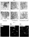
FIG. 3 depicts confocal scanning micrographs showing the cell penetration of fluorescence-labeled magnetic nanoparticle/cell-penetrating peptide complex according to the present invention at varying concentrations [(a): 0 µg of the fluorescence-labeled magnetic nanoparticle/cell-penetrating peptide complex; (b): 36 µg of the fluorescence-labeled magnetic nanoparticle/cell penetrating peptide complex; and (c): 108 µg of the fluorescence-labeled magnetic nanoparticle/cell-penetrating peptide complex]. -
FIG. 4 depicts optical micrographs showing the cell penetration of fluorescence-labeled magnetic nanoparticle/cell-penetrating peptide complex according to the present invention at varying concentrations [(a): 0 µg of the fluorescence-labeled magnetic nanoparticle/cell-penetrating peptide complex; (b): 36 µg of the fluorescence-labeled magnetic nanoparticle/cell penetrating peptide complex; and (c): 108 µg of the fluorescence-labeled magnetic nanoparticle/cell-penetrating peptide complex]. -
FIG. 5 is a graphic diagram showing the results of flow cytometric analysis showing the cell penetration of fluorescence-labeled magnetic nanoparticle/cell-penetrating peptide complex according to the present invention at varying concentrations. -
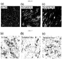
FIG. 6 depicts confocal scanning micrographs showing the osteoblast differentiation of fluorescence-labeled magnetic nanoparticle/cell-penetrating peptide complex according to the present invention at varying concentrations [(a): 0 µg of the fluorescence-labeled magnetic nanoparticle/cell-penetrating peptide complex; (b): 36 µg of the fluorescence-labeled magnetic nanoparticle/cell penetrating peptide complex; and (c): 108 µg of the fluorescence-labeled magnetic nanoparticle/cell-penetrating peptide complex]. -
FIG. 7 depicts optical micrographs showing the osteoblast differentiation of fluorescence-labeled magnetic nanoparticle/cell-penetrating peptide complex according to the present invention at varying concentrations [(a): 0 µg of the fluorescence-labeled magnetic nanoparticle/cell-penetrating peptide complex; (b): 36 µg of the fluorescence-labeled magnetic nanoparticle/cell penetrating peptide complex; and (c): 108 µg of the fluorescence-labeled magnetic nanoparticle/cell-penetrating peptide complex]. -
FIG. 8 and FIG. 9 show measurement results for the effect of a fluorescence-labeled magnetic nanoparticle-cell penetrating peptide complex according to the present invention on mesenchymal stem cells. Specifically,FIG. 8 is a graphic diagram showing the results of flow cytometry for mesenchymal stem cells as model cells treated with fluorescence-labeled antibodies, STRO-1 and CD105, and FIG. 9 depicts confocal scanning micrographs of mesenchymal stem cells as model cells treated with a fluorescence-labeled antibody, STRO-1. - In one aspect, the present invention relates to a method for preparing a complex of a cell-penetrating peptide and a fluorescence-labeled magnetic nanoparticle, the method comprising the steps of: (a) preparing an amino group-containing magnetic nanoparticle by modifying the surface of magnetic nanoparticle with an amino group; (b) preparing a fluorescence-labeled magnetic nanoparticle by adding a fluorescent substance to the amino group-containing magnetic nanoparticle; and (c) linking a cell-penetrating peptide to the fluorescence-labeled magnetic nanoparticle, wherein the diameter of said complex is 10-50 nm and wherein the cell-penetrating peptide is low molecular weight protamine (LMWP; SEQ ID NO: 1: VSRRRRRRGGRRRR).
- Cell-penetrating peptide/fluorescence-labeled magnetic nanoparticle according to the present invention is prepared by the chemical interaction between a cell-penetrating peptide (CPP) serving as a transporter and fluorescence-labeled magnetic nanoparticles enabling the imaging diagnosis of target cells and tissues. When the cell-penetrating peptide/magnetic nanoparticle complex is introduced in vivo in the form of an aqueous solution, they penetrate directly into cells without an endocytosis process, show high stability, maximize imaging diagnosis through optimal targeting and minimize side effects, unlike existing viral peptide transporters.
- As used herein, the term "magnetic nanoparticle" means superparamagnetic material that can temporarily interact with the proton of the surrounding water. Preferably, it means magnetic iron oxide nanoparticle having a particle size of 10-150 nm. More preferably, it means MRI contrast agents that enable the images of substantially all the cells and organs of the body to be obtained. The magnetic nanoparticle is preferably selected from the group consisting of ferric ammonium citrate (FAC), ferrum oxide, superparamagnetic iron oxide nanoparticle (SPIO), carboxy-dextran coated iron oxide nanoparticle, ultrasmall superparamagnetic iron oxide nanoparticle (USPIO), monocrystalline iron oxide nanoparticles (MION), polycrystalline iron oxide nanoparticles (PION) and oral magnetic particles (OMP).
- Also, the fluorescent substance that is used in the present invention is preferably fluorescein isothiocyanate (FITC) or tetramethylrhodamine isothiocyanate (TRITC), but is not limited thereto and may include all chemically modified fluorescent substances, for example alexa fluor, rhodamine red-X, texas red, tetramethylrhodamine, cascade blue, DAPI(4',6-diamidino-2-phenylindole), coumarine, lucifer yellow, dansylaminde, etc.
- The cell-penetrating peptide is low molecular weight protamine (LMWP, SEQ ID NO: 1; VSRRRRRRGGRRRR). The cell-penetrating peptide/magnetic nanoparticle complex according to the present invention may be prepared either by linking the peptide and the nanoparticles to each other through the electrostatic interaction between the positive charge of amino acid and the negative charge of dextran coated on the magnetic nanoparticles or by inducing the chemical linkage there between using a crosslinker. In a preferred embodiment of the present invention, the complex may be prepared by modifying the surface of the magnetic nanoparticles to make a reactive group on the surface in order to increase stability in an in vivo environment, and then inducing the chemical linkage between the magnetic nanoparticles and the peptide using a crosslinker. Because the cell-penetrating peptide that is used in the present invention has free amino groups at the terminal end, it is easy to form the complex using the crosslinker.
- The crosslinker that is used in the present invention may be selected from the group consisting of, but not limited to, 1,4-bis-maleimidobutane (BMB), 1,11-bis-maleimidotetraethyleneglycol (BM[PEO]4), 1-ethyl-3-[3-dimethyl aminopropyl] carbodiimide hydrochloride (EDC), succinimidyl-4-[N-maleimidomethylcyclohexane-1-carboxy-[6-amidocaproate]] (SMCC) and sulfonate thereof (sulfo-SMCC), succimidyl 6-[3-(2-pyridyldithio)-ropionamido] hexanoate (SPDP) and sulfonate thereof (sulfo-SPDP), m-maleimidobenzoyl-N-hydroxysuccinimide ester (MBS) and sulfonate thereof (sulfo-MBS), succimidyl[4-(p-maleimidophenyl)butyrate] (SMPB) and sulfonate (sulfo-SMPB) thereof.
- As shown in
FIG. 1 , the cell-penetrating peptide/magnetic nanoparticle complex according to the present invention is prepared by modifying the surface of negatively charged dextran-coated magnetic nanoparticles with ammonium hydroxide to prepare amino group-containing magnetic nanoparticles, allowing the amino group-containing magnetic nanoparticles to react with a fluorescent substance so as to prepare fluorescence-labeled magnetic nanoparticles, and then adding a crosslinker thereto to prepare fluorescence-labeled magnetic nanoparticles having a free sulfhydryl group. Meanwhile, the cell-penetrating peptide may be coated with a crosslinker to prepare a cell-penetrating peptide having a free sulfhydryl group, which may be allowed to react with said fluorescence-labeled magnetic nanoparticles having a free sulfhydryl group, thus a cell-penetrating peptide/fluorescence-labeled magnetic nanoparticle complex. - In the preparation of the cell-penetrating peptide/fluorescence-labeled magnetic nanoparticle complex according to the present invention, modifying the surface of the magnetic nanoparticles with an amino group may be carried out using one selected from the group consisting of ammonium hydroxide, ethanol, aminopropyl trimethylsilane (APTMS), N-(3-dimethylpropyl)-N-ethylcarbodiimide hydrochloride (EDC), N-hydroxysulfosuccinimide (sulfo-NHS) and N-hydroxysuccinimide (NHS).
- In the cell-penetrating peptide/magnetic nanoparticle complex according to the present invention, the cell-penetrating peptide and the magnetic nanoparticles are linked by S-S bonds. Accordingly, the magnetic nanoparticles may be dissociated from the cell-penetrating peptide by reductase and etc. present in cells, and thus the magnetic nanoparticles may be introduced into cells in an easy and simple way, such that cell imaging and diagnosis can be easily carried out.
- In another aspect, the present invention relates to a complex of a cell-penetrating peptide and a fluorescence-labeled magnetic nanoparticle; and a composition for cell imaging containing said complex, wherein the diameter of said complex is 10-50 nm.
- The composition for cell imaging according to the present invention is preferably administered parenterally, for example, via bolus injection, intravenous injection or intraarterial injection or spray (e.g., aerosol spray) for contrast of lungs. In addition, the composition may be administered via the oral or rectal route and may also be administered according to any known method for administering contrast agents. When the composition according to the present invention is administered parenterally, it must be administered in an aseptic state and must be free of a physiologically unacceptable agent and a paramagnetic, superparamagnetic, ferromagnetic or ferromagnetic contaminant. The composition according to the present invention may contain a preservative, an antimicrobial agent, a buffer solution that is conventionally used in parenteral solutions, an antioxidant, excipient, and other additives that can be used in combination with MR contrast agents and do not interfere with the preparation, storage or use of products.
- Also, although the dosage of the composition for cell imaging according to the present invention will vary depend on the target tissue or organ used and the properties of a measurement system, it is preferably kept at a level as low as possible, as long as sensible contrast can be achieved. The dosage may generally be in the range corresponding to LD50 10%, that is, 1-1000 mg/kg, preferably 2-500 mg/kg, and more preferably 3-300 mg/kg.
- The cell-penetrating peptide/magnetic nanoparticle complex prepared by linking the magnetic nanoparticles for cell imaging and tracing with the cell-penetrating peptide according to the present invention were analyzed for their cell penetration ability and their effect on the inherent characteristics of cells. As a result, it could be observed that the cell penetration of the cell-penetrating peptide/magnetic nanoparticle complex was increased in a concentration-dependent manner. When cells were cultured in various conditions and stained, the complex had no effect on the differentiation of human mesenchymal stem cells (hMSC) into osteoblasts and adipocytes. In addition, it could be observed that, even when model cells were treated with high concentrations of the cell-penetrating peptide/magnetic nanoparticle complex using a cell marker that is predominantly expressed in the cells, the inherent characteristics of the cells did not change.
- Furthermore, the cell-penetrating peptide/magnetic nanoparticle complex was measured for their particle size and surface potential. As a result, it was shown that the magnetic nanoparticle complex had a uniform particle size distribution of 10-50 nm, such that they could penetrate even into compact tissue. In addition, it was shown that the surface potential of magnetic nanoparticles was increased through linkage to positively charged amino groups, suggesting that the process of linking the cell-penetrating peptide to the magnetic nanoparticles was performed in an accurate way.
- Hereinafter, the present invention will be described in further detail with reference to examples.
- Particularly, although the following examples illustrated only ferumoxides, iron oxide contrast agents, as magnetic nanoparticles, it will be obvious to a person skilled in the art that the same as or similar effects to the case of ferumoxides can be obtained even when ferric ammonium citrate (FAC), ferrum oxide, superparamagnetic iron oxide nanoparticle (SPIO), carboxy-dextran coated iron oxide nanoparticle, ultrasmall superparamagnetic iron oxide nanoparticle (USPIO), monocrystalline iron oxide nanoparticles (MION), polycrystalline iron oxide nanoparticles (PION), oral magnetic particles (OMP), etc. are used for the imaging diagnosis of cells and organs.
- 2 ml (11.2 mg/ml) of magnetic nanoparticles (AMI; Cambridge, MA, USA) coated with positively charged dextran was added to 0.5 ml of ammonium hydroxide (Sigma, USA), 10 ml of sodium hydroxide (Sigma, USA), 4 ml of triple deionized water and 4 ml of epichlorohydrin (Sigma, USA). The mixture was allowed to react at room temperature for 24 hours, and then dialyzed with triple deionized water to remove unreacted ammonium hydroxide. To the solution from which unreacted ammonium hydroxide has been removed, 5 mM sodium citrate was added, so that the alkaline environment of the solution was adjusted to a neutral environment. The solution was concentrated using an ultrafiltration membrane (MWCO 3,000) so as to remove unbound reactants. Then, a solution of 3 mg of fluorescent substance FITC (fluorescein isothiocyanate) or TRITC (tetramethylrhodamine isothiocyanate) in 120 µℓ of N,N-dimethylformamide (DMF) was added thereto and reacted under stirring at 25°C.
- 14 mg of an SMCC crosslinker (Pierce Biotechnology) was dissolved in 0.5 ml of DMSO (dimethyl sulfoxide), and then 6 ml of the fluorescence-labeled magnetic nanoparticle solution prepared in Example 1-1 was added thereto. The mixture solution was stirred at room temperature for 5 hours, and then ultrafiltered twice with alkaline phosphate buffer saline (PBS, pH 8). To 6 ml of the ultrafiltered solution containing the fluorescence-labeled magnetic nanoparticles, the cell-penetrating peptide, low molecular weight protamine (LMWP (VSRRRRRRGGRRRR)), dissolved in 1 ml of phosphate buffer saline (PBS, 10X, pH 8.4) at a concentration of 5 mM, was added, and the mixture was stirred at room temperature for 2 hours and at 4°C for 24 hours, thus linking the cell-penetrating peptide (low molecular weight peptide) with the fluorescence-labeled magnetic nanoparticles. The linked low molecular weight protamine and fluorescence-labeled magnetic nanoparticles were ultrafiltered twice with triple deionized water, and the pH of the linked material was measured with pH-measuring paper.
- Magnetic nanoparticles (AMI, Cambridge, MA, USA) coated with negatively charged dextran were purchased and used.
- Amino group-containing magnetic nanoparticles were prepared according to the same composition and method as in Example 1-1, except that the step of adding the fluorescent substance was excluded.
- Each of the cell-permeable peptide/fluorescence-labeled magnetic nanoparticle complex prepared in Example 1 and, as controls, the magnetic nanoparticles of Comparative Example 1 and the amino group-containing magnetic nanoparticles of Comparative Example 2, was dissolved in water. Each of the solutions was dropped onto a carbon-coated copper grid in amounts of 2 mg/mℓ and 10 µℓ, and then naturally dried for 20 hours. The size of each of the dried particles and the dispersion thereof in water were observed with a transmission electron microscope (TEM, JEM-2000EXII, JEOL, JAPAN) at 250,000x magnification and 500,000x magnification at a voltage of 80 kV.
- As a result, as shown in
FIG. 2 , the cell-penetrating peptide/fluorescence-labeled magnetic nanoparticle complexes (c) and (f) showed uniform particle distribution similar to the magnetic nanoparticles (a) and (d) and the amino group-containing magnetic nanoparticles (b) and (e). Also, the particle size determined based on the scale bar was 10-35 nm and similar between the three groups. This suggests that there is no great change in particle size and distribution pattern during the process of chemically linking the cell-permeable peptide with the fluorescence-labeled magnetic nanoparticles and that the cell-penetrating peptide/fluorescence-labeled magnetic nanoparticle complex may be used in contrast agents, because the magnetic nanoparticles have a small size of less than 50 nm and show uniform distribution. - To measure the numerical value of the particle size of the cell-penetrating peptide/fluorescence-labeled magnetic nanoparticle complex, a particle size analyzer (PSSNICOMP, US) was used. The cell-penetrating peptide/fluorescence-labeled magnetic nanoparticle complex prepared in Example 1, the magnetic nanoparticles prepared of Comparative Example 1 and the amino group-containing magnetic nanoparticles of Comparative Example 2 were dissolved in water at a concentration of 2 mg/ml and placed into a glass cuvette, and then the particle size thereof was measured at 23 °C and 632.8 nm.
- Also, to measure the numerical value of the surface potential of the cell-permeable peptide/fluorescence-labeled magnetic nanoparticle complex, a zeta potential analyzer (Otsuka, Japan) was used. The cell-penetrating peptide/fluorescence-labeled magnetic nanoparticle complex prepared in Example 1, the magnetic nanoparticles prepared of Comparative Example 1 and the amino group-containing magnetic nanoparticles of Comparative Example 2 were dissolved in water at a concentration of 2 mg/ml and placed into the analyzer, and then the surface potential thereof was measured.
- As a result, as shown in Table 1 below, the cell-permeable peptide/fluorescence-labeled magnetic nanoparticle complex had a particle size of 26.8 nm which was not greatly different from the particle size (20.4 nm) of the magnetic nanoparticles of Comparative Example 1 and the particle size (23.8 nm) of the amino group-containing magnetic nanoparticles of Comparative Example 2. Also, the magnetic nanoparticle complex had a particle size distribution of less than 50 nm, such that they could penetrate even into compact tissue. Furthermore, the surface potential of the particles was measured to be -51.54 for the magnetic nanoparticles of Comparative Example 1, -10.72 for the amino group-containing magnetic nanoparticles of Comparative Example 2, and 16.97 for the cell-penetrating peptide/fluorescence-labeled magnetic nanoparticle complex of Example 1. Thus, it could be seen that, when the negatively charged dextran-coated magnetic nanoparticles were linked with the positively charged amino group and the cell-penetrating peptide LMWP, the surface potential was changed to positive charges. In addition, it could be seen that the process of preparing the magnetic nanoparticle/LMWP complex was performed in an accurate way.
Table 1: Measurement of particle size and surface potential Particle size (nm) Surface potential (mV) Magnetic nanoparticle (ferumoxides) 20.4 -51.54 Amino group-containing magnetic nanoparticle (ferumoxides-NH2) 23.8 -10.72 Cell-permeable peptide/fluorescence-labeled magnetic nanoparticle complex (ferumoxides-NH2-LMWP) 26.8 16.97 - In order to measure the cell penetration of the cell-penetrating peptide/fluorescence-labeled magnetic nanoparticles, human mesenchymal stem cells (hMSC) were seeded in each well of a 4-well chamber slide at a density of 1 x 10⁴ cells/well, and then incubated in general medium for 20 hours (overnight). Then, the fluorescence-labeled magnetic nanoparticle/cell-penetrating peptide complex (prepared in Example 1) was added to FBS (fetal bovine serum)-free medium in each well in varying concentrations of 0 µg, 36 µg and 108 µg, and after 1 hour, each well was washed twice with phosphate buffer saline (PBS). Then, the cells were stained with a nuclear staining dye (Hoechst 33342, blue), and the cell penetration of the cell-penetrating peptide/magnetic nanoparticle complex was observed with a confocal scanning microscope (IX 70, Olympus Co., Tokyo, Japan).
- As a result, as shown in
FIG. 3 , when the cells were treated with each of 0 µg, 36 µg and 108 µg of the cell-penetrating peptide/fluorescence (FITC, green)-labeled magnetic nanoparticle complex prepared in Example 1, fluorescent particles were observed in the cytoplasm and nucleus of the cells treated with 36 µg of the cell-penetrating peptide/fluorescence-labeled magnetic nanoparticle complex (b) and 108 µg of the cell-penetrating peptide/fluorescence-labeled magnetic nanoparticle complex (c), and the cell penetration of the cell-penetrating peptide/fluorescence-labeled magnetic nanoparticle complex was increased in a concentration-dependent manner. Also, it was seen that the magnetic nanoparticle had no effect on the inherent delivery ability of the cell-penetrating peptide. - In order to examine the intracellular penetration of the fluorescence-labeled magnetic nanoparticle/cell-penetrating peptide complex, human mesenchymal stem cells (hMSC) were cultured in a 6-well plate. Before carrying out Prussian blue staining (Perl's reagent staining), the fluorescence-labeled magnetic nanoparticle/cell-penetrating peptide complex prepared in Example 1 were added to the culture medium in varying amounts of 0 µg, 36 µg and 108 µg, followed by incubation for 20 hours (overnight). After completion of the incubation, the cells were separated from each well using 0.25% trypsin, and the cells were dispended onto a cytospin slide at a density of 2 x 10⁵ cells. The dispensed cells were fixed with 10% NBF (neutral buffered formalin), and then washed twice with phosphate buffer saline (PBS). The iron oxide molecules which penetrated into the cells were stained blue with 2% Perl's reagent, and then the cells were stained red with nuclear fast red. After mounting the slide, the intracellular penetration of the fluorescence-labeled magnetic nanoparticle/cell-penetrating peptide complex was observed with an optical microscope (CKX41, Olympus Co., Tokyo, Japan).
- As a result, as shown in
FIG. 4 , it could be seen that, in the case of 36 µg of the fluorescence-labeled magnetic nanoparticle/cell-penetrating peptide complex (b) and 108 µg of the fluorescence-labeled magnetic nanoparticle/cell-penetrating peptide complex (c), the magnetic nanoparticles were stained (blue) in the cells, and that the intracellular delivery of the magnetic nanoparticles was increased with an increase in the concentration of the fluorescence-labeled magnetic nanoparticle/cell-penetrating peptide complex. - In order to quantitatively analyze the cell penetration of the fluorescence-labeled magnetic nanoparticle/cell-penetrating peptide complex, human mesenchymal stem cells (hMSC) were plated in each well of a 6-well plate at a density of 1 x 10⁵ cells/well, and then incubated in general medium for 20 hours (overnight). The cell-penetrating peptide/fluorescence (FITC)-labeled magnetic nanoparticle complex prepared in Example 1 were added to a fetal bovine serum (FBS)-free DMEM medium in each well in varying amounts of 0 µg, 36 µg and 108 µg. After 1 hour, each well was washed twice with phosphate buffer saline (PBS), and the cells were separated from each well using 0.25% trypsin, washed with phosphate buffer saline (PBS), and then centrifuged to remove the supernatant. The washing and centrifugation procedure was repeated twice, thus removing the fluorescent dye outside the cells. The cells were suspended in 1 mℓ of phosphate buffer saline (PBS), and then the intracellular delivery of the fluorescence-labeled magnetic nanoparticle/cell-penetrating peptide complex of Example 1 were observed using FACSCalibur (BD, USA) at FL-1 (488 nm).
- As a result, as shown in
FIG. 5 , in the case of 36 µg of the cell-penetrating peptide/fluorescence-labeled magnetic nanoparticle complex (b) and 108 µg of the cell-penetrating peptide/fluorescence-labeled magnetic nanoparticle complex (c), the number of cells containing fluorescent particles was increased with an increase in the concentration of complex used. This suggests that the cell penetration ability of the complex was increased in proportion to the increase in the concentration of the cell-penetrating peptide. - In order to examine the effect of the cell-penetrating peptide/fluorescence-labeled magnetic nanoparticle complex on the differentiation of cells, human mesenchymal stem cells (hMSC) were seeded in a 4-well chamber slide at a density of 5 x 10³ density/well, and then incubated in general medium for 20 hours (overnight) to stabilize the cells. The cell-penetrating peptide/fluorescence (TRITC, red)-labeled magnetic nanoparticle complex prepared in Example 1 were added to each well in varying amounts of 0 µg, 36 µg and 108, and then incubated for 24 hours. After completion of the incubation, the cells were cultured in a hard tissue-forming medium containing calcein (calcium green) for 14 days. The composition of the hard tissue-forming medium consisted of alpha-MEM medium containing 15% FBS (fetal bovine serum), 50 mg/mℓL-ascorbic acid, 10-7 M dexamethasone, 1% antibiotic-antimycotic solution and 10 mM beta-glycerol phosphate. After completion of the culture, the medium was removed, and then the cells were washed twice with phosphate (PBS). The washed cells were fixed with 10% NBF (neutral buffered formalin) and stained with a nuclear staining dye (Hoechst 33342, blue), and whether the magnetic nanoparticles having the cell-penetrating peptide linked thereto are involved in differentiation into osteoblasts was observed with a confocal scanning microscope (IX 70, Olympus Co., Tokyo, Japan).
- As a result, as shown in
FIG. 6 , in the case of 36 µg of the cell-penetrating peptide/fluorescence-labeled magnetic nanoparticle complex (b) and 108 µg of the cell-penetrating peptide/fluorescence-labeled magnetic nanoparticle complex (c), calcein staining was uniformly distributed as compared to the group (a) not treated with the cell-penetrating peptide/fluorescence-labeled magnetic nanoparticle complex, and red intracellular staining was observed. This suggests that the cell-penetrating peptide/fluorescence-labeled magnetic nanoparticle complex penetrated into the cells, but had no effect on the osteoblast differentiation of the model cells, even when they were used at high concentration (108 µg). - Human mesenchymal stem cells (hMSC) were plated in a 6-well plate at a density of 1 x 10⁵ cells/well and incubated for 20 hours (overnight) to stabilize the cells. Then, the cell-penetrating peptide/fluorescence-labeled magnetic nanoparticle complex prepared in Example 1 were added to each well in varying concentrations of 0 µg, 36 µg and 108 µg and incubated for 24 hours. Then, the cells were incubated for 3 days in an adipocyte differentiation-inducing medium containing insulin, a hormone involved in lipid metabolism, and indomethacin, a substance promoting the synthesis of arachidonic acid that is a kind of fatty acid, and then, the cells were incubated for 2 days in a maintenance medium containing medium supplements other than insulin. This cell incubation procedure was repeated three times, and then the cells were incubated in a maintenance medium for 7 days to induce differentiation into adipocytes. The differentiation-induced cells were fixed with 10% NBF (neutral buffered formalin), washed with 60% isopropanol and completely dried. Then, fat in the cells was stained with oil red O solution (Sigma, red), and each well was washed with water (H2O). Then, whether the magnetic nanoparticles having the cell-penetrating peptide linked thereto are involved in differentiation into adipocytes was observed with an optical microscope.
- As a result, as shown in
FIG. 7 , the cells treated with each of 36 µg of the cell-penetrating peptide/fluorescence-labeled magnetic nanoparticle complex (b) and 108 µg of the cell-penetrating peptide/fluorescence-labeled magnetic nanoparticle complex (c) showed oil red O staining (red: index of adipocyte differentiation) in the same manner as the group (c) not treated with the cell-penetrating peptide/fluorescence-labeled magnetic nanoparticle complex. This suggests that the cell-penetrating peptide/fluorescence-labeled magnetic nanoparticle complex had no effect on differentiation into adipocytes, even when they were used at high concentration (108 µg). - In order to examine the effect of the cell-penetrating peptide/fluorescence-labeled magnetic nanoparticle complex on the inherent characteristics of mesenchymal stem cells, mouse anti-human endoglin (CD 105) monoclonal antibody (CHEMICON, USA) and mouse anti-STRO-1 monoclonal antibody (CHEMICON, USA), which are expressed predominantly in human mesenchymal stem cells (hMSC), were used as primary antibodies, and the goat anti-mouse IgG (H&L) fluorescein-conjugated affinity-purified secondary antibody (CHEMICON, USA) was used (Puchuantes, S. et al., Tissue Antigens, 50:6, 1997). Human mesenchymal stem cells (hMSC) were plated in each well of a well plate at a density of 1 x 10⁶ cells/well, and then incubated in a general medium for 20 hours. Then, 36 µg/mℓ of the cell-penetrating peptide/fluorescence-labeled magnetic nanoparticle complex (prepared in Example 1) in DMEM medium containing no FBS (fetal bovine serum) were added to each well and incubated for 30 minutes, 1 hour and 4 hours. Then, each well was washed twice with phosphate buffer saline (PBS), and the cells were separated from each well using 0.25% trypsin and washed with phosphate buffer saline. The washed cells were centrifuged to remove the supernatant. The centrifugation procedure was repeated twice to remove particles outside the cells. Then, the primary antibodies, Endoglin (CD 105) and STRO-1, diluted in bovine serum albumin (BSA) at a ratio of 1:25, were added to the cells, and then phosphate buffer saline (PBS) was added thereto. Then, the solution was incubated under shaking at 4 °C for 20 hours. The incubated solution was washed with 0.5% bovine serum albumin (BSA)-containing phosphate buffer saline (PBS), and then centrifuged to remove the supernatant. The secondary antibody diluted in 0.5% bovine serum albumin (BSA)-containing phosphate buffer saline (BSA) at a ratio of 1:50 was added thereto, and then the solution was incubated under shaking at room temperature for 1 hour. Then, the cells were suspended in 300 µℓ of phosphate buffer saline (PBS), and the intracellular delivery of the cell-penetrating peptide/fluorescence-labeled magnetic nanoparticle complex was observed using FACSCalibur (BD, USA) at FL-1 (488 nm).
- As a result, as shown in
FIG. 8 , when the fluorescent dye (FITC, green)-conjugated hMSC (human mesenchymal stem cell) marker, STRO-1 and Endoglin (CD 105) were allowed to react with the model cells labeled with the cell-penetrating peptide/magnetic nanoparticle complex, the fluorescence value observed in the model cells was similar to that of the positive control. This suggests that the cell-penetrating peptide/magnetic nanoparticle complex has no effect on the inherent characteristics of human mesenchymal stem cell (hMSC) as the model cells. Also, this indirectly reveals that the cell-penetrating peptide/magnetic nanoparticle complex according to the present invention has no cytotoxicity when they are used for cell imaging. - In order to examine the effect of the cell-penetrating peptide/magnetic nanoparticle complex on the inherent characteristics of mesenchymal stem cells, human mesenchymal stem cells (hMSC) were seeded in a 4-well chamber slide at a density of 5 x 10³ cells/well, and then incubated in general medium for 20 hours (overnight) to stabilize the cells. Then, 36 µg/mℓ of the cell-penetrating peptide/magnetic nanoparticle complex in DMEM medium containing no FBS (fetal bovine serum) were added to each well and incubated for 30 minutes, 1 hour and 4 hours. Then, each well was washed with phosphate buffer saline (PBS), and the cells were fixed with 10% NBF (neutral buffered formalin) and stained with a nuclear staining dye (Hoechst 33342, blue). Then, the primary antibodies, Endoglin (CD 105) and STRO-1, diluted in 0.5% bovine serum albumin (BSA)-containing phosphate buffer saline (PBS) at a ratio of 1:25, were added thereto and incubated under shaking at 4°C for 20 hours (overnight). The incubated solution was washed with 0.5% bovine serum albumin (BSA)-containing phosphate buffer saline (PBS), and the secondary antibody diluted in 0.5% bovine serum albumin-containing phosphate buffer saline (PBS) at a ratio of 1:50 was added thereto. Then, the solution was incubated under shaking at room temperature for 1 hour. The incubated solution was washed with phosphate buffer saline (PBS). Then, the chamber slide was mounted, and the cells were observed with a confocal scanning microscope.
- As a result, as shown in FIG. 9, when the fluorescent dye (FITC, green)-conjugated hMSC (human mesenchymal stem cell) marker, STRO-1 and Endoglin (CD 105) were allowed to react with the model cells labeled with the cell-penetrating peptide/magnetic nanoparticle complex, the fluorescence value observed in the model cells was similar to that of the positive control. This suggests that the cell-penetrating peptide/magnetic nanoparticle complex has no effect on the inherent characteristics of human mesenchymal stem cell (hMSC) as the model cells. Also, this indirectly reveals that the cell-penetrating peptide/magnetic nanoparticle complex according to the present invention has no cytotoxicity when they are used for cell imaging.
- As described above in detail, unlike the existing viral peptide transporters, the present invention can suggest an innovative therapeutic technology which shows high stability, maximizes the effect of imaging diagnosis through optimal targeting and minimizes side effects.
-
- <110> Seoul National University Industry Foundation
- <120> Complex of Cell Translocational Peptide and Magnetic Nanoparticulates and
Use Thereof - <130> 21275EP
- <140>
EP 08 835 082.2
<141> 2008-10-02 - <150>
10-2007-0099511
<151> 2007-10-02 - <160> 4
- <170> PatentIn version 3.2
- <210> 1
<211> 14
<212> PRT
<213> Unknown - <400> 1
val Ser Arg Arg Arg Arg Arg Arg Gly Gly Arg Arg Arg Arg 14 - <210> 2
<211> 11
<212> PRT
<213> Artificial Sequence - <220>
<223> TAT (TAT from protein transduction domain of HIV protein) - <400> 2
Tyr Gly Arg Lys Lys Arg Arg Gln Arg Arg Arg 11 - <210> 3
<211> 16
<212> PRT
<213> Artificial Sequence - <220>
<223> penetratin - <400> 3
Arg Gln Ile Lys lie Trp Phe Gln Asn Arg Arg Met Lys Trp Lys Lys 16 - <210> 4
<211> 8
<212> PRT
<213> unknown - <400> 4
Arg Arg Arg Arg Arg Arg Arg Arg 8
Claims (9)
- A method for preparing a complex of a cell-penetrating peptide and a fluorescence-labeled magnetic nanoparticle, the method comprising the steps of:(a) preparing an amino group-containing magnetic nanoparticle by modifying the surface of magnetic nanoparticle with an amino group;(b) preparing a fluorescence-labeled magnetic nanoparticle by adding a fluorescent substance to the amino group-containing magnetic nanoparticle; and(c) preparing cell-penetrating peptide/fluorescence-labeled magnetic nanoparticle complex by linking a cell-penetrating peptide to the fluorescence-labeled magnetic nanoparticle, wherein the diameter of said complex is 10-50nm andwherein the cell-penetrating peptide is low molecular weight protamine (LMWP, SEQ ID NO: 1 : VSRRRRRRGGRRRR).
- The method for preparing a complex of a cell-penetrating peptide and a fluorescence-labeled magnetic nanoparticle according to claim 1, wherein the magnetic nanoparticle is selected from the group consisting of ferric ammonium citrate (FAC), ferrum oxide, superparamagnetic iron oxide nanoparticle (SPIO), carboxy-dextran coated iron oxide nanoparticle, ultrasmall superparamagnetic iron oxide nanoparticle (USPIO), monocrystalline iron oxide nanoparticles (MION), polycrystalline iron oxide nanoparticles (PION) and oral magnetic particles (OMP).
- The method for preparing a complex of a cell-penetrating peptide and a fluorescence-labeled magnetic nanoparticle according to claim 1, wherein said linking of step (c) is performed by a crosslinker.
- The method for preparing a complex of a cell-penetrating peptide and a fluorescence-labeled magnetic nanoparticle according to claim 3, wherein the crosslinker is selected from the group consisting of 1,4-bis-maleimidobutane (BMB), 1,11-bis-maleimidotetraethyleneglycol (BM[PEO]4), 1-ethyl-3-[3-dimethyl aminopropyl] carbodiimide hydrochloride (EDC), succinimidyl-4-[N-maleimidomethylcyclohexane-1-carboxy-[6-amidocaproate]] (SMCC) and sulfonate thereof (sulfo- SMCC), succimidyl 6-[3-(2-pyridyldithio)-ropionamido] hexanoate (SPDP) and sulfonate thereof (sulfo-SPDP), m-maleimidobenzoyl-N-hydroxysuccinimide ester (MBS) and sulfonate thereof (sulfo-MBS), succimidyl[4-(p-maleimidophenyl)butyrate] (SMPB) and sulfonate (sulfo-SMPB) thereof.
- The method for preparing a complex of a cell-penetrating peptide and a fluorescence-labeled magnetic nanoparticle according to claim 1, wherein the fluorescent substance is selected from the group consisting of fluorescein isothiocyanate (FITC) or tetramethylrhodamine isothiocyanate (TRITC), alexa fluor, rhodamine red-X, texas red, tetramethylrhodamine, cascade blue, DAPI(4',6-diamidino-2-phenylindole), coumarine, lucifer yellow, and dansylaminde.
- The method for preparing a complex of a cell-penetrating peptide and a fluorescence-labeled magnetic nanoparticle according to claim 1, wherein said modifying the surface of the magnetic nanoparticle of step (a) is carried out using one selected from the group consisting of ammonium hydroxide, ethanol, aminopropyl trimethylsilane (APTMS), N-(3-dimethylpropyl)-N-ethylcarbodiimide hydrochloride (EDC), N-hydroxysulfosuccinimide (sulfo-NHS) and N-hydroxysuccinimide (NHS).
- The method for preparing a complex of a cell-penetrating peptide and a fluorescence-labeled magnetic nanoparticle according to claim 1, which further comprising the step of adjusting pH to neutral by adding sodium citrate to the amino group-containing magnetic nanoparticle prepared in step (a).
- A complex of a cell-penetrating peptide and a fluorescence-labeled magnetic nanoparticle, which is prepared by the method of claim 1, and in which the cell-penetrating peptide is linked to the fluorescence-labeled magnetic nanoparticle, wherein the diameter of said complex is 10-50nm.
- A composition for cell imaging containing the complex in which the cell-penetrating peptide is linked to the fluorescence-labeled magnetic nanoparticle of claim 8.
Applications Claiming Priority (2)
| Application Number | Priority Date | Filing Date | Title |
|---|---|---|---|
| KR1020070099511A KR100951719B1 (en) | 2007-10-02 | 2007-10-02 | Complex of cell permeable peptide and fluorescently labeled magnetic nanoparticles and use thereof |
| PCT/KR2008/005815 WO2009045063A2 (en) | 2007-10-02 | 2008-10-02 | Complex of cell translocational peptide and magnetic nanoparticles and use thereof |
Publications (3)
| Publication Number | Publication Date |
|---|---|
| EP2197783A2 EP2197783A2 (en) | 2010-06-23 |
| EP2197783A4 EP2197783A4 (en) | 2014-09-03 |
| EP2197783B1 true EP2197783B1 (en) | 2018-07-11 |
Family
ID=40526838
Family Applications (1)
| Application Number | Title | Priority Date | Filing Date |
|---|---|---|---|
| EP08835082.2A Active EP2197783B1 (en) | 2007-10-02 | 2008-10-02 | Complex of cell translocational peptide and magnetic nanoparticles and use thereof |
Country Status (4)
| Country | Link |
|---|---|
| US (1) | US20100298536A1 (en) |
| EP (1) | EP2197783B1 (en) |
| KR (1) | KR100951719B1 (en) |
| WO (1) | WO2009045063A2 (en) |
Families Citing this family (22)
| Publication number | Priority date | Publication date | Assignee | Title |
|---|---|---|---|---|
| KR101130550B1 (en) * | 2009-10-29 | 2012-03-23 | 고려대학교 산학협력단 | Cell Penetrating Peptide Modified Nanoliposome and Anti-Atopic Composition Comprising the Same |
| KR101244140B1 (en) * | 2010-08-19 | 2013-03-14 | 국립암센터 | Positively charged superparamagnetic iron oxide nanoparticles, contrast agent using the same and method for preparation thereof |
| JP2012149034A (en) * | 2010-12-27 | 2012-08-09 | Canon Inc | Composite particle, contrast agent for photoacoustic imaging, and photoacoustic imaging method |
| KR101282904B1 (en) * | 2011-01-28 | 2013-07-05 | 서울대학교산학협력단 | Dendritic polymer functionalized magnetic mesoporous solid for adsorption of metallic ions and method for preparation thereof |
| US9448231B2 (en) | 2011-02-28 | 2016-09-20 | Ventana Medical Systems, Inc. | Application of quantum dots for nuclear staining |
| KR101287405B1 (en) * | 2011-04-29 | 2013-07-19 | 가천대학교 산학협력단 | Nanocomposites and method for preparing the same |
| KR101263780B1 (en) | 2011-08-22 | 2013-05-13 | 강원대학교산학협력단 | Self-assembly of coumarin-proteinoid conjugate and method for preparing the same |
| US9675708B2 (en) * | 2011-10-21 | 2017-06-13 | Stemgenics, Inc. | Functionalized nanoparticles for intracellular delivery of biologically active molecules |
| GB201306589D0 (en) * | 2013-04-11 | 2013-05-29 | Abeterno Ltd | Live cell imaging |
| JP6354754B2 (en) * | 2013-06-19 | 2018-07-11 | コニカミノルタ株式会社 | Fluorescent nanoparticles for biomolecule staining and method for producing the same |
| KR101529634B1 (en) * | 2013-08-28 | 2015-06-30 | 서울대학교산학협력단 | Cell permeable Fusion protein for Facilitating Reprogramming inducing and Use Thereof |
| WO2017100683A1 (en) * | 2015-12-09 | 2017-06-15 | Stemgenics, Inc. | Differential drug screening using pluripotent stem cells induced with functionalized nanoparticles |
| KR102640613B1 (en) * | 2016-12-28 | 2024-02-26 | 닛테츠 케미컬 앤드 머티리얼 가부시키가이샤 | Metal-resin composite and its uses |
| CN110231470A (en) * | 2019-06-12 | 2019-09-13 | 桂林理工大学 | A kind of control ferric oxide nano particles are distributed in animal brain and its fluorescence detection method |
| EP3996751A1 (en) * | 2019-07-12 | 2022-05-18 | The Board Of Trustees Of The Leland Stanford Junior University | Functionalized nanoparticles and their use in treating bacterial infections |
| CN112754996B (en) * | 2021-03-16 | 2023-03-31 | 江西省科学院生物资源研究所 | Protamine short peptide modified paclitaxel liposome and preparation method thereof |
| US11998615B2 (en) | 2021-04-14 | 2024-06-04 | The Board Of Trustees Of The Leland Stanford Junior University | Functionalized nanoparticles and their use in treating bacterial infections |
| CN112864261B (en) * | 2021-04-23 | 2021-08-10 | 南昌凯迅光电有限公司 | Flexible gallium arsenide solar cell and preparation method thereof |
| WO2024123134A1 (en) | 2022-12-09 | 2024-06-13 | 서울대학교산학협력단 | Nanoparticle comprising peptide-based conjugate for delivering mrna into b cell and t cell and uses thereof |
| KR20240087591A (en) | 2022-12-09 | 2024-06-19 | 주식회사 나이벡 | Nanoparticle Comprising Peptide-based Conjugate for Delivering Oligonucleotide into Target Cell and Pharmaceutical Composition Comprising Thereof |
| CN117257976A (en) * | 2023-08-21 | 2023-12-22 | 西北大学 | Preparation and application of nerve-targeting magnetic nanoparticles |
| CN119701022B (en) * | 2025-02-28 | 2025-07-25 | 中国科学院自动化研究所 | Bimodal magnetic particle fluorescent probe and application thereof in preparation, tracing and monitoring of dendritic cell vaccine |
Citations (1)
| Publication number | Priority date | Publication date | Assignee | Title |
|---|---|---|---|---|
| WO2004081188A2 (en) * | 2003-03-10 | 2004-09-23 | The Regents Of The University Of Michigan | Non-toxic membrane-translocating peptides |
Family Cites Families (7)
| Publication number | Priority date | Publication date | Assignee | Title |
|---|---|---|---|---|
| US6015787A (en) * | 1997-11-04 | 2000-01-18 | New England Medical Center Hospitals, Inc. | Cell-permeable protein inhibitors of calpain |
| US7019113B2 (en) | 1999-06-07 | 2006-03-28 | Mirus Bio Corporation | Reversible modification of membrane interaction |
| WO2002044419A2 (en) | 2000-11-28 | 2002-06-06 | Wyeth | Expression analysis of kiaa nucleic acids and polypeptides useful in the diagnosis and treatment of prostate cancer |
| US7329638B2 (en) * | 2003-04-30 | 2008-02-12 | The Regents Of The University Of Michigan | Drug delivery compositions |
| KR100568457B1 (en) | 2003-07-22 | 2006-04-07 | 학교법인 성균관대학 | RNA Transfer to Plants Using Cationic Oligopeptides |
| WO2005107818A2 (en) | 2004-04-30 | 2005-11-17 | University Of Florida | Nanoparticles and their use for multifunctional bioimaging |
| RU2007138027A (en) | 2005-03-14 | 2009-04-20 | Борд Оф Риджентс Оф Дзе Юниверсити Оф Техас Систем (Us) | BIOACTIVE PEPTIDES FUS-1 AND COMPLEXES OF POLYPEPTIDES WITH NANOPARTICLES |
-
2007
- 2007-10-02 KR KR1020070099511A patent/KR100951719B1/en active Active
-
2008
- 2008-10-02 EP EP08835082.2A patent/EP2197783B1/en active Active
- 2008-10-02 WO PCT/KR2008/005815 patent/WO2009045063A2/en not_active Ceased
- 2008-10-02 US US12/681,027 patent/US20100298536A1/en not_active Abandoned
Patent Citations (1)
| Publication number | Priority date | Publication date | Assignee | Title |
|---|---|---|---|---|
| WO2004081188A2 (en) * | 2003-03-10 | 2004-09-23 | The Regents Of The University Of Michigan | Non-toxic membrane-translocating peptides |
Non-Patent Citations (3)
| Title |
|---|
| APOSTOLOPOULOS V ET AL: "Delivery of tumor associated antigens to antigen presenting cells using penetratin induces potent immune responses", VACCINE, ELSEVIER LTD, GB, vol. 24, no. 16, 12 April 2006 (2006-04-12), pages 3191 - 3202, XP028010607, ISSN: 0264-410X, [retrieved on 20060412], DOI: 10.1016/J.VACCINE.2006.01.032 * |
| CHRISTIAENS B ET AL: "Enhancement of polymethacrylate-mediated gene delivery by Penetratin", EUROPEAN JOURNAL OF PHARMACEUTICAL SCIENCES, ELSEVIER, AMSTERDAM, NL, vol. 24, no. 5, 1 April 2005 (2005-04-01), pages 525 - 537, XP027803775, ISSN: 0928-0987, [retrieved on 20050401] * |
| PARK YOON JEONG ET AL: "Nontoxic membrane translocation peptide from protamine, low molecular weight protamine (LMWP), for enhanced intracellular protein delivery: in vitro and in vivo study", THE FASEB JOURNAL, FEDERATION OF AMERICAN SOCIETIES FOR EXPERIMENTAL BIOLOGY, BETHESDA; US, vol. 19, no. 9, 1 July 2005 (2005-07-01), pages 1 - 20, XP002587876, ISSN: 0892-6638, [retrieved on 20050720], DOI: 10.1096/FJ.04-2322FJE * |
Also Published As
| Publication number | Publication date |
|---|---|
| EP2197783A2 (en) | 2010-06-23 |
| KR20090034240A (en) | 2009-04-07 |
| WO2009045063A2 (en) | 2009-04-09 |
| EP2197783A4 (en) | 2014-09-03 |
| KR100951719B1 (en) | 2010-04-07 |
| WO2009045063A3 (en) | 2009-06-18 |
| US20100298536A1 (en) | 2010-11-25 |
Similar Documents
| Publication | Publication Date | Title |
|---|---|---|
| US20100298536A1 (en) | Complex of cell translocational peptide and magnetic nanoparticles and use thereof | |
| Andreas et al. | Highly efficient magnetic stem cell labeling with citrate-coated superparamagnetic iron oxide nanoparticles for MRI tracking | |
| Bhirde et al. | Nanoparticles for cell labeling | |
| Huang et al. | Highly efficient cellular labeling of mesoporous nanoparticles in human mesenchymal stem cells: implication for stem cell tracking | |
| Berry et al. | Functionalisation of magnetic nanoparticles for applications in biomedicine | |
| Schulze et al. | Cellular uptake and trafficking of a prototypical magnetic iron oxide label in vitro | |
| Liu et al. | Low molecular weight alkyl-polycation wrapped magnetite nanoparticle clusters as MRI probes for stem cell labeling and in vivo imaging | |
| Hasegawa et al. | Nanogel-quantum dot hybrid nanoparticles for live cell imaging | |
| Yoon et al. | Specific targeting, cell sorting, and bioimaging with smart magnetic silica core–shell nanomaterials | |
| Walther et al. | Quantum dot− carrier peptide conjugates suitable for imaging and delivery applications | |
| Wang et al. | Multifunctional polyglycerol-grafted Fe3O4@ SiO2 nanoparticles for targeting ovarian cancer cells | |
| CN103212093B (en) | A kind of have cell targeted magnetic Nano material and biomedical applications thereof | |
| Wang et al. | Synthesis of tumor-targeted folate conjugated fluorescent magnetic albumin nanoparticles for enhanced intracellular dual-modal imaging into human brain tumor cells | |
| US20210154323A1 (en) | Functionalized nanoparticles for the intracellular delivery of biologically active molecules and methods for their manufacture and use | |
| Suh et al. | Efficient labeling of mesenchymal stem cells using cell permeable magnetic nanoparticles | |
| Mandal et al. | Cellular uptake of gold nanoparticles directly cross-linked with carrier peptides by osteosarcoma cells | |
| US20120045399A1 (en) | Positively-charged superparamagnetic iron oxide nanoparticle, contrast agent using the same and method of preparing the same | |
| Wang et al. | Anti-CD133 monoclonal antibody conjugated immunomagnetic nanosensor for molecular imaging of targeted cancer stem cells | |
| Ranjbarvaziri et al. | Quantum dot labeling using positive charged peptides in human hematopoetic and mesenchymal stem cells | |
| Nakamura et al. | Relaxometric property of organosilica nanoparticles internally functionalized with iron oxide and fluorescent dye for multimodal imaging | |
| CN110585449A (en) | Live cell probe construction method based on neutrophils | |
| Neoh et al. | Surface modification of magnetic nanoparticles for stem cell labeling | |
| Sung et al. | Dual-modal nanoprobes for imaging of mesenchymal stem cell transplant by MRI and fluorescence imaging | |
| CN101590245A (en) | A kind of USPIO-PLA-RGD compound and its preparation method and application | |
| Oghabian et al. | Detectability of Her2 positive tumors using monoclonal antibody conjugated iron oxide nanoparticles in MRI |
Legal Events
| Date | Code | Title | Description |
|---|---|---|---|
| PUAI | Public reference made under article 153(3) epc to a published international application that has entered the european phase |
Free format text: ORIGINAL CODE: 0009012 |
|
| 17P | Request for examination filed |
Effective date: 20100406 |
|
| AK | Designated contracting states |
Kind code of ref document: A2 Designated state(s): AT BE BG CH CY CZ DE DK EE ES FI FR GB GR HR HU IE IS IT LI LT LU LV MC MT NL NO PL PT RO SE SI SK TR |
|
| AX | Request for extension of the european patent |
Extension state: AL BA MK RS |
|
| DAX | Request for extension of the european patent (deleted) | ||
| A4 | Supplementary search report drawn up and despatched |
Effective date: 20140806 |
|
| RIC1 | Information provided on ipc code assigned before grant |
Ipc: B82B 3/00 20060101AFI20140731BHEP |
|
| 17Q | First examination report despatched |
Effective date: 20151124 |
|
| REG | Reference to a national code |
Ref country code: DE Ref legal event code: R079 Ref document number: 602008055986 Country of ref document: DE Free format text: PREVIOUS MAIN CLASS: B82B0003000000 Ipc: A61K0049000000 |
|
| GRAP | Despatch of communication of intention to grant a patent |
Free format text: ORIGINAL CODE: EPIDOSNIGR1 |
|
| RIC1 | Information provided on ipc code assigned before grant |
Ipc: A61K 49/00 20060101AFI20180411BHEP Ipc: A61K 49/18 20060101ALI20180411BHEP Ipc: A61K 49/08 20060101ALI20180411BHEP Ipc: A61K 49/14 20060101ALI20180411BHEP Ipc: B82Y 5/00 20110101ALI20180411BHEP |
|
| INTG | Intention to grant announced |
Effective date: 20180425 |
|
| RIN1 | Information on inventor provided before grant (corrected) |
Inventor name: CHUNG, CHONG-PYOUNG Inventor name: YANG, VICTOR, C. Inventor name: PARK, YOON-JEONG Inventor name: SUH, JIN SOOK |
|
| GRAS | Grant fee paid |
Free format text: ORIGINAL CODE: EPIDOSNIGR3 |
|
| GRAA | (expected) grant |
Free format text: ORIGINAL CODE: 0009210 |
|
| AK | Designated contracting states |
Kind code of ref document: B1 Designated state(s): AT BE BG CH CY CZ DE DK EE ES FI FR GB GR HR HU IE IS IT LI LT LU LV MC MT NL NO PL PT RO SE SI SK TR |
|
| REG | Reference to a national code |
Ref country code: GB Ref legal event code: FG4D |
|
| REG | Reference to a national code |
Ref country code: CH Ref legal event code: EP |
|
| REG | Reference to a national code |
Ref country code: AT Ref legal event code: REF Ref document number: 1016219 Country of ref document: AT Kind code of ref document: T Effective date: 20180715 |
|
| REG | Reference to a national code |
Ref country code: IE Ref legal event code: FG4D |
|
| REG | Reference to a national code |
Ref country code: DE Ref legal event code: R096 Ref document number: 602008055986 Country of ref document: DE |
|
| REG | Reference to a national code |
Ref country code: CH Ref legal event code: NV Representative=s name: RENTSCH PARTNER AG, CH |
|
| REG | Reference to a national code |
Ref country code: NL Ref legal event code: MP Effective date: 20180711 |
|
| REG | Reference to a national code |
Ref country code: LT Ref legal event code: MG4D |
|
| REG | Reference to a national code |
Ref country code: AT Ref legal event code: MK05 Ref document number: 1016219 Country of ref document: AT Kind code of ref document: T Effective date: 20180711 |
|
| PG25 | Lapsed in a contracting state [announced via postgrant information from national office to epo] |
Ref country code: NL Free format text: LAPSE BECAUSE OF FAILURE TO SUBMIT A TRANSLATION OF THE DESCRIPTION OR TO PAY THE FEE WITHIN THE PRESCRIBED TIME-LIMIT Effective date: 20180711 |
|
| PG25 | Lapsed in a contracting state [announced via postgrant information from national office to epo] |
Ref country code: GR Free format text: LAPSE BECAUSE OF FAILURE TO SUBMIT A TRANSLATION OF THE DESCRIPTION OR TO PAY THE FEE WITHIN THE PRESCRIBED TIME-LIMIT Effective date: 20181012 Ref country code: SE Free format text: LAPSE BECAUSE OF FAILURE TO SUBMIT A TRANSLATION OF THE DESCRIPTION OR TO PAY THE FEE WITHIN THE PRESCRIBED TIME-LIMIT Effective date: 20180711 Ref country code: BG Free format text: LAPSE BECAUSE OF FAILURE TO SUBMIT A TRANSLATION OF THE DESCRIPTION OR TO PAY THE FEE WITHIN THE PRESCRIBED TIME-LIMIT Effective date: 20181011 Ref country code: AT Free format text: LAPSE BECAUSE OF FAILURE TO SUBMIT A TRANSLATION OF THE DESCRIPTION OR TO PAY THE FEE WITHIN THE PRESCRIBED TIME-LIMIT Effective date: 20180711 Ref country code: IS Free format text: LAPSE BECAUSE OF FAILURE TO SUBMIT A TRANSLATION OF THE DESCRIPTION OR TO PAY THE FEE WITHIN THE PRESCRIBED TIME-LIMIT Effective date: 20181111 Ref country code: NO Free format text: LAPSE BECAUSE OF FAILURE TO SUBMIT A TRANSLATION OF THE DESCRIPTION OR TO PAY THE FEE WITHIN THE PRESCRIBED TIME-LIMIT Effective date: 20181011 Ref country code: FI Free format text: LAPSE BECAUSE OF FAILURE TO SUBMIT A TRANSLATION OF THE DESCRIPTION OR TO PAY THE FEE WITHIN THE PRESCRIBED TIME-LIMIT Effective date: 20180711 Ref country code: PL Free format text: LAPSE BECAUSE OF FAILURE TO SUBMIT A TRANSLATION OF THE DESCRIPTION OR TO PAY THE FEE WITHIN THE PRESCRIBED TIME-LIMIT Effective date: 20180711 Ref country code: LT Free format text: LAPSE BECAUSE OF FAILURE TO SUBMIT A TRANSLATION OF THE DESCRIPTION OR TO PAY THE FEE WITHIN THE PRESCRIBED TIME-LIMIT Effective date: 20180711 |
|
| PG25 | Lapsed in a contracting state [announced via postgrant information from national office to epo] |
Ref country code: ES Free format text: LAPSE BECAUSE OF FAILURE TO SUBMIT A TRANSLATION OF THE DESCRIPTION OR TO PAY THE FEE WITHIN THE PRESCRIBED TIME-LIMIT Effective date: 20180711 Ref country code: LV Free format text: LAPSE BECAUSE OF FAILURE TO SUBMIT A TRANSLATION OF THE DESCRIPTION OR TO PAY THE FEE WITHIN THE PRESCRIBED TIME-LIMIT Effective date: 20180711 Ref country code: HR Free format text: LAPSE BECAUSE OF FAILURE TO SUBMIT A TRANSLATION OF THE DESCRIPTION OR TO PAY THE FEE WITHIN THE PRESCRIBED TIME-LIMIT Effective date: 20180711 |
|
| REG | Reference to a national code |
Ref country code: DE Ref legal event code: R097 Ref document number: 602008055986 Country of ref document: DE |
|
| PG25 | Lapsed in a contracting state [announced via postgrant information from national office to epo] |
Ref country code: CZ Free format text: LAPSE BECAUSE OF FAILURE TO SUBMIT A TRANSLATION OF THE DESCRIPTION OR TO PAY THE FEE WITHIN THE PRESCRIBED TIME-LIMIT Effective date: 20180711 Ref country code: RO Free format text: LAPSE BECAUSE OF FAILURE TO SUBMIT A TRANSLATION OF THE DESCRIPTION OR TO PAY THE FEE WITHIN THE PRESCRIBED TIME-LIMIT Effective date: 20180711 Ref country code: EE Free format text: LAPSE BECAUSE OF FAILURE TO SUBMIT A TRANSLATION OF THE DESCRIPTION OR TO PAY THE FEE WITHIN THE PRESCRIBED TIME-LIMIT Effective date: 20180711 |
|
| PLBE | No opposition filed within time limit |
Free format text: ORIGINAL CODE: 0009261 |
|
| STAA | Information on the status of an ep patent application or granted ep patent |
Free format text: STATUS: NO OPPOSITION FILED WITHIN TIME LIMIT |
|
| PG25 | Lapsed in a contracting state [announced via postgrant information from national office to epo] |
Ref country code: SK Free format text: LAPSE BECAUSE OF FAILURE TO SUBMIT A TRANSLATION OF THE DESCRIPTION OR TO PAY THE FEE WITHIN THE PRESCRIBED TIME-LIMIT Effective date: 20180711 Ref country code: DK Free format text: LAPSE BECAUSE OF FAILURE TO SUBMIT A TRANSLATION OF THE DESCRIPTION OR TO PAY THE FEE WITHIN THE PRESCRIBED TIME-LIMIT Effective date: 20180711 |
|
| 26N | No opposition filed |
Effective date: 20190412 |
|
| GBPC | Gb: european patent ceased through non-payment of renewal fee |
Effective date: 20181011 |
|
| REG | Reference to a national code |
Ref country code: BE Ref legal event code: MM Effective date: 20181031 |
|
| PG25 | Lapsed in a contracting state [announced via postgrant information from national office to epo] |
Ref country code: LU Free format text: LAPSE BECAUSE OF NON-PAYMENT OF DUE FEES Effective date: 20181002 Ref country code: MC Free format text: LAPSE BECAUSE OF FAILURE TO SUBMIT A TRANSLATION OF THE DESCRIPTION OR TO PAY THE FEE WITHIN THE PRESCRIBED TIME-LIMIT Effective date: 20180711 |
|
| REG | Reference to a national code |
Ref country code: IE Ref legal event code: MM4A |
|
| PG25 | Lapsed in a contracting state [announced via postgrant information from national office to epo] |
Ref country code: BE Free format text: LAPSE BECAUSE OF NON-PAYMENT OF DUE FEES Effective date: 20181031 Ref country code: SI Free format text: LAPSE BECAUSE OF FAILURE TO SUBMIT A TRANSLATION OF THE DESCRIPTION OR TO PAY THE FEE WITHIN THE PRESCRIBED TIME-LIMIT Effective date: 20180711 |
|
| PG25 | Lapsed in a contracting state [announced via postgrant information from national office to epo] |
Ref country code: FR Free format text: LAPSE BECAUSE OF NON-PAYMENT OF DUE FEES Effective date: 20181031 Ref country code: GB Free format text: LAPSE BECAUSE OF NON-PAYMENT OF DUE FEES Effective date: 20181011 Ref country code: IE Free format text: LAPSE BECAUSE OF NON-PAYMENT OF DUE FEES Effective date: 20181002 |
|
| PG25 | Lapsed in a contracting state [announced via postgrant information from national office to epo] |
Ref country code: MT Free format text: LAPSE BECAUSE OF NON-PAYMENT OF DUE FEES Effective date: 20181002 |
|
| PG25 | Lapsed in a contracting state [announced via postgrant information from national office to epo] |
Ref country code: TR Free format text: LAPSE BECAUSE OF FAILURE TO SUBMIT A TRANSLATION OF THE DESCRIPTION OR TO PAY THE FEE WITHIN THE PRESCRIBED TIME-LIMIT Effective date: 20180711 |
|
| PG25 | Lapsed in a contracting state [announced via postgrant information from national office to epo] |
Ref country code: PT Free format text: LAPSE BECAUSE OF FAILURE TO SUBMIT A TRANSLATION OF THE DESCRIPTION OR TO PAY THE FEE WITHIN THE PRESCRIBED TIME-LIMIT Effective date: 20180711 |
|
| PG25 | Lapsed in a contracting state [announced via postgrant information from national office to epo] |
Ref country code: CY Free format text: LAPSE BECAUSE OF FAILURE TO SUBMIT A TRANSLATION OF THE DESCRIPTION OR TO PAY THE FEE WITHIN THE PRESCRIBED TIME-LIMIT Effective date: 20180711 Ref country code: HU Free format text: LAPSE BECAUSE OF FAILURE TO SUBMIT A TRANSLATION OF THE DESCRIPTION OR TO PAY THE FEE WITHIN THE PRESCRIBED TIME-LIMIT; INVALID AB INITIO Effective date: 20081002 |
|
| REG | Reference to a national code |
Ref country code: CH Ref legal event code: U11 Free format text: ST27 STATUS EVENT CODE: U-0-0-U10-U11 (AS PROVIDED BY THE NATIONAL OFFICE) Effective date: 20251101 |
|
| PGFP | Annual fee paid to national office [announced via postgrant information from national office to epo] |
Ref country code: DE Payment date: 20251028 Year of fee payment: 18 |
|
| PGFP | Annual fee paid to national office [announced via postgrant information from national office to epo] |
Ref country code: IT Payment date: 20251031 Year of fee payment: 18 |
|
| PGFP | Annual fee paid to national office [announced via postgrant information from national office to epo] |
Ref country code: CH Payment date: 20251101 Year of fee payment: 18 |