EP2191845A1 - Immunology significant herpes simplex virus antigens and methods for using same - Google Patents
Immunology significant herpes simplex virus antigens and methods for using same Download PDFInfo
- Publication number
- EP2191845A1 EP2191845A1 EP10151241A EP10151241A EP2191845A1 EP 2191845 A1 EP2191845 A1 EP 2191845A1 EP 10151241 A EP10151241 A EP 10151241A EP 10151241 A EP10151241 A EP 10151241A EP 2191845 A1 EP2191845 A1 EP 2191845A1
- Authority
- EP
- European Patent Office
- Prior art keywords
- hsv
- cells
- cell
- polypeptide
- protein
- Prior art date
- Legal status (The legal status is an assumption and is not a legal conclusion. Google has not performed a legal analysis and makes no representation as to the accuracy of the status listed.)
- Withdrawn
Links
Images


Classifications
-
- C—CHEMISTRY; METALLURGY
- C07—ORGANIC CHEMISTRY
- C07K—PEPTIDES
- C07K14/00—Peptides having more than 20 amino acids; Gastrins; Somatostatins; Melanotropins; Derivatives thereof
- C07K14/005—Peptides having more than 20 amino acids; Gastrins; Somatostatins; Melanotropins; Derivatives thereof from viruses
-
- A—HUMAN NECESSITIES
- A61—MEDICAL OR VETERINARY SCIENCE; HYGIENE
- A61P—SPECIFIC THERAPEUTIC ACTIVITY OF CHEMICAL COMPOUNDS OR MEDICINAL PREPARATIONS
- A61P31/00—Antiinfectives, i.e. antibiotics, antiseptics, chemotherapeutics
- A61P31/12—Antivirals
- A61P31/20—Antivirals for DNA viruses
- A61P31/22—Antivirals for DNA viruses for herpes viruses
-
- A—HUMAN NECESSITIES
- A61—MEDICAL OR VETERINARY SCIENCE; HYGIENE
- A61P—SPECIFIC THERAPEUTIC ACTIVITY OF CHEMICAL COMPOUNDS OR MEDICINAL PREPARATIONS
- A61P37/00—Drugs for immunological or allergic disorders
- A61P37/02—Immunomodulators
- A61P37/04—Immunostimulants
-
- A—HUMAN NECESSITIES
- A61—MEDICAL OR VETERINARY SCIENCE; HYGIENE
- A61K—PREPARATIONS FOR MEDICAL, DENTAL OR TOILETRY PURPOSES
- A61K39/00—Medicinal preparations containing antigens or antibodies
- A61K2039/51—Medicinal preparations containing antigens or antibodies comprising whole cells, viruses or DNA/RNA
- A61K2039/515—Animal cells
- A61K2039/5154—Antigen presenting cells [APCs], e.g. dendritic cells or macrophages
-
- A—HUMAN NECESSITIES
- A61—MEDICAL OR VETERINARY SCIENCE; HYGIENE
- A61K—PREPARATIONS FOR MEDICAL, DENTAL OR TOILETRY PURPOSES
- A61K39/00—Medicinal preparations containing antigens or antibodies
- A61K2039/57—Medicinal preparations containing antigens or antibodies characterised by the type of response, e.g. Th1, Th2
-
- C—CHEMISTRY; METALLURGY
- C12—BIOCHEMISTRY; BEER; SPIRITS; WINE; VINEGAR; MICROBIOLOGY; ENZYMOLOGY; MUTATION OR GENETIC ENGINEERING
- C12N—MICROORGANISMS OR ENZYMES; COMPOSITIONS THEREOF; PROPAGATING, PRESERVING, OR MAINTAINING MICROORGANISMS; MUTATION OR GENETIC ENGINEERING; CULTURE MEDIA
- C12N2710/00—MICROORGANISMS OR ENZYMES; COMPOSITIONS THEREOF; PROPAGATING, PRESERVING, OR MAINTAINING MICROORGANISMS; MUTATION OR GENETIC ENGINEERING; CULTURE MEDIA dsDNA viruses
- C12N2710/00011—Details
- C12N2710/16011—Herpesviridae
- C12N2710/16611—Simplexvirus, e.g. human herpesvirus 1, 2
- C12N2710/16622—New viral proteins or individual genes, new structural or functional aspects of known viral proteins or genes
Definitions
- the invention relates compositions that can be used for the treatment and prevention of Herpes Simplex Virus (HSV) infection. More particularly, the invention identifies epitopes of HSV proteins having the antigenic specificity of HSV-specific T cells, and in particular, of CD8+ as well as CD4+ T cells.
- HSV Herpes Simplex Virus
- HSV herpes simplex virus
- HSV U L 49 (VP 22) polypeptides comprising the amino acid sequence 105-190 or 177-220, presenting immunogenic epitopes.
- One of these is at aa 105-126 and is associated with HLA haplotype DPB1*2001 (see Table 5 thereof).
- the invention provides a pharmaceutical composition
- a polypeptide which is (a) a fragment of the herpes simplex virus (HSV) protein U L 49 comprising amino acids 49-57 of, respectively, HSV-2 or HSV-1: Arg Pro Arg Gly Glu Val Arg Phe Leu (SEQ ID NO; 1) or Arg Gln Arg Gly Glu Val Arg Phe Val (SEQ ID NO: 2) and having a length of less than 50 amino acids, or a variant of such a fragment, having a length of less than 50 amino acids and exhibiting at least 70% identity therewith, and which elicits a cellular immune response to HSV-2 or HSV-1, respectively, (b) a fusion protein comprising at least one above-defined fragment or variant and an unrelated sequence or comprising multiple above-defined fragments or variants and (2) a pharmaceutically acceptable carrier.
- HSV herpes simplex virus
- the pharmaceutical composition can be used both prophylactically and therapeutically.
- the polypeptide antigens defined above are recognized by T cells recovered from herpetic lesions.
- the invention additionally provides the pharmaceutical composition defined above for use in preventing or treating HSV infection, a polynucleotide encoding an above-defined polypeptide; a vector including the polynucleotide; a host cell transformed with the vector and a recombinant virus genetically modified to express the polynucleotide, for killing HSV-infected cells, for inhibiting viral replication, for enhancing secretion of antiviral and/or immunomodulatory lymphokines, and for enhancing production of HSV-specific antibody, and generally for stimulating and/or augmenting HSV-specific immunity.
- An ex-vivo method for producing immune cells directed against HSV comprises contacting an immune cell with an APC, preferably a dendritic cell, that has been modified to present an epitope by a polypeptide as defined above.
- the immune cell is a T cell such as a CD4+ or CD8+ T cell.
- the polypeptide is a fragment of a U L 49 protein comprising amino acids 45-59 or 49-63 of U L 49 or a substitutional variant thereof.
- U L 49 45-59 has the sequence:
- U L 49 49-63 has the sequence:
- the invention provides HSV antigens that are useful for the prevention and treatment of HSV infection.
- antigens and/or their constituent epitopes confirmed to be recognized by T-cells derived from herpetic lesions.
- T-cells having specificity for antigens of the invention have demonstrated cytotoxic activity against virally infected cells.
- the identification of immunogenic antigens responsible for T-cell specificity facilitates the development of improved antiviral therapeutic and prophylactic strategies.
- Compositions containing antigens or polynucleotides encoding antigens of the invention provide effectively targeted vaccines for prevention and treatment of HSV infection.
- polypeptide includes proteins, fragments of proteins, and peptides, whether isolated from natural sources, produced by recombinant techniques or chemically synthesized.
- Polypeptides of the invention typically comprise at least about 6 amino acids, and preferably at least about 15 amino acids.
- HSV polypeptide includes HSV-1 and HSV-2, unless otherwise indicated. References to amino acids of HSV proteins or polypeptides are based on the genomic sequence information regarding HSV-2 as described in A. Dolan et al., 1998, J. ViroL 72(3):2010-2021 . As noted below, the predicted polypeptide sequence of ICP0 of HSV-2 based on sequencing RNA from cells transfected with a fragment of ICP0 differs from the published sequence by the omission of amino acid Q26.
- substitutional variant refers to a molecule having one or more amino acid substitutions or deletions in the indicated amino acid sequence, yet retaining the ability to be specifically recognized by an immune cell.
- the amino acid sequence of a substitutional variant is preferably at least 80% identical to the native amino acid sequence, or more preferably, at least 90% identical to the native amino acid sequence. Typically, the substitution is a conservative substitution.
- One method for determining whether a molecule can be specifically recognized by an immune cell is the cytotoxicity assay described in D.M. Koelle et al., 1997, Human Immunol. 53:195-205 .
- an immune cell will specifically recognize a molecule when, for example, stimulation with the molecule results in secretion of greater interferon-gamma than stimulation with control molecules.
- the molecule may stimulate greater than 5 pg/ml, or preferably greater than 10 pg/ml, interferon-ganuma secretion, whereas a control molecule will stimulate less than 5 pg/ml interferon-gamma.
- vector means a construct, which is capable of delivering, and preferably expressing, one or more gene(s) or sequence(s) of interest in a host cell.
- vectors include, but are not limited to, viral vectors, naked DNA or RNA expression vectors, plasmid, cosmid or phage vectors, DNA or RNA expression vectors associated with cationic condensing agents, DNA or RNA expression vectors encapsulated in liposomes, and certain eukaryotic cells, such as producer cells.
- expression control sequence means a nucleic acid sequence that directs transcription of a nucleic acid.
- An expression control sequence can be a promoter, such as a constitutive or an inducible promoter, or an enhancer.
- the expression control sequence is operably linked to the nucleic acid sequence to be transcribed.
- nucleic acid or “polynucleotide” refers to a deoxyribonucleotide or ribonucleotide polymer in either single- or double-stranded form, and unless otherwise limited, encompasses known analogs of natural nucleotides that hybridize to nucleic acids in a manner similar to naturally occurring nucleotides.
- antigen-presenting cell means a cell capable of handling and presenting antigen to a lymphocyte.
- APCs include, but are not limited to, macrophages, Langerhans-dendritic cells, follicular dendritic cells, B cells, monocytes, fibroblasts and fibrocytes.
- Dendritic cells are a preferred type of antigen presenting cell. Dendritic cells are found in many non-lymphoid tissues but can migrate via the afferent lymph or the blood stream to the T-dependent areas of lymphoid organs. In non-lymphoid organs, dendritic cells include Langerhans cells and interstitial dendritic cells. In the lymph and blood, they include afferent lymph veiled cells and blood dendritic cells, respectively. In lymphoid organs, they include lymphoid dendritic cells and interdigitating cells.
- modified to present an epitope refers to antigen-presenting cells (APCs) that have been manipulated to present an epitope by natural or recombinant methods.
- APCs antigen-presenting cells
- the APCs can be modified by exposure to the isolated antigen, alone or as part of a mixture, peptide loading, or by genetically modifying the APC to express a polypeptide that includes one or more epitopes.
- pharmaceutically acceptable carrier includes any material which, when combined with an active ingredient, allows the ingredient to retain biological activity and is non-reactive with the subject's immune system.
- examples include, but are not limited to, any of the standard pharmaceutical carriers such as a phosphate buffered saline solution, water, emulsions such as oil/water emulsion, and various types of wetting agents.
- Preferred diluents for aerosol or parenteral administration are phosphate buffered saline or normal (0.9%) saline.
- compositions comprising such carriers are formulated by well known conventional methods (see, for example, Remington's Pharmaceutical Sciences, 18th edition, A. Gennaro, ed., Mack Publishing Co., Easton, PA, 1990 ).
- adjuvant includes those adjuvants commonly used in the art to facilitate the stimulation of an immune response.
- adjuvants include, but are not limited to, helper peptide; aluminum salts such as aluminum hydroxide gel (alum) or aluminum phosphate; Freund's Incomplete Adjuvant and Complete Adjuvant (Difco Laboratories, Detroit, MI); Merck Adjuvant 65 (Merck and Company, Inc., Rahway, NJ); AS-2 (Smith-Kline Beecham); QS-21 (Aquilla); MPL or 3d-MPL (Corixa Corporation, Hamilton, MT); LEIF; salts of calcium, iron or zinc; an insoluble suspension of acylated tyrosine; acylated sugars; cationically or anionically derivatized polysaccharides; polyphosphazenes; biodegradable microspheres; monophosphoryl lipid A and quil A; muramyl tripeptide phosphatidyl ethanolamine or an immunostimulating
- to "prevent” or “protect against” a condition or disease means to hinder, reduce or delay the onset or progression of the condition or disease.
- the polypeptide is a fragment which comprises amino acids 45-59, 49-57, 49-63, of V L 49 or a substitutional variant thereof.
- the reference to amino acid residues is made with respect to the proteins of the HSV-2 genome as described in A. Dolan et al., 1998, J. Virol. 72(3):2010-2021 .
- the polypeptide can be a fusion protein.
- the fusion protein is soluble.
- a soluble fusion protein of the invention can be suitable for injection into a subject and for eliciting an immune response.
- a polypeptide can be a fusion protein that comprises multiple polypeptides as described herein, or that comprises at least one polypeptide as described herein and an unrelated sequence.
- a fusion partner may, for example, assist in providing T helper epitopes (an immunological fusion partner), preferably T helper epitopes recognized by humans, or may assist in expressing the protein (an expression enhancer) at higher yields than the native recombinant protein.
- Certain preferred fusion partners are both immunological and expression enhancing fusion partners.
- Other fusion partners may be selected so as to increase the solubility of the protein or to enable the protein to be targeted to desired intracellular compartments.
- Still further fusion partners include affinity tags, which facilitate purification of the protein.
- Fusion proteins may generally be prepared using standard techniques, including chemical conjugation.
- a fusion protein is expressed as a recombinant protein, allowing the production of increased levels, relative to a non-fused protein, in an expression system.
- DNA sequences encoding the polypeptide components may be assembled separately, and ligated into an appropriate expression vector.
- the 3' end of the DNA sequence encoding one polypeptide component is ligated, with or without a peptide linker, to the 5' end of a DNA sequence encoding the second polypeptide component so that the reading frames of the sequences are in phase. This permits translation into a single fusion protein that retains the biological activity of both component polypeptides.
- a peptide linker sequence may be employed to separate the first and the second polypeptide components by a distance sufficient to ensure that each polypeptide folds into its secondary and tertiary structures.
- Such a peptide linker sequence is incorporated into the fusion protein using standard techniques well known in the art.
- Suitable peptide linker sequences may be chosen based on the following factors: (1) their ability to adopt a flexible extended conformation; (2) their inability to adopt a secondary structure that could interact with functional epitopes on the first and second polypeptides; and (3) the lack of hydrophobic or charged residues that might react with the polypeptide functional epitopes.
- Preferred peptide linker sequences contain Gly, Asn and Ser residues.
- linker sequences which may be usefully employed as linkers include those disclosed in Maratea et al., 1985, Gene 40:39-46 ; Murphy et al., 1986, Proc. Natl. Acad. Sci. USA 83:8258-8262 ; U.S. Patent No. 4,935,233 and U.S. Patent No. 4,751,180 .
- the linker sequence may generally be from 1 to about 50 amino acids in length. Linker sequences are not required when the first and second polypeptides have non-essential N-terminal amino acid regions that can be used to separate the functional domains and prevent steric interference.
- the ligated DNA sequences are operably linked to suitable transcriptional or translational regulatory elements.
- the regulatory elements responsible for expression of DNA are located 5' to the DNA sequence encoding the first polypeptides.
- stop co dons required to end translation and transcription termination signals are present 3' to the DNA sequence encoding the second polypeptide.
- Fusion proteins are also provided that comprise a polypeptide of the present invention together with an unrelated immunogenic protein.
- the immunogenic protein is capable of eliciting a recall response.
- examples of such proteins include tetanus, tuberculosis and hepatitis proteins (see, for example, Stoute et al., 1997, New Engl. J. Med., 336:86-9 ).
- an immunological fusion partner is derived from protein D, a surface protein of the gram-negative bacterium Haemophilus influenza B ( WO 91/18926 ).
- a protein D derivative comprises approximately the first third of the protein (e.g., the first N-terminal 100-110 amino acids), and a protein D derivative may be lipidated.
- the first 109 residues of a Lipoprotein D fusion partner is included on the N-terminus to provide the polypeptide with additional exogenous T-cell epitopes and to increase the expression level in E. coli (thus functioning as an expression enhancer).
- the lipid tail ensures optimal presentation of the antigen to antigen presenting cells.
- Other fusion partners include the non-structural protein from influenza virus, NS1 (hemaglutinin). Typically, the N-terminal 81 amino acids are used, although different fragments that include T-helper epitopes may be used.
- the immunological fusion partner is the protein known as LYTA, or a portion thereof (preferably a C-terminal portion).
- LYTA is derived from Streptococcus pneumoniae, which synthesizes an N-acetyl-L-alanine amidase known as amidase LYTA (encoded by the LytA gene; Gene 43:265-292,1986 ).
- LYTA is an autolysin that specifically degrades certain bonds in the peptidoglycan backbone.
- the C-terminal domain of the LYTA protein is responsible for the affinity to the choline or to some choline analogues such as DEAE. This property has been exploited for the development of E.
- coli C-LYTA expressing plasmids useful for expression of fusion proteins. Purification of hybrid proteins containing the C-LYTA fragment at the amino terminus has been described (see Biotechnology 10:795-798,1992 ). Within a preferred embodiment, a repeat portion of LYTA may be incorporated into a fusion protein. A repeat portion is found in the C-terminal region starting at residue 178. A particularly preferred repeat portion incorporates residues 188-305.
- a therapeutic agent and a polypeptide of the invention it may be desirable to couple a therapeutic agent and a polypeptide of the invention, or to couple more than one polypeptide of the invention.
- more than one agent or polypeptide may be coupled directly to a first polypeptide of the invention, or linkers that provide multiple sites for attachment can be used.
- a carrier can be used.
- Some molecules are particularly suitable for intercellular trafficking and protein delivery, including, but not limited to, VP22 ( Elliott and O'Hare, 1997, Cell 88:223-233 ; see also Kim et al., 1997, J. Immunol. 159:1666-1668 ; Rojas et al., 1998, Nature Biotechnology 16:370 ; Kato et al., 1998, FEBS Lett.
- a carrier may bear the agents or polypeptides in a variety of ways, including covalent bonding either directly or via a linker group.
- Suitable carriers include proteins such as albumins (e.g., U.S. Patent No. 4,507,234, to Kato et al. ), peptides and polysaccharides such as aminodextran (e.g., U.S. Patent No. 4,699,784, to Shih et al. ).
- a carrier may also bear an agent by noncovalent bonding or by encapsulation, such as within a liposome vesicle (e.g., U.S. Patent Nos. 4,429,008 and 4,873,088 ).
- polypeptides including fusion proteins and polynucleotides as described herein are isolated.
- An "isolated" polypeptide or polynucleotide is one that is removed from its original environment.
- a naturally occurring protein is isolated if it is separated from some or all of the coexisting materials in the natural system.
- polypeptides are at least about 90% pure, more preferably at least about 95% pure and most preferably at least about 99% pure.
- a polynucleotide is considered to be isolated if, for example, it is cloned into a vector that is not part of the natural environment.
- the polypeptide can be isolated from its naturally occurring form, produced by recombinant means or synthesized chemically.
- Recombinant polypeptides encoded by DNA sequences described herein can be readily prepared from the DNA sequences using any of a variety of expression vectors known to those of ordinary skill in the art. Expression may be achieved in any appropriate host cell that has been transformed or transfected with an expression vector containing a DNA molecule that encodes a recombinant polypeptide.
- Suitable host cells include prokaryotes, yeast and higher eukaryotic cells.
- the host cells employed are E. coli, yeast or a mammalian cell line such as Cos or CHO.
- Supernatants from the soluble host/vector systems that secrete recombinant protein or polypeptide into culture media may be first concentrated using a commercially available filter. Following concentration, the concentrate may be applied to a suitable purification matrix such as an affinity matrix or an ion exchange resin. Finally, one or more reverse phase HPLC steps can be employed to further purify a recombinant polypeptide.
- a suitable purification matrix such as an affinity matrix or an ion exchange resin.
- Fragments and variants having less than 50 amino acids may also be generated by synthetic means, using techniques well known to those of ordinary skill in the art.
- polypeptides may be synthesized using any of the commercially available solid-phase techniques, such as the Merrifield solid-phase synthesis method, wherein amino acids are sequentially added to a growing amino acid chain ( Merrifield, 1963, J. Am. Chem. Soc. 85:2146-2149 ).
- Equipment for automated synthesis of polypeptides is commercially available from suppliers such as Perkin Elmer/Applied BioSystems Division (Foster City, CA), and may be operated according to the manufacturer's instructions.
- Variants of the polypeptide for use in accordance with the invention can have one or more amino acid substitutions, deletions, additions and/or insertions in the amino acid sequence indicated that result in a polypeptide that retains the ability to elicit an immune response to HSV or HSV-infected cells.
- Such variants may generally be identified by modifying one of the polypeptide sequences described herein and evaluating the reactivity of the modified polypeptide using a known assay such as a T cell assay described herein.
- Polypeptide variants exhibit at least about 70%, more preferably at least about 90%, and most preferably at least about 95% identity to the identified polypeptides.
- These amino acid substitutions include, but are not necessarily limited to, amino acid substitutions known in the art as "conservative".
- a “conservative" substitution is one in which an amino acid is substituted for another amino acid that has similar properties, such that one skilled in the art of peptide chemistry would expect the secondary structure and hydropathic nature of the polypeptide to be substantially unchanged.
- Amino acid substitutions may generally be made on the basis of similarity in polarity, charge, solubility, hydrophobicity, hydrophilicity and/or the amphipathic nature of the residues.
- negatively charged amino acids include aspartic acid and glutamic acid; positively charged amino acids include lysine and arginine; and amino acids with uncharged polar head groups having similar hydrophilicity values include leucine, isoleucine and valine; glycine and alanine; asparagine and glutamine; and serine, threonine, phenylalanine and tyrosine.
- variant polypeptides differ from a native sequence by substitution, deletion or addition of five amino acids or fewer.
- Variants may also (or alternatively) be modified by, for example, the deletion or addition of amino acids that have minimal influence on the immunogenicity, secondary structure and hydropathic nature of the polypeptide.
- the ability of the variant to elicit an immune response can be compared to the response elicited by the parent polypeptide assayed under identical circumstances.
- an immune response is a cellular immune response.
- the assaying can comprise performing an assay that measures T cell stimulation or activation. Examples of T cells include CD4 and CD8 T cells.
- a T cell stimulation assay is a cytotoxicity assay, such as that described in Koelle, DM et al., Human Immunol. 1997, 53;195-205 .
- the cytotoxicity assay comprises contacting a cell that presents the antigenic viral peptide in the context of the appropriate HLA molecule with a T cell, and detecting the ability of the T cell to kill the antigen presenting cell. Cell killing can be detected by measuring the release of radioactive 51 Cr from the antigen presenting cell. Release of 51 Cr into the medium from the antigen presenting cell is indicative of cell killing.
- An exemplary criterion for increased killing is a statistically significant increase in counts per minute (cpm) based on counting of 51 Cr radiation in media collected from antigen presenting cells admixed with T cells as compared to control media collected from antigen presenting cells admixed with media.
- the invention provides polynucleotides that encode one or more polypeptides of the invention.
- the complete genome sequence of HSV-2, strain HG52, can be found on the NCBI web site (www.ncbi.nih.gov), Accession No. Z86099.
- the polynucleotide can be included in a vector.
- the vector can further comprise an expression control sequence operably linked to the polynucleotide of the invention.
- the vector includes one or more polynucleotides encoding other molecules of interest.
- the polynucleotide of the invention and an additional polynucleotide can be linked so as to encode a fusion protein.
- polynucleotides may be formulated so to permit entry into a cell of a mammal, and expression therein. Such formulations are particularly useful for therapeutic purposes, as described below.
- a polynucleotide may be incorporated into a viral vector such as, but not limited to, adenovirus, adeno-associated virus, retrovirus, or vaccinia or other pox virus (e.g., avian pox virus). Techniques for incorporating DNA into such vectors are well known to those of ordinary skill in the art.
- a retroviral vector may additionally transfer or incorporate a gene for a selectable marker (to aid in the identification or selection of transduced cells) and/or a targeting moiety, such as a gene that encodes a ligand for a receptor on a specific target cell, to render the vector target specific. Targeting may also be accomplished using an antibody, by methods known to those of ordinary skill in the art.
- the invention also provides a host cell transformed with a vector of the invention.
- the transformed host cell can be used in a method of producing a polypeptide of the invention.
- the method comprises culturing the host cell and recovering the polypeptide so produced.
- the recovered polypeptide can be purified from culture supernatant.
- Vectors of the invention can be used to genetically modify a cell, either in vivo, ex vivo or in vitro.
- Several ways of genetically modifying cells are known, including transduction or infection with a viral vector either directly or via a retroviral producer cell, calcium phosphate precipitation, fusion of the recipient cells with bacterial protoplasts containing the DNA, treatment of the recipient cells with liposomes or microspheres containing the DNA, DEAE dextran, receptor-mediated endocytosis, electroporation, micro-injection, and many other techniques known to those of skill. See, e.g., Sambrook et al.
- viral vectors include, but are not limited to retroviral vectors based on, e.g., HIV, SIV, and murine retroviruses, gibbon ape leukemia virus and other viruses such as adeno-associated viruses (AAVs) and adenoviruses.
- retroviral vectors based on, e.g., HIV, SIV, and murine retroviruses, gibbon ape leukemia virus and other viruses such as adeno-associated viruses (AAVs) and adenoviruses.
- retroviral vectors based on, e.g., HIV, SIV, and murine retroviruses, gibbon ape leukemia virus and other viruses such as adeno-associated viruses (AAVs) and adenoviruses.
- AAVs adeno-associated viruses
- Widely used retroviral vectors include those based upon murine leukemia virus (MuLV), gibbon ape leukemia virus (GaLV), ecotropic retroviruses, simian immunodeficiency virus (SIV), human immunodeficiency virus (HIV), and combinations. See, e.g. Buchscher et al. 1992, J. Virol. 66(5):2731-2739 ; Johann et al. 1992, J. Virol. 66(5):1635-1640 ; Sommerfelt et al. 1990, Virol. 176:58-59 ; Wilson et al. 1989, J. Virol. 63:2374-2378 ; Miller et aL 1991, J. Virol.
- RNA polymerase mediated techniques e.g., NASBA
- PCR polymerase chain reaction
- LCR ligase chain reaction
- NASBA RNA polymerase mediated techniques
- Improved methods of cloning in vitro amplified nucleic acids are described in U.S. Patent No. 5,426,039 .
- the invention additionally provides a recombinant microorganism genetically modified to express a polynucleotide of the invention.
- the recombinant microorganism can be useful as a vaccine, and can be prepared using techniques known in the art for the preparation of live attenuated vaccines.
- microorganisms for use as live vaccines include, but are not limited to, viruses and bacteria.
- the recombinant microorganism is a virus.
- suitable viruses include, but are not limited to, vaccinia virus, canary pox virus, retrovirus, lentivirus, HSV and adenovirus.
- the invention provides compositions that are useful for treating and preventing HSV infection.
- the compositions can be used to inhibit viral replication and to kill virally-infected cells.
- the composition is a pharmaceutical composition.
- the composition can comprise a therapeutically or prophylactically effective amount of a polypeptide, polynucleotide, recombinant virus, or immune cell of the invention.
- An effective amount is an amount sufficient to elicit or augment an immune response, e.g., by activating T cells.
- One measure of the activation of T cells is a cytotoxicity assay, as described in D.M. Koelle et al., 1997, Human Immunol. 53:195-205 .
- the composition is a vaccine.
- compositions of the present invention can optionally include a carrier, such as a pharmaceutically acceptable carrier.
- a carrier such as a pharmaceutically acceptable carrier.
- Pharmaceutically acceptable carriers are determined in part by the particular composition being administered, as well as by the particular method used to administer the composition. Accordingly, there is a wide variety of suitable formulations of pharmaceutical compositions of the present invention.
- Formulations suitable for parenteral administration such as, for example, by intraarticular (in the joints), intravenous, intramuscular, intradermal, intraperitoneal, and subcutaneous routes, and carriers include aqueous isotonic sterile injection solutions, which can contain antioxidants, buffers, bacteriostats, and solutes that render the formulation isotonic with the blood of the intended recipient, and aqueous and non-aqueous sterile suspensions that can include suspending agents, solubilizers, thickening agents, stabilizers, preservatives, liposomes, microspheres and emulsions.
- aqueous isotonic sterile injection solutions which can contain antioxidants, buffers, bacteriostats, and solutes that render the formulation isotonic with the blood of the intended recipient
- aqueous and non-aqueous sterile suspensions that can include suspending agents, solubilizers, thickening agents, stabilizers, preservatives, liposomes, micro
- composition of the invention can further comprise one or more adjuvants.
- adjuvants include, but are not limited to, helper peptide, alum, Freund's, muramyl tripeptide phosphatidyl ethanolamine or an immunostimulating complex, including cytokines.
- an adjuvant such as a helper peptide or cytokine can be provided via a polynucleotide encoding the adjuvant.
- Vaccine preparation is generally described in, for example, M.F. Powell and M.J. Newman, eds., "Vaccine Design (the subunit and adjuvant approach),” Plenum Press (NY, 1995 ).
- compositions and vaccines within the scope of the present invention may also contain other compounds, which may be biologically active or inactive.
- other compounds which may be biologically active or inactive.
- one or more immunogenic portions of other viral antigens may be present, either incorporated into a fusion polypeptide or as a separate compound, within the composition or vaccine.
- a pharmaceutical composition or vaccine may contain DNA encoding one or more of the polypeptides of the invention, such that the polypeptide is generated in situ.
- the DNA may be present within any of a variety of delivery systems known to those of ordinary skill in the art, including nucleic acid expression systems, bacteria and viral expression systems. Numerous gene delivery techniques are well known in the art, such as those described by Rolland, 1998, Crit. Rev. Therap. Drug Carrier Systems 15:143-198 , and references cited therein. Appropriate nucleic acid expression systems contain the necessary DNA sequences for expression in the patient (such as a suitable promoter and terminating signal).
- Bacterial delivery systems involve the administration of a bacterium (such as Bacillus-Calmette-Guerrin ) that expresses an immunogenic portion of the polypeptide on its cell surface or secretes such an epitope.
- the DNA may be introduced using a viral expression system (e.g., vaccinia or other pox virus, retrovirus, or adenovirus), which may involve the use of a non-pathogenic (defective), replication competent virus, Suitable systems are disclosed, for example, in Fisher-Hoch et al., 1989, Ptoc. Natl. Acad. Sci. USA 86:317-321 ; Flexner et aL, 1989, Ann. My Acad. Sci.
- a viral expression system e.g., vaccinia or other pox virus, retrovirus, or adenovirus
- compositions of the present invention may be formulated for any appropriate manner of administration, including for example, topical, oral, nasal, intravenous, intracranial, intraperitoneal, subcutaneous or intramuscular administration.
- parenteral administration such as subcutaneous injection
- the carrier preferably comprises water, saline, alcohol, a fat, a wax or a buffer.
- any of the above carriers or a solid carrier such as mannitol, lactose, starch, magnesium stearate, sodium saccharine, talcum, cellulose, glucose, sucrose, and magnesium carbonate, may be employed.
- Biodegradable microspheres may also be employed as carriers for the pharmaceutical compositions of this invention. Suitable biodegradable microspheres are disclosed, for example, in U.S. Patent Nos. 4,897,268 and 5,075,109 .
- compositions may also comprise buffers (e.g., neutral buffered saline or phosphate buffered saline), carbohydrates (e.g., glucose, mannose, sucrose or dextrans), mannitol, proteins, polypeptides or amino acids such as glycine, antioxidants, chelating agents such as EDTA or glutathione, adjuvants (e.g., aluminum hydroxide) and/or preservatives.
- buffers e.g., neutral buffered saline or phosphate buffered saline
- carbohydrates e.g., glucose, mannose, sucrose or dextrans
- mannitol proteins
- proteins polypeptides or amino acids
- proteins e.glycine
- antioxidants e.g., antioxidants, chelating agents such as EDTA or glutathione
- adjuvants e.g., aluminum hydroxide
- preservatives e.g., aluminum hydroxide
- adjuvants may be employed in the vaccines of this invention.
- Most adjuvants contain a substance designed to protect the antigen from rapid catabolism, such as aluminum hydroxide or mineral oil, and a stimulator of immune responses, such as lipid A, Bortadella pertussis or Mycobacterium tuberculosis derived proteins.
- Suitable adjuvants are commercially available as, for example, Freund's Incomplete Adjuvant and Complete Adjuvant (Difco Laboratories, Detroit, MI); Merck Adjuvant 65 (Merck and Company, Inc., Rahway, NJ); aluminum salts such as aluminum hydroxide gel (alum) or aluminum phosphate; salts of calcium, iron or zinc; an insoluble suspension of acylated tyrosine acylated sugars; cationically or anionically derivatized polysaccharides; polyphosphazenes biodegradable microspheres; monophosphoryl lipid A and quil A. Cytokines, such as GM CSF or interleukin-2, -7, or -12, may also be used as adjuvants.
- the adjuvant composition is preferably designed to induce an immune response predominantly of the ThI type.
- High levels of Th1-type cytokines e.g., IFN- ⁇ , IL-2 and IL-12
- Th2-type cytokines e.g., IL-4, IL-5, IL-6, IL-10 and TNF- ⁇
- a patient will support an immune response that includes Th1- and Th2-type responses.
- Th1-type cytokines will increase to a greater extent than the level of Th2-type cytokines.
- the levels of these cytokines may be readily assessed using standard assays. For a review of the families of cytokines, see Mosmann and Coffman, 1989, Ann. Rev. Immunol. 7:145-173 .
- Preferred adjuvants for use in eliciting a predominantly Th1-type response include, for example, a combination of monophosphoryl lipid A, preferably 3-de-O-acylated monophosphoryl lipid A (3D-MPL), together with an aluminum salt.
- MPLTM adjuvants are available from Corixa Corporation (see US Patent Nos. 4,436,727 ; 4,877,611 ; 4,866,034 and 4,912,094 ).
- CpG-containing oligonucleotides in which the CpG dinucleotide is unmethylated also induce a predominantly Th1 response. Such oligonucleotides are well known and are described, for example, in WO 96/02555 .
- Another preferred adjuvant is a saponin, preferably QS21, which may be used alone or in combination with other adjuvants.
- an enhanced system involves the combination of a monophosphoryl lipid A and saponin derivative, such as the combination of QS21 and 3D-MPL as described in WO 94/00153 , or a less reactogenic composition where the QS21 is quenched with cholesterol, as described in WO 96/33739 .
- Other preferred formulations comprises an oil-in-water emulsion and tocopherol.
- a particularly potent adjuvant formulation involving QS21, 3D-MPL and tocopherol in an oil-in-water emulsion is described in WO 95/17210 .
- Another adjuvant that may be used is AS-2 (Smith-Kline Beecham). Any vaccine provided herein may be prepared using well known methods that result in a combination of antigen, immune response enhancer and a suitable carrier or excipient.
- compositions described herein may be administered as part of a sustained release formulation (i.e., a formulation such as a capsule or sponge that effects a slow release of compound following administration).
- a sustained release formulation i.e., a formulation such as a capsule or sponge that effects a slow release of compound following administration.
- Such formulations may generally be prepared using well known technology and administered by, for example, oral, rectal or subcutaneous implantation, or by implantation at the desired target site.
- Sustained-release formulations may contain a polypeptide, polynucleotide or antibody dispersed in a carrier matrix and/or contained within a reservoir surrounded by a rate controlling membrane.
- Carriers for use within such formulations are biocompatible, and may also be biodegradable; preferably the formulation provides a relatively constant level of active component release.
- the amount of active compound contained within a sustained release formulation depends upon the site of implantation, the rate and expected duration of release and the nature of the condition to be treated or prevented.
- Delivery vehicles include antigen presenting cells (APCs), such as dendritic cells, macrophages, B cells, monocytes and other cells that may be engineered to be efficient APCs.
- APCs antigen presenting cells
- Such cells may, but need not, be genetically modified to increase the capacity for presenting the antigen, to improve activation and/or maintenance of the T cell response, to have antiviral effects per se and/or to be immunologically compatible with the receiver (i.e., matched HLA haplotype).
- APCs may generally be isolated from any of a variety of biological fluids and organs, including tumor and peritumoral tissues, and may be autologous, allogeneic, syngeneic or xenogeneic cells.
- Dendritic cells are highly potent APCs ( Banchereau and Steinman, Nature 392:245-251, 1998 ) and have been shown to be effective as a physiological adjuvant for eliciting prophylactic or therapeutic immunity (see Timmerman and Levy, Ann. Rev. Med. 50:507-529, 1999 ).
- dendritic cells may be identified based on their typical shape (stellate in situ, with marked cytoplasmic processes (dendrites) visible in vitro) and based on the lack of differentiation markers of B cells (CD19 and CD20), T cells (CD3), monocytes (CD14) and natural killer cells (CD56), as determined using standard assays.
- Dendritic cells may, of course, be engineered to express specific cell-surface receptors or ligands that are not commonly found on dendritic cells in vivo or ex vivo, and such modified dendritic cells are contemplated by the present invention.
- exosomes antigen-loaded dendritic cells
- Dendritic cells and progenitors may be obtained from peripheral blood, bone marrow, tumor-infiltrating cells, peritumoral tissues-infiltrating cells, lymph nodes, spleen, skin, umbilical cord blood or any other suitable tissue or fluid.
- dendritic cells may be differentiated ex vivo by adding a combination of cytokines such as GM-CSF, IL-4, IL-13 and/or TNF ⁇ to cultures of monocytes harvested from peripheral blood.
- CD34 positive cells harvested from peripheral blood, umbilical cord blood or bone marrow may be differentiated into dendritic cells by adding to the culture medium combinations of GM-CSF, IL-3, TNF ⁇ , CD40 ligand, LPS, flt3 ligand and/or other compound(s) that induce maturation and proliferation of dendritic cells.
- Dendritic cells are conveniently categorized as “immature” and “mature” cells, which allows a simple way to discriminate between two well-characterized phenotypes. However, this nomenclature should not be construed to exclude all possible intermediate stages of differentiation. Immature dendritic cells are characterized as APC with a high capacity for antigen uptake and processing, which correlates with the high expression of Fc ⁇ receptor, mannose receptor and DEC-205 marker.
- APCs may generally be transfected with a polynucleotide encoding a polypeptide (or portion or other variant thereof) such that the polypeptide, or an immunogenic portion thereof, is expressed on the cell surface. Such transfection may take place ex vivo, and a composition or vaccine comprising such transfected cells may then be used for therapeutic purposes, as described herein.
- a gene delivery vehicle that targets a dendritic or other antigen presenting cell may be administered to a patient, resulting in transfection that occurs in vivo.
- In vivo and ex vivo transfection of dendritic cells may generally be performed using any methods known in the art, such as those described in WO 97/24447 , or the gene gun approach described by Mahvi et al., 1997, Immunology and Cell Biology 75:456-460 .
- Antigen loading of dendritic cells may be achieved by incubating dendritic cells or progenitor cells with the tumor polypeptide, DNA (naked or within a plasmid vector) or RNA; or with antigen-expressing recombinant bacterium or viruses (e.g., vaccinia, fowlpox, adenovirus or lentivirus vectors).
- the polypeptide Prior to loading, the polypeptide may be covalently conjugated to an immunological partner that provides T cell help (e.g., a carrier molecule).
- a dendritic cell may be pulsed with a non-conjugated immunological partner, separately or in the presence of the polypeptide.
- Treatment includes prophylaxis and therapy.
- Prophylaxis or treatment can be accomplished by a single direct injection at a single time point or multiple time points. Administration can also be nearly simultaneous to multiple sites.
- Patients or subjects include mammals, such as human, bovine, equine, canine, feline, porcine, and ovine animals. Preferably, the patients or subjects are human.
- compositions are typically administered in vivo via parenteral (e.g. intravenous, subcutaneous, and intramuscular) or other traditional direct routes, such as buccal/sublingual, rectal, oral, nasal, topical, (such as transdermal and ophthalmic), vaginal, pulmonary, intraarterial, intraperitoneal, intraocular, or intranasal routes or directly into a specific tissue.
- parenteral e.g. intravenous, subcutaneous, and intramuscular
- other traditional direct routes such as buccal/sublingual, rectal, oral, nasal, topical, (such as transdermal and ophthalmic), vaginal, pulmonary, intraarterial, intraperitoneal, intraocular, or intranasal routes or directly into a specific tissue.
- compositions are administered in any suitable manner, often with pharmaceutically acceptable carriers. Suitable methods of administering cells in the context of the present invention to a patient are available, and, although more than one route can be used to administer a particular cell composition, a particular route can often provide a more immediate and more effective reaction than another route.
- the dose administered to a patient should be sufficient to effect a beneficial therapeutic response in the patient over time, or to inhibit infection or disease due to infection.
- the composition is administered to a patient in an amount sufficient to elicit an effective immune response to the specific antigens and/or to alleviate, reduce, cure or at least partially arrest symptoms and/or complications from the disease or infection.
- An amount adequate to accomplish this is defined as a "therapeutically effective dose.”
- the dose will be determined by the activity of the composition produced and the condition of the patient, as well as the body weight or surface areas of the patient to be treated.
- the size of the dose also will be determined by the existence, nature, and extent of any adverse side effects that accompany the administration of a particular composition in a particular patient
- the physician In determining the effective amount of the composition to be administered in the treatment or prophylaxis of diseases such as HSV infection, the physician needs to evaluate the production of an immune response against the virus, progression of the disease, and any treatment-related toxicity.
- a vaccine or other composition containing a subunit HSV protein can include 1-10,000 micrograms of HSV protein per dose.
- 10-1000 micrograms of HSV protein is included in each dose in a more preferred embodiment 10-100 micrograms of HSV protein dose.
- a dosage is selected such that a single dose will suffice or, alternatively, several doses are administered over the course of several months.
- compositions containing HSV polynucleotides or peptides similar quantities are administered per dose.
- between 1 and 10 doses may be administered over a 52 week period. Preferably, 6 doses are administered, at intervals of 1 month, and booster vaccinations may be given periodically thereafter. Alternate protocols may be appropriate for individual patients.
- a suitable dose is an amount of a compound that, when administered as described above, is capable of promoting an antiviral immune response, and is at least 10-50% above the basal (i.e., untreated) level. Such vaccines should also be capable of causing an immune response that leads to an improved clinical outcome in vaccinated patients as compared to non-vaccinated patients.
- the amount of each polypeptide present in a dose ranges from about 0.1 ⁇ g to about 5 mg per kg of host. Preferably, the amount ranges from about 10 to about 1000 ⁇ g per dose.
- Suitable volumes for administration will vary with the size, age and immune status of the patient, but will typically range from about 0.1 mL to about 5 mL, with volumes less than about 1 mL being most common.
- compositions comprising immune cells are preferably prepared from immune cells obtained from the subject to whom the composition will be administered.
- the immune cells can be prepared from an HLA-compatible donor.
- the immune cells are obtained from the subject or donor using conventional techniques known in the art, exposed to APCs modified to present an epitope of the invention, expanded ex vivo , and administered to the subject. Protocols for ex vivo therapy are described in Rosenberg et al., 1990, New England J. Med. 9:574-578 .
- compositions can comprise APCs modified to present an epitope of the invention.
- Immune cells may generally be obtained in sufficient quantities for adoptive immunotherapy by growth in vitro, as described herein.
- Culture conditions for expanding single antigen-specific effector cells to several billion in number with retention of antigen recognition in vivo are well known in the art.
- Such in vitro culture conditions typically use intermittent stimulation with antigen, often in the presence of cytokines (such as IL-2) and non-dividing feeder cells.
- cytokines such as IL-2
- immunoreactive polypeptides as provided herein may be used to enrich and rapidly expand antigen-specific T cell cultures in order to generate a sufficient number of cells fot immunotherapy.
- antigen-presenting cells such as dendritic, macrophage, monocyte, fibroblast and/or B cells
- antigen-presenting cells may be pulsed with immunoreactive polypeptides or transfected with one or more polynucleotides using standard techniques well known in the art.
- antigen-presenting cells can be transfected with a polynucleotide having a promoter appropriate for increasing expression in a recombinant virus or other expression system.
- Cultured effector cells for use in therapy must be able to grow and distribute widely, and to survive long term in vivo.
- Administration by many of the routes of administration described herein or otherwise known in the art may be accomplished simply by direct administration using a needle, catheter or related device, at a single time point or at multiple time points.
- the mouse or other subject is immunized with a series of injections. For example up to 10 injections can be administered over the course of several months, typically with one to 4 weeks elapsing between doses. Following the last injection of the series, the subject is challenged with a dose of virus established to be a uniformly lethal dose. A control group receives placebo, while the experimental group is actively vaccinated. Alternatively, a study can be designed using sublethal doses. Optionally, a dose-response study can be included. The end points to be measured in this study include death and severe neurological impairment, as evidenced, for example, by spinal cord gait.
- Survivors can also be sacrificed for quantitative vital cultures of key organs including spinal cord, brain, and the site of injection.
- the quantity of virus present in ground up tissue samples can be measured.
- Compositions can also be tested in previously infected animals for reduction in recurrence to confirm efficacy as a therapeutic vaccine.
- Efficacy can be determined by calculating the IC 50 , which indicates the micrograms of vaccine per kilogram body weight required for protection of 50% of subjects from death.
- the IC 50 will depend on the challenge dose employed.
- one can calculate the LD 50 indicating how many infectious units are required to kill one half of the subjects receiving a particular dose of vaccine. Determination of the post mortem viral titer provides confirmation that viral replication was limited by the immune system.
- mice can be used. Because they can be studied for both acute protection and prevention of recurrence, guinea pigs provide a more physiologically relevant subject for extrapolation to humans. In this type of challenge, a non-lethal dose is administered, the guinea pig subjects develop lesions that heal and recur. Measures can include both acute disease amelioration and recurrence of lesions.
- the intervention with vaccine or other composition can be provided before or after the inoculation, depending on whether one wishes to study prevention versus therapy.
- the invention is useful for treatment and/or prevention of HSV infection in a subject.
- the composition can be used as a therapeutic or prophylactic vaccine.
- the HSV is HSV-2.
- the HSV is HSV-1.
- the invention is useful for inhibiting HSV replication, for killing HSV-infected cells, for increasing secretion of lymphokines having antiviral and/or immunomodulatory activity, and for enhancing production of herpes-specific antibodies.
- One method of use comprises contacting an HSV-infected cell with an immune cell directed against an antigen defined in the invention, for example, as described in the Examples presented herein. The contacting can be performed in vitro or in vivo.
- the immune cell is a T cell.
- T cells include CD4 and CD8 T cells.
- Compositions of the invention can also be used as a tolerizing agent against immunopathologic disease.
- the invention provides a method of producing ex vivo immune cells directed against HSV.
- the method comprises contacting an immune cell with an HSV polypeptide as defined in the invention.
- the immune cell can be contacted with the polypeptide via an antigen-presenting cell, wherein the antigen-presenting cell is modified to present an antigen included in a polypeptide of the invention.
- the antigen-presenting cell is a dendritic cell.
- the cell can be modified by, for example, peptide loading or genetic modification with a nucleic acid sequence encoding the polypeptide.
- the immune cell is a T cell.
- T cells include CD4 and CD8 T cells. Also provided are immune cells produced by the method.
- the immune cells can be used to inhibit HSV replication, to kill HSV-infected cells, in vitro or in vivo, to increase secretion of lymphokines having antiviral and/or immunomodulatory activity, to enhance production of herpes-specific antibodies, or in the treatment or prevention of HSV infection in a subject.
- This example shows the use of expression cloning with full-length viral DNA to identify T-cell antigens. Described herein are HSV epitopes recognized by lesion-infiltrating T-cells discovered by expression cloning. Details of the methods are described in U.S. Patent No. 6,375,952, issued April 23, 2002 .
- Triplicate proliferation assay wells contained 10 4 cloned T-cells, 10 5 irradiated (3300 rad) PBMC or 2.5 X 10 4 irradiated (8000 rad) EBV-LCL as antigen presenting cells (APC), and antigen in 200 ⁇ l T-cell media ( D.M. Koelle et al., 1997, Human. Immunol., 53:195-205 ) in 96-well U-bottom plates. When heat-killed bacteria were used as antigen, the equivalent of 10 5 cfu/well (prior to inactivation) was added and gentamicin (20 ⁇ g/ml) was included.
- HLA DR-specific mAb L243 V.G. Preston et al., 1978, J. Virol., 28:499-517
- HLA DP-specific mAb B7.21 A.J. Watson et al., 1983, Nature, 304:358-360
- HLA DQ-specific mAb SpV-L3 H. Spits et al., 1984, Eur. J. Immunol., 14:299-304 ) were used as described ( D.M. Koelle et al., 1994, J. Virol. 68:2803-2810 ).
- Cytolysis assays were performed in triplicate using 4-hour [51] Cr release as described ( D.M. Koelle et al., 1993, J, Clin. Invest., 91:961-968 ).
- Target EBV-LCL were infected for 18 hours with HSV-3 at a multiplicity of infection of 30 or pulsed with 1.0 ⁇ M peptide for 90 minutes prior to washing as described ( W.W. Kwok et al.,1996, J. Exp. Med.,183:1253-1258 ).
- the effector to target ratio was 20:1.
- Spontaneous release was less than 28%.
- TCC recognizing antigen(s) partially mapped using HSV-1 X HSV-2 intertypic recombinant viruses were selected.
- HSV DNA neat 0.7 map unites encodes T-cell antigens in addition to VP16.
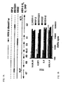
- Epitope mapping for TCC 4.2EI and 2.3 D.M. Koelle et al., 1994, J. Virol., 68:2803-2810 ) was improved with IRV DX32 ( Figure 1A ).
- This HSV-2 based virus contains a block of HSV-1 DNA neat 0.7 map units ( V.G. Preston et al., 1978, J. Virol., 28:499-517 ).
- the U L 48 gene product has the HSV-2 phenotype, as shown by reactivity with HSV-2 type-specific, VP16-specific ( D.M. Koelle et al., 1994, J. Virol., 68:2803-2810 ) T-cell clone 1A.B.25.
- the U L 49 ( Figure 2 ) and U L 50 gene products ( M.V. Williams, 1987, Virology, 156:282-292 ; F. Wohlrab, 1982, J. Virol., 43:935-942 ) also have a HSV-2 phenotype.
- the HSV-2 DNA present in IRV DX32 therefore includes U L 48, U L 49, U L 50, and most likely the intervening U L 49.5. Since TCC 4.2E1 and 2.3 react with RS1 G31 and DX32, but not with RP2 ( Figure 1B ), recognition of U L 49, U L 49.5, or U L 50 is most likely.
- the BamH I w fragment of HSV-2 was selected for expression cloning, since it contains the U L 49, U L 49.5, and most of the U L 50 coding sequences ( A. Cress and S.J. Triezenberg, 1991, Gene, 103:235-238 ; G.D. Elliott and D.M. Meredith, 1992, J. Gen. Virol., 73:723-736 ; N.J. Maitland et al.., 1982, Infect. Immun., 38:834-842 ), 70-90% of random colonies contained an insert; all were of viral origin.
- This clone contains a backwards 80 bp Sma I fragment of U L 49, a 262 bp Sma I fragment of HSV-2 U L 49 DNA predicted to encode amino acids 105 to 190, forward and in-frame with regards to ⁇ -galactosidase, and a 246 bp Sma I fragment of U L 49 forward but out of frame due to a delerion of a single C residue at the 262 bp Sma I fragment-242 bp Sma I fragment junction.
- Clone 3.19 contained a 583 bp Sma I fragment encoding amino acids 118-312 of U L 50, followed by backwards 80 and 96 bp Sma I fragment of U L 49.
- T-cell antigens were confirmed by targeted subcloning and overlapping peptides.
- the 262 bp Sma I fragment of U L 49 of HSV-2 encoding amino acids 105-190 was subcloned into pUEX3 to yield plasmid 49.262.12.
- This protein stimulated TCC 4.2E1 (Table 2).
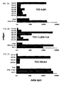
- Only peptide 105-126 of VP22 of HSV-2 (GGPVGAGGRSHAPPARTPKMTR; SEQ ID NO: 4) was stimulatoxy ( Figure 3 ).
- DNA fragments encoding U L 50 118-312 and 118-250 were subcloned into pUEX3. Fusion proteins expressing these fragments were active (Table 2). Table 2. Antigenic specificity of H5V-2 reactive TCC.
- Bacterially-dexived recombinant fusion protein antigens were used at 1:900 dilution.
- Autologous EBV-LCL (clone 4.2E1) or PBMC were used as APC.
- Data are delta cpm [ 3 H] thymidine incorporation compared to media, which was less than 500 cpm in each case.
- TCC ESL4.9 recognized a 44 amino acid fragment of U L 49 gene product VP22 (amino acids 177-220), while TCC ESL2.20 recognized a 34 amino acid fragment of U L 21 (amino acids 148-181) (Table 2).
- TCC ESL4.9 recognized a 44 amino acid fragment of U L 49 gene product VP22 (amino acids 177-220)
- TCC ESL2.20 recognized a 34 amino acid fragment of U L 21 (amino acids 148-181) (Table 2).
- Peptide mapping revealed that amino acids 187-206 ( Figure 3C ) stimulated TCC ESL4.9.
- T-cell antigens were added to the panel of HSV antigens used to probe bulk cultures of lesion-infiltiating T-cells.
- the first available specimens were a set of four biopsies (2 mm each) obtained from day 5 (virus culture positive) of a buttock recurrence of HSV-2 from patient 1 ( D.M. Koelle et al., 1998, J. Clin. Invest. 101:1500-09 ; D.M. Koelle et aL, 1994, J. Virol., 68:2803-2810 ). All four biopsies showed reactivity with VP22 105-190 but not ⁇ -galactosidase, glycoproteins B or D, or VP16.
- TCC were derived after restimulating the original bulk culture for one cycle with VP22 105-190 fusion protein.
- Proliferative responses of TCC 1.L3D5.10.8 ( Figure 3B ) to VP22 (105-190) and constituent peptides document a third T-cell epitope in VP22 contained within amino acids 125-146.
- TCC 4.2E1 The HLA restriction of the TCC recognizing antigens encoded near 0.7 map units was determined in detail. Proliferation of TCC 4.2E1, specific for VP22 105-126, is inhibited 84% by anti-DP, but less than 20% by anti-DR or and-DQ mAb. TCC 4.2E1 is from a DPB1*2001/DPB1*0402 heterozygous donor. Allogeneic EBV-LCL beating DPB1*2001, but not DPB1*0402, present antigen (Table 3), establishing restriction by DPB1*2401. Proliferation of TCC 2.3, specific for U L 50, was inhibited by anti-DR but not anti-DP or anti-DQ mAb.
- This clone is from a DRB1*0301/BRB1*0701 heterozygous donor.
- Allogeneic PBMC from a DRB1*0301 donor presented antigen, consistent with binding of antigenic peptide to this allele.
- presentation by the linked DR gene products DRw52 or DRw53 cannot be ruled out.
- Additional HLA restriction studies are summarized in Table 4.
- Table 3. Determination of restricting HLA allele of lesion-deriwed CD4 TCC 4.2E1 and 2.3. Antigens were ⁇ -gal fusion proteins (Table 2) at 1:900 dilution. Data are delta cpm [ 3 H] thymidine incorporation compared to media, which was less than 500 cpm in each case.
- cytolysis assay target TCC specificity 1 HLA restriction 2 auto HSV-2 auto peptide automock allo HSV-2 allo peptide allo mock 4.2E1 VP22 105- 126 DPB1*200 1 20.7 44.2 -4.1 -2.9 -1.7 4.6 ESL4.9 VP22187-206 DR 3 -0.6 20.2 1.3 0 0 0 1.L3D5.10.8 VP22 125- 146 DR 4 1.1 61.1 -0.3 -0.4 -0.6 -0.4 1.L3D5.10.1 2 VP22125- 146 DR 4 2.5 57.6 1.6 -0.1 -2.5 -1.4 na not available since epitope mapping was not done and synthetic antigenic peptide was not made.
- nd not done. Indicates peptide used (1 ⁇ M) to load targets in CTL assay for selected TCC. 2 Maximum extent of definition of HLA restricting locus and/or allele. Subjects RH and KM were typed serologically; others were typed at the DNA level. 3 Subject is heterozygous for HLA DRB1*0402 and DRB1*1301 and restricting allele has not been determined. 4 Subject is heterozygous for HLA DRB1*0301 and DKB1*1102 and restricting allele has not been determined.
- TCC BM.17 The HLA restriction of TCC BM.17 was studied in detail. Proliferation of TCC BM.17 and the similar clone SB.17 was inhibited 90% by anti-DQ, but less than 25% by anti-DR or anti-DP mAb. Donors BM and SB are heterozygous for HLA DQB1*0201/0501. At high concentrations of peptide, both DQB1*0201- and DQB1*0501 homozygous EBV-LCL appeared to present antigen to TCC BM.17.
- HSV-specific T-cells selectively infiltrate recurrent genital H5V-2 lesions ( D.M. Koelle et al., 1994, J. Infect. Dis., 169:956-961 ).
- Local CTL activity, with CD4 and CD8-mediated components, is correlated with viral clearance ( D.M. Koelle et al.,1998, J. Clin. Invest. 101:1500-09 ).
- the antigens recognized by local HSV-specific T cells are diverse and in many cases unknown ( D.M. Koelle et al., 1994, J. Virol., 68:2803-2810 ). This example documents recognition of tegument protein VP22.
- VP22 reactivity with VP22 was detected in two independent expression cloning experiments with lesion-infiltrating TCC from two donors. VP22 reactivity was also detected during screening of the first available set of bulk lesion-infiltrating lymphocyte cultures. Ten additional clones from three patients have been negative with the disclosed fragments of U L 49, U L -21, and U L 50.
- Tegument antigens may be suitable targets for lesion-infiltrating CD4 T-cells because of their abundance.
- VP16 and VP22 are present in large amounts: on the order of 1.6 x 10 3 molecules of VP16 ( Y. Zhang and J.L.C. McKnight, 1993, J. Virol., 67:1482-1492 ) and 2.5-2.8 x 10 3 molecules of VP22 ( J. Leslie et al., 1996, Virology, 220:60-68 ) are incorporated into each virion in HSV-1.
- polypeptides expressed as C-terminal fusion to VP22 can be co-transported into cells, expression of proteins as VP22 fusions may be of interest as a type of adjuvant preparation. This can be tested by expression of heterologous epitopes in VP22.
- VP16 and VP22 of HSV-1 are strongly, noncovalently associated in infected cells as shown by coimmunoprecipitation. These proteins co-localize in the perinuclear area of cells ( G. Elliott et al., 1995, J. Virol., 69:7932-7941 ; G.D. Elliott et al., 1992, J. Gen. Virol., 73:723-736 ).
- expression cloning has allowed discovery of novel HSV T-cell antigens.
- the in situ enrichment of antigen-specific CD4 T-cells in lesions allows study of the antigenic repertoire unbiased by secondary in vitro stimulation with antigen.
- the favorable characteristics of the HSV genome allow direct use of libraries of whole viral DNA. Tegument proteins are candidates together with membrane glycoproteins for use as HSV vaccines in humans.
- the gene was cloned by PCR from DNA prepared from HSV type 2 strain HG52 using primers GGAAGATCTACCTCTCGCCGCTCCGTCA (SEQ ID NO: 5) at the 5' end of the gene and CCGGAATTCTTGTCTGTCGTCTGAACGCG (SEQ ID NO: 6) at the 3' end of the gene.
- PCR product was digested with Bgl II and EcoR I and cloned into the Bgl II and EcoR I sites in the TA cloning vector pcR2.1-Topo (Invitrogen).
- pGEX2TU-L49HSV2 or control empty vector was transformed into E. coli strain BL21 Bacteria in log-phase growth were adjusted to an OD 600 of 0.4 in LB-ampicillin media. To some tubes isopropyl beta-D-thiogalactopyranoside (IPTG) was added to 0.3 mM. Bacteria were cultured for 1.5 hours at 37°C with rotation. Bacteria were collected by centrifugation and washed 3X in PBS containing 1 mM EDTA, heated to 65°C for 10 minutes, and washed twice more with PBS, and resuspended at approximately 1 X 10 9 bacteria/ml in T-cell medium. Heat-killed bacterial suspensions were used as test antigen.
- IPTG isopropyl beta-D-thiogalactopyranoside
- U L 49 protein of HSV-2 was separately re-amplified by polymerase chain reaction using a high-fidelity DNA polymerase with proof-reading function. The same primers and template were used. The gene was cloned directly into the Bgl II and EcoR I sites of pEGFP-C1 (Clontech). The entire U L 49 gene was sequenced and agreed with published sequence. The predicted amino acid sequence of the expressed protein is identical to that predicted for viral U L 49 except that the initial methionine at amino acid 1 is missing. A N-terminal fusion domain derived from vector pEGFP-C1 is also predicted to be expressed. This plasmid is named pEGFP-C1-UL49HSV2.
- pEGFP-C1-UL49HSV2 plasmid DNA or pEGFP-C1vector control DNA was transfected into Cos-7 cells by lipofection. After 48 hours, cells were scraped and sonicated and a supernatant and pellet phase prepared. Cells from a 9.4 cm 2 dish were used to prepare 300 microliters of supernatant. The pellet from a 9.4 cm 2 dish was resuspended in 300 microliters medium. Supernatant and pellet preparations were used as test antigens.
- test antigens were added to assay wells (96-well, U-bottom) in 200 microliters of T-cell medium containing 1 X 10 5 autologous irradiated peripheral blood mononuclear cells (PBMC) per well and 1 X 10 4 lesion-detived CD4-bearing T-cell clone ESL4.9 for U L 49 (Koelle et al, 1994 and 1998). Assays were performed in duplicate or triplicate. After three days, 3 H thymidine incorporation was measured as described in Example 1.
- PBMC peripheral blood mononuclear cells
- Results are expressed as stimulation index (mean cpm 3 H thymidine incorporation test antigen/mean cpm 3 H thymidine incorporation media control) and delta cpm (mean cpm 3 H thymidine incorporation test antigen minus mean cpm 3 H thymidine incorporation media control). Positive and negative control antigens were run as indicated and as described in Example 1. Table 5. Antigenicity of full-length HSV-2 U L 49 expressed prokaxyorically in E.
- HSV-2 protein U L 49 retains its immunogenicity when expressed as a full-length protein.
- U L 49 was studied in both prokaryotic and eukaryotic systems.
- This example supports the utility of preventative and therapeutic uses of the antigens of the invention by demonstrating the prevalence of responses to these antigens among the population. To do this, seven individuals who were HSV-2 infected as documented by type-specific serology were surveyed. These individuals were different from the individuals from whom the index T-cell clones were recovered from HSV-2 lesions.
- PBMC peripheral blood mononuclear cells
- Reactivity to individual HSV-2 proteins was assessed as follows. Proliferation assays were set up on 96-well round bottom microtiter plates, and each condition was performed in triplicate. To each well, 1 X 10 5 autologous irradiated (3300 rad gamma) PBMC were added as antigen presenting cells. To each well, 1 X 10 4 B1 cells were added. The following control substances were added: media, UV-treated mock virus preparation diluted 1:500, UV-treated HSV-2 strain 333 diluted 1:500, glycoproteins B or D or VP16 protein of HSV-2 (purified) at 4 micrograms per ml final concentration.
- the response to UV-treated HSV-2 was expected to be positive and served as a positive control for the viability and overall specificity of the cells.
- Glycoproteins B and D and VP16 were previously shown to be targets of HSV-specific T-cells ( D. M. Koelle et al., 1994, J. Virol 68(5):2803-2810 ).
- the cloning of the full-length gene and its expression in the eukaryotic Cos-7 system was as described above, as was the preparation of control antigens based on the empty vector.
- the supernatant and pellet after sonication of transfected Cos-7 cells was used at a final dilution of 1:20 in triplicate proliferation assays.
- COS-7 cells (100 mm 2 ) were transfected with the ICP0 genomic clone, and total RNA was prepared after 48 h.
- the primer used for cDNA synthesis (TGC TCTAGA GACTCGATCCCTGCGCGTCGG; Xba I site underlined) (SEQ ID NO: 7) was from the 3'-end of the HSV-2 DNA in the ICP0 genomic clone.
- Moloney murine leukemia virus reverse transcriptase (Life Technologies) was used per the manufacturer.
- PCR used pfu cDNA polymerase, the above 3'-primer, and 5'-primer TAA GGTACC TGAACCCCGGCCCGGCACGAGC (SEQ ID NO: 8) ( Kpn I site).
- PCR used the same 5'-primer and 3'-primer TGC TCTAGA CCAGGCGTGCGGGGCGGCGGG (SEQ ID NO: 9) ( Xha I site). Reaction conditions were individually optimized. Product was digested with Acc 65I and Xba I , gel purified, and ligated into similarly treated pCDNA 3.1-His-B, and in-frame insertion was confirmed by sequencing.
- CD8 clone 5101.1999.23 recognized COS-7 cells co-transfected with HLA A*0201 and a HSV-2 Sau 3aI fragment from bp 102,943-102,876 ( Dolan, A. et al., 1998, J. Virol. 72:2010 ) (Table 8).
- the predicted fusion protein contains HSV-2 U L 47 aa 278-298. Reactivity with U L 47 was confirmed by cotransfection of A*0201 and full-length HSV-2 U L 47 (Table 9). Table 9.
- COS-7 cells were transfected with HLA cDNA and HSV-2 DNA or cDNA as shown. T cell activation was detected by IFN- ⁇ secretion, reported as the mean of duplicate wells.
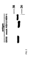
- Two CTL clones are shown to react with U L 47. The epitope recognized by clone 1874.1991.22 is localized to aa 535-590 of U L 47, and the epitope recognized by clone 1874.1997.51 is localized to aa 26-105 of ICP0. Values are mean of duplicate IFN-y secretion into the medium as measured by ELISA. ORF, Open reading frame.
- the CD8 T cell clone 1874.1991.22 recognized COS-7 cells cotransfected with HLA A*0201 and a HSV-2 Sau 3aI fragment from bp 102,875-101,383 (Table 8). This fragment was predicted to contain the DNA encoding U L 47 aa 300-696, intervening DNA, and then aa 1-71 of U L 46. Analysis of the 5'-vector-insert junction in C:2:C10:B9 revealed out-of-frame translation of the initial U L 47 DNA. The insert is expected to contain the U L 46 promoter. The epitope, therefore, could be encoded by U L 46. C:2:C10:B9 also contains potential sites of internal initiation of translation within U L 47.
- CD8 clone 5491.2000.48 The specificity of CD8 clone 5491.2000.48 was determined with a panel of partial- and full-length HSV-2 genes.
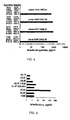
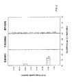
- the HSV-2 genes studied were previously shown to be recognized by CD4 T call clones (See U.S. Patent 6,375,952, issued April 23, 2002 ). Only HSV-2 U L 49, when cotransfected with HLA B*0702, stimulated IFN- ⁇ release by clone 5491.2000.48 ( Fig. 5 ).
- HSV-2 gene U L 47 encodes protein VP13/14, whereas U L 49 encodes VP22; both tegument proteins are loaded into the cytoplasm on virion binding and entry.
- the small genomic HSV-2 fragment of U L 47 recognized by clone 5101.1999.23 was scanned for peptides fitting the A*0201 binding motif (http://134.2.96.221/ and http://bitnas.dcrt.nih.gov/molbio/ hla_bind/).
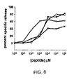
- Peptide U L 47 (HSV-2) 289-298 had a 50% effective concentration (EC 50 ) in the 1-10 nM range in cytolysis assays ( Fig. 6 ).
- U L 47 535-590 (Table 9) was similarly analyzed.
- Peptide 551-559 was active at 1 nM ( Fig. 6 ).
- Potential HLA B*0702-binding peptides in U L 49 of HSV-2 were synthesized, and two (aa 47-55 and 14-22) were active at 1 ⁇ M. Titration ( Fig. 6 ) showed that U L 49 49-57 was highly active, with an EC 50 of ⁇ 10 nM, whereas U L 49 14-22 had activity only at 1 ⁇ M
- the antigenic peptides in U L 47 and U L 49 contain significant amino acid sequence differences from the corresponding predicted HSV-1 peptides ( Dolan, A. et al., 1998, J. Virol. 72:2010 ; McGeoch, D. J. et al., 1988, J. Gen. Virol. 69:1531 ), explaining type-specific recognition of HSV-2 (Table 8).
- ICP0 HLA B45-restricted epitope
- GAETFYVDGA SEQ ID NO: 15
- HSV-2 is mainly present in keratinocytes.
- MOI amount of virus
- time of infection and pretreatment with TFN- ⁇ influenced lysis of dermal fibroblasts and keratinocytes was investigated.
- fibroblasts Fig. 7
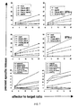
- IFN- ⁇ pretreatment in the absence of IFN- ⁇ pretreatment, infection for 2 h led to detectable lysis, which increased with increasing MOI. Lysis was undetectable ( ⁇ 5% specific release at E:T of 20:1) after overnight infection with MOI 1, 5, or 25.
- IFN- ⁇ pretreatment lysis was generally increased, but 2-h infection was still superior. HLA-mismatched target cells were not lysed, even after peptide loading.
- Kerarinocytes showed some similarities and differences from fibroblast as target cells ( Fig. 7 ). IFN- ⁇ pretreatment generally increased recognition, without leading to lysis of control cells. In contrast to fibroblasts, 18-h infection was generally required. Weak cytolysis of cells infected for 2 h was noted only for IFN- ⁇ -pretreated targets. Chromium release again correlated directly with the amount of infectious virus added, because no specific lysis was noted at MOI 1 or 5.
- the first two clones are HLA A*0201-restricted. Lysis of the A*0201-bearing wild-type EBV-LCL 1874, but not non-A*0201 EBV-LCL 5491, was detected after peptide loading (U L 47 551-559 for clone 1874.1991.22; U L 47 551-559 for clone 5101.1999.23; 1 ⁇ M, 90 min) or HSV-2 infection (MOI 10,18 h). In contrast, peptide loading, but not HSV-2-infection, was able to sensitize TAP-deficient cell lines.
- HSV-2 causes considerable morbidity and mortality, especially in neonates. Because of the chronic nature of the infection, the limitations of antiviral therapy, and the frequency with which transmission is caused by asymptomatic shedding of the virus, vaccination is likely to be required to reduce new HSV-2 infections. The recent report that vaccination with a specific adjuvant and an envelope glycoprotein induced partial protection in HSV-1/HSV-2-seronegative women highlights both the potential efficacy of vaccination and the need for improved formulations and markers of effective immunity.
- HSV-1 early protein thymidine kinase (tk) is recognized by CD8 clones from PBMC of subjects treated with tk -transfected autologous cells, but this is likely a primary immune response.
- a PBMC-derived CD8 T cell clone specific for a melanoma-associated protein (Melan A/MART-1) also reacted with a peptide from HSV-1 glycoprotein C.
- U L 49 encodes VP22, a tegument protein required for viral replication.
- U L 49 protein is also abundant in virions and delivered into the cytoplasm by virus entry. Lysis of EBV-LCL by tegument-specific CD8 CTL was not inhibited by blockade of gene transcription or infection with a replication-incompetent virus, consistent with the processing and presentation of preformed virion input protein.
- TAP-independent processing has been reported in other viral systems.
- the examination of three discrete epitopes in tegument proteins did not reveal evidence for TAP-independent Ag processing of HSV epitopes.
- the CD8 response seems to "evade the evasion," at least in the cases examined to date, while continuing to rely on TAP for Ag processing.
- EBV-LCL EBV-LCL
- tegument protein-specific CTL a short time of infection (2 h) was adequate for target cell sensitization for lysis by tegument protein-specific CTL.
- U L 47 and U L 49 tegument proteins are synthesized with "late" kinetics, typically starting after 6 h or more of viral infection, these data are also consistent with recognition of preformed Ag in fibroblasts. Lysis was MOI dependent. Because HSV preparations typically contain a large number of defective particles, it is likely that tegument proteins were also being delivered into fibroblasts by noninfectious particles.
- IFN- ⁇ pretreatment was able to partially restore lysis of 18-h-infected cells.
- recognition of keratinocytes after 18 h of infection was superior to recognition after 2 h of infection.
- the reason for the difference between fibroblasts and keratinocytes is unknown.
- IFN- ⁇ pretreatment was able to restore some lysis of 2-h-infected cells, and further improved recognition of 18-h-infected cells.
- Tegument proteins have not previously been described as targets of the HSV-specific CD8 T cell response.
- CD4 responses to HSV-1 U L 47 have been detected in HSV-mediated acute retinal necrosis.
- CD4 responses to U L 49 are commonly detected among lesion-infiltrating HSV-2-specific clones. Because responses to U L 49 are also present in the cornea in herpes stromal keratitis in humans, a disease that may be driven by pathogenic Th1-like T cells, caution is warranted in using this protein as a vaccine.
- U L 49 is the only known HSV-2 protein recognized by both CD4 and CD8 T cell clones recovered from herpetic lesions. A unique intercellular transport pathway allows highly efficient uptake of soluble U L 49 protein into a variety of epithelial cell types which could also intersect antigen processing pathways.
- HSV-2 seropositive donors were obtained (AD104, AD116, AD120, D477, HV5101, JH6376, EB5491, TM10062).
- Donors HV5101 and EB5491 experience frequent anogenital lesion recurrences.
- Donors JH6376 and TM10062 experience few or no anogenital lesion recurrences.
- Leukopheresis PBMC were obtained from each donor after informed consent.
- Donor PBMC were HLA-typed by low resolution DNA typing methodology.
- Synthetic peptides (15 amino-acids in length and overlapping in sequence by 11 ammo-acids) were synthesized that spanned the following HSV-2 polypeptides: U L 47 (aa 1-696), U L 49 (aa 1-300), ICP27 (aa 1-512).
- Peptides (5 mg each) were delivered in lyophilized form in glass vials and dissolved at a concentration of 10 mg/ml in DMSO, transferred to sterile cryovials and stored at -20 degrees C. The peptides were screened with CD8+ T cells purified from adherent macrophage-depleted PBMC.
- ELISPOT plates were subsequently developed per a standard protocol. The number of spots per well was counted using an automated video-microscopy ELISPOT reader. Peptides in pools scoring positive were subsequently tested individually in a second ELISPOT assay.
- AD116 the novel peptides U L 49/21-35 (#6)and U L 49/193-208 (#49) scored positive both pooled and individually.
- AD116 also recognized the previously described B*0702-restricted epitope U L 49/49-57 contained in 15-mer peptides U L 49/45-59 (#12) and U L 49/49-63 (#13).
- D477, HV5101, and JH6376 T cells recognized the previously described HLA-A*0201-restricted epitopes U L 47/289-297 and U L 47/550-559 contained in 15-mers #73/#74 and #137/#138, respectively.
- EB5491 T cells recognized the previously described B*0702-restricted epitope U L 49/49-57 epitope contained in 15-mer peptides U L 49/45-59 (#12) and U L 49/49-63 (#13).
- D477 scored positive for peptide pool U L 49 (#11-20).
- TM10062 did not score positive on any peptide pool from U L 47 or U L 49.
- Figure 8 shows the results for AD116.
- Figure 9 shows the results for EB5491, TM10062 and HV5101.
- the peptide hit indicated with a "1” represents U L 49, amino acids 49-57, B*0702-restricted.
- Figure 10 shows results for AD104, AD116, AD120 and D477.
- the peptide hit indicated with a "1” represents peptide #54.
- the peptide hit indicated with a "2” represents peptide #49.
- the peptide hit indicated with a "3" represents U L 49, amino acids 49-57, B*0702-restricted.
- the peptide hit indicated with a "4" represents peptide #6.
- Example 6 Detection of HSV-specific T-cell responses in cervical lymphocytes
- HSV-specific T cells including CD8+ cells, can be detected in cervical lymphocytes.
- Example 7 Detection of HSV-specific T-cell responses in primary genital HSV-2 lesions
- biopsy specimens were collected from a patient presenting with symptoms consistent with primary genital HSV-2 infection.
- the phenotypes of the collected cells were determined, and LIL and PBMC from the specimens were subjected to proliferative and cytotoxicity assays.
- the results show that the HSV-specific proliferative and cytotoxic responses of CTL present in primary genital HSV-2 lesions are typical of those detected during recurrent disease.
- CW7477 developed dysuria, fever, buttock, and lower abdomen lesions three days after his last sexual contact. Lesions lasted 13 days and grew HSV-2.
- Acyclovir treatment was begun on day four of symptoms. Biopsies were done on days four and seven. Serostatus was atypical positive (only a few bands present on immunoblot) at day four, with more bands, but still less than most convalescent sera, on day 26, by enhanced chemiluminescence (ECL; Dalessio J. and Ashley R., J. Clin. Microbiol. 1992, 30(4):1005-7 ) variant of type-specific HSV-2 immunoblot. The clinical and laboratory data were consistent with primary genital HSV-2 infection. Biopsy specimens were obtained on day four and seven of symptoms and bulk LIL expanded with PHA/IL-2 as described above.
- the phenotype of the expanded cells was split between CD4 and CD8 cells, with 15-25% CD3+/CD16,56+ cells and 5-10% TCR ⁇ + cells in the LIL.
- cells from normal skin had almost no CD16,56 (+) events and no TCR ⁇ cells.
- the nature of the CD3+/CD16,56+ cells is unknown but these are frequently seen in expanded LIL.
- the antibody cocktail has a combination of ⁇ CD16-PE and ⁇ CD56-PE.
- HSV-specific proliferative and cytotoxic responses were fairly typical of those detected during recurrent disease ( Koelle DM et al., J. Clin. Invest. 1998; 101:1500-1508 ).
- Cross-reactive responses to HSV-1 and HSV-2 were present, as were antigen-specific responses to HSV glycoproteins. Normal skin responses were low, and PBMC responses were developing by day 15.
Landscapes
- Health & Medical Sciences (AREA)
- Life Sciences & Earth Sciences (AREA)
- Chemical & Material Sciences (AREA)
- Organic Chemistry (AREA)
- Virology (AREA)
- General Health & Medical Sciences (AREA)
- Medicinal Chemistry (AREA)
- Immunology (AREA)
- Molecular Biology (AREA)
- Nuclear Medicine, Radiotherapy & Molecular Imaging (AREA)
- Proteomics, Peptides & Aminoacids (AREA)
- General Chemical & Material Sciences (AREA)
- Animal Behavior & Ethology (AREA)
- Chemical Kinetics & Catalysis (AREA)
- Public Health (AREA)
- Veterinary Medicine (AREA)
- Pharmacology & Pharmacy (AREA)
- Genetics & Genomics (AREA)
- Engineering & Computer Science (AREA)
- Biophysics (AREA)
- Biochemistry (AREA)
- Gastroenterology & Hepatology (AREA)
- Oncology (AREA)
- Communicable Diseases (AREA)
- Biotechnology (AREA)
- Bioinformatics & Cheminformatics (AREA)
- Micro-Organisms Or Cultivation Processes Thereof (AREA)
- Peptides Or Proteins (AREA)
- Medicines Containing Antibodies Or Antigens For Use As Internal Diagnostic Agents (AREA)
- Medicines That Contain Protein Lipid Enzymes And Other Medicines (AREA)
- Pharmaceuticals Containing Other Organic And Inorganic Compounds (AREA)
- Medicines Containing Material From Animals Or Micro-Organisms (AREA)
- Preparation Of Compounds By Using Micro-Organisms (AREA)
Abstract
Arg Pro Arg Gly Glu Val Arg Phe Leu (SEQ ID NO: 1) or
Arg Gln Arg Gly Glu Val Arg Phe Val (SEQ ID NO: 2)
and having a length of less than 50 amino acids, or a variant of such a fragment, having a length of less than 50 amino acids and exhibiting at least 70% identity therewith, and which elicits a cellular immune response to HSV-2 or HSV-1, respectively, (b) a fusion protein comprising at least one above-defined fragment or variant and an unrelated sequence or comprising multiple above-defined fragments or variants and (2) a pharmaceutically acceptable carrier.
Description
- The invention relates compositions that can be used for the treatment and prevention of Herpes Simplex Virus (HSV) infection. More particularly, the invention identifies epitopes of HSV proteins having the antigenic specificity of HSV-specific T cells, and in particular, of CD8+ as well as CD4+ T cells.
- Cellular immune responses are required to limit the severity of recurrent HSV infection in humans. Initial genital HSV-2 infections are prolonged and severe, while recurrences are less severe and more frequently asymptomatic. Resolution of primary HSV-2 infection is associated with infiltration of antigen-specific T cells, including CD8+ cytotoxic T lymphocytes (CTLs). Serial lesion biopsy studies of recurrent HSV-2 infection in humans has shown a shift to CD8+ predominance as lesions mature and correlation of local CTL activity with virus clearance (Koelle, DM et al., J. Clin. Invest. 1998, 101:1500-1508; Cunningham, AL et al., J, Clin, Invest. 1985, 75:226-233). Thus, HSV antigens recognized by CD8+ CTL can be used for novel therapies and vaccines.
- The complete DNA sequence of herpes simplex virus (HSV) is approximately 150 kb and contains about 85 known genes, each of which encodes a protein in the range of 50-1000 amino acids in length. Mostly unknown are the immunogenic epitopes within these proteins, each epitope approximately 9-12 amino acids in length, that are capable of eliciting an effective T cell immune response to viral infection.
- Our prior
PCT Application Publication WO 00/8051 - There remains a need to identify further specific epitopes capable of eliciting an effective immune response to HSV infection. Such information can lead to the identification of more effective immunogenic antigens useful for the prevention and treatment of HSV infection.
- The invention provides a pharmaceutical composition comprising (1) a polypeptide which is (a) a fragment of the herpes simplex virus (HSV)
protein U L49 comprising amino acids 49-57 of, respectively, HSV-2 or HSV-1:
Arg Pro Arg Gly Glu Val Arg Phe Leu (SEQ ID NO; 1) or
Arg Gln Arg Gly Glu Val Arg Phe Val (SEQ ID NO: 2)
and having a length of less than 50 amino acids, or a variant of such a fragment, having a length of less than 50 amino acids and exhibiting at least 70% identity therewith, and which elicits a cellular immune response to HSV-2 or HSV-1, respectively, (b) a fusion protein comprising at least one above-defined fragment or variant and an unrelated sequence or comprising multiple above-defined fragments or variants and (2) a pharmaceutically acceptable carrier. The pharmaceutical composition can be used both prophylactically and therapeutically. The polypeptide antigens defined above are recognized by T cells recovered from herpetic lesions. The invention additionally provides the pharmaceutical composition defined above for use in preventing or treating HSV infection, a polynucleotide encoding an above-defined polypeptide; a vector including the polynucleotide; a host cell transformed with the vector and a recombinant virus genetically modified to express the polynucleotide, for killing HSV-infected cells, for inhibiting viral replication, for enhancing secretion of antiviral and/or immunomodulatory lymphokines, and for enhancing production of HSV-specific antibody, and generally for stimulating and/or augmenting HSV-specific immunity. - An ex-vivo method for producing immune cells directed against HSV is also provided by the invention. The method comprises contacting an immune cell with an APC, preferably a dendritic cell, that has been modified to present an epitope by a polypeptide as defined above. In a preferred embodiment, the immune cell is a T cell such as a CD4+ or CD8+ T cell.
- In a preferred embodiment, the polypeptide is a fragment of a
U L49 protein comprising amino acids 45-59 or 49-63 ofU L49 or a substitutional variant thereof.
UL49 45-59 has the sequence: - Pro Met Arg Ala Arg Pro Arg Gly Glu Val Arg Phe Leu His Tyr (SEQ ID NO: 3) or
- Gln Arg Ser Ala Arg Gln Arg Gly Glu Val Arg Phe Val Gln Tyr (SEQ ID NO: 4).
- UL49 49-63 has the sequence:
- Arg Pro Arg Gly Glu Val Arg Phe Leu His Tyr Asp Glu Ala Gly (SEQ ID NO: 5) or
- Arg Gln Arg Gly Glu Val Arg Phe Val Gln Tyr Pro Glu Ser Asp (SEQ ID NO: G).
-
-
Figure 1A is a schematic representing organization of the HSV genome in the region of 0.67-0.73 map units. Boundaries are approximate. HSV-1 X HSV-2 intertypic recombinant viruses (IRV) are also shown. HSV-2 DNA is indicated by a solid line; HSV-1 DNA by a dashed line, and indeterminate regions by a multiple line. The HSV-2 BamHI w fragment used for expression cloning is also shown. -
Figure 1B is a bar graph showing proliferative responses of T-cell clones (TCC) to the indicated IRV. Data are delta CPM [3H] thymidine incorporation compared to media alone, which was less than 500 cpm in each case. -
Figure 2 is an immunoblot showing determination of the HSV viral phenotype of theU L49 gene product (VP22) of IRV DX32. Lysates of mock-infected cells and cells infected with the viral strains DX32, HSV-1 or HSV-2 were separated by SDS-PAGE, blotted, and probed with VP22-specific mAb. The molecular weights (kD) of marker proteins are shown at right. -
Figure 3A is a bar graph showing T-cell proliferation elicited by various peptide epitopes in VP22 of HSV-2 using TCC 4.2E1. Antigen-presenting cells (APC) were autologous EBV-LCL. Antigens included β-galactosidase and fusion proteins used at 10 µg/ml and peptides used at 3 µM. Data are delta cpm [3H] thymidine incorporation compared to media alone, which was less than 500 cpm in each case. -
Figure 3B is a bar graph showing T-cell proliferation elicited by various peptide epitopes in VP22 of HSV-2 using TCC 1.L3D5.10.8. APC were autologous PBMC. Antigens included β-galactosidase and fusion proteins used at 10 µg/ml and peptides used at 1 µM. Data are delta cpm [3H] thymidine incorporation compared to media alone, which was less than 500 cpm in each case. -
Figure 3C is a bar graph showing T-cell proliferation elicited by various peptide epitopes in VP22 of HSV-2 using TCC ESL4.9. APC were autologous PBMC. Antigens included β-galactosidase and fusion proteins used at 10 µg/ml and peptides used at 1 µM. Data are delta cpm [3H] thymidine incorporation compared to media alone, which was less than 500 cpm in each case. -
Figure 4 shows secretion of IFN-γ by lesion-derived CD8+ T cell clones in response to COS-7 cells transfected with HLA class I heavy chain cDNA and infection with HSV-2 strain 333. Values are mean of duplicate IFN-γ secretion into medium, measured by ELISA. -
Figure 5 shows secretion of IFN-γ by lesion-derived CD8+ T cell clone 5491.2000.48 in response to COS-7 cells co-transfected with HLA B*0702 cDNA and the indicated HSV-2 DNA fragments. All HSV-2 genes are full length except forU L47, which is was tested in two segments encoding the indicated amino acids. Values are mean of duplicate IFN-γ secretion into the medium as measured by ELISA. -
Figure 6 shows lysis by lesion-derived CD8 clones of autologous LCL loaded with HSV-2 peptides at the indicated concentrations. Data are percent specific 51Cr release at E:T 20:1.●, Lysis by clone 5101.1999.23 of targets loaded with UL47 551-559; µ, clone 1874.1991.22 andV L47 289-298; ▼, clone 1874.1997.51 and ICP0 92-101; ∇, clone 5491.2000.48 andU L49 49-57. Lysis of mock-loaded targets was <5% specific release for each clone. -
Figure 7 shows cytolytic activity of CD8 CTL clones against cutaneous cells. Top, UL49-specific clone 5491.2000.48, HLA B*0702-expressing fibroblasts from subject SJ, andpeptide U L49 aa 49-47. Middle, UL 47-specific clone 1874.1991.22, HLA A*0201-bearing fibroblasts from subject 1874, andpeptide U L 47 aa 551-559. Left, Lysis of non-IFN-γ-treated fibroblasts infected at the indicated MOI for 2 h before assay; right, fibroblasts pretreated for 3 days with 500 U/ml IFN-γ and then infected or treated with peptide. In each case, HLA-mismatched target fibroblasts had <5% specific release at E:T 2:1, 6:1, and 20:1. Bottom, HLA A*0201-bearing keratinocytes are used as target cells. Left, UL 47-specific clone 5101.1999.23 andpeptide U L47 aa 289-298; right. UL 47-specific clone 1874.1991.22 andpeptide U L 47 aa 551-559. HLA A*0201-bearing keratinocytes were mock treated or treated with 500 U/ml IFN-γ and then infected for 2 or 18 h with HSV-2 atMOI 25 or loaded with peptide for 90 min. -
Figures 8A-8E show the results of peptide screening for donor AD116. -
Figure 9 shows the results of peptide screening for donors EB5491, TM 10062 and HV5101. -
Figure 10 shows results of peptide screening fordonors AD 104, AD 116, AD 120 and D477. - The invention provides HSV antigens that are useful for the prevention and treatment of HSV infection. Disclosed herein are antigens and/or their constituent epitopes confirmed to be recognized by T-cells derived from herpetic lesions. In some embodiments, T-cells having specificity for antigens of the invention have demonstrated cytotoxic activity against virally infected cells. The identification of immunogenic antigens responsible for T-cell specificity facilitates the development of improved antiviral therapeutic and prophylactic strategies. Compositions containing antigens or polynucleotides encoding antigens of the invention provide effectively targeted vaccines for prevention and treatment of HSV infection.
- All scientific and technical terms used in this application have meanings commonly used in the art unless otherwise specified. As used in this application, the following words or phrases have the meanings specified.
- As used herein, "polypeptide" includes proteins, fragments of proteins, and peptides, whether isolated from natural sources, produced by recombinant techniques or chemically synthesized. Polypeptides of the invention typically comprise at least about 6 amino acids, and preferably at least about 15 amino acids.
- As used herein, "HSV polypeptide" includes HSV-1 and HSV-2, unless otherwise indicated. References to amino acids of HSV proteins or polypeptides are based on the genomic sequence information regarding HSV-2 as described in A. Dolan et al., 1998, J. ViroL 72(3):2010-2021. As noted below, the predicted polypeptide sequence of ICP0 of HSV-2 based on sequencing RNA from cells transfected with a fragment of ICP0 differs from the published sequence by the omission of amino acid Q26.
- As used herein, "substitutional variant" refers to a molecule having one or more amino acid substitutions or deletions in the indicated amino acid sequence, yet retaining the ability to be specifically recognized by an immune cell. The amino acid sequence of a substitutional variant is preferably at least 80% identical to the native amino acid sequence, or more preferably, at least 90% identical to the native amino acid sequence. Typically, the substitution is a conservative substitution. One method for determining whether a molecule can be specifically recognized by an immune cell is the cytotoxicity assay described in D.M. Koelle et al., 1997, Human Immunol. 53:195-205. Other methods for determining whether a molecule can be specifically recognized by an immune cell are described in the examples provided hereinbelow, including the ability to stimulate secretion of interferon-gamma or the ability to lyse cells presenting the molecule. An immune cell will specifically recognize a molecule when, for example, stimulation with the molecule results in secretion of greater interferon-gamma than stimulation with control molecules. For example, the molecule may stimulate greater than 5 pg/ml, or preferably greater than 10 pg/ml, interferon-ganuma secretion, whereas a control molecule will stimulate less than 5 pg/ml interferon-gamma.
- As used herein, "vector" means a construct, which is capable of delivering, and preferably expressing, one or more gene(s) or sequence(s) of interest in a host cell. Examples of vectors include, but are not limited to, viral vectors, naked DNA or RNA expression vectors, plasmid, cosmid or phage vectors, DNA or RNA expression vectors associated with cationic condensing agents, DNA or RNA expression vectors encapsulated in liposomes, and certain eukaryotic cells, such as producer cells.
- As used herein, "expression control sequence" means a nucleic acid sequence that directs transcription of a nucleic acid. An expression control sequence can be a promoter, such as a constitutive or an inducible promoter, or an enhancer. The expression control sequence is operably linked to the nucleic acid sequence to be transcribed.
- The term "nucleic acid" or "polynucleotide" refers to a deoxyribonucleotide or ribonucleotide polymer in either single- or double-stranded form, and unless otherwise limited, encompasses known analogs of natural nucleotides that hybridize to nucleic acids in a manner similar to naturally occurring nucleotides.
- As used herein, "antigen-presenting cell" or "APC" means a cell capable of handling and presenting antigen to a lymphocyte. Examples of APCs include, but are not limited to, macrophages, Langerhans-dendritic cells, follicular dendritic cells, B cells, monocytes, fibroblasts and fibrocytes. Dendritic cells are a preferred type of antigen presenting cell. Dendritic cells are found in many non-lymphoid tissues but can migrate via the afferent lymph or the blood stream to the T-dependent areas of lymphoid organs. In non-lymphoid organs, dendritic cells include Langerhans cells and interstitial dendritic cells. In the lymph and blood, they include afferent lymph veiled cells and blood dendritic cells, respectively. In lymphoid organs, they include lymphoid dendritic cells and interdigitating cells.
- As used herein, "modified" to present an epitope refers to antigen-presenting cells (APCs) that have been manipulated to present an epitope by natural or recombinant methods. For example, the APCs can be modified by exposure to the isolated antigen, alone or as part of a mixture, peptide loading, or by genetically modifying the APC to express a polypeptide that includes one or more epitopes.
- As used herein, "pharmaceutically acceptable carrier" includes any material which, when combined with an active ingredient, allows the ingredient to retain biological activity and is non-reactive with the subject's immune system. Examples include, but are not limited to, any of the standard pharmaceutical carriers such as a phosphate buffered saline solution, water, emulsions such as oil/water emulsion, and various types of wetting agents. Preferred diluents for aerosol or parenteral administration are phosphate buffered saline or normal (0.9%) saline.
- Compositions comprising such carriers are formulated by well known conventional methods (see, for example, Remington's Pharmaceutical Sciences, 18th edition, A. Gennaro, ed., Mack Publishing Co., Easton, PA, 1990).
- As used herein, "adjuvant" includes those adjuvants commonly used in the art to facilitate the stimulation of an immune response. Examples of adjuvants include, but are not limited to, helper peptide; aluminum salts such as aluminum hydroxide gel (alum) or aluminum phosphate; Freund's Incomplete Adjuvant and Complete Adjuvant (Difco Laboratories, Detroit, MI); Merck Adjuvant 65 (Merck and Company, Inc., Rahway, NJ); AS-2 (Smith-Kline Beecham); QS-21 (Aquilla); MPL or 3d-MPL (Corixa Corporation, Hamilton, MT); LEIF; salts of calcium, iron or zinc; an insoluble suspension of acylated tyrosine; acylated sugars; cationically or anionically derivatized polysaccharides; polyphosphazenes; biodegradable microspheres; monophosphoryl lipid A and quil A; muramyl tripeptide phosphatidyl ethanolamine or an immunostimulating complex, including cytokines (e.g., GM-CSF or interleukin-2, -7 or -12) and immunostimulatory DNA sequences. In some embodiments, such as with the use of a polynucleotide vaccine, an adjuvant such as a helper peptide or cytokine can be provided via a polynucleotide encoding the adjuvant.
- As used herein, "a" or "an" means at least one, unless clearly indicated otherwise.
- As used herein, to "prevent" or "protect against" a condition or disease means to hinder, reduce or delay the onset or progression of the condition or disease.
- In an embodiment, the polypeptide is a fragment which comprises amino acids 45-59, 49-57, 49-63, of
V L49 or a substitutional variant thereof. The reference to amino acid residues is made with respect to the proteins of the HSV-2 genome as described in A. Dolan et al., 1998, J. Virol. 72(3):2010-2021. - The polypeptide can be a fusion protein. In one embodiment, the fusion protein is soluble. A soluble fusion protein of the invention can be suitable for injection into a subject and for eliciting an immune response. Within certain embodiments, a polypeptide can be a fusion protein that comprises multiple polypeptides as described herein, or that comprises at least one polypeptide as described herein and an unrelated sequence. A fusion partner may, for example, assist in providing T helper epitopes (an immunological fusion partner), preferably T helper epitopes recognized by humans, or may assist in expressing the protein (an expression enhancer) at higher yields than the native recombinant protein. Certain preferred fusion partners are both immunological and expression enhancing fusion partners. Other fusion partners may be selected so as to increase the solubility of the protein or to enable the protein to be targeted to desired intracellular compartments. Still further fusion partners include affinity tags, which facilitate purification of the protein.
- Fusion proteins may generally be prepared using standard techniques, including chemical conjugation. Preferably, a fusion protein is expressed as a recombinant protein, allowing the production of increased levels, relative to a non-fused protein, in an expression system. Briefly, DNA sequences encoding the polypeptide components may be assembled separately, and ligated into an appropriate expression vector. The 3' end of the DNA sequence encoding one polypeptide component is ligated, with or without a peptide linker, to the 5' end of a DNA sequence encoding the second polypeptide component so that the reading frames of the sequences are in phase. This permits translation into a single fusion protein that retains the biological activity of both component polypeptides.
- A peptide linker sequence may be employed to separate the first and the second polypeptide components by a distance sufficient to ensure that each polypeptide folds into its secondary and tertiary structures. Such a peptide linker sequence is incorporated into the fusion protein using standard techniques well known in the art. Suitable peptide linker sequences may be chosen based on the following factors: (1) their ability to adopt a flexible extended conformation; (2) their inability to adopt a secondary structure that could interact with functional epitopes on the first and second polypeptides; and (3) the lack of hydrophobic or charged residues that might react with the polypeptide functional epitopes. Preferred peptide linker sequences contain Gly, Asn and Ser residues. Other near neutral amino acids, such as Thr and Ala may also be used in the linker sequence. Amino acid sequences which may be usefully employed as linkers include those disclosed in Maratea et al., 1985, Gene 40:39-46; Murphy et al., 1986, Proc. Natl. Acad. Sci. USA 83:8258-8262;
U.S. Patent No. 4,935,233 andU.S. Patent No. 4,751,180 . The linker sequence may generally be from 1 to about 50 amino acids in length. Linker sequences are not required when the first and second polypeptides have non-essential N-terminal amino acid regions that can be used to separate the functional domains and prevent steric interference. - The ligated DNA sequences are operably linked to suitable transcriptional or translational regulatory elements. The regulatory elements responsible for expression of DNA are located 5' to the DNA sequence encoding the first polypeptides. Similarly, stop co dons required to end translation and transcription termination signals are present 3' to the DNA sequence encoding the second polypeptide.
- Fusion proteins are also provided that comprise a polypeptide of the present invention together with an unrelated immunogenic protein. Preferably the immunogenic protein is capable of eliciting a recall response. Examples of such proteins include tetanus, tuberculosis and hepatitis proteins (see, for example, Stoute et al., 1997, New Engl. J. Med., 336:86-9).
- Within preferred embodiments, an immunological fusion partner is derived from protein D, a surface protein of the gram-negative bacterium Haemophilus influenza B (
WO 91/18926 terminal 81 amino acids are used, although different fragments that include T-helper epitopes may be used. - In another embodiment, the immunological fusion partner is the protein known as LYTA, or a portion thereof (preferably a C-terminal portion). LYTA is derived from Streptococcus pneumoniae, which synthesizes an N-acetyl-L-alanine amidase known as amidase LYTA (encoded by the LytA gene; Gene 43:265-292,1986). LYTA is an autolysin that specifically degrades certain bonds in the peptidoglycan backbone. The C-terminal domain of the LYTA protein is responsible for the affinity to the choline or to some choline analogues such as DEAE. This property has been exploited for the development of E. coli C-LYTA expressing plasmids useful for expression of fusion proteins. Purification of hybrid proteins containing the C-LYTA fragment at the amino terminus has been described (see Biotechnology 10:795-798,1992). Within a preferred embodiment, a repeat portion of LYTA may be incorporated into a fusion protein. A repeat portion is found in the C-terminal region starting at residue 178. A particularly preferred repeat portion incorporates residues 188-305.
- In some embodiments, it may be desirable to couple a therapeutic agent and a polypeptide of the invention, or to couple more than one polypeptide of the invention. For example, more than one agent or polypeptide may be coupled directly to a first polypeptide of the invention, or linkers that provide multiple sites for attachment can be used. Alternatively, a carrier can be used. Some molecules are particularly suitable for intercellular trafficking and protein delivery, including, but not limited to, VP22 (Elliott and O'Hare, 1997, Cell 88:223-233; see also Kim et al., 1997, J. Immunol. 159:1666-1668; Rojas et al., 1998, Nature Biotechnology 16:370; Kato et al., 1998, FEBS Lett. 427(2):203-208; Vives et al.,1997, J. Biol. Chem. 272(25):16010-7; Nagahara et al., 1998, Nature Med. 4(12):1449-1452).
- A carrier may bear the agents or polypeptides in a variety of ways, including covalent bonding either directly or via a linker group. Suitable carriers include proteins such as albumins (e.g.,
U.S. Patent No. 4,507,234, to Kato et al. ), peptides and polysaccharides such as aminodextran (e.g.,U.S. Patent No. 4,699,784, to Shih et al. ). A carrier may also bear an agent by noncovalent bonding or by encapsulation, such as within a liposome vesicle (e.g.,U.S. Patent Nos. 4,429,008 and4,873,088 ). - In general, polypeptides (including fusion proteins) and polynucleotides as described herein are isolated. An "isolated" polypeptide or polynucleotide is one that is removed from its original environment. For example, a naturally occurring protein is isolated if it is separated from some or all of the coexisting materials in the natural system.
Preferably, such polypeptides are at least about 90% pure, more preferably at least about 95% pure and most preferably at least about 99% pure. A polynucleotide is considered to be isolated if, for example, it is cloned into a vector that is not part of the natural environment. - The polypeptide can be isolated from its naturally occurring form, produced by recombinant means or synthesized chemically. Recombinant polypeptides encoded by DNA sequences described herein can be readily prepared from the DNA sequences using any of a variety of expression vectors known to those of ordinary skill in the art. Expression may be achieved in any appropriate host cell that has been transformed or transfected with an expression vector containing a DNA molecule that encodes a recombinant polypeptide. Suitable host cells include prokaryotes, yeast and higher eukaryotic cells. Preferably the host cells employed are E. coli, yeast or a mammalian cell line such as Cos or CHO. Supernatants from the soluble host/vector systems that secrete recombinant protein or polypeptide into culture media may be first concentrated using a commercially available filter. Following concentration, the concentrate may be applied to a suitable purification matrix such as an affinity matrix or an ion exchange resin. Finally, one or more reverse phase HPLC steps can be employed to further purify a recombinant polypeptide.
- Fragments and variants having less than 50 amino acids may also be generated by synthetic means, using techniques well known to those of ordinary skill in the art. For example, such polypeptides may be synthesized using any of the commercially available solid-phase techniques, such as the Merrifield solid-phase synthesis method, wherein amino acids are sequentially added to a growing amino acid chain (Merrifield, 1963, J. Am. Chem. Soc. 85:2146-2149). Equipment for automated synthesis of polypeptides is commercially available from suppliers such as Perkin Elmer/Applied BioSystems Division (Foster City, CA), and may be operated according to the manufacturer's instructions.
- Variants of the polypeptide for use in accordance with the invention can have one or more amino acid substitutions, deletions, additions and/or insertions in the amino acid sequence indicated that result in a polypeptide that retains the ability to elicit an immune response to HSV or HSV-infected cells. Such variants may generally be identified by modifying one of the polypeptide sequences described herein and evaluating the reactivity of the modified polypeptide using a known assay such as a T cell assay described herein. Polypeptide variants exhibit at least about 70%, more preferably at least about 90%, and most preferably at least about 95% identity to the identified polypeptides. These amino acid substitutions include, but are not necessarily limited to, amino acid substitutions known in the art as "conservative".
- A "conservative" substitution is one in which an amino acid is substituted for another amino acid that has similar properties, such that one skilled in the art of peptide chemistry would expect the secondary structure and hydropathic nature of the polypeptide to be substantially unchanged. Amino acid substitutions may generally be made on the basis of similarity in polarity, charge, solubility, hydrophobicity, hydrophilicity and/or the amphipathic nature of the residues. For example, negatively charged amino acids include aspartic acid and glutamic acid; positively charged amino acids include lysine and arginine; and amino acids with uncharged polar head groups having similar hydrophilicity values include leucine, isoleucine and valine; glycine and alanine; asparagine and glutamine; and serine, threonine, phenylalanine and tyrosine. Other groups of amino acids that may represent conservative changes include: (1) ala, pro, gly, glu, asp, gln, asn, ser, thr; (2) cys, set, tyr, thr; (3) val, ile, leu, met, ala, phe; (4) lys, arg, his; and (5) phe, tyr, trp, his. A variant may also, or alternatively, contain nonconservative changes. In a preferred embodiment, variant polypeptides differ from a native sequence by substitution, deletion or addition of five amino acids or fewer. Variants may also (or alternatively) be modified by, for example, the deletion or addition of amino acids that have minimal influence on the immunogenicity, secondary structure and hydropathic nature of the polypeptide.
- One can readily confirm the suitability of a particular variant by assaying the ability of the variant polypeptide to elicit an immune response. The ability of the variant to elicit an immune response can be compared to the response elicited by the parent polypeptide assayed under identical circumstances. One example of an immune response is a cellular immune response. The assaying can comprise performing an assay that measures T cell stimulation or activation. Examples of T cells include CD4 and CD8 T cells.
- One example of a T cell stimulation assay is a cytotoxicity assay, such as that described in Koelle, DM et al., Human Immunol. 1997, 53;195-205. In one example, the cytotoxicity assay comprises contacting a cell that presents the antigenic viral peptide in the context of the appropriate HLA molecule with a T cell, and detecting the ability of the T cell to kill the antigen presenting cell. Cell killing can be detected by measuring the release of radioactive 51Cr from the antigen presenting cell. Release of 51Cr into the medium from the antigen presenting cell is indicative of cell killing. An exemplary criterion for increased killing is a statistically significant increase in counts per minute (cpm) based on counting of 51Cr radiation in media collected from antigen presenting cells admixed with T cells as compared to control media collected from antigen presenting cells admixed with media.
- The invention provides polynucleotides that encode one or more polypeptides of the invention. The complete genome sequence of HSV-2, strain HG52, can be found on the NCBI web site (www.ncbi.nih.gov), Accession No. Z86099. The polynucleotide can be included in a vector. The vector can further comprise an expression control sequence operably linked to the polynucleotide of the invention. In some embodiments, the vector includes one or more polynucleotides encoding other molecules of interest. In one embodiment, the polynucleotide of the invention and an additional polynucleotide can be linked so as to encode a fusion protein.
- Within certain embodiments, polynucleotides may be formulated so to permit entry into a cell of a mammal, and expression therein. Such formulations are particularly useful for therapeutic purposes, as described below. Those of ordinary skill in the art will appreciate that there ate many ways to achieve expression of a polynucleotide in a target cell, and any suitable method may be employed. For example, a polynucleotide may be incorporated into a viral vector such as, but not limited to, adenovirus, adeno-associated virus, retrovirus, or vaccinia or other pox virus (e.g., avian pox virus). Techniques for incorporating DNA into such vectors are well known to those of ordinary skill in the art. A retroviral vector may additionally transfer or incorporate a gene for a selectable marker (to aid in the identification or selection of transduced cells) and/or a targeting moiety, such as a gene that encodes a ligand for a receptor on a specific target cell, to render the vector target specific. Targeting may also be accomplished using an antibody, by methods known to those of ordinary skill in the art.
- The invention also provides a host cell transformed with a vector of the invention. The transformed host cell can be used in a method of producing a polypeptide of the invention. The method comprises culturing the host cell and recovering the polypeptide so produced. The recovered polypeptide can be purified from culture supernatant.
- Vectors of the invention can be used to genetically modify a cell, either in vivo, ex vivo or in vitro. Several ways of genetically modifying cells are known, including transduction or infection with a viral vector either directly or via a retroviral producer cell, calcium phosphate precipitation, fusion of the recipient cells with bacterial protoplasts containing the DNA, treatment of the recipient cells with liposomes or microspheres containing the DNA, DEAE dextran, receptor-mediated endocytosis, electroporation, micro-injection, and many other techniques known to those of skill. See, e.g., Sambrook et al. Molecular Cloning - A Laboratory Manual (2nd ed.) 1-3, 1989; and Current Protocols in Molecular Biology, F.M. Ausubel et al., eds., Greene Publishing Associates, Inc. and John Wiley & Sons, Inc., (1994 Supplement).
- Examples of viral vectors include, but are not limited to retroviral vectors based on, e.g., HIV, SIV, and murine retroviruses, gibbon ape leukemia virus and other viruses such as adeno-associated viruses (AAVs) and adenoviruses. (Miller et al. 1990, Mol. Cell Biol. 10:4239; J. Kolberg 1992, NIH Res. 4:43, and Cornetta et al. 1991, Hum. Gene Ther. 2:215). Widely used retroviral vectors include those based upon murine leukemia virus (MuLV), gibbon ape leukemia virus (GaLV), ecotropic retroviruses, simian immunodeficiency virus (SIV), human immunodeficiency virus (HIV), and combinations. See, e.g. Buchscher et al. 1992, J. Virol. 66(5):2731-2739; Johann et al. 1992, J. Virol. 66(5):1635-1640; Sommerfelt et al. 1990, Virol. 176:58-59; Wilson et al. 1989, J. Virol. 63:2374-2378; Miller et aL 1991, J. Virol. 65:2220-2224, and Rosenberg and Fauci 1993 in Fundamental Immunology, Third Edition, W.E. Paul (ed.) Raven Press, Ltd., New York and the references therein; Miller et al. 1990, Mol. Cell. Biol. 10:4239; R. Kolberg 1992, J. NIH Res. 4:43; and Cornetta et al. 1991, Hum. Gene Ther. 2:215.
- In vitro amplification techniques suitable for amplifying sequences to be subcloned into an expression vector are known. Examples of such in vitro amplification methods, including the polymerase chain reaction (PCR), ligase chain reaction (LCR), Qβ-replicase amplification and other RNA polymerase mediated techniques (e.g., NASBA), are found in Sambrook et al. 1989, Molecular Cloning - A Laboratory Manual (2nd Ed) 1-3; and
U.S. Patent No. 4,683,202 ; PCR Protocols A Guide to Methods and Applications (Innis et al. eds.) Academic Press Inc. San Diego, CA 1990. Improved methods of cloning in vitro amplified nucleic acids are described inU.S. Patent No. 5,426,039 . - The invention additionally provides a recombinant microorganism genetically modified to express a polynucleotide of the invention. The recombinant microorganism can be useful as a vaccine, and can be prepared using techniques known in the art for the preparation of live attenuated vaccines. Examples of microorganisms for use as live vaccines include, but are not limited to, viruses and bacteria. In a preferred embodiment, the recombinant microorganism is a virus. Examples of suitable viruses include, but are not limited to, vaccinia virus, canary pox virus, retrovirus, lentivirus, HSV and adenovirus.
- The invention provides compositions that are useful for treating and preventing HSV infection. The compositions can be used to inhibit viral replication and to kill virally-infected cells. The composition is a pharmaceutical composition. The composition can comprise a therapeutically or prophylactically effective amount of a polypeptide, polynucleotide, recombinant virus, or immune cell of the invention. An effective amount is an amount sufficient to elicit or augment an immune response, e.g., by activating T cells. One measure of the activation of T cells is a cytotoxicity assay, as described in D.M. Koelle et al., 1997, Human Immunol. 53:195-205. In some embodiments, the composition is a vaccine.
- The composition can optionally include a carrier, such as a pharmaceutically acceptable carrier. Pharmaceutically acceptable carriers are determined in part by the particular composition being administered, as well as by the particular method used to administer the composition. Accordingly, there is a wide variety of suitable formulations of pharmaceutical compositions of the present invention. Formulations suitable for parenteral administration, such as, for example, by intraarticular (in the joints), intravenous, intramuscular, intradermal, intraperitoneal, and subcutaneous routes, and carriers include aqueous isotonic sterile injection solutions, which can contain antioxidants, buffers, bacteriostats, and solutes that render the formulation isotonic with the blood of the intended recipient, and aqueous and non-aqueous sterile suspensions that can include suspending agents, solubilizers, thickening agents, stabilizers, preservatives, liposomes, microspheres and emulsions.
- The composition of the invention can further comprise one or more adjuvants. Examples of adjuvants include, but are not limited to, helper peptide, alum, Freund's, muramyl tripeptide phosphatidyl ethanolamine or an immunostimulating complex, including cytokines. In some embodiments, such as with the use of a polynucleotide vaccine, an adjuvant such as a helper peptide or cytokine can be provided via a polynucleotide encoding the adjuvant. Vaccine preparation is generally described in, for example, M.F. Powell and M.J. Newman, eds., "Vaccine Design (the subunit and adjuvant approach)," Plenum Press (NY, 1995). Pharmaceutical compositions and vaccines within the scope of the present invention may also contain other compounds, which may be biologically active or inactive. For example, one or more immunogenic portions of other viral antigens may be present, either incorporated into a fusion polypeptide or as a separate compound, within the composition or vaccine.
- A pharmaceutical composition or vaccine may contain DNA encoding one or more of the polypeptides of the invention, such that the polypeptide is generated in situ. As noted above, the DNA may be present within any of a variety of delivery systems known to those of ordinary skill in the art, including nucleic acid expression systems, bacteria and viral expression systems. Numerous gene delivery techniques are well known in the art, such as those described by Rolland, 1998, Crit. Rev. Therap. Drug Carrier Systems 15:143-198, and references cited therein. Appropriate nucleic acid expression systems contain the necessary DNA sequences for expression in the patient (such as a suitable promoter and terminating signal). Bacterial delivery systems involve the administration of a bacterium (such as Bacillus-Calmette-Guerrin) that expresses an immunogenic portion of the polypeptide on its cell surface or secretes such an epitope. In a preferred embodiment, the DNA may be introduced using a viral expression system (e.g., vaccinia or other pox virus, retrovirus, or adenovirus), which may involve the use of a non-pathogenic (defective), replication competent virus, Suitable systems are disclosed, for example, in Fisher-Hoch et al., 1989, Ptoc. Natl. Acad. Sci. USA 86:317-321; Flexner et aL, 1989, Ann. My Acad. Sci. 569:86-103; Flexner et al., 1990, Vaccine 8:17-21;
U.S. Patent Nos.4,603,112 ,4,769,330 , and5,017,487 ;WO 89/01973 U.S. Patent No. 4,777,127 ;GB 2,200,651 EP 0,345,242 ;WO 91102805 - While any suitable carrier known to those of ordinary skill in the art may be employed in the pharmaceutical compositions of this invention, the type of carrier will vary depending on the mode of administration. Compositions of the present invention may be formulated for any appropriate manner of administration, including for example, topical, oral, nasal, intravenous, intracranial, intraperitoneal, subcutaneous or intramuscular administration. For parenteral administration, such as subcutaneous injection, the carrier preferably comprises water, saline, alcohol, a fat, a wax or a buffer. For oral administration, any of the above carriers or a solid carrier, such as mannitol, lactose, starch, magnesium stearate, sodium saccharine, talcum, cellulose, glucose, sucrose, and magnesium carbonate, may be employed. Biodegradable microspheres (e.g., polylactate polyglycolate) may also be employed as carriers for the pharmaceutical compositions of this invention. Suitable biodegradable microspheres are disclosed, for example, in
U.S. Patent Nos. 4,897,268 and5,075,109 . - Such compositions may also comprise buffers (e.g., neutral buffered saline or phosphate buffered saline), carbohydrates (e.g., glucose, mannose, sucrose or dextrans), mannitol, proteins, polypeptides or amino acids such as glycine, antioxidants, chelating agents such as EDTA or glutathione, adjuvants (e.g., aluminum hydroxide) and/or preservatives. Alternatively, compositions of the present invention may be formulated as a lyophilizate. Compounds may also be encapsulated within liposomes using well known technology.
- Any of a variety of adjuvants may be employed in the vaccines of this invention. Most adjuvants contain a substance designed to protect the antigen from rapid catabolism, such as aluminum hydroxide or mineral oil, and a stimulator of immune responses, such as lipid A, Bortadella pertussis or Mycobacterium tuberculosis derived proteins. Suitable adjuvants are commercially available as, for example, Freund's Incomplete Adjuvant and Complete Adjuvant (Difco Laboratories, Detroit, MI); Merck Adjuvant 65 (Merck and Company, Inc., Rahway, NJ); aluminum salts such as aluminum hydroxide gel (alum) or aluminum phosphate; salts of calcium, iron or zinc; an insoluble suspension of acylated tyrosine acylated sugars; cationically or anionically derivatized polysaccharides; polyphosphazenes biodegradable microspheres; monophosphoryl lipid A and quil A. Cytokines, such as GM CSF or interleukin-2, -7, or -12, may also be used as adjuvants.
- Within the vaccines provided herein, the adjuvant composition is preferably designed to induce an immune response predominantly of the ThI type. High levels of Th1-type cytokines (e.g., IFN-γ, IL-2 and IL-12) tend to favor the induction of cell mediated immune responses to an administered antigen. In contrast, high levels of Th2-type cytokines (e.g., IL-4, IL-5, IL-6, IL-10 and TNF-β) tend to favor the induction of humoral immune responses. Following application of a vaccine as provided herein, a patient will support an immune response that includes Th1- and Th2-type responses. Within a preferred embodiment, in which a response is predominantly Th1-type, the level of Th1-type cytokines will increase to a greater extent than the level of Th2-type cytokines. The levels of these cytokines may be readily assessed using standard assays. For a review of the families of cytokines, see Mosmann and Coffman, 1989, Ann. Rev. Immunol. 7:145-173.
- Preferred adjuvants for use in eliciting a predominantly Th1-type response include, for example, a combination of monophosphoryl lipid A, preferably 3-de-O-acylated monophosphoryl lipid A (3D-MPL), together with an aluminum salt. MPL™ adjuvants are available from Corixa Corporation (see
US Patent Nos. 4,436,727 ;4,877,611 ;4,866,034 and4,912,094 ). CpG-containing oligonucleotides (in which the CpG dinucleotide is unmethylated) also induce a predominantly Th1 response. Such oligonucleotides are well known and are described, for example, inWO 96/02555 WO 94/00153 WO 96/33739 WO 95/17210 - The compositions described herein may be administered as part of a sustained release formulation (i.e., a formulation such as a capsule or sponge that effects a slow release of compound following administration). Such formulations may generally be prepared using well known technology and administered by, for example, oral, rectal or subcutaneous implantation, or by implantation at the desired target site. Sustained-release formulations may contain a polypeptide, polynucleotide or antibody dispersed in a carrier matrix and/or contained within a reservoir surrounded by a rate controlling membrane. Carriers for use within such formulations are biocompatible, and may also be biodegradable; preferably the formulation provides a relatively constant level of active component release. The amount of active compound contained within a sustained release formulation depends upon the site of implantation, the rate and expected duration of release and the nature of the condition to be treated or prevented.
- Any of a variety of delivery vehicles may be employed within pharmaceutical compositions and vaccines to facilitate production of an antigen-specific immune response that targets HSV-infected cells. Delivery vehicles include antigen presenting cells (APCs), such as dendritic cells, macrophages, B cells, monocytes and other cells that may be engineered to be efficient APCs. Such cells may, but need not, be genetically modified to increase the capacity for presenting the antigen, to improve activation and/or maintenance of the T cell response, to have antiviral effects per se and/or to be immunologically compatible with the receiver (i.e., matched HLA haplotype). APCs may generally be isolated from any of a variety of biological fluids and organs, including tumor and peritumoral tissues, and may be autologous, allogeneic, syngeneic or xenogeneic cells.
- Certain preferred embodiments of the present invention use dendritic cells or progenitors thereof as antigen-presenting cells. Dendritic cells are highly potent APCs (Banchereau and Steinman, Nature 392:245-251, 1998) and have been shown to be effective as a physiological adjuvant for eliciting prophylactic or therapeutic immunity (see Timmerman and Levy, Ann. Rev. Med. 50:507-529, 1999). In general, dendritic cells may be identified based on their typical shape (stellate in situ, with marked cytoplasmic processes (dendrites) visible in vitro) and based on the lack of differentiation markers of B cells (CD19 and CD20), T cells (CD3), monocytes (CD14) and natural killer cells (CD56), as determined using standard assays. Dendritic cells may, of course, be engineered to express specific cell-surface receptors or ligands that are not commonly found on dendritic cells in vivo or ex vivo, and such modified dendritic cells are contemplated by the present invention. As an alternative to dendritic cells, secreted vesicles antigen-loaded dendritic cells (called exosomes) may be used within a vaccine (Zitvogel et al., 1998, Nature Med. 4:594-600).
- Dendritic cells and progenitors may be obtained from peripheral blood, bone marrow, tumor-infiltrating cells, peritumoral tissues-infiltrating cells, lymph nodes, spleen, skin, umbilical cord blood or any other suitable tissue or fluid. For example, dendritic cells may be differentiated ex vivo by adding a combination of cytokines such as GM-CSF, IL-4, IL-13 and/or TNFα to cultures of monocytes harvested from peripheral blood. Alternatively, CD34 positive cells harvested from peripheral blood, umbilical cord blood or bone marrow may be differentiated into dendritic cells by adding to the culture medium combinations of GM-CSF, IL-3, TNFα, CD40 ligand, LPS, flt3 ligand and/or other compound(s) that induce maturation and proliferation of dendritic cells.
- Dendritic cells are conveniently categorized as "immature" and "mature" cells, which allows a simple way to discriminate between two well-characterized phenotypes. However, this nomenclature should not be construed to exclude all possible intermediate stages of differentiation. Immature dendritic cells are characterized as APC with a high capacity for antigen uptake and processing, which correlates with the high expression of Fcγ receptor, mannose receptor and DEC-205 marker. The mature phenotype is typically characterized by a lower expression of these markers, but a high expression of cell surface molecules responsible for T cell activation such as class I and class II MHC, adhesion molecules (e.g., CD54 and CD11) and costimulatory molecules (e.g., CD40,
CD 80 and CD86). APCs may generally be transfected with a polynucleotide encoding a polypeptide (or portion or other variant thereof) such that the polypeptide, or an immunogenic portion thereof, is expressed on the cell surface. Such transfection may take place ex vivo, and a composition or vaccine comprising such transfected cells may then be used for therapeutic purposes, as described herein. Alternatively, a gene delivery vehicle that targets a dendritic or other antigen presenting cell may be administered to a patient, resulting in transfection that occurs in vivo. In vivo and ex vivo transfection of dendritic cells, for example, may generally be performed using any methods known in the art, such as those described inWO 97/24447 - Treatment includes prophylaxis and therapy. Prophylaxis or treatment can be accomplished by a single direct injection at a single time point or multiple time points. Administration can also be nearly simultaneous to multiple sites. Patients or subjects include mammals, such as human, bovine, equine, canine, feline, porcine, and ovine animals. Preferably, the patients or subjects are human.
- Compositions are typically administered in vivo via parenteral (e.g. intravenous, subcutaneous, and intramuscular) or other traditional direct routes, such as buccal/sublingual, rectal, oral, nasal, topical, (such as transdermal and ophthalmic), vaginal, pulmonary, intraarterial, intraperitoneal, intraocular, or intranasal routes or directly into a specific tissue.
- The compositions are administered in any suitable manner, often with pharmaceutically acceptable carriers. Suitable methods of administering cells in the context of the present invention to a patient are available, and, although more than one route can be used to administer a particular cell composition, a particular route can often provide a more immediate and more effective reaction than another route.
- The dose administered to a patient, in the context of the present invention should be sufficient to effect a beneficial therapeutic response in the patient over time, or to inhibit infection or disease due to infection. Thus, the composition is administered to a patient in an amount sufficient to elicit an effective immune response to the specific antigens and/or to alleviate, reduce, cure or at least partially arrest symptoms and/or complications from the disease or infection. An amount adequate to accomplish this is defined as a "therapeutically effective dose."
- The dose will be determined by the activity of the composition produced and the condition of the patient, as well as the body weight or surface areas of the patient to be treated. The size of the dose also will be determined by the existence, nature, and extent of any adverse side effects that accompany the administration of a particular composition in a particular patient In determining the effective amount of the composition to be administered in the treatment or prophylaxis of diseases such as HSV infection, the physician needs to evaluate the production of an immune response against the virus, progression of the disease, and any treatment-related toxicity.
- For example, a vaccine or other composition containing a subunit HSV protein can include 1-10,000 micrograms of HSV protein per dose. In a preferred embodiment, 10-1000 micrograms of HSV protein is included in each dose in a more preferred embodiment 10-100 micrograms of HSV protein dose. Preferably, a dosage is selected such that a single dose will suffice or, alternatively, several doses are administered over the course of several months. For compositions containing HSV polynucleotides or peptides, similar quantities are administered per dose.
- In one embodiment, between 1 and 10 doses may be administered over a 52 week period. Preferably, 6 doses are administered, at intervals of 1 month, and booster vaccinations may be given periodically thereafter. Alternate protocols may be appropriate for individual patients. A suitable dose is an amount of a compound that, when administered as described above, is capable of promoting an antiviral immune response, and is at least 10-50% above the basal (i.e., untreated) level. Such vaccines should also be capable of causing an immune response that leads to an improved clinical outcome in vaccinated patients as compared to non-vaccinated patients. In general, for pharmaceutical compositions and vaccines comprising one or more polypeptides, the amount of each polypeptide present in a dose ranges from about 0.1 µg to about 5 mg per kg of host. Preferably, the amount ranges from about 10 to about 1000 µg per dose. Suitable volumes for administration will vary with the size, age and immune status of the patient, but will typically range from about 0.1 mL to about 5 mL, with volumes less than about 1 mL being most common.
- Compositions comprising immune cells are preferably prepared from immune cells obtained from the subject to whom the composition will be administered. Alternatively, the immune cells can be prepared from an HLA-compatible donor. The immune cells are obtained from the subject or donor using conventional techniques known in the art, exposed to APCs modified to present an epitope of the invention, expanded ex vivo, and administered to the subject. Protocols for ex vivo therapy are described in Rosenberg et al., 1990, New England J. Med. 9:574-578. In addition, compositions can comprise APCs modified to present an epitope of the invention.
- Immune cells may generally be obtained in sufficient quantities for adoptive immunotherapy by growth in vitro, as described herein. Culture conditions for expanding single antigen-specific effector cells to several billion in number with retention of antigen recognition in vivo are well known in the art. Such in vitro culture conditions typically use intermittent stimulation with antigen, often in the presence of cytokines (such as IL-2) and non-dividing feeder cells. As noted above, immunoreactive polypeptides as provided herein may be used to enrich and rapidly expand antigen-specific T cell cultures in order to generate a sufficient number of cells fot immunotherapy. In particular, antigen-presenting cells, such as dendritic, macrophage, monocyte, fibroblast and/or B cells, may be pulsed with immunoreactive polypeptides or transfected with one or more polynucleotides using standard techniques well known in the art. For example, antigen-presenting cells can be transfected with a polynucleotide having a promoter appropriate for increasing expression in a recombinant virus or other expression system. Cultured effector cells for use in therapy must be able to grow and distribute widely, and to survive long term in vivo. Studies have shown that cultured effector cells can be induced to grow in vivo and to survive long term in substantial numbers by repeated stimulation with antigen supplemented with IL-2 (see, for example, Cheever et al., 1997, Immunological Reviews 157:177).
- Administration by many of the routes of administration described herein or otherwise known in the art may be accomplished simply by direct administration using a needle, catheter or related device, at a single time point or at multiple time points.
- Conventional techniques can be used to confirm the in vivo efficacy of the identified HSV antigens. For example, one technique makes use of a mouse challenge model. Those skilled in the art, however, will appreciate that these methods are routine, and that other models can be used.
- Once a compound or composition to be tested has been prepared, the mouse or other subject is immunized with a series of injections. For example up to 10 injections can be administered over the course of several months, typically with one to 4 weeks elapsing between doses. Following the last injection of the series, the subject is challenged with a dose of virus established to be a uniformly lethal dose. A control group receives placebo, while the experimental group is actively vaccinated. Alternatively, a study can be designed using sublethal doses. Optionally, a dose-response study can be included. The end points to be measured in this study include death and severe neurological impairment, as evidenced, for example, by spinal cord gait. Survivors can also be sacrificed for quantitative vital cultures of key organs including spinal cord, brain, and the site of injection. The quantity of virus present in ground up tissue samples can be measured. Compositions can also be tested in previously infected animals for reduction in recurrence to confirm efficacy as a therapeutic vaccine.
- Efficacy can be determined by calculating the IC50, which indicates the micrograms of vaccine per kilogram body weight required for protection of 50% of subjects from death. The IC50 will depend on the challenge dose employed. In addition, one can calculate the LD50, indicating how many infectious units are required to kill one half of the subjects receiving a particular dose of vaccine. Determination of the post mortem viral titer provides confirmation that viral replication was limited by the immune system.
- A subsequent stage of testing would be a vaginal inoculation challenge. For acute protection studies, mice can be used. Because they can be studied for both acute protection and prevention of recurrence, guinea pigs provide a more physiologically relevant subject for extrapolation to humans. In this type of challenge, a non-lethal dose is administered, the guinea pig subjects develop lesions that heal and recur. Measures can include both acute disease amelioration and recurrence of lesions. The intervention with vaccine or other composition can be provided before or after the inoculation, depending on whether one wishes to study prevention versus therapy.
- The invention is useful for treatment and/or prevention of HSV infection in a subject. The composition can be used as a therapeutic or prophylactic vaccine. In one embodiment, the HSV is HSV-2. Alternatively, the HSV is HSV-1. The invention is useful for inhibiting HSV replication, for killing HSV-infected cells, for increasing secretion of lymphokines having antiviral and/or immunomodulatory activity, and for enhancing production of herpes-specific antibodies. One method of use comprises contacting an HSV-infected cell with an immune cell directed against an antigen defined in the invention, for example, as described in the Examples presented herein. The contacting can be performed in vitro or in vivo. In a preferred embodiment, the immune cell is a T cell. T cells include CD4 and CD8 T cells. Compositions of the invention can also be used as a tolerizing agent against immunopathologic disease.
- In addition, the invention provides a method of producing ex vivo immune cells directed against HSV. The method comprises contacting an immune cell with an HSV polypeptide as defined in the invention. The immune cell can be contacted with the polypeptide via an antigen-presenting cell, wherein the antigen-presenting cell is modified to present an antigen included in a polypeptide of the invention. Preferably, the antigen-presenting cell is a dendritic cell. The cell can be modified by, for example, peptide loading or genetic modification with a nucleic acid sequence encoding the polypeptide. In one embodiment, the immune cell is a T cell. T cells include CD4 and CD8 T cells. Also provided are immune cells produced by the method. The immune cells can be used to inhibit HSV replication, to kill HSV-infected cells, in vitro or in vivo, to increase secretion of lymphokines having antiviral and/or immunomodulatory activity, to enhance production of herpes-specific antibodies, or in the treatment or prevention of HSV infection in a subject.
- The following examples are presented to illustrate the present invention and to assist one of ordinary skill in making and using the same. The examples are not intended in any way to otherwise limit the scope of the invention.
- This example shows the use of expression cloning with full-length viral DNA to identify T-cell antigens. Described herein are HSV epitopes recognized by lesion-infiltrating T-cells discovered by expression cloning. Details of the methods are described in
U.S. Patent No. 6,375,952, issued April 23, 2002 . - Triplicate proliferation assay wells contained 104 cloned T-cells, 105 irradiated (3300 rad) PBMC or 2.5 X 10⁴ irradiated (8000 rad) EBV-LCL as antigen presenting cells (APC), and antigen in 200 µl T-cell media (D.M. Koelle et al., 1997, Human. Immunol., 53:195-205) in 96-well U-bottom plates. When heat-killed bacteria were used as antigen, the equivalent of 105 cfu/well (prior to inactivation) was added and gentamicin (20 µg/ml) was included. After 72 hours, 1 µCi/well [3]H thymidine was added for 18 hours, cells were harvested, and incorporation of thymidine evaluated by liquid scintillation counting. Standard deviations were less than 10% of the mean values. Results are reported as mean cpm or as delta cpm = mean cpm for experimental antigen minus mean cpm for control antigen. Control antigen was mock-infected cell lysate for whole viral antigens and pUEX2-derived β-galactosidase for recombinant protein preparations. To determine the Reactivity of bulk-cultured lesion-derived T-cells, fusion proteins or control β-galactosidase were used at 10 µg/ml. To determine HLA restricting loci, HLA DR-specific mAb L243 (V.G. Preston et al., 1978, J. Virol., 28:499-517), HLA DP-specific mAb B7.21 (A.J. Watson et al., 1983, Nature, 304:358-360), or HLA DQ-specific mAb SpV-L3 (H. Spits et al., 1984, Eur. J. Immunol., 14:299-304) were used as described (D.M. Koelle et al., 1994, J. Virol. 68:2803-2810).
- Cytolysis assays were performed in triplicate using 4-hour [51]Cr release as described (D.M. Koelle et al., 1993, J, Clin. Invest., 91:961-968). Target EBV-LCL were infected for 18 hours with HSV-3 at a multiplicity of infection of 30 or pulsed with 1.0 µM peptide for 90 minutes prior to washing as described (W.W. Kwok et al.,1996, J. Exp. Med.,183:1253-1258). The effector to target ratio was 20:1. Spontaneous release was less than 28%.
- To reduce the complexity of libraries for expression cloning, TCC recognizing antigen(s) partially mapped using HSV-1 X HSV-2 intertypic recombinant viruses (IRV) were selected. HSV DNA neat 0.7 map unites encodes T-cell antigens in addition to VP16. Epitope mapping for TCC 4.2EI and 2.3 (D.M. Koelle et al., 1994, J. Virol., 68:2803-2810) was improved with IRV DX32 (
Figure 1A ). This HSV-2 based virus contains a block of HSV-1 DNA neat 0.7 map units (V.G. Preston et al., 1978, J. Virol., 28:499-517). TheU L48 gene product has the HSV-2 phenotype, as shown by reactivity with HSV-2 type-specific, VP16-specific (D.M. Koelle et al., 1994, J. Virol., 68:2803-2810) T-cell clone 1A.B.25. The UL49 (Figure 2 ) andU L50 gene products (M.V. Williams, 1987, Virology, 156:282-292; F. Wohlrab, 1982, J. Virol., 43:935-942) also have a HSV-2 phenotype. The HSV-2 DNA present in IRV DX32 therefore includesU L48,U L49,U L50, and most likely the intervening UL49.5. Since TCC 4.2E1 and 2.3 react with RS1 G31 and DX32, but not with RP2 (Figure 1B ), recognition ofU L49, UL49.5, orU L50 is most likely. - The BamH I w fragment of HSV-2 was selected for expression cloning, since it contains the
U L49, UL49.5, and most of theU L50 coding sequences (A. Cress and S.J. Triezenberg, 1991, Gene, 103:235-238; G.D. Elliott and D.M. Meredith, 1992, J. Gen. Virol., 73:723-736; N.J. Maitland et al.., 1982, Infect. Immun., 38:834-842), 70-90% of random colonies contained an insert; all were of viral origin. The most active libraries (Table 1) for each TCC (pUEX1 for TCC 4.2E1,pUEX 3 for TCC 2.3) were selected and an individual reactive bacterial clone detected by sequential testing of pools and individual colonies (Table 2). Clone 1.1.3 encodes a fusion protein eliciting proliferation by TCC 4.2E1. This clone contains a backwards 80 bp Sma I fragment ofU L49, a 262 bp Sma I fragment of HSV-2U L49 DNA predicted to encode amino acids 105 to 190, forward and in-frame with regards to β-galactosidase, and a 246 bp Sma I fragment ofU L49 forward but out of frame due to a delerion of a single C residue at the 262 bp Sma I fragment-242 bp Sma I fragment junction. Clone 3.19 contained a 583 bp Sma I fragment encoding amino acids 118-312 ofU L50, followed by backwards 80 and 96 bp Sma I fragment ofU L49. -
Table 1. Identification of protein libraries eliciting proliferation (mean cpm [3H]thymidine incorporation) of HSV-specific TCC. Autologous EBV-LCL (clones 4.2E1 and 2.3) or PBMC were used as APC and library-derived fusion protein antigens were diluted 1:300. Data are mean cpm [3H] thymidine incorporation. library1 control stimuli2 TCC pUEX1-BamH I pUEX2-BamH I pUEX3-BamH media HSV-2 "w'-Sma I "w"-Sma I I "w"-Sma I 4.2E1 10,105 4,150 1,903 286 21,591 2.3 418 785 2,279 102 11,014 pUEX1-HG52- pUEX2-HG52- pUEX3- Sma I-Alu I Sma I-Alu I HG52- Sma I- Alu I ESL4.9 -52 -25 16,235 146 66,013 ESL2.20 1 768 5,427 123 13,359 1 Library names list expression vector, name of HSV-2 restriction fragment or strain of full-length viral DNA, and restriction enzyne(s) used to digest viral DNA.
2 105 autologous irradiated (3300 rad) PBMC and either mock-infected cell lysate or UV-treated HSV-2 antigen. - Identification of T-cell antigens was confirmed by targeted subcloning and overlapping peptides. The 262 bp Sma I fragment of
U L49 of HSV-2 encoding amino acids 105-190 was subcloned into pUEX3 to yield plasmid 49.262.12. This protein stimulated TCC 4.2E1 (Table 2). Only peptide 105-126 of VP22 of HSV-2 (GGPVGAGGRSHAPPARTPKMTR; SEQ ID NO: 4) was stimulatoxy (Figure 3 ). DNAfragments encoding U L50 118-312 and 118-250 were subcloned into pUEX3. Fusion proteins expressing these fragments were active (Table 2).Table 2. Antigenic specificity of H5V-2 reactive TCC. Bacterially-dexived recombinant fusion protein antigens were used at 1:900 dilution. Autologous EBV-LCL (clone 4.2E1) or PBMC were used as APC. Data are delta cpm [3H] thymidine incorporation compared to media, which was less than 500 cpm in each case. recombinant antigen control antigens TCC Clone name viral sequence1 cpm pUEX2 β-gal HSV-1 HSV-2 4.2E1 1.1.3 VP22105-190 4,875 93 nd nd 49.262.1 VP22105-190 6,898 22 2.3 3.19 U L50 118-31243,971 231 543 53,032 50.583.4 U L50 118-31234,453 3 4 50.3973 UL50118-250 66,501 ESL4.9 C11 VP22177-220 59,400 166 112,803 64,685 1Amino acids predicted forward and in-frame with β-galactosidase from sequence data.
2 Confirmatory subclone of 1.1.3 containing only a 262 bp Sma I fragment ofU L49 DNA forward and in-frame with pUEX3.
3Confirmatory subclones of 3.19 containing a 583 bp Sma I fragment ofU L50 or a 397 bp Sma I-Stu I fragment ofU L50 DNA forward and in-frame with pUEX3. - Evaluation of random colonies from full-length. HSV-2 DNA libraries showed that 80-100% contained plasmids with an insert; 80-100% of inserts were of viral origin. For both TCC E5L4.9 and ESL2.20, only the pUEX3 protein library elicited lymphoproliferation (Table 1). Since the libraries were more complex than for those made from the BamH I w fragment, 2,000-3,000 bacterial transformants were screened by a combinatorial method. In preliminary experiments, heat-killed, washed bacteria were found to substitute for inclusion body preparations of protein in lymphoproliferation assays at the pool (5-12 bacterial clones) and final assay stages.
- Sequencing of plasmids in positive bacteria showed that TCC ESL4.9 recognized a 44 amino acid fragment of
U L49 gene product VP22 (amino acids 177-220), while TCC ESL2.20 recognized a 34 amino acid fragment of UL21 (amino acids 148-181) (Table 2). In both cases single Alu I fragments of H5V-2 DNA were inserted in-frame and forwards. Peptide mapping revealed that amino acids 187-206 (Figure 3C ) stimulated TCC ESL4.9. - Newly discovered T-cell antigens were added to the panel of HSV antigens used to probe bulk cultures of lesion-infiltiating T-cells. The first available specimens were a set of four biopsies (2 mm each) obtained from day 5 (virus culture positive) of a buttock recurrence of HSV-2 from patient 1 (D.M. Koelle et al., 1998, J. Clin. Invest. 101:1500-09; D.M. Koelle et aL, 1994, J. Virol., 68:2803-2810). All four biopsies showed reactivity with VP22 105-190 but not β-galactosidase, glycoproteins B or D, or VP16. TCC were derived after restimulating the original bulk culture for one cycle with VP22 105-190 fusion protein. Proliferative responses of TCC 1.L3D5.10.8 (
Figure 3B ) to VP22 (105-190) and constituent peptides document a third T-cell epitope in VP22 contained within amino acids 125-146. - The HLA restriction of the TCC recognizing antigens encoded near 0.7 map units was determined in detail. Proliferation of TCC 4.2E1, specific for VP22 105-126, is inhibited 84% by anti-DP, but less than 20% by anti-DR or and-DQ mAb. TCC 4.2E1 is from a DPB1*2001/DPB1*0402 heterozygous donor. Allogeneic EBV-LCL beating DPB1*2001, but not DPB1*0402, present antigen (Table 3), establishing restriction by DPB1*2401. Proliferation of TCC 2.3, specific for
U L50, was inhibited by anti-DR but not anti-DP or anti-DQ mAb. This clone is from a DRB1*0301/BRB1*0701 heterozygous donor. Allogeneic PBMC from a DRB1*0301 donor presented antigen, consistent with binding of antigenic peptide to this allele. However, presentation by the linked DR gene products DRw52 or DRw53, cannot be ruled out. Additional HLA restriction studies are summarized in Table 4.Table 3. Determination of restricting HLA allele of lesion-deriwed CD4 TCC 4.2E1 and 2.3. Antigens were β-gal fusion proteins (Table 2) at 1:900 dilution. Data are delta cpm [3H] thymidine incorporation compared to media, which was less than 500 cpm in each case. T-cell clone antigen APC HLA type1 delta cpm2 4.2E1 1.1.3 autologous EBV-LCL DPB1*0402,2001 30,719 AMM EBV-LCL DPB1*0402 2,732 ARENT EBV-LCL DPB1*2001 26,218 2.3 50.583.44 autologous PBMC DRB1*0301, 0701 8,964 allogeneic PBMC A DRB1*0701, 1001 45 allogeneic PBMC B DRB1*0301, 1301 19,223 1HLA type at the HLA class II locus as determined by inhibition with mAb.
2In comparison to pUEX2 control protein (1:1000 dilution) with the same APC, which caused less than 500 cpm [3H]thymidine incorporation in each case.Table 4. Cytolytic activity of lesion-derived, tegument-specific CD4 TCC with summary of fine specificity and HLA restriction. Results are percent specific release at an effector to target ratio of 20:1 except ESL4.34 (10:1). Auto=autologous EBV-LCL as target cells; allo=allogeneic EBV-LCL mismatched at the relevant HLA locus (if known) or mismatch at HLA DR and DQ. cytolysis assay target TCC specificity1 HLA restriction2 auto HSV-2 auto peptide automock allo HSV-2 allo peptide allo mock 4.2E1 VP22 105- 126 DPB1*200 1 20.7 44.2 -4.1 -2.9 -1.7 4.6 ESL4.9 VP22187-206 DR3 -0.6 20.2 1.3 0 0 0 1.L3D5.10.8 VP22 125- 146 DR4 1.1 61.1 -0.3 -0.4 -0.6 -0.4 1.L3D5.10.1 2 VP22125- 146 DR4 2.5 57.6 1.6 -0.1 -2.5 -1.4 na=not available since epitope mapping was not done and synthetic antigenic peptide was not made.
nd=not done.
Indicates peptide used (1 µM) to load targets in CTL assay for selected TCC.
2Maximum extent of definition of HLA restricting locus and/or allele. Subjects RH and KM were typed serologically; others were typed at the DNA level.
3Subject is heterozygous for HLA DRB1*0402 and DRB1*1301 and restricting allele has not been determined.
4 Subject is heterozygous for HLA DRB1*0301 and DKB1*1102 and restricting allele has not been determined. - The HLA restriction of TCC BM.17 was studied in detail. Proliferation of TCC BM.17 and the similar clone SB.17 was inhibited 90% by anti-DQ, but less than 25% by anti-DR or anti-DP mAb. Donors BM and SB are heterozygous for HLA DQB1*0201/0501. At high concentrations of peptide, both DQB1*0201- and DQB1*0501 homozygous EBV-LCL appeared to present antigen to TCC BM.17.
- Cytotoxic activities of the CD4 TCC with newly and previously identified specificities were tested using EBV-LCL target cells (Table 4). All clones tested displayed cytolytic activity towards peptide-loaded target cells. Cytolytic activity against target cells infected with HSV-2 showed greater variability. VP22-specific TCC 4.2E1 was active, while VP22-specific TCC from other donors were not.
- HSV-specific T-cells selectively infiltrate recurrent genital H5V-2 lesions (D.M. Koelle et al., 1994, J. Infect. Dis., 169:956-961). Local CTL activity, with CD4 and CD8-mediated components, is correlated with viral clearance (D.M. Koelle et al.,1998, J. Clin. Invest. 101:1500-09). The antigens recognized by local HSV-specific T cells are diverse and in many cases unknown (D.M. Koelle et al., 1994, J. Virol., 68:2803-2810). This example documents recognition of tegument protein VP22.
- The expression cloning system described herein works well with HSV. Genomic double stranded DNA can be used directly since introns are rare in the HSV genome. The same HSV-2 strain, HG52 (A. Dolan et al., 1998, J. Virol. 72:2010-2021) was used to screen candidate lesion-derived TCC and make protein libraries. The relatively low degree of strain variability (M.J. Novotny et al., 1996, Virology, 221:1-13) between HSV-2 strains in the donors and HG52 might rarely lead to omission of epitope(s) recognized in vivo; application to viruses with more strain variation would benefit from the use of autologous isolates.
- Notably, reactivity with VP22 was detected in two independent expression cloning experiments with lesion-infiltrating TCC from two donors. VP22 reactivity was also detected during screening of the first available set of bulk lesion-infiltrating lymphocyte cultures. Ten additional clones from three patients have been negative with the disclosed fragments of
U L49, UL-21, andU L50. - Tegument antigens may be suitable targets for lesion-infiltrating CD4 T-cells because of their abundance. VP16 and VP22 are present in large amounts: on the order of 1.6 x 10³ molecules of VP16 (Y. Zhang and J.L.C. McKnight, 1993, J. Virol., 67:1482-1492) and 2.5-2.8
x 103 molecules of VP22 (J. Leslie et al., 1996, Virology, 220:60-68) are incorporated into each virion in HSV-1. - Because polypeptides expressed as C-terminal fusion to VP22 can be co-transported into cells, expression of proteins as VP22 fusions may be of interest as a type of adjuvant preparation. This can be tested by expression of heterologous epitopes in VP22. VP16 and VP22 of HSV-1 are strongly, noncovalently associated in infected cells as shown by coimmunoprecipitation. These proteins co-localize in the perinuclear area of cells (G. Elliott et al., 1995, J. Virol., 69:7932-7941; G.D. Elliott et al., 1992, J. Gen. Virol., 73:723-736).
- In summary, expression cloning has allowed discovery of novel HSV T-cell antigens. The in situ enrichment of antigen-specific CD4 T-cells in lesions allows study of the antigenic repertoire unbiased by secondary in vitro stimulation with antigen. The favorable characteristics of the HSV genome allow direct use of libraries of whole viral DNA. Tegument proteins are candidates together with membrane glycoproteins for use as HSV vaccines in humans.
- This Example shows that the
full length U L49 protein is effective at stimulating T cell proliferation. The data demonstrate the antigenicity of full-length U L49 expressed in E. coli and in Cos-7 cells. These results confirm that the antigens described hereinabove were accurately identified. - To express full-
length U L 49 protein of HSV-2 in a prokaryotic system, the gene was cloned by PCR from DNA prepared fromHSV type 2 strain HG52 using primers GGAAGATCTACCTCTCGCCGCTCCGTCA (SEQ ID NO: 5) at the 5' end of the gene and CCGGAATTCTTGTCTGTCGTCTGAACGCG (SEQ ID NO: 6) at the 3' end of the gene. PCR product was digested with BglII and EcoR I and cloned into the Bgl II and EcoR I sites in the TA cloning vector pcR2.1-Topo (Invitrogen). The gene was then subcloned into the vector pTrcHisB (Invitrogen) and then into pGEX-2T (Pharmacia). The sequence of the HSV-2U L49 clone had one coding mutation compared to the published sequence (Dolan 1998): amino acid 244 was mutated from serine to proline. The predicted amino acid sequence of the expressed protein also is missing the initial methionine.U L49 contains an N-terminal fusion domain derived from vector pGEX2T. This plasmid is named pGEX2T-UL49HSV2. - To make prokaryotically expressed
full length U L49 of HSV-2, pGEX2TU-L49HSV2 or control empty vector was transformed into E. coli strain BL21 Bacteria in log-phase growth were adjusted to an OD600 of 0.4 in LB-ampicillin media. To some tubes isopropyl beta-D-thiogalactopyranoside (IPTG) was added to 0.3 mM. Bacteria were cultured for 1.5 hours at 37°C with rotation. Bacteria were collected by centrifugation and washed 3X in PBS containing 1 mM EDTA, heated to 65°C for 10 minutes, and washed twice more with PBS, and resuspended at approximately 1X 10⁹ bacteria/ml in T-cell medium. Heat-killed bacterial suspensions were used as test antigen. - To express full-
length U L49 protein of HSV-2 in a eukatyotic system, the gene was separately re-amplified by polymerase chain reaction using a high-fidelity DNA polymerase with proof-reading function. The same primers and template were used. The gene was cloned directly into the Bgl II and EcoR I sites of pEGFP-C1 (Clontech). Theentire U L49 gene was sequenced and agreed with published sequence. The predicted amino acid sequence of the expressed protein is identical to that predicted forviral U L49 except that the initial methionine atamino acid 1 is missing. A N-terminal fusion domain derived from vector pEGFP-C1 is also predicted to be expressed. This plasmid is named pEGFP-C1-UL49HSV2. - To make eukaryotically expressed
full length U L49 of HSV-2, pEGFP-C1-UL49HSV2 plasmid DNA or pEGFP-C1vector control DNA was transfected into Cos-7 cells by lipofection. After 48 hours, cells were scraped and sonicated and a supernatant and pellet phase prepared. Cells from a 9.4 cm2 dish were used to prepare 300 microliters of supernatant. The pellet from a 9.4 cm2 dish was resuspended in 300 microliters medium. Supernatant and pellet preparations were used as test antigens. - These test antigens were added to assay wells (96-well, U-bottom) in 200 microliters of T-cell medium containing 1 X 10⁵ autologous irradiated peripheral blood mononuclear cells (PBMC) per well and 1 X 10⁴ lesion-detived CD4-bearing T-cell clone ESL4.9 for UL49 (Koelle et al, 1994 and 1998). Assays were performed in duplicate or triplicate. After three days, 3H thymidine incorporation was measured as described in Example 1.
- Results are expressed as stimulation index (mean cpm 3H thymidine incorporation test antigen/mean cpm 3H thymidine incorporation media control) and delta cpm (mean cpm 3H thymidine incorporation test antigen minus mean cpm 3H thymidine incorporation media control). Positive and negative control antigens were run as indicated and as described in Example 1.
Table 5. Antigenicity of full-length HSV-2 U L 49 expressed prokaxyorically in E. coli BL21antigen final dilution delta cpm stimulation index UV HSV-2 1:100 26,823 386 heat-killed pGEX2 1:4 -11 0.84 heat-killed pGEX2 1:40 -25 0.64 heat-killed pGEX2 1:400 -8 0.89 heat-killed pGEX2-UL49HSV2 1:4 9,413 135 heat-killed pGEX2-UL49HSV2 1:40 10,526 152 heat-killed pGEX2- UL49HSV2 1:400 5,021 73 Table 6. Antigenicity of full-length HSV-2 U L 49 expressed eukaryotically in Cos-7 cellsantigen finial dilution delta CPM stimulation index UV-mock virus 1:100 -4 0.96 UV HSV-2 1:100 46,510 470 supernatant of conttol-transfected cells 1:4 8 1.08 pellet of control-transfected cells 1:4 131 2.32 supernatant of UL 49-transfected cells 1:4 1,512 16.3 pellet of UL 49-transfected cells 1:4 84,951 859 pellet of UL 49-transfected cells 1:40 35,753 362 pellet of UL 49-transfected cells 1:400 29,854 302 - These results show that HSV-2
protein U L49 retains its immunogenicity when expressed as a full-length protein.U L49 was studied in both prokaryotic and eukaryotic systems. - This example supports the utility of preventative and therapeutic uses of the antigens of the invention by demonstrating the prevalence of responses to these antigens among the population. To do this, seven individuals who were HSV-2 infected as documented by type-specific serology were surveyed. These individuals were different from the individuals from whom the index T-cell clones were recovered from HSV-2 lesions.
- For each subject, PBMC were isolated and plated at 2
X 106 cells/well in 2 mls of T-cell medium in 24-well plates and stimulated in vitro with a 1:500 dilution of UV-inactivated HSV-2 strain 333 for five days. At that time, 40 units/ml recombinant human IL-2 was added for an additional five to six days, giving rise to a short-term, HSV-specific cell line termed a B1 cell line. - Reactivity to individual HSV-2 proteins was assessed as follows. Proliferation assays were set up on 96-well round bottom microtiter plates, and each condition was performed in triplicate. To each well, 1 X 10⁵ autologous irradiated (3300 rad gamma) PBMC were added as antigen presenting cells. To each well, 1
X 104 B1 cells were added. The following control substances were added: media, UV-treated mock virus preparation diluted 1:500, UV-treated HSV-2 strain 333 diluted 1:500, glycoproteins B or D or VP16 protein of HSV-2 (purified) at 4 micrograms per ml final concentration. The response to UV-treated HSV-2 was expected to be positive and served as a positive control for the viability and overall specificity of the cells. Glycoproteins B and D and VP16 were previously shown to be targets of HSV-specific T-cells (D. M. Koelle et al., 1994, J. Virol 68(5):2803-2810). - For the newly discovered antigen UL49, the cloning of the full-length gene and its expression in the eukaryotic Cos-7 system was as described above, as was the preparation of control antigens based on the empty vector. The supernatant and pellet after sonication of transfected Cos-7 cells was used at a final dilution of 1:20 in triplicate proliferation assays.
- Positive responses were scored if the stimulation index (mean cpm 3H thymidine incorporation for test antigen/mean cpm 3H thymidine incorporation for relevant control antigen) was greater than or equal to 4.0. For UV HSV-2 antigen, the relevant control antigen was UV-mock virus. For gB2, gD2, and VP16, the control was media. For the new antigens expressed in Cos-7 cells, the control antigen was either the pellet or supernatant of Cos-7 cells transfected with control empty vector. Results are shown in Table 7. Reactivity with each of the newly discovered antigens was documented in at least one study subject. Overall, reactivity with
U L 49 was observed more frequently and similar to that for the known antigens gB2 and gD2. These data provide support that human individuals, in addition to the index subjects in whom the T-cell reactivity was originally described, are capable of reacting to these antigenic HSV-derived proteins.Table 7. Antigenicity of known and of newly discovered HSV-2 antigens among a group of seven randomly chosen HSV-2 infected immunocompetent adults. ANTIGEN HSV-2 gB2 gD2 VP16 of HSV-2 U L 49 of HSV-2n 7 5 5 0 5 % 100 71 71 0 71 - This example demonstrates that specific CD8 CTL localize to genital HSV-2 lesions. This is shown by serial lesion biopsy studies of recurrent genital HSV-2 lesions using cells that have encountered antigen/APC in situ and are not restimulated with antigen in vitro prior to readout assays.
- To study the cDNA species derived from the positive genomic clone containing portions of ICP0 (Results), COS-7 cells (100 mm2) were transfected with the ICP0 genomic clone, and total RNA was prepared after 48 h. The primer used for cDNA synthesis (TGCTCTAGAGACTCGATCCCTGCGCGTCGG; XbaI site underlined) (SEQ ID NO: 7) was from the 3'-end of the HSV-2 DNA in the ICP0 genomic clone. Moloney murine leukemia virus reverse transcriptase (Life Technologies) was used per the manufacturer. To examine splicing, PCR used pfu cDNA polymerase, the above 3'-primer, and 5'-primer TAAGGTACCTGAACCCCGGCCCGGCACGAGC (SEQ ID NO: 8) (KpnI site). To isolate exon 1 (Dolan, A. et al.,1998, J. Virol. 72:2010) of ICP0, PCR used the same 5'-primer and 3'-primer TGCTCTAGACCAGGCGTGCGGGGCGGCGGG (SEQ ID NO: 9) (XhaI site). Reaction conditions were individually optimized. Product was digested with Acc65I and XbaI, gel purified, and ligated into similarly treated pCDNA 3.1-His-B, and in-frame insertion was confirmed by sequencing.
- Full-
length U L 47 of HSV-2 was cloned by PCR into pCDNA3.1/His-C using 5'-primer CTAGGATCCCCTCCGGCCACCATGTCC (SEQ ID NO: 10) and 3'-primer CGATCTAGACCTATGGGCGTGGCGGGC (SEQ ID NO: 11) (BamHI and XbaI sites underlined). Full-length U L 46 of HSV-2was cloned by PCR into pcDNA3.1/His-C with 5'-primer CGA GGATCCGTCTCCGCCATGCAACGCCG (SEQ ID NO: 12) and 3'-primer CGCTCTAGATTTTAATGGCTCTGGTGTCG (SEQ ID NO: 13) (BanaHI and XbaI sites underlined). Similarly, a construct expressing aa 1-590 ofU L 47 was made by PCR, using the above 5'-primer, an appropriate 3'-primer, and pCDNA3.1/His-C. Expression of aa 1-535 and 536-696 ofU L 47 was driven by constructs derived from full-length U L 47 using a naturally occurring NotI site at aa 535. In-frame vector-HSV-2 fusion at the 5'-end of the HSV-2 DNA was confirmed by sequencing in each case. -
Table 8. CTL activity and HLA restriction of CD8 clones, and initial results of expression cloning1 CD8 CTL Clone 1874.1991.22 5101.1999.23 5491.2000.48 Autologous targets Mock 1.2 6 1.2 HSV-1 0.1 0 2.3 HSV-2 38.3 56.6 63.2 HSV-2 hr259 (ICP4-) 21.3 41.0 ND HSV-2/actinomycin D 45.1 35.8 49.2 HLA-mismatched targets Mock 0 2.5 9.8 HSV-2 0 2.1 7.0 HLA restriction testing Matched allele A*0201 A*0201 B*0702 Mock 3.3 0 5.1 HSV-2 65.2 33.4 69.1 Specificity Positive genomic clone C:1:F1:C7 C:2:C10:B9 UL49-pEGFP-C1 Nucleotides 102,875-101,383 102,943-102,876 107,149-106,247 Predicted HSV-2 ORF(s) U L 47 300-696 ...U L 46 1-71U L 47 278-298U L 49 1-3001 Data are percent specific release in 51Cr release assays at E:T 20:1. For actinomycin D experiments, target cells infected with wild-type HSV-2 were assayed in the presence of 5 µg/ml actinomycin D from 0.5 h before infection through the assay. To assess HLA restriction, allogeneic EBV-LCL were either mismatched at HLA-A and -B or matched with the index subject at only the indicated HLA class I allele. The positive HSV-2 genomic clones are listed by indicating the positive pCDNA3.1 /His A, B, or C library: positive library plate: positive library well:positive final well. For 5491.2000.48, full- length U L 49 of HSV-2 in pEGFP-C1 (Clontech) was positive. The nucleotide numbers and predicted amino acid numbers within the antigenic HSV-2 DNA fragments are given as reported for the HSV-2 strain HG52 genomic sequence (28). - CD8 clone 5101.1999.23 recognized COS-7 cells co-transfected with HLA A*0201 and a HSV-2 Sau3aI fragment from bp 102,943-102,876 (Dolan, A. et al., 1998, J. Virol. 72:2010) (Table 8). The predicted fusion protein contains HSV-2
U L 47 aa 278-298. Reactivity withU L 47 was confirmed by cotransfection of A*0201 and full-length HSV-2 UL 47 (Table 9).Table 9. Confirmation and localization of epitopes recognized by CD8+ clones1 T Cell Clone HSV-2 ORF and Predicted Amino Acids HLA cDNA IFN-γ (pg/ml) 5101.1999.23 None None 0 U L 47 aa 1-696 (full length)None 0 None A*0201 0 U L 47 aa 1-696A*0201 >3000 1874.1991.22 None None 0 U L47 aa 1-696None 0 None A*0201 0 U L46 aa 1-722 (full length)A*0201 0 U L47 aa 1-696A*0201 2984 U L47 aa 1-535A*D201 0 U L47 aa 1-590A*0201 >3000 U L47 aa 536-696A*0201 >3000 1874.1997.51 Genomic, nucleotides 1858-3022 None 0 None B*4501 0 Genomic, nucleotides 1858-3022 B*4501 >600 ICP0 exon 1 cDNA aa 1-25B*4501 3.2 ICP0 exon 1/start ofexon 2 cDNA aa 1-105B*4501 >600 1 The indicated CD8 CTL clones were reactive with HSV-2 genomic clones indicated in Table 8. COS-7 cells were transfected with HLA cDNA and HSV-2 DNA or cDNA as shown. T cell activation was detected by IFN-γ secretion, reported as the mean of duplicate wells. Two CTL clones are shown to react with U L47. The epitope recognized by clone 1874.1991.22 is localized to aa 535-590 ofU L47, and the epitope recognized by clone 1874.1997.51 is localized to aa 26-105 of ICP0. Values are mean of duplicate IFN-y secretion into the medium as measured by ELISA. ORF, Open reading frame. - The CD8 T cell clone 1874.1991.22 recognized COS-7 cells cotransfected with HLA A*0201 and a HSV-2 Sau3aI fragment from bp 102,875-101,383 (Table 8). This fragment was predicted to contain the
DNA encoding U L47 aa 300-696, intervening DNA, and then aa 1-71 ofU L46. Analysis of the 5'-vector-insert junction in C:2:C10:B9 revealed out-of-frame translation of theinitial U L47 DNA. The insert is expected to contain theU L46 promoter. The epitope, therefore, could be encoded byU L46. C:2:C10:B9 also contains potential sites of internal initiation of translation withinU L47.U L46 andU L47 were assayed separately in the COS-7 cotxansfection assay (Table 9).U L47 was active, whereasU L46 was not. Truncation analysis localized the epitope to aa 535-590 (Table 9). - The specificity of CD8 clone 5491.2000.48 was determined with a panel of partial- and full-length HSV-2 genes. The HSV-2 genes studied were previously shown to be recognized by CD4 T call clones (See
U.S. Patent 6,375,952, issued April 23, 2002 ). Only HSV-2U L49, when cotransfected with HLA B*0702, stimulated IFN-γ release by clone 5491.2000.48 (Fig. 5 ). - HSV-2
gene U L47 encodes protein VP13/14, whereasU L49 encodes VP22; both tegument proteins are loaded into the cytoplasm on virion binding and entry. The small genomic HSV-2 fragment ofU L47 recognized by clone 5101.1999.23 was scanned for peptides fitting the A*0201 binding motif (http://134.2.96.221/ and http://bitnas.dcrt.nih.gov/molbio/ hla_bind/). Peptide UL47 (HSV-2) 289-298 had a 50% effective concentration (EC50) in the 1-10 nM range in cytolysis assays (Fig. 6 ).U L47 535-590 (Table 9) was similarly analyzed. Peptide 551-559 was active at 1 nM (Fig. 6 ). Potential HLA B*0702-binding peptides inU L49 of HSV-2 were synthesized, and two (aa 47-55 and 14-22) were active at 1 µM. Titration (Fig. 6 ) showed thatU L49 49-57 was highly active, with an EC50 of <10 nM, whereasU L49 14-22 had activity only at 1 µM The antigenic peptides inU L47 andU L49 contain significant amino acid sequence differences from the corresponding predicted HSV-1 peptides (Dolan, A. et al., 1998, J. Virol. 72:2010; McGeoch, D. J. et al., 1988, J. Gen. Virol. 69:1531), explaining type-specific recognition of HSV-2 (Table 8). - For clone 1874.1997.51, positive reactions to plasmid pools were present in each library. The active plasmids in each library contained a genomic Sax3AI fragment from nucleotides 1858-3022 (Dolan, A. et al, 1998,J. Virol. 72:2010). Nucleotide 2007 listed as T in the published sequence was read as C. In addition to 445 bp of 5'-untranslated sequence, all of predicted
exon 1,intron 1, and the first 234 bp of predictedexon 2 of ICP0 were present, preliminarily identifying the antigen as ICP0. Because alternative splicing of HSV-1 ICP0 has been documented at both the RNA and protein levels (Weber, P. C. et al, 1999, Virology 253:288. Carter, K. L, B. Roizman, 1996, Proc, Natl. Acad. Sci USA 93:12535), the Ag-eacoding mRNA species was first identified in COS-7 cells to determine how the ICP0 genomic clone was sliced in this system. COS-7 cells were transfected with genomic clone C:1:H3:B8 (Table 8), and cDNA was synthesized from total cellular RNA followed by PCR designed to amplify the spliced transcript. The size of the PCR product (∼300 bp) was consistent with the splicing out ofintron 1. Sequencing showed a slight difference from the reported splice point for mature HSV-2 ICP0 mRNA. Three base pairs encoding aa Q26 were missing. Q26 was retained for peptide numbering (below). To determine whether the antigenic peptide lay withinexon 1 orexon 2, PCR was repeated with specific primers. The exon 1-partial exon 2 cDNA, but notexon 1 cDNA, was stimulatory for T cell clone 1874.1997.51 (Table 9), localizing the epitope to aa 26-105 inexon 2. Reactivity was confirmed in CTL assays using a recombinant vaccinia virus expressing ICP0. At E:T 20:1, lysis of vaccinia ICPO-infected target cells was 52.1 % compared with 2.3% for vaccinia wild type. - Two reported HLA B45-restricted epitope, AEEAAGIGIL (SEQ ID NO: 14) and GAETFYVDGA (SEQ ID NO: 15), share with the B44 supertype a preference for negatively charged and hydrophobic amino acid side chains at the P2 and P9 anchor positions. ICP0 (HSV-2) 92-105, containing this motif, was active at 1 µM . Truncation yielded ICP0 (HSV-2) 92-101, with an EC50 in the 1 nM range (
Fig. 6 ). - Within lesions, HSV-2 is mainly present in keratinocytes. How MOI (amount of virus), time of infection, and pretreatment with TFN-γ influenced lysis of dermal fibroblasts and keratinocytes was investigated. For fibroblasts (
Fig. 7 ), in the absence of IFN-γ pretreatment, infection for 2 h led to detectable lysis, which increased with increasing MOI. Lysis was undetectable (<5% specific release at E:T of 20:1) after overnight infection withMOI - Kerarinocytes showed some similarities and differences from fibroblast as target cells (
Fig. 7 ). IFN-γ pretreatment generally increased recognition, without leading to lysis of control cells. In contrast to fibroblasts, 18-h infection was generally required. Weak cytolysis of cells infected for 2 h was noted only for IFN-γ-pretreated targets. Chromium release again correlated directly with the amount of infectious virus added, because no specific lysis was noted atMOI - For each of the three CD8 clones studied, lysis of TAP-defieient cells after HSV-2 infection was greatly reduced in comparison to wild-type EBV-LCL (Table 10). Greater than 90% of each of the TAP-deficient cell lines, as well as control wild-type LCL, were permissive for viral infection and protein synthesis as evaluated by flow cytometry using mAb specific for envelope glycoprotein gD. Peptide loading was able to sensitize the TAP-deficient cells, confirming HLA class I expression.
Table 10. TAP dependence of processing of HSV-2 tegument for presentation to CD8 T cells1 Target Cells Mock Peptide HSV-2 CD8 clone 1874.1991.22 Controls 1874 2.5 54.8 30.7 5491 2.0 -2.5 -1.2 TAP (-) 721.174 -3.9 90.0 1.3 T2 0.7 94.7 2.2 CD8 clone 5101.1999.23 Controls 1874 0.8 52.5 18.2 5491 1.7 -2.1 -1.2 TAP (-) 721.174 0 31.7 2.9 T2 -0.7 71.0 0.8 CD8 clone 5491.2000.48 Controls 1874 0.8 -2.7 3.6 5491 0.2 68.3 21.2 TAP (-) T2/B7.63 -0.4 57.8 3.4 T2 0.2 0.4 3.6 1 Data are percent specific release in 51Cr release assays at E:T 20:1. The first two clones are HLA A*0201-restricted. Lysis of the A*0201-bearing wild-type EBV-LCL 1874, but not non-A*0201 EBV-LCL 5491, was detected after peptide loading ( U L47 551-559 for clone 1874.1991.22;U L47 551-559 for clone 5101.1999.23; 1 µM, 90 min) or HSV-2 infection (MOI 10,18 h). In contrast, peptide loading, but not HSV-2-infection, was able to sensitize TAP-deficient cell lines. Similar data are shown for the third clone, a HLA B*0702-restricted, UL49-specific CTL clone andpeptide U L49 49-57, using the B*0702 autologous EBV-LCL, 5491, the non-B*0702 EBV-LCL, 1874, and the TAP-deficient, HLA B*0702-containing transfectant T2/B7.63. As an additional control, T2 cells, which do not express B*0702, were not lysed after peptide loading. - HSV-2 causes considerable morbidity and mortality, especially in neonates. Because of the chronic nature of the infection, the limitations of antiviral therapy, and the frequency with which transmission is caused by asymptomatic shedding of the virus, vaccination is likely to be required to reduce new HSV-2 infections. The recent report that vaccination with a specific adjuvant and an envelope glycoprotein induced partial protection in HSV-1/HSV-2-seronegative women highlights both the potential efficacy of vaccination and the need for improved formulations and markers of effective immunity.
- Little is known about the specificity of human HSV-2-specific CD8 CTL. The two published epitopes are type-common peptides within glycoproteins B and D. At the nonclonal level, experiments using restimulation of PBMC, drug blocks, and vaccinia recombinants show that HSV-1 ICP4, ICP27, ICP0, all immediate early proteins, HSV-1 early protein ICP6, and possibly other true early proteins may be targets of human CTL. HSV-1 early protein thymidine kinase (tk) is recognized by CD8 clones from PBMC of subjects treated with tk-transfected autologous cells, but this is likely a primary immune response. A PBMC-derived CD8 T cell clone specific for a melanoma-associated protein (Melan A/MART-1) also reacted with a peptide from HSV-1 glycoprotein C.
-
U L49 encodes VP22, a tegument protein required for viral replication.U L49 protein is also abundant in virions and delivered into the cytoplasm by virus entry. Lysis of EBV-LCL by tegument-specific CD8 CTL was not inhibited by blockade of gene transcription or infection with a replication-incompetent virus, consistent with the processing and presentation of preformed virion input protein. - TAP-independent processing has been reported in other viral systems. The examination of three discrete epitopes in tegument proteins did not reveal evidence for TAP-independent Ag processing of HSV epitopes. The CD8 response seems to "evade the evasion," at least in the cases examined to date, while continuing to rely on TAP for Ag processing.
- Most studies of clonal CD8 responses have used EBV-LCL as target cells. These cells are relatively resistant to HSV-mediated class I down-regulation. For dermal fibroblasts, it was found that a short time of infection (2 h) was adequate for target cell sensitization for lysis by tegument protein-specific CTL. Because the
U L47 andU L49 tegument proteins are synthesized with "late" kinetics, typically starting after 6 h or more of viral infection, these data are also consistent with recognition of preformed Ag in fibroblasts. Lysis was MOI dependent. Because HSV preparations typically contain a large number of defective particles, it is likely that tegument proteins were also being delivered into fibroblasts by noninfectious particles. After 18 h of infection, the fibroblasts were not lysed, regardless of MOI, similar to previous results with CD8 CTL clones of unknown fine specificity. IFN-γ pretreatment was able to partially restore lysis of 18-h-infected cells. In contrast to fibroblasts, recognition of keratinocytes after 18 h of infection was superior to recognition after 2 h of infection. The reason for the difference between fibroblasts and keratinocytes is unknown. IFN-γ pretreatment was able to restore some lysis of 2-h-infected cells, and further improved recognition of 18-h-infected cells. - Tegument proteins have not previously been described as targets of the HSV-specific CD8 T cell response. CD4 responses to HSV-1
U L47 have been detected in HSV-mediated acute retinal necrosis. CD4 responses toU L49 are commonly detected among lesion-infiltrating HSV-2-specific clones. Because responses toU L49 are also present in the cornea in herpes stromal keratitis in humans, a disease that may be driven by pathogenic Th1-like T cells, caution is warranted in using this protein as a vaccine. Overall,U L49 is the only known HSV-2 protein recognized by both CD4 and CD8 T cell clones recovered from herpetic lesions. A unique intercellular transport pathway allows highly efficient uptake ofsoluble U L49 protein into a variety of epithelial cell types which could also intersect antigen processing pathways. - In summary, reactivity of lesion-infiltrating, HSV-2 type-specific CD8 T cell clones with the tegument proteins encoded by
genes U L47 and UL49 (VP13/14 and VP22, respectively), and ICP0, are described for the first time. The data are consistent with a modulatory effect of ICP47 and/or vbs on the CD8 response to HSV. TAP function, but not viral gene transcription, is required for recognition by UL47- and UL49-specific clones, consistent with processing of preformed virion input protein. Tegument-specific CD8 clones were able to recognize skin-derived fibroblasts and keratinocytes. Responses were also detectable in the PBMC of additional subjects. - HSV-2 seropositive donors were obtained (AD104, AD116, AD120, D477, HV5101, JH6376, EB5491, TM10062). Donors HV5101 and EB5491 experience frequent anogenital lesion recurrences. Donors JH6376 and TM10062 experience few or no anogenital lesion recurrences. Leukopheresis PBMC were obtained from each donor after informed consent. Donor PBMC were HLA-typed by low resolution DNA typing methodology. Synthetic peptides (15 amino-acids in length and overlapping in sequence by 11 ammo-acids) were synthesized that spanned the following HSV-2 polypeptides: UL47 (aa 1-696), UL49 (aa 1-300), ICP27 (aa 1-512). Peptides (5 mg each) were delivered in lyophilized form in glass vials and dissolved at a concentration of 10 mg/ml in DMSO, transferred to sterile cryovials and stored at -20 degrees C. The peptides were screened with CD8+ T cells purified from adherent macrophage-depleted PBMC. CD8+ T cells were purified by depletion of non-CD8+ cells using a commercial MACS bead kit (Miltenyi). CD8+ T cells purified in this manner were generally > 95% CD8+CD3+CD4-, as measured by flow cytometry (FACS). Peptides were screened by 24-hour co-culture of CD8+ T cells (2 x 10e5/well) and autologous dendritic cells (1 x 10e4/well) and peptides (10 µg/ml each) in 96-well ELISPOT plates that had been pre-coated with anti-human IFN-g antibody 1D1K (mAbTech). Peptides were initially screened as pools of >/= 10 peptides. ELISPOT plates were subsequently developed per a standard protocol. The number of spots per well was counted using an automated video-microscopy ELISPOT reader. Peptides in pools scoring positive were subsequently tested individually in a second ELISPOT assay. For AD116, the
novel peptides U L49/21-35 (#6)andU L49/193-208 (#49) scored positive both pooled and individually. AD116 also recognized the previously described B*0702-restrictedepitope U L49/49-57 contained in 15-mer peptides U L49/45-59 (#12) andU L49/49-63 (#13). D477, HV5101, and JH6376 T cells recognized the previously described HLA-A*0201-restrictedepitopes U L47/289-297 andU L47/550-559 contained in 15-mers #73/#74 and #137/#138, respectively. EB5491 T cells recognized the previously described B*0702-restrictedepitope U L49/49-57 epitope contained in 15-mer peptides U L49/45-59 (#12) andU L49/49-63 (#13). D477 scored positive for peptide pool UL49 (#11-20). TM10062 did not score positive on any peptide pool fromU L47 orU L49. -
Donor: HLA-A HLA-B HLA- C AD104 24, 33 46, 58 01,0302 AD116 0206, 24 0702,35 0702, 1203 AD120 0211,3303 1505,4403 0303, 0706 D477 0201,2501 1501,5101 0304,12 HV5101 0101,0201 0801,57 06, 0701 JH6376 0201, 03 07,44 05, 07 EB5491 01, 26 07, 08 07 TM10062 0201, 26 14,27 01, 08 -
AD104 US3 #33 HSV-2 AIDYVHCKGIIHRDI (SEQ ID NO: 16) HSV-1 AVDYIHRQGIIHRDI (SEQ ID NO: 17) AD116 UL47 # 86HSV-2 AVPLLSAGGAAPPHP (SEQ ID NO: 18) HSV-1 AVPLLSAGGLVSPQS (SEQ ID NO: 19) UL49 #6 HSV-2 ELYYGPVSP-ADPESP (SEQ ID NO: 20) HSV-1 DLYYTPSSGMASPDSP (SEQ ID NO: 21) UL49 # 12HSV-2 PMRARPRGEVRFLHY (SEQ ID NO: 22) HSV-1 QRSARQRGEVRFVQY (SEQ ID NO: 23) UL49 # 13HSV-2 RPRGEVRFLHYDEAG (SEQ ID NO: 24) HSV-1 RQRGEVRFVQYPESD (SEQ ID NO: 25) UL49 # 49HSV-2 VAGFNKRVFCAAVGR (SEQ ID NO: 26) HSV-1 VAGFNKRVFCAAVGR (SEQ ID NO: 27) HV5101 UL47 #143 HSV-2 STAPEVGTYTPLRYAC (SEQ ID NO: 28) HSV-1 FTAPEVGTYTPLRYAC (SEQ ID NO: 29) -
Figure 8 shows the results for AD116.Figure 9 shows the results for EB5491, TM10062 and HV5101. The peptide hit indicated with a "1" representsU L49, amino acids 49-57, B*0702-restricted.Figure 10 shows results for AD104, AD116, AD120 and D477. The peptide hit indicated with a "1" representspeptide # 54. The peptide hit indicated with a "2" representspeptide # 49. The peptide hit indicated with a "3" representsU L49, amino acids 49-57, B*0702-restricted. The peptide hit indicated with a "4" represents peptide #6. - Mucosal immune responses are segregated from PBMC, and localization of HSV-specific CTL to the mucosa of mice is associated with protection from vaginal inoculation. This example demonstrates that HSV-specific T cells, including CD8+ cells, can be detected in cervical lymphocytes.
- Cells from a representative cervical cytobrush specimen were collected during an active genital HSV-2 outbreak and expanded in bulk with PHA/IL-2, and subsequently analyzed for HSV-specific proliferative and cytotoxic responses. Proliferation and cytotoxicity assays used autologous PBMC or LCL as APC as described above for skin-derived lymphocytes. Anti-HLA class I mAb W6/32 or anti-HLA DR mAb L243 were used as described (Koelle DM et al., J. Virol. 1994, 68:2803-10; Koelle DM et al., J. Infect. Dis. 1994, 169:956-61). Antigen-specific proliferative responses and cytotoxic responses were present. Fractionation and mAb inhibition studies show a contribution of CD8 CTL to the cytotoxic response.
- In this example, biopsy specimens were collected from a patient presenting with symptoms consistent with primary genital HSV-2 infection. The phenotypes of the collected cells were determined, and LIL and PBMC from the specimens were subjected to proliferative and cytotoxicity assays. The results show that the HSV-specific proliferative and cytotoxic responses of CTL present in primary genital HSV-2 lesions are typical of those detected during recurrent disease.
- CW7477 developed dysuria, fever, buttock, and lower abdomen lesions three days after his last sexual contact. Lesions lasted 13 days and grew HSV-2. Acyclovir treatment was begun on day four of symptoms. Biopsies were done on days four and seven. Serostatus was atypical positive (only a few bands present on immunoblot) at day four, with more bands, but still less than most convalescent sera, on day 26, by enhanced chemiluminescence (ECL; Dalessio J. and Ashley R., J. Clin. Microbiol. 1992, 30(4):1005-7) variant of type-specific HSV-2 immunoblot. The clinical and laboratory data were consistent with primary genital HSV-2 infection. Biopsy specimens were obtained on day four and seven of symptoms and bulk LIL expanded with PHA/IL-2 as described above.
- The phenotype of the expanded cells was split between CD4 and CD8 cells, with 15-25% CD3+/CD16,56+ cells and 5-10% TCR γδ + cells in the LIL. In comparison, cells from normal skin had almost no CD16,56 (+) events and no TCR γδ cells. The nature of the CD3+/CD16,56+ cells is unknown but these are frequently seen in expanded LIL. The antibody cocktail has a combination of αCD16-PE and αCD56-PE.
- The HSV-specific proliferative and cytotoxic responses were fairly typical of those detected during recurrent disease (Koelle DM et al., J. Clin. Invest. 1998; 101:1500-1508). Cross-reactive responses to HSV-1 and HSV-2 were present, as were antigen-specific responses to HSV glycoproteins. Normal skin responses were low, and PBMC responses were developing by
day 15.

Claims (19)
- A pharmaceutical composition comprising (1) a polypeptide which is (a) a fragment of the herpes simplex virus (HSV) protein UL49 comprising amino acids 49-57 of, respectively, HSV-2 or HSV-1:Arg Pro Arg Gly Glu Val Arg Phe Leu (SEQ ID NO: 1) orArg Gln Arg Gly Glu Val Arg Phe Val (SEQ ID NO: 2)and having a length of less than 50 amino acids, or a variant of such a fragment, having a length of less than 50 amino acids and exhibiting at least 70% identity therewith, and which elicits a cellular immune response to HSV-2 or HSV-1, respectively, (b) a fusion protein comprising at least one above-defined fragment or variant and an unrelated sequence or comprising multiple above-defined fragments or variants and (2) a pharmaceutically acceptable carrier.
- A pharmaceutical composition according to Claim 1, wherein the fragment of the herpes simplex virus protein UL49 comprises amino acids 45-59 of, respectively, HSV-2 or HSV-1:Pro Met Arg Ala Arg Pro Arg Gly Glu Val Arg Phe Leu His Tyr (SEQ ID NO: 3) orGln Arg Ser Ala Arg Gln Arg Gly Glu Val Arg Phe Val Gln Tyr (SEQ ID NO: 4).
- A pharmaceutical composition according to Claim 1, wherein the fragment of the herpes simplex virus protein UL49 comprises amino acids 49-63 of, respectively, HSV-2 or HSV-1:Arg Pro Arg Gly Glu Val Arg Phe Leu His Tyr Asp Glu Ala Gly (SEQ ID NO: 5) orArg Gln Arg Gly Glu Val Arg Phe Val Gln Tyr Pro Glu Ser Asp (SEQ ID NO: 6).
- A pharmaceutical composition according to Claim 1, wherein the variant is a substitutional variant of the HSV-2 or HSV-1 amino acid sequence 49-57 which is at least 80% identical therewith.
- A pharmaceutical composition according to Claim 1, 2, 3 or 4, wherein the fusion protein is soluble in a medium suitable for injection into a subject.
- A pharmaceutical composition according to any one of Claims 1 to 5, for use in preventing or treating HSV infection.
- A polynucleotide encoding a polypeptide as defined in any one of Claims 1 to 5.
- A vector including the polynucleotide of Claim 7.
- A host cell transformed with the vector of Claim 8.
- A method of producing a polypeptide as defined in any one of Claims 1 to 5, comprising culturing the host cell of Claim 9 and recovering the polypeptide so produced.
- A pharmaceutical composition comprising a polynucleotide that encodes a polypeptide defined in any one of Claims 1 to 5 and a pharmaceutically acceptable carrier.
- A recombinant virus genetically modified to express a polynucleotide that encodes an HSV polypeptide defined in any one of Claims 1 to 5.
- A recombinant virus according to Claim 12, wherein the modified virus is a modified vaccinia virus, canary pox virus or adenovirus.
- A pharmaceutical composition comprising the virus of Claim 12 or 13 and a pharmaceutically acceptable carrier.
- A pharmaceutical composition according to any one of Claims 1 to 5, or Claim 13 or 14, further comprising an adjuvant.
- An ex vivo method of producing immune cells directed against herpes simplex virus, comprising contacting an immune cell with an antigen-presenting cell (APC), wherein the APC has been modified to present an epitope by a polypeptide as defined in any one of Claims 1 to 5.
- A method according to Claim 16, wherein the immune cell is a CD8+ T-cell.
- Immune cells obtainable by a method defined in Claim 16 or 17, said cells being for use in treating or preventing HSV infection.
- Use of a herpes simplex virus polypeptide as defined in any one of Claims 1 to 5, for the preparation of a composition for treating or preventing HSV infection.
Applications Claiming Priority (3)
| Application Number | Priority Date | Filing Date | Title |
|---|---|---|---|
| US30892301P | 2001-07-31 | 2001-07-31 | |
| US30942801P | 2001-08-01 | 2001-08-01 | |
| EP02748276A EP1420821B8 (en) | 2001-07-31 | 2002-07-31 | Immunologically significant herpes simplex virus antigens and methods for using same |
Related Parent Applications (1)
| Application Number | Title | Priority Date | Filing Date |
|---|---|---|---|
| EP02748276.9 Division | 2002-07-31 |
Publications (1)
| Publication Number | Publication Date |
|---|---|
| EP2191845A1 true EP2191845A1 (en) | 2010-06-02 |
Family
ID=26976523
Family Applications (2)
| Application Number | Title | Priority Date | Filing Date |
|---|---|---|---|
| EP10151241A Withdrawn EP2191845A1 (en) | 2001-07-31 | 2002-07-31 | Immunology significant herpes simplex virus antigens and methods for using same |
| EP02748276A Expired - Lifetime EP1420821B8 (en) | 2001-07-31 | 2002-07-31 | Immunologically significant herpes simplex virus antigens and methods for using same |
Family Applications After (1)
| Application Number | Title | Priority Date | Filing Date |
|---|---|---|---|
| EP02748276A Expired - Lifetime EP1420821B8 (en) | 2001-07-31 | 2002-07-31 | Immunologically significant herpes simplex virus antigens and methods for using same |
Country Status (8)
| Country | Link |
|---|---|
| US (2) | US6814969B2 (en) |
| EP (2) | EP2191845A1 (en) |
| JP (1) | JP4519461B2 (en) |
| AT (1) | ATE488249T1 (en) |
| AU (1) | AU2002317604A1 (en) |
| CA (1) | CA2454750C (en) |
| DE (1) | DE60238330D1 (en) |
| WO (1) | WO2003011893A2 (en) |
Cited By (2)
| Publication number | Priority date | Publication date | Assignee | Title |
|---|---|---|---|---|
| US9044447B2 (en) | 2009-04-03 | 2015-06-02 | University Of Washington | Antigenic peptide of HSV-2 and methods for using same |
| US9328144B2 (en) | 2009-02-07 | 2016-05-03 | University Of Washington | HSV-1 epitopes and methods for using same |
Families Citing this family (20)
| Publication number | Priority date | Publication date | Assignee | Title |
|---|---|---|---|---|
| AU2003252052A1 (en) | 2002-07-18 | 2004-02-09 | University Of Washington | Rapid, efficient purification of hsv-specific t-lymphocytes and hsv antigens identified via same |
| EP2289547B1 (en) | 2003-09-12 | 2015-12-16 | Agenus Inc. | Vaccine for treatment and prevention of herpes simplex virus infection |
| JP5118018B2 (en) | 2005-04-15 | 2013-01-16 | ヒューマン マトリックス サイエンシーズ、エルエルシー | Plant-derived elastin-binding protein ligand and method of use thereof |
| CA2523032A1 (en) * | 2005-10-07 | 2007-04-07 | Immunovaccine Technologies Inc. | Vaccines for cancer therapy |
| CA2658484A1 (en) * | 2006-07-20 | 2008-01-24 | Vical Incorporated | Compositions and methods for vaccinating against hsv-2 |
| US20100055132A1 (en) * | 2007-04-04 | 2010-03-04 | Thomas Dag Horn | Non-specific delayed-type hypersensitivity response to treat herpes simplex virus infection |
| JP5731198B2 (en) | 2007-09-27 | 2015-06-10 | イムノバクシーン・テクノロジーズ・インコーポレイテッドImmunovaccine Technologies Inc. | Use of liposomes in carriers containing a continuous hydrophobic phase for delivery of polynucleotides in vivo |
| US20100209452A1 (en) * | 2007-10-03 | 2010-08-19 | Immunovaccine Technologies, Inc | Compositions comprising an antigen, an amphipathic compound and a hydrophobic carrier, and uses thereof |
| CN102056622B (en) * | 2008-06-05 | 2016-04-06 | 免疫疫苗技术有限公司 | Composition comprising liposome, antigen, polynucleotide and carrier comprising a continuous phase of hydrophobic substance |
| CN102458463B (en) | 2009-05-22 | 2017-01-18 | 健诺西生物科学公司 | Vaccines against herpes simplex virus type II: compositions and methods for inducing an immune response |
| US20130224236A1 (en) * | 2010-11-03 | 2013-08-29 | University Of Washington | Hsv-1 epitopes and methods for using same |
| CA2856697A1 (en) | 2010-11-24 | 2012-06-07 | Genocea Biosciences, Inc. | Vaccines against herpes simplex virus type 2: compositions and methods for eliciting an immune response |
| CN113876945A (en) | 2011-10-06 | 2022-01-04 | 免疫疫苗技术有限公司 | Liposome composition comprising adjuvant for activating or increasing TLR2 activity and application thereof |
| JP6199878B2 (en) | 2011-11-23 | 2017-09-20 | ジェノセア バイオサイエンシーズ, インコーポレイテッド | Nucleic acid vaccine against herpes simplex virus type 2: Compositions and methods for inducing an immune response |
| PT2850431T (en) | 2012-05-16 | 2018-07-23 | Immune Design Corp | HSV-2 VACCINES |
| EP3261654B1 (en) | 2015-02-26 | 2021-08-11 | Squarex, LLC | Non-specific delayed-type hypersensitivity response to treat herpes simplex virus infection |
| EP3429619A1 (en) * | 2016-03-14 | 2019-01-23 | Redbiotec AG | Means and methods for treating hsv |
| US10350288B2 (en) | 2016-09-28 | 2019-07-16 | Genocea Biosciences, Inc. | Methods and compositions for treating herpes |
| US20200164060A1 (en) * | 2017-07-21 | 2020-05-28 | Fred Hutchinson Cancer Research Center | Herpes simplex virus vaccine epitopes specifically recognized by tissue resident memory t cells |
| CN112710849A (en) * | 2020-12-24 | 2021-04-27 | 首都医科大学附属北京友谊医院 | ELISpot kit for identifying anti-EBV (Epstein-Barr Virus) immunocompetence, preparation method and application |
Citations (3)
| Publication number | Priority date | Publication date | Assignee | Title |
|---|---|---|---|---|
| WO1998055145A1 (en) * | 1997-06-02 | 1998-12-10 | Chiron Corporation | Herpes simplex virus vp22 vaccines and methods of use |
| WO2000008051A2 (en) * | 1998-08-07 | 2000-02-17 | University Of Washington | Immunological herpes simplex virus antigens and methods for use thereof |
| WO2003086308A2 (en) * | 2002-04-11 | 2003-10-23 | Corixa Corporation | Compositions and methods for the diagnosis and treatment of herpes simplex virus infection |
Family Cites Families (11)
| Publication number | Priority date | Publication date | Assignee | Title |
|---|---|---|---|---|
| US4859587A (en) | 1984-06-04 | 1989-08-22 | Institut Merieux | Recombinant herpes simplex viruses, vaccines and methods |
| US4683202A (en) | 1985-03-28 | 1987-07-28 | Cetus Corporation | Process for amplifying nucleic acid sequences |
| CA2088600C (en) | 1990-08-02 | 1999-11-16 | Rae L. Burke | Herpes simplex virus vp16 vaccines |
| CA2090295A1 (en) | 1992-02-03 | 1993-08-04 | Anthony B. Nesburn | Process for the expression of herpes simplex virus type 1 glycoprotein e and methods of use |
| US5426039A (en) | 1993-09-08 | 1995-06-20 | Bio-Rad Laboratories, Inc. | Direct molecular cloning of primer extended DNA containing an alkane diol |
| DE69637147T2 (en) | 1995-07-28 | 2008-03-06 | Marie Curie Cancer Care | TRANSPORT PROTEINS AND ITS USES |
| GB9615726D0 (en) * | 1996-07-26 | 1996-09-04 | Medical Res Council | Anti-viral agent 11 |
| EP0948508A4 (en) | 1996-11-04 | 2001-11-07 | Smithkline Beecham Corp | Novel coding sequences from herpes simplex virus type-2 |
| US6017735A (en) * | 1997-01-23 | 2000-01-25 | Marie Curie Cancer Care | Materials and methods for intracellular transport and their uses |
| AU782676B2 (en) * | 1999-09-30 | 2005-08-18 | Corixa Corporation | Immunologically significant herpes simplex virus antigens |
| WO2002002131A2 (en) * | 2000-06-29 | 2002-01-10 | Corixa Corporation | Compositions and methods for the diagnosis and treatment of herpes simplex virus infection |
-
2002
- 2002-07-31 WO PCT/US2002/024306 patent/WO2003011893A2/en not_active Ceased
- 2002-07-31 AU AU2002317604A patent/AU2002317604A1/en not_active Abandoned
- 2002-07-31 JP JP2003517084A patent/JP4519461B2/en not_active Expired - Fee Related
- 2002-07-31 AT AT02748276T patent/ATE488249T1/en not_active IP Right Cessation
- 2002-07-31 US US10/210,428 patent/US6814969B2/en not_active Expired - Fee Related
- 2002-07-31 DE DE60238330T patent/DE60238330D1/en not_active Expired - Lifetime
- 2002-07-31 CA CA2454750A patent/CA2454750C/en not_active Expired - Fee Related
- 2002-07-31 EP EP10151241A patent/EP2191845A1/en not_active Withdrawn
- 2002-07-31 EP EP02748276A patent/EP1420821B8/en not_active Expired - Lifetime
-
2004
- 2004-06-30 US US10/882,074 patent/US7037509B2/en not_active Expired - Fee Related
Patent Citations (3)
| Publication number | Priority date | Publication date | Assignee | Title |
|---|---|---|---|---|
| WO1998055145A1 (en) * | 1997-06-02 | 1998-12-10 | Chiron Corporation | Herpes simplex virus vp22 vaccines and methods of use |
| WO2000008051A2 (en) * | 1998-08-07 | 2000-02-17 | University Of Washington | Immunological herpes simplex virus antigens and methods for use thereof |
| WO2003086308A2 (en) * | 2002-04-11 | 2003-10-23 | Corixa Corporation | Compositions and methods for the diagnosis and treatment of herpes simplex virus infection |
Non-Patent Citations (5)
| Title |
|---|
| DOLAN A ET AL: "The genome sequence of herpes simplex virus type 2", JOURNAL OF VIROLOGY, THE AMERICAN SOCIETY FOR MICROBIOLOGY, US, vol. 72, no. 3, 1 March 1998 (1998-03-01), pages 2010 - 2021, XP002964837, ISSN: 0022-538X * |
| KOELLE D M ET AL: "CD8 CTL from genital herpes simplex lesions: recognition of viral tegument and immediate early proteins and lysis of infected cutaneous cells", JOURNAL OF IMMUNOLOGY, AMERICAN ASSOCIATION OF IMMUNOLOGISTS, US, vol. 166, no. 6, 1 March 2001 (2001-03-01), pages 4049 - 4058, XP002964836, ISSN: 0022-1767 * |
| KOELLE D M ET AL: "Expression of cutaneous lymphocyte-associated antigen by CD8(+) T cells specific for a skin-tropic virus", JOURNAL OF CLINICAL INVESTIGATION, AMERICAN SOCIETY FOR CLINICAL INVESTIGATION, US LNKD- DOI:10.1172/JCI200215537, vol. 110, no. 4, 1 August 2002 (2002-08-01), pages 537 - 548, XP002400980, ISSN: 0021-9738 * |
| KOELLE DAVID M ET AL: "Tegument-specific, virus-reactive CD4 T cells localize to the cornea in herpes simplex virus interstitial keratitis in humans", JOURNAL OF VIROLOGY, THE AMERICAN SOCIETY FOR MICROBIOLOGY, US, vol. 74, no. 23, 1 December 2000 (2000-12-01), pages 10930 - 10938, XP009132022, ISSN: 0022-538X * |
| MCGEOCH D J ET AL: "THE COMPLETE DNA SEQUENCE OF THE LONG UNIQUE REGION IN THE GENOME OF HERPES SIMPLEX VIRUS TYPE 1", JOURNAL OF GENERAL VIROLOGY, READING, BERKS, GB, vol. 69, 1988, pages 1531 - 1574, XP001007545, ISSN: 0022-1317 * |
Cited By (3)
| Publication number | Priority date | Publication date | Assignee | Title |
|---|---|---|---|---|
| US9328144B2 (en) | 2009-02-07 | 2016-05-03 | University Of Washington | HSV-1 epitopes and methods for using same |
| US9044447B2 (en) | 2009-04-03 | 2015-06-02 | University Of Washington | Antigenic peptide of HSV-2 and methods for using same |
| US9579376B2 (en) | 2009-04-03 | 2017-02-28 | University Of Washington | Antigenic peptide of HSV-2 and methods for using same |
Also Published As
| Publication number | Publication date |
|---|---|
| US20030190324A1 (en) | 2003-10-09 |
| WO2003011893A3 (en) | 2003-09-12 |
| EP1420821A2 (en) | 2004-05-26 |
| AU2002317604A1 (en) | 2003-02-17 |
| US20040241182A1 (en) | 2004-12-02 |
| JP2004537308A (en) | 2004-12-16 |
| EP1420821A4 (en) | 2006-08-30 |
| JP4519461B2 (en) | 2010-08-04 |
| US7037509B2 (en) | 2006-05-02 |
| US6814969B2 (en) | 2004-11-09 |
| EP1420821B1 (en) | 2010-11-17 |
| DE60238330D1 (en) | 2010-12-30 |
| ATE488249T1 (en) | 2010-12-15 |
| EP1420821B8 (en) | 2011-02-02 |
| CA2454750A1 (en) | 2003-02-13 |
| WO2003011893A2 (en) | 2003-02-13 |
| CA2454750C (en) | 2012-09-18 |
Similar Documents
| Publication | Publication Date | Title |
|---|---|---|
| US8852602B2 (en) | Immunological herpes simplex virus antigens and methods for use thereof | |
| JP5602188B2 (en) | Immunologically important herpes simplex virus antigens | |
| US6814969B2 (en) | Immunologically significant herpes simplex virus antigens and methods for using same | |
| US9579376B2 (en) | Antigenic peptide of HSV-2 and methods for using same | |
| CA2780120A1 (en) | Immunologically significant herpes simplex virus antigens and methods for using same |
Legal Events
| Date | Code | Title | Description |
|---|---|---|---|
| PUAI | Public reference made under article 153(3) epc to a published international application that has entered the european phase |
Free format text: ORIGINAL CODE: 0009012 |
|
| AC | Divisional application: reference to earlier application |
Ref document number: 1420821 Country of ref document: EP Kind code of ref document: P |
|
| AK | Designated contracting states |
Kind code of ref document: A1 Designated state(s): AT BE BG CH CY CZ DE DK EE ES FI FR GB GR IE IT LI LU MC NL PT SE SK TR |
|
| RIN1 | Information on inventor provided before grant (corrected) |
Inventor name: MCGOWAN, PATRICK Inventor name: HOSKEN, NANCY ANN Inventor name: KOELLE, DAVID M. Inventor name: CHEN, HONGBO Inventor name: POSAVAD, CHRISTINE M. |
|
| RAP1 | Party data changed (applicant data changed or rights of an application transferred) |
Owner name: UNIVERSITY OF WASHINGTON Owner name: FRED HUTCHINSON CANCER RESEARCH CENTER |
|
| STAA | Information on the status of an ep patent application or granted ep patent |
Free format text: STATUS: THE APPLICATION IS DEEMED TO BE WITHDRAWN |
|
| 18D | Application deemed to be withdrawn |
Effective date: 20101203 |