EP2188302B1 - Prevention of disulfide bond reduction during recombinant production of polypeptides - Google Patents
Prevention of disulfide bond reduction during recombinant production of polypeptides Download PDFInfo
- Publication number
- EP2188302B1 EP2188302B1 EP08781481.0A EP08781481A EP2188302B1 EP 2188302 B1 EP2188302 B1 EP 2188302B1 EP 08781481 A EP08781481 A EP 08781481A EP 2188302 B1 EP2188302 B1 EP 2188302B1
- Authority
- EP
- European Patent Office
- Prior art keywords
- antibodies
- antibody
- human
- cell
- thioredoxin
- Prior art date
- Legal status (The legal status is an assumption and is not a legal conclusion. Google has not performed a legal analysis and makes no representation as to the accuracy of the status listed.)
- Active
Links
- 230000009467 reduction Effects 0.000 title claims description 139
- 108090000765 processed proteins & peptides Proteins 0.000 title claims description 68
- 102000004196 processed proteins & peptides Human genes 0.000 title claims description 63
- 229920001184 polypeptide Polymers 0.000 title claims description 59
- 230000002265 prevention Effects 0.000 title claims description 8
- 238000004519 manufacturing process Methods 0.000 title description 71
- 108090000623 proteins and genes Proteins 0.000 claims description 166
- 108060008226 thioredoxin Proteins 0.000 claims description 151
- 102000004169 proteins and genes Human genes 0.000 claims description 150
- 210000004027 cell Anatomy 0.000 claims description 144
- 238000000034 method Methods 0.000 claims description 119
- 239000012561 harvest cell culture fluid Substances 0.000 claims description 111
- 239000000427 antigen Substances 0.000 claims description 88
- 108091007433 antigens Proteins 0.000 claims description 88
- 102000036639 antigens Human genes 0.000 claims description 88
- 102000002933 Thioredoxin Human genes 0.000 claims description 63
- 229940094937 thioredoxin Drugs 0.000 claims description 61
- 239000003112 inhibitor Substances 0.000 claims description 60
- 102000013090 Thioredoxin-Disulfide Reductase Human genes 0.000 claims description 59
- 108010079911 Thioredoxin-disulfide reductase Proteins 0.000 claims description 59
- XHVAWZZCDCWGBK-WYRLRVFGSA-M Aurothioglucose Chemical group OC[C@H]1O[C@H](S[Au])[C@H](O)[C@@H](O)[C@@H]1O XHVAWZZCDCWGBK-WYRLRVFGSA-M 0.000 claims description 48
- 229960001799 aurothioglucose Drugs 0.000 claims description 48
- 230000027455 binding Effects 0.000 claims description 48
- 239000012634 fragment Substances 0.000 claims description 46
- 230000000694 effects Effects 0.000 claims description 44
- KCXVZYZYPLLWCC-UHFFFAOYSA-N EDTA Chemical compound OC(=O)CN(CC(O)=O)CCN(CC(O)=O)CC(O)=O KCXVZYZYPLLWCC-UHFFFAOYSA-N 0.000 claims description 42
- ARUVKPQLZAKDPS-UHFFFAOYSA-L copper(II) sulfate Chemical compound [Cu+2].[O-][S+2]([O-])([O-])[O-] ARUVKPQLZAKDPS-UHFFFAOYSA-L 0.000 claims description 41
- 229910000366 copper(II) sulfate Inorganic materials 0.000 claims description 38
- 229940009100 aurothiomalate Drugs 0.000 claims description 37
- XJHSMFDIQHVMCY-UHFFFAOYSA-M aurothiomalic acid Chemical compound OC(=O)CC(S[Au])C(O)=O XJHSMFDIQHVMCY-UHFFFAOYSA-M 0.000 claims description 37
- 230000001225 therapeutic effect Effects 0.000 claims description 35
- 239000012930 cell culture fluid Substances 0.000 claims description 30
- 102000005548 Hexokinase Human genes 0.000 claims description 27
- 108700040460 Hexokinases Proteins 0.000 claims description 27
- 108010021625 Immunoglobulin Fragments Proteins 0.000 claims description 27
- 102000008394 Immunoglobulin Fragments Human genes 0.000 claims description 27
- 102000005962 receptors Human genes 0.000 claims description 27
- 108020003175 receptors Proteins 0.000 claims description 27
- -1 GD3 ganglioside Chemical class 0.000 claims description 26
- 241000588724 Escherichia coli Species 0.000 claims description 24
- 206010028980 Neoplasm Diseases 0.000 claims description 24
- 230000005764 inhibitory process Effects 0.000 claims description 21
- 108010047041 Complementarity Determining Regions Proteins 0.000 claims description 18
- FMGSKLZLMKYGDP-USOAJAOKSA-N dehydroepiandrosterone Chemical compound C1[C@@H](O)CC[C@]2(C)[C@H]3CC[C@](C)(C(CC4)=O)[C@@H]4[C@@H]3CC=C21 FMGSKLZLMKYGDP-USOAJAOKSA-N 0.000 claims description 17
- 102100029268 Neurotrophin-3 Human genes 0.000 claims description 16
- 108090000099 Neurotrophin-4 Proteins 0.000 claims description 16
- 102100030086 Receptor tyrosine-protein kinase erbB-2 Human genes 0.000 claims description 16
- 229940099483 Thioredoxin inhibitor Drugs 0.000 claims description 16
- 101001012157 Homo sapiens Receptor tyrosine-protein kinase erbB-2 Proteins 0.000 claims description 15
- 102100022005 B-lymphocyte antigen CD20 Human genes 0.000 claims description 13
- 101000897405 Homo sapiens B-lymphocyte antigen CD20 Proteins 0.000 claims description 13
- 108090000723 Insulin-Like Growth Factor I Proteins 0.000 claims description 13
- 102000004218 Insulin-Like Growth Factor I Human genes 0.000 claims description 13
- 108010073929 Vascular Endothelial Growth Factor A Proteins 0.000 claims description 13
- 102000005789 Vascular Endothelial Growth Factors Human genes 0.000 claims description 13
- 108010019530 Vascular Endothelial Growth Factors Proteins 0.000 claims description 13
- 239000003102 growth factor Substances 0.000 claims description 13
- 108010025020 Nerve Growth Factor Proteins 0.000 claims description 12
- 239000012531 culture fluid Substances 0.000 claims description 12
- FMGSKLZLMKYGDP-UHFFFAOYSA-N Dehydroepiandrosterone Natural products C1C(O)CCC2(C)C3CCC(C)(C(CC4)=O)C4C3CC=C21 FMGSKLZLMKYGDP-UHFFFAOYSA-N 0.000 claims description 11
- QVGXLLKOCUKJST-UHFFFAOYSA-N atomic oxygen Chemical compound [O] QVGXLLKOCUKJST-UHFFFAOYSA-N 0.000 claims description 11
- 238000000855 fermentation Methods 0.000 claims description 11
- 230000004151 fermentation Effects 0.000 claims description 11
- 239000001301 oxygen Substances 0.000 claims description 11
- 229910052760 oxygen Inorganic materials 0.000 claims description 11
- 229960002847 prasterone Drugs 0.000 claims description 11
- 102000004877 Insulin Human genes 0.000 claims description 9
- 108090001061 Insulin Proteins 0.000 claims description 9
- 229910021645 metal ion Inorganic materials 0.000 claims description 9
- 102000007350 Bone Morphogenetic Proteins Human genes 0.000 claims description 8
- 108010007726 Bone Morphogenetic Proteins Proteins 0.000 claims description 8
- 102000007644 Colony-Stimulating Factors Human genes 0.000 claims description 8
- 108010071942 Colony-Stimulating Factors Proteins 0.000 claims description 8
- SRBFZHDQGSBBOR-IOVATXLUSA-N D-xylopyranose Chemical compound O[C@@H]1COC(O)[C@H](O)[C@H]1O SRBFZHDQGSBBOR-IOVATXLUSA-N 0.000 claims description 8
- 108010000521 Human Growth Hormone Proteins 0.000 claims description 8
- 102000002265 Human Growth Hormone Human genes 0.000 claims description 8
- 239000000854 Human Growth Hormone Substances 0.000 claims description 8
- 108090001117 Insulin-Like Growth Factor II Proteins 0.000 claims description 8
- 102000048143 Insulin-Like Growth Factor II Human genes 0.000 claims description 8
- 108010063738 Interleukins Proteins 0.000 claims description 8
- 102000015696 Interleukins Human genes 0.000 claims description 8
- 108090000742 Neurotrophin 3 Proteins 0.000 claims description 8
- 102000003683 Neurotrophin-4 Human genes 0.000 claims description 8
- 102100033857 Neurotrophin-4 Human genes 0.000 claims description 8
- 108010038512 Platelet-Derived Growth Factor Proteins 0.000 claims description 8
- 102000010780 Platelet-Derived Growth Factor Human genes 0.000 claims description 8
- 108010009583 Transforming Growth Factors Proteins 0.000 claims description 8
- 102000009618 Transforming Growth Factors Human genes 0.000 claims description 8
- 239000003114 blood coagulation factor Substances 0.000 claims description 8
- 229940112869 bone morphogenetic protein Drugs 0.000 claims description 8
- 229940047120 colony stimulating factors Drugs 0.000 claims description 8
- 102000006495 integrins Human genes 0.000 claims description 8
- 108010044426 integrins Proteins 0.000 claims description 8
- 229940047122 interleukins Drugs 0.000 claims description 8
- 239000003900 neurotrophic factor Substances 0.000 claims description 8
- 238000012794 pre-harvesting Methods 0.000 claims description 8
- 101150013553 CD40 gene Proteins 0.000 claims description 7
- QGXBDMJGAMFCBF-UHFFFAOYSA-N Etiocholanolone Natural products C1C(O)CCC2(C)C3CCC(C)(C(CC4)=O)C4C3CCC21 QGXBDMJGAMFCBF-UHFFFAOYSA-N 0.000 claims description 7
- 108090000373 Tissue Plasminogen Activator Proteins 0.000 claims description 7
- 102100040245 Tumor necrosis factor receptor superfamily member 5 Human genes 0.000 claims description 7
- QGXBDMJGAMFCBF-LUJOEAJASA-N epiandrosterone Chemical compound C1[C@@H](O)CC[C@]2(C)[C@H]3CC[C@](C)(C(CC4)=O)[C@@H]4[C@@H]3CC[C@H]21 QGXBDMJGAMFCBF-LUJOEAJASA-N 0.000 claims description 7
- 229940088597 hormone Drugs 0.000 claims description 7
- 239000005556 hormone Substances 0.000 claims description 7
- 229920006395 saturated elastomer Polymers 0.000 claims description 7
- 108050005493 CD3 protein, epsilon/gamma/delta subunit Proteins 0.000 claims description 6
- 102000017420 CD3 protein, epsilon/gamma/delta subunit Human genes 0.000 claims description 6
- 241000699802 Cricetulus griseus Species 0.000 claims description 6
- 101800004937 Protein C Proteins 0.000 claims description 6
- 101800001700 Saposin-D Proteins 0.000 claims description 6
- 210000001744 T-lymphocyte Anatomy 0.000 claims description 6
- 230000001580 bacterial effect Effects 0.000 claims description 6
- 238000012545 processing Methods 0.000 claims description 6
- 229960000856 protein c Drugs 0.000 claims description 6
- 230000001105 regulatory effect Effects 0.000 claims description 6
- 108010009575 CD55 Antigens Proteins 0.000 claims description 5
- 102000014914 Carrier Proteins Human genes 0.000 claims description 5
- 102000003951 Erythropoietin Human genes 0.000 claims description 5
- 108090000394 Erythropoietin Proteins 0.000 claims description 5
- 108010051696 Growth Hormone Proteins 0.000 claims description 5
- 102000018997 Growth Hormone Human genes 0.000 claims description 5
- 102100031573 Hematopoietic progenitor cell antigen CD34 Human genes 0.000 claims description 5
- 101000777663 Homo sapiens Hematopoietic progenitor cell antigen CD34 Proteins 0.000 claims description 5
- 101000716102 Homo sapiens T-cell surface glycoprotein CD4 Proteins 0.000 claims description 5
- 101000946843 Homo sapiens T-cell surface glycoprotein CD8 alpha chain Proteins 0.000 claims description 5
- 108091006905 Human Serum Albumin Proteins 0.000 claims description 5
- 102000008100 Human Serum Albumin Human genes 0.000 claims description 5
- 108010008212 Integrin alpha4beta1 Proteins 0.000 claims description 5
- 102100025390 Integrin beta-2 Human genes 0.000 claims description 5
- 229930192627 Naphthoquinone Natural products 0.000 claims description 5
- 102000001938 Plasminogen Activators Human genes 0.000 claims description 5
- 108010001014 Plasminogen Activators Proteins 0.000 claims description 5
- 101710100969 Receptor tyrosine-protein kinase erbB-3 Proteins 0.000 claims description 5
- 102100029986 Receptor tyrosine-protein kinase erbB-3 Human genes 0.000 claims description 5
- 102100029981 Receptor tyrosine-protein kinase erbB-4 Human genes 0.000 claims description 5
- 101710100963 Receptor tyrosine-protein kinase erbB-4 Proteins 0.000 claims description 5
- 102100036011 T-cell surface glycoprotein CD4 Human genes 0.000 claims description 5
- 102100034922 T-cell surface glycoprotein CD8 alpha chain Human genes 0.000 claims description 5
- 102100033571 Tissue-type plasminogen activator Human genes 0.000 claims description 5
- 108010006025 bovine growth hormone Proteins 0.000 claims description 5
- 239000002738 chelating agent Substances 0.000 claims description 5
- 229940105423 erythropoietin Drugs 0.000 claims description 5
- PCHJSUWPFVWCPO-UHFFFAOYSA-N gold Chemical group [Au] PCHJSUWPFVWCPO-UHFFFAOYSA-N 0.000 claims description 5
- 239000010931 gold Substances 0.000 claims description 5
- 229910052737 gold Inorganic materials 0.000 claims description 5
- 239000000122 growth hormone Substances 0.000 claims description 5
- 102000028416 insulin-like growth factor binding Human genes 0.000 claims description 5
- 108091022911 insulin-like growth factor binding Proteins 0.000 claims description 5
- 150000002791 naphthoquinones Chemical class 0.000 claims description 5
- 210000001672 ovary Anatomy 0.000 claims description 5
- 229940127126 plasminogen activator Drugs 0.000 claims description 5
- OXCMYAYHXIHQOA-UHFFFAOYSA-N potassium;[2-butyl-5-chloro-3-[[4-[2-(1,2,4-triaza-3-azanidacyclopenta-1,4-dien-5-yl)phenyl]phenyl]methyl]imidazol-4-yl]methanol Chemical compound [K+].CCCCC1=NC(Cl)=C(CO)N1CC1=CC=C(C=2C(=CC=CC=2)C2=N[N-]N=N2)C=C1 OXCMYAYHXIHQOA-UHFFFAOYSA-N 0.000 claims description 5
- VBEQCZHXXJYVRD-GACYYNSASA-N uroanthelone Chemical compound C([C@@H](C(=O)N[C@H](C(=O)N[C@@H](CS)C(=O)N[C@@H](CC(N)=O)C(=O)N[C@@H](CS)C(=O)N[C@H](C(=O)N[C@@H]([C@@H](C)CC)C(=O)NCC(=O)N[C@@H](CC=1C=CC(O)=CC=1)C(=O)N[C@@H](CO)C(=O)NCC(=O)N[C@@H](CC(O)=O)C(=O)N[C@@H](CCCNC(N)=N)C(=O)N[C@@H](CS)C(=O)N[C@@H](CCC(N)=O)C(=O)N[C@@H]([C@@H](C)O)C(=O)N[C@@H](CCCNC(N)=N)C(=O)N[C@@H](CC(O)=O)C(=O)N[C@@H](CC(C)C)C(=O)N[C@@H](CCCNC(N)=N)C(=O)N[C@@H](CC=1C2=CC=CC=C2NC=1)C(=O)N[C@@H](CC=1C2=CC=CC=C2NC=1)C(=O)N[C@@H](CCC(O)=O)C(=O)N[C@@H](CC(C)C)C(=O)N[C@@H](CCCNC(N)=N)C(O)=O)C(C)C)[C@@H](C)O)NC(=O)[C@H](CO)NC(=O)[C@H](CC(O)=O)NC(=O)[C@H](CC(C)C)NC(=O)[C@H](CO)NC(=O)[C@H](CCC(O)=O)NC(=O)[C@@H](NC(=O)[C@H](CC=1NC=NC=1)NC(=O)[C@H](CCSC)NC(=O)[C@H](CS)NC(=O)[C@@H](NC(=O)CNC(=O)CNC(=O)[C@H](CC(N)=O)NC(=O)[C@H](CC(C)C)NC(=O)[C@H](CS)NC(=O)[C@H](CC=1C=CC(O)=CC=1)NC(=O)CNC(=O)[C@H](CC(O)=O)NC(=O)[C@H](CC=1C=CC(O)=CC=1)NC(=O)[C@H](CO)NC(=O)[C@H](CO)NC(=O)[C@H]1N(CCC1)C(=O)[C@H](CS)NC(=O)CNC(=O)[C@H]1N(CCC1)C(=O)[C@H](CC=1C=CC(O)=CC=1)NC(=O)[C@H](CO)NC(=O)[C@@H](N)CC(N)=O)C(C)C)[C@@H](C)CC)C1=CC=C(O)C=C1 VBEQCZHXXJYVRD-GACYYNSASA-N 0.000 claims description 5
- 230000003612 virological effect Effects 0.000 claims description 5
- LFIUMOHGAWHPEA-JGWLITMVSA-N (2r,3s,4s,5s)-6-fluoro-2,3,4,5-tetrahydroxyhexanal Chemical compound FC[C@@H](O)[C@@H](O)[C@H](O)[C@@H](O)C=O LFIUMOHGAWHPEA-JGWLITMVSA-N 0.000 claims description 4
- OTMOGMOHNPQVCH-PBXRRBTRSA-N (4s,5r,6r)-3,3,4,5,6,7-hexahydroxyheptan-2-one Chemical compound CC(=O)C(O)(O)[C@@H](O)[C@H](O)[C@H](O)CO OTMOGMOHNPQVCH-PBXRRBTRSA-N 0.000 claims description 4
- LOTKRQAVGJMPNV-UHFFFAOYSA-N 1-fluoro-2,4-dinitrobenzene Chemical compound [O-][N+](=O)C1=CC=C(F)C([N+]([O-])=O)=C1 LOTKRQAVGJMPNV-UHFFFAOYSA-N 0.000 claims description 4
- 208000030507 AIDS Diseases 0.000 claims description 4
- 108010059616 Activins Proteins 0.000 claims description 4
- 102000005606 Activins Human genes 0.000 claims description 4
- 101800001288 Atrial natriuretic factor Proteins 0.000 claims description 4
- 102400001282 Atrial natriuretic peptide Human genes 0.000 claims description 4
- 101800001890 Atrial natriuretic peptide Proteins 0.000 claims description 4
- 102100024222 B-lymphocyte antigen CD19 Human genes 0.000 claims description 4
- 102000015081 Blood Coagulation Factors Human genes 0.000 claims description 4
- 108010039209 Blood Coagulation Factors Proteins 0.000 claims description 4
- 102100031092 C-C motif chemokine 3 Human genes 0.000 claims description 4
- 101710155856 C-C motif chemokine 3 Proteins 0.000 claims description 4
- 108010021064 CTLA-4 Antigen Proteins 0.000 claims description 4
- 229940045513 CTLA4 antagonist Drugs 0.000 claims description 4
- 108060001064 Calcitonin Proteins 0.000 claims description 4
- 108010078791 Carrier Proteins Proteins 0.000 claims description 4
- 102100039498 Cytotoxic T-lymphocyte protein 4 Human genes 0.000 claims description 4
- 108010053770 Deoxyribonucleases Proteins 0.000 claims description 4
- 102000016911 Deoxyribonucleases Human genes 0.000 claims description 4
- 108010054218 Factor VIII Proteins 0.000 claims description 4
- 102000001690 Factor VIII Human genes 0.000 claims description 4
- 102000018233 Fibroblast Growth Factor Human genes 0.000 claims description 4
- 108050007372 Fibroblast Growth Factor Proteins 0.000 claims description 4
- 108090000386 Fibroblast Growth Factor 1 Proteins 0.000 claims description 4
- 102100031706 Fibroblast growth factor 1 Human genes 0.000 claims description 4
- 102100024785 Fibroblast growth factor 2 Human genes 0.000 claims description 4
- 108090000379 Fibroblast growth factor 2 Proteins 0.000 claims description 4
- 102000012673 Follicle Stimulating Hormone Human genes 0.000 claims description 4
- 108010079345 Follicle Stimulating Hormone Proteins 0.000 claims description 4
- 102400000321 Glucagon Human genes 0.000 claims description 4
- 108060003199 Glucagon Proteins 0.000 claims description 4
- 102000006771 Gonadotropins Human genes 0.000 claims description 4
- 108010086677 Gonadotropins Proteins 0.000 claims description 4
- 108010017080 Granulocyte Colony-Stimulating Factor Proteins 0.000 claims description 4
- 102000004269 Granulocyte Colony-Stimulating Factor Human genes 0.000 claims description 4
- 108010017213 Granulocyte-Macrophage Colony-Stimulating Factor Proteins 0.000 claims description 4
- 102100039620 Granulocyte-macrophage colony-stimulating factor Human genes 0.000 claims description 4
- 239000000095 Growth Hormone-Releasing Hormone Substances 0.000 claims description 4
- 101000980825 Homo sapiens B-lymphocyte antigen CD19 Proteins 0.000 claims description 4
- 102000006992 Interferon-alpha Human genes 0.000 claims description 4
- 108010047761 Interferon-alpha Proteins 0.000 claims description 4
- 102000003996 Interferon-beta Human genes 0.000 claims description 4
- 108090000467 Interferon-beta Proteins 0.000 claims description 4
- 102000008070 Interferon-gamma Human genes 0.000 claims description 4
- 108010074328 Interferon-gamma Proteins 0.000 claims description 4
- 102000014150 Interferons Human genes 0.000 claims description 4
- 108010050904 Interferons Proteins 0.000 claims description 4
- 108010002352 Interleukin-1 Proteins 0.000 claims description 4
- ZKLLSNQJRLJIGT-OTWZMJIISA-N L-sorbose 1-phosphate Chemical compound OC[C@H](O)[C@@H](O)[C@H](O)C(=O)COP(O)(O)=O ZKLLSNQJRLJIGT-OTWZMJIISA-N 0.000 claims description 4
- 108090001030 Lipoproteins Proteins 0.000 claims description 4
- 102000004895 Lipoproteins Human genes 0.000 claims description 4
- 102000009151 Luteinizing Hormone Human genes 0.000 claims description 4
- 108010073521 Luteinizing Hormone Proteins 0.000 claims description 4
- 108090000542 Lymphotoxin-alpha Proteins 0.000 claims description 4
- 102000004083 Lymphotoxin-alpha Human genes 0.000 claims description 4
- 108010046938 Macrophage Colony-Stimulating Factor Proteins 0.000 claims description 4
- 102000007651 Macrophage Colony-Stimulating Factor Human genes 0.000 claims description 4
- 102000009571 Macrophage Inflammatory Proteins Human genes 0.000 claims description 4
- 108010009474 Macrophage Inflammatory Proteins Proteins 0.000 claims description 4
- 108010052285 Membrane Proteins Proteins 0.000 claims description 4
- 108090000028 Neprilysin Proteins 0.000 claims description 4
- 102000003729 Neprilysin Human genes 0.000 claims description 4
- 102000015336 Nerve Growth Factor Human genes 0.000 claims description 4
- 108090000095 Neurotrophin-6 Proteins 0.000 claims description 4
- 102000003982 Parathyroid hormone Human genes 0.000 claims description 4
- 108090000445 Parathyroid hormone Proteins 0.000 claims description 4
- 229920000388 Polyphosphate Polymers 0.000 claims description 4
- 108010076181 Proinsulin Proteins 0.000 claims description 4
- 102400000834 Relaxin A chain Human genes 0.000 claims description 4
- 101800000074 Relaxin A chain Proteins 0.000 claims description 4
- 102400000610 Relaxin B chain Human genes 0.000 claims description 4
- 101710109558 Relaxin B chain Proteins 0.000 claims description 4
- 108010071390 Serum Albumin Proteins 0.000 claims description 4
- 102000007562 Serum Albumin Human genes 0.000 claims description 4
- 102100022831 Somatoliberin Human genes 0.000 claims description 4
- 101710142969 Somatoliberin Proteins 0.000 claims description 4
- 102000019197 Superoxide Dismutase Human genes 0.000 claims description 4
- 108010012715 Superoxide dismutase Proteins 0.000 claims description 4
- 108091008874 T cell receptors Proteins 0.000 claims description 4
- 102000016266 T-Cell Antigen Receptors Human genes 0.000 claims description 4
- 108090000190 Thrombin Proteins 0.000 claims description 4
- 108010000499 Thromboplastin Proteins 0.000 claims description 4
- 102000002262 Thromboplastin Human genes 0.000 claims description 4
- 102000011923 Thyrotropin Human genes 0.000 claims description 4
- 108010061174 Thyrotropin Proteins 0.000 claims description 4
- 108050006955 Tissue-type plasminogen activator Proteins 0.000 claims description 4
- 102000004887 Transforming Growth Factor beta Human genes 0.000 claims description 4
- 108090001012 Transforming Growth Factor beta Proteins 0.000 claims description 4
- 102000046299 Transforming Growth Factor beta1 Human genes 0.000 claims description 4
- 102000011117 Transforming Growth Factor beta2 Human genes 0.000 claims description 4
- 102400001320 Transforming growth factor alpha Human genes 0.000 claims description 4
- 101800004564 Transforming growth factor alpha Proteins 0.000 claims description 4
- 101800002279 Transforming growth factor beta-1 Proteins 0.000 claims description 4
- 101800000304 Transforming growth factor beta-2 Proteins 0.000 claims description 4
- 108090000097 Transforming growth factor beta-3 Proteins 0.000 claims description 4
- 102000056172 Transforming growth factor beta-3 Human genes 0.000 claims description 4
- 108060008682 Tumor Necrosis Factor Proteins 0.000 claims description 4
- 102000000852 Tumor Necrosis Factor-alpha Human genes 0.000 claims description 4
- 102000003990 Urokinase-type plasminogen activator Human genes 0.000 claims description 4
- 108090000435 Urokinase-type plasminogen activator Proteins 0.000 claims description 4
- 230000004913 activation Effects 0.000 claims description 4
- 239000000488 activin Substances 0.000 claims description 4
- 108010050122 alpha 1-Antitrypsin Proteins 0.000 claims description 4
- 102000015395 alpha 1-Antitrypsin Human genes 0.000 claims description 4
- 229940024142 alpha 1-antitrypsin Drugs 0.000 claims description 4
- SRBFZHDQGSBBOR-STGXQOJASA-N alpha-D-lyxopyranose Chemical compound O[C@@H]1CO[C@H](O)[C@@H](O)[C@H]1O SRBFZHDQGSBBOR-STGXQOJASA-N 0.000 claims description 4
- 230000001455 anti-clotting effect Effects 0.000 claims description 4
- PYMYPHUHKUWMLA-UHFFFAOYSA-N arabinose Natural products OCC(O)C(O)C(O)C=O PYMYPHUHKUWMLA-UHFFFAOYSA-N 0.000 claims description 4
- SRBFZHDQGSBBOR-UHFFFAOYSA-N beta-D-Pyranose-Lyxose Natural products OC1COC(O)C(O)C1O SRBFZHDQGSBBOR-UHFFFAOYSA-N 0.000 claims description 4
- 210000000988 bone and bone Anatomy 0.000 claims description 4
- 210000004556 brain Anatomy 0.000 claims description 4
- 229960004015 calcitonin Drugs 0.000 claims description 4
- BBBFJLBPOGFECG-VJVYQDLKSA-N calcitonin Chemical compound N([C@H](C(=O)N[C@@H](CC(C)C)C(=O)NCC(=O)N[C@@H](CCCCN)C(=O)N[C@@H](CC(C)C)C(=O)N[C@@H](CO)C(=O)N[C@@H](CCC(N)=O)C(=O)N[C@@H](CCC(O)=O)C(=O)N[C@@H](CC(C)C)C(=O)N[C@@H](CC=1NC=NC=1)C(=O)N[C@@H](CCCCN)C(=O)N[C@@H](CC(C)C)C(=O)N[C@@H](CCC(N)=O)C(=O)N[C@@H]([C@@H](C)O)C(=O)N[C@@H](CC=1C=CC(O)=CC=1)C(=O)N1[C@@H](CCC1)C(=O)N[C@@H](CCCNC(N)=N)C(=O)N[C@@H]([C@@H](C)O)C(=O)N[C@@H](CC(N)=O)C(=O)N[C@@H]([C@@H](C)O)C(=O)NCC(=O)N[C@@H](CO)C(=O)NCC(=O)N[C@@H]([C@@H](C)O)C(=O)N1[C@@H](CCC1)C(N)=O)C(C)C)C(=O)[C@@H]1CSSC[C@H](N)C(=O)N[C@@H](CO)C(=O)N[C@@H](CC(N)=O)C(=O)N[C@@H](CC(C)C)C(=O)N[C@@H](CO)C(=O)N[C@@H]([C@@H](C)O)C(=O)N1 BBBFJLBPOGFECG-VJVYQDLKSA-N 0.000 claims description 4
- NSQLIUXCMFBZME-MPVJKSABSA-N carperitide Chemical compound C([C@H]1C(=O)NCC(=O)NCC(=O)N[C@@H](CCCNC(N)=N)C(=O)N[C@@H](CCSC)C(=O)N[C@@H](CC(O)=O)C(=O)N[C@@H](CCCNC(N)=N)C(=O)N[C@H](C(NCC(=O)N[C@@H](C)C(=O)N[C@@H](CCC(N)=O)C(=O)N[C@@H](CO)C(=O)NCC(=O)N[C@@H](CC(C)C)C(=O)NCC(=O)N[C@@H](CSSC[C@@H](C(=O)N1)NC(=O)[C@H](CO)NC(=O)[C@H](CO)NC(=O)[C@H](CCCNC(N)=N)NC(=O)[C@H](CCCNC(N)=N)NC(=O)[C@H](CC(C)C)NC(=O)[C@@H](N)CO)C(=O)N[C@@H](CC(N)=O)C(=O)N[C@@H](CO)C(=O)N[C@@H](CC=1C=CC=CC=1)C(=O)N[C@@H](CCCNC(N)=N)C(=O)N[C@@H](CC=1C=CC(O)=CC=1)C(O)=O)=O)[C@@H](C)CC)C1=CC=CC=C1 NSQLIUXCMFBZME-MPVJKSABSA-N 0.000 claims description 4
- 108700001680 des-(1-3)- insulin-like growth factor 1 Proteins 0.000 claims description 4
- 229940126864 fibroblast growth factor Drugs 0.000 claims description 4
- 229940028334 follicle stimulating hormone Drugs 0.000 claims description 4
- 102000034356 gene-regulatory proteins Human genes 0.000 claims description 4
- 108091006104 gene-regulatory proteins Proteins 0.000 claims description 4
- MASNOZXLGMXCHN-ZLPAWPGGSA-N glucagon Chemical compound C([C@@H](C(=O)N[C@H](C(=O)N[C@@H](CCC(N)=O)C(=O)N[C@@H](CC=1C2=CC=CC=C2NC=1)C(=O)N[C@@H](CC(C)C)C(=O)N[C@@H](CCSC)C(=O)N[C@@H](CC(N)=O)C(=O)N[C@@H]([C@@H](C)O)C(O)=O)C(C)C)NC(=O)[C@H](CC(O)=O)NC(=O)[C@H](CCC(N)=O)NC(=O)[C@H](C)NC(=O)[C@H](CCCNC(N)=N)NC(=O)[C@H](CCCNC(N)=N)NC(=O)[C@H](CO)NC(=O)[C@H](CC(O)=O)NC(=O)[C@H](CC(C)C)NC(=O)[C@H](CC=1C=CC(O)=CC=1)NC(=O)[C@H](CCCCN)NC(=O)[C@H](CO)NC(=O)[C@H](CC=1C=CC(O)=CC=1)NC(=O)[C@H](CC(O)=O)NC(=O)[C@H](CO)NC(=O)[C@@H](NC(=O)[C@H](CC=1C=CC=CC=1)NC(=O)[C@@H](NC(=O)CNC(=O)[C@H](CCC(N)=O)NC(=O)[C@H](CO)NC(=O)[C@@H](N)CC=1NC=NC=1)[C@@H](C)O)[C@@H](C)O)C1=CC=CC=C1 MASNOZXLGMXCHN-ZLPAWPGGSA-N 0.000 claims description 4
- 229960004666 glucagon Drugs 0.000 claims description 4
- 239000002622 gonadotropin Substances 0.000 claims description 4
- 230000002637 immunotoxin Effects 0.000 claims description 4
- 239000002596 immunotoxin Substances 0.000 claims description 4
- 229940051026 immunotoxin Drugs 0.000 claims description 4
- 231100000608 immunotoxin Toxicity 0.000 claims description 4
- 239000000893 inhibin Substances 0.000 claims description 4
- ZPNFWUPYTFPOJU-LPYSRVMUSA-N iniprol Chemical compound C([C@H]1C(=O)NCC(=O)NCC(=O)N[C@H]2CSSC[C@H]3C(=O)N[C@@H](CCCCN)C(=O)N[C@@H](C)C(=O)N[C@@H](CCCNC(N)=N)C(=O)N[C@H](C(N[C@H](C(=O)N[C@@H](CCCNC(N)=N)C(=O)N[C@@H](CC=4C=CC(O)=CC=4)C(=O)N[C@@H](CC=4C=CC=CC=4)C(=O)N[C@@H](CC=4C=CC(O)=CC=4)C(=O)N[C@@H](CC(N)=O)C(=O)N[C@@H](C)C(=O)N[C@@H](CCCCN)C(=O)N[C@@H](C)C(=O)NCC(=O)N[C@@H](CC(C)C)C(=O)N[C@@H](CSSC[C@H](NC(=O)[C@H](CC(O)=O)NC(=O)[C@H](CCC(O)=O)NC(=O)[C@H](C)NC(=O)[C@H](CO)NC(=O)[C@H](CCCCN)NC(=O)[C@H](CC=4C=CC=CC=4)NC(=O)[C@H](CC(N)=O)NC(=O)[C@H](CC(N)=O)NC(=O)[C@H](CCCNC(N)=N)NC(=O)[C@H](CCCCN)NC(=O)[C@H](C)NC(=O)[C@H](CCCNC(N)=N)NC2=O)C(=O)N[C@@H](CCSC)C(=O)N[C@@H](CCCNC(N)=N)C(=O)N[C@@H]([C@@H](C)O)C(=O)N[C@@H](CSSC[C@H](NC(=O)[C@H](CC=2C=CC=CC=2)NC(=O)[C@H](CC(O)=O)NC(=O)[C@H]2N(CCC2)C(=O)[C@@H](N)CCCNC(N)=N)C(=O)N[C@@H](CC(C)C)C(=O)N[C@@H](CCC(O)=O)C(=O)N2[C@@H](CCC2)C(=O)N2[C@@H](CCC2)C(=O)N[C@@H](CC=2C=CC(O)=CC=2)C(=O)N[C@@H]([C@@H](C)O)C(=O)NCC(=O)N2[C@@H](CCC2)C(=O)N3)C(=O)NCC(=O)NCC(=O)N[C@@H](C)C(O)=O)C(=O)N[C@@H](CCC(N)=O)C(=O)N[C@H](C(=O)N[C@@H](CC=2C=CC=CC=2)C(=O)N[C@H](C(=O)N1)C(C)C)[C@@H](C)O)[C@@H](C)CC)=O)[C@@H](C)CC)C1=CC=C(O)C=C1 ZPNFWUPYTFPOJU-LPYSRVMUSA-N 0.000 claims description 4
- 229940079322 interferon Drugs 0.000 claims description 4
- 229940066294 lung surfactant Drugs 0.000 claims description 4
- 239000003580 lung surfactant Substances 0.000 claims description 4
- 229940040129 luteinizing hormone Drugs 0.000 claims description 4
- 230000000813 microbial effect Effects 0.000 claims description 4
- 229940053128 nerve growth factor Drugs 0.000 claims description 4
- 229940032018 neurotrophin 3 Drugs 0.000 claims description 4
- 230000002138 osteoinductive effect Effects 0.000 claims description 4
- 239000000199 parathyroid hormone Substances 0.000 claims description 4
- 229960001319 parathyroid hormone Drugs 0.000 claims description 4
- 239000001205 polyphosphate Substances 0.000 claims description 4
- 235000011176 polyphosphates Nutrition 0.000 claims description 4
- 108010087851 prorelaxin Proteins 0.000 claims description 4
- NGVDGCNFYWLIFO-UHFFFAOYSA-N pyridoxal 5'-phosphate Chemical compound CC1=NC=C(COP(O)(O)=O)C(C=O)=C1O NGVDGCNFYWLIFO-UHFFFAOYSA-N 0.000 claims description 4
- 235000007682 pyridoxal 5'-phosphate Nutrition 0.000 claims description 4
- 239000011589 pyridoxal 5'-phosphate Substances 0.000 claims description 4
- ZRKFYGHZFMAOKI-QMGMOQQFSA-N tgfbeta Chemical compound C([C@H](NC(=O)[C@H](C(C)C)NC(=O)CNC(=O)[C@H](CCC(O)=O)NC(=O)[C@H](CCCNC(N)=N)NC(=O)[C@H](CC(N)=O)NC(=O)[C@H](CC(C)C)NC(=O)[C@H]([C@@H](C)O)NC(=O)[C@H](CCC(O)=O)NC(=O)[C@H]([C@@H](C)O)NC(=O)[C@H](CC(C)C)NC(=O)CNC(=O)[C@H](C)NC(=O)[C@H](CO)NC(=O)[C@H](CCC(N)=O)NC(=O)[C@@H](NC(=O)[C@H](C)NC(=O)[C@H](C)NC(=O)[C@@H](NC(=O)[C@H](CC(C)C)NC(=O)[C@@H](N)CCSC)C(C)C)[C@@H](C)CC)C(=O)N[C@@H]([C@@H](C)O)C(=O)N[C@@H](C(C)C)C(=O)N[C@@H](CC=1C=CC=CC=1)C(=O)N[C@@H](C)C(=O)N1[C@@H](CCC1)C(=O)N[C@@H]([C@@H](C)O)C(=O)N[C@@H](CC(N)=O)C(=O)N[C@@H](CCC(O)=O)C(=O)N[C@@H](C)C(=O)N[C@@H](CC=1C=CC=CC=1)C(=O)N[C@@H](CCCNC(N)=N)C(=O)N[C@@H](C)C(=O)N[C@@H](CC(C)C)C(=O)N1[C@@H](CCC1)C(=O)N1[C@@H](CCC1)C(=O)N[C@@H](CCCNC(N)=N)C(=O)N[C@@H](CCC(O)=O)C(=O)N[C@@H](CCCNC(N)=N)C(=O)N[C@@H](CO)C(=O)N[C@@H](CCCNC(N)=N)C(=O)N[C@@H](CC(C)C)C(=O)N[C@@H](CC(C)C)C(O)=O)C1=CC=C(O)C=C1 ZRKFYGHZFMAOKI-QMGMOQQFSA-N 0.000 claims description 4
- 229960004072 thrombin Drugs 0.000 claims description 4
- 108010042974 transforming growth factor beta4 Proteins 0.000 claims description 4
- 229960005356 urokinase Drugs 0.000 claims description 4
- 102000013585 Bombesin Human genes 0.000 claims description 3
- 108010051479 Bombesin Proteins 0.000 claims description 3
- 102100022641 Coagulation factor IX Human genes 0.000 claims description 3
- 206010009944 Colon cancer Diseases 0.000 claims description 3
- 208000001333 Colorectal Neoplasms Diseases 0.000 claims description 3
- 108090000204 Dipeptidase 1 Proteins 0.000 claims description 3
- 108010076282 Factor IX Proteins 0.000 claims description 3
- 101001046686 Homo sapiens Integrin alpha-M Proteins 0.000 claims description 3
- 101000935040 Homo sapiens Integrin beta-2 Proteins 0.000 claims description 3
- 102100022338 Integrin alpha-M Human genes 0.000 claims description 3
- 102100022297 Integrin alpha-X Human genes 0.000 claims description 3
- 102100030173 Muellerian-inhibiting factor Human genes 0.000 claims description 3
- 101710122877 Muellerian-inhibiting factor Proteins 0.000 claims description 3
- 208000006265 Renal cell carcinoma Diseases 0.000 claims description 3
- 230000036436 anti-hiv Effects 0.000 claims description 3
- 102000006635 beta-lactamase Human genes 0.000 claims description 3
- DNDCVAGJPBKION-DOPDSADYSA-N bombesin Chemical compound C([C@@H](C(=O)N[C@@H](CC(C)C)C(=O)N[C@@H](CCSC)C(N)=O)NC(=O)CNC(=O)[C@@H](NC(=O)[C@H](C)NC(=O)[C@H](CC=1NC2=CC=CC=C2C=1)NC(=O)[C@H](CCC(N)=O)NC(=O)[C@H](CC(N)=O)NC(=O)CNC(=O)[C@H](CC(C)C)NC(=O)[C@H](CCCNC(N)=N)NC(=O)[C@H](CCC(N)=O)NC(=O)[C@H]1NC(=O)CC1)C(C)C)C1=CN=CN1 DNDCVAGJPBKION-DOPDSADYSA-N 0.000 claims description 3
- 210000000069 breast epithelial cell Anatomy 0.000 claims description 3
- 210000001151 cytotoxic T lymphocyte Anatomy 0.000 claims description 3
- 229960004222 factor ix Drugs 0.000 claims description 3
- 230000002607 hemopoietic effect Effects 0.000 claims description 3
- 208000006454 hepatitis Diseases 0.000 claims description 3
- 210000000265 leukocyte Anatomy 0.000 claims description 3
- 201000001441 melanoma Diseases 0.000 claims description 3
- 206010041823 squamous cell carcinoma Diseases 0.000 claims description 3
- 210000002700 urine Anatomy 0.000 claims description 3
- 230000001502 supplementing effect Effects 0.000 claims description 2
- 102400000113 Calcitonin Human genes 0.000 claims 1
- 102400000827 Saposin-D Human genes 0.000 claims 1
- 235000018102 proteins Nutrition 0.000 description 145
- 229950005751 ocrelizumab Drugs 0.000 description 108
- 102100036407 Thioredoxin Human genes 0.000 description 88
- XJLXINKUBYWONI-DQQFMEOOSA-N [[(2r,3r,4r,5r)-5-(6-aminopurin-9-yl)-3-hydroxy-4-phosphonooxyoxolan-2-yl]methoxy-hydroxyphosphoryl] [(2s,3r,4s,5s)-5-(3-carbamoylpyridin-1-ium-1-yl)-3,4-dihydroxyoxolan-2-yl]methyl phosphate Chemical compound NC(=O)C1=CC=C[N+]([C@@H]2[C@H]([C@@H](O)[C@H](COP([O-])(=O)OP(O)(=O)OC[C@@H]3[C@H]([C@@H](OP(O)(O)=O)[C@@H](O3)N3C4=NC=NC(N)=C4N=C3)O)O2)O)=C1 XJLXINKUBYWONI-DQQFMEOOSA-N 0.000 description 61
- 229930027945 nicotinamide-adenine dinucleotide Natural products 0.000 description 55
- 238000007792 addition Methods 0.000 description 42
- 238000004458 analytical method Methods 0.000 description 40
- 239000000243 solution Substances 0.000 description 40
- 108060003951 Immunoglobulin Proteins 0.000 description 39
- 102000018358 immunoglobulin Human genes 0.000 description 39
- 239000000203 mixture Substances 0.000 description 34
- 238000004113 cell culture Methods 0.000 description 32
- 238000000338 in vitro Methods 0.000 description 30
- 230000002829 reductive effect Effects 0.000 description 30
- HNDVDQJCIGZPNO-UHFFFAOYSA-N histidine Natural products OC(=O)C(N)CC1=CN=CN1 HNDVDQJCIGZPNO-UHFFFAOYSA-N 0.000 description 27
- 238000000746 purification Methods 0.000 description 27
- VFRROHXSMXFLSN-UHFFFAOYSA-N Glc6P Natural products OP(=O)(O)OCC(O)C(O)C(O)C(O)C=O VFRROHXSMXFLSN-UHFFFAOYSA-N 0.000 description 26
- 125000003275 alpha amino acid group Chemical group 0.000 description 25
- 238000002474 experimental method Methods 0.000 description 25
- 238000011534 incubation Methods 0.000 description 25
- QAOWNCQODCNURD-UHFFFAOYSA-L Sulfate Chemical compound [O-]S([O-])(=O)=O QAOWNCQODCNURD-UHFFFAOYSA-L 0.000 description 24
- 239000000872 buffer Substances 0.000 description 24
- 238000000502 dialysis Methods 0.000 description 24
- 238000011282 treatment Methods 0.000 description 23
- 239000013598 vector Substances 0.000 description 22
- 102000004190 Enzymes Human genes 0.000 description 21
- 108090000790 Enzymes Proteins 0.000 description 21
- 229940088598 enzyme Drugs 0.000 description 21
- 229940099500 cystamine Drugs 0.000 description 20
- OOTFVKOQINZBBF-UHFFFAOYSA-N cystamine Chemical compound CCSSCCN OOTFVKOQINZBBF-UHFFFAOYSA-N 0.000 description 20
- 230000014509 gene expression Effects 0.000 description 19
- QTBSBXVTEAMEQO-UHFFFAOYSA-N Acetic acid Chemical compound CC(O)=O QTBSBXVTEAMEQO-UHFFFAOYSA-N 0.000 description 18
- BWGNESOTFCXPMA-UHFFFAOYSA-N Dihydrogen disulfide Chemical compound SS BWGNESOTFCXPMA-UHFFFAOYSA-N 0.000 description 18
- 238000003556 assay Methods 0.000 description 18
- 238000006243 chemical reaction Methods 0.000 description 18
- 208000037265 diseases, disorders, signs and symptoms Diseases 0.000 description 18
- 108010008281 Recombinant Fusion Proteins Proteins 0.000 description 17
- 102000007056 Recombinant Fusion Proteins Human genes 0.000 description 17
- LOKCTEFSRHRXRJ-UHFFFAOYSA-I dipotassium trisodium dihydrogen phosphate hydrogen phosphate dichloride Chemical compound P(=O)(O)(O)[O-].[K+].P(=O)(O)([O-])[O-].[Na+].[Na+].[Cl-].[K+].[Cl-].[Na+] LOKCTEFSRHRXRJ-UHFFFAOYSA-I 0.000 description 17
- 239000002953 phosphate buffered saline Substances 0.000 description 17
- 229960004641 rituximab Drugs 0.000 description 17
- 108020004414 DNA Proteins 0.000 description 16
- 201000011510 cancer Diseases 0.000 description 16
- RWSXRVCMGQZWBV-WDSKDSINSA-N glutathione Chemical compound OC(=O)[C@@H](N)CCC(=O)N[C@@H](CS)C(=O)NCC(O)=O RWSXRVCMGQZWBV-WDSKDSINSA-N 0.000 description 15
- 210000004408 hybridoma Anatomy 0.000 description 15
- 230000006037 cell lysis Effects 0.000 description 14
- 239000003446 ligand Substances 0.000 description 14
- 230000001603 reducing effect Effects 0.000 description 14
- 150000003573 thiols Chemical class 0.000 description 14
- 241001465754 Metazoa Species 0.000 description 13
- 230000006870 function Effects 0.000 description 13
- NWUYHJFMYQTDRP-UHFFFAOYSA-N 1,2-bis(ethenyl)benzene;1-ethenyl-2-ethylbenzene;styrene Chemical compound C=CC1=CC=CC=C1.CCC1=CC=CC=C1C=C.C=CC1=CC=CC=C1C=C NWUYHJFMYQTDRP-UHFFFAOYSA-N 0.000 description 12
- 238000004587 chromatography analysis Methods 0.000 description 12
- 238000010828 elution Methods 0.000 description 12
- 230000002401 inhibitory effect Effects 0.000 description 12
- 239000000463 material Substances 0.000 description 12
- 230000008569 process Effects 0.000 description 12
- 239000011347 resin Substances 0.000 description 12
- 229920005989 resin Polymers 0.000 description 12
- 239000007790 solid phase Substances 0.000 description 12
- 241000699666 Mus <mouse, genus> Species 0.000 description 11
- 206010035226 Plasma cell myeloma Diseases 0.000 description 11
- 238000012258 culturing Methods 0.000 description 11
- 201000010099 disease Diseases 0.000 description 11
- 238000005516 engineering process Methods 0.000 description 11
- 238000003306 harvesting Methods 0.000 description 11
- 108010037896 heparin-binding hemagglutinin Proteins 0.000 description 11
- 229940072221 immunoglobulins Drugs 0.000 description 11
- 239000002609 medium Substances 0.000 description 11
- 201000000050 myeloid neoplasm Diseases 0.000 description 11
- 240000004808 Saccharomyces cerevisiae Species 0.000 description 10
- 235000014680 Saccharomyces cerevisiae Nutrition 0.000 description 10
- FAPWRFPIFSIZLT-UHFFFAOYSA-M Sodium chloride Chemical compound [Na+].[Cl-] FAPWRFPIFSIZLT-UHFFFAOYSA-M 0.000 description 10
- 230000004927 fusion Effects 0.000 description 10
- 239000001963 growth medium Substances 0.000 description 10
- 239000012535 impurity Substances 0.000 description 10
- 239000003456 ion exchange resin Substances 0.000 description 10
- 229920003303 ion-exchange polymer Polymers 0.000 description 10
- 239000000523 sample Substances 0.000 description 10
- WQZGKKKJIJFFOK-GASJEMHNSA-N Glucose Natural products OC[C@H]1OC(O)[C@H](O)[C@@H](O)[C@@H]1O WQZGKKKJIJFFOK-GASJEMHNSA-N 0.000 description 9
- LEVWYRKDKASIDU-IMJSIDKUSA-N L-cystine Chemical compound [O-]C(=O)[C@@H]([NH3+])CSSC[C@H]([NH3+])C([O-])=O LEVWYRKDKASIDU-IMJSIDKUSA-N 0.000 description 9
- 230000010757 Reduction Activity Effects 0.000 description 9
- 238000001042 affinity chromatography Methods 0.000 description 9
- 210000003719 b-lymphocyte Anatomy 0.000 description 9
- 210000004369 blood Anatomy 0.000 description 9
- 239000008280 blood Substances 0.000 description 9
- 239000003795 chemical substances by application Substances 0.000 description 9
- 229960003067 cystine Drugs 0.000 description 9
- 239000008103 glucose Substances 0.000 description 9
- 230000012010 growth Effects 0.000 description 9
- 150000007523 nucleic acids Chemical class 0.000 description 9
- 239000012071 phase Substances 0.000 description 9
- 238000002360 preparation method Methods 0.000 description 9
- 241000894007 species Species 0.000 description 9
- JPVYNHNXODAKFH-UHFFFAOYSA-N Cu2+ Chemical compound [Cu+2] JPVYNHNXODAKFH-UHFFFAOYSA-N 0.000 description 8
- 108010053070 Glutathione Disulfide Proteins 0.000 description 8
- 108020004459 Small interfering RNA Proteins 0.000 description 8
- 230000015572 biosynthetic process Effects 0.000 description 8
- 210000004978 chinese hamster ovary cell Anatomy 0.000 description 8
- YPZRWBKMTBYPTK-BJDJZHNGSA-N glutathione disulfide Chemical compound OC(=O)[C@@H](N)CCC(=O)N[C@H](C(=O)NCC(O)=O)CSSC[C@@H](C(=O)NCC(O)=O)NC(=O)CC[C@H](N)C(O)=O YPZRWBKMTBYPTK-BJDJZHNGSA-N 0.000 description 8
- 238000001727 in vivo Methods 0.000 description 8
- 210000004962 mammalian cell Anatomy 0.000 description 8
- 230000007246 mechanism Effects 0.000 description 8
- 239000013612 plasmid Substances 0.000 description 8
- 206010039073 rheumatoid arthritis Diseases 0.000 description 8
- 235000002639 sodium chloride Nutrition 0.000 description 8
- 229910001220 stainless steel Inorganic materials 0.000 description 8
- 239000010935 stainless steel Substances 0.000 description 8
- 239000011550 stock solution Substances 0.000 description 8
- XLYOFNOQVPJJNP-UHFFFAOYSA-N water Substances O XLYOFNOQVPJJNP-UHFFFAOYSA-N 0.000 description 8
- ZKHQWZAMYRWXGA-KQYNXXCUSA-N Adenosine triphosphate Chemical compound C1=NC=2C(N)=NC=NC=2N1[C@@H]1O[C@H](COP(O)(=O)OP(O)(=O)OP(O)(O)=O)[C@@H](O)[C@H]1O ZKHQWZAMYRWXGA-KQYNXXCUSA-N 0.000 description 7
- ZKHQWZAMYRWXGA-UHFFFAOYSA-N Adenosine triphosphate Natural products C1=NC=2C(N)=NC=NC=2N1C1OC(COP(O)(=O)OP(O)(=O)OP(O)(O)=O)C(O)C1O ZKHQWZAMYRWXGA-UHFFFAOYSA-N 0.000 description 7
- IJGRMHOSHXDMSA-UHFFFAOYSA-N Atomic nitrogen Chemical compound N#N IJGRMHOSHXDMSA-UHFFFAOYSA-N 0.000 description 7
- 108020004511 Recombinant DNA Proteins 0.000 description 7
- 229960001456 adenosine triphosphate Drugs 0.000 description 7
- 208000035475 disorder Diseases 0.000 description 7
- 229960003180 glutathione Drugs 0.000 description 7
- 102000039446 nucleic acids Human genes 0.000 description 7
- 108020004707 nucleic acids Proteins 0.000 description 7
- 239000002773 nucleotide Substances 0.000 description 7
- 125000003729 nucleotide group Chemical group 0.000 description 7
- 102000006496 Immunoglobulin Heavy Chains Human genes 0.000 description 6
- 108010019476 Immunoglobulin Heavy Chains Proteins 0.000 description 6
- 102000013463 Immunoglobulin Light Chains Human genes 0.000 description 6
- 108010065825 Immunoglobulin Light Chains Proteins 0.000 description 6
- 241000124008 Mammalia Species 0.000 description 6
- 241001529936 Murinae Species 0.000 description 6
- 241000700159 Rattus Species 0.000 description 6
- 102000003978 Tissue Plasminogen Activator Human genes 0.000 description 6
- 125000000539 amino acid group Chemical group 0.000 description 6
- 230000000295 complement effect Effects 0.000 description 6
- 238000013461 design Methods 0.000 description 6
- 239000011541 reaction mixture Substances 0.000 description 6
- 238000011084 recovery Methods 0.000 description 6
- 238000000926 separation method Methods 0.000 description 6
- 229960000575 trastuzumab Drugs 0.000 description 6
- 241000894006 Bacteria Species 0.000 description 5
- CURLTUGMZLYLDI-UHFFFAOYSA-N Carbon dioxide Chemical compound O=C=O CURLTUGMZLYLDI-UHFFFAOYSA-N 0.000 description 5
- 102000017975 Protein C Human genes 0.000 description 5
- 230000003698 anagen phase Effects 0.000 description 5
- 230000010056 antibody-dependent cellular cytotoxicity Effects 0.000 description 5
- 238000013459 approach Methods 0.000 description 5
- 206010003246 arthritis Diseases 0.000 description 5
- 230000000903 blocking effect Effects 0.000 description 5
- 230000001413 cellular effect Effects 0.000 description 5
- 239000003153 chemical reaction reagent Substances 0.000 description 5
- 230000009089 cytolysis Effects 0.000 description 5
- 229940079593 drug Drugs 0.000 description 5
- 239000003814 drug Substances 0.000 description 5
- 239000013604 expression vector Substances 0.000 description 5
- 238000001914 filtration Methods 0.000 description 5
- 238000000265 homogenisation Methods 0.000 description 5
- 210000004185 liver Anatomy 0.000 description 5
- 210000004698 lymphocyte Anatomy 0.000 description 5
- 230000004048 modification Effects 0.000 description 5
- 238000012986 modification Methods 0.000 description 5
- 230000004108 pentose phosphate pathway Effects 0.000 description 5
- 238000012552 review Methods 0.000 description 5
- 239000011780 sodium chloride Substances 0.000 description 5
- NBSCHQHZLSJFNQ-GASJEMHNSA-N D-Glucose 6-phosphate Chemical compound OC1O[C@H](COP(O)(O)=O)[C@@H](O)[C@H](O)[C@H]1O NBSCHQHZLSJFNQ-GASJEMHNSA-N 0.000 description 4
- 230000010777 Disulfide Reduction Effects 0.000 description 4
- AOJJSUZBOXZQNB-TZSSRYMLSA-N Doxorubicin Chemical compound O([C@H]1C[C@@](O)(CC=2C(O)=C3C(=O)C=4C=CC=C(C=4C(=O)C3=C(O)C=21)OC)C(=O)CO)[C@H]1C[C@H](N)[C@H](O)[C@H](C)O1 AOJJSUZBOXZQNB-TZSSRYMLSA-N 0.000 description 4
- 102000001301 EGF receptor Human genes 0.000 description 4
- 102000003886 Glycoproteins Human genes 0.000 description 4
- 108090000288 Glycoproteins Proteins 0.000 description 4
- 108010091358 Hypoxanthine Phosphoribosyltransferase Proteins 0.000 description 4
- 241000699660 Mus musculus Species 0.000 description 4
- 101100335081 Mus musculus Flt3 gene Proteins 0.000 description 4
- 241000699670 Mus sp. Species 0.000 description 4
- 208000008589 Obesity Diseases 0.000 description 4
- 229910019142 PO4 Inorganic materials 0.000 description 4
- 241000283984 Rodentia Species 0.000 description 4
- 229920002684 Sepharose Polymers 0.000 description 4
- IQFYYKKMVGJFEH-XLPZGREQSA-N Thymidine Chemical compound O=C1NC(=O)C(C)=CN1[C@@H]1O[C@H](CO)[C@@H](O)C1 IQFYYKKMVGJFEH-XLPZGREQSA-N 0.000 description 4
- 230000000692 anti-sense effect Effects 0.000 description 4
- WQZGKKKJIJFFOK-VFUOTHLCSA-N beta-D-glucose Chemical compound OC[C@H]1O[C@@H](O)[C@H](O)[C@@H](O)[C@@H]1O WQZGKKKJIJFFOK-VFUOTHLCSA-N 0.000 description 4
- 230000004071 biological effect Effects 0.000 description 4
- 230000008827 biological function Effects 0.000 description 4
- 238000004590 computer program Methods 0.000 description 4
- 125000000151 cysteine group Chemical class N[C@@H](CS)C(=O)* 0.000 description 4
- 239000000539 dimer Substances 0.000 description 4
- 230000002255 enzymatic effect Effects 0.000 description 4
- 230000002163 immunogen Effects 0.000 description 4
- 230000016784 immunoglobulin production Effects 0.000 description 4
- 230000006872 improvement Effects 0.000 description 4
- 238000002955 isolation Methods 0.000 description 4
- 210000003734 kidney Anatomy 0.000 description 4
- 210000003292 kidney cell Anatomy 0.000 description 4
- 239000011159 matrix material Substances 0.000 description 4
- 235000020824 obesity Nutrition 0.000 description 4
- YPZRWBKMTBYPTK-UHFFFAOYSA-N oxidized gamma-L-glutamyl-L-cysteinylglycine Natural products OC(=O)C(N)CCC(=O)NC(C(=O)NCC(O)=O)CSSCC(C(=O)NCC(O)=O)NC(=O)CCC(N)C(O)=O YPZRWBKMTBYPTK-UHFFFAOYSA-N 0.000 description 4
- 239000010452 phosphate Substances 0.000 description 4
- 239000000047 product Substances 0.000 description 4
- 238000011160 research Methods 0.000 description 4
- 210000002966 serum Anatomy 0.000 description 4
- 239000004055 small Interfering RNA Substances 0.000 description 4
- 230000009870 specific binding Effects 0.000 description 4
- 239000000126 substance Substances 0.000 description 4
- 239000000758 substrate Substances 0.000 description 4
- 210000001519 tissue Anatomy 0.000 description 4
- 238000012546 transfer Methods 0.000 description 4
- 108091032973 (ribonucleotides)n+m Proteins 0.000 description 3
- 108010029731 6-phosphogluconolactonase Proteins 0.000 description 3
- WEVYAHXRMPXWCK-UHFFFAOYSA-N Acetonitrile Chemical compound CC#N WEVYAHXRMPXWCK-UHFFFAOYSA-N 0.000 description 3
- 206010006187 Breast cancer Diseases 0.000 description 3
- 102000055006 Calcitonin Human genes 0.000 description 3
- 241000222120 Candida <Saccharomycetales> Species 0.000 description 3
- 108091026890 Coding region Proteins 0.000 description 3
- 241000699800 Cricetinae Species 0.000 description 3
- 102000002794 Glucosephosphate Dehydrogenase Human genes 0.000 description 3
- 108010018962 Glucosephosphate Dehydrogenase Proteins 0.000 description 3
- 102100029098 Hypoxanthine-guanine phosphoribosyltransferase Human genes 0.000 description 3
- 102000003855 L-lactate dehydrogenase Human genes 0.000 description 3
- 108700023483 L-lactate dehydrogenases Proteins 0.000 description 3
- OKKJLVBELUTLKV-UHFFFAOYSA-N Methanol Chemical compound OC OKKJLVBELUTLKV-UHFFFAOYSA-N 0.000 description 3
- 101000835317 Mus musculus Thioredoxin Proteins 0.000 description 3
- 108091007491 NSP3 Papain-like protease domains Proteins 0.000 description 3
- 102000004316 Oxidoreductases Human genes 0.000 description 3
- 108090000854 Oxidoreductases Proteins 0.000 description 3
- PMZURENOXWZQFD-UHFFFAOYSA-L Sodium Sulfate Chemical compound [Na+].[Na+].[O-]S([O-])(=O)=O PMZURENOXWZQFD-UHFFFAOYSA-L 0.000 description 3
- HEMHJVSKTPXQMS-UHFFFAOYSA-M Sodium hydroxide Chemical compound [OH-].[Na+] HEMHJVSKTPXQMS-UHFFFAOYSA-M 0.000 description 3
- 239000007983 Tris buffer Substances 0.000 description 3
- 239000002671 adjuvant Substances 0.000 description 3
- 230000004075 alteration Effects 0.000 description 3
- 230000008901 benefit Effects 0.000 description 3
- 229910002092 carbon dioxide Inorganic materials 0.000 description 3
- 239000003729 cation exchange resin Substances 0.000 description 3
- 239000006143 cell culture medium Substances 0.000 description 3
- 238000005119 centrifugation Methods 0.000 description 3
- 238000002512 chemotherapy Methods 0.000 description 3
- 239000012539 chromatography resin Substances 0.000 description 3
- 230000001684 chronic effect Effects 0.000 description 3
- 239000002299 complementary DNA Substances 0.000 description 3
- 150000001875 compounds Chemical class 0.000 description 3
- 239000000356 contaminant Substances 0.000 description 3
- 238000004132 cross linking Methods 0.000 description 3
- 239000003431 cross linking reagent Substances 0.000 description 3
- 235000018417 cysteine Nutrition 0.000 description 3
- 230000001419 dependent effect Effects 0.000 description 3
- 150000002019 disulfides Chemical class 0.000 description 3
- 150000004662 dithiols Chemical class 0.000 description 3
- 239000012149 elution buffer Substances 0.000 description 3
- 210000004602 germ cell Anatomy 0.000 description 3
- 239000011521 glass Substances 0.000 description 3
- 230000034659 glycolysis Effects 0.000 description 3
- 229940022353 herceptin Drugs 0.000 description 3
- 230000003053 immunization Effects 0.000 description 3
- 230000008676 import Effects 0.000 description 3
- PGLTVOMIXTUURA-UHFFFAOYSA-N iodoacetamide Chemical compound NC(=O)CI PGLTVOMIXTUURA-UHFFFAOYSA-N 0.000 description 3
- 238000005304 joining Methods 0.000 description 3
- 238000011031 large-scale manufacturing process Methods 0.000 description 3
- 230000000670 limiting effect Effects 0.000 description 3
- 239000012160 loading buffer Substances 0.000 description 3
- 238000005259 measurement Methods 0.000 description 3
- 239000007800 oxidant agent Substances 0.000 description 3
- 230000001590 oxidative effect Effects 0.000 description 3
- 238000003752 polymerase chain reaction Methods 0.000 description 3
- 230000010076 replication Effects 0.000 description 3
- 150000003839 salts Chemical class 0.000 description 3
- 238000005070 sampling Methods 0.000 description 3
- 150000003384 small molecules Chemical class 0.000 description 3
- 229910052938 sodium sulfate Inorganic materials 0.000 description 3
- 238000003860 storage Methods 0.000 description 3
- 230000034005 thiol-disulfide exchange Effects 0.000 description 3
- 230000014616 translation Effects 0.000 description 3
- LENZDBCJOHFCAS-UHFFFAOYSA-N tris Chemical compound OCC(N)(CO)CO LENZDBCJOHFCAS-UHFFFAOYSA-N 0.000 description 3
- YBJHBAHKTGYVGT-ZKWXMUAHSA-N (+)-Biotin Chemical compound N1C(=O)N[C@@H]2[C@H](CCCCC(=O)O)SC[C@@H]21 YBJHBAHKTGYVGT-ZKWXMUAHSA-N 0.000 description 2
- BPBPYQWMFCTCNG-UHFFFAOYSA-N 2-(butan-2-yldisulfanyl)-1H-imidazole Chemical compound CCC(C)SSC1=NC=CN1 BPBPYQWMFCTCNG-UHFFFAOYSA-N 0.000 description 2
- JKMHFZQWWAIEOD-UHFFFAOYSA-N 2-[4-(2-hydroxyethyl)piperazin-1-yl]ethanesulfonic acid Chemical compound OCC[NH+]1CCN(CCS([O-])(=O)=O)CC1 JKMHFZQWWAIEOD-UHFFFAOYSA-N 0.000 description 2
- GANZODCWZFAEGN-UHFFFAOYSA-N 5-mercapto-2-nitro-benzoic acid Chemical compound OC(=O)C1=CC(S)=CC=C1[N+]([O-])=O GANZODCWZFAEGN-UHFFFAOYSA-N 0.000 description 2
- 229920000936 Agarose Polymers 0.000 description 2
- QGZKDVFQNNGYKY-UHFFFAOYSA-O Ammonium Chemical compound [NH4+] QGZKDVFQNNGYKY-UHFFFAOYSA-O 0.000 description 2
- 101100067974 Arabidopsis thaliana POP2 gene Proteins 0.000 description 2
- 206010003445 Ascites Diseases 0.000 description 2
- 102100038080 B-cell receptor CD22 Human genes 0.000 description 2
- DWRXFEITVBNRMK-UHFFFAOYSA-N Beta-D-1-Arabinofuranosylthymine Natural products O=C1NC(=O)C(C)=CN1C1C(O)C(O)C(CO)O1 DWRXFEITVBNRMK-UHFFFAOYSA-N 0.000 description 2
- 206010055113 Breast cancer metastatic Diseases 0.000 description 2
- 108010067225 Cell Adhesion Molecules Proteins 0.000 description 2
- 102000016289 Cell Adhesion Molecules Human genes 0.000 description 2
- 241000282693 Cercopithecidae Species 0.000 description 2
- KRKNYBCHXYNGOX-UHFFFAOYSA-K Citrate Chemical compound [O-]C(=O)CC(O)(CC([O-])=O)C([O-])=O KRKNYBCHXYNGOX-UHFFFAOYSA-K 0.000 description 2
- 108020004635 Complementary DNA Proteins 0.000 description 2
- RYGMFSIKBFXOCR-UHFFFAOYSA-N Copper Chemical compound [Cu] RYGMFSIKBFXOCR-UHFFFAOYSA-N 0.000 description 2
- 201000003883 Cystic fibrosis Diseases 0.000 description 2
- SHZGCJCMOBCMKK-UHFFFAOYSA-N D-mannomethylose Natural products CC1OC(O)C(O)C(O)C1O SHZGCJCMOBCMKK-UHFFFAOYSA-N 0.000 description 2
- 239000006144 Dulbecco’s modified Eagle's medium Substances 0.000 description 2
- 108060006698 EGF receptor Proteins 0.000 description 2
- 241000196324 Embryophyta Species 0.000 description 2
- PNNNRSAQSRJVSB-SLPGGIOYSA-N Fucose Natural products C[C@H](O)[C@@H](O)[C@H](O)[C@H](O)C=O PNNNRSAQSRJVSB-SLPGGIOYSA-N 0.000 description 2
- 241000233866 Fungi Species 0.000 description 2
- 102000006395 Globulins Human genes 0.000 description 2
- 108010044091 Globulins Proteins 0.000 description 2
- 108010024636 Glutathione Proteins 0.000 description 2
- 239000007995 HEPES buffer Substances 0.000 description 2
- 101000884305 Homo sapiens B-cell receptor CD22 Proteins 0.000 description 2
- 101100118549 Homo sapiens EGFR gene Proteins 0.000 description 2
- 101000878605 Homo sapiens Low affinity immunoglobulin epsilon Fc receptor Proteins 0.000 description 2
- 206010020751 Hypersensitivity Diseases 0.000 description 2
- 102100026120 IgG receptor FcRn large subunit p51 Human genes 0.000 description 2
- 101710177940 IgG receptor FcRn large subunit p51 Proteins 0.000 description 2
- 108010054477 Immunoglobulin Fab Fragments Proteins 0.000 description 2
- 102000001706 Immunoglobulin Fab Fragments Human genes 0.000 description 2
- 108010064593 Intercellular Adhesion Molecule-1 Proteins 0.000 description 2
- 102100037877 Intercellular adhesion molecule 1 Human genes 0.000 description 2
- KFZMGEQAYNKOFK-UHFFFAOYSA-N Isopropanol Chemical compound CC(C)O KFZMGEQAYNKOFK-UHFFFAOYSA-N 0.000 description 2
- 244000285963 Kluyveromyces fragilis Species 0.000 description 2
- 241001138401 Kluyveromyces lactis Species 0.000 description 2
- 241000235058 Komagataella pastoris Species 0.000 description 2
- SHZGCJCMOBCMKK-DHVFOXMCSA-N L-fucopyranose Chemical compound C[C@@H]1OC(O)[C@@H](O)[C@H](O)[C@@H]1O SHZGCJCMOBCMKK-DHVFOXMCSA-N 0.000 description 2
- ROHFNLRQFUQHCH-YFKPBYRVSA-N L-leucine Chemical compound CC(C)C[C@H](N)C(O)=O ROHFNLRQFUQHCH-YFKPBYRVSA-N 0.000 description 2
- ROHFNLRQFUQHCH-UHFFFAOYSA-N Leucine Natural products CC(C)CC(N)C(O)=O ROHFNLRQFUQHCH-UHFFFAOYSA-N 0.000 description 2
- 102100038007 Low affinity immunoglobulin epsilon Fc receptor Human genes 0.000 description 2
- 108010064548 Lymphocyte Function-Associated Antigen-1 Proteins 0.000 description 2
- 241000282553 Macaca Species 0.000 description 2
- 101100371037 Mus musculus Txnrd1 gene Proteins 0.000 description 2
- 101100047637 Mus musculus Txnrd2 gene Proteins 0.000 description 2
- 239000007832 Na2SO4 Substances 0.000 description 2
- 108091028043 Nucleic acid sequence Proteins 0.000 description 2
- 108700020796 Oncogene Proteins 0.000 description 2
- 241000283973 Oryctolagus cuniculus Species 0.000 description 2
- 108090000526 Papain Proteins 0.000 description 2
- NBIIXXVUZAFLBC-UHFFFAOYSA-N Phosphoric acid Chemical compound OP(O)(O)=O NBIIXXVUZAFLBC-UHFFFAOYSA-N 0.000 description 2
- 241000235648 Pichia Species 0.000 description 2
- 239000004743 Polypropylene Substances 0.000 description 2
- 241000288906 Primates Species 0.000 description 2
- 239000004365 Protease Substances 0.000 description 2
- 239000012980 RPMI-1640 medium Substances 0.000 description 2
- 241000219061 Rheum Species 0.000 description 2
- 101100123851 Saccharomyces cerevisiae (strain ATCC 204508 / S288c) HER1 gene Proteins 0.000 description 2
- 241000607720 Serratia Species 0.000 description 2
- CDBYLPFSWZWCQE-UHFFFAOYSA-L Sodium Carbonate Chemical compound [Na+].[Na+].[O-]C([O-])=O CDBYLPFSWZWCQE-UHFFFAOYSA-L 0.000 description 2
- 108010000449 TNF-Related Apoptosis-Inducing Ligand Receptors Proteins 0.000 description 2
- 102000002259 TNF-Related Apoptosis-Inducing Ligand Receptors Human genes 0.000 description 2
- 102000036693 Thrombopoietin Human genes 0.000 description 2
- 108010041111 Thrombopoietin Proteins 0.000 description 2
- 241000251539 Vertebrata <Metazoa> Species 0.000 description 2
- 238000002835 absorbance Methods 0.000 description 2
- 239000002253 acid Substances 0.000 description 2
- 230000009471 action Effects 0.000 description 2
- 230000001154 acute effect Effects 0.000 description 2
- OIRDTQYFTABQOQ-KQYNXXCUSA-N adenosine Chemical compound C1=NC=2C(N)=NC=NC=2N1[C@@H]1O[C@H](CO)[C@@H](O)[C@H]1O OIRDTQYFTABQOQ-KQYNXXCUSA-N 0.000 description 2
- 238000013019 agitation Methods 0.000 description 2
- 230000007815 allergy Effects 0.000 description 2
- 235000001014 amino acid Nutrition 0.000 description 2
- 150000001413 amino acids Chemical class 0.000 description 2
- 210000004102 animal cell Anatomy 0.000 description 2
- 239000003957 anion exchange resin Substances 0.000 description 2
- 239000002246 antineoplastic agent Substances 0.000 description 2
- 230000006907 apoptotic process Effects 0.000 description 2
- 210000001106 artificial yeast chromosome Anatomy 0.000 description 2
- IQFYYKKMVGJFEH-UHFFFAOYSA-N beta-L-thymidine Natural products O=C1NC(=O)C(C)=CN1C1OC(CO)C(O)C1 IQFYYKKMVGJFEH-UHFFFAOYSA-N 0.000 description 2
- 238000006664 bond formation reaction Methods 0.000 description 2
- 239000007853 buffer solution Substances 0.000 description 2
- 239000001569 carbon dioxide Substances 0.000 description 2
- 150000001768 cations Chemical class 0.000 description 2
- 230000010261 cell growth Effects 0.000 description 2
- 238000012412 chemical coupling Methods 0.000 description 2
- 238000003776 cleavage reaction Methods 0.000 description 2
- 230000004540 complement-dependent cytotoxicity Effects 0.000 description 2
- 230000021615 conjugation Effects 0.000 description 2
- 229910052802 copper Inorganic materials 0.000 description 2
- 239000010949 copper Substances 0.000 description 2
- 229910000365 copper sulfate Inorganic materials 0.000 description 2
- 229910000153 copper(II) phosphate Inorganic materials 0.000 description 2
- 231100000433 cytotoxic Toxicity 0.000 description 2
- 230000001472 cytotoxic effect Effects 0.000 description 2
- 230000003247 decreasing effect Effects 0.000 description 2
- 238000001085 differential centrifugation Methods 0.000 description 2
- 230000029087 digestion Effects 0.000 description 2
- 229910001873 dinitrogen Inorganic materials 0.000 description 2
- 239000000386 donor Substances 0.000 description 2
- 229960004679 doxorubicin Drugs 0.000 description 2
- 230000008030 elimination Effects 0.000 description 2
- 238000003379 elimination reaction Methods 0.000 description 2
- 210000003527 eukaryotic cell Anatomy 0.000 description 2
- 239000012737 fresh medium Substances 0.000 description 2
- 239000007789 gas Substances 0.000 description 2
- 150000004676 glycans Chemical class 0.000 description 2
- 238000004128 high performance liquid chromatography Methods 0.000 description 2
- 239000000852 hydrogen donor Substances 0.000 description 2
- 230000002209 hydrophobic effect Effects 0.000 description 2
- FDGQSTZJBFJUBT-UHFFFAOYSA-N hypoxanthine Chemical compound O=C1NC=NC2=C1NC=N2 FDGQSTZJBFJUBT-UHFFFAOYSA-N 0.000 description 2
- 238000003384 imaging method Methods 0.000 description 2
- 230000001900 immune effect Effects 0.000 description 2
- 238000002649 immunization Methods 0.000 description 2
- 238000011081 inoculation Methods 0.000 description 2
- NOESYZHRGYRDHS-UHFFFAOYSA-N insulin Chemical compound N1C(=O)C(NC(=O)C(CCC(N)=O)NC(=O)C(CCC(O)=O)NC(=O)C(C(C)C)NC(=O)C(NC(=O)CN)C(C)CC)CSSCC(C(NC(CO)C(=O)NC(CC(C)C)C(=O)NC(CC=2C=CC(O)=CC=2)C(=O)NC(CCC(N)=O)C(=O)NC(CC(C)C)C(=O)NC(CCC(O)=O)C(=O)NC(CC(N)=O)C(=O)NC(CC=2C=CC(O)=CC=2)C(=O)NC(CSSCC(NC(=O)C(C(C)C)NC(=O)C(CC(C)C)NC(=O)C(CC=2C=CC(O)=CC=2)NC(=O)C(CC(C)C)NC(=O)C(C)NC(=O)C(CCC(O)=O)NC(=O)C(C(C)C)NC(=O)C(CC(C)C)NC(=O)C(CC=2NC=NC=2)NC(=O)C(CO)NC(=O)CNC2=O)C(=O)NCC(=O)NC(CCC(O)=O)C(=O)NC(CCCNC(N)=N)C(=O)NCC(=O)NC(CC=3C=CC=CC=3)C(=O)NC(CC=3C=CC=CC=3)C(=O)NC(CC=3C=CC(O)=CC=3)C(=O)NC(C(C)O)C(=O)N3C(CCC3)C(=O)NC(CCCCN)C(=O)NC(C)C(O)=O)C(=O)NC(CC(N)=O)C(O)=O)=O)NC(=O)C(C(C)CC)NC(=O)C(CO)NC(=O)C(C(C)O)NC(=O)C1CSSCC2NC(=O)C(CC(C)C)NC(=O)C(NC(=O)C(CCC(N)=O)NC(=O)C(CC(N)=O)NC(=O)C(NC(=O)C(N)CC=1C=CC=CC=1)C(C)C)CC1=CN=CN1 NOESYZHRGYRDHS-UHFFFAOYSA-N 0.000 description 2
- 230000003993 interaction Effects 0.000 description 2
- 230000002452 interceptive effect Effects 0.000 description 2
- 238000005342 ion exchange Methods 0.000 description 2
- 238000004255 ion exchange chromatography Methods 0.000 description 2
- 238000002843 lactate dehydrogenase assay Methods 0.000 description 2
- 210000005229 liver cell Anatomy 0.000 description 2
- 229920002521 macromolecule Polymers 0.000 description 2
- 239000003550 marker Substances 0.000 description 2
- 230000010534 mechanism of action Effects 0.000 description 2
- 239000003607 modifier Substances 0.000 description 2
- 230000035772 mutation Effects 0.000 description 2
- 229910052757 nitrogen Inorganic materials 0.000 description 2
- 235000015097 nutrients Nutrition 0.000 description 2
- 238000005457 optimization Methods 0.000 description 2
- 229940043515 other immunoglobulins in atc Drugs 0.000 description 2
- 238000010979 pH adjustment Methods 0.000 description 2
- LVOXAJYEGVDSQA-UHFFFAOYSA-N palmarumycin cp(1) Chemical compound C1=CC(OC2(C=CC(=O)C3=C2C=CC=C3O)O2)=C3C2=CC=CC3=C1 LVOXAJYEGVDSQA-UHFFFAOYSA-N 0.000 description 2
- 229940055729 papain Drugs 0.000 description 2
- 235000019834 papain Nutrition 0.000 description 2
- 230000036961 partial effect Effects 0.000 description 2
- 230000010412 perfusion Effects 0.000 description 2
- 230000002572 peristaltic effect Effects 0.000 description 2
- 238000002823 phage display Methods 0.000 description 2
- NBIIXXVUZAFLBC-UHFFFAOYSA-K phosphate Chemical compound [O-]P([O-])([O-])=O NBIIXXVUZAFLBC-UHFFFAOYSA-K 0.000 description 2
- 229920001155 polypropylene Polymers 0.000 description 2
- 229920001282 polysaccharide Polymers 0.000 description 2
- 239000005017 polysaccharide Substances 0.000 description 2
- 238000003127 radioimmunoassay Methods 0.000 description 2
- 238000010188 recombinant method Methods 0.000 description 2
- 238000006479 redox reaction Methods 0.000 description 2
- 239000012557 regeneration buffer Substances 0.000 description 2
- 230000002207 retinal effect Effects 0.000 description 2
- 230000007017 scission Effects 0.000 description 2
- 230000028327 secretion Effects 0.000 description 2
- 150000003958 selenols Chemical class 0.000 description 2
- 235000011152 sodium sulphate Nutrition 0.000 description 2
- 150000003431 steroids Chemical class 0.000 description 2
- 238000006467 substitution reaction Methods 0.000 description 2
- 239000013589 supplement Substances 0.000 description 2
- 230000004083 survival effect Effects 0.000 description 2
- 238000003786 synthesis reaction Methods 0.000 description 2
- 230000008685 targeting Effects 0.000 description 2
- 238000002560 therapeutic procedure Methods 0.000 description 2
- FYSNRJHAOHDILO-UHFFFAOYSA-N thionyl chloride Chemical compound ClS(Cl)=O FYSNRJHAOHDILO-UHFFFAOYSA-N 0.000 description 2
- 229940104230 thymidine Drugs 0.000 description 2
- 229960005267 tositumomab Drugs 0.000 description 2
- 231100000331 toxic Toxicity 0.000 description 2
- 230000002588 toxic effect Effects 0.000 description 2
- 238000011144 upstream manufacturing Methods 0.000 description 2
- 230000035899 viability Effects 0.000 description 2
- 239000011534 wash buffer Substances 0.000 description 2
- 102000040650 (ribonucleotides)n+m Human genes 0.000 description 1
- FALRKNHUBBKYCC-UHFFFAOYSA-N 2-(chloromethyl)pyridine-3-carbonitrile Chemical compound ClCC1=NC=CC=C1C#N FALRKNHUBBKYCC-UHFFFAOYSA-N 0.000 description 1
- KIUMMUBSPKGMOY-UHFFFAOYSA-N 3,3'-Dithiobis(6-nitrobenzoic acid) Chemical compound C1=C([N+]([O-])=O)C(C(=O)O)=CC(SSC=2C=C(C(=CC=2)[N+]([O-])=O)C(O)=O)=C1 KIUMMUBSPKGMOY-UHFFFAOYSA-N 0.000 description 1
- DVLFYONBTKHTER-UHFFFAOYSA-N 3-(N-morpholino)propanesulfonic acid Chemical compound OS(=O)(=O)CCCN1CCOCC1 DVLFYONBTKHTER-UHFFFAOYSA-N 0.000 description 1
- NUFBIAUZAMHTSP-UHFFFAOYSA-N 3-(n-morpholino)-2-hydroxypropanesulfonic acid Chemical compound OS(=O)(=O)CC(O)CN1CCOCC1 NUFBIAUZAMHTSP-UHFFFAOYSA-N 0.000 description 1
- HVCOBJNICQPDBP-UHFFFAOYSA-N 3-[3-[3,5-dihydroxy-6-methyl-4-(3,4,5-trihydroxy-6-methyloxan-2-yl)oxyoxan-2-yl]oxydecanoyloxy]decanoic acid;hydrate Chemical compound O.OC1C(OC(CC(=O)OC(CCCCCCC)CC(O)=O)CCCCCCC)OC(C)C(O)C1OC1C(O)C(O)C(O)C(C)O1 HVCOBJNICQPDBP-UHFFFAOYSA-N 0.000 description 1
- 238000010600 3H thymidine incorporation assay Methods 0.000 description 1
- QFVHZQCOUORWEI-UHFFFAOYSA-N 4-[(4-anilino-5-sulfonaphthalen-1-yl)diazenyl]-5-hydroxynaphthalene-2,7-disulfonic acid Chemical compound C=12C(O)=CC(S(O)(=O)=O)=CC2=CC(S(O)(=O)=O)=CC=1N=NC(C1=CC=CC(=C11)S(O)(=O)=O)=CC=C1NC1=CC=CC=C1 QFVHZQCOUORWEI-UHFFFAOYSA-N 0.000 description 1
- TVZGACDUOSZQKY-LBPRGKRZSA-N 4-aminofolic acid Chemical compound C1=NC2=NC(N)=NC(N)=C2N=C1CNC1=CC=C(C(=O)N[C@@H](CCC(O)=O)C(O)=O)C=C1 TVZGACDUOSZQKY-LBPRGKRZSA-N 0.000 description 1
- IJOJIVNDFQSGAB-SQOUGZDYSA-N 6-O-phosphono-D-glucono-1,5-lactone Chemical compound O[C@H]1[C@H](O)[C@@H](COP(O)(O)=O)OC(=O)[C@@H]1O IJOJIVNDFQSGAB-SQOUGZDYSA-N 0.000 description 1
- QTBSBXVTEAMEQO-UHFFFAOYSA-M Acetate Chemical compound CC([O-])=O QTBSBXVTEAMEQO-UHFFFAOYSA-M 0.000 description 1
- 241000589155 Agrobacterium tumefaciens Species 0.000 description 1
- 102100021266 Alpha-(1,6)-fucosyltransferase Human genes 0.000 description 1
- 108010005853 Anti-Mullerian Hormone Proteins 0.000 description 1
- 241000203069 Archaea Species 0.000 description 1
- 241000228212 Aspergillus Species 0.000 description 1
- 241000351920 Aspergillus nidulans Species 0.000 description 1
- 241000228245 Aspergillus niger Species 0.000 description 1
- 208000023275 Autoimmune disease Diseases 0.000 description 1
- 108090001008 Avidin Proteins 0.000 description 1
- 108010028006 B-Cell Activating Factor Proteins 0.000 description 1
- 208000003950 B-cell lymphoma Diseases 0.000 description 1
- 208000028564 B-cell non-Hodgkin lymphoma Diseases 0.000 description 1
- 241000304886 Bacilli Species 0.000 description 1
- 235000014469 Bacillus subtilis Nutrition 0.000 description 1
- 241000283690 Bos taurus Species 0.000 description 1
- 108091003079 Bovine Serum Albumin Proteins 0.000 description 1
- 208000026310 Breast neoplasm Diseases 0.000 description 1
- 239000002126 C01EB10 - Adenosine Substances 0.000 description 1
- 229940124292 CD20 monoclonal antibody Drugs 0.000 description 1
- 108010036239 CD4-IgG(2) Proteins 0.000 description 1
- 101100289995 Caenorhabditis elegans mac-1 gene Proteins 0.000 description 1
- OYPRJOBELJOOCE-UHFFFAOYSA-N Calcium Chemical compound [Ca] OYPRJOBELJOOCE-UHFFFAOYSA-N 0.000 description 1
- 241000282465 Canis Species 0.000 description 1
- 241000282472 Canis lupus familiaris Species 0.000 description 1
- 208000005623 Carcinogenesis Diseases 0.000 description 1
- 201000009030 Carcinoma Diseases 0.000 description 1
- 206010057248 Cell death Diseases 0.000 description 1
- 241000282552 Chlorocebus aethiops Species 0.000 description 1
- 102100032768 Complement receptor type 2 Human genes 0.000 description 1
- 108091035707 Consensus sequence Proteins 0.000 description 1
- CMSMOCZEIVJLDB-UHFFFAOYSA-N Cyclophosphamide Chemical compound ClCCN(CCCl)P1(=O)NCCCO1 CMSMOCZEIVJLDB-UHFFFAOYSA-N 0.000 description 1
- GUBGYTABKSRVRQ-WFVLMXAXSA-N DEAE-cellulose Chemical compound OC1C(O)C(O)C(CO)O[C@H]1O[C@@H]1C(CO)OC(O)C(O)C1O GUBGYTABKSRVRQ-WFVLMXAXSA-N 0.000 description 1
- 102000053602 DNA Human genes 0.000 description 1
- 238000000018 DNA microarray Methods 0.000 description 1
- 108010008532 Deoxyribonuclease I Proteins 0.000 description 1
- 102000007260 Deoxyribonuclease I Human genes 0.000 description 1
- 102000016607 Diphtheria Toxin Human genes 0.000 description 1
- 108010053187 Diphtheria Toxin Proteins 0.000 description 1
- 241000305071 Enterobacterales Species 0.000 description 1
- 102400001368 Epidermal growth factor Human genes 0.000 description 1
- 101800003838 Epidermal growth factor Proteins 0.000 description 1
- 241000283086 Equidae Species 0.000 description 1
- 241001522878 Escherichia coli B Species 0.000 description 1
- 241000282326 Felis catus Species 0.000 description 1
- 102000009123 Fibrin Human genes 0.000 description 1
- 108010073385 Fibrin Proteins 0.000 description 1
- BWGVNKXGVNDBDI-UHFFFAOYSA-N Fibrin monomer Chemical compound CNC(=O)CNC(=O)CN BWGVNKXGVNDBDI-UHFFFAOYSA-N 0.000 description 1
- 229940123538 Glucose-6 phosphate dehydrogenase inhibitor Drugs 0.000 description 1
- SXRSQZLOMIGNAQ-UHFFFAOYSA-N Glutaraldehyde Chemical compound O=CCCCC=O SXRSQZLOMIGNAQ-UHFFFAOYSA-N 0.000 description 1
- 102000017278 Glutaredoxin Human genes 0.000 description 1
- 108050005205 Glutaredoxin Proteins 0.000 description 1
- 108010063907 Glutathione Reductase Proteins 0.000 description 1
- 102100036442 Glutathione reductase, mitochondrial Human genes 0.000 description 1
- 244000068988 Glycine max Species 0.000 description 1
- 235000010469 Glycine max Nutrition 0.000 description 1
- 229930186217 Glycolipid Natural products 0.000 description 1
- 206010056438 Growth hormone deficiency Diseases 0.000 description 1
- 208000031886 HIV Infections Diseases 0.000 description 1
- 208000037357 HIV infectious disease Diseases 0.000 description 1
- 241001149669 Hanseniaspora Species 0.000 description 1
- 241000238631 Hexapoda Species 0.000 description 1
- 102100026122 High affinity immunoglobulin gamma Fc receptor I Human genes 0.000 description 1
- 241000282412 Homo Species 0.000 description 1
- 101001066181 Homo sapiens 6-phosphogluconolactonase Proteins 0.000 description 1
- 101000819490 Homo sapiens Alpha-(1,6)-fucosyltransferase Proteins 0.000 description 1
- 101000941929 Homo sapiens Complement receptor type 2 Proteins 0.000 description 1
- 101000935587 Homo sapiens Flavin reductase (NADPH) Proteins 0.000 description 1
- 101001022017 Homo sapiens Glucose-6-phosphate 1-dehydrogenase Proteins 0.000 description 1
- 101000913074 Homo sapiens High affinity immunoglobulin gamma Fc receptor I Proteins 0.000 description 1
- 101000956778 Homo sapiens LETM1 domain-containing protein 1 Proteins 0.000 description 1
- 101000917826 Homo sapiens Low affinity immunoglobulin gamma Fc region receptor II-a Proteins 0.000 description 1
- 101000917858 Homo sapiens Low affinity immunoglobulin gamma Fc region receptor III-A Proteins 0.000 description 1
- 101000917839 Homo sapiens Low affinity immunoglobulin gamma Fc region receptor III-B Proteins 0.000 description 1
- 101001109501 Homo sapiens NKG2-D type II integral membrane protein Proteins 0.000 description 1
- UGQMRVRMYYASKQ-UHFFFAOYSA-N Hypoxanthine nucleoside Natural products OC1C(O)C(CO)OC1N1C(NC=NC2=O)=C2N=C1 UGQMRVRMYYASKQ-UHFFFAOYSA-N 0.000 description 1
- DGAQECJNVWCQMB-PUAWFVPOSA-M Ilexoside XXIX Chemical compound C[C@@H]1CC[C@@]2(CC[C@@]3(C(=CC[C@H]4[C@]3(CC[C@@H]5[C@@]4(CC[C@@H](C5(C)C)OS(=O)(=O)[O-])C)C)[C@@H]2[C@]1(C)O)C)C(=O)O[C@H]6[C@@H]([C@H]([C@@H]([C@H](O6)CO)O)O)O.[Na+] DGAQECJNVWCQMB-PUAWFVPOSA-M 0.000 description 1
- 108010058683 Immobilized Proteins Proteins 0.000 description 1
- 108010009817 Immunoglobulin Constant Regions Proteins 0.000 description 1
- 102000009786 Immunoglobulin Constant Regions Human genes 0.000 description 1
- 108700005091 Immunoglobulin Genes Proteins 0.000 description 1
- 108010067060 Immunoglobulin Variable Region Proteins 0.000 description 1
- 102000017727 Immunoglobulin Variable Region Human genes 0.000 description 1
- 108090000191 Inhibitor of growth protein 1 Proteins 0.000 description 1
- 102000003781 Inhibitor of growth protein 1 Human genes 0.000 description 1
- 102000008607 Integrin beta3 Human genes 0.000 description 1
- 108010020950 Integrin beta3 Proteins 0.000 description 1
- 241000588748 Klebsiella Species 0.000 description 1
- 241000235649 Kluyveromyces Species 0.000 description 1
- QNAYBMKLOCPYGJ-REOHCLBHSA-N L-alanine Chemical compound C[C@H](N)C(O)=O QNAYBMKLOCPYGJ-REOHCLBHSA-N 0.000 description 1
- WHUUTDBJXJRKMK-VKHMYHEASA-N L-glutamic acid Chemical compound OC(=O)[C@@H](N)CCC(O)=O WHUUTDBJXJRKMK-VKHMYHEASA-N 0.000 description 1
- ZDXPYRJPNDTMRX-VKHMYHEASA-N L-glutamine Chemical compound OC(=O)[C@@H](N)CCC(N)=O ZDXPYRJPNDTMRX-VKHMYHEASA-N 0.000 description 1
- AYFVYJQAPQTCCC-GBXIJSLDSA-N L-threonine Chemical compound C[C@@H](O)[C@H](N)C(O)=O AYFVYJQAPQTCCC-GBXIJSLDSA-N 0.000 description 1
- QIVBCDIJIAJPQS-VIFPVBQESA-N L-tryptophane Chemical compound C1=CC=C2C(C[C@H](N)C(O)=O)=CNC2=C1 QIVBCDIJIAJPQS-VIFPVBQESA-N 0.000 description 1
- OUYCCCASQSFEME-QMMMGPOBSA-N L-tyrosine Chemical compound OC(=O)[C@@H](N)CC1=CC=C(O)C=C1 OUYCCCASQSFEME-QMMMGPOBSA-N 0.000 description 1
- 102100038448 LETM1 domain-containing protein 1 Human genes 0.000 description 1
- 241000481961 Lachancea thermotolerans Species 0.000 description 1
- 241000235651 Lachancea waltii Species 0.000 description 1
- JVTAAEKCZFNVCJ-UHFFFAOYSA-M Lactate Chemical compound CC(O)C([O-])=O JVTAAEKCZFNVCJ-UHFFFAOYSA-M 0.000 description 1
- 241000192130 Leuconostoc mesenteroides Species 0.000 description 1
- 102100029204 Low affinity immunoglobulin gamma Fc region receptor II-a Human genes 0.000 description 1
- 102100029185 Low affinity immunoglobulin gamma Fc region receptor III-B Human genes 0.000 description 1
- 206010025323 Lymphomas Diseases 0.000 description 1
- KDXKERNSBIXSRK-UHFFFAOYSA-N Lysine Natural products NCCCCC(N)C(O)=O KDXKERNSBIXSRK-UHFFFAOYSA-N 0.000 description 1
- 239000004472 Lysine Substances 0.000 description 1
- 239000007993 MOPS buffer Substances 0.000 description 1
- 241000282567 Macaca fascicularis Species 0.000 description 1
- FYYHWMGAXLPEAU-UHFFFAOYSA-N Magnesium Chemical compound [Mg] FYYHWMGAXLPEAU-UHFFFAOYSA-N 0.000 description 1
- NQTADLQHYWFPDB-UHFFFAOYSA-N N-Hydroxysuccinimide Chemical compound ON1C(=O)CCC1=O NQTADLQHYWFPDB-UHFFFAOYSA-N 0.000 description 1
- 230000004988 N-glycosylation Effects 0.000 description 1
- 102100022680 NKG2-D type II integral membrane protein Human genes 0.000 description 1
- 241000221960 Neurospora Species 0.000 description 1
- 241000221961 Neurospora crassa Species 0.000 description 1
- 101100407828 Neurospora crassa (strain ATCC 24698 / 74-OR23-1A / CBS 708.71 / DSM 1257 / FGSC 987) ptr-3 gene Proteins 0.000 description 1
- 244000061176 Nicotiana tabacum Species 0.000 description 1
- 235000002637 Nicotiana tabacum Nutrition 0.000 description 1
- XMEKHKCRNHDFOW-UHFFFAOYSA-N O.O.[Na].[Na] Chemical compound O.O.[Na].[Na] XMEKHKCRNHDFOW-UHFFFAOYSA-N 0.000 description 1
- 108020005187 Oligonucleotide Probes Proteins 0.000 description 1
- 229930012538 Paclitaxel Natural products 0.000 description 1
- 241001057811 Paracoccus <mealybug> Species 0.000 description 1
- 241000228143 Penicillium Species 0.000 description 1
- 102000057297 Pepsin A Human genes 0.000 description 1
- 108090000284 Pepsin A Proteins 0.000 description 1
- 102000035195 Peptidases Human genes 0.000 description 1
- 108091005804 Peptidases Proteins 0.000 description 1
- 239000002202 Polyethylene glycol Substances 0.000 description 1
- 206010036105 Polyneuropathy Diseases 0.000 description 1
- 102100025067 Potassium voltage-gated channel subfamily H member 4 Human genes 0.000 description 1
- 101710163352 Potassium voltage-gated channel subfamily H member 4 Proteins 0.000 description 1
- 241000588769 Proteus <enterobacteria> Species 0.000 description 1
- 241000589516 Pseudomonas Species 0.000 description 1
- 201000004681 Psoriasis Diseases 0.000 description 1
- 241000700157 Rattus norvegicus Species 0.000 description 1
- 101710100968 Receptor tyrosine-protein kinase erbB-2 Proteins 0.000 description 1
- 108010039491 Ricin Proteins 0.000 description 1
- 108091005986 S-glutathionylated proteins Proteins 0.000 description 1
- 229910006124 SOCl2 Inorganic materials 0.000 description 1
- 241000235070 Saccharomyces Species 0.000 description 1
- 241000607142 Salmonella Species 0.000 description 1
- 241000293869 Salmonella enterica subsp. enterica serovar Typhimurium Species 0.000 description 1
- 241000235347 Schizosaccharomyces pombe Species 0.000 description 1
- 241000311088 Schwanniomyces Species 0.000 description 1
- 241001123650 Schwanniomyces occidentalis Species 0.000 description 1
- 241000607768 Shigella Species 0.000 description 1
- VYPSYNLAJGMNEJ-UHFFFAOYSA-N Silicium dioxide Chemical compound O=[Si]=O VYPSYNLAJGMNEJ-UHFFFAOYSA-N 0.000 description 1
- XUIMIQQOPSSXEZ-UHFFFAOYSA-N Silicon Chemical compound [Si] XUIMIQQOPSSXEZ-UHFFFAOYSA-N 0.000 description 1
- BQCADISMDOOEFD-UHFFFAOYSA-N Silver Chemical compound [Ag] BQCADISMDOOEFD-UHFFFAOYSA-N 0.000 description 1
- RAHZWNYVWXNFOC-UHFFFAOYSA-N Sulphur dioxide Chemical group O=S=O RAHZWNYVWXNFOC-UHFFFAOYSA-N 0.000 description 1
- 101710159478 Thioredoxin-like protein Proteins 0.000 description 1
- AYFVYJQAPQTCCC-UHFFFAOYSA-N Threonine Natural products CC(O)C(N)C(O)=O AYFVYJQAPQTCCC-UHFFFAOYSA-N 0.000 description 1
- 239000004473 Threonine Substances 0.000 description 1
- 206010043561 Thrombocytopenic purpura Diseases 0.000 description 1
- 108010034949 Thyroglobulin Proteins 0.000 description 1
- 102000009843 Thyroglobulin Human genes 0.000 description 1
- 241001149964 Tolypocladium Species 0.000 description 1
- 102000004338 Transferrin Human genes 0.000 description 1
- 108090000901 Transferrin Proteins 0.000 description 1
- 241000223259 Trichoderma Species 0.000 description 1
- GLNADSQYFUSGOU-GPTZEZBUSA-J Trypan blue Chemical compound [Na+].[Na+].[Na+].[Na+].C1=C(S([O-])(=O)=O)C=C2C=C(S([O-])(=O)=O)C(/N=N/C3=CC=C(C=C3C)C=3C=C(C(=CC=3)\N=N\C=3C(=CC4=CC(=CC(N)=C4C=3O)S([O-])(=O)=O)S([O-])(=O)=O)C)=C(O)C2=C1N GLNADSQYFUSGOU-GPTZEZBUSA-J 0.000 description 1
- 229940122618 Trypsin inhibitor Drugs 0.000 description 1
- 101710162629 Trypsin inhibitor Proteins 0.000 description 1
- QIVBCDIJIAJPQS-UHFFFAOYSA-N Tryptophan Natural products C1=CC=C2C(CC(N)C(O)=O)=CNC2=C1 QIVBCDIJIAJPQS-UHFFFAOYSA-N 0.000 description 1
- 102100036922 Tumor necrosis factor ligand superfamily member 13B Human genes 0.000 description 1
- 102100040112 Tumor necrosis factor receptor superfamily member 10B Human genes 0.000 description 1
- 244000000188 Vaccinium ovalifolium Species 0.000 description 1
- 241000863000 Vitreoscilla Species 0.000 description 1
- 241000235013 Yarrowia Species 0.000 description 1
- 229960000446 abciximab Drugs 0.000 description 1
- 238000009825 accumulation Methods 0.000 description 1
- 150000007513 acids Chemical class 0.000 description 1
- 229940099983 activase Drugs 0.000 description 1
- 230000003213 activating effect Effects 0.000 description 1
- 229960005305 adenosine Drugs 0.000 description 1
- 238000009098 adjuvant therapy Methods 0.000 description 1
- 238000005273 aeration Methods 0.000 description 1
- 230000009824 affinity maturation Effects 0.000 description 1
- 238000001261 affinity purification Methods 0.000 description 1
- 230000004931 aggregating effect Effects 0.000 description 1
- 235000004279 alanine Nutrition 0.000 description 1
- 125000000217 alkyl group Chemical group 0.000 description 1
- 208000026935 allergic disease Diseases 0.000 description 1
- NBSCHQHZLSJFNQ-DVKNGEFBSA-N alpha-D-glucose 6-phosphate Chemical compound O[C@H]1O[C@H](COP(O)(O)=O)[C@@H](O)[C@H](O)[C@H]1O NBSCHQHZLSJFNQ-DVKNGEFBSA-N 0.000 description 1
- BAWFJGJZGIEFAR-OPDHFMQKSA-N alpha-Nicotinamide Adenine Dinucleotide Chemical compound NC(=O)C1=CC=C[N+]([C@@H]2[C@@H]([C@H](O)[C@@H](COP([O-])(=O)OP(O)(=O)OC[C@@H]3[C@H]([C@@H](O)[C@@H](O3)N3C4=NC=NC(N)=C4N=C3)O)O2)O)=C1 BAWFJGJZGIEFAR-OPDHFMQKSA-N 0.000 description 1
- 229960003318 alteplase Drugs 0.000 description 1
- 229940037003 alum Drugs 0.000 description 1
- 229910000147 aluminium phosphate Inorganic materials 0.000 description 1
- 125000003277 amino group Chemical group 0.000 description 1
- 229960003896 aminopterin Drugs 0.000 description 1
- 238000004873 anchoring Methods 0.000 description 1
- 238000005349 anion exchange Methods 0.000 description 1
- 125000000129 anionic group Chemical group 0.000 description 1
- 239000003242 anti bacterial agent Substances 0.000 description 1
- 239000000868 anti-mullerian hormone Substances 0.000 description 1
- 230000001028 anti-proliverative effect Effects 0.000 description 1
- 230000000259 anti-tumor effect Effects 0.000 description 1
- 229940088710 antibiotic agent Drugs 0.000 description 1
- 238000011091 antibody purification Methods 0.000 description 1
- 210000000628 antibody-producing cell Anatomy 0.000 description 1
- 238000011319 anticancer therapy Methods 0.000 description 1
- 229940041181 antineoplastic drug Drugs 0.000 description 1
- 239000007864 aqueous solution Substances 0.000 description 1
- 208000006673 asthma Diseases 0.000 description 1
- 229940120638 avastin Drugs 0.000 description 1
- 150000001540 azides Chemical class 0.000 description 1
- SQVRNKJHWKZAKO-UHFFFAOYSA-N beta-N-Acetyl-D-neuraminic acid Natural products CC(=O)NC1C(O)CC(O)(C(O)=O)OC1C(O)C(O)CO SQVRNKJHWKZAKO-UHFFFAOYSA-N 0.000 description 1
- 230000001588 bifunctional effect Effects 0.000 description 1
- 108091008324 binding proteins Proteins 0.000 description 1
- 238000002306 biochemical method Methods 0.000 description 1
- 239000012472 biological sample Substances 0.000 description 1
- 229960000074 biopharmaceutical Drugs 0.000 description 1
- 238000005460 biophysical method Methods 0.000 description 1
- 229960002685 biotin Drugs 0.000 description 1
- 235000020958 biotin Nutrition 0.000 description 1
- 239000011616 biotin Substances 0.000 description 1
- 210000001185 bone marrow Anatomy 0.000 description 1
- 229940098773 bovine serum albumin Drugs 0.000 description 1
- 210000004899 c-terminal region Anatomy 0.000 description 1
- 239000011575 calcium Substances 0.000 description 1
- 229910052791 calcium Inorganic materials 0.000 description 1
- 230000036952 cancer formation Effects 0.000 description 1
- 238000005251 capillar electrophoresis Methods 0.000 description 1
- 150000001720 carbohydrates Chemical group 0.000 description 1
- 239000001768 carboxy methyl cellulose Substances 0.000 description 1
- 235000010948 carboxy methyl cellulose Nutrition 0.000 description 1
- 150000007942 carboxylates Chemical class 0.000 description 1
- 239000008112 carboxymethyl-cellulose Substances 0.000 description 1
- 229940105329 carboxymethylcellulose Drugs 0.000 description 1
- 231100000504 carcinogenesis Toxicity 0.000 description 1
- 230000015556 catabolic process Effects 0.000 description 1
- 229940023913 cation exchange resins Drugs 0.000 description 1
- 125000002091 cationic group Chemical group 0.000 description 1
- 230000030833 cell death Effects 0.000 description 1
- 230000003833 cell viability Effects 0.000 description 1
- 230000007541 cellular toxicity Effects 0.000 description 1
- 208000019065 cervical carcinoma Diseases 0.000 description 1
- 230000008859 change Effects 0.000 description 1
- 239000013000 chemical inhibitor Substances 0.000 description 1
- 238000001311 chemical methods and process Methods 0.000 description 1
- 239000007795 chemical reaction product Substances 0.000 description 1
- 239000003638 chemical reducing agent Substances 0.000 description 1
- 230000000973 chemotherapeutic effect Effects 0.000 description 1
- 210000000038 chest Anatomy 0.000 description 1
- 238000011210 chromatographic step Methods 0.000 description 1
- 239000012504 chromatography matrix Substances 0.000 description 1
- 239000012501 chromatography medium Substances 0.000 description 1
- 238000010367 cloning Methods 0.000 description 1
- 230000000052 comparative effect Effects 0.000 description 1
- 230000001447 compensatory effect Effects 0.000 description 1
- 238000010668 complexation reaction Methods 0.000 description 1
- 239000008139 complexing agent Substances 0.000 description 1
- 238000011109 contamination Methods 0.000 description 1
- 238000007796 conventional method Methods 0.000 description 1
- ORTQZVOHEJQUHG-UHFFFAOYSA-L copper(II) chloride Chemical compound Cl[Cu]Cl ORTQZVOHEJQUHG-UHFFFAOYSA-L 0.000 description 1
- OPQARKPSCNTWTJ-UHFFFAOYSA-L copper(ii) acetate Chemical compound [Cu+2].CC([O-])=O.CC([O-])=O OPQARKPSCNTWTJ-UHFFFAOYSA-L 0.000 description 1
- 238000009295 crossflow filtration Methods 0.000 description 1
- 229960004397 cyclophosphamide Drugs 0.000 description 1
- UFULAYFCSOUIOV-UHFFFAOYSA-N cysteamine Chemical compound NCCS UFULAYFCSOUIOV-UHFFFAOYSA-N 0.000 description 1
- XUJNEKJLAYXESH-UHFFFAOYSA-N cysteine Natural products SCC(N)C(O)=O XUJNEKJLAYXESH-UHFFFAOYSA-N 0.000 description 1
- 210000000805 cytoplasm Anatomy 0.000 description 1
- 230000006378 damage Effects 0.000 description 1
- 230000034994 death Effects 0.000 description 1
- 230000002950 deficient Effects 0.000 description 1
- 238000006731 degradation reaction Methods 0.000 description 1
- 238000012217 deletion Methods 0.000 description 1
- 230000037430 deletion Effects 0.000 description 1
- 238000003745 diagnosis Methods 0.000 description 1
- 238000002059 diagnostic imaging Methods 0.000 description 1
- 239000010432 diamond Substances 0.000 description 1
- 238000007865 diluting Methods 0.000 description 1
- 238000010790 dilution Methods 0.000 description 1
- 239000012895 dilution Substances 0.000 description 1
- 229940042399 direct acting antivirals protease inhibitors Drugs 0.000 description 1
- XIMIGUBYDJDCKI-UHFFFAOYSA-N diselenium Chemical compound [Se]=[Se] XIMIGUBYDJDCKI-UHFFFAOYSA-N 0.000 description 1
- 125000002228 disulfide group Chemical group 0.000 description 1
- 108010067396 dornase alfa Proteins 0.000 description 1
- 231100000673 dose–response relationship Toxicity 0.000 description 1
- 229960000284 efalizumab Drugs 0.000 description 1
- 239000012636 effector Substances 0.000 description 1
- 238000005538 encapsulation Methods 0.000 description 1
- 210000002472 endoplasmic reticulum Anatomy 0.000 description 1
- 230000009483 enzymatic pathway Effects 0.000 description 1
- 238000006911 enzymatic reaction Methods 0.000 description 1
- 229940116977 epidermal growth factor Drugs 0.000 description 1
- 102000052116 epidermal growth factor receptor activity proteins Human genes 0.000 description 1
- 108700015053 epidermal growth factor receptor activity proteins Proteins 0.000 description 1
- DEFVIWRASFVYLL-UHFFFAOYSA-N ethylene glycol bis(2-aminoethyl)tetraacetic acid Chemical compound OC(=O)CN(CC(O)=O)CCOCCOCCN(CC(O)=O)CC(O)=O DEFVIWRASFVYLL-UHFFFAOYSA-N 0.000 description 1
- 230000005284 excitation Effects 0.000 description 1
- 230000007717 exclusion Effects 0.000 description 1
- 238000000605 extraction Methods 0.000 description 1
- 230000002349 favourable effect Effects 0.000 description 1
- 229950003499 fibrin Drugs 0.000 description 1
- 239000003527 fibrinolytic agent Substances 0.000 description 1
- 238000000684 flow cytometry Methods 0.000 description 1
- 239000012530 fluid Substances 0.000 description 1
- 230000003325 follicular Effects 0.000 description 1
- 125000000524 functional group Chemical group 0.000 description 1
- 102000037865 fusion proteins Human genes 0.000 description 1
- 108020001507 fusion proteins Proteins 0.000 description 1
- 238000001502 gel electrophoresis Methods 0.000 description 1
- 238000003500 gene array Methods 0.000 description 1
- 238000007429 general method Methods 0.000 description 1
- 229930195712 glutamate Natural products 0.000 description 1
- ZDXPYRJPNDTMRX-UHFFFAOYSA-N glutamine Natural products OC(=O)C(N)CCC(N)=O ZDXPYRJPNDTMRX-UHFFFAOYSA-N 0.000 description 1
- 230000013595 glycosylation Effects 0.000 description 1
- 238000006206 glycosylation reaction Methods 0.000 description 1
- 150000002343 gold Chemical class 0.000 description 1
- 150000002344 gold compounds Chemical class 0.000 description 1
- 210000005256 gram-negative cell Anatomy 0.000 description 1
- 238000013340 harvest operation Methods 0.000 description 1
- 230000036541 health Effects 0.000 description 1
- 206010073071 hepatocellular carcinoma Diseases 0.000 description 1
- 239000000833 heterodimer Substances 0.000 description 1
- 150000002402 hexoses Chemical class 0.000 description 1
- 102000051373 human G6PD Human genes 0.000 description 1
- 208000033519 human immunodeficiency virus infectious disease Diseases 0.000 description 1
- 238000009396 hybridization Methods 0.000 description 1
- 229910052588 hydroxylapatite Inorganic materials 0.000 description 1
- 230000028993 immune response Effects 0.000 description 1
- 210000000987 immune system Anatomy 0.000 description 1
- 238000003018 immunoassay Methods 0.000 description 1
- 238000003365 immunocytochemistry Methods 0.000 description 1
- 230000005847 immunogenicity Effects 0.000 description 1
- 238000001114 immunoprecipitation Methods 0.000 description 1
- 230000001976 improved effect Effects 0.000 description 1
- 238000011065 in-situ storage Methods 0.000 description 1
- 230000002779 inactivation Effects 0.000 description 1
- 208000015181 infectious disease Diseases 0.000 description 1
- 238000002347 injection Methods 0.000 description 1
- 239000007924 injection Substances 0.000 description 1
- 150000002484 inorganic compounds Chemical class 0.000 description 1
- 229910010272 inorganic material Inorganic materials 0.000 description 1
- 229910052500 inorganic mineral Inorganic materials 0.000 description 1
- 238000003780 insertion Methods 0.000 description 1
- 230000037431 insertion Effects 0.000 description 1
- 238000007689 inspection Methods 0.000 description 1
- 229940125396 insulin Drugs 0.000 description 1
- 230000010354 integration Effects 0.000 description 1
- 230000003834 intracellular effect Effects 0.000 description 1
- 238000007912 intraperitoneal administration Methods 0.000 description 1
- 230000002427 irreversible effect Effects 0.000 description 1
- 108010045069 keyhole-limpet hemocyanin Proteins 0.000 description 1
- 238000011174 lab scale experimental method Methods 0.000 description 1
- 238000011068 loading method Methods 0.000 description 1
- 210000005265 lung cell Anatomy 0.000 description 1
- 210000002751 lymph Anatomy 0.000 description 1
- 210000001165 lymph node Anatomy 0.000 description 1
- 125000003588 lysine group Chemical group [H]N([H])C([H])([H])C([H])([H])C([H])([H])C([H])([H])C([H])(N([H])[H])C(*)=O 0.000 description 1
- 230000002101 lytic effect Effects 0.000 description 1
- 239000011777 magnesium Substances 0.000 description 1
- 229910052749 magnesium Inorganic materials 0.000 description 1
- 230000014759 maintenance of location Effects 0.000 description 1
- 238000004949 mass spectrometry Methods 0.000 description 1
- 239000012092 media component Substances 0.000 description 1
- 230000001404 mediated effect Effects 0.000 description 1
- 239000012528 membrane Substances 0.000 description 1
- 229960003151 mercaptamine Drugs 0.000 description 1
- 230000037353 metabolic pathway Effects 0.000 description 1
- MYWUZJCMWCOHBA-VIFPVBQESA-N methamphetamine Chemical compound CN[C@@H](C)CC1=CC=CC=C1 MYWUZJCMWCOHBA-VIFPVBQESA-N 0.000 description 1
- 238000010327 methods by industry Methods 0.000 description 1
- 244000005700 microbiome Species 0.000 description 1
- 238000003801 milling Methods 0.000 description 1
- 235000010755 mineral Nutrition 0.000 description 1
- 239000011707 mineral Substances 0.000 description 1
- 230000002438 mitochondrial effect Effects 0.000 description 1
- 229950003063 mitumomab Drugs 0.000 description 1
- 238000002156 mixing Methods 0.000 description 1
- 238000010369 molecular cloning Methods 0.000 description 1
- AHEWZZJEDQVLOP-UHFFFAOYSA-N monobromobimane Chemical compound BrCC1=C(C)C(=O)N2N1C(C)=C(C)C2=O AHEWZZJEDQVLOP-UHFFFAOYSA-N 0.000 description 1
- 239000000178 monomer Substances 0.000 description 1
- 208000010125 myocardial infarction Diseases 0.000 description 1
- YOHYSYJDKVYCJI-UHFFFAOYSA-N n-[3-[[6-[3-(trifluoromethyl)anilino]pyrimidin-4-yl]amino]phenyl]cyclopropanecarboxamide Chemical compound FC(F)(F)C1=CC=CC(NC=2N=CN=C(NC=3C=C(NC(=O)C4CC4)C=CC=3)C=2)=C1 YOHYSYJDKVYCJI-UHFFFAOYSA-N 0.000 description 1
- 229930014626 natural product Natural products 0.000 description 1
- 108010068617 neonatal Fc receptor Proteins 0.000 description 1
- JCXJVPUVTGWSNB-UHFFFAOYSA-N nitrogen dioxide Inorganic materials O=[N]=O JCXJVPUVTGWSNB-UHFFFAOYSA-N 0.000 description 1
- 239000002777 nucleoside Substances 0.000 description 1
- 125000003835 nucleoside group Chemical group 0.000 description 1
- 229940063149 nutropin Drugs 0.000 description 1
- 239000002751 oligonucleotide probe Substances 0.000 description 1
- 238000002515 oligonucleotide synthesis Methods 0.000 description 1
- 229950007283 oregovomab Drugs 0.000 description 1
- 230000003204 osmotic effect Effects 0.000 description 1
- 230000002018 overexpression Effects 0.000 description 1
- 230000003647 oxidation Effects 0.000 description 1
- 238000007254 oxidation reaction Methods 0.000 description 1
- 229960001592 paclitaxel Drugs 0.000 description 1
- 230000001575 pathological effect Effects 0.000 description 1
- 230000037361 pathway Effects 0.000 description 1
- XYJRXVWERLGGKC-UHFFFAOYSA-D pentacalcium;hydroxide;triphosphate Chemical compound [OH-].[Ca+2].[Ca+2].[Ca+2].[Ca+2].[Ca+2].[O-]P([O-])([O-])=O.[O-]P([O-])([O-])=O.[O-]P([O-])([O-])=O XYJRXVWERLGGKC-UHFFFAOYSA-D 0.000 description 1
- 150000004686 pentahydrates Chemical class 0.000 description 1
- 229940111202 pepsin Drugs 0.000 description 1
- 239000000137 peptide hydrolase inhibitor Substances 0.000 description 1
- 238000010647 peptide synthesis reaction Methods 0.000 description 1
- 230000000737 periodic effect Effects 0.000 description 1
- 210000005259 peripheral blood Anatomy 0.000 description 1
- 239000011886 peripheral blood Substances 0.000 description 1
- 210000005105 peripheral blood lymphocyte Anatomy 0.000 description 1
- 210000001322 periplasm Anatomy 0.000 description 1
- 239000008194 pharmaceutical composition Substances 0.000 description 1
- 125000002467 phosphate group Chemical group [H]OP(=O)(O[H])O[*] 0.000 description 1
- 230000035479 physiological effects, processes and functions Effects 0.000 description 1
- 229940068196 placebo Drugs 0.000 description 1
- 239000000902 placebo Substances 0.000 description 1
- 239000013600 plasmid vector Substances 0.000 description 1
- 229920001223 polyethylene glycol Polymers 0.000 description 1
- 230000007824 polyneuropathy Effects 0.000 description 1
- 238000011176 pooling Methods 0.000 description 1
- 230000004481 post-translational protein modification Effects 0.000 description 1
- 230000000750 progressive effect Effects 0.000 description 1
- 210000001236 prokaryotic cell Anatomy 0.000 description 1
- 230000001737 promoting effect Effects 0.000 description 1
- 230000000644 propagated effect Effects 0.000 description 1
- 230000000069 prophylactic effect Effects 0.000 description 1
- 239000012562 protein A resin Substances 0.000 description 1
- 238000000159 protein binding assay Methods 0.000 description 1
- 238000001742 protein purification Methods 0.000 description 1
- 230000012743 protein tagging Effects 0.000 description 1
- 230000002797 proteolythic effect Effects 0.000 description 1
- 229940024999 proteolytic enzymes for treatment of wounds and ulcers Drugs 0.000 description 1
- 230000002285 radioactive effect Effects 0.000 description 1
- 239000012217 radiopharmaceutical Substances 0.000 description 1
- 229940121896 radiopharmaceutical Drugs 0.000 description 1
- 230000002799 radiopharmaceutical effect Effects 0.000 description 1
- 238000002708 random mutagenesis Methods 0.000 description 1
- 239000011535 reaction buffer Substances 0.000 description 1
- 230000008707 rearrangement Effects 0.000 description 1
- 238000003259 recombinant expression Methods 0.000 description 1
- 238000005215 recombination Methods 0.000 description 1
- 230000006798 recombination Effects 0.000 description 1
- 230000008929 regeneration Effects 0.000 description 1
- 238000011069 regeneration method Methods 0.000 description 1
- 229940116176 remicade Drugs 0.000 description 1
- 229940107685 reopro Drugs 0.000 description 1
- 230000004044 response Effects 0.000 description 1
- 238000013207 serial dilution Methods 0.000 description 1
- 210000000717 sertoli cell Anatomy 0.000 description 1
- 230000035939 shock Effects 0.000 description 1
- SQVRNKJHWKZAKO-OQPLDHBCSA-N sialic acid Chemical compound CC(=O)N[C@@H]1[C@@H](O)C[C@@](O)(C(O)=O)OC1[C@H](O)[C@H](O)CO SQVRNKJHWKZAKO-OQPLDHBCSA-N 0.000 description 1
- 239000000741 silica gel Substances 0.000 description 1
- 229910002027 silica gel Inorganic materials 0.000 description 1
- 229910052710 silicon Inorganic materials 0.000 description 1
- 239000010703 silicon Substances 0.000 description 1
- 229910052709 silver Inorganic materials 0.000 description 1
- 239000004332 silver Substances 0.000 description 1
- 229940115586 simulect Drugs 0.000 description 1
- 239000011734 sodium Substances 0.000 description 1
- 229910052708 sodium Inorganic materials 0.000 description 1
- RDZTWEVXRGYCFV-UHFFFAOYSA-M sodium 2-[4-(2-hydroxyethyl)piperazin-1-yl]ethanesulfonate Chemical compound [Na+].OCCN1CCN(CCS([O-])(=O)=O)CC1 RDZTWEVXRGYCFV-UHFFFAOYSA-M 0.000 description 1
- PTLRDCMBXHILCL-UHFFFAOYSA-M sodium arsenite Chemical compound [Na+].[O-][As]=O PTLRDCMBXHILCL-UHFFFAOYSA-M 0.000 description 1
- 229910000029 sodium carbonate Inorganic materials 0.000 description 1
- 238000002415 sodium dodecyl sulfate polyacrylamide gel electrophoresis Methods 0.000 description 1
- 239000007787 solid Substances 0.000 description 1
- 239000002904 solvent Substances 0.000 description 1
- 108700031632 somatrem Proteins 0.000 description 1
- 229960004532 somatropin Drugs 0.000 description 1
- 238000001228 spectrum Methods 0.000 description 1
- 238000009987 spinning Methods 0.000 description 1
- 210000000952 spleen Anatomy 0.000 description 1
- 238000010561 standard procedure Methods 0.000 description 1
- 238000011146 sterile filtration Methods 0.000 description 1
- 239000008223 sterile water Substances 0.000 description 1
- 238000007920 subcutaneous administration Methods 0.000 description 1
- 238000010254 subcutaneous injection Methods 0.000 description 1
- 239000007929 subcutaneous injection Substances 0.000 description 1
- KDYFGRWQOYBRFD-UHFFFAOYSA-L succinate(2-) Chemical compound [O-]C(=O)CCC([O-])=O KDYFGRWQOYBRFD-UHFFFAOYSA-L 0.000 description 1
- 229940014800 succinic anhydride Drugs 0.000 description 1
- BDHFUVZGWQCTTF-UHFFFAOYSA-M sulfonate Chemical compound [O-]S(=O)=O BDHFUVZGWQCTTF-UHFFFAOYSA-M 0.000 description 1
- 239000013595 supernatant sample Substances 0.000 description 1
- 239000000725 suspension Substances 0.000 description 1
- 238000004114 suspension culture Methods 0.000 description 1
- 230000002459 sustained effect Effects 0.000 description 1
- 208000024891 symptom Diseases 0.000 description 1
- 229940036185 synagis Drugs 0.000 description 1
- 201000000596 systemic lupus erythematosus Diseases 0.000 description 1
- RCINICONZNJXQF-MZXODVADSA-N taxol Chemical compound O([C@@H]1[C@@]2(C[C@@H](C(C)=C(C2(C)C)[C@H](C([C@]2(C)[C@@H](O)C[C@H]3OC[C@]3([C@H]21)OC(C)=O)=O)OC(=O)C)OC(=O)[C@H](O)[C@@H](NC(=O)C=1C=CC=CC=1)C=1C=CC=CC=1)O)C(=O)C1=CC=CC=C1 RCINICONZNJXQF-MZXODVADSA-N 0.000 description 1
- 238000012360 testing method Methods 0.000 description 1
- UEUXEKPTXMALOB-UHFFFAOYSA-J tetrasodium;2-[2-[bis(carboxylatomethyl)amino]ethyl-(carboxylatomethyl)amino]acetate Chemical compound [Na+].[Na+].[Na+].[Na+].[O-]C(=O)CN(CC([O-])=O)CCN(CC([O-])=O)CC([O-])=O UEUXEKPTXMALOB-UHFFFAOYSA-J 0.000 description 1
- 229940126622 therapeutic monoclonal antibody Drugs 0.000 description 1
- 125000003396 thiol group Chemical group [H]S* 0.000 description 1
- 229960000103 thrombolytic agent Drugs 0.000 description 1
- 229960002175 thyroglobulin Drugs 0.000 description 1
- 229960000187 tissue plasminogen activator Drugs 0.000 description 1
- 239000003053 toxin Substances 0.000 description 1
- 231100000765 toxin Toxicity 0.000 description 1
- 108700012359 toxins Proteins 0.000 description 1
- 239000011573 trace mineral Substances 0.000 description 1
- 235000013619 trace mineral Nutrition 0.000 description 1
- 239000012581 transferrin Substances 0.000 description 1
- 230000009261 transgenic effect Effects 0.000 description 1
- 230000007704 transition Effects 0.000 description 1
- 238000002054 transplantation Methods 0.000 description 1
- 238000011269 treatment regimen Methods 0.000 description 1
- 239000002753 trypsin inhibitor Substances 0.000 description 1
- OUYCCCASQSFEME-UHFFFAOYSA-N tyrosine Natural products OC(=O)C(N)CC1=CC=C(O)C=C1 OUYCCCASQSFEME-UHFFFAOYSA-N 0.000 description 1
- 238000000870 ultraviolet spectroscopy Methods 0.000 description 1
- 239000013603 viral vector Substances 0.000 description 1
- 238000012784 weak cation exchange Methods 0.000 description 1
Images


Classifications
-
- C—CHEMISTRY; METALLURGY
- C07—ORGANIC CHEMISTRY
- C07K—PEPTIDES
- C07K14/00—Peptides having more than 20 amino acids; Gastrins; Somatostatins; Melanotropins; Derivatives thereof
-
- C—CHEMISTRY; METALLURGY
- C07—ORGANIC CHEMISTRY
- C07K—PEPTIDES
- C07K16/00—Immunoglobulins [IGs], e.g. monoclonal or polyclonal antibodies
- C07K16/18—Immunoglobulins [IGs], e.g. monoclonal or polyclonal antibodies against material from animals or humans
- C07K16/28—Immunoglobulins [IGs], e.g. monoclonal or polyclonal antibodies against material from animals or humans against receptors, cell surface antigens or cell surface determinants
- C07K16/2887—Immunoglobulins [IGs], e.g. monoclonal or polyclonal antibodies against material from animals or humans against receptors, cell surface antigens or cell surface determinants against CD20
-
- C—CHEMISTRY; METALLURGY
- C12—BIOCHEMISTRY; BEER; SPIRITS; WINE; VINEGAR; MICROBIOLOGY; ENZYMOLOGY; MUTATION OR GENETIC ENGINEERING
- C12N—MICROORGANISMS OR ENZYMES; COMPOSITIONS THEREOF; PROPAGATING, PRESERVING, OR MAINTAINING MICROORGANISMS; MUTATION OR GENETIC ENGINEERING; CULTURE MEDIA
- C12N1/00—Microorganisms, e.g. protozoa; Compositions thereof; Processes of propagating, maintaining or preserving microorganisms or compositions thereof; Processes of preparing or isolating a composition containing a microorganism; Culture media therefor
- C12N1/38—Chemical stimulation of growth or activity by addition of chemical compounds which are not essential growth factors; Stimulation of growth by removal of a chemical compound
-
- A—HUMAN NECESSITIES
- A61—MEDICAL OR VETERINARY SCIENCE; HYGIENE
- A61K—PREPARATIONS FOR MEDICAL, DENTAL OR TOILETRY PURPOSES
- A61K39/00—Medicinal preparations containing antigens or antibodies
- A61K39/395—Antibodies; Immunoglobulins; Immune serum, e.g. antilymphocytic serum
- A61K39/39591—Stabilisation, fragmentation
-
- A—HUMAN NECESSITIES
- A61—MEDICAL OR VETERINARY SCIENCE; HYGIENE
- A61P—SPECIFIC THERAPEUTIC ACTIVITY OF CHEMICAL COMPOUNDS OR MEDICINAL PREPARATIONS
- A61P37/00—Drugs for immunological or allergic disorders
- A61P37/02—Immunomodulators
-
- A—HUMAN NECESSITIES
- A61—MEDICAL OR VETERINARY SCIENCE; HYGIENE
- A61P—SPECIFIC THERAPEUTIC ACTIVITY OF CHEMICAL COMPOUNDS OR MEDICINAL PREPARATIONS
- A61P5/00—Drugs for disorders of the endocrine system
-
- C—CHEMISTRY; METALLURGY
- C07—ORGANIC CHEMISTRY
- C07K—PEPTIDES
- C07K1/00—General methods for the preparation of peptides, i.e. processes for the organic chemical preparation of peptides or proteins of any length
- C07K1/14—Extraction; Separation; Purification
-
- C—CHEMISTRY; METALLURGY
- C07—ORGANIC CHEMISTRY
- C07K—PEPTIDES
- C07K1/00—General methods for the preparation of peptides, i.e. processes for the organic chemical preparation of peptides or proteins of any length
- C07K1/14—Extraction; Separation; Purification
- C07K1/16—Extraction; Separation; Purification by chromatography
- C07K1/22—Affinity chromatography or related techniques based upon selective absorption processes
-
- C—CHEMISTRY; METALLURGY
- C07—ORGANIC CHEMISTRY
- C07K—PEPTIDES
- C07K16/00—Immunoglobulins [IGs], e.g. monoclonal or polyclonal antibodies
- C07K16/18—Immunoglobulins [IGs], e.g. monoclonal or polyclonal antibodies against material from animals or humans
- C07K16/28—Immunoglobulins [IGs], e.g. monoclonal or polyclonal antibodies against material from animals or humans against receptors, cell surface antigens or cell surface determinants
- C07K16/2863—Immunoglobulins [IGs], e.g. monoclonal or polyclonal antibodies against material from animals or humans against receptors, cell surface antigens or cell surface determinants against receptors for growth factors, growth regulators
-
- C—CHEMISTRY; METALLURGY
- C07—ORGANIC CHEMISTRY
- C07K—PEPTIDES
- C07K16/00—Immunoglobulins [IGs], e.g. monoclonal or polyclonal antibodies
- C07K16/18—Immunoglobulins [IGs], e.g. monoclonal or polyclonal antibodies against material from animals or humans
- C07K16/32—Immunoglobulins [IGs], e.g. monoclonal or polyclonal antibodies against material from animals or humans against translation products of oncogenes
-
- C—CHEMISTRY; METALLURGY
- C12—BIOCHEMISTRY; BEER; SPIRITS; WINE; VINEGAR; MICROBIOLOGY; ENZYMOLOGY; MUTATION OR GENETIC ENGINEERING
- C12N—MICROORGANISMS OR ENZYMES; COMPOSITIONS THEREOF; PROPAGATING, PRESERVING, OR MAINTAINING MICROORGANISMS; MUTATION OR GENETIC ENGINEERING; CULTURE MEDIA
- C12N5/00—Undifferentiated human, animal or plant cells, e.g. cell lines; Tissues; Cultivation or maintenance thereof; Culture media therefor
- C12N5/0018—Culture media for cell or tissue culture
-
- C—CHEMISTRY; METALLURGY
- C07—ORGANIC CHEMISTRY
- C07K—PEPTIDES
- C07K2317/00—Immunoglobulins specific features
- C07K2317/10—Immunoglobulins specific features characterized by their source of isolation or production
- C07K2317/14—Specific host cells or culture conditions, e.g. components, pH or temperature
-
- C—CHEMISTRY; METALLURGY
- C07—ORGANIC CHEMISTRY
- C07K—PEPTIDES
- C07K2317/00—Immunoglobulins specific features
- C07K2317/20—Immunoglobulins specific features characterized by taxonomic origin
- C07K2317/24—Immunoglobulins specific features characterized by taxonomic origin containing regions, domains or residues from different species, e.g. chimeric, humanized or veneered
-
- C—CHEMISTRY; METALLURGY
- C07—ORGANIC CHEMISTRY
- C07K—PEPTIDES
- C07K2317/00—Immunoglobulins specific features
- C07K2317/30—Immunoglobulins specific features characterized by aspects of specificity or valency
- C07K2317/31—Immunoglobulins specific features characterized by aspects of specificity or valency multispecific
Definitions
- the invention concerns methods and means for preventing the reduction of disulfide bonds during the recombinant production of disulfide-containing polypeptides.
- the invention concerns the prevention of disulfide bond reduction during harvesting of disulfide-containing polypeptides, including antibodies, from recombinant host cell cultures.
- recombinant proteins are produced by cell culture, using either eukaryotic cells, such as mammalian cells, or prokaryotic cells, such as bacterial cells, engineered to produce the protein of interest by insertion of a recombinant plasmid containing the nucleic acid encoding the desired protein.
- eukaryotic cells such as mammalian cells
- prokaryotic cells such as bacterial cells
- the conformation of the protein including its tertiary structure, must be maintained during its purification and isolation, and the protein's multiple functional groups must be protected from degradation.
- Mammalian cells have become the dominant system for the production of mammalian proteins for clinical applications, primarily due to their ability to produce properly folded and assembled heterologous proteins, and their capacity for post-translational modifications.
- Chinese hamster ovary (CHO) cells, and cell lines obtained from various other mammalian sources such as, for example, mouse myeloma (NS0), baby hamster kidney (BHK), human embryonic kidney (HEK-293) and human retinal cells, such as the PER.C6® cell line isolated from a human retinal cell, which provides human glycosylation characteristics, and is able to naturally produce antibodies that match human physiology,. have been approved by regulatory agencies for the production of biopharmaceutical products.
- a small number of transformed recombinant host cells are allowed to grow in culture for several days (see, e.g ., Figure 23 ). Once the cells have undergone several rounds of replication, they are transferred to a larger container where they are prepared to undergo fermentation.
- the media in which the cells are grown and the levels of oxygen, nitrogen and carbon dioxide that exist during the production cycle may have a significant impact on the production process. Growth parameters are determined specifically for each cell line and these parameters are measured frequently to assure optimal growth and production conditions.
- the recombinant protein can be harvested.
- the cells are engineered to secrete the polypeptide into the cell culture media, so the first step in the purification process is to separate the cells from the media.
- harvesting includes centrifugation and filtration to produce a Harvested Cell Culture Fluid (HCCF).
- HCCF Harvested Cell Culture Fluid
- the media is then subjected to several additional purification steps that remove any cellular debris, unwanted proteins, salts, minerals or other undesirable elements.
- the recombinant protein is highly pure and is suitable for human therapeutic use.
- the instant invention generally relates to a method for preventing reduction of a disulfide bond in a polypeptide expressed in a recombinant host cell, comprising supplementing the pre-harvest or harvested culture fluid of the recombinant host cell with an inhibitor of thioredoxin or a thioredoxin-like protein, as defined in the claims.
- the thioredoxin inhibitor is added to the pre-harvest culture fluid.
- the thioredoxin inhibitor is added to the harvested culture fluid.
- the thioredoxin inhibitor is a direct inhibitor of thioredoxin.
- the thioredoxin inhibitor may, for example, be an alkyl-2-imidazolyl disulfide or a naphthoquinone spiroketal derivative.
- the thioredoxin inhibitor is a specific inhibitor of thioredoxin reductase.
- the thioredoxin inhibitor may, for example, be a gold complex, where the gold complex may, for example, be aurothioglucose (ATG) or aurothiomalate (ATM). While the effective inhibitory concentration may vary, it typically is between about 0.1 mM and 1 mM. Similarly, the minimum effective inhibitory concentration varies depending on the nature of the polypeptide and overall circumstances, and is typically reached when the ATG or ATG concentration is at least about four-times of thioreduxin concentration in the pre-harvest or harvested culture fluid.
- ATG aurothioglucose
- ATM aurothiomalate
- the thioredoxin inhibitor is a metal ion in the form of cupric sulfate, where the effective inhibitory concentration generally is between about 5 ⁇ M and about 100 ⁇ M, or between about 10 ⁇ M and about 80 ⁇ M, or between about 15 ⁇ M and about 50 ⁇ M.
- the minimum inhibitory concentration of cupric sulfate also varies, but typically is reached when cupric sulfate is added at a concentration at least about two-times of thioredoxin concentration in the pre-harves or harvested culture fluid.
- the thioredoxin inhibitor is an oxidizing agent, e.g., an inhibitor of G6PD, selected from pyridoxal 5'-phosphate, 1 fluoro-2,4 dinitrobenzene, dehydroepiandrosterone (DHEA) and epiandrosterone (EA).
- Typical effective inhibitor concentrations of DHEA are between abour 0.05 mM and about 5 mM, or between about 0.1 mM and about 2.5 mM.
- the thioredoxin inhibitor is an inhibitor of hexokinase activity, including, without limitation, chelators of metal ions, such as, for example, ethylenediamine tetraacetic acid (EDTA).
- EDTA is typically added and effective at a concentration between about 5 mM and about 60 mM, or about 10 mM and about 50 mM, or about 20 mM and about 40 mM.
- the inhibitor of hexokinase activity is selected from the group consisting of sorbose-1-phosphate, polyphosphates, 6-deoxy-6-fluoroglucose, 2-C-hydroxy-methylglucose, xylose, and lyxose.
- the thioredoxin inhibitor is an antibody specifically binding to a thioredoxin reductase.
- the thioredoxin inhibitor is a measure indirectly resulting in the inhibition of thioreduxin activity, which is air sparging the harvested culture fluid of the recombinant host cell.
- indirect means for inhibiting thioredoxin activity such as air sparging can be combined with the use of direct thioredoxin inhibitors, such as those listed above.
- the polypeptide may, for example, be an antibody, or a biologically functional fragment of an antibody.
- Representative antibody fragments include Fab, Fab', F(ab') 2 , scFv, (scFv) 2 , dAb, complementarity determining region (CDR) fragments, linear antibodies, single-chain antibody molecules, minibodies, diabodies, and multispecific antibodies formed from antibody fragments.
- Therapeutic antibodies include, without limitation, anti-HER2 antibodies anti-CD20 antibodies; anti-IL-8 antibodies; anti-VEGF antibodies; anti-CD40 antibodies, anti-CD11a antibodies; anti-CD 18 antibodies; anti-IgE antibodies; anti-Apo-2 receptor antibodies; anti-Tissue Factor (TF) antibodies; anti-human ⁇ 4 ⁇ 7 integrin antibodies; anti-EGFR antibodies; anti-CD3 antibodies; anti-CD25 antibodies; anti-CD4 antibodies; anti-CD52 antibodies; anti-Fc receptor antibodies; anti-carcinoembryonic antigen (CEA) antibodies; antibodies directed against breast epithelial cells; antibodies that bind to colon carcinoma cells; anti-CD38 antibodies; anti-CD33 antibodies; anti-CD22 antibodies; anti-EpCAM antibodies; anti-GpIIb/IIIa antibodies; anti-RSV antibodies; anti-CMV antibodies; anti-HIV antibodies; anti-hepatitis antibodies; anti-CA 125 antibodies; anti- ⁇ v ⁇ 3 antibodies; anti-human renal cell carcinoma antibodies; anti-human 17-1A
- the therapeutic antibody is an antibody binding to a HER receptor, VEGF, IgE, CD20, CD11a, CD40, or DR5.
- the HER receptor is HER1 and/or HER2, preferably HER2.
- the HER2 antibody may, for example, comprise a heavy and/or light chain variable domain sequence selected from the group consisting of SEQ ID NO:16, 17, 18, and 19.
- the therapeutic antibody is an antibody that binds to CD20.
- the anti-CD20 antibody may, for example, comprise a heavy and/or light chain variable domain sequence selected from the group consisting of SEQ ID NOS: 1 through 15.
- the therapeutic antibody is an antibody that binds to VEGF.
- the anti-VEGF antibody may, for example, comprise a heavy and/or light chain variable domain sequence selected from the group consisting of SEQ ID NOS: 20 through 25.
- the therapeutic antibody is an antibody that binds CD11a.
- the anti-CD11a antibody may, for example, comprise a heavy and/or light chain variable domain sequence selected from the group consisting of SEQ ID NOS: 26 through 29.
- the therapeutic antibody binds to a DR5 receptor.
- the anti-DR5 antibody may, for example, be selected from the group consisting of Apomabs 1.1, 2.1, 3.1, 4.1, 5.1, 5.2, 5.3 , 6.1, 6.2, 6.3, 7.1, 7.2, 7.3,8.1, 8.3, 9.1, 1.2, 2.2, 3.2, 4.2, 5.2, 6.2, 7.2, 8.2, 9.2, 1.3, 2.2, 3.3, 4.3, 5.3, 6.3, 7.3, 8.3, 9.3, and 25.3, and preferably is Apomab 8.3 or Apomab 7.3, and most preferably Apomab 7.3.
- the polypeptide expressed in the recombinant host cell is a therapeutic polypeptide.
- the therapeutic polypeptide can be selected from the group consisting of a growth hormone, including human growth hormone and bovine growth hormone; growth hormone releasing factor; parathyroid hormone; thyroid stimulating hormone; lipoproteins; alpha-1-antitrypsin; insulin A-chain; insulin B-chain; proinsulin; follicle stimulating hormone; calcitonin; luteinizing hormone; glucagon; clotting factors such as factor VIIIC, factor IX, tissue factor, and von Willebrands factor; anti-clotting factors such as Protein C; atrial natriuretic factor; lung surfactant; a plasminogen activator, such as urokinase or human urine or tissue-type plasminogen activator (t-PA); bombesin; thrombin; hemopoietic growth factor; tumor necrosis factor-alpha and -beta; en
- the recombinant host cell can be an eukaryotic host cell, such as a mammalian host cell, including, for example, Chinese Hamster Ovary (CHO) cells.
- a mammalian host cell including, for example, Chinese Hamster Ovary (CHO) cells.
- the recombinant host cell can also be a prokaryotic host cell, such as a bacterial cell, including, without limitation, E. coli cells.
- the term “reduction” is used to refer to the reduction of one or more disulfide bonds of the protein or antibody.
- the terms “ocrelizumab reduction” is used interchangeably with the term “ocrelizumab disulfide bond reduction”
- the term “antibody (Ab) reduction” is used interchangeably with the term “antibody (Ab) disulfide bond reuction.”
- reduction or “disulfide bond reduction” are used in the broadest sense, and include complete and partial reduction and reduction of some or all of the disulfide bonds, interchain or intrachain, present in a protein such as an antibody.
- protein is meant a sequence of amino acids for which the chain length is sufficient to produce the higher levels of tertiary and/or quaternary structure. This is to distinguish from “peptides” or other small molecular weight drugs that do not have such structure.
- the protein herein will have a molecular weight of at least about 15-20 kD, preferably at least about 20 kD.
- proteins encompassed within the definition herein include all mammalian proteins, in particular, therapeutic and diagnostic proteins, such as therapeutic and diagnostic antibodies, and, in general proteins that contain one or more disulfide bonds, including multi-chain polypeptides comprising one or more inter- and/or intrachain disulfide bonds.
- therapeutic protein or “therapeutic polypeptide” refers to a protein that is used in the treatment of disease, regardless of its indication or mechanism of action. In order for therapeutic proteins to be useful in the clinic it must be manufactured in large quantities.
- Manufacturing scale production of therapeutic proteins, or other proteins, utilize cell cultures ranging from about 400L to about 80,000 L, depending on the protein being produced and the need. Typically such manufacturing scale production utilizes cell culture sizes from about 400 L to about 25,000 L. Within this range, specific cell culture sizes such as 4,000 L, about 6,000 L, about 8,000, about 10,000, about 12,000 L, about 14,000 L, or about 16,000 L are utilized.
- therapeutic antibody refers to an antibody that is used in the treatment of disease.
- a therapeutic antibody may have various mechanisms of action.
- a therapeutic antibody may bind and neutralize the normal function of a target associated with an antigen.
- a monoclonal antibody that blocks the activity of the of protein needed for the survival of a cancer cell causes the cell's death.
- Another therapeutic monoclonal antibody may bind and activate the normal function of a target associated with an antigen.
- a monoclonal antibody can bind to a protein on a cell and trigger an apoptosis signal.
- Yet another monoclonal antibody may bind to a target antigen expressed only on diseased tissue; conjugation of a toxic payload (effective agent), such as a chemotherapeutic or radioactive agent, to the monoclonal antibody can create an agent for specific delivery of the toxic payload to the diseased tissue, reducing harm to healthy tissue.
- a toxic payload such as a chemotherapeutic or radioactive agent
- a "biologically functional fragment" of a therapeutic antibody will exhibit at least one if not some or all of the biological functions attributed to the intact antibody, the function comprising at least specific binding to the target antigen.
- diagnosis protein refers to a protein that is used in the diagnosis of a disease.
- diagnostic antibody refers to an antibody that is used as a diagnostic reagent for a disease.
- the diagnostic antibody may bind to a target antigen that is specifically associated with, or shows increased expression in, a particular disease.
- the diagnostic antibody may be used, for example, to detect a target in a biological sample from a patient, or in diagnostic imaging of disease sites, such as tumors, in a patient.
- a "biologically functional fragment" of a diagnostic antibody will exhibit at least one if not some or all of the biological functions attributed to the intact antibody, the function comprising at least specific binding to the target antigen.
- “Purified” means that a molecule is present in a sample at a concentration of at least 80-90% by weight of the sample in which it is contained.
- the protein, including antibodies, which is purified is preferably essentially pure and desirably essentially homogeneous (i.e. free from contaminating proteins etc.).
- An "essentially pure” protein means a protein composition comprising at least about 90% by weight of the protein, based on total weight of the composition, preferably at least about 95% by weight.
- An "essentially homogeneous" protein means a protein composition comprising at least about 99% by weight of protein, based on total weight of the composition.
- the protein is an antibody.
- Antibodies (Abs) and “immunoglobulins” (Igs) are glycoproteins having the same structural characteristics. While antibodies exhibit binding specificity to a specific antigen, immunoglobulins include both antibodies and other antibody-like molecules which generally lack antigen specificity. Polypeptides of the latter kind are, for example, produced at low levels by the lymph system and at increased levels by myelomas.
- antibody is used in the broadest sense and specifically covers monoclonal antibodies (including full length antibodies which have an immunoglobulin Fc region), antibody compositions with polyepitopic specificity, bispecific antibodies, diabodies, and single-chain molecules such as scFv molecules, as well as antibody fragments ( e.g ., Fab, F(ab') 2 , and Fv).
- a monoclonal antibody refers to an antibody obtained from a population of substantially homogeneous antibodies, i.e., the individual antibodies comprising the population are identical except for possible mutations, e.g., naturally occurring mutations, that may be present in minor amounts. Thus, the modifier “monoclonal” indicates the character of the antibody as not being a mixture of discrete antibodies.
- such a monoclonal antibody typically includes an antibody comprising a polypeptide sequence that binds a target, wherein the target-binding polypeptide sequence was obtained by a process that includes the selection of a single target binding polypeptide sequence from a plurality of polypeptide sequences.
- the selection process can be the selection of a unique clone from a plurality of clones, such as a pool of hybridoma clones, phage clones, or recombinant DNA clones.
- a selected target binding sequence can be further altered, for example, to improve affinity for the target, to humanize the target binding sequence, to improve its production in cell culture, to reduce its immunogenicity in vivo, to create a multispecific antibody, etc., and that an antibody comprising the altered target binding sequence is also a monoclonal antibody of this invention.
- each monoclonal antibody of a monoclonal antibody preparation is directed against a single determinant on an antigen.
- monoclonal antibody preparations are advantageous in that they are typically uncontaminated by other immunoglobulins.
- the modifier "monoclonal” indicates the character of the antibody as being obtained from a substantially homogeneous population of antibodies, and is not to be construed as requiring production of the antibody by any particular method.
- the monoclonal antibodies to be used in accordance with the present invention may be made by a variety of techniques, including, for example, the hybridoma method (e.g., Kohler et al., Nature, 256: 495 (1975 ); Harlow et al., Antibodies: A Laboratory Manual, (Cold Spring Harbor Laboratory Press, 2nd ed.
- the monoclonal antibodies herein specifically include "chimeric" antibodies in which a portion of the heavy and/or light chain is identical with or homologous to corresponding sequences in antibodies derived from a particular species or belonging to a particular antibody class or subclass, while the remainder of the chain(s) is identical with or homologous to corresponding sequences in antibodies derived from another species or belonging to another antibody class or subclass, as well as fragments of such antibodies, so long as they exhibit the desired biological activity ( U.S. Patent No. 4,816,567 ; and Morrison et al., Proc. Natl. Acad. Sci. USA 81:6851-6855 (1984 )).
- Humanized forms of non-human (e.g. , murine) antibodies are chimeric antibodies that contain minimal sequence derived from non-human immunoglobulin.
- a humanized antibody is a human immunoglobulin (recipient antibody) in which residues from a hypervariable region of the recipient are replaced by residues from a hypervariable region of a non-human species (donor antibody) such as mouse, rat, rabbit, or nonhuman primate having the desired specificity, affinity, and/or capacity.
- donor antibody such as mouse, rat, rabbit, or nonhuman primate having the desired specificity, affinity, and/or capacity.
- framework region (FR) residues of the human immunoglobulin are replaced by corresponding non-human residues.
- humanized antibodies may comprise residues that are not found in the recipient antibody or in the donor antibody.
- a humanized antibody will comprise substantially all of at least one, and typically two, variable domains, in which all or substantially all of the hypervariable loops correspond to those of a non-human immunoglobulin, and all or substantially all the FRs are those of a human immunoglobulin sequence.
- the humanized antibody optionally will also comprise at least a portion of an immunoglobulin constant region (Fc), typically that of a human immunoglobulin.
- Fc immunoglobulin constant region
- the humanized antibody includes a PrimatizedTM antibody wherein the antigen-binding region of the antibody is derived from an antibody produced by immunizing macaque monkeys with the antigen of interest.
- a "human antibody” is one which possesses an amino acid sequence which corresponds to that of an antibody produced by a human and/or has been made using any of the techniques for making human antibodies as disclosed herein. This definition of a human antibody specifically excludes a humanized antibody comprising non-human antigen-binding residues.
- affinity matured antibody is one with one or more alterations in one or more CDRs/HVRs thereof which result in an improvement in the affinity of the antibody for antigen, compared to a parent antibody which does not possess those alteration(s).
- Preferred affinity matured antibodies will have nanomolar or even picomolar affinities for the target antigen.
- Affinity matured antibodies are produced by procedures known in the art. Marks et al., Bio/Technology 10:779-783 (1992 ) describes affinity maturation by V H and V L domain shuffling. Random mutagenesis of CDR/HVR and/or framework residues is described by: Barbas et al., Proc Nat. Acad. Sci.
- variable region or “variable domain” of an antibody refers to the amino-terminal domains of the heavy or light chain of the antibody.
- the variable domain of the heavy chain may be referred to as "V H .”
- the variable domain of the light chain may be referred to as "V L .” These domains are generally the most variable parts of an antibody and contain the antigen-binding sites.
- variable refers to the fact that certain portions of the variable domains differ extensively in sequence among antibodies and are used in the binding and specificity of each particular antibody for its particular antigen. However, the variability is not evenly distributed throughout the variable domains of antibodies. It is concentrated in three segments called complementarity-determining regions (CDRs) or hypervariable regions (HVRs) both in the light-chain and the heavy-chain variable domains. The more highly conserved portions of variable domains are called the framework regions (FR).
- CDRs complementarity-determining regions
- HVRs hypervariable regions
- FR framework regions
- the variable domains of native heavy and light chains each comprise four FR regions, largely adopting a beta-sheet configuration, connected by three CDRs, which form loops connecting, and in some cases forming part of, the beta-sheet structure.
- the CDRs in each chain are held together in close proximity by the FR regions and, with the CDRs from the other chain, contribute to the formation of the antigen-binding site of antibodies (see Kabat et al., Sequences of Proteins of Immunological Interest, Fifth Edition, National Institute of Health, Bethesda, MD (1991 )).
- the constant domains are not involved directly in the binding of an antibody to an antigen, but exhibit various effector functions, such as participation of the antibody in antibody-dependent cellular toxicity.
- the "light chains" of antibodies (immunoglobulins) from any vertebrate species can be assigned to one of two clearly distinct types, called kappa ( ⁇ ) and lambda ( ⁇ ), based on the amino acid sequences of their constant domains.
- antibodies can be assigned to different classes.
- immunoglobulins There are five major classes of immunoglobulins: IgA, IgD, IgE, IgG and IgM, and several of these may be further divided into subclasses (isotypes), e.g., IgG 1 , IgG 2 , IgG 3 , IgG 4 , IgA 1 , and IgA 2 .
- the heavy chain constant domains that correspond to the different classes of immunoglobulins are called a, d, e, g, and m, respectively.
- An antibody may be part of a larger fusion molecule, formed by covalent or non-covalent association of the antibody with one or more other proteins or peptides.
- full length antibody “intact antibody” and “whole antibody” are used herein interchangeably to refer to an antibody in its substantially intact form, not antibody fragments as defined below.
- Antibody fragments comprise only a portion of an intact antibody, wherein the portion retains at least one, and as many as most or all, of the functions normally associated with that portion when present in an intact antibody.
- an antibody fragment comprises an antigen binding site of the intact antibody and thus retains the ability to bind antigen.
- an antibody fragment for example one that comprises the Fc region, retains at least one of the biological functions normally associated with the Fc region when present in an intact antibody, such as FcRn binding, antibody half life modulation, ADCC function and complement binding.
- an antibody fragment is a monovalent antibody that has an in vivo half life substantially similar to an intact antibody.
- such an antibody fragment may comprise an antigen binding arm linked to an Fc sequence capable of conferring in vivo stability to the fragment.
- Papain digestion of antibodies produces two identical antigen-binding fragments, called “Fab” fragments, each with a single antigen-binding site, and a residual "Fc” fragment, whose name reflects its ability to crystallize readily. Pepsin treatment yields an F(ab') 2 fragment that has two antigen-combining sites and is still capable of cross-linking antigen.
- the Fab fragment contains the heavy- and light-chain variable domains and also contains the constant domain of the light chain and the first constant domain (CH1) of the heavy chain.
- Fab' fragments differ from Fab fragments by the addition of a few residues at the carboxy terminus of the heavy chain CH1 domain including one or more cysteines from the antibody hinge region.
- Fab'-SH is the designation herein for Fab' in which the cysteine residue(s) of the constant domains bear a free thiol group.
- F(ab')2 antibody fragments originally were produced as pairs of Fab' fragments which have hinge cysteines between them. Other chemical couplings of antibody fragments are also known.
- Fv is the minimum antibody fragment which contains a complete antigen-binding site.
- a two-chain Fv species consists of a dimer of one heavy- and one light-chain variable domain in tight, non-covalent association.
- scFv single-chain Fv
- one heavy- and one light-chain variable domain can be covalently linked by a flexible peptide linker such that the light and heavy chains can associate in a "dimeric" structure analogous to that in a two-chain Fv species. It is in this configuration that the three CDRs of each variable domain interact to define an antigen-binding site on the surface of the V H -V L dimer.
- the six CDRs confer antigen-binding specificity to the antibody.
- Single-chain Fv or “scFv” antibody fragments comprise the V H and V L domains of an antibody, wherein these domains are present in a single polypeptide chain.
- the scFv polypeptide further comprises a polypeptide linker between the V H and V L domains which enables the scFv to form the desired structure for antigen binding.
- diabodies refers to small antibody fragments with two antigen-binding sites, which fragments comprise a heavy-chain variable domain (V H ) connected to a light-chain variable domain (V L ) in the same polypeptide chain (V H -V L ).
- V H heavy-chain variable domain
- V L light-chain variable domain
- Diabodies may be bivalent or bispecific. Diabodies are described more fully in, for example, EP 404,097 ; WO93/1161 ; Hudson et al., (2003) Nat. Med. 9:129-134 ; and Hollinger et al., Proc. Natl. Acad. Sci. USA 90: 6444-6448 (1993 ). Triabodies and tetrabodies are also described in Hudson et al., (2003) Nat. Med. 9:129-134 .
- the antibody may bind to any protein, including, witout limitation, a member of the HER receptor family, such as HER1 (EGFR), HER2, HER3 and HER4; CD proteins such as CD3, CD4, CD8, CD 19, CD20, CD21, CD22, and CD34; cell adhesion molecules such as LFA-1, Mol, p150,95, VLA-4, ICAM-1, VCAM and av/p3 integrin including either ⁇ or ⁇ or subunits thereof (e.g.
- anti-CD11a, anti-CD18 or anti-CD11b antibodies growth factors such as vascular endothelial growth factor (VEGF); IgE; blood group antigens; flk2/flt3 receptor; obesity (OB) receptor; and protein C.
- VEGF vascular endothelial growth factor
- IgE IgE
- blood group antigens flk2/flt3 receptor
- OB obesity receptor
- protein C protein C
- exemplary proteins include growth hormone (GH), including human growth hormone (hGH) and bovine growth hormone (bGH); growth hormone releasing factor; parathyroid hormone; thyroid stimulating hormone; lipoproteins; ⁇ -1-antitrypsin; insulin A-chain; insulin B-chain; proinsulin; follicle stimulating hormone; calcitonin; luteinizing hormone; glucagon; clotting factors such as factor VIIIC, factor , tissue factor, and von Willebrands factor; anti-clotting factors such as Protein C; atrial natriuretic factor; lung surfactant; a plasminogen activator, such as urokinase or tissue-type plasminogen activator (t-PA); bombazine; thrombin; tumor necrosis factor- ⁇ and - ⁇ ; enkephalinase; RANTES (regulated on activation normally T-cell expressed and secreted); human macrophage inflammatory protein (MIP-1- ⁇ ); serum albumin such as human serum albumin (HSA); mulle
- a "biologically functional fragment" of an antibody comprises only a portion of an intact antibody, wherein the portion retains at least one, and as many as most or all, of the functions normally associated with that portion when present in an intact antibody.
- a biologically functional fragment of an antibody comprises an antigen binding site of the intact antibody and thus retains the ability to bind antigen.
- a biologically functional fragment of an antibody for example one that comprises the Fc region, retains at least one of the biological functions normally associated with the Fc region when present in an intact antibody, such as FcRn binding, antibody half life modulation, ADCC function and complement binding.
- a biologically functional fragment of an antibody is a monovalent antibody that has an in vivo half life substantially similar to an intact antibody.
- such a biologically functional fragment of an antibody may comprise an antigen binding arm linked to an Fc sequence capable of conferring in vivo stability to the fragment.
- thioredoxin inhibitor and “Trx inhibitor” are used interchangeably, and include all agents and measures effective in inhibiting thioredoxin activity.
- thioredoxin (Trx) inhibitors include all agents and measures blocking any component of the Trx, G6PD and/or hexokinase enzyme systems.
- inhibitor includes complete elimination (blocking) and reduction of thioredoxin activity, and, consequently, complete or partial elimination of disulfide bond reduction in a protein, such as an antibody.
- an “isolated” antibody is one which has been identified and separated and/or recovered from a component of its natural environment. Contaminant components of its natural environment are materials which would interfere with research, diagnostic or therapeutic uses for the antibody, and may include enzymes, hormones, and other proteinaceous or nonproteinaceous solutes.
- an antibody is purified (1) to greater than 95% by weight of antibody as determined by, for example, the Lowry method, and in some embodiments, to greater than 99% by weight; (2) to a degree sufficient to obtain at least 15 residues of N-terminal or internal amino acid sequence by use of, for example, a spinning cup sequenator, or (3) to homogeneity by SDS-PAGE under reducing or nonreducing conditions using, for example, Coomassie blue or silver stain.
- Isolated antibody includes the antibody in situ within recombinant cells since at least one component of the antibody's natural environment will not be present. Ordinarily, however, isolated antibody will be prepared by at least one purification step.
- Protein A and “ProA” are used interchangeably herein and encompasses Protein A recovered from a native source thereof, Protein A produced synthetically ( e.g . by peptide synthesis or by recombinant techniques), and variants thereof which retain the ability to bind proteins which have a C H 2/C H 3 region, such as an Fc region.
- Protein A can be purchased commercially from Repligen, GE Healthcare and Fermatech. Protein A is generally immobilized on a solid phase support material.
- the term “ProA” also refers to an affinity chromatography resin or column containing chromatographic solid support matrix to which is covalently attached Protein A.
- chromatography refers to the process by which a solute of interest in a mixture is separated from other solutes in a mixture as a result of differences in rates at which the individual solutes of the mixture migrate through a stationary medium under the influence of a moving phase, or in bind and elute processes.
- affinity chromatography and "protein affinity chromatography” are used interchangeably herein and refer to a protein separation technique in which a protein of interest or antibody of interest is reversibly and specifically bound to a biospecific ligand.
- the biospecific ligand is covalently attached to a chromatographic solid phase material and is accessible to the protein of interest in solution as the solution contacts the chromatographic solid phase material.
- the protein of interest e.g. , antibody, enzyme, or receptor protein
- Binding of the protein of interest to the immobilized ligand allows contaminating proteins or protein impurities to be passed through the chromatographic medium while the protein of interest remains specifically bound to the immobilized ligand on the solid phase material.
- the specifically bound protein of interest is then removed in active form from the immobilized ligand with low pH, high pH, high salt, competing ligand, and the like, and passed through the chromatographic column with the elution buffer, free of the contaminating proteins or protein impurities that were earlier allowed to pass through the column.
- Any component can be used as a ligand for purifying its respective specific binding protein, e.g. antibody.
- non-affinity chromatography and “non-affinity purification” refer to a purification process in which affinity chromatography is not utilized.
- Non-affinity chromatography includes chromatographic techniques that rely on non-specific interactions between a molecule of interest (such as a protein, e.g. antibody) and a solid phase matrix.
- a “cation exchange resin” refers to a solid phase which is negatively charged, and which thus has free cations for exchange with cations in an aqueous solution passed over or through the solid phase.
- a negatively charged ligand attached to the solid phase to form the cation exchange resin may, e.g., be a carboxylate or sulfonate.
- Commercially available cation exchange resins include carboxy-methyl-cellulose, sulphopropyl (SP) immobilized on agarose (e.g. SP-SEPHAROSE FAST FLOWTM or SP-SEPHAROSE HIGH PERFORMANCETM, from GE Healthcare) and sulphonyl immobilized on agarose (e.g.
- a "mixed mode ion exchange resin” refers to a solid phase which is covalently modified with cationic, anionic, and hydrophobic moieties.
- a commercially available mixed mode ion exchange resin is BAKERBOND ABXTM (J.T. Baker, Phillipsburg, NJ) containing weak cation exchange groups, a low concentration of anion exchange groups, and hydrophobic ligands attached to a silica gel solid phase support matrix.
- anion exchange resin is used herein to refer to a solid phase which is positively charged, e.g. having one or more positively charged ligands, such as quaternary amino groups, attached thereto.
- commercially available anion exchange resins include DEAE cellulose, QAE SEPHADEXTM and FAST Q SEPHAROSETM (GE Healthcare).
- a “buffer” is a solution that resists changes in pH by the action of its acid-base conjugate components.
- Various buffers which can be employed depending, for example, on the desired pH of the buffer are described in Buffers. A Guide for the Preparation and Use of Buffers in Biological Systems, Gueffroy, D., ed. Calbiochem Corporation (1975 ).
- the buffer has a pH in the range from about 2 to about 9, alternatively from about 3 to about 8, alternatively from about 4 to about 7 alternatively from about 5 to about 7.
- Non-limiting examples of buffers that will control the pH in this range include MES, MOPS, MOPSO, Tris, HEPES, phosphate, acetate, citrate, succinate, and ammonium buffers, as well as combinations of these.
- the "loading buffer” is that which is used to load the composition comprising the polypeptide molecule of interest and one or more impurities onto the ion exchange resin.
- the loading buffer has a conductivity and/or pH such that the polypeptide molecule of interest (and generally one or more impurities) is/are bound to the ion exchange resin or such that the protein of interest flows through the column while the impurities bind to the resin.
- the "intermediate buffer” is used to elute one or more impurities from the ion exchange resin, prior to eluting the polypeptide molecule of interest.
- the conductivity and/or pH of the intermediate buffer is/are such that one or more impurity is eluted from the ion exchange resin, but not significant amounts of the polypeptide of interest.
- wash buffer when used herein refers to a buffer used to wash or re-equilibrate the ion exchange resin, prior to eluting the polypeptide molecule of interest. Conveniently, the wash buffer and loading buffer may be the same, but this is not required.
- the "elution buffer” is used to elute the polypeptide of interest from the solid phase.
- the conductivity and/or pH of the elution buffer is/are such that the polypeptide of interest is eluted from the ion exchange resin.
- a “regeneration buffer” may be used to regenerate the ion exchange resin such that it can be re-used.
- the regeneration buffer has a conductivity and/or pH as required to remove substantially all impurities and the polypeptide of interest from the ion exchange resin.
- substantially similar denotes a sufficiently high degree of similarity between two numeric values (for example, one associated with an antibody of the invention and the other associated with a reference/comparator antibody), such that one of skill in the art would consider the difference between the two values to be of little or no biological and/or statistical significance within the context of the biological characteristic measured by said values (e.g ., Kd values).
- the difference between said two values is, for example, less than about 50%, less than about 40%, less than about 30%, less than about 20%, and/or less than about 10% as a function of the reference/comparator value.
- vector is intended to refer to a nucleic acid molecule capable of transporting another nucleic acid to which it has been linked.
- plasmid refers to a circular double stranded DNA into which additional DNA segments may be ligated.
- phage vector refers to a viral vector, wherein additional DNA segments may be ligated into the viral genome.
- viral vector is capable of autonomous replication in a host cell into which they are introduced (e.g ., bacterial vectors having a bacterial origin of replication and episomal mammalian vectors).
- vectors e.g ., non-episomal mammalian vectors
- vectors can be integrated into the genome of a host cell upon introduction into the host cell, and thereby are replicated along with the host genome.
- certain vectors are capable of directing the expression of genes to which they are operatively linked.
- Such vectors are referred to herein as "recombinant expression vectors," or simply, "expression vectors.”
- expression vectors of utility in recombinant DNA techniques are often in the form of plasmids.
- plasmid and vector may be used interchangeably as the plasmid is the most commonly used form of vector.
- Percent (%) amino acid sequence identity with respect to a reference polypeptide sequence is defined as the percentage of amino acid residues in a candidate sequence that are identical with the amino acid residues in the reference polypeptide sequence, after aligning the sequences and introducing gaps, if necessary, to achieve the maximum percent sequence identity, and not considering any conservative substitutions as part of the sequence identity. Alignment for purposes of determining percent amino acid sequence identity can be achieved in various ways that are within the skill in the art, for instance, using publicly available computer software such as BLAST, BLAST-2, ALIGN or Megalign (DNASTAR) software. Those skilled in the art can determine appropriate parameters for aligning sequences, including any algorithms needed to achieve maximal alignment over the full length of the sequences being compared.
- % amino acid sequence identity values are generated using the sequence comparison computer program ALIGN-2.
- the ALIGN-2 sequence comparison computer program was authored by Genentech, Inc., and the source code has been filed with user documentation in the U.S. Copyright Office, Washington D.C., 20559, where it is registered under U.S. Copyright Registration No. TXU510087.
- the ALIGN-2 program is publicly available from Genentech, Inc., South San Francisco, California, or may be compiled from the source code.
- the ALIGN-2 program should be compiled for use on a UNIX operating system, preferably digital UNIX V4.0D. All sequence comparison parameters are set by the ALIGN-2 program and do not vary.
- the % amino acid sequence identity of a given amino acid sequence A to, with, or against a given amino acid sequence B is calculated as follows: 100 times the fraction X / Y
- Percent (%) nucleic acid sequence identity is defined as the percentage of nucleotides in a candidate sequence that are identical with the nucleotides in a reference Factor D-encoding sequence, after aligning the sequences and introducing gaps, if necessary, to achieve the maximum percent sequence identity. Alignment for purposes of determining percent nucleic acid sequence identity can be achieved in various ways that are within the skill in the art, for instance, using publicly available computer software such as BLAST, BLAST-2, ALIGN or Megalign (DNASTAR) software. Those skilled in the art can determine appropriate parameters for measuring alignment, including any algorithms needed to achieve maximal alignment over the full length of the sequences being compared. Sequence identity is then calculated relative to the longer sequence, i.e. even if a shorter sequence shows 100% sequence identity wit a portion of a longer sequence, the overall sequence identity will be less than 100%.
- Treatment refers to both therapeutic treatment and prophylactic or preventative measures. Those in need of treatment include those already with the disorder as well as those in which the disorder is to be prevented. “Treatment” herein encompasses alleviation of the disease and of the signs and symptoms of the particular disease.
- a “disorder” is any condition that would benefit from treatment with the protein. This includes chronic and acute disorders or diseases including those pathological conditions which predispose the mammal to the disorder in question.
- disorders to be treated herein include carcinomas and allergies.
- mammal for purposes of treatment refers to any animal classified as a mammal, including humans, non-human higher primates, other vertebrates, domestic and farm animals, and zoo, sports, or pet animals, such as dogs, horses, cats, cows, etc.
- the mammal is human.
- Interfering RNA or "small interfering RNA (siRNA)” is a double stranded RNA molecule less than about 30 nucleotides in length that reduces expression of a target gene. Interfering RNA may be identified and synthesized using known methods ( Shi Y., Trends in Genetics 19(1):9-12 (2003 ), WO/2003056012 and WO2003064621 ), and siRNA libraries are commercially available, for example from Dharmacon, Colorado. Frequently, siRNAs can be successfully designed to target the 5' end of a gene.
- the present invention concerns methods for the prevention of the reduction of disulfide bonds of proteins during recombinant production.
- the invention concerns methods for preventing the reduction of disulfide bonds of recombinant proteins during processing following fermentation.
- the methods of the invention are particulary valuable for large scale production of disulfide bond containing proteins, such as at a manufacturing scale.
- the methods of the invention are useful for large scale protein production at a scale of greater than 5,000 L.
- disulfide bond reduction occurs during processing of the Harvested Cell Culture Fluid (HCCF) produced during manufacturing of recombinant proteins that contain disulfide bonds.
- HCCF Harvested Cell Culture Fluid
- this reduction is observed after cell lysis, especially mechanical cell lysis during harvest operations, when it reaches a certain threshold, such as, for example, from about 30% to about 70%, or from about 40% to about 60%, or from about 50% to about 60% total cell lysis.
- This threshold will vary, depending on the nature of the protein (e.g. antibody) produced, the recombinant host, the production system, production parameters used, and the like, and can be readily determined experimentally.
- Such reduction might result from a variety of factors and conditions during the manufacturing process, and might be caused by a variety of reducing agents.
- the present invention is based, at least in part, on the recognition that the root cause of this reduction is an active thioredoxin (Trx) or thioredoxin-like system in the HCCF.
- Trx The Trx enzyme system, composed of Trx, thioredoxin reductase (TrxR) and NADPH, is a hydrogen donor system for reduction of disulfide bonds in proteins.
- Trx is a small monomeric protein with a CXXC active site motif that catalyzes many redox reactions through thiol-disufide exchange.
- the oxidized Trx can be reduced by NADPH via TrxR.
- the reduced Trx is then able to catalyze the reduction of disulfides in proteins.
- the NADPH required for thioredoxin system is provided via reactions in pentose phosphate pathway and glycolysis.
- LDH Lactate Dehydrogenase
- disulfide bond reduction can be prevented by blocking any component of the Trx, G6PD and hexokinase enzyme systems.
- Inhibitors of these enzyme systems are collectively referred to herein as “thioredoxin inhibitors,'' or "Trx inhibitors.”
- the Trx inhibitors are typically added to the cell culture fluid (CCF), which contains the recombinant host cells and the culture media, and/or to the harvested cell culture fluid (HCCF), which is obtained after harvesting by centrifugation, filtration, or similar separation methods.
- the HCCF lacks intact host cells but typically contains host cell proteins and other contaminants, including DNA, which are removed in subsequent purification steps.
- the Trx inhibitors may be added before harvest and/or during harvest, preferably before harvest.
- non-specific methods can also be used to prevent the reduction of disulfide bond reduction following fermentation during the recombinant production of recombinant proteins, such as air sparging or pH adjustment.
- Certain reduction inhibition methods contemplated herein are listed in the following Table 1.
- Table 1 Reduction Inhibition Methods Method' Purpose Addition of EDTA, EGTA, or citrate To inhibit hexokinase Addition of sorbose-1-phosphate, polyphosphates, 6-deoxy-6-fluoroglucose, 2-C-hydroxy-methylglucose, xylose, or lyxose To inhibit hexokinase Addition of epiandrosterone or dehydroepiandrosterone (DHEA) To inhibit G6PD Addition of pyridoxal 5'-phosphate or 1-fluoro-2,4-dinitrobenzene To inhibit G6PD Addition of metal ions as cupric sulfate To inhibit Trx system Addition of alkyl-2-imidazolyl disulfides and related compounds (e.g ., 1 methylpropyl-2-imidazolyl disulfide 2 ) or naphthoquinone spiroketal derivatives (e.g.
- palmarumycin CP 1 2 To inhibit Trx Addition of aurothioglucose (ATG) or aurothiomalate (ATM) To inhibit TrxR Air sparging To deplete G6P and NADPH; oxidizing agent pH Adjustment to below 6.0 To reduce thiol-disulfide exchange rate and Trx system activity 1 Applied to CCF prior to harvest or in HCCF immediately after harvest. 2 Currently not available commercially.
- Trx inhibitors'' for use in the methods include, without limitation, (1) direct inhibitors of Trx, such as alkyl-2-imidazolyl disulfides and related compounds ( e . g ., 1 methylpropyl-2-imidazolyl disulfide) (Kirkpatrick et al., 1998 and 1999, supra ) and naphthoquinone spiroketal derivatives ( e.g ., palmarumycin CP 1 ) (Wipf et al., 2001, supra ); (2) specific inhibitors of TrxR, including gold complexes, such as aurothioglucose (ATG) and aurothiomalate (ATM) ( see, e.g., the review by Gromer et al., 2004), which are examples of irreversible inhibitors of TrxR; (3) metal ions, as cupric sulfate, which can form readily complexes with thiols and selenols, and thus
- Mg 2+ such as EDTA, and compounds that react with SH groups, sorbose-1-phosphate, polyphosphates, 6-deoxy-6-fluoroglucose, 2-C-hydroxy-methylglucose, xylose and lyxose (Sols et al.. 1958, supra; McDonald, 1955, supra ); further hexokinase, inhibitors are disclosed in U.S. Patent No. 5,854,067 entitled "Hexokinase Inhibitors.” It will be understood that these inhibitors are listed for illustration only. Other Trx inhibitors exists and can be used, alone or in various combinations, in the methods of the present invention.
- Trx inhibitors for use in the methods also include reagents whereby the reduction of recombinantly produced antibodies or proteins may be reduced or prevented by decreasing the levels of enzymes of the Trx system, the pentose phosphate pathway or hexokinase, at various points during the production campaign. In some embodiments, this reduction of enzyme levels may be accomplished by the use of targeted siRNAs, antisense nucleotides, or antibodies. To design targeted siRNAs or antisense nucleotides to the genes as found in CHO cells, these gene sequences are available from public databases to select sequences for targeting enzymes in different organisms. See Example 9 below for examples of the genes of the E. coli and mouse Trx system.
- a recombinant protein in addition to using inhibitors discussed above, it is also possible in certain embodiments to prevent the reduction of a recombinant protein to be purifed by sparging the HCCF with air to maintain an oxidizing redox potential in the HCCF.
- This is a non-directed measure that can deplete glucose, G6P and NADPH by continuously oxidizing the reduced forms of Trx and TrxR.
- Air sparging of the HCCF tank can be performed, for example, with an air flow of about 100 liters to about 200 liters, such as, for example, 150 liters per minutes.
- Air sparging can be performed to reach an endpoint percentage of saturation; for example, air sparging can be continued until the HCCF is about 100% saturated with air, or it can be continued until the HCCR is about 30% saturated with air, or until it is between about 100% saturated to about 30% saturated with air.
- the minimum amount of dissolved oxygen (dO 2 ) required for the desired inhibitory effect also depends on the antibody or other recombinant protein produced. Thus, for example, about 10% dO 2 (or about 10 sccm for continuous stream) will have the desired effect during the production of antibody 2H7 (Variant A), while Apomab might require a higher (about 30%) dO 2 .
- a nother non-directed method usable to block the reduction of the recombinant protein is lowering the pH of the HCCF. This takes advantage of particularly slow thiol-disulfide exchange at lower pH values (Whitesides et al., 1977, supra ; pleasants et al., 1989, supra ). Therefore, the activity of the Trx system is significantly lower at pH values below 6, and thus the reduction of the recombinant protein, such as ocrelizumab, can be inhibited.
- the non-directed approaches can also be combined with each other and/or with the use of one or more Trx inhibitors.
- Disulfide bond reduction can be inhibited ( i.e., partially or fully blocked) by using one or more Trx inhibitors and/or applying non-directed approaches following completion of the cell culture process, preferably to CCF prior to harvest or in the HCCF immediately after harvest.
- the optimal time and mode of application and effective amounts depend on the nature of the protein to be purified, the recombinant host cells, and the specific production method used. Determination of the optimal parameters is well within the skill of those of ordinary skill in the art.
- cupric sulfate in the form of pentahydrate or the anhydrous form
- CCF or HCCF in the concentration range of from about 5 ⁇ M to about 100 ⁇ M, such as from about 10 ⁇ M to about 80 ⁇ M, preferably from about 15 ⁇ M to about 50 ⁇ M.
- copper e.g. about 0.04 ⁇ M CuSO 4 for the CHO cell cultures used in the Examples herein
- this amount is in addition to the copper, if any, already present in the cell culture.
- Any copper (II) salt can be used instead of CuSO 4 as long as solubility is not an issue.
- copper acetate and copper chloride which are both soluble in water, can be used instead of CuSO 4 .
- the minimum effective concentration may also depend on the antibody produced and the stage where the inhibitor is used. Thus, for example, when cupric sulfate is added pre-lysis, for antibody 2H7 (Variant A) the minimum effective concentration is about 30 ⁇ M, for Apomab is about 75 ⁇ M, and for antibody Variant C (see Table 2) is about 50 ⁇ M.
- EDTA can be used in a wide concentration range, depending on the extent of cell lysis, the recombinant host cell used, and other parameters of the production process.
- EDTA can be typically added in a concentration of between about 5 mM to about 60 mM, such as from about 10 mM to about 50 mM, or from about 20 mM to about 40 mM, depending on the extent of cell lysis.
- concentrations of EDTA will suffice, while for a cell lysis of about 75% - 100%, the required EDTA concentration is higher, such as, for example, from about 20 mM to about 40 mM.
- the minimum effective concentration may also depend on the antibody produced. Thus, for example, for antibody 2H7 (Variant A) the minimum effective EDTA concentration is about 10 mM.
- DHEA as a Trx inhibitor is typically effective at a lower concentration, such as for example, in the concentration range from about 0.05 mM to about 5 mM. preferably from about 0.1 mM to about 2.5 mM.
- Trx inhibitors such as aurothioglucose (ATG) and aurothiomalate (ATM) inhibit reduction of disulfide bonds in the ⁇ M concentration range.
- ATG or ATM may be added in a concentration between about 0.1 mM to about 1 mM. While the minimum inhibitory concentration varies depending on the actual conditions, for ATG and ATM typically it is around 4 x TrxR concentration.
- the mammalian host cell used in the manufacturing process is a chinese hamster ovary (CHO) cell ( Urlaub et al., Proc. Natl. Acad. Sci. USA 77:4216 (1980 )).
- Other mammalian host cells include, without limitation, monkey kidney CV1 line transformed by SV40 (COS-7, ATCC CRL 1651); human embryonic kidney line (293 or 293 cells subcloned for growth in suspension culture), Graham et al., J. Gen Virol. 36:59 (1977 )); baby hamster kidney cells (BHK, ATCC CCL 10); mouse sertoli cells (TM4, Mather, Biol. Reprod.
- monkey kidney cells (CV1 ATCC CCL 70); African green monkey kidney cells (VERO-76, ATCC CRL-1587); human cervical carcinoma cells (HELA, ATCC CCL 2); canine kidney cells (MDCK, ATCC CCL 34); buffalo rat liver cells (BRL 3A, ATCC CRL 1442); human lung cells (W138, ATCC CCL 75); human liver cells (Hep G2, HB 8065); mouse mammary tumor (MMT 060562, ATCC CCL51); TRI cells ( Mather et al., Annals N.Y. Acad. Sci.
- MRC 5 cells a human hepatoma line (Hep G2)
- FS4 cells a human hepatoma line (Hep G2)
- myeloma or lymphoma cells e.g. Y0, J558L, P3 and NS0 cells
- a preferred host cell for the production of the polypeptides herein is the CHO cell line DP12 (CHO K1 dhfr - ). This is one of the best known CHO cell lines, widely used in laboratory practice ( see, for example, EP 0,307,247, published March 15, 1989 ). In addition, other CHO-K1 (dhfr - ) cell lines are known and can be used in the methods of the present invention.
- the mammalian host cells used to produce peptides, polypeptides and proteins can be cultured in a variety of media.
- Commercially available media such as Ham's F10 (Sigma), Minimal Essential Medium ((MEM), Sigma), RPMI-1640 (Sigma), and Dulbecco's Modified Eagle's Medium ((DMEM, Sigma) are suitable for culturing the host cells.
- any of these media may be supplemented as necessary with hormones and/or other growth factors (such as insulin, transferrin, or epidermal growth factor), salts (such as sodium chloride, calcium, magnesium, and phosphate), buffers (such as HEPES), nucleosides (such as adenosine and thymidine), antibiotics (such as GentamycinTM drug), trace elements (defined as inorganic compounds usually present at final concentrations in the micromolar range), and glucose or an equivalent energy source. Any other necessary supplements may also be included at appropriate concentrations that would be known to those skilled in the art.
- the culture conditions such as temperature, pH, and the like, are those previously used with the host cell selected for expression, and will be apparent to the ordinarily skilled artisan.
- a protocol for the production, recovery and purification of recombinant antibodies in mammalian, such as CHO, cells may include the following steps:
- the cells of the culture may be propagated according to any scheme or routine that may be suitable for the particular host cell and the particular production plan contemplated. Therefore, a single step or multiple step culture procedure may be employed.
- a single step culture the host cells are inoculated into a culture environment and the processes are employed during a single production phase of the cell culture.
- a multi-stage culture can be used.
- cells may be cultivated in a number of steps or phases. For instance, cells may be grown in a first step or growth phase culture wherein cells, possibly removed from storage, are inoculated into a medium suitable for promoting growth and high viability. The cells may be maintained in the growth phase for a suitable period of time by the addition of fresh medium to the host cell culture.
- fed batch or continuous cell culture conditions may be devised to enhance growth of the mammalian cells in the growth phase of the cell culture.
- cells are grown under conditions and for a period of time that is maximized for growth.
- Culture conditions such as temperature, pH, dissolved oxygen (dO 2 ) and the like, are those used with the particular host and will be apparent to the ordinarily skilled artisan.
- the pH is adjusted to a level between about 6.5 and 7.5 using either an acid (e.g., CO 2 ) or a base (e.g., Na2CO 3 or NaOH).
- a suitable temperature range for culturing mammalian cells such as CHO cells is between about 30°C to 38°C, and a suitable dO 2 is between 5-90% of air saturation.
- the cells may be used to inoculate a production phase or step of the cell culture.
- the production phase or step may be continuous with the inoculation or growth phase or step.
- the cell culture environment during the production phase of the cell culture is typically controlled.
- factors affecting cell specific productivity of the mammalian host cell may be manipulated such that the desired sialic acid content is achieved in the resulting glycoprotein.
- the production phase of the cell culture process is preceded by a transition phase of the cell culture in which parameters for the production phase of the cell culture are engaged. Further details of this process are found in U.S. Patent No. 5,721,121 , and Chaderjian et al., Biotechnol. Prog. 21(2):550-3 (2005 ), the entire disclosures of which are expressly incorporated by reference herein.
- the first step of a purification process involves lysis of the cell, which can be done by a variety of methods, including mechanical shear, osmotic shock, or enzymatic treatments. Such disruption releases the entire contents of the cell into the homogenate, and in addition produces subcellular fragments that are difficult to remove due to their small size. These are generally removed by differential centrifugation or by filtration. The same problem arises, although on a smaller scale, with directly secreted proteins due to the natural death of cells and release of intracellular host cell proteins and components in the course of the protein production run.
- the desired protein is separated from impurities when the impurities specifically adhere to the column, and the protein of interest does not, that is, the protein of interest is present in the "flow-through.”
- purification of recombinant proteins from the cell culture of mammalian host cells may include one or more affinity (e.g. protein A) and/or ion exchange chomarographic steps.
- Ion exchange chromatography is a chromatographic technique that is commonly used for the purification of proteins.
- ion exchange chromatography charged patches on the surface of the solute are attracted by opposite charges attached to a chromatography matrix, provided the ionic strength of the surrounding buffer is low. Elution is generally achieved by increasing the ionic strength (i.e. conductivity) of the buffer to compete with the solute for the charged sites of the ion exchange matrix.
- Changing the pH and thereby altering the charge of the solute is another way to achieve elution of the solute.
- the change in conductivity or pH may be gradual (gradient elution) or stepwise (step elution). In the past, these changes have been progressive; i.e., the pH or conductivity is increased or decreased in a single direction.
- yeast host cells such as common baker's yeast or Saccharomyces cerevisiae
- suitable vectors include episomally-replicating vectors based on the 2-micron plasmid, integration vectors, and yeast artificial chromosome (YAC) vectors.
- yeast suitable for recombinant production of heterologous proteins include Schizosaccharomyces pombe ( Beach and Nurse, Nature, 290: 140 (1981 ); EP 139,383 published 2 May 1985 ); Kluyveromyces hosts ( U.S. Pat. No.
- filamentous fungi such as, e.g., Neurospora, Penicillium, Tolypocladium ( WO 91/00357 published 10 Jan. 1991 ), and Aspergillus hosts such as A. nidulans ( Ballance et al., Biochem. Biophys. Res. Commun., 112: 284 289 (1983 ); Tilburn et al., Gene, 26: 205 221 (1983 ); Yelton et al., Proc. Natl. Acad. Sci. USA, 81: 1470 1474 (1984 )) and A. niger ( Kelly and Hynes, EMBO J., 4: 475 479 (1985 )).
- A. niger Kelly and Hynes, EMBO J., 4: 475 479 (1985 )
- Methylotropic yeasts are suitable herein and include, but are not limited to, yeast capable of growth on methanol selected from the genera consisting of Hansenula, Candida, Kloeckera, Pichia, Saccharomyces, Torulopsis, and Rhodolorula.
- yeast capable of growth on methanol selected from the genera consisting of Hansenula, Candida, Kloeckera, Pichia, Saccharomyces, Torulopsis, and Rhodolorula.
- a list of specific species that are exemplary of this class of yeasts may be found in C. Anthony, The Biochemistry of Methylotrophs, 269 (1982 ). Expression systems for the listed and other yeasts are well known in the art and/or are commercially available.
- suitable vectors include baculoviral vectors.
- suitable expression vectors include vectors derived from the Ti plasmid of Agrobacterium tumefaciens.
- Prokaryotic host cells suitable for expressing antibodies and other proteins to be protected by means of the instant invention include Archaebacteria and Eubacteria, such as Gram-negative or Gram-positive organisms.
- useful bacteria include Eschericia (e.g., E. coli), Bacilli ( e.g., B. subtilis), Enterobacteria, Pseudomonas species (e.g., P. aeruginosa ), Salmonella typhimurium, Serratia marcescans, Klebsiella, Proteus, Shigella, Rhizobia, Vitreoscilla , or Paracoccus.
- E. coli strains include strain W3110 ( Bachmann, Cellular and Molecular Biology, vol. 2 (Washington, D.C.: American Society for Microbiology, 1987), pp. 1190-1219 ; ATCC Deposit No. 27,325) and derivatives thereof, including strain 33D3 having genotype W3110 ⁇ fhuA ( ⁇ tonA) ptr3 lac Iq lacL8 ⁇ ompT ⁇ (nmpc-fepE) degP41 kanR ( U.S. Pat. No. 5,639,635 ).
- Other strains and derivatives thereof such as E. coli 294 (ATCC 31,446), E. coli B, E.
- colil 1776 (ATCC 31,537) and E. coli RV308(ATCC 31,608) are also suitable. These examples are illustrative rather than limiting. Methods for constructing derivatives of any of the above-mentioned bacteria having defined genotypes are known in the art and described in, for example, Bass et al., Proteins, 8:309-314 (1990 ). It is generally necessary to select the appropriate bacteria taking into consideration replicability of the replicon in the cells of a bacterium. For example, E.
- coli, Serratia, or Salmonella species can be suitably used as the host when well known plasmids such as pBR322, pBR325, pACYC177, or pKN410 are used to supply the replicon.
- plasmids such as pBR322, pBR325, pACYC177, or pKN410 are used to supply the replicon.
- the host cell should secrete minimal amounts of proteolytic enzymes, and additional protease inhibitors may desirably be incorporated in the cell culture.
- Cell lysis is typically accomplished using mechanical disruption techniques such as homogenization or head milling. While the protein of interest is generally effectively liberated, such techniques have several disadvantages ( Engler, Protein Purification Process Engineering, Harrison eds., 37 55 (1994 )). Temperature increases, which often occur during processing, may result in inactivation of the protein. Moreover, the resulting suspension contains a broad spectrum of contaminating proteins, nucleic acids, and polysaccharides. Nucleic acids and polysaccharides increase solution viscosity, potentially complicating subsequent processing by centrifugation, cross-flow filtration, or chromatography. Complex associations of these contaminants with the protein of interest can complicate the purification process and result in unacceptably low yields. Improved methods for purification of heterologous polypeptides from microbial fermentation broth or homogenate are described, for example, in U.S. Patent No. 7,169,908 , the entire disclosure of which is expressly incorporated herein by reference.
- the methods of the present invention are used to prevent the reduction of inter- and/or intrachain disulfide bonds of antibodies, including therapeutic and diagnostic antibodies.
- Antibodies within the scope of the present invention include, but are not limited to: anti-HER2 antibodies including Trastuzumab (HERCEPTIN®) ( Carter et al., Proc. Natl. Acad. Sci. USA, 89:4285-4289 (1992 ), U.S. Patent No. 5,725,856 ); anti-CD20 antibodies such as chimeric anti-CD20 "C2B8" as in US Patent No. 5,736,137 (RITUXAN®), a chimeric or humanized variant of the 2H7 antibody as in US Patent No.
- anti-VEGF antibodies including humanized and/or affinity matured anti-VEGF antibodies such as the humanized anti-VEGF antibody huA4.6.1 AVASTIN® ( Kim et al., Growth Factors, 7:53-64 (1992 ), International Publication No.
- anti-PSCA antibodies WO01/40309
- anti-CD40 antibodies including S2C6 and humanized variants thereof ( WO00/75348 ); anti-CD11a ( US Patent No. 5,622,700 , WO 98/23761 , Steppe et al., Transplant Intl. 4:3-7 (1991 ), and Hourmant et al., Transplantation 58:377-380 (1994 )); anti-IgE ( Presta el al., J. Immunol. 151:2623-2632 (1993 ), and International Publication No. WO 95/19181 ); anti-CD18 ( US Patent No.
- anti-CD25 or anti-tac antibodies such as CHO-621 (SIMULECT®) and (ZENAPAX®) ( See US Patent No. 5,693,762 issued December 2, 1997 ); anti-CD4 antibodies such as the cM-7412 antibody ( Choy et al., Arthritis Rheum 39(1):52-56 (1996 )); anti-CD52 antibodies such as CAMPATH-1H ( Riechmann et al., Nature 332:323-337 (1988 )); anti-Fc receptor antibodies such as the M22 antibody directed against Fc ⁇ RI as in Graziano et al., J. Immunol.
- anti-carcinoembryonic antigen (CEA) antibodies such as hMN-14 ( Sharkey et al., Cancer Res. 55(23Suppl): 5935s-5945s (1995 ); antibodies directed against breast epithelial cells including huBrE-3, hu-Mc 3 and CHL6 ( Ceriani et al., Cancer Res. 55(23): 5852s-5856s (1995 ); and Richman et al., Cancer Res. 55(23 Supp): 5916s-5920s (1995 )); antibodies that bind to colon carcinoma cells such as C242 ( Litton et al., Eur J. Immunol.
- anti-CD38 antibodies e.g. AT 13/5 ( Ellis et al., J. Immunol. 155(2):925-937 (1995 )); anti-CD33 antibodies such as Hu M195 ( Jurcic et al., Cancer Res 55(23 Suppl):5908s-5910s (1995 ) and CMA-676 or CDP771; anti-CD22 antibodies such as LL2 or LymphoCide ( Juweid et al., Cancer Res 55(23 Suppl):5899s-5907s (1995 )); anti-EpCAM antibodies such as 17-1A (PANOREX®); anti-GpIIb/IIIa antibodies such as abciximab or c7E3 Fab (REOPRO®); anti-RSV antibodies such as MEDI-493 (SYNAGIS®); anti-CMV antibodies such as PROTOVIR®; anti-HIV antibodies such as PRO542; anti-hepatitis antibodies such as the
- the methods of the present invention are used for the production of the following antibodies and recombinant proteins.
- Rituximab (RITUXAN®) is a genetically engineered chimeric murine/human monoclonal antibody directed against the CD20 antigen.
- Rituximab is the antibody called "C2B8" in U.S. Pat. No. 5,736,137 issued Apr. 7, 1998 (Anderson et al. ).
- Rituximab is indicated for the treatment of patients with relapsed or refractory low-grade or follicular, CD20-positive, B cell non-Hodgkin's lymphoma.
- rituximab binds human complement and lyses lymphoid B cell lines through complement-dependent cytotoxicity (CDC) ( Reff et al., Blood 83(2):435-445 (1994 )). Additionally, it has significant activity in assays for antibody-dependent cellular cytotoxicity (ADCC). More recently, rituximab has been shown to have anti-proliferative effects in tritiated thymidine incorporation assays and to induce apoptosis directly, while other anti-CD 19 and CD20 antibodies do not ( Maloney et al., Blood 88(10):637a (1996 )).
- rituximab sensitizes drug-resistant human B cell lymphoma cell lines to the cytotoxic effects of doxorubicin, CDDP, VP-1 6, diphtheria toxin and ricin ( Demidem et al., Cancer Chemotherapy & Radiopharmaceuticals 12(3):177-186 (1997 )).
- Patents and patent publications concerning CD20 antibodies include U.S. Pat. Nos. 5,776,456 , 5,736,137 , 6,399,061 , and 5,843,439 , as well as U.S. patent application Nos. US 2002/0197255A1 , US 2003/0021781A1 , US 2003/0082172 A1 , US 2003/0095963 A1 , US 2003/0147885 A1 (Anderson et al. ); U.S. Pat. No. 6,455,043B1 and WO00/09160 (Grillo-Lopez, A.
- the invention provides pharmaceutical compositions comprising humanized 2H7 anti-CD20 antibodies.
- the humanized 2H7 antibody is an antibody listed in Table 2.
- Each of the antibody variants A, B and I of Table 2 comprises the light chain variable sequence (V L ):
- Each of the antibody variants C, D, F and G of Table 2 comprises the light chain variable sequence (V L ):
- the antibody variant H of Table 2 comprises the light chain variable sequence (V L ) of SEQ ID NO:3 (above) and the heavy chain variable sequence (V H ):
- Each of the antibody variants A, B and I of Table 2 comprises the full length light chain sequence:
- Variant A of Table 2 comprises the full length heavy chain sequence:
- Variant B of Table 2 comprises the full length heavy chain sequence:
- Variant I of Table 2 comprises the full length heavy chain sequence:
- Each of the antibody variants C. D, F, G and H of Table 2 comprises the full length light chain sequence:
- Variant C of Table 2 comprises the full length heavy chain sequence:
- Variant D of Table 2 comprises the full length heavy chain sequence:
- Variant F of Table 2 comprises the full length heavy chain sequence:
- Variant G of Table 2 comprises the full length heavy chain sequence:
- Variant H of Table 2 comprises the full length heavy chain sequence:
- the humanized 2H7 antibody of the invention further comprises amino acid alterations in the IgG Fc and exhibits increased binding affinity for human FcRn over an antibody having wild-type IgG Fc, by at least 60 fold, at least 70 fold, at least 80 fold, more preferably at least 100 fold, preferably at least 125 fold, even more preferably at least 150 fold to about 170 fold.
- Humanized 2H7 antibody compositions of the present invention include compositions of any of the preceding humanized 2H7 antibodies having a Fc region, wherein about 80-100% (and preferably about 90-99%) of the antibody in the composition comprises a mature core carbohydrate structure which lacks fucose, attached to the Fc region of the glycoprotein.
- Such compositions were demonstrated herein to exhibit a surprising improvement in binding to Fc(RIIIA(F158), which is not as effective as Fc(RIIIA (V158) in interacting with human IgG.
- Fc(RIIIA (F158) is more common than Fc(RIIIA (V158) in normal, healthy African Americans and Caucasians. See Lehrnbecher et al., Blood 94:4220 (1999 ).
- CHO Chinese Hamster Ovary Cells
- YB2/0 and Lec13 can produce antibodies with 78 to 98% nonfucosylated species.
- Shinkawa et al., J Bio. Chem. 278 (5), 3466-347 (2003 ) reported that antibodies produced in YB2/0 and Lec13 cells, which have less FUT8 activity, show significantly increased ADCC activity in vitro.
- a bispecific humanized 2H7 antibody encompasses an antibody wherein one arm of the antibody has at least the antigen binding region of the H and/or L chain of a humanized 2H7 antibody of the invention, and the other arm has V region binding specificity for a second antigen.
- the second antigen is selected from the group consisting of CD3, CD64, CD32A, CD16, NKG2D or other NK activating ligands.
- a recombinant humanized version of the murine HER2 antibody 4D5 (huMAb4D5-8, rhuMAb HER2, trastuzumab or HERCEPTIN®; U.S. Patent No. 5,821,337 ) is clinically active in patients with HER2-overexpressing metastatic breast cancers that have received extensive prior anti-cancer therapy ( Baselga et al., J. Clin. Oncol. 14:737-744 (1996 )).
- Trastuzumab received marketing approval from the Food and Drug Administration (FDA) September 25, 1998 for the treatment of patients with metastatic breast cancer whose tumors overexpress the HER2 protein.
- FDA Food and Drug Administration
- the anti-HER2 antibody comprises the following V L and V H domain sequences:
- the anti-HER2 antibody comprises the V L (SEQ ID NO:18) and V H (SEQ ID NO:19) domain sequences of trastuzumab as shown in Figure 21 and Figure 22 , respectively.
- HER2 antibodies with various properties have been described in Tagliabue et al., Int. J. Cancer 47:933-937 (1991 ); McKenzie et al., Oncogene 4:543-548 (1989 ); Maier et al., Cancer Res. 51:5361-5369 (1991 ); Bacus et al., Molecular Carcinogenesis 3:350-362 (1990 ); Stancovski et al., PNAS (USA) 88:8691-8695 (1991 ); Bacus et al., Cancer Research 52:2580-2589 (1992 ); Xu et al., Int. J.
- anti-VEGF antibodies may, for example, comprise the following sequences:
- the anti-VEGF antibody comprises the following V L sequence (SEQ ID NO:22):
- the anti-VEGF antibody comprises the following V L sequence (SEQ ID NO:24):
- the humanized anti-CD11a antibody efalizumab or Raptiva® ( U.S. Patent No. 6,037,454 ) received marketing approval from the Food and Drug Administration on October 27, 2003 for the treatment for the treatment of psoriasis.
- One embodiment provides for an anti-human CD11a antibody comprising the V L and V H sequences of HuMHM24 below:
- the anti-human CD11a antibody may comprise the V H of SEQ ID NO:27 and the full length L chain of HuMHM24 having the sequence of: or the L chain above with the H chain having the sequence of:
- Antibodies to the DR5 receptor (anti-DR5) antibodies can also be produced in accordance with the present invention.
- Such anti-DR5 antibodies specifically include all antibody variants disclosed in PCT Publication No. WO 2006/083971 , such as the anti-DR5 antibodies designated Apomabs 1.1, 2.1, 3.1, 4.1, 5.1, 5.2, 5.3 , 6.1, 6.2, 6.3, 7.1, 7.2, 7.3,8.1, 8.3, 9.1, 1.2, 2.2, 3.2, 4.2, 5.2, 6.2, 7.2, 8.2, 9.2, 1.3, 2.2, 3.3, 4.3, 5.3, 6.3, 7.3, 8.3, 9.3, and 25.3, especially Apomab 8.3 and Apomab 7.3, preferably Apomab 7.3.
- the entire content of WO 2006/083971 is hereby expressly incorporated by reference.
- tissue plasminogen activators such as human tissue plasminogen activator (htPA, alteplase, ACTIVASE®), a thrombolytic agent for the treatment of myocardial infarction
- htPA tissue plasminogen activator
- TNKaseTM a ht-PA variant with extended half-life and fibrin specificity for single-bolus administration
- rhGH recombinant human growth hormone
- rhGH somatropin
- NUTROPIN® NUTROPIN®
- PROTROPIN® recombinant human deoxyribonuclease I
- DNase I recombinant human deoxyribonuclease I
- disulfide-containing biologically important proteins include growth hormone, including human growth hormone and bovine growth hormone; growth hormone releasing factor; parathyroid hormone; thyroid stimulating hormone; lipoproteins; alpha-1-antitrypsin; insulin A-chain; insulin B-chain; proinsulin; follicle stimulating hormone; calcitonin; luteinizing hormone; glucagon; clotting factors such as factor VIIIC, factor IX, tissue factor, and von Willebrands factor; anti-clotting factors such as Protein C; atrial natriuretic factor; lung surfactant; a plasminogen activator, such as urokinase or human urine or tissue-type plasminogen activator (t-PA); bombesin; thrombin; hemopoietic growth factor; tumor necrosis factor-alpha and -beta; enkephalinase; RANTES (regulated on activation normally T-cell expressed and secreted); human macrophage inflammatory protein (MIP-1-alpha);
- the antibodies and other recombinant proteins herein can be produced by well known techniques of recombinant DNA technology.
- the skilled practitioner could generate antibodies directed against an antigen of interest, e.g ., using the techniques described below.
- the antibody herein is directed against an antigen of interest.
- the antigen is a biologically important polypeptide and administration of the antibody to a mammal suffering from a disease or disorder can result in a therapeutic benefit in that mammal.
- antibodies directed against nonpolypeptide antigens are also contemplated.
- the antigen is a polypeptide, it may be a transmembrane molecule ( e.g. receptor) or ligand such as a growth factor.
- Exemplary antigens include those proteins described in section (3) below.
- Exemplary molecular targets for antibodies encompassed by the present invention include CD proteins such as CD3, CD4, CD8, CD19, CD20, CD22, CD34, CD40; members of the ErbB receptor family such as the EGF receptor, HER2, HER3 or HER4 receptor; cell adhesion molecules such as LFA-1, Mac1, p150,95, VLA-4, ICAM-1, VCAM and ⁇ v/ ⁇ 3 integrin including either ⁇ or ⁇ subunits thereof ( e.g .
- anti-CD11a, anti-CD18 or anti-CD11b antibodies growth factors such as VEGF; IgE; blood group antigens; flk2/flt3 receptor; obesity (OB) receptor; mpl receptor; CTLA-4; protein C, or any of the other antigens mentioned herein.
- Antigens to which the antibodies listed above bind are specifically included within the scope herein.
- Soluble antigens or fragments thereof, optionally conjugated to other molecules, can be used as immunogens for generating antibodies.
- immunogens for transmembrane molecules, such as receptors, fragments of these (e.g. the extracellular domain of a receptor) can be used as the immunogen.
- transmembrane molecules such as receptors
- fragments of these e.g. the extracellular domain of a receptor
- cells expressing the transmembrane molecule can be used as the immunogen.
- Such cells can be derived from a natural source (e.g . cancer cell lines) or may be cells which have been transformed by recombinant techniques to express the transmembrane molecule.
- a protein that is immunogenic in the species to be immunized e.g ., keyhole limpet hemocyanin, serum albumin, bovine th
- Animals are immunized against the antigen, immunogenic conjugates, or derivatives by combining, e.g., 100 ⁇ g or 5 ⁇ g of the protein or conjugate (for rabbits or mice, respectively) with 3 volumes of Freund's complete adjuvant and injecting the solution intradermally at multiple sites.
- the animals are boosted with 1/5 to 1/10 the original amount of antigen or conjugate in Freund's complete adjuvant by subcutaneous injection at multiple sites.
- Seven to 14 days later the animals are bled and the serum is assayed for antibody titer. Animals are boosted until the titer plateaus.
- the animal is boosted with the conjugate of the same antigen, but conjugated to a different protein and/or through a different cross-linking reagent.
- Conjugates also can be made in recombinant cell culture as protein fusions.
- aggregating agents such as alum are suitably used to enhance the immune response.
- Monoclonal antibodies may be made using the hybridoma method first described by Kohler et al., Nature, 256:495 (1975 ), or may be made by recombinant DNA methods ( U.S. Patent No. 4,816,567 ).
- a mouse or other appropriate host animal such as a hamster or macaque monkey
- lymphocytes that produce or are capable of producing antibodies that will specifically bind to the protein used for immunization.
- lymphocytes may be immunized in vitro. Lymphocytes then are fused with myeloma cells using a suitable fusing agent, such as polyethylene glycol, to form a hybridoma cell ( Goding, Monoclonal Antibodies: Principles and Practice, pp.59-103 (Academic Press, 1986 )).
- the hybridoma cells thus prepared are seeded and grown in a suitable culture medium that preferably contains one or more substances that inhibit the growth or survival of the unfused, parental myeloma cells.
- a suitable culture medium that preferably contains one or more substances that inhibit the growth or survival of the unfused, parental myeloma cells.
- the culture medium for the hybridomas typically will include hypoxanthine, aminopterin, and thymidine (HAT medium), which substances prevent the growth of HGPRT-deficient cells.
- Preferred myeloma cells are those that fuse efficiently, support stable high-level production of antibody by the selected antibody-producing cells, and are sensitive to a medium such as HAT medium.
- preferred myeloma cell lines are murine myeloma lines, such as those derived from MOPC-21 and MPC-11 mouse tumors available from the Salk Institute Cell Distribution Center, San Diego, California USA, and SP-2 or X63-Ag8-653 cells available from the American Type Culture Collection, Rockville, Maryland USA.
- Human myeloma and mouse-human heteromyeloma cell lines also have been described for the production of human monoclonal antibodies ( Kozbor, J. Immunol., 133:3001 (1984 ); Brodeur et al., Monoclonal Antibody Production Techniques and Applications, pp. 51-63 (Marcel Dekker, Inc., New York, 1987 )).
- Culture medium in which hybridoma cells are growing is assayed for production of monoclonal antibodies directed against the antigen.
- the binding specificity of monoclonal antibodies produced by hybridoma cells is determined by immunoprecipitation or by an in vitro binding assay, such as radioimmunoassay (RIA) or enzyme-linked immunoabsorbent assay (ELISA).
- RIA radioimmunoassay
- ELISA enzyme-linked immunoabsorbent assay
- hybridoma cells After hybridoma cells are identified that produce antibodies of the desired specificity. affinity, and/or activity, the clones may be subcloned by limiting dilution procedures and grown by standard methods ( Goding, Monoclonal Antibodies: Principles and Practice, pp.59-103 (Academic Press, 1986 )). Suitable culture media for this purpose include, for example, D-MEM or RPMI-1640 medium.
- the hybridoma cells may be grown in vivo as ascites tumors in an animal.
- the monoclonal antibodies secreted by the subclones are suitably separated from the culture medium, ascites fluid, or serum by conventional immunoglobulin purification procedures such as, for example, Protein A-Sepharose, hydroxyapatite chromatography, gel electrophoresis, dialysis, or affinity chromatography.
- immunoglobulin purification procedures such as, for example, Protein A-Sepharose, hydroxyapatite chromatography, gel electrophoresis, dialysis, or affinity chromatography.
- Protein A chromatography procedure described herein is used.
- DNA encoding the monoclonal antibodies is readily isolated and sequenced using conventional procedures (e.g., by using oligonucleotide probes that are capable of binding specifically to genes encoding the heavy and light chains of the monoclonal antibodies).
- the hybridoma cells serve as a preferred source of such DNA.
- the DNA may be placed into expression vectors, which are then transfected into host cells such as E. coli cells, simian COS cells, Chinese hamster ovary (CHO) cells, or myeloma cells that do not otherwise produce immunoglobulin protein, to obtain the synthesis of monoclonal antibodies in the recombinant host cells.
- the DNA also may be modified, for example, by substituting the coding sequence for human heavy- and light-chain constant domains in place of the homologous murine sequences ( U.S. Patent No. 4,816,567 ; Morrison, et al., Proc. Natl Acad. Sci. USA, 81:6851 (1984 )), or by covalently joining to the immunoglobulin coding sequence all or part of the coding sequence for a non-immunoglobulin polypeptide.
- non-immunoglobulin polypeptides are substituted for the constant domains of an antibody, or they are substituted for the variable domains of one antigen-combining site of an antibody to create a chimeric bivalent antibody comprising one antigen-combining site having specificity for an antigen and another antigen-combining site having specificity for a different antigen.
- monoclonal antibodies can be isolated from antibody phage libraries generated using the techniques described in McCafferty et al., Nature, 348:552-554 (1990 ). Clackson et al., Nature, 352:624-628 (1991 ) and Marks et al., J. Mol. Biol., 222:581-597 (1991 ) describe the isolation of murine and human antibodies, respectively, using phage libraries.
- a humanized antibody has one or more amino acid residues introduced into it from a source which is non-human. These non-human amino acid residues are often referred to as "import" residues, which are typically taken from an "import” variable domain. Humanization can be essentially performed following the method of Winter and co-workers ( Jones et al., Nature, 321:522-525 (1986 ); Riechmann et al., Nature, 332:323-327 (1988 ); Verhoeyen et al., Science, 239:1534-1536 (1988 )), by substituting rodent CDRs or CDR sequences for the corresponding sequences of a human antibody. Accordingly, such "humanized” antibodies are chimeric antibodies ( U.S. Patent No.
- humanized antibodies are typically human antibodies in which some CDR residues and possibly some FR residues are substituted by residues from analogous sites in rodent antibodies.
- variable domains both light and heavy
- sequence of the variable domain of a rodent antibody is screened against the entire library of known human variable-domain sequences.
- the human sequence which is closest to that of the rodent is then accepted as the human FR for the humanized antibody ( Sims et al., J. Immunol., 151:2296 (1993 )).
- Another method uses a particular framework derived from the consensus sequence of all human antibodies of a particular subgroup of light or heavy chains. The same framework may be used for several different humanized antibodies ( Carter et al., Proc. Natl. Acad. Sci. USA, 89:4285 (1992 ); Presta et al., J. Immnol., 151:2623 (1993 )).
- humanized antibodies are prepared by a process of analysis of the parental sequences and various conceptual humanized products using three-dimensional models of the parental and humanized sequences.
- Three-dimensional immunoglobulin models are commonly available and are familiar to those skilled in the art.
- Computer programs are available which illustrate and display probable three-dimensional conformational structures of selected candidate immunoglobulin sequences. Inspection of these displays permits analysis of the likely role of the residues in the functioning of the candidate immunoglobulin sequence, i.e., the analysis of residues that influence the ability of the candidate immunoglobulin to bind its antigen.
- FR residues can be selected and combined from the recipient and import sequences so that the desired antibody characteristic, such as increased affinity for the target antigen(s), is achieved.
- the CDR residues are directly and most substantially involved in influencing antigen binding.
- transgenic animals e.g., mice
- transgenic animals e.g., mice
- J H antibody heavy-chain joining region
- Human antibodies can also be derived from phage-display libraries ( Hoogenboom et al., J. Mol. Biol., 227:381 (1991 ); Marks et al., J. Mol. Biol., 222:581-597 (1991 ); Vaughan et al., Nature Biotech 14:309 (1996 )).
- antibody fragments Traditionally, these fragments were derived via proteolytic digestion of intact antibodies (see, e.g., Morimoto et al., Journal of Biochemical and Biophysical Methods 24:107-117 (1992 ) and Brennan et al., Science, 229:81 (1985 )). However, these fragments can now be produced directly by recombinant host cells. For example, the antibody fragments can be isolated from the antibody phage libraries discussed above. Alternatively, Fab'-SH fragments can be directly recovered from E. coli and chemically coupled to form F(ab') 2 fragments ( Carter et al., Bio/Technology 10:163-167 (1992 )).
- F(ab') 2 fragments can be isolated directly from recombinant host cell culture.
- Other techniques for the production of antibody fragments will be apparent to the skilled practitioner.
- the antibody of choice is a single chain Fv fragment (scFv) ( see WO 93/16185 ).
- Multispecific antibodies have binding specificities for at least two different antigens. While such molecules normally will only bind two antigens (i.e. bispecific antibodies, BsAbs), antibodies with additional specificities such as trispecific antibodies are encompassed by this expression when used herein.
- bispecific antibodies are known in the art. Traditional production of full length bispecific antibodies is based on the coexpression of two immunoglobulin heavy chain-light chain pairs, where the two chains have different specificities ( Millstein et al., Nature, 305:537-539 (1983 )). Because of the random assortment of immunoglobulin heavy and light chains, these hybridomas (quadromas) produce a potential mixture of 10 different antibody molecules, of which only one has the correct bispecific structure. Purification of the correct molecule, which is usually done by affinity chromatography steps, is rather cumbersome, and the product yields are low. Similar procedures are disclosed in WO 93/08829 , and in Traunecker et al., EMBO J., 10:3655-3659 (1991 ).
- the interface between a pair of antibody molecules can be engineered to maximize the percentage of heterodimers which are recovered from recombinant cell culture.
- the preferred interface comprises at least a part of the C H 3 domain of an antibody constant domain.
- one or more small amino acid side chains from the interface of the first antibody molecule are replaced with larger side chains (e.g. tyrosine or tryptophan).
- Compensatory "cavities" of identical or similar size to the large side chain(s) are created on the interface of the second antibody molecule by replacing large amino acid side chains with smaller ones (e.g. alanine or threonine). This provides a mechanism for increasing the yield of the heterodimer over other unwanted end-products such as homodimers.
- Bispecific antibodies include cross-linked or "heteroconjugate" antibodies.
- one of the antibodies in the heteroconjugate can be coupled to avidin, the other to biotin.
- Such antibodies have, for example, been proposed to target immune system cells to unwanted cells ( US Patent No. 4,676,980 ), and for treatment of HIV infection ( WO 91/00360 , WO 92/200373 , and EP 03089 ).
- Heteroconjugate antibodies may be made using any convenient cross-linking methods. Suitable cross-linking agents are well known in the art, and are disclosed in US Patent No. 4,676,980 , along with a number of cross-linking techniques.
- bispecific antibodies can be prepared using chemical linkage.
- Brennan et al., Science, 229: 81 (1985 ) describe a procedure wherein intact antibodies are proteolytically cleaved to generate F(ab') 2 fragments. These fragments are reduced in the presence of the dithiol complexing agent sodium arsenite to stabilize vicinal dithiols and prevent intermolecular disulfide formation.
- the Fab' fragments generated are then converted to thionitrobenzoate (TNB) derivatives.
- One of the Fab'-TNB derivatives is then reconverted to the Fab'-thiol by reduction with mercaptoethylamine and is mixed with an equimolar amount of the other Fab'-TNB derivative to form the bispecific antibody.
- the bispecific antibodies produced can be used as agents for the selective immobilization of enzymes.
- bispecific antibodies have been produced using leucine zippers.
- the leucine zipper peptides from the Fos and Jun proteins were linked to the Fab' portions of two different antibodies by gene fusion.
- the antibody homodimers were reduced at the hinge region to form monomers and then re-oxidized to form the antibody heterodimers. This method can also be utilized for the production of antibody homodimers.
- the fragments comprise a heavy-chain variable domain (V H ) connected to a light-chain variable domain (V L ) by a linker which is too short to allow pairing between the two domains on the same chain. Accordingly, the V H and V L domains of one fragment are forced to pair with the complementary V L and V H domains of another fragment, thereby forming two antigen-binding sites.
- V H and V L domains of one fragment are forced to pair with the complementary V L and V H domains of another fragment, thereby forming two antigen-binding sites.
- sFv single-chain Fv
- the antibodies can be "linear antibodies” as described in Zapata et al., Protein Eng. 8(10):1057-1062 (1995 ). Briefly, these antibodies comprise a pair of tandem Fd segments (V H -C H 1-V H -C H 1) which form a pair of antigen binding regions. Linear antibodies can be bispecific or monospecific.
- Antibodies with more than two valencies are contemplated.
- trispecific antibodies can be prepared. Tutt et al., J. Immunol. 147: 60 (1991 ).
- the simplest and most straightforward immunoadhesin design combines the binding domain(s) of the adhesin (e.g. the extracellular domain (ECD) of a receptor) with the hinge and Fc regions of an immunoglobulin heavy chain.
- ECD extracellular domain
- nucleic acid encoding the binding domain of the adhesin will be fused C-terminally to nucleic acid encoding the N-terminus of an immunoglobulin constant domain sequence, however N-terminal fusions are also possible.
- the encoded chimeric polypeptide will retain at least functionally active hinge, C H 2 and C H 3 domains of the constant region of an immunoglobulin heavy chain. Fusions are also made to the C-terminus of the Fc portion of a constant domain, or immediately N-terminal to the C H 1 of the heavy chain or the corresponding region of the light chain.
- the precise site at which the fusion is made is not critical; particular sites are well known and may be selected in order to optimize the biological activity, secretion, or binding characteristics of the immunoadhesin.
- the adhesin sequence is fused to the N-terminus of the Fc domain of immunoglobulin G I (IgG I ). It is possible to fuse the entire heavy chain constant region to the adhesin sequence. However, more preferably, a sequence beginning in the hinge region just upstream of the papain cleavage site which defines IgG Fc chemically ( i.e. residue 216, taking the first residue of heavy chain constant region to be 114), or analogous sites of other immunoglobulins is used in the fusion.
- the adhesin amino acid sequence is fused to (a) the hinge region and C H 2 and C H 3 or (b) the C H 1, hinge, C H 2 and C H 3 domains, of an IgG heavy chain.
- the immunoadhesins are assembled as multimers, and particularly as heterodimers or heterotetramers.
- these assembled immunoglobulins will have known unit structures.
- a basic four chain structural unit is the form in which IgG, IgD, and IgE exist.
- a four chain unit is repeated in the higher molecular weight immunoglobulins; IgM generally exists as a pentamer of four basic units held together by disulfide bonds.
- IgA globulin, and occasionally IgG globulin may also exist in multimeric form in serum. In the case of multimer, each of the four units may be the same or different.
- the adhesin sequences can be inserted between immunoglobulin heavy chain and light chain sequences, such that an immunoglobulin comprising a chimeric heavy chain is obtained.
- the adhesin sequences are fused to the 3' end of an immunoglobulin heavy chain in each arm of an immunoglobulin, either between the hinge and the C H 2 domain, or between the C H 2 and C H 3 domains. Similar constructs have been reported by Hoogenboom, et al., Mol. Immunol. 28:1027-1037 (1991 ).
- an immunoglobulin light chain might be present either covalently associated to an adhesin-immunoglobulin heavy chain fusion polypeptide, or directly fused to the adhesin.
- DNA encoding an immunoglobulin light chain is typically coexpressed with the DNA encoding the adhesin-immunoglobulin heavy chain fusion protein.
- the hybrid heavy chain and the light chain will be covalently associated to provide an immunoglobulin-like structure comprising two disulfide-linked immunoglobulin heavy chain-light chain pairs.
- Immunoadhesins are most conveniently constructed by fusing the cDNA sequence encoding the adhesin portion in-frame to an immunoglobulin cDNA sequence.
- fusion to genomic immunoglobulin fragments can also be used (see, e.g. Aruffo et al., Cell 61:1303-1313 (1990 ); and Stamenkovic et al., Cell 66:1133-1144 (1991 )).
- the latter type of fusion requires the presence of Ig regulatory sequences for expression.
- cDNAs encoding IgG heavy-chain constant regions can be isolated based on published sequences from cDNA libraries derived from spleen or peripheral blood lymphocytes, by hybridization or by polymerase chain reaction (PCR) techniques.
- the cDNAs encoding the "adhesin" and the immunoglobulin parts of the immunoadhesin are inserted in tandem into a plasmid vector that directs efficient expression in the chosen host cells.
- Materials and devices used in the experiments described in the experimental examples include: stainless steel vials (mini-tanks, Flow Components, Dublin, CA; short (50 cc) and tall (55 cc)); dialysis tubing (Spectra/Por, 6-8000 MWCO, cat. # 132645), 0.22 ⁇ m filter (Millipore Millipak Gamma Gold cat. # MPGL04GH2); phosphate buffered saline (PBS, EMD, cat. # 6506); ethylenediaminetetraacetic acid (EDTA, Sigma, cat. # E4884); ⁇ -nicotinamide adenine dinucleotide phosphate (NADPH, Calbiochem, cat.
- stainless steel vials mini-tanks, Flow Components, Dublin, CA; short (50 cc) and tall (55 cc)
- dialysis tubing Spectra/Por, 6-8000 MWCO, cat. # 132645), 0.22 ⁇ m filter
- Trx Sigma. cat. # T8690
- TrxR Sigma, cat. # T9698
- All chemicals and reagents were used as received with no further purification.
- Stock solutions of EDTA 250 mM, pH 7.5
- CuSO 4 (10 mM)
- ATG (30 mM
- ATM (30 mM)
- NADPH 75 mM
- G6P 300 mM
- Complete lysis of CCF was achieved by high pressure homogenization using a Microfluidics HC-8000 homogenizer.
- the pressure regulator of the instrument was set to 4,000-8,000 psi, and the CCF was pulled in through the homogenizer to obtain complete cell lysis (membrane breakage) after a single pass.
- the CCF homogenate was collected once water was purged through the system.
- the homogenate was transferred to centrifuge bottles and centrifuged in a Sorval RC-3B rotor centrifuge at 4,500 rpm for 30 minutes at 20 °C.
- centrate was decanted and then depth filtered followed by 0.22 ⁇ m sterile filtration using a peristaltic pump with silicon tubing to generate the final HCCF from the homogenized CCF (100% cell lysis).
- the CCF was centrifuged straight from the fermentor without any homogenization and then the centrate was filtered with a sterile 0.22 ⁇ m filter to generate the HCCF.
- a laminar flow hood was used in handling all mini-tanks and all materials used in the HCCF incubation experiments were either autoclaved or rinsed using 70% isopropanol to minimize bacterial contamination.
- a dialysis experiment was carried out in order to determine whether the components causing reduction of ocrelizumab were small molecules or macromolecules (i.e. enzymes).
- a sample of 3 mL of purified and formulated ocrelizumab (30.2 mg/mL) was dialyzed against 1 L of phosphate buffered saline (PBS, 10 mM pH 7.2) for 24 hours and the PBS was changed after 8 hours. The concentration of the ocrelizumab sample was then adjusted to 1 mg/mL using the absorbance at 280 nm. Aliquots were stored at -70 °C prior to use. Dialysis tubing was hydrated overnight in a 0.05% azide solution and rinsed with sterile water prior to use.
- PBS phosphate buffered saline
- the HCCF obtained from homogenization of CCF from a 3-L fermentor was thawed and filtered through a 0.22 ⁇ m Millipak filter using a peristaltic pump.
- Six short mini-tanks were filled with 30 mL of HCCF each.
- 500 ⁇ L of ocrelizumab sample in sealed dialysis tubing was added.
- the mini-tanks were sealed and loaded into a bench top mixer (Barnstead Lab-Line MAX Q 4000) operating at 35 rpm and ambient temperature.
- one mini-tank was removed from the mixer, and aliquots of the HCCF (in the mini-tank) and ocrelizumab sample (in the dialysis bag) were taken and stored at -70 °C until analyzed with the free thiol assay and the Bioanalyzer assay (described below).
- a tall mini-tank was filled with 27 mL of HCCF.
- various reagents NADPH, G6P, inhibitors of G6PD or TrxR
- PBS 10 mM pH 7.2
- the mini-tanks were sealed and loaded into a bench top mixer running at 35 rpm and ambient temperature.
- the exteriors of the mini-tanks were sterilized with 70% IPA and opened in a laminar flow hood for the removal of an aliquot.
- the mini-tanks were then re-sealed and loaded back into the bench top mixer. All aliquots were stored at -70 °C until analyzed with the free thiol assay and Bioanalyzer assay (described below).
- TrxR rat liver
- lyophilized Trx human was reconstituted with PBS (10 mM, pH 7.2) yielding a 500 ⁇ M solution.
- a solution of 20 mM NADPH and 10 mM ATG and ATM solutions were prepared in water.
- a standard curve using GSH was generated in PBS (10 mM, pH 6.0 ⁇ 0.05). From a 110 mM GSH solution, standards were prepared at concentrations of 0, 5.5, 11, 22, 44, 55, 110 and 550 ⁇ M through serial dilution. From an acetonitrile stock solution of mBB (10 mM stored at -20 °C), a 100 ⁇ M solution of mBB was prepared in PBS (10 mM, pH 10.0 ⁇ 0.05) and stored away from light.
- a stock solution of either 250 mM EDTA or 50 mM CuSO 4 was added to the CCF prior to homogenization to evaluate a range of final concentrations to prevent antibody disulfide reduction.
- these solutions were then mixed with the HCCF generated from the non-homogenized CCF (also containing EDTA or CuSO 4 ) in order to dilute and decrease the total level of cell lysis to below the 100% maximum.
- a stock solution of 1 M acetic acid was added to a final blended HCCF solution (homogenized CCF and non-homogenized CCF) to decrease the pH of the solution to prevent antibody disulfide reduction.
- each HCCF solution (containing EDTA, CuSO 4 , acetic acid, or no addition for the control) was held in a 50 mL 316L stainless steel vial.
- the vial was sealed with a clamp, and the solution was not aerated or agitated.
- the vial was stored at room temperature (18 - 22 °C). At pre-determined time points, the solution was removed and purified over a lab scale protein A affinity resin.
- Antibody in harvested cell culture fluid samples can be captured and purified using a specific affinity chromatography resin.
- Protein A resin (Millipore, Prosep-vA High Capacity) was selected as the affinity resin for antibody purification.
- the resin was packed in a 0.66 cm inner diameter glass column (Omnifit®) with a 14 cm bed height resulting in a 4.8 mL final column volume. Chromatography was performed using an AKTA Explorer 100 chromatography system (GE Healthcare).
- the resin was exposed to buffers and HCCF at a linear flow rate between 350-560 cm/hr.
- the resin was equilibrated with 25 mM Tris, 25 mM NaCl, 5 mM EDTA, pH 7.1.
- the resin was loaded between 5 - 15 mg antibody per mL of resin.
- the antibody concentration in the HCCF was determined using an immobilized protein A HPLC column (Applied Biosystems, POROS A). After loading, the resin was washed with 25 mM Tris, 25 mM NaCl, 5 mM EDTA, 0.5 M TMAC, pH 7.1, and then the antibody was eluted using 0.1 M acetic acid, pH 2.9.
- Elution pooling was based on UV absorbance at 280 nm measured inline after the column.
- the purified elution pools were pH-adjusted using 1 M Sodium HEPES to pH 5.0-5.5. After regeneration of the resin with 0.1M phosphoric acid, the same or similar packed resins were used for subsequent purification of other HCCF solutions.
- the antibody concentration in the purified protein A pool was measured using UV spectrometry at 280 nm.
- the purified protein A elution pools were analyzed by the Bioanalyzer assay to quantitate the percentage of intact antibody at 150 kDa molecular weight.
- a dialysis experiment was designed and carried out to determine if the reduction of ocrelizumab was caused by small reducing molecules or macromolecules (e.g., enzymes).
- purified intact ocrelizumab was placed in a dialysis bag with a molecular weight cut off (MWCO) of 7000 and incubated the dialysis bag in HCCF containing ocrelizumab in a stainless steel mini-tank.
- MWCO molecular weight cut off
- Trx system was tested for its ability to reduce ocrelizumab in vitro by incubating intact ocrelizumab with Trx, TrxR, and NADPH.
- the Bioanalyzer results indicate that ocrelizumab was reduced in vitro by the Trx system ( Figure 5 ).
- the rate of reduction in this in vitro system appears to be slower than that in the HCCF (for example when compared to the reduction shown in Figure 2 ). This is likely due to lower concentrations of the enzymes (Trx and Trx-R) and/or the buffer system used in the in vitro reaction because reaction rate of Trx system is dependent on both the enzyme concentrations and buffer systems.
- Cupric sulfate is known for its ability to provide oxidizing redox potential and has been used in the cell culture processes to minimize free thiol ( i.e., minimize unpaired cysteine) levels in recombinant antibody molecules (Chaderjian et al., 2005, supra ) . Cupric sulfate was tested for efficacy in inhibiting the Trx system in vitro and the subsequent reduction of ocrelizumab. In this in vitro reduction experiment, the buffer system was changed from PBS to histidine sulfate to avoid the formation of insoluble Cu 3 (PO 4 ) 2 .
- FIG 8 shows that ocrelizumab was readily reduced by the Trx system in the histidine sulfate buffer (even faster than in PBS buffer).
- the addition of CuSO 4 to this reaction clearly inhibits the ocrelizumab reduction ( Figure 9 ).
- TrxR aurothioglucose
- ATM aurothiomalate
- Trx system was active in the HCCF and reduced ocrelizumab as observed in the manufacturing runs resulting in reduced antibody molecules or in the lab scale experiments, both gold compounds (ATG and ATM) should be able to inhibit the reduction of ocrelizumab in HCCF.
- Figure 10 shows that ocrelizumab was readily reduced in an HCCF from homogenized CCT generated from a 3-L fermentor after a period of incubation. However, the ocrelizumab reduction event was completely inhibited when either 1 mM ATG or ATM was added to the HCCF ( Figures 11 and 12 ). These results demonstrated that the Trx system is active in the HCCF and is directly responsible for the reduction of ocrelizumab.
- the reduction of disulfides by the Trx system requires the reducing equivalents from NADPH ( Figure 4 ).
- the main cellular metabolic pathway that provides NADPH for all reductive biosynthesis reactions is the pentose phosphate pathway.
- the enzymes in this pathway must be still active in the HCCF in order to keep the Trx system active.
- the first step in the pentose phosphate pathway (catalyzed by G6PD) must be active to reduce NADP + to NADPH while converting G6P to 6-phosphogluconolactone.
- G6P is most likely produced from glucose and adenosine 5'-triphosphate (ATP) by the hexokinase activity in HCCF.
- ATP adenosine 5'-triphosphate
- HCCF The reducing activity in the HCCF appeared to be transitory in some cases and may be inhibited over time under certain storage conditions or after multiple freeze/thaw cycles. HCCF that has fully lost reducing activity provided an opportunity to explore the role of NADPH and G6P in the reduction of ocrelizumab by Trx system.
- the hexokinase catalyzes the transfer of phosphate group from Mg2+-ATP to glucose, a reaction that requires the complexation of Mg2+ with ATP (Hammes & Kochavi, 1962a & 1962b, supra). Since EDTA is a metal ion chelator, especially for Mg2+, it can be an effective inhibitor of hexokinase. The observation that an excess amount of EDTA can effectively block the reduction indicates the involvement of hexokinase (i.e. providing G6P) in the mechanism of ocrelizumab reduction.
- EDTA blocks the reduction of ocrelizumab by eliminating the hexokinase activity and thereby reducing the G6P level available for G6PD, and subsequently the NADPH level available for the Trx system.
- EDTA is every effective in blocking the reduction of ocrelizumab in fresh HCCF, it was unable to prevent the reduction of ocerlizumab in the beta run HCCF in which the Trx system activity was lost then reactivated by the addition of G6P.
- the reduction of ocrelizumab was observed in an HCCF incubation experiment in which 5 mM G6P and 20 mM EDTA (final concentrations) were added to the beta run HCCF that had fully lost reducing activity ( Figure 18 ).
- no reduction was seen in the control incubation experiment in which no G6P and EDTA were added.
- the EDTA used in this manner may therefore inhibit neither the Trx system nor the G6PD, and may function as an inhibitor for hexokinase, which produces the G6P for the G6PD. Without G6P, the Trx system would not be supplied with the necessary NADPH for activity.
- Dehydroepiandrosterone as well as other similar G6PD inhibitors, effectively blocks G6PD activity (Gordon et al., 1995, supra ).
- G6PD inhibitors also prevent the reduction of an antibody in HCCF, for example, ocrelizumab, by blocking the generation of NADPH.
- the ability of DHEA to inhibit the reduction of orcelizumab is demonstrated in an HCCF incubation experiment. Adding DHEA to a HCCF prevents antibody reduction.
- DHEA is typically used in the concentration range from about 0.05 mM to about 5 mM. DHEA is also typically used in the concentration range from about 0.1 mM to about 2.5 mM.
- HCCFs Four different HCCFs were stored and held in the stainless steel vials. The solutions were similar in the amount of cell lysis, which were generated by diluting HCCF from homogenized CCF with HCCF from non-homogenized CCF. For example, 150 mL of the first lysed solution was mixed with 50 mL of the second solution, respectively.
- the four HCCF mixtures evaluated in this study contained either: (1) 20 mM EDTA, (2) 30 ⁇ M CuSO 4 , (3) 15 mM acetic acid (pH 5.5), and (4) no chemical inhibitor was added for the control solution.
- the design of targeted siRNAs or antisense nucleotides to the genes as found in CHO cells may be done by using publicly available sequences such as those for E. coli thioredoxin TrxA (SEQ ID NO:30), E. coli thioredoxin reductase TrxB (SEQ ID NO:31); mouse thioredoxin 1 (SEQ ID NO:32), mouse thioreodoxin 2 (SEQ ID NO:33), mouse thioredoxin reductase 1 (SEQ ID NO:34), and mouse thioredoxin reductase 2 (SEQ ID NO:35).
- E. coli thioredoxin TrxA SEQ ID NO:30
- E. coli thioredoxin reductase TrxB SEQ ID NO:31
- mouse thioredoxin 1 SEQ ID NO:32
- mouse thioreodoxin 2 SEQ ID NO:33
- mouse thioredoxin 1 The sequence of mouse thioredoxin 1 is:
- mouse thioreodoxin 2 The sequence of mouse thioreodoxin 2 is:
- mouse thioredoxin reductase 1 The sequence of mouse thioredoxin reductase 1 is:
- mouse thioredoxin reductase 2 The sequence of mouse thioredoxin reductase 2 is:
- TrxR rat liver
- lyophilized Trx human was reconstituted with PBS (10 mM, pH 7.2) yielding a 500 ⁇ M solution.
- a solution of 20 mM NADPH and 10 mM ATG and ATM solutions were prepared in water.
- reaction buffer (10 mM histidine, 10 mM Na2SO4, 137 mM NaCl, 2.5 mM KCl, pH 7.0), 25 ⁇ L NADPH, 16 ⁇ L formulated ocrelizumab solution (30.2 mg/mL) and 5 ⁇ L Trx were gently mixed.
- the reaction was initiated by the addition of 17.5 ⁇ L TrxR.
- the reaction was incubated at room temperature for 24 hours. Aliquots of 20 ⁇ L were taken at each sampling time-point and stored at -70 °C until analyzed by the Bioanalyzer assay.
- Figure 24 shows a digital gel-like image from Bioanalyzer analysis (each lane representing a time point) showing that incubation of intact ocrelizumab ("2H7," a humanized anti-CD20 antibody, referred to as "Variant A" above) (1 mg/mL) with 0.1 ⁇ M TrxR (rat liver), 5 ⁇ M Trx (human) and 1 mM NADPH in 10 mM histidine sulfate buffer results in the reduction of ocrelizumab in less than one hour.
- 2H7 a humanized anti-CD20 antibody
- Aurothioglucose was added to the ocrelizumab mixture described above, at the following concentrations: 1 mM; 0.6 ⁇ M (6:1 ATG:TrxR); 0.4 ⁇ M (4:1 ATG:TrxR); and 0.2 ⁇ M (2:1 ATG:TrxR).
- aurothioglucose added at concentrations 1 mM, 0.6 ⁇ M, and 0.4 ⁇ M effectively inhibits the reduction of ocrelizumab by the thioredoxin system.
- aurothioglucose added at a concentration of 0.2 ⁇ M cannot inhibit ocrelizumab reduction after 24 hours.
- ATM Aurothiomalate
- CuSO 4 was added to the ocrelizumab mixture described above, at concentrations of 20 ⁇ M (4:1 Cu 2+ :Trx) ; 10 ⁇ m (2:1 Cu 2+ :Trx); and 5 ⁇ M (1:1 Cu 2+ :Trx).
- CuSO 4 effectively inhibits thioredoxin-induced reduction of ocrelizumab at concentrations of 20 ⁇ M and 10 ⁇ M ( Figures 31 and 32 ), but the 5 ⁇ M concentration is insufficient to result in a complete inhibition of reduction ( Figure 33 ).
- Cystamine was added to the ocrelizumab mixture describe above at the following concentrations: 532 ⁇ M (20:1 cystamine:2H7 (Variant A) disulfide); 266 ⁇ M (10:1 cystamine:2H7 (Variant A) disulfide); 133 ⁇ M (5:1 cystamine:2H7 disulfide); and 26.6 ⁇ M (1:1 cystamine:2H7 (Variant A) disulfide).
- cystamine effectively inhibits thioredoxin-induced reduction of ocrelizumab at concentrations of 532 ⁇ M (20:1 cystamine:2H7 (Variant A) disulfide) and 266 ⁇ M (10:1 cystamine:2H7 (Variant A)) ( Figures 34 and 35 ) but the 133 ⁇ M (5:1 cystamine:2H7 (Variant A) disulfide) and 26.6 ⁇ M (1:1 cystamine:2H7 (Variant A) disulfide) concentrations are insufficient to inhibit the reduction of ocrelizumab after 24 hours ( Figures 36 and 37 ).
- Cystine was added to the ocrelizumab mixture described above at a concentration of 2.6 mM. As shown in Figure 38 , at this concentration cystine effectively inhibits reduction of ocrelizumab by the thioredoxin system. It is noted that the minimum effective concentration of cystine (just as the effective minimum concentration of other inhibitors) depends on the actual circumstances, and might be different for different proteins, such as antibodies, and might vary depending on the timing of addition. Thus, for example, if cystine is added pre-lysis, the minimum effective concentration for antibody 2H7 (Variant A) is about 1.3 mM, for Apomab about 1 mM and for antibody Variant C about 4.5 mM.
- the minimum effective concentration typically is somewhat higher, and is about 5.2 mM for 2H7 (Variant A), 6 mM for Apomab and 9 mM for antibody Variant C.
- the minimum effective inhibitory concentration is about 40x of the antibody concentration (in ⁇ M).
- GSSG was added to the ocrelizumab mixture described above at a concentration of 2.6 mM. As shown in Figure 39 , at this concentration GSSG effectively inhibits reduction of ocrelizumab by the thioredoxin system. It is noted, however, that the minimum effective concentration of oxidize glutathione (just as that of the other inhibitors) depends on the actual circumstances, such as, for example, on the nature of the protein (e.g. antibody) produced and the timing of addition. For example, for antibody 2H7 (Variant A) the minimum effective concentration is about 1.3 mM for addition prior to lysis.
- Figure 40 shows a digital gel-like image from Bioanalyzer analysis (each lane representing a time point) showing that incubation of intact ocrelizumab.
- 2H7 a humanized anti-CD20 antibody, Variant A
- 10 ⁇ g/mL hexokinase 50 ⁇ g/mL glucose-6-phosphate dehydrogenase, 5 ⁇ M thioredoxin, 0.1 ⁇ M thoredoxin reductase, 2 mM glucose, 0.6 mM ATP, 2 mM Mg 2+ , and 2 mM NADP in 50 mM histidine sulfate buffered at pH 7.38 results in the reduction of ocrelizumab in about one hour.
- Addition of 0.1 mM HDEA a known glucose-6-phosphate dehydrogenase inhibitor does not inhibit the reduction.
Description
- The invention concerns methods and means for preventing the reduction of disulfide bonds during the recombinant production of disulfide-containing polypeptides. In particular, the invention concerns the prevention of disulfide bond reduction during harvesting of disulfide-containing polypeptides, including antibodies, from recombinant host cell cultures.
- In the biotechnology industry, pharmaceutical applications require a variety of proteins produced using recombinant DNA techniques. Generally, recombinant proteins are produced by cell culture, using either eukaryotic cells, such as mammalian cells, or prokaryotic cells, such as bacterial cells, engineered to produce the protein of interest by insertion of a recombinant plasmid containing the nucleic acid encoding the desired protein. For a protein to remain biologically active, the conformation of the protein, including its tertiary structure, must be maintained during its purification and isolation, and the protein's multiple functional groups must be protected from degradation.
- Mammalian cells have become the dominant system for the production of mammalian proteins for clinical applications, primarily due to their ability to produce properly folded and assembled heterologous proteins, and their capacity for post-translational modifications. Chinese hamster ovary (CHO) cells, and cell lines obtained from various other mammalian sources, such as, for example, mouse myeloma (NS0), baby hamster kidney (BHK), human embryonic kidney (HEK-293) and human retinal cells, such as the PER.C6® cell line isolated from a human retinal cell, which provides human glycosylation characteristics, and is able to naturally produce antibodies that match human physiology,. have been approved by regulatory agencies for the production of biopharmaceutical products.
- Usually, to begin the production cycle, a small number of transformed recombinant host cells are allowed to grow in culture for several days (see, e.g.,
Figure 23 ). Once the cells have undergone several rounds of replication, they are transferred to a larger container where they are prepared to undergo fermentation. The media in which the cells are grown and the levels of oxygen, nitrogen and carbon dioxide that exist during the production cycle may have a significant impact on the production process. Growth parameters are determined specifically for each cell line and these parameters are measured frequently to assure optimal growth and production conditions. - When the cells grow to sufficient numbers, they are transferred to large-scale production tanks and grown for a longer period of time. At this point in the process, the recombinant protein can be harvested. Typically, the cells are engineered to secrete the polypeptide into the cell culture media, so the first step in the purification process is to separate the cells from the media. Typically, harvesting includes centrifugation and filtration to produce a Harvested Cell Culture Fluid (HCCF). The media is then subjected to several additional purification steps that remove any cellular debris, unwanted proteins, salts, minerals or other undesirable elements. At the end of the purification process, the recombinant protein is highly pure and is suitable for human therapeutic use.
- Although this process has been the subject of much study and improvements over the past several decades, the production of recombinant proteins is still not without difficulties. Thus, for example, during the recombinant production of polypeptides comprising disulfide bonds, especially multi-chain polypeptides comprising inter-chain disulfide bonds such as antibodies, it is essential to protect and retain the disulfide bonds throughout the manufacturing, recovery and purification process, in order to produce properly folded polypeptides with the requisite biological activity.
- Chaderjian et al. (2005) Biotech. Prog. 21, 550 -53 describes the addition of copper sulfate to cell culture medium immediately after inoculation facilitating disulfide bond formation and resulting in a reduced level of free thiol in the epxressed antibody. Kerblat et al. (1999) Immunology 97, 62-68 describes the role of thioredoxin in the reduction of recombinantly expressed IgG by cleavage of H-L and L-L disulfide bonds. Zhang et al. (2002) Biotechnol. Prog. 18, 509-513 describes that a small portion of recombinantly expressed monoclonal antibodies lack intermolecular disulfide bonds but remain non-covalently associated under native conditions, as a result of incomplete disulfide bond formation in the endoplasmic reticulum.
- The instant invention generally relates to a method for preventing reduction of a disulfide bond in a polypeptide expressed in a recombinant host cell, comprising supplementing the pre-harvest or harvested culture fluid of the recombinant host cell with an inhibitor of thioredoxin or a thioredoxin-like protein, as defined in the claims.
- In one embodiment, the thioredoxin inhibitor is added to the pre-harvest culture fluid.
- In another embodiment, the thioredoxin inhibitor is added to the harvested culture fluid.
- In a further embodiment, the thioredoxin inhibitor is a direct inhibitor of thioredoxin.
- The thioredoxin inhibitor may, for example, be an alkyl-2-imidazolyl disulfide or a naphthoquinone spiroketal derivative.
- In a further embodiment, the thioredoxin inhibitor is a specific inhibitor of thioredoxin reductase.
- The thioredoxin inhibitor may, for example, be a gold complex, where the gold complex may, for example, be aurothioglucose (ATG) or aurothiomalate (ATM). While the effective inhibitory concentration may vary, it typically is between about 0.1 mM and 1 mM. Similarly, the minimum effective inhibitory concentration varies depending on the nature of the polypeptide and overall circumstances, and is typically reached when the ATG or ATG concentration is at least about four-times of thioreduxin concentration in the pre-harvest or harvested culture fluid.
- In another embodiment of this aspect of the invention, the thioredoxin inhibitor is a metal ion in the form of cupric sulfate, where the effective inhibitory concentration generally is between about 5 µM and about 100 µM, or between about 10 µM and about 80 µM, or between about 15 µM and about 50 µM. The minimum inhibitory concentration of cupric sulfate also varies, but typically is reached when cupric sulfate is added at a concentration at least about two-times of thioredoxin concentration in the pre-harves or harvested culture fluid.
- In different embodiment, the thioredoxin inhibitor is an oxidizing agent, e.g., an inhibitor of G6PD, selected from pyridoxal 5'-phosphate, 1 fluoro-2,4 dinitrobenzene, dehydroepiandrosterone (DHEA) and epiandrosterone (EA). Typical effective inhibitor concentrations of DHEA are between abour 0.05 mM and about 5 mM, or between about 0.1 mM and about 2.5 mM.
- In a further embodiment, the thioredoxin inhibitor is an inhibitor of hexokinase activity, including, without limitation, chelators of metal ions, such as, for example, ethylenediamine tetraacetic acid (EDTA). EDTA is typically added and effective at a concentration between about 5 mM and about 60 mM, or about 10 mM and about 50 mM, or about 20 mM and about 40 mM.
- In other preferred embodiments, the inhibitor of hexokinase activity is selected from the group consisting of sorbose-1-phosphate, polyphosphates, 6-deoxy-6-fluoroglucose, 2-C-hydroxy-methylglucose, xylose, and lyxose.
- In a still further embodiment, the thioredoxin inhibitor is
an antibody specifically binding to a thioredoxin reductase. - In another embodiment, the thioredoxin inhibitor is a measure indirectly resulting in the inhibition of thioreduxin activity, which is air sparging the harvested culture fluid of the recombinant host cell.
- In various embodiments, indirect means for inhibiting thioredoxin activity, such as air sparging can be combined with the use of direct thioredoxin inhibitors, such as those listed above.
- In all embodiments, the polypeptide may, for example, be an antibody, or a biologically functional fragment of an antibody. Representative antibody fragments include Fab, Fab', F(ab')2, scFv, (scFv)2, dAb, complementarity determining region (CDR) fragments, linear antibodies, single-chain antibody molecules, minibodies, diabodies, and multispecific antibodies formed from antibody fragments.
- Therapeutic antibodies include, without limitation, anti-HER2 antibodies anti-CD20 antibodies; anti-IL-8 antibodies; anti-VEGF antibodies; anti-CD40 antibodies, anti-CD11a antibodies; anti-CD 18 antibodies; anti-IgE antibodies; anti-Apo-2 receptor antibodies; anti-Tissue Factor (TF) antibodies; anti-human α4β7 integrin antibodies; anti-EGFR antibodies; anti-CD3 antibodies; anti-CD25 antibodies; anti-CD4 antibodies; anti-CD52 antibodies; anti-Fc receptor antibodies; anti-carcinoembryonic antigen (CEA) antibodies; antibodies directed against breast epithelial cells; antibodies that bind to colon carcinoma cells; anti-CD38 antibodies; anti-CD33 antibodies; anti-CD22 antibodies; anti-EpCAM antibodies; anti-GpIIb/IIIa antibodies; anti-RSV antibodies; anti-CMV antibodies; anti-HIV antibodies; anti-hepatitis antibodies; anti-CA 125 antibodies; anti-αvβ3 antibodies; anti-human renal cell carcinoma antibodies; anti-human 17-1A antibodies; anti-human colorectal tumor antibodies; anti-human melanoma antibody R24 directed against GD3 ganglioside; anti-human squamous-cell carcinoma; and anti-human leukocyte antigen (HLA) antibodies, and anti-HLA DR antibodies.
- In other embodiments, the therapeutic antibody is an antibody binding to a HER receptor, VEGF, IgE, CD20, CD11a, CD40, or DR5.
- In a further embodiment, the HER receptor is HER1 and/or HER2, preferably HER2. The HER2 antibody may, for example, comprise a heavy and/or light chain variable domain sequence selected from the group consisting of SEQ ID NO:16, 17, 18, and 19.
- In another embodiment, the therapeutic antibody is an antibody that binds to CD20. The anti-CD20 antibody may, for example, comprise a heavy and/or light chain variable domain sequence selected from the group consisting of SEQ ID NOS: 1 through 15.
- In yet another embodiment, the therapeutic antibody is an antibody that binds to VEGF. The anti-VEGF antibody may, for example, comprise a heavy and/or light chain variable domain sequence selected from the group consisting of SEQ ID NOS: 20 through 25.
- In an additional embodiment, the therapeutic antibody is an antibody that binds CD11a. The anti-CD11a antibody may, for example, comprise a heavy and/or light chain variable domain sequence selected from the group consisting of SEQ ID NOS: 26 through 29.
- In a further embodiment, the therapeutic antibody binds to a DR5 receptor. The anti-DR5 antibody may, for example, be selected from the group consisting of Apomabs 1.1, 2.1, 3.1, 4.1, 5.1, 5.2, 5.3 , 6.1, 6.2, 6.3, 7.1, 7.2, 7.3,8.1, 8.3, 9.1, 1.2, 2.2, 3.2, 4.2, 5.2, 6.2, 7.2, 8.2, 9.2, 1.3, 2.2, 3.3, 4.3, 5.3, 6.3, 7.3, 8.3, 9.3, and 25.3, and preferably is Apomab 8.3 or Apomab 7.3, and most preferably Apomab 7.3.
- In other embodiments of the method of the present invention, the polypeptide expressed in the recombinant host cell is a therapeutic polypeptide. For example, the therapeutic polypeptide can be selected from the group consisting of a growth hormone, including human growth hormone and bovine growth hormone; growth hormone releasing factor; parathyroid hormone; thyroid stimulating hormone; lipoproteins; alpha-1-antitrypsin; insulin A-chain; insulin B-chain; proinsulin; follicle stimulating hormone; calcitonin; luteinizing hormone; glucagon; clotting factors such as factor VIIIC, factor IX, tissue factor, and von Willebrands factor; anti-clotting factors such as Protein C; atrial natriuretic factor; lung surfactant; a plasminogen activator, such as urokinase or human urine or tissue-type plasminogen activator (t-PA); bombesin; thrombin; hemopoietic growth factor; tumor necrosis factor-alpha and -beta; enkephalinase; RANTES (regulated on activation normally T-cell expressed and secreted); human macrophage inflammatory protein (MIP-1-alpha); a serum albumin such as human serum albumin; Muellerian-inhibiting substance; relaxin A-chain; relaxin B-chain; prorelaxin; mouse gonadotropin-associated peptide; a microbial protein, such as beta-lactamase; DNase; IgE; a cytotoxic T-lymphocyte associated antigen (CTLA), such as CTLA-4; inhibin; activin; vascular endothelial growth factor (VEGF); receptors for hormones or growth factors; Protein A or D; rheumatoid factors; a neurotrophic factor such as bone-derived neurotrophic factor (BDNF), neurotrophin-3, -4, -5, or -6 (NT-3, NT-4, NT-5, or NT-6), or a nerve growth factor such as NGF-β; platelet-derived growth factor (PDGF); fibroblast growth factor such as aFGF and bFGF; epidermal growth factor (EGF); transforming growth factor (TGF) such as TGF-alpha and TGF-beta, including TGF-β1, TGF-β2, TGF-β3, TGF-β4, or TGF-β5; insulin-like growth factor-I and -II (IGF-I and IGF-II); des(1-3)-IGF-I (brain IGF-I), insulin-like growth factor binding proteins; CD proteins such as CD3, CD4, CD8, CD19, CD20, CD34, and CD40; erythropoietin; osteoinductive factors; immunotoxins; a bone morphogenetic protein (BMP); an interferon such as interferon-alpha, -beta, and -gamma; colony stimulating factors (CSFs), e.g., M-CSF, GM-CSF, and G-CSF; interleukins (ILs), e.g., IL-1 to IL-10; superoxide dismutase; T-cell receptors; surface membrane proteins; decay accelerating factor; viral antigen such as, for example, a portion of the AIDS envelope; transport proteins; homing receptors; addressins; regulatory proteins; integrins such as CD11a, CD11b, CD11c, CD18, an ICAM, VLA-4 and VCAM; a tumor associated antigen such as HER2, HER3 or HER4 receptor; and fragments of said polypeptides.
- In all embodiments, the recombinant host cell can be an eukaryotic host cell, such as a mammalian host cell, including, for example, Chinese Hamster Ovary (CHO) cells.
- In all embodiments, the recombinant host cell can also be a prokaryotic host cell, such as a bacterial cell, including, without limitation, E. coli cells.
-
-
Figure 1 . Dialysis Experiment: Digital gel-like imaging obtained from Bioanalyzer analysis (each lane representing a time point) demonstrating that ocrelizumab (rhuMAb 2H7 - Variant A) inside the dialysis bag remained intact during the incubation period. -
Figure 2 . Dialysis Experiment: Digital gel-like imaging obtained from Bioanalyzer analysis (each lane representing a time point) showing that ocrelizumab outside the dialysis bag was reduced during the incubation period. This is evidenced by the loss of intact antibody (∼ 150 kDa) and the formation of antibody fragments depicted in the Figure. At the 48-hour time point (Lane 7), the reduced antibody appeared to be reoxidized, presumably as a result of loosing reduction activity in the Harvested Cell Culture Fluid (HCCF). The band appearing just above the 28 kDa marker arose from the light chain of antibody. There was a significant amount of free light already present in the HCCF before the incubation began. The presence of excess free light chain and dimers of light chain in the HCCF is typical for the cell line producing ocrelizumab. -
Figure 3 . Free Thiol Levels from Dialysis Experiment: Purified ocrelizumab in phosphate buffered saline (PBS) was inside the dialysis bag and HCCF containing ocrelizumab was outside the bag. Free thiols inside (boxes) and outside (diamonds) the dialysis bag reached comparable levels within a few hours, indicating a good exchange of small molecule components in the HCCF between inside and outside the dialysis bag. -
Figure 4 . Thioredoxin System and Other Reactions Involved in Antibody Reduction: The thioredoxin system, comprising thioredoxin (Trx), thioredoxin reductase (TrxR) and NADPH, functions as a hydrogen donor system for reduction of disulfide bonds in proteins. Trx is a small monomeric protein with a CXXC active site motif that catalyzes many redox reactions through thiol-disulfide exchange. The oxidized Trx can be reduced by NADPH via TrxR. The reduced Trx is then able to catalyze the reduction of disulfides in proteins. The NADPH required for thioredoxin system is provided via reactions in pentose phosphate pathway and glycolysis. -
Figure 5 . In Vitro Activity of Thioredoxin System: Digital gel-like image from Bioanalyzer analysis (each lane representing a time point) demonstrating that incubation of intact ocrelizumab (1 mg/mL) with 0.1 mM TrxR (rat liver), 5 mM Trx (human), and 1 mM NADPH in PBS resulted in the complete reduction of ocrelizumab; the ocrelizumab was completely reduced in less than 21 hours. -
Figure 6 . In Vitro Activity of Thioredoxin System Inhibited by Aurothioglucose: The addition of aurothioglucose (ATG) to the same reaction mixture as described in the caption forFigure 5 , above, effectively inhibited the ocrelizumab reduction. This is seen by the digital gel-like image from Bioanalyzer analysis (each lane representing a time point). -
Figure 7 . In vitro Activity of Thioredoxin System Inhibited by Aurothiomalate: The addition of aurothiomalate (ATM) at a concentration of 1 mM to the same reaction mixture as described in the caption forFigure 5 , above, effectively inhibited the ocrelizumab reduction. This is seen by the digital gel-like image from Bioanalyzer analysis (each lane representing a time point). -
Figure 8 . In Vitro Activity of Thioredoxin System: Digital gel-like image from Bioanalyzer analysis (each lane representing a time point) showing that incubation of intact ocrelizumab (1 mg/mL) with 0.1 mM TrxR (rat liver), 5 mM Trx (human), and 1 mM NADPH in 10 mM histidine sulfate buffer resulted in the reduction of ocrelizumab in less than 1 hour. -
Figure 9 . In vitro Activity of Thioredoxin System Inhibited by CuSO4: The addition of CuSO4 at a concentration of 50 µM to the same reaction mixture as described in the caption forFigure 8 effectively inhibited the ocrelizumab reduction as shown in the digital gel-like image from Bioanalyzer analysis (each lane representing a time point). -
Figure 10 . Ocrelizumab Reduction: Digital gel-like image from Bioanalyzer analysis (each lane representing a time point) showing that ocrelizumab was reduced in an incubation experiment using HCCF from a homogenized CCF generated from a 3-L fermentor. -
Figure 11 . Inhibition of Ocrelizumab Reduction In HCCF by Aurothioglucose: Digital gel-like image from Bioanalyzer analysis (each lane representing a time point) showing that the addition of 1 mM aurothioglucose to the same HCCF as used for the incubation experiment as shown inFigure 10 inhibited the reduction of ocrelizumab. -
Figure 12 . Inhibition of Ocrelizumab Reduction In HCCF by Aurothiomalate: Digital gel-like image from Bioanalyzer (each lane representing a time point) analysis indicating that the addition of 1 mM aurothiomalate to the same HCCF as used for the incubation experiment shown inFigure 10 inhibited the reduction of ocrelizumab. -
Figure 13 . Losing Reduction Activity in HCCF: The HCCF from one of the large scale manufacturing runs for ocrelizumab (the "beta" run) that was subject to several freeze/thaw cycles demonstrated no ocrelizumab reduction when used in an incubation experiment. This was shown by Bioanalyzer analysis (each lane representing a time point), and can be contrasted to the antibody reduction seen previously in the freshly thawed HCCF from the same fermentation batch. -
Figure 14 . The Lost Reduction Activity in HCCF Restored by Addition of NADPH: The reduction of ocrelizumab was observed again in the Bioanalyzer assay (each lane representing a time point) after the addition of NADPH at a concentration of 5 mM into the HCCF where the reduction activity has been eliminated under the conditions described above inFigure 13 . -
Figure 15 . The Lost Reduction Activity in HCCF Restored by Addition of Glucose-6-Phosphate: The reduction of ocrelizumab was observed again in the Bioanalyzer assay (each lane representing a time point) after the addition of G6P at a concentration of 10 mM into the HCCF. where the reduction activity has been eliminated due to the treatment described above inFigure 13 . -
Figure 16 . Ocrelizumab Reduction: A digital gel-like image from Bioanalyzer analysis showing that ocrelizumab was reduced in an incubation experiment using a HCCF from a large scale manufacturing run (the "alpha" run). -
Figure 17 . EDTA Inhibits Ocrelizumab Reduction: Digital gel-like image from Bioanalyzer analysis (each lane representing a time point) showing that the reduction of ocrelizumab was inhibited in an incubation experiment using a HCCF from the alpha run with EDTA added at a concentration of 20 mM to the HCCF whose reducing activity is demonstrated inFigure 16 . -
Figure 18 . The Lost Reduction Activity in "Beta Run" HCCF Restored by Addition of Glucose-6-Phosphate but No Inhibition of Reduction by EDTA: The reduction of ocrelizumab was observed in the Bioanalyzer assay (each lane representing a time point) after the addition of G6P at a concentration of 5 mM and 20 mM EDTA into the HCCF whose reduction activity had been lost (seeFigure 13 ). In contrast to the results shown inFigure 17 , the presence of EDTA did not block the reduction of ocreliumab. -
Figure 19 . Inhibition of Ocrelizumab Reduction: by (i) addition of EDTA, (ii) addition of CuSO4, or (iii) adjustment of pH to 5.5. All three different methods, (1) addition of EDTA, (2) addition of CuSO4, and (3) adjustment of pH to 5.5, used independently, were effective in inhibiting ocrelizumab reduction. This was demonstrated by the depicted quantitative Bioanalyzer results that showed that nearly 100% intact (150 kDa) antibody remained in the protein A elution pools. In contrast, ocrelizumab was completely reduced in the control HCCF after 20 hours of HCCF hold time. -
Figure 20 . Inhibition of Ocrelizumab Reduction by Air Sparging: Sparging the HCCF with air was effective in inhibiting ocrelizumab disulfide bond reduction. This was demonstrated by the quantitative Bioanalyzer results showing that nearly 100% intact (150 kDa) antibody remained in the protein A elution pools. In contrast, ocrelizumab was almost completely reduced in the control HCCF after 5 hours of sparging with nitrogen. -
Figure 21 shows the VL (SEQ ID NO. 24) amino acid sequence of an anti-Her2 antibody (Trastuzumab). -
Figure 22 shows the VH (SEQ ID No. 25) amino acid sequence of an anti-Her2 antibody (Trastuzumab). -
Figure 23 is a schematic showing some steps of a typical large scale manufacturing process. -
Figure 24 is a digital gel-like image from Bioanalyzer analysis: 2H7 (Variant A) + 1 mM NADPH + 5 µM thioreduxin + 0.1 µM thioredoxin reductase (recombinant) in 10 mM histidine sulfate. -
Figure 25 is a digital gel-like image from Bioanalyzer analysis: 2H7 (Variant A) + 1 mM NADPH + 5 µM thioredoxin + 0.1 µM thioredoxin reductase (recombinant) in 1 mM histidine sulfate + 1 mM ATG. -
Figure 26 is a digital gel-like image from Bioanalyzer analysis: 2H7(Variant A) + 1 mM NADPH + 5 µM thioredoxin + 0.1 µM thioredoxin reductase (recombinant) in 10 mM histidine sulfate + 0.6 µM ATG (6:1 ATG:TrxR). -
Figure 27 is a digital gel-like image from Bioanalyzer analysis: 2H7 (Variant A) + 1 mM NADPH + 5 µM thioredoxin + 0.1 µM thioredoxin reductase (recombinant) in 10 mM histidine sulfate + 0.4 µM ATG (4:1 ATG:TrxR). -
Figure 28 is a digital gel-like image from Bioanalyzer analysis: 2H7 (Variant A)+ 1 mM NADPH + 5 µM thioredoxin + 0.1 µM thioredoxin reductase (recombinant) in 10 mM histidine sulfate + 0.2 µM ATG (2:1 ATG:TrxR). -
Figure 29 is a digital gel-like image from Bioanalyzer analysis: 2H7 (Variant A) + 1 mM NADPH + 5 µM thioredoxin + 0.1 µM thioredoxin reductase (recombinant) in 10 mM histidine sulfate + 0.1 mM autothiomalate (ATM). -
Figure 30 is a digital gel-like image from Bioanalyzer analysis: 2H7 (Variant A) + 1 mM NADPH + 5 µM thioredoxin + 0.1 µM thioredoxin reductase (recombinant) in 10 mM histidine sulfate + 0.01 mM autothiomalate (ATM). -
Figure 31 is a digital gel-like image from Bioanalyzer analysis: 2H7 (Variant A) + 1 mM NADPH + 5 µM thioredoxin + 0.1 µM thioredoxin reductase (recombinant) in 10 mM histidine sulfate + 20 µM CuSO4 (4:1 Cu2+:Trx). -
Figure 32 is a digital gel-like image from Bioanalyzer analysis: 2H7 (Variant A) + 1 mM NADPH. + 5 µM thioredoxin + 0.1 µM thioredoxin reductase (recombinant) in 10 mM histidine sulfate + 10 µM CuSO4 (2:1 Cu2+:Trx). -
Figure 33 is a digital gel-like image from Bioanalyzer analysis: 2H7 (Variant A) + 1 mM NADPH + 5 µM thioredoxin + 0.1 µM thioredoxin reductase (recombinant) in 10 mM histidine sulfate + 5 µM CuSO4 (1:1 Cu2+:Trx). -
Figure 34 is a digital gel-like image from Bioanalyzer analysis: 2H7 (Variant A) + 1 mM NADPH. + 5 µM thioredoxin + 0.1 µM thioredoxin reductase (recombinant) in 10 mM histidine sulfate + 532 µM cystamine (20:1 cystamine:2H7 disulfide). -
Figure 35 is a digital gel-like image from Bioanalyzer analysis: 2H7 (Variant A) + 1 mM NADPH + 5 µM thioredoxin + 0.1 µM thioredoxin reductase (recombinant) in 10 mM histidine sulfate + 266 µM cystamine (10:1 cystamine:2H7 disulfide). -
Figure 36 is a digital gel-like image from Bioanalyzer analysis: 2H7 (Variant A) + 1 mM NADPH + 5 µM thioredoxin + 0.1 µM thioredoxin reductase (recombinant) in 10 mM histidine sulfate + 133 µM cystamine (5:1 cystamine:2H7 disulfide). -
Figure 37 is a digital gel-like image from Bioanalyzer analysis: 2H7 (Variant A) + 1 mM NADPH. + 5 µM thioredoxin + 0.1 µM thioredoxin reductase (recombinant) in 10 mM histidine sulfate + 26.6 µM cystamine (1:1 cystamine:2H7 disulfide). -
Figure 38 is a digital gel-like image from Bioanalyzer analysis: 2H7 (Variant A) + 1 mM NADPH + 5 µM thioredoxin + 0.1 µM thioredoxin reductase (recombinant) in 10 mM histidine sulfate (pH=7.6) + 2.6 mM cystine. -
Figure 39 is a digital gel-like image from Bioanalyzer analysis: 2H7 (Variant A) + 1 mM NADPH + 5 µM thioredoxin + 0.1 µM thioredoxin reductase (recombinant) in 10 mM histidine sulfate + 2.6 mM GSSG (oxidized glutathione). -
Figure 40 Reconstructed enzymatic reduction system. 1 mg/ml 2H7 (Variant A) + 10 µg/mL hexokinase, 50 µg/mL glucose-6-phosphate dehydrogenase, 5 µM thioredoxin, 0.1 µM thioredoxin reductase, 2 mM glucose, 0.6 mM ATP, 2 mM Mug2+, and 2 mM NADP in 50 mM histidine sulfate buffer at pH=7.38. -
Figure 41 The thioredoxin system requires NADPH. 1 mg/ml 2H7 (Variant A) + 5 µM thioredoxin, 0.1 µM thioredoxin reductase, and 2 mM NADP in 50 mM histidine sulfate buffer at pH=7.38. - In the present invention, in the context of proteins, including antibodies, in general, or with regard to any specific protein or antibody, the term "reduction" is used to refer to the reduction of one or more disulfide bonds of the protein or antibody. Thus, for example, the terms "ocrelizumab reduction" is used interchangeably with the term "ocrelizumab disulfide bond reduction" and the term "antibody (Ab) reduction" is used interchangeably with the term "antibody (Ab) disulfide bond reuction."
- The terms "reduction" or "disulfide bond reduction" are used in the broadest sense, and include complete and partial reduction and reduction of some or all of the disulfide bonds, interchain or intrachain, present in a protein such as an antibody.
- By "protein" is meant a sequence of amino acids for which the chain length is sufficient to produce the higher levels of tertiary and/or quaternary structure. This is to distinguish from "peptides" or other small molecular weight drugs that do not have such structure. Typically, the protein herein will have a molecular weight of at least about 15-20 kD, preferably at least about 20 kD. Examples of proteins encompassed within the definition herein include all mammalian proteins, in particular, therapeutic and diagnostic proteins, such as therapeutic and diagnostic antibodies, and, in general proteins that contain one or more disulfide bonds, including multi-chain polypeptides comprising one or more inter- and/or intrachain disulfide bonds.
- The term "therapeutic protein" or "therapeutic polypeptide" refers to a protein that is used in the treatment of disease, regardless of its indication or mechanism of action. In order for therapeutic proteins to be useful in the clinic it must be manufactured in large quantities. "Manufacturing scale" production of therapeutic proteins, or other proteins, utilize cell cultures ranging from about 400L to about 80,000 L, depending on the protein being produced and the need. Typically such manufacturing scale production utilizes cell culture sizes from about 400 L to about 25,000 L. Within this range, specific cell culture sizes such as 4,000 L, about 6,000 L, about 8,000, about 10,000, about 12,000 L, about 14,000 L, or about 16,000 L are utilized.
- The term "therapeutic antibody" refers to an antibody that is used in the treatment of disease. A therapeutic antibody may have various mechanisms of action. A therapeutic antibody may bind and neutralize the normal function of a target associated with an antigen. For example, a monoclonal antibody that blocks the activity of the of protein needed for the survival of a cancer cell causes the cell's death. Another therapeutic monoclonal antibody may bind and activate the normal function of a target associated with an antigen. For example, a monoclonal antibody can bind to a protein on a cell and trigger an apoptosis signal. Yet another monoclonal antibody may bind to a target antigen expressed only on diseased tissue; conjugation of a toxic payload (effective agent), such as a chemotherapeutic or radioactive agent, to the monoclonal antibody can create an agent for specific delivery of the toxic payload to the diseased tissue, reducing harm to healthy tissue. A "biologically functional fragment" of a therapeutic antibody will exhibit at least one if not some or all of the biological functions attributed to the intact antibody, the function comprising at least specific binding to the target antigen.
- The term "diagnostic protein" refers to a protein that is used in the diagnosis of a disease.
- The term "diagnostic antibody" refers to an antibody that is used as a diagnostic reagent for a disease. The diagnostic antibody may bind to a target antigen that is specifically associated with, or shows increased expression in, a particular disease. The diagnostic antibody may be used, for example, to detect a target in a biological sample from a patient, or in diagnostic imaging of disease sites, such as tumors, in a patient. A "biologically functional fragment" of a diagnostic antibody will exhibit at least one if not some or all of the biological functions attributed to the intact antibody, the function comprising at least specific binding to the target antigen.
- "Purified" means that a molecule is present in a sample at a concentration of at least 80-90% by weight of the sample in which it is contained.
- The protein, including antibodies, which is purified is preferably essentially pure and desirably essentially homogeneous (i.e. free from contaminating proteins etc.).
- An "essentially pure" protein means a protein composition comprising at least about 90% by weight of the protein, based on total weight of the composition, preferably at least about 95% by weight.
- An "essentially homogeneous" protein means a protein composition comprising at least about 99% by weight of protein, based on total weight of the composition.
- As noted above, in certain embodiments, the protein is an antibody. "Antibodies" (Abs) and "immunoglobulins" (Igs) are glycoproteins having the same structural characteristics. While antibodies exhibit binding specificity to a specific antigen, immunoglobulins include both antibodies and other antibody-like molecules which generally lack antigen specificity. Polypeptides of the latter kind are, for example, produced at low levels by the lymph system and at increased levels by myelomas.
- The term "antibody" is used in the broadest sense and specifically covers monoclonal antibodies (including full length antibodies which have an immunoglobulin Fc region), antibody compositions with polyepitopic specificity, bispecific antibodies, diabodies, and single-chain molecules such as scFv molecules, as well as antibody fragments (e.g., Fab, F(ab')2, and Fv).
- The term "monoclonal antibody" as used herein refers to an antibody obtained from a population of substantially homogeneous antibodies, i.e., the individual antibodies comprising the population are identical except for possible mutations, e.g., naturally occurring mutations, that may be present in minor amounts. Thus, the modifier "monoclonal" indicates the character of the antibody as not being a mixture of discrete antibodies. In certain embodiments, such a monoclonal antibody typically includes an antibody comprising a polypeptide sequence that binds a target, wherein the target-binding polypeptide sequence was obtained by a process that includes the selection of a single target binding polypeptide sequence from a plurality of polypeptide sequences. For example, the selection process can be the selection of a unique clone from a plurality of clones, such as a pool of hybridoma clones, phage clones, or recombinant DNA clones. It should be understood that a selected target binding sequence can be further altered, for example, to improve affinity for the target, to humanize the target binding sequence, to improve its production in cell culture, to reduce its immunogenicity in vivo, to create a multispecific antibody, etc., and that an antibody comprising the altered target binding sequence is also a monoclonal antibody of this invention. In contrast to polyclonal antibody preparations which typically include different antibodies directed against different determinants (epitopes), each monoclonal antibody of a monoclonal antibody preparation is directed against a single determinant on an antigen. In addition to their specificity, monoclonal antibody preparations are advantageous in that they are typically uncontaminated by other immunoglobulins.
- The modifier "monoclonal" indicates the character of the antibody as being obtained from a substantially homogeneous population of antibodies, and is not to be construed as requiring production of the antibody by any particular method. For example, the monoclonal antibodies to be used in accordance with the present invention may be made by a variety of techniques, including, for example, the hybridoma method (e.g., Kohler et al., Nature, 256: 495 (1975); Harlow et al., Antibodies: A Laboratory Manual, (Cold Spring Harbor Laboratory Press, 2nd ed. 1988); Hammerling et al., in: Monoclonal Antibodies and T-Cell Hybridomas 563-681 (Elsevier, N.Y., 1981)), recombinant DNA methods (see, e.g.,
U.S. Patent No. 4,816,567 ), phage display technologies (see, e.g., Clackson et al., Nature, 352: 624-628 (1991); Marks et al., J. Mol. Biol. 222: 581-597 (1992); Sidhu et al., J. Mol. Biol. 338(2): 299-310 (2004); Lee et al., J. Mol. Biol. 340(5): 1073-1093 (2004); Fellouse, Proc. Natl. Acad. Sci. USA 101(34): 12467-12472 (2004); and Lee et al., J. Immunol. Methods 284(1-2): 119-132(2004), and technologies for producing human or human-like antibodies in animals that have parts or all of the human immunoglobulin loci or genes encoding human immunoglobulin sequences (see, e.g.,WO98/24893 WO96/34096 WO96/33735 WO91/10741 U.S. Patent Nos. 5,545,807 ;5,545,806 ;5,569,825 ;5,625,126 ;5,633,425 ;5,661,016 ; Marks et al., BioTechnology 10: 779-783 (1992); Lonberg et al., Nature 368: 856-859 (1994): Morrison, Nature 368: 812-813 (1994); Fishwild et al., Nature Biotechnol. 14: 845-851 (1996); Neuberger, Nature Biotechnol. 14: 826 (1996) and Lonberg and Huszar, Intern. Rev. Immunol. 13: 65-93 (1995). - The monoclonal antibodies herein specifically include "chimeric" antibodies in which a portion of the heavy and/or light chain is identical with or homologous to corresponding sequences in antibodies derived from a particular species or belonging to a particular antibody class or subclass, while the remainder of the chain(s) is identical with or homologous to corresponding sequences in antibodies derived from another species or belonging to another antibody class or subclass, as well as fragments of such antibodies, so long as they exhibit the desired biological activity (
U.S. Patent No. 4,816,567 ; and Morrison et al., Proc. Natl. Acad. Sci. USA 81:6851-6855 (1984)). - "Humanized" forms of non-human (e.g., murine) antibodies are chimeric antibodies that contain minimal sequence derived from non-human immunoglobulin. In one embodiment, a humanized antibody is a human immunoglobulin (recipient antibody) in which residues from a hypervariable region of the recipient are replaced by residues from a hypervariable region of a non-human species (donor antibody) such as mouse, rat, rabbit, or nonhuman primate having the desired specificity, affinity, and/or capacity. In some instances, framework region (FR) residues of the human immunoglobulin are replaced by corresponding non-human residues. Furthermore, humanized antibodies may comprise residues that are not found in the recipient antibody or in the donor antibody. These modifications may be made to further refine antibody performance. In general, a humanized antibody will comprise substantially all of at least one, and typically two, variable domains, in which all or substantially all of the hypervariable loops correspond to those of a non-human immunoglobulin, and all or substantially all the FRs are those of a human immunoglobulin sequence. The humanized antibody optionally will also comprise at least a portion of an immunoglobulin constant region (Fc), typically that of a human immunoglobulin. For further details, see Jones et al., Nature 321:522-525 (1986); Riechmann et al., Nature 332:323-329 (1988); and Presta, Curr. Op. Struct. Biol. 2:593-596 (1992). See also the following review articles and references cited therein: Vaswani and Hamilton, Ann. Allergy, Asthma & Immunol. 1:105-115 (1998); Hamis, Biochem. Soc. Transactions 23:1035-1038 (1995); Hurle and Gross, Curr. Op. Biotech. 5:428-433 (1994). The humanized antibody includes a Primatized™ antibody wherein the antigen-binding region of the antibody is derived from an antibody produced by immunizing macaque monkeys with the antigen of interest.
- A "human antibody" is one which possesses an amino acid sequence which corresponds to that of an antibody produced by a human and/or has been made using any of the techniques for making human antibodies as disclosed herein. This definition of a human antibody specifically excludes a humanized antibody comprising non-human antigen-binding residues.
- An "affinity matured" antibody is one with one or more alterations in one or more CDRs/HVRs thereof which result in an improvement in the affinity of the antibody for antigen, compared to a parent antibody which does not possess those alteration(s). Preferred affinity matured antibodies will have nanomolar or even picomolar affinities for the target antigen. Affinity matured antibodies are produced by procedures known in the art. Marks et al., Bio/Technology 10:779-783 (1992) describes affinity maturation by VH and VL domain shuffling. Random mutagenesis of CDR/HVR and/or framework residues is described by: Barbas et al., Proc Nat. Acad. Sci. USA 91:3809-3813 (1994); Schier et al., Gene 169:147-155 (1995); Yelton et al., J. Immunol. 155:1994-2004 (1995); Jackson et al., J. Immunol. 154(7):3310-9 (1995); and Hawkins et al., J. Mol. Biol. 226:889-896 (1992).
- The "variable region" or "variable domain" of an antibody refers to the amino-terminal domains of the heavy or light chain of the antibody. The variable domain of the heavy chain may be referred to as "VH." The variable domain of the light chain may be referred to as "VL." These domains are generally the most variable parts of an antibody and contain the antigen-binding sites.
- The term "variable" refers to the fact that certain portions of the variable domains differ extensively in sequence among antibodies and are used in the binding and specificity of each particular antibody for its particular antigen. However, the variability is not evenly distributed throughout the variable domains of antibodies. It is concentrated in three segments called complementarity-determining regions (CDRs) or hypervariable regions (HVRs) both in the light-chain and the heavy-chain variable domains. The more highly conserved portions of variable domains are called the framework regions (FR). The variable domains of native heavy and light chains each comprise four FR regions, largely adopting a beta-sheet configuration, connected by three CDRs, which form loops connecting, and in some cases forming part of, the beta-sheet structure. The CDRs in each chain are held together in close proximity by the FR regions and, with the CDRs from the other chain, contribute to the formation of the antigen-binding site of antibodies (see Kabat et al., Sequences of Proteins of Immunological Interest, Fifth Edition, National Institute of Health, Bethesda, MD (1991)). The constant domains are not involved directly in the binding of an antibody to an antigen, but exhibit various effector functions, such as participation of the antibody in antibody-dependent cellular toxicity.
- The "light chains" of antibodies (immunoglobulins) from any vertebrate species can be assigned to one of two clearly distinct types, called kappa (κ) and lambda (λ), based on the amino acid sequences of their constant domains.
- Depending on the amino acid sequences of the constant domains of their heavy chains, antibodies (immunoglobulins) can be assigned to different classes. There are five major classes of immunoglobulins: IgA, IgD, IgE, IgG and IgM, and several of these may be further divided into subclasses (isotypes), e.g., IgG1, IgG2, IgG3, IgG4, IgA1, and IgA2. The heavy chain constant domains that correspond to the different classes of immunoglobulins are called a, d, e, g, and m, respectively. The subunit structures and three-dimensional configurations of different classes of immunoglobulins are well known and described generally in, for example, Abbas et al., Cellular and Mol. Immunology, 4th ed. (2000). An antibody may be part of a larger fusion molecule, formed by covalent or non-covalent association of the antibody with one or more other proteins or peptides.
- The terms "full length antibody," "intact antibody" and "whole antibody" are used herein interchangeably to refer to an antibody in its substantially intact form, not antibody fragments as defined below. The terms particularly refer to an antibody with heavy chains that contain the Fc region.
- "Antibody fragments" comprise only a portion of an intact antibody, wherein the portion retains at least one, and as many as most or all, of the functions normally associated with that portion when present in an intact antibody. In one embodiment, an antibody fragment comprises an antigen binding site of the intact antibody and thus retains the ability to bind antigen. In another embodiment, an antibody fragment, for example one that comprises the Fc region, retains at least one of the biological functions normally associated with the Fc region when present in an intact antibody, such as FcRn binding, antibody half life modulation, ADCC function and complement binding. In one embodiment, an antibody fragment is a monovalent antibody that has an in vivo half life substantially similar to an intact antibody. For example, such an antibody fragment may comprise an antigen binding arm linked to an Fc sequence capable of conferring in vivo stability to the fragment.
- Papain digestion of antibodies produces two identical antigen-binding fragments, called "Fab" fragments, each with a single antigen-binding site, and a residual "Fc" fragment, whose name reflects its ability to crystallize readily. Pepsin treatment yields an F(ab')2 fragment that has two antigen-combining sites and is still capable of cross-linking antigen.
- The Fab fragment contains the heavy- and light-chain variable domains and also contains the constant domain of the light chain and the first constant domain (CH1) of the heavy chain. Fab' fragments differ from Fab fragments by the addition of a few residues at the carboxy terminus of the heavy chain CH1 domain including one or more cysteines from the antibody hinge region. Fab'-SH is the designation herein for Fab' in which the cysteine residue(s) of the constant domains bear a free thiol group. F(ab')2 antibody fragments originally were produced as pairs of Fab' fragments which have hinge cysteines between them. Other chemical couplings of antibody fragments are also known.
- "Fv" is the minimum antibody fragment which contains a complete antigen-binding site. In one embodiment, a two-chain Fv species consists of a dimer of one heavy- and one light-chain variable domain in tight, non-covalent association. In a single-chain Fv (scFv) species, one heavy- and one light-chain variable domain can be covalently linked by a flexible peptide linker such that the light and heavy chains can associate in a "dimeric" structure analogous to that in a two-chain Fv species. It is in this configuration that the three CDRs of each variable domain interact to define an antigen-binding site on the surface of the VH-VL dimer. Collectively, the six CDRs confer antigen-binding specificity to the antibody. However, even a single variable domain (or half of an Fv comprising only three CDRs specific for an antigen) has the ability to recognize and bind antigen, although at a lower affinity than the entire binding site.
- "Single-chain Fv" or "scFv" antibody fragments comprise the VH and VL domains of an antibody, wherein these domains are present in a single polypeptide chain. Generally, the scFv polypeptide further comprises a polypeptide linker between the VH and VL domains which enables the scFv to form the desired structure for antigen binding. For a review of scFv see Pluckthun, in The Pharmacology of Monoclonal Antibodies, vol. 113, Rosenburg and Moore eds., Springer-Verlag, New York, pp. 269-315 (1994).
- The term "diabodies" refers to small antibody fragments with two antigen-binding sites, which fragments comprise a heavy-chain variable domain (VH) connected to a light-chain variable domain (VL) in the same polypeptide chain (VH-VL). By using a linker that is too short to allow pairing between the two domains on the same chain, the domains are forced to pair with the complementary domains of another chain and create two antigen-binding sites. Diabodies may be bivalent or bispecific. Diabodies are described more fully in, for example,
EP 404,097 WO93/1161 - The antibody may bind to any protein, including, witout limitation, a member of the HER receptor family, such as HER1 (EGFR), HER2, HER3 and HER4; CD proteins such as CD3, CD4, CD8, CD 19, CD20, CD21, CD22, and CD34; cell adhesion molecules such as LFA-1, Mol, p150,95, VLA-4, ICAM-1, VCAM and av/p3 integrin including either α or β or subunits thereof (e.g. anti-CD11a, anti-CD18 or anti-CD11b antibodies); growth factors such as vascular endothelial growth factor (VEGF); IgE; blood group antigens; flk2/flt3 receptor; obesity (OB) receptor; and protein C. Other exemplary proteins include growth hormone (GH), including human growth hormone (hGH) and bovine growth hormone (bGH); growth hormone releasing factor; parathyroid hormone; thyroid stimulating hormone; lipoproteins; α-1-antitrypsin; insulin A-chain; insulin B-chain; proinsulin; follicle stimulating hormone; calcitonin; luteinizing hormone; glucagon; clotting factors such as factor VIIIC, factor , tissue factor, and von Willebrands factor; anti-clotting factors such as Protein C; atrial natriuretic factor; lung surfactant; a plasminogen activator, such as urokinase or tissue-type plasminogen activator (t-PA); bombazine; thrombin; tumor necrosis factor-α and -β; enkephalinase; RANTES (regulated on activation normally T-cell expressed and secreted); human macrophage inflammatory protein (MIP-1-α); serum albumin such as human serum albumin (HSA); mullerian-inhibiting substance; relaxin A-chain; relaxin B-chain; prorelaxin; mouse gonadotropin-associated peptide; DNase; inhibin; activin; receptors for hormones or growth factors; an integrin; protein A or D; rheumatoid factors; a neurotrophic factor such as bone-derived neurotrophic factor (BDNF), neurotrophin-3, -4, -5, or -6 (NT-3, NT-4, NT-5, or NT-6), or a nerve growth factor such as NGF-β; platelet-derived growth factor (PDGF); fibroblast growth factor such as aFGF and bFGF; epidermal growth factor (EGF); transforming growth factor (TGF) such as TGF-α, and TGF-β, including TGF-β1, TGF-β2, TGF-β3, TGF-β4, or TGF-β5; insulin-like growth factor-I and -II (IGF-I and IGF-II); des(1-3)-IGF-I (brain IGF-I); insulin-like growth factor binding proteins (IGFBPs); erythropoietin (EPO); thrombopoietin (TPO); osteoinductive factors; immunotoxins; a bone morphogenetic protein (BMP); an interferon such as interferon-α, -β, and -γ; colony stimulating factors (CSFs), e.g., M-CSF, GM-CSF, and G-CSF; interleukins (ILs), e.g., IL-1 to IL-10; superoxide dismutase; T-cell receptors; surface membrane proteins; decay accelerating factor (DAF); a viral antigen such as, for example, a portion of the AIDS envelope; transport proteins; homing receptors; addressins; regulatory proteins; immunoadhesins; antibodies; and biologically active fragments or variants of any of the above-listed polypeptides.Many other antibodies and/or other proteins may be used in accordance with the instant invention, and the above lists are not meant to be limiting.
- A "biologically functional fragment" of an antibody comprises only a portion of an intact antibody, wherein the portion retains at least one, and as many as most or all, of the functions normally associated with that portion when present in an intact antibody. In one embodiment, a biologically functional fragment of an antibody comprises an antigen binding site of the intact antibody and thus retains the ability to bind antigen. In another embodiment, a biologically functional fragment of an antibody, for example one that comprises the Fc region, retains at least one of the biological functions normally associated with the Fc region when present in an intact antibody, such as FcRn binding, antibody half life modulation, ADCC function and complement binding. In one embodiment, a biologically functional fragment of an antibody is a monovalent antibody that has an in vivo half life substantially similar to an intact antibody. For example, such a biologically functional fragment of an antibody may comprise an antigen binding arm linked to an Fc sequence capable of conferring in vivo stability to the fragment.
- The terms "thioredoxin inhibitor" and "Trx inhibitor" are used interchangeably, and include all agents and measures effective in inhibiting thioredoxin activity. Thus, thioredoxin (Trx) inhibitors include all agents and measures blocking any component of the Trx, G6PD and/or hexokinase enzyme systems. In this context, "inhibition" includes complete elimination (blocking) and reduction of thioredoxin activity, and, consequently, complete or partial elimination of disulfide bond reduction in a protein, such as an antibody.
- An "isolated" antibody is one which has been identified and separated and/or recovered from a component of its natural environment. Contaminant components of its natural environment are materials which would interfere with research, diagnostic or therapeutic uses for the antibody, and may include enzymes, hormones, and other proteinaceous or nonproteinaceous solutes. In some embodiments, an antibody is purified (1) to greater than 95% by weight of antibody as determined by, for example, the Lowry method, and in some embodiments, to greater than 99% by weight; (2) to a degree sufficient to obtain at least 15 residues of N-terminal or internal amino acid sequence by use of, for example, a spinning cup sequenator, or (3) to homogeneity by SDS-PAGE under reducing or nonreducing conditions using, for example, Coomassie blue or silver stain. Isolated antibody includes the antibody in situ within recombinant cells since at least one component of the antibody's natural environment will not be present. Ordinarily, however, isolated antibody will be prepared by at least one purification step.
- The terms "Protein A" and "ProA" are used interchangeably herein and encompasses Protein A recovered from a native source thereof, Protein A produced synthetically (e.g. by peptide synthesis or by recombinant techniques), and variants thereof which retain the ability to bind proteins which have a
C H2/C H3 region, such as an Fc region. Protein A can be purchased commercially from Repligen, GE Healthcare and Fermatech. Protein A is generally immobilized on a solid phase support material. The term "ProA" also refers to an affinity chromatography resin or column containing chromatographic solid support matrix to which is covalently attached Protein A. - The term "chromatography" refers to the process by which a solute of interest in a mixture is separated from other solutes in a mixture as a result of differences in rates at which the individual solutes of the mixture migrate through a stationary medium under the influence of a moving phase, or in bind and elute processes.
- The term "affinity chromatography" and "protein affinity chromatography" are used interchangeably herein and refer to a protein separation technique in which a protein of interest or antibody of interest is reversibly and specifically bound to a biospecific ligand. Preferably, the biospecific ligand is covalently attached to a chromatographic solid phase material and is accessible to the protein of interest in solution as the solution contacts the chromatographic solid phase material. The protein of interest (e.g., antibody, enzyme, or receptor protein) retains its specific binding affinity for the biospecific ligand (antigen, substrate, cofactor, or hormone, for example) during the chromatographic steps, while other solutes and/or proteins in the mixture do not bind appreciably or specifically to the ligand. Binding of the protein of interest to the immobilized ligand allows contaminating proteins or protein impurities to be passed through the chromatographic medium while the protein of interest remains specifically bound to the immobilized ligand on the solid phase material. The specifically bound protein of interest is then removed in active form from the immobilized ligand with low pH, high pH, high salt, competing ligand, and the like, and passed through the chromatographic column with the elution buffer, free of the contaminating proteins or protein impurities that were earlier allowed to pass through the column. Any component can be used as a ligand for purifying its respective specific binding protein, e.g. antibody.
- The terms "non-affinity chromatography" and "non-affinity purification" refer to a purification process in which affinity chromatography is not utilized. Non-affinity chromatography includes chromatographic techniques that rely on non-specific interactions between a molecule of interest (such as a protein, e.g. antibody) and a solid phase matrix.
- A "cation exchange resin" refers to a solid phase which is negatively charged, and which thus has free cations for exchange with cations in an aqueous solution passed over or through the solid phase. A negatively charged ligand attached to the solid phase to form the cation exchange resin may, e.g., be a carboxylate or sulfonate. Commercially available cation exchange resins include carboxy-methyl-cellulose, sulphopropyl (SP) immobilized on agarose (e.g. SP-SEPHAROSE FAST FLOW™ or SP-SEPHAROSE HIGH PERFORMANCE™, from GE Healthcare) and sulphonyl immobilized on agarose (e.g. S-SEPHAROSE FAST FLOW™ from GE Healthcare). A "mixed mode ion exchange resin" refers to a solid phase which is covalently modified with cationic, anionic, and hydrophobic moieties. A commercially available mixed mode ion exchange resin is BAKERBOND ABX™ (J.T. Baker, Phillipsburg, NJ) containing weak cation exchange groups, a low concentration of anion exchange groups, and hydrophobic ligands attached to a silica gel solid phase support matrix.
- The term "anion exchange resin" is used herein to refer to a solid phase which is positively charged, e.g. having one or more positively charged ligands, such as quaternary amino groups, attached thereto. Commercially available anion exchange resins include DEAE cellulose, QAE SEPHADEX™ and FAST Q SEPHAROSE™ (GE Healthcare).
- A "buffer" is a solution that resists changes in pH by the action of its acid-base conjugate components. Various buffers which can be employed depending, for example, on the desired pH of the buffer are described in Buffers. A Guide for the Preparation and Use of Buffers in Biological Systems, Gueffroy, D., ed. Calbiochem Corporation (1975). In one embodiment, the buffer has a pH in the range from about 2 to about 9, alternatively from about 3 to about 8, alternatively from about 4 to about 7 alternatively from about 5 to about 7. Non-limiting examples of buffers that will control the pH in this range include MES, MOPS, MOPSO, Tris, HEPES, phosphate, acetate, citrate, succinate, and ammonium buffers, as well as combinations of these.
- The "loading buffer" is that which is used to load the composition comprising the polypeptide molecule of interest and one or more impurities onto the ion exchange resin. The loading buffer has a conductivity and/or pH such that the polypeptide molecule of interest (and generally one or more impurities) is/are bound to the ion exchange resin or such that the protein of interest flows through the column while the impurities bind to the resin.
- The "intermediate buffer" is used to elute one or more impurities from the ion exchange resin, prior to eluting the polypeptide molecule of interest. The conductivity and/or pH of the intermediate buffer is/are such that one or more impurity is eluted from the ion exchange resin, but not significant amounts of the polypeptide of interest.
- The term "wash buffer" when used herein refers to a buffer used to wash or re-equilibrate the ion exchange resin, prior to eluting the polypeptide molecule of interest. Conveniently, the wash buffer and loading buffer may be the same, but this is not required.
- The "elution buffer" is used to elute the polypeptide of interest from the solid phase. The conductivity and/or pH of the elution buffer is/are such that the polypeptide of interest is eluted from the ion exchange resin.
- A "regeneration buffer" may be used to regenerate the ion exchange resin such that it can be re-used. The regeneration buffer has a conductivity and/or pH as required to remove substantially all impurities and the polypeptide of interest from the ion exchange resin.
- The term "substantially similar" or "substantially the same," as used herein, denotes a sufficiently high degree of similarity between two numeric values (for example, one associated with an antibody of the invention and the other associated with a reference/comparator antibody), such that one of skill in the art would consider the difference between the two values to be of little or no biological and/or statistical significance within the context of the biological characteristic measured by said values (e.g., Kd values). The difference between said two values is, for example, less than about 50%, less than about 40%, less than about 30%, less than about 20%, and/or less than about 10% as a function of the reference/comparator value.
- The phrase "substantially reduced," or "substantially different," as used herein with regard to amounts or numerical values (and not as reference to the chemical process of reduction), denotes a sufficiently high degree of difference between two numeric values (generally one associated with a molecule and the other associated with a reference/comparator molecule) such that one of skill in the art would consider the difference between the two values to be of statistical significance within the context of the biological characteristic measured by said values (e.g., Kd values). The difference between said two values is, for example, greater than about 10%, greater than about 20%, greater than about 30%, greater than about 40%, and/or greater than about 50% as a function of the value for the reference/comparator molecule.
- The term "vector," as used herein, is intended to refer to a nucleic acid molecule capable of transporting another nucleic acid to which it has been linked. One type of vector is a "plasmid," which refers to a circular double stranded DNA into which additional DNA segments may be ligated. Another type of vector is a phage vector. Another type of vector is a viral vector, wherein additional DNA segments may be ligated into the viral genome. Certain vectors are capable of autonomous replication in a host cell into which they are introduced (e.g., bacterial vectors having a bacterial origin of replication and episomal mammalian vectors). Other vectors (e.g., non-episomal mammalian vectors) can be integrated into the genome of a host cell upon introduction into the host cell, and thereby are replicated along with the host genome. Moreover, certain vectors are capable of directing the expression of genes to which they are operatively linked. Such vectors are referred to herein as "recombinant expression vectors," or simply, "expression vectors." In general, expression vectors of utility in recombinant DNA techniques are often in the form of plasmids. In the present specification, "plasmid" and "vector" may be used interchangeably as the plasmid is the most commonly used form of vector.
- "Percent (%) amino acid sequence identity" with respect to a reference polypeptide sequence is defined as the percentage of amino acid residues in a candidate sequence that are identical with the amino acid residues in the reference polypeptide sequence, after aligning the sequences and introducing gaps, if necessary, to achieve the maximum percent sequence identity, and not considering any conservative substitutions as part of the sequence identity. Alignment for purposes of determining percent amino acid sequence identity can be achieved in various ways that are within the skill in the art, for instance, using publicly available computer software such as BLAST, BLAST-2, ALIGN or Megalign (DNASTAR) software. Those skilled in the art can determine appropriate parameters for aligning sequences, including any algorithms needed to achieve maximal alignment over the full length of the sequences being compared. For purposes herein, however, % amino acid sequence identity values are generated using the sequence comparison computer program ALIGN-2. The ALIGN-2 sequence comparison computer program was authored by Genentech, Inc., and the source code has been filed with user documentation in the U.S. Copyright Office, Washington D.C., 20559, where it is registered under U.S. Copyright Registration No. TXU510087. The ALIGN-2 program is publicly available from Genentech, Inc., South San Francisco, California, or may be compiled from the source code. The ALIGN-2 program should be compiled for use on a UNIX operating system, preferably digital UNIX V4.0D. All sequence comparison parameters are set by the ALIGN-2 program and do not vary.
- In situations where ALIGN-2 is employed for amino acid sequence comparisons, the % amino acid sequence identity of a given amino acid sequence A to, with, or against a given amino acid sequence B (which can alternatively be phrased as a given amino acid sequence A that has or comprises a certain % amino acid sequence identity to, with, or against a given amino acid sequence B) is calculated as follows:

- where X is the number of amino acid residues scored as identical matches by the sequence alignment program ALIGN-2 in that program's alignment of A and B, and
- where Y is the total number of amino acid residues in B.
- "Percent (%) nucleic acid sequence identity" is defined as the percentage of nucleotides in a candidate sequence that are identical with the nucleotides in a reference Factor D-encoding sequence, after aligning the sequences and introducing gaps, if necessary, to achieve the maximum percent sequence identity. Alignment for purposes of determining percent nucleic acid sequence identity can be achieved in various ways that are within the skill in the art, for instance, using publicly available computer software such as BLAST, BLAST-2, ALIGN or Megalign (DNASTAR) software. Those skilled in the art can determine appropriate parameters for measuring alignment, including any algorithms needed to achieve maximal alignment over the full length of the sequences being compared. Sequence identity is then calculated relative to the longer sequence, i.e. even if a shorter sequence shows 100% sequence identity wit a portion of a longer sequence, the overall sequence identity will be less than 100%.
- "Treatment" refers to both therapeutic treatment and prophylactic or preventative measures. Those in need of treatment include those already with the disorder as well as those in which the disorder is to be prevented. "Treatment" herein encompasses alleviation of the disease and of the signs and symptoms of the particular disease.
- A "disorder" is any condition that would benefit from treatment with the protein. This includes chronic and acute disorders or diseases including those pathological conditions which predispose the mammal to the disorder in question. Non-limiting examples of disorders to be treated herein include carcinomas and allergies.
- "Mammal" for purposes of treatment refers to any animal classified as a mammal, including humans, non-human higher primates, other vertebrates, domestic and farm animals, and zoo, sports, or pet animals, such as dogs, horses, cats, cows, etc. Preferably, the mammal is human.
- An "interfering RNA" or "small interfering RNA (siRNA)" is a double stranded RNA molecule less than about 30 nucleotides in length that reduces expression of a target gene. Interfering RNA may be identified and synthesized using known methods (Shi Y., Trends in Genetics 19(1):9-12 (2003),
WO/2003056012 andWO2003064621 ), and siRNA libraries are commercially available, for example from Dharmacon, Lafayette, Colorado. Frequently, siRNAs can be successfully designed to target the 5' end of a gene. - The practice of the present invention will employ, unless otherwise indicated, conventional techniques of molecular biology and the like, which are within the skill of the art. Such techniques are explained fully in the literature. See e.g., Molecular Cloning: A Laboratory Manual, (J. Sambrook et al., Cold Spring Harbor Laboratory, Cold Spring Harbor, N.Y., 1989); Current Protocols in Molecular Biology (F. Ausubel et al., eds., 1987 updated); Essential Molecular Biology (T. Brown ed., IRL Press 1991); Gene Expression Technology (Goeddel ed., Academic Press 1991); Methods for Cloning and Analysis of Eukaryotic Genes (A. Bothwell et al., eds., Bartlett Publ. 1990); Gene Transfer and Expression (M. Kriegler, Stockton Press 1990); Recombinant DNA Methodology II (R. Wu et al., eds., Academic Press 1995); PCR: A Practical Approach (M. McPherson et al., IRL Press at Oxford University Press 1991); Oligonucleotide Synthesis (M. Gait ed., 1984); Cell Culture for Biochemists (R. Adams ed., Elsevier Science Publishers 1990); Gene Transfer Vectors for Mammalian Cells (J. Miller & M. Calos eds., 1987); Mammalian Cell Biotechnology (M. Butler ed., 1991); Animal Cell Culture (J. Pollard et al., eds., Humana Press 1990); Culture of Animal Cells, 2nd Ed. (R. Freshney et al., eds., Alan R. Liss 1987); Flow Cytometry and Sorting (M. Melamed et al., eds., Wiley-Liss 1990); the series Methods in Enzymology (Academic Press, Inc.);Wirth M. and Hauser H. (1993); Immunochemistry in Practice, 3rd edition, A. Johnstone & R. Thorpe, Blackwell Science, Cambridge, MA, 1996; Techniques in Immunocytochemistry, (G. Bullock & P. Petrusz eds., Academic Press 1982, 1983, 1985, 1989); Handbook of Experimental Immunology, (D. Weir & C. Blackwell, eds.); Current Protocols in Immunology (J. Coligan et al., eds. 1991); Immunoassay (E. P. Diamandis & T.K. Christopoulos, eds., Academic Press, Inc., 1996); Goding (1986) Monoclonal Antibodies: Principles and Practice (2d ed) Academic Press, New York; Ed Harlow and David Lane, Antibodies A laboratory Manual, Cold Spring Harbor Laboratory, Cold Spring Harbor, New York, 1988; Antibody Engineering, 2nd edition (C. Borrebaeck, ed., Oxford University Press, 1995); and the series Annual Review of Immunology; the series Advances in Immunology
- The present invention concerns methods for the prevention of the reduction of disulfide bonds of proteins during recombinant production. In particular, the invention concerns methods for preventing the reduction of disulfide bonds of recombinant proteins during processing following fermentation. The methods of the invention are particulary valuable for large scale production of disulfide bond containing proteins, such as at a manufacturing scale. In one embodiment, the methods of the invention are useful for large scale protein production at a scale of greater than 5,000 L.
- It has been experimentally found that disulfide bond reduction occurs during processing of the Harvested Cell Culture Fluid (HCCF) produced during manufacturing of recombinant proteins that contain disulfide bonds. Typically, this reduction is observed after cell lysis, especially mechanical cell lysis during harvest operations, when it reaches a certain threshold, such as, for example, from about 30% to about 70%, or from about 40% to about 60%, or from about 50% to about 60% total cell lysis. This threshold will vary, depending on the nature of the protein (e.g. antibody) produced, the recombinant host, the production system, production parameters used, and the like, and can be readily determined experimentally.
- Theoretically, such reduction might result from a variety of factors and conditions during the manufacturing process, and might be caused by a variety of reducing agents. The present invention is based, at least in part, on the recognition that the root cause of this reduction is an active thioredoxin (Trx) or thioredoxin-like system in the HCCF.
- The Trx enzyme system, composed of Trx, thioredoxin reductase (TrxR) and NADPH, is a hydrogen donor system for reduction of disulfide bonds in proteins. Trx is a small monomeric protein with a CXXC active site motif that catalyzes many redox reactions through thiol-disufide exchange. The oxidized Trx can be reduced by NADPH via TrxR. The reduced Trx is then able to catalyze the reduction of disulfides in proteins. The NADPH required for thioredoxin system is provided via reactions in pentose phosphate pathway and glycolysis. The results presented herein demonstrate that NADPH, which is required for activity of the Trx system is provided by glucose-6-phosphate dehyrogenase (G6PD) activity, which generates NADPH. from glucose and ATP by hexokinase (see
Figure 4 ). These cellular enzymes (Trx system, G6PD, and hexokinase) along with their substrates are released into the CCF upon cell lysis, allowing reduction to occur. Accordingly, disulfide reduction can be prevented by inhibitors of the Trx enzyme system or upstream enzyme systems providing components for an active Trx system, such as G6PD and hexokinase activity. - For further details of these enzyme systems, or regarding other details of protein production, see, for example: Babson, A.L. and Babson, S.R. (1973) Kinetic Colorimetric Measurement of Serum Lactate Dehydrogenase Activity. Clin. Chem. 19: 766-769; Michael W. Laird et al., "Optimization of BLyS Production and Purification from Eschericia coli," Protein Expression and Purification 39:237-246 (2005); John C. Joly et al., "Overexpression of Eschericia coli Oxidoreductases Increases Recombinant Insulin-like Growth Factor-I Accumulation," Proc. Natl. Acad. Sci. USA 95:2773-2777 (March 1998); Dana C. Andersen et al., "Production Technologies for Monoclonal Antibodies and Their Fragments," Current Opinion in Biotechnology 15:456-462 (2004); Yariv Mazor et al., "Isolation of Engineered, Full-length Antibodies from Libraries Expressed in Escherichia coli," Nature Biotech. 25, 563 - 565 (01 Jun 2007); Laura C. Simmons et al., "Expression of Full-length Immunoglobulins in Escherichia coli: Rapid and Efficient Production of Aglycosylated Antibodies," Journal of Immunological Methods 263:133-147 (2002); Paul H. Bessette et al., "Efficient Folding of Proteins with Multiple Disulfide Bonds in the Eschericia coli cytoplasm,'' Proc. Natl. Acad. Sci. 96(24):13703-08 (1999); Chaderjian, W.B., Chin, E.T., Harris, R.J., and Etcheverry, T.M., (2005) "Effect of copper sulfate on performance of a serum-free CHO cell culture process and the level of free thiol in the recombinant antibody expressed," Biotechnol. Prog. 21: 550-553; Gordon G., Mackow M.C., and Levy H.R., (1995) "On the mechanism of interaction of steroids with human glucose 6-phosphate dehydrogenase," Arch. Biochem. Biophys. 318: 25-29; Gromer S., Urig S., and Becker K., (2004) "TheTrx System - From Science to Clinic," Medicinal Research Reviews, 24: 40-89; Hammes G.G. and Kochavi D., (1962a) "Studies of the Enzyme Hexokinase. I. Steady State Kinetics at ; Hammes G.G. and Kochavi D., (1962b) "Studies of the Enzyme Hexokinase. III. The Role of the Metal Ion," J. Am. Chem. Soc. 84:2076-2079; Johansson C., Lillig C.H., and Holmgren A., (2004) "Human Mitochondrial Glutaredoxin Reduces S-Glutathionylated Proteins with High Affinity Accepting Electrons from Either Glutathione or Thioredoxin Reductase," J. Biol. Chem. 279:7537-7543; Legrand, C., Bour, J.M., Jacob, C. , Capiaumont J., Martial, A., Marc, A., Wudtke, M., Kretzmer, G., Demangel, C., Duval, D., and Hache J., (1992) "Lactate Dehydrogenase (LDH) Activity of the Number of Dead Cells in the Medium of Cultured Eukaryotic Cells as Marker," J. Biotechnol., 25: 231-243; McDonald, M.R., (1955) "Yeast Hexokinase : ATP + Hexose --> Hexose-6-phosphate + ADP," Methods in Enzymology, 1: 269-276, Academic Press, NY; Sols, A., DelaFuente, G., Villar-Palasi, C., and Asensio, C., (1958) "Substrate Specificity and Some Other Properties of Bakers' Yeast Hexokinase," Biochim Biophys Acta 30: 92-101; Kirkpatrick D.L., Kuperus M., Dowdeswell M., Potier N., Donald L.J., Kunkel M., Berggren M., Angulo M., and Powis G., (1998) "Mechanisms of inhibition of the Trx growth factor system by antitumor 2-imidazolyl disulfides," Biochem. Pharmacol. 55: 987-994; Kirkpatrick D.L.,Watson S.,Kunkel M., Fletcher S., Ulhaq S., and Powis G., (1999) "Parallel syntheses of disulfide inhibitors of the Trx redox system as potential antitumor agents," Anticancer Drug Des. 14: 421-432; Milhausen, M., and Levy, H.R., (1975) "Evidence for an Essential Lysine in G6PD from Leuconostoc mesenteroides," Eur. J. Biochem. 50: 453-461; Pleasants, J.C., Guo, W., and Rabenstein, D.L., (1989) "A comparative study of the kinetics of selenol/diselenide and thiol/disulfide exchange reactions," J. Am. Chem. Soc. 111: 6553-6558; Whitesides, G.M., Lilburn, J.E., and Szajewski, R.P., (1977) "Rates of thioldisulfide interchange reactions between mono- and dithiols and Ellman's reagent," J. Org. Chem. 42: 332-338; and Wipf P., Hopkins T.D., Jung J.K., Rodriguez S., Birmingham A., Southwick E.C., Lazo J.S., and Powis G, (2001) "New inhibitors of the Trx-TrxR system based on a naphthoquinone spiroketal natural product lead," Bioorg. Med. Chem. Lett. 11: 2637-2641.
- According to one aspect of the present invention, disulfide bond reduction can be prevented by blocking any component of the Trx, G6PD and hexokinase enzyme systems. Inhibitors of these enzyme systems are collectively referred to herein as "thioredoxin inhibitors,'' or "Trx inhibitors." The Trx inhibitors are typically added to the cell culture fluid (CCF), which contains the recombinant host cells and the culture media, and/or to the harvested cell culture fluid (HCCF), which is obtained after harvesting by centrifugation, filtration, or similar separation methods. The HCCF lacks intact host cells but typically contains host cell proteins and other contaminants, including DNA, which are removed in subsequent purification steps. Thus, the Trx inhibitors may be added before harvest and/or during harvest, preferably before harvest.
- Alternatively or in addition other, non-specific methods can also be used to prevent the reduction of disulfide bond reduction following fermentation during the recombinant production of recombinant proteins, such as air sparging or pH adjustment. Certain reduction inhibition methods contemplated herein are listed in the following Table 1.
Table 1: Reduction Inhibition Methods Method' Purpose Addition of EDTA, EGTA, or citrate To inhibit hexokinase Addition of sorbose-1-phosphate, polyphosphates, 6-deoxy-6-fluoroglucose, 2-C-hydroxy-methylglucose, xylose, or lyxose To inhibit hexokinase Addition of epiandrosterone or dehydroepiandrosterone (DHEA) To inhibit G6PD Addition of pyridoxal 5'-phosphate or 1-fluoro-2,4-dinitrobenzene To inhibit G6PD Addition of metal ions as cupric sulfate To inhibit Trx system Addition of alkyl-2-imidazolyl disulfides and related compounds (e.g., 1 methylpropyl-2-imidazolyl disulfide2) or naphthoquinone spiroketal derivatives (e.g. palmarumycin CP1 2) To inhibit Trx Addition of aurothioglucose (ATG) or aurothiomalate (ATM) To inhibit TrxR Air sparging To deplete G6P and NADPH; oxidizing agent pH Adjustment to below 6.0 To reduce thiol-disulfide exchange rate and Trx system activity 1Applied to CCF prior to harvest or in HCCF immediately after harvest.
2Currently not available commercially. - "Trx inhibitors'' for use in the methods include, without limitation, (1) direct inhibitors of Trx, such as alkyl-2-imidazolyl disulfides and related compounds (e.g., 1 methylpropyl-2-imidazolyl disulfide) (Kirkpatrick et al., 1998 and 1999, supra) and naphthoquinone spiroketal derivatives (e.g., palmarumycin CP1) (Wipf et al., 2001, supra); (2) specific inhibitors of TrxR, including gold complexes, such as aurothioglucose (ATG) and aurothiomalate (ATM) (see, e.g., the review by Gromer et al., 2004), which are examples of irreversible inhibitors of TrxR; (3) metal ions, as cupric sulfate, which can form readily complexes with thiols and selenols, and thus can be used in embodiments of the instant invention as inhibitors of TrxR or Trx; (4) inhibitors of G6PD, such as, for example, pyridoxal 5'-phosphate and 1 fluoro-2,4 dinitrobenzene (Milhausen and Levy 1975, supra), certain steroids, such as dehydroepiandrosterone (DHEA) and epiandrosterone (EA) (Gordon et al., 1995, supra); and (4) inhibitors of hexokinase, activity (and thereby production of G6P for the G6PD), including chelators of metal ions, e.g. Mg2+, such as EDTA, and compounds that react with SH groups, sorbose-1-phosphate, polyphosphates, 6-deoxy-6-fluoroglucose, 2-C-hydroxy-methylglucose, xylose and lyxose (Sols et al.. 1958, supra; McDonald, 1955, supra); further hexokinase, inhibitors are disclosed in
U.S. Patent No. 5,854,067 entitled "Hexokinase Inhibitors." It will be understood that these inhibitors are listed for illustration only. Other Trx inhibitors exists and can be used, alone or in various combinations, in the methods of the present invention. - "Trx inhibitors" for use in the methods also include reagents whereby the reduction of recombinantly produced antibodies or proteins may be reduced or prevented by decreasing the levels of enzymes of the Trx system, the pentose phosphate pathway or hexokinase, at various points during the production campaign. In some embodiments, this reduction of enzyme levels may be accomplished by the use of targeted siRNAs, antisense nucleotides, or antibodies. To design targeted siRNAs or antisense nucleotides to the genes as found in CHO cells, these gene sequences are available from public databases to select sequences for targeting enzymes in different organisms. See Example 9 below for examples of the genes of the E. coli and mouse Trx system.
- In addition to using inhibitors discussed above, it is also possible in certain embodiments to prevent the reduction of a recombinant protein to be purifed by sparging the HCCF with air to maintain an oxidizing redox potential in the HCCF. This is a non-directed measure that can deplete glucose, G6P and NADPH by continuously oxidizing the reduced forms of Trx and TrxR. Air sparging of the HCCF tank can be performed, for example, with an air flow of about 100 liters to about 200 liters, such as, for example, 150 liters per minutes. Air sparging can be performed to reach an endpoint percentage of saturation; for example, air sparging can be continued until the HCCF is about 100% saturated with air, or it can be continued until the HCCR is about 30% saturated with air, or until it is between about 100% saturated to about 30% saturated with air. The minimum amount of dissolved oxygen (dO2) required for the desired inhibitory effect also depends on the antibody or other recombinant protein produced. Thus, for example, about 10% dO2 (or about 10 sccm for continuous stream) will have the desired effect during the production of antibody 2H7 (Variant A), while Apomab might require a higher (about 30%) dO2.
- A nother non-directed method usable to block the reduction of the recombinant protein is lowering the pH of the HCCF. This
takes advantage of particularly slow thiol-disulfide exchange at lower pH values (Whitesides et al., 1977, supra; Pleasants et al., 1989, supra). Therefore, the activity of the Trx system is significantly lower at pH values below 6, and thus the reduction of the recombinant protein, such as ocrelizumab, can be inhibited. - The non-directed approaches can also be combined with each other and/or with the use of one or more Trx inhibitors.
- Disulfide bond reduction can be inhibited (i.e., partially or fully blocked) by using one or more Trx inhibitors and/or applying non-directed approaches following completion of the cell culture process, preferably to CCF prior to harvest or in the HCCF immediately after harvest. The optimal time and mode of application and effective amounts depend on the nature of the protein to be purified, the recombinant host cells, and the specific production method used. Determination of the optimal parameters is well within the skill of those of ordinary skill in the art.
- For example, in a mammalian cell culture process, such as the CHO antibody production process described in the Examples herein, if cupric sulfate (CuSO4 in the form of pentahydrate or the anhydrous form) is used as a Trx inhibitor, it can be added to supplement the CCF or HCCF in the concentration range of from about 5 µM to about 100 µM, such as from about 10 µM to about 80 µM, preferably from about 15 µM to about 50 µM. Since some cell cultures already contain copper (e.g. about 0.04 µM CuSO4 for the CHO cell cultures used in the Examples herein), this amount is in addition to the copper, if any, already present in the cell culture. Any copper (II) salt can be used instead of CuSO4 as long as solubility is not an issue. For example, copper acetate and copper chloride, which are both soluble in water, can be used instead of CuSO4. The minimum effective concentration may also depend on the antibody produced and the stage where the inhibitor is used. Thus, for example, when cupric sulfate is added pre-lysis, for antibody 2H7 (Variant A) the minimum effective concentration is about 30 µM, for Apomab is about 75 µM, and for antibody Variant C (see Table 2) is about 50 µM. When cupric sulfate is added in CC medium, for antibody 2H7 (Variant A) the minimum effective concentration is about 15 µM, for Apomab is about 25 µM, and for antibody Variant C is about 20 µM. One typical minimal CuSO4 inhibitor concentration of 2 x Trx concentration (or Trx equivalence).
- EDTA can be used in a wide concentration range, depending on the extent of cell lysis, the recombinant host cell used, and other parameters of the production process. For example, when using CHO or other mammalian host cells, EDTA can be typically added in a concentration of between about 5 mM to about 60 mM, such as from about 10 mM to about 50 mM, or from about 20 mM to about 40 mM, depending on the extent of cell lysis. For lower degree of cell lysis, lower concentrations of EDTA will suffice, while for a cell lysis of about 75% - 100%, the required EDTA concentration is higher, such as, for example, from about 20 mM to about 40 mM. The minimum effective concentration may also depend on the antibody produced. Thus, for example, for antibody 2H7 (Variant A) the minimum effective EDTA concentration is about 10 mM.
- DHEA as a Trx inhibitor is typically effective at a lower concentration, such as for example, in the concentration range from about 0.05 mM to about 5 mM. preferably from about 0.1 mM to about 2.5 mM.
- Other Trx inhibitors, such as aurothioglucose (ATG) and aurothiomalate (ATM) inhibit reduction of disulfide bonds in the µM concentration range. Thus, for example, ATG or ATM may be added in a concentration between about 0.1 mM to about 1 mM. While the minimum inhibitory concentration varies depending on the actual conditions, for ATG and ATM typically it is around 4 x TrxR concentration.
- It is noted that all inhibitors can be used in an excess amount, therefore, it is not always necessary to know the amount of Trx or TrxR in the system.
- In a preferred embodiment, the mammalian host cell used in the manufacturing process is a chinese hamster ovary (CHO) cell (Urlaub et al., Proc. Natl. Acad. Sci. USA 77:4216 (1980)). Other mammalian host cells include, without limitation, monkey kidney CV1 line transformed by SV40 (COS-7, ATCC CRL 1651); human embryonic kidney line (293 or 293 cells subcloned for growth in suspension culture), Graham et al., J. Gen Virol. 36:59 (1977)); baby hamster kidney cells (BHK, ATCC CCL 10); mouse sertoli cells (TM4, Mather, Biol. Reprod. 23:243-251 (1980)); monkey kidney cells (CV1 ATCC CCL 70); African green monkey kidney cells (VERO-76, ATCC CRL-1587); human cervical carcinoma cells (HELA, ATCC CCL 2); canine kidney cells (MDCK, ATCC CCL 34); buffalo rat liver cells (BRL 3A, ATCC CRL 1442); human lung cells (W138, ATCC CCL 75); human liver cells (Hep G2, HB 8065); mouse mammary tumor (MMT 060562, ATCC CCL51); TRI cells (Mather et al., Annals N.Y. Acad. Sci. 383:44-68 (1982));
MRC 5 cells; FS4 cells: a human hepatoma line (Hep G2); and myeloma or lymphoma cells (e.g. Y0, J558L, P3 and NS0 cells) (seeUS Patent No. 5,807,715 ). - A preferred host cell for the production of the polypeptides herein is the CHO cell line DP12 (CHO K1 dhfr-). This is one of the best known CHO cell lines, widely used in laboratory practice (see, for example,
EP 0,307,247, published March 15, 1989 ). In addition, other CHO-K1 (dhfr-) cell lines are known and can be used in the methods of the present invention. - The mammalian host cells used to produce peptides, polypeptides and proteins can be cultured in a variety of media. Commercially available media such as Ham's F10 (Sigma), Minimal Essential Medium ((MEM), Sigma), RPMI-1640 (Sigma), and Dulbecco's Modified Eagle's Medium ((DMEM, Sigma) are suitable for culturing the host cells. In addition, any of the media described in Ham and Wallace (1979), Meth. in Enz. 58:44, Barnes and Sato (1980), Anal. Biochem. 102:255,
U.S. Pat. No. 4,767,704 ;4,657,866 ;4,927,762 ; or4,560,655 ;WO 90/03430 WO 87/00195 U.S. Pat. No. Re. 30,985 ; orU.S. Pat. No. 5,122,469 , the disclosures of all of which are incorporated herein by reference, may be used as culture media for the host cells. Any of these media may be supplemented as necessary with hormones and/or other growth factors (such as insulin, transferrin, or epidermal growth factor), salts (such as sodium chloride, calcium, magnesium, and phosphate), buffers (such as HEPES), nucleosides (such as adenosine and thymidine), antibiotics (such as Gentamycin™ drug), trace elements (defined as inorganic compounds usually present at final concentrations in the micromolar range), and glucose or an equivalent energy source. Any other necessary supplements may also be included at appropriate concentrations that would be known to those skilled in the art. The culture conditions, such as temperature, pH, and the like, are those previously used with the host cell selected for expression, and will be apparent to the ordinarily skilled artisan. - A protocol for the production, recovery and purification of recombinant antibodies in mammalian, such as CHO, cells may include the following steps:
- Cells may be cultured in a stirred tank bioreactor system and a fed batch culture, procedure is employed. In a preferred fed batch culture the mammalian host cells and culture medium are supplied to a culturing vessel initially and additional culture nutrients are fed, continuously or in discrete increments, to the culture during culturing, with or without periodic cell and/or product harvest before termination of culture. The fed batch culture can include, for example, a semi-continuous fed batch culture, wherein periodically whole culture (including cells and medium) is removed and replaced by fresh medium. Fed batch culture is distinguished from simple batch culture in which all components for cell culturing (including the cells and all culture nutrients) are supplied to the culturing vessel at the start of the culturing process. Fed batch culture can be further distinguished from perfusion culturing insofar as the supernate is not removed from the culturing vessel during the process (in perfusion culturing, the cells are restrained in the culture by, e.g., filtration, encapsulation, anchoring to microcarriers etc. and the culture medium is continuously or intermittently introduced and removed from the culturing vessel).
- Further, the cells of the culture may be propagated according to any scheme or routine that may be suitable for the particular host cell and the particular production plan contemplated. Therefore, a single step or multiple step culture procedure may be employed. In a single step culture the host cells are inoculated into a culture environment and the processes are employed during a single production phase of the cell culture. Alternatively, a multi-stage culture can be used. In the multi-stage culture cells may be cultivated in a number of steps or phases. For instance, cells may be grown in a first step or growth phase culture wherein cells, possibly removed from storage, are inoculated into a medium suitable for promoting growth and high viability. The cells may be maintained in the growth phase for a suitable period of time by the addition of fresh medium to the host cell culture.
- In certain embodiments, fed batch or continuous cell culture conditions may be devised to enhance growth of the mammalian cells in the growth phase of the cell culture. In the growth phase cells are grown under conditions and for a period of time that is maximized for growth. Culture conditions, such as temperature, pH, dissolved oxygen (dO2) and the like, are those used with the particular host and will be apparent to the ordinarily skilled artisan. Generally, the pH is adjusted to a level between about 6.5 and 7.5 using either an acid (e.g., CO2) or a base (e.g., Na2CO3 or NaOH). A suitable temperature range for culturing mammalian cells such as CHO cells is between about 30°C to 38°C, and a suitable dO2 is between 5-90% of air saturation.
- At a particular stage the cells may be used to inoculate a production phase or step of the cell culture. Alternatively, as described above the production phase or step may be continuous with the inoculation or growth phase or step.
- The cell culture environment during the production phase of the cell culture is typically controlled. Thus, if a glycoprotein is produced, factors affecting cell specific productivity of the mammalian host cell may be manipulated such that the desired sialic acid content is achieved in the resulting glycoprotein. In a preferred aspect, the production phase of the cell culture process is preceded by a transition phase of the cell culture in which parameters for the production phase of the cell culture are engaged. Further details of this process are found in
U.S. Patent No. 5,721,121 , and Chaderjian et al., Biotechnol. Prog. 21(2):550-3 (2005), the entire disclosures of which are expressly incorporated by reference herein. - Following fermentation proteins are purified. Procedures for purification of proteins from cell debris initially depend on the site of expression of the protein. Some proteins can be caused to be secreted directly from the cell into the surrounding growth media; others are made intracellularly. For the latter proteins, the first step of a purification process involves lysis of the cell, which can be done by a variety of methods, including mechanical shear, osmotic shock, or enzymatic treatments. Such disruption releases the entire contents of the cell into the homogenate, and in addition produces subcellular fragments that are difficult to remove due to their small size. These are generally removed by differential centrifugation or by filtration. The same problem arises, although on a smaller scale, with directly secreted proteins due to the natural death of cells and release of intracellular host cell proteins and components in the course of the protein production run.
- Once a clarified solution containing the protein of interest has been obtained, its separation from the other proteins produced by the cell is usually attempted using a combination of different chromatography techniques. These techniques separate mixtures of proteins on the basis of their charge, degree of hydrophobicity, or size. Several different chromatography resins are available for each of these techniques, allowing accurate tailoring of the purification scheme to the particular protein involved. The essence of each of these separation methods is that proteins can be caused either to move at different rates down a long column, achieving a physical separation that increases as they pass further down the column, or to adhere selectively to the separation medium, being then differentially eluted by different solvents. In some cases, the desired protein is separated from impurities when the impurities specifically adhere to the column, and the protein of interest does not, that is, the protein of interest is present in the "flow-through." Thus, purification of recombinant proteins from the cell culture of mammalian host cells may include one or more affinity (e.g. protein A) and/or ion exchange chomarographic steps.
- Ion exchange chromatography is a chromatographic technique that is commonly used for the purification of proteins. In ion exchange chromatography, charged patches on the surface of the solute are attracted by opposite charges attached to a chromatography matrix, provided the ionic strength of the surrounding buffer is low. Elution is generally achieved by increasing the ionic strength (i.e. conductivity) of the buffer to compete with the solute for the charged sites of the ion exchange matrix. Changing the pH and thereby altering the charge of the solute is another way to achieve elution of the solute. The change in conductivity or pH may be gradual (gradient elution) or stepwise (step elution). In the past, these changes have been progressive; i.e., the pH or conductivity is increased or decreased in a single direction.
- For further details of the industrial purification of therapeutic antibodies see, for example, Fahrner et al., Biotechnol. Genet. Eng. Rev. 18:301-27 (2001), the entire disclosure of which is expressly incorporated by reference herein.
- In addition to mammalian host cells, other eukaryotic organisms can be used as host cells for expression of the recombinant protein. For expression in yeast host cells, such as common baker's yeast or Saccharomyces cerevisiae, suitable vectors include episomally-replicating vectors based on the 2-micron plasmid, integration vectors, and yeast artificial chromosome (YAC) vectors. Other yeast suitable for recombinant production of heterologous proteins include Schizosaccharomyces pombe (Beach and Nurse, Nature, 290: 140 (1981);
EP 139,383 published 2 May 1985 U.S. Pat. No. 4,943,529 ; Fleer et al., Bio/Technology, 2: 968 975 (1991)) such as, e.g., K. lactis (MW98-8C, CBS683, CBS4574; Louvencourt et al., J. Bacteriol., 737 (1983)), K. fragilis (ATCC 12,424), K. bulgaricus (ATCC 16,045), K. wickeramii (ATCC 24,178), K. waltii (ATCC 56,500), K. drosophilarum (ATCC 36,906; Van den Berg et al., Bio/Technology, 8: 135 (1990)), K. thermotolerans, and K. marxianus; yarrowia (EP 402,226 EP 183,070 EP 244,234 EP 394,538 published 31 Oct. 1990 WO 91/00357 published 10 Jan. 1991 - For expression in insect host cells, such as Sf9 cells, suitable vectors include baculoviral vectors. For expression in plant host cells, particularly dicotyledonous plant hosts, such as tobacco, suitable expression vectors include vectors derived from the Ti plasmid of Agrobacterium tumefaciens.
- The methods of the present invention also extend to cultures of prokaryotic host cells. Prokaryotic host cells suitable for expressing antibodies and other proteins to be protected by means of the instant invention include Archaebacteria and Eubacteria, such as Gram-negative or Gram-positive organisms. Examples of useful bacteria include Eschericia (e.g., E. coli), Bacilli (e.g., B. subtilis), Enterobacteria, Pseudomonas species (e.g., P. aeruginosa), Salmonella typhimurium, Serratia marcescans, Klebsiella, Proteus, Shigella, Rhizobia, Vitreoscilla, or Paracoccus. In one embodiment, gram-negative cells are used. Examples of E. coli strains include strain W3110 (Bachmann, Cellular and Molecular Biology, vol. 2 (Washington, D.C.: American Society for Microbiology, 1987), pp. 1190-1219; ATCC Deposit No. 27,325) and derivatives thereof, including strain 33D3 having genotype W3110 ΔfhuA (ΔtonA) ptr3 lac Iq lacL8 ΔompTΔ(nmpc-fepE) degP41 kanR (
U.S. Pat. No. 5,639,635 ). Other strains and derivatives thereof, such as E. coli 294 (ATCC 31,446), E. coli B, E. colil 1776 (ATCC 31,537) and E. coli RV308(ATCC 31,608) are also suitable. These examples are illustrative rather than limiting. Methods for constructing derivatives of any of the above-mentioned bacteria having defined genotypes are known in the art and described in, for example, Bass et al., Proteins, 8:309-314 (1990). It is generally necessary to select the appropriate bacteria taking into consideration replicability of the replicon in the cells of a bacterium. For example, E. coli, Serratia, or Salmonella species can be suitably used as the host when well known plasmids such as pBR322, pBR325, pACYC177, or pKN410 are used to supply the replicon. Typically the host cell should secrete minimal amounts of proteolytic enzymes, and additional protease inhibitors may desirably be incorporated in the cell culture. - Methods for the production, recovery and purification of recombinant proteins from non-mammalian host cell cultures are also well known in the art. If the polypeptide is produced in a non-mammalian cell, e.g., a microorganism such as fungi or E. coli, the polypeptide will be recovered inside the cell or in the periplasmic space (Kipriyanov and Little, Molecular Biotechnology, 12: 173 201 (1999); Skerra and Pluckthun, Science, 240: 1038 1040 (1988)). Hence, it is necessary to release the protein from the cells to the extracellular medium by extraction such as cell lysis. Such disruption releases the entire contents of the cell into the homogenate, and in addition produces subcellular fragments that are difficult to remove due to their small size. These are generally removed by differential centrifugation or by filtration.
- Cell lysis is typically accomplished using mechanical disruption techniques such as homogenization or head milling. While the protein of interest is generally effectively liberated, such techniques have several disadvantages (Engler, Protein Purification Process Engineering, Harrison eds., 37 55 (1994)). Temperature increases, which often occur during processing, may result in inactivation of the protein. Moreover, the resulting suspension contains a broad spectrum of contaminating proteins, nucleic acids, and polysaccharides. Nucleic acids and polysaccharides increase solution viscosity, potentially complicating subsequent processing by centrifugation, cross-flow filtration, or chromatography. Complex associations of these contaminants with the protein of interest can complicate the purification process and result in unacceptably low yields. Improved methods for purification of heterologous polypeptides from microbial fermentation broth or homogenate are described, for example, in
U.S. Patent No. 7,169,908 , the entire disclosure of which is expressly incorporated herein by reference. - It is emphasized that the fermentation, recovery and purification methods described herein are only for illustration purposes. The methods of the present inventon can be combined with any manufacturing process developed for the production, recovery and purification of recombinant proteins.
- In a preferred embodiment, the methods of the present invention are used to prevent the reduction of inter- and/or intrachain disulfide bonds of antibodies, including therapeutic and diagnostic antibodies. Antibodies within the scope of the present invention include, but are not limited to: anti-HER2 antibodies including Trastuzumab (HERCEPTIN®) (Carter et al., Proc. Natl. Acad. Sci. USA, 89:4285-4289 (1992),
U.S. Patent No. 5,725,856 ); anti-CD20 antibodies such as chimeric anti-CD20 "C2B8" as inUS Patent No. 5,736,137 (RITUXAN®), a chimeric or humanized variant of the 2H7 antibody as inUS Patent No. 5,721,108B1 , or Tositumomab (BEXXAR®); anti-IL-8 (St John et al., Chest, 103:932 (1993), and International Publication No.WO 95/23865 WO 96/30046 WO 98/45331, published October 15, 1998 WO01/40309 WO00/75348 US Patent No. 5,622,700 ,WO 98/23761 WO 95/19181 US Patent No. 5,622,700, issued April 22, 1997 , or as inWO 97/26912, published July 31, 1997 US Patent No. 5,714,338, issued February 3, 1998 orUS Patent No. 5,091,313, issued February 25, 1992 ,WO 93/04173 published March 4, 1993 PCT/US98/13410 filed June 30, 1998 US Patent No. 5,714,338 ); anti-Apo-2 receptor antibody (WO 98/51793 published November 19, 1998 US Patent No. 5,672,347 issued September 30, 1997 , Lorenz et al., J. Immunol. 156(4):1646-1653 (1996), and Dhainaut et al., Crit. Care Med. 23(9):1461-1469 (1995)); anti-Tissue Factor (TF) (European Patent No.0 420 937 B1 granted November 9, 1994); anti-human α4β7 integrin (WO 98/06248 published February 19, 1998 WO 96/40210 published December 19, 1996 US Patent No. 4,515,893 issued May 7, 1985 ); anti-CD25 or anti-tac antibodies such as CHO-621 (SIMULECT®) and (ZENAPAX®) (SeeUS Patent No. 5,693,762 issued December 2, 1997 ); anti-CD4 antibodies such as the cM-7412 antibody (Choy et al., Arthritis Rheum 39(1):52-56 (1996)); anti-CD52 antibodies such as CAMPATH-1H (Riechmann et al., Nature 332:323-337 (1988)); anti-Fc receptor antibodies such as the M22 antibody directed against FcγRI as in Graziano et al., J. Immunol. 155(10):4996-5002 (1995); anti-carcinoembryonic antigen (CEA) antibodies such as hMN-14 (Sharkey et al., Cancer Res. 55(23Suppl): 5935s-5945s (1995); antibodies directed against breast epithelial cells including huBrE-3, hu-Mc 3 and CHL6 (Ceriani et al., Cancer Res. 55(23): 5852s-5856s (1995); and Richman et al., Cancer Res. 55(23 Supp): 5916s-5920s (1995)); antibodies that bind to colon carcinoma cells such as C242 (Litton et al., Eur J. Immunol. 26(1):1-9 (1996)); anti-CD38 antibodies, e.g. AT 13/5 (Ellis et al., J. Immunol. 155(2):925-937 (1995)); anti-CD33 antibodies such as Hu M195 (Jurcic et al., Cancer Res 55(23 Suppl):5908s-5910s (1995) and CMA-676 or CDP771; anti-CD22 antibodies such as LL2 or LymphoCide (Juweid et al., Cancer Res 55(23 Suppl):5899s-5907s (1995)); anti-EpCAM antibodies such as 17-1A (PANOREX®); anti-GpIIb/IIIa antibodies such as abciximab or c7E3 Fab (REOPRO®); anti-RSV antibodies such as MEDI-493 (SYNAGIS®); anti-CMV antibodies such as PROTOVIR®; anti-HIV antibodies such as PRO542; anti-hepatitis antibodies such as the anti-Hep B antibody OSTAVIR®; anti-CA 125 antibody OvaRex; anti-idiotypic GD3 epitope antibody BEC2; anti-αvβ3 antibody VITAXIN®; anti-human renal cell carcinoma antibody such as ch-G250; ING-1; anti-human 17-1A antibody (3622W94); anti-human colorectal tumor antibody (A33); anti-human melanoma antibody R24 directed against GD3 ganglioside; anti-human squamous-cell carcinoma (SF-25); and anti-human leukocyte antigen (HLA) antibodies such as Smart ID10 and the anti-HLA DR antibody Oncolym (Lym-1). The preferred target antigens for the antibody herein are: HER2 receptor, VEGF, IgE, CD20, CD11a, and CD40. - Many of these antibodies are widely used in clinical practice to treat various diseases, including cancer.
- In certain specific embodiments, the methods of the present invention are used for the production of the following antibodies and recombinant proteins.
- Rituximab (RITUXAN®) is a genetically engineered chimeric murine/human monoclonal antibody directed against the CD20 antigen. Rituximab is the antibody called "C2B8" in
U.S. Pat. No. 5,736,137 issued Apr. 7, 1998 (Anderson et al. ). Rituximab is indicated for the treatment of patients with relapsed or refractory low-grade or follicular, CD20-positive, B cell non-Hodgkin's lymphoma. In vitro mechanism of action studies have demonstrated that rituximab binds human complement and lyses lymphoid B cell lines through complement-dependent cytotoxicity (CDC) (Reff et al., Blood 83(2):435-445 (1994)). Additionally, it has significant activity in assays for antibody-dependent cellular cytotoxicity (ADCC). More recently, rituximab has been shown to have anti-proliferative effects in tritiated thymidine incorporation assays and to induce apoptosis directly, while other anti-CD 19 and CD20 antibodies do not (Maloney et al., Blood 88(10):637a (1996)). Synergy between rituximab and chemotherapies and toxins has also been observed experimentally. In particular, rituximab. sensitizes drug-resistant human B cell lymphoma cell lines to the cytotoxic effects of doxorubicin, CDDP, VP-1 6, diphtheria toxin and ricin (Demidem et al., Cancer Chemotherapy & Radiopharmaceuticals 12(3):177-186 (1997)). In vivo preclinical studies have shown that rituximab depletes B cells from the peripheral blood, lymph nodes, and bone marrow of cynomolgus monkeys, presumably through complement and cell-mediated processes (Reff et al., Blood 83(2):435-445 (1994)). - Patents and patent publications concerning CD20 antibodies include
U.S. Pat. Nos. 5,776,456 ,5,736,137 ,6,399,061 , and5,843,439 , as well as U.S. patent application Nos.US 2002/0197255A1 ,US 2003/0021781A1 ,US 2003/0082172 A1 ,US 2003/0095963 A1 ,US 2003/0147885 A1 (Anderson et al. );U.S. Pat. No. 6,455,043B1 andWO00/09160 (Grillo-Lopez, A. WO00/27428 (Grillo-Lopez and White WO00/27433 (Grillo-Lopez and Leonard WO00/44788 (Braslawsky et al. WO01/10462 (Rastetter, W. WO01/10461 (Rastetter and White WO01/10460 (White and Grillo-Lopez US2002/0006404 andWO02/04021 (Hanna and Hariharan US2002/0012665 A1 andWO01/74388 (Hanna, N. US 2002/0058029 A1 (Hanna, N.); U.S. application No.US 2003/0103971 A1 (Hariharan and Hanna ); U.S. application No.US2002/0009444A1 , andWO01/80884 (Grillo-Lopez, A. WO01/97858 (White, C. US2002/0128488A1 andWO02/34790 (Reff, M. 02/060955 (Braslawsky et al WO2/096948 (Braslawsky WO02/079255 (Reff and Davies U.S. Pat. No. 6,171,586B1 , andWO98/56418 (Lam et al. WO98/58964 (Raju, S. WO99/22764 (Raju, S. WO99/51642 U.S. Pat. No. 6,194,551B1 ,U.S. Pat. No. 6,242,195B1 ,U.S. Pat. No. 6,528,624B1 andU.S. Pat. No. 6,538,124 (Idusogie et al. );WO00/42072 (Presta, L. WO00/67796 (Curd et al. WO01/03734 (Grillo-Lopez et al. US 2002/0004587A1 andWO01/77342 (Miller and Presta US2002/0197256 (Grewal, I. ); U.S. application No.US 2003/0157108 A1 (Presta, L. );U.S. Pat. Nos. 6,090,365B1 ,6,287,537B1 ,6,015,542 ,5,843,398 , and5,595,721, (Kaminski et al.);U.S. Pat. Nos. 5,500,362 ,5,677,180 ,5,721,108 , and6,120,767 (Robinson et al.);U.S. Pat. No. 6,410,391B1 (Raubitschek et al.);U.S. Pat. No. 6,224,866B1 andWO00/20864 (Barbera-Guillem, E. WO01/13945 (Barbera-Guillem, E. WO00/67795 (Goldenberg US 2003/01339301 A1 WO00/74718 (Goldenberg and Hansen WO00/76542 (Golay et al. WO01/72333 (Wolin and Rosenblatt U.S. Pat. No. 6,368,596B1 (Ghetie et al. ); U.S. application No.US2002/0041847 A1 , (Goldenberg, D. US2003/0026801A1 (Weiner and Hartmann );WO02/102312 (Engleman, E. U.S. patent application No. 2003/0068664 (Albitar et al. );WO03/002607 (Leung, S. WO 03/049694 US 2003/0185796 A1 (Wolin et al. );WO03/061694 (Sing and Siegall US 2003/0219818 A1 (Bohen et al. );US 2003/0219433 A1 andWO 03/068821 (Hansen et al. U.S. Pat. No. 5,849,898 andEP application no. 330,191 (Seed et al. U.S. Pat. No. 4,861,579 andEP332,865A2 (Meyer and Weiss U.S. Pat. No. 4,861,579 (Meyer et al .) andWO95/03770 (Bhat et al. - Publications concerning therapy with Rituximab include: Perotta and Abuel "Response of chronic relapsing ITP of 10 years duration to Rituximab" Abstract # 3360 Blood 10(1)(part 1-2): p. 88B (1998); Stashi et al., "Rituximab chimeric anti-CD20 monoclonal antibody treatment for adults with chronic idopathic thrombocytopenic purpura" Blood 98(4):952-957 (2001); Matthews, R. "Medical Heretics" New Scientist (7 Apr., 2001); Leandro et al., "Clinical outcome in 22 patients with rheumatoid arthritis treated with B lymphocyte depletion" Ann Rheum Dis 61:833-888 (2002); Leandro et al., "Lymphocyte depletion in rheumatoid arthritis: early evidence for safety, efficacy and dose response. Arthritis & Rheumatism 44(9): S370 (2001); Leandro et al., "An open study of B lymphocyte depletion in systemic lupus erythematosus", Arthritis & Rheumatism 46(1):2673-2677 (2002); Edwards and Cambridge "Sustained improvement in rheumatoid arthritis following a protocol designed to deplete B lymphocytes" Rheumatology 40:205-211 (2001); Edwards et al., "B-lymphocyte depletion therapy in rheumatoid arthritis and other autoimmune disorders" Biochem. Soc. Trans. 30(4):824-828 (2002); Edwards et al., "Efficacy and safety of Rituximab, a B-cell targeted chimeric monoclonal antibody: A randomized, placebo controlled trial in patients with rheumatoid arthritis. Arthritis & Rheumatism 46(9): S197 (2002); Levine and Pestronk "IgM antibody-related polyneuropathies: B-cell depletion chemotherapy using Rituximab" Neurology 52: 1701-1704 (1999); DeVita et al., "Efficacy of selective B cell blockade in the treatment of rheumatoid arthritis" Arthritis & Rheumatism 46:2029-2033 (2002); Hidashida et al., "Treatment of DMARD-Refractory rheumatoid arthritis with rituximab." Presented at the Annual Scientific Meeting of the American College of Rheumatology; October 24-29; New Orleans, La. 2002; Tuscano, J. "Successful treatment of Infliximab-refractory rheumatoid arthritis with rituximab" Presented at the Annual Scientific Meeting of the American College of Rheumatology; October 24-29; New Orleans, La. 2002. Sarwal et al., N. Eng. J. Med. 349(2):125-138 (July 10, 2003) reports molecular heterogeneity in acute renal allograft rejection identified by DNA microarray profiling.
- In various embodiments, the invention provides pharmaceutical compositions comprising humanized 2H7 anti-CD20 antibodies. In specific embodiments, the humanized 2H7 antibody is an antibody listed in Table 2.
Table 2 - Humanized anti-CD20 Antibody and Variants Thereof 2H7 VL VH Full L chain Full H chain variant SEQ ID NO. SEQ ID NO. SEQ ID NO. SEQ ID NO. A 1 2 6 7 B 1 2 6 8 C 3 4 9 10 D 3 4 9 11 F 3 4 9 12 G 3 4 9 13 H 3 5 9 14 I 1 2 6 15 - Each of the antibody variants A, B and I of Table 2 comprises the light chain variable sequence (VL):
- DIQMTQSPSSLSASVGDRVTITCRASSSVSYMHWYQQKPGKAPKPLIYAPSNL ASGVPSRFSGSGSGTDFTLTISSLQPEDFATYYCQQWSFNPPTFGQGTKVEIKR (SEQ ID NO:1); and
- the heavy chain variable sequence (VH):

- Each of the antibody variants C, D, F and G of Table 2 comprises the light chain variable sequence (VL):
- DIQMTQSPSSLSASVGDRVTITCRASSSVSYLHWYQQKPGKAPKPLIYAPSNL ASGVPSRFSGSGSGTDFTLTISSLQPEDFATYYCQQWAFNPPTFGQGTKVEIKR (SEQ ID NO:3), and
- the heavy chain variable sequence (VH):

- The antibody variant H of Table 2 comprises the light chain variable sequence (VL) of SEQ ID NO:3 (above) and the heavy chain variable sequence (VH):

- Each of the antibody variants A, B and I of Table 2 comprises the full length light chain sequence:

- Variant A of Table 2 comprises the full length heavy chain sequence:

- Variant B of Table 2 comprises the full length heavy chain sequence:

- Variant I of Table 2 comprises the full length heavy chain sequence:


- Each of the antibody variants C. D, F, G and H of Table 2 comprises the full length light chain sequence:
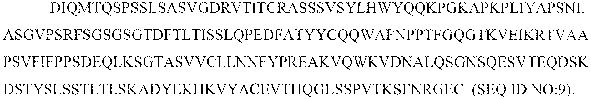
- Variant C of Table 2 comprises the full length heavy chain sequence:

- Variant D of Table 2 comprises the full length heavy chain sequence:

- Variant F of Table 2 comprises the full length heavy chain sequence:

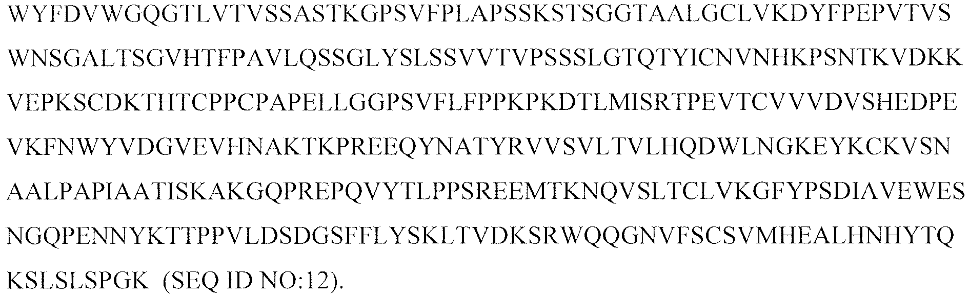
- Variant G of Table 2 comprises the full length heavy chain sequence:

- Variant H of Table 2 comprises the full length heavy chain sequence:

- In certain embodiments, the humanized 2H7 antibody of the invention further comprises amino acid alterations in the IgG Fc and exhibits increased binding affinity for human FcRn over an antibody having wild-type IgG Fc, by at least 60 fold, at least 70 fold, at least 80 fold, more preferably at least 100 fold, preferably at least 125 fold, even more preferably at least 150 fold to about 170 fold.
- The N-glycosylation site in IgG is at Asn297 in the
C H2 domain. Humanized 2H7 antibody compositions of the present invention include compositions of any of the preceding humanized 2H7 antibodies having a Fc region, wherein about 80-100% (and preferably about 90-99%) of the antibody in the composition comprises a mature core carbohydrate structure which lacks fucose, attached to the Fc region of the glycoprotein. Such compositions were demonstrated herein to exhibit a surprising improvement in binding to Fc(RIIIA(F158), which is not as effective as Fc(RIIIA (V158) in interacting with human IgG. Fc(RIIIA (F158) is more common than Fc(RIIIA (V158) in normal, healthy African Americans and Caucasians. See Lehrnbecher et al., Blood 94:4220 (1999). Historically, antibodies produced in Chinese Hamster Ovary Cells (CHO), one of the most commonly used industrial hosts, contain about 2 to 6% in the population that are nonfucosylated. YB2/0 and Lec13, however, can produce antibodies with 78 to 98% nonfucosylated species. Shinkawa et al., J Bio. Chem. 278 (5), 3466-347 (2003), reported that antibodies produced in YB2/0 and Lec13 cells, which have less FUT8 activity, show significantly increased ADCC activity in vitro. The production of antibodies with reduced fucose content are also described in e.g., Li et al., (GlycoFi) "Optimization of humanized IgGs in glycoengineered Pichia pastoris" in Nature Biology ; Niwa R. et al., Cancer Res. 64(6):2127-2133 (2004);US 2003/0157108 (Presta );US 6,602,684 andUS 2003/0175884 (Glycart Biotechnology);US 2004/0093621 ,US 2004/0110704 ,US 2004/0132140 (all of Kyowa Hakko Kogyo). - A bispecific humanized 2H7 antibody encompasses an antibody wherein one arm of the antibody has at least the antigen binding region of the H and/or L chain of a humanized 2H7 antibody of the invention, and the other arm has V region binding specificity for a second antigen. In specific embodiments, the second antigen is selected from the group consisting of CD3, CD64, CD32A, CD16, NKG2D or other NK activating ligands.
- A recombinant humanized version of the murine HER2 antibody 4D5 (huMAb4D5-8, rhuMAb HER2, trastuzumab or HERCEPTIN®;
U.S. Patent No. 5,821,337 ) is clinically active in patients with HER2-overexpressing metastatic breast cancers that have received extensive prior anti-cancer therapy (Baselga et al., J. Clin. Oncol. 14:737-744 (1996)). Trastuzumab received marketing approval from the Food and Drug Administration (FDA) September 25, 1998 for the treatment of patients with metastatic breast cancer whose tumors overexpress the HER2 protein. In November 2006, the FDA approved Herceptin as part of a treatment regimen containing doxorubicin, cyclophosphamide and paclitaxel, for the adjuvant treatment of patients with HER2-positive, node-positive breast cancer. - In one embodiment, the anti-HER2 antibody comprises the following VL and VH domain sequences:
- humanized 2C4 version 574 antibody VL (SEQ ID NO:16)

- and humanized 2C4 version 574 antibody VH (SEQ ID NO:17)

- In another embodiment, the anti-HER2 antibody comprises the VL (SEQ ID NO:18) and VH (SEQ ID NO:19) domain sequences of trastuzumab as shown in
Figure 21 and Figure 22 , respectively. - Other HER2 antibodies with various properties have been described in Tagliabue et al., Int. J. Cancer 47:933-937 (1991); McKenzie et al., Oncogene 4:543-548 (1989); Maier et al., Cancer Res. 51:5361-5369 (1991); Bacus et al., Molecular Carcinogenesis 3:350-362 (1990); Stancovski et al., PNAS (USA) 88:8691-8695 (1991); Bacus et al., Cancer Research 52:2580-2589 (1992); Xu et al., Int. J. Cancer 53:401-408 (1993);
WO94/00136 U.S. Patent No. 5,783,186 ; and Klapper et al., Oncogene 14:2099-2109 (1997). - The anti-VEGF antibodies may, for example, comprise the following sequences:
- In one embodiment, the anti-VEGF antibody comprises the following VL sequence (SEQ ID NO:20):
- DIQMTQTTSS LSASLGDRVI ISCSASQDIS NYLNWYQQKP DGTVKVLIYF TSSLHSGVPS RFSGSGSGTD YSLTISNLEP EDIATYYCQQ YSTVPWTFGG GTKLEIKR; and
- the following VH sequence (SEQ ID NO:21):


- In another embodiment, the anti-VEGF antibody comprises the following VL sequence (SEQ ID NO:22):
- DIQMTQSPSS LSASVGDRVT ITCSASQDIS NYLNWYQQKP GKAPKVLIYF TSSLHSGVPS RFSGSGSGTD FTLTISSLQP EDFATYYCQQ YSTVPWTFGQ GTK VEIKR; and
- the following VH sequence (SEQ ID NO:23):

- In a third embodiment, the anti-VEGF antibody comprises the following VL sequence (SEQ ID NO:24):
- DIQLTQSPSS LSASVGDRVT ITCSASQDIS NYLNWYQQKP GKAPKVLIYF TSSLHSGVPS RFSGSGSGTD FTLTISSLQP EDFATYYCQQ YSTVPWTFGQ GTKVEIKR; and
- the following VH sequence (SEQ ID NO:25):

- The humanized anti-CD11a antibody efalizumab or Raptiva® (
U.S. Patent No. 6,037,454 ) received marketing approval from the Food and Drug Administration on October 27, 2003 for the treatment for the treatment of psoriasis. One embodiment provides for an anti-human CD11a antibody comprising the VL and VH sequences of HuMHM24 below: - VL (SEQ ID NO:26):
and

- VH (SEQ ID NO:27):

- The anti-human CD11a antibody may comprise the VH of SEQ ID NO:27 and the full length L chain of HuMHM24 having the sequence of:or
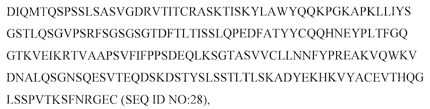
the L chain above with the H chain having the sequence of:
- Antibodies to the DR5 receptor (anti-DR5) antibodies can also be produced in accordance with the present invention. Such anti-DR5 antibodies specifically include all antibody variants disclosed in PCT Publication No.
WO 2006/083971 , such as the anti-DR5 antibodies designated Apomabs 1.1, 2.1, 3.1, 4.1, 5.1, 5.2, 5.3 , 6.1, 6.2, 6.3, 7.1, 7.2, 7.3,8.1, 8.3, 9.1, 1.2, 2.2, 3.2, 4.2, 5.2, 6.2, 7.2, 8.2, 9.2, 1.3, 2.2, 3.3, 4.3, 5.3, 6.3, 7.3, 8.3, 9.3, and 25.3, especially Apomab 8.3 and Apomab 7.3, preferably Apomab 7.3. The entire content ofWO 2006/083971 is hereby expressly incorporated by reference. - In addition to antibodies, the methods of the present invention find utility in the manufacturing of other polypeptides including disulfide bonds. Representative examples of such polypeptides include, without limitation, the following therapeutic proteins: tissue plasminogen activators (t-PAs), such as human tissue plasminogen activator (htPA, alteplase, ACTIVASE®), a thrombolytic agent for the treatment of myocardial infarction; a TNKase™, a ht-PA variant with extended half-life and fibrin specificity for single-bolus administration; recombinant human growth hormone (rhGH, somatropin, NUTROPIN®, PROTROPIN®) for the treatment of growth hormone deficiency in children and adults; and recombinant human deoxyribonuclease I (DNase I) for the treatment of cystic fibrosis (CF).
- Examples of disulfide-containing biologically important proteins include growth hormone, including human growth hormone and bovine growth hormone; growth hormone releasing factor; parathyroid hormone; thyroid stimulating hormone; lipoproteins; alpha-1-antitrypsin; insulin A-chain; insulin B-chain; proinsulin; follicle stimulating hormone; calcitonin; luteinizing hormone; glucagon; clotting factors such as factor VIIIC, factor IX, tissue factor, and von Willebrands factor; anti-clotting factors such as Protein C; atrial natriuretic factor; lung surfactant; a plasminogen activator, such as urokinase or human urine or tissue-type plasminogen activator (t-PA); bombesin; thrombin; hemopoietic growth factor; tumor necrosis factor-alpha and -beta; enkephalinase; RANTES (regulated on activation normally T-cell expressed and secreted); human macrophage inflammatory protein (MIP-1-alpha); a serum albumin such as human serum albumin; Muellerian-inhibiting substance; relaxin A-chain; relaxin B-chain; prorelaxin; mouse gonadotropin-associated peptide; a microbial protein, such as beta-lactamase; DNase; IgE; a cytotoxic T-lymphocyte associated antigen (CTLA), such as CTLA-4; inhibin; activin; vascular endothelial growth factor (VEGF); receptors for hormones or growth factors; Protein A or D; rheumatoid factors; a neurotrophic factor such as bone-derived neurotrophic factor (BDNF), neurotrophin-3, -4, -5, or -6 (NT-3, NT-4, NT-5, or NT-6), or a nerve growth factor such as NGF-β; platelet-derived growth factor (PDGF); fibroblast growth factor such as aFGF and bFGF; epidermal growth factor (EGF); transforming growth factor (TGF) such as TGF-alpha and TGF-beta, including TGF-β1, TGF-β2, TGF-β3, TGF-β4, or TGF-β5; insulin-like growth factor-I and -II (IGF-I and IGF-II); des(1-3)-IGF-I (brain IGF-I), insulin-like growth factor binding proteins; CD proteins such as CD3, CD4, CD8, CD19, CD20, CD34, and CD40; erythropoietin; osteoinductive factors; immunotoxins; a bone morphogenetic protein (BMP); an interferon such as interferon-alpha, -beta, and -gamma; colony stimulating factors (CSFs), e.g., M-CSF, GM-CSF, and G-CSF; interleukins (ILs), e.g., IL-1 to IL-10; superoxide dismutase; T-cell receptors; surface membrane proteins; decay accelerating factor; viral antigen such as, for example, a portion of the AIDS envelope; transport proteins; homing receptors; addressins; regulatory proteins; integrins such as CD11a, CD11b, CD11c, CD18, an ICAM, VLA-4 and VCAM; a tumor associated antigen such as HER2, HER3 or HER4 receptor; and fragments of any of the above-listed polypeptides.
- The antibodies and other recombinant proteins herein can be produced by well known techniques of recombinant DNA technology. Thus, aside from the antibodies specifically identified above, the skilled practitioner could generate antibodies directed against an antigen of interest, e.g., using the techniques described below.
- The antibody herein is directed against an antigen of interest. Preferably, the antigen is a biologically important polypeptide and administration of the antibody to a mammal suffering from a disease or disorder can result in a therapeutic benefit in that mammal. However, antibodies directed against nonpolypeptide antigens (such as tumor-associated glycolipid antigens; see
US Patent 5,091,178 ) are also contemplated. Where the antigen is a polypeptide, it may be a transmembrane molecule (e.g. receptor) or ligand such as a growth factor. Exemplary antigens include those proteins described in section (3) below. Exemplary molecular targets for antibodies encompassed by the present invention include CD proteins such as CD3, CD4, CD8, CD19, CD20, CD22, CD34, CD40; members of the ErbB receptor family such as the EGF receptor, HER2, HER3 or HER4 receptor; cell adhesion molecules such as LFA-1, Mac1, p150,95, VLA-4, ICAM-1, VCAM and αv/β3 integrin including either α or β subunits thereof (e.g. anti-CD11a, anti-CD18 or anti-CD11b antibodies); growth factors such as VEGF; IgE; blood group antigens; flk2/flt3 receptor; obesity (OB) receptor; mpl receptor; CTLA-4; protein C, or any of the other antigens mentioned herein. Antigens to which the antibodies listed above bind are specifically included within the scope herein. - Soluble antigens or fragments thereof, optionally conjugated to other molecules, can be used as immunogens for generating antibodies. For transmembrane molecules, such as receptors, fragments of these (e.g. the extracellular domain of a receptor) can be used as the immunogen. Alternatively, cells expressing the transmembrane molecule can be used as the immunogen. Such cells can be derived from a natural source (e.g. cancer cell lines) or may be cells which have been transformed by recombinant techniques to express the transmembrane molecule.
- Other antigens and forms thereof useful for preparing antibodies will be apparent to those in the art.
- Polyclonal antibodies are preferably raised in animals by multiple subcutaneous (sc) or intraperitoneal (ip) injections of the relevant antigen and an adjuvant. It may be useful to conjugate the antigen to a protein that is immunogenic in the species to be immunized, e.g., keyhole limpet hemocyanin, serum albumin, bovine thyroglobulin, or soybean trypsin inhibitor using a bifunctional or derivatizing agent, for example, maleimidobenzoyl sulfosuccinimide ester (conjugation through cysteine residues), N-hydroxysuccinimide (through lysine residues), glutaraldehyde, succinic anhydride, SOCl2, or R1N=C=NR, where R and R1 are different alkyl groups.
- Animals are immunized against the antigen, immunogenic conjugates, or derivatives by combining, e.g., 100 µg or 5 µg of the protein or conjugate (for rabbits or mice, respectively) with 3 volumes of Freund's complete adjuvant and injecting the solution intradermally at multiple sites. One month later the animals are boosted with 1/5 to 1/10 the original amount of antigen or conjugate in Freund's complete adjuvant by subcutaneous injection at multiple sites. Seven to 14 days later the animals are bled and the serum is assayed for antibody titer. Animals are boosted until the titer plateaus. Preferably, the animal is boosted with the conjugate of the same antigen, but conjugated to a different protein and/or through a different cross-linking reagent. Conjugates also can be made in recombinant cell culture as protein fusions. Also, aggregating agents such as alum are suitably used to enhance the immune response.
- Monoclonal antibodies may be made using the hybridoma method first described by Kohler et al., Nature, 256:495 (1975), or may be made by recombinant DNA methods (
U.S. Patent No. 4,816,567 ). - In the hybridoma method, a mouse or other appropriate host animal, such as a hamster or macaque monkey, is immunized as hereinabove described to elicit lymphocytes that produce or are capable of producing antibodies that will specifically bind to the protein used for immunization. Alternatively, lymphocytes may be immunized in vitro. Lymphocytes then are fused with myeloma cells using a suitable fusing agent, such as polyethylene glycol, to form a hybridoma cell (Goding, Monoclonal Antibodies: Principles and Practice, pp.59-103 (Academic Press, 1986)).
- The hybridoma cells thus prepared are seeded and grown in a suitable culture medium that preferably contains one or more substances that inhibit the growth or survival of the unfused, parental myeloma cells. For example, if the parental myeloma cells lack the enzyme hypoxanthine guanine phosphoribosyl transferase (HGPRT or HPRT), the culture medium for the hybridomas typically will include hypoxanthine, aminopterin, and thymidine (HAT medium), which substances prevent the growth of HGPRT-deficient cells.
- Preferred myeloma cells are those that fuse efficiently, support stable high-level production of antibody by the selected antibody-producing cells, and are sensitive to a medium such as HAT medium. Among these, preferred myeloma cell lines are murine myeloma lines, such as those derived from MOPC-21 and MPC-11 mouse tumors available from the Salk Institute Cell Distribution Center, San Diego, California USA, and SP-2 or X63-Ag8-653 cells available from the American Type Culture Collection, Rockville, Maryland USA. Human myeloma and mouse-human heteromyeloma cell lines also have been described for the production of human monoclonal antibodies (Kozbor, J. Immunol., 133:3001 (1984); Brodeur et al., Monoclonal Antibody Production Techniques and Applications, pp. 51-63 (Marcel Dekker, Inc., New York, 1987)).
- Culture medium in which hybridoma cells are growing is assayed for production of monoclonal antibodies directed against the antigen. Preferably, the binding specificity of monoclonal antibodies produced by hybridoma cells is determined by immunoprecipitation or by an in vitro binding assay, such as radioimmunoassay (RIA) or enzyme-linked immunoabsorbent assay (ELISA).
- After hybridoma cells are identified that produce antibodies of the desired specificity. affinity, and/or activity, the clones may be subcloned by limiting dilution procedures and grown by standard methods (Goding, Monoclonal Antibodies: Principles and Practice, pp.59-103 (Academic Press, 1986)). Suitable culture media for this purpose include, for example, D-MEM or RPMI-1640 medium. In addition, the hybridoma cells may be grown in vivo as ascites tumors in an animal.
- The monoclonal antibodies secreted by the subclones are suitably separated from the culture medium, ascites fluid, or serum by conventional immunoglobulin purification procedures such as, for example, Protein A-Sepharose, hydroxyapatite chromatography, gel electrophoresis, dialysis, or affinity chromatography. Preferably the Protein A chromatography procedure described herein is used.
- DNA encoding the monoclonal antibodies is readily isolated and sequenced using conventional procedures (e.g., by using oligonucleotide probes that are capable of binding specifically to genes encoding the heavy and light chains of the monoclonal antibodies). The hybridoma cells serve as a preferred source of such DNA. Once isolated, the DNA may be placed into expression vectors, which are then transfected into host cells such as E. coli cells, simian COS cells, Chinese hamster ovary (CHO) cells, or myeloma cells that do not otherwise produce immunoglobulin protein, to obtain the synthesis of monoclonal antibodies in the recombinant host cells.
- The DNA also may be modified, for example, by substituting the coding sequence for human heavy- and light-chain constant domains in place of the homologous murine sequences (
U.S. Patent No. 4,816,567 ; Morrison, et al., Proc. Natl Acad. Sci. USA, 81:6851 (1984)), or by covalently joining to the immunoglobulin coding sequence all or part of the coding sequence for a non-immunoglobulin polypeptide. - Typically such non-immunoglobulin polypeptides are substituted for the constant domains of an antibody, or they are substituted for the variable domains of one antigen-combining site of an antibody to create a chimeric bivalent antibody comprising one antigen-combining site having specificity for an antigen and another antigen-combining site having specificity for a different antigen.
- In a further embodiment, monoclonal antibodies can be isolated from antibody phage libraries generated using the techniques described in McCafferty et al., Nature, 348:552-554 (1990). Clackson et al., Nature, 352:624-628 (1991) and Marks et al., J. Mol. Biol., 222:581-597 (1991) describe the isolation of murine and human antibodies, respectively, using phage libraries. Subsequent publications describe the production of high affinity (nM range) human antibodies by chain shuffling (Marks et al., Bio/Technology, 10:779-783 (1992)), as well as combinatorial infection and in vivo recombination as a strategy for constructing very large phage libraries (Waterhouse et al., Nuc. Acids. Res., 21:2265-2266 (1993)). Thus, these techniques are viable alternatives to traditional hybridoma techniques for isolation of monoclonal antibodies.
- A humanized antibody has one or more amino acid residues introduced into it from a source which is non-human. These non-human amino acid residues are often referred to as "import" residues, which are typically taken from an "import" variable domain. Humanization can be essentially performed following the method of Winter and co-workers (Jones et al., Nature, 321:522-525 (1986); Riechmann et al., Nature, 332:323-327 (1988); Verhoeyen et al., Science, 239:1534-1536 (1988)), by substituting rodent CDRs or CDR sequences for the corresponding sequences of a human antibody. Accordingly, such "humanized" antibodies are chimeric antibodies (
U.S. Patent No. 4,816,567 ) wherein substantially less than an intact human variable domain has been substituted by the corresponding sequence from a non-human species. In practice, humanized antibodies are typically human antibodies in which some CDR residues and possibly some FR residues are substituted by residues from analogous sites in rodent antibodies. - The choice of human variable domains, both light and heavy, to be used in making the humanized antibodies is very important to reduce antigenicity. According to the so-called "best-fit" method, the sequence of the variable domain of a rodent antibody is screened against the entire library of known human variable-domain sequences. The human sequence which is closest to that of the rodent is then accepted as the human FR for the humanized antibody (Sims et al., J. Immunol., 151:2296 (1993)). Another method uses a particular framework derived from the consensus sequence of all human antibodies of a particular subgroup of light or heavy chains. The same framework may be used for several different humanized antibodies (Carter et al., Proc. Natl. Acad. Sci. USA, 89:4285 (1992); Presta et al., J. Immnol., 151:2623 (1993)).
- It is further important that antibodies be humanized with retention of high affinity for the antigen and other favorable biological properties. To achieve this goal, according to a preferred method, humanized antibodies are prepared by a process of analysis of the parental sequences and various conceptual humanized products using three-dimensional models of the parental and humanized sequences. Three-dimensional immunoglobulin models are commonly available and are familiar to those skilled in the art. Computer programs are available which illustrate and display probable three-dimensional conformational structures of selected candidate immunoglobulin sequences. Inspection of these displays permits analysis of the likely role of the residues in the functioning of the candidate immunoglobulin sequence, i.e., the analysis of residues that influence the ability of the candidate immunoglobulin to bind its antigen. In this way, FR residues can be selected and combined from the recipient and import sequences so that the desired antibody characteristic, such as increased affinity for the target antigen(s), is achieved. In general, the CDR residues are directly and most substantially involved in influencing antigen binding.
- Alternatively, it is now possible to produce transgenic animals (e.g., mice) that are capable, upon immunization, of producing a full repertoire of human antibodies in the absence of endogenous immunoglobulin production. For example, it has been described that the homozygous deletion of the antibody heavy-chain joining region (JH) gene in chimeric and germ-line mutant mice results in complete inhibition of endogenous antibody production. Transfer of the human germ-line immunoglobulin gene array in such germ-line mutant mice will result in the production of human antibodies upon antigen challenge. See, e.g., Jakobovits et al., Proc. Natl. Acad. Sci. USA, 90:2551 (1993); Jakobovits et al., Nature, 362:255-258 (1993); Bruggermann et al., Year in Immuno., 7:33 (1993); and Duchosal et al., Nature 355:258 (1992). Human antibodies can also be derived from phage-display libraries (Hoogenboom et al., J. Mol. Biol., 227:381 (1991); Marks et al., J. Mol. Biol., 222:581-597 (1991); Vaughan et al., Nature Biotech 14:309 (1996)).
- Various techniques have been developed for the production of antibody fragments. Traditionally, these fragments were derived via proteolytic digestion of intact antibodies (see, e.g., Morimoto et al., Journal of Biochemical and Biophysical Methods 24:107-117 (1992) and Brennan et al., Science, 229:81 (1985)). However, these fragments can now be produced directly by recombinant host cells. For example, the antibody fragments can be isolated from the antibody phage libraries discussed above. Alternatively, Fab'-SH fragments can be directly recovered from E. coli and chemically coupled to form F(ab')2 fragments (Carter et al., Bio/Technology 10:163-167 (1992)). According to another approach, F(ab')2 fragments can be isolated directly from recombinant host cell culture. Other techniques for the production of antibody fragments will be apparent to the skilled practitioner. In other embodiments, the antibody of choice is a single chain Fv fragment (scFv) (see
WO 93/16185 - Multispecific antibodies have binding specificities for at least two different antigens. While such molecules normally will only bind two antigens (i.e. bispecific antibodies, BsAbs), antibodies with additional specificities such as trispecific antibodies are encompassed by this expression when used herein.
- Methods for making bispecific antibodies are known in the art. Traditional production of full length bispecific antibodies is based on the coexpression of two immunoglobulin heavy chain-light chain pairs, where the two chains have different specificities (Millstein et al., Nature, 305:537-539 (1983)). Because of the random assortment of immunoglobulin heavy and light chains, these hybridomas (quadromas) produce a potential mixture of 10 different antibody molecules, of which only one has the correct bispecific structure. Purification of the correct molecule, which is usually done by affinity chromatography steps, is rather cumbersome, and the product yields are low. Similar procedures are disclosed in
WO 93/08829 - According to another approach described in
WO96/27011 C H3 domain of an antibody constant domain. In this method, one or more small amino acid side chains from the interface of the first antibody molecule are replaced with larger side chains (e.g. tyrosine or tryptophan). Compensatory "cavities" of identical or similar size to the large side chain(s) are created on the interface of the second antibody molecule by replacing large amino acid side chains with smaller ones (e.g. alanine or threonine). This provides a mechanism for increasing the yield of the heterodimer over other unwanted end-products such as homodimers. - Bispecific antibodies include cross-linked or "heteroconjugate" antibodies. For example, one of the antibodies in the heteroconjugate can be coupled to avidin, the other to biotin. Such antibodies have, for example, been proposed to target immune system cells to unwanted cells (
US Patent No. 4,676,980 ), and for treatment of HIV infection (WO 91/00360 WO 92/200373 EP 03089 US Patent No. 4,676,980 , along with a number of cross-linking techniques. - Techniques for generating bispecific antibodies from antibody fragments have also been described in the literature. For example, bispecific antibodies can be prepared using chemical linkage. Brennan et al., Science, 229: 81 (1985) describe a procedure wherein intact antibodies are proteolytically cleaved to generate F(ab')2 fragments. These fragments are reduced in the presence of the dithiol complexing agent sodium arsenite to stabilize vicinal dithiols and prevent intermolecular disulfide formation. The Fab' fragments generated are then converted to thionitrobenzoate (TNB) derivatives. One of the Fab'-TNB derivatives is then reconverted to the Fab'-thiol by reduction with mercaptoethylamine and is mixed with an equimolar amount of the other Fab'-TNB derivative to form the bispecific antibody. The bispecific antibodies produced can be used as agents for the selective immobilization of enzymes.
- Recent progress has facilitated the direct recovery of Fab'-SH fragments from E. coli, which can be chemically coupled to form bispecific antibodies. Shalaby et al., J. Exp. Med., 175: 217-225 (1992) describe the production of a fully humanized bispecific antibody F(ab')2 molecule. Each Fab' fragment was separately secreted from E. coli and subjected to directed chemical coupling in vitro to form the bispecific antibody. The bispecific antibody thus formed was able to bind to cells overexpressing the ErbB2 receptor and normal human T cells, as well as trigger the lytic activity of human cytotoxic lymphocytes against human breast tumor targets.
- Various techniques for making and isolating bispecific antibody fragments directly from recombinant cell culture have also been described. For example, bispecific antibodies have been produced using leucine zippers. Kostelny et al., J. Immunol., 148(5):1547-1553 (1992). The leucine zipper peptides from the Fos and Jun proteins were linked to the Fab' portions of two different antibodies by gene fusion. The antibody homodimers were reduced at the hinge region to form monomers and then re-oxidized to form the antibody heterodimers. This method can also be utilized for the production of antibody homodimers. The "diabody" technology described by Hollinger et al., Proc. Natl. Acad. Sci. USA, 90:6444-6448 (1993) has provided an alternative mechanism for making bispecific antibody fragments. The fragments comprise a heavy-chain variable domain (VH) connected to a light-chain variable domain (VL) by a linker which is too short to allow pairing between the two domains on the same chain. Accordingly, the VH and VL domains of one fragment are forced to pair with the complementary VL and VH domains of another fragment, thereby forming two antigen-binding sites. Another strategy for making bispecific antibody fragments by the use of single-chain Fv (sFv) dimers has also been reported. See Gruber et al., J. Immunol., 152:5368 (1994). Alternatively, the antibodies can be "linear antibodies" as described in Zapata et al., Protein Eng. 8(10):1057-1062 (1995). Briefly, these antibodies comprise a pair of tandem Fd segments (VH-CH1-VH-CH1) which form a pair of antigen binding regions. Linear antibodies can be bispecific or monospecific.
- Antibodies with more than two valencies are contemplated. For example, trispecific antibodies can be prepared. Tutt et al., J. Immunol. 147: 60 (1991).
- The simplest and most straightforward immunoadhesin design combines the binding domain(s) of the adhesin (e.g. the extracellular domain (ECD) of a receptor) with the hinge and Fc regions of an immunoglobulin heavy chain. Ordinarily, when preparing the immunoadhesins of the present invention, nucleic acid encoding the binding domain of the adhesin will be fused C-terminally to nucleic acid encoding the N-terminus of an immunoglobulin constant domain sequence, however N-terminal fusions are also possible.
- Typically, in such fusions the encoded chimeric polypeptide will retain at least functionally active hinge,
C H2 andC H3 domains of the constant region of an immunoglobulin heavy chain. Fusions are also made to the C-terminus of the Fc portion of a constant domain, or immediately N-terminal to theC H1 of the heavy chain or the corresponding region of the light chain. The precise site at which the fusion is made is not critical; particular sites are well known and may be selected in order to optimize the biological activity, secretion, or binding characteristics of the immunoadhesin. - In a preferred embodiment, the adhesin sequence is fused to the N-terminus of the Fc domain of immunoglobulin GI (IgGI). It is possible to fuse the entire heavy chain constant region to the adhesin sequence. However, more preferably, a sequence beginning in the hinge region just upstream of the papain cleavage site which defines IgG Fc chemically (i.e. residue 216, taking the first residue of heavy chain constant region to be 114), or analogous sites of other immunoglobulins is used in the fusion. In a particularly preferred embodiment, the adhesin amino acid sequence is fused to (a) the hinge region and
C H2 andC H3 or (b) theC H1, hinge,C H2 andC H3 domains, of an IgG heavy chain. - For bispecific immunoadhesins, the immunoadhesins are assembled as multimers, and particularly as heterodimers or heterotetramers. Generally, these assembled immunoglobulins will have known unit structures. A basic four chain structural unit is the form in which IgG, IgD, and IgE exist. A four chain unit is repeated in the higher molecular weight immunoglobulins; IgM generally exists as a pentamer of four basic units held together by disulfide bonds. IgA globulin, and occasionally IgG globulin, may also exist in multimeric form in serum. In the case of multimer, each of the four units may be the same or different.
- Various exemplary assembled immunoadhesins within the scope herein are schematically diagrammed below:
- ACL-ACL;
- ACH-(ACH, ACL-ACH, ACL-VHCH, or VLCL-ACH);
- ACH-ACH-(ACL-ACH, ACL-VHCH, VLCL-ACH, or VLCL-VHCH)
- ACL-VHCH-(ACH, or ACL-VHCH, or VLCL-ACH);
- VLCL-ACH-(ACL-VH,CH, or VLCL-ACH); and
- (A-Y)n-(VLCL-VHCH)2,
- In the interests of brevity, the foregoing structures only show key features; they do not indicate joining (J) or other domains of the immunoglobulins, nor are disulfide bonds shown. However, where such domains are required for binding activity, they shall be constructed to be present in the ordinary locations which they occupy in the immunoglobulin molecules.
- Alternatively, the adhesin sequences can be inserted between immunoglobulin heavy chain and light chain sequences, such that an immunoglobulin comprising a chimeric heavy chain is obtained. In this embodiment, the adhesin sequences are fused to the 3' end of an immunoglobulin heavy chain in each arm of an immunoglobulin, either between the hinge and the
C H2 domain, or between theC H2 andC H3 domains. Similar constructs have been reported by Hoogenboom, et al., Mol. Immunol. 28:1027-1037 (1991). - Although the presence of an immunoglobulin light chain is not required in the immunoadhesins of the present invention, an immunoglobulin light chain might be present either covalently associated to an adhesin-immunoglobulin heavy chain fusion polypeptide, or directly fused to the adhesin. In the former case, DNA encoding an immunoglobulin light chain is typically coexpressed with the DNA encoding the adhesin-immunoglobulin heavy chain fusion protein. Upon secretion, the hybrid heavy chain and the light chain will be covalently associated to provide an immunoglobulin-like structure comprising two disulfide-linked immunoglobulin heavy chain-light chain pairs. Methods suitable for the preparation of such structures are, for example, disclosed in
U.S. Patent No. 4,816,567, issued 28 March 1989 . - Immunoadhesins are most conveniently constructed by fusing the cDNA sequence encoding the adhesin portion in-frame to an immunoglobulin cDNA sequence. However, fusion to genomic immunoglobulin fragments can also be used (see, e.g. Aruffo et al., Cell 61:1303-1313 (1990); and Stamenkovic et al., Cell 66:1133-1144 (1991)). The latter type of fusion requires the presence of Ig regulatory sequences for expression. cDNAs encoding IgG heavy-chain constant regions can be isolated based on published sequences from cDNA libraries derived from spleen or peripheral blood lymphocytes, by hybridization or by polymerase chain reaction (PCR) techniques. The cDNAs encoding the "adhesin" and the immunoglobulin parts of the immunoadhesin are inserted in tandem into a plasmid vector that directs efficient expression in the chosen host cells.
- The following examples are offered for illustrative purposes only, and are not intended to limit the scope of the present invention in any way.
- Commercially available reagents referred to in the examples were used according to manufacturer's instructions unless otherwise indicated. The source of those cells identified in the following examples, and throughout the specification, by ATCC accession numbers is the American Type Culture Collection, Manassas, Virginia.
- The following materials and methods were used in Examples 2-8 below.
- Materials and devices used in the experiments described in the experimental examples include: stainless steel vials (mini-tanks, Flow Components, Dublin, CA; short (50 cc) and tall (55 cc)); dialysis tubing (Spectra/Por, 6-8000 MWCO, cat. # 132645), 0.22 µm filter (Millipore Millipak Gamma Gold cat. # MPGL04GH2); phosphate buffered saline (PBS, EMD, cat. # 6506); ethylenediaminetetraacetic acid (EDTA, Sigma, cat. # E4884); α-nicotinamide adenine dinucleotide phosphate (NADPH, Calbiochem, cat. # 481973); dehydroepiandrosterone (DHEA, TCI, cat. # D0044); cupric sulfate (Sigma, cat. # C8027), glucose-6-phosphate (G6P, Calbiochem, cat. # 346764); aurothioglucose (ATG, USP, cat. # 1045508); aurothiomalate (ATM, Alfa Aesar, cat. # 39740); reduced glutathione (GSH, J.T. Baker, cat. # M770-01); monobromobimane (mBB, Fluka, cat. # 69898); histidine (J.T. Baker, cat. # 2080-05); sodium sulfate (J.T. Baker, cat. # 3897-05); Trx (Sigma. cat. # T8690); TrxR (Sigma, cat. # T9698). All chemicals and reagents were used as received with no further purification. Stock solutions of EDTA (250 mM, pH 7.5), CuSO4 (10 mM), ATG (30 mM), ATM (30 mM), NADPH (75 mM), G6P (300 mM) were prepared for use in the mini-tank time course studies.
- In order to generate ocrelizumab CCF for the various reduction studies, a representative small-scale fermentation process was utilized similar to the methods described previously (Chaderjian et al., 2005). Briefly, 3 liter glass stirred-tank Applikon® bioreactors fitted with pitched blade impellers were used for the inoculum-train and production cultures with the ocrelizumab media components. The bioreactors were outfitted with calibrated dissolved oxygen (DO), pH and temperature probes. DO, pH, temperature, and agitation rate were controlled using digital control units to the defined parameters of the ocrelizumab manufacturing process. The working volume for both the inoculum-train and production cultures was 1.5 L. Daily samples were analyzed on a NOVA Bioprofile blood gas analyzer to ensure the accuracy of the on-line value for pH and dissolved oxygen as well as to monitor the glucose, lactate, ammonium, glutamine, glutamate, and sodium concentrations in the cultures. Daily samples were also taken to monitor cell growth, viability, and titer. Cell growth was measured both by viable cell counts using a ViCell as well as on a packed cell volume (PCV) basis. Culture viability was determined by trypan blue exclusion on a ViCell instrument. Supernatant samples were assayed by an HPLC-based method to measure ocrelizumab titer values.
- Complete lysis of CCF was achieved by high pressure homogenization using a Microfluidics HC-8000 homogenizer. The pressure regulator of the instrument was set to 4,000-8,000 psi, and the CCF was pulled in through the homogenizer to obtain complete cell lysis (membrane breakage) after a single pass. The CCF homogenate was collected once water was purged through the system. The homogenate was transferred to centrifuge bottles and centrifuged in a Sorval RC-3B rotor centrifuge at 4,500 rpm for 30 minutes at 20 °C. The centrate was decanted and then depth filtered followed by 0.22 µm sterile filtration using a peristaltic pump with silicon tubing to generate the final HCCF from the homogenized CCF (100% cell lysis). Alternatively, the CCF was centrifuged straight from the fermentor without any homogenization and then the centrate was filtered with a sterile 0.22 µm filter to generate the HCCF.
- A laminar flow hood was used in handling all mini-tanks and all materials used in the HCCF incubation experiments were either autoclaved or rinsed using 70% isopropanol to minimize bacterial contamination.
- For lactate dehydrogenase assay, see Babson & Babson (1973) and Legrand et al., (1992), which are hereby incorporated by reference.
- A dialysis experiment was carried out in order to determine whether the components causing reduction of ocrelizumab were small molecules or macromolecules (i.e. enzymes). A sample of 3 mL of purified and formulated ocrelizumab (30.2 mg/mL) was dialyzed against 1 L of phosphate buffered saline (PBS, 10 mM pH 7.2) for 24 hours and the PBS was changed after 8 hours. The concentration of the ocrelizumab sample was then adjusted to 1 mg/mL using the absorbance at 280 nm. Aliquots were stored at -70 °C prior to use. Dialysis tubing was hydrated overnight in a 0.05% azide solution and rinsed with sterile water prior to use. The HCCF obtained from homogenization of CCF from a 3-L fermentor was thawed and filtered through a 0.22 µm Millipak filter using a peristaltic pump. Six short mini-tanks were filled with 30 mL of HCCF each. To each mini-tank, 500 µL of ocrelizumab sample in sealed dialysis tubing was added. The mini-tanks were sealed and loaded into a bench top mixer (Barnstead Lab-Line MAX Q 4000) operating at 35 rpm and ambient temperature. For each time-point, one mini-tank was removed from the mixer, and aliquots of the HCCF (in the mini-tank) and ocrelizumab sample (in the dialysis bag) were taken and stored at -70 °C until analyzed with the free thiol assay and the Bioanalyzer assay (described below).
- A tall mini-tank was filled with 27 mL of HCCF. Depending on the experiment design, various reagents (NADPH, G6P, inhibitors of G6PD or TrxR) were added to the desired concentration, and the final volume in the mini-tank was brought to 30 mL with PBS (10 mM pH 7.2). The mini-tanks were sealed and loaded into a bench top mixer running at 35 rpm and ambient temperature. At each-time point for sampling, the exteriors of the mini-tanks were sterilized with 70% IPA and opened in a laminar flow hood for the removal of an aliquot. The mini-tanks were then re-sealed and loaded back into the bench top mixer. All aliquots were stored at -70 °C until analyzed with the free thiol assay and Bioanalyzer assay (described below).
- A commercial TrxR (rat liver) solution (4 µM) was diluted with water to yield a 2.86 µM solution. Lyophilized Trx (human) was reconstituted with PBS (10 mM, pH 7.2) yielding a 500 µM solution. A solution of 20 mM NADPH and 10 mM ATG and ATM solutions were prepared in water.
- In a black polypropylene 1.5 mL micro centrifuge tube, 437 µL PBS, 25 µL NADPH, 16 µL formulated ocrelizumab solution (30.2 mg/mL) and 5 µL Trx were gently mixed. The reaction was initiated by the addition of 17.5 µL TrxR. The reaction was incubated at room temperature for 24 hours. Aliquots of 20 µL were taken at each sampling time-point and stored at -70 °C until analyzed by the Bioanalyzer assay (see below). Controls were performed to determine if the enzymatic pathway was active when an enzyme was omitted by substituting an equal volume of PBS for either Trx and/or TrxR in the reaction mixture.
- Inhibition of the Trx system was demonstrated using the same reaction conditions described above with the addition of 5 µL ATG or ATM. To demonstrate the inhibition of Trx system by Cu2+, 2.5 µL of CuSO4 (10 mM) was added to reaction mixture using the same enzymes but a different buffer (10 mM histidine, 10 mM Na2SO4, 137 mM NaCl, 2.5 mM KCl, pH 7.0) to prevent formation of insoluble Cu3(PO4)2.
- A standard curve using GSH was generated in PBS (10 mM, pH 6.0±0.05). From a 110 mM GSH solution, standards were prepared at concentrations of 0, 5.5, 11, 22, 44, 55, 110 and 550 µM through serial dilution. From an acetonitrile stock solution of mBB (10 mM stored at -20 °C), a 100 µM solution of mBB was prepared in PBS (10 mM, pH 10.0±0.05) and stored away from light.
- In a black, flat bottomed 96 well plate, 100 µL of mBB was dispensed into each well. For the standard curve, 10 µL of standard GSH solution was added yielding a working pH of 8.0 ± 0.2. For samples, 10 µL of sample was added to the wells. All wells were prepared in triplicate. The plate was incubated at room temperature for 1 hour in the dark then read using a fluorescence plate reader (Molecular Devices SpectraMax® Gemini XS) with an excitation wavelength of 390 nm and an emission wavelength of 490 nm. A linear standard curve was generated using the average result of the three standard wells plotted versus GSH concentration. Free thiol levels in samples were calculated from the linear equation of the standard curve using the average value of the three sample wells.
- Capillary electrophoresis measurements were acquired using the Agilent 2100 Bioanalyzer. Sample preparation was carried out as described in the Agilent Protein 230 Assay Protocol (manual part number G2938-90052) with minor changes. HCCF samples were diluted, 1:4 and Protein A samples werediluted to 1.0 g/L with water prior to preparation. For HCCF samples at the denaturing step, 24 µL of a 50 mM iodoacetamide (IAM), 0.5% SDS solution was added in addition to the 2 µL of denaturing solution provided. For Protein A samples, 0.5% SDS with no IAM and 2 µl of denaturing solution were used. Digital gel-like images were generated using Agilent 2100 Expert software.
- Three separate stock solutions were used in the lab scale HCCF hold time studies: (1) 250 mM stock solution of EDTA (pH 7.4) prepared using EDTA, disodium dihydrate (Mallinckrodt, cat. # 7727-06 or Sigma, cat. # E-5134) and EDTA, tetrasodium dihydrate (Sigma, cat. #E-6511), (2) 50 mM stock solution of cupric sulfate pentahydrate (CuSO4, Sigma, cat. # C-8027), and (3) 1 M acetic acid solution (Mallinckrodt, cat. # V193).
- A stock solution of either 250 mM EDTA or 50 mM CuSO4 was added to the CCF prior to homogenization to evaluate a range of final concentrations to prevent antibody disulfide reduction. Once the final HCCF was generated from the homogenized CCF, these solutions were then mixed with the HCCF generated from the non-homogenized CCF (also containing EDTA or CuSO4) in order to dilute and decrease the total level of cell lysis to below the 100% maximum. Alternatively, a stock solution of 1 M acetic acid was added to a final blended HCCF solution (homogenized CCF and non-homogenized CCF) to decrease the pH of the solution to prevent antibody disulfide reduction.
- Approximately 30 - 50 mL of each HCCF solution (containing EDTA, CuSO4, acetic acid, or no addition for the control) was held in a 50 mL 316L stainless steel vial. The vial was sealed with a clamp, and the solution was not aerated or agitated. The vial was stored at room temperature (18 - 22 °C). At pre-determined time points, the solution was removed and purified over a lab scale protein A affinity resin.
- Similar results can be obtained with other oxidizing agents, such as, for example, cystine and oxidized glutathione.
- To evaluate air sparging of the HCCF generated from homogenized CCF to prevent antibody disulfide reduction, 3-L glass or 15-L stainless steel vessels were utilized. Approximately 1-5 L of HCCF was 0.22 µm sterile filtered into each sterilized vessel. Experimental conditions were maintained at 18-22°C and 50 (15-L fermentor) or 275 rpm (3-L fermentor) agitation either with or without pH control by the addition of carbon dioxide. Solutions were either sparged with air to increase the dissolved oxygen level to air saturation or with nitrogen (control) to remove any dissolved oxygen in solution. Gas flow to each vessel was variable dependent upon whether a constant aeration rate was used or a minimum level of dissolved oxygen was maintained. At pre-determined time points, 25-50 mL samples were removed from both vessels and purified over a lab scale protein A affinity resin prior to analysis.
- Antibody in harvested cell culture fluid samples can be captured and purified using a specific affinity chromatography resin. Protein A resin (Millipore, Prosep-vA High Capacity) was selected as the affinity resin for antibody purification. The resin was packed in a 0.66 cm inner diameter glass column (Omnifit®) with a 14 cm bed height resulting in a 4.8 mL final column volume. Chromatography was performed using an
AKTA Explorer 100 chromatography system (GE Healthcare). - The resin was exposed to buffers and HCCF at a linear flow rate between 350-560 cm/hr. The resin was equilibrated with 25 mM Tris, 25 mM NaCl, 5 mM EDTA, pH 7.1. For each purification, the resin was loaded between 5 - 15 mg antibody per mL of resin. The antibody concentration in the HCCF was determined using an immobilized protein A HPLC column (Applied Biosystems, POROS A). After loading, the resin was washed with 25 mM Tris, 25 mM NaCl, 5 mM EDTA, 0.5 M TMAC, pH 7.1, and then the antibody was eluted using 0.1 M acetic acid, pH 2.9. Elution pooling was based on UV absorbance at 280 nm measured inline after the column. The purified elution pools were pH-adjusted using 1 M Sodium HEPES to pH 5.0-5.5. After regeneration of the resin with 0.1M phosphoric acid, the same or similar packed resins were used for subsequent purification of other HCCF solutions.
- The antibody concentration in the purified protein A pool was measured using UV spectrometry at 280 nm. The purified protein A elution pools were analyzed by the Bioanalyzer assay to quantitate the percentage of intact antibody at 150 kDa molecular weight.
- A dialysis experiment was designed and carried out to determine if the reduction of ocrelizumab was caused by small reducing molecules or macromolecules (e.g., enzymes). In this dialysis experiment, purified intact ocrelizumab was placed in a dialysis bag with a molecular weight cut off (MWCO) of 7000 and incubated the dialysis bag in HCCF containing ocrelizumab in a stainless steel mini-tank. As shown in
Figures 1 and2 , the ocrelizumab inside the bag was not reduced after the incubation period (Figure 1 ), whereas the ocrelizumab outside the bag in the HCCF was significantly reduced soon after the incubation started. This was evidenced by the loss of intact ocrelizumab (∼150 kDa) and the formation of ocrelizumab fragments (various combinations of heavy and light chains) (Figure 2 ). The mass spectrometry analysis of the ocrelizumab in the protein A elution pools from the reduced manufacturing runs indicated that those observed fragments were formed by reduction of only the inter-chain disulfide bonds. - The free thiol measurement showed that no free thiols were present inside the dialysis bag at the beginning of the incubation; however the levels of free thiols inside and outside the dialysis bag become comparable in less than five hours after the incubation started, indicating that the small molecule components in the HCCF are fully equilibrated inside and outside the dialysis bag (
Figure 3 ). Since the reduction was observed only outside but not inside the dialysis bag with a MWCO of 7000 Da, the molecular weight of the reducing molecule(s) must be greater than 7000 Da. Thus, an enzymatic reaction is responsible for the reduction of ocrelizumab. - The Trx system was tested for its ability to reduce ocrelizumab in vitro by incubating intact ocrelizumab with Trx, TrxR, and NADPH. The Bioanalyzer results indicate that ocrelizumab was reduced in vitro by the Trx system (
Figure 5 ). The rate of reduction in this in vitro system appears to be slower than that in the HCCF (for example when compared to the reduction shown inFigure 2 ). This is likely due to lower concentrations of the enzymes (Trx and Trx-R) and/or the buffer system used in the in vitro reaction because reaction rate of Trx system is dependent on both the enzyme concentrations and buffer systems. - Cupric sulfate is known for its ability to provide oxidizing redox potential and has been used in the cell culture processes to minimize free thiol (i.e., minimize unpaired cysteine) levels in recombinant antibody molecules (Chaderjian et al., 2005, supra). Cupric sulfate was tested for efficacy in inhibiting the Trx system in vitro and the subsequent reduction of ocrelizumab. In this in vitro reduction experiment, the buffer system was changed from PBS to histidine sulfate to avoid the formation of insoluble Cu3(PO4)2.
Figure 8 shows that ocrelizumab was readily reduced by the Trx system in the histidine sulfate buffer (even faster than in PBS buffer). The addition of CuSO4 to this reaction clearly inhibits the ocrelizumab reduction (Figure 9 ). - Two commercially available specific inhibitors of TrxR, aurothioglucose (ATG) and aurothiomalate (ATM), were tested for their ability to inhibit the Trx system in vitro and the reduction of ocrelizumab. Both ATG and ATM can effectively inhibit the reduction of ocrelizumab in the assay described above (see
Figures 6 and7 ). The addition of aurothioglucose or aurothiomalate, at a concentration of 1 mM to the same reaction mixture as described in the caption forFigure 5 effectively inhibited the ocrelizumab reduction as shown in the digital gel-like image from Bioanalyzer analysis. - If the Trx system was active in the HCCF and reduced ocrelizumab as observed in the manufacturing runs resulting in reduced antibody molecules or in the lab scale experiments, both gold compounds (ATG and ATM) should be able to inhibit the reduction of ocrelizumab in HCCF.
Figure 10 shows that ocrelizumab was readily reduced in an HCCF from homogenized CCT generated from a 3-L fermentor after a period of incubation. However, the ocrelizumab reduction event was completely inhibited when either 1 mM ATG or ATM was added to the HCCF (Figures 11 and12 ). These results demonstrated that the Trx system is active in the HCCF and is directly responsible for the reduction of ocrelizumab. - The reduction of disulfides by the Trx system requires the reducing equivalents from NADPH (
Figure 4 ). The main cellular metabolic pathway that provides NADPH for all reductive biosynthesis reactions is the pentose phosphate pathway. For the antibody reduction event to occur, the enzymes in this pathway must be still active in the HCCF in order to keep the Trx system active. At a minimum, the first step in the pentose phosphate pathway (catalyzed by G6PD) must be active to reduce NADP+ to NADPH while converting G6P to 6-phosphogluconolactone. In addition, G6P is most likely produced from glucose and adenosine 5'-triphosphate (ATP) by the hexokinase activity in HCCF. The overall mechanism of ocrelizumab reduction is summarized inFigure 4 . - The reducing activity in the HCCF appeared to be transitory in some cases and may be inhibited over time under certain storage conditions or after multiple freeze/thaw cycles. HCCF that has fully lost reducing activity provided an opportunity to explore the role of NADPH and G6P in the reduction of ocrelizumab by Trx system.
- An HCCF from a large scale manufacturing run (the "beta" run) was subjected to several freeze/thaw cycles and used in an experiment designed to measure reduction; no ocrelizumab reduction was observed (
Figure 13 ) despite its ability to bring about antibody reduction seen previously in freshly-thawed HCCF from this same fermentation. NADPH was added to this non-reducing HCCF at a concentration of 5 mM and the reduction event returned (Figure 14 ). Therefore, the Trx system is still intact and active in the HCCF where reduction no longer occurs, and capable of reducing protein and/or antibody if supplied with cofactors. Additionally, the reducing activity was lost over time as the NADPH source was depleted (presumably due to the oxidation of NADPH by all of the reductive reactions that compete for NADPH), and not because the Trx system was degraded or inactivated. - This was verified by another experiment. 10 mM G6P was added to a HCCF that had been repeatedly freeze-thawed from the beta run. This G6P addition reactivated the Trx system which subsequently reduced ocrelizumab in the HCCF incubation experiment (
Figure 15 ). This demonstrated that the reduction of ocrelizumab in the HCCF was caused by the activities of both the Trx system and G6PD. Furthermore, G6PD is still active in a repeatedly freeze/thawed HCCF of the beta run; the loss of reduction activity in this a repeatedly freeze/thawed HCCF beta run appears to be due to the depletion of G6P, which thus eliminated the conversion of NADP+ to NADPH. - In our studies, we have observed that EDTA can effectively inhibit the ocrelizumab reduction in the HCCF incubation experiment. As shown in
Figure 16 , the ocrelizumab was reduced after incubating the HCCF from a 12,000 L scale ocrelizumab manufacturing run (not repeatedly freeze/thawed and no loss of reducing activity) at ambient temperature for more than 19 hours. However, the reduction was completely inhibited when 20 mM EDTA was added to the 12 kL HCCF and held in a separate stainless steel minitank (Figure 17 ). In the first step of glycolysis, the hexokinase catalyzes the transfer of phosphate group from Mg2+-ATP to glucose, a reaction that requires the complexation of Mg2+ with ATP (Hammes & Kochavi, 1962a & 1962b, supra). Since EDTA is a metal ion chelator, especially for Mg2+, it can be an effective inhibitor of hexokinase. The observation that an excess amount of EDTA can effectively block the reduction indicates the involvement of hexokinase (i.e. providing G6P) in the mechanism of ocrelizumab reduction. Without being bound by this, or any other theory, EDTA blocks the reduction of ocrelizumab by eliminating the hexokinase activity and thereby reducing the G6P level available for G6PD, and subsequently the NADPH level available for the Trx system. - Although EDTA is every effective in blocking the reduction of ocrelizumab in fresh HCCF, it was unable to prevent the reduction of ocerlizumab in the beta run HCCF in which the Trx system activity was lost then reactivated by the addition of G6P. For example, the reduction of ocrelizumab was observed in an HCCF incubation experiment in which 5 mM G6P and 20 mM EDTA (final concentrations) were added to the beta run HCCF that had fully lost reducing activity (
Figure 18 ). However, no reduction was seen in the control incubation experiment in which no G6P and EDTA were added. Without being bound by this or any other theory, the EDTA used in this manner may therefore inhibit neither the Trx system nor the G6PD, and may function as an inhibitor for hexokinase, which produces the G6P for the G6PD. Without G6P, the Trx system would not be supplied with the necessary NADPH for activity. - Dehydroepiandrosterone (DHEA), as well as other similar G6PD inhibitors, effectively blocks G6PD activity (Gordon et al., 1995, supra). G6PD inhibitors also prevent the reduction of an antibody in HCCF, for example, ocrelizumab, by blocking the generation of NADPH. The ability of DHEA to inhibit the reduction of orcelizumab is demonstrated in an HCCF incubation experiment. Adding DHEA to a HCCF prevents antibody reduction.
- DHEA is typically used in the concentration range from about 0.05 mM to about 5 mM. DHEA is also typically used in the concentration range from about 0.1 mM to about 2.5 mM.
- Four different HCCFs were stored and held in the stainless steel vials. The solutions were similar in the amount of cell lysis, which were generated by diluting HCCF from homogenized CCF with HCCF from non-homogenized CCF. For example, 150 mL of the first lysed solution was mixed with 50 mL of the second solution, respectively. The four HCCF mixtures evaluated in this study contained either: (1) 20 mM EDTA, (2) 30 µM CuSO4, (3) 15 mM acetic acid (pH 5.5), and (4) no chemical inhibitor was added for the control solution. The ocrelizumab antibody from all four mixtures was purified immediately (t = 0 hr) using protein A chromatography and then again after 20 hr and 40 hr of storage in the stainless steel vials. Purified protein A elution pools were analyzed by the Bioanalyzer assay to quantitate the percentage of intact antibody (150 kDa). The results showed that greater than 90% intact antibody was present in all four mixtures at the initial time point (
Figure 19 ). However, at the 20 hr time point, intact antibody was not detected in the control mixture (without any addition) indicating reduction of the antibody disulfide bonds. In the three other mixtures, over 90% intact antibody was still detected at both 20 hr and 40 hr time points, demonstrating the prevention of disulfide bond reduction by all three inhibitors tested. - One HCCF mixture generated from homogenized CCF was stored and held in two separate 10 L stainless steel fermentors. One vessel was sparged with air while the other vessel was sparged with nitrogen gas. The ocrelizumab antibody was purified immediately (t = 0 hr) from the initial mixture using protein A chromatography. At selected time points, 50 mL samples were removed from each vessel and the antibody was purified using protein A chromatography. Purified protein A elution pools were then analyzed by the Bioanalyzer assay to quantitate the percentage of intact antibody at 150 kDa. The results showed that approximately 85% intact antibody was present in the initial solution (
Figure 20 ), indicating some early reduction of the antibody disulfide bonds prior to exposure to oxygen (i.e. sparged air in the fermentor). Once the mixture was sparged with air for two hours, greater than 90% intact antibody was measured for the remainder of the 36 hr study. In contrast, when the mixture was sparged with nitrogen gas, the antibody reduction event continued as measured at 2 hr (28% 150 kDa peak) and 6 hr (5% 150 kDa peak). These results demonstrated the prevention of disulfide bond reduction in the antibody when the HCCF mixture generated from homogenized CCF was exposed to oxygen. - The design of targeted siRNAs or antisense nucleotides to the genes as found in CHO cells may be done by using publicly available sequences such as those for E. coli thioredoxin TrxA (SEQ ID NO:30), E. coli thioredoxin reductase TrxB (SEQ ID NO:31); mouse thioredoxin 1 (SEQ ID NO:32), mouse thioreodoxin 2 (SEQ ID NO:33), mouse thioredoxin reductase 1 (SEQ ID NO:34), and mouse thioredoxin reductase 2 (SEQ ID NO:35). One of ordinary skill in the art can use these sequences to select sequences to design Trx inhibitors for targeting enzymes in different organisms and/or cells, such as CHO cells.
- The sequence of E. coli Thioredoxin TrxA is:

- The sequence of E. coli Thioredoxin TrxB is:

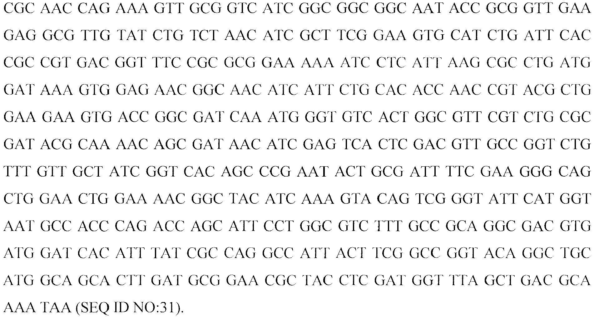
- The sequence of
mouse thioredoxin 1 is:
- The sequence of
mouse thioreodoxin 2 is:
- The sequence of
mouse thioredoxin reductase 1 is:

- The sequence of
mouse thioredoxin reductase 2 is:

- A commercial TrxR (rat liver) solution (4 µM) was diluted with water to yield a 2.86 µM solution. Lyophilized Trx (human) was reconstituted with PBS (10 mM, pH 7.2) yielding a 500 µM solution. A solution of 20 mM NADPH and 10 mM ATG and ATM solutions were prepared in water.
- In a black polypropylene 1.5 mL micro centrifuge tube, 437 µL reaction buffer (10 mM histidine, 10 mM Na2SO4, 137 mM NaCl, 2.5 mM KCl, pH 7.0), 25 µL NADPH, 16 µL formulated ocrelizumab solution (30.2 mg/mL) and 5 µL Trx were gently mixed. The reaction was initiated by the addition of 17.5 µL TrxR. The reaction was incubated at room temperature for 24 hours. Aliquots of 20 µL were taken at each sampling time-point and stored at -70 °C until analyzed by the Bioanalyzer assay.
- Inhibition of the Trx system was demonstrated using the same reaction conditions described above with the addition of various inhibitors.
-
Figure 24 shows a digital gel-like image from Bioanalyzer analysis (each lane representing a time point) showing that incubation of intact ocrelizumab ("2H7," a humanized anti-CD20 antibody, referred to as "Variant A" above) (1 mg/mL) with 0.1 µM TrxR (rat liver), 5 µM Trx (human) and 1 mM NADPH in 10 mM histidine sulfate buffer results in the reduction of ocrelizumab in less than one hour. - Aurothioglucose (ATG) was added to the ocrelizumab mixture described above, at the following concentrations: 1 mM; 0.6 µM (6:1 ATG:TrxR); 0.4 µM (4:1 ATG:TrxR); and 0.2 µM (2:1 ATG:TrxR).
- As attested by the digital gel-like images from Bioanalyzer analysis shown in
Figures 25-27 , aurothioglucose added atconcentrations 1 mM, 0.6 µM, and 0.4 µM effectively inhibits the reduction of ocrelizumab by the thioredoxin system. However, as shown inFigure 28 , under these experimental conditions aurothioglucose added at a concentration of 0.2 µM cannot inhibit ocrelizumab reduction after 24 hours. - Aurothiomalate (ATM) was added to the ocrelizumab mixture described above, at concentrations of 0.1 mM and 0.01 mM. As attested by the digital gel-like images from Bioanalyzer analysis shown in
Figures 29 and30 , ATM effectively inhibits the reduction of ocrelizumab by the thioredoxin system at both concentrations tested. - CuSO4 was added to the ocrelizumab mixture described above, at concentrations of 20 µM (4:1 Cu2+:Trx) ; 10 µm (2:1 Cu2+:Trx); and 5 µM (1:1 Cu2+:Trx). As shown in
Figures 31-33 , CuSO4 effectively inhibits thioredoxin-induced reduction of ocrelizumab at concentrations of 20 µM and 10 µM (Figures 31 and32 ), but the 5 µM concentration is insufficient to result in a complete inhibition of reduction (Figure 33 ). - Cystamine was added to the ocrelizumab mixture describe above at the following concentrations: 532 µM (20:1 cystamine:2H7 (Variant A) disulfide); 266 µM (10:1 cystamine:2H7 (Variant A) disulfide); 133 µM (5:1 cystamine:2H7 disulfide); and 26.6 µM (1:1 cystamine:2H7 (Variant A) disulfide). As shown in
Figures 34-37 , cystamine effectively inhibits thioredoxin-induced reduction of ocrelizumab at concentrations of 532 µM (20:1 cystamine:2H7 (Variant A) disulfide) and 266 µM (10:1 cystamine:2H7 (Variant A)) (Figures 34 and35 ) but the 133 µM (5:1 cystamine:2H7 (Variant A) disulfide) and 26.6 µM (1:1 cystamine:2H7 (Variant A) disulfide) concentrations are insufficient to inhibit the reduction of ocrelizumab after 24 hours (Figures 36 and37 ). - Cystine was added to the ocrelizumab mixture described above at a concentration of 2.6 mM. As shown in
Figure 38 , at this concentration cystine effectively inhibits reduction of ocrelizumab by the thioredoxin system. It is noted that the minimum effective concentration of cystine (just as the effective minimum concentration of other inhibitors) depends on the actual circumstances, and might be different for different proteins, such as antibodies, and might vary depending on the timing of addition. Thus, for example, if cystine is added pre-lysis, the minimum effective concentration for antibody 2H7 (Variant A) is about 1.3 mM, for Apomab about 1 mM and for antibody Variant C about 4.5 mM. When cystine is added in the cell culture medium, the minimum effective concentration typically is somewhat higher, and is about 5.2 mM for 2H7 (Variant A), 6 mM for Apomab and 9 mM for antibody Variant C. Usually, for cystine, cystamine and oxidized glutathione (see below) the minimum effective inhibitory concentration is about 40x of the antibody concentration (in µM). - GSSG was added to the ocrelizumab mixture described above at a concentration of 2.6 mM. As shown in
Figure 39 , at this concentration GSSG effectively inhibits reduction of ocrelizumab by the thioredoxin system. It is noted, however, that the minimum effective concentration of oxidize glutathione (just as that of the other inhibitors) depends on the actual circumstances, such as, for example, on the nature of the protein (e.g. antibody) produced and the timing of addition. For example, for antibody 2H7 (Variant A) the minimum effective concentration is about 1.3 mM for addition prior to lysis. -
Figure 40 shows a digital gel-like image from Bioanalyzer analysis (each lane representing a time point) showing that incubation of intact ocrelizumab. ("2H7," a humanized anti-CD20 antibody, Variant A) (1 mg/mL) with 10 µg/mL hexokinase, 50 µg/mL glucose-6-phosphate dehydrogenase, 5 µM thioredoxin, 0.1 µM thoredoxin reductase, 2 mM glucose, 0.6 mM ATP, 2 mM Mg2+, and 2 mM NADP in 50 mM histidine sulfate buffered at pH 7.38 results in the reduction of ocrelizumab in about one hour. Addition of 0.1 mM HDEA, a known glucose-6-phosphate dehydrogenase inhibitor does not inhibit the reduction. - As shown in the digital gel-like image from Bioanalyzer analysis of
Figure 41 , incubation of intact ocrelizumab (1 mg/mL) with 5 µM thioredoxin, 0.1 µM thioredoxin reductase, and 2 mM NADP in 50 mM histidine sulfate buffer at pH 7.38 does not result in the reduction of the ocrelizumab antibody. Reduction of ocrelizumab could not occur without hexokinase and glucose-6-phosphate dehydrogenase and their substrates to generate NADPH. - The invention illustratively described herein can suitably be practiced in the absence of any element or elements, limitation or limitations that is not specifically disclosed herein. Thus, for example, the terms "comprising," "including," "containing," etc. shall be read expansively and without limitation. Additionally, the terms and expressions employed herein have been used as terms of description and not of limitation, and there is no intention in the use of such terms and expressions of excluding any equivalent of the invention shown or portion thereof, but it is recognized that various modifications are possible within the scope of the invention claimed. Thus, it should be understood that although the present invention has been specifically disclosed by preferred embodiments and optional features, modifications and variations of the inventions embodied herein disclosed can be readily made by those skilled in the art, and that such modifications and variations are considered to be within the scope of the inventions disclosed herein. The inventions have been described broadly and generically herein. Each of the narrower species and subgeneric groupings falling within the generic disclosure also form the part of these inventions. This includes within the generic description of each of the inventions a proviso or negative limitation that will allow removing any subject matter from the genus, regardless or whether or not the material to be removed was specifically recited. In addition, where features or aspects of an invention are described in terms of the Markush group, those schooled in the art will recognize that the invention is also thereby described in terms of any individual member or subgroup of members of the Markush group. Further, when a reference to an aspect of the invention lists a range of individual members, as for example, 'SEQ ID NO:1 to SEQ ID NO: 100, inclusive,' it is intended to be equivalent to listing every member of the list individually, and additionally it should be understood that every individual member may be excluded or included in the claim individually.
- The steps depicted and/or used in methods herein may be performed in a different order than as depicted and/or stated. The steps are merely exemplary of the order these steps may occur. The steps may occur in any order that is desired such that it still performs the goals of the claimed invention.
- From the description of the invention herein, it is manifest that various equivalents can be used to implement the concepts of the present invention without departing from its scope. Moreover, while the invention has been described with specific reference to certain embodiments, a person of ordinary skill in the art would recognize that changes can be made in form and detail without departing from the spirit and the scope of the invention. The described embodiments are considered in all respects as illustrative and not restrictive. It should also be understood that the invention is not limited to the particular embodiments described herein, but is capable of many equivalents, rearrangements, modifications, and substitutions without departing from the scope of the invention.
-
- <110> Genentech, Inc.
- <120> PREVENTION OF DISULFIDE BOND REDUCTION DURING RECOMBINANT PRODUCTION OF POLYPEPTIDES
- <130> GNE-0246PCT
- <140> Not yet Assigned
<141> Herewith - <150>
US 60/948,577
<151> 2007-07-09 - <160> 35
- <170> PatentIn version 3.5
- <210> 1
<211> 107
<212> PRT
<213> Artificial - <220>
<223> light chain variable - <400> 1

- <210> 2
<211> 122
<212> PRT
<213> Artificial - <220>
<223> heavy chain variable - <400> 2

- <210> 3
<211> 107
<212> PRT
<213> Artificial - <220>
<223> light chain variable - <400> 3


- <210> 4
<211> 122
<212> PRT
<213> Artificial - <220>
<223> heavy chain variable - <400> 4

- <210> 5
<211> 122
<212> PRT
<213> Artificial - <220>
<223> heavy chain variable - <400> 5

- <210> 6
<211> 213
<212> PRT
<213> Artificial - <220>
<223> full length light chain - <400> 6

- <210> 7
<211> 452
<212> PRT
<213> Artificial - <220>
<223> full length heavy chain sequence - <400> 7


- <210> 8
<211> 452
<212> PRT
<213> Artificial - <220>
<223> full length heavy chain - <400> 8


- <210> 9
<211> 213
<212> PRT
<213> Artificial - <220>
<223> full length light chain - <400> 9

- <210> 10
<211> 452
<212> PRT
<213> Artificial - <220>
<223> full length heavy chain - <400> 10


- <210> 11
<211> 452
<212> PRT
<213> Artificial - <220>
<223> full length heavy chain sequence - <400> 11


- <210> 12
<211> 452
<212> PRT
<213> Artificial - <220>
<223> full length heavy chain sequence - <400> 12


- <210> 13
<211> 452
<212> PRT
<213> Artificial - <220>
<223> full length heavy chain sequence - <400> 13


- <210> 14
<211> 452
<212> PRT
<213> Artificial - <220>
<223> full length heavy chain - <400> 14

- <210> 15
<211> 452
<212> PRT
<213> Artificial - <220>
<223> full length heavy chain - <400> 15


- <210> 16
<211> 107
<212> PRT
<213> Artificial - <220>
<223> antibody - <400> 16

- <210> 17
<211> 119
<212> PRT
<213> Artificial - <220>
<223> antibody - <400> 17

- <210> 18
<211> 107
<212> PRT
<213> Artificial - <220>
<223> light chain - <400> 18

- <210> 19
<211> 120
<212> PRT
<213> Artificial - <220>
<223> heavy chain - <400> 19

- <210> 20
<211> 108
<212> PRT
<213> artificial - <220>
<223> antibody - <400> 20

- <210> 21
<211> 123
<212> PRT
<213> artificial - <220>
<223> antibody - <400> 21


- <210> 22
<211> 108
<212> PRT
<213> Artificial - <220>
<223> antibody - <400> 22

- <210> 23
<211> 123
<212> PRT
<213> Artificial - <220>
<223> antibody - <400> 23
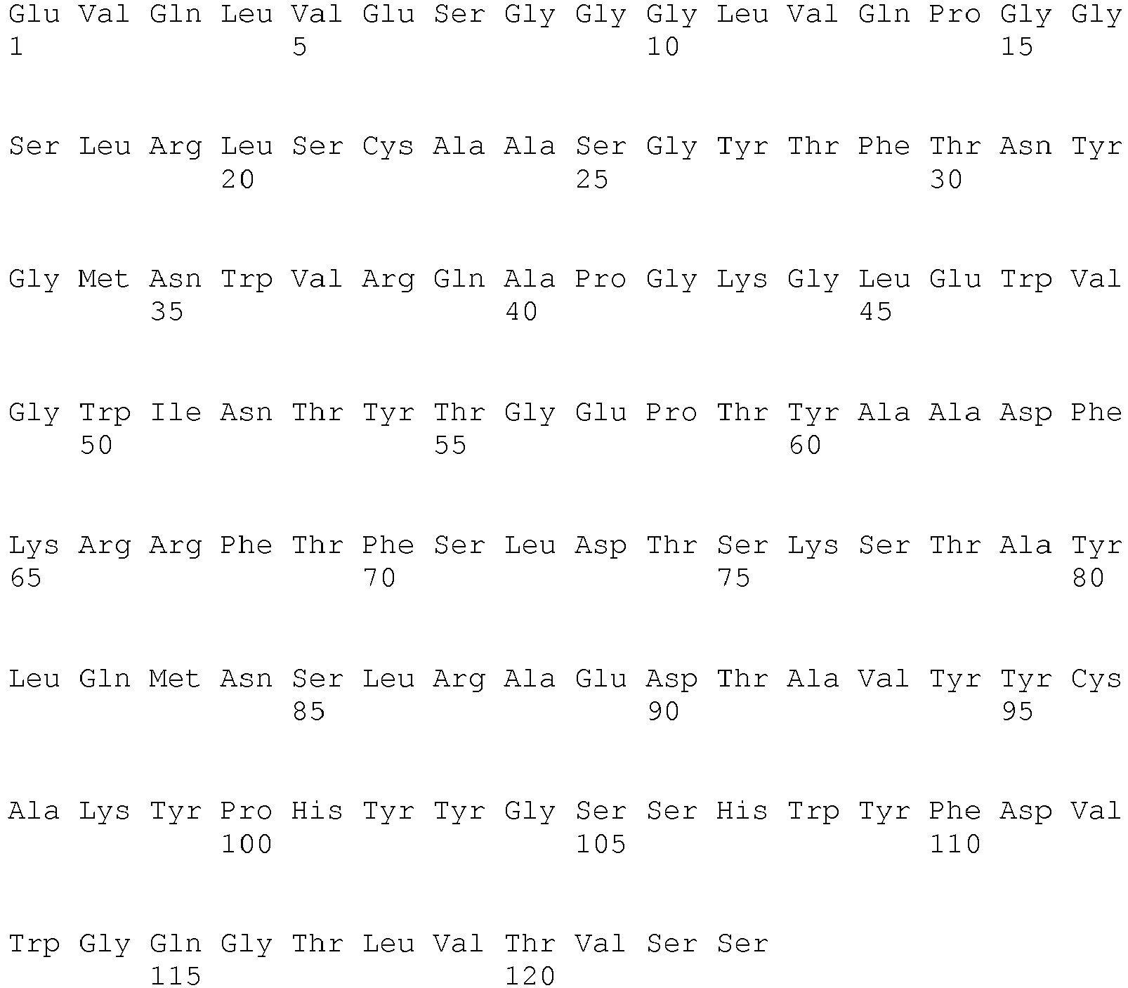
- <210> 24
<211> 108
<212> PRT
<213> Artificial - <220>
<223> antibody - <400> 24


- <210> 25
<211> 123
<212> PRT
<213> Artificial - <220>
<223> antibody - <400> 25

- <210> 26
<211> 108
<212> PRT
<213> Artificial - <220>
<223> antibody - <400> 26

- <210> 27
<211> 121
<212> PRT
<213> Artificial - <220>
<223> antibody - <400> 27


- <210> 28
<211> 214
<212> PRT
<213> Artificial - <220>
<223> antibody - <400> 28


- <210> 29
<211> 451
<212> PRT
<213> Artificial - <220>
<223> antibody - <400> 29

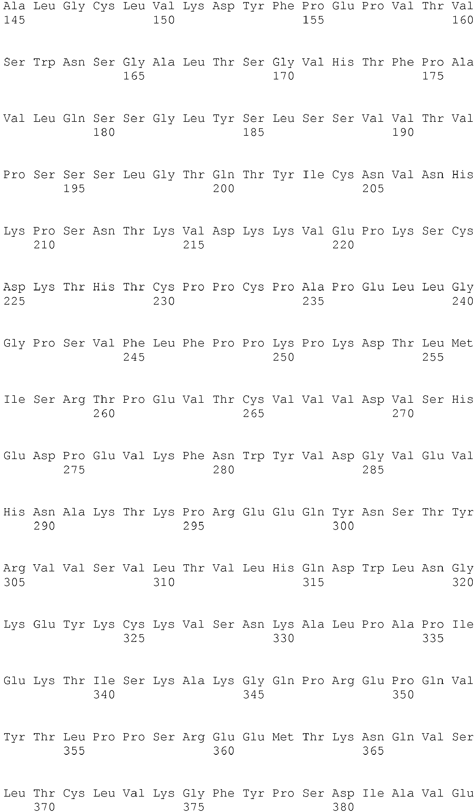

- <210> 30
<211> 384
<212> DNA
<213> E. coli Thioredoxin TrxA - <400> 30

- <210> 31
<211> 966
<212> DNA
<213> E. coli thioredoxin reductase TrxB - <400> 31
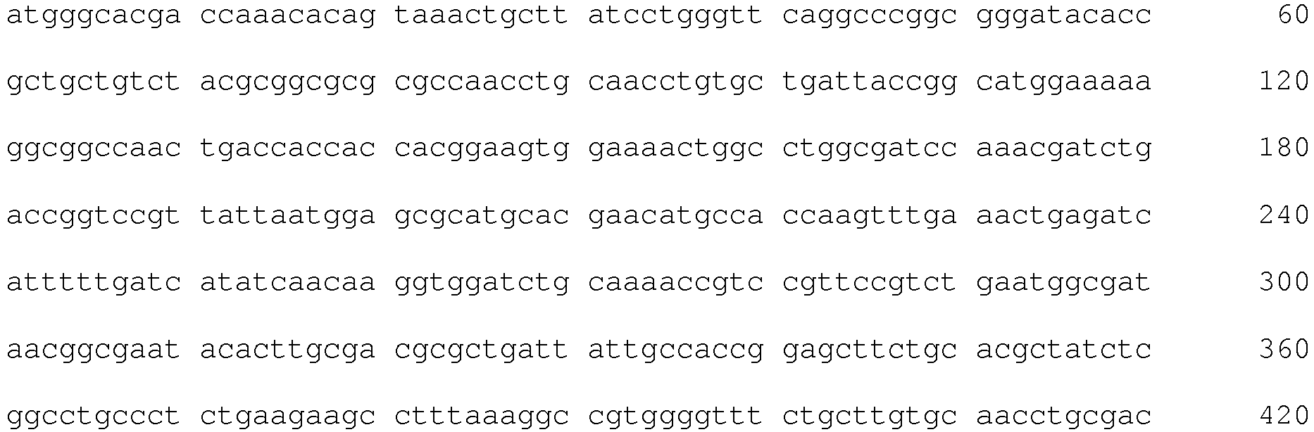

- <210> 32
<211> 318
<212> DNA
<213> mus musculus - <400> 32

- <210> 33
<211> 501
<212> DNA
<213> mus musculus - <400> 33


- <210> 34
<211> 1494
<212> DNA
<213> mus musculus - <400> 34
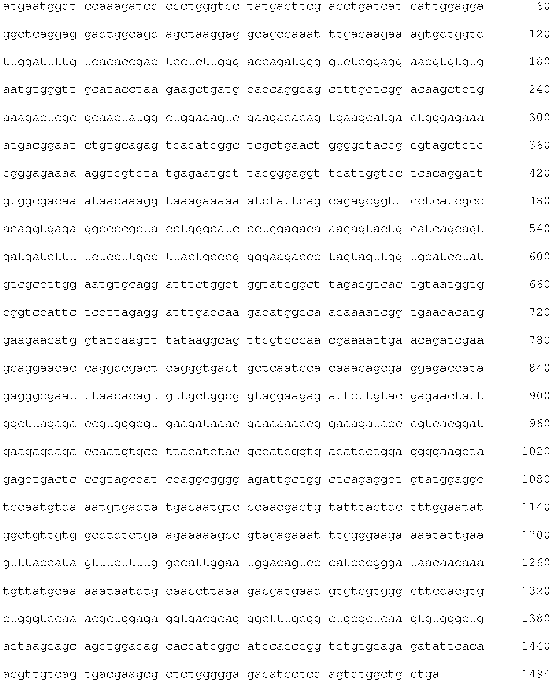
- <210> 35
<211> 1578
<212> DNA
<213> mus musculus - <400> 35
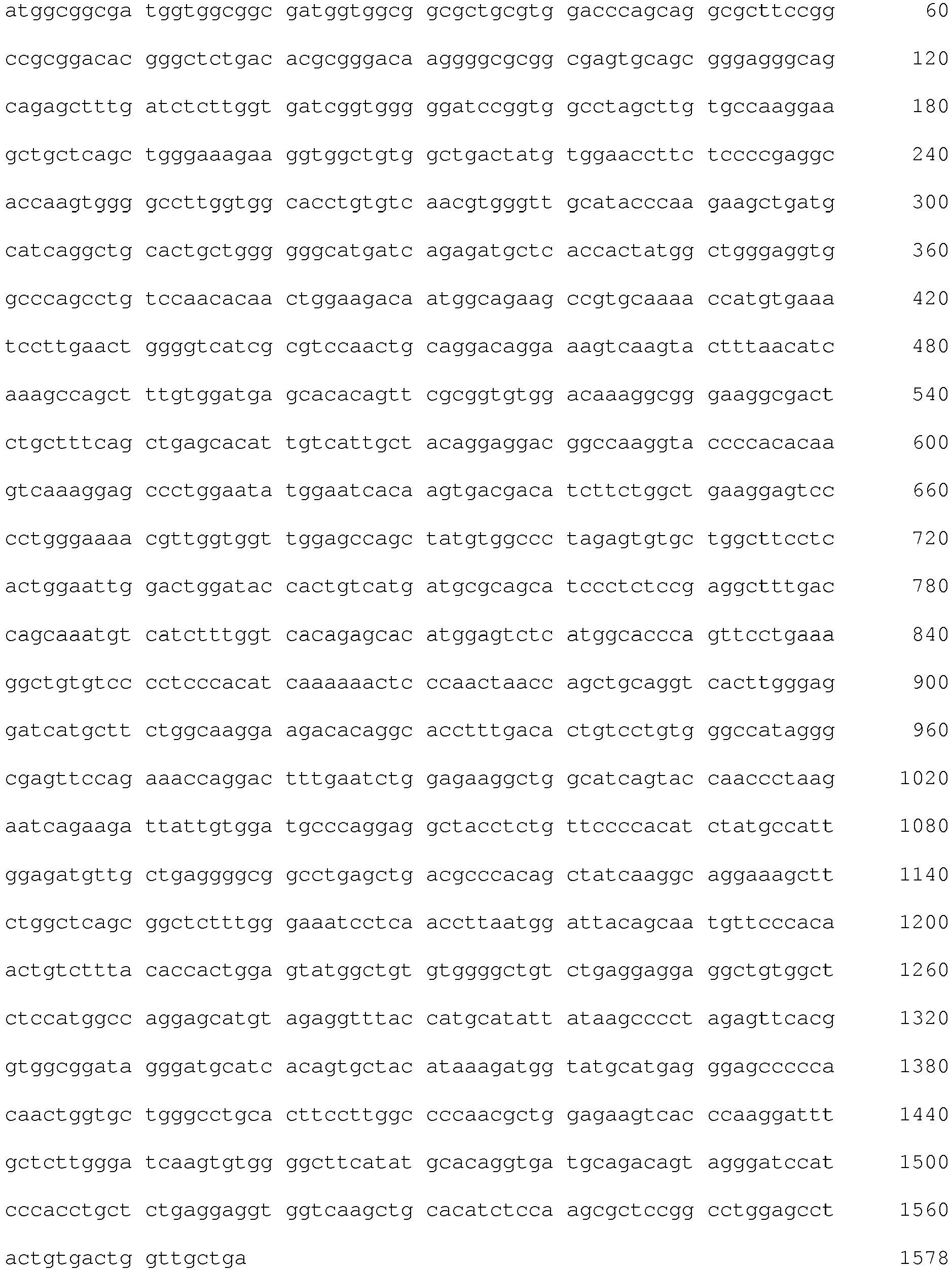
VL is an immunoglobulin light chain variable domain;
VH is an immunoglobulin heavy chain variable domain;
CL is an immunoglobulin light chain constant domain;
CH is an immunoglobulin heavy chain constant domain;
n is an integer greater than 1;
Y designates the residue of a covalent cross-linking agent.
Claims (19)
- A method for the prevention of the reduction of a disulfide bond in a polypeptide expressed in a recombinant host cell during processing following fermentation, comprising, following fermentation, supplementing the pre-harvest cell culture fluid (CCF) or harvested cell culture fluid (HCCF) of said recombinant host cell with a thioredoxin inhibitor, wherein said thioredoxin inhibitor is:(i) a direct inhibitor of thioredoxin;(ii) a specific inhibitor of thioredoxin reductase;(iii) is cupric sulfate ;(a) added in a concentration between about 5 µM and about 100 µM;(b) added in a concentration between about 10 µM to about 80 µM;(c) added in a concentration between about 15 µM and about 50 µM; or (d) added at a concentration at least about two-times of thioredoxin concentration in said pre-harvest or harvested culture fluid.(iv) an inhibitor of G6PD, selected from the group consisting of pyridoxal 5'-phosphate, 1 fluoro-2,4 dinitrobenzene, dehydroepiandrosterone (DHEA) and epiandrosterone (EA);(v) an inhibitor of hexokinase activity, which inhibitor is (a) a chelator of metal ions; or (b) selected from the group consisting of sorbose-1-phosphate, polyphosphates, 6-deoxy-6-fluoroglucose, 2-C-hydroxy-methylglucose, xylose, and lyxose;(vi) an antibody specifically binding to a thioredoxin reductase; or(vii) a measure indirectly resulting in the inhibition of thioredoxin activity, which measure is air sparging the harvested culture fluid of said recombinant host cell.
- The method of claim 1 (i) wherein said thioredoxin inhibitor is an alkyl-2-imidazolyl disulfide or a naphthoquinone spiroketal derivative.
- The method of claim 1 (ii) wherein said thioredoxin inhibitor is a gold complex.
- The method of claim 3 wherein said gold complex is aurothioglucose (ATG) or aurothiomalate (ATM), and, optionally, said ATG or said ATM is added at a concentration (a) between about 0.1 mM and about 1 mM, or (b) at least about four-times of thioredoxin reductase concentration in said pre-harvest or harvested culture fluid.
- The method of claim 1 (iii) wherein said cupric sulfate is in the form of pentrahydrate or in anhydrous form.
- The method of claim 1(iv) wherein said inhibitor is DHEA and wherein said DHEA is added in a concentration (a) between about 0.05 mM and about 5 mM, or (b) between about 0.1 mM and about 2.5 mM.
- The method of claim 1 (v)(a) wherein said chelator of metal ions is ethylenediamine tetraacetic acid (EDTA).
- The method of claim 7 wherein said EDTA is added in a concentration (i) between about 5 mM and about 60 mM; (ii) between about 10 mM and about 50 mM; or (iii) between about 20 mM and about 40 mM.
- The method of any one of claims 1 to 8 further comprising the step of air sparging the harvested culture fluid of said recombinant host cell.
- The method of claim 9, wherein:(a) said air sparging is continued until the pre-harvest or harvested culture fluid is at least about 30% saturated with air, or is between about 100% saturated to about 30% saturated with air; or(b) the amount of dissolved oxygen (dO2) in the pre-harvest or harvested culture fluid is at least about 10% or is at least about 30%.
- The method of any one of the preceding claims wherein said polypeptide is an antibody, or a biologically functional fragment of an antibody.
- The method of claim 11 wherein (i) said antibody fragment is selected from the group consisting of Fab, Fab', F(ab')2, scFv, (scFv)2, dAb, complementarity determining region (CDR) fragments, linear antibodies, single-chain antibody molecules, minibodies, diabodies, and multispecific antibodies formed from antibody fragments, or (ii) said antibody or antibody fragment is a therapeutic antibody or a biologically functional fragment thereof.
- The method of claim 12(ii) wherein said therapeutic antibody is selected from the group consisting of anti-HER2 antibodies anti-CD20 antibodies; anti-IL-8 antibodies; anti-VEGF antibodies; anti-CD40 antibodies, anti-GD11a antibodies; anti-CD18 antibodies; anti-IgE antibodies; anti-Apo-2 receptor antibodies; anti-Tissue Factor (TF) antibodies; anti-human α4β7 integrin antibodies; anti-EGFR antibodies; anti-CD3 antibodies; anti-CD25 antibodies; anti-CD4 antibodies; anti-CD52 antibodies; anti-Fc receptor antibodies; anti-carcinoembryonic antigen (CEA) antibodies; antibodies directed against breast epithelial cells; antibodies that bind to colon carcinoma cells; anti-CD38 antibodies; anti-CD33 antibodies; anti-CD22 antibodies; anti-EpCAM antibodies; anti-GpIIb/IIIa antibodies; anti-RSV antibodies; anti-CMV antibodies; anti-HIV antibodies; anti-hepatitis antibodies; anti-CA 125 antibodies; anti-αvβ3 antibodies; anti-human renal cell carcinoma antibodies; anti-human 17-1A antibodies; anti-human colorectal tumor antibodies; anti-human melanoma antibody R24 directed against GD3 ganglioside; anti-human squamous-cell carcinoma; and anti-human leukocyte antigen (HLA) antibodies, and anti-HLA DR antibodies.
- The method of claim 12(ii) wherein said therapeutic antibody is an antibody binding to:(a) a HER receptor, and wherein, optionally, the HER receptor is HER2 and said therapeutic antibody comprises a heavy and/or light chain variable domain sequence selected from the group consisting of SEQ ID NOS: 16, 17, 18, and 19;(b) VEGF, and wherein, optionally, said therapeutic antibody comprises a heavy and/or light chain variable domain sequence selected from the group consisting of SEQ ID NOS: 20 through 25;(c) IgE;(d) CD20, and wherein, optionally, said therapeutic antibody comprises a heavy and/or light chain variable domain sequence selected from the group consisting of SEQ ID NOS: 1 through 15;(e) CD11a, and wherein, optionally, said therapeutic antibody comprises a heavy and/or light chain variable domain sequence selected from the group consisting of SEQ ID NOS: 26 through 29;(f) CD40; or(g) DR5, and wherein, optionally, said therapeutic antibody is selected from the group consisting of Apomabs 1.1, 2.1, 3.1, 4.1, 5.1, 5.2, 5.3, 6.1, 6.2, 6.3, 7.1, 7.2, 7.3,8.1, 8.3, 9.1, 1.2, 2.2, 3.2, 4.2, 5.2, 6.2, 7.2, 8.2, 9.2, 1.3, 2.2, 3.3, 4.3, 5.3, 6.3, 7.3, 8.3, 9.3, and 25.3.
- The method of any one of claims 1 to 10 wherein said polypeptide is a therapeutic polypeptide and wherein, optionally, said therapeutic polypeptide is selected from the group consisting of a growth hormone, including human growth hormone and bovine growth hormone; growth hormone releasing factor; parathyroid hormone; thyroid stimulating hormone; lipoproteins; alpha-1-antitrypsin; insulin A-chain; insulin B-chain; proinsulin; follicle stimulating hormone; calcitonin; luteinizing hormone; glucagon; clotting factors such as factor VIIIC, factor IX, tissue factor, and von Willebrands factor; anti-clotting factors such as Protein C; atrial natriuretic factor; lung surfactant; a plasminogen activator, such as urokinase or human urine or tissue-type plasminogen activator (t-PA); bombesin; thrombin; hemopoietic growth factor; tumor necrosis factor-alpha and -beta; enkephalinase; RANTES (regulated on activation normally T-cell expressed and secreted); human macrophage inflammatory protein (MIP-1-alpha); a serum albumin such as human serum albumin; Muellerian-inhibiting substance; relaxin A-chain; relaxin B-chain; prorelaxin; mouse gonadotropin-associated peptide; a microbial protein, such as beta-lactamase; DNase; IgE; a cytotoxic T-lymphocyte associated antigen (CTLA), such as CTLA-4; inhibin; activin; vascular endothelial growth factor (VEGF); receptors for hormones or growth factors; Protein A or D; rheumatoid factors; a neurotrophic factor such as bone-derived neurotrophic factor (BDNF), neurotrophin-3, -4, -5, or -6 (NT-3, NT-4, NT-5, or NT-6), or a nerve growth factor such as NGF-β; platelet-derived growth factor (PDGF); fibroblast growth factor such as aFGF and bFGF; epidermal growth factor (EGF); transforming growth factor (TGF)such as TGF-alpha and TGF-beta, including TGF-β1, TGF-β2, TGF-β3, TGF-β4, or TGF-β5; insulin-like growth factor-I and -II (IGF-I and IGF-II); des(1-3)-IGF-I (brain IGF-I), insulin-like growth factor binding proteins; CD proteins such as CD3, CD4, CD8, CD19, CD20, CD34, and CD40; erythropoietin; osteoinductive factors; immunotoxins; a bone morphogenetic protein (BMP); an interferon such as interferon-alpha, -beta, and - gamma; colony stimulating factors (CSFs), e.g., M-CSF, GM-CSF, and G-CSF; interleukins (ILs), e.g., IL-1 to IL-10; superoxide dismutase; T-cell receptors; surface membrane proteins; decay accelerating factor; viral antigen such as, for example, a portion of the AIDS envelope; transport proteins; homing receptors; addressins; regulatory proteins; integrins such as CD11a, CD11b, CD11c, CD18, an ICAM, VLA-4 and VCAM; a tumor associated antigen such as HER2, HER3 or HER4 receptor; and fragments of said polypeptides.
- The method of any one of claims 1 to 15 wherein said recombinant host cell is (i) an eukaryotic host cell, or (ii) a prokaryotic host cell.
- The method of claim 16 wherein said eukaryotic host cell is a mammalian host cell and, optionally, is a Chinese Hamster Ovary (CHO) cell, or said prokaryotic host cell is a bacterial cell and, optionally, is an E. coli cell.
- The method of any one of claims 1 to 17 further comprising the step of recovering said polypeptide from the harvested cell culture fluid (HCCF).
- The method of any one of claims 1 to 18 further comprising the step of purifying said polypeptide from the harvested cell culture fluid (HCCF).
Priority Applications (10)
| Application Number | Priority Date | Filing Date | Title |
|---|---|---|---|
| EP23153140.1A EP4219522A3 (en) | 2007-07-09 | 2008-07-08 | Prevention of disulfide bond reduction during recombinant production of polypeptides |
| PL17196350T PL3327026T3 (en) | 2007-07-09 | 2008-07-08 | Prevention of disulfide bond reduction during recombinant production of polypeptides |
| EP23214759.5A EP4335863A2 (en) | 2007-07-09 | 2008-07-08 | Prevention of disulfide bond reduction during recombinant production of polypeptides |
| EP12178200.7A EP2586788B1 (en) | 2007-07-09 | 2008-07-08 | Prevention of disulfide bond reduction during recombinant production of polypeptides |
| SI200831956T SI2188302T1 (en) | 2007-07-09 | 2008-07-08 | Prevention of disulfide bond reduction during recombinant production of polypeptides |
| PL08781481T PL2188302T3 (en) | 2007-07-09 | 2008-07-08 | Prevention of disulfide bond reduction during recombinant production of polypeptides |
| EP23172637.3A EP4245766A3 (en) | 2007-07-09 | 2008-07-08 | Prevention of disulfide bond reduction during recombinant production of polypeptides |
| EP19181777.4A EP3597659B1 (en) | 2007-07-09 | 2008-07-08 | Prevention of disulfide bond reduction during recombinant production of polypeptides |
| EP17196350.7A EP3327026B1 (en) | 2007-07-09 | 2008-07-08 | Prevention of disulfide bond reduction during recombinant production of polypeptides |
| PL12178200T PL2586788T3 (en) | 2007-07-09 | 2008-07-08 | Prevention of disulfide bond reduction during recombinant production of polypeptides |
Applications Claiming Priority (2)
| Application Number | Priority Date | Filing Date | Title |
|---|---|---|---|
| US94867707P | 2007-07-09 | 2007-07-09 | |
| PCT/US2008/069395 WO2009009523A2 (en) | 2007-07-09 | 2008-07-08 | Prevention of disulfide bond reduction during recombinant production of polypeptides |
Related Child Applications (7)
| Application Number | Title | Priority Date | Filing Date |
|---|---|---|---|
| EP19181777.4A Division EP3597659B1 (en) | 2007-07-09 | 2008-07-08 | Prevention of disulfide bond reduction during recombinant production of polypeptides |
| EP23214759.5A Division EP4335863A2 (en) | 2007-07-09 | 2008-07-08 | Prevention of disulfide bond reduction during recombinant production of polypeptides |
| EP17196350.7A Division EP3327026B1 (en) | 2007-07-09 | 2008-07-08 | Prevention of disulfide bond reduction during recombinant production of polypeptides |
| EP23172637.3A Division EP4245766A3 (en) | 2007-07-09 | 2008-07-08 | Prevention of disulfide bond reduction during recombinant production of polypeptides |
| EP12178200.7A Division-Into EP2586788B1 (en) | 2007-07-09 | 2008-07-08 | Prevention of disulfide bond reduction during recombinant production of polypeptides |
| EP12178200.7A Division EP2586788B1 (en) | 2007-07-09 | 2008-07-08 | Prevention of disulfide bond reduction during recombinant production of polypeptides |
| EP23153140.1A Division EP4219522A3 (en) | 2007-07-09 | 2008-07-08 | Prevention of disulfide bond reduction during recombinant production of polypeptides |
Publications (2)
| Publication Number | Publication Date |
|---|---|
| EP2188302A2 EP2188302A2 (en) | 2010-05-26 |
| EP2188302B1 true EP2188302B1 (en) | 2017-11-01 |
Family
ID=39791340
Family Applications (7)
| Application Number | Title | Priority Date | Filing Date |
|---|---|---|---|
| EP08781481.0A Active EP2188302B1 (en) | 2007-07-09 | 2008-07-08 | Prevention of disulfide bond reduction during recombinant production of polypeptides |
| EP17196350.7A Active EP3327026B1 (en) | 2007-07-09 | 2008-07-08 | Prevention of disulfide bond reduction during recombinant production of polypeptides |
| EP23153140.1A Pending EP4219522A3 (en) | 2007-07-09 | 2008-07-08 | Prevention of disulfide bond reduction during recombinant production of polypeptides |
| EP19181777.4A Active EP3597659B1 (en) | 2007-07-09 | 2008-07-08 | Prevention of disulfide bond reduction during recombinant production of polypeptides |
| EP23172637.3A Pending EP4245766A3 (en) | 2007-07-09 | 2008-07-08 | Prevention of disulfide bond reduction during recombinant production of polypeptides |
| EP12178200.7A Revoked EP2586788B1 (en) | 2007-07-09 | 2008-07-08 | Prevention of disulfide bond reduction during recombinant production of polypeptides |
| EP23214759.5A Pending EP4335863A2 (en) | 2007-07-09 | 2008-07-08 | Prevention of disulfide bond reduction during recombinant production of polypeptides |
Family Applications After (6)
| Application Number | Title | Priority Date | Filing Date |
|---|---|---|---|
| EP17196350.7A Active EP3327026B1 (en) | 2007-07-09 | 2008-07-08 | Prevention of disulfide bond reduction during recombinant production of polypeptides |
| EP23153140.1A Pending EP4219522A3 (en) | 2007-07-09 | 2008-07-08 | Prevention of disulfide bond reduction during recombinant production of polypeptides |
| EP19181777.4A Active EP3597659B1 (en) | 2007-07-09 | 2008-07-08 | Prevention of disulfide bond reduction during recombinant production of polypeptides |
| EP23172637.3A Pending EP4245766A3 (en) | 2007-07-09 | 2008-07-08 | Prevention of disulfide bond reduction during recombinant production of polypeptides |
| EP12178200.7A Revoked EP2586788B1 (en) | 2007-07-09 | 2008-07-08 | Prevention of disulfide bond reduction during recombinant production of polypeptides |
| EP23214759.5A Pending EP4335863A2 (en) | 2007-07-09 | 2008-07-08 | Prevention of disulfide bond reduction during recombinant production of polypeptides |
Country Status (24)
| Country | Link |
|---|---|
| US (11) | US20090053786A1 (en) |
| EP (7) | EP2188302B1 (en) |
| JP (5) | JP5687057B2 (en) |
| KR (6) | KR101643514B1 (en) |
| CN (3) | CN101932591B (en) |
| AU (2) | AU2008275229B2 (en) |
| BR (1) | BRPI0812603B1 (en) |
| CA (2) | CA3113365A1 (en) |
| DE (1) | DE17196350T1 (en) |
| DK (2) | DK3327026T3 (en) |
| ES (4) | ES2941738T3 (en) |
| FI (1) | FI3597659T3 (en) |
| HR (3) | HRP20230461T3 (en) |
| HU (4) | HUE046992T2 (en) |
| IL (6) | IL202934B (en) |
| LT (2) | LT3597659T (en) |
| MX (2) | MX2010000196A (en) |
| PL (4) | PL3327026T3 (en) |
| PT (2) | PT3327026T (en) |
| SG (3) | SG10201913363VA (en) |
| SI (4) | SI2188302T1 (en) |
| TR (1) | TR201802088T4 (en) |
| TW (4) | TWI574974B (en) |
| WO (1) | WO2009009523A2 (en) |
Cited By (2)
| Publication number | Priority date | Publication date | Assignee | Title |
|---|---|---|---|---|
| EP3597659A1 (en) | 2007-07-09 | 2020-01-22 | Genentech, Inc. | Prevention of disulfide bond reduction during recombinant production of polypeptides |
| WO2020201296A1 (en) | 2019-04-01 | 2020-10-08 | The Automation Partnership (Cambridge) Ltd. | Operation process for a cell cultivation system |
Families Citing this family (76)
| Publication number | Priority date | Publication date | Assignee | Title |
|---|---|---|---|---|
| AU2004287643C1 (en) | 2003-11-05 | 2012-05-31 | Roche Glycart Ag | CD20 antibodies with increased FC receptor binding affinity and effector function |
| RU2518289C2 (en) | 2006-09-13 | 2014-06-10 | Эббви Инк, | Method for preparing fed-batch antibody or its fragment (versions) |
| US8911964B2 (en) | 2006-09-13 | 2014-12-16 | Abbvie Inc. | Fed-batch method of making human anti-TNF-alpha antibody |
| TWI472339B (en) | 2008-01-30 | 2015-02-11 | Genentech Inc | Composition comprising antibody that binds to domain ii of her2 and acidic variants thereof |
| TW201014605A (en) | 2008-09-16 | 2010-04-16 | Genentech Inc | Methods for treating progressive multiple sclerosis |
| WO2010141039A1 (en) * | 2008-10-20 | 2010-12-09 | Abbott Laboratories | Isolation and purification of antibodies using protein a affinity chromatography |
| CN104974251A (en) | 2008-10-20 | 2015-10-14 | Abbvie公司 | Viral inactivation during purification of antibodies |
| CA2759370C (en) * | 2009-04-30 | 2020-02-11 | Peter Schotte | Method for the production of domain antibodies |
| UA104626C2 (en) * | 2009-06-17 | 2014-02-25 | Эббви Биотерапеутикс Инк. | Anti-vegf antibodies and their uses |
| ES2793348T3 (en) | 2009-08-11 | 2020-11-13 | Hoffmann La Roche | Protein production in glutamine-free cell culture media |
| AR078161A1 (en) | 2009-09-11 | 2011-10-19 | Hoffmann La Roche | VERY CONCENTRATED PHARMACEUTICAL FORMULATIONS OF AN ANTIBODY ANTI CD20. USE OF THE FORMULATION. TREATMENT METHOD |
| ES2594893T3 (en) * | 2009-12-16 | 2016-12-23 | Abbvie Biotherapeutics Inc. | Anti HER2 antibodies and their uses |
| EP2552482A2 (en) * | 2010-03-31 | 2013-02-06 | Université de Genève | Stabilized antibody preparations and uses thereof |
| TW201302793A (en) | 2010-09-03 | 2013-01-16 | Glaxo Group Ltd | Novel antigen binding proteins |
| CN101974535B (en) * | 2010-11-03 | 2012-12-19 | 北京天广实生物技术股份有限公司 | Preparation and application of novel anti-ErbB2 humanized antibody MIL12 |
| US9067988B2 (en) | 2010-12-01 | 2015-06-30 | Alderbio Holdings Llc | Methods of preventing or treating pain using anti-NGF antibodies |
| US9078878B2 (en) | 2010-12-01 | 2015-07-14 | Alderbio Holdings Llc | Anti-NGF antibodies that selectively inhibit the association of NGF with TrkA, without affecting the association of NGF with p75 |
| US9539324B2 (en) | 2010-12-01 | 2017-01-10 | Alderbio Holdings, Llc | Methods of preventing inflammation and treating pain using anti-NGF compositions |
| US11214610B2 (en) | 2010-12-01 | 2022-01-04 | H. Lundbeck A/S | High-purity production of multi-subunit proteins such as antibodies in transformed microbes such as Pichia pastoris |
| US9783602B2 (en) | 2010-12-01 | 2017-10-10 | Alderbio Holdings Llc | Anti-NGF compositions and use thereof |
| US9884909B2 (en) | 2010-12-01 | 2018-02-06 | Alderbio Holdings Llc | Anti-NGF compositions and use thereof |
| WO2012149197A2 (en) | 2011-04-27 | 2012-11-01 | Abbott Laboratories | Methods for controlling the galactosylation profile of recombinantly-expressed proteins |
| CN102911265B (en) * | 2011-08-05 | 2014-02-05 | 王革 | Recombination variant of human nerve growth factors and preparation method thereof |
| KR20200037425A (en) * | 2012-03-27 | 2020-04-08 | 제넨테크, 인크. | Improved harvest operations for recombinant proteins |
| US9150645B2 (en) | 2012-04-20 | 2015-10-06 | Abbvie, Inc. | Cell culture methods to reduce acidic species |
| US9067990B2 (en) | 2013-03-14 | 2015-06-30 | Abbvie, Inc. | Protein purification using displacement chromatography |
| US9181572B2 (en) | 2012-04-20 | 2015-11-10 | Abbvie, Inc. | Methods to modulate lysine variant distribution |
| US9249182B2 (en) | 2012-05-24 | 2016-02-02 | Abbvie, Inc. | Purification of antibodies using hydrophobic interaction chromatography |
| US9650411B2 (en) | 2012-08-07 | 2017-05-16 | Kyowa Hakko Kirin Co., Ltd. | Method of purifying protein |
| CA2883272A1 (en) | 2012-09-02 | 2014-03-06 | Abbvie Inc. | Methods to control protein heterogeneity |
| US9512214B2 (en) | 2012-09-02 | 2016-12-06 | Abbvie, Inc. | Methods to control protein heterogeneity |
| JP6411891B2 (en) | 2012-10-03 | 2018-10-24 | 協和発酵キリン株式会社 | Method for preventing reduction of polypeptide by adding amino acid to culture medium |
| US20140154255A1 (en) | 2012-11-30 | 2014-06-05 | Abbvie Biotherapeutics Inc. | Anti-vegf antibodies and their uses |
| SG11201507230PA (en) | 2013-03-12 | 2015-10-29 | Abbvie Inc | Human antibodies that bind human tnf-alpha and methods of preparing the same |
| US9499614B2 (en) | 2013-03-14 | 2016-11-22 | Abbvie Inc. | Methods for modulating protein glycosylation profiles of recombinant protein therapeutics using monosaccharides and oligosaccharides |
| US8921526B2 (en) | 2013-03-14 | 2014-12-30 | Abbvie, Inc. | Mutated anti-TNFα antibodies and methods of their use |
| US9017687B1 (en) | 2013-10-18 | 2015-04-28 | Abbvie, Inc. | Low acidic species compositions and methods for producing and using the same using displacement chromatography |
| RU2019115089A (en) | 2013-04-16 | 2019-06-11 | Дженентек, Инк. | VARIANTS OF PERTUSUMAB AND THEIR ANALYTICAL CHARACTERISTICS |
| WO2015009699A1 (en) * | 2013-07-15 | 2015-01-22 | Ketter Patrick | Methods and compositions to prevent or treat bacterial infections |
| WO2015051293A2 (en) | 2013-10-04 | 2015-04-09 | Abbvie, Inc. | Use of metal ions for modulation of protein glycosylation profiles of recombinant proteins |
| US9085618B2 (en) | 2013-10-18 | 2015-07-21 | Abbvie, Inc. | Low acidic species compositions and methods for producing and using the same |
| US9181337B2 (en) | 2013-10-18 | 2015-11-10 | Abbvie, Inc. | Modulated lysine variant species compositions and methods for producing and using the same |
| US8946395B1 (en) | 2013-10-18 | 2015-02-03 | Abbvie Inc. | Purification of proteins using hydrophobic interaction chromatography |
| WO2015073884A2 (en) | 2013-11-15 | 2015-05-21 | Abbvie, Inc. | Glycoengineered binding protein compositions |
| EP3077522A1 (en) * | 2013-12-04 | 2016-10-12 | Immunogen, Inc. | Compositions and methods for antibody production |
| US9620278B2 (en) * | 2014-02-19 | 2017-04-11 | General Electric Company | System and method for reducing partial discharge in high voltage planar transformers |
| US10647777B2 (en) * | 2014-05-28 | 2020-05-12 | Genzyme Corporation | Methods of controlling the formation of disulfide bonds in protein solutions |
| US9840553B2 (en) | 2014-06-28 | 2017-12-12 | Kodiak Sciences Inc. | Dual PDGF/VEGF antagonists |
| AR103172A1 (en) * | 2014-12-22 | 2017-04-19 | Novartis Ag | SELECTIVE REDUCTION OF CYSTEINE WASTE IN ANTIBODIES IL-17 |
| AU2016305331B2 (en) * | 2015-08-12 | 2019-05-16 | Pfizer Inc. | Capped and uncapped antibody cysteines, and their use in antibody-drug conjugation |
| HRP20231544T1 (en) * | 2015-08-13 | 2024-03-15 | Amgen Inc. | Charged depth filtration of antigen-binding proteins |
| EP3397276A4 (en) | 2015-12-30 | 2019-12-18 | Kodiak Sciences Inc. | Antibodies and conjugates thereof |
| SG10202109689QA (en) | 2016-04-15 | 2021-10-28 | Alder Biopharmaceuticals Inc | Humanized anti-pacap antibodies and uses thereof |
| AU2017262734B2 (en) * | 2016-05-10 | 2020-04-30 | Medimmune, Llc | Prevention of protein disulfide bond reduction |
| BR112018073133A2 (en) * | 2016-05-10 | 2019-04-30 | Genentech Inc | methods of decreasing trisulfide bonds during recombinant polypeptide production |
| CA3026589A1 (en) * | 2016-06-10 | 2017-12-14 | Lonza Ltd | Method for stabilizing proteins |
| US20190225694A1 (en) | 2016-06-28 | 2019-07-25 | Zaklady Farmaceutyczne Polpharma Sa | Recombinant production of monoclonal antibodies |
| CN106366160B (en) * | 2016-10-11 | 2019-06-14 | 厦门大学 | The method that building is rich in disulfide bond peptide molecule skeleton is precisely matched based on disulfide bond |
| US20190374639A1 (en) | 2016-11-21 | 2019-12-12 | Polpharma Biologics S.A. | Aqueous pharmaceutical formulations |
| CA3044664C (en) | 2016-11-30 | 2022-11-22 | Oncomed Pharmaceuticals, Inc. | Methods for treatment of cancer comprising tigit-binding agents |
| EP3564367A4 (en) | 2016-12-28 | 2020-08-12 | Kyowa Kirin Co., Ltd. | Method of selecting cells |
| CN110621341A (en) * | 2017-04-26 | 2019-12-27 | 百时美施贵宝公司 | Antibody production method that minimizes disulfide bond reduction |
| EP3621989B1 (en) * | 2017-05-09 | 2023-06-28 | Bristol-Myers Squibb Company | Method of controlling the formation of pink color during antibody manufacturing |
| US10913795B2 (en) | 2017-05-17 | 2021-02-09 | Immunogen, Inc. | Compositions and methods for antibody production |
| WO2019040671A1 (en) * | 2017-08-22 | 2019-02-28 | Biogen Ma Inc. | Methods for purifying antibodies having reduced high molecular weight aggregates |
| TW201923069A (en) * | 2017-10-02 | 2019-06-16 | 美商拜耳保健有限責任公司 | Methods for preventing disulfide bond reduction in cell culture harvest with selenite |
| TW201927335A (en) * | 2017-10-02 | 2019-07-16 | 美商拜耳保健有限責任公司 | Methods for preventing disulfide bond reduction in cell culture harvest |
| US11447547B1 (en) | 2017-12-13 | 2022-09-20 | Amgen Inc. | Method of antigen-binding protein production |
| EP3788151A4 (en) * | 2018-05-04 | 2022-01-12 | Sigma-Aldrich Co. LLC | Engineered cells with modified host cell protein profiles |
| WO2020028616A1 (en) * | 2018-08-02 | 2020-02-06 | Lonza Ltd | Methods for manufacturing recombinant protein comprising a disulfide bond |
| AU2019377526A1 (en) | 2018-11-08 | 2021-06-10 | Sutro Biopharma, Inc. | E coli strains having an oxidative cytoplasm |
| US20220306734A1 (en) | 2019-07-24 | 2022-09-29 | H. Lundbeck A/S | Anti-mglur5 antibodies and uses thereof |
| EP4041312A4 (en) | 2019-10-10 | 2023-12-20 | Kodiak Sciences Inc. | Methods of treating an eye disorder |
| CN113480652B (en) * | 2021-07-30 | 2023-04-07 | 成都景泽生物制药有限公司 | Method for producing recombinant EGFR antibody active molecules by fermentation culture of recombinant CHO cells |
| WO2023049687A1 (en) | 2021-09-21 | 2023-03-30 | Bristol-Myers Squibb Company | Methods of controlling the level of dissolved oxygen (do) in a solution comprising a recombinant protein in a storage container |
| US20240092870A1 (en) * | 2022-09-15 | 2024-03-21 | Sanofi | Methods of determining protein reduction susceptability |
Citations (35)
| Publication number | Priority date | Publication date | Assignee | Title |
|---|---|---|---|---|
| US4572798A (en) | 1984-12-06 | 1986-02-25 | Cetus Corporation | Method for promoting disulfide bond formation in recombinant proteins |
| EP0363126A2 (en) | 1988-10-04 | 1990-04-11 | Eli Lilly And Company | Method for the purification of vitamin K-dependent proteins |
| WO1993010260A1 (en) | 1991-11-21 | 1993-05-27 | The Board Of Trustees Of The Leland Stanford Junior University | Controlling degradation of glycoprotein oligosaccharides by extracellular glycosisases |
| EP0612846A1 (en) | 1993-01-28 | 1994-08-31 | Amgen Inc. | G-CSF analog compositions and methods |
| WO1997026357A1 (en) | 1996-01-19 | 1997-07-24 | Board Of Regents, The University Of Texas System | Methods and compositions for inhibiting hexokinase |
| WO1998014467A1 (en) | 1996-09-30 | 1998-04-09 | Human Genome Sciences, Inc. | Method of purifying protein from inclusion bodies |
| WO1998021234A2 (en) | 1996-11-15 | 1998-05-22 | Genentech, Inc. | Purification of neurotrophins |
| US5808006A (en) | 1993-08-20 | 1998-09-15 | Genentech, Inc. | Refolding of polypeptides like recombinant insulin-like growth factor IGF-I |
| US5830761A (en) | 1995-06-07 | 1998-11-03 | Genetics Institute, Inc. | Medium and methods for culturing mammalian cho cells |
| US5866362A (en) | 1985-03-28 | 1999-02-02 | Chiron Corporation | Enhanced purification and expression of insoluble recombinant proteins |
| WO1999033988A1 (en) | 1997-12-29 | 1999-07-08 | Chong Kun Dang Corporation | A process for preparing human proinsulin |
| WO1999054440A1 (en) | 1998-04-21 | 1999-10-28 | Micromet Gesellschaft Für Biomedizinische Forschung Mbh | CD19xCD3 SPECIFIC POLYPEPTIDES AND USES THEREOF |
| WO2000012537A1 (en) | 1998-08-27 | 2000-03-09 | Lg Chemical Limited | Process for the preparation of active somatotropin from inclusion bodies |
| WO2002020818A1 (en) | 2000-09-06 | 2002-03-14 | The Board Of Trustees Of The Leland Stanford Junior University | Enhanced in vitro synthesis of active proteins containing disulfide bonds |
| EP1241250A2 (en) | 1998-05-15 | 2002-09-18 | Genentech, Inc. | Human dehydrogenase-like protein and nucleic acid encoding it |
| US6566514B1 (en) | 1998-01-30 | 2003-05-20 | Genesense Technologies Inc. | Oligonucleotide sequences complementary to thioredoxin or thioredoxin reductase genes and methods of using same to modulate cell growth |
| WO2004001056A1 (en) | 2002-06-24 | 2003-12-31 | Dr. Reddy's Laboratories Ltd. | Process for preparing g-csf |
| US20040029229A1 (en) | 2002-05-20 | 2004-02-12 | Reeves Philip J. | High level protein expression system |
| US20040138424A1 (en) | 2001-03-09 | 2004-07-15 | Kozo Takeda | Method of purifying protein |
| US20050038231A1 (en) | 2003-07-28 | 2005-02-17 | Genentech, Inc. | Reducing protein a leaching during protein a affinity chromatography |
| US6872563B1 (en) | 1999-10-05 | 2005-03-29 | President And Fellows Of Harvard College | Compositions and methods for production of disulfide bond containing proteins in host cells |
| US20050100984A1 (en) | 1995-04-11 | 2005-05-12 | Martin Opper | Cytoplasmic expression of antibodies, antibody fragments and antibody fragment fusion proteins in E. coli |
| WO2006034488A2 (en) | 2004-09-23 | 2006-03-30 | Genentech, Inc. | Cysteine engineered antibodies and conjugates |
| WO2006047340A2 (en) | 2004-10-22 | 2006-05-04 | Amgen Inc. | Methods for refolding of recombinant antibodies |
| US20060105389A1 (en) | 2004-10-19 | 2006-05-18 | Kordyum Vitaliy A | Method for obtaining single chain antibodies to human interferon alpha2b |
| WO2006054063A1 (en) | 2004-11-19 | 2006-05-26 | Ucb Pharma S.A. | Process for obtaining antibodies |
| WO2006060083A1 (en) | 2004-10-22 | 2006-06-08 | Amgen Inc. | Methods for refolding polypeptides |
| US20060142549A1 (en) | 2002-09-11 | 2006-06-29 | Kozo Takeda | Method of purifying protein |
| WO2006084264A2 (en) | 2005-02-07 | 2006-08-10 | Genentech, Inc. | Cd20 antibody variants and uses thereof |
| US20060216790A1 (en) | 2003-10-10 | 2006-09-28 | Novo Nordisk Healthcare A/G | Method for large-scale production of polypeptide in eukaryote cells and a culture vessel suitable therefor |
| US7189811B2 (en) | 2002-09-06 | 2007-03-13 | National Institute Of Immunology | Process for solubilization of recombinant proteins expressed as inclusion body |
| WO2008127305A2 (en) | 2006-11-01 | 2008-10-23 | Biogen Idec Ma Inc. | Method of isolating biomacromolecules using low ph and divalent cations |
| WO2009009523A2 (en) | 2007-07-09 | 2009-01-15 | Genentech, Inc. | Prevention of disulfide bond reduction during recombinant production of polypeptides |
| US7723490B2 (en) | 1999-11-12 | 2010-05-25 | Amgen Inc. | Process for correction of a disulfide misfold in Fc molecules |
| EP1228076B1 (en) | 1999-09-20 | 2011-07-06 | NewSouth Innovations Pty Limited | A substantially cell membrane impermeable compound and use thereof |
Family Cites Families (236)
| Publication number | Priority date | Publication date | Assignee | Title |
|---|---|---|---|---|
| US524474A (en) * | 1894-08-14 | Seed-planter | ||
| US670534A (en) * | 1900-07-27 | 1901-03-26 | Oliver L Carmical | Seeding-machine and fertilizer-distributer. |
| US1369649A (en) * | 1920-01-02 | 1921-02-22 | George A Gieseler | Pneumatic conveying mechanism |
| US2062295A (en) * | 1932-11-22 | 1936-12-01 | Niagara Sprayer And Chemical C | Crop duster |
| US2171205A (en) * | 1937-08-26 | 1939-08-29 | Louis A Zinke | Powder distributing attachment for tractors |
| US2193738A (en) * | 1938-05-21 | 1940-03-12 | Nat Conveyors Co Inc | Pneumatic conveyer |
| US2559183A (en) * | 1946-12-10 | 1951-07-03 | Lawton L Barnett | Transplanter drill set |
| US2671690A (en) * | 1948-02-21 | 1954-03-09 | Ehrenkrook Karl Von | Forced air sprayer and duster |
| US2937049A (en) * | 1949-10-18 | 1960-05-17 | Osawa Hirosaburo | Method of and an apparatus for carrying coals out of a vertical shaft with the aid of heavy liquid in the coal mine |
| US2814531A (en) * | 1956-04-27 | 1957-11-26 | Clough Equipment Co | Balanced pneumatic conveying systems for pulverulent material |
| US3207560A (en) * | 1962-12-26 | 1965-09-21 | Monsanto Co | Unloading device |
| US3197261A (en) * | 1964-03-09 | 1965-07-27 | Simpson Herbert Corp | System for feeding and transporting materials |
| US3386474A (en) * | 1965-05-26 | 1968-06-04 | Chemagro Corp | Diverter valve for finely divided solids |
| US3515315A (en) * | 1966-01-24 | 1970-06-02 | Kidd Designs Ltd Archie | Combined seed and fertilizer drills for agricultural use |
| US3387895A (en) * | 1966-12-29 | 1968-06-11 | Combustion Eng | Non-clogging splitter unit for dividing the flow of fluid-conveyed material |
| FR1558490A (en) * | 1967-12-27 | 1969-02-28 | ||
| US3543704A (en) * | 1968-08-05 | 1970-12-01 | Deere & Co | Seed planter |
| US3845702A (en) | 1970-04-01 | 1974-11-05 | Mies Filter Prod Inc | Apparatus for cooking chicken |
| DE2440888C3 (en) * | 1974-08-27 | 1978-11-23 | Waeschle Maschinenfabrik Gmbh, 7980 Ravensburg | System for the successive loading of several unloading stations connected one behind the other to a pneumatic conveying line via separators with bulk material |
| US4049494A (en) | 1975-09-04 | 1977-09-20 | The United States Of America As Represented By The Secretary Of Agriculture | Vaccine production process |
| US4036408A (en) * | 1975-12-24 | 1977-07-19 | Acf Industries, Incorporated | Hopper having means for directing blasts of gas to a discharge outlet or the hopper |
| US4280419A (en) * | 1977-03-21 | 1981-07-28 | International Harvester Company | Pneumatic system for conveying granular material |
| USRE30985E (en) | 1978-01-01 | 1982-06-29 | Serum-free cell culture media | |
| FR2413974A1 (en) | 1978-01-06 | 1979-08-03 | David Bernard | DRYER FOR SCREEN-PRINTED SHEETS |
| US4200412A (en) * | 1978-04-20 | 1980-04-29 | Dynamic Air Incorporated | Control for pneumatic conveying system |
| US4264243A (en) * | 1979-01-04 | 1981-04-28 | Dundee Cement Company | Constant vacuum barge unloading system |
| US4515893A (en) | 1979-04-26 | 1985-05-07 | Ortho Pharmaceutical Corporation | Hybrid cell line for producing complement-fixing monoclonal antibody to human T cells |
| US4244522A (en) * | 1979-08-31 | 1981-01-13 | Deere & Company | Liquid storage and distribution apparatus for agricultural implements |
| CA1126587A (en) * | 1980-07-11 | 1982-06-29 | Daniel W. Kelm | Seed cup assembly |
| US4413935A (en) * | 1981-06-29 | 1983-11-08 | Combustion Engineering, Inc. | Flow splitter for dividing a stream of pulverulent material into multiple streams |
| GB2112506A (en) * | 1981-12-30 | 1983-07-20 | Shell Int Research | Distributing valve |
| US4943529A (en) | 1982-05-19 | 1990-07-24 | Gist-Brocades Nv | Kluyveromyces as a host strain |
| DE3239897A1 (en) * | 1982-10-28 | 1984-05-03 | Trützschler GmbH & Co KG, 4050 Mönchengladbach | METHOD AND DEVICE FOR CONVEYING FIBER-SHAPED SUBSTANCES, IN PARTICULAR TEXTILE FIBER FLAKES CONTAINED IN AIR-FLOATING, IN SPINNING PREPARATION MACHINES |
| US4560655A (en) | 1982-12-16 | 1985-12-24 | Immunex Corporation | Serum-free cell culture medium and process for making same |
| US4657866A (en) | 1982-12-21 | 1987-04-14 | Sudhir Kumar | Serum-free, synthetic, completely chemically defined tissue culture media |
| US4620948A (en) | 1982-12-22 | 1986-11-04 | Genentech, Inc. | Purification and activity assurance of precipitated heterologous proteins |
| US4816567A (en) | 1983-04-08 | 1989-03-28 | Genentech, Inc. | Recombinant immunoglobin preparations |
| AU3145184A (en) | 1983-08-16 | 1985-02-21 | Zymogenetics Inc. | High expression of foreign genes in schizosaccharomyces pombe |
| US4767704A (en) | 1983-10-07 | 1988-08-30 | Columbia University In The City Of New York | Protein-free culture medium |
| US4562779A (en) * | 1984-01-26 | 1986-01-07 | Briggs Manufacturing, Inc. | Foldable drill implement |
| DE3420616A1 (en) * | 1984-06-02 | 1985-12-05 | Alb. Klein Gmbh & Co Kg, 5241 Niederfischbach | DEVICE AND METHOD FOR CONTROLLING A LOCKING PROCESS FOR A PITCHING PARTICLE PARTICLE |
| US5672347A (en) | 1984-07-05 | 1997-09-30 | Genentech, Inc. | Tumor necrosis factor antagonists and their use |
| US5807715A (en) | 1984-08-27 | 1998-09-15 | The Board Of Trustees Of The Leland Stanford Junior University | Methods and transformed mammalian lymphocyte cells for producing functional antigen-binding protein including chimeric immunoglobulin |
| US4879231A (en) | 1984-10-30 | 1989-11-07 | Phillips Petroleum Company | Transformation of yeasts of the genus pichia |
| US4652630A (en) | 1985-02-22 | 1987-03-24 | Monsanto Company | Method of somatotropin naturation |
| GB8516415D0 (en) | 1985-06-28 | 1985-07-31 | Celltech Ltd | Culture of animal cells |
| US4676980A (en) | 1985-09-23 | 1987-06-30 | The United States Of America As Represented By The Secretary Of The Department Of Health And Human Services | Target specific cross-linked heteroantibodies |
| US4738770A (en) * | 1985-11-25 | 1988-04-19 | Cities Service Oil And Gas Corp. | Apparatus and method of removal of catalyst blockage from reactor recycle pumps |
| US5091178A (en) | 1986-02-21 | 1992-02-25 | Oncogen | Tumor therapy with biologically active anti-tumor antibodies |
| US4927762A (en) | 1986-04-01 | 1990-05-22 | Cell Enterprises, Inc. | Cell culture medium with antioxidant |
| GB8610600D0 (en) | 1986-04-30 | 1986-06-04 | Novo Industri As | Transformation of trichoderma |
| IL85035A0 (en) | 1987-01-08 | 1988-06-30 | Int Genetic Eng | Polynucleotide molecule,a chimeric antibody with specificity for human b cell surface antigen,a process for the preparation and methods utilizing the same |
| US4779765A (en) * | 1987-04-13 | 1988-10-25 | Deere & Company | Fertilizer divider for dual air system |
| US4793743A (en) * | 1987-04-29 | 1988-12-27 | Morris Rod-Weeder Company Ltd. | Air seeder purging system |
| IL87737A (en) | 1987-09-11 | 1993-08-18 | Genentech Inc | Method for culturing polypeptide factor dependent vertebrate recombinant cells |
| US5091313A (en) | 1988-08-05 | 1992-02-25 | Tanox Biosystems, Inc. | Antigenic epitopes of IgE present on B cell but not basophil surface |
| US5720937A (en) | 1988-01-12 | 1998-02-24 | Genentech, Inc. | In vivo tumor detection assay |
| US4872785A (en) * | 1988-01-22 | 1989-10-10 | Schrage Timothy L | Shut-off means for air-actuated planter |
| US4843983A (en) * | 1988-02-02 | 1989-07-04 | Deere & Company | Pneumatic manifold quick coupling |
| HUT53672A (en) | 1988-02-25 | 1990-11-28 | Gen Hospital Corp | Quick immunoselective cloning process |
| US5506126A (en) | 1988-02-25 | 1996-04-09 | The General Hospital Corporation | Rapid immunoselection cloning method |
| US4861579A (en) | 1988-03-17 | 1989-08-29 | American Cyanamid Company | Suppression of B-lymphocytes in mammals by administration of anti-B-lymphocyte antibodies |
| DE68919361T2 (en) | 1988-06-21 | 1995-05-24 | Genentech Inc | THERAPEUTIC COMPOSITIONS FOR THE TREATMENT OF MYOCARD INFARTS. |
| US6048728A (en) | 1988-09-23 | 2000-04-11 | Chiron Corporation | Cell culture medium for enhanced cell growth, culture longevity, and product expression |
| WO1990003430A1 (en) | 1988-09-23 | 1990-04-05 | Cetus Corporation | Cell culture medium for enhanced cell growth, culture longevity and product expression |
| GB8823869D0 (en) | 1988-10-12 | 1988-11-16 | Medical Res Council | Production of antibodies |
| DK167639B1 (en) * | 1988-11-16 | 1993-12-06 | Kongskilde Maskinfabrik As | GRANULATED MATERIAL SEARCH MACHINE |
| US5530101A (en) | 1988-12-28 | 1996-06-25 | Protein Design Labs, Inc. | Humanized immunoglobulins |
| ES2038579T3 (en) | 1989-04-28 | 1997-02-16 | Rhein Biotech Proz & Prod Gmbh | YEAST CELLS OF THE GENUS SCHWANNIOMYCES. |
| EP0402226A1 (en) | 1989-06-06 | 1990-12-12 | Institut National De La Recherche Agronomique | Transformation vectors for yeast yarrowia |
| DE3920358A1 (en) | 1989-06-22 | 1991-01-17 | Behringwerke Ag | BISPECIFIC AND OLIGO-SPECIFIC, MONO- AND OLIGOVALENT ANTI-BODY CONSTRUCTS, THEIR PRODUCTION AND USE |
| EP0479909B1 (en) | 1989-06-29 | 1996-10-30 | Medarex, Inc. | Bispecific reagents for aids therapy |
| FR2649120B1 (en) | 1989-06-30 | 1994-01-28 | Cayla | NOVEL STRAIN AND ITS MUTANTS OF FILAMENTOUS MUSHROOMS, PROCESS FOR PRODUCING RECOMBINANT PROTEINS USING SAID STRAIN, AND STRAINS AND PROTEINS OBTAINED BY SAID METHOD |
| WO1991001367A1 (en) | 1989-07-20 | 1991-02-07 | Bioeng, Inc. | Supercritical fluid disruption of and extraction from microbial cells |
| ATE119938T1 (en) | 1989-08-21 | 1995-04-15 | Takeda Chemical Industries Ltd | PRODUCTION OF HUMAN NERVE GROWTH FACTORS. |
| US5033914A (en) * | 1989-09-29 | 1991-07-23 | Cyclonaire Corporation | High efficiency feeder apparatus for pneumatic conveying lines |
| SG43009A1 (en) | 1989-10-16 | 1997-10-17 | Amgen Inc | Stem cell factor |
| GB2237288A (en) | 1989-10-18 | 1991-05-01 | Celltech Ltd | Amplification of a heterologous gene in recombinant eukaryotic host cells |
| DE69133566T2 (en) | 1990-01-12 | 2007-12-06 | Amgen Fremont Inc. | Formation of xenogenic antibodies |
| KR0149181B1 (en) | 1990-06-29 | 1998-08-17 | 데이비드 알, 맥지 | Melanin production by transformed microorganisms |
| JP2938569B2 (en) | 1990-08-29 | 1999-08-23 | ジェンファーム インターナショナル,インコーポレイティド | Method for producing xenogeneic immunoglobulin and transgenic mouse |
| US5661016A (en) | 1990-08-29 | 1997-08-26 | Genpharm International Inc. | Transgenic non-human animals capable of producing heterologous antibodies of various isotypes |
| US5633425A (en) | 1990-08-29 | 1997-05-27 | Genpharm International, Inc. | Transgenic non-human animals capable of producing heterologous antibodies |
| US5625126A (en) | 1990-08-29 | 1997-04-29 | Genpharm International, Inc. | Transgenic non-human animals for producing heterologous antibodies |
| US5545806A (en) | 1990-08-29 | 1996-08-13 | Genpharm International, Inc. | Ransgenic non-human animals for producing heterologous antibodies |
| US5122469A (en) | 1990-10-03 | 1992-06-16 | Genentech, Inc. | Method for culturing Chinese hamster ovary cells to improve production of recombinant proteins |
| US5069583A (en) * | 1990-10-26 | 1991-12-03 | Caldwell Lawrence G | Device for changing flow paths in a pneumatic conveying system |
| GB2251249B (en) | 1990-12-28 | 1995-06-21 | Mogam Biotech Res Inst | High-density medium for animal cell culture |
| US5494381A (en) * | 1991-04-11 | 1996-02-27 | The Young Industries, Inc. | Apparatus and method for pneumatically conveying bulk materials |
| WO1992020373A1 (en) | 1991-05-14 | 1992-11-26 | Repligen Corporation | Heteroconjugate antibodies for treatment of hiv infection |
| US5161473A (en) * | 1991-05-17 | 1992-11-10 | Deere & Company | Seed distribution system and method for a seeding implement |
| DE69233254T2 (en) | 1991-06-14 | 2004-09-16 | Genentech, Inc., South San Francisco | Humanized Heregulin antibody |
| GB9114948D0 (en) | 1991-07-11 | 1991-08-28 | Pfizer Ltd | Process for preparing sertraline intermediates |
| ES2296839T3 (en) | 1991-08-14 | 2008-05-01 | Genentech Inc. | IMMUNOGLOBULIN VARIANTS FOR SPECIFIC FC-EPSILON RECEIVERS. |
| WO1993008829A1 (en) | 1991-11-04 | 1993-05-13 | The Regents Of The University Of California | Compositions that mediate killing of hiv-infected cells |
| ATE419355T1 (en) | 1992-02-06 | 2009-01-15 | Novartis Vaccines & Diagnostic | MARKER FOR CANCER AND BIOSYNTHETIC BINDING PROTEIN FOR IT |
| US5240355A (en) * | 1992-05-22 | 1993-08-31 | Nol-Tec Systems, Inc. | Dense phase transporter pneumatic conveying system |
| EP0655924A4 (en) | 1992-06-30 | 1996-09-11 | Oncologix Inc | A COMBINATION OF ANTI-erbB-2 MONOCLONAL ANTIBODIES AND METHOD OF USING. |
| US5456909A (en) | 1992-08-07 | 1995-10-10 | T Cell Sciences, Inc. | Glycoform fractions of recombinant soluble complement receptor 1 (sCR1) having extended half-lives in vivo |
| JPH08500017A (en) * | 1992-08-17 | 1996-01-09 | ジェネンテク,インコーポレイテッド | Bispecific immune adhesin |
| CA2140933A1 (en) | 1992-08-21 | 1994-02-22 | Paula M. Jardieu | Method for treating an lfa-1 mediated disorder |
| US7744877B2 (en) | 1992-11-13 | 2010-06-29 | Biogen Idec Inc. | Expression and use of anti-CD20 Antibodies |
| WO1994011026A2 (en) | 1992-11-13 | 1994-05-26 | Idec Pharmaceuticals Corporation | Therapeutic application of chimeric and radiolabeled antibodies to human b lymphocyte restricted differentiation antigen for treatment of b cell lymphoma |
| US5736137A (en) | 1992-11-13 | 1998-04-07 | Idec Pharmaceuticals Corporation | Therapeutic application of chimeric and radiolabeled antibodies to human B lymphocyte restricted differentiation antigen for treatment of B cell lymphoma |
| US5379706A (en) * | 1993-04-07 | 1995-01-10 | Agco Corporation | Seed distribution system for planters and drills |
| AU5752594A (en) * | 1993-04-20 | 1994-11-03 | Deere & Company | Air separation tube for a pneumatic delivery system |
| US5407305A (en) * | 1993-04-29 | 1995-04-18 | Semco, Inc. | Continuous dense phase conveying method utilizing high pressure gas at predetermined gas pressures within a conveying pipe |
| US5417972A (en) | 1993-08-02 | 1995-05-23 | The Board Of Trustees Of The Leland Stanford Junior University | Method of killing B-cells in a complement independent and an ADCC independent manner using antibodies which specifically bind CDIM |
| US5595721A (en) | 1993-09-16 | 1997-01-21 | Coulter Pharmaceutical, Inc. | Radioimmunotherapy of lymphoma using anti-CD20 |
| ES2108566T3 (en) | 1993-12-10 | 1997-12-16 | Genentech Inc | PROCEDURES TO DIAGNOSE ALLERGIES AND TO SELECT ANTI-ALLERGIC THERAPEUTIC AGENTS. |
| MX9602818A (en) | 1994-01-18 | 1997-06-28 | Genentech Inc | A METHOD OF TREATMENT OF PARASITIC INFECTION USING IgE ANTAGONISTS. |
| US5707622A (en) | 1994-03-03 | 1998-01-13 | Genentech, Inc. | Methods for treating ulcerative colitis |
| GB9413127D0 (en) | 1994-06-30 | 1994-08-24 | Philips Electronics Uk Ltd | Data processing apparatus |
| US5639635A (en) | 1994-11-03 | 1997-06-17 | Genentech, Inc. | Process for bacterial production of polypeptides |
| US5575225A (en) * | 1995-02-27 | 1996-11-19 | Unverferth Manufacturing Company, Inc. | Air-powered apparatus for filling grain drills and method of using same |
| US5731168A (en) | 1995-03-01 | 1998-03-24 | Genentech, Inc. | Method for making heteromultimeric polypeptides |
| IL117645A (en) | 1995-03-30 | 2005-08-31 | Genentech Inc | Vascular endothelial cell growth factor antagonists for use as medicaments in the treatment of age-related macular degeneration |
| EP0822830B1 (en) | 1995-04-27 | 2008-04-02 | Amgen Fremont Inc. | Human anti-IL-8 antibodies, derived from immunized xenomice |
| WO1996034096A1 (en) | 1995-04-28 | 1996-10-31 | Abgenix, Inc. | Human antibodies derived from immunized xenomice |
| EP0827409A1 (en) * | 1995-05-15 | 1998-03-11 | Cedars-Sinai Medical Center | Compositions and methods for inhibiting xenograft rejection |
| US5721121A (en) | 1995-06-06 | 1998-02-24 | Genentech, Inc. | Mammalian cell culture process for producing a tumor necrosis factor receptor immunoglobulin chimeric protein |
| WO1996040210A1 (en) | 1995-06-07 | 1996-12-19 | Imclone Systems Incorporated | Antibody and antibody fragments for inhibiting the growth of tumors |
| US5783186A (en) | 1995-12-05 | 1998-07-21 | Amgen Inc. | Antibody-induced apoptosis |
| AU724449B2 (en) | 1996-01-23 | 2000-09-21 | Genentech Inc. | Anti-CD18 antibodies for use against stroke |
| US5813801A (en) * | 1996-06-28 | 1998-09-29 | Mac Equipment, Inc. | Dense phase particulate conveying system and method with continuous air leakage management |
| US7147851B1 (en) | 1996-08-15 | 2006-12-12 | Millennium Pharmaceuticals, Inc. | Humanized immunoglobulin reactive with α4β7 integrin |
| US5749682A (en) * | 1996-09-10 | 1998-05-12 | Renfro Corporation | Article collection and distribution system |
| CA2269825A1 (en) | 1996-10-24 | 1998-04-30 | Kenneth Brigham | Gene delivery and expression in areas inaccessible to direct protein delivery |
| WO1998023645A1 (en) * | 1996-11-27 | 1998-06-04 | Genentech, Inc. | Affinity purification of polypeptide on protein a matrix |
| US6037454A (en) | 1996-11-27 | 2000-03-14 | Genentech, Inc. | Humanized anti-CD11a antibodies |
| CA2272842C (en) | 1996-11-27 | 2012-05-15 | Genentech, Inc. | Humanized anti-cd11a antibodies |
| ES2301183T3 (en) | 1996-12-03 | 2008-06-16 | Amgen Fremont Inc. | COMPLETELY HUMAN ANTIBODY THAT JOINS THE EGFR RECEIVER. |
| US6306393B1 (en) | 1997-03-24 | 2001-10-23 | Immunomedics, Inc. | Immunotherapy of B-cell malignancies using anti-CD22 antibodies |
| EP1787999B1 (en) | 1997-04-07 | 2010-08-04 | Genentech, Inc. | Anti-VEGF antibodies |
| US20020032315A1 (en) * | 1997-08-06 | 2002-03-14 | Manuel Baca | Anti-vegf antibodies |
| DK0981618T4 (en) | 1997-05-15 | 2011-11-21 | Genentech Inc | Anti-Apo-2 antibody |
| US6171586B1 (en) | 1997-06-13 | 2001-01-09 | Genentech, Inc. | Antibody formulation |
| CA2635352C (en) | 1997-06-13 | 2012-09-11 | Genentech, Inc. | Stabilized antibody formulation |
| US5927217A (en) * | 1997-06-16 | 1999-07-27 | Conserva Pak Seeding Systems | Metering particulate material |
| US6955917B2 (en) | 1997-06-20 | 2005-10-18 | Bayer Healthcare Llc | Chromatographic method for high yield purification and viral inactivation of antibodies |
| ES2244066T3 (en) | 1997-06-24 | 2005-12-01 | Genentech, Inc. | PROCEDURE AND COMPOSITIONS OF GALACTOSILATED GLICOPROTEINS. |
| US6368596B1 (en) | 1997-07-08 | 2002-04-09 | Board Of Regents, The University Of Texas System | Compositions and methods for homoconjugates of antibodies which induce growth arrest or apoptosis of tumor cells |
| US5878679A (en) * | 1997-08-18 | 1999-03-09 | Deere & Company | Product disconnect for metering device |
| EP1028751B1 (en) | 1997-10-31 | 2008-12-31 | Genentech, Inc. | Methods and compositions comprising glycoprotein glycoforms |
| US6528624B1 (en) | 1998-04-02 | 2003-03-04 | Genentech, Inc. | Polypeptide variants |
| CA2323757C (en) | 1998-04-02 | 2011-08-02 | Genentech, Inc. | Antibody variants and fragments thereof |
| US6194551B1 (en) | 1998-04-02 | 2001-02-27 | Genentech, Inc. | Polypeptide variants |
| US6242195B1 (en) | 1998-04-02 | 2001-06-05 | Genentech, Inc. | Methods for determining binding of an analyte to a receptor |
| DK1071700T3 (en) | 1998-04-20 | 2010-06-07 | Glycart Biotechnology Ag | Glycosylation modification of antibodies to enhance antibody-dependent cellular cytotoxicity |
| US6047652A (en) * | 1998-05-12 | 2000-04-11 | Case Corporation | Seed planter distribution system |
| ES2317702T5 (en) | 1998-08-11 | 2012-07-11 | Biogen Idec Inc. | Combination therapies for B-cell lymphomas comprising administration of anti-CD20 antibodies |
| BR9914491A (en) * | 1998-09-14 | 2002-01-15 | Lars Ostergaard Pedersen | Process for producing a functional immunoglobulin superfamily protein |
| US6224866B1 (en) | 1998-10-07 | 2001-05-01 | Biocrystal Ltd. | Immunotherapy of B cell involvement in progression of solid, nonlymphoid tumors |
| US6298797B1 (en) * | 1998-11-05 | 2001-10-09 | Flexi-Coil Ltd. | Nurse inductor apparatus for air seeders |
| ATE454166T1 (en) | 1998-11-09 | 2010-01-15 | Biogen Idec Inc | TREATMENT OF PATIENTS RECEIVING A BONE MARROW TRANSPLANT OR A PERIPHERAL BLOOD STEM CELL TRANSPLANT WITH ANTI-CD20 ANTIBODIES |
| ES2543819T3 (en) | 1998-11-09 | 2015-08-24 | Biogen Inc. | Treatment of hematologic malignancies associated with circulating tumor cells using chimeric antibody directed against CD20 |
| WO2000042072A2 (en) | 1999-01-15 | 2000-07-20 | Genentech, Inc. | Polypeptide variants with altered effector function |
| US6897044B1 (en) | 1999-01-28 | 2005-05-24 | Biogen Idec, Inc. | Production of tetravalent antibodies |
| EP1035172A3 (en) | 1999-03-12 | 2002-11-27 | Fuji Photo Film Co., Ltd. | Azomethine compound and oily magenta ink |
| EP1048732A1 (en) * | 1999-04-26 | 2000-11-02 | F. Hoffmann-La Roche Ag | Process for producing natural folded and secreted proteins |
| KR20020027311A (en) | 1999-05-07 | 2002-04-13 | 제넨테크, 인크. | Treatment of Autoimmune Diseases with Antagonists Which Bind to B Cell Surface Markers |
| US6946129B1 (en) | 1999-06-08 | 2005-09-20 | Seattle Genetics, Inc. | Recombinant anti-CD40 antibody and uses thereof |
| KR20020020730A (en) | 1999-06-09 | 2002-03-15 | 오트리브 데이비스 더블유 | Immunotherapy of autoimmune disorders using antibodies which target B-cells |
| ITMI991299A1 (en) | 1999-06-11 | 2000-12-11 | Consiglio Nazionale Ricerche | USE OF ANTIBODIES AGAINST SURFACE ANTIGENS FOR THE TREATMENT OF DISEASE TRANSPLANT AGAINST GUESTS |
| DE19930748C2 (en) | 1999-07-02 | 2001-05-17 | Infineon Technologies Ag | Method for producing EEPROM and DRAM trench memory cell areas on a chip |
| BR0013201A (en) | 1999-07-12 | 2002-04-30 | Genentech Inc | Method of blocking an immune response to an external antigen in mammals through the use of an antagonist that binds to cd20, method of treating mammals, method of treating a disease of graft versus host or host versus graft in mammals, method of numbness of mammals awaiting transplantation and industrialized article |
| ATE327033T1 (en) | 1999-07-30 | 2006-06-15 | Genentech Inc | CHARGED FILTRATION MEMBRANES AND USES THEREOF |
| US8557244B1 (en) | 1999-08-11 | 2013-10-15 | Biogen Idec Inc. | Treatment of aggressive non-Hodgkins lymphoma with anti-CD20 antibody |
| US6451284B1 (en) | 1999-08-11 | 2002-09-17 | Idec Pharmaceuticals Corporation | Clinical parameters for determining hematologic toxicity prior to radioimmunotheraphy |
| CA2378646A1 (en) | 1999-08-11 | 2001-02-15 | Idec Pharmaceuticals Corporation | Treatment of patients having non-hodgkins lymphoma with bone marrow involvement with anti-cd20 antibodies |
| AU6929100A (en) | 1999-08-23 | 2001-03-19 | Biocrystal Limited | Methods and compositions for immunotherapy of b cell involvement in promotion ofa disease condition comprising multiple sclerosis |
| JP4028237B2 (en) | 1999-10-29 | 2007-12-26 | ジェネンテック・インコーポレーテッド | Anti-prostate stem cell antigen (PSCA) antibody composition and method of use |
| US20020006404A1 (en) | 1999-11-08 | 2002-01-17 | Idec Pharmaceuticals Corporation | Treatment of cell malignancies using combination of B cell depleting antibody and immune modulating antibody related applications |
| US6426208B1 (en) | 1999-11-12 | 2002-07-30 | Harbor-Ucla Research And Education Institute | Recombinant α-L-iduronidase, methods for producing and purifying the same and methods for treating diseases caused by deficiencies thereof |
| AU2151401A (en) | 1999-12-22 | 2001-07-03 | Novo Nordisk A/S | Method for extractive refolding of scrambled single-chain polypeptides |
| US20030185796A1 (en) | 2000-03-24 | 2003-10-02 | Chiron Corporation | Methods of therapy for non-hodgkin's lymphoma |
| WO2001072333A1 (en) | 2000-03-24 | 2001-10-04 | Chiron Corporation | Methods of therapy for non-hodgkin's lymphoma using a combination_of an antibody to cd20 and interleuking-2 |
| JP2004500412A (en) | 2000-03-31 | 2004-01-08 | アイデック ファーマスーティカルズ コーポレイション | Combination of anti-cytokine antibody or antagonist and anti-CD20 for treatment of B-cell lymphoma |
| WO2001077342A1 (en) | 2000-04-11 | 2001-10-18 | Genentech, Inc. | Multivalent antibodies and uses therefor |
| CA2405632A1 (en) | 2000-04-25 | 2001-11-01 | Idec Pharmaceutical Corporation | Intrathecal administration of rituximab for treatment of central nervous system lymphomas |
| US20030009301A1 (en) | 2000-05-30 | 2003-01-09 | M.B. Anand | Integrated utility meter-reading, billing, payment and usage management system |
| AU6836301A (en) | 2000-06-20 | 2002-01-02 | Idec Pharma Corp | Treatment of b-cell associated diseases such as malignancies and autoimmune diseases using a cold anti-cd20 antibody/radiolabeled anti-cd22 antibody combination |
| ES2332444T3 (en) | 2000-06-22 | 2010-02-05 | University Of Iowa Research Foundation | COMBINATION OF CPG AND ANTIBODIES DIRECTED AGAINST CD19, CD20, CD22 OR CD40 FOR THE TREATMENT OR PREVENTION OF CANCER. |
| MXPA03000306A (en) | 2000-07-12 | 2004-04-05 | Idec Pharma Corp | Treatment of b cell malignancies using combination of b cell depleting antibody and immune modulating antibody related applications. |
| US7041479B2 (en) * | 2000-09-06 | 2006-05-09 | The Board Of Trustess Of The Leland Stanford Junior University | Enhanced in vitro synthesis of active proteins containing disulfide bonds |
| MXPA03002262A (en) | 2000-09-18 | 2003-10-15 | Idec Pharma Corp | Combination therapy for treatment of autoimmune diseases using b cell depleting/immunoregulatory antibody combination. |
| US20040185535A1 (en) | 2003-03-21 | 2004-09-23 | Giles Wilson | Industrial-scale serum-free production of recombinant FVII in mammalian cells |
| AU2002213357A1 (en) | 2000-10-20 | 2002-05-06 | Idec Pharmaceuticals Corporation | Variant igg3 rituxan r and therapeutic use thereof |
| US20070292411A1 (en) | 2000-11-08 | 2007-12-20 | Human Genome Sciences, Inc. | Antibodies That Immunospecifically Bind to TRAIL Receptors |
| CN1494553A (en) | 2001-01-29 | 2004-05-05 | IDECҩ�﹫˾ | Modified antibodies and method of use |
| AU2002327164A1 (en) | 2001-01-29 | 2002-12-09 | Idec Pharmaceuticals Corporation | Engineered tetravalent antibodies and methods of use |
| US20030103971A1 (en) | 2001-11-09 | 2003-06-05 | Kandasamy Hariharan | Immunoregulatory antibodies and uses thereof |
| EE200300408A (en) | 2001-02-23 | 2003-12-15 | Immunex Corporation | Increasing the yield of active proteins |
| US20030040095A1 (en) | 2001-03-16 | 2003-02-27 | Achille Arini | Method for the production of pharmaceutically active recombinant proteins |
| JP4679035B2 (en) | 2001-04-02 | 2011-04-27 | ジェネンテック, インコーポレイテッド | Combination therapy |
| JP2005500018A (en) | 2001-04-02 | 2005-01-06 | アイデック ファーマスーティカルズ コーポレイション | Recombinant antibody coexpressed with GnTIII |
| WO2003061694A1 (en) | 2001-05-10 | 2003-07-31 | Seattle Genetics, Inc. | Immunosuppression of the humoral immune response by anti-cd20 antibodies |
| JP2004532642A (en) | 2001-06-13 | 2004-10-28 | ジェネンテック・インコーポレーテッド | Animal cell culture method and polypeptide production in animal cells |
| JP2005515161A (en) | 2001-06-14 | 2005-05-26 | インターミューン インコーポレイテッド | Combination therapy of γ-interferon and B cell specific antibody |
| US7321026B2 (en) | 2001-06-27 | 2008-01-22 | Skytech Technology Limited | Framework-patched immunoglobulins |
| KR20040054669A (en) | 2001-08-03 | 2004-06-25 | 글리카트 바이오테크놀로지 아게 | Antibody glycosylation variants having increased antibody-dependent cellular cytotoxicity |
| EP1438583B1 (en) | 2001-09-20 | 2009-09-16 | Board of Regents, The University of Texas System | Measuring circulating therapeutic antibody, antigen and antigen/antibody complexes using elisa assays |
| WO2003029442A1 (en) | 2001-10-02 | 2003-04-10 | Novo Nordisk Health Care Ag | Method for production of recombinant proteins in eukaryote cells |
| HUP0600342A3 (en) | 2001-10-25 | 2011-03-28 | Genentech Inc | Glycoprotein compositions |
| US6852680B2 (en) | 2001-10-26 | 2005-02-08 | Ethyl Corporation | Dithiocarbamates containing alkylthio and hydroxy substituents |
| US20060040348A1 (en) | 2001-11-13 | 2006-02-23 | Minmin Qin | Methods for producing and purifying recombinant alpha-L-iduronidase |
| AU2002362098A1 (en) | 2001-12-07 | 2003-06-23 | Chiron Corporation | Methods of therapy for non-hodgkin's lymphoma |
| GB0130955D0 (en) | 2001-12-24 | 2002-02-13 | Cancer Res Ventures | Expression system |
| US20040093621A1 (en) | 2001-12-25 | 2004-05-13 | Kyowa Hakko Kogyo Co., Ltd | Antibody composition which specifically binds to CD20 |
| US20030166282A1 (en) | 2002-02-01 | 2003-09-04 | David Brown | High potency siRNAS for reducing the expression of target genes |
| RU2004127458A (en) | 2002-02-14 | 2005-10-27 | Иммуномедикс, Инк. (Us) | ANTI-CD20 ANTIBODIES, THEIR HYBRID PROTEINS AND WAYS OF THEIR USE |
| WO2003083056A2 (en) | 2002-03-23 | 2003-10-09 | Research Development Foundation | Secretion of proteins with multiple disulfide bonds in bacteria and uses thereof |
| AU2003236022A1 (en) | 2002-04-09 | 2003-10-20 | Kyowa Hakko Kogyo Co., Ltd. | Cells with modified genome |
| US20040132140A1 (en) | 2002-04-09 | 2004-07-08 | Kyowa Hakko Kogyo Co., Ltd. | Production process for antibody composition |
| ES2545067T3 (en) | 2002-04-26 | 2015-09-08 | Genentech, Inc. | Protein purification based on non-affinity |
| US20030219818A1 (en) | 2002-05-10 | 2003-11-27 | Bohen Sean P. | Methods and compositions for determining neoplastic disease responsiveness to antibody therapy |
| GB0216648D0 (en) | 2002-07-18 | 2002-08-28 | Lonza Biologics Plc | Method of expressing recombinant protein in CHO cells |
| US20040138428A1 (en) | 2002-10-18 | 2004-07-15 | Gerardo Zapata | System and method for cleaving antibodies |
| JP2006517415A (en) | 2003-01-09 | 2006-07-27 | ジェネンテック・インコーポレーテッド | Purification of polypeptides |
| EP1592703B1 (en) | 2003-01-31 | 2008-06-04 | N.V. Organon | Method for protein isolation in anoxic conditions |
| WO2004069876A2 (en) * | 2003-02-06 | 2004-08-19 | Micromet Ag | Trimeric polypeptide construct to induce an enduring t cell response |
| DE602005002829T2 (en) | 2004-03-01 | 2008-07-10 | Ares Trading S.A. | USE OF A SERUM-FREE CELL CULTURE MEDIUM FOR THE PRODUCTION OF IL-18BP IN MAMMALIAN CELLS |
| JP2006186521A (en) | 2004-12-27 | 2006-07-13 | Leader Electronics Corp | Bit error rate (ber) measuring method and apparatus for tuner |
| US8029783B2 (en) | 2005-02-02 | 2011-10-04 | Genentech, Inc. | DR5 antibodies and articles of manufacture containing same |
| JP6088723B2 (en) | 2005-11-23 | 2017-03-01 | ジェネンテック, インコーポレイテッド | Compositions and methods for B cell assays. |
| WO2007103521A2 (en) | 2006-03-07 | 2007-09-13 | Novartis Ag | Preventing norvaline and norleucine misincorporation in recombinant proteins |
| RU2518289C2 (en) * | 2006-09-13 | 2014-06-10 | Эббви Инк, | Method for preparing fed-batch antibody or its fragment (versions) |
| US8691534B2 (en) | 2006-12-29 | 2014-04-08 | Celgene Corporation | Preparation of romidepsin |
| US8467449B2 (en) * | 2007-01-08 | 2013-06-18 | Qualcomm Incorporated | CAVLC enhancements for SVC CGS enhancement layer coding |
| EP2703007A1 (en) | 2007-03-30 | 2014-03-05 | MedImmune, LLC | Antibodies with decreased deamidation profiles |
| WO2008135498A2 (en) | 2007-05-04 | 2008-11-13 | Novo Nordisk A/S | Prevention of protein degradation in mammalian cell cultures |
| CA2688572A1 (en) | 2007-07-12 | 2009-01-15 | Timothea Miriam Murphy | A hydroalcoholic fat emulsion and emulsifier for use therein |
| MX2012000730A (en) | 2009-07-24 | 2012-01-27 | Hoffmann La Roche | Stirrer system. |
| US9813410B2 (en) | 2014-06-26 | 2017-11-07 | Rakuten, Inc. | Information processing apparatus, information processing method, and information processing program |
| AU2019375412A1 (en) | 2018-11-05 | 2021-06-03 | Avista Pharma Solutions, Inc. | Chemical compounds |
| IT201900007998A1 (en) | 2019-06-04 | 2020-12-04 | Benelli Armi Spa | FOOTBALL FOR WEAPON |
-
2008
- 2008-07-08 EP EP08781481.0A patent/EP2188302B1/en active Active
- 2008-07-08 HR HRP20230461TT patent/HRP20230461T3/en unknown
- 2008-07-08 KR KR1020157008711A patent/KR101643514B1/en active IP Right Grant
- 2008-07-08 AU AU2008275229A patent/AU2008275229B2/en active Active
- 2008-07-08 MX MX2010000196A patent/MX2010000196A/en active IP Right Grant
- 2008-07-08 HU HUE17196350A patent/HUE046992T2/en unknown
- 2008-07-08 FI FIEP19181777.4T patent/FI3597659T3/en active
- 2008-07-08 HU HUE19181777A patent/HUE061746T2/en unknown
- 2008-07-08 CA CA3113365A patent/CA3113365A1/en not_active Abandoned
- 2008-07-08 EP EP17196350.7A patent/EP3327026B1/en active Active
- 2008-07-08 EP EP23153140.1A patent/EP4219522A3/en active Pending
- 2008-07-08 SI SI200831956T patent/SI2188302T1/en unknown
- 2008-07-08 KR KR1020187010775A patent/KR102055873B1/en active IP Right Grant
- 2008-07-08 KR KR1020217004772A patent/KR102262298B1/en active IP Right Grant
- 2008-07-08 ES ES19181777T patent/ES2941738T3/en active Active
- 2008-07-08 SG SG10201913363VA patent/SG10201913363VA/en unknown
- 2008-07-08 KR KR1020167023443A patent/KR101851580B1/en active IP Right Grant
- 2008-07-08 EP EP19181777.4A patent/EP3597659B1/en active Active
- 2008-07-08 EP EP23172637.3A patent/EP4245766A3/en active Pending
- 2008-07-08 PL PL17196350T patent/PL3327026T3/en unknown
- 2008-07-08 ES ES17196350T patent/ES2751022T3/en active Active
- 2008-07-08 PT PT171963507T patent/PT3327026T/en unknown
- 2008-07-08 ES ES12178200.7T patent/ES2659829T3/en active Active
- 2008-07-08 MX MX2013012717A patent/MX354993B/en unknown
- 2008-07-08 PL PL12178200T patent/PL2586788T3/en unknown
- 2008-07-08 US US12/217,745 patent/US20090053786A1/en not_active Abandoned
- 2008-07-08 SI SI200832097T patent/SI3327026T1/en unknown
- 2008-07-08 SI SI200832205T patent/SI3597659T1/en unknown
- 2008-07-08 SG SG10202005450PA patent/SG10202005450PA/en unknown
- 2008-07-08 CN CN200880105932.5A patent/CN101932591B/en active Active
- 2008-07-08 BR BRPI0812603-8A patent/BRPI0812603B1/en active IP Right Grant
- 2008-07-08 PL PL08781481T patent/PL2188302T3/en unknown
- 2008-07-08 CA CA2691692A patent/CA2691692C/en active Active
- 2008-07-08 DK DK17196350T patent/DK3327026T3/en active
- 2008-07-08 PT PT191817774T patent/PT3597659T/en unknown
- 2008-07-08 CN CN202011172162.5A patent/CN112480255A/en active Pending
- 2008-07-08 HU HUE08781481A patent/HUE037162T2/en unknown
- 2008-07-08 LT LTEP19181777.4T patent/LT3597659T/en unknown
- 2008-07-08 KR KR1020197036352A patent/KR20190140090A/en active Application Filing
- 2008-07-08 SG SG10201406411XA patent/SG10201406411XA/en unknown
- 2008-07-08 ES ES08781481.0T patent/ES2655474T3/en active Active
- 2008-07-08 WO PCT/US2008/069395 patent/WO2009009523A2/en active Application Filing
- 2008-07-08 LT LT17196350T patent/LT3327026T/en unknown
- 2008-07-08 HU HUE12178200A patent/HUE037633T2/en unknown
- 2008-07-08 DE DE17196350.7T patent/DE17196350T1/en active Pending
- 2008-07-08 SI SI200831955T patent/SI2586788T1/en unknown
- 2008-07-08 KR KR1020107002755A patent/KR20100061442A/en active IP Right Grant
- 2008-07-08 CN CN201610815117.4A patent/CN107090017A/en active Pending
- 2008-07-08 JP JP2010516193A patent/JP5687057B2/en active Active
- 2008-07-08 PL PL19181777.4T patent/PL3597659T3/en unknown
- 2008-07-08 DK DK19181777.4T patent/DK3597659T3/en active
- 2008-07-08 EP EP12178200.7A patent/EP2586788B1/en not_active Revoked
- 2008-07-08 TR TR2018/02088T patent/TR201802088T4/en unknown
- 2008-07-08 EP EP23214759.5A patent/EP4335863A2/en active Pending
- 2008-07-09 TW TW097125921A patent/TWI574974B/en active
- 2008-07-09 TW TW109131532A patent/TW202124409A/en unknown
- 2008-07-09 TW TW105130528A patent/TWI666217B/en active
- 2008-07-09 TW TW108105684A patent/TWI702225B/en active
-
2009
- 2009-12-24 IL IL202934A patent/IL202934B/en active IP Right Grant
-
2012
- 2012-01-19 US US13/354,223 patent/US8574869B2/en active Active
-
2013
- 2013-04-10 AU AU2013203411A patent/AU2013203411C9/en active Active
- 2013-10-01 US US14/043,758 patent/US20140128575A1/en not_active Abandoned
-
2014
- 2014-01-30 JP JP2014016227A patent/JP6030079B2/en active Active
-
2015
- 2015-03-16 IL IL237755A patent/IL237755B/en active IP Right Grant
- 2015-03-16 IL IL237754A patent/IL237754A0/en unknown
-
2016
- 2016-07-10 IL IL246683A patent/IL246683A0/en unknown
- 2016-10-14 JP JP2016202283A patent/JP2017046706A/en active Pending
-
2017
- 2017-04-17 US US15/488,917 patent/US20170313780A1/en not_active Abandoned
-
2018
- 2018-02-08 JP JP2018021368A patent/JP6721621B2/en active Active
- 2018-04-26 HR HRP20180673TT patent/HRP20180673T1/en unknown
- 2018-06-07 IL IL259860A patent/IL259860A/en unknown
-
2019
- 2019-01-04 US US16/240,592 patent/US10759866B2/en active Active
- 2019-10-21 HR HRP20191905TT patent/HRP20191905T1/en unknown
-
2020
- 2020-02-11 IL IL272591A patent/IL272591A/en unknown
- 2020-02-12 JP JP2020021092A patent/JP2020105180A/en active Pending
- 2020-04-13 US US16/847,317 patent/US10906986B2/en active Active
- 2020-04-13 US US16/847,309 patent/US10808037B1/en active Active
- 2020-11-02 US US17/087,313 patent/US11639395B2/en active Active
- 2020-12-16 US US17/124,314 patent/US11078294B2/en active Active
-
2023
- 2023-03-31 US US18/194,471 patent/US20230383004A1/en active Pending
- 2023-05-01 US US18/310,448 patent/US20230365704A1/en active Pending
Patent Citations (37)
| Publication number | Priority date | Publication date | Assignee | Title |
|---|---|---|---|---|
| US4572798A (en) | 1984-12-06 | 1986-02-25 | Cetus Corporation | Method for promoting disulfide bond formation in recombinant proteins |
| US5866362A (en) | 1985-03-28 | 1999-02-02 | Chiron Corporation | Enhanced purification and expression of insoluble recombinant proteins |
| EP0363126A2 (en) | 1988-10-04 | 1990-04-11 | Eli Lilly And Company | Method for the purification of vitamin K-dependent proteins |
| WO1993010260A1 (en) | 1991-11-21 | 1993-05-27 | The Board Of Trustees Of The Leland Stanford Junior University | Controlling degradation of glycoprotein oligosaccharides by extracellular glycosisases |
| EP0612846A1 (en) | 1993-01-28 | 1994-08-31 | Amgen Inc. | G-CSF analog compositions and methods |
| US5808006A (en) | 1993-08-20 | 1998-09-15 | Genentech, Inc. | Refolding of polypeptides like recombinant insulin-like growth factor IGF-I |
| US20050100984A1 (en) | 1995-04-11 | 2005-05-12 | Martin Opper | Cytoplasmic expression of antibodies, antibody fragments and antibody fragment fusion proteins in E. coli |
| US5830761A (en) | 1995-06-07 | 1998-11-03 | Genetics Institute, Inc. | Medium and methods for culturing mammalian cho cells |
| WO1997026357A1 (en) | 1996-01-19 | 1997-07-24 | Board Of Regents, The University Of Texas System | Methods and compositions for inhibiting hexokinase |
| WO1998014467A1 (en) | 1996-09-30 | 1998-04-09 | Human Genome Sciences, Inc. | Method of purifying protein from inclusion bodies |
| WO1998021234A2 (en) | 1996-11-15 | 1998-05-22 | Genentech, Inc. | Purification of neurotrophins |
| WO1999033988A1 (en) | 1997-12-29 | 1999-07-08 | Chong Kun Dang Corporation | A process for preparing human proinsulin |
| US6566514B1 (en) | 1998-01-30 | 2003-05-20 | Genesense Technologies Inc. | Oligonucleotide sequences complementary to thioredoxin or thioredoxin reductase genes and methods of using same to modulate cell growth |
| WO1999054440A1 (en) | 1998-04-21 | 1999-10-28 | Micromet Gesellschaft Für Biomedizinische Forschung Mbh | CD19xCD3 SPECIFIC POLYPEPTIDES AND USES THEREOF |
| EP1241250A2 (en) | 1998-05-15 | 2002-09-18 | Genentech, Inc. | Human dehydrogenase-like protein and nucleic acid encoding it |
| WO2000012537A1 (en) | 1998-08-27 | 2000-03-09 | Lg Chemical Limited | Process for the preparation of active somatotropin from inclusion bodies |
| EP1228076B1 (en) | 1999-09-20 | 2011-07-06 | NewSouth Innovations Pty Limited | A substantially cell membrane impermeable compound and use thereof |
| US6872563B1 (en) | 1999-10-05 | 2005-03-29 | President And Fellows Of Harvard College | Compositions and methods for production of disulfide bond containing proteins in host cells |
| US7723490B2 (en) | 1999-11-12 | 2010-05-25 | Amgen Inc. | Process for correction of a disulfide misfold in Fc molecules |
| WO2002020818A1 (en) | 2000-09-06 | 2002-03-14 | The Board Of Trustees Of The Leland Stanford Junior University | Enhanced in vitro synthesis of active proteins containing disulfide bonds |
| US20040138424A1 (en) | 2001-03-09 | 2004-07-15 | Kozo Takeda | Method of purifying protein |
| US20040029229A1 (en) | 2002-05-20 | 2004-02-12 | Reeves Philip J. | High level protein expression system |
| WO2004001056A1 (en) | 2002-06-24 | 2003-12-31 | Dr. Reddy's Laboratories Ltd. | Process for preparing g-csf |
| US7189811B2 (en) | 2002-09-06 | 2007-03-13 | National Institute Of Immunology | Process for solubilization of recombinant proteins expressed as inclusion body |
| US20060142549A1 (en) | 2002-09-11 | 2006-06-29 | Kozo Takeda | Method of purifying protein |
| US20050038231A1 (en) | 2003-07-28 | 2005-02-17 | Genentech, Inc. | Reducing protein a leaching during protein a affinity chromatography |
| WO2005016968A2 (en) | 2003-07-28 | 2005-02-24 | Genentech, Inc. | Reducing protein a leaching during protein a affinity chromatography |
| US20060216790A1 (en) | 2003-10-10 | 2006-09-28 | Novo Nordisk Healthcare A/G | Method for large-scale production of polypeptide in eukaryote cells and a culture vessel suitable therefor |
| WO2006034488A2 (en) | 2004-09-23 | 2006-03-30 | Genentech, Inc. | Cysteine engineered antibodies and conjugates |
| US20060105389A1 (en) | 2004-10-19 | 2006-05-18 | Kordyum Vitaliy A | Method for obtaining single chain antibodies to human interferon alpha2b |
| WO2006047340A2 (en) | 2004-10-22 | 2006-05-04 | Amgen Inc. | Methods for refolding of recombinant antibodies |
| WO2006060083A1 (en) | 2004-10-22 | 2006-06-08 | Amgen Inc. | Methods for refolding polypeptides |
| WO2006054063A1 (en) | 2004-11-19 | 2006-05-26 | Ucb Pharma S.A. | Process for obtaining antibodies |
| WO2006084264A2 (en) | 2005-02-07 | 2006-08-10 | Genentech, Inc. | Cd20 antibody variants and uses thereof |
| WO2008127305A2 (en) | 2006-11-01 | 2008-10-23 | Biogen Idec Ma Inc. | Method of isolating biomacromolecules using low ph and divalent cations |
| WO2009009523A2 (en) | 2007-07-09 | 2009-01-15 | Genentech, Inc. | Prevention of disulfide bond reduction during recombinant production of polypeptides |
| EP2188302A2 (en) | 2007-07-09 | 2010-05-26 | Genentech, Inc. | Prevention of disulfide bond reduction during recombinant production of polypeptides |
Non-Patent Citations (112)
Cited By (7)
| Publication number | Priority date | Publication date | Assignee | Title |
|---|---|---|---|---|
| EP3597659A1 (en) | 2007-07-09 | 2020-01-22 | Genentech, Inc. | Prevention of disulfide bond reduction during recombinant production of polypeptides |
| US10759866B2 (en) | 2007-07-09 | 2020-09-01 | Genentech, Inc. | Prevention of disulfide bond reduction during recombinant production of polypeptides |
| US10808037B1 (en) | 2007-07-09 | 2020-10-20 | Genentech, Inc. | Prevention of disulfide bond reduction during recombinant production of polypeptides |
| US10906986B2 (en) | 2007-07-09 | 2021-02-02 | Genentech, Inc. | Prevention of disulfide bond reduction during recombinant production of polypeptides |
| US11078294B2 (en) | 2007-07-09 | 2021-08-03 | Genentech, Inc. | Prevention of disulfide bond reduction during recombinant production of polypeptides |
| US11639395B2 (en) | 2007-07-09 | 2023-05-02 | Genentech, Inc. | Prevention of disulfide bond reduction during recombinant production of polypeptides |
| WO2020201296A1 (en) | 2019-04-01 | 2020-10-08 | The Automation Partnership (Cambridge) Ltd. | Operation process for a cell cultivation system |
Also Published As
Similar Documents
| Publication | Publication Date | Title |
|---|---|---|
| US11639395B2 (en) | Prevention of disulfide bond reduction during recombinant production of polypeptides | |
| AU2018201301B2 (en) | Prevention of disulfide bond reduction during recombinant production of polypeptides |
Legal Events
| Date | Code | Title | Description |
|---|---|---|---|
| PUAI | Public reference made under article 153(3) epc to a published international application that has entered the european phase |
Free format text: ORIGINAL CODE: 0009012 |
|
| 17P | Request for examination filed |
Effective date: 20100202 |
|
| AK | Designated contracting states |
Kind code of ref document: A2 Designated state(s): AT BE BG CH CY CZ DE DK EE ES FI FR GB GR HR HU IE IS IT LI LT LU LV MC MT NL NO PL PT RO SE SI SK TR |
|
| AX | Request for extension of the european patent |
Extension state: AL BA MK RS |
|
| 17Q | First examination report despatched |
Effective date: 20100719 |
|
| REG | Reference to a national code |
Ref country code: HK Ref legal event code: DE Ref document number: 1144436 Country of ref document: HK |
|
| TPAC | Observations filed by third parties |
Free format text: ORIGINAL CODE: EPIDOSNTIPA |
|
| TPAC | Observations filed by third parties |
Free format text: ORIGINAL CODE: EPIDOSNTIPA |
|
| GRAP | Despatch of communication of intention to grant a patent |
Free format text: ORIGINAL CODE: EPIDOSNIGR1 |
|
| INTG | Intention to grant announced |
Effective date: 20160309 |
|
| GRAJ | Information related to disapproval of communication of intention to grant by the applicant or resumption of examination proceedings by the epo deleted |
Free format text: ORIGINAL CODE: EPIDOSDIGR1 |
|
| STAA | Information on the status of an ep patent application or granted ep patent |
Free format text: STATUS: EXAMINATION IS IN PROGRESS |
|
| GRAP | Despatch of communication of intention to grant a patent |
Free format text: ORIGINAL CODE: EPIDOSNIGR1 |
|
| STAA | Information on the status of an ep patent application or granted ep patent |
Free format text: STATUS: GRANT OF PATENT IS INTENDED |
|
| INTC | Intention to grant announced (deleted) | ||
| INTG | Intention to grant announced |
Effective date: 20161207 |
|
| GRAJ | Information related to disapproval of communication of intention to grant by the applicant or resumption of examination proceedings by the epo deleted |
Free format text: ORIGINAL CODE: EPIDOSDIGR1 |
|
| STAA | Information on the status of an ep patent application or granted ep patent |
Free format text: STATUS: EXAMINATION IS IN PROGRESS |
|
| INTC | Intention to grant announced (deleted) | ||
| GRAP | Despatch of communication of intention to grant a patent |
Free format text: ORIGINAL CODE: EPIDOSNIGR1 |
|
| STAA | Information on the status of an ep patent application or granted ep patent |
Free format text: STATUS: GRANT OF PATENT IS INTENDED |
|
| INTG | Intention to grant announced |
Effective date: 20170524 |
|
| GRAS | Grant fee paid |
Free format text: ORIGINAL CODE: EPIDOSNIGR3 |
|
| GRAA | (expected) grant |
Free format text: ORIGINAL CODE: 0009210 |
|
| STAA | Information on the status of an ep patent application or granted ep patent |
Free format text: STATUS: THE PATENT HAS BEEN GRANTED |
|
| AK | Designated contracting states |
Kind code of ref document: B1 Designated state(s): AT BE BG CH CY CZ DE DK EE ES FI FR GB GR HR HU IE IS IT LI LT LU LV MC MT NL NO PL PT RO SE SI SK TR |
|
| AX | Request for extension of the european patent |
Extension state: AL BA MK RS |
|
| REG | Reference to a national code |
Ref country code: GB Ref legal event code: FG4D |
|
| REG | Reference to a national code |
Ref country code: CH Ref legal event code: EP Ref country code: AT Ref legal event code: REF Ref document number: 941911 Country of ref document: AT Kind code of ref document: T Effective date: 20171115 |
|
| REG | Reference to a national code |
Ref country code: IE Ref legal event code: FG4D |
|
| REG | Reference to a national code |
Ref country code: CH Ref legal event code: NV Representative=s name: F. HOFFMANN-LA ROCHE AG, CH |
|
| REG | Reference to a national code |
Ref country code: RO Ref legal event code: EPE |
|
| REG | Reference to a national code |
Ref country code: DE Ref legal event code: R096 Ref document number: 602008052777 Country of ref document: DE |
|
| REG | Reference to a national code |
Ref country code: SE Ref legal event code: TRGR |
|
| REG | Reference to a national code |
Ref country code: NL Ref legal event code: FP |
|
| REG | Reference to a national code |
Ref country code: ES Ref legal event code: FG2A Ref document number: 2655474 Country of ref document: ES Kind code of ref document: T3 Effective date: 20180220 |
|
| REG | Reference to a national code |
Ref country code: AT Ref legal event code: UEP Ref document number: 941911 Country of ref document: AT Kind code of ref document: T Effective date: 20171101 |
|
| REG | Reference to a national code |
Ref country code: LT Ref legal event code: MG4D |
|
| PG25 | Lapsed in a contracting state [announced via postgrant information from national office to epo] |
Ref country code: NO Free format text: LAPSE BECAUSE OF FAILURE TO SUBMIT A TRANSLATION OF THE DESCRIPTION OR TO PAY THE FEE WITHIN THE PRESCRIBED TIME-LIMIT Effective date: 20180201 Ref country code: LT Free format text: LAPSE BECAUSE OF FAILURE TO SUBMIT A TRANSLATION OF THE DESCRIPTION OR TO PAY THE FEE WITHIN THE PRESCRIBED TIME-LIMIT Effective date: 20171101 Ref country code: FI Free format text: LAPSE BECAUSE OF FAILURE TO SUBMIT A TRANSLATION OF THE DESCRIPTION OR TO PAY THE FEE WITHIN THE PRESCRIBED TIME-LIMIT Effective date: 20171101 |
|
| PG25 | Lapsed in a contracting state [announced via postgrant information from national office to epo] |
Ref country code: LV Free format text: LAPSE BECAUSE OF FAILURE TO SUBMIT A TRANSLATION OF THE DESCRIPTION OR TO PAY THE FEE WITHIN THE PRESCRIBED TIME-LIMIT Effective date: 20171101 Ref country code: IS Free format text: LAPSE BECAUSE OF FAILURE TO SUBMIT A TRANSLATION OF THE DESCRIPTION OR TO PAY THE FEE WITHIN THE PRESCRIBED TIME-LIMIT Effective date: 20180301 Ref country code: HR Free format text: LAPSE BECAUSE OF FAILURE TO SUBMIT A TRANSLATION OF THE DESCRIPTION OR TO PAY THE FEE WITHIN THE PRESCRIBED TIME-LIMIT Effective date: 20171101 Ref country code: BG Free format text: LAPSE BECAUSE OF FAILURE TO SUBMIT A TRANSLATION OF THE DESCRIPTION OR TO PAY THE FEE WITHIN THE PRESCRIBED TIME-LIMIT Effective date: 20180201 |
|
| REG | Reference to a national code |
Ref country code: GR Ref legal event code: EP Ref document number: 20180400270 Country of ref document: GR Effective date: 20180518 |
|
| REG | Reference to a national code |
Ref country code: FR Ref legal event code: PLFP Year of fee payment: 11 |
|
| REG | Reference to a national code |
Ref country code: DE Ref legal event code: R026 Ref document number: 602008052777 Country of ref document: DE |
|
| PG25 | Lapsed in a contracting state [announced via postgrant information from national office to epo] |
Ref country code: DK Free format text: LAPSE BECAUSE OF FAILURE TO SUBMIT A TRANSLATION OF THE DESCRIPTION OR TO PAY THE FEE WITHIN THE PRESCRIBED TIME-LIMIT Effective date: 20171101 Ref country code: CY Free format text: LAPSE BECAUSE OF FAILURE TO SUBMIT A TRANSLATION OF THE DESCRIPTION OR TO PAY THE FEE WITHIN THE PRESCRIBED TIME-LIMIT Effective date: 20171101 Ref country code: EE Free format text: LAPSE BECAUSE OF FAILURE TO SUBMIT A TRANSLATION OF THE DESCRIPTION OR TO PAY THE FEE WITHIN THE PRESCRIBED TIME-LIMIT Effective date: 20171101 |
|
| PLBI | Opposition filed |
Free format text: ORIGINAL CODE: 0009260 |
|
| PLAX | Notice of opposition and request to file observation + time limit sent |
Free format text: ORIGINAL CODE: EPIDOSNOBS2 |
|
| REG | Reference to a national code |
Ref country code: HU Ref legal event code: AG4A Ref document number: E037162 Country of ref document: HU |
|
| 26 | Opposition filed |
Opponent name: MICHALSKI HUETTERMANN PARTNER PATENTANWAELTE MBB Effective date: 20180730 Opponent name: DF-MP DOERRIES FRANK-MOLNIA POHLMAN PATENTANWAEL Effective date: 20180801 Opponent name: MERCK SHARP DOHME CORP. Effective date: 20180801 Opponent name: MATHYS SQUIRE LLP Effective date: 20180801 Opponent name: MAIWALD PATENT- UND RECHTSANWALTSGESELLSCHAFT MBH Effective date: 20180801 Opponent name: DR. H. ULRICH DOERRIES Effective date: 20180801 Opponent name: BAYER INTELLECTUAL PROPERTY GMBH Effective date: 20180731 Opponent name: HOFFMANN EITLE Effective date: 20180801 Opponent name: OETKE, CORNELIA Effective date: 20180801 Opponent name: GRUENECKER PATENT- UND RECHTSANWAELTE PARTG MBB Effective date: 20180730 |
|
| REG | Reference to a national code |
Ref country code: HK Ref legal event code: GR Ref document number: 1144436 Country of ref document: HK |
|
| PLAF | Information modified related to communication of a notice of opposition and request to file observations + time limit |
Free format text: ORIGINAL CODE: EPIDOSCOBS2 |
|
| REG | Reference to a national code |
Ref country code: SK Ref legal event code: T4 Ref document number: E 27265 Country of ref document: SK |
|
| PLAB | Opposition data, opponent's data or that of the opponent's representative modified |
Free format text: ORIGINAL CODE: 0009299OPPO |
|
| R26 | Opposition filed (corrected) |
Opponent name: HOFFMANN EITLE Effective date: 20180801 |
|
| PLAF | Information modified related to communication of a notice of opposition and request to file observations + time limit |
Free format text: ORIGINAL CODE: EPIDOSCOBS2 |
|
| PLBB | Reply of patent proprietor to notice(s) of opposition received |
Free format text: ORIGINAL CODE: EPIDOSNOBS3 |
|
| PLBI | Opposition filed |
Free format text: ORIGINAL CODE: 0009260 |
|
| 26 | Opposition filed |
Opponent name: SANDOZ GMBH Effective date: 20200122 |
|
| PG25 | Lapsed in a contracting state [announced via postgrant information from national office to epo] |
Ref country code: PT Free format text: LAPSE BECAUSE OF FAILURE TO SUBMIT A TRANSLATION OF THE DESCRIPTION OR TO PAY THE FEE WITHIN THE PRESCRIBED TIME-LIMIT Effective date: 20171101 |
|
| PLBP | Opposition withdrawn |
Free format text: ORIGINAL CODE: 0009264 |
|
| PLAB | Opposition data, opponent's data or that of the opponent's representative modified |
Free format text: ORIGINAL CODE: 0009299OPPO |
|
| R26 | Opposition filed (corrected) |
Opponent name: MATHYS & SQUIRE LLP Effective date: 20180801 |
|
| PLAB | Opposition data, opponent's data or that of the opponent's representative modified |
Free format text: ORIGINAL CODE: 0009299OPPO |
|
| PLAB | Opposition data, opponent's data or that of the opponent's representative modified |
Free format text: ORIGINAL CODE: 0009299OPPO |
|
| R26 | Opposition filed (corrected) |
Opponent name: HOFFMANN EITLE Effective date: 20180801 |
|
| R26 | Opposition filed (corrected) |
Opponent name: MICHALSKI HUETTERMANN & PARTNER PATENTANWAELTE MBB Effective date: 20180730 Opponent name: MAIWALD PATENT- UND RECHTSANWALTSGESELLSCHAFT MBH Effective date: 20180801 Opponent name: BAYER INTELLECTUAL PROPERTY GMBH Effective date: 20180731 |
|
| PLAB | Opposition data, opponent's data or that of the opponent's representative modified |
Free format text: ORIGINAL CODE: 0009299OPPO |
|
| R26 | Opposition filed (corrected) |
Opponent name: MERCK SHARP & DOHME CORP. Effective date: 20180801 |
|
| PLAB | Opposition data, opponent's data or that of the opponent's representative modified |
Free format text: ORIGINAL CODE: 0009299OPPO |
|
| R26 | Opposition filed (corrected) |
Opponent name: MERCK SHARP & DOHME LLC Effective date: 20180801 |
|
| APBM | Appeal reference recorded |
Free format text: ORIGINAL CODE: EPIDOSNREFNO |
|
| APBP | Date of receipt of notice of appeal recorded |
Free format text: ORIGINAL CODE: EPIDOSNNOA2O |
|
| APAH | Appeal reference modified |
Free format text: ORIGINAL CODE: EPIDOSCREFNO |
|
| APBM | Appeal reference recorded |
Free format text: ORIGINAL CODE: EPIDOSNREFNO |
|
| APBP | Date of receipt of notice of appeal recorded |
Free format text: ORIGINAL CODE: EPIDOSNNOA2O |
|
| APBM | Appeal reference recorded |
Free format text: ORIGINAL CODE: EPIDOSNREFNO |
|
| APBP | Date of receipt of notice of appeal recorded |
Free format text: ORIGINAL CODE: EPIDOSNNOA2O |
|
| APBM | Appeal reference recorded |
Free format text: ORIGINAL CODE: EPIDOSNREFNO |
|
| APBP | Date of receipt of notice of appeal recorded |
Free format text: ORIGINAL CODE: EPIDOSNNOA2O |
|
| PLAB | Opposition data, opponent's data or that of the opponent's representative modified |
Free format text: ORIGINAL CODE: 0009299OPPO |
|
| APBM | Appeal reference recorded |
Free format text: ORIGINAL CODE: EPIDOSNREFNO |
|
| APBP | Date of receipt of notice of appeal recorded |
Free format text: ORIGINAL CODE: EPIDOSNNOA2O |
|
| APBM | Appeal reference recorded |
Free format text: ORIGINAL CODE: EPIDOSNREFNO |
|
| APBP | Date of receipt of notice of appeal recorded |
Free format text: ORIGINAL CODE: EPIDOSNNOA2O |
|
| R26 | Opposition filed (corrected) |
Opponent name: MAIWALD GMBH Effective date: 20180801 Opponent name: MICHALSKI HUETTERMANN & PARTNER PATENTANWAELTE MBB Effective date: 20180730 |
|
| APBQ | Date of receipt of statement of grounds of appeal recorded |
Free format text: ORIGINAL CODE: EPIDOSNNOA3O |
|
| APBQ | Date of receipt of statement of grounds of appeal recorded |
Free format text: ORIGINAL CODE: EPIDOSNNOA3O |
|
| APBQ | Date of receipt of statement of grounds of appeal recorded |
Free format text: ORIGINAL CODE: EPIDOSNNOA3O |
|
| APBQ | Date of receipt of statement of grounds of appeal recorded |
Free format text: ORIGINAL CODE: EPIDOSNNOA3O |
|
| PGFP | Annual fee paid to national office [announced via postgrant information from national office to epo] |
Ref country code: RO Payment date: 20230630 Year of fee payment: 16 Ref country code: NL Payment date: 20230622 Year of fee payment: 16 Ref country code: MC Payment date: 20230622 Year of fee payment: 16 Ref country code: IT Payment date: 20230620 Year of fee payment: 16 Ref country code: IE Payment date: 20230622 Year of fee payment: 16 Ref country code: FR Payment date: 20230621 Year of fee payment: 16 Ref country code: CZ Payment date: 20230623 Year of fee payment: 16 |
|
| P01 | Opt-out of the competence of the unified patent court (upc) registered |
Effective date: 20230710 |
|
| PGFP | Annual fee paid to national office [announced via postgrant information from national office to epo] |
Ref country code: TR Payment date: 20230626 Year of fee payment: 16 Ref country code: SK Payment date: 20230623 Year of fee payment: 16 Ref country code: SE Payment date: 20230622 Year of fee payment: 16 Ref country code: PL Payment date: 20230622 Year of fee payment: 16 Ref country code: LU Payment date: 20230620 Year of fee payment: 16 Ref country code: GR Payment date: 20230622 Year of fee payment: 16 |
|
| PGFP | Annual fee paid to national office [announced via postgrant information from national office to epo] |
Ref country code: BE Payment date: 20230622 Year of fee payment: 16 |
|
| PGFP | Annual fee paid to national office [announced via postgrant information from national office to epo] |
Ref country code: MT Payment date: 20230628 Year of fee payment: 16 Ref country code: GB Payment date: 20230620 Year of fee payment: 16 Ref country code: ES Payment date: 20230801 Year of fee payment: 16 Ref country code: CH Payment date: 20230801 Year of fee payment: 16 Ref country code: AT Payment date: 20230622 Year of fee payment: 16 |
|
| PGFP | Annual fee paid to national office [announced via postgrant information from national office to epo] |
Ref country code: SI Payment date: 20230703 Year of fee payment: 16 Ref country code: HU Payment date: 20230630 Year of fee payment: 16 Ref country code: DE Payment date: 20230620 Year of fee payment: 16 |