EP2186877A2 - Cytokine concentration system - Google Patents
Cytokine concentration system Download PDFInfo
- Publication number
- EP2186877A2 EP2186877A2 EP09252629A EP09252629A EP2186877A2 EP 2186877 A2 EP2186877 A2 EP 2186877A2 EP 09252629 A EP09252629 A EP 09252629A EP 09252629 A EP09252629 A EP 09252629A EP 2186877 A2 EP2186877 A2 EP 2186877A2
- Authority
- EP
- European Patent Office
- Prior art keywords
- container
- blood
- irap
- beads
- luer lock
- Prior art date
- Legal status (The legal status is an assumption and is not a legal conclusion. Google has not performed a legal analysis and makes no representation as to the accuracy of the status listed.)
- Granted
Links
Images




Classifications
-
- C—CHEMISTRY; METALLURGY
- C07—ORGANIC CHEMISTRY
- C07K—PEPTIDES
- C07K14/00—Peptides having more than 20 amino acids; Gastrins; Somatostatins; Melanotropins; Derivatives thereof
- C07K14/435—Peptides having more than 20 amino acids; Gastrins; Somatostatins; Melanotropins; Derivatives thereof from animals; from humans
- C07K14/52—Cytokines; Lymphokines; Interferons
- C07K14/54—Interleukins [IL]
-
- B—PERFORMING OPERATIONS; TRANSPORTING
- B01—PHYSICAL OR CHEMICAL PROCESSES OR APPARATUS IN GENERAL
- B01L—CHEMICAL OR PHYSICAL LABORATORY APPARATUS FOR GENERAL USE
- B01L3/00—Containers or dishes for laboratory use, e.g. laboratory glassware; Droppers
- B01L3/50—Containers for the purpose of retaining a material to be analysed, e.g. test tubes
- B01L3/502—Containers for the purpose of retaining a material to be analysed, e.g. test tubes with fluid transport, e.g. in multi-compartment structures
- B01L3/5021—Test tubes specially adapted for centrifugation purposes
Definitions
- the present invention relates to methods and apparatus for producing therapeutically active proteins, such as anti-inflammatory cytokines.
- Interleukin-1 plays a key role in the pathology of osteoarthritis or intervertebral disc degeneration/prolapse.
- IL-1Ra is a naturally occurring, structural derivative of IL-1 that competitively binds to the receptor and inhibits the biological effects of IL-1.
- the endogenous effects of IL-1Ra are anti-inflammatory in nature, opposite to that of IL-1.
- a minimum IL-1Ra /IL-1 ⁇ ratio of 10:1 is required to inhibit IL-1 activity.
- IL-1 Ra can be synthesized by recombinant methods.
- autologous IL-1Ra like all autologous proteins that are intrinsic to the body, is advantageous because the natural post-translational modifications such as glycosylations are already present. This is not the case with recombinant proteins because they are produced in prokaryotic hosts.
- U.S. Patent Nos. 6,713,246 and 6,759,188 to Reinecke disclose a method for producing IL-1Ra which can be employed directly in the therapy without using adsorbed serum and plasma constituents.
- Reinecke provides a method for producing IL-1Ra in a special syringe made of glass, quartz or a plastic, the syringe being filled with blood, and incubated, to form the IL-1Ra being formed.
- the internal structure of the Reinecke syringe consists of a special material, in particular a glass, plastic, quartz and/or corundum, the surface of which is modified with the aid of a corrosive agent (chromosulphonic acid).
- the syringe is filled with a patient's blood and incubated to form IL-1Ra.
- the blood enriched with the protein is then centrifuged (to remove solid constituents such as blood platelets) and the serum containing IL-1Ra is reinjected into the patient (for example, into a diseased joint of the patient).
- the syringe used by Reinecke has its inner surface textured by the acid and further includes glass beads to increase the internal surface area of the syringe and, thus, to provide a larger inducing surface.
- the glass beads with a diameter of from 1 to 5 mm, occupy no more than 50% of the internal volume of the syringe.
- the syringe is used to remove the patient's blood, to process the blood (to produce IL-1Ra), and then reinject the autologous IL-1Ra back into the patient.
- the present invention expands upon this method, to produce autologous IL-1 Ra in a more efficient manner.
- the present invention provides a unique container for producing anti-inflammatory cytokines, specifically IL-1Ra, and/or other therapeutically active proteins, for treatment of human or non-human damaged tissue such as cartilage and neurological tissue, together with methods of doing same.
- blood from the patient is transferred into a special container provided with a cap configured to allow the patient's blood to flow on an inner side of the container, to prevent lysis of the blood cells.
- the container is a dual luer lock centrifuge tube that contains beads that are coated with a silanized coating.
- the container is then incubated and centrifuged.
- the serum containing the autologous therapeutically active protein (e.g., IL-1Ra) in the container is withdrawn through the luer lock of the container, for injection back into the patient.
- the autologous therapeutically active protein e.g., IL-1Ra
- the angular orientation of the conduit from the longitudinal axis is between 45-180 degrees, more preferably between 45-90 degrees.
- the end of the conduit is shaped or offset from the axis of the conduit.
- the second container has a conical shape.
- the second container has a closed distal end, more preferably for placement in a centrifuge.
- the second container is made from a sterilizable material.
- the second container contains beads.
- the beads are manufactured from a glass-like composition such as, borosilicate glass, alumina, silicate, quartz, bioglass, ceramic glass, flint glass, fluorosilicate glass, phosphosilicate glass, and cobalt glass or conundrum.
- the beads have a spherical shape to provide for a maximum surface area for blood contact.
- the container may contain gels, wool, powder, plastic, granules or fibres.
- the beads are provided with a coating to maximize the production of IL-1Ra by the monocytes within the blood.
- the coating may be selected from the group consisting of: silane, surfactants, polyether, polyester, polyurethane, and polyol groups.
- the beads range in size from 1-5mm, more preferably about 3.0mm.
- the method of using the container to produce anti-inflammatory cytokines and other therapeutically active proteins comprises the steps of: (i) providing a container comprising a dual luer lock centrifuge tube with beads; (ii) injecting blood from a mammal, preferably from a patient, into the dual luer lock centrifuge tube; (iii) incubating and centrifuging the blood in the beads/dual luer lock centrifuge tube; and (iv) removing, through the luer lock of the beads/dual luer lock centrifuge tube, serum containing the IL-1Ra or other protein generated during incubation.
- Figure 1 illustrates a perspective view of a beads/dual luer lock centrifuge tube of the present invention.
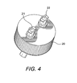
- Figures 2-4 illustrate various views of the cap of the beads/dual luer lock centrifuge tube of the present invention.
- Figure 5 illustrates a magnified view of IRAP 3.5 mm beads (25x Magnification).
- Figure 6 illustrates (left-right) Baseline, IRAP, and IRAP II levels of TNF-a, IL-10, and IL-1 ⁇ .
- Figure 7 illustrates (left-right) Baseline, IRAP, and IRAP II levels of IL-1 Ra.
- Figure 8 illustrates IRAP and IRAP II IL-1Ra/IL-1 ⁇ ratios.
- the present invention provides apparatus and methods for providing anti-inflammatory cytokines, specifically IL-1Ra, or other beneficial protein, for treatment of human or non-human damaged tissue such as cartilage and musculoskeletal tissue.
- Blood is withdrawn from the patient and then transferred into a special container with a cap that is configured to allow the patient's blood to flow on an inner side of the container, to prevent lysis of the blood cells.
- the container is a dual luer lock centrifuge tube that contains beads that are coated with a silanized coating. The container is then incubated and centrifuged. Subsequent to the incubation and the centrifugation, the serum containing autologous IL- is withdrawn through the luer lock of the container, and injected back into the patient.
- blood is withdrawn from the patient using a two-container system comprising a first container (for example, a conventional syringe) and a second container (for example, a dual luer lock centrifuge tube).
- a first container for example, a conventional syringe
- a second container for example, a dual luer lock centrifuge tube.
- the second container comprises a cap that is configured to allow the patient's blood to flow on an inner side of the second container, to prevent lysis of the blood cells.
- the dual luer lock centrifuge tube contains beads which are not etched with chromium sulfate, as in Reinecke, but rather are coated with a silanized coating.
- the second container is then incubated and centrifuged. Subsequent to the incubation and the centrifugation, the serum containing autologous IL-1Ra is withdrawn through the luer lock of the second container, and injected back into the patient.
- the present invention also provides a method of obtaining anti-inflammatory cytokines for treatment of tissue injuries.
- the method comprises the steps of: (i) providing an apparatus comprising a dual luer lock centrifuge tube with beads; (ii) drawing blood into a conventional syringe; (iii) injecting the blood from the conventional syringe into the dual luer lock centrifuge tube; (iv) incubating and centrifuging the blood in the beads/dual luer lock centrifuge tube; and (iv) removing, through the luer lock of the beads/dual luer lock centrifuge tube, serum containing the IL-1Ra protein generated during incubation.
- blood is first obtained from a patient with a conventional syringe.
- the blood is then introduced into the beads/dual luer lock centrifuge tube through a conduit that forms an angle with the longitudinal axis of the tube, to allow the blood to flow into the tube on the sidewall of the tube, to prevent lysis of the blood cells.
- the blood is subsequently incubated at 37° C, to stimulate the monocytes in the blood to produce IL-1Ra.
- the blood containing elevated levels of IL-1Ra is then centrifuged in the beads/dual luer lock centrifuge tube to isolate the IL-1Ra protein.
- the serum containing the concentrated IL-1Ra protein is removed through a luer lock of the beads/dual luer lock centrifuge tube.
- the isolated protein may be subsequently employed in surgical repairs, promoting the healing of the repair in orthopedic and neurological applications, for example.
- Figures 1-4 illustrate an exemplary embodiment of a dual luer lock centrifuge tube 100 of the present invention.
- Tube 100 includes a cap 20 securely attached to container 30 containing beads 40.
- the tube 100 and cap 20 provide for a closed system allowing for sterile incubation and withdrawal of the serum containing IL-1 Ra.
- Cap 20 comprises luer locks 21, 22, at least one of the luer locks including a conduit 25 with a distal end having an angular orientation (i.e., at least a portion of the conduit 25 forms an angle with the longitudinal axis of container 30, as shown in Figure 3 ).
- the angular orientation allows blood (withdrawn from a patient with an ordinary syringe) to be introduced into the container 30 by flowing on the side (the inner side) of the container 30, to prevent lysis of the blood cells.
- the angular orientation may be between 45 - 180 degrees.
- the end of the conduit may be shaped or offset from the axis of the conduit.
- the container 30 has a conical shape and a closed distal end for placement in a centrifuge.
- the container is made from a sterilizable material such as polypropylene and holds a volume of approximately 60 ml although it could be any size that would fit within a centrifuge.
- the outside of the container has incremental markings to assist in withdrawing the serum in the desired dosing quantities.
- the container 30 contains beads 40.
- the beads are manufactured from a glass like composition such as, borosilicate glass, alumina, silicate, quartz, bioglass, ceramic glass, flint glass, fluorosilicate glass, phosphosilicate glass, and cobalt glass or conundrum.
- the beads have a spherical shape to provide for a maximum surface area for blood contact.
- the container may contain gels, wool, powder, plastic, granules or fibres.
- the beads are provided with a coating to maximize the production of IL-1Ra by the monocytes within the blood.
- the coating may be silane, surfactants, polyether, polyester, polyurethane, or polyol groups.
- the beads may range in size from 1-5mm, but preferably are 3.0mm.
- Optimal production of IL-1Ra occurs when the maximum surface area of the beads is exposed to the blood within the container. A maximum amount of blood in the container is also necessary to optimize the production of IL-1Ra.
- the volume of the beads In order to accomplish both goals, the volume of the beads must be minimized to accomplish the maximum exposed surface area. Accordingly, the diameter of the beads has been tailored to maximize the volume of injected blood in the container and maximize the surface area for blood/bead contact.
- the objective of the study was two fold: (1) to fabricate a borosilicate bead/tube assembly 100 (IRAP II) of the present invention that performed better than the currently-known IRAP syringe system (IRAP); and (2) to determine the levels of two pro-inflammatory cytokines (IL-1 ⁇ and TNF-a) and two anti-inflammatory cytokines (IL-1Ra and IL-10) in human whole blood using the conventional IRAP syringe and IRAP II centrifuge tube.
- Biological testing was performed by utilizing enzyme-linked immunosorbant assays (ELISA) to measure serum levels of the cytokines.
- ELISA enzyme-linked immunosorbant assays
- IRAP Bead Composition Beads from a sterilized IRAP syringe were examined using IRF, X-ray Fluorescence Spectroscopy, to determine the elemental composition of the beads.
- IRAP Bead SEM Beads from a sterilized IRAP syringe were sent for SEM analysis.
- Group A Baseline 2x 10 mL glass vacutainers (Be
- the blood samples were spun down in a centrifuge (such as Hermle Z300 centrifuge from Labnet, Edison, NJ) at about 4000 rpm for about 10 minutes.
- Group A was centrifuged approximately 15 minutes after it had been drawn (to give the sample time to clot), and groups B and C were centrifuged following a 24 hour incubation period in an incubator (such as 2.5 ft 3 Precision Economy Incubator from Precision Scientific, Winchester VA) at about 37°C.
- an incubator such as 2.5 ft 3 Precision Economy Incubator from Precision Scientific, Winchester VA
- each donor's serum was removed from the container using a 5 mL polypropylene syringe (Becton Dickinson, Franklin, NJ) and an 18 gage needle.
- the serum from each sample was then filtered with 0.22 um Millex GP filters (Millipore, Billerica, MA) aliquotted into 5.0 mL cryovials (Wheaton, Millville, NJ), and stored at approximately -81°C.
- Serum samples were thawed to room temperature and then tested according to the Quantikine ELISA kit protocols (R&D Systems, Minneapolis, MN).
- the plate wells were washed using a Bio-Tek ELx 50 microplate strip washer, and absorbance of each sample was determined using a Bio-Tek ELx808 absorbance microplate reader.
- the serum samples for each donor were run in duplicate. Concentrations were calculated using, for example, a KC Junior (Bio-Tek Instruments, Winooski, VT) ,by converting absorbance to concentration based on a standard curve of optical density versus concentration.
- XRF X-ray Fluorescence Spectroscopy
- the syringe contained 200 glass beads with a diameter of 2.5 mm, creating a total surface area of 39.27 cm 2 , Equation 1 below.
- Equation 1 Total surface area of the glass beads (SA 1 ) was determined using the formula of a sphere multiplied by the number of beads (n 1 ).
- S ⁇ A 1 4 ⁇ ⁇ ⁇ r 1 2 ⁇ n 1
- Equation 2 IRAP measured surface area of the glass beads (SA 2 ) was determined using the formula of a sphere multiplied by the number of beads (N 2 ).
- S ⁇ A 2 4 ⁇ ⁇ ⁇ r 2 2 ⁇ n 2
- the number of beads (no) required to equal the surface area created by all the beads in the Group B IRAP syringe was calculated using Equation 3 below.
- Equation 3 The number of 3.0 mm beads (no) for the Group C centrifuge tube needed to equal the surface area of the Group B IRAP syringe is calculated by setting the total surface area equal to SA 2 and solving for no.
- TNF-alpha pro-inflammatory IL-10 (pg/ml): anti-inflammatory Mean Range StErr Mean Range StErr Baseline 9.5 6.1 -14.5 1.0 Baseline 16.9 9.7 -38.5 0.1 IRAP 32.3 13.8 -74.5 8.0 IRAP 25.7 16.0 -57.5 5.4 IRAP II 18.8 10.1 -23.5 1.7 IRAP II 37.8 22.7 -68.0 20.1 IL-1beta (pg/ml): pro-inflammatory IL-ra (pg/ml): anti-Inflammatory Mean Range StErr Mean Range StErr Baseline ⁇ 3.9 ⁇ 3.9 ⁇ 3.9 Baseline 355 118-1113 115 IRAP 27.5 9.3-50.4 5.4 IRAP 1233 261 -2708 344 IRAP II 58.8 23.9-195.2 20.1 IRAP II 9688 5908 -14604 1196
- Figure 6 illustrates Baseline, IRAP, and IRAP II levels of TNF-a, IL-10, and IL-1 ⁇ (*denotes significant difference).
- Figure 7 illustrates Baseline, IRAP, and IRAP II levels of IL-1Ra (*denotes significant difference).
- IL-1Ra/IL-1 ⁇ ratio 10:1 is required to inhibit IL-1 activity [2].
- IL-1Ra/IL-1 ⁇ ratios are illustrated in Figure 4 and Table 3.
- Figure 8 illustrates IRAP and IRAP II IL-1Ra/IL-1 ⁇ ratios (*denotes significant difference).
- Ratio of IL-ra/IL-1 beta Mean Range StErr IRAP 53.1 10.4 -134.5 16.0 IRAP II 239.6 60.7 -461.2 46.8
- Group C offers a more convenient two-container system which significantly increases the amount of autologous IL-1Ra produced.
- Other distinguishing differences include the contained silanized borosilicate spheres, a specific sphere diameter which not only maximizes the sphere surface area/blood contact, but also maximizes the amount of blood volume in the Group C centrifuge tube.
- the autologous serum produced by Group C centrifuge tube contains a higher ratio of anti-inflammatory proteins (IL-1Ra) to pro-inflammatory proteins (IL-1). Since the serum produced by Group C contains a concentrated combination of biologically relevant autologous growth factors, it can be an attractive option in treatment for tissue injury or osteoarthritis.
Landscapes
- Chemical & Material Sciences (AREA)
- Health & Medical Sciences (AREA)
- Life Sciences & Earth Sciences (AREA)
- Organic Chemistry (AREA)
- Biochemistry (AREA)
- Gastroenterology & Hepatology (AREA)
- Zoology (AREA)
- Biophysics (AREA)
- General Health & Medical Sciences (AREA)
- Genetics & Genomics (AREA)
- Medicinal Chemistry (AREA)
- Molecular Biology (AREA)
- Proteomics, Peptides & Aminoacids (AREA)
- Toxicology (AREA)
- Medicines Containing Material From Animals Or Micro-Organisms (AREA)
Abstract
Description
- The present invention relates to methods and apparatus for producing therapeutically active proteins, such as anti-inflammatory cytokines.
- Interleukin-1 (IL-1) plays a key role in the pathology of osteoarthritis or intervertebral disc degeneration/prolapse. The biological antagonist, interleukin-1 receptor antagonist (IL-1Ra), intervenes in the physiological mechanism of these diseases. IL-1Ra is a naturally occurring, structural derivative of IL-1 that competitively binds to the receptor and inhibits the biological effects of IL-1. The endogenous effects of IL-1Ra are anti-inflammatory in nature, opposite to that of IL-1. A minimum IL-1Ra /IL-1β ratio of 10:1 is required to inhibit IL-1 activity.
- It has been known for some years that iL-1 Ra can be synthesized by recombinant methods. However, autologous IL-1Ra, like all autologous proteins that are intrinsic to the body, is advantageous because the natural post-translational modifications such as glycosylations are already present. This is not the case with recombinant proteins because they are produced in prokaryotic hosts.
- Stimulation of monocytes by adherent immunoglobulin G to form the interleukin-1 receptor antagonist is described by Arend and Leung in lmmunological Reviews (1994) 139, 71-78 and Moore et al. in Am. J. Respir. Cell Mol. Biol. (1992) 6, 569-575. Andersen et al. in Autoimmunity (1995) 22, 127-133 explains that the therapeutic effect of immunoglobulin G to be observed in vivo cannot be attributed to an enhanced formation of interleukin-1 receptor antagonist, and that the in vitro formation of the interleukin-1 receptor antagonist (IL-1Ra) by monocytes depends on serum and plasma constituents adsorbed on polypropylene. The therapeutic use of adsorbed serum and plasma constituents to stimulate the formation of therapeutically interesting proteins in therapies is not only very costly but also involves the risk of contamination with infectious particles with which the serum and plasma constituents may be contaminated.
-
U.S. Patent Nos. 6,713,246 and6,759,188 to Reinecke disclose a method for producing IL-1Ra which can be employed directly in the therapy without using adsorbed serum and plasma constituents. Specifically, Reinecke provides a method for producing IL-1Ra in a special syringe made of glass, quartz or a plastic, the syringe being filled with blood, and incubated, to form the IL-1Ra being formed. The internal structure of the Reinecke syringe consists of a special material, in particular a glass, plastic, quartz and/or corundum, the surface of which is modified with the aid of a corrosive agent (chromosulphonic acid). The syringe is filled with a patient's blood and incubated to form IL-1Ra. The blood enriched with the protein is then centrifuged (to remove solid constituents such as blood platelets) and the serum containing IL-1Ra is reinjected into the patient (for example, into a diseased joint of the patient). - The syringe used by Reinecke has its inner surface textured by the acid and further includes glass beads to increase the internal surface area of the syringe and, thus, to provide a larger inducing surface. The glass beads, with a diameter of from 1 to 5 mm, occupy no more than 50% of the internal volume of the syringe. In Reinecke's method, the syringe is used to remove the patient's blood, to process the blood (to produce IL-1Ra), and then reinject the autologous IL-1Ra back into the patient. The present invention expands upon this method, to produce autologous IL-1 Ra in a more efficient manner.
- The present invention provides a unique container for producing anti-inflammatory cytokines, specifically IL-1Ra, and/or other therapeutically active proteins, for treatment of human or non-human damaged tissue such as cartilage and neurological tissue, together with methods of doing same.
- According to the present invention there is provided a method for producing a therapeutically active protein in a two-container system, together with a (second) container, as defined in the appended independent claims. Further preferable features are defined in the appended dependent claims. Features of the second container defined with respect to the method claims are equally applicable to the container of the apparatus claims.
- In performing the method of the present invention, blood from the patient is transferred into a special container provided with a cap configured to allow the patient's blood to flow on an inner side of the container, to prevent lysis of the blood cells. Preferably, the container is a dual luer lock centrifuge tube that contains beads that are coated with a silanized coating. Preferably, the container is then incubated and centrifuged. Preferably, subsequent to the incubation and the centrifugation, the serum containing the autologous therapeutically active protein (e.g., IL-1Ra) in the container is withdrawn through the luer lock of the container, for injection back into the patient.
- Preferably, the angular orientation of the conduit from the longitudinal axis is between 45-180 degrees, more preferably between 45-90 degrees. In certain embodiments, the end of the conduit is shaped or offset from the axis of the conduit.
- Preferably, the second container has a conical shape. Preferably, the second container has a closed distal end, more preferably for placement in a centrifuge. Preferably, the second container is made from a sterilizable material.
- Preferably, the second container contains beads. Preferably, the beads are manufactured from a glass-like composition such as, borosilicate glass, alumina, silicate, quartz, bioglass, ceramic glass, flint glass, fluorosilicate glass, phosphosilicate glass, and cobalt glass or conundrum. In a preferred embodiment, the beads have a spherical shape to provide for a maximum surface area for blood contact. In an alternate embodiment the container may contain gels, wool, powder, plastic, granules or fibres. Preferably, the beads are provided with a coating to maximize the production of IL-1Ra by the monocytes within the blood. The coating may be selected from the group consisting of: silane, surfactants, polyether, polyester, polyurethane, and polyol groups. Preferably, the beads range in size from 1-5mm, more preferably about 3.0mm.
- Preferably, the method of using the container to produce anti-inflammatory cytokines and other therapeutically active proteins comprises the steps of: (i) providing a container comprising a dual luer lock centrifuge tube with beads; (ii) injecting blood from a mammal, preferably from a patient, into the dual luer lock centrifuge tube; (iii) incubating and centrifuging the blood in the beads/dual luer lock centrifuge tube; and (iv) removing, through the luer lock of the beads/dual luer lock centrifuge tube, serum containing the IL-1Ra or other protein generated during incubation.
- Other features and advantages of the present invention will become apparent from the following description of the invention, which refers to the accompanying drawing.
-
Figure 1 illustrates a perspective view of a beads/dual luer lock centrifuge tube of the present invention. -
Figures 2-4 illustrate various views of the cap of the beads/dual luer lock centrifuge tube of the present invention. -
Figure 5 illustrates a magnified view of IRAP 3.5 mm beads (25x Magnification). -
Figure 6 illustrates (left-right) Baseline, IRAP, and IRAP II levels of TNF-a, IL-10, and IL-1β. -
Figure 7 illustrates (left-right) Baseline, IRAP, and IRAP II levels of IL-1 Ra. -
Figure 8 illustrates IRAP and IRAP II IL-1Ra/IL-1β ratios. - In the following detailed description, reference is made to various specific embodiments in which the invention may be practiced. These embodiments are described with sufficient detail to enable those skilled in the art to practice the invention, and it is to be understood that other embodiments may be employed, and that structural and logical changes may be made without departing from the scope of the appended claims.
- The present invention provides apparatus and methods for providing anti-inflammatory cytokines, specifically IL-1Ra, or other beneficial protein, for treatment of human or non-human damaged tissue such as cartilage and musculoskeletal tissue.
- Blood is withdrawn from the patient and then transferred into a special container with a cap that is configured to allow the patient's blood to flow on an inner side of the container, to prevent lysis of the blood cells. The container is a dual luer lock centrifuge tube that contains beads that are coated with a silanized coating. The container is then incubated and centrifuged. Subsequent to the incubation and the centrifugation, the serum containing autologous IL- is withdrawn through the luer lock of the container, and injected back into the patient.
- According to an exemplary embodiment, blood is withdrawn from the patient using a two-container system comprising a first container (for example, a conventional syringe) and a second container (for example, a dual luer lock centrifuge tube). Approximately 50 ml of blood is withdrawn with the first container and then transferred into the second container (the dual luer lock centrifuge tube). The second container comprises a cap that is configured to allow the patient's blood to flow on an inner side of the second container, to prevent lysis of the blood cells. The dual luer lock centrifuge tube contains beads which are not etched with chromium sulfate, as in Reinecke, but rather are coated with a silanized coating. The second container is then incubated and centrifuged. Subsequent to the incubation and the centrifugation, the serum containing autologous IL-1Ra is withdrawn through the luer lock of the second container, and injected back into the patient.
- The present invention also provides a method of obtaining anti-inflammatory cytokines for treatment of tissue injuries. The method comprises the steps of: (i) providing an apparatus comprising a dual luer lock centrifuge tube with beads; (ii) drawing blood into a conventional syringe; (iii) injecting the blood from the conventional syringe into the dual luer lock centrifuge tube; (iv) incubating and centrifuging the blood in the beads/dual luer lock centrifuge tube; and (iv) removing, through the luer lock of the beads/dual luer lock centrifuge tube, serum containing the IL-1Ra protein generated during incubation.
- According to an exemplary embodiment, blood is first obtained from a patient with a conventional syringe. The blood is then introduced into the beads/dual luer lock centrifuge tube through a conduit that forms an angle with the longitudinal axis of the tube, to allow the blood to flow into the tube on the sidewall of the tube, to prevent lysis of the blood cells. The blood is subsequently incubated at 37° C, to stimulate the monocytes in the blood to produce IL-1Ra. The blood containing elevated levels of IL-1Ra is then centrifuged in the beads/dual luer lock centrifuge tube to isolate the IL-1Ra protein. The serum containing the concentrated IL-1Ra protein is removed through a luer lock of the beads/dual luer lock centrifuge tube. The isolated protein may be subsequently employed in surgical repairs, promoting the healing of the repair in orthopedic and neurological applications, for example.
- Referring now to the drawings, where like elements are designated by like reference numerals,
Figures 1-4 illustrate an exemplary embodiment of a dual luerlock centrifuge tube 100 of the present invention.Tube 100 includes acap 20 securely attached tocontainer 30 containingbeads 40. Thetube 100 andcap 20 provide for a closed system allowing for sterile incubation and withdrawal of the serum containing IL-1 Ra. - Details of the
cap 20 of the dual luerlock centrifuge tube 100 of the present invention are illustrated inFigures 2-4 .Cap 20 comprises luer locks 21, 22, at least one of the luer locks including aconduit 25 with a distal end having an angular orientation (i.e., at least a portion of theconduit 25 forms an angle with the longitudinal axis ofcontainer 30, as shown inFigure 3 ). The angular orientation allows blood (withdrawn from a patient with an ordinary syringe) to be introduced into thecontainer 30 by flowing on the side (the inner side) of thecontainer 30, to prevent lysis of the blood cells. The angular orientation may be between 45 - 180 degrees. Alternately, the end of the conduit may be shaped or offset from the axis of the conduit. - The
container 30 has a conical shape and a closed distal end for placement in a centrifuge. The container is made from a sterilizable material such as polypropylene and holds a volume of approximately 60 ml although it could be any size that would fit within a centrifuge. The outside of the container has incremental markings to assist in withdrawing the serum in the desired dosing quantities. - The
container 30 containsbeads 40. The beads are manufactured from a glass like composition such as, borosilicate glass, alumina, silicate, quartz, bioglass, ceramic glass, flint glass, fluorosilicate glass, phosphosilicate glass, and cobalt glass or conundrum. In a preferred embodiment the beads have a spherical shape to provide for a maximum surface area for blood contact. In an alternate embodiment the container may contain gels, wool, powder, plastic, granules or fibres. The beads are provided with a coating to maximize the production of IL-1Ra by the monocytes within the blood. The coating may be silane, surfactants, polyether, polyester, polyurethane, or polyol groups. The beads may range in size from 1-5mm, but preferably are 3.0mm. Optimal production of IL-1Ra occurs when the maximum surface area of the beads is exposed to the blood within the container. A maximum amount of blood in the container is also necessary to optimize the production of IL-1Ra. In order to accomplish both goals, the volume of the beads must be minimized to accomplish the maximum exposed surface area. Accordingly, the diameter of the beads has been tailored to maximize the volume of injected blood in the container and maximize the surface area for blood/bead contact. - A method of obtaining concentrated IL-1Ra according to an exemplary embodiment of the present invention will be described below.
- Using aseptic technique, prepare the jugular vein for needle penetration. Attach the needle to the 60mL syringe and puncture the jugular vein.
- Harvest ∼50mL of whole blood into the 60mL syringe.
- Remove needle from jugular and detach from syringe.
- Unscrew the red tethered cap and loosen the white tethered cap on the centrifuge tube.
- Attach the syringe with blood to the red luer and inject blood into the centrifuge tube, holding the syringe and centrifuge tube at an angle of approximate ∼60°. The blood should flow against the side of the centrifuge tube. (Take care not to inject above Max Fill Line marked on the centrifuge tube.)
- Remove syringe, recap and tighten both tethered male caps, and gently rock the blood in the centrifuge tube.
- Place in incubator for approximately 24 hours at 37°C, not to exceed 24 hours.
- After incubation, place the centrifuge tube into a centrifuge for 10 minutes at 4000 rpm. Make sure to balance centrifuge with an appropriate counter weight.
- Remove white tethered cap and loosen red cap. Slowly draw the serum into a 20mL syringe using a spinal needle, being careful not to pull up red blood cells.
- Attach a sterile 0.22µm filter between the 20mL syringe containing the serum and an empty, sterile 6mL syringe. Transfer 4mL of serum to the 6mL syringe, detach the 6mL syringe and cap.
- Repeat until all the serum has been transferred through the sterile filter into the 6mL syringes. Individual doses may be used immediately or frozen at or less than -18°C for administration at a later date.
- Excess blood and/or serum should be disposed of appropriately.
- A method of obtaining concentrated IL-1Ra according to an exemplary embodiment of the present invention will be described below with reference to a particular and only exemplary study and as detailed below:
- The objective of the study was two fold: (1) to fabricate a borosilicate bead/tube assembly 100 (IRAP II) of the present invention that performed better than the currently-known IRAP syringe system (IRAP); and (2) to determine the levels of two pro-inflammatory cytokines (IL-1β and TNF-a) and two anti-inflammatory cytokines (IL-1Ra and IL-10) in human whole blood using the conventional IRAP syringe and IRAP II centrifuge tube. Biological testing was performed by utilizing enzyme-linked immunosorbant assays (ELISA) to measure serum levels of the cytokines.
- IRAP Bead Composition: Beads from a sterilized IRAP syringe were examined using IRF, X-ray Fluorescence Spectroscopy, to determine the elemental composition of the beads.
- IRAP Bead SEM: Beads from a sterilized IRAP syringe were sent for SEM analysis.
- Blood incubation and ELISA: Blood samples were collected from n=8 healthy volunteers, all male, ages 28-52 with a mean age of 38. Donors were divided into 3 groups: Group A Baseline,
2x 10 mL glass vacutainers (Becton Dickinson, Franklin Lakes, NJ), Group B, a 60 mL IRAP syringe (Orthogen, Düsseldorf, Germany), and Group C, a 50 mL centrifuge tube with silanized borosilicate beads. Approximately 20 mL of blood was collected for Group A, 50 mL of blood was collected for Group B into the large IRAP syringe, and approximately 40 mL of blood was collected for Group C into the polypropylene centrifuge tube. All groups were inverted ten times following collection and the Group B IRAP syringe and Group C IRAP II centrifuge tube were placed into incubator at 37°C for 24 hours. - The blood samples were spun down in a centrifuge (such as Hermle Z300 centrifuge from Labnet, Edison, NJ) at about 4000 rpm for about 10 minutes. Group A was centrifuged approximately 15 minutes after it had been drawn (to give the sample time to clot), and groups B and C were centrifuged following a 24 hour incubation period in an incubator (such as 2.5 ft3 Precision Economy Incubator from Precision Scientific, Winchester VA) at about 37°C. After centrifugation, each donor's serum was removed from the container using a 5 mL polypropylene syringe (Becton Dickinson, Franklin, NJ) and an 18 gage needle. The serum from each sample was then filtered with 0.22 um Millex GP filters (Millipore, Billerica, MA) aliquotted into 5.0 mL cryovials (Wheaton, Millville, NJ), and stored at approximately -81°C.
- Serum samples were thawed to room temperature and then tested according to the Quantikine ELISA kit protocols (R&D Systems, Minneapolis, MN). The plate wells were washed using a Bio-Tek ELx 50 microplate strip washer, and absorbance of each sample was determined using a Bio-Tek ELx808 absorbance microplate reader. The serum samples for each donor were run in duplicate. Concentrations were calculated using, for example, a KC Junior (Bio-Tek Instruments, Winooski, VT) ,by converting absorbance to concentration based on a standard curve of optical density versus concentration.
- XRF: XRF (X-ray Fluorescence Spectroscopy) was performed on the Group B IRAP beads. The results are presented in Table 1 below. The elemental breakdown very closely resembles borosilicate glass (SiO2∼81%, B2O3=∼13, Na2O=∼4%, and other=∼2%).
Table 1: XRF analysis of the IRAP beads Elemental Compound % composition SiO2 79.73 B2O3 12.57 Al2O3 3.53 Na2O 2.94 K2O 0.71 SO3 0.169 TiO2 0.1612 Cl 0.0609 Fe2O3 0.0465 SrO 0.0354 BaO 0.0353 NiO 0.0076 ZnO 0.007 PB 0.000032 - SEM: SEM analysis was performed to determine if the Group B IRAP beads were etched with 50% (volume/volume) CrSO4 as described in the Meijer paper (
Figure 5 ) (Meijer, H., et al., The production of anti-inflammatory cytokines in whole blood by physico-chemical induction. Inflamm Res, 2003. 52(10): p. 404-7). Micro-craters were noticed on the surface of the IRAP borosilicate beads. It was inconclusive whether these were created by the CrSO4 or if this was a product of the beads colliding against each other within the syringe.Figure 5 illustrates a view of Group B IRAP 3.5 mm beads (25x Magnification). - Bead Calculation: In order to match the surface area created by the Group B IRAP beads (d=3.5 mm), the Group C IRAP II centrifuge tubes of the present invention were loaded with enough silanized beads (d=3.0mm) to match the total surface area created by all the beads in the Group B IRAP syringe. In the original Orthogen paper (Meijer et al., The production of anti-inflammatory cytokines in whole blood by physico-chemical induction. Inflamm Res, 2003. 52(10): p. 404-7), the syringe contained 200 glass beads with a diameter of 2.5 mm, creating a total surface area of 39.27 cm2, Equation 1 below.
- Equation 1: Total surface area of the glass beads (SA1) was determined using the formula of a sphere multiplied by the number of beads (n1).


- There were n=240 glass beads in the current 60 mL IRAP syringe supplied by Orthogen, and the measured bead diameter was 3.5 mm (contradicting the Orthogen paper). The total surface area of glass beads was 92.4cm2, Equation 2 below.
- Equation 2: IRAP measured surface area of the glass beads (SA2) was determined using the formula of a sphere multiplied by the number of beads (N2).


- The number of beads (no) required to equal the surface area created by all the beads in the Group B IRAP syringe was calculated using Equation 3 below.
- Equation 3: The number of 3.0 mm beads (no) for the Group C centrifuge tube needed to equal the surface area of the Group B IRAP syringe is calculated by setting the total surface area equal to SA2 and solving for no.


- Each Group C IRAP II polypropylene centrifuge tube was loaded with 327 silanized borosilicate glass beads, d=3.0mm.
- ELISA: The mean values, range, and standard error of n=8 donors were calculated for TNF-a, IL-10, IL-1β, and IL-1Ra, Table 2 below.
Figure 6 andFigure 7 depict all cytokine mean levels. Standard error is utilized to compare the past studies. - TNF-a: IRAP II levels were not significantly different compared to IRAP and baseline (p=0.176 and p=0.551, respectively). Higher levels associated with IRAP were significantly different than baseline levels (p=0.009).
- IL-10: Higher levels in IRAP II were significantly different than baseline levels (p=0.031). IRAP II was not significantly different than IRAP (p=0.349, p<0.05), and IRAP was not significantly different from baseline levels (p=0.750, p>0.05).
- IL-1β: Higher levels in IRAP li were significantly different than baseline levels (p=0.009). IRAP II was not significantly different from baseline levels (p=0.239, P<0.05), and IRAP was not significantly different from baseline levels (p=0.431, P>0.05).
- IL-1Ra: Higher levels in IRAP II were significantly different compared to both baseline levels (p<0.001) and IRAP levels (p<0.001). IRAP was not significantly different from baseline levels (p=1.000, P>0.001).
Table 2: The mean of n=8 donors is shown with the range and the standard error (STE). Values denoted with a less than symbol indicate for factor was below the limit of detection of the assay kit. TNF-alpha (pg/ml): pro-inflammatory IL-10 (pg/ml): anti-inflammatory Mean Range StErr Mean Range StErr Baseline 9.5 6.1 -14.5 1.0 Baseline 16.9 9.7 -38.5 0.1 IRAP 32.3 13.8 -74.5 8.0 IRAP 25.7 16.0 -57.5 5.4 IRAP II 18.8 10.1 -23.5 1.7 IRAP II 37.8 22.7 -68.0 20.1 IL-1beta (pg/ml): pro-inflammatory IL-ra (pg/ml): anti-Inflammatory Mean Range StErr Mean Range StErr Baseline <3.9 <3.9 <3.9 Baseline 355 118-1113 115 IRAP 27.5 9.3-50.4 5.4 IRAP 1233 261 -2708 344 IRAP II 58.8 23.9-195.2 20.1 IRAP II 9688 5908 -14604 1196 -
Figure 6 illustrates Baseline, IRAP, and IRAP II levels of TNF-a, IL-10, and IL-1β (*denotes significant difference).Figure 7 illustrates Baseline, IRAP, and IRAP II levels of IL-1Ra (*denotes significant difference). - A minimum IL-1Ra/IL-1β ratio of 10:1 is required to inhibit IL-1 activity [2]. IL-1Ra/IL-1β ratios are illustrated in
Figure 4 and Table 3. A test (a=0.05) was performed and the higher ratio produced by IRAP II is significantly different than IRAP (p=0.002). -
Figure 8 illustrates IRAP and IRAP II IL-1Ra/IL-1β ratios (*denotes significant difference).Table 3: The IL-1Ra/IL-1β mean, range, and standard error (STE) of n=8 donors Ratio of IL-ra/IL-1 beta Mean Range StErr IRAP 53.1 10.4 -134.5 16.0 IRAP II 239.6 60.7 -461.2 46.8 - In comparison to the Group B system, Group C offers a more convenient two-container system which significantly increases the amount of autologous IL-1Ra produced. Other distinguishing differences include the contained silanized borosilicate spheres, a specific sphere diameter which not only maximizes the sphere surface area/blood contact, but also maximizes the amount of blood volume in the Group C centrifuge tube. Research also indicated that the autologous serum produced by Group C centrifuge tube contains a higher ratio of anti-inflammatory proteins (IL-1Ra) to pro-inflammatory proteins (IL-1). Since the serum produced by Group C contains a concentrated combination of biologically relevant autologous growth factors, it can be an attractive option in treatment for tissue injury or osteoarthritis.
- In vitro incubation of human whole blood at body temperature for 24 hours using the Group C IRAP II centrifuge system induces production of anti- and pro-inflammatory cytokines. IL-10, IL-1β and TNF-a levels in the Group C IRAP II system were not different compared to the Group B IRAP. The Group C IRAP II produces higher levels of IL-1Ra when compared to the Group B IRAP. The higher levels of IL-1Ra also led to a higher ratio of IL-1Ra/IL-1β.
- While the present invention is described herein with reference to illustrative embodiments for particular applications, it should be understood that the invention is not limited thereto. Those having ordinary skill in the art and access to the teachings provided herein will recognize additional modifications, applications, embodiments and substitution of equivalents all fall within the scope of the invention, without departing from the scope of the appended claims.
Claims (14)
- A method for producing a therapeutically active protein in a two-container system, comprising:placing blood from a mammal into a first container of the two-container system;injecting the blood into a second container of the two-container system, said second container defining a longitudinal axis and comprising a cap having dual luer locks, at least one of the luer locks including a conduit with a distal end which forms an angle with said longitudinal axis such that in-use blood introduced into said second container via said at least one luer lock including a conduit flows on a sidewall of said second container; andincubating the second container for a sufficient time and at a sufficient temperature to produce a therapeutically active protein in the blood.
- The method of claim 1, further comprising the step of removing the produced IL-1 Ra in the blood through a luer lock of the second container, for injection of the produced IL-1 Ra back into the mammal to promote tissue repair.
- The method of any of the preceding claims, wherein the first container is a conventional syringe.
- The method of any of the preceding claims, wherein the second container is a dual luer lock centrifuge tube.
- The method of claim 4, further comprising the step of subjecting the blood from the dual luer lock centrifuge tube to a centrifugation process.
- The method of any of the preceding claims, wherein the second container comprises beads, spheres, gels, wool, powder, granules, fibres or particles made of glass, plastic, corundum and/or quartz.
- The method of any of the preceding claims, wherein the second container comprises particles coated with a silanized coating.
- The method of claim 7, wherein the particles are selected from the group consisting of beads, spheres, gels and granules.
- The method of any of the preceding claims, wherein the second container is centrifuged.
- The method of any of the preceding claims, wherein the blood is incubated in the second container from about 12 to 72 hours.
- The method of any of the preceding claims, wherein the blood is incubated in the second container at about 20 to 37 °C.
- The method of any of the preceding claims, wherein the therapeutically active protein is IL-1Ra.
- The method of any of the preceding claims, wherein the therapeutically active protein is IL-1Ra.
- A container defining a longitudinal axis and comprising a cap having dual luer locks, at least one of the luer locks including a conduit with a distal end which forms an angle with said longitudinal axis such that in-use blood introduced into said container via said at least one luer lock including a conduit flows on a sidewall of said container.
Applications Claiming Priority (1)
| Application Number | Priority Date | Filing Date | Title |
|---|---|---|---|
| US11541108P | 2008-11-17 | 2008-11-17 |
Publications (3)
| Publication Number | Publication Date |
|---|---|
| EP2186877A2 true EP2186877A2 (en) | 2010-05-19 |
| EP2186877A3 EP2186877A3 (en) | 2011-05-04 |
| EP2186877B1 EP2186877B1 (en) | 2019-08-21 |
Family
ID=41796417
Family Applications (1)
| Application Number | Title | Priority Date | Filing Date |
|---|---|---|---|
| EP09252629.2A Active EP2186877B1 (en) | 2008-11-17 | 2009-11-16 | Method for producing therapeutically active proteins |
Country Status (2)
| Country | Link |
|---|---|
| US (1) | US8460227B2 (en) |
| EP (1) | EP2186877B1 (en) |
Cited By (16)
| Publication number | Priority date | Publication date | Assignee | Title |
|---|---|---|---|---|
| WO2012030593A3 (en) * | 2010-09-03 | 2012-05-18 | Biomet Biologics, Llc | Methods and compositions for delivering interleukin-1 receptor antagonist |
| US8663146B2 (en) | 2007-03-06 | 2014-03-04 | Biomet Biologics, Llc | Angiogenesis initiation and growth |
| US8753690B2 (en) | 2008-02-27 | 2014-06-17 | Biomet Biologics, Llc | Methods and compositions for delivering interleukin-1 receptor antagonist |
| WO2015085348A1 (en) * | 2013-12-09 | 2015-06-18 | Cytokine Medical Australia Pty Ltd | Apparatus for producing therapeutically active proteins in blood and uses thereof |
| US9701728B2 (en) | 2008-02-27 | 2017-07-11 | Biomet Biologics, Llc | Methods and compositions for delivering interleukin-1 receptor antagonist |
| US9758806B2 (en) | 2013-03-15 | 2017-09-12 | Biomet Biologics, Llc | Acellular compositions for treating inflammatory disorders |
| US9763875B2 (en) | 2009-08-27 | 2017-09-19 | Biomet Biologics, Llc | Implantable device for production of interleukin-1 receptor antagonist |
| US9833474B2 (en) | 2013-11-26 | 2017-12-05 | Biomet Biologies, LLC | Methods of mediating macrophage phenotypes |
| US9878011B2 (en) | 2013-03-15 | 2018-01-30 | Biomet Biologics, Llc | Treatment of inflammatory respiratory disease using biological solutions |
| US9895418B2 (en) | 2013-03-15 | 2018-02-20 | Biomet Biologics, Llc | Treatment of peripheral vascular disease using protein solutions |
| US9950035B2 (en) | 2013-03-15 | 2018-04-24 | Biomet Biologics, Llc | Methods and non-immunogenic compositions for treating inflammatory disorders |
| US10143725B2 (en) | 2013-03-15 | 2018-12-04 | Biomet Biologics, Llc | Treatment of pain using protein solutions |
| US10208095B2 (en) | 2013-03-15 | 2019-02-19 | Biomet Manufacturing, Llc | Methods for making cytokine compositions from tissues using non-centrifugal methods |
| US10441635B2 (en) | 2014-11-10 | 2019-10-15 | Biomet Biologics, Llc | Methods of treating pain using protein solutions |
| US10576130B2 (en) | 2013-03-15 | 2020-03-03 | Biomet Manufacturing, Llc | Treatment of collagen defects using protein solutions |
| US10729552B2 (en) | 2015-03-18 | 2020-08-04 | Biomet C.V. | Implant configured for hammertoe and small bone fixation |
Families Citing this family (16)
| Publication number | Priority date | Publication date | Assignee | Title |
|---|---|---|---|---|
| US7832566B2 (en) | 2002-05-24 | 2010-11-16 | Biomet Biologics, Llc | Method and apparatus for separating and concentrating a component from a multi-component material including macroparticles |
| US20060278588A1 (en) | 2002-05-24 | 2006-12-14 | Woodell-May Jennifer E | Apparatus and method for separating and concentrating fluids containing multiple components |
| US20080269762A1 (en) * | 2007-04-25 | 2008-10-30 | Biomet Manufacturing Corp. | Method and device for repair of cartilage defects |
| US20110052561A1 (en) * | 2009-08-27 | 2011-03-03 | Biomet Biologics,LLC | Osteolysis treatment |
| US9011846B2 (en) | 2011-05-02 | 2015-04-21 | Biomet Biologics, Llc | Thrombin isolated from blood and blood fractions |
| US10167310B2 (en) * | 2012-01-31 | 2019-01-01 | Estar Technologies Ltd | System and method for producing interleukin receptor antagonist (IRA) |
| US9205110B2 (en) | 2012-07-18 | 2015-12-08 | Arthrex, Inc. | Enhanced autologous growth factor production and delivery system |
| US9241977B2 (en) | 2013-03-01 | 2016-01-26 | Arthrex, Inc. | Enhanced anabolic cytokine production and delivery system |
| CA2866480A1 (en) * | 2014-09-30 | 2016-03-30 | Antnor Limited | Method and composition for producing enhanced anti-inflammatory and regenerative agents from autologous physiological fluid |
| US10596236B2 (en) | 2016-03-10 | 2020-03-24 | Arthrex, Inc. | Systems and methods for preparing a thrombin serum |
| AU2017230810B2 (en) | 2016-03-10 | 2022-03-10 | Arthrex, Inc. | Systems and methods for preparing protein enhanced serums |
| RU2617537C1 (en) * | 2016-05-24 | 2017-04-25 | Общество с ограниченной ответственностью "Просто лаборатория" | System for autologous serum obtaining |
| US10744500B2 (en) | 2017-12-29 | 2020-08-18 | Arthrex, Inc. | Systems and methods for cellular separation |
| CA3089825C (en) | 2018-01-29 | 2022-08-30 | Forever Labs, Inc. | System and method for isolating extracellular vesicles |
| US20210332315A1 (en) * | 2020-04-22 | 2021-10-28 | Ben M. Khalaj | Filtration-centrifuge tube apparatus for harvesting adipose derived stem cells |
| AU2022335348A1 (en) | 2021-08-24 | 2024-02-22 | Forever Labs, Inc. | Device and method for isolating extracellular vesicles |
Citations (2)
| Publication number | Priority date | Publication date | Assignee | Title |
|---|---|---|---|---|
| DE102006005016A1 (en) | 2006-02-03 | 2007-08-16 | Orthogen Ag | Conditioned blood composition and process for its preparation |
| US7520402B2 (en) | 2002-09-19 | 2009-04-21 | Harvest Technologies Corporation | Sterile disposable unit |
Family Cites Families (4)
| Publication number | Priority date | Publication date | Assignee | Title |
|---|---|---|---|---|
| DE19903876B4 (en) | 1999-02-01 | 2006-09-28 | Orthogen Gentechnologie Gmbh | Process for the in vitro formation and accumulation of interleukin-1 receptor antagonists |
| EP1406492B1 (en) * | 2001-06-06 | 2009-12-30 | Perfusion Partners & Associates, Inc. | Centrifuge tube assembly |
| DE102004051300C5 (en) * | 2004-10-20 | 2013-01-24 | Fresenius Kabi Deutschland Gmbh | Cap for containers filled with medical fluids |
| EP2146794B1 (en) * | 2007-04-12 | 2016-10-19 | Biomet Biologics, LLC | Buoy suspension fractionation system |
-
2009
- 2009-11-10 US US12/616,081 patent/US8460227B2/en active Active
- 2009-11-16 EP EP09252629.2A patent/EP2186877B1/en active Active
Patent Citations (2)
| Publication number | Priority date | Publication date | Assignee | Title |
|---|---|---|---|---|
| US7520402B2 (en) | 2002-09-19 | 2009-04-21 | Harvest Technologies Corporation | Sterile disposable unit |
| DE102006005016A1 (en) | 2006-02-03 | 2007-08-16 | Orthogen Ag | Conditioned blood composition and process for its preparation |
Cited By (26)
| Publication number | Priority date | Publication date | Assignee | Title |
|---|---|---|---|---|
| US8663146B2 (en) | 2007-03-06 | 2014-03-04 | Biomet Biologics, Llc | Angiogenesis initiation and growth |
| US9352002B2 (en) | 2007-03-06 | 2016-05-31 | Biomet Biologics, Llc | Angiogenesis initiation and growth |
| US8753690B2 (en) | 2008-02-27 | 2014-06-17 | Biomet Biologics, Llc | Methods and compositions for delivering interleukin-1 receptor antagonist |
| US11725031B2 (en) | 2008-02-27 | 2023-08-15 | Biomet Manufacturing, Llc | Methods and compositions for delivering interleukin-1 receptor antagonist |
| US9308224B2 (en) | 2008-02-27 | 2016-04-12 | Biomet Biologics, Llc | Methods and compositions for delivering interleukin-1 receptor antagonist |
| US9701728B2 (en) | 2008-02-27 | 2017-07-11 | Biomet Biologics, Llc | Methods and compositions for delivering interleukin-1 receptor antagonist |
| US10400017B2 (en) | 2008-02-27 | 2019-09-03 | Biomet Biologics, Llc | Methods and compositions for delivering interleukin-1 receptor antagonist |
| US10106587B2 (en) | 2008-02-27 | 2018-10-23 | Biomet Biologics, Llc | Methods and compositions for delivering interleukin-1 receptor antagonist |
| US9763875B2 (en) | 2009-08-27 | 2017-09-19 | Biomet Biologics, Llc | Implantable device for production of interleukin-1 receptor antagonist |
| WO2012030593A3 (en) * | 2010-09-03 | 2012-05-18 | Biomet Biologics, Llc | Methods and compositions for delivering interleukin-1 receptor antagonist |
| US9119829B2 (en) | 2010-09-03 | 2015-09-01 | Biomet Biologics, Llc | Methods and compositions for delivering interleukin-1 receptor antagonist |
| US9895418B2 (en) | 2013-03-15 | 2018-02-20 | Biomet Biologics, Llc | Treatment of peripheral vascular disease using protein solutions |
| US11957733B2 (en) | 2013-03-15 | 2024-04-16 | Biomet Manufacturing, Llc | Treatment of collagen defects using protein solutions |
| US9950035B2 (en) | 2013-03-15 | 2018-04-24 | Biomet Biologics, Llc | Methods and non-immunogenic compositions for treating inflammatory disorders |
| US10143725B2 (en) | 2013-03-15 | 2018-12-04 | Biomet Biologics, Llc | Treatment of pain using protein solutions |
| US10208095B2 (en) | 2013-03-15 | 2019-02-19 | Biomet Manufacturing, Llc | Methods for making cytokine compositions from tissues using non-centrifugal methods |
| US9758806B2 (en) | 2013-03-15 | 2017-09-12 | Biomet Biologics, Llc | Acellular compositions for treating inflammatory disorders |
| US9878011B2 (en) | 2013-03-15 | 2018-01-30 | Biomet Biologics, Llc | Treatment of inflammatory respiratory disease using biological solutions |
| US10441634B2 (en) | 2013-03-15 | 2019-10-15 | Biomet Biologics, Llc | Treatment of peripheral vascular disease using protein solutions |
| US10576130B2 (en) | 2013-03-15 | 2020-03-03 | Biomet Manufacturing, Llc | Treatment of collagen defects using protein solutions |
| US9833474B2 (en) | 2013-11-26 | 2017-12-05 | Biomet Biologies, LLC | Methods of mediating macrophage phenotypes |
| US12378524B2 (en) | 2013-11-26 | 2025-08-05 | Biomet Manufacturing, Llc | Methods of mediating macrophage phenotypes |
| US10946043B2 (en) | 2013-11-26 | 2021-03-16 | Biomet Biologics, Llc | Methods of mediating macrophage phenotypes |
| WO2015085348A1 (en) * | 2013-12-09 | 2015-06-18 | Cytokine Medical Australia Pty Ltd | Apparatus for producing therapeutically active proteins in blood and uses thereof |
| US10441635B2 (en) | 2014-11-10 | 2019-10-15 | Biomet Biologics, Llc | Methods of treating pain using protein solutions |
| US10729552B2 (en) | 2015-03-18 | 2020-08-04 | Biomet C.V. | Implant configured for hammertoe and small bone fixation |
Also Published As
| Publication number | Publication date |
|---|---|
| US20100125236A1 (en) | 2010-05-20 |
| EP2186877A3 (en) | 2011-05-04 |
| US8460227B2 (en) | 2013-06-11 |
| EP2186877B1 (en) | 2019-08-21 |
Similar Documents
| Publication | Publication Date | Title |
|---|---|---|
| EP2186877B1 (en) | Method for producing therapeutically active proteins | |
| EP1151004B1 (en) | Method for producing il-1ra, a therapeutically active protein from body fluids | |
| JP4038331B2 (en) | Methods for inducing proteins with therapeutic effects | |
| BRPI0707360A2 (en) | conditioned blood composition and process for its production | |
| KR20160073382A (en) | Biomarkers for cell therapy | |
| SG192254A1 (en) | Pluripotent stem cells and method of stimulating and extracting non-embryonic pluripotent stem cells from mammal blood and using reconstituted pluripotent stem cells to treat diseases including chronic obstructive pulmonary disease | |
| Miles et al. | Intraindividual variability of carbamazepine, phenobarbital, and phenytoin concentrations in saliva | |
| EP2687217B1 (en) | Enhanced autologous growth factor production and delivery system | |
| US12605405B2 (en) | Methods for the production of pharmaceutical agents | |
| CN111978394B (en) | Preparation method of polyclonal preparation for COVID-19 patient specific immunotherapy | |
| RU2833471C1 (en) | Modular kit for extracorporeal pharmacotherapy (versions) | |
| Meacock et al. | Immunologic and clinical properties of two purified ragweed fractions | |
| CN114259506B (en) | Application of CMKLR+ mesenchymal stem cells in the preparation of drugs for the treatment of ankylosing spondylitis | |
| CN1720993A (en) | A kind of stem cell medicine for the treatment of Alzheimer and preparation method thereof | |
| EP4304781A1 (en) | Systems and methods for the preparation of enriched serum | |
| WO2017204688A2 (en) | System for producing autologous serum | |
| US9763981B2 (en) | Method for producing autologous proteins | |
| EP0899271A1 (en) | Process for inducing therapeutically active proteins | |
| FAIZAL et al. | How Rich is Platelet Rich Plasma? A Simple Preparation Method of PRP and Its Quality According to Various Classification Systems | |
| Neama et al. | The Effect of Competitive Antigen on Some Cytokine in Rabbits | |
| Anchie | Plasmapheresis as a treatment for myasthenia gravis | |
| Lieberman et al. | Lectin-like factor and co-factor in serum from cystic fibrosis heterozygotes | |
| Chekalin et al. | Pharmacokinetic of prednisolone in patients with rheumatoid arthritis | |
| Irwin | TISSUE-LIPID SOLUBILITY AND" ADSORPTION" OF BARBITURATES |
Legal Events
| Date | Code | Title | Description |
|---|---|---|---|
| PUAI | Public reference made under article 153(3) epc to a published international application that has entered the european phase |
Free format text: ORIGINAL CODE: 0009012 |
|
| AK | Designated contracting states |
Kind code of ref document: A2 Designated state(s): AT BE BG CH CY CZ DE DK EE ES FI FR GB GR HR HU IE IS IT LI LT LU LV MC MK MT NL NO PL PT RO SE SI SK SM TR |
|
| AX | Request for extension of the european patent |
Extension state: AL BA RS |
|
| PUAL | Search report despatched |
Free format text: ORIGINAL CODE: 0009013 |
|
| AK | Designated contracting states |
Kind code of ref document: A3 Designated state(s): AT BE BG CH CY CZ DE DK EE ES FI FR GB GR HR HU IE IS IT LI LT LU LV MC MK MT NL NO PL PT RO SE SI SK SM TR |
|
| AX | Request for extension of the european patent |
Extension state: AL BA RS |
|
| RIC1 | Information provided on ipc code assigned before grant |
Ipc: C12M 1/26 20060101AFI20100315BHEP Ipc: C07K 14/54 20060101ALI20110329BHEP Ipc: A61K 38/20 20060101ALI20110329BHEP |
|
| 17P | Request for examination filed |
Effective date: 20110725 |
|
| 17Q | First examination report despatched |
Effective date: 20120816 |
|
| TPAC | Observations filed by third parties |
Free format text: ORIGINAL CODE: EPIDOSNTIPA |
|
| TPAC | Observations filed by third parties |
Free format text: ORIGINAL CODE: EPIDOSNTIPA |
|
| STAA | Information on the status of an ep patent application or granted ep patent |
Free format text: STATUS: EXAMINATION IS IN PROGRESS |
|
| GRAP | Despatch of communication of intention to grant a patent |
Free format text: ORIGINAL CODE: EPIDOSNIGR1 |
|
| STAA | Information on the status of an ep patent application or granted ep patent |
Free format text: STATUS: GRANT OF PATENT IS INTENDED |
|
| INTG | Intention to grant announced |
Effective date: 20190227 |
|
| GRAS | Grant fee paid |
Free format text: ORIGINAL CODE: EPIDOSNIGR3 |
|
| GRAA | (expected) grant |
Free format text: ORIGINAL CODE: 0009210 |
|
| STAA | Information on the status of an ep patent application or granted ep patent |
Free format text: STATUS: THE PATENT HAS BEEN GRANTED |
|
| AK | Designated contracting states |
Kind code of ref document: B1 Designated state(s): AT BE BG CH CY CZ DE DK EE ES FI FR GB GR HR HU IE IS IT LI LT LU LV MC MK MT NL NO PL PT RO SE SI SK SM TR |
|
| REG | Reference to a national code |
Ref country code: GB Ref legal event code: FG4D |
|
| REG | Reference to a national code |
Ref country code: CH Ref legal event code: EP |
|
| REG | Reference to a national code |
Ref country code: DE Ref legal event code: R096 Ref document number: 602009059528 Country of ref document: DE |
|
| REG | Reference to a national code |
Ref country code: AT Ref legal event code: REF Ref document number: 1169783 Country of ref document: AT Kind code of ref document: T Effective date: 20190915 |
|
| REG | Reference to a national code |
Ref country code: IE Ref legal event code: FG4D |
|
| REG | Reference to a national code |
Ref country code: LT Ref legal event code: MG4D |
|
| REG | Reference to a national code |
Ref country code: NL Ref legal event code: MP Effective date: 20190821 |
|
| PG25 | Lapsed in a contracting state [announced via postgrant information from national office to epo] |
Ref country code: BG Free format text: LAPSE BECAUSE OF FAILURE TO SUBMIT A TRANSLATION OF THE DESCRIPTION OR TO PAY THE FEE WITHIN THE PRESCRIBED TIME-LIMIT Effective date: 20191121 Ref country code: NL Free format text: LAPSE BECAUSE OF FAILURE TO SUBMIT A TRANSLATION OF THE DESCRIPTION OR TO PAY THE FEE WITHIN THE PRESCRIBED TIME-LIMIT Effective date: 20190821 Ref country code: SE Free format text: LAPSE BECAUSE OF FAILURE TO SUBMIT A TRANSLATION OF THE DESCRIPTION OR TO PAY THE FEE WITHIN THE PRESCRIBED TIME-LIMIT Effective date: 20190821 Ref country code: NO Free format text: LAPSE BECAUSE OF FAILURE TO SUBMIT A TRANSLATION OF THE DESCRIPTION OR TO PAY THE FEE WITHIN THE PRESCRIBED TIME-LIMIT Effective date: 20191121 Ref country code: HR Free format text: LAPSE BECAUSE OF FAILURE TO SUBMIT A TRANSLATION OF THE DESCRIPTION OR TO PAY THE FEE WITHIN THE PRESCRIBED TIME-LIMIT Effective date: 20190821 Ref country code: PT Free format text: LAPSE BECAUSE OF FAILURE TO SUBMIT A TRANSLATION OF THE DESCRIPTION OR TO PAY THE FEE WITHIN THE PRESCRIBED TIME-LIMIT Effective date: 20191223 Ref country code: LT Free format text: LAPSE BECAUSE OF FAILURE TO SUBMIT A TRANSLATION OF THE DESCRIPTION OR TO PAY THE FEE WITHIN THE PRESCRIBED TIME-LIMIT Effective date: 20190821 Ref country code: FI Free format text: LAPSE BECAUSE OF FAILURE TO SUBMIT A TRANSLATION OF THE DESCRIPTION OR TO PAY THE FEE WITHIN THE PRESCRIBED TIME-LIMIT Effective date: 20190821 |
|
| PG25 | Lapsed in a contracting state [announced via postgrant information from national office to epo] |
Ref country code: LV Free format text: LAPSE BECAUSE OF FAILURE TO SUBMIT A TRANSLATION OF THE DESCRIPTION OR TO PAY THE FEE WITHIN THE PRESCRIBED TIME-LIMIT Effective date: 20190821 Ref country code: IS Free format text: LAPSE BECAUSE OF FAILURE TO SUBMIT A TRANSLATION OF THE DESCRIPTION OR TO PAY THE FEE WITHIN THE PRESCRIBED TIME-LIMIT Effective date: 20191221 Ref country code: ES Free format text: LAPSE BECAUSE OF FAILURE TO SUBMIT A TRANSLATION OF THE DESCRIPTION OR TO PAY THE FEE WITHIN THE PRESCRIBED TIME-LIMIT Effective date: 20190821 Ref country code: GR Free format text: LAPSE BECAUSE OF FAILURE TO SUBMIT A TRANSLATION OF THE DESCRIPTION OR TO PAY THE FEE WITHIN THE PRESCRIBED TIME-LIMIT Effective date: 20191122 |
|
| REG | Reference to a national code |
Ref country code: AT Ref legal event code: MK05 Ref document number: 1169783 Country of ref document: AT Kind code of ref document: T Effective date: 20190821 |
|
| PG25 | Lapsed in a contracting state [announced via postgrant information from national office to epo] |
Ref country code: TR Free format text: LAPSE BECAUSE OF FAILURE TO SUBMIT A TRANSLATION OF THE DESCRIPTION OR TO PAY THE FEE WITHIN THE PRESCRIBED TIME-LIMIT Effective date: 20190821 |
|
| PG25 | Lapsed in a contracting state [announced via postgrant information from national office to epo] |
Ref country code: DK Free format text: LAPSE BECAUSE OF FAILURE TO SUBMIT A TRANSLATION OF THE DESCRIPTION OR TO PAY THE FEE WITHIN THE PRESCRIBED TIME-LIMIT Effective date: 20190821 Ref country code: IT Free format text: LAPSE BECAUSE OF FAILURE TO SUBMIT A TRANSLATION OF THE DESCRIPTION OR TO PAY THE FEE WITHIN THE PRESCRIBED TIME-LIMIT Effective date: 20190821 Ref country code: AT Free format text: LAPSE BECAUSE OF FAILURE TO SUBMIT A TRANSLATION OF THE DESCRIPTION OR TO PAY THE FEE WITHIN THE PRESCRIBED TIME-LIMIT Effective date: 20190821 Ref country code: EE Free format text: LAPSE BECAUSE OF FAILURE TO SUBMIT A TRANSLATION OF THE DESCRIPTION OR TO PAY THE FEE WITHIN THE PRESCRIBED TIME-LIMIT Effective date: 20190821 Ref country code: RO Free format text: LAPSE BECAUSE OF FAILURE TO SUBMIT A TRANSLATION OF THE DESCRIPTION OR TO PAY THE FEE WITHIN THE PRESCRIBED TIME-LIMIT Effective date: 20190821 Ref country code: PL Free format text: LAPSE BECAUSE OF FAILURE TO SUBMIT A TRANSLATION OF THE DESCRIPTION OR TO PAY THE FEE WITHIN THE PRESCRIBED TIME-LIMIT Effective date: 20190821 |
|
| PG25 | Lapsed in a contracting state [announced via postgrant information from national office to epo] |
Ref country code: SK Free format text: LAPSE BECAUSE OF FAILURE TO SUBMIT A TRANSLATION OF THE DESCRIPTION OR TO PAY THE FEE WITHIN THE PRESCRIBED TIME-LIMIT Effective date: 20190821 Ref country code: IS Free format text: LAPSE BECAUSE OF FAILURE TO SUBMIT A TRANSLATION OF THE DESCRIPTION OR TO PAY THE FEE WITHIN THE PRESCRIBED TIME-LIMIT Effective date: 20200224 Ref country code: CZ Free format text: LAPSE BECAUSE OF FAILURE TO SUBMIT A TRANSLATION OF THE DESCRIPTION OR TO PAY THE FEE WITHIN THE PRESCRIBED TIME-LIMIT Effective date: 20190821 Ref country code: SM Free format text: LAPSE BECAUSE OF FAILURE TO SUBMIT A TRANSLATION OF THE DESCRIPTION OR TO PAY THE FEE WITHIN THE PRESCRIBED TIME-LIMIT Effective date: 20190821 |
|
| REG | Reference to a national code |
Ref country code: DE Ref legal event code: R097 Ref document number: 602009059528 Country of ref document: DE |
|
| REG | Reference to a national code |
Ref country code: CH Ref legal event code: PL |
|
| PLBE | No opposition filed within time limit |
Free format text: ORIGINAL CODE: 0009261 |
|
| STAA | Information on the status of an ep patent application or granted ep patent |
Free format text: STATUS: NO OPPOSITION FILED WITHIN TIME LIMIT |
|
| PG2D | Information on lapse in contracting state deleted |
Ref country code: IS |
|
| PG25 | Lapsed in a contracting state [announced via postgrant information from national office to epo] |
Ref country code: MC Free format text: LAPSE BECAUSE OF FAILURE TO SUBMIT A TRANSLATION OF THE DESCRIPTION OR TO PAY THE FEE WITHIN THE PRESCRIBED TIME-LIMIT Effective date: 20190821 Ref country code: LI Free format text: LAPSE BECAUSE OF NON-PAYMENT OF DUE FEES Effective date: 20191130 Ref country code: LU Free format text: LAPSE BECAUSE OF NON-PAYMENT OF DUE FEES Effective date: 20191116 Ref country code: CH Free format text: LAPSE BECAUSE OF NON-PAYMENT OF DUE FEES Effective date: 20191130 |
|
| 26N | No opposition filed |
Effective date: 20200603 |
|
| REG | Reference to a national code |
Ref country code: BE Ref legal event code: MM Effective date: 20191130 |
|
| PG25 | Lapsed in a contracting state [announced via postgrant information from national office to epo] |
Ref country code: SI Free format text: LAPSE BECAUSE OF FAILURE TO SUBMIT A TRANSLATION OF THE DESCRIPTION OR TO PAY THE FEE WITHIN THE PRESCRIBED TIME-LIMIT Effective date: 20190821 |
|
| PG25 | Lapsed in a contracting state [announced via postgrant information from national office to epo] |
Ref country code: IE Free format text: LAPSE BECAUSE OF NON-PAYMENT OF DUE FEES Effective date: 20191116 |
|
| PG25 | Lapsed in a contracting state [announced via postgrant information from national office to epo] |
Ref country code: BE Free format text: LAPSE BECAUSE OF NON-PAYMENT OF DUE FEES Effective date: 20191130 |
|
| PG25 | Lapsed in a contracting state [announced via postgrant information from national office to epo] |
Ref country code: CY Free format text: LAPSE BECAUSE OF FAILURE TO SUBMIT A TRANSLATION OF THE DESCRIPTION OR TO PAY THE FEE WITHIN THE PRESCRIBED TIME-LIMIT Effective date: 20190821 |
|
| PG25 | Lapsed in a contracting state [announced via postgrant information from national office to epo] |
Ref country code: HU Free format text: LAPSE BECAUSE OF FAILURE TO SUBMIT A TRANSLATION OF THE DESCRIPTION OR TO PAY THE FEE WITHIN THE PRESCRIBED TIME-LIMIT; INVALID AB INITIO Effective date: 20091116 Ref country code: MT Free format text: LAPSE BECAUSE OF FAILURE TO SUBMIT A TRANSLATION OF THE DESCRIPTION OR TO PAY THE FEE WITHIN THE PRESCRIBED TIME-LIMIT Effective date: 20190821 |
|
| PG25 | Lapsed in a contracting state [announced via postgrant information from national office to epo] |
Ref country code: MK Free format text: LAPSE BECAUSE OF FAILURE TO SUBMIT A TRANSLATION OF THE DESCRIPTION OR TO PAY THE FEE WITHIN THE PRESCRIBED TIME-LIMIT Effective date: 20190821 |
|
| PGFP | Annual fee paid to national office [announced via postgrant information from national office to epo] |
Ref country code: GB Payment date: 20250925 Year of fee payment: 17 |
|
| PGFP | Annual fee paid to national office [announced via postgrant information from national office to epo] |
Ref country code: FR Payment date: 20250908 Year of fee payment: 17 |
|
| PGFP | Annual fee paid to national office [announced via postgrant information from national office to epo] |
Ref country code: DE Payment date: 20250923 Year of fee payment: 17 |