EP2184365A1 - Multi-marked diploid yeast strains for use in measuring eukaryotic chromosome instability - Google Patents
Multi-marked diploid yeast strains for use in measuring eukaryotic chromosome instability Download PDFInfo
- Publication number
- EP2184365A1 EP2184365A1 EP08305778A EP08305778A EP2184365A1 EP 2184365 A1 EP2184365 A1 EP 2184365A1 EP 08305778 A EP08305778 A EP 08305778A EP 08305778 A EP08305778 A EP 08305778A EP 2184365 A1 EP2184365 A1 EP 2184365A1
- Authority
- EP
- European Patent Office
- Prior art keywords
- chromosome
- cells
- yeast strain
- selectable marker
- medium
- Prior art date
- Legal status (The legal status is an assumption and is not a legal conclusion. Google has not performed a legal analysis and makes no representation as to the accuracy of the status listed.)
- Withdrawn
Links
- 208000037051 Chromosomal Instability Diseases 0.000 title claims abstract description 116
- 240000004808 Saccharomyces cerevisiae Species 0.000 title claims abstract description 52
- 210000000349 chromosome Anatomy 0.000 claims abstract description 94
- 238000000034 method Methods 0.000 claims abstract description 55
- 208000037088 Chromosome Breakage Diseases 0.000 claims abstract description 39
- 150000001875 compounds Chemical class 0.000 claims abstract description 38
- 208000020584 Polyploidy Diseases 0.000 claims abstract description 8
- 210000004027 cell Anatomy 0.000 claims description 119
- 239000003550 marker Substances 0.000 claims description 73
- 108090000623 proteins and genes Proteins 0.000 claims description 48
- 239000002609 medium Substances 0.000 claims description 46
- 101150008604 CAN1 gene Proteins 0.000 claims description 27
- 210000002230 centromere Anatomy 0.000 claims description 18
- 239000006152 selective media Substances 0.000 claims description 13
- 238000001514 detection method Methods 0.000 claims description 12
- 238000007747 plating Methods 0.000 claims description 6
- 230000003362 replicative effect Effects 0.000 claims description 4
- 230000035945 sensitivity Effects 0.000 abstract description 13
- 235000005911 diet Nutrition 0.000 abstract description 4
- 230000000378 dietary effect Effects 0.000 abstract description 4
- 231100000419 toxicity Toxicity 0.000 abstract description 2
- 230000001988 toxicity Effects 0.000 abstract description 2
- 101150050575 URA3 gene Proteins 0.000 description 45
- 101100246753 Halobacterium salinarum (strain ATCC 700922 / JCM 11081 / NRC-1) pyrF gene Proteins 0.000 description 44
- 238000003556 assay Methods 0.000 description 44
- 235000014680 Saccharomyces cerevisiae Nutrition 0.000 description 35
- SEHFUALWMUWDKS-UHFFFAOYSA-N 5-fluoroorotic acid Chemical compound OC(=O)C=1NC(=O)NC(=O)C=1F SEHFUALWMUWDKS-UHFFFAOYSA-N 0.000 description 22
- 230000035772 mutation Effects 0.000 description 17
- ROHFNLRQFUQHCH-YFKPBYRVSA-N L-leucine Chemical compound CC(C)C[C@H](N)C(O)=O ROHFNLRQFUQHCH-YFKPBYRVSA-N 0.000 description 16
- 101150009006 HIS3 gene Proteins 0.000 description 14
- 101100394989 Rhodopseudomonas palustris (strain ATCC BAA-98 / CGA009) hisI gene Proteins 0.000 description 14
- MHAJPDPJQMAIIY-UHFFFAOYSA-N Hydrogen peroxide Chemical compound OO MHAJPDPJQMAIIY-UHFFFAOYSA-N 0.000 description 13
- FSBIGDSBMBYOPN-VKHMYHEASA-N L-canavanine Chemical compound OC(=O)[C@@H](N)CCONC(N)=N FSBIGDSBMBYOPN-VKHMYHEASA-N 0.000 description 13
- 239000003814 drug Substances 0.000 description 13
- 230000001965 increasing effect Effects 0.000 description 13
- 208000031448 Genomic Instability Diseases 0.000 description 12
- 230000007423 decrease Effects 0.000 description 12
- 229940079593 drug Drugs 0.000 description 12
- 108700028369 Alleles Proteins 0.000 description 11
- 101100153788 Schizosaccharomyces pombe (strain 972 / ATCC 24843) tpx1 gene Proteins 0.000 description 11
- 230000022131 cell cycle Effects 0.000 description 11
- 230000036542 oxidative stress Effects 0.000 description 11
- 230000012010 growth Effects 0.000 description 10
- 239000003642 reactive oxygen metabolite Substances 0.000 description 10
- ROHFNLRQFUQHCH-UHFFFAOYSA-N Leucine Natural products CC(C)CC(N)C(O)=O ROHFNLRQFUQHCH-UHFFFAOYSA-N 0.000 description 9
- FSBIGDSBMBYOPN-UHFFFAOYSA-N O-guanidino-DL-homoserine Natural products OC(=O)C(N)CCON=C(N)N FSBIGDSBMBYOPN-UHFFFAOYSA-N 0.000 description 8
- 238000012360 testing method Methods 0.000 description 8
- 101100085270 Schizosaccharomyces pombe (strain 972 / ATCC 24843) ade5 gene Proteins 0.000 description 7
- 230000000694 effects Effects 0.000 description 7
- 230000021121 meiosis Effects 0.000 description 7
- 101150019623 mip-1 gene Proteins 0.000 description 7
- WQZGKKKJIJFFOK-GASJEMHNSA-N Glucose Natural products OC[C@H]1OC(O)[C@H](O)[C@@H](O)[C@@H]1O WQZGKKKJIJFFOK-GASJEMHNSA-N 0.000 description 6
- 239000008103 glucose Substances 0.000 description 6
- 230000010627 oxidative phosphorylation Effects 0.000 description 6
- 230000005778 DNA damage Effects 0.000 description 5
- 231100000277 DNA damage Toxicity 0.000 description 5
- 206010028980 Neoplasm Diseases 0.000 description 5
- 102000053062 Rad52 DNA Repair and Recombination Human genes 0.000 description 5
- 108700031762 Rad52 DNA Repair and Recombination Proteins 0.000 description 5
- 230000018199 S phase Effects 0.000 description 5
- 230000006378 damage Effects 0.000 description 5
- 230000007547 defect Effects 0.000 description 5
- 238000002474 experimental method Methods 0.000 description 5
- 230000002068 genetic effect Effects 0.000 description 5
- 230000013011 mating Effects 0.000 description 5
- 210000003470 mitochondria Anatomy 0.000 description 5
- 230000000241 respiratory effect Effects 0.000 description 5
- 101100366466 Caenorhabditis elegans spo-11 gene Proteins 0.000 description 4
- 108020004414 DNA Proteins 0.000 description 4
- PWKSKIMOESPYIA-BYPYZUCNSA-N L-N-acetyl-Cysteine Chemical compound CC(=O)N[C@@H](CS)C(O)=O PWKSKIMOESPYIA-BYPYZUCNSA-N 0.000 description 4
- 108020005196 Mitochondrial DNA Proteins 0.000 description 4
- 101100355599 Neurospora crassa (strain ATCC 24698 / 74-OR23-1A / CBS 708.71 / DSM 1257 / FGSC 987) mus-11 gene Proteins 0.000 description 4
- 101150006234 RAD52 gene Proteins 0.000 description 4
- 108010084455 Zeocin Proteins 0.000 description 4
- 229960004308 acetylcysteine Drugs 0.000 description 4
- 201000011510 cancer Diseases 0.000 description 4
- 239000007788 liquid Substances 0.000 description 4
- 230000002438 mitochondrial effect Effects 0.000 description 4
- 230000004792 oxidative damage Effects 0.000 description 4
- CWCMIVBLVUHDHK-ZSNHEYEWSA-N phleomycin D1 Chemical compound N([C@H](C(=O)N[C@H](C)[C@@H](O)[C@H](C)C(=O)N[C@@H]([C@H](O)C)C(=O)NCCC=1SC[C@@H](N=1)C=1SC=C(N=1)C(=O)NCCCCNC(N)=N)[C@@H](O[C@H]1[C@H]([C@@H](O)[C@H](O)[C@H](CO)O1)O[C@@H]1[C@H]([C@@H](OC(N)=O)[C@H](O)[C@@H](CO)O1)O)C=1N=CNC=1)C(=O)C1=NC([C@H](CC(N)=O)NC[C@H](N)C(N)=O)=NC(N)=C1C CWCMIVBLVUHDHK-ZSNHEYEWSA-N 0.000 description 4
- 238000012216 screening Methods 0.000 description 4
- 101100208421 Candida albicans (strain SC5314 / ATCC MYA-2876) TMP1 gene Proteins 0.000 description 3
- 208000005623 Carcinogenesis Diseases 0.000 description 3
- PEDCQBHIVMGVHV-UHFFFAOYSA-N Glycerine Chemical compound OCC(O)CO PEDCQBHIVMGVHV-UHFFFAOYSA-N 0.000 description 3
- 101100182720 Homo sapiens LY6E gene Proteins 0.000 description 3
- 102100032131 Lymphocyte antigen 6E Human genes 0.000 description 3
- OUUQCZGPVNCOIJ-UHFFFAOYSA-M Superoxide Chemical compound [O-][O] OUUQCZGPVNCOIJ-UHFFFAOYSA-M 0.000 description 3
- 101150048440 TSA1 gene Proteins 0.000 description 3
- RIOXQFHNBCKOKP-UHFFFAOYSA-N benomyl Chemical compound C1=CC=C2N(C(=O)NCCCC)C(NC(=O)OC)=NC2=C1 RIOXQFHNBCKOKP-UHFFFAOYSA-N 0.000 description 3
- MITFXPHMIHQXPI-UHFFFAOYSA-N benzoxaprofen Natural products N=1C2=CC(C(C(O)=O)C)=CC=C2OC=1C1=CC=C(Cl)C=C1 MITFXPHMIHQXPI-UHFFFAOYSA-N 0.000 description 3
- 230000036952 cancer formation Effects 0.000 description 3
- 231100000504 carcinogenesis Toxicity 0.000 description 3
- 230000002950 deficient Effects 0.000 description 3
- 238000012217 deletion Methods 0.000 description 3
- 230000037430 deletion Effects 0.000 description 3
- 230000005782 double-strand break Effects 0.000 description 3
- 230000004064 dysfunction Effects 0.000 description 3
- 238000000855 fermentation Methods 0.000 description 3
- 230000004151 fermentation Effects 0.000 description 3
- 238000005259 measurement Methods 0.000 description 3
- 108020004707 nucleic acids Proteins 0.000 description 3
- 102000039446 nucleic acids Human genes 0.000 description 3
- 150000007523 nucleic acids Chemical class 0.000 description 3
- 235000018102 proteins Nutrition 0.000 description 3
- 102000004169 proteins and genes Human genes 0.000 description 3
- 230000006798 recombination Effects 0.000 description 3
- 238000005215 recombination Methods 0.000 description 3
- 239000007787 solid Substances 0.000 description 3
- 239000000126 substance Substances 0.000 description 3
- 210000003411 telomere Anatomy 0.000 description 3
- 102000055501 telomere Human genes 0.000 description 3
- 108091035539 telomere Proteins 0.000 description 3
- 239000007222 ypd medium Substances 0.000 description 3
- 108010006654 Bleomycin Proteins 0.000 description 2
- OKTJSMMVPCPJKN-UHFFFAOYSA-N Carbon Chemical compound [C] OKTJSMMVPCPJKN-UHFFFAOYSA-N 0.000 description 2
- 230000004543 DNA replication Effects 0.000 description 2
- 108700039887 Essential Genes Proteins 0.000 description 2
- 102000029749 Microtubule Human genes 0.000 description 2
- 108091022875 Microtubule Proteins 0.000 description 2
- 241000235343 Saccharomycetales Species 0.000 description 2
- ISAKRJDGNUQOIC-UHFFFAOYSA-N Uracil Chemical compound O=C1C=CNC(=O)N1 ISAKRJDGNUQOIC-UHFFFAOYSA-N 0.000 description 2
- 230000032683 aging Effects 0.000 description 2
- 239000003242 anti bacterial agent Substances 0.000 description 2
- 239000003963 antioxidant agent Substances 0.000 description 2
- 230000003078 antioxidant effect Effects 0.000 description 2
- 235000006708 antioxidants Nutrition 0.000 description 2
- 230000003115 biocidal effect Effects 0.000 description 2
- 229960001561 bleomycin Drugs 0.000 description 2
- OYVAGSVQBOHSSS-UAPAGMARSA-O bleomycin A2 Chemical compound N([C@H](C(=O)N[C@H](C)[C@@H](O)[C@H](C)C(=O)N[C@@H]([C@H](O)C)C(=O)NCCC=1SC=C(N=1)C=1SC=C(N=1)C(=O)NCCC[S+](C)C)[C@@H](O[C@H]1[C@H]([C@@H](O)[C@H](O)[C@H](CO)O1)O[C@@H]1[C@H]([C@@H](OC(N)=O)[C@H](O)[C@@H](CO)O1)O)C=1N=CNC=1)C(=O)C1=NC([C@H](CC(N)=O)NC[C@H](N)C(N)=O)=NC(N)=C1C OYVAGSVQBOHSSS-UAPAGMARSA-O 0.000 description 2
- 101150076596 bub-1 gene Proteins 0.000 description 2
- 238000004364 calculation method Methods 0.000 description 2
- 229910052799 carbon Inorganic materials 0.000 description 2
- 239000003795 chemical substances by application Substances 0.000 description 2
- 230000008711 chromosomal rearrangement Effects 0.000 description 2
- 230000003247 decreasing effect Effects 0.000 description 2
- 230000007812 deficiency Effects 0.000 description 2
- 201000010099 disease Diseases 0.000 description 2
- 208000037265 diseases, disorders, signs and symptoms Diseases 0.000 description 2
- 231100000673 dose–response relationship Toxicity 0.000 description 2
- 238000000799 fluorescence microscopy Methods 0.000 description 2
- 230000006870 function Effects 0.000 description 2
- 230000004927 fusion Effects 0.000 description 2
- 108020001507 fusion proteins Proteins 0.000 description 2
- 102000037865 fusion proteins Human genes 0.000 description 2
- 230000009643 growth defect Effects 0.000 description 2
- 210000003783 haploid cell Anatomy 0.000 description 2
- 230000001771 impaired effect Effects 0.000 description 2
- 230000002779 inactivation Effects 0.000 description 2
- 239000003112 inhibitor Substances 0.000 description 2
- 231100000225 lethality Toxicity 0.000 description 2
- 101150109301 lys2 gene Proteins 0.000 description 2
- 230000007246 mechanism Effects 0.000 description 2
- 210000004688 microtubule Anatomy 0.000 description 2
- 230000036438 mutation frequency Effects 0.000 description 2
- 235000015097 nutrients Nutrition 0.000 description 2
- 210000001850 polyploid cell Anatomy 0.000 description 2
- 230000008439 repair process Effects 0.000 description 2
- 230000029058 respiratory gaseous exchange Effects 0.000 description 2
- RODXRVNMMDRFIK-UHFFFAOYSA-N scopoletin Chemical compound C1=CC(=O)OC2=C1C=C(OC)C(O)=C2 RODXRVNMMDRFIK-UHFFFAOYSA-N 0.000 description 2
- 239000000243 solution Substances 0.000 description 2
- 238000007619 statistical method Methods 0.000 description 2
- -1 superoxide anions Chemical class 0.000 description 2
- 238000012546 transfer Methods 0.000 description 2
- 108700026220 vif Genes Proteins 0.000 description 2
- XLYOFNOQVPJJNP-UHFFFAOYSA-N water Substances O XLYOFNOQVPJJNP-UHFFFAOYSA-N 0.000 description 2
- 210000005253 yeast cell Anatomy 0.000 description 2
- 108091032973 (ribonucleotides)n+m Proteins 0.000 description 1
- PKYCWFICOKSIHZ-UHFFFAOYSA-N 1-(3,7-dihydroxyphenoxazin-10-yl)ethanone Chemical compound OC1=CC=C2N(C(=O)C)C3=CC=C(O)C=C3OC2=C1 PKYCWFICOKSIHZ-UHFFFAOYSA-N 0.000 description 1
- VSNHCAURESNICA-NJFSPNSNSA-N 1-oxidanylurea Chemical compound N[14C](=O)NO VSNHCAURESNICA-NJFSPNSNSA-N 0.000 description 1
- FWBHETKCLVMNFS-UHFFFAOYSA-N 4',6-Diamino-2-phenylindol Chemical compound C1=CC(C(=N)N)=CC=C1C1=CC2=CC=C(C(N)=N)C=C2N1 FWBHETKCLVMNFS-UHFFFAOYSA-N 0.000 description 1
- 229930024421 Adenine Natural products 0.000 description 1
- GFFGJBXGBJISGV-UHFFFAOYSA-N Adenine Chemical compound NC1=NC=NC2=C1N=CN2 GFFGJBXGBJISGV-UHFFFAOYSA-N 0.000 description 1
- 108091023037 Aptamer Proteins 0.000 description 1
- 108050006400 Cyclin Proteins 0.000 description 1
- 102000016736 Cyclin Human genes 0.000 description 1
- 238000000116 DAPI staining Methods 0.000 description 1
- 102000053602 DNA Human genes 0.000 description 1
- 230000012746 DNA damage checkpoint Effects 0.000 description 1
- 239000012623 DNA damaging agent Substances 0.000 description 1
- 230000033616 DNA repair Effects 0.000 description 1
- 231100001074 DNA strand break Toxicity 0.000 description 1
- 102000016928 DNA-directed DNA polymerase Human genes 0.000 description 1
- 108010014303 DNA-directed DNA polymerase Proteins 0.000 description 1
- XEHFSYYAGCUKEN-UHFFFAOYSA-N Dihydroscopoletin Natural products C1CC(=O)OC2=C1C=C(OC)C(O)=C2 XEHFSYYAGCUKEN-UHFFFAOYSA-N 0.000 description 1
- 108090000790 Enzymes Proteins 0.000 description 1
- 102000004190 Enzymes Human genes 0.000 description 1
- VSNHCAURESNICA-UHFFFAOYSA-N Hydroxyurea Chemical compound NC(=O)NO VSNHCAURESNICA-UHFFFAOYSA-N 0.000 description 1
- 206010020751 Hypersensitivity Diseases 0.000 description 1
- QIVBCDIJIAJPQS-VIFPVBQESA-N L-tryptophane Chemical compound C1=CC=C2C(C[C@H](N)C(O)=O)=CNC2=C1 QIVBCDIJIAJPQS-VIFPVBQESA-N 0.000 description 1
- 229940123973 Oxygen scavenger Drugs 0.000 description 1
- 101150055094 PMC1 gene Proteins 0.000 description 1
- 101100405125 Rattus norvegicus Nr4a2 gene Proteins 0.000 description 1
- 102000000505 Ribonucleotide Reductases Human genes 0.000 description 1
- 108010041388 Ribonucleotide Reductases Proteins 0.000 description 1
- 101100451949 Saccharomyces cerevisiae (strain ATCC 204508 / S288c) HXT13 gene Proteins 0.000 description 1
- 101100400958 Schizosaccharomyces pombe (strain 972 / ATCC 24843) med14 gene Proteins 0.000 description 1
- 108020004459 Small interfering RNA Proteins 0.000 description 1
- 238000002105 Southern blotting Methods 0.000 description 1
- QIVBCDIJIAJPQS-UHFFFAOYSA-N Tryptophan Natural products C1=CC=C2C(CC(N)C(O)=O)=CNC2=C1 QIVBCDIJIAJPQS-UHFFFAOYSA-N 0.000 description 1
- 102000044209 Tumor Suppressor Genes Human genes 0.000 description 1
- 108700025716 Tumor Suppressor Genes Proteins 0.000 description 1
- 230000006682 Warburg effect Effects 0.000 description 1
- 238000009825 accumulation Methods 0.000 description 1
- 229960000643 adenine Drugs 0.000 description 1
- 230000002152 alkylating effect Effects 0.000 description 1
- 230000003321 amplification Effects 0.000 description 1
- 230000000692 anti-sense effect Effects 0.000 description 1
- 230000006851 antioxidant defense Effects 0.000 description 1
- QVGXLLKOCUKJST-UHFFFAOYSA-N atomic oxygen Chemical compound [O] QVGXLLKOCUKJST-UHFFFAOYSA-N 0.000 description 1
- 230000005540 biological transmission Effects 0.000 description 1
- 230000015572 biosynthetic process Effects 0.000 description 1
- 230000005907 cancer growth Effects 0.000 description 1
- 230000032677 cell aging Effects 0.000 description 1
- 230000006369 cell cycle progression Effects 0.000 description 1
- 230000004663 cell proliferation Effects 0.000 description 1
- 230000002759 chromosomal effect Effects 0.000 description 1
- 210000001728 clone cell Anatomy 0.000 description 1
- 239000012141 concentrate Substances 0.000 description 1
- 238000010276 construction Methods 0.000 description 1
- 230000001351 cycling effect Effects 0.000 description 1
- 230000002380 cytological effect Effects 0.000 description 1
- 230000001419 dependent effect Effects 0.000 description 1
- 230000001687 destabilization Effects 0.000 description 1
- 238000001784 detoxification Methods 0.000 description 1
- 235000014113 dietary fatty acids Nutrition 0.000 description 1
- 239000012895 dilution Substances 0.000 description 1
- 238000010790 dilution Methods 0.000 description 1
- 230000027721 electron transport chain Effects 0.000 description 1
- ZMMJGEGLRURXTF-UHFFFAOYSA-N ethidium bromide Chemical compound [Br-].C12=CC(N)=CC=C2C2=CC=C(N)C=C2[N+](CC)=C1C1=CC=CC=C1 ZMMJGEGLRURXTF-UHFFFAOYSA-N 0.000 description 1
- 229960005542 ethidium bromide Drugs 0.000 description 1
- 210000003527 eukaryotic cell Anatomy 0.000 description 1
- 229930195729 fatty acid Natural products 0.000 description 1
- 239000000194 fatty acid Substances 0.000 description 1
- 150000004665 fatty acids Chemical class 0.000 description 1
- 235000013305 food Nutrition 0.000 description 1
- 235000012041 food component Nutrition 0.000 description 1
- 239000005428 food component Substances 0.000 description 1
- 238000012252 genetic analysis Methods 0.000 description 1
- 231100000025 genetic toxicology Toxicity 0.000 description 1
- 230000001738 genotoxic effect Effects 0.000 description 1
- 230000002414 glycolytic effect Effects 0.000 description 1
- 238000013537 high throughput screening Methods 0.000 description 1
- HNDVDQJCIGZPNO-UHFFFAOYSA-N histidine Natural products OC(=O)C(N)CC1=CN=CN1 HNDVDQJCIGZPNO-UHFFFAOYSA-N 0.000 description 1
- 229960001330 hydroxycarbamide Drugs 0.000 description 1
- TUJKJAMUKRIRHC-UHFFFAOYSA-N hydroxyl Chemical compound [OH] TUJKJAMUKRIRHC-UHFFFAOYSA-N 0.000 description 1
- 230000006872 improvement Effects 0.000 description 1
- 238000001727 in vivo Methods 0.000 description 1
- 230000001939 inductive effect Effects 0.000 description 1
- 230000000977 initiatory effect Effects 0.000 description 1
- 230000010354 integration Effects 0.000 description 1
- 150000002632 lipids Chemical class 0.000 description 1
- 238000012423 maintenance Methods 0.000 description 1
- 238000004519 manufacturing process Methods 0.000 description 1
- 230000000873 masking effect Effects 0.000 description 1
- 230000004066 metabolic change Effects 0.000 description 1
- 230000002503 metabolic effect Effects 0.000 description 1
- 230000004898 mitochondrial function Effects 0.000 description 1
- 230000006705 mitochondrial oxidative phosphorylation Effects 0.000 description 1
- 230000008811 mitochondrial respiratory chain Effects 0.000 description 1
- 230000011278 mitosis Effects 0.000 description 1
- 239000000203 mixture Substances 0.000 description 1
- 230000004048 modification Effects 0.000 description 1
- 238000012986 modification Methods 0.000 description 1
- 231100000310 mutation rate increase Toxicity 0.000 description 1
- 238000003199 nucleic acid amplification method Methods 0.000 description 1
- 210000004940 nucleus Anatomy 0.000 description 1
- 230000002018 overexpression Effects 0.000 description 1
- 239000007800 oxidant agent Substances 0.000 description 1
- 230000003647 oxidation Effects 0.000 description 1
- 238000007254 oxidation reaction Methods 0.000 description 1
- 229910052760 oxygen Inorganic materials 0.000 description 1
- 239000001301 oxygen Substances 0.000 description 1
- 230000036961 partial effect Effects 0.000 description 1
- 230000037361 pathway Effects 0.000 description 1
- 230000004962 physiological condition Effects 0.000 description 1
- 239000002574 poison Substances 0.000 description 1
- 231100000614 poison Toxicity 0.000 description 1
- 231100000572 poisoning Toxicity 0.000 description 1
- 230000000607 poisoning effect Effects 0.000 description 1
- 108091033319 polynucleotide Proteins 0.000 description 1
- 102000040430 polynucleotide Human genes 0.000 description 1
- 239000002157 polynucleotide Substances 0.000 description 1
- 231100000683 possible toxicity Toxicity 0.000 description 1
- 230000003334 potential effect Effects 0.000 description 1
- 231100000696 potential genotoxic effect Toxicity 0.000 description 1
- 230000008569 process Effects 0.000 description 1
- 108090000765 processed proteins & peptides Proteins 0.000 description 1
- 230000005855 radiation Effects 0.000 description 1
- 230000002829 reductive effect Effects 0.000 description 1
- 230000010076 replication Effects 0.000 description 1
- 238000011160 research Methods 0.000 description 1
- 230000035806 respiratory chain Effects 0.000 description 1
- 230000027756 respiratory electron transport chain Effects 0.000 description 1
- 230000002441 reversible effect Effects 0.000 description 1
- 238000012552 review Methods 0.000 description 1
- FWYIBGHGBOVPNL-UHFFFAOYSA-N scopoletin Natural products COC=1C=C2C=CC(OC2=C(C1)O)=O FWYIBGHGBOVPNL-UHFFFAOYSA-N 0.000 description 1
- 230000011664 signaling Effects 0.000 description 1
- 239000004055 small Interfering RNA Substances 0.000 description 1
- 150000003384 small molecules Chemical class 0.000 description 1
- 230000019130 spindle checkpoint Effects 0.000 description 1
- 230000028070 sporulation Effects 0.000 description 1
- 230000006641 stabilisation Effects 0.000 description 1
- 238000011105 stabilization Methods 0.000 description 1
- 230000035882 stress Effects 0.000 description 1
- 230000000153 supplemental effect Effects 0.000 description 1
- 230000004083 survival effect Effects 0.000 description 1
- 230000005945 translocation Effects 0.000 description 1
- 229940035893 uracil Drugs 0.000 description 1
- 230000035899 viability Effects 0.000 description 1
Images


Classifications
-
- C—CHEMISTRY; METALLURGY
- C12—BIOCHEMISTRY; BEER; SPIRITS; WINE; VINEGAR; MICROBIOLOGY; ENZYMOLOGY; MUTATION OR GENETIC ENGINEERING
- C12N—MICROORGANISMS OR ENZYMES; COMPOSITIONS THEREOF; PROPAGATING, PRESERVING, OR MAINTAINING MICROORGANISMS; MUTATION OR GENETIC ENGINEERING; CULTURE MEDIA
- C12N15/00—Mutation or genetic engineering; DNA or RNA concerning genetic engineering, vectors, e.g. plasmids, or their isolation, preparation or purification; Use of hosts therefor
- C12N15/09—Recombinant DNA-technology
- C12N15/10—Processes for the isolation, preparation or purification of DNA or RNA
- C12N15/1034—Isolating an individual clone by screening libraries
- C12N15/1082—Preparation or screening gene libraries by chromosomal integration of polynucleotide sequences, HR-, site-specific-recombination, transposons, viral vectors
-
- C—CHEMISTRY; METALLURGY
- C12—BIOCHEMISTRY; BEER; SPIRITS; WINE; VINEGAR; MICROBIOLOGY; ENZYMOLOGY; MUTATION OR GENETIC ENGINEERING
- C12N—MICROORGANISMS OR ENZYMES; COMPOSITIONS THEREOF; PROPAGATING, PRESERVING, OR MAINTAINING MICROORGANISMS; MUTATION OR GENETIC ENGINEERING; CULTURE MEDIA
- C12N15/00—Mutation or genetic engineering; DNA or RNA concerning genetic engineering, vectors, e.g. plasmids, or their isolation, preparation or purification; Use of hosts therefor
- C12N15/09—Recombinant DNA-technology
- C12N15/87—Introduction of foreign genetic material using processes not otherwise provided for, e.g. co-transformation
- C12N15/90—Stable introduction of foreign DNA into chromosome
- C12N15/902—Stable introduction of foreign DNA into chromosome using homologous recombination
- C12N15/905—Stable introduction of foreign DNA into chromosome using homologous recombination in yeast
Definitions
- the present invention concerns a method for measuring chromosome instability as well as a yeast strain suitable for carrying out such a method.
- GCRs gross chromosomal rearrangements
- translocations such as translocations, deletion of a chromosome arm, interstitial deletions or inversions.
- GCRs inactivate tumor-suppressor genes or generate novel fusion proteins that initiate carcinogenesis. Due to the importance of GCR during tumorigenesis, both in the initiation and progression steps, the cancer research community has become very interested in the molecular ethiology of GCRs.
- yeast in an excellent model organism for this purpose since the cells are normal and contain many human gene homologs, which increase GCR rates when mutated similar to what has been observed in many human cancers. Therefore, yeast has been used a model for studying GCRs, both for better understanding the molecular ethiology of GCRs and for testing the effect of drugs on GCRs during preclinical and clinical trials.
- the GCR assay is commonly used to monitor chromosomal instabilities in budding yeast ( Chen and Kolodner, 1999, Nat Genet. 23:81-85 ; Schmidt et al. 2006, Methods Enzymol. 409:462-476 ; Motegi and Myung, 2007, Methods 41:168-176 ).
- the loss of two markers is selected using both canavanine (Can) and 5-fluorooroticacid (5-FoA).
- Can canavanine
- the frequency of GCR events is very low (about 10 -10 ). Thus over 10 billion cells are required for each assay, which imposes strong practical limitations.
- Kanellis et al. By introducing the same selectable markers on a different chromosome, Kanellis et al. (2007, PLoS Genet. 3:e134) increased the detection limit of the GCR assay by about 250 fold (i.e. a frequency of GCR events of about 9 10 -8 ). Still, the detection limit is very low. The GCR assay is therefore unsuitable for large numbers of measurements. For instance, the screening of a library of ten thousands compounds using the method of Kanellis et al. (2007, PLoS Genet. 3:e134) would require thirty thousand Petri dishes. This makes the use of the GCR assay for high-throughput screenings both expensive and extremely difficult to put in place from a practical point of view. There is therefore a need for an assay for measuring chromosome instability that would exhibit a higher detection limit.
- the measurement of genetic instability is based on the selection of a gene loss.
- Several types of DNA damages co-exist, and the main challenge is to overcome the masking effects that one type of event can impose on another.
- Two parameters are of particular importance: the number of markers and the ploidy of the cell.
- the frequency of point mutations is around 10 -6 .
- that frequency drops to 10 -12 .
- the existing GCR assay thus uses two markers (the CAN1 and URA3 genes).
- the frequency of chromosome break in the GCR assay is around 10 -10 . That frequency is thus about 100 fold higher than the frequency of double genes mutations. In other terms, the double genes mutations do not mask chromosome break events.
- the GCR assay is done in a haploid context.
- the chromosome break has to occur in a limited distal region of chromosome V, between the CAN1 marker and the first essential gene (PMC1). This explains why the frequency of chromosome break is so low.
- PMC1 the first essential gene
- the use of a diploid or polyploid strain in order to allow functional complementation of essential genes appears not to be an option since it is expected that the chromosome loss events will mask the chromosome break events.
- a diploid or a polyploid cell usually looses an entire chromosome at a frequency that is as high as about 10 -5 , i.e., a frequency that is about 100 000 fold higher than the GCR frequency in a haploid cell.
- a yeast strain that is particularly suitable for measuring chromosome instability (CIN) has been found by the inventors. This strain has the main following features:
- the frequency of break events in such a strain is high, around 10 -5 , i.e. similar to chromosome loss frequency.
- chromosome loss events do not mask chromosome break events in such a strain.
- methods for measuring chromosome instability based on the use of this strain allow distinguishing between chromosome break events (yielding Can 5-FoA resistant LEU+ clones in the case of the LD 1577 strain) and chromosome loss (yielding Can 5-FoA resistant leu- clones in the case of the LD 1577 strain).
- the method for measuring CIN using such a strain allows detecting chromosome loss events in addition to chromosome break events.
- the method in accordance with the invention allows detecting chromosome break events with a greatly improved sensitivity compared to the prior art assays. It is 20 000 fold more sensitive than the original GCR assay disclosed by Chen and Kolodner (1999, Nat Genet. 23:81-85 ), and 100 fold more sensitive than the GCR assay disclosed by Kanellis et al. (2007, PLoS Genet. 3:e134 ).
- the method in accordance with the invention can be performed starting from a single colony. It can therefore be carried out as a clonal assay, which can thus detect early events in chromosome instability rather than amplified non-independent events.
- the method according to the invention can be scaled down. Multiple samples can be run easily, which allows repetition and accurate statistical analysis of the data.
- the method according to the invention may be performed using multi-well plates rather than Petri dishes. It thus allows robotic handling and drastically reduces the cost.
- the method in accordance with the invention being applicable to very small samples, it is suitable for screening purposes. It may for example be used to assess geno-toxicity and/or geno-protection of large numbers of products or mutants. For example, the screening of a library of ten thousands compounds using the method according to the invention would only require about three hundred 96-well plates, which is a great improvement compared to the thirty thousand Petri dishes that would be required using the method disclosed by Kanellis et al. (2007, PLoS Genet. 3:e134 ).
- the strain according to the invention is thus a very useful tool to analyze chromosomal instabilities in all kinds of physical, chemical or genetic conditions. More specifically, the method according to the invention is for example useful for evaluating toxicity of pharmaceutical and/or dietary compounds.
- the invention is directed to a yeast strain comprising a first and a second selectable marker located on a chromosome, wherein said first and second selectable markers are negative selectable markers, characterized in that :
- selectable marker refers to an allele, the presence or the absence of which can be selected. More specifically, the presence or the absence of said marker can be selected in a cell either by addition of a selective compound to the medium on which the cell is grown, or by removal of a selective compound from the medium on which the cell is grown.
- Selectable markers are well-known in the art and include, e.g. CAN1, URA3 and LEU2 .
- the selectable marker according to the invention may for example correspond to an auxotrophic marker (e.g. LEU2, which confers the capacity to a cell to grow in the absence of leucine) or to an allele conferring resistance or sensitivity to a drug (e.g. CAN1 and URA3, which confer sensitivity to canavanine and to 5-fluorooroticacid, respectively).
- Selectable markers are also commonly referred to as “selection markers" in the art.
- a "positive selectable marker” refers to a selective marker of which the presence can be selected.
- a " negative selectable marker” refers to a selectable marker of which the absence can be selected (i.e. the presence of the marker can be counter-selected).
- positive selectable markers examples include e.g. LEU2, the presence of which can be selected on a medium lacking leucine, HIS3, the presence of which can be selected on a medium lacking histidine, TRP1 , the presence of which can be selected on a medium lacking tryptophan, ADE2 , the presence of which can be selected on a medium lacking adenine, and kanMX4 , the presence of which can be selected on a medium comprising G418.
- LEU2 the presence of which can be selected on a medium lacking leucine
- HIS3 the presence of which can be selected on a medium lacking histidine
- TRP1 the presence of which can be selected on a medium lacking tryptophan
- ADE2 the presence of which can be selected on a medium lacking adenine
- kanMX4 the presence of which can be selected on a medium comprising G418.
- negative selectable markers examples include CAN1 and URA3, the absence of which can be selected on media comprising canavanine and 5-fluorooroticacid, respectively.
- a gene indicated in capital letters refers to a wild-type, functional allele.
- a gene indicated in small letters refers to a mutated, non functional allele.
- the first and second selectable markers correspond to negative selectable markers, i.e. markers that can be counter-selected in the presence of a selective compound. These first and second markers may for example correspond to CAN1 and URA3, which can be counter-selected in the presence of canavanine and 5-fluorooroticacid, respectively.
- the third selectable marker may either correspond to a positive selectable marker or to a negative selectable marker.
- the third selectable marker preferably corresponds to a positive selectable marker such as e.g. LEU2.
- the first, second and third selectable markers necessarily correspond to different markers, and they are necessarily absent from all other chromosomes, otherwise it would be impossible to select their presence or their absence on the chromosome of interest.
- the first, second and third selectable markers are only present on one of the homologous copies of said chromosome.
- the first, second and third selectable markers are only present on one of the two homologous chromosomes. In other words, all three markers are located not only on the same chromosome, but also on the same copy of the chromosome.
- the first and second selectable markers are located on the same arm of the chromosome (i.e., on the same side of the centromere).
- the yeast strain according to the invention is suitable for measuring chromosome instability (CIN), i.e., the first and second selectable markers are located on the chromosome in such a way as to allow selecting cells in which a chromosome break or a chromosome loss event has occurred, and the third selectable marker is located on the chromosome in such a way as to allow distinguishing between chromosome break and chromosome loss events.
- CIN chromosome instability
- the distance between the centromere and the first or second selectable marker that is located closest to the centromere defines the scorable breakage zone.
- the first and second selectable markers may for example be located at a distance of at least 50, 100, 200, 300, 400, 500, 800 or 1000 kb to the centromere. Preferably, said distance is of at least 100 kb. This distance can be increased or decreased by the skilled in the art, depending on the chromosome on which the markers are located and on the chromosome break frequency that is wished for.
- the first and second selectable markers are located in the distal part of the chromosome, i.e. in the most centromere-distal moiety of a chromosome arm. More preferably, they are located in the most centromere-distal third of the chromosome arm. Most preferably, they are located in the most centromere-distal quarter of the chromosome arm.
- the first and second selectable markers are located at a distance of at least (or of about) 5, 10, 20, 50 or 100 kb to the telomere in order to avoid a possible gene repression effect.
- the first and second selectable markers are linked together, i.e. they exhibit a recombination frequency of less than 50%. More preferably, they exhibit a recombination frequency of less than 40%, 30%, 20%, 10%, 5% or 1%.
- a short distance between the first and second selectable markers allows the amplification of the first and second selectable markers as a cassette by PCR, and their transfer to a different chromosome location if desired. In addition, putting them further apart would reduce the scorable breakage zone.
- the distance between the first and second selectable marker may be of less than 50, 40, 30, 25, 20, 15, 10 or 5 kb.
- the third selectable marker is located on the chromosome in such a way as to allow distinguishing between chromosome break and chromosome loss events. More specifically, the third selectable marker is located close to the centromere, in such a way as to be lost upon chromosome loss events but not (or rarely) upon chromosome break events.
- the third selectable marker is located in the most centromere-proximal moiety of a chromosome arm. More preferably, it is located in the most centromere-proximal third of the chromosome arm. Most preferably, it is located in the most centromere-proximal quarter of the chromosome arm.
- the third selectable marker may for example be located at a distance of less than 50, 40, 30, 20, 10 or 5 kb to the centromere. Preferably, it is located at a distance of less than 30 kb to the centromere.
- the third selectable marker may or may not be located on the same arm of the chromosome than the first and second selectable markers.
- the third selectable marker is located on the opposite arm of the chromosome as compared to the first and second selectable markers. This prevents the risk of a chromosome break event occurring between the third selectable marker and the centromere being counted as a chromosome loss event.
- the third selectable marker is located on the same chromosome arm as the first and second selectable markers. In this case, the distance between the third selectable marker and the first and second selectable markers is preferably of at least 20, 40, 50, 60, 80 or 100 kb.
- a preferred yeast strain in accordance with the invention is a strain in which:
- the yeast strain in accordance with the invention is preferably diploid, i.e. it comprises two homologous copies of each of the 32 chromosomes.
- the diploid strain is preferably a MATa/ ⁇ strain, i.e., it does not result from an illegitimate mating phenomenon.
- the strain may correspond to a polyploid strain instead of a diploid strain. In such a case, the three selectable markers are only present on one of all homologous copies of the chromosome.
- the selectable markers may be located on any of the chromosomes. They may for example be located on chromosome V or on chromosome XV.
- the skilled in the art may choose to study a given chromosome, breakage zones of particular interest (for example, slow replicating zones or zones known to be fragile) or add an heterologous sequence coming from a different organism to test the fragility of particular sites (see e.g. page 174 of Motegi and Myung, 2007, Methods, 41:168-176 ).
- the yeast strain in accordance with the invention may further comprise one or more deleted gene, disrupted gene, mutated gene, exogenous gene and/or gene placed under the control of an exogenous promoter allowing its overexpression. Such a yeast strain may be used to test whether said gene increases or decreases chromosome instability.
- the yeast strain in accordance with the invention may also comprise additional selectable markers (e.g. markers conferring resistance to an antibiotic).
- additional markers may or may not be located on the same chromosome as the selectable markers in accordance with the invention.
- the yeast strain in accordance with the invention is not capable of entering meiosis.
- a strain may for example be obtained by deleted a gene essential for meiosis such as e.g. spo11 or a ime2 .
- the strain is deleted for spo11. Indeed, spo11 is meiotic-specific (i.e. the deletion does not affect mitosis). spo11 mutants undergo sporulation, but all the spores are not viable.
- a most preferred strain in accordance with the invention is the strain referred to as LD 1577 as well as strains derived therefrom. These strains are diploid strains carrying the CAN1 and URA3 negative selectable markers, about 8 to 8.5 kb apart, on the left arm of chromosome V.
- one copy of the chromosome V comprises:
- the other chromosome V of such strains comprises a mutated can1 allele, a mutated ura3 allele and a wild-type HXT13 allele.
- the invention is further directed to a yeast chromosome comprising a first and a second negative selectable marker located on the same arm of said chromosome characterized in that said chromosome further comprises a third selectable marker.
- selectable markers are located on the chromosome as described hereabove for the yeast strain in accordance with the invention. More specifically, the first and second selectable markers are located in the distal part of the chromosome, preferably linked together, and the third selectable marker is located close to the centromere.
- the yeast strain in accordance with the invention can advantageously be used for measuring chromosome instability.
- the invention is directed to a method for measuring chromosome instability comprising the steps of:
- the detection of a colony growing on the medium of step (b) indicates that a chromosome loss or a chromosome break event has occurred in the cells of said colony.
- chromosome instability refers both to chromosomal break and to chromosome loss.
- the method may also be used to measure either chromosome break or chromosome loss.
- non-selective medium refers to a medium on which a given yeast strain can grow irrespective of the absence or the presence of the first, second and third selective markers on its chromosomes.
- a medium comprising leucine, and devoid of canavanine (Can) and of 5-fluorooroticacid (5-FoA) is a non-selective medium for an experiment carried out starting from a strain comprising the URA3, CAN1 and LEU2 selectable markers.
- the medium of step (b) selects cells in which said first and second selectable markers have been lost.
- the medium of step (b) is a non selective medium in respect of the third selectable marker (i.e. the cells must be able to grow irrespective of the presence or the absence of the third selectable marker).
- a medium comprising leucine, canavanine (Can) and 5-fluorooroticacid (5-FoA) can be used at step (b) when the experiment is carried out starting from a strain comprising URA3 and CAN1 as the first and second selectable markers and LEU2 as the third selectable marker.
- the medium of step (c) selects cells in which the third selectable marker is present or absent. More specifically, when the third selectable marker is a positive selectable marker, cells are selected for the presence of said marker. On the contrary, when the third selectable marker is a negative selectable marker, cells are selected for the absence of said marker.
- a medium devoid of leucine can be used at step (c) when the experiment is carried out starting from a strain comprising LEU2 as the third selectable marker.
- step (a) preferably further comprises the step of growing the yeast strain on a non-selective medium.
- the yeast strain may either be grown in a liquid medium or on a solid medium. During this growth, chromosome instability events may occur.
- the non-selective medium may for example consist of a rich YPD medium comprising 2% glucose.
- the cells are preferably grown at about 20-42°C, 20-37°C, 25-35°C or 30°C.
- the yeast strain may be plated on any type of support such as e.g. Petri dishes or multi-well plates.
- the medium may be liquid or solid.
- Petri dishes are particularly suitable for performing clonal assays.
- multi-well plates are particularly suitable for performing assays with a large number of samples, for example in the frame of a screening.
- step (a) may further comprise the steps of isolating a colony and suspending the cells of said colony in a solution.
- Said solution may for example consist of water.
- the wells may for example comprise 200 ⁇ l liquid medium and be inoculated with about 200 cells/well. After growth to saturation, strains usually reach around 100 million cells/ml. 5 ⁇ l of that culture (i.e. about 5.10 5 cells) may then be used for plating at step (b). Those numbers can easily be adapted by the skilled in the art.
- the strain may first be cultivated on a selective medium into ensure that all selectable markers are present.
- the strain may be cultivated on a medium lacking uracil and leucine.
- the method according to the invention may be used to determine whether a candidate compound increases or decreases chromosome instability.
- the non-selective medium of step (a) further comprises the candidate compound to be tested.
- the chromosome instability of the strain in the presence of said candidate compound may then be compared to the chromosome instability of the same strain in the absence of said candidate compound.
- the method according to the invention may also be used to determine whether a candidate gene increases or decreases chromosome instability.
- the yeast strain provided at step (a) is a strain wherein the candidate gene is deleted, disrupted, inactivated or overexpressed.
- the chromosome instability of the strain in which said candidate gene is deleted, disrupted, inactivated or overexpressed may then be compared to the chromosome instability of the same strain in which said candidate gene is in its wild-type form.
- the overexpressed candidate gene may correspond to a heterologous gene that is absent from the wild-type yeast genome.
- candidate gene refers to genes, compounds and physical parameters that are susceptible of playing a role in chromosome instability.
- the strains and methods may be used to determine whether a candidate gene, a candidate compound or a candidate physical parameter increases or decreases chromosome instability.
- the skilled in the art may determine whether a candidate gene plays a role in chromosome instability by comparing the chromosome instability measured in a yeast strain according to the invention in which said candidate gene is deleted or over-expressed with the chromosome instability measured in a yeast strain according to the invention that comprises a wild type candidate gene.
- the skilled in the art may determine whether a candidate compound plays a role in chromosome instability by comparing the chromosome instability measured in yeast strains according to the invention that are grown in the presence and in the absence of the candidate compound, respectively.
- the candidate compound may correspond to any compound such as e.g. a chemical molecule (for example a small molecule), a protein, a peptide, a lipid, a DNA molecule, a RNA molecule, an antisense polynucleotide, a small interfering RNA, an aptamer or an antibiotic.
- a chemical molecule for example a small molecule
- the candidate compounds may for example be manufactured and/or used by industries in the field of food, agribusiness, agro-chemistry and/or pharmaceuticals.
- the candidate compound corresponds to a compound intended for a pharmaceutical use such as, e.g., a compound undergoing preclinical or clinical trials for the treatment of a disease.
- the candidate compound corresponds to a dietary compound, a nutrient and/or a food component.
- the methods in accordance with the invention are useful for evaluating the effect of a dietary compound on genetic instability and thus evaluating their potential genotoxic effect.
- the candidate compound corresponds to a candidate chemical compound (e.g. manufactured and/or used in the field of agro-chemistry), and the method according to the invention is carried out to test the potential toxicity of such candidate chemical compounds.
- a candidate chemical compound e.g. manufactured and/or used in the field of agro-chemistry
- the candidate gene may correspond to any gene. It may for example correspond to a gene of which the human homolog plays a role in a disease such as e.g. cancer.
- the candidate physical parameter may for example correspond to light or radiation.
- the candidate physical parameter may correspond to a physiological or a growth parameter such as temperature, nutrient composition growth conditions, etc.
- Step (b) allows selecting cells wherein a chromosome instability event has occurred.
- the cells are plated on a solid medium in order to count the number of colonies obtained.
- plating on a Petri dish is preferred.
- a chosen number of cells may be plated on a Petri dish (e.g. about 0.1-10 or 1-5 10 5 cells) and allowed to grow for e.g. 1-10, 1-5 or 1-3 days.
- the cells are preferably grown at about 20-42 °C, 20-37 °C, 25-35 °C or 30°C.
- the number of colonies growing on the Petri dishes is indicative of the number of chromosome instability events that have occurred among the number of plated cells.
- Step (c) allows distinguishing between chromosome loss and chromosome break events. This is determined by replicating the cells growing at step (b) on a medium allowing determining whether the third selectable marker is still present or whether it is lost as well.
- the third selectable marker preferably corresponds to a positive selectable marker.
- the medium of step (c) selects cells in which the third selectable marker is present.
- the third selectable marker may correspond to a negative selectable marker.
- the medium of step (c) selects cells in which the third selectable marker is absent.
- the frequency and/or the rate of chromosome instability can easily be calculated by the skilled in the art.
- the number of CIN events per 10 6 cells (referred to as “the CIN frequency") may be calculated.
- the rate of chromosome instability may be measured using the method that is disclosed in the paragraph entitled “2.4. Real calculation” at page 170 of Motegi and Myung (2007 Methods 41:168-176 ). This method, referred to as the "Lea-Coulson median estimator", as well as other calculation methods that may be used, is reviewed in Foster (2006, Methods Enzymol. 409:195-213 ).
- the method according to the invention may further comprise the step of detecting haploid cells growing at step (b) and/or (c) by a mating type-test.
- this step is optional since it has been shown that the number of meiosis event is negligible compared to the number of chromosome loss events (see Example 2).
- the invention is further directed to the use of a strain according to the invention or to a method according to the invention for determining whether a candidate gene, a candidate compound and/or a candidate physical parameter increases or decreases chromosome instability (see e.g. Example 3).
- This method may also be used to determine whether a candidate compound or a candidate physical parameter increases or decreases chromosome break or chromosome loss.
- the candidate compound is preferably added to the non-selective medium used at step (a) of the method for measuring chromosome instability according to the invention.
- the candidate physical parameter is preferably applied during the growth in the non-selective medium of step (a) of the method for measuring chromosome instability according to the invention.
- This method may also be used to determine whether a candidate gene increases or decreases chromosome break or chromosome loss.
- the two strains are preferably identical except for the presence or the absence of said candidate gene.
- the method may be performed with a strain overexpressing said candidate gene at step (i), and a strain comprising a wild-type candidate gene at step (ii).
- a LEU2 wild type gene was integrated at the mutated ura3 locus, close to CEN5 , in the RDK 3615 Mata strain carrying a URA3-CAN1 selection cassette ( Myung et al., 2001, Nature, 411:1073-6 ).
- LD 1520 has the following genotype: MATa, ura3-52::LEU2, leu2 ⁇ 1, trp1 ⁇ 63, his3 ⁇ 200, lys2 ⁇ Bgl, hom3-10, ade2 ⁇ 1, ade8, hxt13::URA3 CAN1 (S288c).
- the mating partner carrying a can1 mutant allele marked with kanMX4 marker is referred to as LD 1571 and has the following genotype: MATalpha his3 ::1 leu2::0 met15::0 ura3::0 can1::kanMX 4 (Euroscarf).
- LD 1520 was mated with LD 1571 and a diploid zygote was pulled of. This led to the strain referred to as LD 1577, i.e. the wild type CINA strain (for a schematic view of this strain, see Figure 1 ).
- LD 1847 a Matalpha version of LD 1520 ( ura3-52::LEU2 hxt13::URA3 CAN1 ).
- the chosen mutation was separately introduced in the LD 1607 Mata strain carrying a natural untagged can1 mutation.
- Strain LD 1607 has the genotype: Matalpha leu2::0 lys2::0 met15::0 trp1::63 ura3::0 can1. Crossing of such parents leads to homozygous mutant diploid strain with the appropriate heterozygous selection markers for chromosome instability assay.
- strains were grown at 30°C on YPD medium (2% glucose) unless indicated otherwise.
- strain genotype Comment 1459 MATa, ura3-52, leu2 ⁇ 1, trp1 ⁇ 63, his3 ⁇ 200, lys2 ⁇ Bgl, hom3-10, ade2 ⁇ 1, ade8, hxt13::URA3 CAN1 1472 MATa, ura3-52, leu2 ⁇ 1, trp1 ⁇ 63, his3 ⁇ 200, lys2 ⁇ Bgl, hom3-10, ade2 ⁇ 1, ade8, hxt13::URA3 ⁇ O 1496 MATa, ade2-1, ade3, trp1-1, can1-100, leu2-3112, his3-11, 15, ura3, GAL, psi+ mrps28::HIS3 RAD52:GFP ⁇ 0 1520 MATa, ura3-52::LEU2, leu2 ⁇ 1, trp1 ⁇ 63, his3 ⁇ 200, lys2 ⁇ Bgl, hom3-10, ade
- Dilutions of the CINA strain were done on the plate of choice (unless stated, rich YPD 2% glucose plates). They were grown at 30°C to give rise to isolated colonies (around one hundred colonies/plate). When grown to visible colonies (10 6 cells for small colonies, up to around 10 7 cells for large ones), a representative colony was picked and measured both for cell number and for cell size using a Casy Counter. Individual colonies were picked and suspended in water (20 ⁇ l). The chosen number of cells (around 1-5 10 5 cells) was plated and allowed to grow for 2-3 days at 30°C on selective plates (Can 5-FoA) in order to select for chromosome instability events. Plating adjustment of cell number were made depending on the genomic instability of individual strains.
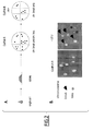
- Chromosome break could be distinguished from full chromosome loss by replica plating of Can 5-FoA R clones on plates lacking leucine (see Figure 2 ).
- Can 5-FoA resistant Leu+ clones result from chromosomal break between the URA3-CAN1 and LEU2 markers (see Figure 1 ) while Can 5-FOA resistant leuclones result from full chromosome loss of the homolog carrying the selective markers (see Figure 1 ).
- meiosis could lead to false positives (Can 5-FoA resistant). Unlike the genuine diploid clones, those are haploid and can be detected using mating type-tests. Such maters can stem either from meiotic recombination or from the loss of chromosome III, the chromosome carrying the mating type locus. The occurrence of meiosis was thus assessed for a WT CINA strain. Positive maters ranged from 1 to 5% of total Can 5-FoA resistant clones. Thus meiosis is negligible compared to the global percentage of chromosome instability values. In addition, RD cells do not sporulate and hence do not suffer from this small bias.
- the rate of chromosome instability was calculated using the Lea-Coulson median estimator (see e.g. Foster, 2006, Methods Enzymol. 409:195-213 ).
- the strain When the wild type CINA strain is grown on rich YEPD medium, the strain displayed a chromosome instability of about 6 marker losses per million of cells (6 10 -6 Can 5-FOA resistant, see Figure 3A ). About half of the losses were due to chromosome break (Can 5-FOA resistant, 60% LEU+) and half to chromosome loss (Can 5-FOA resistant, 40% leu-).
- Hydroxyurea an inhibitor of ribonucleotide reductase Rnr1 slows down replication forks ( Alvino et al. 2007 Mol Cell Biol. 27, 6396-6406 ) and increased chromosome instability 8 fold.
- CIN was increased 3-4 fold (22 +/- 6 10 -6 ) in a cell cycle mutants clb5 .
- the CIN assay thus also allows detecting genomic destabilization.
- the CIN assay is a sensitive assay that allows the detection of chromosome instability.
- it is a clonal assay that can be done on small samples.
- CINA is a highly sensitive yeast assay to quantify chromosome instability.
- the GCR (gross chromosomal rearrangement) assay was used to monitor chromosomal instabilities in budding yeast.
- the loss of two markers (composed of the CAN1 and URA3 genes) was positively selected using both canavanine and 5-FoA drugs.
- the frequency of such events is very low, around 10 -10 (for a recent review, see Motegi and Myung, 2007, Methods 41, 168-176 ).
- the CIN assay presents greatly improved sensitivity and supplemental features compared to GCR.
- the CIN assay is 20 000 fold more sensitive (6 10 -6 for a wild type) than the original GCR assay, and 100 fold more sensitive compared to an improved GCR version (Kanellis et al. 2007 PLoS Genet 3, e134).
- the CIN assay allows measuring and distinguishing between chromosome breaks and chromosome loss.
- the CIN assay has also proved useful to test CIN in the presence of cell cycle drugs and in cell cycle mutants. It should also be a very useful tool to analyze chromosomal instabilities in all kinds of physical, chemical or genetic conditions.
- the CIN assay can be performed on single colonies (usually ranging from about 1 to 10 million of cells per colony). Importantly, the CIN assay allows starting from a unique cell. It is therefore a clonal assay which can detect early events in chromosome instability.
- Eucaryotic cells generally rely largely on mitochondrial oxidative phosphorylation (OXPHOS) for their ATP- supplies.
- OXPHOS mitochondrial oxidative phosphorylation
- cells can proliferate without OXPHOS, using the glycolytic/fermentation pathway, with a much reduced ATP/glucose yield. This process is common in baker yeast grown on high glucose, or in cells grown in absence of oxygen. It is also the fate of cells harboring mutations disabling the respiratory-chain (either nuclear encoded genes, leading to "petites", or mutations/loss of mitochondrial DNA, termed p-/0). Fermentation has also been described in cancer cells, as early as in the 1920's, a feature known as the "Warburg effect".
- Oxidative stress is a major issue for most organisms. Mitochondria are both the main producer of oxidants and primary targets of the oxidative damage.
- Reactive oxygen species ROS
- ROS Reactive oxygen species
- targets proteins, fatty acids, nucleic acids
- low levels of ROS can also have beneficiary effects on cell survival and serve as second messengers. Production of highly reactive hydroxyl radical from hydrogen peroxide or superoxide radical can lead to DNA strand breaks.
- ROS has been shown to be an important cause of chromosome instabilities (CIN).
- mitochondria-deficient cells came out as the main category of mutants hypersensitive to external oxidative stress ( Thorpe et al. 2004 Proc Natl Acad Sci U S A 101, 6564-6569 ). However, the cause of that sensitivity is unknown. In HeLa cells, mitochondrial defects correlates with impaired DNA repair in the nucleus, an observation that might explain their sensitivity to DNA damage by hydrogen peroxide.
- ROS are a threat in both wild type and in respiratory-defective (RD) cells, but the latter show an increased sensitivity to oxidative stress.
- RD respiratory-defective
- ETC respiratory electron transport chain
- Those include nuclear mutations ( "petite” ) or mitochondrial ones (p- for partial and ⁇ 0 for total loss of mtDNA). All have in common a crippled mitochondrial respiratory chain.
- Those respiratory-defective (RD) cells are characterized by their inability to grow on non-fermentable carbon source.
- RD cells from three sources were analyzed: (a) ⁇ 0 cells (b) a mip1 mutant, defective for mitochondrial-specific DNA polymerase ( Genga et al. 1986 J Biol Chem 261, 9328-9332 ) and (c) pet mutant, a natural occurring petite strain. None of these strains had visible mitochondrial DNA by DAPI stain. Strikingly, when tested using a CIN assay, all three types of RD cells showed a significant increase in nuclear genome instability, from 5 to 30 fold. The chromosome instabilities corresponded to both chromosome breaks and losses.
- Genome instability as described above has been observed in various mutant contexts.
- Two main types are (a) cell cycle mutants, affecting “the cell cycle engine”, and (b) "checkpoint” or surveillance mechanism mutants ( Hartwell and Weinert 1989 Science 246, 629-634 ).
- Defects in the DNA replication machinery is a major contributor to this type of instability, as S phase is probably the most sensitive period challenging DNA integrity during cell proliferation.
- this class of genes is important to maintain the fidelity of cycle events, hence an increase of genomic instability observed in those mutants (recently reviewed in Aguilera and Gomez-Gonzalez 2008 Nat Rev Genet 9, 204-217 ).
- checkpoint mutants are often sensitive to various cell cycle drugs (affecting S and/or M phase). If RD cells have high CIN because of a putative checkpoint defect, they are expected to be sensitive to drugs challenging the corresponding surveillance mechanism.
- cell cycle drugs including S-phase inhibitor (HU), DNA alkylating (MMS) agents, DNA double-strand break formation (bleomycin) and microtubule poison (benomyl) was therefore tested on RD strains.
- wild type (checkpoint proficient) and checkpoint mutants for DNA damage/DNA replication mec1 sml1 and spindle checkpoint mutant bub1 were used.
- RD cells ( ⁇ 0, mip1, mrps28 ) were not particularly sensitive to any of the tested drugs. This suggests that RD cells have no major defects sensing, signaling or repairing these types of damages. A mild sensitivity was observed with the DNA damaging agent bleomycin. However, the same RD cells showed striking growth defects when plated on media inducing an oxidative stress (hydrogen peroxide, 4 and 6 mM). Oxidative stress blocked RD cells quickly, at unbudded or single budded stage. Thus most cells did not complete a single cell cycle ( ⁇ 0, mip1, mrps28 ), while WT and bub1 controls formed colonies in the same conditions.
- respiratory competent mec1 sml1 cells were as sensitive as RD cells to H 2 O 2 , a fact worth noting. These cells arrested mostly as budded cells in the first cell cycle. Hence, RD cells seem particularly sensitive to oxidative stress but not to other tested cell cycle drugs.
- NAC oxygen scavenger N-acetyl cysteine
- RD cells grow faster and with far less genomic instability in the presence of an anti-oxidant. This suggests the presence of an ill-processed internal oxidative stress in mitochondria-defective cells, leading both to slow growth and to high genomic instability in standard conditions.
- TSA1 a major ROS detoxifying enzyme, TSA1 ( Chae et al. 1994 J Biol Chem 269, 27670-27678 ), was deleted both in respiratory-competent (RC) and respiratory-defective (RD) backgrounds. The mutant was then challenged them with low H 2 O 2 doses (1 mM).
- tsa1 RC cells showed high CIN (131 +/- 18 10 -6 , 20 fold above wild type RC control).
- Low dose (1 mM) of H2O2 increased CIN only a little further (204 +/- 27 10 -6 ), indicating that these cells are still capable of dealing with this stress (either by detoxification and/or repair).
- RD cells showed a different behavior. Firstly, the basal level of CIN in tsa1 RD was higher (456 +/- 98 10 -6 , 76 fold above wild-type CIN levels).
- a cytological marker was used.
- the Rad52p protein is present and concentrates at sites of DNA damage (double-strand breaks).
- a Rad52-GFP fusion can be used to detect repair-foci in vivo .
- yeast strains harboring an integrated RAD52::GFP fusion construct were analyzed by fluorescence microscopy of life cells ( Lisby et al. 2001 Proc Natl Acad Sci U S A 98, 8276-8282 ).
Landscapes
- Life Sciences & Earth Sciences (AREA)
- Health & Medical Sciences (AREA)
- Genetics & Genomics (AREA)
- Engineering & Computer Science (AREA)
- Chemical & Material Sciences (AREA)
- Zoology (AREA)
- Organic Chemistry (AREA)
- Wood Science & Technology (AREA)
- Biomedical Technology (AREA)
- Bioinformatics & Cheminformatics (AREA)
- Biotechnology (AREA)
- General Engineering & Computer Science (AREA)
- Molecular Biology (AREA)
- Plant Pathology (AREA)
- Biophysics (AREA)
- Microbiology (AREA)
- Biochemistry (AREA)
- General Health & Medical Sciences (AREA)
- Physics & Mathematics (AREA)
- Mycology (AREA)
- Crystallography & Structural Chemistry (AREA)
- Virology (AREA)
- Bioinformatics & Computational Biology (AREA)
- Measuring Or Testing Involving Enzymes Or Micro-Organisms (AREA)
- Micro-Organisms Or Cultivation Processes Thereof (AREA)
Abstract
The present invention concerns a method for measuring chromosome instability as well as a diploid or polyploid yeast strain suitable for carrying out such a method. The method according to the invention allows detecting chromosome break events with a greatly improved sensitivity. In addition, it also allows detecting chromosome loss events. The method according to the invention is for example useful for evaluating toxicity of pharmaceutical and/or dietary compounds.
Description
- The present invention concerns a method for measuring chromosome instability as well as a yeast strain suitable for carrying out such a method.
- Life relies on the faithful transmission of DNA information from generation to generation. It is therefore of importance to assess the mistakes and damages that can occur on chromosomes. For example, cancer progression is often associated with the accumulation of gross chromosomal rearrangements (GCRs), such as translocations, deletion of a chromosome arm, interstitial deletions or inversions. In many instances, GCRs inactivate tumor-suppressor genes or generate novel fusion proteins that initiate carcinogenesis. Due to the importance of GCR during tumorigenesis, both in the initiation and progression steps, the cancer research community has become very interested in the molecular ethiology of GCRs. Yeast in an excellent model organism for this purpose since the cells are normal and contain many human gene homologs, which increase GCR rates when mutated similar to what has been observed in many human cancers. Therefore, yeast has been used a model for studying GCRs, both for better understanding the molecular ethiology of GCRs and for testing the effect of drugs on GCRs during preclinical and clinical trials.
- The GCR assay is commonly used to monitor chromosomal instabilities in budding yeast (Chen and Kolodner, 1999, Nat Genet. 23:81-85; Schmidt et al. 2006, Methods Enzymol. 409:462-476; Motegi and Myung, 2007, Methods 41:168-176). In the GCR assay, the loss of two markers (composed of the CAN1 and URA3 genes) is selected using both canavanine (Can) and 5-fluorooroticacid (5-FoA). In the haploid wild type strain used for GCR assays, the frequency of GCR events is very low (about 10-10). Thus over 10 billion cells are required for each assay, which imposes strong practical limitations. By introducing the same selectable markers on a different chromosome, Kanellis et al. (2007, PLoS Genet. 3:e134) increased the detection limit of the GCR assay by about 250 fold (i.e. a frequency of GCR events of about 9 10-8). Still, the detection limit is very low. The GCR assay is therefore unsuitable for large numbers of measurements. For instance, the screening of a library of ten thousands compounds using the method of Kanellis et al. (2007, PLoS Genet. 3:e134) would require thirty thousand Petri dishes. This makes the use of the GCR assay for high-throughput screenings both expensive and extremely difficult to put in place from a practical point of view. There is therefore a need for an assay for measuring chromosome instability that would exhibit a higher detection limit.
- In the GCR assay, the measurement of genetic instability is based on the selection of a gene loss. Several types of DNA damages co-exist, and the main challenge is to overcome the masking effects that one type of event can impose on another. Two parameters are of particular importance: the number of markers and the ploidy of the cell.
- When a single gene marker is used, the frequency of point mutations is around 10-6. For two markers, that frequency drops to 10-12. The existing GCR assay thus uses two markers (the CAN1 and URA3 genes). The frequency of chromosome break in the GCR assay is around 10-10. That frequency is thus about 100 fold higher than the frequency of double genes mutations. In other terms, the double genes mutations do not mask chromosome break events.
- The GCR assay is done in a haploid context. To be viable, the chromosome break has to occur in a limited distal region of chromosome V, between the CAN1 marker and the first essential gene (PMC1). This explains why the frequency of chromosome break is so low. However, the use of a diploid or polyploid strain in order to allow functional complementation of essential genes appears not to be an option since it is expected that the chromosome loss events will mask the chromosome break events. Indeed, a diploid or a polyploid cell usually looses an entire chromosome at a frequency that is as high as about 10-5, i.e., a frequency that is about 100 000 fold higher than the GCR frequency in a haploid cell. Therefore frequency of chromosome loss seems to prohibit to the use of a diploid or a polylploid strain for the measurement of chromosome breaks. Chromosome loss events appearing to be far more frequent than break events, they are thus expected to mask break events in a diploid or a polyploid cell.
- A yeast strain that is particularly suitable for measuring chromosome instability (CIN) has been found by the inventors. This strain has the main following features:
- it is diploid or polyploid, i.e. both break and full chromosome loss can occur without loss of viability;
- it comprises two negative selectable markers which allow selecting cells in which a chromosome break or a chromosome loss event has occurred. In the LD 1577 strain, these markers correspond to URA3 and CAN1, which are linked in the distal part of chromosome V. Chromosome instability is measured by lost of these two selectable markers; and
- it comprises a third selectable marker which allows distinguishing between chromosome break and chromosome loss events. In LD 1577, this third marker corresponds to the LEU2 marker, which is located close to centromere V.
- It has surprisingly been found that the frequency of break events in such a strain is high, around 10-5, i.e. similar to chromosome loss frequency. Thus, unexpectedly, chromosome loss events do not mask chromosome break events in such a strain. Furthermore, methods for measuring chromosome instability based on the use of this strain allow distinguishing between chromosome break events (yielding Can 5-FoA resistant LEU+ clones in the case of the LD 1577 strain) and chromosome loss (yielding Can 5-FoA resistant leu- clones in the case of the LD 1577 strain). Thus, in contrast to prior art methods, the method for measuring CIN using such a strain allows detecting chromosome loss events in addition to chromosome break events.
- The method in accordance with the invention allows detecting chromosome break events with a greatly improved sensitivity compared to the prior art assays. It is 20 000 fold more sensitive than the original GCR assay disclosed by Chen and Kolodner (1999, Nat Genet. 23:81-85), and 100 fold more sensitive than the GCR assay disclosed by Kanellis et al. (2007, PLoS Genet. 3:e134).
- Due to its increased sensitivity, the method in accordance with the invention can be performed starting from a single colony. It can therefore be carried out as a clonal assay, which can thus detect early events in chromosome instability rather than amplified non-independent events.
- Further due to its increased sensitivity, the method according to the invention can be scaled down. Multiple samples can be run easily, which allows repetition and accurate statistical analysis of the data. In addition, the method according to the invention may be performed using multi-well plates rather than Petri dishes. It thus allows robotic handling and drastically reduces the cost.
- The method in accordance with the invention being applicable to very small samples, it is suitable for screening purposes. It may for example be used to assess geno-toxicity and/or geno-protection of large numbers of products or mutants. For example, the screening of a library of ten thousands compounds using the method according to the invention would only require about three hundred 96-well plates, which is a great improvement compared to the thirty thousand Petri dishes that would be required using the method disclosed by Kanellis et al. (2007, PLoS Genet. 3:e134).
- The strain according to the invention is thus a very useful tool to analyze chromosomal instabilities in all kinds of physical, chemical or genetic conditions. More specifically, the method according to the invention is for example useful for evaluating toxicity of pharmaceutical and/or dietary compounds.
- The invention is directed to a yeast strain comprising a first and a second selectable marker located on a chromosome, wherein said first and second selectable markers are negative selectable markers, characterized in that:
- said yeast strain is diploid or polyploid;
- said yeast strain further comprises a third selectable marker located on said chromosome; and
- said first, second and third selectable markers are only present on one of the homologous copies of said chromosome.
- As used herein, the term "selectable marker" refers to an allele, the presence or the absence of which can be selected. More specifically, the presence or the absence of said marker can be selected in a cell either by addition of a selective compound to the medium on which the cell is grown, or by removal of a selective compound from the medium on which the cell is grown. Selectable markers are well-known in the art and include, e.g. CAN1, URA3 and LEU2. The selectable marker according to the invention may for example correspond to an auxotrophic marker (e.g. LEU2, which confers the capacity to a cell to grow in the absence of leucine) or to an allele conferring resistance or sensitivity to a drug (e.g. CAN1 and URA3, which confer sensitivity to canavanine and to 5-fluorooroticacid, respectively). Selectable markers are also commonly referred to as "selection markers" in the art.
- As used herein, a "positive selectable marker" refers to a selective marker of which the presence can be selected. A "negative selectable marker" refers to a selectable marker of which the absence can be selected (i.e. the presence of the marker can be counter-selected).
- Examples of positive selectable markers include e.g. LEU2, the presence of which can be selected on a medium lacking leucine, HIS3, the presence of which can be selected on a medium lacking histidine, TRP1, the presence of which can be selected on a medium lacking tryptophan, ADE2, the presence of which can be selected on a medium lacking adenine, and kanMX4, the presence of which can be selected on a medium comprising G418.
- Examples of negative selectable markers include CAN1 and URA3, the absence of which can be selected on media comprising canavanine and 5-fluorooroticacid, respectively.
- As used herein, a gene indicated in capital letters refers to a wild-type, functional allele. A gene indicated in small letters refers to a mutated, non functional allele.
- The first and second selectable markers correspond to negative selectable markers, i.e. markers that can be counter-selected in the presence of a selective compound. These first and second markers may for example correspond to CAN1 and URA3, which can be counter-selected in the presence of canavanine and 5-fluorooroticacid, respectively. The third selectable marker may either correspond to a positive selectable marker or to a negative selectable marker. The third selectable marker preferably corresponds to a positive selectable marker such as e.g. LEU2. Of course, the first, second and third selectable markers necessarily correspond to different markers, and they are necessarily absent from all other chromosomes, otherwise it would be impossible to select their presence or their absence on the chromosome of interest.
- The first, second and third selectable markers are only present on one of the homologous copies of said chromosome. For example, when the strain is diploid, the first, second and third selectable markers are only present on one of the two homologous chromosomes. In other words, all three markers are located not only on the same chromosome, but also on the same copy of the chromosome.
- The first and second selectable markers are located on the same arm of the chromosome (i.e., on the same side of the centromere).
- The yeast strain according to the invention is suitable for measuring chromosome instability (CIN), i.e., the first and second selectable markers are located on the chromosome in such a way as to allow selecting cells in which a chromosome break or a chromosome loss event has occurred, and the third selectable marker is located on the chromosome in such a way as to allow distinguishing between chromosome break and chromosome loss events.
- The distance between the centromere and the first or second selectable marker that is located closest to the centromere defines the scorable breakage zone. The first and second selectable markers may for example be located at a distance of at least 50, 100, 200, 300, 400, 500, 800 or 1000 kb to the centromere. Preferably, said distance is of at least 100 kb. This distance can be increased or decreased by the skilled in the art, depending on the chromosome on which the markers are located and on the chromosome break frequency that is wished for.
- In a preferred embodiment, the first and second selectable markers are located in the distal part of the chromosome, i.e. in the most centromere-distal moiety of a chromosome arm. More preferably, they are located in the most centromere-distal third of the chromosome arm. Most preferably, they are located in the most centromere-distal quarter of the chromosome arm.
- Also preferably, the first and second selectable markers are located at a distance of at least (or of about) 5, 10, 20, 50 or 100 kb to the telomere in order to avoid a possible gene repression effect.
- Also preferably, the first and second selectable markers are linked together, i.e. they exhibit a recombination frequency of less than 50%. More preferably, they exhibit a recombination frequency of less than 40%, 30%, 20%, 10%, 5% or 1%. A short distance between the first and second selectable markers allows the amplification of the first and second selectable markers as a cassette by PCR, and their transfer to a different chromosome location if desired. In addition, putting them further apart would reduce the scorable breakage zone. In a preferred embodiment, the distance between the first and second selectable marker may be of less than 50, 40, 30, 25, 20, 15, 10 or 5 kb.
- The third selectable marker is located on the chromosome in such a way as to allow distinguishing between chromosome break and chromosome loss events. More specifically, the third selectable marker is located close to the centromere, in such a way as to be lost upon chromosome loss events but not (or rarely) upon chromosome break events.
- In a preferred embodiment, the third selectable marker is located in the most centromere-proximal moiety of a chromosome arm. More preferably, it is located in the most centromere-proximal third of the chromosome arm. Most preferably, it is located in the most centromere-proximal quarter of the chromosome arm. The third selectable marker may for example be located at a distance of less than 50, 40, 30, 20, 10 or 5 kb to the centromere. Preferably, it is located at a distance of less than 30 kb to the centromere.
- The third selectable marker may or may not be located on the same arm of the chromosome than the first and second selectable markers. In a preferred embodiment, the third selectable marker is located on the opposite arm of the chromosome as compared to the first and second selectable markers. This prevents the risk of a chromosome break event occurring between the third selectable marker and the centromere being counted as a chromosome loss event. Alternatively, the third selectable marker is located on the same chromosome arm as the first and second selectable markers. In this case, the distance between the third selectable marker and the first and second selectable markers is preferably of at least 20, 40, 50, 60, 80 or 100 kb.
- Thus a preferred yeast strain in accordance with the invention is a strain in which:
- the first and second selectable are located at a distance of at least 50, 100, 200, 300, 400, 500, 800 or 1000 kb to the centromere;
- the first and second selectable are located at a distance of at least 5, 10, 20 or 50 kb to the telomere;
- the third selectable is located at a distance of less than 50, 40, 30, 20, 10 or 5 kb to the centromere;
- the third selectable is located on the opposite arm of the chromosome relative to the first and selectable marker, or, when located on the same arm, at a distance of at least 10, 25, 50, 60, 70, 80 or 100 kb to the first and second selectable markers; and/or
- the distance between the first and second selectable marker is of less than 50, 25, 10 or 5 kb.
- The yeast strain in accordance with the invention is preferably diploid, i.e. it comprises two homologous copies of each of the 32 chromosomes. The diploid strain is preferably a MATa/α strain, i.e., it does not result from an illegitimate mating phenomenon. Alternatively, the strain may correspond to a polyploid strain instead of a diploid strain. In such a case, the three selectable markers are only present on one of all homologous copies of the chromosome.
- The selectable markers may be located on any of the chromosomes. They may for example be located on chromosome V or on chromosome XV. The skilled in the art may choose to study a given chromosome, breakage zones of particular interest (for example, slow replicating zones or zones known to be fragile) or add an heterologous sequence coming from a different organism to test the fragility of particular sites (see e.g. page 174 of Motegi and Myung, 2007, Methods, 41:168-176).
- The yeast strain in accordance with the invention may further comprise one or more deleted gene, disrupted gene, mutated gene, exogenous gene and/or gene placed under the control of an exogenous promoter allowing its overexpression. Such a yeast strain may be used to test whether said gene increases or decreases chromosome instability.
- The yeast strain in accordance with the invention may also comprise additional selectable markers (e.g. markers conferring resistance to an antibiotic). The additional markers may or may not be located on the same chromosome as the selectable markers in accordance with the invention.
- In a preferred embodiment, the yeast strain in accordance with the invention is not capable of entering meiosis. Such a strain may for example be obtained by deleted a gene essential for meiosis such as e.g. spo11 or a ime2. In a preferred embodiment, the strain is deleted for spo11. Indeed, spo11 is meiotic-specific (i.e. the deletion does not affect mitosis). spo11 mutants undergo sporulation, but all the spores are not viable.
- Preferred strains according to the invention are described in further details in Example 1. A most preferred strain in accordance with the invention is the strain referred to as LD 1577 as well as strains derived therefrom. These strains are diploid strains carrying the CAN1 and URA3 negative selectable markers, about 8 to 8.5 kb apart, on the left arm of chromosome V. Thus one copy of the chromosome V comprises:
- the wild-type CAN1 allele, which is located at its natural locus;
- the wild-type URA3 allele, which integrated at the hxt13 locus; and
- the wild-type LEU2 allele, which integrated at the ura3 locus.
- The other chromosome V of such strains comprises a mutated can1 allele, a mutated ura3 allele and a wild-type HXT13 allele.
- In such strains:
- the first, second and third selectable markers are located on the same arm of chromosome V (the left arm);
- the first and second selectable are located at a distance of about 110 kb to the centromere (CEN5), and of about 22 kb to the left telomere of chromosome V; and
- the third selectable is located at a distance of about 30 kb to CEN5, and of about 83 kb to the first and second selectable markers.
- The invention is further directed to a yeast chromosome comprising a first and a second negative selectable marker located on the same arm of said chromosome characterized in that said chromosome further comprises a third selectable marker. These selectable markers are located on the chromosome as described hereabove for the yeast strain in accordance with the invention. More specifically, the first and second selectable markers are located in the distal part of the chromosome, preferably linked together, and the third selectable marker is located close to the centromere.
- The yeast strain in accordance with the invention can advantageously be used for measuring chromosome instability. Thus the invention is directed to a method for measuring chromosome instability comprising the steps of:
- a) providing a yeast strain according to the invention;
- b) plating cells of said yeast strain on a medium selecting cells in which said first and second selectable markers have been lost; and
- c) replicating said cells on a medium selecting cells in which said third selectable marker is either present or absent.
- In this method, the detection of a colony growing on the medium of step (b) indicates that a chromosome loss or a chromosome break event has occurred in the cells of said colony.
- As used herein, the term "chromosome instability" (CIN) refers both to chromosomal break and to chromosome loss. As further detailed below, the method may also be used to measure either chromosome break or chromosome loss.
- As used herein, the term "non-selective medium" refers to a medium on which a given yeast strain can grow irrespective of the absence or the presence of the first, second and third selective markers on its chromosomes. For example, a medium comprising leucine, and devoid of canavanine (Can) and of 5-fluorooroticacid (5-FoA), is a non-selective medium for an experiment carried out starting from a strain comprising the URA3, CAN1 and LEU2 selectable markers.
- The medium of step (b) selects cells in which said first and second selectable markers have been lost. On the other hand, the medium of step (b) is a non selective medium in respect of the third selectable marker (i.e. the cells must be able to grow irrespective of the presence or the absence of the third selectable marker). For example, a medium comprising leucine, canavanine (Can) and 5-fluorooroticacid (5-FoA) can be used at step (b) when the experiment is carried out starting from a strain comprising URA3 and CAN1 as the first and second selectable markers and LEU2 as the third selectable marker.
- The medium of step (c) selects cells in which the third selectable marker is present or absent. More specifically, when the third selectable marker is a positive selectable marker, cells are selected for the presence of said marker. On the contrary, when the third selectable marker is a negative selectable marker, cells are selected for the absence of said marker. For example, a medium devoid of leucine can be used at step (c) when the experiment is carried out starting from a strain comprising LEU2 as the third selectable marker.
- After provision of a yeast strain according to the invention, step (a) preferably further comprises the step of growing the yeast strain on a non-selective medium. The yeast strain may either be grown in a liquid medium or on a solid medium. During this growth, chromosome instability events may occur. The non-selective medium may for example consist of a rich YPD medium comprising 2% glucose. The cells are preferably grown at about 20-42°C, 20-37°C, 25-35°C or 30°C.
- At step (a), the yeast strain may be plated on any type of support such as e.g. Petri dishes or multi-well plates. The medium may be liquid or solid. Petri dishes are particularly suitable for performing clonal assays. On the other hand, multi-well plates are particularly suitable for performing assays with a large number of samples, for example in the frame of a screening.
- When a clonal assay is carried out (i.e., an assay performed with cells derived from a single colony), the strain is preferably grown on a Petri dish until isolated colonies are obtained. Then, a representative colony can be picked. Thus, after growth of the strain on a non-selective medium, step (a) may further comprise the steps of isolating a colony and suspending the cells of said colony in a solution. Said solution may for example consist of water.
- When multi-well plates are used, the wells may for example comprise 200 µl liquid medium and be inoculated with about 200 cells/well. After growth to saturation, strains usually reach around 100 million cells/ml. 5 µl of that culture (i.e. about 5.105 cells) may then be used for plating at step (b). Those numbers can easily be adapted by the skilled in the art.
- Upon provision of the strain in accordance of the invention and before cultivation on the non-selective medium, the strain may first be cultivated on a selective medium into ensure that all selectable markers are present. For example, when the experiment is carried out with the LD 1577 strain, the strain may be cultivated on a medium lacking uracil and leucine.
- The method according to the invention may be used to determine whether a candidate compound increases or decreases chromosome instability. In the frame of this embodiment, the non-selective medium of step (a) further comprises the candidate compound to be tested. The chromosome instability of the strain in the presence of said candidate compound may then be compared to the chromosome instability of the same strain in the absence of said candidate compound.
- The method according to the invention may also be used to determine whether a candidate gene increases or decreases chromosome instability. In the frame of this embodiment, the yeast strain provided at step (a) is a strain wherein the candidate gene is deleted, disrupted, inactivated or overexpressed. The chromosome instability of the strain in which said candidate gene is deleted, disrupted, inactivated or overexpressed may then be compared to the chromosome instability of the same strain in which said candidate gene is in its wild-type form. The overexpressed candidate gene may correspond to a heterologous gene that is absent from the wild-type yeast genome.
- As used herein, the terms "candidate gene", "candidate compound" and "candidate physical parameter" refer to genes, compounds and physical parameters that are susceptible of playing a role in chromosome instability. The strains and methods may be used to determine whether a candidate gene, a candidate compound or a candidate physical parameter increases or decreases chromosome instability. For example, the skilled in the art may determine whether a candidate gene plays a role in chromosome instability by comparing the chromosome instability measured in a yeast strain according to the invention in which said candidate gene is deleted or over-expressed with the chromosome instability measured in a yeast strain according to the invention that comprises a wild type candidate gene. Similarly, the skilled in the art may determine whether a candidate compound plays a role in chromosome instability by comparing the chromosome instability measured in yeast strains according to the invention that are grown in the presence and in the absence of the candidate compound, respectively.
- The candidate compound may correspond to any compound such as e.g. a chemical molecule (for example a small molecule), a protein, a peptide, a lipid, a DNA molecule, a RNA molecule, an antisense polynucleotide, a small interfering RNA, an aptamer or an antibiotic. The candidate compounds may for example be manufactured and/or used by industries in the field of food, agribusiness, agro-chemistry and/or pharmaceuticals.
- In a preferred embodiment, the candidate compound corresponds to a compound intended for a pharmaceutical use such as, e.g., a compound undergoing preclinical or clinical trials for the treatment of a disease.
- In another preferred embodiment, the candidate compound corresponds to a dietary compound, a nutrient and/or a food component. Indeed, the methods in accordance with the invention are useful for evaluating the effect of a dietary compound on genetic instability and thus evaluating their potential genotoxic effect.
- In still another preferred embodiment, the candidate compound corresponds to a candidate chemical compound (e.g. manufactured and/or used in the field of agro-chemistry), and the method according to the invention is carried out to test the potential toxicity of such candidate chemical compounds.
- The candidate gene may correspond to any gene. It may for example correspond to a gene of which the human homolog plays a role in a disease such as e.g. cancer.
- The candidate physical parameter may for example correspond to light or radiation. Alternatively, the candidate physical parameter may correspond to a physiological or a growth parameter such as temperature, nutrient composition growth conditions, etc.
- Step (b) allows selecting cells wherein a chromosome instability event has occurred. At step (b), the cells are plated on a solid medium in order to count the number of colonies obtained. Thus plating on a Petri dish is preferred. When multi-well plates are used at step (a), rectangular Petri dishes with the same size as the multi-well plates may be used, thus allowing automatic transfer from the medium of step (a) to the medium of step (b). A chosen number of cells may be plated on a Petri dish (e.g. about 0.1-10 or 1-5 105 cells) and allowed to grow for e.g. 1-10, 1-5 or 1-3 days. The cells are preferably grown at about 20-42 °C, 20-37 °C, 25-35 °C or 30°C. The number of colonies growing on the Petri dishes is indicative of the number of chromosome instability events that have occurred among the number of plated cells.
- Step (c) allows distinguishing between chromosome loss and chromosome break events. This is determined by replicating the cells growing at step (b) on a medium allowing determining whether the third selectable marker is still present or whether it is lost as well.
- The third selectable marker preferably corresponds to a positive selectable marker. In this case, the medium of step (c) selects cells in which the third selectable marker is present. In the frame of this embodiment:
- the detection of a colony growing both on the medium of step (b) and on the medium of step (c) indicates that a chromosome break event has occurred in the cells of said colony; and/or
- the detection of a colony growing on the medium of step (b) but not on the medium of step (c) indicates that a chromosome loss event has occurred in the cells of said colony.
- Alternatively, the third selectable marker may correspond to a negative selectable marker. In this case, the medium of step (c) selects cells in which the third selectable marker is absent. In the frame of this embodiment
- the detection of a colony growing both on the medium of step (b) and on the medium of step (c) indicates that a chromosome loss event has occurred in the cells of said colony; and/or
- the detection of a colony growing on the medium of step (b) but not on the medium of step (c) indicates that a chromosome break event has occurred in the cells of said colony.
- Once the number of chromosome break and the number of chromosome loss events have been measured, the frequency and/or the rate of chromosome instability (and/or of each one of these events) can easily be calculated by the skilled in the art. For example, the number of CIN events per 106 cells (referred to as "the CIN frequency") may be calculated. Alternatively, the rate of chromosome instability may be measured using the method that is disclosed in the paragraph entitled "2.4. Real calculation" at page 170 of Motegi and Myung (2007 Methods 41:168-176). This method, referred to as the "Lea-Coulson median estimator", as well as other calculation methods that may be used, is reviewed in Foster (2006, Methods Enzymol. 409:195-213).
- In order to make sure that the loss of all three markers is due to a chromosome loss event and not to a meiosis event, it is possible to test whether the colonies detected at step (b) and/or (c) are still diploid. Thus, the method according to the invention may further comprise the step of detecting haploid cells growing at step (b) and/or (c) by a mating type-test. However, this step is optional since it has been shown that the number of meiosis event is negligible compared to the number of chromosome loss events (see Example 2).
- The invention is further directed to the use of a strain according to the invention or to a method according to the invention for determining whether a candidate gene, a candidate compound and/or a candidate physical parameter increases or decreases chromosome instability (see e.g. Example 3).
- A preferred embodiment of the invention is directed to a method of determining whether a candidate compound or a candidate physical parameter increases or decreases chromosome instability comprising the steps of:
- i. measuring chromosome instability in the presence of said candidate compound or candidate physical parameter;
- ii. measuring chromosome instability in the absence of said candidate compound or candidate physical parameter;
- iii. comparing the chromosome instability measured at step (i) and (ii);
- chromosome instability is measured according to any one of the methods described herein;
- the measure of a higher chromosome instability at step (i) than at step (ii) indicates that said candidate compound or candidate physical parameter increases chromosome instability; and
- the measure of a lower chromosome instability at step (i) than at step (ii) indicates that said candidate compound or candidate physical parameter decreases chromosome instability.
- This method may also be used to determine whether a candidate compound or a candidate physical parameter increases or decreases chromosome break or chromosome loss. The candidate compound is preferably added to the non-selective medium used at step (a) of the method for measuring chromosome instability according to the invention. The candidate physical parameter is preferably applied during the growth in the non-selective medium of step (a) of the method for measuring chromosome instability according to the invention.
- Another preferred embodiment of the invention is directed to a method of determining whether a candidate gene increases or decreases chromosome instability comprising the steps of:
- i. measuring chromosome instability in a strain comprising said candidate gene;
- ii. measuring chromosome instability in a strain wherein said candidate gene is deleted or disrupted;
- iii. comparing the chromosome instability measured at step (i) and (ii);
- chromosome instability is measured according to any of the methods described herein;
- the measure of a higher chromosome instability at step (i) than at step (ii) indicates that said candidate gene increases chromosome instability; and
- the measure of a lower chromosome instability at step (i) than at step (ii) indicates that said candidate gene decreases chromosome instability.
- This method may also be used to determine whether a candidate gene increases or decreases chromosome break or chromosome loss. The two strains are preferably identical except for the presence or the absence of said candidate gene. Alternatively, the method may be performed with a strain overexpressing said candidate gene at step (i), and a strain comprising a wild-type candidate gene at step (ii).
- All references cited herein, including journal articles or abstracts, published or unpublished patent application, issued patents or any other references, are entirely incorporated by reference herein, including all data, tables, figures and text presented in the cited references.
- The invention will be further evaluated in view of the following examples and figures.
-
-
Figure 1 corresponds to a schematic view of a strain in accordance with the invention. Genome instability is measured by positive selection of marker loss (URA3 and CAN1 genes), on the distal arm of chromosome V. Combined loss of URA3 and CAN1 genes leads to colonies resistant to the drugs 5-FoA and canavanine (Can FoA resistant). In a CINA strain, this phenotype can either be due to chromosome break (Leu+) or to full chromosome loss (leu-). -
Figure 2 is a scheme of an assay in accordance with the invention (CIN assay). A. Starting from single cells, clones are grown and chromosome instability events detected on CanFoA plates. Replica on plates lacking leucine allows to distinguish between chromosome break (CanFoA resistant, LEU+) and chromosome loss (Can FoA resistant, leu-). B. Picture of colonies growing on CanFoA plates and on plates lacking leucine. -
Figure 3 shows the CIN rates of various strains in accordance of the invention, in the presence of various compounds. The strains grown to colonies on YEPD at 30°C. Each assay was repeated 18 times. A. CIN rate in the wild-type CINA strain (LD1577; WT) and in a strain comprising an additional mutation of the S-phase cyclin clb5 (clb5). B. CIN rate in the wild-type CINA strain (LD1577). The strain was grown on YEPD plates (C), on YEPD plates in the presence of benomyl (12 µg/l) (+Beno), or on YEPD plates in the presence of hydroxyurea (50 mM) (+HU). C. CIN rate in the wild-type CINA strain (LD1577). The strain was grown on YEPD plates (C), or on YEPD plates in the presence of increasing concentrations of Zeocin (1 or 5 µg/ml). - A LEU2 wild type gene was integrated at the mutated ura3 locus, close to CEN5, in the RDK 3615 Mata strain carrying a URA3-CAN1 selection cassette (Myung et al., 2001, Nature, 411:1073-6).
- Proper integration of the marker was verified by southern blot and genetic analysis. The resulting strain is referred to as LD 1520 and has the following genotype: MATa, ura3-52::LEU2, leu2Δ1, trp1Δ63, his3Δ200, lys2ΔBgl, hom3-10, ade2Δ1, ade8, hxt13::URA3 CAN1 (S288c).
- The mating partner carrying a can1 mutant allele marked with kanMX4 marker is referred to as LD 1571 and has the following genotype: MATalpha his3 ::1 leu2::0 met15::0 ura3::0 can1::kanMX 4 (Euroscarf).
- LD 1520 was mated with LD 1571 and a diploid zygote was pulled of. This led to the strain referred to as LD 1577, i.e. the wild type CINA strain (for a schematic view of this strain, see
Figure 1 ). - To generate mutant strains for CIN assays, a mutation in a given gene was introduced separately in haploid parents by genetic crosses.
- One parent was LD 1847, a Matalpha version of LD 1520 (ura3-52::LEU2 hxt13::URA3 CAN1).
- The chosen mutation was separately introduced in the LD 1607 Mata strain carrying a natural untagged can1 mutation. Strain LD 1607 has the genotype: Matalpha leu2::0 lys2::0 met15::0 trp1::63 ura3::0 can1. Crossing of such parents leads to homozygous mutant diploid strain with the appropriate heterozygous selection markers for chromosome instability assay.
- To generate ρO strains, cells were grown to saturation in liquid YPD medium + ethidium bromide (10mg/ml) and plated on YPD plates for individual colonies. Clones were checked for growth defects on a non-fermentable carbon source (YPG 2% glycerol) and absence of mtDNA was confirmed by DAPI staining.
- For natural petite, untreated cells were plated on YPD 2% glucose in the absence of treatment or selection and small growing colonies were screened as above (YPG-).
- Strains with a defined nuclear-encoded mitochondrial defect (mip1, mrps28) were checked likewise.
- The strains were grown at 30°C on YPD medium (2% glucose) unless indicated otherwise.
-
strain genotype Comment 1459 MATa, ura3-52, leu2Δ1, trp1Δ63, his3Δ200, lys2ΔBgl, hom3-10, ade2Δ1, ade8, hxt13::URA3 CAN1 1472 MATa, ura3-52, leu2Δ1, trp1Δ63, his3Δ200, lys2ΔBgl, hom3-10, ade2Δ1, ade8, hxt13::URA3 ρO 1496 MATa, ade2-1, ade3, trp1-1, can1-100, leu2-3112, his3-11, 15, ura3, GAL, psi+ mrps28::HIS3 RAD52:GFP ρ0 1520 MATa, ura3-52::LEU2, leu2Δ1, trp1Δ63, his3Δ200, lys2ΔBgl, hom3-10, ade2Δ1, ade8, hxt13::URA3 CAN1 1571 MATα, his3 ::1 leu2::0 met 15::0 ura3::0 can1::kanMX 4 TRP+ Obtained from Euroscarf 1577 ∼/MATaα, ura3-52::LEU2, leu2Δ1, trp1Δ63, his3Δ200, lys2ΔBgl, hom3-10, ade2Δ1, ade8, hxt13::URA3, CAN1 / his3 ::1 leu2::0 met15::0 ura3::0 can1::kanMX 4 Referred to as CINA WT 1607 MATα/eu2::0 lys2::0 met15::0 trp1::63 ura3::0 can 1 Natu ral can1 mutant 1690 MATa/α, ura3-52::LEU2, leu2Δ1, trp1Δ63, his3Δ200, lys2ΔBgl, hom3-10, ade2Δ1, ade8, hxt13::URA3, CAN1 / his3 ::1 leu2::0 met15::0 ura3::0 can1::kanMX 4 ρ0 CINA ρ0 1719 MA Ta RAD52:GFP (unmarked) ADE+ 1721 MATα, RAD52:GFP (unmarked) ADE+ ρ0 1779 MATa/α, his3 ::1 leu2::0 met15::0 ura3::0 trp1 mip1::kanMX4 hxt13::URA3 CAN1 / his3 ::1 leu2::0 met15::0 ura3::0 can 1::kanMX 4 mip1::kanMX4 (ρ0) CINA mip1 (ρ0) 1781 MATa/α, ura3-52::LEU2, leu2Δ1, trp1Δ63, his3Δ200, lys2ΔBgl, hom3-10, ade2Δ1, ade8, hxt13::URA3, CAN1 / his3 ::1 leu2::0 met15::0 ura3::0 can1::kanMX 4 pet (ρ0) CINA Natural petite of 1577 1822 MATa/α, his3 ::1 leu2::0 met15::0 ura3::0 tsa1::kanMX 4 ura3-52::LEU2 hxt13::URA3 CAN1 / his3::1 leu2::0 met15::0 ura3::0 tsa1::kanMX 4 can 1 CINA tsa1 1829 MATa/α, his3 ::1 leu2::0 met15::0 ura3::0 tsa1::kanMX 4 ura3-52::LEU2 hxt13::URA3 CAN1 / his3 ::1 leu2::0 met15::0 ura3::0 tsa1::kanMX 4 can 1 ρ0 CINA tsa1 ρ0 1847 MATα, ura3-52::LEU2 hxt13::URA3 CAN1 his3 TRP+ - Dilutions of the CINA strain were done on the plate of choice (unless stated, rich YPD 2% glucose plates). They were grown at 30°C to give rise to isolated colonies (around one hundred colonies/plate). When grown to visible colonies (106 cells for small colonies, up to around 107 cells for large ones), a representative colony was picked and measured both for cell number and for cell size using a Casy Counter. Individual colonies were picked and suspended in water (20 µl). The chosen number of cells (around 1-5 105 cells) was plated and allowed to grow for 2-3 days at 30°C on selective plates (Can 5-FoA) in order to select for chromosome instability events. Plating adjustment of cell number were made depending on the genomic instability of individual strains.
- Using this protocol, six assays could easily be done on a single small Petri dish. Chromosome break could be distinguished from full chromosome loss by replica plating of Can 5-FoA R clones on plates lacking leucine (see
Figure 2 ). Can 5-FoA resistant Leu+ clones result from chromosomal break between the URA3-CAN1 and LEU2 markers (seeFigure 1 ) while Can 5-FOA resistant leuclones result from full chromosome loss of the homolog carrying the selective markers (seeFigure 1 ). - It may be noted that low frequency meiosis could lead to false positives (Can 5-FoA resistant). Unlike the genuine diploid clones, those are haploid and can be detected using mating type-tests. Such maters can stem either from meiotic recombination or from the loss of chromosome III, the chromosome carrying the mating type locus. The occurrence of meiosis was thus assessed for a WT CINA strain. Positive maters ranged from 1 to 5% of total Can 5-FoA resistant clones. Thus meiosis is negligible compared to the global percentage of chromosome instability values. In addition, RD cells do not sporulate and hence do not suffer from this small bias.
- The rate of chromosome instability was calculated using the Lea-Coulson median estimator (see e.g. Foster, 2006, Methods Enzymol. 409:195-213).
- When the wild type CINA strain is grown on rich YEPD medium, the strain displayed a chromosome instability of about 6 marker losses per million of cells (6 10-6 Can 5-FOA resistant, see
Figure 3A ). About half of the losses were due to chromosome break (Can 5-FOA resistant, 60% LEU+) and half to chromosome loss (Can 5-FOA resistant, 40% leu-). - As a control, drugs and mutations known to affect chromosome stability in yeast were used (see
Figure 3A and 3B ). - Benomyl (12 µg/ml) increased CIN by 5 fold (29 +/- 5 10-6). Most events (Can 5-FoAR) were due to total chromosome loss (10% LEU+, 90% leu-). This was expected for a microtubule poisoning agent.
- Growth in presence of zeocin, which creates double strand breaks, strongly increased CIN in a dose-dependent manner : 20 fold (130 +/- 11 10-6) at 1µg/ml of zeocin, and 120 fold (740 +/- 185 10-6) at 5 µg/ml of zeocin. As expected, there was a strong bias towards chromosome breaks (over 90% LEU+).
- Hydroxyurea, an inhibitor of ribonucleotide reductase Rnr1, slows down replication forks (Alvino et al. 2007 Mol Cell Biol. 27, 6396-6406) and increased chromosome instability 8 fold.
- CIN was increased 3-4 fold (22 +/- 6 10-6) in a cell cycle mutants clb5. The CIN assay thus also allows detecting genomic destabilization.
- Hence, the CIN assay is a sensitive assay that allows the detection of chromosome instability. In addition, it is a clonal assay that can be done on small samples.
- CINA is a highly sensitive yeast assay to quantify chromosome instability.
- Until now, the GCR (gross chromosomal rearrangement) assay was used to monitor chromosomal instabilities in budding yeast. The loss of two markers (composed of the CAN1 and URA3 genes) was positively selected using both canavanine and 5-FoA drugs. In a haploid wild type strain, the frequency of such events is very low, around 10-10 (for a recent review, see Motegi and Myung, 2007, Methods 41, 168-176).
- The CIN assay presents greatly improved sensitivity and supplemental features compared to GCR. The CIN assay is 20 000 fold more sensitive (6 10-6 for a wild type) than the original GCR assay, and 100 fold more sensitive compared to an improved GCR version (Kanellis et al. 2007 PLoS Genet 3, e134). In addition, the CIN assay allows measuring and distinguishing between chromosome breaks and chromosome loss. The CIN assay has also proved useful to test CIN in the presence of cell cycle drugs and in cell cycle mutants. It should also be a very useful tool to analyze chromosomal instabilities in all kinds of physical, chemical or genetic conditions.
- Because of the scale down, multiple samples can be run easily and allow repetition and statistical analysis of the data, as well as lowering considerably the cost of the assay. Due to its increased sensitivity, the CIN assay can be performed on single colonies (usually ranging from about 1 to 10 million of cells per colony). Importantly, the CIN assay allows starting from a unique cell. It is therefore a clonal assay which can detect early events in chromosome instability.
- Eucaryotic cells generally rely largely on mitochondrial oxidative phosphorylation (OXPHOS) for their ATP- supplies. However, cells can proliferate without OXPHOS, using the glycolytic/fermentation pathway, with a much reduced ATP/glucose yield. This process is common in baker yeast grown on high glucose, or in cells grown in absence of oxygen. It is also the fate of cells harboring mutations disabling the respiratory-chain (either nuclear encoded genes, leading to "petites", or mutations/loss of mitochondrial DNA, termed p-/0). Fermentation has also been described in cancer cells, as early as in the 1920's, a feature known as the "Warburg effect". Once considered a side-effect of cancer growth conditions, this metabolic change from respiration to fermentation has raised a lot of interest. Growing evidences point to a genetically controlled switch, with an important role in tumorigenesis. Mitochondria dysfunction and decreased respiration rates are also associated with aging. Hence, a subset of eukaryotic cells function in the absence of OXPHOS, due to physiological conditions, mutations or controlled metabolic switches.
- Oxidative stress is a major issue for most organisms. Mitochondria are both the main producer of oxidants and primary targets of the oxidative damage. Reactive oxygen species (ROS) are mainly produced through leakage of electrons from the OXPHOS chain, and the 'free-radical theory of aging' proposed that oxidative damage over time is responsible for cellular aging. ROS can damage a wide variety of targets (proteins, fatty acids, nucleic acids...) but low levels of ROS can also have beneficiary effects on cell survival and serve as second messengers. Production of highly reactive hydroxyl radical from hydrogen peroxide or superoxide radical can lead to DNA strand breaks. In wild type yeast cells grown under standard conditions, ROS has been shown to be an important cause of chromosome instabilities (CIN).
- In a yeast genome-wide study, mitochondria-deficient cells came out as the main category of mutants hypersensitive to external oxidative stress (Thorpe et al. 2004 Proc Natl Acad Sci U S A 101, 6564-6569). However, the cause of that sensitivity is unknown. In HeLa cells, mitochondrial defects correlates with impaired DNA repair in the nucleus, an observation that might explain their sensitivity to DNA damage by hydrogen peroxide.
- In summary, ROS are a threat in both wild type and in respiratory-defective (RD) cells, but the latter show an increased sensitivity to oxidative stress. A few elements suggest that this phenomenon may be due to impaired repair capacities, but the molecular details remain unknown.
- What, if any, is the consequence of OXPHOS inactivation on the maintenance of nuclear genome integrity? Quantitatively, RD yeast cells display a small increase in mutation rate compared to wild type. Mutations affecting mitochondria OXPHOS are found in some cancer cells, sometimes associated with gross chromosomal modifications. A model linking mitochondria dysfunction and nuclear genome instability has therefore been put forward and discussed. However, quantitative data showing a strong impact of respiratory dysfunction on nuclear genome stability is missing in support of this model.
- In the examples herebelow, the potential effects of respiratory deficiency on nuclear genome stability have been addressed in quantitative terms.
- Mutations in many of the genes involved in mitochondrial functions lead to inactivation (directly or indirectly) of the respiratory electron transport chain (ETC). Those include nuclear mutations ("petite") or mitochondrial ones (p- for partial and ρ0 for total loss of mtDNA). All have in common a crippled mitochondrial respiratory chain. Those respiratory-defective (RD) cells are characterized by their inability to grow on non-fermentable carbon source.
- For this study, RD cells from three sources were analyzed: (a) ρ0 cells (b) a mip1 mutant, defective for mitochondrial-specific DNA polymerase (Genga et al. 1986 J Biol Chem 261, 9328-9332) and (c) pet mutant, a natural occurring petite strain. None of these strains had visible mitochondrial DNA by DAPI stain. Strikingly, when tested using a CIN assay, all three types of RD cells showed a significant increase in nuclear genome instability, from 5 to 30 fold. The chromosome instabilities corresponded to both chromosome breaks and losses.
- In previous studies, point mutations in RD cells had been measured and a mild increase had been observed (Rasmussen et al. 2003 Nucleic Acids Res 31, 3909-3917). Here, mutation frequencies were measured using haploid parental strains. It was shown that mutation frequencies in URA3 gene reached the expected value (around 1 10-6 in wild type RC cells). Identical values were measured in ρ0 RD cells, with broad standard deviation, as expected for stochastic events. Hence, unlike chromosome instability, which is significantly higher in RD ρ0 cells, point mutations frequencies at the URA3 locus are similar in RC and RD cells.
- Genome instability as described above has been observed in various mutant contexts. Two main types are (a) cell cycle mutants, affecting "the cell cycle engine", and (b) "checkpoint" or surveillance mechanism mutants (Hartwell and Weinert 1989 Science 246, 629-634). Defects in the DNA replication machinery is a major contributor to this type of instability, as S phase is probably the most sensitive period challenging DNA integrity during cell proliferation. Although usually not essential for cell cycle progression, this class of genes is important to maintain the fidelity of cycle events, hence an increase of genomic instability observed in those mutants (recently reviewed in Aguilera and Gomez-Gonzalez 2008 Nat Rev Genet 9, 204-217).
- Because of their function, checkpoint mutants are often sensitive to various cell cycle drugs (affecting S and/or M phase). If RD cells have high CIN because of a putative checkpoint defect, they are expected to be sensitive to drugs challenging the corresponding surveillance mechanism. The effect of cell cycle drugs, including S-phase inhibitor (HU), DNA alkylating (MMS) agents, DNA double-strand break formation (bleomycin) and microtubule poison (benomyl) was therefore tested on RD strains. As control strains, wild type (checkpoint proficient) and checkpoint mutants for DNA damage/DNA replication mec1 sml1 and spindle checkpoint mutant bub1 were used.
- It was demonstrated that RD cells (ρ0, mip1, mrps28) were not particularly sensitive to any of the tested drugs. This suggests that RD cells have no major defects sensing, signaling or repairing these types of damages. A mild sensitivity was observed with the DNA damaging agent bleomycin. However, the same RD cells showed striking growth defects when plated on media inducing an oxidative stress (hydrogen peroxide, 4 and 6 mM). Oxidative stress blocked RD cells quickly, at unbudded or single budded stage. Thus most cells did not complete a single cell cycle (ρ0, mip1, mrps28), while WT and bub1 controls formed colonies in the same conditions. Surprisingly, respiratory competent mec1 sml1 cells were as sensitive as RD cells to H2O2, a fact worth noting. These cells arrested mostly as budded cells in the first cell cycle. Hence, RD cells seem particularly sensitive to oxidative stress but not to other tested cell cycle drugs.
- RD cell's lethality is observed when external oxidative stress is applied. One could wonder whether internal ROS damage is responsible for the elevated CIN observed earlier in unperturbed RD cells. If so, one might expect to reverse this effect by a mere addition of an anti-oxidant to the medium. It is indeed the case.
- Addition of the oxygen scavenger N-acetyl cysteine (NAC) accelerated the growth of RD cells in a dose dependent manner. More strikingly, NAC addition to the medium fully reversed high CIN of
RD cells 30 fold, down to the control wild type level. Addition of NAC to WT control cells neither led to accelerated growth nor to chromosome stabilization. - Hence, RD cells grow faster and with far less genomic instability in the presence of an anti-oxidant. This suggests the presence of an ill-processed internal oxidative stress in mitochondria-defective cells, leading both to slow growth and to high genomic instability in standard conditions.
- To further explore this idea, a major ROS detoxifying enzyme, TSA1 (Chae et al. 1994 J Biol Chem 269, 27670-27678), was deleted both in respiratory-competent (RC) and respiratory-defective (RD) backgrounds. The mutant was then challenged them with low H2O2 doses (1 mM).
- This concentration did not increase CIN, neither in RC TSA1+ cells nor in RD TSA1+ cells. tsa1 RC cells showed high CIN (131 +/- 18 10-6, 20 fold above wild type RC control). Low dose (1 mM) of H2O2 increased CIN only a little further (204 +/- 27 10-6), indicating that these cells are still capable of dealing with this stress (either by detoxification and/or repair). RD cells showed a different behavior. Firstly, the basal level of CIN in tsa1 RD was higher (456 +/- 98 10-6, 76 fold above wild-type CIN levels). Secondly, the mild H2O2 treatment further increased this instability to extreme proportions (3600 +/- 346 10-6, 600 fold above wild-type CIN level). Thus, when crippled for a main anti-oxidant defense, RD cells, unlike RC, prove very inefficient in maintaining genomic stability in the face of a low external oxidative stress.
- Experiments above clearly point to oxidative damage as a source of instability for genomes of RD cells, leading even to lethality when external ROS are applied. Hydrogen peroxide levels (measured using scopoletin) are elevated in ρ0 cells as well as in various pet mutants (using AmplexRed detection).
- To determine the level of superoxide anions in RD cells, oxidation of DHE was measured in the three RD strains mentioned above. A clear ten fold decrease of superoxide anions was seen in RD ρ0 compared to wild type. This result is consistent with the article by Rasmussen et al. (2003 Nucleic Acids Res 31, 3909-3917). Thus, RD cells undergo ROS-dependent CIN, despite low internal superoxide levels but rather elevated H2O2 levels.
- To have a more global view of the damage in the populations, a cytological marker was used. The Rad52p protein is present and concentrates at sites of DNA damage (double-strand breaks). Using fluorescence microscopy, a Rad52-GFP fusion can be used to detect repair-foci in vivo. Thus yeast strains harboring an integrated RAD52::GFP fusion construct were analyzed by fluorescence microscopy of life cells (Lisby et al. 2001 Proc Natl Acad Sci U S A 98, 8276-8282).
- In a wild-type RC population grown under unperturbed conditions, about 5% of the cells display such foci, mostly arising from chromosomal breaks during S-phase. Strikingly, in a respiratory-defective population harboring the Rad52::GFP fusion protein, clear foci are visible in every second cell of ρ0 RD (52%), a very high percentage both compared to wild type (10 fold increase) and to other known unstable mutants like mec1 (22%), the latter being a central DNA damage checkpoint gene. Thus, DNA damage seems widely present in ρ0 RD cells cycling in unperturbed conditions.
- The data show that the nuclear genome becomes highly destabilized in respiratory-deficient cells (chromosome instabilities rather than point mutations). The data strongly suggest that the source of this instability is due to oxidative stress in RD ρ0 cells.
Claims (15)
- A yeast strain for measuring chromosome instability comprising a first and a second selectable marker located on the same arm of a chromosome, wherein said first and second selectable markers are negative selectable markers, characterized in that:- said yeast strain is diploid or polyploid;- said yeast strain further comprises a third selectable marker located on said chromosome; and- said first, second and third selectable markers are only present on one of the homologous copies of said chromosome.
- The yeast strain according to claim 1, wherein said yeast strain is diploid.
- The yeast strain according to claim 1 or 2, wherein the first and second selectable markers are linked together.
- The yeast strain according to any one of claims 1 to 3, wherein the first and second selectable markers are located in the most centromere-distal third of said arm, and wherein the third selectable marker is located in the most centromere-proximal third of the same or of the opposite arm.
- The yeast strain according to any one of claims 1 to 4, wherein:- the first and second selectable markers are located at a distance of at least 50 kb to the centromere; and- the third selectable marker is located at a distance of less than 50 kb to the centromere.
- The yeast strain according to any one of claims 1 to 5, wherein:- said first selectable marker is CAN1;- said second selectable marker is URA3; and/or- said third selectable marker is LEU2.
- The yeast strain according to any one of claims 1 to 6, wherein said chromosome is chromosome V.
- A method for measuring chromosome instability comprising the steps of:a) providing a yeast strain according to any one of claims 1 to 7;b) plating cells of said yeast strain on a medium selecting cells in which said first and second selectable markers have been lost; and,c) replicating cells growing at step (b) on a medium selecting cells in which said third selectable marker is either present or absent.
- The method according to claim 8, wherein step (a) further comprises the step of growing said yeast strain on a non-selective medium.
- The method according to claim 9, wherein said yeast strain is grown on said non-selective medium in the presence of a compound susceptible of playing a role in chromosome instability.
- The method according to claim 9 or 10, wherein said yeast strain is characterized in that a gene susceptible of playing a role in chromosome instability is deleted, disrupted, inactivated or overexpressed in said yeast strain.
- The method according to any one of claims 9 to 11, wherein step (a) further comprises the steps of isolating a colony and suspending cells of said colony in a solution.
- The method according to any one of claims 8 to 12, wherein said third selectable marker is a positive selectable marker, and wherein the medium of step (c) selects cells in which said third selectable marker is present.
- The method according to claim 13, wherein :- the detection of a colony growing both on the medium of step (b) and on the medium of step (c) indicates that a chromosome break event has occurred in the cells of said colony; and/or- the detection of a colony growing on the medium of step (b) but not on the medium of step (c) indicates that a chromosome loss event has occurred in the cells of said colony.
- Use of the strain according to any one of claims 1 to 7 for measuring chromosome instability.
Priority Applications (1)
| Application Number | Priority Date | Filing Date | Title |
|---|---|---|---|
| EP08305778A EP2184365A1 (en) | 2008-11-05 | 2008-11-05 | Multi-marked diploid yeast strains for use in measuring eukaryotic chromosome instability |
Applications Claiming Priority (1)
| Application Number | Priority Date | Filing Date | Title |
|---|---|---|---|
| EP08305778A EP2184365A1 (en) | 2008-11-05 | 2008-11-05 | Multi-marked diploid yeast strains for use in measuring eukaryotic chromosome instability |
Publications (1)
| Publication Number | Publication Date |
|---|---|
| EP2184365A1 true EP2184365A1 (en) | 2010-05-12 |
Family
ID=40394292
Family Applications (1)
| Application Number | Title | Priority Date | Filing Date |
|---|---|---|---|
| EP08305778A Withdrawn EP2184365A1 (en) | 2008-11-05 | 2008-11-05 | Multi-marked diploid yeast strains for use in measuring eukaryotic chromosome instability |
Country Status (1)
| Country | Link |
|---|---|
| EP (1) | EP2184365A1 (en) |
-
2008
- 2008-11-05 EP EP08305778A patent/EP2184365A1/en not_active Withdrawn
Non-Patent Citations (20)
| Title |
|---|
| AGUILERA; GOMEZ-GONZALEZ, NAT REV GENET, vol. 9, 2008, pages 204 - 217 |
| ALVINO ET AL., MOL CELL BIOL., vol. 27, 2007, pages 6396 - 6406 |
| CHAE ET AL., J BIOL CHEM, vol. 269, 1994, pages 27670 - 27678 |
| CHEN; KOLODNER, NAT GENET, vol. 23, 1999, pages 81 - 85 |
| CHEN; KOLODNER, NAT GENET., vol. 23, 1999, pages 81 - 85 |
| FOSTER, METHODS ENZYMOL., vol. 409, 2006, pages 195 - 213 |
| GENGA ET AL., J BIOL CHEM, vol. 261, 1986, pages 9328 - 9332 |
| HARTWELL; WEINERT, SCIENCE, vol. 246, 1989, pages 629 - 634 |
| KANELLIS ET AL., PLOS GENET., vol. 3, 2007, pages E134 |
| KANELLIS PAMELA ET AL: "A screen for suppressors of gross chromosomal Rearrangements identifies a conserved role for PLP in preventing DNA lesions", PLOS GENETICS, vol. 3, no. 8, August 2007 (2007-08-01), pages 1438 - 1453, XP002518301, ISSN: 1553-7390(print) 1553-7404(ele * |
| LISBY ET AL., PROC NATL ACAD SCI U S A, vol. 98, 2001, pages 8276 - 8282 |
| MOTEGI ET AL: "Measuring the rate of gross chromosomal rearrangements in Saccharomyces cerevisiae: A practical approach to study genomic rearrangements observed in cancer", METHODS : A COMPANION TO METHODS IN ENZYMOLOGY, ACADEMIC PRESS INC., NEW YORK, NY, US, vol. 41, no. 2, 23 December 2006 (2006-12-23), pages 168 - 176, XP005755875, ISSN: 1046-2023 * |
| MOTEGI; MYUNG, METHODS, vol. 41, 2007, pages 168 - 176 |
| MOTEGI; MYUNG, METHODS, vol. 41, 2007, pages 170 |
| MOTEGI; MYUNG, METHODS, vol. 41, 2007, pages 174 |
| MYUNG ET AL., NATURE, vol. 411, 2001, pages 1073 - 6 |
| RASMUSSEN ET AL., NUCLEIC ACIDS RES, vol. 31, 2003, pages 3909 - 3917 |
| SCHMIDT ET AL., METHODS ENZYMOL., vol. 409, 2006, pages 462 - 476 |
| TENNYSON RACHEL B ET AL: "A novel selection system for chromosome translocations in Saccharomyces cerevisiae", GENETICS, vol. 160, no. 4, April 2002 (2002-04-01), pages 1363 - 1373, XP002518302, ISSN: 0016-6731 * |
| THORPE ET AL., PROC NATL ACAD SCI U S A, vol. 101, 2004, pages 6564 - 6569 |
Similar Documents
| Publication | Publication Date | Title |
|---|---|---|
| Bressan et al. | Alteration of N-terminal phosphoesterase signature motifs inactivates Saccharomyces cerevisiae Mre11 | |
| Hoyt et al. | Chromosome instability mutants of Saccharomyces cerevisiae that are defective in microtubule-mediated processes | |
| Schatz et al. | Isolation and characterization of conditional-lethal mutations in the TUB1 alpha-tubulin gene of the yeast Saccharomyces cerevisiae. | |
| Jessop et al. | Meiotic chromosome synapsis-promoting proteins antagonize the anti-crossover activity of sgs1 | |
| Aguilera et al. | Genetic control of intrachromosomal recombination in Saccharomyces cerevisiae. I. Isolation and genetic characterization of hyper-recombination mutations | |
| Leibowitz et al. | A chromosomal gene required for killer plasmid expression, mating, and spore maturation in Saccharomyces cerevisiae. | |
| Zubenko et al. | Protein degradation, meiosis and sporulation in proteinase-deficient mutants of Saccharomyces cerevisiae | |
| Thomas et al. | The genetic control of direct-repeat recombination in Saccharomyces: the effect of rad52 and rad1 on mitotic recombination at GAL10, a transcriptionally regulated gene | |
| Hartwell et al. | Altered fidelity of mitotic chromosome transmission in cell cycle mutants of S. cerevisiae | |
| Huang et al. | POL32, a subunit of the Saccharomyces cerevisiae DNA polymerase δ, defines a link between DNA replication and the mutagenic bypass repair pathway | |
| Herr et al. | DNA replication error-induced extinction of diploid yeast | |
| Xu et al. | Mrc1 is required for sister chromatid cohesion to aid in recombination repair of spontaneous damage | |
| Baruffini et al. | In vivo analysis of mtDNA replication defects in yeast | |
| Davis et al. | Nonrandom homolog segregation at meiosis I in Schizosaccharomyces pombe mutants lacking recombination | |
| Lenburg et al. | Genetic evidence for a morphogenetic function of the Saccharomyces cerevisiae Pho85 cyclin-dependent kinase | |
| Rockmill et al. | The yeast med1 mutant undergoes both meiotic homolog nondisjunction and precocious separation of sister chromatids. | |
| Bjornsti | Cancer therapeutics in yeast | |
| Choi et al. | Hrq1 functions independently of Sgs1 to preserve genome integrity in Saccharomyces cerevisiae | |
| Leem et al. | The possible mechanism of action of ciclopirox olamine in the yeast Saccharomyces cerevisiae | |
| Choi et al. | Lack of superoxide dismutase in a rad51 mutant exacerbates genomic instability and oxidative stress-mediated cytotoxicity in Saccharomyces cerevisiae | |
| Jablonovich et al. | Characterization of the role played by the RAD59 gene of Saccharomyces cerevisiae in ectopic recombination | |
| US6232074B1 (en) | Functional gene array in yeast | |
| Vinton et al. | A slowed cell cycle stabilizes the budding yeast genome | |
| Paidhungat et al. | Cdc1 is required for growth and Mn2+ regulation in Saccharomyces cerevisiae | |
| US20100240074A1 (en) | Set of Yeast Cells, Method of Identifying Target Candidate Molecule, Method of Analyzing Action Mechanism and Screening Method |
Legal Events
| Date | Code | Title | Description |
|---|---|---|---|
| PUAI | Public reference made under article 153(3) epc to a published international application that has entered the european phase |
Free format text: ORIGINAL CODE: 0009012 |
|
| AK | Designated contracting states |
Kind code of ref document: A1 Designated state(s): AT BE BG CH CY CZ DE DK EE ES FI FR GB GR HR HU IE IS IT LI LT LU LV MC MT NL NO PL PT RO SE SI SK TR |
|
| AX | Request for extension of the european patent |
Extension state: AL BA MK RS |
|
| STAA | Information on the status of an ep patent application or granted ep patent |
Free format text: STATUS: THE APPLICATION IS DEEMED TO BE WITHDRAWN |
|
| 18D | Application deemed to be withdrawn |
Effective date: 20101113 |